A Rapid Colorimetrie in Situ Messenger RNA Hybridization Technique for Analysis of Epidermal Growth...
8
1993;53:937-943. Cancer Res Robert Radinsky, Corazon D. Bucana, Lee M. Ellis, et al. Carcinomas Paraffin-embedded Surgical Specimens of Human Colon Technique for Analysis of Epidermal Growth Factor Receptor in Messenger RNA Hybridization in Situ A Rapid Colorimetric Updated version http://cancerres.aacrjournals.org/content/53/5/937 Access the most recent version of this article at: E-mail alerts related to this article or journal. Sign up to receive free email-alerts Subscriptions Reprints and . [email protected] Department at To order reprints of this article or to subscribe to the journal, contact the AACR Publications Permissions . [email protected] Department at To request permission to re-use all or part of this article, contact the AACR Publications Research. on October 17, 2013. © 1993 American Association for Cancer cancerres.aacrjournals.org Downloaded from Research. on October 17, 2013. © 1993 American Association for Cancer cancerres.aacrjournals.org Downloaded from Research. on October 17, 2013. © 1993 American Association for Cancer cancerres.aacrjournals.org Downloaded from Research. on October 17, 2013. © 1993 American Association for Cancer cancerres.aacrjournals.org Downloaded from Research. on October 17, 2013. © 1993 American Association for Cancer cancerres.aacrjournals.org Downloaded from Research. on October 17, 2013. © 1993 American Association for Cancer cancerres.aacrjournals.org Downloaded from Research. on October 17, 2013. © 1993 American Association for Cancer cancerres.aacrjournals.org Downloaded from Research. on October 17, 2013. © 1993 American Association for Cancer cancerres.aacrjournals.org Downloaded from
-
Upload
independent -
Category
Documents
-
view
0 -
download
0
Transcript of A Rapid Colorimetrie in Situ Messenger RNA Hybridization Technique for Analysis of Epidermal Growth...
1993;53:937-943. Cancer Res Robert Radinsky, Corazon D. Bucana, Lee M. Ellis, et al. CarcinomasParaffin-embedded Surgical Specimens of Human ColonTechnique for Analysis of Epidermal Growth Factor Receptor in
Messenger RNA Hybridizationin SituA Rapid Colorimetric
Updated version
http://cancerres.aacrjournals.org/content/53/5/937
Access the most recent version of this article at:
E-mail alerts related to this article or journal.Sign up to receive free email-alerts
Subscriptions
Reprints and
To order reprints of this article or to subscribe to the journal, contact the AACR Publications
Permissions
To request permission to re-use all or part of this article, contact the AACR Publications
Research. on October 17, 2013. © 1993 American Association for Cancercancerres.aacrjournals.org Downloaded from
Research. on October 17, 2013. © 1993 American Association for Cancercancerres.aacrjournals.org Downloaded from
Research. on October 17, 2013. © 1993 American Association for Cancercancerres.aacrjournals.org Downloaded from
Research. on October 17, 2013. © 1993 American Association for Cancercancerres.aacrjournals.org Downloaded from
Research. on October 17, 2013. © 1993 American Association for Cancercancerres.aacrjournals.org Downloaded from
Research. on October 17, 2013. © 1993 American Association for Cancercancerres.aacrjournals.org Downloaded from
Research. on October 17, 2013. © 1993 American Association for Cancercancerres.aacrjournals.org Downloaded from
Research. on October 17, 2013. © 1993 American Association for Cancercancerres.aacrjournals.org Downloaded from
[CANCER RESEARCH 5.1. 937-943. March I. 199.11
Advances in Brief
A Rapid Colorimetrie in Situ Messenger RNA Hybridization Technique for Analysisof Epidermal Growth Factor Receptor in Paraffin-embedded Surgical Specimensof Human Colon Carcinomas1
Robert Radinsky,2 Corazón D. Bucana, Lee M. Ellis,3 Ricardo Sanchez, Karen R. Cleary, David J. Brigati, and
Isaiah J. Fidler
Departments of Cell Biology IK. R.. C. D. B.. R. S.. I. J. FJ. Surgery IL. M. £./. and Pathology ¡K. K. C.¡. The University of Texas M. D. Anderson Cancer Center.Houston, Texas 77030. and Department of Pathology, The University of Oklahoma Health Science Center. Oklahoma City. Oklahoma 73190 ¡D.J. B.I
Abstract
We have developed a rapid colorimetrie in situ iiiKN.V hybridizationprocedure to analyze epidermal growth factor receptor (EGF-R) transcripts in paraffin-embedded surgical specimens of human colon carcinomas. This technique is based on the use of 24-base oligonucleotide probeslabeled with 6 biotin molecules at the 3' end. mRNA integrity was verified
using a hyperbiotinylated 30-residue-long deoxythymidylate oligonucle
otide probe, and the specificity of the reaction was confirmed by usinglabeled EGF-R-specific sense and antisense probes. Avidin alkaline phos-
phatase detection and the capillary technology used in the MicroprobeSystem allowed for completion of the procedure in under 5 h. The humanA431 epidermoid carcinoma cells growing in culture and Fixed with formalin as well as paraffin-embedded sections of this tumor growing s.c. in
nude mice served as positive controls. In situ hybridization with antisenseEGF-R oligonucleotide probes directly correlated with EGF-R mRNA and
protein levels observed by Northern blot and immunohistochemistry, respectively. In situ hybridization of paraffin-embedded sections of primary
human colon carcinoma and métastasesfrom liver and lymph node revealed cell-specific .staining with EGF-R antisense oligonucleotide probes
that correlated directly with Northern blot and immunohistochemistryanalyses. Since this rapid and sensitive in situ mRNA hybridization technique can be used in properly preserved paraffin-embedded tissues, it
allows for retrospective analyses of human tumor specimens using archival material.
Introduction
Organ-specific tumor dissemination occurs when metastatic cells
reach an organ environment where they are capable of proliferation(1). The implantation of HCC4 cells isolated from surgical specimens
into orthotopic sites of nude mice produces hepatic and lymph nodemétastasesby cells from Dukes' stage D neoplasms, whereas cellsfrom Dukes' stage A or B tumors produce only local growths (2, 3).
In nude mice, highly metastatic HCC cells also respond to specificmitogens associated with liver regeneration subsequent to hepatec-
tomy ( 1). Since transforming growth factor a has recently been implicated as a physiological regulator of liver regeneration by an autocrine mechanism (4-6), we analyzed its receptor, EGF-R. on HCCcells. We found that HCC cells isolated from métastases(Dukes' stage
Received 11/10/92; accepted 1/19/93.The costs of publication of this article were defrayed in part by the payment of page
charges. This article must therefore be hereby marked advertisement in accordance with18 U.S.C. Section 1734 solely to indicate this fact.
1Supported in part by Cancer Center Support Core Grant CA 16672; Grant T32 CA(»599 and Grant R35 CA-42107 from the National Cancer Institute. NIH (I. J. F.); andPostdoctoral Fellowship Grant PF-3446 from The American Cancer Society (R. R.).
2 To whom requests for reprints should be addressed, at M. D. Anderson Cancer Center.
Department of Cell Biology. HMB 173. 1515 Holcombe Boulevard. Houston. TX 77030.1Present address: UCLA Medical Center. Division of Surgical Oncology. 10833 Le
Conte Avenue. Los Angeles. CA 90024.4 The abbreviations used are: HCC, human colon carcinoma; EGF-R. epidermal
growth factor receptor; ISH. in situ mRNA hybridization; cDNA, complementary DNA;d(T)i(1, 30-residue-long polydeoxythymidylate: PBS, phosphate-buffered saline.
D) expressed significantly increased EGF-R mRNA transcripts and
functional protein receptors as compared to HCC cells with low metastatic potential (Dukes' stage A or B) (7, 8).5 These data support the
hypothesis that organ-derived paracrine growth factors can stimulate
the growth of malignant cells expressing appropriate receptors.Until recently, the identification of cells expressing the EGF-R in
culture or in vivo depended on the use of antibodies to the receptor andimmunofluorescence or immunohistochemistry techniques (9, 10).Northern blot analysis for specific mRNA in human tumors requiresthe availability of fresh specimens. Moreover, since normal colonieepithelium and hepatocytes express EGF-R mRNA transcripts (4. 6,
11). this analysis cannot determine whether the mRNA originates intumor cells or normal cells or both. ISH. in conjunction with immunohistochemistry, can provide valuable information on determiningthe exact source of mRNA and a protein such as a growth factorreceptor in a given tumor specimen. Moreover. ISH can determineintratumoral heterogeneity in gene expression and identify specificcells that contain a particular mRNA transcript. Unfortunately, radioactive ISH methodology is labor intensive and time consuming, requires radioactive handling facilities, and in contrast to nonradioactiveprobes gives poor spatial resolution (12. 13).
In this article, we report the development of a nonradioactive ISHtechnique using formalin-fixed paraffin-embedded surgical specimens
that can be completed in less than 5 h. This procedure is a modification of one developed by Brigati et al. (14. 15) for DNA usingbiotinylated oligonucleotides labeled with a tail of 6 hiotin moleculesat the 3' end and an avidin alkaline phosphatase detection system. An
EGF-R-specific oligonucleotide (antisense) probe produced a strong
reaction in human A431 carcinoma cultured cells and tumors growingin nude mice and in 5 different HCC surgical specimens. The specificmRNA expression identified by ISH directly correlated with immunohistochemistry and Northern blot analysis.
Materials and Methods
Cells and in Vitro Culture Conditions. A431 human epidermoid carcinoma cells were obtained from American Type Culture Collection (Rockville.MD). Cell lines were maintained in Dulbecco's modified Eagle's medium
supplemented with 10% fetal bovine serum, sodium pyruvate, nonessentialamino acids, L-glutamine, and 2-fold vitamin solution (GIBCO. Grand Island.
NY). Cell cultures were maintained on plastic and were incubated in 57cCOi-95% air at 37°C.Cultures were free of Mycoplaxma and pathogenic
murine viruses (Microbiological Associates, Bethesda. MD).Animals and Production of Tumors. Male athymic BALB/c nude mice
were obtained from the animal production area of the NCI-Frederick CancerResearch Facility (Frederick. MD). The mice were maintained under specific-pathogen-free conditions and were used for experiments when they were 8
s R. Radinsky el al., manuscript in preparation.
937
RAPID IN SITU mRNA HYBRIDIZATION
weeks old. Animals were maintained in facilities approved by the AmericanAssociation for Accreditation of Laboratory Animal Care and in accordancewith current regulations and standards of the United States Department ofAgriculture. Department of Health and Human Services, and the NIH.
To produce tumors. A43I cells growing in culture were harvested and I x10'' cells in 0.2 ml of Hanks' balanced salt solution were implanted s.c. in nude
mice. Three to 4 weeks later, the mice were killed and tumors exceeding 8 mmin diameter were resected. One part was snap-fro/en in liquid nitrogen and the
other was placed into 10% buffered formalin for routine processing.Surgical Specimens. Immediately after resection, surgical specimens were
divided into 2 parts. One was snap-frozen in liquid nitrogen and the other
placed in 10% buffered formalin and processed for routine histopathology. Thefro/en samples and the parallel tissue blocks were received from the Department of Pathology. M. D. Anderson Cancer Center.
Oligonucleotide Probes. F.GF-R-specific oligonucleotide DNA probeswere designed complementary to the 5' end of the human mRNA transcript
based on published reports of the cDNA sequence (16). The 24-base oligonucleotide (87.5% GC content) of sequence 5'GGA'GCG'CTG'CCC'CGG'C-CG'TCC'CGG.V was of the antisense orientation and hence complementary to
the EGF-R mRNA. The sequence of the corresponding control sense oligonucleotide was 5'CCG'GGA'CGG'CCG'GGG'CAG'CGC'TCC3'. A similar
pattern of staining was obtained by using 2 additional 21-base antisense oli
gonucleotide probes (76 and 61% GC content I from other regions of the humanEGF-R cDNA sequence. A d(T),0 oligonucleotide was used to verify the
integrity of the mRNA in each sample. All DNA probes were synthesi/.ed with6 biotin molecules (hyperbiotinylated) at the 3' end via direct coupling using
standard phosphoramidite chemistry ((TAG(-BBB-(TAG)-BBB] (17). synthe
si/.ed by Research Genetics, Huntsville. AL.The specificity of the oligonucleotide sequence was initially determined by
a GenEMBL database search using the Genetics Computer Group sequenceanalysis software package (GCG. Inc.. Madison. WI) based on the FastAalgorithm (18). All oligonucleotide sequences showed 100% homology withthe target EGF-R sequence and minimal homology with nonspecific gene
sequences. The specificity of the antisense oligonucleotide was confirmed byNorthern blot analysis. Lyophilized oligonucleotide probes were reconstitutedto a stock concentration of 1 ug/jal with 10 msi Tris base-1 ITIMEDTA buffer pH
7.5. The working concentration was obtained following titration of the probewith known positive cells and tissue. For the d(T),{, probe, the stock wasdiluted I:2(XK); and in the case of the EGF-R probes, the stock was diluted
1:400. Each probe was diluted with Probe Diluent (Research Genetics) immediately before use.
Preparation of Samples for in Situ Hybridization. ProbeOn Plus slides(Fisher Scientific. Pittsburgh. PA) were dry-sterilized at 200°Cfor 2 h, cooled,
and placed in circular culture dishes. The slides were seeded with cells to 50%confluency. incubated overnight, washed, fixed with 4% paraformaldehyde inPBS (v:v) for 30 min, rinsed with 50 DIMglycine in Tris buffer (pH 7.6) for 3min. treated with 1% Triton, rinsed with Tris buffer, and then treated with 0.2NHC1 for 20 min. The samples were washed in Tris buffer and then dehydratedin a graded series (70, 80, 95, and 100%) of ethanol followed by chloroformand 100% ethanol for 10 min each. The samples were acetylated (0.25% aceticanhydride in O.I M triethanolamine) for 10 min and hybridized as describedbelow. (Samples may be fixed and stored in 70% ethanol at 4°Cif the proce
dure cannot be finished on the same day.)Sections (4-5 urn) of fixed tissues were mounted on silane-treated ProbeOn
slides. The slides were placed in a 6()°Coven for 10-20 min and dewaxed with
a solution containing 3 parts of limolene (Hemo-De: Fisher Scientific. Hous
ton, TX) and 1 part xylene for 5 min followed by xylene for 4 min and 100%followed by 95% ethanol for 1 min each. The slides were placed in theMicroprobe slide holder, acetylated. and treated with pepsin for 4 min at105°C.The samples were rinsed with Tris buffer and hybridized as indicated
below.ISH. ISH was carried out using the Microprobe System (Fisher Scientific),
originally designed by one of us (D. J. B.) and consisting of an incubator thatallows the presetting of 3 temperatures in the range of 30-1 IO°C.a slide holder
that holds 10 pairs of ProbeOn slides vertically (facilitating capillary technology), and a staining station for reagents and buffer washes (14, 19).
Slides were rinsed with Tris buffer 3 times for 30 s and hybridization wascarried out at 52°C(40°Cfor culture cells) for 40 min and washed with Probe
Wash 3 times for 3 min at 52°C.The samples were incubated with avidin-alkaline phosphatase agent for 30 min at 40°Cand then rinsed in 50 IHMTris
buffer (pH 7.6). followed by a brief rinse (30 s) in alkaline phosphataseenhancer. These samples were incubated with the chromogen substrate for 20min at 40°C.Additional incubation with fresh chromogen substrate could be
used to enhance weak reaction ( 19). The samples were rinsed with Tris bufferand counterstained with aqueous hematoxylin. covered with Crystal Mount,heat-dried, and examined. The samples were mounted with Permount before
photography. Probe Wash was purchased from Research Genetics. Avidin-
alkaline phosphatase. alkaline phosphatase enhancer, chromogen. and aqueoushematoxylin were purchased from Biomeda Corp. (Foster City, CA). In subsequent assays, diethylpyrocarbonate-treated water was used in making up the
fixative and buffer solutions.Hybridization of the samples with a biotinylated d(T),0 probe was used to
confirm the compatibility of the sample preparation with the procedure. Todemonstrate the specificity of the hybridization signal, the following controlswere performed: (</) RNase pretreatment of tissue section; (h) substitution ofthe anlisense probe with a biotin-labeled sense probe; and (c) excess comple
mentary sense oligonucleotide. No signal was obtained following any of these3 treatments. Control for endogenous alkaline phosphatase included treatmentof the samples in the absence of the biotinylated probe and use of the chromogen alone.
Immunohistochemical Analysis. A43I cells growing in culture weretreated with monoclonal mouse anti-EGF-R antibody clone 528 (Oncogene
Science. Inc.. Uniondale, NY) using the immunogold silver staining technique(20). Briefly, cells were plated on 4-chamber slides to 50% confluence, fixed
with cold acetone for 10 min. and rinsed with PBS. The slides were placed ina humidified chamber and incubated with PBS containing 1% normal goatserum plus 1% bovine serum albumin for 20 min, followed by appropriatedilutions of the primary antibody, and then incubated overnight at 4°C.The
slides were washed with PBS and incubated further with a 1:30 dilution ofgold-labeled goat anti-mouse antibody (Amersham Corp.. Arlington Heights.
IL) for 1 h at room temperature. The slides were washed with PBS. fixed with2% glutaraldehyde in PBS for IO min. and washed extensively with distilledwater. The samples were treated with Silver Intense (Amersham Corp.) for 10min and rinsed with distilled water. Additional incubation with Silver Intensewas sometimes necessary. Brightfield and epi polariza! ion microscopy wereused.
A431 tumor specimens were fixed in 10% buffered formalin for 24 h andembedded in paraffin, and sections were cut and placed on ProbeOn slides.EGF-R receptors on paraffin-embedded samples were detected using the ac
celerated immunohistochemistry procedure ( 14, 19). The primary antibody wasa monoclonal anti-EGF-R (ICN Immunobiologicals. Lisle. ID. Streptavidin-
alkaline phosphatase was used for detection.Northern Blot Analyses. Total cellular RNA was extracted from 1 x IO7
A43I cells growing in culture, from freshly excised A431 tumors, and from thefresh HCC surgical specimens using a guanidinium thiocyanate/hot phenolmethod as described previously (21, 22). For Northern blot analyses, polyade-nylated RNA was prepared by oligo deoxythymidylate cellulose chromatog-raphy. fractionated on I% denaturing formaldehyde/agarose gels, electrotrans-
ferred at 0.6 amp to GeneScreen nylon membrane (DuPont Co., Boston, MA),and UV cross-linked with 120,000 uj/cm: using a UV Stratalinker 1800
(Stratagene. La Jolla, CA). Hybridizations were performed as described (22).Filters were washed twice at 60-65°C with 30 imi NaCl/3 min sodium citrate,
pH 7.2/0.1% sodium dodecyl sulfate. The DNA probes used were: (a) a3.8-kilobase Xhol restriction endonuclease fragment from the plasmid pCH-CEGFr, corresponding to the full-length human EGF-R cDNA (16) (courtesy
of Dr. F. Kern. Lombardi Cancer Research Center, Washington, DC); and(b) a 1.3-kilobase Pstl gene fragment corresponding to rat glyceraldehyde-3-
phosphate dehydrogenase (GAPDH) (23). Each DNA fragment was purified by agarose gel electrophoresis, recovered using GeneClean (BIO 101, Inc.,La Jolla, CA), and radiolabeled by the random primer technique using a-12P-
labeled deoxyribonucleotide triphosphates (24).
Results
In Situ mRNA Hybridization and Immunohistochemical Analysis of EGF-R in A431 Cells and Formalin-fixed Paraffin-embedded A431 Tumors. Unlike most cell types which display average
938
RAPID IN SITL' mRNA HYBRIUI/ATION
EGF-R numbers from 20,000 to 200,000 per cell (25), A43I cellsexpress approximately 2 X IO6 EGF-Rs per cell ( 16, 26, 27). We thus
chose this cell type to verify the specificity of the ISH analysisutilizing the hyperbiotinylated EGF-R antisense oligonucleotide
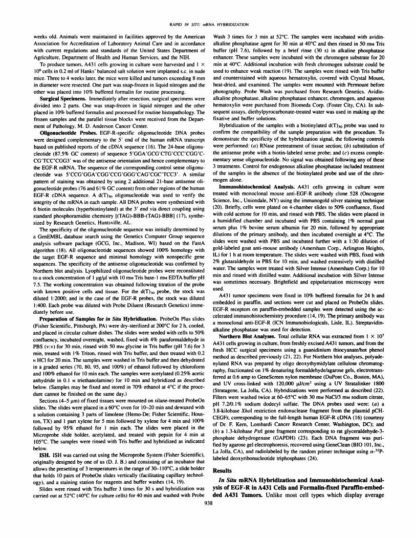
probe. In the first set of experiments, we determined whetherparaformaldehyde fixation of cultured cells or formalin fixation andparaffin embedding of tissues interfered with ISH for mRNA. Ahyperbiotinylated d(T),(l DNA oligonucleotide probe complementaryto polyadenylated mRNA transcripts was used to confirm the integrityof mRNA in all samples. As seen in Fig. I, ti and e, A43I cellsgrowing in culture and in solid tumors demonstrated intense alkalinephosphatasc reactivity in the cytoplasm and nucleus. These results areconsistent with the cellular distribution of precursor heterogeneousnuclear RNA in the nucleus and polyadenylated mRNA in the cytoplasm (28). The hyperbiotinylated antisense oligonucleotide probe(complementary to the EGF-R mRNA) synthesized with 6 biotingroups at the 3' end produced dark red cytoplasmic staining radiating
from the perinuclear area (Fig. I, /; and/). In the nucleus, the labelwas a diffuse pink to red (Fig. 1, b and/). The same oligonucleotideprobe labeled with only one biotin group at the 5' end produced
minimal staining (data not shown). Control ISH analyses using thecorresponding sense oligonucleotide probe labeled with 6 biotin molecules at the 3' end showed minimal staining (Fig. I, c and #). A
reagent control hybridization reaction using no probes produced nostaining (data not shown). Normal epithelial cells (epidermal layer)stained positive for EGF-R (Fig. I,/and h). Avidin alkaline phos-
phatase detection and the capillary technology used in the MicroprobeSystem ( 19) allowed for completion of this ISH technique in less than5 h. Immunohistochemical analysis of the A43I culture cells using amonoclonal anti-EGF-R antibody, immunogold labeling, and visual
ization by epipolarization optics revealed intense staining of the cellsurface in a punctate pattern correlating with the ISH results (Fig. I,compare tl and />). Immunolocalization of the EGF-R protein in sections of A431 tumors using a second monoclonal anti-EGF-R antibodyand alkaline phosphatase-cytochemical staining revealed an intense
reaction consistent with the ISH results (Fig. I, compare /; and/).Control antibodies showed no cross-reactivity with either the A431
cultured cells or tumor tissue.ISH and Immunohistochemical Analysis of EGF-R in Forma
lin-fixed Paraffin-embedded HCC Specimens. The use of ISH forEGF-R mRNA should allow for determination of intratumoral heter
ogeneity and identification of positive cells. The first specimen studied was from a primary Dukes' stage C2 tumor. To confirm that the
mRNA in the section was intact, we used the d(T),„oligonucleotideprobe (Fig. 2«).ISH with the antisense EGF-R oligonucleotide probe
produced distinct cytochemieal staining in the tumor (Fig. 2/j). Neither a control EGF-R sense probe (Fig. 2r) nor reagent control hy
bridization (not shown) produced staining. Immunohistochemicalanalysis of this paraffin-embedded primary HCC with a mouse monoclonal anti-EGF-R antibody and alkaline phosphatase detection
showed weakly stained tumor cells (Fig. 2ci}.Antibody reactivity wasalso observed on endothelial cells in this specimen, probably becauseof cross-reactivity with the blood group antigen la epitope (29) (Fig.
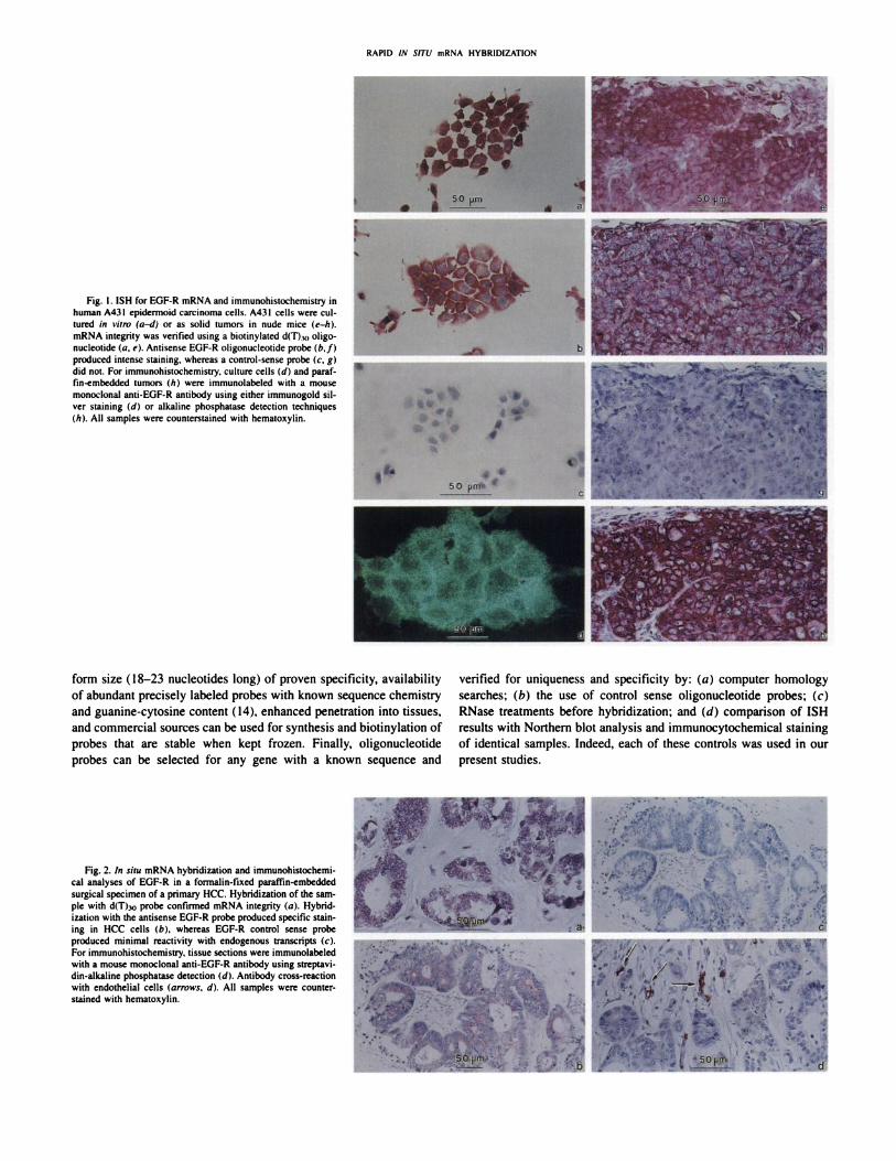
2il, arrows).We next performed ISH on paraffin sections of 3 HCC liver mé
tastases (from 3 different patients). All samples demonstrated comparable and intense cytochemieal reaction with the hyperbiotinylatedd(T),o oligonucleotide probe, verifying that the fixation process didnot lead to significant degradation of the mRNA (not shown). ISHanalysis with the EGF-R antisense probe demonstrated staining of
metastatic HCC cells in liver tissue (Fig. 3. a. r. and e). Cytochemieal
reactivity in the cytoplasm of HCC cells ranged from diffuse pink todeep red (Fig. 3, a, c. and <'). Hepatocytes, which are known to expressEGF-R (4-6), reacted with the antisense probe albeit at a lower level
than the HCC cells (Fig. 3. a and c). Reagent control hybridizationusing no probes initially showed endogenous alkaline phosphataseactivity in bile ducts. This reactivity was reduced to backgroundlevel by pretreatment of the tissue with levamisole (1 mg/ml) assuggested by Hankin and Lloyd (30) (data not shown). ISH of eachparaffin-embedded HCC liver métastaseswith the hyperbiotinylated
control sense oligonucleotide probe showed no staining (Fig. 3. /;,d. and/).
The final specimen was an HCC lymph node metastasis discoveredsubsequent to microscopic examination of a formalin-fixed and paraffin-embedded tissue. Here again, the hyperbiotinylated EGF-R oligonucleotide probe showed cell-specific staining in the HCC metas
tasis, albeit to a lesser intensity than HCC in the colon or liver (Fig.3#). The lack of reactivity in the adjacent lymph node tissue served asan internal control (Fig. 3#). Hybridization with the control senseprobe showed no reaction (Fig. 3/i). Only metastatic colon carcinomatissue demonstrated positive cytochemieal reactivity with hyperbiotinylated d(T),0 oligonucleotide probe. Lymphoid tissue, known to bemetabolically inactive (28), was negative (data not shown).
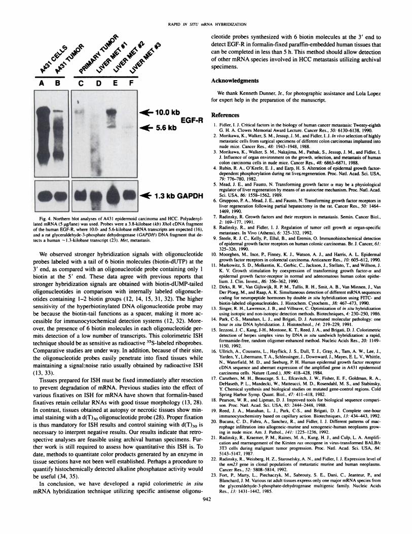
Northern Blot Analysis of EGF-R mRNA Expression. To confirm the ISH data, we examined the levels of EGF-R mRNA tran
scripts by Northern blot analysis of the same HCC surgical specimens.We could not perform Northern blot analysis for EGF-R mRNA in the
HCC lymph node metastasis because this lesion was discovered subsequent to microscopic examination of fixed tissue. High expressionof EGF-R-specific mRNA transcripts was found in both the A431
cultured cells and solid tumors (Fig. 4, Lanes A and B). directlycorrelating with the ISH and immunohistochemical analyses. Northern blot analysis of the primary HCC showed lower EGF-R mRNA
expression levels in comparison to the A431 samples (Fig. 4, compareLime C with Lane B). These results are also consistent with the ISHand immunohistochemical reactivity observed in the primary HCCspecimen. Northern blot analysis of 3 HCC liver métastasesrevealedexpression of EGF-R mRNA-specific transcripts (Fig. 4, Lanes D-F).
Similar results were obtained when the antisense ISH oligonucleotidewas used as a hybridization probe (data not shown).
Discussion
The present results demonstrate the advantages of a nonradioactivecolorimetrie ISH technique for detecting mRNA of EGF-R in human
A43I cells growing in culture and as tumor tissue (nude mice) and informalin-fixed paraffin-embedded specimens of HCC. The ISH resultscorrelated with those obtained by Northern blot analysis and by im-munohistochemistry for EGF-R. This ISH procedure permitted iden
tification of distribution of nucleic acid sequences in specific cellswith a better spatial resolution than a radioactive ISH (12, 13). Theprocedure was carried out with formalin-fixed tissues and completed
in less than 5 h.We used the human A431 epidermoid carcinoma cells in prelimi
nary studies because these cells have approximately 2 X IO6 EGF-
R/cell (26, 27). We found high EGF-R mRNA expression and protein
levels in the A431 cells. Other cells, such as HCC, display lowerlevels of EGF-R (20,000-200,000) per cell (25). The excellent corre
lation between Northern, ISH, and immunocytochemical assays provided evidence for the specificity and sensitivity of this colorimetrieISH technique to detect EGF-R mRNA expression in fixed and em
bedded specimens.The use of biotinylated DNA oligonucleotide probes for ISH offers
several advantages over full-length cDNA probes. These include uni-
RAPID IN SITU mRNA HYBRIDIZATION
Fig. I. ISH tor EGF-R mRNA and ¡mmunohistochemistry in
human A43I epidermoid carcinoma cells. A431 cells were cultured in vitro ta-d) or as solid tumors in nude mice (e-h).mRNA integrity was verified using a biotinylated d(T)w oligo-nuclcotide (a, e). Antisense EGF-R oligonucleotide probe (*./)produced intense staining, whereas a control-sense probe (e, gìdid not. For immunohistochemistry. culture cells id) and paraffin-embedded tumors (/() were immunolabeled with a mousemonoclonal anti-EGF-R antibody using either immunogold sil
ver staining (d) or alkaline phosphatase detection techniques(hi. All samples were counterstained with hematoxylin.
form size (18-23 nucleotides long) of proven specificity, availability
of abundant precisely labeled probes with known sequence chemistryand guanine-cytosine content (14), enhanced penetration into tissues,
and commercial sources can be used for synthesis and biotinylation ofprobes that are stable when kept frozen. Finally, oligonucleotideprobes can be selected for any gene with a known sequence and
verified for uniqueness and specificity by: (a) computer homologysearches; (b) the use of control sense oligonucleotide probes; (c)RNase treatments before hybridization; and (d) comparison of ISHresults with Northern blot analysis and immunocytochemical stainingof identical samples. Indeed, each of these controls was used in ourpresent studies.
Fig. 2. In situ mRNA hybridization and immunohistochemi-cal analyses of EGF-R in a formalin-fixed paraffin-embedded
surgical specimen of a primary HCC. Hybridization of the sample with d(T)io probe confirmed mRNA integrity («).Hybridization with the antisense EGF-R probe produced specific staining in HCC cells (b). whereas EGF-R control sense probeproduced minimal reactivity with endogenous transcripts (ci.For immunohistochemistry. tissue sections were immunolabeledwith a mouse monoclonal anti-EGF-R antibody using streptavi-din-alkaline phosphatase detection (</). Antibody cross-reactionwith endothelial cells (arrows, d). All samples were counter-stained with hematoxylin.
RAPID IN SITU mRNA HYBRIDI/AI ION
... •¿�••
. .1. /«.sii«mRNA h\hridi/ation analysis of paraffin-embedded HCC specimens from liver and lymph node métastases.Hybridisation of the samples with a hyperbiotinylatedoligonucleotide probe confirmed mRNA integrity (not shown). Hybridization with the antisense EGF-R oligonucleotide revealed positive staining of HCC métastasesin the liver
parenchyma («,e. e ) or lymph node ( ,c>.Normal hepatocytes demonstrated a weak reactivity. In contrast, normal lymphoid tissue did not react with the antisense EGF-R probe ( # ).EGF-R control sense probes showed no reactivity with endogenous transcripts (h. d.f. hi. All sections were counterstained with hematoxylin. a, b. liver metastasis 1; c. d. liver metastasis2; e.f, liver metastasis 3; ,c. h. lymph node metastasis.
941
RAPID IN SITU mRNA HYBRIDIZATION
V*V
A B
•¿�10.0 kb
5.6kbEGF-R
"f* 1.3kbGAPDH
Fig. 4. Northern blot analyses of A431 epidermoid carcinoma and HCC. Polyadenyl-lated mRNA (5 pg/lane) was used. Probes were a 3.8-kilobase (kb) Xho\ cDNA fragmentof the human EGF-R. where IO.O- and 5.6-kilobase mRNA transcripts are expected ( I6).and a rat glyceraldehyde-3-phosphute dehydrogenase {GAPDHI DNA fragment that detects a human ~l.3-kilobase transcript (23). Mel, metastasis.
We observed stronger hybridization signals with oligonucleotideprobes labeled with a tail of 6 biotin molecules (biotin-dUTP) at the3' end, as compared with an oligonucleotide probe containing only Ibiotin at the 5' end. These data agree with previous reports that
stronger hybridization signals are obtained with biotin-dUMP-tailedoligonucleotides in comparison with internally labeled oligonucle-otides containing 1-2 biotin groups (12. 14, 15, 31, 32). The higher
sensitivity of the hyperbiotinylated DNA oligonucleotide probe maybe because the biotin-tail functions as a spacer, making it more ac
cessible for immunocytochemical detection systems (12, 32). Moreover, the presence of 6 biotin molecules in each oligonucleotide permits detection of a low number of transcripts. This colorimetrie ISHtechnique should be as sensitive as radioactive ^S-labeled riboprobes.
Comparative studies are under way. In addition, because of their size,the oligonucleotide probes easily penetrate into fixed tissues whilemaintaining a signahnoise ratio usually obtained by radioactive ISH(13,33).
Tissues prepared for ISH must be fixed immediately after resectionto prevent degradation of mRNA. Previous studies into the effect ofvarious fixatives on ISH for mRNA have shown that formalin-based
fixatives retain cellular RNAs with good tissue morphology ( 13. 28).In contrast, tissues obtained at autopsy or necrotic tissues show minimal staining with a d(T),<>oligonucleotide probe (28). Proper fixationis thus mandatory for ISH results and control staining with d(T)30 isnecessary to interpret negative results. Our results indicate that retrospective analyses are feasible using archival human specimens. Further work is still required to assess how quantitative this ISH is. Todate, methods to quantitate color products generated by an enzyme intissue sections have not been well established. Perhaps a procedure toquantify histochemically detected alkaline phosphatase activity wouldbe useful (34, 35).
In conclusion, we have developed a rapid colorimetrie in situmRNA hybridization technique utilizing specific antisense oligonu
cleotide probes synthesized with 6 biotin molecules at the 3' end to
detect EGF-R in formalin-fixed paraffin-embedded human tissues that
can be completed in less than 5 h. This method should allow detectionof other mRNA species involved in HCC metastasis utilizing archivalspecimens.
Acknowledgments
We thank Kenneth Dunner, Jr.. for photographic assistance and Lola Lope/,for expert help in the preparation of the manuscript.
References
1. Fidler. I. J. Critical factors in the biology of human cancer metastasis: Twenty-eighthG. H. A. Clowes Memorial Award Lecture. Cancer Res.. 50: 6130-6138. 1990.
2. Morikawa. K.. Walker, S. M-, Jessup. J. M., and Fidler. I. J. In vivo selection of highlymetastatic cells from surgical specimens of different colon carcinomas implanted intonude mice. Cancer Res.. 48: 1943-1948. 1988.
3. Morikawa. K., Walker. S. M.. Nakajima, M.. Pathak. S.. Jessup. J. M.. and Fidler, l.J. Influence of organ environment on the growth, selection, and metastasis of humancolon carcinoma cells in nude mice. Cancer Res., 4M: 6863-6871. 1988.
4. Rubin. R. A.. O'Keefe. E. J.. and Earp. H. S. Alteration of epidermal growth factor-
dependent phosphorylation during rat live®eneration. Proc. Nati. Acad. Sci. USA,79: 776-780. 1982.
5. Mead. J. E.. and Fausto, N. Transforming growth factor a may be a physiologicalregulator of liver regeneration by means of an autocrine mechanism. Proc. Nati. Acad.Sci. USA. 86: 1558-1562. 1989.
6. Grupposo. P. A.. Mead, J. E.. and Fausto. N. Transforming growth factor receptors inliver regeneration following partial hepatectomy in the rat. Cancer Res.. 50: 1464-1469. 1990.
7. Radinsky. R. Growth factors and their receptors in metastasis. Semin. Cancer Biol..2: 169-177, 1991.
8. Radinsky, R.. and Fidler. I. J. Regulation of tumor cell growth at organ-specificmétastases.In Vivo (Athens). 6: 325-332. 1992.
9. Steele. R. J. C.. Kelly. P.. Ellul, B.. and Eremin. O. Immunohistochemical detectionof epidermal growth factor receptors on human colonie carcinomas. Br. J. Cancer. 61:325-326. 1990.
10. Moorghen. M.. Ince, P.. Finney. K. J.. Watson. A. J.. and Harris. A. L. Epidermalgrowth factor receptors in colorectal carcinoma. Anticancer Res.. 10: 605-612, 1990.
11. Markowitz, S. D.. Molkentin. K.. Gerbic, C.. Jackson. J., Stellato. T.. and Willson, J.K. V. Growth stimulation by coexpression of transforming growth factor-a andepidermal growth factor-receptor in normal and adenomatous human colon epithelium. J. Clin. Invest.. 86: 356-362. 1990.
12. Dirks. R. W.. Van Gijlswijk. R. P. M.. TullÃs.R. H.. Smit, A. B., Van Minnen. J., VanDer Ploeg. M.. and Raap, A. K. Simultaneous detection of different mRNA sequencescoding for neuropeptide hormones by double in situ hybridization using FITC- andbiotin-labeled oligonucleotides. J. Histochem. Cytochem., 38: 467—473, 1990.
13. Singer, R. H., Lawrence, J. B.. and Villnave. C. Optimization of m .v/ni hybridizationusing isotopie and non-isotopic detection methods. Biotechniques. 4: 230-250. 1986.
14. Park. C-S.. Manaban. L. J.. and Brigati. D. J. Automated molecular pathology: onehour in situ DNA hybridization. J. Histotechnol.. 14: 219-229. 1991.
15. lezzoni. J. C.. Kang. J-H.. Montone. K. T. Reed. J. A., and Brigati. D. J. Colorimetriedetection of herpes simplex virus by DNA in aim sandwich hybridization: a rapid,formamide-free. random oligomer-enhanced method. Nucleic Acids Res.. 20: 1149-
1150. 1992.16. Ullrich. A.. Coussens. L., Hayflick, J. S.. Dull, T. J., Gray, A., Tarn, A. W., Lee, J.,
Yarden. Y. Libermann. T. A.. Schlessinger. J.. Downward. J.. Mayes. E. L. V. Whittle.N.. Waterfield. M. D., and Seeburg. P. H. Human epidermal growth factor receptorcDNA sequence and aberrant expression of the amplified gene in A43I epidermoidcarcinoma cells. Nature (Lond.). 3W: 418-128. 1984.
17. Caruthers. M. H., Beaucage. S. L.. Efcavitch. J. W.. Fisher. E. F., Goldman. R. A.,DeHaseth, P. L.. Mandecki. W.. Matteucci. M. D.. Rosendahl, M. S.. and Stabinsky.Y. Chemical synthesis and biological studies on mutated gene-control regions. ColdSpring Harbor Symp. Quant. Biol.. 47: 411-418. 1982.
18. Pearson. W. R., and Lipman, D. J. Improved tools for biological sequence comparison. Proc. Nati. Acad. Sci. USA, 85: 2444-2448. 1988.
19. Reed. J. A.. Manahan. L. J., Park. C-S.. and Brigati. D. J. Complete one-hourimmunocytochemistry based on capillary action. Biotechniques, 13: 434—443.1992.
20. Bucana. C. D.. Fabra. A.. Sanchez. R.. and Fidler. I. J. Different patterns of macrophage infiltration into allogeneic-murine and xenogeneic-human neoplasms growing in nude mice. Am. J. Pathol.. 141: 1225-1236. 1992.
21. Radinsky. R.. Kraemer. P. M.. Raines. M. A.. Kung. H. J.. and Culp. L. A. Amplification and rearrangement of the Kirsten ras oncogene in virus-transformed BALB/c3T3 cells during malignant tumor progression. Proc. Nati. Acad. Sci. USA. 84:5143-5147. 1987.
22. Radinsky. R.. Weisberg. H. Z.. Staroselsky. A. N.. and Fidler. I. J. Expression level ofthe nm23 gene in clonal populations of metastalic murine and human neoplasms.Cancer Res.. 52: 5808-5814. 1992.
23. Fort, P., Marty, L.. Piechaczyk. M., Sabrouty. S. E.. Dani, C.. Jeanteur. P.. andBlanchard. J. M. Various rat adult tissues express only one major mRNA species fromthe glyceraldehyde-3-phosphate-dehydrogenase multigenic family. Nucleic AcidsRes., 13: 1431-1442. 1985.
942

RAPII) IN SITU mRNA HYBRIDIZATION
24. Feinberg. A. P., and Vogelstein, B. A technique for radiolabeling DNA restrictionendonuclease fragments to high specific activity. Anal. Biochem., 132: 6-13, 1983.
25. Yarden, Y., and Ullrich, A. Growth factor receptor tyrosine kinases. Annu. Rev.Biochem., 57: 443^»78, 1988.
26. Fabricant, R. N., DeLarco, J. E., and Todaro. G. L. Nerve growth factor receptorson human melanoma cells in culture. Proc. Nati. Acad. Sci. USA. 74: 565-569.
1977.27. Haigler, H. T.. Ash. 1. F, Singer. S. J.. and Cohen. S. Visualization by fluorescence of
the binding and intemalization of epidermal growth factor in human carcinoma cellsA43I. Proc. Nati. Acad. Sci. USA. 75: 3317-3321, 1978.
28. Pringle. J. H.. Primrose, L., Kind, C. N.. Talbot. I. C., and Lauder, I. In situ hybridization demonstration of poly-adenylated RNA sequences in formalin-fixed paraffinsections using a biotinylated oligonucleotide poly d(T) probe. J. Pathol., 15H: 279-286, 1989.
29. Limas, C. Relationship of epidermal growth factor receptor detectability with the A,B, H blood group antigens: emphasis on normal and neoplastic urothelium. Am. J.Pathol.. /.?«.-131-137. 1991.
30. Hankin. R. C., and Lloyd, R. V. Detection of messenger RNA in routinely processedtissue sections with biotinylated oligonucleotide probes. Am. J. Clin. Pathol.. 92:166-171, 1989.
31. Guitteny, A. F., Fouque, B., Mougin. C.. Teoule. R., and Bloch. B. Histologicaldetection of messenger RNAs with biotinylated synthetic oligonucleotide probes. J.Histochem. Cytochem., 36: 563-571, 1988.
32. Cook, A. F., Vuocolo. E., and Brake!, C. L. Synthesis and hybridization of a series ofbiotinylated oligonucleotides. Nucleic Acids Res.. 16: 4077^)095. 1988.
33. Wakamiya, N., Horiguchi. J., and Kufe. D. Detection of c-fms and CSF-I RNA by insilu hybridization. Leukemia (Baltimore). /: 518-520. 1987.
34. Bcnno. R. H., Tucker. L. W.. Joh, T. H., and Reis, D. J. Quantitative ¡mmunocy-tochemistry of tyrosine hydroxylase in rat brain. 1. Development of a computer-assisted method using the peroxidase technique. Brain Res., 246: 225-236, 1982.
35. Finkelstein, Y. Wolff, M.. and Biegon, A. Brain acetylcholinesterase after acuteparathion poisoning: a comparative quantitative histochemical analysis post-mortem.Annu. Neurol.. 24: 252-257. 1988.
943