A microRNA DNA methylation signature for human cancer metastasis
Upload
independentCategory
view
0download
0
A Prognostic DNA Methylation Signature for Stage INon–Small-Cell Lung CancerJuan Sandoval, Jesus Mendez-Gonzalez, Ernest Nadal, Guoan Chen, F. Javier Carmona, Sergi Sayols,Sebastian Moran, Holger Heyn, Miguel Vizoso, Antonio Gomez, Montse Sanchez-Cespedes, Yassen Assenov,Fabian Muller, Christoph Bock, Miquel Taron, Josefina Mora, Lucia A. Muscarella, Triantafillos Liloglou,Michael Davies, Marina Pollan, Maria J. Pajares, Wenceslao Torre, Luis M. Montuenga, Elisabeth Brambilla,John K. Field, Luca Roz, Marco Lo Iacono, Giorgio V. Scagliotti, Rafael Rosell, David G. Beer,and Manel Esteller
Author affiliations appear at the end ofthis article.
Published online ahead of print atwww.jco.org on September 30, 2013.
Supported by Grant No. HEALTH-F2-2010-258677-CURELUNG Project fromthe European Community’s SeventhFramework Programme (FP7/2007-2013); Grants No. PI10/02992, PI10/00166, RD06/0020/0066, and RD06/0020/0062 from the Institute of HealthCarlos III; the Cellex Foundation;and the Roy Castel Lung CancerFoundation.
J.S. and J.M.-G. contributed equally tothis article.
Authors’ disclosures of potential con-flicts of interest and author contribu-tions are found at the end of thisarticle.
Corresponding author: Manel Esteller,MD, PhD, Cancer Epigenetics and Biol-ogy Program, 3rd Floor, Hospital Durani Reynals, Av. Gran Via 199-203, 08908L’Hospitalet, Barcelona, Catalonia,Spain; e-mail: [email protected].
© 2013 by American Society of ClinicalOncology
0732-183X/13/3199-1/$20.00
DOI: 10.1200/JCO.2012.48.5516
A B S T R A C T
PurposeNon–small-cell lung cancer (NSCLC) is a tumor in which only small improvements in clinicaloutcome have been achieved. The issue is critical for stage I patients for whom there are noavailable biomarkers that indicate which high-risk patients should receive adjuvant chemotherapy.We aimed to find DNA methylation markers that could be helpful in this regard.
Patients and MethodsA DNA methylation microarray that analyzes 450,000 CpG sites was used to study tumoral DNAobtained from 444 patients with NSCLC that included 237 stage I tumors. The prognostic DNAmethylation markers were validated by a single-methylation pyrosequencing assay in an indepen-dent cohort of 143 patients with stage I NSCLC.
ResultsUnsupervised clustering of the 10,000 most variable DNA methylation sites in the discovery cohortidentified patients with high-risk stage I NSCLC who had shorter relapse-free survival (RFS; hazardratio [HR], 2.35; 95% CI, 1.29 to 4.28; P � .004). The study in the validation cohort of the significantmethylated sites from the discovery cohort found that hypermethylation of five genes wassignificantly associated with shorter RFS in stage I NSCLC: HIST1H4F, PCDHGB6, NPBWR1,ALX1, and HOXA9. A signature based on the number of hypermethylated events distinguishedpatients with high- and low-risk stage I NSCLC (HR, 3.24; 95% CI, 1.61 to 6.54; P � .001).
ConclusionThe DNA methylation signature of NSCLC affects the outcome of stage I patients, and it can bepractically determined by user-friendly polymerase chain reaction assays. The analysis of the bestDNA methylation biomarkers improved prognostic accuracy beyond standard staging.
J Clin Oncol 31. © 2013 by American Society of Clinical Oncology
INTRODUCTION
Non–small-cell lung cancer (NSCLC) is the leadingcause of cancer-related death.1 The poor prognosisof patients with NSCLC is associated with severalfactors, among which are late disease diagnosis andthe small number of effective drugs. The absence ofvalidated prognostic biomarkers could also be rele-vant, because even patients with stage I NSCLC whoundergo potentially curative surgical resection are athigh risk of dying from recurrent disease, with a5-year relapse rate of 35% to 50%.1 Although adju-vant platinum-based chemotherapy is beneficial inmore advanced resected disease, in which most ofthe patients have a high risk of recurrence,2-6 it hasfailed to show a survival benefit for patients at stage
I.7,8 One explanation for these negative data in theearly stages could be the lack of biologic factors pre-dicting their recurrence and the fact that, in theabsence of useful biomarkers, all stage I NSCLCs arepooled, making it more difficult to draw meaningfulclinical conclusions.
In the search for new potential biomarkers ofhuman cancer, the hypermethylation of the CpGisland sequences located in the promoter regions oftumor suppressor genes are gaining prominence.9-11
We wondered whether DNA methylation markerscould also be used to provide a prognostic snapshotof lung tumors. Herein, we have obtained DNAmethylation signatures associated with shorterrelapse-free survival (RFS) in stage I NSCLCs thatcould be useful in the design of clinical trials for
JOURNAL OF CLINICAL ONCOLOGY O R I G I N A L R E P O R T
© 2013 by American Society of Clinical Oncology 1
http://jco.ascopubs.org/cgi/doi/10.1200/JCO.2012.48.5516The latest version is at Published Ahead of Print on September 30, 2013 as 10.1200/JCO.2012.48.5516
Copyright 2013 by American Society of Clinical Oncologyfrom 161.116.100.92
Information downloaded from jco.ascopubs.org and provided by at L HOSPITALET DEL LLOBREGA on October 1, 2013Copyright © 2013 American Society of Clinical Oncology. All rights reserved.from 161.116.100.92
Information downloaded from jco.ascopubs.org and provided by at L HOSPITALET DEL LLOBREGA on October 1, 2013Copyright © 2013 American Society of Clinical Oncology. All rights reserved.from 161.116.100.92
Information downloaded from jco.ascopubs.org and provided by at L HOSPITALET DEL LLOBREGA on October 1, 2013Copyright © 2013 American Society of Clinical Oncology. All rights reserved.from 161.116.100.92
Information downloaded from jco.ascopubs.org and provided by at L HOSPITALET DEL LLOBREGA on October 1, 2013Copyright © 2013 American Society of Clinical Oncology. All rights reserved.from 161.116.100.92
Information downloaded from jco.ascopubs.org and provided by at L HOSPITALET DEL LLOBREGA on October 1, 2013Copyright © 2013 American Society of Clinical Oncology. All rights reserved.from 161.116.100.92
Information downloaded from jco.ascopubs.org and provided by at L HOSPITALET DEL LLOBREGA on October 1, 2013Copyright © 2013 American Society of Clinical Oncology. All rights reserved.from 161.116.100.92
Information downloaded from jco.ascopubs.org and provided by at L HOSPITALET DEL LLOBREGA on October 1, 2013Copyright © 2013 American Society of Clinical Oncology. All rights reserved.from 161.116.100.92
Information downloaded from jco.ascopubs.org and provided by at L HOSPITALET DEL LLOBREGA on October 1, 2013Copyright © 2013 American Society of Clinical Oncology. All rights reserved.from 161.116.100.92
Information downloaded from jco.ascopubs.org and provided by at L HOSPITALET DEL LLOBREGA on October 1, 2013Copyright © 2013 American Society of Clinical Oncology. All rights reserved.

adjuvant chemotherapy in the expanding population of those diag-nosed with early-stage lung cancer.
PATIENTS AND METHODS
Study Design and Patient Population
Patients were eligible to enter the study as part of either discovery orvalidation cohorts if they underwent surgical resection of NSCLC in any of theinternational participating institutions. Patients treated with neoadjuvanttherapy and/or patients with large cell-carcinoma were not included in thestudy. The clinical characteristics of the NSCLC surgical samples obtained areprovided in Table 1. Descriptors of the patients by site of origin and for each
single case are included in the Data Supplement. Tumors were collected bysurgical resection from patients who provided consent and under approval bythe institutional review boards. The median clinical follow-up was 7.2 years.Follow-up was performed by using radiographic imaging (chest x-ray andcomputed tomography scans), and time of recurrence was noted. In addition,25 histologically normal lung tissue counterparts without any histologic evi-dence of malignancy were also analyzed (Data Supplement). The NSCLCtumor samples were studied in a consecutive manner as they arrived at thecentralized DNA methylation facility and passed the technical quality checks.
Procedures
The DNA methylation status of 450,000 CpG sites was established byusing the Infinium 450K Methylation Array.12,13 The methylation score of
Table 1. Clinical Characteristics of the Discovery and Validation Cohorts
Characteristic
Discovery Cohort(n � 444)
Subset FromDiscovery Cohortfor RFS Analysis
(n � 198)�
Subset FromDiscovery Cohortfor RFS Analysis
(stage I) (n � 147)�Validation Cohort
(stage I) (n � 143)†
No. % No. % No. % No. %
Age, yearsMedian 65 65.5 65.9 63.7Range 35-90 38-85 38-85 32-78
SexMale 254 57 107 54 78 53 126 88Female 190 43 91 46 69 47 17 12
Smoking historyCurrent or former smoker 334 75 169 85 127 86 122 85Nonsmoker 47 11 25 13 17 12 10 7Unknown 63 14 4 2 3 2 11 8
Disease stageI 237 53 147 74 147 100 143 100II 94 21 22 11 0 0 0 0III 102 23 26 13 0 0 0 0IV 11 3 3 2 0 0 0 0
Tumor typeAdenocarcinoma 322 73 155 78 118 80 79 55Squamous cell carcinoma 122 27 43 22 29 20 64 45
Thoracic surgery practiceLobectomy 396 90 172 86 132 90 117 82Pneumonectomy 23 5 13 7 3 2 3 2Segmentectomy 24 5 13 7 12 8 2 1Unknown 1 0 0 0 0 0 21 15
Adjuvant treatmentNone 211 48 198 100 147 100 143 100Chemotherapy 24 5 0 0 0 0 0 0Chemotherapy plus radiotherapy 12 3 0 0 0 0 0 0Radiotherapy 27 6 0 0 0 0 0 0Unknown 170 38 0 0 0 0 0 0
RecurrenceYes 161 36 98 49 53 36 51 36No 150 34 100 51 94 64 92 64Unknown 133 30 0 0 0 0 0 0
RFS, monthsAverage 46.7 50.8 60.7 42.3Range 0.6-224 0.6-224 0.6-224 2.6-130
Origin of the samplesEurope 291 66 100 51 68 46 142 99United States 153 34 98 49 79 54 1 1
Abbreviation: RFS, relapse-free survival.�Patients from the discovery cohort who had undergone resection of non–small-cell lung cancer and did not receive adjuvant chemotherapy before relapse.†All patients from the validation cohort had undergone resection of non–small-cell lung cancer and did not receive adjuvant chemotherapy before relapse.
Sandoval et al
2 © 2013 by American Society of Clinical Oncology JOURNAL OF CLINICAL ONCOLOGY

each CpG is represented as a � value. Samples were clustered in an unsuper-vised manner by using the 10,000 most variable � values for CpG methylationaccording to the standard deviation for the CpG sites located in promoterregions by hierarchical clustering using the complete method for agglomerat-ing the Manhattan distances (Data Supplement). DNA methylation microar-ray data are available from the National Center for BiotechnologyInformation’s Gene Expression Omnibus.14 Pyrosequencing analyses to de-termine CpG methylation status were conducted as previously described.15
Statistical Analysis
Assay results were compared with patient outcomes in a double-blindmanner. Median follow-up duration was calculated according to the inverseKaplan-Meier method. Differences in distributions between groups were ex-amined by the �2 test. The Kaplan-Meier method was used to estimate RFS,and differences among the groups were analyzed with the log-rank test. Hazardratios (HRs) from univariate Cox regression analysis were used to determinethe association of clinicopathologic features with relapse. Multivariate Coxproportional hazards regression was used to evaluate independent prognosticfactors associated with RFS.
RESULTS
Characteristics of Patients in the Discovery Cohort
Clinical characteristics of the 444 patients in the discovery cohortare listed in Table 1. Descriptors of the patients by site of origin and foreach single case are shown in the Data Supplement. The clinicopath-ologic characteristics of the lung tumors studied were related to the siteof origin (United States v Europe).16-18
DNA Methylation Profiles Identify Two Groups With
Different RFS Rates
We first evaluated a genome-wide DNA methylation profile ofthe original cohort of 444 patients with lung tumors, which includedtwo NSCLC subtypes (adenocarcinoma and squamous cell carcino-mas) by using a previously validated 450,000 CpG methylation mi-croarray.12,13 In addition, 25 histologically normal lung tissuecounterparts without any histologic evidence of malignancy were alsoanalyzed (Data Supplement).
The analyses of CpG methylation � values from the DNA meth-ylation microarray within all primary NSCLCs (n � 444) and histo-logically normal tissues (n � 25) identified 10,000 promoter CpGswith the most variable CpG methylation levels (Data Supplement).These 10,000 top-ranked CpG sites were plotted in an unsupervisedmanner in the 444 primary NSCLCs (Fig 1A). The hierarchical clus-tering distinguished two main types of tumors that accounted for 70(16%; group A) and 374 (84%; group B) patients. The �2 tests showeda significantly higher proportion of the adenocarcinoma histologictype in group A (�2 test P � .02), but no other significant differ-ences in the distribution of the tumors according to stage, sex, orsmoking history between group A and group B were observed(Data Supplement).
We investigated whether these two DNA methylation groups hadany effect on the RFS of these patients. We analyzed the subset ofpatients who had undergone resection of NSCLC and had not receivedadjuvant chemotherapy before relapse, because of the possible con-founding effect of chemotherapy on the RFS. Overall survival was notselected as an end point for the study because it could be affected bysubsequent therapies received at relapse. Overall, 198 patients withNSCLC met the criteria for inclusion in the RFS cohort. Most impor-tantly, these group A patients with NSCLC had a significantly shorter
RFS, as shown in the Kaplan-Meier survival analysis (log-rank testP � .001; Fig 1B) and in the univariate (HR, 2.45; P � .001) andmultivariate (HR, 2.40; P � .001) Cox regression analyses of stage,histology, smoking history, age, and sex (Data Supplement). In refer-ence to histology, the unsupervised clustering analysis of either ade-nocarcinomas or squamous cell carcinomas also identified a groupassociated with shorter RFS (HR, 2.47; P � .002 and HR 4.93; P �.001, respectively; Data Supplement).
We wanted to extend these observations to identify those NSCLCtumors that, despite their low stage, are prone to recurrence. Theselection of these patients is critical because approximately 30% to40% of patients with stage I NSCLC die of recurrent disease.19-21 Toaddress this, the profile of the aforementioned 10,000 promoter CpGs,which had already shown their prognostic value throughout allNSCLC stages, was plotted in an unsupervised manner in the 237patients with stage I NSCLC (Fig 1C). Hierarchical clustering distin-guished two main types of tumors, accounting for 63 (27%; group 1)and 174 (73%; group 2) patients. The �2 tests revealed no significantdifferences in the distribution of the tumors in the two groups by sex,smoking history, and histologic type (Data Supplement). Among the237 patients with stage I NSCLC, 147 met the described criteria forinclusion in the RFS cohort. The ineligible patients (n � 90) did notshow a higher recurrence rate (�2 test P � .12). Group 1 identifiedpatients with high-risk stage I NSCLC that had lower RFS, as revealedby the Kaplan-Meier survival analysis (log-rank test P � .03; Fig 1D)and in the univariate (HR, 1.85; P � .037) and multivariate (HR, 2.35;P � .004) Cox regressions of histology, smoking history, age, and sex(Data Supplement). In reference to histology, the unsupervised clus-tering analysis of the adenocarcinomas in stage I also identified agroup associated with shorter RFS (HR, 2.94; P � .003; Data Supple-ment), and a trend was observed for squamous cell carcinomas (HR,2.55; P � .09; Data Supplement). We also performed a Cox analysisthat included smoking pack-years as a covariate. We categorized pack-years22 as less than 30 or � 30. The inclusion of the pack-year datavalue did not change the significant association of group 1 tumors withshorter RFS (HR, 2.3; P � .007). For all the patients with stage INSCLC (because stage IA and IB have different outcomes), we alsoadded this particular feature (according to the sixth revision of theTNM classification criteria) to the Cox regression multivariate analy-sis and group 1 remained significantly associated with shorter RFS(HR, 2.12; P � .018). The inclusion of tumor size within stage I (alsoan indicator of poor prognosis in NSCLC) in the Cox analysis did notalter the significant association of group 1 tumors with shorter RFS(HR, 2.02; P � .05). The reclassification of the stage I tumors accord-ing to the seventh revision of the TNM classification criteria alsoconfirmed that group 1 patients remained significantly associatedwith shorter RFS (HR, 2.14; P � .05).
Identification of Candidate Genes as DNA Methylation
Biomarkers of Shorter RFS in the Discovery Cohort of
Stage I NSCLC
The identification of a DNA methylation signature for stage INSCLC that predicts early recurrence might be useful, but thefinding of a smaller panel of DNA methylation biomarkers couldsimplify the process. To achieve this goal, we developed an integra-tive approach to rank the CpG sites that, according to their meth-ylation status (� values), were best at discriminating the 444NSCLC samples from the 25 histologically normal lung tissue
Clinical Value of DNA Methylation in NSCLC
www.jco.org © 2013 by American Society of Clinical Oncology 3

samples. This analysis identified 338 highly ranked CpG sites (DataSupplement). From these, we focused on the CpGs located inregulatory regions: promoter CpG islands9-11 and shores.23,24 Wefound that 150 of the 338 CpG sites were located in the describedregions. All of these 150 CpG sites were present in the 10,000 CpGsites used in the clustering. CpG hypermethylation of these 150sites was significantly enriched in group A versus group B (t testP � .001) and in group 1 versus group 2 (t test P � .001), support-ing their potential prognostic value. Thus, we tested the methyl-ation value of each of these 150 CpG sites for RFS in the 147 stage Itumors by Kaplan-Meier survival and multivariate Cox regression.We identified 54 CpGs corresponding to 42 genes that were signif-icantly associated with shorter RFS at a 10% false discovery rate(Data Supplement). Our data mining approach can be comple-mented by others and, in this regard, the promoter CpG sites ofother methylation markers in lung cancer25 did not pass the criteriaused. However, we confirmed that CDH13 and RASSF1A hyper-methylation was associated with shorter RFS (HR, 3.47; P � .01and HR, 2.17; P � .02, respectively) in the 147 stage I tumors.
Validation of Candidate Genes as DNA Methylation
Biomarkers of Shorter RFS in an Independent Cohort
of Stage I NSCLC
Once we had identified 42 genes with CpG promoter methyl-ation that influenced RFS in our initial discovery cohort of 147 stage Itumors, we sought to validate these single DNA methylation markersin an additional cohort of 143 patients with stage I NSCLC (Table 1).Descriptors of the patients by site of origin and for each single sampleare shown in the Data Supplement. All these new NSCLC sampleswere obtained from patients who had undergone a resection and didnot receive adjuvant chemotherapy. The validation cohort, in com-parison to the discovery set, was significantly enriched in Europeansamples and, thus, in affected men and squamous cell carcinomas.16,17
The methylation levels at the described CpG sites were analyzed bypyrosequencing15 to test a more affordable large-scale approach.Methylation value by pyrosequencing was obtained from the averageof each of the CpG dinucleotides included in the sequence analyzed(Data Supplement). Because the DNA material was limited, weselected the top 10 genes (Data Supplement) with an HR of more than
A
C
B puorGA puorG
2 puorG1 puorG
B
Rela
pse-
Free
Sur
viva
l (pr
opor
tion)
Time Since Surgery (years)
1.0
0.8
0.6
0.4
0.2
010 515
HR, 2.40 (95% CI, 1.45 to 3.95)P < .001
Group A (n = 28)Group B (n = 70)
No. at riskGroup AGroup B
28
170
3
67
1
16
03
Rela
pse-
Free
Sur
viva
l (pr
opor
tion)
Time Since Surgery (years)
1.0
0.8
0.6
0.4
0.2
010 515
HR, 2.35 (95% CI, 1.29 to 4.28)P = .004
Group 1 (n = 37)Group 2 (n = 110)
No. at riskGroup 1Group 2
37
110
1254
2
14
03
D
Fig 1. DNA methylation signatures associated with recurrence-free survival (RFS) in non–small-cell lung cancer samples. (A) Unsupervised hierarchical clustering andheat map associated with the methylation profile (according to the color scale shown) of the 444 primary tumor specimens based on the 10,000 most variable promoter� values. Two different histologic subtypes are represented: adenocarcinoma (Adeno; red) and squamous cell carcinoma (yellow). Each column represents an individualpatient and each row an individual CpG. (B) Kaplan-Meier analysis for RFS among the 198 patients with RFS information according to the two groups obtained fromthe clustering. The P value corresponds to the hazard ratio (HR) adjusted by multivariate regression (including age, sex, smoking history, stage, and histologic type).(C) Unsupervised clustering and the heat map of the subset of 237 patients with stage I non–small-cell lung cancer. (D) Kaplan-Meier estimates for RFS among thesubset of 147 patients with RFS information according to the two groups obtained in the clustering. The P value reflects the HR adjusted as in the analysis in (B).
Sandoval et al
4 © 2013 by American Society of Clinical Oncology JOURNAL OF CLINICAL ONCOLOGY
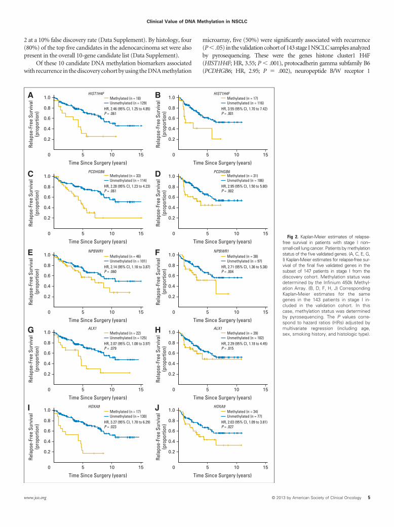
2 at a 10% false discovery rate (Data Supplement). By histology, four(80%) of the top five candidates in the adenocarcinoma set were alsopresent in the overall 10-gene candidate list (Data Supplement).
Of these 10 candidate DNA methylation biomarkers associatedwith recurrence in the discovery cohort by using the DNA methylation
microarray, five (50%) were significantly associated with recurrence(P � .05) in the validation cohort of 143 stage I NSCLC samples analyzedby pyrosequencing. These were the genes histone cluster1 H4F(HIST1H4F; HR, 3.55; P � .001), protocadherin gamma subfamily B6(PCDHGB6; HR, 2.95; P � .002), neuropeptide B/W receptor 1
A
Rela
pse-
Free
Sur
viva
l(p
ropo
rtion
)
Time Since Surgery (years)
1.0
0.8
0.6
0.4
0.2
010 515
HR, 2.46 (95% CI, 1.25 to 4.85)P = .061
Methylated (n = 18)Unmethylated (n = 129)
F4H1TSIHF4H1TSIH
6BGHDCP6BGHDCP
1RWBPN1RWBPN
1XLA1XLA
9AXOH9AXOH
B
Rela
pse-
Free
Sur
viva
l(p
ropo
rtion
)Time Since Surgery (years)
1.0
0.8
0.6
0.4
0.2
010 515
HR, 3.55 (95% CI, 1.70 to 7.42)P = .001
Methylated (n = 17)Unmethylated (n = 116)
C
Rela
pse-
Free
Sur
viva
l(p
ropo
rtion
)
Time Since Surgery (years)
1.0
0.8
0.6
0.4
0.2
010 515
HR, 2.28 (95% CI, 1.23 to 4.23)P = .061
Methylated (n = 33)Unmethylated (n = 114)
D
Rela
pse-
Free
Sur
viva
l(p
ropo
rtion
)
Time Since Surgery (years)
1.0
0.8
0.6
0.4
0.2
010 515
HR, 2.95 (95% CI, 1.50 to 5.80)P = .002
Methylated (n = 31)Unmethylated (n = 106)
E
Rela
pse-
Free
Sur
viva
l(p
ropo
rtion
)
Time Since Surgery (years)
1.0
0.8
0.6
0.4
0.2
010 515
HR, 2.14 (95% CI, 1.18 to 3.87)P = .060
Methylated (n = 46)Unmethylated (n = 101)
F
Rela
pse-
Free
Sur
viva
l(p
ropo
rtion
)
Time Since Surgery (years)
1.0
0.8
0.6
0.4
0.2
010 515
HR, 2.71 (95% CI, 1.36 to 5.38)P = .004
Methylated (n = 38)Unmethylated (n = 97)
G
Rela
pse-
Free
Sur
viva
l(p
ropo
rtion
)
Time Since Surgery (years)
1.0
0.8
0.6
0.4
0.2
010 515
HR, 2.07 (95% CI, 1.08 to 3.97)P = .079
Methylated (n = 22)Unmethylated (n = 125)
H
Rela
pse-
Free
Sur
viva
l(p
ropo
rtion
)
Time Since Surgery (years)
1.0
0.8
0.6
0.4
0.2
010 515
HR, 2.29 (95% CI, 1.18 to 4.45)P = .015
Methylated (n = 39)Unmethylated (n = 102)
I
Rela
pse-
Free
Sur
viva
l(p
ropo
rtion
)
Time Since Surgery (years)
1.0
0.8
0.6
0.4
0.2
010 515
HR, 3.27 (95% CI, 1.70 to 6.29)P = .023
Methylated (n = 17)Unmethylated (n = 130)
J
Rela
pse-
Free
Sur
viva
l(p
ropo
rtion
)
Time Since Surgery (years)
1.0
0.8
0.6
0.4
0.2
010 515
HR, 2.03 (95% CI, 1.09 to 3.81)P = .027
Methylated (n = 34)Unmethylated (n = 77)
Fig 2. Kaplan-Meier estimates of relapse-free survival in patients with stage I non–small-cell lung cancer. Patients by methylationstatus of the five validated genes. (A, C, E, G,I) Kaplan-Meier estimates for relapse-free sur-vival of the final five validated genes in thesubset of 147 patients in stage I from thediscovery cohort. Methylation status wasdetermined by the Infinium 450k Methyl-ation Array. (B, D, F, H, J) CorrespondingKaplan-Meier estimates for the samegenes in the 143 patients in stage I in-cluded in the validation cohort. In thiscase, methylation status was determinedby pyrosequencing. The P values corre-spond to hazard ratios (HRs) adjusted bymultivariate regression (including age,sex, smoking history, and histologic type).
Clinical Value of DNA Methylation in NSCLC
www.jco.org © 2013 by American Society of Clinical Oncology 5

(NPBWR1; HR, 2.71; P � .004), ALX homeobox protein 1 (ALX1; HR,2.29;P� .015),andhomeoboxA9(HOXA9;HR,2.03;P� .027;Fig2andData Supplement). In addition, three other genes (30%) showed a trendtoward significance (OTX2; HR, 1.82; P � .11; TRIM58; HR, 1.57; P �.14; and TRH; HR, 4.23; P� .17; Data Supplement). The pyrosequencingvalues for the five significant genes (HIST1H4F, PCDHGB6,NPBWR1, ALX1, and HOXA9) in all studied samples, histologi-cally normal tissues (n � 25), and primary NSCLC (n � 143) areprovided in the Data Supplement.
We also observed a greater risk of shorter RFS, according to Kaplan-Meier plots, when stage I NSCLCs harbored a large number of thefive statistically significant hypermethylated markers (HIST1H4F,PCDHGB6, NPBWR1, ALX1, and HOXA9). To obtain the most useful
methylation signature, we chose the cutoff of zero to one versus two ormorehypermethylatedmarkers,because itwas thebestoneinresemblingthe percentage of expected recurrences.1,19-21 The described methylationsignature divides the patients with stage I tumors into two arms: patientswithzerotoonemethylatedmarkersthatshowlongerRFSandthosewithtwo or more hypermethylated genes that were associated with a higherrisk of poor RFS by Kaplan-Meier estimates (Fig 3A). The heavily hyper-methylated group identified patients with high-risk stage I NSCLC whohad shorter RFS, as shown by the Kaplan-Meier survival analysis (log-rank test P � .010; Fig 3A) and the univariate (HR, 2.26; P � .012) andmultivariate (HR, 3.24; P � .001) Cox regressions (Data Supplement).The identified methylation signature remained significantly associatedwith shorter RFS in the Cox regression multivariate analysis, even when
Discovery Cohort (450K array) Validation Cohort (pyrosequencing)
Hazard Ratio for Recurrence (95% CI)DNA MethylationSignatures
Group 1 heat map stage 1
HIST1H4F
PCDHGB6
NPBWR1
ALX1
HOXA9
0-1 v ≥ 2 hypermethylated genes
1.29 to 4.28
1.25 to 4.85
1.23 to 4.23
1.18 to 3.87
1.08 to 3.97
1.70 to 6.29
1.09 to 3.47
2.35
95% CIHR
95% CIHR
2.46
2.28
2.14
2.07
3.27
1.95
3.55
2.95
2.71
2.29
2.03
3.24
1.70 to 7.42
1.50 to 5.80
1.36 to 5.38
1.18 to 4.45
1.09 to 3.81
1.61 to 6.54
A
C
Rela
pse-
Free
Sur
viva
l (pr
opor
tion)
Time Since Surgery (years)
1.0
0.8
0.6
0.4
0.2
010 515
HR, 3.24 (95% CI, 1.61 to 6.54)P = .001
0-1 (n = 67)2-5 (n = 35)
No. at risk0-12-5
6735
266
31
00
B
Rela
pse-
Free
Sur
viva
l (pr
opor
tion)
Time Since Surgery (years)
1.0
0.8
0.6
0.4
0.2
010 515
HR, 1.95 (95% CI, 1.09 to 3.47)P = .001
0-1 (n =103)2-5 (n=44)
No. at risk0-12-5
10344
4818
133
21
0.5 1 2 4 8 0.5 1 2 4 8
Fig 3. (A-B) Kaplan-Meier estimates of relapse-free survival by number of methylated genes and (C) forest plot with hazard ratios for recurrence in stage I non–small-cell lung cancer.(A, B) In each panel, patients are grouped into methylation low (blue) or methylation high (gold) groups according to the number of methylated genes (zero to one v two to five) fromthe five-gene signature (including HIST1H4F, NPBWR1, PCDHGB6, ALX1, and HOXA9). (A) Patients from the validation cohort analyzed by pyrosequencing. (B) Patients from thediscovery cohort analyzed by the DNA methylation microarray. The P values correspond to hazard ratios (HRs) adjusted by multivariate regression (including age, sex, smoking history,and histologic type). (C) The forest plot shows the multivariate Cox regression for the various DNA methylation classifiers of patients with stage I non–small-cell lung cancer. Data forthe group 1 heat map in stage I were obtained from the discovery cohort with the 450K array. Data from each of the five significant genes and the zero to one v � two hypermethylatedgenes signature model were obtained from both the discovery cohort with the 450K array and the validation cohort by pyrosequencing. The prognostic value for each gene or signaturewas adjusted for age, sex, smoking history, and histologic type.
Sandoval et al
6 © 2013 by American Society of Clinical Oncology JOURNAL OF CLINICAL ONCOLOGY

stage I tumors were subdivided into IA and IB according to the sixthrevision of the TNM classification (HR, 3.09; P � .002). The reclassifica-tion of the stage I tumors according to the seventh revision of the TNMclassification criteria also confirmed the relevance of the enriched hyper-methylationgroupforshorterRFSin103originalstageItumorsforwhichall the necessary clinicopathologic information was available (HR, 2.89;P � .010). The inclusion of tumor size in the Cox analysis within stage Idid not alter the significant association of tumors with two or moremethylated markers with shorter RFS (HR, 2.88; P � .011). Because 80%of recurrences of stage I NSCLC occur within 3 years of surgery,19 we alsocalculated how many patients relapsed in this period. We observed that48% (95% CI, 39.8% to 56.4%) of patients from the enriched methylatedgroup relapsed, but only 18% (95% CI, 16.1% to 19.5%) of those in thelow methylated group (zero to one methylated markers). Finally, as ex-pected, the prognostic zero to one versus two or more hypermethylatedgenes signature obtained by pyrosequencing in the validation cohort wasalsoobservedinthe147stageINSCLCsfromthediscoverycohortstudiedbytheDNAmethylationmicroarray(HR,1.95;P� .023;Fig3B).Overall,we have identified DNA methylation classifiers that, at a different level ofresolution, are potential prognostic biomarkers of shorter RFS in stage INSCLC (Fig 3C).
DISCUSSION
One challenge in lung cancer management is that, despite completesurgical resection, patients with early-stage NSCLC are at considerablerisk of recurrence and death. For this reason, we studied samples fromstage I patients with the aim of identifying candidate DNA methyl-ation biomarkers that can distinguish between patients at low risk ofrelapse and those at high risk for whom adjuvant treatment could beprescribed. Our results have distinguished two prognostic groups ofstage I NSCLC at two levels of resolution by using a DNA methylationmicroarray profile that includes 10,000 CpG sites and by obtaining amethylation signature based on five genes derived from Cox regres-sion models that could simplify the decision-making process.
Our study represents the largest cohort of primary NSCLCs stud-ied for high-resolution DNA methylation analyses with a clinical ori-entation, a complement to the genomics26 and expression27 data.Other genomic approaches with lower resolution have determinedDNA methylation profiles in NSCLCs,15,28-32 although they have notfocused on stage I. Candidate gene approaches have also suggestedDNA methylation markers that are linked with prognosis inNSCLC.25,33 An example is provided by the suggested associationbetween p16INK4a, CDH13, RASSF1A, and APC hypermethylationand early recurrence in stage I lung cancer.25 In addition, some CpGmethylation events may be associated with better prognosis,34 andtheir identification will require further analyses. It is also noteworthyto explain that useful hypermethylated markers to add to those char-acterized herein are possible and can be obtained from further miningof our publically available DNA methylation data.
Among the genes in our five-gene methylation signature,HOXA9 hypermethylation has been described in lung tumorigene-sis.29,35,36 Although the association of HOXA9 methylation with RFSwas not assessed, HOXA9 hypermethylation relates to poor prognosisin other tumor types.37,38 For the other genes, PCDHGB6 andNPBWR1 hypermethylation occur in breast39 and prostate40 cancer,respectively, and both are associated with poor prognosis. Although
our analysis was not aimed at finding markers of chemoresponse, theobserved CpG hypermethylation of a particular histone gene in thehigh-risk group (HIST1H4F) warrants further research because asmall subset of patients with NSCLC are sensitive to histone deacety-lase inhibitors.41 The identified patients with lung cancer whose highrisk is associated with the described DNA methylation markers mightalso be a candidate group to receive DNA demethylating agents.42
The introduction of new therapies in NSCLC, such as epidermalgrowth factor receptor and anaplastic lymphoma kinase inhibitors, is apromising avenue for improving the outcome of these patients, but thetarget population is small. Although surgery remains the reference treat-ment in stage I NSCLCs, recurrence of the disease still occurs. Adjuvantplatinum-based chemotherapy is beneficial in stage II and IIIa NSCLC.2-6
Most studies have failed to show a survival benefit for adjuvant chemo-therapy in stage I,7,8 although a trend was observed for stage IB.7,8 How-ever,nomolecularbiomarkerswereinvestigatedinthosetrials.Ifwecouldidentify stage I NSCLCs associated with shorter RFS, we could designstage-specific clinical trials in which a benefit of adjuvant therapies couldaccrue to the high-risk population. The DNA methylation markers iden-tified herein, once they have been externally validated, could be useful forgenerating treatment guidelines for early-stage lung tumors.
AUTHORS’ DISCLOSURES OF POTENTIAL CONFLICTSOF INTEREST
Although all authors completed the disclosure declaration, the followingauthor(s) and/or an author’s immediate family member(s) indicated afinancial or other interest that is relevant to the subject matter underconsideration in this article. Certain relationships marked with a “U” arethose for which no compensation was received; those relationships markedwith a “C” were compensated. For a detailed description of the disclosurecategories, or for more information about ASCO’s conflict of interest policy,please refer to the Author Disclosure Declaration and the Disclosures ofPotential Conflicts of Interest section in Information for Contributors.Employment or Leadership Position: None Consultant or AdvisoryRole: None Stock Ownership: None Honoraria: John K. Field, ChineseCancer Association 2013, 14th European Lung Cancer Congress 2013,5th Asia Pacific Lung Cancer Conference 2012 Research Funding: NoneExpert Testimony: None Patents: None Other Remuneration: None
AUTHOR CONTRIBUTIONS
Conception and design: Juan Sandoval, Jesus Mendez-Gonzalez,Manel EstellerProvision of study materials or patients: Ernest Nadal, MontseSanchez-Cespedes, Josefina Mora, Lucia A. Muscarella, Marina Pollan,Luis Montuenga, Elisabeth Brambilla, John K. Field, Luca Roz, GiorgioV. Scagliotti, David BeerCollection and assembly of data: Juan Sandoval, JesusMendez-Gonzalez, Ernest Nadal, Guoan Chen, F. Javier Carmona, SergiSayols, Sebastian Moran, Holger Heyn, Miguel Vizoso, Antonio Gomez,Montse Sanchez-Cespedes, Christoph Bock, Miquel Taron, JosefinaMora, Lucia A. Muscarella, Triantafillos Liloglou, Michael Davies,Marina Pollan, Maria J. Pajares, Wenceslao Torre, Luis M. Montuenga,Elisabeth Brambilla, John K. Field, Luca Roz, Marco Lo Iacono, GiorgioV. Scagliotti, Rafael Rosell, David G. Beer, Manel EstellerData analysis and interpretation: Juan Sandoval, JesusMendez-Gonzalez, Antonio Gomez, Yassen Assenov, Fabian Muller,Christoph Bock, Manel EstellerManuscript writing: All authorsFinal approval of manuscript: All authors
Clinical Value of DNA Methylation in NSCLC
www.jco.org © 2013 by American Society of Clinical Oncology 7

REFERENCES
1. Siegel R, Naishadham D, Jemal A: Cancerstatistics, 2012. CA Cancer J Clin 62:10-29, 2012
2. Scagliotti GV, Fossati R, Torri V, et al: Ran-domized study of adjuvant chemotherapy for com-pletely resected stage I, II, or IIIA non-small-cell lungcancer. J Natl Cancer Inst 95:1453-1461, 2003
3. Waller D, Peake MD, Stephens RJ, et al:Chemotherapy for patients with non-small cell lungcancer: The surgical setting of the Big Lung Trial. EurJ Cardiothorac Surg 26:173-182, 2004
4. Winton T, Livingston R, Johnson D, et al: Vinore-lbine plus cisplatin vs. observation in resected non-small-cell lung cancer. N Engl J Med 352:2589-2597, 2005
5. Douillard JY, Rosell R, De Lena M, et al: Adjuvantvinorelbine plus cisplatin versus observation in patientswith completely resected stage IB-IIIA non-small-celllung cancer (Adjuvant Navelbine International Trialist As-sociation [ANITA]): A randomised controlled trial. LancetOncol 7:719-727, 2006
6. Arriagada R, Dunant A, Pignon JP, et al: Long-termresults of the international adjuvant lung cancer trialevaluating adjuvant cisplatin-based chemotherapy in re-sected lung cancer. J Clin Oncol 28:35-42, 2010
7. Pignon JP, Tribodet H, Scagliotti GV, et al:Lung adjuvant cisplatin evaluation: A pooled analysisby the LACE Collaborative Group. J Clin Oncol26:3552-3559, 2008
8. Strauss GM, Herndon JE 2nd, Maddaus MA,et al: Adjuvant paclitaxel plus carboplatin comparedwith observation in stage IB non-small-cell lungcancer: CALGB 9633 with the Cancer and LeukemiaGroup B, Radiation Therapy Oncology Group, andNorth Central Cancer Treatment Group StudyGroups. J Clin Oncol 26:5043-5051, 2008
9. Herman JG, Baylin SB: Gene silencing incancer in association with promoter hypermethyl-ation. N Engl J Med 349:2042-2054, 2003
10. Esteller M: Epigenetics in cancer. N EnglJ Med 358:1148-1159, 2008
11. Baylin SB, Jones PA: A decade of exploringthe cancer epigenome: Biological and translationalimplications. Nat Rev Cancer 11:726-734, 2011
12. Sandoval J, Heyn H, Moran S, et al: Validation of aDNA methylation microarray for 450,000 CpG sites in thehuman genome. Epigenetics 6:692-702, 2011
13. Bibikova M, Barnes B, Tsan C, et al: Highdensity DNA methylation array with single CpG siteresolution. Genomics 98:288-295, 2011
14. National Center for Biotechnology Information:GeneExpressionOmnibus.http://www.ncbi.nlm.nih.gov/geo/query/acc.cgi?acc�GSE39279
15. Fernandez AF, Assenov Y, Martin-Subero JI,et al: A DNA methylation fingerprint of 1628 humansamples. Genome Res 22:407-419, 2012
16. Travis WD, Brambilla E, Muller-Hermlink HK,et al: Pathology and genetics of tumours of the lung,pleura, thymus and heart, in, World Health Organi-zation (eds): Classification of Tumours, Lyon,France, IARC Press, 2004
17. Curado MP, Edwards B, Shin HR, et al: CancerIncidence in Five Continents, Vol IX. Lyon, France,International Agency for Research on Cancer (IARC)Scientific Publication 160, 2007
18. Janssen-Heijnen ML, Coebergh JW: Thechanging epidemiology of lung cancer in Europe.Lung Cancer 41:245-258, 2003
19. Martini N, Bains MS, Burt ME, et al: Incidenceof local recurrence and second primary tumors inresected stage I lung cancer. J Thorac CardiovascSurg 109:120-129, 1995
20. Mountain CF: Revisions in the International Sys-tem for Staging Lung Cancer. Chest 111:1710-1717,1997
21. Hoffman PC, Mauer AM, Vokes EE. Lungcancer. Lancet 355:479-485, 2000
22. Fujisawa T, Iizasa T, Saitoh Y, et al: Smokingbefore surgery predicts poor long-term survival inpatients with stage I non-small-cell lung carcinomas.J Clin Oncol 17:2086-2091, 1999
23. Irizarry RA, Ladd-Acosta C, Wen B, et al: Thehuman colon cancer methylome shows similar hypo- andhypermethylation at conserved tissue-specific CpG is-land shores. Nat Genet 41:178-186, 2009
24. Hansen KD, Timp W, Bravo HC, et al: In-creased methylation variation in epigenetic domainsacross cancer types. Nat Genet 43:768-775, 2011
25. Brock MV, Hooker CM, Ota-Machida E, et al: DNAmethylation markers and early recurrence in stage I lungcancer. N Engl J Med 358:1118-1128, 2008
26. Cancer Genome Atlas Research Network:Comprehensive genomic characterization of squa-mous cell lung cancers. Nature 489:519-525, 2012
27. Kratz JR, He J, Van Den Eeden SK, et al: Apractical molecular assay to predict survival in re-sected non-squamous, non-small-cell lung cancer:Development and international validation studies.Lancet 379:823-832, 2012
28. Brena RM, Morrison C, Liyanarachchi S, et al:Aberrant DNA methylation of OLIG1, a novel prog-nostic factor in non-small cell lung cancer. PLoSMed 4:e108, 2007
29. Rauch T, Wang Z, Zhang X, et al: Homeoboxgene methylation in lung cancer studied by genome-wide analysis with a microarray-based methylatedCpG island recovery assay. Proc Natl Acad Sci U S A104:5527-5532, 2007
30. Christensen BC, Marsit CJ, Houseman EA, etal: Differentiation of lung adenocarcinoma, pleuralmesothelioma, and nonmalignant pulmonary tissuesusing DNA methylation profiles. Cancer Res 69:6315-6321, 2009
31. Selamat SA, Chung BS, Girard L, et al:Genome-scale analysis of DNA methylation in lungadenocarcinoma and integration with mRNA expres-sion. Genome Res 22:1197-1211, 2012
32. Heller G, Babinsky VN, Ziegler B, et al:Genome-wide CpG island methylation analyses innon-small cell lung cancer patients. Carcinogenesis34:513-521, 2013
33. Toyooka S, Suzuki M, Maruyama R, et al: Therelationship between aberrant methylation and sur-vival in non-small-cell lung cancers. Br J Cancer91:771-774, 2004
34. Claus R, Lucas DM, Stilgenbauer S, et al:Quantitative DNA methylation analysis identifies asingle CpG dinucleotide important for ZAP-70 ex-pression and predictive of prognosis in chronic lym-phocytic leukemia. J Clin Oncol 30:2483-2491, 2012
35. Son JW, Jeong KJ, Jean WS, et al: Genome-widecombination profiling of DNA copy number and methyl-ation for deciphering biomarkers in non-small cell lungcancer patients. Cancer Lett 311:29-37, 2011
36. Nelson HH, Marsit CJ, Christensen BC, et al:Key epigenetic changes associated with lung cancerdevelopment: Results from dense methylation arrayprofiling. Epigenetics 7:559-566, 2012
37. Alaminos M, Davalos V, Cheung NK, et al:Clustering of gene hypermethylation associatedwith clinical risk groups in neuroblastoma. J NatlCancer Inst 96:1208-1219, 2004
38. Di Vinci A, Casciano I, Marasco E, et al:Quantitative methylation analysis of HOXA3, 7, 9,and 10 genes in glioma: Association with tumorWHO grade and clinical outcome. J Cancer Res ClinOncol 138:35-47, 2012
39. Miyamoto K, Fukutomi T, Akashi-Tanaka S, et al:Identification of 20 genes aberrantly methylated in hu-man breast cancers. Int J Cancer 116:407-414, 2005
40. Cottrell S, Jung K, Kristiansen G, et al: Discoveryand validation of 3 novel DNA methylation markers ofprostate cancer prognosis. J Urol 177:1753-1758, 2007
41. Ramalingam SS, Maitland ML, Frankel P, et al:Carboplatin and paclitaxel in combination with eithervorinostat or placebo for first-line therapy of advancednon-small-cell lung cancer. J Clin Oncol 28:56-62, 2010
42. Juergens RA, Wrangle J, Vendetti FP, et al:Combination epigenetic therapy has efficacy in pa-tients with refractory advanced non-small cell lungcancer. Cancer Discov 1:598-607, 2011
Affiliations
Juan Sandoval, Jesus Mendez-Gonzalez, F. Javier Carmona, Sergi Sayols, Sebastian Moran, Holger Heyn, Miguel Vizoso, Antonio Gomez, MontseSanchez-Cespedes, and Manel Esteller, Bellvitge Biomedical Research Institute; Josefina Mora, Hospital de la Santa Creu i Sant Pau; Manel Esteller,UniversityofBarcelonaandInstitucioCatalanadeRecercaiEstudisAvancats,Barcelona;MiquelTaronandRafaelRosell,CatalanInstituteofOncology,Badalona, Catalonia; Marina Pollan, Instituto de Salud Carlos III, Madrid; Maria J. Pajares and Luis M. Montuenga, University of Navarra; WenceslaoTorre, Clínica University de Navarra, Pamplona, Spain; Ernest Nadal, Guoan Chen, and David G. Beer, University of Michigan Medical School, AnnArbor, MI; Yassen Assenov and Fabian Muller, Max Planck Institute, Saarbrucken, Germany; Christoph Bock, Center for Molecular Medicine of theAustrian Academy of Sciences, Vienna, Austria; Lucia A. Muscarella, Istituto Di Ricovero e Cura a Carattere Scientifico (IRCCS) Casa Sollievo dellaSofferenza, San Giovanni Rotondo, Italy; Triantafillos Liloglou, Michael Davies, and John K. Field, The University of Liverpool Cancer Research Centre,Liverpool, United Kingdom; Elisabeth Brambilla, Centre Hospitalier Universitaire A Michallon, Grenoble, France; Luca Roz, IRCCS FoundationNational Cancer Institute, Milan; Marco Lo Iacono and Giorgio V. Scagliotti, University of Torino, Orbassano (Torino), Italy.
■ ■ ■
Sandoval et al
8 © 2013 by American Society of Clinical Oncology JOURNAL OF CLINICAL ONCOLOGY

Acknowledgment
We thank Diana García, Carles Arribas, Vito Michele Fazio, Annamaria la Torre, and Ruben Pio for their support.
Clinical Value of DNA Methylation in NSCLC
www.jco.org © 2013 by American Society of Clinical Oncology 9
Copyright © 2022 FDOKUMEN