
A phase I/II trial of rAd/p53 (SCH 58500) gene replacement in recurrent ovarian cancer
14
A phase I/II trial of rAd/ p53 (SCH 58500) gene replacement in recurrent ovarian cancer Richard E Buller, 1 Ingo B Runnebaum, 2 Beth Y Karlan, 3 Jo Ann Horowitz, 4 Mark Shahin, 1 Thomas Buekers, 1 Stan Petrauskas, 4 Rolf Kreienberg, 5 Dennis Slamon, 3 and Mark Pegram 3 1 Department of Obstetrics and Gynecology, Division of Gynecologic Oncology, The University of Iowa Hospitals and Clinics, Iowa City, Iowa, USA; 2 Department of Obstetrics and Gynecology, University of Freiburg, Freiburg, Germany; 3 University of California, Los Angeles, California, USA; 4 Schering-Plough Research Institute, Kenilworth, New Jersey, USA; and 5 University of Ulm, Ulm, Germany. Purpose: To determine the safety, gene transfer, host immune response, and pharmacokinetics of a replication - deficient adenovirus encoding human, recombinant, wild - type p53 (SCH 58500) delivered into the peritoneal cavity (i.p.) alone and sequentially in combination with platinum - based chemotherapy, of patients with recurrent ovarian, primary peritoneal, or fallopian tube cancer containing aberrant or mutant p53. Methods: SCH 58500 was administered i.p. to three groups of patients with heavily pretreated recurrent disease. Group 1 ( n =17) received a single dose of SCH 58500 escalated from 7.510 10 to 7.510 12 particles. Group 2 ( n = 9 ) received two or three doses of SCH 58500 given alone for one cycle, and then with chemotherapy for two cycles. The SCH 58500 dose was further escalated to 2.510 13 particles/dose in group 2. A third group ( n = 15 ) received a 5 - day regimen of SCH 58500 given at 7.510 13 particles / dose per day i.p. alone for cycle 1 and then with intravenous carboplatin / paclitaxel chemotherapy for cycles 2 and 3. Results: No dose - limiting toxicity resulted from the delivery of 236 / 287 ( 82.2% ) planned doses of SCH 58500. Fever, hypotension abdominal complaints, nausea, and vomiting were the most common adverse events. Vector - specific transgene expression in tumor was documented by RT - PCR in cells from both ascitic fluid and tissue biopsies. Despite marked increases in serum adenoviral antibody titers, transgene expression was measurable in 17 of 20 samples obtained after two or three cycles of SCH 58500. Vector was detectable in peritoneal fluid by 24 hours and persisted for as long as 7 days whereas none was detected in urine or stool. There was poor correlation between CT scans and CA125 responses. CA125 responses, defined as a greater than 50% decrement in serum CA125 from baseline, were documented in 8 of 16 women who completed three cycles of the multidose regimen. Conclusion: CT scans are not a valid measure of response to i.p. SCH 58500 due to extensive adenoviral - induced inflammatory changes. Intraperitoneal SCH 58500 is safe, well tolerated, and combined with platinum - based chemotherapy can be associated with a significant reduction of serum CA125 in heavily pretreated patients with recurrent ovarian, primary peritoneal, or fallopian tube cancer. Cancer Gene Therapy (2002) 9, 553 – 566 doi:10.1038/sj.cgt.7700472 Keywords: p53; gene therapy; ovarian cancer; CA125 I t is projected that 23,400 women will be diagnosed with ovarian cancer and 13,900 will die from this disease during the year 2001. 1 These statistics make ovarian cancer the fifth leading cause of death among women in the United States. Ovarian cancer offers several unique opportunities for novel therapeutic intervention. First, despite the tendency to present at advanced International Federation of Gynecology and Obstetrics ( FIGO ) stage reflected by the observation that nearly 73% of ovarian cancers are no longer confined to the ovary at diagnosis, 2 this cancer often remains confined within the abdominal cavity throughout its course. 3,4 Second, initial, complete clinical responses are the expected norm following surgical cytoreduction and adjuvant sys- temic chemotherapy. Unfortunately, recurrence, progression, and death from disease is the eventual outcome for more than 75% of women diagnosed with epithelial ovarian cancer. Finally, because both primary 5,6 and secondary 7,8 surgical cytoreduction are cornerstones of the therapeutic approach to this cancer, tissue samples are often available for molecular genetic studies. Such studies have resulted in a better understanding of some of the molecular changes associated with ovarian cancer and how they may influence prognosis or response to treatment. Received April 4, 2002. Address correspondence and reprint requests to: Dr Richard E Buller, Department of Obstetrics and Gynecology, Division of Gynecologic Oncology, 200 Hawkins Drive — #4630 JCP, Iowa City, IA 52242- 1009, USA. E - mail: richard - [email protected] Presented in part at the 7th International Conference on Gene Therapy of Cancer, San Diego, CA, November 19–21, 1998 and the 30th Annual Meeting of the Society of Gynecologic Oncologists, San Francisco, CA March 20 – 24, 1999. Cancer Gene Therapy (2002) 9, 553 – 566 D 2002 Nature Publishing Group All rights reserved 0929-1903 / 02 $25.00 www.nature.com/ cgt
Transcript of A phase I/II trial of rAd/p53 (SCH 58500) gene replacement in recurrent ovarian cancer

A phase I/II trial of rAd/p53 (SCH 58500) genereplacement in recurrent ovarian cancer
Richard E Buller,1 Ingo B Runnebaum,2 Beth Y Karlan,3 Jo Ann Horowitz,4
Mark Shahin,1 Thomas Buekers,1 Stan Petrauskas,4 Rolf Kreienberg,5 Dennis Slamon,3
and Mark Pegram3
1Department of Obstetrics and Gynecology, Division of Gynecologic Oncology, The University of Iowa Hospitalsand Clinics, Iowa City, Iowa, USA; 2Department of Obstetrics and Gynecology, University of Freiburg, Freiburg,Germany; 3University of California, Los Angeles, California, USA; 4Schering-Plough Research Institute,Kenilworth, New Jersey, USA; and 5University of Ulm, Ulm, Germany.
Purpose: To determine the safety, gene transfer, host immune response, and pharmacokinetics of a replication-deficient adenovirus
encoding human, recombinant, wild- type p53 (SCH 58500) delivered into the peritoneal cavity ( i.p. ) alone and sequentially in
combination with platinum-based chemotherapy, of patients with recurrent ovarian, primary peritoneal, or fallopian tube cancer
containing aberrant or mutant p53. Methods: SCH 58500 was administered i.p. to three groups of patients with heavily pretreated
recurrent disease. Group 1 (n=17) received a single dose of SCH 58500 escalated from 7.5�1010 to 7.5�1012 particles. Group 2
(n=9) received two or three doses of SCH 58500 given alone for one cycle, and then with chemotherapy for two cycles. The SCH
58500 dose was further escalated to 2.5�1013 particles /dose in group 2. A third group (n=15) received a 5-day regimen of SCH
58500 given at 7.5�1013 particles /dose per day i.p. alone for cycle 1 and then with intravenous carboplatin /paclitaxel
chemotherapy for cycles 2 and 3. Results: No dose- limiting toxicity resulted from the delivery of 236/287 (82.2%) planned doses of
SCH 58500. Fever, hypotension abdominal complaints, nausea, and vomiting were the most common adverse events. Vector -
specific transgene expression in tumor was documented by RT-PCR in cells from both ascitic fluid and tissue biopsies. Despite
marked increases in serum adenoviral antibody titers, transgene expression was measurable in 17 of 20 samples obtained after two or
three cycles of SCH 58500. Vector was detectable in peritoneal fluid by 24 hours and persisted for as long as 7 days whereas none
was detected in urine or stool. There was poor correlation between CT scans and CA125 responses. CA125 responses, defined as a
greater than 50% decrement in serum CA125 from baseline, were documented in 8 of 16 women who completed three cycles of the
multidose regimen. Conclusion: CT scans are not a valid measure of response to i.p. SCH 58500 due to extensive adenoviral - induced
inflammatory changes. Intraperitoneal SCH 58500 is safe, well tolerated, and combined with platinum-based chemotherapy can be
associated with a significant reduction of serum CA125 in heavily pretreated patients with recurrent ovarian, primary peritoneal, or
fallopian tube cancer.
Cancer Gene Therapy (2002) 9, 553–566 doi:10.1038/sj.cgt.7700472
Keywords: p53; gene therapy; ovarian cancer; CA125
It is projected that 23,400 women will be diagnosed withovarian cancer and 13,900 will die from this disease
during the year 2001.1 These statistics make ovarian cancerthe fifth leading cause of death among women in the UnitedStates. Ovarian cancer offers several unique opportunities fornovel therapeutic intervention. First, despite the tendency to
present at advanced International Federation of Gynecologyand Obstetrics (FIGO) stage reflected by the observationthat nearly 73% of ovarian cancers are no longer confined tothe ovary at diagnosis,2 this cancer often remains confinedwithin the abdominal cavity throughout its course.3,4
Second, initial, complete clinical responses are the expectednorm following surgical cytoreduction and adjuvant sys-temic chemotherapy. Unfortunately, recurrence, progression,and death from disease is the eventual outcome for more than75% of women diagnosed with epithelial ovarian cancer.Finally, because both primary5,6 and secondary7,8 surgicalcytoreduction are cornerstones of the therapeutic approach tothis cancer, tissue samples are often available for moleculargenetic studies. Such studies have resulted in a betterunderstanding of some of the molecular changes associatedwith ovarian cancer and how they may influence prognosisor response to treatment.
Received April 4, 2002.
Address correspondence and reprint requests to: Dr Richard E Buller,
Department of Obstetrics and Gynecology, Division of Gynecologic
Oncology, 200 Hawkins Drive — #4630 JCP, Iowa City, IA 52242-
1009, USA. E-mail: richard [email protected]
Presented in part at the 7th International Conference on Gene Therapy
of Cancer, San Diego, CA, November 19–21, 1998 and the 30th Annual
Meeting of the Society of Gynecologic Oncologists, San Francisco, CA
March 20–24, 1999.
Cancer Gene Therapy (2002) 9, 553 – 566D 2002 Nature Publishing Group All rights reserved 0929-1903 /02 $25.00
www.nature.com/cgt
Mutation of the p53 tumor suppressor gene is one of themost frequent molecular genetic changes in cancer.9,10 Wild-type p53 functions include roles in DNA repair following G1cell cycle arrest, and directing irreparably damaged cellstoward apoptotic pathways, thus maintaining the integrity ofthe genome.11 Both in vitro and in vivo evidence suggeststhat cells with altered p53 function may be less responsive tocertain chemotherapeutics than those that are able to expresswild- type p53.12,13 p53 dysfunction frequently results frommutations that can generate both missense and nonsenseinactivating mutations. Rare gain of function mutations hasalso been described.14 Nearly 70% of advanced stage ovariancancers contain p53 mutations and many of these mutationsrender the cancers p53 null.15 -17 Overall, p53 null mutationscan be associated with extremely poor prognosis reflected, atleast in part, by early and distant metastasis.4 These ob-servations suggest that p53 mutation is of fundamentalimportance in the progression of ovarian cancer.
Despite the association of distant metastasis with p53 nullmutation, most ovarian cancers usually remain confined tothe abdominal cavity throughout their course and provide aunique opportunity for regional delivery of therapeuticagents. Intraperitoneal delivery of chemotherapy can providea pharmacokinetic advantage over intravenous dosing bymaximizing delivery of drug directly to tumor and minimiz-ing systemic side effects.18 A seminal study by the Gyne-cologic Oncology Group has demonstrated both responseand survival advantage to women with minimal residualdisease treated with intraperitoneal ( i.p. ) cisplatin afterprimary cytoreductive surgery for ovarian cancer.19 Thus,ovarian cancer is a unique model for gene replacementstrategies.20,21
Preclinical studies in several in vivo models have shownthat delivery of wild- type p53 to tumor cells can beachieved.22 -31 Extension of these studies, particularly inlung cancer, to phase I clinical trials has producedencouraging results.32 -36 To date, gene transfer in thesesystems has been accomplished with cationic lipids and avariety of viral vectors including the retroviruses andadenoviruses.37 The use of an adenoviral vector, which hasbeen rendered replication deficient, offers several advantagesfor therapeutic gene replacement strategies in cancer.37 First,in contrast to retroviruses, adenoviral vectors efficientlytransduce both dividing and quiescent cells. Second, theycan be produced in high titers with particle numbersapproaching the number of target cancer cells. Third, abystander effect has been observed to occur following dosingwith adenoviral vectors. Fourth, adenoviral vectors do notintegrate into the host genome minimizing concerns regard-ing insertional mutagenesis. Taken together, these observa-tions encouraged us to undertake a phase I / II trial of humanrecombinant adenoviral p53 gene therapy with rAd/p53(SCH 58500) in recurrent ovarian cancer. Preliminary resultshave been presented in part.38 The objectives of the studywere: (a) to determine safety and tolerability to SCH 58500alone and in combination with chemotherapy, (b) todetermine the ability to transfer wild- type p53 sequencesinto ovarian cancer cells in vivo, (c) to measure serum andascites antibody responses to this form of therapy along withtheir influence on gene transfer, (d) to determine the
pharmacokinetics of SCH 58500 in ascites and serum, and(e) to evaluate tumor response when multiple doses aredelivered to patients over a 3-month period. Our findingsindicate that gene transfer of SCH58500 can be accomplishedwith minimal toxicity and that reduction in a surrogatemarker, CA125, suggests the potential for clinical activity.
Methods
SCH 58500
SCH 58500 is a novel antineoplastic agent consisting of arecombinant adenoviral vector containing the cloned,human, wild- type p53 tumor suppressor gene cDNA, whichis under the control of the human cytomegalovirusimmediate early promoter /enhancer element. SCH 58500is derived from a type 5 adenovirus, a common serotypebelonging to subgroup C, which has been renderedreplication-defective through deletion of the viral genesE1a, E1b, and protein IX.39 Vector is produced using GMPstandards and has been tested for the presence of viral,bacterial, and other contaminants.
Tumor p53 mutation status
For screening, a representative primary or recurrent tumorsample from each patient who had signed informed consentwas analyzed for p53 mutation by immunohistochemistryutilizing both Pab 1801 (diluted 1:40) and Pab 240 (diluted1:20) antibodies (Pharmingen, San Diego, CA). A positivestain with either antibody was considered to reflect aberranttumor p53 protein and confirmed eligibility. Although thisfinding does not always reflect a p53 mutation, most authorsconsider immunopositive tumor to contain dysfunctionalp53.16 Sections with <10% of cells showing nuclear stainingwere considered negative. Such individuals were excludedfrom study entry unless a p53 DNA sequence abnormalitycould be documented.16
Antiadenovirus antibody assay
An ELISA was used to measure antiadenovirus antibodiesspecific for adenoviral coat proteins (antihexon antibodies)in serum and ascites. Samples were assayed in parallel withnormal human serum and a ratio of sample titer versusnormal human serum titer was calculated. If this ratio wasless than 0.28 the sample was considered negative.
Patient eligibility and exclusion criteria
Only female patients at least 18 years of age previouslytreated with surgery and chemotherapy for ovarian, fallopiantube, or primary peritoneal carcinoma now presenting withpathologically confirmed recurrence of disease were eligible.An elevated CA125 was not required for entry. For thoseindividuals without malignant ascites at recurrence, werequired surgically documented i.p. disease accessible tolaparoscopic or percutaneous biopsy. A tumor p53 mutationwas required as described above. All treated individualsfunctioned with a Karnofsky performance status of at least60% and a minimum life expectancy of 3 months. Stand-ardized clinical laboratory tests were within normal limits.
Cancer Gene Therapy
Ovarian cancer gene therapyRE Buller et al
554
Previous whole abdominal radiotherapy was not allowed.Before the first treatment cycle a contrast study of theabdomen demonstrated free flow of instilled agent. Either aspiral CT with i.p. contrast, or i.p. Hypaque (Nycomed,Princeton, NJ) in 500 mL of normal saline followed in30 minutes with a conventional flat plate x-ray was used todetermine adequate peritoneal distribution of the infuseate.Three eligible, consented patients did not receive treatmentwith SCH 58500 based on poor distribution of contrast.Initially, only patients serologically positive for antiadeno-virus type 5 antibody at screening were treated.
Patients not eligible for the study included those pregnantor nursing, and those with presence of serious bacterial, viral,fungal, or parasitic infection. Patients with evidence ofadenoviral infection, as determined by ELISA, at screeningwere excluded and the chronic use of immunosuppressanttherapy or use of another investigational drug within3 months of proposed treatment with SCH 58500 alsoresulted in exclusion. Known human immunodeficiencyvirus (HIV)-positive individuals were also excluded. Short -term bolus use of dexamethasone as an antiemetic, or aspremedication for paclitaxel, was allowed.
Registration
An institutional human subjects review board approvedinformed consent was obtained before the performance ofany test or evaluation not considered standard of care forpatients with peritoneal carcinomatosis. The same consentdetailed the treatment with SCH 58500 and alternatives. Nopatient received SCH 58500 without signing an informedconsent.
SCH 58500 delivery
SCH 58500 was infused over 20 minutes into the peritonealcavity via a Hickman (Bard Systems, Salt Lake City, UT),Tenckhoff (CR Bard, Murray Hill, NJ), or Porta Cath (SIMSDeltec, St. Paul, MN) catheter. In preliminary studies, allcatheters were shown to be compatible with SCH 58500. Thegoals of the infusions were to use a constant volume for eachdose, with the volume large enough to generate adequate i.p.distribution, while at the same time providing a tolerabletotal volume. To achieve these goals, some variability ofinfusion volumes was required. Patients with clinicallysignificant, preexisting ascites underwent drainage of theascites before dosing with SCH 58500. Patients in group1 then received SCH 58500 in 1000 mL of 0.9% NaCl.Group 2 and 3 patients received SCH 58500 in 250 mL, for2 (Level 4), 3 (Level 5), and 5 (Level 6) days. By the end offive daily administrations ( i.e., level 6), a total infusionvolume of 1250 mL had been reached. Any additional ascitesthat accumulated during the course of administration ofmultiple doses was not removed except in one patient whohad a large volume of ascites with her recurrent disease.Following this patient’s first dose in cycle 1, the day 2 dosewas delayed 24 hours to allow for ascites drainage. In theabsence of ascites, each dose of SCH 58500 was infused in500 mL of 0.9% NaCl so that by the end of five dailyadministrations ( i.e., level 6), a total infusion volume of2500 mL had been reached. Patients were then rotated every
15 minutes for 2 hours into Trendelenberg, right lateral, leftlateral, and sitting positions.
Treatments
This sequential cohort, nonrandomized study, was conductedin three groups of patients. Table 1 outlines the treatmentschema for i.p. SCH 58500. For group 1 patients, a singletreatment dose of SCH 58500 was escalated from 7.5�1010
particles to 7.5�1012 particles per dose in four steps. Threepatients were to be treated with SCH 58500 at each doselevel in this group. The decision to escalate or expand a doselevel was based on review of safety data for the patientswithin the single-dose level cohort under study or after day 7of the first cycle when multiple cycles were given. A single,potential dose- limiting toxicity (DLT; see Results ) promp-ted us to expand level 2 from three to six patients. Afterinitial safety data were obtained, two additional antiadeno-viral antibody negative individuals were allowed to enter atlevel 1. Therefore, a total of 17 patients were treated ingroup 1. Patients treated in this group were allowed to enterthe multiple-dose group (see below) if they continued tomeet all eligibility criteria.
For group 2 patients (n=9), cytotoxic chemotherapy wasadded in cycles 2 and 3 to allow differentiation between SCH58500 side effects when it was given alone in cycle 1 andthose related to its combination with chemotherapy. Thedose of SCH 58500 was further escalated to 2.5�1013
particles although single-day dosing was increased first to 2and then to 3 days per treatment cycle. Six group 2 patientsreceived single-agent i.p. cisplatin at 100 mg/m2 on day 1 ofcycles 2 and 3 for dose levels 4 and 5 only. A 30-minuteinfusion of cisplatin was delivered in 1 liter of 0.9% NaCl1 hour following the SCH 58500 infusion. The rest of themultiple -cycle patients were treated with intravenouschemotherapy. Paclitaxel at 175 mg/m2 was infused over3 hours immediately before SCH 58500 on day 1 whereascarboplatin was infused over 30 minutes immediately afterSCH 58500 on day 3 of cycles 2 and 3 at dose levels 5 and 6.The carboplatin dose was based on an area under the curve
Table 1 SCH 58500 treatment regimens
Doselevel
Particlesdelivered
Treatmentdays Chemotherapy Cycles*
Group 1 — single-dose SCH 58500
1 7.5�1010 1 None 1
2 7.5�1011 1 None 13 2.5�1012 1 None 1
4 7.5�1012 1 None 1
Group 2 — escalating-dose SCH 58500 plus chemotherapy4 7.5�1012 2 Cisplatin ( i.p. ) 3
5 2.5�1013 3 Cisplatin ( i.p. ) 3
5 2.5�1013 3 Carboplatin /Taxol ( i.v. ) 3
Group 3 — multiple -dose SCH 58500 plus conventional
chemotherapy
6 7.5�1013 5 Carboplatin /Taxol ( i.v. ) 3
*Treatments were repeated at 28-day intervals.
Cancer Gene Therapy
Ovarian cancer gene therapyRE Buller et al
555
(AUC) of 6 mg/mL min with the GFR based on theCockroff-Gault formula for creatinine clearance.40 Withmultiple-dose, multiple-cycle regimens, paclitaxel wasbefore the vector because of in vitro evidence that this agentenhances transfection efficiency of SCH 58500.23
Once safety was confirmed by interpatient escalation,group 3 patients (n=15) were treated with intravenouscarboplatin and paclitaxel in combination with SCH 58500 at7.5�1013 particles, dose level 6. The number of doses ofSCH 58500 was escalated from 3 to 5 per cycle. For patientsin this group, either measurable or evaluable disease wasrequired. Measurable disease was defined as a bidirectionallymeasurable lesion with clearly defined margins on physicalexam or x-ray, computed tomography (CT), or magneticresonance image analysis. Evaluable disease was defined asan elevated CA125 tumor antigen level greater than twotimes the institutional norm.
Tumor sampling. Twenty-four to 72 hours following single-dose SCH 58500, or 24 hours after the last dose of SCH58500 in each multiple dose of the study agent, theperitoneal cavity was drained to obtain tumor cells. Patientswho did not have ascites with recurrence of their cancer, orwho had inadequate ascites following SCH 58500 dosing,were separately consented to laparoscopy for cycles 1 and 3to obtain tumor and normal tissue samples for the variousPCR studies. Pathologic, or cytologic, examination con-firmed the presence of malignant cells in the samples of allpatients.
Toxicity. The study design with escalating doses of SCH58500 was aimed to determine dose- limiting toxicitiesutilizing standard WHO criteria. Any grade 4 (G1; WHO)toxicity, or a grade 3 (G3) toxicity lasting greater than1 week, was to be considered dose limiting (DLT), unlessthe event was obviously related to another procedure (e.g.,anemia due to chronic test phlebotomy).
Nausea, vomiting, and anorexia were excluded as dose-limiting toxicities in patients receiving chemotherapy.
Patient monitoring. All single-dose patients were treated asinpatients. A qualitative ELISA kit (Cambridge Biotech,Worcester, MA) was used to confirm the absence of viralshedding in urine and stool samples before dosing, duringtreatment, and before hospital discharge. Vital signs wereobtained before and periodically following the administra-tion of SCH 58500. Physical exams, performance status,weight, and adverse event assessments were performed dailywhereas the patients were hospitalized and at prescribedintervals following discharge: Day 7, 14, 21, and 2 monthsafter dosing, then every 3 months until death. Laboratorydata included serum and ascites sampling for pharmacoki-netic studies, complete blood counts (CBC), fibrinogen,fibrin split products, PT, PTT, serum C3, C4, CH50,electrolytes including magnesium, blood glucose, andCA125. Laboratory tests were performed at each visit,except CA125, which was monthly. Baseline abdominal andpelvic computed tomograms were obtained along with achest x-ray before dosing. Follow-up scans were obtained at28 days and as clinically indicated for patients who receivedmultiple cycles of SCH 58500. Lesions were measured intwo perpendicular directions. Standard response definitions
were used, i.e., complete response (CR) required thedisappearance of all gross evidence of disease for at least4 weeks; partial response (PR) required a reduction in lesionsize in excess of 50% lasting at least 4 weeks; progressivedisease was said to have occurred on the basis of a 25%increase in lesion size; all other measurable disease caseswere considered to define stable disease (SD). As anadditional measure of response, changes in serum CA125were evaluated. The 50% and 75% CA125 responses asdefined by Rustin et al41,42 have been shown to correlate wellwith conventional CT response measures.
Documentation of gene transfer and viral persistence. TotalRNA was extracted and cDNA prepared from ascites ortissue biopsies. The QIAamp 96 Spin Blood Kit (Qiagen,Valencia, CA) was used to extract viral DNA from serum.Polymerase chain reactions were carried out utilizing primersspecific for both the adenovirus and the p53 gene as well as� -actin or glyceraldehyde 3-phosphate dehydrogenase(G3PD) collectively referred to as housekeeping genes orHKG. The MIMIC2 (Clontech, Palo Alto, CA) reversetranscriptase technique allowed for semiquantitative com-parisons of mRNA levels. Tripartite leader sequence-specific primers permitted the resolution of SCH 58500sequence from host p53 sequence (See Results ).
In situ PCR. Five-micrometer sections of formalin-fixed,paraffin -embedded tissue were placed on 1.2-mm silane-coated Perkin-Elmer (Foster City, CA) in situ PCR glassslides. Slides were baked 2–3 hours at 608C to reduce RNAcontent. Slides were then treated sequentially with 0.02 NHCl, Proteinase K, and acetic acid. Thirty-five PCR cycleswere carried out using dinitrophenyl - labeled primers(DNP) specific for SCH 58500. Following incubation withanti -DNP antibody conjugated to alkaline phosphatase,visualization was achieved by adding nitro-blue tetrazo-lium-5-bromo-4-chloro-3- indolyl phosphate as substrateand counterstaining with Nuclear Fast Red. Negative stainingwas pink, whereas positive staining was blue and nuclear.
Statistics. Differences in toxicities between SCH 58500 andSCH 58500 plus chemotherapy cycles were evaluated withFisher’s Exact test, 2- tailed. Mean CA125 changes wereanalyzed by ANOVA, or t tests as appropriate. A P value of<.05 was required for significance.
Results
Patient selection and characteristics
One hundred and fifty -five patients signed informed consentand entered into screening at three sites. Overexpression ofp53 protein by immunohistochemistry was demonstrated for79 of the 155 (51%) cancers tested. The Iowa site carried outp53 gene sequencing on 25 of 28 p53 immunonegativecancers screened at that institution. An additional 8 patients(7 with p53 null mutations) met the p53 eligibility criteria inthis fashion. So that 57% (88/155) of patients screened wereeligible for entry. Overall, 36 patients were dosed with SCH58500. Five patients were treated on both the single-dosearm of the study and the later multiple -dose program.Therefore, 41% (36/88) of the eligible patients representing
Cancer Gene Therapy
Ovarian cancer gene therapyRE Buller et al
556
23% (36/155) of the patients screened were actually treatedwith SCH 58500. The mean age of the individuals dosed was60 years (range: 39–76). Demographic and disease-relatedparameters for this cohort of heavily pretreated individualswith recurrent peritoneal carcinomatosis are summarized inTable 2. Most individuals had recurrent ovarian cancer. Themean interval from primary diagnosis to dosing with SCH58500 was 778 days (range: 115–2360 days). The meanplatinum-free interval to dosing with SCH 58500 was263 days (range: 37–711 days). Nine of 36 patients had aplatinum-free interval of more than one year. The meannumber of prior chemotherapy regimens was 2.8 with 22%of individuals receiving four or more prior regimens and33% receiving just one prior regimen. All individuals hadpreviously received platinum-based chemotherapy, and allbut two had prior treatment with a taxane. Three patients hadrecurrent disease evaluable only on the basis of laparoscopicbiopsy. Fourteen patients could be considered to havesmall -volume disease, arbitrarily defined as less than orequal to 2 cm.
Toxicity
Two hundred and thirty-six different signs or symptomswere recorded as adverse events. These varied from singlepatient WHO grade 1 (G1) events such as increased earwaxand nonspecific breast complaints to a G4 transient ischemic
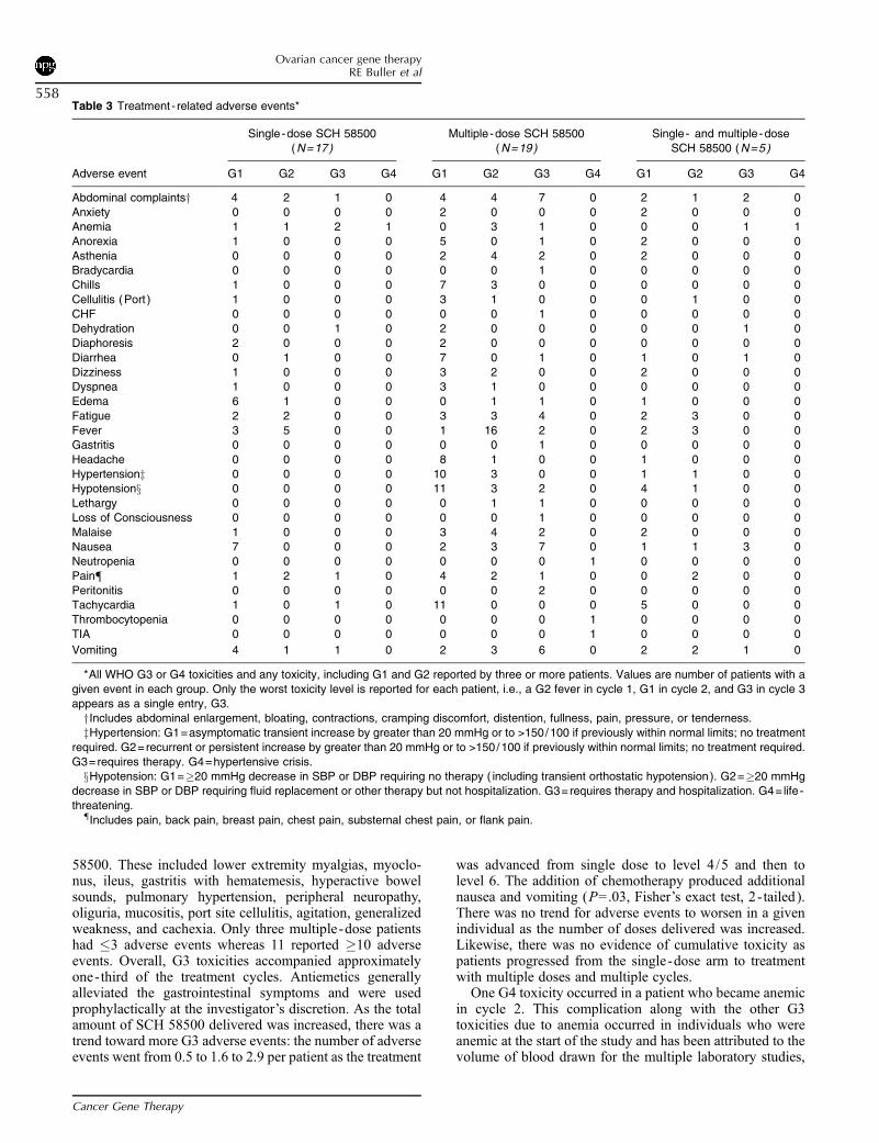
attack. Because adenoviral particles delivered in this studyare more than a log higher than in any previously reportedgene therapy trial, great attention was paid to completereporting of all potential adverse events. From a practicalstandpoint, we have chosen to present all serious G3 or G4events, but only the G1 and grade 2 (G2) events thatoccurred in three or more treated individuals. This of courseunderreports the total number of minor adverse events. Eachtreatment -related adverse event is recorded as the highest -grade toxicity experienced out of all treatment cyclesreceived by that patient as explained in the legend toTable 3. The events are listed in this table on the basis ofoccurrence in either the single-dose or multiple-dosegroups. To show that there was no cumulative toxicity, wehave listed the five patients who were treated with multiple -dose SCH 58500 after completion of the single-dose portionof the study separately. The most common adverse events inthe single-dose group included fever (47%), nausea (41%),edema (41%), abdominal complaints (41%), and anemia(29%). Seven patients experienced �5 different adverseevents whereas only 1 patient had no adverse events at all.Eight G3 or G4 adverse events were reported in four patients.These included anemia (2:G3; 1:G4), abdominal complaints(1:G3), dehydration (1:G3), pain (1:G3), tachycardia(1:G3), and vomiting (1:G3). There was no unusual toxicityin the two serum antiadenoviral antibody negative patientsdosed at level 1.
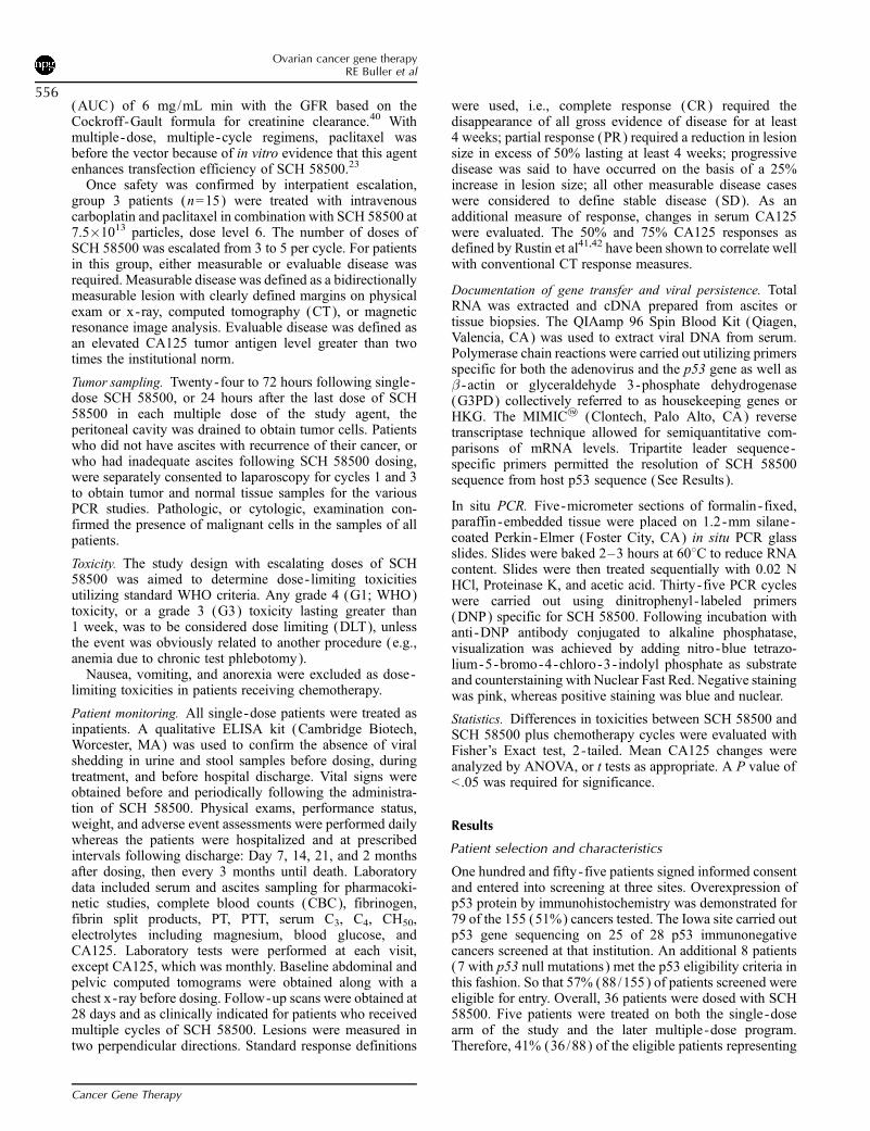
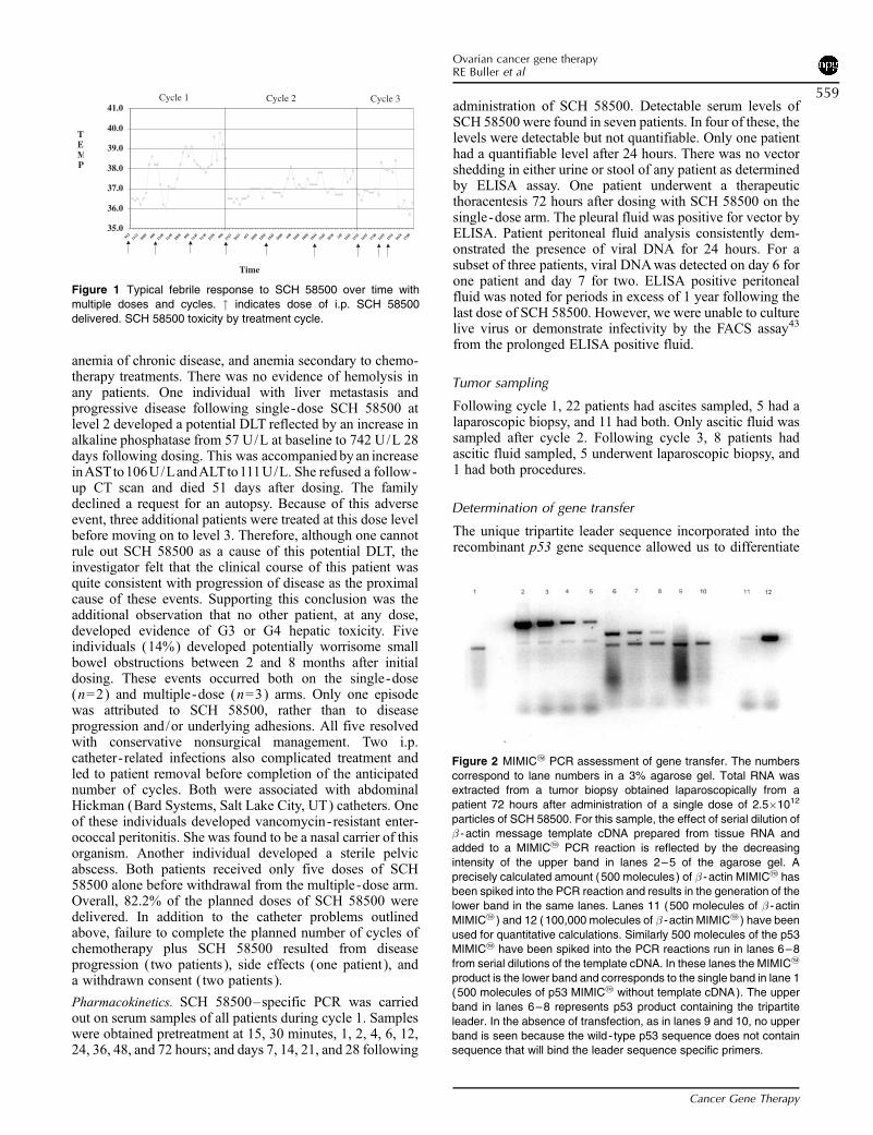
Fever was also the most common adverse event experi-enced by 100% of the multiple -dose patients. This signdeveloped within 2 to 4 hours of dosing. The highest reportedtemperature was 40.58C. Four cycles were accompanied byG3 febrile responses (>408C) among two different patients.After fever was noted in the initial dosing cohorts, patientswere generally given prophylactic acetaminophen. Thesubsequent febrile responses were attenuated, but this mayalso have been due to the steroid premedication given beforechemotherapy for cycles 2 and 3. Figure 1 demonstrates thisobservation graphically for a patient treated at dose level 5. Inthe multidose cohort, the next most frequent signs andsymptoms related to SCH 58500 included hypotension(89%), a variety of abdominal complaints (79%), hyper-tension (68%), nausea (63%), tachycardia (58%), vomiting(58%), and fatigue (53%) — often in the same patient andcycle as the hypertension was noted. The blood pressurechanges prevalent in this group were not seen at all in thesingle-dose group, but they were generally considered mildbecause only one G3 toxicity occurred. All of these mostcommon adverse events, except hypertension, also occurredin 100% of the single-dose patients who reenrolled in themultiple -dose regimen. However, there was no progressionof toxicity grade in those re- treated relative to those initiallytreated at the same dose of SCH 58500.
Forty-seven G3 or G4 toxicities were reported in13 patients who received multiple-dose SCH 58500. Manyof the new WHO G3 toxicities were probably related tochemotherapy because they usually appeared in cycles 2and 3. The patient with congestive heart failure alsodeveloped a G4 neutropenia with concomitant thrombocy-topenia in cycle 3. A few new low-grade adverse eventswere reported when chemotherapy was combined with SCH
Table 2 Study cohort demographics
Primary diagnosis
Ovarian cancer 30
Peritoneal cancer 5Fallopian tube cancer 1
Prior chemotherapy
Mean number of regimens 2.8 range [1–8 ]
Mean treatment cycle 13 range [5–31 ]Mean drugs�cycles 24.4 range [8–78 ]
SCH 58500 treatment
Single dose 17
Multiple dose 24Both 5
Interval: (days)
Diagnosis to SCH 58500 777.7 range [115–2360]
median=630
Platinum- free to SCH58500 first dose*
263 range [37–711]median=261
Disease status
Elevated CA125 33
CT measurable lesionsy>2 cm 22
�2 cm 5
Normal CA125 and CT scan 3
*The platinum- free interval for nine patients was �365 days.yAll CT-measurable disease was accompanied by an elevated
CA125.
Cancer Gene Therapy
Ovarian cancer gene therapyRE Buller et al
557
58500. These included lower extremity myalgias, myoclo-nus, ileus, gastritis with hematemesis, hyperactive bowelsounds, pulmonary hypertension, peripheral neuropathy,oliguria, mucositis, port site cellulitis, agitation, generalizedweakness, and cachexia. Only three multiple-dose patientshad �3 adverse events whereas 11 reported �10 adverseevents. Overall, G3 toxicities accompanied approximatelyone- third of the treatment cycles. Antiemetics generallyalleviated the gastrointestinal symptoms and were usedprophylactically at the investigator’s discretion. As the totalamount of SCH 58500 delivered was increased, there was atrend toward more G3 adverse events: the number of adverseevents went from 0.5 to 1.6 to 2.9 per patient as the treatment
was advanced from single dose to level 4/5 and then tolevel 6. The addition of chemotherapy produced additionalnausea and vomiting (P=.03, Fisher’s exact test, 2- tailed).There was no trend for adverse events to worsen in a givenindividual as the number of doses delivered was increased.Likewise, there was no evidence of cumulative toxicity aspatients progressed from the single-dose arm to treatmentwith multiple doses and multiple cycles.
One G4 toxicity occurred in a patient who became anemicin cycle 2. This complication along with the other G3toxicities due to anemia occurred in individuals who wereanemic at the start of the study and has been attributed to thevolume of blood drawn for the multiple laboratory studies,
Table 3 Treatment - related adverse events*
Single -dose SCH 58500(N=17)
Multiple -dose SCH 58500(N=19)
Single- and multiple -doseSCH 58500 (N=5)
Adverse event G1 G2 G3 G4 G1 G2 G3 G4 G1 G2 G3 G4
Abdominal complaintsy 4 2 1 0 4 4 7 0 2 1 2 0
Anxiety 0 0 0 0 2 0 0 0 2 0 0 0Anemia 1 1 2 1 0 3 1 0 0 0 1 1
Anorexia 1 0 0 0 5 0 1 0 2 0 0 0
Asthenia 0 0 0 0 2 4 2 0 2 0 0 0Bradycardia 0 0 0 0 0 0 1 0 0 0 0 0
Chills 1 0 0 0 7 3 0 0 0 0 0 0
Cellulitis (Port ) 1 0 0 0 3 1 0 0 0 1 0 0
CHF 0 0 0 0 0 0 1 0 0 0 0 0Dehydration 0 0 1 0 2 0 0 0 0 0 1 0
Diaphoresis 2 0 0 0 2 0 0 0 0 0 0 0
Diarrhea 0 1 0 0 7 0 1 0 1 0 1 0
Dizziness 1 0 0 0 3 2 0 0 2 0 0 0Dyspnea 1 0 0 0 3 1 0 0 0 0 0 0
Edema 6 1 0 0 0 1 1 0 1 0 0 0
Fatigue 2 2 0 0 3 3 4 0 2 3 0 0
Fever 3 5 0 0 1 16 2 0 2 3 0 0Gastritis 0 0 0 0 0 0 1 0 0 0 0 0
Headache 0 0 0 0 8 1 0 0 1 0 0 0
Hypertensionz 0 0 0 0 10 3 0 0 1 1 0 0Hypotensionx 0 0 0 0 11 3 2 0 4 1 0 0
Lethargy 0 0 0 0 0 1 1 0 0 0 0 0
Loss of Consciousness 0 0 0 0 0 0 1 0 0 0 0 0
Malaise 1 0 0 0 3 4 2 0 2 0 0 0Nausea 7 0 0 0 2 3 7 0 1 1 3 0
Neutropenia 0 0 0 0 0 0 0 1 0 0 0 0
Pain{ 1 2 1 0 4 2 1 0 0 2 0 0
Peritonitis 0 0 0 0 0 0 2 0 0 0 0 0Tachycardia 1 0 1 0 11 0 0 0 5 0 0 0
Thrombocytopenia 0 0 0 0 0 0 0 1 0 0 0 0
TIA 0 0 0 0 0 0 0 1 0 0 0 0
Vomiting 4 1 1 0 2 3 6 0 2 2 1 0
*All WHO G3 or G4 toxicities and any toxicity, including G1 and G2 reported by three or more patients. Values are number of patients with a
given event in each group. Only the worst toxicity level is reported for each patient, i.e., a G2 fever in cycle 1, G1 in cycle 2, and G3 in cycle 3
appears as a single entry, G3.yIncludes abdominal enlargement, bloating, contractions, cramping discomfort, distention, fullness, pain, pressure, or tenderness.
zHypertension: G1=asymptomatic transient increase by greater than 20 mmHg or to >150/100 if previously within normal limits; no treatment
required. G2=recurrent or persistent increase by greater than 20 mmHg or to >150/100 if previously within normal limits; no treatment required.
G3=requires therapy. G4=hypertensive crisis.xHypotension: G1=�20 mmHg decrease in SBP or DBP requiring no therapy ( including transient orthostatic hypotension). G2=�20 mmHg
decrease in SBP or DBP requiring fluid replacement or other therapy but not hospitalization. G3=requires therapy and hospitalization. G4= life -
threatening.{Includes pain, back pain, breast pain, chest pain, substernal chest pain, or flank pain.
Cancer Gene Therapy
Ovarian cancer gene therapyRE Buller et al
558
anemia of chronic disease, and anemia secondary to chemo-therapy treatments. There was no evidence of hemolysis inany patients. One individual with liver metastasis andprogressive disease following single-dose SCH 58500 atlevel 2 developed a potential DLT reflected by an increase inalkaline phosphatase from 57 U/L at baseline to 742 U/L 28days following dosing. This was accompanied by an increaseinASTto106U/LandALTto111U/L. She refused a follow-up CT scan and died 51 days after dosing. The familydeclined a request for an autopsy. Because of this adverseevent, three additional patients were treated at this dose levelbefore moving on to level 3. Therefore, although one cannotrule out SCH 58500 as a cause of this potential DLT, theinvestigator felt that the clinical course of this patient wasquite consistent with progression of disease as the proximalcause of these events. Supporting this conclusion was theadditional observation that no other patient, at any dose,developed evidence of G3 or G4 hepatic toxicity. Fiveindividuals (14%) developed potentially worrisome smallbowel obstructions between 2 and 8 months after initialdosing. These events occurred both on the single-dose(n=2) and multiple-dose (n=3) arms. Only one episodewas attributed to SCH 58500, rather than to diseaseprogression and/or underlying adhesions. All five resolvedwith conservative nonsurgical management. Two i.p.catheter- related infections also complicated treatment andled to patient removal before completion of the anticipatednumber of cycles. Both were associated with abdominalHickman (Bard Systems, Salt Lake City, UT) catheters. Oneof these individuals developed vancomycin-resistant enter-ococcal peritonitis. She was found to be a nasal carrier of thisorganism. Another individual developed a sterile pelvicabscess. Both patients received only five doses of SCH58500 alone before withdrawal from the multiple -dose arm.Overall, 82.2% of the planned doses of SCH 58500 weredelivered. In addition to the catheter problems outlinedabove, failure to complete the planned number of cycles ofchemotherapy plus SCH 58500 resulted from diseaseprogression ( two patients ), side effects (one patient ), anda withdrawn consent ( two patients ).
Pharmacokinetics. SCH 58500–specific PCR was carriedout on serum samples of all patients during cycle 1. Sampleswere obtained pretreatment at 15, 30 minutes, 1, 2, 4, 6, 12,24, 36, 48, and 72 hours; and days 7, 14, 21, and 28 following
administration of SCH 58500. Detectable serum levels ofSCH 58500 were found in seven patients. In four of these, thelevels were detectable but not quantifiable. Only one patienthad a quantifiable level after 24 hours. There was no vectorshedding in either urine or stool of any patient as determinedby ELISA assay. One patient underwent a therapeuticthoracentesis 72 hours after dosing with SCH 58500 on thesingle-dose arm. The pleural fluid was positive for vector byELISA. Patient peritoneal fluid analysis consistently dem-onstrated the presence of viral DNA for 24 hours. For asubset of three patients, viral DNAwas detected on day 6 forone patient and day 7 for two. ELISA positive peritonealfluid was noted for periods in excess of 1 year following thelast dose of SCH 58500. However, we were unable to culturelive virus or demonstrate infectivity by the FACS assay43
from the prolonged ELISA positive fluid.
Tumor sampling
Following cycle 1, 22 patients had ascites sampled, 5 had alaparoscopic biopsy, and 11 had both. Only ascitic fluid wassampled after cycle 2. Following cycle 3, 8 patients hadascitic fluid sampled, 5 underwent laparoscopic biopsy, and1 had both procedures.
Determination of gene transfer
The unique tripartite leader sequence incorporated into therecombinant p53 gene sequence allowed us to differentiate
35.0
36.0
37.0
38.0
39.0
40.0
41.0
952
1122
2000 80
011
4012
4020
00 800
1145
1230
2100 90
019
2120
21 651
2000
1200
1845
2000 60
016
0010
0018
4419
4520
30 245
1445
1535
1655
1740
1455
1555
1624
1709
TEMP
Cycle 2Cycle 1
Time
Cycle 3
Figure 1 Typical febrile response to SCH 58500 over time with
multiple doses and cycles. " indicates dose of i.p. SCH 58500
delivered. SCH 58500 toxicity by treatment cycle.
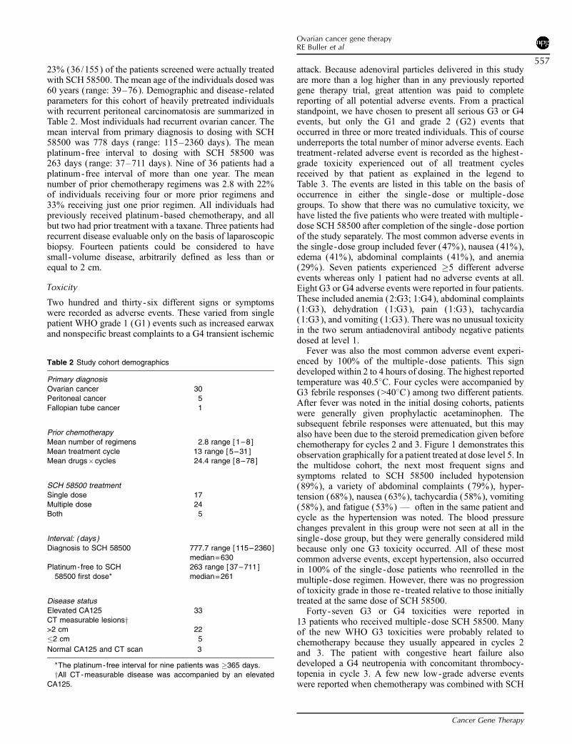
Figure 2 MIMIC2 PCR assessment of gene transfer. The numbers
correspond to lane numbers in a 3% agarose gel. Total RNA wasextracted from a tumor biopsy obtained laparoscopically from a
patient 72 hours after administration of a single dose of 2.5�1012
particles of SCH 58500. For this sample, the effect of serial dilution of� -actin message template cDNA prepared from tissue RNA and
added to a MIMIC2 PCR reaction is reflected by the decreasing
intensity of the upper band in lanes 2–5 of the agarose gel. A
precisely calculated amount (500 molecules) of � -actin MIMIC2 hasbeen spiked into the PCR reaction and results in the generation of the
lower band in the same lanes. Lanes 11 (500 molecules of � -actin
MIMIC2 ) and 12 (100,000 molecules of � -actin MIMIC2 ) have been
used for quantitative calculations. Similarly 500 molecules of the p53MIMIC2 have been spiked into the PCR reactions run in lanes 6–8
from serial dilutions of the template cDNA. In these lanes the MIMIC2
product is the lower band and corresponds to the single band in lane 1(500 molecules of p53 MIMIC2 without template cDNA). The upper
band in lanes 6–8 represents p53 product containing the tripartite
leader. In the absence of transfection, as in lanes 9 and 10, no upper
band is seen because the wild - type p53 sequence does not containsequence that will bind the leader sequence specific primers.
Cancer Gene Therapy
Ovarian cancer gene therapyRE Buller et al
559
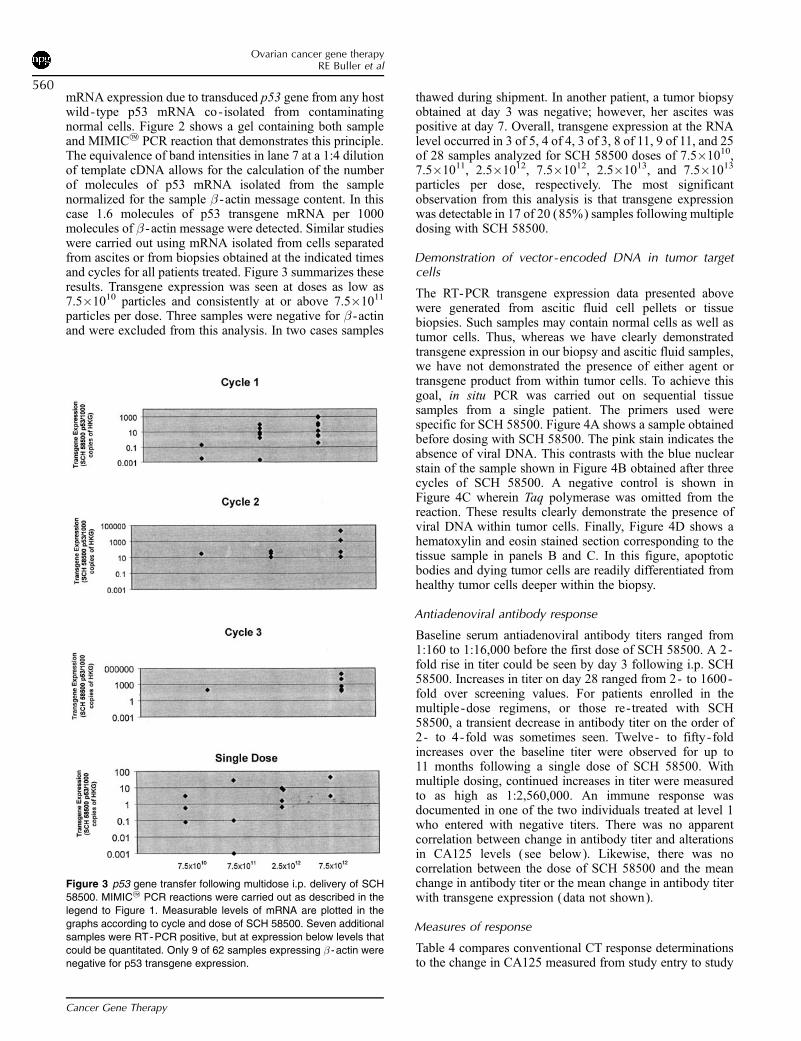
mRNA expression due to transduced p53 gene from any hostwild- type p53 mRNA co-isolated from contaminatingnormal cells. Figure 2 shows a gel containing both sampleand MIMIC2 PCR reaction that demonstrates this principle.The equivalence of band intensities in lane 7 at a 1:4 dilutionof template cDNA allows for the calculation of the numberof molecules of p53 mRNA isolated from the samplenormalized for the sample � -actin message content. In thiscase 1.6 molecules of p53 transgene mRNA per 1000molecules of � -actin message were detected. Similar studieswere carried out using mRNA isolated from cells separatedfrom ascites or from biopsies obtained at the indicated timesand cycles for all patients treated. Figure 3 summarizes theseresults. Transgene expression was seen at doses as low as7.5�1010 particles and consistently at or above 7.5�1011
particles per dose. Three samples were negative for � -actinand were excluded from this analysis. In two cases samples
thawed during shipment. In another patient, a tumor biopsyobtained at day 3 was negative; however, her ascites waspositive at day 7. Overall, transgene expression at the RNAlevel occurred in 3 of 5, 4 of 4, 3 of 3, 8 of 11, 9 of 11, and 25of 28 samples analyzed for SCH 58500 doses of 7.5�1010,7.5�1011, 2.5�1012, 7.5�1012, 2.5�1013, and 7.5�1013
particles per dose, respectively. The most significantobservation from this analysis is that transgene expressionwas detectable in 17 of 20 (85%) samples following multipledosing with SCH 58500.
Demonstration of vector-encoded DNA in tumor targetcells
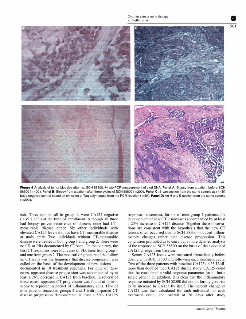
The RT-PCR transgene expression data presented abovewere generated from ascitic fluid cell pellets or tissuebiopsies. Such samples may contain normal cells as well astumor cells. Thus, whereas we have clearly demonstratedtransgene expression in our biopsy and ascitic fluid samples,we have not demonstrated the presence of either agent ortransgene product from within tumor cells. To achieve thisgoal, in situ PCR was carried out on sequential tissuesamples from a single patient. The primers used werespecific for SCH 58500. Figure 4A shows a sample obtainedbefore dosing with SCH 58500. The pink stain indicates theabsence of viral DNA. This contrasts with the blue nuclearstain of the sample shown in Figure 4B obtained after threecycles of SCH 58500. A negative control is shown inFigure 4C wherein Taq polymerase was omitted from thereaction. These results clearly demonstrate the presence ofviral DNA within tumor cells. Finally, Figure 4D shows ahematoxylin and eosin stained section corresponding to thetissue sample in panels B and C. In this figure, apoptoticbodies and dying tumor cells are readily differentiated fromhealthy tumor cells deeper within the biopsy.
Antiadenoviral antibody response
Baseline serum antiadenoviral antibody titers ranged from1:160 to 1:16,000 before the first dose of SCH 58500. A 2-fold rise in titer could be seen by day 3 following i.p. SCH58500. Increases in titer on day 28 ranged from 2- to 1600-fold over screening values. For patients enrolled in themultiple -dose regimens, or those re- treated with SCH58500, a transient decrease in antibody titer on the order of2- to 4-fold was sometimes seen. Twelve- to fifty -foldincreases over the baseline titer were observed for up to11 months following a single dose of SCH 58500. Withmultiple dosing, continued increases in titer were measuredto as high as 1:2,560,000. An immune response wasdocumented in one of the two individuals treated at level 1who entered with negative titers. There was no apparentcorrelation between change in antibody titer and alterationsin CA125 levels (see below). Likewise, there was nocorrelation between the dose of SCH 58500 and the meanchange in antibody titer or the mean change in antibody titerwith transgene expression (data not shown).
Measures of response
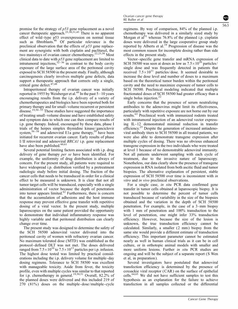
Table 4 compares conventional CT response determinationsto the change in CA125 measured from study entry to study
Figure 3 p53 gene transfer following multidose i.p. delivery of SCH
58500. MIMIC2 PCR reactions were carried out as described in thelegend to Figure 1. Measurable levels of mRNA are plotted in the
graphs according to cycle and dose of SCH 58500. Seven additional
samples were RT-PCR positive, but at expression below levels that
could be quantitated. Only 9 of 62 samples expressing � -actin werenegative for p53 transgene expression.
Cancer Gene Therapy
Ovarian cancer gene therapyRE Buller et al
560
exit. Three tumors, all in group 1, were CA125 negative( <35 U/dL) at the time of enrollment. Although all threehad biopsy-proven recurrence of disease, none had CT-measurable disease either. Six other individuals withelevated CA125 levels did not have CT-measurable diseaseat study entry. Two individuals without CT-measurabledisease were treated in both group 1 and group 2. There wereno CR or PRs documented by CT scan. On the contrary, thebest CT responses were four cases of SD, three from group 1and one from group 2. The most striking feature of the followup CT scans was the frequency that disease progression wascalled on the basis of the development of new lesions —documented in 18 treatment regimens. For nine of thesecases, apparent disease progression was accompanied by atleast a 26% decrease in CA125 from baseline. In several ofthese cases, apparent CT progression was found at laparo-scopy to represent a pocket of inflammatory cells. Five ofnine patients treated in groups 2 and 3 with purported CTdisease progression demonstrated at least a 50% CA125
response. In contrast, for six of nine group 1 patients, thedevelopment of new CT lesions was accompanied by at leasta 25% increase in CA125 disease. Together these observa-tions are consistent with the hypothesis that the new CTlesions often occurred due to SCH 58500–induced inflam-matory changes rather than disease progression. Thisconclusion prompted us to carry out a more detailed analysisof the response to SCH 58500 on the basis of the associatedCA125 change from baseline.
Serum CA125 levels were measured immediately beforedosing with SCH 58500 and following each treatment cycle.Two of the three patients with baseline CA125s <35 U/dLmore than doubled their CA125 during study. CA125 couldthus be considered a valid response parameter for all but asingle patient. In addition, it is clear that the inflammatoryresponse initiated by SCH 58500 did not uniformly give riseto an increase in CA125 by itself. The percent change inCA125 was then calculated for each individual for eachtreatment cycle, and overall at 28 days after study
Figure 4 Analysis of tumor biopsies after i.p. SCH 58500. In situ PCR measurement of viral DNA. Panel A: Biopsy from a patient before SCH
58500 (�400). Panel B: Biopsy from a patient after three cycles of SCH 58500 (�200). Panel C: 5-�m section from the same sample as (4 -B)but a negative control based on omission of Taq polymerase from the PCR reaction (�40). Panel D: An H and E section from the same sample
(�200).
Cancer Gene Therapy
Ovarian cancer gene therapyRE Buller et al
561
completion. Comparison of CA125 levels following treat-ment with SCH 58500 alone at �2.5�1011 particles /dose totreatment at �2.5�1012 particles /dose demonstrated a meanincrease in serum CA125 of 94% versus a mean decreaseof 8.5% at the higher dose (P=.07, 2- tailed, unequalvariances). Thus, SCH 58500 alone at higher doses provides
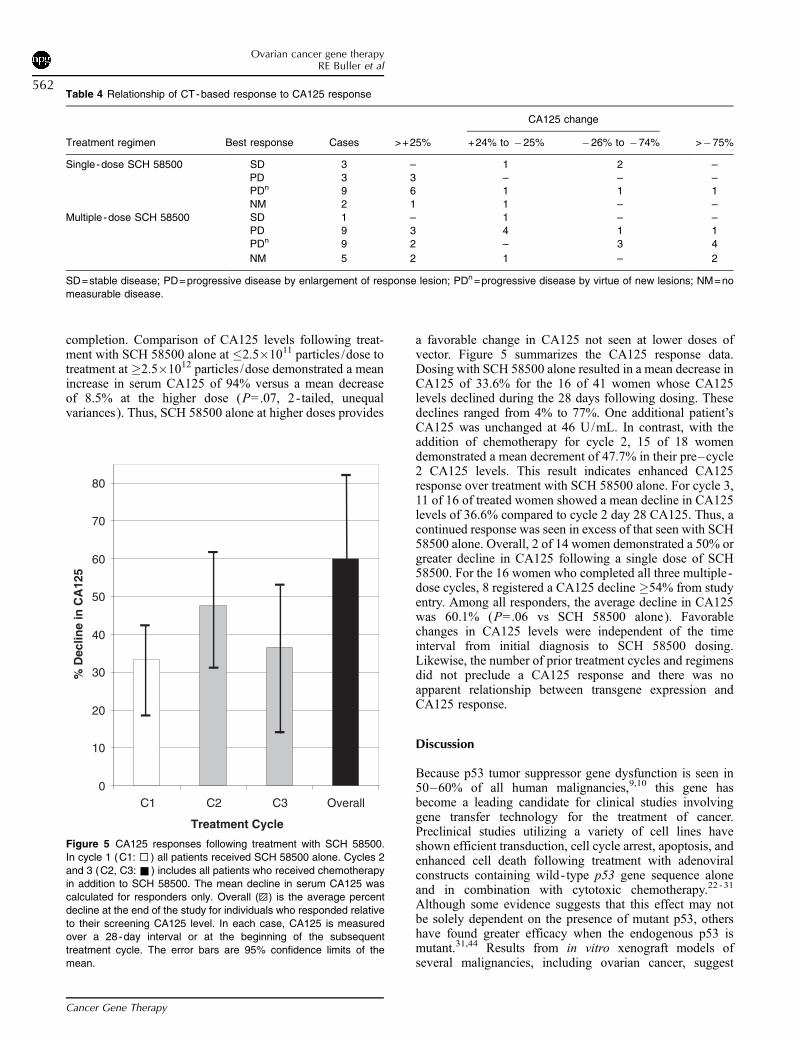
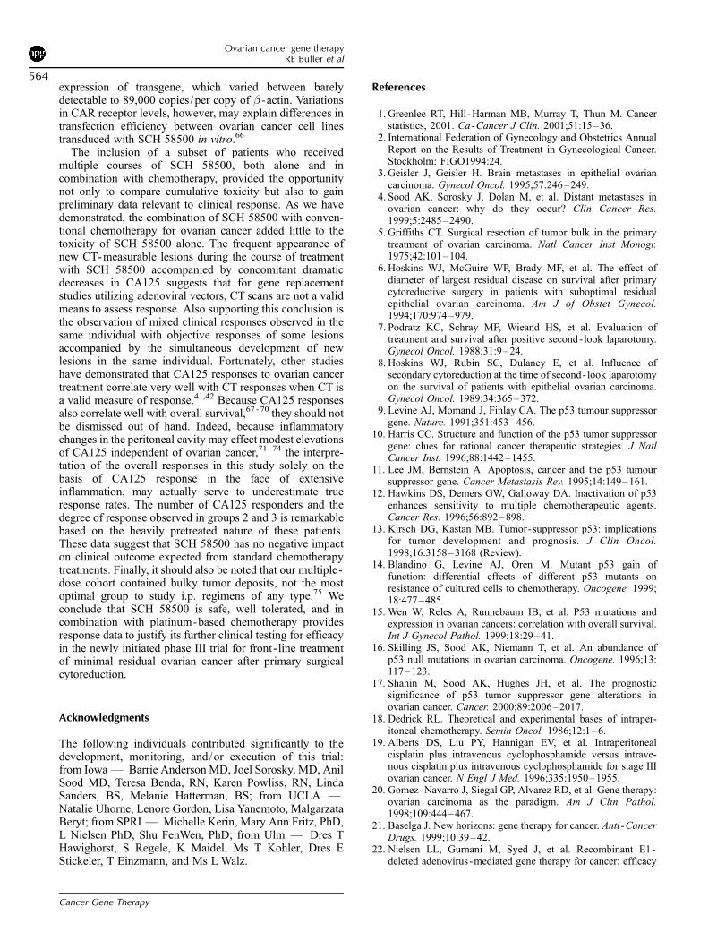
a favorable change in CA125 not seen at lower doses ofvector. Figure 5 summarizes the CA125 response data.Dosing with SCH 58500 alone resulted in a mean decrease inCA125 of 33.6% for the 16 of 41 women whose CA125levels declined during the 28 days following dosing. Thesedeclines ranged from 4% to 77%. One additional patient’sCA125 was unchanged at 46 U/mL. In contrast, with theaddition of chemotherapy for cycle 2, 15 of 18 womendemonstrated a mean decrement of 47.7% in their pre–cycle2 CA125 levels. This result indicates enhanced CA125response over treatment with SCH 58500 alone. For cycle 3,11 of 16 of treated women showed a mean decline in CA125levels of 36.6% compared to cycle 2 day 28 CA125. Thus, acontinued response was seen in excess of that seen with SCH58500 alone. Overall, 2 of 14 women demonstrated a 50% orgreater decline in CA125 following a single dose of SCH58500. For the 16 women who completed all three multiple -dose cycles, 8 registered a CA125 decline �54% from studyentry. Among all responders, the average decline in CA125was 60.1% (P=.06 vs SCH 58500 alone). Favorablechanges in CA125 levels were independent of the timeinterval from initial diagnosis to SCH 58500 dosing.Likewise, the number of prior treatment cycles and regimensdid not preclude a CA125 response and there was noapparent relationship between transgene expression andCA125 response.
Discussion
Because p53 tumor suppressor gene dysfunction is seen in50–60% of all human malignancies,9,10 this gene hasbecome a leading candidate for clinical studies involvinggene transfer technology for the treatment of cancer.Preclinical studies utilizing a variety of cell lines haveshown efficient transduction, cell cycle arrest, apoptosis, andenhanced cell death following treatment with adenoviralconstructs containing wild- type p53 gene sequence aloneand in combination with cytotoxic chemotherapy.22 -31
Although some evidence suggests that this effect may notbe solely dependent on the presence of mutant p53, othershave found greater efficacy when the endogenous p53 ismutant.31,44 Results from in vitro xenograft models ofseveral malignancies, including ovarian cancer, suggest
0
10
20
30
40
50
60
70
80
C1 C2 C3 Overall
Treatment Cycle
% D
eclin
e in
CA
125
Figure 5 CA125 responses following treatment with SCH 58500.
In cycle 1 (C1: 5 ) all patients received SCH 58500 alone. Cycles 2
and 3 (C2, C3: & ) includes all patients who received chemotherapyin addition to SCH 58500. The mean decline in serum CA125 was
calculated for responders only. Overall (E ) is the average percent
decline at the end of the study for individuals who responded relative
to their screening CA125 level. In each case, CA125 is measuredover a 28-day interval or at the beginning of the subsequent
treatment cycle. The error bars are 95% confidence limits of the
mean.
Table 4 Relationship of CT-based response to CA125 response
CA125 change
Treatment regimen Best response Cases >+25% +24% to 25% 26% to 74% >75%
Single -dose SCH 58500 SD 3 – 1 2 –
PD 3 3 – – –PDn 9 6 1 1 1
NM 2 1 1 – –
Multiple -dose SCH 58500 SD 1 – 1 – –PD 9 3 4 1 1
PDn 9 2 – 3 4
NM 5 2 1 – 2
SD=stable disease; PD=progressive disease by enlargement of response lesion; PDn =progressive disease by virtue of new lesions; NM=nomeasurable disease.
Cancer Gene Therapy
Ovarian cancer gene therapyRE Buller et al
562
promise for the strategy of p53 gene replacement as a novelcancer therapeutic approach.25,30,31,45 There is no apparenteffect of wild- type p53 overexpression on normal tissuesuch as fibroblasts.46 Of particular relevance is thepreclinical observation that the effects of p53 gene replace-ment are synergistic with both cisplatin and paclitaxel, thetwo mainstays of ovarian cancer chemotherapy.22,23,30 Mostclinical data to date with p53 gene replacement are limited tointratumoral injections,32 -36 in contrast to the body cavityexposure of the large surface area of the peritoneal cavityexposed to SCH 58500 in the present study. Finally, althoughcarcinogenesis clearly involves multiple gene defects, datasupport a therapeutic approach that corrects only a single,critical gene defect.47,48
Intraperitoneal therapy of ovarian cancer was initiallyreported in 1955 byWeisberger et al.49 In the past 5–10 yearsencouraging results from the i.p. delivery of a variety ofchemotherapeutics and biologics have been reported both forprimary therapy and for small -volume recurrent or persistentdisease.19,50 -55 These studies have suggested the importanceof treating small -volume disease and have established safetyand symptom data to which one can then compare results ofi.p. gene therapy. Indeed, encouraged by these data, phase Itrials of the herpes simplex thymidine kinase/ganciclovirsystem,56 -58 and adenoviral E1a gene therapy,59 have beeninitiated for recurrent ovarian cancer. Early results of phaseI / II retroviral and adenoviral BRCA1 i.p. gene replacementhave also been published.60,61
Several potential limiting factors associated with i.p. drugdelivery of gene therapy per se have been identified. Forexample, the uniformity of drug distribution is always ofconcern. For the present study, all patients were required tohave widespread i.p. distribution verified by a pretreatmentradiologic study before initial dosing. The fraction of thecancer cells that needs to be transduced in order for a clinicaleffect to be measured is unknown. It is clear that not alltumor target cells will be transduced, especially with a singleadministration of vector because the depth of penetrationinto tumor appears limited.62 Furthermore, there is concernthat the accumulation of adhesions and the host immuneresponse may prevent effective gene transfer with repetitivedosing of a viral vector. In the present study, multiplelaparoscopies on the same patient provided the opportunityto demonstrate that individual inflammatory response washighly variable and that peritoneal distribution can clearlychange over time.
The present study was designed to determine the safety ofthe SCH 58500 adenoviral vector delivered into theperitoneal cavity of women with refractory ovarian cancer.No maximum tolerated dose (MTD) was established as theprotocol-defined DLT was not met. The doses deliveredranged from 7.5�1010 to 7.5�1013 particles per i.p. infusion.The highest dose tested was limited by practical consid-erations including the i.p. delivery volume for multiple-daydosing regimens. Tolerance to SCH 58500 was excellentwith manageable toxicity. Aside from fever, the toxicityprofile, even with multiple cycles was similar to that reportedfor i.p. chemotherapy in general.19,50,51 Overall, 82.2% ofthe planned doses were delivered and this included 219 of270 (81%) doses on the multiple-dose/multiple-cycle
regimens. By way of comparison, 84% of the planned i.p.chemotherapy was delivered in a similarly sized study byMorgan et al51 whereas 76.8% of the planned i.p. cisplatindoses were delivered in the large cooperative group studyreported by Alberts et al.19 Progression of disease was themost common reason for incomplete dosing rather than sideeffects in the present study.
Vector-specific gene transfer and mRNA expression ofSCH 58500 was seen at doses as low as 7.5�1010 particles /single dose and was frequently detected in patients thatreceived 7.5�1011 particles /dose. It seemed desirable toincrease the dose level and number of doses to a maximumbased on the theoretical tumor burden within the peritonealcavity and the need to maximize exposure of tumor cells toSCH 58500. Preclinical modeling indicated that multiplefractionated doses of SCH 58500 had greater efficacy than asingle bolus injection.22
Early concerns that the presence of serum neutralizingantibodies to the adenovirus might limit its effectiveness,particularly with repetitive exposure are not borne out by ourresults.63 Preclinical work with immunized rodents treatedwith intratumoral injection of an adenoviral vector express-ing IL-12 demonstrated minimal reduction in transferefficiency.25 Despite the generation of increased antiadeno-viral antibody titers to SCH 58500 in all treated patients, wewere also able to demonstrate transgene expression aftermultiple cycles of dosing. There was no obvious enhancedtransgene expression in the two individuals who were treatedat level 1 because of no demonstrable adenoviral immunity.Not all patients underwent sampling with each cycle oftreatment, due to the invasive nature of laparoscopy.Nonetheless, our data clearly show the presence of transgeneexpression in RNA isolated from both ascitic fluid and tumorbiopsies. The alternative explanation of persistent, stableexpression of SCH 58500 over time is inconsistent with invitro and in vivo preclinical observations.
For a single case, in situ PCR data confirmed genetransfer in tumor cells obtained at laparoscopic biopsy. It isnot possible to determine the percent of tumor cellstransduced because of variability in the size of the biopsiesobtained and the variation in the depth of SCH 58500penetration. For example, in the case of a 3-mm biopsywith 1 mm of penetration and 100% transduction to thelevel of penetration, one might infer 33% transductionefficiency. However, because the size of the lesion isunknown, the true transduction efficiency cannot becalculated. Similarly, a smaller (2 mm) biopsy from thesame site would provide a different estimate of transductionefficiency. This important parameter cannot be estimatednearly as well in human clinical trials as it can be in cellculture, or in orthotopic animal models with smaller andmore uniform lesions. Further in situ PCR studies areongoing and will be the subject of a separate report (S Wenet al, in preparation).
Several investigators have postulated that adenoviraltransfection efficiency is determined by the presence ofcoxsackie viral receptor (CAR) on the surface of epithelialcells.64,65 We did not have sufficient samples to test thishypothesis as an explanation for the failure to achievetransfection in all samples collected or the differential
Cancer Gene Therapy
Ovarian cancer gene therapyRE Buller et al
563
expression of transgene, which varied between barelydetectable to 89,000 copies /per copy of � -actin. Variationsin CAR receptor levels, however, may explain differences intransfection efficiency between ovarian cancer cell linestransduced with SCH 58500 in vitro.66
The inclusion of a subset of patients who receivedmultiple courses of SCH 58500, both alone and incombination with chemotherapy, provided the opportunitynot only to compare cumulative toxicity but also to gainpreliminary data relevant to clinical response. As we havedemonstrated, the combination of SCH 58500 with conven-tional chemotherapy for ovarian cancer added little to thetoxicity of SCH 58500 alone. The frequent appearance ofnew CT-measurable lesions during the course of treatmentwith SCH 58500 accompanied by concomitant dramaticdecreases in CA125 suggests that for gene replacementstudies utilizing adenoviral vectors, CT scans are not a validmeans to assess response. Also supporting this conclusion isthe observation of mixed clinical responses observed in thesame individual with objective responses of some lesionsaccompanied by the simultaneous development of newlesions in the same individual. Fortunately, other studieshave demonstrated that CA125 responses to ovarian cancertreatment correlate very well with CT responses when CT isa valid measure of response.41,42 Because CA125 responsesalso correlate well with overall survival,67 - 70 they should notbe dismissed out of hand. Indeed, because inflammatorychanges in the peritoneal cavity may effect modest elevationsof CA125 independent of ovarian cancer,71 - 74 the interpre-tation of the overall responses in this study solely on thebasis of CA125 response in the face of extensiveinflammation, may actually serve to underestimate trueresponse rates. The number of CA125 responders and thedegree of response observed in groups 2 and 3 is remarkablebased on the heavily pretreated nature of these patients.These data suggest that SCH 58500 has no negative impacton clinical outcome expected from standard chemotherapytreatments. Finally, it should also be noted that our multiple-dose cohort contained bulky tumor deposits, not the mostoptimal group to study i.p. regimens of any type.75 Weconclude that SCH 58500 is safe, well tolerated, and incombination with platinum-based chemotherapy providesresponse data to justify its further clinical testing for efficacyin the newly initiated phase III trial for front - line treatmentof minimal residual ovarian cancer after primary surgicalcytoreduction.
Acknowledgments
The following individuals contributed significantly to thedevelopment, monitoring, and/or execution of this trial:from Iowa — Barrie Anderson MD, Joel Sorosky, MD, AnilSood MD, Teresa Benda, RN, Karen Powliss, RN, LindaSanders, BS, Melanie Hatterman, BS; from UCLA —Natalie Uhorne, Lenore Gordon, Lisa Yanemoto, MalgarzataBeryt; from SPRI — Michelle Kerin, Mary Ann Fritz, PhD,L Nielsen PhD, Shu FenWen, PhD; from Ulm — Dres THawighorst, S Regele, K Maidel, Ms T Kohler, Dres EStickeler, T Einzmann, and Ms L Walz.
References
1. Greenlee RT, Hill -Harman MB, Murray T, Thun M. Cancerstatistics, 2001. Ca-Cancer J Clin. 2001;51:15–36.
2. International Federation of Gynecology and Obstetrics AnnualReport on the Results of Treatment in Gynecological Cancer.Stockholm: FIGO1994:24.
3. Geisler J, Geisler H. Brain metastases in epithelial ovariancarcinoma. Gynecol Oncol. 1995;57:246–249.
4. Sood AK, Sorosky J, Dolan M, et al. Distant metastases inovarian cancer: why do they occur? Clin Cancer Res.1999;5:2485–2490.
5. Griffiths CT. Surgical resection of tumor bulk in the primarytreatment of ovarian carcinoma. Natl Cancer Inst Monogr.1975;42:101–104.
6. Hoskins WJ, McGuire WP, Brady MF, et al. The effect ofdiameter of largest residual disease on survival after primarycytoreductive surgery in patients with suboptimal residualepithelial ovarian carcinoma. Am J of Obstet Gynecol.1994;170:974–979.
7. Podratz KC, Schray MF, Wieand HS, et al. Evaluation oftreatment and survival after positive second- look laparotomy.Gynecol Oncol. 1988;31:9–24.
8. Hoskins WJ, Rubin SC, Dulaney E, et al. Influence ofsecondary cytoreduction at the time of second- look laparotomyon the survival of patients with epithelial ovarian carcinoma.Gynecol Oncol. 1989;34:365–372.
9. Levine AJ, Momand J, Finlay CA. The p53 tumour suppressorgene. Nature. 1991;351:453–456.
10. Harris CC. Structure and function of the p53 tumor suppressorgene: clues for rational cancer therapeutic strategies. J NatlCancer Inst. 1996;88:1442–1455.
11. Lee JM, Bernstein A. Apoptosis, cancer and the p53 tumoursuppressor gene. Cancer Metastasis Rev. 1995;14:149–161.
12. Hawkins DS, Demers GW, Galloway DA. Inactivation of p53enhances sensitivity to multiple chemotherapeutic agents.Cancer Res. 1996;56:892–898.
13. Kirsch DG, Kastan MB. Tumor-suppressor p53: implicationsfor tumor development and prognosis. J Clin Oncol.1998;16:3158–3168 (Review).
14. Blandino G, Levine AJ, Oren M. Mutant p53 gain offunction: differential effects of different p53 mutants onresistance of cultured cells to chemotherapy. Oncogene. 1999;18:477–485.
15. Wen W, Reles A, Runnebaum IB, et al. P53 mutations andexpression in ovarian cancers: correlation with overall survival.Int J Gynecol Pathol. 1999;18:29–41.
16. Skilling JS, Sood AK, Niemann T, et al. An abundance ofp53 null mutations in ovarian carcinoma. Oncogene. 1996;13:117–123.
17. Shahin M, Sood AK, Hughes JH, et al. The prognosticsignificance of p53 tumor suppressor gene alterations inovarian cancer. Cancer. 2000;89:2006–2017.
18. Dedrick RL. Theoretical and experimental bases of intraper-itoneal chemotherapy. Semin Oncol. 1986;12:1–6.
19. Alberts DS, Liu PY, Hannigan EV, et al. Intraperitonealcisplatin plus intravenous cyclophosphamide versus intrave-nous cisplatin plus intravenous cyclophosphamide for stage IIIovarian cancer. N Engl J Med. 1996;335:1950–1955.
20. Gomez-Navarro J, Siegal GP, Alvarez RD, et al. Gene therapy:ovarian carcinoma as the paradigm. Am J Clin Pathol.1998;109:444–467.
21. Baselga J. New horizons: gene therapy for cancer. Anti -CancerDrugs. 1999;10:39–42.
22. Nielsen LL, Gurnani M, Syed J, et al. Recombinant E1-deleted adenovirus -mediated gene therapy for cancer: efficacy
Cancer Gene Therapy
Ovarian cancer gene therapyRE Buller et al
564
studies with p53 tumor suppressor gene and liver histologyin mouse tumor xenograft models. Hum Gene Ther. 1998;9:681–94.
23. Nielsen LL, Lipari P, Dell J, et al. Adenovirus -mediated p53gene therapy and paclitaxel have synergistic efficacy in modelsof human head and neck, ovarian, prostate, and breast cancer.Clin Cancer Res. 1998;4:835–846.
24. Nielsen LL, Dell J, Maxwell E, et al. Efficacy of p53adenovirus -mediated gene therapy against human breastcancer xenografts. Cancer Gene Ther. 1997;4:129–138.
25. Bramson JL, Hitt M, Gauldie J, et al. Pre -existing immunity toadenovirus does not prevent tumor regression followingintratumoral administration of a vector expressing IL-12 butinhibits virus dissemination. Gene Ther. 1997;4:1069–1076.
26. von Gruenigen VE, Santoso JT, Coleman RL, et al. In vivostudies of adenovirus -based p53 gene therapy for ovariancancer. Gynecol Oncol. 1998;69:197–204.
27. Kim J, Hwang ES, Kim JS, et al. Intraperitoneal gene therapywith adenoviral -mediated p53 tumor suppressor gene forovarian cancer model in nude mouse. Cancer Gene Ther. 1999;6:172–178.
28. Anderson SC, Johnson DE, Harris MP, et al. P53 gene therapyin a rat model for hepatocellular carcinoma: intra -arterialdelivery of a recombinant adenovirus. Clin Cancer Res.1998;4:1649–1659.
29. Gurnani M, Lipari P, Dell J, et al. Adenovirus -mediated p53gene therapy has greater efficacy when combined withchemotherapy against human head and neck, ovarian, prostate,and breast cancer. Cancer Chemother Pharmacol. 1999;44:143–151.
30. Fujiwara T, Grimm EA, Mukhopadhyay T, et al. Induction ofchemosensitivity in human lung cancer cells in vivo byadenovirus -mediated transfer of the wild - type p53 gene.Cancer Res. 1994;54:2287–2291.
31. Zhang W-W, Fang X, Mazur W, et al. High-efficiency genetransfer and high- level expression of wild - type p53 in humanlung cancer cells mediated by recombinant adenovirus. CancerGene Ther. 1994;1:5–13.
32. Roth JA, Nguyen D, Lawrence DD, et al. Retrovirus -mediatedwild - type gene transfer to tumors of patients with lung cancer.Nat Med. 1996;2:985–991.
33. Nemunaitis J, Swisher SG, Timmons T, et al. Adenovirus -mediated p53 gene transfer in sequence with cisplatin to tumorsof patients with non-small -cell lung cancer. J Clin Oncol.2000;18:609–622.
34. Clayman GL, El -Naggar AK, Lippman SM, et al. Adenovirus -mediated p53 gene transfer in patients with advanced recurrenthead and neck squamous cell carcinoma. J Clin Oncol.1998;16:2221–2232.
35. Swisher, SG, Roth JA, Nemunaitis J, et al. Adenovirus -mediated p53 gene transfer in advanced non-small -cell lungcancer. J Natl Cancer Inst. 1999;91:763–771.
36. Cusack JC, Spitz FR, Nguyen D, et al. High levels of genetransduction in human lung tumors following intralesionalinjection of recombinant adenovirus. Cancer Gene Ther.1996;3:245–249.
37. Nielsen LL, Maneval DC. P53 tumor suppressor gene therapyfor cancer. Cancer Gene Ther. 1998;5:52–63.
38. Buller RE, Pegram M, Runnebaum I, et al. A phase I / II trial ofrecombinant adenoviral human p53 (SCH 58500) intraper-itoneal ( IP) gene therapy in recurrent ovarian cancer. GynecolOncol. 1999;72:452–453.
39. Wills KN, Maneval DC, Menzel P, et al. Development andcharacterization of recombinant adenoviruses encodinghuman p53 for gene therapy of cancer. Hum Gen Ther. 1994;5:1079–1088.
40. Cockcroft DW, Gault, MH. Prediction of creatinine clearancefrom serum creatinine. Nephron. 1976;16:31–41.
41. Rustin GJ, Nelstrop AE, McClean P, et al. Defining response ofovarian carcinoma to initial chemotherapy according to serumCA 125. J Clin Oncol. 1996;14:1545–1551.
42. Rustin GJ, Nelstrop AE, Bentzen SM, et al. Use of tumourmarkers in monitoring the course of ovarian cancer. Ann Oncol.1999;10:S21–S27.
43. Musco ML, Cui S, Small D, et al. A comparison of flowcytometry and laser scanning cytometry for the intracellularevaluation of adenoviral infectivity and p53 protein expressionin gene therapy. Cytometry. 1998;33:290–296.
44. Wolf JK, Mills G, Bazzet L, et al. Adenovirus -mediatedp53 growth inhibition of ovarian cancer cells is independentof endogenous p53 status. Gynecol Oncol. 1999;75:261–266.
45. Mujoo K, Maneval DC, Anderson SC, et al. Adenoviral -mediated p53 tumor suppressor gene therapy of human ovariancarcinoma. Oncogene. 1996;12:1617–1623.
46. Liu T-J, el -Naggar AK, McDonnell TJ, et al. Apoptosisinduction mediated by wild - type p53 adenoviral gene transferin squamous cell carcinoma of the head and neck. Cancer Res.1995;55:3117–3122.
47. Chen P-L, Chen Y, Bookstein R, et al. Genetic mechanisms oftumor suppression by the human p53 gene. Science.1990;250:1576–1580.
48. Fujiwara T, Grimm EA, Roth JA. Gene therapeutics and genetherapy for cancer. Curr Opin Oncol. 1994;6:96–105.
49. Weisberger AS, Levine B, Storaasli JP. Use of nitrogen mustardin treatment of serous effusions of neoplastic origin. J Am MedAssoc. 1955;159:1704–1707.
50. Berek JS, Markman M, Stonebraker B, et al. Intraperitonealinterferon -� in residual ovarian carcinoma: a phase IIgynecologic oncology group study. Gynecol Oncol. 1999;75:10–14.
51. Morgan RJ Jr, Braly P, Cecchi G, et al. Phase II trial ofintraperitoneal cisplatin with intravenous doxorubicin andcyclophosphamide in previously untreated patients withadvanced ovarian cancer — long- term follow-up. GynecolOncol. 1999;75:419–426.
52. Barakat RR, Almadrones L, Venkatraman ES, et al. A phase IItrial of intraperitoneal cisplatin and etoposide as consolidationtherapy in patients with stage II– IV epithelial ovarian cancerfollowing negative surgical assessment. Gynecol Oncol.1998;69:17–22.
53. Muggia FM, Liu PY, Alberts DS, et al. Intraperitonealmitoxantrone or floxuridine: effects on time- to - failure andsurvival in patients with minimal residual ovarian cancer aftersecond- look laparotomy — a randomized phase II study bythe Southwest Oncology Group. Gynecol Oncol. 1996;61:395–402.
54. Markman M, Reichman B, Hakes T, et al. Impact on survival ofsurgically defined favorable responses to salvage intraperito-neal chemotherapy in small -volume residual ovarian cancer.J Clin Oncol. 1992;10:1479–1484.
55. Markman M, Brady MF, Spirtos NM, et al. Phase II trial ofintraperitoneal paclitaxel in carcinoma of the ovary, tube, andperitoneum: a Gynecologic Oncology Group Study. J ClinOncol. 1998;16:2620–2624.
56. Link CJ Jr, Moorman D, Seregina T, et al. A phase I trial of invivo gene therapy with the herpes simplex thymidine kinase /ganciclovir system for the treatment of refractory or recurrentovarian cancer. Hum Gene Ther. 1996;7:1161–1179.
57. Alvarez RD, Curiel DT. A phase I study of recombinantadenovirus vector–mediated intraperitoneal delivery of herpessimplex virus thymidine kinase (HSV-TK) gene and
Cancer Gene Therapy
Ovarian cancer gene therapyRE Buller et al
565
intravenous ganciclovir for previously treated ovarian andextraovarian cancer patients. Hum Gene Ther. 1997;8:597–613.
58. Hasenburg A, Tong XW, Rojas -Martinez A, et al. Thymidinekinase (TK) gene therapy of solid tumors: valacyclovirfacilitates outpatient treatment. Anticancer Res. 1999;19:2163–2165.
59. Hortobagyi, GN, Hung MC, Lopez-Berestein G. A phase Imulticenter study of E1A gene therapy for patients withmetastatic breast cancer and epithelial ovarian cancer thatoverexpresses HER-2/neu or epithelial ovarian cancer. HumGene Ther. 1998;9:1775–1798.
60. Tait DL, Obermiller PS, Redlin -Frazier S, et al. A phase I trialof retroviral BRCA1sv gene therapy in ovarian cancer. ClinCancer Res. 1997;3:1959–1968.
61. Tait DL, Obermiller PS, Hatmaker AR, et al. Ovarian cancerBRCA1 gene therapy: phase I and II trial differences inimmune response and vector stability. Clin Cancer Res. 1999;5:1708–1714.
62. Grace MJ, Xie L, Musco ML, et al. The use of laser scanningcytometry to assess depth of penetration of adenovirus p53gene therapy in human xenograft biopsies. Am J Pathol. 1999;155:1869–1878.
63. Hitt M, Addison CL, Graham FL. Human adenovirus vectorsfor gene transfer into mammalian cells. Adv Pharmacol.1997;40:137–206.
64. Bergelson JM, Cunningham, JA, Droguett G, et al. Isolation ofa common receptor for Coxsackie B viruses and adenoviruses 2and 5. Science. 1997;275:1320–1323.
65. Yingming L, Pong R-C, Bergelson JM, et al. Loss ofadenoviral receptor expression in human bladder cancer cells:a potential impact on the efficacy of gene therapy. Cancer Res.1999;59:325–330.
66. Pegram M, Tseng Y, Baldwin RL, et al. Expression ofcoxsackie and adenovirus receptor (CAR) correlates with
efficiency of adenovirus transduction of human ovariancarcinoma cells. Gynecol Oncol. 1999;72:452.
67. Buller RE, Berman ML, Bloss JD, et al. CA 125 regression: amodel for epithelial ovarian cancer response. Am J ObstetGynecol. 1991;165:360–367.
68. Buller RE, Vasilev S, DiSaia PJ. CA125 kinetics: a costeffective clinical tool to evaluate clinical trial outcomes in the1990s. Am J Obstet Gynecol. 1996;174:1241–1254.
69. van der Burg MEL, Lammes FB, van Putten WLJ, et al.Ovarian cancer: the prognostic value of the serum half - life ofCA125 during induction chemotherapy. Gynecol Oncol. 1988;30:307–312.
70. Rustin GJS, Gennings JN, Nelstrop AE, et al. Use of CA-125to predict survival of patients with ovarian carcinoma. J ClinOncol. 1989;7:1667–1671.
71. Yedema CA, Kenemans P, Thomas CM, et al. CA125 serumlevels in the early post -operative period do not reflect tumourreduction obtained by cytoreductive surgery. Eur J Cancer.1993;29A:966–971.
72. Van Der Zee AG, Duk JM, Aalders JG, et al. The effect ofabdominal surgery on the serum concentration of the tumour-associated antigen CA 125. Br J Obstet Gynaecol. 1990;97:934–938.
73. Talbot RW, Jacobsen DJ, Nagorney DM, et al. Temporaryelevation of CA 125 after abdominal surgical treatment forbenign disease and cancer. Surg Gynecol Obstet. 1989;168:407–412.
74. Zeimet AG, Offner FA, Marth C, et al. Modulation of CA-125release by inflammatory cytokines in human peritonealmesothelial and ovarian cancer cells. Anticancer Res. 1997;17:3129–3131.
75. Howell SB, Zimm S, Markman M, et al. Long- term survival ofadvanced refractory ovarian carcinoma patients with small -volume disease treated with intraperitoneal chemotherapy.J Clin Oncol. 1987;5:1607–1612.
Cancer Gene Therapy
Ovarian cancer gene therapyRE Buller et al
566