A Peer-Review Journal - CiteSeerX
57
INFLAMMATORY BOWEL DISEASE: A PRACTICAL APROACH, SERIES #77 METABOLIC BONE DISEASE IN INFLAMMATORY BOWEL DISEASE by Fayez K. Ghishan, Pawel R. Kiela NUTRITION ISSUES IN GASTROENTEROLOGY, SERIES #109 NUTRITION AND NUTRACEUTICALS FOR MUSCLE MAINTENANCE AND RECOVERY: HERO OR HOKUM? by Joe Krenitsky PRACTICAL APPROACHES TO THE DIAGNOSIS AND TREATMENT OF COLORECTAL CANCER, SERIES #7 FLAT POLYPS: ENDOSCOPIC DETECTION AND TREATMENT by Susan G. Coe, Michael B. Wallace A CASE REPORT MESENTERIC PANNICULTIS PRESENTING AS RECURRENT SMALL BOWEL OBSTRUCTION IN AN ELDERLY MALE by Ashish Kataria, Sanjaya K. Satapathy, Richard Straus, Silvat Sheikh-Fayyaz, Ronald Greenberg A CASE SERIES ENDOSCOPIC CLIP CLOSURE OF BENIGN ESOPHAGOPLEURAL FISTULAS By Kimberly Salaycik Kolkhorst, Eric Hill, Patrick Brady FELLOWS’ CORNER INGESTION OF MULTIPLE MAGNETS by Arieda Gjkopulli, Ritu Walia, Gia Bradley, Kalpana Murthy, David Tuchman A Peer-Review Journal September 2012 Vol. XXXVI No. 9 PRACTICAL GASTROENTEROLOGY www.practicalgastro.com S e p t e m b e r C o n f e r e n c e I s s u e L a s V e g a s , N e v a d a · O c t o b e r 1 7 - 2 2 , 2 0 1 2 A Peer Review Journal PRACTICAL GASTRO
-
Upload
khangminh22 -
Category
Documents
-
view
1 -
download
0
Transcript of A Peer-Review Journal - CiteSeerX

INFLAMMATORY BOWEL DISEASE:A PRACTICAL APROACH, SERIES #77METABOLIC BONE DISEASE IN INFLAMMATORY BOWEL DISEASEby Fayez K. Ghishan, Pawel R. Kiela
NUTRITION ISSUES IN GASTROENTEROLOGY, SERIES #109NUTRITION AND NUTRACEUTICALS FOR MUSCLEMAINTENANCE AND RECOVERY: HERO OR HOKUM?by Joe Krenitsky
PRACTICAL APPROACHES TO THE DIAGNOSISAND TREATMENT OF COLORECTAL CANCER, SERIES #7FLAT POLYPS: ENDOSCOPIC DETECTION AND TREATMENTby Susan G. Coe, Michael B. Wallace
A CASE REPORTMESENTERIC PANNICULTIS PRESENTING AS RECURRENT SMALL BOWEL OBSTRUCTION IN AN ELDERLY MALEby Ashish Kataria, Sanjaya K. Satapathy, Richard Straus, Silvat Sheikh-Fayyaz, Ronald Greenberg
A CASE SERIESENDOSCOPIC CLIP CLOSURE OF BENIGN ESOPHAGOPLEURAL FISTULASBy Kimberly Salaycik Kolkhorst, Eric Hill, Patrick Brady
FELLOWS’ CORNERINGESTION OF MULTIPLE MAGNETS by Arieda Gjkopulli, Ritu Walia, Gia Bradley, Kalpana Murthy, David Tuchman
A Peer-Review JournalSeptember 2012Vol. XXXVI No. 9
PRACTICALGASTROENTEROLOGY
www.practicalgastro.com
September Conference Is
sue
Las
Veg
as, N
evada · October 17-22, 2012
A Peer Review Journal
PRACTICAL
A Peer Review Journal
PRACTICALGASTRO
Cover_September_12.indd 1 9/12/12 10:35 PM
BPA
AMP
Practical Gastroenterology is a professional clinical journal focusing on the diagnosis and management of digestive diseases. Each issue consists of articles on topics that physicians encounter in daily practice.Authors are selected for their expert knowledge of their topics and their participation does not imply endorsement of products advertised. Authors’ opinions expressed in their articles are not necessarily those of Practical Gastroenterology, its publisher, Editorial Board, or its advertisers. Practical Gastroenterology, its publisher, editors, Editorial Board, and its advertisers assume no liability or responsibility for any claims, actions or damages resulting from the publication of any article.AUTHOR INFORMATION: A “Guideline for Authors” is available without charge. For a free copy, e-mail: [email protected] Gastroenterology (ISSN 0277-4208) is published monthly by Practical Gastroenterology Publishing, Inc. Periodical Postage Paid at Westhampton Beach, NY, and additional mailing offices. Postmaster: Send address changes to Practical Gastroenterology, 99B Main Street, Westhampton Beach, New York 11978. Printed in the USA. (631) 288-4404; Fax (631) 288-4435.Contents of Practical Gastroenterology are protected by the U.S. Copyright Law. Reproduction, photocopying, storage or transmission by magnetic or electronic means is strictly prohibited by law. For permission to reuse material from Practical Gastroenterology (ISSN 0277-4208), please access www.copyright.com or contact the Copyright Clearance Center, Inc. (CCC), 222 Rosewood Drive, Danvers, MA 01923, 978-750-8400. CCC is a not-for-profit organization that provides licenses and registration for a variety of uses. Special rates apply for multiple copies when used internally for educa-tional purposes. Violation of copyright will result in legal action, including civil and/or criminal penalties, and suspension of service.ANNUAL SUBSCRIPTION RATES. USA: 1 year $145 US, 2 years $270 US, 3 years $405 US. FOREIGN: 1 year $190 US, 2 years $340 US, 3 years $510 US. Subscription should be mailed to Practical Gastroenterology, 99B Main Street, Westhampton Beach, New York 11978.CHANGE OF ADDRESS: Practical Gastroenterology uses the frequently updated rosters maintained by two national medical associations to mail issues of the journal to physicians who qualify. If you have been receiving Practical Gastroenterology and have had a change of professional address, please make sure to advise the appropriate association of your new address. Membership is not required. For MDs, write: American Medical Association, 515 North State St., Chicago, IL 60610, (800) 262-3211, www.ama-assn.org. For DOs, write: American Osteopathic Association, 212 East Ohio St., Chicago, IL 60611, (800) 621-1773, www.osteopathic.org.CLASSIFIED ADS: For more information on Classified Ads, contact: Practical Gastroenterology Classified Department, 99B Main Street, Westhampton Beach, New York 11978. Phone (631) 288-4404; Fax (631) 288-4435.
EDITORIAL BOARDRad M. Agrawal, M.D. Theodore Bayless, M.D. Henry J. Binder, M.D. H. Worth Boyce, Jr., M.D. R. Keith Campbell, R.Ph. William D. Carey, M.D. Donald O. Castell, M.D. Richard K. Chessler, M.D. Murray H. Cohen, D.O. T.S. Dharmarajan, M.D. Anthony DiMarino, M.D. George E. Dukes, Pharm.D. Dayna Early, M.D. Gerald Friedman, M.D. Justin T. Kupec, M.D. John M. Levey, M.D. Richard W. McCallum, M.D.
Denis M. McCarthy, M.D. George W. Meyer, M.D. Sam A. Nixon, M.D. Kevin W. Olden, M.D. Melissa Palmer, M.D. C.S. Pitchumoni, M.D.John F. Pohl, M.D. Carol Rees Parrish, M.S., R.D. Andrew K. Roorda, M.D. David B. Sachar, M.D. Melvin Schapiro, M.D. Jerome H. Siegel, M.D. Uma Sundaram, M.D. Jorge Valenzuela, M.D. Jon A. Vanderhoof, M.D. Arnold Wald, M.D.
SERIES EDITORSRad M. Agrawal, M.D. Diseases of the Biliary Tract
Diseases of the PancreasD.O. Castell, M.D. GERD in the 21st CenturyT.S. Dharmarajan, M.D. Geriatric Gastroenterology C.S. Pitchumoni, M.D. Jack A. Di Palma, M.D. Foodborne IllnessDayna Early, M.D. Sex-Based Differences in GastroenterologyJorge L. Herrera, M.D. Viral HepatitisMuralidhar Jatla, M.D. Celiac Disease: A Comprehensive Ritu Verma, M.D. Review and UpdateSeymour Katz, M.D. Inflammatory Bowel Disease:
A Practical ApproachJustin T. Kupec, M.D. Case ReportsKevin W. Olden, M.D. Treatment of Irritable Bowel SyndromeMelissa Palmer, M.D. Hepatitis C: A New Era of TreatmentHenry P. Parkman, M.D. G.I. Motility: A Series from the
American Motility SocietyCarol Rees Parrish, M.S., R.D. Nutrition Issues in GastroenterologyAndrew K. Roorda, M.D. Endoscopy: Opening New EyesMelvin Schapiro, M.D. Practical Approaches to the Diagnosis
and Treatment of Colorectal CancerUma Sundaram, M.D. Original Research
Murray H. Cohen, D.O. From the LiteratureC.S. Pitchumoni, M.D. Fellows’ Corner Rad M. Agrawal, M.D. John F. Pohl, M.D. Book Reviews. From the Pediatric Literature Jerome H. Siegel, M.D. Practical EndoscopyChristina M. Surawicz, M.D. Pearls of Gastroenterology
Publisher Vivian M. Mahl
E-mail: [email protected]
Circulation Manager: James D. Green E-mail: [email protected]
Editor: Adrien A. Mahl E-mail: [email protected]
Art Director: Adrien A. Mahl
National Accounts RepresentativePractical Gastroenterology
99B Main Street, Westhampton Beach, NY 11978 Telephone: (631) 288-4404
Web site: www.practicalgastro.com
September 2012Vol. XXXVI No. 9PRACTICAL
GASTROENTEROLOGY
DEPARTMENT EDITORS
Masthead_September_12.indd 5 9/12/12 10:34 PM

September 2012Vol. XXXVI No. 9PRACTICAL
GASTROENTEROLOGY
Nutrition and Nutraceuticals for Muscle Maintenance and Recovery: Hero or Hokum?by Joe KrenitskyMuscle loss during hospitalization, especially during intensive care unit admissions, contributes to muscle weakness, functional limitations and the need for extended rehabilitation services. This review will evaluate the data investigating the potential of nutraceuticals and nutrition strategies to minimize muscle loss and accelerate rehabilitation of muscle mass and strength.
27
INflammatory bowel dISeaSe: a practIcal aproach, SerIeS #77
Metabolic Bone Disease in Inflammatory Bowel Diseaseby Fayez K. Ghishan, Pawel R. Kiela
Chronic inflammatory bowel diseases (IBD) affect bone metabolism and are frequently associated with decreased bone mineral density (BMD) and increased risk of fractures. New studies continue to unravel a complex network of interactions leading to the inflammation-associated loss of BMD, and may help direct treatment of IBD toward more bone-sparing strategies. Understanding the pathophysiology of osteopenia and osteoporosis in Crohn’s disease and ulcerative colitis are critical for the correct choice of available treatments or the development of new targeted therapies.
16
NutrItIoN ISSueS IN gaStroeNterology, SerIeS #109
Flat Polyps: Endoscopic Detection and Treatmentby Susan G. Coe, Michael B. WallaceFlat and serrated polyps are a particular challenge to detect and may explain why some of these cancers occur. When performed by appropriately trained endoscopists in carefully selected patients, endoscopic resection can reduce the risk and morbidity of resection. In this review we define the types of difficult to detect polyps, methods to increase their detection, and describe available endoscopic resection techniques.
49practIcal approacheS to the dIagNoSIS aNd treatmeNt of colorectal caNcer, SerIeS #7
TOC_Sept_12.indd 6 9/17/12 12:26 PM

September 2012Vol. XXXVI No. 9PRACTICAL
GASTROENTEROLOGY
60Endoscopic Clip Closure of Benign Esophagopleural Fistulasby Kimberly Salaycik Kolkhorst, Eric Hill, Patrick Brady
a caSe SerIeS
56Mesenteric Pannicultis Presenting as Recurrent Small Bowel Obstruction in an Elderly Maleby Ashish Kataria, Sanjaya K. Satapathy, Richard Straus, Silvat Sheikh-Fayyaz, Ronald Greenberg
a caSe report
90Fellows’ Cornerby Arieda Gjkopulli, Ritu Walia, Gia Bradley, Kalpana Murthy, David Tuchman
fellowS’ corNer
TOC_Sept_12.indd 9 9/12/12 10:32 PM

DEPARTMENTS
Crossword Puzzle 94
by Myles Mellor
Meetings Calendar 93
Meetings, events, courses, symposia, and their contacts.
Medical Bulletin Board 82
News items of interest to the nation’s gastroenterologists.
From the Pediatric Gastroenterology Literature 74
by John F. Pohl, M.D., editor of “From the Pediatric Gastroenterology Literature” is on the Editorial Board of Practical Gastroenterology.
Book Reviews 68
Recently published books and CD-ROMs in gastroenterology. John F. Pohl, M.D., editor of “Book Reviews,” is on the Editorial Board of Practical Gastroenterology.
From the Literature 76Murray H. Cohen, D.O., “From the Literature” Editor,is on the Editorial Board of Practical Gastroenterology.
Reader Request Fax Form 91
Readers may obtain additional information about products and services that appear in Practical Gastroenterology.
TOC_Sept_12.indd 15 9/12/12 10:33 PM

Inflammatory bowel dIsease: a practIcal approach, serIes #77
16 Practical GastroenteroloGy • sePtember 2012
Seymour Katz, M.D., Series Editor
Inflammatory bowel dIsease: a practIcal approach, serIes #77
Metabolic Bone Disease in Inflammatory Bowel Disease
Fayez K. Ghishan Pawel R. Kiela
Pawel R. Kiela, DVM, Ph.D., Associate Professor of Pediatrics and Immunobiology, Steele Children’s Research Center, University of Arizona. Fayez K. Ghishan, MD, Department of Pediatrics, Steele Children’s Research Center, University of Arizona, Tucson, AZ
Chronic inflammatory bowel diseases (IBD) affect bone metabolism and are frequently associated with decreased bone mineral density (BMD) and increased risk of fractures. Experimental models of IBD and as well as data from pediatric and adult IBD patients do not provide a uniform answer whether the changes in bone metabolism leading to decreased mineral density are the result of decreased bone formation, increased bone desorption, or both. New studies continue to unravel a complex network of interactions leading to the inflammation-associated loss of BMD, and may help direct treatment of IBD toward more bone-sparing strategies. Nutritional interventions (dietary calcium and vitamin D supplementation) are of limited efficacy in IBD patients. Therefore, appreciating the extent of the problem and understanding the pathophysiology of osteopenia and osteoporosis in Crohn’s disease and ulcerative colitis are critical for the correct choice of available treatments or the development of new targeted therapies.
Inflammatory bowel diseases (IBD), which include Crohn’s Disease (CD) and Ulcerative Colitis (UC) affect more than 1.5 million people in the United
States. A major extraintestinal manifestation of IBD relates to osteopenia and osteoporosis, which occur in 22-77% and 17-41% respectively, resulting in higher risk for bone fractures. Vertebral fractures are seen in 22% of adults with IBD. In children, the risk of fracture is twofold with each standard deviation (SD;
Z-score unit) of decrease in areal bone mineral density (BMD). Pediatric onset IBD is expected to decrease peak bone accrual and accelerated bone loss into later adulthood, while adult onset IBD may similarly accelerate progressive loss of BMD, especially in women. Therefore, it is important to manage patient’s long term risks for poor bone health, abnormal bone development, osteoporosis and, ultimately, greater risk for fractures which may have a significant impact on mortality later in life. This may require a concerted and coordinated effort on the part of the patient’s primary care physician, gastroenterologist, and endocrinologist. This multidisciplinary care is of particular importance since the mechanisms for bone loss in IBD are multifactorial and include many factors exemplified in Table 1.
IntroductIon
Katz_IBD_September_12.indd 16 9/12/12 10:36 PM

Inflammatory bowel dIsease: a practIcal approach, serIes #77
Metabolic Bone Disease in IBD
Practical GastroenteroloGy • sePtember 2012 17
We have previously reviewed the current concepts explaining the effects of inflammation, inflammatory mediators and their signaling effectors on calcium and phosphate homeostasis, osteoblast and osteoclast function, and the potential limitations of vitamin D use as an immunomodulator and anabolic hormone in IBD.1 In this article, we will provide a brief summary
Table 1. Risk Factors for Bone Loss in Inflammatory Bowel Disease
• Malnutrition
• MalabsorptionofvitaminD,calcium,andvitaminK
• Lowbodymassindex(BMI)
• LowbonemineralintensitypeakinIBDpatientswithpediatriconset
• Chronicinflammatorystate
• TypeofIBD(CDvs.UC;smallintestinalinvolvement)
• Increasingage
• Femalegender
• Immobilization
• Chronicuseofcorticosteroids
• Previousfragilityfracture
• Hypogonadism
• Smoking
• Familyhistoryofosteoporosis
of these aspects, and focus on more clinically relevant approaches to managing metabolic bone disease in IBD.
Physiology of Bone Formation and RemodelingBone has multiple functions. It provides support for the body, protects underlying soft tissues, allows movement through muscle, tendon, and ligament attachment, it is a site for hematopoiesis and blood cell storage, works as an endocrine and immune organ, and stores calcium and phosphorus, the two most abundant minerals in the body. Bone is a dynamic structure in a continuous state of bone formation and remodeling. Ninety percent of adult bone mass is gained during the first two decades of life. Almost 10 percent of total bone content is replaced every year.
Osteoblasts and osteoclasts are the major players of bone formation and remodeling. Osteoblasts are derived from mesenchymal stem cells which secrete bone matrix proteins and promote mineralization with deposition of calcium and phosphate in type 1 collagen. The proliferation and differentiation of osteoblasts depends on RunX2 (runt-related transcription factor) and osterix. Osteoblasts express RANKL (receptor activator of nuclear factor -kappa B ligand). Binding of RANKL to RANK, its receptor on osteoclasts, is inhibited by its decoy receptor osteoprotegrin (OPG) (Fig. 1). Osteoclasts are involved in bone resorption which takes three weeks, whereas the repair phase takes about three months. Therefore, coupling and synchronization of osteoblast and osteoclast activities are of paramount importance in preventing bone loss.
RANKL is also secreted by stromal cells and activated T cells. The balance between RANKL and OPG, controlled by many factors including 1,25(OH)2 vitamin D3 and inflammatory mediators, determines the net outcome of bone formation or resorption (Fig. 1).
Clinically utilized markers for bone formation include bone alkaline phosphatase (b-ALP), osteocalcin (OC), and procollagen type 1 amino-terminal propeptide (P1NP). Bone resorption makers include collagen cross-links (urinary pyridinoline and deoxypyridinoline), and serum or urinary cross-link-containing peptide fragments (NTx - N-telopeptide of collagen type I; SCTx C-terminal telopeptide). Other markers of bone turnover, such as serum tartrate-resistant acid phosphatase (TRAP) 5b, serum cathepsin K, leptin, adiponectin, RANK, RANKL, and OPG may eventually be used as markers of bone metabolism, albeit with limited specificity. A number of factors determine
Katz_IBD_September_12.indd 17 9/12/12 10:37 PM

Metabolic Bone Disease in IBD
INFLAMMATORY BOWEL DISEASE: A PRACTICAL APPROACH, SERIES #77
18� PRACTICAL�GASTROENTEROLOGY� •� SEPTEMBER�2012
reliability of bone turnover marker assays, including diurnal and day-to-day variability, food consumption, sample handling, patient age and puberty/menopause status. These have been well reviewed by Singer and Eyre.2
Pathophysiology of Bone Disease in IBD PatientsThe pathogenesis of osteopenia and osteoporosis is multifactorial and can be divided into three major categories. First, inflammation associated cytokine release resulting in disruption of bone formation and remodeling. Second, inflammation induced decreased intake of nutrients, malabsorption, and loss of nutrients essential for bone formation via the gastrointestinal tract and renal tubules. Third, iatrogenic effect induced by glucocorticoids used on a continuous basis. The interrelationship of some of these events is shown in Figure 2. The role of the RANK-RANKL-OPG axis in osteoporosis has been studied and reviewed extensively [e.g.3] and will not be reviewed here. Since the supply of calcium and phosphate are essential for building bone matrix comprised primarily of hydroxyapatite (Ca10(PO4)6(OH)2), we will focus on the role of inflammation in mineral homeostasis and bone metabolism, as pertinent to IBD.
The Role of PHEX During InflammationOur laboratory has been involved in the study of PHEX, which is mutated in the X-linked hypophosphatemic
Vitamin D resistant rickets. PHEX encodes a neutral zinc endopeptidase expressed primarily on osteoblasts and osteocytes. PHEX regulates phosphate homeostasis, and is crititical for intrinsic mineralizing activity of osteoblasts. PHEX is downregulated by TNFα in the course of experimental colitis at the transcriptional level.4 Anti-TNFα antibodies restore PHEX expression. Further work from our laboratory has shown that in osteoblasts, TNFα induces poly(ADP-Ribose) polymerase 1 (PARP-1)-dependent post-translational modification (poly-ADP-rybosylation) of the p65(RelA) subunit of NF-κB which then acts as a transcriptional repressor of PHEX gene.5 Interestingly, in animal studies, NF-κB inhibition resulted in increased bone formation and BMD. Since PARP-1 deficiency or inhibition is also protective in experimental colitis, it is conceivable that inhibition of this enzyme may become an attractive strategy for the treatment of inflammation and bone loss in IBD.
The Role of Klotho During InflammationKlotho encodes an anti-inflammatory protein with multiple functions including regulation of vitamin D and phosphate metabolism. Indeed, serendipitously obtained Klotho hypomorphic mice exhibited symptoms typical of human aging, short life span, infertility, arteriosclerosis, skin atrophy, severe osteoporosis, and emphysema. Klotho encodes 130-kDA single pass transmembrane protein with beta-glucuronidase activity
(continued on page 21)
Figure 1. RANK, RANKL, OPG and major factors regulating this axis in physiological and inflammatory states.

Inflammatory bowel dIsease: a practIcal approach, serIes #77
Metabolic Bone Disease in IBD
Practical GastroenteroloGy • sePtember 2012 21
and it’s primarily expressed in epithelial cells of renal distal tubules, choroidal plexus and parathyroid glands. We have demonstrated transcriptional downregulation of renal Klotho expression by TNFα and INFγ in multiple models of IBD (TNBS colitis, SPF-associated germ free IL-10-/- mice, and T cell transfer colitis).6
Intestinal And Renal Calcium TransportCalcium absorption in the gastrointestinal tract occurs mainly in the duodenum and proximal jejunum. It is estimated that under conditions of limited dietary intake and low luminal Ca2+ concentrations, active transport mediated by TRPV6 accounts for approximately 80% of the total calcium absorption. However at a high Ca2+ supply (>50mM), the contribution of the active transport diminishes to below 10%. Both renal TRPV5 and intestine-predominant TRPV6 channels are regulated at the transcriptional level via 1,25 (OH)2 D3, parathyroid hormone, and estrogens. The importance of these two channels has been verified in knockout mice, which have significant defects in Ca2+ homeostasis. Following the entry of Ca2+ into the cells, it is shuttled via calbindins D9K or D28K, as well as via an alternative vesicular transport. Ca2+ transport at the basolateral membrane occurs via sodium-calcium exchanger, NCX1, and via a calcium extrusion pump PMCA1b.
Although not systematically studied, it is conceivable that in IBD patients, malnutrition and dairy avoidance increases the contribution of active Ca2+ transport, therefore alterations of both intestinal and renal Ca2+ (re)absorption is likely to contribute to negative Ca2+balance and ultimately to BMD loss. Indeed, in mouse model of Crohn’s-like ileitis (TNFα-overexpressing TNFΔARE mice), both duodenal and renal Ca2+ absorptive epithelia showed significant downregulation of TRPV6, calbindin D9K, PMCA1b, as well as calbindin D28K and NCX1, respectively.7 Klotho, through it enzymatic activity, also participates in stabilization of a key renal Ca2+ channel TRPV5 (transient receptor potential vanilloid 5) at the apical membrane in the epithelium of the renal distal convoluted tubules, thus facilitating active Ca2+ reabsorption. In the used experimental IBD models, decreased Klotho expression is accompanied by increased fractional urinary Ca2+ excretion and dramatic decrease of TRPV5 protein expression (Kiela and Ghishan, unpublished observations).
Intestinal and Renal Phosphate AbsorptionHypophosphatemia is the hallmark of rickets. Although phosphate is a widely abundant dietary constituent, the homeostatic Pi regulation largely depends on the intestinal type llb Na+/Pi co-transporter (SLC34a2) and renal type IIa and IIc Na+/Pi co-transporter (SLC34a1 and SLC34a3, respectively) in renal tubules. In animal model of colitis as well as in vitro, TNFα downregulates phosphate transport and SLC34a2 expression via a transcriptional mechanism. FGF23, a phosphaturic hormone, is upregulated in IBD8 and in animal models of colitis (Kiela and Ghishan; unpublished data). 1,25(OH)2 D3, also increased in a subset of IBD patients8,9 dramatically increases FGF23 levels. Therefore, although there are no studies investigating phosphate (re)absorption in IBD patients, it appears that vitamin D3 and FGF23 may play a major role in phosphate homeostasis during inflammation.
Methods for Assessing Bone Health and Special Considerations for Pediatric IBD PatientsDual energy x-ray absorptiometry (DXA) is the most widely used method for diagnosing osteoporosis in adults. DXA is not a true volumetric measurement of bone density, but rather a 2D representation of a three-dimensional structure which provides an estimate of the amount of bone mineral density and bone area. Bone mineral density is then calculated as the bone mineral content BMC / bone area (G/cm2) and often referred to as an areal BMD (aBMD). The advantages of DXA are its wide availability, short scan times, and relatively low radiation exposure. Measurements of aBMD are influenced by bone size, with larger bones having inflated aBMD measurements. This observation has relevance to measurements of bone mineral density in pediatric patients because of the differences in bone size at different ages. The bone mineral density results are often presented as T and Z scores. The world health organization criteria for diagnosis of osteoporosis in adults are based on aBMD, T scores defined as the standard deviation score of the observed aBMD compared to that of a normal young adult. A T score of less than -1 SD indicates ostopenia, and a T score of -2.5 SD indicates osteoporosis. In pediatric patients, rather than a T score, Z score is calculated and defined as the SD score based on gender specific norms. This is definitely a more appropriate method for comparison of aBMD in pediatric patients. The
(continued from page 18)
Katz_IBD_September_12.indd 21 9/12/12 10:37 PM

22 PRACTICAL GASTROENTEROLOGY • SEPTEMBER 2012
INFLAMMATORY BOWEL DISEASE: A PRACTICAL APPROACH, SERIES #77INFLAMMATORY BOWEL DISEASE: A PRACTICAL APPROACH, SERIES #77
Metabolic Bone Disease in IBD
major limitations of DXA are its dependence on areal rather than volumetric bone density which clearly results in underestimation of bone density in shorter individuals, such as pediatric patients with stunted growth. Moreover, DXA does not differentiate between cortical and trabecular bone. Furthermore, there is no evidence that aBMD is predictive of future fracture risks. Because DXA provides little data on measures of bone geometry and trabecular microarchitecture of the bone, other methods have been proposed. Those include a peripheral quantative computed tomography (pQCT) which assesses bone in three dimensions and allows for separation of cortical and trabecular bone. Therefore, pQCT allows the determination of bone size, geometry, and quality of bone. However, pQCT also has some problems in regards to its use in pediatrics due to insufficiency of reference data included in the software, which would take into account children representative of the population, with adequate representation of age, and gender. Furthermore, the standardization of scan acquisition and analysis including a consensus where to mark the end of bone in children with large growth
plates could be a problem. Furthermore, CT scans require radiation. Another method is using quantitative ultrasound (QUS) which assesses bone by measuring the speed of sound of an ultrasound wave along the bone. The advantages of QUS are no radiation exposure, and that it has been shown to be comparable to DXA in adults in identifying multiple fractures. It is clear that it has an appeal to Pediatric patients due to the lack of radiation exposure, low cost, and portability. While this test is still in its infancy in Pediatric patients, the likelihood of gaining a wide application is appealing.
Concerns and Alternatives toBone DensitometrySome studies in post-menopausal women and in general population indicate that vast majority of fractures occur in patients with T-scores >-2.5, therefore in the osteopenic rather than osteoporotic range, thus suggesting that factors other than BMD should be considered when estimating fracture risk and determining the need for regular screening and treatment. Moreover, although IBD patients are certainly at increased risk of fractures,
Figure 2. Factors and mechanisms associated with bone mass loss and increased risk of fractures in patients with inflammatory bowel diseases. IBD, inflammatory bowel disease (adapted from Ghishan and Kiela, Am J Physiol Gastrointest Liver Physiol 300:G191-G201, 2011)
Katz_IBD_September_12.indd 22 9/12/12 10:38 PM

Inflammatory bowel dIsease: a practIcal approach, serIes #77Inflammatory bowel dIsease: a practIcal approach, serIes #77
Metabolic Bone Disease in IBD
Practical GastroenteroloGy • sePtember 2012 23
low BMD by itself is believed to confer only a modestly increased risk. Therefore routine screening with DEXA were not viewed as justified by guidelines issued by the British Society of Gastroenterology10 or the American Gastroenterological Association.11 Recently, based on data from population-based cohorts, and using guidelines of the National Osteoporosis Guidelines Group (NOGG), World Health Organization (WHO) developed an online tool to assess the need for DEXA screening, treatment, and lifestyle changes. The tool named FRAX (WHO Fracture Risk Assessment Tool) is available at http://www.shef.ac.uk/FRAX/tool.jsp and recent retrospective study with CD and UC patients suggests that this tool can accurately predict the risk of fractures in IBD patients without DEXA.12 This tool requires further development and validation. The accuracy of prediction may be increased by including femoral neck BMD value, while the interpretation of the prediction data or the recommendations appears limited due to inclusion of circular reference to a known T score, and needs to be verified in pediatric patients. Further development of additional criteria, particularly for pediatric IBD cases, may be required for a better tool performance.
A Clinician’s Dilemma: To Screen or not to ScreenIn 2003 American Gastroenterological Association released a position statement and guidelines on osteoporosis in gastrointestinal diseases.11 At that time, AGA took a stance that “osteomalacia and vitamin D deficiency are not common in IBD (including Crohn’s disease) and are unlikely to be important causes of most cases of diminished bone mineral density in IBD.” The guidelines recommended that DEXA scans should be selectively ordered in IBD patients based on a thorough risk factor assessment. This publication was later followed by a more detailed report and gudilines by the British Society of Gastroenterology.10 Although later evidence from multiple IBD populations suggested much stronger association of IBD with metabolic bone disease, risk of fractures, and 25(OH) D3 deficiency, lack of awareness, associated radiation exposure, additional cost and insurance coverage are frequently the reasons for underutilization of DEXA in IBD patient population. As an example, a recent study on the VA patient population13 found relatively low rates of DEXA use to screen for osteoporosis in patients with IBD, although the prevalence of osteoporosis in the tested IBD
population was high, with over one-fourth of patients meeting WHO osteoporosis criteria. Importantly, as individual criteria, the guidelines did not identify patients with higher prevalence of osteoporosis. Also, testing for and treatment of secondary causes of low BMD were suboptimal. On the other hand, opponents of routine bone screening IBD may argue that DEXA should be used only if it guides the strategy for the treatment of the primary disease. To this end, diagnosis of osteopenia and osteoporosis in IBD patients would be a counter-indication for chronic steroid therapy.
IBD TREATMENT VS. BONE DISEASE
GlucocorticoidsAlthough loss of BMD in IBD has been demonstrated in steroid-naïve patients, and no significant correlation between steroid use and IBD-associated osteopenia and osteoporosis has been found, iatrogenic effects of glucocorticoids cannot be neglected. Glucocorticoids impair osteoblast function, induce osteoblast apoptosis, reduce intestinal calcium absorption, and increase renal excretion of calcium. Patients on prolonged glucocorticoids regimen are at increased risk for fracture, although studies show a decrease in fracture risk upon glucocorticoid cessation. On the other hand, we have demonstrated that glucocorticoids induce expression of Phex gene in osteoblasts, suggesting a mechanism potentially mitigating bone loss. While prolonged use of systemically acting steroids (prednisone, prednisolone, and or methylprednisolone) is likely a contributing factor to IBD-associated bone loss, locally acting corticosteroids, such as budesonide, may represent a more bone sparing strategy.
Treatment of the Underlying DiseaseTreatment of the underlying inflammation and induction of remission in Crohn’s disease and ulcerative colitis should be the primary goal. Immunosuppression utilizing mesalamine, immunomodulators, and biologicals appear to be the most important factor in restoring bone density. The evidence from our data and others suggest that inflammatory cytokines negatively impact calcium and phosphate absorption, and downregulate genes involved in bone formation. Reversing this effect requires halting the inflammatory cascade in IBD patients.
(continued on page 25)
Katz_IBD_September_12.indd 23 9/12/12 10:38 PM

Practical GastroenteroloGy • sePtember 2012
Metabolic Bone Disease in IBD
Life Style ModificationAll IBD patients should be advised on the importance of lifestyle changes. A balanced diet with optimal amounts of nutrients is essential. Patients with IBD should not smoke and should use alcohol with caution. Physical activity, especially regular weight-bearing exercise, is important in maintaining bone density.
Vitamin D & K, Calcium, Phosphate, and MagnesiumClassically, vitamin D3 deficiency has been described in IBD patients. Therefore, supplementation of vitamin D3 seems an intuitive choice for patients with active disease to increase Ca2+ absorption, and as a potential immunomodulator. However, studies in pediatric IBD patients failed to show that supplementation with vitamin D3 and calcium had a significant effect on bone mineral density accrual. Moreover, a significant subset of IBD patients shows elevated levels of 1,25(OH)2 D3
8,9 likely secondary to upregulation of 1α(OH)ase, during uncontrolled inflammation. In experimental settings of established murine T- cell transfer colitis, switching from “low” (730IU/day human equivalent dose, HED) to high dietary vitamin D3 intake (3,650IU/day HED) for only 12 days resulted in relative decrease in serum osteocalcin and OPG, and loss of trabecular bone (Kiela & Ghishan, unpublished data). Therefore, we believe that high-dose vitamin D3 supplementation in IBD patients with active inflammation or flare-up may be detrimental to the bone. However, once the inflammatory activity is suppressed, the patient should be supplemented with Vitamin D3 at 2,000-5,000 IU/day with measurement of 25(OH)D3 to be maintained between 30-80 ng/ml. Calcium intake should be between 1,000-1,500 mg/day. Optimal phosphate intake in adults is approximately 700 mg/day and magnesium to 400 mg/day. Vitamin K intake of 80-100 µg/day is adequate.
BisphosphonatesThese compounds inhibit bone osteoclastic activity, and are commonly used in postmenopausal women with osteoporosis. Bisphosphonates may be used for the prevention and treatment of proven osteoporosis in IBD patients, patients with atraumatic fractures,
(continued from page 23)
DRAFT FCB HealthCareFile Name: R1067A_Pepta_Prebio _1-2_Ad.inddLocation: PrePressClient: NestleProduct: Health ScienceJob #: 2NES_GI_R1067Live Area: 3.125" x 9.75"Small Trim: Bleed: 4.375" x 11.25"Colors: 4C
CAD RouterArt Director: Jayson Salomon (3329)Production: Jim Giarratano (3141)Traffi c Person: Luigi Lubrano (3695)Mac Operator: LloydDate: 04/12/12Time: 7:15 pmRound: 11
SIGN-OFF Date Time OK Correx QueryStudio ManagerTraffi cVisual QCEditorCopywriterCopy SupervisorArt DirectorArt SupervisorAcct. ExecutiveAcct. ExecutiveProduction
COMMENTS4/2/12 • Mechanical -gh4/4/12 • Correc -LA4/5/12 • Correx, update hi-res-LP4/9/12 • Correx, update hi-res • PL4/10/12 • Correx • PL4/12/12 • Correx • LA
Publication:Critical Care MedicineICU DirectorClinical Gastroenterology & HepatologyJrl of Parenteral & Enteral NutritionNutrition in Clinical Practice
• The balanced peptide profi le of PEPTAMEN® 1.5 formula helps to promote absorption and tolerance in GI-compromised patients1,2
• Now available with 6.5 g/L PREBIO1™, a soluble fi ber blend that promotesthe growth of benefi cial bacteria to support digestive health3,4
• Available in 250 mL Tetra Prisma® or 1000 mL UltraPak®
References: 1. Donald P et al. Nutr Res. 1993;14:3-12. 2. Borlase BC et al. Surg Gynecol Obstet. 1992;174:181-188. 3. Gibson GR et al. Gastroenterology. 1995;108:975-982. 4. Bouhnik Y et al. J Nutr. 1999;129:113-116.
USE UNDER MEDICAL SUPERVISION
All trademarks are owned by Société des Produits Nestlé S.A., Vevey, Switzerland or used with permission. © 2012 Nestlé. All rights reserved. Florham Park, NJ 07932-1521 U.S.A. PPTM-11838-0112
NEW!
Introducing the power of prebiotics in a proven, calorically dense peptide-based formula
To receive samples for patient trialing, visit www.NestleNutritionSamples.com and enter registration code PP003
®with
R1067A_Pepta_Prebio_1-2_Ad.indd 1 4/12/12 7:51 PM
Inflammatory bowel dIsease, #77
Katz_IBD_September_12.indd 25 9/12/12 10:38 PM

Metabolic Bone Disease in IBD
INFLAMMATORY BOWEL DISEASE: A PRACTICAL APPROACH, SERIES #77
26 PRACTICAL GASTROENTEROLOGY • SEPTEMBER 2012
and patients unable to withdraw from corticosteroids after 3 months of use. Clinical evidence suggests effectiveness of bisphosphonates in patients with steroid-induced bone loss.14, 15 In postmenopausal osteoporotic women with IBD, long-term treatment with risedronate was effective in increasing BMD and reducing vertebral and nonvertebral fracture risk.16
Although bisphosphonate therapy has been beneficial in general in Crohn’s disease or ulcerative colitis,17
potential complications such as osteonecrosis of the jaw and paradoxical increased risk of fractures, suggest a more careful approach. Rheumatoid arthritis has been suggested as a relevant risk factor for bisphosphonate-related osteonecrosis of the jaw. Whether with increased use of this class of drugs IBD will result is similar risk association is not known. However, until more clinical data is accumulated, use of bisphosphonates should be limited to steroid-dependent patients with proven osteoporosis and postmenopausal osteoporotic women with IBD. ■
References
1 . Ghishan FK, Kiela PR. Advances in the understanding of mineral and bone metabolism in inflammatory bowel diseases. American journal of physiology. Gastrointestinal and liver physiology 2011;300(2):G191-201.
2 . Singer FR, Eyre DR. Using biochemical markers of bone turn-over in clinical practice. Cleve Clin J Med 2008;75(10):739-50.
3 . Trouvin AP, Goeb V. Receptor activator of nuclear factor-kappaB ligand and osteoprotegerin: maintaining the balance to prevent bone loss. Clin Interv Aging 2010;5:345-54.
4 . Uno JK, Kolek OI, Hines ER, Xu H, Timmermann BN, Kiela PR, et al. The role of tumor necrosis factor alpha in down-regulation of osteoblast Phex gene expression in experimental murine colitis. Gastroenterology 2006;131(2):497-509.
5 . Majewski PM, Thurston RD, Ramalingam R, Kiela PR, Ghishan FK. Cooperative role of NF-{kappa}B and poly(ADP-ribose) polymerase 1 (PARP-1) in the TNF-induced inhibition of PHEX expression in osteoblasts. J Biol Chem 2010;285(45):34828-38.
6 . Thurston RD, Larmonier CB, Majewski PM, Ramalingam R, Midura-Kiela M, Laubitz D, et al. Tumor necrosis factor and
interferon-gamma down-regulate Klotho in mice with colitis. Gastroenterology 2010;138(4):1384-94, 1394 e1-2.
7 . Huybers S, Apostolaki M, van der Eerden BC, Kollias G, Naber TH, Bindels RJ, et al. Murine TNF(DeltaARE) Crohn’s disease model displays diminished expression of intestinal Ca2+ trans-porters. Inflammatory bowel diseases 2008;14(6):803-11.
8 . El-Hodhod MA, Hamdy AM, Abbas AA, Moftah SG, Ramadan AA. Fibroblast growth factor 23 contributes to diminished bone mineral density in childhood inflammatory bowel disease. BMC gastroenterology 2012;12(1):44.
9 . Abreu MT, Kantorovich V, Vasiliauskas EA, Gruntmanis U, Matuk R, Daigle K, et al. Measurement of vitamin D levels in inflammatory bowel disease patients reveals a subset of Crohn’s disease patients with elevated 1,25-dihydroxyvitamin D and low bone mineral density. Gut 2004;53(8):1129-36.
1 0. Lewis N, Scott B. Guidelines for Osteoporosis in Inflammatory Bowel Disease and Coeliac Disease. In. http://www.bsg.org.uk/images/stories/clinical/ost_coe_ibd.pdf; 2007.
1 1. American Gastroenterological Association medical position statement: guidelines on osteoporosis in gastrointestinal dis-eases. Gastroenterology 2003;124(3):791-4.
1 2. Goodhand JR, Kamperidis N, Nguyen H, Wahed M, Rampton DS. Application of the WHO fracture risk assessment tool (FRAX) to predict need for DEXA scanning and treatment in patients with inflammatory bowel disease at risk of osteoporosis. Alimentary pharmacology & therapeutics 2011;33(5):551-8.
1 3. Etzel JP, Larson MF, Anawalt BD, Collins J, Dominitz JA. Assessment and management of low bone density in inflam-matory bowel disease and performance of professional society guidelines. Inflammatory bowel diseases 2011;17(10):2122-9.
1 4. Abitbol V, Briot K, Roux C, Roy C, Seksik P, Charachon A, et al. A double-blind placebo-controlled study of intravenous clodronate for prevention of steroid-induced bone loss in inflammatory bowel disease. Clinical gastroenterology and hepatology : the official clinical practice journal of the American Gastroenterological Association 2007;5(10):1184-9.
1 5. Kriel MH, Tobias JH, Creed TJ, Lockett M, Linehan J, Bell A, et al. Use of risedronate to prevent bone loss following a single course of glucocorticoids: findings from a proof-of-concept study in inflammatory bowel disease. Osteoporosis interna-tional : a journal established as result of cooperation between the European Foundation for Osteoporosis and the National Osteoporosis Foundation of the USA 2010;21(3):507-13.
1 6. Palomba S, Manguso F, Orio F, Jr., Russo T, Oppedisano R, Sacchinelli A, et al. Effectiveness of risedronate in osteoporotic postmenopausal women with inflammatory bowel disease: a prospective, parallel, open-label, two-year extension study. Menopause 2008;15(4 Pt 1):730-6.
1 7. Henderson S, Hoffman N, Prince R. A double-blind placebo-controlled study of the effects of the bisphosphonate risedronate on bone mass in patients with inflammatory bowel disease. The American journal of gastroenterology 2006;101(1):119-23.
Add the App instantly to your iPad or iPhone:http://itunes.apple.com/us/app/practical-gastroenterology/id525788285?mt=8&ign-mpt=uo%3D4
Add the App instantly to your Android:https://market.android.com/details?id=com.texterity.android.PracticalGastroApphttp://www.amazon.com/gp/product/B00820QCSE
A Token of Our APPreciation© for Our Loyal ReadersDownload PRACTICAL GASTROENTEROLOGY to your Mobile Device
Available for Free on iTunes, Google Play and Amazon
A
A Peer Review Journal
PRACTICAL
A Peer Review JournalPeer Review JournalPeer Review Journal
PRACTICALPRACTICALPRACTICALGASTRO
Katz_IBD_September_12.indd 26 9/17/12 7:51 AM

Carol Rees Parrish, M.S., R.D., Series Editor
Muscle loss during hospitalization, especially during intensive care unit admissions, contributes to muscle weakness, functional limitations and the need for extended rehabilitation services. The elderly are particularly affected by muscle loss due to age-related sarcopenia at baseline and because they have delayed recovery due to anabolic resistance. Inadequate nutrition contributes to muscle loss during hospitalizations, but the provision of full nutrition alone is unable to completely prevent muscle loss due to illness and inactivity. Nutrition strategies and supplements offer a way to minimize muscle loss and potentially accelerate recovery from periods of catabolism and muscle loss during hospitalizations. Several amino acids or amino acid derivatives increase nitrogen retention in animal models and appear to have a favorable action on human muscle metabolism. This review will evaluate the data investigating the potential of nutraceuticals and nutrition strategies to minimize muscle loss and accelerate rehabilitation of muscle mass and strength.
Practical GastroenteroloGy • sePtember 2012 27
Joe Krenitsky MS, RD. University of Virginia Health System, Charlottesville, VA
Nutrition and Nutraceuticals for Muscle Maintenance and Recovery: Hero or Hokum?
Joe Krenitsky
nutrition issues in gastroenterology, series #109
adequate nutrition reduces muscle breakdown, nutrition alone cannot completely preserve lean muscle mass in hospitalized adult patients.3, 4 In the early phase of critical illness, catabolism is unavoidable.5 Research has demonstrated that the negative nitrogen balance associated with the early stage of critical illness are not completely reversed even when calories and protein are provided far in excess of requirements.4 Furthermore, the lack of exercise and general immobilization that occurs in hospitalized patients results in breakdown of skeletal muscle regardless of nutrition intake.5 The loss of skeletal muscle during hospitalizations exacerbates muscle weakness, which can hamper weaning from
In the past 40 years there has been substantial progress in the ability to provide nutrition support to hospitalized patients, especially those that are
critically ill. Small bore and percutaneous feeding tubes allow safe enteral nutrition (EN) in circumstances and disease states previously considered impossible or inadvisable.1, 2 Patients with a dysfunctional gastrointestinal tract can receive their full nutrition needs via parenteral nutrition (PN). However, while providing
Process and Significance of Muscle Loss
Parrish_Sept_12.indd 27 9/12/12 10:40 PM

28� Practical�GastroenteroloGy� •� sePtember�2012
nutrition issues in gastroenterology, series #109nutrition issues in gastroenterology, series #109
Nutrition and Nutraceuticals for Muscle Maintenance and Recovery
mechanical ventilation, delay recovery, and increase the need for and duration of rehabilitation services.5
A study of ARDS survivors revealed that discharge body weights were 18% less than preadmission weight, and there was a prolonged functional disability in many patients that persisted, even when pulmonary function returned to normal.6, 7 Intensive Care Unit acquired weakness has been reported in 50% of patients ventilated > 1 week and was still present in 25% of ICU patients 7 days later.8
Elderly patients are particularly susceptible to the effects of muscle loss while hospitalized and have delayed recovery of skeletal muscle mass compared to younger patients.9 Typically there is a progressive loss of skeletal muscle between 20 and 80 years of age that eventually amounts to 35-40% of total muscle mass, therefore older patients enter their hospitalization with less functional reserve.10 The elderly are also resistant to the normal stimulating effects of dietary protein on muscle protein synthesis, which may be one of the reasons why the elderly experience delayed recovery from periods of immobilization and muscle loss.11 The loss of skeletal muscle mass (sarcopenia) in elderly patients contributes to disability with reduced abilities for stair climbing, rising from a chair, load carrying and even walking.10 Increased loss of muscle during hospitalizations further reduces leg strength and stability, and may increase the risk of falls.12, 13 Nutritional strategies or supplements that minimize muscle loss during hospitalization and enhance recovery of muscle mass and strength during rehabilitation have the potential to decrease functional disability after an illness and decrease the need for, or duration of, rehabilitation services after surviving a critical illness. It is important that neutraceuticals with anabolic potential be adequately studied to understand the full effects on patient outcomes before they are routinely used in clinical practice. It is clear that some nutritional interventions that initially appeared promising had negative effects that were not apparent in animal, or small short-term studies, and are revealed only in large randomized trials.14-17 This article will examine available research of agents with the potential to accelerate recovery from episodes of skeletal muscle loss and review nutritional strategies that may help reduce loss of muscle mass during hospitalizations.
ArginineArginine is considered a conditionally essential amino acid because it functions as an essential amino acid
under conditions of growth, pregnancy, or injury.18
Arginine is involved in the detoxification of ammonia through the urea cycle, the synthesis of creatine, nitric oxide, and in supraphysiologic doses increases the secretion of growth hormone, glucagon, insulin and prolactin.18 Supplemental arginine improves nitrogen balance and measures of T-cell immune function of hospitalized adult patients.19 Supplemental arginine also increased protein accumulation and collagen deposition in catheters implanted into muscle of healthy volunteers, suggesting that arginine could have a favorable influence on wound healing.20
However, studies of arginine supplemented enteral feedings have also revealed increased mortality in septic patients and a study of supplemental arginine in patients with cardiovascular disease was halted due to significantly increased mortality in the arginine supplemented group.16, 21 Although there is data to suggest that supplemental arginine could have potential benefits for strength, muscle, rehabilitation and wound healing, there are no randomized studies of the effect of arginine on any functional endpoints that matter such as the need for rehabilitation after hospitalization, time for recovery, or actual healing of wounds. Considering that randomized studies have demonstrated unexpected negative effects of supplemental arginine in some circumstances, arginine supplementation appears to be an area worthy of further investigation rather than routine clinical use at this time.
Branched Chain Amino Acids and LeucineStudies of isolated muscle tissue and animal models have provided evidence that the branched chain amino acids, particularly leucine, stimulate muscle protein synthesis.22, 23 In healthy young adults, adding additional leucine to an oral amino acid supplement (3.5g leucine) increased muscle anabolic signaling, but did not stimulate muscle protein synthesis more than an amino acid supplement with a normal leucine content (1.7g leucine). 24 However, in an elderly population (66 +/- 2 years) increasing the concentration of leucine in an amino acid supplement to 2.8g increased protein synthesis by 20% compared to the standard amino acid supplement containing 1.7 gm leucine.25 Healthy elderly subjects who ingested a leucine-rich amino acid supplement had rates of protein synthesis similar to younger patients (30 +/- 2 years).
Although leucine supplementation appears to acutely increase protein synthesis in older patients, there
Parrish_Sept_12.indd 28 9/12/12 10:40 PM
nutrition issues in gastroenterology, series #109nutrition issues in gastroenterology, series #109
Nutrition and Nutraceuticals for Muscle Maintenance and Recovery
Practical GastroenteroloGy • sePtember 2012 29
is no evidence that long-term leucine supplementation will result in significant functional changes or influence patient outcomes. A randomized, placebo-controlled study investigated the effect of adding 2.5g supplemental leucine to each meal (3X/day) for 3 months in a group of healthy elderly (71 +/- 4 years) men.26 There were no significant differences in muscle mass or strength between the placebo and leucine groups at the end of 3 months. There is evidence that resistance exercise may have a synergistic effect on protein synthesis with branched-chain enriched proteins, therefore it would be worthwhile to study the potential for leucine as an adjunct to resistance exercise in patients undergoing rehabilitation or in elderly patients with sarcopenia.27
Due to the fact that leucine stimulates protein synthesis by increased signaling through a pathway that is increased in some forms of cancer, some experts have questioned if leucine could accelerate growth of existing tumors.25 Colon cancers with an unfavorable prognosis were reported to have increased leucine uptake, and branched-chain enriched amino acid mixtures appear to stimulate tumor growth.28, 29 While supplemental branched-chain amino acids or leucine may not be advisable during treatment of existing malignancy, there is sufficient evidence for an effect of leucine on protein synthesis to justify further investigations.
Beta-hydroxy-beta-methylbutyrate (HMB)Beta-hydroxy-beta-methylbutyrate (HMB) is a metabolite derived from the amino acid leucine.30
HMB has been studied in athletes, the elderly and in various pathological states after studies demonstrated an increase in lean muscle mass and protein synthesis in animals with HMB supplementation. Randomized studies in athletes have demonstrated that the beneficial effects of HMB on muscle appear to be limited to novice athletes, because elite or highly trained athletes do not appear to benefit from HMB supplementation.31, 32 A small randomized study of HMB supplementation (3 gm/day) in 48 critically ill trauma patients, demonstrated that HMB significantly improved nitrogen balance from the first 7 days compared to the last 7 days, more than placebo or a combination of 3g HMB, 14 gm arginine and 14g glutamine (Juven).33 Interestingly, the addition of arginine and glutamine to HMB in trauma patients appears to negate any benefits of HMB on protein metabolism. The group that received the combination of HMB with arginine and glutamine had numerically lower nitrogen balance compared to control patients,
and significantly worse nitrogen balance compare to the HMB group.
A year long randomized study of elderly patients investigated the effects of an HMB-arginine-lysine combination (HMB/Arg/Lys) on body composition, protein metabolism, strength and functionality.34 Compared to the placebo group, HMB/Arg/Lys significantly increased body cell mass by 1.6% and lean mass by 1.2%. Despite these statistically different results there was only a trivial difference in lean mass between the groups at the end of 1 year of supplementation (average of 1.2 lbs lean mass), and there was no significant difference in strength or functional status between the two groups of patients. There was a gradual loss of handgrip and leg strength in both groups over the year, and the results of the functionality tests were not different between treatments groups. However, recently the authors published a post-hoc re-analysis of this data based upon the vitamin D status of the participants.35 The investigators found that the 11 patients who received HMB/Arg/Lys supplement with adequate vitamin D status (25-OH vitamin D > 30ng/mL) had a significant improvement in strength. Those patients who received the control product, and subjects that received HMB/Arg/Lys, but were vitamin D insufficient or deficient (25-OH vitamin D < 30 ng/ml), did not have a significant change in strength. Obviously, the number of patients in the cohort (n = 11) that received the HMB/Arg/Lys with adequate vitamin D status is so small that it prevents strong conclusions and points to the need for a larger study. However, these results do point out that it is difficult to study the effects of a potential treatment for muscle mass or strength if there are other deficiencies or pathologic states that would prevent accrual of lean muscle mass. Studies of HMB in other pathologic conditions such as cancer, rheumatoid cachexia, and HIV/AIDS, also suffer from very small numbers of patients, limited study time frames and reliance on surrogate markers rather than real functional outcomes that matter.36-39
Fish OilFish oil provides the n-3 fatty acids eicosapentaenoic acid (EPA) and docosahexaenoic acid (DHA), which influence a wide variety of cellular functions. Among other actions, EPA and DHA supplementation decreases production of proinflammatory cytokines in stressed states, alters the sensitivity of skeletal muscle
(continued on page 34)
Parrish_Sept_12.indd 29 9/12/12 10:40 PM

34� PRACTICAL�GASTROENTEROLOGY� •� SEPTEMBER�2012
NUTRITION ISSUES IN GASTROENTEROLOGY, SERIES #109
Nutrition and Nutraceuticals for Muscle Maintenance and Recovery
to the effects of insulin, and may decrease protein breakdown.40, 41
Early unblinded studies of fish oil supplementation in cancer patients reported decreased weight loss, preserved lean muscle mass, and improved appetite.40, 42, 43 However, larger randomized studies of fish oil supplementation in cancer patients did not report significant advantages compared to placebo.40-46
A recent randomized study (n = 45) reported that elderly women who took fish oil supplements had improved response to a strength training program compared to patients taking placebo and undergoing the same training program.40 Women taking the fish oil supplements without exercising had no significant
difference in strength or functional capacity compared to the placebo group. All of the women had improved strength and functional capacity after the 90 day exercise program, but women taking the fish oil supplement during the exercise program had significantly greater gains in peak strength and functional capacity compared to the placebo group.40 There is a need for larger and longer duration studies to evaluate the effect of fish oil supplements on body composition and outcomes in hospitalized and rehabilitation patients.
GlutamineSerum and intracellular concentrations of glutamine are decreased in critical illness, and provision of parenteral glutamine (PG) was reported to improve
(continued from page 29)
ArginineThere are no randomized human studies demonstrating improvements in muscle mass or functional outcomes with arginine supplements. Arginine supplementation is worthy of further investigation rather than routine clinical use at this time.
Branched Chain Amino Acids and LeucineLeucine and BCAA supplements do not appear effective for increasing muscle mass without exercise. Leucine supplements should be studied in patients undergoing rehabilitation and actively engaged in physical therapy or resistance exercise programs, especially in patients with marginal intake of high quality proteins.
Beta-hydroxy-beta-methylbutyrate (HMB)A small short term study demonstrated improved nitrogen balance in ICU patient receiving HMB. Long term HMB supplementation in older patients resulted in trivial increases in muscle mass and no functional improvements. There is a need for adequately powered randomized studies to determine if HMB improves outcomes or functional status and whether there are risks, drawbacks or limitations of HMB supplementation for some populations. Current studies are too small and too short to establish the safety of HMB in patients with malignant disease.
Fish OilThere is a need for large, longitudinal studies to evaluate the effect of fish oil supplements on body composition and outcomes in hospitalized and rehabilitation patients.
GlutamineThere are no human studies that support a role for glutamine supplements as effective agents to prevent muscle loss or restore muscle mass.
Ornithine alpha-ketoglutarate (OKG)Small, controlled studies demonstrate an improvement in nitrogen balance, muscle mass and some outcomes with OKG supplements. There is sufficient evidence of OKG’s action on enhancing nitrogen balance, appetite and weight to support larger properly controlled studies of OKG on patient outcomes.
Table 1. Considerations for the Use of Anabolic Therapies
nutrition issues in gastroenterology, series #109nutrition issues in gastroenterology, series #109
Nutrition and Nutraceuticals for Muscle Maintenance and Recovery
Practical GastroenteroloGy • sePtember 2012 35
nitrogen balance in adult patients after surgery or trauma.47 Supplemental glutamine induces heat shock proteins, which allows cells and tissues to become more stress tolerant in experimental models.48 Two studies in critically ill patients who received parenteral nutrition (PN) supplemented with PG reportedly had improved 6-month survival, compared to patients who received glutamine-free PN.49, 50 Supplemental PG did not result in any significant difference in short-term mortality (ICU or hospital) in these studies.49, 50 The improvement in long-term mortality without significant changes in short-term mortality could reflect the impact of glutamine on muscle loss or general nutrition status that only resulted in improved outcomes over a longer period of time. Unfortunately, there are no studies that have examined the effect of supplemental glutamine on long-term muscle changes, time to recovery of functional status, or need for rehabilitation after a hospitalization. PG may improve tolerance to the stressed state, but does not appear to affect protein synthesis or muscle breakdown in healthy adults. Svanberg et al demonstrated that there was no change in protein synthesis or breakdown with PG in healthy adults and reported that the provision of glutamine enriched parenteral amino acids appeared to negatively affect uptake of other amino acids into the muscle that are necessary for protein synthesis.51
The results of studies with PG supplementation may not apply to glutamine supplementation via the enteral route. Although there is no glutamine in standard PN, glutamine is available as a component of protein in both oral and enteral feedings. Additionally, the cells of the intestinal lumen and liver use enteral glutamine
for fuel, so there is much less glutamine reaching the systemic circulation than is ingested.52 A randomized blinded study of 363 critically ill patients reported no significant difference in short term or 6 month outcomes with enteral glutamine supplementation compared to an isonitrogenous control group.53
Several studies have provided enteral glutamine in combination with HMB and arginine (HMB-ARG-GLUT). There was no significant improvement in lean body mass after gastric bypass with supplemental HMB-ARG-GLUT compared to controls.54 In a study of patients with rheumatoid cachexia there was no significant improvement in fat-free mass or functional status with supplemental HMB-ARG-GLUT compared to controls.55 While there may be a benefit of PG supplementation to improve outcomes and nitrogen balance in some ICU populations, there are no adequately powered studies of patient outcomes to support a role for oral glutamine as an agent to maintain, or restore, muscle mass in hospitalized or rehabilitation patients.
Glutamine may be beneficial for mitigating the side effects of a few chemo- and radiation therapy regimens; some experts have theorized that glutamine supplementation may help protect normal cells while sensitizing some tumor cells to the effects of chemotherapy and radiation therapy.56, 57 However, there are no large randomized studies investigating the effects of extended glutamine on oncology outcomes.
One of the few contraindications to glutamine supplementation is end-stage hepatic failure. Patients with decompensated cirrhosis had a significant increase
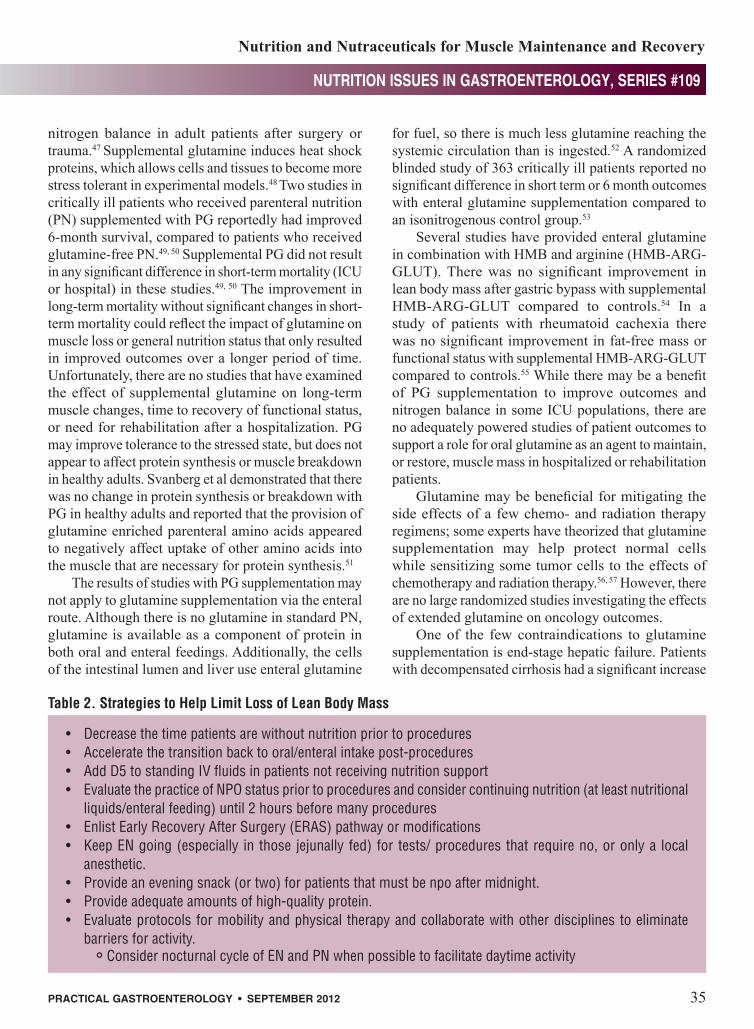
• Decreasethetimepatientsarewithoutnutritionpriortoprocedures• Acceleratethetransitionbacktooral/enteralintakepost-procedures• AddD5tostandingIVfluidsinpatientsnotreceivingnutritionsupport• EvaluatethepracticeofNPOstatuspriortoproceduresandconsidercontinuingnutrition(atleastnutritional
liquids/enteralfeeding)until2hoursbeforemanyprocedures• EnlistEarlyRecoveryAfterSurgery(ERAS)pathwayormodifications• KeepENgoing(especially inthosejejunallyfed)fortests/proceduresthatrequireno,oronlya local
anesthetic.• Provideaneveningsnack(ortwo)forpatientsthatmustbenpoaftermidnight.• Provideadequateamountsofhigh-qualityprotein.• Evaluateprotocolsformobilityandphysicaltherapyandcollaboratewithotherdisciplinestoeliminate
barriersforactivity.°ConsidernocturnalcycleofENandPNwhenpossibletofacilitatedaytimeactivity
Table 2. Strategies to Help Limit Loss of Lean Body Mass
Parrish_Sept_12.indd 35 9/12/12 10:40 PM

Nutrition and Nutraceuticals for Muscle Maintenance and Recovery
nutrition issues in gastroenterology, series #109
36� Practical�GastroenteroloGy� •� sePetmber�2012
in serum ammonia (75 to 169 umol/L cirrhosis vs. 52 to 78 umol/L control patients), 60 minutes after a single 20g dose of enteral glutamine.58 Among the patients with cirrhosis, there was a significant increase in the number of patients meeting criteria for hepatic encephalopathy 60 minutes after the dose of enteral glutamine (44 vs. 69%).58
Ornithine alpha-ketoglutarate (OKG)Ornithine alpha-ketoglutarate (OKG), also known as ornithine oxoglurate, is the salt of 2 molecules of ornithine linked to one molecule of α-ketoglutarate. OKG was initially proposed as a possible treatment for hepatic encephalopathy in the 1960’s, and marketed under the name Ornicetil.59 Controlled trials suggested that OKG was not efficacious for resolving hepatic coma, but clinicians noted that patients treated with OKG appeared to have improved nutrition status.60
OKG was reported to improve nitrogen balance in the 1980’s in patients with trauma, after surgery and burn injury.61-63 OKG causes a transient increase in growth hormone secretion, and supports glutamine levels post injury in humans.60, 64 Two double-blind randomized studies of adult burn patients demonstrated that 10g of OKG given 2X/day significantly decreased wound healing times and improved nitrogen balance compared to control patients.65, 66
Several studies have investigated the effect of OKG supplementation in elderly patients.67, 68 A double-blind randomized trial of OKG investigated the effects of 10g OKG/day for 2 months (patients monitored for 4 months total) in 185 elderly ambulatory patients.67
There was a significant improvement in appetite, body weight and independence in the OKG group compared to the placebo group after 30 days, and also after 60 days. Two months after OKG was stopped, there was still a significant improvement in the quality-of-life and medical-cost index in the OKG group, and the investigators reported that there was an overall cost saving of 37% related to OKG use.67
A second study randomized 370 non-hospitalized healthy adults that had recently recovered from various illnesses to receive either 10g of OKG/day or an isocaloric placebo.69 Patients who received OKG had a significant improvement in appetite and weight gain after 60 days compared to the placebo group.69
Although there is a long history of use and a number of studies documenting that OKG increases nitrogen balance in various pathologic states, there are some
limitations to existing evidence. A number of early trials did not provide isonitogenous controls, and there is limited data regarding patient outcomes due to the limited size or duration of the studies.68 There is evidence in animal models that OKG decreases muscle breakdown without stimulating tumor growth, but minimal human data about the potential risk of OKG use in humans with malignant disease.70 The use of any agent that has the potential to enhance protein synthesis should raise concern for it’s potential to function as an accelerant for tumor growth. OKG supplementation increases arginine production, and thus could invoke synthesis of nitric oxide with a potential detrimental effect on septic critically ill patients in a fashion similar to arginine supplemented feeding.71 There is sufficient evidence of OKG’s action on enhancing nitrogen balance and appetite to support larger properly controlled studies of OKG on patient outcomes.
Nutrition and Feeding StrategiesAlthough full nutrition does not completely prevent muscle loss in hospitalized patients, it is clear that inadequate nutrition accelerates muscle loss, and prolonged or recurrent periods without nutrition cause large amounts of body protein to be burned for energy.4, 72 Healthy adults that are deprived of food have an adaptation to starvation within several days, with decreased metabolic rate and protein oxidation and increased utilization of fat for fuel. However, patients with illness or injury experience hypermetabolism and rapid protein breakdown even when starved. Fat cannot be converted directly into glucose, therefore, when glycogen stores are quickly depleted, large amounts of body proteins are catabolized to meet the needs of cells that are dependent on glucose.4, 73
Many patients arrive at the hospital with a history of weight loss or decreased oral intake, and thus have depleted glycogen stores on admission. Acutely ill hospitalized patients with depleted glycogen stores will have urinary nitrogen loses of 10-15g /24 hrs.4, 73, 74 Providing as little as 300-400 dextrose calories per 24 hours (75-100mL/hr of 5% dextrose) decreases muscle breakdown in half, as evidenced by a decrease in urinary nitrogen losses to 5-7g /24 hrs.73 While short periods of time without significant nutrition are not overtly injurious, the cumulative effect of periods of semi-starvation undoubtedly contributes to muscle loss during hospitalization. Decreasing the amount of time
(continued on page 38)
Parrish_Sept_12.indd 36 9/12/12 10:40 PM

38� Practical�GastroenteroloGy� •� sePtember�2012
nutrition issues in gastroenterology, series #109nutrition issues in gastroenterology, series #109
Nutrition and Nutraceuticals for Muscle Maintenance and Recovery
that a patient is without food is one of the most concrete, feasible and cost-effective interventions to implement.
Cumulative calorie deficit while in the hospitalPatients that take food by mouth or receive enteral nutrition support (EN) inevitably have repeated interruptions in their nutrition and develop a large cumulative calorie and protein deficit during their hospitalization.75, 76 A randomized study comparing full feeding with reduced calorie and protein “trophic” feeding in patients with acute respiratory distress syndrome (ARDS) demonstrated that patients receiving reduced feeding during the early part of their admission had significantly increased need for rehabilitation services after their hospitalization.77 The use of PN to supplement inadequate enteral intake appears to create more problems than it cures however.78, 79 Supplemental PN provided to those patients with functional GI tracts who were receiving inadequate EN resulted in significantly increased infectious complications, and increased the duration of hospitalization.78, 79 Although supplemental PN carries excessive risk and expense, there are a number of less invasive strategies that can be used to limit cumulative nutrition deficit during hospitalizations. Oral nutrition supplements significantly decreased weight loss and increased muscle strength in postoperative hospitalized patients and when provided to malnourished patients after hospital discharge significantly increased muscle strength and improved quality of life.80-82 EN support is frequently held due to outdated practices with poor, or no supporting evidence. Involvement of nutrition support professionals and adoption of evidence-based feeding protocols can increase nutrition delivery to patients that require EN.1, 76
It is obvious that some periods without full nutrition for hospitalized patients are unavoidable due to the need to be npo for tests and procedures. However it is equally obvious that many protocols stipulating no oral/enteral intake prior to a test are based more on convention than necessity. The results of several studies reported that patients may continue to receive nutrition until 2 hours before many procedures without increasing complications.83-85 Carbohydrate and protein feeding pre-procedure may also decrease postoperative insulin resistance and appears to decrease postoperative muscle loss.86 Although patients that are unable to protect their airway are at increased risk of reflux if they receive
gastric EN while supine, feedings often do not need to be held for a protracted time period pre-procedure. Furthermore, patients that receive EN into the small intestine can often have nutrition infusion continue until shortly before the test.
Although there is evidence that extended periods without food before many procedures is not necessary, actually changing hospital protocols and practices can be difficult to achieve. There is an extended history to the “npo after midnight” practice and it is difficult to overcome the tremendous inertia of tradition. Updating outdated protocols to minimize periods of fasting pre-procedure may not be adequate by itself. Education about the potential advantages of reduced preoperative fasting and conducting follow-up quality evaluations to determine if the new protocols are being followed may help increase compliance and eventually decrease the nutrition deficit that patients accrue in the hospital setting.
In addition to decreasing the time that a patient is without nutrition prior to procedures, it is often possible to accelerate the transition back to oral intake post-procedure. There is copious evidence that rapid removal of nasogastric tubes and earlier introduction of food after surgical procedures does not have significant disadvantages, and may have benefits.87, 88 There is a need for studies that investigate protocols to minimize non-essential downtime of oral and EN support to determine if more consistent nutrition provision can minimize muscle loss during hospitalizations and affect outcomes, functional status, or rehabilitation needs.
ProteinNormally, muscle protein synthesis is transiently increased after ingestion of dietary protein. Ingestion of dietary protein has an enhanced ability to increase muscle protein synthesis in healthy people after exercise. Doses as small as 5g of high quality protein increase the rate of muscle protein synthesis, with maximal effects being reached at 20g in young adults. Elderly patients appear less sensitive to the effect of dietary protein after exercise because muscle protein synthesis was not stimulated in doses lower than 20g, and maximal effects were not reached until 40g of protein were ingested.11
High quality protein such as whey increases protein synthesis after exercise compared to lower quality proteins.11
Unfortunately, immobilization or bedrest (unloading) blunts the ability of protein or amino acids
(continued from page 36)
Parrish_Sept_12.indd 38 9/12/12 10:41 PM

nutrition issues in gastroenterology, series #109nutrition issues in gastroenterology, series #109
Nutrition and Nutraceuticals for Muscle Maintenance and Recovery
Practical GastroenteroloGy • sePtember 2012 39
to stimulate muscle protein synthesis.72 Protein or amino acid supplements were ineffective in decreasing muscle catabolism in patients that had no physical activity.72, 89 However, amino acid supplementation was effective in reducing muscle mass loss when combined with a minimal amount (5 minutes/day) of physical activity.90 There is a need for further studies of exercise combined with high quality protein supplements to determine optimal protein dosing and exercise requirements to preserve and restore muscle mass.
DiscussionThe loss of lean muscle mass during hospitalizations very likely contributes to functional impairments, reduced quality of life and increased costs for rehabilitation.11, 91, 92 The elderly, who are an expanding segment of our population, are especially susceptible to the negative effects of muscle loss. While there are a number of nutritional supplements that show promise and are worthy of additional research, there is a need for adequately powered studies that investigate meaningful outcomes and cost effectiveness before they are routinely used in clinical practice (see Table 1). OKG is the only anabolic nutraceutical with demonstrated outcome improvements in controlled studies, but there is limited data in acutely or critically ill patients receiving OKG.67 Some nutraceuticals that increase anabolism may have the potential to accelerate tumor growth and available research does not adequately address potential safety risks. Randomized studies over the past 20 years have repeatedly demonstrated unexpected harmful effects of relatively benign nutrients or nutraceuticals that initially appeared promising in animal or small scale human studies.14-17 Some critically or acutely ill populations may be at particular risk from enhancing protein synthesis because it is possible that reversing catabolism in the earlier stages of illness may have unexpected negative effects. The use of anabolic steroid oxandrolone in ventilator dependent surgical patients resulted in a significantly longer period of mechanical ventilation and intensive care unit stay, which may be related to increased collagen deposition leading to increased fibrotic pulmonary changes.93 In two large multicenter randomized studies of surgical and medical critically ill patients, the administration of human growth hormone resulted in a significantly longer ICU and hospital stay, duration of mechanical ventilation and mortality compared to patients that received placebo.94 There is insufficient evidence to
assume that nutraceuticals with anabolic potential would necessarily share the same risk factors as pharmacologic anabolics in similar populations. However, it would be similarly unreasonable to assume that neutraceuticals with anabolic potential would be safe in critically ill populations without randomized studies that have a sufficient number of patients with the power to examine patient outcomes.
Nutrition strategies may help limit the amount of muscle that is lost (see Table 2), but nutrition alone, even when optimized, cannot prevent muscle loss during inactivity or critical illness.4 Exercise, particularly resistance exercise, is especially potent for preventing and restoring muscle loss.91 Optimizing physical activity and implementing resistance exercise programs appears to be considerably more effective for restoring muscle mass than nutritional interventions alone.11, 91 Considering the evidence that some nutrition interventions only demonstrate anabolic potential when administered in concert with resistance exercise,40 there is a clear need for adequate studies of this kind to fully evaluate the potential of combined therapies. Recent research has demonstrated that even isolated vitamin inadequacy can potentially become a factor that limits the ability to respond to anabolic therapies.35 There is also the need to explore the potential of combining nutritional and pharmacologic, as well as nutrition, pharmacologic and exercise efforts to counteract muscle loss. Studies of pharmacologic approaches to enhance anabolism have rarely considered nutritional factors as a potential rate limiting step for muscle response.Likewise, studies of nutritional supplementation have not evaluated potential endocrine or metabolic abnormalities that can potentially influence the ability to respond to nutritional interventions.
ConclusionsThere are a number of nutritional supplements that have demonstrated potential as agents to help maintain or recover muscle during and after illness. However, there is a need for larger studies examining patient outcomes and cost effectiveness before routine clinical use can be recommended. Nutritional strategies and protocols that minimize time without nutrition during hospitalizations may reduce muscle loss and can generally be implemented without increasing costs. Optimizing existing efforts to prevent time without nutrition and increase physical activity are concrete
(continued on page 44)
Parrish_Sept_12.indd 39 9/12/12 10:41 PM
44� Practical�GastroenteroloGy� •� sePtember�2012
nutrition issues in gastroenterology, series #109nutrition issues in gastroenterology, series #109
Nutrition and Nutraceuticals for Muscle Maintenance and Recovery
steps that can be implemented now to reduce and restore muscle mass until additional research with anabolic nutraceuticals are available. Future nutrition studies should investigate not only short term outcomes such as survival and ICU length of stay, but also include longer term outcomes such as functional status and requirements for rehabilitation as outcomes. Programs that optimize physical activity and combined exercise and nutrition programs should be studied in the future.n
References1. Parrish CR. Enteral feeding: the art and the science. Nutr
Clin Pract, 2003;18:76-85.2. Krenitsky J, Makola D, Parrish C. Parenteral Nutrition in
Pancreatitis is Passé: But Are We Ready for Gastric Feeding? A Critical Evaluation of the Literature–Part I. Practical Gastroenterology, 2007; 92-112.
3. Frankenfield DC, Smith JS, Cooney RN. Accelerated nitrogen loss after traumatic injury is not attenuated by achievement of energy balance. J Parenter Enteral Nutr, 1997;21:324-329.
4. Shaw JH, Wildbore M, Wolfe RR. Whole body protein kinetics in severely septic patients. The response to glu-cose infusion and total parenteral nutrition. Ann Surg, 1987;205:288-294.
5. Puthucheary Z, Harridge S, Hart N. Skeletal muscle dys-function in critical care: wasting, weakness, and rehabilita-tion strategies. Crit Care Med, 2010;38: S676-682.
6. Herridge MS, Cheung AM, Tansey CM et al. One-year outcomes in survivors of the acute respiratory distress syn-drome. N Engl J Med, 2003;348:683-693.
7. Herridge MS, Tansey CM, Matté A et al. Functional disabil-ity 5 years after acute respiratory distress syndrome. N Engl J Med, 2011;364:1293-1304.
8. De Jonghe B, Sharshar T, Lefaucheur JP et al. Paresis acquired in the intensive care unit: a prospective multicenter study. JAMA, 2002;288:2859-2867.
9. Suetta C, Hvid LG, Justesen L et al. Effects of aging on human skeletal muscle after immobilization and retraining. J Appl Physiol, 2009;107:1172-1180.
10. Bross R, Javanbakht M, Bhasin S. Anabolic interventions for aging-associated sarcopenia. J Clin Endocrinol Metab, 1999;84:3420-3430.
11. Breen L, Phillips SM. Skeletal muscle protein metabolism in the elderly: Interventions to counteract the ‘anabolic resis-tance’ of ageing. Nutr Metab, 2011;8:68-79.
12. Hvid L, Aagaard P, Justesen L et al. Effects of aging on muscle mechanical function and muscle fiber morphology during short-term immobilization and subsequent retraining. J Appl Physiol, 2010;109:1628-1634.
13. Whipple RH, Wolfson LI, Amerman PM. The relationship of knee and ankle weakness to falls in nursing home resi-dents: an isokinetic study. J Am Geriatr Soc, 1987;35:13-20.
14. The Alpha-Tocopherol, Beta-Carotene Cancer Prevention Study Group. The effect of vitamin E and beta carotene on the incidence of lung cancer and other cancers in male smok-ers. N Engl J Med, 1994;330:1029–1035.
15. Besselink MG, van Santvoort HC, Buskens E, et al.; Dutch Acute Pancreatitis Study Group. Probiotic prophylaxis in predicted severe acute pancreatitis: a randomized, double-blind, placebo-controlled trial. Lancet, 2008;371:651-659.
16. Schulman SP, Becker LC, Kass DA etal. L-arginine therapy in acute myocardial infarction: the Vascular Interaction With Age in Myocardial Infarction (VINTAGE MI) randomized clinical trial. JAMA, 2006;295:58-64.
17. Lonn E, Bosch J, Yusuf S et al. Effects of long-term vitamin E supplementation on cardiovascular events and cancer: a randomized controlled trial. JAMA, 2005;293:1338-1347.
18. Visek WJ. Arginine needs, physiological state and usual diets. A reevaluation. J Nutr, 1986;116:36-46.
19. Daly JM, Reynolds J, Thom A et al. Immune and meta-bolic effects of arginine in the surgical patient. Ann Surg, 1988;208:512-523.
20. Kirk SJ, Hurson M, Regan MC, et al. Arginine stimulates wound healing and immune function in elderly human beings. Surgery, 1993;114:155-159.
21. Mendez C, Jurkovich GJ, Garcia I, et al. Effects of an immune enhancing diet in critically injured patients. J Trauma, 1997;42:933-940.
22. Crozier SJ, Kimball SR, Emmert SW, et al. Oral leucine administration stimulates protein synthesis in rat skeletal muscle. J Nutr, 2005;135:376-382.
23. Anthony JC, Anthony TG, Kimball SR, et al. Signaling path-ways involved in translational control of protein synthesis in skeletal muscle by leucine. J Nutr, 2001;131:856S-860S.
24. Glynn EL, Fry CS, Drummond MJ, et al. Excess leucine intake enhances muscle anabolic signaling but not net protein anabolism in young men and women. J Nutr, 2010;140:1970-1976.
25. Leenders M, van Loon LJ. Leucine as a pharmaconutrient to prevent and treat sarcopenia and type 2 diabetes. Nutr Rev, 2011;69:675-689.
26. Verhoeven S, Vanschoonbeek K, Verdijk LB, et al. Long-term leucine supplementation does not increase muscle mass or strength in healthy elderly men. Am J Clin Nutr, 2009;89:1468–1475.
27. Pasiakos SM, McClung HL, McClung JP, et al. Leucine-enriched essential amino acid supplementation during mod-erate steady state exercise enhances postexercise muscle protein synthesis. Am J Clin Nutr, 2011;94:809-818.
28. Hagmüller E, Kollmar HB, Günther HJ, et al. Protein metab-olism in human colon carcinomas: in vivo investigations using a modified tracer technique with L-[1-13C]leucine. Cancer Res, 1995;55:1160-1167.
29. McNurlan MA, Heys SD, Park KG, et al. Tumour and host tissue responses to branched-chain amino acid supplementa-tion of patients with cancer. Clin Science, 1994;86:339-345.
30. Van Koevering M, Nissen S. Oxidation of leucine and alpha-ketoisocaproate to beta-hydroxy-beta-methylbutyrate in vivo. Am J Physiol, 1992;262:E27-31.
31. Wilson GJ, Wilson JM, Manninen AH. Effects of beta-hydroxy-beta-methylbutyrate (HMB) on exercise perfor-mance and body composition across varying levels of age, sex, and training experience: A review. Nutr Metab, 2008;5:1-17.
32. Rowlands DS, Thomson JS. Effects of beta-hydroxy-beta-methylbutyrate supplementation during resistance train-ing on strength, body composition, and muscle damage in trained and untrained young men: a meta-analysis. J Strength Cond Res. 2009 May;23(3):836-46.
33. Kuhls DA, Rathmacher JA, Musngi MD, et al. Beta-hydroxy-beta-methylbutyrate supplementation in critically ill trauma patients. J Trauma, 2007;62:125-131.
34. Baier S, Johannsen D, Abumrad N, et al. Year-long changes in protein metabolism in elderly men and women supplemented with a nutrition cocktail of beta-hydroxy-beta-methylbutyrate (HMB), L-arginine, and L-lysine. J Parenter Enteral Nutr, 2009;33:71-82.
35. Fuller JC Jr, Baier S, Flakoll P, et al. Vitamin D status affects strength gains in older adults supplemented with a combina-tion of β-hydroxy-β-methylbutyrate, arginine, and lysine: a cohort study. J Parenter Enteral Nutr, 2011;35:757-762.
36. Marcora S, Lemmey A, Maddison P. Dietary treatment of rheumatoid cachexia with beta-hydroxy-beta-methylbutyr-ate, glutamine and arginine: a randomised controlled trial. Clin Nutr, 2005;24:442-454.
37. Hsieh LC, Chien SL, Huang MS, et al. Anti-inflammatory
(continued from page 39)
Parrish_Sept_12.indd 44 9/12/12 10:41 PM

nutrition issues in gastroenterology, series #109nutrition issues in gastroenterology, series #109
Nutrition and Nutraceuticals for Muscle Maintenance and Recovery
Practical GastroenteroloGy • sePtember 2012 45
and anticatabolic effects of short-term beta-hydroxy-beta-methylbutyrate supplementation on chronic obstructive pul-monary disease patients in intensive care unit. Asia Pac J Clin Nutr, 2006;15:544-550.
38. Clark RH, Feleke G, Din M, et al. Nutritional treatment for acquired immunodeficiency virus-associated wasting using beta-hydroxy beta-methylbutyrate, glutamine, and arginine: a randomized, double-blind, placebo-controlled study. J Parenter Enteral Nutr. 2000;24:133-139.
39. May PE, Barber A, D’Olimpio JT, et al. Reversal of cancer-related wasting using oral supplementation with a combi-nation of beta-hydroxy-beta-methylbutyrate, arginine, and glutamine. Am J Surg, 2002;183:471-479.
40. Rodacki CL, Rodacki AL, Pereira G, et al. Fish-oil supple-mentation enhances the effects of strength training in elderly women. Am J Clin Nutr, 2012;95:428-436.
41. Murphy RA, Yeung E, Mazurak VC, et al. Influence of eicosapentaenoic acid supplementation on lean body mass in cancer cachexia. Br J Cancer, 2011;105:1469-473.
42. Wigmore SJ, Fearon KC, Maingay JP, et al. Down-regulation of the acute-phase response in patients with pan-creatic cancer cachexia receiving oral eicosapentaenoic acid is mediated via suppression of interleukin-6. Clin Science, 1997;92:215-221.
43. Wigmore SJ, Barber MD, Ross JA, et al. Effect of oral eicosapentaenoic acid on weight loss in patients with pan-creatic cancer. Nutr Cancer, 2000;36: 177–184
44. Fearon KC, Barber MD, Moses AG, et al. Double-blind, placebo-controlled, randomized study of eicosapentaenoic acid diester in patients with cancer cachexia. J Clin Oncol, 2006;24:3401-407.
45. Fearon KC, Von Meyenfeldt MF, Moses AG, et al. Effect of a protein and energy dense N-3 fatty acid enriched oral sup-plement on loss of weight and lean tissue in cancer cachexia: a randomised double blind trial. Gut, 2003;52:1479-1486.
46. Jatoi A, Rowland K, Loprinzi CL, et al. An eicosapentae-noic acid supplement versus megestrol acetate versus both for patients with cancer-associated wasting: a North Central Cancer Treatment Group and National Cancer Institute of Canada collaborative effort. J Clin Oncol, 2004;22:2469-2476.
47. Wilmore DW. The effect of glutamine supplementation in patients following elective surgery and accidental injury. J Nutr, 2001;131:2543S-25949S.
48. Peng ZY, Hamiel CR, Banerjee A, et al. Glutamine attenua-tion of cell death and inducible nitric oxide synthase expres-sion following inflammatory cytokine-induced injury is dependent on heat shock factor-1 expression. J Parenter Enteral Nutr, 2006;30:400-406.
49. Goeters C, Wenn A, Mertes N, et al. Parenteral L-alanyl-L-glutamine improves 6-month outcome in critically ill patients. Crit Care Med, 2002;30:2032–2037.
50. Griffiths RD, Jones C, Palmer TE. Six-month outcome of critically ill patients given glutamine-supplemented paren-teral nutrition. Nutrition, 1997;13:295–302.
51. Svanberg E, Möller-Loswick AC, Matthews DE, et al. The effect of glutamine on protein balance and amino acid flux across arm and leg tissues in healthy volunteers. Clin Physiol, 2001;21:478-489.
52. Wernerman J. Glutamine supplementation. Ann Intensive Care. 2011;18:1:25-30.
53. Hall JC, Dobb G, Hall J, et al. A prospective randomized trial of enteral glutamine in critical illness. Intensive Care Med, 2003;29:1710–1716.
54. Clements RH, Saraf N, Kakade M, et al. Nutritional effect of oral supplement enriched in beta-hydroxy-beta-methylbu-tyrate, glutamine and arginine on resting metabolic rate after laparoscopic gastric bypass. Surg Endosc, 2011;25:1376-1382.
55. Marcora S, Lemmey A, Maddison P. Dietary treatment of rheumatoid cachexia with beta-hydroxy-beta-methylbutyr-ate, glutamine and arginine: a randomised controlled trial.
Clin Nutr, 2005;24:442-454.56. Kuhn KS, Muscaritoli M, Wischmeyer P, et al. Glutamine as
indispensable nutrient in oncology: experimental and clinical evidence. Eur J Nutr, 2010;49:197-210.
57. Savarese DM, Savy G, Vahdat L, Wischmeyer PE, Corey B. Prevention of chemotherapy and radiation toxicity with glutamine. Cancer Treat Rev, 2003;29:501-513.
58. Ditisheim S, Giostra E, Burkhard PR, et al. A capillary blood ammonia bedside test following glutamine load to improve the diagnosis of hepatic encephalopathy in cirrhosis. BMC Gastroenterol, 2011;11:134-141.
59. Chainuvati T, Plengvanit U, Viranuvatti V. Ornicetil on encephalopathy. Effect of ornicetil (ornithine alpha-ketoglu-tarate) on encephalopathy in patients with acute and chronic liver disease. Acta Hepatogastroenterol, 1977;24:434-439.
60. Blonde-Cynober F, Aussel C, Cynober L. Use of ornithine alpha-ketoglutarate in clinical nutrition of elderly patients. Nutrition, 2003;19:73-75.
61. Leander U, Vesterberg K, FUrst P, Vinnars E, Johnsson C. Nitrogen sparing effect of Ornicetil in trauma. Presented at the 4th Congress of European Society of Parenteral and Enteral Nutrition, Vienna, Austria, September 26-29, 1982.
62. Leander U, Fürst P, Vesterberg K, et al. Nitrogen sparing effect of Ornicetil in the immediate postoperative state clinical biochemistry and nitrogen balance. Clin Nutr, 1985;4:43-51.
63. Cynober L, Saizy R, Nguyen Dinh F, et al. Effect of enter-ally administered ornithine alpha-ketoglutarate on plasma and urinary amino acid levels after burn injury. J Trauma, 1984;24:590-596.
64. De Bandt JP, Cynober L. Therapeutic use of branched-chain amino acids in burn, trauma, and sepsis. J Nutr, 2006;136:308S-13S.
65. Donati L, Ziegler F, Pongelli G, Signorini MS. Nutritional and clinical efficacy of ornithine alpha-ketoglutarate in severe burn patients. Clin Nutr, 1999;18:307-311.
66. Coudray-Lucas C, Le Bever H, Cynober L, et al. Ornithine alpha-ketoglutarate improves wound healing in severe burn patients: a prospective randomized double-blind trial versus isonitrogenous controls. Crit Care Med, 2000;28:1772-1776.
67. Brocker P, Vellas B, Albarede JL, Poynard T. A two-centre, randomized, double-blind trial of ornithine oxoglutarate in 194 elderly, ambulatory, convalescent subjects. Age Ageing. 1994 Jul;23(4):303-6.
68. Walrand S. Ornithine alpha-ketoglutarate: could it be a new therapeutic option for sarcopenia? J Nutr Health Aging, 2010;14:570-577.
69. Debry G, Poyard T. Value of ornithine alpha-ketoglutarate for nutritional support in convalescent, malnourished elderly subjects. Facts Res Gerontol, 1995;9:1.
70. Le Bricon T, Cynober L, Baracos VE. Ornithine alpha-keto-glutarate limits muscle protein breakdown without stimulat-ing tumor growth in rats bearing Yoshida ascites hepatoma. Metabolism, 1994;43:899-905.
71. Cynober L. Ornithine alpha-ketoglutarate as a potent precur-sor of arginine and nitric oxide: a new job for an old friend. J Nutr, 2004;134:2858S-2862S.
72. Biolo G, Ciocchi B, Stulle M, et al. Calorie restriction accel-erates the catabolism of lean body mass during 2 wk of bed rest. Am J Clin Nut, 2007;86:366-372.
73. Wolfe BM, Culebras JM, Sim AJ, et al. Substrate interaction in intravenous feeding: comparative effects of carbohydrate and fat on amino acid utilization in fasting man. Ann Surg, 1977;186:518-540
74. Wernerman J, von der Decken A, Vinnars E. Protein syn-thesis in skeletal muscle in relation to nitrogen balance after abdominal surgery: the effect of total parenteral nutrition. J Parenter Enteral Nutr, 1986;10:578-582.
75. Hise ME, Halterman K, Gajewski BJ, et al. Feeding prac-tices of severely ill intensive care unit patients: an evaluation of energy sources and clinical outcomes. J Am Diet Assoc,
Parrish_Sept_12.indd 45 9/12/12 10:41 PM
Nutrition and Nutraceuticals for Muscle Maintenance and Recovery
nutrition issues in gastroenterology, series #109
46� Practical�GastroenteroloGy� •� sePetmber�2012
2007;107:458-465.76. Soguel L, Revelly JP, Schaller MD, et al. Energy deficit and
length of hospital stay can be reduced by a two-step quality improvement of nutrition therapy: The intensive care unit dietitian can make the difference. Crit Care Med, 2011; 15.
77. Rice TW, Mogan S, Hays MA, et al. Randomized trial of initial trophic versus full-energy enteral nutrition in mechan-ically ventilated patients with acute respiratory failure. Crit Care Med, 2011;39:1-398.
78. Singer P, Anbar R, Cohen J, et al. The tight calorie control study (TICACOS): a prospective, randomized, controlled pilot study of nutritional support in critically ill patients. Intensive Care Med, 2011;37:601-609.
79. Casaer MP, Mesotten D, Hermans G, et al. Early versus late parenteral nutrition in critically ill adults. N Engl J Med, 2011;11;365:506-517.
80. Rana SK, Bray J, Menzies-Gow N, et al. Short term ben-efits of post-operative oral dietary supplements in surgical patients. Clin Nutr, 1992;11:337-344.
81. Price R, Daly F, Pennington CR, McMurdo ME. Nutritional supplementation of very old people at hospital discharge increases muscle strength: a randomised controlled trial. Gerontology, 2005;51:179-85.
82. Norman K, Kirchner H, Freudenreich M, et al. Three month intervention with protein and energy rich supplements improve muscle function and quality of life in malnourished patients with non-neoplastic gastrointestinal disease--a ran-domized controlled trial. Clin Nutr, 2008;27:48-56.
83. de Aguilar-Nascimento JE, Dock-Nascimento DB. Reducing preoperative fasting time: A trend based on evidence. World J Gastrointest Surg. 2010 Mar 27;2(3):57-60.
84. Kaska M, Grosmanová T, Havel E, et al. The impact and safety of preoperative oral or intravenous carbohydrate administration versus fasting in colorectal surgery--a ran-domized controlled trial. Wien Klin Wochenschr. 2010 Jan;122(1-2):23-30.
85. Power S, Kavanagh DO, McConnell G, et al. Reducing preoperative fasting in elective adult surgical patients: a case-control study. Ir J Med Sci. 2012 Mar;181(1):99-104.
86. Perrone F, da-Silva-Filho AC, Adôrno IF, et al. Effects of preoperative feeding with a whey protein plus carbohydrate drink on the acute phase response and insulin resistance. A randomized trial. Nutr J, 2011;10:66.
87. Osland E, Yunus RM, Khan S, Memon MA. Early versus traditional postoperative feeding in patients undergoing resectional gastrointestinal surgery: a meta-analysis. J Parenter Enteral Nutr, 2011;35:473-487.
88. Andersen HK, Lewis SJ, Thomas S. Early enteral nutrition within 24h of colorectal surgery versus later commence-ment of feeding for postoperative complications. Cochrane Database Syst Rev, 2006;(4):CD004080.
89. Brooks NE, Cadena SM, Vannier E, et al. Effects of resis-tance exercise combined with essential amino acid supple-mentation and energy deficit on markers of skeletal muscle atrophy and regeneration during bed rest and active recovery. Muscle Nerve, 2010;42:927-935.
90. Paddon-Jones D, Sheffiel-Moore M, Urban RJ, et al. Essential amino acid and carbohydrate supplementation ameliorates muscle protein loss in humans during 28 days bedrest. J Clin Endocrinol Metab, 2004;8:4351-4358.
91. Glover EI, Phillips SM. Resistance exercise and appropri-ate nutrition to counteract muscle wasting and promote muscle hypertrophy. Curr Opin Clin Nutr Metab Care, 2010;13(6):630-4.
92. Rennie MJ. Anabolic resistance in critically ill patients. Crit Care Med, 2009;37(10 Suppl):S398-S399.
93. Bulger EM, Jurkovich GJ, Farver CL, et al. Oxandrolone does not improve outcome of ventilator dependent surgical patients. Ann Surg, 2004, 240:472-478.
94. Takala J, Ruokonen E, Webster NR, et al. Increased mortal-ity associated with growth hormone treatment in critically ill adults. N Engl J Med, 1999;341:785-792.
REPRINTSSpecial rates are available for
quantities of 100 or more.
For further details email us at:[email protected]
PRACTICALGASTROENTEROLOGY
CelebratingOver 3 Decades
of Service
Parrish_Sept_12.indd 46 9/12/12 10:41 PM

Practical GastroenteroloGy • sePtember 2012 49
Susan G. Coe
Flat Polyps: Endoscopic Detection and Treatment
Melvin Schapiro, M.D., Series Editor
Colonoscopy offers a method of screening for and prevention of colorectal cancer. Unfortunately, interval cancers still occur after negative screening colonoscopy suggesting that some pre-cancerous lesions may be missed. Flat and serrated polyps are a particular challenge to detect and may explain why some of these cancers occur. Detection of these subtle lesions requires recognition of their existence, training in their detection, careful inspection technique and adequate bowel preparation. Once a large, flat or otherwise complicated polyp is detected, endoscopic methods exist that offer an alternative to surgery. When performed by appropriately trained endoscopists in carefully selected patients, endoscopic resection can reduce the risk and morbidity of resection. In this review we define the types of difficult to detect polyps, methods to increase their detection, and describe available endoscopic resection techniques.
Michael B. Wallace
Susan G. Coe, M.D., Fellow, Department of Gastroenterology, Mayo Clinic Florida,Michael B. Wallace, M.D., MPH, Professor of Medicine, Department of Gastroenterology, Chair, Division of Gastroenterology and Hepatology, Mayo Clinic Florida
Practical aPProaches to the Diagnosis anD treatment of colorectal cancer #7
Colorectal cancer remains third in new cancer diagnoses and cause of cancer related death.1 In addition to colorectal cancer (CRC) screening,
colonoscopy also offers cancer prevention by allowing the removal of adenomatous polyps. Despite this benefit, the degree of risk reduction offered by colonoscopy for right-sided CRC has recently been questioned.2
Colorectal cancers develop from adenomatous polyps. Effective colorectal cancer prevention relies
on early identification and removal of these lesions at their pre-malignant stage. Unfortunately, colonoscopy is imperfect and interval cancers occur in up to 6% of patients after screening colonoscopy.3 There are many potential reasons why interval cancers occur however most agree that missed pre-cancerous polyps play an important role. Some of the most subtle and difficult polyps to detect are the flat and serrated polyps, making them a likely culprit for interval cancer. This is particularly true in the right colon where the bowel preparation is often less than ideal.
Once a large flat or serrated polyp is detected, the next challenge is removal. This is especially true in the right colon where positioning can be a difficult. In the past these patients were routinely sent for surgery, however endoscopic methods of resection
IntroduCtIon
Schapiro_September_12.indd 49 9/17/12 12:37 PM

Flat Polyps: Endoscopic Detection and Treatment
Practical aPProaches to the Diagnosis anD treatment of colorectal cancer #7
50� Practical�GastroenteroloGy� •� sePtember�2012
have eliminated the need for surgery for many patients. When done by experienced hands, in carefully selected patients, the risks of endoscopic mucosal resection are only modestly higher than for routine polypectomy.
TYPES OF SUBTLE NEOPLASIAFlat AdenomasThe term flat is used to describe a colon lesion measuring less than 2 cm in size that is elevated less than 2.5mm above the surrounding normal colonic mucosa (figure 1). Flat lesions larger than 2cm are termed “laterally spreading” lesions. Laterally spreading lesions are further sub-divided based on their appearance as either granular (having a nodular appearance, or cluster of sessile lesions) or non-granular (figure 2). As a flat lesion becomes more locally invasive, it penetrates deeper into the colonic wall, becoming a depressed lesion. Depressed lesions have a base that is lower than the normal surrounding mucosa, indicating a higher degree of invasion and dysplasia.
Serrated PolypsSerrated polyps are now recognized as part of an alternative pathway to colorectal cancer. Unlike the traditional adenoma to carcinoma sequence, colorectal cancers with high levels of microsatellite instability are thought to have developed through a process of CpG island methylation. This process occurs most often in serrated polyps exhibiting BRAF mutations.4
Serrated polyps are variants of hyperplastic polyps. They are often flat, located in the right colon and are often covered by an adherent mucous cap, adding to the challenge in their detection (figure 3).
ImPrOvINg DETEcTIONrecognitionThe first step in improving detection of subtle colorectal lesions is recognizing they exist. Flat and depressed adenomas, collectively referred to as non-polypoid neoplasia, were first described in the colon in the Japanese population over 25 years ago.5 Since then, several prospective studies have proven the existence of non-polypoid neoplasia worldwide.
The prevalence of non-polypoid neoplasia in the United States was first described in a landmark study of Veterans Administration (VA) patients. In this study, non-polypoid lesions were found in 9.4% of patients, representing 15% of all polyps detected. Despite their
(continued on page 52)
Figure 1. Subtle large flat adenoma
Figure 3. Flat serrated adenoma covered by adherent mucous cap.
Figure 2: Laterally spreading adenoma, granular type
Schapiro_September_12.indd 50 9/12/12 10:44 PM

Flat Polyps: Endoscopic Detection and Treatment
Practical aPProaches to the Diagnosis anD treatment of colorectal cancer #7
52� Practical�GastroenteroloGy� •� sePtember�2012
(continued from page 50)
small numbers, these non-polypoid lesions accounted for 54% of superficial carcinomas suggesting they are more likely to harbor advanced pathology.6 Later studies have resulted in similar findings with approximately 10% of adenomas described as flat.7 A tandem colonoscopy study found that up to 32% of flat polyps and 42% of flat adenomas were missed suggesting their true prevalence is likely under-reported.8
Despite several studies finding a higher prevalence of advanced pathology in non-polypoid lesions, the US National Polyp Study found the opposite. Flat adenomas in this study were no more likely to contain high-grade dysplasia than their sessile or pedunculated counterparts. Unlike flat lesions, however, truly depressed lesions were found to carry an increased risk for advanced pathology.9
Though we are beginning to appreciate the role of serrated polyps in colorectal cancer, their true prevalence is unknown. Confusing nomenclature and lack of agreement in diagnostic criteria have resulted in underreporting of serrated polyps by endoscopists and pathologists alike. What literature does exist suggests that up to 6-10% of patients undergoing screening colonoscopy may have a serrated polyp.10
This diagnostic challenge has led many gastroenterologists to argue that large hyperplastic polyps, particularly in the right colon, should be treated as neoplastic. Though not currently included in our multi-society surveillance interval guidelines, large hyperplastic polyps should be considered when determining a patient’s lifetime risk of colorectal cancer and need for surveillance. In our practice, large hyperplastic polyps prompt surveillance intervals that mirror those assigned to similar size adenomatous polyps. More studies are needed to address this issue and to determine the true impact these polyps have on colorectal cancer risk.
Subtle Clues to DetectionIt is often only a subtle clue that will suggest the presence of a flat polyp. Among these are subtle color differences compared to the surrounding mucosa such as an area that appears slightly more red or pale. Spontaneous mucosal hemorrhage or easy friability often indicate presence of neoplasia, however these findings are easy to misinterpret as minor mucosal trauma caused by the colonoscope. Deformity of the colon wall, in particular of the folds, and absence of vascularity in the
region of a flat polyp are other subtle clues. In the right colon, serrated polyps can be particularly challenging to detect as they are often covered with an adherent mucous cap that requires aggressive rinsing to reveal the underlying polyp. Residual stool and adherent bile seen with inadequate bowel preparation can easily mask these subtle clues hindering detection.
Bowel PreparationThe importance of excellent bowel preparation in the detection of non-polypoid neoplasia cannot be emphasized enough. A retrospective study that examined the outcomes in 5000 patients after screening colonoscopy highlights this importance. Of the 17 interval cancers that occurred in these patients, 6 had screening colonoscopies that were incomplete due to poor prep and another 4 had a lesion seen but not recognized as malignant. Considerably lower flat polyp detection was also seen in those who had inadequate prep (9%) compared to those with adequate prep (28%).11
Many bowel purgatives exist. Patients should be instructed on the importance of a good bowel preparation and encouraged to follow the preparation instructions provided. Our practice has adopted a split bowel prep regimen that has shown to provide better overall bowel preparation, but more importantly, better preparation of the right colon.
Inspection TechniquesAdenoma detection is a validated predictor of interval colorectal cancer12 that is strongly influenced by the performing endoscopist.13 Despite this, wide variability exists between endoscopists in this important measure. When high adenoma detectors have been studied formally, certain behaviors are observed. Compared to low detectors, high adenoma detectors spend more time on inspection of proximal surfaces, used irrigation more often to cleanse segments of poorly prepped colon mucosa, distended the colon more during inspection and used tip deflection to look behind colonic folds.14 These techniques are simple to teach and when incorporated into routine practice result in increased adenoma detection.
In our prospective study, we introduced an endoscopist training module that focused on recognizing the clues of subtle polyps and on the techniques associated with high adenoma detectors. In doing so
(continued on page 54)
Schapiro_September_12.indd 52 9/12/12 10:44 PM
54� Practical�GastroenteroloGy� •� sePtember�2012
Practical aPProaches to the Diagnosis anD treatment of colorectal cancer #7Practical aPProaches to the Diagnosis anD treatment of colorectal cancer #7
Flat Polyps: Endoscopic Detection and Treatment
we found an increase in overall adenoma detection from 36% to 47% in those who received training.15 Our results highlight the importance of training to improve recognition and detection of subtle lesions. In the age of quality measure reporting, patients and providers will have increasing access to the adenoma detection rates of endoscopists. This access will provide physicians with the ability to refer patients to gastroenterologists who offer the highest level of colorectal cancer risk reduction.
TreatmentMost polyps, including those that are flat, are less than 10mm and can be managed easily during routine colonoscopy. In contrast, large, flat polyps often require specialized techniques, which require additional devices, skill and time. As a result, patients with very large flat polyps have historically been referred for surgical resection. Though complete cure can be ensured with surgical resection, it also comes with significant cost, increased risk of morbidity, and in rare cases mortality, for what are most often benign lesions. Endoscopic mucosal resection (EMR) and endoscopic sub-mucosal dissection (ESD) are terms used to describe a variety of devices and techniques used to resect large polyps, and in some cases, early colorectal cancers.
The technique of EMR was first described for flexible colonoscopy in 1973 and has been practiced widely in regions, such as Japan, with a high prevalence of large, flat lesions. As large, flat lesions have become increasingly recognized worldwide, EMR has become more widely adopted. Several tools exist that allow safe removal of large lesions that are limited to the superficial mucosal layer. These include lift and snare, cap-assisted and ligation-assisted techniques, each with the goal to remove all neoplastic tissue down to the sub-mucosal layer.16
Endoscopic mucosal resection has proven a safe alternative to surgery in experienced hands. Major complications such as bleeding and perforation are infrequent, occurring in 0.5 to 6% of cases depending on the series.17 The risk of bleeding is directly related to the size of the polyp with those larger than 3 cm conferring the greatest risk.18, 19 This knowledge allows endoscopists to apply endoscopic closure devices as prophylaxis against bleeding.
One major limitation to EMR is that large lesions (>20mm) require piece-meal resection, which prohibits
accurate pathologic margin assessment. Advanced imaging modalities such as high definition white light imaging, dye-based or virtual chromoendoscopy, and confocal endomicroscopy can aide in endoscopic lateral margin assessment. With these techniques, complete resection rates of over 96% have been reported.20 Close follow up with repeat colonoscopy to ensure ablation of any residual neoplasia is recommended at 3 months and again at 1 year following EMR. Residual adenomatous tissue at the time of follow-up is reported between 4 – 14% depending on the expertise of the center.20-22 Endoscopic sub-mucosal dissection (ESD) describes a group of techniques and devices that allows en-bloc resection of very large (>20mm) non-polypoid neoplasia. En-bloc resection provides the ability for complete assessment of deep and lateral margins not offered by piece-meal EMR techniques. In centers offering this technique, much lower rates of residual disease are reported (2 versus 14%). The trade-offs however include longer procedure times (108 +- 71 minutes versus 29 +-25 minutes, p<0.001) and an increased risk of perforation (6 vs. 1%), though most perforations can be managed endoscopically.23 Provided the local expertise and surgical support is available, ESD is the preferred method for complete endoscopic removal of polyps >20mm where there is a high suspicion of early invasive carcinoma, as indicated by a depressed type polyps or surface pit pattern suggestive of superficial invasion. When done by appropriately trained endoscopists, both EMR and ESD, have the benefit of avoiding surgery.
SummaryFlat and serrated polyps can be subtle and challenging to detect, and as a result, likely contributed to the occurrence of interval colorectal cancer after screening colonoscopy.
Detection of non-polypoid colorectal lesions requires recognition of their existence, training in their detection, careful inspection technique and bowel preparation that is adequate to allow their subtle features to be visualized. Referring physicians can assist their patients by providing information on the importance of bowel purgative and by referring them to endoscopists who are known for high quality colonoscopy. Adenoma detection rates are a validated measure of quality in colonoscopy and a predictor of interval colorectal cancer, and therefore can serve as a barometer for this quality.
(continued from page 52)
Schapiro_September_12.indd 54 9/12/12 10:45 PM

Practical aPProaches to the Diagnosis anD treatment of colorectal cancer #7Practical aPProaches to the Diagnosis anD treatment of colorectal cancer #7
Flat Polyps: Endoscopic Detection and Treatment
Practical GastroenteroloGy • sePtember 2012 55
When a large, flat or otherwise complicated polyp is detected, endoscopic methods offer an alternative to surgery that can reduce the risk and morbidity associated with surgical resection. Referral to a center with both the expertise in endoscopic resection methods (EMR and/or ESD) and adequate surgical support is required to minimize complications. Short interval follow up colonoscopies in the year following endoscopic resection are also needed to ensure complete resection.n
References
1. Society AC. Colorectal Cancer Facts & figures 2011-2013. 2011 [cited 2011 November 9]; Available from: http://www.cancer.org/acs/groups/content/@epidemiologysurveilance/documents/document/acspc-028323.pdf.
2. Baxter NN, Goldwasser MA, Paszat LF, Saskin R, Urbach DR, Rabeneck L. Association of Colonoscopy and Death From Colorectal Cancer. Ann of Intern Med. 2009 January 6, 2009;150(1):1-8.
3. Bressler B, Paszat LF, Chen Z. Rates of new or missed colorectal cancers after colonoscopy and their risk factors: a population-based analysis. Gastroenterology. [10.1053/j.gas-tro.2006.10.027]. 2007;132:96-102.
4. Huang CS, Farraye FA, Yang S, O’Brien MJ. The Clinical Significance of Serrated Polyps. Am J Gastroenterol. 2011;106(2):229-40.
5. Muto T, Kamiya J, Sawada T, Konishi F, Sugihara K, Kubota Y, et al. Small “flat adenoma” of the large bowel with special reference to its clinicopathologic features. Dis Colon Rectum. 1985;28(10):847-51 10.1007/BF02555490.
6. Soetikno RM, Kaltenbach T, Rouse RV, Park W, Maheshwari A, Sato T, et al. Prevalence of nonpolypoid (flat and depressed) colorectal neoplasms in asymptomatic and symptomatic adults. JAMA. 2008 Mar 5;299(9):1027-35.
7. Gorgun E, Church J. Flat adenomas of the large bowel: a single endoscopist study. Dis Colon Rectum. 2009 May;52(5):972-7.
8. Heresbach D, Barrioz T, Lapalus MG, Coumaros D, Bauret P, Potier P, et al. Miss rate for colorectal neoplastic polyps: a pro-spective multicenter study of back-to-back video colonoscopies. Endoscopy. 2008 Apr;40(4):284-90.
9. O’Brien MJ, Winawer SJ, Zauber AG, Bushey MT, Sternberg SS, Gottlieb LS, et al. Flat adenomas in the National Polyp Study: Is there increased risk for high-grade dysplasia ini-tially or during surveillance? Clinical Gastroenterology and Hepatology. 2004;2(10):905-11.
10. Hetzel JT, Huang CS, Coukos JA, Omstead K, Cerda SR, Yang S, et al. Variation in the Detection of Serrated Polyps in an Average Risk Colorectal Cancer Screening Cohort. Am J Gastroenterol. 2010;105(12):2656-64.
11. Leaper M, Johnston MJ, Barclay M, Dobbs BR, Frizelle FA. Reasons for Failure to Diagnose Colorectal Carcinoma at Colonoscopy. Endoscopy. 2004 17.06.2004;36(06):499,503.
12. Kaminski MF, Regula J, Kraszewska E. Quality indicators for colonoscopy and the risk of interval cancer. N Engl J Med. [10.1056/NEJMoa0907667]. 2010;362:1795-803.
13. Chen SC, Rex DK. Endoscopist can be more powerful than age and male gender in predicting adenoma detection at colonos-copy. Am J Gastroenterol. 2007 Apr;102(4):856-61.
14. Rex DK. Colonoscopic withdrawal technique is associated with adenoma miss rates. Gastrointest Endosc. 2000 Jan;51(1):33-6.
15. Coe S, Thomas c, Diehl NN, Wallace MB. An Endoscopic Quality Improvement Program (EQUIP) improves detection of colorectal adenomas. Am J Gastroenterol. 2012, IN PRESS.
16. Kantsevoy SV, Adler DG, Conway JD, Diehl DL, Farraye FA, Kwon R, et al. Endoscopic mucosal resection and endoscopic submucosal dissection. Gastrointest Endosc. 2008;68(1):11-8.
17. Repici A, Pellicano R, Strangio G, Danese S, Fagoonee S, Malesci A. Endoscopic mucosal resection for early colorectal neoplasia: pathologic basis, procedures, and outcomes. Dis Colon Rectum. [Review].52(8):1502-15.
18. Qumseya B, Wolfsen C, Wang YR, Othman MO, Wolfsen HC, Wallace M, et al. Predictors of Delayed Bleeding Post-endoscopic Mucosal Resection. Am J Gastroenterol. 2011 October 2008;106(Supplement 2):S415.
19. Qumseya B, Wolfsen C, Wang YR, Othman MO, Wolfsen HC, Wallace M, et al. Esophageal and Gastric Endoscopic Mucosal Resection are Associated with Increased Risk of Bleeding during Procedure. Am J Gastroenterol. 2011;106(Supplement 2):S428.
20. Fujiya M, Moriichi K, Saitoh Y, Watari J, Kohgo Y. Endoscopic piecemeal resection is a practical option to cure colorectal tumors. Dig Endosc.21 Suppl 1:S28-30.
21. Conio M, Blanchi S, Filiberti R, Ruggeri C, Fisher DA. Cap-assisted endoscopic mucosal resection of large polyps involving the ileocecal valve. Endoscopy. 2010;42(8):677-80.
22. Caputi Iambrenghi O, Ugenti I, Martines G, Marino F, Francesco Altomare D, Memeo V. Endoscopic management of large colorectal polyps. Int J Colorectal Dis.24(7):749-53.
23. Saito Y, Fukuzawa M, Matsuda T, Fukunaga S, Sakamoto T, Uraoka T, et al. Clinical outcome of endoscopic submucosal dissection versus endoscopic mucosal resection of large colorec-tal tumors as determined by curative resection. Surg Endosc. [Comparative Study Evaluation Studies].24(2):343-52.
PRACTICALGASTROENTEROLOGY
www.practicalgastro.com
Visit Our Website:
CelebratingOver 3 Decades
of Service
Schapiro_September_12.indd 55 9/12/12 10:45 PM

Ashish Kataria,1 MD, Sanjaya K. Satapathy,2 MD, DM, Richard Straus,3 MD, Silvat Sheikh-Fayyaz,4 MD, Ronald Greenberg,2 MD 1Department of Internal Medicine, 2Division of Gastroenterology, 3Department of Surgery and 4Department of Pathology, Long Island Jewish Medical Center, New Hyde Park, NY
56� Practical�GastroenteroloGy� •� sePtember�2012
a case report
requires cooperation between radiologists, surgeons and pathologists. We report a case of an 82-year-old male who presented with recurrent small intestinal obstruction found to have a mesenteric mass that was ultimately diagnosed as sclerosing mesenteritis.
CASE REPORTAn 82-year-old Caucasian male presented to the emergency department with a two-day history of left upper abdominal cramps, vomiting, obstipation and abdominal distension. The patient had a history of recurrent, self-limited episodes of small bowel obstruction over the course of last four years with similar symptoms during each admission. His past medical history was significant for hypertension, diabetes and hyperlipidemia. About four years ago, the patient had an exploratory laparotomy done for small bowel obstruction. There was a dense, fibrotic mass measuring approximately 7 cm by 5 cm fixed to the root of the small intestinal mesentery with matted adhesions of the small bowel. A diagnosis of sclerosing mesenterits was made. At that time, the mass could be not removed due to significant mesenteric involvement. Biopsy of the mass was taken and lysis of adhesions was performed. Histologically, the biopsy showed fibro-fatty tissue densely infiltrated by chronic inflammation and fat necrosis (Figure 1, 2 and 3). After surgery the patient continued to have recurrent episodes of partial small bowel obstruction during the next four years, each episode being managed conservatively with resolution of symptoms.
InTROduCTIOn
Sclerosing mesenteritis (SM) is a rare, benign condition that consists of chronic, fibrosing inflammation affecting the mesentery. While
usually following an indolent course, the disease may potentially present with significant morbidities and unusually death.1 While its etiology is unknown, SM mainly affects the mesentery surrounding the small intestine and only rarely the mesentery surrounding the colon.2 The umbrella term sclerosing mesenteritis includes the acute form called mesenteric pannicultis (MP) (predominated histologically by inflammation of the mesenteric fat) and the chronic form denoted as retractile mesenterits (RM) (consisting of mainly fibrosis).3
The inflammation and fibrosis from sclerosing mesenteritis may cause varied abdominal pathologies giving rise to non-specific presentations and, as such, diagnosis is easily missed and is confirmed only by histology. Patients may present with abdominal pain, intestinal obstruction, fever, chylous ascites, abdominal mass, constipation, gastrointestinal bleeding or diarrhea.1 There have been rare cases of mesenteric panniculitis presenting as small bowel obstruction 1, 4-6.
Because its clinical manifestations are nonspecific and atypical, the preoperative diagnosis of sclerosing mesenteritis can be very difficult to make clinically and
Mesenteric Pannicultis Presenting as Recurrent Small Bowel Obstruction in an Elderly Male
by Ashish Kataria, Sanjaya K. Satapathy, Richard Straus, Silvat Sheikh-Fayyaz, Ronald Greenberg
(continued on page 58)
Case Report_September_12.indd 56 9/12/12 10:46 PM

58� Practical�GastroenteroloGy� •� sePtember�2012
a case reporta case report
Mesenteric Panniculitis and Small Bowel Obstruction
During the current admission, his symptoms were more severe and unresolving than previous episodes. His laboratory values were remarkable for hemoconcentration and hypokalemia. Erect and supine radiographs of the abdomen showed several air fluid levels consistent with small bowel obstruction with a transition point in the proximal jejunum. The CT scan (Figure 4) demonstrated a calcified mass along the root of the mesentery with multiple adjacent small lymph nodes and mesenteric fat infiltration, which was essentially unchanged over the last four years. The patient was given intravenous hydration, a nasogastric tube was placed and oral intake was held. The patient failed conservative management and, after 48 hours, exploratory laparotomy was performed. The mass was again biopsied and a gastro-jejunostomy was performed. Post-operatively, the patient continued to have a high gastric output through the nasogastric tube and was kept “npo” for 5 days. A course of intravenous steroids was given without significant resolution of his symptoms. A jejunostomy-tube was placed for enteral nutritional support and gastric tube for palliative relief of symptoms related to nausea and vomiting secondary to retention. Furthermore, his post-operative course was complicated with fungemia, gram negative sepsis and a prolonged hospital course. Ultimately, he was discharged to a rehabilitation facility after recovery but was admitted back in the hospital shortly after with recurrent sepsis. He subsequently expired due to multi-organ failure secondary to gram negative sepsis in spite of intensive supportive care.
DISCUSSIONSclerosing mesenteritis is a rare disease of unknown etiology that is characterized by tumor-like mass composed of chronic nonspecific inflammation, fat necrosis and fibrosis. 1-3 Although various causes such as infection, trauma or ischemia of the mesentery as well as autoimmune disease have been suggested, the exact etiology of the disease still cannot be determined.3 Also, many malignancies (including lymphoma, breast cancer, lung cancer, melanoma and colon cancer 7 or other solid malignancies) have been implicated in its pathogenesis. In most cases, sclerosing mesenteritis involves the alvine mesentery, but it can also affect the mesocolon, peripancreatic region, omentum, retroperitoneum, or pelvis.1,7
The mean age group of patients is 50-70 years
Figure 1. (Original magnification x 40; HE stain): Fibro fatty tissue with chronic inflammation and fibrosis
Figure 2. (Original magnification x 40; HE stain): shows inflammation and fat necrosis
Figure 3. (Original magnification x 200; HE stain): shows sclerosis with degenerative changes
(continued from page 56)
Case Report_September_12.indd 58 9/12/12 10:46 PM

a case reporta case report
Practical GastroenteroloGy • sePtember 2012 59
Mesenteric Panniculitis and Small Bowel Obstruction
and1 men are affected twice as commonly as women.
Patients may present with vague abdominal pain, mass, dyspepsia, altered bowel habits, ascites or small bowel obstruction.1 The symptoms usually result as a result of direct involvement of the bowel by the concurrent inflammation or by adjacent mesentery, vasculitis or ischemia. The mechanism of bowel obstruction is usually adjacent inflammation of the bowel, fibrotic adhesions or direct mechanical compression.1
Making the diagnosis of sclerosing mesenteritis is difficult and it is found incidentally in almost half of the reported cases. The diagnosis was rarely made pre-operatively as described in the literature. While the erythrocyte sedimentation rate may be increased in rare cases, extensive biochemical workup may not yield the diagnosis.1 CT scan may be more beneficial, showing a soft tissue density in the base the mesentery, mass3, calcification or cystic changes. Misty mesentery, a subtle attenuation in the mesentery due to chronic inflammation is commonly the only specific CT scan finding.8 The “Fat ring sign”, an area of fat preservation around the mesenteric fat, may be present and may help to differentiate it from a malignancy.
Definitive diagnosis is histopathologic. Fat necrosis and chronic inflammation can be seen in the early stages and frank fibrosis is noted in the late stages.9 Often a combination of these is noted [Figs 1, 2 and 3].
The treatment of SM is usually empiric, individualized and has not been standardized. Early stages may regress spontaneously requiring no treatment. Immunosuppressive therapy such as azathioprine,
steroids and colchicine have shown promising results 10-12 in some studies but need further research.
Surgery and radiation therapy have also been used for symptomatic relief 13. As discussed above, intestinal obstruction that remains unresolved with conservative management, partial resection, bypass or colostomy may be necessary. In a recently published study,1 complete or partial resection of the mesenteric mass with adherent small bowel was possible in only one-third of the patients who underwent surgery and could be a curative option and should be considered when appropriate. n
References
1. Akram S, Pardi DS, Schaffner JA, et al. Sclerosing mesenteritis: clinical features, treatment, and outcome in ninety-two patients. Clin Gastroenterol Hepatol. 2007; 5:589–596.
2. Nobili C, Degrate L, Caprotti R, et al. Extensive sclerosing mesenteritis of the rectosigmoid colon associated with erosive colitis. Gastroenterol Res Pract. 2009;2009:176793.
3. Gu GL, Wang SL, Wei XM, et al. Sclerosing mesenteritis as a rare cause of abdominal pain and intraabdominal mass: a cases report and review of the literature. Cases J. 2008; 16;1:242
4. Lim CS, Singh Ranger G, et al. Sclerosing mesenteritis present-ing with small bowel obstruction and subsequent retroperitoneal fibrosis. Eur J Gastroenterol Hepatol. 2006; 18:1285-1287
5. Khan IA, Noureldin OH, El-Bakry A, et al. - Intestinal obstruc-tion due to mesenteric panniculitis. J Coll Physicians Surg Pak. 2003; 13:661-2
6. Honoré LH. Partial obstruction of the small bowel caused by retractile mesenteritis. South Med J. 1981; 74:73-75
7. Hiridis S, Hadgigeorgiou R, Karakitsos D, et al. Sclerosing mesenteritis affecting the small and the large intestine in a male patient with non-Hodgkin lymphoma: a case presentation and review of the literature. J Med Case Reports. 2008; 17; 2:388
8. Mindelzun RE, Jeffrey RB, Jr, Lane MJ, et al. - The misty mes-entery on CT: differential diagnosis.. AJR Am J Roentgenol. 1996; 167:61–65.
9. Emory TS, Monihan JM, Carr NJ, Sobin LH. Sclerosing mesen-teritis, mesenteric panniculitis and mesenteric lipodystrophy; a single entity? Am J Surg Pathol. 1997; 21:392–398.
10. 11.Sclerosing mesenteritis. Response to cyclophosphamide. Bush RW, Hammar SP Jr, Rudolph RH. Arch Intern Med. 1986;146:503-505
11. Bala A, Coderre SP, Johnson DR, et al. Treatment of scleros-ing mesenteritis with corticosteroids and azathioprine.. Can J Gastroenterol. 2001; 15:533-535.
12. Généreau T, Bellin MF, Wechsler B, et al. Demonstration of efficacy of combining corticosteroids and colchicine in two patients with idiopathic sclerosing mesenteritis.. Dig Dis Sci. 1996; 41:684-688.
13. Parra-Davila E, McKenney MG, Sleeman D, et al. Mesenteric panniculitis: case report and literature review. Am Surg. 1998; 64:768–771.
Figure 4: Abdominal CT scan. Sagittal CT scan demonstrates a solid soft-tissue mass which was related to small bowel mesentery (white arrow).
www.practicalgastro.com
Case Report_September_12.indd 59 9/12/12 10:47 PM

Kimberly Salaycik Kolkhorst, DO, Eric Hill, MD, Patrick Brady, MD. University of South Florida, Tampa, FL.
60� PRACTICAL�GASTROENTEROLOGY� •� SEPTEMBER�2012
A CASE SERIES
Background: The development of benign esophagopleural fistulas is associated with a poor prognosis and may result in severe respiratory compromise. Esophagopleural fistulas rarely close spontaneously. Surgical management, the standard treatment, is associated with significant morbidity and mortality and significant costs. In this study, we present the result of endoscopic closure of three acute esophagopleural fistulas using metal clips. Methods: Three patients (2 female and 1 male) with a mean age of 80, who had developed an esophagopleural fistula (mean age 12.7 days) as a result of esophageal surgery, were treated endoscopically by application of metal clips. Observations: Endoclip treatment resulted in complete fistula closure, as confirmed by Gastrografin esophagography, in all three patients after one session.Conclusions: The use of endoscopic clips in the closure of benign esophagopleural fistulas is an effective and safe alternative to surgery.
Endoscopic Clip Closure of Benign Esophagopleural Fistulasby Kimberly Salaycik Kolkhorst, Eric Hill, Patrick Brady
INTRODUCTION
Esophagopleural fistulas (EPF) are anomalous tracts that connect the lumen of the esophagus to the pleural lining of the respiratory tract. They
can be of congenital or acquired in origin. Acquired EPF are categorized as malignant or benign. Benign esophagopleural fistulas (BEPF) are most commonly reported after pulmonary resection.1 Additional etiologies include infection, trauma, radiation, iatrogenic injury, prolonged mechanical ventilation and prior esophageal surgery. The development of BEPF is associated with a poor prognosis and, if untreated, leads to pleural effusion, respiratory compromise, sepsis and death. Patients that survive acute episodes are at additional risk of developing chronic fistulas that become more difficult to manage.
Spontaneous closure of BEPF is rare and conservative management alone is often insufficient. The aims of effective treatment are to treat the infection, support nutrition, eradicate the pleural effusion and close the fistula. Traditionally, BEPF are closed surgically via a transthoracic approach, however, this is a high-risk procedure that is associated with significant morbidity, mortality and hospital costs.2
Advances in endoscopic techniques have increased therapeutic options for patients and minimally invasive procedures often offer a safe alternative to surgery. In this article, we present our experience using endoclips to close benign esophagopleural fistulas that resulted from recent esophageal surgery in three patients. The data on these patients was collected prospectively as part of a study of enteric fistula closure using endoscopic clips. This study has been approved by the University of South Florida IRB.
(continued on page 62)

62� Practical�GastroenteroloGy� •� sePtember�2012
A cAse seriesA cAse series
Endoscopic Clip Closure of Benign Esophagopleural Fistulas
Case RepoRts
patient 1An 89 year-old Caucasian female underwent laparoscopic Heller myotomy, esophageal diverticulectomy and anterior fundoplication for a hiatal hernia and esophageal diverticulum. Ten days later, she presented to the emergency room (ER) complaining of worsening fatigue, cough, dyspnea, subjective fevers and anorexia. She was found to have a leukocytosis and computed tomography (CT) scan of the chest revealed a loculated, right-sided pleural effusion. The patient was made NPO, started on antibiotics and received a CT-guided thoracentesis and thoracostomy tube. In order to support nutrition, a nasoenteric feeding tube was placed and jejunal tube feedings were instituted. Gastrografin esophagography revealed a fistulous tract at the right aspect of the distal esophagus with a contained leak tracking laterally to the right (Figure 1A). Esophagogastroduodenoscopy (EGD) identified a fistulous tract opening at 34 cm measuring 3 mm in diameter (Figure 2A). Two ResolutionTM clips (Boston Scientific, Natick, MA) were placed, however, only one was successful in approximating the edges over the area of the fistulous tract (Figure 2B). The patient remained NPO and follow-up Gastrografin esophagography two days later revealed a focal outpouching of the esophagus in the area of prior leak but no further evidence of
leak (Figure 1B). Her diet was advanced as tolerated and the nasoenteric tube was discontinued. She was discharged to a skilled nursing facility five days after
Figure 1. Gastrografin esophagography. A. Fistulous tract at the right aspect of the distal esophagus with contained leak tracking to the right (black arrow). B. No further evidence of esophageal leak after endoclip placement.
Figure 2. Endoscopic images. A. Fistulous tract (black arrow) identified within the esophagus. B. Post clip placement. The clip farthest from the Dobhoff tube was successful in approximating the edges of the fistula.
(continued from page 60)
Case Report_September2_12.indd 62 9/12/12 10:48 PM

A cAse seriesA cAse series
Practical GastroenteroloGy • sePtember 2012 63
Endoscopic Clip Closure of Benign Esophagopleural Fistulas
clip placement and did not experience recurrence of the pleural effusion.
Patient 2
A 73 year-old Caucasian male with adenocarcinoma of the distal esophagus underwent 5 weeks of chemotherapy and radiation followed by Ivor Lewis esophagectomy, pyloroplasty and feeding jejunostomy tube placement. Post-operatively, the patient developed an esophageal leak as evidenced by the presence of grape juice in the chest tube after oral consumption. Gastrografin esophagography, however, was without evidence of an esophageal leak. The chest tube drainage eventually slowed and the patient was discharged with the chest tube in place. Three weeks later, he presented for outpatient follow-up and was found to have increased drainage of up to 200 cc/day of thick yellow-brown fluid with a foul odor. He was then admitted to the hospital and made NPO. CT scan of the thorax revealed a leak of enteric contrast at the level of the esophageal anastomosis with extravasation directed posteriorly into the right chest (Figure 3A). EGD performed the subsequent day identified a fistulous tract at 33 cm measuring 6 mm in diameter (Figure 4A). Four Resolution TM endoclips were positioned over the fistulous tract (Figure 4B). A Gastrografin esophagography obtained the following day revealed resolution of the previously identified fistulous tract (Figure 3B). His diet was successfully advanced and the patient was discharged to home.
Patient 3A 78 year-old Caucasian female underwent a Heller myotomy with anterior fundoplication and esophageal diverticulectomy for achalasia with an esophageal diverticulum. She subsequently developed respiratory failure seven days later. A CT of the chest revealed a loculated, right-sided pleural effusion. A bedside chest tube was attempted, but was unsuccessful. She was taken to the operating room (OR) and underwent video-assisted thoracic surgery with drainage of the effusion, take-down of loculations and received two
Figure 3. A. CT scan of the thorax revealing a leak of enteric contrast at the level of the esophageal anastomosis with extravasation directed posteriorly into the right chest (white arrow). B. Gastrografin esophagography without evidence of esophageal leak one day following endoclip placement.
Figure 4. Endoscopic images. A. Fistulous tract (white arrow) located at area of previous esophageal anastomosis. B. Three endoclips were released, but two clips (located on the right) were successful in approximating the edges of the fistula.
Case Report_September2_12.indd 63 9/12/12 10:48 PM

Endoscopic Clip Closure of Benign Esophagopleural Fistulas
a case series
64� Practical�GastroenteroloGy� •�sePtember�2012
chest tubes; however, the pleural effusion recurred. Gastrografin esophagography revealed extravasation of contrast from the distal esophagus (Figure 5A). EGD identified a fistulous tract at 32 cm measuring 5 mm in diameter (Figure 6A) with occasional air bubbles
emanating from it. One ResolutionTM endoclip was placed over the fistulous tract (Figure 6B). Gastrografin esophagography obtained five days later showed a small outpouching in the area of previous leak but no evidence of extravasation of contrast (Figure 5B). Throughout the hospital course, the patient remained NPO and received TPN for nutritional support. After closure of the esophagopleural fistula was confirmed, her diet was advanced as tolerated and she was discharged to a skilled nursing facility without additional recurrence of the pleural effusion.
DiscussionBenign esophagopleural fistulas are commonly associated with respiratory complications and present significant management challenges. The persistent flow of fluid and air through the fistula tract between the esophagus and the pleural space likely hinders wound healing and prevents spontaneous fistula closure. Conservative management that involves draining the pleural effusion, antibiotics and nutritional support takes time and alone may not be sufficient. As a result, the incidence of spontaneous closure of EPF is extremely low. The most favorable outcomes of BEPF are likely to result from increased awareness, early detection and early treatment.
Endoclips were first described as a therapeutic maneuver for GI bleeding hemostasis in 1975.3
Several years later, case series of other indications have accumulated and include closing fistulas and
Figure 5. Gastrografin esophagography. A. Extravasation of contrast from the distal esophagus prior to clip placement (black arrow). B. No further evidence of contrast extravasation after clip placement.
(continued on page 66)
Figure 6. Endoscopic images. A. Fistulous tract (black arrow) located in the esophageal wall. B. Successful clip placement and closure of the fistula.
Case Report_September2_12.indd 64 9/12/12 10:49 PM

66� Practical�GastroenteroloGy� •� sePtember�2012
A cAse series
Endoscopic Clip Closure of Benign Esophagopleural Fistulas
perforations, as radio opaque markers, and for the anchoring of stents, catheters or feeding tubes. The stainless steel Resolution ™ endoclips used on our patients have prongs which, when fully opened, measure 11 mm. The clipping technique involves grasping the mucosal and submucosal layers at the orifice edges and approximating the opposite walls, thus leading to macroscopic closure of the fistula opening. This method eliminates injury to surrounding tissues and allows for improved closure of the fistula. Additionally, the use of bowel rest immediately following the procedure helps to further promote mucosal healing. As the wound heals, the endoclips spontaneously fall off and are safely excreted through the GI tract.
Considerable literature exists reporting endoscopic closure of esophageal perforations using various combinations of clips,4-7 stents,8, 9 fibrin glue10, 11 argon plasma electrocoagulation12 and percutaneous suturing.12 However, studies are limited on the application of endoclips to esophagopleural fistulas. To our knowledge, there are only 4 previously reported cases of EPF closure using endoclips.13-15 The first reported case13 used argon plasma coagulation (APC) in addition to endoclips to close an EPF that was 3 years old and 12mm in size. Successful closure required 4 sessions over 15 weeks. The second and third reported cases14 used endoclips alone to successfully close 2 EPF with mean age of 45.5 days after 1 session. The fourth case15 used APC, fibrin glue and endoclips to close an EPF that had been many years old and 8mm in size. Successful closure required 5 sessions over 3 weeks. The endoscopic clipping technique described in our study resulted in complete closure of acute benign esophagopleural fistulas in three patients after one session, as confirmed by Gastrografin esophagography.
The acute nature (mean age 12.7 days) and smaller diameter (mean size 4.7 mm) of our EPF likely contributed to successful closure using endoclips alone after only one session. Our results stress the importance of early detection and early intervention of EPF and introduce implications for deciding which fistulas are more appropriate candidates for endoclip placement alone versus the additional use of other techniques including APC and fibrin glue. Coagulation of the mucosal edges prior to clip placement was considered to be a critical step in the closure of 2 previously reported cases.13, 15 These fistulas were of long duration and the tracts were likely epithelialized. Thus, direct
relationships may exist between fistula size, fistula age and the need for coagulation to de-epithelialize the fistula prior to clip placement. Additionally, fistulas treated early, before the tract epithelializes, may not require coagulation, which merely serves to de-epithelialize the tract.
In summary, the endoscopic treatment of benign esophagopleural fistulas using clips promotes closure in a safe and effective manner and is a useful and less costly alternative to surgery. Additional studies are warranted to further evaluate the efficacy of using endoclips alone for fistulas of various sizes and ages and the necessity of additional techniques, especially argon plasma coagulation, prior to clip placement in select esophagopleural fistulas. n
References1. Trigui W, Le Pimpec-Barthes F, Shaker W, et al. Simultaneous
bronchopleural and esophagopleural fistulas after pneumonec-tomy. Ann Thorac Surg 2002;74:923-924.
2. Faber LP PH, Mitchell JB, Johnson DH. Complications of surgery. In Lung cancer, principles and practice, 2nd ed. Philadelphia: Lippincott Williams & Wilkins 2000;742-765.
3. Hayashi I, Yonezawa TM, Kuwabara T, Kudoh I. The study on staunch clip for the treatment by endoscopy. Gastroenterol Endosc 1975;17: 92-101.
4. Qadeer MA, Dumot JA, Vargo JJ et al. Endoscopic clips for closing esophageal perforations: case report and pooled analysis. Gastrointest Endosc 2007;66:605–611.
5. Cipolletta L, Bianco MA, Rotondano G et al. Endoscopic clip-ping of perforation following pneumatic dilation of esophagoje-junal anastomotic strictures. Endoscopy 2000;32:720–722.
6. Shimamoto C, Hirata I, Umegaki E et al. Closure of an esopha-geal perforation due to fish bone ingestion by endoscopic clip application. Gastrointest Endosc 2000;51:736–739.
7. Shimizu Y, Kato M, Yamamoto J et al. Endoscopic clip appli-cation for closure of esophageal perforations caused by EMR. Gastrointest Endosc 2004;60:636–639.
8. Al-Haddad M, Craig CA, Odell J, et al. The use of self-expand-able plastic stents for non-malignant esophago-pleural fistulas. Diseases of the Esophagus 2007;20:538-541.
9. Sarper A, Oz N, Cihangir C, et al. The efficacy of self-expand-ing stents for palliation of malignant esophageal strictures and fistulas. Eur J Cardiothorac Surg 2003;23:794-798.
10. Cellier C, Landi B, Faye A, et al. Upper gastrointestinal tract fistulae: endoscopic obliteration with fibrin sealant. Gastrointest Endosc 1996:4:731-733.
11. Hirao M, Shimada M, Kaneko T, et al. Two cases of esophageal fistula, in which fibrin glue preparation was effective. Nippon Kyobu Geka Gakkai Zasshi 1994:42:2144-2149.
12. Adler DG, McAfee M, Gostout CJ. Closure of an esophagopleu-ral fistula by using fistula tract coagulation and an endoscopic suturing device. Gastro-intest Endosc 2001;54:652–653.
13. Van Bodegraven AA, Kuipers EJ, Bonenkamp HJ et al. Esophagopleural fistula treated endoscopically with argon beam electrocoagulation and clips. Gastrointest Endosc 1999;50:407-409.
14. Raymer, G, Sadana A, Campbell D at al. Endoscopic clip application as an adjunct to closure of mature esophageal perfo-ration with fistulae. Clinical Gastroenterology and Hepatology 2003:1:44-50.
15. Schlager A, Khalaileh A, Zamir G, et al. An endoscopic repair option for acquired esophagorespiratory fistulas. Journal of lapro-endoscopic & advanced surgical techniques 2010;20:465-468.
(continued from page 64)
Case Report_September2_12.indd 66 9/12/12 10:49 PM

68� Practical�GastroenteroloGy� •�sePtember�2012
book�reviews
(continued on page 70)
Self-Expanding Stents in Gastrointestinal Endoscopy Editor: Douglas Adler Publisher: SLACK Inc. 2012ISBN: 978-1-61711-028-3Price: $109.95
Self-Expanding Stents in Gastrointestinal Endoscopy is a multi-authored text on evidence- based use of self-expanding stents (SES) in adult gastrointestinal (GI) endoscopy. This comprehensive text is divided into fourteen chapters including lists of references. The first thirteen chapters, organized by anatomical area of stenting, address the FDA-approved uses of various types of stents in benign and malignant pathology of the luminal GI tract (esophagus, stomach/duodenum, and colon) as well as the pancreaticobiliary tree. These include plastic, uncovered, partially covered, and fully covered self-expanding metal stents. Intra- and post procedural complications of stent insertion and their management are also reviewed. The final chapter focuses on the future of SES in GI endoscopy specifically drug-eluting, biodegradable, and radioactive stents. Multiple tables are included which summarize studies, scoring systems, and features of most stents available in the US. The fluoroscopic and full color endoscopic images are of high quality and add visual appeal and depth to the text.
There is some repetition and redundancy with the discussion of stent insertion techniques and complications. However, this aspect does not take away from the quality of the book. In general, the text is concise and each chapter can be viewed as a stand-alone monograph on this particular topic.
Overall, I enjoyed reading this book. In my opinion, this all-in-one resource is a helpful guide to anything that pertains to SES for both trainees and practicing endoscopists of any skill level. I think it should be readily available as a reference text particularly in the GI endoscopy suite.
Rami El Abiad, MDAssistant Professor of Medicine
University of Iowa Hospitals and ClinicsIowa City, IA
Autoimmune Hepatitis: A Guide for Practicing CliniciansEditors: Gideon M. Hirschfield & E. Jenny HeathcotePublisher: Humana Press, SpringerISBN number: 978-1-60761-568-2Price: $115-189 (Kindle edition $151.20)
This 243 page book, with 236 pages of text, images, figures, tables, and references, was written by an international collection of authors and experts as a clinical reference for providers caring for patients with autoimmune hepatitis. The structure of the book covers the major areas of concern in caring for patients with autoimmune hepatitis and each chapter concludes with a “chapter summary”, in addition to a section of “useful tips” and “common pitfalls” for practitioners, which reinforce key clinical issues.
This book, however, does not simply recite the standard approach to patient care of serologic testing, biopsy interpretation, and immunosuppression. Each chapter serves as a resource to elucidate the pathophysiology, immunology, and optimal management approaches in new patients, non-responders, children, overlap syndromes, or less frequent clinical scenarios, including pregnancy and liver transplantation.
Color figures and diagrams are nice additions to clarify basic science concepts and frequent tables and algorithms illustrate important management aspects and provide general guidelines to guide patient care. An additional feature is the “Immune response: A primer” appendix at the end of chapter one which provides a worthwhile immunology review for all clinicians and probably should be read first, if trying to understand basic immunology.
Admittedly, it was surprising that only one liver biopsy consistent with autoimmune hepatitis occurs in the book and this image is not in the diagnosis chapter. Such images are readily available, but this is only a minor critique.
An additional attribute of this book is the overall breadth that is covered by reading the book from cover to cover, which is quite easily done as the chapters are succinct and well written. Each chapter, however, also can be read in isolation, according to one’s interest or the clinical question that needs answering. The international approach also proves insightful as
Book_Reviews_September_12.indd 68 9/12/12 10:50 PM

70� Practical�GastroenteroloGy� •�sePtember�2012
book�reviews
(continued from page 68)
European and American readers can benefit from the approach from “the other side of the pond” when trying to provide the best care in frequently challenging clinical scenarios. Overall, this book concisely summarizes the key issues and challenges in caring for patients with autoimmune hepatitis while serving as a valuable asset to each clinician.
M. Kyle Jensen, MD, MScAssistant Professor
Pediatric Gastroenterology/Hepatologyand Liver Transplant
University of UtahSalt Lake City
A Practical Guide to Reflux: Causes, Consequences, and CareEditor: John W. BirkPublisher: Nova Biomedical 2012ISBN 10: 161324922588 pagesPrice: $52.00
This is a short well-referenced book that reads like a tome on the topic of esophageal reflux. In other words, this little book (81 pages including references) requires close attentiveness and possibly a great amount of caffeine to completely read through. In the end, it offers little over what many other better written books and more up to date articles already offer on the subject.
The book consists of four unevenly written chapters on gastroesophageal reflux disease (GERD). I gather from the editor and authors listed, that the primary GI faculty physician editor enlisted four of his gastroenterology fellows to write each of the chapters. The book is poorly edited resulting in each of the chapters providing duplicate information, written in inconsistent styles. I must compliment the author of the second chapter for writing a thorough and up-to-date chapter titled “Causes and Consequences of GERD.” This chapter is a concise review covering all of the essential information of GERD pathophysiology for gastroenterology fellows and for the practicing gastroenterologist who may need a brief refresher for
board recertification. Unfortunately, even here, there are not enough diagrams or quality photographs to help cement the essential material into one’s memory. The other three chapters cover the incidence and epidemiology of GERD, diagnosis of GERD, and treatment of GERD. The authors of these chapters cover their titled topics too superficially and in an uneven manner for them to be worthwhile reading.
Overall, I cannot figure out who is the target audience for this book. It is too detailed to be relevant for medical students and internal medicine residents, yet it does not provide enough background information and pictorials to make it useful for GI fellows to read. Therefore, I cannot recommend this book, while I commend the GI fellow authors for at least making the effort in writing it.
Sherman M. Chamberlain, MD, FACP, AGAF, FACG Associate Professor of Medicine
Medical Director, Endoscopy Section of Gastroenterology and Hepatology
Georgia Health Sciences University Augusta, GA
Clinical Gastroinestinal EndoscopyEditors: GG Ginsberg, CJ Gostout,ML Kochman, ID NortonPublisher: Elsevier Saunders 2012ISBN No: 978-1-4377-1529-3Book $246.36
This book provides a comprehensive review of endoscopy as it applies to clinical gastroenterology. The concise overview of clinical conditions in the introductory part of each chapter serves as a nice prelude to the endoscopy-centered portion of the text and makes for easy reading and assimilation. Brief tables and easy-to-follow figures, illustrations and diagrams enhance the text. The stature of the authors in the field of gastrointestinal endoscopy contributes to its strength. A main attraction of the book is its focus on descriptive endoscopic content, which is hard to find in other standard and traditional textbooks of gastroenterology. This aspect makes it a particularly helpful reference for gastroenterologists and endoscopists who seek answers to endoscopic “how to” as well as “why” questions. The
Book_Reviews_September_12.indd 70 9/12/12 10:50 PM
Practical GastroenteroloGy • sePtember 2012 71
book reviews
only other source of this kind of precious information is seasoned mentors in the endoscopy unit and thorough exchange of experiences with colleagues – both of which can be hard to find when needed the most.
By extension, this book is more ideally suited for inquisitive endoscopists who have already built a solid foundation on clinical endoscopy and wish to refine, or even vary, their practical endoscopic techniques and expand their endoscopic knowledge base. While this book would be ideal reading material for more advanced trainees toward the end of their training, it may be more challenging for gastroenterology trainees in the early stages of their training seeking to master the basics of endoscopy or for gastroenterologists with an interest only in basic clinical endoscopy.
A major highlight of this book is the complementary online access to the full text, videos and abstracts. In addition, all references can be found online only. Regrettably, we do not have access to this feature of the book, and therefore, we are not in a position to comment on its content.
Constantinos Anastassiades MDAssistant Professor of Medicine
Amitabh Chak MDProfessor of Medicine
Case Western Reserve University School of MedicineCleveland, Ohio
Iron Physiology and Pathophysiology in HumansEditors: Gregory J Anderson and Gordan D MclarenHumana Press 2012ISBN-10: 1603274847Price: $222.93
My friends are always asking me why I find iron fun to study. One reason is that perhaps more than other field, iron physiology spans so many different spheres. Iron deficiency may be understood through the medical, nutritional and even public or global health context; improved iron therapies and fortification remain an important goal. Iron overload may result from inherited
genetic disorders or long-term transfusions for acquired or congenital hematologic conditions; improved strategies for iron removal remain an important need. The interaction between iron and inflammation and infection positions it as a key immunologic player. Measurement of iron status utilizes rapidly evolving biochemical, hematologic, and radiologic studies. Pathways of regulation of mediators of iron metabolism have been understood through the prism of cell biology, forward genetics, and genetic epidemiology. The richness and diversity in this field are synthesized in Anderson and McLaren’s Iron Physiology and Pathophysiology in Humans.
Many of the authorities in this field have contributed chapters. Section One of the textbook addresses cellular iron metabolism, addressing the mechanisms for iron storage and intracellular iron physiology. Section Two focuses on systemic iron metabolism, iron nutrition, intestinal iron absorption, plasma iron transport, systemic iron regulation, and interactions between iron and the reticuloendothelial and immune systems. The pathophysiology of iron deficiency and iron loading anaemia and the various iron overload disorders are addressed in the next two sections. The fifth section deals with the important topics of measurement of body iron stores, genetic testing for disorders of iron homeostasis, and iron chelation. The final section of the book deals with mammalian and other models for studying iron metabolism.
The chapters are well written and comprehensive, and are targeted for the sub-specialist audience. Information that would take the uninitiated days to assemble is neatly tabulated (for example, features of non-HFE hemochromatosis, clinical trials of deferasirox, murine models for studying hepcidin signaling). Several topics reappear in different chapters by different authors; this is both valuable (to observe how different researchers contextualize the same problem) and irritating (for the student who just wishes to see a topic covered once). True to its title, this is not the book for those seeking a clinical tome–for example, although there is detail on iron chelation, there is only scant attention to the rapidly evolving repertoire of oral and intravenous iron agents and iron fortificants, while MRI-based imaging techniques now critical for assessment of iron overload could have been addressed in more detail.
I wish I’d had this book just as I commenced my PhD – every student planning on a major, honors, masters, doctorate or post doctorate on a topic relating to iron
Book_Reviews_September_12.indd 71 9/12/12 10:50 PM

72� Practical�GastroenteroloGy� •�sePtember�2012
book�reviews
John Pohl, M.D., Book Editor, is on the Editorial Board of Practical Gastroenterology
B A R R E T T S D E L P H IO F A H M R NW E A K P R E C U R S O R SE R P H O T O S P OL A S E I M D E M E R O L
S C A B E S E I UD O S E L O W T E A BY T B S E P T A A LS E R U M I R U P T U R EP I S F O L NL O C A T E A B L A T I O NA T U A T E S F ES Q U A M O U S R I B O S EI R R C A R A DA C E T I C A C I D M D S
Answers to this month’s crossword puzzle:
Interactive Crossword and Answers can also be found on our website:
www.practicalgastro.com
metabolism should find themselves a copy. It will also help investigators update themselves on topics outside their immediate area of focus, Gastroenterologists, hematologists, nephrologists and other internists may find this book useful to provide valuable physiologic insights; although it may not cover their clinical questions. But I’ll be clutching onto my copy!
Dr. Sant-Rayn PasrichaNHMRC CJ Martin Early Career Fellow
Nossal Institute for Global HealthFaculty of Medicine, Dentistry and Health Sciences
University of Melbourne; and Haematologist, State Thalassaemia Service,
Monash Medical Centre, ClaytonVictoria Australia
Gut Instincts: A Clinician’s Handbook of Digestive and Liver DiseasesEditors: Eric EsrailianPublisher: Slack Incorporated 2012ISBN 13: 978-1-55642-977-4Price: $49.95
There are few quick reference books focused on gastroenterology and hepatology. Gut Instincts is a pocket guide that has an ambitious goal to become an efficient quick reference for topics frequently encountered in gastroenterology and hepatology. The book’s authors combine evidence from clinical trials with expert opinions throughout the book. The book is an information-dense read spanning 324 pages. The level and depth of information would best benefit learners in gastroenterology or would be a quick reference for a busy practitioner.
The book is organized into four sections that are divided into chapters focused on common problems. This organization helps make Gut Instincts a good quick reference book. Each chapter is short, ranging from 6 to 12 pages. Within the text, each section is marked with clearly distinct titles allowing the reader to quickly hone to the information they seek. The many tables and diagrams are clear and convey information efficiently. At the end of each section, there is a “Gut Instincts” section that highlights a few important points that the
author deems most important. I recommend Gut Instincts as a good pocket resource
for medical students and residents on a gastroenterology rotation or as a good review for fellows or practitioners in gastroenterology. The book effectively achieves its goal of being an efficient reference without becoming an encyclopedic textbook.
George B. Smallfield, MD, MSPHAssistant Professor of Medicine
Division of Gastroenterology
Book_Reviews_September_12.indd 72 9/12/12 10:50 PM

74� Practical�GastroenteroloGy� •� sePtember�2012
from the pediatric literature
Lymphoma Risk in Children with Inflammatory Bowel DiseaseDue to the early age of immune suppression exposure in children with inflammatory bowel disease (IBD), there is concern that such medication may lead to an increased risk of malignancy. This possibility is especially concerning as there is an increased risk of hepatosplenic T-cell lymphoma in children with IBD in young males who are receiving thiopurines with or without the addition of anti-tumor necrosis factor (TNF) antibodies.
The authors of this study retrospectively evaluated a large IBD data set over a 30-year period to determine actual patient risk. This study was comprised of 1560 study subjects from 1979 to 2009 who were treated at a single-pediatric tertiary care center with expertise in IBD. Follow-up time of all patients after an initial diagnosis of IBD was calculated in months up until their current visit or until they left the practice. The amount of time of treatment for IBD for all medication classes, including thiopurines and anti-TNF antibodies was also calculated.
In order to determine medication risk, “Patient-years taking medications” was used to determine the total number of years that patients were treated with specific medical therapies. “Patient-years observed” was used to determine the aggregate number of months a patient was exposed to a specific medical therapy.
In total, 1374 patient charts qualified for analysis. There was a mean follow-up of 4.8 ± 3.4 years per patient with 58% of patients having Crohn disease, 39% having ulcerative colitis, and 3% having inflammatory bowel disease unclassified. A thiopurine agent was used in 61% of patients, and 22% of patients were treated with an anti-TNF antibody. When all patients were considered, only 2 cases of lymphoma were identified in follow up visits. Both patients were males (12 and 18 years of age), and both were receiving thiopurine treatment. Both patients were treated with chemotherapy and were alive after treatment. A third patient receiving thiopurine therapy was diagnosed with EBV-related hemophagocytic lymphohistiocytosis, and the disease was successfully treated with discontinuation of the thiopurine. This data helped determine a risk of two lymphomas per 2574 patient-years if patients were actually taking this medication, and a risk of two lymphomas per 4441 patient-years if a patient had ever taken a thiopurine. The absolute incidence rate of lymphoma in children receiving thiopurines was
3 per 10,000 patient-years. Of note, no lymphomas were noted in any patient receiving anti-TNF antibody therapy. This absolute incidence was compared to the National Cancer Institute Surveillance Epidemiology and End Results database which showed an incidence lymphoma rate of all children 19 years or younger of 4.5 per 10,000 patient-years in children receiving thiopurines and of 0.58 per 10,000 patient-years in all children.
This study suggests that the risk of lymphoma development was low in this study in a manner similar to other studies although the results were not statistically significant. There appears to be a slight risk of lymphoma development in children with IBD receiving thiopurines in which the causes (such as genetic risk factors) need to be further elucidated.
(Ashworth L, Billett A, Mitchell P, Nuti F, Siegel C, Bousvaros A. “Lymphoma risk in children and young adults with inflammatory bowel disease: analysis of a large single-center cohort.” Inflammatory Bowel Diseases. 2012: 18: 838-843).
Another Antibiotic as a Motility Agent in Children?Antibiotics, such as erythromycin, are used as gastric motility agents. Minimal therapies are available to enhance small bowel motility. Amoxicillin / clavulanate (A/C) is known to cause diarrhea as a side effect, and it may be a potential therapy to induce small bowel motility. The authors of this study attempted to study the effect of A/C on small bowel motility in children.
Consecutive, scheduled patients being tested with antroduodenal manometry (ADM) participated in this study. The test dose for A/C was given as 20mg/kg amoxicillin and 1mg/kg clavulanate per kilogram. Patients were split into “Group A” in which A/C was given one hour after a meal followed by ADM testing and “Group B” in which A/C was given one hour before a meal followed by ADM testing. In total, 20 patients participated in the study, and all patients had spontaneous phase III duodenal contractions of the migrating motor complex. However, only 2 of the 10 patients in Group A developed phase III contractions compared to 9 of 10 patients in Group B. Both groups had development of a phase III contraction within 8.8 ± 6.7 minutes of A/C administration. Most patients had a phase III duodenal contraction after an antral contraction during fasting; however, no such effect was
From_Ped_Lit_September_12.indd 74 9/12/12 10:52 PM

Practical GastroenteroloGy • sePtember 2012 75
from the pediatric literature
REPRINTS
www.practicalgastro.com
PRACTICALGASTROENTEROLOGY
Special rates are available forquantities of 100 or more.
For further details visit our website:
John Pohl, M.D., Book Editor, is on the Editorial Board of Practical Gastroenterology
seen after A/C administration.This study suggests that A/C has potential as a small
bowel motility agent, and it may have a potential benefit when combined with erythromycin. This study is small and will require larger, placebo-controlled verification although the results are promising. It is unknown what effect A/C use would have on bacterial drug resistance or development of Clostridium difficile infection.
(Gomez R, Fernandez S, Aspirot A, Punati J, Skaggs B, Mousa H, DiLorenzo C. “Effect of Amoxicillin/Clavulanate on Gastrointestinal Motility in Children.” Journal of Pediatric Gastroenterology and Nutrition. 2012: 54:780-784).
Gastroparesis in ChildrenGastroparesis is known to occur in children. However, its prevalence in the pediatric population and its etiology are poorly defined. The authors of this study identified all cases of pediatric gastroparesis over a 6-year period at a tertiary academic children’s hospital. All patients were diagnosed with gastroparesis by gastric scintigraphy, and the causes of gastroparesis as well as the response to medications were reviewed.
In total, 239 pediatric patients were identified with gastroparesis as defined by the study’s inclusion criteria. Male and female patients were affected equally, and the mean patient age was 7.9 ± 5.9 years. Female patients presented with symptoms later in life (mean 9 ± 5.9 years) compared to males (6.7 ± 5.7 years) with the most common symptom of both genders being abdominal pain and vomiting. There was no difference in the mean age of those children presenting with mild, moderate, or severe delay in either liquid or solid emptying.
Most patients were treated with a combination of erythromycin (76.6%) and dietary changes (74%) although metoclopramide, tegaserod, and azithromycin also were utilized. A small percentage of patients received cisapride, antihistamines, anti-emetics, and tricyclic antidepressants as part their care. Enteral feeds and total parenteral nutrition were utilized in 26% of patients. No patients received intra-pyloric botulinum toxin injections or gastric pacing.
Idiopathic gastroparesis was the most common cause of symptoms, occurring equally among both genders (70%). Drug side effects were the second most common cause of gastroparesis (18%) although narcotic use in this study population was low (2%). Post-surgical
causes, often associated with fundoplication, was the third most common cause (12.5%). Patients showed statistical improvement of symptoms during the mean follow up of 24 months, although significantly more girls complained of abdominal pain between the first and last clinical encounter.
This study has described the clinical presentation of pediatric gastroparesis by evaluating a large number of patients in a retrospective manner. In particular, idiopathic gastroparesis was the most common etiology, and narcotic use was only rarely involved as a cause of symptoms. Most patients had improvement of symptoms over time, and intrapyloric botulinum toxin injection was not used as a treatment modality. This study provides interesting clinical data necessitating prospective trials for medical therapy in this patient population.
(Waseem S, Islam S, Kahn G, Moshiree B, Talley N. “Spectrum of gastroparesis in children.” Journal of Pediatric Gastroenterology and Nutrition. 2012: 55: 166-172).
From_Ped_Lit_September_12.indd 75 9/12/12 10:52 PM

76� Practical�GastroenteroloGy� •� sePtember�2012
from the literature
(continued on page 78)
Changes in Risk For Colorectal Cancer in IBDTo determine the relative risk (RR), CRC risk in a nationwide cohort of 47,374 Danish patients with IBD, study was carried out over a 30-year period. Poisson Regression-Derived Incidence Rate Ratios of CRC from one year after IBD diagnosis, adjusted for age, sex, and calendar time was determined.
Incidence of CRC among patients with IBD versus individuals without IBD was compared.
During 178,000,000 person/years of followup evaluation, 268 patients with UC and 70 patients with CD developed CRC. The overall risk of CRC among patients with UC was comparable with that of the general population (RR 1.07). However, patients diagnosed with UC in childhood or in adolescence, those with long duration of disease, and those with concomitant PSC were at an increased risk.
For patients with UC, the overall RR for CRC decreased from 1.34 in 1979 to 1998 to 0.57 in 1999 to 2008. Among patients with CD, the overall RR for CRC was 0.85, which did not change over time.
It was concluded that a diagnosis of UC or CD no longer seems to increase patient’s risk of CRC, although subgroups of patients with UC remain at increased risk. The decreasing risk for CRC from 1979 to 2008 might result from improved therapies for patients with IBD.
Jess, T., Simonsen, J., Tore, K., et al. “Decreasing Risk of Colorectal Cancer in Patients With Inflammatory Bowel Disease Over 30 Years.” Gastroenterology, 2012; Vol. 143, pp. 375-381.
Seroprevalence of HBV DNA with Mutation After Complete VaccinationDespite the success of a universal vaccination program against HBV in Taiwan, a small, but substantial proportion of individuals remain infected by mutant viruses that escape the vaccine. To investigate the seroepidemiology, HBsAg, antibody to hepatitis B core antigen (anti-HBC), and antibody to hepatitis B surface antigen (anti-HBS), in 1214 serum samples collected throughout Taiwan from individuals 0.6 to 87 years old were measured.
HBV DNA was detected using PCR and sequence analysis in vaccine recipients who tested positive for anti-HBC and/or HBsAg.
The overall seroprevalence of HBsAg and anti-HBC was significantly lower among individuals born after
the initiation of the nationwide vaccination program. However, increasing seroprevalence of anti-HBC and isolated anti-HBS was noted when subjects were grouped by age at 10 to 14, 14 to 18, and 18 to 21 years of age. Values were 0.4%, 1.9%, and 8.1%, respectively. A large increase was observed in the percentage of patients who tested positive for HBV DNA at 18 to 21 years of age (3% versus 0.2%), for all eligible subjects and 5 were at 0.7% versus 0.3% for subjects vaccinated with 3 doses. Five of eight completely vaccinated individuals who were seropositive for HBV DNA carried variants with mutations in the S-gene.
It was concluded that universal vaccination effectively controls HBV infection in children and adolescents, but after adolescence, there is a significant increase in seroprevalence of anti-HBS and anti-HBC, and HBV DNA indicating that new preventive strategies are needed for adults.
Lai, M., Lin, T., Tsao, K., et. al. “Increasing Seroprevalence of HBV DNA with mutations in the S-Gene Among Individuals Greater than 18 Years Old After Complete Vaccination.” Gastroenterology, 2012; Vol. 143, pp. 400-407.
Albumin Infusion in Large-Volume Abdominal ParacentesisTo determine whether morbidity and mortality differ between patients receiving albumin versus alternative treatment, a meta-analysis was carried out, including randomized trials, evaluating albumin infusion in patients with tense ascites. Primary end points were post paracentesis circulatory dysfunction, hyponatremia, and mortality.
Computer searches of bibliographic and abstract databases and the Cochrane Library were carried out and results were quantitatively combined under a fixed effects model. Seventeen trials with 1,225 total patients were included. There was no evidence of heterogeneity or publication bias.
Compared with alternative treatments, albumin reduced the incidence of post-paracentesis circulatory dysfunction (OR 0.39). Significant reductions in that complication by albumin was also shown in a subgroup analyses versus each of the other volume expanders tested. The occurrence of hyponatremia was also decreased by albumin, compared with alternative
From_Lit_September_12.indd 76 9/12/12 10:52 PM
78� Practical�GastroenteroloGy� •� sePtember�2012
from the literature
treatments (OR 0.58). In addition, mortality was lower in patients receiving albumin in alternative treatments (OR 0.64).
This meta-analysis provides evidence that albumin reduces morbidity and mortality among patients with tense ascites undergoing large volume paracentesis, compared with alternative treatments investigated thus far.
Bernarti, M., Caraceni, P., Navackis, R., Wilkes, M. “Albumin Infusion in Patients Undergoing Large-Volume Paracentesis: A Meta-Analysis of Randomized Trials.” Hepatology, 2012; Vol. 55, pp. 1172-1181.
C. Difficile and Relationship to PPI UseTo perform a systematic review of incident and recurrent Clostridium difficile infection (CDI) in proton pump users (PPI), and to evaluate the relative impact of concurrent antibiotic use or switching acid suppression to histamine-2 receptor antagonists (H2RAs), a meta-analysis was carried out through December 2011 with reports on the risk of CDI, with and without PPI use.
Forty-two observational studies, including 313,000 participants were utilized. Pooled analysis of 39 studies showed a statistically significant association between PPI use and risk of developing CDI, with an odds ratio (OR) of 1.74, compared with nonusers. A pooled analysis of three studies showed a significant associated risk of recurrent CDI associated with PPIs (OR 2.51). Subgroup analysis failed to fully clarify the source of the substantial statistical heterogeneity.
Adjusted indirect comparison demonstrated the use of H2RAs as an alternative, carried a lower risk (OR 0.71), compared with PPIs. Concomitant use of PPI and antibiotics conferred a greater risk (OR 1.96), above that of PPIs alone.
It was concluded that there was substantial statistical and clinical heterogeneity, but findings indicated probable association between PPI use and incident and recurrent CDI, further increased by concomitant use of antibiotics, whereas H2RAs may be less harmful.
Kwok, C., Arthur, A., Adnimueze, C., et al. “Risk of Clostridium Difficile Infection With Acid-Suppressing Drugs and Antibiotics: Meta-Analysis.” American Journal of Gastroenterology, 2012; Vol. 107, pp. 1011-1019.
Hypertriglyceridemia/Pancreatitis, Statins, and Fibrate TherapyTo investigate associations between statins or fibrate therapy and incident pancreatitis in large, randomized trials, literature searches of MEDLINE EMBASE and Web of Science were carried out from January 1, 1994 and January 1, 1972 for statin and fibrate trials, respectively, through June 9, 2012.
Published pancreatic data was tabulated where available (6 trials). Unpublished data was obtained from investigators (22 trials).
Randomized controlled cardiovascular end-point trials investigating effects of statin therapy or fibrate therapy were included and studies with more than 1000 participants followed up for more than one year were included.
Trial-specific data described numbers of participants developing pancreatitis and change in triglyceride levels at one year periods. Trial-specific risk ratios (RRs), were calculated and combined using random effects model meta-analysis. Between studies, heterogeneity was assessed using the I2 statistic.
In 16 placebo-controlled and standard care-controlled statin trials with 113,800 participants conducted over a weighted mean followup of 4.1 years, 309 participants developed pancreatitis (134 assigned to statin, 175 assigned to control – RR 0.77). In five dose comparison statin trials with 39,614 participants conducted over 4.8 years, 156 participants developed pancreatitis (70 assigned to intensive dose, 86 assigned to moderate dose – RR 0.82).
Combined results of all 21 statin trials provided RR 0.79. In seven fibrate trials with 40,162 participants conducted over 5.3 years, 144 participants developed pancreatitis (84 assigned to fibrate therapy, 60 assigned to placebo – RR 1.39).
In a pooled analysis of randomized trial data, use of statin therapy was associated with lower risk of pancreatitis in patients with normal or mildly elevated triglyceride levels.
Preiss, D., Tikkanen, M., Welsh, P., et al. “Lipid-Modifying Therapies and Risk of Pancreatitis: A Meta-Analysis.” JAMA, 2012; Vol. 308 (8): 804-811.
Serum Ferritin Elevations in Chronic Hepatitis CA comprehensive analysis of the role of serum ferritin and its genetic determinants was carried out in the
(continued from page 76)
From_Lit_September_12.indd 78 9/12/12 10:53 PM

Practical GastroenteroloGy • sePtember 2012 79
from the literature
pathogenesis and treatment of chronic hepatitis C (CHC). Serum ferritin levels at baseline of therapy with PEG Interferon and Ribavirin on or before biopsy were correlated with clinical and histologic features of HCV infection, including necroinflammatory activity (N = 970), fibrosis (N = 980), steatosis (N = 886), and response to treatment (N = 876).
The association between high serum ferritin levels and the endpoints were assessed by logistic regression. The serum ferritin was one of the strongest pretreatment predictors of treatment failure (OR = 0.45). The association remained highly significant in a multivariate analysis (OR = 0.35), with OR comparable to that of IL28B genotype.
Serum ferritin levels were also independently associated with severe liver fibrosis (OR = 2.67), and steatosis (OR = 2.29), but not to necroinflammatory activity. Genetic variations had only a limited impact on serum ferritin levels.
It was concluded in patients with CHC, elevated serum ferritin levels are independently associated with advanced liver fibrosis, hepatic steatosis, and poor response to Interferon Alfa-based therapy.
Lange, C., Kutalik, Z., Morikawa, T., et al and Source Hepatic C Cohort Study Group. “Serum Ferritin Levels are Associated With a Distinct Genotype of Chronic Hepatitis C, Poorly Responding to Pegylated Interferon-Alfa and Ribavirin Therapy.” Hepatology, 2012; Vol. 55, pp. 1038-1047.
Complications Associated with Treatment of Inflammatory Bowel DiseaseTo analyze the safety of long-term Infliximab treatment with/without concomitant immunomodulators across Crohn’s disease (CD), and ulcerative colitis (UC), clinical trials, primary safety data was pooled across 10 CD or UC trials, including 5 randomized control trials, contributing data from patients who received intravenous Infliximab 5 or 10 mg/kg (N = 1713); azathioprine or placebo (N = 406), with or without azathioprine. Pooled incidences and 95% confidence intervals (CI) were determined for mortality, infection, and malignancy.
No increase in infections was observed. There were serious infections or malignancy with Infliximab versus placebo in these patients with IBD. In patients with UC but not CD, immunomodulator treatment versus
treatment without immunomodulator yielded a higher incidence of infection (120.07 per 100 patient years versus 92.47 per 100 patient years).
Among placebo-treated patients with CD but not UC, those with immunomodulator use demonstrated a higher incidence of malignancy (1.84 per 100 patient years). Mortality and infection-related mortality appeared unaffected by Infliximab or immunomodulator treatment.
It was concluded that Infliximab treatment of IBD did not appear to affect incidences of infection, mortality, or malignancy.
Immunomodulator treated UC patients demonstrated a higher incidence of infection and immunomodulator plus placebo-treated Crohn’s disease patients demonstrated a higher incidence of malignancy.
Lichtenstein, G., Rutgeerts, P., Sandborn, W., et al. “A Pooled Analysis of Infections, Malignancy, and Mortality in Infliximab and Immunomodulator-Treated Adult Patients With Inflammatory Bowel Disease.” American Journal of Gastroenterology, 2012; Vol. 107, pp. 1051-1063.
PEG Interferon-Associated Retinopathyin HCV With HypertensionTo investigate the frequency and clinical significance of retinopathy during therapy with Peg-Ifna and ribavirin in 97 consecutive HCV patients, in all, 54 (55.7%) and 43 (44.3%) with PEG-Interferon 2A and 2B, respectively, ophthalmologic examination was performed before therapy (baseline), at 3 and 6 months of therapy and 3 months after the end of therapy. Overall, 30.9% of patients developed retinopathy as defined by the presence of cotton wool spots and/or retinal hemorrhages.
Variables significantly associated with retinopathy during treatment were age, metabolic syndrome, hypertension, cryoglobulinemia, and preexisting intraocular lesions at baseline. By multivariate analysis, the only variable independently associated with PEG Interferon-associated retinopathy was hypertension (HR 4.9).The frequency of retinopathy was significantly higher in hypertensive patients versus those without hypertension at 18.5% vs. 5.7% at baseline, 15.7% versus 19% at 3 months, and 32% vs. 6.2% at 6 months.
In one hypertensive patient (1.1%), who developed bilateral branch retinal vein occlusion at 6 months,
From_Lit_September_12.indd 79 9/12/12 10:53 PM

80� Practical�GastroenteroloGy� •� sePtember�2012
from the literature
Murray H. Cohen, D.O., “From the Literature” Editor, is on the Editorial Board of Practical Gastroenterology.
the therapy was discontinued. A cost analysis showed that screening for PEG Interferon Alfa-associated retinopathy was cost-effective as compared with TSH screening.
It was concluded that retinopathy is frequent during treatment with PEG Interferon-Alfa and ribavirin, especially in hypertensive patients who may develop serious complications. Screening for retinopathy should be recommended for those patients.
Vujosevic, S., Tempesta, D., Noventa, F., Medina, E., Sebastiani, G. “Pegylated Interferon-Associated Retinopathy is Frequent in Hepatitis C Virus Patients With Hypertension and Justifies Ophthalmologic Screening.” Hepatology, 2012; Vol. 56, pp. 453-463.
Colorectal Carcinoma With IBD From 1998 to 2010To calculate the incidence and standardizing mortality ratios of CRC among adult individuals with intact colons using Kaiser Permanente of Northern California’s database of members with IBD and general membership data for the period of 1998 to June 2010, and to include trends in medication use and rates of cancer detection over time, 29 cancers were identified among patients with CD and 53 with UC.
Overall, the incidence rates of cancers among individuals with CD, UC, or in the general membership were 75, 76, and 47.1, respectively per 100,000 person/years. In the general population, the incidence of CRC was 21% higher from 2007 to 2010 than in 1998 to 2001, coincident with the growth of CRC screening programs. The incidence of CRC among individuals with CD or UC was 60% higher than in the general population. During 1998 to 2008, the standardized mortality ratio for CRC in individuals with CD was 2.3 and 2.0 in individuals with UC over the study period.
Antitumor necrosis factor agents replaced other therapy for CD and UC; the rate of colonoscopy increased by 33% among patients with CD and decreased by 9% in those with UC.
It was concluded that from 1998 to 2010, the incidence of CRC in patients with IBD was 60% higher than in the general population and essentially stable over time.
Herrington, L., Liu, L., Levin, T., et al. “Incidence of Mortality in Colorectal Adenocarcinoma in Patients With Inflammatory Bowel Disease From 1998 to 2010.” Gastroenterology, 2012; Vol. 143, pp. 382-389.
HCV Reinfection and SuperinfectionTo evaluate reinfection and superinfection during treatment for recent HCV, the Australian Trial in Acute Hepatitis C (ATAHC), that was a prospective study of the natural history and treatment of recent HCV, defined superinfection by detection of infection with an HCV strain distinct from the primary strain in the setting of spontaneous or treatment-induced viral suppression.
Among 163 patients, 111 were treated and 79% had treatment-induced viral suppression and 60% achieved SVR. Following treatment-induced viral suppression, recurrence was observed in 19%, including 12 with relapse and 5 with reinfection. Among 52 untreated patients, 58% had SVR and recurrence was observed in 10%, including 2 with reinfection. Following reinfection, ALT levels greater than 1.5 times the upper limits of normal were observed in 71% (5 of 7). Among 37 with persistence, superinfection was observed in 16% (3 of 19) of those treated, 17% (3 of 18), of those untreated.
In adjusted analysis, reinfection/superinfection occurred more often in participants with poorer social functioning and more often in those with ongoing injecting drug use (IDU).
It was concluded that reinfection and superinfection can occur during treatment of recent HCV and are associated with poor social functioning and ongoing IDU. ALT levels may be a useful clinical marker of reexposure.
Grebely, J., Pham, S., Matthews, G., et al. on behalf of the ATAHC Study Group. “Hepatitis C Virus Reinfection and Superinfection Among Treated and Untreated Participants With Recent Infection.” Hepatology, 2012; Vol. 55, pg. 1058-1069.
Visit Our Website:
www.practicalgastro.com
From_Lit_September_12.indd 80 9/12/12 10:53 PM

82� Practical�GastroenteroloGy� •� sePtember�2012
medical�bulletin�board
FDA Approves Ferring’s PREPOPIK™ (sodium picosulfate, magnesium oxide, and anhydrous citric acid) For Colonoscopy PrepNew Low-Volume Regimen With 10 Ounces of Prep SolutionPARSIPPANY, N.J PRNewswire/ -- The U.S. Food and Drug Administration (FDA) granted Ferring Pharmaceuticals Inc. approval to market PREPOPIK (sodium picosulfate, magnesium oxide, and anhydrous citric acid) for oral solution indicated for cleansing of the colon as a preparation for colonoscopy in adults. PREPOPIK is a low-volume, orange-flavored, dual-acting, stimulant and osmotic laxative. The FDA approval is based on data from two pivotal Phase III non-inferiority studies in which PREPOPIK was compared to 2L PEG+E plus 2x 5-mg bisacodyl tablets. In both studies, PREPOPIK achieved the primary endpoint (successful colon cleansing based on the Aronchick Scale), demonstrating non-inferiority to the comparator [Study 1: 84.2% v. 74.4%; Study 2: 83.0% v. 79.7%].(1) Additionally, PREPOPIK demonstrated statistical superiority in cleansing of the colon versus the comparator.(1)
The most common (>1%) adverse reactions in Study 1 possibly or probably related to PREPOPIK (n=305) versus the study comparator (n=298) were nausea (2.6% v. 3.7%), headache (1.6% v. 1.7%) and vomiting (1.0% v. 3.4%).(1) The most common (>1%) adverse reactions in Study 2 were possibly or probably related to PREPOPIK (n=296) versus the comparator (n=302) were nausea (3.0% v. 4.3%), headache (2.7% v. 1.7%) and vomiting (1.4% v. 2.0%).(1)
Once commercially available, PREPOPIK will be the lowest volume active ingredient colon preparation available – with 10 ounces of prep solution.
Colon cancer is the third most common cancer and second leading cause of cancer death in the United States.(2) Colonoscopies have been shown to help reduce the incidence of colon cancer and deaths associated with the disease.(3,4) Complete visualization of the bowel is needed to conduct a thorough colonoscopy to identify precancerous lesions and diagnose other gastrointestinal disorders.(5) Aversion to bowel prep solutions, including the substantial liquid volume, has been recognized as a key barrier to completion of essential colonoscopy prep regimens.(3)
“Successful bowel prep is critical for gastroenterologists to clearly see any polyps or abnormalities, yet the sheer volume of prep solutions can prevent patients from adequately completing their
prep regimens, leading to suboptimal visualization of the colon,” said Dr. Douglas K. Rex, Director of Endoscopy at Indiana University Hospital; and Professor, Department of Medicine, Division of Gastroenterology and Hepatology, University of Indiana School of Medicine.
PREPOPIK is approved with two dosing options. Preferably, it can be given as the American College of Gastroenterology (ACG)-recommended split-dose taken in the evening before and on the morning of the procedure.(1) Recommended by the ACG as the optimal way to prepare for colonoscopy, split-dosing has been shown to improve cleansing quality given its greater proximity to procedure time and appears to have higher compliance due to better tolerability of the liquid volume.(6) Day-before dosing is an alternative regimen for patients for whom split-dosing is inappropriate, accounting for colonoscopy scheduling, distance traveled, and other personal circumstances.
“Ferring has a strong global GI presence and with this approval, we are very pleased to introduce PREPOPIK as our first gastroenterology product in the United States,” said Aaron Graff, President & COO, Ferring Pharmaceuticals Inc. “Colonoscopy rates are lower than the target set forth by public health initiatives to detect and prevent colorectal cancer. Adults who have avoided getting screened may benefit from this effective regimen with the lowest active ingredient volume of any FDA-approved bowel prep.”
About PREPOPIKThe FDA approval of PREPOPIK marks Ferring Gastroenterology’s first entry into the gastrointestinal market in the U.S. Approved since 1980 outside of the U.S., PREPOPIK has been used by 28.8 million patients globally based on post-marketing experience.(7) The company is committed to growing a U.S. franchise focused on helping people suffering from long-term and debilitating gastrointestinal problems. Ferring has a long history in the international gastroenterology market, where PREPOPIK is available in Canada (marketed under the name PICO-SALAX®), U.K., and other countries (marketed under the names PICOLAX® and PICOPREP® in various other countries).
About Ferring Pharmaceuticals Inc.Ferring Pharmaceuticals Inc. is a subsidiary of Ferring Pharmaceuticals, a privately owned, international pharmaceutical company. Ferring Pharmaceuticals
MBB_September_12.indd 82 9/12/12 10:55 PM
Practical GastroenteroloGy • sePtember 2012 83
medical bulletin board
specializes in the research, development and commercialization of compounds in general and pediatric endocrinology, gastroenterology, infertility, obstetrics/gynecology, orthopaedics and urology. For more information, call 1-888-FERRING (1-888-337-7464); visit www.FerringUSA.com or www.PREPOPIK.comAll registered trademarks above are owned by Ferring B.V. Contact: Rebecca Westelman Burson-Marsteller 212-614-4262 [email protected]. PREPOPIK™ [Prescribing Information]. Parsippany, NJ:
Ferring Pharmaceuticals Inc.; July 2012.2. U.S. Center for Disease Control and Prevention. Basic
Information About Colorectal Cancer. http://www.cdc.gov/cancer/colorectal/basic_info/index.htm. Last updated: July 18, 2011.
3. Medina, G.G. and McQueen A. What Would Make Getting Colorectal Cancer Screening Easier? Perspectives from Screeners and Nonscreeners. Gastroenterol Res Pract. 2012;895807:1-8.
4. American Cancer Society. Colorectal Cancer Facts and Figures 2011-2013. http://www.cancer.org/Research/CancerFactsFigures/ColorectalCancerFactsFigures/colorectal-cancer-facts-figures-2011-2013-page. Accessed June 25, 2012.
5. Kim, H.N. and Raju, G.S. Bowel Preparation and Colonoscopy Technique to Detect Non-Polypoid Colorectal Neoplasms. Gastrointest Endoscopy Clin N Am. 2010;20:437-448.
6. Rex, D.K. et al. American College of Gastroenterology Guidelines for Colorectal Cancer Screening 2008. Am J Gastroenterol.2009; 104:739 – 750; doi: 10.1038/ajg.2009.104; published online 24 February 2009.
7. Ferring Pharmaceuticals, DATA ON FILE.
COLONOSCOPY SCREENING MARKEDLY REDUCES COLORECTAL CANCER INCIDENCE AND DEATHSwiss study shows that colonoscopy with polypectomy significantly reduces colorectal cancer incidence and mortality in the general population
OAK BROOK, Ill. – A study from researchers in Switzerland found that colonoscopy with polypectomy significantly reduces colorectal cancer incidence and colorectal cancer-related death in the general population. A total of 12 colorectal cancer cases were identified in the screening group of 1,912 patients and 213 cases of colorectal cancer were found in the non-screened group of 20,774 patients. One of the 12 persons of the screened individuals with a colorectal cancer and 51 of the 213 persons of the non-screened individuals with a colorectal cancer died because of their cancers. The study appears in the July issue of GIE: Gastrointestinal Endoscopy, the monthly peer-reviewed scientific journal of the American Society for Gastrointestinal Endoscopy (ASGE).
Colorectal cancer (CRC) has a very high incidence in Switzerland as well as in other European countries
and is the second most frequent cause of cancer-related deaths in Europe. It is detected in approximately 413,000 people in Europe every year, half of whom die because of the disease. Therefore a need exists for efficient strategies for prevention and early detection of CRC. Colonoscopy with the possibility of an immediate polypectomy is a recommended and preferred screening method because polyps (growths in the colon) can turn into cancer over the course of years to decades. Removing polyps during a colonoscopy prevents that polyp from becoming cancerous.
“In contrast to earlier CRC screening studies that used colonoscopy, this population-based closed cohort observational study aimed to obtain complete and comparable data on CRC incidence and CRC-related mortality after a single screening colonoscopy compared with no screening, while taking into account the potential differences in risk profiles between the screened and non-screened participants,” said study lead author Urs A. Marbet, Cantonal Hospital of Uri, Altdorf, Switzerland. “We found that colorectal cancer screening by colonoscopy markedly reduces not only the incidence of colorectal cancer, but also cancer-related death. We are unaware of any other long-term prospective study assessing the role of colonoscopy screening for the reduction of colorectal cancer incidence and mortality in a well-defined, population-based setting under real-life conditions.”
MethodsThe researchers’ objective was to compare the incidence of and mortality from CRC among individuals screened by colonoscopy and non-screened individuals. The study involved 1,912 screened patients and 20,774 non-screened control participants. It was a closed cohort study in a population-based setting in a precisely defined area with a low level of population migration (a mainly rural area of Switzerland) from June 1, 2000 to June 1, 2001. Colonoscopies were performed by 11 board-certified gastroenterologists, including three local gastroenterologists who were supported by 10 gastroenterology trainees from the University Hospitals of Basel and Zurich; each of the trainees had performed at least 200 procedures. Study participants were aged 50 to 80 years old.
CRC cases in this closed cohort study were prospectively collected during the screening period of one year and the follow-up period of six years (June 1, 2001 to May 31, 2007). The main outcome
MBB_September_12.indd 83 9/12/12 10:55 PM

84� Practical�GastroenteroloGy� •� sePtember�2012
medical�bulletin�board
measurements included follow-up data that were corrected for negligible migration balance in the area, and included tumor characteristics and risk or protective factors, age and sex, participation in general health screening examinations, history of CRC in a first-degree relative, smoking status, body mass index, frequency of sports activity, eating habits, and patients’ professions. Colorectal cancer and cancer-related death was recorded for all participants. Statistical comparisons were made between the screened and non-screened patient groups.
ResultsPolyps were found in 565 of the 1,912 screened individuals (29.6 percent), including 374 persons (19.6 percent) with adenomas (precancerous polyps) by histology. All polyps found during colonoscopy were removed during the procedure, except for very small lesions in the rectum, or later by surgery (surgical polyp removal occurred in seven cases -- 0.36 percent). Overall, 1,279 polyps were removed. Colorectal cancer incidence was significantly reduced by colonoscopy screening. Overall, 225 colorectal cancers were detected. A total of 12 CRC cases were found in the screened group (0.6 percent of the screened persons), including one which was found during follow-up 60 months after the initial screening (0.05 percent of screened persons). In the non-screened group, there were 213 cases of CRC (1.0 percent). None of the non-screened patients, of whom five presented with synchronous cancers, and none of the persons who had been excluded from screening, had previously undergone a colonoscopy.
A total of 72 percent of the screened-group cancers (66.7 percent including the one detected during follow-up) and 19.7 percent of the cancers in the control group were at Tumor (T) stage one or two. One of the 12 persons of the screened individuals with a colorectal cancer and 51 of the 213 persons of the non-screened individuals with a colorectal cancer died because of their cancers. Colorectal cancer–associated mortality was clearly lower in the screened group. The risk profile in the screened group was comparable to that in the general population. Risk factors such as lifestyle, smoking, and body mass index, as well as family history, were similar in both groups. “Blue-collar workers” had a higher incidence of CRC compared with “white-collar workers.” The risk factors identified for CRC were a positive family history and smoking. The researchers noted that possible limitations of the study included the relatively low number of participants,
confounding factors related to the ethnicity of the subjects, and that it was a non-randomized study.
The researchers found in this closed cohort study a substantial reduction in the incidence of colorectal cancer and colorectal cancer–related mortality in a sample of asymptomatic individuals undergoing a single colonoscopy screening compared with non-screened individuals.
About the American Society for Gastrointestinal Endoscopy
Since its founding in 1941, the American Society for Gastrointestinal Endoscopy (ASGE) has been dedicated to advancing patient care and digestive health by promoting excellence and innovation in gastrointestinal endoscopy. ASGE, with more than 12,000 members worldwide, promotes the highest standards for endoscopic training and practice, fosters endoscopic research, recognizes distinguished contributions to endoscopy, and is the foremost resource for endoscopic education. Visit www.asge.org and www.screen4coloncancer.org for more information and to find a qualified doctor in your area.
About EndoscopyEndoscopy is performed by specially-trained physicians called endoscopists using the most current technology to diagnose and treat diseases of the gastrointestinal tract. Using flexible, thin tubes called endoscopes, endoscopists are able to access the human digestive tract without incisions via natural orifices. Endoscopes are designed with high-intensity lighting and fitted with precision devices that allow viewing and treatment of the gastrointestinal system.
PRACTICALGASTROENTEROLOGY
CelebratingOver 3 Decades
of Service
(continued on page 86)
MBB_September_12.indd 84 9/12/12 10:55 PM
86� Practical�GastroenteroloGy� •� sePtember�2012
medical�bulletin�board
Endoscopic Therapy is an Effective Treatment for Chronic Pancreatitis, Pitt Researchers FindPITTSBURGH, PA – Endoscopic therapy was found to be effective for patients with chronic pancreatitis, according to researchers at the University of Pittsburgh School of Medicine, whose findings appear in the July issue of the Journal of Clinical Gastroenterology and Hepatology.
Chronic pancreatitis is a progressive inflammatory disease characterized by abdominal pain and permanent damage to the pancreas. Pain associated with the condition is often a result of pancreatic duct obstruction from stones or strictures. Endoscopic therapy is a minimally invasive procedure to treat these obstructions, alleviating the pressure in the pancreatic duct and ensuring adequate drainage of pancreatic secretions.
The researchers analyzed data on 146 patients enrolled in the North American Pancreatitis Study-2 to assess the utilization, effectiveness and long-term clinical outcomes of endoscopic therapy and surgery in patients with chronic pancreatitis compared with those who were managed medically.
Abdominal pain, the most debilitating symptom for those with the disease, was present in two-thirds of patients, with over half of those describing the pain as constant and requiring daily narcotics. Among study participants, 58 percent underwent endoscopic therapy, 33 percent were managed medically and 9 percent had surgery prior to the study. Of those who had endoscopic therapy, 33 percent later had surgery.
“Among those who were treated with endoscopic therapy, more than half had complete or partial long-term clinical success. Compared with those managed medically, patients undergoing endoscopic therapy were more symptomatic before treatment and had more complex disease. Of those patients who failed to improve after endoscopic therapy, half experienced good clinical outcomes following subsequent surgery,” said Dhiraj Yadav, M.D., M.P.H., lead author and associate professor of medicine in the Division of Gastroenterology, Hepatology and Nutrition at the University of Pittsburgh School of Medicine.
In addition, researchers found that the patients who responded to endoscopic therapy had the treatment sooner after diagnosis than those who didn’t respond to the therapy. This finding suggests that a degree of irreversibility develops as the disease progresses and may indicate a role for endoscopic or surgical
intervention early in the disease course.“Based on these findings, we propose a stepwise
approach for managing chronic pancreatitis, starting with medical management. When indicated, patients should be considered for endoscopic therapy early in the disease course,” said Dr. Yadav. “A multidisciplinary, proactive approach is critical to controlling symptoms and disease progression in an effective, safe and lasting manner.”Co-authors of the study are Bridger Clarke, M.D., Adam Slivka, M.D., Ph.D., Yutaka Tomizawa, M.D., Michael Sanders, M.D., Georgios Papachristou, M.D., and David Whitcomb, M.D., Ph.D., all from the University of Pittsburgh.
About the University of Pittsburgh School of MedicineAs one of the nation’s leading academic centers for biomedical research, the University of Pittsburgh School of Medicine integrates advanced technology with basic science across a broad range of disciplines in a continuous quest to harness the power of new knowledge and improve the human condition. Driven mainly by the School of Medicine and its affiliates, Pitt has ranked among the top 10 recipients of funding from the National Institutes of Health since 1997. In rankings recently released by the National Science Foundation, Pitt ranked fifth among all American universities in total federal science and engineering research and development support.
Likewise, the School of Medicine is equally committed to advancing the quality and strength of its medical and graduate education programs, for which it is recognized as an innovative leader, and to training highly skilled, compassionate clinicians and creative scientists well-equipped to engage in world-class research. The School of Medicine is the academic partner of UPMC, which has collaborated with the University to raise the standard of medical excellence in Pittsburgh and to position health care as a driving force behind the region’s economy. For more information about the School of Medicine, see www.medschool.pitt.edu
(continued from page 84)
(continued on page 88)
www.practicalgastro.com
MBB_September_12.indd 86 9/12/12 10:55 PM

88� Practical�GastroenteroloGy� •� sePtember�2012
medical�bulletin�board
Prometheus Launches New Monitoring Test To Help Guide Inflammatory Bowel Disease ManagementPROMETHEUS ® Anser ™ IFX designed to help identify potential causes for loss of treatment response among IBD patients using infliximab
SAN DIEGO, PRNewswire – Prometheus Laboratories Inc., a specialty pharmaceutical and diagnostic company, announced today the market launch of its proprietary new generation monitoring test, PROMETHEUS Anser IFX. This test measures drug (infliximab) and drug antibody levels in one sample among inflammatory bowel disease (IBD) patients using infliximab – helping physicians identify potential causes for loss of treatment response and helping to guide patient management decisions. This is the first commercial test utilizing Prometheus' proprietary homogenous mobility shift assay (HMSA) platform technology. Prometheus intends to use this platform for subsequent introductions of additional tests targeted to other biologic agents being used to treat a variety of autoimmune diseases.
The Crohn's and Colitis Foundation of America estimates that approximately 1.4 million Americans suffer from IBD. Approximately 50% of IBD patients using infliximab may eventually experience a loss of treatment response during their treatment. For some patients, this loss of treatment response may be the result of insufficient infliximab levels. For others, the loss may be due to the development of antibodies to infliximab (ATI). If the loss of treatment response is due to the development of ATI, increasing the infliximab dose – the most common first step for physicians – may be less effective than switching to another treatment agent. PROMETHEUS Anser IFX test was verified with more than 3,000 IBD clinical patient samples.
"The need for PROMETHEUS Anser IFX is high, and its availability marks the latest milestone in our continuing commitment to significant advancements in personalized medicine for gastroenterologists, patients and healthcare providers," said Joseph M. Limber, President and Chief Executive Officer of Prometheus. "PROMETHEUS Anser IFX provides significant value to the IBD patient using infliximab and his or her treating physician – potentially saving time and effort in guiding treatment decisions when response to infliximab is lost."
About IBDIBD, including Crohn's disease and ulcerative colitis,
is a chronic inflammatory condition of the intestinal tract. Symptoms of the disease may include diarrhea, abdominal pain, fever and rectal bleeding. Patients may require long-term medical care, including hospitalizations, surgeries and therapeutics. The condition can be difficult to diagnose and manage clinically while consuming a substantial amount of healthcare resources in terms of physician time, procedures and medications.
About InfliximabInfliximab belongs to a class of drugs called tumor necrosis factor (TNF) blockers. TNF blockers suppress the immune system by blocking the activity of TNF, a substance in the body that can cause inflammation and lead to autoimmune diseases. In addition to being approved for ulcerative colitis, infliximab is approved for the treatment of other autoimmune diseases such as Crohn's disease in adults and children 6 years and older, as well as rheumatoid arthritis, ankylosing spondylitis (arthritis affecting the joints in the spine and the pelvis), psoriatic arthritis (joint pain associated with psoriasis), and plaque psoriasis in adults. The drugs in this class include Remicade® (infliximab), Enbrel® (etanercept), Humira® (adalimumab), Cimzia® (certolizumab pegol) and Simponi® (golimumab).
About PrometheusPrometheus Laboratories Inc. is committed to improving lives through the development and commercialization of novel pharmaceutical and diagnostic products that enable physicians to provide greater individualized patient care. Prometheus is a leader in applying the principles of personalized medicine to the diagnosis and treatment of gastrointestinal diseases and is applying these principles to oncology. Its strategy includes the marketing and delivery of pharmaceutical products complemented by proprietary diagnostic testing services. By integrating therapeutics and diagnostics, Prometheus believes it can provide physicians with more targeted solutions to optimize care for their patients. Prometheus became part of Nestlé Health Science in July 2011. Prometheus' corporate offices are located in San Diego, California. For more information about Prometheus, please visit www.prometheuslabs.comCONTACT: Beth Kriegel, Prometheus Laboratories Inc., Vice President, Finance & Strategy, (858) 410-2516
(continued from page 86)
MBB_September_12.indd 88 9/12/12 10:55 PM

INFLAMMATORY BOWEL DISEASE: A PRACTICAL APPROACH, SERIES #73
Arieda Gjkopulli,1 Ritu Walia,2 Gia Bradley,2
Kalpana Murthy,2 David Tuchman2 1Department of Pediatrics, Herman and Walter Samuelson Children’s Hospital 2Division of Pediatric Gastroenterology, Herman and Walter Samuelson Children’s Hospital
FELLOWS’ CORNER
90� Practical�GastroenteroloGy� •� sePtember�2012
by Arieda Gjkopulli, Ritu Walia, Gia Bradley, Kalpana Murthy, David Tuchman
C.S. Pitchumoni, MD, Editor
Fellows’ CornerIngestion of Multiple Magnets
An 11-year-old girl presented to the emergency department with epigastric abdominal pain and nausea that began nine days after she ingested
four small high-power spherical magnets. Physical examination was within normal limits. Abdominal X-ray revealed four magnets attached to one another in the left upper quadrant. On upper endoscopy, a single magnet was found adherent to the posterior gastric wall that did not move with gentle pressure (figure 1). Abdominal CAT scan demonstrated four magnets lying in a linear configuration within in the body of the stomach extending into the rugal folds into the posterior gastric walls (figure2). The patient subsequently underwent a laparoscopy that demonstrated perforations in the stomach and duodenum with the four magnets lying in a newly formed gastro-duodenal fistula. After the magnets were successfully removed, the fistula and the perforations were repaired. The patient’s post-operative course was uneventful.
Questions1. How do you manage multiple magnet ingestion
in children?
2. What are the possible complications of multiple magnet ingestion in children?
DISCUSSIONAdolescents use magnets to mimic body piercings (i.e., tongue and nose), which can lead to unintentional inhalation or ingestion. In the case of a single magnet ingestion, spontaneous passage will likely occur. Multiple magnets pose a unique hazard since they can attract each other through the bowel walls, leading to such complications as pressure necrosis, ulceration, perforation, fistula formation, obstruction and, in rare cases, volvulus and possibly death.1 Endoscopic removal is needed in about 10-20% of cases of magnet ingestion; however, more concerning is that approximately 1% of cases necessitate operative management for intestinal obstruction or perforation.2
(continued on page 92)
CASe PreSeNtAtION
Figure 1. Magnet as seen on the upper endoscopy
Fellows_Sept_12.indd 90 9/12/12 10:56 PM

Practical GastroenteroloGy • sePtmeber 2012 91
reader request fax formFor additional inFormation aBoUt ProdUCtS and SErViCES
fil
l i
t o
ut
an
d f
ax
to
63
1-2
88
-44
35
Abbott Laboratories Humira ............................................................. 10-14
Aptalis Viokace .............................................................47, 48
Balneol ...................................................................53
Bayless Book .........................................................89
Beutlich LP, Pharmaceuticals HurriCaine ONE ......................................................65
Bracco ....................................................................69
Braintree Laboratories, Inc. SuPrep .............................................................19, 20
Ferndale Healthcare, Inc. .....................................77
Given Imaging .......................................................51
Hepatitis Summit ...................................................85
HMB Endoscopy Products ......................................93
Imedex ...................................................................81
Konsyl Pharmaceuticals, Inc. Sitzmarks ...............................................................57
Nestlé U.S.A. Peptamen Prebio ..............................................24, 25
Peptamen Jr. ..........................................................37
Otsuka BreathTek ..............................................................7,8
Pernix Omeclamox ...................................................... 40-43
Quintron Instruments Breath Testing ........................................................61
Shire US, Inc. Lialda ................................................................ 30-33
Sigma-Tau Pharmaceuticals, Inc. VSL#3 & VSL#3 DS ................................................73
Takeda Pharmaceuticals North America, Inc. Dexilant ........................................Covers 3, 4; 95, 96
Vertex Pharmaceuticals Incorporated INCIVEK™ (telaprevir) ............................ Cover 2, 1-4
To receive additional information about products and services that appear in Practical GastroenteroloGy, check the appropriate boxes
on this coupon, fill in your name and address and send it to:✔
Practical GastroenteroloGy99B Main Street, Westhampton Beach, NY 11978
Or send your form via fax to: (631) 288-4435
Although every effort has been made to ensure the accuracy of this index, we cannot absolutely guarantee against the eventuality of last minute changes or omissions.
NAME hospitAl/ coMpANy
AddrEss
suitE/Apt # city
stAtE zip –
phoNE – ArEA codE please place one letter or numeral in each box provided.
RFF_September_12.indd 91 9/12/12 11:01 PM

FELLOWS’ CORNER
92 PRACTICAL GASTROENTEROLOGY • SEPTEMBER 2012
Fellows’ Corner
• Send in a brief case report.• No more than one double-spaced page.• One or two illustrations, up to four questions
and answers and a three-quarter to one-page discussion of the case.
• Case to include no more than two authors.
is open to Trainees and Residents only.Section Editor:
C.S. Pitchumoni, M.D.
Case should be e-mailed to:C. S. Pitchumoni, M.D.
[email protected] U.S. Consumer Product Safety Commission has launched a magnet awareness program. Additionally, the North American Society of Pediatric Gastroenterology, Hepatology, and Nutrition has taken the initiative to increase awareness among parents and physicians of the dangers of magnet ingestion. We also encourage pediatricians to counsel patients at each visit about the risks of magnet ingestion as part of routine anticipatory guidance. Everyone should be aware of the serious consequences of these ingestions so that treatment can occur in an expeditious manner. ■
(continued from page 90)
Figure 2. CAT scan of the abdomen revealing four magnets
References1. Hernandez Anselmi E, Gutierrez San Roman C, Barrios Fontoba
JE, Ayuso Gonzalez L, Valdes Dieguez E, Lluna Gonzalez J, et al. Intestinal perforation caused by magnetic toys. J Pediatr Surg. 2007;42:E13-6.
2. Chandra S, Hiremath G, Kim S, Enav B. Magnet ingestion in children and teenagers: an emerging health concern for pediatri-cians and pediatric subspecialists. J Pediatr Gastroenterol Nutr. 2012;54:828.
Add the App instantly to your iPad or iPhone:http://itunes.apple.com/us/app/practical-gastroenterology/id525788285?mt=8&ign-mpt=uo%3D4
Add the App instantly to your Android:https://market.android.com/details?id=com.texterity.android.PracticalGastroApphttp://www.amazon.com/gp/product/B00820QCSE
A Token of Our APPreciation© for Our Loyal Readers
Download PRACTICAL GASTROENTEROLOGY to your Mobile Device
Available for Free on iTunes, Google Play and Amazon
Download
Available for Free on iTunes, Google Play and AmazonA Peer Review Journal
PRACTICAL
A Peer Review Journal
PRACTICALPRACTICALPRACTICALGASTRO
Fellows_Sept_12.indd 92 9/17/12 7:50 AM
H.M.B. ENDOSCOPY PRODUCTSPlantation, Florida, U.S.A. (Minutes from Miami International Airport)
tel: (954) 792-6522 (800) 659-5743
fax: (954) 792-6535
e-mail: [email protected]
Website:
www.hmbendoscopy.com
OLYMPUS • PENTAXFUJINON • STORz
FLEXIbLE ENdOScOPIc EqUiPMENT
Bought, Sold, Traded & Repaired
FiBERSCOPES & ViDEOSCOPES
Complete Video Systems Endoscopic Accessories
including Cameras, Forceps, Printers, Cables,
Monitors, and Adapters
Visa, MasterCard, aMeriCan express aCCepted
Practical GastroenteroloGy • sePtember 2012 93
meetings calendar
September 30 - October 4, 2012The 2012 Clinical Congress of the American College of SurgeonsACS—one of the largest international meetings of surgeons in the world—will convene in Chicago, Ill. The Chicago Hilton and Towers will serve as the headquarters hotel for the meeting, and the McCormick Place Convention Center will house the scientific panel sessions and courses, as well as all scientific and technical exhibits. This year’s Clinical Congress will feature an outstanding educational program and the launch of the College’s year-long Centennial celebration. Registration will open in early June. Additional program information can be viewed online at: www.facs.org/clincon2012
October 19-24, 2012 ACG 2012American College of Gastroenterology Annual Meetingand Postgraduate CourseLas Vegas, NV. Excellent faculty and a clinical focus make the ACG Annual Scientific Meeting and Postgraduate Course the premier GI clinical event of the year. Network with your peers, share experiences from your practice, and get unparalleled access to faculty for in-depth discussions on a broad range of cutting edge topics for the GI physician. The ACG Annual Scientific Meeting offers the latest clinical information on key topics for the GI physician. Register online at:www.acgmeetings.gi.org/registration.asp
December 13-15, 20122012 Advances in Inflammatory Bowel Diseases, Crohn’s & Colitis Foundation’s Clinical & Research ConferenceThe Westin Diplomat, 3555 South Ocean Drive, Hollywood, Florida 33019. The premier IBD meeting of the year. Abstract Submission Deadline: September 9, 2012 Early Registration Deadline: October 4, 2012 Two workshops, The Future of IBD and The Basics of IBD, will be held at the conference. This “can’t miss” event will inform healthcare professionals and researchers of advances and breakthroughs in the field in an effort to stimulate better care and research for patients. The outstanding faculty is comprised of expert specialists who will lead the sessions and interact with the conference attendees. For more information visit: http://www.advancesinibd.com/2012/index.asp
Calendar_September_12.indd 93 9/12/12 10:59 PM

94� Practical�GastroenteroloGy� •�sePtember�2012
practical gastroenterology crossword puzzle
by Myles Mellor
(Answers on page 72)
22 Mark of a recovering wound
23 Compass point
24 Recommended amount
26 Less than normal
28 Drink
30 Dividing partitions between tissues or cavities
33 Light metal symbol
34 Antitoxin
35 Bursting apart
1 Alimentary canal section
2 Most patients with HGD should receive EMR and ___ , per studies
3 Esophageal adenocarci-noma, for short
4 Formation of clots
6 Form of radiation treatment
7 Lamina ____
8 Not dissolvable
10 Time span
11 Ampoule
12 And, in French
13 It lays out regulations
14 Operates
18 Vane direction
21 Columnar ____
24 The D in HGD
25 Abnormal narrowing of a bodily canal or passageway
27 You and I
29 Bowel movement, abbr.
31 Slender surgical instrument
32 You in France
33 Colorless and odorless inert gas, symbol
36 Standard
38 Saturated substances
40 Belly, in slang
42 Requirements
43 A sensation (as a bright light) that precedes the onset of certain disorders such as a migraine attack
46 Dispirited
47 Unit of radioactive activity
DOWN
acrOss
1 The B in BE
5 Process used as a rigorous means of determining consensus in a defined clinical area
9 Unstable
11 Forerunners
15 Urgent care area, for short
16 Shots
17 Use a surgical beam
19 Messaging system
20 Painkiller
37 Northern California city (abbr.)
39 Pinpoint
41 Excision
43 Had a meal
44 Scaly
45 Component of RNA
48 Unit of absorbed ionizing radiation
49 Spraying with thiscan improve visualization of lesions (2 words)
50 Doctors, for short
1 2 3 4 5 6 7 8
9 10 11 12 13 14
15 16
17 18 19 20 21
22 23
24 25 26 27 28
29 30 31 32 33
34 35 36
37 38
39 40 41 42
43
44 45 46
47 48
49 50
Crossword_September_12.indd 94 9/12/12 11:01 PM