A new sol–gel process for producing Na2O-containing bioactive glass ceramics
11
A new sol–gel process for producing Na 2 O-containing bioactive glass ceramics Qi-Zhi Chen a,b, * , Yuan Li a , Li-Yu Jin a , Julian M.W. Quinn c,d , Paul A. Komesaroff e a Department of Materials Engineering, Monash University, Clayton, Victoria 3800, Australia b Division of Biological Engineering, Monash University, Clayton, Victoria 3800, Australia c Prince Henry’s Institute, Clayton, Victoria 3168, Australia d Department of Medicine, University of Melbourne, Fitzroy, Victoria 3065, Australia e Department of Medicine, Monash University, Alfred Hospital, Commercial Road, Prahran, Victoria 3181, Australia article info Article history: Received 9 December 2009 Received in revised form 24 April 2010 Accepted 27 April 2010 Available online 4 May 2010 Keywords: Sol–gel Bioactive glass Glass ceramic Sodium oxide Biological absorbability abstract The sol–gel process of producing SiO 2 –CaO bioactive glasses is well established, but problems remain with the poor mechanical properties of the amorphous form and the bioinertness of its crystalline coun- terpart. These properties may be improved by incorporating Na 2 O into bioactive glasses, which can result in the formation of a hard yet biodegradable crystalline phase from bioactive glasses when sintered. However, production of Na 2 O-containing bioactive glasses by sol–gel methods has proved to be difficult. This work reports a new sol–gel process for the production of Na 2 O-containing bioactive glass ceramics, potentially enabling their use as medical implantation materials. Fine powders of 45S5 (a Na 2 O-contain- ing composition) glass ceramic have for the first time been successfully synthesized using the sol–gel technique in aqueous solution under ambient conditions, with the mean particle size being 5 lm. A comparative study of sol–gel derived S70C30 (a Na 2 O-free composition) and 45S5 glass ceramic materials revealed that the latter possesses a number of features desirable in biomaterials used for bone tissue engineering, including (i) the crystalline phase Na 2 Ca 2 Si 3 O 9 that couples good mechanical strength with satisfactory biodegradability, (ii) formation of hydroxyapatite, which may promote good bone bonding and (iii) cytocompatibility. In contrast, the sol–gel derived S70C30 glass ceramic consisted of a virtually inert crystalline phase CaSiO 3 . Moreover, amorphous S70C30 largely transited to CaCO 3 with minor hydroxyapatite when immersed in simulated body fluid under standard tissue culture conditions. In con- clusion, sol–gel derived Na 2 O-containing glass ceramics have significant advantages over related Na 2 O- free materials, having a greatly improved combination of mechanical capability and biological absorbability. Ó 2010 Acta Materialia Inc. Published by Elsevier Ltd. All rights reserved. 1. Introduction Bioactive glasses have been much studied in attempts to devel- op suitable materials for use as implants in the human body to repair and replace diseased or damaged bone. Such implant mate- rials need mechanical strength, but also the ability to harmlessly degrade over time to allow their gradual replacement with newly formed bone. The best characterized type is 45S5 Bioglass Ò (Table 1), which has been used in a number of medical devices approved by the US Food and Drug Administration (FDA) [1]. Commercially produced bioactive glasses have been made by conventional glass powder manufacturing methods, i.e. melting and quenching. Meanwhile, increasing research efforts are being invested in fabri- cation of bioactive glasses using the sol–gel technique [2], as it is an extremely versatile process with many advantages over melt- ing–quenching processes. Using the sol–gel process, ceramic or glass materials can be fabricated in a variety of forms, including ultra-fine spherical powders, thin film coatings, ceramic fibres, microporous inorganic membranes, monolithic ceramics and glasses and highly porous aerogel materials [3]. Despite its advantages, the sol–gel technique has not yet been successfully applied to the production of Na 2 O-containing bioac- tive glasses or glass ceramics. All members of the 49–86S series of sol–gel derived bioactive glasses, for instance, contain SiO 2 , CaO and P 2 O 5 , but none contain Na 2 O [3,4]. Inclusion of Na 2 O in a sol–gel bioactive glass represents a technical challenge due to the high hydrolytic reactivity of sodium alkoxide in water [4], which is so great that some researchers have instead turned to the use of MgO in the sol–gel fabrication of bioactive glasses, producing SiO 2 –CaO–P 2 O 5 –MgO glasses [5]. However, there are good reasons to believe that inclusion of Na 2 O in the fabrication of bioceramic materials would offer opportunities to improve mechanical strength without losing a satisfactory biodegradability. 1742-7061/$ - see front matter Ó 2010 Acta Materialia Inc. Published by Elsevier Ltd. All rights reserved. doi:10.1016/j.actbio.2010.04.022 * Corresponding author at: Department of Materials Engineering, Monash University, Clayton, Victoria 3800, Australia. Tel.: +61 3 99053599; fax: +61 3 99054940. E-mail address: [email protected] (Q.-Z. Chen). Acta Biomaterialia 6 (2010) 4143–4153 Contents lists available at ScienceDirect Acta Biomaterialia journal homepage: www.elsevier.com/locate/actabiomat
Transcript of A new sol–gel process for producing Na2O-containing bioactive glass ceramics

Acta Biomaterialia 6 (2010) 4143–4153
Contents lists available at ScienceDirect
Acta Biomaterialia
journal homepage: www.elsevier .com/locate /actabiomat
A new sol–gel process for producing Na2O-containing bioactive glass ceramics
Qi-Zhi Chen a,b,*, Yuan Li a, Li-Yu Jin a, Julian M.W. Quinn c,d, Paul A. Komesaroff e
a Department of Materials Engineering, Monash University, Clayton, Victoria 3800, Australiab Division of Biological Engineering, Monash University, Clayton, Victoria 3800, Australiac Prince Henry’s Institute, Clayton, Victoria 3168, Australiad Department of Medicine, University of Melbourne, Fitzroy, Victoria 3065, Australiae Department of Medicine, Monash University, Alfred Hospital, Commercial Road, Prahran, Victoria 3181, Australia
a r t i c l e i n f o a b s t r a c t
Article history:Received 9 December 2009Received in revised form 24 April 2010Accepted 27 April 2010Available online 4 May 2010
Keywords:Sol–gelBioactive glassGlass ceramicSodium oxideBiological absorbability
1742-7061/$ - see front matter � 2010 Acta Materialdoi:10.1016/j.actbio.2010.04.022
* Corresponding author at: Department of MatUniversity, Clayton, Victoria 3800, Australia. Tel.: +99054940.
E-mail address: [email protected] (Q
The sol–gel process of producing SiO2–CaO bioactive glasses is well established, but problems remainwith the poor mechanical properties of the amorphous form and the bioinertness of its crystalline coun-terpart. These properties may be improved by incorporating Na2O into bioactive glasses, which can resultin the formation of a hard yet biodegradable crystalline phase from bioactive glasses when sintered.However, production of Na2O-containing bioactive glasses by sol–gel methods has proved to be difficult.This work reports a new sol–gel process for the production of Na2O-containing bioactive glass ceramics,potentially enabling their use as medical implantation materials. Fine powders of 45S5 (a Na2O-contain-ing composition) glass ceramic have for the first time been successfully synthesized using the sol–geltechnique in aqueous solution under ambient conditions, with the mean particle size being �5 lm. Acomparative study of sol–gel derived S70C30 (a Na2O-free composition) and 45S5 glass ceramic materialsrevealed that the latter possesses a number of features desirable in biomaterials used for bone tissueengineering, including (i) the crystalline phase Na2Ca2Si3O9 that couples good mechanical strength withsatisfactory biodegradability, (ii) formation of hydroxyapatite, which may promote good bone bondingand (iii) cytocompatibility. In contrast, the sol–gel derived S70C30 glass ceramic consisted of a virtuallyinert crystalline phase CaSiO3. Moreover, amorphous S70C30 largely transited to CaCO3 with minorhydroxyapatite when immersed in simulated body fluid under standard tissue culture conditions. In con-clusion, sol–gel derived Na2O-containing glass ceramics have significant advantages over related Na2O-free materials, having a greatly improved combination of mechanical capability and biologicalabsorbability.
� 2010 Acta Materialia Inc. Published by Elsevier Ltd. All rights reserved.
1. Introduction
Bioactive glasses have been much studied in attempts to devel-op suitable materials for use as implants in the human body torepair and replace diseased or damaged bone. Such implant mate-rials need mechanical strength, but also the ability to harmlesslydegrade over time to allow their gradual replacement with newlyformed bone. The best characterized type is 45S5 Bioglass� (Table1), which has been used in a number of medical devices approvedby the US Food and Drug Administration (FDA) [1]. Commerciallyproduced bioactive glasses have been made by conventional glasspowder manufacturing methods, i.e. melting and quenching.Meanwhile, increasing research efforts are being invested in fabri-cation of bioactive glasses using the sol–gel technique [2], as it is
ia Inc. Published by Elsevier Ltd. A
erials Engineering, Monash61 3 99053599; fax: +61 3
.-Z. Chen).
an extremely versatile process with many advantages over melt-ing–quenching processes. Using the sol–gel process, ceramic orglass materials can be fabricated in a variety of forms, includingultra-fine spherical powders, thin film coatings, ceramic fibres,microporous inorganic membranes, monolithic ceramics andglasses and highly porous aerogel materials [3].
Despite its advantages, the sol–gel technique has not yet beensuccessfully applied to the production of Na2O-containing bioac-tive glasses or glass ceramics. All members of the 49–86S seriesof sol–gel derived bioactive glasses, for instance, contain SiO2,CaO and P2O5, but none contain Na2O [3,4]. Inclusion of Na2O ina sol–gel bioactive glass represents a technical challenge due tothe high hydrolytic reactivity of sodium alkoxide in water [4],which is so great that some researchers have instead turned tothe use of MgO in the sol–gel fabrication of bioactive glasses,producing SiO2–CaO–P2O5–MgO glasses [5]. However, there aregood reasons to believe that inclusion of Na2O in the fabricationof bioceramic materials would offer opportunities to improvemechanical strength without losing a satisfactory biodegradability.
ll rights reserved.

Table 1Composition of the sol–gel derived 45S5, nominal 45S5 composition, crystalline phaseNa2Ca2Si3O9 and S70C30 (mol.%).
Nominal 45S5 Na2Ca2Si3O9 Sol–gel 45S5 S70C30
SiO2 46.14 50 48.99 70Na2O 24.35 16.667 20.23 0CaO 26.91 33.333 28.03 30P2O5 2.60 0 2.95 0
4144 Q.-Z. Chen et al. / Acta Biomaterialia 6 (2010) 4143–4153
Firstly, in the glass industry Na2O is added to reduce the meltingpoint of silica-based glasses, whereas other components such asCaO and MgO are added to stabilize theses glasses, which wouldotherwise be rendered water soluble. Secondly, the presence ofNa2O offers advantages in relation to the crystallization treatmentthat is applied to improve the mechanical properties of bioceram-ics. Because scaffolds of amorphous bioactive glasses are veryfragile, to achieve good mechanical strength bioactive glass foamshave to be sintered to form crystalline phases [6]. In bioactiveglasses lacking Na2O the crystalline phase is bioinert [7], whichmeans that mechanical strength is improved, at the cost of sacrific-ing degradability. In contrast, sintered 45S5 Bioglass� ceramicspossesses both sound mechanical strength and satisfactory biode-gradability, which can be attributed to the formation of a crystallinephase, Na2Ca2Si3O9 [6]. Work on such melt-derived Na2O-contain-ing glass ceramics [6] suggests that Na2O may be a critical compo-nent in the production of biodegradable bioceramics with enoughmechanical strength to be used as scaffolding materials in bone tis-sue engineering.
As an alternative approach to this problem, sol–gel fabricationof SiO2–CaO–P2O5–Na2O glasses employing an organic solventhas been attempted [8], by preparing the sol of alkoxide precursorsof SiO2–P2O5–CaO–Na2O in ethylene glycol solution under a nitro-gen atmosphere. Although promising, the production environmentemployed in this protocol is inconvenient and difficult to use, andit is probably for these reasons there has been little subsequentdevelopment of this technique. Therefore, the primary objectiveof the present work was to develop a sol–gel based protocol forthe production of Na2O-containing bioactive glass ceramics whichcan be employed under ambient conditions. This would potentiallyenable these materials to be produced to a high quality but cheaplyand in commercially viable quantities. In this work, we chose 45S5,a well-known Na2O-containing composition, as a specific exampleto develop such a new sol–gel process, which would in principle beapplicable to all Na2O-containing bioactive glass ceramics.
The characterization and evaluation of melt-derived 45S5 Bio-glass� ceramics are well documented [6,9–12]. Briefly: (i) the for-mation of Na2Ca2Si3O9 significantly improves the mechanicalproperties of the material; (ii) crystallization does not inhibit bio-activity, with the bone bonding ability (indicated by the formationof hydroxyapatite) remaining in the fully crystallized ceramics; (iii)when immersed in body fluid the crystalline phase Na2Ca2Si3O9
decomposes and transits to amorphous hydroxyapatite (HA), aneasily degradable mineral in vivo. Therefore, the second objectiveof this work was to establish that Na2O can be successfully incor-porated into sol–gel 45S5 glass ceramics produced by this method.To this end, we demonstrate that the sol–gel derived 45S5 material
Table 2Recipes for the sol–gel 45S5 and S70C30 materials.
Sol–gel product HNO3 solution concentration (M) Molar ratio water/chemical
45S5 0.10–1.0 10S70C30 0.5 10
possesses the above three features that only Na2O-containing glassceramics have, i.e. formation of Na2Ca2Si3O9 in the sintered mate-rial and decomposition of the crystalline phase and formation ofamorphous HA throughout the material in simulated body fluid(SBF). Since the new sol–gel protocol involves the use of an acidiccatalyst, it is also essential to evaluate the derived 45S5 materialin vitro to provide some preliminary assessment of the likely clin-ical utility of the product. It should be noted, however, that theexcellent biocompatibility of melt-derived 45S5 materials is welldocumented.
The last objective of this work was to demonstrate that incorpo-ration of Na2O results in a material with a mechanically sound andyet biodegradable crystalline phase. To this end, the sol–gel de-rived 45S5 glass ceramic material and a Na2O-free (S70C30 compo-sition, Table 1) bioceramics were compared for the followingaspects: crystalline phases, degradation kinetics and formation ofHA.
2. Materials and experimental procedures
2.1. Materials
The following chemicals were used as precursors for the synthe-sis of the sol–gel 45S5 and S70C30 materials: tetraethyl orthosili-cate (TEOS) (Aldrich, 99%), triethyl phosphate (TEP) (Eastman,99.8%), sodium nitrate (Sigma–Aldrich, 99%) and calcium nitratetetrahydrate (Sigma–Aldrich, 99%).
2.2. Sol–gel process
The process and a flowchart are provided in Table 2 and Fig. 1,respectively. In brief, the molar ratios of TEOS, TEP, NaNO3 and Ca(-NO3)2�4H2O were designed according to the molar ratio of SiO2,P2O5, Na2O and CaO in 45S5 and S70C30. To achieve a clear solthe molar ratio between water and the four precursor chemicalswas set at 10. Each chemical was added reasonably slowly intothe HNO3 aqueous solution at room temperature. Each compoundin the sequence was added only when the previous solution be-came clear, and was then stirred for at least 1 h. The resulting gelwas dried at 60 and 200 �C for 72 and 40 h, respectively, aged at600 �C for 5 h and sintered at 1000 �C for 2 h.
2.3. Sintering
The primary purpose of sintering the aged gel was to decom-pose sodium nitrate NaNO3 and calcium nitrate Ca(NO3)2 in orderto obtain Na2O and CaO in the material. The full thermal decompo-sition of NaNO3 and Ca(NO3)2 occurs at about 680 �C and 560 �C,respectively [13,14], and the crystallization temperature of 45S5Bioglass� is 600 �C [15]. Hence, crystallization occurs in the glassesduring the decomposition treatment of NaNO3 and Ca(NO3)2. Thecrystallization, however, would not be problematical provided thatthe crystalline phase is Na2Ca2Si3O9 [6]. To ensure this, thesintering condition was set at 1000 �C in order to achieve bothsatisfactory mechanical properties and biodegradability of thematerial [6].
Chemical
HNO3–H2O (ml) TEOS (ml) TEP (ml) Ca(NO3)2�4H2O (g) NaNO3 (g)
76.0 34.0 3.1 21.0 13.945.3 47.0 – 21.2 –
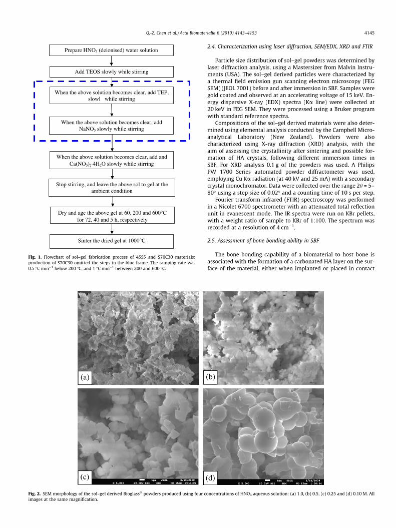
Fig. 2. SEM morphology of the sol–gel derived Bioglass� powders produced using four cimages at the same magnification.
Add TEOS slowly while stirring
When the above solution becomes clear, add TEP, slowl while stirring
When the above solution becomes clear, add and Ca(NO3)2⋅4H2O slowly while stirring
Dry and age the above gel at 60, 200 and 600°C for 72, 40 and 5 h, respectively
When the above solution becomes clear, add NaNO3 slowly while stirring
Stop stirring, and leave the above sol to gel at the ambient condition
Prepare HNO3 (deionised) water solution
Sinter the dried gel at 1000°C
Fig. 1. Flowchart of sol–gel fabrication process of 45S5 and S70C30 materials;production of S70C30 omitted the steps in the blue frame. The ramping rate was0.5 �C min�1 below 200 �C, and 1 �C min�1 between 200 and 600 �C.
Q.-Z. Chen et al. / Acta Biomaterialia 6 (2010) 4143–4153 4145
2.4. Characterization using laser diffraction, SEM/EDX, XRD and FTIR
Particle size distribution of sol–gel powders was determined bylaser diffraction analysis, using a Mastersizer from Malvin Instru-ments (USA). The sol–gel derived particles were characterized bya thermal field emission gun scanning electron microscopy (FEGSEM) (JEOL 7001) before and after immersion in SBF. Samples weregold coated and observed at an accelerating voltage of 15 keV. En-ergy dispersive X-ray (EDX) spectra (Ka line) were collected at20 keV in FEG SEM. They were processed using a Bruker programwith standard reference spectra.
Compositions of the sol–gel derived materials were also deter-mined using elemental analysis conducted by the Campbell Micro-analytical Laboratory (New Zealand). Powders were alsocharacterized using X-ray diffraction (XRD) analysis, with theaim of assessing the crystallinity after sintering and possible for-mation of HA crystals, following different immersion times inSBF. For XRD analysis 0.1 g of the powders was used. A PhilipsPW 1700 Series automated powder diffractometer was used,employing Cu Ka radiation (at 40 kV and 25 mA) with a secondarycrystal monochromator. Data were collected over the range 2h = 5–80� using a step size of 0.02� and a counting time of 10 s per step.
Fourier transform infrared (FTIR) spectroscopy was performedin a Nicolet 6700 spectrometer with an attenuated total reflectionunit in evanescent mode. The IR spectra were run on KBr pellets,with a weight ratio of sample to KBr of 1:100. The spectrum wasrecorded at a resolution of 4 cm�1.
2.5. Assessment of bone bonding ability in SBF
The bone bonding capability of a biomaterial to host bone isassociated with the formation of a carbonated HA layer on the sur-face of the material, either when implanted or placed in contact
oncentrations of HNO3 aqueous solution: (a) 1.0, (b) 0.5, (c) 0.25 and (d) 0.10 M. All

(a) 0.25 M
0
1
2
3
4
5
60
0.11
0.19
0.32
0.55
0.93
1.58
2.68
4.56
7.75
13.2
22.4
38.1
64.7
Particle Size / µm
Perc
enta
ge /
%
(b) 0.1 M
0
0.5
1
1.5
2
2.5
3
3.5
00.
10.
11
0.19
0.32
0.55
0.93
1.58
2.68
4.56
7.75
13.2
22.4
38.1
Particle Size / µm
Perc
enta
ge /
%
Fig. 3. Particle size distribution of the sol–gel derived 45S5 powders at HNO3
aqueous solution concentrations of (a) 0.25 and (b) 0.1 M. The average particle sizeswere 4.5 and 5.8 lm, respectively.
4146 Q.-Z. Chen et al. / Acta Biomaterialia 6 (2010) 4143–4153
with biological fluids [16,17]. Hence, the ability to bond with bonecan be preliminarily assessed in vitro in SBF by monitoring the for-mation of HA on its surface. To do this we used the standardin vitro procedure described by Kokubo et al. [18]. The foams wereimmersed in 75 ml of acellular SBF in flasks. The flasks were placedinside an incubator at 37 �C. The pH of the solution was maintainedconstant at 7.25. The size of all samples for these tests was10 � 10 � 10 mm. Two samples were extracted from the SBF solu-tion after given times of 7, 15 and 30 days. The SBF was replacedtwice per week because the cation concentration decreased duringthe course of the experiments as a result of changes in the chemicalcomposition of the samples. Once removed from the incubationmedium samples were rinsed gently, firstly in 100% ethanol, thenusing pure deionized water, and finally left to dry at ambient tem-perature in a desiccator. HA was identified from XRD diffractionpeaks.
2.6. Measurement of pH values and ion concentrations in the tissueculture medium
Samples (1 g) of the materials to be tested were soaked in 10 mlof tissue culture medium in 15 ml conical tubes under standardcell culture conditions within a culture incubator (37 �C in humid-ified air containing 5% CO2). Medium was collected and transferredto new tubes at different intervals up to 2 days. Acidity was mea-sured by insertion of a pH meter probe into the collected solutions,via a specifically designed incubator entrance.
After pH measurement the same solutions were used to analyseion concentrations. All solutions were analysed using ion chroma-tography. An ICS-1000 (Dionex) ion chromatograph with attachedautosampler was used for the analysis of sodium (Na+) and calcium
(Ca2+) ions. The ICS-2500 (Dionex) was used for the analysis ofphosphate (PO4
3�) and silicon (SiO42�) anions.
2.7. Biocompatibility evaluation: elution test method
MG63, a human osteoblast-like osteosarcoma cell line (ATCC),was used for biocompatibility assessment because of the well-de-fined and reproducible proliferative activity of these cells. In addi-tion, the elution test method (ISO 10993) was adopted in thepresent work. In this method extracts were obtained by placingthe test (sol–gel derived Bioglass� ceramics) and control (HA andNovabone� Bioglass�) materials in separate aliquots of cell culturemedium under standard conditions (0.2 g ml�1 of culture mediumfor 24 h at 37 �C). MG63 cells were cultured in Dulbecco’s modifiedEagle’s medium (DMEM) with 10% heat-inactivated fetal bovineserum, 0.1% penicillin/streptomycin at 37 �C with 5% CO2. Cellswere then plated in a 48-well tissue culture plate at a concentra-tion of 2 � 104 cells per well. After 2 days the cell culture mediumwas removed and replaced with medium containing the extracts.Cells were placed back in the incubator for a 24 h treatment. Cellswere observed under an optical microscope for visible signs of tox-icity in response to the test and control materials.
Quantification of cell viability was achieved by measuring lac-tate dehydrogenase (LDH) release using a commercial kit (Sig-ma–Aldrich Tox-7). Culture medium (200 lm per well) wascollected after MG63 cells had been exposed to the medium con-taining extract. The number of dead cells resulting from treatmentwith extractants was determined from these samples. The numberof live cells was measured using the total LDH method of Tox-7, inwhich live cells were lysed and the medium collected. The LDH lev-els were determined by measuring the absorbance (A490 � A690)using the commercial kit Tox-7 and a spectrophotometer. Ourstandard curve shows that there is a reasonably good linear rela-tionship between the number of cells and LDH level in the rangeof 5 � 103–5 � 104. Hence, the percentage of dead cells can be sim-ply given by the equation:
%dead cells ¼ LDH in extractant mediumtotal LDH
ð1Þ
2.8. Cell proliferation: Alamar blue
Cell proliferation was assessed using a commercial Alamarblue™ assay kit (Life Technologies). Alamar blue™ is non-toxic tocells. The assay does not interrupt cell culture, allowing continuousmeasurement of cell proliferation kinetics. Hence, the Alamarblue™ assay is appropriate to evaluate the long-term cytotoxicityof biomaterials due to biodegradation under physiological condi-tions. MG63 cells were seeded at 5000 cells ml�1 each well in a48-well plate and cultured in the medium with extract. Incubatedmedium in wells with neither cells nor testing materials extractswere used as negative controls. After culture for 48 h, 100 ll of Ala-mar blue™ indicator was added to each well (except backgroundcontrols) and incubated under culture conditions for 6 h. The med-ium was then transferred to a new plate, followed by absorbancedetermination at wavelengths of 570 and 600 nm in a spectropho-tometer (Thermo Scientific, Pathtech Australia).
The above procedures were repeated every 48 h until conflu-ence was reached (between days 6 and 8). Cell proliferation wasquantified by the percentage reduction of Alamar blue, i.e.
%reduced Alamar blue ¼ eoxðk2ÞAðk1Þ � eoxðk1ÞAðk2ÞeREDðk1ÞA�ðk2Þ � eREDðk2ÞA�ðk1Þ
� 100;
ð2Þ
where A(k1) and A(k2) are the absorbance values of test wells mea-sured at wavelengths k1 and k2 and A�ðk1Þ and A�ðk2Þ are the values

0.5
0.4
0.3
0.2
0.1
0
0.4
0.3
0.2
0.1
0
0.4
0.3
0.2
0.1
0
0.4
0.3
0.2
0.1
0
0.4
0.3
0.2
0.1
0
NovaBone Bioglass®
Sintered NovaBone Bioglass®
Sol-gel derived and sintered Bioglass®, 0.25M
Ca(NO3)24H2O
NaNO3
NO3
H2O
SiO2
PO2
NO3
SiO2
SiO2
PO4
PO4
Crys
Crys
2.500 2.000 1.500 1.000 500Wavenumber/ cm-1
Abs
orba
nce
Fig. 4. FTIR spectra of two nitrate precursors and commercial Bioglass� (with and without sintering as indicated) and the sol–gel derived Bioglass� sintered at 1000 �C for 2 h.The FTIR spectra profiles of the four powders are the same. The spectra of the sol–gel derived Bioglass� ceramics have the same profile as that of the commercial NovaBone45S5 Bioglass� sintered at 1000 �C for 2 h.
Q.-Z. Chen et al. / Acta Biomaterialia 6 (2010) 4143–4153 4147
of absorbance of negative control wells containing only mediumand Alamar blue™ without cells. All values were blanked with thereadings of background controls. The other parameters in Eq. (2)are as follows: eoxðk1Þ ¼ 80:586, eoxðk2Þ ¼ 117:216, eREDðk1Þ ¼155:677 and eREDðk2Þ ¼ 14:652.
2.9. Statistics
All experiments were run with five samples and the data wererepresented as means ± SE. Statistical difference was analysedusing one-way analysis of variance (ANOVA) with Tukey’s posthoc test, and a P value of <0.05 was considered significant.
3. Results and discussion
3.1. Particle size of the sol–gel derived 45S5 powders
As the molar concentration of HNO3 was decreased from 1 to0.1 M the average particle size in the powder increased fromapproximately 1–5 lm, as estimated by SEM observation (Fig. 2).However, the fine particles made using the 0.5 and 1 M HNO3 solu-tions were severely agglomerated, such that the bulk samples had
to be mechanically ground to break down the agglomerated parti-cles. In contrast, the microspheres of the powders produced in the0.1 and 0.25 M HNO3 solutions were loosely packed, resulting inwell-separated and fine particle powders. Hence, the rest of thisstudy characterized and evaluated the powders produced usingthe 0.1 and 0.25 M HNO3 solutions.
Fig. 3 shows the powder size distribution of sol–gel powdersproduced in the 0.1 and 0.25 M HNO3 solutions. The peak of thesize distribution profile is located at around 4.5 lm for both pow-ders. For the powder produced in the 0.25 M solution the sized ob-served by SEM (Fig. 2c) appeared smaller than 4.5 lm, which,however, may be attributed to the slight agglomeration of particlesin this powder due to the slightly higher concentration of catalystHNO3. The particle size on average was between 4.5 and 5.8 lm indiameter for powders produced in 0.25 and 0.1 M HNO3 solution,respectively.
3.2. FTIR analysis of as sintered sol–gel derived 45S5 powders
The sol–gel process for producing S70C30 materials is now wellestablished [19]. Hence, the FTIR analysis in this work focused onthe 45S5 material produced with the newly developed sol–gel rec-ipe. FTIR (Fig. 4) confirmed the decomposition of the two nitrates,
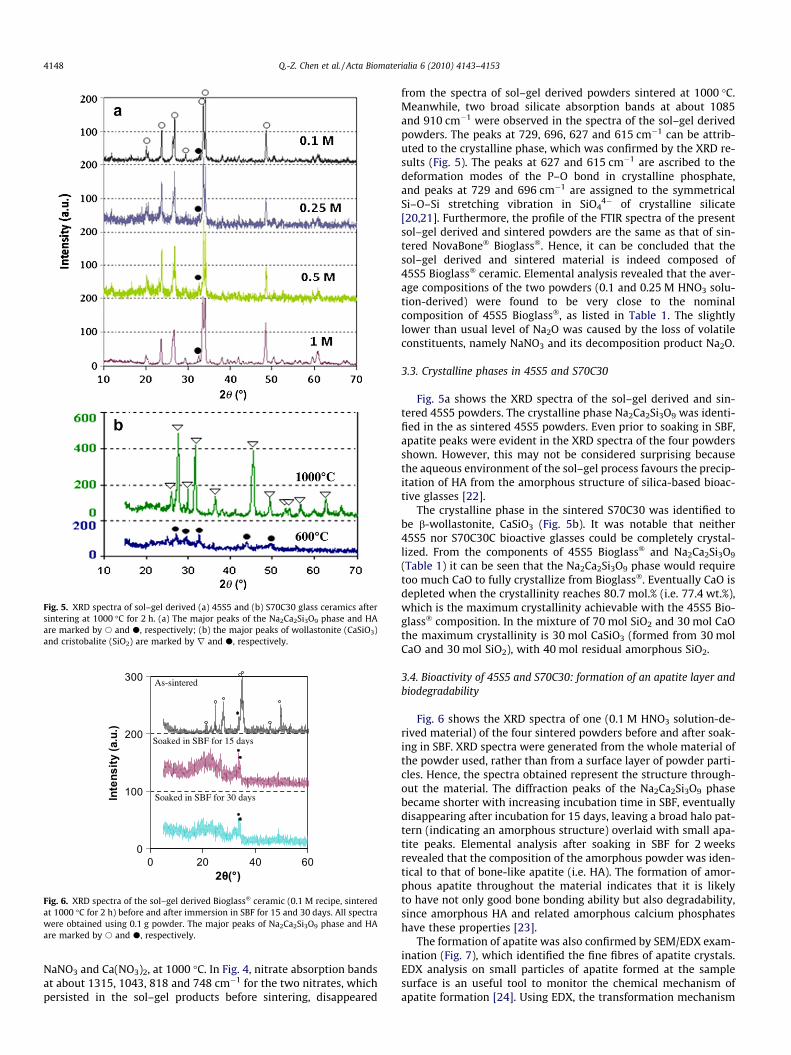
Fig. 5. XRD spectra of sol–gel derived (a) 45S5 and (b) S70C30 glass ceramics aftersintering at 1000 �C for 2 h. (a) The major peaks of the Na2Ca2Si3O9 phase and HAare marked by s and d, respectively; (b) the major peaks of wollastonite (CaSiO3)and cristobalite (SiO2) are marked by r and d, respectively.
0
100
200
300
0 20 40 602θ(°)
Inte
nsity
(a.u
.)
As-sintered
Soaked in SBF for 15 days
Soaked in SBF for 30 days
Fig. 6. XRD spectra of the sol–gel derived Bioglass� ceramic (0.1 M recipe, sinteredat 1000 �C for 2 h) before and after immersion in SBF for 15 and 30 days. All spectrawere obtained using 0.1 g powder. The major peaks of Na2Ca2Si3O9 phase and HAare marked by s and d, respectively.
4148 Q.-Z. Chen et al. / Acta Biomaterialia 6 (2010) 4143–4153
NaNO3 and Ca(NO3)2, at 1000 �C. In Fig. 4, nitrate absorption bandsat about 1315, 1043, 818 and 748 cm�1 for the two nitrates, whichpersisted in the sol–gel products before sintering, disappeared
from the spectra of sol–gel derived powders sintered at 1000 �C.Meanwhile, two broad silicate absorption bands at about 1085and 910 cm�1 were observed in the spectra of the sol–gel derivedpowders. The peaks at 729, 696, 627 and 615 cm�1 can be attrib-uted to the crystalline phase, which was confirmed by the XRD re-sults (Fig. 5). The peaks at 627 and 615 cm�1 are ascribed to thedeformation modes of the P–O bond in crystalline phosphate,and peaks at 729 and 696 cm�1 are assigned to the symmetricalSi–O–Si stretching vibration in SiO4
4� of crystalline silicate[20,21]. Furthermore, the profile of the FTIR spectra of the presentsol–gel derived and sintered powders are the same as that of sin-tered NovaBone� Bioglass�. Hence, it can be concluded that thesol–gel derived and sintered material is indeed composed of45S5 Bioglass� ceramic. Elemental analysis revealed that the aver-age compositions of the two powders (0.1 and 0.25 M HNO3 solu-tion-derived) were found to be very close to the nominalcomposition of 45S5 Bioglass�, as listed in Table 1. The slightlylower than usual level of Na2O was caused by the loss of volatileconstituents, namely NaNO3 and its decomposition product Na2O.
3.3. Crystalline phases in 45S5 and S70C30
Fig. 5a shows the XRD spectra of the sol–gel derived and sin-tered 45S5 powders. The crystalline phase Na2Ca2Si3O9 was identi-fied in the as sintered 45S5 powders. Even prior to soaking in SBF,apatite peaks were evident in the XRD spectra of the four powdersshown. However, this may not be considered surprising becausethe aqueous environment of the sol–gel process favours the precip-itation of HA from the amorphous structure of silica-based bioac-tive glasses [22].
The crystalline phase in the sintered S70C30 was identified tobe b-wollastonite, CaSiO3 (Fig. 5b). It was notable that neither45S5 nor S70C30C bioactive glasses could be completely crystal-lized. From the components of 45S5 Bioglass� and Na2Ca2Si3O9
(Table 1) it can be seen that the Na2Ca2Si3O9 phase would requiretoo much CaO to fully crystallize from Bioglass�. Eventually CaO isdepleted when the crystallinity reaches 80.7 mol.% (i.e. 77.4 wt.%),which is the maximum crystallinity achievable with the 45S5 Bio-glass� composition. In the mixture of 70 mol SiO2 and 30 mol CaOthe maximum crystallinity is 30 mol CaSiO3 (formed from 30 molCaO and 30 mol SiO2), with 40 mol residual amorphous SiO2.
3.4. Bioactivity of 45S5 and S70C30: formation of an apatite layer andbiodegradability
Fig. 6 shows the XRD spectra of one (0.1 M HNO3 solution-de-rived material) of the four sintered powders before and after soak-ing in SBF. XRD spectra were generated from the whole material ofthe powder used, rather than from a surface layer of powder parti-cles. Hence, the spectra obtained represent the structure through-out the material. The diffraction peaks of the Na2Ca2Si3O9 phasebecame shorter with increasing incubation time in SBF, eventuallydisappearing after incubation for 15 days, leaving a broad halo pat-tern (indicating an amorphous structure) overlaid with small apa-tite peaks. Elemental analysis after soaking in SBF for 2 weeksrevealed that the composition of the amorphous powder was iden-tical to that of bone-like apatite (i.e. HA). The formation of amor-phous apatite throughout the material indicates that it is likelyto have not only good bone bonding ability but also degradability,since amorphous HA and related amorphous calcium phosphateshave these properties [23].
The formation of apatite was also confirmed by SEM/EDX exam-ination (Fig. 7), which identified the fine fibres of apatite crystals.EDX analysis on small particles of apatite formed at the samplesurface is an useful tool to monitor the chemical mechanism ofapatite formation [24]. Using EDX, the transformation mechanism

5 μm
(a)
5 μm 0
10
2030
40
50
6070
80
90
P Ca O Na SiElement
Ato
mic
%
(b)
Nominal 45S5
Nominal CaP/HA EDX
Fig. 7. (a) SEM images and (b) EDX analysis of apatite formed on the sol–gel derived Bioglass� ceramics (0.25 M recipe, sintered at 1000 �C for 2 h). Samples were soaked inSBF for 2 weeks.
Fig. 8. (a) X-ray diffraction patters of sol–gel derived (unsintered) S70C30C glass before and after immersion in SBF. The major peaks of calcite (CaCO3) and HA are marked byr and d, respectively. (b) XRD patterns of the sintered (at 1000 �C for 2 h) S70C30C glass ceramic before and after immersion in SBF. The major peaks of wollastonite (CaSiO3)are marked by r. All spectra were obtained using 0.1 g powder.
Fig. 9. SEM images of precipitates formed on the sol–gel derived (a) S70C30 glass and (b) S70C30 glass ceramics sintered at 1000 �C for 2 h. Samples were soaked in SBF for2 weeks.
Q.-Z. Chen et al. / Acta Biomaterialia 6 (2010) 4143–4153 4149
0
10
20
30
40
50
60
70
80
Element
Ato
mic
%a
S70C30
EDX
HA/CaP
3 days
0
10
20
30
40
50
60
70
80
P Ca O Si P Ca O Si Element
Ato
mic
%
S70C30
EDX
HA/CaP
4 weeks b
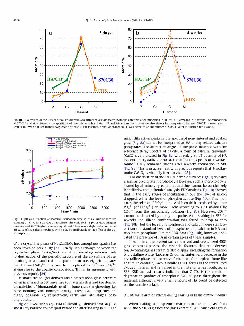
Fig. 10. EDX results for the surface of sol–gel derived S70C30 bioactive glass foams (without sintering) after immersion in SBF for (a) 3 days and (b) 4 weeks. The compositionof S70C30 and stoichiometric compositions of two calcium phosphates (HA and tricalcium phosphate) are also shown for comparison. Sintered S70C30 showed similarresults, but with a much more slowly changing profile. For instance, a similar change to (a) was detected on the surface of S70C30 after incubation for 4 weeks.
7
7.5
8
8.5
9
9.5
0 500 1000 1500 2000 2500 3000Time / min
pH v
alue
Medium OnlyNovaBone BioglassSol-gel 45S5 glass-ceramicsSol-gel 70S30 glass-ceramicsSol-gel 70S30 glass
Fig. 11. pH as a function of material incubation time in tissue culture medium(DMEM) at 37 �C in a 5% CO2 atmosphere. The variations in pH of 45S5 Bioglassceramics and S70C30 glass were not significant. There was a slight reduction in thepH value of the culture medium, which may be attributable to the effect of the CO2
atmosphere.
4150 Q.-Z. Chen et al. / Acta Biomaterialia 6 (2010) 4143–4153
of the crystalline phase of Na2Ca2Si3O9 into amorphous apatite hasbeen revealed previously [24]. Briefly, ion exchange between thecrystalline phase Na2Ca2Si3O9 and its surrounding solution leadsto destruction of the periodic structure of the crystalline phase,resulting in a disordered amorphous structure. Fig. 7b indicatesthat Na+ and SiO4
2� ions have been replaced by Ca2+ and PO43�,
giving rise to the apatite composition. This is in agreement withprevious reports [24].
In short, the sol–gel derived and sintered 45S5 glass ceramicswhen immersed in SBF gave rise to materials that had the desiredbioactivities of biomaterials used in bone tissue engineering, i.e.bone bonding and biodegradability. These two properties arehighly desirable at, respectively, early and late stages post-implantation.
Fig. 8 shows the XRD spectra of the sol–gel derived S70C30 glassand its crystallized counterpart before and after soaking in SBF. The
major diffraction peaks in the spectra of non-sintered and soakedglass (Fig. 8a) cannot be interpreted as HA or any related calciumphosphates. The diffraction angles of the peaks matched with thereference X-ray spectra of calcite, a form of calcium carbonate(CaCO3), as indicated in Fig. 8a, with only a small quantity of HAevident. In crystallized S70C30 the diffractions peaks of b-wollas-tonite CaSiO3 remained strong after 4 weeks incubation in SBF(Fig. 8b). This is in agreement with previous reports that b-wollas-tonite CaSiO3 is virtually inert in vivo [25].
SEM observation of the S70C30 sample surfaces (Fig. 9) revealeda similar precipitate morphology. However, such a morphology isshared by all mineral precipitates and thus cannot be conclusivelyidentified without chemical analysis. EDX analysis (Fig. 10) showedthat in the early stages of incubation in SBF the level of silicondropped, while the level of phosphorus rose (Fig. 10a). This indi-cates the release of SiO4
2� ions, which could be replaced by eitherPO4
3� (or HPO42�) or, more likely according to XRD analysis, by
CO32� from the surrounding solution (Fig. 8a). However, CO3
2�
cannot be detected by a polymer probe. After soaking in SBF for4 weeks the silicon concentration was found to drop to zero(Fig. 10b), but the levels of phosphorus and calcium were still low-er than the standard levels of phosphorus and calcium in HA andtricalcium phosphate. Limited EDX data (Fig. 10b), however, indi-cated the presence of HA in certain areas of these samples.
In summary, the present sol–gel derived and crystallized 45S5glass ceramics possess the essential features that melt-derivedNa2O-containg glass ceramics also display, including the formationof crystalline phase Na2Ca2Si3O9 during sintering, a decrease in thecrystalline phase and extensive formation of amorphous bone-likeapatite. In contrast, b-wollastonite CaSiO3 forms in the crystallizedS70C30 material and remained in the material when incubated inSBF. XRD analysis clearly indicated that CaCO3 is the dominantdegradation product of amorphous S70C30 glass throughout thematerial, although a very small amount of HA could be detectedon the sample surface.
3.5. pH value and ion release during soaking in tissue culture medium
When soaking in an aqueous environment the ion release from45S5 and S70C30 glasses and glass ceramics will cause changes in

(a) NovaBone Bioglass
0
50
100
150
200
250
300
0 500 1000 1500 2000 2500 3000Incubation time / min
Incr
emen
t in
ion
conc
entr
atio
n / m
g/L
Na
Ca
PSi
(b) Sol-gel Derived sintered 45S5 glass-ceramics
0
50
100
150
200
250
300
0 500 1000 1500 2000 2500 3000Incubation time / min
Incr
emen
t in
ion
conc
entra
tion
/ mg/
L
NaCaPSi
(d) Sol-gel derived sintered 70S30 glass-ceramics
-30
-10
10
30
50
70
90
0 500 1000 1500 2000 2500 3000
Incubation time / min
Incr
emen
t in
ion
conc
entra
tion
/ mg/
L
NaCaPSi
(c) Sol-gel derived 70S30 glass
-30
-10
10
30
50
70
90
110
130
0 500 1000 1500 2000 2500 3000
Incubation time / min
Incr
emen
t in
ion
conc
entra
tion
/ mg/
L
NaCaPSi
Fig. 12. Analysis of Na, Ca, P and Si in tissue culture medium (DMEM) in which the following materials were immersed for 48 h: (a) NovaBone 45S5 Bioglass�; (b) sol–gelderived and sintered 45S5 glass ceramic; (c) sol–gel derived S70C30 glass and (d) sol–gel derived and sintered S70C30 glass ceramic.
0102030405060708090
100
Day 0 Day 2 Day 4 Day 6Tissue Culture Duration
% R
educ
ed
HANovaBoneSol-gel 45S5 (0.1M)Sol-gel 45S5 (0.25M)
Fig. 13. MG63 cell proliferation kinetics measured by the Alamar blue™ technique.The initial plating density was 5000 cells ml�1 in each well of a 48-well plate (n = 3).Medium with hydroxyapatite and commercial 45S5 Bioglass� (NovaBone) extractswere positive controls. Two types of sol–gel derived and sintered 45S5 glassceramics were made with 0.1 and 0.25 M HNO3 solutions as indicated. Sol–gelderived 45S5 and Novabone™ 45S5 materials were sintered under the sameconditions (1000 �C for 2 h). The differences between NovaBone™ and the sol–gelderived 45S5 materials were not significant (P > 0.05) on days 2–6. The differencesbetween HA, NovaBone™ and the sol–gel derived 45S5 materials were notsignificant (P > 0.05) on days 2–4. On day 6 the significance values were HA vs.BG_0.1 M (P < 0.01), HA vs. Novabone (P < 0.01), HA vs. BG_0.25 M (P > 0.05).
Q.-Z. Chen et al. / Acta Biomaterialia 6 (2010) 4143–4153 4151
the pH value and ion concentration of the surrounding fluid. Previ-ous studies on this subject have been conducted with deionizedwater, phosphate-buttered saline (PBS), SBF and tris-buffered solu-tions [10,12,19,26,27]. We focused on the results of soaking inDMEM tissue culture medium, which is relevant to the evaluationof the biocompatibility of the materials by cell culture methods.
Immersion of 45S5 Bioglass� increased the pH value of DMEMrapidly, reaching a saturated state (pH �9) after 24 h (Fig. 11). Thissteady value is lower than the value of pH �11 in a previous reportusing PBS [26], due probably to incubation in a 5% CO2 atmospherein the present work. The effects of sintered 45S5 glass ceramics andnon-sintered S70C30 glass on the pH of the culture medium werenot significantly different (P > 0.05), and the sintered S70C30 glassceramic caused the least increase in pH value of the culturemedium.
The increment in pH values due to 45S5 materials can be attrib-uted to the fast release of Na+ ions, as shown in Fig. 12. A compar-ison of Fig. 12a and b reveals that ion release was slower in thecrystallized 45S5 glass ceramic. This was also observed with theS70C30 material (Fig. 12c and d). The ion release from 45S5 Bio-glass� observed in the present work was generally slower com-pared with those previously reported in studies using deionizedwater [26]. This can be explained by certain differences in theexperimental procedures, namely that in the previous study waterwas replaced with fresh deionized water after each measurement,whereas in the present work medium was not changed after eachmeasurement during the 2 day experiment. The procedure used inthis work is in accordance with standard tissue culture practice, i.e.culture medium is changed every 2 days. Hence, the slower ion re-
lease from bioactive glasses in the present study was the result ofnon-replacement of the medium, because the released ion in themedium would slow down further release of ions from the materi-als into solution.

Fig. 14. Cytotoxicity of the test materials using MG63 cells, detected by measuringthe release of lactate dehydrogenase (LDH) into medium containing the extractedsubstances. Medium with HA and commercial Bioglass� (NovaBone�) extractsserved as positive controls. Two types of sol–gel derived and sintered 45S5 glassceramics were made with 0.1 and 0.25 M HNO3 solution as indicated. Sol–gelderived 45S5 and Novabone™ 45S5 materials were sintered under the sameconditions (1000 �C for 2 h). No significant differences were found in the percentageof dead cells between any two of the four groups (P > 0.05).
4152 Q.-Z. Chen et al. / Acta Biomaterialia 6 (2010) 4143–4153
3.6. Evaluation of cytocompatibility
The biocompatibility of melt-derived 45S5 and sol–gel derivedS70C30 has been extensively evaluated previously, both in vitroand in vivo [28–30]. Given that the current sol–gel derived 45S5was produced by a completely new process which involved anacidic catalyst HNO3, it was essential to evaluate the biocompati-bility of the materials, using the cytocompatibility standard de-scribed by ISO 10993 and required by the FDA. Melt-derived45S5 Bioglass� (NovaBone) and HA were used as controls in thiswork.
Medium containing the extracts of sintered powders was foundto support proliferation of MG63 cells and visual microscopicobservation could identify no gross differences in cell proliferationin the following four groups of samples: HA, commercial (Nova-Bone�) Bioglass� and sol–gel derived Bioglass� ceramics (0.1and 0.25 M). This was quantitatively confirmed using the Alamarblue™ technique (Fig. 13). This revealed that none of the growthparameters of MG63 cells were statistically different betweenthe four groups of medium tested, with no significant differenceseen between any two of the cellular growth kinetic curves(P > 0.05). The sol–gel derived and sintered glass ceramics werefound to have very similar cytocompatibilities to both HA andcommercial NovaBone� Bioglass�, as indicated in Fig. 14. The pro-portions of dead cells observed in MG63 cultures exposed to thedifferent types of Bioglass� or HA were not significantly different(P > 0.05).
4. Conclusions
Fine powders of Na2O-containing glass ceramics have beensuccessfully synthesized using the sol–gel technique in aqueoussolution under ambient conditions. Microspheres of size �5 lmcan be easily achieved at low cost, eliminating the time-consum-ing processes, i.e. grinding and sieving. The sol–gel derived andsintered 45S5 glass ceramic materials possess the essential fea-tures of Na2O-containing bioactive materials, namely the forma-tion of crystalline phase Na2Ca2Si3O9 during sintering, a decreasein the crystalline phase and extensive formation of amorphousbone-like apatite. A comparative study on Na2O-containing andNa2O-free bioactive glass ceramics indicated that Na2O (or oxidesof other active elements, such as potassium) could be an impor-tant constituent enabling achievement of an optimal combinationof sound mechanical properties and good biodegradability in onematerial.
Acknowledgement
Q.-Z.C. would like to acknowledge support from the New StaffResearch Grants and Small Research Grants of the Faculty of Engi-neering at Monash University.
Appendix A. Figures with essential colour discrimination
Certain figures in this article, particularly Figures 1, 3–8 and 10–13, are difficult to interpret in black and white. The full colourimages can be found in the on-line version, at doi:10.1016/j.actbio.2010.04.019.
References
[1] Best SM, Porter AE, Thian ES, Huang J. Bioceramics: past, present and for thefuture. J Eur Ceram Soc 2008;28:1319.
[2] Jones JR. New trends in bioactive scaffolds: the importance of nanostructure. JEur Ceram Soc 2009;29:1275.
[3] Hench LL. Sol–gel materials for bioceramic applications. Curr Opin Solid StateMater Sci 1997;2:604.
[4] Ramila A, Balas F, Vallet-Regi M. Synthesis routes for bioactive sol–gel glasses:alkoxides versus nitrates. Chem Mater 2002;14:542.
[5] Balamurugan A, Ballossier G, Michel J, Kannan S, Benhayoune H, Rebelo AHS,et al. Sol gel derived SiO2–CaO–MgO–P2O5 bioglass system-preparation andin vitro characterization. J Biomed Mater Res Part B Appl Biomater 2007;83B:546.
[6] Chen QZ, Thompson ID, Boccaccini AR. 45S5 Bioglass (R)-derived glass–ceramic scaffolds for bone tissue engineering. Biomaterials 2006;27:2414.
[7] Bao HD, Guo ZX, Yu J. Effect of electrically inert particulate filler on electricalresistivity of polymer/multi-walled carbon nanotube composites. Polymer2008;49:3826.
[8] Carta D, Pickup DM, Knowles JC, Smith ME, Newport RJ. Sol–gel synthesis ofthe P2O5–CaO–Na2O–SiO2 system as a novel bioresorbable glass. J Mater Chem2005;15:2134.
[9] Peitl O, LaTorre GP, Hench LL. Effect of crystallization on apatite-layerformation of bioactive glass 45S5. J Biomed Mater Res 1996;30:509.
[10] Peitl O, Zanotto ED, Hench LL. Highly bioactive P2O5–Na2O–CaO–SiO2 glass–ceramics. J Non-Cryst Solids 2001;292:115.
[11] Clupper DC, Mecholsky JJ, LaTorre GP, Greenspan DC. Sintering temperatureeffects on the in vitro bioactive response of tape cast and sintered bioactiveglass–ceramic in tris buffer. J Biomed Mater Res 2001;57:532.
[12] Clupper DC, Mecholsky JJ, LaTorre GP, Greenspan DC. Bioactivity of tape castand sintered bioactive glass–ceramic in simulated body fluid. Biomaterials2002;23:2599.
[13] Hoshino Y, Utsunomiya T, Abe O. The thermal-decomposition of sodium-nitrate and the effects of several oxides on the decomposition. Bull Chem SocJpn 1981;54:1385.
[14] Ettarh C, Galwey AK. A kinetic and mechanistic study of the thermaldecomposition of calcium nitrate. Thermochim Acta 1996;288:203.
[15] Clupper DC, Hench LL. Crystallization kinetics of tape cast bioactive glass 45S5.J Non-Cryst Solids 2003;318:43.
[16] Hench LL. Theory of bioactivity: the potential for skeletal regeneration. Analesde Quim 1997;93:S44.
[17] Hench LL, Wilson J. Surface-active biomaterials. Science 1984;226:630.[18] Kokubo T, Hata K, Nakamura T, Yamamura T. Apatite formation on ceramics,
metals, and polymers induced by a CaO–SiO2-based glass in simulated bodyfluid. In: Bonfield W, Hastings GW, Tanner KE, editors. Bioceramics, vol. 4.London: Butterworth-Heinemainn; 1991. p. 113.
[19] Saravanapavan P, Hench LL. Dissolution of bioactive gel–glass powders in theSiO2–CaO system. Key Eng Mat 2003;240–242:213.
[20] Boccaccini AR, Chen Q, Lefebvre L, Gremillard L, Chevalier J. Sintering,crystallisation and biodegradation behaviour of bioglass (R)-derived glass–ceramics. Faraday Discuss 2007;136:27.
[21] Lefebvre L, Chevalier J, Gremillard L, Zenati R, Thollet G, Bernache-Assolant D,et al. Structural transformations of bioactive glass 45S5 with thermaltreatments. Acta Mater 2007;55:3305.
[22] Bellantone M, Hench LL. Bioactive behaviour of sol–gel derived antibacterialbioactive glass. Bioceramics 2000;192–1:617.
[23] Hench LL. Bioceramics. J Am Ceram Soc 1998;81:1705.[24] Chen QZ, Rezwan K, Francon V, Armitage D, Nazhat SN, Jones FH, et al. Surface
functionalization of bioglass((R))-derived porous scaffolds. Acta Biomater2007;3:551.
[25] Yamamuro T. A/W glass–ceramics: clinical application. In: Hench LL, Wilson J,editors. An introduction to biomaterials. Singapore: World Scientific; 1999.p. 75.
[26] Chen QZ, Ahmed I, Knowles JC, Nazhat SN, Boccaccini AR, Rezwan K. Collagenrelease kinetics of surface functionalized 4555 bioglass (R)-based porousscaffolds. J Biomed Mater Res Part A 2008;86A:987.

Q.-Z. Chen et al. / Acta Biomaterialia 6 (2010) 4143–4153 4153
[27] Saravanapavan P, Jones JR, Pryce RS, Hench LL. Bioactivity of gel–glass powdersin the CaO–SiO2 system: a comparison with ternary (CaO–P2O5–SiO2) andquaternary glasses (SiO2–CaO–P2O5–Na2O). J Biomed Mater Res Part A2003;66A:110.
[28] Saravanapavan P, Selvakumaran J, Hench LL. Indirect cytotoxicity evaluation ofsoluble silica, calcium, phosphate silver ions. Bioceramics 16 2004;254–2:785.
[29] Saravanapavan P, Jones JR, Verrier S, Beilby R, Shirtliff VJ, Hench LL, et al.Binary CaO–SiO2 gel–glasses for biomedical applications. Bio-med Mater Eng2004;14:467.
[30] Stanley HR, Hall MB, Clark AE, King CJ, Hench LL, Berte JJ. Using 45S5 bioglasscones as endosseous ridge maintenance implants to prevent alveolar ridgeresorption: a 5-year evaluation. Int J Oral Maxillofacial Implants 1997;12:95.