The high-conductance channel of porin-less yeast mitochondria
Upload
independentCategory
view
0download
0
A MITOCHONDRIA-TARGETED NITROXIDE/HEMIGRAMICIDIN SCONJUGATE PROTECTS MOUSE EMBRYONIC CELLS AGAINSTGAMMA IRRADIATION
Jianfei Jiang, Ph.D.*,†, Natalia A. Belikova, Ph.D.*,†, Adam T. Hoye, B.A.*,‡, Qing Zhao,B.S.*,†, Michael W. Epperly, Ph.D.*,§, Joel S. Greenberger, M.D.*,§, Peter Wipf, Ph.D.*,‡, andValerian E. Kagan, Ph.D., D.Sci.*,†* Center for Medical Countermeasures Against Radiation, University of Pittsburgh, Pittsburgh, PA
† Center for Free Radical and Antioxidant Health, Department of Environmental and Occupational Health,University of Pittsburgh, Pittsburgh, PA
‡ Department of Chemistry, University of Pittsburgh, Pittsburgh, PA
§ Department of Radiation Oncology, University of Pittsburgh, Pittsburgh, PA
AbstractPurpose—To evaluate the in vitro radioprotective effect of the mitochondria-targetedhemigramicidin S–conjugated 4-amino-2,2,6,6-tetramethyl-piperidine-N-oxyl (hemi-GS-TEMPO)5-125 in γ-irradiated mouse embryonic cells and adenovirus-12 SV40 hybrid virus transformedhuman bronchial epithelial cells BEAS-2B and explore the mechanisms involved in itsradioprotective effect.
Methods and Materials—Cells were incubated with 5-125 before (10 minutes) or after (1 hour)γ-irradiation. Superoxide generation was determined by using dihydroethidium assay, and lipidoxidation was quantitated by using a fluorescence high-performance liquid chromatography–basedAmplex Red assay. Apoptosis was characterized by evaluating the accumulation of cytochrome c inthe cytosol and externalization of phosphatidylserine on the cell surface. Cell survival was measuredby means of a clonogenic assay.
Results—Treatment (before and after irradiation) of cells with 5-125 at low concentrations (5, 10,and 20 μM) effectively suppressed γ-irradiation–induced superoxide generation, cardiolipinoxidation, and delayed irradiation–induced apoptosis, evaluated by using cytochrome c release andphosphatidylserine externalization. Importantly, treatment with 5-125 increased the clonogenicsurvival rate of γ-irradiated cells. In addition, 5-125 enhanced and prolonged γ-irradiation–inducedG2/M phase arrest.
Conclusions—Radioprotection/mitigation by hemi-GS-TEMPO likely is caused by its ability toact as an electron scavenger and prevent superoxide generation, attenuate cardiolipin oxidation inmitochondria, and hence prevent the release of proapoptotic factors from mitochondria. Othermechanisms, including cell-cycle arrest at the G2/M phase, may contribute to the protection.
Reprint requests to: Valerian E. Kagan, Ph.D, D.Sci., Department of Environmental and Occupational Health, University of Pitts-burgh,Bridgeside Point, 100 Technology Drive, Suite 333, Pitts-burgh, PA 15219. Tel: (412) 624-9479; Fax: (412) 624-9361; E-mail:[email protected] of interest: none.
NIH Public AccessAuthor ManuscriptInt J Radiat Oncol Biol Phys. Author manuscript; available in PMC 2009 March 1.
Published in final edited form as:Int J Radiat Oncol Biol Phys. 2008 March 1; 70(3): 816–825. doi:10.1016/j.ijrobp.2007.10.047.
NIH
-PA Author Manuscript
NIH
-PA Author Manuscript
NIH
-PA Author Manuscript
Keywordsγ-Irradiation; Hemigramicidin S-conjugated nitroxide; Reactive oxygen species; Cardiolipinoxidation; Cytochrome c
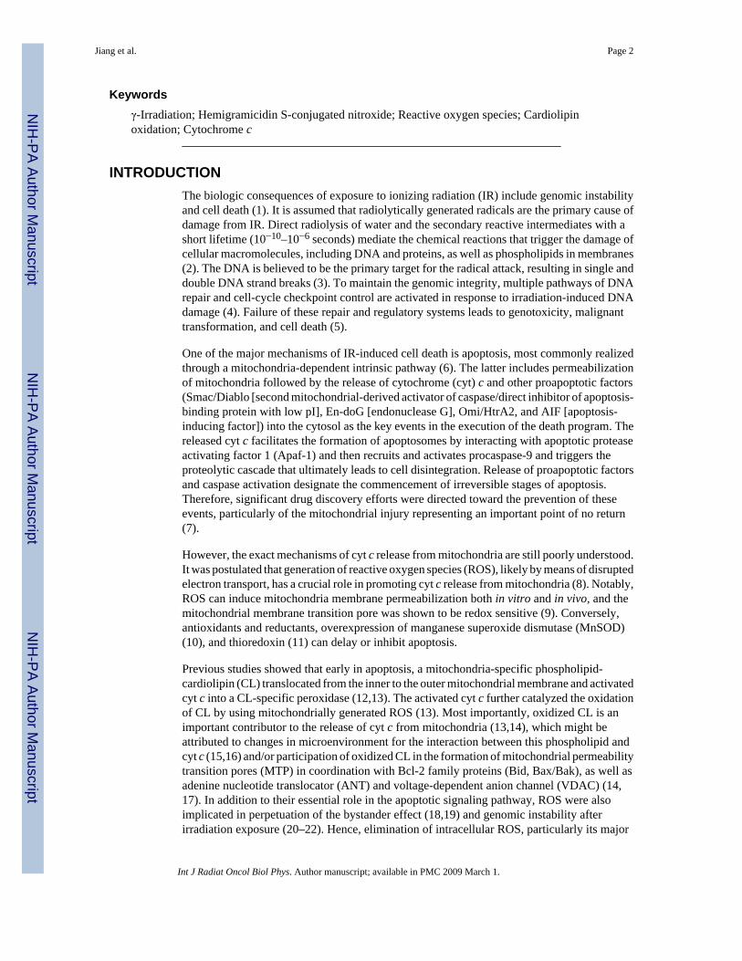
INTRODUCTIONThe biologic consequences of exposure to ionizing radiation (IR) include genomic instabilityand cell death (1). It is assumed that radiolytically generated radicals are the primary cause ofdamage from IR. Direct radiolysis of water and the secondary reactive intermediates with ashort lifetime (10−10–10−6 seconds) mediate the chemical reactions that trigger the damage ofcellular macromolecules, including DNA and proteins, as well as phospholipids in membranes(2). The DNA is believed to be the primary target for the radical attack, resulting in single anddouble DNA strand breaks (3). To maintain the genomic integrity, multiple pathways of DNArepair and cell-cycle checkpoint control are activated in response to irradiation-induced DNAdamage (4). Failure of these repair and regulatory systems leads to genotoxicity, malignanttransformation, and cell death (5).
One of the major mechanisms of IR-induced cell death is apoptosis, most commonly realizedthrough a mitochondria-dependent intrinsic pathway (6). The latter includes permeabilizationof mitochondria followed by the release of cytochrome (cyt) c and other proapoptotic factors(Smac/Diablo [second mitochondrial-derived activator of caspase/direct inhibitor of apoptosis-binding protein with low pI], En-doG [endonuclease G], Omi/HtrA2, and AIF [apoptosis-inducing factor]) into the cytosol as the key events in the execution of the death program. Thereleased cyt c facilitates the formation of apoptosomes by interacting with apoptotic proteaseactivating factor 1 (Apaf-1) and then recruits and activates procaspase-9 and triggers theproteolytic cascade that ultimately leads to cell disintegration. Release of proapoptotic factorsand caspase activation designate the commencement of irreversible stages of apoptosis.Therefore, significant drug discovery efforts were directed toward the prevention of theseevents, particularly of the mitochondrial injury representing an important point of no return(7).
However, the exact mechanisms of cyt c release from mitochondria are still poorly understood.It was postulated that generation of reactive oxygen species (ROS), likely by means of disruptedelectron transport, has a crucial role in promoting cyt c release from mitochondria (8). Notably,ROS can induce mitochondria membrane permeabilization both in vitro and in vivo, and themitochondrial membrane transition pore was shown to be redox sensitive (9). Conversely,antioxidants and reductants, overexpression of manganese superoxide dismutase (MnSOD)(10), and thioredoxin (11) can delay or inhibit apoptosis.
Previous studies showed that early in apoptosis, a mitochondria-specific phospholipid-cardiolipin (CL) translocated from the inner to the outer mitochondrial membrane and activatedcyt c into a CL-specific peroxidase (12,13). The activated cyt c further catalyzed the oxidationof CL by using mitochondrially generated ROS (13). Most importantly, oxidized CL is animportant contributor to the release of cyt c from mitochondria (13,14), which might beattributed to changes in microenvironment for the interaction between this phospholipid andcyt c (15,16) and/or participation of oxidized CL in the formation of mitochondrial permeabilitytransition pores (MTP) in coordination with Bcl-2 family proteins (Bid, Bax/Bak), as well asadenine nucleotide translocator (ANT) and voltage-dependent anion channel (VDAC) (14,17). In addition to their essential role in the apoptotic signaling pathway, ROS were alsoimplicated in perpetuation of the bystander effect (18,19) and genomic instability afterirradiation exposure (20–22). Hence, elimination of intracellular ROS, particularly its major
Jiang et al. Page 2
Int J Radiat Oncol Biol Phys. Author manuscript; available in PMC 2009 March 1.
NIH
-PA Author Manuscript
NIH
-PA Author Manuscript
NIH
-PA Author Manuscript
source, mitochondrial ROS, by antioxidants may be an important opportunity for developingradioprotectors and radiomitigators. Protection by antioxidants against IR has been studied formore than 50 years (23).
Among different antioxidants, stable nitroxide radicals were suggested as potentradioprotectors because of the multiplicity of their direct radical scavenging properties, as wellas catalytic “enzyme”-like mechanisms (24,25). 4-Hydroxy-2,2,6,6-tetramethylpiperidine-N-oxyl (TEMPOL) is a nitroxide with properties as a radioprotector in vitro and in vivo that werestudied extensively (26–29). Although found to be effective, the required high millimolarconcentrations, partially because of its poor partitioning into cells and mitochondria, set a limitfor broader applications of TEM-POL (30). Recent developments in targeted delivery ofantioxidants, including nitroxides, to mitochondria offer new opportunities in radioprotectionof cells and tissues (31,32). We showed that conjugation of 4-amino-2,2,6,6-tetramethyl-piperidine-N-oxyl (4-AT) with a segment of gramicidin S (GS) dramatically increasedintracellular and mitochondrial accumulation of the nitroxide conjugates, resulting in decreasedlevels of mitochondrial superoxide, inhibition of CL peroxidation, and protection of mouseembryonic cells against actinomycin D–induced apoptosis (31,32). Therefore, we hypothesizedthat mitochondria-targeted hemigramicidin S conjugated 4-amino-2,2,6,6-tetramethyl-piperidine-N-oxyl hemi-GS-TEMPO may be effective in protecting cells against IR-inducedcell death, thus offering a convenient therapeutic window for radiomitigation. Here, we presentevidence that low micromolar concentrations of a hemi-GS-TEMPO homologue, 5-125 (Fig.1A), effectively protected mouse embryonic cells and human bronchial epithelial BEAS-2Bcells against γ-irradiation using preirradiation or postirradiation treatments.
METHODS AND MATERIALSMaterials
The Hemi-GS-TEMPO 5-125 was prepared as previously described (31). Microperoxidase-11(MP-11), 4-AT, polyethylene glycol-superoxide dismutase (PEG-SOD), and propidium iodide(PI) were from Sigma (St. Louis, MO). Annexin-V kit was from Biovision (Mountain View,CA). Amplex Red (N-acetyl-3,7-dihy-droxyphenoxazine) and dihydroethidium (DHE) werepurchased from Invitrogen (Carlsbad, CA). Anti-cyt c antibody was purchased from BDPharmingen (San Diego, CA), and antiactin antibody was from Calbiochem (San Diego, CA).All other reagents, unless indicated, were from Sigma.
Cell cultureMouse embryonic cells (courtesy of Dr. X. Wang, University of Texas, Dallas) were culturedin Dulbecco’s Modified Eagle’s Medium (DMEM) supplemented with 15% fetal bovineserum, 25 mM of HEPES (4(2-hydroxyethyl)-1-piperazineethane-sulfonic acid), 50 mg/L ofuridine, 110 mg l/L of pyruvate, 2 mM of glutamine, 1 × nonessential amino acids, 0.05 mMof 2′-mercaptoethanol, 0.5 × 10⁶ U/L of mouse leukemia inhibitory factor, 100 U/L ofpenicillin, and 100 μg/L of streptomycin in a humidified atmosphere of 5% carbon dioxide:95% air at 37°C. The adenovirus-12 SV40 hybrid virus transformed human bronchial epithelialcells BEAS-2B were obtained from American Type Culture Collection (Manassas, VA) andcultured in a serum-free bronchial epithelial growth medium (Clonetics, Walkersville, MD).
Electron paramagnetic resonance-based analysis of partitioning and distribution ofnitroxides
To compare partitioning efficiency, cells (1 × 10⁷/ml) were incubated with 10 μM of nitroxidesfor 10 minutes. EPR spectra of nitroxide radicals in cell or mitochondrial fraction were recordedafter mixing with acetonitrile (1:1 vol/vol) after 5 minutes of incubation with 2 mM of K3Fe(CN)6 using a JEOL-RE1X EPR spectrometer (Joel, Tokyo, Japan) under the following
Jiang et al. Page 3
Int J Radiat Oncol Biol Phys. Author manuscript; available in PMC 2009 March 1.
NIH
-PA Author Manuscript
NIH
-PA Author Manuscript
NIH
-PA Author Manuscript

conditions: 3,350 G center field, 25 G scan range, 0.79 G field modulation, 20 mW microwavepower, 0.1 second time constant, and 4 minutes scan time. Mitochondria were obtained byusing a mitochondria isolation kit (Pierce, Rockford, IL) according to the manufacturer’sinstructions. Partitioning efficiency was calculated as percentage of initial signal. Amounts ofnitroxide radicals integrated into mitochondria were normalized to the content of cyt c oxidasesubunit IV(COX-IV).
IrradiationCells were cultured in fresh medium before irradiation and were γ-irradiated using a Shepherdmodel 143–45A irradiator (J.L. Shepherd & Associates, San Fernando, CA) at a dose-rate of4 Gy/min. Cells were incubated with nitroxides in complete culture medium duringpreirradiation (10 minutes) or postirradiation (1 hour) treatment. Nitroxides containing mediumwere replaced with fresh culture medium after the indicated incubation period (2, 3, 4, 5, or 6hours). Cells were then incubated at 37°C in 5% carbon dioxide until harvest. For comparison,cells were also treated with PEG-SOD (100 U/ml) 10 minutes before irradiation.
Superoxide generationOxidation-dependent fluorogenic dye, DHE, was used to evaluate intracellular production ofsuperoxide radicals. The DHE is cell permeable and in the presence of superoxide, is oxidizedto fluorescent ethidium, which intercalates into DNA. Briefly, cells were washed once withserum/phenol red–free medium at the indicated time, then incubated with 5 μM of DHE for 30minutes in serum/phenol red–free medium. At the end of incubation, cells were collected bymeans of trypsinization and resuspended in phosphate-buffered saline (PBS). The fluorescenceof ethidium was measured by using a FACScan flow cytometer (Becton-Dickinson,Rutherford, NJ) supplied with the CellQuest software. Mean fluorescence intensity from10,000 cells was acquired by using a 585/42-nm band-pass filter.
CL oxidationThe CL hydroperoxides were determined by means of fluorescence high-performance liquidchromatography (HPLC) of products formed in an MP-11–catalyzed reaction with afluorogenic substrate, Amplex Red. Oxidized phospholipids were hydrolyzed by usingpancreatic phospholipase A2 (2 U/ml) in 25 mM of phosphate buffer containing 1 mMCaCl2, 0.5 mM of EDTA, and 0.5 mM of sodium dodecyl sulfate (pH 8.0 at room temperaturefor 30 minutes). After that, Amplex Red and MP-11 were added and samples were incubatedfor 40 minutes at 4°C. A Shimadzu LC-100AT vp HPLC system equipped with fluorescencedetector (RF-10Axl, excitation/emission = 560/590 nm) and autosampler (SIL-10AD vp)(Shimadzu, Columbia, MD) were used for analysis of products separated by means of HPLC(Eclipse XDB-C18 column, 5 μm, 150 × 4.6 mm) (Agilent Technologies, Santa Clara, CA).The mobile phase was composed of NaH2PO4 (25 mM, pH 7.0)/methanol (60:40 vol/vol).
Accumulation of cyt c in cytosolTranslocation of cyt c was examined by using Western blot. At the end of incubation, harvestedcells were resuspended in lysis buffer containing 250 mM of sucrose, 20 mM of HEPES-potassium hydroxide (KOH) (pH 7.5), 10 mM of potassium chloride, 1.5 mM of magnesiumchloride, 1 mM of ethylene glycol tetraacetic acid (EDTA), 1 mM of EGTA, 1 mM ofdithiothrestol (DTT), 1 mM of phenylmethylsulfonyl fluoride (PMSF), 1 μg/ml of aprotinin,1 μg/ml of leupeptin, and 0.05% digitonin for 3 minutes on ice, then centrifuged at 8,500 × gfor 5 minutes. The resulting supernatants were subjected to 12% sodium dodecyl sulfate-polyacrylamide gel electrophoresis and transferred to a nitrocellulose membrane, which wasprobed with antibodies against cyt c or actin (loading control), followed by horseradishperoxidase–coupled detection.
Jiang et al. Page 4
Int J Radiat Oncol Biol Phys. Author manuscript; available in PMC 2009 March 1.
NIH
-PA Author Manuscript
NIH
-PA Author Manuscript
NIH
-PA Author Manuscript

Phosphatidylserine externalizationExternalization of phosphatidylserine (PS) was analyzed by means of flow cytometry using anAnnexin-V kit. Briefly, harvested cells were stained with Annexin-V–fluoresceinisothiocyanate and PI for 5 minutes in the dark before flow cytometry analysis. Ten thousandevents were collected on a FACScan flow cytometer (Becton-Dickinson) supplied withCellQuest software.
Clonogenic assayCells were plated in 35-mm Petri dishes with 2 ml of culture medium at a proper density (100–1,000 cells/dish). Cells were treated with 5-125 either before (10 minutes) or after (1 hour) γ-irradiation. The Hemi-GS-TEMPO 5-125 was removed from medium 4 hours after irradiation.Colonies were fixed and stained with 0.25% crystal violet and 10% formalin (35% vol/vol) in80% methanol for 30 minutes after a 9-day incubation period, and colonies of 50 or more cellswere counted as survivors. The surviving fraction was calculated as the plating efficiency ofsamples relative to that of control. Data were fitted to the single-hit multitarget model by usingSigmaPlot 9.0 (Systat Software, San Jose, CA). The D0 values were calculated as the reciprocalof the slope of the exponential region of the survival curves. The measure of the shoulder region(n) is where the exponential region extrapolates to the X axis. Data are presented are mean ±SE (n = 3).
DNA content determinationCellular DNA content was measured through the incorporation of PI. Briefly, cells were fixedwith 70% ethanol, then washed with PBS and resuspended in PBS containing 100 μg/ml ofDNase-free RNase A (Roche Molecular Biochemicals, Mannheim, Germany) and 40 μg/ml ofPI. Cellular DNA content was determined by means of linear amplification in the FL-3 channel(650-nm long-pass filter) of a FACScan flow cytometer equipped with CellQuest software.Cell debris was gated out by forward and side scatter under the same condition, and a minimumof 10,000 gated cells was acquired. Cell counts were plotted against FL-3 area on a linear scale.Histograms from one representative experiment of three are shown. Sub-G1, hyperploid, andG2/M populations were quantified and expressed as a percentage of the total gated population,and values were averaged from three experiments.
Statistical analysisAll data are expressed as mean ± SE of at least three independent experiments. Statisticalcomparisons between groups were performed by using Student’s t test.
RESULTSHemi-GS-TEMPO 5-125 partitions into mitochondria of mouse embryonic cells
We used EPR spectroscopy to evaluate partitioning of 5-125 (Fig. 1A) into cells, particularlyinto their mitochondrial fraction. A distinctive characteristic signal from the nitroxide moietyof 5-125 was detected in mitochondria (Fig. 1D). Thus, the estimated partitioning of 5-125 intocells and mitochondria was 62.3% and 16.1% of the total, respectively (Fig. 1C). Conversely,greater than 98% of the parental compound 4-AT (Fig. 1B) remained in the aqueousextracellular medium under the same condition. Accordingly, no discernable EPR signal wasrecorded from mitochondria when cells were incubated with 4-AT (Fig. 1C and 1D).
Hemi-GS-TEMPO 5-125 inhibited γ-irradiation–induced superoxide generation and CLoxidation
To study γ-irradiation–induced oxidative stress and effects of hemi-GS-TEMPO, we used afluorogenic probe, DHE, for which oxidation to ethidium is believed to be caused
Jiang et al. Page 5
Int J Radiat Oncol Biol Phys. Author manuscript; available in PMC 2009 March 1.
NIH
-PA Author Manuscript
NIH
-PA Author Manuscript
NIH
-PA Author Manuscript
predominantly by the intracellular production of superoxide (33). Gamma-irradiation (10 Gy)induced superoxide generation in mouse embryonic cells in a time-dependent manner (Fig.2A). Mean fluorescence of ethidium was 1.6- (p < 0.01) and 2.5-fold (p < 0.01) greater 24 and48 hours after irradiation compared with nonirradiated controls, respectively. No significantincrease in superoxide generation was observed 1 hour after irradiation. Preirradiation (10minutes) treatment of cells with mitochondria-targeted nitroxide 5-125 (20 μM, removed after5 hours of postirradiation incubation) dramatically suppressed irradiation-induced superoxidegeneration (p < 0.01; Fig. 2A). Conversely, the nontargeted parental compound, 4-AT, had noeffect on γ-irradiation–induced superoxide generation under the same conditions (data notshown). Because intracellular superoxide level did not increase significantly 1 hour afterirradiation, we reasoned that postirradiation treatment also might be effective. Postirradiationtreatment (20 μM of 5-125 was added to cells 1 hour after irradiation and removed after a 5-hour incubation) also significantly blocked superoxide generation in irradiated cells after 24and 48 hours of postirradiation incubation (p < 0.05 and p < 0.01, respectively; Fig. 2A). Theefficiency of 5-125 to suppress irradiation-elicited super-oxide was shown further bycomparing with the superoxide scavenging activity of PEG-SOD (Fig. 2A). We found thateither preirradiation or postirradiation treatment with 5-125 (20 μM) was more effective insuppressing irradiation-induced superoxide generation than PEG-SOD (100 U/ml).
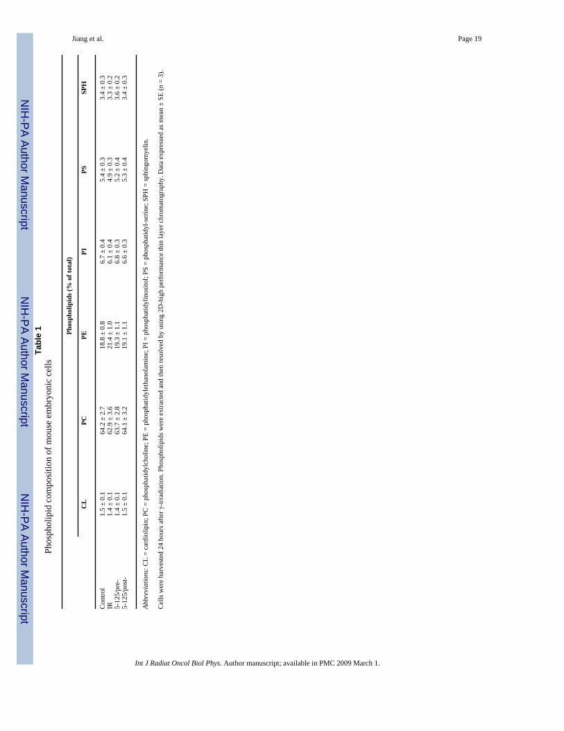
As previously reported (12), generation of superoxide/ROS is critical for oxidation of CL.Therefore, we assessed the effect of 5-125 on the irradiation-induced accumulation of CLhydroperoxides by using an HPLC-based Amplex Red assay. The content of CL-hydroperoxides in nonirradiated cells (13.1 ± 2.9 pmol/nmol CL) increased to 34.6 ± 3.3 (p <0.01) and 84.3 ± 5.8 (p < 0.01) pmol/nmol CL 24 and 48 hours postirradiation, approximately2.6- and ~6.4-fold increases, respectively (Fig. 2B, insert is a typical two-dimensional highperformance thin layer chromatography profile). Oxidation of CL did not exert any effect onthe overall phospholipid composition of the cells (Table 1). Accumulation of CLhydroperoxides decreased to 18.1 ± 3.8 (p < 0.05) and 39.6 ± 4.6 (p < 0.01) pmol/nmol CL in5-125 pre-treated cells after 24 and 48 hours of postirradiation incubation. Importantly,postirradiation (1-hour) treatment of cells with 5-125 (for 5 hours) also suppressed (albeit lesseffectively than under pretreatment) irradiation-induced accumulation of CL hydroperoxidesto 23.8 ± 3.8 (p < 0.05) and 55.8 ± 5.9 (p < 0.01) pmol/nmol CL after 24 and 48 hours ofpostirradiation incubation, respectively.
Effect of hemi-GS-TEMPO 5-125 on γ-irradiation–induced apoptosisBecause oxidation of CL participates in the release of proapoptotic factors from mitochondriainto the cytosol (12), we next examined whether inhibition of superoxide generation and CLperoxidation was associated with attenuation of γ-irradiation–induced apoptosis. Western blotanalysis showed that γ-irradiation (10 Gy) induced a time-dependent release of cyt c into thecytosol (Fig. 3A and 3B). Preirradiation treatment of cells with 5-125 (20 μM) inhibited theaccumulation of cyt c in the cytosolic fraction. In line with data for CL oxidation, postirradiationtreatment with 5-125 inhibited cyt c release, although to a lesser degree than under thepretreatment protocol (Fig. 3A and 3B).
The antiapoptotic effects of 5-125 were quantitatively studied further by analyzing PSexternalization, another hallmark of cell apoptosis occurring downstream of cyt c release.Gamma-irradiation induced PS externalization in approximately 25.9% ± 1.1% of mouseembryonic cells 48 hours after irradiation (p < 0.01 vs. 1.1% ± 0.3% in nonirradiated controls).The hemi-GS-TEMPO 5-125 (preirradiation treatment) decreased PS externalization in dose-and time-dependent fashions (Fig. 3B and 3C). Postirradiation treatment of cells with 20 μMof 5-125 (for 2, 4, 5, and 6 hours) also protected cells against γ-irradiation–induced PSexternalization (Fig. 3C), although not as effectively as the preirradiation treatment.
Jiang et al. Page 6
Int J Radiat Oncol Biol Phys. Author manuscript; available in PMC 2009 March 1.
NIH
-PA Author Manuscript
NIH
-PA Author Manuscript
NIH
-PA Author Manuscript
To provide further evidence for the radioprotective effect of 5-125, we also used humanbronchial epithelial BEAS-2B cells. Gamma-irradiation (10 Gy) induced PS externalization inapproximately 21.4% ± 2.0% of cells 72 hours after irradiation exposure. Pretreatment (10minutes before irradiation, removed after 5 hours of incubation) of cells with 5 and 10 μM of5-125 significantly attenuated γ-irradiation–induced PS externalization (15.6% ± 1.5% and11.2% ± 1.0%; p < 0.05 and p < 0.01 vs. irradiated cells without 5-125 treatment, respectively).Similarly, postirradiation treatment (1 hour) with 5-125 decreased irradiation–induced PSexternalization in BEAS-2B cells, although to a lesser extent than the preirradiation regimen(Fig. 3E).
Effect of hemi-GS-TEMPO 5-125 on cell survivalRadioprotective effects of 5-125 were examined further by using the clonogenic survival assay.The hemi-GS-TEMPO alone exerted no effect on the plating efficiency of mouse embryoniccells (data not shown). As shown in Fig. 4, pretreatment (10 minutes) of cells with 20 μM of5-125 (removed after 4 hours of incubation) provided marked protection of cells, shown bythe increased D0 (1.24 ± 0.03 vs. 0.90 ± 0.04; p < 0.01), whereas the parental nonconjugatedcompound, 4-AT, at the same concentration showed no protective effect (data not shown).Significant (D0 = 1.07 ± 0.04; p < 0.05) radioprotection also was afforded by 5-125 when cellswere treated with it 1 hour after irradiation, although its action was less effective comparedwith preirradiation treatment. The shoulder region n value was not significantly different afterpreirradiation or postirradiation treatment with 5-125 (n = 4.53 ± 0.29 and 4.95 ± 0.35 vs. 5.92± 0.38 in irradiated cells without 5-125 treatment, respectively).
Hemi-GS-TEMPO 5-125 prolonged γ-irradiation–induced G2/M phase arrestArrest at specific points of the cell cycle is a universal response to DNA damage in eukaryoticcells. The G2 cell-cycle checkpoint after DNA damage helps ensure the integrity of the genome(34,35). Previous work showed that nitroxide treatment led to an increased number of cells inthe G2/M phase of the cell cycle (36). Therefore, we evaluated the effect of 5-125 on cell-cycledistribution (Fig. 5). Pronounced G2/M phase arrest by γ-irradiation (10 Gy) alone wasobserved as early as 6 hours after irradiation, at which point approximately 65% of cells werefound at the G2/M phase.
After 24 and 32 hours postirradiation, cells were able to partially override the G2/M block andreentered mitosis. This was documented by the reappearance of a G1 peak, which wasaccompanied by a significant increase in hyperploid population (~15.8% and 21.7% at 24 and32 hours, respectively; p < 0.01 vs. control). By 48 hours, the hyperploid population in γ-irradiated cells mounted up to 25.6%. In the presence of 5-125 (pretreatment), enhanced G2/M accumulation (~90%) was observed 6 hours after irradiation. By 24 and 32 hours,approximately 78.6% and 62.9% of cells remained in the G2/M phase (p < 0.01 vs. irradiatedcells without 5-125 treatment). However, a decreased hyperploid population (4.3% and 11.7%at 24 and 32 hours, respectively) was observed. After 48 hours of postirradiation incubation,although significantly fewer apoptotic cells were detected (Fig. 3C), the hyperploid population(21.9%) was not significantly different from that of irradiated cells without 5-125 treatment.Similar results were obtained when treatment with 5-125 was performed 1 hour afterirradiation. Conversely, 4-AT did not exert an effect on cell-cycle distribution in irradiatedcells under the same conditions (data not shown). In the absence of irradiation exposure, 5-125alone did not show an effect on cell-cycle distribution.
DISCUSSIONBy undergoing one-electron transfer reactions, nitroxides can either be reduced to thecorresponding hydroxylamines or oxidized to the corresponding oxoammonium cation species
Jiang et al. Page 7
Int J Radiat Oncol Biol Phys. Author manuscript; available in PMC 2009 March 1.
NIH
-PA Author Manuscript
NIH
-PA Author Manuscript
NIH
-PA Author Manuscript
(2). The former reaction may be associated with scavenging of excessive electrons from therespiratory chain in mitochondria that prevents reduction of molecular oxygen to superoxide.In addition, hydroxylamines also are effective scavengers of several types of highly reactiveradical species (2). Because radiolysis of water is associated with the production of bothoxidizing and reducing reactive intermediates, nitroxides may be particularly effective in directscavenging of irradiation-induced radicals attacking biomacromolecules. In line with this,several groups recently reported that TEMPOL-mediated radioprotection was related to thereduced number of DNA double-strand breaks (2,37). However, it is critical that sufficientlyhigh concentrations of nitroxides are achieved at the sites of scavenging of radiation-inducedreactive radical species. Unfortunately, these high millimolar concentrations of nitroxides areimpractical for the purpose of prevention of irradiation-induced accumulation of reactiveradicals. However, irradiation-induced DNA damage and subsequent signaling triggersapoptotic response of mitochondria, which is usually delayed by several hours from the timeof the irradiation exposure. Combined with a recently developed strategy of targeted deliveryand concentration of antioxidants in mitochondria, this offers a unique new opportunity forusing nitroxides not only as radioprotectors, but also as radiomitigators.
Here, we report for the first time that hemi-GS-TEMPO 5-125 at low concentrations (5–20μM) afforded a marked radioprotective effect in mouse embryonic cells. The effectiveconcentration was almost 1,000-fold less than that of TEMPOL used in previous studies (2).This effect likely is caused, at least in part, by the increased intracellular and intramitochondrialpartitioning of hemi-GS-TEMPO.
According to Mitchell et al. (2,28), TEMPOL must be present during irradiation to be effective,suggesting that the protective mechanisms involve interaction with short-lived radiolyticintermediates. We found that postirradiation treatment of cells with 5-125 also protected cellsagainst γ-irradiation, clearly pointing to the involvement of mechanisms different from directscavenging of radiolysis intermediates in the protective effects. A great deal of researchindicated that mitochondria are both the primary source and major target of ROS (reviewed in[38]). A disrupted electron transport chain massively generates superoxide radicals originatingfrom damaged mitochondrial complexes I and III. Mitochondrial proteins and DNA,oxidatively modified by ROS, perpetuate the production of increasing concentrations of ROS,thus causing even more cellular damage. More importantly, oxidation of a mitochondria-specific phospholipid, CL, is implicated in the release of proapoptotic factors frommitochondria into cytosol. Thus, a radical scavenging pathway is a likely mechanism of theaction of nitroxide insofar as high concentrations of 5-125 accumulated in mitochondriainteract with mitochondrial ROS, the generation of which is dependent on intracellularsignaling and delayed by several hours from the moment of irradiation damage to DNA. Thisis in line with our previous findings, showing effective mitochondrial accumulation and properorientation of 5-125 at the interface of polar and nonpolar compartments of the mitochondriamembrane. This membrane topography permits 5-125 to successfully compete with molecularoxygen for electrons from the respiratory carriers, thereby preventing univalent reduction ofmolecular oxygen to yield superoxide. This interpretation is compatible with the observationthat postirradiation treatment with 5-125 showed less protection compared with preirradiationtreatment because of the lack of radical scavenging of radiolytic reactive species duringirradiation.
It also is possible that 5-125 exerts its protective action through pathways not related to itsdirect radical scavenging properties, but associated with its ability to affect general redoxregulation, particularly at the level of regulation of DNA repair and cell-cycle check points.Suy et al. (36) reported that nitroxides caused an increased number of cells in the G2/M phaseof the cell cycle. Cell-cycle checkpoints (G1/S and G2/M) are the major genomic surveillancemechanisms regulated in response to DNA damage. Sustained G2/M phase arrest allows more
Jiang et al. Page 8
Int J Radiat Oncol Biol Phys. Author manuscript; available in PMC 2009 March 1.
NIH
-PA Author Manuscript
NIH
-PA Author Manuscript
NIH
-PA Author Manuscript
time for repair of DNA damage and enhances survival. Increased radiation resistance with aprolonged arrest in G2/M was documented (39). Conversely, acceleration of G2/M transitionresulted in radiosensitization of cells (40,41). In this study, we found that 5-125 enhanced γ-irradiation–induced accumulation of the G2/M population and prolonged the G2/M phase arrestwith either preirradiation or postirradiation treatment. Recent work emphasized the importanceof redox status as one of the important factors of regulation of DNA repair and cell-cycle arrest(42,43).
Other mechanisms may contribute further to the radioprotective effect of hemi-GS-nitroxides.Our results show that approximately 74.2% of integrated hemi-GS-TEMPO was locatedoutside mitochondria, including the nucleus. Given the high hydrophobicity of hemi-GS-TEMPO (32), it predominantly partitions into membranes. Similar electron-acceptor functionsof 5-125 may be realized in the cytoplasmic and nuclear membranes, provided their truncatedelectron-transport chains (such as the reduced form of nicotinamide-adenine dinucleotidephosphate oxidase/cyt P-450 complexes in endoplasmic reticulum, electron carriers in nuclearand plasma membranes) act as superoxide generators during irradiation-induced apoptosis.Additionally, 5-125 may act as an SOD-mimetic and dismutate superoxide radicals to hydrogenperoxide (24,25). Finally, Suy et al. (44) reported that the nontargeted nitroxide, TEMPOL,stimulated the extra cellular signal-regulated kinase (ERK) signaling pathway, which primarilypromotes growth and proliferation/survival.
Because mouse embryonic cells, the major model used in this study, have unusual responsesto irradiation, such as P53-independent apoptosis and lack of G1 checkpoint (45), one canassume that results obtained here are not directly applicable to other cell types. However, thesignificant radio-protective effect of 5-125 observed in BEAS-2B cells suggests that theprotective effect is not limited to a specific cell line with unusual characteristics.
Taken together with the data presented in this study, it is reasonable to hypothesize that multiplemechanisms are involved in the radioprotection of 5-125, including scavenging of electronsfrom respiratory chains, direct interaction with ROS, prevention of superoxide generation, andCL oxidation in mitochondria and redox regulation of the cell cycle.
AcknowledgementsSupported by National Institutes of Health, National Institute of Allergy and Infectious Diseases Grant U19-AI068021and The Human Frontier Science Program.
References1. Little JB, Nagasawa H, Pfenning T, et al. Radiation-induced genomic instability: Delayed mutagenic
and cytogenetic effects of X rays and alpha particles. Radiat Res 1997;148:299–307. [PubMed:9339945]
2. Mitchell JB, Russo A, Kuppusamy P, et al. Radiation, radicals, and images. Ann N Y Acad Sci2000;899:28–43. [PubMed: 10863527]
3. Bryant PE. Enzymatic restriction of mammalian cell DNA: Evidence for double-strand breaks aspotentially lethal lesions. Int J Radiat Biol 1985;48:55–60.
4. Elledge SJ. Cell cycle checkpoints: Preventing an identity crisis. Science 1996;274:1664–1672.[PubMed: 8939848]
5. Sachs RK, Chen AM, Brenner DJ. Proximity effects in the production of chromosome aberrations byionizing radiation. Int J Radiat Biol 1997;71:1–19. [PubMed: 9020958]
6. Newton K, Strasser A. Ionizing radiation and chemotherapeutic drugs induce apoptosis in lymphocytesin the absence of Fas or FADD/MORT1 signaling. Implications for cancer therapy. J Exp Med2000;191:195–200. [PubMed: 10620618]
Jiang et al. Page 9
Int J Radiat Oncol Biol Phys. Author manuscript; available in PMC 2009 March 1.
NIH
-PA Author Manuscript
NIH
-PA Author Manuscript
NIH
-PA Author Manuscript

7. Szewczyk A, Wojtczak L. Mitochondria as a pharmacological target. Pharmacol Rev 2002;54:101–127. [PubMed: 11870261]
8. Kowaltowski AJ, Castilho RF, Vercesi AE. Opening of the mitochondrial permeability transition poreby uncoupling or inorganic phosphate in the presence of Ca2+ is dependent on mitochondrial-generatedreactive oxygen species. FEBS Lett 1996;378:150–152. [PubMed: 8549822]
9. Kroemer G, Reed JC. Mitochondrial control of cell death. Nat Med 2000;6:513–519. [PubMed:10802706]
10. Wong GH, Elwell JH, Oberley LW, et al. Manganous superoxide dismutase is essential for cellularresistance to cytotoxicity of tumor necrosis factor. Cell 1989;58:923–931. [PubMed: 2476237]
11. Iwata S, Hori T, Sato N, et al. Adult T cell leukemia (ATL)-derived factor/human thioredoxin preventsapoptosis of lymphoid cells induced by L-cystine and glutathione depletion: Possible involvementof thiol-mediated redox regulation in apoptosis caused by pro-oxidant state. J Immunol1997;158:3108–3117. [PubMed: 9120263]
12. Fernandez MG, Troiano L, Moretti L, et al. Early changes in intramitochondrial cardiolipindistribution during apoptosis. Cell Growth Differ 2002;13:449–455. [PubMed: 12354754]
13. Kagan VE, Tyurin VA, Jiang J, et al. Cytochrome c acts as a cardiolipin oxygenase required forrelease of proapoptotic factors. Nat Chem Biol 2005;1:223–232. [PubMed: 16408039]
14. Petrosillo G, Casanova G, Matera M, et al. Interaction of peroxidized cardiolipin with rat-heartmitochondrial membranes: Induction of permeability transition and cytochrome c release. FEBS Lett2006;580:6311–6316. [PubMed: 17083938]
15. Ott M, Robertson JD, Gogvadze V, et al. Cytochrome c release from mitochondria proceeds by atwo-step process. Proc Natl Acad Sci U S A 2002;99:1259–1263. [PubMed: 11818574]
16. Garrido C, Galluzzi L, Brunet M, et al. Mechanisms of cytochrome c release from mitochondria. CellDeath Differ 2006;13:1423–1433. [PubMed: 16676004]
17. Gonzalvez F, Gottlieb E. Cardiolipin: Setting the beat of apoptosis. Apoptosis 2007;12:877–885.[PubMed: 17294083]
18. Narayanan PK, Goodwin EH, Lehnert BE. Alpha particles initiate biological production of superoxideanions and hydrogen peroxide in human cells. Cancer Res 1997;57:3963–3971. [PubMed: 9307280]
19. Iyer R, Lehnert BE. Factors underlying the cell growth-related bystander responses to alpha particles.Cancer Res 2000;60:1290–1298. [PubMed: 10728689]
20. Spitz DR, Azzam EI, Li JJ, et al. Metabolic oxidation/reduction reactions and cellular responses toionizing radiation: A unifying concept in stress response biology. Cancer Metastasis Rev2004;23:311–322. [PubMed: 15197331]
21. Limoli CL, Giedzinski E, Morgan WF, et al. Persistent oxidative stress in chromosomally unstablecells. Cancer Res 2003;63:3107–3111. [PubMed: 12810636]
22. Kim GJ, Chandrasekaran K, Morgan WF. Mitochondrial dysfunction, persistently elevated levels ofreactive oxygen species and radiation-induced genomic instability: A review. Mutagenesis2006;21:361–367. [PubMed: 17065161]
23. Weiss JF, Landauer MR. Radioprotection by antioxidants. Ann N Y Acad Sci 2000;899:44–60.[PubMed: 10863528]
24. Saito K, Takeshita K, Ueda J, et al. Two reaction sites of a spin label, TEMPOL (4-hydroxy-2,2,6,6-tetramethylpiperidine-N-oxyl), with hydroxyl radical. J Pharm Sci 2003;92:275–280. [PubMed:12532377]
25. Mitchell JB, Samuni A, Krishna MC, et al. Biologically active metal-independent superoxidedismutase mimics. Biochemistry 1990;29:2802–2807. [PubMed: 2161256]
26. Mitchell JB, Krishna MC. Nitroxides as radiation protectors. Mil Med 2002;167:49–50. [PubMed:11873514]
27. Hahn SM, DeLuca AM, Coffin D, et al. In vivo radioprotection and effects on blood pressure of thestable free radical nitroxides. Int J Radiat Oncol Biol Phys 1998;42:839–842. [PubMed: 9845107]
28. Mitchell JB, DeGraff W, Kaufman D, et al. Inhibition of oxygen-dependent radiation-induced damageby the nitroxide superoxide dismutase mimic, tempol. Arch Biochem Biophys 1991;289:62–70.[PubMed: 1654848]
Jiang et al. Page 10
Int J Radiat Oncol Biol Phys. Author manuscript; available in PMC 2009 March 1.
NIH
-PA Author Manuscript
NIH
-PA Author Manuscript
NIH
-PA Author Manuscript

29. Hahn SM, Tochner Z, Krishna CM, et al. Tempol, a stable free radical, is a novel murine radiationprotector. Cancer Res 1992;52:1750–1753. [PubMed: 1551104]
30. Gariboldi MB, Ravizza R, Petterino C, et al. Study of in vitro and in vivo effects of the piperidinenitroxide Tempol—A potential new therapeutic agent for gliomas. Eur J Cancer 2003;39:829–837.[PubMed: 12651210]
31. Wipf P, Xiao J, Jiang J, et al. Mitochondrial targeting of selective electron scavengers: Synthesis andbiological analysis of hemigramicidin-TEMPO conjugates. J Am Chem Soc 2005;127:12460–12461.[PubMed: 16144372]
32. Jiang J, Kurnikov I, Belikova NA, et al. Structural requirements for optimized delivery, inhibition ofoxidative stress and anti-apoptotic activity of targeted nitroxides. J Pharmacol Exp Ther2006;320:1050–1060. [PubMed: 17179468]
33. Rothe G, Valet G. Flow cytometric analysis of respiratory burst activity in phagocytes withhydroethidine and 2′,7′-dichlorofluorescin. J Leukoc Biol 1990;47:440–448. [PubMed: 2159514]
34. Hartwell LH, Kastan MB. Cell cycle control and cancer. Science 1994;266:1821–1828. [PubMed:7997877]
35. Featherstone C, Jackson SP. DNA double-strand break repair. Curr Biol 1999;9:R759–R761.[PubMed: 10531043]
36. Suy S, Mitchell JB, Samuni A, et al. Nitroxide tempo, a small molecule, induces apoptosis in prostatecarcinoma cells and suppresses tumor growth in athymic mice. Cancer 2005;103:1302–1313.[PubMed: 15685617]
37. Erker L, Schubert R, Yakushiji H, et al. Cancer chemoprevention by the antioxidant tempol actspartially via the p53 tumor suppressor. Hum Mol Genet 2005;14:1699–1708. [PubMed: 15888486]
38. Orrenius S. Reactive oxygen species in mitochondria-mediated cell death. Drug Metab Rev2007;39:443–455. [PubMed: 17786631]
39. Miyata H, Doki Y, Yamamoto H, et al. Overexpression of CDC25B overrides radiation-inducedG2-M arrest and results in increased apoptosis in esophageal cancer cells. Cancer Res 2001;61:3188–3193. [PubMed: 11306507]
40. Bache M, Pigorsch S, Dunst J, et al. Loss of G2/M arrest correlates with radiosensitization in twohuman sarcoma cell lines with mutant p53. Int J Cancer 2001;96:110–117. [PubMed: 11291094]
41. Miyata H, Doki Y, Shiozaki H, et al. CDC25B and p53 are independently implicated in radiationsensitivity for human esophageal cancers. Clin Cancer Res 2000;6:4859–4865. [PubMed: 11156245]
42. Rothstein EC, Lucchesi PA. Redox control of the cell cycle: A radical encounter. Antioxid RedoxSignal 2005;7:701–703. [PubMed: 15890015]
43. Cakir Y, Ballinger SW. Reactive species-mediated regulation of cell signaling and the cell cycle: Therole of MAPK. Antioxid Redox Signal 2005;7:726–740. [PubMed: 15890019]
44. Suy S, Mitchell JB, Ehleiter D, et al. Nitroxides tempol and tempo induce divergent signal transductionpathways in MDA-MB 231 breast cancer cells. J Biol Chem 1998;273:17871–17878. [PubMed:9651392]
45. Aladjem MI, Spike BT, Rodewald LW, et al. ES cells do not activate p53-dependent stress responsesand undergo p53-independent apoptosis in response to DNA damage. Curr Biol 1998;8:145–155.[PubMed: 9443911]
Jiang et al. Page 11
Int J Radiat Oncol Biol Phys. Author manuscript; available in PMC 2009 March 1.
NIH
-PA Author Manuscript
NIH
-PA Author Manuscript
NIH
-PA Author Manuscript

Fig. 1.Structure of (A) 4-amino-2,2,6,6-tetramethyl-piperidine-N-oxyl (4-AT), (B) hemigramicidinS–conjugated 4-amino-2,2,6,6-tetramethyl-piperidine-N-oxyl (hemi-GS-TEMPO) 5-125, (C)their cellular and mitochondrial integration efficiencies in mouse embryonic cells, and (D)representative electron paramagnetic resonance (EPR) spectrum of nitroxides recovered frommitochondria.
Jiang et al. Page 12
Int J Radiat Oncol Biol Phys. Author manuscript; available in PMC 2009 March 1.
NIH
-PA Author Manuscript
NIH
-PA Author Manuscript
NIH
-PA Author Manuscript
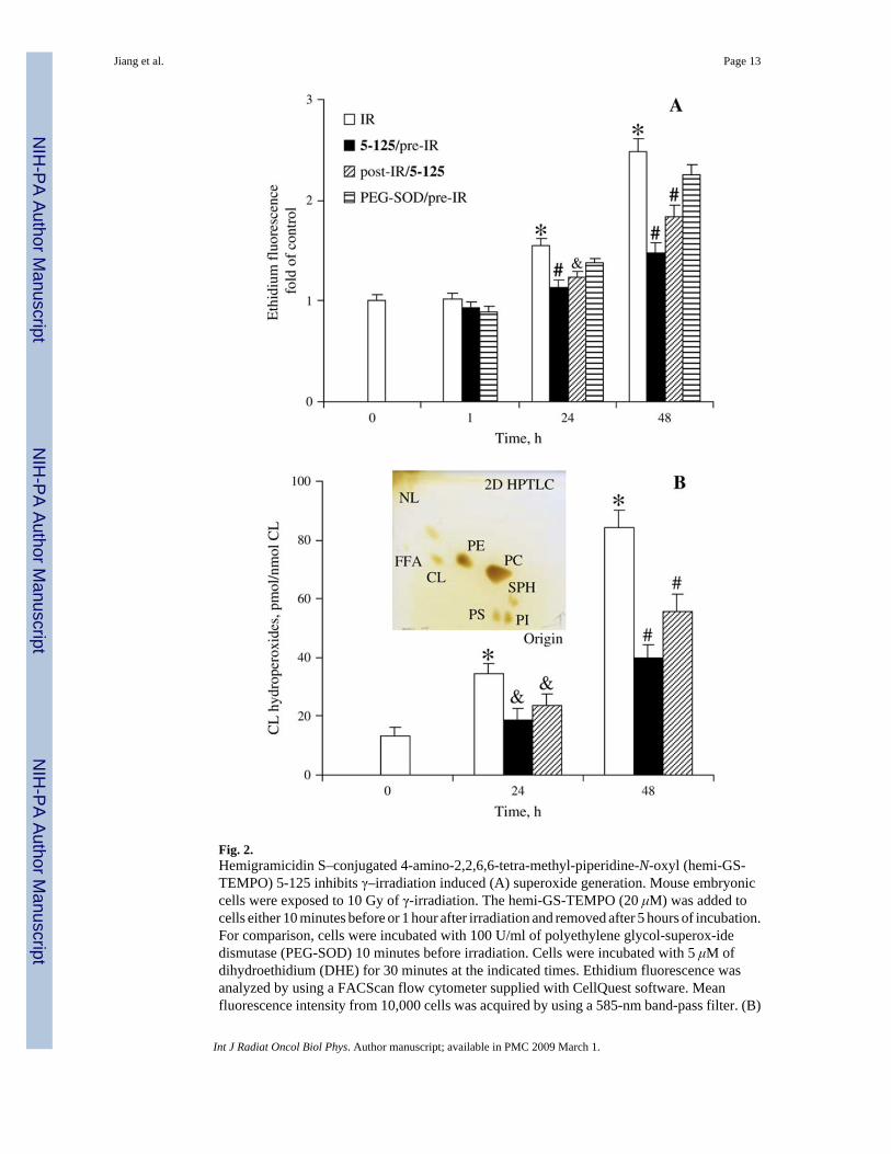
Fig. 2.Hemigramicidin S–conjugated 4-amino-2,2,6,6-tetra-methyl-piperidine-N-oxyl (hemi-GS-TEMPO) 5-125 inhibits γ–irradiation induced (A) superoxide generation. Mouse embryoniccells were exposed to 10 Gy of γ-irradiation. The hemi-GS-TEMPO (20 μM) was added tocells either 10 minutes before or 1 hour after irradiation and removed after 5 hours of incubation.For comparison, cells were incubated with 100 U/ml of polyethylene glycol-superox-idedismutase (PEG-SOD) 10 minutes before irradiation. Cells were incubated with 5 μM ofdihydroethidium (DHE) for 30 minutes at the indicated times. Ethidium fluorescence wasanalyzed by using a FACScan flow cytometer supplied with CellQuest software. Meanfluorescence intensity from 10,000 cells was acquired by using a 585-nm band-pass filter. (B)
Jiang et al. Page 13
Int J Radiat Oncol Biol Phys. Author manuscript; available in PMC 2009 March 1.
NIH
-PA Author Manuscript
NIH
-PA Author Manuscript
NIH
-PA Author Manuscript
Cardiolipin oxidation. Cardiolipin hydroperoxides were determined by using a fluorescenthigh-performance liquid chromatography (HPLC)-based Amplex Red assay. Data presentedas mean ± SE (n = 3). *p < 0.01 vs. nonirradiated cells; # (&)p < 0.01 (0.05) vs. irradiated cellswithout 5-125 treatment under the same condition. Insert is a typical 2D-high performance thinlayer chromatography profile of phospholipids from cells.
Jiang et al. Page 14
Int J Radiat Oncol Biol Phys. Author manuscript; available in PMC 2009 March 1.
NIH
-PA Author Manuscript
NIH
-PA Author Manuscript
NIH
-PA Author Manuscript

Fig. 3.(A) Hemigramicidin S–conjugated 4-amino-2,2,6,6-tetramethyl-piperidine-N-oxyl (hemi-GS-TEMPO) 5-125 blocks γ-irradiation–induced accumulation of cytochrome (cyt) c in the cytosolof mouse embryonic cells. (B) Densitometry ratio of cyt c/actin. Semiquantitation of the bandswas carried out by means of densitometry using Labworks Image Acquisition and AnalysisSoftware (UVP, Upland, CA). Level of cyt c release was expressed as the mean densitometryratio of cyt c over actin. (C) Dose (5, 10, and 20 μM)-dependent radioprotective effect of 5-125(pretreatment) on γ-irradiation (10 Gy)-induced phosphatidylserine (PS) externalization. After48 hours of postirradiation incubation, cells were harvested and stained with Annexin-V–fluorescein isothiocyanate (FITC) and propidium iodide (PI) before flow cytometry analysis.(D) Time (2, 3, 4, 5, and 6 hours)-dependent radioprotective effect of 5-125 (20 μM) on γ-irradiation (10 Gy)-induced PS externalization (48 hours after irradiation) in mouse embryoniccells. (E) Effect of 5-125 on γ-irradiation (10 Gy)-induced PS externalization in BEAS-2B
Jiang et al. Page 15
Int J Radiat Oncol Biol Phys. Author manuscript; available in PMC 2009 March 1.
NIH
-PA Author Manuscript
NIH
-PA Author Manuscript
NIH
-PA Author Manuscript

cells. Cells were treated with 5-125 (5 or 10 μM) before (10 minutes) or after (1 hour)irradiation. Externalization of PS was analyzed 72 hours after irradiation exposure. Data shownas mean ± SE (n = 3). *(&)p < 0.01 (0.05) vs. irradiated cells without 5-125 treatment, #p <0.05 vs. cells pretreated with 5-125.
Jiang et al. Page 16
Int J Radiat Oncol Biol Phys. Author manuscript; available in PMC 2009 March 1.
NIH
-PA Author Manuscript
NIH
-PA Author Manuscript
NIH
-PA Author Manuscript

Fig. 4.Gamma-irradiation dose survival curves of mouse embryonic cells. Cells were pretreated (10minutes) or post-treated (1 hour) with 5-125 (20 μM), which was removed after a 4-hourincubation period. The surviving fraction was calculated as the plating efficiency of the samplesrelative to that of the control. Data were fitted to a single-hit multitarget model using SigmaPlot9.0 (Systat Software). Data presented as mean ± SE (n = 3).
Jiang et al. Page 17
Int J Radiat Oncol Biol Phys. Author manuscript; available in PMC 2009 March 1.
NIH
-PA Author Manuscript
NIH
-PA Author Manuscript
NIH
-PA Author Manuscript

Fig. 5.Hemigramicidin S–conjugated 4-amino-2,2,6,6-tetramethyl-piperidine-N-oxyl (hemi-GS-TEMPO) 5-125 (20 μM) prolongs γ-irradiation (10 Gy)–induced G2/M arrest in mouseembryonic cells. Harvested cells were fixed in 70% alcohol at 4°C overnight, then pelleted andresuspended in phosphate-buffered saline (PBS) containing 100 μg/ml of RNase A and 40 μg/ml of propidium iodide (PI) for 15 minutes. Cellular DNA content was determined by meansof linear amplification in the FL-3 channel (>650 nm) of a FACScan flow cytometer equippedwith CellQuest software. Cell debris was gated out by forward and side scatter under the samecondition, and a minimum of 10,000 gated cells was acquired. Histograms shown are from onerepresentative experiment of three.
Jiang et al. Page 18
Int J Radiat Oncol Biol Phys. Author manuscript; available in PMC 2009 March 1.
NIH
-PA Author Manuscript
NIH
-PA Author Manuscript
NIH
-PA Author Manuscript
NIH
-PA Author Manuscript
NIH
-PA Author Manuscript
NIH
-PA Author Manuscript
Jiang et al. Page 19Ta
ble
1Ph
osph
olip
id c
ompo
sitio
n of
mou
se e
mbr
yoni
c ce
lls
Phos
phol
ipid
s (%
of t
otal
)
CL
PCPE
PIPS
SPH
Con
trol
1.5
± 0.
164
.2 ±
2.7
18.8
± 0
.86.
7 ±
0.4
5.4
± 0.
33.
4 ±
0.3
IR1.
4 ±
0.1
62.9
± 3
.621
.4 ±
1.0
6.1
± 0.
44.
9 ±
0.3
3.3
± 0.
25-
125/
pre-
1.4
± 0.
163
.7 ±
2.8
19.3
± 1
.16.
8 ±
0.3
5.2
± 0.
43.
6 ±
0.2
5-12
5/po
st-
1.5
± 0.
164
.1 ±
3.2
19.1
± 1
.16.
6 ±
0.3
5.3
± 0.
43.
4 ±
0.3
Abbr
evia
tions
: CL
= ca
rdio
lipin
; PC
= p
hosp
hatid
ylch
olin
e; P
E =
phos
phat
idyl
etha
nola
min
e; P
I = p
hosp
hatid
ylin
osito
l; PS
= p
hosp
hatid
yl-s
erin
e; S
PH =
sphi
ngom
yelin
.
Cel
ls w
ere
harv
este
d 24
hou
rs a
fter γ
-irra
diat
ion.
Pho
spho
lipid
s wer
e ex
tract
ed a
nd th
en re
solv
ed b
y us
ing
2D-h
igh
perf
orm
ance
thin
laye
r chr
omat
ogra
phy.
Dat
a ex
pres
sed
as m
ean
± SE
(n =
3).
Int J Radiat Oncol Biol Phys. Author manuscript; available in PMC 2009 March 1.
Copyright © 2022 FDOKUMEN