Panoramic Mapping with Information Technologies for ... - MDPI

A fast and flexible panoramic virtualreality system for behavioural andelectrophysiological experimentsJouni Takalo1, Arto Piironen1, Anna Honkanen1, Mikko Lempea1, Mika Aikio2, Tuomas Tuukkanen1
& Mikko Vahasoyrinki1
1Department of Physics, University of Oulu, PO.BOX 3000, 90014 Oulun yliopisto, Finland, 2VTT, Kaitovayla 1, 90571 Oulu,Finland.
Ideally, neuronal functions would be studied by performing experiments with unconstrained animals whilstthey behave in their natural environment. Although this is not feasible currently for most animal models,one can mimic the natural environment in the laboratory by using a virtual reality (VR) environment. Herewe present a novel VR system based upon a spherical projection of computer generated images using amodified commercial data projector with an add-on fish-eye lens. This system provides equidistant visualstimulation with extensive coverage of the visual field, high spatio-temporal resolution and flexible stimulusgeneration using a standard computer. It also includes a track-ball system for closed-loop behaviouralexperiments with walking animals. We present a detailed description of the system and characterize itthoroughly. Finally, we demonstrate the VR system’s performance whilst operating in closed-loopconditions by showing the movement trajectories of the cockroaches during exploratory behaviour in a VRforest.
Atraditional approach for studying neuronal mechanisms is to securely fix, anesthetize and/or dissect
animals to allow stable electrophysiological recordings to be made whilst they are presented with sim-plified sensory stimuli. More complex stimuli with naturalistic spatio-temporal structures introduced as
an improvement upon the experimental paradigm1–7 are now acknowledged to be essential for gaining a morecomplete understanding of the sensory processing. In addition to the stimulus properties, sensory processing isalso known to be modulated by the behavioural state of an animal (see eg8–14). Therefore, it would be beneficial tocarry out experiments in minimally constrained conditions and in naturalistic sensory environments, whereanimals can affect the environment through their own actions (e.g. walking).
There are numerous examples of neurophysiological experiments in awake but constrained animals. Oneexample of such a controlled experiment is the classical opto-motor paradigm used to study the visual behaviourof tethered insects when they are either walking on a track-ball, or flying attached to a torque-meter15. Becauseexperiments in freely moving animals are still mostly limited to the larger animal models (see eg16–18), virtualreality (VR) environments have been recently developed and introduced to the experimental set-ups to providemore naturalistic but well controlled settings19–26.
We present a novel VR system that is based on a commercial data projector and a custom add-on fish-eye lensthat projects images to the inner surface of a sphere. It also includes a track-ball to enable closed loop experimentsin walking animals. The entire system is computer controlled for flexible stimulus control and easy integrationwith electrophysiological and behavioural experiments.
ResultsThe goal of this study was to develop a virtual reality (VR) system suitable for studying the processing of visualinformation in animal models that possess a fast visual system with vast visual field, such as flying day-activeinsects. Therefore, following design requirements were set: (1) coverage of a large field of view; (2) spatialresolution better than 1u when observed from the animal’s point of view27; (3) image update rates above 100Hz and no flicker below 200 Hz; (4) bright illumination up to day-light levels; (5) contrast performance within therange of natural environments; (6) flexible stimulus generation software that runs on a standard computer; (7)implementation of closed-loop behavioural paradigms.
SUBJECT AREAS:SENSORY SYSTEMS
NEUROSCIENCE
METHODS
ANIMAL BEHAVIOUR
Received6 February 2012
Accepted27 February 2012
Published22 March 2012
Correspondence andrequests for materials
should be addressed toM.V. (mikko.
SCIENTIFIC REPORTS | 2 : 324 | DOI: 10.1038/srep00324 1

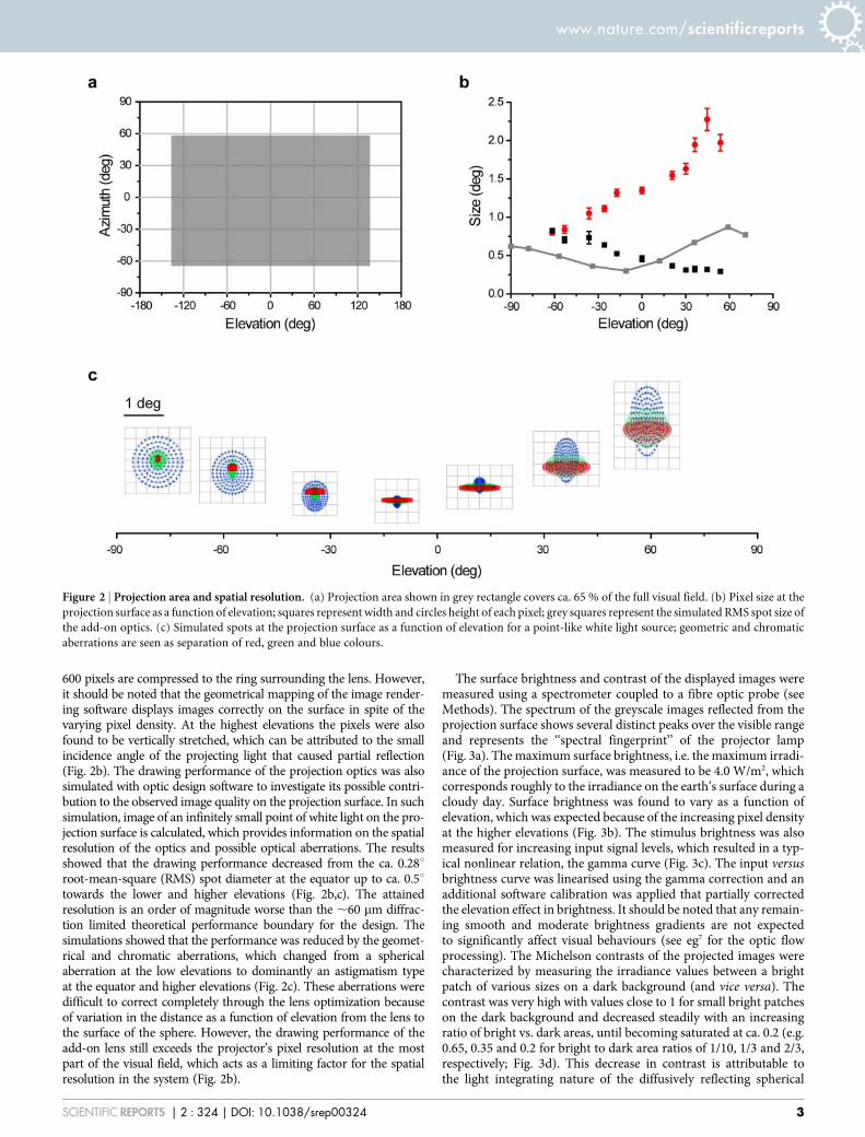
The VR environment that we developed is a spherical projectionsystem, where a data projector is used to display computer generatedimages on the inner surface of the sphere (Fig. 1a,b). To achieve highimage refresh rate required for the animal models with fast vision, theprojector was modified to operate with 360 Hz in greyscale mode(see Methods). Previously described mirror-based projection sys-tems20,21,28 were used as a starting point for the optical design(Fig. 1c,d). However, they were found to be unsuitable for the systembecause of the large projection angle, drawing capability require-ments and the mirror blocking a large part of the visual field. Wewere not able to verify if these limitations were specific to our sub-optimal mirror design or whether it is inherent limitation to themirror-based approach. This is because the earlier descriptions ofsuch systems20,21,28 do not provide any simulated or measured dataabout the drawing capability or image quality on the projectionsurface. Instead of a mirror-based approach, a fish-eye type lens
projection was selected that has not to our knowledge been describedearlier for spherical projection systems (see also29 for early account oflens-based projection on to a cylindrical screen). The fish-eye lenscan be used to project images over the whole spherical surface whenplaced at the top (Fig. 1e). The animal observes the VR environmentfrom the centre of the sphere, which covers 65 % of the full visual fieldequidistantly (2135 to 135 degrees in azimuth and 264 to 57 degreesin elevation; Fig. 2a). It should be noted that areas used for theinstrumentation overlap with locations that animals cannot oftensee (such as below and behind them).
As a measure of the spatial resolution of the VR the sizes of indi-vidual pixels were measured from the projection surface. The angularsize of the pixels as observed from the animal’s perspective werefound to be ca. 0.8 degree squares at low elevations but becamehorizontally compressed towards higher elevations (Fig. 2b). Thisis attributable to the projection geometry where, at the extreme,
Figure 1 | VR system and projection optics. (a) 3D model of the VR system showing projector with add-on lens, spherical projection surface, track ball,
microscope and micromanipulator. (b) Photograph of a behavioural experiment with the forest scene; cockroach is on a track ball tethered to a metal wire
and it is circulating the forest edge. (c) Ray tracing simulations of the projection optics design with a flat mirror and an aspherical angular amplification
mirror20,21,28; the mirror blocks ca. 45 degrees of the visual field. (d) Design with two aspherical mirrors28 for angular amplification that blocks smaller
visual field than in (c). (e) The fish-eye lens design.
www.nature.com/scientificreports
SCIENTIFIC REPORTS | 2 : 324 | DOI: 10.1038/srep00324 2
600 pixels are compressed to the ring surrounding the lens. However,it should be noted that the geometrical mapping of the image render-ing software displays images correctly on the surface in spite of thevarying pixel density. At the highest elevations the pixels were alsofound to be vertically stretched, which can be attributed to the smallincidence angle of the projecting light that caused partial reflection(Fig. 2b). The drawing performance of the projection optics was alsosimulated with optic design software to investigate its possible contri-bution to the observed image quality on the projection surface. In suchsimulation, image of an infinitely small point of white light on the pro-jection surface is calculated, which provides information on the spatialresolution of the optics and possible optical aberrations. The resultsshowed that the drawing performance decreased from the ca. 0.28uroot-mean-square (RMS) spot diameter at the equator up to ca. 0.5utowards the lower and higher elevations (Fig. 2b,c). The attainedresolution is an order of magnitude worse than the ,60 mm diffrac-tion limited theoretical performance boundary for the design. Thesimulations showed that the performance was reduced by the geomet-rical and chromatic aberrations, which changed from a sphericalaberration at the low elevations to dominantly an astigmatism typeat the equator and higher elevations (Fig. 2c). These aberrations weredifficult to correct completely through the lens optimization becauseof variation in the distance as a function of elevation from the lens tothe surface of the sphere. However, the drawing performance of theadd-on lens still exceeds the projector’s pixel resolution at the mostpart of the visual field, which acts as a limiting factor for the spatialresolution in the system (Fig. 2b).
The surface brightness and contrast of the displayed images weremeasured using a spectrometer coupled to a fibre optic probe (seeMethods). The spectrum of the greyscale images reflected from theprojection surface shows several distinct peaks over the visible rangeand represents the ‘‘spectral fingerprint’’ of the projector lamp(Fig. 3a). The maximum surface brightness, i.e. the maximum irradi-ance of the projection surface, was measured to be 4.0 W/m2, whichcorresponds roughly to the irradiance on the earth’s surface during acloudy day. Surface brightness was found to vary as a function ofelevation, which was expected because of the increasing pixel densityat the higher elevations (Fig. 3b). The stimulus brightness was alsomeasured for increasing input signal levels, which resulted in a typ-ical nonlinear relation, the gamma curve (Fig. 3c). The input versusbrightness curve was linearised using the gamma correction and anadditional software calibration was applied that partially correctedthe elevation effect in brightness. It should be noted that any remain-ing smooth and moderate brightness gradients are not expectedto significantly affect visual behaviours (see eg7 for the optic flowprocessing). The Michelson contrasts of the projected images werecharacterized by measuring the irradiance values between a brightpatch of various sizes on a dark background (and vice versa). Thecontrast was very high with values close to 1 for small bright patcheson the dark background and decreased steadily with an increasingratio of bright vs. dark areas, until becoming saturated at ca. 0.2 (e.g.0.65, 0.35 and 0.2 for bright to dark area ratios of 1/10, 1/3 and 2/3,respectively; Fig. 3d). This decrease in contrast is attributable tothe light integrating nature of the diffusively reflecting spherical
Figure 2 | Projection area and spatial resolution. (a) Projection area shown in grey rectangle covers ca. 65 % of the full visual field. (b) Pixel size at the
projection surface as a function of elevation; squares represent width and circles height of each pixel; grey squares represent the simulated RMS spot size of
the add-on optics. (c) Simulated spots at the projection surface as a function of elevation for a point-like white light source; geometric and chromatic
aberrations are seen as separation of red, green and blue colours.
www.nature.com/scientificreports
SCIENTIFIC REPORTS | 2 : 324 | DOI: 10.1038/srep00324 3

projection surface, which is an unavoidable consequence of the spher-ical projection geometry. Importantly, the measured image contrastsare still well within the range of naturally occurring contrasts30.
The temporal performance of the VR system was characterized bymonitoring the projected stimuli with a photodiode. Full field stimuliof an intermediate and full brightness of a ca. 6 ms long sequence ofthree experimenter controlled frames were presented, followed bya ca. 2 ms white segment controlled by the projector hardware(Fig. 4a; see Methods). Fast intensity fluctuations within each frameare caused by the digital encoding of the light intensity that is imple-mented in the projector hardware. However, this fast fluctuationtakes place in micro-second time scale, too fast to be perceived byany known visual system, and becomes integrated in the visual systemas a constant intensity level. The power spectrum of the data wascalculated for the full field stimulus of maximum brightness, whichrevealed a 120 Hz flicker attributable to the uncontrollable whitesegment (Fig. 4b). Data from the stimulus with intermediate bright-ness showed further increase in the flicker (Fig. 4a), which leads to thecorresponding increases in the power spectrum at 120 Hz and itsharmonics (Fig. 4b). Note that the digital implementation of the lightintensity encoding leads to signal analogous to the digital white noise,which gives rise to flat power spectra.
Intracellular in vivo recordings were carried out in blowfly(Calliphora vicina) photoreceptors, insect with one of the fastestknown visual system31–33. Our logic was that if their photoreceptorsdo not respond to the rapid fluctuations in the light intensity attrib-utable to the digital light intensity encoding or the 120 Hz flickerassociated with the white segment, the VR can be considered to be
suitable for any animal model with fast vision. Step-like full fieldstimuli from darkness to the varying brightness levels were used toelicit photoreceptor responses. No response to rapid light intensityencoding fluctuations was observed but, after the initial transientresponse, the photoreceptor’s membrane potentials were locked tothe 120 Hz flicker (Fig. 5a). The flicker responses were largest atintermediate brightness values, which resulted from the proportion-ally larger intensity fluctuations in the stimulus (Fig. 5b). To addressthis problem the 2nd generation version of the same projector modelwas used, in which the manufacturer had removed the white segmentby modifying the projector’s firmware (see Methods). Intracellularphotoreceptor recordings made using the new projector model topresent the stimulus showed no 120 Hz ripple in the step responses(Fig. 5d,e). The differences in the voltage waveforms between the twoprojectors were attributable to the reduced background light andhigher brightness of the stimuli in the measurements with the 2nd
generation model. Analysis of the power spectrum from responses tosteady light using the older and new projector model also showedthat peak in the photoreceptor voltage response power related tothe 120 Hz flicker had decreased by ca. 104 orders in magnitude(Fig. 5c,f). Therefore, it can be concluded that the developed VRequipped with the 2nd generation projector model enables stimulat-ing even the fastest visual systems with high 360 Hz frame rate andwithout flicker artefacts.
Finally, cockroaches (Periplaneta americana) were used as an ani-mal model to demonstrate the suitability of the VR system for beha-vioural studies with walking behaviour. Although not an animal withfast vision the cockroach was selected because it has robust walking
Figure 3 | Characterization of the surface brightness and contrast. (a) Spectrum of the grey scale image at the projection surface for 100% brightness full
field stimulus. (b) Projection surface irradiance as a function of elevation for 100% brightness full field stimulus. (c) Projection surface irradiance as a
function of input intensity level; line shows fit of the gamma curve with the gamma value of 2.56. (d) Contrast as a function of the area of the 100%
brightness stimulus patch at dark background or dark patch at 100% bright back-ground.
www.nature.com/scientificreports
SCIENTIFIC REPORTS | 2 : 324 | DOI: 10.1038/srep00324 4

behaviour on the track-ball and it was readily available in the labor-atory. The visual system of cockroaches is too slow for them to detectthe 120 Hz flicker34. Therefore, the VR system with the 1st generationprojector installed was used for these experiments. The cockroacheswere tethered from the pronotum and placed on a track-ball system,which enabled them to navigate in the VR forest (Fig. 1b;Supplementary video S1). Although the track-ball weight exceededthe optimal weight for the cockroach35, they had no difficulties inrotating the nearly frictionless track-ball. Each experiment startedin the middle of the forest (Fig. 6a,b). The trees were disproportio-nately small compared to a cockroach, but they can still be consid-ered as concrete visual objects. The cockroaches remained active inthe VR for extended periods of time, typically showing burst ofwalking activity interspersed with periods of inactivity (Fig. 6c,d).While approaching a tree the cockroaches would typically antennatetowards it, presumably trying to establish a tactile contact with thevisual object (Supplementary video S1). Typically, they avoidedthe trees, walking around them but occasionally, possibly the lackof tactile feedback from the antennae, caused them to continue toapproach the tree, eventually walking through it. This outcome wasnot prevented by the software. This behaviour can be seen in thetrack plots (Fig. 6a,b) and in the histograms of cockroach distance tothe tree centre (Fig. 6e,f). Note that especially the other individual hasspent lots of time at the distance close to the tree trunk (Fig. 6f). At alarger scale the cockroaches fixated on the darkness surrounding theforest and headed towards the forest edge along a fairly straight route
(Fig. 6a,b). Once at the edge (Fig. 1b), they typically started to followit, often walking around the perimeter of the virtual world severaltimes (Fig. 6a,b; Supplementary video S2). None of the cockroachesreturned to the forest after they had found the dark forest edge, whichis not surprising considering their innate preference for dark areas inthe environment.
DiscussionWe have successfully developed a VR system for animal models thathave fast vision, such as day-active insects. The system we developedprovides good spatio-temporal performance and covers most of therelevant parts of the visual field, which is known to be essential for theoptic flow dependent behaviours in insects15,26,36. The spherical VRalso has equidistant projection geometry, which removed need forthe real-time perspective distortion corrections required with e.g. flatcomputer screens26, which could become a limiting factor for thespatio-temporal performance in high frame rate system such asone presented here. The VR system can also produce stimuli withequivalent brightness to a cloudy-day and contrasts comparable tothe natural environment. Finally, it is controlled with a standardcomputer and software that provides flexible stimulus generationand control possibilities. These technical features coupled with dem-onstrable success in both behavioural and electrophysiologicalexperiments (see also37) allow us to conclude that the system is suit-able for presenting animals with the behaviourally relevant andnaturalistic stimuli. Further details of the design, including manufac-turing instructions, are available from authors upon request to enablesystem replication.
A temporal flicker problem was identified during the system char-acterization, which was also verified in electrophysiological record-ings from blowfly photoreceptors to be detectable by animals withvery fast vision. However, this problem was avoided by replacementof the original projector with the 2nd generation model, which iscommercially available from the projector manufacturer. Becausethe 1st generation projector was already fully adequate for the cock-roach and several other animal models used in the laboratory, wedecided not to replace it with the newer model but instead build asecond VR system based on the 2nd generation model for the animalswith faster vision (work in progress). The main remaining limitationwith the developed VR system is the inability to generate colourstimulation, which was compromised to increase the frame rate to360 Hz. However, many visual behaviours are not dependent uponcolour information (e.g. optic flow processing15,36,38) and the colourfilters can be used in the lens assembly for simple full field manip-ulation of the spectral content of the stimuli.
Other approaches have been described in the literature for imple-menting VR environments, but detailed comparison to our system isnot possible because lack of quantitative data on the performance ofthese VR system in the published results. A LED-based virtual realitysystem has been presented that also covers a large fraction of thevisual field and in which the stimulus is controlled through a modi-fied VGA graphic card7 (see also39). Its main advantages are theflicker free image with high brightness but, on the other hand, itsdisadvantages include a limited spatial resolution, as well as require-ments for complex custom instrumentation and control software. Itshould also be noted that our VR system already achieves the day-light level brightness, which can be increased further when using the2nd generation projector that has brighter lamp in it.
Data projector based VR systems have also been described forrodents, which include the track-ball system20,21. However, their tem-poral performance is too low for animals with fast vision, whichwould see the typically 60 Hz frame rate as high contrast flicker(see also19). In addition, based on our simulations, the mirror basedprojection optics used in these systems is expected to produce lowerimage quality than the novel fish-eye design of our VR system.Therefore, the lens based design is expected to provide significant
Figure 4 | Measurements of the stimulus intensity flickering. (a) Light
intensity fluctuations measured for the full field stimuli with brightness of
100% (top) and 50% (bottom). Coloured bar represents ca. 2 ms colour
segments used to encode the sequential image frames and white
corresponds to white segment; note that intensity varies from dark to full
brightness. (b) Power spectra of the 0% (black), 50% (blue) and 100%
(red) brightness full field stimuli show large power peaks at 120 Hz and its
harmonics that are associated with the white segment.
www.nature.com/scientificreports
SCIENTIFIC REPORTS | 2 : 324 | DOI: 10.1038/srep00324 5

improvement in the image quality of the spherical VR systems ingeneral. A VR system was also recently reported that uses two customDLP projector engines to create a panoramic field of view with back-projecting spherical surfaces22. It is reported to produce greyscaleframe rates of up to 8000 frames/s in a binary mode, however, thiscorresponds to only 60 Hz frame rate when 8 bit intensity encoding isused as in our system. It also provides stimulation options for othersensory modalities but, for the visual stimulation, our system pro-vides more flexible stimulus control and comparable spatio-temporalperformance with much simpler and more cost efficient imple-mentation.
Our future method development work will focus on implementingmultisensory stimulation possibilities into the VR. For example, anolfactometer will be included to enable stimulation of the olfactoryand mechanosensory pathways. The VR based approach will beextensively used in our future works to replicate traditional arena
or maze experiments in the VR, as well as to implement more com-plex experiments enabled by the easy real-time manipulation of thevisual environment (e.g. a visual object that the animal has fixatedcan be easily moved to different location or modified by the appear-ance). Importantly, the behaving animals can be constrained inthe VR to enable simultaneous electrophysiological measurements,which can be used to investigate the neuronal mechanisms behindthe behaviours.
MethodsVR design. The system involves the projection of computer generated images ontothe inner surface of a spherical projection screen using a data projector incombination with a fish-eye lens (Fig. 1a). An animal is placed in the centre of thesphere, which from the animal’s viewpoint enables covering of the full visual fieldequidistantly with the screen. A sphere with 400 mm diameter was built from a glassfibre by laminating the fibres on top of a positive half-sphere mould. The two halves ofthe sphere were attached to each other and sections from the resulting full sphere were
Figure 5 | 120 Hz is detected by the blowfly photoreceptors. (a) Photoreceptor voltage responses with 1st generation projector for a full field step stimuli
from darkness to varying brightness from maximum; each trace is an average of 10 trials; stimulus is shown below. (b) Photoreceptor voltage response
(black) and photodiode signal (red) with the 1st generation projector for full field stimulus of the constant 100% (top) and 18% (bottom) brightness;
photodiode signal off-set and scale is arbitrary. (c) Power spectra of the photoreceptor voltage responses to full field stimuli with 100% (black) and 18%
(red) brightness; the 120 Hz and its harmonics are clearly visible. (d)–(f) Similar measurements as in (a)–(c), but with the 2nd generation projector; scale in
the (d) and (e) are as in the (a) and (b), respectively; note that the residual flicker detected by the photodiode is too fast to be seen by the photoreceptors.
www.nature.com/scientificreports
SCIENTIFIC REPORTS | 2 : 324 | DOI: 10.1038/srep00324 6

subsequently removed from the top and the bottom to accommodate the lens and theanimal holder, respectively (Fig. 1a). In addition, a 90u section was removed fromthe back to provide access for a stereo microscope and micromanipulators requiredfor the electrophysiology. The inner surface of the sphere was polished and coatedwith barium sulphate based paint (ODP97-1.0, Gigahertz-Optic GmbH) that forms a
high-quality white Lambertian surface. This reduces directional reflections andincreases surface brightness.
Fish-eye type add-on lens was designed to be used in conjunction with the pro-jector’s own lens (together called projection optics). ZEMAXH (Zemax DevelopmentCorp) software was used for the optical design and optimization. The optical
Figure 6 | Cockroach exploratory behaviour in the VR environment. (a) Cockroach no. 1 and; (b) no. 2 exploring in the virtual forest; red crosses mark
20 s time progression; circles mark location of the trees; world is dark outside the forest (see also Supplementary videos; note that videos are not from the
data shown in the figure). (c) and (d) movement speed for the trajectories shown in (a) and (b) showing bouts of activity followed by inactivity periods. (e)
and (f) histograms of the distance to tree centre for the data in (a) and (b); note that trees were rectangles with their larger dimensions shown in the
histogram as grey bar.
www.nature.com/scientificreports
SCIENTIFIC REPORTS | 2 : 324 | DOI: 10.1038/srep00324 7
properties of the projector lens were measured to allow it to be replaced by a thin lensequivalent in the optics design. At the image source, the projection optics was opti-mized to use the approximately 10.5 mm diameter circle of the projector’s digitalmicromirror device (DMD), which corresponds to a circle of 600 pixels in diameter.This allows the system to take full advantage of the available resolution of the SVGAprojector. The designed add-on lens assembly includes one catalogue singlet (KXP-189, Newport Corp.), a plane mirror, two custom doublets and two custom singlets(Fig. 7a,b). The effective focal length of the projection optics is 4.1 mm. The lensassembly also includes slots for placing optical filters (Fig. 7a), such as neutral densityand colour filters.
Animals with fast vision, such as flying insects, have a flicker fusion rate thatexceeds the frame rate of the commercial data projector27,32,33. A moderately priceddigital light processing (DLP) type data projector was selected for the VR system(DepthQH, Lightspeed Design Inc). It can produce 120 Hz frame rate with 24 bitcolour reproduction and SVGA resolution. The colour filter was removed from theprojector’s image engine to harness the colour production for tripling the frame rateof 8 bit greyscale images to 360 Hz. Custom software was developed to control theimage production, based on an open-source Ogre3d graphic rendering engine (http://www.ogre3d.org/). The software runs with a standard computer and a Windowsoperating system (Microsoft Inc.). The rendering is based on a cube map with sixcameras for each frame. The VR environment is created as a cube of specific size,where each camera is facing one of the inner faces of the cube. A geometrical trans-formation is included in the compiled VR control files that subsequently morphs thecube into a sphere. In addition, the control files include functional data that can beused to control aspects of the VR environment, such as moving objects or changingthe viewpoint within the VR. The pixels that are not ‘‘seen’’ by the projection opticsand the ones that would project to the sectors removed from the sphere or directly tothe animal are set to black. The software creates greyscale images with 8 bit intensityencoding that are displayed sequentially on the colour channels in the order of blue,green and red to produce 360 Hz frame-rate. It should be noted that in the 1st
generation projector model the hardware implemented white channel always followsthe red channel and its brightness is automatically determined, based on the threecolour channels intensities. The white segment is eliminated in the 2nd generationspecial model that was made recently available by the projector manufacturer (resultof an independent development project; courtesy of Anthony Leonardo, Janelia FarmResearch Campus, HHMI and Lightspeed Design Inc.).
A track-ball was designed according to the previously described approach usingcomputer mice and built to permit closed-loop behavioural experiments with thewalking animals (Fig. 7c)20,21,40–43. It is placed on a heater platform that can control thetemperature inside the VR system within a range of 20 240uC (Fig. 1b). The track ballincludes an air-supported hollow Styrofoam sphere with 93 mm diameter and 4.8 gweight (Fig. 7c), which is placed to the centre of the sphere (Fig 1a,b). The track-ballchassis is milled from PVC plastic. The animal is tethered and placed on top of thenearly frictionless ball. The ball weight is heavier than what would be optimal formatching its inertia to that of e.g. cockroach35, but this has not lead to difficulties inrotating the ball. Rotations of the ball are measured by two image sensors disas-sembled from an optical computer mouse (AMU45EU, Targus Group InternationalInc.). The image sensors (ADNS-2610) are integrated to the track ball chassis andconnected to the VR controlling computer with the USB cables of the computer mice.The sensor frame rate is 1500 frames/s, resolution is ca. 0.078 degrees and the max-imum speed and acceleration are 0.3 m/s and 2.5 m/s2, respectively. This performanceis sufficient for the typical experiments with walking insects (Fig. 7c,d), except for themaximum speed that may be exceeded e.g. during fast escape responses (, 1 m/s in
cockroach). In such cases, the measured movement and the subsequent VR controlwould be inaccurate. Rotations of the ball are streamed to the memory buffers of themice and polled by the VR software through raw input interface of the MS Windowsoperating system at every vsync event associated with the frame rate update. Therotation values are transformed into the control commands for the VR, which enablescalculating difference between two frames used to move the VR scene accordingly. Inaddition of the closed-loop control, the track-ball can also be used in the open-loopmode, where animal movements are measured but not used to control the VRenvironment.
Technical measurements. Pixel diameters on the projection surface were measuredfrom the varying elevations along the 135 degrees in azimuth. The measurementswere done manually using a calliper and repeated several times to increase thereliability of the measurements (N 5 6). Contrast and surface brightnessmeasurements were made with a calibrated spectrometer (USB4000 UV-VIS, Oceanoptics Inc). Spectrum and surface irradiances with varying brightness levels weremeasured using the full field stimuli. For the contrast measurements a white patchstimulus of varying size on a black background or black patch stimulus on a whitebackground were used. The fibre optic probe (NA 5 0.22) of the spectrometer waspositioned either 20 or 154 mm from the surface of the sphere, or in the centre of it, formeasuring the size of pixels, contrasts and brightnesses, respectively. The contrasts, c,were calculated according to the Michelson formula c 5 (Is2Ib)/( Is1Ib) where Is andIb are the surface irradiance of the stimulus and the background, respectively.
Stimuli were also measured with a photodiode. The photodiode signals wereamplified and saved to a computer with a 50 kHz sampling rate. Because thephotodiodes were only used to monitor temporal dynamics of the stimulus intensity,they were not calibrated leading to arbitrary output values. The data acquisition in allmeasurements were carried out with a data acquisition card (PCIe-6259, NationalInstruments Corp.) and a custom LABVIEWH (National Instruments Corp.)program. Power spectral densities were estimated in MATLABH (Mathworks Inc.)using the Welch’s method with a hamming window.
Animal experiments. Blowflies (Calliphora vicina) were grown in a laboratory stockthat is regularly refreshed with animals obtained from the Blades Biological Ltd, UK.All data presented was collected from the female flies. Flies were fixed on a piece ofmicroscopy glass with a mixture of a paraffin and beeswax. Legs and antennae wereremoved and wounds sealed with the wax to prevent drying. A small hole was cut inthe dorsal part of the cornea of the left eye for the electrode insertion. Sharp glasscapillary microelectrodes were manufactured using a laser puller (P-2000, SutterInstrument Company Inc.). An Ag/AgCl reference electrode was placed in a smallopening cut in the fly’s abdomen. The glass capillary electrodes were driven to the eyewith a custom piezo manipulator (courtesy of Sensapex Ltd.). Experiments wereperformed on the cells with dark resting potential below 260 mV and an inputresistance of at least 15MV44. The photoreceptors were stimulated using full fieldflashes of varying brightness. The experiments shown in Fig. 5a–c were performedusing the 1st generation projector that is integrated to the VR system and those inFig. 5d–f with the 2nd generation model projecting to a planar screen coated with thesame paint as in the spherical screen. Measurements were made from four animals(for cells N 5 12) and data was acquired at 4 kHz sampling rate.
Adult male American cockroaches (Periplaneta americana) from a laboratorystock were used for the behavioural experiments. The tip of a wire holder was fixed tothe pronotum of a carbon dioxide-anesthetized cockroach with a mixture of beeswaxand resin. The cockroach was mounted on the track ball and care was taken to adjust
Figure 7 | Fish-eye lens and trackball schematics. (a) Schematic cross section of the add-on fish-eye lens assembly; colour filter slot is highlighted in grey
colour. (b) Custom lenses in the assembly shown in more detail; R refers to surface curvature of the spherical lens. (c) Section through a 3D wire model of
the track-ball. Pressure port at the lower part is for coupling of the air supply to provide low friction air cushion; one of the two sensors is shown in the
cross section.
www.nature.com/scientificreports
SCIENTIFIC REPORTS | 2 : 324 | DOI: 10.1038/srep00324 8

the animal’s posture and elevation from the ball so that it could move as normally aspossible. Each cockroach was allowed to recover from the anaesthesia and adapt to theexperimental set-up for at least 15 minutes prior to starting the experiment, whichwas found to be sufficient time for the full recovery of normal activity level. Theanimals were not exposed to any stimuli during this time (i.e. VR was dark).The cockroaches were able to navigate freely in the virtual forest environment,including the black surround (Fig. 1b). The forest was evenly lit without any shadowsunder the trees.
1. Karmeier, K., van Hateren, J. H., Kern, R. & Egelhaaf, M. Encoding of naturalisticoptic flow by a population of blowfly motion-sensitive neurons. J. Neurophysiol.96, 1602–1614 (2006).
2. Niven, J. E., Vahasoyrinki, M., Juusola, M. & French, A. S. Interactions betweenlight-induced currents, voltage-gated currents, and input signal properties inDrosophila photoreceptors. J Neurophysiol 91, 2696–706 (2004).
3. Passaglia, C., Dodge, F., Herzog, E., Jackson, S. & Barlow, R. Deciphering a neuralcode for vision. Proc. Natl. Acad. Sci. U. S. A. 94, 12649–12654 (1997).
4. Reinagel, P. How do visual neurons respond in the real world? Curr. Opin.Neurobiol. 11, 437–442 (2001).
5. Vinje, W. E. & Gallant, J. L. Sparse coding and decorrelation in primary visualcortex during natural vision. Science 287, 1273–1276 (2000).
6. Felsen, G. & Dan, Y. A natural approach to studying vision. Nat. Neurosci. 8,1643–1646 (2005).
7. Lindemann, J. P. et al. FliMax, a novel stimulus device for panoramic andhighspeed presentation of behaviourally generated optic flow. Vision Res. 43, 779–791 (2003).
8. Maimon, G., Straw, A. D. & Dickinson, M. H. Active flight increases the gain ofvisual motion processing in Drosophila. Nat. Neurosci. 13, 393–399 (2010).
9. Chiappe, M. E., Seelig, J. D., Reiser, M. B. & Jayaraman, V. Walking modulatesspeed sensitivity in Drosophila motion vision. Curr. Biol. 20, 1470–1475 (2010).
10. Ramirez, J. M. & Pearson, K. G. Alteration of bursting properties in interneuronsduring locust flight. J. Neurophysiol. 70, 2148–2160 (1993).
11. Schmidt, M. F. & Konishi, M. Gating of auditory responses in the vocal controlsystem of awake songbirds. Nat. Neurosci. 1, 513–518 (1998).
12. Rind, F. C., Santer, R. D. & Wright, G. A. Arousal facilitates collision avoidancemediated by a looming sensitive visual neuron in a flying locust. J. Neurophysiol.100, 670–680 (2008).
13. Rosner, R., Egelhaaf, M. & Warzecha, A. K. Behavioural state affects motion-sensitive neurones in the fly visual system. J. Exp. Biol. 213, 331–338 (2010).
14. Longden, K. D. & Krapp, H. G. State-dependent performance of optic-flowprocessing interneurons. J. Neurophysiol. 102, 3606–3618 (2009).
15. Borst, A., Haag, J. & Reiff, D. F. Fly motion vision. Annu. Rev. Neurosci. 33,49–70 (2010).
16. O’Keefe, J. & Dostrovsky, J. The hippocampus as a spatial map. Preliminaryevidence from unit activity in the freely-moving rat. Brain Res. 34, 171–175(1971).
17. Lee, A. K., Epsztein, J. & Brecht, M. Head-anchored whole-cell recordings in freelymoving rats. Nat. Protoc. 4, 385–392 (2009).
18. Fotowat, H., Harrison, R. R. & Gabbiani, F. Multiplexing of motor information inthe discharge of a collision detecting neuron during escape behaviors. Neuron 69,147–158 (2011).
19. Gray, J. R., Pawlowski, V. & Willis, M. A. A method for recording behavior andmultineuronal CNS activity from tethered insects flying in virtual space.J. Neurosci. Methods 120, 211–223 (2002).
20. Holscher, C., Schnee, A., Dahmen, H., Setia, L. & Mallot, H. A. Rats are able tonavigate in virtual environments. J. Exp. Biol. 208, 561–569 (2005).
21. Harvey, C. D., Collman, F., Dombeck, D. A. & Tank, D. W. Intracellular dynamicsof hippocampal place cells during virtual navigation. Nature 461, 941–946 (2009).
22. Taylor, G. K. et al. New experimental approaches to the biology of flight controlsystems. J. Exp. Biol. 211, 258–266 (2008).
23. Strauss, R., Schuster, S. & Gotz, K. G. Processing of artificial visual feedback in thewalking fruit fly Drosophila melanogaster. J. Exp. Biol. 200, 1281–1296 (1997).
24. Schuster, S., Strauss, R. & Gotz, K. G. Virtual-reality techniques resolve the visualcues used by fruit flies to evaluate object distances. Curr. Biol. 12, 1591–1594(2002).
25. Fry, S. N., Rohrseitz, N., Straw, A. D. & Dickinson, M. H. TrackFly: virtual realityfor a behavioral system analysis in free-flying fruit flies. J. Neurosci. Methods 171,110–117 (2008).
26. Luu, T., Cheung, A., Ball, D. & Srinivasan, M. V. Honeybee flight: a novel‘streamlining’ response. J. Exp. Biol. 214, 2215–2225 (2011).
27. Land, M. F. Visual acuity in insects. Annu. Rev. Entomol. 42, 147–177 (1997).
28. Chahl, J. S. & Srinivasan, M. V. Reflective surfaces for panoramic imaging. Appl.Opt. 36, 8275–8285 (1997).
29. Gotz, K. G. Course-control, metabolism and wing interference during ultralongtethered flight in Drosophila melanogaster. J. Exp. Biol. 128, 35–46 (1987).
30. Laughlin, S. B. Form and function in retinal processing. Trends in Neurosciences10, 487–483 (1987).
31. Hardie, R. C. & Raghu, P. Visual transduction in Drosophila. Nature 413, 186–93(2001).
32. Howard, J., Dubs, A. & Payne, R. The dynamics of phototransduction in insects.Journal of Comparative Physiology a-Sensory Neural and Behavioral Physiology154, 707–718 (1984).
33. Juusola, M., Kouvalainen, E., Jarvilehto, M. & Weckstrom, M. Contrast gain,signal-to-noise ratio, and linearity in light-adapted blowfly photoreceptors.J. Gen. Physiol. 104, 593–621 (1994).
34. Heimonen, K., Salmela, I., Kontiokari, P. & Weckstrom, M. Large functionalvariability in cockroach photoreceptors: optimization to low light levels.J. Neurosci. 26, 13454–13462 (2006).
35. Weber, T., Thorson, J. & Huber, F. Auditory behavior of cricket. I. Dynamics ofcompensated walking and discrimination paradigms on the Kramer treadmill.J Comp Physiol A 141, 215–232 (1981).
36. Egelhaaf, M. et al. Neural encoding of behaviourally relevant visual-motioninformation in the fly. Trends Neurosci. 25, 96–102 (2002).
37. Piironen, A., Weckstrom, M. & Vahasoyrinki, M. Ultra-small and customizablemultichannel electrodes for extracellular recordings. J. Neurophysiol. (2011).
38. Borst, A. & Haag, J. Neural networks in the cockpit of the fly. J. Comp. Physiol. A.Neuroethol Sens. Neural Behav. Physiol. 188, 419–437 (2002).
39. Reiser, M. B. & Dickinson, M. H. A modular display system for insect behavioralneuroscience. J. Neurosci. Methods 167, 127–139 (2008).
40. Seelig, J. D. et al. Two-photon calcium imaging from head-fixed Drosophiladuring optomotor walking behavior. Nat. Methods 7, 535–540 (2010).
41. Gotz, K. G. & Wenking, H. Visual control of locomotion in the walking fruitfly. J.Comp. Physiol. A. Neuroethol Sens. Neural Behav. Physiol. 85, 235–266 (1973).
42. Buchner, E. Elementary movement detectors in an insect visual-system. BiolCybern 24, 85–101 (1976).
43. Hedwig, B. & Poulet, J. F. Mechanisms underlying phonotactic steering in thecricket Gryllus bimaculatus revealed with a fast trackball system. J. Exp. Biol. 208,915–927 (2005).
44. Weckstrom, M., Hardie, R. C. & Laughlin, S. B. Voltage-activated potassiumchannels in blowfly photoreceptors and their role in light adaptation. J Physiol440, 635–57 (1991).
AcknowledgmentsWe would like to thank LudoCraft Oy for developing the custom rendering engine,Lightspeed Design Inc. for the help with the projector modifications and Sensapex Ltd. forsupplying the piezo micromanipulators. We are also thankful for Matti Weckstrom andJeremy Niven for the insightful comments on the manuscript.The work was supported by the Finnish Graduate School of Neuroscience (AP), BiocenterOulu Graduate School (AH), Academy of Finland grants 118480 (MV) and 129762 (MV)and Sigrid Juselius Foundation (MV).
Author contributionsMV conceived the VR design concept; MA performed optics design, ML and TT carried outinstrumentation design and manufacturing; JT contributed to software development andmade technical measurements; AP performed electrophysiological and AH behaviouralexperiments; JT, AP, AH, MA and MV wrote the manuscript.
Additional informationSupplementary information accompanies this paper at http://www.nature.com/scientificreports
Competing financial interests: The authors declare no competing financial interests.
License: This work is licensed under a Creative CommonsAttribution-NonCommercial-ShareAlike 3.0 Unported License. To view a copy of thislicense, visit http://creativecommons.org/licenses/by-nc-sa/3.0/
How to cite this article: Takalo, J. et al. A fast and flexible panoramic virtual reality systemfor behavioural and electrophysiological experiments. Sci. Rep. 2, 324; DOI:10.1038/srep00324 (2012).
www.nature.com/scientificreports
SCIENTIFIC REPORTS | 2 : 324 | DOI: 10.1038/srep00324 9
Copyright © 2022 FDOKUMEN