Cocaine-induced alterations in prostaglandin production in rabbit aorta
Journal of Biomechanics 44 (2011) 2544–2550
Contents lists available at ScienceDirect
journal homepage: www.elsevier.com/locate/jbiomech
Journal of Biomechanics
0021-92
doi:10.1
n Corr
E-m
URL
www.JBiomech.com
A constitutive model for vascular tissue that integrates fibril, fiber andcontinuum levels with application to the isotropic and passive properties ofthe infrarenal aorta
Giampaolo Martufi, T. Christian Gasser n
Department of Solid Mechanics, School of Engineering Sciences, Royal Institute of Technology (KTH), Osquars Backe 1, SE-100 44 Stockholm, Sweden
a r t i c l e i n f o
Article history:
Accepted 17 July 2011A fundamental understanding of the mechanical properties of the extracellular matrix (ECM) is
critically important to quantify the amount of macroscopic stress and/or strain transmitted to the
Keywords:
Collagen
Microfiber
Aneurysm
Aorta
Finite Element
Multiscale
90/$ - see front matter & 2011 Elsevier Ltd. A
016/j.jbiomech.2011.07.015
esponding author.
ail addresses: [email protected] (G. Martufi), tg@
: http://www-old.hallf.kth.se/~tg/vascumech
a b s t r a c t
cellular level of vascular tissue. Structural constitutive models integrate histological and mechanical
information, and hence, allocate stress and strain to the different microstructural components of the
vascular wall. The present work proposes a novel multi-scale structural constitutive model of passive
vascular tissue, where collagen fibers are assembled by proteoglycan (PG) cross-linked collagen fibrils
and reinforce an otherwise isotropic matrix material. Multiplicative kinematics account for the
straightening and stretching of collagen fibrils, and an orientation density function captures the spatial
organization of collagen fibers in the tissue. Mechanical and structural assumptions at the collagen
fibril level define a piece-wise analytical stress–stretch response of collagen fibers, which in turn is
integrated over the unit sphere to constitute the tissue’s macroscopic mechanical properties. The
proposed model displays the salient macroscopic features of vascular tissue, and employs the material
and structural parameters of clear physical meaning. Likewise, the constitutive concept renders a highly
efficient multi-scale structural approach that allows for the numerical analysis at the organ level. Model
parameters were estimated from isotropic mean-population data of the normal and aneurysmatic
aortic wall and used to predict in-vivo stress states of patient-specific vascular geometries, thought to
demonstrate the robustness of the particular Finite Element (FE) implementation. The collagen fibril
level of the multi-scale constitutive formulation provided an interface to integrate vascular wall biology
and to account for collagen turnover.
& 2011 Elsevier Ltd. All rights reserved.
1. Introduction
The extracellular matrix (ECM) provides an essential support-ing framework for the structural and functional properties ofvessel walls. The ECM mainly contains elastin, collagen, andproteoglycans (PGs) (Carey, 1991) and their three-dimensionalorganization is vital to accomplish proper physiological functions.The ECM, therefore, rather than being merely a system of scaffold-ing for the surrounding cells, is an active mechanical structure thatcontrols the micro-mechanical and macro-mechanical environ-ments to which vascular tissue is exposed. Specifically, a properunderstanding of the mechanical properties of the ECM is criticallyimportant to estimate and quantify the amount of stress and/or
ll rights reserved.
hallf.kth.se (T.C. Gasser).
(T.C. Gasser).
strain transmitted from the macroscopic to the cellular levels ofvascular tissue.
Constitutive modeling of vascular tissue is an active field ofresearch and numerous descriptions have been reported. How-ever, the phenomenological approaches (Vaishnav et al., 1972;Fung et al., 1979; Chuong and Fung, 1983; Takamizawa andHayashi, 1987; Humphrey, 1995; Delfino et al., 1997) that havebeen successfully used to fit experimental data cannot allocatestress or strain to the different histological constituents in thevascular wall. Structural constitutive descriptions (Lanir, 1983;Wuyts et al., 1995; Holzapfel et al., 2000; Holzapfel et al., 2002;Zulliger et al., 2004; Gasser et al., 2006; Gasser, 2011) overcomethis limitation and integrate histological and mechanical informa-tion of the arterial wall.
Specifically, collagen fibers in the vascular wall have a majorimpact on the mechanical properties at higher loads (Roach andBurton, 1957; Greenwald and Berry, 1980), i.e. the condition experi-enced by the aneurysm wall. In addition to the volume fraction ofcollagen, its spatial arrangement, including the spread in orientations
G. Martufi, T. Christian Gasser / Journal of Biomechanics 44 (2011) 2544–2550 2545
(Finlay et al., 1995), significantly affects the macroscopic mechanicalproperties (Gasser et al., 2006). Collagen fibrils are regarded as thebasic building blocks of collagenous ECMs, and their organization intosuprafibrillar structures determines macroscopic tissue properties(Wess, 2008; Fratzl, 2008).
The present work proposes a novel constitutive model for thevascular wall, which integrates the collagen’s fibril and fiberlevels with the tissue’s continuum level. The proposed constitu-tive concept renders a highly efficient multi-scale structuralapproach that allows for the numerical analysis at the organlevel. Specifically, cross-linked collagen fibrils are thought to formcollagen fibers, which in turn are integrated over the unit sphereto define the tissue’s macroscopic properties (Lanir, 1983;Federico and Gasser, 2010). Material and structural parametersare estimated from macroscopic in-vitro tests of aortic tissue asdescribed earlier in the published literature (Vande Geest et al.,2006). The model’s collagen fibril level provided an interface tointegrate the vascular wall biology in order to study the impact ofthe turnover of collagen on the macroscopic properties of thetissue, and vice versa.
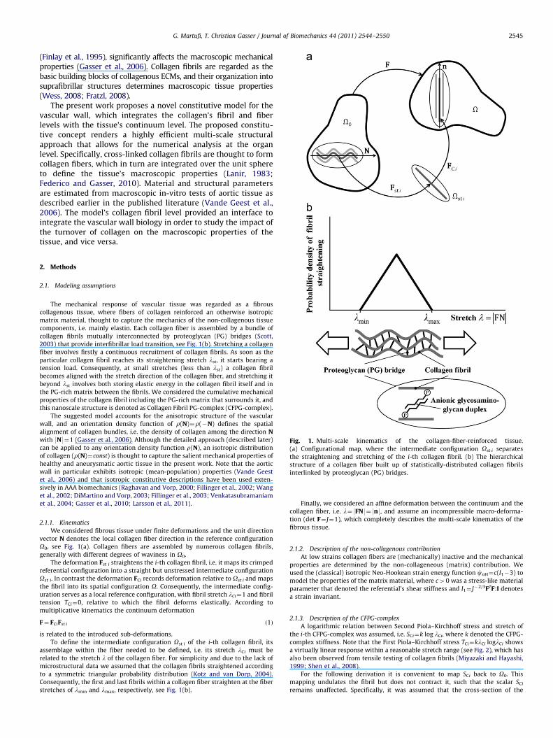
Fig. 1. Multi-scale kinematics of the collagen-fiber-reinforced tissue.
(a) Configurational map, where the intermediate configuration Ost i separates
the straightening and stretching of the i-th collagen fibril. (b) The hierarchical
structure of a collagen fiber built up of statistically-distributed collagen fibrils
interlinked by proteoglycan (PG) bridges.
2. Methods
2.1. Modeling assumptions
The mechanical response of vascular tissue was regarded as a fibrous
collagenous tissue, where fibers of collagen reinforced an otherwise isotropic
matrix material, thought to capture the mechanics of the non-collagenous tissue
components, i.e. mainly elastin. Each collagen fiber is assembled by a bundle of
collagen fibrils mutually interconnected by proteoglycan (PG) bridges (Scott,
2003) that provide interfibrillar load transition, see Fig. 1(b). Stretching a collagen
fiber involves firstly a continuous recruitment of collagen fibrils. As soon as the
particular collagen fibril reaches its straightening stretch lst, it starts bearing a
tension load. Consequently, at small stretches (less than lst) a collagen fibril
becomes aligned with the stretch direction of the collagen fiber, and stretching it
beyond lst involves both storing elastic energy in the collagen fibril itself and in
the PG-rich matrix between the fibrils. We considered the cumulative mechanical
properties of the collagen fibril including the PG-rich matrix that surrounds it, and
this nanoscale structure is denoted as Collagen Fibril PG-complex (CFPG-complex).
The suggested model accounts for the anisotropic structure of the vascular
wall, and an orientation density function of r(N)¼r(�N) defines the spatial
alignment of collagen bundles, i.e. the density of collagen among the direction Nwith 9N9¼1 (Gasser et al., 2006). Although the detailed approach (described later)
can be applied to any orientation density function r(N), an isotropic distribution
of collagen (r(N)¼const) is thought to capture the salient mechanical properties of
healthy and aneurysmatic aortic tissue in the present work. Note that the aortic
wall in particular exhibits isotropic (mean-population) properties (Vande Geest
et al., 2006) and that isotropic constitutive descriptions have been used exten-
sively in AAA biomechanics (Raghavan and Vorp, 2000; Fillinger et al., 2002; Wang
et al., 2002; DiMartino and Vorp, 2003; Fillinger et al., 2003; Venkatasubramaniam
et al., 2004; Gasser et al., 2010; Larsson et al., 2011).
2.1.1. Kinematics
We considered fibrous tissue under finite deformations and the unit direction
vector N denotes the local collagen fiber direction in the reference configuration
O0, see Fig. 1(a). Collagen fibers are assembled by numerous collagen fibrils,
generally with different degrees of waviness in O0.
The deformation Fst i straightens the i-th collagen fibril, i.e. it maps its crimped
referential configuration into a straight but unstressed intermediate configuration
Ost i. In contrast the deformation FCi records deformation relative to Ost i and maps
the fibril into its spatial configuration O. Consequently, the intermediate config-
uration serves as a local reference configuration, with fibril stretch lCi¼1 and fibril
tension TCi¼0, relative to which the fibril deforms elastically. According to
multiplicative kinematics the continuum deformation
F¼ FCiFst i ð1Þ
is related to the introduced sub-deformations.
To define the intermediate configuration Ost i of the i-th collagen fibril, its
assemblage within the fiber needed to be defined, i.e. its stretch lCi must be
related to the stretch l of the collagen fiber. For simplicity and due to the lack of
microstructural data we assumed that the collagen fibrils straightened according
to a symmetric triangular probability distribution (Kotz and van Dorp, 2004).
Consequently, the first and last fibrils within a collagen fiber straighten at the fiber
stretches of lmin and lmax, respectively, see Fig. 1(b).
Finally, we considered an affine deformation between the continuum and the
collagen fiber, i.e. l¼9FN9¼9n9, and assume an incompressible macro-deforma-
tion (det F¼ J¼1), which completely describes the multi-scale kinematics of the
fibrous tissue.
2.1.2. Description of the non-collagenous contribution
At low strains collagen fibers are (mechanically) inactive and the mechanical
properties are determined by the non-collageneous (matrix) contribution. We
used the (classical) isotropic Neo-Hookean strain energy function cnH¼c(I1�3) to
model the properties of the matrix material, where c40 was a stress-like material
parameter that denoted the referential’s shear stiffness and I1¼ J�2/3FTF:I denotes
a strain invariant.
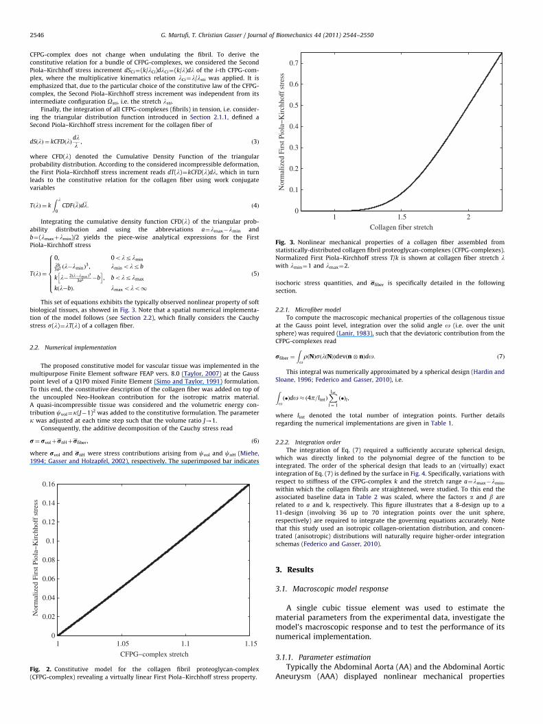
2.1.3. Description of the CFPG-complex
A logarithmic relation between Second Piola–Kirchhoff stress and stretch of
the i-th CFPG-complex was assumed, i.e. SCi¼k log lCi, where k denoted the CFPG-
complex stiffness. Note that the First Piola–Kirchhoff stress TCi¼klCi loglCi shows
a virtually linear response within a reasonable stretch range (see Fig. 2), which has
also been observed from tensile testing of collagen fibrils (Miyazaki and Hayashi,
1999; Shen et al., 2008).
For the following derivation it is convenient to map SCi back to O0. This
mapping undulates the fibril but does not contract it, such that the scalar SCi
remains unaffected. Specifically, it was assumed that the cross-section of the
1 1.5 20
0.1
0.2
0.3
0.4
0.5
0.6
0.7
Collagen fiber stretch
Nor
mal
ized
Fir
st P
iola
−K
irch
hoff
str
ess
Fig. 3. Nonlinear mechanical properties of a collagen fiber assembled from
statistically-distributed collagen fibril proteoglycan-complexes (CFPG-complexes).
Normalized First Piola–Kirchhoff stress T/k is shown at collagen fiber stretch lwith lmin¼1 and lmax¼2.
G. Martufi, T. Christian Gasser / Journal of Biomechanics 44 (2011) 2544–25502546
CFPG-complex does not change when undulating the fibril. To derive the
constitutive relation for a bundle of CFPG-complexes, we considered the Second
Piola–Kirchhoff stress increment dSCi¼(k/lCi)dlCi¼(k/l)dl of the i-th CFPG-com-
plex, where the multiplicative kinematics relation lCi¼l/lsti was applied. It is
emphasized that, due to the particular choice of the constitutive law of the CFPG-
complex, the Second Piola–Kirchhoff stress increment was independent from its
intermediate configuration Osti, i.e. the stretch lsti.
Finally, the integration of all CFPG-complexes (fibrils) in tension, i.e. consider-
ing the triangular distribution function introduced in Section 2.1.1, defined a
Second Piola–Kirchhoff stress increment for the collagen fiber of
dSðlÞ ¼ kCFDðlÞdll
, ð3Þ
where CFD(l) denoted the Cumulative Density Function of the triangular
probability distribution. According to the considered incompressible deformation,
the First Piola–Kirchhoff stress increment reads dT(l)¼kCFD(l)dl, which in turn
leads to the constitutive relation for the collagen fiber using work conjugate
variables
TðlÞ ¼ k
Z l
0CDFðlÞdl: ð4Þ
Integrating the cumulative density function CFD(l) of the triangular prob-
ability distribution and using the abbreviations a¼lmax�lmin and
b¼(lmaxþlmin)/2 yields the piece-wise analytical expressions for the First
Piola–Kirchhoff stress
TðlÞ ¼
0, 0olrlmin
2k3a2 ðl�lminÞ
3 , lmin olrb
k l� 2ðl�lmax Þ3
3a2 �bh i
, bolrlmax
kðl�bÞ: lmax olo1
8>>>>><>>>>>:
ð5Þ
This set of equations exhibits the typically observed nonlinear property of soft
biological tissues, as showed in Fig. 3. Note that a spatial numerical implementa-
tion of the model follows (see Section 2.2), which finally considers the Cauchy
stress s(l)¼lT(l) of a collagen fiber.
2.2. Numerical implementation
The proposed constitutive model for vascular tissue was implemented in the
multipurpose Finite Element software FEAP vers. 8.0 (Taylor, 2007) at the Gauss
point level of a Q1P0 mixed Finite Element (Simo and Taylor, 1991) formulation.
To this end, the constitutive description of the collagen fiber was added on top of
the uncoupled Neo-Hookean contribution for the isotropic matrix material.
A quasi-incompressible tissue was considered and the volumetric energy con-
tribution cvol¼k(J�1)2 was added to the constitutive formulation. The parameter
k was adjusted at each time step such that the volume ratio J-1.
Consequently, the additive decomposition of the Cauchy stress read
r¼ rvolþrnHþrfiber , ð6Þ
where rvol and rnH were stress contributions arising from cvol and cnH (Miehe,
1994; Gasser and Holzapfel, 2002), respectively. The superimposed bar indicates
1 1.05 1.1 1.150
0.02
0.04
0.06
0.08
0.1
0.12
0.14
0.16
CFPG−complex stretch
Nor
mal
ized
Fir
st P
iola
−K
irch
hoff
str
ess
Fig. 2. Constitutive model for the collagen fibril proteoglycan-complex
(CFPG-complex) revealing a virtually linear First Piola–Kirchhoff stress property.
isochoric stress quantities, and rfiber is specifically detailed in the following
section.
2.2.1. Microfiber model
To compute the macroscopic mechanical properties of the collagenous tissue
at the Gauss point level, integration over the solid angle o (i.e. over the unit
sphere) was required (Lanir, 1983), such that the deviatoric contribution from the
CFPG-complexes read
rfiber ¼
ZorðNÞsðlðNÞÞdevðn� nÞdo: ð7Þ
This integral was numerically approximated by a spherical design (Hardin and
Sloane, 1996; Federico and Gasser, 2010), i.e.
Zoð�Þdo� ð4p=lintÞ
Xlint
l ¼ 1
ð�Þl ,
where lint denoted the total number of integration points. Further details
regarding the numerical implementations are given in Table 1.
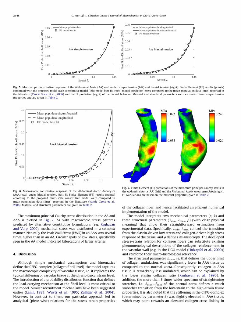
2.2.2. Integration order
The integration of Eq. (7) required a sufficiently accurate spherical design,
which was directly linked to the polynomial degree of the function to be
integrated. The order of the spherical design that leads to an (virtually) exact
integration of Eq. (7) is defined by the surface in Fig. 4. Specifically, variations with
respect to stiffness of the CFPG-complex k and the stretch range a¼lmax�lmin,
within which the collagen fibrils are straightened, were studied. To this end the
associated baseline data in Table 2 was scaled, where the factors a and b are
related to a and k, respectively. This figure illustrates that a 8-design up to a
11-design (involving 36 up to 70 integration points over the unit sphere,
respectively) are required to integrate the governing equations accurately. Note
that this study used an isotropic collagen-orientation distribution, and concen-
trated (anisotropic) distributions will naturally require higher-order integration
schemas (Federico and Gasser, 2010).
3. Results
3.1. Macroscopic model response
A single cubic tissue element was used to estimate thematerial parameters from the experimental data, investigate themodel’s macroscopic response and to test the performance of itsnumerical implementation.
3.1.1. Parameter estimation
Typically the Abdominal Aorta (AA) and the Abdominal AorticAneurysm (AAA) displayed nonlinear mechanical properties
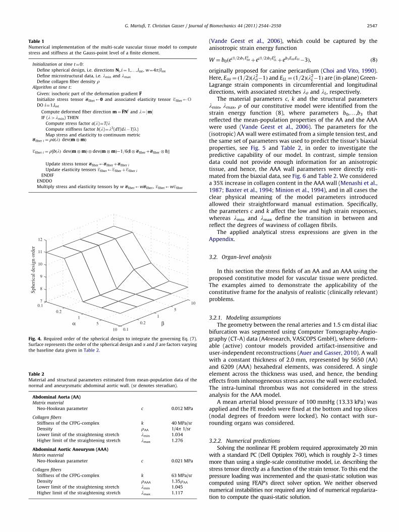
Table 1Numerical implementation of the multi-scale vascular tissue model to compute
stress and stiffness at the Gauss-point level of a finite element.
Initialization at time t¼0:
Define spherical design, i.e. directions Ni ,i¼ 1,. . .,lint, w¼4p/lint
Define microstructural data, i.e. lmin and lmax
Define collagen fiber density rAlgorithm at time t:
Given: isochoric part of the deformation gradient F
Initialize stress tensor rfiber’0 and associated elasticity tensor cfiber’O
DO i¼1,lint
Compute deformed fiber direction m¼ FNi and l¼9m9IF (l4lmin) THEN
Compute stress factor a(l)¼T/lCompute stiffness factor b(l)¼l2(dT/dl�T/l)
Map stress and elasticity to continuum metricrfiber i ¼ raðlÞ devðm�mÞ
cfiber i ¼ r½bðlÞ devðm�mÞ � devðm�mÞ�1=6ðI� rfiberþrfiber � IÞ�
Update stress tensor rfiber’rfiberþrfiber i
Update elasticity tensors cfiber’cfiberþcfiber i
ENDIF
ENDDO
Multiply stress and elasticity tensors by w rfiber’wrfiber, cfiber’wcfiber
0.10.2
15
10 0.1
0.2
1
5
107
8
9
10
11
12
βα
Sphe
rica
l des
ign
orde
r
Fig. 4. Required order of the spherical design to integrate the governing Eq. (7).
Surface represents the order of the spherical design and a and b are factors varying
the baseline data given in Table 2.
Table 2Material and structural parameters estimated from mean-population data of the
normal and aneurysmatic abdominal aortic wall. (sr denotes steradian).
Abdominal Aorta (AA)Matrix material
Neo-Hookean parameter c 0.012 MPa
Collagen fibers
Stiffness of the CFPG-complex k 40 MPa/sr
Density rAA 1/4p 1/sr
Lower limit of the straightening stretch lmin 1.034
Higher limit of the straightening stretch lmax 1.276
Abdominal Aortic Aneurysm (AAA)Matrix material
Neo-Hookean parameter c 0.021 MPa
Collagen fibers
Stiffness of the CFPG-complex k 63 MPa/sr
Density rAAA 1.35rAA
Lower limit of the straightening stretch lmin 1.045
Higher limit of the straightening stretch lmax 1.117
G. Martufi, T. Christian Gasser / Journal of Biomechanics 44 (2011) 2544–2550 2547
(Vande Geest et al., 2006), which could be captured by theanisotropic strain energy function
W ¼ b0ðeð1=2Þb1E2
yyþeð1=2Þb2E2LLþeb3EyyELL�3Þ, ð8Þ
originally proposed for canine pericardium (Choi and Vito, 1990).Here, Eyy ¼ ð1=2Þðl2
y�1Þ and ELL ¼ ð1=2Þðl2L�1Þ are (in-plane) Green-
Lagrange strain components in circumferential and longitudinaldirections, with associated stretches ly and lL, respectively.
The material parameters c, k and the structural parameterslmin, lmax, r of our constitutive model were identified from thestrain energy function (8), where parameters b0,y,b3 thatreflected the mean-population properties of the AA and the AAAwere used (Vande Geest et al., 2006). The parameters for the(isotropic) AA wall were estimated from a simple tension test, andthe same set of parameters was used to predict the tissue’s biaxialproperties, see Fig. 5 and Table 2, in order to investigate thepredictive capability of our model. In contrast, simple tensiondata could not provide enough information for an anisotropictissue, and hence, the AAA wall parameters were directly esti-mated from the biaxial data, see Fig. 6 and Table 2. We considereda 35% increase in collagen content in the AAA wall (Menashi et al.,1987; Baxter et al., 1994; Minion et al., 1994), and in all cases theclear physical meaning of the model parameters introducedallowed their straightforward manual estimation. Specifically,the parameters c and k affect the low and high strain responses,whereas lmin and lmax define the transition in between andreflect the degrees of waviness of collagen fibrils.
The applied analytical stress expressions are given in theAppendix.
3.2. Organ-level analysis
In this section the stress fields of an AA and an AAA using theproposed constitutive model for vascular tissue were predicted.The examples aimed to demonstrate the applicability of theconstitutive frame for the analysis of realistic (clinically relevant)problems.
3.2.1. Modeling assumptions
The geometry between the renal arteries and 1.5 cm distal iliacbifurcation was segmented using Computer Tomography-Angio-graphy (CT-A) data (A4research, VASCOPS GmbH), where deform-able (active) contour models provided artifact-insensitive anduser-independent reconstructions (Auer and Gasser, 2010). A wallwith a constant thickness of 2.0 mm, represented by 5650 (AA)and 6209 (AAA) hexahedral elements, was considered. A singleelement across the thickness was used, and hence, the bendingeffects from inhomogeneous stress across the wall were excluded.The intra-luminal thrombus was not considered in the stressanalysis for the AAA model.
A mean arterial blood pressure of 100 mmHg (13.33 kPa) wasapplied and the FE models were fixed at the bottom and top slices(nodal degrees of freedom were locked). No contact with sur-rounding organs was considered.
3.2.2. Numerical predictions
Solving the nonlinear FE problem required approximately 20 minwith a standard PC (Dell Optiplex 760), which is roughly 2–3 timesmore than using a single-scale constitutive model, i.e. describing thestress tensor directly as a function of the strain tensor. To this end thepressure loading was incremented and the quasi-static solution wascomputed using FEAP’s direct solver option. We neither observednumerical instabilities nor required any kind of numerical regulariza-tion to compute the quasi-static solution.
1 1.05 1.1 1.150
0.01
0.02
0.03
0.04
0.05
Stretch λ
Firs
t Pio
la−
Kir
chho
ff s
tres
s [M
Pa]
Mean population dataFE model best fit
AA simple tension
1 1.05 1.1 1.150
0.01
0.02
0.03
0.04
0.05
0.06
Stretch λ
Firs
t Pio
la−
Kir
chho
ff s
tres
s [M
Pa]
Mean population data longitudinalMean population data circumferentialFE model prediction
AA biaxial tension
Fig. 5. Macroscopic constitutive response of the Abdominal Aorta (AA) wall under simple tension (left) and biaxial tension (right). Finite Element (FE) results (points)
computed with the proposed multi-scale constitutive model (left: model best fit; right: model prediction) were compared to the mean-population data (lines) reported in
the literature (Vande Geest et al., 2006) and the FE prediction (right) of the biaxial behavior. Material and structural parameters were estimated from simple tension
properties and are given in Table 2.
1 1.02 1.04 1.06 1.08 1.10
0.05
0.1
0.15
0.2
0.25
0.3
Stretch λ
Firs
t Pio
la−
Kir
chho
ff s
tres
s [M
Pa]
Mean pop. data circumferentialMean pop. data longitudinalFE model best fit
AAA biaxial tension
Fig. 6. Macroscopic constitutive response of the Abdominal Aortic Aneurysm
(AAA) wall under biaxial tension. Best fit Finite Element (FE) results (points)
according to the proposed multi-scale constitutive model were compared to
mean-population data (lines) reported in the literature (Vande Geest et al.,
2006). Material and structural parameters are given in Table 2.
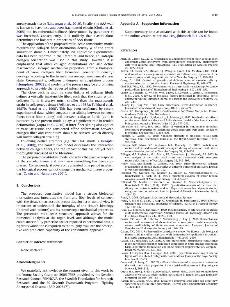
Fig. 7. Finite Element (FE) predictions of the maximum principal Cauchy stress in
the Abdominal Aorta (AA) (left) and the Abdominal Aortic Aneurysm (AAA) (right).
FE calculations are based on the material properties given in Table 2.
G. Martufi, T. Christian Gasser / Journal of Biomechanics 44 (2011) 2544–25502548
The maximum principal Cauchy stress distribution in the AA andAAA is plotted in Fig. 7. As with macroscopic stress patternspredicted by alternative constitutive formulations (e.g. Raghavanand Vorp, 2000), mechanical stress was distributed in a complexmanner. Naturally the Peak Wall Stress (PWS) in an AAA was severaltimes higher than in an AA. Circular spots of low stress, specificallyseen in the AA model, indicated bifurcations of larger arteries.
4. Discussion
Although simple mechanical assumptions and kinematicsdefine the CFPG-complex (collagen fibril level), the model capturesthe macroscopic complexity of vascular tissue, i.e. it replicates thetypical stiffening of vascular tissue at the physiological strain level.The introduction of a probability distribution function that definesthe load-carrying mechanism at the fibril level is most critical tothe model. Similar recruitment mechanisms have been suggestedearlier (Lanir, 1983; Wuyts et al., 1995; Zulliger et al., 2004).However, in contrast to them, our particular approach led toanalytical (piece-wise) relations for the stress–strain properties
of the collagen fiber, and hence, facilitated an efficient numericalimplementation of the model.
The model integrates two mechanical parameters (c, k) andthree structural parameters (lmin, lmax, r) (with clear physicalmeaning) that allow their straightforward estimation fromexperimental data. Specifically, lmin, lmax control the transitionfrom the elastin-driven low stress and collagen-driven high stressresponse of the tissue, and r defines its anisotropy. The developedstress–strain relation for collagen fibers can substitute existingphenomenological descriptions of the collagen reinforcement inthe vascular wall (e.g. in the HGO model (Holzapfel et al., 2000))and reinforce their micro-histological relevance.
The structural parameter lmax, i.e. that defines the upper limitof collagen undulation, was significantly lower in AAA tissue ascompared to the normal aorta. Consequently, collagen in AAAtissue is remarkably less undulated, which can be explained bythe lower elastin collagen ratio (Raghavan et al., 1996). Inaddition, the more than 3 times wider spectrum of straighteningstretches, i.e. lmax�lmin of the normal aorta defines a muchsmoother transition from the low-strain to the high-strain tissueproperties. It is also noted that the stiffening in the CFPG-complex(determined by parameter k) was slightly elevated in AAA tissue,which may point towards an elevated collagen cross-linking in
G. Martufi, T. Christian Gasser / Journal of Biomechanics 44 (2011) 2544–2550 2549
aneurysmatic tissue (Lindeman et al., 2010). Finally, the AAA wallis known to have less and even fragmented elastin (Choke et al.,2005) but its referential stiffness (determined by parameter c)was increased. Consequently, it is unlikely that elastin alonedetermines the low-strain properties of AAA tissue.
The application of the proposed multi-scale constitutive modelrequires the collagen fiber orientation density r of the entiresimulation domain. Unfortunately, no applicable experimentaldata has been reported in the literature, and hence, an isotropiccollagen orientation was used in this study. However, it isemphasized that other collagen distributions can also definemacroscopic isotropic mechanical properties. From a biologicalpoint of view, collagen fiber formation (orientation density)develops according to the tissue’s macroscopic mechanical stressstate. Consequently, collagen undergoes an adaptation process(Humphrey, 2002) and modeling the process may be a promisingapproach to provide the requested information.
The close packing and the cross-linking of collagen fibrilsdefines a virtually inextensible fiber, such that the strain withincollagen fibrils is always much smaller than the macroscopicstrain in collagenous tissue (Folkhard et al., 1987a; Folkhard et al.,1987b; Fratzl et al., 1998). This has been confirmed by recentexperimental data, which suggests that sliding between collagenfibers (inter-fiber sliding) and between collagen fibrils (as it iscaptured by the present model) plays a significant role in tendondeformation (Gupta et al., 2010). If inter-fiber sliding also appliesto vascular tissue, the considered affine deformation betweencollagen fiber and continuum should be relaxed, which directlywill lower collagen straining.
Following earlier attempts (e.g. the HGO model (Holzapfelet al., 2000)), the constitutive model disregards the interactionbetween collagen fibers, and the impact of this has not yet beenthoroughly discussed in the literature.
The proposed constitutive model considers the passive responseof the vascular tissue, and any tissue remodeling has been sup-pressed. Consequently, it covers only a limited time period in whichthe biological process cannot change the mechanical tissue proper-ties (Cowin and Humphrey, 2001).
5. Conclusions
The proposed constitutive model has a strong biologicalmotivation and integrates the fibril and fiber levels of collagenwith the tissue’s macroscopic properties. Such a structural view isimportant to understand the interplay of the tissue’s histology(internal architecture) and its macroscopic mechanical properties.The presented multi-scale structural approach allows for thenumerical analysis at the organ level, and although the modelcould successfully prescribe earlier reported experimental data, arigorous validation is required to thoroughly evaluate the descrip-tive and predictive capability of the constitutive approach.
Conflict of interest statement
None declared.
Acknowledgments
We gratefully acknowledge the support given to this work bythe Young Faculty Grant no. 2006-7568 provided by the SwedishResearch Council, VINNOVA, the Swedish Foundation for StrategicResearch, and the EC Seventh Framework Program; ‘FightingAneurysmal Disease (FAD-200647)’.
Appendix A. Supporting information
Supplementary data associated with this article can be foundin the online version at doi:10.1016/j.jbiomech.2011.07.015.
References
Auer, M., Gasser, T.C., 2010. Reconstruction and finite element mesh generation ofabdominal aortic aneurysms from computerized tomography angiographydata with minimal user interactions. IEEE Transaction on Medical Imaging29, 1022–1028.
Baxter, B.T., Davis, V.A., Minion, D.J., Wang, Y., Lynch, T.G., McManus, B.C., 1994.Abdominal aortic aneurysms are associated with altered matrix proteins of thenonaneurysmal aortic segments. Journal of Vascular Surgery 19, 797–803.
Carey, D., 1991. Control of growth and differentiation of vascular cells byextracellular matrix proteins. Annual Review of Physiology 53, 161–177.
Choi, H.S., Vito, R.P., 1990. Two-dimensional stress–strain relationship for caninepericardium. Journal of Biomechanical Engineering 112 (2), 153–159.
Choke, E., Cockerill, G., Wilson, W.R., Sayed, S., Dawson, J., Loftus, I., Thompson,M.M., 2005. A review of biological factors implicated in abdominal aorticaneurysm rupture. European Journal of Vascular and Endovascular Surgery 30,227–244.
Chuong, C.J., Fung, Y.C., 1983. Three-dimensional stress distribution in arteries.Journal of Biomechanical Engineering 105 (3), 268–274.
Cowin, S.C., Humphrey, J.D., 2001. Cardiovascular Soft Tissue Mechanics. KluwerAcademic Publisher, The Netherlands.
Delfino, A., Stergiopulos, N., Moore Jr., J.E., Meister, J.J., 1997. Residual strain effectson the stress field in a thick wall finite element model of the human carotidbifurcation. Journal of Biomechanics 30 (8), 777–786.
DiMartino, E.S., Vorp, D.A., 2003. Effect of variation in intraluminal thrombusconstitutive properties on abdominal aortic aneurysm wall stress. Annals ofBiomedical Engineering 31, 804–809.
Federico, S., Gasser, T.C., 2010. Nonlinear elasticity of biological tissues withstatistical fiber orientation. Journal of the Royal Society Interface 7 (47),955–966.
Fillinger, M.F., Marra, S.P., Raghavan, M.L., Kennedy, F.E., 2003. Prediction ofrupture risk in abdominal aortic aneurysm during observation: wall stressversus diameter. Journal of Vascular Surgery 37, 724–732.
Fillinger, M.F., Raghavan, M.L., Marra, S.P., Cronenwett, J.L., Kennedy, F.E., 2002. Invivo analysis of mechanical wall stress and abdominal aortic aneurysmrupture risk. Journal of Vascular Surgery 36, 589–597.
Finlay, H.M., McCullough, L., Canham, P.B., 1995. Three-dimensional collagenorganization of human brain arteries at different transmural pressures. Journalof Vascular Research 32, 301–312.
Folkhard, W., Geerken, W., Knorzer, E., Mosler, E., Nemetschekgansler, H.,Nemetschek, T., Koch, M.H.J., 1987a. Structural dynamic of native tendoncollagen. Journal of Molecular Biology 193, 405–407.
Folkhard, W., Mosler, E., Geerken, W., Knorzer, E., Nemetschekgansler, H.,Nemetschek, T., Koch, M.H.J., 1987b. Quantitative-analysis of the molecularsliding mechanism in native tendon collagen—time-resolved dynamic studiesusing synchrotron radiation. Internal Journal of Biological Macromolecules 9,169–175.
Fratzl, P., 2008. Collagen: Structure and Mechanics. Springer.Fratzl, P., Misof, K., Zizak, I., Rapp, G., Amenitsch, H., Bernstorff, S., 1998. Fibrillar
structure and mechanical properties of collagen. Journal of Structural Biology122, 119–122.
Fung, Y.C., Fronek, K., Patitucci, P., 1979. Pseudoelasticity of arteries and the choiceof its mathematical expression. American Journal of Physiology—Hearth andCirculation Phisiology 237, H620–H621.
Gasser, T.C., Auer, M., Labruto, F., Swedenborg, J., Roy, J., 2010. Biomechanicalrupture risk assessment of abdominal aortic aneurysms: model complexityversus predictability of finite element simulations. European Journal ofVascular and Endovascular Surgery 40, 176–185.
Gasser, T.C., 2011. An irreversible constitutive model for fibrous soft biologicaltissue: a. 3D microfiber approach with demonstrative application to abdom-inal aortic aneurysms. Acta Biomaterialia 7 (6), 2457–2466.
Gasser, T.C., Holzapfel, G.A., 2002. A rate-independent elastoplastic constitutivemodel for (biological) fiber-reinforced composites at finite strains: continuumbasis, algorithmic formulation and finite element implementation. Computa-tional Mechanics 29, 340–360.
Gasser, T.C., Ogden, R.W., Holzapfel, G.A., 2006. Hyperelastic modelling of arteriallayers with distributed collagen fiber orientations. Journal of the Royal SocietyInterface 3, 15–35.
Greenwald, S., Berry, C., 1980. The effect of alterations of scleroprotein content onthe static mechanical properties of the arterial wall. Advances in PhysiologicalScience 8, 203–212.
Gupta, H.S., Steo, J., Krauss, S., Boesecke, P., Screen, H.R.C., 2010. In situ multi-levelanalysis of viscoelastic deformation mechanisms in tendon collagen. Journal ofStructural Biology 169, 183–191.
Hardin, R.H., Sloane, N.J.A., 1996. McLaren’s improved snub cube and other newspherical designs in three dimentions. Discrete and Computational Geometry15, 429–441.
G. Martufi, T. Christian Gasser / Journal of Biomechanics 44 (2011) 2544–25502550
Holzapfel, G.A., Gasser, T.C., Ogden, R.W., 2000. A new constitutive framework for
arterial wall mechanics and a comparative study of material models. Journal of
elasticity 61, 1–48.Holzapfel, G.A., Gasser, T.C., Stadler, M., 2002. A structural model for the
viscoelastic behavior of arterial walls: continuum formulation and finite
element analysis. European Journal of Mechanic—A/Solids 21 (3), 441–463.Humphrey, J.D., 1995. Mechanics of the arterial wall: review and directions.
Critical reviews in biomedical engineering 23 (1–2), 1–162.Humphrey, J.D., 2002. Cardiovascular Solid Mechanics. Cells, Tissues, and Organs.
Springer-Verlag, New York.Kotz, S., van Dorp, J.R., 2004. Beyond Beta: Other Continuous Families of Distribu-
tions with Bounded Support and Applications. World Scientific.Larsson, E., Labruto, F., Gasser, T.C., Swedenborg, J., Hultgren, R., 2011. Analysis of
aortic wall stress and rupture risk in AAA patients with a gender perspective.
Journal of Vascular Surgery 54 (2), 295–299.Lanir, Y., 1983. Constitutive equations for fibrous connective tissues. Journal of
biomechanics 16 (1), 1–12.Lindeman, J.H.N., Ashcroft, B.A., Beenakker, J.W.M., van Es, M., Koekkoek, N.B.R.,
Prins, F.A., Tielemans, J.F., Abdul-Hussien, H., Bank, R.A., Oosterkamp, T.H.,
2010. Distinct defects in collagen microarchitecture underlie vessel-wall
failure in advanced abdominal aneurysms and aneurysms in Marfan syn-
drome. Proceedings of the National Academy of Sciences 107 (2), 862–865.Menashi, S., Campa, J.S., Greenhalgh, R.M., Powell, J.T., 1987. Collagen in abdominal
aortic aneurysm: typing, content, and degradation. Journal of Vascular Surgery
6 (6), 578–582.Miehe, C., 1994. Aspects of the formulation and finite element implementation of
large strain isotropic elasticity. International Journal for Numerical Methods in
Engineering 37, 1981–2004.Minion, D.J., Davis, V.A., Nejezchleb, P.A., Wang, Y., Lynch, T.G., McManus, B.M.,
1994. Elastin is increased in abdominal aortic aneurysms. Journal of Surgical
Research 57, 443–446.Miyazaki, H., Hayashi, K., 1999. Tensile tests of collagen fibers obtained from the
rabbit patellar tendon. Biomedical Microdevices 2, 151–157.Raghavan, M.L., Vorp, D.A., 2000. Toward a biomechanical tool to evaluate rupture
potential of abdominal aortic aneurysm: identification of a finite strain
constitutive model and evaluation of its applicability. Journal of Biomechanics
33, 475–482.
Raghavan, M.L., Webster, M.W., Vorp, D.A., 1996. Ex vivo biomechanical behaviorof abdominal aortic aneurysm: assessment using a new mathematical model.Annals of Biomedical Engineering 24, 573–582.
Roach, M.R., Burton, A.C., 1957. The reason for the shape of the distensibilitycurves of arteries. Canadian Journal of Physiology and Pharmacology 35,681–690.
Scott, J.E., 2003. Elasticity in extracellular matrix ‘shape modules’ of tendon,cartilage, etc. a sliding proteoglycan-filament model. The Journal of Physiology553 (2), 335–343.
Shen, Z.L., Dodge, M.R., Kahn, H., Ballarini, R., Eppell, S.J., 2008. Stress–strainexperiments on individual collagen fibrils. Biophysical Journal 95 (8), 3956–3963.
Simo, J.C., Taylor, R.L., 1991. Quasi-incompressible finite elasticity in principalstretches. Continuum basis and numerical algorithms. Computer Methods inApplied Mechanics and Engineering 85, 273–310.
Takamizawa, K., Hayashi, K., 1987. Strain energy density function and uniformstrain hypothesis for arterial mechanics. Journal of Biomechanics 20 (1), 7–17.
Taylor, R.L., 2007. FEAP—A Finite Element Analysis Program, Version 8.2 UserManual. University of California at Berkeley, Berkeley, California.
Vaishnav, R.N., Young, J.T., Janicki, J.S., Patel, J.S., 1972. Nonlinear anisotropicelastic properties of the canine aorta. Biophysical Journal 12 (8), 1008–1027.
Vande Geest, J.P., Sacks, M.S., Vorp, D.A., 2006. The effects of aneurysm on the biaxialmechanical behavior of human abdominal aorta. Journal of Biomechanics 39,1324–1334.
Venkatasubramaniam, A.K., Fagan, M.J., Mehta, T., Mylankal, K.J., Ray, B., Kuhan, G.,Chetter, I.C., McCollum, P.T., 2004. A comparative study of aortic wall stressusing finite element analysis for ruptured and nonruptured abdominal aorticaneurysms. European Journal of Vascular and Endovascular Surgery 28,168–176.
Wess, T.J., 2008. Collagen fibrillar structure and hierarchies. In: Fratzl, P. (Ed.),Collagen Structure and Mechanics. Springer, pp. 49–80.
Zulliger, M.A., Fridez, P., Hayashi, K., Stergiopulos, N., 2004. A strain energyfunction for arteries accounting for wall composition and structure. Journalof Biomechanics 37 (7), 989–1000.
Wang, D.H.J., Makaroun, M.S., Webster, M.W., Vorp, D.A., 2002. Effect of intralum-inal thrombus on wall stress in patient-specific models of abdominal aorticaneurysm. Journal of Vascular Surgery 36, 598–604.
Wuyts, F.L., Vanhuyse, V.J., Langewouters, G.J., Decraemer, W.F., Raman, E.R., Buyle,S., 1995. Elastic properties of human aortas in relation to age and athero-sclerosis: a structural model. Physics in Medicine and Biology 40, 1577–1597.
Copyright © 2022 FDOKUMEN