A Comparative Pharmacognostic Study on the Leaf, Stem and Root Components of Gymnema sylvestre...
12
1 23 Proceedings of the National Academy of Sciences, India Section B: Biological Sciences ISSN 0369-8211 Proc. Natl. Acad. Sci., India, Sect. B Biol. Sci. DOI 10.1007/s40011-012-0088-z A Comparative Pharmacognostic Study on the Leaf, Stem and Root Components of Gymnema sylvestre (Retz) R. Br. ex. Sm Ganesh C. Nikalje, Saurabha B. Zimare & Nutan P. Malpathak
Transcript of A Comparative Pharmacognostic Study on the Leaf, Stem and Root Components of Gymnema sylvestre...

1 23
Proceedings of the NationalAcademy of Sciences, India Section B:Biological Sciences ISSN 0369-8211 Proc. Natl. Acad. Sci., India, Sect. B Biol.Sci.DOI 10.1007/s40011-012-0088-z
A Comparative Pharmacognostic Study onthe Leaf, Stem and Root Components ofGymnema sylvestre (Retz) R. Br. ex. Sm
Ganesh C. Nikalje, Saurabha B. Zimare& Nutan P. Malpathak

1 23
Your article is protected by copyright and all
rights are held exclusively by The National
Academy of Sciences, India. This e-offprint
is for personal use only and shall not be self-
archived in electronic repositories. If you
wish to self-archive your work, please use the
accepted author’s version for posting to your
own website or your institution’s repository.
You may further deposit the accepted author’s
version on a funder’s repository at a funder’s
request, provided it is not made publicly
available until 12 months after publication.

RESEARCH ARTICLE
A Comparative Pharmacognostic Study on the Leaf, Stemand Root Components of Gymnema sylvestre (Retz) R. Br. ex. Sm
Ganesh C. Nikalje • Saurabha B. Zimare •
Nutan P. Malpathak
Received: 22 May 2012 / Revised: 25 August 2012 / Accepted: 27 August 2012
� The National Academy of Sciences, India 2012
Abstract Gymnema sylvestre is commonly used in
indigenous system of medicine to control diabetes mellitus
and is a source of various chemical constituents which are
medicinally important. There is much scope to study phar-
macognosy of this potent antidiabetic plant. Pharmacog-
nostic study of leaf, stem and roots was performed. For
quantitative microscopy of leaf the authors observed sto-
matal index 12 per mm2 area. The studied plant material
showed the presence of saponin, proteins, sugars, alkaloids
while starch is absent in leaf, stem and root. Phytochemical
analysis of plant material showed saponins, proteins,
reducing sugars and fats present in all samples. For percent
extractive values methanolic extract showed highest
extractive value in leaf (6.25), stem (4.9) and root (1.75)
while benzene showed lowest extractive values in leaf (1.2),
stem (0.5) and root (0.5). The distilled water, acetone,
absolute alcohol, petroleum ether and chloroform showed
moderate percentage extractive value. The total ash content
of leaf (0.083) was higher than that of stem (0.047) and root
(0.036), and acid insoluble ash of stem (0.02) was higher
than that of leaf (0.01) and root (0.001). The authors
observed quantity of gymnemic acid for leaf, stem and root
as 33.52, 22.20 and 13.00 mg/g DW respectively by high
performance thin layer chromatography.
Keywords Gymnema sylvestre � Pharmacognosy �Histochemistry � Phytochemistry �High performance thin layer chromatography
Introduction
Gymnema sylvestre (Retz) R. Br. ex Sm. belongs to family
Apocynaceae (previously Asclepiadaceae) [1], a vulnerable
slow growing species, perennial woody climber of tropical
and subtropical regions [2], having well known antidiabetic
property. Leaves of G. sylvestre are commonly used in
indigenous system of medicine to control diabetes mellitus
[3–5]. Besides these, leaves of G. sylvestre also have
hypoglycaemic [6], stomachic [4, 7], stimulant [8], laxa-
tive, diuretic, anti-sweetener [4], anti-bacterial [9–11], anti-
viral [4, 9] and anti-inflammatory [12] activity. It is a good
source of various secondary metabolites such as tannins,
alkaloids, saponins etc. Twenty different saponins and
glycosides have been reported in G. sylvestre [5]. Some
important saponins are gymnemic acid, deacyl gymnemic
acid, gymnemagenin and gymnestogenin [4]. The medici-
nal properties of G. sylvestre are mainly due to the pres-
ence of gymnemic acid.
Over the last decade, diabetes mellitus has emerged as
an important clinical and public health problem throughout
the world. The world prevalence of diabetes among adults
(aged 20–79 years) has been 6.4 %, affecting 285 million
adults in 2010 and will increase to 7.7 % affecting
439 million adults by 2030. Between 2010 and 2030, there
will be a 69 % increase in number of adults with diabetes
in developing countries and a 20 % increase in developed
countries [13].
Of all various chemical constituents found in G. sylves-
tre, gymnemic acid is the most important compound which
G. C. Nikalje � S. B. Zimare � N. P. Malpathak (&)
Department of Botany, University of Pune, Pune 411 007,
Maharashtra, India
e-mail: [email protected]
123
Proc. Natl. Acad. Sci., India, Sect. B Biol. Sci.
DOI 10.1007/s40011-012-0088-z
Author's personal copy

is present in all plant parts [4]. There is much scope to
study of pharmacognosy of this potent antidiabetic plant as
pharmacognosy helps in standardization of herbal drugs.
Material and Methods
Plant Material
Fresh leaves, stem and roots were collected from Path-
argaon, Lonavala, District Pune for pharmacognostic
study. The geographical location of collection site is
18�45015.3100 N. 73�31039.8100 E with elevation of 676 m.
Microscopic Studies
Thin transverse sections (TS) were taken from leaf
(through midrib), stem and root, double stained with
alcoholic (70 %) safranin and light green and mounted in
Canada balsam by following the micro technique method
of Johanson [14]. Microscopic characters were studied as
per Wallis [15] and Trease and Evans [16]. Microphotog-
raphy was performed for all preparations.
Quantitative Microscopy
Leaves were studied for their quantitative microscopy.
It includes stomatal number and stomatal index.
Stomatal Number and Stomatal Index (SI)
For the study of stomata and epidermis of leaf, upper and
lower epidermis was peeled off. It was then immersed into
hot alcohol to remove adhering tissues and pigments and
then stained with aqueous safranin and mounted in glyc-
erine. Stomatal index was calculated using following for-
mula [16]:
SI per mm2 area� �
% ¼ S
Eþ S� 100
where SI is the stomatal index, S is the no. of stomata per
unit area, and E is the no. of ordinary epidermal cells per
unit area.
Histochemical Tests
Free hand sections of the leaf, stem and root were done and
tested with the respective reagents. The detection and
localization of alkaloids and saponins were determined
using methods given by Johanson [14] and Krishnamurthy
[17].
1) Test for Alkaloids Sections were treated with the
following reagent for detecting alkaloids.
– Wagner’s Reagent Iodine 1 g and potassium
iodide 2 g were dissolved in 50 ml of distilled
water. Reddish brown precipitate shows presence
of alkaloids.
– Dragendroff’s Reagent (a) Solution A: basic
bismuth nitrate 0.85 g was dissolved in mixture
of 10 ml of acetic acid and 40 ml of distilled
water. (B) Solution B: 8 g of KI was dissolved in
20 ml of distilled water. Stock solution: equal
volumes of solution A and B were mixed together.
The formation of precipitate or development of
turbidity in the sections clearly indicated presence
of alkaloids.
– Mayer’s Reagent Potassium mercuric iodide solu-
tion: about 13.55 g of mercuric chloride (HgCl2)
and 50 g potassium iodide (KI) were dissolved in
one litre of distilled water. Cream or pale yellow
colour indicated presence of alkaloids.
– Hager’s Reagent Saturated aqueous solution of
picric acid was diluted in equal amount of distilled
water. Yellow precipitate in section indicates pres-
ence of alkaloids.
2) Test for Saponins Sections were placed directly in 1
drop of H2SO4 on a slide, which gives a characteristic
sequence of colour reactions, beginning with yellow,
changing to red in 30 min and finally becoming violet
or blue green in a short time. To determine localization
of saponins, Sections were put in saturated barium
hydroxide solution for about 24 h. Sections were
washed with calcium chloride then placed in potas-
sium dichromate. Yellow colour indicated presence of
saponins.
Phytochemical Tests
Qualitative tests for the presence of starch, proteins, tan-
nins, saponins, reducing sugars, anthroquinones, flavonoids
and glycosides were carried out was carried out by standard
methods of Harborne [18] and Trease and Evans [19].
Determination of Percentage Extractives
The percentage extractives determined by the method used
in Indian pharmacopoeia by Anonymous [20]. About 5 gm
of air dried powder was macerated with 100 ml of desired
solvent such as water, alcohol, petroleum ether and sol-
vent ether of specified strength, in closed flask of for 24 h.
The mixture was vigorously shaken at intervals. After
24 h the solution was rapidly filtered without any loss of
Proc. Natl. Acad. Sci., India, Sect. B Biol. Sci.
123
Author's personal copy

solvent. Them from the filtrate about 20 cc of the solution
was evaporated of dryness in a flat bottomed shallow
porcelain dish, dried at 100 �C and weighed. Then the
percentage of solvent was calculated with reference to the
air dried drug.
Ash Analysis
About 1 gm of powder was taken into porcelain crucible
previously heated and weighed. The powder was evenly
scattered in fine layer on bottom of the crucible. Then the
crucible was heated in furnace at a temperature not exceed-
ing 450 �C until free from carbon, cooled and weighed. The
percentage of total carbon free ash was calculated with
reference to air dried powder.
Acid Insoluble Ash
Carbon free ash was boiled with 25 ml of 2 M hydro-
chloric acid for 5 min, filtered through ash less filter
paper, was washed with hot water, and then the filter paper
was dried in oven, ignited in crucible previously weighed,
cooled and weighed. The percentage of the acid insoluble
ash was calculated with reference to the air dried powder
[20].
Fluorescence Analysis
The fluorescence analysis of powdered plant parts were
studied as per the methods followed by Chase and Pratt
[21]. The source of radiation was mercury, provided with a
Corning ultra-type filter which transmits the near ultravi-
olet (above 3,100 A) and absorbs wavelength in the visible
portions of the spectrum.
Quantification of Gymnemic Acid Using
Gymnemagenin by HPTLC
Extraction of Gymnemagenin
Air dried plant samples were grounded to powder. An
aliquot (1 g) accurately weighed was cold macerated in
50 ml of mixture of 95 % ethanol:water (1:1 v/v) for ca
48 h. The obtained solution was filtered and concentrated
under vacuum, redissolved in methanol (25 ml) in a water
bath at 55 �C. The final volume was made up to 50 ml.
This extract was further used for analysis with the help of
high performance thin layer chromatography (HPTLC).
Standard gymnemagenin was obtained from Natural
Remedies, Bangalore. All other chemicals used were of
analytical grade (Merck, India).
HPTLC Analysis of Samples
Sample solutions were applied onto the plates (10 9
10 cm) with automated TLC sampler Linomat V (Camag,
Muttenz, Switzerland) and were controlled by Win CATS
software 1.3.3 (Camag, Muttenz, Switzerland). Plates were
developed in 10 9 10 cm twin trough glass chambers
(Camag, Muttenz, Switzerland). A TLC scanner III with
Win CATS software was used for scanning the TLC plates.
Pre-coated silica gel aluminium plates 60 F254 (10 9
10 cm) (Merck, Darmstadt, Germany) were used. A con-
stant application rate of 0.2 ll/s1 was employed with a
bandwidth of 6 mm. The slit dimension was kept at 4.0 9
0.30 mm and scanning speed of 50 mm/s1 was employed.
The mobile phase consisted of chloroform:methanol (8:2)
v/v. The length of chromatogram run was 90 mm. After
development, chromatographic plates were dipped into
derivatization reagent (5 % modified vanillin-sulphuric
acid reagent) and then the derivatized plates were heated at
110 �C for 15 min in a pre-heated oven. The bluish-black
coloured spot corresponding to gymnemagenin was
observed. The plates were scanned at 610 nm.
Conversion of Gymnemagenin to Gymnemic Acid
The conversion of gymnemagenin to gymnemic acid was
done using the equation as follows:
C = X (809.0/506.7) where, C is the content of gym-
nemic acid in the sample, X is the content of gymne-
magenin present in the sample, 506.7 is the molecular
weight of gymnemagenin and 809.0 is the molecular
weight of gymnemic acid [4].
Results and Discussion
Microscopic Studies
TS of Stem
The TS of stem shows circular outline with entire or
somewhat wavy margin. Trichomes are multicellular, un-
iserate [22]. Epidermis forms a single outermost layer
consisting of barrel to rectangular cells. A thick cuticle
covers the epidermis. The outer region of cortex is made up
of 3–4 rows of polygonal or oval parenchymatous cells.
A distinct endodermis is absent. Pericycle is represented by
scattered groups of thick walled stone cells, forming a large
and complete ring. The vascular tissues occur in following
sequence—primary phloem, secondary phloem, cambium,
secondary xylem, primary xylem, interaxillary phloem and
pith (Fig. 1a).
Proc. Natl. Acad. Sci., India, Sect. B Biol. Sci.
123
Author's personal copy

TS of Leaf
Leaf is dorsiventral and shows the presence of midrib and
lamina (Fig. 1b).
Midrib
The midrib is broadly hemispherical on the abaxial side
with short lump on the adaxial side. Multicellular
unbranched trichomes consist of single rows of cells on
both sides of midrib. The ground tissue is parenchymatous
and compact. Vascular strand of midrib occurs as discon-
tinuous wide arc on the abaxial side. Xylem elements are in
radial rows, multiples of phloem patches both in upper and
lower sides of xylem strands.
Lamina
The epidermal cells of the lamina are square shaped with
outer convex wall and thin cuticle. Palisade tissue is single
layered, cylindrical, compact and occupies one-third
thickness of lamina. The spongy parenchyma cells are
lobed, four to five layered and loosely arranged. The lateral
vascular bundles of vein are small, collateral with thin
bundle sheath.
TS of Root
The TS of root shows circular outline. It is entire or wavy
in nature. Epidermis is multilayered with parenchymatous
cells and covered with thick layer of cuticle. Parenchy-
matous cortex was 6–7 cells in thickness with some glands
present in the cortex, presence of endodermis and pericy-
cle, xylem followed by phloem and pith (Fig. 1c).
Quantitative Microscopy
Stomatal Number and Stomatal Index Paracytic or aniso-
cytic type stomata were observed, which coincided with the
observations of Madhurima et al. [7]. Stomata were con-
fined to the lower epidermis and absent in upper epidermis.
Stomatal index (12 per mm2) was recorded which differed
with the findings of Madhurima et al. [7] who has reported
SI (20–30 per mm2) on abaxial surface, whereas coincides
with the observations made by Najafi and Deokule [23]
who reported SI 12.04/mm2 (Fig. 2; Table 1).
Histochemical Tests
Gymnema sylvestre is rich sources of saponin and alkaloids
[11] so in the present study histochemical test are restricted
Fig. 1 Anatomy (TS) of different plant parts of G. sylvestre
Proc. Natl. Acad. Sci., India, Sect. B Biol. Sci.
123
Author's personal copy

to only histochemistry of saponin and alkaloids of different
plant parts. The free hand sections of leaf, stem and root of
G. sylvestre were treated with different reagents to analyze
histochemistry. For confirming presence of saponins, sec-
tions were treated with dilute H2SO4 which gave charac-
teristic sequence of colour reactions beginning with
immediate occurrence of yellow, followed by red within
30 min and finally to bluish green. This was observed in all
the sections which concluded that saponin was present in
all studied plant parts. Alkaloids were detected using dif-
ferent reagents such as Mayer’s, which gave reddish brown
precipitate showing presence of alkaloids. For Dragendr-
off’s reagent, formation of precipitate or development
of turbidity in the sections clearly indicated presence of
alkaloids. Hager’s reagent produced yellow precipitate in
section indicating presence of alkaloids, while Wagner’s
reagent gave cream or pale yellow colour which showed
presence of alkaloids (Figs. 3, 4, 5). All the above men-
tioned tests were positive for alkaloids for all studied plant
parts.
Phytochemical Tests Alkaloids, glycosides and flavo-
noids were detected in alcoholic extracts and tannins,
saponins and anthroquinones were detected in water
extracts of leaf and/or stem and/or root powders. They are
in agreement with Kalidass and Mohan [24] who per-
formed phytochemical screening of the powdered aerial
plant parts of G. sylvestre. Contrasting results were
obtained for steroids in leaf, stem and root extracts when
compared with Kalidass and Mohan [24]. Alkaloids,
flavonoids and glycosides were detected in alcoholic
extracts of leaf, stem and roots using different reagents.
Alkaloids were detected using Mayer’s, Wagner’s, Drag-
endroff and Hager’s Reagent and it was present in all
studied plant parts. Flavonoids were confirmed in all
studied plant parts where Mg turnings were added to conc.
hydrochloric acid and positive test was indicated by
occurrence of pink colour. Glycosides were present only in
stem extract which gave white precipitation in benzene and
absent in leaf and root extract which did not give any
precipitation. Water extract of leaf, stem and root was used
to detect starch, saponins, tannins, proteins, reducing sug-
ars, anthroquinones, fats and steroids. I2KI reagent gave
bluish black precipitate to the extract which confirmed
presence of starch in leaf and stem extract. Vigorous
shaking of the extract in distilled water produced foam
confirming presence of saponins in all studied plant parts.
Leaf, stem and root extract when added with acidic FeCl3and MBB reagent gave a red and orange precipitation resp.
to confirm presence of tannins and proteins resp. in all
studied plant parts. Benedict’s reagent when added to the
extracts did not show any change which showed absence of
reducing sugars in all studied plant parts. Anthroquinones
were detected in leaf extract only, which gave orange
precipitation in benzene-ammonium hydroxide reagent.
Sudan III dye confirmed presence of fats in all studied plant
parts which gave orange precipitation. Chloroform–conc.
Sulphuric acid reagent when added to water extracts did
not show any change in colour which indicates absence of
steroids (Tables 2, 3).
Percentage Extractive Value
The selection of the solvents was done on the basis of
findings of Madhurima et al. [7] where five solvents were
used such as hexane, water, acetone, alcohol and chloro-
form. In addition to these solvents methanol, benzene,
petroleum ether and chloroform were also used. Hexane
was excluded from the experiment as hexane showed
Fig. 2 Quantitative microscopy
of leaves of G. sylvestre(Stomata)
Table 1 Quantitative microscopy of G. sylvestre leaves
No. Parameter Result
1. Stomatal index 12/mm2 area
2. Stomatal number 11/unit area
Proc. Natl. Acad. Sci., India, Sect. B Biol. Sci.
123
Author's personal copy
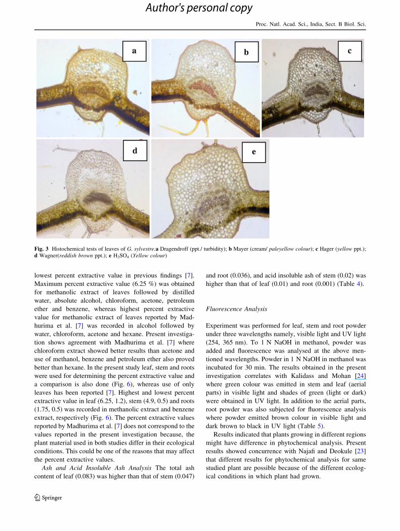
lowest percent extractive value in previous findings [7].
Maximum percent extractive value (6.25 %) was obtained
for methanolic extract of leaves followed by distilled
water, absolute alcohol, chloroform, acetone, petroleum
ether and benzene, whereas highest percent extractive
value for methanolic extract of leaves reported by Mad-
hurima et al. [7] was recorded in alcohol followed by
water, chloroform, acetone and hexane. Present investiga-
tion shows agreement with Madhurima et al. [7] where
chloroform extract showed better results than acetone and
use of methanol, benzene and petroleum ether also proved
better than hexane. In the present study leaf, stem and roots
were used for determining the percent extractive value and
a comparison is also done (Fig. 6), whereas use of only
leaves has been reported [7]. Highest and lowest percent
extractive value in leaf (6.25, 1.2), stem (4.9, 0.5) and roots
(1.75, 0.5) was recorded in methanolic extract and benzene
extract, respectively (Fig. 6). The percent extractive values
reported by Madhurima et al. [7] does not correspond to the
values reported in the present investigation because, the
plant material used in both studies differ in their ecological
conditions. This could be one of the reasons that may affect
the percent extractive values.
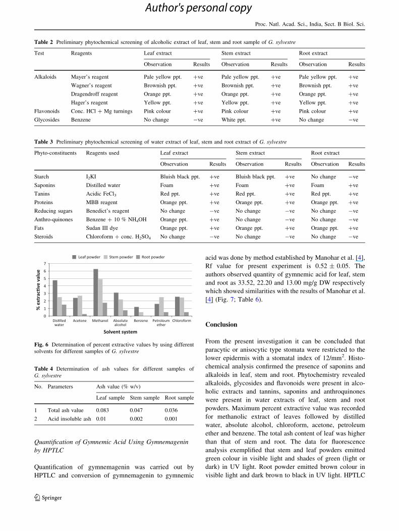
Ash and Acid Insoluble Ash Analysis The total ash
content of leaf (0.083) was higher than that of stem (0.047)
and root (0.036), and acid insoluble ash of stem (0.02) was
higher than that of leaf (0.01) and root (0.001) (Table 4).
Fluorescence Analysis
Experiment was performed for leaf, stem and root powder
under three wavelengths namely, visible light and UV light
(254, 365 nm). To 1 N NaOH in methanol, powder was
added and fluorescence was analysed at the above men-
tioned wavelengths. Powder in 1 N NaOH in methanol was
incubated for 30 min. The results obtained in the present
investigation correlates with Kalidass and Mohan [24]
where green colour was emitted in stem and leaf (aerial
parts) in visible light and shades of green (light or dark)
were obtained in UV light. In addition to the aerial parts,
root powder was also subjected for fluorescence analysis
where powder emitted brown colour in visible light and
dark brown to black in UV light (Table 5).
Results indicated that plants growing in different regions
might have difference in phytochemical analysis. Present
results showed concurrence with Najafi and Deokule [23]
that different results for phytochemical analysis for same
studied plant are possible because of the different ecolog-
ical conditions in which plant had grown.
Fig. 3 Histochemical tests of leaves of G. sylvestre.a Dragendroff (ppt./ turbidity); b Mayer (cream/ paleyellow colour); c Hager (yellow ppt.);
d Wagner(reddish brown ppt.); e H2SO4 (Yellow colour)
Proc. Natl. Acad. Sci., India, Sect. B Biol. Sci.
123
Author's personal copy

Fig. 4 Histochemical tests of stem of G. sylvestre. a Dragendroff (ppt./ turbidity); b Hager (yellow ppt.); c Wagner (reddish brown ppt.) ;
d Mayer (cream/ paleyellow colour); e H2SO4 (Yellow colour)
Fig. 5 Histochemical tests of root of G. sylvestre. a Dragendroff (ppt./ turbidity); b Mayer (cream/ paleyellow colour); c Hager (yellow ppt.);
d Wagner(reddish brown ppt.); e H2SO4 (Yellow colour)
Proc. Natl. Acad. Sci., India, Sect. B Biol. Sci.
123
Author's personal copy
Quantification of Gymnemic Acid Using Gymnemagenin
by HPTLC
Quantification of gymnemagenin was carried out by
HPTLC and conversion of gymnemagenin to gymnemic
acid was done by method established by Manohar et al. [4],
Rf value for present experiment is 0.52 ± 0.05. The
authors observed quantity of gymnemic acid for leaf, stem
and root as 33.52, 22.20 and 13.00 mg/g DW respectively
which showed similarities with the results of Manohar et al.
[4] (Fig. 7; Table 6).
Conclusion
From the present investigation it can be concluded that
paracytic or anisocytic type stomata were restricted to the
lower epidermis with a stomatal index of 12/mm2. Histo-
chemical analysis confirmed the presence of saponins and
alkaloids in leaf, stem and root. Phytochemistry revealed
alkaloids, glycosides and flavonoids were present in alco-
holic extracts and tannins, saponins and anthroquinones
were present in water extracts of leaf, stem and root
powders. Maximum percent extractive value was recorded
for methanolic extract of leaves followed by distilled
water, absolute alcohol, chloroform, acetone, petroleum
ether and benzene. The total ash content of leaf was higher
than that of stem and root. The data for fluorescence
analysis exemplified that stem and leaf powders emitted
green colour in visible light and shades of green (light or
dark) in UV light. Root powder emitted brown colour in
visible light and dark brown to black in UV light. HPTLC
Table 2 Preliminary phytochemical screening of alcoholic extract of leaf, stem and root sample of G. sylvestre
Test Reagents Leaf extract Stem extract Root extract
Observation Results Observation Results Observation Results
Alkaloids Mayer’s reagent Pale yellow ppt. ?ve Pale yellow ppt. ?ve Pale yellow ppt. ?ve
Wagner’s reagent Brownish ppt. ?ve Brownish ppt. ?ve Brownish ppt. ?ve
Dragendroff reagent Orange ppt. ?ve Orange ppt. ?ve Orange ppt. ?ve
Hager’s reagent Yellow ppt. ?ve Yellow ppt. ?ve Yellow ppt. ?ve
Flavonoids Conc. HCl ? Mg turnings Pink colour ?ve Pink colour ?ve Pink colour ?ve
Glycosides Benzene No change -ve White ppt. ?ve No change -ve
Table 3 Preliminary phytochemical screening of water extract of leaf, stem and root extract of G. sylvestre
Phyto-constituents Reagents used Leaf extract Stem extract Root extract
Observation Results Observation Results Observation Results
Starch I2KI Bluish black ppt. ?ve Bluish black ppt. ?ve No change -ve
Saponins Distilled water Foam ?ve Foam ?ve Foam ?ve
Tanins Acidic FeCl3 Red ppt. ?ve Red ppt. ?ve Red ppt. ?ve
Proteins MBB reagent Orange ppt. ?ve Orange ppt. ?ve Orange ppt. ?ve
Reducing sugars Benedict’s reagent No change -ve No change -ve No change -ve
Anthro-quinones Benzene ? 10 % NH4OH Orange ppt. ?ve No change -ve No change -ve
Fats Sudan III dye Orange ppt. ?ve Orange ppt. ?ve Orange ppt. ?ve
Steroids Chloroform ? conc. H2SO4 No change -ve No change -ve No change -ve
Fig. 6 Determination of percent extractive values by using different
solvents for different samples of G. sylvestre
Table 4 Determination of ash values for different samples of
G. sylvestre
No. Parameters Ash value (% w/v)
Leaf sample Stem sample Root sample
1 Total ash value 0.083 0.047 0.036
2 Acid insoluble ash 0.01 0.002 0.001
Proc. Natl. Acad. Sci., India, Sect. B Biol. Sci.
123
Author's personal copy

analysis revealed that gymnemic acid content was highest
in leaf followed by stem and root. In present investigation
it is also concluded that plants growing in different regions
might have difference in phytochemical contents due to the
variability in ecological conditions.
As G. sylvestre has various traditional, therapeutic and
pharmacological uses, the present study may be useful to
provide information with respect to its identification,
authentication and standardization. Pharmacognostic and
phytochemical analysis can serve as a tool for accurate
identification.
References
1. Rapini A, Chase MW, Goyder DJ, Griffiths J (2003) Asclepiad-
aceae classification: evaluating the phylogenetic relationships of
New World Asclepiadaceae (Apocynaceae). Taxon 52:33–50
Table 5 Fluorescence analysis of leaf, stem and root powder of G. sylvestre
Experiment Leaf powder Stem powder Root powder
Visible
light
UV light (nm) Visible
light
UV light (nm) Visible light UV light (nm)
254 365 254 365 254 365
Powder Green Dark
green
Light green Green Green Light green Brown Brown Dark
brown
Powder ? 1 N NaOH
in methanol
Light
green
Dark
green
Greenish
brown
Light
brown
Green Greenish
brown
Yellowish
brown
Light
brown
Brown
Powder in 1 N NaOH
in methanol (30 min)
Green Dark
green
Blackish
green
Dark
green
Green Blackish
green
Dark brown Dark
brown
Black
Fig. 7 Quantification of gymnemagenin using HPTLC. a Standard gymnemagenin, b leaf, c stem, d root
Table 6 Conversion of gymnemagenin to gymnemic acid
Extract Gymnemagenin
content (mg/g DW)
Gymnemic
acid (mg/g DW)
Leaf 21.00 33.52
Stem 13.91 22.20
Root 08.70 13.00
Proc. Natl. Acad. Sci., India, Sect. B Biol. Sci.
123
Author's personal copy

2. Anonymous (2001) Use of marker compounds in manufacturing
and labelling botanically derive dietary supplements. American
Herbal Products Associations, Silver Spring
3. Mall GK, Mishra PK, Prakash V (2009) Antidiabetic and hypo-
lipidemic activity of Gymnema sylvestre in alloxan induced dia-
betic rats. GJBBR 4(1):37–42
4. Manohar SH, Naik PM, Praveen N, Murthy HN (2009) Distri-
bution of gymnemic acid in various organs of Gymnema sylves-tre. J For Res 20(3):268–270
5. Trivedi PD, Pundarikakshudu K (2008) A validated high per-
formance thin-layer chromatographic method for the estimation
of gymnemic acids through gymnemagenin in Gymnema sylves-tre, materials, extracts and formulations. Int J Appl Sci 6:1–19
6. Spasov A, Samokhina MP, Bulanov AE (2008) Antidiabetic
properties of Gymnema sylvestre. Pharm Chem J 42(11):626–629
7. Madhurima, Ansari SH, Prawez A, Sayeed A, Akhtar MS (2009)
Parmacognostic and phytochemical analysis of Gymnema syl-vestre R. Br. leaves. J Herb Med Toxicol 3(1):73–80
8. Yoshikawa K, Ahihira S, Matsura K, Miyase T (1992) Dammarane
saponin from Gymnema sylvestre. Phytochemistry 31:237–241
9. Satdive RK, Abhilash P, Fulzele DP (2003) Antimicrobial
activity of Gymnema sylvestre leaf extract. Fitoterapia 74(7–8):
699–701
10. Shivanna Y, Raveesha KA (2009) In vitro antibacterial effect of
selected medicinal plant extracts. J Nat Prod 2:64–69
11. Khanna VG, Kannabiran K (2008) Antimicrobial activity of
saponin fractions of the leaves of Gymnema sylvestre and Eclipta
prostrate. World J Microbiol Biotechnol 24:2737–2740
12. Malik JK, Manvi FV, Alagawadi KR, Noolvi M (2008) Evalution
of antiinflammatory activity of Gymnema sylvestre leaves extract
in rats. Int J Green Pharm 2:114–115
13. Shaw JE, Sicree RA, Zimmet PZ (2010) Global estimates of the
prevalence of diabetes for 2010 and 2030. Diabetes Res Clin
Pract 87:4–14
14. Johanson DA (1940) Plant microtechnique. McGraw-Hill Book
co. Inc., New York
15. Wallis TE (1967) A textbook of pharmacognosy. J and A
Churchill Ltd, London
16. Trease G, Evans W (1972) Pharmacognosy. University press
Aberdeen, Great Britain, pp 161–163
17. Krishnamurthy KV (1988) Methods in plant histochemistry.
Vishvanandan Pvt. Ltd, Madras
18. Harborne JB (1973) Phytochemical methods, 2nd edn. Chapman
and Hall International edition, London, pp 5–8
19. Trease GE, Evans WC (eds) (2002) Trease and Evans pharma-
cognosy, 15th edn. W. B. Saunders, Edinburgh/London/New
York/Philadelphia/St. Louis/Sydney/Toranto, pp 3–4, 528–533,
538–547
20. Anonymous (1955) Pharmacopoeia of India, 1st edn. Govern-
ment of India, Ministry of Health Manager Publication, Delhi,
pp 370–864
21. Chase C, Pratt R (1949) Fluorescence of powdered vegetable
drugs with particular reference to development of system of
identification. J Am Pharm Assoc 38:324
22. Saneja A, Sharma C, Aneja KR, Pahwa R (2010) Gymnemasylvestre (Gurmar): a review. Der Pharm Lett 2(1):275–284
23. Najafi S, Deokule SS (2011) Studies on Gymnema sylvestre—a
medicinally important plant of the family Asclepiadaceae. TJS
9(2):26–32
24. Kalidass C, Mohan VR (2010) Pharmacognostical and phyto-
chemical investigation studies on Gymnema sylvestre R.Br. Int J
Biol Technol 1(1):8–11
Proc. Natl. Acad. Sci., India, Sect. B Biol. Sci.
123
Author's personal copy