A Cellular Potts model simulating cell migration on and in matrix environments
27
MATHEMATICAL BIOSCIENCES doi:10.3934/mbe.2013.10.235 AND ENGINEERING Volume 10, Number 1, February 2013 pp. 235–261 A CELLULAR POTTS MODEL SIMULATING CELL MIGRATION ON AND IN MATRIX ENVIRONMENTS Marco Scianna and Luigi Preziosi Department of Mathematics, Politecnico di Torino Corso Duca degli Abruzzi 24, 10129 Torino, Italy Katarina Wolf Department of Cell Biology, Radboud University Nijmegen Medical Centre 6500 HB Nijmegen, The Netherlands Abstract. Cell migration on and through extracellular matrix is fundamental in a wide variety of physiological and pathological phenomena, and is exploited in scaffold-based tissue engineering. Migration is regulated by a number of ex- tracellular matrix- or cell-derived biophysical parameters, such as matrix fiber orientation, pore size, and elasticity, or cell deformation, proteolysis, and ad- hesion. We here present an extended Cellular Potts Model (CPM) able to qualitatively and quantitatively describe cell migration efficiencies and pheno- types both on two-dimensional substrates and within three-dimensional matri- ces, close to experimental evidence. As distinct features of our approach, cells are modeled as compartmentalized discrete objects, differentiated into nucleus and cytosolic region, while the extracellular matrix is composed of a fibrous mesh and a homogeneous fluid. Our model provides a strong correlation of the directionality of migration with the topological extracellular matrix distribu- tion and a biphasic dependence of migration on the matrix structure, density, adhesion, and stiffness, and, moreover, simulates that cell locomotion in highly constrained fibrillar obstacles requires the deformation of the cell’s nucleus and/or the activity of cell-derived proteolysis. In conclusion, we here propose a mathematical modeling approach that serves to characterize cell migration as a biological phenomenon in healthy and diseased tissues and in engineering applications. 1. Introduction. Cell migration on and within tissues plays a critical role in a diverse array of processes, such as in developing embryos, where the coordinated movement of cells of different origin along extracellular matrix (ECM) layers is cru- cial for organogenesis, and migratory defects at all stages lead to severe embryonic malformations [42]. In adult organisms, cell movement is mostly quiescent, but active or re-activated, respectively, in processes such as immune surveillance or in- flammation, where leukocytes migrate from blood vessels into infected tissues and then into the lymph node for effector functions [27], or in wound healing, where migration contributes to the repair of both basement membranes underlying ep- ithelium and connective tissues. In pathologic conditions, cell migration is involved in chronic inflammatory diseases such as artheriosclerosis, or in cancer cell invasion into ECM and metastatization [69]. The process of cell migration is finally exploited 2010 Mathematics Subject Classification. Primary: 92B05, 92C15; Secondary: 92C42, 92C17. Key words and phrases. Cellular Potts model, extracellular matrix, cell migration. 235
Transcript of A Cellular Potts model simulating cell migration on and in matrix environments

MATHEMATICAL BIOSCIENCES doi:10.3934/mbe.2013.10.235AND ENGINEERINGVolume 10, Number 1, February 2013 pp. 235–261
A CELLULAR POTTS MODEL SIMULATING CELL MIGRATION
ON AND IN MATRIX ENVIRONMENTS
Marco Scianna and Luigi Preziosi
Department of Mathematics, Politecnico di TorinoCorso Duca degli Abruzzi 24, 10129 Torino, Italy
Katarina Wolf
Department of Cell Biology, Radboud University Nijmegen Medical Centre
6500 HB Nijmegen, The Netherlands
Abstract. Cell migration on and through extracellular matrix is fundamental
in a wide variety of physiological and pathological phenomena, and is exploitedin scaffold-based tissue engineering. Migration is regulated by a number of ex-
tracellular matrix- or cell-derived biophysical parameters, such as matrix fiber
orientation, pore size, and elasticity, or cell deformation, proteolysis, and ad-hesion. We here present an extended Cellular Potts Model (CPM) able to
qualitatively and quantitatively describe cell migration efficiencies and pheno-
types both on two-dimensional substrates and within three-dimensional matri-ces, close to experimental evidence. As distinct features of our approach, cells
are modeled as compartmentalized discrete objects, differentiated into nucleus
and cytosolic region, while the extracellular matrix is composed of a fibrousmesh and a homogeneous fluid. Our model provides a strong correlation of the
directionality of migration with the topological extracellular matrix distribu-
tion and a biphasic dependence of migration on the matrix structure, density,adhesion, and stiffness, and, moreover, simulates that cell locomotion in highly
constrained fibrillar obstacles requires the deformation of the cell’s nucleusand/or the activity of cell-derived proteolysis. In conclusion, we here propose
a mathematical modeling approach that serves to characterize cell migration
as a biological phenomenon in healthy and diseased tissues and in engineeringapplications.
1. Introduction. Cell migration on and within tissues plays a critical role in adiverse array of processes, such as in developing embryos, where the coordinatedmovement of cells of different origin along extracellular matrix (ECM) layers is cru-cial for organogenesis, and migratory defects at all stages lead to severe embryonicmalformations [42]. In adult organisms, cell movement is mostly quiescent, butactive or re-activated, respectively, in processes such as immune surveillance or in-flammation, where leukocytes migrate from blood vessels into infected tissues andthen into the lymph node for effector functions [27], or in wound healing, wheremigration contributes to the repair of both basement membranes underlying ep-ithelium and connective tissues. In pathologic conditions, cell migration is involvedin chronic inflammatory diseases such as artheriosclerosis, or in cancer cell invasioninto ECM and metastatization [69]. The process of cell migration is finally exploited
2010 Mathematics Subject Classification. Primary: 92B05, 92C15; Secondary: 92C42, 92C17.Key words and phrases. Cellular Potts model, extracellular matrix, cell migration.
235

236 MARCO SCIANNA, LUIGI PREZIOSI AND KATARINA WOLF
in biomedical engineering applications for the regeneration of various tissues, suchas cartilage, skin, or peripheral nerves in vivo or in vitro [11, 38, 75, 86].
Cell motile behavior is determined by a complex set of mechanisms not only con-sisting of biochemical extracellular signaling and subsequent rearrangements, i.e.of the cytoskeleton, within the cell, but also of biophysical parameters of the sur-rounding environment, whose basic component is the Extracellular Matrix (ECM).The ECM forms a number of structures, i.e., two-dimensional (2D) flat basementmembranes, or three-dimensional (3D) connective tissues that include 2D surfacesof big ECM bundles and can be described as a complex network of insoluble struc-tural fibrous proteins such as collagen type I or others, glycoproteins and solubleglycosaminoglycans, which, together, provide microstructural guidance cues andbiochemical stimuli for moving individuals.
How does a cell migrate then on and in ECMs? For the basic program of cellmigration over flat ECM substrates, four requirements have been identified: mi-grating cells (i) are morphologically polarized in the direction of motion, (ii) adhereto their environment via adhesive molecules, i.e., integrins, (iii) generate tractionforces by contraction of cytoskeletal elements, and (iv) retract their rear end [1, 45].For migration within 3D porous environments, in addition to these basic principles,the cell requires to steer its way through steric obstacles [17, 66, 83]. This can beachieved either by passing through constricted openings of the ECM by significantcell deformation and cytoskeletal force generation, or by activating a cell-derivedproteolytic machinery able to degrade matrix components and to open space forcell movement [23, 24, 41, 68, 84].
This basic motile behavior is further modulated by a number of mechanisms thatinclude determinants from both the surrounding extracellular matrix (adhesive lig-ands, fiber distribution, pore size, and elastic modulus) and the cell itself (adhesivestrength, deformability, and proteolysis) (refer to [28, 45, 83] and references therein)that we aim to systematically analyze by a modeling approach based on an extendedCellular Potts Model (CPM, [4, 33, 34, 36, 50, 71]). This is a grid-based Monte Carlotechnique employing a stochastic energy minimization principle, used here to displaythe evolution of a cell population with distinct migratory behaviors that depend onmatrix- or cell-derived parameters. As a distinct feature of our approach, each cellis modeled as a discrete compartmentalized object, differentiated into nucleus andcytosol, while the matrices are constituted of two components, an inhomogeneousfibrous collagen-like network, and a homogeneous interstitial medium.
The model is highly flexible, being able to characterize the migratory behaviorof cells in several conditions, both on 2D substrata and in 3D ECMs. In the simula-tions, characteristics like cell shape and directionality are not imposed a priori, butare a result of the interaction with the matrix fibrous component. As an outcome,we focus on experimentally addressable characteristics of cell locomotion, i.e., celloverall displacement, velocity and persistence time, and cell shape, predicting howthese quantities are influenced by manipulations of either cell or matrix properties.
Consistently with experimental observations, our findings provide evidence fora biphasic cell migratory behavior on planar substrates in response to variationsof the number of matrix ligands or adhesion strength, with maximal movementsat intermediate values. In 3D matrix environments, the geometrical distributionof the collagenous network, such as matrix alignment or pore size, or the matrixelasticity will be demonstrated to affect cell behavior in a similar way. Further, thecell compartmentalization allow to discern the mechanical rigidity of the nucleus

A CPM SIMULATING CELL MIGRATION ON AND IN MATRIX ENVIRONMENTS 237
that, being higher than the cytosol, limits the migration capacity of the entire cell.Finally, we will include ECM-directed proteolysis, resulting in enhanced migrationin restricted environments.
The remaining parts of this publication are organized as follows: in Section 2(Mathematical Model), we clarify the assumptions on which our approach is based.The computational findings are then presented in Section 3 (Results), where weseparately analyze both topological and mechanical features of different matrices,and variations in cell biophysical properties on the cell migratory behavior. Finally,the results are discussed in Section 4 (Discussion), and in Section 5 (Appendix),we provide details on the estimates of statistical quantities used to characterize themigratory capacity of moving individuals.
2. Mathematical model. The above introduced cell-ECM system is modeled atthe mesoscopic level using an extended Cellular Potts Model, a grid-based stochasticapproach, which describes the behavior of single individuals and their interactionswith the local microenvironment in energetic terms and constraints. The simulationdomains are d-dimensional regular lattices (i.e., numerical repeated graphs formedby equivalent sites) Ω ⊂ Rd, where d = 2, 3 (we will specify the spatial dimensionsaccording to the specific application described in the following). Each d-dimensionalsite x ∈ Ω ⊂ Rd is labeled by an integer number, σ(x), which can be interpreted asa degenerate spin originally coming from statistical physics [40, 61]. As classicallyadopted in CPM applications, a neighbor of x is identified by x′, and its overallneighborhood by Ω
′
x, i.e. Ω′
x = x′ ∈ Ω : x′ is a neighbor of x. Subdomains ofcontiguous sites with identical spin form discrete objects Σσ (i.e., Σσ = x ∈ Ω :σ(x) = σ), which are characterized by an object type, τ(Σσ).
The spatial domain is then occupied by cells, ECM fibers and physiological liquid.The simulated cells, η, are defined as compartmentalized units, composed of twosubregions which, in turn, are classical CPM objects Σσ: the nucleus, a centralcluster of type τ = N , and the surrounding cytosol of type τ = C. Each cellcompartment is obviously characterized, as an additional attribute, by the clusterid η(Σσ) to identify the individual it belongs to. The cell population resides eitheron a 2D or within a 3D ECM.
The environment surrounding the cells is differentiated into a homogeneousmedium-like state, τ = M , and an inhomogeneous collagen-like state, τ = F .The medium-like state reproduces the mixture of soluble components (among oth-ers, proteoglycans and glycoproteins in water), which compose the interstitial fluidconstant in viscosity. The collagen state represents instead a network of insolu-ble macromolecules, that associates into first-order collagen fibrils and second-orderfibers and displays the most abundant structure in mammalian tissues. Each fibrouscomponent is treated as a CPM standard and non-compartmentalized CPM objectΣσ. Dimension, density and distribution of the fibrous structures will be specifiedin next sections and will reproduce 2D and 3D matrix types, respectively, typicallyemployed for in vitro assays. The inclusion of an explicit two-component matrixenvironment, already present in some other CPM applications [5, 32, 49, 67], is afundamental aspect of this work: it allows an accurate analysis of how cell migra-tory behavior is influenced by the heterogeneous fibrillar extracellular environmentand therefore by the ECM specific biophysical and biomechanical properties whilecells glide in medium of constant and homogeneous physical properties.

238 MARCO SCIANNA, LUIGI PREZIOSI AND KATARINA WOLF
The simulated cell culture evolves to iteratively and stochastically reduce thefree energy of the overall system, defined by the so-called hamiltonian H, whoseexpression will be clarified below. The core algorithm is a modified Metropolismethod for Monte Carlo-Boltzmann dynamics [36, 52], which is able to implementthe natural exploratory behavior of biological individuals. Procedurally, at eachtime step t in the model, called Monte Carlo Step (MCS), a lattice site, xsource, isselected at random and assigns its spin, σ(xsource), to one of its unlike neighbors,
xtarget ∈ Ω′
x : xtarget /∈ Σσ, also randomly selected. The net energy differencedue to the proposed change of domain configuration, ∆H|σ(xsource)→σ(xtarget) =H(after spin copy) −H(before spin copy), is then evaluated. The trial lattice update isfinally accepted with a Boltzmann-like probability function:
P (σ(xsource)→ σ(xtarget))(t) =
= tanh(εTΣσ(xsource)(t)) min1, e−∆H|σ(xsource)→σ(xtarget)
/TΣσ(xsource)(t)
(1)
where TΣσ(xsource)(t) ∈ R+ is a Boltzmann temperature. It does not reflect any
conventional thermal temperature but it is a measure of the mobility of the movingcompartment Σσ(xsource). The specific form of (1) is a definitive improvement ofthe classical function used in all CPM applications (formally recovered in the limitε → ∞). The standard transition probability has in fact a significant weaknessin the fact that, in the case of non positive net energy differences caused by theproposed displacement (∆H|σ(xsource)→σ(xtarget) ≤ 0), each element Σσ(xsource) iscertainly going to move, regardless of its intrinsic motility, given by TΣσ(xsource)
,
which lacks biological realism. For example, a “frozen” cell (i.e., with negligibleintrinsic motility) does not extend its pseudopods towards a chemical source even ifit senses a high chemotactic gradient (which, in the absence of other external forces,would result in ∆H 0). This issue is addressed using transition probabilitiessimilar to (1), i.e., which take into account the object motility TΣσ(xsource)
also inthe case of energetically favorable displacement attempts. Indeed, the choice offunction tanh is a modeling option: more in general, the reader can use any othercontinuous and increasing law p(TΣσ(xsource)
(t)) : R+ 7→ [0, 1] characterized by
p(0) = 0;
limTΣσ→+∞
p(TΣσ ) = 1,(2)
as commented in detail in [71]. In particular, for τ(Σσ(xsource)) = N , TΣσ(xsource)=
TN gives a measure of the relative motility of the cell nucleus, while, for τ(Σσ(xsource))= C, TΣσ(xsource)
= TC is a measure of the intrinsic motility of the overall individ-
ual, as it provides the frequency of the ruffles of its cytosol (which, on a molecularlevel, are determined by polarization/depolarization processes of the actin cytoskele-ton, refer to [53, 60, 64] and references therein). Finally, for τ(Σσ(xsource)) = F ,TΣσ(xsource)
= TF determines the vibration degree of matrix fibers. For each cell,
TN is a low value (< 1), resulting in a more passive motion of the nucleus (withrespect to the cell membrane), which, unable of autonomous movement, is draggedby the surrounding cytosol, characterized instead by a high TC 1 (see our recentwork [70] for a detailed mechanical explanation). In most simulations, the matrixfibers are instead assumed to be fixed by setting TF = 0.

A CPM SIMULATING CELL MIGRATION ON AND IN MATRIX ENVIRONMENTS 239
For any given time t, the system hamiltonian, whose minimization drives theevolution of the system, is defined as:
H(t) = Hshape(t) +Hadhesion(t). (3)
Hshape models the geometrical attributes of simulated objects (both subcellularcompartments and matrix threads), which are written as non-dimensional relativedeformations in the following quadratic form:
Hshape(t) = Hvolume(t) +Hsurface(t) =
=∑Σσ
[κΣσ (t)
(vΣσ (t)− Vτ(Σσ)
vΣσ (t)
)2
+ νΣσ (t)
(sΣσ (t)− Sτ(Σσ)
sΣσ (t)
)2],
(4)
depending on the actual volume and surface of the object, vΣσ (t) and sΣσ (t) (whichreduce, respectively, to its surface and perimeter in two dimensions), as well ason the same quantities in the relaxed state, Vτ(Σσ) and Sτ(Σσ), corresponding to itsinitial measures. The formulation of (4) allows to have finite energetic contributions,as well as a blow up in the case of vΣσ (t), sΣσ (t) → 0, see again [71] for a detailedexplanation. κΣσ (t) and νΣσ (t) ∈ R+ are mechanical moduli in units of energy:in particular, κΣσ (t) refers to volume changes, while νΣσ (t) relates to the degreeof deformability/elasticity of the related object, i.e., the ease with which it is ableto remodel. Indeed, assuming that cells do not significantly grow or shrink duringmigration, the fluctuations of their volumes are kept negligible with high constantvalues κΣσ = κ 1, for any individual η and for Σσ such that τ(Σσ) = N,C.Moreover, cells moving in matrix environments are typically deformable, but theirnuclei show a higher rigidity with respect to the cytoplasm region: therefore, forany η and for Σσ such that τ(Σσ) = C, we set νΣσ = νC 1, while and for Σσsuch that τ(Σσ) = N , we set νΣσ = νN 1. The extracellular environment isinstead assumed to have homogeneous mechanical and microstructural properties:in particular the matrix fibers are assumed to be rigid by setting κF = νF 1.However, it is useful to underline that in the following we will analyze how theexplicit variation of fiber and nucleus stiffness will affect cell migratory phenotypeswithin 3D matrices.Hadhesion is the general extension of Steinberg’s Differential Adhesion Hypothesis
(DAH) [36, 76, 77]. In particular, it is differentiated into the contributions of eitherthe generalized contact tension between the nucleus and the cytoplasm within thesame cell, or the effective adhesion between a cell and both the medium and thefibrillar matrix component, and, in case of collision, between cells:
Hadhesion(t) = Hintadhesion(t) +Hext
adhesion(t) =
=∑
x∈Ω,x′∈Ω′x
η(Σσ(x))=η(Σσ(x′))
Σσ(x) 6=Σσ(x)
J intτ(Σσ(x)),τ(Σσ(x′))+
∑x∈Ω,x′∈Ω
′x
η(Σσ(x))6=η(Σσ(x′))
Σσ(x) 6=Σσ(x′)
Jextτ(Σσ(x)),τ(Σσ(x′)).
(5)
The Js are binding energies per unit area, which are obviously symmetric. Inparticular, J intN,C implicitly models the forces exerted by intermediate and actinfilaments, and microtubules to anchor the nucleus to the cell cytoskeleton and pre-venting cells from fragmenting. Even though cell-cell interaction is a very rare event,JextC,C represents the local adhesive strength between neighboring cells, a measure of
the local quantity of active and exposed cadherin molecules. JextC,M and JextC,F evaluateinstead the heterophilic contact interactions between cells and matrix components.

240 MARCO SCIANNA, LUIGI PREZIOSI AND KATARINA WOLF
Specifically, JextC,M and JextC,F are a measure of the affinity between cell surface adhe-
sion complexes (i.e., sugar-binding receptors or integrins) to either non-solid (i.e.,glycosaminoglycans in medium), or solid (i.e., fibrillar collagen) extracellular lig-ands, respectively [73]. In particular, given J intN,C 0 to prevent cell splitting, we
assume JextC,F < JextC,M since, as widely demonstrated in literature, most cell lines instandard conditions adhere more strongly to the fibrous part of the extracellularmatrix rather than to its soluble component (see [78] and references therein). JextC,C
is instead kept high to avoid cell-cell adhesive interactions upon accidental cell col-lisions that may affect cell movement. Setting constant and homogeneous valuesfor the bond energies Js corresponds to the assumption of a uniform distributionof adhesion molecules on cell surfaces and of ligands in the external environment,without any change during the observation time. A summary of values of all themodel parameters used in the simulations is given in Table 1.
Finally, it is useful to underline that, while in the 2D case cells can freely move onthe entire extracellular ECM-coated surface, in 3D environments, the collagenouspart of the matrix represents a potential steric obstacle that moving individualsmust overcome during motion, whereas, in parallel, within interstitial medium theycan freely float.
3. Results.
3.1. Simulation characteristics. To apply the Cellular Potts Model to simulateand describe cell migration on and in ECM matrices, we start with default cell-ECMconditions and subsequently expose migrating cells to specific conditions, such asmatrix orientation, density, adhesiveness etc., relevant for migration in vitro and invivo.
The basic CPM in both 2D and 3D conditions contains certain common spatialand temporal characteristics. The spatial simulation domain Ω is a regular d-dimensional lattice with periodic boundary conditions and a basic grid size (lateraledge) of 1.3 µm. In all the bidimensional simulations, Ω ⊂ R2 represents a squaredsection, whose side length is 2.69 · 104 sites (i.e, 3.5 cm) of an experimental dish,which is commonly used for planar migration assays [16]. In the 3D case, Ω ⊂ R3
reproduces instead an experimental scaffold with a volumetric extension of 1 cm3
(formed by 4.55 · 1011 cubic voxels). The temporal resolution of the model isthe MCS which is set to correspond to 2 s to compare cellular dynamics withexperimental observations. All the performed simulations last 12 h (≈ 21600 MCS)to ensure the development of sufficient long migration paths. The choice of thisrelatively short observation time allows also to exclude critical events, such as cellapoptosis and duplication, processes that ultimately go along with immobilizationof cells [1]. As in particular during the approximately 1-3 hour-long mitotic phasecells undergo reversible rounding and abrogation of migration, inclusion of mitosiswould reduce the sharpness of the readouts on migration dynamics and cell shape.
The basic cell-matrix model to be simulated consists of a heterogeneous ECM offibrillar and amorph (‘medium’) components hosting a cell population of low densityto allow isolated motions with very rare cell encounters.
In all 2D simulations, we plate 1 · 103 cells/cm2, as done in [16], while in all3D simulations, we embed 2 · 103 cells/cm3, reproducing the cellular density ofthe experimental migration assays performed in [37]. The cells that interact withcollagen-like fibers, i.e., fibroblasts or cancer cells of epithelial or mesenchymal ori-gin, display initially a round non-migratory unpolarized morphology: therefore, as

A CPM SIMULATING CELL MIGRATION ON AND IN MATRIX ENVIRONMENTS 241
Table 1. Model Parameters
Parameter Description Value Reference
VN surface of nucleus (in 2D) 34.7 [µm2] [1]SN perimeter of nucleus (in 2D) 20.8 [µm] [1]VC surface of cytosol (in 2D) 104.3 [µm2] [1]SC perimeter of cytosol (in 2D) 62.6 [µm] [1]VN volume of nucleus (in 3D) 155.2 [µm3] [1]SN surface of nucleus (in 3D) 138.8 [µm2] [1]VC volume of cytosol (in 3D) 1076.3 [µm3] [1]SC surface of cytosol (in 3D) 694.6 [µm2] [1]VF surface of matrix fibers 20 [µm2] [8, 63, 65]SF length of matrix fibers 20 [µm] [8, 63, 65]ε coefficient of Boltzmann probability 1TN motility of nucleus 0.5 [70]TC motility of cytosol 9 [70]TF motility of matrix fibers 0κ compressibility of cell volume 10νN rigidity of nucleus 8.5 [26, 70]νC rigidity of cytoplasm 0.8 [26, 70]νF rigidity of matrix fibers 11κF compressibility of matrix fibers 11J intN,C generalized intercellular adhesion -20 [71]
JextC,C cell-cell adhesive strength 12
JextC,M cell-medium adhesive strength 6.5 [78]
JextC,F cell-fiber adhesive strength 4.5 [78]
Dm diffusion constant of MMPs 5−4 [µm2s−1] [68, 81]λm on-rate constant of MMP decay 2 · 10−3 [s−1] [81]πm on-rate constant of MMP production 5 · 10−3 [s−1] [81]
default condition, we start with round flat disks with a central round nucleus for themodeling on 2D surfaces, and with spheres with a spherical nuclear compartmentfor the modeling in 3D matrices. In both cases, their overall diameter is 10 gridsites (≈ 14 µm), while the nucleus is 5 grid sites (≈ 7 µm) in diameter. For thereader’s convenience, we underline that the entire volume (resp., area) of a cell isthe sum of the volume (resp., area) of the nucleus and of the cytosolic region, whileits external surface (resp., perimeter) is instead the difference between the surface(resp., perimeter) of the cytosol and the surface (resp., perimeter) of the nucleus.These dimensions, given in Table 1, reflect the mean measures of typical eukarioticcells except white blood cells [1].
In our model, we set the length of a collagen-like fiber equal to 15 lattice sites(≈ 20 µm). Its thickness would generally range between 100-500 nm [8, 63, 65],and therefore it would be substantially smaller than the grid resolution. However,following a common approach for CPM applications [32, 67], we here accorded afiber the measure of a single grid site, so that it is reproducible in the domain Ω. Forsake of simplicity, we will use the term fiber for both the basic short ECM structure

242 MARCO SCIANNA, LUIGI PREZIOSI AND KATARINA WOLF
(≈ 20 µm long threads) simulated for the 2D condition, and the long structurecrossing the entire spatial domain of the 3D cubic network.
Figure 1. Standard cell-ECM models and migration in two and threedimensions. Top panels (A): sections of the simulation domain Ω ofboth the 2D and the 3D case. Depicted are standard two-componentsubstrates containing both an isotropic fibrous ECM of moderate den-sity (yellow stripes) and the medium (black), and cells (grey circles orspheres). As an initial condition, a sparse population of cells is plated onor into the matrices. Bottom panels (B): cell migration on or within theabove-represented isotropic ECMs. Wind-rose graphs of 10 randomlychosen cell tracks over 12 h. Black circles represent the ending locationof each cell center of mass. In both conditions, cells display a Brownianrandom movement with net final displacement ca. 50 µm, MSD ca. 9· 104 ± 0.5 · 103 µm2 (median 8.8 · 104 µm2), and velocity ca. 10 ±0.6 µm/h (median 9.7 µm/h). As reproduced from selected cell paths,the persistence time is low (ca. 1.5 ± 0.2 h, median 1.2 h). Here andin the following all values are given as means ± s.d. over 50 randomlychosen individuals (see appendix). The cell migratory behavior is con-sistent with the extracellular environment isotropy, and the absence ofchemical gradients or other directional biases.

A CPM SIMULATING CELL MIGRATION ON AND IN MATRIX ENVIRONMENTS 243
3.2. Isotropic 2D and 3D matrices. We first test the model for standard ma-trices containing an isotropic, moderately dense, fibrous network for both two andthree dimensions. As planar substrate, we distribute 3 · 105 flat collagen-like fibersin each x and y-direction of the 3.5 cm-side length dish, yielding a density of 500fibers/mm2, see left top panel of Fig. 1(A). The analogous isotropic 3D scaffold con-sists of a regular cubic mesh of collagen fibers creating a uniform pore distribution of10 µm width (i.e., the same order of magnitude of the initial cell diameter, see Fig.1(A), right top panel). We simulate a regular fibrous network to avoid the minorheterogeneities often experienced in experimental matrices, where the distributionof the threads and the relative pore diameters is only roughly constant [47, 54, 82].As shown in the wind-rose graph (Fig. 1(B)), when cells migrate on both 2D andin 3D matrices, the selected cell paths display a random walk, without preferreddirection, in the absence of biasing chemical gradients or matrix anisotropies.
Such migratory path structures and quantitative parameters are consistent withexperimental results for both 2D and 3D porous ECMs for a large number of celltypes from epithelial or mesenchymal origin. These include human adult vascularsmooth muscle cells (HSMCs) plated on flat type IV collagen (CnIV) substrates ofsimilar concentrations [16] or human glioma cells plated on polyacrylamide ECMs[79], and for different fibroblastic and cancerous cell lines migrating within 3Dfibrous matrices of similar geometrical and structural properties, i.e., NR6 mousefibroblasts in collagen-glycosaminoglycan matrices [37], or human melanoma cellsin collagen lattices [25]. Indeed, these comparisons provide confidence in the choiceof parameters describing the biophysical and mechanical properties of the simulatedcell-ECM system.
3.3. Anisotropic 2D and 3D matrices. Next, we analyze the migratory char-acteristics of a cell population in the case of anisotropic matrices. In particular, wekeep fixed the quantity of fibers as displayed in Fig. 1, but progressively changetheir distribution by increasing their number along the same x-direction, leavingremaining fibers disposed in their standard direction (Fig. 2(A, B; top rows)). Thealignment of the matrix is quantified by evaluating a proper index, that can becalled alignment index, given by
Nalign =(d nx/ntot)− 1
d− 1, (6)
where d is the dimension of the domain, nx the number of threads along the x-direction and ntot their overall number. This quantity scales the percentage of fibersaligned along the x-direction, so that it is zero in the case of isotropic networks and1 in the case of fully aligned matrices.
As a result, for both 2D and 3D migration, the paths gradually adapt towardsanisotropic random walks, in particular, the directional cell motion increases to-wards the principal direction of alignment, see Fig. 2(A,B; bottom rows). Interest-ingly, the cells final average velocity and MSD remain constant despite increasingsubstrate orientation, with very similar values for both 2D and 3D conditions (seeFig. 2(C)). However, the cell’s 2D and 3D directed motile behavior in response tofiber distribution directly correlate with a strong increase in time (up to 5 hours)that cells are able to perform persistent (no back-and-forth) movement (see Fig.2(C), right). Therefore, ECM geometry and architecture directly impact on themigration pattern of individual cells. The directionality of cell movement is herenot introduced a priori, but is a direct result of well-defined directional-guidance

244 MARCO SCIANNA, LUIGI PREZIOSI AND KATARINA WOLF
Figure 2. Orientation of cell migration along matrix fiber topology byincreasing alignment of basic fibers for both 2D (A) and 3D (B). Thealignment of the matrices is quantified by the index Nalign, defined in(6). (A, B; top rows) Simulation-obtained cell morphologies. (A, B; bot-tom rows) Wind-rose graphs of 10 randomly chosen cell tracks over 12 h.(C) Cell mean square displacement (MSD), average velocity and persis-tence time for both 2D and 3D matrix, displayed with box-and-whiskerplots, with box edges as 25th and 75th percentiles and whiskers as 10th
and 90th percentiles. Horizontal lines, median; large black dots, meanof the distribution. Statistical significance (p < 0.05) was determinedfor motile fraction data by the Students’ t-test and for non-normallydistributed data sets by the Kolmogorov-Smirnov test over 50 randomlychosen individuals (also see appendix). The directional component ofcell motion increases as all fibers align, with no change in cell speed.

A CPM SIMULATING CELL MIGRATION ON AND IN MATRIX ENVIRONMENTS 245
cues provided by the specific matrices. The anisotropy of the matrices induces infact a re-orientation of moving cells in the direction of the threads (i.e., with theformation of clearly distinguishable leading and trailing edges, see Fig. 2(A, B;top rows)) and the consequent motion along them, which is no longer an isotropicBrownian movement, but a highly biased locomotion.
The efficacy of cell migration is highly affected by the orientation and spac-ing of matrix components and its adhesive ligands, respectively, as experimentallyproven by lithographic and microprinting techniques creating 1D ECM pathwaysthat offer geometric guidance and adhesive structures at a microscale [9, 17, 19, 46].Moreover, several experimental models have demonstrated the cell’s preference tomigrate along aligned matrix fibers within 3D environments, such as fibroblastsin collagen [15] or neuronal cells in fibrin substrates [18]. Lastly, in vivo intravi-tal imaging studies of carcinoma cells in the mammary fat pad have pointed outthe preferential chemotactic movement of invasive malignant cells along thick bun-dles of collagen fibers offering a 2D surface towards blood vessels [13], while in thelymph node paracortex, the aligned microarchitecture of collagen and fibronectinfibers ensheathed by fibroblastic reticular cells significantly influenced the migratorybehavior of T-cells [3].
3.4. Pore size in 3D matrices. The ECM fibers and bundles in in vivo tissuesthat provide guidance as well as barriers into structures that create pores andgaps of strongly varying local densities [82]. Connective tissues, i.e. of the skin,are categorized into loose and dense extracellular tissues and form irregular gapsbetween ca. 1 to 1000 µm exceeding, matching or measuring below a moving cell’sdiameter [82]. In diseased tissues, i.e., progressing tumors, the associated stromaoften changes its architecture, i.e., into fibrotic tissues, over time [14]. Together,healthy as well as diseased tissues of varying gap sizes provide both guidance andphysical barriers for moving cells.
In the CPM model, we simulate the effect of varying substrate fiber density oncell migration in 3D networks, where matrix fibers form a regular cubic mesh, withuniform pore sizes of originally 10 µm that increase of decrease due to modulation ofthe fiber numbers, whereas the diameter of the moving cell remains approximately10 µm. In result, the simulations predict a bimodal behavior of cell velocity andpersistence (Fig. 3(A)). At low numbers of fibers the 3D scaffold constitutes a sparsenetwork, resulting in pores significantly larger than the diameter of the character-istic cell shape. In this case cells exhibit a short-range movement while their bodyremains in a stationary ameboid-like state, regardless of their deformation ability,presumably because the distance to the nearest distal matrix fiber is too high toexperience adhesive interactions that enable cells to extend their membrane (Fig.3(B)). On the other hand, the formation of pore diameters of cellular or slightlysubcellular ranges allows cells to physically interact with fibers in all three spatialdirections and is associated with most efficient migration rates [25]. In this case, mi-grating cells apply an elongated morphology and slightly reduce their diameters toca. 8-10 µm. Finally, an increase in the abundance of 3D matrix threads results inthe formation of a scaffold characterized by small pores with limited available space(i.e., half of a cell diameter or less), and a substantially decreased cell migrationrate is predicted. The formation of long cytosolic formations are not sufficient topass through such steric hindrance, as the nucleus can not enter (νN it too high). Afollowing section will examine the effect of nuclear deformability on cell migration.

246 MARCO SCIANNA, LUIGI PREZIOSI AND KATARINA WOLF
Figure 3. Dependency of cell migration and morphology on the poresize (i.e., the side length of the small cubes formed by the fibers, see Fig.1(A)) in 3D matrices. (A) Box-and-whisker plots (means, lines; medians,dots; see appendix) display cell MSD, cell average velocity, and persis-tence time from 50 randomly selected cells migrating within matrices ofdecreasing pore size. (B) Cell morphological transitions (evaluated bythe cell aspect ratio defined as the ratio between the actual cell surfaceand the surface of the isovolumic sphere, see appendix) over time uponvarying pore dimensions of 20 µm (blue line), 10 µm (black line), and 5µm (green line). Cell morphologies are obtained from the simulations.Cell elongation increases with decrements of pore size (i.e., increments offiber number) until a threshold value, defined by the limit deformabilityof the nucleus. As in the following, each value in the plot is shown asmean ± s.d. over 50 randomly chosen individuals (see also appendix).
In summary, cells display a biphasic relationship that reveals most optimal migra-tion at pore sizes at cellular or somewhat subcellular diameters, and diminishes atgaps greatly bigger or smaller than the moving cell diameter.
The outcomes of our models are consistent with the relative observations providedin the experimental literature. In 3D environments, neutrophil migration (bothvelocity and directional coefficient) has been reported to vary in a biphasic mannerwith the gel pore size [41], while mouse fibroblasts have been observed to migratesignificantly more in collagen-glycosaminoglycan (CG) scaffolds featuring pore sizesslightly smaller than cellular dimensions, whereas they have exhibited less dispersionin matrices with larger pores [37].
3.5. Cell-Fiber adhesiveness. Cell-matrix adhesion is mainly mediated by inte-grins on the cell surface that connect ECM ligands to the cytoskeleton as well as tosignaling molecules. Adhesion can be modulated by a number of parameters, suchas (i) the number of substratum ligands, (ii) the expression and activation levels

A CPM SIMULATING CELL MIGRATION ON AND IN MATRIX ENVIRONMENTS 247
Figure 4. Biphasic relationship of both cell motility and related mor-phology with fiber number on 2D substrates. The number of fibersis step-wise increased from 6 · 103 to 6 · 107 per dish (with 6 · 105
fibers per dish representing the standard case). All other parametersremain unchanged, such as in the standard case of Fig. 1. (A) Thebox-and-whisker plots (means are lines, medians are dots, see appen-dix) represent cell MSD, average velocity, and persistence time from50 randomly selected cells. (B) Changes in aspect ratio (actual borderlength/circumpherence with same area) during migration over 12 hoursupon varying fiber density. Number of fibers are: 6 · 103 (blue, lowdensity), 2 · 106 (black, intermediate density), and 6 · 107 (green, highfiber density). Migration-associated lamellipodial ruffling is maximal atintermediate fiber densities, whereas at low and high number of threadscells remained roundish, associated with little migration.
of integrins, and (iii) the resulting integrin-ligand binding affinity, which can bereduced by β1 integrin antibodies that block integrin binding epitopes to ECM orby soluble ligands that compete with ligand binding, or can be enhanced by integrinactivating agents.
From the mathematical point of view, adhesiveness is modeled by both the sub-strate fiber density and the cell-fiber adhesion parameter JextC,F .
3.5.1. Substrate Density of 2D Matrices. As mentioned above, adhesion depends onthe number of substratum ligands applied here as varying fiber densities placed onto2D surfaces. Since the variation of fiber concentrations in a 3D porous lattice willconcomitantly change available space and thereby interdependent pro-migratoryco-parameters, we excluded this approach from analysis.
We simulate here both migration over a surface covered with an increasingamount of matrix fibers distributed equally and isotropically along the x− and

248 MARCO SCIANNA, LUIGI PREZIOSI AND KATARINA WOLF
Figure 5. Biphasic relationship of cell motility and cell-fiber adhesionstrength, given by the model parameter Jext
C,F , for both 2D or 3D ma-trices. All other parameters remain unchanged, such as in the standardcase of Fig. 1. The box-and-whisker plots (means are lines, mediansare dots, see appendix) represent cell MSD, average velocity, and persis-tence time from 50 randomly selected cells. Maximal migratory capacityis seen at intermediate values of cell-fiber adhesiveness.
y−directions, and related cell spreading. Cell spreading as an early adhesion-dependent event is characterized by an increase of cell surface area over time [12],followed by the formation of migratory, i.e. pseudopodial structures. By keepingthe variation of cell contact area with the underlying substrate nearly fixed (i.e., byhigh values of κ), our simulations do not capture cell spreading. Instead, we quanti-fied elongation of pseudopodia as a measure for adhesion-dependent migration over2D surfaces through the aspect ratio, defined in the appendix. Indeed, migrationefficiencies develop a bell-shaped distribution from low towards high fiber numberswith a maximum at intermediate fiber numbers (Fig. 4(A)). At low ligand density,cells are unable to find sufficient collagen-like sites to attach and, in consequence, donot significantly displace. At the other extremum, an abundance of substratum lig-ands will lead to the formation of stable focal adhesions and, hence, low detachmentand migration rates. Concomitantly, in both cases, cells mantain an ameboid-likeshapes (see Fig. 4(B), lower inset). At intermediate fiber densities, relatively short-lived focal adhesions will form resulting in optimization of attachment-detachmentcycles and maximal cell movement. The optimization of focal adhesion dynamicsresults in optimal cell movement and in an increment in membrane ruffling by theformation of membrane-rich structures, such as lamellipodia or filopodia, indicativeof a migratory phenotype (see Fig. 4(B), upper inset).
Different studies have coherently shown that migration on planar substrates withlow fiber densities is limited by the cells impossibility to form sufficient attachmentsfor the generation of traction and forward movement [24, 35, 44]. On the other hand,optimal ligand densities, precluding the formation of stable focal adhesions [2, 12,43, 57], cause rapid focal adhesion turnovers and result in maximal cell movement.Eventually, at high densities migration is blocked because integrin receptors engageinto stable focal adhesions that exclude coordinated attachment-detachment forcell movement [21, 30]. Blocked migration due to stable focal contact formation isusually accompanied by an increased spreaded area (again, refer to [12]), which we,however, did not capture with our approach.
3.5.2. Cell-Fiber Adhesion Strength for 2D and 3D Matrices. Adhesion strengthis mediated by integrin activation levels and resulting affinity to fibers. We thussimulate cell motility both over 2D surfaces and within 3D matrices (at standard

A CPM SIMULATING CELL MIGRATION ON AND IN MATRIX ENVIRONMENTS 249
conditions, Fig. 1) as a function of varying integrin-mediated cell-fiber adhesionstrength JextC,F . As a basic migration-adhesion relationship, the migratory capabilityof moving individuals can be sorted into the three regimes of high, intermediate orlow adhesion strength and in principle is valid for movements both over a surfaceand within a 3D matrix (Fig. 5). At high integrin engagement (say JextC,F < 3), cellsdisplay barely no detectable movement within the observation period, by beingunable to detach from fibers. From an energetic view point, cells minimize thehamiltonian H by keeping such an adhesive contact. Given the high differencebetween JextC,F and JextC,M , moving individuals have in fact no benefit from furthermovements, meaning that an overly adhesive substrate causes the formation ofintegrin clusters on the cell surface strongly binding to substrate and not allowingdetachment as needed for further migration.
Intermediate values of JextC,F (say, in the interval [3,6]) yield moderately highadhesive forces associated with a balance of attachment and detachment, allowingcells to efficiently move along 2D surfaces or within the fibrous network with amaximal distance covered. Finally, above a certain value of JextC,F (say, > 6.5),
adhesion is lacking, and consequently cells (such as mesenchymal or epithelial cells)display barely detectable movement within the observation period. Given thatJextC,M < JextC,F , cells prefer to fluctuate around the initial position in the interstitialfluid, avoiding contacts with the collagenous threads. Indeed, if a passive contacthappens, cells soon detach from the fiber without exerting the traction needed forfurther movements.
The biphasic distribution of MSD is associated with a similar correspondingdistribution in velocity, but only a flat curve in persistence (Fig. 5). Therefore, theadhesion-dependent overall motility is mediated mostly by a cell velocity, whereasthe persistent component of cell motion consists of a relatively flat curve and referswith its low persistence level to random movement (Fig. 1(B)).
In both two and three dimensions, the similarity of the biphasic dependence be-tween the migratory properties of cells and their adhesiveness is consistent withpublished experimental literature, i.e., on tumor cells expressing high levels of β1integrins [48]. However, they are not necessarily valid for all cell types, such as leuko-cytes that use adhesion-independent strategies when moving within a 3D collagennetwork [22]. The different assays used for cells when migrating on 2D or within3D matrices have an impact on the conclusions of migration capacities. Whereas anon-adhesive cell detaches from a surface and cannot migrate anymore, non-adhesivecells are caught in a 3D network and may not, or may migrate by unspecific inter-actions with the lattice or by cytoskeleton-mediated propulsive mechanisms [24].
In accordance with our simulated data, a number of 3D ECM assays have shownsimilar trends for adherent cell types, such as human prostate carcinoma cells, whosevelocity has been plotted as a biphasic function of an adhesiveness parameter such asligands functionality as well as receptor density [88], or melanoma cells, cultured incollagen scaffolds and stimulated with different concentrations of integrin-bindingpeptide RGD [10]. Together, cell velocity can vary non-linearly with increasingligand concentration, as it first increases, reaches a maximum and then decreaseswhile the number of ligands still increase [47, 57]. Thus, an intermediate level ofcell adhesion to underlying or surrounding ECM is of crucial importance for theeffectiveness of cell migration.
250 MARCO SCIANNA, LUIGI PREZIOSI AND KATARINA WOLF
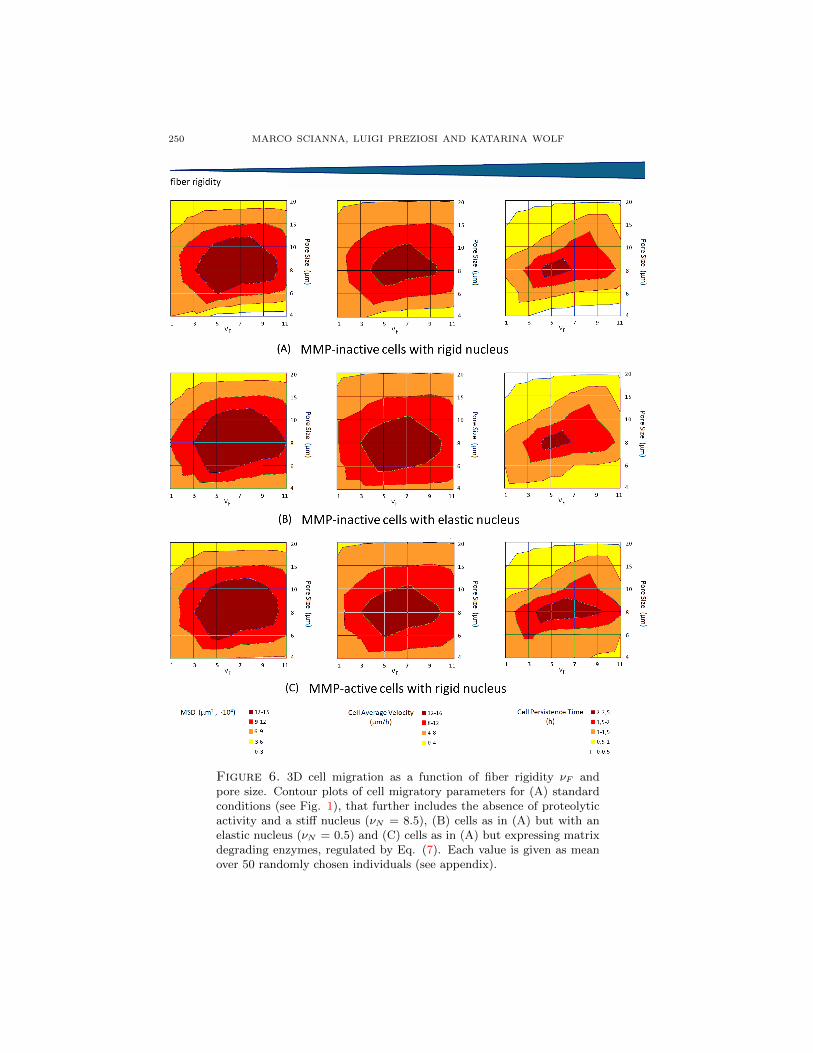
Figure 6. 3D cell migration as a function of fiber rigidity νF andpore size. Contour plots of cell migratory parameters for (A) standardconditions (see Fig. 1), that further includes the absence of proteolyticactivity and a stiff nucleus (νN = 8.5), (B) cells as in (A) but with anelastic nucleus (νN = 0.5) and (C) cells as in (A) but expressing matrixdegrading enzymes, regulated by Eq. (7). Each value is given as meanover 50 randomly chosen individuals (see appendix).

A CPM SIMULATING CELL MIGRATION ON AND IN MATRIX ENVIRONMENTS 251
Figure 7. Dependency of cell deformation within a 3D matrix on fiberstiffness. Variations of cell aspect ratios (defined as in Fig. 3) duringmigration at increasing fiber elasticities (from top to bottom). For allconditions, the pore size is kept constant (5 µm).
3.6. Fiber elasticity of 3D matrices. In the body, extracellular tissues displaya range of elastic characteristics that are modulated by the collagen content, theamount of cross-links between collagenous molecules and the presence of elasticfibers. Dense tissues are usually rigid, thus increasing matrix density should en-hance rigidity. In experimental studies using 3D ECM that where either modulatedin density, i.e., fiber concentration [85], or rigidity [55, 56, 72, 74], the other compo-nent becomes influenced as well. However, to separate at least theoretically theseinterdependent effects, we here simulate both varying scaffold stiffness (regulated byνF ) and the geometrical microstructure. To quantify such convoluting factors, weprovide contour plots of migration efficacy, as joint functions of pore size and fiberelasticity, that illustrate cell motile parameters as differently colored ‘landscapes’(Fig. 6). It is useful to underline that elastic fibers are also characterized by a lowconstant TF = 0.2, as they are no longer rigid but can deform.
In Fig. 6(A) at high pore size (i.e., 20 µm), cells display a reduced motile behav-ior, regardless of the fiber stiffness, as already shown in Fig. 3(A). The rationale ofthis is that in very loose tissues cells migration is not supported by guiding fibersaround the cell and consequently, the cell migrates along single fibers that, however,when stiff, promote to some extent traction and therefore migration. Next, at in-termediate mesh dimensions, both cell velocity and persistence (and, consequently,overall displacement) reach maximal levels biphasically depending on matrix elas-ticity. If the collagenous threads are too elastic (i.e., νF < 3), they can be easilydeformed, without representing a sufficient anchor for pulling force generation re-quired for cell motion. With a moderate stiffness (i.e., 5 < νF < 9), the matrixfibers can be slightly arranged into contact guidance formations, thereby facilitat-ing cell migration. On the contrary, a too rigid network (i.e., νF > 9) forms stericobstacles that can be somewhat less efficiently overcome by moving individuals.Finally, small pore sizes allow motility only within elastic matrices, whereas migra-tion is negligible for intermediate or high rigidities of the fibers. Migrating cells areable to move within small pores in fact only by significantly deforming the matrixnetwork, creating open space to pass through. Therefore, if the pore size is much

252 MARCO SCIANNA, LUIGI PREZIOSI AND KATARINA WOLF
Figure 8. Dependence of migration-associated cell morphology overtime on nuclear deformability. Evolution of cells aspect ratio duringmigration trough small pore sizes (5 µm) and high fiber stiffness (νF =11). Green line represents cells with a standard rigid nucleus (νN = 8.5),blue line represents cells with an elastic nucleus (νN = 0.5). All theother model parameters are the same as the standard simulation of Fig.1. Note that a high deformability of the nucleus allows cells to adaptbetter to highly constrained environments.
smaller than the dimension of the cell, matrix elasticity exerts an increasing influ-ence. When evaluating the plots at constant rigidity, cell migration displays thesame bimodal dependence on pore size previously captured in Fig. 3(A).
Such variations in fiber rigidity induce a suite of cell morphological changes (Fig.7). Cells plated within rigid scaffolds typically display actomyosin traction-mediatedelongated phenotypes, whereas, when cultured in progressively softer matrices theyshow instead decreasing elongation. Cells within complete compliant ECMs remainuniformly rounded, as cytoskeletal traction requires the counterforce provided by arigid matrix [79]. In summary, cells migrate in a biphasic manner upon increasingseparately density or stiffness, and also upon combined increase of density andstiffness reflecting experimental conditions best (imagine a decreasing curve in theplots of Fig. 6 from left top to right bottom).
As experimental examples, bimodal relationships between cell migratory abilityand the deformability of 3D matrix scaffolds have been observed in experimen-tal models of smooth muscle cells [59] and mouse fibroblasts, cultured in stepwiseEDAC-cross-linked CG matrices of constant pore size [37]. A biphasic dependenceon matrix rigidity has been previously reported also in isotropic homogeneous net-works, as in the case of prostate cancer cells embedded in Matrigel with a fixedfibronectin level and variables stiffness [88].
3.7. Nuclear compressibility in 3D migration. As pointed out in the previoussection, to migrate within ECM of pores smaller than a cellular diameter, cells needto deform their body including the nucleus as the most rigid organelle [26]. Thus,the degree of nuclear deformability may contribute to the migration efficacy of acell. The nucleus elasticity is mainly regulated by both the chromatin structure,and lamin intermediate filaments, that underlie the nuclear envelope [26, 31]. Thesoftness of a nucleus can be modeled by lowering the values of the nuclear rigidityνN from 8.5 (see Table 1) to 0.5 (compare Fig. 6(A) and (B)). At high pore sizesof 10 µm (or higher) and low fiber rigidity (at left upper corner of left and middle

A CPM SIMULATING CELL MIGRATION ON AND IN MATRIX ENVIRONMENTS 253
panels in Figs. 6(A) and (B)), migration remains unaltered regardless of nuclearelasticity, as moving cells do not experience steric hindrance. However, once meshdimensions and scaffold rigidity move to a intermediate ranges, nuclear elasticitysomewhat facilitates cell movement, measured as MSD (an estimate product ofpersistence time and velocity). Whereas the velocity contributes to this enhancedoverall movement, the persistence time remains equal (i.e., cells containing rigid nu-clei already persistantly migrated through intermediate matrix networks). Lastly,as pore size and matrix elasticity further decrease to form a highly constrainedenvironment (lower right corner), the simulations demonstrate that enhanced nu-clear deformability provides a migration advantage characterised by both increasedvelocity and persistence time. Such a facilitated locomotion is mediated by an elon-gated and deformable nuclear configuration allowing the entire cell to squeeze andstretch more easily and thereby pass through the steric obstacles of a dense andrigid matrix (Fig. 8).
Our simulations relate to a number of experimental works, such as [81], where cellmigration efficacy decreases with increasing matrix density and is associated withnuclear deformation, or [6], where glioma cell lines significantly deform their nucleusupon recruitment of non-muscle myosin II (NMMII) for squeezing through narrowlocations in a brain model in vivo, thereby increasing their metastatic potential.
3.8. Matrix degradation in 3D migration. In the previous sections, we havedemonstrated that cells move within matrix fibers of varying density and stiff-ness that act as constraints, deforming both body and nucleus. As an additionalmechanism to overcome limited space cells may upregulate proteolytic enzymesthat degrade ECM structure (i.e., matrix metalloproteinases, MMPs) that act ei-ther when bound to the cell surface or when secreted into the extracellular space.Accordingly, cells degrade steric fiber obstacles either in a cell contact-dependentmanner targeting locally confining fibers, or, in a diffusive manner leading to gradi-ent formation and consequently, a more overall weakening of the surrounding tissuestructure [83]. As a result, barrier-free matrix spaces will be created resulting inlonger distance traveling. We here perform simulations with cells that executeboth contact-dependent and soluble proteolysis. The local concentration of the netprotolytic activity (both surface-bound and diffusive) is defined as m(x, t), and isassumed to evolve following a standard reaction-diffusion equation:
∂m(x, t)
∂t= P (x, t)︸ ︷︷ ︸production
+Dm∇2m(x, t)︸ ︷︷ ︸diffusion
−λmm(x, t)δ(τ(Σσ(x)),M)︸ ︷︷ ︸decay
, (7)
where δ(τ(Σσ(x)),M) = 1 in the interstitial mediumM and 0 elsewhere. λm andDm
are, respectively, the decay rate and the effective diffusion coefficient of proteolyticenzymes, constant and homogeneous in the extracellular environment. A low valueof Dm models proteolysis being strongly localized in regions close to cell membranes,in agreement with experimental evidence in [68, 81]. P (x, t) models instead the localproduction of proteases either at the cell surface or secreted away from the externalcell surface, at a constant rate πm which only activates upon local contact betweenthe cell and a collagenous component:
P (x, t) =
πm if x : τ(Σσ(x)) = C and ∃x′ ∈ Ω′
x : τ(Σσ(x′)) = M ;
0 else,(8)

254 MARCO SCIANNA, LUIGI PREZIOSI AND KATARINA WOLF
where we recall that C stands for cell cytosolic region. MMPs are capable todegrade the fibrous component of the matrix: to reproduce this biological effect, alattice grid site x belonging to a degraded collagenous fiber becomes a generalizedmedium (fluid) site when its local level of MMPs (m(x, t)) is sufficiently high (inour simulations above 2.5 µM). This change is implemented by changing its type τfrom F (fiber) to M (medium), as done in [32]. The comparison of cell migrationof either MMP-active and MMP-inactive individuals (Fig. 6(A) and (C)), revealsthat at high and intermediate pore size and/or low matrix rigidity, the proteolyticmachinery does not appreciably affect cell motion. The loose fiber network does notrepresent a significative obstacle for cell migration, which therefore is not enhancedfurther by MMP activity. In the case of small pores formed by rigid collagenousfibers (lower right), MMP activity promotes instead cell migration appreciably.This suggests that proteases, by degrading matrix fibers, are able to break stericobstacles in the close proximity of moving individuals, opening spaces for them tosample greater distances without turning back.
The role of MMPs activity in cell migratory behavior captured in our modelis in good agreement with the experimental results provided in [62] for dermalfibroblasts embedded in molecularly engineered PEG hydrogels, where a significativeincrement in the number of migrating individuals was observed upon up-regulationof proteolytic enzymes.
In conclusion, summarizing all the examined parameters, cell migration is greatlyinfluenced by a number of complex ECM- and cell-derived characteristics that, inaddition, display a number of interdependencies [28] and, together, determine thenet outcome on migration.
4. Discussion. Due to the increasingly recognized importance of cell migrationprocesses in matrix environments and its exploitation for therapy and for tissueengineering, a growing number of theoretical models has been developed. Thesemodeling approaches analyze the relative importance of single and interrelated pa-rameters to predict migration behavior.
We employed a simple and intuitive version of the Cellular Potts Model to simu-late the motile behavior of cells seeded either on 2D matrix substrates or embeddedwithin 3D matrix scaffolds. In contrast to previous approaches, the Cellular PottsModel used here treats each cell as compartmentalized into nucleus and cytoplasm,whose movement is driven by explicit interactions with the extracellular environ-ment differentiated into fibers and medium. The introduction of the nucleus andits mechanical properties on one side and of the extracellular matrix and its specificfibrous characteristics on the other side allowed to simulate for the first time boththeir specific contributions in cell migration.
In particular, we considered isolated pro-migratory parameters derived eitherfrom the ECM, such as orientation, pore size, ligand density, or rigidity, or from thecell, such as adhesion, nuclear rigidity, or proteolysis, that control both cell migra-tion efficacy and migratory phenotypes. In all proposed cases, the computationalresults are consistent with a number of published experimental counterparts, andconfirm linear or biphasic dependencies of migration dynamics on single matrix- orcell-derived determinants (see short paragraphs at the end of each result section).
As a clear advantage of a theoretical approach, we have been able to indepen-dently vary and modulate in a graded fashion all biophysical cell parameters andmicrostructural properties of the matrix environment, which is helpful in dissecting

A CPM SIMULATING CELL MIGRATION ON AND IN MATRIX ENVIRONMENTS 255
the complex relationships between cell motility and the biophysical, biochemicaland molecular properties of the matrix [28]. However, a modeling approach thatdescribes isolated parameters is unable to encompass the complexity apparent inmigration processes in vivo. Some of these additional, here disregarded, factorsare (i) additional matrix deposition of moving individuals, leading to altered trac-tion generation, adhesion and contact guidance; (ii) soluble or matrix-bound gradi-ents of chemoattractants; (iii) molecular signals transmitted from the ECM to cells(outside-in signaling), thereby changing the activity of polarization- or contractility-mediating proteins (Rac, Rho) [24]; or (iv) inside-out signaling for reinforcement ofadhesion [79].
Despite the limitations of theoretical modeling, our approach could be appliedto the design of synthetic implant materials, i.e., acellular scaffolds with optimalvalues of pore size and stiffness that may accelerate cell in-growth, critically forregenerative treatments [7, 11, 38, 80]. Further, applying our modeling approachon defined cancer invasion and inhibition studies in vitro and in vivo may assist inpredicting some outcomes on therapeutic interventions. At this regard, it would bebiologically relevant to adapt our approach to specific cell lines, characterized by dis-tinct biophysical phenotypes (i.e., intrinsic motility, elasticity, or proteases activity).This can be easily done by inheriting the model parameters from experimentally-measured quantities, characteristic of the selected cell Further, it would be inter-esting to analyze collective migration of cellular aggregates fundamental in severalphysio-pathological processes, as commented in [39]. In such aggregates a differ-entiation may occur among individuals of the same origin, (i.e., tip and stalk cellsduring angiogenic processes, or leader and follower cells during a skin wound healing[29]), whereas competitions for nutrients or altered heterotypic interactions may sig-nificantly affect the migratory capacity of an entire cell lineage (for example, cancercells of epithelial origin inhibit the motility and induce apoptosis in neighboring nor-mal individuals). Obviously, in this case, it is necessary to define all cell types in themodel framework, together with their phenotypic parameters and the mechanimsunderlying the behavior of and mutual interactions within the cell collective.
In summary, our findings may contribute to both understanding and exploitationof cell migration processes on and in tissues.
Appendix A. Appendix. The position of a cell η at time t is established by thecoordinate of its center of mass (CM) xCMη (t). The wind-rose plots in both the 2Dand in the 3D case are therefore generated by tracking the position of the centerof mass of 10 randomly chosen cells at 15-min (450 MCS) intervals, overlying thestarting coordinates at the origin of the graph. This type of representation is par-ticularly useful to evaluate the cell net displacements over time and the distributionof their final dispersion.
Similarly, the instantaneous velocity of η is the velocity of its center of mass:
vη(t) =xCMη (t)− xCMη (t−∆t)
∆t, (9)
where ∆t = 1 MCS, as done in similar works [51, 58, 70].The average velocity of an individual over the entire simulation is instead given
by
vη(t) =xCMη (tfinal)− xCMη (0)
tfinal, (10)

256 MARCO SCIANNA, LUIGI PREZIOSI AND KATARINA WOLF
where tfinal corresponds to the final time of the observation period which, as ex-plained in the text, is set to 21600 MCS (12 hours).
The mean squared displacement (MSD) of a cell η at time t is calculated as
< d2η(t) >=< (xCMη (t)− xCMη (0))2 >, (11)
where xCMη (0) is the initial position of its center of mass. Following [16, 87], thesquared displacements are averaged over all previous time steps, in order to takeinto account the back and forth motions exhibited by the moving individuals. Asdemonstrated in a number of previous experimental [16, 37] and computational[16, 87] studies, at sufficiently long times the mean square displacements vary ap-proximately linearly with the number of time steps. It can therefore be relatedto cell instantaneous velocity (vη) and persistence time (pη, which quantifies thedirectional productive motion) with the so-called persistence-random-walk (PRW)law:
< d2η(t) >= 2v2
η(t)pη(t)[t− pη(t)(1− e−t/pη(t))]. (12)
In particular, at still longer observation periods, (12) reduces to:
< d2η(t) >≈ 2v2
η(t)pη(t)t, (13)
and the persistence time of a moving individual can be directly calculated as
pη(t) ≈< d2
η(t) >
2v2η(t)t
. (14)
The PRW relation has been demonstrated to characterize the cells migratory be-havior more properly than other common methods, which calculate the averagedistance migrated by biological individuals in an arbitrary time interval, as com-mented in [20]. For the statistical analysis, cells that do not display a final MSDgreater than their diameters are classified as non-motile and assigned a velocity of0 µm/h and an undefined persistence time, as we follow the criterion described in[16, 30].
A.1. Statistics. Cell motile parameters (MSD, velocity and persistence time) arerepresented in the figures as box-and-whisker plots, where the edges of the boxesare the 25th and 75th percentiles and the whiskers the 10th and 90th percentiles.The horizontal line represents the median, while the large black dot corresponds tothe mean of the distribution. Statistical significance (p < 0.05) was determined formotile fraction data by the Students’ t-test and for non-normally distributed datasets by the Kolmogorov-Smirnov test over each 50 randomly chosen individuals.
In the multidimensional contour plots the values of the cell migratory parametersare means over 50 randomly chosen individuals.
Quantitative analysis of cell morphological changes is carried out by evaluatingthe evolution of the cell aspect ratio, given by the ratio between the actual cellsurface (respectively, border in 2D) and the surface of the sphere having the samevolume (respectively, the length of the circumpherence having the same area in2D). It is useful to underline that in our model cell volume (respectively, area in2D) is kept nearly fixed by high values of κ in Eq. (4), see Table 1. Therefore theaspect ratio gives a quantitative measure of cell membrane ruffling. Finally, thetime evolution of the aspect ratio is given in the plots with mean ± s.d. over 50randomly chosen individuals.

A CPM SIMULATING CELL MIGRATION ON AND IN MATRIX ENVIRONMENTS 257
Acknowledgments. This work was supported by the Netherlands Science Orga-nization (NWO-VIDI 317.10.364 to K.W.), and by the Ministry of Research andUniversity through a PRIN on “Mathematical models of the mechanical interactionsof single cells and cell aggregates with the surrounding environment” to L.P. andM.S.
REFERENCES
[1] B. Alberts, D. Bray, J. Lewis, M. Raff, K. Roberts and J. D. Watson, “Molecular Biology of
the Cell,” 3rd edition, Garland Science, 1994.[2] M. Arnold, V. C. Hirschfeld-Warneken, T. Lohmuller, P. Heil, J. Blummel, E. A. Cavalcanti-
Adam, M. Lopez-Garcıa, P. Walther, H. Kessler, B. Geiger and J. P. Spatz, Induction of cell
polarization and migration by a gradient of nanoscale variations in adhesive ligand spacing,Nano Lett., 8 (2008), 2063–2069.
[3] M. Bajenoff, J. G. Egen, L. Y. Koo, J. P. Laugier, F. Brau, N. Glaichenhaus and R. N.
Germain, Stromal cell networks regulate lymphocyte entry, migration, and territoriality inlymph nodes, Immunity, 25 (2006), 989–1001.
[4] A. Balter, R. M. Merks, N. J. Poplawski, M. Swat and J. A. Glazier, The Glazier-Graner-
Hogeweg model: Extensions, future directions, and opportunities for further study, in “Single-Cell-Based Models in Biology and Medicine, Mathematics and Biosciences in Interactions”
(eds. A. R. A. Anderson, M. A. J. Chaplain and K. A. Rejniak), Birkhauser, (2007), 157–167.[5] A. L. Bauer, T. L. Jackson and Y. Jiang, A cell-based model exhibiting branching and anas-
tomosis during tumor-induced angiogenesis, Biophys. J., 92 (2007), 3105–3121.
[6] C. Beadle, M. C. Assanah, P. Monzo, R. Vallee, S. Rosenfeld and P. Canoll, The role ofmyosin ii in glioma invasion of the brain, Mol. Biol. Cell., 19 (2008), 3357–3368.
[7] J. Behring, R. Junker, X. F. Walboomers, B. Chessnut and J. A. Jansen, Toward guided
tissue and bone regeneration: Morphology, attachment, proliferation, and migration of cellscultured on collagen barrier membranes. A systematic review, Odontology, 96 (2008), 1–11.
[8] A. O. Brightman, B. P. Rajwa, J. E. Sturgis, M. E. McCallister, J. P. Robinson and S.
L. Voytik-Harbin, Time-lapse confocal reflection microscopy of collagen fibrillogenesis andextracellular matrix assembly in vitro, Biopolymers, 54 (2000), 222–234.
[9] A. Brock, E. Chang, C. C. Ho, P. LeDuc, X. Jiang, G. M. Whitesides and D. E. Ingber, Geo-
metric determinants of directional cell motility revealed using microcontact printing, Lang-muir, 19 (2003), 1611–1617.
[10] B. T. Burgess, J. L. Myles and R. B. Dickinson, Quantitative analysis of adhesion-mediatedcell migration in three-dimensional gels of RGD-grafted collagen, Ann. Biomed. Eng., 28
(2003), 110–118.
[11] R. M. Capito and M. Spector, Scaffold-based articular cartilage repair, IEEE Eng. Med. Biol.Mag., 22 (2003), 42–50.
[12] E. A. Cavalcanti-Adam, T. Volberg, A. Micoulet, H. Kessler, B. Geiger and J. P. Spatz, Cellspreading and focal adhesion dynamics are regulated by spacing of integrin ligands, Biophys.J., 92 (2007), 2964–2974.
[13] J. Condeelis and J. E. Segall, Intravital imaging of cell movement in tumours, Nat. Rev.
Cancer, 3 (2003), 921–930.[14] M. W. Conklin, J. C. Eickhoff, K. M. Riching, C. A. Pehlke, K. W. Eliceiri, P. P. Provenzano,
A. Friedl and P. J. Keely, Aligned collagen is a prognostic signature for survival in humanbreast carcinoma, Am. J. Pathol., 178 (2011), 1221–1232.
[15] R. B. Dickinson, S. Guido and R. T. Tranquillo, Biased cellmigration of fibroblasts exhibiting
contact guidance in oriented collagen gels, Ann. Biomed. Eng., 22 (1994), 342–356.[16] P. A. DiMilla, J. A. Stone, J. A. Quinn, S. M. Albelda and D. A. Lauffenburger, Maximal
migration of human smooth-muscle cells on fibronectin and type-IV collagen occurs at an
intermediate attachment strength, J. Cell. Biol., 122 (1993), 729–737.[17] A. D. Doyle, F. W. Wang, K. Matsumoto and K. M. Yamada, One-dimensional topography
underlies three-dimensional fibrillar cell migration, J. Cell. Biol., 184 (2009), 481–490.
[18] N. Dubey, P. C. Letourneau and R. T. Tranquillo, Neuronal contact guidance in magneticallyaligned fibrin gels: effect of variation in gel mechano-structural properties, Biomaterials, 22
(2001), 1065–1075.

258 MARCO SCIANNA, LUIGI PREZIOSI AND KATARINA WOLF
[19] G. A. Dunn and T. Ebendal, Contact guidance on oriented collagen gels, Exp. Cell. Res.,111 (1978), 475–479.
[20] G. A. Dunn, Characterizing a kinesis response: Time-averaged measures of cell speed and
directional persistence, Agents Actions Suppl., 12 (1983), 14–33.[21] A. Engler, L. Bacakova, C. Newman, A. Hategan, M. Griffin and D. Discher, Substrate com-
pliance versus ligand density in cell on gel responses, Biophys. J., 86 (2004), 617–628.[22] P. Friedl, F. Entschladen, C. Conrad, B. Niggemann and K. S. Zanker, CD4+ T-lymphocytes
migrating in three-dimensional collagen lattices lack focal adhesions and utilize β1 integrin-
independent strategies for polarization, interaction with collagen fibers and locomotion, Eur.J. Immunol., 28 (1998), 2331–2343.
[23] P. Friedl and E. B. Brocker, The biology of cell locomotion within three-dimensional extracel-
lular matrix , Cell. Mol. Life Sci., 57 (2000), 41–64.[24] P. Friedl and K. Wolf, Tumour-cell invasion and migration: Diversity and escape mechanisms,
Nat. Rev. Cancer, 3 (2003), 362–374.
[25] P. Friedl, K. Maaser, C. E. Klein, B. Niggemann, G. Krohne and K. S. Zanker, Migrationof highly aggressive MV3 melanoma cells in 3-dimensional collagen lattices results in local
matrix reorganization and shedding of alpha2 and beta1 integrins and CD44, Cancer Res.,
57 (1997), 2061–2070.[26] P. Friedl, K. Wolf and J. Lammerding, Nuclear mechanics during cell migration, Curr. Opin.
Cell. Biol., 23 (2011), 253.[27] P. Friedl and B. Weigelin, Interstitial leukocyte migration and immune function, Nat. Im-
munol., 9 (2008), 960–969.
[28] P. Friedl and K. Wolf, Plasticity of cell migration: A multiscale tuning model, J. Cell. Biol.,188 (2009), 11–19.
[29] P. Friedl and D. Gilmour, Collective cell migration in morphogenesis, regeneration and cancer,
Nat. Rev. Mol. Cell. Biol., 10 (2009), 445–457.[30] C. Gaudet, W. Marganski, S. Kim, C. T. Brown, V. Gunderia, M. Dembo and J. Wong,
Influence of type I collagen surface density on fibroblast spreading, motility, and contractility,
Biophys. J., 85 (2003), 3329–3335.[31] G. Gerlitz and M. Bustin, The role of chromatin structure in cell migration, Trends Cell.
Biol., 21 (2011), 6–11.
[32] C. Giverso, M. Scianna, L. Preziosi, N. Lo Buono and A. Funaro, Individual cell-based modelfor in-vitro mesothelial invasion of ovarian cancer, Math. Model. Nat. Phenom., 5 (2010),
203–223.[33] J. A. Glazier, A. Balter and N. J. Poplawski, Magnetization to morphogenesis: A brief history
of the Glazier-Graner-Hogeweg model, in “Single-Cell-Based Models in Biology and Medicine,
Mathematics and Biosciences in Interactions” (eds. A. R. A. Anderson, M. A. J. Chaplainand K. A. Rejniak), Birkhauser, (2007), 79–106.
[34] J. A. Glazier and F. Graner, Simulation of the differential adhesion driven rearrangement ofbiological cells, Phys. Rev. E Stat. Phys. Plasmas Fluids Relat. Interdiscip. Topics, 47 (1993),2128–2154.
[35] S. L. Goodman, G. Risse and K. Vondermark, The E8 subfragment of laminin promotes
locomotion of myoblasts over extracellular- matrix, J. Cell. Biol., 109 (1989), 799–809.[36] F. Graner and J. A. Glazier, Simulation of biological cell sorting using a two-dimensional
extended Potts model , Phys. Rev. Lett., 69 (1992), 2013–2016.[37] B. A. Harley, H. Kim, M. H. Zaman, I. V. Yannas, D. A. Lauffenburger and L. J. Gibson, Mi-
croarchitecture of three-dimensional scaffolds influences cell migration behavior via junction
interactions, Biophys. J., 95 (2008), 4013–4024.
[38] B. A. Harley, M. H. Spilker, J. W. Wu, K. Asano, H. P. Hsu, M. Spector and I. V. Yannas,Optimal degradation rate for collagen chambers used for regeneration of peripheral nerves
over long gaps, Cells Tissues Organs, 176 (2008), 153–165.[39] O. Ilina and P. Friedl, Mechanisms of collective cell migration at a glance, J. Cell Sci., 122
(2009), 3203–3208.
[40] E. Ising, Beitrag zur theorie des ferromagnetismus, Z. Physik., 31 (1925), 253.[41] R. M. Kuntz and W. M. Saltzman, Neutrophil motility in extracellular matrix gels: mesh size
and adhesion affect speed of migration, Biophys. J., 72 (1997), 1472–1480.
[42] S. Kurosaka and A. Kashina, Cell biology of embryonic migration, Birth Defects Res. C.Embryo Today, 84 (2008), 102–122.

A CPM SIMULATING CELL MIGRATION ON AND IN MATRIX ENVIRONMENTS 259
[43] E. Lamers, R. Van Horssen, J. Te Riet, F. C. Van Delft, R. Luttge, X. F. Walboomers andJ. A. Jansen, The influence of nanoscale topographical cues on initial osteoblast morphology
and migration, Eur. Cell. Mater., 9 (2010), 329–343.
[44] D. A. Lauffenburger and J. J. Lindermann, “Receptors: Bodels for Binding, Trafficking, andSignaling,” Oxford University Press, London, 1996.
[45] D. A. Lauffenburger and A. F. Horwitz, Cell migration: a physically integrated molecularprocess, Cell, 84 (1996), 359–369.
[46] D. Lehnert, B. Wehrle-Haller, C. David, U. Weiland, C. Ballestrem, B. A. Imhof and M. Bast-
meyer, Cell behaviour on micropatterned substrata: limits of extracellular matrix geometryfor spreading and adhesion, J. Cell. Sci., 117 (2004), 41–52.
[47] M. P. Lutolf, J. L. Lauer-Fields, H. G. Schmoekel, A. T. Metters, F. E. Weber, G. B. Fields
and J. A. Hubbell, Synthetic matrix metalloproteinase-sensitive hydrogels for the conductionof tissue regeneration: Engineering cell-invasion characteristics, Proc. Natl. Acad. Sci. U. S.
A., 100 (2003), 5413–5418.
[48] K. Maaser, K. Wolf, C. E. Klein, B. Niggemann, K. S. Zanker, E. B. Brocker and Friedl, Func-tional hierarchy of simultaneously expressed adhesion receptors: Integrin α2β1 but not CD44
mediates MV3 melanoma cell migration and matrix reorganization within three-dimensional
hyaluronan-containing collagen matrices, Mol. Biol. Cell., 10 (1999), 3067–3079.[49] A. W. Mahoney, B. G. Smith, N. S. Flann and G. J. Podgorski, Discovering novel cancer
therapies: A computational modeling and search approach, in “IEEE conference on Compu-tational Intelligence in Bioinformatics and Bioengineering,” (2008), 233–240.
[50] A. F. M aree, V. A. Grieneisen and P. Hogeweg, The Cellular Potts Model and biophysical
properties of cells, tissues and morphogenesis, in “Single-Cell-Based Models in Biology andMedicine, Mathematics and Biosciences in Interactions” (eds. A. R. A. Anderson, M. A. J.
Chaplain and K. A. Rejniak), Birkhauser, (2007), 107–136.
[51] R. M. Merks, E. D. Perryn, A. Shirinifard and J. A. Glazier, Contact-inhibited chemotaxis inde novo and sprouting blood vessel growth, PLoS Comput. Biol., 4 (2008), e1000163, 16 pp.
[52] N. Metropolis, A. W. Rosenbluth, M. N. Rosenbluth, A. H. Teller and E. Teller, Equation of
state calculations by fast computing machines, J. Chem. Phys., 21 (1953), 1087–1092.[53] A. Mogilner and G. Oster, Polymer motors: Ppushing out the front and pulling up the back,
Current Biology, 13 (2003), R721–733.
[54] F. J. O’Brien, B. A. Harley, I. V. Yannas and L. Gibson, Influence of freezing rate on porestructure in freeze-dried collagen-GAG scaffolds, Biomaterials, 25 (2004), 1077–1086.
[55] L. H. Olde Damink, P. J. Dijkstra, M. J. Luyn, P. B. Wachem, P. Nieuwenhuis and J. Feijen,Glutaraldehyde as a crosslinking agent for collagen-based biomaterials, J. Mater. Sci. Mater.
Med., 6 (1995), 460–472.
[56] J. M. Orban, L. B. Wilson, J. A. Kofroth, M. S. El-Kurdi, T. M. Maul and D. A. Vorp,Crosslinking of collagen gels by transglutaminase, J. Biomed. Mater. Res. A, 68 (2004), 756–
762.[57] S. P. Palecek, J. C. Loftus, M. H. Ginsberg, D. A. Lauffenburger and A. F. Horwitz, Integrin-
ligand binding properties govern cell migration speed through cell-substratum adhesiveness,
Nature, 385 (1997), 537–540.
[58] E. D. Perryn, A. Czirok and C. D. Little, Vascular sprout formation entails tissue deforma-tions and VE-cadherin-dependent cell-autonomous motility, Dev. Biol., 313 (2008), 545–555.
[59] S. R. Peyton and A. J. Putnam, Extracellular matrix rigidity governs smooth muscle cellmotility in a biphasic fashion, J. Cell. Physiol., 204 (2005), 198–209.
[60] T. D. Pollard and G. G. Borisy, Cellular motility driven by assembly and disassembly of actin
filaments, Cell, 112 (2003), 453–465.
[61] R. B. Potts, Some generalized order-disorder transformations, Proc. Camb. Phil. Soc., 48(1952), 106–109.
[62] G. P. Raeber, M. P. Lutolf and J. A. Hubbell, Molecularly engineered PEG hydrogels: a novelmodel system for proteolytically mediated cell migration, Biophys. J., 89 (2005), 1374–1388.
[63] C. B. Raub, V. Suresh, T. Krasieva, J. Lyubovitsky, J. D. Mih, A. J. Putnam, B. J. Tromberg
and S. C. George, Noninvasive assessment of collagen gel microstructure and mechanics usingmultiphoton microscopy, Biophys. J., 92 (2007), 2212–2222.
[64] A. J. Ridley, M. A. Schwartz, K. Burridge, R. A. Firtel, M. H. Ginsberg, G. Borisy, J. T.
Parsons and A. R. Horwitz, Cell migration: Integrating signals from front to back, Science,302 (2003), 1704–1709.

260 MARCO SCIANNA, LUIGI PREZIOSI AND KATARINA WOLF
[65] B. A. Roeder, K. Kokini, J. E. Sturgis, J. P. Robinson and S. L. Voytik-Harbin, Tensilemechanical properties of three-dimensional type I collagen extracellular matrices with varied
microstructure, J. Biomech. Eng., 124 (2002), 214–222.
[66] C. G. Rolli, T. Seufferlein, R. Kemkemer and J. P. Spatz, Impact of tumor cell cytoskeletonorganization on invasiveness and migration: A microchannel-based approach, PLoS ONE, 5
(2010), e8726.[67] B. M. Rubenstein and L. J. Kaufman, The role of extracellular matrix in glioma invasion: A
Cellular Potts Model approach, Biophys. J., 95 (2006), 5661–5680.
[68] F. Sabeh, R. Shimizu-Hirota and S. J. Weiss, Protease-dependent versus -independent cancercell invasion programs: Three-dimensional amoeboid movement revisited, J. Cell. Biol., 185
(2009), 11–19.
[69] E. Sahai, Illuminating the metastatic process, Nat. Rev. Cancer, 7 (2007), 737–749.[70] M. Scianna, A multiscale hybrid model for pro-angiogenic calcium signals in a vascular en-
dothelial cell, Bull. Math. Biol., 76 (2011), 1253–1291.
[71] M. Scianna and L. Preziosi, Multiscale developments of the Cellular Potts Model, MultiscaleModel. Simul., 10 (2012), 342–382.
[72] M. T. Sheu, J. C. Huang, G. C. Yeh and H. O. Ho, Characterization of collagen gel solutions
and collagen matrices for cell culture, Biomaterials, 22 (2001), 1713–1719.[73] S. Schmidt and P. Friedl, Interstitial cell migration: Integrin-dependent and alternative ad-
hesion mechanisms, Cell. Tissue Res., 339 (2010), 83–92.[74] R. C. Siegel, Collagen cross-linking. Synthesis of collagen cross-links in vitro with highly
purified lysyl oxidase, J. Biol. Chem., 251 (1976), 5786–5792.
[75] C. Spadaccio, A. Rainer, S. De Porcellinis, M. Centola, F. De Marco, M. Chello, M. Trombetta,M. and J. A. Genovese, A G-CSF functionalized PLLA scaffold for wound repair: An in vitro
preliminary study, Conf. Proc. IEEE Eng. Med. Biol. Soc., 2010 (2010), 843–846.
[76] M. S. Steinberg, Reconstruction of tissues by dissociated cells. Some morphogenetic tissuemovements and the sorting out of embryonic cells may have a common explanation, Science,
141 (1963), 401–408.
[77] M. S. Steinberg, Does differential adhesion govern self-assembly processes in histogenesis?Equilibrium configurations and the emergence of a hierarchy among populations of embryonic
cells, J. Exp. Zool., 171 (1970), 395–433.
[78] D. G. Stupack, The biology of integrins, Oncology, 21 (2007), 6–12.[79] T. A. Ulrich, E. M. De Juan Pardo and S. Kumar, The mechanical rigidity of the extracellular
matrix regulates the structure, motility, and proliferation of glioma cells, Cancer Res., 69(2009), 4167–4174.
[80] M. C. Wake, C. W. Patrick and A. G. Mikos, Pore morphology effects on the fibrovascular
tissue growth in porous polymer substrates, Cell. Transplant., 3 (1994), 339–343.[81] K. Wolf, Y. I. Wu, Y. Liu, J. Geiger and E. Tam, Multi-step pericellular proteolysis controls
the transition from individual to collective cancer cell invasion, Nat. Cell. Biol., 9 (2007),893–904.
[82] K. Wolf, S. Alexander, V. Schacht, L. M. Coussens, U. H. Von Andrian, J. Van Rheenen, E.
Deryugina and P. Friedl, Collagen-based cell migration models in vitro and in vivo, Semin.
Cell. Dev. Biol., 20 (2009), 931–941.[83] K. Wolf and P. Friedl, Extracellular matrix determinants of proteolytic and non-proteolytic
cell migration, Trends Cell. Biol., 21 (2011), 736–744.[84] K. Wolf, I. Mazo, H. Leung, K. Engelke, U. H. Von Andrian, E. I. Deryugina, A. Y. Strongin,
E. B. Brocker and P. Friedl, Compensation mechanism in tumor cell migration mesenchy-
malmoeboid transition after blocking of pericellular proteolysis, J. Cell. Biol., 160 (2003),
267–277.[85] Y. L. Yang and L. J. Kaufman, Rheology and confocal reflectance microscopy as probes of
mechanical properties and structure during collagen and collagen/hyaluronan self-assembly,Biophys. J., 96 (2009), 1566–1585.
[86] I. V. Yannas, E. Lee, D. P. Orgill, E. M. Skrabut, G. F. Murphy, Synthesis and characteri-
zation of a model extracellular matrix that induces partial regeneration of adult mammalianskin, Proc. Natl. Acad. Sci. U. S. A., 86 (1989), 933–937.
[87] M. H. Zaman, P. Matsudaira and D. A. Lauffenburger, Understanding effects of matrix pro-
tease and matrix organization on directional persistence and translational speed in three-dimensional cell migration, Ann. Biomed. Eng., 35 (2007), 91–100.

A CPM SIMULATING CELL MIGRATION ON AND IN MATRIX ENVIRONMENTS 261
[88] M. H. Zaman, L. M. Trapani, A. L. Sieminski, D. Mackellar, H. Gong, R. D. Kamm, A. Wells,D. A. Lauffenburger and P. Matsudaira, Migration of tumor cells in 3D matrices is governed
by matrix stiffness along with cell-matrix adhesion and proteolysis, Proc. Natl. Acad. Sci.
USA, 103 (2006), 10889–10894.
Received May 10, 2012; Accepted July 25, 2012.
E-mail address: [email protected]
E-mail address: [email protected]
E-mail address: [email protected]