Case Report: Visual Rehabilitation in Hemianopia Patients ...
Upload
khangminh22Category
view
2download
0
University of New England University of New England
DUNE: DigitalUNE DUNE: DigitalUNE
Case Report Papers Physical Therapy Student Papers
12-2021
Utilizing An Evidence-Based Practice Framework In Non-Operative Utilizing An Evidence-Based Practice Framework In Non-Operative
ACL Rehabilitation - A Case Report ACL Rehabilitation - A Case Report
Eric Norman
Michael Madore
Kathryn Magee
Tyler Calimer
Parker Nally
Follow this and additional works at: https://dune.une.edu/pt_studcrpaper
Part of the Physical Therapy Commons
© 2021 The Authors
Utilizing An Evidence-Based Practice Framework In Non-Operative ACL Rehabilitation - 1
A Case Report 2
Eric Norman, B.S., Michael Madore, B.S., Kathryn Magee, B.S., Tyler Calimer, B.S., Parker 3
Nally, B.S. 4
5
The authors are students in the Doctor of Physical Therapy Program at the University of New 6
England, 716 Stevens Avenue, Portland, Maine, 04103. 7
8
Acknowledgements: We would like to thank Dr. Jim Cavanaugh for the guidance and wisdom in 9
the creation of this case report. 10
11
Abstract word count: 265 12
Manuscript word count: 3,459 13
14
15
16
17
18
19
20
21
22
23

Abstract 24
Study Design: Case Report.Background: The patient was a 51-year-old female who tore her 25
left anterior cruciate ligament (ACL) playing pickleball and opted for non-operative treatment. 26
The clinicians involved treated the patient using the three pillars of practice: relevant scientific 27
evidence, clinician experience, and patient perspective. Treatment: The patient’s treatment 28
consisted of therapeutic exercises, neuromuscular re-education, soft tissue massage, and 29
motivational interviewing. Therapeutic exercises were designed to strengthen the muscles 30
surrounding her hip and knee. Neuromuscular re-education helped to improve the patient’s knee 31
stability and balance, while soft tissue massage was used to decrease swelling. Motivational 32
interviewing helped her better align her attitude toward her restrictions with the goals of the 33
rehabilitation process. Outcome: The patient made progress and increased satisfaction 34
throughout rehabilitation which was illustrated by objective tests and subjective reports. 35
Improvements were noted with a 24% positive change in lower extremity functional scale 36
(LEFS) score between initial and follow-up testing. Single leg hop tests by discharge revealed a 37
90% or higher right-to-left compatibility score. Discussion: This case highlighted the plausibility 38
of a conservative treatment approach in a patient with an ACL tear. It introduced a clinical 39
decision-making model that emphasized the importance of including relevant research in the 40
form of Clinical Practice Guidelines (CPG’s), addressing the patient's perspective on the impact 41
of her injury, and incorporating clinician expertise in observing movement impairments. During 42
treatment, the patient demonstrated appropriate lower extremity strength and stability according 43
to the clinician’s expertise and outcome measures, but she did not feel confident enough to return 44
to previous lifestyle activities. Patient perspective played a large role in determining progressions 45
and accomplishments. 46
Key Words: Non-operative ACL rehabilitation, Evidence-Based Practice 47
Background 48
One of the most commonly injured structures in the knee is the anterior cruciate ligament 49
(ACL), with injuries occurring in approximately 1 in 3,500 people annually in the U.S.1 Over a 50
nine-year span (2005-2013) the median cost of ACL reconstruction procedures was just over 51
$9,000, with an overall cost per patient just over $13,400.2 As preventative and conservative 52
treatments become more popular, it is important to consider the benefits and risks of each 53
treatment. One study analyzed the quality of life for patients post-acute ACL rupture, comparing 54
surgery versus conservative management. Results indicated no significant difference in patient’s 55
quality of life between those that chose surgical reconstruction versus those who preferred 56
conservative management.3 57
Evidence-based practice (EBP), established by Sackett is the concept of incorporating the 58
following three pillars into successful clinical decision-making in medicine: scientific evidence, 59
patient’s values and preferences, and clinical judgement.4 According to Sackett, scientific 60
evidence is the objective measurement which helps guide clinical practice. The patient's 61
perspective allows us to understand how the patient views their injury and the effect it is having 62
on their lives. Clinical expertise allows clinicians to use prior experiences to modify a plan of 63
care as needed. These concepts together allow clinicians to create patient-centered goals that 64
emphasize what the patient wishes to improve, while maintaining a focus on their main 65
diagnosis. 66
67
Introduction 68
This report focuses on a patient with a recent ACL tear. Following an initial consultation 69
with a surgeon, the patient elected conservative treatment for her injury. After her initial physical 70
therapy examination, her therapist determined that she was deemed a good candidate for 71
conservative treatment based on the following reasons: similar active range of motion (AROM) 72
bilaterally, minimal swelling, and her motivation to succeed. Due to the lack of relevant non-73
operative treatment guidelines, aspects of the case report have incorporated the usage of post-74
operative clinical practice guidelines from Van Melic.5 During the time that the patient was seen 75
for physical therapy, she made great strides towards her goals of returning to activities and 76
hobbies, as seen in the exercise progression and four-stage hop test. With further discussion 77
between the patient, physical therapist (PT), and student physical therapist (SPT), it was clear 78
that the patient herself did not have full confidence in her affected limb. This influenced the 79
clinicians to take a step back and observe the case not only through objective data but also from 80
the lenses of the patient perspective and clinician expertise. 81
It is crucial to keep in mind all three pillars of evidence-based practice as it pertains to the 82
case, in order for both the clinician and patient to effectively participate in the decision-making 83
process. The importance of this is highlighted by the APTA which describes how the plan of care 84
should be designed in collaboration with the patient, looking at specific patient goals.6 The 85
purpose of this case report was to analyze the rehabilitation process and its effects on a patient 86
with an ACL tear who opted for a conservative treatment plan, with clinicians utilizing evidence-87
based practice as a framework for clinical decision-making. 88
89
Case Presentation 90
History 91
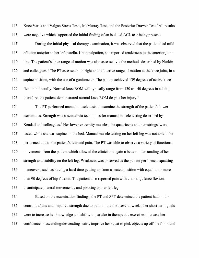
The patient was a 51-year-old female who experienced a left ACL tear while playing 92
recreational pickleball. She went to her doctor's office where she was scheduled for a magnetic 93
resonance imaging (MRI) 5 days after the injury. The imaging showed a left knee ACL tear 94
without other intra-articular or ligamentous involvement. She presented with no significant 95
medical or surgical history, was not taking any medication, and was overall a healthy middle-96
aged woman. She presented to outpatient physical therapy over a month after her injury for an 97
examination. 98
The patient was chosen for the case report due to her openness, reliability, initial 99
adherence to the home exercise program (HEP), and willingness to be involved in the study. She 100
worked as a secretary for an orthopedic surgeon’s office, where employees encouraged her to 101
seek medical professional help for her knee injury. She had goals of returning to her normal 102
activities of playing tennis, skiing, and running outdoors. She stated that she would participate in 103
a conservative, physical therapy-based approach, once a week for eight weeks. She went on to 104
state she did not know, or expect a large benefit from physical therapy but was willing to 105
participate. 106
Examination 107
The patient’s chief complaints were mild weakness, pain (4/10 on the numeric pain rating 108
scale) throughout the day, and the negative psychological impact due to not being able to 109
participate in her normal activities. She denied locking, catching, or buckling of her left knee. 110
Although the patient had imaging that confirmed a left ACL tear, the PT conducted special tests 111
and measures that confirmed the diagnosis and ruled out other injuries. The Anterior Drawer 112
Test and Lachman Test provided positive results, which confirmed the ACL tear diagnosis.7 113
Additional tests were also performed to rule out other possible ligament injuries, including the 114
Knee Varus and Valgus Stress Tests, McMurray Test, and the Posterior Drawer Test.7 All results 115
were negative which supported the initial finding of an isolated ACL tear being present. 116
During the initial physical therapy examination, it was observed that the patient had mild 117
effusion anterior to her left patella. Upon palpation, she reported tenderness to the anterior joint 118
line. The patient’s knee range of motion was also assessed via the methods described by Norkin 119
and colleagues.8 The PT assessed both right and left active range of motion at the knee joint, in a 120
supine position, with the use of a goniometer. The patient achieved 139 degrees of active knee 121
flexion bilaterally. Normal knee ROM will typically range from 130 to 140 degrees in adults; 122
therefore, the patient demonstrated normal knee ROM despite her injury.8 123
The PT performed manual muscle tests to examine the strength of the patient’s lower 124
extremities. Strength was assessed via techniques for manual muscle testing described by 125
Kendall and colleagues.9 Her lower extremity muscles, the quadriceps and hamstrings, were 126
tested while she was supine on the bed. Manual muscle testing on her left leg was not able to be 127
performed due to the patient’s fear and pain. The PT was able to observe a variety of functional 128
movements from the patient which allowed the clinician to gain a better understanding of her 129
strength and stability on the left leg. Weakness was observed as the patient performed squatting 130
maneuvers, such as having a hard time getting up from a seated position with equal to or more 131
than 90 degrees of hip flexion. The patient also reported pain with end-range knee flexion, 132
unanticipated lateral movements, and pivoting on her left leg. 133
Based on the examination findings, the PT and SPT determined the patient had motor 134
control deficits and impaired strength due to pain. In the first several weeks, her short-term goals 135
were to increase her knowledge and ability to partake in therapeutic exercises, increase her 136
confidence in ascending/descending stairs, improve her squat to pick objects up off the floor, and 137

utilize strategies to help reduce knee pain. In the following months to a year, she wanted to 138
return to pain-free running, skiing, and recreational tennis. Given her pain, lack of strength, full 139
ROM, increased motivation, enthusiasm to participate, and lack of comorbidities, she was given 140
a fair to good prognosis for recovery. 141
Treatment 142
The patient agreed to attend physical therapy but did not want to come into the clinic 143
more than once a week for personal reasons. She participated in physical therapy once a week for 144
60-minutes for 9 weeks, working with both the PT and SPT. Based on previous evidence, 145
someone receiving rehabilitation for an ACL injury would typically attend physical therapy 2 to 146
3 times a week.1 The SPT led 6 out of 9 of the patient’s treatment sessions, while the PT treated 147
the patient for the remaining 3 sessions. She received an initial plan of care which consisted of 148
therapeutic exercises intended to strengthen and stabilize the muscles in her left lower extremity, 149
along with soft tissue massage to manage edema around the knee. To optimize the patient’s 150
outcomes, neuromuscular training was incorporated into the plan of care. The CPG for ACL 151
post-operative rehabilitation suggests that both resistance training and neuromuscular training 152
will result in the most optimal outcomes.5 153
Soft Tissue Massage 154
During early treatment sessions, the PT performed soft tissue massage to her left knee to 155
decrease pain and swelling, following a quick warm-up on the assault air bike (Model F-22, 156
Advance Fitness, Made in Taiwan). The patient would sit at the edge of the bed with support 157
under her thighs allowing her legs to hang dependently, which roughly allowed 90 degrees of 158
knee flexion. The patient extended and flexed her left knee while the physical therapist 159
performed distal to proximal stroking motion over the anterior knee joint for 10-minutes. 160
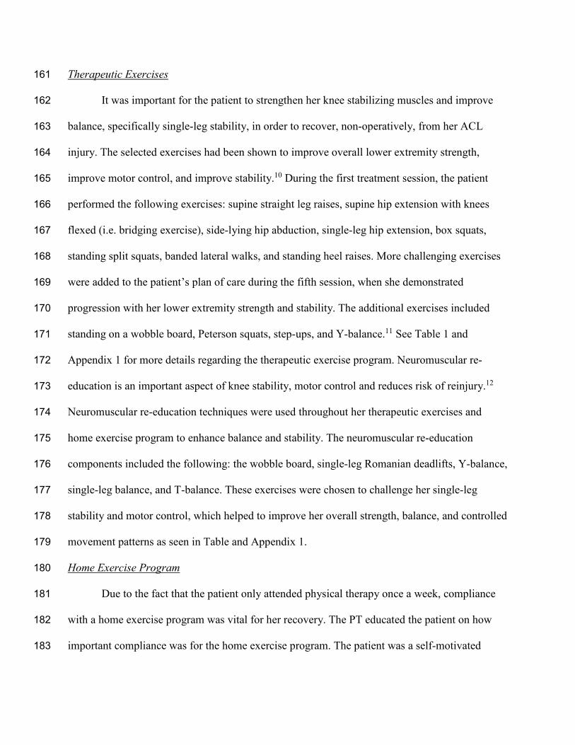
Therapeutic Exercises 161
It was important for the patient to strengthen her knee stabilizing muscles and improve 162
balance, specifically single-leg stability, in order to recover, non-operatively, from her ACL 163
injury. The selected exercises had been shown to improve overall lower extremity strength, 164
improve motor control, and improve stability.10 During the first treatment session, the patient 165
performed the following exercises: supine straight leg raises, supine hip extension with knees 166
flexed (i.e. bridging exercise), side-lying hip abduction, single-leg hip extension, box squats, 167
standing split squats, banded lateral walks, and standing heel raises. More challenging exercises 168
were added to the patient’s plan of care during the fifth session, when she demonstrated 169
progression with her lower extremity strength and stability. The additional exercises included 170
standing on a wobble board, Peterson squats, step-ups, and Y-balance.11 See Table 1 and 171
Appendix 1 for more details regarding the therapeutic exercise program. Neuromuscular re-172
education is an important aspect of knee stability, motor control and reduces risk of reinjury.12 173
Neuromuscular re-education techniques were used throughout her therapeutic exercises and 174
home exercise program to enhance balance and stability. The neuromuscular re-education 175
components included the following: the wobble board, single-leg Romanian deadlifts, Y-balance, 176
single-leg balance, and T-balance. These exercises were chosen to challenge her single-leg 177
stability and motor control, which helped to improve her overall strength, balance, and controlled 178
movement patterns as seen in Table and Appendix 1. 179
Home Exercise Program 180
Due to the fact that the patient only attended physical therapy once a week, compliance 181
with a home exercise program was vital for her recovery. The PT educated the patient on how 182
important compliance was for the home exercise program. The patient was a self-motivated 183
individual and thoroughly enjoyed physical activity. Similar to the physical therapy sessions, her 184
home exercise program included the following: bridges, side-lying hip abduction, chair squats, 185
side lunges, and single-leg balance. See Table 2 for home exercise program dosage. 186
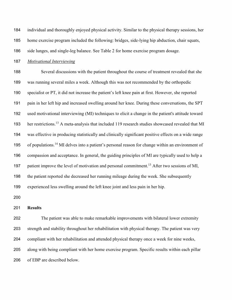
Motivational Interviewing 187
Several discussions with the patient throughout the course of treatment revealed that she 188
was running several miles a week. Although this was not recommended by the orthopedic 189
specialist or PT, it did not increase the patient’s left knee pain at first. However, she reported 190
pain in her left hip and increased swelling around her knee. During these conversations, the SPT 191
used motivational interviewing (MI) techniques to elicit a change in the patient's attitude toward 192
her restrictions.13 A meta-analysis that included 119 research studies showcased revealed that MI 193
was effective in producing statistically and clinically significant positive effects on a wide range 194
of populations.14 MI delves into a patient’s personal reason for change within an environment of 195
compassion and acceptance. In general, the guiding principles of MI are typically used to help a 196
patient improve the level of motivation and personal commitment.13 After two sessions of MI, 197
the patient reported she decreased her running mileage during the week. She subsequently 198
experienced less swelling around the left knee joint and less pain in her hip. 199
200
Results 201
The patient was able to make remarkable improvements with bilateral lower extremity 202
strength and stability throughout her rehabilitation with physical therapy. The patient was very 203
compliant with her rehabilitation and attended physical therapy once a week for nine weeks, 204
along with being compliant with her home exercise program. Specific results within each pillar 205
of EBP are described below. 206
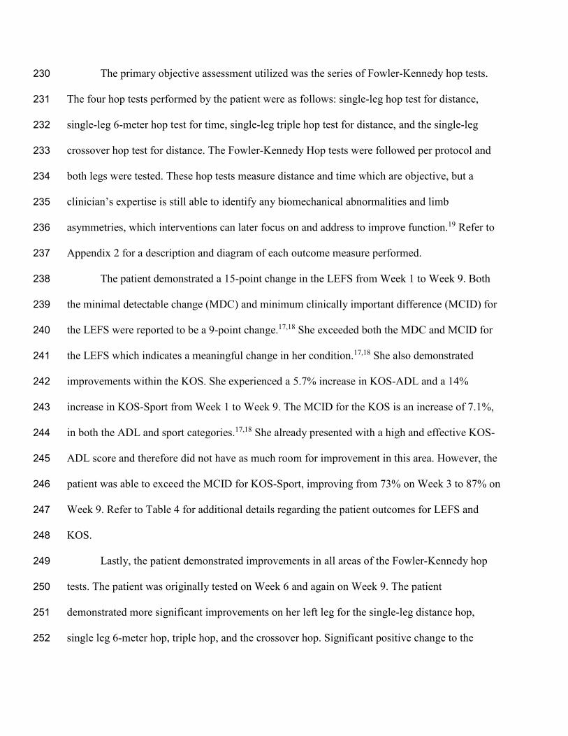
Scientific Evidence 207
Patient outcomes were measured using LEFS, Knee Outcome Survey (KOS), and the 208
Fowler-Kennedy Hop tests. These outcome measures have been shown to be helpful in observing 209
those with lower extremity injuries.15,16 The LEFS is used to assess the patient's perceived level 210
of difficulty in performing a variety of activities.17 The LEFS is a 20 question, self-report form 211
that has been shown to be reliable, valid, and sensitive to change.18 It is scored on a 0-4 scale, 212
from extreme difficulty/unable to perform to no difficulty. A patient could score 0-80 points, 0 213
representing very low function and 80 representing very high function.17 214
The KOS is a subjective questionnaire that aims to assess the effect of the patient’s self-215
reported symptoms on activities of daily living (ADL) and on their sports activities.15,16 The 216
ADL section includes 6 questions designed to determine the ability to perform general daily 217
activities and 8 questions designed to determine the ability to perform specific functional 218
tasks.15,16 Each question is scored 0-5, indicating unable to perform to no difficulty. The total 219
possible score for the ADL section is 70.15,16 The sports activities scale (SAS) section includes 7 220
questions on the ability to perform sports and recreational activities and 4 questions on the ability 221
to perform specific sport activities.15,16 Similar to the ADL section, each question is scored 0-5, 222
indicating unable to perform to no difficulty. The total possible score for the SAS section is 223
55.15,16 The total scores are calculated by finding a percentage for both the ADL and SAS 224
sections. The ADL score would be divided by 70, multiplied by 100, and the SAS score would 225
be divided by 55, multiplied by 100. The higher the percentage, the higher level of physical 226
function.15,16 This patient-reported outcome measure has demonstrated excellent validity, 227
reliability, and responsiveness to assess functional limitations throughout the rehabilitation 228
process for a variety of knee injuries.15,16 229
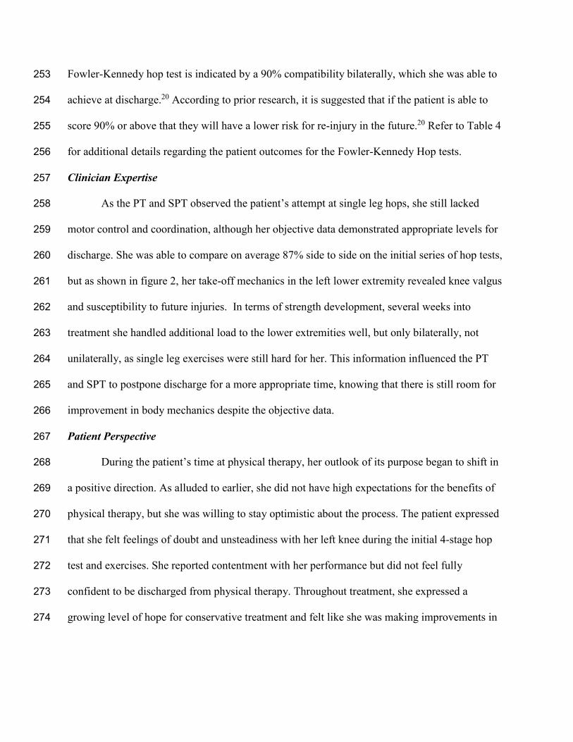
The primary objective assessment utilized was the series of Fowler-Kennedy hop tests. 230
The four hop tests performed by the patient were as follows: single-leg hop test for distance, 231
single-leg 6-meter hop test for time, single-leg triple hop test for distance, and the single-leg 232
crossover hop test for distance. The Fowler-Kennedy Hop tests were followed per protocol and 233
both legs were tested. These hop tests measure distance and time which are objective, but a 234
clinician’s expertise is still able to identify any biomechanical abnormalities and limb 235
asymmetries, which interventions can later focus on and address to improve function.19 Refer to 236
Appendix 2 for a description and diagram of each outcome measure performed. 237
The patient demonstrated a 15-point change in the LEFS from Week 1 to Week 9. Both 238
the minimal detectable change (MDC) and minimum clinically important difference (MCID) for 239
the LEFS were reported to be a 9-point change.17,18 She exceeded both the MDC and MCID for 240
the LEFS which indicates a meaningful change in her condition.17,18 She also demonstrated 241
improvements within the KOS. She experienced a 5.7% increase in KOS-ADL and a 14% 242
increase in KOS-Sport from Week 1 to Week 9. The MCID for the KOS is an increase of 7.1%, 243
in both the ADL and sport categories.17,18 She already presented with a high and effective KOS-244
ADL score and therefore did not have as much room for improvement in this area. However, the 245
patient was able to exceed the MCID for KOS-Sport, improving from 73% on Week 3 to 87% on 246
Week 9. Refer to Table 4 for additional details regarding the patient outcomes for LEFS and 247
KOS. 248
Lastly, the patient demonstrated improvements in all areas of the Fowler-Kennedy hop 249
tests. The patient was originally tested on Week 6 and again on Week 9. The patient 250
demonstrated more significant improvements on her left leg for the single-leg distance hop, 251
single leg 6-meter hop, triple hop, and the crossover hop. Significant positive change to the 252
Fowler-Kennedy hop test is indicated by a 90% compatibility bilaterally, which she was able to 253
achieve at discharge.20 According to prior research, it is suggested that if the patient is able to 254
score 90% or above that they will have a lower risk for re-injury in the future.20 Refer to Table 4 255
for additional details regarding the patient outcomes for the Fowler-Kennedy Hop tests. 256
Clinician Expertise 257
As the PT and SPT observed the patient’s attempt at single leg hops, she still lacked 258
motor control and coordination, although her objective data demonstrated appropriate levels for 259
discharge. She was able to compare on average 87% side to side on the initial series of hop tests, 260
but as shown in figure 2, her take-off mechanics in the left lower extremity revealed knee valgus 261
and susceptibility to future injuries. In terms of strength development, several weeks into 262
treatment she handled additional load to the lower extremities well, but only bilaterally, not 263
unilaterally, as single leg exercises were still hard for her. This information influenced the PT 264
and SPT to postpone discharge for a more appropriate time, knowing that there is still room for 265
improvement in body mechanics despite the objective data. 266
Patient Perspective 267
During the patient’s time at physical therapy, her outlook of its purpose began to shift in 268
a positive direction. As alluded to earlier, she did not have high expectations for the benefits of 269
physical therapy, but she was willing to stay optimistic about the process. The patient expressed 270
that she felt feelings of doubt and unsteadiness with her left knee during the initial 4-stage hop 271
test and exercises. She reported contentment with her performance but did not feel fully 272
confident to be discharged from physical therapy. Throughout treatment, she expressed a 273
growing level of hope for conservative treatment and felt like she was making improvements in 274

her strength and pain, week to week. She felt the ongoing treatment sessions helped instill 275
confidence in herself and improve her left knee. 276
During a follow up encounter with the patient 5-weeks after discharge, she expressed her 277
appreciation to the conversations surrounding her activities outside of therapy. She appreciated 278
being listened to by the clinicians and felt grateful to have had an input in the rehab program. 279
She believed the reduction in activity outside of rehab, but not completely eliminating it, was 280
vital to her success both physically and mentally. 281
282
Discussion 283
ACL ruptures have been a common injury that has imposed a heavy burden on the 284
healthcare system in the past several years.21 Along with preventative treatment, conservative 285
rehabilitation within complete ACL ruptures has been increasingly popular in recent years.22 In 286
reference to knee joint stability, functional outcomes, and overall satisfaction from the patients, 287
studies have found similar outcomes versus surgical intervention in ACL management.22 288
Furthermore, other articles also suggested that clinicians should not heavily rely on the results of 289
outcome measures and rather monitor function throughout rehab in order to make decisions 290
regarding progression and discharge.23 291
On average, patients completed 16.90 ± 10.60 PT visits following ACL reconstruction.24 292
The patient in this case report only completed 9 treatment sessions, under half the amount of 293
treatment sessions typically performed. Only completing half the average number of treatment 294
sessions meant this patient saved between $800 - $1200 on physical therapy alone, not 295
accounting for the median cost of ACL reconstruction surgery of $9000.2 This demonstrated the 296
reduced financial impact that a conservative treatment approach to an ACL repair can have. 297
Despite the lack of research evidence for her particular case, the PT and SPT used 298
objective measurements to track change and give a degree of object framework for discharge. 299
However, the clinicians found it vital to incorporate more of the patient's perspective and 300
clinician expertise more so than scientific evidence. Compared to abiding by a strict rehab 301
guideline from a post-surgical procedure, this allowed for a flexible rehabilitation program and 302
for the patient to drive her own progress based on her perceived difficulty and pain. In this case 303
study, the patient expressed a great deal of satisfaction with her rehab process. Lastly, the patient 304
described an increase in confidence with her involved limb and was very impressed with the 305
progress she made. The PT and SPT hypothesized, if it was not for utilizing all three pillars of 306
evidence-based practice, the successful outcomes may not have been achieved. 307
308
Key Points 309
Findings 310
This case report found that physical therapy may still be effective for an intense 311
rehabilitation program even in a situation with the patient coming in once a week for a limited 312
number of weeks. The case report also supported the importance of the three pillars of practice, 313
relevant scientific research, patient perspective, and clinician expertise. 314
Implications 315
There is a gap in the literature regarding non-operative ACL rehabilitation. This case 316
report provides a potential treatment plan, incorporating all three pillars of EBP, for conservative 317
ACL treatment. The plan could be beneficial for future cases and have an impact on clinical 318
practice. 319
Caution 320
Limitations to this study included the lack of evidence and literature regarding best 321
practice decisions for non-operative ACL tears. It should also be noted that interventions 322
provided in this case were given to an individual with a high level of activity and may not be 323
appropriate in all cases. Additionally, it can be difficult to quantify the importance of all three 324
pillars of EBP in the terms of a case report. 325
326
References: 327
1. Evans J, Nielson Jl. Anterior Cruciate Ligament Knee Injuries. [Updated 2021 Feb 19]. 328
In: StatPearls [Internet]. Treasure Island (FL): StatPearls Publishing; 2021 Jan-. 329
Available from: https://www.ncbi.nlm.nih.gov/books/NBK499848/ 330
2. Herzog MM, Marshall SW, Lund JL, Pate V, Spang JT. Cost of Outpatient 331
Arthroscopic Anterior Cruciate Ligament Reconstruction Among 332
Commercially Insured Patients in the United States, 2005-2013. 2017. 333
Volume 5(1): doi: 10.1177/2325967116684776 334
3. Eggerding V, Reijman M, Meuffels DE, Es EV, Arkel EV, Brand IV, Linge JV, Zijl J, 335
Bierma-Zeinstra S, Koopmanschap M. ACL reconstruction for all is not 336
cost-effective after acute ACL rupture. 2021. 0: 1-5. . 337
doi:10.1136/bjsports-2020-102564 338
4. Sackett DL: Evidence-based medicine and treatment choices. Lancet 1997;349:570 339
5. Van Melick N, Van Cingel, Robert E H, Brooijmans F, et al. Evidence-based clinical 340
practice update: Practice guidelines for anterior cruciate ligament rehabilitation 341
based on a systematic review and multidisciplinary consensus. Br J Sports Med. 342
2016;50(24):1506. doi: 10.1136/bjsports-2015-095898 343
6. APTA Board of Directors. Principles of physical therapist patient client management 344
model. APTA.org Web site. https://guide.apta.org/patient-client-management. 345
Updated 2014. Accessed September 28, 2021. 346
7. Coffey R, Bordoni B. Lachman Test. In: StatPearls. Treasure Island (FL): StatPearls 347
Publishing; August 7, 2021. 348
8. Norkin, Cynthia & White, D. Joyce. Measurement of joint motion: A guide to 349
goniometry. 5th ed. Philadelphia, PA: F.A. Davis Company; 2016. 350
9. Kendal PF, McCreary KE, Provance GP, Rodgers MM, Romani AW. Muscles: Testing 351
and function with posture and pain. 5th ed. Philadelphia, PA: Lippincott Williams 352
& Wilkins. 353
10. Fitzgerald GK, Axe MJ, Synder-Mackler L. Proposed Practice Guidelines for 354
Nonoperative Anterior Cruciate Ligament Rehabilitation of Physically Active 355
Individuals. 2000; 30(4): 194-203. 356
11. Greenberg ET, Barle M, Glassmann E, Jung MK. Interrater and test-retest reliability of 357
the Y Balance test in healthy, early adolescent female athletes. Int J Sports Phys 358
Ther. 2019;14(2):204-213. 359
12. Sugimoto, D., Myer, G., Barber Foss, K., Pepin, M., Micheli, L. and Hewett, T., 2016. 360
Critical components of neuromuscular training to reduce ACL injury risk in 361
female athletes: meta-regression analysis. British Journal of Sports Medicine, 362
50(20), pp.1259-1266. 363
13. Prescott DS. Motivational Interviewing: as Easy as It Looks?. Curr Psychiatry Rep. 364
2020;22(7):35. Published 2020 May 28. doi:10.1007/s11920-020-01158-z 365
14. Lundahl BW, Kunz C, Brownell C, Tollefson D, Burke BL. A meta-analysis of 366
motivational interviewing: twenty-five years of empirical studies. Res Soc Work 367
Pract. 2010;20(2):137-160. 368
https://journals.sagepub.com/doi/full/10.1177/1049731509347850. doi:369
10.1177/1049731509347850 370

15. Irrgang JJ, Snyder-Mackler L, Wainner RS, Fu FH, Harner CD. Development of a 371
patient-reported measure of function of the knee. J Bone Joint Surg Am. 372
1998;80(8):1132-1145. doi:10.2106/00004623-199808000-00006 373
16. Bradbury M, Brosky JA Jr, Walker JF, West K. Relationship between scores from the 374
Knee Outcome Survey and a single assessment numerical rating in patients with 375
patellofemoral pain. Physiother Theory Pract. 2013;29(7):531-535. 376
doi:10.3109/09593985.2012.762077 377
17. Binkley JM, Stratford PW, Lott SA, Riddle DL. The Lower Extremity Functional Scale 378
(LEFS): scale development, measurement properties, and clinical application. 379
North American Orthopaedic Rehabilitation Research Network. Phys Ther. 380
1999;79(4):371-383. 381
18. Mehta SP, Fulton A, Quach C, Thistle M, Toledo C, Evans NA. Measurement Properties 382
of the Lower Extremity Functional Scale: A Systematic Review. J Orthop Sports 383
Phys Ther. 2016;46(3):200-216. doi:10.2519/jospt.2016.6165 384
19. Logerstedt D, Grindem H, Lynch A, et al. Single-legged hop tests as predictors of 385
self-reported knee function after anterior cruciate ligament reconstruction. Am J 386
Sports Med. 2012;40(10):2348-2356. 387
https://journals.sagepub.com/doi/full/10.1177/0363546512457551. doi: 388
10.1177/0363546512457551 389
20. Grindem H, Snyder-Mackler L, Moksnes H, Engebretsen L, Risberg MA. Simple 390
decision. rules can reduce reinjury risk by 84% after ACL reconstruction: the 391
Delaware-Oslo ACL cohort study. Br J Sports Med. 2016;50(13):804-808. 392
doi:10.1136/bjsports-2016-096031 393
21. Evans J, Nielson Jl. Anterior Cruciate Ligament Knee Injuries. [Updated 2021 Feb 19]. 394
In: StatPearls [Internet]. Treasure Island (FL): StatPearls Publishing; 2021 Jan-. 395
Available from: https://www.ncbi.nlm.nih.gov/books/NBK499848/ 396
22. Krause M, Freudenthaler F, Frosch KH, Achtnich A, Petersen W, Akoto R. Operative 397
Versus Conservative Treatment of Anterior Cruciate Ligament Rupture. 2018: 398
115(51-52): 855-862. DOI: 10.3238/arztebl.2018.0855 399
23. Davies WT, Myer GD, Read PJ. Is It Time We Better Understood the Tests We are Using 400
for Return to Sport Decision Making Following ACL Reconstruction? A Critical 401
Review of the Hop Tests. Sports Med. 2020;50(3):485-495. 402
doi:10.1007/s40279-019-01221-7 403
24. Burroughs PJ, Kahan JB, Moore HG, Grauer JN, Gardner EC. Temporal Utilization of 404
Physical Therapy Visits After Anterior Cruciate Ligament Reconstruction. Orthop 405
J Sports Med. 2021;9(2):2325967120982293. Published 2021 Feb 19. 406
doi:10.1177/2325967120982293 407
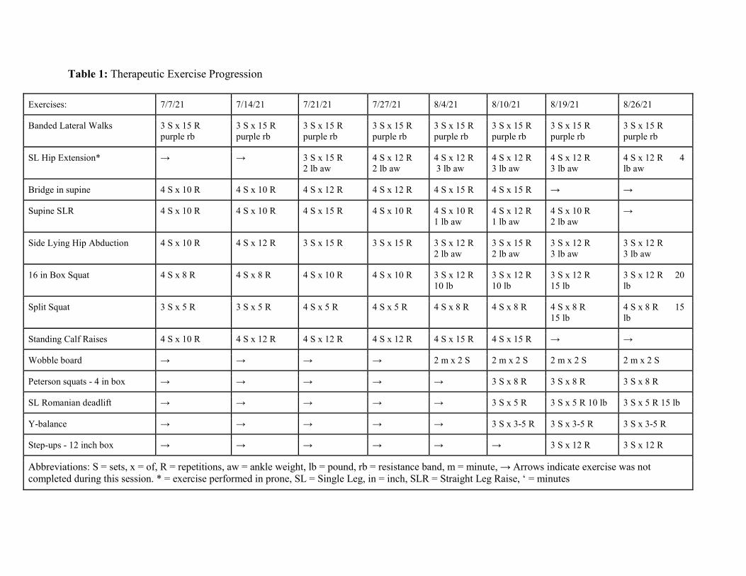
Table 1: Therapeutic Exercise Progression
Exercises: 7/7/21 7/14/21 7/21/21 7/27/21 8/4/21 8/10/21 8/19/21 8/26/21
Banded Lateral Walks 3 S x 15 R purple rb
3 S x 15 R purple rb
3 S x 15 R purple rb
3 S x 15 R purple rb
3 S x 15 R purple rb
3 S x 15 R purple rb
3 S x 15 R purple rb
3 S x 15 R purple rb
SL Hip Extension*
→ → 3 S x 15 R 2 lb aw
4 S x 12 R 2 lb aw
4 S x 12 R 3 lb aw
4 S x 12 R 3 lb aw
4 S x 12 R 3 lb aw
4 S x 12 R 4 lb aw
Bridge in supine 4 S x 10 R 4 S x 10 R 4 S x 12 R 4 S x 12 R 4 S x 15 R 4 S x 15 R → →
Supine SLR 4 S x 10 R 4 S x 10 R 4 S x 15 R 4 S x 10 R 4 S x 10 R 1 lb aw
4 S x 12 R 1 lb aw
4 S x 10 R 2 lb aw
→
Side Lying Hip Abduction 4 S x 10 R 4 S x 12 R 3 S x 15 R 3 S x 15 R 3 S x 12 R 2 lb aw
3 S x 15 R 2 lb aw
3 S x 12 R 3 lb aw
3 S x 12 R 3 lb aw
16 in Box Squat 4 S x 8 R 4 S x 8 R 4 S x 10 R 4 S x 10 R 3 S x 12 R 10 lb
3 S x 12 R 10 lb
3 S x 12 R 15 lb
3 S x 12 R 20 lb
Split Squat 3 S x 5 R 3 S x 5 R 4 S x 5 R 4 S x 5 R 4 S x 8 R 4 S x 8 R 4 S x 8 R 15 lb
4 S x 8 R 15 lb
Standing Calf Raises 4 S x 10 R 4 S x 12 R 4 S x 12 R 4 S x 12 R 4 S x 15 R 4 S x 15 R → →
Wobble board → → → → 2 m x 2 S 2 m x 2 S 2 m x 2 S 2 m x 2 S
Peterson squats - 4 in box → → → → → 3 S x 8 R 3 S x 8 R 3 S x 8 R
SL Romanian deadlift → → → → → 3 S x 5 R 3 S x 5 R 10 lb 3 S x 5 R 15 lb
Y-balance → → → → → 3 S x 3-5 R 3 S x 3-5 R 3 S x 3-5 R
Step-ups - 12 inch box → → → → → → 3 S x 12 R 3 S x 12 R
Abbreviations: S = sets, x = of, R = repetitions, aw = ankle weight, lb = pound, rb = resistance band, m = minute, → Arrows indicate exercise was not completed during this session. * = exercise performed in prone, SL = Single Leg, in = inch, SLR = Straight Leg Raise, ‘ = minutes
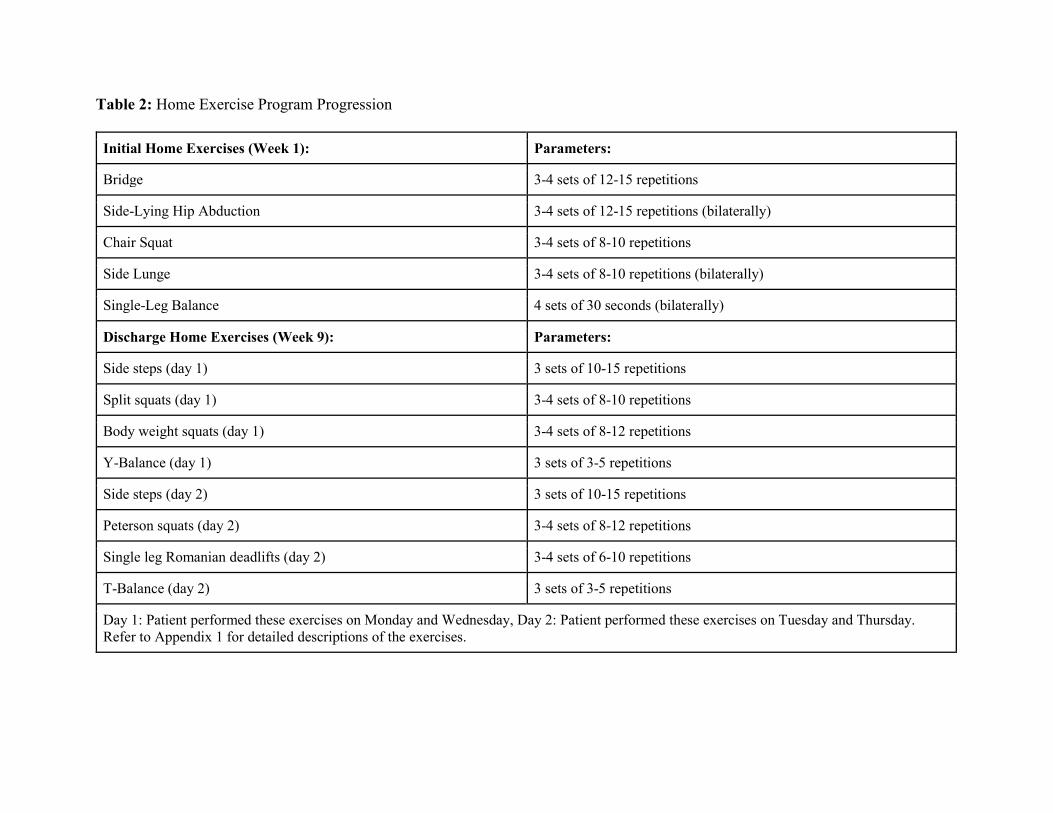
Table 2: Home Exercise Program Progression
Initial Home Exercises (Week 1): Parameters:
Bridge 3-4 sets of 12-15 repetitions
Side-Lying Hip Abduction 3-4 sets of 12-15 repetitions (bilaterally)
Chair Squat 3-4 sets of 8-10 repetitions
Side Lunge 3-4 sets of 8-10 repetitions (bilaterally)
Single-Leg Balance 4 sets of 30 seconds (bilaterally)
Discharge Home Exercises (Week 9): Parameters:
Side steps (day 1) 3 sets of 10-15 repetitions
Split squats (day 1) 3-4 sets of 8-10 repetitions
Body weight squats (day 1) 3-4 sets of 8-12 repetitions
Y-Balance (day 1) 3 sets of 3-5 repetitions
Side steps (day 2) 3 sets of 10-15 repetitions
Peterson squats (day 2) 3-4 sets of 8-12 repetitions
Single leg Romanian deadlifts (day 2) 3-4 sets of 6-10 repetitions
T-Balance (day 2) 3 sets of 3-5 repetitions
Day 1: Patient performed these exercises on Monday and Wednesday, Day 2: Patient performed these exercises on Tuesday and Thursday. Refer to Appendix 1 for detailed descriptions of the exercises.
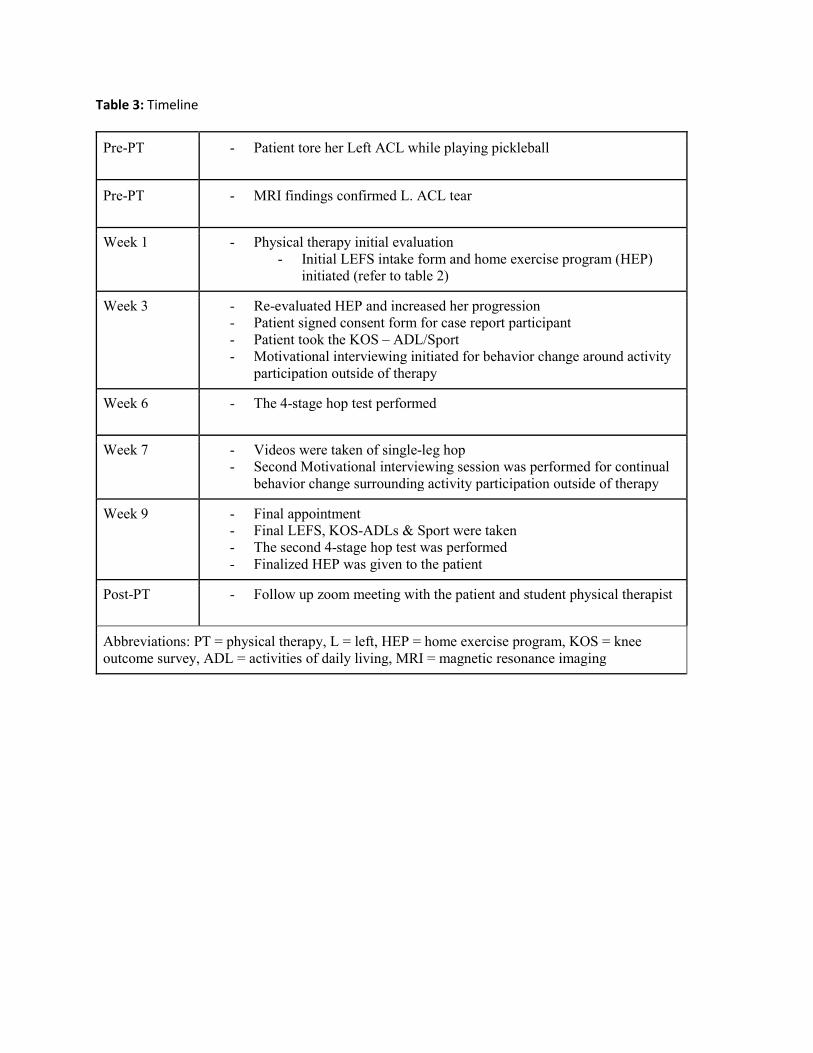
Table 3: Timeline
Pre-PT - Patient tore her Left ACL while playing pickleball
Pre-PT - MRI findings confirmed L. ACL tear
Week 1 - Physical therapy initial evaluation - Initial LEFS intake form and home exercise program (HEP)
initiated (refer to table 2)
Week 3 - Re-evaluated HEP and increased her progression - Patient signed consent form for case report participant - Patient took the KOS – ADL/Sport - Motivational interviewing initiated for behavior change around activity
participation outside of therapy
Week 6 - The 4-stage hop test performed
Week 7 - Videos were taken of single-leg hop - Second Motivational interviewing session was performed for continual
behavior change surrounding activity participation outside of therapy
Week 9 - Final appointment - Final LEFS, KOS-ADLs & Sport were taken - The second 4-stage hop test was performed - Finalized HEP was given to the patient
Post-PT - Follow up zoom meeting with the patient and student physical therapist
Abbreviations: PT = physical therapy, L = left, HEP = home exercise program, KOS = knee outcome survey, ADL = activities of daily living, MRI = magnetic resonance imaging
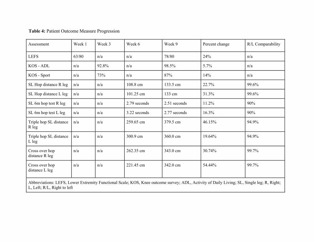
Table 4: Patient Outcome Measure Progression
Assessment Week 1 Week 3 Week 6 Week 9 Percent change R/L Comparability
LEFS 63/80 n/a n/a 78/80 24% n/a
KOS - ADL n/a 92.8% n/a 98.5% 5.7% n/a
KOS - Sport n/a 73% n/a 87% 14% n/a
SL Hop distance R leg n/a n/a 108.8 cm 133.5 cm 22.7% 99.6%
SL Hop distance L leg n/a n/a 101.25 cm 133 cm 31.3% 99.6%
SL 6m hop test R leg n/a n/a 2.79 seconds 2.51 seconds 11.2% 90%
SL 6m hop test L leg n/a n/a 3.22 seconds 2.77 seconds 16.3% 90%
Triple hop SL distance R leg
n/a n/a 259.65 cm 379.5 cm 46.15% 94.9%
Triple hop SL distance L leg
n/a n/a 300.9 cm 360.0 cm 19.64% 94.9%
Cross over hop distance R leg
n/a n/a 262.35 cm 343.0 cm 30.74% 99.7%
Cross over hop distance L leg
n/a n/a 221.45 cm 342.0 cm 54.44% 99.7%
Abbreviations: LEFS, Lower Extremity Functional Scale; KOS, Knee outcome survey; ADL, Activity of Daily Living; SL, Single leg; R, Right; L, Left; R/L, Right to left
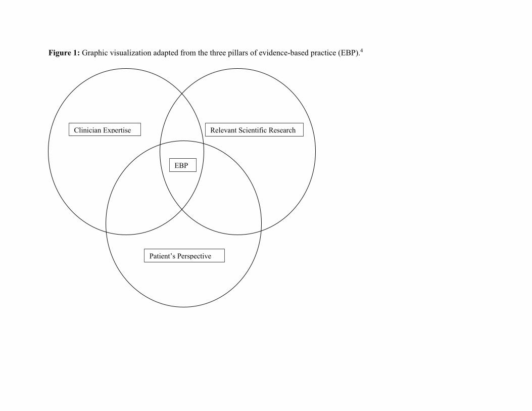
Figure 1: Graphic visualization adapted from the three pillars of evidence-based practice (EBP).4
Relevant Scientific Research
EBP
Clinician Expertise
Patient’s Perspective

Figure 2: Visualization of eccentric phase during take-off on left lower extremity during 4-stage hop test, taken during the 5th week (initial hop test). The red line indicates valgus motion in a still frame as the patient prepares to jump off of their left lower extremity.
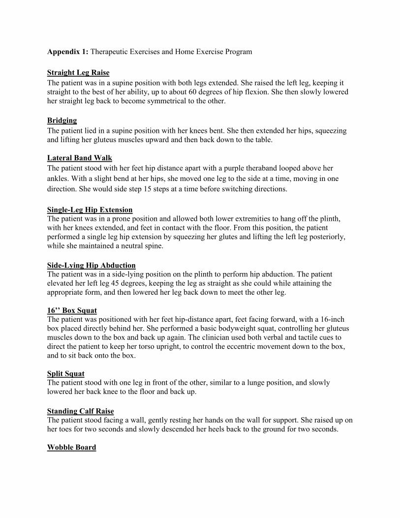
Appendix 1: Therapeutic Exercises and Home Exercise Program Straight Leg Raise The patient was in a supine position with both legs extended. She raised the left leg, keeping it straight to the best of her ability, up to about 60 degrees of hip flexion. She then slowly lowered her straight leg back to become symmetrical to the other. Bridging The patient lied in a supine position with her knees bent. She then extended her hips, squeezing and lifting her gluteus muscles upward and then back down to the table. Lateral Band Walk The patient stood with her feet hip distance apart with a purple theraband looped above her ankles. With a slight bend at her hips, she moved one leg to the side at a time, moving in one direction. She would side step 15 steps at a time before switching directions. Single-Leg Hip Extension The patient was in a prone position and allowed both lower extremities to hang off the plinth, with her knees extended, and feet in contact with the floor. From this position, the patient performed a single leg hip extension by squeezing her glutes and lifting the left leg posteriorly, while she maintained a neutral spine. Side-Lying Hip Abduction The patient was in a side-lying position on the plinth to perform hip abduction. The patient elevated her left leg 45 degrees, keeping the leg as straight as she could while attaining the appropriate form, and then lowered her leg back down to meet the other leg. 16’’ Box Squat The patient was positioned with her feet hip-distance apart, feet facing forward, with a 16-inch box placed directly behind her. She performed a basic bodyweight squat, controlling her gluteus muscles down to the box and back up again. The clinician used both verbal and tactile cues to direct the patient to keep her torso upright, to control the eccentric movement down to the box, and to sit back onto the box. Split Squat The patient stood with one leg in front of the other, similar to a lunge position, and slowly lowered her back knee to the floor and back up. Standing Calf Raise The patient stood facing a wall, gently resting her hands on the wall for support. She raised up on her toes for two seconds and slowly descended her heels back to the ground for two seconds. Wobble Board
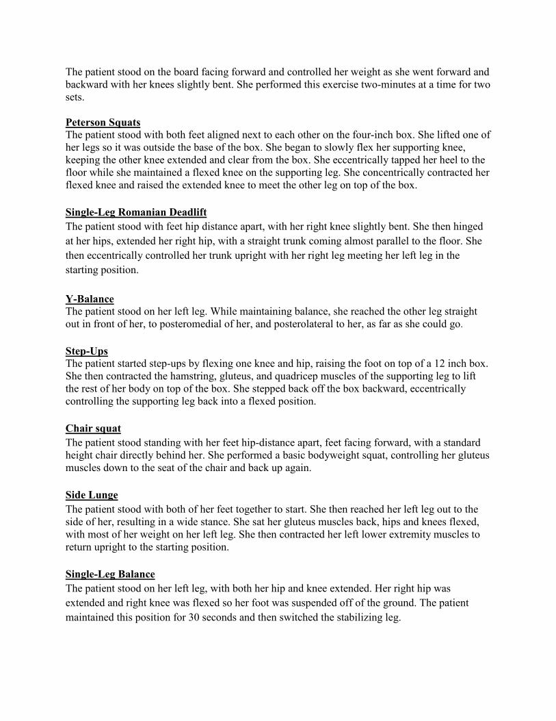
The patient stood on the board facing forward and controlled her weight as she went forward and backward with her knees slightly bent. She performed this exercise two-minutes at a time for two sets. Peterson Squats The patient stood with both feet aligned next to each other on the four-inch box. She lifted one of her legs so it was outside the base of the box. She began to slowly flex her supporting knee, keeping the other knee extended and clear from the box. She eccentrically tapped her heel to the floor while she maintained a flexed knee on the supporting leg. She concentrically contracted her flexed knee and raised the extended knee to meet the other leg on top of the box. Single-Leg Romanian Deadlift The patient stood with feet hip distance apart, with her right knee slightly bent. She then hinged at her hips, extended her right hip, with a straight trunk coming almost parallel to the floor. She then eccentrically controlled her trunk upright with her right leg meeting her left leg in the starting position. Y-Balance The patient stood on her left leg. While maintaining balance, she reached the other leg straight out in front of her, to posteromedial of her, and posterolateral to her, as far as she could go. Step-Ups The patient started step-ups by flexing one knee and hip, raising the foot on top of a 12 inch box. She then contracted the hamstring, gluteus, and quadricep muscles of the supporting leg to lift the rest of her body on top of the box. She stepped back off the box backward, eccentrically controlling the supporting leg back into a flexed position. Chair squat The patient stood standing with her feet hip-distance apart, feet facing forward, with a standard height chair directly behind her. She performed a basic bodyweight squat, controlling her gluteus muscles down to the seat of the chair and back up again. Side Lunge The patient stood with both of her feet together to start. She then reached her left leg out to the side of her, resulting in a wide stance. She sat her gluteus muscles back, hips and knees flexed, with most of her weight on her left leg. She then contracted her left lower extremity muscles to return upright to the starting position. Single-Leg Balance The patient stood on her left leg, with both her hip and knee extended. Her right hip was extended and right knee was flexed so her foot was suspended off of the ground. The patient maintained this position for 30 seconds and then switched the stabilizing leg.

T-Balance Similar to the Y-balance, the patient balanced her weight over one leg and reached her non-planted foot directly posteriorly, anteromedial, and anterolateral, touching at each of the three points.
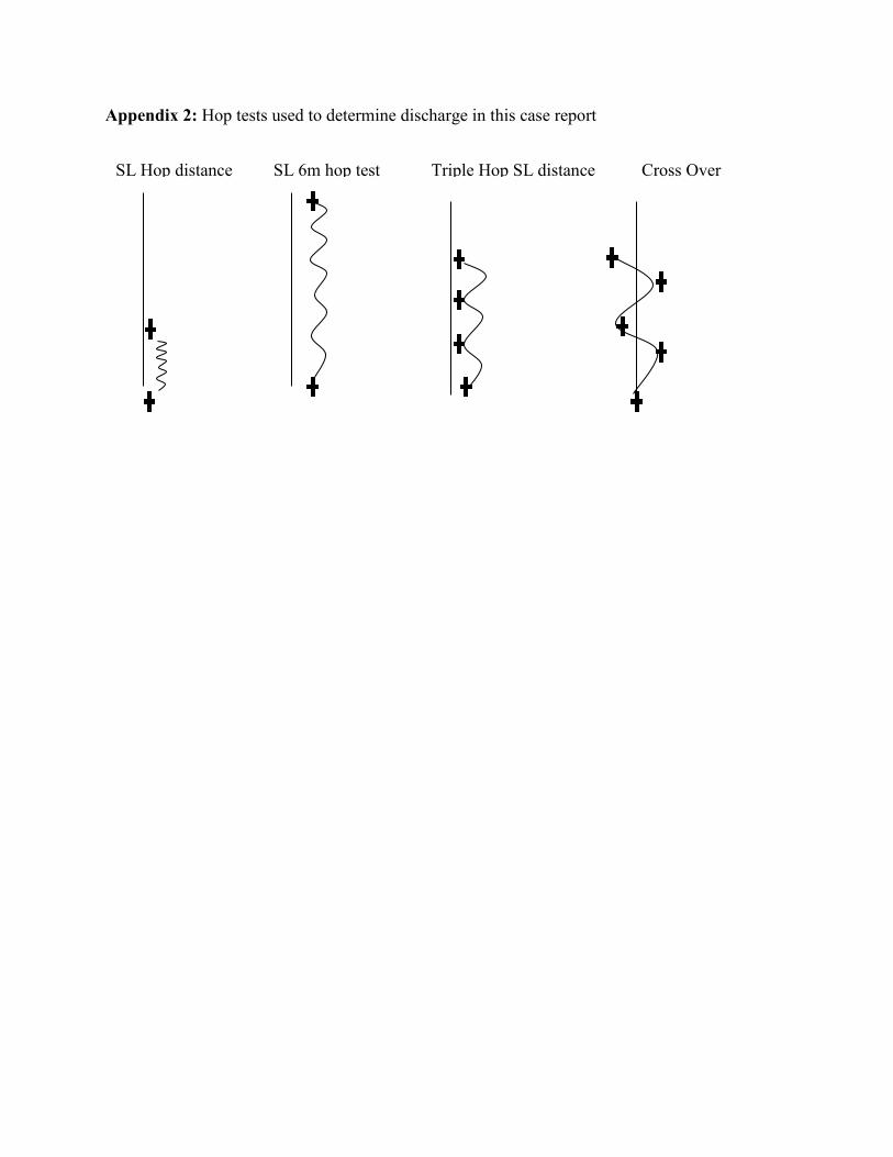
Appendix 2: Hop tests used to determine discharge in this case report
SL Hop distance SL 6m hop test Triple Hop SL distance Cross Over
Copyright © 2022 FDOKUMEN