Case Report: Visual Rehabilitation in Hemianopia Patients ...
11
CASE REPORT published: 21 July 2021 doi: 10.3389/fneur.2021.680211 Frontiers in Neurology | www.frontiersin.org 1 July 2021 | Volume 12 | Article 680211 Edited by: Jiawei Zhou, Wenzhou Medical University, China Reviewed by: Christine Rogers, University of Cape Town, South Africa Benjamin A. Rowland, Wake Forest University, United States *Correspondence: Michael Reber [email protected] Specialty section: This article was submitted to Neurorehabilitation, a section of the journal Frontiers in Neurology Received: 13 March 2021 Accepted: 15 June 2021 Published: 21 July 2021 Citation: Daibert-Nido M, Pyatova Y, Cheung K, Nayomi C, Markowitz SN, Bouffet E and Reber M (2021) Case Report: Visual Rehabilitation in Hemianopia Patients. Home-Based Visual Rehabilitation in Patients With Hemianopia Consecutive to Brain Tumor Treatment: Feasibility and Potential Effectiveness. Front. Neurol. 12:680211. doi: 10.3389/fneur.2021.680211 Case Report: Visual Rehabilitation in Hemianopia Patients. Home-Based Visual Rehabilitation in Patients With Hemianopia Consecutive to Brain Tumor Treatment: Feasibility and Potential Effectiveness Monica Daibert-Nido 1,2 , Yulia Pyatova 1 , Kyle Cheung 2 , Camilus Nayomi 2 , Samuel N. Markowitz 1,2 , Eric Bouffet 3 and Michael Reber 1,2,4 * 1 Department of Ophthalmology and Vision Sciences, University of Toronto, Toronto, ON, Canada, 2 Donald K. Johnson Eye Institute, Krembil Research Institute, University Health Network, Toronto, ON, Canada, 3 Division of Hematology/Oncology, Research Institute, The Hospital for Sick Children, Toronto, ON, Canada, 4 Laboratory Medicine and Pathobiology, Cell and System Biology, University of Toronto, Toronto, ON, Canada Background/Objectives: Visual field loss is frequent in patients with brain tumors, worsening their daily life and exacerbating the burden of disease, and no supportive care strategies exist. In this case series, we sought to characterize the feasibility and potential effectiveness of a home-based visual rehabilitation program in hemianopia patients using immersive virtual-reality stimulation. Subjects/Methods: Two patients, one with homonymous hemianopia and the other with bitemporal hemianopia, consecutive to pediatric brain tumors, with no prior visual rehabilitation performed 15 min of home-based audiovisual stimulation every 2 days for 6 weeks (case 2) and 7 weeks (case 1) between February and August 2020. Patients used a virtual-reality, stand-alone, and remotely controlled device loaded with a non-commercial audiovisual stimulation program managed in real time from the laboratory. Standard visual outcomes assessed in usual care in visual rehabilitation were measured at the clinic. Following a mixed method approach in this pragmatic study of two cases, we collected quantitative and qualitative data on feasibility and potential effectiveness and compared the results pre- and post-treatment. Results: Implementation and wireless delivery of the audiovisual stimulation, remote data collection, and analysis for cases 1 and 2 who completed 19/20 and 20/20 audiovisual stimulation sessions at home, respectively, altogether indicated feasibility. Contrast sensitivity increased in both eyes for cases 1 and 2. Visual fields, measured by binocular Esterman and monocular Humphrey full-field analyses, improved in case 1. A minor increase was observed in case 2. Cases 1 and 2 enhanced reading speed. Case 2 strongly improved quality of life scores.
-
Upload
khangminh22 -
Category
Documents
-
view
1 -
download
0
Transcript of Case Report: Visual Rehabilitation in Hemianopia Patients ...

CASE REPORTpublished: 21 July 2021
doi: 10.3389/fneur.2021.680211
Frontiers in Neurology | www.frontiersin.org 1 July 2021 | Volume 12 | Article 680211
Edited by:
Jiawei Zhou,
Wenzhou Medical University, China
Reviewed by:
Christine Rogers,
University of Cape Town, South Africa
Benjamin A. Rowland,
Wake Forest University, United States
*Correspondence:
Michael Reber
Specialty section:
This article was submitted to
Neurorehabilitation,
a section of the journal
Frontiers in Neurology
Received: 13 March 2021
Accepted: 15 June 2021
Published: 21 July 2021
Citation:
Daibert-Nido M, Pyatova Y, Cheung K,
Nayomi C, Markowitz SN, Bouffet E
and Reber M (2021) Case Report:
Visual Rehabilitation in Hemianopia
Patients. Home-Based Visual
Rehabilitation in Patients With
Hemianopia Consecutive to Brain
Tumor Treatment: Feasibility and
Potential Effectiveness.
Front. Neurol. 12:680211.
doi: 10.3389/fneur.2021.680211
Case Report: Visual Rehabilitation inHemianopia Patients. Home-BasedVisual Rehabilitation in Patients WithHemianopia Consecutive to BrainTumor Treatment: Feasibility andPotential EffectivenessMonica Daibert-Nido 1,2, Yulia Pyatova 1, Kyle Cheung 2, Camilus Nayomi 2,
Samuel N. Markowitz 1,2, Eric Bouffet 3 and Michael Reber 1,2,4*
1Department of Ophthalmology and Vision Sciences, University of Toronto, Toronto, ON, Canada, 2Donald K. Johnson Eye
Institute, Krembil Research Institute, University Health Network, Toronto, ON, Canada, 3Division of Hematology/Oncology,
Research Institute, The Hospital for Sick Children, Toronto, ON, Canada, 4 Laboratory Medicine and Pathobiology, Cell and
System Biology, University of Toronto, Toronto, ON, Canada
Background/Objectives: Visual field loss is frequent in patients with brain tumors,
worsening their daily life and exacerbating the burden of disease, and no supportive care
strategies exist. In this case series, we sought to characterize the feasibility and potential
effectiveness of a home-based visual rehabilitation program in hemianopia patients using
immersive virtual-reality stimulation.
Subjects/Methods: Two patients, one with homonymous hemianopia and the other
with bitemporal hemianopia, consecutive to pediatric brain tumors, with no prior visual
rehabilitation performed 15min of home-based audiovisual stimulation every 2 days for 6
weeks (case 2) and 7weeks (case 1) between February and August 2020. Patients used a
virtual-reality, stand-alone, and remotely controlled device loaded with a non-commercial
audiovisual stimulation programmanaged in real time from the laboratory. Standard visual
outcomes assessed in usual care in visual rehabilitation were measured at the clinic.
Following a mixed method approach in this pragmatic study of two cases, we collected
quantitative and qualitative data on feasibility and potential effectiveness and compared
the results pre- and post-treatment.
Results: Implementation and wireless delivery of the audiovisual stimulation, remote
data collection, and analysis for cases 1 and 2 who completed 19/20 and 20/20
audiovisual stimulation sessions at home, respectively, altogether indicated feasibility.
Contrast sensitivity increased in both eyes for cases 1 and 2. Visual fields, measured
by binocular Esterman and monocular Humphrey full-field analyses, improved in case 1.
A minor increase was observed in case 2. Cases 1 and 2 enhanced reading speed. Case
2 strongly improved quality of life scores.
Daibert-Nido et al. Visual Rehabilitation in Hemianopia
Conclusion: This is the first report of a home-based virtual-reality visual rehabilitation
program for adult patients with hemianopia consecutive to a pediatric brain tumor.
We show the feasibility in real-world conditions and potential effectiveness of such
technology on visual perception and quality of life.
Keywords: low-vision, rehabilitation, virtual-reality, personalized medicine 2, hemianopia or hemianopsia,
hemianopia rehabilitation
INTRODUCTION
Central nervous system tumors are the second most commonmalignancies in childhood (1). Brain tumor and its treatment canaffect the visual system at different levels, from the optic nerves(through compression or infiltration), to subcortical structureslike the superior colliculus (SC) and lateral geniculate nuclei(LGN) to optic tracts, optic radiations, and visual cortices (1–4). Children with brain tumors can present visual impairmentslike decreased visual acuity and contrast sensitivity (CS), lossof color vision, and visual field loss such as hemianopias (1–5).These visual field defects in children affect their psycho-socialand educational development with long-term debilitating effects.Hemianopia patients present difficulties in detecting stimuliin the defective visual field and show defective scanning andexploration (6). Moreover, they show a rotation and compressionof the auditory space leading to imprecise localization ofsound across both hemispaces (7). Patients with hemianopianaturally develop oculomotor strategies (more saccades towardthe blind field) to compensate for visual field loss (8, 9),but visual rehabilitation procedure must still be developedto optimize/improve visual perception in the blind field andenhance the quality of life. Here, we report, for the first time,the feasibility and potential effectiveness of a visual rehabilitationprocedure consecutive to a pediatric brain tumor in two youngadult patients with hemianopia. We developed a home-basedaudiovisual stimulation protocol (currently not commercialized)using immersive virtual reality (IVR) in the stand-alone andremotely controlled head-mounted display (HMD) Oculus Go.The basic concept relies on the stimulation of residual visionand multisensory-induced visual plasticity using specific visualtasks, combined to spatially and temporally congruent auditorystimuli (10, 11). This approach is based on the multisensoryintegration properties of the brain for the detection andtracking of elements in the surroundings (12, 13). Recentevidence shows that static audiovisual stimulation reorganizesthe functional connectivity in subcortical and cortical visualareas in hemianopia patients improving visual perception inthe blind hemifield (11, 14). Here, we used the 3D-multipleobject tracking (3D-MOT) paradigm, which closely matchesattentionally demanding real-life situations (15, 16). The setupof the task is highly dynamic as the objects change theirlocation over time, requiring a continuous deployment of visualattention and oculomotor control to avoid confusions betweenthe objects (16). 3D-MOT stimulation programs displayedon monitors or TV screens have been shown to increasebrain capacity for complex processes such as anticipation,
eye-tracking, field of view, and even decision-making in healthyand pathological participants (17–19). Using a mixed methodapproach, our objective was to test the feasibility (treatmentimplementation and wireless delivery, remote collection andanalysis of data, compliance and adherence, and adverse effects)and potential effectiveness (visual function and functional vision)of a home-based, IVR, audiovisual stimulation program onquality of life and visual field perception. This treatment wasperformed between March and August 2020, during the stay-at-home order (Ontario Government—Emergency Managementand Civil Protection Act, Order in Council 518/2020) due to theCOVID-19 pandemic. The audiovisual stimulation protocol wasimplemented remotely (telerehabilitation) in the patient’s deviceand data were collected every 2 days. The patients were ableto comply and adhere to the audiovisual rehabilitation protocolwithout any interruptions in the treatment.
CASE DESCRIPTION
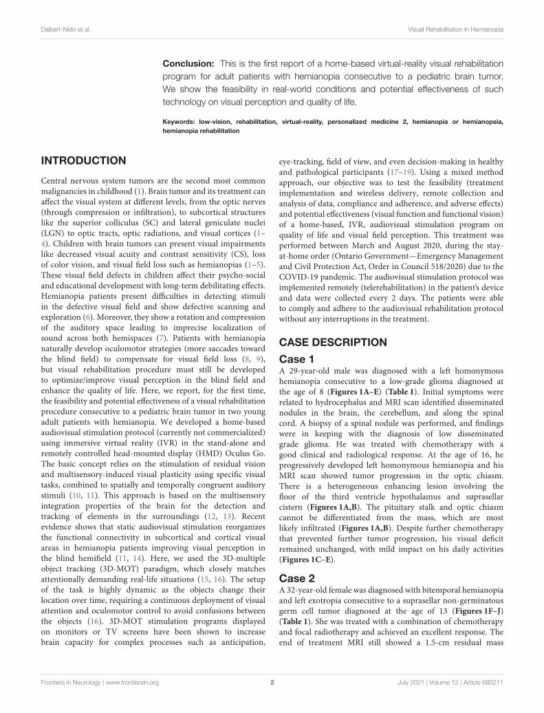
Case 1A 29-year-old male was diagnosed with a left homonymoushemianopia consecutive to a low-grade glioma diagnosed atthe age of 8 (Figures 1A–E) (Table 1). Initial symptoms wererelated to hydrocephalus and MRI scan identified disseminatednodules in the brain, the cerebellum, and along the spinalcord. A biopsy of a spinal nodule was performed, and findingswere in keeping with the diagnosis of low disseminatedgrade glioma. He was treated with chemotherapy with agood clinical and radiological response. At the age of 16, heprogressively developed left homonymous hemianopia and hisMRI scan showed tumor progression in the optic chiasm.There is a heterogeneous enhancing lesion involving thefloor of the third ventricle hypothalamus and suprasellarcistern (Figures 1A,B). The pituitary stalk and optic chiasmcannot be differentiated from the mass, which are mostlikely infiltrated (Figures 1A,B). Despite further chemotherapythat prevented further tumor progression, his visual deficitremained unchanged, with mild impact on his daily activities(Figures 1C–E).
Case 2A 32-year-old female was diagnosed with bitemporal hemianopiaand left exotropia consecutive to a suprasellar non-germinatousgerm cell tumor diagnosed at the age of 13 (Figures 1F–J)(Table 1). She was treated with a combination of chemotherapyand focal radiotherapy and achieved an excellent response. Theend of treatment MRI still showed a 1.5-cm residual mass
Frontiers in Neurology | www.frontiersin.org 2 July 2021 | Volume 12 | Article 680211
Daibert-Nido et al. Visual Rehabilitation in Hemianopia
FIGURE 1 | Case presentation. (A–E) Case 1 MRI axial (A) and coronal (B) view (T1 + contrast). Yellow circles indicate the remaining tumor mass after chemotherapy.
(C) Schematic of the visual field defects. (D) Diagram of the optic pathway indicating the location of the injury. (E) Esterman binocular field test at baseline
(pre-treatment). Black dots correspond to unseen point of light. (F–J) Case 2 MRI axial (F) and coronal (G) view (T1 + contrast). Yellow circles indicate the remaining
tumor mass after chemo- and focal radiotherapy. (H) Schematic of the visual field defects. (I) Diagram of the optic pathway indicating the location of the injury. (J)
Esterman binocular field test at baseline (pre-treatment). Black dots correspond to unseen point of light.
in the suprasellar region, inseparable from the hypothalamusand optic chiasm, which remained unchanged over time(Figures 1F,G). Her visual impairment, corresponding to a
bitemporal hemianopia (Figures 1H–J), was detected at thetime of initial diagnosis did not improve despite the successfulmanagement of her tumor.
Frontiers in Neurology | www.frontiersin.org 3 July 2021 | Volume 12 | Article 680211
Daibert-Nido et al. Visual Rehabilitation in Hemianopia
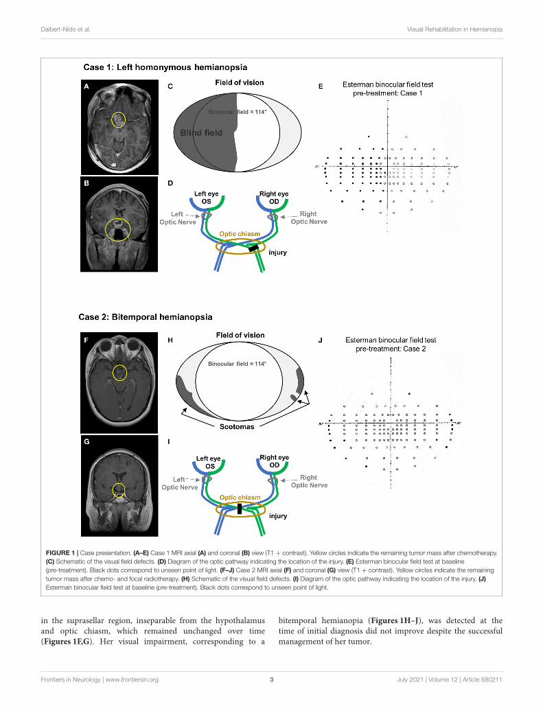
TABLE 1 | Summary of the episode of care.
Case 1
Timeline/age 8 years old (1999) 16 years old (2007) 29 years old (2020)
Episode of care Grade glioma in brain, cerebellum, and
spinal cord
- Chemotherapy
- Diagnosis of left homonymous hemianopsia
- Tumor progression in the optic chiasm
- Left homonymous hemianopsia worsening
- No improvement of the left hemianopia
- 7-week home-based visual rehabilitation program
Case 2
Timeline/age 13 years old (2001) 32 years old (2020)
Episode of care Diagnosis of suprasellar non-germinatous germ cell tumor
- Chemotherapy and focal radiotherapy
- Diagnosis of bitemporal hemianopia and left exotropia
- No improvement of bitemporal hemianopia
- 6-week home-based visual rehabilitation program
AssessmentsVisual assessments investigating both visual function andfunctional vision were performed at the Ophthalmology Lowvision Clinic at the TorontoWestern Hospital (University HealthNetwork, Toronto, Canada) following standard procedures inlow-vision rehabilitation (20–23). These assessments includevisual acuity [best corrected visual acuity (BCVA)] measuredusing the Early Treatment Diabetic Retinopathy Study (ETDRS)charts at 4m and CS measured using the Functional AcuityContrast Test (FACT). Retinal sensitivity (RS) and fixationstability [FS; bivariate contour ellipse area (BCEA), 63%] weremeasured using the Macular Integrity Assessment (MAIA)microperimeter (CentreVue, Padova, Italy). Field of visionwas evaluated by the monocular Humphrey full field andEsterman binocular field of vision, which were assessed using theHumphrey Full Field Analyzer 3 standard automated perimeter(HFA 3, Zeiss, Heilberg, Germany). Quality of life was measuredin two subdomains (orientation and mobility—reading) usingthe Veteran’s affairs low-vision visual functioning questionnaire(LV-VFQ) (24). Reading speed was measured at critical printsize using the Minnesota Low Vision Reading (MNREAD) test(25). Results were analyzed using the coefficient of repeatability(COR), specific to each assessment, to compare pre- and post-treatment data (26). The COR, also referred as the smallestreal difference (SRD), quantifies an absolute reliability in thesame unit as the assessment tool. The COR is directly relatedto the 95% limits of agreement proposed by Bland and Altman(27). It corresponds to the value below which the absolutedifferences between two measurements would lie within 95%of probability. Therefore, measurement values strictly differentfrom COR indicate an effect of the treatment (26, 28).
DIAGNOSTIC ASSESSMENT
TreatmentThe patients followed an IVR audiovisual stimulation protocolat home using the stand-alone and remotely controlled HMDOculus Go for 7 weeks (case 1) or 6 weeks (case 2). Every2 days, the patient performed one session of IVR audiovisualstimulation composed of three blocks of 15 trials of 20 s,equivalent to 3 × 5min of continuous audiovisual stimulation.
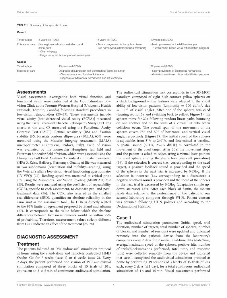
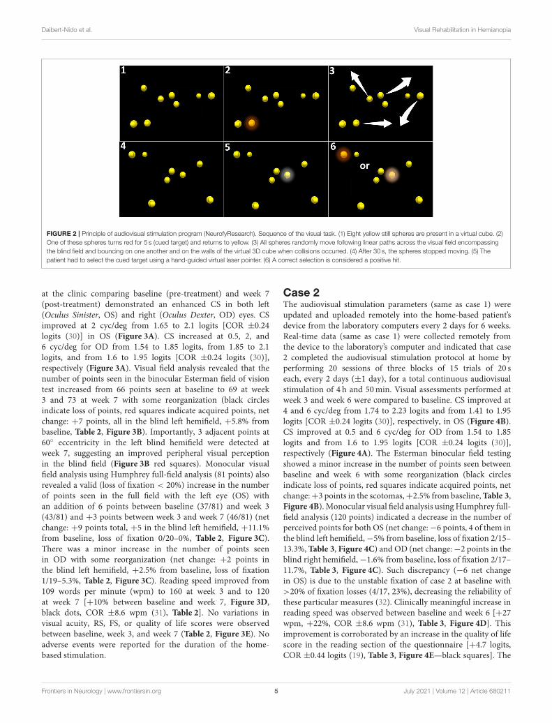
The audiovisual stimulation task corresponds to the 3D-MOTparadigm composed of eight high-contrast yellow spheres ona black background whose features were adapted to the visualability of low-vision patients (luminosity = 100 cd/m2, size= 1.57◦ of visual angle). After one of the spheres was cued(turning red for 5 s and switching back to yellow, Figure 2), thespheres move for 20 s following random linear paths, bouncingon one another and on the walls of a virtual 3D cube whencollisions occur. The overall span of the movement of thespheres covers 78◦ and 50◦ of horizontal and vertical visualangle, respectively (Figure 2). The initial speed of the spheresis adjustable, from 3◦/s to 240◦/s. and determined at baseline.A spatial sound (50Hz, 25–65 dBHL) is correlated to themovement of the cued target. After 20 s, the movement stopsand the patient is asked to select, using a virtual laser pointer,the cued sphere among the distractors (mark-all procedure)(16). If the selection is correct (i.e., corresponding to the cuedtarget), a positive feedback sound is provided and the speedof the spheres in the next trial is increased by 0.05log. If theselection is incorrect (i.e., corresponding to a distractor), anegative feedback sound is provided and the speed of the spheresin the next trial is decreased by 0.05log (adaptative simple up-down staircase) (29). After each block of 5min, the systemsends data relative to the performance of the patient to oursecured laboratory computer through Wi-Fi. Patient consentwas obtained following UHN policies and according to theDeclaration of Helsinki.
Case 1The audiovisual stimulation parameters (initial speed, trialduration, number of targets, total number of spheres, numberof blocks, and number of sessions) were updated and uploadedremotely into the patient’s device from the laboratory’scomputers every 2 days for 7 weeks. Real-time data (date/time,average/maximum speed of the spheres, positive hits, numberof trials/blocks/sessions performed, total time, and responsetime) were collected remotely from the device and indicatedthat case 1 completed the audiovisual stimulation protocol athome by performing 19 sessions of 3 blocks of 15 trials of 20 seach, every 2 days (±1 day), for a total continuous audiovisualstimulation of 4 h and 45min. Visual assessments performed
Frontiers in Neurology | www.frontiersin.org 4 July 2021 | Volume 12 | Article 680211
Daibert-Nido et al. Visual Rehabilitation in Hemianopia
FIGURE 2 | Principle of audiovisual stimulation program (NeurofyResearch). Sequence of the visual task. (1) Eight yellow still spheres are present in a virtual cube. (2)
One of these spheres turns red for 5 s (cued target) and returns to yellow. (3) All spheres randomly move following linear paths across the visual field encompassing
the blind field and bouncing on one another and on the walls of the virtual 3D cube when collisions occurred. (4) After 30 s, the spheres stopped moving. (5) The
patient had to select the cued target using a hand-guided virtual laser pointer. (6) A correct selection is considered a positive hit.
at the clinic comparing baseline (pre-treatment) and week 7(post-treatment) demonstrated an enhanced CS in both left(Oculus Sinister, OS) and right (Oculus Dexter, OD) eyes. CSimproved at 2 cyc/deg from 1.65 to 2.1 logits [COR ±0.24logits (30)] in OS (Figure 3A). CS increased at 0.5, 2, and6 cyc/deg for OD from 1.54 to 1.85 logits, from 1.85 to 2.1logits, and from 1.6 to 1.95 logits [COR ±0.24 logits (30)],respectively (Figure 3A). Visual field analysis revealed that thenumber of points seen in the binocular Esterman field of visiontest increased from 66 points seen at baseline to 69 at week3 and 73 at week 7 with some reorganization (black circlesindicate loss of points, red squares indicate acquired points, netchange: +7 points, all in the blind left hemifield, +5.8% frombaseline, Table 2, Figure 3B). Importantly, 3 adjacent points at60◦ eccentricity in the left blind hemifield were detected atweek 7, suggesting an improved peripheral visual perceptionin the blind field (Figure 3B red squares). Monocular visualfield analysis using Humphrey full-field analysis (81 points) alsorevealed a valid (loss of fixation < 20%) increase in the numberof points seen in the full field with the left eye (OS) withan addition of 6 points between baseline (37/81) and week 3(43/81) and +3 points between week 3 and week 7 (46/81) (netchange: +9 points total, +5 in the blind left hemifield, +11.1%from baseline, loss of fixation 0/20–0%, Table 2, Figure 3C).There was a minor increase in the number of points seenin OD with some reorganization (net change: +2 points inthe blind left hemifield, +2.5% from baseline, loss of fixation1/19–5.3%, Table 2, Figure 3C). Reading speed improved from109 words per minute (wpm) to 160 at week 3 and to 120at week 7 [+10% between baseline and week 7, Figure 3D,black dots, COR ±8.6 wpm (31), Table 2]. No variations invisual acuity, RS, FS, or quality of life scores were observedbetween baseline, week 3, and week 7 (Table 2, Figure 3E). Noadverse events were reported for the duration of the home-based stimulation.
Case 2The audiovisual stimulation parameters (same as case 1) wereupdated and uploaded remotely into the home-based patient’sdevice from the laboratory computers every 2 days for 6 weeks.Real-time data (same as case 1) were collected remotely fromthe device to the laboratory’s computer and indicated that case2 completed the audiovisual stimulation protocol at home byperforming 20 sessions of three blocks of 15 trials of 20 seach, every 2 days (±1 day), for a total continuous audiovisualstimulation of 4 h and 50min. Visual assessments performed atweek 3 and week 6 were compared to baseline. CS improved at4 and 6 cyc/deg from 1.74 to 2.23 logits and from 1.41 to 1.95logits [COR ±0.24 logits (30)], respectively, in OS (Figure 4B).CS improved at 0.5 and 6 cyc/deg for OD from 1.54 to 1.85logits and from 1.6 to 1.95 logits [COR ±0.24 logits (30)],respectively (Figure 4A). The Esterman binocular field testingshowed a minor increase in the number of points seen betweenbaseline and week 6 with some reorganization (black circlesindicate loss of points, red squares indicate acquired points, netchange:+3 points in the scotomas,+2.5% from baseline,Table 3,Figure 4B). Monocular visual field analysis using Humphrey full-field analysis (120 points) indicated a decrease in the number ofperceived points for both OS (net change:−6 points, 4 of them inthe blind left hemifield,−5% from baseline, loss of fixation 2/15–13.3%, Table 3, Figure 4C) and OD (net change:−2 points in theblind right hemifield,−1.6% from baseline, loss of fixation 2/17–11.7%, Table 3, Figure 4C). Such discrepancy (−6 net changein OS) is due to the unstable fixation of case 2 at baseline with>20% of fixation losses (4/17, 23%), decreasing the reliability ofthese particular measures (32). Clinically meaningful increase inreading speed was observed between baseline and week 6 [+27wpm, +22%, COR ±8.6 wpm (31), Table 3, Figure 4D]. Thisimprovement is corroborated by an increase in the quality of lifescore in the reading section of the questionnaire [+4.7 logits,COR ±0.44 logits (19), Table 3, Figure 4E—black squares]. The
Frontiers in Neurology | www.frontiersin.org 5 July 2021 | Volume 12 | Article 680211

Daibert-Nido et al. Visual Rehabilitation in Hemianopia
FIGURE 3 | Case 1—Visual outcome measures. (A) Contrast sensitivity at baseline (gray line) and at the end of the treatment (week 7—black line) in the left (OS) and
the right (OD) eye. (B) Esterman binocular field test post-treatment (week 7). Red squares indicate newly acquired points, black circles indicate lost points, compared
to baseline. (C) Monocular Humphrey full-field test (81 points) post-treatment (week 7) in the left (OS) and right (OD) eye. Red squares indicate newly acquired points,
and black circles indicate lost points, compared to baseline. (D) Reading speed at baseline, week 3, and post-treatment (week 7). (E) Quality of life scores at baseline,
week 3, and post-treatment (week 7). Gray shaded areas in (A,D,E) indicate the values of the coefficient of repeatability (±COR). OD, oculus dexter; OS, oculus
sinister; W, week.
mobility section of the quality of life questionnaire showedonly minor improvement [+0.62 logits, COR ±0.44 logits (22),Table 3, Figure 4E—black triangles]. No improvements in visualacuity, RS, and FS were observed between all time points(Table 3). No adverse events were reported for the duration thehome-based stimulation program [VRISE questionnaire score =33/35, >25 the threshold for cybersickness (33)].
DISCUSSION
This case series shows the feasibility of a home-basedvisual rehabilitation program using an audiovisual 3D-MOTstimulation paradigm in IVR HMD and potential effectivenesson visual perception in hemianopia patients. We were able toimplement the program and update the stimulation procedures
Frontiers in Neurology | www.frontiersin.org 6 July 2021 | Volume 12 | Article 680211
Daibert-Nido et al. Visual Rehabilitation in Hemianopia
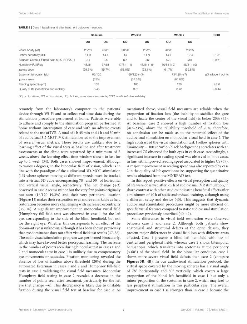
TABLE 2 | Case 1 baseline and after treatment outcome measures.
Baseline Week 3 Week 7 COR
OD OS OD OS OD OS
Visual Acuity (VA) 20/20 20/25 20/20 20/25 20/20 20/25
Retinal sensitivity (dB) 14.3 14.4 14 11.8 14.7 12.4 ±1.51
Bivariate Contour Ellipse Area 63% (BCEA, 2) 0.4 0.6 0.5 0.5 0.3 0.5 ±0.61
Humphrey Full Field 48/81 37/81 47/81 (−1) 43/81 (+6) 50/81 (+2) 46/81 (+9)
(points seen) (59.3%) (45.7%) (58.0%) (53.1%) (61.7%) (56.8%)
Esterman binocular field 66/120 69/120 (+3) 73/120 (+7) ±3 adjacent points
(points seen) (55%) (57.5%) (60.8%)
Reading speed (wpm) 109 160 120 ±8.6
Quality of life (orientation and mobility) 3.48 3.01 3.48 ±0.44
OD, oculus dexter; OS, oculus sinister; dB, decibels; wpm, words per minute; COR, coefficient of repeatability.
remotely from the laboratory’s computer to the patients’device through Wi-Fi and to collect real-time data during thestimulation procedure performed at home. Patients were ableto adhere and comply to the stimulation program performed athome without interruption of care and with no adverse eventsrelated to the use of IVR. A total of 4 h 45min and 4 h and 50minof audiovisual 3D-MOT IVR stimulation led to the improvementof several visual metrics. These results are unlikely due to alearning effect of the visual tests as baseline and after treatmentassessments at the clinic were separated by a minimum of 3weeks, above the learning effect time window shown to last forup to 1 week (34). Both cases showed improvement, althoughto various degrees, in the binocular field of vision, which is inline with the paradigm of the audiovisual 3D-MOT stimulation(15) where spheres moving at different speeds must be trackedinto a virtual 3D cube encompassing 78◦ and 50◦ of horizontaland vertical visual angle, respectively. The net change (+3)observed in case 2 seems minor but the very few points originallynot seen (16/120–19.3%) and their very peripheral location(Figure 1J) makes their restoration evenmore remarkable as fieldrestoration becomesmore challenging with increased eccentricity(35, 36). A significant improvement in monocular visual field(Humphrey full-field test) was observed in case 1 for the lefteye, corresponding to the side of the blind hemifield, but notfor the right eye. Whether this corresponds to an effect of thedominant eye is unknown, although it has been shown previouslythat eye dominance does not affect visual field test results (37, 38).The audiovisual stimulation programwas performed binocularly,which may have favored better perceptual learning. The increasein the number of points seen during binocular test in cases 1 and2 and monocular test in case 1 is unlikely due to compensatoryeye movements or saccades. Fixation monitoring revealed theabsence of loss of fixation above threshold (20%) during theautomated Esterman in cases 1 and 2 and Humphrey full-fieldtests in case 1 validating the visual field measures. MonocularHumphrey field testing in case 2 revealed a decrease in thenumber of points seen after treatment, particularly for the lefteye (net change −6). This discrepancy is likely due to unstablefixation during the visual field test at baseline for case 2. As
mentioned above, visual field measures are reliable when theproportion of fixation loss (the inability to stabilize the gazeand to fixate the center of the visual field) is below 20% (32).At baseline, case 2 showed a high number of fixation loss(4/7–23%), above the reliability threshold of 20%; therefore,no conclusion can be made as to the potential effect of theaudiovisual stimulation on monocular visual field in case 2. Thehigh contrast of the visual stimulation task (yellow spheres withluminosity= 100 cd/m2 on black background) correlates with anincreased CS observed for both eyes in each case. Accordingly, asignificant increase in reading speed was observed in both cases,in line with improved reading speed associated to higher CS (39).A major improvement in reading speed was also reported by case2 in the quality-of-life questionnaire, supporting the quantitativeresults obtained from the MNREAD test.
In this report, positive effects on visual perception and qualityof life were observed after<5 h of audiovisual IVR stimulation, insharp contrast with other studies indicating beneficial effects aftera minimum of 40 h of static audiovisual training, although usinga different setup and device (40). This suggests that dynamicaudiovisual stimulation procedures might be more efficient onspecific visual features compared to static audiovisual stimulationprocedures previously described (40–42).
Some differences in visual field restoration were observedbetween case 1 and case 2. Although both patients showanatomical and structural defects at the optic chiasm, theypresent major differences in visual field loss with different areasaffected. Case 1 presents a blind left hemifield with loss ofcentral and peripheral fields whereas case 2 shows bitemporalhemianopia, which translates into scotomas at the periphery(>60◦) of the visual field. In the binocular condition, case 1shows more severe visual field defects than case 2 (compareFigures 3B, 4B). In our audiovisual stimulation protocol, thevirtual space covered by the moving spheres has a visual angleof 78◦ horizontally and 50◦ vertically, which covers a largeproportion of the blind left hemifield in case 1 but only aminor proportion of the scotomas in case 2, which may lead toless peripheral stimulation in this particular case. The overallimprovement in case 1 is stronger than in case 2 because the
Frontiers in Neurology | www.frontiersin.org 7 July 2021 | Volume 12 | Article 680211

Daibert-Nido et al. Visual Rehabilitation in Hemianopia
FIGURE 4 | Case 2—Visual outcome measures and quality of life. (A) Contrast sensitivity at baseline (gray line) and at the end of the treatment (week 6—black line) in
the left (OS) and the right (OD) eye. (B) Esterman binocular field test post-treatment (week 6). Red squares indicate newly acquired points, and black circles indicate
lost points, compared to baseline. (C) Monocular Humphrey full-field test (120 points) post-treatment (week 6) in the left (OS) and right (OD) eye. Red squares indicate
newly acquired points, and black circles indicate lost points, compared to baseline. (D) Reading speeds at baseline, week 3, and post-treatment (week 6). (E) Quality
of life scores at baseline, week 3, and post-treatment (week 6). Gray shaded areas in (A,D,E) indicate the values of the coefficient of repeatability (±COR). OD, oculus
dexter; OS, oculus sinister; W, week.
original binocular field defects were also more pronounced incase 1.
Visual field restoration in hemianopia patients has been acontroversial topic (10, 43–46). Several studies suggest thatvisual fields can be restored in hemianopia by enhanced
oculomotor function and compensatory eye movement (40, 41,47). Others suggest that a restoration of visual perception inthe blind hemifield could be the consequence of a functionalreorganization of the connectivity in subcortical and corticalstructures after visual rehabilitation (11, 14). Moreover, recent
Frontiers in Neurology | www.frontiersin.org 8 July 2021 | Volume 12 | Article 680211

Daibert-Nido et al. Visual Rehabilitation in Hemianopia
TABLE 3 | Case 2 baseline and after treatment outcome measures.
Baseline Week 3 Week 6 COR
OD OS OD OS OD OS
Visual Acuity (VA) 20/20 20/40 20/20 20/40 20/20 20/40
Retinal sensitivity (dB) 16.2 13.5 14.9 15.2 14 13.9 ±1.51
Bivariate Contour Ellipse Area 63% (BCEA, 2) 0.5 2.4 0.2 2.4 0.4 2 ±0.61
Humphrey Full Field (points seen) 83/120
(69.2%)
92/120
(76.7%)
80/120 (−3)
(66.7%)
91/120 (−1)
(75.8%)
81/120 (−2)
(67.5%)
86/120 (−6)
(71.7%)
Esterman binocular field 104/120 103/120 (−1) 107/120 (+3) ±3 adjacent points
(points seen) (86.7%) (85.8%) (89.2%)
Reading speed (wpm) 123 131 150 ±8.6
Quality of life (reading) 8.3 13 13 ±0.44
Quality of life (orientation and mobility) 2.58 3.01 3.2 ±0.44
OD, oculus dexter; OS, oculus sinister; dB, decibels; wpm, words per minute; COR, coefficient of repeatability.
evidence indicates that audiovisual stimulation, during whichauditory and visual stimuli are spatially and temporallycorrelated, improve visual perception more efficiently thanvisual-only stimulations (11, 45, 48). The exact mechanismsof visual field restoration are still largely unknown. Visualfield restoration following a dynamic audiovisual stimulationmay involve both neuronal plasticity in subcortical and corticalvisual brain areas (11, 14, 49, 50) and oculomotor enhancementand compensatory eye movement (8, 47) in a non-mutuallyexclusive manner. In our protocol, the patients track ahigh-contrast, sound-generating, cued target using vision andaudition. This tracking requires eye movement control andaudiovisual processing, whichmay help to recalibrate audiovisualperception through neuroplasticity and, concomitantly, tostimulate oculomotor control. When traveling through the blindfield or scotomas, the cued target can still be tracked usingcorrelated spatial sound, reinforcing the audiovisual association.Considering that hemianopia patients often present impairedsound localization (7), such audiovisual stimulation programmay also improve spatial sound localization. Moreover, repeatedexposure to identical stimuli in an identical environmentmaximizes perceptual learning (51).
Our study shows limitations as to the extent of the visualfield stimulated (78◦ horizontal and 50◦ vertical), which shouldbe increased to approach the normal extent of the visualfield (200◦ horizontal and 130◦ vertical) to further stimulateperipheral vision. An increase of the duration of the stimulationprogram (>7 weeks) with more sessions per day (2 sessions of15min separated by a few hours of rest to avoid VR inducedsymptoms) should be developed. Positive effects of the treatmentwere observed mostly in binocular vision (Esterman binocularfield test) with less effect on monocular vision (Humphreyfull-field test). This can be explained by the improved FS,which occurs under binocular condition (35) allowing morepoints to be seen with the Esterman binocular field test. Ouraudiovisual stimulation protocol is binocular, which very likelyimpacts binocular vision. Amonocular version of our audiovisualstimulation procedure, tailored to the ability of each eye
individually, should be tested. When visual field measurementsare deemed not reliable, they should be repeated within a shorttime window (<24 h) to avoid unreliable data and to allowthe patient to rest. The use of a built-in eye-tracking systemin the device could provide valuable information as to thevisual tracking strategy used by patients and would allow theelaboration of more powerful audiovisual stimulation programs.
IVR in HMD is an emerging and very promising visualrehabilitation approach using high-technology devices (52–56).It is developed to provide perceptual learning with betterecological validity due to virtual reality, greater flexibility dueto home-based stimulation protocol, and improved efficiencydue to patient-tailored protocols (52, 53). Our dynamic IVRaudiovisual stimulation procedure based on the 3D-MOTparadigm mimics real-life, complex dynamical situations whereindividuals apprehend their environment using both visual andauditory information (16).
Although no conclusion related to effectiveness of ourtreatment can be made from this report, our results supportthe feasibility of a larger RCT and suggest that patients withlong-term visual field defects are still be able to restore somevisual perception, in line with more recent work indicatingthat adaptation and plasticity can still occur at later stages inadults (57). This case series started just before the first stay-at-home order in Ontario and continued throughout the COVID-19 pandemic without interruption of care of patient-tailoredtreatment, further supporting the relevancy of home-based,remotely controlled, rehabilitation procedures. Future pragmaticrandomized controlled studies will address the effectiveness ofaudiovisual IVR rehabilitation procedure in adults and earlierduring the disease in children when neuronal plasticity ismore effective. Integrated knowledge translation and a patient-centered approach will be included in further RCTs usingpatients’ and caregivers/partners’ open-ended questionnaires.An estimation of long-term effectiveness of IVR audiovisualstimulation must also be addressed. A home-based visualrehabilitation procedure is a promising approach not only todecrease the burden of disease by substantially diminishing
Frontiers in Neurology | www.frontiersin.org 9 July 2021 | Volume 12 | Article 680211
Daibert-Nido et al. Visual Rehabilitation in Hemianopia
the number of visits to the clinic and therefore relievingthe constraints of commute and transportation, but also forpopulations living remotely and often left without easy access tomodern medicine and treatments.
PATIENT PERSPECTIVE
Case 1At the age of eight, I was diagnosed with brain and spine tumors.The primary site was on the optic chiasm, where the optic nervesconnect to the brain. I underwent 2 years of chemotherapy,followed by another year when I was 19. The location of thetumors caused my vision to be impacted. For as long as I canremember, I have lived with a deficit in my left peripheral vision.This is something I was always told could never be fixed, thatI would have to live with it for my entire life, and just haveto learn to compensate for the deficit. So, I did, looking tomy left more often than my right, though this did not alwaysprevent me from bumping into objects or people while walking.I thought this was my new normal, and I would live the rest ofmy life with the left-side deficit. You can imagine my surprisewhen one of my doctors told me about the work being doneby Dr. Reber. I had doubts anything could work but gave it atry anyway. I was told to use an oculus VR headset every otherday, with a specialized program to assist my peripheral vision.To my surprise and excitement, after 3 months of doing thisregimen, I could feel the positive effects from the VR headset. Iam now much more confident walking around outside, bumpinginto things less, and can certainly notice the improvement in myleft peripheral vision. I am extremely thankful for everything Dr.Reber has done to improve my quality of life, especially afterbeing told this is something that could not be fixed. I am alsograteful to donors such as yourselves who help to make thisresearch possible. My life has been changed for the better asa direct result of Dr. Reber’s work and will forever be gratefulfor that.
DATA AVAILABILITY STATEMENT
The original contributions presented in the study are includedin the article/supplementary material, further inquiries can bedirected to the corresponding author/s.
ETHICS STATEMENT
Ethical review and approval was not required for the study onhuman participants in accordance with the local legislation andinstitutional requirements. The patients/participants providedtheir written informed consent to participate in this study.Written informed consent was obtained from the individual(s)for the publication of any potentially identifiable images or dataincluded in this article.
AUTHOR CONTRIBUTIONS
MD-N, SM, EB, andMR designed the protocol, analyzed the data,interpreted the results, and wrote/edited the manuscript. MD-N,YP, and SM performed the assessments and generated the data.KC managed the remote control of the device. KC, CN, and MRextracted the data. All authors attest that they meet the currentICMJE criteria for Authorship.
FUNDING
This work was funded by the start-up grant, Donald K. JohnsonEye Institute, Krembil Research Institute, University HealthNetwork (MR).
ACKNOWLEDGMENTS
We thank the Toronto Western and General HospitalFoundation for financial support (MR). We thankNeuropowertrain for their collaboration.
REFERENCES
1. Peragallo JH. Visual function in children with primary brain tumors. Curr
Opin Neurol. (2019) 32:75–81. doi: 10.1097/WCO.0000000000000644
2. Peragallo JH. Effects of brain tumors on vision in children. Int Ophthalmol
Clin. (2018) 58:83–95. doi: 10.1097/IIO.0000000000000237
3. Fried I, Tabori U, Tihan T, Reginald A, Bouffet E. Optic pathway gliomas: a
review. CNS Oncol. (2013) 2:143–59. doi: 10.2217/cns.12.47
4. Wan MJ, Zapotocky M, Bouffet E, Bartels U, Kulkarni AV, Drake JM. Long-
term visual outcomes of craniopharyngioma in children. J Neurooncol. (2018)
137:645–51. doi: 10.1007/s11060-018-2762-3
5. Jariyakosol S, Peragallo JH. The effects of primary brain tumors on vision
and quality of life in pediatric patients. Semin Neurol. (2015) 35:587–98.
doi: 10.1055/s-0035-1563571
6. Zihl J. Visual scanning behavior in patients with homonymous hemianopia.
Neuropsychologia. (1995) 33:287–303. doi: 10.1016/0028-3932(94)00119-A
7. Lewald J, Kentridge RW, Peters S, TegenthoffM, Heywood CA, HausmannM.
Auditory-visual localization in hemianopia. Neuropsychology. (2013) 27:573–
82. doi: 10.1037/a0033451
8. Trauzettel-Klosinski S. Current methods of visual rehabilitation.Dtsch Arztebl
Int. (2011) 108:871–8. doi: 10.3238/arztebl.2011.0871
9. Goodwin D. Homonymous hemianopia: challenges and solutions. Clin
Ophthalmol. (2014) 8:1919–27. doi: 10.2147/OPTH.S59452
10. Sabel BA, Henrich-Noack P, Fedorov A, Gall C. Vision restoration
after brain and retina damage: the “residual vision activation theory.”
Prog Brain Res. (2011) 192:199–262. doi: 10.1016/B978-0-444-53355-5.
00013-0
11. Grasso PA, Gallina J, Bertini C. Shaping the visual system: cortical
and subcortical plasticity in the intact and the lesioned brain.
Neuropsychologia. (2020) 142:107464. doi: 10.1016/j.neuropsychologia.2020.
107464
12. Meredith MA, Stein BE. Interactions among converging sensory inputs in the
superior colliculus. Science. (1983) 221:389–91. doi: 10.1126/science.6867718
13. Basso MA, May PJ. Circuits for action and cognition: a view
from the superior colliculus. Annu Rev Vis Sci. (2017) 3:197–226.
doi: 10.1146/annurev-vision-102016-061234
14. Ajina S, Bridge H. Subcortical pathways to extrastriate visual cortex underlie
residual vision following bilateral damage to V1. Neuropsychologia. (2019)
128:140–9. doi: 10.1016/j.neuropsychologia.2018.01.007
15. Pylyshyn ZW, Annan V. Dynamics of target selection in
multiple object tracking (MOT). Spat Vis. (2006) 19:485–504.
doi: 10.1163/156856806779194017
Frontiers in Neurology | www.frontiersin.org 10 July 2021 | Volume 12 | Article 680211

Daibert-Nido et al. Visual Rehabilitation in Hemianopia
16. Meyerhoff HS, Papenmeier F, Huff M. Studying visual attention using the
multiple object tracking paradigm: a tutorial review.Atten Percept Psychophys.
(2017) 79:1255–74. doi: 10.3758/s13414-017-1338-1
17. Romeas T, Guldner A, Faubert J. 3D-Multiple Object Tracking training task
improves passing decision-making accuracy in soccer players. Psychol Sport
Exer. (2016) 22:1–9. doi: 10.1016/j.psychsport.2015.06.002
18. Legault I, Faubert J. Perceptual-cognitive training improves biological
motion perception: evidence for transferability of training in healthy aging.
Neuroreport. (2012) 23:469–73. doi: 10.1097/WNR.0b013e328353e48a
19. Corbin-Berrigan L-A, Kowalski K, Faubert J, Christie B, Gagnon I.
Three-dimensional multiple object tracking in the pediatric population:
the NeuroTracker and its promising role in the management
of mild traumatic brain injury. Neuroreport. (2018) 29:559–63.
doi: 10.1097/WNR.0000000000000988
20. Markowitz SN. Principles of modern low vision rehabilitation. Can J
Ophthalmol. (2006) 41:289–312. doi: 10.1139/I06-027
21. Markowitz SN. A practice template for low-vision rehabilitation. Can J
Ophthalmol. (2009) 44:610. doi: 10.3129/i09-099
22. Markowitz SN. State-of-the-art: low vision rehabilitation. Can J Ophthalmol.
(2016) 51:59–66. doi: 10.1016/j.jcjo.2015.11.002
23. Leat S. 2020 CAO clinical practice guideline: optometric low vision
rehabilitation full guidelines. Can J Optometry. (2020) 82:19–62.
doi: 10.15353/cjo.v82i1.1636
24. Stelmack JA, Szlyk JP, Stelmack TR, Demers-Turco P, Williams RT, Moran D,
et al. Measuring outcomes of vision rehabilitation with the Veterans Affairs
Low Vision Visual Functioning Questionnaire. Invest Ophthalmol Vis Sci.
(2006) 47:3253–61. doi: 10.1167/iovs.05-1319
25. Legge GE, Ross JA, Luebker A, LaMay JM. Psychophysics of reading. VIII.
The Minnesota Low-Vision Reading Test. Optom Vis Sci. (1989) 66:843–53.
doi: 10.1097/00006324-198912000-00008
26. Vaz S, Falkmer T, Passmore AE, Parsons R, Andreou P. The case for using
the repeatability coefficient when calculating test-retest reliability. PLoS ONE.
(2013) 8:e73990. doi: 10.1371/journal.pone.0073990
27. Bland JM, Altman DG. Applying the right statistics: analyses of measurement
studies. Ultrasound Obstetr Gynecol. (2003) 22:85–93. doi: 10.1002/uog.122
28. Beckerman H, Roebroeck ME, Lankhorst GJ, Becher JG, Bezemer
PD, Verbeek ALM. Smallest real difference, a link between
reproducibility and responsiveness. Qual Life Res. (2001) 10:571–8.
doi: 10.1023/A:1013138911638
29. Levitt H. Transformed up-down methods in psychoacoustics. J Acoust Soc
Am. (1971) 49(Suppl. 2):467. doi: 10.1121/1.1912375
30. Kara S, Gencer B, Ersan I, Arikan S, Kocabiyik O, Tufan HA, et al.
Repeatability of contrast sensitivity testing in patients with age-related
macular degeneration, glaucoma, and cataract. Arquivos Brasil Oftalmol.
(2016) 79:323–7. doi: 10.5935/0004-2749.20160092
31. Subramanian A, Pardhan S. The repeatability of MNREAD acuity charts
and variability at different test distances. Optom Vis Sci. (2006) 83:572–6.
doi: 10.1097/01.opx.0000232225.00311.53
32. Sanabria O, Feuer WJ, Anderson DR. Pseudo-loss of fixation
in automated perimetry. Ophthalmology. (1991) 98:76–8.
doi: 10.1016/S0161-6420(91)32338-8
33. Kourtesis P, Collina S, Doumas LAA,MacPherson SE. Validation of the virtual
reality neuroscience questionnaire: maximum duration of immersive virtual
reality sessions without the presence of pertinent adverse symptomatology.
Front Hum Neurosci. (2019) 13:417. doi: 10.3389/fnhum.2019.00417
34. Acton JH, Bartlett NS, Greenstein VC. Comparing the Nidek MP-1 and
humphrey field analyzer in normal subjects.OptomVis Sci. (2011) 88:1288–97.
doi: 10.1097/OPX.0b013e31822b3746
35. Raveendran RN, BobierWR, Thompson B. Binocular vision and fixational eye
movements. J Vis. (2019) 19:9. doi: 10.1167/19.4.9
36. Tarita-Nistor L, Brent MH, Steinbach MJ, González EG. Fixation stability
during binocular viewing in patients with age-related macular degeneration.
Invest Ophthalmol Vis Sci. (2011) 52:1887–93. doi: 10.1167/iovs.10-6059
37. Spry PGD, Furber JE, Harrad RA. The effect of ocular
dominance on visual field testing. Optom Vis Sci. (2002) 79:93–7.
doi: 10.1097/00006324-200202000-00010
38. Shneor E, Hochstein S. Effects of eye dominance in visual perception. Int
Congress Ser. (2005) 1282:719–23. doi: 10.1016/j.ics.2005.05.006
39. Pearce E, Sivaprasad S, Chong NV. Factors affecting reading speed in patients
with diabetic macular edema treated with laser photocoagulation. PLoS ONE.
(2014) 9:e0105696. doi: 10.1371/journal.pone
40. Bolognini N, Rasi F, Coccia M, Làdavas E. Visual search improvement
in hemianopic patients after audio-visual stimulation. Brain. (2005) 128(Pt
12):2830–42. doi: 10.1093/brain/awh656
41. Passamonti C, Bertini C, Làdavas E. Audio-visual stimulation improves
oculomotor patterns in patients with hemianopia. Neuropsychologia. (2009)
47:546–55. doi: 10.1016/j.neuropsychologia.2008.10.008
42. Frassinetti F, Bolognini N, Bottari D, Bonora A, Làdavas E. Audiovisual
integration in patients with visual deficit. J Cogn Neurosci. (2005) 17:1442–52.
doi: 10.1162/0898929054985446
43. Horton JC. Disappointing results from Nova Vision’s visual restoration
therapy. Br J Ophthalmol. (2005) 89:1–2. doi: 10.1136/bjo.2004.058214
44. Plant GT. A work out for hemianopia. Br J Ophthalmol. (2005) 89:2.
doi: 10.1136/bjo.2004.053173
45. Frolov A, Feuerstein J, Subramanian PS. Homonymous Hemianopia
and Vision Restoration Therapy. Neurol Clin. (2017) 35:29–43.
doi: 10.1016/j.ncl.2016.08.010
46. Reinhard J, Schreiber A, Schiefer U, Kasten E, Sabel BA, Kenkel S, et
al. Does visual restitution training change absolute homonymous visual
field defects? A fundus controlled study. Br J Ophthalmol. (2005) 89:30–5.
doi: 10.1136/bjo.2003.040543
47. Pambakian A, Currie J, Kennard C. Rehabilitation strategies for patients
with homonymous visual field defects. J Neuroophthalmol. (2005) 25:136–42.
doi: 10.1097/01.WNO.0000161661.29391.0D
48. Shams L, Seitz AR. Benefits of multisensory learning. Trends Cogn Sci. (2008)
12:411–7. doi: 10.1016/j.tics.2008.07.006
49. Huxlin KR. Perceptual plasticity in damaged adult visual systems. Vision Res.
(2008) 48:2154–66. doi: 10.1016/j.visres.2008.05.022
50. Tamietto M, Morrone MC. Visual plasticity: blindsight bridges anatomy
and function in the visual system. Curr Biol. (2016) 26:R70–3.
doi: 10.1016/j.cub.2015.11.026
51. Sagi D. Perceptual learning in vision research. Vision Res. (2011) 51:1552–66.
doi: 10.1016/j.visres.2010.10.019
52. Huygelier H, Schraepen B, van Ee R, Vanden Abeele V, Gillebert CR.
Acceptance of immersive head-mounted virtual reality in older adults. Sci Rep.
(2019) 9:1–12. doi: 10.1038/s41598-019-41200-6
53. Huygelier H, Schraepen B, Lafosse C, Vaes N, Schillebeeckx F, Michiels K, et
al. An immersive virtual reality game to train spatial attention orientation
after stroke: a feasibility study. Appl Neuropsychol Adult. (2020) 27:1–21.
doi: 10.1080/23279095.2020.1821030
54. Nesaratnam N, Thomas P, Vivian A. Stepping into the virtual unknown:
feasibility study of a virtual reality-based test of ocular misalignment. Eye.
(2017) 31:1503–6. doi: 10.1038/eye.2017.97
55. Yasuda K, Muroi D, Hirano M, Saichi K, Iwata H. Differing effects of
an immersive virtual reality programme on unilateral spatial neglect on
activities of daily living. Case Reports. (2018) 2018:1–5. doi: 10.1136/bcr-2017-
222860
56. Kim W-S, Paik N-J, Cho S. Development and validation of
virtual prism adaptation therapy. In: 2017 International Conference
on Virtual Rehabilitation (ICVR). Montreal, QC (2017). p. 1–2.
doi: 10.1109/ICVR.2017.8007471
57. Legge GE, Chung STL. Low vision and plasticity: implications
for rehabilitation. Annu Rev Vis Sci. (2016) 2:321–43.
doi: 10.1146/annurev-vision-111815-114344
Conflict of Interest: The authors declare that the research was conducted in the
absence of any commercial or financial relationships that could be construed as a
potential conflict of interest.
Copyright © 2021 Daibert-Nido, Pyatova, Cheung, Nayomi, Markowitz, Bouffet and
Reber. This is an open-access article distributed under the terms of the Creative
Commons Attribution License (CC BY). The use, distribution or reproduction in
other forums is permitted, provided the original author(s) and the copyright owner(s)
are credited and that the original publication in this journal is cited, in accordance
with accepted academic practice. No use, distribution or reproduction is permitted
which does not comply with these terms.
Frontiers in Neurology | www.frontiersin.org 11 July 2021 | Volume 12 | Article 680211