feature case report opinion - Dental Tribune International
60
roots issn 2193-4673 • Vol. 17 • Issue 3/2021 3/21 international magazine of endodontics feature Elements of dental instrument design case report Root canal therapy in a maxillary first molar with a highly curved mesiobuccal root opinion The impacts of the pandemic on dental practice
-
Upload
khangminh22 -
Category
Documents
-
view
1 -
download
0
Transcript of feature case report opinion - Dental Tribune International
rootsissn 2193-4673 • Vol. 17 • Issue 3/2021
3/21
international magazine of endodontics
featureElements of dental instrument design
case reportRoot canal therapy in a maxillary first molar with a highly curved mesiobuccal root
opinionThe impacts of the pandemic on dental practice

ww
w.fo
tona
.com
SWEEPS® Photoacoustic Endodontics
• Shock Wave Enhanced Emission Photoacoustic Streaming• Improved debridement and disinfection• Minimally invasive• Faster, safer and more effective• More patient friendly
9964
7 C
E EN
G/2
Looking for a more effective endodontic treatment?
The Power of
Available with and SkyPulse®
For related patents see: www.fotona.com/patents
Visit www.fotona.com to find out more!
The importance of human connectionThe COVID-19 pandemic has changed life as we know it. Repeated lockdowns and restrictions have affected millions of people worldwide. The impact is huge, and we might not realise it yet completely. The pandemic situa-tion is still very uncertain and differs from region to region and country to country. Not everyone practises preven-tive measures such as mask wearing, maintaining a so-cial distance and washing hands frequently, but people are more willingly using digital technology for communi-cation. People who previously did not rely on the latest technology now have adopted novel digital methods to stay in touch with their friends, family or co-workers. Also many people are still working from home, and those who used to travel a lot are now travelling less or not at all.
In medicine, we have observed the advancement of tele-health, which might soon become widespread for many specialties. Medical practitioners and patients who have used technology that allows them to conduct and re-ceive medical care remotely have found that it can work well for certain appointments, like cardiology check-ups and therapy for a mental health condition. It might not be an ideal solution for dentistry; however, certain help and information can be offered to the patient at a dis-tance, and many dental professionals have started offer-ing such remote dental consultations. Of course, there
are problems for which patients need to see a doctor in person, but the pandemic introduced a new urgency to what had been a gradual transition to remote patient visits.
The pandemic has affected private lives, businesses and movements. While everyone’s situation is different and some people have experienced enormous dif�culties, many have found that it has been possible to deal with the crisis or even discovered new business opportuni-ties. During the pandemic, people have learned to take care of themselves in many new ways, as they have had to adapt to new work or school schedules, change their �tness routine and reduce social contact. Many have started looking for new stress management strategies, focusing more on health and well-being.
One thing is common to continents and countries: many of us have realised how much we need other peo-ple, whether family, community or business colleagues. Human connection is invaluable, and even the most advanced technology cannot replace it.
Magda WojtkiewiczManaging Editor
Magda Wojtkiewicz
Managing Editor
editorial |
3 2021 03roots

editorialThe importance of human connection 03Magda Wojtkiewicz
featureElements of dental instrument design 06Dr L. Stephen Buchanan
techniqueTreatment of complex root canal systems—challenging but achievable 10Dr Ralf Schlichting
case reportEn route to the apex with the navigator 18Dr Thomas Rieger
Root canal therapy in a maxillary � rst molar with a highly curved mesiobuccal root 20Dr Jens Emmelmann
Target endodontic microsurgery 24Dr Hugo Sousa Dias, Prof. Paula Andrea Villa Machado & Dr Felipe Restrepo
The use of Bio-C Sealer and Bio-C Repair in periapical surgery 30Drs Renato Interliche, Douglas Giordani Negreiros Cortez & Clauber Romagnoli
clinical reportCalcium silicate-based endodontic materials: A clinical perspective 32Drs Jenner Argueta & Benjamín Rodríguez
news Machine-learning algorithms may help in predicting tooth loss 38Franziska Beier
Literature favours air puri� ers as COVID-19 transmission risk mitigant 40Jeremy Booth
opinionThe impacts of the pandemic on dental practice 42Dr Gary Glassman
practice managementSlow Dentistry global network growing 44Nina Blaettler
Slow down everyone—dentistry does not need to be done at speed Part 3: An interview with Dr Miguel Stanley 45
interviewDentists who collect: Dr Kenneth Montague of Toronto 48An interview with Dr Kenneth Montague
manufacturer news 54
meetingsInternational events 56
about the publishersubmission guidelines 57international imprint 58
rootsissn 2193-4673 • Vol. 17 • Issue 3/2021
3/21
international magazine of endodontics
featureElements of dental instrument design
case reportRoot canal therapy in a maxillary first molar with a highly curved mesiobuccal root
opinionThe impacts of the pandemic on dental practice
Cover image courtesy of FKG (www.fkg.ch).
page 24
page 32
page 45
| content
04 3 2021roots
0067
65 0
6.21
micromega.coltene.com/one-reci
Exploring opposite directions
One RECIMicroMega
ROOT CANAL SHAPING
NEW
Single file in reciprocating motion
´ Cutting efficiency
´ Safety
´ Flexibility
´ Minimally invasive
TEST IT!

Elements of dental instrument designDr L. Stephen Buchanan, USA
Dentists are inveterate inventors because every procedure we do is a prototype. All human teeth in a state of disease are alike but different, and in honouring those diversities, we invent all day long, every day in practice. Add to this the fact that dentists are very me-chanical people. We do micro-procedures all day long, and we are regularly frustrated by the limitations of the tools and materials we use. Because of this irritation, it occurs to pretty much every dentist during our careers that some of these tools and materials could be better. This is how it begins.
The epiphany, the “big idea”, is the second-best experience in inventor land. More than most people realise, big idea epiphanies are perhaps the most fun dental nerds can have with all their clothes on, especially if it is never followed up with a patent application. How-ever, the best experience in inventor land is seeing a new product you invented make it to success in the marketplace, but this is very rare, and it often involves a personal �nan-cial experience I call “the valley of death”—the inevitable delay in return after all the develop-ment money has been spent.
What is involved in applying for a patent? The �rst part is cheap—it is called a provisional patent—and it requires as little as a pencil-drawn illustration of the novel and inventive idea. In the US, the provisional application costs less than $1,000 for the legal work and application fees. After that, you have a year to write and submit your �nal patent application with claims. The legal expense for this is $5,000 plus the United States Patent and Trademark Of�ce application cost.
The largest hit comes when the inventor must declare, at the one-year mark, any foreign countries that are to be included in the application. This is the part that can suck $100,000 out of your pocket within two to four years, and the deadline to this fateful decision often comes before the full potential of the patent application is known, as licensing negotiations can be on hold for months and years before a company prototypes, licenses or dumps the product.
There is an inventor joke that goes, what is the most pre-dictable way to become a millionaire from patenting inven-tions? The answer is, start with $5 million, and sooner or
later you will be a millionaire. So, what goes into a successful new product, and how do we avoid a crash and burn?
Peter Drucker states in his essay “The disci-pline of innovation” that “there are of course innovations that spring from a �ash of genius. Most innovations, however, especially the successful ones, result from a conscious, pur-poseful search for new innovation opportuni-ties, which are found in only a few situations(my emphasis). Four such areas of opportunity exist within a company or industry: unexpected occurrences, incongruities, process needs, and industry and market changes. ... Three additional sources of opportunity exist outside a company in its social and intellectual envi-ronment: demographic changes, changes in perception, and new knowledge.” I highly rec-ommend reading the entire essay in Harvard Business Review’s compilation On Innovation.1
The question to ask oneself before jumping in is, have I found one of these areas of op-portunity with a product/service/tool that will make dentists’ lives better? If the extent of the answer instead is, I want to be an inventor, that is cool as long as you know what you do not know and you do your homework before spending cash and heart muscle on a vision quest. Falling in love with your invention can deafen you to your friends’ sage advice, then
break your heart and empty your bank account like dat-ing a ridiculously good-looking person without character.
If you want to get your mind right about this, watch Kristen Wiig’s “Red Flag” skit for Saturday Night Live on YouTube and then keep an eye out for red �ags that surface during development. Watch the opera Carmen to understand how you can be in love with someone or something that does not love you at all. Or, just do it like I have: spend hundreds of thousands of dollars on “brilliant” patents for products that will never get built or licensed.
The value of prototyping
Dan Fischer, founder of Ultradent Products, advised: “It’s one thing to draw and create something in dimensions as large as a napkin or a piece of paper. It’s another thing to create them at the sizes that may be needed to enter
1
© K
iefe
rPix
/Shu
tter
sto
ck.c
om
Fig. 1: Traverse rotary � le. The
design and fabrication of these in-
struments empower them to nego-
tiate canals to their terminal points.
| feature
06 3 2021roots

inside of a canal or inside of a cavity preparation.”2 My ex-perience has been that I can seldom intellectualise, during early stages of an invention, what the �nal product will look like and exactly how it will behave. Stated another way, I can only get half way there before a prototype must be fabricated and put into action to know any more about it. I have had 22 US and foreign patents granted and usually have several in process, and I can say without embarrass-ment that very few of my ideas ended up the way I thought of them working upon conception.
Successful innovation requires careful deconstruction of the failures of every round of prototyping, redesigning the next round to answer the identi�ed problem(s) and fabricat-ing another prototype—rinse and repeat until it works the way you hoped. The design process for Kerr Endodontics’ Traverse rotary negotiation �les required 23 prototype iterations before the instruments worked to my speci�ca-tions (Fig. 1).
Once in a great while, the challenge is to accurately de-construct an unexpected success. This is undoubtedly a quality problem, but these can be as mystifying as the unexpected failures. It took me two and a half years of using a System B Heat Source (Kerr Endodontics) with the continuous wave of obturation technique before I under-stood how a method that took 2.5 seconds to perform could be superior to warm gutta-percha techniques taking 10–15 minutes to complete. Weirdly, the continuous wave electric heat pluggers I designed worked the �rst time they were used. More typically, GT Files took several years of trials to get right.
Understanding the market
In the same article, Fischer encourages potential inventors to study and realise what the dental market really is going to be like for the proposed product. He cautions that “early inventors can start doing multiplication, without ever sub-tracting or dividing. We’re usually multiplying, and we’re mul-tiplying how many units we feel are going to be bought by how many dentists who are going to use them. How many times a day can be multiplied by how many patients in a year. We can come up with tens of millions of dollars of projected successes. ... If we’re not careful, the numbers become so tantalising in our brains that it’s dif�cult for us to accept a small start that may be required �rst. It’s that human nature thing that can run away with us if we’re not careful.”
Perhaps most important is that the tool solves a genuine problem that dentists currently encounter and that the ben-e�t of the solution (the new tool) is greater than its cost. Taking it a step further, Drucker, in his book Innovation and Entrepreneurship, states that to be successful any new product, tool or technique must deliver a 10× advan-tage to make it worthwhile for new users to go through the expense and dif�culty of changing their current mode
of work.3 The best tool on earth will not sell if it costs too much to buy and requires too much effort for too long for neophyte users to achieve competence.
Finally, inventors must understand that both markets and technology are dynamic realities, a factor that must be se-riously considered. The right innovation developed before its time is not going to happen until its time arrives. Bill Gross, a serial entrepreneur from the age of 12, explains in his TED Talk, “The single biggest reason why start-ups succeed”, that timing trumps all other variables.4 My experience indi-cates that he is dead on in his assessment. Inventors must ask themselves whether the market is ready for their idea and whether all the technologies necessary for the suc-cess of their product already exist. Sometimes a great idea needs to be put on the back shelf until the timing is right.
For example, when I met Chuck Hull, the inventor of stereo-lithography (3D printing), I asked him whether we could use 3D printing to print an actual-scale tooth replica from re-constructed CT scans. He replied that it was possible, but that it would be 20 years or so before costs would go down and the resolution of 3D printing would be small enough.
© K
iefe
rPix
/Shu
tter
sto
ck.c
om
Fig. 2: TrueTooth and TrueJaw 3D-printed procedural training replicas
created by my company, DELabs.
Fig. 3: Endo-Bender plier. Note the smooth ergonomic contours where the
clinician’s thumb and palm connect with it and the end view showing the con-
cave upper clamp jaw and the convex lower bending anvil jaw that together
can immediately emboss a smooth curve on to the very last � utes of a ne-
gotiating � le to enable it to bypass coronal or apical impediments. The lower
jaw graduates from a 0.5 mm bending radius to fully � at for straightening
previously bent instruments or pluggers.
2
3
feature |
3 2021 07roots

His prediction was realised 22 years later, after the original patents expired, the costs of the machines went down and the resolution improved so that a printed root or canal cur-vature was smooth rather than staircased, and my prod-ucts TrueTooth and TrueJaw were born (Fig. 2).
Designing the anatomical interface between dentist and tooth
Tools designed by dentists for dentists are the most ef�-cient tools to use. In my experience, Intuit, the QuickBooks accounting software company, takes that a step further in requiring creative employees to “Design for Delight” in order to acquire users who are active promoters of the product.5 Designing for delight means creating a quality experience for users as the top priority, rather than de-signing for minimal cost of manufacturing—which is OK if one accepts the fact that the result will be less elegant in practice. It is not much more work and expense to de-sign facility and elegance into tools. For example, during the development stage of my �rst dental invention, the Endo-Bender plier, two separate toolmakers edited my
design to be cheaper to make (but less fun to use), so I �red them, bought a block of carving wax, cut out and �nished the upper and lower members to my speci�cations, and had them cast in stainless steel and welded together—so worth the extra effort (Fig. 3).
Another example is the new DELabs dental instrument and procedural kit line, the Legacy Collection. Given the go-ahead by DenMat’s Hartzell Instruments, my mission was to design a unique new dental instrument handle for tradi-tional as well as custom working ends from my own instru-ment sets (Fig. 4). The signature handle has large-diameter �nger grips to improve clinical comfort and manual control. The surface is made by lathe-cut rings that increase in pitch just under �ngertip positions, yet are able to be eas-ily and completely cleaned of blood, sealer, etc. by rotating the handle back and forth under an alcohol gauze (unlike other common texturing surfaces on instrument handles, such as cross-hatch knurling and complex grind patterns, which are dif�cult to fully clean). The stainless-steel �nger grips are separated by a narrow waist that aids baton twirling to quickly switch between working ends.
The Legacy Collection instruments and products come individually or in procedural sets, including a set for each conventional endodontic procedural step, such as diag-nosis, isolation, access, negotiation, shaping and clean-ing, obturation and assistants. The sets have curated instruments with traditional working ends, like the DG16 endodontic explorer, as well as with custom ends, like the DG16 bent-ends endodontic explorer (Fig. 5), which features a second bend to enable early identi�cation of molar ori�ces in calci�ed pulp chambers and when cutting minimally invasive access cavities. Certain procedural sets include a double-ended mirror handle with 16 and 20 mm Zirc Crystal HD mirrors (Fig. 6). At either end of each Buchanan Continuous Wave Plugger and Buchanan Minimally Invasive Endo Plugger are ISO colour rings to indicate plugger tip sizes.
Getting your baby to market
In most ways, the lowest-risk path to new product devel-opment is to license the patent/s to a company that will complete its development and manufacture and sell it. However, dealing with a corporate structure can be nearly impossible because so many individuals, cells and divi-sions have to sign off on it—and that is assuming that they want to do it in the �rst place. Sometimes, the engineer-ing department will stiff-arm marketing with a not-invented-here argument, and it is blocked.
My �rst �le design, the Safety Hedstrom File (later to be-come the SafeSider by Essential Dental Systems), took so many years to be prototyped by the corporation I licensed to make it that the market for it passed before its intro-duction as rotary �les made their debut. Conversely, the
Fig. 4: Buchanan Continuous Wave Plugger, one of DELabs’ Legacy Collec-
tion instruments, with unique handle and identi� cation features. Note the
oversize stainless-steel � nger grips to optimise manual control, separated
by a narrow waist that enables smooth instrument � ips, and identi� cation
rings in ISO colours next to each working end. Note the rings on each � nger
grip, designed to provide enhanced grip for gloved � ngertips; these grooves
have a concentric pattern to enable cleansing them of sticky dental materials.
Fig. 5: The custom secondary bends in the working ends of the Legacy Collection
DG16 bent-ends endodontic explorer enable earlier identi� cation of molar ori� ces
in calci� ed pulp chambers and when cutting minimally invasive access cavities.
4
5
| feature
08 3 2021roots

licensee of my GT System patent, Dr Ben Johnson’s pri-vately owned Tulsa Dental Products, rapidly �nished devel-opment of my GT Hand File just in time for it to be swept up into the rotary revolution, and GT Rotary Files became Tulsa Dental’s �agship product for the following �ve years and still sell remarkably well.
In this case, hedging my bet made the difference between success and failure, and since then I have had most of my licensing successes in tool design with privately owned companies. The problem with this strategy is that the ma-jority of those small, nimble companies that develop suc-cessful new products are bought by larger corporations, and then you have to work with them.
The reason corporations buy smaller companies is be-cause of the much greater leeway these privately held companies have to spend development money and wait several years before seeing the return on their investment. The strength of corporations is their ability to wring every last penny of market value from existing intellectual prop-erty, but eventually they often suck more of the previously created intellectual property equity out of their acquisition than they create, and a long slow downward trend is seen unless further acquisitions can be put in place to obfus-cate this reality.
The greatest entrepreneurial successes in endodontics—Tulsa Dental and EdgeEndo, for example—were only achieved because the endodontist inventors, Dr Johnson and Dr Charles Goodis, respectively, did it themselves by starting companies. Sonendo, a start-up out of a medical technology incubator with no previous dental experience, developed a multi-sonic root canal cleaning technology, building a company around it and in the process chang-ing the specialty of endodontics. Starting your own com-pany has the highest potential reward; however, it also has the highest risk pro�le—typical of most scalable revenue streams.6
Do not call my baby ugly: Some � nal pieces of advice
Be really �ckle about whatever material, tool or technique you are currently using. I love tools for the power they provide to accomplish previously unattainable missions, like continuous wave electric heat pluggers reducing the time to three-dimensionally �ll root canals from minutes to sec-onds. However, the day I �nd a better, faster or simpler way to �ll root canals, continuous wave pluggers will be dead to me. Ideally, you obsolete your own inventions before somebody else does.
Listen to everybody’s opinion, but make up your own mind in the �nal assessment. Most users have ideas about how existing products could be incrementally improved, but they lack the vision to ask for an entirely new product
category—nobody ever asked Apple for an iPod, iPhone, iPad or iWatch. You cannot get to the �nish line without per-sistence, but persistence by itself will never get you there either. Those who persist, but can pivot on a dime when faced with new data will get there �rst.
With that said, there is nothing like the thrill of successfully seeing an invention through all the impediments that stand in its way. Never forget that, with the right lever and fulcrum, you can move the world.
Editorial note: A list of references is available from the publisher. This article was �rst published in the US edition of roots—the international magazine of endodontics, vol. 9, issue 1/2019.
about
Dr L. Stephen Buchanan, DDS, has lectured and taught hands-on endodontic continuing education courses for 30 years, both in his DELabs Academy in Santa Barbara in California in the US, as well as in dental schools and meetings around the world. He currently serves as an assistant clinical professor
at the Herman Ostrow School of Dentistry of the University of Southern California and University of California, Los Angeles School of Dentistry, both in the US.
Dr Buchanan is nationally and internationally known as an expert in the research and development of new technology, instruments and techniques in endodontics, designing many of these products, including 3D-printed teeth and jaw replicas TrueTooth and TrueJaw and the Legacy Collection dental instruments and procedural sets, for his company DELabs. He is the owner of more than 22 US and international patents, and his tools are used by endodontic specialists and general dentists worldwide. Dr Buchanan also maintains a private practice limited to micro-endodontics and implant surgery in Santa Barbara and is a diplomate of the American Board of Endodontics and a fellow of the International College of Dentists and American College of Dentists.
Fig. 6: The Legacy Collection double-ended mirror with 16 and 20 mm Zirc
Crystal HD mirror heads. These mirrors re� ect at least 30% more light and
are more scratch-resistant than traditional rhodium-plated mirror surfaces.
The 16 mm mirror size is great for views into mandibular molar access cavi-
ties when sitting in the 12 o’clock position.
6
feature |
3 2021 09roots
Treatment of complex root canal systems—challenging but achievableDr Ralf Schlichting, Germany
Introduction
The aim of endodontic therapy should be to prevent or heal periapical periodontitis. Periapical periodontitis is an in�ammatory process in the periradicular tissue that is triggered by bacteria in the infected root canal system.1 The best possible elimination of microorgan-isms, infected tissue and infected dentine is essential for successful endodontic therapy.2 The cleaning of a complex root canal system is done on the one hand by mechanical preparation using hand instruments and modern endodontic �le systems and on the other hand by disinfecting the root canals with rinsing solutions and—of great importance—the activation of the rinsing solutions. This is why we speak of chemomechanical preparation of the root canals.3
The challenges in achieving this goal are manifold; the greatest challenge lies in the complex anatomy of the root canal system. Locating it and creating suf�cient
access to the root canal system can pose dif�culties for the practitioner. Narrow canal systems, severe curva-tures, isthmuses and apical rami�cations are just some examples. If you also consider that canal systems are only really round in very few cases and that there are bulges or lateral canal systems, the complexity of the task becomes even more apparent (Fig. 1).
The reasons for performing endodontics
The bacterial infection of the root canal system is one of the main reasons for the establishment of periapi-cal periodontitis.4 The infection usually occurs via the dentinal tubules, carious lesions, leaky �llings, leaky prosthetic restorations, microcracks, trauma or even erosions.5 The presence of mixed bacterial �ora has been demonstrated in both primary and persistent infections.6 The mean number of bacteria in primary infections was 4.6 × 10⁷ CFU (colony-forming units) per apex.7 Persistent infections still had 5.4 × 10⁴ CFU per
© Im
mer
sio
n Im
ager
y/S
hutt
erst
ock
.co
m
| technique
10 3 2021roots

apex.8 The bacteria are able to penetrate deep into the dentinal tubules up to 300 μm.9
Enterococcus faecalis has even been detected up to 500 μm away from the main canal.10 The problem with the elimination of bacteria from the root canal system is not the bacteria �oating in planktonic form,11 that is, in the tissue �uid, but the bacteria organised within the bio�lm. This is a conglomerate of different bac-terial species that are organised within an exopoly-saccharide matrix that adheres strongly to the canal walls and to the dentinal tubules.12 The exopolysac-charide matrix is produced by the bacteria organised in the bio�lm itself. The bacteria are also networked.13 In all advanced stages of periapical periodontitis, an intra-canal infection caused by bio�lm can be assumed.14
The greatest possible elimination of infected tissue, bacteria and, above all, bio�lm is the factor that de-cides whether endodontic therapy is successful or not.15 Therefore, the application of a modern mechani-cal treatment concept, the adequate use of disinfecting solutions and the activation of the latter are of utmost importance (Figs. 2a & b).
Complex root canal systems
Generally, every root canal system has its own com-plexity. The best approach in order to avoid mistakes is to judge every root canal system as dif�cult. In par-ticular, the most challenging problems in the mechan-ical preparation of a root canal system are obliterated root canals, narrow root canals and severely curved root canals.
The key to success: Access cavity
Improper access will hinder a direct view of the pulp chamber �oor and the detection of root canal ori�ces, as well as prevent the straight-line introduction of in-struments into the root canal system and controlled preparation and obturation. One can assume that the quality of the access cavity is pivotal for the treatment outcome in endodontics.16, 17 Recently, the right size of the access cavity has been a matter of debate. The traditional endodontic access cavity approach em-phasises the importance of convenience of form and the execution of extension for prevention.18 The con-servative endodontic access cavity approach stresses the preservation of sound tooth structure.19 The ultra-
conservative approach of creating only small holes over each ori�ce is known as ninja access.20 Ultra- conservative access cavities have to be carried out using the dental microscope and CBCT and are technically very challenging.
Fig. 1: Mandibular molar canal system. (Image: © Dr Holm Reuver) Figs. 2a & b: Maxillary molar. Diagnostic radiograph (a). Post-operative radiograph (b).
(Images: © Dr Holm Reuver)
“...elimination of infected tissue, bacteria and, above all,
bio�lm is the factor that decides whether endodontic therapy is successful or not.”
1 2a 2b
technique |
3 2021 11roots

While the preservation of sound tooth structure is a very important aspect for endodontically treated teeth, the most important aspect for tooth survival is state-of-the-art endodontic treatment. Therefore, proper access to every root canal system is a prerequisite for success. Depending on the technical equipment available and the skills of the practitioner, one should preserve as much sound tooth structure as possible without sacri�cing the straight-line access to the root canal system.
The toolbox
Besides knowledge, speci�c training and technical skills, one needs some technical devices to ful�l the challenging task of endodontic treatment. First of all,
every endodontic treatment should be carried out with dental dam isolation. Magni�cation plays a pivotal role in every endodontic treatment. Compared with loupes with a 3.5-fold magni�cation, the dental operating microscope leads to tenfold higher visual information.21 Treatment complications have been found to be low-ered signi�cantly by using the microscope.22 However, even loupes with higher magni�cation will make end-odontic work much easier.
Besides these essentials, we use the following equip-ment in my clinic:
– diamond burs;– ultrasonic device and ultrasonic tips;– Gates-Glidden drills;– endodontic �le holder and micro-�les;– small hand �les; and– glide path �les.
Access cavity
As mentioned earlier a meticulously designed access cavity facilitates all further steps to be carried out. For eventually removing the roof of the pulp chamber in a primary treatment or gaining access to the pulp chamber �oor in a retreatment, usually diamond-coated burs are used. A conical diamond-coated endodontic access bur is very helpful in creating a slightly conical outline form. The �ame-shaped bur is used to smoothen the walls of the access cavity and to remove hard tissue above the canal entrances (Fig. 3).
A great deal of attention has to be paid to design of straight-line access to all root canal systems (Fig. 4). Only straight-line access enables an optimal view of the root canal ori�ces, the stress-free insertion of all instruments necessary for the treatment, such as hand instruments, nickel–titanium (NiTi) �les and irrigation cannulas, as well
Fig. 4: Access cavity. Fig. 5: Endo Holder and K-type micro-�le.
Fig. 3: Diamond bur set.
3
4 5
| technique
12 3 2021roots
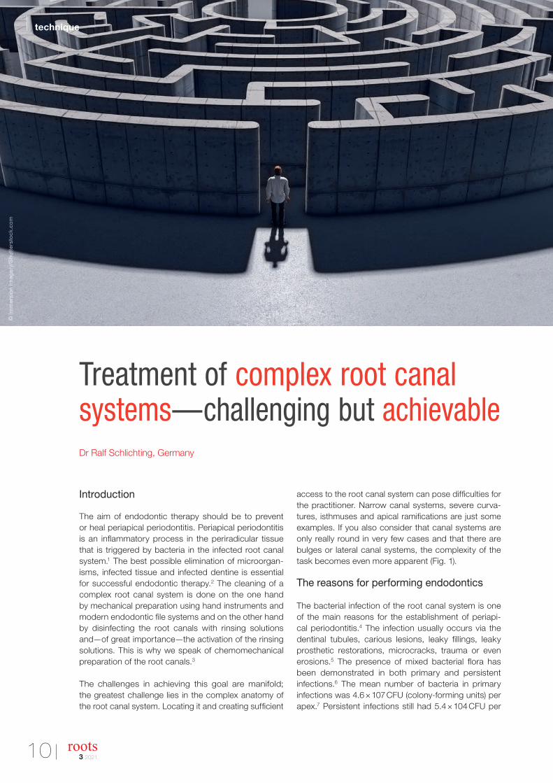
as medication and obturation materials. Ideally, the canal entrances form the cornerstones of the always slightly conical access cavity. Exploring the root canal ori�ces can occasionally be very challenging. One very useful tool is the Endo Holder and micro-�les (MANI; Fig. 5). The holder is ergonomically designed and can be con-nected to different tips. These tips are hand �les of differ-ent sizes and designs. For instance, you can use an ISO size 15 K-type micro-�le with different angulations of the tip. The different tips can easily be changed by screwing them in or out using a screw thread. The great advan-tage of this design is the better view on to the tip of the instrument. That is very important to control whether the tip slightly enters into a root canal ori�ce. When using a classic hand instrument, the operator’s �ngers constrict the operator’s �eld of vision. Another very helpful feature of the Endo Holder is the possibility of connecting it to an apex locator (Fig. 6), allowing working length deter-mination to be carried out with much more visual control compared with hand instruments.
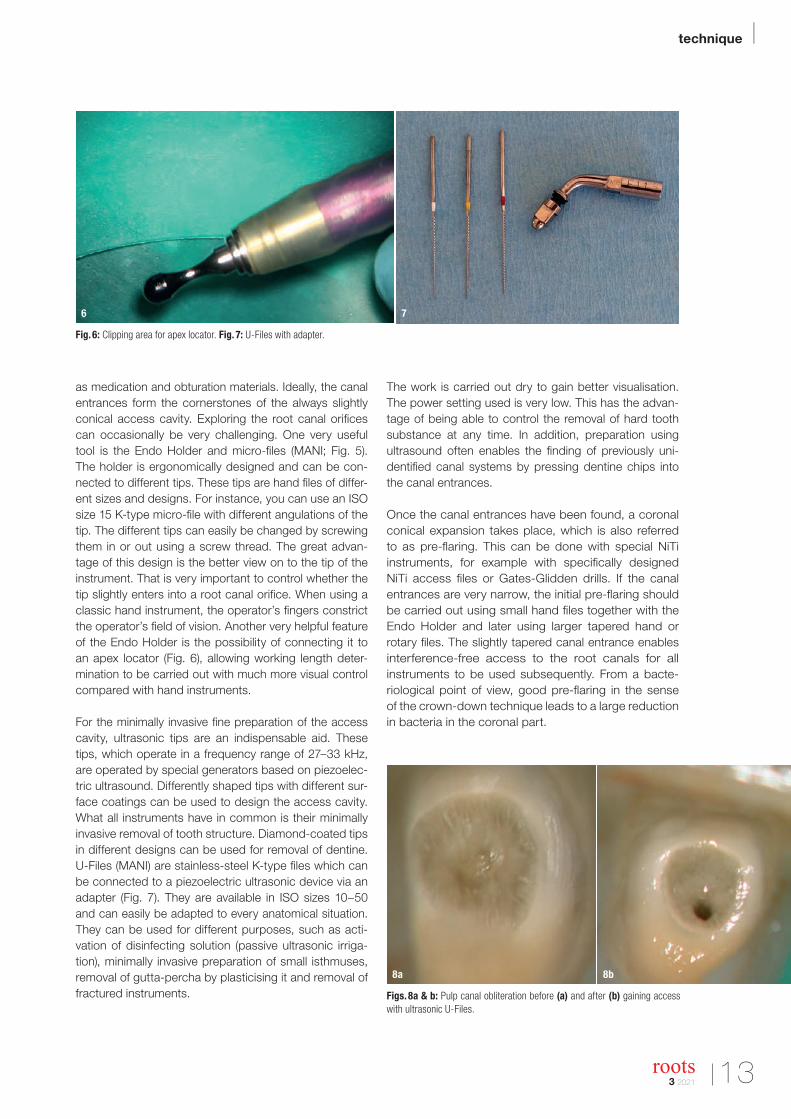
For the minimally invasive �ne preparation of the access cavity, ultrasonic tips are an indispensable aid. These tips, which operate in a frequency range of 27–33 kHz, are operated by special generators based on piezoelec-tric ultrasound. Differently shaped tips with different sur-face coatings can be used to design the access cavity. What all instruments have in common is their minimally invasive removal of tooth structure. Diamond-coated tips in different designs can be used for removal of dentine. U-Files (MANI) are stainless-steel K-type �les which canbe connected to a piezoelectric ultrasonic device via anadapter (Fig. 7). They are available in ISO sizes 10–50and can easily be adapted to every anatomical situation.They can be used for different purposes, such as acti-vation of disinfecting solution (passive ultrasonic irriga-tion), minimally invasive preparation of small isthmuses,removal of gutta-percha by plasticising it and removal offractured instruments.
The work is carried out dry to gain better visualisation. The power setting used is very low. This has the advan-tage of being able to control the removal of hard tooth substance at any time. In addition, preparation using ultrasound often enables the �nding of previously uni-denti�ed canal systems by pressing dentine chips into the canal entrances.
Once the canal entrances have been found, a coronal conical expansion takes place, which is also referred to as pre-�aring. This can be done with special NiTi instruments, for example with speci�cally designed NiTi access �les or Gates-Glidden drills. If the canal entrances are very narrow, the initial pre-�aring should be carried out using small hand �les together with the Endo Holder and later using larger tapered hand or rotary �les. The slightly tapered canal entrance enables interference-free access to the root canals for all instruments to be used subsequently. From a bacte-riological point of view, good pre-�aring in the sense of the crown-down technique leads to a large reduction in bacteria in the coronal part.
Fig. 6: Clipping area for apex locator. Fig. 7: U-Files with adapter.
Figs. 8a & b: Pulp canal obliteration before (a) and after (b) gaining access with ultrasonic U-Files.
6 7
8a 8b
technique |
3 2021 13roots

Obliterated root canal systems
Root canal therapy of teeth with pulp chamber and root canal obliteration can be very challenging.23 Obliteration of the pulp and coronal parts of the root canal space may occur as a result of the formation of tertiary dentine, as seen during caries progression24 or after tooth resto-ration.25 In addition, pulp space obliteration may occur in teeth that have sustained trauma.26
Finding the entrance into an obliterated root canal and gaining access to the more apical part of the root canal system can be technically demanding and time-consuming.27 Perforations, incorrect alignment and excessive removal of sound tooth structure are some of the complications which frequently occur. Correct access cavity prepara-tion is crucial to avoid these complications.28
Pulpal obliteration has been described as a tertiary den-tine response to trauma, producing dentine highly irreg-
ular in pattern and calci�cation.29 Tertiary dentine is of different colour and structure than the surrounding sec-ondary dentine. Therefore, magni�cation and illumination by the dental operating microscope are a major advan-tage for visualising these different structures (Figs. 8a & b).
Once the tertiary dentine is visualised, it can be removed carefully. One very minimally invasive option is to remove it layer by layer using ultrasonic tips. Thanks to the dif-ferent tip diameters, the U-Files are very useful, adapting to different canal diameters. This procedure should be carried out without water cooling to control the dentine removal for each iteration. Often a white spot can be de-tected, a point where dentine particles from the abrasion process accumulate. With a picking motion of the Endo Holder, you can try to detect the hidden root canal en-trance. If there is minor resistance while the Endo Holder is removed, it should be checked whether there is no iatrogenic perforation using the apex locator. Then the canal entrance can be gradually widened with, for ex-ample hand �les or different tip sizes of the Endo Holder. Finally, one can use small rotary glide path �les, which are inserted only 3–4 mm into the canal entrance, to widen the canal entrance even more. The �nal stage of pre- �aring can then be carried out as previously mentioned. It has to be pointed out that irrigation in between these different steps is very important.
The management of narrow canal systems
The anatomy of root canals is primarily determined by genetics, but it is also in�uenced, under the circum-stances of a vital pulp, by external and internal stimuli. The age of the patient has a major in�uence upon the anatomical relations within the tooth: the older a vital tooth is and the more it is subjected to mechanical, chemical, thermal and microbial stimuli, the more secondary and tertiary dentine will be produced. The results are not only a narrowing or partial obliteration of the root canal but also compartmentalisation into diverse root canal structures.30 An example of this phenomenon is the man-dibular incisor, which has a typical root with proximal, dis-crete grooves along the root length axis. Over the years, progressive deposition of secondary dentine occurs at the root canal walls from coronal to apical. This leads to the constriction of the root canals in the coronal or middle sections of the root canal.31
After preparing a correct access cavity and pre-�aring the coronal part of the root canal, the next step is ne-gotiating the root canal to full length. Generally, narrow root canals have a small taper or even parallel root canal walls. As mentioned before, the cross sections can be constricted even more owing to deposition of sec-ondary dentine (Fig. 9). Is there any strategy for negoti-ating these small canals? First of all, one needs the right equipment. Owing to the very small diameter of those
Fig. 9: Constricted root canal in the middle third. (Image: © Dr Frank Paque)
Figs. 10a & b: Diameter (a) and working part (b) of a D Finder.
Fig. 11: Radiograph of severely curved root canal system. Figs. 12a & b:
Examples of root curvatures with the same angle but a different radius.
A = ; B = ; � = angle; r = radius.
10b
9 10a
12a11 12b
| technique
14 3 2021roots

canals, the initial widening should be carried out using hand �les. One advantage is that the risk of instrument fractures is lower in stainless-steel hand �les then in small NiTi �les.32 Secondly, the tactile feeling is much more direct to the �ngers compared with rotary instruments. Historically, stainless-steel K-type �les or reamers were used for the task of negotiating these sorts of root ca-nals. The ISO 3630 speci�cation de�nes the design of endodontic hand �les. One of the requirements is a con-stant taper of the working part from 0.02 mm. The length of the working part is 16 mm. Looking more closely at an ISO size 8 hand instrument, one can easily assume that the diameter of the tip is 0.08 mm (ISO size 8). In the mid-dle of the working part, 8.00 mm above, the diameter has already increased to 0.24 mm (ISO size 24) and at the end of the working part to 0.40 mm (ISO size 40). These con-siderations lead to the implication that even small hand �les are too large in diameter in their middle and coronal parts to negotiate small canal systems. Often, the friction in the middle or coronal part is the reason why the �le cannot be negotiated further apically.
A further disadvantage of traditional hand �les is their design. For instance, stainless-steel K-type �les have a rectangular diameter and cutting edges all the way from the tip to the end of the working part. Therefore, the cut-ting edges can have friction all over the entire root canal, especially in narrow parts with deposition of secondary dentine.
Thanks to its unique design features, the D Finder �le (MANI) can be a helpful tool in these cases. First of all, the �le has a D-like cross section. This cross section is the reason for the very good centring ability of the D Finder in the root canal system. The second unique feature is the working part (Figs. 10a & b). There are only some sharp cutting edges and very smooth surface areas in between. The diameter, together with the smooth sur-faces along the working part, results in the �le gliding through the root canal and by passed any obstacles.
Compared with traditional root canal instruments, the D Finder features increased stiffness or, technically speaking, a superior buckling resistance.33 That is the reason why slightly more downward force in the direc-tion of the tip can be generated. Ideally, the �le is used in a balanced force technique. Thanks to its design, it nor-mally glides through the root canal. In case of too much friction, the canal space can be widened with the sharp cutting edges. D Finders are available in ISO sizes 8, 10 and 12 and in 21 and 25 mm lengths. Whenever pos-sible, one should choose a 21 mm D Finder, as it has better buckling resistance.
So much for equipment, now for technique. If you cannot advance with the smallest �le (e.g. ISO size 8 D Finder) in a small root canal, do not try to push or force the �le down. That is the road to failure! Instead, measure the length which the �le can penetrate. As mentioned before, �les seldom bind at the tip but very often in the middle or coronal section. Now change to the next larger �le, for example an ISO size 10 or 12 D Finder, and prepare the canal to the provisional working length. Remember to irrigate copiously in between the �les. Once you have reached the provisional working length and have irrigated copiously, change back to your smallest �le (e.g. ISO size 8 D Finder) and try to advance further into the canal. In most cases, it is possible to penetrate deeper into the root canal. You can repeat this procedure
Fig. 13: Same tip size but a different taper and �exibility. D = diameter. Fig. 14: JIZAI �les.
“Perforations, incorrect alignment and excessive
removal of sound tooth structure are some of the complications
which frequently occur.”
13 14
technique |
3 2021 15roots
as long as you have a con�rmed working length using the apex locator. Once you have reached working length, you gradually widen the canal system with the next size hand �les. This procedure is often referred to as creat-ing a glide path.34 In fact, it is just achieving space for the next size �le.
According to West, a glide path is established when one can bring an ISO size 10 �le to working length easily.35
For several reasons, it is recommended to change to ro-tary or reciprocating root canal preparation as soon as possible.36, 37 Modern glide path �les, which are available in different sizes and tapers, can easily be used after the initial glide path preparation with hand �les. The sequence I regularly use is ISO sizes 8 and 10 D Finders, followed by an ISO 13/0.04 rotary glide path �le.
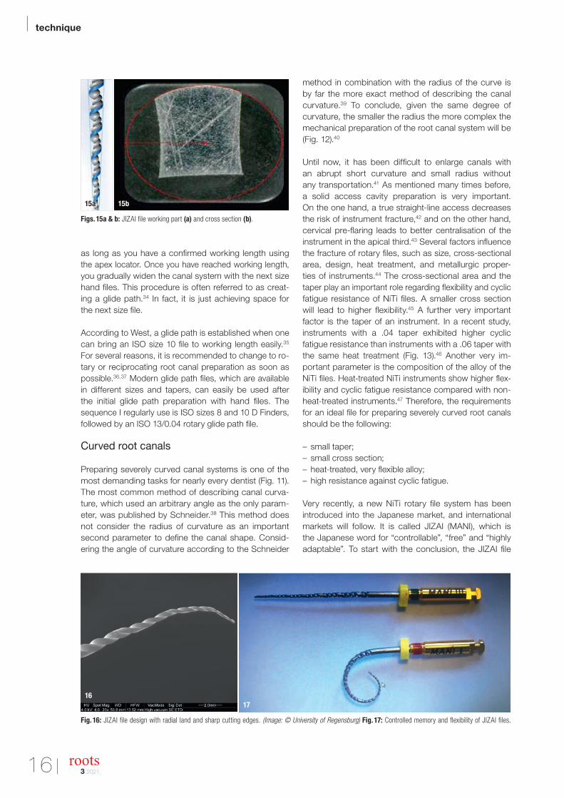
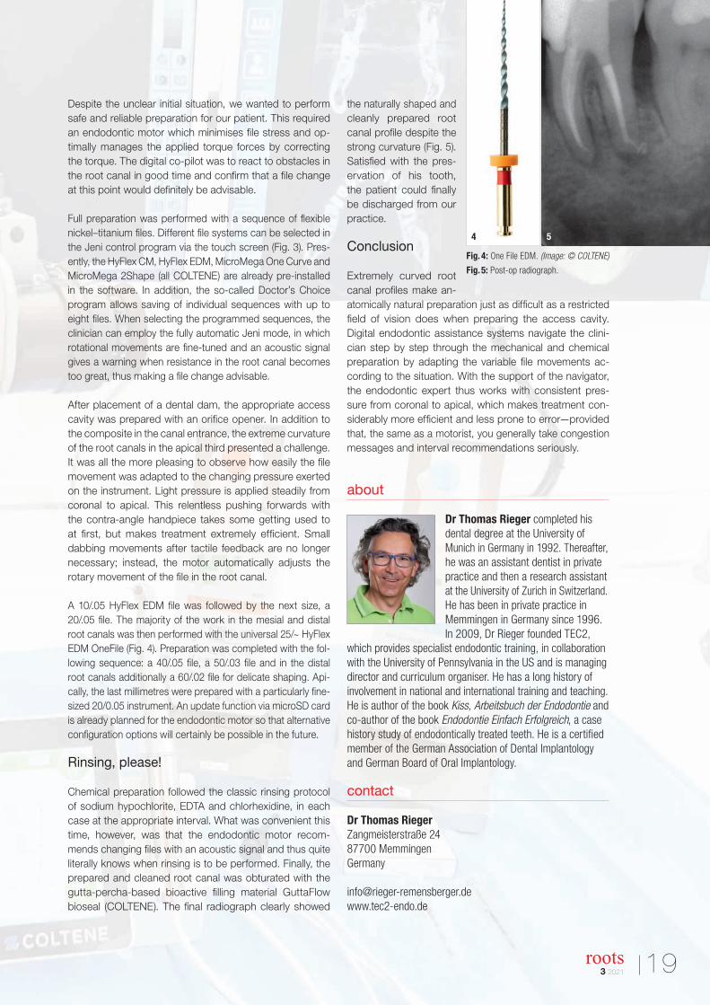
Curved root canals
Preparing severely curved canal systems is one of the most demanding tasks for nearly every dentist (Fig. 11). The most common method of describing canal curva-ture, which used an arbitrary angle as the only param-eter, was published by Schneider.38 This method does not consider the radius of curvature as an important second parameter to de�ne the canal shape. Consid-ering the angle of curvature according to the Schneider
method in combination with the radius of the curve is by far the more exact method of describing the canal curvature.39 To conclude, given the same degree of curvature, the smaller the radius the more complex the mechanical preparation of the root canal system will be (Fig. 12).40
Until now, it has been dif�cult to enlarge canals with an abrupt short curvature and small radius without any transportation.41 As mentioned many times before, a solid access cavity preparation is very important. On the one hand, a true straight-line access decreases the risk of instrument fracture,42 and on the other hand, cervical pre-�aring leads to better centralisation of the instrument in the apical third.43 Several factors in�uence the fracture of rotary �les, such as size, cross-sectional area, design, heat treatment, and metallurgic proper-ties of instruments.44 The cross-sectional area and the taper play an important role regarding �exibility and cyclic fatigue resistance of NiTi �les. A smaller cross section will lead to higher �exibility.45 A further very important factor is the taper of an instrument. In a recent study, instruments with a .04 taper exhibited higher cyclic fatigue resistance than instruments with a .06 taper with the same heat treatment (Fig. 13).46 Another very im-portant parameter is the composition of the alloy of the NiTi �les. Heat-treated NiTi instruments show higher �ex-ibility and cyclic fatigue resistance compared with non-heat-treated instruments.47 Therefore, the requirements for an ideal �le for preparing severely curved root canals should be the following:
– small taper;– small cross section;– heat-treated, very �exible alloy;– high resistance against cyclic fatigue.
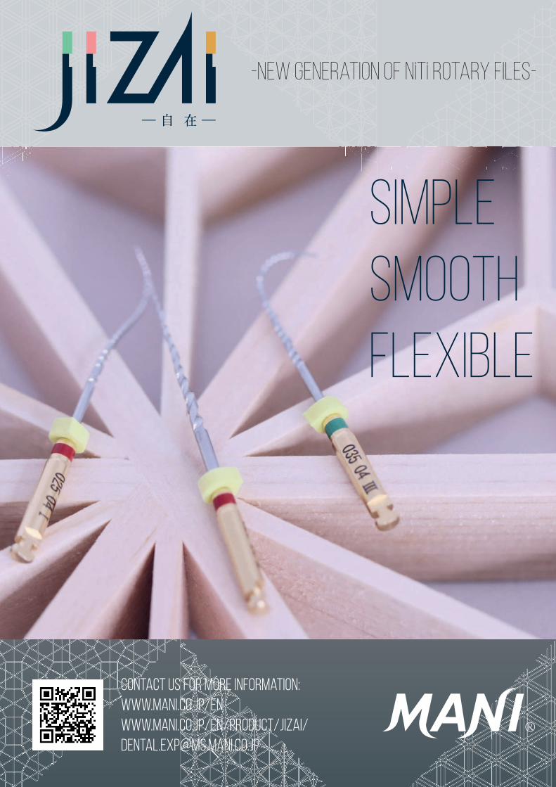
Very recently, a new NiTi rotary �le system has been introduced into the Japanese market, and international markets will follow. It is called JIZAI (MANI), which is the Japanese word for “controllable”, “free” and “highlyadaptable”. To start with the conclusion, the JIZAI �le
Fig. 16: JIZAI � le design with radial land and sharp cutting edges. (Image: © University of Regensburg) Fig. 17: Controlled memory and � exibility of JIZAI � les.
Figs. 15a & b: JIZAI � le working part (a) and cross section (b).
15a 15b
1617
| technique
16 3 2021roots

exhibits superior �exibility and superior safety compared with all competitors in the market. The idea behind de-veloping the JIZAI �le was to make endodontics easier, more predictable and safer for every dentist around the world. The treatment manual is very easy to understand, and the �les can be used for very easy to very compli-cated canal systems.
The JIZAI �le features some unique design elements. Together with a newly developed heat treatment pro-cedure, the result is outstanding handling. Looking at the �le, one can immediately see its outstanding design (Fig. 14). Besides sharp cutting edges, it has very smooth surface areas formerly called radial lands. These bearing surfaces are one of the reasons for the excellent centring ability of the �le inside the root canal and minimise the so-called screwing-in effect. The sharp cutting edges with varying angles are designed for an ef�cient cutting ability without being aggressive. The tip is a non-cutting tip (Fig. 15). The cross section is off-centred and rectan-gular with a rounded downside (Fig. 16). Therefore, the root canal wall is shaped only at the contact points of the cutting edges. Minimising the contact surface with the root canal dentine prevents taper lock and screw-in ef-fects in the root canal. This reduces the torsional stress,48 makes it easier to follow the complex root canal and facilitates root canal shaping.49
The unique heat treatment procedure delivers unmatched �exibility (Fig. 17). In addition to that, controlled memory makes the �le bendable. Flexibility is directly associated with safety. Cyclic fatigue comparison to all competitors available showed more than 2.5-fold better values then the nearest competitor.
Besides safety, another very important aspect is cen-tring ability, that is, how well a �le preserves the origi-nal canal curvature. A recently published study showed that JIZAI �les preserved original canal curvature signi�-cantly better than all other �les and that the preparation time was signi�cantly faster than the other �le systems tested.50
All of these features are very important and demonstrate how reliable the JIZAI �le is. For the practitioner, the most important points are user friendliness and simplicity. Using the JIZAI �le system, the dentist will be enabled to cover nearly every canal anatomy, from very narrow and severely curved (Figs. 18a & b) to even very wide canal systems. The idea is to provide the practitioner with a very simple manual and a set of no more than three different �les to cover nearly all treatment challenges.
Conclusion
The treatment of complex root canal systems is always challenging for every dentist. Respecting the microbial
principles of endodontics is one key to success. Despite some controversies about the right size of the access cavity, preparing straight-line access to the root canal system is another key factor. The use of a dental dam and ultrasonics facilitates the treatment procedures of every dentist. Especially in complex anatomies, the use of newly developed �le systems manufactured from sophisticated heat-treated NiTi alloys which are very safe and user-friendly are other key points for success.
But remember, training, patience and fun in performing endodontics can be very helpful as well!
Editorial note: A list of references is available from the publisher.
about
Dr Ralf Schlichting is a specialist in endodontics, having completed the postgraduate programme in endodontics and dental traumatology of the Deutsche Gesellschaft für Endodontologie und zahnärztliche Traumatologie (German society for endodontics and dental traumatology). He is a member of the American
Association of Endodontists and a certi�ed member of the European Society of Endodontology. Dr Schlichting runs a private practice limited to endodontics.He is a long-time board member of the DGET and lectures worldwide. Dr Schlichting is the author of numerous peer-reviewed articles and has collaborated in several studies concerning activation and irrigation.He is involved in the development of endodontic instruments.
contact
Dr Ralf SchlichtingNibelungenplatz 1/294032 PassauGermany
Figs. 18a & b: Retreatment with JIZAI �les. Final preparation 24/.04 JIZAI �le (a)
and 35/.04 JIZAI �le (b).
18a 18b
technique |
3 2021 17roots

En route to the apex with the navigator Dr Thomas Rieger, Germany
Introduction
Unfortunately, routine cases tend to be the exception in endodontic practice; creating the optimal root canal access cavity under tooth-coloured composite can some-times prove quite tricky, even for experienced clinicians. In the following case, the author demonstrates how a digital endodontic assistance system noticeably facilitates navigation in obscure terrain.
Long-distance travel without a navigation system is hardly conceivable for many motorists. So why should one vol-untarily forego a fully automatic co-pilot when negotiating the curves in the root canal? In the following patient case, the author describes the decisions the intelligent co-driver can actively support and what congestion messages and other useful additional information can in principle be gained from such a system.
Obstacles en route
A 51-year-old male patient presented at our practice late this summer, having been referred to us by his dentist for further endodontic evaluation of pain in his left mandible. The most striking feature was pronounced periodontitis, which quickly became apparent during the initial exam-ination. CBCT con�rmed the suspected overall situation: severe periapical periodontitis was diagnosed in tooth #37, and there was no doubt that root canal therapy was indi-cated (Fig. 1). The patient was promptly informed about his poor periodontal status and agreed to endodontic therapy.
It soon became evident that another factor would further complicate navigation through the root canal system: the mesial canal entrances contained tooth-coloured composite from a previous restoration. Identifying the transition from the pulp chamber to the root canal between dentine and the well-adapted �lling material would therefore prove to be rather dif-�cult. The entire treatment was performed exclusively under the microscope and not only for preparing the access cavity. This at least allowed optimisation of the view of the work �eld.
A second highly topical working aid supported us in the preparation of the mesial and distal root canals. The CanalPro Jeni endodontic motor was used for the �rst time in the case described (Fig. 2). The “enchanting Jeni” is a novel digital endodontic assistance system from interna-tional dental specialist COLTENE. Jeni derives its nickname from its inventor, Dr Eugenio Pedullà. The idea for quasi- autonomous driving in the root canal came to the Italian endodontic expert during the preparation of an S-shaped root canal; a fully automatic endodontic motor that auton-omously navigates its way through the root canal would make root canal therapy considerably safer and less prone to error, just like a navigation system in road traf�c, espe-cially in the often stressful daily routines of a dental practice.
Congestion reports and interval recommendations included
In the same manner as a driving assistance system, Jeni navigates the user safely and quickly through the root canal. With its complex algorithms, the endodontic motor controls the variable �le movements in millisecond cycles. Rotational movement, speed and torque are continuously adapted to the prevailing conditions in the root canal.
Fig. 1: Pre-op radiographic image of tooth #37. Fig. 2: Fully automated
endodontic motor in the practice. (Image: © COLTENE)
Fig. 3: Sequence selection by touch screen.
1 2
3
| case report
Despite the unclear initial situation, we wanted to perform safe and reliable preparation for our patient. This required an endodontic motor which minimises �le stress and op-timally manages the applied torque forces by correcting the torque. The digital co-pilot was to react to obstacles in the root canal in good time and con�rm that a �le change at this point would de�nitely be advisable.
Full preparation was performed with a sequence of �exible nickel–titanium �les. Different �le systems can be selected in the Jeni control program via the touch screen (Fig. 3). Pres-ently, the HyFlex CM, HyFlex EDM, MicroMega One Curve and MicroMega 2Shape (all COLTENE) are already pre-installed in the software. In addition, the so-called Doctor’s Choice program allows saving of individual sequences with up to eight �les. When selecting the programmed sequences, the clinician can employ the fully automatic Jeni mode, in which rotational movements are �ne-tuned and an acoustic signal gives a warning when resistance in the root canal becomes too great, thus making a �le change advisable.
After placement of a dental dam, the appropriate access cavity was prepared with an ori�ce opener. In addition to the composite in the canal entrance, the extreme curvature of the root canals in the apical third presented a challenge. It was all the more pleasing to observe how easily the �le movement was adapted to the changing pressure exerted on the instrument. Light pressure is applied steadily from coronal to apical. This relentless pushing forwards with the contra-angle handpiece takes some getting used to at �rst, but makes treatment extremely ef�cient. Small dabbing movements after tactile feedback are no longer necessary; instead, the motor automatically adjusts the rotary movement of the �le in the root canal.
A 10/.05 HyFlex EDM �le was followed by the next size, a 20/.05 �le. The majority of the work in the mesial and distal root canals was then performed with the universal 25/~ HyFlex EDM OneFile (Fig. 4). Preparation was completed with the fol-lowing sequence: a 40/.05 �le, a 50/.03 �le and in the distal root canals additionally a 60/.02 �le for delicate shaping. Api-cally, the last millimetres were prepared with a particularly �ne-sized 20/0.05 instrument. An update function via microSD card is already planned for the endodontic motor so that alternative con�guration options will certainly be possible in the future.
Rinsing, please!
Chemical preparation followed the classic rinsing protocol of sodium hypochlorite, EDTA and chlorhexidine, in each case at the appropriate interval. What was convenient this time, however, was that the endodontic motor recom-mends changing �les with an acoustic signal and thus quite literally knows when rinsing is to be performed. Finally, the prepared and cleaned root canal was obturated with the gutta-percha-based bioactive �lling material GuttaFlow bioseal (COLTENE). The �nal radiograph clearly showed
the naturally shaped and cleanly prepared root canal pro�le despite the strong curvature (Fig. 5). Satis�ed with the pres-ervation of his tooth, the patient could �nally be discharged from our practice.
Conclusion
Extremely curved root canal pro�les make an-atomically natural preparation just as dif�cult as a restricted �eld of vision does when preparing the access cavity. Digital endodontic assistance systems navigate the clini-cian step by step through the mechanical and chemical preparation by adapting the variable �le movements ac-cording to the situation. With the support of the navigator, the endodontic expert thus works with consistent pres-sure from coronal to apical, which makes treatment con-siderably more ef�cient and less prone to error—provided that, the same as a motorist, you generally take congestion messages and interval recommendations seriously.
Fig. 4: One File EDM. (Image: © COLTENE)
Fig. 5: Post-op radiograph.
about
Dr Thomas Rieger completed his dental degree at the University of Munich in Germany in 1992. Thereafter, he was an assistant dentist in private practice and then a research assistant at the University of Zurich in Switzerland. He has been in private practice in Memmingen in Germany since 1996. In 2009, Dr Rieger founded TEC2,
which provides specialist endodontic training, in collaboration with the University of Pennsylvania in the US and is managing director and curriculum organiser. He has a long history of involvement in national and international training and teaching.He is author of the book Kiss, Arbeitsbuch der Endodontie and co-author of the book Endodontie Einfach Erfolgreich, a case history study of endodontically treated teeth. He is a certi� ed member of the German Association of Dental Implantology and German Board of Oral Implantology.
contact
Dr Thomas RiegerZangmeisterstraße 2487700 MemmingenGermany
4 5
3 2021 19roots

Root canal therapy in a maxillary � rst molar with a highlycurved mesiobuccal rootDr Jens Emmelmann, Austria
Introduction
Root canal therapy of the maxillary molars often poses considerable challenges for the dentist owing to the anatomical complexity of the root canal system. The teeth in most cases have more than three canals, and the mesial canal system in particular can have sharp curves.
Successful endodontic therapy requires removal of necrotic pulp tissue, dental debris, and microorganisms and their metabolic products to the greatest possible extent. Bacteria are the major cause of endodontic dis-ease and endodontic failures. They can form bacterial bio�lms on the walls of the root canals. If the root ca-nal system cannot be prepared completely by chemical and mechanical means, owing to anatomical complex-ities for example, residual bio�lm can contribute to the failure of the treatment. Highly curved root canals lead to incompletely prepared canals, and the danger of instrument fracture is considerably greater owing to the increased stress.
Mechanical root canal preparation only ever forms part of root canal preparation. For complete cleaning and disinfection of root canals, irrigation solutions must be
used. To ensure application of irrigation solution also in the apical region of the root canal, irrigation cannu-las that have a suitable diameter and, particularly with highly curved canals, that are highly �exible must be used. Activation of irrigation solutions can even improve the ef�cacy of cleaning and disinfection.
Case presentation
A 43-year-old female patient was referred to us for root canal therapy of tooth #26. Owing to symptomatic pulp necrosis, the referring dentist had already performed the trepanation of the pulp chamber. When the patient presented to our clinic, she was free of symptoms. On the preoperative intra-oral radiograph (Fig. 1), the signi�cant curvature of the mesial root could already be seen.
Clinial procedure
First, there was a consultation and information ses-sion with the patient. After in�ltration anaesthesia, the tooth was isolated with a dental dam (COLTENE). The access cavity was �rst cleaned and disinfected with 5% sodium hypochlorite (NaOCl) under the Pro Magis
© D
rab
yad
/Shu
tter
sto
ck.c
om
| case report
20 3 2021roots

dental microscope (ZEISS). The cleaned access cavity was re�ned with EndoExplorer 1–3 instruments (Komet). A total of three canal entrances could be detected and enlarged.
In the next step, initial scouting of the coronal root ca-nal sections was performed with C-PILOT hand �les (VDW) in ISO sizes 08 and 10. In particular, the mesio-buccal canal was checked with pre-curved hand �les to determine whether the curvature at the transition from the coronal to the middle root canal third could be managed. Because this was possible, this canal was enlarged and prepared up to the middle third with an R-PILOT instrument (VDW). The root canal was irrigated
with 5% NaOCl using the EDDY FLEX.CANNULA (Fig.2) (VDW), and the patency of the canal was checked with a pre-curved hand �le.
Using a second VDW.CONNECT Drive endodontic motor (VDW) and a R25 RECIPROC blue �le (VDW), the canal was prepared in steps, alternating with the R-PILOT �le initially to the apical third. Using the Root ZX mini apex locator (Morita) and a C-PILOT �le ISO size 10, patency was con�rmed and the working length determined. The apical canal third up to the working length was then mechanically prepared with R-PILOT and 20/.05 and 25/.04 VDW.ROTATE (Fig. 3)instruments (VDW).
Fig. 1: Initial radiograph. Fig. 2: Irrigation with EDDY FLEX.CANNULA.
1 2
Fig. 3: Size 20/.05 VDW.ROTATE. Fig. 4: R25 RECIPROC blue.
3 4
case report |
3 2021 21roots

Dental debris was regularly �ushed out of the canal with NaOCl, and the patency of the canal checked with an ISO size 10 hand �le (recapitulation to working length or patency). After apical gauging with nickel–titanium (NiTi) K-�les (VDW), the root canal was �nally shaped with a 30/.04 VDW.ROTATE �le (VDW) to the working length. Working with the six-handed technique proved advantageous for the frequent instrument changes and repeated checking of the working length so that this dif�cult canal could be prepared in reasonable time.
The uncomplicated distobuccal and palatal canals could subsequently be quickly accessed and prepared to working length with R25 RECIPROC blue (Fig. 4) and 30/.04 VDW.ROTATE �les.
Shaping was followed by generous irrigation of all canals with 17% EDTA (COLTENE) and again with 5% NaOCl. The irrigation solutions were sonically activated with the EDDY irrigation tip (VDW). AH Temp calcium hydroxide (Dentsply Sirona) was applied as a temporary root canal �lling, and the access cavity was temporarily sealed with sterile PTFE tape and GC Fuji IX GP glass ionomer cement (GC).
At the second visit about three weeks later, under in�l-tration anaesthesia, a dental dam and a dental micro-scope, the temporary occlusal seal was removed and the calcium hydroxide was thoroughly �ushed out. The working length was again checked using endodontic length determination. Gutta-percha points (VDW) were adjusted to working length (tugback). A control radio-graph was taken with the gutta-percha points in situ (Fig. 5). The point that was too long in the palatal ca-nal was shortened and checked again. Final root canal irrigation was performed with sonically activated 17% EDTA and 5% NaOCl (Fig. 6).
After drying the canals (Fig. 7) with micro-suction, the canals were sealed with a bioceramic sealer ( EndoSequence BC Sealer, Brasseler) and gutta- percha points in the single-cone technique. After com-plete cleaning of the cavity and sandblasting with alu-minium oxide, the tooth was restored with an adhesive composite �lling (everX Flow and G-ænial, both GC). After removal of the dental dam, a �nal radiograph was prepared (Fig. 8).
Thanks to the generous irrigation protocol and sonic activation, a lateral canal in the apical section of the palatal root canal could be cleaned and �lled with sealer.
Discussion
Root canal therapy of teeth with highly curved root canals is frequently associated with dif�culties for the dentist. Curves always mean a greater risk of prepa-ration errors. A common problem in curved canals is the formation of steps due to the resetting force of the instruments. This can in turn lead to sections
Fig. 5: Master point radiograph. Fig. 6: EDDY irrigation tip.
5 6
“For complete cleaning and disinfection of
root canals, irrigation solutions must be used.”
| case report
22 3 2021roots

of the canal no longer being accessible to prepar- ation, and the bacterial micro�ora that is potentially present can lead to endodontic failures. When at-tempting to forcefully overcome ledges, blockages due to compaction of debris or, in the worst case, canal perforation may occur. Curved canals cause problems also for instruments because they always result in increased stress on the instrument, promot-ing fracture.
As always in dif�cult treatment situations, proceeding in a slow and controlled manner is the key to success. Pre-curved (steel) hand �les for initially opening and scouting of short canal sections and small, highly �ex-ible NiTi instruments applied with absolutely no pres-sure help to avoid fundamental preparation errors. As a user of VDW instruments, switching between pre-curved C-PILOT, R-PILOT, R25 RECIPROC blue and �exible VDW.ROTATE instruments with a taper of .04 has proved successful for our of�ce in dif�cult canals. The next larger instrument is used somewhat shorter than the previous. The canal is thus enlarged from the coronal to the apical third in small sections. After the larger instrument, a smaller one can then penetrate more deeply into the canal. To prevent step formation, pressure on the instruments must be avoided as much as possible. Mechanised instruments should always be kept moving. Constantly checking patency with a small steel �le and frequent (sonically activated) irrigation can prevent blockage of the canal with debris. To ensure that irrigation solutions can also reach deep canal sec-tions, thin, �exible irrigation cannulas are useful. In my opinion, �exible plastic cannulas such as the EDDY FLEX.CANNULA are highly suitable. The sonic (or ultra-sonic) tips to activate the irrigation solutions should also be highly �exible.
Conclusion
It is critical for the success of endodontic treatments that the entire root canal system is chemically and me-chanically prepared as thoroughly as possible. Curved root canals are often a barrier to achieving this goal. Whether we were able to achieve this in the present case will be seen at the follow-up examinations.
about
Dr Jens Emmelmann completed his dental degree at Heidelberg University in Germany and the Medical University of Graz in Austria. He completed the postgraduate programme in endodontics and dental traumatology of the Deutsche Gesellschaft für Endodontologie und zahnärztliche Traumatologie (German society for endodontics and dental traumatology) to become a certi�ed member of the society. Dr Emmelmann runs a dental practice
specialising in endodontics and traumatology in Lieboch in Austria.
Fig. 7: Canals prepared for root canal �lling. Fig. 8: Final radiographic image.
7 8
“Root canal therapy of teeth with highly curved root canals is frequently
associated with dif�culties for the dentist.”
case report |
3 2021 23roots

Target endodontic microsurgeryDr Hugo Sousa Dias, Prof. Paula Andrea Villa Machado & Dr Felipe Restrepo, Portugal & Colombia
Endodontic microsurgery (EMS) has become a more effective treatment compared with more traditional sur-gical approaches. Regardless of the technique improve-ments, it can be challenging to locate the root apex, es-pecially in cases of dif�cult access, intact thick buccal cortical bone and anatomical obstacles. This article de-scribes a new approach with the use of a 3D-printed tem-plate to guide osteotomy in order to access the root apex.
Introduction
CAD/CAM and 3D-printing technology applications were �rst developed and applied to dentistry in the 1990s.
At �rst, this technique was used for the fabrication of �xed restorations, but it was mostly applied to oral sur-gery for the fabrication of guiding templates for implant site preparation and insertion.1, 2 Nowadays, since the introduction of CBCT, CAD/CAM and 3D printing have several applications in dentistry, including in endodon-tics where the use of guiding templates has recently been introduced.2
These templates may be used to guide endodontic ac-cess in calci�ed canals and to guide osteotomy.1–3 EMS is one of the options for treating persistent periapical periodontitis after the failure of non-surgical root canal
Figs. 1a–n: Pre-op CBCT scan of tooth #36. The axial (a), coronal (b) and sagittal views (c) showed a hypo-dense zone around the apical third of the mesial
and distal roots and intact cortical buccal bone. A template that marked the limits of a cortical window to accurately reach the apical area of both roots was
designed (d). During microsurgery, the template was adjusted (e), the limits of the cortical window were marked in the bone (f) with a saw mounted in a
Piezotome CUBE LED handpiece, then the bone was cut and removed (g & h) to access the apical area and perform the apicectomy, apical cavity preparation
and retro-�lling of the mesial and distal roots (i). Finally, the cortical window was replaced and stabilised with collagen tape (j). Immediate post-op radiograph
of tooth #36 (k). Two-year follow-up CBCT scan, axial (l), coronal (m) and sagittal views (n).
1a
1c
1b
1d
| case report
24 3 2021roots

therapy or retreatment.2–4 Over the years, endodontic equipment, instruments and materials have been im-proved and better techniques have been developed. These developments have allowed greater understand-ing of the apical anatomy and increased the success rate of endodontic surgery, and consequently, it has become a more effective treatment.2, 3, 5
EMS requires a targeted osteotomy and root end resec-tion based upon anatomical landmarks and preopera-tive radiographs or CBCT measurements, and it com-bines the use of magni�cation and illumination provided by dental microscopes with the proper use of micro- instruments.1, 3, 5 This allows a more precise and predict-able approach with easier identi�cation of root apices, smaller osteotomies and shallower resection angles that allow the maintenance of cortical bone and preserva-tion of root length and dental structures.3, 5 In addition, the use of dental microscopes allows the identi�cation of anatomical details such as isthmuses, microfractures,
lateral canals and �ns of the resected root before root end preparation and �lling.
The advances in EMS along with modern diagnostic techniques, such as CBCT, used in diagnosis, pretreat-ment planning and post-treatment or follow-up evalua-tion contributed to higher success rates (85.0–96.8%) compared with more traditional approaches.3, 5
Some of the prognostic factors that may in�uence EMS outcomes include tooth position, lesion type, root end preparation, �lling material and coronal restoration.6–8 Dif�-cult accessibility, thick buccal bone and anatomical obsta-cles (mental foramen, inferior alveolar nerve and maxillary sinus) have been related to poorer outcomes.2 The extent of periapical bone destruction and osteotomy may also contribute to postoperative complications, such as pain and swelling. When the buccal bone plate is still intact, the extent of osteotomy tends to be increased, because it is dif�cult to �nd the exact location of the root apex.2, 3, 5
1e 1f
1g
1i 1j 1k
1h
case report |
3 2021 25roots

In this case report, we present a novel endodontic sur-gery approach using a 3D-printed template for guided osteotomy.
Case 1
A 63‐year‐old female patient, with a non-contributory medical history, consulted owing to moderate pain associated with her previously treated mandibular left �rst molar. CBCT revealed that a mesiobuccal canal had being missed during initial treatment, that the mesial and distal roots were affected by an
apical lesion, and that the cortical buccal bone was intact.
Retreatment was done in two appointments, and cal-cium hydroxide was used as the intra-canal dressing after removing the previous �lling material and cleaning and shaping three canals. After one week, root canal therapy was completed. Two months later, symptoms persisted and clinical examination revealed pain on vertical percussion; periodontal probing depth and mobility were within normal limits. CBCT revealed no signs of bone healing (Figs. 1a–c). The diagnosis for tooth #36 was previously treated symptomatic peri-apical periodontitis, and the treatment of choice was guided EMS.
An intra-oral scan (TRIOS, 3Shape) of the mandi-ble, and the resulting STL �le was combined with the DICOM �les of the CBCT scan to plan a surgical guide using the Blue Sky Bio software. A template that marked the limits of a cortical window to accurately reach the apical area was designed and printed (Fig. 1d).
Under local anaesthesia, a full-thickness mucoperios-teal �ap was re�ected and the printed template was used to mark the cortical window, which was cut with a Piezotome CUBE LED handpiece (ACTEON), removed (Figs. 1e–h) and then placed in sterile saline. An api-cectomy was done (Fig. 1i), and the mesial canals were retro-prepared with ultrasonic tips (NSK) and �lled with EndoSequence BC RRM Fast Set Putty (Brasseler). The cortical window was then placed back and stabi-lised with collagen sponges in the gaps (collagen tape, Zimmer Biomet; Figs. 1j & k), and the �ap was sutured using 6/0 prolene suture material (Corpaul).
At the two-year follow-up, clinical examination and CBCT showed evidence of healing of the apical lesions and the cortical bone without symptoms or complica-tions (Figs. 1l–n).
Case 2
A 38‐year‐old female patient consulted owing to mod-erate pain associated with her previously treated maxillary right second premolar. Her medical history was non-contributory. Clinical examination revealed that the tooth was slightly sensitive to vertical percussion. Periodontal probing depth and mobility were within normal limits. A periapical radiograph showed the presence of a separated instrument outside of the root (Fig. 2a), and a preoperative CBCT scan demonstrated that the buccal bone plate was intact (Figs. 2b & c). End-odontic retreatment had been performed �ve months earlier. The diagnosis for tooth #15 was previously treated symptomatic periapical periodontitis, and the treatment of choice was guided EMS.
1l
1m
1n
| case report
26 3 2021roots

An intra-oral scan (TRIOS) of the maxilla, and the re-sulting STL �le (Fig. 2d) was combined with the DICOM �les of the CBCT scan to plan a surgical guide using the Zirkonzahn.Implant-Planner software (Zirkonzahn) modi�ed with Meshmixer (Autodesk). A template that marked the limits of a cortical window to accu-rately reach the apical area was designed and printed (Figs. 2e & f).
Under local anaesthesia, a full-thickness mucoperios-teal �ap was re�ected, providing visualisation of the buc-cal bone (Fig. 2g), and the printed template was used to mark the cortical window (Fig. 2h), which was cut with a Piezotome CUBE LED handpiece, and the separated instrument was exposed (Fig. 2i) and removed (Fig. 2j). After apicectomy, retro-preparation was done using
ultra sonic tips (ACTEON) and sealed with TotalFill BC RRM Fast Set Putty (FKG) (Fig. 2k). The �ap was sutured using 5/0 prolene suture material (Fig. 2l). The sutures were removed 72 hours postoperatively. After two years the patient came to our of�ce for a follow-up radio-graph, the tooth was asymptomatic and in function (Fig. 2m).
Discussion
The use of 3D-printing technology in the �eld of end-odontics has been described for surgical guides, guided endodontic access, autogenous transplantation, edu-cational models and clinical simulation and has become a valid treatment option in dentistry.1, 9 Even though the literature is still limited to case reports and preclinical studies, guided surgery seems to be a more accurate
Figs. 2a–m: Pre-op radiograph (a) and pre-op CBCT scan of tooth #15, sagital (b) and axial views (c). STL �le obtained from the intra-oral scan (d). Planned template for guided endodontic
microsurgery of tooth #15 (e). Printed template for guided endodontic microsurgery of tooth #15 (f). Full-thickness mucoperiosteal �ap re�ected (g). The template was adjusted
and the limits of the cortical window were marked in the bone (h). Separated instrument out of the root near the apical area of tooth #15 (i). Removed separated instrument (j).
Post-op radiograph after apicectomy, retro-preparation and retro-�lling with TotalFill BC RRM Fast Set Putty (k). Sutured �ap (l). Two years follow-up radiograph (m).
2a 2b 2c
2d
case report |
3 2021 27roots

and successful technique compared with traditional access.1, 10
The use of surgical guides for periapical surgery was �rst suggested by Pinsky et al. for more precise and consistent localisation of root apices. In a preclin-ical study, the apical mean distance of osteotomies to planned objectives was 0.79 mm (± 0.33 SD) with surgical guides and 2.27 mm (± 1.46 SD) in freehand osteotomies.4
Since then, case reports have been published in which is demonstrated that the use of 3D printing is a help-ful tool in periapical surgery, in anterior and posterior teeth.2, 3, 11–13 This guided approach bene�ts osteotomy
and facilitates the location of root apices, minimising the risk of damaging anatomical structures. It can also be used to locate with precision extruded root �lling material for its removal.2, 9, 14
The cortical window technique (lid approach) was intro-duced into periapical surgery by Khoury and Hensher in 1987, and it allows better access to and visibility of the apical zone and the lesion location and prevents the formation of large residual bone defects.15
Although EMS has become a more predictable and reliable treatment option, it can be challenging to locate root ends for resection in molar cases or in cases involv-ing teeth near potentially problematic anatomical struc-tures, such as adjacent roots, the inferior alveolar nerve, the mental foramen, the maxillary sinus and the greater palatine artery.1, 2, 11, 16 Surgical templates are particularly helpful in dif�cult cases: they help to reduce iatrogenic injury producing an osteotomy site with predictable angulation, diameter and depth.11, 12 After endodontic surgery, success depends on the bacteria-tight seal of the root canal system with root end �lling and on the de�ciencies of the periapical and marginal bone tissue adjacent to the lesion.16 Consequently, the osteotomy should be “as small as possible but as large as nec-essary” to accomplish the clinical objectives, because there is a direct relationship between the size of the os-teotomy and the speed of healing: the smaller the oste-otomy, the faster the healing.5 The extent of osteotomy tends to be increased by less experienced clinicians.2, 16 During freehand osteotomy, deviation from the ideal site can occur as result of human error, leading to the inevitable removal of sound bone tissue.1
There is a consensus about the bene�ts associated with the use of guided surgery. The virtual backward planning simpli�es the surgical procedure, shortens operative time, improves accuracy and reduces post-operative complications.9, 14, 17 Guided surgery has the potential to allow less experienced clinicians to treat
2f2e
2g
2h 2i
| case report
28 3 2021roots
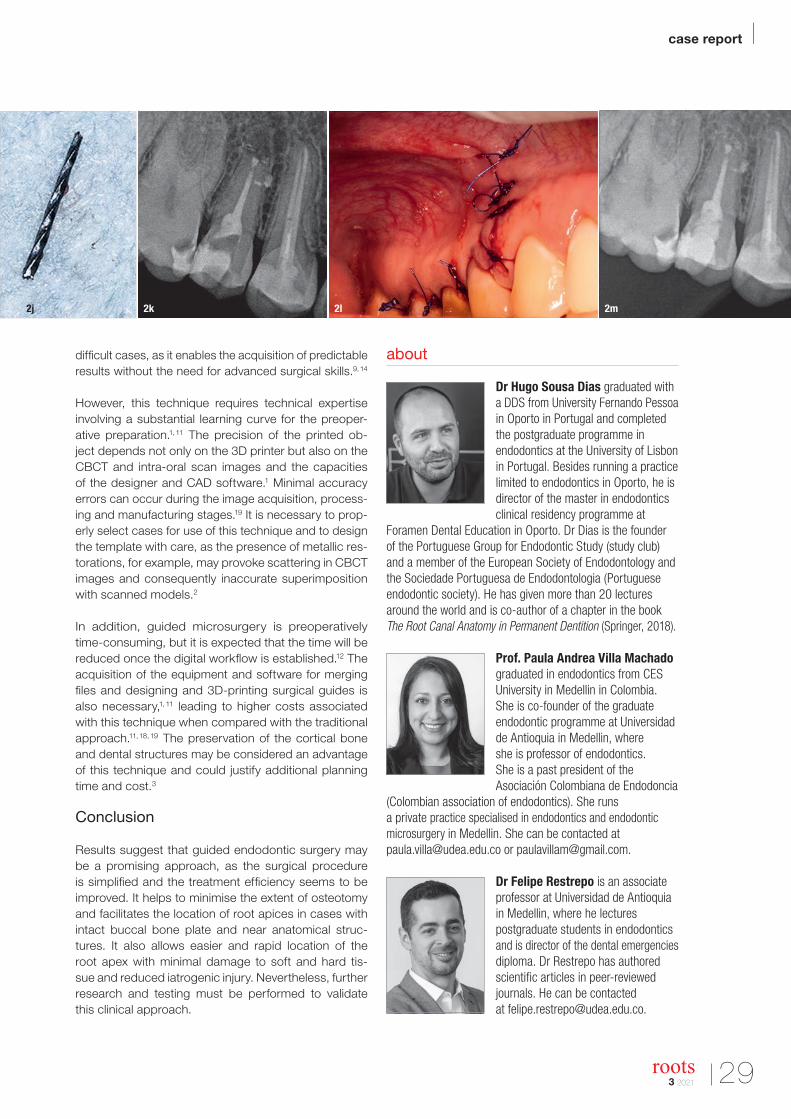
dif�cult cases, as it enables the acquisition of predictable results without the need for advanced surgical skills.9, 14
However, this technique requires technical expertise involving a substantial learning curve for the preoper-ative preparation.1, 11 The precision of the printed ob-ject depends not only on the 3D printer but also on the CBCT and intra-oral scan images and the capacities of the designer and CAD software.1 Minimal accuracy errors can occur during the image acquisition, process-ing and manufacturing stages.19 It is necessary to prop-erly select cases for use of this technique and to design the template with care, as the presence of metallic res-torations, for example, may provoke scattering in CBCT images and consequently inaccurate superimposition with scanned models.2
In addition, guided microsurgery is preoperatively time-consuming, but it is expected that the time will be reduced once the digital work�ow is established.12 The acquisition of the equipment and software for merging �les and designing and 3D-printing surgical guides is also necessary,1, 11 leading to higher costs associated with this technique when compared with the traditional approach.11, 18, 19 The preservation of the cortical bone and dental structures may be considered an advantage of this technique and could justify additional planning time and cost.3
Conclusion
Results suggest that guided endodontic surgery may be a promising approach, as the surgical procedure is simpli�ed and the treatment ef�ciency seems to be improved. It helps to minimise the extent of osteotomy and facilitates the location of root apices in cases with intact buccal bone plate and near anatomical struc-tures. It also allows easier and rapid location of the root apex with minimal damage to soft and hard tis-sue and reduced iatrogenic injury. Nevertheless, further research and testing must be performed to validate this clinical approach.
about
Dr Hugo Sousa Dias graduated with a DDS from University Fernando Pessoa in Oporto in Portugal and completed the postgraduate programme in endodontics at the University of Lisbon in Portugal. Besides running a practice limited to endodontics in Oporto, he is director of the master in endodontics clinical residency programme at
Foramen Dental Education in Oporto. Dr Dias is the founder of the Portuguese Group for Endodontic Study (study club) and a member of the European Society of Endodontology and the Sociedade Portuguesa de Endodontologia (Portuguese endodontic society). He has given more than 20 lectures around the world and is co-author of a chapter in the book The Root Canal Anatomy in Permanent Dentition (Springer, 2018).
Prof. Paula Andrea Villa Machado graduated in endodontics from CES University in Medellin in Colombia. She is co-founder of the graduate endodontic programme at Universidad de Antioquia in Medellin, where she is professor of endodontics. She is a past president of the Asociación Colombiana de Endodoncia
(Colombian association of endodontics). She runs a private practice specialised in endodontics and endodontic microsurgery in Medellin. She can be contacted at [email protected] or [email protected].
Dr Felipe Restrepo is an associate professor at Universidad de Antioquia in Medellin, where he lectures postgraduate students in endodontics and is director of the dental emergencies diploma. Dr Restrepo has authored scienti�c articles in peer-reviewed journals. He can be contacted at [email protected].
2j 2k 2l 2m
case report |
3 2021 29roots
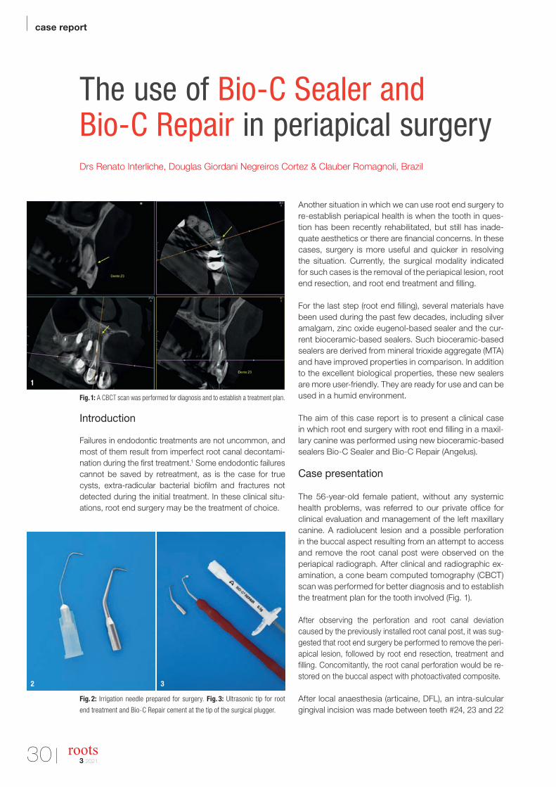
The use of Bio-C Sealer and Bio-C Repair in periapical surgeryDrs Renato Interliche, Douglas Giordani Negreiros Cortez & Clauber Romagnoli, Brazil
Introduction
Failures in endodontic treatments are not uncommon, and most of them result from imperfect root canal decontami-nation during the �rst treatment.1 Some endodontic failures cannot be saved by retreatment, as is the case for true cysts, extra-radicular bacterial bio�lm and fractures not detected during the initial treatment. In these clinical situ-ations, root end surgery may be the treatment of choice.
Another situation in which we can use root end surgery to re-establish periapical health is when the tooth in ques-tion has been recently rehabilitated, but still has inade-quate aesthetics or there are �nancial concerns. In these cases, surgery is more useful and quicker in resolving the situation. Currently, the surgical modality indicated for such cases is the removal of the periapical lesion, root end resection, and root end treatment and �lling.
For the last step (root end �lling), several materials have been used during the past few decades, including silver amalgam, zinc oxide eugenol-based sealer and the cur-rent bioceramic-based sealers. Such bioceramic-based sealers are derived from mineral trioxide aggregate (MTA) and have improved properties in comparison. In addition to the excellent biological properties, these new sealers are more user-friendly. They are ready for use and can be used in a humid environment.
The aim of this case report is to present a clinical case in which root end surgery with root end �lling in a maxil-lary canine was performed using new bioceramic-based sealers Bio-C Sealer and Bio-C Repair (Angelus).
Case presentation
The 56-year-old female patient, without any systemic health problems, was referred to our private of�ce for clinical evaluation and management of the left maxillary canine. A radiolucent lesion and a possible perforation in the buccal aspect resulting from an attempt to access and remove the root canal post were observed on the periapical radiograph. After clinical and radiographic ex-amination, a cone beam computed tomography (CBCT) scan was performed for better diagnosis and to establish the treatment plan for the tooth involved (Fig. 1).
After observing the perforation and root canal deviation caused by the previously installed root canal post, it was sug-gested that root end surgery be performed to remove the peri-apical lesion, followed by root end resection, treatment and �lling. Concomitantly, the root canal perforation would be re-stored on the buccal aspect with photoactivated composite.
After local anaesthesia (articaine, DFL), an intra-sulcular gingival incision was made between teeth #24, 23 and 22
© K
iefe
rPix
/Shu
tter
sto
ck.c
om
Fig. 1: A CBCT scan was performed for diagnosis and to establish a treatment plan.
Fig. 2: Irrigation needle prepared for surgery. Fig. 3: Ultrasonic tip for root
end treatment and Bio-C Repair cement at the tip of the surgical plugger.
1
2 3
| case report
30 3 2021roots
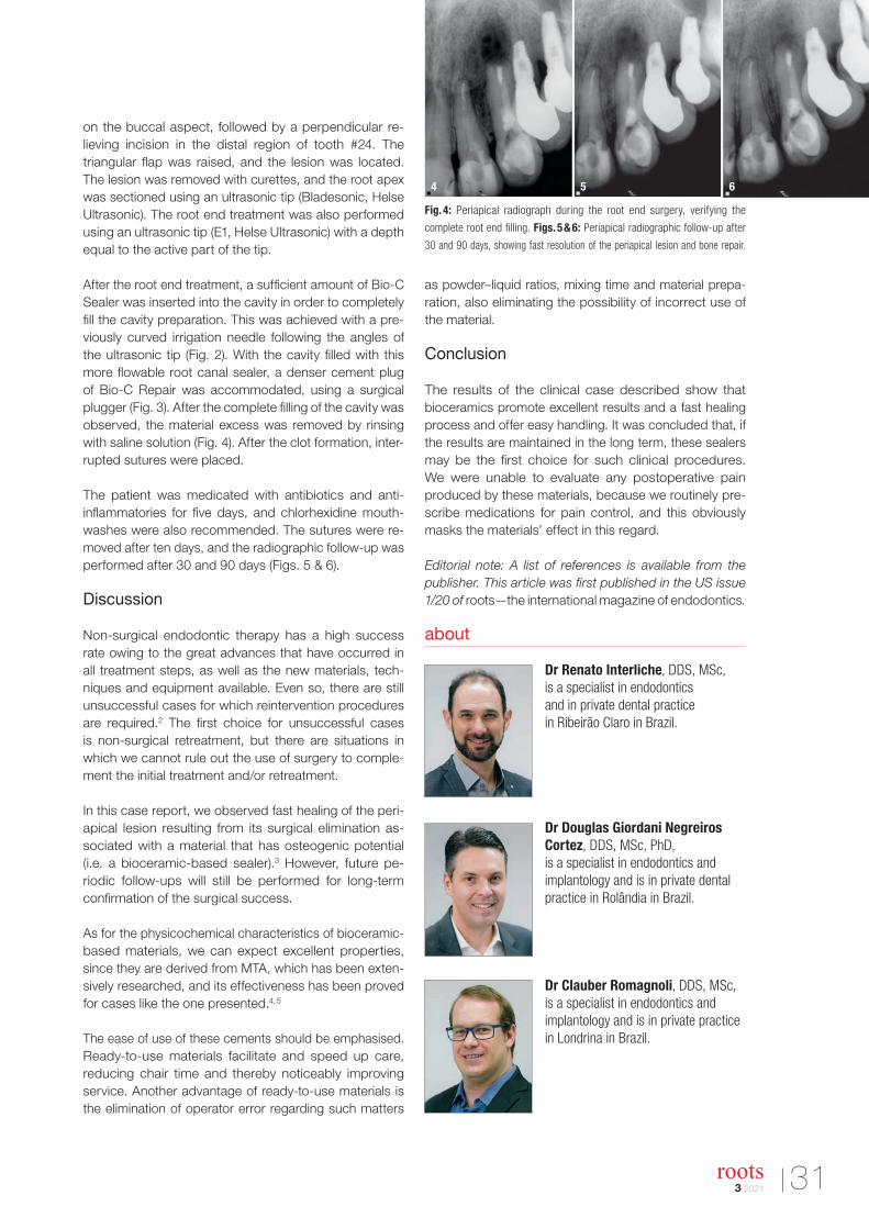
on the buccal aspect, followed by a perpendicular re-lieving incision in the distal region of tooth #24. The triangular �ap was raised, and the lesion was located. The lesion was removed with curettes, and the root apex was sectioned using an ultrasonic tip (Bladesonic, Helse Ultrasonic). The root end treatment was also performed using an ultrasonic tip (E1, Helse Ultrasonic) with a depth equal to the active part of the tip.
After the root end treatment, a suf�cient amount of Bio-C Sealer was inserted into the cavity in order to completely �ll the cavity preparation. This was achieved with a pre-viously curved irrigation needle following the angles of the ultrasonic tip (Fig. 2). With the cavity �lled with this more �owable root canal sealer, a denser cement plug of Bio-C Repair was accommodated, using a surgical plugger (Fig. 3). After the complete �lling of the cavity was observed, the material excess was removed by rinsing with saline solution (Fig. 4). After the clot formation, inter-rupted sutures were placed.
The patient was medicated with antibiotics and anti- in�ammatories for �ve days, and chlorhexidine mouth-washes were also recommended. The sutures were re-moved after ten days, and the radiographic follow-up was performed after 30 and 90 days (Figs. 5 & 6).
Discussion
Non-surgical endodontic therapy has a high success rate owing to the great advances that have occurred in all treatment steps, as well as the new materials, tech-niques and equipment available. Even so, there are still unsuccessful cases for which reintervention procedures are required.2 The �rst choice for unsuccessful cases is non-surgical retreatment, but there are situations in which we cannot rule out the use of surgery to comple-ment the initial treatment and/or retreatment.
In this case report, we observed fast healing of the peri-apical lesion resulting from its surgical elimination as-sociated with a material that has osteogenic potential (i.e. a bioceramic-based sealer).3 However, future pe-riodic follow-ups will still be performed for long-term con�rmation of the surgical success.
As for the physicochemical characteristics of bioceramic- based materials, we can expect excellent properties, since they are derived from MTA, which has been exten-sively researched, and its effectiveness has been proved for cases like the one presented.4, 5
The ease of use of these cements should be emphasised. Ready-to-use materials facilitate and speed up care, reducing chair time and thereby noticeably improving service. Another advantage of ready-to-use materials is the elimination of operator error regarding such matters
as powder–liquid ratios, mixing time and material prepa-ration, also eliminating the possibility of incorrect use of the material.
Conclusion
The results of the clinical case described show that bioceramics promote excellent results and a fast healing process and offer easy handling. It was concluded that, if the results are maintained in the long term, these sealers may be the �rst choice for such clinical procedures. We were unable to evaluate any postoperative pain produced by these materials, because we routinely pre-scribe medications for pain control, and this obviously masks the materials’ effect in this regard.
Editorial note: A list of references is available from the publisher. This article was �rst published in the US issue 1/20 of roots—the international magazine of endodontics.
© K
iefe
rPix
/Shu
tter
sto
ck.c
om
about
Dr Renato Interliche, DDS, MSc, is a specialist in endodontics and in private dental practice in Ribeirão Claro in Brazil.
Dr Douglas Giordani Negreiros Cortez, DDS, MSc, PhD, is a specialist in endodontics and implantology and is in private dental practice in Rolândia in Brazil.
Dr Clauber Romagnoli, DDS, MSc, is a specialist in endodontics and implantology and is in private practice in Londrina in Brazil.
Fig. 4: Periapical radiograph during the root end surgery, verifying the
complete root end �lling. Figs. 5 & 6: Periapical radiographic follow-up after
30 and 90 days, showing fast resolution of the periapical lesion and bone repair.
4 5 6
3 2021 31roots

Calcium silicate-based endodontic materials: A clinical perspectiveDrs Jenner Argueta & Benjamín Rodríguez, Guatemala
Introduction
Root canal decontamination and adequate sealing play an important role in endodontic success. However, the obturation process represents a challenge to the clinician owing to the complex anatomy and irregularities of the root canal system, making selection of the obturation ma-terial a key step during treatment.1 Calcium silicate-based endodontic materials are ceramic compounds that pres-ent good sealing properties owing to their similarity to human hydroxyapatite.2 One of their great advantages is excellent biocompatibility, which prevents rejection by the surrounding tissue, as it contains calcium phosphate, thereby improving the biochemical properties, providing a crystalline structure similar to the bone and dentine apatite.3 The calcium silicate-based materials include in their composition alumina, zirconia, bioactive glass, glass- ceramics, hydroxyapatite and resorbable calcium phos-phates.4
Mineral trioxide aggregate (MTA), one of the most suc-cessful bioactive materials, was developed in the 1990s at Loma Linda University in the US by Dr Mahmoud Torabinejad. Over the years, MTA has demonstrated excellent sealing properties, bioactivity, biocompatibility and antibacterial ef-fects owing to its ability to increase the pH by the release of hy-droxyl ions.5 The above-mentioned characteristics are shared among the different types of calcium silicate-based materials. These materials can be found in several consistencies and commercial presentations, and the choice of material will rely on the clinical scenario. Their applications have been indicated for vital pulp therapy, �lling of bone defects, root canal obtura-tion, retro-�lling in periapical surgery, perforation repair, regen-erative endodontics and repair of resorptive defects.6
Vital pulp therapy
The outcome of exposed dental pulp if left untreated will be irreversible pulpitis followed by pulp necrosis.7 Therefore,
Figs. 1a–g: A direct pulp capping procedure sequence. Initial bitewing radiograph. The proximity of the previous restoration to the pulp space was evident (a).
Initial periapical radiograph. No evidence of periapical disease (b). Pulp exposure (c). Pulp exposure after haemostasis, achieved after applying pressure with
a sterile cotton pellet for 20 seconds (d). Placement of pulp capping material before removal of excess material in the peripheral areas (e). Non-radiopaque
temporary restoration in place (f). Post-op radiograph taken at the end of the �rst appointment with the non-radiopaque temporary restoration in place (g).
1
| clinical report
32 3 2021roots

if the clinician wishes to avoid pulpectomy, vital pulp therapy might be a good choice, especially in young permanent teeth.8 Current bioceramics, mainly in putty consistency, are used for these purposes, owing to their ability to bond with dentine through the formation of a hydroxyapatite layer on the surface and their capacity to stimulate cell recruit-ment and differentiation at the level of the pulp exposure.9 Additionally, when compared with MTA, they show similar antibacterial properties against the main cariogenic bac-teria, Streptococcus mutans and lactobacilli.10 They also show good biocompatibility and can induce the formation of reparative dentinal bridges.
Case 1
During an urgent appointment, a 35-year-old male patient reported sharp and short-lasting pain in tooth #15 in re-sponse to cold stimuli. The established diagnosis was reversible pulpitis and normal periapical tissue. During the removal of the old amalgam restoration, pulp exposure of approximately 3 mm in diameter was created at the level of the buccal pulp horn (Fig. 1a–g). As there was no profuse bleeding and the pulp diagnosis was reversible pulpitis, it was decided to proceed with a direct pulp capping procedure.
Fig. 2: Six-month follow-up radiograph showing a thick dentinal bridge under the pulp capping. Fig. 3: Three-year follow-up radiograph showing the well-adapted
restoration. Fig. 4: Video of a vital pulp therapy procedure (https://youtu.be/bfBksF35IjY). Fig. 5: Video of clinical techniques for the application of calcium
silicate-based endodontic sealers (https://youtu.be/h3p1mZlNUzM).
Figs. 6a–d: Root canal therapy performed through a conservative access cavity in a tooth #37 with severe curvatures at the mesial root. The root canal
obturation was performed by the hydraulic compaction technique.
2 3
6a
6b 6c 6d
4
5
clinical report |
3 2021 33roots

After disinfection of the cavity using 2.5% sodium hypo-chlorite, a high-consistency calcium silicate-based material (CeraPutty, Meta Biomed) was used for direct pulp capping. At the end of the �rst appointment, a non-radiopaque temporary restoration (NexTemp LC, Meta Biomed) was placed with the objective of con�rming by radiograph ap-propriate placement of the direct pulp capping material. A direct adhesive restoration was placed during the sec-ond appointment using composite (Ez�l, Meta Biomed).
The patient was asymptomatic after the treatment and responded normally to sensitivity tests during the follow-up
sessions. A thick dentinal bridge was observed radio-graphically at the six-month follow-up appointment (Fig. 2). The restoration was well adapted at the three-year follow- up (Fig. 3).
Root canal therapy
It is crucial to obtain a 3D seal at the time of �lling the root canal system. It is dif�cult to achieve this goal when per-forming conventional obturation techniques that involve gutta-percha and eugenol-based cements owing to the lack of dentinal adhesion to the canal walls and their in-capacity to replicate the root canal system anatomy. The current bioactive calcium silicate-based materials can per-form well in this scenario owing to their physico-chemical properties.11 These types of sealers are mainly composed of calcium silicates, zirconia, tantalum oxide, calcium phosphate and �llers.12 They are recommended for use with a single-cone technique, usually called the hydraulic compaction technique, which simpli�es the �lling process without compromising the outcome.13 In addition to their sealing abilities, their potential to provide a bioactive sur-face with stimulation of hard-tissue formation and good antibacterial properties make them a great option as a �ller material in conventional root canal therapy.14 Figure 5 shows some of the current clinical techniques for appli-cation of these sealers.
Cases 2 and 3
In both cases presented, the root canal therapy was nec-essary due to an irreversible pulpitis process affecting
Figs. 7a & b: Root canal therapy performed in tooth #46 through a zirconia
crown. The access cavity was prepared as conservatively as possible. The
root canal obturation was performed by the hydraulic compaction technique.
Fig. 8: Video of conservative root canal therapy (https://youtu.be/M87d_Ycitcg).
Figs. 9a–c: Radiographic sequence of the treatment performed in tooth #11. Initial radiograph. The root resorption and lateral radiolucent area were evident (a).
Working length measurement at the level of the resorption (b). Root resorption repair with a calcium silicate-based material (c).
7a
8
7b
9a 9b 9c
| clinical report
34 3 2021roots
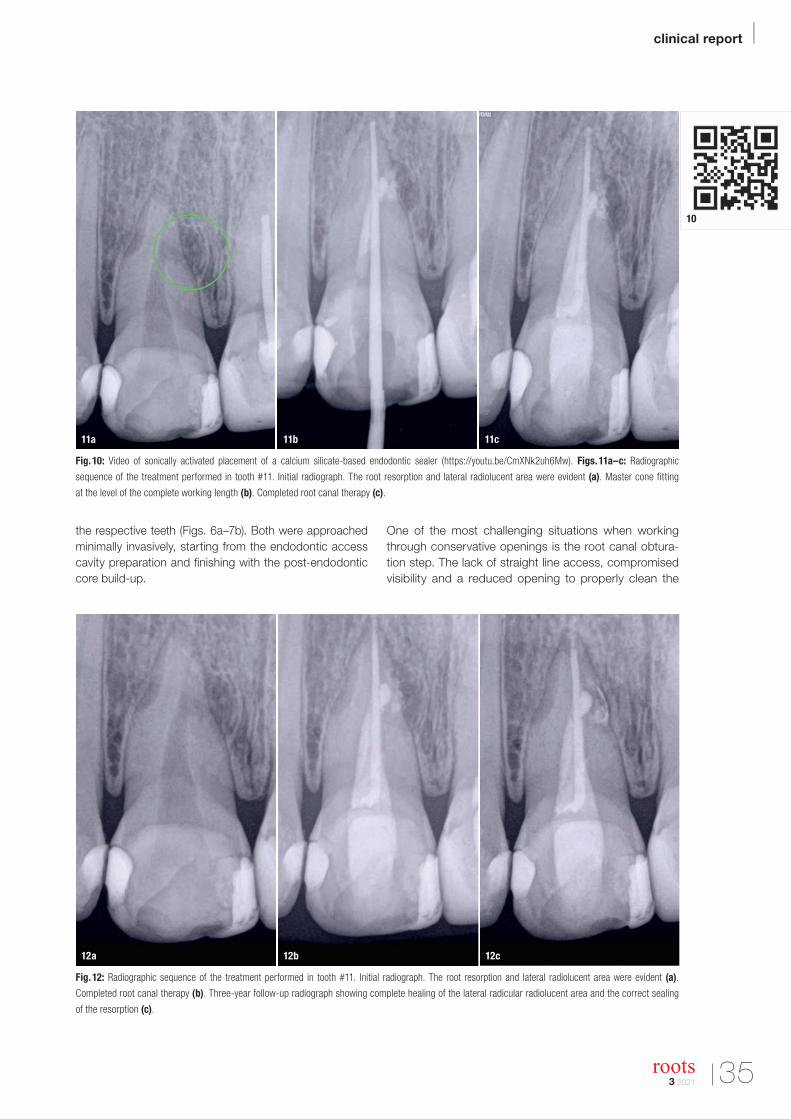
the respective teeth (Figs. 6a–7b). Both were approached minimally invasively, starting from the endodontic access cavity preparation and �nishing with the post-endodontic core build-up.
One of the most challenging situations when working through conservative openings is the root canal obtura-tion step. The lack of straight line access, compromised visibility and a reduced opening to properly clean the
Fig. 10: Video of sonically activated placement of a calcium silicate-based endodontic sealer (https://youtu.be/CmXNk2uh6Mw). Figs. 11a–c: Radiographic
sequence of the treatment performed in tooth #11. Initial radiograph. The root resorption and lateral radiolucent area were evident (a). Master cone �tting
at the level of the complete working length (b). Completed root canal therapy (c).
Fig. 12: Radiographic sequence of the treatment performed in tooth #11. Initial radiograph. The root resorption and lateral radiolucent area were evident (a).
Completed root canal therapy (b). Three-year follow-up radiograph showing complete healing of the lateral radicular radiolucent area and the correct sealing
of the resorption (c).
11a
12a 12b 12c
11b 11c
10
clinical report |
3 2021 35roots

pulp chamber are factors to take into consideration.15 The physical properties of calcium silicate-based endodontic sealers allow clinicians to overcome those challenging as-pects. The relatively easy obturation technique, �owability and easy process of cleaning the pulp chamber provide the necessary means for proceeding with the endodontic therapy as conservatively as possible without compro-mising the prognosis. In the clinical cases presented, the Aurum Blue system (Meta Biomed) was used for the root canal shaping and the calcium silicate-based endodontic sealer CeraSeal (Meta Biomed) was used, applied with the cold hydraulic compaction technique (Fig. 8).
Radicular resorption repair
Iatrogenic root perforations and resorptive defects are un-desirable events that result in communication between the root canal system and the periodontium, causing a long- standing in�ammatory response and the potential loss of alveolar bone. The role of calcium silicate-based materials in this scenario has been well de�ned since MTA was intro-duced, proving the ability to seal the perforation site and to promote tissue regeneration.16 Some of the current bioactive materials developed as premixed pastes and in putty con-sistency have been released to be used as an alternative to MTA. Faster setting, superior handling properties and sim-ilar biocompatibility to MTA are some of their advantages.17
Case 4
A 35-year-old male patient reported pain affecting the cen-tral incisors. Upon clinical examination, an acute periapical abscess was detected; radiographically, resorptive radic-ular defects in tooth #11 were observed. The patient re-ported a traumatic injury in the area several years before. The established diagnosis was pulp necrosis and an acute periapical abscess.
During the �rst appointment for root canal therapy, it was not possible to reach the complete length of the root canal, because the �les tended to go inside of the mesial resorp-tion. It was decided to seal the resorptive defect (Figs. 9a–c) using CeraSeal sonically activated using the EQ-S device (Meta Biomed; Fig. 10). At the second appointment, the material placed at the resorption was completely hard. That hard barrier allowed the �le to pass through the entire length of the physiological root canal. The root canal therapy was �nished using CeraSeal as the endodontic sealer (Figs. 11a–c). The three-year follow-up radiograph showed complete heal-ing of the lateral radicular radiolucent area (Figs. 12a–c).
Periapical surgery
An ideal root end �lling material needs to possess sev-eral characteristics, such as good biocompatibility, excel-lent sealing ability, bactericidal or at least bacteriostatic, and capacity to stimulate periapical tissue healing.18 The gold standard material for root end �lling in periapical sur-gery has been MTA for several years, although it presents some manipulation challenges to the clinician. Conse-quently, premixed bioceramic materials in the form of putty or paste have been developed that present similar proper-ties to those of MTA, but with better handling properties.19
Case 5
A 50-year-old male patient presented to the dental of�ce with Grade III mobility and intra-oral swelling affecting tooth #21. The patient also reported pain when biting on
Fig. 15: Ultrasonic retro-preparation of tooth #11 during periapical surgery. Fig. 16: Retro-obturation of tooth #11 during periapical surgery using CeraPutty.
Fig. 17: Radiograph after periapical surgery showing the well-adapted and dense retro-obturation.
Fig. 13: Initial radiograph of teeth #11 and 21. Radiolucent areas in both teeth
were visible, and cemental tears were present in tooth #21. Fig. 14: Bone
grafting material placed at the extraction site of tooth #21.
13
15 16 17
14
36 3 2021roots

tooth #11 (Fig. 13). The established diagnosis for tooth #21 was previously treated and acute periapical abscess; cemental tears at mesial and distal root surfaces were de-tected, as well as a root fracture. The established diagno-sis for tooth #11 was previously treated and symptomatic periapical periodontitis.
Tooth #21 was extracted, and a bone grafting material (DM Bone, Meta Biomed) was placed in the area to prepare the site for receiving an implant in the future (Fig. 14). Four weeks after the extraction, periapical surgery was performed on tooth #11, and the minimum amount of root was resected in order to avoid compromising the crown–root ratio. The retrocavity was prepared using ultrasonic tips (Fig. 15), and CeraPutty was used to retro-obturate the root (Figs. 16 & 17). The sutures were removed �ve days after surgery (Figs. 18a–c). The four-month follow-up radiograph showed the process of healing in both treated areas (Fig. 19).
Conclusion
Nowadays in the �eld of endodontics, it is important to have a sealer material that ful�ls almost all of the ideal prop-erties described by Grossman, that is supported by evi-dence-based literature20 and that is easy to manipulate by practitioners who give pre-eminence to saving the natural dentition. Saving the natural teeth has great human and professional value, especially in the era of dental implants, where misdiagnosis might prematurely condemn the pa-tient to losing the natural dentition. During the daily jour-ney of endodontists, giving the best of themselves to pre-serving the natural dental organs, calcium silicate-based endodontic materials are an excellent ally because of their capacity to promote both healing and tissue mineralisa-tion, their excellent biocompatibility, their remarkable seal-ing properties, their hydrophilicity and their comfortable handling, among other properties. Access to good materials
and equipment, combined with the skills of experienced dental practitioners, is essential to ensure successful results during the journey of saving natural teeth.
Editorial note: A list of references is available from the publisher.
contact
Dr Jenner Argueta earned his degree in dentistry and master’s degree in endodontics from the Universidad de San Carlos de Guatemala, where he achieved multiple awards as an outstanding student. He is a certi� ed researcher at the Guatemalan national council for science and technology and teaches endodontics at the Universidad
Mariano Gálvez de Guatemala. Dr Argueta also runs a clinical practice focused on micro-endodontics and micro-restorative dentistry. He was president of the Academia de Endodoncia de Guatemala (endodontic academy of Guatemala) from 2016 to 2020 and is the Guatemalan representative for the Latin American Endodontic Association. Dr Argueta can be contacted at [email protected].
Dr Benjamín Rodríguez earned his degree in dentistry from the Universidad Francisco Marroquín in Guatemala and his MSc in endodontics from the Universidad de San Carlos de Guatemala. In 2019, after six years of clinical practice limited to endodontics, he earned a postgraduate degree in oral and maxillofacial radiology from
the Universitat Internacional de Cataluña in Barcelona in Spain.
Figs. 18a–c: Post-op suturing sequence. Immediate post-op suturing (a). Five-day follow-up clinical image, just before the suture removal (b). Clinical image
immediately post-suture removal, showing good healing of the area (c). Fig. 19: Four-month follow-up radiograph showing the process of healing in both
treated areas, the periapical surgery of tooth #11 and bone grafting at the extraction site of tooth #21.
18a
18b
18c 19
clinical report |
3 2021 37roots

Machine-learning algorithms may help in predicting tooth lossBy Franziska Beier, Dental Tribune International
Little is known about socioeconomic factors in re-lation to tooth loss. In a new study, researchers at the Harvard School of Dental Medicine (HSDM) have devel-oped machine-learning algorithms for predicting tooth loss in adults that—in addition to the obvious parameters such as age and dental care—included patients’ socio-economic factors. The �ndings suggest that these tools may help identify teeth at risk in order to ensure early intervention.
Generally, tooth loss can be prevented if dental disease is caught and treated at an early stage. This is con�rmed by studies that found that patients who attend regular checkups are less likely to lose teeth. However, barri-ers such as access to dental care and high costs can
discourage patients from seeing a dentist. In the US, a decisive factor may be that adult dental coverage is not an essential health bene�t in most public health in surance programs. Owing to this lack of routine care, by the time these patients see a dentist, it is already too late to save the tooth, and extraction becomes the most affordable option. This is where the screening tool could help to identify high-risk patients in time.
According to the researchers, machine-learning meth-ods have been applied in medicine to provide information for clinical decisions; however, they have not been devel-oped to predict oral health outcomes yet. Therefore, the researchers developed and tested �ve algorithms using different combinations of parameters—such as medical
The screening tool was designed in such a way that it can also be used by nondental professionals.
© Iv
anov
a Te
tyan
a/S
hutt
erst
ock
.co
m
REGISTER FOR FREEDT Study Club – e-learning platform
webinars / live operations / online CE events / CE credits
Dentistry's largest online education community
Tribune Group is an ADA CERP Recognized Provider. ADA CERP is a service of the American Dental Association to assist dental professionals in identifying quality providers of continuing dental education. ADA CERP does not approve or endorse individual courses or instructors, nor does it imply acceptance of credit hours by boards of dentistry.This continuing education activity has been planned and implemented in accordance with the standards of the ADA Continuing Education Recognition Program (ADA CERP) through joint efforts between Tribune Group and Dental Tribune Int. GmbH.
@DTStudyClubwww.dtstudyclub.com
| news
38 3 2021roots

conditions and socioeconomic background—to predict tooth loss in adults and to compare the performances of the different tools. To develop the screening tools, the research team used data from nearly 12,000 adults from the National Health and Nutrition Examination Survey.
Socioeconomic characteristics decisive
Comparing the performances of the different algorithms, the researchers found that those models which incor-porated socioeconomic characteristics, such as race and education, were better at predicting tooth loss than those models relying on traditional dental clinical indi-cators alone.
“Our analysis showed that while all machine-learning models can be useful predictors of risk, those that in-corporate socioeconomic variables can be especially powerful screening tools to identify those at heightened risk for tooth loss,” said lead author Dr Hawazin Elani, assistant professor of oral health policy and epidemiology at HSDM, in a university press release.
“This work highlights the importance of social determi-nants of health. Knowing the patient’s education level, employment status, and income is just as relevant for
predicting tooth loss as assessing their clinical dental status,” she added.
In addition to the socioeconomic background of patients, the research team also determined preexisting medical conditions as predictors of tooth loss. “We found that medical conditions—such as arthritis, diabetes, high cholesterol, hypertension and cardiovascular diseases—are among the predictors of tooth loss. Clinicians could use this information to screen patients at high risk for tooth loss and coordinate their referral and dental care,” they stated.
Developed tool may be used by different health care providers
The screening tool was designed to be applied world-wide and in a variety of health care settings, even by nondental professionals, as it assesses the risk for tooth loss without the need for a dental examination. However, any patient deemed at high risk for losing a tooth would still have to undergo an actual examination.
The study, titled “Predictors of tooth loss: A machine learning approach”, was published online on 18 June 2021, in PLOS ONE.
REGISTER FOR FREEDT Study Club – e-learning platform
webinars / live operations / online CE events / CE credits
Dentistry's largest online education community
Tribune Group is an ADA CERP Recognized Provider. ADA CERP is a service of the American Dental Association to assist dental professionals in identifying quality providers of continuing dental education. ADA CERP does not approve or endorse individual courses or instructors, nor does it imply acceptance of credit hours by boards of dentistry.This continuing education activity has been planned and implemented in accordance with the standards of the ADA Continuing Education Recognition Program (ADA CERP) through joint efforts between Tribune Group and Dental Tribune Int. GmbH.
@DTStudyClubwww.dtstudyclub.com
AD
news |

Literature favours air puri�ers as COVID-19 transmission risk mitigantBy Jeremy Booth, Dental Tribune International
The pandemic has been a trying time for dentists, particularly for owner-dentists who must make deci-sions that may in�uence the safety of their treatment team. Recent studies have found that the rate of SARS-CoV-2 transmission in dental settings has been lower than what was expected when the pandemic was declared, and the literature has shown that air puri�ers could play a role in keeping transmission rates down.
Scientists urged the World Health Organization in August last year to recognise that SARS-CoV-2 could be spread through microdroplets that are small enough
to remain airborne for extended periods. By this time, many dental clinic owners had already reviewed and upgraded their supplemental ventilation systems, and many more were considering it. The use of air puri�ers to reduce the risk of transmission in dental settings has since been validated by various scienti�c studies; however, uncertainty remains.
An April 2021 review by the Scottish Dental Clinical Effectiveness Programme (SDCEP) examined recent lit-erature concerning the mitigation of aerosol-generating procedures (AGPs) in dentistry. Many of the research
Air purifying systems have been shown to reduce particle count and fallow time after aerosol generating procedures in dental settings.
© D
on
Pab
lo/S
hutt
erst
ock
.co
m
| news
40 3 2021roots
papers were experimental in nature and not all were peer-reviewed, owing to the abundance of scienti�c research that is currently being conducted and the urgency of publishing it. The SDCEP review found that air cleaners were not recommended to reduce the risk of SARS-CoV-2 transmission, owing to a lack of “higher certainty evidence” on their ef�cacy as a mitigating tool.
One of the studies that the SDCEP review analysed had been published by UK researchers in November 2020 under the title “A clinical study measuring dental aero-sols with and without a high-volume extraction device” in the British Dental Journal. The researchers measured and compared particle count during AGPs that were conducted with and without the use of high-volume extraction.
They wrote: “Without the use of an external high- volume extraction device during aerosol generating pro-cedures, there is a signi�cant increase of PM2.5- and PM10-sized particle count from the use of micromotor high-speed, air turbine high-speed, slow-speed and ultrasonic handpieces.”
Extra-oral scavenging found to reduce particle count
The SDCEP review also cited a study, titled “Fallow time determination in dentistry using aerosol measurement”, by researchers from the UK which was published on the preprint server medRxiV in January. The study examined the calculation of fallow time after AGPs in care facilities that were mechanically ventilated and non-mechanically ventilated, and whether extra-oral scavenging (EOS) could reduce aerosol production and fallow time. They found that AGPs carried out in mechanically ventilated treat-ment rooms produced a low particle count, which returned to baseline within a period of 10 minutes after AGPs. EOS was found to have a greater effect in non-mechanically ventilated environments, and to reduce the spike in particle counts in mechanically ventilated rooms.
The authors wrote: “AGPs should not be carried out in surgeries where ventilation is not possible. Mechanical ventilation for AGPs should be gold standard; where not available or practical then the use of natural ventilation with EOS helps reduce FT.”
In May, researchers in Germany studied small particle concentration caused by AGPs during dental student training under high-�ow suction both with and without the use of a mobile EOS device. Lower particle count was detected after high-speed tooth preparation when an EOS device was used. The researchers found that high-�ow suction was effective in reducing the small
particle count, which was further reduced through the use of an EOS device.
“The additional use of an EOS device should be care-fully considered when performing treatments, such as high-speed tooth preparation, that generate particularly small particles when more people are present and all other protective options have been exhausted,” the authors wrote.
The study, titled “The ef�cacy of an extraoral scaveng-ing device on reducing aerosol particles ≤ 5 µm during dental aerosol-generating procedures: An exploratory pilot study in a university setting”, was published in BDJ Open in May 2021.
High-volume aspiration found to be viable mitigation strategy
A study by researchers in Leeds in the UK aimed to evaluate risk mitigation strategies during AGPs by mea-suring viral air concentrations during crown prepara-tion and root canal access procedures. The procedures were carried out using an air turbine or high-speed contra-angle handpiece (HSCAH) with the mitigation strategies rubber dam or high-speed aspiration or with no mitigation strategy.
The researchers wrote: “Compared to an air turbine, the HSCAH reduced settled bioaerosols by 99.72%, 100.00%, and 100.00% for no mitigation, aspiration, and rubber dam, respectively. Bacteriophage concen-trations in the air were reduced by 99.98%, 100.00%, and 100.00% with the same mitigations. Use of the HSCAH with high-volume aspiration resulted in no detectable bacteriophage, both on non-splatter set-tle plates and in air samples taken 6 to 10 minutes post-procedure.”
The researchers concluded that the use of HSCAH together with high-volume aspiration or a rubber dam meant that a prolonged fallow period could be avoided. “Equipping our dental surgeries with these tools will be crucial to protecting the health, safety, and future of dental teams and services,” they wrote.
The study, titled “Dental mitigation strategies to reduce aerosolization of SARS-CoV-2”, was published in the Journal of Dental Research in August.
The results of a number of studies suggest that air puri�ers do help to reduce the risk of SARS-CoV-2 transmission in dental settings; however, as the SDCEP review found, the subject requires further study, meaning that owner-dentists may have to personally appraise the literature before a general consensus can be reached.
news |
3 2021 41roots

The impacts of the pandemic on dental practiceA shift in the oral health paradigmDr Gary Glassman, Canada
Over a year into the COVID-19 pandemic, dental practices continue to face unprecedented challenges on both per-sonal and professional levels. We have had to cope with the universal concerns about health risks to our patients, our families and ourselves. Added to our concerns for physi-cal well-being are those of �scal health, as the uncertainty around the economic consequences of the pandemic on our clinic operations becomes more evident.
In an American Dental Association COVID-19 impact poll, over half of the participating dentists reported an increase in bruxism and damaged teeth since the pandemic hit.1
Concurrently, approximately 30% of the clinicians re-ported a rise in periodontal disease among their patients.1
Our goal is to keep our patients healthy as we work to re-verse the upward trend of oral disease. The links between oral health, physical well-being and a strong immune system must be at the forefront of our efforts to educate patients. Let us look more closely at issues of concern and their solutions.
Fatigue
Depending on the location of the dental of�ce, visits to the dental hygienist vary between being �at to being down by as much as 80%. Just the donning and dof�ng of personal protective equip-ment repeatedly and the dif�culty breathing through an N95 respirator are suf�cient to give rise to physical and mental ex-haustion. Conversely, the extra time now available can help our hygienists focus on patient education with regard to periodon-tal health and the importance of undertaking preventive care.
Social isolation, mental well-being and oral health
COVID-19 is having a negative impact on the mental well-being of us all. A study from the Centre for Addiction and
Mental Health in Toronto in Canada shows that, although we are “beginning to see a decrease in anxiety levels, the rates of depression and loneliness remain elevated”.2 Moreover, research has shown that there is a two-way connection be-tween mental health and oral hygiene.3 During the Canadian lockdown, not only have Canadians had little or no access to professional oral care, but many have also been using sub-stances such as alcohol, cannabis and tobacco,2 which may have negative consequences to physical and mental wellness.
This is while individuals with pre-existing eating dis-orders such as bulimia and anorexia, with the attendant adverse oral effects, have experienced signi�cant worsen-ing of symptoms owing to increased emotional strain and reduced access to treatment.4
Increased unhealthy eating habits (such as day-long snacking and sipping on soft drinks and fruit juice) to cope with the isolation-induced despair have led to an increased incidence of dental erosion.
All told, the challenges to oral and physical well-being are mounting. It is well known that periodontal disease, one of the most common chronic in�ammatory diseases, can wreak havoc on our patients’ systemic health over time and, in some cases, exacerbate the adverse sequelae of patients with cardiovascular disease, diabetes or cognitive impairment.
The solutions
We are still in the pandemic, and our patients still need to achieve healthy smiles.
Compassion Many patients are paying fewer or no visits to the dentist simply owing to the cost. Some have lost their health in-
© K
iefe
rPix
/Shu
tter
sto
ck.c
om
| opinion
42 3 2021roots

surance; others choose to spend their reduced budgets on family necessities. It is important to identify these patients,educate them on the long-term economic and health ben-e�ts of preventive care, be empathetic, triage treatment needs and offer �nancial arrangements, when appropriate, so that they can continue with their oral care.
Elevated communicationPatient engagement can go a long way. As practices are experiencing challenges navigating the pandemic proto-cols and restrictions, it has become clear that patient com-munication is more important than ever, both prior to and between maintenance visits. It adds value to the patient experience and strengthens patients’ relationship with the dental care team.
Many patients are still avoiding regular dental visits owing to COVID-19 concerns. It is critical to educate these patients about the consequences of neglecting oral care, explaining the importance of maintenance visits and how these visits allow early detection of dental or periodontal disease.
Whether it is by e-mail, text message or phone call, inform-ing patients about the safety measures put in place, as-suring them that it is safe to visit the dentist and facilitating their navigation through pandemic protocols will encour-age them to attend their appointments.
Patient outreach before visits can be the �rst step in the patient communication journey, and continuing that con-nection between visits reinforces patients’ trust in a way that goes beyond their time in the chair. This is an import-ant contributor to the success and survival of the dental practice. Extending appointments can help reduce team stress while allowing for increased time for patient educa-tion and, most importantly, an opportunity to address fears and answer questions.
Presenting the science and emphasising home care
Oral self-care has never been more important. All patients, regardless of their unique needs, look to us as dental pro-fessionals to guide them in achieving their oral �tness goals, be it primary prevention or reversing the effects of periodontal disease.
One way we can help our patients is by offering them clear, easy and effective solutions, which includes placing evidence-based tools at their �ngertips. For example, we know that electric tooth-brushes with sonic and oscillating–rotating technologies are signi�cantly more ef�cient in reversing gin-givitis and removing plaque5 than are manual toothbrushes.6
Stannous �uoride (vs sodium �uoride) dentifrices, such as Crest Gum toothpastes, are additional examples of oral care aids with a well-established effectiveness pro�le in
bio�lm management and improvement of gingivitis.7, 8 Es-pecially now, with the post-pandemic reduction in ultra-sonic procedures, patients can be reminded that the tooth-paste they use matters when it comes to achieving opti-mum oral health. This is particularly true in endodontically treated cases, especially if the treatment is followed by prostheses or subgingival direct restorations.
There are also programmes that offer affordable and sci-ence-backed solutions designed for oral self-care at home, reminding us that helping patients become healthy starts in our clinics, but continues at home. Online communica-tion tools are available to help improve our reach, consid-ering that most Canadian patients are between the ages of 25 and 59 and that over half of all patients prefer digital communication to phone calls.9
Helping patients feel their best
Patients are always keen to improve their appearance to enhance their mental well-being, but we can only start talking about aesthetics when we are out of the woods with proper oral �tness.
The pandemic has triggered an increased awareness of and interest in home self-care, and the upward trend of demand for effective, easy-to-use, versatile solutions is here to stay. Patients rely on us to educate them about the best products available to maintain their oral health and physical well-being.
Editorial note: A list of references is available from the publisher. This article originally appeared in Oral Health Magazine (May 2021), and an edited version is provided here with permission from Newcom Media.
contact
Dr Gary Glassman is the author of numerous publications and lectures globally on endodontics and is on the staff at the University of Toronto’s Faculty of Dentistry in Canada in the graduate department of endodontics. As adjunct professor of dentistry and director of endodontic programming from 2010 to 2017, Dr Glassman
helped develop the dental school curriculum for the oral health science programme for the University of Technology in Kingston in Jamaica. He is faculty chair for DC Institute and chief dental of� cer for dentalcorp in Canada, as well as endodontic editor of the Oral Health Magazine and Inside Dentistry. Dr Glassman is a fellow of the Royal College of Dentists of Canada and the American College of Dentists. His personal website is www.drgaryglassman.com, and his of� ce website is www.rootcanals.ca. He can be reached at [email protected].
© K
iefe
rPix
/Shu
tter
sto
ck.c
om
3 2021 43roots

Slow Dentistry global network growingBy Nina Blaettler, Switzerland
We at the Slow Dentistry global network have been very busy over the last few years. We have grown our membership to over 40 countries and have expanded our partnerships and sponsorships to include some of the leading names in the industry.
We are proud to announce that, after a rigorous regula-tory process conducted by the government of Switzer-
land, the Slow Dentistry global network is now of�cially part of a new non-pro�t foundation, the Foundation for Excellence in Dentistry, and it is now this foundation that fully owns and operates Slow Dentistry.
Thinking about when we �rst launched Slow Dentistry, it seems as if we are looking back at a huge historic gulf of change. The world seems to have changed tremendously in so many ways; it is dif�cult to imagine that 18 months ago almost nobody had heard of COVID-19. Here at Slow Dentistry, we believe that much of this change can be used to our advantage in order to improve dentistry, enhance healthcare and create a more ful�lling, stress-free and safer workplace for all oral healthcare profes-sionals around the globe, and we look forward to 2022 with optimism, hope and great expectations.
Thank you all for supporting the foundation and the Slow Dentistry global network. I particularly wish to rec-ognise the contributions of our growing family of mem-bers and of our amazing global ambassadors; it is a real privilege to work with such excellent professionals who go the extra mile to care for their patients.
Nina Blaettler, president of the Foundation for Excellence in Dentistry.
Slow Dentistry advocates longer turnaround times between patients to allow for proper room disinfection, promoting a safer working environment and
quality care. (Image: © Slow Dentistry)
© N
ina
Bla
ettle
r
| practice management
44 3 2021roots

Slow down everyone—dentistry does not need to be done at speedPart 3: “Slow Dentistry has resonated around the world and is growing exponentially”An interview with Dr Miguel Stanley, an ambassador for and the co-founder of Slow Dentistry
By Iveta Ramonaite, Dental Tribune International
Since 2010, Slow Dentistry has been encouraging dental professionals to manage their time at the clinic better and to practise ethical and quality care. Now, more than a decade later, the movement has achieved substantial growth and continues to gain strength de-spite the recent challenges of the COVID-19 pandemic. In this interview with Dental Tribune International, Dr Miguel Stanley, an ambassador for and the co-founder of Slow Dentistry, talks about some of the movement’s proudest achievements so far and about Slow Dentistry’s vision for the future.
An increasing number of dental professionals are joining and advocating for the Slow Dentistry move-ment. What do you consider to be some of your proudest achievements so far?Just the fact that so many great dentists, hygienists and other dental professionals around the world have joined the Slow Dentistry global network, helping us spread the message of ethically driven treatment plan-ning and patient preparation, is a great achievement in itself. Slow Dentistry makes so much sense to peo-ple once they learn more about it. It is very straightfor-ward and requires no investment, no advanced tech-nology and no special training—just a desire to do the right thing for your team and your patients. That is why Slow Dentistry has resonated around the world and is growing exponentially day after day, and I’m very im-pressed with the fact that this growth is mostly organic and not driven by excessive marketing but rather by dentists’ motivation to separate themselves from those clinicians who are focused mostly on volume and pro�t over quality care.
Another major achievement that happened in the last year thanks to the tireless efforts of our president, Nina Blaettler, was the transformation of Slow Dentistry from a limited liability company to a Swiss non-pro�t foun-dation. The foundation is called the FED, which stands for Foundation for Excellence in Dentistry, and this now
means that every single dental professional who signs up to our global network is actually contributing to a cause and not to a private company.
Swiss non-pro�t foundations are highly regulated and their constant monitoring is required to ensure that the funds are used for the greater good. The entire objec-tive of the Slow Dentistry global network is to disrupt what we believe is a bad trend in dentistry, one where clinics are created solely for generating pro�t. There are so many loving and caring dental professionals around the world who needed a way to differentiate themselves from pro�t-seeking clinicians. Our network is expand-ing, and we’ve received numerous contributions from existing and new members, as well as donations from companies within the dental universe. We are gaining greater means to invest in educating the general pub-lic, helping them understand that they have basic rights when visiting a dentist.
Dr Miguel Stanley has been an ardent supporter of Slow Dentistry for over
a decade now and is optimistic about its future. (All images: © Slow Dentistry)
practice management |
3 2021 45roots

Slow Dentistry has four cornerstones, namely proper pa-tient consent to treatment that has been proposed; proper room and equipment disinfection and sterilisation and avoidance of cross contamination in the dental of�ce; proper anaesthesia and analgesia and, �nally, the use of dental rubber dam. These four cornerstones are univer-sally true, and when applied, they ensure that patients have a safer and better experience at the dentist. We stand for ethical and quality dentistry, and hopefully, we can grow bigger and have an even greater impact in the world.
Has Slow Dentistry gained even more strength during the COVID-19 pandemic? If so, why?Since March 2020, the Slow Dentistry global network has had a 550% growth in new memberships. Moreover, we are now also receiving donations from companies that truly support our ethical message. The tireless efforts of all of the volunteers who work for the Slow Dentistry movement have really paid off, and we are incredibly proud of this achievement.
In the last year and a half, we have migrated the website to a completely new platform, allowing our members to have a more �uid and easier-to-use interface, and we are now working with an incredibly talented team of web designers and marketing experts. But I think that the main reason for this incredible growth is the fact that one of our four cor-nerstones is focused on disinfection. We advocate proper room disinfection, not just tidying up the treatment room.
Before the pandemic, many clinics were focused on treating as many patients as possible. All of our members agree that this is not the way to go, that proper room disinfection takes time to ensure that the dental unit, the surfaces, the �oor and everything the patient touches are properly disinfected. There is no way you can do this in a rush, and we are �nd-
ing that, on average, it takes between 8 and 10 minutes to properly prepare a treatment room between patients.
Before the pandemic, many clinics were getting away with just tidying up the treatment room in a few minutes so that they could have faster turnaround times. This has allowed the owners of the clinic to see more patients in a day and to generate more pro�t—at the risk of cross-contamination between patients. This has generated a lot of stress for dental assistants and nurses as well as dentists.
We cannot overlook the burden of mental fatigue and men-tal illness in dentistry. Seeing a high volume of patients a day really does create a lot of stress, and I believe that many dentists did not understand that this stress was probably also subliminally created by the fact that they understood that they were not doing things properly.
So once the pandemic hit, the general public as well as many dentists started understanding the importance of proper disinfection. This has since become a major pri-ority, and while we were the only organisation in the world that had been advocating this for over �ve years, things just blew up. It was amazing! We were �nally vindicated.
I can tell you that in November 2019, in our very �rst Slow Dentistry meeting in London, many dentists found our advo-cating longer turnaround times between patients to promote better safety and reduce the possibility of cross-contaminationlaughable. This is no longer the case. We take this incred-ibly seriously even though we understand that this proba-bly means that clinics need to see fewer patients a day in order to get things right. This might generate less pro�t, but it promotes a safer environment, better care and a more pro-pitious environment for a healthier mindset and less stress, which leads to better mental health at the end of the day.
According to Dr Miguel Stanley, dental patients should be made aware of their basic rights when visiting a dentist, including a disinfected treatment room
and pain-free dental treatment.
| practice management
46 3 2021roots

Have you noticed any post-pandemic trends in den-tistry related to the movement?One thing we have noticed is that some copycat organisa-tions and academies are trying to imitate what we are doing, but are seeking to pro�t �nancially from this. We would hope that people understand that, as a non-pro�t foundation, the objective of our movement is not to make personal pro�t, as all of our team members are volunteers and so are our hon-orary global ambassadors around the globe. We don’t sell weekend courses, no one is trying to gain personally from this and we are focused on getting our message to as many people in the world as possible, in as many languages as possible. Everyone in our network is inclusive and positive and wants the best for dentistry and for patients.
I think that millennial dentists have a different mindset in terms of their relationship with their patients and a work–life balance. I think that mental health has become a big issue, and there are a lot of great dentists that are publicly talking about this im-portant topic. Two of our global ambassadors, Drs Kyle Stanley and Simon Chard, use their platforms to promote mental health in dentistry. At the end of the day, there is a common thread to all of these discussions, and that is to do the right thing.
Allowing younger dentists more time to get things right is im-perative in moving forward. Big-chain dental clinics or dental service organisations need to make sure that their younger dentists have all the time they need to work stress-free and to get things right the �rst time around. If we don’t facilitate this, we will soon be seeing a mental health pandemic in our profession, not to mention poor-quality dentistry.
The truth is that the Slow Dentistry global network should not be necessary. Every dentist around the world should be practising it, but they’re not, and that is why it’s important that people understand our core values. We are not selling anything, no courses are needed to understand it and no-body is pro�ting from anything here; it’s truly a humanitar-ian effort on a global scale, and we are all in this together.
What is next for Slow Dentistry, and what is the ulti-mate goal?One of the major decisions from Nina this year was that we would move slowly into the future. We don’t want to deviate from our core values and mission. We would like to grow our network to be in every single country around the world, supporting those dentists with marketing at a grass-roots level, ensuring that populations around the world understand that they have the right to a disinfected treatment room, a signed, valid consent form, pain-free dental treatment and a dental dam for all of their root ca-nal therapy and most of their restorative work. If we get this message across, we will have already made a deep impact. We are going to stay very true to our mission.
We are also working closely with companies and helping their marketing departments understand the value of do-nating to our global network, since by supporting us they are indirectly promoting ethical dentistry, which I truly be-lieve should the core value of every company. We want to support companies by supporting quality dentistry. You simply can’t practise quality dentistry in a rush. Everybody wins by practising Slow Dentistry.
We’re also going to be coming out with some new badges that can be acquired by dental professionals who are al-ready members of the Slow Dentistry global network. These include the digital dentistry badge, the small busi-ness owner badge and the educator badge. Staying true to our message, these new areas will help identify prac-titioners who go the extra mile and make the extra invest-ments both in money and in time to better serve their pa-tients and their communities. For example, a dentist who uses surgical guides to place dental implants spends a lot of time planning the treatment and coordinating and designing cases just so that patients can have a more accurate and precise intervention, and that might actually end up requir-ing less chair time. These guys deserve recognition.
This is almost counter-intuitive if you think about it. Slow Dentistry doesn’t mean more time in the chair or work-ing slowly; Slow Dentistry is about time invested to deliver quality care. So we believe that dentists who use digital technologies to mitigate failure and improve work�ows deserve to be rewarded or at least recognised for the time spent investing, training and preparing these work�ows to better treat their patients.
The small business owner badge will identify clinicians who ful�l the following criteria: they only work in one clinic, own more than 50% of that clinic and are the clinical director of the practice. Why is this important? When you have all of your eggs in one basket, you tend to protect the basket a lot more. Through discussions with colleagues around the globe, we have found anecdotal evidence that, when these factors are in play, there is a lot more heart in the game and people care a lot more. Obviously, this doesn’t make you a better dentist, and there is nothing wrong with having many clinics, but some-times patients might want a dentist who is simply a dentist and not also a businessman, and they need a way to distinguish.
Practising Slow Dentistry involves using the appropriate tools and developing
the right mindset.
practice management |

Dentists who collect: Dr Kenneth Montague of TorontoAn interview with Dr Kenneth Montague, a dentist and art collector
By Jeremy Booth, Dental Tribune International
Although he has a growing art collection and is busy with many other activities, general dentist Dr Kenneth Montague says that his patients still come �rst. Mon-tague began exhibiting art in his home in the 1990s and has since built up the Wedge Collection—one of Can-ada’s largest private art collections exploring African diasporic culture and contemporary Black life. Dental Tribune International spoke with Montague about how music and the arts have shaped his dental practice, about his collection and the photographs currently on display at his clinic, and about expressing oneself as a dentist.
Dr Montague, thank you very much for speaking with us. Could you tell us something about your background?I came out of dental school very young, at 24, and spent the �rst half of my life in Windsor in Ontario. I grew up with Jamaican-born parents who had been early im-migrants to Canada and who lived in that community.
There was a bit of a Caribbean culture in the house, par-ticularly in terms of food and music; and then Detroit, Michigan, was just across the river, and the city had, at that time, in the 1970s, a vibrant African American cul-ture which was very visible not only in music and �lm but also in the politics of the time. At the same time, I also had this very typical Canadian youth experience, which was all about popular music and popular culture, and so the blend of these in�uences created something which was quite unique and which has really in�uenced me as an art collector.
Could you introduce your collection to us?I have the privately-owned Wedge Collection, and a non-pro�t arts organisation called Wedge Curatorial Projects, which has a focus on the emerging artistic practices of artists of African descent and an increas-ing emphasis on Black Canadian artists. My primary interest has been Black identity and Black history, so for me, that very varied and rich local history is re-
Dr Montague at Word of Mouth Dentistry. (Image: © Justin Aranha)
| interview
48 3 2021roots
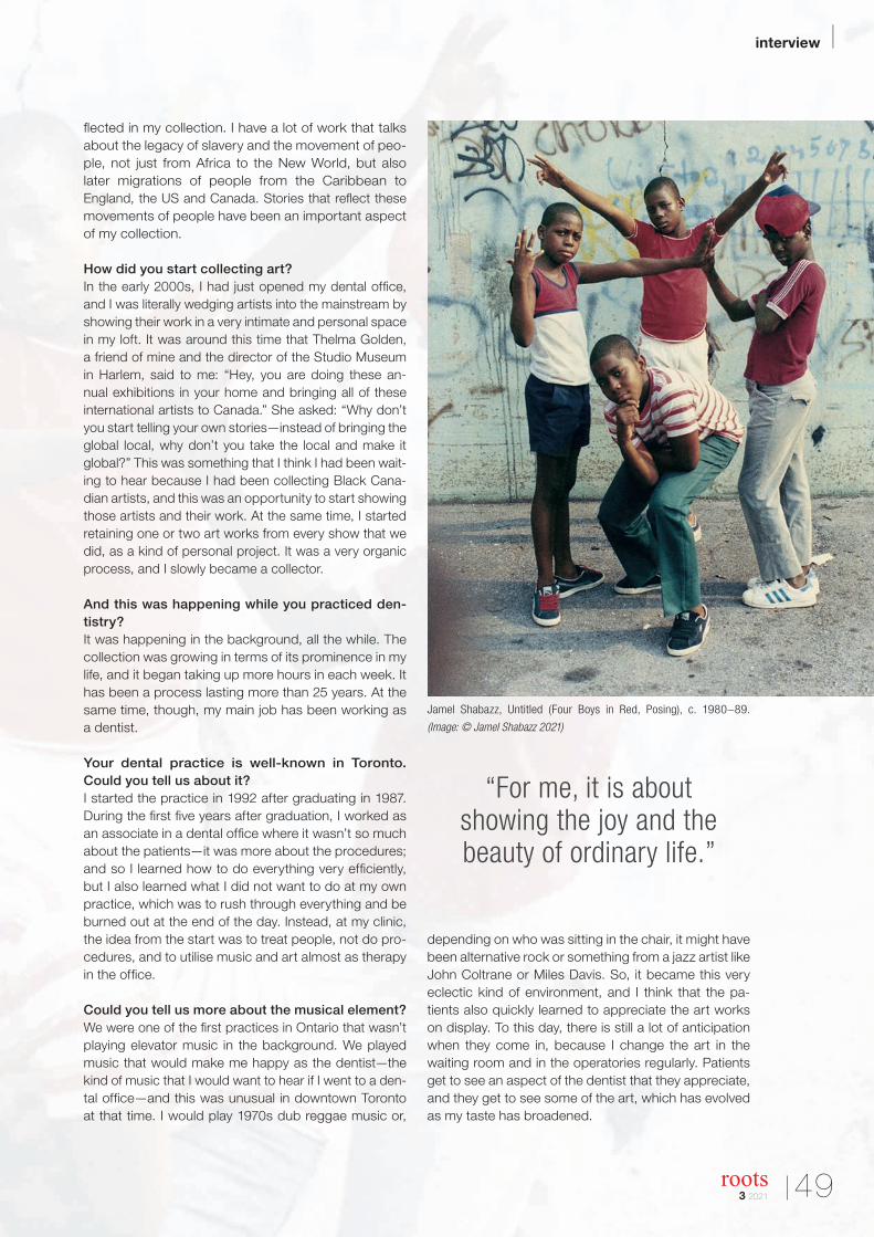
�ected in my collection. I have a lot of work that talks about the legacy of slavery and the movement of peo-ple, not just from Africa to the New World, but also later migrations of people from the Caribbean to England, the US and Canada. Stories that re�ect these movements of people have been an important aspect of my collection.
How did you start collecting art?In the early 2000s, I had just opened my dental of�ce, and I was literally wedging artists into the mainstream by showing their work in a very intimate and personal space in my loft. It was around this time that Thelma Golden, a friend of mine and the director of the Studio Museum in Harlem, said to me: “Hey, you are doing these an-nual exhibitions in your home and bringing all of these international artists to Canada.” She asked: “Why don’t you start telling your own stories—instead of bringing the global local, why don’t you take the local and make it global?” This was something that I think I had been wait-ing to hear because I had been collecting Black Cana-dian artists, and this was an opportunity to start showing those artists and their work. At the same time, I started retaining one or two art works from every show that we did, as a kind of personal project. It was a very organic process, and I slowly became a collector.
And this was happening while you practiced den-tistry?It was happening in the background, all the while. The collection was growing in terms of its prominence in my life, and it began taking up more hours in each week. It has been a process lasting more than 25 years. At the same time, though, my main job has been working as a dentist.
Your dental practice is well-known in Toronto. Could you tell us about it?I started the practice in 1992 after graduating in 1987. During the �rst �ve years after graduation, I worked as an associate in a dental of�ce where it wasn’t so much about the patients—it was more about the procedures; and so I learned how to do everything very ef�ciently, but I also learned what I did not want to do at my own practice, which was to rush through everything and be burned out at the end of the day. Instead, at my clinic, the idea from the start was to treat people, not do pro-cedures, and to utilise music and art almost as therapy in the of�ce.
Could you tell us more about the musical element?We were one of the �rst practices in Ontario that wasn’t playing elevator music in the background. We played music that would make me happy as the dentist—the kind of music that I would want to hear if I went to a den-tal of�ce—and this was unusual in downtown Toronto at that time. I would play 1970s dub reggae music or,
depending on who was sitting in the chair, it might have been alternative rock or something from a jazz artist like John Coltrane or Miles Davis. So, it became this very eclectic kind of environment, and I think that the pa-tients also quickly learned to appreciate the art works on display. To this day, there is still a lot of anticipation when they come in, because I change the art in the waiting room and in the operatories regularly. Patients get to see an aspect of the dentist that they appreciate, and they get to see some of the art, which has evolved as my taste has broadened.
Jamel Shabazz, Untitled (Four Boys in Red, Posing), c. 1980–89.
(Image: © Jamel Shabazz 2021)
“For me, it is about showing the joy and the beauty of ordinary life.”
interview |
3 2021 49roots
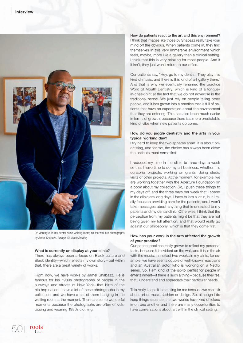
What is currently on display at your clinic?There has always been a focus on Black culture and Black identity—which re�ects my own story—but within that, there are a great variety of works.
Right now, we have works by Jamel Shabazz. He is famous for his 1980s photographs of people in the subways and streets of New York—that birth of the hip hop nation. I have a lot of these photographs in my collection, and we have a set of them hanging in the waiting room at the moment. There are some wonderful moments because the photographs are often of kids, posing and wearing 1980s clothing.
How do patients react to the art and this environment?I think that images like those by Shabazz really take your mind off the obvious. When patients come in, they �nd themselves in this very immersive environment which feels, maybe, more like a gallery than a clinical setting. I think that this is very relaxing for most people. And if it isn’t, they just won’t return to our of�ce.
Our patients say, “Hey, go to my dentist. They play this kind of music, and there is this kind of art gallery there.” And that is why we eventually renamed the practice Word of Mouth Dentistry, which is kind of a tongue-in-cheek hint at the fact that we do not advertise in the traditional sense. We just rely on people telling other people, and it has grown into a practice that is full of pa-tients that have an expectation about the environment that they are entering. This has also been much easier in terms of growth, because there is a more predictable kind of vibe when new patients do come.
How do you juggle dentistry and the arts in your typical working day?I try hard to keep the two spheres apart. It is about pri-oritising, and for me, the choice has always been clear: the patients must come �rst.
I reduced my time in the clinic to three days a week so that I have time to do my art business, whether it is curatorial projects, working on grants, doing studio visits or other projects. At the moment, for example, we are working together with the Aperture Foundation on a book about my collection. So, I push these things to my days off, and the three days per week that I spend in the clinic are long days. I have to jam a lot in, but I re-ally focus on providing care for the patients, and I won’t take messages about anything that is unrelated to my patients and my dental clinic. Otherwise, I think that the perception from my patients might be that they are not being given my full attention, and that would really go against our philosophy, which is that they come �rst.
How has your work in the arts affected the growth of your practice?Our patient pool has really grown to re�ect my personal taste, because it is evident on the wall, and it is in the air with the music. In the last two weeks in my clinic, for ex-ample, we have seen a couple of well-known musicians and an Australian actor who is working on a Net�ix series. So, I am kind of the go-to dentist for people in entertainment—if there is such a thing—because they feel that I understand and appreciate their particular needs.
This really keeps it interesting for me because we can talk about art or music, fashion or design. So, although I do keep things separate, the two worlds have kind of folded in on one another and there are many opportunities to have conversations about art within the clinical setting.
Dr Montague in his dental clinic waiting room; on the wall are photographs
by Jamel Shabazz. (Image: © Justin Aranha)
| interview
50 3 2021roots
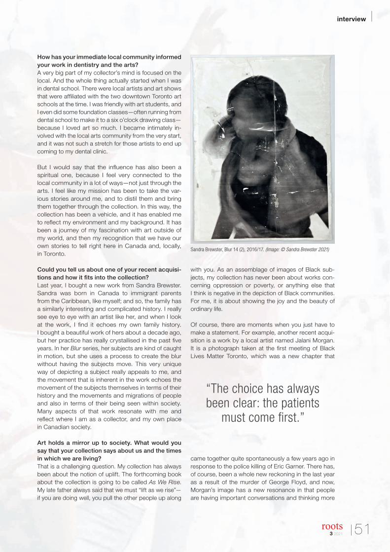
How has your immediate local community informed your work in dentistry and the arts?A very big part of my collector’s mind is focused on the local. And the whole thing actually started when I was in dental school. There were local artists and art shows that were af�liated with the two downtown Toronto art schools at the time. I was friendly with art students, and I even did some foundation classes—often running from dental school to make it to a six o’clock drawing class—because I loved art so much. I became intimately in-volved with the local arts community from the very start, and it was not such a stretch for those artists to end up coming to my dental clinic.
But I would say that the in�uence has also been a spiritual one, because I feel very connected to the local community in a lot of ways—not just through the arts. I feel like my mission has been to take the var-ious stories around me, and to distil them and bring them together through the collection. In this way, the collection has been a vehicle, and it has enabled me to re�ect my environment and my background. It has been a journey of my fascination with art outside of my world, and then my recognition that we have our own stories to tell right here in Canada and, locally, in Toronto.
Could you tell us about one of your recent acquisi-tions and how it �ts into the collection?Last year, I bought a new work from Sandra Brewster. Sandra was born in Canada to immigrant parents from the Caribbean, like myself; and so, the family has a similarly interesting and complicated history. I really see eye to eye with an artist like her, and when I look at the work, I �nd it echoes my own family history. I bought a beautiful work of hers about a decade ago, but her practice has really crystallised in the past �ve years. In her Blur series, her subjects are kind of caught in motion, but she uses a process to create the blur without having the subjects move. This very unique way of depicting a subject really appeals to me, and the movement that is inherent in the work echoes the movement of the subjects themselves in terms of their history and the movements and migrations of people and also in terms of their being seen within society. Many aspects of that work resonate with me and re�ect where I am as a collector, and my own place in Canadian society.
Art holds a mirror up to society. What would you say that your collection says about us and the times in which we are living?That is a challenging question. My collection has always been about the notion of uplift. The forthcoming book about the collection is going to be called As We Rise. My late father always said that we must “lift as we rise”—if you are doing well, you pull the other people up along
with you. As an assemblage of images of Black sub-jects, my collection has never been about works con-cerning oppression or poverty, or anything else that I think is negative in the depiction of Black communities. For me, it is about showing the joy and the beauty of ordinary life.
Of course, there are moments when you just have to make a statement. For example, another recent acqui-sition is a work by a local artist named Jalani Morgan. It is a photograph taken at the �rst meeting of Black Lives Matter Toronto, which was a new chapter that
came together quite spontaneously a few years ago in response to the police killing of Eric Garner. There has, of course, been a whole new reckoning in the last year as a result of the murder of George Floyd, and now, Morgan’s image has a new resonance in that people are having important conversations and thinking more
Sandra Brewster, Blur 14 (2), 2016/17. (Image: © Sandra Brewster 2021)
“The choice has always been clear: the patients
must come � rst.”
interview |
3 2021 51roots

deeply about police brutality and racial injustices. My collection has always shown the reality of lives lived. You have to balance the desire for uplift with the need to convey the urgency of this moment.
Thank you for these personal re�ections. One last question: what advice would you give to other den-tists when it comes to expressing themselves?
As small business owners, we are taught that the customer is always right. But I think that you have to satisfy yourself, as the dentist, �rst. In my opinion, the way to go is to create an environment that makes you happy in your workspace. For me, that has been to
surround myself with the music that I love, however eclectic it might be, and the art that I love, however speci�c it might be.
Obviously, there are limits in terms of appropriateness, but if your practice re�ects who you are, then it will grow in a predictable way, with people who are like-minded, who will understand what you are doing. And it makes your practice a way more fun place, a way easier place, to be. I feel that I am attracting people who speak my language, who understand my philosophy—not only about dentistry but also about life.
I have found that people are less concerned with the technical aspects of dentistry than they are with your chairside manner. They recognise that, if you have been practising for over 30 years, as I have, then you should have expert knowledge in your �eld.
So, for me, re�ecting who you are is going to make your life easier as a dentist. In my case, it is contemporary art that helps me do this. You might be someone who loves sports, and so your of�ce might broadcast football games on the TV and have sports memorabilia and a themed waiting room. That’s okay, the dentist has to be happy �rst, and this is often a very personal thing. If you love art, then I think that you shouldn’t be afraid to show it on the wall—and share that lovewith your patients.
Jalani Morgan, Black Lives They Matter Here, 2014. (Image: © Jalani Morgan 2021)
“I have found that people are less concerned with the
technical aspects of dentistry than they are with your
chairside manner.”
| interview
52 3 2021roots

PRAGUE
26–29 May 2022Prague, Czech Republic
www.ROOTS-SUMMIT.com
is coming to
Tribune Group GmbH is an ADA CERP Recognized Provider. ADA CERP is a service of the American Dental Association to assist dental professionals in identifying quality providers of continuing dental education. ADA CERP does not approve or endorse individual courses or instructors, nor does it imply acceptance of credit hours by boards of dentistry. Tribune Group GmbH designates this activity for 18.5 continuing education credits. This continuing education activity has been planned and implemented in accordance with the standards of the ADA Continuing Education Recognition Program (ADA CERP) through joint efforts between Tribune Group GmbH and Dental Tribune International GmbH.

At the funfair, some love the ups and downs on the roller coaster; others love going round in circles on the carousel. Mechanical root canal preparation follows a similar pattern of motion: dentists can choose between reciprocating and continuously rotating systems. For fans of preparation using reciprocating motion, dental specialist COLTENE now features a new � le in its range.
For all situationsSince the introduction of the � rst reciprocating � le over a decade ago, reciprocating motion has become established among many dentists. COLTENE is now expanding its portfolio by adding to its proven nickel–titanium rotary systems: MicroMega One RECI, the single shaping � le in reciprocating motion. This enables dentists to choose their favourite � le system from COLTENE regardless of which type of motion they prefer to use.
Safe, � exible and minimally invasiveInspired from its patented cross section, MicroMega One RECI is characterised by its cutting performance. Additionally, the C.Wire heat treatment lends � exibility, controlled memory, pre-bendability and curvature conservation to the � le.
Owing to the small � le diameter of 1 mm, MicroMega One RECI protects the peri-cervical area and minimises mechanical im-pact on the dental hard tissue. Its high cyclic fatigue resistance provides additional safety. As a result, dentists can enjoy an excellent compromise between cutting ef� ciency and safety, as well as � exibility and minimally invasive use, in root canal therapy.
These advantages are also convincing in practice, as the Italian endodontic specialist Dr Davide Mancino points out: “What impressed me most about the One RECI is its extreme respect of the root canal anatomy and its resistance to fracture. In my
opinion, the One RECI is the safest, most powerful and minimally invasive reciprocating single-� le system on the market today.” Furthermore, MicroMega One RECI is available in � ve sizes, from 20/.04 to 45/.04, and three lengths (21, 25 and 31 mm), allowing optimal preparation of the root canal for almost any clinical case. The � les are supplied exclusively in sterile blisters.
Advice and supportInterested dentists are welcome to view an informative short video about the MicroMega One RECI � le system on the COLTENE YouTube channel at https://youtu.be/USlVhv736qE. For optimal integration of the instruments into the practice system and for any questions regarding application in endodontics, the experts from the COLTENE sales team are always on hand to offer help and advice. For dentists who are interested in learning more about the latest trends and ideas from the dental world, COLTENE also offers a wide range of education courses, practical workshops and webinars.
www.coltene.com
New � le for reciprocating preparation
COLTENE launches MicroMega One RECI
| manufacturer news
54 3 2021roots

FKG Dentaire presents the Rooter X3000, rede�ning endodontic standards. This novel cordless endodontic motor combines technology, functionality and adaptability in a 20 cm unit. It stands out for the fastest speed on the market (3,000 rpm), ten programmable memories and preset programs for different types of FKG �les. Its ergonomic design, light weight and 360° rotatable extra-slim contra-angle head ensure perfect handling and visibility.
Every new FKG product responds to the most exacting needs of endodontists with leading-edge technology and uncompromising quality. The Rooter X3000 advances endodontics with a new cord-less, brushless endodontic motor with integrated apex locator.
Adaptability as technical keywordBenchmark speed coupled with guaranteed stability and preci-sion are only the tip of the iceberg of Rooter X3000’s features. Adaptability re�ects its core, starting with the four FKG presets: XP-endo Treatment, XP-endo Retreatment, RACE EVO and R-Motion. This saves time for endodontists, who can rely on the manufac-turer’s prede�ned recommendations for each �le type. The end-odontic motor also provides ten user-programmable memories
for all modes (EAL, CW, CCW, REC or ATR), more than 60 speeds and—of course—a high-precision built-in apex locator and a range of automatic apical functions.
Agility as practical credoAs with any device, practice validates innovation. With its ergo-nomic handpiece weighing only 157 g, perfect balance and fully rotatable ultra-slim contra-angle head, the Rooter X3000 en-sures a comfortable grip, agile handling and excellent visibility of the operating �eld. FKG has designed and developed all details to provide maximum freedom of use to the practitioner, opti-mising both speed and ef�ciency. Features include complete adaptability of the device and its display for both left- and right-handed users, a broad range of user-controlled settings, extensive �le brand compatibility, wireless charging stand, and volume control.
www.fkg.ch
Editorial note: Rooter, XP-endo, RACE EVO and R-Motion are registered trademarks of FKG company.
High-speed cordless endodontic motor
FKG Dentaire expands its endodontic motor range with the new Rooter X3000
manufacturer news |
3 2021 55roots

International events
16th International Congress of the InternationalFederation of Dental Anesthesiology Societies
17–19 September 2021Moscow, Russiawww.ifdas.org
World Clinical Laser Institute (WCLI) 2021 Asia Paci�c Symposium
9–10 October 2021 (on-site and online event)Dubai, UAEhttps://events.wcli.org
57th Annual Meeting of the Canadian Academy of Endodontics
18 September 2021 (online event only)Canadawww.caendo.ca
Dentex—International Dental Equipment Exhibition
21–23 October 2021Brussels, Belgiumwww.dentex.be/en
IDS—International Dental Show
22–25 September 2021 (on-site and online event)Cologne, Germanywww.ids-cologne.de
Italian Society of Endodontics (SIE) National Meeting
12–13 November 2021Bologna, Italywww.endodonzia.it
CEDE—Central European Dental Exhibition
7–9 October 2021 Łódź, Polandwww.cede.pl/en
AAE Annual Meeting
27–30 April 2022 (on-site and online event)Phoenix, USwww.aae.org
FDI World Dental Congress
26–29 September 2021 (online event only)Australiawww.fdiworlddental.org/ fdi-world-dental-congress
GNYDM—Greater New York Dental Meeting
26 November–1 December 2021New York, USwww.gnydm.com
© 0
6pho
to/S
hutt
erst
ock
.co
m
| meetings
56 3 2021roots

© 3
2 p
ixel
s/S
hutt
erst
ock
.co
m
How to send us your work Please note that all the textual compo-nents of your submission must be com-bined into one MS Word document. Please do not submit multiple �les for each of these items:
· the complete article; · all the image (tables, charts, photo-
graphs, etc.) captions; · the complete list of sources consulted
and · the author or contact information (bi-
ographical sketch, mailing address, e-mail address, etc.).
In addition, images must not be embed-ded into the MS Word document. All im-ages must be submitted separately, and details about such submission follow be-low under image requirements.
Text length
Article lengths can vary greatly—from 1,500 to 5,500 words—depending on the subject matter. Our approach is that if you need more or fewer words to do the topic justice, then please make the article as long or as short as necessary.
We can run an unusually long article in multiple parts, but this usually entails a topic for which each part can stand alone because it contains so much in-formation.
In short, we do not want to limit you in terms of article length, so please use the word count above as a general guideline and if you have speci�c questions, please do not hesitate to contact us.
Text formatting
We also ask that you forego any special formatting beyond the use of italics and boldface. If you would like to emphasise certain words within the text, please only use italics (do not use underlining or a larger font size). Boldface is reserved for article headers. Please do not use un-derlining.
Please use single spacing and make sure that the text is left justi�ed. Please do not centre text on the page. Do not indent paragraphs, rather place a blank line be-tween paragraphs. Please do not add tab stops.
Should you require a special layout, please let the word processing pro-gramme you are using help you do this formatting automatically. Similarly, should you need to make a list, or add footnotes or endnotes, please let the word process-ing programme do it for you automatically. There are menus in every programme that will enable you to do so. The fact is that no matter how carefully done, errors can creep in when you try to number footnotes yourself.
Any formatting contrary to stated above will require us to remove such formatting before layout, which is very time-consum-ing. Please consider this when formatting your document.
Image requirements
Please number images consecutively throughout the article by using a new number for each image. If it is imperative that certain images are grouped together, then use lowercase letters to designate these in a group (for example, 2a, 2b, 2c).
Please place image references in your article wherever they are appropriate, whether in the middle or at the end of a sentence. If you do not directly refer to the image, place the reference at the end of the sentence to which it relates enclosed within brackets and before the period.
In addition, please note:
· We require images in TIF or JPEG for-mat.
· These images must be no smaller than 6 x 6 cm in size at 300 DPI.
· These image �les must be no smaller than 80 KB in size (or they will print the size of a postage stamp!).
Larger image �les are always better, and those approximately the size of 1 MB are best. Thus, do not size large image �les down to meet our requirements but send us the largest �les available. (The larger the starting image is in terms of bytes, the more leeway the designer has for resizing the image in order to �ll up more space should there be room available.)
Also, please remember that images must not be embedded into the body of the article submitted. Images must be submitted separately to the textual sub-mission.
You may submit images via e-mail or share the �les in our cloud storage (please contact us for the link).
Please also send us a head shot of your-self that is in accordance with the re-quirements stated above so that it can be printed with your article.
Abstracts
An abstract of your article is not re-quired.
Author or contact information
The author’s contact information and a head shot of the author are included at the end of every article. Please note the exact information you would like to ap-pear in this section and format it accord-ing to the requirements stated above. A short biographical sketch may precede the contact information if you provide us with the necessary information (60 words or less).
Questions?
Magda Wojtkiewicz (Managing Editor)[email protected]
submission guidelines |
3 2021 57roots

Copyright Regulations
All rights reserved. © 2021 Dental Tribune International GmbH. Reproduction in any manner in any language, in whole or in part, without the prior written permission of Dental Tribune International GmbH is expressly prohibited. Dental Tribune International GmbH makes every effort to report clinical information and manufacturers’ product news accurately but cannot assume responsibility for the validity of product claims or for typographical errors. The publisher also does not assume responsibility for product names, claims or statements made by advertisers. Opinions expressed by authors are their own and may not reflect those of Dental Tribune International GmbH.
Imprint
Publisher and Chief Executive Of�cerTorsten R. Oemus [email protected]
Managing EditorMagda [email protected]
DesignersFranziska Schmid
Copy EditorsSabrina RaaffAnn-Katrin Paulick
Editorial BoardFernando Goldberg, ArgentinaMarkus Haapasalo, CanadaClemens Bargholz, GermanyMichael Baumann, GermanyBenjamin Briseno, GermanyHeike Steffen, GermanyGary Cheung, Hong KongAsgeir Sigurdsson, IcelandAdam Stabholz, IsraelPhilippe Sleiman, LebanonJorge Vera, Mexico Unni Endal, NorwayEsteban Brau, SpainJosé Pumarola, SpainKishor Gulabivala, United KingdomWilliam P. Saunders, United KingdomFred Barnett, USAL. Stephan Buchanan, USAJames Gutmann, USABen Johnson, USAKenneth Koch, USASergio Kuttler, USAJohn Nusstein, USAOve Peters, USA
International Administration
Chief Financial Of�cerDan Wunderlich
Chief Content Of�cerClaudia Duschek
Clinical EditorsNathalie SchüllerMagda Wojtkiewicz
EditorsFranziska BeierJeremy BoothBrendan DayMonique MehlerIveta Ramonaite
Executive Producer Gernot Meyer
Advertising Disposition Marius Mezger
Art Director Alexander Jahn
International Headquarters
Dental Tribune International GmbHHolbeinstr. 29, 04229 Leipzig, GermanyTel.: +49 341 48474-302Fax: +49 341 48474-173General requests: [email protected] requests: [email protected] www.dental-tribune.com
Printed byLöhnert DruckHandelsstraße 1204420 Markranstädt, Germany
| international imprint
58 3 2021roots