7α-methyl-19-nortestosterone, a synthetic androgen with high potency: structure-activity...
10
7a-methyl-19-nortestosterone, a synthetic androgen with high potency: structure-activity comparisons with other androgens N. Kumar*, A. Crozat, F. Li, J.F. Catterall, C.W. Bardin, K. Sundaram Center for Biomedical Research, The Population Council, 1230 York Avenue, New York, NY 10021, USA Received 29 March 1999; accepted 6 September 1999 Abstract Studies of androgen receptor (AR)-mediated events in vivo are often complicated by problems related to hormone metabolism and pharmacokinetics. Compounds can be metabolically transformed to agents with altered potency. We have investigated some aspects of the structure-activity relationships of testosterone (T) and its analogs using in vivo and in vitro assays. The dose response of ventral prostate (VP) and levator ani (LA) to T, dihydrotestosterone (DHT), 19-nortestosterone (19-NT), 7a-methyl- 19-NT (MENT), 7a-cyano-19-NT (CNNT) and 7a-acetylthio-19-NT (ATNT), was investigated in castrated rats. The most potent androgenic steroid (VP response) was MENT followed by T, DHT, 19-NT, ATNT, and CNNT. On the other hand, the order of anabolic potency (LA response) was MENT > 19-NT > T > DHT > ATNT > CNNT. There was a good correlation between bioactivity and binding anity to AR for the 7a-substituted androgens compared to T. In contrast, relative to their binding anity to AR, the androgenic potency of DHT and 19-NT was lower compared to T. The reason for the lower in vivo androgenic activity of 19-NT is attributable to its enzymatic conversion to 5a-reduced-19-NT in the prostate. In the case of DHT, the lower bioactivity could be attributed to its faster metabolic clearance rate relative to T. The correlation was further investigated in vitro by co-transfection of rat ARcDNA expression plasmid and a reporter plasmid encoding the chloramphenicol acetyl transferase (CAT) gene driven by an androgen inducible promoter into CV-1 cells. All the androgens led to a dose-dependent increase in the CAT activity. MENT was found to be the most potent followed by DHT, 19-NT, T, and CNNT. The specificity of the androgenic response was confirmed by its inhibition with hydroxyflutamide, an antiandrogen. Thus, there was a good correlation between binding anity and in vitro bioactivity in the transient transfection assay for the androgens. This suggests that the in vivo bioactivity of androgens could be influenced not only by binding anity to receptors but also by factors such as absorption, binding to serum proteins and metabolism. However, the high potency of MENT is primarily related to its higher anity to AR. # 2000 Elsevier Science Ltd. All rights reserved. Keywords: Androgens; Biopotency; Receptor binding 1. Introduction Steroid hormone receptors have pronounced struc- tural requirements for their ligands as shown by speci- ficity of the response and correlation between receptor binding anity and bioactivity [1–3]. This appreciation led to the development and clinical use of a series of synthetic corticosteroids, progestins and estrogens with higher potency than the natural hormones. However, with respect to androgens, no significant progress has been made in developing high potency compounds. An improvement in therapeutic utilization has been brought about mainly by esterification of testosterone (T) molecule and by depot preparations [4,5]. As early as 1960, marked increase in the androgenic potency was demonstrated for the 7a-methyl derivatives of 19- nortestosterone [6–8]. However, due to weak activity when given orally, no serious attempts were made to utilize these androgens. Avery et al. [9], reported the Journal of Steroid Biochemistry & Molecular Biology 71 (1999) 213–222 0960-0760/00/$ - see front matter # 2000 Elsevier Science Ltd. All rights reserved. PII: S0960-0760(99)00143-0 www.elsevier.com/locate/jsbmb * Corresponding author. Tel.: +1-212-327-8743; fax: +1-212-327- 7678. E-mail address: [email protected] (N. Kumar).
-
Upload
independent -
Category
Documents
-
view
1 -
download
0
Transcript of 7α-methyl-19-nortestosterone, a synthetic androgen with high potency: structure-activity...
7a-methyl-19-nortestosterone, a synthetic androgen with highpotency: structure-activity comparisons with other androgens
N. Kumar*, A. Crozat, F. Li, J.F. Catterall, C.W. Bardin, K. Sundaram
Center for Biomedical Research, The Population Council, 1230 York Avenue, New York, NY 10021, USA
Received 29 March 1999; accepted 6 September 1999
Abstract
Studies of androgen receptor (AR)-mediated events in vivo are often complicated by problems related to hormone metabolism
and pharmacokinetics. Compounds can be metabolically transformed to agents with altered potency. We have investigated someaspects of the structure-activity relationships of testosterone (T) and its analogs using in vivo and in vitro assays. The doseresponse of ventral prostate (VP) and levator ani (LA) to T, dihydrotestosterone (DHT), 19-nortestosterone (19-NT), 7a-methyl-19-NT (MENT), 7a-cyano-19-NT (CNNT) and 7a-acetylthio-19-NT (ATNT), was investigated in castrated rats. The most
potent androgenic steroid (VP response) was MENT followed by T, DHT, 19-NT, ATNT, and CNNT. On the other hand, theorder of anabolic potency (LA response) was MENT> 19-NT> T>DHT>ATNT> CNNT. There was a good correlationbetween bioactivity and binding a�nity to AR for the 7a-substituted androgens compared to T. In contrast, relative to their
binding a�nity to AR, the androgenic potency of DHT and 19-NT was lower compared to T. The reason for the lower in vivoandrogenic activity of 19-NT is attributable to its enzymatic conversion to 5a-reduced-19-NT in the prostate. In the case ofDHT, the lower bioactivity could be attributed to its faster metabolic clearance rate relative to T. The correlation was further
investigated in vitro by co-transfection of rat ARcDNA expression plasmid and a reporter plasmid encoding thechloramphenicol acetyl transferase (CAT) gene driven by an androgen inducible promoter into CV-1 cells. All the androgens ledto a dose-dependent increase in the CAT activity. MENT was found to be the most potent followed by DHT, 19-NT, T, andCNNT. The speci®city of the androgenic response was con®rmed by its inhibition with hydroxy¯utamide, an antiandrogen.
Thus, there was a good correlation between binding a�nity and in vitro bioactivity in the transient transfection assay for theandrogens. This suggests that the in vivo bioactivity of androgens could be in¯uenced not only by binding a�nity to receptorsbut also by factors such as absorption, binding to serum proteins and metabolism. However, the high potency of MENT is
primarily related to its higher a�nity to AR. # 2000 Elsevier Science Ltd. All rights reserved.
Keywords: Androgens; Biopotency; Receptor binding
1. Introduction
Steroid hormone receptors have pronounced struc-tural requirements for their ligands as shown by speci-®city of the response and correlation between receptorbinding a�nity and bioactivity [1±3]. This appreciationled to the development and clinical use of a series of
synthetic corticosteroids, progestins and estrogens withhigher potency than the natural hormones. However,with respect to androgens, no signi®cant progress hasbeen made in developing high potency compounds. Animprovement in therapeutic utilization has beenbrought about mainly by esteri®cation of testosterone(T) molecule and by depot preparations [4,5]. As earlyas 1960, marked increase in the androgenic potencywas demonstrated for the 7a-methyl derivatives of 19-nortestosterone [6±8]. However, due to weak activitywhen given orally, no serious attempts were made toutilize these androgens. Avery et al. [9], reported the
Journal of Steroid Biochemistry & Molecular Biology 71 (1999) 213±222
0960-0760/00/$ - see front matter # 2000 Elsevier Science Ltd. All rights reserved.
PII: S0960-0760(99 )00143 -0
www.elsevier.com/locate/jsbmb
* Corresponding author. Tel.: +1-212-327-8743; fax: +1-212-327-
7678.
E-mail address: [email protected] (N. Kumar).

synthesis of 7a-methyl-17ab-propionyloxy-D-homoes-tra-4,16, dien-3-one, which showed high androgenic ac-tivity parenterally and also exhibited an improvementin oral activity. Our studies have shown that 7a-methyl-19-nortestosterone (MENT), a potent androgencould be a better androgen for hormone replacementtherapy and male contraception when given parenter-ally as sustained release formulations [10±12].
In general, the high potency of 7a-methylatedandrogens was shown to correlate with their higherbinding a�nity to androgen receptors [8,13]. However,other reports showed no correlation between the recep-tor binding a�nity and bioactivity of some 7a-methyl-ated androgens [9,14,15]. Thus it was suggested thatstructural modi®cations of a compound can lead tochanges in its biological activity based on binding a�-nity, di�erences in absorption, binding to plasma pro-teins and/or susceptibility to the action of metabolizingenzymes. The slower metabolism of a synthetic steroidmay lead to an increase in its half life in circulation,thus a�ecting its uptake by target tissues and bioactiv-ity. The activity of a steroid can also be modulated atthe level of a target organ by tissue-speci®c metabolismas is the case with T and 19-nortestosterone (19-NT)[16]. For example, androgenic activity of T is ampli®edwhereas that of 19-NT is reduced upon 5a-reductionin the sex accessory glands. In contrast, their anabolicpotency on muscle, where they are not 5a-reduced, isnot a�ected [17].
The present study was undertaken to understand themechanisms involved in the di�erences in the biopo-tency of some of the natural and synthetic androgens.Dose-response curves of their androgenic and anabolicactivity in vivo were compared with their relative bind-ing a�nities to androgen receptor, sex hormone bind-ing globulin and their in vitro activity in transienttransfection assays.
2. Materials and methods
2.1. Reagents
Chemicals and solvents were of reagent grade. 7a-methyl-19-nortestosterone (MENT), 7a-cyano-19-nor-testosterone (CNNT) and 7a-acetylthio-19-nortestos-terone (ATNT) were custom synthesized by SRIInternational (Menlo Park, CA, USA). The 7b-methyl-19-nortestosterone was kindly made available by DrRichard P. Blye, Contraceptive Development Branch,Center for Population Research, NIH, Bethesda, MD,USA. Flutamide was a gift from Dr R. Neri, ScheringPlough, Kenilworth, NJ, USA. Testosterone (T), dihy-drotestosterone (DHT), and 19-nortestosterone (19-NT) were purchased from Steraloids, Inc., Wilton,NH, USA. The 5a-reduced derivatives of 19-NT,
DHNT and MENT, DHMENT were synthesized byBirch reduction over lithium-ammonia solution. The®nal products were recrystallized in ethyl acetate/hex-ane to give white crystals of DHMENT (m.p. 205±2068C) and DHNT (m.p. 115±1168C). Molecusol (2-hydroxy-propyl-b-cyclodextrin, 45% aqueous solution)was purchased from Pharmatec Inc., Alchua, FL,USA. Alzet
1
osmotic pumps were purchased fromAlza Corp., Palo Alto, CA, USA.
2.2. Animals and treatment
Immature (21±23 day old) and adult (body weight:225±275 g) male Sprague±Dawley rats were purchasedfrom Charles River Laboratories, Kingston, NY, USAand housed in accordance with the standards set forthin the NIH Guide for the Care and Use of LaboratoryAnimals. Rats (6 per group) were castrated through ascrotal incision and randomly distributed into treat-ment groups. Additional rats were used as intact con-trols. The dose-response comparisons of 19-NT andMENT with T were done in immature castrated rats.One day following castration, rats were treated sc withdi�erent doses of androgens for 10 days. For otherdose-response comparisons such as DHT vs T or 7a-cyano/7a-acetylthio-19-NT vs T, adult castrated ratswere used. In these studies, treatment was initiated onthe day of castration. Androgens were administered bysubdermal placement of Alzet
1
osmotic pumps for 14days. Androgens were dissolved in Molecusol (45% w/v). At the end of the treatment, rats were exsangui-nated,, and the ventral prostate (VP), seminal vesicles(SV), and levator ani (LA) were removed, cleaned ofadherent tissue, and weighed.
2.3. Preparation of androgen receptors anddetermination of binding a�nities
One day following castration, male rats (bodyweight: 300±400 g) were killed and the ventral pros-tates collected over ice, minced into small pieces andhomogenized in 10 mM Tris-HCl bu�er, pH 7.4 con-taining 1.5 mM EDTA, 5 mM dithiothreitol, 10 mMsodium molybdate, 10% glycerol (v/v) and 1 mM phe-nylmethyl-sulfonyl ¯uoride (PMSF), using PolytronPT-10 homogenizer. The homogenate was centrifugedfor 20 min at 1000 g in a refrigerated centrifuge. Thesupernatant was centrifuged at 105,000 g for 90 min at48C in a Beckman Ultracentrifuge. The supernatant(cytosol) was removed and stored in aliquots at ÿ708Cuntil use. Aliquots of cytosol fractions (100±200 ml)were incubated with [3H]-dimethyl-19-nortestosterone(Mibolerone) in the presence of increasing concen-trations of competitors at 48C for 18 h. Bound andfree radioactivity was separated by the hydroxylapatitemethod as described by Isomaa et al. [18]. Hydroxyla-
N. Kumar et al. / Journal of Steroid Biochemistry & Molecular Biology 71 (1999) 213±222214
patite powder (0.67 g) was suspended in 12 ml of sep-aration bu�er (10 mM Tris-HCl, 0.5 mM bacitracin,5 mM dithiothreitol, 10 mM NaH2PO4, 10% (v/v) gly-cerol, pH 7.6. The slurry was mixed repeatedly at 48Cfor 30 min, centrifuged at 1500 g for 10 min and sus-pended again in 12 ml of bu�er. An aliquot (0.5 ml) ofthe hydroxylapatite slurry was added to the receptorassay tubes which were vortexed and incubated for30 min at 48C with additional vortexing every 5 min.The tubes were centrifuged at 1500 g for 10 min, thesupernatant was discarded, and 1 ml washing bu�er(10 mM Tris-HCl, 5 mM dithiothreitol, 10 mM NaH2PO4, 0.25% Triton X-100, 10% glycerol, pH 7.4)was added to each tube. The samples were vortexedthree times during the subsequent 10 min incubationand then centrifuged as above. This washing procedurewas repeated twice. After the last wash, 1.0 ml ethanolwas added to each tube in order to extract the boundradioactivity from the hydroxylapatite pellets. After a30 min incubation at room temperature, the tubeswere centrifuged and the supernatant transferred tocounting vials. The pellets were re-extracted with 0.5 mlethanol, which was combined with the previous extractand counted for radioactivity in 5 ml Beckman ReadySafe
2
scintillation ¯uid.
2.4. Binding to sex hormone binding globulin (SHBG)
Third trimester human pregnancy serum was used asa source of SHBG. To remove endogenous steroids,500 ml of pregnancy serum was treated with DCC (1%Norit-A and 0.5% dextran T-70) in assay bu�er(10 mM Tris-HCl containing 10% glycerol 0.2% gela-tin, pH 7.4). The solution was incubated for 2 h on icewith intermittent mixing and centrifuged at 48C for10 min. The supernatant was kept frozen until used.For the competitive binding studies, an aliquot of theSHBG preparation was diluted 1:100 with assay bu�erand duplicate aliquots (100 ml) incubated (18 h at 48C)with 1 pmol of [3H]-DHT and increasing concen-trations of androgens. Bound and free radioactivitywas separated by addition of DCC. Supernatant wasdecanted into scintillation vials containing 5 ml ofBeckman Ready Safe
2
scintillation ¯uid and countedin a Packard Tricarb scintillation counter. Speci®cbinding was determined by subtracting nonspeci®cbinding from total binding. Speci®c binding in the pre-sence of unlabeled steroids was plotted as percent rela-tive to control vs molar excess of unlabeled steroidsand IC50 values were determined.
2.5. Cell culture and transfection assays
CV1 cells (stocks) were maintained in an atmosphereof 10% CO2 at 378C in Dulbecco's modi®ed Eagle'smedium (DMEM) containing 10% fetal calf serum
(Sigma, St Louis, MO, USA) and 1% penicillin Ðstreptomycin (10,000 IU/ml and 10,000 mg/ml) (FlowLaboratories, McLean, VA, USA). For the durationof the transfection experiments, the CV1 cells weremaintained in DMEM without red phenol, containing10% charcoal stripped fetal calf serum.
CV1 cells were plated at a density of 106 cell/10 cmdish for 24 h before transfection. The transfectionswere performed by the calcium phosphate Ð DNA co-precipitation method according to a proceduredescribed earlier [19]. Typically, each dish was cotrans-fected with a total of 10 mg DNA containing 4 mgpSG5aAR [20], 4 mg pSMG.CAT (Pharmacia) and 2 mgof lacZ constructs. Cells were incubated with the DNAprecipitate for 5 h followed by a 15% glycerol shockfor 3 min. Transfection e�ciency was monitored witha lacZ expression vector under the control of b-actinpromoter. Androgens were added to the cells immedi-ately after transfection. The steroid containing mediawas replaced 24 h later and the transfected cells wereharvested after 48 h.
Chloramphenicol acetyltransferase (CAT) assayswere performed as described [21]. Thin-layer platesexposed on X-Omat AR ®lms. CAT activity was quan-titated on a Buskin system 200 imaging scanner andnormalized to the b-Galactosidase activity. b-Galacto-sidase activity in the same cellular extracts was deter-mined by using o-nitrophenyl-galactopyranoside(ONPG) as the substrate, essentially as described [22].
3. Results
3.1. E�ect of structural modi®cation on binding a�nity
3.1.1. Binding to ARThe binding a�nity of androgens to AR was
assessed using [3H]-Mibolerone as a tracer. The displa-
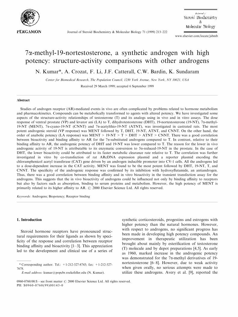
Table 1
Relative binding a�nities of androgens to androgen receptors (AR)
and sex hormone binding globulin (SHBG)
Steroid Relative binding a�nitya
AR SHBG
1. Testosterone 100 (6.95) 100
2. Nortestosterone (19-NT) 230 (3.01) 5
3. 7a-methyl-19-NT (MENT) 380 (1.82) 6
4. Dihydrotestosterone 290 (2.39) 340
5. Dihydro-19-NT 10 (94.74) 5
6. Dihydro-MENT 30 (24.98) 2
a The nanomolar concentration of steroid competitors that reduced
binding by 50% were determined. The IC50 ratio of the reference
compound (testosterone) to the test compound multiplied by 100 is
termed the relative binding a�nity. The IC50 values shown in par-
entheses were determined from two separate experiments.
N. Kumar et al. / Journal of Steroid Biochemistry & Molecular Biology 71 (1999) 213±222 215

cement curves of di�erent androgens are shown in Fig.1. A comparison of the relative binding a�nitiesshowed that the a�nity of MENT (IC50=1.82 nM)for AR was the highest followed by DHT (2.39), 19-NT (3.01), T (6.95), DHMENT (24.98) and DHNT(94.74) (Table 1). Thus 19-NT had a 2-fold andMENT almost a 4-fold higher RBA than T. However,the 5a-reduced derivatives of both 19-NT (DHNT)and MENT (DHMENT) exhibited much lower RBAcompared to the 5a-reduced derivative of T (DHT).
The importance of the substitution with methylgroup at 7a-position on 19-NT was investigated byusing 7b-methyl, 7a-cyano and 7a-acetylthio substi-tutions. Methyl substitution at the 7b-position resultedin 8-fold decrease in binding a�nity (IC50=27.3 nM)
compared to 7a-methyl derivative (MENT) (Fig. 2).Similarly 7a-substituted cyano or acetylthio 19-NT de-rivatives showed 10- and 4-fold decrease in bindinga�nity, respectively, compared to MENT (data notshown).
3.1.2. Binding to SHBGSince the binding of androgens to SHBG in the cir-
culation is known to a�ect their clearance, the bindinga�nity of androgens to SHBG was studied using [3H]-DHT as ligand. The results show that DHT had themaximum ability to displace [3H]-DHT followed by T,MENT, 19-NT, and mibolerone (Fig. 3) (Table 1).The binding a�nity of these androgens to SHBG didnot correlate with their binding to AR.
Fig. 1. Competitive inhibition of [3H] Mibolerone binding to androgen receptor (AR), prepared from ventral prostate cytosol, by testosterone
(T), dihydrotestosterone (DHT), 19-nortestosterone (19-NT), dihydro-19-NT (DHNT), MENT and dihydro-MENT (DHMENT).
Fig. 2. Competitive inhibition of [3H] Mibolerone binding to AR (ventral prostate cytosol) by mibolerone, 7a-methyl-19-NT (MENT) and 7b-methyl-19-NT (7b-MENT).
N. Kumar et al. / Journal of Steroid Biochemistry & Molecular Biology 71 (1999) 213±222216

3.2. Structure-activity relationships
3.2.1. Androgenic activity in ratsThe biopotency of the androgens was investigated in
immature castrated rats by the Hershberger assay [23],and adult castrated rats. The dose response compari-son of T, 19-NT and MENT in adult castrated ratswere reported earlier [24]. Therefore, biopotency ofthese androgens were compared in immature castratedrats. The dose response curves are shown in Fig. 4.The biopotency of MENT was 4-fold greater than thatof testosterone for maintaining VP weights and 15-foldhigher for maintaining muscle (levator ani) mass.However, 19-NT, in spite of its greater binding a�nityshowed only one-®fth the androgenic activity on VPwhile its anabolic potency was 4-fold greater comparedto T. These results of the androgenic/anabolic potencyof MENT and 19-NT were comparable to the earlierresults in adult castrated rats treated with androgensvia Alzet1; osmotic pumps [24]. The androgenic andanabolic potency of DHT was found to be slightly lessthan that of T, despite its higher RBA (Fig. 5). Thus,as with 19-NT, the androgenic/anabolic potency ofDHT did not correlate with its binding a�nity to ARin vitro. Since 19-NT could be 5a-reduced to DHNTin the accessory sex organs, the bioactivity of syntheticDHNT was compared with that of 19-NT at equi-molar doses in castrated rats. DHNT showed lowpotency on both VP and LA (Fig. 6), which correlatedwell with its binding a�nity to AR (Table 1). Eventhough MENT does not undergo enzymatic 5a-re-duction, the synthetic 5a-reduced MENT (DHMENT)was found to be only slightly stimulatory to both pros-tate and muscle (Fig. 6) in agreement with its lowbinding a�nity to AR.
The biological activity of seven substituted 19-NTderivatives was compared in castrated rats. Dose-re-
sponse comparison of 7a-cyano-19-NT showed it topossess one-®fth the androgenic potency and one half
Fig. 3. Competitive inhibition of [3H] DHT binding to Sex Hormone Binding Globulin (SHBG) by di�erent androgens. Third trimester human
pregnancy serum was used as a source of SHBG.
Fig. 4. Dose-response curves for ventral prostate and levator ani
weight increases by testosterone (T), 19-nortestosterone (19-NT) and
MENT in immature castrated male rats. One day following cas-
tration, rats were injected with di�erent doses of the androgens for
10 days. Rats were killed one day after the last injection. IC=intact
control; CC=castrated control.
N. Kumar et al. / Journal of Steroid Biochemistry & Molecular Biology 71 (1999) 213±222 217
the anabolic potency of T. 7a-acetylthio-19-NT pos-sessed one-half of the androgenic activity of T whileits anabolic potency was 2-fold greater (Table 2).Methyl substitution at the 7b position of 19-NT led toalmost complete loss of androgenic and anabolic ac-tivities (data not shown).
3.2.2. E�ect of androgens on CAT activity intransfection assays
The relative potency of the androgens was also com-pared in co-transfection experiments using CV-1 cellstransfected with a rat androgen receptor (AR) ex-pression plasmid and a reporter plasmid encoding theCAT gene under the control of mouse mammarytumor virus long terminal repeat (MMTV-LTR).MMTV-LTR mediated androgen-dependent transcrip-tional activation of a reporter gene in transiently trans-fected CV-1 cells [25] that normally do not expressendogenous androgen receptors.
The speci®city of the response was shown by eitheromitting the reporter or the expression vector in the DNA-calcium precipitate. In addition, a promoterless
reporter construct did not have any CAT activity andan unrelated promoter did not display androgen regu-
Fig. 6. E�ect of 19-nortesosterone (19-NT), MENT and their 5a-reduced derivatives (DHNT and DHMENT) on ventral prostate and
levator ani in castrated male rats. Details as per Fig. 5. I=intact;
C=castrated.
Table 2
Comparison of the biopotency of some natural and synthetic andro-
gens on ventral prostate and levator ania
Steroid Biopotency based on LA/VP
Ventral Prostate Levator Ani
Testosteroneb 1.0 1.0 1.0
19-nortestosterone (19-NT)b 0.2 4.0 20.0
7a-methyl-19-NT (MENT)b 4.0 15.0 4.0
Dihydrotestosteronec 0.9 0.7 0.8
7a-cyano-19-NTd 0.2 0.4 2.0
7a-acetylthio-19-NTd 0.4 2.1 5.0
a Potencies were calculated either by `All®t' computer program or
graphically. The response to testosterone was assigned a value of
one.b Biopotency estimates from dose-response comparisons shown in
Fig. 4.c Biopotency based on data from Fig. 5.d Biopotencies were calculated from dose-response curves of 7a-
cyano-19-NT and 7a- acetylthio-19-NT at four dose levels.
Fig. 5. Dose-response curves for ventral prostate and levator ani
weight increases by testosterone (T) and dihydrotestosterone (DHT)
in adult castrated rats. On the day of castration, rats were implanted
subcutaneously with Alzet osmotic pumps releasing the androgens at
di�erent dose levels for 14 days and the rats were killed on day 15.
N. Kumar et al. / Journal of Steroid Biochemistry & Molecular Biology 71 (1999) 213±222218

lation of the CAT reporter (data not shown). Theandrogens, T, DHT, 19-NT, MENT and CNNTinduced dose-dependent responses in CAT activity inthe transfection assays (Fig. 7). MENT was found tobe most active followed by DHT, 19-NT, T, andCNNT. The speci®city of this response to androgenswas established by blocking their activity using theantiandrogen, hydroxy¯utamide (Fig. 8). The results ofthis in vitro study showed a good correlation betweenthe binding a�nity to AR and the activity in the tran-sient transfection assays. The extent of activity in thisin vitro assay is due to a direct e�ect of the com-pounds independent of metabolism and clearance.Hence, for 19-NT and DHT, whose androgenic
potency in vivo was reduced signi®cantly due to targetorgan speci®c metabolism or increased clearance, theactivity in the transfection assays was not reduced andre¯ected more faithfully their binding a�nity to AR.
4. Discussion
The present studies demonstrate that the mechan-isms which determine the relative biopotency of T, 19-NT and its derivatives not only involve di�erences intheir binding a�nities to AR, but also their metab-olism in the target tissues which leads to the formation
Fig. 7. Relative potency of androgens in a transient transfection assay in CV-1 cells transfected with a rat androgen receptor (AR) expression
plasmid and a reporter plasmid encoding chloramphenicol acetyl transferase (CAT) under the control of mouse mammary tumor virus long term-
inal repeat (MMTV-LTR). Each dose of androgen was tested in triplicate. T, testosterone; DHT, dihydrotestosterone; CNNT, 7a-cyano-19-NT;
19-NT, 19-nortestosterone; MENT, 7a-methyl-19-NT.
Fig. 8. E�ect of hydroxy¯utamide (an antiandrogen) on testosterone (T) and MENT stimulated CAT activity in the transfected CV-1 cells.
N. Kumar et al. / Journal of Steroid Biochemistry & Molecular Biology 71 (1999) 213±222 219

of compounds with greater or lesser activity than theparent compound.
It is well established that T is converted to DHT inthe sex accessory glands, and since the binding a�nityof DHT to AR is 3±5 times higher than that of T, itsaction is ampli®ed in these tissues [8,10]. On the otherhand, in castrated rats the potency of exogenouslyadministered DHT was found to be either equal to orless than that of T. Similar reduction in the e�ect ofexogenous DHT relative to T was observed in themaintenance of spermatogenesis in hypophysectomizedrats [26]. However, DHT was shown to be 10 timesmore potent than T in in vitro transient transfectionassays using CHO cells which are de®cient in androgenmetabolizing enzymes [27,28]. The decrease in andro-genic activity of exogenously administered DHT couldbe due to its faster metabolism in rats. Since rats donot express SHBG, the metabolic clearance rate(MCR) of T and DHT is mainly dependent on liverand extrahepatic tissues. In the rat, the MCR of DHT(8729 L/100 g BW) is much higher than that of T (18L/100 g BW) [29,30]. The di�erence in the MCRresults in lower plasma levels of DHT compared to thelevels of T when an equivalent amount of the twoandrogens are administered [31]. The reduction in theactivity of exogenously administered DHT in rats can-not be extrapolated to other animals and man whopossess SHBG in circulation. Exogenously adminis-tered DHT has been shown to be more potent than Tin stimulating the growth of ventral prostate, seminalvesicles and bulbo-urethral glands in castrated Rhesusmonkeys [32]. The overall metabolic clearance rate ofDHT (764±630 L/day) is lower than that of T (1200±950 L/day) in normal men [33,34]. This is believed tobe due to the higher a�nity of DHT to SHBG com-pared to T [35], resulting in decreased availability ofDHT for metabolism by liver enzymes [34].
For most steroids 5a-reduction in the liver is con-sidered to be the rate limiting step in their metabolicclearance [36]. It was suggested that certain changes inthe steroid structure makes them resistant to 5a-re-duction in the liver, thereby decreasing their clearanceand increasing their uptake by the target organ. Sucha mechanism for the increased biopotency was pro-posed for synthetic glucocorticoids [37]. However, eventhough MENT cannot be 5a-reduced, it was clearedmore rapidly than T in monkeys and men [12]. This isbecause MENT does not bind to SHBG [12], thus itsclearance will not be hampered by binding to plasmaproteins as is the case for T and DHT [34]. Testoster-one is transported in plasma bound to SHBG, albu-min, and in free form. Biologically active T representsboth the albumin bound and free fraction since thehalf-dissociation time of T from albumin is rapid(<0.01 s), which is within the probable time of capil-lary transit through androgen target tissues [38,39].
The half-dissociation time of T from SHBG is rela-tively slow (>20 s), such that it is biologically notavailable at target tissues [40]. Since MENT does notbind to SHBG, its greater potency could also be dueto a higher percentage of unbound MENT in theplasma in addition to its higher a�nity to AR as com-pared to T. The higher concentration of unboundMENT may result in greater uptake by the target tis-sues, increasing its biopotency. A relationship betweenplasma protein binding and the relative biopotencies ofsubstituted corticoids has been demonstrated earlier[41].
The biopotency di�erences between T and 19-NT incastrated rats, re¯ect yet another mechanism by whichthe activity of a steroid on the target-organ can bealtered based on organ speci®c metabolism. Comparedto T, 19-NT was found to be 5±6 times less potent onsex accessory glands but 3±4 times more potent as ananabolic agent. The di�erences in their androgenic vsanabolic potency are attributable to their 5a-reductionin the accessory glands but not in the muscle [17]. Thebinding a�nity of 19-NT to AR is higher than that ofT. But the opposite is the case for their 5a-reducedmetabolites. In the prostate, 5a-reduction of 19-NT toDHNT lowers its binding a�nity to AR, resulting indecreased androgenic activity in vivo [16]. In the caseof MENT, the 7a-methylated derivative of 19-NT, itshigh potency could be attributed primarily to itsgreater a�nity for AR and to its inability to undergo5a-reduction.
It is clear from these studies that substitution at the7a-position with a methyl group is important forenhanced binding a�nity and increased biological ac-tivity since substitution with a cyano or a acetylthiomoiety resulted in lower binding a�nity and decreasedbiological activity. The importance of the a-con®gur-ation of the methyl group was further con®rmed sincea b-con®guration led to a decrease in its androgenicpotency. A similar e�ect of 7b-methyl con®guration onthe potency of DHT has also been reported [42]. Thissuggested that the conformational changes forincreased receptor binding are speci®c for 7a-methyl-ated derivatives. Liao and colleagues have proposedthat the selectivity of androgen binding to its receptoris de®ned mainly by 7a- and/or 17a-position of thesteroid structure. Substitution with a methyl group inthese positions leads to a ¯attening of the steroid mol-ecule which leads to a conformation that is less hin-dered for receptor binding [8,13]. Similarly, 7a-methylsubstituent on estradiol has been shown to enhance itsreceptor binding a�nity [43,44].
The correlation between biopotency and bindinga�nity was further evaluated in vitro by co-transfec-tion of AR cDNA expression plasmid and a reporterplasmid encoding the CAT gene into CV-1 cells. Inthese assays MENT was found to be the most active
N. Kumar et al. / Journal of Steroid Biochemistry & Molecular Biology 71 (1999) 213±222220
followed by DHT, 19-NT, T and CNNT. The speci-®city of this response to androgens was established byits inhibition with hydroxy¯utamide, an antiandrogen.In these studies, there was a good correlation betweenbinding a�nity to AR and the in vitro activity intransfection assays. Thus, the androgenic potency of19-NT and DHT in castrated rats did not correlatewith either their receptor binding a�nity or their CATactivity. The reason for the lower androgenic activityof the former is its metabolism to the less potent 5a-reduced derivative, and that of the latter is its fasterclearance. Hence, factors other than receptor bindinga�nity have important physiological implications inthe overall action of steroid hormones.
In conclusion, the biological activity of androgens ismodulated by their binding to speci®c plasma proteins,their overall metabolic clearance, organ-speci®c meta-bolic transformation and binding a�nity to androgenreceptors. The increased relative anabolic activity ofMENT is due to its higher a�nity for androgen recep-tors and its resistance to 5a-reduction in the prostate.
Acknowledgements
We wish to acknowledge the generous supportreceived from US Agency for International Develop-ment (Cooperative agreement CCP-A-00-94-00013-04)and NICHD, NIH for Grants HD-29990, HD-13541and RO1 DK52960 (JFC). We also wish to thank MsIrene Gorzelany Mr M. Willman and Mr W. DeJesusfor technical assistance.
References
[1] J. Delettre, J.P. Mornon, G. Lepicard, T. Ojasoo, J.P.
Raynaud, Steroid ¯exibility and receptor speci®city, J. Steroid
Biochem. 13 (1980) 45±59.
[2] J.P. Raynaud, T. Ojasoo, The relevance of structure-a�nity re-
lationships in the study of steroid hormone action, in: H.
Eriksson, J.-A. Gustafsson (Eds.), Steroid Hormone Receptors:
Structure and Function, Elsevier, Amsterdam, 1983, pp. 141±
170.
[3] E. Dahlberg, A. Thalen, R. Brattsand, J.-A. Gustafsson, U.
Johansson, K. Roempke, T. Saqrtok, Correlation between
chemical structure, receptor binding, and biological activity of
some novel, highly active, 16a,17a-acetal-substituted glucocor-
ticoids, Molec. Pharmacol. 25 (1983) 70±78.
[4] K. Junkman, Long-acting steroids in reproduction, Recent
Prog. Horm. Res. 13 (1957) 389±419.
[5] E. Nieschlag, H.M. Behre, Pharmacology and clinical uses of
testosterone, in: E. Nieschlag, H.M. Behre (Eds.), Testosterone:
Action, De®ciency, and Substitution, Springer±Verlag, Berlin,
Heidelberg, 1990, pp. 92±114.
[6] A. Segalo�, The enhanced local androgenic activity of 19-nor
steroids and stabilization of their structure by 7a- and 17a-methyl substituents to highly potent androgens by any route of
administration, Steroids 1 (1963) 299±315.
[7] A. Segalo�, R.B. Gabbard, 14-dehydro-19-nortestosterone and
its 7a-methyl derivative, Steroids 22 (1973) 99±105.
[8] S. Liao, T. Liang, S. Fang, E. Castaneda, T.-C. Shao, Steroid
structure and androgenic activity, J. Biol. Chem. 248 (1973)
6154±6162.
[9] M.A. Avery, M. Tanabe, D.F. Crowe, G. Detre, R.H. Peters,
W.K.M. Chong, Synthesis and testing of 17a, b- hydroxy-7a-methyl-D-homoestra-4,16-dien-3-one: a highly potent orally
active androgen, Steroids 55 (1990) 59±64.
[10] N. Kumar, A.K. Didolkar, C. Monder, C.W. Bardin, K.
Sundaram, The biological activity of 7a-methyl-19-nortestoster-
one is not ampli®ed in male reproductive tract as is that of tes-
tosterone, Endocrinology 130 (1992) 3677±3683.
[11] K. Sundaram, N. Kumar, C.W. Bardin, 7a-methyl-nortestos-
terone MENT: The optimal androgen for male contraception,
Ann. Med. 25 (1993) 199±205.
[12] N. Kumar, J. Suvisaari, Y.-Y. Tsong, C. Aguillaume, C.W.
Bardin, P. LaÈ hteenmki, K. Sundaram, Pharmacokinetics of 7a-methyl-19-nortestosterone in men and cynomologus monkeys,
J. Androl. 18 (1997) 352±358.
[13] K.M.B. Chan, S. Smythe, S. Liao, Androgen receptor binding
and androgenicity of methylated 4-ene-3-ketosteroids having
no 17-hydroxy group, J. Steroid Biochem. 11 (1979) 1193±
1196.
[14] S.A. Shain, R.W. Boesel, Saturation analysis of the binding of
androgens, antiandrogens and estrogens by the cytoplasmic
high a�nity androgen receptor of the rat ventral prostate, J.
Steroid Biochem. 6 (1975) 43±50.
[15] J. Steinsapir, M. Bryhan, T.G. Muldoon, Relative binding
properties of microsomal and cytosolic androgen receptor
species of the ventral prostate, Endocrinology 125 (1989) 2297±
2311.
[16] F. Celotti, Cesi P. Negri, Anabolic steroids: a review of their
e�ects on the muscle, of their possible mechanisms of action
and of their use in athletics, J. Steroid Biochem. Molec. Biol.
43 (1992) 469±477.
[17] N. Kumar, K. Sundaram, C.W. Bardin, Feedback regulation
of gonadotropins by androgens in rats: Is 5a-reductaseinvolved?, J. Steroid. Biochem. Molec. Biol. 52 (1995) 105±112.
[18] V. Isomaa, A.E.I. Pajunen, C.W. Bardin, O.A. JaÈ nne, Nuclear
androgen receptors in the mouse kidney: Validation of a new
assay, Endocrinology 111 (1982) 833±843.
[19] F.L. Graham, A.J. Eb Van der, A new technique for the assay
of infectivity of human adenovirus 5 DNA, Virology 52 (1973)
456±467.
[20] J.J. Palvimo, P.J. Kallio, T. Ikonen, M. Mehto, O.A. JaÈ nne,
Dominant negative regulation of transactivation by the rat
androgen receptor: roles of the N-terminal domain and hetero-
dimer formation, Mol. Endocrinol. 7 (1993) 1399±1407.
[21] C.M. Gorman, L.F. Mo�at, B.H. Howard, Recombinant gen-
omes which express chloramphenicol acetyltransferase in mam-
malian cells, Molec. Cell. Biol. 2 (1982) 1044±1051.
[22] C.V. Hall, P.E. Jacob, G.M. Ringold, F. Lee, Expression and
regulation of Escherichia coli lacZ gene fusions in mammalian
cells, J. Molec. Appl. Genet. 2 (1983) 101±109.
[23] L.G. Hershberger, E.G. Shipley, R.K. Meyer, Myotrophic ac-
tivity of 1-nortestosterone and other steroids determined by
modi®ed levator ani method, Proc. Soc. Exp. Biol. Med. 83
(1953) 175±180.
[24] N. Kumar, L.-X. Shan, M. Hardy, C.W. Bardin, K.
Sundaram, Mechanism of androgen-induced thymolysis in rats,
Endocrinology 136 (1995) 4887±4893.
[25] M.V. Govindan, Speci®c region in hormone binding domain is
essential for hormone binding and transactivation by human
androgen receptor, Mol. Endocrinol. 4 (1990) 417±427.
[26] M.E. Harris, A. Bartke, J. Weisz, D. Watson, E�ects of testos-
terone and dihydrotestosterone on spermatogenesis, rete testis
N. Kumar et al. / Journal of Steroid Biochemistry & Molecular Biology 71 (1999) 213±222 221

¯uid, and peripheral androgen levels in hypophysectomized rat,
Fertil. Steril. 28 (1977) 1113±1137.
[27] J.-P. Deslypere, M. Young, J.D. Wilson, M.J. McPhaul,
Testosterone and 5a-dihydrotestosterone interact di�erently
with the androgen receptor to enhance transcription of the
MMTV-CAT receptor gene, Mol. Cell. Endocrinol. 88 (1992)
15±22.
[28] Z.-X. Zhou, M.V. Lane, J.A. Kemppainen, F.S. French, E.M.
Wilson, Speci®city of ligand-dependent androgen receptor
stabilization: Receptor domain interactions in¯uence ligand dis-
sociation and receptor stability, Mol. Endocrinol. 9 (1995)
208±218.
[29] D.K. Lee, C.E. Bird, A.F. Clark, In vivo metabolism of 3H-
testosterone in adult male rats: e�ects of estrogen adminis-
tration, Steroids 26 (1975) 137±147.
[30] E.J. Van Doorn, B. Burns, D. Wood, C.E. Bird, A.F. Clark,
In vivo metabolism of 3H-dihydrotestosterone and 3H-andros-
tanediol in adult male rats, J. Steroid Biochem. 6 (1975) 1549±
1554.
[31] W. Bartsch, C. Knabbe, V. Klaus-Dieter, Regulation and com-
partmentalization of androgens in rat prostate and muscle, J.
Steroid Biochem. 19 (1983) 929±937.
[32] N. Dinakar, R. Arora, M.R.N. Prasad, E�ects of microquanti-
ties of 5a-dihydrotestosterone on the epididymis and accessory
glands of the castrated Rhesus monkey, Macacca mulatta, Int.
J. Fertil. 19 (1974) 133±139.
[33] R. Horton, J. Shinsako, P.M. Forsham, Testosterone pro-
duction and metabolic clearance rates with volume of distri-
bution in normal adult men and women, Acta Endocrinol.
(Copenhagen) 48 (1965) 446±458.
[34] T. Ishimaru, W.A. Edmiston, L. Pages, R. Norton, Splanchnic
extraction and conversion of testosterone and dihydrotestoster-
one in man, J. Clin. Endocrinol. Metab. 46 (1978) 528±533.
[35] W. Heyns, P. De Moor, Kinetics of dissociation of 17b-hy-droxysteroids from the steroid binding b-globulin of human
plasma, J. Clin. Endocrinol. Metab. 32 (1971) 147±154.
[36] A.F. Clark, Steroid D4-reductases: their physiological role and
signi®cance, in: R. Hobkirk (Ed.), Steroid Biochemistry, Vol.
1, CRC Press, Florida, 1979, pp. 1±27.
[37] J.R. Florini, L.L. Smith, D.A. Buyske, Metabolic fate of a syn-
thetic corticosteroid (triamcinolone) in the dog, J. Biol. Chem.
236 (1961) 1038±1042.
[38] W.M. Pardridge, Transport of protein bound hormone into tis-
sues in vivo, Endocrine Rev. 2 (1981) 103±123.
[39] W.M. Pardridge, Serum bioavailability of sex steroid hor-
mones, Clin. Endocrinol. Metab. 15 (1986) 259±278.
[40] D.A. Damassa, A.W. Gustafson, E�ect of chronic infusions of
sex steroid-binding protein on the testosterone-mediated inhi-
bition of gonadotropin secretion and maintenance of sex acces-
sory glands in male rats, Endocrinology 123 (1988) 1885±1892.
[41] J.R. Florini, D.A. Buyske, Plasma protein binding of triamci-
nolone-H3 and hydrocortisone-4-C14, J. Biol. Chem. 236
(1961) 247.
[42] W.H. Chin, M.E. Wol�, Synthesis and biological activities of
7b-methyl steroids, J. Med. Chem. 22 (1979) 1257±1260.
[43] F.J. Zeelen, E.W. Bergink, Structure-activity relationships of
steroid estrogens, in: J. Raus, H. Martens, G. Leclercq (Eds.),
Cytotoxic Estrogens in Hormone Receptive Tumours,
Academic Press, London, 1980, pp. 39±48.
[44] A. LaMorte, N. Kumar, C.W. Bardin, K. Sundaram,
Aromatization of 7a-methyl-19-nortestosterone by human pla-
cental microsomes in vitro, J. Steroid Biochem. Molec. Biol. 48
(1994) 297±303.
N. Kumar et al. / Journal of Steroid Biochemistry & Molecular Biology 71 (1999) 213±222222