
3D In Vivo Optical Skin Imaging for Intense Pulsed Light and Fractional Ablative Resurfacing of...
21
3D In Vivo Optical Skin Imaging for Intense Pulsed Light and Fractional Ablative Resurfacing of Photodamaged Skin Matteo Tretti Clementoni, MD a, *, Rosalia Lavagno, MD a , Maximilian Catenacci, MD b , Roman Kantor, PhD c , Guido Mariotto, PhD c , Igor Shvets, PhD d Topography of the skin surface as well as melanin and hemoglobin concentration and distribution are a mirror of the functional skin status. Changes in these features not only are a tool for early-stage diagnosis of diseases but also give an indication of the response to medical and cosmetic treatment. Therefore, their evalua- tion is of great interest for dermatologic research. However, although physicians can apply clas- sification rules to visual diagnosis, the overall clinical approach is subjective and qualitative, with a critical dependence on training and ex- perience. Over the past 20 years, several nonin- vasive techniques for measuring the skin’s properties have been developed and tested to extend the accuracy of visual assessment alone. Many of them were not only very precise but also very complicated; others were very simple but approximations. In this article, a new optical, precise, user- friendly measuring system is presented to demon- strate the effectiveness of the most common laser treatments on photodamaged skin. Many of the skin changes commonly associated with aging, changes in pigmentation, telangiectasia, sallow- ness, and wrinkling are actually the result of sun exposure. Changes in pigmentation (blotchy brown freckles and age spots), dryness, areas of redness, thinning of the dermis, loss of elasticity, fine lines, and deep wrinkles are all signs of chronic UV expo- sure. Excluding injectables, surgery, and peelings, the most common procedures to improve these changes in the skin appearance are the IPL treat- ments and, more recently, the fractional resur- facings. Preoperative and postoperative images of patients treated with these procedures and analyzed using this new three-dimensional (D) in vivo optical skin imaging system are shown. a Istituto Dermatologico Europeo, Milano, Italy b Skindermolaser, Roma, Italy c Miravex Ltd, Trinity Research and Innovation, O’Reilly Institute, Trinity College Dublin, Dublin, Ireland d School of Physics, Trinity College Dublin, Dublin, Ireland * Corresponding author. E-mail address: [email protected] KEYWORDS 3D Photoaging Photodamaged skin Fractional laser treatment Skin topography Facial Plast Surg Clin N Am 19 (2011) 737–757 doi:10.1016/j.fsc.2011.07.014 1064-7406/11/$ – see front matter Ó 2011 Elsevier Inc. All rights reserved. facialplastic.theclinics.com
-
Upload
independent -
Category
Documents
-
view
1 -
download
0
Transcript of 3D In Vivo Optical Skin Imaging for Intense Pulsed Light and Fractional Ablative Resurfacing of...
3D In Vivo Optical SkinImaging for IntensePulsed Light andFractional AblativeResurfacing ofPhotodamaged Skin
Matteo Tretti Clementoni, MDa,*,Rosalia Lavagno, MDa, Maximilian Catenacci, MDb,Roman Kantor, PhDc, Guido Mariotto, PhDc,Igor Shvets, PhDdKEYWORDS
� 3D � Photoaging � Photodamaged skin� Fractional laser treatment � Skin topography
Topography of the skin surface as well asmelanin and hemoglobin concentration anddistribution are a mirror of the functional skinstatus. Changes in these features not only area tool for early-stage diagnosis of diseases butalso give an indication of the response to medicaland cosmetic treatment. Therefore, their evalua-tion is of great interest for dermatologic research.However, although physicians can apply clas-sification rules to visual diagnosis, the overallclinical approach is subjective and qualitative,with a critical dependence on training and ex-perience. Over the past 20 years, several nonin-vasive techniques for measuring the skin’sproperties have been developed and tested toextend the accuracy of visual assessment alone.Many of them were not only very precise but alsovery complicated; others were very simple butapproximations.
a Istituto Dermatologico Europeo, Milano, Italyb Skindermolaser, Roma, Italyc Miravex Ltd, Trinity Research and Innovation, O’Reillyd School of Physics, Trinity College Dublin, Dublin, Irelan* Corresponding author.E-mail address: [email protected]
Facial Plast Surg Clin N Am 19 (2011) 737–757doi:10.1016/j.fsc.2011.07.0141064-7406/11/$ – see front matter � 2011 Elsevier Inc. All
In this article, a new optical, precise, user-friendly measuring system is presented to demon-strate the effectiveness of the most common lasertreatments on photodamaged skin. Many of theskin changes commonly associated with aging,changes in pigmentation, telangiectasia, sallow-ness, and wrinkling are actually the result of sunexposure. Changes in pigmentation (blotchy brownfreckles and age spots), dryness, areas of redness,thinning of the dermis, loss of elasticity, fine lines,and deepwrinkles are all signs of chronic UV expo-sure. Excluding injectables, surgery, and peelings,the most common procedures to improve thesechanges in the skin appearance are the IPL treat-ments and, more recently, the fractional resur-facings. Preoperative and postoperative imagesof patients treated with these procedures andanalyzed using this new three-dimensional (D)in vivo optical skin imaging system are shown.
Institute, Trinity College Dublin, Dublin, Irelandd
rights reserved. facialplastic.theclinics.com
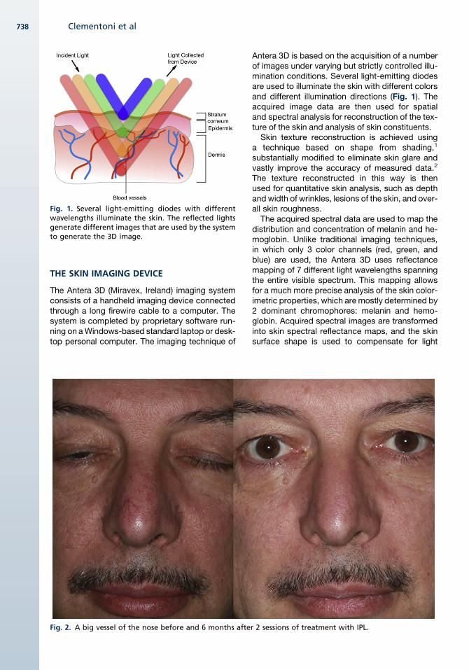
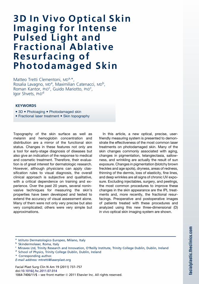
Fig. 1. Several light-emitting diodes with differentwavelengths illuminate the skin. The reflected lightsgenerate different images that are used by the systemto generate the 3D image.
Clementoni et al738
THE SKIN IMAGING DEVICE
The Antera 3D (Miravex, Ireland) imaging systemconsists of a handheld imaging device connectedthrough a long firewire cable to a computer. Thesystem is completed by proprietary software run-ning on aWindows-based standard laptop or desk-top personal computer. The imaging technique of
Fig. 2. A big vessel of the nose before and 6 months afte
Antera 3D is based on the acquisition of a numberof images under varying but strictly controlled illu-mination conditions. Several light-emitting diodesare used to illuminate the skin with different colorsand different illumination directions (Fig. 1). Theacquired image data are then used for spatialand spectral analysis for reconstruction of the tex-ture of the skin and analysis of skin constituents.Skin texture reconstruction is achieved using
a technique based on shape from shading,1
substantially modified to eliminate skin glare andvastly improve the accuracy of measured data.2
The texture reconstructed in this way is thenused for quantitative skin analysis, such as depthand width of wrinkles, lesions of the skin, and over-all skin roughness.The acquired spectral data are used to map the
distribution and concentration of melanin and he-moglobin. Unlike traditional imaging techniques,in which only 3 color channels (red, green, andblue) are used, the Antera 3D uses reflectancemapping of 7 different light wavelengths spanningthe entire visible spectrum. This mapping allowsfor a much more precise analysis of the skin color-imetric properties, which are mostly determined by2 dominant chromophores: melanin and hemo-globin. Acquired spectral images are transformedinto skin spectral reflectance maps, and the skinsurface shape is used to compensate for light
r 2 sessions of treatment with IPL.
3D In Vivo Optical Skin Imaging 739
intensity variation due to the varying direction ofincident illumination. The reflectance data aretransformed into skin absorption coefficients andused to quantify melanin and hemoglobin concen-trations usingmathematical correlation with knownspectral absorption data of these chromophores.3
The images acquired with the Antera 3D can bevisualized in several different modes: standardcolor skin, texture elevation map, and melaninand hemoglobin concentration maps with 2D and3D perspective representation. The clinician canselect specific skin areas for quantitative analysisand carry out before and after analyses with previ-ously acquired images. Spot-On, the automaticmatching technique that registers two or moreimages to one another is used to compensate forrelative shifts and rotations between images,ensuring accurate data analysis. The measure-ment data can be presented by quickly creatinga report that shows the analyzed images togetherwith the measured values and comparison charts.Data can be stored on the computer or included inother document processing applications such asMicrosoft Word or Microsoft PowerPoint.
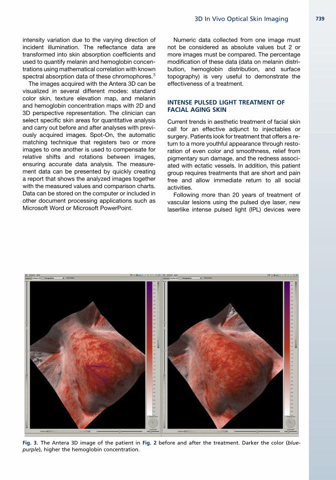
Fig. 3. The Antera 3D image of the patient in Fig. 2 befpurple), higher the hemoglobin concentration.
Numeric data collected from one image mustnot be considered as absolute values but 2 ormore images must be compared. The percentagemodification of these data (data on melanin distri-bution, hemoglobin distribution, and surfacetopography) is very useful to demonstrate theeffectiveness of a treatment.
INTENSE PULSED LIGHT TREATMENT OFFACIAL AGING SKIN
Current trends in aesthetic treatment of facial skincall for an effective adjunct to injectables orsurgery. Patients look for treatment that offers a re-turn to a more youthful appearance through resto-ration of even color and smoothness, relief frompigmentary sun damage, and the redness associ-ated with ectatic vessels. In addition, this patientgroup requires treatments that are short and painfree and allow immediate return to all socialactivities.
Following more than 20 years of treatment ofvascular lesions using the pulsed dye laser, newlaserlike intense pulsed light (IPL) devices were
ore and after the treatment. Darker the color (blue-
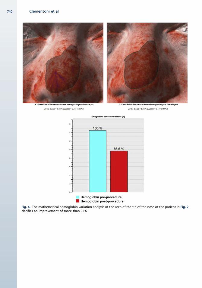
Fig. 4. The mathematical hemoglobin variation analysis of the area of the tip of the nose of the patient in Fig. 2clarifies an improvement of more than 33%.
Clementoni et al740
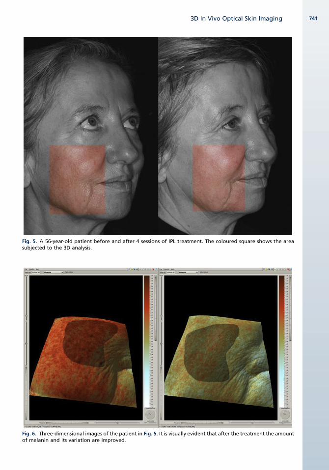
Fig. 5. A 56-year-old patient before and after 4 sessions of IPL treatment. The coloured square shows the areasubjected to the 3D analysis.
Fig. 6. Three-dimensional images of the patient in Fig. 5. It is visually evident that after the treatment the amountof melanin and its variation are improved.
3D In Vivo Optical Skin Imaging 741
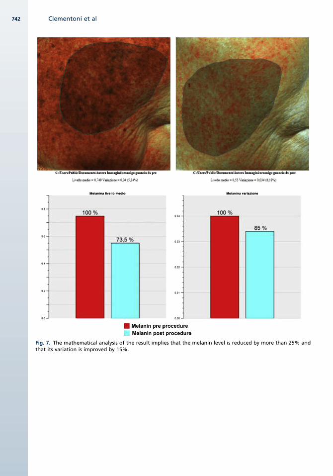
Fig. 7. The mathematical analysis of the result implies that the melanin level is reduced by more than 25% andthat its variation is improved by 15%.
Clementoni et al742
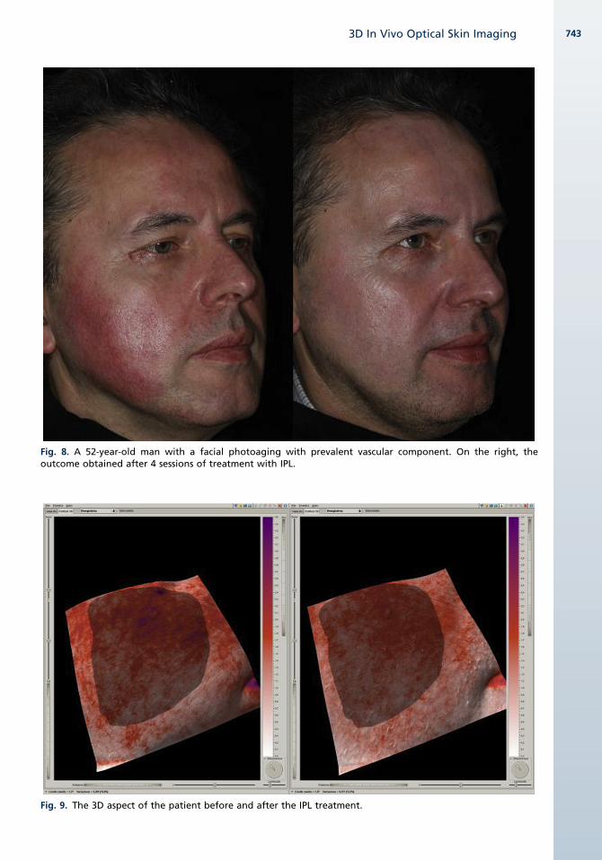
Fig. 8. A 52-year-old man with a facial photoaging with prevalent vascular component. On the right, theoutcome obtained after 4 sessions of treatment with IPL.
Fig. 9. The 3D aspect of the patient before and after the IPL treatment.
3D In Vivo Optical Skin Imaging 743
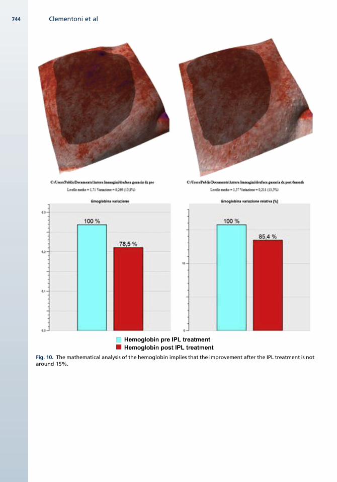
Fig. 10. The mathematical analysis of the hemoglobin implies that the improvement after the IPL treatment is notaround 15%.
Clementoni et al744
3D In Vivo Optical Skin Imaging 745
developed at the end of the last century. These IPLdevices treat these UV exposure–correlated con-ditions with success and provide a solution forthe essential lifestyle criteria when used in a care-fully administered program. This new IPL skin reju-venation technique now has a clinical history ofmore than 300,000 treatments with excellentpatient acceptance. IPL differs from laser light inthat, rather than monochromatic single wave-length, IPL emits a noncoherent broad-spectrumlight. The IPL devices used in the rejuvenationprocedure emit a spectrum extending from 500nm to 1200 nm. To customize the light energydelivery for a given procedure, the operator usesa cutoff filter, or light guide, of designated wave-length, below which the spectrum is selectivelyeliminated. The IPL system conforms to the prin-ciple of selective photothermolysis. For dilatedvessels, as seen in patients with sun damageand rosacea, the light energy with high absorptionby hemoglobin and oxyhemoglobin reaches thedermal capillary bed and selectively destroys theabnormal vessels. For sun spots and lentigos, as
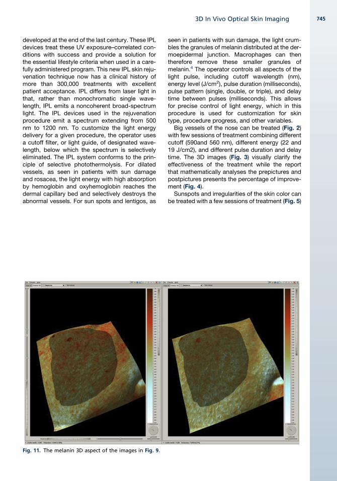
Fig. 11. The melanin 3D aspect of the images in Fig. 9.
seen in patients with sun damage, the light crum-bles the granules of melanin distributed at the der-moepidermal junction. Macrophages can thentherefore remove these smaller granules ofmelanin.4 The operator controls all aspects of thelight pulse, including cutoff wavelength (nm),energy level (J/cm2), pulse duration (milliseconds),pulse pattern (single, double, or triple), and delaytime between pulses (milliseconds). This allowsfor precise control of light energy, which in thisprocedure is used for customization for skintype, procedure progress, and other variables.
Big vessels of the nose can be treated (Fig. 2)with few sessions of treatment combining differentcutoff (590and 560 nm), different energy (22 and19 J/cm2), and different pulse duration and delaytime. The 3D images (Fig. 3) visually clarify theeffectiveness of the treatment while the reportthat mathematically analyses the prepictures andpostpictures presents the percentage of improve-ment (Fig. 4).
Sunspots and irregularities of the skin color canbe treated with a few sessions of treatment (Fig. 5)
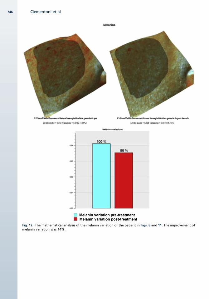
Fig. 12. The mathematical analysis of the melanin variation of the patient in Figs. 8 and 11. The improvement ofmelanin variation was 14%.
Clementoni et al746
3D In Vivo Optical Skin Imaging 747
crumbling the pigment granules at different depth.Clinical images show a change that is less easy todiscern, while using the 3D system, the improve-ment can be better appreciated (Fig. 6) and quan-tified (Fig. 7).
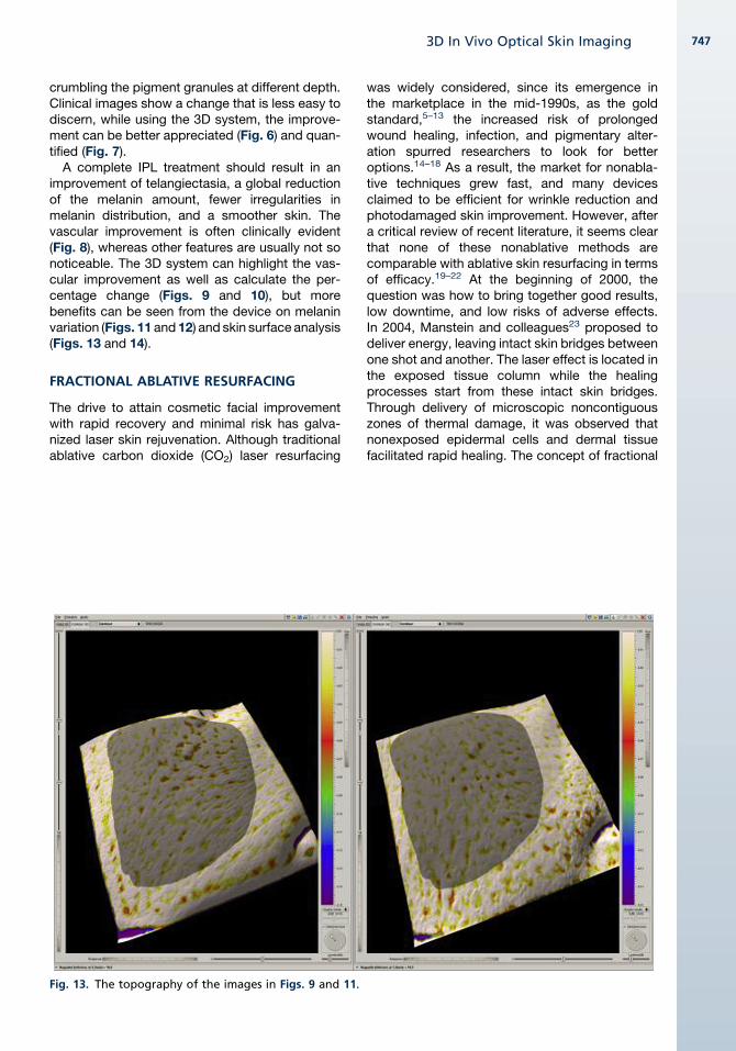
A complete IPL treatment should result in animprovement of telangiectasia, a global reductionof the melanin amount, fewer irregularities inmelanin distribution, and a smoother skin. Thevascular improvement is often clinically evident(Fig. 8), whereas other features are usually not sonoticeable. The 3D system can highlight the vas-cular improvement as well as calculate the per-centage change (Figs. 9 and 10), but morebenefits can be seen from the device on melaninvariation (Figs. 11 and 12) and skin surface analysis(Figs. 13 and 14).
FRACTIONAL ABLATIVE RESURFACING
The drive to attain cosmetic facial improvementwith rapid recovery and minimal risk has galva-nized laser skin rejuvenation. Although traditionalablative carbon dioxide (CO2) laser resurfacing
Fig. 13. The topography of the images in Figs. 9 and 11.
was widely considered, since its emergence inthe marketplace in the mid-1990s, as the goldstandard,5–13 the increased risk of prolongedwound healing, infection, and pigmentary alter-ation spurred researchers to look for betteroptions.14–18 As a result, the market for nonabla-tive techniques grew fast, and many devicesclaimed to be efficient for wrinkle reduction andphotodamaged skin improvement. However, aftera critical review of recent literature, it seems clearthat none of these nonablative methods arecomparable with ablative skin resurfacing in termsof efficacy.19–22 At the beginning of 2000, thequestion was how to bring together good results,low downtime, and low risks of adverse effects.In 2004, Manstein and colleagues23 proposed todeliver energy, leaving intact skin bridges betweenone shot and another. The laser effect is located inthe exposed tissue column while the healingprocesses start from these intact skin bridges.Through delivery of microscopic noncontiguouszones of thermal damage, it was observed thatnonexposed epidermal cells and dermal tissuefacilitated rapid healing. The concept of fractional
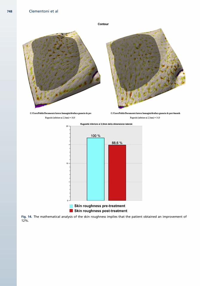
Fig. 14. The mathematical analysis of the skin roughness implies that the patient obtained an improvement of12%.
Clementoni et al748
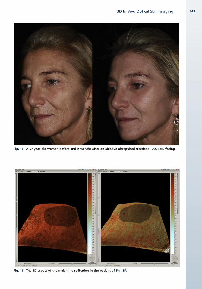
Fig. 15. A 57-year-old woman before and 9 months after an ablative ultrapulsed fractional CO2 resurfacing.
Fig. 16. The 3D aspect of the melanin distribution in the patient of Fig. 15.
3D In Vivo Optical Skin Imaging 749
Clementoni et al750
delivery of energy was originally proposed fora nonablative wavelength but very quickly alsoapplied to the ablative lasers. The idea was toincorporate the well-known results of ablativelasers while maintaining a short recovery timeand a low incidence of adverse side effects.Fractional ablative CO2 resurfacing performed
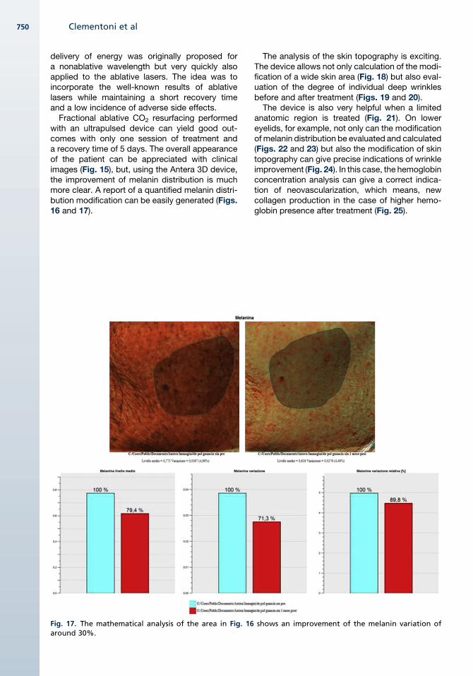
with an ultrapulsed device can yield good out-comes with only one session of treatment anda recovery time of 5 days. The overall appearanceof the patient can be appreciated with clinicalimages (Fig. 15), but, using the Antera 3D device,the improvement of melanin distribution is muchmore clear. A report of a quantified melanin distri-bution modification can be easily generated (Figs.16 and 17).
Fig. 17. The mathematical analysis of the area in Fig. 16around 30%.
The analysis of the skin topography is exciting.The device allows not only calculation of the modi-fication of a wide skin area (Fig. 18) but also eval-uation of the degree of individual deep wrinklesbefore and after treatment (Figs. 19 and 20).The device is also very helpful when a limited
anatomic region is treated (Fig. 21). On lowereyelids, for example, not only can the modificationof melanin distribution be evaluated and calculated(Figs. 22 and 23) but also the modification of skintopography can give precise indications of wrinkleimprovement (Fig. 24). In this case, the hemoglobinconcentration analysis can give a correct indica-tion of neovascularization, which means, newcollagen production in the case of higher hemo-globin presence after treatment (Fig. 25).
shows an improvement of the melanin variation of
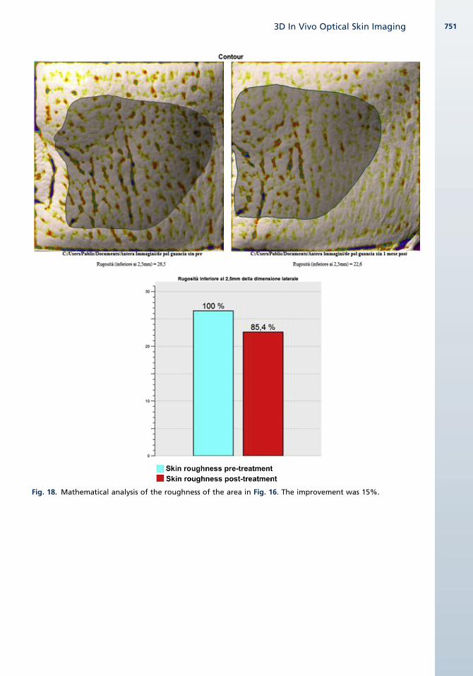
Fig. 18. Mathematical analysis of the roughness of the area in Fig. 16. The improvement was 15%.
3D In Vivo Optical Skin Imaging 751
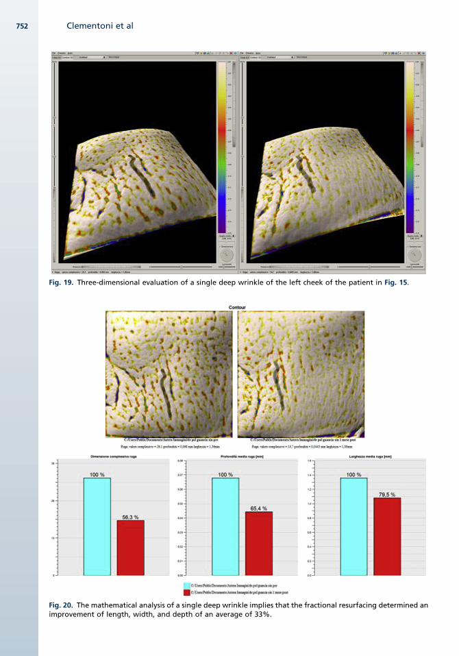
Fig. 19. Three-dimensional evaluation of a single deep wrinkle of the left cheek of the patient in Fig. 15.
Fig. 20. The mathematical analysis of a single deep wrinkle implies that the fractional resurfacing determined animprovement of length, width, and depth of an average of 33%.
Clementoni et al752
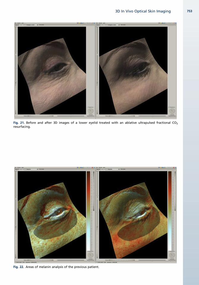
Fig. 21. Before and after 3D images of a lower eyelid treated with an ablative ultrapulsed fractional CO2
resurfacing.
Fig. 22. Areas of melanin analysis of the previous patient.
3D In Vivo Optical Skin Imaging 753
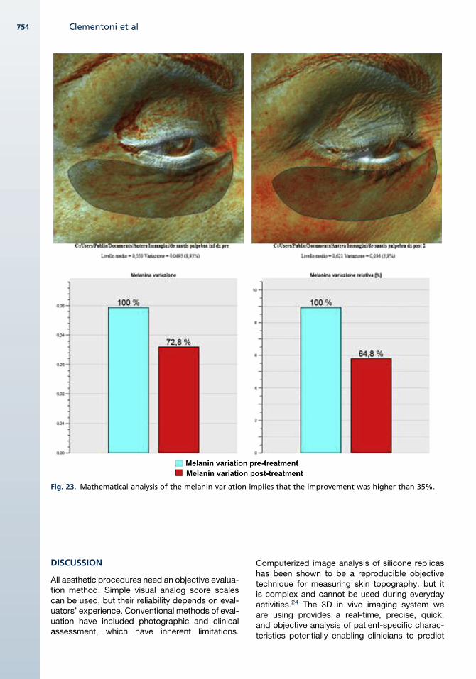
Fig. 23. Mathematical analysis of the melanin variation implies that the improvement was higher than 35%.
Clementoni et al754
DISCUSSION
All aesthetic procedures need an objective evalua-tion method. Simple visual analog score scalescan be used, but their reliability depends on eval-uators’ experience. Conventional methods of eval-uation have included photographic and clinicalassessment, which have inherent limitations.
Computerized image analysis of silicone replicashas been shown to be a reproducible objectivetechnique for measuring skin topography, but itis complex and cannot be used during everydayactivities.24 The 3D in vivo imaging system weare using provides a real-time, precise, quick,and objective analysis of patient-specific charac-teristics potentially enabling clinicians to predict
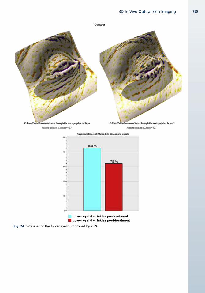
Fig. 24. Wrinkles of the lower eyelid improved by 25%.
3D In Vivo Optical Skin Imaging 755
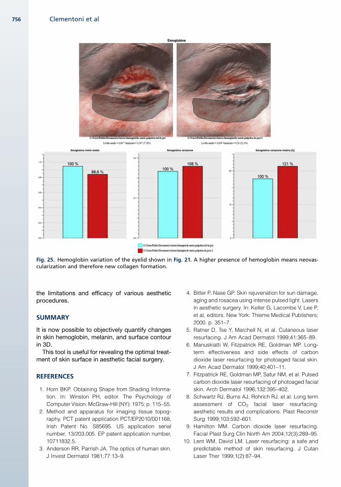
Fig. 25. Hemoglobin variation of the eyelid shown in Fig. 21. A higher presence of hemoglobin means neovas-cularization and therefore new collagen formation.
Clementoni et al756
the limitations and efficacy of various aestheticprocedures.
SUMMARY
It is now possible to objectively quantify changesin skin hemoglobin, melanin, and surface contourin 3D.This tool is useful for revealing the optimal treat-
ment of skin surface in aesthetic facial surgery.
REFERENCES
1. Horn BKP. Obtaining Shape from Shading Informa-
tion. In: Winston PH, editor. The Psychology of
Computer Vision. McGraw-Hill (NY): 1975; p. 115–55.
2. Method and apparatus for imaging tissue topog-
raphy. PCT patent application PCT/EP2010/001168,
Irish Patent No. S85695. US application serial
number, 13/203,005. EP patent application number,
10711832.5.
3. Anderson RR, Parrish JA. The optics of human skin.
J Invest Dermatol 1981;77:13–9.
4. Bitter P, Nase GP. Skin rejuvenation for sun damage,
aging and rosacea using intense pulsed light. Lasers
in aesthetic surgery. In: Keller G, Lacombe V, Lee P,
et al, editors. New York: Thieme Medical Publishers;
2000. p. 351–7.
5. Ratner D, Tse Y, Marchell N, et al. Cutaneous laser
resurfacing. J Am Acad Dermatol 1999;41:365–89.
6. Manuskiatti W, Fitzpatrick RE, Goldman MP. Long-
term effectiveness and side effects of carbon
dioxide laser resurfacing for photoaged facial skin.
J Am Acad Dermatol 1999;40:401–11.
7. Fitzpatrick RE, Goldman MP, Satur NM, et al. Pulsed
carbon dioxide laser resurfacing of photoaged facial
skin. Arch Dermatol 1996;132:395–402.
8. Schwartz RJ, Burns AJ, Rohrich RJ, et al. Long term
assessment of CO2 facial laser resurfacing:
aesthetic results and complications. Plast Reconstr
Surg 1999;103:592–601.
9. Hamilton MM. Carbon dioxide laser resurfacing.
Facial Plast Surg Clin North Am 2004;12(3):289–95.
10. Lent WM, David LM. Laser resurfacing: a safe and
predictable method of skin resurfacing. J Cutan
Laser Ther 1999;1(2):87–94.
3D In Vivo Optical Skin Imaging 757
11. Airan LE, Hruza G. Current lasers in skin resurfacing.
Facial Plast Surg Clin North Am 2002;10(1):87–101.
12. Fitzpatrick RE. CO2 laser resurfacing. Dermatol Clin
2001;19(3):443–51.
13. Fitzpatrick RE. Maximizing benefits and minimizing
risk with CO2 laser resurfacing. Dermatol Clin
2002;20(1):77–86.
14. Bernstein LJ, Kauvar AN, Grossman MC, et al. The
short- and long-term side effects of carbon dioxide
laser resurfacing. Dermatol Surg 1997;23:519–25.
15. Nanni CA, Alster TS. Complications of carbon
dioxide laser resurfacing. An evaluation of 500
patients. Dermatol Surg 1998;24:315–20.
16. Sriprachya-Anunt S, Fitzpatrick RE, Goldman MP,
et al. Infections complicating pulsed carbon dioxide
laser resurfacing for photoaged facial skin. Dermatol
Surg 1997;23:527–36.
17. Berwald C, Levy JL, Magalon G. Complications
of the resurfacing laser: retrospective study of 749
patients. Ann Chir Plast Esthet 2004;49(4):360–5.
18. Sullivan SA, Dailey RA. Complications of laser resur-
facing and their management. Ophthal Plast Re-
constr Surg 2000;16(6):417–26.
19. SadickNS.Updateonnon-ablative light therapy for re-
juvenation: a review. Lasers SurgMed2003;32:120–8.
20. Williams EF III, Dahiya R. Review of nonablative
laser resurfacing modalities. Facial Plast Surg Clin
North Am 2004;12(3):305–10.
21. Grema H, Greve B, Raulin C. Facial rhytides—sub-
surfacing or resurfacing? A review. Lasers Surg
Med 2003;32(5):405–12.
22. Bjerring P. Photorejuvenation—an overview. Med
Laser Appl 2004;19:186–95.
23. Manstein D, Herron GS, Sink RK, et al. Fractional
photothermolysis: a new concept for cutaneous re-
modeling using microscopic patterns of thermal
injury. Lasers Surg Med 2004;34(5):426–38.
24. GroveGL,GroveMJ,LeydenJJ, etal. Skin replicaanal-
ysis of photodamaged skin after therapy with tretinoin
emollient cream. J Am Acad Dermatol 1991;25:231–7.


![The Resurfacing of Arabic Qaf [q] in the Speech of Young ...](https://static.fdokumen.com/doc/165x107/6327fe736d480576770d8d74/the-resurfacing-of-arabic-qaf-q-in-the-speech-of-young-.jpg)


