2021 ACC/AHA/SCAI Guideline for Coronary Artery Revascularization
97
Circulation e18 January 18, 2022 Circulation. 2022;145:e18–e114. DOI: 10.1161/CIR.0000000000001038 Circulation is available at www.ahajournals.org/journal/circ *Writing committee members are required to recuse themselves from voting on sections to which their specific relationships with industry may apply; see Appendix 1 for detailed information. †ACC/AHA Representative. ‡ACC/AHA Joint Committee on Clinical Practice Guidelines Liaison. §ACC/AHA Task Force on Data Standards Representative. ‖SCAI Representative. ACC/AHA Joint Committee on Clinical Practice Guidelines Members, see page e80. The American Heart Association requests that this document be cited as follows: Lawton JS, Tamis-Holland JE, Bangalore S, Bates ER, Beckie TM, Bischoff JM, Bittl JA, Cohen MG, DiMaio JM, Don CW, Fremes SE, Gaudino MF, Goldberger ZD, Grant MC, Jaswal JB, Kurlansky PA, Mehran R, Metkus TS Jr, Nnacheta LC, Rao SV, Sellke FW, Sharma G, Yong CM, Zwischenberger BA. 2021 ACC/AHA/SCAI guideline for coronary artery revascularization: a report of the American College of Cardiology/American Heart Association Joint Committee on Clinical Practice Guidelines. Circulation. 2022;145:e18–e114. doi: 10.1161/CIR.0000000000001038 © 2021 by the American College of Cardiology Foundation and the American Heart Association, Inc. ACC/AHA/SCAI CLINICAL PRACTICE GUIDELINE 2021 ACC/AHA/SCAI Guideline for Coronary Artery Revascularization: A Report of the American College of Cardiology/American Heart Association Joint Committee on Clinical Practice Guidelines Writing Committee Members* Jennifer S. Lawton, MD, FAHA, Chair†; Jacqueline E. Tamis-Holland, MD, FAHA, FACC, FSCAI, Vice Chair‡; Sripal Bangalore, MD, MHA, FACC, FAHA, FSCAI†; Eric R. Bates, MD, FACC, FAHA†; Theresa M. Beckie, PhD, FAHA†; James M. Bischoff, MEd†; John A. Bittl, MD, FACC†; Mauricio G. Cohen, MD, FACC, FSCAI§; J. Michael DiMaio, MD†; Creighton W. Don, MD, PhD, FACC‖; Stephen E. Fremes, MD, FACC; Mario F. Gaudino, MD, PhD, MSCE, FACC, FAHA†; Zachary D. Goldberger, MD, FACC, FAHA‡; Michael C. Grant, MD, MSE†; Jang B. Jaswal, MS†; Paul A. Kurlansky, MD, FACC†; Roxana Mehran, MD, FACC†; Thomas S. Metkus Jr, MD, FACC†; Lorraine C. Nnacheta, DrPH, MPH†; Sunil V. Rao, MD, FACC†; Frank W. Sellke, MD, FACC, FAHA†; Garima Sharma, MD, FACC†; Celina M. Yong, MD, MBA, MSc, FSCAI, FACC, FAHA†; Brittany A. Zwischenberger, MD† AIM: The guideline for coronary artery revascularization replaces the 2011 coronary artery bypass graft surgery and the 2011 and 2015 percutaneous coronary intervention guidelines, providing a patient-centric approach to guide clinicians in the treatment of patients with significant coronary artery disease undergoing coronary revascularization as well as the supporting documentation to encourage their use. METHODS: A comprehensive literature search was conducted from May 2019 to September 2019, encompassing studies, reviews, and other evidence conducted on human subjects that were published in English from PubMed, EMBASE, the Cochrane Collaboration, CINHL Complete, and other relevant databases. Additional relevant studies, published through May 2021, were also considered. STRUCTURE: Coronary artery disease remains a leading cause of morbidity and mortality globally. Coronary revascularization is an important therapeutic option when managing patients with coronary artery disease. The 2021 coronary artery revascularization guideline provides recommendations based on contemporary evidence for the treatment of these patients. The recommendations present an evidence-based approach to managing patients with coronary artery disease who are being considered for coronary revascularization, with the intent to improve quality of care and align with patients’ interests. Key Words: AHA Scientific Statements ◼ percutaneous coronary intervention ◼ angioplasty ◼ coronary artery bypass graft surgery ◼ myocardial infarction ◼ cardiac surgery, stent(s) ◼ angiogram ◼ angiography ◼ percutaneous transluminal coronary angioplasty ◼ coronary atherosclerosis ◼ saphenous vein graft ◼ internal mammary artery graft ◼ internal thoracic artery graft ◼ arterial graft ◼ post-bypass ◼ non–ST-segment–elevated myocardial infarction ◼ vein graft lesions ◼ myocardial revascularization ◼ multivessel PCI ◼ left ventricular dysfunction Downloaded from http://ahajournals.org by on April 8, 2022
-
Upload
khangminh22 -
Category
Documents
-
view
1 -
download
0
Transcript of 2021 ACC/AHA/SCAI Guideline for Coronary Artery Revascularization
Circulation
e18 January 18, 2022 Circulation. 2022;145:e18–e114. DOI: 10.1161/CIR.0000000000001038
Circulation is available at www.ahajournals.org/journal/circ
*Writing committee members are required to recuse themselves from voting on sections to which their specific relationships with industry may apply; see Appendix 1 for detailed information. †ACC/AHA Representative. ‡ACC/AHA Joint Committee on Clinical Practice Guidelines Liaison. §ACC/AHA Task Force on Data Standards Representative. ‖SCAI Representative.
ACC/AHA Joint Committee on Clinical Practice Guidelines Members, see page e80.
The American Heart Association requests that this document be cited as follows: Lawton JS, Tamis-Holland JE, Bangalore S, Bates ER, Beckie TM, Bischoff JM, Bittl JA, Cohen MG, DiMaio JM, Don CW, Fremes SE, Gaudino MF, Goldberger ZD, Grant MC, Jaswal JB, Kurlansky PA, Mehran R, Metkus TS Jr, Nnacheta LC, Rao SV, Sellke FW, Sharma G, Yong CM, Zwischenberger BA. 2021 ACC/AHA/SCAI guideline for coronary artery revascularization: a report of the American College of Cardiology/American Heart Association Joint Committee on Clinical Practice Guidelines. Circulation. 2022;145:e18–e114. doi: 10.1161/CIR.0000000000001038
© 2021 by the American College of Cardiology Foundation and the American Heart Association, Inc.
ACC/AHA/SCAI CLINICAL PRACTICE GUIDELINE
2021 ACC/AHA/SCAI Guideline for Coronary Artery Revascularization: A Report of the American College of Cardiology/American Heart Association Joint Committee on Clinical Practice GuidelinesWriting Committee Members*
Jennifer S. Lawton, MD, FAHA, Chair†; Jacqueline E. Tamis-Holland, MD, FAHA, FACC, FSCAI, Vice Chair‡; Sripal Bangalore, MD, MHA, FACC, FAHA, FSCAI†; Eric R. Bates, MD, FACC, FAHA†; Theresa M. Beckie, PhD, FAHA†; James M. Bischoff, MEd†; John A. Bittl, MD, FACC†; Mauricio G. Cohen, MD, FACC, FSCAI§; J. Michael DiMaio, MD†; Creighton W. Don, MD, PhD, FACC‖; Stephen E. Fremes, MD, FACC; Mario F. Gaudino, MD, PhD, MSCE, FACC, FAHA†; Zachary D. Goldberger, MD, FACC, FAHA‡; Michael C. Grant, MD, MSE†; Jang B. Jaswal, MS†; Paul A. Kurlansky, MD, FACC†; Roxana Mehran, MD, FACC†; Thomas S. Metkus Jr, MD, FACC†; Lorraine C. Nnacheta, DrPH, MPH†; Sunil V. Rao, MD, FACC†; Frank W. Sellke, MD, FACC, FAHA†; Garima Sharma, MD, FACC†; Celina M. Yong, MD, MBA, MSc, FSCAI, FACC, FAHA†; Brittany A. Zwischenberger, MD†
AIM: The guideline for coronary artery revascularization replaces the 2011 coronary artery bypass graft surgery and the 2011 and 2015 percutaneous coronary intervention guidelines, providing a patient-centric approach to guide clinicians in the treatment of patients with significant coronary artery disease undergoing coronary revascularization as well as the supporting documentation to encourage their use.
METHODS: A comprehensive literature search was conducted from May 2019 to September 2019, encompassing studies, reviews, and other evidence conducted on human subjects that were published in English from PubMed, EMBASE, the Cochrane Collaboration, CINHL Complete, and other relevant databases. Additional relevant studies, published through May 2021, were also considered.
STRUCTURE: Coronary artery disease remains a leading cause of morbidity and mortality globally. Coronary revascularization is an important therapeutic option when managing patients with coronary artery disease. The 2021 coronary artery revascularization guideline provides recommendations based on contemporary evidence for the treatment of these patients. The recommendations present an evidence-based approach to managing patients with coronary artery disease who are being considered for coronary revascularization, with the intent to improve quality of care and align with patients’ interests.
Key Words: AHA Scientific Statements ◼ percutaneous coronary intervention ◼ angioplasty ◼ coronary artery bypass graft surgery ◼ myocardial infarction ◼ cardiac surgery, stent(s) ◼ angiogram ◼ angiography ◼ percutaneous transluminal coronary angioplasty ◼ coronary atherosclerosis ◼ saphenous vein graft ◼ internal mammary artery graft ◼ internal thoracic artery graft ◼ arterial graft
◼ post-bypass ◼ non–ST-segment–elevated myocardial infarction ◼ vein graft lesions ◼ myocardial revascularization ◼ multivessel PCI ◼ left ventricular dysfunction
Clinical Statements and Guidelines
Dow
nloaded from http://ahajournals.org by on A
pril 8, 2022

Lawton et al 2021 ACC/AHA/SCAI Coronary Revascularization Guideline
Circulation. 2022;145:e18–e114. DOI: 10.1161/CIR.0000000000001038 January 18, 2022 e19
CLINICAL STATEMENTS
AND GUIDELINES
CONTENTSAbstract . . . . . . . . . . . . . . . . . . . . . . . . . . . . . . . . . . . . . . . . . .e18Top 10 Take-Home Messages . . . . . . . . . . . . . . . . . . . . .e20Preamble . . . . . . . . . . . . . . . . . . . . . . . . . . . . . . . . . . . . . . . .e211. Introduction . . . . . . . . . . . . . . . . . . . . . . . . . . . . . . . . . e22
1.1. Methodology and Evidence Review . . . . . .e221.2. Organization of the Writing Committee . . . . .e241.3. Document Review and Approval . . . . . . . . .e241.4. Scope of the Guideline . . . . . . . . . . . . . . . . .e241.5. Class of Recommendation and Level
of Evidence . . . . . . . . . . . . . . . . . . . . . . . . . . . .e251.6. Abbreviations . . . . . . . . . . . . . . . . . . . . . . . . . .e25
2. Improving Equity of Care in Revascularization and Shared Decision-Making . . . . . . . . . . . . . . . . . .e262.1. Improving Equity of Care in
Revascularization . . . . . . . . . . . . . . . . . . . . . . .e262.2. Shared Decision-Making and
Informed Consent . . . . . . . . . . . . . . . . . . . . . .e273. Preprocedural Assessment and the
Heart Team . . . . . . . . . . . . . . . . . . . . . . . . . . . . . . . . . .e293.1. The Heart Team . . . . . . . . . . . . . . . . . . . . . . . .e293.2. Predicting Patient Risk of Death
With CABG . . . . . . . . . . . . . . . . . . . . . . . . . . . .e304. Defining Lesion Severity . . . . . . . . . . . . . . . . . . . . . .e30
4.1. Angiography to Define Anatomy and Assess Lesion Severity . . . . . . . . . . . . .e30
4.2. Defining Coronary Artery Lesion Complexity: Calculation of the SYNTAX (Synergy Between PCI With TAXUS and Cardiac Surgery) Score . . . . . . . . . . . . . . . . .e31
4.3. Use of Coronary Physiology to Guide Revascularization With PCI . . . . . . . . . . . . . .e31
4.4. Intravascular Ultrasound to Assess Lesion Severity . . . . . . . . . . . . . . . . . . . . . . . . .e32
5. Revascularization in STEMI . . . . . . . . . . . . . . . . . . . .e335.1. Revascularization of the Infarct
Artery in Patients With STEMI . . . . . . . . . . .e335.2. Revascularization of the Non-Infarct
Artery in Patients With STEMI . . . . . . . . . . .e356. Revascularization in Non–ST-Segment–Elevation
Acute Coronary Syndrome (NSTE-ACS) . . . . . . . . e386.1. Coronary Angiography and Revascularization
in Patients with NSTE-ACS . . . . . . . . . . . . .e387. Revascularization in SIHD . . . . . . . . . . . . . . . . . . . . .e40
7.1. Revascularization to Improve Survival in SIHD Compared With Medical Therapy. . . . . . . e40
7.2. Revascularization to REDUCE Cardiovascular Events in SIHD Compared with Medical Therapy . . . . . . . . .e43
7.3. Revascularization to Improve Symptoms . . . . . e448. Situations in Which PCI or CABG Would
Be Preferred . . . . . . . . . . . . . . . . . . . . . . . . . . . . . . . . .e448.1. Patients With Complex Disease . . . . . . . . .e448.2. Patients With Diabetes . . . . . . . . . . . . . . . . . .e458.3. Patients With Previous CABG . . . . . . . . . . .e46
8.4. DAPT Adherence . . . . . . . . . . . . . . . . . . . . . . . e479. Special Populations and Situations . . . . . . . . . . . . . e47
9.1. Revascularization in Pregnant Patients . . . . . . . . . . . . . . . . . . . . . . . . . . . . . . . e47
9.2. Revascularization in Older Patients . . . . . . e479.3. Revascularization in Patients With
Chronic Kidney Disease (CKD) . . . . . . . . . .e489.4. Revascularization in Patients
Before Noncardiac Surgery . . . . . . . . . . . . .e499.5. Revascularization in Patients to
Reduce Ventricular Arrhythmias . . . . . . . . .e509.6. Revascularization in Patients With
SCAD . . . . . . . . . . . . . . . . . . . . . . . . . . . . . . . . .e509.7. Revascularization in Patients With
Cardiac Allografts . . . . . . . . . . . . . . . . . . . . . .e519.8. Revascularization in Patients
Before Transcatheter Aortic Valve Replacement (TAVR) . . . . . . . . . . . . . . . . . . .e51
9.9. Revascularization in Patients With Anomalous Coronary Artery . . . . . . . . . . . . .e51
10. General Procedural Issues for PCI . . . . . . . . . . . . .e5210.1. Radial and Femoral Approaches
for PCI . . . . . . . . . . . . . . . . . . . . . . . . . . . . . . . .e5210.2. Choice of Stent Type . . . . . . . . . . . . . . . . . . .e5210.3. Use of Intravascular Imaging . . . . . . . . . . . .e5310.4. Thrombectomy . . . . . . . . . . . . . . . . . . . . . . . . .e5410.5. Treatment of Calcified Lesions . . . . . . . . . .e5410.6. Treatment of Saphenous Vein Graft
(SVG) Disease (Previous CABG) . . . . . . . .e5510.7. Treatment of CTO . . . . . . . . . . . . . . . . . . . . . .e5610.8. Treatment of Patients With Stent
Restenosis . . . . . . . . . . . . . . . . . . . . . . . . . . . . .e5610.9. Hemodynamic Support for
Complex PCI . . . . . . . . . . . . . . . . . . . . . . . . . . . e5711. Pharmacotherapy in Patients
Undergoing PCI . . . . . . . . . . . . . . . . . . . . . . . . . . . . . . e5711.1. Aspirin and Oral P2Y12 Inhibitors
in Patients Undergoing PCI . . . . . . . . . . . . . e5711.2. Intravenous P2Y12 Inhibitors in
Patients Undergoing PCI . . . . . . . . . . . . . . . .e6011.3. Intravenous Glycoprotein IIb/IIIa
Inhibitors in Patients Undergoing PCI . . . . . .e6011.4. Heparin, Low-Molecular-Weight
Heparin, and Bivalirudin in Patients Undergoing PCI . . . . . . . . . . . . . . . . . . . . . . . .e61
12. General Procedural Issues for CABG . . . . . . . . . .e6212.1. Perioperative Considerations in
Patients Undergoing CABG . . . . . . . . . . . . .e6212.2. Bypass Conduits in Patients
Undergoing CABG . . . . . . . . . . . . . . . . . . . . .e6312.3. CABG in Patients Undergoing Other
Cardiac Surgery . . . . . . . . . . . . . . . . . . . . . . . .e6412.4. Use of Epiaortic Ultrasound in
Patients Undergoing CABG . . . . . . . . . . . . .e6512.5. Use of Cardiopulmonary Bypass in
Patients Undergoing CABG . . . . . . . . . . . . .e66
Dow
nloaded from http://ahajournals.org by on A
pril 8, 2022

Lawton et al 2021 ACC/AHA/SCAI Coronary Revascularization Guideline
January 18, 2022 Circulation. 2022;145:e18–e114. DOI: 10.1161/CIR.0000000000001038e20
CLIN
ICAL
STA
TEM
ENTS
AN
D GU
IDEL
INES
13. Pharmacotherapy in Patients Undergoing CABG . . . . . . . . . . . . . . . . . . . . . . . . . . . . . . . . . . . . . . .e6613.1. Insulin Infusion and Other Measures
to Reduce Sternal Wound Infection in Patients Undergoing CABG . . . . . . . . . . . . .e66
13.2. Antiplatelet Therapy in Patients Undergoing CABG . . . . . . . . . . . . . . . . . . . . .e68
13.3. Beta Blockers and Amiodarone in Patients Undergoing CABG . . . . . . . . . . . . .e69
14. Pharmacotherapy in Patients After Revascularization . . . . . . . . . . . . . . . . . . . . . . . . . . . . . e7014.1. Pharmacotherapy for Risk Factor Control
in Patients After Revascularization . . . . . . . e7014.2. Dual Antiplatelet Therapy in Patients
After PCI . . . . . . . . . . . . . . . . . . . . . . . . . . . . . . e7014.3. Antiplatelet Therapy in Patients
After CABG . . . . . . . . . . . . . . . . . . . . . . . . . . . . e7014.4. Beta Blockers in Patients After
Revascularization . . . . . . . . . . . . . . . . . . . . . . . e7214.5. Beta Blockers for the Prevention of
Atrial Fibrillation After CABG . . . . . . . . . . . . e7214.6. Antiplatelet Therapy in Patients
With Atrial Fibrillation on Anticoagulation After PCI . . . . . . . . . . . . . . . . . . . . . . . . . . . . . . e73
15. Recommendations for Addressing Psychosocial Factors and Lifestyle Changes After Revascularization . . . . . . . . . . . . . . . . . . . . . . . . e7315.1. Cardiac Rehabilitation and Education . . . . . . e7315.2. Smoking Cessation in Patients After
Revascularization . . . . . . . . . . . . . . . . . . . . . . . e7415.3. Psychological Interventions in
Patients After Revascularization . . . . . . . . . e7616. Revascularization Outcomes . . . . . . . . . . . . . . . . . . . e76
16.1. Assessment of Outcomes in Patients After Revascularization . . . . . . . . . e76
17. Unanswered Questions and Future Directions . . . . . . . . . . . . . . . . . . . . . . . . . . . . . . . . . . . . e7717.1. Special Populations . . . . . . . . . . . . . . . . . . . . . e77
17.1.1. Underrepresented Racial and Ethnic Groups . . . . . . . . . . . . . . e77
17.2. Special Clinical Situations . . . . . . . . . . . . . . . e7717.2.1. Left Ventricular Dysfunction . . . . . . e7717.2.2. SCAD . . . . . . . . . . . . . . . . . . . . . . . . . . e7817.2.3. Coronary Artery Aneurysm . . . . . . . e7817.2.4. Myocardial Bridging . . . . . . . . . . . . . e7817.2.5. Treatment of Graft Failure . . . . . . . e7817.2.6. Antiplatelet Therapy in Patients
With ACS After CABG With an Indication for Anticoagulation . . . . . .e78
17.3. Revascularization Considerations . . . . . . . . e7817.3.1. Use of the Radial Artery for
a Conduit After Radial Artery Catheterization . . . . . . . . . . . . . . . . . . e78
17.3.2. Completeness of Revascularization in Multivessel Disease . . . . . . . . . . . e79
17.3.3. Hybrid Coronary Surgery . . . . . . . . e79
17.3.4. Revascularization Before Percutaneous Valve Procedures . . . . . . . . . . . . . . . . . . . . . e79
17.3.5. Revascularization Before Organ Transplantation . . . . . . . . . . . e79
References . . . . . . . . . . . . . . . . . . . . . . . . . . . . . . . . . . . . . . .e80Appendix 1
Author Relationships With Industry and Other Entities (Relevant) . . . . . . . . . . . . . . . . . . . . e110
Appendix 2Reviewer Relationships With Industry and Other Entities (Comprehensive) . . . . . . . . . . . . . e112
TOP 10 TAKE-HOME MESSAGES1. Treatment decisions regarding coronary revascu-
larization in patients with coronary artery disease should be based on clinical indications, regardless of sex, race, or ethnicity, because there is no evi-dence that some patients benefit less than oth-ers, and efforts to reduce disparities of care are warranted.
2. In patients being considered for coronary revas-cularization for whom the optimal treatment strat-egy is unclear, a multidisciplinary Heart Team approach is recommended. Treatment decisions should be patient centered, incorporate patient preferences and goals, and include shared decision-making.
3. For patients with significant left main disease, surgical revascularization is indicated to improve survival relative to that likely to be achieved with medical therapy. Percutaneous revascularization is a reasonable option to improve survival, com-pared with medical therapy, in selected patients with low to medium anatomic complexity of cor-onary artery disease and left main disease that is equally suitable for surgical or percutaneous revascularization.
4. Updated evidence from contemporary trials supplement older evidence with regard to mor-tality benefit of revascularization in patients with stable ischemic heart disease, normal left ventricular ejection fraction, and triple-vessel coronary artery disease. Surgical revasculariza-tion may be reasonable to improve survival. A survival benefit with percutaneous revascular-ization is uncertain. Revascularization decisions are based on consideration of disease complex-ity, technical feasibility of treatment, and a Heart Team discussion.
5. The use of a radial artery as a surgical revascu-larization conduit is preferred versus the use of a saphenous vein conduit to bypass the second most important target vessel with significant ste-nosis after the left anterior descending coronary
Dow
nloaded from http://ahajournals.org by on A
pril 8, 2022

Lawton et al 2021 ACC/AHA/SCAI Coronary Revascularization Guideline
Circulation. 2022;145:e18–e114. DOI: 10.1161/CIR.0000000000001038 January 18, 2022 e21
CLINICAL STATEMENTS
AND GUIDELINES
artery. Benefits include superior patency, reduced adverse cardiac events, and improved survival.
6. Radial artery access is recommended in patients undergoing percutaneous intervention who have acute coronary syndromes or stable ischemic heart disease, to reduce bleeding and vascular complica-tions compared with a femoral approach. Patients with acute coronary syndromes also benefit from a reduction in mortality rate with this approach.
7. A short duration of dual antiplatelet therapy after percutaneous revascularization in patients with stable ischemic heart disease is reasonable to reduce the risk of bleeding events. After consid-eration of recurrent ischemia and bleeding risks, select patients may safely transition to P2Y12 inhibitor monotherapy and stop aspirin after 1 to 3 months of dual antiplatelet therapy.
8. Staged percutaneous intervention (while in hospi-tal or after discharge) of a significantly stenosed nonculprit artery in patients presenting with an ST-segment–elevation myocardial infarction is recommended in select patients to improve out-comes. Percutaneous intervention of the noncul-prit artery at the time of primary percutaneous coronary intervention is less clear and may be considered in stable patients with uncomplicated revascularization of the culprit artery, low-com-plexity nonculprit artery disease, and normal renal function. In contrast, percutaneous intervention of the non-culprit artery can be harmful in patients in cardiogenic shock.
9. Revascularization decisions in patients with dia-betes and multivessel coronary artery disease are optimized by the use of a Heart Team approach. Patients with diabetes who have triple-vessel dis-ease should undergo surgical revascularization; percutaneous coronary intervention may be con-sidered if they are poor candidates for surgery.
10. Treatment decisions for patients undergoing sur-gical revascularization of coronary artery disease should include the calculation of a patient’s sur-gical risk with the Society of Thoracic Surgeons score. The usefulness of the SYNTAX score calcu-lation in treatment decisions is less clear because of the interobserver variability in its calculation and its absence of clinical variables.
PREAMBLESince 1980, the American College of Cardiology (ACC) and American Heart Association (AHA) have translated scientific evidence into clinical practice guidelines with recommendations to improve cardiovascular health. These guidelines, which are based on systematic meth-ods to evaluate and classify evidence, provide a founda-tion for the delivery of quality cardiovascular care. The
ACC and AHA sponsor the development and publication of clinical practice guidelines without commercial sup-port, and members volunteer their time to the writing and review efforts. Guidelines are official policy of the ACC and AHA. For some guidelines, the ACC and AHA part-ner with other organizations.
Intended UseClinical practice guidelines provide recommendations applicable to patients with or at risk of developing cardiovascular disease (CVD). The focus is on medi-cal practice in the United States, but these guidelines are relevant to patients throughout the world. Although guidelines may be used to inform regulatory or payer decisions, the intent is to improve quality of care and align with patients’ interests. Guidelines are intended to define practices meeting the needs of patients in most, but not all, circumstances and should not replace clinical judgment.
Clinical ImplementationManagement, in accordance with guideline recom-mendations, is effective only when followed by both practitioners and patients. Adherence to recommen-dations can be enhanced by shared decision-making between clinicians and patients, with patient engage-ment in selecting interventions on the basis of indi-vidual values, preferences, and associated conditions and comorbidities.
Methodology and ModernizationThe ACC/AHA Joint Committee on Clinical Practice Guidelines (Joint Committee) continuously reviews, updates, and modifies guideline methodology on the basis of published standards from organizations, in-cluding the Institute of Medicine,1,2 and on the basis of internal reevaluation. Similarly, presentation and delivery of guidelines are reevaluated and modified in response to evolving technologies and other factors to optimally facilitate dissemination of information to health care professionals at the point of care.
Numerous modifications to the guidelines have been implemented to make them shorter and enhance “user-friendliness.” Guidelines are written and presented in a modular, “knowledge chunk” format, in which each chunk includes a table of recommendations, a brief synopsis, recommendation-specific supportive text and, when appropriate, flow diagrams or additional tables. Hyper-linked references are provided for each modular knowl-edge chunk to facilitate quick access and review.
In recognition of the importance of cost–value con-siderations, in certain guidelines, when appropriate and feasible, an analysis of value for a drug, device, or
Dow
nloaded from http://ahajournals.org by on A
pril 8, 2022
Lawton et al 2021 ACC/AHA/SCAI Coronary Revascularization Guideline
January 18, 2022 Circulation. 2022;145:e18–e114. DOI: 10.1161/CIR.0000000000001038e22
CLIN
ICAL
STA
TEM
ENTS
AN
D GU
IDEL
INES
intervention may be performed in accordance with the ACC/AHA methodology.3
To ensure that guideline recommendations remain current, new data will be reviewed on an ongoing basis by the writing committee and staff. Going forward, tar-geted sections or knowledge chunks will be revised dynamically after publication and timely peer review of potentially practice-changing science. The previous designations of “full revision” and “focused update” will be phased out. For additional information and policies on guideline development, readers may consult the ACC/AHA guideline methodology manual4 and other methodology articles.5-7
Selection of Writing Committee MembersThe Joint Committee strives to ensure that the guide-line writing committee contains requisite content exper-tise and is representative of the broader cardiovascular community by selection of experts across a spectrum of backgrounds, representing different geographic regions, sexes, races, ethnicities, intellectual perspectives/biases, and clinical practice settings. Organizations and profes-sional societies with related interests and expertise are invited to participate as partners or collaborators.
Relationships With Industry and Other EntitiesThe ACC and AHA have rigorous policies and meth-ods to ensure that documents are developed without bias or improper influence. The complete policy on rela-tionships with industry and other entities (RWI) can be found online. Appendix 1 of the guideline lists writing committee members’ relevant RWI; for the purposes of full transparency, their comprehensive disclosure infor-mation is available in a Supplemental Appendix. Com-prehensive disclosure information for the Joint Commit-tee is also available online.
Evidence Review and Evidence Review CommitteesIn developing recommendations, the writing committee uses evidence-based methodologies that are based on all available data.4,5 Literature searches focus on ran-domized controlled trials (RCTs) but also include regis-tries, nonrandomized comparative and descriptive stud-ies, case series, cohort studies, systematic reviews, and expert opinion. Only key references are cited.
An independent evidence review committee is com-missioned when there are ≥1 questions deemed of utmost clinical importance and merit formal systematic review to determine which patients are most likely to benefit from a drug, device, or treatment strategy, and to what degree. Criteria for commissioning an evidence review committee and formal systematic review include
absence of a current authoritative systematic review, feasibility of defining the benefit and risk in a time frame consistent with the writing of a guideline, relevance to a substantial number of patients, and likelihood that the findings can be translated into actionable recom-mendations. Evidence review committee members may include methodologists, epidemiologists, clinicians, and biostatisticians. Recommendations developed by the writing committee on the basis of the systematic review are marked.“SR”
Guideline-Directed Management and TherapyThe term guideline-directed medical therapy (GDMT) en-compasses clinical evaluation, diagnostic testing, and both pharmacological and procedural treatments. For these and all recommended drug treatment regimens, the reader should confirm dosage with product insert material and evaluate for contraindications and interactions. Recom-mendations are limited to drugs, devices, and treatments approved for clinical use in the United States.
Patrick T. O’Gara, MD, MACC, FAHAChair, ACC/AHA Joint Committee on
Clinical Practice Guidelines
1. INTRODUCTION1.1. Methodology and Evidence ReviewThe recommendations listed in this guideline are, whenev-er possible, evidence based. An initial extensive evidence review, which included literature derived from research in-volving human subjects, published in English, and indexed in the US National Library of Medicine and the National Center for Biotechnology information (through PubMed), EMBASE, the Cochrane Collaboration, CINHL Complete, and other selected databases relevant to this guideline, was conducted from May 2019 to September 2019. Key search words included but were not limited to the follow-ing: percutaneous coronary intervention, angioplasty, coro-nary artery bypass graft (CABG) surgery, myocardial infarc-tion, cardiac surgery, stent(s), angiogram, angiography, percutaneous transluminal coronary angioplasty, coronary atherosclerosis, saphenous vein graft, internal mammary ar-tery (IMA) graft, internal thoracic artery graft, arterial graft, post-bypass, non-ST elevated myocardial infarction, vein graft lesions, myocardial revascularization, multivessel PCI, and left ventricular dysfunction. Additional relevant studies, published through May 2021 during the guideline writing process, were also considered by the writing committee and added to the evidence tables when appropriate. The final evidence tables are included in the Online Data Sup-plement and summarize the evidence used by the writing committee to formulate recommendations. References selected and published in the present document are rep-resentative and not all-inclusive.
Dow
nloaded from http://ahajournals.org by on A
pril 8, 2022
Lawton et al 2021 ACC/AHA/SCAI Coronary Revascularization Guideline
e23
CLINICAL STATEMENTS
AND GUIDELINES
Circulation. 2022;145:e18–e114. DOI: 10.1161/CIR.0000000000001038 January 18, 2022
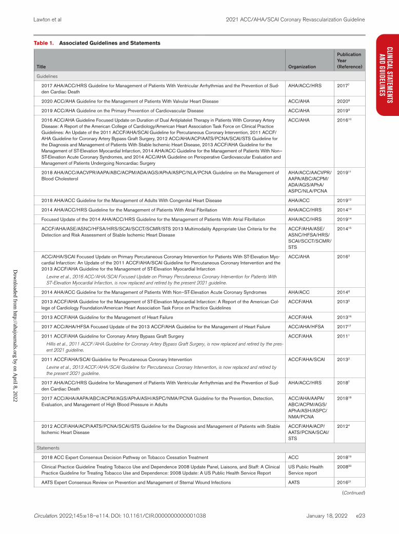
Table 1. Associated Guidelines and Statements
Title Organization
Publication Year (Reference)
Guidelines
2017 AHA/ACC/HRS Guideline for Management of Patients With Ventricular Arrhythmias and the Prevention of Sud-den Cardiac Death
AHA/ACC/HRS 20177
2020 ACC/AHA Guideline for the Management of Patients With Valvular Heart Disease ACC/AHA 20208
2019 ACC/AHA Guideline on the Primary Prevention of Cardiovascular Disease ACC/AHA 20199
2016 ACC/AHA Guideline Focused Update on Duration of Dual Antiplatelet Therapy in Patients With Coronary Artery Disease: A Report of the American College of Cardiology/American Heart Association Task Force on Clinical Practice Guidelines: An Update of the 2011 ACCF/AHA/SCAI Guideline for Percutaneous Coronary Intervention, 2011 ACCF/AHA Guideline for Coronary Artery Bypass Graft Surgery, 2012 ACC/AHA/ACP/AATS/PCNA/SCAI/STS Guideline for the Diagnosis and Management of Patients With Stable Ischemic Heart Disease, 2013 ACCF/AHA Guideline for the Management of ST-Elevation Myocardial Infarction, 2014 AHA/ACC Guideline for the Management of Patients With Non–ST-Elevation Acute Coronary Syndromes, and 2014 ACC/AHA Guideline on Perioperative Cardiovascular Evaluation and Management of Patients Undergoing Noncardiac Surgery
ACC/AHA 201610
2018 AHA/ACC/AACVPR/AAPA/ABC/ACPM/ADA/AGS/APhA/ASPC/NLA/PCNA Guideline on the Management of Blood Cholesterol
AHA/ACC/AACVPR/AAPA/ABC/ACPM/ADA/AGS/APhA/ASPC/NLA/PCNA
201911
2018 AHA/ACC Guideline for the Management of Adults With Congenital Heart Disease AHA/ACC 201912
2014 AHA/ACC/HRS Guideline for the Management of Patients With Atrial Fibrillation AHA/ACC/HRS 201413
Focused Update of the 2014 AHA/ACC/HRS Guideline for the Management of Patients With Atrial Fibrillation AHA/ACC/HRS 201914
ACCF/AHA/ASE/ASNC/HFSA/HRS/SCAI/SCCT/SCMR/STS 2013 Multimodality Appropriate Use Criteria for the Detection and Risk Assessment of Stable Ischemic Heart Disease
ACCF/AHA/ASE/ASNC/HFSA/HRS/SCAI/SCCT/SCMR/STS
201415
ACC/AHA/SCAI Focused Update on Primary Percutaneous Coronary Intervention for Patients With ST-Elevation Myo-cardial Infarction: An Update of the 2011 ACCF/AHA/SCAI Guideline for Percutaneous Coronary Intervention and the 2013 ACCF/AHA Guideline for the Management of ST-Elevation Myocardial Infarction
Levine et al., 2016 ACC/AHA/SCAI Focused Update on Primary Percutaneous Coronary Intervention for Patients With ST-Elevation Myocardial Infarction, is now replaced and retired by the present 2021 guideline.
ACC/AHA 20163
2014 AHA/ACC Guideline for the Management of Patients With Non–ST-Elevation Acute Coronary Syndromes AHA/ACC 20146
2013 ACCF/AHA Guideline for the Management of ST-Elevation Myocardial Infarction: A Report of the American Col-lege of Cardiology Foundation/American Heart Association Task Force on Practice Guidelines
ACCF/AHA 20135
2013 ACCF/AHA Guideline for the Management of Heart Failure ACCF/AHA 201316
2017 ACC/AHA/HFSA Focused Update of the 2013 ACCF/AHA Guideline for the Management of Heart Failure ACC/AHA/HFSA 201717
2011 ACCF/AHA Guideline for Coronary Artery Bypass Graft Surgery
Hillis et al., 2011 ACCF/AHA Guideline for Coronary Artery Bypass Graft Surgery, is now replaced and retired by the pres-ent 2021 guideline.
ACCF/AHA 20111
2011 ACCF/AHA/SCAI Guideline for Percutaneous Coronary Intervention
Levine et al., 2013 ACCF/AHA/SCAI Guideline for Percutaneous Coronary Intervention, is now replaced and retired by the present 2021 guideline.
ACCF/AHA/SCAI 20132
2017 AHA/ACC/HRS Guideline for Management of Patients With Ventricular Arrhythmias and the Prevention of Sud-den Cardiac Death
AHA/ACC/HRS 20187
2017 ACC/AHA/AAPA/ABC/ACPM/AGS/APhA/ASH/ASPC/NMA/PCNA Guideline for the Prevention, Detection, Evaluation, and Management of High Blood Pressure in Adults
ACC/AHA/AAPA/ABC/ACPM/AGS/APhA/ASH/ASPC/NMA/PCNA
201818
2012 ACCF/AHA/ACP/AATS/PCNA/SCAI/STS Guideline for the Diagnosis and Management of Patients with Stable Ischemic Heart Disease
ACCF/AHA/ACP/AATS/PCNA/SCAI/STS
20124
Statements
2018 ACC Expert Consensus Decision Pathway on Tobacco Cessation Treatment ACC 201819
Clinical Practice Guideline Treating Tobacco Use and Dependence 2008 Update Panel, Liaisons, and Staff: A Clinical Practice Guideline for Treating Tobacco Use and Dependence: 2008 Update: A US Public Health Service Report
US Public Health Service report
200820
AATS Expert Consensus Review on Prevention and Management of Sternal Wound Infections AATS 201621
(Continued )
Dow
nloaded from http://ahajournals.org by on A
pril 8, 2022
Lawton et al 2021 ACC/AHA/SCAI Coronary Revascularization Guideline
e24
CLIN
ICAL
STA
TEM
ENTS
AN
D GU
IDEL
INES
January 18, 2022 Circulation. 2022;145:e18–e114. DOI: 10.1161/CIR.0000000000001038
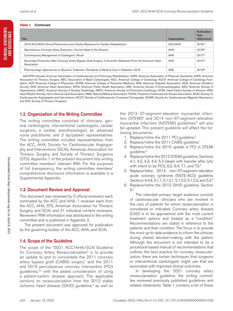
1.2. Organization of the Writing CommitteeThe writing committee consisted of clinicians, gen-eral cardiologists, interventional cardiologists, cardiac surgeons, a cardiac anesthesiologist, an advanced nurse practitioner, and 2 lay/patient representatives. The writing committee included representatives from the ACC, AHA, Society for Cardiovascular Angiogra-phy and Interventions (SCAI), American Association for Thoracic Surgery, and Society of Thoracic Surgeons (STS). Appendix 1 of the present document lists writing committee members’ relevant RWI. For the purposes of full transparency, the writing committee members’ comprehensive disclosure information is available in a Supplemental Appendix.
1.3. Document Review and ApprovalThis document was reviewed by 2 official reviewers each nominated by the ACC and AHA; 1 reviewer each from the ACC, AHA, STS, American Association for Thoracic Surgery, and SCAI; and 31 individual content reviewers. Reviewers’ RWI information was distributed to the writing committee and is published in Appendix 2.
The present document was approved for publication by the governing bodies of the ACC, AHA, and SCAI.
1.4. Scope of the GuidelineThe scope of the “2021 ACC/AHA/SCAI Guideline for Coronary Artery Revascularization” is to provide an update to and to consolidate the 2011 coronary artery bypass graft (CABG) surgery1 and the 2011 and 2015 percutaneous coronary intervention (PCI) guidelines,2,3 with the added consideration of using a patient-centric disease approach. The applicable sections on revascularization from the 2012 stable ischemic heart disease (SIHD) guideline,4 as well as
the 2013 ST-segment–elevation myocardial infarc-tion (STEMI)5 and 2014 non–ST-segment–elevation myocardial infarction (NSTEMI) guidelines,6 will also be updated. This present guideline will affect the fol-lowing documents:
1. Replace/retire the 2011 PCI guideline.2
2. Replace/retire the 2011 CABG guideline.1
3. Replace/retire the 2015 update in PCI in STEMI guideline.3
4. Replace/retire the 2013 STEMI guideline, Sections 4.1, 4.2, 4.3, 4.4, 5.3 (deals with transfer after lytic with intent to do PCI), 6.2, 6.4, 7.1, and 7.2.5
5. Replace/retire 2014 non–ST-segment–elevation acute coronary syndrome (NSTE-ACS) guideline, Sections 4.4.4, 5.1.1, 5.1.2.1, 5.1.2.2, 5.1.2.3, and 5.2.6
6. Replace/retire the 2012 SIHD guideline, Section 5.4
The intended primary target audience consists of cardiovascular clinicians who are involved in the care of patients for whom revascularization is considered or indicated. Coronary artery disease (CAD) is to be approached with the most current treatment options and treated as a “condition.” Recommendations are stated in reference to the patients and their condition. The focus is to provide the most up-to-date evidence to inform the clinician during shared decision-making with the patient. Although the document is not intended to be a procedural-based manual of recommendations that outlines the best practice for coronary revascular-ization, there are certain techniques that surgeons or interventional cardiologists might use that are associated with improved clinical outcomes.
In developing the 2021 coronary artery revascularization guideline, the writing commit-tee reviewed previously published guidelines and related statements. Table 1 contains a list of these
2018 ACC/AHA Clinical Performance and Quality Measures for Cardiac Rehabilitation ACC/AHA 201822
Spontaneous Coronary Artery Dissection: Current State of the Science AHA 201823
Contemporary Management of Cardiogenic Shock AHA 201724
Secondary Prevention After Coronary Artery Bypass Graft Surgery: A Scientific Statement From the American Heart Association
AHA 201525
Pharmacologic Approaches to Glycemic Treatment: Standards of Medical Care in Diabetes—2018 ADA 201826
AACVPR indicates American Association of Cardiovascular and Pulmonary Rehabilitation; AAPA, American Association of Physician Assistants; AATS, American Association for Thoracic Surgery; ABC, Association of Black Cardiologists; ACC, American College of Cardiology; ACCF, American College of Cardiology Foun-dation; ACP, American College of Physicians; ACPM, American College of Preventive Medicine; ADA, American Diabetes Association; AGS, American Geriatrics Society; AHA, American Heart Association; APhA, American Public Health Association; ASE, American Society of Echocardiography; ASH, American Society of Hypertension; ASNC, American Society of Nuclear Cardiology; ASPC, American Society for Preventive Cardiology; HFSA, Heart Failure Society of America; HRS, Heart Rhythm Society; NLA, National Lipid Association; NMA, National Medical Association; PCNA, Preventive Cardiovascular Nurses Association; SCAI, Society for Cardiovascular Angiography and Interventions; SCCT, Society of Cardiovascular Computed Tomography; SCMR, Society for Cardiovascular Magnetic Resonance; and STS, Society of Thoracic Surgeons.
Table 1. Continued
Title Organization
Publication Year (Reference)
Dow
nloaded from http://ahajournals.org by on A
pril 8, 2022
Lawton et al 2021 ACC/AHA/SCAI Coronary Revascularization Guideline
e25
CLINICAL STATEMENTS
AND GUIDELINES
Circulation. 2022;145:e18–e114. DOI: 10.1161/CIR.0000000000001038 January 18, 2022
publications and statements deemed pertinent to this writing effort and is intended for use as a resource, thus obviating the need to repeat existing guideline recommendations.
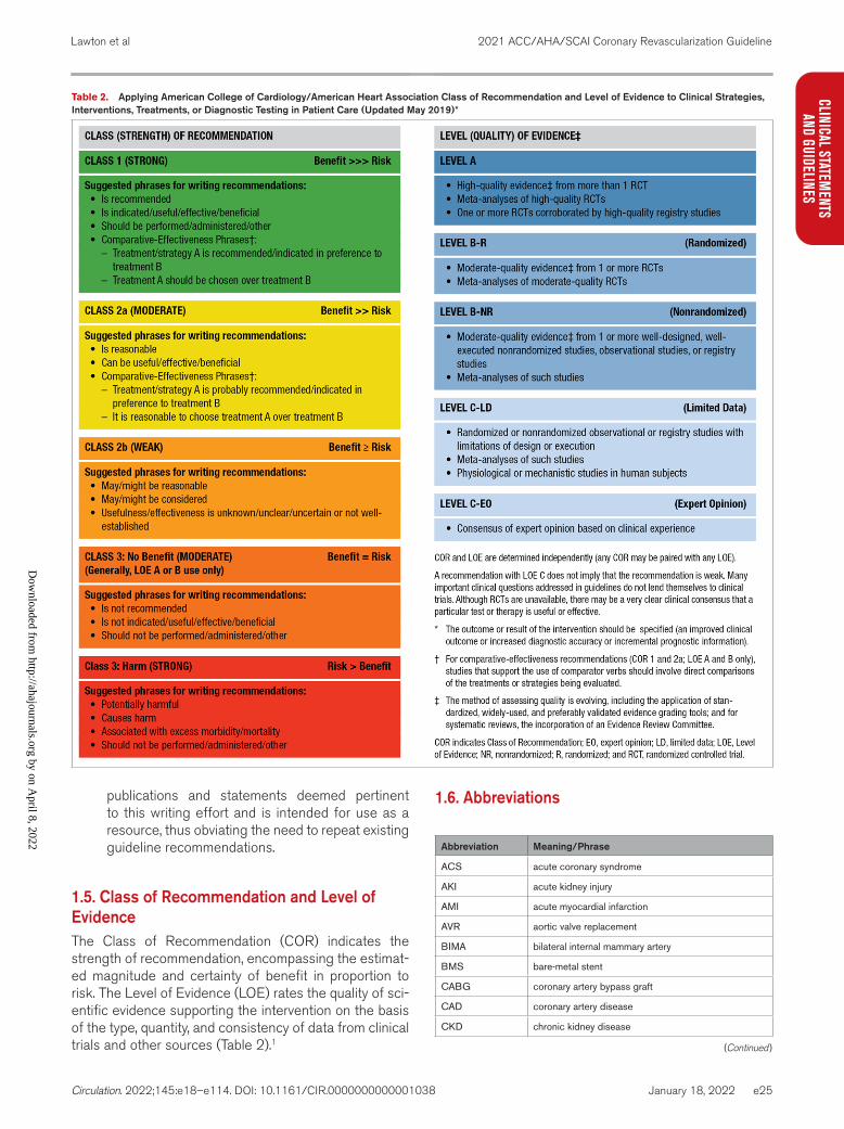
1.5. Class of Recommendation and Level of EvidenceThe Class of Recommendation (COR) indicates the strength of recommendation, encompassing the estimat-ed magnitude and certainty of benefit in proportion to risk. The Level of Evidence (LOE) rates the quality of sci-entific evidence supporting the intervention on the basis of the type, quantity, and consistency of data from clinical trials and other sources (Table 2).1
1.6. Abbreviations
Abbreviation Meaning/Phrase
ACS acute coronary syndrome
AKI acute kidney injury
AMI acute myocardial infarction
AVR aortic valve replacement
BIMA bilateral internal mammary artery
BMS bare-metal stent
CABG coronary artery bypass graft
CAD coronary artery disease
CKD chronic kidney disease
Table 2. Applying American College of Cardiology/American Heart Association Class of Recommendation and Level of Evidence to Clinical Strategies, Interventions, Treatments, or Diagnostic Testing in Patient Care (Updated May 2019)*
(Continued )
Dow
nloaded from http://ahajournals.org by on A
pril 8, 2022
Lawton et al 2021 ACC/AHA/SCAI Coronary Revascularization Guideline
January 18, 2022 Circulation. 2022;145:e18–e114. DOI: 10.1161/CIR.0000000000001038e26
CLIN
ICAL
STA
TEM
ENTS
AN
D GU
IDEL
INES
COR Class of Recommendation
CTO chronic total occlusion
CVD cardiovascular disease
DAPT dual antiplatelet therapy
DES drug-eluting stent
ECG electrocardiogram
FFR fractional flow reserve
GDMT guideline-directed medical therapy
iFR instantaneous wave-free ratio
IMA internal mammary artery
ISR in-stent restenosis
IVUS intravascular ultrasound
LAD left anterior descending
LIMA left internal mammary artery
LOE Level of Evidence
MACE major adverse cardiovascular events
MI myocardial infarction
NSTE-ACS non–ST-segment–elevation acute coronary syndrome
NSTEMI non–ST-segment–elevation myocardial infarction
OCT optical coherence tomography
PCI percutaneous coronary intervention
RCT randomized controlled trial
SCAD spontaneous coronary artery dissection
SIHD stable ischemic heart disease
STEMI ST-segment–elevation myocardial infarction
SVG saphenous vein graft
SYNTAX Synergy Between PCI With TAXUS and Cardiac Surgery
TAVR transcatheter aortic valve replacement
UFH unfractionated heparin
VT ventricular tachycardia
2. IMPROVING EQUITY OF CARE IN REVASCULARIZATION AND SHARED DECISION-MAKING2.1. Improving Equity of Care in Revascularization
Recommendation to Improve Equity of Care in RevascularizationReferenced studies that support the recommendation are summarized in Online Data Supplement 1.
COR LOE Recommendation
1 B-NR
1. In patients who require coronary revascu-larization, treatment decisions should be based on clinical indication, regardless of sex1-7 or race or ethnicity,8-10 and efforts to reduce disparities of care are war-ranted.11,12
SynopsisHealth disparities by sex and race are evident across the spectrum of CVD in the United States,7,9,13-15 and mounting evidence demonstrates that social factors are strongly associated with cardiovascular health outcomes.16, 17 Differences in access to care, cardio-vascular treatment, mortality rate, and readmission outcomes persist by important sociodemographic characteristics that include but are not limited to so-cioeconomic status, race, and ethnicity.18-22 African Americans,23-25 Hispanics,24 and South Asians26 (with substantial heterogeneity within Asian subgroups) have a higher prevalence of cardiovascular risk factors and crude mortality.16 Although access to health care remains a problem, even after entering into the health care system, women and non-White patients are less likely to receive reperfusion therapy, an invasive strat-egy, or revascularization9,13,27-37 and more likely to have worse outcomes.37-40 As compared with White male pa-tients, women and Black patients with acute coronary syndrome (ACS) receive less guideline-based therapy in hospital and at discharge.27,32,41,42 Differences in co-morbidities, health education, presentation, socioeco-nomic status, regional hospital capability and quality, and insurance and health care access15,28,29,35,37,43-48 contribute to the problem, but disparities can per-sist despite adjustment for these factors.7,30-32,49,50 In a study of patients with cardiac symptoms, clinicians were less likely to recommend cardiac catheterization to women and non-White patients than to White male patients, despite being given the exact same clinical vignette for White male patients.51 Continued vigilance against conscious and unconscious gender, racial, and ethnic discrimination and purposeful efforts to in-crease the implementation of guideline-based therapy for all patients, regardless of sex, race, or ethnicity, are needed.
Recommendation-Specific Supportive TextAfter controlling for greater baseline comorbidities among patients undergoing revascularization, sev-eral observational studies have demonstrated that Black,28,52-54 Hispanic,24,50 and Asian55,56 patients have outcomes similar to those of White patients. Simi-larly, after controlling for baseline comorbidities and treatment strategy, most studies demonstrate similar outcomes in women and men.1-6 Post hoc analyses of randomized trials evaluating revascularization pro-vide compelling evidence, inasmuch as enrolled pa-tients are more similar and the decision to revascu-larize is protocol driven. In the SHOCK (Should We Emergently Revascularize Occluded Coronaries for Cardiogenic Shock) trial, revascularization rates were lowest and mortality rates highest for Hispanics and
1.6. Abbreviations Continued
Abbreviation Meaning/Phrase
Dow
nloaded from http://ahajournals.org by on A
pril 8, 2022
Lawton et al 2021 ACC/AHA/SCAI Coronary Revascularization Guideline
e27
CLINICAL STATEMENTS
AND GUIDELINES
Circulation. 2022;145:e18–e114. DOI: 10.1161/CIR.0000000000001038 January 18, 2022
African Americans, but there was no interaction be-tween race and the mortality benefit of revasculariza-tion.9 Similar results have been reported for women with shock in the CULPRIT-SHOCK (Culprit Le-sion Only PCI versus Multivessel PCI in Cardiogenic Shock trial.57 In the TACTICS-TIMI 18 (Treat Angina With Aggrastat and Determine Cost of Therapy With Invasive or Conservative Strategy—Thrombolysis In Myocardial Infarction 18) trial, evaluating patients with NSTE-ACS, non-White patients and female pa-tients had more comorbidity and more major adverse cardiovascular event (MACE) outcomes than White and male patients but were revascularized at the same rate. After adjustment for baseline character-istics, the invasive strategy was equally beneficial for all patients, without evidence of racial differences.10 A meta-analysis of RCT of invasive vs conservative strategies in women and men with NSTE-ACS report-ed a similar proportional benefit of an invasive strate-gy in women and men, (although low risk women with biomarker negative ACS did not derive a benefit to an early invasive strategy).1 Additionally, studies have shown similar relative benefits of primary PCI3 and revascularization in SIHD.5,6 for women and men. In view of these findings, the decision to offer revascu-larization should be made on the basis of a patient’s clinical characteristics, and preferences and should be the same for all patients, regardless of sex, race, or ethnicity.
2.2. Shared Decision-Making and Informed Consent
Recommendations for Shared Decision-Making and Informed Consent
COR LOE Recommendations
1 C-LD
1. In patients undergoing revascularization, deci-sions should be patient centered—that is, considerate of the patient’s preferences and goals, cultural beliefs, health literacy, and social determinants of health—and made in collabora-tion with the patient’s support system.1,2
1 C-LD
2. In patients undergoing coronary angiography or revascularization, adequate information about benefits, risks, therapeutic conse-quences, and potential alternatives in the performance of percutaneous and surgi-cal myocardial revascularization should be given, when feasible, with sufficient time for informed decision-making to improve clinical outcomes.3-5
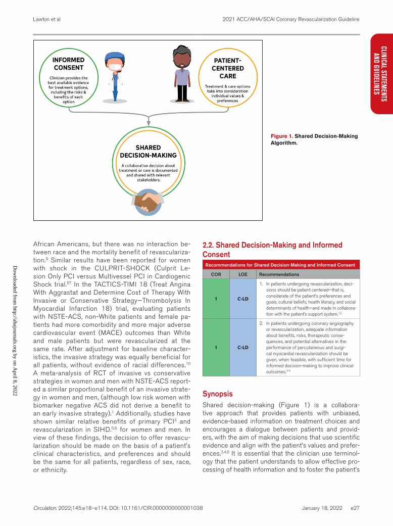
SynopsisShared decision-making (Figure 1) is a collabora-tive approach that provides patients with unbiased, evidence-based information on treatment choices and encourages a dialogue between patients and provid-ers, with the aim of making decisions that use scientific evidence and align with the patient’s values and prefer-ences.3,4,6 It is essential that the clinician use terminol-ogy that the patient understands to allow effective pro-cessing of health information and to foster the patient’s
Figure 1. Shared Decision-Making Algorithm.
Dow
nloaded from http://ahajournals.org by on A
pril 8, 2022
Lawton et al 2021 ACC/AHA/SCAI Coronary Revascularization Guideline
January 18, 2022 Circulation. 2022;145:e18–e114. DOI: 10.1161/CIR.0000000000001038e28
CLIN
ICAL
STA
TEM
ENTS
AN
D GU
IDEL
INES
participation in treatment decisions.7 The use of online modules, decision aids, or videos about treatment op-tions can help patients better understand the risks and benefits of various therapies. Patients are interested in how a recommended treatment might impact their
prognosis and quality of life.8 In the treatment decision-making process, the patient’s best interest should be placed first, and the active participation of the patient and significant others should be engaged. The contribu-tions of social determinants of health to CVD are poorly understood.9,10 but may impact a patient’s decision with regard to treatments. In high-income countries, 4 so-cioeconomic status metrics have been associated with CVD: income level, educational attainment, employment status, and environmental factors.11-13
Recommendation-Specific Supportive Text1. Shared decision-making is vital to patient-cen-
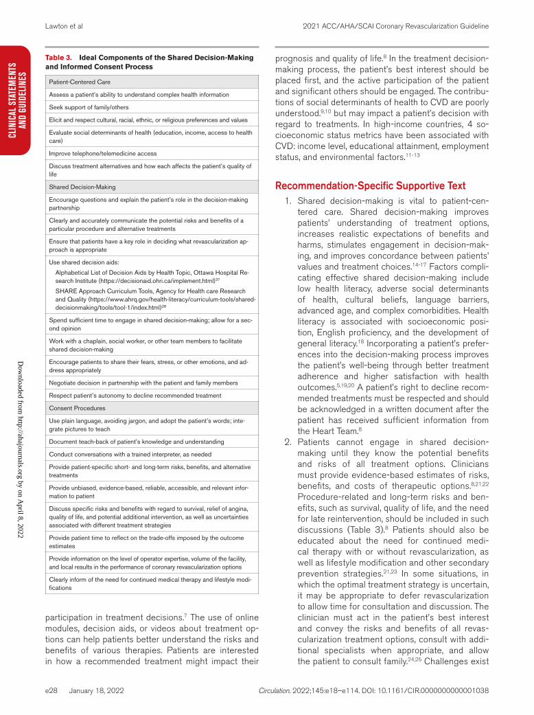
tered care. Shared decision-making improves patients’ understanding of treatment options, increases realistic expectations of benefits and harms, stimulates engagement in decision-mak-ing, and improves concordance between patients’ values and treatment choices.14-17 Factors compli-cating effective shared decision-making include low health literacy, adverse social determinants of health, cultural beliefs, language barriers, advanced age, and complex comorbidities. Health literacy is associated with socioeconomic posi-tion, English proficiency, and the development of general literacy.18 Incorporating a patient’s prefer-ences into the decision-making process improves the patient’s well-being through better treatment adherence and higher satisfaction with health outcomes.5,19,20 A patient’s right to decline recom-mended treatments must be respected and should be acknowledged in a written document after the patient has received sufficient information from the Heart Team.8
2. Patients cannot engage in shared decision-making until they know the potential benefits and risks of all treatment options. Clinicians must provide evidence-based estimates of risks, benefits, and costs of therapeutic options.8,21,22 Procedure-related and long-term risks and ben-efits, such as survival, quality of life, and the need for late reintervention, should be included in such discussions (Table 3).8 Patients should also be educated about the need for continued medi-cal therapy with or without revascularization, as well as lifestyle modification and other secondary prevention strategies.21,23 In some situations, in which the optimal treatment strategy is uncertain, it may be appropriate to defer revascularization to allow time for consultation and discussion. The clinician must act in the patient’s best interest and convey the risks and benefits of all revas-cularization treatment options, consult with addi-tional specialists when appropriate, and allow the patient to consult family.24,25 Challenges exist
Table 3. Ideal Components of the Shared Decision-Making and Informed Consent Process
Patient-Centered Care
Assess a patient’s ability to understand complex health information
Seek support of family/others
Elicit and respect cultural, racial, ethnic, or religious preferences and values
Evaluate social determinants of health (education, income, access to health care)
Improve telephone/telemedicine access
Discuss treatment alternatives and how each affects the patient’s quality of life
Shared Decision-Making
Encourage questions and explain the patient’s role in the decision-making partnership
Clearly and accurately communicate the potential risks and benefits of a particular procedure and alternative treatments
Ensure that patients have a key role in deciding what revascularization ap-proach is appropriate
Use shared decision aids:
Alphabetical List of Decision Aids by Health Topic, Ottawa Hospital Re-search Institute (https://decisionaid.ohri.ca/implement.html)27
SHARE Approach Curriculum Tools, Agency for Health care Research and Quality (https://www.ahrq.gov/health-literacy/curriculum-tools/shared-decisionmaking/tools/tool-1/index.html)28
Spend sufficient time to engage in shared decision-making; allow for a sec-ond opinion
Work with a chaplain, social worker, or other team members to facilitate shared decision-making
Encourage patients to share their fears, stress, or other emotions, and ad-dress appropriately
Negotiate decision in partnership with the patient and family members
Respect patient’s autonomy to decline recommended treatment
Consent Procedures
Use plain language, avoiding jargon, and adopt the patient’s words; inte-grate pictures to teach
Document teach-back of patient’s knowledge and understanding
Conduct conversations with a trained interpreter, as needed
Provide patient-specific short- and long-term risks, benefits, and alternative treatments
Provide unbiased, evidence-based, reliable, accessible, and relevant infor-mation to patient
Discuss specific risks and benefits with regard to survival, relief of angina, quality of life, and potential additional intervention, as well as uncertainties associated with different treatment strategies
Provide patient time to reflect on the trade-offs imposed by the outcome estimates
Provide information on the level of operator expertise, volume of the facility, and local results in the performance of coronary revascularization options
Clearly inform of the need for continued medical therapy and lifestyle modi-fications
Dow
nloaded from http://ahajournals.org by on A
pril 8, 2022

Lawton et al 2021 ACC/AHA/SCAI Coronary Revascularization Guideline
e29
CLINICAL STATEMENTS
AND GUIDELINES
Circulation. 2022;145:e18–e114. DOI: 10.1161/CIR.0000000000001038 January 18, 2022
when scientific data support a treatment, but the patient prefers an alternative treatment; in 1 study, patients preferred PCI over CABG, even when the risk of death with PCI was double the risk with CABG.26
3. PREPROCEDURAL ASSESSMENT AND THE HEART TEAM3.1. The Heart Team
Recommendation for the Heart TeamReferenced studies that support the recommendation are summarized in Online Data Supplement 2.
COR LOE Recommendation
1 B-NR
1. In patients for whom the optimal treatment strategy is unclear, a Heart Team approach that includes representatives from interven-tional cardiology, cardiac surgery, and clinical cardiology is recommended to improve patient outcomes.1-7
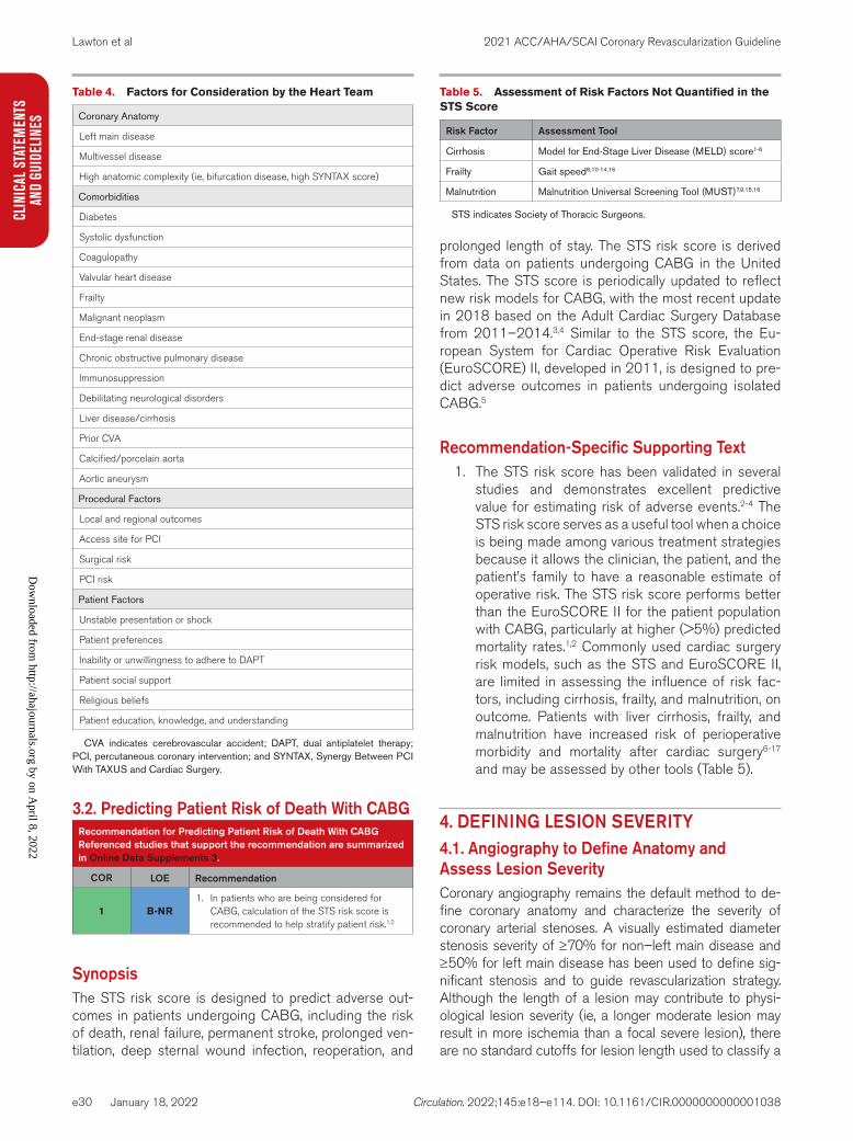
SynopsisThe multidisciplinary Heart Team, which involves the cardiologist, cardiac surgeon, and other specialists, has become a critical component of the revascular-ization decision. Initially, the Heart Team approach to decision-making for coronary disease arose within the context of randomized trials comparing PCI with CABG to ensure selected patients were equally suit-ed for either strategy before randomization.8 Subse-quently, the Heart Team has become an important paradigm in clinical practice, emphasizing the impor-tance of team consensus on the optimal approach to revascularization. Ideal situations for Heart Team
consideration include patients with complex coro-nary disease, comorbid conditions that could impact the success of the revascularization strategy, and other clinical or social situations that may impact outcomes (Figure 2 and Table 4). The Heart Team process should rest on the principles of collegiality, mutual respect, and commitment to excellence. The logistics of convening the Heart Team should depend on local resources and workflows. Models include daily to weekly scheduled meetings and ad hoc ac-tivation.1,2,4,6,9 Remote conferences have also been advocated.9 Additionally, there should be a process for rapid activation of the Heart Team for urgent or emergency clinical situations.
Recommendation-Specific Supporting Text1. Observational studies using the Heart Team have
included interventional cardiology, cardiac surgery, and noninvasive cardiologists1-4,6 Additional profes-sionals who offer input may include the patient’s primary physician, as well as palliative care, criti-cal care, anesthesiology, and imaging specialists. Observational studies have demonstrated favor-able outcomes when the Heart Team was used in cases of unprotected left main disease, triple-vessel disease, double-vessel disease involving the proximal left anterior descending (LAD) artery, or single-vessel disease involving the proximal LAD artery in the context of diabetes, or in cases in which the referring physician requested such evaluation.5-7,10,11 Heart Team decisions are gen-erally reproducible4 and associated with good outcomes.2,6
Figure 2. Phases of Patient-Centric Care in the Treatment of Coronary Artery Disease.CV indicates cardiovascular; SIHD, stable ischemic heart disease; and STEMI, ST-segment–elevation myocardial infarction.
Dow
nloaded from http://ahajournals.org by on A
pril 8, 2022
Lawton et al 2021 ACC/AHA/SCAI Coronary Revascularization Guideline
January 18, 2022 Circulation. 2022;145:e18–e114. DOI: 10.1161/CIR.0000000000001038e30
CLIN
ICAL
STA
TEM
ENTS
AN
D GU
IDEL
INES
3.2. Predicting Patient Risk of Death With CABGRecommendation for Predicting Patient Risk of Death With CABGReferenced studies that support the recommendation are summarized in Online Data Supplements 3.
COR LOE Recommendation
1 B-NR1. In patients who are being considered for
CABG, calculation of the STS risk score is recommended to help stratify patient risk.1,2
SynopsisThe STS risk score is designed to predict adverse out-comes in patients undergoing CABG, including the risk of death, renal failure, permanent stroke, prolonged ven-tilation, deep sternal wound infection, reoperation, and
prolonged length of stay. The STS risk score is derived from data on patients undergoing CABG in the United States. The STS score is periodically updated to reflect new risk models for CABG, with the most recent update in 2018 based on the Adult Cardiac Surgery Database from 2011–2014.3,4 Similar to the STS score, the Eu-ropean System for Cardiac Operative Risk Evaluation (EuroSCORE) II, developed in 2011, is designed to pre-dict adverse outcomes in patients undergoing isolated CABG.5
Recommendation-Specific Supporting Text1. The STS risk score has been validated in several
studies and demonstrates excellent predictive value for estimating risk of adverse events.2-4 The STS risk score serves as a useful tool when a choice is being made among various treatment strategies because it allows the clinician, the patient, and the patient’s family to have a reasonable estimate of operative risk. The STS risk score performs better than the EuroSCORE II for the patient population with CABG, particularly at higher (>5%) predicted mortality rates.1,2 Commonly used cardiac surgery risk models, such as the STS and EuroSCORE II, are limited in assessing the influence of risk fac-tors, including cirrhosis, frailty, and malnutrition, on outcome. Patients with liver cirrhosis, frailty, and malnutrition have increased risk of perioperative morbidity and mortality after cardiac surgery6-17 and may be assessed by other tools (Table 5).
4. DEFINING LESION SEVERITY4.1. Angiography to Define Anatomy and Assess Lesion SeverityCoronary angiography remains the default method to de-fine coronary anatomy and characterize the severity of coronary arterial stenoses. A visually estimated diameter stenosis severity of ≥70% for non–left main disease and ≥50% for left main disease has been used to define sig-nificant stenosis and to guide revascularization strategy. Although the length of a lesion may contribute to physi-ological lesion severity (ie, a longer moderate lesion may result in more ischemia than a focal severe lesion), there are no standard cutoffs for lesion length used to classify a
Table 4. Factors for Consideration by the Heart Team
Coronary Anatomy
Left main disease
Multivessel disease
High anatomic complexity (ie, bifurcation disease, high SYNTAX score)
Comorbidities
Diabetes
Systolic dysfunction
Coagulopathy
Valvular heart disease
Frailty
Malignant neoplasm
End-stage renal disease
Chronic obstructive pulmonary disease
Immunosuppression
Debilitating neurological disorders
Liver disease/cirrhosis
Prior CVA
Calcified/porcelain aorta
Aortic aneurysm
Procedural Factors
Local and regional outcomes
Access site for PCI
Surgical risk
PCI risk
Patient Factors
Unstable presentation or shock
Patient preferences
Inability or unwillingness to adhere to DAPT
Patient social support
Religious beliefs
Patient education, knowledge, and understanding
CVA indicates cerebrovascular accident; DAPT, dual antiplatelet therapy; PCI, percutaneous coronary intervention; and SYNTAX, Synergy Between PCI With TAXUS and Cardiac Surgery.
Table 5. Assessment of Risk Factors Not Quantified in the STS Score
Risk Factor Assessment Tool
Cirrhosis Model for End-Stage Liver Disease (MELD) score1-6
Frailty Gait speed8,10-14,16
Malnutrition Malnutrition Universal Screening Tool (MUST)7,9,15,16
STS indicates Society of Thoracic Surgeons.
Dow
nloaded from http://ahajournals.org by on A
pril 8, 2022
Lawton et al 2021 ACC/AHA/SCAI Coronary Revascularization Guideline
Circulation. 2022;145:e18–e114. DOI: 10.1161/CIR.0000000000001038 January 18, 2022 e31
CLINICAL STATEMENTS
AND GUIDELINES
severe stenosis. An angiographically intermediate coronary stenosis is defined as a diameter stenosis severity of 40% to 69%, and generally warrants additional investigation to assess physiological significance. There is controversy over whether visually estimated diameter stenosis or quantita-tive coronary angiography better predicts the functional significance of a coronary stenosis.1,2 The difference in mean diameter stenosis between quantitative coronary an-giography and visual estimation varies from 10% to 20% and is dependent on stenosis severity.3-5 The use of optimal angiographic projections, multiple angiographic views, and adjunct imaging or physiology may aid in the assessment of coronary anatomy when coronary angiography is used.
4.2. Defining Coronary Artery Lesion Complexity: Calculation of the SYNTAX (Synergy Between PCI With TAXUS and Cardiac Surgery) Score
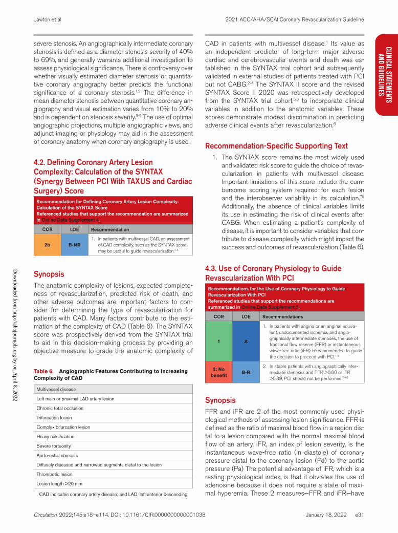
Recommendation for Defining Coronary Artery Lesion Complexity: Calculation of the SYNTAX ScoreReferenced studies that support the recommendation are summarized in Online Data Supplement 4.
COR LOE Recommendation
2b B-NR1. In patients with multivessel CAD, an assessment
of CAD complexity, such as the SYNTAX score, may be useful to guide revascularization.1-4
SynopsisThe anatomic complexity of lesions, expected complete-ness of revascularization, predicted risk of death, and other adverse outcomes are important factors to con-sider for determining the type of revascularization for patients with CAD. Many factors contribute to the esti-mation of the complexity of CAD (Table 6). The SYNTAX score was prospectively derived from the SYNTAX trial to aid in this decision-making process by providing an objective measure to grade the anatomic complexity of
CAD in patients with multivessel disease.1 Its value as an independent predictor of long-term major adverse cardiac and cerebrovascular events and death was es-tablished in the SYNTAX trial cohort and subsequently validated in external studies of patients treated with PCI but not CABG.2-4 The SYNTAX II score and the revised SYNTAX Score II 2020 was retrospectively developed from the SYNTAX trial cohort.5,6 to incorporate clinical variables in addition to the anatomic variables. These scores demonstrate modest discrimination in predicting adverse clinical events after revascularization.6
Recommendation-Specific Supporting Text1. The SYNTAX score remains the most widely used
and validated risk score to guide the choice of revas-cularization in patients with multivessel disease. Important limitations of this score include the cum-bersome scoring system required for each lesion and the interobserver variability in its calculation.7,8 Additionally, the absence of clinical variables limits its use in estimating the risk of clinical events after CABG. When estimating a patient’s complexity of disease, it is important to consider variables that con-tribute to disease complexity which might impact the success and outcomes of revascularization (Table 6).
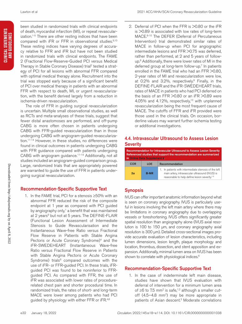
4.3. Use of Coronary Physiology to Guide Revascularization With PCI
Recommendations for the Use of Coronary Physiology to Guide Revascularization With PCIReferenced studies that support the recommendations are summarized in Online Data Supplement 5.
COR LOE Recommendations
1 A
1. In patients with angina or an anginal equiva-lent, undocumented ischemia, and angio-graphically intermediate stenoses, the use of fractional flow reserve (FFR) or instantaneous wave-free ratio (iFR) is recommended to guide the decision to proceed with PCI.1-6
3: No benefit
B-R2. In stable patients with angiographically inter-
mediate stenoses and FFR >0.80 or iFR >0.89, PCI should not be performed.7-10
SynopsisFFR and iFR are 2 of the most commonly used physi-ological methods of assessing lesion significance. FFR is defined as the ratio of maximal blood flow in a region dis-tal to a lesion compared with the normal maximal blood flow of an artery. iFR, an index of lesion severity, is the instantaneous wave-free ratio (in diastole) of coronary pressure distal to the coronary lesion (Pd) to the aortic pressure (Pa) The potential advantage of iFR, which is a resting physiological index, is that it obviates the use of adenosine because it does not require a state of maxi-mal hyperemia. These 2 measures—FFR and iFR—have
Table 6. Angiographic Features Contributing to Increasing Complexity of CAD
Multivessel disease
L eft main or proximal LAD artery lesion
Chronic total occlusion
Trifurcation lesion
Complex bifurcation lesion
Heavy calcification
Severe tortuosity
Aorto-ostial stenosis
Diffusely diseased and narrowed segments distal to the lesion
Thrombotic lesion
Lesion length >20 mm
CAD indicates coronary artery disease; and LAD, left anterior descending.
Dow
nloaded from http://ahajournals.org by on A
pril 8, 2022
Lawton et al 2021 ACC/AHA/SCAI Coronary Revascularization Guideline
January 18, 2022 Circulation. 2022;145:e18–e114. DOI: 10.1161/CIR.0000000000001038e32
CLIN
ICAL
STA
TEM
ENTS
AN
D GU
IDEL
INES
been studied in randomized trials with clinical endpoints of death, myocardial infarction (MI), or repeat revascular-ization.1-5 There are other resting indices that have been compared with iFR or FFR in observational studies.9,10 These resting indices have varying degrees of accura-cy relative to FFR and iFR but have not been studied in randomized trials with clinical endpoints. The FAME 2 (Fractional Flow-Reserve-Guided PCI versus Medical Therapy in Stable Coronary Disease) trial2 tested a strat-egy of PCI for all lesions with abnormal FFR compared with optimal medical therapy alone. Recruitment into the trial was stopped early because of a significant benefit of PCI over medical therapy in patients with an abnormal FFR with respect to death, MI, or urgent revasculariza-tion, with the benefit derived largely from a reduction in ischemia-driven revascularization.
The role of FFR in guiding surgical revascularization is uncertain. Multiple small observational studies, as well as RCTs and meta-analyses of these trials, suggest that fewer distal anastomoses are performed, and off-pump CABG is more often chosen in patients undergoing CABG with FFR-guided revascularization than in those undergoing CABG with angiogram-guided revasculariza-tion.11-14 However, in these studies, no differences were found in clinical outcomes in patients undergoing CABG with FFR guidance compared with patients undergoing CABG with angiogram guidance.11-14 Additionally, not all studies included an angiogram-guided comparison group. Large, randomized trials that are appropriately powered are warranted to guide the use of FFR in patients under-going surgical revascularization.
Recommendation-Specific Supportive Text1. In the FAME trial, PCI for a stenosis ≥50% with an
abnormal FFR reduced the risk of the composite endpoint at 1 year as compared with PCI guided by angiography only,1 a benefit that was maintained at 2 years9 but not at 5 years. The DEFINE-FLAIR (Functional Lesion Assessment of Intermediate Stenosis to Guide Revascularisation and the Instantaneous Wave-free Ratio versus Fractional Flow Reserve in Patients with Stable Angina Pectoris or Acute Coronary Syndrome)4 and the iFR-SWEDEHEART (Instantaneous Wave-free Ratio versus Fractional Flow Reserve in Patients with Stable Angina Pectoris or Acute Coronary Syndrome) trials6 compared outcomes with the use of iFR- or FFR-guided PCI. In these trials, iFR-guided PCI was found to be noninferior to FFR-guided PCI. As compared with FFR, the use of iFR was associated with lower rates of procedure-related chest pain and shorter procedural time. In randomized trials, the rates of short- and long-term MACE were lower among patients who had PCI guided by physiology with either FFR or iFR.4,6
2. Deferral of PCI when the FFR is >0.80 or the iFR is >0.89 is associated with low rates of long-term MACE.8-10 The DEFER (Deferral of Percutaneous Intervention) trial demonstrated similar rates of MACE in follow-up when PCI for angiographic intermediate lesions and FFR >0.75 was deferred, rather than performed, at 2 and 5 years of follow-up.8 Additionally, there were lower rates of MI in the deferred group at long-term follow-up.7 In patients enrolled in the FAME trial who had an FFR >0.80, 2-year rates of MI and revascularization were low, at 0.2% and 3.2%, respectively.9 Finally, in the DEFINE-FLAIR and the iFR-SWEDEHEART trials, rates of MACE in patients who had PCI deferred on the basis of an FFR >0.80 or an iFR >0.89 were 4.05% and 4.12%, respectively,10 with unplanned revascularization being the most frequent cause of MACE. The cutoffs of FFR and iFR provided were those used in the clinical trials. On occasion, bor-derline values may warrant further ischemia testing or additional investigations.
4.4. Intravascular Ultrasound to Assess Lesion Severity
Recommendation for Intravascular Ultrasound to Assess Lesion SeverityReferenced studies that support the recommendation are summarized in Online Data Supplement 6.
COR LOE Recommendation
2a B-NR1. In patients with intermediate stenosis of the left
main artery, intravascular ultrasound (IVUS) is reasonable to help define lesion severity.1-5
SynopsisIVUS can offer important anatomic information beyond what is seen on coronary angiography. IVUS is particularly use-ful in lesions involving the left main artery where there may be limitations in coronary angiography due to overlapping vessels or foreshortening. IVUS offers significantly greater spatial resolution than angiography alone (IVUS axial reso-lution is 100 to 150 μm, and coronary angiography axial resolution is 300 μm). Detailed cross-sectional images pro-vide accurate evaluation of lesion characteristics, including lumen dimensions, lesion length, plaque morphology and location, thrombus, dissection, and stent apposition and ex-pansion. Additionally, minimal lumen area on IVUS has been shown to correlate with physiological indices.6
Recommendation-Specific Supportive Text1. In the case of indeterminate left main disease,
studies have shown that IVUS evaluation with deferral of intervention for a minimum lumen area of ≥6 to 7.5 mm2 is safe,1,2 although a smaller cut-off (4.5–4.8 mm2) may be more appropriate in patients of Asian descent.3 Moderate correlations
Dow
nloaded from http://ahajournals.org by on A
pril 8, 2022
Lawton et al 2021 ACC/AHA/SCAI Coronary Revascularization Guideline
Circulation. 2022;145:e18–e114. DOI: 10.1161/CIR.0000000000001038 January 18, 2022 e33
CLINICAL STATEMENTS
AND GUIDELINES
between FFR values and IVUS minimal lumen area cutoffs have been demonstrated in left main disease.4,5 Compared with the left main artery, smaller cutoffs have been suggested for IVUS of the LAD artery.7 Developed more recently, optical coherence tomography (OCT) has been shown to correlate well with IVUS measurements.8 However, because OCT requires blood clearance, its effec-tiveness for imaging ostial left main disease is limited.
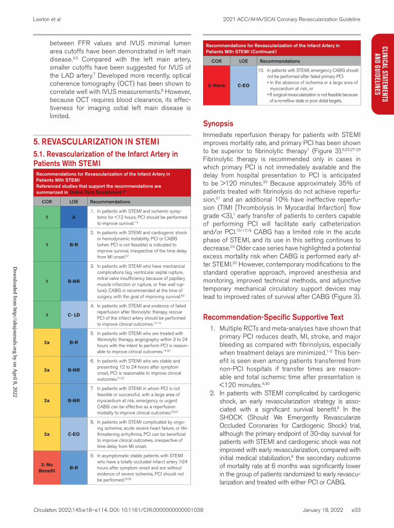
5. REVASCULARIZATION IN STEMI5.1. Revascularization of the Infarct Artery in Patients With STEMI
Recommendations for Revascularization of the Infarct Artery in Patients With STEMIReferenced studies that support the recommendations are summarized in Online Data Supplement 7.
COR LOE Recommendations
1 A1. In patients with STEMI and ischemic symp-
toms for <12 hours, PCI should be performed to improve survival.1-5
1 B-R
2. In patients with STEMI and cardiogenic shock or hemodynamic instability, PCI or CABG (when PCI is not feasible) is indicated to improve survival, irrespective of the time delay from MI onset.6,7
1 B-NR
3. In patients with STEMI who have mechanical complications (eg, ventricular septal rupture, mitral valve insufficiency because of papillary muscle infarction or rupture, or free wall rup-ture), CABG is recommended at the time of surgery, with the goal of improving survival.8,9
1 C- LD
4. In patients with STEMI and evidence of failed reperfusion after fibrinolytic therapy, rescue PCI of the infarct artery should be performed to improve clinical outcomes.10-13
2a B-R
5. In patients with STEMI who are treated with fibrinolytic therapy, angiography within 3 to 24 hours with the intent to perform PCI is reason-able to improve clinical outcomes.14-20
2a B-NR
6. In patients with STEMI who are stable and presenting 12 to 24 hours after symptom onset, PCI is reasonable to improve clinical outcomes.21,22
2a B-NR
7. In patients with STEMI in whom PCI is not feasible or successful, with a large area of myocardium at risk, emergency or urgent CABG can be effective as a reperfusion modality to improve clinical outcomes.23,24
2a C-EO
8. In patients with STEMI complicated by ongo-ing ischemia, acute severe heart failure, or life-threatening arrhythmia, PCI can be beneficial to improve clinical outcomes, irrespective of time delay from MI onset.
3: No Benefit
B-R
9. In asymptomatic stable patients with STEMI who have a totally occluded infarct artery >24 hours after symptom onset and are without evidence of severe ischemia, PCI should not be performed.25,26
3: Harm C-EO
10. In patients with STEMI, emergency CABG should not be performed after failed primary PCI: • In the absence of ischemia or a large area of
myocardium at risk, or • If surgical revascularization is not feasible because of a no-reflow state or poor distal targets.
SynopsisImmediate reperfusion therapy for patients with STEMI improves mortality rate, and primary PCI has been shown to be superior to fibrinolytic therapy1 (Figure 3).6,23,27-29 Fibrinolytic therapy is recommended only in cases in which primary PCI is not immediately available and the delay from hospital presentation to PCI is anticipated to be >120 minutes.30 Because approximately 35% of patients treated with fibrinolysis do not achieve reperfu-sion,31 and an additional 10% have ineffective reperfu-sion (TIMI [Thrombolysis In Myocardial Infarction] flow grade <3),1 early transfer of patients to centers capable of performing PCI will facilitate early catheterization and/or PCI.15-17,19 CABG has a limited role in the acute phase of STEMI, and its use in this setting continues to decrease.23 Older case series have highlighted a potential excess mortality risk when CABG is performed early af-ter STEMI.32 However, contemporary modifications to the standard operative approach, improved anesthesia and monitoring, improved technical methods, and adjunctive temporary mechanical circulatory support devices may lead to improved rates of survival after CABG (Figure 3).
Recommendation-Specific Supportive Text1. Multiple RCTs and meta-analyses have shown that
primary PCI reduces death, MI, stroke, and major bleeding as compared with fibrinolysis, especially when treatment delays are minimized.1-5 This ben-efit is seen even among patients transferred from non-PCI hospitals if transfer times are reason-able and total ischemic time after presentation is <120 minutes.4,30
2. In patients with STEMI complicated by cardiogenic shock, an early revascularization strategy is asso-ciated with a significant survival benefit.6 In the SHOCK (Should We Emergently Revascularize Occluded Coronaries for Cardiogenic Shock) trial, although the primary endpoint of 30-day survival for patients with STEMI and cardiogenic shock was not improved with early revascularization, compared with initial medical stabilization,6 the secondary outcome of mortality rate at 6 months was significantly lower in the group of patients randomized to early revascu-larization and treated with either PCI or CABG.
Recommendations for Revascularization of the Infarct Artery in Patients With STEMI (Continued )
COR LOE Recommendations
Dow
nloaded from http://ahajournals.org by on A
pril 8, 2022

Lawton et al 2021 ACC/AHA/SCAI Coronary Revascularization Guideline
e34
CLIN
ICAL
STA
TEM
ENTS
AN
D GU
IDEL
INES
January 18, 2022 Circulation. 2022;145:e18–e114. DOI: 10.1161/CIR.0000000000001038
3. The mortality rate associated with emergency CABG and surgical management of a mechani-cal complication of STEMI remains high.33 However, there are currently few medical or percutaneous treatment methods to effectively treat ventricular rupture, papillary muscle rup-ture leading to severe mitral regurgitation, or ischemic ventricular septal defect. CABG and these associated procedures may be neces-sary to treat the mechanical complications of STEMI or cardiogenic shock in the emergency setting.34-38 Placement of a mechanical support device may be useful in temporizing a patient with a mechanical complication of STEMI, and urgent or emergency surgery remains the best treatment.39,40 No RCT has examined the benefit of adding CABG at the time of emergent cardiac surgery for treatment of a mechanical complica-tion of STEMI versus emergent surgery for the treatment of a mechanical complication alone. In addition, no RCT has examined the benefit of
emergent cardiac surgery for the treatment of a mechanical complication of STEMI versus initial medical stabilization and delayed surgery.
4. Rescue PCI performed in patients with evidence of failed reperfusion after fibrinolytic therapy has been associated with a reduction in cardiovascular events,10-13 when compared with conservative care or repeat fibrinolysis. In these studies, patients ran-domized to rescue PCI had higher rates of bleeding and cerebrovascular accident.10-12 All these studies were performed in the era of femoral artery access, with limited options for antiplatelet and anticoagu-lant therapy. Previous concerns about increased bleeding or increased stroke risk in patients under-going rescue PCI could be mitigated by using half-dose tenecteplase in patients >75 years of age,31 by substituting radial access in place of femoral access,41 or by eliminating the routine use of plate-let glycoprotein IIb/IIIa inhibitors. With these tech-niques, the reduced complications associated with PCI would provide a more favorable balance of risk
Figure 3. Indications for Revascularization in STEMI (Patients Without Fibrinolytics).Colors correspond to Table 2. CABG indicates coronary artery bypass graft; PCI, percutaneous coronary intervention; and STEMI, ST-segment–elevation myocardial infarction. This algorithm summarizes the recommendations in this guideline for revascularization of the infarct artery in STEMI. It is not meant to encompass every patient scenario or situation, and clinicians are encouraged to use a Heart Team approach when care decisions are unclear and to see the accompanying supportive text for each recommendation. Additionally, in situations that lack sufficient data to make formal recommendations for care, please see Section 17, “Unanswered Questions and Future Directions.”
Dow
nloaded from http://ahajournals.org by on A
pril 8, 2022

Lawton et al 2021 ACC/AHA/SCAI Coronary Revascularization Guideline
Circulation. 2022;145:e18–e114. DOI: 10.1161/CIR.0000000000001038 January 18, 2022 e35
CLINICAL STATEMENTS
AND GUIDELINES
to benefit with rescue PCI than with conservative care.
5. Studies have shown a reduction in MACE when routine early angiography with the intent to perform PCI is performed after fibrinolytic therapy.15,16,18-20,42 This was further supported by several meta-anal-yses of these trials, which showed a reduction in death or infarction with an early invasive approach after fibrinolytic therapy.14,17 In these early-transfer studies, more than 80% of patients who were transferred underwent PCI to treat a significant residual stenosis or suboptimal flow of the infarct artery. The benefit of immediate angiography was most notable in patients undergoing angiography early after symptom onset or after administration of fibrinolytic therapy.14
6. The benefit of PCI for asymptomatic patients pre-senting 12 to 24 hours after symptom onset is not well studied. The BRAVE-2 (Beyond 12 Hours Reperfusion Alternative Evaluation-2) trial exam-ined the benefits of PCI in reducing infarct size in asymptomatic patients with STEMI and symptom onset >12 hours but <48 hours before presenta-tion.21 In this small study, an invasive strategy of coronary stenting was associated with a reduction in left ventricular infarct size (primary endpoint) compared with a conservative strategy.21 Moreover, an invasive strategy was associated with a reduc-tion in adjusted 4-year mortality rate compared with the conservative strategy.43 Observational data from the Prospective National Observational study also supported a lower adjusted 1-year mortality rate in patients with STEMI and symptom onset 12 to 24 hours before presentation.22 This infor-mation should be balanced by the potential for “harm” when PCI of a totally occluded artery is per-formed more than 24 hours after symptom onset. Therefore, delayed PCI of an infarct artery beyond 24 hours should be considered only in patients with a patent artery.
7. In patients with STEMI, there may be situations in which PCI is not possible for anatomic reasons or because of the presence of severe left main or multivessel CAD. Additionally, in unusual circum-stances, PCI may not be successful. In such cases, CABG can be an effective primary reperfusion strategy, particularly if there is a large area of myo-cardium at risk.23,24
8. There are no RCTs examining the benefit of PCI in patients with STEMI presenting >12 hours after symptom onset who have clinical evidence of ongoing ischemia, acute severe heart failure, or life-threatening arrhythmias. Intuitively, a strategy of delayed reperfusion in these unstable patient subsets would be expected to improve symptoms
and outcomes, and for this reason PCI should be considered.
9. In OAT (Occluded Artery Trial), PCI of a totally occluded vessel did not reduce cardiovascular events at 4 years of follow-up,25 and there was a trend toward a higher rate of recurrent infarction in the group of patients randomized to PCI. Patients who had severe ischemia on noninvasive stress testing were not enrolled in this trial. Similar find-ings were noted in the DECOPI (Desobstruction Coronaire en Post-Infarctus) trial, which enrolled patients with an occluded artery presenting 2 to 15 days after symptom onset.26
10. Emergency CABG to restore flow to the infarct artery after failed PCI should be considered only in patients with ongoing ischemia and a large area of myocardium at risk. In some cases, after primary PCI, the vessel remains occluded or with slow flow caused by distal embolization (no reflow). The no-reflow phenomenon refers to unsuccessful microvascular reperfusion even in the presence of a widely patent epicardial coronary artery. This usu-ally occurs with reperfusion in the setting of PCI for the treatment of STEMI, after prolonged myo-cardial ischemia, or with a large thrombus burden. Because CABG is unlikely to improve perfusion to the subtended myocardium in the setting of no-reflow, emergency CABG may be harmful in this setting and may subject the patient to unnecessary risk.
5.2. Revascularization of the Non-Infarct Artery in Patients With STEMI
Recommendations for Revascularization of the Non-Infarct Artery in Patients With STEMIReferenced studies that support the recommendations are summarized in Online Data Supplement 8.
COR LOE Recommendations
1 A
1. In selected hemodynamically stable patients with STEMI and multivessel disease, after successful primary PCI, staged PCI of a sig-nificant non-infarct artery stenosis is recom-mended to reduce the risk of death or MI.1-4
2a C-EO
2. In selected patients with STEMI with com-plex multivessel non-infarct artery disease, after successful primary PCI, elective CABG is reasonable to reduce the risk of cardiac events.
2b B-R
3. In selected hemodynamically stable patients with STEMI and low-complexity multivessel disease, PCI of a non-infarct artery stenosis may be considered at the time of primary PCI to reduce cardiac event rates.1,2,5-7
3: Harm B-R
4. In patients with STEMI complicated by car-diogenic shock, routine PCI of a non-infarct artery at the time of primary PCI should not be performed because of the higher risk of death or renal failure.8-10
Dow
nloaded from http://ahajournals.org by on A
pril 8, 2022
Lawton et al 2021 ACC/AHA/SCAI Coronary Revascularization Guideline
January 18, 2022 Circulation. 2022;145:e18–e114. DOI: 10.1161/CIR.0000000000001038e36
CLIN
ICAL
STA
TEM
ENTS
AN
D GU
IDEL
INES
SynopsisA Heart Team approach is utilized to determine optimal revascularization strategy in patients with STEMI and multivessel CAD. Revascularization strategies (Figure 4) for patients with STEMI and multivessel disease include multivessel PCI at the time of primary PCI, PCI of the infarct artery only followed by staged PCI of a non-infarct artery, PCI of the infarct artery only with an ischemia-guided approach to treatment of a non-infarct artery, or PCI of the infarct artery only with elective CABG. Ob-servational studies and meta-analyses have reported conflicting results for the superiority of one approach over another.11 Recent randomized trials of PCI in STEMI support the safety and efficacy of multivessel PCI in se-lected patients with STEMI.2-4,6,7 The data are strongest for patients undergoing staged PCI.4 It should be noted that only one-third of enrolled patients in these trials had triple-vessel disease, and most of these trials excluded patients with left main disease, chronic total occlusion (CTO) of the non-infarct artery, or complex non-infarct artery disease. For this reason, CABG remains a reason-able option in patients with residual complex non-infarct artery disease. Ideal patients who may benefit from re-vascularization of non-infarct arteries include those with a large area of myocardium at risk and those without significant comorbidities that would increase the risk of revascularization.
Recommendation-Specific Supportive Text1. RCTs have demonstrated a reduction in MACE
with staged PCI (either in hospital or after dis-charge) compared with culprit vessel–only PCI.1-4 This benefit is driven largely by a reduction in the risk of repeat revascularization or re-infarction. Most recently, the COMPLETE (Complete versus Culprit-Only Revascularization Strategies to Treat Multivessel Disease after Early PCI for STEMI) trial enrolled 4041 patients and demonstrated a 3-year reduction in the combined endpoint of death or MI with staged PCI of the non-infarct artery (per-formed within 45 days of STEMI), compared with conservative care.4 These benefits were consis-tent, irrespective of the timing of the non-infarct artery PCI.12 Most of these trials included patients with low-complexity disease. No trial has shown a difference in the outcome of mortality rate alone between the strategies. In the COMPLETE trial, <1% of enrolled patients had non-infarct artery disease involving the left main artery, and the baseline SYNTAX score of the enrolled patients was low. Additionally, these trials enrolled patients with lesions that had a >70% diameter stenosis. For intermediate lesions, physiological testing with iFR or FFR may be useful to guide PCI.4
2. In patients with STEMI and complex multives-sel CAD, elective CABG remains an appropriate revascularization option after successful PCI of an infarct artery in patients who meet criteria for CABG (Table 7). Although the COMPLETE trial demonstrated that staged PCI of the non-infarct artery is associated with a reduction in the risk of death or recurrent infarction in follow-up, patients intended for a planned surgical revasculariza-tion procedure were not included in this study. In patients with complex non-infarct artery disease, the decision to proceed with PCI versus CABG of the non-infarct artery should include a Heart Team discussion.
3. Randomized trials have shown a reduction in MACE with a multivessel PCI strategy performed at the time of primary PCI as compared with cul-prit artery–only PCI.1,2,5-7 The benefits reported in these trials were driven largely by a reduction in repeat revascularization with multivessel PCI. PCI of a non-infarct artery stenosis may be considered at the time of successful primary PCI, but patients should be carefully selected. Patients who are most appropriate for complete revascularization at the time of primary PCI include those with uncom-plicated PCI of the infarct artery and with low-com-plexity non-infarct artery disease who have normal left ventricular filling pressures and normal renal function. Clinicians should integrate clinical data, lesion severity and complexity, patient stability, risk of volume overload, and risk of contrast nephropa-thy before embarking on an immediate multivessel primary PCI strategy.
4. Culprit vessel–only primary PCI is recommended as the primary PCI strategy in most patients with STEMI complicated by cardiogenic shock who have multivessel disease. This is based on consistent findings from observational data and 1 random-ized trial that showed no advantage for immedi-ate multivessel PCI.8-10 In the CULPRIT-SHOCK (Culprit Lesion Only PCI Versus Multivessel PCI in Cardiogenic Shock) trial of patients with acute MI (AMI) complicated by cardiogenic shock, mul-tivessel PCI at the time of primary PCI resulted in a higher risk of the primary endpoint of death or need for renal replacement therapy.8,9 Of note, in this trial, investigators were permitted to proceed with PCI of all non-infarct vessels with >70% diameter stenosis that were ≥2 mm in diameter, including those that were chronically occluded. The risks associated with immediate multivessel PCI include volume overload, contrast nephropa-thy, and ischemic complications in the non-infarct artery that could cause further hemodynamic deterioration.
Dow
nloaded from http://ahajournals.org by on A
pril 8, 2022

Lawton et al 2021 ACC/AHA/SCAI Coronary Revascularization Guideline
e37
CLINICAL STATEMENTS
AND GUIDELINES
Circulation. 2022;145:e18–e114. DOI: 10.1161/CIR.0000000000001038 January 18, 2022
Figure 4. Revascularization of Noninfarct-Related Coronary Artery Lesions in Patients With STEMI.Colors correspond to Table 2. CABG indicates coronary artery bypass graft; GDMT, guideline-directed medical therapy; PCI, percutaneous coronary intervention; and STEMI, ST-segment–elevation myocardial infarction. *Normal blood pressure and heart rate left ventricular end-diastolic pressure <20 mm Hg, no chronic renal insufficiency or acute kidney injury, and expected total contrast volume <3× glomerular filtration rate, simple lesion anatomy. †In making the decision about the need for and mode of revascularization the Heart Team should consider the suitability of the non-culprit artery for PCI, the coronary complexity and the risk of revascularization, the extent of myocardium at risk, and patient comorbidities, including life expectancy or other significant patient comorbidities, such as chronic renal insufficiency or acute kidney injury. ‡Staged PCI can be performed in hospital or after discharge, up to 45 days post MI.
Symbol denotes time elapsed before proceeding to the next procedure. This algorithm summarizes the recommendations in this
guideline for the care of patients with STEMI and noninfarct artery disease. It is not meant to encompass every patient scenario or situation, and clinicians are encouraged to use a Heart Team approach when care decisions are unclear and to see the accompanying supportive text for each recommendation. Additionally, in situations that lack sufficient data to make formal recommendations for care, please see Section 17, “Unanswered Questions and Future Directions.”
Dow
nloaded from http://ahajournals.org by on A
pril 8, 2022
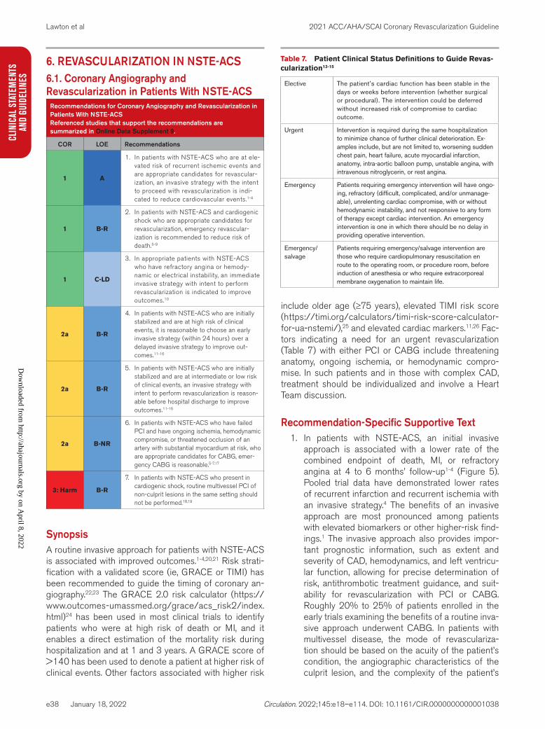
Lawton et al 2021 ACC/AHA/SCAI Coronary Revascularization Guideline
January 18, 2022 Circulation. 2022;145:e18–e114. DOI: 10.1161/CIR.0000000000001038e38
CLIN
ICAL
STA
TEM
ENTS
AN
D GU
IDEL
INES
6. REVASCULARIZATION IN NSTE-ACS6.1. Coronary Angiography and Revascularization in Patients With NSTE-ACS
Recommendations for Coronary Angiography and Revascularization in Patients With NSTE-ACSReferenced studies that support the recommendations are summarized in Online Data Supplement 9.
COR LOE Recommendations
1 A
1. In patients with NSTE-ACS who are at ele-vated risk of recurrent ischemic events and are appropriate candidates for revascular-ization, an invasive strategy with the intent to proceed with revascularization is indi-cated to reduce cardiovascular events.1-4
1 B-R
2. In patients with NSTE-ACS and cardiogenic shock who are appropriate candidates for revascularization, emergency revascular-ization is recommended to reduce risk of death.5-9
1 C-LD
3. In appropriate patients with NSTE-ACS who have refractory angina or hemody-namic or electrical instability, an immediate invasive strategy with intent to perform revascularization is indicated to improve outcomes.10
2a B-R
4. In patients with NSTE-ACS who are initially stabilized and are at high risk of clinical events, it is reasonable to choose an early invasive strategy (within 24 hours) over a delayed invasive strategy to improve out-comes.11-16
2a B-R
5. In patients with NSTE-ACS who are initially stabilized and are at intermediate or low risk of clinical events, an invasive strategy with intent to perform revascularization is reason-able before hospital discharge to improve outcomes.11-16
2a B-NR
6. In patients with NSTE-ACS who have failed PCI and have ongoing ischemia, hemodynamic compromise, or threatened occlusion of an artery with substantial myocardium at risk, who are appropriate candidates for CABG, emer-gency CABG is reasonable.5-7,17
3: Harm B-R
7. In patients with NSTE-ACS who present in cardiogenic shock, routine multivessel PCI of non-culprit lesions in the same setting should not be performed.18,19
SynopsisA routine invasive approach for patients with NSTE-ACS is associated with improved outcomes.1-4,20,21 Risk strati-fication with a validated score (ie, GRACE or TIMI) has been recommended to guide the timing of coronary an-giography.22,23 The GRACE 2.0 risk calculator (https://www.outcomes-umassmed.org/grace/acs_risk2/index.html)24 has been used in most clinical trials to identify patients who were at high risk of death or MI, and it enables a direct estimation of the mortality risk during hospitalization and at 1 and 3 years. A GRACE score of >140 has been used to denote a patient at higher risk of clinical events. Other factors associated with higher risk
include older age (≥75 years), elevated TIMI risk score (https://timi.org/calculators/timi-risk-score-calculator-for-ua-nstemi/),25 and elevated cardiac markers.11,26 Fac-tors indicating a need for an urgent revascularization (Table 7) with either PCI or CABG include threatening anatomy, ongoing ischemia, or hemodynamic compro-mise. In such patients and in those with complex CAD, treatment should be individualized and involve a Heart Team discussion.
Recommendation-Specific Supportive Text1. In patients with NSTE-ACS, an initial invasive
approach is associated with a lower rate of the combined endpoint of death, MI, or refractory angina at 4 to 6 months’ follow-up1-4 (Figure 5). Pooled trial data have demonstrated lower rates of recurrent infarction and recurrent ischemia with an invasive strategy.4 The benefits of an invasive approach are most pronounced among patients with elevated biomarkers or other higher-risk find-ings.1 The invasive approach also provides impor-tant prognostic information, such as extent and severity of CAD, hemodynamics, and left ventricu-lar function, allowing for precise determination of risk, antithrombotic treatment guidance, and suit-ability for revascularization with PCI or CABG. Roughly 20% to 25% of patients enrolled in the early trials examining the benefits of a routine inva-sive approach underwent CABG. In patients with multivessel disease, the mode of revasculariza-tion should be based on the acuity of the patient’s condition, the angiographic characteristics of the culprit lesion, and the complexity of the patient’s
Table 7. Patient Clinical Status Definitions to Guide Revas-cularization13-15
Elective The patient’s cardiac function has been stable in the days or weeks before intervention (whether surgical or procedural). The intervention could be deferred without increased risk of compromise to cardiac outcome.
Urgent Intervention is required during the same hospitalization to minimize chance of further clinical deterioration. Ex-amples include, but are not limited to, worsening sudden chest pain, heart failure, acute myocardial infarction, anatomy, intra-aortic balloon pump, unstable angina, with intravenous nitroglycerin, or rest angina.
Emergency Patients requiring emergency intervention will have ongo-ing, refractory (difficult, complicated, and/or unmanage-able), unrelenting cardiac compromise, with or without hemodynamic instability, and not responsive to any form of therapy except cardiac intervention. An emergency intervention is one in which there should be no delay in providing operative intervention.
Emergency/salvage
Patients requiring emergency/salvage intervention are those who require cardiopulmonary resuscitation en route to the operating room, or procedure room, before induction of anesthesia or who require extracorporeal membrane oxygenation to maintain life.
Dow
nloaded from http://ahajournals.org by on A
pril 8, 2022
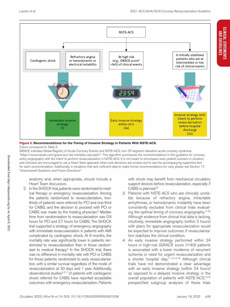
Lawton et al 2021 ACC/AHA/SCAI Coronary Revascularization Guideline
e39
CLINICAL STATEMENTS
AND GUIDELINES
Circulation. 2022;145:e18–e114. DOI: 10.1161/CIR.0000000000001038 January 18, 2022
anatomy and, when appropriate, should include a Heart Team discussion.
2. In the SHOCK trial, patients were randomized to med-ical therapy or emergency revascularization. Among the patients randomized to revascularization, two-thirds of patients were referred for PCI and one-third for CABG, and the decision to proceed with PCI or CABG was made by the treating physician.9 Median time from randomization to revascularization was 0.9 hours for PCI and 2.7 hours for CABG. The SHOCK trial supported a strategy of emergency angiography with immediate revascularization in patients with AMI complicated by cardiogenic shock. At 6 months, the mortality rate was significantly lower in patients ran-domized to revascularization than in those random-ized to medical therapy.9 In the SHOCK trial, there was no difference in mortality rate with PCI or CABG for those patients randomized to early revasculariza-tion, with a similar survival regardless of the mode of revascularization at 30 days and 1 year. Additionally, observational studies5-7,17 of patients with cardiogenic shock referred for CABG have reported acceptable outcomes with emergency revascularization. Patients
with shock may benefit from mechanical circulatory support devices before revascularization, especially if CABG is planned.27
3. Patients with NSTE-ACS who are clinically unsta-ble because of refractory angina, intractable arrhythmias, or hemodynamic instability have been consistently excluded from clinical trials evaluat-ing the optimal timing of coronary angiography.11,28 Although evidence from clinical trial data is lacking, intuitively, immediate angiography (within 2 hours) with plans for appropriate revascularization would be expected to improve outcomes if revasculariza-tion stabilizes the clinical condition.
4. An early invasive strategy performed within 24 hours in high-risk (GRACE score >140) patients is associated with a lower incidence of recurrent ischemia or need for urgent revascularization and a shorter hospital stay.11,12,14-16 Although clinical trials have not demonstrated a clear advantage with an early invasive strategy (within 24 hours) as opposed to a delayed invasive strategy in the overall population of patients with NSTE-ACS,12,16 prespecified subgroup analyses of these trials
Figure 5. Recommendations for the Timing of Invasive Strategy in Patients With NSTE-ACS.Colors correspond to Table 2. GRACE indicates Global Registry of Acute Coronary Events; and NSTE-ACS, non–ST-segment–elevation acute coronary syndrome. *https://www.mdcalc.com/grace-acs-risk-mortality-calculator.31 This algorithm summarizes the recommendations in this guideline for coronary artery angiography with the intent to perform revascularization in NSTE-ACS. It is not meant to encompass every patient scenario or situation, and clinicians are encouraged to use a Heart Team approach when care decisions are unclear and to see the accompanying supportive text for each recommendation. Additionally, in situations that lack sufficient data to make formal recommendations for care, please see Section 17, “Unanswered Questions and Future Directions.”
Dow
nloaded from http://ahajournals.org by on A
pril 8, 2022

Lawton et al 2021 ACC/AHA/SCAI Coronary Revascularization Guideline
January 18, 2022 Circulation. 2022;145:e18–e114. DOI: 10.1161/CIR.0000000000001038e40
CLIN
ICAL
STA
TEM
ENTS
AN
D GU
IDEL
INES
support the use of an early invasive strategy for high-risk patients.11,12,16 The TIMACS (Timing of Intervention in Acute Coronary Syndromes) trial, in which patients were enrolled within 24 hours of symptoms and randomized to angiography at ≤ 24 hours versus ≥ 36 hours from time of ran-domization, and the VERDICT (Very Early vs Deferred Invasive Evaluation Using Computerized Tomography) trial, in which patients were random-ized to angiography at <12 hours versus 48 to 72 hours from time of diagnosis, both evaluated the value of early invasive management of symptoms for patients with NSTE-ACS and demonstrated a lower rate of cardiovascular events in follow-up in the high-risk subgroup of patients randomized to early angiography.
5. In intermediate- or low-risk patients, timing is not critical, and a delayed invasive strategy within 48 to 72 hours has been demonstrated to be accept-able11-16,28 Randomized trial data have not dem-onstrated differences in rates of death and MI between an early invasive strategy (coronary angi-ography <24 hours after admission) and a delayed invasive approach (48–72 hours) in a nonse-lected population of patients with NSTEMI.11-16,29,30 However, low-risk patients benefit from a routine invasive strategy before hospital discharge, with a significant reduction in the risk of cardiovascular death or MI, compared with a selective invasive strategy.21
6. Although there are no randomized trials specifi-cally evaluating emergency CABG versus medi-cal therapy or delayed revascularization in patients with NSTE-ACS and failed PCI who have ongoing ischemia or hemodynamic compromise, multiple ret-rospective reviews have noted a reduced mortality rate in patients with an emergency approach.6,9,17 The appropriate safe timing of CABG is carefully determined with a Heart Team approach in patients with NSTE-ACS who are on dual antiplatelet ther-apy (DAPT).
7. In the CULPRIT-SHOCK trial, almost 40% of enrolled patients had an NSTEMI. As mentioned in section 5.2, patients were included in the trial if they had 2 or more vessels with >70% diameter stenosis that were ≥2 mm in diameter. Those with chronic total occlusions were eligible for inclusion in the study. Patients in the CULPRIT-SHOCK trial who were randomized to culprit-only PCI with the option of staged revascularization of non-culprit lesions had a lower rate of the composite end-point of death and dialysis at 30 days and 1 year. Culprit-vessel PCI was associated with a signifi-cant all-cause mortality rate reduction at 30 days but not at 1 year.18,19 As was noted in patients with STEMI, the results of the CULPRIT-SHOCK trial
showed no benefit to immediate multivessel PCI in NSTEMI.
7. REVASCULARIZATION IN SIHD7.1. Revascularization to Improve Survival in SIHD Compared With Medical Therapy
Recommendations for Revascularization to Improve Survival in SIHD Compared With Medical TherapyReferenced studies that support the recommendations are summarized in Online Data Supplement 10.
COR LOE Recommendations
Left ventricular dysfunction and multivessel CAD
1 B-R
1. In patients with SIHD and multivessel CAD appropriate for CABG with severe left ventric-ular systolic dysfunction (left ventricular ejec-tion fraction <35%), CABG is recommended to improve survival.1,2
2a B-NR
2. In selected patients with SIHD and multivessel CAD appropriate for CABG and mild-to-moder-ate left ventricular systolic dysfunction (ejection fraction 35%–50%), CABG (to include a left internal mammary artery [LIMA] graft to the LAD) is reasonable to improve survival.3-8
Left main CAD
1 B-R3. In patients with SIHD and significant left main
stenosis, CABG is recommended to improve survival.9-12
2a B-NR
4. In selected patients with SIHD and significant left main stenosis for whom PCI can provide equivalent revascularization to that possible with CABG, PCI is reasonable to improve survival.9
Multivessel CAD
2b B-R
5. In patients with SIHD, normal ejection frac-tion, significant stenosis in 3 major coronary arteries (with or without proximal LAD), and anatomy suitable for CABG, CABG may be reasonable to improve survival.10,13-15
2b B-R
6. In patients with SIHD, normal ejection frac-tion, significant stenosis in 3 major coronary arteries (with or without proximal LAD), and anatomy suitable for PCI, the usefulness of PCI to improve survival is uncertain.14-24
Stenosis in the proximal LAD artery
2b B-R
7. In patients with SIHD, normal left ventricular ejection fraction, and significant stenosis in the proximal LAD, the usefulness of coronary revascularization to improve survival is uncer-tain.10,14,17,24-27
Single- or double-vessel disease not involving the proximal LAD
3: No Benefit
B-R
8. In patients with SIHD, normal left ventricular ejection fraction, and 1- or 2-vessel CAD not involving the proximal LAD, coronary revas-cularization is not recommended to improve survival.10,14,16,26,28,29
3: Harm B-NR
9. In patients with SIHD who have ≥1 coronary arteries that are not anatomically or function-ally significant (<70% diameter of non–left main coronary artery stenosis, FFR >0.80), coronary revascularization should not be performed with the primary or sole intent to improve survival.26,30
Dow
nloaded from http://ahajournals.org by on A
pril 8, 2022
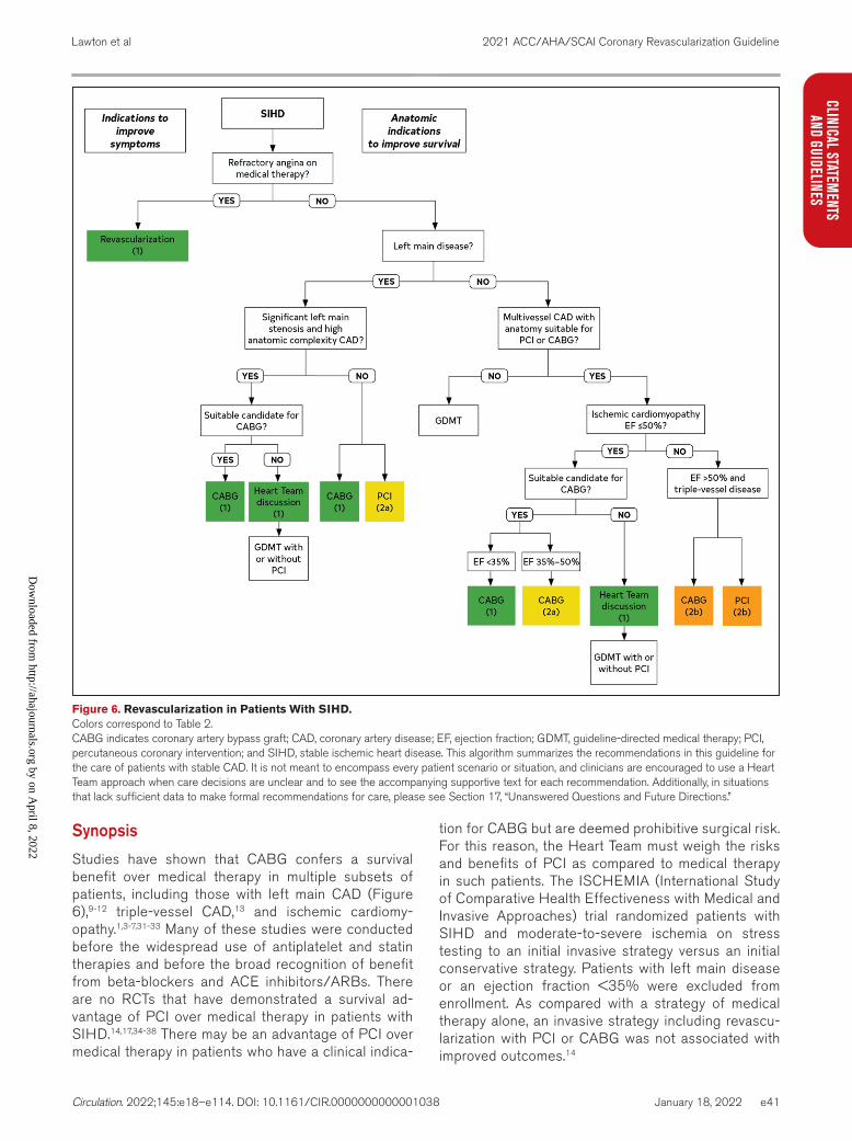
Lawton et al 2021 ACC/AHA/SCAI Coronary Revascularization Guideline
e41
CLINICAL STATEMENTS
AND GUIDELINES
Circulation. 2022;145:e18–e114. DOI: 10.1161/CIR.0000000000001038 January 18, 2022
Synopsis
Studies have shown that CABG confers a survival benefit over medical therapy in multiple subsets of patients, including those with left main CAD (Figure 6),9-12 triple-vessel CAD,13 and ischemic cardiomy-opathy.1,3-7,31-33 Many of these studies were conducted before the widespread use of antiplatelet and statin therapies and before the broad recognition of benefit from beta-blockers and ACE inhibitors/ARBs. There are no RCTs that have demonstrated a survival ad-vantage of PCI over medical therapy in patients with SIHD.14,17,34-38 There may be an advantage of PCI over medical therapy in patients who have a clinical indica-
tion for CABG but are deemed prohibitive surgical risk. For this reason, the Heart Team must weigh the risks and benefits of PCI as compared to medical therapy in such patients. The ISCHEMIA (International Study of Comparative Health Effectiveness with Medical and Invasive Approaches) trial randomized patients with SIHD and moderate-to-severe ischemia on stress testing to an initial invasive strategy versus an initial conservative strategy. Patients with left main disease or an ejection fraction <35% were excluded from enrollment. As compared with a strategy of medical therapy alone, an invasive strategy including revascu-larization with PCI or CABG was not associated with improved outcomes.14
Figure 6. Revascularization in Patients With SIHD.Colors correspond to Table 2. CABG indicates coronary artery bypass graft; CAD, coronary artery disease; EF, ejection fraction; GDMT, guideline-directed medical therapy; PCI, percutaneous coronary intervention; and SIHD, stable ischemic heart disease. This algorithm summarizes the recommendations in this guideline for the care of patients with stable CAD. It is not meant to encompass every patient scenario or situation, and clinicians are encouraged to use a Heart Team approach when care decisions are unclear and to see the accompanying supportive text for each recommendation. Additionally, in situations that lack sufficient data to make formal recommendations for care, please see Section 17, “Unanswered Questions and Future Directions.”
Dow
nloaded from http://ahajournals.org by on A
pril 8, 2022
Lawton et al 2021 ACC/AHA/SCAI Coronary Revascularization Guideline
January 18, 2022 Circulation. 2022;145:e18–e114. DOI: 10.1161/CIR.0000000000001038e42
CLIN
ICAL
STA
TEM
ENTS
AN
D GU
IDEL
INES
Recommendation-Specific Supporting TextLeft Ventricular Dysfunction and Multivessel CAD
1. The strongest evidence in the past decade to sup-port revascularization with CABG in patients with left ventricular dysfunction and CAD appropriate for CABG has been the STICH (Surgical Treatment for Ischemic Heart Failure) trial,1,39,40 which random-ized patients with left ventricular dysfunction (ejec-tion fraction ≤35%) to either CABG with medical therapy or medical therapy alone. This study initially did not demonstrate a survival benefit for CABG over a median follow-up of 5 years,39 but a sub-sequent report from this trial evaluating long-term follow-up at 10 years reported a survival benefit of CABG compared with medical therapy alone.1,40 The use of myocardial viability studies in this study population demonstrated no relevance to study outcomes; however, this testing was not standard-ized.41 There are insufficient data to make recom-mendations for using PCI in this patient population.
2. Evidence for a survival advantage with CABG in patients with SIHD and moderate left ventricular dysfunction comes from subgroup analyses of patients enrolled in the Coronary Artery Surgery Study3 and the Veterans Administration Coronary Artery Bypass Cooperative Study with LV dysfunc-tion,42 as well as a meta-analysis10 of the RCTs of CABG versus medical therapy. In these studies, patients with left ventricular dysfunction had a significant survival benefit with CABG, particularly patients with accompanying triple-vessel disease. Several registry studies have supported these find-ings.4-7,31-33 The use of PCI in this patient population requires more study.
Left Main CAD3. The Veterans Administration Coronary Artery
Bypass Surgery Cooperative Study randomized patients with SIHD to a strategy of CABG versus medical therapy.43 In this study, close to 15% of enrolled patients had significant left main disease.43 At 42 months’ follow-up, CABG was associated with a significant survival benefit in the subgroup of patients with left main disease.11 Additionally, a meta-analysis of the RCTs comparing CABG with medical therapy supported these findings, with a 70% reduction in 5-year mortality rate with CABG versus medical therapy for the group of patients with left main disease.10 Subsequent studies have supported these findings.9,44-46 Although the evi-dence to support revascularization with CABG is derived mainly from older RCTs, there are no new data to refute this evidence, as all of the contem-porary clinical trials comparing revascularization with medical therapy have excluded patients with significant stenoses of the left main artery.14,24
4. The evidence for a survival advantage for PCI over medical therapy in patients with left main CAD is inferential but plausible. Several registry stud-ies have suggested a survival benefit of PCI over medical therapy in patients with left main CAD.,47,48 A network meta-analysis of 19 studies found that the survival advantage for PCI over medical therapy in patients with left main CAD was identical to the survival advantage for CABG over medical therapy.9 Additionally, RCTs and meta-analyses of these trials evaluating outcomes of PCI versus CABG in patients with low-to-medium anatomic complexity of CAD and with left main disease that is equally suitable for surgical or percutaneous revascularization have reported similar survival with PCI and CABG.49-55
Multivessel CAD5. The new Class 2b recommendation, which rep-
resents a downgrade from a Class 1 recommen-dation in the 2011 CABG guideline,56 reflects new evidence showing no advantage of CABG over medical therapy alone to improve survival in patients with 3-vessel CAD with preserved LV function and no LM disease. The older recommen-dation was based on evidence from registry stud-ies,26,29,48,57 a meta-analysis,10 and a single RCT,13 all of which were completed >20 to 40 years ago before the development of newer surgical tech-niques or advances in medical therapy associated with improved prognosis.58,59 Newer evidence from the ISCHEMIA trial14 and from meta-analyses, which incorporated15,60-62 or did not incorporate37 the ISCHEMIA results, as well as a more detailed review of earlier studies63 supported this down-grade. After several hours of deliberation, the writ-ing committee concluded that using CABG as a revascularization strategy versus medical therapy alone “may be reasonable” to improve survival in stable patients with 3-vessel CAD. The writing committee recognized that an adequately pow-ered trial to test this hypothesis is unfeasible in the current era but proposed that revascularization confers other benefits to patients with multivessel CAD and SIHD. Accordingly, Section 7.3. highlights the advantages of revascularization over medical therapy for the prevention of cardiovascular events.
6. The writing committee reviewed newer evidence and concluded that the ability of PCI to improve survival, compared with medical therapy alone in patients with multivessel CAD, remains uncertain. The recommendation, which reflects a weaker endorsement for PCI than for CABG in patients with multivessel CAD, is supported by evidence from an older registry study48 and a subgroup analysis of patients receiving everolimus-eluting stents in a network meta-analysis37 that did not incorporate the results of the ISCHEMIA trial.14
Dow
nloaded from http://ahajournals.org by on A
pril 8, 2022

Lawton et al 2021 ACC/AHA/SCAI Coronary Revascularization Guideline
Circulation. 2022;145:e18–e114. DOI: 10.1161/CIR.0000000000001038 January 18, 2022 e43
CLINICAL STATEMENTS
AND GUIDELINES
The preponderance of newer evidence against a survival advantage of PCI comes from the ISCHEMIA trial itself,14 which is consistent with the results of multiple earlier RCTs17-23 and multiple contemporary meta-analyses15,60-62 incorporating the ISCHEMIA trial results,14 all of which have not shown a survival advantage for PCI over medical therapy for patients with multivessel CAD.
Stenosis in the Proximal LAD Artery7. An earlier meta-analysis10 and several earlier reg-
istry studies26,29 suggested a survival advantage of CABG over medical therapy in patients with dis-ease in the proximal LAD. Additionally, a network meta-analysis found a survival advantage for PCI.37 However, a dedicated RCT found no survival advan-tage for either CABG or PCI over medical therapy in this setting,64,65 and the ISCHEMIA trial14 showed no difference in event rates with either CABG or PCI over medical therapy when patients had multivessel CAD involving the proximal LAD. In the ISCHEMIA trial, close to half of enrolled patients had >50% stenosis of the proximal LAD; in this study, there was no heterogeneity of treatment effect on out-comes with the presence of LAD disease.
Single- or Double-Vessel Disease Not Involving the Proximal LAD
8. A clinical principle from several studies is that the more myocardium is at risk, the greater is the sur-vival advantage of revascularization over medical therapy, and in patients with little myocardium at risk (1- or 2-vessel CAD without LAD involvement), there is likely no survival benefit of revasculariza-tion in patients with SIHD.10,17,26,29,37
9. In patients without clinical or physiological evi-dence of significant disease, bypass surgery of nonobstructive disease has been reported to stimulate progression of CAD,66 and PCI may pre-cipitate periprocedural MIs23 and is not associated with improved outcomes.30,67-69
7.2. Revascularization to Reduce Cardiovascular Events in SIHD Compared With Medical Therapy
Recommendation for Revascularization to Reduce Cardiovascular Events in SIHD Compared With Medical TherapyReferenced studies that support the recommendation are summarized in Online Data Supplement 11.
COR LOE Recommendation
2a B-R
1. In patients with SIHD and multivessel CAD appropriate for either CABG or PCI, revas-cularization is reasonable to lower the risk of cardiovascular events such as spontaneous MI, unplanned urgent revascularizations, or cardiac death.1-8
SynopsisClinical practice guidelines have traditionally included recommendations for revascularization in patients with SIHD based on the ability of CABG or PCI to improve overall survival (Section 7.1.) or to reduce ischemic symptoms (Section 7.3.), as compared with medical therapy alone. However, there are other clinical events that can affect a patient’s overall prognosis, and that remain important considerations for patients. Several studies suggest that revascularization with CABG or PCI lowers the risk of adverse events such as cardiac death, MI, or urgent revascularization compared with medical therapy alone.2,5,6,9,10
Recommendation-Specific Supporting Text1. In MASS (Medicine, Angioplasty or Surgery Study)
II, the 10-year rates of cardiac death were lower after CABG or PCI than after medical therapy alone.2 Lower rates of cardiac death were seen after revascularization than with medical therapy alone in a meta-analysis of 25 studies enrolling 19 806 patients.3 However, a statistically nonsignifi-cant reduction was seen in a concurrent meta-anal-ysis of 12 103 patients enrolled in 7 RCTs.8 Several other studies found no difference in cardiac death after revascularization than with medical therapy alone.10,11
Cardiac death may be related to the occurrence of MI after revascularization.12 The relative prog-nostic importance of procedural MIs versus that of late spontaneous MIs remains uncertain.13 In the ISCHEMIA trial,1 the incidence of procedural type 4a or type 5 MIs was increased with revascularization, but the incidence of late MI (spontaneous MI [type 1], demand-induced MI [type 2], or MIs associated with stent thrombosis [type 4b] or with restenosis [type 4c]) was reduced. A preplanned analysis of the MI patterns in the ISCHEMIA trial4 found that all-cause death was increased with spontaneous MIs but not with procedural MIs. A large network meta-analysis found that spontaneous MI was reduced by revas-cularization compared with medical therapy alone.3 However, a concurrent analysis found an increased rate of procedural MI, a reduced rate of nonproc-edural MI, and no difference in overall MI.11 On the contrary, another meta-analysis of stable patients, did not show a reduction in MI with revasculariza-tion,10 and 1 other study reported reduction in MI with CABG but not with PCI.9 Revascularization with CABG or PCI may reduce the need for subsequent urgent revascularization or hospitalization for acute coronary events.5-8
Dow
nloaded from http://ahajournals.org by on A
pril 8, 2022
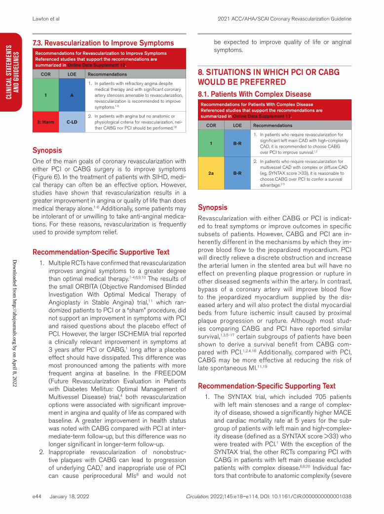
Lawton et al 2021 ACC/AHA/SCAI Coronary Revascularization Guideline
January 18, 2022 Circulation. 2022;145:e18–e114. DOI: 10.1161/CIR.0000000000001038e44
CLIN
ICAL
STA
TEM
ENTS
AN
D GU
IDEL
INES
7.3. Revascularization to Improve SymptomsRecommendations for Revascularization to Improve SymptomsReferenced studies that support the recommendations are summarized in Online Data Supplement 12.
COR LOE Recommendations
1 A
1. In patients with refractory angina despite medical therapy and with significant coronary artery stenoses amenable to revascularization, revascularization is recommended to improve symptoms.1-6
3: Harm C-LD2. In patients with angina but no anatomic or
physiological criteria for revascularization, nei-ther CABG nor PCI should be performed.7,8
SynopsisOne of the main goals of coronary revascularization with either PCI or CABG surgery is to improve symptoms (Figure 6). In the treatment of patients with SIHD, medi-cal therapy can often be an effective option. However, studies have shown that revascularization results in a greater improvement in angina or quality of life than does medical therapy alone.1-6 Additionally, some patients may be intolerant of or unwilling to take anti-anginal medica-tions. For these reasons, revascularization is frequently used to provide symptom relief.
Recommendation-Specific Supportive Text1. Multiple RCTs have confirmed that revascularization
improves anginal symptoms to a greater degree than optimal medical therapy.1-4,6,9,10 The results of the small ORBITA (Objective Randomised Blinded Investigation With Optimal Medical Therapy of Angioplasty in Stable Angina) trial,11 which ran-domized patients to PCI or a “sham” procedure, did not support an improvement in symptoms with PCI and raised questions about the placebo effect of PCI. However, the larger ISCHEMIA trial reported a clinically relevant improvement in symptoms at 3 years after PCI or CABG,1 long after a placebo effect should have dissipated. This difference was most pronounced among the patients with more frequent angina at baseline. In the FREEDOM (Future Revascularization Evaluation in Patients with Diabetes Mellitus: Optimal Management of Multivessel Disease) trial,4 both revascularization options were associated with significant improve-ment in angina and quality of life as compared with baseline. A greater improvement in health status was noted with CABG compared with PCI at inter-mediate-term follow-up, but this difference was no longer significant in longer-term follow-up.
2. Inappropriate revascularization of nonobstruc-tive plaques with CABG can lead to progression of underlying CAD,7 and inappropriate use of PCI can cause periprocedural MIs8 and would not
be expected to improve quality of life or anginal symptoms.
8. SITUATIONS IN WHICH PCI OR CABG WOULD BE PREFERRED8.1. Patients With Complex Disease
Recommendations for Patients With Complex DiseaseReferenced studies that support the recommendations are summarized in Online Data Supplement 13.
COR LOE Recommendations
1 B-R
1. In patients who require revascularization for significant left main CAD with high-complexity CAD, it is recommended to choose CABG over PCI to improve survival.1,2
2a B-R
2. In patients who require revascularization for multivessel CAD with complex or diffuse CAD (eg, SYNTAX score >33), it is reasonable to choose CABG over PCI to confer a survival advantage.2-5
SynopsisRevascularization with either CABG or PCI is indicat-ed to treat symptoms or improve outcomes in specific subsets of patients. However, CABG and PCI are in-herently different in the mechanisms by which they im-prove blood flow to the jeopardized myocardium. PCI will directly relieve a discrete obstruction and increase the arterial lumen in the stented area but will have no effect on preventing plaque progression or rupture in other diseased segments within the artery. In contrast, bypass of a coronary artery will improve blood flow to the jeopardized myocardium supplied by the dis-eased artery and will also protect the distal myocardial beds from future ischemic insult caused by proximal plaque progression or rupture. Although most stud-ies comparing CABG and PCI have reported similar survival,1,3,5-17 certain subgroups of patients have been shown to derive a survival benefit from CABG com-pared with PCI.1,2,4,18 Additionally, compared with PCI, CABG may be more effective at reducing the risk of late spontaneous MI.11,19
Recommendation-Specific Supporting Text1. The SYNTAX trial, which included 705 patients
with left main stenoses and a range of complex-ity of disease, showed a significantly higher MACE and cardiac mortality rate at 5 years for the sub-group of patients with left main and high-complex-ity disease (defined as a SYNTAX score >33) who were treated with PCI.1 With the exception of the SYNTAX trial, the other RCTs comparing PCI with CABG in patients with left main disease excluded patients with complex disease.6,8,20 Individual fac-tors that contribute to anatomic complexity (severe
Dow
nloaded from http://ahajournals.org by on A
pril 8, 2022
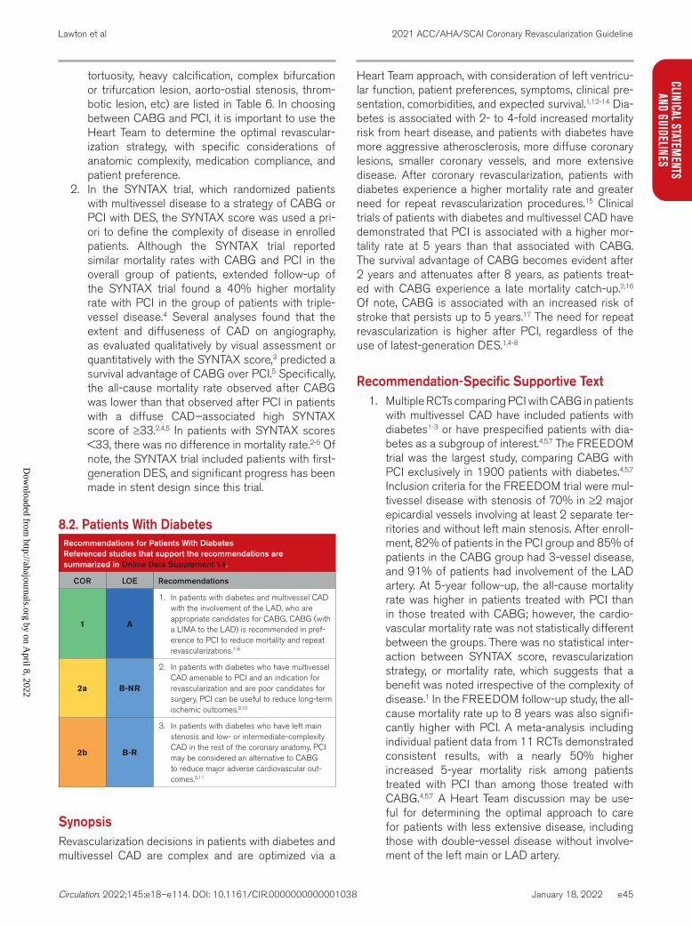
Lawton et al 2021 ACC/AHA/SCAI Coronary Revascularization Guideline
Circulation. 2022;145:e18–e114. DOI: 10.1161/CIR.0000000000001038 January 18, 2022 e45
CLINICAL STATEMENTS
AND GUIDELINES
tortuosity, heavy calcification, complex bifurcation or trifurcation lesion, aorto-ostial stenosis, throm-botic lesion, etc) are listed in Table 6. In choosing between CABG and PCI, it is important to use the Heart Team to determine the optimal revascular-ization strategy, with specific considerations of anatomic complexity, medication compliance, and patient preference.
2. In the SYNTAX trial, which randomized patients with multivessel disease to a strategy of CABG or PCI with DES, the SYNTAX score was used a pri-ori to define the complexity of disease in enrolled patients. Although the SYNTAX trial reported similar mortality rates with CABG and PCI in the overall group of patients, extended follow-up of the SYNTAX trial found a 40% higher mortality rate with PCI in the group of patients with triple-vessel disease.4 Several analyses found that the extent and diffuseness of CAD on angiography, as evaluated qualitatively by visual assessment or quantitatively with the SYNTAX score,3 predicted a survival advantage of CABG over PCI.5 Specifically, the all-cause mortality rate observed after CABG was lower than that observed after PCI in patients with a diffuse CAD–associated high SYNTAX score of ≥33.2,4,5 In patients with SYNTAX scores <33, there was no difference in mortality rate.2-5 Of note, the SYNTAX trial included patients with first-generation DES, and significant progress has been made in stent design since this trial.
8.2. Patients With DiabetesRecommendations for Patients With DiabetesReferenced studies that support the recommendations are summarized in Online Data Supplement 14.
COR LOE Recommendations
1 A
1. In patients with diabetes and multivessel CAD with the involvement of the LAD, who are appropriate candidates for CABG, CABG (with a LIMA to the LAD) is recommended in pref-erence to PCI to reduce mortality and repeat revascularizations.1-8
2a B-NR
2. In patients with diabetes who have multivessel CAD amenable to PCI and an indication for revascularization and are poor candidates for surgery, PCI can be useful to reduce long-term ischemic outcomes.9,10
2b B-R
3. In patients with diabetes who have left main stenosis and low- or intermediate-complexity CAD in the rest of the coronary anatomy, PCI may be considered an alternative to CABG to reduce major adverse cardiovascular out-comes.5,11
SynopsisRevascularization decisions in patients with diabetes and multivessel CAD are complex and are optimized via a
Heart Team approach, with consideration of left ventricu-lar function, patient preferences, symptoms, clinical pre-sentation, comorbidities, and expected survival.1,12-14 Dia-betes is associated with 2- to 4-fold increased mortality risk from heart disease, and patients with diabetes have more aggressive atherosclerosis, more diffuse coronary lesions, smaller coronary vessels, and more extensive disease. After coronary revascularization, patients with diabetes experience a higher mortality rate and greater need for repeat revascularization procedures.15 Clinical trials of patients with diabetes and multivessel CAD have demonstrated that PCI is associated with a higher mor-tality rate at 5 years than that associated with CABG. The survival advantage of CABG becomes evident after 2 years and attenuates after 8 years, as patients treat-ed with CABG experience a late mortality catch-up.2,16 Of note, CABG is associated with an increased risk of stroke that persists up to 5 years.17 The need for repeat revascularization is higher after PCI, regardless of the use of latest-generation DES.1,4-8
Recommendation-Specific Supportive Text1. Multiple RCTs comparing PCI with CABG in patients
with multivessel CAD have included patients with diabetes1-3 or have prespecified patients with dia-betes as a subgroup of interest.4,5,7 The FREEDOM trial was the largest study, comparing CABG with PCI exclusively in 1900 patients with diabetes.4,5,7 Inclusion criteria for the FREEDOM trial were mul-tivessel disease with stenosis of 70% in ≥2 major epicardial vessels involving at least 2 separate ter-ritories and without left main stenosis. After enroll-ment, 82% of patients in the PCI group and 85% of patients in the CABG group had 3-vessel disease, and 91% of patients had involvement of the LAD artery. At 5-year follow-up, the all-cause mortality rate was higher in patients treated with PCI than in those treated with CABG; however, the cardio-vascular mortality rate was not statistically different between the groups. There was no statistical inter-action between SYNTAX score, revascularization strategy, or mortality rate, which suggests that a benefit was noted irrespective of the complexity of disease.1 In the FREEDOM follow-up study, the all-cause mortality rate up to 8 years was also signifi-cantly higher with PCI. A meta-analysis including individual patient data from 11 RCTs demonstrated consistent results, with a nearly 50% higher increased 5-year mortality risk among patients treated with PCI than among those treated with CABG.4,5,7 A Heart Team discussion may be use-ful for determining the optimal approach to care for patients with less extensive disease, including those with double-vessel disease without involve-ment of the left main or LAD artery.
Dow
nloaded from http://ahajournals.org by on A
pril 8, 2022

Lawton et al 2021 ACC/AHA/SCAI Coronary Revascularization Guideline
January 18, 2022 Circulation. 2022;145:e18–e114. DOI: 10.1161/CIR.0000000000001038e46
CLIN
ICAL
STA
TEM
ENTS
AN
D GU
IDEL
INES
2. Patients with diabetes who are at high surgical risk and require coronary revascularization are more likely to be treated with PCI in current practice.9 In an observational registry that included high-risk patients with refractory ischemia, 5-year survival rates were similar among those treated with CABG and PCI.10
3. There are no RCTs specifically comparing PCI with CABG in patients with diabetes and left main CAD. However, a large RCT exclusively enrolled patients with left main CAD, and the subgroup analysis of patients with diabetes informs this recommendation.13 In the EXCEL trial, which included patients with left main CAD and low- or intermediate-complexity CAD, approximately 30% of patients had diabetes. At 3 years, the composite of death, stroke, or MI was not significantly differ-ent between PCI and CABG among patients with diabetes. However, the all-cause mortality rate was almost 2 times higher in the PCI group. There was no interaction between diabetes status and revas-cularization modality.
8.3. Patients With Previous CABGRecommendations for Patients With Previous CABGReferenced studies that support the recommendations are summarized in Online Data Supplement 15.
COR LOE Recommendations
2a B-NR
1. In patients with previous CABG with a patent LIMA to the LAD who need repeat revascu-larization, if PCI is feasible, it is reasonable to choose PCI over CABG.1,2
2a C-LD
2. In patients with previous CABG and refractory angina on GDMT that is attributable to LAD disease, it is reasonable to choose CABG over PCI when an internal mammary artery (IMA) can be used as a conduit to the LAD.3,4
2b B-NR
3. In patients with previous CABG and complex CAD, it may be reasonable to choose CABG over PCI when an IMA can be used as a con-duit to the LAD.3,4
SynopsisA Heart Team approach and shared decision-making are important in patients who require repeat revas-cularization after CABG. There are no randomized trials comparing medical therapy with revasculariza-tion in patients with previous CABG. PCI and repeat CABG in patients with previous CABG are associated with higher rates of procedural failure and complica-tions5,6 and worse outcomes than those of patients without previous CABG.6-8 The need for any repeat revascularization after PCI or CABG is itself an in-dependent predictor of higher mortality risk.9 Factors that influence the choice of revascularization modal-
ity include the availability of the IMA for grafting, a patent graft to the LAD, comorbid conditions, patient factors and preferences, the quality of the target vessels, anatomic complexity of the native and graft disease, and the feasibility and risks of the revascu-larization method.
Recommendation-Specific Supportive Text1. In patients with previous CABG, percutaneous
intervention of a native vessel or a saphenous vein graft (SVG) is probably indicated in prefer-ence to redo CABG, particularly if a LIMA-LAD is not planned or if the patient already has a patent LIMA-LAD, which increases the risk of a redo sternotomy. Randomized and retrospective comparisons of PCI versus repeat CABG show lower in-hospital stroke and mortality rates associated with PCI,2,3 although long-term mor-tality rates appeared similar. In circumstances of acute graft closure, PCI of the native vessel is often performed in preference to redo CABG10 or treating an acutely thrombosed graft with fresh suture lines.11
2. A patient with previous CABG faces increased risk during revascularization via CABG,9 including higher rates of in-hospital death and stroke, com-pared with patients who undergo revascularization via PCI.2,3 Observational data suggest that the use of CABG over PCI may result in improved long-term outcomes; however, results are inconsistent and not supported by high-quality RCTs.3,12,13 If PCI is not an option, if a patent IMA to LAD is not present, or if an IMA is available to be used as a conduit for the LAD, CABG is often chosen as the revascularization strategy in patients with previous CABG and refractory angina who are at an accept-able risk for reoperation.3,4
3. Two large observational studies with propensity matching in patients with previous CABG and complex CAD inform decisions about revascular-ization. The first noted that current clinical prac-tice favored redo CABG over PCI for patients at higher risk and with fewer functional grafts, more CTOs, and lower systolic function, whereas PCI was favored in patients with a patent LIMA and amenable anatomy.3 The second noted that LIMA grafting to the LAD confers a long-term survival advantage.4 Thus, decisions about revas-cularization should involve consideration of fac-tors that may favor repeat CABG, such as the availability of an IMA for LAD grafting, ability to provide left main revascularization, recurrent restenosis of stents, or high-complexity PCI, in such patients.
Dow
nloaded from http://ahajournals.org by on A
pril 8, 2022
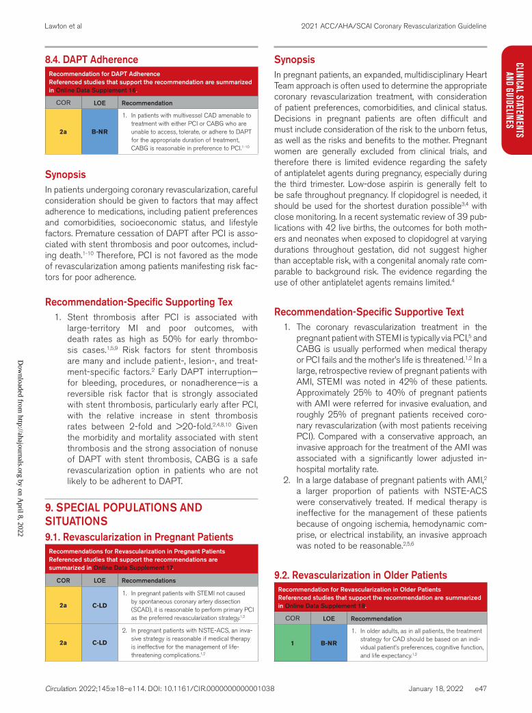
Lawton et al 2021 ACC/AHA/SCAI Coronary Revascularization Guideline
Circulation. 2022;145:e18–e114. DOI: 10.1161/CIR.0000000000001038 January 18, 2022 e47
CLINICAL STATEMENTS
AND GUIDELINES
8.4. DAPT AdherenceRecommendation for DAPT AdherenceReferenced studies that support the recommendation are summarized in Online Data Supplement 16.
COR LOE Recommendation
2a B-NR
1. In patients with multivessel CAD amenable to treatment with either PCI or CABG who are unable to access, tolerate, or adhere to DAPT for the appropriate duration of treatment, CABG is reasonable in preference to PCI.1-10
SynopsisIn patients undergoing coronary revascularization, careful consideration should be given to factors that may affect adherence to medications, including patient preferences and comorbidities, socioeconomic status, and lifestyle factors. Premature cessation of DAPT after PCI is asso-ciated with stent thrombosis and poor outcomes, includ-ing death.1-10 Therefore, PCI is not favored as the mode of revascularization among patients manifesting risk fac-tors for poor adherence.
Recommendation-Specific Supporting Tex1. Stent thrombosis after PCI is associated with
large-territory MI and poor outcomes, with death rates as high as 50% for early thrombo-sis cases.1,5,9 Risk factors for stent thrombosis are many and include patient-, lesion-, and treat-ment-specific factors.2 Early DAPT interruption—for bleeding, procedures, or nonadherence—is a reversible risk factor that is strongly associated with stent thrombosis, particularly early after PCI, with the relative increase in stent thrombosis rates between 2-fold and >20-fold.2,4,8,10 Given the morbidity and mortality associated with stent thrombosis and the strong association of nonuse of DAPT with stent thrombosis, CABG is a safe revascularization option in patients who are not likely to be adherent to DAPT.
9. SPECIAL POPULATIONS AND SITUATIONS9.1. Revascularization in Pregnant Patients
Recommendations for Revascularization in Pregnant PatientsReferenced studies that support the recommendations are summarized in Online Data Supplement 17.
COR LOE Recommendations
2a C-LD
1. In pregnant patients with STEMI not caused by spontaneous coronary artery dissection (SCAD), it is reasonable to perform primary PCI as the preferred revascularization strategy.1,2
2a C-LD
2. In pregnant patients with NSTE-ACS, an inva-sive strategy is reasonable if medical therapy is ineffective for the management of life-threatening complications.1,2
SynopsisIn pregnant patients, an expanded, multidisciplinary Heart Team approach is often used to determine the appropriate coronary revascularization treatment, with consideration of patient preferences, comorbidities, and clinical status. Decisions in pregnant patients are often difficult and must include consideration of the risk to the unborn fetus, as well as the risks and benefits to the mother. Pregnant women are generally excluded from clinical trials, and therefore there is limited evidence regarding the safety of antiplatelet agents during pregnancy, especially during the third trimester. Low-dose aspirin is generally felt to be safe throughout pregnancy. If clopidogrel is needed, it should be used for the shortest duration possible3,4 with close monitoring. In a recent systematic review of 39 pub-lications with 42 live births, the outcomes for both moth-ers and neonates when exposed to clopidogrel at varying durations throughout gestation, did not suggest higher than acceptable risk, with a congenital anomaly rate com-parable to background risk. The evidence regarding the use of other antiplatelet agents remains limited.4
Recommendation-Specific Supportive Text1. The coronary revascularization treatment in the
pregnant patient with STEMI is typically via PCI,5 and CABG is usually performed when medical therapy or PCI fails and the mother’s life is threatened.1,2 In a large, retrospective review of pregnant patients with AMI, STEMI was noted in 42% of these patients. Approximately 25% to 40% of pregnant patients with AMI were referred for invasive evaluation, and roughly 25% of pregnant patients received coro-nary revascularization (with most patients receiving PCI). Compared with a conservative approach, an invasive approach for the treatment of the AMI was associated with a significantly lower adjusted in-hospital mortality rate.
2. In a large database of pregnant patients with AMI,2 a larger proportion of patients with NSTE-ACS were conservatively treated. If medical therapy is ineffective for the management of these patients because of ongoing ischemia, hemodynamic com-prise, or electrical instability, an invasive approach was noted to be reasonable.2,5,6
9.2. Revascularization in Older PatientsRecommendation for Revascularization in Older PatientsReferenced studies that support the recommendation are summarized in Online Data Supplement 18.
COR LOE Recommendation
1 B-NR
1. In older adults, as in all patients, the treatment strategy for CAD should be based on an indi-vidual patient’s preferences, cognitive function, and life expectancy.1,2
Dow
nloaded from http://ahajournals.org by on A
pril 8, 2022
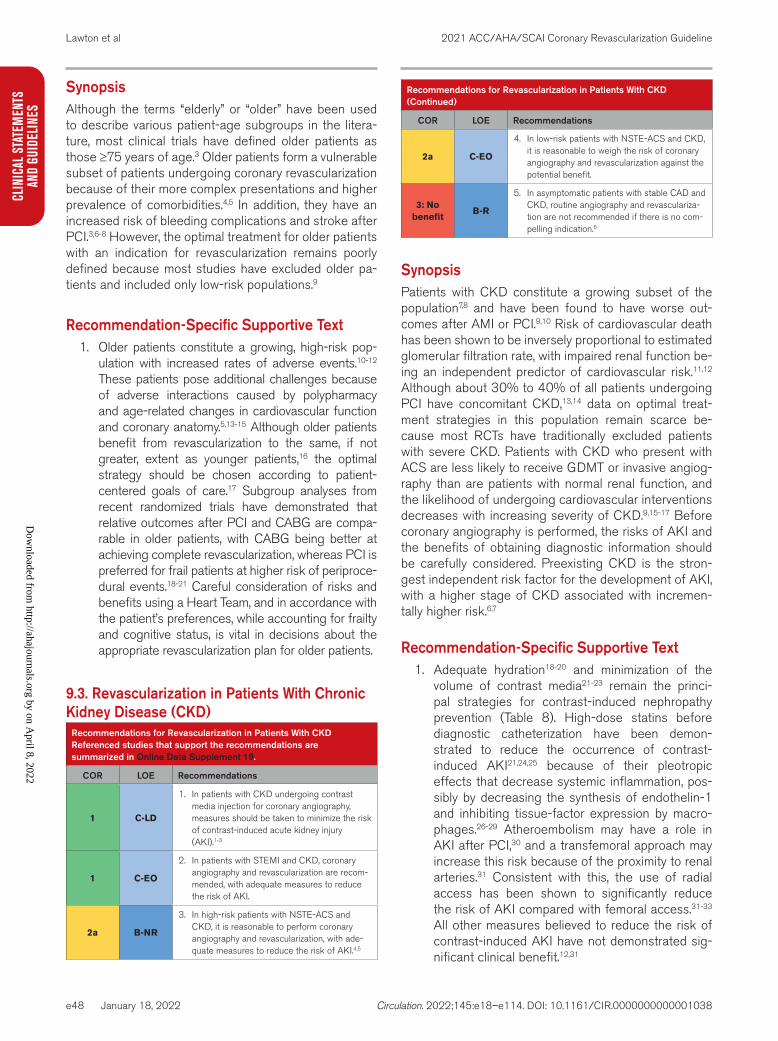
Lawton et al 2021 ACC/AHA/SCAI Coronary Revascularization Guideline
January 18, 2022 Circulation. 2022;145:e18–e114. DOI: 10.1161/CIR.0000000000001038e48
CLIN
ICAL
STA
TEM
ENTS
AN
D GU
IDEL
INES
SynopsisAlthough the terms “elderly” or “older” have been used to describe various patient-age subgroups in the litera-ture, most clinical trials have defined older patients as those ≥75 years of age.3 Older patients form a vulnerable subset of patients undergoing coronary revascularization because of their more complex presentations and higher prevalence of comorbidities.4,5 In addition, they have an increased risk of bleeding complications and stroke after PCI.3,6-8 However, the optimal treatment for older patients with an indication for revascularization remains poorly defined because most studies have excluded older pa-tients and included only low-risk populations.9
Recommendation-Specific Supportive Text1. Older patients constitute a growing, high-risk pop-
ulation with increased rates of adverse events.10-12 These patients pose additional challenges because of adverse interactions caused by polypharmacy and age-related changes in cardiovascular function and coronary anatomy.5,13-15 Although older patients benefit from revascularization to the same, if not greater, extent as younger patients,16 the optimal strategy should be chosen according to patient-centered goals of care.17 Subgroup analyses from recent randomized trials have demonstrated that relative outcomes after PCI and CABG are compa-rable in older patients, with CABG being better at achieving complete revascularization, whereas PCI is preferred for frail patients at higher risk of periproce-dural events.18-21 Careful consideration of risks and benefits using a Heart Team, and in accordance with the patient’s preferences, while accounting for frailty and cognitive status, is vital in decisions about the appropriate revascularization plan for older patients.
9.3. Revascularization in Patients With Chronic Kidney Disease (CKD)
Recommendations for Revascularization in Patients With CKDReferenced studies that support the recommendations are summarized in Online Data Supplement 19.
COR LOE Recommendations
1 C-LD
1. In patients with CKD undergoing contrast media injection for coronary angiography, measures should be taken to minimize the risk of contrast-induced acute kidney injury (AKI).1-3
1 C-EO
2. In patients with STEMI and CKD, coronary angiography and revascularization are recom-mended, with adequate measures to reduce the risk of AKI.
2a B-NR
3. In high-risk patients with NSTE-ACS and CKD, it is reasonable to perform coronary angiography and revascularization, with ade-quate measures to reduce the risk of AKI.4,5
2a C-EO
4. In low-risk patients with NSTE-ACS and CKD, it is reasonable to weigh the risk of coronary angiography and revascularization against the potential benefit.
3: No benefit
B-R
5. In asymptomatic patients with stable CAD and CKD, routine angiography and revasculariza-tion are not recommended if there is no com-pelling indication.6
SynopsisPatients with CKD constitute a growing subset of the population7,8 and have been found to have worse out-comes after AMI or PCI.9,10 Risk of cardiovascular death has been shown to be inversely proportional to estimated glomerular filtration rate, with impaired renal function be-ing an independent predictor of cardiovascular risk.11,12 Although about 30% to 40% of all patients undergoing PCI have concomitant CKD,13,14 data on optimal treat-ment strategies in this population remain scarce be-cause most RCTs have traditionally excluded patients with severe CKD. Patients with CKD who present with ACS are less likely to receive GDMT or invasive angiog-raphy than are patients with normal renal function, and the likelihood of undergoing cardiovascular interventions decreases with increasing severity of CKD.9,15-17 Before coronary angiography is performed, the risks of AKI and the benefits of obtaining diagnostic information should be carefully considered. Preexisting CKD is the stron-gest independent risk factor for the development of AKI, with a higher stage of CKD associated with incremen-tally higher risk.6,7
Recommendation-Specific Supportive Text1. Adequate hydration18-20 and minimization of the
volume of contrast media21-23 remain the princi-pal strategies for contrast-induced nephropathy prevention (Table 8). High-dose statins before diagnostic catheterization have been demon-strated to reduce the occurrence of contrast-induced AKI21,24,25 because of their pleotropic effects that decrease systemic inflammation, pos-sibly by decreasing the synthesis of endothelin-1 and inhibiting tissue-factor expression by macro-phages.26-29 Atheroembolism may have a role in AKI after PCI,30 and a transfemoral approach may increase this risk because of the proximity to renal arteries.31 Consistent with this, the use of radial access has been shown to significantly reduce the risk of AKI compared with femoral access.31-33 All other measures believed to reduce the risk of contrast-induced AKI have not demonstrated sig-nificant clinical benefit.12,31
Recommendations for Revascularization in Patients With CKD (Continued)
COR LOE Recommendations
Dow
nloaded from http://ahajournals.org by on A
pril 8, 2022
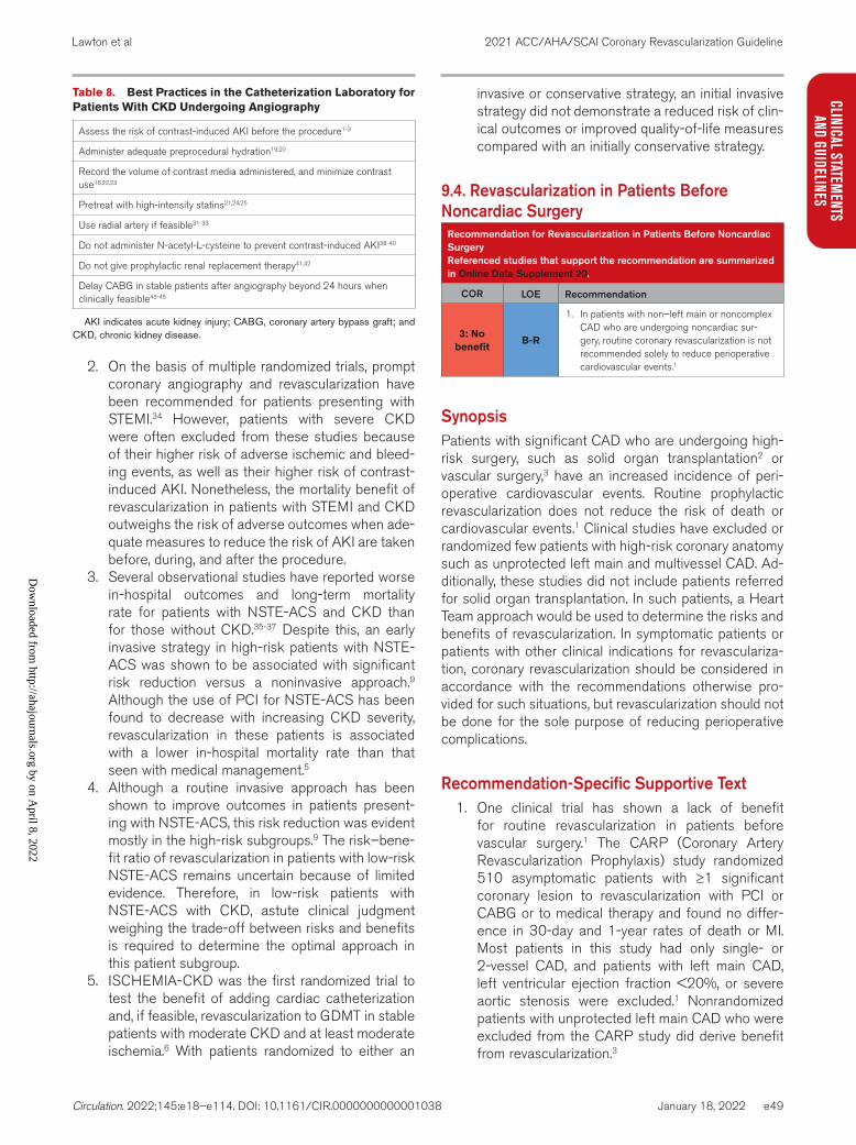
Lawton et al 2021 ACC/AHA/SCAI Coronary Revascularization Guideline
Circulation. 2022;145:e18–e114. DOI: 10.1161/CIR.0000000000001038 January 18, 2022 e49
CLINICAL STATEMENTS
AND GUIDELINES
2. On the basis of multiple randomized trials, prompt coronary angiography and revascularization have been recommended for patients presenting with STEMI.34 However, patients with severe CKD were often excluded from these studies because of their higher risk of adverse ischemic and bleed-ing events, as well as their higher risk of contrast-induced AKI. Nonetheless, the mortality benefit of revascularization in patients with STEMI and CKD outweighs the risk of adverse outcomes when ade-quate measures to reduce the risk of AKI are taken before, during, and after the procedure.
3. Several observational studies have reported worse in-hospital outcomes and long-term mortality rate for patients with NSTE-ACS and CKD than for those without CKD.35-37 Despite this, an early invasive strategy in high-risk patients with NSTE-ACS was shown to be associated with significant risk reduction versus a noninvasive approach.9 Although the use of PCI for NSTE-ACS has been found to decrease with increasing CKD severity, revascularization in these patients is associated with a lower in-hospital mortality rate than that seen with medical management.5
4. Although a routine invasive approach has been shown to improve outcomes in patients present-ing with NSTE-ACS, this risk reduction was evident mostly in the high-risk subgroups.9 The risk–bene-fit ratio of revascularization in patients with low-risk NSTE-ACS remains uncertain because of limited evidence. Therefore, in low-risk patients with NSTE-ACS with CKD, astute clinical judgment weighing the trade-off between risks and benefits is required to determine the optimal approach in this patient subgroup.
5. ISCHEMIA-CKD was the first randomized trial to test the benefit of adding cardiac catheterization and, if feasible, revascularization to GDMT in stable patients with moderate CKD and at least moderate ischemia.6 With patients randomized to either an
invasive or conservative strategy, an initial invasive strategy did not demonstrate a reduced risk of clin-ical outcomes or improved quality-of-life measures compared with an initially conservative strategy.
9.4. Revascularization in Patients Before Noncardiac Surgery
Recommendation for Revascularization in Patients Before Noncardiac SurgeryReferenced studies that support the recommendation are summarized in Online Data Supplement 20.
COR LOE Recommendation
3: No benefit
B-R
1. In patients with non–left main or noncomplex CAD who are undergoing noncardiac sur-gery, routine coronary revascularization is not recommended solely to reduce perioperative cardiovascular events.1
SynopsisPatients with significant CAD who are undergoing high-risk surgery, such as solid organ transplantation2 or vascular surgery,3 have an increased incidence of peri-operative cardiovascular events. Routine prophylactic revascularization does not reduce the risk of death or cardiovascular events.1 Clinical studies have excluded or randomized few patients with high-risk coronary anatomy such as unprotected left main and multivessel CAD. Ad-ditionally, these studies did not include patients referred for solid organ transplantation. In such patients, a Heart Team approach would be used to determine the risks and benefits of revascularization. In symptomatic patients or patients with other clinical indications for revasculariza-tion, coronary revascularization should be considered in accordance with the recommendations otherwise pro-vided for such situations, but revascularization should not be done for the sole purpose of reducing perioperative complications.
Recommendation-Specific Supportive Text1. One clinical trial has shown a lack of benefit
for routine revascularization in patients before vascular surgery.1 The CARP (Coronary Artery Revascularization Prophylaxis) study randomized 510 asymptomatic patients with ≥1 significant coronary lesion to revascularization with PCI or CABG or to medical therapy and found no differ-ence in 30-day and 1-year rates of death or MI. Most patients in this study had only single- or 2-vessel CAD, and patients with left main CAD, left ventricular ejection fraction <20%, or severe aortic stenosis were excluded.1 Nonrandomized patients with unprotected left main CAD who were excluded from the CARP study did derive benefit from revascularization.3
Table 8. Best Practices in the Catheterization Laboratory for Patients With CKD Undergoing Angiography
Assess the risk of contrast-induced AKI before the procedure1-3
Administer adequate preprocedural hydration19,20
Record the volume of contrast media administered, and minimize contrast use18,22,23
Pretreat with high-intensity statins21,24,25
Use radial artery if feasible31-33
Do not administer N-acetyl-L-cysteine to prevent contrast-induced AKI38-40
Do not give prophylactic renal replacement therapy41,42
Delay CABG in stable patients after angiography beyond 24 hours when clinically feasible43-45
AKI indicates acute kidney injury; CABG, coronary artery bypass graft; and CKD, chronic kidney disease.
Dow
nloaded from http://ahajournals.org by on A
pril 8, 2022

Lawton et al 2021 ACC/AHA/SCAI Coronary Revascularization Guideline
January 18, 2022 Circulation. 2022;145:e18–e114. DOI: 10.1161/CIR.0000000000001038e50
CLIN
ICAL
STA
TEM
ENTS
AN
D GU
IDEL
INES
9.5. Revascularization in Patients to Reduce Ventricular Arrhythmias
Recommendations for Revascularization in Patients to Reduce Ventricular ArrhythmiasReferenced studies that support the recommendations are summarized in Online Data Supplement 21.
COR LOE Recommendations
1 B-NR
1. In patients with ventricular fibrillation, polymor-phic ventricular tachycardia (VT), or cardiac arrest, revascularization of significant CAD is recommended to improve survival.1-4
3: No Benefit
C-LD
2. In patients with CAD and suspected scar-mediated sustained monomorphic VT, revas-cularization is not recommended for the sole purpose of preventing recurrent VT.5-9
SynopsisIn patients with ventricular arrhythmias, the evaluation for potential ischemic CAD will guide appropriate treat-ment, including coronary revascularization.10,11 The “2017 AHA/ACC/HRS Guideline for Management of Patients With Ventricular Arrhythmias and the Prevention of Sud-den Cardiac Death” describes situations in which CABG or PCI may benefit patients with ischemic CAD.12 Ob-servational studies have shown that revascularization in patients with life-threatening ventricular arrhythmias2,13 and in survivors of cardiac arrest14 is associated with ar-rhythmia reduction and improved survival. Monomorphic VT may be seen in patients with large AMIs; however, it is often attributable to reentrant rhythms from scar and not acute ischemia. Therefore, revascularization alone has not been shown to improve patient outcomes.15
Recommendation-Specific Supportive Text1. In patients who survive cardiac arrest or have ven-
tricular fibrillation or polymorphic VT, revasculariza-tion with CABG3 or PCI2 is associated with a lower likelihood of death.1,16 In patients with decreased left ventricular ejection fraction and ischemic heart disease amenable to CABG, the risk of sudden cardiac death is lower with CABG than with medi-cal therapy.17,18 Although definitive conclusions may not be drawn from these studies because of the selection and survival biases inherent to these observational studies, revascularization may reduce the burden of polymorphic VT and ventricular fibril-lation, resulting in improved survival.
2. In contrast to ventricular fibrillation and polymor-phic VT, monomorphic VT in the nonacute setting is typically attributable to scar-related reentry or increased automaticity, rather than coronary artery ischemia.19 There are limited reports of AMIs pre-senting with monomorphic VT,20 isolated coronary artery ischemia causing isolated bundle-branch VT that is successfully treated with PCI,21 and
exercise-induced VT associated with ischemia that resolves after CABG.19,22 Retrospective studies show an association of incomplete or unsuccessful revascularization of CAD with a higher VT burden and worse outcomes, but the association is most likely because of patient-level factors rather than the success of the intervention.1,9,23 In numerous large cohort studies, elective coronary artery revas-cularization alone has not been shown to reduce ventricular arrhythmias in stable patients.5-8
9.6. Revascularization in Patients With SCADRecommendations for Revascularization in Patients With SCADReferenced studies that support the recommendations are summarized in Online Data Supplement 22.
COR LOE Recommendations
2b C-LD
1. In patients with SCAD who have hemody-namic instability or ongoing ischemia despite conservative therapy, revascularization may be considered if feasible.1-5
3: Harm C-LD2. Routine revascularization for SCAD should not
be performed.1-5
SynopsisSCAD is characterized by the interruption of the coro-nary artery intimal layer and intramural hematoma, caus-ing vessel compression, and typically presenting as an ACS. Although most dissections will heal without inter-vention, a notable subset is associated with ongoing symptomatic ischemia, which can progress to complete occlusion. Treatment of patients with SCAD is chal-lenging, and guidance from randomized trials is lacking. Observational studies indicate that most conservatively managed patients recover without further intervention. In patients with ongoing ischemia, vessel occlusion, or pa-tient instability, selective revascularization may be neces-sary. However, unlike other forms of ACS, routine revas-cularization for patients with SCAD may not confer the same benefit. PCI wires may propagate the dissection, and balloons and stents can extend the hematoma and lead to vessel occlusion. CABG onto a dissected vessel or one with a propensity to dissect is challenging, and as many as 30% of patients have acute graft closure.5 The current state of the science and best practices for treat-ing SCAD are described in the AHA scientific statement, which is based on an evaluation of retrospective studies and expert opinion.6
Recommendation-Specific Supportive Text1. Although SCAD will often heal with conservative
management, patients with ongoing ischemia, vessel occlusion, or instability may require urgent revascularization. In a retrospective study of 53 patients with SCAD and presenting with STEMI,
Dow
nloaded from http://ahajournals.org by on A
pril 8, 2022
Lawton et al 2021 ACC/AHA/SCAI Coronary Revascularization Guideline
Circulation. 2022;145:e18–e114. DOI: 10.1161/CIR.0000000000001038 January 18, 2022 e51
CLINICAL STATEMENTS
AND GUIDELINES
62% underwent revascularization with PCI and 7.5% with CABG. Although rates of revasculariza-tion and PCI success were lower in patients with SCAD than in age-matched patients with STEMI attributable to atherosclerosis, overall survival was higher in patients with SCAD.3 A single-center, retrospective study of 189 patients with SCAD reported similar mortality rates at 5 years with a strategy of revascularization and with conservative care. In this cohort of patients, there was a higher rate of emergency or urgent CABG in patients with a patent vessel when they were treated with PCI versus conservative care.5 Although there are no RCTs comparing revascularization with conserva-tive care in patients who have failed medical ther-apy, it is reasonable to consider revascularization in the presence of ongoing ischemia and hemody-namic instability.
2. Three large, single-center retrospective studies of patients undergoing PCI for SCAD described a failure rate of 35% to 53% and a need for urgent CABG of 9% to 13%. In these studies, conserva-tively treated patients experienced recurrent symp-toms leading to revascularization only 2% to 10% of the time.1,2,5,7 Two meta-analyses evaluated the outcomes of patients who were treated conserva-tively compared with those who were acutely revas-cularized.1,4 There were no differences in short- or long-term mortality rate, MI, heart failure, or SCAD recurrence between the groups. However, in the 3 largest retrospective studies, there was a strong indication that there were more cardiovascular events in patients who had a first-line revascular-ization strategy. These studies are limited by selec-tion and treatment biases, as revascularization was typically performed on higher-risk patients who were more likely to have an occluded artery.2,5,7 Nevertheless, despite these limitations, the data support a conservative management approach in clinically stable patients.
9.7. Revascularization in Patients With Cardiac Allografts
Recommendation for Revascularization in Patients With Cardiac Allografts
COR LOE Recommendation
2a C-LD1. In patients with cardiac allograft vasculopathy
and severe, proximal, discrete coronary lesions, revascularization with PCI is reasonable.1,2
SynopsisIn patients after orthotopic heart transplantation, the on-set of allograft vasculopathy presents a challenging treat-ment dilemma. Cardiac allograft vasculopathy is a major
cause of death after the first year following orthotopic heart transplantation.3-5 Cardiac allograft vasculopathy is often diffuse and characterized by concentric and rapidly progressive intimal hyperplasia.6,7 Multiple immunologic and nonimmunologic risk factors have been linked to the accelerated progression of disease.8,9 Treatment options are limited, with retransplantation being the only defini-tive therapy for cardiac allograft vasculopathy.10 However, the scarcity of donor organs and worse outcomes, com-pared with initial transplantation, remain important limita-tions.11,12 Revascularization with PCI serves as a palliative treatment option in patients with focal disease.2,13 Stud-ies have demonstrated lower periprocedural and inter-mediate-term mortality rates with stent implantation than with balloon angioplasty.9
Recommendation-Specific Supportive Text1. Because the pathogenesis of cardiac allograft vas-
culopathy involves more diffuse intimal hyperplasia than focal atherosclerotic plaques, rates of death and MI remain higher in these patients.13,14 Use of PCI can be beneficial in patients with cardiac allograft vasculopathy who present with severe, proximal, discrete lesions.1,2 Although DES have demonstrated a clear benefit over bare-metal stents (BMS) for native CAD, patients with cardiac allograft vasculopathy were not included in these trials. However, there is a signal toward better outcomes with DES, especially with regard to the occurrence of restenosis.4,15,16
9.8. Revascularization in Patients Before Transcatheter Aortic Valve Replacement (TAVR)9.8.1. Special Considerations Before Transcatheter Valve TherapyRecommendations for revascularization in patients be-fore TAVR should be accessed in the 2020 valvular heart disease guideline.1
9.9. Revascularization in Patients With Anomalous Coronary ArteryCoronary artery anomalies are among the most common congenital cardiovascular abnormalities. These include the anomalous aortic origin of a coronary artery, coro-nary fistula, and myocardial bridge. Natural history and presentation can be extremely variable, and much of the historical data from autopsy and surgical studies are now being more fully informed by increasing diagnostic capa-bility.1,2 Sudden cardiac death and myocardial ischemia remain the major clinical concerns. The presentation and most appropriate management of these patients was re-viewed extensively in the “2018 AHA/ACC Guideline for the Management of Adults With Congenital Heart Dis-
Dow
nloaded from http://ahajournals.org by on A
pril 8, 2022
Lawton et al 2021 ACC/AHA/SCAI Coronary Revascularization Guideline
January 18, 2022 Circulation. 2022;145:e18–e114. DOI: 10.1161/CIR.0000000000001038e52
CLIN
ICAL
STA
TEM
ENTS
AN
D GU
IDEL
INES
ease,” which has provided guidance that reflects the cur-rent state of the evolving evidence.3
10. GENERAL PROCEDURAL ISSUES FOR PCI10.1. Radial and Femoral Approaches for PCI
Recommendations for Radial and Femoral Approaches for PCIReferenced studies that support the recommendations are summarized in Online Data Supplement 23.
COR LOE Recommendations
1 A
1. In patients with ACS undergoing PCI, a radial approach is indicated in preference to a femoral approach to reduce the risk of death, vascular complications, or bleeding.1-4
1 A
2. In patients with SIHD undergoing PCI, the radial approach is recommended to reduce access site bleeding and vascular complica-tions.4-7
SynopsisOver the past decade, the proportion of patients under-going radial artery catheterization and PCI has increased exponentially.8 Patients prefer the transradial approach,9 and this approach offers the advantage of earlier time to ambulation, lower rate of vascular and bleeding com-plications, and improved cardiovascular outcomes in pa-tients with ACS.4 An important caveat to radial access trials1,2,9 is that the treating physicians were required to have experience in radial artery access, and therefore, it is not surprising that femoral crossover rates were no-tably low among patients assigned to radial access.1-4 For this reason, it is encouraged that all operators gain experience in radial artery access so that they may ulti-mately acquire the skills needed to have expertise with this approach. The decision to use the transradial ap-proach should be tempered with the possibility that the radial artery may be needed for bypass grafting in the future. In patients for whom there is a high likelihood of future CABG, the choice of vascular access may require discussion with the patient and the cardiac surgeon. In centers where expertise in the transradial approach is unavailable, or in those patients who are unable to get radial artery catheterization because of anatomic or clinical limitations, femoral artery access remains the de-fault strategy.
Recommendation-Specific Supportive Text1. The MATRIX (Minimizing Adverse Haemorrhagic
Events by TRansradial Access Site and Systemic Implementation of angioX Access) trial2 dem-onstrated a significantly lower rate of the copri-mary endpoint of net adverse clinical events (30-day death, nonfatal infarction and stroke,
and non-CABG major bleeding) among patients with ACS randomized to the transradial approach than among those randomized to the transfemoral approach. This difference was driven by a lower rate of bleeding events and a lower 30-day mortality rate. A prespecified subgroup analysis of patients with STEMI enrolled in RIVAL (Trial of Trans-radial versus Trans-femoral Percutaneous Coronary Intervention Access Site Approach in Patients with Unstable Angina or Myocardial Infarction Managed with an Invasive Strategy) demonstrated a lower mortality rate at 30 days with transradial access. A meta-analysis of the RCT supported these findings and reported lower rates of mor-tality and bleeding with radial access in patients with ACS.3,4 Although the SAFARI-STEMI (Safety and Efficacy of Femoral Access versus Radial for Primary Percutaneous Intervention in ST-Elevation Myocardial Infarction) trial showed no difference in 30-day mortality rate between radial and femoral access, this trial was stopped early for futility and enrolled less than half its planned sample size.10 Of note, in patients with a high likelihood of needing future CABG, radial access of the dominant artery will allow preservation of the nondominant radial artery for use as a bypass graft.
2. In patients undergoing coronary angiography or PCI without ACS, the transradial approach signifi-cantly reduces bleeding and vascular access site complications but has not been shown to signifi-cantly reduce rates of MACE or mortality.4
10.2. Choice of Stent TypeRecommendation for Choice of Stent TypeReferenced studies that support the recommendation are summarized in Online Data Supplement 24.
COR LOE Recommendation
1 A1. In patients undergoing PCI, DES should be
used in preference to BMS to prevent reste-nosis, MI, or acute stent thrombosis.1-4
SynopsisEarlier studies comparing outcomes with first-generation DES and BMS reported an increase in late stent throm-bosis and increased mortality rate with DES.5-8 Over the past 2 decades, there has been a significant evolution in DES technology, including the optimization of drug, poly-mer, and stent design, which has supported the safety as well as the efficacy of newer DES. To make sense of the small absolute differences between stent types, several large meta-analyses have been completed1-4 and have suggested that the currently available DES have higher efficacy and safety and lower restenosis rates than both first-generation DES and BMS.1-4
Dow
nloaded from http://ahajournals.org by on A
pril 8, 2022
Lawton et al 2021 ACC/AHA/SCAI Coronary Revascularization Guideline
Circulation. 2022;145:e18–e114. DOI: 10.1161/CIR.0000000000001038 January 18, 2022 e53
CLINICAL STATEMENTS
AND GUIDELINES
Recommendation-Specific Supportive Text1. In the evaluation of early or late stent thrombosis,
several meta-analyses suggest that stents can be ranked from more safe to less safe as follows: dura-ble-polymer DES ≥ biodegradable-polymer DES > BMS.1-4 A meta-analysis of individual-level data of 20 RCTs (N=26 616), in which 29% of patients had SIHD, 14% had unstable angina, 25% had NSTEMI, and 28% had STEMI,1 confirmed a sig-nificantly reduced risk of MI and stent thrombosis, as well as a trend toward a lower cardiac mortality rate, with a newer-generation DES compared with a BMS. Newer-generation DES were defined as any DES released after the original sirolimus-eluting or paclitaxel-eluting DES. For this reason, there are limited roles for the use of BMS except for unusual circumstances, such as a lack of DES availabil-ity or unique patient circumstances that warrant extremely short-duration DAPT (ie, <1 month).
10.3. Use of Intravascular ImagingRecommendations for Use of Intravascular ImagingReferenced studies that support the recommendations are summarized in Online Data Supplement 25.
COR LOE Recommendations
2a B-R
1. In patients undergoing coronary stent implan-tation, IVUS can be useful for procedural guidance, particularly in cases of left main or complex coronary artery stenting, to reduce ischemic events.1-10
2a B-R
2. In patients undergoing coronary stent implan-tation, OCT is a reasonable alternative to IVUS for procedural guidance, except in ostial left main disease.11-13
2a C-LD3. In patients with stent failure, IVUS or OCT is
reasonable to determine the mechanism of stent failure.14-17
SynopsisBecause of limitations in angiography, intracoronary im-aging can be a useful tool to guide coronary stent im-plantation, particularly in cases involving the left main artery or complex lesions. IVUS enables full-thickness visibility of the vessel wall, enabling pre-PCI assessment of plaque burden, extent of calcification, lesion length, and external elastic lamina diameter for stent sizing and post-PCI assessment of minimum stent area, malappo-sition, underexpansion, tissue protrusion, edge disease, and edge dissection.18,19 OCT uses infrared light to gen-erate high-resolution images of the vessel wall, with par-ticular advantages in assessing calcium thickness, lipid, thrombus, fibroatheroma, and plaque rupture, as well as stent strut neointimal thickness and apposition, and edge dissections.20 However, OCT has more limited depth of imaging. It also requires blood clearance through the use of contrast injection, which diminishes its use in ostial left
main disease. IVUS and OCT can assist with assessing the need for lesion preparation, stent sizing, minimizing geographic miss, verifying stent expansion, evaluating complications, and identifying causes of stent failure.20
Recommendation-Specific Supportive Text1. In patients undergoing PCI, multiple meta-analy-
ses2,3,21-23 have shown a reduction in MACE with IVUS-guided versus angiographic-guided PCI. The ULTIMATE (Intravascular Ultrasound Guided Drug Eluting Stents Implantation in “All-Comers” Coronary Lesions) trial, which was the largest trial of routine IVUS-guided PCI, demonstrated a lower rate of target-vessel failure (cardiac death, target-vessel infarction, and clinically driven target-vessel revas-cularization) with IVUS-guided PCI than with angi-ographic-guided PCI at 12 months.6 Additionally, at 3 years, there was a significantly lower rate of stent thrombosis and target-vessel revascularization with IVUS-guided PCI.10 Most of the RCTs focusing on the use of IVUS in complex lesions (left main, CTOs, and long lesions) were small and not pow-ered to evaluate clinical endpoints. Some of these trials have reported lower MACE with IVUS-guided PCI in long lesions,4 CTOs,8 or left main stenting.5 A meta-analysis of RCTs of complex lesions also demonstrated lower rates of MACE, target-vessel revascularization, and target-lesion revasculariza-tion when IVUS guidance was used.1
2. The ILUMIEN (Optical Coherence Tomography Compared to Intravascular Ultrasound and Angiography to Guide Coronary Stent Implantation: a Multicenter Randomized Trial in Percutaneous Coronary Intervention) study showed noninferior-ity of OCT compared with IVUS with respect to the primary endpoint of post-PCI minimum stent area, with similarly low rates of procedural MACE.11 The OPINION (Optical Frequency Domain Imaging Versus Intravascular Ultrasound in Percutaneous Coronary Intervention) study showed noninferiority of OCT- compared with IVUS-guided PCI for the combined endpoint of cardiac death, target-vessel MI, and ischemia-driven target-lesion revascular-ization at 1 year.24 The DOCTORS (Does Optical Coherence Tomography Optimize Results of Stenting) trial demonstrated that, compared with angiography-guided PCI, OCT-guided PCI resulted in improved post-PCI FFR.12 Randomized and registry data have shown that an OCT minimum stent area of <4.5 to 5.0 mm2 is an independent predictor of MACE.25,26 The ILUMIEN IV (Optical Coherence Tomography [OCT] Guided Coronary Stent Implantation Compared to Angiography: a Multicenter Randomized Trial in PCI) trial is an ongoing trial designed to evaluate clinical
Dow
nloaded from http://ahajournals.org by on A
pril 8, 2022
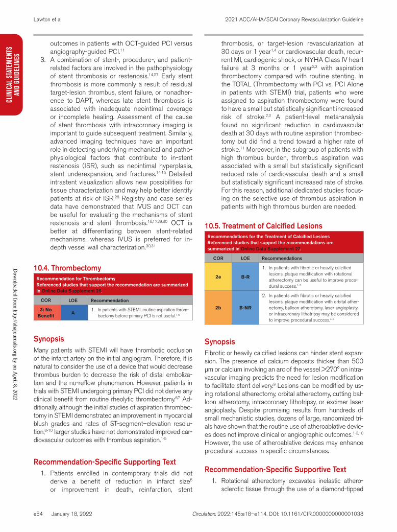
Lawton et al 2021 ACC/AHA/SCAI Coronary Revascularization Guideline
January 18, 2022 Circulation. 2022;145:e18–e114. DOI: 10.1161/CIR.0000000000001038e54
CLIN
ICAL
STA
TEM
ENTS
AN
D GU
IDEL
INES
outcomes in patients with OCT-guided PCI versus angiography-guided PCI.11
3. A combination of stent-, procedure-, and patient-related factors are involved in the pathophysiology of stent thrombosis or restenosis.14,27 Early stent thrombosis is more commonly a result of residual target-lesion thrombus, stent failure, or nonadher-ence to DAPT, whereas late stent thrombosis is associated with inadequate neointimal coverage or incomplete healing. Assessment of the cause of stent thrombosis with intracoronary imaging is important to guide subsequent treatment. Similarly, advanced imaging techniques have an important role in detecting underlying mechanical and patho-physiological factors that contribute to in-stent restenosis (ISR), such as neointimal hyperplasia, stent underexpansion, and fractures.14,15 Detailed intrastent visualization allows new possibilities for tissue characterization and may help better identify patients at risk of ISR.28 Registry and case series data have demonstrated that IVUS and OCT can be useful for evaluating the mechanisms of stent restenosis and stent thrombosis.16,17,29,30 OCT is better at differentiating between stent-related mechanisms, whereas IVUS is preferred for in-depth vessel wall characterization.30,31
10.4. ThrombectomyRecommendation for ThrombectomyReferenced studies that support the recommendation are summarized in Online Data Supplement 26.
COR LOE Recommendation
3: No Benefit
A1. In patients with STEMI, routine aspiration throm-
bectomy before primary PCI is not useful.1-5
SynopsisMany patients with STEMI will have thrombotic occlusion of the infarct artery on the initial angiogram. Therefore, it is natural to consider the use of a device that would decrease thrombus burden to decrease the risk of distal emboliza-tion and the no-reflow phenomenon. However, patients in trials with STEMI undergoing primary PCI did not derive any clinical benefit from routine rheolytic thrombectomy.6,7 Ad-ditionally, although the initial studies of aspiration thrombec-tomy in STEMI demonstrated an improvement in myocardial blush grades and rates of ST-segment–elevation resolu-tion,8-10 larger studies have not demonstrated improved car-diovascular outcomes with thrombus aspiration.1-5
Recommendation-Specific Supporting Text1. Patients enrolled in contemporary trials did not
derive a benefit of reduction in infarct size5 or improvement in death, reinfarction, stent
thrombosis, or target-lesion revascularization at 30 days or 1 year1,4 or cardiovascular death, recur-rent MI, cardiogenic shock, or NYHA Class IV heart failure at 3 months or 1 year2,3 with aspiration thrombectomy compared with routine stenting. In the TOTAL (Thrombectomy with PCI vs. PCI Alone in patients with STEMI) trial, patients who were assigned to aspiration thrombectomy were found to have a small but statistically significant increased risk of stroke.2,3 A patient-level meta-analysis found no significant reduction in cardiovascular death at 30 days with routine aspiration thrombec-tomy but did find a trend toward a higher rate of stroke.11 Moreover, in the subgroup of patients with high thrombus burden, thrombus aspiration was associated with a small but statistically significant reduced rate of cardiovascular death and a small but statistically significant increased rate of stroke. For this reason, additional dedicated studies focus-ing on the selective use of thrombus aspiration in patients with high thrombus burden are needed.
10.5. Treatment of Calcified LesionsRecommendations for the Treatment of Calcified LesionsReferenced studies that support the recommendations are summarized in Online Data Supplement 27.
COR LOE Recommendations
2a B-R
1. In patients with fibrotic or heavily calcified lesions, plaque modification with rotational atherectomy can be useful to improve proce-dural success.1-3
2b B-NR
2. In patients with fibrotic or heavily calcified lesions, plaque modification with orbital ather-ectomy, balloon atherotomy, laser angioplasty, or intracoronary lithotripsy may be considered to improve procedural success.4-8
SynopsisFibrotic or heavily calcified lesions can hinder stent expan-sion. The presence of calcium deposits thicker than 500 μm or calcium involving an arc of the vessel >270° on intra-vascular imaging predicts the need for lesion modification to facilitate stent delivery.9 Lesions can be modified by us-ing rotational atherectomy, orbital atherectomy, cutting bal-loon atherotomy, intracoronary lithotripsy, or excimer laser angioplasty. Despite promising results from hundreds of small mechanistic studies, dozens of large, randomized tri-als have shown that the routine use of atheroablative devic-es does not improve clinical or angiographic outcomes.1-3,10 However, the use of atheroablative devices may enhance procedural success in specific circumstances.
Recommendation-Specific Supportive Text1. Rotational atherectomy excavates inelastic athero-
sclerotic tissue through the use of a diamond-tipped
Dow
nloaded from http://ahajournals.org by on A
pril 8, 2022

Lawton et al 2021 ACC/AHA/SCAI Coronary Revascularization Guideline
Circulation. 2022;145:e18–e114. DOI: 10.1161/CIR.0000000000001038 January 18, 2022 e55
CLINICAL STATEMENTS
AND GUIDELINES
burr that rotates at high speeds. Although older stud-ies have shown that the use of rotational atherectomy is associated with increased rates of restenosis3 and increased late lumen loss,1 RCTs have demonstrated enhanced stent delivery and expansion in heav-ily calcified vessels with rotational atherectomy as compared with the use of conventional balloons1 or cutting or sculpting balloons.2 For this reason, despite the lack of data to support improved long-term out-comes with rotational atherectomy, rotational ather-ectomy remains an important tool in certain situations to properly “prepare” a lesion for stenting.
2. Orbital atherectomy has many features in common with rotational atherectomy and has similar clinical indications for use.4,5 Cutting balloons6 and scoring balloons7 section atheromatous plaque through a technique called balloon atherotomy, but their value may be limited to the technical advantage of slip-ping less often than conventional balloons in ostial lesions or lesions associated with ISR. Excimer laser coronary angioplasty uses a photo-acoustic mecha-nism11 that may facilitate the treatment of calcified lesions or nonexpandable stents.12,13 In certain lesion subsets, such as stent underexpansion that cannot be dilated with high-pressure balloon inflations, high-energy laser angioplasty can disrupt the calcific lesion beneath the stent struts and facilitate stent expan-sion.14 The evidence base for additional techniques to modify calcified or fibrotic lesions, including atherec-tomy, cutting balloons, or laser, are limited to registry studies and case series.10 Other potentially emerging modalities include intracoronary lithotripsy.8,15
10.6. Treatment of Saphenous Vein Graft (SVG) Disease (Previous CABG)
Recommendations for Treatment of SVG Disease (Previous CABG)Referenced studies that support the recommendations are summarized in Online Data Supplement 28.
COR LOE Recommendations
2a B-R
1. In select patients with previous CABG under-going PCI of a SVG, the use of an embolic protection device, when technically feasible, is reasonable to decrease the risk of distal embolization.1-3
2a B-NR
2. In patients with previous CABG, if PCI of a diseased native coronary artery is feasible, then it is reasonable to choose PCI of the native coronary artery over PCI of the severely diseased SVG.4-6
3: No Benefit
C-LD3. In patients with a chronic occlusion of a SVG,
percutaneous revascularization of the SVG should not be performed.7,8
SynopsisIn patients with previous CABG undergoing PCI of an SVG, the incidence of MACE is significantly higher than
those with native coronary artery PCI because of the higher risk of procedural complications, including the no-reflow phenomenon and periprocedural MI.6 Compared with native coronary arteries, atherosclerotic plaques in SVGs are more diffuse, with thinner, more friable fibrous caps that increase the risk of distal debris embolization during PCI.9 In several large prospective registries, pa-tients who underwent SVG PCI were more likely to have no-reflow,5 stent thrombosis, ischemia-driven target-ves-sel revascularization, increased overall adjusted MACE, and increased risk of death4,5,10 in long-term follow-up than were those who underwent non-SVG PCI.
Recommendation-Specific Supportive Text1. The term “embolic protection devices” refers to
the group of devices designed to prevent distal embolization. The SAFER (Saphenous vein graft Angioplasty Free of Emboli Randomized) trial com-paring the outcomes of SVG PCI with the use of an embolic protection device (Medtronic Guardwire, Minneapolis, MN) with conventional stenting of the SVG demonstrated a significant reduction in the primary endpoint of death, MI, emergency bypass, or target-lesion revascularization at 30 days with the GuardWire distal protection device.1 A subse-quent study comparing different embolic protection devices reported noninferiority of the FilterWire EX device (Boston Scientific, Marlborough, MA) to the GuardWire.2 In contemporary PCI, embolic protection devices are used in only 14% to 21% of patients,11,12 and only the filter-based devices are currently in use. Observational studies exploring the “real-world” benefits of embolic protection devices provide con-flicting findings, with 1 study showing no benefit of embolic protection devices and another showing sig-nificant harm when embolic protection devices are not used.11,12 A meta-analysis of 2 randomized stud-ies and 6 observational reports showed no benefit with the use of embolic protection devices, although selection biases and unmeasured confounders are important limitations of these observational studies.13
2. In patients with previous CABG who require PCI, two-thirds of these procedures are performed on the native artery instead of the bypass graft.4,5 Although there are no randomized studies com-paring PCI of a diseased native artery with PCI of an SVG, observational studies have shown that intervening in a native coronary artery instead of an SVG is associated with improved outcomes. In a large prospective registry, patients with prior CABG who underwent SVG PCI had higher rates of cardiac death, stent thrombosis, ischemia-driven target-vessel revascularization, and overall MACE at 2 years than did those who underwent PCI of the native vessel.10 The risk of MACE remained elevated
Dow
nloaded from http://ahajournals.org by on A
pril 8, 2022
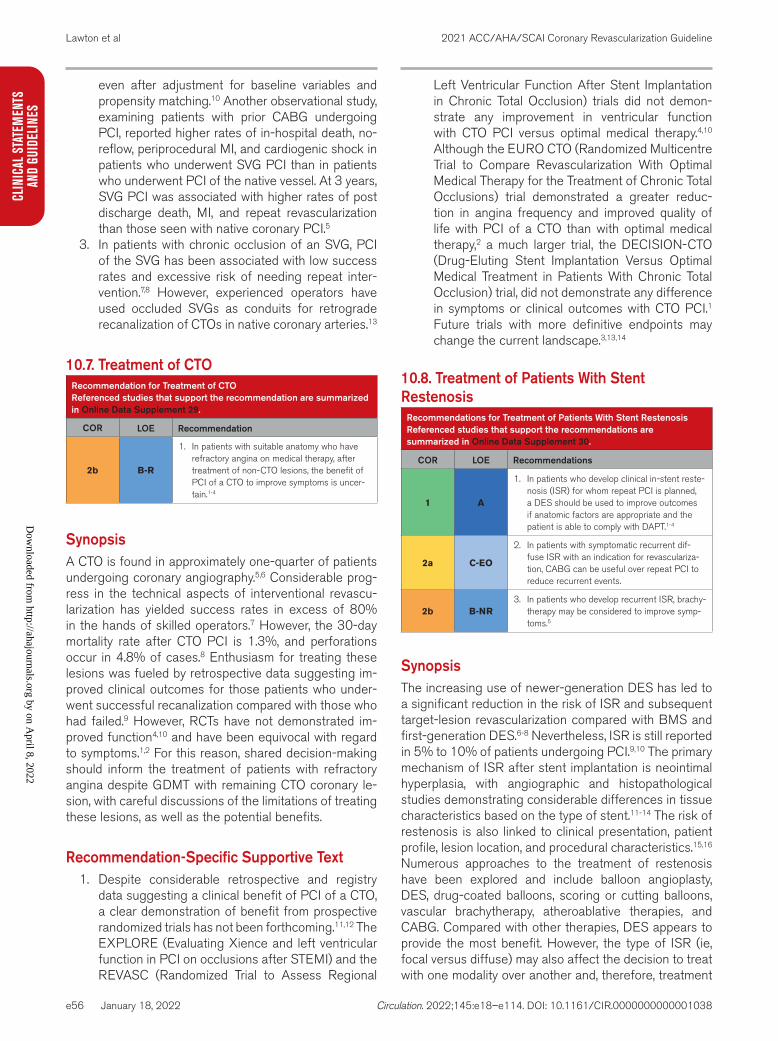
Lawton et al 2021 ACC/AHA/SCAI Coronary Revascularization Guideline
January 18, 2022 Circulation. 2022;145:e18–e114. DOI: 10.1161/CIR.0000000000001038e56
CLIN
ICAL
STA
TEM
ENTS
AN
D GU
IDEL
INES
even after adjustment for baseline variables and propensity matching.10 Another observational study, examining patients with prior CABG undergoing PCI, reported higher rates of in-hospital death, no-reflow, periprocedural MI, and cardiogenic shock in patients who underwent SVG PCI than in patients who underwent PCI of the native vessel. At 3 years, SVG PCI was associated with higher rates of post discharge death, MI, and repeat revascularization than those seen with native coronary PCI.5
3. In patients with chronic occlusion of an SVG, PCI of the SVG has been associated with low success rates and excessive risk of needing repeat inter-vention.7,8 However, experienced operators have used occluded SVGs as conduits for retrograde recanalization of CTOs in native coronary arteries.13
10.7. Treatment of CTORecommendation for Treatment of CTOReferenced studies that support the recommendation are summarized in Online Data Supplement 29.
COR LOE Recommendation
2b B-R
1. In patients with suitable anatomy who have refractory angina on medical therapy, after treatment of non-CTO lesions, the benefit of PCI of a CTO to improve symptoms is uncer-tain.1-4
SynopsisA CTO is found in approximately one-quarter of patients undergoing coronary angiography.5,6 Considerable prog-ress in the technical aspects of interventional revascu-larization has yielded success rates in excess of 80% in the hands of skilled operators.7 However, the 30-day mortality rate after CTO PCI is 1.3%, and perforations occur in 4.8% of cases.8 Enthusiasm for treating these lesions was fueled by retrospective data suggesting im-proved clinical outcomes for those patients who under-went successful recanalization compared with those who had failed.9 However, RCTs have not demonstrated im-proved function4,10 and have been equivocal with regard to symptoms.1,2 For this reason, shared decision-making should inform the treatment of patients with refractory angina despite GDMT with remaining CTO coronary le-sion, with careful discussions of the limitations of treating these lesions, as well as the potential benefits.
Recommendation-Specific Supportive Text1. Despite considerable retrospective and registry
data suggesting a clinical benefit of PCI of a CTO, a clear demonstration of benefit from prospective randomized trials has not been forthcoming.11,12 The EXPLORE (Evaluating Xience and left ventricular function in PCI on occlusions after STEMI) and the REVASC (Randomized Trial to Assess Regional
Left Ventricular Function After Stent Implantation in Chronic Total Occlusion) trials did not demon-strate any improvement in ventricular function with CTO PCI versus optimal medical therapy.4,10 Although the EURO CTO (Randomized Multicentre Trial to Compare Revascularization With Optimal Medical Therapy for the Treatment of Chronic Total Occlusions) trial demonstrated a greater reduc-tion in angina frequency and improved quality of life with PCI of a CTO than with optimal medical therapy,2 a much larger trial, the DECISION-CTO (Drug-Eluting Stent Implantation Versus Optimal Medical Treatment in Patients With Chronic Total Occlusion) trial, did not demonstrate any difference in symptoms or clinical outcomes with CTO PCI.1 Future trials with more definitive endpoints may change the current landscape.3,13,14
10.8. Treatment of Patients With Stent Restenosis
Recommendations for Treatment of Patients With Stent RestenosisReferenced studies that support the recommendations are summarized in Online Data Supplement 30.
COR LOE Recommendations
1 A
1. In patients who develop clinical in-stent reste-nosis (ISR) for whom repeat PCI is planned, a DES should be used to improve outcomes if anatomic factors are appropriate and the patient is able to comply with DAPT.1-4
2a C-EO
2. In patients with symptomatic recurrent dif-fuse ISR with an indication for revasculariza-tion, CABG can be useful over repeat PCI to reduce recurrent events.
2b B-NR3. In patients who develop recurrent ISR, brachy-
therapy may be considered to improve symp-toms.5
SynopsisThe increasing use of newer-generation DES has led to a significant reduction in the risk of ISR and subsequent target-lesion revascularization compared with BMS and first-generation DES.6-8 Nevertheless, ISR is still reported in 5% to 10% of patients undergoing PCI.9,10 The primary mechanism of ISR after stent implantation is neointimal hyperplasia, with angiographic and histopathological studies demonstrating considerable differences in tissue characteristics based on the type of stent.11-14 The risk of restenosis is also linked to clinical presentation, patient profile, lesion location, and procedural characteristics.15,16 Numerous approaches to the treatment of restenosis have been explored and include balloon angioplasty, DES, drug-coated balloons, scoring or cutting balloons, vascular brachytherapy, atheroablative therapies, and CABG. Compared with other therapies, DES appears to provide the most benefit. However, the type of ISR (ie, focal versus diffuse) may also affect the decision to treat with one modality over another and, therefore, treatment
Dow
nloaded from http://ahajournals.org by on A
pril 8, 2022
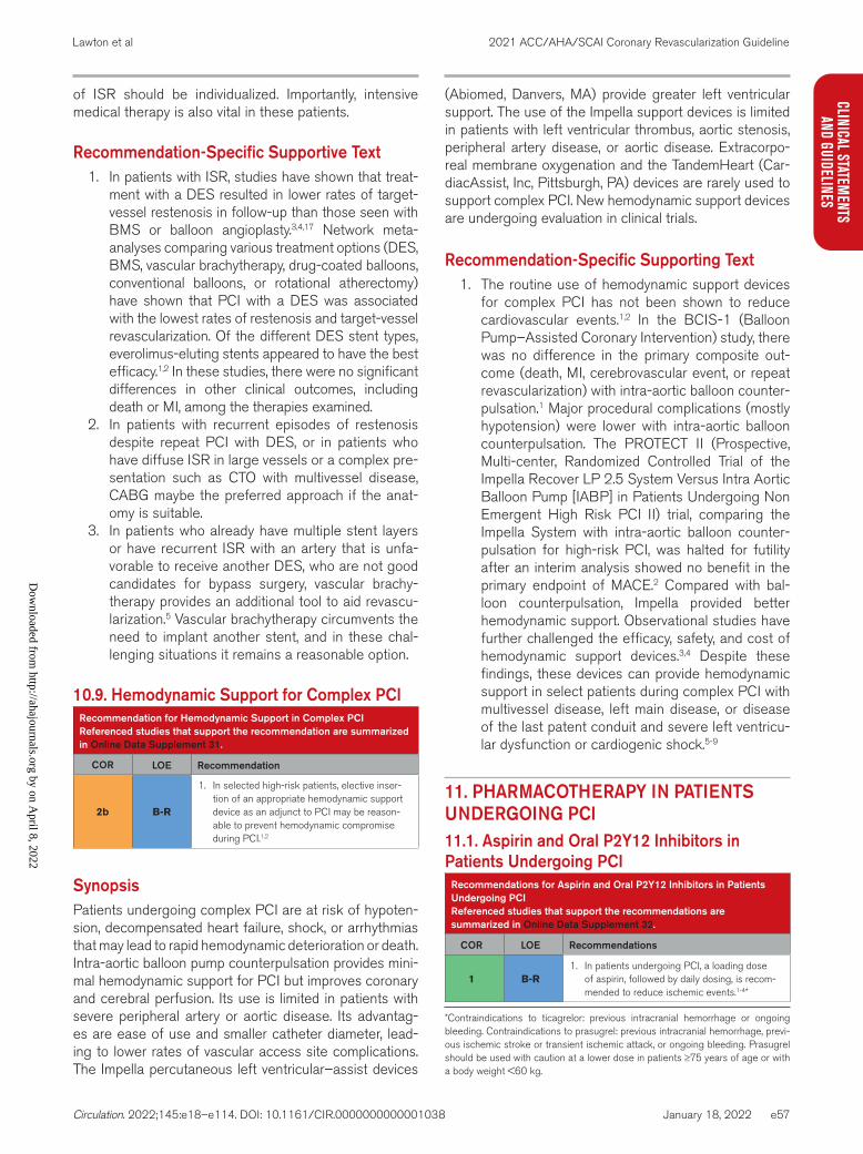
Lawton et al 2021 ACC/AHA/SCAI Coronary Revascularization Guideline
Circulation. 2022;145:e18–e114. DOI: 10.1161/CIR.0000000000001038 January 18, 2022 e57
CLINICAL STATEMENTS
AND GUIDELINES
of ISR should be individualized. Importantly, intensive medical therapy is also vital in these patients.
Recommendation-Specific Supportive Text1. In patients with ISR, studies have shown that treat-
ment with a DES resulted in lower rates of target-vessel restenosis in follow-up than those seen with BMS or balloon angioplasty.3,4,17 Network meta-analyses comparing various treatment options (DES, BMS, vascular brachytherapy, drug-coated balloons, conventional balloons, or rotational atherectomy) have shown that PCI with a DES was associated with the lowest rates of restenosis and target-vessel revascularization. Of the different DES stent types, everolimus-eluting stents appeared to have the best efficacy.1,2 In these studies, there were no significant differences in other clinical outcomes, including death or MI, among the therapies examined.
2. In patients with recurrent episodes of restenosis despite repeat PCI with DES, or in patients who have diffuse ISR in large vessels or a complex pre-sentation such as CTO with multivessel disease, CABG maybe the preferred approach if the anat-omy is suitable.
3. In patients who already have multiple stent layers or have recurrent ISR with an artery that is unfa-vorable to receive another DES, who are not good candidates for bypass surgery, vascular brachy-therapy provides an additional tool to aid revascu-larization.5 Vascular brachytherapy circumvents the need to implant another stent, and in these chal-lenging situations it remains a reasonable option.
10.9. Hemodynamic Support for Complex PCIRecommendation for Hemodynamic Support in Complex PCIReferenced studies that support the recommendation are summarized in Online Data Supplement 31.
COR LOE Recommendation
2b B-R
1. In selected high-risk patients, elective inser-tion of an appropriate hemodynamic support device as an adjunct to PCI may be reason-able to prevent hemodynamic compromise during PCI.1,2
SynopsisPatients undergoing complex PCI are at risk of hypoten-sion, decompensated heart failure, shock, or arrhythmias that may lead to rapid hemodynamic deterioration or death. Intra-aortic balloon pump counterpulsation provides mini-mal hemodynamic support for PCI but improves coronary and cerebral perfusion. Its use is limited in patients with severe peripheral artery or aortic disease. Its advantag-es are ease of use and smaller catheter diameter, lead-ing to lower rates of vascular access site complications. The Impella percutaneous left ventricular–assist devices
(Abiomed, Danvers, MA) provide greater left ventricular support. The use of the Impella support devices is limited in patients with left ventricular thrombus, aortic stenosis, peripheral artery disease, or aortic disease. Extracorpo-real membrane oxygenation and the TandemHeart (Car-diacAssist, Inc, Pittsburgh, PA) devices are rarely used to support complex PCI. New hemodynamic support devices are undergoing evaluation in clinical trials.
Recommendation-Specific Supporting Text1. The routine use of hemodynamic support devices
for complex PCI has not been shown to reduce cardiovascular events.1,2 In the BCIS-1 (Balloon Pump–Assisted Coronary Intervention) study, there was no difference in the primary composite out-come (death, MI, cerebrovascular event, or repeat revascularization) with intra-aortic balloon counter-pulsation.1 Major procedural complications (mostly hypotension) were lower with intra-aortic balloon counterpulsation. The PROTECT II (Prospective, Multi-center, Randomized Controlled Trial of the Impella Recover LP 2.5 System Versus Intra Aortic Balloon Pump [IABP] in Patients Undergoing Non Emergent High Risk PCI II) trial, comparing the Impella System with intra-aortic balloon counter-pulsation for high-risk PCI, was halted for futility after an interim analysis showed no benefit in the primary endpoint of MACE.2 Compared with bal-loon counterpulsation, Impella provided better hemodynamic support. Observational studies have further challenged the efficacy, safety, and cost of hemodynamic support devices.3,4 Despite these findings, these devices can provide hemodynamic support in select patients during complex PCI with multivessel disease, left main disease, or disease of the last patent conduit and severe left ventricu-lar dysfunction or cardiogenic shock.5-9
11. PHARMACOTHERAPY IN PATIENTS UNDERGOING PCI11.1. Aspirin and Oral P2Y12 Inhibitors in Patients Undergoing PCI
Recommendations for Aspirin and Oral P2Y12 Inhibitors in Patients Undergoing PCIReferenced studies that support the recommendations are summarized in Online Data Supplement 32.
COR LOE Recommendations
1 B-R1. In patients undergoing PCI, a loading dose
of aspirin, followed by daily dosing, is recom-mended to reduce ischemic events.1-4*
*Contraindications to ticagrelor: previous intracranial hemorrhage or ongoing bleeding. Contraindications to prasugrel: previous intracranial hemorrhage, previ-ous ischemic stroke or transient ischemic attack, or ongoing bleeding. Prasugrel should be used with caution at a lower dose in patients ≥75 years of age or with a body weight <60 kg.
Dow
nloaded from http://ahajournals.org by on A
pril 8, 2022
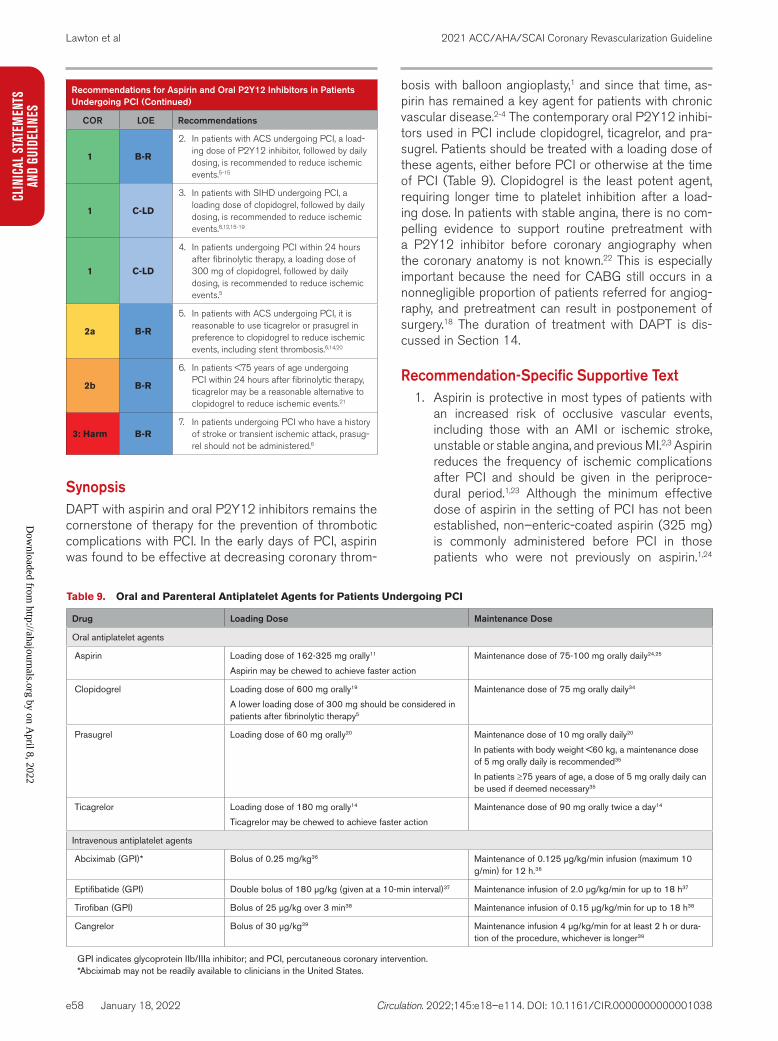
Lawton et al 2021 ACC/AHA/SCAI Coronary Revascularization Guideline
January 18, 2022 Circulation. 2022;145:e18–e114. DOI: 10.1161/CIR.0000000000001038e58
CLIN
ICAL
STA
TEM
ENTS
AN
D GU
IDEL
INES
1 B-R
2. In patients with ACS undergoing PCI, a load-ing dose of P2Y12 inhibitor, followed by daily dosing, is recommended to reduce ischemic events.5-15
1 C-LD
3. In patients with SIHD undergoing PCI, a loading dose of clopidogrel, followed by daily dosing, is recommended to reduce ischemic events.8,12,15-19
1 C-LD
4. In patients undergoing PCI within 24 hours after fibrinolytic therapy, a loading dose of 300 mg of clopidogrel, followed by daily dosing, is recommended to reduce ischemic events.5
2a B-R
5. In patients with ACS undergoing PCI, it is reasonable to use ticagrelor or prasugrel in preference to clopidogrel to reduce ischemic events, including stent thrombosis.6,14,20
2b B-R
6. In patients <75 years of age undergoing PCI within 24 hours after fibrinolytic therapy, ticagrelor may be a reasonable alternative to clopidogrel to reduce ischemic events.21
3: Harm B-R7. In patients undergoing PCI who have a history
of stroke or transient ischemic attack, prasug-rel should not be administered.6
SynopsisDAPT with aspirin and oral P2Y12 inhibitors remains the cornerstone of therapy for the prevention of thrombotic complications with PCI. In the early days of PCI, aspirin was found to be effective at decreasing coronary throm-
bosis with balloon angioplasty,1 and since that time, as-pirin has remained a key agent for patients with chronic vascular disease.2-4 The contemporary oral P2Y12 inhibi-tors used in PCI include clopidogrel, ticagrelor, and pra-sugrel. Patients should be treated with a loading dose of these agents, either before PCI or otherwise at the time of PCI (Table 9). Clopidogrel is the least potent agent, requiring longer time to platelet inhibition after a load-ing dose. In patients with stable angina, there is no com-pelling evidence to support routine pretreatment with a P2Y12 inhibitor before coronary angiography when the coronary anatomy is not known.22 This is especially important because the need for CABG still occurs in a nonnegligible proportion of patients referred for angiog-raphy, and pretreatment can result in postponement of surgery.18 The duration of treatment with DAPT is dis-cussed in Section 14.
Recommendation-Specific Supportive Text1. Aspirin is protective in most types of patients with
an increased risk of occlusive vascular events, including those with an AMI or ischemic stroke, unstable or stable angina, and previous MI.2,3 Aspirin reduces the frequency of ischemic complications after PCI and should be given in the periproce-dural period.1,23 Although the minimum effective dose of aspirin in the setting of PCI has not been established, non–enteric-coated aspirin (325 mg) is commonly administered before PCI in those patients who were not previously on aspirin.1,24
Recommendations for Aspirin and Oral P2Y12 Inhibitors in Patients Undergoing PCI (Continued)
COR LOE Recommendations
Table 9. Oral and Parenteral Antiplatelet Agents for Patients Undergoing PCI
Drug Loading Dose Maintenance Dose
Oral antiplatelet agents
Aspirin Loading dose of 162-325 mg orally11
Aspirin may be chewed to achieve faster action
Maintenance dose of 75-100 mg orally daily24,25
Clopidogrel Loading dose of 600 mg orally19
A lower loading dose of 300 mg should be considered in patients after fibrinolytic therapy5
Maintenance dose of 75 mg orally daily34
Prasugrel Loading dose of 60 mg orally20 Maintenance dose of 10 mg orally daily20
In patients with body weight <60 kg, a maintenance dose of 5 mg orally daily is recommended35
In patients ≥75 years of age, a dose of 5 mg orally daily can be used if deemed necessary35
Ticagrelor Loading dose of 180 mg orally14
Ticagrelor may be chewed to achieve faster action
Maintenance dose of 90 mg orally twice a day14
Intravenous antiplatelet agents
Abciximab (GPI)* Bolus of 0.25 mg/kg36 Maintenance of 0.125 μg/kg/min infusion (maximum 10 g/min) for 12 h.36
Eptifibatide (GPI) Double bolus of 180 μg/kg (given at a 10-min interval)37 Maintenance infusion of 2.0 μg/kg/min for up to 18 h37
Tirofiban (GPI) Bolus of 25 μg/kg over 3 min38 Maintenance infusion of 0.15 μg/kg/min for up to 18 h38
Cangrelor Bolus of 30 μg/kg39 Maintenance infusion 4 μg/kg/min for at least 2 h or dura-tion of the procedure, whichever is longer39
GPI indicates glycoprotein IIb/IIIa inhibitor; and PCI, percutaneous coronary intervention.*Abciximab may not be readily available to clinicians in the United States.
Dow
nloaded from http://ahajournals.org by on A
pril 8, 2022
Lawton et al 2021 ACC/AHA/SCAI Coronary Revascularization Guideline
Circulation. 2022;145:e18–e114. DOI: 10.1161/CIR.0000000000001038 January 18, 2022 e59
CLINICAL STATEMENTS
AND GUIDELINES
Observational data and retrospective analyses of RCTs have demonstrated that a lower dose of chronic daily aspirin (<100 mg) after PCI results in the best combination of safety and efficacy.24-26 On the basis of an analysis of aspirin dosing and outcomes in the PLATO (Trial to Assess The Study of Platelet Inhibition and Patient Outcomes) study, a low dose of aspirin (<100 mg) should be used in patients treated with ticagrelor. There are data to suggest that in the treatment of ACS, a chew-able aspirin formulation may be preferable to solid tablet aspirin.27
2. P2Y12 inhibitors are essential for treating patients undergoing PCI. Their use was first evaluated in studies exploring the optimal antithrombotic regi-mens after coronary stent implantation. In these earlier studies, ticlopidine was found to be superior to aspirin alone or the combination of aspirin and anticoagulant therapy10,11,13 in reducing ischemic events after coronary stent implantation. Because of unacceptable side effects of ticlopidine, clopi-dogrel was later used in place of ticlopidine, with clinical trials demonstrating similar efficacy but lower rates of drug discontinuation attributable to noncardiac events.16 With clopidogrel, the intro-duction of prasugrel and ticagrelor supported the use of a more potent P2Y12 inhibitor agent for PCI in ACS.6,14 A loading dose of a P2Y12 agent should be given to minimize the time to platelet inhibition. There are conflicting data on the ben-efits of pretreatment with a P2Y12 inhibitor before the anatomy is known, particularly in patients with NSTE-ACS.7,17,28-31 In contemporary times, with most patients with ACS undergoing early angiog-raphy, a strategy of loading with a P2Y12 inhibitor after the anatomy is known appears to offer similar benefit to preloading.31
3. The CREDO (Clopidogrel for the Reduction of Events During Observation) trial18 demonstrated a reduction in ischemic events, including the risk of death, MI, or stroke, with a loading dose of clopi-dogrel and treatment up to 9 months after elec-tive PCI. There was a trend toward a lower event rate when preloading with a 300-mg clopidogrel dose was given >3 hours before PCI. A 600-mg loading dose of clopidogrel is associated with a shorter time to platelet inhibition and therefore is the preferred dose. Ticagrelor and prasugrel have not been studied for long-term clinical outcomes in patients with SIHD undergoing PCI.
4. Patients with STEMI who were treated with fibrino-lytic therapy and referred for PCI are at increased bleeding and ischemic risk. Clopidogrel is the only P2Y12 inhibitor agent studied in patients immediately after the administration of fibrino-lytic therapy. In the CLARITY (Clopidogrel as
Adjunctive Reperfusion Therapy) trial, clopidogrel pretreatment in conjunction with fibrinolytic ther-apy resulted in a 46% reduction in the rate of car-diovascular death or recurrent MI or stroke at 30 days among patients referred for PCI.5 Major and minor bleeding was similar between the groups. In this study, patients randomized to clopidogrel were administered a 300-mg load during or immediately after fibrinolytic therapy, followed by 75 mg daily.5 In contemporary times, the loading dose of clopi-dogrel for patients undergoing PCI after fibrinolytic therapy should be individualized. A larger loading dose of 600 mg may be used for most patients, whereas the lower 300-mg loading dose is gener-ally reserved for older patients or those at higher risk of bleeding.
5. TRITON-TIMI-38 (Trial to Assess Improvement in Therapeutic Outcomes by Optimizing Platelet Inhibition with Prasugrel–Thrombolysis in Myocardial Infarction)6 and PLATO14 demonstrated that treatment with prasugrel (TRITON-TIMI-38) and ticagrelor (PLATO), compared with clopido-grel, reduced the rate of the composite endpoint of death from vascular causes, MI, or stroke. These agents were also associated with a lower rate of stent thrombosis. In the TRITON-TIMI 38 trial, non-CABG major bleeding was significantly higher with prasugrel.6 In PLATO, although there were no significant differences in the rates of study-defined bleeding events with ticagrelor, non-CABG major bleeding was significantly higher among patients treated with ticagrelor14 than among patients treated with clopidogrel.14 Because of the increased bleeding risk, these more potent agents should be used with caution in older patients. One study suggested that clopidogrel may be a reason-able alternative for older patients with ACS under-going PCI, with similar rates of ischemic events and less bleeding.32 The open-labeled design of this trial and the high rate of crossover limit the generalization of the study results. Further studies are needed to determine the ideal P2Y12 inhibitor for use in older patients with ACS undergoing PCI.
6. In patients with fibrinolytic-treated STEMI, ticagre-lor is associated with a greater inhibition of plate-let reactivity than that seen with clopidogrel.33 In PLATO, patients were excluded from enroll-ment if they were treated with fibrinolytic therapy within 24 hours of enrollment. Therefore, although PLATO supported the use of ticagrelor over clopi-dogrel in patients with STEMI treated with fibrino-lytic therapy, there were limited data on the safety of ticagrelor when given early after fibrinolytic therapy. The TREAT (Ticagrelor in Patients With ST Elevation Myocardial Infarction Treated With Pharmacological Thrombolysis) trial was designed
Dow
nloaded from http://ahajournals.org by on A
pril 8, 2022
Lawton et al 2021 ACC/AHA/SCAI Coronary Revascularization Guideline
January 18, 2022 Circulation. 2022;145:e18–e114. DOI: 10.1161/CIR.0000000000001038e60
CLIN
ICAL
STA
TEM
ENTS
AN
D GU
IDEL
INES
to examine the safety of ticagrelor in patients treated with fibrinolytic therapy for STEMI.21 In this study, ticagrelor was found to be noninferior to clopidogrel in rates of TIMI major bleeding, fatal bleeding, and intracranial bleeding.21
7. A more detailed analysis of the TRITON study that was designed to evaluate net clinical benefit (MACE events plus bleeding) with prasugrel dem-onstrated no net benefit of prasugrel compared with clopidogrel for patients with low body weight (<60 kg) or those ≥75 years of age and found net harm with prasugrel for patients with previous tran-sient ischemic attack or cerebrovascular accident.6 For this reason, prasugrel is contraindicated in patients with a history of transient ischemic attack or stroke. Caution is advised in the use of prasug-rel in patients weighing <60 kg or in patients ≥75 years of age.
11.2. Intravenous P2Y12 Inhibitors in Patients Undergoing PCI
Recommendation for Intravenous P2Y12 Inhibitors in Patients Undergoing PCIReferenced studies that support the recommendation are summarized in Online Data Supplement 33.
COR LOE Recommendation
2b B-R
1. In patients undergoing PCI who are P2Y12 inhibitor naïve, intravenous cangrelor may be reasonable to reduce periprocedural ischemic events.1-3
SynopsisCangrelor is a potent, direct, reversible, short-acting in-travenous P2Y12 inhibitor with rapid onset of platelet inhibition and restoration of platelet function within 1 hour of discontinuation. Cangrelor thus provides rapid, predictable, and profound inhibition of platelets. It can be efficacious in preventing stent thrombosis and may be considered in patients who have not been pretreated with a P2Y12 inhibitor, in patients whose absorption of oral medications may be inhibited, or in patients who are unable to take oral medications. Cangrelor has been investigated within the CHAMPION (Cangrelor versus Standard Therapy to Achieve Optimal Management of Platelet Inhibition) program and compared with a loading dose of clopidogrel given at the time of PCI in 3 large-scale clinical trials.1,2,4 There are no studies comparing cangrelor with a loading dose of ticagrelor or prasugrel given at the time of PCI.
Recommendation-Specific Supporting Text1. The CHAMPION PLATFORM and the CHAMPION
PCI trials did not show a reduction in the pri-mary outcome (ie, death, MI, or ischemia-driven
revascularization at 48 hours) with cangrelor. However, in CHAMPION PLATFORM, cangrelor resulted in lower rates of the prespecified second-ary outcomes of stent thrombosis and death.1 In the CHAMPION PHOENIX trial, the primary endpoint, which included death, MI, ischemia-driven revas-cularization, or stent thrombosis, was significantly reduced with cangrelor.2 This was driven mainly by a reduction in periprocedural MI and intrapro-cedural stent thrombosis. A pooled patient-level meta-analysis of the CHAMPION trials supported these findings, demonstrating a lower rate of the composite endpoint of death, MI, ischemia-driven revascularization, or stent thrombosis at 48 hours with cangrelor than with clopidogrel.3 Additionally, cangrelor was associated with a 41% reduction in stent thrombosis. Although major bleeding was similar between the groups, minor bleeding was more frequent in the cangrelor group.3
11.3. Intravenous Glycoprotein IIb/IIIa Inhibitors in Patients Undergoing PCI
Recommendations for Intravenous Glycoprotein IIb/IIIa Inhibitors in Patients Undergoing PCIReferenced studies that support the recommendations are summarized in Online Data Supplement 34.
COR LOE Recommendations
2a C-LD
1. In patients with ACS undergoing PCI with large thrombus burden, no-reflow, or slow flow, intravenous glycoprotein IIb/IIIa inhibitor agents are reasonable to improve procedural success.1,2
3: No Benefit
B-R2. In patients with SIHD undergoing PCI, the
routine use of an intravenous glycoprotein IIb/IIIa inhibitor agent is not recommended.3-5
SynopsisGlycoprotein IIb/IIIa receptor inhibitors are direct-acting antiplatelet agents targeting the glycoprotein IIb/IIIa platelet receptor. Many of the trials of glycoprotein IIb/IIIa inhibitors in the setting of ACS were conducted in an era before the use of potent P2Y12 inhibitors or before rou-tine stenting.2,6 Additionally, in the earlier trials, the time from presentation to coronary angiography was often prolonged. In the contemporary era of shorter revascular-ization times and use of potent DAPT, the benefit of gly-coprotein IIb/IIIa receptor inhibitor agents is diminished.2,7
Recommendation-Specific Supporting Text1. In trials of patients with ACS, glycoprotein IIb/
IIIa receptor inhibitors have not been associated with improved clinical outcomes and may increase bleeding complications.7,8 Because the addition of glycoprotein IIb/IIIa receptor inhibitors can decrease thrombus burden by further inhibiting
Dow
nloaded from http://ahajournals.org by on A
pril 8, 2022

Lawton et al 2021 ACC/AHA/SCAI Coronary Revascularization Guideline
Circulation. 2022;145:e18–e114. DOI: 10.1161/CIR.0000000000001038 January 18, 2022 e61
CLINICAL STATEMENTS
AND GUIDELINES
platelet aggregation,9 the use of glycoprotein IIb/IIIa receptor inhibitors in the era of more potent antiplatelet agents is generally reserved for patients with a large thrombus burden or no-reflow or slow flow that is believed to be attributable to distal embolization of thrombus.
2. In patients with SIHD who are undergoing PCI, the use of glycoprotein IIb/IIIa receptor inhibi-tors in addition to a clopidogrel load does not reduce ischemic events.3,4 Patients enrolled in the ISAR-REACT (iNtracoronary Stenting and Antithrombotic Regimen: Rapid Early Action for Coronary Treatment) trial who were random-ized to pretreatment with abciximab and a 600-mg loading dose of clopidogrel had outcomes similar to those of patients receiving clopidogrel alone.3 Major bleeding was not significantly dif-ferent between the 2 groups, although the rate of severe thrombocytopenia was significantly higher in the abciximab group.3 A subgroup analy-sis of the ESPRIT (Enhance Suppression of the Platelet IIB/IIIA receptor with Integrilin Therapy) trial showed no benefit in the primary endpoint of death, MI, urgent target-vessel revascularization, and thrombotic bailout with glycoprotein IIb/IIIa inhibitor therapy with eptifibatide at 48 hours in the group of patients undergoing PCI for stable angina.5 Six-month rates of death or MI were also not significantly different in this subgroup of patients.4
11.4. Heparin, Low-Molecular-Weight Heparin, and Bivalirudin in Patients Undergoing PCI
Recommendations for Heparin, Low-Molecular-Weight Heparin, and Bivalirudin in Patients Undergoing PCIReferenced studies that support the recommendations are summarized in Online Data Supplement 35.
COR LOE Recommendations
1 C-EO1. In patients undergoing PCI, administration of
intravenous unfractionated heparin (UFH) is useful to reduce ischemic events.
1 C-LD
2. In patients with heparin-induced thrombocyto-penia undergoing PCI, bivalirudin or argatro-ban should be used to replace UFH to avoid thrombotic complications.1,2
2b A3. In patients undergoing PCI, bivalirudin may
be a reasonable alternative to UFH to reduce bleeding.3-12
2b B-R
4. In patients treated with upstream subcutane-ous enoxaparin for unstable angina or NSTE-ACS, the use of intravenous enoxaparin may be considered at the time of PCI to reduce ischemic events.13-17
3: Harm B-R
5. In patients on therapeutic subcutaneous enoxaparin, in whom the last dose was admin-istered within 12 hours of PCI, UFH should not be used for PCI and may increase bleed-ing.14,18,19
SynopsisAntithrombotic therapy is a mainstay of treatment in patients undergoing PCI. Currently, there are 3 anti-thrombotic agents that have been studied in PCI. These are UFH, bivalirudin, and enoxaparin. Fondaparinux is no longer recommended as the only anticoagulant in PCI because of a higher incidence of guiding-catheter thrombosis.20,21 Consideration of the patient’s clinical presentation (eg, stable disease, NSTE-ACS, or STEMI) and bleeding risk profile22 may influence selection of the optimal anticoagulant type. Suggested dosing regimens of parenteral agents are shown in Table 10.
Recommendation-Specific Supportive Text1. As the only anticoagulant available for many years,
UFH has been the standard of care by default and the primary comparator for novel agents in RCTs.23 Dosing recommendations were established from early studies that demonstrated a relationship between activated clotting times and ischemic complications,23-26 but it is unclear that these analy-ses translate to the modern coronary stent era.27-31 Thus, the exact use of dosing based on activated clotting times in current practice is uncertain. The routine use of full-dose anticoagulation therapy after PCI is no longer indicated.
2. Heparin-induced thrombocytopenia occurs when the heparin molecule binds to platelet factor 4. Argatroban and bivalirudin are direct thrombin inhibitors and do not bind to platelet factor 4. Because of their different mechanism of action, argatroban1 and bivalirudin2 are acceptable alter-native anticoagulants for use in patients with hep-arin-induced thrombocytopenia.
3. RCTs comparing bivalirudin and heparin have reported no difference in ischemic endpoints; however, less bleeding was reported with bivali-rudin.3-7,9,29,32-37 Although the reduction in bleeding complications with the use of bivalirudin was seen in most trials, in real-world practice, this benefit may be less pronounced with routine use of radial artery intervention and low rates of glycoprotein IIb/IIIa inhibitor use. Meta-analyses of clinical trial data support these findings, highlighting that the magnitude of the lower bleeding risk with bivali-rudin in various trials depended on variable inclu-sion of a glycoprotein IIb/IIIa inhibitor.10,11 The VALIDATE-SWEDEHEART (Bivalirudin vs Heparin in NSTEMI and STEMI in Patients on Modern Antiplatelet Therapy in SWEDEHEART) study38 examined a prolonged bivalirudin infusion versus UFH. Patients were treated with the more potent P2Y12 inhibitors, 90% had radial artery access, and there was a low rate of glycoprotein IIb/IIIa
Dow
nloaded from http://ahajournals.org by on A
pril 8, 2022

Lawton et al 2021 ACC/AHA/SCAI Coronary Revascularization Guideline
e62
CLIN
ICAL
STA
TEM
ENTS
AN
D GU
IDEL
INES
January 18, 2022 Circulation. 2022;145:e18–e114. DOI: 10.1161/CIR.0000000000001038
inhibitor use. Compared with UFH, bivalirudin was not associated with improved rates of MACE, major bleeding, or stent thrombosis at 6 months.
4. Enoxaparin is considered a safe alternative to UFH.15,16 In the SYNERGY (Superior Yield of the New Strategy of Enoxaparin, Revascularization and Glycoprotein IIb/IIIa Inhibitors)14 and AtoZ (Aggrastat to Zocor)13 trials of patients with NSTE-ACS, enoxaparin was shown to be noninferior to UFH, with no difference in rates of death, MI, or major bleeding. In primary PCI, the ATOLL (Acute STEMI Treated With Primary Angioplasty and Intravenous Lovenox or UFH to Lower Ischemic and Bleeding Events)15 trial compared intravenous enoxaparin and UFH and showed a reduction in the main secondary endpoint (composite of death, recurrent ACS, and urgent revascularization) in the enoxaparin arm, without more bleeding. These find-ings have been supported by a subsequent large meta-analysis that included patients undergoing PCI for STEMI and NSTE-ACS, which showed a reduction in rates of all-cause death and bleed-ing.16 Almost all patients undergoing elective PCI who are administered enoxaparin (0.5 mg/kg IV) will have a peak anti-Xa level >0.5 IU/mL.31,39
5. In clinical trials, patients who were given upstream enoxaparin and then switched to UFH had more complications, which are attributed at least in part to stacking both medications at the time of PCI, even when heparin is administered as long as 10 hours after the last dose of enoxaparin.18,19 While these trials were performed prior to the wide-spread use of radial artery access, it is preferable to avoid the administration of UFH in patients that have received enoxaparin in the previous 12 hours to reduce risk of bleeding.
12. GENERAL PROCEDURAL ISSUES FOR CABG12.1. Perioperative Considerations in Patients Undergoing CABG
Recommendation for Perioperative Considerations in Patients Undergoing CABGReferenced studies that support the recommendation are summarized in Online Data Supplement 36.
COR LOE Recommendation
1 B-NR
1. For patients undergoing CABG, establishment of multidisciplinary, evidence-based periopera-tive management programs is recommended to optimize analgesia, minimize opioid expo-sure, prevent complications and to reduce time to extubation, length of stay, and health care costs.1-3
SynopsisPrevious recommendations with regard to perioperative management assessed the role of certain monitoring modalities to guide intraoperative and postoperative de-cision-making and also emphasized the use of fast-track cardiac anesthesia, which uses short-acting anesthetic agents to improve outcomes after CABG.4 More recently, cardiac surgical service lines have been encouraged to expand the scope of these efforts to develop multidisci-plinary perioperative programs that incorporate bundled evidence-based surgical, anesthetic, and nursing interven-tions, including the targeted use of appropriate monitoring modalities, to optimize care and improve patient recovery.
Recommendation-Specific Supporting Text1. Fast-track cardiac anesthesia was extensively
studied in the early 2000s and was found to reduce
Table 10. Anticoagulant Dosing During PCI*
Dosing of Parenteral Anticoagulants During PCI
Drug Patient Has Received Previous Anticoagulant TherapyPatient Has Not Received Previous Anticoagulant Therapy
UFH Additional UFH as needed (eg, 2000–5000 U) to achieve an ACT of 250-300 s*
70–100 U/kg initial bolus to achieve target ACT of 250–300 s*
Enoxaparin For previous treatment with enoxaparin, if the last SC dose was administered 8–12 h earlier or if only 1 SC dose of enoxaparin has been administered, an IV dose of 0.3 mg/kg of enoxaparin should be given43-45
If the last SC dose was administered within the previous 8 h, no additional enoxaparin should be given
0.5–0.75 mg/kg IV bolus
Bivalirudin For patients who have received UFH, repeat ACT
If ACT is not in therapeutic range, then give 0.75 mg/kg IV bolus, then 1.75 mg/kg/h IV infusion
0.75 mg/kg bolus, 1.75 mg/kg/h IV infusion
Argatroban 200 μg/kg IV bolus, then 15 μg/kg/min IV infusion 350 μg/kg, then 15 μg/kg/min IV infusion
ACS indicates acute coronary syndrome; ACT, activated clotting time; CTO, chronic total occlusion; PCI, percutaneous coronary intervention; and UFH, unfraction-ated heparin.
*Target ACTs for UFH dosing shown for HemoTec (GmbH, Switzerland) or I-Stat (Abbott) device. For Hemochron ACT (Werfen) devices, ACT goals are 50 s higher. In the case of CTO or ACS, consider higher target ACT. If IV glycoprotein IIb/IIIa receptor inhibitor is planned, target ACT 200–250 s.26,27,31,40-42
Dow
nloaded from http://ahajournals.org by on A
pril 8, 2022

Lawton et al 2021 ACC/AHA/SCAI Coronary Revascularization Guideline
Circulation. 2022;145:e18–e114. DOI: 10.1161/CIR.0000000000001038 January 18, 2022 e63
CLINICAL STATEMENTS
AND GUIDELINES
opioid use and hasten extubation5,6; however, it did not uniformly reduce complications or length of stay in patients after CABG.7,8 More recently, cardiac surgical service lines have expanded on fast-track anesthetic agents and implemented enhanced recovery programs, which use more extensive phase-specific perioperative interven-tions. Such programs have been shown to prevent early postoperative complications, minimize expo-sure to opioid-based analgesia, and reduce time to extubation, time in the intensive care unit, and hospital length of stay after CABG.1-3 Components of such programs may include liberation of “noth-ing by mouth” status, bundled surgical site infec-tion prevention, multimodal nonopioid analgesics, protocolized short-acting anesthetics, targeted-organ perfusion strategies, and early postopera-tive ambulation. Assessment of enhanced recovery programs has been generally limited to moderate-sized observational studies, and additional research is required to determine the necessary features and implementation strategies. Providers may con-sider the use of specific anesthetic and monitoring modalities outlined in Table 11.
12.2. Bypass Conduits in Patients Undergoing CABG
Recommendations for Bypass Conduits in Patients Undergoing CABGReferenced studies that support the recommendations are summarized in Online Data Supplement 37.
COR LOE Recommendations
1 B-R
1. In patients undergoing isolated CABG, the use of a radial artery is recommended in prefer-ence to a saphenous vein conduit to graft the second most important, significantly stenosed, non–LAD vessel to improve long-term cardiac outcomes.1-3
1 B-NR
2. In patients undergoing CABG, an IMA, prefer-ably the left, should be used to bypass the LAD when bypass of the LAD is indicated to improve survival and reduce recurrent isch-emic events.4-9
2a B-NR
3. In patients undergoing CABG, bilateral IMA (BIMA) grafting by experienced operators can be beneficial in appropriate patients to improve long-term cardiac outcomes.3,10-12
SynopsisIn the choice of conduits for CABG, both clinical and technical factors (eg, life expectancy, presence of dia-betes, presence of CKD, degree of target stenosis) are considered (Table 12). Decades of data have supported the use of the LIMA to graft the LAD to prolong survival. These data are from observational studies and, for the most part, were derived before the introduction of cur-rent optimal medical therapy. The LIMA is preferable un-less specific contraindications are present. The right IMA
Table 11. Perioperative Anesthetic and Monitoring Consid-erations for CABG
Anesthetic considerations
Perioperative analgesia Nonopioid medications (eg, acetaminophen, ketamine, dexmedetomidine) and/or regional techniques (eg, truncal nerve blocks), par-ticularly as part of a multimodal analgesic approach, have been shown to reduce periop-erative opioid use in cardiac surgery.1-16
Maintenance anesthesia Although volatile (versus intravenous) anes-thesia may facilitate earlier extubation,2,6-8,12,14 recent evidence suggests that the choice of maintenance anesthetic likely does not impact mortality rate after cardiac surgery.17-21
Mechanical ventilation An intraoperative lung-protective ventilation strategy (ie, tidal volume of 6–8 mL/kg pre-dicted body weight + positive end-expiratory pressure) has been shown to improve pul-monary mechanics and reduce postoperative pulmonary complications.21-25
Goal-directed therapy Goal-directed therapy, which creates proto-cols for the use of fluids and vasopressors to target specific hemodynamic goals, has yielded inconsistent results and requires ad-ditional investigation to determine its use in cardiac surgery.26,27
TEE
CABG + valve procedures
Intraoperative TEE aids in the real-time as-sessment of heart valve function and pathol-ogy in those undergoing combination CABG and valve surgery.28-30
Isolated CABG procedures
The use of intraoperative TEE in isolated CABG is less established but has been shown to aid in surgical and anesthetic deci-sion-making as a tool for real-time assessment of hemodynamic status, regional wall motion, ventricular function, valve anatomy, and dia-stolic function.28-35
Pulmonary artery catheters
High-risk surgery Highly selective use of pulmonary artery cath-eters for high-risk patients (ie, older, with con-gestive heart failure, pulmonary hypertension, or previous multiple valve procedures) may be safe and may potentially aid in the surveillance and treatment of hemodynamic instability.36-38
Low-risk surgery The use of pulmonary artery catheters in low-risk or clinically stable patients is discouraged because the practice is associated with in-creased interventions that incur greater health care expense without associated improvement in morbidity or mortality rates.38-40
CNS monitoring
Cerebral oxygen saturation
Intraoperative monitoring of cerebral oxygen saturation (ie, near-infrared spectroscopy) to detect cerebral hypoperfusion has been shown to guide anesthetic decision-making and may prevent postoperative neurocognitive dysfunction.41-47
Processed electroen-cephalogram
Routine use of intraoperative monitoring of processed electroencephalogram (ie, bispec-tral index) has yielded inconsistent results with respect to the prevention of recall, determina-tion of depth of anesthesia, or improvement in rate of recovery after cardiac surgery.48-51
CABG indicates coronary artery bypass graft; CNS, central nervous system; and TEE, transesophageal echocardiography.
Dow
nloaded from http://ahajournals.org by on A
pril 8, 2022
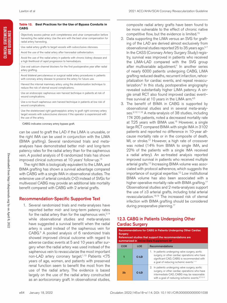
Lawton et al 2021 ACC/AHA/SCAI Coronary Revascularization Guideline
January 18, 2022 Circulation. 2022;145:e18–e114. DOI: 10.1161/CIR.0000000000001038e64
CLIN
ICAL
STA
TEM
ENTS
AN
D GU
IDEL
INES
can be used to graft the LAD if the LIMA is unusable, or the right IMA can be used in conjunction with the LIMA (BIMA grafting). Several randomized trials and meta-analyses have demonstrated better mid- and long-term patency rates for the radial artery than for the saphenous vein. A pooled analysis of 6 randomized trials has shown improved clinical outcomes at 10 years’ follow-up.13
The right IMA is biologically equivalent to the LIMA, and BIMA grafting has shown a survival advantage compared with CABG with a single IMA in observational studies. The extensive use of arterial conduits (>2) instead of SVGs for multivessel CABG may provide an additional late mortality benefit compared with CABG with 2 arterial grafts.
Recommendation-Specific Supportive Text1. Several randomized trials and meta-analyses have
reported better mid- and long-term patency rates for the radial artery than for the saphenous vein,2,14 while observational studies and meta-analyses have suggested a survival benefit when the radial artery is used instead of the saphenous vein for CABG.3 A pooled analysis of 6 randomized trials showed improved clinical outcome with regard to adverse cardiac events at 5 and 10 years after sur-gery when the radial artery was used instead of the saphenous vein to revascularize the most important non-LAD artery coronary target.1,13 Patients <75 years of age, women, and patients with preserved renal function seem to benefit the most from the use of the radial artery. The evidence is based largely on the use of the radial artery constructed as an aortocoronary graft. In observational studies,
composite radial artery grafts have been found to be more vulnerable to the effect of chronic native competitive flow, but the evidence is limited.15
2. Data supporting the LIMA versus an SVG for graft-ing of the LAD are derived almost exclusively from observational studies reported 25 to 35 years ago.5-7 In the CASS (Coronary Artery Surgery Study) regis-try, survival was improved in patients who received the LIMA-LAD compared with the SVG group after multivariable adjustment.7 In another series of nearly 6000 patients undergoing CABG, LIMA grafting reduced deaths, recurrent infarction, rehos-pitalization for cardiac events, and repeat revascu-larization.6 In this study, postoperative angiography revealed substantially higher LIMA patency. A sin-gle small RCT also found improved cardiac event–free survival at 10 years in the LIMA arm.4,5
3. The benefit of BIMA in CABG is supported by observational studies and in several meta-analy-ses.3,10,11,16 A meta-analysis of 38 studies, including 174 205 patients, noted a decreased mortality rate at 7.25 years with BIMA use.16 However, a single large RCT compared BIMA with single IMA in 3102 patients and reported no difference in 10-year all-cause mortality rate or in the composite of death, MI, or stroke.12 However, a high rate of crossover was noted (14% from BIMA to single IMA, and 22% of the patients with a single IMA received a radial artery). An as-treated analysis reported improved survival in patients who received multiple arterial grafts.12 Increasing BIMA volume was asso-ciated with protocol adherence, which suggests the importance of surgical expertise.12 Low institutional BIMA volume has also been associated with a higher operative mortality rate with BIMA grafting.17 Observational studies and 2 meta-analyses support the use of ≥3 arterial grafts, including total arterial revascularization.18,19 The increased risk of sternal infection with BIMA grafting should be considered during preoperative planning.17
12.3. CABG in Patients Undergoing Other Cardiac Surgery
Recommendations for CABG in Patients Undergoing Other Cardiac SurgeryReferenced studies that support the recommendations are summarized in Online Data Supplement 38.
COR LOE Recommendations
1 C-LD
1. In patients undergoing valve surgery, aortic surgery, or other cardiac operations who have significant CAD, CABG is recommended with a goal of reducing ischemic events.1-11
2b C-LD
2. In patients undergoing valve surgery, aortic surgery, or other cardiac operations who have intermediate CAD, CABG may be reasonable with a goal of reducing ischemic events.5,7,10,12
Table 12. Best Practices for the Use of Bypass Conduits in CABG
Objectively assess palmar arch completeness and ulnar compensation before harvesting the radial artery. Use the arm with the best ulnar compensation for radial artery harvesting.
Use radial artery grafts to target vessels with subocclusive stenoses.
Avoid the use of the radial artery after transradial catheterization.
Avoid the use of the radial artery in patients with chronic kidney disease and a high likelihood of rapid progression to hemodialysis.
Use oral calcium channel blockers for the first postoperative year after radial artery grafting.
Avoid bilateral percutaneous or surgical radial artery procedures in patients with coronary artery disease to preserve the artery for future use.
Harvest the internal mammary artery using the skeletonization technique to reduce the risk of sternal wound complications.
Use an endoscopic saphenous vein harvest technique in patients at risk of wound complications.
Use a no-touch saphenous vein harvest technique in patients at low risk of wound complications.
Use the skeletonized right gastroepiploic artery to graft right coronary artery target vessels with subocclusive stenosis if the operator is experienced with the use of the artery.
CABG indicates coronary artery bypass graft.
Dow
nloaded from http://ahajournals.org by on A
pril 8, 2022

Lawton et al 2021 ACC/AHA/SCAI Coronary Revascularization Guideline
Circulation. 2022;145:e18–e114. DOI: 10.1161/CIR.0000000000001038 January 18, 2022 e65
CLINICAL STATEMENTS
AND GUIDELINES
SynopsisThe decision to add CABG to another planned cardiac surgery in patients with significant CAD is multifactorial. Considerations include but are not limited to comorbidi-ties, technical feasibility of CABG, extent of the jeopar-dized myocardium, availability of conduit, left ventricular ejection fraction, and the additional time needed to con-struct the coronary bypass while on cardiopulmonary by-pass. A multidisciplinary discussion with a Heart Team can help weigh the risks and benefits of adding CABG to the index cardiac operation. Age does not appear to be a prohibitive risk factor for the addition of CABG to other cardiac surgery in patients between the ages of 75 and 84 years,1,2,4 but risk increases in patients ≥85 years of age.12 The available evidence supporting CABG at the time of cardiac surgery performed for another primary indication (ie, valve, aortic, or other cardiac surgery) is limited and complicated because many studies include patients with CAD defined as at least 1 vessel with ste-nosis ≥50%, thus including both intermediate and sig-nificant CAD. The knowledge that incomplete revascu-larization is associated with reduced long-term survival rates after surgery compared with patients who receive complete revascularization may inform decisions about adding CABG to other cardiac surgery.5,10 Additionally, it has become standard practice to bypass significant coronary artery stenoses in patients undergoing other cardiac surgery.
Recommendation-Specific Supporting Text1. Several observational studies and meta-analyses
compared clinical outcomes in patients undergoing aortic valve replacement (AVR) with and without significant CAD (≥70% stenosis of any major epi-cardial coronary vessel, including side branches, or ≥50% stenosis of the left main).6-9,12 Patients who underwent AVR with concomitant CABG demon-strated long-term survival and health-related qual-ity of life similar to that of patients without CAD. Concomitant CABG may increase the risk of peri-operative morbidity and mortality compared with isolated AVR.2-4 In patients with significant CAD who underwent isolated AVR or AVR plus CABG, concomitant CABG was associated with a reduced late all-cause mortality rate (hazard ratio, 0.62; 95% CI, 0.49–0.79; P<0.001).10 A large study of 6151 patients found that patients with extensive CAD (>50% left main stenosis or ≥3 diseased vessels) undergoing AVR with CABG had more comorbidities and had more perioperative mor-bidity, but not mortality, than did patients with less extensive CAD undergoing AVR with CABG.12 A large study that propensity-matched patients undergoing AVR to patients undergoing AVR with
CABG demonstrated no differences in morbidity or mortality between groups, which suggests that sur-vival is dominated largely by patient comorbidities.7 Studies focusing on CABG as a secondary pro-cedure during other cardiac operations, including mitral valve, tricuspid valve, aortic, and pericardial surgery, are limited.
2. Limited data are available comparing CABG for intermediate CAD (stenosis, 40%–69%) in patients undergoing other cardiac surgery. Observational studies comparing patients undergoing AVR with and without CABG for intermediate CAD suggest that the addition of CABG may reduce ischemic events.5,7,10,12 Patients with intermediate CAD may benefit from physiological testing with iFR or FFR to guide decision-making.
12.4. Use of Epiaortic Ultrasound in Patients Undergoing CABG
Recommendation for Use of Epiaortic Ultrasound in Patients Undergoing CABGReferenced studies that support the recommendation are summarized in Online Data Supplement 39.
COR LOE Recommendation
2a B-NR
1. In patients undergoing CABG, the routine use of epiaortic ultrasound scanning can be useful to evaluate the presence, location, and severity of plaque in the ascending aorta to reduce the incidence of atheroembolic complications.1-10
SynopsisAtherosclerotic disease of the aorta is common in pa-tients who undergo CABG surgery, with a reported prevalence that varies between 19% and 90%, depend-ing on the patient population and modality of examina-tion.2,11-16 There has been a clear association between aortic atherosclerosis and stroke,17 especially in patients undergoing CABG.11,12,18-21 Epiaortic ultrasound has been demonstrated to be far superior to either surgical digital palpation or transesophageal echocardiography for de-fining the presence and extent of disease and has come to be recognized as the “gold standard” for the detection of aortic atherosclerosis.13-15,22-24 There has been consid-erable variability in the extent to which epiaortic ultra-sound has changed operative strategy, varying from 4% to 22%.1,2,6,9,16
Recommendation-Specific Supporting Text1. The ability of routine epiaortic ultrasound to
decrease stroke risk in patients undergoing CABG is unclear. One small prospective trial failed to meet a prespecified 50% difference in neurocognitive testing.4 Although it is not uniformly reported,3
Dow
nloaded from http://ahajournals.org by on A
pril 8, 2022
Lawton et al 2021 ACC/AHA/SCAI Coronary Revascularization Guideline
January 18, 2022 Circulation. 2022;145:e18–e114. DOI: 10.1161/CIR.0000000000001038e66
CLIN
ICAL
STA
TEM
ENTS
AN
D GU
IDEL
INES
several large retrospective and registry stud-ies,1,2,6,7,9,10 as well as most single-center prospec-tive studies, found an association with reduced risk of stroke.3-5,8 Procedural risk, extra time required, and cost are minimal. The use of epiaortic ultra-sound to evaluate the presence, location, and severity of atherosclerotic plaque in the ascend-ing aorta allows for the intraoperative adjustment of operative technique to avoid atheroembolic complications.
12.5. Use of Cardiopulmonary Bypass in Patients Undergoing CABG
Recommendations for Use of Cardiopulmonary Bypass in Patients Undergoing CABGReferenced studies that support the recommendations are summarized in Online Data Supplement 40.
COR LOE Recommendations
2a B-R
1. In patients with significant calcification of the aorta, the use of techniques to avoid aortic manipulation (off-pump techniques or beating heart) is reasonable to decrease the incidence of perioperative stroke when performed by experienced surgeons.1,2
2b B-R
2. In patients with significant pulmonary disease, off-pump surgery may be reasonable to reduce perioperative risk when performed by experienced surgeons.2-6
SynopsisWhen the operative strategy for CABG for a patient is be-ing planned, it may be determined that the risks of aortic manipulation preclude the safe use of a cross-clamp or cannulation of the ascending aorta, and significant pul-monary disease may increase risk of cardiopulmonary bypass. In such cases, the risks and benefits of alterna-tive operative strategies (off-pump or beating heart) are considered, along with surgeon experience with such strategies. Excellent surgical results can be achieved by surgeons experienced in off-pump techniques with either on-pump or off-pump CABG.2,4-15 The major concerns with the off-pump approach relate to the technical difficulty of bypassing coronary arteries in the circumflex distribution, as well as the small and intramyocardial segments. These issues have resulted in a tendency toward fewer grafts per patient,2,16,17 a potential for incomplete revascularization,2,15 and a concern about long-term graft patency.17-20
Recommendation-Specific Supportive Text1. Off-pump CABG was developed to reduce the
risks associated with cardiopulmonary bypass and aortic manipulation and the associated potential for neurological, renal, and myocardial injury. There are discrepant findings between observational and ret-rospective studies and prospective RCTs.6,12,15,16,20,21
The use of an off-pump approach with minimized aortic manipulation may result in a decreased incidence of perioperative stroke in the presence of a calcified ascending aorta.1,2 Reported short-term benefits of decreased blood product use and length of stay may be operator driven rather than procedure driven and may be achievable with either approach.22 Reduced perioperative renal injury may not be sustained on longer follow-up.23 To the extent that the off-pump technique permits less manipulation of the aorta, there appears to be a decreased incidence of perioperative stroke that is difficult to discern from individual studies.1,2
2. Off-pump CABG has been shown to be associated with earlier extubation, reduced blood transfusion, and reduced duration of mechanical ventilation compared with on-pump CABG and may improve outcomes for patients with increased pulmonary risk, which is perhaps related to avoidance of the systemic inflammatory response attributable to cardiopulmonary bypass and its impact on pulmo-nary function.2-6
13. PHARMACOTHERAPY IN PATIENTS UNDERGOING CABG13.1. Insulin Infusion and Other Measures to Reduce Sternal Wound Infection in Patients Undergoing CABG
Recommendations for Insulin Infusion and Other Measures to Reduce Sternal Wound Infection in Patients Undergoing CABGReferenced studies that support the recommendations are summarized in Online Data Supplement 41.
COR LOE Recommendations
1 B-R
1. In patients undergoing CABG, an intraopera-tive continuous insulin infusion should be initiated to maintain serum glucose level <180 mg/dL to reduce sternal wound infection.1-3
1 B-R
2. In patients undergoing CABG, the use of continuous intravenous insulin to achieve and maintain an early postoperative blood glucose concentration of <180 mg/dL while avoid-ing hypoglycemia is indicated to reduce the incidence of adverse events, including deep sternal wound infection.3-6
1 B-NR3. In patients undergoing CABG, a comprehen-
sive approach to reduce sternal wound infec-tion is recommended.7-14
2b B-R
4. In patients undergoing CABG, the usefulness of continuous intravenous insulin designed to achieve a target intraoperative blood glucose concentration <140 mg/dL is uncertain.4,15
SynopsisSternal wound infection has become less common in CABG surgery, with current rates reported to be <1%.16 However, the associated risk of death may
Dow
nloaded from http://ahajournals.org by on A
pril 8, 2022
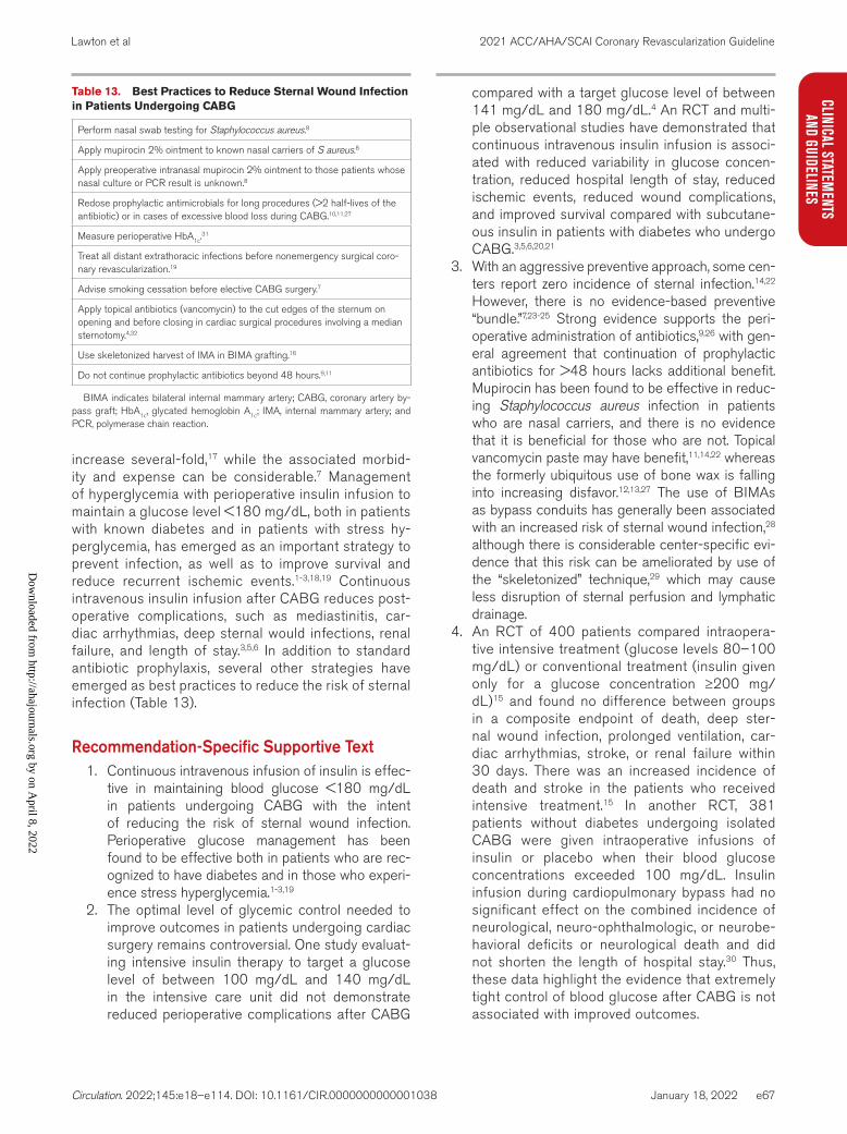
Lawton et al 2021 ACC/AHA/SCAI Coronary Revascularization Guideline
Circulation. 2022;145:e18–e114. DOI: 10.1161/CIR.0000000000001038 January 18, 2022 e67
CLINICAL STATEMENTS
AND GUIDELINES
increase several-fold,17 while the associated morbid-ity and expense can be considerable.7 Management of hyperglycemia with perioperative insulin infusion to maintain a glucose level <180 mg/dL, both in patients with known diabetes and in patients with stress hy-perglycemia, has emerged as an important strategy to prevent infection, as well as to improve survival and reduce recurrent ischemic events.1-3,18,19 Continuous intravenous insulin infusion after CABG reduces post-operative complications, such as mediastinitis, car-diac arrhythmias, deep sternal would infections, renal failure, and length of stay.3,5,6 In addition to standard antibiotic prophylaxis, several other strategies have emerged as best practices to reduce the risk of sternal infection (Table 13).
Recommendation-Specific Supportive Text1. Continuous intravenous infusion of insulin is effec-
tive in maintaining blood glucose <180 mg/dL in patients undergoing CABG with the intent of reducing the risk of sternal wound infection. Perioperative glucose management has been found to be effective both in patients who are rec-ognized to have diabetes and in those who experi-ence stress hyperglycemia.1-3,19
2. The optimal level of glycemic control needed to improve outcomes in patients undergoing cardiac surgery remains controversial. One study evaluat-ing intensive insulin therapy to target a glucose level of between 100 mg/dL and 140 mg/dL in the intensive care unit did not demonstrate reduced perioperative complications after CABG
compared with a target glucose level of between 141 mg/dL and 180 mg/dL.4 An RCT and multi-ple observational studies have demonstrated that continuous intravenous insulin infusion is associ-ated with reduced variability in glucose concen-tration, reduced hospital length of stay, reduced ischemic events, reduced wound complications, and improved survival compared with subcutane-ous insulin in patients with diabetes who undergo CABG.3,5,6,20,21
3. With an aggressive preventive approach, some cen-ters report zero incidence of sternal infection.14,22 However, there is no evidence-based preventive “bundle.”7,23-25 Strong evidence supports the peri-operative administration of antibiotics,9,26 with gen-eral agreement that continuation of prophylactic antibiotics for >48 hours lacks additional benefit. Mupirocin has been found to be effective in reduc-ing Staphylococcus aureus infection in patients who are nasal carriers, and there is no evidence that it is beneficial for those who are not. Topical vancomycin paste may have benefit,11,14,22 whereas the formerly ubiquitous use of bone wax is falling into increasing disfavor.12,13,27 The use of BIMAs as bypass conduits has generally been associated with an increased risk of sternal wound infection,28 although there is considerable center-specific evi-dence that this risk can be ameliorated by use of the “skeletonized” technique,29 which may cause less disruption of sternal perfusion and lymphatic drainage.
4. An RCT of 400 patients compared intraopera-tive intensive treatment (glucose levels 80–100 mg/dL) or conventional treatment (insulin given only for a glucose concentration ≥200 mg/dL)15 and found no difference between groups in a composite endpoint of death, deep ster-nal wound infection, prolonged ventilation, car-diac arrhythmias, stroke, or renal failure within 30 days. There was an increased incidence of death and stroke in the patients who received intensive treatment.15 In another RCT, 381 patients without diabetes undergoing isolated CABG were given intraoperative infusions of insulin or placebo when their blood glucose concentrations exceeded 100 mg/dL. Insulin infusion during cardiopulmonary bypass had no significant effect on the combined incidence of neurological, neuro-ophthalmologic, or neurobe-havioral deficits or neurological death and did not shorten the length of hospital stay.30 Thus, these data highlight the evidence that extremely tight control of blood glucose after CABG is not associated with improved outcomes.
Table 13. Best Practices to Reduce Sternal Wound Infection in Patients Undergoing CABG
Perform nasal swab testing for Staphylococcus aureus.8
Apply mupirocin 2% ointment to known nasal carriers of S aureus.8
Apply preoperative intranasal mupirocin 2% ointment to those patients whose nasal culture or PCR result is unknown.8
Redose prophylactic antimicrobials for long procedures (>2 half-lives of the antibiotic) or in cases of excessive blood loss during CABG.10,11,27
Measure perioperative HbA1c.31
Treat all distant extrathoracic infections before nonemergency surgical coro-nary revascularization.19
Advise smoking cessation before elective CABG surgery.7
Apply topical antibiotics (vancomycin) to the cut edges of the sternum on opening and before closing in cardiac surgical procedures involving a median sternotomy.4,32
Use skeletonized harvest of IMA in BIMA grafting.16
Do not continue prophylactic antibiotics beyond 48 hours.9,11
BIMA indicates bilateral internal mammary artery; CABG, coronary artery by-pass graft; HbA1c, glycated hemoglobin A1c; IMA, internal mammary artery; and PCR, polymerase chain reaction.
Dow
nloaded from http://ahajournals.org by on A
pril 8, 2022
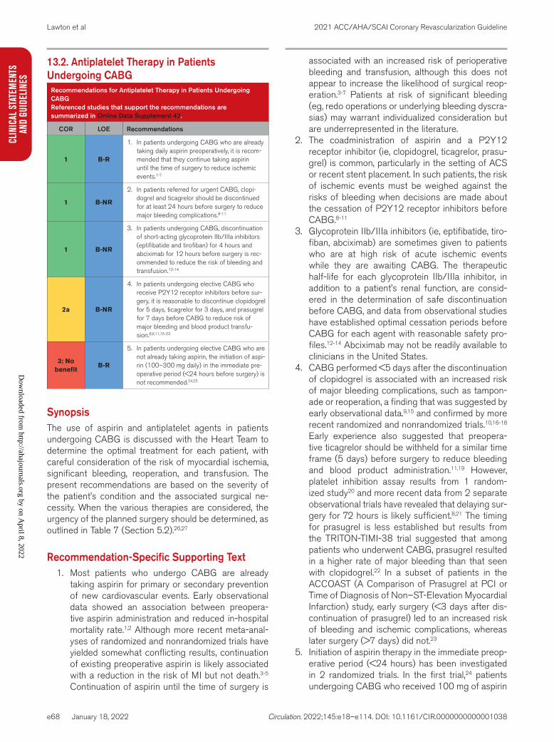
Lawton et al 2021 ACC/AHA/SCAI Coronary Revascularization Guideline
January 18, 2022 Circulation. 2022;145:e18–e114. DOI: 10.1161/CIR.0000000000001038e68
CLIN
ICAL
STA
TEM
ENTS
AN
D GU
IDEL
INES
13.2. Antiplatelet Therapy in Patients Undergoing CABG
Recommendations for Antiplatelet Therapy in Patients Undergoing CABGReferenced studies that support the recommendations are summarized in Online Data Supplement 42.
COR LOE Recommendations
1 B-R
1. In patients undergoing CABG who are already taking daily aspirin preoperatively, it is recom-mended that they continue taking aspirin until the time of surgery to reduce ischemic events.1-7
1 B-NR
2. In patients referred for urgent CABG, clopi-dogrel and ticagrelor should be discontinued for at least 24 hours before surgery to reduce major bleeding complications.8-11
1 B-NR
3. In patients undergoing CABG, discontinuation of short-acting glycoprotein IIb/IIIa inhibitors (eptifibatide and tirofiban) for 4 hours and abciximab for 12 hours before surgery is rec-ommended to reduce the risk of bleeding and transfusion.12-14
2a B-NR
4. In patients undergoing elective CABG who receive P2Y12 receptor inhibitors before sur-gery, it is reasonable to discontinue clopidogrel for 5 days, ticagrelor for 3 days, and prasugrel for 7 days before CABG to reduce risk of major bleeding and blood product transfu-sion.8,9,11,15-23
3: No benefit
B-R
5. In patients undergoing elective CABG who are not already taking aspirin, the initiation of aspi-rin (100–300 mg daily) in the immediate pre-operative period (<24 hours before surgery) is not recommended.24,25
SynopsisThe use of aspirin and antiplatelet agents in patients undergoing CABG is discussed with the Heart Team to determine the optimal treatment for each patient, with careful consideration of the risk of myocardial ischemia, significant bleeding, reoperation, and transfusion. The present recommendations are based on the severity of the patient’s condition and the associated surgical ne-cessity. When the various therapies are considered, the urgency of the planned surgery should be determined, as outlined in Table 7 (Section 5.2).26,27
Recommendation-Specific Supporting Text1. Most patients who undergo CABG are already
taking aspirin for primary or secondary prevention of new cardiovascular events. Early observational data showed an association between preopera-tive aspirin administration and reduced in-hospital mortality rate.1,2 Although more recent meta-anal-yses of randomized and nonrandomized trials have yielded somewhat conflicting results, continuation of existing preoperative aspirin is likely associated with a reduction in the risk of MI but not death.3-5 Continuation of aspirin until the time of surgery is
associated with an increased risk of perioperative bleeding and transfusion, although this does not appear to increase the likelihood of surgical reop-eration.3-7 Patients at risk of significant bleeding (eg, redo operations or underlying bleeding dyscra-sias) may warrant individualized consideration but are underrepresented in the literature.
2. The coadministration of aspirin and a P2Y12 receptor inhibitor (ie, clopidogrel, ticagrelor, prasu-grel) is common, particularly in the setting of ACS or recent stent placement. In such patients, the risk of ischemic events must be weighed against the risks of bleeding when decisions are made about the cessation of P2Y12 receptor inhibitors before CABG.8-11
3. Glycoprotein IIb/IIIa inhibitors (ie, eptifibatide, tiro-fiban, abciximab) are sometimes given to patients who are at high risk of acute ischemic events while they are awaiting CABG. The therapeutic half-life for each glycoprotein IIb/IIIa inhibitor, in addition to a patient’s renal function, are consid-ered in the determination of safe discontinuation before CABG, and data from observational studies have established optimal cessation periods before CABG for each agent with reasonable safety pro-files.12-14 Abciximab may not be readily available to clinicians in the United States.
4. CABG performed <5 days after the discontinuation of clopidogrel is associated with an increased risk of major bleeding complications, such as tampon-ade or reoperation, a finding that was suggested by early observational data.9,15 and confirmed by more recent randomized and nonrandomized trials.10,16-18 Early experience also suggested that preopera-tive ticagrelor should be withheld for a similar time frame (5 days) before surgery to reduce bleeding and blood product administration.11,19 However, platelet inhibition assay results from 1 random-ized study20 and more recent data from 2 separate observational trials have revealed that delaying sur-gery for 72 hours is likely sufficient.8,21 The timing for prasugrel is less established but results from the TRITON-TIMI-38 trial suggested that among patients who underwent CABG, prasugrel resulted in a higher rate of major bleeding than that seen with clopidogrel.22 In a subset of patients in the ACCOAST (A Comparison of Prasugrel at PCI or Time of Diagnosis of Non–ST-Elevation Myocardial Infarction) study, early surgery (<3 days after dis-continuation of prasugrel) led to an increased risk of bleeding and ischemic complications, whereas later surgery (>7 days) did not.23
5. Initiation of aspirin therapy in the immediate preop-erative period (<24 hours) has been investigated in 2 randomized trials. In the first trial,24 patients undergoing CABG who received 100 mg of aspirin
Dow
nloaded from http://ahajournals.org by on A
pril 8, 2022
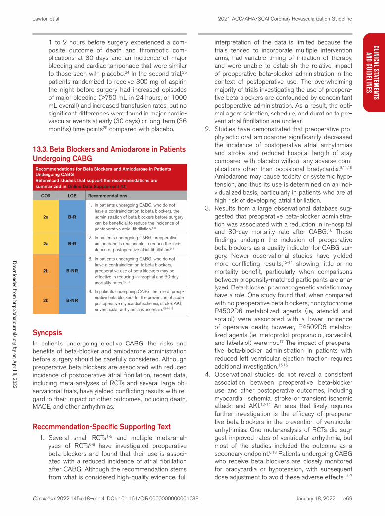
Lawton et al 2021 ACC/AHA/SCAI Coronary Revascularization Guideline
Circulation. 2022;145:e18–e114. DOI: 10.1161/CIR.0000000000001038 January 18, 2022 e69
CLINICAL STATEMENTS
AND GUIDELINES
1 to 2 hours before surgery experienced a com-posite outcome of death and thrombotic com-plications at 30 days and an incidence of major bleeding and cardiac tamponade that were similar to those seen with placebo.24 In the second trial,25 patients randomized to receive 300 mg of aspirin the night before surgery had increased episodes of major bleeding (>750 mL in 24 hours, or 1000 mL overall) and increased transfusion rates, but no significant differences were found in major cardio-vascular events at early (30 days) or long-term (36 months) time points25 compared with placebo.
13.3. Beta Blockers and Amiodarone in Patients Undergoing CABG
Recommendations for Beta Blockers and Amiodarone in Patients Undergoing CABGReferenced studies that support the recommendations are summarized in Online Data Supplement 43.
COR LOE Recommendations
2a B-R
1. In patients undergoing CABG, who do not have a contraindication to beta blockers, the administration of beta blockers before surgery can be beneficial to reduce the incidence of postoperative atrial fibrillation.1-8
2a B-R2. In patients undergoing CABG, preoperative
amiodarone is reasonable to reduce the inci-dence of postoperative atrial fibrillation.9-11
2b B-NR
3. In patients undergoing CABG, who do not have a contraindication to beta blockers, preoperative use of beta blockers may be effective in reducing in-hospital and 30-day mortality rates.12-18
2b B-NR
4. In patients undergoing CABG, the role of preop-erative beta blockers for the prevention of acute postoperative myocardial ischemia, stroke, AKI, or ventricular arrhythmia is uncertain.12-14,18
SynopsisIn patients undergoing elective CABG, the risks and benefits of beta-blocker and amiodarone administration before surgery should be carefully considered. Although preoperative beta blockers are associated with reduced incidence of postoperative atrial fibrillation, recent data, including meta-analyses of RCTs and several large ob-servational trials, have yielded conflicting results with re-gard to their impact on other outcomes, including death, MACE, and other arrhythmias.
Recommendation-Specific Supporting Text1. Several small RCTs1-5 and multiple meta-anal-
yses of RCTs6-8 have investigated preoperative beta blockers and found that their use is associ-ated with a reduced incidence of atrial fibrillation after CABG. Although the recommendation stems from what is considered high-quality evidence, full
interpretation of the data is limited because the trials tended to incorporate multiple intervention arms, had variable timing of initiation of therapy, and were unable to establish the relative impact of preoperative beta-blocker administration in the context of postoperative use. The overwhelming majority of trials investigating the use of preopera-tive beta blockers are confounded by concomitant postoperative administration. As a result, the opti-mal agent selection, schedule, and duration to pre-vent atrial fibrillation are unclear.
2. Studies have demonstrated that preoperative pro-phylactic oral amiodarone significantly decreased the incidence of postoperative atrial arrhythmias and stroke and reduced hospital length of stay compared with placebo without any adverse com-plications other than occasional bradycardia.9,11,19 Amiodarone may cause toxicity or systemic hypo-tension, and thus its use is determined on an indi-vidualized basis, particularly in patients who are at high risk of developing atrial fibrillation.
3. Results from a large observational database sug-gested that preoperative beta-blocker administra-tion was associated with a reduction in in-hospital and 30-day mortality rate after CABG.16 These findings underpin the inclusion of preoperative beta blockers as a quality indicator for CABG sur-gery. Newer observational studies have yielded more conflicting results,12-14 showing little or no mortality benefit, particularly when comparisons between propensity-matched participants are ana-lyzed. Beta-blocker pharmacogenetic variation may have a role. One study found that, when compared with no preoperative beta blockers, noncytochrome P4502D6 metabolized agents (ie, atenolol and sotalol) were associated with a lower incidence of operative death; however, P4502D6 metabo-lized agents (ie, metoprolol, propranolol, carvedilol, and labetalol) were not.17 The impact of preopera-tive beta-blocker administration in patients with reduced left ventricular ejection fraction requires additional investigation.15,16
4. Observational studies do not reveal a consistent association between preoperative beta-blocker use and other postoperative outcomes, including myocardial ischemia, stroke or transient ischemic attack, and AKI.12-14 An area that likely requires further investigation is the efficacy of preopera-tive beta blockers in the prevention of ventricular arrhythmias. One meta-analysis of RCTs did sug-gest improved rates of ventricular arrhythmia, but most of the studies included the outcome as a secondary endpoint.6,18 Patients undergoing CABG who receive beta blockers are closely monitored for bradycardia or hypotension, with subsequent dose adjustment to avoid these adverse effects .4-7
Dow
nloaded from http://ahajournals.org by on A
pril 8, 2022
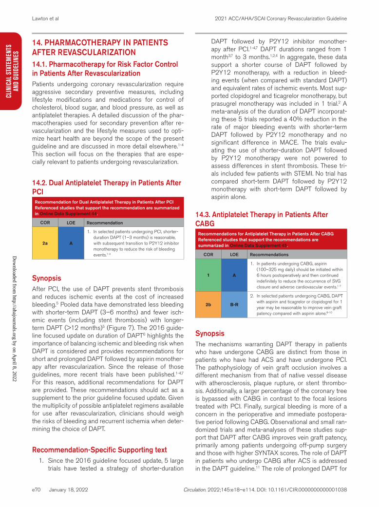
Lawton et al 2021 ACC/AHA/SCAI Coronary Revascularization Guideline
January 18, 2022 Circulation. 2022;145:e18–e114. DOI: 10.1161/CIR.0000000000001038e70
CLIN
ICAL
STA
TEM
ENTS
AN
D GU
IDEL
INES
14. PHARMACOTHERAPY IN PATIENTS AFTER REVASCULARIZATION14.1. Pharmacotherapy for Risk Factor Control in Patients After RevascularizationPatients undergoing coronary revascularization require aggressive secondary preventive measures, including lifestyle modifications and medications for control of cholesterol, blood sugar, and blood pressure, as well as antiplatelet therapies. A detailed discussion of the phar-macotherapies used for secondary prevention after re-vascularization and the lifestyle measures used to opti-mize heart health are beyond the scope of the present guideline and are discussed in more detail elsewhere.1-4 This section will focus on the therapies that are espe-cially relevant to patients undergoing revascularization.
14.2. Dual Antiplatelet Therapy in Patients After PCI
Recommendation for Dual Antiplatelet Therapy in Patients After PCIReferenced studies that support the recommendation are summarized in Online Data Supplement 44.
COR LOE Recommendation
2a A
1. In selected patients undergoing PCI, shorter-duration DAPT (1–3 months) is reasonable, with subsequent transition to P2Y12 inhibitor monotherapy to reduce the risk of bleeding events.1-4
SynopsisAfter PCI, the use of DAPT prevents stent thrombosis and reduces ischemic events at the cost of increased bleeding.5 Pooled data have demonstrated less bleeding with shorter-term DAPT (3–6 months) and fewer isch-emic events (including stent thrombosis) with longer-term DAPT (>12 months)5 (Figure 7). The 2016 guide-line focused update on duration of DAPT6 highlights the importance of balancing ischemic and bleeding risk when DAPT is considered and provides recommendations for short and prolonged DAPT followed by aspirin monother-apy after revascularization. Since the release of those guidelines, more recent trials have been published.1-4,7 For this reason, additional recommendations for DAPT are provided. These recommendations should act as a supplement to the prior guideline focused update. Given the multiplicity of possible antiplatelet regimens available for use after revascularization, clinicians should weigh the risks of bleeding and recurrent ischemia when deter-mining the choice of DAPT.
Recommendation-Specific Supporting text1. Since the 2016 guideline focused update, 5 large
trials have tested a strategy of shorter-duration
DAPT followed by P2Y12 inhibitor monother-apy after PCI.1-4,7 DAPT durations ranged from 1 month3,7 to 3 months.1,2,4 In aggregate, these data support a shorter course of DAPT followed by P2Y12 monotherapy, with a reduction in bleed-ing events (when compared with standard DAPT) and equivalent rates of ischemic events. Most sup-ported clopidogrel and ticagrelor monotherapy, but prasugrel monotherapy was included in 1 trial.2 A meta-analysis of the duration of DAPT incorporat-ing these 5 trials reported a 40% reduction in the rate of major bleeding events with shorter-term DAPT followed by P2Y12 monotherapy and no significant difference in MACE. The trials evalu-ating the use of shorter-duration DAPT followed by P2Y12 monotherapy were not powered to assess differences in stent thrombosis. These tri-als included few patients with STEMI. No trial has compared short-term DAPT followed by P2Y12 monotherapy with short-term DAPT followed by aspirin alone.
14.3. Antiplatelet Therapy in Patients After CABG
Recommendations for Antiplatelet Therapy in Patients After CABGReferenced studies that support the recommendations are summarized in Online Data Supplement 45.
COR LOE Recommendations
1 A
1. In patients undergoing CABG, aspirin (100–325 mg daily) should be initiated within 6 hours postoperatively and then continued indefinitely to reduce the occurrence of SVG closure and adverse cardiovascular events.1-7
2b B-R
2. In selected patients undergoing CABG, DAPT with aspirin and ticagrelor or clopidogrel for 1 year may be reasonable to improve vein graft patency compared with aspirin alone.8-10
SynopsisThe mechanisms warranting DAPT therapy in patients who have undergone CABG are distinct from those in patients who have had ACS and have undergone PCI. The pathophysiology of vein graft occlusion involves a different mechanism from that of native vessel disease with atherosclerosis, plaque rupture, or stent thrombo-sis. Additionally, a larger percentage of the coronary tree is bypassed with CABG in contrast to the focal lesions treated with PCI. Finally, surgical bleeding is more of a concern in the perioperative and immediate postopera-tive period following CABG. Observational and small ran-domized trials and meta-analyses of these studies sup-port that DAPT after CABG improves vein graft patency, primarily among patients undergoing off-pump surgery and those with higher SYNTAX scores. The role of DAPT in patients who undergo CABG after ACS is addressed in the DAPT guideline.11 The role of prolonged DAPT for
Dow
nloaded from http://ahajournals.org by on A
pril 8, 2022

Lawton et al 2021 ACC/AHA/SCAI Coronary Revascularization Guideline
e71
CLINICAL STATEMENTS
AND GUIDELINES
Circulation. 2022;145:e18–e114. DOI: 10.1161/CIR.0000000000001038 January 18, 2022
Figure 7. Use of DAPT for Patients After PCI.Colors correspond to Table 2. ACS indicates acute coronary syndrome; BMS, bare metal stent; DAPT, dual antiplatelet therapy; DES, drug-eluting stent; P2Y12, platelet adenosine diphosphate P2Y12 receptor; PCI, percutaneous coronary intervention; and SIHD, stable ischemic heart disease. This algorithm is adapted from the 2016 DAPT guideline6 and includes new recommendations from this guideline for the care of patients with CAD. It is not meant to encompass every patient scenario or situation, and clinicians are encouraged to use a Heart Team approach when care decisions are unclear and to see the accompanying supportive text for each recommendation. Additionally, in situations that lack sufficient data to make formal recommendations for care, please see Section 17, “Unanswered Questions and Future Directions.”
Dow
nloaded from http://ahajournals.org by on A
pril 8, 2022

Lawton et al 2021 ACC/AHA/SCAI Coronary Revascularization Guideline
January 18, 2022 Circulation. 2022;145:e18–e114. DOI: 10.1161/CIR.0000000000001038e72
CLIN
ICAL
STA
TEM
ENTS
AN
D GU
IDEL
INES
general secondary prevention in patients with a distant history of CABG is not well established.
Recommendation-Specific Supporting Text1. Surgical bleeding remains a concern in the peri-
operative and immediate postoperative periods, and therefore bleeding risk is an important con-sideration in the use of antiplatelet therapy. Older data have shown that aspirin improves vein graft patency.1,2,5,6 Although 1 small study demonstrated higher rates of bleeding with aspirin after CABG,12 the totality of evidence supports the early use1-6 of aspirin to improve SVG patency and reduce isch-emic complications.
2. Small RCTs, observational data, and meta-analyses have demonstrated that DAPT (mostly with aspirin and clopidogrel) after CABG improves vein graft patency, primarily among patients undergoing off-pump surgery. The DACAB (Different Antiplatelet Therapy Strategy After Coronary Artery Bypass Graft Surgery) trial10 compared DAPT with a single antiplatelet regimen in 500 patients undergoing CABG. Off-pump procedures were performed in 75% of these patients. At 1-year follow-up, the DAPT group was found to have the highest vein graft patency, when assessed with coronary com-puted tomography angiogram, compared with aspi-rin alone.
14.4. Beta Blockers in Patients After Revascularization
Recommendation for Beta Blockers in Patients After RevascularizationReferenced studies that support the recommendation are summarized in Online Data Supplement 46.
COR LOE Recommendation
3: No benefit
C-LD
1. In patients with SIHD and normal left ven-tricular function, the routine use of chronic oral beta blockers is not beneficial to reduce cardiovascular events after complete revascu-larization.1-6
SynopsisIn patients who have undergone revascularization, the risks and benefits of beta blockers should be considered before the initiation of therapy. The benefit of beta block-ers for secondary prevention after acute infarction or for those with left ventricular dysfunction has been clearly reported in clinical trials examining these subgroups, and recommendations based on this evidence are outlined in previous guidelines.7,8 However, in patients without acute infarction or left ventricular dysfunction, there is a paucity of data to support a benefit of the routine use of beta blockers after revascularization, especially in patients without residual disease. Further risk reduction may not be useful in patients after MI with normal left ventricular
ejection fraction in the presence of GDMT with antiplate-let treatment, statins, and angiotensin-converting enzyme inhibitors or angiotensin-receptor blockers. Thus, in the absence of new data to guide current therapy, clinicians will need to make decisions on an individualized basis.
Recommendation-Specific Supportive Text1. A large meta-analysis of patients undergoing PCI
for stable angina showed no differences in adjusted rates of death, MI, stroke, or revascularization but a higher rate of heart failure readmissions among patients who were prescribed a beta blocker at hospital discharge.1 The REACH (Reduction of Atherothrombosis for Continued Health) Registry’s investigators showed that, after a median of 44 months’ follow-up, beta-blocker use was not asso-ciated with a reduction in the composite cardio-vascular outcome in a large cohort of patients with SIHD.2 Additional studies have also supported an increased incidence of heart failure in patients treated with beta blockers in the reperfusion era.9 In a large cohort of patients with newly diagnosed CAD, a modest benefit of beta-blocker use was reported, although this benefit was noted only in patients with a previous MI.4 Hence, the decision to continue beta blockers in the long term in patients after revascularization should be made on an indi-vidualized basis.
14.5. Beta Blockers for the Prevention of Atrial Fibrillation After CABG
Recommendation for Beta Blockers for the Prevention of Atrial Fibrillation After CABGReferenced studies that support the recommendation are summarized in Online Data Supplement 47.
COR LOE Recommendation
1 B-R
1. In patients after CABG, beta blockers are recommended and should be started as soon as possible to reduce the incidence or clinical sequelae of postoperative atrial fibrillation.1-7
SynopsisNew-onset postoperative atrial fibrillation occurs in about 18% of patients after CABG and is associated with a 4-fold increased risk of stroke and a 3-fold increase in all-cause mortality rate.8,9 Postoperative atrial fibrillation after CABG can be challenging to prevent and treat.
Recommendation-Specific Supportive Text1. RCTs have yielded conflicting results with regard
to the ability of beta blockers to influence periop-erative cardiovascular morbidity and mortality. A large meta-analysis found that beta-blocker use may reduce the incidence of atrial fibrillation and
Dow
nloaded from http://ahajournals.org by on A
pril 8, 2022

Lawton et al 2021 ACC/AHA/SCAI Coronary Revascularization Guideline
Circulation. 2022;145:e18–e114. DOI: 10.1161/CIR.0000000000001038 January 18, 2022 e73
CLINICAL STATEMENTS
AND GUIDELINES
ventricular arrhythmias and hospital stay10 but found no evidence of a difference in rates of early all-cause death, MI, cerebrovascular events, hypo-tension, or bradycardia.10
14.6. Antiplatelet Therapy in Patients With Atrial Fibrillation on Anticoagulation After PCI
Recommendations for Antiplatelet Therapy in Patients With Atrial Fibrillation on Anticoagulation After PCIReferenced studies that support the recommendations are summarized in Online Data Supplement 48.
COR LOE Recommendations
1 B-R
1. In patients with atrial fibrillation who are undergoing PCI and are taking oral anticoagu-lant therapy, it is recommended to discontinue aspirin treatment after 1 to 4 weeks while maintaining P2Y12 inhibitors in addition to a non–vitamin K oral anticoagulant (rivaroxaban, dabigatran, apixaban, or edoxaban) or warfarin to reduce the risk of bleeding.1-7
2a B-R
2. In patients with atrial fibrillation who are undergoing PCI, are taking oral anticoagu-lant therapy, and are treated with DAPT or a P2Y12 inhibitor monotherapy, it is reasonable to choose a non–vitamin K oral anticoagulant over warfarin to reduce the risk of bleeding.1,3,4
SynopsisPatients undergoing PCI frequently have or develop con-comitant indications for anticoagulant therapy, including atrial fibrillation, venous thromboembolism, and prosthet-ic heart valves. The most robust evidence for anticoagu-lant management in such patients comes from trials in patients with atrial fibrillation. The 2019 focused update of the atrial fibrillation guidelines8 gave a Class 2a rec-ommendation to a P2Y12 inhibitor with a non–vitamin K oral anticoagulant (rivaroxaban or dabigatran) or a vi-tamin K antagonist (warfarin) rather than triple therapy with an anticoagulant and DAPT. Since the publication of this guideline focused update, there have been 2 ad-ditional trials1,4 examining the benefits of dual anticoagu-lant therapy after PCI in patients with atrial fibrillation. On the basis of analyses of these trials, the recommenda-tions for antiplatelet and anticoagulant therapy after PCI in patients with atrial fibrillation have been updated.
Recommendation-Specific Supporting Text1. Two recent trials—the AUGUSTUS (Safety and
Efficacy of Apixaban Versus Vitamin K Antagonist and Aspirin Versus Aspirin Placebo in Patients With Atrial Fibrillation and ACS and/or PCI) trial1 and the ENTRUST-AF-PCI (Edoxaban–Based Versus Vitamin K Antagonist–Based Antithrombotic Regimen After Successful Coronary Stenting in Patients With Atrial Fibrillation) trial4—examined regimens of apixaban and edoxaban and sup-ported earlier findings,6,7 reporting lower bleeding
rates in patients with atrial fibrillation who were treated with a non–vitamin K oral anticoagulant and P2Y12 inhibitor than in those treated with triple therapy after PCI. Although none of the tri-als was powered for ischemic endpoints, pooled data from these trials1 have shown rates of death, MI, and stent thrombosis with dual therapy that are similar to those seen with triple therapy. All patients enrolled in these trials were briefly treated with triple therapy after PCI before the aspirin was discontinued. An analysis of stent thrombo-sis rates suggested that 80% of events occur within 30 days of PCI.3 For this reason, it is pos-sible that prolonging aspirin therapy to 1 month after PCI may reduce the risk of stent thrombosis.3 Therefore, in patients deemed to be at high risk of stent thrombosis, aspirin could be maintained for up to 30 days.
2. The AUGUSTUS trial2 randomized patients with atrial fibrillation undergoing PCI and found that apixaban, as compared with warfarin, reduced the rate of bleeding and was associated with a lower incidence of the combined endpoint of death or hospitalization. Compared with other treatment reg-imens, the combination of apixaban with a P2Y12 inhibitor was associated with the lowest rates of bleeding. The ENTRUST-AF-PCI trial4 compared edoxaban and P2Y12 monotherapy with triple therapy with a vitamin K antagonist in patients with atrial fibrillation undergoing PCI. Although there were fewer bleeding events in the first 14 days in the vitamin K antagonist arm, a landmark analysis from 14 days onward demonstrated less bleeding in the dual-therapy group.
15. RECOMMENDATIONS FOR ADDRESSING PSYCHOSOCIAL FACTORS AND LIFESTYLE CHANGES AFTER REVASCULARIZATION15.1. Cardiac Rehabilitation and Education
Recommendations for Cardiac Rehabilitation and EducationReferenced studies that support the recommendations are summarized in Online Data Supplement 49.
COR LOE Recommendations
1 A
1. In patients who have undergone revasculariza-tion, a comprehensive cardiac rehabilitation program (home based or center based) should be prescribed either before hospital discharge or during the first outpatient visit to reduce deaths and hospital readmissions and improve quality of life.1-4
1 C-LD
2. Patients who have undergone revasculariza-tion should be educated about CVD risk factors and their modification to reduce car-diovascular events.5-7
Dow
nloaded from http://ahajournals.org by on A
pril 8, 2022

Lawton et al 2021 ACC/AHA/SCAI Coronary Revascularization Guideline
January 18, 2022 Circulation. 2022;145:e18–e114. DOI: 10.1161/CIR.0000000000001038e74
CLIN
ICAL
STA
TEM
ENTS
AN
D GU
IDEL
INES
SynopsisCardiac rehabilitation is an evidence-based interven-tion comprising patient education, behavior modifi-cation, and exercise training to improve secondary prevention outcomes in patients with CVD.8 Cardiac re-habilitation assists patients with adherence to healthy lifestyle habits; addresses comorbid conditions (eg, diabetes); monitors for safety issues, including new or recurrent symptoms; and facilitates adherence to evidence-based medical therapies.9 Cardiac rehabilita-tion may include a center-based cardiac rehabilitation program that incorporates face-to-face supervised ex-ercise or an alternative cardiac rehabilitation delivery model that meets criteria for safety and effectiveness, as specified by the cardiac rehabilitation guidelines of the American Association of Cardiovascular and Pul-monary Rehabilitation.10
Recommendation-Specific Supportive Text1. The safety and effectiveness of the tradi-
tional, medically supervised center-based car-diac rehabilitation model are well established. Observational studies and RCTs have demon-strated that center-based cardiac rehabilitation is effective in reducing hospital readmissions, secondary events, and deaths in patients with CVD.1,2,4,11 Guidelines and standards of care have been well defined for center-based cardiac rehabilitation, including core components,10 core competencies,12 clinical practice guidelines,13 performance measures,12 and certification (pro-gram and individual).9 Home-based cardiac reha-bilitation can help improve delivery of cardiac rehabilitation to eligible patients by overcoming common barriers that impede a patient’s par-ticipation in center-based cardiac rehabilitation, including transportation challenges, competing time demands, and lack of a center-based car-diac rehabilitation program near the patient’s home.14 Core components of home-based car-diac rehabilitation are similar to those for center-based cardiac rehabilitation.14 Cochrane reviews concluded that home- and center-based cardiac rehabilitation have similar effects on quality of life and costs among patients with recent MI or coro-nary revascularization.15-17
2. Patients and caregivers should receive a compre-hensive plan of care and educational materials during the hospital stay that support adherence to evidence-based therapies. The “2019 ACC/AHA Guideline on the Primary Prevention of Cardiovascular Disease”18 provides comprehen-sive recommendations for improving risk fac-tors for CVD (unhealthy dietary pattern, lack of
exercise and physical activity, obesity, diabe-tes, high blood cholesterol, hypertension, and tobacco use). This information and management can be accomplished in a center-based or a home-based cardiac rehabilitation program and should be tailored to age, health literacy, cultural practices, and socioeconomic status.7 The basic self-care activities important to CVD manage-ment are captured in the AHA’s Life’s Simple 7 program (eg, smoking cessation, maintenance of body mass index, physical activity, healthy diet, maintaining low cholesterol, maintaining normal blood pressure, and maintaining normal fasting plasma glucose).19
15.2. Smoking Cessation in Patients After Revascularization
Recommendations for Smoking Cessation in Patients After RevascularizationReferenced studies that support the recommendations are summarized in Online Data Supplement 50.
COR LOE Recommendations
1 A
1. In patients who use tobacco and have undergone coronary revascularization, a combination of behavioral interventions plus pharmacotherapy is recommended to maxi-mize cessation and reduce adverse cardiac events.1-3
1 A
2. In patients who use tobacco and have under-gone coronary revascularization, smoking ces-sation interventions are recommended during hospitalization and should include supportive follow-up for at least 1 month after discharge to facilitate tobacco cessation and reduce morbidity and mortality.4-6
SynopsisTobacco use, especially cigarette smoking, is a major risk factor for cardiovascular morbidity and mortality and is the leading preventable cause of death world-wide.6,7 Among patients with coronary heart disease, continued cigarette smoking after revascularization is associated with adverse clinical outcomes,8 par-ticularly stent thrombosis.9 Electronic nicotine delivery systems or e-cigarettes10 are a class of tobacco prod-uct that emit aerosol containing fine and ultrafine par-ticulates, nicotine, and toxic gases that may increase risk of CVD and pulmonary disease.11-13 The dominant pattern of e-cigarette use in adults is dual use of both combustible cigarettes and e-cigarettes.14,15 When pa-tients are counseled about risk factor management after revascularization, the topic of tobacco abuse is paramount and timely, because patients who are hos-pitalized after revascularization are often at their most attentive state. The recommendations for smoking cessation counseling and treatment are outlined in the “2019 ACC/AHA Guideline on the Primary Prevention
Dow
nloaded from http://ahajournals.org by on A
pril 8, 2022

Lawton et al 2021 ACC/AHA/SCAI Coronary Revascularization Guideline
Circulation. 2022;145:e18–e114. DOI: 10.1161/CIR.0000000000001038 January 18, 2022 e75
CLINICAL STATEMENTS
AND GUIDELINES
of Cardiovascular Disease”16 and are also applicable to the secondary prevention of patients after coronary artery revascularization.
Recommendation-Specific Supportive Text1. The US Public Health Service’s Clinical Practice
Guideline for Smoking Cessation recommends smoking-cessation pharmacotherapy for all smokers attempting to quit.17 The most effective smoking-cessation therapies include both behav-ioral and pharmacological interventions.1,6 High-quality evidence showed that using a combination of behavioral support and medication increases the chances of successfully quitting for at least 6 months.2,5 Moreover, the chance of success was increased by 70% to 100% compared with just brief advice or support. Among patients with CVD who were motivated to quit smoking, varenicline and bupropion are efficacious for smoking cessa-tion, as are individual and telephone counseling.2,5 Varenicline was the most efficacious of therapies in patients with stable CVD who were motivated to quit smoking.2,5,18,19 In 1 study, abstinence rates in the group of patients treated with varenicline were higher than in those treated with placebo, a result that persisted for 52 weeks.19 Given the uncer-tainties of the long-term effects of e-cigarettes
on health, clinicians have been urged to advise cigarette smokers seeking to quit to use evi-dence-based, US Food and Drug Administration–approved, safe and effective smoking cessation pharmacotherapies as first-line treatments in pref-erence to e-cigarettes.7
2. Studies have shown that when hospitalized tobacco users receive counseling with sup-portive follow-up for ≥1 month after discharge, smoking cessation rates increase by 37% at 6 to 12 months after discharge.4 Varenicline use for hospitalized smokers with ACS who were moti-vated to quit significantly increased abstinence versus placebo at 1 year after discharge.20-22 At week 24, using varenicline increased smoking abstinence and reduced cigarette use by ≥50%. There is no evidence that pharmacotherapies (varenicline, bupropion, and nicotine replacement versus placebo) increase the risk of cardiovascu-lar adverse events during or after treatment.19,21,23 The EAGLES (Neuropsychiatric Safety and Efficacy of Varenicline, Bupropion, and Nicotine Patch in Smokers With and Without Psychiatric Disorders) trial showed that pharmacotherapies do not increase the risk of cardiovascular or neuropsychiatric adverse events compared with nicotine patch or placebo in smokers with and without psychiatric disorders.18,23
Figure 8. Traditional and Psychosocial Risk Factors for ASCVD.ASCVD indicates atherosclerotic cardiovascular disease.
Dow
nloaded from http://ahajournals.org by on A
pril 8, 2022
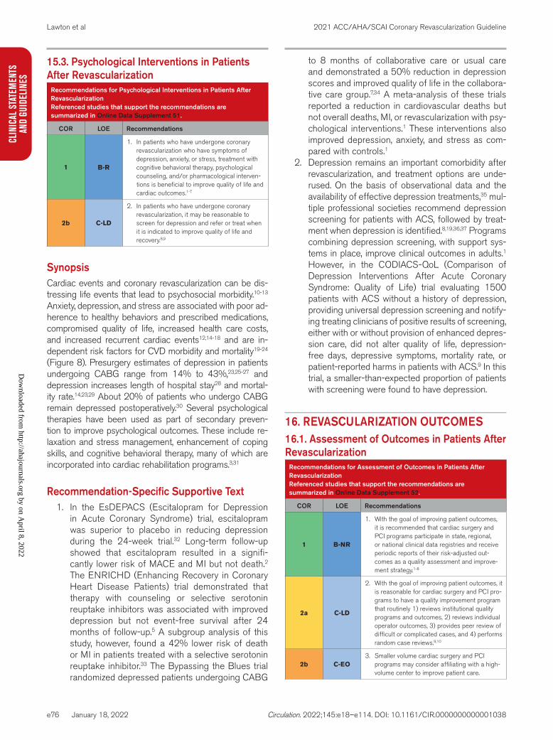
Lawton et al 2021 ACC/AHA/SCAI Coronary Revascularization Guideline
January 18, 2022 Circulation. 2022;145:e18–e114. DOI: 10.1161/CIR.0000000000001038e76
CLIN
ICAL
STA
TEM
ENTS
AN
D GU
IDEL
INES
15.3. Psychological Interventions in Patients After Revascularization
Recommendations for Psychological Interventions in Patients After RevascularizationReferenced studies that support the recommendations are summarized in Online Data Supplement 51.
COR LOE Recommendations
1 B-R
1. In patients who have undergone coronary revascularization who have symptoms of depression, anxiety, or stress, treatment with cognitive behavioral therapy, psychological counseling, and/or pharmacological interven-tions is beneficial to improve quality of life and cardiac outcomes.1-7
2b C-LD
2. In patients who have undergone coronary revascularization, it may be reasonable to screen for depression and refer or treat when it is indicated to improve quality of life and recovery.8,9
SynopsisCardiac events and coronary revascularization can be dis-tressing life events that lead to psychosocial morbidity.10-13 Anxiety, depression, and stress are associated with poor ad-herence to healthy behaviors and prescribed medications, compromised quality of life, increased health care costs, and increased recurrent cardiac events12,14-18 and are in-dependent risk factors for CVD morbidity and mortality19-24 (Figure 8). Presurgery estimates of depression in patients undergoing CABG range from 14% to 43%,23,25-27 and depression increases length of hospital stay28 and mortal-ity rate.14,23,29 About 20% of patients who undergo CABG remain depressed postoperatively.30 Several psychological therapies have been used as part of secondary preven-tion to improve psychological outcomes. These include re-laxation and stress management, enhancement of coping skills, and cognitive behavioral therapy, many of which are incorporated into cardiac rehabilitation programs.3,31
Recommendation-Specific Supportive Text1. In the EsDEPACS (Escitalopram for Depression
in Acute Coronary Syndrome) trial, escitalopram was superior to placebo in reducing depression during the 24-week trial.32 Long-term follow-up showed that escitalopram resulted in a signifi-cantly lower risk of MACE and MI but not death.2 The ENRICHD (Enhancing Recovery in Coronary Heart Disease Patients) trial demonstrated that therapy with counseling or selective serotonin reuptake inhibitors was associated with improved depression but not event-free survival after 24 months of follow-up.5 A subgroup analysis of this study, however, found a 42% lower risk of death or MI in patients treated with a selective serotonin reuptake inhibitor.33 The Bypassing the Blues trial randomized depressed patients undergoing CABG
to 8 months of collaborative care or usual care and demonstrated a 50% reduction in depression scores and improved quality of life in the collabora-tive care group.7,34 A meta-analysis of these trials reported a reduction in cardiovascular deaths but not overall deaths, MI, or revascularization with psy-chological interventions.1 These interventions also improved depression, anxiety, and stress as com-pared with controls.1
2. Depression remains an important comorbidity after revascularization, and treatment options are unde-rused. On the basis of observational data and the availability of effective depression treatments,35 mul-tiple professional societies recommend depression screening for patients with ACS, followed by treat-ment when depression is identified.8,19,36,37 Programs combining depression screening, with support sys-tems in place, improve clinical outcomes in adults.1 However, in the CODIACS-QoL (Comparison of Depression Interventions After Acute Coronary Syndrome: Quality of Life) trial evaluating 1500 patients with ACS without a history of depression, providing universal depression screening and notify-ing treating clinicians of positive results of screening, either with or without provision of enhanced depres-sion care, did not alter quality of life, depression-free days, depressive symptoms, mortality rate, or patient-reported harms in patients with ACS.9 In this trial, a smaller-than-expected proportion of patients with screening were found to have depression.
16. REVASCULARIZATION OUTCOMES16.1. Assessment of Outcomes in Patients After Revascularization
Recommendations for Assessment of Outcomes in Patients After RevascularizationReferenced studies that support the recommendations are summarized in Online Data Supplement 52.
COR LOE Recommendations
1 B-NR
1. With the goal of improving patient outcomes, it is recommended that cardiac surgery and PCI programs participate in state, regional, or national clinical data registries and receive periodic reports of their risk-adjusted out-comes as a quality assessment and improve-ment strategy.1-8
2a C-LD
2. With the goal of improving patient outcomes, it is reasonable for cardiac surgery and PCI pro-grams to have a quality improvement program that routinely 1) reviews institutional quality programs and outcomes, 2) reviews individual operator outcomes, 3) provides peer review of difficult or complicated cases, and 4) performs random case reviews.9,10
2b C-EO3. Smaller volume cardiac surgery and PCI
programs may consider affiliating with a high-volume center to improve patient care.
Dow
nloaded from http://ahajournals.org by on A
pril 8, 2022

Lawton et al 2021 ACC/AHA/SCAI Coronary Revascularization Guideline
Circulation. 2022;145:e18–e114. DOI: 10.1161/CIR.0000000000001038 January 18, 2022 e77
CLINICAL STATEMENTS
AND GUIDELINES
SynopsisCenters that provide coronary revascularization should participate in clinical data registries with the intent to re-view and continuously improve patient outcomes. Com-parison of outcomes through the use of national data-bases allows individual- and program-level assessment of the care provided and the opportunity to enhance care with quality improvement initiatives. Collaboration with other centers allows peer review and discussion, as well as sharing and adoption of effective quality improvement measures.
Recommendation-Specific Supporting Text1. Participation in regional, state, or national registries
that provide regular, risk-adjusted outcomes is beneficial in quality assessment and improvement. It allows participants to compare their performance to regional or national validated benchmarks, iden-tify opportunities for improvement, and dissemi-nate best practices.1-8
2. Quality and performance measures are defined by attributes related to structure, processes, and risk-adjusted outcomes. Structural attributes include elements such as equipment, supplies, staffing, institution- and operator-level volumes, and elec-tronic health records. Processes include strategies for appropriate patient selection; protocols for pre- and postprocedural care, procedural execution, and management of complications; and participation in databases and registries for benchmarking the performance of the program and individual opera-tor. Risk-adjusted outcomes are the end result of these structures and processes of care, and when available, they may be more reliable measures of quality than the institutional-level and individual operator–level volumes.9-11
3. Smaller-volume coronary revascularization pro-grams may benefit from affiliation and collabora-tion with larger volume programs. Standardized processes from both centers may be shared bidirectionally, and periodic exchange of staff will facilitate the transfer of best practices. Teaching conferences, as well as conferences on morbidity and mortality in both centers, may be shared via videoconferencing. In addition, residents and fel-lows may rotate between programs. Program size is typically defined by the specific database.
17. UNANSWERED QUESTIONS AND FUTURE DIRECTIONSThe indications for revascularization and the approach to treatment in patients with CAD are generally based on evidence supporting benefit or lack thereof. Many rec-
ommendations for revascularization are derived from the results of RCTs or observational studies of large regis-tries or cohorts of patients that show consistent trends in outcomes. However, there are some patient subgroups and clinical scenarios for which there is a paucity of evi-dence to support a formal recommendation. In addition, in some circumstances, it is inappropriate or unethical to perform a randomized trial comparing 2 treatments. Furthermore, in some situations, even in the absence of strong evidence, recommendations are created on the basis of experiential consensus on best practices for the delivery of care. In these cases, further research is needed to inform practice, which would enable updated recommendations based on clinical trial results.
17.1. Special Populations17.1.1. Underrepresented Racial and Ethnic GroupsDespite advances in the identification of risk factors for CVD and the widespread use of evidence-based strat-egies to manage CVD, there are persistent sex, racial, and ethnic disparities in the delivery of care and in mor-bidity and mortality.1-8 Studies have shown that Black patients and patients of South Asian descent with CAD have worse outcomes than do White patients.2,4 Additionally, studies have reported worse outcomes in women than in men, although this finding is largely at-tenuated after adjustment for differences in baseline presentation and treatment.9 Recommendations for care in patients with CAD are often derived from RCT data with an unequal representation of women and ra-cial and ethnic groups.10 Although a study’s findings might be extrapolated to such populations, it is unclear whether similar outcomes can be assumed from the reported trial results across all populations of patients. For this reason, in the planning of clinical trials, mea-sures to ensure the enrollment of underrepresented racial and ethnic groups should be implemented to in-form better care of patients.11
17.2. Special Clinical Situations17.2.1. Left Ventricular DysfunctionRCT data support the use of CABG for the treatment of patients with coronary heart disease and left ventricular dysfunction to improve survival.12-14 Although the STICH trial reported improved outcomes with CABG compared with medical therapy, the advantage of CABG over medical therapy was independent of the presence or absence of myocardial viability.15-17 Critics have argued that the lack of a relative benefit of myocardial viability in predicting outcome with CABG was largely a result of the type of testing used.18 Many surgeons still use vi-ability testing to guide decisions about revascularization in patients with severe left ventricular dysfunction. Nev-ertheless, in view of the lack of association of myocar-
Dow
nloaded from http://ahajournals.org by on A
pril 8, 2022
Lawton et al 2021 ACC/AHA/SCAI Coronary Revascularization Guideline
January 18, 2022 Circulation. 2022;145:e18–e114. DOI: 10.1161/CIR.0000000000001038e78
CLIN
ICAL
STA
TEM
ENTS
AN
D GU
IDEL
INES
dial viability with derived benefit from revascularization, it remains unclear whether viability studies should be used to inform clinical practice, and if they are used, it remains unclear which method of assessment provides the most useful information.
There are currently insufficient data on the role of PCI in patients with left ventricular dysfunction to improve survival or cardiovascular outcomes. To address this gap in evidence, the ongoing REVIVED-BCIS2 (Percutane-ous Revascularization for Ischemic Ventricular Dysfunc-tion)19 study will evaluate the benefits of PCI versus medical therapy in reducing the combined endpoint of death or hospitalization for heart failure at 2 years’ follow-up. Additional studies are also currently under way, including the ISCHEMIA–Heart Failure Planning Study,20 which is expected to pave the way for a larger phase 3 trial of PCI in patients with systolic heart failure.
17.2.2. SCADSCAD is increasingly recognized as a cause of ACS in young patients, particularly women, and is present in roughly one-fourth of women ≤50 years of age present-ing with AMI.21 The management of SCAD has evolved over the years toward a more cautious use of PCI after various case series demonstrated low success rates and higher rates of complications with PCI for SCAD, as well as good long-term outcomes in conservatively treated patients.22 For this reason, expert consensus statements emphasize conservative care in most patients.23 How-ever, managing patients with SCAD who have ongoing symptoms, hemodynamic instability, or severely compro-mised blood flow of an artery subtending a large amount of myocardium (ie, the proximal LAD or left main lesions) is particularly problematic, as conservative care may not be a good option. Further investigation therefore is need-ed to understand the ideal scenarios for proceeding with revascularization and the optimal techniques for revascu-larization in SCAD.
17.2.3. Coronary Artery AneurysmCoronary artery aneurysms and fistulas are uncommon findings on coronary angiography, with a reported preva-lence of 0.02% to 0.2%.24,25 Most patients with coronary artery aneurysms are asymptomatic, but coronary artery aneurysms can lead to ischemia, vessel thrombosis, fis-tula formation, or rupture.24-27 There are no randomized studies evaluating the most effective therapy for these patients. Case reports and case series have described various methods for repair of aneurysms or fistulas, in-cluding covered stenting, coil embolization, Amplatzer device implantation, and surgical bypass with exclusion of the aneurysm.24-27 Because many patients remain as-ymptomatic and treatments for aneurysms or fistulas are not well defined, information on the timing of intervention (with respect to size and/or symptoms) and the ideal ap-proach to treatment (surgical excision versus percutane-ous therapies) is strongly needed.
17.2.4. Myocardial BridgingMyocardial bridging occurs when there is systolic com-pression of a coronary artery because of a segmental intramyocardial course of the vessel, and it is seen in up to 25% of patients undergoing coronary angiography.28 Although most myocardial bridging is clinically insignifi-cant, severe bridging has been inferred to produce myo-cardial ischemia, coronary thrombosis, AMI, and stress cardiomyopathy.28 In patients with ischemic pain and a myocardial bridge, provocative testing can be performed by measuring FFR under baseline conditions and dur-ing dobutamine stress or by obtaining positron emis-sion tomographic imaging during adenosine vasodilator challenge.29 If a patient has evidence of severe ischemia and a significant myocardial bridge, surgical approaches are available; small studies have reported subsequent improvement in angina, as documented by the Seattle Angina Questionnaire.30 Although these data appear promising, the long-term risks and benefits of surgery for myocardial bridging are uncertain, and larger studies are needed to define best practices in these circumstances.
17.2.5. Treatment of Graft FailureRobust data are also lacking for recommendations for clinical situations that include acute graft failure after CABG, the percutaneous treatment of significant arterial graft disease after CABG, and percutaneous interven-tions via an arterial graft after CABG. Such circumstanc-es warrant discussion with a Heart Team and further in-vestigation.
17.2.6. Antiplatelet Therapy in Patients With ACS After CABG With an Indication for AnticoagulationAlthough antiplatelet therapy in patients with atrial fibril-lation who are on anticoagulation after PCI is detailed in Section 14.5., there are no data to inform the treat-ment of patients after ACS who undergo CABG and also have an indication for anticoagulation (atrial fibrillation or mechanical valve). Care in such patients requires further study and careful consideration of bleeding risks, recur-rent ischemic events, graft patency, and risk of thrombo-embolic events.
17.3. Revascularization Considerations17.3.1. Use of the Radial Artery for a Conduit After Radial Artery CatheterizationThe number of patients undergoing radial artery cath-eterization has increased exponentially over the years.31 Some patients undergoing radial artery catheterization will ultimately require CABG. In patients undergoing CABG, the radial artery is the preferred conduit after the use of the LIMA.32 However, if the radial artery has been manipulated (eg, for access to perform coronary angiography or intervention), there is informal agreement among surgeons to generally avoid the use of this ar-tery as a conduit for grafting, because of the findings of
Dow
nloaded from http://ahajournals.org by on A
pril 8, 2022

Lawton et al 2021 ACC/AHA/SCAI Coronary Revascularization Guideline
Circulation. 2022;145:e18–e114. DOI: 10.1161/CIR.0000000000001038 January 18, 2022 e79
CLINICAL STATEMENTS
AND GUIDELINES
reduced acute and long-term graft patency in such pa-tients.33,34 Intimal tears, medial dissections, and increased intimal thickness are frequently found after radial artery catheterization,35 and greater intimal hyperplasia was noted in the radial artery in pathology studies after ra-dial artery catheterization.34 The studies, which evaluated the integrity of the radial artery early after radial artery catheterization, were generally performed within 6 weeks of the radial artery procedure. The persistence of these abnormal findings in the radial artery in longer-term fol-low-up is uncertain. Given the increase in the use of ra-dial access for coronary angiography and intervention, it would be important to know whether these pathological findings remain over time. Further research is needed to determine whether there is healing of the radial artery and, if so, whether the radial artery might be considered suitable for graft harvesting after a prespecified period of time that allows for resumption of normal endothelial integrity.
17.3.2. Completeness of Revascularization in Multivessel DiseaseIn patients with multivessel disease, when feasible, op-erators often attempt to treat all vessels to allow for a complete revascularization. There are no randomized studies prospectively comparing planned complete versus incomplete revascularization in SIHD, but sev-eral observational studies have concluded that patients who undergo CABG or PCI have worse outcomes if major epicardial vessels with significant stenoses are not revascularized during the index procedure.36-41 In the SYNTAX trial, in which complete revascularization was attempted in all patients, patients who underwent CABG or PCI with incomplete revascularization had worse cardiovascular outcomes at long-term follow-up.42 Nevertheless, patients who have incomplete re-vascularization are more likely to have a greater burden of comorbidities, including older age, diabetes, renal failure, previous MI, lower left ventricular function, and more extensive and complex coronary anatomy, that may also impact the completeness of revascularization. The observational studies comparing patients who re-ceive complete or incomplete revascularization cannot fully account for the underlying reasons why an opera-tor might choose to revascularize only a limited area. It is reasonable to rationalize that complete revascular-ization to improve perfusion of as large an amount of myocardium as possible is a good strategy and likely improves patient outcomes. Nevertheless, the ISCH-EMIA trial, which encouraged complete revasculariza-tion (especially if the arteries supplied areas in which there was ischemia on stress testing), did not demon-strate improved cardiovascular outcomes with revascu-larization. As such, when considering multivessel PCI or additional bypass grafting during a CABG proce-dure, one must be mindful of the theoretical benefits
of complete revascularization for the individual patient. RCTs are needed to examine the benefits of complete revascularization in SIHD, with trial designs that mimic the trials performed on patients with STEMI and multi-vessel disease.43
17.3.3. Hybrid Coronary SurgeryThe hybrid approach to coronary revascularization (which combines minimally invasive off-pump grafting of the LIMA to the LAD, with PCI of the remaining ves-sels) has gained increasing popularity in recent years, although it is still performed by few select centers in the United States.44 Small RCTs and observational studies with propensity-matching of hybrid revascu-larization versus conventional CABG45-47 have found similar rates of death, MI, stroke, and repeat revascu-larization. Unfortunately, the Hybrid Coronary Revas-cularization trial, a phase 3, large-scale, randomized trial designed to compare multivessel PCI with hybrid coronary surgery in patients with disease of the LAD and ≥1 additional stenoses, was terminated early be-cause of low enrollment (ClinicalTrials.gov identifier: NCT03089398). For this reason, the role of hybrid surgery as an alternative to multivessel PCI for pa-tients with multivessel disease involving the LAD re-mains unclear. Furthermore, the Hybrid Coronary Re-vascularization trial did not compare hybrid surgery as an alternative to traditional CABG and, as such, addi-tional studies to evaluate the use of hybrid surgery in these circumstances are needed. Other areas in need of further research include the use of non-sternotomy coronary artery revascularization.
17.3.4. Revascularization Before Percutaneous Valve ProceduresThe presence of CAD in patients referred for TAVR is variable, with 15% to 81% of patients enrolled in the landmark trials of TAVR having obstructive CAD.48 Al-though the presence of CAD, particularly complex CAD, is associated with worse outcomes after TAVR,49 obser-vational studies have not demonstrated improved out-comes when PCI is performed before TAVR.50 The RCT that evaluated TAVR versus surgical AVR advised that PCI be performed before TAVR in patients with proxi-mal obstruction of large vessels.51 For this reason, PCI is often planned before valve procedures, and guidelines indicate that PCI may be reasonable in patients with se-vere disease of the proximal arteries.52 Nevertheless, this recommendation is based on limited data, and therefore, further research is needed to determine whether the rou-tine use of PCI before percutaneous valve procedures improves outcomes.
17.3.5. Revascularization Before Organ TransplantationThere are currently no RCTs evaluating the role of revas-cularization before solid organ transplantation, although
Dow
nloaded from http://ahajournals.org by on A
pril 8, 2022

Lawton et al 2021 ACC/AHA/SCAI Coronary Revascularization Guideline
January 18, 2022 Circulation. 2022;145:e18–e114. DOI: 10.1161/CIR.0000000000001038e80
CLIN
ICAL
STA
TEM
ENTS
AN
D GU
IDEL
INES
the RCT of revascularization before vascular surgery did not report improved outcomes with PCI.53 Nonetheless, because of the increased risk of cardiovascular events among renal transplant recipients,54 routine risk assess-ment is often performed before consideration for trans-plantation. When obstructive CAD is noted, many trans-plantation surgeons are hesitant to proceed with surgery in this complex group of patients without revasculariza-tion; therefore, it is common for a patient to be referred for revascularization in preparation for organ transplanta-tion. In the Ischemia CKD trial, there were no differences in outcomes with routine revascularization even in the presence of severe ischemia, although only about 10% of enrolled patients were on the waitlist for transplanta-tion. Even less is known about revascularization before liver transplantation. For this reason, it remains unclear whether revascularization before organ transplantation imparts a better outcome, and RCTs are needed to fur-ther inform care in this complex group of patients.
ACC/AHA JOINT COMMITTEE MEMBERSPatrick T. O’Gara, MD, MACC, FAHA, Chair; Joshua A. Beckman, MD, MS, FAHA, FACC, Chair-Elect; Glenn N. Levine, MD, FACC, FAHA, Immediate Past Chair*; Sana M. Al-Khatib, MD, MHS, FACC, FAHA*; Anastasia L. Armbruster, PharmD, FACC; Kim K. Birtcher, PharmD, MS, AACC*; Joaquin E. Cigarroa, MD, FACC*; Lisa de las Fuentes, MD, MS, FAHA; Anita Deswal, MD, MPH, FACC, FAHA; Dave L. Dixon, PharmD, FACC*; Lee A. Fleisher, MD, FACC, FAHA*; Federico Gentile, MD, FACC*; Zachary D. Goldberger, MD, FACC, FAHA*; Bulent Gorenek, MD, FACC; Norrisa Haynes, MD, MPH; Adrian F. Hernandez, MD, MHS; Mark A. Hlatky, MD, FACC, FAHA*; José A. Joglar, MD, FACC, FAHA; W. Schuyler Jones, MD, FACC; Joseph E. Marine, MD, FACC*; Daniel B. Mark, MD, MPH, FACC, FAHA; Debabrata Mukherjee, MD, FACC, FAHA, FSCAI; Latha P. Palaniappan, MD, MS, FACC, FAHA; Mar-iann R. Piano, RN, PhD, FAHA; Tanveer Rab, MD, FACC; Erica S. Spatz, MD, MS, FACC; Jacqueline E. Tamis-Hol-land, MD, FAHA, FACC, FSCAI; Duminda N. Wijeysundera, MD, PhD*; Y. Joseph Woo, MD, FACC, FAHA
PRESIDENTS AND STAFFAmerican College of CardiologyDipti N. Itchhaporia, MD, FACC, PresidentCathleen C. Gates, Chief Executive OfficerMaryAnne Elma, MPH, Senior Director, Enterprise
Content and Digital StrategyGrace D. Ronan, Team Leader, Clinical Policy PublicationsTimothy W. Schutt, MA, Clinical Practice Guidelines
Analyst
American College of Cardiology/American Heart AssociationThomas S.D. Getchius, Director, Guideline Strategy and
OperationsAbdul R. Abdullah, MD, Director, Guideline Science and
Methodology
American Heart AssociationMitchell S.V. Elkind, MD, MS, FAAN, FAHA, PresidentNancy Brown, Chief Executive OfficerMariell Jessup, MD, FAHA, Chief Science and Medical
OfficerRadhika Rajgopal Singh, PhD, Senior Vice President,
Office of Science and MedicineJohanna A. Sharp, MSN, RN, Science and Medicine
Advisor, Office of Science, Medicine and HealthJody Hundley, Production and Operations Manager,
Scientific Publications, Office of Science Operations
ARTICLE INFORMATIONThis document was approved by the American College of Cardiology Clinical Policy Approval Committee, the American Heart Association Science Advisory and Coordinating Committee, the American College of Cardiology Science and Quality Committee, the American Heart Association Executive Committee, and the Society for Cardiovascular Angiography and Interventions Executive Com-mittee in August 2021.
The Comprehensive RWI Data Supplement table is available with this article at http://circ.ahajournals.org/lookup/suppl/doi:10.1161/CIR.0000000000001038.
This article has been copublished in the Journal of the American College of Cardiology.
Copies: This document is available on the websites of the American Heart Association (professional.heart.org) and the American College of Cardiology (www.acc.org). A copy of the document is also available at https://professional.heart.org/statements by selecting the “Guidelines & Statements” button. To purchase additional reprints, call 215-356-2721 or email [email protected].
The expert peer review of AHA-commissioned documents (eg, scientific statements, clinical practice guidelines, systematic reviews) is conducted by the AHA Office of Science Operations. For more on AHA statements and guidelines development, visit https://professional.heart.org/statements. Select the “Guide-lines & Statements” drop-down menu near the top of the webpage, then click “Publication Development.”
Permissions: Multiple copies, modification, alteration, enhancement, and/or distribution of this document are not permitted without the express permission of the American Heart Association. Instructions for obtaining permission are located at https://www.heart.org/permissions. A link to the “Copyright Permis-sions Request Form” appears in the second paragraph (https://www.heart.org/en/aboutus/statements-and-policies/copyright-request-form).
REFERENCES
PREAMBLE 1. Committee on Standards for Developing Trustworthy Clinical Practice
Guidelines, Institute of Medicine (US). Clinical Practice Guidelines We Can Trust. National Academies Press; 2011.
2. Committee on Standards for Systematic Reviews of Comparative Effective-ness Research, Institute of Medicine (US). Finding What Works in Health Care: Standards for Systematic Reviews. National Academies Press; 2011.
3. Anderson JL, Heidenreich PA, Barnett PG, et al. ACC/AHA statement on cost/value methodology in clinical practice guidelines and performance measures: a report of the American College of Cardiology/American Heart Association Task Force on Performance Measures and Task Force on Prac-tice Guidelines. Circulation. 2014;129:2329–2345.*Former Joint Committee member; current member during the writing effort.
Dow
nloaded from http://ahajournals.org by on A
pril 8, 2022
Lawton et al 2021 ACC/AHA/SCAI Coronary Revascularization Guideline
Circulation. 2022;145:e18–e114. DOI: 10.1161/CIR.0000000000001038 January 18, 2022 e81
CLINICAL STATEMENTS
AND GUIDELINES
4. ACCF/AHA/Task Force on Practice Guidelines. Methodology Manual and Policies From the ACCF/AHA Task Force on Practice Guidelines. American College of Cardiology and American Heart Association. 2010. Accessed April 12, 2021. Available at: https://www.acc.org/Guidelines/About-Guide-lines-and-Clinical-Documents/Methodology and https://professional.heart.org/-/media/phd-files/guidelines-and-statements/methodology_manual_and_policies_ucm_319826.pdf
5. Halperin JL, Levine GN, Al-Khatib SM, et al. Further evolution of the ACC/AHA clinical practice guideline recommendation classification system: a re-port of the American College of Cardiology/American Heart Association Task Force on Clinical Practice Guidelines. Circulation. 2016;133:1426–1428.
6. Arnett DK, Goodman RA, Halperin JL, et al. AHA/ACC/HHS strategies to enhance application of clinical practice guidelines in patients with cardio-vascular disease and comorbid conditions: from the American Heart Asso-ciation, American College of Cardiology, and US Department of Health and Human Services. Circulation. 2014;130:1662–1667.
7. Levine GN, O’Gara PT, Beckman JA, et al. Recent innovations, modifications, and evolution of ACC/AHA Clinical Practice Guidelines: an update for our constituencies: a report of the American College of Cardiology/American Heart Association Task Force on Clinical Practice Guidelines. Circulation. 2019;139:e879–e886.
1.4. Scope of the Guideline 1. Hillis LD, Smith PK, Anderson JL, et al. 2011 ACCF/AHA guideline for
coronary artery bypass graft surgery: a report of the American College of Cardiology Foundation/American Heart Association Task Force on Practice Guidelines. Circulation. 2011;124:e652–e735.
2. Levine GN, Bates ER, Blankenship JC, et al. 2011 ACCF/AHA/SCAI guideline for percutaneous coronary intervention: a report of the American College of Cardiology Foundation/American Heart Association Task Force on Practice Guidelines and the Society for Cardiovascular Angiography and Interventions. Circulation. 2011;124:e574–e651.
3. Levine GN, Bates ER, Blankenship JC, et al. 2015 ACC/AHA/SCAI focused update on primary percutaneous coronary intervention for patients with ST-elevation myocardial infarction: an update of the 2011 ACCF/AHA/SCAI guideline for percutaneous coronary intervention and the 2013 ACCF/AHA guideline for the management of ST-elevation myocardial infarction: a re-port of the American College of Cardiology/American Heart Association Task Force on Clinical Practice Guidelines and the Society for Cardiovascu-lar Angiography and Interventions. Circulation. 2016;133:1135–1147.
4. Fihn SD, Gardin JM, Abrams J, et al. 2012 ACCF/AHA/ACP/AATS/PCNA/SCAI/STS guideline for the diagnosis and management of patients with stable ischemic heart disease: a report of the American College of Cardiology Foundation/American Heart Association Task Force on Practice Guidelines, and the American College of Physicians, American Association for Thoracic Surgery, Preventive Cardiovascular Nurses Association, Society for Cardiovascular Angiography and Interventions, and Society of Thoracic Surgeons. Circulation. 2012;126:e354–e471.
5. O’Gara PT, Kushner FG, Ascheim DD, et al. 2013 ACCF/AHA guideline for the management of ST-elevation myocardial infarction: a report of the American College of Cardiology Foundation/American Heart Association Task Force on Practice Guidelines. Circulation. 2013;127:e362–e425.
6. Amsterdam EA, Wenger NK, Brindis RG, et al. 2014 AHA/ACC guideline for the management of patients with non-ST-elevation acute coronary syn-dromes: a report of the American College of Cardiology/American Heart Association Task Force on Practice Guidelines. Circulation. 2014;130:e344–e426.
7. Al-Khatib SM, Stevenson WG, Ackerman MJ, et al. 2017 AHA/ACC/HRS guideline for management of patients with ventricular arrhythmias and the prevention of sudden cardiac death: a report of the American College of Cardiology/American Heart Association Task Force on Clinical Practice Guidelines and the Heart Rhythm Society. Circulation. 2018;138:e272–e391.
8. Otto CM, Nishimura RA, Bonow RO, et al. 2020 ACC/AHA guideline for the management of patients with valvular heart disease: a report of the Ameri-can College of Cardiology/American Heart Association Joint Committee on Clinical Practice Guidelines. Circulation. 2021;143:e72–e227.
9. Arnett DK, Blumenthal R, Albert M, et al. 2019 ACC/AHA guideline on the primary prevention of cardiovascular disease: a report of the American College of Cardiology/American Heart Association Task Force on Clinical Practice Guidelines. Circulation. 2019;140:e596–e646.
10. Levine GN, Bates ER, Bittl JA, et al. 2016 ACC/AHA guideline focused up-date on duration of dual antiplatelet therapy in patients with coronary artery disease: a report of the American College of Cardiology/American Heart Association Task Force on Clinical Practice Guidelines: an update of the 2011 ACCF/AHA/SCAI guideline for percutaneous coronary intervention, 2011 ACCF/AHA guideline for coronary artery bypass graft surgery, 2012 ACC/AHA/ACP/AATS/PCNA/SCAI/STS guideline for the diagnosis and management of patients with stable ischemic heart disease, 2013 ACCF/AHA guideline for the management of ST-elevation myocardial infarction, 2014 AHA/ACC guideline for the management of patients with non-ST-elevation acute coronary syndromes, and 2014 ACC/AHA guideline on perioperative cardiovascular evaluation and management of patients under-going noncardiac surgery. Circulation. 2016;134:e123–e155.
11. Grundy SM, Stone NJ, Bailey AL, et al. 2018 AHA/ACC/AACVPR/AAPA/ABC/ACPM/ADA/AGS/APhA/ASPC/NLA/PCNA guideline on the man-agement of blood cholesterol: a report of the American College of Cardiol-ogy/American Heart Association Task Force on Clinical Practice Guidelines. Circulation. 2019;139:e1082–e1143.
12. Stout KK, Daniels CJ, Aboulhosn JA, et al. 2018 AHA/ACC Guideline for the management of adults with congenital heart disease: a report of the American College of Cardiology/American Heart Association Task Force on Clinical Practice Guidelines. Circulation. 2019;139:e698–e800.
13. January CT, Wann LS, Alpert JS, et al. 2014 AHA/ACC/HRS guideline for the management of patients with atrial fibrillation: a report of the American College of Cardiology/American Heart Association Task Force on Practice Guidelines and the Heart Rhythm Society. Circulation. 2014;130:e199–e267.
14. January CT, Wann LS, Calkins H, et al. 2019 AHA/ACC/HRS focused up-date of the 2014 AHA/ACC/HRS guideline for the management of pa-tients with atrial fibrillation: a report of the American College of Cardiology/American Heart Association Task Force on Clinical Practice Guidelines and the Heart Rhythm Society. Circulation. 2019;140:e125–e151.
15. Wolk MJ, Bailey SR, Doherty JU, et al. ACCF/AHA/ASE/ASNC/HFSA/HRS/SCAI/SCCT/SCMR/STS 2013 multimodality appropriate use criteria for the detection and risk assessment of stable ischemic heart disease: a report of the American College of Cardiology Foundation Appropriate Use Criteria Task Force, American Heart Association, American Society of Echo-cardiography, American Society of Nuclear Cardiology, Heart Failure Society of America, Heart Rhythm Society, Society for Cardiovascular Angiography and Interventions, Society of Cardiovascular Computed Tomography, Soci-ety for Cardiovascular Magnetic Resonance, and Society of Thoracic Sur-geons. J Am Coll Cardiol. 2014;63:380–406.
16. Yancy CW, Jessup M, Bozkurt B, et al. 2013 ACCF/AHA guideline for the management of heart failure: a report of the American College of Cardiol-ogy Foundation/American Heart Association Task Force on practice guide-lines. Circulation. 2013;128:e240–e327.
17. Yancy CW, Jessup M, Bozkurt B, et al. 2017 ACC/AHA/HFSA fupdate of the 2013 ACCF/AHA guideline for the management of heart failure: a report of the American College of Cardiology/American Heart Association Task Force on Clinical Practice Guidelines and the Heart Failure Society of America. Circulation. 2017;136:e137–e161.
18. Whelton PK, Carey RM, Aronow WS, et al. 2017 ACC/AHA/AAPA/ABC/ACPM/AGS/APhA/ASH/ASPC/NMA/PCNA guideline for the prevention, detection, evaluation, and management of high blood pressure in adults: a report of the American College of Cardiology/American Heart Association Task Force on Clinical Practice Guidelines. Hypertension. 2018;71:e13–e115.
19. Barua RS, Rigotti NA, Benowitz NL, et al. 2018 ACC expert consensus decision pathway on tobacco cessation treatment: a report of the American College of Cardiology Task Force on Clinical Expert Consensus Documents. J Am Coll Cardiol. 2018;72:3332–3365.
20. Clinical Practice Guideline Treating Tobacco Use and Dependence 2008 Update Panel, Liaisons, and Staff. A clinical practice guideline for treating tobacco use and dependence: 2008 update. A US Public Health Service report. Am J Prev Med. 2008;35:158–176.
21. Lazar HL, Salm TV, Engelman R, et al. Prevention and management of ster-nal wound infections. J Thorac Cardiovasc Surg. 2016;152:962–972.
22. Thomas RJ, Balady G, Banka G, et al. 2018 ACC/AHA clinical performance and quality measures for cardiac rehabilitation: a report of the American College of Cardiology/American Heart Association Task Force on Perfor-mance Measures. Circ Cardiovasc Qual Outcomes. 2018;11:e000037.
23. Hayes SN, Kim ESH, Saw J, et al. Spontaneous coronary artery dissection: current state of the science: a scientific statement from the American Heart Association. Circulation. 2018;137:e523–e557.
Dow
nloaded from http://ahajournals.org by on A
pril 8, 2022

Lawton et al 2021 ACC/AHA/SCAI Coronary Revascularization Guideline
January 18, 2022 Circulation. 2022;145:e18–e114. DOI: 10.1161/CIR.0000000000001038e82
CLIN
ICAL
STA
TEM
ENTS
AN
D GU
IDEL
INES
24. van Diepen S, Katz JN, Albert NM, et al. Contemporary management of car-diogenic shock: a scientific statement from the American Heart Association. Circulation. 2017;136:e232–e268.
25. Kulik A, Ruel M, Jneid H, et al. Secondary prevention after coronary artery bypass graft surgery: a scientific statement from the American Heart As-sociation. Circulation. 2015;131:927–964.
26. American Diabetes Association. 8. Pharmacologic approaches to glycemic treatment: standards of medical care in diabetes—2018. Diabetes Care. 2018;41:S73–S85.
1.5. Class of Recommendation and Level of Evidence 1. ACCF/AHA Task Force on Practice Guidelines. Methodology Manual and
Policies From the ACCF/AHA Task Force on Practice Guidelines. American College of Cardiology and American Heart Association. 2010. Accessed April 12, 2021. Available at: https://www.acc.org/Guidelines/About-Guide-lines-and-Clinical-Documents/Methodology and https://professional.heart.org/-/media/phd-files/guidelines-and-statements/methodology_manual_and_policies_ucm_319826.pdf
2.1. Improving Equity of Care in Revascularization 1. O’Donoghue M, Boden WE, Braunwald E, et al. Early invasive vs conser-
vative treatment strategies in women and men with unstable angina and non-ST-segment elevation myocardial infarction: a meta-analysis. JAMA. 2008;300:71–80.
2. Lee LC, Poh KK, Tang TPL, et al. The impact of gender on the outcomes of invasive versus conservative management of patients with non-ST-segment elevation myocardial infarction. Ann Acad Med Singap. 2010;39:168–172.
3. Tamis-Holland JE, Palazzo A, Stebbins AL, et al. Benefits of direct angio-plasty for women and men with acute myocardial infarction: results of the Global Use of Strategies to Open Occluded Arteries in Acute Coronary Syndromes Angioplasty (GUSTO II-B) Angioplasty Substudy. Am Heart J. 2004;147:133–139.
4. Heer T, Hochadel M, Schmidt K, et al. Sex differences in percutaneous coro-nary intervention-insights from the coronary angiography and PCI registry of the German Society of Cardiology. J Am Heart Assoc. 2017;6: e004972.
5. Tamis-Holland JE, Lu J, Korytkowski M, et al. Sex differences in presenta-tion and outcome among patients with type 2 diabetes and coronary artery disease treated with contemporary medical therapy with or without prompt revascularization: a report from the BARI 2D Trial (Bypass Angioplasty Re-vascularization Investigation 2 Diabetes). J Am Coll Cardiol. 2013;61:1767–1776.
6. Davis KB, Chaitman B, Ryan T, et al. Comparison of 15-year survival for men and women after initial medical or surgical treatment for coronary artery disease: a CASS registry study. Coronary Artery Surgery Study. J Am Coll Cardiol. 1995;25:1000–1009.
7. Gudnadottir GS, Andersen K, Thrainsdottir IS, et al. Gender differences in coronary angiography, subsequent interventions, and outcomes among pa-tients with acute coronary syndromes. Am Heart J. 2017;191:65–74.
8. Golomb M, Redfors B, Crowley A, et al. Prognostic impact of race in patients undergoing PCI: analysis from 10 randomized coronary stent trials. JACC Cardiovasc Interv. 2020;13:1586–1595.
9. Palmeri ST, Lowe AM, Sleeper LA, et al. Racial and ethnic differences in the treatment and outcome of cardiogenic shock following acute myocardial infarction. Am J Cardiol. 2005;96:1042–1049.
10. Sabatine MS, Blake GJ, Drazner MH, et al. Influence of race on death and ischemic complications in patients with non-ST-elevation acute coro-nary syndromes despite modern, protocol-guided treatment. Circulation. 2005;111:1217–1224.
11. Cantor JC, DeLia D, Tiedemann A, et al. Reducing racial disparities in coro-nary angiography. Health Aff (Millwood). 2009;28:1521–1531.
12. Miller CD, Stopyra JP, Mahler SA, et al. ACES (Accelerated Chest Pain Evaluation With Stress Imaging) protocols eliminate testing disparities in patients with chest pain. Crit Pathw Cardiol. 2019;18:5–9.
13. Rashid M, Fischman DL, Martinez SC, et al. Temporal trends and predictors of time to coronary angiography following non-ST-elevation acute coronary syndrome in the USA. Coron Artery Dis. 2019;30:159–170.
14. Zhao M, Woodward M, Vaartjes I, et al. Sex differences in cardiovascular medication prescription in primary care: a systematic review and meta-anal-ysis. J Am Heart Assoc. 2020;9: e014742.
15. Arora S, Stouffer GA, Kucharska-Newton A, et al. Fifteen-year trends in management and outcomes of non-ST-segment-elevation myocardial in-
farction among black and white patients: the ARIC community surveillance study, 2000-2014. J Am Heart Assoc. 2018;7:e010203.
16. Havranek EP, Mujahid MS, Barr DA, et al. Social determinants of risk and outcomes for cardiovascular disease: a scientific statement from the Ameri-can Heart Association. Circulation. 2015;132:873–898.
17. Walli-Attaei M, Joseph P, Rosengren A, et al. Variations between women and men in risk factors, treatments, cardiovascular disease incidence, and death in 27 high-income, middle-income, and low-income countries (PURE): a prospective cohort study. Lancet. 2020;396:97–109.
18. Backholer K, Peters SAE, Bots SH, et al. Sex differences in the relationship between socioeconomic status and cardiovascular disease: a systematic re-view and meta-analysis. J Epidemiol Community Health. 2017;71:550–557.
19. Malambo P, Kengne AP, De Villiers A, et al. Built environment, selected risk factors and major cardiovascular disease outcomes: a systematic review. PLoS One. 2016;11: e0166846.
20. Carnethon MR, Pu J, Howard G, et al. Cardiovascular health in African Americans: a scientific statement from the American Heart Association. Circulation. 2017;136:e393–e423.
21. Volgman AS, Palaniappan LS, Aggarwal NT, et al. Atherosclerotic cardio-vascular disease in South Asians in the United States: epidemiology, risk factors, and treatments: a scientific statement from the American Heart As-sociation. Circulation. 2018;138:e1–e34.
22. Rodriguez CJ, Allison M, Daviglus ML, et al. Status of cardiovascular disease and stroke in Hispanics/Latinos in the United States: a science advisory from the American Heart Association. Circulation. 2014;130:593–625.
23. Beohar N, Davidson CJ, Massaro EM, et al. The impact of race/ethnicity on baseline characteristics and the burden of coronary atherosclerosis in the Bypass Angioplasty Revascularization Investigation 2 Diabetes trial. Am Heart J. 2011;161:755–763.
24. Mehran R, Chandrasekhar J, Davis S, et al. Impact of race and ethnicity on the clinical and angiographic characteristics, social determinants of health, and 1-year outcomes after everolimus-eluting coronary stent procedures in women. Circ Cardiovasc Interv. 2019;12:e006918.
25. Feinstein M, Ning H, Kang J, et al. Racial differences in risks for first car-diovascular events and noncardiovascular death: the Atherosclerosis Risk in Communities study, the Cardiovascular Health Study, and the Multi-Ethnic Study of Atherosclerosis. Circulation. 2012;126:50–59.
26. Pursnani S, Merchant M. South Asian ethnicity as a risk factor for coronary heart disease. Atherosclerosis. 2020;315:126–130.
27. Sonel AF, Good CB, Mulgund J, et al. Racial variations in treatment and out-comes of black and white patients with high-risk non-ST-elevation acute coronary syndromes: insights from CRUSADE (Can Rapid Risk Stratification of Unstable Angina Patients Suppress Adverse Outcomes With Early Imple-mentation of the ACC/AHA Guidelines?). Circulation. 2005;111:1225–1232.
28. Pearte CA, Myerson M, Coresh J, et al. Variation and temporal trends in the use of diagnostic testing during hospitalization for acute myocardial infarc-tion by age, gender, race, and geography (the Atherosclerosis Risk In Com-munities Study). Am J Cardiol. 2008;101:1219–1225.
29. Freund KM, Jacobs AK, Pechacek JA, et al. Disparities by race, ethnicity, and sex in treating acute coronary syndromes. J Womens Health (Larchmt). 2012;21:126–132.
30. Vaccarino V, Rathore SS, Wenger NK, et al. Sex and racial differences in the management of acute myocardial infarction, 1994 through 2002. N Engl J Med. 2005;353:671–682.
31. Cenko E, Yoon J, Kedev S, et al. Sex differences in outcomes after STEMI: effect modification by treatment strategy and age. JAMA Intern Med. 2018;178:632–639.
32. Blomkalns AL, Chen AY, Hochman JS, et al. Gender disparities in the di-agnosis and treatment of non-ST-segment elevation acute coronary syn-dromes: large-scale observations from the CRUSADE (Can Rapid Risk Stratification of Unstable Angina Patients Suppress Adverse Outcomes With Early Implementation of the American College of Cardiology/American Heart Association Guidelines) National Quality Improvement Initiative. J Am Coll Cardiol. 2005;45:832–837.
33. Asleh R, Manemann SM, Weston SA, et al. Sex differences in outcomes af-ter myocardial infarction in the community. Am J Med. 2021;134:114–121.
34. Iantorno M, Rogers T, Torguson R, et al. Racial disparities in clinical charac-teristics and outcomes of women undergoing percutaneous coronary inter-vention. Cardiovasc Revasc Med. 2019;20:1039–1042.
35. Hravnak M, Whittle J, Kelley ME, et al. Symptom expression in coronary heart disease and revascularization recommendations for black and white patients. Am J Public Health. 2007;97:1701–1708.
36. Mirvis DM, Graney MJ. Impact of race and age on the effects of regionaliza-tion of cardiac procedures in the Department of Veterans Affairs health care system. Am J Cardiol. 1998;81:982–987.
Dow
nloaded from http://ahajournals.org by on A
pril 8, 2022

Lawton et al 2021 ACC/AHA/SCAI Coronary Revascularization Guideline
Circulation. 2022;145:e18–e114. DOI: 10.1161/CIR.0000000000001038 January 18, 2022 e83
CLINICAL STATEMENTS
AND GUIDELINES
37. Vaccarino V, Parsons L, Every NR, et al. Sex-based differences in early mor-tality after myocardial infarction. National Registry of Myocardial Infarction 2 Participants. N Engl J Med. 1999;341:217–225.
38. Shaw LJ, Shaw RE, Merz CNB, et al. Impact of ethnicity and gender differ-ences on angiographic coronary artery disease prevalence and in-hospital mortality in the American College of Cardiology-National Cardiovascular Data Registry. Circulation. 2008;117:1787–1801.
39. Thomas KL, Honeycutt E, Shaw LK, et al. Racial differences in long-term survival among patients with coronary artery disease. Am Heart J. 2010;160:744–751.
40. Lichtman JH, Wang Y, Jones SB, et al. Age and sex differences in inhospital complication rates and mortality after percutaneous coronary intervention procedures: evidence from the NCDR. Am Heart J. 2014;167:376–383.
41. Mehta JL, Bursac Z, Mehta P, et al. Racial disparities in prescriptions for cardioprotective drugs and cardiac outcomes in Veterans Affairs Hospitals. Am J Cardiol. 2010;105:1019–1023.
42. Gregory PC, LaVeist TA, Simpson C. Racial disparities in access to cardiac rehabilitation. Am J Phys Med Rehabil. 2006;85:705–710.
43. Lutfiyya MN, Lipsky MS, Bales RW, et al. Disparities in knowledge of heart attack and stroke symptoms among adult men: an analysis of behavioral risk factor surveillance survey data. J Natl Med Assoc. 2008;100:1116–1124.
44. Pamboukian SV, Funkhouser E, Child IG, et al. Disparities by insurance sta-tus in quality of care for elderly patients with unstable angina. Ethn Dis. 2006;16:799–807.
45. Trivedi AN, Sequist TD, Ayanian JZ. Impact of hospital volume on ra-cial disparities in cardiovascular procedure mortality. J Am Coll Cardiol. 2006;47:417–424.
46. Nallamothu BK, Lu X, Vaughan-Sarrazin MS, et al. Coronary revasculariza-tion at specialty cardiac hospitals and peer general hospitals in Black Medi-care beneficiaries. Circ Cardiovasc Qual Outcomes. 2008;1:116–122.
47. Kim DH, Daskalakis C, Lee AN, et al. Racial disparity in the relationship between hospital volume and mortality among patients undergoing coronary artery bypass grafting. Ann Surg. 2008;248:886–892.
48. Li S, Chen A, Mead K. Racial disparities in the use of cardiac revasculariza-tion: does local hospital capacity matter? PLoS One. 2013;8: e69855.
49. Cram P, Bayman L, Popescu I, et al. Racial disparities in revascularization rates among patients with similar insurance coverage. J Natl Med Assoc. 2009;101:1132–1139.
50. Mahajan AM, Claessen BE, Chandrasekhar J, et al. Outcomes by gen-der and ethnicity after percutaneous coronary intervention. Am J Cardiol. 2019;123:1941–1948.
51. Schulman KA, Berlin JA, Harless W, et al. The effect of race and sex on physicians’ recommendations for cardiac catheterization. N Engl J Med. 1999;340:618–626.
52. Sullivan LT 2nd, Mulder H, Chiswell K, et al. Racial differences in long-term outcomes among black and white patients with drug-eluting stents. Am Heart J. 2019;214:46–53.
53. Gaglia MA Jr, Steinberg DH, Pinto Slottow TL, et al. Racial disparities in outcomes following percutaneous coronary intervention with drug-eluting stents. Am J Cardiol. 2009;103:653–658.
54. Chen MS, Bhatt DL, Chew DP, et al. Outcomes in African Ameri-cans and Whites after percutaneous coronary intervention. Am J Med. 2005;118:1019–1025.
55. Kaul U, Patel TM, Zambahari R, et al. Evaluation of the XIENCE V everolimus eluting coronary stent system in the Asian population of the SPIRIT V single arm study. 2-year clinical follow-up data. Indian Heart J. 2011;63:402–408.
56. Krishnamurthy A, Keeble C, Burton-Wood N, et al. Clinical outcomes follow-ing primary percutaneous coronary intervention for ST-elevation myocardial infarction according to sex and race. Eur Heart J Acute Cardiovasc Care. 2019;8:264–272.
57. Rubini Gimenez M, Zeymer U, Desch S, et al. Sex-Specific management in patients with acute myocardial infarction and cardiogenic shock: a substudy of the CULPRIT-SHOCK trial. Circ Cardiovasc Interv. 2020;13:e008537.
2.2. Shared Decision-Making and Informed Consent 1. Lamore K, Montalescot L, Untas A. Treatment decision-making in chronic
diseases: what are the family members’ roles, needs and attitudes? A sys-tematic review. Patient Educ Couns. 2017;100:2172–2181.
2. Stacey D, Bennett CL, Barry MJ, et al. Decision aids for people facing health treatment or screening decisions. Cochrane Database Syst Rev. 2011;10:CD001431.
3. Lin GA, Fagerlin A. Shared decision making: state of the science. Circ Car-diovasc Qual Outcomes. 2014;7:328–334.
4. Ting HH, Brito JP, Montori VM. Shared decision making: science and action. Circ Cardiovasc Qual Outcomes. 2014;7:323–327.
5. Hughes TM, Merath K, Chen Q, et al. Association of shared decision-making on patient-reported health outcomes and healthcare utilization. Am J Surg. 2018;216:7–12.
6. Chewning B, Bylund CL, Shah B, et al. Patient preferences for shared deci-sions: a systematic review. Patient Educ Couns. 2012;86:9–18.
7. Magnani JW, Mujahid MS, Aronow HD, et al. Health literacy and cardio-vascular disease: fundamental relevance to primary and secondary preven-tion: a scientific statement from the American Heart Association. Circulation. 2018;138:e48–e74.
8. Neumann F-J, Sousa-Uva M, Ahlsson A, et al. 2018 ESC/EACTS guide-lines on myocardial revascularization. Eur Heart J. 2019;40:87–165.
9. Schultz WM, Kelli HM, Lisko JC, et al. Socioeconomic status and cardiovas-cular outcomes: challenges and interventions. Circulation. 2018;137:2166–2178.
10. Martínez-García M, Salinas-Ortega M, Estrada-Arriaga I, et al. A system-atic approach to analyze the social determinants of cardiovascular disease. PLoS One. 2018;13: e0190960.
11. Mosquera PA, San Sebastian M, Waenerlund A-K, et al. Income-related in-equalities in cardiovascular disease from mid-life to old age in a northern Swedish cohort: a decomposition analysis. Soc Sci Med. 2016;149:135–144.
12. Khaing W, Vallibhakara SA, Attia J, et al. Effects of education and income on cardiovascular outcomes: a systematic review and meta-analysis. Eur J Prev Cardiol. 2017;24:1032–1042.
13. Baggett TP, Liauw SS, Hwang SW. Cardiovascular disease and homeless-ness. J Am Coll Cardiol. 2018;71:2585–2597.
14. Stacey D, Légaré F, Lewis K, et al. Decision aids for people facing health treatment or screening decisions. Cochrane Database Syst Rev. 2017;4:CD001431.
15. Perez Jolles M, Richmond J, Thomas KC. Minority patient preferences, bar-riers, and facilitators for shared decision-making with health care providers in the USA: a systematic review. Patient Educ Couns. 2019;102:1251–1262.
16. Provance JB, Spertus JA, Decker C, et al. Assessing patient preferences for shared decision-making in peripheral artery disease. Circ Cardiovasc Qual Outcomes. 2019;9:e005730.
17. Arnett DK, Blumenthal RS, Albert MA, et al. 2019 ACC/AHA guideline on the primary prevention of cardiovascular disease: a report of the American College of Cardiology/American Heart Association Task Force on Clinical Practice Guidelines. Circulation. 2019;140:e596–e646.
18. Havranek EP, Mujahid MS, Barr DA, et al. Social determinants of risk and outcomes for cardiovascular disease: a scientific statement from the Ameri-can Heart Association. Circulation. 2015;132:873–898.
19. Elwyn G, Frosch DL, Kobrin S. Implementing shared decision-making: con-sider all the consequences. Implement Sci. 2016;11:114.
20. Milky G, Thomas J 3rd. Shared decision making, satisfaction with care and medication adherence among patients with diabetes. Patient Educ Couns. 2020;103:661–669.
21. Fihn SD, Gardin JM, Abrams J, et al. 2012 ACCF/AHA/ACP/AATS/PCNA/SCAI/STS guideline for the diagnosis and management of patients with stable ischemic heart disease: a report of the American College of Cardiology Foundation/American Heart Association Task Force on Practice Guidelines, and the American College of Physicians, American Association for Thoracic Surgery, Preventive Cardiovascular Nurses Association, Society for Cardiovascular Angiography and Interventions, and Society of Thoracic Surgeons. Circulation. 2012;126:e354–e471.
22. Whitney SN, McGuire AL, McCullough LB. A typology of shared deci-sion making, informed consent, and simple consent. Ann Intern Med. 2004;140:54–59.
23. Smith SC Jr, Benjamin EJ, Bonow RO, et al. AHA/ACCF secondary preven-tion and risk reduction therapy for patients with coronary and other ath-erosclerotic vascular disease: 2011 update: a guideline from the American Heart Association and American College of Cardiology Foundation. Circula-tion. 2011;124:2458–2473.
24. Levine GN, Bates ER, Blankenship JC, et al. 2011 ACCF/AHA/SCAI guideline for percutaneous coronary intervention: a report of the American College of Cardiology Foundation/American Heart Association Task Force on Practice Guidelines and the Society for Cardiovascular Angiography and Interventions. Circulation. 2011;124:e574–e651.
25. Hillis LD, Smith PK, Anderson JL, et al. 2011 ACCF/AHA guideline for coronary artery bypass graft surgery: a report of the American College of Cardiology Foundation/American Heart Association Task Force on Practice Guidelines. Circulation. 2011;124:e652–e735.
Dow
nloaded from http://ahajournals.org by on A
pril 8, 2022

Lawton et al 2021 ACC/AHA/SCAI Coronary Revascularization Guideline
January 18, 2022 Circulation. 2022;145:e18–e114. DOI: 10.1161/CIR.0000000000001038e84
CLIN
ICAL
STA
TEM
ENTS
AN
D GU
IDEL
INES
26. Kipp R, Lehman J, Israel J, et al. Patient preferences for coronary artery bypass graft surgery or percutaneous intervention in multivessel coronary artery disease. Catheter Cardiovasc Interv. 2013;82:212–218.
27. Ottawa Hospital Research Institute. Patient Decision Aids: Implementation Toolkit. 2014. Accessed June 25, 2021. Available at: https://decisionaid.ohri.ca/implement.html
28. Agency for Healthcare Research and Quality. The SHARE Approach—Es-sential Steps of Shared Decisionmaking: Quick Reference Guide. 2020. Ac-cessed June 25, 2021. Available at: https://www.ahrq.gov/health-literacy/professional-training/shared-decision/tools/resource-1.html
3.1. The Heart Team 1. Bonzel T, Schächinger V, Dörge H. Description of a Heart Team approach
to coronary revascularization and its beneficial long-term effect on clinical events after PCI. Clin Res Cardiol. 2016;105:388–400.
2. Chu D, Anastacio MM, Mulukutla SR, et al. Safety and efficacy of imple-menting a multidisciplinary heart team approach for revascularization in pa-tients with complex coronary artery disease: an observational cohort pilot study. JAMA Surg. 2014;149:1109–1112.
3. Leonardi S, Marino M, Crimi G, et al. APpropriAteness of percutaneous Coronary interventions in patients with ischaemic HEart disease in Italy: the APACHE pilot study. BMJ Open. 2017;7: e016909.
4. Pavlidis AN, Perera D, Karamasis GV, et al. Implementation and consistency of Heart Team decision-making in complex coronary revascularisation. Int J Cardiol. 2016;206:37–41.
5. Sanchez CE, Dota A, Badhwar V, et al. Revascularization heart team rec-ommendations as an adjunct to appropriate use criteria for coronary re-vascularization in patients with complex coronary artery disease. Catheter Cardiovasc Interv. 2016;88:e103–e112.
6. Yamasaki M, Abe K, Horikoshi R, et al. Enhanced outcomes for coronary artery disease obtained by a multidisciplinary heart team approach. Gen Thorac Cardiovasc Surg. 2019;67:841–848.
7. Patterson T, McConkey HZR, Ahmed-Jushuf F, et al. Long-term outcomes following heart team revascularization recommendations in complex coro-nary artery disease. J Am Heart Assoc. 2019;8: e011279.
8. Mohr FW, Morice M-C, Kappetein AP, et al. Coronary artery bypass graft surgery versus percutaneous coronary intervention in patients with three-vessel disease and left main coronary disease: 5-year follow-up of the ran-domised, clinical SYNTAX trial. Lancet. 2013;381:629–638.
9. Ram E, Goldenberg I, Kassif Y, et al. Comparison of patients with multivessel disease treated at centers with and without on-site cardiac surgery. J Thorac Cardiovasc Surg. 2018;155:865–873.e3.
10. King SB 3rd, Barnhart HX, Kosinski AS, et al. Angioplasty or surgery for multivessel coronary artery disease: comparison of eligible registry and ran-domized patients in the EAST trial and influence of treatment selection on outcomes. Emory Angioplasty versus Surgery Trial Investigators. Am J Car-diol. 1997;79:1453–1459.
11. Feit F, Brooks MM, Sopko G, et al. Long-term clinical outcome in the Bypass Angioplasty Revascularization Investigation Registry: comparison with the randomized trial. BARI Investigators. Circulation. 2000;101:2795–2802.
3.2. Predicting Patient Risk of Death With CABG 1. Osnabrugge RL, Speir AM, Head SJ, et al. Performance of EuroSCORE II in
a large US database: implications for transcatheter aortic valve implantation. Eur J Cardiothorac Surg. 2014;46:400–408.
2. Ad N, Holmes SD, Patel J, et al. Comparison of EuroSCORE II, Original EuroSCORE, and The Society of Thoracic Surgeons Risk Score in Cardiac Surgery Patients. Ann Thorac Surg. 2016;102:573–579.
3. O’Brien SM, Feng L, He X, et al. The Society of Thoracic Surgeons 2018 adult cardiac surgery risk models: part 2-statistical methods and results. Ann Thorac Surg. 2018;105:1419–1428.
4. Shahian DM, Jacobs JP, Badhwar V, et al. The Society of Thoracic Surgeons 2018 adult cardiac surgery risk models: part 1-background, design consid-erations, and model development. Ann Thorac Surg. 2018;105:1411–1418.
5. Nashef SA, Roques F, Sharples LD, et al. EuroSCORE II. Eur J Cardiothorac Surg. 2012;41:734–744. discussion 44–5.
6. Thielmann M, Mechmet A, Neuhäuser M, et al. Risk prediction and outcomes in patients with liver cirrhosis undergoing open-heart surgery. Eur J Cardio-thorac Surg. 2010;38:592–599.
7. Modi A, Vohra HA, Barlow CW. Do patients with liver cirrhosis undergo-ing cardiac surgery have acceptable outcomes? Interact Cardiovasc Thorac Surg. 2010;11:630–634.
8. Reichart D, Rosato S, Nammas W, et al. Clinical frailty scale and outcome af-ter coronary artery bypass grafting. Eur J Cardiothorac Surg. 2018;54:1102–1109.
9. Sündermann S, Dademasch A, Rastan A, et al. One-year follow-up of pa-tients undergoing elective cardiac surgery assessed with the Comprehen-sive Assessment of Frailty test and its simplified form. Interact Cardiovasc Thorac Surg. 2011;13:119–123; discussion 23.
10. Sündermann SH, Dademasch A, Seifert B, et al. Frailty is a predictor of short- and mid-term mortality after elective cardiac surgery independently of age. Interact Cardiovasc Thorac Surg. 2014;18:580–585.
11. Afilalo J, Eisenberg MJ, Morin JF, et al. Gait speed as an incremental predic-tor of mortality and major morbidity in elderly patients undergoing cardiac surgery. J Am Coll Cardiol. 2010;56:1668–1676.
12. Afilalo J, Mottillo S, Eisenberg MJ, et al. Addition of frailty and disability to cardiac surgery risk scores identifies elderly patients at high risk of mortality or major morbidity. Circ Cardiovasc Qual Outcomes. 2012;5:222–228.
13. Lytwyn J, Stammers AN, Kehler DS, et al. The impact of frailty on functional survival in patients 1 year after cardiac surgery. J Thorac Cardiovasc Surg. 2017;154:1990–1999.
14. Sepehri A, Beggs T, Hassan A, et al. The impact of frailty on outcomes after cardiac surgery: a systematic review. J Thorac Cardiovasc Surg. 2014;148:3110–3117.
15. Ringaitienė D, Gineitytė D, Vicka V, et al. Impact of malnutrition on postop-erative delirium development after on pump coronary artery bypass grafting. J Cardiothorac Surg. 2015;10:74.
16. Lomivorotov VV, Efremov SM, Boboshko VA, et al. Prognostic value of nu-tritional screening tools for patients scheduled for cardiac surgery. Interact Cardiovasc Thorac Surg. 2013;16:612–618.
17. Bayir H, Yildiz I. Malnutrition and adverse effects in cardiac surgery. Thorac Cardiovasc Surg. 2015;63:349–350.
4.1. Angiography to Define Anatomy and Assess Lesion Severity 1. Adjedj J, Xaplanteris P, Toth G, et al. Visual and quantitative assessment of
coronary stenoses at angiography versus fractional flow reserve: the impact of risk factors. Circ Cardiovasc Imaging. 2017;10:e006243.
2. Beauman GJ, Vogel RA. Accuracy of individual and panel visual interpreta-tions of coronary arteriograms: implications for clinical decisions. J Am Coll Cardiol. 1990;16:108–113.
3. Fleming RM, Kirkeeide RL, Smalling RW, et al. Patterns in visual interpreta-tion of coronary arteriograms as detected by quantitative coronary arteriog-raphy. J Am Coll Cardiol. 1991;18:945–951.
4. Goldberg RK, Kleiman NS, Minor ST, et al. Comparison of quantitative coro-nary angiography to visual estimates of lesion severity pre and post PTCA. Am Heart J. 1990;119:178–184.
5. Nallamothu BK, Spertus JA, Lansky AJ, et al. Comparison of clinical in-terpretation with visual assessment and quantitative coronary angiog-raphy in patients undergoing percutaneous coronary intervention in con-temporary practice: the Assessing Angiography (A2) project. Circulation. 2013;127:1793–1800.
4.2. Defining Coronary Artery Lesion Complexity: Calculation of the SYNTAX (Synergy Between PCI With TAXUS and Cardiac Surgery) Score 1. Sianos G, Morel M-A, Kappetein AP, et al. The SYNTAX Score: an angio-
graphic tool grading the complexity of coronary artery disease. EuroInterven-tion. 2005;1:219–227.
2. Garg S, Serruys PW, Silber S, et al. The prognostic utility of the SYNTAX score on 1-year outcomes after revascularization with zotarolimus- and everolimus-eluting stents: a substudy of the RESOLUTE All Comers Trial. JACC Cardiovasc Interv. 2011;4:432–441.
3. Wykrzykowska JJ, Garg S, Girasis C, et al. Value of the SYNTAX score for risk assessment in the all-comers population of the randomized multicenter LEADERS (Limus Eluted from A Durable versus ERodable Stent coating) trial. J Am Coll Cardiol. 2010;56:272–277.
4. Cavalcante R, Sotomi Y, Mancone M, et al. Impact of the SYNTAX scores I and II in patients with diabetes and multivessel coronary disease: a pooled analysis of patient level data from the SYNTAX, PRECOMBAT, and BEST trials. Eur Heart J. 2017;38:1969–1977.
5. Farooq V, van Klaveren D, Steyerberg EW, et al. Anatomical and clinical characteristics to guide decision making between coronary artery bypass
Dow
nloaded from http://ahajournals.org by on A
pril 8, 2022

Lawton et al 2021 ACC/AHA/SCAI Coronary Revascularization Guideline
Circulation. 2022;145:e18–e114. DOI: 10.1161/CIR.0000000000001038 January 18, 2022 e85
CLINICAL STATEMENTS
AND GUIDELINES
surgery and percutaneous coronary intervention for individual patients: de-velopment and validation of SYNTAX score II. Lancet. 2013;381:639–650.
6. Takahashi K, Serruys PW, Fuster V, et al. Redevelopment and validation of the SYNTAX score II to individualise decision making between percutane-ous and surgical revascularisation in patients with complex coronary artery disease: secondary analysis of the multicentre randomised controlled SYN-TAXES trial with external cohort validation. Lancet. 2020;396:1399–1412.
7. Généreux P, Palmerini T, Caixeta A, et al. SYNTAX score reproducibility and variability between interventional cardiologists, core laboratory tech-nicians, and quantitative coronary measurements. Circ Cardiovasc Interv. 2011;4:553–561.
8. Zhang Y-J, Iqbal J, Campos CM, et al. Prognostic value of site SYNTAX score and rationale for combining anatomic and clinical factors in decision making: insights from the SYNTAX trial. J Am Coll Cardiol. 2014;64:423–432.
4.3. Use of Coronary Physiology to Guide Revascularization With PCI 1. Tonino PAL, De Bruyne B, Pijls NHJ, et al. Fractional flow reserve versus
angiography for guiding percutaneous coronary intervention. N Engl J Med. 2009;360:213–224.
2. De Bruyne B, Pijls NHJ, Kalesan B, et al. Fractional flow reserve-guided PCI versus medical therapy in stable coronary disease. N Engl J Med. 2012;367:991–1001.
3. De Bruyne B, Fearon WF, Pijls NHJ, et al. Fractional flow reserve-guided PCI for stable coronary artery disease. N Engl J Med. 2014;371:1208–1217.
4. Davies JE, Sen S, Dehbi H-M, et al. Use of the instantaneous wave-free ratio or fractional flow reserve in PCI. N Engl J Med. 2017;376:1824–1834.
5. Xaplanteris P, Fournier S, Pijls NHJ, et al. Five-year outcomes with PCI guided by fractional flow reserve. N Engl J Med. 2018;379:250–259.
6. Götberg M, Christiansen EH, Gudmundsdottir IJ, et al. Instantaneous wave-free ratio versus fractional flow reserve to guide PCI. N Engl J Med. 2017;376:1813–1823.
7. Zimmermann FM, Ferrara A, Johnson NP, et al. Deferral vs. performance of percutaneous coronary intervention of functionally non-significant coronary stenosis: 15-year follow-up of the DEFER trial. Eur Heart J. 2015;36:3182–3188.
8. Pijls NHJ, van Schaardenburgh P, Manoharan G, et al. Percutaneous coro-nary intervention of functionally nonsignificant stenosis: 5-year follow-up of the DEFER study. J Am Coll Cardiol. 2007;49:2105–2111.
9. Pijls NHJ, Fearon WF, Tonino PAL, et al. Fractional flow reserve versus an-giography for guiding percutaneous coronary intervention in patients with multivessel coronary artery disease: 2-year follow-up of the FAME (Frac-tional Flow Reserve Versus Angiography for Multivessel Evaluation) study. J Am Coll Cardiol. 2010;56:177–184.
10. Escaned J, Ryan N, Mejía-Rentería H, et al. Safety of the deferral of coro-nary revascularization on the basis of instantaneous wave-free ratio and fractional flow reserve measurements in stable coronary artery disease and acute coronary syndromes. JACC Cardiovasc Interv. 2018;11:1437–1449.
11. Bruno F, D’Ascenzo F, Marengo G, et al. Fractional flow reserve guided ver-sus angiographic guided surgical revascularization: a meta-analysis. Cath-eter Cardiovasc Interv. 2020;98:e18–e23.
12. Timbadia D, Ler A, Sazzad F, et al. FFR-guided versus coronary angiogram-guided CABG: a review and meta-analysis of prospective randomized con-trolled trials. J Card Surg. 2020;35:2785–2793.
13. Thuesen AL, Riber LP, Veien KT, et al. Fractional flow reserve versus an-giographically-guided coronary artery bypass grafting. J Am Coll Cardiol. 2018;72:2732–2743.
14. Toth GG, De Bruyne B, Kala P, et al. Graft patency after FFR-guided versus angiography-guided coronary artery bypass grafting: the GRAFFITI trial. Eu-roIntervention. 2019;15:e999–e1005.
4.4. Intravascular Ultrasound to Assess Lesion Severity 1. de la Torre Hernandez JM, Hernandez Hernandez F, Alfonso F, et al. Pro-
spective application of pre-defined intravascular ultrasound criteria for as-sessment of intermediate left main coronary artery lesions results from the multicenter LITRO study. J Am Coll Cardiol. 2011;58:351–358.
2. Fassa AA, Wagatsuma K, Higano ST, et al. Intravascular ultrasound-guided treatment for angiographically indeterminate left main coronary artery dis-ease: a long-term follow-up study. J Am Coll Cardiol. 2005;45:204–211.
3. Park SJ, Ahn JM, Kang SJ, et al. Intravascular ultrasound-derived minimal lumen area criteria for functionally significant left main coronary artery ste-nosis. JACC Cardiovasc Interv. 2014;7:868–874.
4. Jasti V, Ivan E, Yalamanchili V, et al. Correlations between fractional flow reserve and intravascular ultrasound in patients with an ambiguous left main coronary artery stenosis. Circulation. 2004;110:2831–2836.
5. Kang SJ, Lee JY, Ahn JM, et al. Intravascular ultrasound-derived predictors for fractional flow reserve in intermediate left main disease. JACC Cardio-vasc Interv. 2011;4:1168–1174.
6. Waksman R, Legutko J, Singh J, et al. FIRST: Fractional Flow Reserve and Intravascular Ultrasound Relationship Study. J Am Coll Cardiol. 2013;61:917–923.
7. Koo BK, Yang HM, Doh JH, et al. Optimal intravascular ultrasound cri-teria and their accuracy for defining the functional significance of inter-mediate coronary stenoses of different locations. JACC Cardiovasc Interv. 2011;4:803–811.
8. Kubo T, Akasaka T, Shite J, et al. OCT compared with IVUS in a coronary lesion assessment: the OPUS-CLASS study. JACC Cardiovasc Imaging. 2013;6:1095–1104.
5.1. Revascularization of the Infarct Artery in Patients With STEMI 1. Keeley EC, Boura JA, Grines CL. Primary angioplasty versus intravenous
thrombolytic therapy for acute myocardial infarction: a quantitative review of 23 randomised trials. Lancet. 2003;361:13–20.
2. Zijlstra F, Hoorntje JC, de Boer MJ, et al. Long-term benefit of primary an-gioplasty as compared with thrombolytic therapy for acute myocardial in-farction. N Engl J Med. 1999;341:1413–1419.
3. Global Use of Strategies to Open Occluded Coronary Arteries in Acute Cor-onary Syndromes (GUSTO IIb) Angioplasty Substudy Investigators. A clini-cal trial comparing primary coronary angioplasty with tissue plasminogen activator for acute myocardial infarction. N Engl J Med. 1997;336:1621–1628.
4. Andersen HR, Nielsen TT, Rasmussen K, et al. A comparison of coronary angioplasty with fibrinolytic therapy in acute myocardial infarction. N Engl J Med. 2003;349:733–742.
5. Grines CL, Browne KF, Marco J, et al. A comparison of immediate an-gioplasty with thrombolytic therapy for acute myocardial infarction. The Primary Angioplasty in Myocardial Infarction Study Group. N Engl J Med. 1993;328:673–679.
6. Hochman JS, Sleeper LA, Webb JG, et al. Early revascularization in acute myocardial infarction complicated by cardiogenic shock. SHOCK Investiga-tors. Should We Emergently Revascularize Occluded Coronaries for Cardio-genic Shock? N Engl J Med. 1999;341:625–634.
7. Mehta RH, Lopes RD, Ballotta A, et al. Percutaneous coronary intervention or coronary artery bypass surgery for cardiogenic shock and multivessel coronary artery disease? Am Heart J. 2010;159:141–147.
8. Chevalier P, Burri H, Fahrat F, et al. Perioperative outcome and long-term survival of surgery for acute post-infarction mitral regurgitation. Eur J Car-diothorac Surg. 2004;26:330–335.
9. Russo A, Suri RM, Grigioni F, et al. Clinical outcome after surgical cor-rection of mitral regurgitation due to papillary muscle rupture. Circulation. 2008;118:1528–1534.
10. Gershlick AH, Stephens-Lloyd A, Hughes S, et al. Rescue angioplasty after failed thrombolytic therapy for acute myocardial infarction. N Engl J Med. 2005;353:2758–2768.
11. Sutton AGC, Campbell PG, Graham R, et al. A randomized trial of rescue angioplasty versus a conservative approach for failed fibrinolysis in ST-seg-ment elevation myocardial infarction: the Middlesbrough Early Revascular-ization to Limit INfarction (MERLIN) trial. J Am Coll Cardiol. 2004;44:287–296.
12. Wijeysundera HC, Vijayaraghavan R, Nallamothu BK, et al. Rescue angio-plasty or repeat fibrinolysis after failed fibrinolytic therapy for ST-segment myocardial infarction: a meta-analysis of randomized trials. J Am Coll Cardiol. 2007;49:422–430.
13. Collet J-P, Montalescot G, Le May M, et al. Percutaneous coronary interven-tion after fibrinolysis: a multiple meta-analyses approach according to the type of strategy. J Am Coll Cardiol. 2006;48:1326–1335.
14. Madan M, Halvorsen S, Di Mario C, et al. Relationship between time to in-vasive assessment and clinical outcomes of patients undergoing an early invasive strategy after fibrinolysis for ST-segment elevation myocardial in-farction: a patient-level analysis of the randomized early routine invasive clinical trials. JACC Cardiovasc Interv. 2015;8:166–174.
Dow
nloaded from http://ahajournals.org by on A
pril 8, 2022
Lawton et al 2021 ACC/AHA/SCAI Coronary Revascularization Guideline
January 18, 2022 Circulation. 2022;145:e18–e114. DOI: 10.1161/CIR.0000000000001038e86
CLIN
ICAL
STA
TEM
ENTS
AN
D GU
IDEL
INES
15. Cantor WJ, Fitchett D, Borgundvaag B, et al. Routine early angioplasty after fibrinolysis for acute myocardial infarction. N Engl J Med. 2009;360:2705–2718.
16. Di Mario C, Dudek D, Piscione F, et al. Immediate angioplasty versus stan-dard therapy with rescue angioplasty after thrombolysis in the Combined Abciximab REteplase Stent Study in Acute Myocardial Infarction (CA-RESS-in-AMI): an open, prospective, randomised, multicentre trial. Lancet. 2008;371:559–568.
17. Borgia F, Goodman SG, Halvorsen S, et al. Early routine percutaneous coro-nary intervention after fibrinolysis vs. standard therapy in ST-segment el-evation myocardial infarction: a meta-analysis. Eur Heart J. 2010;31:2156–2169.
18. Armstrong PW, WEST Steering Committee. A comparison of pharmacologic therapy with/without timely coronary intervention vs. primary percutane-ous intervention early after ST-elevation myocardial infarction: the WEST (Which Early ST-elevation myocardial infarction Therapy) study. Eur Heart J. 2006;27:1530–1538.
19. Fernandez-Avilés F, Alonso JJ, Castro-Beiras A, et al. Routine invasive strat-egy within 24 hours of thrombolysis versus ischaemia-guided conservative approach for acute myocardial infarction with ST-segment elevation (GRA-CIA-1): a randomised controlled trial. Lancet. 2004;364:1045–1053.
20. Le May MR, Wells GA, Labinaz M, et al. Combined angioplasty and pharma-cological intervention versus thrombolysis alone in acute myocardial infarc-tion (CAPITAL AMI study). J Am Coll Cardiol. 2005;46:417–424.
21. Schömig A, Mehilli J, Antoniucci D, et al. Mechanical reperfusion in patients with acute myocardial infarction presenting more than 12 hours from symp-tom onset: a randomized controlled trial. JAMA. 2005;293:2865–2872.
22. Gierlotka M, Gasior M, Wilczek K, et al. Reperfusion by primary percutane-ous coronary intervention in patients with ST-segment elevation myocardial infarction within 12 to 24 hours of the onset of symptoms (from a prospec-tive national observational study [PL-ACS]). Am J Cardiol. 2011;107:501–508.
23. Pi Y, Roe MT, Holmes DN, et al. Utilization, characteristics, and in-hospital outcomes of coronary artery bypass grafting in patients with ST-segment-elevation myocardial infarction: results from the National Cardiovascular Data Registry Acute Coronary Treatment and Intervention Outcomes Net-work Registry—Get With The Guidelines. Circ Cardiovasc Qual Outcomes. 2017;10:e003490.
24. Grothusen C, Friedrich C, Loehr J, et al. Outcome of stable patients with acute myocardial infarction and coronary artery bypass surgery within 48 hours: a single-center, retrospective experience. J Am Heart Assoc. 2017;6: e005498.
25. Hochman JS, Lamas GA, Buller CE, et al. Coronary intervention for persis-tent occlusion after myocardial infarction. N Engl J Med. 2006;355:2395–2407.
26. Steg PG, Thuaire C, Himbert D, et al. DECOPI (DEsobstruction COronaire en Post-Infarctus): a randomized multi-centre trial of occluded artery angio-plasty after acute myocardial infarction. Eur Heart J. 2004;25:2187–2194.
27. Mehta RH, Grab JD, O’Brien SM, et al. Clinical characteristics and in-hospi-tal outcomes of patients with cardiogenic shock undergoing coronary artery bypass surgery: insights from the Society of Thoracic Surgeons National Cardiac Database. Circulation. 2008;117:876–885.
28. Acharya D, Gulack BC, Loyaga-Rendon RY, et al. Clinical characteristics and outcomes of patients with myocardial infarction and cardiogenic shock un-dergoing coronary artery bypass surgery: data from the Society of Thoracic Surgeons National Database. Ann Thorac Surg. 2016;101:558–566.
29. Santarpino G, Ruggieri VG, Mariscalco G, et al. Outcome in patients having salvage coronary artery bypass grafting. Am J Cardiol. 2015;116:1193–1198.
30. Pinto DS, Kirtane AJ, Nallamothu BK, et al. Hospital delays in reperfusion for ST-elevation myocardial infarction: implications when selecting a reper-fusion strategy. Circulation. 2006;114:2019–2025.
31. Armstrong PW, Gershlick AH, Goldstein P, et al. Fibrinolysis or primary PCI in ST-segment elevation myocardial infarction. N Engl J Med. 2013;368:1379–1387.
32. Braxton JH, Hammond GL, Letsou GV, et al. Optimal timing of coronary artery bypass graft surgery after acute myocardial infarction. Circulation. 1995;92:II66–II68.
33. Tavakoli R, Weber A, Brunner-La Rocca H, et al. Results of surgery for ir-reversible moderate to severe mitral valve regurgitation secondary to myo-cardial infarction. Eur J Cardiothorac Surg. 2002;21:818–824.
34. Shamshad F, Kenchaiah S, Finn PV, et al. Fatal myocardial rupture after acute myocardial infarction complicated by heart failure, left ventricular dys-function, or both: the VALsartan In Acute myocardial iNfarcTion Trial (VAL-IANT) Am Heart J. 2010;160:145–151.
35. Schroeter T, Lehmann S, Misfeld M, et al. Clinical outcome after mitral valve surgery due to ischemic papillary muscle rupture. Ann Thorac Surg. 2013;95:820–824.
36. Arnaoutakis GJ, Zhao Y, George TJ, et al. Surgical repair of ventricular septal defect after myocardial infarction: outcomes from the Society of Thoracic Surgeons National Database. Ann Thorac Surg. 2012;94:436–443. discus-sion 43-4.
37. Singh V, Rodriguez AP, Bhatt P, et al. Ventricular septal defect compli-cating ST-elevation myocardial infarctions: a call for action. Am J Med. 2017;130:863.e1–863.e12.
38. Lanz J, Wyss D, Räber L, et al. Mechanical complications in patients with ST-segment elevation myocardial infarction: a single centre experience. PLoS One. 2019;14: e0209502.
39. Wan Y-D, Sun T-W, Kan Q-C, et al. The effects of intra-aortic balloon pumps on mortality in patients undergoing high-risk coronary revascularization: a meta-analysis of randomized controlled trials of coronary artery bypass grafting and stenting era. PLoS One11;2016, e0147291.
40. Kettner J, Sramko M, Holek M, et al. Utility of intra-aortic balloon pump sup-port for ventricular septal rupture and acute mitral regurgitation complicat-ing acute myocardial infarction. Am J Cardiol. 2013;112:1709–1713.
41. Mason PJ, Shah B, Tamis-Holland JE, et al. An update on radial artery ac-cess and best practices for transradial coronary angiography and interven-tion in acute coronary syndrome: a scientific statement from the American Heart Association. Circ Cardiovasc Interv. 2018;11:e000035.
42. D’Souza SP, Mamas MA, Fraser DG, et al. Routine early coronary angio-plasty versus ischaemia-guided angioplasty after thrombolysis in acute ST-elevation myocardial infarction: a meta-analysis. Eur Heart J. 2011;32:972–982.
43. Ndrepepa G, Kastrati A, Mehilli J, et al. Mechanical reperfusion and long-term mortality in patients with acute myocardial infarction presenting 12 to 48 hours from onset of symptoms. JAMA. 2009;301:487–488.
5.2. Revascularization of the Non-Infarct Artery in Patients With STEMI 1. Politi L, Sgura F, Rossi R, et al. A randomised trial of target-vessel versus
multi-vessel revascularisation in ST-elevation myocardial infarction: major adverse cardiac events during long-term follow-up. Heart. 2010;96:662–667.
2. Gershlick AH, Khan JN, Kelly DJ, et al. Randomized trial of complete versus lesion-only revascularization in patients undergoing primary percutaneous coronary intervention for STEMI and multivessel disease: the CvLPRIT trial. J Am Coll Cardiol. 2015;65:963–972.
3. Engstrom T, Kelbaek H, Helqvist S, et al. Complete revascularisation versus treatment of the culprit lesion only in patients with ST-segment elevation myocardial infarction and multivessel disease (DANAMI-3-PRIMULTI): an open-label, randomised controlled trial. Lancet. 2015;386:665–671.
4. Mehta SR, Wood DA, Storey RF, et al. Complete revascularization with multi-vessel PCI for myocardial infarction. N Engl J Med. 2019;381:1411–1421.
5. Di Mario C, Mara S, Flavio A, et al. Single vs multivessel treatment during primary angioplasty: results of the multicentre randomised HEpacoat for cuLPrit or multivessel stenting for Acute Myocardial Infarction (HELP AMI) Study. Int J Cardiovasc Intervent. 2004;6:128–133.
6. Wald DS, Morris JK, Wald NJ, et al. Randomized trial of preventive angio-plasty in myocardial infarction. N Engl J Med. 2013;369:1115–1123.
7. Smits PC, Abdel-Wahab M, Neumann FJ, et al. Fractional flow reserve-guided multivessel angioplasty in myocardial infarction. N Engl J Med. 2017;376:1234–1244.
8. Thiele H, Akin I, Sandri M, et al. PCI strategies in patients with acute myocar-dial infarction and cardiogenic shock. N Engl J Med. 2017;377:2419–2432.
9. Thiele H, Akin I, Sandri M, et al. One-year outcomes after PCI strategies in cardiogenic shock. N Engl J Med. 2018;379:1699–1710.
10. Kolte D, Sardar P, Khera S, et al. Culprit vessel-only versus multivessel per-cutaneous coronary intervention in patients with cardiogenic shock com-plicating ST-segment-elevation myocardial infarction: a collaborative meta-analysis. Circ Cardiovasc Interv. 2017;10:e005582.
11. Bates ER, Tamis-Holland JE, Bittl JA, et al. PCI strategies in patients with ST-segment elevation myocardial infarction and multivessel coronary artery disease. J Am Coll Cardiol. 2016;68:1066–1081.
12. Wood DA, Cairns JA, Wang J, et al. Timing of staged nonculprit artery re-vascularization in patients with ST-segment elevation myocardial infarction: COMPLETE Trial. J Am Coll Cardiol. 2019;74:2713–2723.
13. Dehmer GJ, Badhwar V, Bermudez EA, et al. 2020 AHA/ACC key data elements and definitions for coronary revascularization: a report of the
Dow
nloaded from http://ahajournals.org by on A
pril 8, 2022

Lawton et al 2021 ACC/AHA/SCAI Coronary Revascularization Guideline
Circulation. 2022;145:e18–e114. DOI: 10.1161/CIR.0000000000001038 January 18, 2022 e87
CLINICAL STATEMENTS
AND GUIDELINES
American College of Cardiology/American Heart Association Task Force on Clinical Data Standards (Writing Committee to Develop Clinical Data Standards for Coronary Revascularization). Circ Cardiovasc Qual Outcomes. 2020;13:e000059.
14. STS. ACSD Training Manual. 2019. Accessed March 30, 2021. Available at https://www.sts.org/sites/default/files/ACSD_TrainingManualV2-9_July2019.pdf
15. Eagle KA, Guyton RA, Davidoff R, et al. ACC/AHA 2004 guideline update for coronary artery bypass graft surgery: a report of the American College of Cardiology/American Heart Association Task Force on Practice Guidelines (Committee to Update the 1999 Guidelines for Coronary Artery Bypass Graft Surgery). Circulation. 2004;110:e340–e437.
6.1. Coronary Angiography and Revascularization in Patients With NSTE-ACS 1. Cannon CP, Weintraub WS, Demopoulos LA, et al. Comparison of early in-
vasive and conservative strategies in patients with unstable coronary syn-dromes treated with the glycoprotein IIb/IIIa inhibitor tirofiban. N Engl J Med. 2001;344:1879–1887.
2. Fox KA, Poole-Wilson PA, Henderson RA, et al. Interventional versus conserva-tive treatment for patients with unstable angina or non-ST-elevation myocar-dial infarction: the British Heart Foundation RITA 3 randomised trial. Ran-domized Intervention Trial of unstable Angina. Lancet. 2002;360:743–751.
3. Invasive compared with non-invasive treatment in unstable coronary-artery disease: FRISC II prospective randomised multicentre study. FRagmin and Fast Revascularisation during InStability in Coronary artery disease Investi-gators. Lancet. 1999;354:708–715.
4. Mehta SR, Cannon CP, Fox KAA, et al. Routine vs selective invasive strate-gies in patients with acute coronary syndromes: a collaborative meta-analy-sis of randomized trials. JAMA. 2005;293:2908–2917.
5. Liakopoulos OJ, Schlachtenberger G, Wendt D, et al. Early clinical outcomes of surgical myocardial revascularization for acute coronary syndromes com-plicated by cardiogenic shock: a report from the North-Rhine-Westphalia Surgical Myocardial Infarction Registry. J Am Heart Assoc. 2019;8: e012049.
6. Acharya D, Gulack BC, Loyaga-Rendon RY, et al. Clinical characteristics and outcomes of patients with myocardial infarction and cardiogenic shock un-dergoing coronary artery bypass surgery: data from the Society of Thoracic Surgeons National Database. Ann Thorac Surg. 2016;101:558–566.
7. Mehta RH, Grab JD, O’Brien SM, et al. Clinical characteristics and in-hospi-tal outcomes of patients with cardiogenic shock undergoing coronary artery bypass surgery: insights from the Society of Thoracic Surgeons National Cardiac Database. Circulation. 2008;117:876–885.
8. White HD, Assmann SF, Sanborn TA, et al. Comparison of percutaneous coronary intervention and coronary artery bypass grafting after acute myo-cardial infarction complicated by cardiogenic shock: results from the Should We Emergently Revascularize Occluded Coronaries for Cardiogenic Shock (SHOCK) trial. Circulation. 2005;112:1992–2001.
9. Hochman JS, Sleeper LA, Webb JG, et al. Early revascularization in acute myocardial infarction complicated by cardiogenic shock. SHOCK Investiga-tors. Should we emergently revascularize occluded coronaries for cardio-genic shock? N Engl J Med. 1999;341:625–634.
10. Kolte D, Khera S, Dabhadkar KC, et al. Trends in coronary angiography, revascularization, and outcomes of cardiogenic shock complicating non-ST-elevation myocardial infarction. Am J Cardiol. 2016;117:1–9.
11. Jobs A, Mehta SR, Montalescot G, et al. Optimal timing of an invasive strat-egy in patients with non-ST-elevation acute coronary syndrome: a meta-analysis of randomised trials. Lancet. 2017;390:737–746.
12. Kofoed KF, Kelbæk H, Hansen PR, et al. Early versus standard care inva-sive examination and treatment of patients with non-ST-segment elevation acute coronary syndrome. Circulation. 2018;138:2741–2750.
13. Milosevic A, Vasiljevic-Pokrajcic Z, Milasinovic D, et al. Immediate versus delayed invasive intervention for non-STEMI patients: the RIDDLE-NSTEMI study. JACC Cardiovasc Interv. 2016;9:541–549.
14. Reuter P-G, Rouchy C, Cattan S, et al. Early invasive strategy in high-risk acute coronary syndrome without ST-segment elevation. The Sisca random-ized trial. Int J Cardiol. 2015;182:414–418.
15. Deharo P, Ducrocq G, Bode C, et al. Timing of angiography and outcomes in high-risk patients with non-ST-segment-elevation myocardial infarction managed invasively: insights from the TAO trial (Treatment of Acute Coro-nary Syndrome With Otamixaban). Circulation. 2017;136:1895–1907.
16. Mehta SR, Granger CB, Boden WE, et al. Early versus delayed invasive in-tervention in acute coronary syndromes. N Engl J Med. 2009;360:2165–2175.
17. Axelsson TA, Mennander A, Malmberg M, et al. Is emergency and sal-vage coronary artery bypass grafting justified? The Nordic Emergency/Salvage Coronary Artery Bypass Grafting study. Eur J Cardiothorac Surg. 2016;49:1451–1456.
18. Thiele H, Akin I, Sandri M, et al. PCI strategies in patients with acute myocar-dial infarction and cardiogenic shock. N Engl J Med. 2017;377:2419–2432.
19. Thiele H, Akin I, Sandri M, et al. One-year outcomes after PCI strategies in cardiogenic shock. N Engl J Med. 2018;379:1699–1710.
20. Qayyum R, Khalid MR, Adomaityte J, et al. Systematic review: comparing routine and selective invasive strategies for the acute coronary syndrome. Ann Intern Med. 2008;148:186–196.
21. Fox KAA, Clayton TC, Damman P, et al. Long-term outcome of a routine versus selective invasive strategy in patients with non-ST-segment elevation acute coronary syndrome a meta-analysis of individual patient data. J Am Coll Cardiol. 2010;55:2435–2445.
22. de Araújo Gonçalves P, Ferreira J, Aguiar C, et al. TIMI, PURSUIT, and GRACE risk scores: sustained prognostic value and interaction with revas-cularization in NSTE-ACS. Eur Heart J. 2005;26:865–872.
23. Fox KAA, Fitzgerald G, Puymirat E, et al. Should patients with acute coronary disease be stratified for management according to their risk? Derivation, ex-ternal validation and outcomes using the updated GRACE risk score. BMJ Open. 2014;4: e004425.
24. Global Registry of Acute Coronary Events (GRACE). The GRACE ACS Risk Score Calculator 2.0; 2021. Accessed June 9, 2021. Available at https://www.outcomes-umassmed.org/grace/acs_risk2/index.html.
25. TIMI Study Group. TIMI Risk Score Calculator for UA/NSTEMI; 2020. Ac-cessed June 9, 2021. Available at https://timi.org/calculators/timi-risk-score-calculator-for-ua-nstemi/.
26. Antman EM, Cohen M, Bernink PJ, et al. The TIMI risk score for unstable angina/non-ST elevation MI: a method for prognostication and therapeutic decision making. JAMA. 2000;284:835–842.
27. Wan Y-D, Sun T-W, Kan Q-C, et al. The effects of intra-aortic balloon pumps on mortality in patients undergoing high-risk coronary revascularization: a meta-analysis of randomized controlled trials of coronary artery bypass grafting and stenting era. PLoS One. 2016;11: e0147291.
28. Awan A, Ogunti R, Fatima U, et al. Timing of percutaneous coronary inter-vention in non-ST elevation acute coronary syndrome—meta-analysis and systematic review of literature. Cardiovasc Revasc Med. 2020;21:1398–1404.
29. Badings EA, The SHK, Dambrink J-HE, et al. Early or late intervention in high-risk non-ST-elevation acute coronary syndromes: results of the ELISA-3 trial. EuroIntervention. 2013;9:54–61.
30. Navarese EP, Gurbel PA, Andreotti F, et al. Optimal timing of coronary in-vasive strategy in non-ST-segment elevation acute coronary syndromes: a systematic review and meta-analysis. Ann Intern Med. 2013;158:261–270.
31. Gore J, Fox KAA. GRACE ACS Risk and Mortality Calculator. 2021, Ac-cessed September 20, 2019. Available at https://www.mdcalc.com/grace-acs-risk-mortality-calculator
7.1. Revascularization to Improve Survival in SIHD Compared With Medical Therapy 1. Velazquez EJ, Lee KL, Jones RH, et al. Coronary-artery bypass surgery in
patients with ischemic cardiomyopathy. N Engl J Med. 2016;374:1511–1520.
2. Gaudino M, Hameed I, Khan FM, et al. Treatment strategies in ischaemic left ventricular dysfunction: a network meta-analysis. Eur J Cardiothorac Surg. 2021;59:293–301.
3. Coronary artery surgery study (CASS): a randomized trial of coronary artery bypass surgery: survival data. Circulation. 1983;68:939–950.
4. Marui A, Kimura T, Nishiwaki N, et al. Comparison of five-year outcomes of coronary artery bypass grafting versus percutaneous coronary interven-tion in patients with left ventricular ejection fractions ≤50% versus >50% (from the CREDO-Kyoto PCI/CABG Registry Cohort-2). Am J Cardiol. 2014;114:988–996.
5. Zhang D, Lyu S, Song X, et al. Coronary artery bypass grafting versus per-cutaneous coronary intervention in patients with left ventricular systolic dys-function: a meta-analysis. Angiology. 2017;68:19–28.
6. Orlandini A, Castellana N, Pascual A, et al. Myocardial viability for decision-making concerning revascularization in patients with left ventricular dys-function and coronary artery disease: a meta-analysis of non-randomized and randomized studies. Int J Cardiol. 2015;182:494–499.
7. Wolff G, Dimitroulis D, Andreotti F, et al. Survival benefits of invasive ver-sus conservative strategies in heart failure in patients with reduced ejec-
Dow
nloaded from http://ahajournals.org by on A
pril 8, 2022

Lawton et al 2021 ACC/AHA/SCAI Coronary Revascularization Guideline
January 18, 2022 Circulation. 2022;145:e18–e114. DOI: 10.1161/CIR.0000000000001038e88
CLIN
ICAL
STA
TEM
ENTS
AN
D GU
IDEL
INES
tion fraction and coronary artery disease: a meta-analysis. Circ Heart Fail. 2017;10:e003255.
8. Passamani E, Davis KB, Gillespie MJ, et al. A randomized trial of coronary artery bypass surgery: survival of patients with a low ejection fraction. N Engl J Med. 1985;312:1665–1671.
9. Bittl JA, He Y, Jacobs AK, et al. Bayesian methods affirm the use of percuta-neous coronary intervention to improve survival in patients with unprotected left main coronary artery disease. Circulation. 2013;127:2177–2185.
10. Yusuf S, Zucker D, Peduzzi P, et al. Effect of coronary artery bypass graft surgery on survival: overview of 10-year results from randomised trials by the Coronary Artery Bypass Graft Surgery Trialists Collaboration. Lancet. 1994;344:563–570.
11. Takaro T, Peduzzi P, Detre KM, et al. Survival in subgroups of patients with left main coronary artery disease. Veterans Administration Coopera-tive Study of Surgery for Coronary Arterial Occlusive Disease. Circulation. 1982;66:14–22.
12. Talano JV, Scanlon PJ, Meadows WR, et al. Influence of surgery on sur-vival in 145 patients with left main coronary artery disease. Circulation. 1975;52(suppl 2):I-105–I-111.
13. Long-term results of prospective randomised study of coronary artery by-pass surgery in stable angina pectoris. European Coronary Surgery Study Group. Lancet. 1982;2:1173–1180.
14. Maron DJ, Hochman JS, Reynolds HR, et al. Initial invasive or conservative strategy for stable coronary disease. N Engl J Med. 2020;382:1395–1407.
15. Bangalore S, Maron DJ, Stone GW, et al. Routine revascularization versus initial medical therapy for stable ischemic heart disease: a systematic review and meta-analysis of randomized trials. Circulation. 2020;142:841–857.
16. Chacko L, P Howard J, Rajkumar C, et al. Effects of percutaneous coronary intervention on death and myocardial infarction stratified by stable and un-stable coronary artery disease: a meta-analysis of randomized controlled trials. Circ Cardiovasc Qual Outcomes. 2020;13:e006363.
17. Boden WE, O’Rourke RA, Teo KK, et al. Optimal medical therapy with or without PCI for stable coronary disease. N Engl J Med. 2007;356:1503–1516.
18. Sedlis SP, Hartigan PM, Teo KK, et al. Effect of PCI on long-term survival in patients with stable ischemic heart disease. N Engl J Med. 2015;373:1937–1946.
19. Hueb W, Soares PR, Gersh BJ, et al. The medicine, angioplasty, or surgery study (MASS-II): a randomized, controlled clinical trial of three therapeutic strategies for multivessel coronary artery disease: one-year results. J Am Coll Cardiol. 2004;43:1743–1751.
20. Hueb W, Lopes N, Gersh BJ, et al. Ten-year follow-up survival of the Medi-cine, Angioplasty, or Surgery Study (MASS II): a randomized controlled clini-cal trial of 3 therapeutic strategies for multivessel coronary artery disease. Circulation. 2010;122:949–957.
21. Hueb W, Lopes NH, Gersh BJ, et al. Five-year follow-up of the Medicine, Angioplasty, or Surgery Study (MASS II): a randomized controlled clinical trial of 3 therapeutic strategies for multivessel coronary artery disease. Cir-culation. 2007;115:1082–1089.
22. TIME Investigators. Trial of invasive versus medical therapy in elderly pa-tients with chronic symptomatic coronary-artery disease (TIME): a ran-domised trial. Lancet. 2001;358:951–957.
23. Pitt B, Waters D, Brown WV, et al. Aggressive lipid-lowering therapy com-pared with angioplasty in stable coronary artery disease. Atorvastatin versus Revascularization Treatment Investigators. N Engl J Med. 1999;341:70–76.
24. Chaitman BR, Hardison RM, Adler D, et al. The Bypass Angioplasty Revas-cularization Investigation 2 Diabetes randomized trial of different treatment strategies in type 2 diabetes mellitus with stable ischemic heart disease: impact of treatment strategy on cardiac mortality and myocardial infarction. Circulation. 2009;120:2529–2540.
25. Kapoor JR, Gienger AL, Ardehali R, et al. Isolated disease of the proximal left anterior descending artery comparing the effectiveness of percutane-ous coronary interventions and coronary artery bypass surgery. JACC Car-diovasc Interv. 2008;1:483–491.
26. Jones RH, Kesler K, Phillips HR 3rd, et al. Long-term survival benefits of coronary artery bypass grafting and percutaneous transluminal angio-plasty in patients with coronary artery disease. J Thorac Cardiovasc Surg. 1996;111:1013–1025.
27. Aziz O, Rao C, Panesar SS, et al. Meta-analysis of minimally invasive internal thoracic artery bypass versus percutaneous revascularisation for isolated lesions of the left anterior descending artery. BMJ. 2007;334:617.
28. Hannan EL, Samadashvili Z, Cozzens K, et al. Comparative outcomes for patients who do and do not undergo percutaneous coronary intervention for stable coronary artery disease in New York. Circulation. 2012;125:1870–1879.
29. Smith PK, Califf RM, Tuttle RH, et al. Selection of surgical or percutane-ous coronary intervention provides differential longevity benefit. Ann Thorac Surg. 2006;82:1420–1428. discussion 8-9.
30. Pijls NHJ, van Schaardenburgh P, Manoharan G, et al. Percutaneous coro-nary intervention of functionally nonsignificant stenosis: 5-year follow-up of the DEFER study. J Am Coll Cardiol. 2007;49:2105–2111.
31. Hamad MAS, van Straten AHM, Schönberger JPAM, et al. Preoperative ejection fraction as a predictor of survival after coronary artery bypass graft-ing: comparison with a matched general population. J Cardiothorac Surg. 2010;5:29.
32. Jiang L, Xu L, Song L, et al. Comparison of three treatment strategies for patients with triple-vessel coronary disease and left ventricular dysfunction. J Interv Cardiol. 2018;31:310–318.
33. Uyar IS, Sahin V, Akpinar MB, et al. Decision making and results of coronary artery bypass grafting for patients with poor left ventricular function. Heart Surg Forum. 2013;16:E118–E124.
34. Katritsis DG, Ioannidis JPA. Percutaneous coronary intervention versus conservative therapy in nonacute coronary artery disease: a meta-analysis. Circulation. 2005;111:2906–2912.
35. Doenst T, Haverich A, Serruys P, et al. PCI and CABG for treating stable coronary artery disease: JACC review topic of the week. J Am Coll Cardiol. 2019;73:964–976.
36. Stergiopoulos K, Brown DL. Initial coronary stent implantation with medical therapy vs medical therapy alone for stable coronary artery disease: meta-analysis of randomized controlled trials. Arch Intern Med. 2012;172:312–319.
37. Windecker S, Stortecky S, Stefanini GG, et al. Revascularisation versus medical treatment in patients with stable coronary artery disease: network meta-analysis. BMJ. 2014;348:g3859.
38. Group BDS, Frye RL, August P, et al. A randomized trial of therapies for type 2 diabetes and coronary artery disease. N Engl J Med. 2009;360:2503–2515.
39. Velazquez EJ, Lee KL, Deja MA, et al. Coronary-artery bypass surgery in patients with left ventricular dysfunction. N Engl J Med. 2011;364:1607–1616.
40. Petrie MC, Jhund PS, She L, et al. Ten-year outcomes after coronary ar-tery bypass grafting according to age in patients with heart failure and left ventricular systolic dysfunction: an analysis of the extended follow-up of the STICH trial (Surgical Treatment for Ischemic Heart Failure). Circulation. 2016;134:1314–1324.
41. Bonow RO, Maurer G, Lee KL, et al. Myocardial viability and survival in isch-emic left ventricular dysfunction. N Engl J Med. 2011;364:1617–1625.
42. Veterans Administration Coronary Artery Bypass Surgery Cooperative Study Group. Eleven-year survival in the Veterans Administration ran-domized trial of coronary bypass surgery for stable angina. N Engl J Med. 1984;311:1333–1339.
43. Murphy ML, Hultgren HN, Detre K, et al. Treatment of chronic stable angina. A preliminary report of survival data of the randomized Veterans Administra-tion cooperative study. N Engl J Med. 1977;297:621–627.
44. Varnauskas E. Twelve-year follow-up of survival in the randomized European Coronary Surgery Study. N Engl J Med. 1988;319:332–337.
45. Mathur VS, Guinn GA. Prospective randomized study of the surgical therapy of stable angina. Cardiovasc Clin. 1977;8:131–144.
46. Chaitman BR, Fisher LD, Bourassa MG, et al. Effect of coronary bypass surgery on survival patterns in subsets of patients with left main coronary artery disease: report of the Collaborative Study in Coronary Artery Surgery (CASS). Am J Cardiol. 1981;48:765–777.
47. Lee PH, Ahn J-M, Chang M, et al. Left main coronary artery disease: secular trends in patient characteristics, treatments, and outcomes. J Am Coll Car-diol. 2016;68:1233–1246.
48. Dzavik V, Ghali WA, Norris C, et al. Long-term survival in 11 661 patients with multivessel coronary artery disease in the era of stenting: a report from the Alberta Provincial Project for Outcome Assessment in Coronary Heart Disease (APPROACH) Investigators. Am Heart J. 2001;142:119–126.
49. Morice M-C, Serruys PW, Kappetein AP, et al. Five-year outcomes in pa-tients with left main disease treated with either percutaneous coronary intervention or coronary artery bypass grafting in the synergy between per-cutaneous coronary intervention with taxus and cardiac surgery trial. Circu-lation. 2014;129:2388–2394.
50. Mäkikallio T, Holm NR, Lindsay M, et al. Percutaneous coronary angioplasty versus coronary artery bypass grafting in treatment of unprotected left main stenosis (NOBLE): a prospective, randomised, open-label, non-inferiority trial. Lancet. 2016;388:2743–2752.
51. Holm NR, Mäkikallio T, Lindsay MM, et al. Percutaneous coronary angio-plasty versus coronary artery bypass grafting in the treatment of unpro-
Dow
nloaded from http://ahajournals.org by on A
pril 8, 2022
Lawton et al 2021 ACC/AHA/SCAI Coronary Revascularization Guideline
Circulation. 2022;145:e18–e114. DOI: 10.1161/CIR.0000000000001038 January 18, 2022 e89
CLINICAL STATEMENTS
AND GUIDELINES
tected left main stenosis: updated 5-year outcomes from the randomised, non-inferiority NOBLE trial. Lancet. 2020;395:191–199.
52. Kuno T, Ueyama H, Rao SV, et al. Percutaneous coronary intervention or coronary artery bypass graft surgery for left main coronary artery disease: a meta-analysis of randomized trials. Am Heart J. 2020;227:9–10.
53. Park D-W, Ahn J-M, Park H, et al. Ten-year outcomes after drug-eluting stents versus coronary artery bypass grafting for left main coronary disease: extended follow-up of the PRECOMBAT trial. Circulation. 2020;141:1437–1446.
54. Ahmad Y, Howard JP, Arnold AD, et al. Mortality after drug-eluting stents vs. coronary artery bypass grafting for left main coronary artery disease: a meta-analysis of randomized controlled trials. Eur Heart J. 2020;41:3228–3235.
55. Gallo M, Blitzer D, Laforgia PL, et al. Percutaneous coronary intervention versus coronary artery bypass graft for left main coronary artery disease: a meta-analysis. J Thorac Cardiovasc Surg. 2020. S0022-5223(20)30888-6.
56. Hillis LD, Smith PK, Anderson JL, et al. 2011 ACCF/AHA guideline for coronary artery bypass graft surgery: a report of the American College of Cardiology Foundation/American Heart Association Task Force on Practice Guidelines. Developed in collaboration with the American Association for Thoracic Surgery, Society of Cardiovascular Anesthesiologists, and Society of Thoracic Surgeons. Circulation. 2011;124:e652–e735.
57. Myers WO, Schaff HV, Gersh BJ, et al. Improved survival of surgically treated patients with triple vessel coronary artery disease and severe angina pec-toris: a report from the Coronary Artery Surgery Study (CASS) registry. J Thorac Cardiovasc Surg. 1989;97:487–495.
58. Collins R, Reith C, Emberson J, et al. Interpretation of the evidence for the efficacy and safety of statin therapy. Lancet. 2016;388:2532–2561.
59. Levine GN, Bates ER, Bittl JA, et al. 2016 ACC/AHA guideline focused up-date on duration of dual antiplatelet therapy in patients with coronary artery disease: a report of the American College of Cardiology/American Heart Association Task Force on Clinical Practice Guidelines: an update of the 2011 ACCF/AHA/SCAI guideline for percutaneous coronary intervention, 2011 ACCF/AHA guideline for coronary artery bypass graft surgery, 2012 ACC/AHA/ACP/AATS/PCNA/SCAI/STS guideline for the diagnosis and management of patients with stable ischemic heart disease, 2013 ACCF/AHA guideline for the management of ST-elevation myocardial infarction, 2014 AHA/ACC guideline for the management of patients with non-ST-elevation acute coronary syndromes, and 2014 ACC/AHA guideline on perioperative cardiovascular evaluation and management of patients under-going noncardiac surgery. Circulation. 2016;134:e123–e155.
60. Navarese EP, Lansky AJ, Kereiakes DJ, et al. Cardiac mortality in patients randomised to elective coronary revascularisation plus medical therapy or medical therapy alone: a systematic review and meta-analysis. Eur Heart J. 2021:ehab246. https://doi.org/10.1093/eurheartj/ehab246. Online ahead of print May 18, 2021.
61. Vij A, Kassab K, Chawla H, et al. Invasive therapy versus conservative therapy for patients with stable coronary artery disease: an updated meta-analysis. Clin Cardiol. 2021;44:675–682.
62. Laukkanen JA, Kunutsor SK. Revascularization versus medical therapy for the treatment of stable coronary artery disease: a meta-analysis of contem-porary randomized controlled trials. Int J Cardiol. 2021;324:13–21.
63. Brooks MM, Chaitman BR, Nesto RW, et al. Clinical and angiographic risk stratification and differential impact on treatment outcomes in the Bypass Angioplasty Revascularization Investigation 2 Diabetes (BARI 2D) trial. Cir-culation. 2012;126:2115–2124.
64. Hueb WA, Bellotti G, de Oliveira SA, et al. The Medicine, Angioplasty or Surgery Study (MASS): a prospective, randomized trial of medical therapy, balloon angioplasty or bypass surgery for single proximal left anterior de-scending artery stenoses. J Am Coll Cardiol. 1995;26:1600–1605.
65. Hueb WA, Soares PR, Almeida De Oliveira S, et al. Five-year follow-up of the medicine, angioplasty, or surgery study (MASS): a prospective, ran-domized trial of medical therapy, balloon angioplasty, or bypass surgery for single proximal left anterior descending coronary artery stenosis. Circulation. 1999;100:II-107–II-113.
66. Cashin WL, Sanmarco ME, Nessim SA, et al. Accelerated progression of atherosclerosis in coronary vessels with minimal lesions that are bypassed. N Engl J Med. 1984;311:824–828.
67. Pijls NHJ, Fearon WF, Tonino PAL, et al. Fractional flow reserve versus an-giography for guiding percutaneous coronary intervention in patients with multivessel coronary artery disease: 2-year follow-up of the FAME (Frac-tional Flow Reserve Versus Angiography for Multivessel Evaluation) study. J Am Coll Cardiol. 2010;56:177–184.
68. Escaned J, Ryan N, Mejía-Rentería H, et al. Safety of the deferral of coro-nary revascularization on the basis of instantaneous wave-free ratio and fractional flow reserve measurements in stable coronary artery disease and acute coronary syndromes. JACC Cardiovasc Interv. 2018;11:1437–1449.
69. Zimmermann FM, Ferrara A, Johnson NP, et al. Deferral vs. performance of percutaneous coronary intervention of functionally non-significant coronary stenosis: 15-year follow-up of the DEFER trial. Eur Heart J. 2015;36:3182–3188.
7.2. Revascularization to Reduce Cardiovascular Events in SIHD Compared With Medical Therapy 1. Maron DJ, Hochman JS, Reynolds HR, et al. Initial invasive or conservative
strategy for stable coronary disease. N Engl J Med. 2020;382:1395–1407. 2. Hueb W, Lopes N, Gersh BJ, et al. Ten-year follow-up survival of the Medi-
cine, Angioplasty, or Surgery Study (MASS II): a randomized controlled clini-cal trial of 3 therapeutic strategies for multivessel coronary artery disease. Circulation. 2010;122:949–957.
3. Navarese EP, Lansky AJ, Kereiakes DJ, et al. Cardiac mortality in patients randomised to elective coronary revascularisation plus medical therapy or medical therapy alone: a systematic review and meta-analysis. Eur Heart J. 2021:ehab246. https://doi.org/10.1093/eurheartj/ehab246. Online ahead of print May 18, 2021.
4. Chaitman BR, Alexander KP, Cyr DD, et al. Myocardial infarction in the ISCHEMIA Trial: impact of different definitions on incidence, prognosis, and treatment comparisons. Circulation. 2021;143:790–804.
5. De Bruyne B, Fearon WF, Pijls NHJ, et al. Fractional flow reserve-guided PCI for stable coronary artery disease. N Engl J Med. 2014;371:1208–1217.
6. De Bruyne B, Pijls NHJ, Kalesan B, et al. Fractional flow reserve-guided PCI versus medical therapy in stable coronary disease. N Engl J Med. 2012;367:991–1001.
7. Laukkanen JA, Kunutsor SK. Revascularization versus medical therapy for the treatment of stable coronary artery disease: a meta-analysis of contem-porary randomized controlled trials. Int J Cardiol. 2021;324:13–21.
8. Vij A, Kassab K, Chawla H, et al. Invasive therapy versus conservative therapy for patients with stable coronary artery disease: an updated meta-analysis. Clin Cardiol. 2021;44:675–682.
9. Windecker S, Stortecky S, Stefanini GG, et al. Revascularisation versus medical treatment in patients with stable coronary artery disease: network meta-analysis. BMJ. 2014;348:g3859.
10. Chacko L, P Howard J, Rajkumar C, et al. Effects of percutaneous coronary intervention on death and myocardial infarction stratified by stable and un-stable coronary artery disease: a meta-analysis of randomized controlled trials. Circ Cardiovasc Qual Outcomes. 2020;13:e006363.
11. Bangalore S, Maron DJ, Stone GW, et al. Routine revascularization versus initial medical therapy for stable ischemic heart disease: a systematic review and meta-analysis of randomized trials. Circulation. 2020;142:841–857.
12. Doenst T, Haverich A, Serruys P, et al. PCI and CABG for treating stable coronary artery disease: JACC review topic of the week. J Am Coll Cardiol. 2019;73:964–976.
13. Thygesen K, Alpert JS, Jaffe AS, et al. Fourth universal definition of myocar-dial infarction (2018). Circulation. 2018;138:e618–e651.
7.3. Revascularization to Improve Symptoms 1. Spertus JA, Jones PG, Maron DJ, et al. Health-status outcomes with invasive
or conservative care in coronary disease. N Engl J Med. 2020;382:1408–1419.
2. Nishigaki K, Yamazaki T, Kitabatake A, et al. Percutaneous coronary inter-vention plus medical therapy reduces the incidence of acute coronary syn-drome more effectively than initial medical therapy only among patients with low-risk coronary artery disease a randomized, comparative, multicenter study. JACC Cardiovasc Interv. 2008;1:469–479.
3. Fearon WF, Nishi T, De Bruyne B, et al. Clinical outcomes and cost-effective-ness of fractional flow reserve-guided percutaneous coronary intervention in patients with stable coronary artery disease: three-year follow-up of the FAME 2 trial (Fractional Flow Reserve Versus Angiography for Multivessel Evaluation). Circulation. 2018;137:480–487.
4. Abdallah MS, Wang K, Magnuson EA, et al. Quality of life after PCI vs CABG among patients with diabetes and multivessel coronary artery disease: a randomized clinical trial. JAMA. 2013;310:1581–1590.
Dow
nloaded from http://ahajournals.org by on A
pril 8, 2022
Lawton et al 2021 ACC/AHA/SCAI Coronary Revascularization Guideline
January 18, 2022 Circulation. 2022;145:e18–e114. DOI: 10.1161/CIR.0000000000001038e90
CLIN
ICAL
STA
TEM
ENTS
AN
D GU
IDEL
INES
5. Baron SJ, Chinnakondepalli K, Magnuson EA, et al. Quality-of-life after everolimus-eluting stents or bypass surgery for left-main disease: results from the EXCEL Trial. J Am Coll Cardiol. 2017;70:3113–3122.
6. Brooks MM, Chung SC, Helmy T, et al. Health status after treatment for coronary artery disease and type 2 diabetes mellitus in the Bypass An-gioplasty Revascularization Investigation 2 Diabetes trial. Circulation. 2010;122:1690–1699.
7. Cashin WL, Sanmarco ME, Nessim SA, et al. Accelerated progression of atherosclerosis in coronary vessels with minimal lesions that are bypassed. N Engl J Med. 1984;311:824–828.
8. Pitt B, Waters D, Brown WV, et al. Aggressive lipid-lowering therapy com-pared with angioplasty in stable coronary artery disease. Atorvastatin ver-sus Revascularization Treatment Investigators. N Engl J Med. 1999;341: 70–76.
9. Wijeysundera HC, Nallamothu BK, Krumholz HM, et al. Meta-analysis: ef-fects of percutaneous coronary intervention versus medical therapy on an-gina relief. Ann Intern Med. 2010;152:370–379.
10. Weintraub WS, Spertus JA, Kolm P, et al. Effect of PCI on quality of life in patients with stable coronary disease. N Engl J Med. 2008;359:677–687.
11. Al-Lamee R, Thompson D, Dehbi HM, et al. Percutaneous coronary inter-vention in stable angina (ORBITA): a double-blind, randomised controlled trial. Lancet. 2018;391:31–40.
8.1. Patients With Complex Disease 1. Morice MC, Serruys PW, Kappetein AP, et al. Five-year outcomes in patients
with left main disease treated with either percutaneous coronary interven-tion or coronary artery bypass grafting in the synergy between percutane-ous coronary intervention with taxus and cardiac surgery trial. Circulation. 2014;129:2388–2394.
2. Mohr FW, Morice MC, Kappetein AP, et al. Coronary artery bypass graft surgery versus percutaneous coronary intervention in patients with three-vessel disease and left main coronary disease: 5-year follow-up of the ran-domised, clinical SYNTAX trial. Lancet. 2013;381:629–638.
3. Serruys PW, Morice MC, Kappetein AP, et al. Percutaneous coronary inter-vention versus coronary-artery bypass grafting for severe coronary artery disease. N Engl J Med. 2009;360:961–972.
4. Thuijs D, Kappetein AP, Serruys PW, et al. Percutaneous coronary interven-tion versus coronary artery bypass grafting in patients with three-vessel or left main coronary artery disease: 10-year follow-up of the multicentre ran-domised controlled SYNTAX trial. Lancet. 2019;394:1325–1334.
5. Head SJ, Milojevic M, Daemen J, et al. Mortality after coronary artery by-pass grafting versus percutaneous coronary intervention with stenting for coronary artery disease: a pooled analysis of individual patient data. Lancet. 2018;391:939–948.
6. Makikallio T, Holm NR, Lindsay M, et al. Percutaneous coronary angioplasty versus coronary artery bypass grafting in treatment of unprotected left main stenosis (NOBLE): a prospective, randomised, open-label, non-inferiority trial. Lancet. 2016;388:2743–2752.
7. Buszman PE, Buszman PP, Banasiewicz-Szkrobka I, et al. Left main stent-ing in comparison with surgical revascularization: 10-year outcomes of the (Left Main Coronary Artery Stenting) LE MANS Trial. JACC Cardiovasc In-terv. 2016;9:318–327.
8. Park SJ, Kim YH, Park DW, et al. Randomized trial of stents versus by-pass surgery for left main coronary artery disease. N Engl J Med. 2011;364:1718–1727.
9. Boudriot E, Thiele H, Walther T, et al. Randomized comparison of percuta-neous coronary intervention with sirolimus-eluting stents versus coronary artery bypass grafting in unprotected left main stem stenosis. J Am Coll Cardiol. 2011;57:538–545.
10. Seung KB, Park DW, Kim YH, et al. Stents versus coronary-artery by-pass grafting for left main coronary artery disease. N Engl J Med. 2008;358:1781–1792.
11. Gallo M, Blitzer D, Laforgia PL, et al. Percutaneous coronary intervention versus coronary artery bypass graft for left main coronary artery disease: a meta-analysis. J Thorac Cardiovasc Surg. Published online April 15, 2020. https://doi.org/10.1016/j.jtcvs.2020.04.010
12. Ahmad Y, Howard JP, Arnold AD, et al. Mortality after drug-eluting stents vs. coronary artery bypass grafting for left main coronary artery disease: a meta-analysis of randomized controlled trials. Eur Heart J. 2020;41:3228–3235.
13. Bittl JA, He Y, Jacobs AK, et al. Bayesian methods affirm the use of percuta-neous coronary intervention to improve survival in patients with unprotected left main coronary artery disease. Circulation. 2013;127:2177–2185.
14. Cavalcante R, Sotomi Y, Lee CW, et al. Outcomes after percutaneous coro-nary intervention or bypass surgery in patients with unprotected left main disease. J Am Coll Cardiol. 2016;68:999–1009.
15. Farkouh ME, Domanski M, Sleeper LA, et al. Strategies for multivessel re-vascularization in patients with diabetes. N Engl J Med. 2012;367:2375–2384.
16. Rodriguez AE, Baldi J, Fernandez Pereira C, et al. Five-year follow-up of the Argentine randomized trial of coronary angioplasty with stenting versus coronary bypass surgery in patients with multiple vessel disease (ERACI II). J Am Coll Cardiol. 2005;46:582–588.
17. Hueb W, Lopes N, Gersh BJ, et al. Ten-year follow-up survival of the Medi-cine, Angioplasty, or Surgery Study (MASS II): a randomized controlled clini-cal trial of 3 therapeutic strategies for multivessel coronary artery disease. Circulation. 2010;122:949–957.
18. Gaudino M, Hameed I, Farkouh ME, et al. Overall and cause-specific mortality in randomized clinical trials comparing percutaneous interven-tions with coronary bypass surgery: a meta-analysis. JAMA Intern Med. 2020;180:1638–1646.
19. Doenst T, Haverich A, Serruys P, et al. PCI and CABG for treating stable coronary artery disease: JACC review topic of the week. J Am Coll Cardiol. 2019;73:964–976.
20. Stone GW, Kappetein AP, Sabik JF, et al. Five-year outcomes after PCI or CABG for left main coronary disease. N Engl J Med. 2019;381:1820–1830.
8.2. Patients With Diabetes 1. Farkouh ME, Domanski M, Sleeper LA, et al. Strategies for multivessel re-
vascularization in patients with diabetes. N Engl J Med. 2012;367:2375–2384.
2. Farkouh ME, Domanski M, Dangas GD, et al. Long-term survival follow-ing multivessel revascularization in patients with diabetes: the FREEDOM follow-on study. J Am Coll Cardiol. 2019;73:629–638.
3. Kamalesh M, Sharp TG, Tang XC, et al. Percutaneous coronary intervention versus coronary bypass surgery in United States veterans with diabetes. J Am Coll Cardiol. 2013;61:808–816.
4. Kappetein AP, Head SJ, Morice MC, et al. Treatment of complex coronary artery disease in patients with diabetes: 5-year results comparing outcomes of bypass surgery and percutaneous coronary intervention in the SYNTAX trial. Eur J Cardiothorac Surg. 2013;43:1006–1013.
5. Head SJ, Milojevic M, Daemen J, et al. Mortality after coronary artery by-pass grafting versus percutaneous coronary intervention with stenting for coronary artery disease: a pooled analysis of individual patient data. Lancet. 2018;391:939–948.
6. Verma S, Farkouh ME, Yanagawa B, et al. Comparison of coronary artery bypass surgery and percutaneous coronary intervention in patients with diabetes: a meta-analysis of randomised controlled trials. Lancet Diabetes Endocrinol. 2013;1:317–328.
7. Park SJ, Ahn JM, Kim YH, et al. Trial of everolimus-eluting stents or bypass surgery for coronary disease. N Engl J Med. 2015;372:1204–1212.
8. Park DW, Kim YH, Song HG, et al. Long-term outcome of stents versus bypass surgery in diabetic and nondiabetic patients with multivessel or left main coronary artery disease: a pooled analysis of 5775 individual patient data. Circ Cardiovasc Interv. 2012;5:467–475.
9. Pandey A, McGuire DK, de Lemos JA, et al. Revascularization trends in patients with diabetes mellitus and multivessel coronary artery disease presenting with non-ST elevation myocardial infarction: insights from the National Cardiovascular Data Registry Acute Coronary Treatment and In-tervention Outcomes Network Registry-get with the guidelines (NCDR AC-TION Registry-GWTG). Circ Cardiovasc Qual Outcomes. 2016;9:197–205.
10. Sedlis SP, Morrison DA, Lorin JD, et al. Percutaneous coronary intervention versus coronary bypass graft surgery for diabetic patients with unstable an-gina and risk factors for adverse outcomes with bypass: outcome of diabetic patients in the AWESOME randomized trial and registry. J Am Coll Cardiol. 2002;40:1555–1566.
11. Milojevic M, Serruys PW, Sabik JF 3rd, et al. Bypass surgery or stenting for left main coronary artery disease in patients with diabetes. J Am Coll Cardiol. 2019;73:1616–1628.
12. Mohr FW, Morice MC, Kappetein AP, et al. Coronary artery bypass graft surgery versus percutaneous coronary intervention in patients with three-vessel disease and left main coronary disease: 5-year follow-up of the ran-domised, clinical SYNTAX trial. Lancet. 2013;381:629–638.
13. Stone GW, Sabik JF, Serruys PW, et al. Everolimus-eluting stents or by-pass surgery for left main coronary artery disease. N Engl J Med. 2016;375:2223–2235.
Dow
nloaded from http://ahajournals.org by on A
pril 8, 2022
Lawton et al 2021 ACC/AHA/SCAI Coronary Revascularization Guideline
Circulation. 2022;145:e18–e114. DOI: 10.1161/CIR.0000000000001038 January 18, 2022 e91
CLINICAL STATEMENTS
AND GUIDELINES
14. Makikallio T, Holm NR, Lindsay M, et al. Percutaneous coronary angioplasty versus coronary artery bypass grafting in treatment of unprotected left main stenosis (NOBLE): a prospective, randomised, open-label, non-inferiority trial. Lancet. 2016;388:2743–2752.
15. Armstrong EJ, Rutledge JC, Rogers JH. Coronary artery revascularization in patients with diabetes mellitus. Circulation. 2013;128:1675–1685.
16. Thuijs D, Kappetein AP, Serruys PW, et al. Percutaneous coronary interven-tion versus coronary artery bypass grafting in patients with three-vessel or left main coronary artery disease: 10-year follow-up of the multicentre ran-domised controlled SYNTAX trial. Lancet. 2019;394:1325–1334.
17. Head SJ, Milojevic M, Daemen J, et al. Stroke rates following surgical versus per-cutaneous coronary revascularization. J Am Coll Cardiol. 2018;72:386–398.
8.3. Patients With Previous CABG 1. Yap C-H, Sposato L, Akowuah E, et al. Contemporary results show repeat
coronary artery bypass grafting remains a risk factor for operative mortality. Ann Thorac Surg. 2009;87:1386–1391.
2. Morrison DA, Sethi G, Sacks J, et al. Percutaneous coronary intervention versus repeat bypass surgery for patients with medically refractory myo-cardial ischemia: AWESOME randomized trial and registry experience with post-CABG patients. J Am Coll Cardiol. 2002;40:1951–1954.
3. Brener SJ, Lytle BW, Casserly IP, et al. Predictors of revascularization meth-od and long-term outcome of percutaneous coronary intervention or repeat coronary bypass surgery in patients with multivessel coronary disease and previous coronary bypass surgery. Eur Heart J. 2006;27:413–418.
4. Sabik JF, Raza S, Blackstone EH, et al. Value of internal thoracic artery grafting to the left anterior descending coronary artery at coronary reopera-tion. J Am Coll Cardiol. 2013;61:302–310.
5. Tajti P, Karmpaliotis D, Alaswad K, et al. In-hospital outcomes of chronic total occlusion percutaneous coronary interventions in patients with prior coro-nary artery bypass graft surgery. Circ Cardiovasc Interv. 2019;12:e007338.
6. Toma A, Stähli BE, Gick M, et al. Long-term follow-up of patients with previ-ous coronary artery bypass grafting undergoing percutaneous coronary in-tervention for chronic total occlusion. Am J Cardiol. 2016;118:1641–1646.
7. Pershad A, Gulati M, Karmpaliotis D, et al. A sex stratified outcome analysis from the OPEN-CTO registry. Catheter Cardiovasc Interv. 2019;93:1041–1047.
8. van der Heijden LC, Kok MM, Zocca P, et al. Long-term outcome of consec-utive patients with previous coronary bypass surgery, treated with newer-generation drug-eluting stents. J Am Heart Assoc. 2018;7:e007212.
9. Brilakis ES, Rao SV, Banerjee S, et al. Percutaneous coronary intervention in native arteries versus bypass grafts in prior coronary artery bypass graft-ing patients: a report from the National Cardiovascular Data Registry. JACC Cardiovasc Interv. 2011;4:844–850.
10. Alkhouli M, Alqahtani F, Alreshidan M, et al. Incidence, predictors, and out-comes of early acute myocardial infarction following coronary artery bypass grafting. Am J Cardiol. 2019;124:1027–1030.
11. Abdel-Karim A-RR, Banerjee S, Brilakis ES. Percutaneous intervention of acutely occluded saphenous vein grafts: contemporary techniques and out-comes. J Invasive Cardiol. 2010;22:253–257.
12. Subramanian S, Sabik JF 3rd, Houghtaling PL, et al. Decision-making for patients with patent left internal thoracic artery grafts to left anterior de-scending. Ann Thorac Surg. 2009;87:1392–1398; discussion 400.
13. Gyenes G, Norris CM, Graham MM. Percutaneous revascularization im-proves outcomes in patients with prior coronary artery bypass surgery. Cath-eter Cardiovasc Interv. 2013;82:E148–E154.
8.4. DAPT Adherence 1. Almalla M, Schroder J, Hennings V, et al. Long-term outcome af-
ter angiographically proven coronary stent thrombosis. Am J Cardiol. 2013;111:1289–1294.
2. Brodie BR, Garg A, Stuckey TD, et al. Fixed and modifiable correlates of drug-eluting stent thrombosis from a large all-comers registry: insights from ADAPT-DES. Circ Cardiovasc Interv. 2015;8:e002568.
3. Cutlip DE, Kereiakes DJ, Mauri L, et al. Thrombotic complications associ-ated with early and late nonadherence to dual antiplatelet therapy. JACC Cardiovasc Interv. 2015;8:404–410.
4. Genereux P, Rutledge DR, Palmerini T, et al. Stent thrombosis and dual an-tiplatelet therapy interruption with everolimus-eluting stents: insights from the Xience V coronary stent system trials. Circ Cardiovasc Interv. 2015;8: e001362.
5. Jeger RV, Pfisterer ME, Sorensen R, et al. Tradeoff between bleeding and stent thrombosis in different dual antiplatelet therapy regimes: impor-tance of case fatality rates and effective treatment durations. Am Heart J. 2014;168:698–705.
6. Koskinas KC, Zanchin T, Klingenberg R, et al. Incidence, predictors, and clinical impact of early prasugrel cessation in patients with ST-elevation myocardial infarction. J Am Heart Assoc. 2018;7:e008085.
7. Mehran R, Baber U, Steg PG, et al. Cessation of dual antiplatelet treatment and cardiac events after percutaneous coronary intervention (PARIS): 2 year results from a prospective observational study. Lancet. 2013;382:1714–1722.
8. Rozemeijer R, Wing Wong C, Leenders G, et al. Incidence, angiographic and clinical predictors, and impact of stent thrombosis: a 6-year survey of 6 545 consecutive patients. Neth Heart J. 2019;27:321–329.
9. Secemsky EA, Matteau A, Yeh RW, et al. Comparison of short- and long-term cardiac mortality in early versus late stent thrombosis (from pooled PROTECT trials). Am J Cardiol. 2015;115:1678–1684.
10. Silber S, Kirtane AJ, Belardi JA, et al. Lack of association between dual antiplatelet therapy use and stent thrombosis between 1 and 12 months following resolute zotarolimus-eluting stent implantation. Eur Heart J. 2014;35:1949–1956.
9.1. Revascularization in Pregnant Patients 1. Baris L, Hakeem A, Moe T, et al. Acute coronary syndrome and ischemic
heart disease in pregnancy: data from the EURObservational Research Pro-gramme-European Society of Cardiology registry of pregnancy and cardiac disease. J Am Heart Assoc. 2020;9:e015490.
2. Smilowitz NR, Gupta N, Guo Y, et al. Acute myocardial infarction dur-ing pregnancy and the puerperium in the United States. Mayo Clin Proc. 2018;93:1404–1414.
3. Mehta LS, Warnes CA, Bradley E, et al. Cardiovascular considerations in caring for pregnant patients: a scientific statement from the American Heart Association. Circulation. 2020;141:e884–e903.
4. Nana M, Morgan H, Moore S, et al. Antiplatelet therapy in pregnancy: a systematic review. Pharmacol Res. 2021;168:105547.
5. Regitz-Zagrosek V, Roos-Hesselink JW, Bauersachs J, et al. 2018 ESC guidelines for the management of cardiovascular diseases during preg-nancy. Eur Heart J. 2018;39:3165–3241.
6. Amsterdam EA, Wenger NK, Brindis RG, et al. 2014 AHA/ACC guideline for the management of patients with non-ST-elevation acute coronary syndromes: a report of the American College of Cardiology/American Heart Association Task Force on Practice Guidelines. Circulation. 2014;130:e344–e426.
9.2. Revascularization in Older Patients 1. Tinetti ME, Bogardus ST Jr, Agostini JV. Potential pitfalls of disease-
specific guidelines for patients with multiple conditions. N Engl J Med. 2004;351:2870–2874.
2. Yourman LC, Lee SJ, Schonberg MA, et al. Prognostic indices for older adults: a systematic review. JAMA. 2012;307:182–192.
3. Varenne O, Cook S, Sideris G, et al. Drug-eluting stents in elderly patients with coronary artery disease (SENIOR): a randomised single-blind trial. Lan-cet. 2018;391:41–50.
4. Roberts WC, Shirani J. Comparison of cardiac findings at necropsy in octo-genarians, nonagenarians, and centenarians. Am J Cardiol. 1998;82:627–631.
5. Seki A, Fishbein MC. Age-related cardiovascular changes and diseases. 4th ed. In: Buja LM, Butany J, eds. Cardiovascular Pathology. Elsevier, Inc; 2016:57–83.
6. Guagliumi G, Stone GW, Cox DA, et al. Outcome in elderly patients undergo-ing primary coronary intervention for acute myocardial infarction: results from the Controlled Abciximab and Device Investigation to Lower Late Angioplas-ty Complications (CADILLAC) trial. Circulation. 2004;110:1598–1604.
7. Li L, Geraghty OC, Mehta Z, et al. Age-specific risks, severity, time course, and outcome of bleeding on long-term antiplatelet treatment after vascular events: a population-based cohort study. Lancet. 2017;390:490–499.
8. Urban P, Meredith IT, Abizaid A, et al. Polymer-free drug-coated coronary stents in patients at high bleeding risk. N Engl J Med. 2015;373:2038–2047.
9. Lee PY, Alexander KP, Hammill BG, et al. Representation of elderly persons and women in published randomized trials of acute coronary syndromes. JAMA. 2001;286:708–713.
Dow
nloaded from http://ahajournals.org by on A
pril 8, 2022
Lawton et al 2021 ACC/AHA/SCAI Coronary Revascularization Guideline
January 18, 2022 Circulation. 2022;145:e18–e114. DOI: 10.1161/CIR.0000000000001038e92
CLIN
ICAL
STA
TEM
ENTS
AN
D GU
IDEL
INES
10. Batchelor WB, Anstrom KJ, Muhlbaier LH, et al. Contemporary outcome trends in the elderly undergoing percutaneous coronary interventions: re-sults in 7 472 octogenarians. National Cardiovascular Network Collabora-tion. J Am Coll Cardiol. 2000;36:723–730.
11. Johnman C, Oldroyd KG, Mackay DF, et al. Percutaneous coronary interven-tion in the elderly: changes in case-mix and periprocedural outcomes in 31 758 patients treated between 2000 and 2007. Circ Cardiovasc Interv. 2010;3:341–345.
12. Singh M, Peterson ED, Roe MT, et al. Trends in the association between age and in-hospital mortality after percutaneous coronary intervention: National Cardiovascular Data Registry experience. Circ Cardiovasc Interv. 2009;2:20–26.
13. Capodanno D, Angiolillo DJ. Antithrombotic therapy in the elderly. J Am Coll Cardiol. 2010;56:1683–1692.
14. Lakatta EG, Levy D. Arterial and cardiac aging: major shareholders in car-diovascular disease enterprises: part II: the aging heart in health: links to heart disease. Circulation. 2003;107:346–354.
15. Newman AB, Naydeck BL, Sutton-Tyrrell K, et al. Coronary artery calcifi-cation in older adults to age 99: prevalence and risk factors. Circulation. 2001;104:2679–2684.
16. Graham MM, Ghali WA, Faris PD, et al. Survival after coronary revasculariza-tion in the elderly. Circulation. 2002;105:2378–2384.
17. Afilalo J, Karunananthan S, Eisenberg MJ, et al. Role of frailty in patients with cardiovascular disease. Am J Cardiol. 2009;103:1616–1621.
18. Stone GW, Sabik JF, Serruys PW, et al. Everolimus-eluting stents or by-pass surgery for left main coronary artery disease. N Engl J Med. 2016;375:2223–2235.
19. Park SJ, Ahn JM, Kim YH, et al. Trial of everolimus-eluting stents or bypass surgery for coronary disease. N Engl J Med. 2015;372:1204–1212.
20. Ahn JM, Roh JH, Kim YH, et al. Randomized trial of stents versus bypass surgery for left main coronary artery disease: 5-year outcomes of the PRE-COMBAT Study. J Am Coll Cardiol. 2015;65:2198–2206.
21. Madhavan MV, Gersh BJ, Alexander KP, et al. Coronary artery disease in patients ≥80 years of age. J Am Coll Cardiol. 2018;71:2015–2040.
9.3. Revascularization in Patients With Chronic Kidney Disease (CKD) 1. Mehran R, Aymong ED, Nikolsky E, et al. A simple risk score for prediction
of contrast-induced nephropathy after percutaneous coronary intervention: development and initial validation. J Am Coll Cardiol. 2004;44:1393–1399.
2. Moscucci M, Rogers EK, Montoye C, et al. Association of a continuous qual-ity improvement initiative with practice and outcome variations of contempo-rary percutaneous coronary interventions. Circulation. 2006;113:814–822.
3. Mehran R, Dangas GD, Weisbord SD. Contrast-associated acute kidney in-jury. N Engl J Med. 2019;380:2146–2155.
4. Smilowitz NR, Gupta N, Guo Y, et al. Management and outcomes of acute myocardial infarction in patients with chronic kidney disease. Int J Cardiol. 2017;227:1–7.
5. Bhatia S, Arora S, Bhatia SM, et al. Non-ST-segment-elevation myocardial infarction among patients with chronic kidney disease: a propensity score-matched comparison of percutaneous coronary intervention versus conser-vative management. J Am Heart Assoc. 2018;7:e007920.
6. Bangalore S, Maron DJ, Fleg JL, et al. International Study of Comparative Health Effectiveness with Medical and Invasive Approaches—Chronic Kidney Disease (ISCHEMIA-CKD): rationale and design. Am Heart J. 2018;205:42–52.
7. Collins AJ, Foley RN, Gilbertson DT, et al. United States Renal Data System public health surveillance of chronic kidney disease and end-stage renal disease. Kidney Int Suppl (2011). 2015;5:2–7.
8. Coresh J, Selvin E, Stevens LA, et al. Prevalence of chronic kidney disease in the United States. JAMA. 2007;298:2038–2047.
9. Fox KAA, Clayton TC, Damman P, et al. Long-term outcome of a routine versus selective invasive strategy in patients with non-ST-segment elevation acute coronary syndrome a meta-analysis of individual patient data. J Am Coll Cardiol. 2010;55:2435–2445.
10. Eagle KA, Lim MJ, Dabbous OH, et al. A validated prediction model for all forms of acute coronary syndrome: estimating the risk of 6-month postdis-charge death in an international registry. JAMA. 2004;291:2727–2733.
11. Fox CS, Matsushita K, Woodward M, et al. Associations of kidney disease measures with mortality and end-stage renal disease in individuals with and without diabetes: a meta-analysis. Lancet. 2012;380:1662–1673.
12. Matsushita K, van der Velde M, Astor BC, et al. Association of estimated glo-merular filtration rate and albuminuria with all-cause and cardiovascular mor-
tality in general population cohorts: a collaborative meta-analysis. Chronic Kidney Disease Prognosis Consortium. Lancet. 2010;375:2073–2081.
13. Tsai TT, Messenger JC, Brennan JM, et al. Safety and efficacy of drug-eluting stents in older patients with chronic kidney disease: a report from the linked CathPCI Registry-CMS claims database. J Am Coll Cardiol. 2011;58:1859–1869.
14. Dehmer GJ, Weaver D, Roe MT, et al. A contemporary view of diagnos-tic cardiac catheterization and percutaneous coronary intervention in the United States: a report from the CathPCI Registry of the National Car-diovascular Data Registry, 2010 through June 2011. J Am Coll Cardiol. 2012;60:2017–2031.
15. Blicher TM, Hommel K, Olesen JB, et al. Less use of standard guideline-based treatment of myocardial infarction in patients with chronic kidney dis-ease: a Danish nation-wide cohort study. Eur Heart J. 2013;34:2916–2923.
16. Han JH, Chandra A, Mulgund J, et al. Chronic kidney disease in patients with non-ST-segment elevation acute coronary syndromes. Am J Med. 2006;119:248–254.
17. Wong JA, Goodman SG, Yan RT, et al. Temporal management patterns and outcomes of non-ST elevation acute coronary syndromes in patients with kidney dysfunction. Eur Heart J. 2009;30:549–557.
18. Marenzi G, Assanelli E, Campodonico J, et al. Contrast volume during pri-mary percutaneous coronary intervention and subsequent contrast-induced nephropathy and mortality. Ann Intern Med. 2009;150:170–177.
19. Merten GJ, Burgess WP, Gray LV, et al. Prevention of contrast-induced ne-phropathy with sodium bicarbonate: a randomized controlled trial. JAMA. 2004;291:2328–2334.
20. Mueller C, Buerkle G, Buettner HJ, et al. Prevention of contrast media-associated nephropathy: randomized comparison of 2 hydration regi-mens in 1620 patients undergoing coronary angioplasty. Arch Intern Med. 2002;162:329–336.
21. Giacoppo D, Gargiulo G, Buccheri S, et al. Preventive strategies for contrast-induced acute kidney injury in patients undergoing percutaneous coronary procedures: evidence from a hierarchical Bayesian network meta-analysis of 124 trials and 28 240 patients. Circ Cardiovasc Interv. 2017;10:e004383.
22. Laskey WK, Jenkins C, Selzer F, et al. Volume-to-creatinine clearance ra-tio: a pharmacokinetically based risk factor for prediction of early creati-nine increase after percutaneous coronary intervention. J Am Coll Cardiol. 2007;50:584–590.
23. McCullough PA, Wolyn R, Rocher LL, et al. Acute renal failure after coronary intervention: incidence, risk factors, and relationship to mortality. Am J Med. 1997;103:368–375.
24. Li Y, Liu Y, Fu L, et al. Efficacy of short-term high-dose statin in preventing contrast-induced nephropathy: a meta-analysis of seven randomized con-trolled trials. PLoS One. 2012;7: e34450.
25. Leoncini M, Toso A, Maioli M, et al. Early high-dose rosuvastatin and car-dioprotection in the protective effect of rosuvastatin and antiplatelet ther-apy on contrast-induced acute kidney injury and myocardial damage in patients with acute coronary syndrome (PRATO-ACS) study. Am Heart J. 2014;168:792–797.
26. Davignon J. Beneficial cardiovascular pleiotropic effects of statins. Circula-tion. 2004;109:III-39–III-43.
27. Ichiki T, Takeda K, Tokunou T, et al. Downregulation of angiotensin II type 1 receptor by hydrophobic 3-hydroxy-3-methylglutaryl coenzyme A reductase inhibitors in vascular smooth muscle cells. Arterioscler Thromb Vasc Biol. 2001;21:1896–1901.
28. Hernández-Perera O, Pérez-Sala D, Navarro-Antolín J, et al. Effects of the 3-hydroxy-3-methylglutaryl-CoA reductase inhibitors, atorvastatin and sim-vastatin, on the expression of endothelin-1 and endothelial nitric oxide syn-thase in vascular endothelial cells. J Clin Invest. 1998;101:2711–2719.
29. Bonetti PO, Lerman LO, Napoli C, et al. Statin effects beyond lipid lower-ing—are they clinically relevant? Eur Heart J. 2003;24:225–248.
30. Stratta P, Bozzola C, Quaglia M. Pitfall in nephrology: contrast nephropathy has to be differentiated from renal damage due to atheroembolic disease. J Nephrol. 2012;25:282–289.
31. Cortese B, Sciahbasi A, Sebik R, et al. Comparison of risk of acute kidney injury after primary percutaneous coronary interventions with the transradial approach versus the transfemoral approach (from the PRIPITENA urban registry). Am J Cardiol. 2014;114:820–825.
32. Andò G, Cortese B, Russo F, et al. Acute kidney injury after radial or femoral access for invasive acute coronary syndrome management: AKI-MATRIX. J Am Coll Cardiol. 2017;69:2592–2603.
33. Kooiman J, Seth M, Dixon S, et al. Risk of acute kidney injury after percuta-neous coronary interventions using radial versus femoral vascular access: insights from the Blue Cross Blue Shield of Michigan Cardiovascular Con-sortium. Circ Cardiovasc Interv. 2014;7:190–198.
Dow
nloaded from http://ahajournals.org by on A
pril 8, 2022
Lawton et al 2021 ACC/AHA/SCAI Coronary Revascularization Guideline
Circulation. 2022;145:e18–e114. DOI: 10.1161/CIR.0000000000001038 January 18, 2022 e93
CLINICAL STATEMENTS
AND GUIDELINES
34. Keeley EC, Boura JA, Grines CL. Primary angioplasty versus intravenous thrombolytic therapy for acute myocardial infarction: a quantitative review of 23 randomised trials. Lancet. 2003;361:13–20.
35. Santopinto JJ, Fox KA, Goldberg RJ, et al. Creatinine clearance and ad-verse hospital outcomes in patients with acute coronary syndromes: find-ings from the global registry of acute coronary events (GRACE). Heart. 2003;89:1003–1008.
36. Januzzi JL, Cannon CP, DiBattiste PM, et al. Effects of renal insufficiency on early invasive management in patients with acute coronary syndromes (The TACTICS-TIMI 18 Trial). Am J Cardiol. 2002;90:1246–1249.
37. Gibson CM, Dumaine RL, Gelfand EV, et al. Association of glomerular fil-tration rate on presentation with subsequent mortality in non-ST-segment elevation acute coronary syndrome; observations in 13 307 patients in five TIMI trials. Eur Heart J. 2004;25:1998–2005.
38. ACT Investigators. Acetylcysteine for prevention of renal outcomes in pa-tients undergoing coronary and peripheral vascular angiography: main re-sults from the randomized Acetylcysteine for Contrast-induced nephropathy Trial (ACT). Circulation. 2011;124:1250–1259.
39. Thiele H, Hildebrand L, Schirdewahn C, et al. Impact of high-dose N-acet-ylcysteine versus placebo on contrast-induced nephropathy and myocardial reperfusion injury in unselected patients with ST-segment elevation myocar-dial infarction undergoing primary percutaneous coronary intervention. The LIPSIA-N-ACC (Prospective, Single-Blind, Placebo-Controlled, Random-ized Leipzig Immediate PercutaneouS Coronary Intervention Acute Myocar-dial Infarction N-ACC) Trial. J Am Coll Cardiol. 2010;55:2201–2209.
40. Gonzales DA, Norsworthy KJ, Kern SJ, et al. A meta-analysis of N-ace-tylcysteine in contrast-induced nephrotoxicity: unsupervised clustering to resolve heterogeneity. BMC Med. 2007;5:32.
41. Vogt B, Ferrari P, Schönholzer C, et al. Prophylactic hemodialysis after radio-contrast media in patients with renal insufficiency is potentially harmful. Am J Med. 2001;111:692–698.
42. Cruz DN, Goh CY, Marenzi G, et al. Renal replacement therapies for preven-tion of radiocontrast-induced nephropathy: a systematic review. Am J Med. 2012;125:66–78.e3.
43. Warren J, Mehran R, Baber U, et al. Incidence and impact of acute kidney in-jury in patients with acute coronary syndromes treated with coronary artery bypass grafting: insights from the Harmonizing Outcomes With Revascu-larization and Stents in Acute Myocardial Infarction (HORIZONS-AMI) and Acute Catheterization and Urgent Intervention Triage Strategy (ACUITY) trials. Am Heart J. 2016;171:40–47.
44. Mehta RH, Honeycutt E, Patel UD, et al. Relationship of the time inter-val between cardiac catheterization and elective coronary artery bypass surgery with postprocedural acute kidney injury. Circulation. 2011;124: S149–S155.
45. Mariscalco G, Banach M. Editorial: atrial fibrillation after coronary surgery: the need for an effective pharmacological prophylaxis. Curr Vasc Pharmacol. 2013;11:985–987.
9.4. Revascularization in Patients Before Noncardiac Surgery 1. McFalls EO, Ward HB, Moritz TE, et al. Coronary-artery revascularization be-
fore elective major vascular surgery. N Engl J Med. 2004;351:2795–2804. 2. Smilowitz NR, Guo Y, Rao S, et al. Perioperative cardiovascular outcomes
of non-cardiac solid organ transplant surgery. Eur Heart J Qual Care Clin Outcomes. 2019;5:72–78.
3. Garcia S, Moritz TE, Ward HB, et al. Usefulness of revascularization of pa-tients with multivessel coronary artery disease before elective vascular sur-gery for abdominal aortic and peripheral occlusive disease. Am J Cardiol. 2008;102:809–813.
9.5. Revascularization in Patients to Reduce Ventricular Arrhythmias 1. Cook JR, Rizo-Patron C, Curtis AB, et al. Effect of surgical revasculariza-
tion in patients with coronary artery disease and ventricular tachycardia or fibrillation in the Antiarrhythmics Versus Implantable Defibrillators (AVID) Registry. Am Heart J. 2002;143:821–826.
2. Dumas F, Cariou A, Manzo-Silberman S, et al. Immediate percutaneous coronary intervention is associated with better survival after out-of-hospital cardiac arrest: insights from the PROCAT (Parisian Region Out of hospital Cardiac ArresT) registry. Circ Cardiovasc Interv. 2010;3:200–207.
3. Every NR, Fahrenbruch CE, Hallstrom AP, et al. Influence of coronary bypass surgery on subsequent outcome of patients resuscitated from out of hospi-tal cardiac arrest. J Am Coll Cardiol. 1992;19:1435–1439.
4. Dumas F, Bougouin W, Geri G, et al. Emergency percutaneous coronary intervention in post-cardiac arrest patients without ST-segment elevation pattern: insights from the PROCAT II Registry. JACC Cardiovasc Interv. 2016;9:1011–1018.
5. Brugada J, Aguinaga L, Mont L, et al. Coronary artery revascularization in patients with sustained ventricular arrhythmias in the chronic phase of a myocardial infarction: effects on the electrophysiologic substrate and out-come. J Am Coll Cardiol. 2001;37:529–533.
6. Chiriac L, Dumitrescu S, Samoilă M, et al. Evaluation at patients with ventric-ular arrhythmias and coronary artery disease of myocardial revascularization effects. Rom J Intern Med. 2010;48:47–50.
7. Mondésert B, Khairy P, Schram G, et al. Impact of revascularization in patients with sustained ventricular arrhythmias, prior myocardial infarction, and pre-served left ventricular ejection fraction. Heart Rhythm. 2016;13:1221–1227.
8. Elsokkari I, Parkash R, Gray CJ, et al. Effect of coronary revascularization on long-term clinical outcomes in patients with ischemic cardiomyopathy and recurrent ventricular arrhythmia. Pacing Clin Electrophysiol. 2018;41:775–779.
9. Chi WK, Gong M, Bazoukis G, et al. Impact of coronary artery chronic total occlusion on arrhythmic and mortality outcomes: a systematic review and meta-analysis. JACC Clin Electrophysiol. 2018;4:1214–1223.
10. Cronier P, Vignon P, Bouferrache K, et al. Impact of routine percutaneous coronary intervention after out-of-hospital cardiac arrest due to ventricular fibrillation. Crit Care (London, England). 2011;15:R122.
11. Spaulding CM, Joly LM, Rosenberg A, et al. Immediate coronary an-giography in survivors of out-of-hospital cardiac arrest. N Engl J Med. 1997;336:1629–1633.
12. Al-Khatib SM, Stevenson WG, Ackerman MJ, et al. 2017 AHA/ACC/HRS guideline for management of patients with ventricular arrhythmias and the prevention of sudden cardiac death: a report of the American College of Car-diology/American Heart Association Task Force on Clinical Practice Guide-lines and the Heart Rhythm Society. Circulation. 2018;138:e272–e391.
13. Ngaage DL, Cale AR, Cowen ME, et al. Early and late survival after surgical revascularization for ischemic ventricular fibrillation/tachycardia. Ann Thorac Surg. 2008;85:1278–1281.
14. Zanuttini D, Armellini I, Nucifora G, et al. Impact of emergency coronary angiography on in-hospital outcome of unconscious survivors after out-of-hospital cardiac arrest. Am J Cardiol. 2012;110:1723–1728.
15. Kalarus Z, Svendsen JH, Capodanno D, et al. Cardiac arrhythmias in the emergency settings of acute coronary syndrome and revascularization: an European Heart Rhythm Association (EHRA) consensus document. Euro-pace. 2019;21:1603–1604.
16. Milojevic M, Head SJ, Parasca CA, et al. Causes of death following PCI ver-sus CABG in complex CAD: 5-year follow-up of SYNTAX. J Am Coll Cardiol. 2016;67:42–55.
17. Velazquez EJ, Lee KL, Jones RH, et al. Coronary-artery bypass surgery in patients with ischemic cardiomyopathy. N Engl J Med. 2016;374:1511–1520.
18. Carson P, Wertheimer J, Miller A, et al. The STICH trial (Surgical Treat-ment for Ischemic Heart Failure): mode-of-death results. JACC Heart Fail. 2013;1:400–408.
19. Berntsen RF, Gunnes P, Lie M, et al. Surgical revascularization in the treat-ment of ventricular tachycardia and fibrillation exposed by exercise-induced ischaemia. Eur Heart J. 1993;14:1297–1303.
20. Andersen JA, Freeman P, Larsen JM, et al. Monomorphic ventricular tachy-cardia as the primary presentation of an anterior STEMI. Clin Case Rep. 2019;7:1680–1684.
21. Marrakchi S, Laroussi L, Bennour E, et al. Coronary PCI revascularization novel treatment of bundle branch reentrant ventricular tachycardia. J Cardiol Cases. 2019;20:151–154.
22. Mathes P. The effect of coronary revascularization on exercise-induced ven-tricular arrhythmias. Eur Heart J. 1987;8(SupplD):79–81.
23. Raja V, Wiegn P, Obel O, et al. Impact of chronic total occlusions and coro-nary revascularization on all-cause mortality and the incidence of ventricu-lar arrhythmias in patients with ischemic cardiomyopathy. Am J Cardiol. 2015;116:1358–1362.
9.6. Revascularization in Patients With SCAD 1. Jamil A, Tajrishi FZ, Kahe F, et al. Spontaneous coronary artery dissection
managed with a conservative or revascularization approach: a meta-analy-sis. J Cardiovasc Med. 2020;21:42–50.
Dow
nloaded from http://ahajournals.org by on A
pril 8, 2022
Lawton et al 2021 ACC/AHA/SCAI Coronary Revascularization Guideline
January 18, 2022 Circulation. 2022;145:e18–e114. DOI: 10.1161/CIR.0000000000001038e94
CLIN
ICAL
STA
TEM
ENTS
AN
D GU
IDEL
INES
2. Lettieri C, Zavalloni D, Rossini R, et al. Management and long-term prognosis of spontaneous coronary artery dissection. Am J Cardiol. 2015;116:66–73.
3. Lobo AS, Cantu SM, Sharkey SW, et al. Revascularization in patients with spontaneous coronary artery dissection and ST-segment elevation myocar-dial infarction. J Am Coll Cardiol. 2019;74:1290–1300.
4. Martins JL, Afreixo V, Santos L, et al. Medical treatment or revascularisation as the best approach for spontaneous coronary artery dissection: a systematic review and meta-analysis. Eur Heart J Acute Cardiovasc Care. 2018;7:614–623.
5. Tweet MS, Eleid MF, Best PJ, et al. Spontaneous coronary artery dissec-tion: revascularization versus conservative therapy. Circ Cardiovasc Interv. 2014;7:777–786.
6. Hayes SN, Kim ESH, Saw J, et al. Spontaneous coronary artery dissection: current state of the science: a scientific statement from the American Heart Association. Circulation. 2018;137:e523–e557.
7. Saw J, Aymong E, Sedlak T, et al. Spontaneous coronary artery dissection: association with predisposing arteriopathies and precipitating stressors and cardiovascular outcomes. Circ Cardiovasc Interv. 2014;7:645–655.
9.7. Revascularization in Patients With Cardiac Allografts 1. Lee MS, Lluri G, Finch W, et al. Role of percutaneous coronary interven-
tion in the treatment of cardiac allograft vasculopathy. Am J Cardiol. 2018;121:1051–1055.
2. Luc JGY, Choi JH, Rizvi SA, et al. Percutaneous coronary intervention versus coronary artery bypass grafting in heart transplant recipients with coronary allograft vasculopathy: a systematic review and meta-analysis of 1,520 pa-tients. Ann Cardiothorac Surg. 2018;7:19–30.
3. Taylor DO, Stehlik J, Edwards LB, et al. Registry of the International Society for Heart and Lung Transplantation: Twenty-sixth Official Adult Heart Trans-plant Report-2009. J Heart Lung Transplant. 2009;28:1007–1022.
4. Dasari TW, Hennebry TA, Hanna EB, et al. Drug eluting versus bare metal stents in cardiac allograft vasculopathy: a systematic review of literature. Catheter Cardiovasc Interv. 2011;77:962–969.
5. Gao SZ, Hunt SA, Schroeder JS, et al. Does rapidity of development of transplant coronary artery disease portend a worse prognosis? J Heart Lung Transplant. 1994;13:1119–1124.
6. Agarwal S, Parashar A, Kapadia SR, et al. Long-term mortality after car-diac allograft vasculopathy: implications of percutaneous intervention. JACC Heart Fail. 2014;2:281–288.
7. Johnson DE, Alderman EL, Schroeder JS, et al. Transplant coronary artery disease: histopathologic correlations with angiographic morphology. J Am Coll Cardiol. 1991;17:449–457.
8. Raichlin ER, McConnell JP, Lerman A, et al. Systemic inflammation and met-abolic syndrome in cardiac allograft vasculopathy. J Heart Lung Transplant. 2007;26:826–833.
9. Colombo P, Bruschi G, Sacco A, et al. Percutaneous coronary interventions in cardiac allograft vasculopathy: a single-center experience. Transplant Proc. 2010;42:1286–1290.
10. Serruys PW, Morice MC, Kappetein AP, et al. Percutaneous coronary inter-vention versus coronary-artery bypass grafting for severe coronary artery disease. N Engl J Med. 2009;360:961–972.
11. Kobashigawa J. What is the optimal prophylaxis for treatment of cardiac al-lograft vasculopathy? Curr Control Trials Cardiovasc Med. 2000;1:166–171.
12. Srivastava R, Keck BM, Bennett LE, et al. The results of cardiac retrans-plantation: an analysis of the Joint International Society for Heart and Lung Transplantation/United Network for Organ Sharing Thoracic Registry. Transplantation. 2000;70:606–612.
13. Schmauss D, Weis M. Cardiac allograft vasculopathy: recent developments. Circulation. 2008;117:2131–2141.
14. Jonas M, Fang JC, Wang JC, et al. In-stent restenosis and remote coronary lesion progression are coupled in cardiac transplant vasculopathy but not in native coronary artery disease. J Am Coll Cardiol. 2006;48:453–461.
15. Tremmel JA, Ng MK, Ikeno F, et al. Comparison of drug-eluting versus bare met-al stents in cardiac allograft vasculopathy. Am J Cardiol. 2011;108:665–668.
16. Lee MS, Kobashigawa J, Tobis J. Comparison of percutaneous coronary intervention with bare-metal and drug-eluting stents for cardiac allograft vasculopathy. JACC Cardiovasc Interv. 2008;1:710–715.
9.8. Revascularization in Patients Before Transcatheter Aortic Valve Replacement (TAVR) 1. Otto CM, Nishimura RA, Bonow RO, et al. 2020 ACC/AHA guideline for the
management of patients with valvular heart disease: a report of the Ameri-
can College of Cardiology/American Heart Association Joint Committee on Clinical Practice Guidelines. Circulation. 2021;143:e72–e227.
9.9. Revascularization in Patients With Anomalous Coronary Artery 1. Angelini P. Coronary artery anomalies: an entity in search of an identity.
Circulation. 2007;115:1296–1305. 2. Cheezum MK, Liberthson RR, Shah NR, et al. Anomalous aortic origin of a
coronary artery from the inappropriate sinus of valsalva. J Am Coll Cardiol. 2017;69:1592–1608.
3. Stout KK, Daniels CJ, Aboulhosn JA, et al. 2018 AHA/ACC guideline for the management of adults with congenital heart disease: a report of the American College of Cardiology/American Heart Association Task Force on Clinical Practice Guidelines. Circulation. 2019;139:e698–e800.
10.1. Radial and Femoral Approaches for PCI 1. Romagnoli E, Biondi-Zoccai G, Sciahbasi A, et al. Radial versus femoral
randomized investigation in ST-segment elevation acute coronary syn-drome: the RIFLE-STEACS (Radial Versus Femoral Randomized Investiga-tion in ST-Elevation Acute Coronary Syndrome) study. J Am Coll Cardiol. 2012;60:2481–2489.
2. Valgimigli M, Gagnor A, Calabró P, et al. Radial versus femoral access in patients with acute coronary syndromes undergoing invasive management: a randomised multicentre trial. Lancet. 2015;385:2465–2476.
3. Andò G, Capodanno D. Radial versus femoral access in invasively managed patients with acute coronary syndrome: a systematic review and meta-anal-ysis. Ann Intern Med. 2015;163:932–940.
4. Ferrante G, Rao SV, Jüni P, et al. Radial versus femoral access for coro-nary interventions across the entire spectrum of patients with coronary ar-tery disease: a meta-analysis of randomized trials. JACC Cardiovasc Interv. 2016;9:1419–1434.
5. Feldman DN, Swaminathan RV, Kaltenbach LA, et al. Adoption of radial ac-cess and comparison of outcomes to femoral access in percutaneous coro-nary intervention: an updated report from the national cardiovascular data registry (2007-2012). Circulation. 2013;127:2295–2306.
6. Louvard Y, Benamer H, Garot P, et al. Comparison of transradial and trans-femoral approaches for coronary angiography and angioplasty in octogenar-ians (the OCTOPLUS study). Am J Cardiol. 2004;94:1177–1180.
7. Santas E, Bodi V, Sanchis J, et al. The left radial approach in daily practice. A randomized study comparing femoral and right and left radial approaches. Rev Esp Cardiol. 2009;62:482–490.
8. Masoudi FA, Ponirakis A, de Lemos JA, et al. Trends in US cardiovascular care: 2016 report from 4 ACC national cardiovascular data registries. J Am Coll Cardiol. 2017;69:1427–1450.
9. Jolly SS, Yusuf S, Cairns J, et al. Radial versus femoral access for coronary angi-ography and intervention in patients with acute coronary syndromes (RIVAL): a randomised, parallel group, multicentre trial. Lancet. 2011;377:1409–1420.
10. Le May M, Wells G, So D, et al. Safety and efficacy of femoral access vs radial access in ST-segment elevation myocardial infarction: the SAFARI-STEMI randomized clinical trial. JAMA Cardiol. 2020;5:126–134.
10.2. Choice of Stent Type 1. Piccolo R, Bonaa KH, Efthimiou O, et al. Drug-eluting or bare-metal
stents for percutaneous coronary intervention: a systematic review and individual patient data meta-analysis of randomised clinical trials. Lancet. 2019;393:2503–2510.
2. Palmerini T, Benedetto U, Biondi-Zoccai G, et al. Long-term safety of drug-eluting and bare-metal stents: evidence from a comprehensive network meta-analysis. J Am Coll Cardiol. 2015;65:2496–2507.
3. Kang SH, Park KW, Kang DY, et al. Biodegradable-polymer drug-eluting stents vs. bare metal stents vs. durable-polymer drug-eluting stents: a sys-tematic review and Bayesian approach network meta-analysis. Eur Heart J. 2014;35:1147–1158.
4. Bangalore S, Toklu B, Amoroso N, et al. Bare metal stents, durable poly-mer drug eluting stents, and biodegradable polymer drug eluting stents for coronary artery disease: mixed treatment comparison meta-analysis. BMJ. 2013;347:f6625.
5. Bavry AA, Kumbhani DJ, Helton TJ, et al. Late thrombosis of drug-eluting stents: a meta-analysis of randomized clinical trials. Am J Med. 2006;119:1056–1061.
Dow
nloaded from http://ahajournals.org by on A
pril 8, 2022

Lawton et al 2021 ACC/AHA/SCAI Coronary Revascularization Guideline
Circulation. 2022;145:e18–e114. DOI: 10.1161/CIR.0000000000001038 January 18, 2022 e95
CLINICAL STATEMENTS
AND GUIDELINES
6. Lagerqvist B, James SK, Stenestrand U, et al. Long-term outcomes with drug-eluting stents versus bare-metal stents in Sweden. N Engl J Med. 2007;356:1009–1019.
7. Ong AT, McFadden EP, Regar E, et al. Late angiographic stent thrombosis (LAST) events with drug-eluting stents. J Am Coll Cardiol. 2005;45:2088–2092.
8. Nordmann AJ, Briel M, Bucher HC. Mortality in randomized controlled trials comparing drug-eluting vs. bare metal stents in coronary artery disease: a meta-analysis. Eur Heart J. 2006;27:2784–2814.
10.3. Use of Intravascular Imaging 1. Bavishi C, Sardar P, Chatterjee S, et al. Intravascular ultrasound-guided vs
angiography-guided drug-eluting stent implantation in complex coronary le-sions: Meta-analysis of randomized trials. Am Heart J. 2017;185:26–34.
2. Buccheri S, Franchina G, Romano S, et al. Clinical outcomes following intra-vascular imaging-guided versus coronary angiography-guided percutane-ous coronary intervention with stent implantation: a systematic review and Bayesian network meta-analysis of 31 studies and 17 882 patients. JACC Cardiovasc Interv. 2017;10:2488–2498.
3. Elgendy IY, Mahmoud AN, Elgendy AY, et al. Outcomes with intravascular ultrasound-guided stent implantation: a meta-analysis of randomized trials in the era of drug-eluting stents. Circ Cardiovasc Interv. 2016;9:e003700.
4. Hong S-J, Kim B-K, Shin D-H, et al. Effect of intravascular ultrasound-guided vs angiography-guided everolimus-eluting stent implantation: the IVUS-XPL randomized clinical trial. JAMA. 2015;314:2155–2163.
5. Ladwiniec A, Walsh SJ, Holm NR, et al. Intravascular ultrasound to guide left main stem intervention: a NOBLE trial substudy. EuroIntervention. 2020;16:201–209.
6. Zhang J, Gao X, Kan J, et al. Intravascular ultrasound versus angiography-guided drug-eluting stent implantation: the ULTIMATE trial. J Am Coll Car-diol. 2018;72:3126–3137.
7. Andell P, Karlsson S, Mohammad MA, et al. Intravascular ultrasound guid-ance is associated with better outcome in patients undergoing unprotected left main coronary artery stenting compared with angiography guidance alone. Circ Cardiovasc Interv. 2017;10:e004813.
8. Kim B-K, Shin D-H, Hong M-K, et al. Clinical impact of intravascular ultra-sound-guided chronic total occlusion intervention with zotarolimus-eluting versus biolimus-eluting stent implantation: randomized study. Circ Cardio-vasc Interv. 2015;8:e002592.
9. Witzenbichler B, Maehara A, Weisz G, et al. Relationship between intra-vascular ultrasound guidance and clinical outcomes after drug-eluting stents: the assessment of dual antiplatelet therapy with drug-eluting stents (ADAPT-DES) study. Circulation. 2014;129:463–470.
10. Gao X-F, Ge Z, Kong X-Q, et al. 3-year outcomes of the ULTIMATE trial comparing intravascular ultrasound versus angiography-guided drug-eluting stent implantation. JACC Cardiovasc Interv. 2021;14:247–257.
11. Ali ZA, Maehara A, Généreux P, et al. Optical coherence tomography com-pared with intravascular ultrasound and with angiography to guide coronary stent implantation (ILUMIEN III: OPTIMIZE PCI): a randomised controlled trial. Lancet. 2016;388:2618–2628.
12. Meneveau N, Souteyrand G, Motreff P, et al. Optical coherence tomography to optimize results of percutaneous coronary intervention in patients with non-ST-elevation acute coronary syndrome: results of the multicenter, ran-domized DOCTORS study (Does Optical Coherence Tomography Optimize Results of Stenting). Circulation. 2016;134:906–917.
13. Antonsen L, Thayssen P, Maehara A, et al. Optical coherence tomography guided percutaneous coronary intervention with nobori stent implantation in patients with non-ST-segment-elevation myocardial infarction (OCTACS) trial: difference in strut coverage and dynamic malapposition patterns at 6 months. Circ Cardiovasc Interv. 2015;8:e002446.
14. Dangas GD, Claessen BE, Caixeta A, et al. In-stent restenosis in the drug-eluting stent era. J Am Coll Cardiol. 2010;56:1897–1907.
15. Fujii K, Mintz GS, Kobayashi Y, et al. Contribution of stent underexpansion to recurrence after sirolimus-eluting stent implantation for in-stent restenosis. Circulation. 2004;109:1085–1088.
16. Choi S-Y, Witzenbichler B, Maehara A, et al. Intravascular ultrasound find-ings of early stent thrombosis after primary percutaneous intervention in acute myocardial infarction: a Harmonizing Outcomes with Revasculariza-tion and Stents in Acute Myocardial Infarction (HORIZONS-AMI) substudy. Circ Cardiovasc Interv. 2011;4:239–247.
17. Souteyrand G, Amabile N, Mangin L, et al. Mechanisms of stent thrombo-sis analysed by optical coherence tomography: insights from the national PESTO French registry. Eur Heart J. 2016;37:1208–1216.
18. Steinberg DH, Mintz GS, Mandinov L, et al. Long-term impact of routinely detected early and late incomplete stent apposition: an integrated intra-vascular ultrasound analysis of the TAXUS IV, V, and VI and TAXUS ATLAS workhorse, long lesion, and direct stent studies. JACC Cardiovasc Interv. 2010;3:486–494.
19. Kobayashi N, Mintz GS, Witzenbichler B, et al. Prevalence, features, and prognostic importance of edge dissection after drug-eluting stent implan-tation: an ADAPT-DES intravascular ultrasound substudy. Circ Cardiovasc Interv. 2016;9:e003553.
20. Maehara A, Matsumura M, Ali ZA, et al. IVUS-guided versus OCT-guided coronary stent implantation: a critical appraisal. JACC Cardiovasc Imaging. 2017;10:1487–1503.
21. Zhang Y-J, Pang S, Chen X-Y, et al. Comparison of intravascular ultrasound guided versus angiography guided drug eluting stent implantation: a sys-tematic review and meta-analysis. BMC Cardiovasc Disord. 2015;15:153.
22. Nerlekar N, Cheshire CJ, Verma KP, et al. Intravascular ultrasound guid-ance improves clinical outcomes during implantation of both first- and second-generation drug-eluting stents: a meta-analysis. EuroIntervention. 2017;12:1632–1642.
23. Ahn J-M, Kang S-J, Yoon S-H, et al. Meta-analysis of outcomes after intra-vascular ultrasound-guided versus angiography-guided drug-eluting stent implantation in 26 503 patients enrolled in three randomized trials and 14 observational studies. Am J Cardiol. 2014;113:1338–1347.
24. Kubo T, Shinke T, Okamura T, et al. Optical frequency domain imaging vs. in-travascular ultrasound in percutaneous coronary intervention (OPINION tri-al): one-year angiographic and clinical results. Eur Heart J. 2017;38:3139–3147.
25. Prati F, Romagnoli E, Burzotta F, et al. Clinical impact of OCT findings during PCI: the CLI-OPCI II study. JACC Cardiovasc Imaging. 2015;8:1297–1305.
26. Soeda T, Uemura S, Park S-J, et al. Incidence and clinical significance of poststent optical coherence tomography findings: one-year follow-up study from a multicenter registry. Circulation. 2015;132:1020–1029.
27. Holmes DR Jr, Kereiakes DJ, Garg S, et al. Stent thrombosis. J Am Coll Cardiol. 2010;56:1357–1365.
28. Malle C, Tada T, Steigerwald K, et al. Tissue characterization after drug-eluting stent implantation using optical coherence tomography. Arterioscler Thromb Vasc Biol. 2013;33:1376–1383.
29. Hassan AKM, Bergheanu SC, Stijnen T, et al. Late stent malapposition risk is higher after drug-eluting stent compared with bare-metal stent implanta-tion and associates with late stent thrombosis. Eur Heart J. 2010;31:1172–1180.
30. Alfonso F, Sandoval J, Cárdenas A, et al. Optical coherence tomography: from research to clinical application. Minerva Med. 2012;103:441–464.
31. Moses JW, Dangas G, Mehran R, et al. Drug-eluting stents in the real world: how intravascular ultrasound can improve clinical outcome. Am J Cardiol. 2008;102(9suppl):24J–28J.
10.4. Thrombectomy 1. Fröbert O, Lagerqvist B, Olivecrona GK, et al. Thrombus aspiration during ST-
segment elevation myocardial infarction. N Engl J Med. 2013;369:1587–1597.
2. Jolly SS, Cairns JA, Yusuf S, et al. Randomized trial of primary PCI with or without routine manual thrombectomy. N Engl J Med. 2015;372:1389–1398.
3. Jolly SS, Cairns JA, Yusuf S, et al. Outcomes after thrombus aspiration for ST elevation myocardial infarction: 1-year follow-up of the prospective ran-domised TOTAL trial. Lancet. 2016;387:127–135.
4. Lagerqvist B, Fröbert O, Olivecrona GK, et al. Outcomes 1 year after throm-bus aspiration for myocardial infarction. N Engl J Med. 2014;371:1111–1120.
5. Stone GW, Maehara A, Witzenbichler B, et al. Intracoronary abciximab and aspiration thrombectomy in patients with large anterior myocardial infarc-tion: the INFUSE-AMI randomized trial. JAMA. 2012;307:1817–1826.
6. Ali A, Cox D, Dib N, et al. Rheolytic thrombectomy with percutaneous coro-nary intervention for infarct size reduction in acute myocardial infarction: 30-day results from a multicenter randomized study. J Am Coll Cardiol. 2006;48:244–252.
7. Migliorini A, Stabile A, Rodriguez AE, et al. Comparison of AngioJet rheolytic thrombectomy before direct infarct artery stenting with direct stenting alone in patients with acute myocardial infarction. The JETSTENT trial. J Am Coll Cardiol. 2010;56:1298–1306.
8. Sardella G, Mancone M, Bucciarelli-Ducci C, et al. Thrombus aspiration during primary percutaneous coronary intervention improves myocardial
Dow
nloaded from http://ahajournals.org by on A
pril 8, 2022
Lawton et al 2021 ACC/AHA/SCAI Coronary Revascularization Guideline
January 18, 2022 Circulation. 2022;145:e18–e114. DOI: 10.1161/CIR.0000000000001038e96
CLIN
ICAL
STA
TEM
ENTS
AN
D GU
IDEL
INES
reperfusion and reduces infarct size: the EXPIRA (thrombectomy with ex-port catheter in infarct-related artery during primary percutaneous coronary intervention) prospective, randomized trial. J Am Coll Cardiol. 2009;53:309–315.
9. Svilaas T, Vlaar PJ, van der Horst IC, et al. Thrombus aspiration during prima-ry percutaneous coronary intervention. N Engl J Med. 2008;358:557–567.
10. Vlaar PJ, Svilaas T, van der Horst IC, et al. Cardiac death and reinfarction after 1 year in the Thrombus Aspiration during Percutaneous coronary in-tervention in Acute myocardial infarction Study (TAPAS): a 1-year follow-up study. Lancet. 2008;371:1915–1920.
11. Jolly SS, James S, Džavík V, et al. Thrombus aspiration in ST-segment-ele-vation myocardial infarction: an individual patient meta-analysis: Thrombec-tomy Trialists Collaboration. Circulation. 2017;135:143–152.
10.5. Treatment of Calcified Lesions 1. Abdel-Wahab M, Richardt G, Joachim Büttner H, et al. High-speed rotational
atherectomy before paclitaxel-eluting stent implantation in complex calci-fied coronary lesions: the randomized ROTAXUS (Rotational Atherectomy Prior to Taxus Stent Treatment for Complex Native Coronary Artery Disease) trial. JACC Cardiovasc Interv. 2013;6:10–19.
2. Abdel-Wahab M, Toelg R, Byrne RA, et al. High-speed rotational atherec-tomy versus modified balloons prior to drug-eluting stent implantation in se-verely calcified coronary lesions. Circ Cardiovasc Interv. 2018;11:e007415.
3. Bittl JA, Chew DP, Topol EJ, et al. Meta-analysis of randomized trials of percutaneous transluminal coronary angioplasty versus atherectomy, cutting balloon atherotomy, or laser angioplasty. J Am Coll Cardiol. 2004;43:936–942.
4. Parikh K, Chandra P, Choksi N, et al. Safety and feasibility of orbital ather-ectomy for the treatment of calcified coronary lesions: the ORBIT I trial. Catheter Cardiovasc Interv. 2013;81:1134–1139.
5. Chambers JW, Feldman RL, Himmelstein SI, et al. Pivotal trial to evaluate the safety and efficacy of the orbital atherectomy system in treating de novo, severely calcified coronary lesions (ORBIT II). JACC Cardiovasc Interv. 2014;7:510–518.
6. Albiero R, Silber S, Di Mario C, et al. Cutting balloon versus conventional balloon angioplasty for the treatment of in-stent restenosis: results of the restenosis cutting balloon evaluation trial (RESCUT). J Am Coll Cardiol. 2004;43:943–949.
7. Kufner S, Joner M, Schneider S, et al. Neointimal modification with scoring balloon and efficacy of drug-coated balloon therapy in patients with reste-nosis in drug-eluting coronary stents: a randomized controlled trial. JACC Cardiovasc Interv. 2017;10:1332–1340.
8. Kereiakes DJ, Di Mario C, Riley RF, et al. Intravascular lithotripsy for treat-ment of calcified coronary lesions: patient-level pooled analysis of the dis-rupt CAD studies. JACC Cardiovasc Interv. 2021;14:1337–1348.
9. Mehanna E, Abbott JD, Bezerra HG. Optimizing percutaneous coronary in-tervention in calcified lesions: insights from optical coherence tomography of atherectomy. Circ Cardiovasc Interv. 2018;11:e006813.
10. Bittl JA. Role of adjunctive devices—atherectomy, cutting balloon, and laser. Topol EJ, Teirstein PS Textbook of Interventional Cardiology. 8th ed. Elsevier Inc; 2019: 577–589.
11. Bittl JA. Physical aspects of excimer laser angioplasty for undilatable le-sions. Catheter Cardiovasc Interv. 2008;71:808–809.
12. Latib A, Takagi K, Chizzola G, et al. Excimer laser lesion modification to ex-pand non-dilatable stents: the ELLEMENT registry. Cardiovasc Revasc Med. 2014;15:8–12.
13. Lee T, Shlofmitz RA, Song L, et al. The effectiveness of excimer laser angioplas-ty to treat coronary in-stent restenosis with peri-stent calcium as assessed by optical coherence tomography. EuroIntervention. 2019;15:e279–e288.
14. Noble S, Bilodeau L. High energy excimer laser to treat coronary in-stent restenosis in an underexpanded stent. Catheter Cardiovasc Interv. 2008;71:803–807.
15. Ali ZA, Nef H, Escaned J, et al. Safety and effectiveness of coronary intra-vascular lithotripsy for treatment of severely calcified coronary stenoses: the Disrupt CAD II study. Circ Cardiovasc Interv. 2019;12:e008434.
10.6. Treatment of Saphenous Vein Graft (SVG) Disease (Previous CABG) 1. Baim DS, Wahr D, George B, et al. Randomized trial of a distal embolic pro-
tection device during percutaneous intervention of saphenous vein aorto-coronary bypass grafts. Circulation. 2002;105:1285–1290.
2. Stone GW, Rogers C, Hermiller J, et al. Randomized comparison of distal protection with a filter-based catheter and a balloon occlusion and aspira-tion system during percutaneous intervention of diseased saphenous vein aorto-coronary bypass grafts. Circulation. 2003;108:548–553.
3. Valle JA, Glorioso TJ, Schuetze KB, et al. Contemporary use of embolic pro-tection devices during saphenous vein graft intervention. Circ Cardiovasc Interv. 2019;12:e007636.
4. Brilakis ES, Rao SV, Banerjee S, et al. Percutaneous coronary intervention in native arteries versus bypass grafts in prior coronary artery bypass graft-ing patients: a report from the National Cardiovascular Data Registry. JACC Cardiovasc Interv. 2011;4:844–850.
5. Brilakis ES, O’Donnell CI, Penny W, et al. Percutaneous coronary interven-tion in native coronary arteries versus bypass grafts in patients with prior coronary artery bypass graft surgery: insights from the Veterans Affairs Clinical Assessment, Reporting, and Tracking Program. JACC Cardiovasc Interv. 2016;9:884–893.
6. Hoffmann R, Hamm C, Nienaber CA, et al. Implantation of sirolimus-eluting stents in saphenous vein grafts is associated with high clinical follow-up event rates compared with treatment of native vessels. Coron Artery Dis. 2007;18:559–564.
7. Al-Lamee R, Ielasi A, Latib A, et al. Clinical and angiographic outcomes after percutaneous recanalization of chronic total saphenous vein graft occlusion using modern techniques. Am J Cardiol. 2010;106:1721–1727.
8. de Feyter PJ, Serruys P, van den Brand M, et al. Percutaneous translumi-nal angioplasty of a totally occluded venous bypass graft: a challenge that should be resisted. Am J Cardiol. 1989;64:88–90.
9. de Vries MR, Simons KH, Jukema JW, et al. Vein graft failure: from patho-physiology to clinical outcomes. Nat Rev Cardiol. 2016;13:451–470.
10. Redfors B, Généreux P, Witzenbichler B, et al. Percutaneous coronary inter-vention of saphenous vein graft. Circ Cardiovasc Interv. 2017;10:e004953.
11. Brennan JM, Al-Hejily W, Dai D, et al. Three-year outcomes associated with embolic protection in saphenous vein graft intervention: results in 49 325 senior patients in the Medicare-linked National Cardiovascular Data Regis-try CathPCI Registry. Circ Cardiovasc Interv. 2015;8:e001403.
12. Shoaib A, Kinnaird T, Curzen N, et al. Outcomes following percutaneous coronary intervention in saphenous vein grafts with and without embolic protection devices. JACC Cardiovasc Interv. 2019;12:2286–2295.
13. Paul TK, Bhatheja S, Panchal HB, et al. Outcomes of saphenous vein graft intervention with and without embolic protection device: a comprehensive review and meta-analysis. Circ Cardiovasc Interv. 2017;10:e005538.
10.7. Treatment of CTO 1. Lee SW, Lee PH, Ahn JM, et al. Randomized trial evaluating percutaneous
coronary intervention for the treatment of chronic total occlusion. Circulation. 2019;139:1674–1683.
2. Werner GS, Martin-Yuste V, Hildick-Smith D, et al. A randomized multicen-tre trial to compare revascularization with optimal medical therapy for the treatment of chronic total coronary occlusions. Eur Heart J. 2018;39:2484–2493.
3. Obedinskiy AA, Kretov EI, Boukhris M, et al. The IMPACTOR-CTO trial. JACC Cardiovasc Interv. 2018;11:1309–1311.
4. Henriques JP, Hoebers LP, Råmunddal T, et al. Percutaneous intervention for concurrent chronic total occlusions in patients with STEMI: the EX-PLORE trial. J Am Coll Cardiol. 2016;68:1622–1632.
5. Fefer P, Knudtson ML, Cheema AN, et al. Current perspectives on coronary chronic total occlusions: the Canadian Multicenter Chronic Total Occlusions Registry. J Am Coll Cardiol. 2012;59:991–997.
6. Tsai TT, Stanislawski MA, Shunk KA, et al. Contemporary incidence, man-agement, and long-term outcomes of percutaneous coronary interventions for chronic coronary artery total occlusions: insights from the VA CART Pro-gram. JACC Cardiovasc Interv. 2017;10:866–875.
7. Tajti P, Burke MN, Karmpaliotis D, et al. Prevalence and outcomes of per-cutaneous coronary interventions for ostial chronic total occlusions: in-sights from a multicenter chronic total occlusion registry. Can J Cardiol. 2018;34:1264–1274.
8. Sapontis J, Salisbury AC, Yeh RW, et al. Early procedural and health sta-tus outcomes after chronic total occlusion angioplasty: a report from the OPEN-CTO Registry (Outcomes, Patient Health Status, and Efficiency in Chronic Total Occlusion Hybrid Procedures). JACC Cardiovasc Interv. 2017;10:1523–1534.
9. Stone GW, Reifart NJ, Moussa I, et al. Percutaneous recanalization of chron-ically occluded coronary arteries: a consensus document: part II. Circulation. 2005;112:2530–2537.
Dow
nloaded from http://ahajournals.org by on A
pril 8, 2022
Lawton et al 2021 ACC/AHA/SCAI Coronary Revascularization Guideline
Circulation. 2022;145:e18–e114. DOI: 10.1161/CIR.0000000000001038 January 18, 2022 e97
CLINICAL STATEMENTS
AND GUIDELINES
10. Mashayekhi K, Nührenberg TG, Toma A, et al. A randomized trial to assess regional left ventricular function after stent implantation in chronic total oc-clusion: the REVASC trial. JACC Cardiovasc Interv. 2018;11:1982–1991.
11. Abo-Aly M, Misumida N, Backer N, et al. Percutaneous coronary interven-tion with drug-eluting stent versus optimal medical therapy for chronic total occlusion: systematic review and meta-analysis. Angiology. 2019;70:908–915.
12. Li KHC, Wong KHG, Gong M, et al. Percutaneous coronary intervention versus medical therapy for chronic total occlusion of coronary arteries: a systematic review and meta-analysis. Curr Atheroscler Rep. 2019;21:42.
13. Christiansen E. ISCHEMIA-CTO Trial - Revascularisation or Optimal Medi-cal Therapy of CTO (ISCHEMIA-CTO). 2018. Accessed August 15, 2019. Available at https://clinicaltrials.gov/ct2/show/NCT03563417?term=03563417&rank=1.
14. Minneapolis Heart Institute Foundation. The SHINE-CTO Trial (SHINE-CTO). 2016 Accessed August 15, 2019. Available at https://clinicaltrials.gov/ct2/results?cond=&term=02784418&cntry=&state=&city=&dist=.
10.8. Treatment of Patients With Stent Restenosis 1. Siontis GC, Stefanini GG, Mavridis D, et al. Percutaneous coronary inter-
ventional strategies for treatment of in-stent restenosis: a network meta-analysis. Lancet. 2015;386:655–664.
2. Giacoppo D, Gargiulo G, Aruta P, et al. Treatment strategies for coro-nary in-stent restenosis: systematic review and hierarchical Bayesian network meta-analysis of 24 randomised trials and 4880 patients. BMJ. 2015;351:h5392.
3. Mehilli J, Byrne RA, Tiroch K, et al. Randomized trial of paclitaxel- versus sirolimus-eluting stents for treatment of coronary restenosis in sirolimus-eluting stents: the ISAR-DESIRE 2 (Intracoronary Stenting and Angio-graphic Results: Drug Eluting Stents for In-Stent Restenosis 2) study. J Am Coll Cardiol. 2010;55:2710–2716.
4. Kastrati A, Mehilli J, von Beckerath N, et al. Sirolimus-eluting stent or pa-clitaxel-eluting stent vs balloon angioplasty for prevention of recurrences in patients with coronary in-stent restenosis: a randomized controlled trial. JAMA. 2005;293:165–171.
5. Negi SI, Torguson R, Gai J, et al. Intracoronary brachytherapy for recurrent drug-eluting stent failure. JACC Cardiovasc Interv. 2016;9:1259–1265.
6. Tada T, Byrne RA, Simunovic I, et al. Risk of stent thrombosis among bare-metal stents, first-generation drug-eluting stents, and second-generation drug-eluting stents: results from a registry of 18 334 patients. JACC Car-diovasc Interv. 2013;6:1267–1274.
7. Alfonso F, Fernandez C. Second-generation drug-eluting stents. Moving the field forward. J Am Coll Cardiol. 2011;58:26–29.
8. Gada H, Kirtane AJ, Newman W, et al. 5-year results of a randomized comparison of XIENCE V everolimus-eluting and TAXUS paclitaxel-elut-ing stents: final results from the SPIRIT III trial (clinical evaluation of the XIENCE V everolimus eluting coronary stent system in the treatment of pa-tients with de novo native coronary artery lesions). JACC Cardiovasc Interv. 2013;6:1263–1266.
9. Dangas GD, Claessen BE, Caixeta A, et al. In-stent restenosis in the drug-eluting stent era. J Am Coll Cardiol. 2010;56:1897–1907.
10. Alfonso F, Byrne RA, Rivero F, et al. Current treatment of in-stent restenosis. J Am Coll Cardiol. 2014;63:2659–2673.
11. Mehran R, Dangas G, Abizaid AS, et al. Angiographic patterns of in-stent restenosis: classification and implications for long-term outcome. Circula-tion. 1999;100:1872–1878.
12. Goto K, Zhao Z, Matsumura M, et al. Mechanisms and patterns of intravas-cular ultrasound in-stent restenosis among bare metal stents and first- and second-generation drug-eluting stents. Am J Cardiol. 2015;116:1351–1357.
13. Kang SJ, Mintz GS, Park DW, et al. Mechanisms of in-stent restenosis after drug-eluting stent implantation: intravascular ultrasound analysis. Circ Car-diovasc Interv. 2011;4:9–14.
14. Farb A, Sangiorgi G, Carter AJ, et al. Pathology of acute and chronic coro-nary stenting in humans. Circulation. 1999;99:44–52.
15. Singh M, Gersh BJ, McClelland RL, et al. Clinical and angiographic predic-tors of restenosis after percutaneous coronary intervention: insights from the Prevention of Restenosis With Tranilast and Its Outcomes (PRESTO) trial. Circulation. 2004;109:2727–2731.
16. Stolker JM, Kennedy KF, Lindsey JB, et al. Predicting restenosis of drug-eluting stents placed in real-world clinical practice: derivation and validation of a risk model from the EVENT registry. Circ Cardiovasc Interv. 2010;3:327–334.
17. Dibra A, Kastrati A, Alfonso F, et al. Effectiveness of drug-eluting stents in patients with bare-metal in-stent restenosis: meta-analysis of randomized trials. J Am Coll Cardiol. 2007;49:616–623.
10.9. Hemodynamic Support for Complex PCI 1. Perera D, Stables R, Thomas M, et al. Elective intra-aortic balloon counter-
pulsation during high-risk percutaneous coronary intervention: a random-ized controlled trial. JAMA. 2010;304:867–874.
2. O’Neill WW, Kleiman NS, Moses J, et al. A prospective, randomized clinical trial of hemodynamic support with Impella 2.5 versus intra-aortic balloon pump in patients undergoing high-risk percutaneous coronary intervention: the PROTECT II study. Circulation. 2012;126:1717–1727.
3. Amin AP, Spertus JA, Curtis JP, et al. The evolving landscape of Impella use in the United States among patients undergoing percutaneous coronary intervention with mechanical circulatory support. Circulation. 2020;141:273–284.
4. Dhruva SS, Ross JS, Mortazavi BJ, et al. Association of use of an intravascu-lar microaxial left ventricular assist device vs intra-aortic balloon pump with in-hospital mortality and major bleeding among patients with acute myo-cardial infarction complicated by cardiogenic shock. JAMA. 2020;323:734–745.
5. Baumann S, Werner N, Ibrahim K, et al. Indication and short-term clinical outcomes of high-risk percutaneous coronary intervention with microaxial Impella® pump: results from the German Impella® registry. Clin Res Cardiol. 2018;107:653–657.
6. Curtis JP, Rathore SS, Wang Y, et al. Use and effectiveness of intra-aortic balloon pumps among patients undergoing high risk percutaneous coronary intervention: insights from the National Cardiovascular Data Registry. Circ Cardiovasc Qual Outcomes. 2012;5:21–30.
7. Maini B, Naidu SS, Mulukutla S, et al. Real-world use of the Impella 2.5 circulatory support system in complex high-risk percutaneous coronary in-tervention: the USpella Registry. Catheter Cardiovasc Interv. 2012;80:717–725.
8. Rihal CS, Naidu SS, Givertz MM, et al. 2015 SCAI/ACC/HFSA/STS clini-cal expert consensus statement on the use of percutaneous mechanical circulatory support devices in cardiovascular care. J Am Coll Cardiol. 2015;65:e7–e26.
9. Vetrovec GW, Anderson M, Schreiber T, et al. The cVAD registry for percu-taneous temporary hemodynamic support: a prospective registry of Impella mechanical circulatory support use in high-risk PCI, cardiogenic shock, and decompensated heart failure. Am Heart J. 2018;199:115–121.
11.1. Aspirin and Oral P2Y12 Inhibitors in Patients Undergoing PCI 1. Barnathan ES, Schwartz JS, Taylor L, et al. Aspirin and dipyridamole in the
prevention of acute coronary thrombosis complicating coronary angioplasty. Circulation. 1987;76:125–134.
2. Antithrombotic Trialists’ Collaboration. Collaborative meta-analysis of ran-domised trials of antiplatelet therapy for prevention of death, myocardial infarction, and stroke in high risk patients. BMJ. 2002;324:71–86.
3. Collaborative overview of randomised trials of antiplatelet therapy–I: preven-tion of death, myocardial infarction, and stroke by prolonged antiplatelet therapy in various categories of patients. Antiplatelet Trialists’ Collaboration. BMJ. 1994;308:81–106.
4. Baigent C, Blackwell L, Collins R, et al. Aspirin in the primary and second-ary prevention of vascular disease: collaborative meta-analysis of individual participant data from randomised trials. Antithrombotic Trialists’ (ATT) Col-laboration. Lancet. 2009;373:1849–1860.
5. Sabatine MS, Cannon CP, Gibson CM, et al. Effect of clopidogrel pretreat-ment before percutaneous coronary intervention in patients with ST-eleva-tion myocardial infarction treated with fibrinolytics: the PCI-CLARITY study. JAMA. 2005;294:1224–1232.
6. Wiviott SD, Braunwald E, McCabe CH, et al. Prasugrel versus clopidogrel in pa-tients with acute coronary syndromes. N Engl J Med. 2007;357:2001–2015.
7. Montalescot G, van’t Hof AW, Lapostolle F, et al. Prehospital ticagrelor in ST-segment elevation myocardial infarction. N Engl J Med. 2014;371:1016–1027.
8. Taniuchi M, Kurz HI, Lasala JM. Randomized comparison of ticlopidine and clopidogrel after intracoronary stent implantation in a broad patient popula-tion. Circulation. 2001;104:539–543.
Dow
nloaded from http://ahajournals.org by on A
pril 8, 2022
Lawton et al 2021 ACC/AHA/SCAI Coronary Revascularization Guideline
January 18, 2022 Circulation. 2022;145:e18–e114. DOI: 10.1161/CIR.0000000000001038e98
CLIN
ICAL
STA
TEM
ENTS
AN
D GU
IDEL
INES
9. Bellemain-Appaix A, O’Connor SA, Silvain J, et al. Association of clopido-grel pretreatment with mortality, cardiovascular events, and major bleeding among patients undergoing percutaneous coronary intervention: a system-atic review and meta-analysis. JAMA. 2012;308:2507–2516.
10. Schömig A, Neumann FJ, Kastrati A, et al. A randomized comparison of an-tiplatelet and anticoagulant therapy after the placement of coronary-artery stents. N Engl J Med. 1996;334:1084–1089.
11. Leon MB, Baim DS, Popma JJ, et al. A clinical trial comparing three anti-thrombotic-drug regimens after coronary-artery stenting. Stent Anticoagula-tion Restenosis Study Investigators. N Engl J Med. 1998;339:1665–1671.
12. Moussa I, Oetgen M, Roubin G, et al. Effectiveness of clopidogrel and aspirin versus ticlopidine and aspirin in preventing stent thrombosis after coronary stent implantation. Circulation. 1999;99:2364–2366.
13. Calver AL, Blows LJ, Harmer S, et al. Clopidogrel for prevention of major cardiac events after coronary stent implantation: 30-day and 6-month re-sults in patients with smaller stents. Am Heart J. 2000;140:483–491.
14. Wallentin L, Becker RC, Budaj A, et al. Ticagrelor versus clopidogrel in pa-tients with acute coronary syndromes. N Engl J Med. 2009;361:1045–1057.
15. Müller C, Büttner HJ, Petersen J, et al. A randomized comparison of clopi-dogrel and aspirin versus ticlopidine and aspirin after the placement of coronary-artery stents. Circulation. 2000;101:590–593.
16. Bertrand ME, Rupprecht HJ, Urban P, et al. Double-blind study of the safety of clopidogrel with and without a loading dose in combination with aspirin compared with ticlopidine in combination with aspirin after coronary stent-ing: the clopidogrel aspirin stent international cooperative study (CLAS-SICS). Circulation. 2000;102:624–629.
17. Di Sciascio G, Patti G, Pasceri V, et al. Effectiveness of in-laboratory high-dose clopidogrel loading versus routine pre-load in patients undergoing percutaneous coronary intervention: results of the ARMYDA-5 PRELOAD (Antiplatelet therapy for Reduction of MYocardial Damage during Angio-plasty) randomized trial. J Am Coll Cardiol. 2010;56:550–557.
18. Steinhubl SR, Berger PB, Mann JT 3rd, et al. Early and sustained dual oral antiplatelet therapy following percutaneous coronary intervention: a ran-domized controlled trial. JAMA. 2002;288:2411–2420.
19. von Beckerath N, Taubert D, Pogatsa-Murray G, et al. Absorption, metabo-lization, and antiplatelet effects of 300-, 600-, and 900-mg loading doses of clopidogrel: results of the ISAR-CHOICE (Intracoronary Stenting and An-tithrombotic Regimen: Choose Between 3 High Oral Doses for Immediate Clopidogrel Effect) trial. Circulation. 2005;112:2946–2950.
20. Montalescot G, Wiviott SD, Braunwald E, et al. Prasugrel compared with clopidogrel in patients undergoing percutaneous coronary intervention for ST-elevation myocardial infarction (TRITON-TIMI 38): double-blind, ran-domised controlled trial. Lancet. 2009;373:723–731.
21. Berwanger O, Nicolau JC, Carvalho AC, et al. Ticagrelor vs clopidogrel after fibrinolytic therapy in patients with ST-elevation myocardial infarction: a ran-domized clinical trial. JAMA Cardiol. 2018;3:391–399.
22. Widimsky P, Motovská Z, Simek S, et al. Clopidogrel pre-treatment in stable angina: for all patients >6 h before elective coronary angiography or only for angiographically selected patients a few minutes before PCI? A randomized multicentre trial PRAGUE-8. Eur Heart J. 2008;29:1495–1503.
23. Schwartz L, Bourassa MG, Lespérance J, et al. Aspirin and dipyridamole in the prevention of restenosis after percutaneous transluminal coronary angioplasty. N Engl J Med. 1988;318:1714–1719.
24. Jolly SS, Pogue J, Haladyn K, et al. Effects of aspirin dose on ischaemic events and bleeding after percutaneous coronary intervention: insights from the PCI-CURE study. Eur Heart J. 2009;30:900–907.
25. Serebruany VL, Steinhubl SR, Berger PB, et al. Analysis of risk of bleeding complications after different doses of aspirin in 192 036 patients enrolled in 31 randomized controlled trials. Am J Cardiol. 2005;95:1218–1222.
26. Mahaffey KW, Wojdyla DM, Carroll K, et al. Ticagrelor compared with clopi-dogrel by geographic region in the Platelet Inhibition and Patient Outcomes (PLATO) trial. Circulation. 2011;124:544–554.
27. Nordt SP, Clark RF, Castillo EM, et al. Comparison of three aspirin formula-tions in human volunteers. West J Emerg Med. 2011;12:381–385.
28. Dörler J, Edlinger M, Alber HF, et al. Clopidogrel pre-treatment is associ-ated with reduced in-hospital mortality in primary percutaneous coronary intervention for acute ST-elevation myocardial infarction. Eur Heart J. 2011;32:2954–2961.
29. Zeymer U, Arntz H-R, Mark B, et al. Efficacy and safety of a high loading dose of clopidogrel administered prehospitally to improve primary percuta-neous coronary intervention in acute myocardial infarction: the randomized CIPAMI trial. Clin Res Cardiol. 2012;101:305–312.
30. Montalescot G, Bolognese L, Dudek D, et al. Pretreatment with prasug-rel in non-ST-segment elevation acute coronary syndromes. N Engl J Med. 2013;369:999–1010.
31. Tarantini G, Mojoli M, Varbella F, et al. Timing of Oral P2Y12 Inhibitor ad-ministration in patients with non-ST-segment elevation acute coronary syn-drome. J Am Coll Cardiol. 2020;76:2450–2459.
32. Gimbel M, Qaderdan K, Willemsen L, et al. Clopidogrel versus ticagrelor or prasugrel in patients aged 70 years or older with non-ST-elevation acute coronary syndrome (POPular AGE): the randomised, open-label, non-inferi-ority trial. Lancet. 2020;395:1374–1381.
33. Dehghani P, Lavoie A, Lavi S, et al. Effects of ticagrelor versus clopidogrel on platelet function in fibrinolytic-treated STEMI patients undergoing early PCI. Am Heart J. 2017;192:105–112.
34. Mehta SR, Yusuf S, Peters RJ, et al. Effects of pretreatment with clopidogrel and aspirin followed by long-term therapy in patients undergoing percuta-neous coronary intervention: the PCI-CURE study. Lancet. 2001;358:527–533.
35. Menichelli M, Neumann F-J, Ndrepepa G, et al. Age- and weight-adapt-ed dose of prasugrel versus standard dose of ticagrelor in patients with acute coronary syndromes: results from a randomized trial. Ann Intern Med. 2020;173:436–444.
36. Stone GW, Grines CL, Cox DA, et al. Comparison of angioplasty with stent-ing, with or without abciximab, in acute myocardial infarction. N Engl J Med. 2002;346:957–966.
37. Giugliano RP, White JA, Bode C, et al. Early versus delayed, provisional ep-tifibatide in acute coronary syndromes. N Engl J Med. 2009;360:2176–2190.
38. Valgimigli M, Campo G, Percoco G, et al. Comparison of angioplasty with infusion of tirofiban or abciximab and with implantation of sirolimus-eluting or uncoated stents for acute myocardial infarction: the MULTISTRATEGY randomized trial. JAMA. 2008;299:1788–1799.
39. Bhatt DL, Stone GW, Mahaffey KW, et al. Effect of platelet inhibition with cangrelor during PCI on ischemic events. N Engl J Med. 2013;368:1303–1313.
11.2. Intravenous P2Y12 Inhibitors in Patients Undergoing PCI 1. Bhatt DL, Lincoff AM, Gibson CM, et al. Intravenous platelet blockade with
cangrelor during PCI. N Engl J Med. 2009;361:2330–2341. 2. Bhatt DL, Stone GW, Mahaffey KW, et al. Effect of platelet inhibition with
cangrelor during PCI on ischemic events. N Engl J Med. 2013;368:1303–1313.
3. Steg PG, Bhatt DL, Hamm CW, et al. Effect of cangrelor on periprocedural outcomes in percutaneous coronary interventions: a pooled analysis of patient-level data. Lancet. 2013;382:1981–1992.
4. Harrington RA, Stone GW, McNulty S, et al. Platelet inhibition with cangrelor in patients undergoing PCI. N Engl J Med. 2009;361:2318–2329.
11. 3. Intravenous Glycoprotein IIb/IIIa Inhibitors in Patients Undergoing PCI 1. Stone GW, Maehara A, Witzenbichler B, et al. Intracoronary abciximab
and aspiration thrombectomy in patients with large anterior myocar-dial infarction: the INFUSE-AMI randomized trial. JAMA. 2012;307: 1817–1826.
2. Montalescot G, Barragan P, Wittenberg O, et al. Platelet glycoprotein IIb/IIIa inhibition with coronary stenting for acute myocardial infarction. N Engl J Med. 2001;344:1895–1903.
3. Kastrati A, Mehilli J, Schühlen H, et al. A clinical trial of abciximab in elective percutaneous coronary intervention after pretreatment with clopidogrel. N Engl J Med. 2004;350:232–238.
4. O’Shea JC, Hafley GE, Greenberg S, et al. Platelet glycoprotein IIb/IIIa in-tegrin blockade with eptifibatide in coronary stent intervention: the ESPRIT trial: a randomized controlled trial. JAMA. 2001;285:2468–2473.
5. Espirit Investigators. Enhanced Suppression of the Platelet IIb/IIIa Recep-tor with Integrilin Therapy. Novel dosing regimen of eptifibatide in planned coronary stent implantation (ESPRIT): a randomised, placebo-controlled trial. Lancet. 2000;356:2037–2044.
6. Platelet Glycoprotein IIb/IIIa in Unstable Angina: Receptor Suppression Us-ing Integrilin Therapy (PURSUIT) Trial Investigators. Inhibition of platelet glycoprotein IIb/IIIa with eptifibatide in patients with acute coronary syn-dromes. N Engl J Med. 1998;339:436–443.
7. Kastrati A, Neumann FJ, Schulz S, et al. Abciximab and heparin ver-sus bivalirudin for non-ST-elevation myocardial infarction. N Engl J Med. 2011;365:1980–1989.
Dow
nloaded from http://ahajournals.org by on A
pril 8, 2022

Lawton et al 2021 ACC/AHA/SCAI Coronary Revascularization Guideline
Circulation. 2022;145:e18–e114. DOI: 10.1161/CIR.0000000000001038 January 18, 2022 e99
CLINICAL STATEMENTS
AND GUIDELINES
8. Stone GW, Grines CL, Cox DA, et al. Comparison of angioplasty with stent-ing, with or without abciximab, in acute myocardial infarction. N Engl J Med. 2002;346:957–966.
9. Moser M, Bertram U, Peter K, et al. Abciximab, eptifibatide, and tirofiban exhibit dose-dependent potencies to dissolve platelet aggregates. J Cardio-vasc Pharmacol. 2003;41:586–592.
11.4. Heparin, Low-Molecular-Weight Heparin, and Bivalirudin in Patients Undergoing PCI 1. Lewis BE, Matthai WH Jr, Cohen M, et al. Argatroban anticoagulation during
percutaneous coronary intervention in patients with heparin-induced throm-bocytopenia. Catheter Cardiovasc Interv. 2002;57:177–184.
2. Mahaffey KW, Lewis BE, Wildermann NM, et al. The anticoagulant therapy with bivalirudin to assist in the performance of percutaneous coronary inter-vention in patients with heparin-induced thrombocytopenia (ATBAT) study: main results. J Invasive Cardiol. 2003;15:611–616.
3. Kastrati A, Neumann FJ, Mehilli J, et al. Bivalirudin versus unfraction-ated heparin during percutaneous coronary intervention. N Engl J Med. 2008;359:688–696.
4. Lincoff AM, Bittl JA, Harrington RA, et al. Bivalirudin and provisional glyco-protein IIb/IIIa blockade compared with heparin and planned glycoprotein IIb/IIIa blockade during percutaneous coronary intervention: REPLACE-2 randomized trial. JAMA. 2003;289:853–863.
5. Stone GW, White HD, Ohman EM, et al. Bivalirudin in patients with acute coronary syndromes undergoing percutaneous coronary intervention: a subgroup analysis from the Acute Catheterization and Urgent Intervention Triage strategy (ACUITY) trial. Lancet. 2007;369:907–919.
6. Kastrati A, Neumann FJ, Schulz S, et al. Abciximab and heparin ver-sus bivalirudin for non-ST-elevation myocardial infarction. N Engl J Med. 2011;365:1980–1989.
7. Valgimigli M, Gagnor A, Calabró P, et al. Radial versus femoral access in patients with acute coronary syndromes undergoing invasive management: a randomised multicentre trial. Lancet. 2015;385:2465–2476.
8. Steg PG, van’t Hof A, Hamm CW, et al. Bivalirudin started during emergency transport for primary PCI. N Engl J Med. 2013;369:2207–2217.
9. Stone GW, Witzenbichler B, Guagliumi G, et al. Bivalirudin during pri-mary PCI in acute myocardial infarction. N Engl J Med. 2008;358:2218–2230.
10. Capodanno D, Gargiulo G, Capranzano P, et al. Bivalirudin versus heparin with or without glycoprotein IIb/IIIa inhibitors in patients with STEMI under-going primary PCI: an updated meta-analysis of 10 350 patients from five randomized clinical trials. Eur Heart J Acute Cardiovasc Care. 2016;5:253–262.
11. Cavender MA, Sabatine MS. Bivalirudin versus heparin in patients planned for percutaneous coronary intervention: a meta-analysis of randomised con-trolled trials. Lancet. 2014;384:599–606.
12. Shah R, Latham SB, Porta JM, et al. Bivalirudin with a post-procedure infu-sion versus heparin monotherapy for the prevention of stent thrombosis. Catheter Cardiovasc Interv. 2019;94:210–215.
13. Blazing MA, De Lemos JA, Dyke CK, et al. The A-to-Z Trial: methods and ra-tionale for a single trial investigating combined use of low-molecular-weight heparin with the glycoprotein IIb/IIIa inhibitor tirofiban and defining the ef-ficacy of early aggressive simvastatin therapy. Am Heart J. 2001;142:211–217.
14. Ferguson JJ, Califf RM, Antman EM, et al. Enoxaparin vs unfractionated heparin in high-risk patients with non-ST-segment elevation acute coronary syndromes managed with an intended early invasive strategy: primary re-sults of the SYNERGY randomized trial. JAMA. 2004;292:45–54.
15. Montalescot G, Zeymer U, Silvain J, et al. Intravenous enoxaparin or unfrac-tionated heparin in primary percutaneous coronary intervention for ST-ele-vation myocardial infarction: the international randomised open-label ATOLL trial. Lancet. 2011;378:693–703.
16. Silvain J, Beygui F, Barthelemy O, et al. Efficacy and safety of enoxaparin versus unfractionated heparin during percutaneous coronary intervention: systematic review and meta-analysis. BMJ. 2012;344:e553.
17. Montalescot G, White HD, Gallo R, et al. Enoxaparin versus unfraction-ated heparin in elective percutaneous coronary intervention. N Engl J Med. 2006;355:1006–1017.
18. Drouet L, Bal dit Sollier C, Martin J. Adding intravenous unfractionated heparin to standard enoxaparin causes excessive anticoagulation not de-tected by activated clotting time: results of the STACK-on to ENOXaparin (STACKENOX) study. Am Heart J. 2009;158:177–184.
19. Cohen M, Mahaffey KW, Pieper K, et al. A subgroup analysis of the impact of prerandomization antithrombin therapy on outcomes in the SYNERGY trial: enoxaparin versus unfractionated heparin in non-ST-segment elevation acute coronary syndromes. J Am Coll Cardiol. 2006;48:1346–1354.
20. Yusuf S, Mehta SR, Chrolavicius S, et al. Comparison of fondaparinux and enoxaparin in acute coronary syndromes. N Engl J Med. 2006;354:1464–1476.
21. Yusuf S, Mehta SR, Chrolavicius S, et al. Effects of fondaparinux on mortal-ity and reinfarction in patients with acute ST-segment elevation myocardial infarction: the OASIS-6 randomized trial. JAMA. 2006;295:1519–1530.
22. Rao SC, Chhatriwalla AK, Kennedy KF, et al. Pre-procedural estimate of individualized bleeding risk impacts physicians’ utilization of bivalirudin dur-ing percutaneous coronary intervention. J Am Coll Cardiol. 2013;61:1847–1852.
23. Bittl JA, Strony J, Brinker JA, et al. Treatment with bivalirudin (Hirulog) as compared with heparin during coronary angioplasty for unstable or postin-farction angina. Hirulog Angioplasty Study Investigators. N Engl J Med. 1995;333:764–769.
24. Ferguson JJ, Dougherty KG, Gaos CM, et al. Relation between procedural activated coagulation time and outcome after percutaneous transluminal coronary angioplasty. J Am Coll Cardiol. 1994;23:1061–1065.
25. McGarry TF Jr, Gottlieb RS, Morganroth J, et al. The relationship of antico-agulation level and complications after successful percutaneous translumi-nal coronary angioplasty. Am Heart J. 1992;123:1445–1451.
26. Narins CR, Hillegass WB Jr, Nelson CL, et al. Relation between acti-vated clotting time during angioplasty and abrupt closure. Circulation. 1996;93:667–671.
27. Brener SJ, Moliterno DJ, Lincoff AM, et al. Relationship between activated clotting time and ischemic or hemorrhagic complications: analysis of 4 re-cent randomized clinical trials of percutaneous coronary intervention. Circu-lation. 2004;110:994–998.
28. Mottillo S, Filion KB, Joseph L, et al. Defining optimal activated clotting time for percutaneous coronary intervention: a systematic review and Bayesian meta-regression. Catheter Cardiovasc Interv. 2017;89:351–366.
29. Schulz S, Angiolillo DJ, Antoniucci D, et al. Randomized comparison of ti-cagrelor versus prasugrel in patients with acute coronary syndrome and planned invasive strategy--design and rationale of the iNtracoronary Stent-ing and Antithrombotic Regimen: Rapid Early Action for Coronary Treatment (ISAR-REACT) 5 trial. J Cardiovasc Transl Res. 2014;7:91–100.
30. Tolleson TR, O’Shea JC, Bittl JA, et al. Relationship between heparin antico-agulation and clinical outcomes in coronary stent intervention: observations from the ESPRIT trial. J Am Coll Cardiol. 2003;41:386–393.
31. Montalescot G, Cohen M, Salette G, et al. Impact of anticoagulation levels on outcomes in patients undergoing elective percutaneous coronary interven-tion: insights from the STEEPLE trial. Eur Heart J. 2008;29:462–471.
32. Fabris E, Kilic S, Van’t Hof AWJ, et al. One-year mortality for bivalirudin vs heparins plus optional glycoprotein iib/iiia inhibitor treatment started in the ambulance for ST-segment elevation myocardial infarction: a sec-ondary analysis of the EUROMAX randomized clinical trial. JAMA Cardiol. 2017;2:791–796.
33. Han Y, Guo J, Zheng Y, et al. Bivalirudin vs heparin with or without tirofi-ban during primary percutaneous coronary intervention in acute myocardial infarction: the BRIGHT randomized clinical trial. JAMA. 2015;313:1336–1346.
34. Mehran R, Lansky AJ, Witzenbichler B, et al. Bivalirudin in patients undergo-ing primary angioplasty for acute myocardial infarction (HORIZONS-AMI): 1-year results of a randomised controlled trial. Lancet. 2009;374:1149–1159.
35. Schulz S, Richardt G, Laugwitz KL, et al. Comparison of prasugrel and bi-valirudin vs clopidogrel and heparin in patients with ST-segment elevation myocardial infarction: Design and rationale of the Bavarian Reperfusion Al-ternatives Evaluation (BRAVE) 4 trial. Clin Cardiol. 2014;37:270–276.
36. Shahzad A, Kemp I, Mars C, et al. Unfractionated heparin versus bivalirudin in primary percutaneous coronary intervention (HEAT-PPCI): an open-label, single centre, randomised controlled trial. Lancet. 2014;384:1849–1858.
37. Stone GW, Clayton T, Deliargyris EN, et al. Reduction in cardiac mortal-ity with bivalirudin in patients with and without major bleeding: the HORI-ZONS-AMI trial (Harmonizing Outcomes with Revascularization and Stents in Acute Myocardial Infarction). J Am Coll Cardiol. 2014;63:15–20.
38. Erlinge D, Omerovic E, Frobert O, et al. Bivalirudin versus heparin mono-therapy in myocardial infarction. N Engl J Med. 2017;377:1132–1142.
39. Choussat R, Montalescot G, Collet JP, et al. A unique, low dose of intrave-nous enoxaparin in elective percutaneous coronary intervention. J Am Coll Cardiol. 2002;40:1943–1950.
Dow
nloaded from http://ahajournals.org by on A
pril 8, 2022

Lawton et al 2021 ACC/AHA/SCAI Coronary Revascularization Guideline
January 18, 2022 Circulation. 2022;145:e18–e114. DOI: 10.1161/CIR.0000000000001038e100
CLIN
ICAL
STA
TEM
ENTS
AN
D GU
IDEL
INES
40. Chew DP, Bhatt DL, Lincoff AM, et al. Defining the optimal activated clotting time during percutaneous coronary intervention: aggregate results from 6 randomized, controlled trials. Circulation. 2001;103:961–966.
41. Boccara A, Benamer H, Juliard JM, et al. A randomized trial of a fixed high dose vs a weight-adjusted low dose of intravenous heparin during coronary angioplasty. Eur Heart J. 1997;18:631–635.
42. Schulz S, Mehilli J, Neumann FJ, et al. ISAR-REACT 3A: a study of reduced dose of unfractionated heparin in biomarker negative patients undergoing percutaneous coronary intervention. Eur Heart J. 2010;31:2482–2491.
43. Gibson CM, Murphy SA, Montalescot G, et al. Percutaneous coronary in-tervention in patients receiving enoxaparin or unfractionated heparin after fibrinolytic therapy for ST-segment elevation myocardial infarction in the ExTRACT-TIMI 25 trial. J Am Coll Cardiol. 2007;49:2238–2246.
44. Levine GN, Ferrando T. Degree of anticoagulation after one subcutaneous and one subsequent intravenous booster dose of enoxaparin: implications for patients with acute coronary syndromes undergoing early percutaneous coronary intervention. J Thromb Thrombolysis. 2004;17:167–171.
45. Martin JL, Fry ET, Sanderink GJ, et al. Reliable anticoagulation with enoxa-parin in patients undergoing percutaneous coronary intervention: the phar-macokinetics of enoxaparin in PCI (PEPCI) study. Catheter Cardiovasc Interv. 2004;61:163–170.
12.1. Perioperative Considerations in Patients Undergoing CABG 1. Grant MC, Isada T, Ruzankin P, et al. Results from an enhanced recovery
program for cardiac surgery. J Thorac Cardiovasc Surg. 2020;159:1393–1402.e7.
2. Williams JB, McConnell G, Allender JE, et al. One-year results from the first US-based enhanced recovery after cardiac surgery (ERAS Cardiac) program. J Thorac Cardiovasc Surg. 2019;157:1881–1888.
3. Li M, Zhang J, Gan TJ, et al. Enhanced recovery after surgery pathway for patients undergoing cardiac surgery: a randomized clinical trial. Eur J Car-diothorac Surg. 2018;54:491–497.
4. Hillis LD, Smith PK, Anderson JL, et al. 2011 ACCF/AHA guideline for coronary artery bypass graft surgery: a report of the American College of Cardiology Foundation/American Heart Association Task Force on Practice Guidelines. Developed in collaboration with the American Association for Thoracic Surgery, Society of Cardiovascular Anesthesiologists, and Society of Thoracic Surgeons. Circulation. 2011;124:e652–e735.
5. Silbert BS, Scott DA, Evered LA, et al. A comparison of the effect of high- and low-dose fentanyl on the incidence of postoperative cognitive dysfunc-tion after coronary artery bypass surgery in the elderly. Anesthesiology. 2006;104:1137–1145.
6. Ender J, Borger MA, Scholz M, et al. Cardiac surgery fast-track treatment in a postanesthetic care unit: six-month results of the Leipzig fast-track concept. Anesthesiology. 2008;109:61–66.
7. Myles PS, Daly DJ, Djaiani G, et al. A systematic review of the safety and ef-fectiveness of fast-track cardiac anesthesia. Anesthesiology. 2003;99:982–987.
8. Wong W-T, Lai VK, Chee YE, et al. Fast-track cardiac care for adult cardiac surgical patients. Cochrane Database Syst Rev. 2016;9:CD003587.
9. Borde DP, Futane SS, Asegaonkar B, et al. Effect of perioperative prega-balin on postoperative quality of recovery in patients undergoing Off-Pump Coronary Artery Bypass Grafting (OPCABG): a prospective, randomized, double-blind trial. J Cardiothorac Vasc Anesth. 2017;31:1241–1245.
10. Jelacic S, Bollag L, Bowdle A, et al. Intravenous acetaminophen as an adjunct analgesic in cardiac surgery reduces opioid consumption but not opioid-related adverse effects: a randomized controlled trial. J Cardiothorac Vasc Anesth. 2016;30:997–1004.
11. Joshi SS, Jagadeesh AM. Efficacy of perioperative pregabalin in acute and chronic post-operative pain after off-pump coronary artery bypass surgery: a randomized, double-blind placebo controlled trial. Ann Card Anaesth. 2013;16:180–185.
12. Khalil MA, Abdel Azeem MS. The impact of dexmedetomidine infusion in sparing morphine consumption in off-pump coronary artery bypass grafting. Semin Cardiothorac Vasc Anesth. 2013;17:66–71.
13. Lahtinen P, Kokki H, Hakala T, et al. S(+)-ketamine as an analgesic ad-junct reduces opioid consumption after cardiac surgery. Anesth Analg. 2004;99:1295–1301.
14. Menda F, Köner O, Sayin M, et al. Effects of single-dose gabapentin on postoperative pain and morphine consumption after cardiac surgery. J Car-diothorac Vasc Anesth. 2010;24:808–813.
15. Subramaniam B, Shankar P, Shaefi S, et al. Effect of intravenous acetamino-phen vs placebo combined with propofol or dexmedetomidine on postop-erative delirium among older patients following cardiac surgery: the DEX-ACET randomized clinical trial. JAMA. 2019;321:686–696.
16. Grant MC, Isada T, Ruzankin P, et al. Opioid-sparing cardiac anesthesia: secondary analysis of an enhanced recovery program for cardiac surgery. Anesth Analg. 2020;131:1852–1861.
17. Landoni G, Biondi-Zoccai GGL, Zangrillo A, et al. Desflurane and sevoflu-rane in cardiac surgery: a meta-analysis of randomized clinical trials. J Car-diothorac Vasc Anesth. 2007;21:502–511.
18. Landoni G, Greco T, Biondi-Zoccai G, et al. Anaesthetic drugs and survival: a Bayesian network meta-analysis of randomized trials in cardiac surgery. Br J Anaesth. 2013;111:886–896.
19. Landoni G, Guarracino F, Cariello C, et al. Volatile compared with total in-travenous anaesthesia in patients undergoing high-risk cardiac surgery: a randomized multicentre study. Br J Anaesth. 2014;113:955–963.
20. Landoni G, Lomivorotov VV, Nigro Neto C, et al. Volatile anesthetics ver-sus total intravenous anesthesia for cardiac surgery. N Engl J Med. 2019;380:1214–1225.
21. Symons JA, Myles PS. Myocardial protection with volatile anaesthetic agents during coronary artery bypass surgery: a meta-analysis. Br J Anaesth. 2006;97:127–136.
22. Zamani MM, Najafi A, Sehat S, et al. The effect of intraoperative lung pro-tective ventilation vs conventional ventilation, on postoperative pulmonary complications after cardiopulmonary bypass. J Cardiovasc Thorac Res. 2017;9:221–228.
23. Zochios V, Klein AA, Gao F. Protective invasive ventilation in cardiac surgery: a systematic review with a focus on acute lung injury in adult cardiac surgi-cal patients. J Cardiothorac Vasc Anesth. 2018;32:1922–1936.
24. Sundar S, Novack V, Jervis K, et al. Influence of low tidal volume venti-lation on time to extubation in cardiac surgical patients. Anesthesiology. 2011;114:1102–1110.
25. Yang D, Grant MC, Stone A, et al. A meta-analysis of intraoperative ventila-tion strategies to prevent pulmonary complications: is low tidal volume alone sufficient to protect healthy lungs? Ann Surg. 2016;263:881–887.
26. Aya HD, Cecconi M, Hamilton M, et al. Goal-directed therapy in cardiac sur-gery: a systematic review and meta-analysis. Br J Anaesth. 2013;110:510–517.
27. Li P, Qu L-P, Qi D, et al. Significance of perioperative goal-directed hemo-dynamic approach in preventing postoperative complications in patients after cardiac surgery: a meta-analysis and systematic review. Ann Med. 2017;49:343–351.
28. Kihara C, Murata K, Wada Y, et al. Impact of intraoperative transesopha-geal echocardiography in cardiac and thoracic aortic surgery: experience in 1011 cases. J Cardiol. 2009;54:282–288.
29. Savage RM, Lytle BW, Aronson S, et al. Intraoperative echocardiography is indicated in high-risk coronary artery bypass grafting. Ann Thorac Surg. 1997;64:368–373. discussion 73-4.
30. Eltzschig HK, Rosenberger P, Löffler M, et al. Impact of intraoperative trans-esophageal echocardiography on surgical decisions in 12 566 patients un-dergoing cardiac surgery. Ann Thorac Surg. 2008;85:845–852.
31. Qaddoura FE, Abel MD, Mecklenburg KL, et al. Role of intraoperative trans-esophageal echocardiography in patients having coronary artery bypass graft surgery. Ann Thorac Surg. 2004;78:1586–1590.
32. Swaminathan M, Morris RW, De Meyts DD, et al. Deterioration of regional wall motion immediately after coronary artery bypass graft surgery is as-sociated with long-term major adverse cardiac events. Anesthesiology. 2007;107:739–745.
33. Swaminathan M, Nicoara A, Phillips-Bute BG, et al. Utility of a simple al-gorithm to grade diastolic dysfunction and predict outcome after coronary artery bypass graft surgery. Ann Thorac Surg. 2011;91:1844–1850.
34. Suehiro K, Tanaka K, Yamada T, et al. The utility of intra-operative three-di-mensional transoesophageal echocardiography for dynamic measurement of stroke volume. Anaesthesia. 2015;70:150–159.
35. Aggarwal N, Unnikrishnan KP, Biswas I, et al. Intraoperative assessment of transient and persistent regional left ventricular wall motion abnormalities in patients undergoing coronary revascularization surgery using real time three-dimensional transesophageal echocardiography: a prospective obser-vational study. Echocardiography. 2017;34:1649–1659.
36. Chiang Y, Hosseinian L, Rhee A, et al. Questionable benefit of the pulmo-nary artery catheter after cardiac surgery in high-risk patients. J Cardiotho-rac Vasc Anesth. 2015;29:76–81.
37. Schwann TA, Zacharias A, Riordan CJ, et al. Safe, highly selective use of pulmonary artery catheters in coronary artery bypass grafting: an objective
Dow
nloaded from http://ahajournals.org by on A
pril 8, 2022

Lawton et al 2021 ACC/AHA/SCAI Coronary Revascularization Guideline
Circulation. 2022;145:e18–e114. DOI: 10.1161/CIR.0000000000001038 January 18, 2022 e101
CLINICAL STATEMENTS
AND GUIDELINES
patient selection method. Ann Thorac Surg. 2002;73:1394–1401. discus-sion 401-2.
38. Schwann NM, Hillel Z, Hoeft A, et al. Lack of effectiveness of the pulmonary artery catheter in cardiac surgery. Anesth Analg. 2011;113:994–1002.
39. Ramsey SD, Saint S, Sullivan SD, et al. Clinical and economic effects of pul-monary artery catheterization in nonemergent coronary artery bypass graft surgery. J Cardiothorac Vasc Anesth. 2000;14:113–118.
40. Resano FG, Kapetanakis EI, Hill PC, et al. Clinical outcomes of low-risk patients undergoing beating-heart surgery with or without pulmonary artery catheterization. J Cardiothorac Vasc Anesth. 2006;20:300–306.
41. Zorrilla-Vaca A, Healy R, Grant MC, et al. Intraoperative cerebral oximetry-based management for optimizing perioperative outcomes: a meta-analysis of randomized controlled trials. Can J Anaesth. 2018;65:529–542.
42. Colak Z, Borojevic M, Bogovic A, et al. Influence of intraoperative cere-bral oximetry monitoring on neurocognitive function after coronary artery bypass surgery: a randomized, prospective study. Eur J Cardiothorac Surg. 2015;47:447–454.
43. Vretzakis G, Georgopoulou S, Stamoulis K, et al. Monitoring of brain oxy-gen saturation (INVOS) in a protocol to direct blood transfusions during cardiac surgery: a prospective randomized clinical trial. J Cardiothorac Surg. 2013;8:145.
44. Kara I, Erkin A, Sacli H, et al. The effects of near-infrared spectroscopy on the neurocognitive functions in the patients undergoing coronary artery bypass grafting with asymptomatic carotid artery disease: a randomized pro-spective study. Ann Thorac Cardiovasc Surg. 2015;21:544–550.
45. Rogers CA, Stoica S, Ellis L, et al. Randomized trial of near-infrared spec-troscopy for personalized optimization of cerebral tissue oxygenation during cardiac surgery. Br J Anaesth. 2017;119:384–393.
46. Uysal S, Lin H-M, Trinh M, et al. Optimizing cerebral oxygenation in cardiac surgery: a randomized controlled trial examining neurocognitive and periop-erative outcomes. J Thorac Cardiovasc Surg. 2020;159:943–953.e3.
47. Deschamps A, Hall R, Grocott H, et al. Cerebral oximetry monitoring to main-tain normal cerebral oxygen saturation during high-risk cardiac surgery: a randomized controlled feasibility trial. Anesthesiology. 2016;124:826–836.
48. Kertai MD, Pal N, Palanca BJA, et al. Association of perioperative risk factors and cumulative duration of low bispectral index with intermediate-term mortality after cardiac surgery in the B-Unaware Trial. Anesthesiology. 2010;112:1116–1127.
49. Vance JL, Shanks AM, Woodrum DT. Intraoperative bispectral index moni-toring and time to extubation after cardiac surgery: secondary analysis of a randomized controlled trial. BMC Anesthesiol. 2014;14:79.
50. Villafranca A, Thomson IA, Grocott HP, et al. The impact of bispectral index ver-sus end-tidal anesthetic concentration-guided anesthesia on time to tracheal extubation in fast-track cardiac surgery. Anesth Analg. 2013;116:541–548.
51. Vretzakis G, Ferdi E, Argiriadou H, et al. Influence of bispectral index monitoring on decision making during cardiac anesthesia. J Clin Anesth. 2005;17:509–516.
12.2. Bypass Conduits in Patients Undergoing CABG 1. Gaudino M, Benedetto U, Fremes S, et al. Radial-artery or saphenous-vein grafts
in coronary-artery bypass surgery. N Engl J Med. 2018;378:2069–2077. 2. Cao C, Manganas C, Horton M, et al. Angiographic outcomes of radial
artery versus saphenous vein in coronary artery bypass graft surgery: a meta-analysis of randomized controlled trials. J Thorac Cardiovasc Surg. 2013;146:255–261.
3. Gaudino M, Lorusso R, Rahouma M, et al. Radial artery versus right internal thoracic artery versus saphenous vein as the second conduit for coronary artery bypass surgery: a network meta-analysis of clinical outcomes. J Am Heart Assoc. 2019;8:e010839.
4. Zeff RH, Kongtahworn C, Iannone LA, et al. Internal mammary artery ver-sus saphenous vein graft to the left anterior descending coronary artery: prospective randomized study with 10-year follow-up. Ann Thorac Surg. 1988;45:533–536.
5. Boylan MJ, Lytle BW, Loop FD, et al. Surgical treatment of isolated left an-terior descending coronary stenosis. Comparison of left internal mammary artery and venous autograft at 18 to 20 years of follow-up. J Thorac Cardio-vasc Surg. 1994;107:657–662.
6. Loop FD, Lytle BW, Cosgrove DM, et al. Influence of the internal-mammary-artery graft on 10-year survival and other cardiac events. N Engl J Med. 1986;314:1–6.
7. Cameron A, Davis KB, Green G, et al. Coronary bypass surgery with internal-thoracic-artery grafts--effects on survival over a 15-year period. N Engl J Med. 1996;334:216–219.
8. Tatoulis J, Buxton BF, Fuller JA. The right internal thoracic artery: the for-gotten conduit–5 766 patients and 991 angiograms. Ann Thorac Surg. 2011;92:9–15; discussion-7.
9. Magruder JT, Young A, Grimm JC, et al. Bilateral internal thoracic artery grafting: does graft configuration affect outcome? J Thorac Cardiovasc Surg. 2016;152:120–127.
10. Yi G, Shine B, Rehman SM, et al. Effect of bilateral internal mammary ar-tery grafts on long-term survival: a meta-analysis approach. Circulation. 2014;130:539–545.
11. Takagi H, Goto SN, Watanabe T, et al. A meta-analysis of adjusted haz-ard ratios from 20 observational studies of bilateral versus single internal thoracic artery coronary artery bypass grafting. J Thorac Cardiovasc Surg. 2014;148:1282–1290.
12. Taggart DP, Benedetto U, Gerry S, et al. Bilateral versus single internal-thoracic-artery grafts at 10 years. N Engl J Med. 2019;380:437–446.
13. Gaudino M, Benedetto U, Fremes S, et al. Association of radial artery graft vs saphenous vein graft with long-term cardiovascular outcomes among pa-tients undergoing coronary artery bypass grafting: a systematic review and meta-analysis. JAMA. 2020;324:179–187.
14. Benedetto U, Raja SG, Albanese A, et al. Searching for the second best graft for coronary artery bypass surgery: a network meta-analysis of ran-domized controlled trials. Eur J Cardiothorac Surg. 2015;47:59–65.
15. Abu-Omar Y, Mussa S, Anastasiadis K, et al. Duplex ultrasonography pre-dicts safety of radial artery harvest in the presence of an abnormal Allen test. Ann Thorac Surg. 2004;77:116–119.
16. Gaudino M, Di Franco A, Rahouma M, et al. Unmeasured confounders in observational studies comparing bilateral versus single internal thoracic ar-tery for coronary artery bypass grafting: a meta-analysis. J Am Heart Assoc. 2018;7:e008010.
17. Schwann TA, Habib RH, Wallace A, et al. Operative outcomes of multiple-arterial versus single-arterial coronary bypass grafting. Ann Thorac Surg. 2018;105:1109–1119.
18. Gaudino M, Puskas JD, Di Franco A, et al. Three arterial grafts improve late survival: a meta-analysis of propensity-matched studies. Circulation. 2017;135:1036–1044.
19. Yanagawa B, Verma S, Mazine A, et al. Impact of total arterial revasculariza-tion on long term survival: a systematic review and meta-analysis of 130 305 patients. Int J Cardiol. 2017;233:29–36.
12.3. CABG in Patients Undergoing Other Cardiac Surgery 1. Abel NJ, Rogal GJ, Burns P, et al. Aortic valve replacement with and without
coronary artery bypass graft surgery in octogenarians: is it safe and fea-sible? Cardiology. 2013;124:163–173.
2. Agarwal S, Garg A, Parashar A, et al. In-hospital mortality and stroke after surgical aortic valve replacement: a nationwide perspective. J Thorac Cardio-vasc Surg. 2015;150:571–578.e8.
3. Biancari F, Martin M, Bordin G, et al. Basic data from 176 studies on the immediate outcome after aortic valve replacement with or without coronary artery bypass surgery. J Cardiothorac Vasc Anesth. 2014;28:1251–1256.
4. Carnero-Alcázar M, Reguillo-Lacruz F, Alswies A, et al. Short- and mid-term results for aortic valve replacement in octogenarians. Interact Cardiovasc Thorac Surg. 2010;10:549–554.
5. Di Gioia G, Pellicano M, Toth GG, et al. Clinical outcome of patients with aortic stenosis and coronary artery disease not treated according to current recommendations. J Cardiovasc Transl Res. 2016;9:145–152.
6. Roberts WC, Roberts CC, Vowels TJ, et al. Effect of coronary bypass and valve structure on outcome in isolated valve replacement for aortic stenosis. Am J Cardiol. 2012;109:1334–1340.
7. Beach JM, Mihaljevic T, Svensson LG, et al. Coronary artery disease and outcomes of aortic valve replacement for severe aortic stenosis. J Am Coll Cardiol. 2013;61:837–848.
8. Shan L, Saxena A, McMahon R, et al. A systematic review on the quality of life benefits after aortic valve replacement in the elderly. J Thorac Cardiovasc Surg. 2013;145:1173–1189.
9. Vasques F, Lucenteforte E, Paone R, et al. Outcome of patients aged ≥80 years undergoing combined aortic valve replacement and coronary artery bypass grafting: a systematic review and meta-analysis of 40 studies. Am Heart J. 2012;164:410–418.e1.
10. Thalji NM, Suri RM, Daly RC, et al. The prognostic impact of concomitant coronary artery bypass grafting during aortic valve surgery: implications for revascularization in the transcatheter era. J Thorac Cardiovasc Surg. 2015;149:451–460.
Dow
nloaded from http://ahajournals.org by on A
pril 8, 2022
Lawton et al 2021 ACC/AHA/SCAI Coronary Revascularization Guideline
January 18, 2022 Circulation. 2022;145:e18–e114. DOI: 10.1161/CIR.0000000000001038e102
CLIN
ICAL
STA
TEM
ENTS
AN
D GU
IDEL
INES
11. Yamanaka K, Komiya T, Tsuneyoshi H, et al. Outcomes of Concomitant Total Aortic Arch Replacement with Coronary Artery Bypass Grafting. Ann Thorac Cardiovasc Surg. 2016;22:251–257.
12. Li Z, Anderson I, Amsterdam EA, et al. Effect of coronary artery disease extent on contemporary outcomes of combined aortic valve replacement and coronary artery bypass graft surgery. Ann Thorac Surg. 2013;96:2075–2082.
12.4. Use of Epiaortic Ultrasound in Patients Undergoing CABG 1. Biancari F, Santini F, Tauriainen T, et al. Epiaortic ultrasound to prevent stroke
in coronary artery bypass grafting. Ann Thorac Surg. 2020;109:294–301. 2. Yamaguchi A, Adachi H, Tanaka M, et al. Efficacy of intraoperative epiaortic
ultrasound scanning for preventing stroke after coronary artery bypass sur-gery. Ann Thorac Cardiovasc Surg. 2009;15:98–104.
3. Das S, Dunning J. Can epiaortic ultrasound reduce the incidence of intra-operative stroke during cardiac surgery? Interact Cardiovasc Thorac Surg. 2004;3:71–75.
4. Djaiani G, Ali M, Borger MA, et al. Epiaortic scanning modifies planned intra-operative surgical management but not cerebral embolic load during coro-nary artery bypass surgery. Anesth Analg. 2008;106:1611–1618.
5. Gold JP, Torres KE, Maldarelli W, et al. Improving outcomes in coronary sur-gery: the impact of echo-directed aortic cannulation and perioperative he-modynamic management in 500 patients. Ann Thorac Surg. 2004;78:1579–1585.
6. Joo HC, Youn YN, Kwak YL, et al. Intraoperative epiaortic scanning for pre-venting early stroke after off-pump coronary artery bypass. Br J Anaesth. 2013;111:374–381.
7. Lyons JM, Thourani VH, Puskas JD, et al. Intraoperative epiaortic ultrasound scanning guides operative strategies and identifies patients at high risk dur-ing coronary artery bypass grafting. Innovations (Phila). 2009;4:99–105.
8. Nakamura M, Okamoto F, Nakanishi K, et al. Does intensive management of cerebral hemodynamics and atheromatous aorta reduce stroke after coro-nary artery surgery? Ann Thorac Surg. 2008;85:513–519.
9. Rosenberger P, Shernan SK, Löffler M, et al. The influence of epiaortic ultra-sonography on intraoperative surgical management in 6051 cardiac surgi-cal patients. Ann Thorac Surg. 2008;85:548–553.
10. Zingone B, Rauber E, Gatti G, et al. The impact of epiaortic ultrasonograph-ic scanning on the risk of perioperative stroke. Eur J Cardiothorac Surg. 2006;29:720–728.
11. Blauth CI, Cosgrove DM, Webb BW, et al. Atheroembolism from the ascend-ing aorta. An emerging problem in cardiac surgery. J Thorac Cardiovasc Surg. 1992;103:1104–1111. discussion 11-2.
12. Dávila-Román VG, Barzilai B, Wareing TH, et al. Atherosclerosis of the as-cending aorta. Prevalence and role as an independent predictor of cerebro-vascular events in cardiac patients. Stroke. 1994;25:2010–2016.
13. Bolotin G, Domany Y, de Perini L, et al. Use of intraoperative epiaortic ultra-sonography to delineate aortic atheroma. Chest. 2005;127:60–65.
14. Suvarna S, Smith A, Stygall J, et al. An intraoperative assessment of the ascending aorta: a comparison of digital palpation, transesophageal echo-cardiography, and epiaortic ultrasonography. J Cardiothorac Vasc Anesth. 2007;21:805–809.
15. Sylivris S, Calafiore P, Matalanis G, et al. The intraoperative assessment of ascending aortic atheroma: epiaortic imaging is superior to both trans-esophageal echocardiography and direct palpation. J Cardiothorac Vasc Anesth. 1997;11:704–707.
16. Hangler HB, Nagele G, Danzmayr M, et al. Modification of surgical technique for ascending aortic atherosclerosis: impact on stroke reduction in coronary artery bypass grafting. J Thorac Cardiovasc Surg. 2003;126:391–400.
17. Meissner I, Khandheria BK, Sheps SG, et al. Atherosclerosis of the aor-ta: risk factor, risk marker, or innocent bystander? A prospective popula-tion-based transesophageal echocardiography study. J Am Coll Cardiol. 2004;44:1018–1024.
18. Hogue CW Jr, Murphy SF, Schechtman KB, et al. Risk factors for early or delayed stroke after cardiac surgery. Circulation. 1999;100:642–647.
19. Roach GW, Kanchuger M, Mangano CM, et al. Adverse cerebral outcomes after coronary bypass surgery. Multicenter Study of Perioperative Ischemia Research Group and the Ischemia Research and Education Foundation In-vestigators. N Engl J Med. 1996;335:1857–1863.
20. Djaiani G, Fedorko L, Borger M, et al. Mild to moderate atheromatous dis-ease of the thoracic aorta and new ischemic brain lesions after conventional coronary artery bypass graft surgery. Stroke. 2004;35:e356–e358.
21. Schachner T, Zimmer A, Nagele G, et al. The influence of ascending aortic atherosclerosis on the long-term survival after CABG. Eur J Cardiothorac Surg. 2005;28:558–562.
22. Dávila-Román VG, Phillips KJ, Daily BB, et al. Intraoperative transesopha-geal echocardiography and epiaortic ultrasound for assessment of athero-sclerosis of the thoracic aorta. J Am Coll Cardiol. 1996;28:942–947.
23. Royse C, Royse A, Blake D, et al. Screening the thoracic aorta for atheroma: a comparison of manual palpation, transesophageal and epiaortic ultraso-nography. Ann Thorac Cardiovasc Surg. 1998;4:347–350.
24. Van Zaane B, Zuithoff NP, Reitsma JB, et al. Meta-analysis of the diagnostic accuracy of transesophageal echocardiography for assessment of athero-sclerosis in the ascending aorta in patients undergoing cardiac surgery. Acta Anaesthesiol Scand. 2008;52:1179–1187.
12.5. Use of Cardiopulmonary Bypass in Patients Undergoing CABG 1. Lamy A, Devereaux PJ, Prabhakaran D, et al. Off-pump or on-pump coro-
nary-artery bypass grafting at 30 days. N Engl J Med. 2012;366:1489–1497.
2. Smart NA, Dieberg G, King N. Long-term outcomes of on- versus off-pump coronary artery bypass grafting. J Am Coll Cardiol. 2018;71:983–991.
3. Al-Ruzzeh S, George S, Bustami M, et al. Effect of off-pump coronary artery bypass surgery on clinical, angiographic, neurocognitive, and quality of life outcomes: randomised controlled trial. BMJ. 2006;332:1365.
4. Angelini GD, Taylor FC, Reeves BC, et al. Early and midterm outcome af-ter off-pump and on-pump surgery in Beating Heart Against Cardioplegic Arrest Studies (BHACAS 1 and 2): a pooled analysis of two randomised controlled trials. Lancet. 2002;359:1194–1199.
5. Houlind K, Kjeldsen BJ, Madsen SN, et al. On-pump versus off-pump coronary artery bypass surgery in elderly patients: results from the Danish on-pump versus off-pump randomization study. Circulation. 2012;125:2431–2439.
6. Lamy A, Devereaux PJ, Prabhakaran D, et al. Effects of off-pump and on-pump coronary-artery bypass grafting at 1 year. N Engl J Med. 2013;368:1179–1188.
7. Nathoe HM, van Dijk D, Jansen EW, et al. A comparison of on-pump and off-pump coronary bypass surgery in low-risk patients. N Engl J Med. 2003;348:394–402.
8. Puskas JD, Williams WH, Duke PG, et al. Off-pump coronary artery bypass grafting provides complete revascularization with reduced myocardial injury, transfusion requirements, and length of stay: a prospective randomized comparison of two hundred unselected patients undergoing off-pump ver-sus conventional coronary artery bypass grafting. J Thorac Cardiovasc Surg. 2003;125:797–808.
9. Houlind K, Fenger-Gron M, Holme SJ, et al. Graft patency after off-pump coronary artery bypass surgery is inferior even with identical heparinization protocols: results from the Danish On-pump Versus Off-pump Randomiza-tion Study (DOORS). J Thorac Cardiovasc Surg. 2014;148:1812–1819.e2.
10. Hueb W, Lopes NH, Pereira AC, et al. Five-year follow-up of a randomized comparison between off-pump and on-pump stable multivessel coronary artery bypass grafting. The MASS III Trial. Circulation. 2010;122:S48–S52.
11. Moller CH, Perko MJ, Lund JT, et al. No major differences in 30-day outcomes in high-risk patients randomized to off-pump versus on-pump coronary by-pass surgery: the best bypass surgery trial. Circulation. 2010;121:498–504.
12. Diegeler A, Börgermann J, Kappert U, et al. Five-year outcome after off-pump or on-pump coronary artery bypass grafting in ederly patients. Circula-tion. 2019;139:1865–1871.
13. Garg AX, Devereaux PJ, Yusuf S, et al. Kidney function after off-pump or on-pump coronary artery bypass graft surgery: a randomized clinical trial. JAMA. 2014;311:2191–2198.
14. Noiseux N, Stevens L, Chartrand-Lefebvre C, et al. Evaluation of graft pa-tency in off-pump versus on-pump CABG: the PATENCY-CORONARY trial. Can J Cardiol. 2015;31(Suppl):S110.
15. Deppe A-C, Arbash W, Kuhn EW, et al. Current evidence of coronary artery bypass grafting off-pump versus on-pump: a systematic review with meta-analysis of over 16 900 patients investigated in randomized controlled tri-als†. Eur J Cardiothorac Surg. 2016;49:1031–1041. discussion 41.
16. Edelman JJ, Yan TD, Bannon PG, et al. Coronary artery bypass grafting with and without manipulation of the ascending aorta--a meta-analysis. Heart Lung Circ. 2011;20:318–324.
17. Lamy A, Devereaux PJ, Prabhakaran D, et al. Five-year outcomes af-ter off-pump or on-pump coronary-artery bypass grafting. N Engl J Med. 2016;375:2359–2368.
Dow
nloaded from http://ahajournals.org by on A
pril 8, 2022
Lawton et al 2021 ACC/AHA/SCAI Coronary Revascularization Guideline
Circulation. 2022;145:e18–e114. DOI: 10.1161/CIR.0000000000001038 January 18, 2022 e103
CLINICAL STATEMENTS
AND GUIDELINES
18. Shroyer AL, Hattler B, Wagner TH, et al. Five-year outcomes after on-pump and off-pump coronary-artery bypass. N Engl J Med. 2017;377: 623–632.
19. Takagi H, Matsui M, Umemoto T. Off-pump coronary artery bypass may in-crease late mortality: a meta-analysis of randomized trials. Ann Thorac Surg. 2010;89:1881–1888.
20. Møller CH, Penninga L, Wetterslev J, et al. Off-pump versus on-pump coro-nary artery bypass grafting for ischaemic heart disease. Cochrane Database Syst Rev. 2012;3:CD007224.
21. Kuss O, von Salviati B, Börgermann J. Off-pump versus on-pump coronary artery bypass grafting: a systematic review and meta-analysis of propensity score analyses. J Thorac Cardiovasc Surg. 2010;140:829–835, 35.e1-13.
22. D’Agostino RS, Jacobs JP, Badhwar V, et al. The Society of Thoracic Sur-geons Adult Cardiac Surgery Database: 2019 update on outcomes and quality. Ann Thorac Surg. 2019;107:24–32.
23. Diegeler A, Borgermann J, Kappert U, et al. Off-pump versus on-pump coronary-artery bypass grafting in elderly patients. N Engl J Med. 2013;368:1189–1198.
13.1. Insulin Infusion and Other Measures to Reduce Sternal Wound Infection in Patients Undergoing CABG 1. Furnary AP, Zerr KJ, Grunkemeier GL, et al. Continuous intravenous insulin
infusion reduces the incidence of deep sternal wound infection in diabetic patients after cardiac surgical procedures. Ann Thorac Surg. 1999;67:352–360; discussion 60-2.
2. Hruska LA, Smith JM, Hendy MP, et al. Continuous insulin infusion reduces infectious complications in diabetics following coronary surgery. J Card Surg. 2005;20:403–407.
3. Lazar HL, Chipkin SR, Fitzgerald CA, et al. Tight glycemic control in diabetic coronary artery bypass graft patients improves perioperative outcomes and decreases recurrent ischemic events. Circulation. 2004;109:1497–1502.
4. Umpierrez G, Cardona S, Pasquel F, et al. Randomized Controlled Trial of Intensive Versus Conservative Glucose Control in Patients Undergoing Coronary Artery Bypass Graft Surgery: GLUCO-CABG Trial. Diabetes Care. 2015;38:1665–1672.
5. Furnary AP, Gao G, Grunkemeier GL, et al. Continuous insulin infusion re-duces mortality in patients with diabetes undergoing coronary artery bypass grafting. J Thorac Cardiovasc Surg. 2003;125:1007–1021.
6. Furnary AP, Wu Y. Eliminating the diabetic disadvantage: the Portland Dia-betic Project. Semin Thorac Cardiovasc Surg. 2006;18:302–308.
7. Rogers L, Vaja R, Bleetman D, et al. Interventions to prevent surgical site infection in adults undergoing cardiac surgery. Cochrane Database of Syst Rev. 2019;5:CD013332.
8. Edwards LD. The epidemiology of 2056 remote site infections and 1966 surgical wound infections occurring in 1865 patients: a four year study of 40 923 operations at Rush-Presbyterian-St. Luke’s Hospital, Chicago. Ann Surg. 1976;184:758–766.
9. Engelman R, Shahian D, Shemin R, et al. The Society of Thoracic Surgeons practice guideline series: antibiotic prophylaxis in cardiac surgery, part II: antibiotic choice. Ann Thorac Surg. 2007;83:1569–1576.
10. Lazar HL, Salm TV, Engelman R, et al. Prevention and management of ster-nal wound infections. J Thorac Cardiovasc Surg. 2016;152:962–972.
11. Vander Salm TJ, Okike ON, Pasque MK, et al. Reduction of sternal in-fection by application of topical vancomycin. J Thorac Cardiovasc Surg. 1989;98:618–622.
12. Steingrímsson S, Gustafsson R, Gudbjartsson T, et al. Sternocutaneous fis-tulas after cardiac surgery: incidence and late outcome during a ten-year follow-up. Ann Thorac Surg. 2009;88:1910–1915.
13. Bhatti F, Dunning J. Does liberal use of bone wax increase the risk of medi-astinitis? Interact Cardiovasc Thorac Surg. 2003;2:410–412.
14. Kieser TM, Rose MS, Aluthman U, et al. Toward zero: deep sternal wound infection after 1001 consecutive coronary artery bypass procedures using arterial grafts: implications for diabetic patients. J Thorac Cardiovasc Surg. 2014;148:1887–1895.
15. Gandhi GY, Nuttall GA, Abel MD, et al. Intensive intraoperative insulin ther-apy versus conventional glucose management during cardiac surgery: a randomized trial. Ann Intern Med. 2007;146:233–243.
16. D’Agostino RS, Jacobs JP, Badhwar V, et al. The Society of Thoracic Sur-geons Adult Cardiac Surgery Database: 2019 update on outcomes and quality. Ann Thorac Surg. 2019;107:24–32.
17. Gelijns AC, Moskowitz AJ, Acker MA, et al. Management practices and ma-jor infections after cardiac surgery. J Am Coll Cardiol. 2014;64:372–381.
18. Anderson DJ, Podgorny K, Berríos-Torres SI, et al. Strategies to prevent surgical site infections in acute care hospitals: 2014 update. Infect Control Hosp Epidemiol. 2014;35(suppl2):S66–S88.
19. Lazar HL, McDonnell M, Chipkin SR, et al. The Society of Thoracic Surgeons practice guideline series: blood glucose management during adult cardiac surgery. Ann Thorac Surg. 2009;87:663–669.
20. Abelev Z, Seth A, Patel R, et al. Continuous insulin infusion is associated with a reduced post-surgical length of stay, but not with the complication rate, in patients with diabetes mellitus undergoing coronary artery bypass graft. J Endocrinol Invest. 2011;34:770–774.
21. Ogawa S, Okawa Y, Sawada K, et al. Continuous postoperative insulin in-fusion reduces deep sternal wound infection in patients with diabetes undergoing coronary artery bypass grafting using bilateral internal mam-mary artery grafts: a propensity-matched analysis. Eur J Cardiothorac Surg. 2016;49:420–426.
22. Lazar HL, Ketchedjian A, Haime M, et al. Topical vancomycin in combination with perioperative antibiotics and tight glycemic control helps to eliminate sternal wound infections. J Thorac Cardiovasc Surg. 2014;148:1035–1038, 8-40.
23. Koek MBG, Hopmans TEM, Soetens LC, et al. Adhering to a national sur-gical care bundle reduces the risk of surgical site infections. PLoS One. 2017;12:e0184200.
24. Lavallée JF, Gray TA, Dumville J, et al. The effects of care bundles on patient outcomes: a systematic review and meta-analysis. Implement Sci. 2017;12:142.
25. Andrade LS, Siliprandi EMO, Karsburg LL, et al. Surgical site infection pre-vention bundle in cardiac surgery. Arq Bras Cardiol. 2019;112:769–774.
26. Edwards FH, Engelman RM, Houck P, et al. The Society of Thoracic Sur-geons practice guideline series: antibiotic prophylaxis in cardiac surgery, part I: duration. Ann Thorac Surg. 2006;81:397–404.
27. Vestergaard RF, Jensen H, Vind-Kezunovic S, et al. Bone healing after me-dian sternotomy: a comparison of two hemostatic devices. J Cardiothorac Surg. 2010;5:117.
28. Schwann TA, Habib RH, Wallace A, et al. Operative outcomes of multiple-arterial versus single-arterial coronary bypass grafting. Ann Thorac Surg. 2018;105:1109–1119.
29. Sa MP, Ferraz PE, Escobar RR, et al. Skeletonized versus pedicled internal thoracic artery and risk of sternal wound infection after coronary bypass surgery: meta-analysis and meta-regression of 4817 patients. Interact Car-diovasc Thorac Surg. 2013;16:849–857.
30. Butterworth J, Wagenknecht LE, Legault C, et al. Attempted control of hy-perglycemia during cardiopulmonary bypass fails to improve neurologic or neurobehavioral outcomes in patients without diabetes mellitus undergoing coronary artery bypass grafting. J Thorac Cardiovasc Surg. 2005;130:1319.
31. Cardona S, Pasquel FJ, Fayfman M, et al. Hospitalization costs and clinical outcomes in CABG patients treated with intensive insulin therapy. J Diabe-tes Complications. 2017;31:742–747.
32. Hillis LD, Smith PK, Anderson JL, et al. 2011 ACCF/AHA guideline for coronary artery bypass graft surgery: a report of the American College of Cardiology Foundation/American Heart Association Task Force on Practice Guidelines. Circulation. 2011;124:e652–e735.
13.2. Antiplatelet Therapy in Patients Undergoing CABG 1. Bybee KA, Powell BD, Valeti U, et al. Preoperative aspirin therapy is associ-
ated with improved postoperative outcomes in patients undergoing coronary artery bypass grafting. Circulation. 2005;112(suppl I):I-286–I-292.
2. Dacey LJ, Munoz JJ, Johnson ER, et al. Effect of preoperative aspirin use on mortality in coronary artery bypass grafting patients. Ann Thorac Surg. 2000;70:1986–1990.
3. Hastings S, Myles P, McIlroy D. Aspirin and coronary artery surgery: a sys-tematic review and meta-analysis. Br J Anaesth. 2015;115:376–385.
4. Ma X, Ma C, Yun Y, et al. Safety and efficacy outcomes of preoperative aspirin in patients undergoing coronary artery bypass grafting: a systematic review and meta-analysis. J Cardiovasc Pharmacol Ther. 2014;19:97–113.
5. Sá MPBO, Soares AF, Miranda RGA, et al. Stopping versus continuing ace-tylsalicylic acid before coronary artery bypass surgery: a systematic review and meta-analysis of 14 randomized controlled trials with 4499 patients. Eur J Cardiothorac Surg. 2017;52:838–847.
6. Jacob M, Smedira N, Blackstone E, et al. Effect of timing of chronic preop-erative aspirin discontinuation on morbidity and mortality in coronary artery bypass surgery. Circulation. 2011;123:577–583.
7. Mikkola R, Wistbacka J-O, Gunn J, et al. Timing of preoperative aspirin dis-continuation and outcome after elective coronary artery bypass graft sur-gery. J Cardiothorac Vasc Anesth. 2012;26:245–250.
Dow
nloaded from http://ahajournals.org by on A
pril 8, 2022

Lawton et al 2021 ACC/AHA/SCAI Coronary Revascularization Guideline
January 18, 2022 Circulation. 2022;145:e18–e114. DOI: 10.1161/CIR.0000000000001038e104
CLIN
ICAL
STA
TEM
ENTS
AN
D GU
IDEL
INES
8. Hansson EC, Jidéus L, Åberg B, et al. Coronary artery bypass grafting-relat-ed bleeding complications in patients treated with ticagrelor or clopidogrel: a nationwide study. Eur Heart J. 2016;37:189–197.
9. Herman CR, Buth KJ, Kent BA, et al. Clopidogrel increases blood transfu-sion and hemorrhagic complications in patients undergoing cardiac surgery. Ann Thorac Surg. 2010;89:397–402.
10. Firanescu CE, Martens EJ, Schönberger JPAM, et al. Postoperative blood loss in patients undergoing coronary artery bypass surgery after preopera-tive treatment with clopidogrel: a prospective randomised controlled study. Eur J Cardiothorac Surg. 2009;36:856–862.
11. Held C, Asenblad N, Bassand JP, et al. Ticagrelor versus clopidogrel in pa-tients with acute coronary syndromes undergoing coronary artery bypass surgery: results from the PLATO (Platelet Inhibition and Patient Outcomes) trial. J Am Coll Cardiol. 2011;57:672–684.
12. Bizzarri F, Scolletta S, Tucci E, et al. Perioperative use of tirofiban hydro-chloride (Aggrastat) does not increase surgical bleeding after emergency or urgent coronary artery bypass grafting. J Thorac Cardiovasc Surg. 2001;122:1181–1185.
13. Dyke CM, Bhatia D, Lorenz TJ, et al. Immediate coronary artery bypass sur-gery after platelet inhibition with eptifibatide: results from PURSUIT. Plate-let Glycoprotein IIb/IIIa in Unstable Angina: Receptor Suppression Using Integrelin Therapy. Ann Thorac Surg. 2000;70:866–871. discussion 71-2.
14. Lincoff AM, LeNarz LA, Despotis GJ, et al. Abciximab and bleeding during coronary surgery: results from the EPILOG and EPISTENT trials. Improve Long-term Outcome with abciximab GP IIb/IIIa blockade. Evaluation of Platelet IIb/IIIa Inhibition in STENTing. Ann Thorac Surg. 2000;70:516–526.
15. Berger JS, Frye CB, Harshaw Q, et al. Impact of clopidogrel in patients with acute coronary syndromes requiring coronary artery bypass surgery: a multicenter analysis. J Am Coll Cardiol. 2008;52:1693–1701.
16. Siller-Matula JM, Petre A, Delle-Karth G, et al. Impact of preoperative use of P2Y12 receptor inhibitors on clinical outcomes in cardiac and non-cardiac surgery: a systematic review and meta-analysis. Eur Heart J Acute Cardio-vasc Care. 2017;6:753–770.
17. Biancari F, Airaksinen KEJ, Lip GYH. Benefits and risks of using clopidogrel before coronary artery bypass surgery: systematic review and meta-analysis of randomized trials and observational studies. J Thorac Cardiovasc Surg. 2012;143:665–675.e4.
18. Kremke M, Tang M, Bak M, et al. Antiplatelet therapy at the time of coronary artery bypass grafting: a multicentre cohort study. European J Cardiothorac Surg. 2013;44:e133–e140.
19. Gherli R, Mariscalco G, Dalén M, et al. Safety of preoperative use of ticagre-lor with or without aspirin compared with aspirin alone in patients with acute coronary syndromes undergoing coronary artery bypass grafting. JAMA Cardiol. 2016;1:921–928.
20. Gurbel PA, Bliden KP, Butler K, et al. Randomized double-blind assessment of the ONSET and OFFSET of the antiplatelet effects of ticagrelor versus clopidogrel in patients with stable coronary artery disease: the ONSET/OFFSET study. Circulation. 2009;120:2577–2585.
21. Tomšič A, Schotborgh MA, Manshanden JSJ, et al. Coronary artery bypass grafting-related bleeding complications in patients treated with dual anti-platelet treatment. Eur J Cardiothorac Surg. 2016;50:849–856.
22. Smith PK, Goodnough LT, Levy JH, et al. Mortality benefit with prasugrel in the TRITON-TIMI 38 coronary artery bypass grafting cohort: risk-adjusted retrospective data analysis. J Am Coll Cardiol. 2012;60:388–396.
23. Dudek D, Dziewierz A, Widimsky P, et al. Impact of prasugrel pretreatment and timing of coronary artery bypass grafting on clinical outcomes of pa-tients with non-ST-segment elevation myocardial infarction: from the A Comparison of Prasugrel at PCI or Time of Diagnosis of Non-ST-Elevation Myocardial Infarction (ACCOAST) study. Am Heart J. 2015;170:1025–1032.e2.
24. Myles PS, Smith JA, Forbes A, et al. Stopping vs. continuing aspirin before coronary artery surgery. N Engl J Med. 2016;374:728–737.
25. Deja MA, Kargul T, Domaradzki W, et al. Effects of preoperative aspirin in coronary artery bypass grafting: a double-blind, placebo-controlled, random-ized trial. J Thorac Cardiovasc Surg. 2012;144:204–209.
26. STS. ACSD Training Manual. 2019. Accessed March 30, 2021. Available at https://www.sts.org/sites/default/files/ACSD_TrainingManualV2-9_July2019.pdf
27. Eagle KA, Guyton RA, Davidoff R, et al. ACC/AHA 2004 guideline update for coronary artery bypass graft surgery: a report of the American College of Cardiology/American Heart Association Task Force on Practice Guidelines (Committee to Update the 1999 Guidelines for Coronary Artery Bypass Graft Surgery). Circulation. 2004;110:e340–e437.
13.3. Beta Blockers and Amiodarone in Patients Undergoing CABG 1. Auer J, Weber T, Berent R, et al. A comparison between oral antiarrhythmic
drugs in the prevention of atrial fibrillation after cardiac surgery: the pilot study of prevention of postoperative atrial fibrillation (SPPAF), a randomized, placebo-controlled trial. Am Heart J. 2004;147:636–643.
2. Imren Y, Benson AA, Zor H, et al. Preoperative beta-blocker use reduc-es atrial fibrillation in off-pump coronary bypass surgery. ANZ J Surg. 2007;77:429–432.
3. Pfisterer ME, Klöter-Weber UC, Huber M, et al. Prevention of supraven-tricular tachyarrhythmias after open heart operation by low-dose sotalol: a prospective, double-blind, randomized, placebo-controlled study. Ann Thorac Surg. 1997;64:1113–1119.
4. Gomes JA, Ip J, Santoni-Rugiu F, et al. Oral d,l sotalol reduces the incidence of postoperative atrial fibrillation in coronary artery bypass surgery patients: a randomized, double-blind, placebo-controlled study. J Am Coll Cardiol. 1999;34:334–339.
5. Yazicioglu L, Eryilmaz S, Sirlak M, et al. The effect of preoperative digitalis and atenolol combination on postoperative atrial fibrillation incidence. Eur J Cardiothorac Surg. 2002;22:397–401.
6. Blessberger H, Kammler J, Domanovits H, et al. Perioperative beta-blockers for preventing surgery-related mortality and morbidity. Cochrane Database Syst Rev. 2014:Cd004476.
7. Thein PM, White K, Banker K, et al. Preoperative se of oral beta-adrener-gic blocking agents and the incidence of new-onset atrial fibrillation after cardiac surgery. A systematic review and meta-analysis. Heart Lung Circ. 2018;27:310–321.
8. Crystal E, Connolly SJ, Sleik K, et al. Interventions on prevention of postop-erative atrial fibrillation in patients undergoing heart surgery: a meta-analy-sis. Circulation. 2002;106:75–80.
9. Arsenault KA, Yusuf AM, Crystal E, et al. Interventions for preventing post-operative atrial fibrillation in patients undergoing heart surgery. Cochrane Database Syst Rev. 2013;1:CD003611.
10. Chatterjee S, Sardar P, Mukherjee D, et al. Timing and route of amiodarone for prevention of postoperative atrial fibrillation after cardiac surgery: a net-work regression meta-analysis. Pacing Clin Electrophysiol. 2013;36:1017–1023.
11. Mitchell LB, Exner DV, Wyse DG, et al. Prophylactic Oral Amiodarone for the Prevention of Arrhythmias that Begin Early After Revascularization, Valve Replacement, or Repair: PAPABEAR: a randomized controlled trial. JAMA. 2005;294:3093–3100.
12. Brinkman WT, Herbert MA, Prince SL, et al. Preoperative beta-blocker usage: is it really worthy of being a quality indicator? Ann Thorac Surg. 2011;92:788–795. discussion 95-6.
13. Kohsaka S, Miyata H, Motomura N, et al. Effects of preoperative β-blocker use on clinical outcomes after coronary artery bypass grafting: a report from the Japanese Cardiovascular Surgery Database. Anesthesiology. 2016;124:45–55.
14. O’Neal JB, FTt Billings, Liu X, et al. Effect of preoperative beta-blocker use on outcomes following cardiac surgery. Am J Cardiol. 2017;120:1293–1297.
15. Lin T, Hasaniya NW, Krider S, et al. Mortality reduction with beta-blockers in ischemic cardiomyopathy patients undergoing coronary artery bypass graft-ing. Congest Heart Fail. 2010;16:170–174.
16. Ferguson TB Jr, Coombs LP, Peterson ED. Preoperative beta-blocker use and mortality and morbidity following CABG surgery in North America. JAMA. 2002;287:2221–2227.
17. Kertai MD, Esper SA, Akushevich I, et al. Preoperative CYP2D6 metabo-lism-dependent β-blocker use and mortality after coronary artery bypass grafting surgery. J Thorac Cardiovasc Surg. 2014;147:1368–1375.e3.
18. Blessberger H, Lewis SR, Pritchard MW, et al. Perioperative beta-blockers for preventing surgery-related mortality and morbidity in adults undergoing cardiac surgery. Cochrane Database Syst Rev. 2019;9:Cd013435.
19. Aasbo JD, Lawrence AT, Krishnan K, et al. Amiodarone prophylaxis reduces major cardiovascular morbidity and length of stay after cardiac surgery: a meta-analysis. Ann Intern Med. 2005;143:327–336.
14.1. Pharmacotherapy for Risk Factor Control in Patients After Revascularization 1. Grundy SM, Stone NJ, Bailey AL, et al. 2018 AHA/ACC/AACVPR/AAPA/
ABC/ACPM/ADA/AGS/APhA/ASPC/NLA/PCNA Guideline on the man-
Dow
nloaded from http://ahajournals.org by on A
pril 8, 2022

Lawton et al 2021 ACC/AHA/SCAI Coronary Revascularization Guideline
Circulation. 2022;145:e18–e114. DOI: 10.1161/CIR.0000000000001038 January 18, 2022 e105
CLINICAL STATEMENTS
AND GUIDELINES
agement of blood cholesterol: a report of the American College of Cardiol-ogy/American Heart Association Task Force on Clinical Practice Guidelines. Circulation. 2019;139:e1082–e1143.
2. Arnett DK, Blumenthal RS, Albert MA, et al. 2019 ACC/AHA guideline on the primary prevention of cardiovascular disease: a report of the American College of Cardiology/American Heart Association Task Force on Clinical Practice Guidelines. Circulation. 2019;140:e596–e646.
3. Whelton PK, Carey RM, Aronow WS, et al. 2017 ACC/AHA/AAPA/ABC/ACPM/AGS/APhA/ASH/ASPC/NMA/PCNA guideline for the prevention, detection, evaluation, and management of high blood pressure in adults: a report of the American College of Cardiology/American Heart Association Task Force on Clinical Practice Guidelines. Hypertension. 2018;71:e13–e115.
4. American Diabetes Association. 8. Pharmacologic approaches to glyce-mic treatment: standards of medical care in diabetes-2018. Diabetes Care. 2018;41:S73–S85.
14.2. Dual Antiplatelet Therapy in Patients After PCI 1. Mehran R, Baber U, Sharma SK, et al. Ticagrelor with or without aspirin in
high-risk patients after PCI. N Engl J Med. 2019;381:2032–2042. 2. Hahn JY, Song YB, Oh JH, et al. Effect of P2Y12 inhibitor monotherapy vs
dual antiplatelet therapy on cardiovascular events in patients undergoing percutaneous coronary intervention: the SMART-CHOICE randomized clini-cal trial. JAMA. 2019;321:2428–2437.
3. Watanabe H, Domei T, Morimoto T, et al. Effect of 1-month dual antiplatelet therapy followed by clopidogrel vs 12-month dual antiplatelet therapy on cardiovascular and bleeding events in patients receiving PCI: the STOP-DAPT-2 randomized clinical trial. JAMA. 2019;321:2414–2427.
4. Kim BK, Hong SJ, Cho YH, et al. Effect of ticagrelor monotherapy vs ti-cagrelor with aspirin on major bleeding and cardiovascular events in pa-tients with acute coronary syndrome: the TICO randomized clinical trial. JAMA. 2020;323:2407–2416.
5. Bittl JA, Baber U, Bradley SM, et al. Duration of dual antiplatelet therapy: a systematic review for the 2016 ACC/AHA guideline focused update on duration of dual antiplatelet therapy in patients with coronary artery disease: a report of the American College of Cardiology/American Heart Association Task Force on Clinical Practice Guidelines. Circulation. 2016;134:e156–e178.
6. Levine GN, Bates ER, Bittl JA, et al. 2016 ACC/AHA guideline focused up-date on duration of dual antiplatelet therapy in patients with coronary artery disease: a report of the American College of Cardiology/American Heart Association Task Force on Clinical Practice Guidelines: an update of the 2011 ACCF/AHA/SCAI guideline for percutaneous coronary intervention, 2011 ACCF/AHA guideline for coronary artery bypass graft surgery, 2012 ACC/AHA/ACP/AATS/PCNA/SCAI/STS guideline for the diagnosis and management of patients with stable ischemic heart disease, 2013 ACCF/AHA guideline for the management of ST-elevation myocardial infarction, 2014 AHA/ACC guideline for the management of patients with non-ST-elevation acute coronary syndromes, and 2014 ACC/AHA guideline on perioperative cardiovascular evaluation and management of patients under-going noncardiac surgery. Circulation. 2016;134:e123–e155.
7. Vranckx P, Valgimigli M, Juni P, et al. Ticagrelor plus aspirin for 1 month, fol-lowed by ticagrelor monotherapy for 23 months vs aspirin plus clopidogrel or ticagrelor for 12 months, followed by aspirin monotherapy for 12 months after implantation of a drug-eluting stent: a multicentre, open-label, ran-domised superiority trial. Lancet. 2018;392:940–949.
14.3. Antiplatelet Therapy in Patients After CABG 1. Mangano DT, Multicenter Study of Perioperative Ischemia Research
Group. Aspirin and mortality from coronary bypass surgery. N Engl J Med. 2002;347:1309–1317.
2. Lorenz RL, Schacky CV, Weber M, et al. Improved aortocoronary bypass patency by low-dose aspirin (100 mg daily). Effects on platelet aggregation and thromboxane formation. Lancet. 1984;1:1261–1264.
3. Antithrombotic Trialists’ Collaboration. Collaborative meta-analysis of ran-domised trials of antiplatelet therapy for prevention of death, myocardial infarction, and stroke in high risk patients. BMJ. 2002;324:71–86.
4. Chakos A, Jbara D, Singh K, et al. Network meta-analysis of antiplatelet therapy following coronary artery bypass grafting (CABG): none versus one versus two antiplatelet agents. Ann Cardiothorac Surg. 2018;7:577–585.
5. Goldman S, Copeland J, Moritz T, et al. Saphenous vein graft patency 1 year after coronary artery bypass surgery and effects of antiplatelet ther-apy. Results of a Veterans Administration Cooperative Study. Circulation. 1989;80:1190–1197.
6. Brown BG, Cukingnan RA, DeRouen T, et al. Improved graft patency in pa-tients treated with platelet-inhibiting therapy after coronary bypass surgery. Circulation. 1985;72:138–146.
7. Meister W, von Schacky C, Weber M, et al. Low-dose acetylsalicylic acid (100 mg/day) after aortocoronary bypass surgery: a placebo-controlled trial. Br J Clin Pharmacol. 1984;17:703–711.
8. Cardoso R, Knijnik L, Whelton SP, et al. Dual versus single antiplatelet ther-apy after coronary artery bypass graft surgery: an updated meta-analysis. Int J Cardiol. 2018;269:80–88.
9. Solo K, Lavi S, Kabali C, et al. Antithrombotic treatment after coronary artery bypass graft surgery: systematic review and network meta-analysis. BMJ. 2019;367:l5476.
10. Zhao Q, Zhu Y, Xu Z, et al. Effect of ticagrelor plus aspirin, ticagrelor alone, or aspirin alone on saphenous vein graft patency 1 year after coronary ar-tery bypass grafting: a randomized clinical trial. JAMA. 2018;319:1677–1686.
11. Levine GN, Bates ER, Bittl JA, et al. 2016 ACC/AHA guideline focused update on duration of dual antiplatelet therapy in patients with coronary artery disease: a report of the American College of Cardiology/American Heart Association Task Force on Clinical Practice Guidelines: an update of the 2011 ACCF/AHA/SCAI guideline for percutaneous coronary in-tervention, 2011 ACCF/AHA guideline for coronary artery bypass graft surgery, 2012 ACC/AHA/ACP/AATS/PCNA/SCAI/STS guideline for the diagnosis and management of patients with stable ischemic heart disease, 2013 ACCF/AHA guideline for the management of ST-elevation myo-cardial infarction, 2014 AHA/ACC guideline for the management of pa-tients with non-ST-elevation acute coronary syndromes, and 2014 ACC/AHA guideline on perioperative cardiovascular evaluation and manage-ment of patients undergoing noncardiac surgery. Circulation. 2016;134: e123–e155.
12. Sethi GK, Copeland JG, Goldman S, et al. Implications of preoperative administration of aspirin in patients undergoing coronary artery bypass grafting. Department of Veterans Affairs Cooperative Study on Antiplatelet Therapy. J Am Coll Cardiol. 1990;15:15–20.
14.4. Beta Blockers in Patients After Revascularization 1. Motivala AA, Parikh V, Roe M, et al. Predictors, trends, and outcomes
(among older patients ≥65 years of age) associated with beta-blocker use in patients with stable angina undergoing elective percutaneous coronary intervention: insights from the NCDR Registry. JACC Cardiovasc Interv. 2016;9:1639–1648.
2. Bangalore S, Steg G, Deedwania P, et al. β-Blocker use and clinical out-comes in stable outpatients with and without coronary artery disease. JAMA. 2012;308:1340–1349.
3. Bangalore S, Makani H, Radford M, et al. Clinical outcomes with β-blockers for myocardial infarction: a meta-analysis of randomized trials. Am J Med. 2014;127:939–953.
4. Andersson C, Shilane D, Go AS, et al. β-blocker therapy and cardiac events among patients with newly diagnosed coronary heart disease. J Am Coll Cardiol. 2014;64:247–252.
5. Angeloni E, Melina G, Roscitano A, et al. β-Blockers improve survival of patients with chronic obstructive pulmonary disease after coronary artery bypass grafting. Ann Thorac Surg. 2013;95:525–531.
6. Zhang H, Yuan X, Zhang H, et al. Efficacy of long-term β-blocker therapy for secondary prevention of long-term outcomes after coronary artery bypass grafting surgery. Circulation. 2015;131:2194–2201.
7. Amsterdam EA, Wenger NK, Brindis RG, et al. 2014 AHA/ACC guideline for the management of patients with non-ST-elevation acute coronary syn-dromes: a report of the American College of Cardiology/American Heart As-sociation Task Force on Practice Guidelines. Circulation. 2014;130:e344–e426.
8. O’Gara PT, Kushner FG, Ascheim DD, et al. 2013 ACCF/AHA guideline for the management of ST-elevation myocardial infarction: a report of the American College of Cardiology Foundation/American Heart Association Task Force on Practice Guidelines. Circulation. 2013;127:e362–e425.
9. Bangalore S, Makani H, Radford M, et al. Clinical outcomes with beta-block-ers for myocardial infarction: a meta-analysis of randomized trials. Am J Med. 2014;127:939–953.
Dow
nloaded from http://ahajournals.org by on A
pril 8, 2022
Lawton et al 2021 ACC/AHA/SCAI Coronary Revascularization Guideline
January 18, 2022 Circulation. 2022;145:e18–e114. DOI: 10.1161/CIR.0000000000001038e106
CLIN
ICAL
STA
TEM
ENTS
AN
D GU
IDEL
INES
14.5. Beta Blockers for the Prevention of Atrial Fibrillation After CABG 1. Arsenault KA, Yusuf AM, Crystal E, et al. Interventions for preventing post-
operative atrial fibrillation in patients undergoing heart surgery. Cochrane Database Syst Rev. 2013;1:CD003611.
2. Crystal E, Garfinkle MS, Connolly SS, et al. Interventions for preventing post-operative atrial fibrillation in patients undergoing heart surgery. Cochrane Database Syst Rev. 2004:Cd003611.
3. Burgess DC, Kilborn MJ, Keech AC. Interventions for prevention of post-operative atrial fibrillation and its complications after cardiac surgery: a meta-analysis. Eur Heart J. 2006;27:2846–2857.
4. Khan MF, Wendel CS, Movahed MR. Prevention of post-coronary artery by-pass grafting (CABG) atrial fibrillation: efficacy of prophylactic beta-block-ers in the modern era: a meta-analysis of latest randomized controlled trials. Ann Noninvasive Electrocardiol. 2013;18:58–68.
5. Hillis LD, Smith PK, Anderson JL, et al. 2011 ACCF/AHA guideline for coronary artery bypass graft surgery: a report of the American College of Cardiology Foundation/American Heart Association Task Force on Practice Guidelines. Circulation. 2011;124:e652–e735.
6. Fuster V, Ryden LE, Cannom DS, et al. ACC/AHA/ESC 2006 guidelines for the management of patients with atrial fibrillation: a report of the American College of Cardiology/American Heart Association Task Force on Practice Guidelines and the European Society of Cardiology Committee for Practice Guidelines (Writing Committee to Revise the 2001 Guidelines for the Man-agement of Patients With Atrial Fibrillation). Circulation. 2006;114:e257–e354.
7. Fuster V, Rydén LE, Cannom DS, et al. 2011 ACCF/AHA/HRS focused updates incorporated into the ACC/AHA/ESC 2006 guidelines for the management of patients with atrial fibrillation: a report of the American Col-lege of Cardiology Foundation/American Heart Association Task Force on practice guidelines. Circulation. 2011;123:e269–e367.
8. Kaw R, Hernandez AV, Masood I, et al. Short- and long-term mortality as-sociated with new-onset atrial fibrillation after coronary artery bypass grafting: a systematic review and meta-analysis. J Thorac Cardiovasc Surg. 2011;141:1305–1312.
9. Kosmidou I, Chen S, Kappetein AP, et al. New-onset atrial fibrillation af-ter PCI or CABG for left main disease: the EXCEL trial. J Am Coll Cardiol. 2018;71:739–748.
10. Blessberger H, Lewis SR, Pritchard MW, et al. Perioperative beta-blockers for preventing surgery-related mortality and morbidity in adults undergoing non-cardiac surgery. Cochrane Database Syst Rev. 2019;9:Cd013438.
14.6. Antiplatelet Therapy in Patients With Atrial Fibrillation on Anticoagulation After PCI 1. Lopes RD, Heizer G, Aronson R, et al. Antithrombotic therapy after acute cor-
onary syndrome or PCI in atrial fibrillation. N Engl J Med. 2019;380:1509–1524.
2. Lopes RD, Hong H, Harskamp RE, et al. Safety and efficacy of antithrom-botic strategies in patients with atrial fibrillation undergoing percutaneous coronary intervention: a network meta-analysis of randomized controlled tri-als. JAMA Cardiol. 2019;4:747–755.
3. Lopes RD, Leonardi S, Wojdyla DM, et al. Stent Thrombosis in Patients With Atrial Fibrillation Undergoing Coronary Stenting in the AUGUSTUS Trial. Circulation. 2020;141:781–783.
4. Vranckx P, Valgimigli M, Eckardt L, et al. Edoxaban-based versus vitamin K antagonist-based antithrombotic regimen after successful coronary stent-ing in patients with atrial fibrillation (ENTRUST-AF PCI): a randomised, open-label, phase 3b trial. Lancet. 2019;394:1335–1343.
5. Dewilde WJ, Oirbans T, Verheugt FW, et al. Use of clopidogrel with or without aspirin in patients taking oral anticoagulant therapy and undergoing percu-taneous coronary intervention: an open-label, randomised, controlled trial. Lancet. 2013;381:1107–1115.
6. Cannon CP, Bhatt DL, Oldgren J, et al. Dual antithrombotic therapy with dab-igatran after PCI in atrial fibrillation. N Engl J Med. 2017;377:1513–1524.
7. Gibson CM, Mehran R, Bode C, et al. Prevention of bleeding in patients with atrial fibrillation undergoing PCI. N Engl J Med. 2016;375:2423–2434.
8. January CT, Wann LS, Calkins H, et al. 2019 AHA/ACC/HRS focused up-date of the 2014 AHA/ACC/HRS guideline for the management of pa-tients with atrial fibrillation: a report of the American College of Cardiology/American Heart Association Task Force on Clinical Practice Guidelines and the Heart Rhythm Society. Circulation. 2019;140:e125–e151.
15.1. Cardiac Rehabilitation and Education 1. Anderson L, Sharp GA, Norton RJ, et al. Home-based versus centre-based
cardiac rehabilitation. Cochrane Database Syst Rev. 2017;6: Cd007130. 2. Anderson L, Oldridge N, Thompson DR, et al. Exercise-based cardiac reha-
bilitation for coronary heart disease: Cochrane systematic review and meta-analysis. J Am Coll Cardiol. 2016;67:1–12.
3. Janssen V, De Gucht V, Dusseldorp E, et al. Lifestyle modification programmes for patients with coronary heart disease: a systematic review and meta-analy-sis of randomized controlled trials. Eur J Prev Cardiol. 2013;20:620–640.
4. Anderson L, Brown JP, Clark AM, et al. Patient education in the management of coronary heart disease. Cochrane Database Syst Rev. 2017;6:CD008895.
5. Eckel RH, Jakicic JM, Ard JD, et al. 2013 AHA/ACC guideline on life-style management to reduce cardiovascular risk: a report of the American College of Cardiology/American Heart Association Task Force on Practice Guidelines. J Am Coll Cardiol. 2014;63:2960–2984.
6. Riegel B, Moser DK, Buck HG, et al. Self-care for the prevention and man-agement of cardiovascular disease and stroke: a scientific statement for healthcare professionals from the American Heart Association. J Am Heart Assoc 6;2017, e006997.
7. Smith SC Jr, Benjamin EJ, Bonow RO, et al. AHA/ACCF secondary preven-tion and risk reduction therapy for patients with coronary and other ath-erosclerotic vascular disease: 2011 update: a guideline from the American Heart Association and American College of Cardiology Foundation. Circula-tion. 2011;124:2458–2473.
8. Dalal HM, Doherty P, Taylor RS. Cardiac rehabilitation. BMJ. 2015;351:h5000. 9. Thomas RJ, Balady G, Banka G, et al. 2018 ACC/AHA clinical performance
and quality measures for cardiac rehabilitation: a report of the American College of Cardiology/American Heart Association Task Force on Perfor-mance Measures. Circ Cardiovasc Qual Outcomes. 2018;11:e000037.
10. Balady GJ, Williams MA, Ades PA, et al. Core components of cardiac rehabil-itation/secondary prevention programs: 2007 update: a scientific statement from the American Heart Association Exercise, Cardiac Rehabilitation, and Prevention Committee, the Council on Clinical Cardiology; the Councils on Cardiovascular Nursing, Epidemiology and Prevention, and Nutrition, Physi-cal Activity, and Metabolism; and the American Association of Cardiovascu-lar and Pulmonary Rehabilitation. Circulation. 2007;115:2675–2682.
11. Dunlay SM, Pack QR, Thomas RJ, et al. Participation in cardiac rehabilita-tion, readmissions, and death after acute myocardial infarction. Am J Med. 2014;127:538–546.
12. Hamm LF, Sanderson BK, Ades PA, et al. Core competencies for cardiac rehabilitation/secondary prevention professionals: 2010 update: position statement of the American Association of Cardiovascular and Pulmonary Rehabilitation. J Cardiopulm Rehabil Prev. 2011;31:2–10.
13. American Association of Cardiovascular and Pulmonary Rehabilitation. Guidelines for Cardiac Rehabilitation and Secondary Prevention Programs. 5th ed. Human Kinetics;2013:323.
14. Thomas RJ, Beatty AL, Beckie TM, et al. Home-based cardiac rehabilitation: a scientific statement from the American Association of Cardiovascular and Pulmonary Rehabilitation, the American Heart Association, and the Ameri-can College of Cardiology. Circulation. 2019;140:e69–e89.
15. Buckingham SA, Taylor RS, Jolly K, et al. Home-based versus centre-based cardiac rehabilitation: abridged Cochrane systematic review and meta-anal-ysis. Open Heart. 2016;3: e000463.
16. Dalal HM, Zawada A, Jolly K, et al. Home based versus centre based car-diac rehabilitation: Cochrane systematic review and meta-analysis. BMJ. 2010;340:b5631.
17. Taylor RS, Dalal H, Jolly K, et al. Home-based versus centre-based cardiac rehabilitation. Cochrane Database Syst Rev. 2015;8:CD007130.
18. Arnett DK, Blumenthal RS, Albert MA, et al. 2019 ACC/AHA guideline on the primary prevention of cardiovascular disease: a report of the American College of Cardiology/American Heart Association Task Force on Clinical Practice Guidelines. Circulation. 2019;140:e596–e646.
19. Lloyd-Jones DM, Hong Y, Labarthe D, et al. Defining and setting national goals for cardiovascular health promotion and disease reduction: the Ameri-can Heart Association’s strategic impact goal through 2020 and beyond. Circulation. 2010;121:586–613.
15.2. Smoking Cessation in Patients After Revascularization 1. Cahill K, Lindson-Hawley N, Thomas KH, et al. Nicotine receptor par-
tial agonists for smoking cessation. Cochrane Database Syst Rev. 2016;2016:Cd006103.
Dow
nloaded from http://ahajournals.org by on A
pril 8, 2022
Lawton et al 2021 ACC/AHA/SCAI Coronary Revascularization Guideline
Circulation. 2022;145:e18–e114. DOI: 10.1161/CIR.0000000000001038 January 18, 2022 e107
CLINICAL STATEMENTS
AND GUIDELINES
2. Stead LF, Koilpillai P, Fanshawe TR, et al. Combined pharmacotherapy and behavioural interventions for smoking cessation. Cochrane Database Syst Rev. 2016;3:Cd008286.
3. Ebbert JO, Elrashidi MY, Stead LF. Interventions for smokeless tobacco use cessation. Cochrane Database Syst Rev. 2015;2015:Cd004306.
4. Rigotti NA, Clair C, Munafò MR, et al. Interventions for smoking cessation in hospitalised patients. Cochrane Database Syst Rev. 2012;5:Cd001837.
5. Suissa K, Larivière J, Eisenberg MJ, et al. Efficacy and safety of smoking cessation interventions in patients with cardiovascular disease: a network meta-analysis of randomized controlled trials. Circ Cardiovasc Qual Out-comes. 2017;10:e002458.
6. US Department of Health and Human Services. Smoking Cessation. A Re-port of the Surgeon General. 2020. Accessed March 23, 2020. Available at: https://www.hhs.gov/sites/default/files/2020-cessation-sgr-full-report.pdf
7. Barua RS, Rigotti NA, Benowitz NL, et al. 2018 ACC expert consensus decision pathway on tobacco cessation treatment: a report of the American College of Cardiology Task Force on Clinical Expert Consensus Documents. J Am Coll Cardiol. 2018;72:3332–3365.
8. Zhang YJ, Iqbal J, van Klaveren D, et al. Smoking is associated with ad-verse clinical outcomes in patients undergoing revascularization with PCI or CABG: the SYNTAX trial at 5-year follow-up. J Am Coll Cardiol. 2015;65:1107–1115.
9. D’Ascenzo F, Bollati M, Clementi F, et al. Incidence and predictors of coro-nary stent thrombosis: evidence from an international collaborative meta-analysis including 30 studies, 221 066 patients, and 4276 thromboses. Int J Cardiol. 2013;167:575–584.
10. Bhatnagar A, Whitsel LP, Ribisl KM, et al. Electronic cigarettes: a policy state-ment from the American Heart Association. Circulation. 2014;130:1418–1436.
11. Bhatnagar A, Whitsel LP, Blaha MJ, et al. New and emerging tobacco prod-ucts and the nicotine endgame: the role of robust regulation and compre-hensive tobacco control and prevention: a presidential advisory from the American Heart Association. Circulation. 2019;139:e937–e958.
12. Conklin DJ, Schick S, Blaha MJ, et al. Cardiovascular injury induced by to-bacco products: assessment of risk factors and biomarkers of harm. A To-bacco Centers of Regulatory Science compilation. Am J Physiol Heart Circ Physiol. 2019;316:H801–H827.
13. Bhatnagar A. Cardiovascular perspective of the promises and perils of e-cigarettes. Circ Res. 2016;118:1872–1875.
14. Mirbolouk M, Charkhchi P, Kianoush S, et al. Prevalence and distribution of e-cigarette use among US adults: behavioral risk factor surveillance system, 2016 Ann Intern Med. 2018;169:429–438.
15. Bao W, Xu G, Lu J, et al. Changes in electronic cigarette use among adults in the United States, 2014-2016. JAMA. 2018;319:2039–2041.
16. Arnett DK, Blumenthal RS, Albert MA, et al. 2019 ACC/AHA guideline on the primary prevention of cardiovascular disease: a report of the American College of Cardiology/American Heart Association Task Force on Clinical Practice Guidelines. Circulation. 2019;140:e596–e646.
17. Clinical Practice Guideline Treating Tobacco Use and Dependence 2008 Update Panel Liaisons, and Staff. A clinical practice guideline for treating tobacco use and dependence: 2008 update. A US Public Health Service report. Am J Prev Med. 2008;35:158–176.
18. Anthenelli RM, Benowitz NL, West R, et al. Neuropsychiatric safety and effi-cacy of varenicline, bupropion, and nicotine patch in smokers with and with-out psychiatric disorders (EAGLES): a double-blind, randomised, placebo-controlled clinical trial. Lancet. 2016;387:2507–2520.
19. Rigotti NA, Pipe AL, Benowitz NL, et al. Efficacy and safety of varenicline for smoking cessation in patients with cardiovascular disease: a randomized trial. Circulation. 2010;121:221–229.
20. Windle SB, Bata I, Madan M, et al. A randomized controlled trial of the ef-ficacy and safety of varenicline for smoking cessation after acute coronary syndrome: design and methods of the Evaluation of Varenicline in Smoking Cessation for Patients Post-Acute Coronary Syndrome trial. Am Heart J. 2015;170:635–640.e1.
21. Windle SB, Dehghani P, Roy N, et al. Smoking abstinence 1 year after acute coronary syndrome: follow-up from a randomized controlled trial of vareni-cline in patients admitted to hospital. CMAJ. 2018;190:E347–E354.
22. Eisenberg MJ, Windle SB, Roy N, et al. Varenicline for smoking cessa-tion in hospitalized patients with acute coronary syndrome. Circulation. 2016;133:21–30.
23. Benowitz NL, Pipe A, West R, et al. Cardiovascular safety of varenicline, bupropion, and nicotine patch in smokers: a randomized clinical trial. JAMA Intern Med. 2018;178:622–631.
15.3. Psychological Interventions in Patients After Revascularization 1. Richards SH, Anderson L, Jenkinson CE, et al. Psychological interventions
for coronary heart disease: Cochrane systematic review and meta-analysis. Eur J Prev Cardiol. 2018;25:247–259.
2. Kim JM, Stewart R, Lee YS, et al. Effect of escitalopram vs placebo treat-ment for depression on long-term cardiac outcomes in patients with acute coronary syndrome: a randomized clinical trial. JAMA. 2018;320:350–358.
3. Blumenthal JA, Sherwood A, Smith PJ, et al. Enhancing cardiac rehabilita-tion with stress management training: a randomized, clinical efficacy trial. Circulation. 2016;133:1341–1350.
4. Rakowska JM. Brief strategic therapy in first myocardial infarction patients with increased levels of stress: a randomized clinical trial. Anxiety Stress Coping. 2015;28:687–705.
5. Berkman LF, Blumenthal J, Burg M, et al. Effects of treating depression and low perceived social support on clinical events after myocardial infarction: the Enhancing Recovery in Coronary Heart Disease Patients (ENRICHD) Randomized Trial. JAMA. 2003;289:3106–3116.
6. Waterman LA, Belnap BH, Gebara MA, et al. Bypassing the blues: insomnia in the depressed post-CABG population. Ann Clin Psychiatry. 2020;32:17–26.
7. Rollman BL, Belnap BH, LeMenager MS, et al. Telephone-delivered col-laborative care for treating post-CABG depression: a randomized controlled trial. JAMA. 2009;302:2095–2103.
8. Siu AL, Bibbins-Domingo K, Grossman DC, et al. Screening for depression in adults: US Preventive Services Task Force Recommendation Statement. JAMA. 2016;315:380–387.
9. Kronish IM, Moise N, Cheung YK, et al. Effect of depression screening after acute coronary syndromes on quality of life: the CODIACS-QoL randomized clinical trial. JAMA Intern Med. 2019;180:45–53.
10. Tully PJ, Cosh SM, Baumeister H. The anxious heart in whose mind? A systematic review and meta-regression of factors associated with anxiety disorder diagnosis, treatment and morbidity risk in coronary heart disease. J Psychosom Res. 2014;77:439–448.
11. Dickens C. Depression in people with coronary heart disease: prognostic significance and mechanisms. Curr Cardiol Rep. 2015;17:83.
12. Davidson KW, Alcántara C, Miller GE. Selected psychological comorbidities in coronary heart disease: challenges and grand opportunities. Am Psychol. 2018;73:1019–1030.
13. Thombs BD, Bass EB, Ford DE, et al. Prevalence of depression in survivors of acute myocardial infarction. J Gen Intern Med. 2006;21:30–38.
14. Stenman M, Holzmann MJ, Sartipy U. Association between preoperative de-pression and long-term survival following coronary artery bypass surgery - a systematic review and meta-analysis. Int J Cardiol. 2016;222:462–466.
15. Okunrintemi V, Valero-Elizondo J, Michos ED, et al. Association of depres-sion risk with patient experience, healthcare expenditure, and health re-source utilization among adults with atherosclerotic cardiovascular disease. J Gen Intern Med. 2019;34:2427–2434.
16. Bangalore S, Shah R, Pappadopulos E, et al. Cardiovascular hazards of in-sufficient treatment of depression among patients with known cardiovascu-lar disease: a propensity score adjusted analysis. Eur Heart J Qual Care Clin Outcomes. 2018;4:258–266.
17. Cohen BE, Edmondson D, Kronish IM. State of the art review: depression, stress, anxiety, and cardiovascular disease. Am J Hypertens. 2015;28:1295–1302.
18. Leifheit-Limson EC, Kasl SV, Lin H, et al. Adherence to risk factor manage-ment instructions after acute myocardial infarction: the role of emotional support and depressive symptoms. Ann Behav Med. 2012;43:198–207.
19. Lichtman JH, Froelicher ES, Blumenthal JA, et al. Depression as a risk factor for poor prognosis among patients with acute coronary syndrome: systematic review and recommendations: a scientific statement from the American Heart Association. Circulation. 2014;129:1350–1369.
20. Gale CR, Batty GD, Osborn DP, et al. Mental disorders across the adult life course and future coronary heart disease: evidence for general susceptibil-ity. Circulation. 2014;129:186–193.
21. Steptoe A, Kivimäki M. Stress and cardiovascular disease: an update on current knowledge. Annu Rev Public Health. 2013;34:337–354.
22. Carney RM, Freedland KE, Steinmeyer B, et al. Depression and five year survival following acute myocardial infarction: a prospective study. J Affect Disord. 2008;109:133–138.
23. Geulayov G, Novikov I, Dankner D, et al. Symptoms of depression and anxi-ety and 11-year all-cause mortality in men and women undergoing coronary artery bypass graft (CABG) surgery. J Psychosom Res. 2018;105:106–114.
Dow
nloaded from http://ahajournals.org by on A
pril 8, 2022

Lawton et al 2021 ACC/AHA/SCAI Coronary Revascularization Guideline
January 18, 2022 Circulation. 2022;145:e18–e114. DOI: 10.1161/CIR.0000000000001038e108
CLIN
ICAL
STA
TEM
ENTS
AN
D GU
IDEL
INES
24. Pedersen SS, von Känel R, Tully PJ, et al. Psychosocial perspectives in car-diovascular disease. Eur J Prev Cardiol. 2017;24:108–115.
25. Tully PJ, Baker RA, Turnbull D, et al. The role of depression and anxiety symptoms in hospital readmissions after cardiac surgery. J Behav Med. 2008;31:281–290.
26. Blumenthal JA, Lett HS, Babyak MA, et al. Depression as a risk factor for mortality after coronary artery bypass surgery. Lancet. 2003;362:604–609.
27. Tully PJ, Winefield HR, Baker RA, et al. Confirmatory factor analysis of the Beck Depression Inventory-II and the association with cardiac morbidity and mortality after coronary revascularization. J Health Psychol. 2011;16:584–595.
28. Poole L, Leigh E, Kidd T, et al. The combined association of depression and socioeconomic status with length of post-operative hospital stay following coronary artery bypass graft surgery: data from a prospective cohort study. J Psychosom Res. 2014;76:34–40.
29. Connerney I, Sloan RP, Shapiro PA, et al. Depression is associated with in-creased mortality 10 years after coronary artery bypass surgery. Psychosom Med. 2010;72:874–881.
30. Ravven S, Bader C, Azar A, et al. Depressive symptoms after CABG surgery: a meta-analysis. Harv Rev Psychiatry. 2013;21:59–69.
31. Rutledge T, Redwine LS, Linke SE, et al. A meta-analysis of mental health treatments and cardiac rehabilitation for improving clinical outcomes and depression among patients with coronary heart disease. Psychosom Med. 2013;75:335–349.
32. Kim JM, Bae KY, Stewart R, et al. Escitalopram treatment for depressive dis-order following acute coronary syndrome: a 24-week double-blind, placebo-controlled trial. J Clin Psychiatry. 2015;76:62–68.
33. Taylor CB, Youngblood ME, Catellier D, et al. Effects of antidepressant medi-cation on morbidity and mortality in depressed patients after myocardial in-farction. Arch Gen Psychiatry. 2005;62:792–798.
34. Rollman BL, Belnap BH, LeMenager MS, et al. The Bypassing the Blues treatment protocol: stepped collaborative care for treating post-CABG de-pression. Psychosom Med. 2009;71:217–230.
35. Nieuwsma JA, Williams JW Jr, Namdari N, et al. Diagnostic accuracy of screening tests and treatment for post-acute coronary syndrome depres-sion: a systematic review. Ann Intern Med. 2017;167:725–735.
36. Lichtman JH, Bigger JT Jr, Blumenthal JA, et al. Depression and coronary heart disease: recommendations for screening, referral, and treatment: a sci-ence advisory from the American Heart Association Prevention Committee of the Council on Cardiovascular Nursing, Council on Clinical Cardiology, Council on Epidemiology and Prevention, and Interdisciplinary Council on Quality of Care and Outcomes Research. Circulation. 2008;118:1768–1775.
37. Smith SC Jr, Benjamin EJ, Bonow RO, et al. AHA/ACCF secondary preven-tion and risk reduction therapy for patients with coronary and other ath-erosclerotic vascular disease: 2011 update: a guideline from the American Heart Association and American College of Cardiology Foundation. Circula-tion. 2011;124:2458–2473.
16.1. Assessment of Outcomes in Patients After Revascularization 1. Hannan EL, Wu C, Ryan TJ, et al. Do hospitals and surgeons with higher
coronary artery bypass graft surgery volumes still have lower risk-adjusted mortality rates? Circulation. 2003;108:795–801.
2. Kim LK, Looser P, Swaminathan RV, et al. Outcomes in patients undergoing coronary artery bypass graft surgery in the United States based on hospital volume, 2007 to 2011. J Thorac Cardiovasc Surg. 2016;151:1686–1692.
3. Nallamothu BK, Saint S, Ramsey SD, et al. The role of hospital volume in coronary artery bypass grafting: is more always better? J Am Coll Cardiol. 2001;38:1923–1930.
4. Peterson ED, Coombs LP, DeLong ER, et al. Procedural volume as a marker of quality for CABG surgery. JAMA. 2004;291:195–201.
5. Shahian DM, O’Brien SM, Normand SL, et al. Association of hospital coro-nary artery bypass volume with processes of care, mortality, morbidity, and the Society of Thoracic Surgeons composite quality score. J Thorac Cardio-vasc Surg. 2010;139:273–282.
6. Wu C, Hannan EL, Ryan TJ, et al. Is the impact of hospital and surgeon volumes on the in-hospital mortality rate for coronary artery bypass graft surgery limited to patients at high risk? Circulation. 2004;110:784–789.
7. Shahian DM, Jacobs JP, Edwards FH, et al. The society of thoracic surgeons national database. Heart. 2013;99:1494–1501.
8. Campanella P, Vukovic V, Parente P, et al. The impact of public reporting on clinical outcomes: a systematic review and meta-analysis. BMC Health Serv Res. 2016;16:296.
9. Harold JG, Bass TA, Bashore TM, et al. ACCF/AHA/SCAI 2013 update of the clinical competence statement on coronary artery interventional procedures: a report of the American College of Cardiology Foundation/American Heart Association/American College of Physicians Task Force on Clinical Competence and Training (Writing Committee to Revise the 2007 Clinical Competence Statement on Cardiac Interventional Procedures). Cir-culation. 2013;128:436–472.
10. Naidu SS, Aronow HD, Box LC, et al. SCAI expert consensus statement: 2016 best practices in the cardiac catheterization laboratory. Catheter Car-diovasc Interv. 2016;88:407–423.
11. Levine GN, Bates ER, Blankenship JC, et al. 2011 ACCF/AHA/SCAI guideline for percutaneous coronary intervention: a report of the American College of Cardiology Foundation/American Heart Association Task Force on Practice Guidelines and the Society for Cardiovascular Angiography and Interventions. Circulation. 2011;124:e574–e651.
17. Unanswered Questions and Future Directions 1. Carnethon MR, Pu J, Howard G, et al. Cardiovascular health in African
Americans: a scientific statement from the American Heart Association. Circulation. 2017;136:e393–e423.
2. Bucholz EM, Ma S, Normand S-LT, et al. Race, socioeconomic status, and life expectancy after acute myocardial infarction. Circulation. 2015;132:1338–1346.
3. Yong CM, Ungar L, Abnousi F, et al. Racial differences in quality of care and outcomes after acute coronary syndrome. Am J Cardiol. 2018;121:1489–1495.
4. Volgman AS, Palaniappan LS, Aggarwal NT, et al. Atherosclerotic cardio-vascular disease in South Asians in the United States: epidemiology, risk factors, and treatments: a scientific statement from the American Heart As-sociation. Circulation. 2018;138:e1–e34.
5. Jose PO, Frank ATH, Kapphahn KI, et al. Cardiovascular disease mortality in Asian Americans. J Am Coll Cardiol. 2014;64:2486–2494.
6. Brister SJ, Hamdulay Z, Verma S, et al. Ethnic diversity: South Asian ethnic-ity is associated with increased coronary artery bypass grafting mortality. J Thorac Cardiovasc Surg. 2007;133:150–154.
7. Toor IS, Jaumdally R, Lip GYH, et al. Differences between South Asians and White Europeans in five year outcome following percutaneous coronary intervention. Int J Clin Pract. 2011;65:1259–1266.
8. Burton BN, Munir NA, Labastide AS, et al. An update on racial disparities with 30-day outcomes after coronary artery bypass graft under the Afford-able Care Act. J Cardiothorac Vasc Anesth. 2019;33:1890–1898.
9. Gupta T, Kolte D, Khera S, et al. Contemporary sex-based differences by age in presenting characteristics, use of an early invasive strategy, and inhospital mortality in patients with non-ST-segment-elevation myocardial infarction in the United States. Circ Cardiovasc Interv. 2018;11:e005735.
10. Zhang T, Tsang W, Wijeysundera HC, et al. Reporting and representation of ethnic minorities in cardiovascular trials: a systematic review. Am Heart J. 2013;166:52–57.
11. Ortega RF, Yancy CW, Mehran R, et al. Overcoming lack of diversity in car-diovascular clinical trials: a new challenge and strategies for success. Circu-lation. 2019;140:1690–1692.
12. Velazquez EJ, Lee KL, Deja MA, et al. Coronary-artery bypass surgery in patients with left ventricular dysfunction. N Engl J Med. 2011;364:1607–1616.
13. Petrie MC, Jhund PS, She L, et al. Ten-year outcomes after coronary ar-tery bypass grafting according to age in patients with heart failure and left ventricular systolic dysfunction: an analysis of the extended follow-up of the STICH trial (Surgical Treatment for Ischemic Heart Failure). Circulation. 2016;134:1314–1324.
14. Velazquez EJ, Lee KL, Jones RH, et al. Coronary-artery bypass surgery in patients with ischemic cardiomyopathy. N Engl J Med. 2016;374:1511–1520.
15. Bonow RO, Maurer G, Lee KL, et al. Myocardial viability and survival in isch-emic left ventricular dysfunction. N Engl J Med. 2011;364:1617–1625.
16. Orlandini A, Castellana N, Pascual A, et al. Myocardial viability for decision-making concerning revascularization in patients with left ventricular dys-function and coronary artery disease: a meta-analysis of non-randomized and randomized studies. Int J Cardiol. 2015;182:494–499.
17. Kunadian V, Zaman A, Qiu W. Revascularization among patients with severe left ventricular dysfunction: a meta-analysis of observational studies. Eur J Heart Fail. 2011;13:773–784.
18. Cortigiani L, Bigi R, Sicari R. Is viability still viable after the STICH trial? Eur Heart J Cardiovasc Imaging. 2012;13:219–226.
Dow
nloaded from http://ahajournals.org by on A
pril 8, 2022

Lawton et al 2021 ACC/AHA/SCAI Coronary Revascularization Guideline
Circulation. 2022;145:e18–e114. DOI: 10.1161/CIR.0000000000001038 January 18, 2022 e109
CLINICAL STATEMENTS
AND GUIDELINES
19. Perera D, Clayton T, Petrie MC, et al. Percutaneous revascularization for ischemic ventricular dysfunction: rationale and design of the REVIVED-BCIS2 Trial: percutaneous coronary intervention for ischemic cardiomyopa-thy. JACC Heart Fail. 2018;6:517–526.
20. ISCHEMIA Trial. Other Trials of Potential Interest. Accessed October 13, 2021. https://www.ischemiatrial.org/other-trials-potential-interest
21. Saw J, Aymong E, Mancini GBJ, et al. Nonatherosclerotic coronary artery disease in young women. Can J Cardiol. 2014;30:814–819.
22. Tweet MS, Eleid MF, Best PJM, et al. Spontaneous coronary artery dissec-tion: revascularization versus conservative therapy. Circ Cardiovasc Interv. 2014;7:777–786.
23. Hayes SN, Kim ESH, Saw J, et al. Spontaneous coronary artery dissection: current state of the science: a scientific statement from the American Heart Association. Circulation. 2018;137:e523–e557.
24. Crawley PD, Mahlow WJ, Huntsinger DR, et al. Giant coronary artery aneu-rysms: review and update. Tex Heart Inst J. 2014;41:603–608.
25. Keyser A, Hilker MK, Husser O, et al. Giant coronary aneurysms exceeding 5 cm in size. Interact Cardiovasc Thorac Surg. 2012;15:33–36.
26. Li Y-F, Zhang Z-W, Wang S-S, et al. Transcatheter closure of congenital coronary artery fistulas with a giant coronary artery aneurysm in children: ex-periences from a single center. Chin Med J (Engl). 2017;130:1919–1925.
27. Hirata K, Yagi N, Wake M, et al. Coronary steal due to ruptured right coro-nary aneurysm causing myocardial infarction in a patient with systemic lu-pus erythematosus. Cardiovasc Diagn Ther. 2014;4:333–336.
28. Tarantini G, Migliore F, Cademartiri F, et al. Left anterior descending artery myocardial bridging: a clinical approach. J Am Coll Cardiol. 2016;68:2887–2899.
29. Gould KL, Johnson NP. Myocardial bridges: lessons in clinical coronary pathophysiology. JACC Cardiovasc Imaging. 2015;8:705–709.
30. Boyd JH, Pargaonkar VS, Scoville DH, et al. Surgical unroofing of hemody-namically significant left anterior descending myocardial bridges. Ann Tho-rac Surg. 2017;103:1443–1450.
31. Mason PJ, Shah B, Tamis-Holland JE, et al. An update on radial artery ac-cess and best practices for transradial coronary angiography and interven-tion in acute coronary syndrome: a scientific statement from the American Heart Association. Circ Cardiovasc Interv. 2018;11:e000035.
32. Gaudino M, Taggart D, Suma H, et al. The choice of conduits in coronary artery bypass surgery. J Am Coll Cardiol. 2015;66:1729–1737.
33. Ruzieh M, Moza A, Siddegowda Bangalore B, et al. Effect of transradial catheterisation on patency rates of radial arteries used as a conduit for coronary bypass. Heart Lung Circ. 2017;26:296–300.
34. Kamiya H, Ushijima T, Kanamori T, et al. Use of the radial artery graft after transradial catheterization: is it suitable as a bypass conduit? Ann Thorac Surg. 2003;76:1505–1509.
35. Yonetsu T, Kakuta T, Lee T, et al. Assessment of acute injuries and chronic intimal thickening of the radial artery after transradial coronary intervention by optical coherence tomography. Eur Heart J. 2010;31:1608–1615.
36. Ahn J-M, Park D-W, Lee CW, et al. Comparison of stenting versus by-pass surgery according to the completeness of revascularization in severe coronary artery disease: patient-level pooled analysis of the SYNTAX, PRE-COMBAT, and BEST trials. JACC Cardiovasc Interv. 2017;10:1415–1424.
37. Ando T, Takagi H, Grines CL. Complete versus incomplete revascularization with drug-eluting stents for multi-vessel disease in stable, unstable angina or non-ST-segment elevation myocardial infarction: a meta-analysis. J Interv Cardiol. 2017;30:309–317.
38. Bangalore S, Guo Y, Samadashvili Z, et al. Outcomes with complete versus incomplete revascularization in patients with multivessel coronary disease
undergoing percutaneous coronary intervention with everolimus eluting stents. Am J Cardiol. 2020;125:362–369.
39. Farooq V, Serruys PW, Garcia-Garcia HM, et al. The negative impact of in-complete angiographic revascularization on clinical outcomes and its as-sociation with total occlusions: the SYNTAX (Synergy Between Percutane-ous Coronary Intervention with Taxus and Cardiac Surgery) trial. J Am Coll Cardiol. 2013;61:282–294.
40. Zimarino M, Ricci F, Romanello M, et al. Complete myocardial revasculariza-tion confers a larger clinical benefit when performed with state-of-the-art techniques in high-risk patients with multivessel coronary artery disease: a meta-analysis of randomized and observational studies. Catheter Cardiovasc Interv. 2016;87:3–12.
41. Schwartz L, Bertolet M, Feit F, et al. Impact of completeness of revascular-ization on long-term cardiovascular outcomes in patients with type 2 diabe-tes mellitus: results from the Bypass Angioplasty Revascularization Investi-gation 2 Diabetes (BARI 2D). Circ Cardiovasc Interv. 2012;5:166–173.
42. Head SJ, Davierwala PM, Serruys PW, et al. Coronary artery bypass grafting vs. percutaneous coronary intervention for patients with three-vessel disease: final five-year follow-up of the SYNTAX trial. Eur Heart J. 2014;35:2821–2830.
43. Mehta SR, Wood DA, Storey RF, et al. Complete revascularization with multi-vessel PCI for myocardial infarction. N Engl J Med. 2019;381:1411–1421.
44. Harskamp RE, Brennan JM, Xian Y, et al. Practice patterns and clinical out-comes after hybrid coronary revascularization in the United States: an analy-sis from the society of thoracic surgeons adult cardiac database. Circulation. 2014;130:872–879.
45. Tajstra M, Hrapkowicz T, Hawranek M, et al. Hybrid coronary revascular-ization in selected patients with multivessel disease: 5-year clinical out-comes of the prospective randomized pilot study. JACC Cardiovasc Interv. 2018;11:847–852.
46. Gąsior M, Zembala MO, Tajstra M, et al. Hybrid revascularization for multives-sel coronary artery disease. JACC Cardiovasc Interv. 2014;7:1277–1283.
47. Shen L, Hu S, Wang H, et al. One-stop hybrid coronary revascularization versus coronary artery bypass grafting and percutaneous coronary interven-tion for the treatment of multivessel coronary artery disease: 3-year follow-up results from a single institution. J Am Coll Cardiol. 2013;61:2525–2533.
48. Faroux L, Guimaraes L, Wintzer-Wehekind J, et al. Coronary artery disease and transcatheter aortic valve replacement: JACC state-of-the-art review. J Am Coll Cardiol. 2019;74:362–372.
49. D’Ascenzo F, Verardi R, Visconti M, et al. Independent impact of extent of coronary artery disease and percutaneous revascularisation on 30-day and one-year mortality after TAVI: a meta-analysis of adjusted observational re-sults. EuroIntervention. 2018;14:e1169–e1177.
50. Kotronias RA, Kwok CS, George S, et al. Transcatheter aortic valve implan-tation with or without percutaneous coronary artery revascularization strat-egy: a systematic review and meta-analysis. J Am Heart Assoc. 2017;6: e005960.
51. Leon MB, Smith CR, Mack MJ, et al. Transcatheter or surgical aortic-valve re-placement in intermediate-risk patients. N Engl J Med. 2016;374:1609–1620.
52. Otto CM, Nishimura RA, Bonow RO, et al. 2020 ACC/AHA guideline for the management of patients with valvular heart disease: a report of the American College of Cardiology/American Heart Association Joint Com-mittee on Clinical Practice Guidelines. Circulation. 2021;143:e72–e227.
53. McFalls EO, Ward HB, Moritz TE, et al. Coronary-artery revascularization be-fore elective major vascular surgery. N Engl J Med. 2004;351:2795–2804.
54. Rao NN, Coates PT. Cardiovascular disease after kidney transplant. Semin Nephrol. 2018;38:291–297.
Dow
nloaded from http://ahajournals.org by on A
pril 8, 2022
Lawton et al 2021 ACC/AHA/SCAI Coronary Revascularization Guideline
e110
CLIN
ICAL
STA
TEM
ENTS
AN
D GU
IDEL
INES
January 18, 2022 Circulation. 2022;145:e18–e114. DOI: 10.1161/CIR.0000000000001038
APPENDIXAppendix 1. Author Relationships With Industry and Other Entities (Relevant)—2021 ACC/AHA/SCAI Guideline for Coronary Artery Revascularization
Committee Member Employment Consultant
Speakers Bureau
Ownership/ Partnership/ Principal
Personal Research
Institutional, Organizational, or Other Financial Benefit
Expert Witness
Jennifer S. Lawton, Chair
Johns Hopkins Medicine—Professor and Chief of Cardiac Surgery
None None None None None None
Jacqueline E. Tamis-Holland Vice-Chair/JCPG liaison
Mount Sinai Morningside Hospital—Associ-ate Director, Cardiac Catheterization Labo-ratory; Icahn School of Medicine at Mount Sinai—Professor of Medicine
None None None None None None
Sripal Bangalore New York University School of Medicine, The Leon H. Charney Division of Cardiol-ogy—Professor of Medicine and Director, Complex Coronary Intervention; Director of Research, Cardiac Catheterization Labora-tory and Director, Cardiovascular Outcomes Group
• Abbott*• Amgen*• Biotronik• Meril• Pfizer• Reata• SMT
None None • Abbott*• Reata*
None None
Eric R. Bates University of Michigan Department of Inter-nal Medicine—Professor of Internal Medicine
None None None None None None
Theresa M. Beckie
University of South Florida, Tampa—Profes-sor and Associate Dean PhD Program, Col-lege of Nursing Professor, College of Medi-cine, Division of Cardiovascular Sciences
None None None None None None
James M. Bischoff
AHA Volunteer (Patient Representative) None None None None None None
John A. Bittl Advent Health Ocala, Interventional Cardiol-ogy— Chief of Staff, Medical Director of Sur-gical Services, Research, and Education
None None None None None None
Mauricio G. Cohen (TFDS liaison)
University of Miami Hospital and Clinics—Professor of Medicine and Director, Cardiac Catheterization Laboratory
• Abiomed*• AstraZeneca*• Medtronic*• Merit Medical*• Terumo Medical• Zoll
None • Accumed Radial Systems
None None None
J. Michael DiMaio
Baylor Scott & White Health System—Medi-cal Director of Surgical Services
None None None None None None
Creighton W. Don (SCAI representative)
VA Puget Sound Medical Center—Associ-ate Professor of Medicine and Section Chief Cardiology; University of Washington Division of Cardiology—Director of the Inter-ventional Cardiology and Structural Heart Fellowships
• Siemens None None None • Abbott*• Boston
Scientific*• CSI*• Medtronic*• Spectranetics*
None
Stephen E. Fremes (AATS representative)
University of Toronto Schulich Heart Centre—Cardiovascular Surgery Professor
None None None None • Bayer†,‡• Edwards‡• Medtronic‡
None
Mario F. Gaudino Weill Cornell Medicine Professor/Surgeon—Cardiothoracic Surgery
None None None None None None
Zachary D. Goldberger
University of Wisconsin School of Medicine and Public Health—Associate Professor of Medicine, Division of Cardiovascular Medicine/Electrophysiology; University of Wisconsin School of Medicine and Public Health—Associate Program Director, Clinical Electrophysiology Fellowship
None None None None None None
Michael C. Grant The Johns Hopkins Medical Institutions, The Armstrong Institute for Patient Safety and Quality—Associate Professor, Divisions of Cardiothoracic Anesthesia, Surgical Critical Care and Acute Care Surgery and Core Faculty, Departments of Anesthesiology/Critical Care Medicine and Surgery
None None None None None None
(Continued )
Dow
nloaded from http://ahajournals.org by on A
pril 8, 2022

Lawton et al 2021 ACC/AHA/SCAI Coronary Revascularization Guideline
e111
CLINICAL STATEMENTS
AND GUIDELINES
Circulation. 2022;145:e18–e114. DOI: 10.1161/CIR.0000000000001038 January 18, 2022
Jang B. Jaswal National Ambassador for AHA (Patient Representative)
None None None None None None
Paul A. Kurlansky Columbia University College of Physicians and Surgeons—Associate Professor of Sur-gery, Division of Cardiothoracic Surgery; Co-lumbia HeartSource—Director of Research, Recruitment and CQI; Center for Innovation and Outcomes Research—Associate Director
None None None None None None
Roxana Mehran Mount Sinai—Professor in Cardiovascular Clinical Research and Outcomes, Professor of Medicine (Cardiology); Icahn School of Medicine at Mount Sinai Population Health Science and Policy—Director of Interventional Cardiovascular Research and Clinical Trials
• Bayer• Boston
Scientific• Janssen
Pharmaceu-ticals
None • Claret*• Boston
Scientific*• Controlrad*• Elixir
Medical*
• Abbott*• Abiomed*• AstraZeneca*• Bayer*
None None
Thomas S. Metkus
Johns Hopkins University School of Medi-cine—Assistant Professor of Medicine and Surgery, Division of Cardiology, Department of Medicine and Division of Cardiac Surgery Department of Surgery
None None None None None None
Lorraine C. Nnacheta§
American Heart Association/American Col-lege of Cardiology—Guideline Advisor
None None None None • AHA/ACC sal-aried employee
None
Sunil V. Rao Duke University Health System—Professor of Medicine and Section Chief, Cardiology
None None None • Amgen*• Bayer• Shockwave
Medical• Svelte Medi-
cal
None None
Frank W. Sellke Alpert Medical School of Brown University and Rhode Island Hospital—Director of the Cardiovascular Institute and Karl Karlson Professor and Chief of Cardiothoracic Surgery
• Stryker None None • Bayer† None None
Garima Sharma Johns Hopkins University School of Medi-cine, Department of Medicine, Ciccarone Center for the Prevention of Cardiovascular Disease, Division of Cardiology—Assistant Professor of Medicine and Director of Cardio-Obstetrics Program
None None None None None None
Celina M. Yong Stanford University School of Medicine—As-sistant Professor, Division of Cardiovascular Medicine; VA Palo Alto Health care System—Director of Interventional Cardiology
None None None None None None
Brittany A. Zwischenberger
Duke University—Assistant Professor, Divi-sion of Cardiothoracic Surgery
None None None None None None
This table represents the relationships of committee members with industry and other entities that were determined to be relevant to this document. These relationships were reviewed and updated in conjunction with all meetings and/or conference calls of the writing committee during the document development process. The table does not neces-sarily reflect relationships with industry at the time of publication. A person is deemed to have a significant interest in a business if the interest represents ownership of ≥5% of the voting stock or share of the business entity, or ownership of ≥$5000 of the fair market value of the business entity; or if funds received by the person from the business entity exceed 5% of the person’s gross income for the previous year. Relationships that exist with no financial benefit are also included for the purpose of transparency. Relationships in this table are modest unless otherwise noted.
According to the ACC/AHA, a person has a relevant relationship IF: a) the relationship or interest relates to the same or similar subject matter, intellectual property or asset, topic, or issue addressed in the document; or b) the company/entity (with whom the relationship exists) makes a drug, drug class, or device addressed in the document or makes a competing drug or device addressed in the document; or c) the person or a member of the person’s household, has a reasonable potential for financial, professional or other personal gain or loss as a result of the issues/content addressed in the document. Writing committee members are required to recuse themselves from voting on sections to which their specific relationships with industry and other entities may apply.
*Significant relationship.†No financial benefit.‡This disclosure was entered under the Clinical Trial Enroller category in the ACC’s disclosure system. To appear in this category, the author acknowledges that there is no direct
or institutional relationship with the trial sponsor as defined in the (ACCF or ACC/AHA) Disclosure Policy for Writing Committees.§Lorraine Nnacheta is an AHA/ACC joint staff member and acts as the guideline advisor for the “2021 ACC/AHA/SCAI Guideline for Coronary Artery Revascularization.” No
relevant relationships to report. Non-voting author on recommendations and not included/counted in the RWI balance for this committee. AATS indicates American Association for Thoracic Surgery; ACC, American College of Cardiology; ACCF, American College of Cardiology Foundation; AHA, American Heart
Association; CQI, Continuous Quality Improvement; JCPG, Joint Committee on Clinical Practice Guidelines; RWI, relationships with industry and other entities; SCAI, Society for Cardiovascular Angiography and Interventions; TFDS, ACC/AHA Task Force for Clinical Data Standards; and VA, Veterans Affairs.
Appendix 1. Continued
Committee Member Employment Consultant
Speakers Bureau
Ownership/ Partnership/ Principal
Personal Research
Institutional, Organizational, or Other Financial Benefit
Expert Witness
Dow
nloaded from http://ahajournals.org by on A
pril 8, 2022

Lawton et al 2021 ACC/AHA/SCAI Coronary Revascularization Guideline
e112
CLIN
ICAL
STA
TEM
ENTS
AN
D GU
IDEL
INES
January 18, 2022 Circulation. 2022;145:e18–e114. DOI: 10.1161/CIR.0000000000001038
Appendix 2. Reviewer Relationships With Industry and Other Entities (Comprehensive)—2021 ACC/AHA/SCAI Guideline for Coronary Revascularization (January 2021)
Reviewer Representation Employment ConsultantSpeakers Bureau
Ownership/ Partnership/ Principal Personal Research
Institutional, Organizational, or Other Financial Benefit
Expert Witness
Anastasia L. Armbruster
Content Reviewer—Joint Committee on Clinical Practice Guidelines
University of Health Sciences and Phar-macy in St. Louis
None • AstraZen-eca
None None None None
Joshua A. Beck-man
Official Reviewer—Joint Committee on Clinical Practice Guidelines
Vanderbilt University Medical Center
• Amgen• JanOne• Janssen Pharma-
ceuticals*
None • EMX†• JanaCare†• VIA*
• Bayer (DSMB)• Novartis (DSMB)
• Amgen None
Kim K. Birtcher Content Reviewer—Joint Committee on Clinical Practice Guidelines
University of Houston College of Pharmacy
• Jones & Bartlett Learning
None None None None None
Lynne T. Braun Content Reviewer—ACC/AHA
Rush University (Re-tired)
None None None None • AHA†• PCNA†• UptoDate
None
Edward Butler Lay Reviewer Lay Stakeholder Rep-resentativeRetired, Mint Hill, NC
None None None None None None
Anita Deswal Content Reviewer—Joint Committee on Clinical Practice Guidelines
University of Texas MD Anderson Cancer Center
None None None None • ACC• AHA
None
Dave L. Dixon Content Reviewer—Joint Committee on Clinical Practice Guidelines
Virginia Common-wealth University School of Pharmacy
• American Pharmacists Association
None None • Centers for Dis-ease Control and Prevention*
• Community Phar-macy Foundation*
• Accreditation Coun-cil for Lipidology†
• American College of Pharmacy Car-diology Practice Research Network†
• National Lipid Asso-ciation†
None
David Faxon Content Reviewer—ACC/AHA
Brigham and Women’s Hospital
• Boston Scientific• CSL Behring*
None None • Boston Scientific (DSMB)
• CSL Behring (DSMB)
• Medtronic†
• Akenia Therapeutics*• Medtronic• REVA Medical
None
Lisa de las Fuentes
Content Reviewer—Joint Committee on Clinical Practice Guidelines
Washington University in St. Louis
• Acceleron• Altavant• Arena• Bayer• Express Scripts• Gossamer• Johnson &
Johnson• Phase Bio• V-wave• Vaderis• WebMD*
• Simply Speaking*
None • Acceleron*• Altavant*• Bayer• Complexa*• Foundation for the
National Institutes of Health
• Johnson & Johnson*• Liquidia*• Medtronic*• NIH*• Reata*• Trio Analytics• United Therapeutics*• University of Ken-
tucky (DSMB)†• University of
Toronto (DSMB)†
• ACC†• AHA†• Circulation Journals • Pulmonary Hyper-
tension Association
None
Kirk N. Garratt Official Reviewer—SCAI ChristianaCare None None • LifeCuff Tech-nologies*
• Abbott (DSMB)*• Jarvik Heart (DSMB)
None None
Zachary D. Goldberger
Content Reviewer—Joint Committee on Clinical Practice Guidelines
Associate Professor, University of Wisconsin-Madison, School of Medi-cine and Public Health
None None None None None None
Bulent Gorenek Content Reviewer—Joint Committee on Clinical Practice Guidelines
Eskisehir Osmangazi University
• AstraZeneca• Sandoz
None None None None None
Robert Guyton Official Reviewer—STS Emory University • Edwards Life-sciences
None None • Edwards Lifesciences†
• NIH†
• Boston Scientific*• Edwards
Lifesciences*‡• Medtronic‡
None
Norissa Haynes Content Reviewer—Joint Committee on Clinical Practice Guidelines
University of Pennsylvania
None None None None None None
(Continued )
Dow
nloaded from http://ahajournals.org by on A
pril 8, 2022
Lawton et al 2021 ACC/AHA/SCAI Coronary Revascularization Guideline
e113
CLINICAL STATEMENTS
AND GUIDELINES
Circulation. 2022;145:e18–e114. DOI: 10.1161/CIR.0000000000001038 January 18, 2022
Adrian F. Hernandez
Content Reviewer—Joint Committee on Clinical Practice Guidelines
Duke University • Amgen• AstraZeneca*• Bayer• Biofourmis• Boehringer
Ingelheim• Boston Scientific*• Cytokinetics• Daiichi Sankyo• Eli Lilly• Merck*• Myokardia• Novartis*• Pfizer• Relypsa• Sanofi-aventis*• Xogenex
None None • American Regent• AstraZeneca*• Eidos (DSMB)• Genentech• GlaxoSmithKline*• Janssen Pharma-
ceuticals• Merck• NIH†• Novartis*• PCORI†• Verily*
• AHA†• AstraZeneca• Boston Scientific• CSL Behring• Janssen Pharmaceu-
ticals*• Merck• Novartis• Genentech*• Relypsa• Sanofi-aventis
• Defen-dant, Patent Dis-pute, 2019
Jose A. Joglar Content Reviewer—Joint Committee on Clinical Practice Guidelines
UT Southwestern Medical Center
None None None None None None
W. Schuyler Jones
Content Reviewer—Joint Committee on Clinical Practice Guidelines
Duke University • Amgen• Bayer• Janssen Pharma-
ceuticals*• Pfizer
None None • Bristol Myers-Squibb*
• PCORI
• Abbott*• Amgen• AstraZeneca• Boehringer Ingelheim• Cardiovascular Sys-
tems Inc.*• Janssen Pharma-
ceuticals• ZOLL Medical
None
Andrew M. Kates
Content Reviewer—ACC/AHA
Washington University School of Medicine
None None None None None None
Jim LoFaso Lay Reviewer Veeco; Solomon Page (Contactor)
None None None None None None
Thomas MacGillivray
Content Reviewer—ACC/AHA
Houston Methodist Hospital
None None None None • Xylocor Therapeutics‡
None
Daniel B. Mark Content Reviewer—Joint Committee on Clinical Practice Guidelines
Duke University None None None • HeartFlow*• Merck
• HeartFlow*• Merck*
None
Sara C. Martinez
Content Reviewer—ACC
Providence Health and Services
None None None None • Abiomed• Boston Scientific• Novartis• Maquet Cardiovascular
None
Venu Menon Content Reviewer—ACC/AHA
Cleveland Clinic None None None None • Novartis† None
L. Kristin Newby Content Reviewer—ACC/AHA
Duke University • Beckman-Coulter
• Bristol Myers Squibb
• CSL• Medtronic• NHLBI• Quidel• Roche
Diagnostics
None None • BioKier• Boehringer
Ingelheim• CDC• David H. Murdock
Institute for Busi-ness and Culture
• NIH*• North Carolina
DHHS
• AHA†• ACC, Oregon
Chapter• AstraZeneca†• Boehringer Ingelheim• David H. Murdock
Research Institute†• JACC, Deputy
Editor*
None
Michelle O’Donoghue
Content Reviewer—ACC/AHA
Brigham and Women’s Hospital
• Amgen• CVS Caremark• Janssen Pharma-
ceuticals• Novartis
None None • Amgen*• AstraZeneca
(DSMB)*• Eisai*• Intarcia*• Janssen Pharma-
ceuticals*• The Medicines
Company/Novartis*
• Kowa Pharmaceu-ticals
None
Patrick T. O’Gara
Content Reviewer—Joint Committee on Clinical Practice Guidelines
Brigham and Women’s Hospital
None None None None • Edwards Life-sciences†
• Medtrace†• Medtronic†•JAMA Cardiology*• NIH*
None
(Continued )
Appendix 2. Continued
Reviewer Representation Employment ConsultantSpeakers Bureau
Ownership/ Partnership/ Principal Personal Research
Institutional, Organizational, or Other Financial Benefit
Expert Witness
Dow
nloaded from http://ahajournals.org by on A
pril 8, 2022

Lawton et al 2021 ACC/AHA/SCAI Coronary Revascularization Guideline
e114
CLIN
ICAL
STA
TEM
ENTS
AN
D GU
IDEL
INES
January 18, 2022 Circulation. 2022;145:e18–e114. DOI: 10.1161/CIR.0000000000001038
Latha P. Palaniappan
Content Reviewer—Joint Committee on Clinical Practice Guidelines
Stanford Concierge and Executive Medi-cine
• National Minority Cardiovascular Alliance
None None • NIH* None None
Mariann R. Piano
Content Reviewer—Joint Committee on Clinical Practice Guidelines
Vanderbilt University None None None None None None
Marc Ruel Official Reviewer—AHA University of Ottawa Heart Institute
• Edwards Life-sciences
• Medtronic
None None • Cryolife• Medtronic*
None None
Jorge F. Saucedo
Official Reviewer—AHA Medical College of Wisconsin
None None None None • B. Braun Interven-tional Systems Inc.
None
Erica S. Spatz Content Reviewer—Joint Committee on Clinical Practice Guidelines
Yale University School of Medicine
None None None • FDA* None None
Elaine Tseng Content Reviewer—ACC/AHA
University of California San Francisco and San Francisco VA Medical Center
None None • ReValve Med† • NIH* • AATS†• AHA†• Cryolife‡*• Edwards
Lifesciences• Journal of Heart
Disease Valve†• Medtronic• STS†• University of
California• Zimmer Biomet
None
Saraschandra Vallabhajosyula
Content Reviewer—AHA
Emory University School of Medicine
None None None None None None
Y. Joseph Woo Content Reviewer—Joint Committee on Clinical Practice Guidelines
Stanford University School of Medicine
None None None None • NIH* None
Marco Zenati Official Reviewer—AATS Harvard Medical School
None None None • NIH* None None
This table represents all relationships of reviewers with industry and other entities that were reported at the time of peer review, including those not deemed to be relevant to this document, at the time this document was under review. The table does not necessarily reflect relationships with industry at the time of publication. A person is deemed to have a significant interest in a business if the interest represents ownership of ≥5% of the voting stock or share of the business entity, or ownership of ≥$5000 of the fair market value of the business entity; or if funds received by the person from the business entity exceed 5% of the person’s gross income for the previous year. Relationships that exist with no financial benefit are also included for the purpose of transparency. Relationships in this table are modest unless otherwise noted. Names are listed in alphabetical order within each category of review. Please refer to https://www.acc.org/guidelines/about-guidelines-and-clinical-documents/ relationships-with-industry-policy for definitions of disclosure categories or additional information about the ACC/AHA Disclosure Policy for Writing Committees.
*Significant relationship.†No financial benefit.‡This disclosure was entered under the Clinical Trial Enroller category in the ACC’s disclosure system. To appear in this category, the individual acknowledges that
there is no direct or institutional relationship with the trial sponsor as defined in the (ACCF or AHA/ACC) Disclosure Policy for Writing Committees.AATS indicates American Association for Thoracic Surgery; ACC, American College of Cardiology; ACCF, American College of Cardiology Foundation; AHA,
American Heart Association; DSMB, Data and Safety Monitoring Board; FDA, US Food and Drug Administration; JACC, Journal of the American College of Cardiology; JAMA, Journal of the American Medical Association; NIH, National Institutes of Health; PCNA, Preventive Cardiovascular Nurses Association; PCORI, Patient-Centered Outcomes Research Institute; SCAI, Society for Cardiovascular Angiography and Interventions; STS, Society of Thoracic Surgeons; UT, University of Texas; and VA, Veterans Affairs.
Appendix 2. Continued
Reviewer Representation Employment ConsultantSpeakers Bureau
Ownership/ Partnership/ Principal Personal Research
Institutional, Organizational, or Other Financial Benefit
Expert Witness
Dow
nloaded from http://ahajournals.org by on A
pril 8, 2022