ACCF/ACR/AHA/NASCI/SCMR 2010 Expert Consensus ...
49
EXPERT CONSENSUS DOCUMENT ACCF/ACR/AHA/NASCI/SCMR 2010 Expert Consensus Document on Cardiovascular Magnetic Resonance A Report of the American College of Cardiology Foundation Task Force on Expert Consensus Documents Writing Committee Members W. Gregory Hundley, MD, FACC, FAHA, Chair* David A. Bluemke, MD, PHD, FAHA† J. Paul Finn, MD† Scott D. Flamm, MD‡ Mark A. Fogel, MD, FACC, FAHA, FAAP§ Matthias G. Friedrich, MD, FESC‡ Vincent B. Ho, MD, MBA, FAHA** Michael Jerosch-Herold, PHD Christopher M. Kramer, MD, FACC, FAHA* Warren J. Manning, MD, FACC* Manesh Patel, MD Gerald M. Pohost, MD, FACC, FAHA¶ Arthur E. Stillman, MD, PHD, FACR, FAHA# Richard D. White, MD, FACC, FAHA# Pamela K. Woodard, MD, FACR, FAHA *American College of Cardiology Foundation Representative; †North American Society for Cardiovascular Imaging Representative; ‡Society for Cardiovascular Magnetic Resonance Representative; §American Academy of Pediatrics; American College of Radiology Representa- tive; ¶ACCF Task Force Liaison; #American Heart Association Representative. **The findings and conclusions in this expert consensus document reflect ACCF policy and do not necessarily represent the views of the Uniformed Services University of the Health Sciences, the U.S. Department of Defense, or the U.S. Government, by whom Dr. Ho is employed. ACCF Task Force Members Robert A. Harrington, MD, FACC, FAHA, Chair Jeffrey L. Anderson, MD, FACC, FAHA†† Eric R. Bates, MD, FACC Charles R. Bridges, MD, MPH, FACC, FAHA Mark J. Eisenberg, MD, MPH, FACC, FAHA Victor A. Ferrari, MD, FACC, FAHA Cindy L. Grines, MD, FACC†† Mark A. Hlatky, MD, FACC, FAHA Alice K. Jacobs, MD, FACC, FAHA Sanjay Kaul, MD, MBBS, FACC, FAHA Robert C. Lichtenberg, MD, FACC†† Jonathan R. Lindner, MD, FACC†† David J. Moliterno, MD, FACC Debabrata Mukherjee, MD, FACC Gerald M. Pohost, MD, FACC, FAHA†† Robert S. Rosenson, MD, FACC, FAHA Richard S. Schofield, MD, FACC, FAHA†† Samuel J. Shubrooks, JR, MD, FACC, FAHA†† James H. Stein, MD, FACC, FAHA Cynthia M. Tracy, MD, FACC, FAHA†† Howard H. Weitz, MD, FACC Deborah J. Wesley, RN, BSN, CCA ††Former Task Force member during the writing effort This document was approved by the American College of Cardiology Board of Trustees in January 2009, the American College of Radiology in December 2009, the American Heart Association Science Advisory and Coordinating Committee in September 2009, the North American Society for Cardiovascular Imaging in December 2009, and the Society for Cardiovascular Magnetic Resonance in December 2009. The American College of Cardiology Foundation requests that this document be cited as follows: Hundley WG, Bluemke DA, Finn JP, Flamm SD, Fogel MA, Friedrich MG, Ho VB, Jerosch-Herold M, Kramer CM, Manning WJ, Patel M, Pohost GM, Stillman AE, White RD, Woodard PK. ACCF/ACR/AHA/NASCI/ SCMR 2010 expert consensus document on cardiovascular magnetic resonance: a report of the American College of Cardiology Foundation Task Force on Expert Consensus Documents. J Am Coll Cardiol 2010;55:2614 – 62. This article has been copublished in the June 8, 2010, issue of Circulation. Copies: This document is available on the World Wide Web sites of the American College of Cardiology (www.acc.org) and the American Heart Association (my. americanheart.org). For copies of this document, please contact Elsevier Inc. Reprint Department, fax (212) 633-3820, e-mail [email protected]. Permissions: Modification, alteration, enhancement, and/or distribution of this document are not permitted without the express permission of the American College of Cardiology Foundation. Please contact Elsevier’s permission department at [email protected]. Journal of the American College of Cardiology Vol. 55, No. 23, 2010 © 2010 by the American College of Cardiology Foundation and the American Heart Association, Inc. ISSN 0735-1097/$36.00 Published by Elsevier Inc. doi:10.1016/j.jacc.2009.11.011
-
Upload
khangminh22 -
Category
Documents
-
view
2 -
download
0
Transcript of ACCF/ACR/AHA/NASCI/SCMR 2010 Expert Consensus ...
TTtiDD
cFPS
Journal of the American College of Cardiology Vol. 55, No. 23, 2010© 2010 by the American College of Cardiology Foundation and the American Heart Association, Inc. ISSN 0735-1097/$36.00P
EXPERT CONSENSUS DOCUMENT
ACCF/ACR/AHA/NASCI/SCMR2010 Expert Consensus Document onCardiovascular Magnetic ResonanceA Report of the American College of Cardiology Foundation Task Force onExpert Consensus Documents
ublished by Elsevier Inc. doi:10.1016/j.jacc.2009.11.011
W
DJSMMVMCW
GARP
*AfAtRdvU
WritingCommitteeMembers
ohost GM, Stillman AE, WCMR 2010 expert consensu
. Gregory Hundley, MD, FACC, FAHA,Chair*
avid A. Bluemke, MD, PHD, FAHA†. Paul Finn, MD†cott D. Flamm, MD‡ark A. Fogel, MD, FACC, FAHA, FAAP§atthias G. Friedrich, MD, FESC‡
incent B. Ho, MD, MBA, FAHA�**ichael Jerosch-Herold, PHDhristopher M. Kramer, MD, FACC, FAHA*arren J. Manning, MD, FACC*
hite RD, Woodard PK. ACCF/ACR/AHA/NASCI/s document on cardiovascular magnetic resonance: a
of Cardiologyhealthpermissi
erald M. Pohost, MD, FACC, FAHA¶rthur E. Stillman, MD, PHD, FACR, FAHA#ichard D. White, MD, FACC, FAHA#amela K. Woodard, MD, FACR, FAHA�
American College of Cardiology Foundation Representative; †Northmerican Society for Cardiovascular Imaging Representative; ‡Society
or Cardiovascular Magnetic Resonance Representative; §Americancademy of Pediatrics; �American College of Radiology Representa-
ive; ¶ACCF Task Force Liaison; #American Heart Associationepresentative. **The findings and conclusions in this expert consensusocument reflect ACCF policy and do not necessarily represent theiews of the Uniformed Services University of the Health Sciences, the.S. Department of Defense, or the U.S. Government, by whom Dr.
Manesh Patel, MDHo is employed.
ACCFTask ForceMembers
Robert A. Harrington, MD, FACC, FAHA,Chair
Jeffrey L. Anderson, MD, FACC, FAHA††Eric R. Bates, MD, FACCCharles R. Bridges, MD, MPH, FACC, FAHAMark J. Eisenberg, MD, MPH, FACC, FAHAVictor A. Ferrari, MD, FACC, FAHACindy L. Grines, MD, FACC††Mark A. Hlatky, MD, FACC, FAHAAlice K. Jacobs, MD, FACC, FAHASanjay Kaul, MD, MBBS, FACC, FAHARobert C. Lichtenberg, MD, FACC††
Jonathan R. Lindner, MD, FACC††David J. Moliterno, MD, FACCDebabrata Mukherjee, MD, FACCGerald M. Pohost, MD, FACC, FAHA††Robert S. Rosenson, MD, FACC, FAHARichard S. Schofield, MD, FACC, FAHA††Samuel J. Shubrooks, JR, MD, FACC,
FAHA††James H. Stein, MD, FACC, FAHACynthia M. Tracy, MD, FACC, FAHA††Howard H. Weitz, MD, FACCDeborah J. Wesley, RN, BSN, CCA
††Former Task Force member during the writing effort
his document was approved by the American College of Cardiology Board ofrustees in January 2009, the American College of Radiology in December 2009,
he American Heart Association Science Advisory and Coordinating Committeen September 2009, the North American Society for Cardiovascular Imaging in
ecember 2009, and the Society for Cardiovascular Magnetic Resonance inecember 2009.The American College of Cardiology Foundation requests that this document be
ited as follows: Hundley WG, Bluemke DA, Finn JP, Flamm SD, Fogel MA,riedrich MG, Ho VB, Jerosch-Herold M, Kramer CM, Manning WJ, Patel M,
report of the American College of Cardiology Foundation Task Force on ExpertConsensus Documents. J Am Coll Cardiol 2010;55:2614–62.
This article has been copublished in the June 8, 2010, issue of Circulation.Copies: This document is available on the World Wide Web sites of the American
College of Cardiology (www.acc.org) and the American Heart Association (my.americanheart.org). For copies of this document, please contact Elsevier Inc. ReprintDepartment, fax (212) 633-3820, e-mail [email protected].
Permissions: Modification, alteration, enhancement, and/or distribution of thisdocument are not permitted without the express permission of the American College
Foundation. Please contact Elsevier’s permission department [email protected].

P
1
2
3
2615JACC Vol. 55, No. 23, 2010 Hundley et al.June 8, 2010:2614–62 Expert Consensus on Cardiovascular Magnetic Resonance
TABLE OF CONTENTS
reamble . . . . . . . . . . . . . . . . . . . . . . . . . . . . . . . . . . . . . . . . . . . . . . . . . . . .2616
. Introduction . . . . . . . . . . . . . . . . . . . . . . . . . . . . . . . . . . . . . . . . . . . . .2617
1.1. Writing Committee Organization. . . . . . . . . . . . . . . .2617
1.2. Document Development Process . . . . . . . . . . . . . . .26171.2.1. Relationships With Industry. . . . . . . . . . . . . . . .26171.2.2. Consensus Development. . . . . . . . . . . . . . . . . . . .26171.2.3. External Peer Review . . . . . . . . . . . . . . . . . . . . . . .26171.2.4. Final Writing Committee and Task Force
Sign-Off on the Document . . . . . . . . . . . . . . . . .26171.2.5. Document Approval . . . . . . . . . . . . . . . . . . . . . . . .2617
1.3. Purpose of This Expert ConsensusDocument . . . . . . . . . . . . . . . . . . . . . . . . . . . . . . . . . . . . . . . . .2617
1.4. Document Overview . . . . . . . . . . . . . . . . . . . . . . . . . . . . . .2617
1.5. CMR Physics . . . . . . . . . . . . . . . . . . . . . . . . . . . . . . . . . . . . . .2618
1.6. Magnetic Field Strength . . . . . . . . . . . . . . . . . . . . . . . . .2618
1.7. Configuration and InstrumentationWithin the CMR Suite . . . . . . . . . . . . . . . . . . . . . . . . . . . .2618
1.8. Advantages of CMR . . . . . . . . . . . . . . . . . . . . . . . . . . . . . .2618
. Assessment of Cardiovascular Structure andFunction With CMR . . . . . . . . . . . . . . . . . . . . . . . . . . . . . . . . . . . . .2618
2.1. Dimension and Morphology . . . . . . . . . . . . . . . . . . . . .26182.1.1. Dark Blood Imaging. . . . . . . . . . . . . . . . . . . . . . . .26182.1.2. Bright Blood Imaging . . . . . . . . . . . . . . . . . . . . . .2619
2.2. Myocardial Function . . . . . . . . . . . . . . . . . . . . . . . . . . . . .2619
2.3. Metabolism . . . . . . . . . . . . . . . . . . . . . . . . . . . . . . . . . . . . . . .2620
2.4. Phase-Contrast Blood Flow . . . . . . . . . . . . . . . . . . . . . .2620
2.5. Myocardial Perfusion . . . . . . . . . . . . . . . . . . . . . . . . . . . .2620
2.6. Angiography. . . . . . . . . . . . . . . . . . . . . . . . . . . . . . . . . . . . . . .2621
2.7. Tissue Characterization. . . . . . . . . . . . . . . . . . . . . . . . . .2621
. Important Applications . . . . . . . . . . . . . . . . . . . . . . . . . . . . . . . .2621
3.1. Heart Failure . . . . . . . . . . . . . . . . . . . . . . . . . . . . . . . . . . . . . .26213.1.1. Potential Advantages of CMR Relative to
Other Imaging Modalities . . . . . . . . . . . . . . . . . .26243.1.2. Summary of Existing Guidelines and
Appropriate Use Criteria . . . . . . . . . . . . . . . . . . .2624
3.2. Coronary Artery Disease. . . . . . . . . . . . . . . . . . . . . . . . .26243.2.1. Anomalous Coronary Artery Identification .26253.2.2. Potential Advantages of CMR Relative to
Other Imaging Modalities . . . . . . . . . . . . . . . . . .26253.2.3. Coronary Artery Aneurysms . . . . . . . . . . . . . . . .26253.2.4. Coronary CMR for Identification of Native
Vessel Coronary Stenoses. . . . . . . . . . . . . . . . . . .26253.2.5. Coronary CMR for Coronary Artery Bypass
Graft Assessment . . . . . . . . . . . . . . . . . . . . . . . . . . .26263.2.6. Potential Advantages of CMR Relative to
Other Imaging Modalities . . . . . . . . . . . . . . . . . .26273.2.7. Summary of Existing Guidelines and
Appropriate Use Criteria . . . . . . . . . . . . . . . . . . .2627
3.3. Ischemic Heart Disease . . . . . . . . . . . . . . . . . . . . . . . . .26273.3.1. Myocardial Perfusion Imaging . . . . . . . . . . . . . .2627
3.3.2. Stress Imaging of Ventricular Function . . . . .26283.3.3. Stress Perfusion and Functional Imaging forPrognosis Assessment . . . . . . . . . . . . . . . . . . . . . .2629
3.3.4. Magnetic Resonance Spectroscopy. . . . . . . . . .26293.3.5. Potential Advantages of CMR Relative to
Other Imaging Modalities . . . . . . . . . . . . . . . . . .26293.3.6. Summary of Existing Guidelines and
Appropriate Use Criteria . . . . . . . . . . . . . . . . . . .2630
3.4. Myocardial Infarction/Scar . . . . . . . . . . . . . . . . . . . . .26303.4.1. Infarct Imaging . . . . . . . . . . . . . . . . . . . . . . . . . . . . .26303.4.2. LV Remodeling After Acute Myocardial
Infarction. . . . . . . . . . . . . . . . . . . . . . . . . . . . . . . . . . .26313.4.3. Potential Advantages of CMR Relative to
Other Imaging Modalities . . . . . . . . . . . . . . . . . .26313.4.4. Summary of Existing Guidelines and
Appropriate Use Criteria . . . . . . . . . . . . . . . . . . .2631
3.5. Non-ischemic Cardiomyopathy/Myocarditis . . .26313.5.1. Hypertrophic Cardiomyopathy . . . . . . . . . . . . .26323.5.2. Arrhythmogenic Right Ventricular
Cardiomyopathy . . . . . . . . . . . . . . . . . . . . . . . . . . . .26323.5.3. Noncompaction Cardiomyopathy . . . . . . . . . . .26323.5.4. Dilated Cardiomyopathy . . . . . . . . . . . . . . . . . . .26323.5.5. Acute Viral Myocarditis . . . . . . . . . . . . . . . . . . . .26333.5.6. Sarcoidosis. . . . . . . . . . . . . . . . . . . . . . . . . . . . . . . . . .26333.5.7. Amyloidosis . . . . . . . . . . . . . . . . . . . . . . . . . . . . . . . .26333.5.8. Hemochromatosis . . . . . . . . . . . . . . . . . . . . . . . . . .26333.5.9. Potential Advantages of CMR Relative to
Other Imaging Modalities . . . . . . . . . . . . . . . . . .26343.5.10. Summary of Existing Guidelines and
Appropriate Use Criteria . . . . . . . . . . . . . . . . . . .2635
3.6. Assessment of Valvular Heart Disease . . . . . . . .26353.6.1. Potential Advantages of CMR Relative to
Other Imaging Modalities . . . . . . . . . . . . . . . . . .26363.6.2. Summary of Existing Guidelines and
Appropriate Use Criteria . . . . . . . . . . . . . . . . . . .2636
3.7. Cardiac Masses. . . . . . . . . . . . . . . . . . . . . . . . . . . . . . . . . . .26363.7.1. Characterization of Cardiac Masses. . . . . . . . .26363.7.2. Benign Versus Malignant Cardiac Masses . .26363.7.3. Potential Advantages of CMR Relative to
Other Imaging Modalities . . . . . . . . . . . . . . . . . .26373.7.4. Summary of Existing Guidelines and
Appropriate Use Criteria . . . . . . . . . . . . . . . . . . .2637
3.8. Pericardial Disease (ConstrictivePericarditis) . . . . . . . . . . . . . . . . . . . . . . . . . . . . . . . . . . . . . . .26373.8.1. Potential Advantages of CMR Relative to
Other Imaging Modalities . . . . . . . . . . . . . . . . . .26383.8.2. Summary of Existing Guidelines and
Appropriate Use Criteria . . . . . . . . . . . . . . . . . . .2638
3.9. Congenital Heart Disease . . . . . . . . . . . . . . . . . . . . . . .26383.9.1. Anatomy . . . . . . . . . . . . . . . . . . . . . . . . . . . . . . . . . . .26383.9.2. Physiology . . . . . . . . . . . . . . . . . . . . . . . . . . . . . . . . . .26383.9.3. Biventricular Function . . . . . . . . . . . . . . . . . . . . . .26383.9.4. Congenital Aortic Disease . . . . . . . . . . . . . . . . . .26383.9.5. Potential Advantages of CMR Relative to
Other Imaging Modalities . . . . . . . . . . . . . . . . . .26403.9.6. Summary of Existing Guidelines and
Appropriate Use Criteria . . . . . . . . . . . . . . . . . . .2640
3.10. Pulmonary Angiography . . . . . . . . . . . . . . . . . . . . . . . . .26403.10.1. Pulmonary Emboli . . . . . . . . . . . . . . . . . . . . . . . . .26403.10.2. Summary of Existing Guidelines and
Appropriate Use Criteria . . . . . . . . . . . . . . . . . . .2641
3.11. Atrial Fibrillation . . . . . . . . . . . . . . . . . . . . . . . . . . . . . . . . .2641
3.11.1. Preablation Planning . . . . . . . . . . . . . . . . . . . . . . .2641
4
5
R
Aa
AI
P
TCEtHCCprnAoacecftrAcaWtiqttmlwitt
patprcsomaap
2616 Hundley et al. JACC Vol. 55, No. 23, 2010Expert Consensus on Cardiovascular Magnetic Resonance June 8, 2010:2614–62
3.11.2. Potential Advantages of CMR Relative toOther Imaging Modalities . . . . . . . . . . . . . . . . . .2641
3.11.3. Summary of Existing Guidelines andAppropriate Use Criteria . . . . . . . . . . . . . . . . . . .2641
3.12. Peripheral Arterial Disease . . . . . . . . . . . . . . . . . . . . .26413.12.1. Potential Advantages of CMR Relative to
Other Imaging Modalities . . . . . . . . . . . . . . . . . .26423.12.2. Summary of Existing Guidelines and
Appropriate Use Criteria . . . . . . . . . . . . . . . . . . .2643
3.13. Carotid Arterial Disease . . . . . . . . . . . . . . . . . . . . . . . . .26433.13.1. Potential Advantages of CMR Relative to
Other Imaging Modalities . . . . . . . . . . . . . . . . . .26443.13.2. Summary of Existing Guidelines and
Appropriate Use Criteria . . . . . . . . . . . . . . . . . . .2644
3.14. CMR of Thoracic Aortic Disease . . . . . . . . . . . . . . . .26443.14.1. Potential Advantages of CMR Relative to
Other Imaging Modalities . . . . . . . . . . . . . . . . . .26443.14.2. Summary of Existing Guidelines and
Appropriate Use Criteria . . . . . . . . . . . . . . . . . . .2644
3.15. Renal Arterial Disease . . . . . . . . . . . . . . . . . . . . . . . . . . .26443.15.1. Potential Advantages of CMR Relative to
Other Imaging Modalities . . . . . . . . . . . . . . . . . .26453.15.2. Summary of Existing Guidelines and
Appropriate Use Criteria . . . . . . . . . . . . . . . . . . .2645
. CMR Safety . . . . . . . . . . . . . . . . . . . . . . . . . . . . . . . . . . . . . . . . . . . . . .2645
4.1. Introduction . . . . . . . . . . . . . . . . . . . . . . . . . . . . . . . . . . . . . . .2645
4.2. General Safety Considerations forImplanted Devices . . . . . . . . . . . . . . . . . . . . . . . . . . . . . . .2645
4.3. CMR Scanning Post Device Implantation . . . . . .2646
4.4. Coronary Artery and PeripheralVascular Stents . . . . . . . . . . . . . . . . . . . . . . . . . . . . . . . . . .2646
4.5. Aortic Stent Grafts . . . . . . . . . . . . . . . . . . . . . . . . . . . . . . .2646
4.6. Intracardiac Devices . . . . . . . . . . . . . . . . . . . . . . . . . . . . .2646
4.7. Inferior Vena Cava Filters . . . . . . . . . . . . . . . . . . . . . . .2647
4.8. Embolization Coils . . . . . . . . . . . . . . . . . . . . . . . . . . . . . . .2647
4.9. Hemodynamic Monitoring and TemporaryPacing Devices . . . . . . . . . . . . . . . . . . . . . . . . . . . . . . . . . . .2647
4.10. Permanent Cardiac Pacemakers andImplantable Cardioverter Defibrillators . . . . . . . .2647
4.11. Retained Transvenous Pacemaker andDefibrillator Leads. . . . . . . . . . . . . . . . . . . . . . . . . . . . . . . .2648
4.12. Hemodynamic Support Devices . . . . . . . . . . . . . . . . .2648
4.13. Gadolinium Contrast Agents . . . . . . . . . . . . . . . . . . . .2648
. Summary . . . . . . . . . . . . . . . . . . . . . . . . . . . . . . . . . . . . . . . . . . . . . . . . .2648
eferences . . . . . . . . . . . . . . . . . . . . . . . . . . . . . . . . . . . . . . . . . . . . . . . . . .2650
ppendix 1. Author Relationships With Industrynd Other Entities . . . . . . . . . . . . . . . . . . . . . . . . . . . . . . . . . . . . . . . . . .2660
ppendix 2. Peer Reviewer Relationships Withndustry and Other Entities . . . . . . . . . . . . . . . . . . . . . . . . . . . . . . .2661
reamble
his document was developed by the American College ofardiology Foundation (ACCF) Task Force on Clinicalxpert Consensus Documents (ECDs) and cosponsored by
he American College of Radiology (ACR), Americaneart Association (AHA), North American Society forardiovascular Imaging (NASCI), and the Society forardiovascular Magnetic Resonance (SCMR), to provide aerspective on the current state of cardiovascular magneticesonance (CMR). ECDs are intended to inform practitio-ers and other interested parties of the opinion of theCCF and document cosponsors concerning evolving areasf clinical practice and/or technologies that are widelyvailable or new to the practice community. Topics arehosen for coverage because the evidence base, the experi-nce with technology, and/or the clinical practice are notonsidered sufficiently well developed to be evaluated by theormal ACCF/AHA practice guidelines process. Often theopic is the subject of ongoing investigation. Thus, theeader should view the ECD as the best attempt of theCCF and document cosponsors to inform and guide
linical practice in areas where rigorous evidence may not bevailable or the evidence to date is not widely accepted.
hen feasible, ECDs include indications or contraindica-ions. Typically, formal recommendations are not providedn ECDs as these documents do not formally grade theuality of evidence, and the provision of “Recommenda-ions” is felt to be more appropriately within the purview ofhe ACCF/AHA Practice Guidelines. However, recom-endations from ACCF/AHA Clinical Practice Guide-
ines and ACCF Appropriate Use Criteria are presentedhere pertinent to the discussion. The writing committee is
n agreement with these recommendations. Finally, someopics covered by ECDs will be addressed subsequently byhe ACCF/AHA Practice Guidelines Committee.
The task force makes every effort to avoid any actual orotential conflicts of interest that might arise as a result ofn outside relationship or personal interest of a member ofhe writing panel. Specifically, all members of the writinganel are asked to provide disclosure statements of all suchelationships that might be perceived as real or potentialonflicts of interest to inform the writing effort. Thesetatements are reviewed by the parent task force, reportedrally to all members of the writing panel at the firsteeting, and updated as changes occur. The relationships
nd industry information for writing committee membersnd peer reviewers are published in Appendix 1 and Ap-endix 2 of the document, respectively.
Robert A. Harrington, MD, FACC, FAHAChair, ACCF Task Force on
Clinical Expert Consensus Documents

1
1
TiTdcAtbe
1
1
ArrumteA
1
DtasOcttradtmm
1
Tfampoecmwd
CT
ucawwTctr
1S
TdwFp
1
TrlaaatSrrmAi
1
TSttcrvioApnwf
1
Cata
2617JACC Vol. 55, No. 23, 2010 Hundley et al.June 8, 2010:2614–62 Expert Consensus on Cardiovascular Magnetic Resonance
. Introduction
.1. Writing Committee Organization
he writing committee consisted of acknowledged expertsn the field of CMR, as well as a liaison from the ACCFask Force on Clinical ECDs, the oversight group for thisocument. In addition to 2 ACCF members, the writingommittee included 1 representative from the Americancademy of Pediatrics (AAP) and 2 representatives from
he ACR, AHA, NASCI, and the SCMR. Representationy an outside organization does not necessarily implyndorsement.
.2. Document Development Process
.2.1. Relationships With Industry
t its first meeting, each member of the writing committeeeported all relationships with industry and other entitieselevant to this document topic. This information waspdated, if applicable, at the beginning of all subsequenteetings and full committee conference calls. As noted in
he Preamble, relevant relationships with industry and otherntities of writing committee members are published inppendix 1.
.2.2. Consensus Development
uring the first meeting, the writing committee discussedhe topics to be covered in the document and assigned leaduthors for each section. Authors conducted literatureearches and drafted their sections of the document outline.ver a series of meetings and conference calls, the writing
ommittee reviewed each section, discussed document con-ent, and ultimately arrived at a consensus on a documenthat was sent for external peer review. Following peereview, the writing committee chair engaged authors toddress reviewer comments and finalize the document forocument approval by participating organizations. Of note,eleconferences were scheduled between the writing com-ittee chair and members who were not present at theeetings to ensure consensus on the document.
.2.3. External Peer Review
his document was reviewed by 8 official representativesrom the ACCF, ACR, AHA, NASCI, and SCMR, as wells 4 content reviewers, resulting in 279 peer review com-ents. See list of peer reviewers, affiliations for the review
rocess, and corresponding relationships with industry andther entities in Appendix 2. Peer review comments werentered into a table and reviewed in detail by the writingommittee chair. The chair engaged writing committeeembers to respond to the comments, and the documentas revised to incorporate reviewer comments whereeemed appropriate by the writing committee.In addition, a member of the ACCF Task Force on
linical ECDs served as lead reviewer for this document.
his person conducted an independent review of the doc- vment at the time of peer review. Once the writingommittee documented its response to reviewer commentsnd updated the manuscript, the lead reviewer assessedhether all peer review issues were handled adequately orhether there were gaps that required additional review.he lead reviewer reported to the task force chair that all
omments were handled appropriately and recommendedhat the document go forward to the task force for finaleview and sign-off.
.2.4. Final Writing Committee and Task Forceign-Off on the Document
he writing committee formally signed off on the finalocument, as well as the relationships with industry thatould be published with the document. The ACCF Taskorce on Clinical ECDs also reviewed and formally ap-roved the document to be sent for organizational approval.
.2.5. Document Approval
he final version of the document along with the peereview comments and responses to comments were circu-ated to the ACCF Board of Trustees for review andpproval. Several issues arose during board review that wereddressed by the writing committee. The document waspproved in November 2009. The document was then sento the governing boards of the ACR, AHA, NASCI, andCMR for endorsement consideration, along with the peereview comments/responses for their respective official peereviewers. All 4 organizations formally endorsed this docu-ent. This document will be considered current until theCCF Task Force on Clinical ECDs revises or withdraws
t from publication.
.3. Purpose of This Expert Consensus Document
his document is the first ACCF/ACR/AHA/NASCI/CMR Expert Consensus Document on CMR. It serveshe following purposes: 1) it introduces the basic instrumen-ation, physics, scan techniques, safety parameters, andontraindications associated with CMR acquisitions; 2) iteviews the use of CMR for assessing patients with cardio-ascular disease processes; and 3) unique capabilities ofmage data generated with CMR are provided relative tother imaging techniques. Finally, recommendations fromCCF/AHA clinical practice guidelines and ACCF appro-riate use criteria are presented where pertinent. In addition,ew recommendations for the use of CMR in clinical practiceere developed by this writing committee and are presented
or those situations where guidelines are unavailable.
.4. Document Overview
MR is an imaging modality that provides a mechanism tossess cardiac or vascular anatomy, function, perfusion, andissue characteristics in a highly reproducible manner duringsingle examination. Images can be acquired in patients of
arious body habitus, in a time-efficient fashion, without an

ii
1
Cn(saba“ppatm
1
TmCdqgsvmss
1W
ChssaanThscluaiawp
1
Cwwt
niidsbbacs
alcctmqoripddw
qmmcAmracMvmsSame
2a
2
2
Dwat
2618 Hundley et al. JACC Vol. 55, No. 23, 2010Expert Consensus on Cardiovascular Magnetic Resonance June 8, 2010:2614–62
nvasive procedure or exposure to ionizing radiation orodinated intravenous contrast medium.
.5. CMR Physics
MR is based on the detection of signals from hydrogenuclei which are in very high concentration within the bodyapproximately 100 M) (1). Upon a patient entering acanner, hydrogen nuclei align with and “precess” about thexis of the magnetic field. This precession can be perturbedy application of additional small magnetic field pulses. Bypplying these pulses in a controlled manner in the form ofpulse sequences,” signals can be received and processed toroduce an image of the spatial distribution of the spins orrotons within the body. A unique feature of CMR is thevailability of multiple types of pulse sequences for imaginghat can define cardiac structure, characterize tissue, oreasure cardiovascular function.
.6. Magnetic Field Strength
he strength of the magnetic field within the scanner iseasured in Tesla (T) (2). Typical commercially availableMR field strengths for use in patients with cardiovascularisease are 1.0-, 1.5-, and 3.0-T. In general, images ac-uired at higher field strengths exhibit proportionallyreater signals, and thus can produce images with higherpatial resolution and more precise delineation of cardiac orascular structures. On occasion, however, artifacts becomeore prominent at higher field strengths, which may
ometimes negate the advantage provided by the higherpatial resolution.
.7. Configuration and Instrumentationithin the CMR Suite
MR suites are comprised of 5 components: 1) the roomousing the scanner; 2) the console room used to direct thecanning process; 3) an image interpretation room; 4) apace allocated for the preparation and recovery of patients;nd 5) a technical room for magnet-related equipment. Inddition to the magnet, accessory equipment for the scan-ing procedure is also present in the CMR scanner room.his equipment includes special devices that function in aigh magnetic field to monitor heart rate and blood pres-ure, as well as administer intravenous medications or CMRontrast agents. The operator console for the scanner isocated outside of the scanning room. This master console istilized by the technologist or physician to direct imagecquisition, implement pulse sequences, and to displaymages for immediate review after acquisition. Once imagesre acquired, they are often transferred to other computerorkstations for the purpose of image analysis, storage, andhysician review.
.8. Advantages of CMR
MR possesses several advantages for the study of patientsith cardiovascular disease (3). First, images are acquiredithout application of ionizing radiation or the administra-
ion of radioactive isotopes or iodinated contrast. The t
oninvasive acquisition of images without the use of ioniz-ng radiation facilitates the diagnosis and subsequent mon-toring of medical conditions without incurring the risk ofeveloping conditions related to ionizing radiation expo-ure. Second, CMR images can be acquired throughout theody in any tomographic plane without limitations imposedy body habitus. This feature can be helpful in patients withcoustic window limitations during transthoracic echo-ardiography or attenuation artifacts during radionuclidecintigraphy.
Third, CMR is a flexible imaging modality that allowsssessment of multiple different parameters of cardiovascu-ar anatomy and function. As mentioned, CMR can defineardiovascular anatomy and structure, characterize tissueomposition (including myocardial viability), measure func-ion in terms of heart wall motion or blood flow, assessetabolism with spectroscopic techniques, visualize and
uantify myocardial perfusion, and define the course andrientation of epicardial coronary arteries. Importantly,ecent advances allow for the acquisition of this type ofnformation throughout the body; thus, the ability exists torecisely define cardiovascular phenotype in patients withisease processes such as atherosclerosis, cardiomyopathies,iabetes, and hypertension that commonly affect individualsith cardiovascular disease (3).A fourth advantage of CMR imaging is the ability to
uantify with relatively high spatial and temporal resolutioneaningful measures of cardiovascular structure or perfor-ance that discriminate normal from abnormal pathologic
onditions or denote adverse cardiovascular prognoses (3).t 1.5-T, voxel sizes of 1 � 1 � 3 cm can be acquired withost pulse sequence strategies. When cine sequences are
equired, frame rates of 20 to 40 ms are routinely availablellowing for the characterization of time-dependent pro-esses such as left ventricular (LV) diastolic function.
easurements of myocardial mass; blood flow throughessels or across valves; LV or right ventricular (RV)yocardial thickening, strain, or tissue perfusion; infarct
ize; or plaque burden can be quantified in absolute terms.tudies have confirmed high reproducibility and low vari-nce of these measures in repeated samples indicatingarked precision of CMR for use in clinical or research
xaminations (4).
. Assessment of Cardiovascular Structurend Function With CMR
.1. Dimension and Morphology
.1.1. Dark Blood Imaging
ark blood imaging sequences, for example those acquiredith spin echo or inversion recovery techniques, are used to
cquire morphologic images of the heart (5–8). In theseechniques, protons in nonmoving or slowly moving struc-
ures such as the myocardium provide high signal in the
igtr
smhmp
2
Btd(e(witrr
fopms
2
Cmdnscrmoavtfm
cgodsowtte
tsdemlrSmittbc
ccdamamconss
daivAsW
tpptpmcsbsqtnCrmH
2619JACC Vol. 55, No. 23, 2010 Hundley et al.June 8, 2010:2614–62 Expert Consensus on Cardiovascular Magnetic Resonance
mages, while rapidly flowing blood within the heart andreat vessels moves out of the imaging slice (and areherefore not exposed to both of the radiofrequency pulses),esulting in a signal void (hence the term “dark blood”).
Dark blood imaging strategies are used throughout thepectrum of cardiovascular diseases, including the assess-ent of cardiac and great vessel morphology in congenital
eart disease and thoracic aortic disease (9–11), the assess-ent of myocardial masses, and the evaluation of the
ericardium (12–14).
.1.2. Bright Blood Imaging
right blood imaging is advantageous for acquiring highemporal resolution cine movies of LV and RV systolic andiastolic function. Imaging strategies include gradient echoGRE), segmented k-space GRE, GRE hybridized with ancho-planar readout, and steady-state free precessionSSFP) techniques. These sequences produce images inhich the blood pool is bright relative to the adjacent
ntermediate signal intensity of the myocardium. Theseechniques can also be used to identify intravoxel dephasingelated to turbulent blood flow from valvular stenosis oregurgitation (15).
Cine CMR for evaluation of cardiac volumes and systolicunction is considered a standard of reference by whichther modalities are validated (7). This includes normalhysiology such as atrial or right-sided myocardial assess-ent, as well as pathological conditions with low flow states
uch as congestive heart failure.
.2. Myocardial Function
MR is an accurate and highly reproducible technique foreasuring ejection fraction and ventricular volumes in 3
imensions (16). Unlike 2-dimensional (2D) projection tech-iques, cine CMR imaging does not rely on geometric as-umptions or calculations based on incomplete sampling of theardiac volumes (17–19). Newer SSFP techniques have largelyeplaced conventional GRE for cine CMR assessment ofyocardial volumes, mass, and systolic function (20,21). An
ffset exists between the older conventional GRE techniquesnd SSFP cine-generated CMR measures. The offset betweenolumes and mass between the 2 CMR methods is linear overhe range of interest, so that normal databases for myocardialunction may be adapted for the newer SSFP cine CMRethod (22).For CMR measurement of myocardial volume and mass,
onsecutive breath-hold short axis 6- to 10-mm tomo-raphic cine short-axis cross-sections of the heart arebtained; the summation of discs method is then applied toetermine the total myocardial mass and volume (3). Aeries of long-axis views rotated around the anatomical axisf the left ventricle can also be used to assess LV functionith comparable accuracy (23–25). In a typical application,
he temporal resolution of cine CMR for myocardial func-ion determination is 50 ms or less. Breath-hold time for
ach cross-sectional slice is approximately 5 to 10 seconds; mhe lower imaging times are achieved with newer CMRcanners that use parallel imaging techniques. For myocar-ial mass, the total volume of the myocardial wall atnd-diastole is multiplied by the specific gravity of theyocardium (1.05 g/mm3). Myocardial mass and ventricu-
ar volumes are commonly adjusted for body size by dividingaw measures by body surface area to derive indexed values.ingle acquisition, 3-dimensional (3D) CMR acquisitionethods for the heart are available. The temporal resolution
n thin, relatively new acquisition is typically lower (100 ms)han the slice-by-slice acquisition methods; spatial resolu-ion is lower as well. The primary advantage is a singlereath-hold of 20 to 30 seconds to cover the entire myo-ardium in this cine 3D mode.
A significant advantage of CMR for evaluation of myo-ardial mass and volume is its reproducibility and accuracyompared with 2D planar or projection techniques thatepend on geometric assumptions in order to define massnd volume determinations. As a result, small changes inyocardial mass and/or volume can be detected over time or
s a result of therapy. This is particularly useful for deter-ining the impact of therapy or for research purposes in
linical trials where sample size can be reduced by an orderf magnitude compared with planar or projection tech-iques using LV geometric assumptions (26,27). CMR LVize and systolic function are precisely determined withtandard errors of about 5% (16,19,28–30).
Using CMR, normal LV volumes and mass have beenetermined to be smaller for women than men even afterdjustment for body size (16). In normal individuals, LV masss relatively constant with increasing age in adults, although LVolumes decrease by about 3% per decade from age 45 years.sian-American men tend to have slightly smaller body
ize–adjusted LV mass and volumes (5%) compared withhites, African-Americans, and Hispanics.Regional myocardial function may be assessed using CMR
agging (31,32). In this method, specialized radiofrequencyulses are applied prior to the beginning of the cine CMRulses sequence. These additional pulses result in alteration ofhe magnetic properties of the heart, typically in a grid stripeattern. The grids or stripes are dark relative to the remainingyocardium, and the grids are displaced as a result of myo-
ardial motion/contraction. For research purposes, specializedoftware is available for dynamic analysis of the spacingetween the magnetic stripes, allowing regional myocardialtrain to be calculated. CMR tagging has allowed preciseuantification of regional heterogeneity in myocardial contrac-ion in the setting of coronary artery disease (CAD) andonischemic cardiomyopathy (33–36). In clinical practice,MR tagging is most commonly interpreted qualitatively
ather than quantitatively. New methods (DENSE [displace-ent encoding with stimulated echoes in CMR] [37] andARP [harmonic phase] [38]) may offer more automated
ethods for myocardial strain analysis.
2
Ctfrhhicttpro(Tpcepigrmai3
2
ICf((ttttcsb(admd
h(vi(ipl
2
Maapmpiommmficwis
lPbam3tirtsdsr
paCfvcCtp(sd
Cdsrwtt
2620 Hundley et al. JACC Vol. 55, No. 23, 2010Expert Consensus on Cardiovascular Magnetic Resonance June 8, 2010:2614–62
.3. Metabolism
MR can be used to assess myocardial metabolism withouthe need for administration of radioactive tracers; the basisor the assessment of myocardial metabolism is magneticesonance spectroscopy. For spectroscopy, nuclei other thanydrogen may be studied, but there are substantial scannerardware modifications and signal-to-noise compromises
nvolved in using other nuclei. At the time of writing,linical cardiac spectroscopy is not available as a routineool. Spectroscopic approaches have been applied to evaluatehe behavior of the high-energy phosphates; phosphorus-31rovides the basis for such evaluation (39). The spectrum isepresented by a series of peaks, each of which represents 1r more molecular species, including adenosine triphosphateATP), phosphocreatine (PCr), and inorganic phosphate.he position of a spectral peak is determined by thehenomenon of chemical shift, which is related to thehemical nature and environment of the molecule. Forxample, the position of or chemical shift of the inorganichosphate peak is related to the intracellular pH. Withschemia, the environment becomes acidic, and the inor-anic phosphate peak is shifted to the right. Due to theelatively low concentration of 31P, a large volume ofyocardium (20 to 30 cm3) must be interrogated to gener-
te a 31P spectrum at 1.5-T. Spectral resolution can bemproved by using a higher field strength, for example,.0-T, and thus, 3.0-T is often preferred.
.4. Phase-Contrast Blood Flow
n addition to the magnitude data used to generate cineMR images of cardiac function, the phase data collected
rom the image acquisition can be used to measure velocity40). The use of the phase data, termed the “phase-contrast”PC) technique, relies on the fact that blood flowinghrough a magnetic field gradient produces a phase shifthat is proportional to the velocity of flow (41). By summinghe PC-generated velocities within the area of the lumenhroughout the cardiac cycle, blood flow within the vesselan be calculated. PC-CMR measures of blood flow agreetrongly with those obtained in phantom models as well asy both noninvasive and other accepted invasive techniques42,43). Conventional PC magnetic resonance (MR) usu-lly encodes the velocity in a single direction. More recentlyeveloped tridirectional PC MR allows velocity encoding inultiple directions, facilitating direct visualization of flow
isturbances such as vortices or turbulent flow (44).Clinically, PC-CMR measures of blood flow velocity
ave been acquired in the aorta (43), the pulmonary arteries45), coronary artery bypass grafts (46), and across heartalves (47). These data are useful for identifying abnormal-ties of blood flow in patients with diseases of the aortaaortic dissection, aneurysms, or coarctation) (46), congen-tal heart disease (either through native vessels or surgicallylaced conduits) (48,49), or stenotic/regurgitant valve
esions (3). s
.5. Myocardial Perfusion
yocardial perfusion imaging by CMR is most commonlychieved with rapid dynamic imaging during the first pass oftracer or contrast agent (50). Coronary autoregulation
rovides an efficient mechanism for maintaining adequateyocardial blood flow during resting conditions in the
resence of flow-limiting epicardial lesions. However, dur-ng stress, myocardial perfusion is inadequate in the settingf flow-limiting epicardial coronary artery stenoses. Theyocardial perfusion examination therefore consists of aeasurement at baseline (rest) and a comparative measure-ent during stress. The term stress is used here in a generic
orm, and in most cases, a vasodilator is administered tonduce maximal hyperemia and determine the coronary flowapacitance. The pharmacological agents that are mostidely used for myocardial perfusion imaging with CMR
nclude adenosine and dipyridamole. Exercise-inducedtress is currently performed in specialized academic centers.
Contrast agents used for CMR generally reduce both theongitudinal (T1) and transverse (T2) relaxation times (51).ulse sequence techniques sensitive to T1, T2, or both cane employed to detect the transit of contrast agent throughperfusion bed. Currently, myocardial perfusion studies areostly based on T1-weighted 2D, multislice imaging, withto 5 slices being considered the minimum for coverage of
he heart. As an alternative to vasodilator perfusion imag-ng, dobutamine can be administered for assessment ofegional contractile response during rest and stress condi-ions. Recent data on the prognostic value of CMR perfu-ion imaging indicate that patients with a normal myocar-ial vasodilator perfusion reserve and normal dobutaminetress (DS) wall motion have a 3-year event-free survivalate of 99.2% (52).
In patients with suspected coronary disease, myocardialerfusion reserve measured by CMR yields high diagnosticccuracy for the detection of flow-limiting lesions (53–55).MR perfusion imaging has also been used to assess
unctional improvements after percutaneous coronary inter-entions (56–58). Microvascular dysfunction and microvas-ular obstruction after myocardial infarction are detected byMR (59,60), and the presence of microvascular obstruc-
ion detected by early hypoenhancement carries valuablerognostic information, independent of infarct size61–63). The extent and incidence of microvascular ob-truction observed with CMR has been associated with theuration of ischemia before coronary intervention (64).An international, multicenter study demonstrated thatMR perfusion imaging exhibits high specificity foretecting coronary disease (65). Other single-centertudies have shown similar findings (66). High spatialesolution provides high utility for detecting flow deficitsithin the subendocardium layer (66 – 68), the portion of
he ventricular wall most vulnerable to any flow reduc-ions. CMR perfusion imaging, by virtue of its excellent
patial resolution, may also be indicated in pediatric
pp
2
MraMppper
ptivtmavosadt
2
AotvcTpwiwtd
rsq(
ticc(bcGp
Tbhptaahi“hda
LecflsLamea
3
3
Cmcddtqa
uaaoeolrotvapo
qf
2621JACC Vol. 55, No. 23, 2010 Hundley et al.June 8, 2010:2614–62 Expert Consensus on Cardiovascular Magnetic Resonance
atients, where any exposure to ionizing radiation is ofarticular concern (69).
.6. Angiography
agnetic resonance angiography (MRA) exhibits benefitselated to its lack of exposure to ionizing radiation, iodin-ted contrast agents, or arterial access (70–72). Moreover,
RA image acquisitions are typically 3D and afford im-roved visualization of complex geometries through imageostprocessing of maximum intensity projection and multi-lanar reformations of 3D data sets. MRA techniquesxhibit high utility for assessing the carotid arteries, aorta,enal arteries, and peripheral vasculature.
CMR offers a variety of methods for visualizing vascularathology. Conventional T1- and T2-weighted dark bloodechniques (e.g., spin echo, fast spin echo, and doublenversion recovery fast spin echo) enable proper depiction ofessel walls (73). Bright blood imaging techniques (Table 1;ime-of-flight, phase contrast, SSFP, and contrast-enhancedagnetic resonance angiography [CE-MRA]) provide the
bility to evaluate blood flow and to generate images ofessel lumens that allow selective display of vascular anat-my in 3D projections. With improvements in scannerpeed, it is now possible to perform rapid frame rate MRA,lso known as time-resolved MR angiography, allowingirect visualization of flow dynamics, which may be impor-ant for assessment of vascular shunts or dissections.
.7. Tissue Characterization
unique feature of CMR is the ability to use characteristicsf proton relaxation, typically referred to as the relaxationimes T1, T2, and T2*, to characterize myocardial orascular tissue. Whereas T1 images are often used forontrast-enhanced studies (see the following text), T2 and2* imaging mostly have been used in noncontrast ap-roaches. For example, within the myocardium, T2-eighted CMR imaging is sensitive to regional or global
ncreases of myocardial water content. Increased myocardialater content has been shown in acute heart diseases such as
ransplant rejection, acute myocarditis, and acute myocar-ial infarction (74) (Figure 1A).Another noncontrast tissue characterization technique
elates to the T2* relaxation of the tissue. T2* times areignificantly altered by the myocardial iron content; theiruantification provides an excellent marker for iron overloadsee Section 3.5.8, Hemochromatosis).
Contrast agents such as gadolinium (Gd) chelates shortenhe T1 relaxation time within the surrounding tissue andncrease the signal intensity of regions with high Gdoncentration during T1-weighted imaging. In essence, Gdhelates facilitate water visualization in the intravascularblood) or in the extravascular organ tissue space. This cane used to selectively identify areas with reduced or in-reased “uptake” of Gd (Figure 1B). Regional differences ofd inflow characteristics after intravenous injection (“first-
ass imaging”) can be used to assess myocardial perfusion. (
1-weighted sequences with 3 to 5 slices per heartbeat haveeen used in the diagnostic workup for CAD with a veryigh negative-predictive value (52,75). Early after the firstass of Gd, a significant fraction of the injected Gd entershe interstitial space. Several minutes after intravenousdministration of Gd, the larger volume of distributionvailable in necrotic or fibrotic myocardium results in aigher concentration of contrast agent than what is present
n viable myocardium. This is typically referred to asdelayed (hyper)enhancement” or “late gadolinium en-ancement” (LGE) (76). The transmural extent of myocar-ial scars as defined by LGE predicts functional recoveryfter revascularization (77) and is related to prognosis (78).
Patterns other than the endocardial accumulation ofGE can occur. For example, LV epicardial and midwallnhancement are known to be associated with infectiousauses of myocardial inflammation (Figure 1C). Also, in-ammatory conditions involving the heart, such as witharcoidosis, are associated with midwall accumulation ofGE. A special mechanism may be the cause for Gdccumulation in cardiac amyloidosis. Data indicate that aolecular binding of Gd to amyloid may lead to the
xtensive uptake of the agent in myocardial tissue, typicallyssociated with a very rapid washout from blood (79).
. Important Applications
.1. Heart Failure
MR may be used for assessment of LV and RV size andorphology, systolic and diastolic function, and for
haracterizing myocardial tissue for the purpose of un-erstanding the etiology of LV systolic or diastolicysfunction. The writing committee recognizes the po-ential capabilities of spectroscopic techniques for ac-uiring metabolic information of the heart when evalu-ting individuals with heart failure.
When assessing patients with heart failure, CMR isseful in several aspects (80). Questions that may benswered by CMR include understanding of the presencend severity of morphological and functional abnormalitiesf the LV or RV myocardium, determining the underlyingtiology (e.g., ischemic versus nonischemic disease) of LVr RV dysfunction, and identifying prognostic factors re-ated to patient outcomes. Often, follow-up studies areequired during or after therapeutic interventions. CMRffers more accurate assessment of function and morphologyhan most available imaging modalities, providing reliableolumetric data with high diagnostic image quality in nearlyll patients. Table 2 displays quantitative and qualitativearameters, each of which can be used as diagnostic markersr descriptors in patients with suspected heart failure.In general, cine SSFP sequences are used to visualize and
uantify global left and right atrial and ventricular systolicunction with reference data sets for normal subjects
16,81,86). Regional LV and RV systolic function can be
T
D
F
M
B
P
A
T
2afl
2622 Hundley et al. JACC Vol. 55, No. 23, 2010Expert Consensus on Cardiovascular Magnetic Resonance June 8, 2010:2614–62
able 1. Cardiovascular Evaluation of Structure and Function Using Cardiovascular Magnetic Resonance
Target of Evaluation Technique Description Advantage Common Clinical Indication(s)
imension andmorphology
SE and double IR
GRE/SSFP (not cine)
“Dark blood”
“Bright blood”
● Vessel and myocardial wall evaluation
● Less sensitive to motion artifact thandark blood SE
● LV dimensions, relationshipsof heart to other structures inchest
● Myocardial masses,pericardial disease
● Aortic dimensions andinternal lesions, includingintimal flap of dissection
unction Cine SSFP (1.5-T)or cine GRE (higher
field strengths;e.g., 3.0-T)
Tissue tagging
“Bright blood” cine withtemporal resolution of�30–60 ms
● High temporal resolution● Relatively flow-independent● 2D and 3D high accuracy and
reproducibility
● LV and RV volumes andejection fraction, such as inheart failure
● LV and RV regional wallmotion
● Valvular heart disease● With tagging, useful for
quantifying LV and RV systolicand diastolic function
etabolism MR spectroscopywith 31P
Detection of spectral peaksfor 31P metabolites
● High specificity ● Ischemia evaluation
lood flow velocity Phase-contrast imaging Blood velocity leads to phaseshift displayed on grayscale
● High accuracy● Velocity and flow quantitation● Locating and identifying intracardiac
shunts or valvular lesions
● Valvular poststenotic andregurgitant flow
● Large (aorta) and medium(renal, femoral, carotid)arterial flow
● Pulmonary artery and veinblood flow
● Qp/Qs (intracardiac shunts)● Determination of true and
false lumen blood flow
erfusion T1-sensitive sequences,single-shot, multisliceacquisitions w/GREor GRE-EPI hybridsequences
Contrast-based first-passimaging for detection ofhypoperfused myocardialsegments
● High spatial resolution (�2 mmin-plane)
● Rapid results
● Ischemia evaluation,including detection of CADunder stress
● Microvascular disease
ngiography Noncontrast MRA (e.g.,TOF, proximalcompression, SSFP)
Relies on blood flow (TOF andproximal compression) orT2/T1 ratio (SSFP)
● No contrast required ● Coronary artery angiographyfor detection of stenosis oranomalous origin/course
3D CE-MRA T1 shortening with contrast-enhanced MRA image
● Fast and reliably provides“luminogram” for most vascularterritories
● Bypass graft stenosis● Aortography● Carotid angiography● Renal angiography● Peripheral angiography
issue Noncontrast
characterization T1-weighted spin echo Fat has very high signalintensity
● Sensitive for increased fat content ● ARVC/D● Cardiac mass
T2-weighted spin echo Low signal-to-noise ratio, butvery sensitive to edema
● Sensitive for increased water content ● Acute infarction● Acute myocarditis
T2*-weightedsequences
Iron leads to T2* shortening,quantitative evaluation isrequired
● Sensitive for iron ● Hemochromatosis
Contrast-based
T1-weighted spin echo Early enhancement reflectshyperemia and capillaryleak
● Inflammation ● Myocarditis● Acute MI
T1-weighted/inversionrecovery
Late enhancement
Late enhancement reflectsareas with delayed washout of gadolinium
● Sensitive for necrosis, fibrosis, andmyocardial amyloid
● MI● Myocarditis● Infiltrative disease
(e.g., amyloid, sarcoid)● Hypertrophic or eosinophilic
cardiomyopathy
D indicates 2-dimensional; 3D, 3-dimensional; ARVC/D, arrhythmogenic right ventricular cardiomyopathy/dysplasia; CAD, coronary artery disease; CE-MRA, contrast-enhanced magnetic resonance
ngiography; GRE, gradient echo; GRE-EPI, gradient echotype planar imaging; IR, inversion recovery; LV, left ventricular; MI, myocardial infarction; MR, magnetic resonance; Qp/Qs, pulmonary to systemicow ratio; RV, right ventricular; SE, spin echo; SSFP, steady state free precession; T, Tesla; and TOF, time-of-flight.
af
F
sdIm
F
Pme ; and
T
S
M
W
D
R
I
B
2623JACC Vol. 55, No. 23, 2010 Hundley et al.June 8, 2010:2614–62 Expert Consensus on Cardiovascular Magnetic Resonance
ssessed in great detail using myocardial tagging, with circum-erential strain the most widely described parameter (82,87).
Diastolic LV function has also been assessed with CMR.or this purpose, analogous echocardiographic parameters
igure 1. Cardiovascular Magnetic Resonance of Acute Myocardit
anel A: T2-weighted image of LV myocardial edema showing global bright signal inteent (T1-weighted spin echo) before (left) and after (right) Gd administration; enhancho sequence with myocardial nulling) 10 minutes after Gd. Gd indicates gadolinium
able 2. Cardiovascular Magnetic Resonance–Derived Paramet
Parameters
ystolic function LV and RV end-diastolic volumes and indices
LV and RV end-systolic volumes and indices
LV and RV stroke volume and index
LV and RV ejection fraction
Cardiac output and cardiac index
Regional and global systolic wall thickening
Regional or global measures of myocardial s
orphology LV mass and indices
Mean and maximum myocardial wall thickne
Assessment of pericardium
all stress End-systolic wall stress
iastolic function Circumferential strain and strain rate
Peak untwisting rate
End-diastolic forward flow in pulmonary veins
E/A ratio
eversible acute injury Edema (regional or global high signal intensiT2-weighted images)
rreversible injury, prognosis Myocardial fibrosis (late enhancement)
SA indicates body surface area; E/A, early/atrial (late) ratio for ventricular filling; LV, left ventricular;
uch as transmitral flow pattern or the presence of end-iastolic pulmonary vein forward flow can be utilized (83).n addition, CMR provides approaches for quantifying LVyocardial tissue velocity and strain/strain rates. Indicating
(ratio 2.2) of the left ventricle relative to the myocardium. Panel B: Early enhance-t ratio 5.4. Panel C: Arrows indicating late enhancement (T1-weighted gradientLV, left ventricular.
n Patients With Suspected Heart Failure
Acronym Units Reference
LVEDV(I), RVEDV(I), mL, mL/cmheight, mL/m2BSA (16,79,81)
LVESV(I), RVESV(I) mL, mL/cmheight, mL/m2BSA
LVSV(I), RVSV(I) mL, mL/cmheight, mL/m2BSA
LVEF, RVEF %
CO, CI mL/min, mL/min/m2BSA
%
Ecc (%), (%)/s
LVM g, g/cmheight, g/m2BSA (16,79,81)
MWT mm
mm
ESWS N/m2 � 1000 (30)
Ecc (%), (%)/s (82)
°/s
E/A, Ea (83)
(84)
% of LV mass or myocardialsegment
(85)
is
nsitycemen
ers i
train
ss
ty in
N, Newton; and RV, right ventricular.

irdf
ttiLbitcattciopRrfrr
mate
sccbtsm
3O
Caateierpptphna
3A
Taisfihc
SCIat
3
Caauom
taamCpa
Ci6atsdpcrnoppkaptwl
2624 Hundley et al. JACC Vol. 55, No. 23, 2010Expert Consensus on Cardiovascular Magnetic Resonance June 8, 2010:2614–62
ts usefulness, strain analysis has been used for detectingegional abnormalities in patients with LV hypertrophyespite normal systolic function and lack of clinical evidenceor heart disease (33).
CMR may also provide important information regardingissue abnormalities (see Section 2.7, Tissue Characteriza-ion). Focal fibrosis defined by LGE has provided novelnsights into etiology and risk assessment of patients withV dysfunction. Of great importance, the regional distri-ution of scarring allows an accurate discrimination ofschemic from nonischemic cardiomyopathies (88). In con-rast to subendocardial involvement, patients with nonis-hemic etiologies of heart failure either do not have detect-ble focal scars or have a nonsubendocardial distributionhat is very distinct from ischemic subendocardial andransmural patterns. Even within the group of nonischemicardiomyopathies, the regional distribution may help todentify the underlying etiology. In hypertrophic cardiomy-pathy (HCM), the LGE is typically found in hypertro-hied regions and in the interventricular septum close to theV insertion areas. In dilated cardiomyopathy, an intramu-
al layer of septal fibrosis has been described as a typicaleature and is of strong prognostic value (85,89). Typicalegional patterns of LGE in various etiologies have beeneviewed elsewhere (90).
In patients with acute heart failure, T2-weighted CMRay be useful to detect myocardial inflammation due to
cute myocarditis (91). In cardiac iron overload, quantifica-ion of T2* relaxation times (92) have proven useful forstimating intramyocardial iron content.
Abnormal high-energy phosphate metabolism has beentudied by 31P-CMR spectroscopy in patients with dilatedardiomyopathy (93) and HCM (94). 31P-CMR spectros-opy, however, is limited by a strong signal from water-ound protons and difficulties in spectral interpretation dueo the weak 31P signal. Due to these limitations, 31P-CMRpectroscopy does not yet have a clinical role in theanagement of heart failure.
.1.1. Potential Advantages of CMR Relative tother Imaging Modalities
MR measurements of biventricular function and volumesre highly reproducibile, accurate, and can be acquired withhigh temporal resolution, thereby allowing precise iden-
ification of the point in time in which end-systole andnd-diastole occurs. High precision and avoidance of ion-zing radiation allows CMR to be used in longitudinal serialvaluations of patients with heart failure and to assessesponse to medical intervention or to evaluate diseaserogression (26,95). Furthermore, CMR has unique ap-roaches to visualize tissue pathology, such as fibrosis, andherefore provides important diagnostic information. Im-ortantly, CMR is highly advantageous in patients that mayave body habitus limitations with other imaging tech-iques (i.e., acoustic window limitations or attenuation
rtifacts). t.1.2. Summary of Existing Guidelines andppropriate Use Criteria
he ACC/AHA 2005 Guideline Update for the Diagnosisnd Management of Chronic Heart Failure in the Adultndicates that CMR may be useful in evaluating chamberize and ventricular mass, as well as assessing cardiacunction and wall motion (96). CMR may also be used todentify myocardial viability and scar tissue in patients witheart failure. CMR of the heart or liver may be useful foronfirming the presence of iron overload (96).
The ACCF/ACR/SCCT/SCMR/ASNC/NASCI/CAI/SIR 2006 Appropriateness Criteria for Cardiacomputed Tomography and Cardiac Magnetic Resonance
maging lists CMR evaluation of LV function as anppropriate indication in heart failure patients or those withechnically limited echocardiograms (97).
.2. Coronary Artery Disease
MR may be useful for identifying coronary arterynomalies and aneurysms, and for determining coronaryrtery patency. In specialized centers, CMR may betilized to identify patients with multivessel CAD with-ut exposure to ionizing radiation or iodinated contrastedium.Over the past decade, CMR has evolved into an impor-
ant diagnostic modality for patients with suspected anom-lous CAD and coronary artery aneurysms. In specializedcademic centers of excellence, CMR has reached sufficientaturity for discrimination of patients with multivesselAD. This may be especially helpful among patientsresenting with a dilated cardiomyopathy in the absence ofclinical history of myocardial infarction.Coronary CMR is more technically challenging than
MR of other vascular beds due to several unique issuesncluding: the small caliber of the coronary arteries (3- to-mm diameter), the near constant motion of the coronaryrteries (during both the respiratory and the cardiac cycles),he high level of tortuosity of the coronary arteries, and theurrounding signal from adjacent epicardial fat and myocar-ium (98–105). To overcome these obstacles, CMR ap-roaches employ 1) cardiac triggering (e.g., vector electro-ardiogram [ECG]) to suppress bulk cardiac motion; 2)espiratory motion suppression (e.g., breath-hold, CMRavigators); 3) prepulses to enhance contrast-to-noise ratiof the coronary arterial blood (e.g., fat saturation, T2reparation); and 4) 3D acquisition that offers superiorostprocessing capabilities. Bright blood (segmented-space GRE and SSFP) are most commonly used withoutn exogenous contrast agent (e.g., Gd diethylene triamineentaacetic acid). A special consideration in this popula-ion is intracoronary stents (see Section 4, CMR Safety),hich are generally CMR compatible but demonstrate a
ocal signal void/image distortion that is dependent on both
he stent material and the CMR sequence, thereby preclud-
ii
3
A[wacyttsav
3O
Catyaoo(iuuds
3
ImcTl(ctSa
3N
Dnr
T
M
P
V
T
B
*ba
FC
Astatt
FS
AMw
2625JACC Vol. 55, No. 23, 2010 Hundley et al.June 8, 2010:2614–62 Expert Consensus on Cardiovascular Magnetic Resonance
ng direct evaluation of intrastent and peristent coronaryntegrity.
.2.1. Anomalous Coronary Artery Identification
lthough unusual (less than 1% of the general population106]) and usually benign, congenital coronary anomalies inhich the anomalous segment courses between the aorta
nd pulmonary artery are a well-recognized cause of myo-ardial ischemia and sudden cardiac death, especially amongoung adults (99). Catheter-based X-ray angiography hasraditionally been the diagnostic imaging test to identifyhese anomalies, but the presence of an anomalous vessel isometimes only suspected after the procedure, particularly in
situation where there was unsuccessful engagement orisualization of a coronary artery.
.2.2. Potential Advantages of CMR Relative tother Imaging Modalities
MR has several advantages for diagnosing coronary arterynomalies. CMR does not require ionizing radiation (likelyo be an important consideration among adolescents andounger adults with suspected anomalous CAD) or iodin-ted contrast agents. Both 2D breath-hold and targeted 3Dr whole-heart free-breathing navigator coronary CMR meth-ds have been used with similar excellent results (Table 3),Figures 2 and 3) (98,100–105), including several instancesn which the 3D aspects of coronary CMR were of markedtility relative to 2D projection techniques (Table 3). These of coronary CMR for suspected anomalous coronaryisease is also very helpful when an intramural course isuspected or present (107).
.2.3. Coronary Artery Aneurysms
n the absence of a percutaneous intervention, the vastajority of acquired coronary aneurysms are due to muco-
utaneous lymph node syndrome (Kawasaki’s disease).hese aneurysms are the source of both short- and
ong-term morbidity and mortality (108). Coronary CMRFigure 4) studies have confirmed the high accuracy oforonary CMR for both the identification and the charac-erization (diameter/length) of these aneurysms (109–111).imilar data have been reported for ectatic coronary arteries
able 3. CMR Identification of Anomalous Coronary Vessels
Investigators(Reference) n
Correctly ClassifiedAnomalous Vessels
cConnell et al. (101) 15 14 (93%)
ost et al. (102) 19 19 (100%)*
liegen et al. (105) 12 11 (92%)†
aylor et al. (104) 25 24 (96%)
unce et al. (98) 26 26 (100%)‡
Includes 3 patients originally misclassified by X-ray angiography. †Includes 5 patients unable toe classified by X-ray angiography. ‡Includes 11 patients unable to be classified by X-rayngiography.
nd fistulas (112).ht
.2.4. Coronary CMR for Identification ofative Vessel Coronary Stenoses
ata regarding the clinical utility of coronary CMR forative vessel integrity are based on high-risk populationseferred for X-ray angiography. No data are available
igure 2. Cardiovascular Magnetic Resonance of aoronary Artery Anomaly
n oblique axial reconstruction is presented from a “whole-heart coronary MRA”equence. The white arrow notes the normally arising left main coronary artery fromhe left sinus of Valsalva. The black arrowhead highlights the right coronary arteryrising anomalously from the anterior aspect of the left sinus of Valsalva superioro the left main origin and then coursing between the aortic root and the outflowract of the right ventricle. MRA indicates magnetic resonance angiography.
igure 3. Cardiovascular Magnetic Resonance of aingle Coronary Artery
3-dimensional volume-rendered reconstruction from a “whole-heart coronaryRA” sequence in a patient with single ventricle and a single coronary artery. Thehite arrow denotes the proximal right coronary artery, whereas the black arrow
ighlights the elongated left main coronary artery arising from a common origin withhe right coronary artery. MRA indicates magnetic resonance angiography.
rwpg
sewdm
Ccsvf(cunaDde
ciaT(ca
3A
IsrtrtC(
FP
Tpa
TF
P
P
3
f
2626 Hundley et al. JACC Vol. 55, No. 23, 2010Expert Consensus on Cardiovascular Magnetic Resonance June 8, 2010:2614–62
egarding the use of coronary CMR for patients presentingith chest pain or for screening purposes of even high-riskatients. In addition, the majority of CMR data has beenenerated in a few highly specialized centers.
Using modern free-breathing, navigator-gated 3D-egmented GRE methods, good results have been shown,specially for the proximal coronary segments and in subjectsith high image quality scans (Table 4) (113–123). Focalisease is depicted as local signal attenuation. An internationalulticenter, free-breathing, 3D volume-targeted coronary
igure 4. Cardiovascular Magnetic Resonance of aroximal Aneurysm
ransverse targeted 3-dimensional T2 prepulse coronary MRA of a subject with aroximal right coronary artery aneurysm. Ao indicates aorta; L, left coronary artery;nd MRA, magnetic resonance angiography.
able 4. Free-Breathing 3D Gradient Echo Coronary CMR Usingocal >50% Diameter Coronary Stenoses
Investigators(Reference) n Techn
rospective navigators with real-time correction-targeted 3D
Bunce et al. (98)* 34
Sommer et al. (123)† 112
Bogaert et al. (113) 19
Dewey et al. (115) 15‡ SS
Maintz et al. (119) TF
SS
Ozgun et al. (120) 20 SS
Jahnke et al. (116) 21 SS
rospective navigators with real-time correction whole-heart SSFP
Sakuma et al. (121) 101
Jahnke et al. (117) 55
Sakuma et al. (122) 106
Kim et al. (118) 109
D indicates 3-dimensional; CMR, cardiovascular magnetic resonance; LM/3VD, left main coro
*Excludes 5 patients for “lack of cooperation” and 15 segments for being uninterpretable. †Basedree breathing CMR images.
MR study of patients without prior X-ray angiography usingommon hardware and software demonstrated a very highensitivity (100%) and modestly high specificity (85%) withery high negative-predictive value (100%) of coronary CMRor the identification of left main and multivessel CADgreater than or equal to 50% diameter stenosis by quantitativeoronary angiography) (Table 4) (118). The results were not asseful for identifying single-vessel disease. Accordingly, coro-ary CMR is especially valuable for patients who present withdilated cardiomyopathy in the absence of clinical infarction.ata suggest it is useful and can supplement LGE methods for
etermining the underlying etiology (ischemic versus nonisch-mic) of the cardiomyopathy (124).
Increasing data are now available on whole-heart SSFPoronary CMR methods. Although the technique utilizes annferior in-plane spatial resolution, data appear to be at least asccurate as free-breathing methods (Table 4) (117,121,122).his type of data may be useful in heavily calcified lesions
107). Coronary MRA may also be useful for assessing heavilyalcified arteries on computed tomography where bloomingrtifact may obscure the vessel lumen (125).
.2.5. Coronary CMR for Coronary Artery Bypass Graftssessment
n comparison with the native coronary arteries, reverseaphenous vein and internal mammary artery grafts areelatively easy to image due to their minimal motion duringhe cardiac and respiratory cycles and the larger lumen ofeverse saphenous vein grafts. With schematic knowledge ofhe origin and touchdown site of each graft, a variety ofMR sequences have been used to identify graft patency
126–131).
spective Navigators for Identification of
For >50% Diameter Stenosis
Sensitivity (%) Specificity (%)
88 72
74 63
88 (good quality) 91 (good quality)
85–92 50–83
86 98
92 67
81 82
82 82
79 91
82 91
78 91
82 90
93 (patient) 59 (patient)
100 (LM/3VD) 85 (LM/3VD)
ery or 3-vessel disease; SSFP, steady-state free precession; and TFE, turbo fast-echo.
Pro
ique
FP
E
FP
FP
FP
nary art
on 74% of coronary artery segments analyzable by CMR. ‡Based on 60% of patients with good
iisasaqsata8SCg(
3O
Iatraewdatd
3A
Tmcoyiauaa
Ndnta
3
TLfw
tapaowisTtee
ccp
3
Csainvctarpraim
fc(cildciparsi1
tsiyam
2627JACC Vol. 55, No. 23, 2010 Hundley et al.June 8, 2010:2614–62 Expert Consensus on Cardiovascular Magnetic Resonance
Limitations of coronary CMR bypass graft assessmentnclude difficulties related to local signal loss/artifact due tomplanted metallic objects (hemostatic clips, ostial stainlessteel graft markers, sternal wires, coexistent prosthetic valvesnd supporting struts or rings, and graft stents). Imagingtrategies used to image coronary arteries have also beenpplied to saphenous vein grafts (132) and reported to beuite accurate for assessment of saphenous vein graft steno-es, with very good agreement between quantitative X-rayngiography for assessment of both graft occlusion (sensi-ivity 83% [36% to 100%]; specificity 100% [92% to 100%])nd graft stenosis (greater than or equal to 50%; sensitivity2% [57% to 96%]; specificity 88% [72% to 97%]) (133).aphenous vein and internal mammary artery bypass graftMR can also be combined with rest and adenosine stress
raft flow assessment using phase velocity CMR techniques133) and suggest superior results.
.2.6. Potential Advantages of CMR Relative tother Imaging Modalities
n addition to coronary artery anomalies, CMR is highlydvantageous for identifying aneurysms or fistula withouthe use of contrast materials or exposing patients to ionizingadiation. These particular advantages are well suited forssessing both children and relatively young women thatxperience an increased risk of adverse events associatedith exposure to ionizing radiation. At expert centers, earlyata suggest CMR may have a role in identifying coronaryrterial stenoses in arterial bypass grafts, as well as excludinghe presence of left main or 3-vessel coronary arterialisease.
.2.7. Summary of Existing Guidelines andppropriate Use Criteria
he ACC/AHA 2002 Guideline Update for the Manage-ent of Patients With Chronic Stable Angina indicates that
oronary CMR is a suitable method to identify anomalousrigins of coronary arteries. It may be particularly useful inounger individuals with signs or symptoms of myocardialschemia for the purpose of identifying coronary arterynomalies and in individuals with the presence of a contin-ous murmur for identifying an anomalous origin of the leftnterior descending or circumflex artery from the pulmonaryrtery or coronary arterial venous fistulas (134).
Similarly, the ACCF/ACR/SCCT/SCMR/ASNC/ASCI/SCAI/SIR 2006 Appropriateness Criteria for Car-
iac Computed Tomography and Cardiac Magnetic Reso-ance Imaging indicates that it is appropriate to use CMRo evaluate patients suspected of exhibiting coronary anom-lies (97).
.3. Ischemic Heart Disease
he combination of CMR stress perfusion, function, andGE allows the use of CMR as a primary form of testing
or: 1) identifying patients with ischemic heart disease
hen there are resting ECG abnormalities or an inability so exercise; 2) defining patients with large vessel CADnd its distribution who are candidates for interventionalrocedures; or 3) determining patients who are appropri-te candidates for interventional procedures. Assessmentf LV wall motion after low-dose dobutamine in patientsith resting akinetic LV wall segments is useful for
dentifying patients that will develop improvement in LVystolic function after coronary arterial revascularization.he writing committee recognizes the potential advan-
ages of spectroscopic techniques for identifying earlyvidence of myocardial ischemia that may or may not bevident using existing non-CMR methods.
CMR is well suited to detect many of the physiologiconsequences of ischemia through the assessment of myo-ardial abnormalities of perfusion, diastolic and systolicerformance, and metabolism.
.3.1. Myocardial Perfusion Imaging
MR perfusion imaging is performed using a T1-weightedequence to visualize first passage of a Gd-based contrastgent in transit through the heart. Following peripheralnjection, the contrast is detected against the background ofulled (dark) myocardium with rapid enhancement duringasodilation stress. Signal intensity correlates with contrastoncentration and analysis can be performed in a quantita-ive, semiquantitative, or qualitative fashion. Qualitatively,n experienced observer examines the myocardium foregions of low signal or hypoperfusion relative to normallyerfused segments (Figure 5). Because the contrast agentsapidly redistribute into the extracellular space, quantitativenalysis is limited to the initial upslope in the tissuentensity curve, which has been shown to correlate well with
easures of microsphere blood flow (135).Validation of CMR perfusion in humans has been per-
ormed in a number of clinical studies employing a variety ofontrast agents, analysis techniques, and reference standards136) (Table 5). One study examined signal-intensity timeurves in both patients and controls following dipyridamolenfusion and bolus injection of a Gd chelate (55). Using ainear fit to determine the upslope, a threshold value wasefined to distinguish between normal and ischemic myo-ardium. Diagnostic accuracy was 87% with a high level ofnterobserver agreement. CMR perfusion, 13N-ammoniaositron emission tomography, and quantitative coronaryngiography were compared in a study using calculation ofegional signal intensity upslopes (67). Analysis of theubendocardial upslope data showed a sensitivity and spec-ficity of 91% and 94%, respectively, when compared to3N-ammonia positron emission tomography and greaterhan 85% when compared to quantitative angiography. Atudy combining qualitative analysis of CMR perfusionmages with LGE identification of myocardial infarctionielded a sensitivity of 89%, specificity of 87%, and overallccuracy of 88% compared to X-ray angiography (142). Aeta-analysis of all CMR perfusion studies demonstrated a
ensitivity of 91% and specificity of 81% for the diagnosis of

Ccs(rpo
3
Dsmaa
mvfie
aeabfsfap
F
Fstii ss but
TC
C
D
G
I
K
K
N
P
P
P
S
S
T
C
2628 Hundley et al. JACC Vol. 55, No. 23, 2010Expert Consensus on Cardiovascular Magnetic Resonance June 8, 2010:2614–62
AD on a per-patient level (146). A multicenter studyomparing CMR perfusion to SPECT suggests a higherpecificity of CMR perfusion but similar overall accuracy65). Clinically, it is important to note that to accomplishesults associated with these multicenter results, appropriatehysician and staff training is required, and a facility capablef performing the stress testing is required.
.3.2. Stress Imaging of Ventricular Function
obutamine is commonly administered to evaluatetress-function CMR with a qualitative evaluation of wallotion as the dose of dobutamine is increased, an
pplication similar to DS echocardiography. CMR safetynd efficacy have been assessed extensively. CMR exhibits
igure 5. Myocardial Perfusion Imaging
irst-pass contrast-enhanced perfusion images from a 73-year-old diabetic man usinion of 0.075 mM/kg of gadolinium chelate at 4 cc/s. The top panel of short-axis imhe bottom panel obtained in the same short-axis slices 10 minutes later at rest. Thty seen at both stress and rest, consistent with myocardial infarction. The mid left vnferior walls. The apical left ventricle shows an inferolateral perfusion defect at stre
able 5. Sensitivity and Specificity of Recent CMR Perfusion Soronary Arterial Luminal Narrowings >50%
Investigators (Reference) n Stre
ury et al. (137) 46 Dipyridamole
oyle et al. (138) 184 Dipyridamole
iang et al. (139) 44 Adenosine
shida et al. (140) 104 Dipyridamole/isom
awase et al. (141) 50 Nicorandil
lem et al. (142) 92 Adenosine
agel et al. (55) 84 Adenosine
ilz et al. (143) 171 Adenosine
lein et al. (144) 68 Adenosine
lein et al. (144) 82 Adenosine
akuma et al. (66) 40 Dipyridamole
chwitter et al. (67) 47 Dipyridamole
akase et al. (145) 102 Dipyridamole
MR indicates cardiovascular magnetic resonance.Modified from Nandalur et al. (146).
ajor complications (i.e., the development of sustainedentricular tachycardia) in less than 0.1% of subjects,ndings that are similar to those observed with DSchocardiography (147).
Studies have shown breath-hold GRE DS CMR to havehigh accuracy for detecting ischemia, related in part to
xcellent LV endocardial visualization throughout dobut-mine/atropine stress protocols (148). DS CMR appears toe particularly valuable for patients who are poor candidatesor DS echocardiography (149). A list of DS cine CMRtudies is shown in Table 6. A meta-analysis of stress-unctional CMR studies demonstrated a sensitivity of 83%nd specificity of 86% for the demonstration of CAD on aer-patient level (146).
rid gradient echo–echo planar pulse sequence with parallel imaging during infu-was obtained during adenosine stress, a 4-minute infusion at 0.14 mg/kg, and
of the left ventricle on the left demonstrates an inferior wall perfusion abnormal-le demonstrates a large perfusion defect only at stress in the anterolateral andis normal at rest. cc indicates cubic centimeter; and mM, millimolar.
s on a Per-Patient Basis for Detecting
ent Sensitivity (%) Specificity (%)
97 75
57 78
93 75
handgrip exercise 90 85
94 94
89 87
88 90
96 83
96 83
88 74
81 68
86 70
93 85
g a hybagese baseentric
tudie
ss Ag
etric
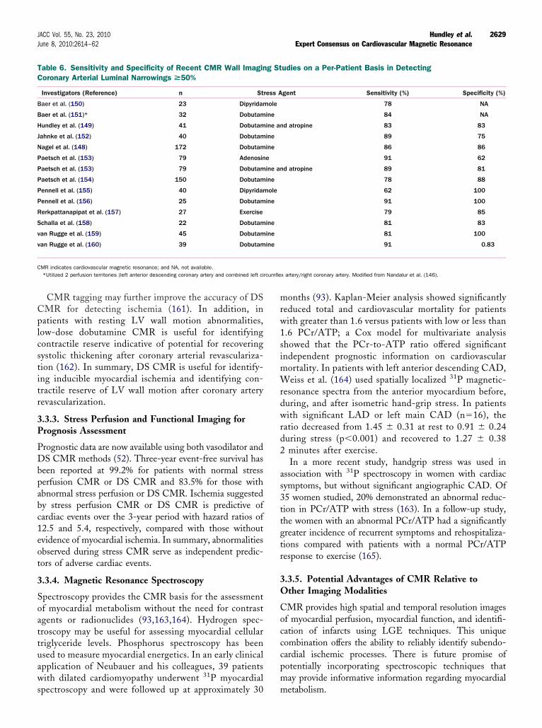
Cplcstitr
3P
PDbpabc1eot
3
Soattuaws
mrw1simWrdwrd2
as3ttgtr
3O
Cocccpm
TC
B
B
H
J
N
P
P
P
P
P
R
S
v
v
Crcumfle
2629JACC Vol. 55, No. 23, 2010 Hundley et al.June 8, 2010:2614–62 Expert Consensus on Cardiovascular Magnetic Resonance
CMR tagging may further improve the accuracy of DSMR for detecting ischemia (161). In addition, inatients with resting LV wall motion abnormalities,
ow-dose dobutamine CMR is useful for identifyingontractile reserve indicative of potential for recoveringystolic thickening after coronary arterial revasculariza-ion (162). In summary, DS CMR is useful for identify-ng inducible myocardial ischemia and identifying con-ractile reserve of LV wall motion after coronary arteryevascularization.
.3.3. Stress Perfusion and Functional Imaging forrognosis Assessment
rognostic data are now available using both vasodilator andS CMR methods (52). Three-year event-free survival has
een reported at 99.2% for patients with normal stresserfusion CMR or DS CMR and 83.5% for those withbnormal stress perfusion or DS CMR. Ischemia suggestedy stress perfusion CMR or DS CMR is predictive ofardiac events over the 3-year period with hazard ratios of2.5 and 5.4, respectively, compared with those withoutvidence of myocardial ischemia. In summary, abnormalitiesbserved during stress CMR serve as independent predic-ors of adverse cardiac events.
.3.4. Magnetic Resonance Spectroscopy
pectroscopy provides the CMR basis for the assessmentf myocardial metabolism without the need for contrastgents or radionuclides (93,163,164). Hydrogen spec-roscopy may be useful for assessing myocardial cellularriglyceride levels. Phosphorus spectroscopy has beensed to measure myocardial energetics. In an early clinicalpplication of Neubauer and his colleagues, 39 patientsith dilated cardiomyopathy underwent 31P myocardial
able 6. Sensitivity and Specificity of Recent CMR Wall Imaginoronary Arterial Luminal Narrowings >50%
Investigators (Reference) n St
aer et al. (150) 23 Dipyridam
aer et al. (151)* 32 Dobutam
undley et al. (149) 41 Dobutam
ahnke et al. (152) 40 Dobutam
agel et al. (148) 172 Dobutam
aetsch et al. (153) 79 Adenosin
aetsch et al. (153) 79 Dobutam
aetsch et al. (154) 150 Dobutam
ennell et al. (155) 40 Dipyridam
ennell et al. (156) 25 Dobutam
erkpattanapipat et al. (157) 27 Exercise
challa et al. (158) 22 Dobutam
an Rugge et al. (159) 45 Dobutam
an Rugge et al. (160) 39 Dobutam
MR indicates cardiovascular magnetic resonance; and NA, not available.*Utilized 2 perfusion territories (left anterior descending coronary artery and combined left ci
pectroscopy and were followed up at approximately 30 m
onths (93). Kaplan-Meier analysis showed significantlyeduced total and cardiovascular mortality for patientsith greater than 1.6 versus patients with low or less than.6 PCr/ATP; a Cox model for multivariate analysishowed that the PCr-to-ATP ratio offered significantndependent prognostic information on cardiovascular
ortality. In patients with left anterior descending CAD,eiss et al. (164) used spatially localized 31P magnetic-
esonance spectra from the anterior myocardium before,uring, and after isometric hand-grip stress. In patientsith significant LAD or left main CAD (n�16), the
atio decreased from 1.45 � 0.31 at rest to 0.91 � 0.24uring stress (p�0.001) and recovered to 1.27 � 0.38minutes after exercise.In a more recent study, handgrip stress was used in
ssociation with 31P spectroscopy in women with cardiacymptoms, but without significant angiographic CAD. Of5 women studied, 20% demonstrated an abnormal reduc-ion in PCr/ATP with stress (163). In a follow-up study,he women with an abnormal PCr/ATP had a significantlyreater incidence of recurrent symptoms and rehospitaliza-ions compared with patients with a normal PCr/ATPesponse to exercise (165).
.3.5. Potential Advantages of CMR Relative tother Imaging Modalities
MR provides high spatial and temporal resolution imagesf myocardial perfusion, myocardial function, and identifi-ation of infarcts using LGE techniques. This uniqueombination offers the ability to reliably identify subendo-ardial ischemic processes. There is future promise ofotentially incorporating spectroscopic techniques thatay provide informative information regarding myocardial
udies on a Per-Patient Basis in Detecting
gent Sensitivity (%) Specificity (%)
78 NA
84 NA
d atropine 83 83
89 75
86 86
91 62
d atropine 89 81
78 88
62 100
91 100
79 85
81 83
81 100
91 0.83
x artery/right coronary artery. Modified from Nandalur et al. (146).
g St
ress A
ole
ine
ine an
ine
ine
e
ine an
ine
ole
ine
ine
ine
ine
etabolism.
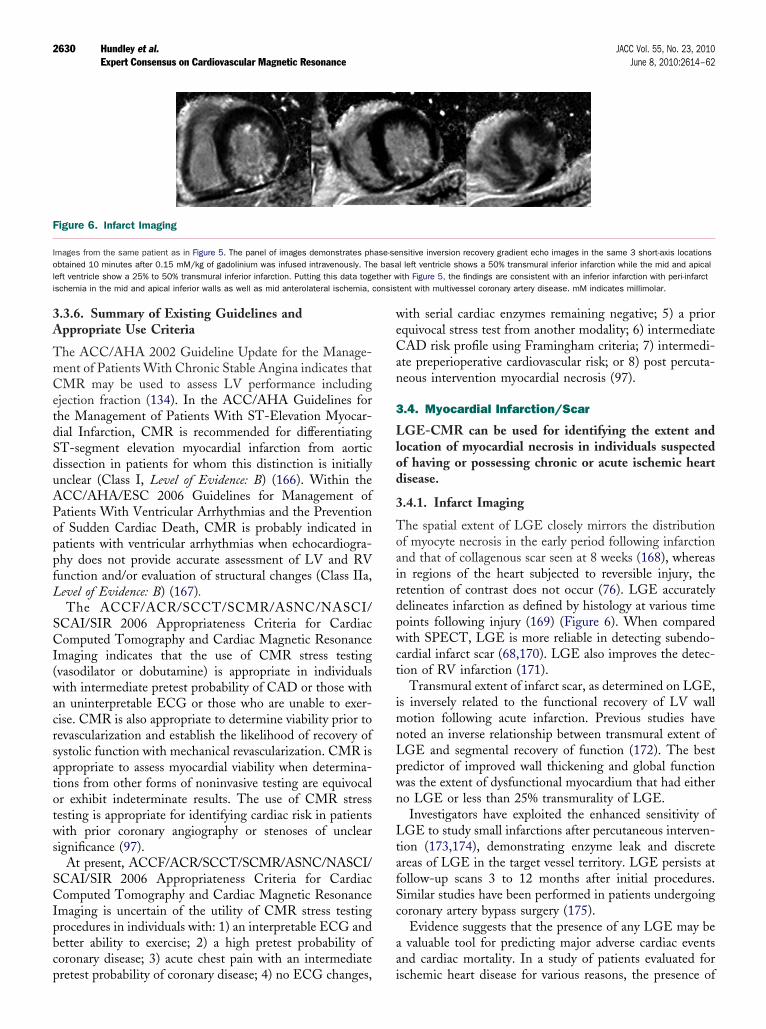
3A
TmCetdSduAPoppfL
SCI(wacrsatotws
SCIpbcp
weCan
3
Llod
3
Toairdpwct
imnLpwn
LtafSc
aa
F
Iol ther wi onsis
2630 Hundley et al. JACC Vol. 55, No. 23, 2010Expert Consensus on Cardiovascular Magnetic Resonance June 8, 2010:2614–62
.3.6. Summary of Existing Guidelines andppropriate Use Criteria
he ACC/AHA 2002 Guideline Update for the Manage-ent of Patients With Chronic Stable Angina indicates thatMR may be used to assess LV performance including
jection fraction (134). In the ACC/AHA Guidelines forhe Management of Patients With ST-Elevation Myocar-ial Infarction, CMR is recommended for differentiatingT-segment elevation myocardial infarction from aorticissection in patients for whom this distinction is initiallynclear (Class I, Level of Evidence: B) (166). Within theCC/AHA/ESC 2006 Guidelines for Management ofatients With Ventricular Arrhythmias and the Preventionf Sudden Cardiac Death, CMR is probably indicated inatients with ventricular arrhythmias when echocardiogra-hy does not provide accurate assessment of LV and RVunction and/or evaluation of structural changes (Class IIa,evel of Evidence: B) (167).The ACCF/ACR/SCCT/SCMR/ASNC/NASCI/
CAI/SIR 2006 Appropriateness Criteria for Cardiacomputed Tomography and Cardiac Magnetic Resonance
maging indicates that the use of CMR stress testingvasodilator or dobutamine) is appropriate in individualsith intermediate pretest probability of CAD or those with
n uninterpretable ECG or those who are unable to exer-ise. CMR is also appropriate to determine viability prior toevascularization and establish the likelihood of recovery ofystolic function with mechanical revascularization. CMR isppropriate to assess myocardial viability when determina-ions from other forms of noninvasive testing are equivocalr exhibit indeterminate results. The use of CMR stressesting is appropriate for identifying cardiac risk in patientsith prior coronary angiography or stenoses of unclear
ignificance (97).At present, ACCF/ACR/SCCT/SCMR/ASNC/NASCI/
CAI/SIR 2006 Appropriateness Criteria for Cardiacomputed Tomography and Cardiac Magnetic Resonance
maging is uncertain of the utility of CMR stress testingrocedures in individuals with: 1) an interpretable ECG andetter ability to exercise; 2) a high pretest probability oforonary disease; 3) acute chest pain with an intermediate
igure 6. Infarct Imaging
mages from the same patient as in Figure 5. The panel of images demonstrates phbtained 10 minutes after 0.15 mM/kg of gadolinium was infused intravenously. Th
eft ventricle show a 25% to 50% transmural inferior infarction. Putting this data togeschemia in the mid and apical inferior walls as well as mid anterolateral ischemia, c
retest probability of coronary disease; 4) no ECG changes, i
ith serial cardiac enzymes remaining negative; 5) a priorquivocal stress test from another modality; 6) intermediateAD risk profile using Framingham criteria; 7) intermedi-
te preperioperative cardiovascular risk; or 8) post percuta-eous intervention myocardial necrosis (97).
.4. Myocardial Infarction/Scar
GE-CMR can be used for identifying the extent andocation of myocardial necrosis in individuals suspectedf having or possessing chronic or acute ischemic heartisease.
.4.1. Infarct Imaging
he spatial extent of LGE closely mirrors the distributionf myocyte necrosis in the early period following infarctionnd that of collagenous scar seen at 8 weeks (168), whereasn regions of the heart subjected to reversible injury, theetention of contrast does not occur (76). LGE accuratelyelineates infarction as defined by histology at various timeoints following injury (169) (Figure 6). When comparedith SPECT, LGE is more reliable in detecting subendo-
ardial infarct scar (68,170). LGE also improves the detec-ion of RV infarction (171).
Transmural extent of infarct scar, as determined on LGE,s inversely related to the functional recovery of LV wall
otion following acute infarction. Previous studies haveoted an inverse relationship between transmural extent ofGE and segmental recovery of function (172). The bestredictor of improved wall thickening and global functionas the extent of dysfunctional myocardium that had eithero LGE or less than 25% transmurality of LGE.Investigators have exploited the enhanced sensitivity of
GE to study small infarctions after percutaneous interven-ion (173,174), demonstrating enzyme leak and discretereas of LGE in the target vessel territory. LGE persists atollow-up scans 3 to 12 months after initial procedures.imilar studies have been performed in patients undergoingoronary artery bypass surgery (175).
Evidence suggests that the presence of any LGE may bevaluable tool for predicting major adverse cardiac events
nd cardiac mortality. In a study of patients evaluated for
nsitive inversion recovery gradient echo images in the same 3 short-axis locationsl left ventricle shows a 50% transmural inferior infarction while the mid and apicalith Figure 5, the findings are consistent with an inferior infarction with peri-infarct
tent with multivessel coronary artery disease. mM indicates millimolar.
ase-see basa
schemic heart disease for various reasons, the presence of
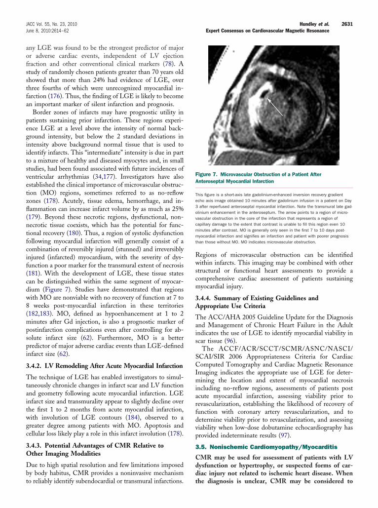
aofsstfa
pegiitsvetzfl(ntfcif(cdw8(mpspi
3
Ttaitwgc
3O
Dbt
Rwscm
3A
Tais
SCImiarfdvp
3
Cdd
FA
Te3ovcmmt
2631JACC Vol. 55, No. 23, 2010 Hundley et al.June 8, 2010:2614–62 Expert Consensus on Cardiovascular Magnetic Resonance
ny LGE was found to be the strongest predictor of majorr adverse cardiac events, independent of LV ejectionraction and other conventional clinical markers (78). Atudy of randomly chosen patients greater than 70 years oldhowed that more than 24% had evidence of LGE, overhree fourths of which were unrecognized myocardial in-arction (176). Thus, the finding of LGE is likely to becomen important marker of silent infarction and prognosis.
Border zones of infarcts may have prognostic utility inatients sustaining prior infarction. These regions experi-nce LGE at a level above the intensity of normal back-round intensity, but below the 2 standard deviations inntensity above background normal tissue that is used todentify infarcts. This “intermediate” intensity is due in parto a mixture of healthy and diseased myocytes and, in smalltudies, had been found associated with future incidences ofentricular arrhythmias (34,177). Investigators have alsostablished the clinical importance of microvascular obstruc-ion (MO) regions, sometimes referred to as no-reflowones (178). Acutely, tissue edema, hemorrhage, and in-ammation can increase infarct volume by as much as 25%179). Beyond these necrotic regions, dysfunctional, non-ecrotic tissue coexists, which has the potential for func-ional recovery (180). Thus, a region of systolic dysfunctionollowing myocardial infarction will generally consist of aombination of reversibly injured (stunned) and irreversiblynjured (infarcted) myocardium, with the severity of dys-unction a poor marker for the transmural extent of necrosis181). With the development of LGE, these tissue statesan be distinguished within the same segment of myocar-ium (Figure 7). Studies have demonstrated that regionsith MO are nonviable with no recovery of function at 7 toweeks post-myocardial infarction in these territories
182,183). MO, defined as hypoenhancement at 1 to 2inutes after Gd injection, is also a prognostic marker of
ostinfarction complications even after controlling for ab-olute infarct size (62). Furthermore, MO is a betterredictor of major adverse cardiac events than LGE-definednfarct size (62).
.4.2. LV Remodeling After Acute Myocardial Infarction
he technique of LGE has enabled investigators to simul-aneously chronicle changes in infarct scar and LV functionnd geometry following acute myocardial infarction. LGEnfarct size and transmurality appear to slightly decline overhe first 1 to 2 months from acute myocardial infarction,ith involution of LGE contours (184), observed to areater degree among patients with MO. Apoptosis andellular loss likely play a role in this infarct involution (178).
.4.3. Potential Advantages of CMR Relative tother Imaging Modalities
ue to high spatial resolution and few limitations imposedy body habitus, CMR provides a noninvasive mechanism
o reliably identify subendocardial or transmural infarctions. tegions of microvascular obstruction can be identifiedithin infarcts. This imaging may be combined with other
tructural or functional heart assessments to provide aomprehensive cardiac assessment of patients sustainingyocardial injury.
.4.4. Summary of Existing Guidelines andppropriate Use Criteria
he ACC/AHA 2005 Guideline Update for the Diagnosisnd Management of Chronic Heart Failure in the Adultndicates the use of LGE to identify myocardial viability incar tissue (96).
The ACCF/ACR/SCCT/SCMR/ASNC/NASCI/CAI/SIR 2006 Appropriateness Criteria for Cardiacomputed Tomography and Cardiac Magnetic Resonance
maging indicates the appropriate use of LGE for deter-ining the location and extent of myocardial necrosis
ncluding no-reflow regions, assessments of patients postcute myocardial infarction, assessing viability prior toevascularization, establishing the likelihood of recovery ofunction with coronary artery revascularization, and toetermine viability prior to revascularization, and assessingiability when low-dose dobutamine echocardiography hasrovided indeterminate results (97).
.5. Nonischemic Cardiomyopathy/Myocarditis
MR may be used for assessment of patients with LVysfunction or hypertrophy, or suspected forms of car-iac injury not related to ischemic heart disease. When
igure 7. Microvascular Obstruction of a Patient Afternteroseptal Myocardial Infarction
his figure is a short-axis late gadolinium-enhanced inversion recovery gradientcho axis image obtained 10 minutes after gadolinium infusion in a patient on Dayafter reperfused anteroseptal myocardial infarction. Note the transmural late gad-
linium enhancement in the anteroseptum. The arrow points to a region of micro-ascular obstruction in the core of the infarction that represents a region ofapillary damage to the extent that contrast is unable to fill this region even 10inutes after contrast. MO is generally only seen in the first 7 to 10 days post-yocardial infarction and signifies an infarction and patient with poorer prognosis
han those without MO. MO indicates microvascular obstruction.
he diagnosis is unclear, CMR may be considered to

ipdaotes
([(mioipttm
3
Itdhmomlodt(dTmtdHH(oht
3C
Cammirhf
riasrptr(i
3
Notcinomvdc
3
DLm
FP
Upid
2632 Hundley et al. JACC Vol. 55, No. 23, 2010Expert Consensus on Cardiovascular Magnetic Resonance June 8, 2010:2614–62
dentify the etiology of cardiac dysfunction in patientsresenting with heart failure including: 1) evaluation ofilated cardiomyopathy in the setting of normal coronaryrteries; 2) patients with positive cardiac enzymes with-ut obstructive atherosclerosis on angiography; 3) pa-ients suspected of amyloidosis or other infiltrative dis-ases; 4) HCM; 5) arrhythmogenic RV dysplasia; or 6)yncope or ventricular arrhythmia.
Nonischemic cardiomyopathies include genetic formsHCM, arrhythmogenic right ventricular cardiomyopathyARVC], LV noncompaction, and others), mixed formsdilated cardiomyopathies [DCM], and restrictive cardio-yopathies), and acquired forms (myocarditis, stress-
nduced cardiomyopathy, peripartum cardiomyopathy, andthers). Knowledge of the etiology of a cardiomyopathy ismportant for diagnosis, therapy, and prognosis. CMRrovides a noninvasive measure to provide this knowledgehrough determination of cardiac chamber size and struc-ure, LV and RV regional and global function, perfusion,etabolism, and tissue composition.
.5.1. Hypertrophic Cardiomyopathy
nappropriate myocardial hypertrophy, loss of diastolic func-ion, development of intramyocardial fibrosis, and possibleynamic systolic obstruction of the LV outflow tract areallmarks of HCM. CMR accurately quantifies myocardialass and regional wall thickness in all myocardial segments. In
bstructive HCM, systolic anterior movement of the anterioritral valve apparatus and a turbulent jet can be identified on
ong-axis cine bright blood imaging studies. The area of thebstructed LV outflow tract can be quantified for diagnosis andirecting therapy longitudinally over time (185). Specific pat-erns of focal or regional LGE have been reported in HCM186) and found to be associated with regional hypertrophy,ecreased systolic thickening, and perfusion deficits (187).hese patterns can be scattered throughout the hypertrophiedyocardium and are dissimilar to the endocardial based pat-
erns of LGE seen after myocardial infarction. Preliminaryata suggest a prognostic relevance of LGE in patients withCM (89,186). CMR is also very sensitive for detectingCM in the first-degree relatives of those with clinical HCM
188). During treatment, CMR can readily identify the effectsf alcohol septal ablation (25). LGE in hypertrophied muscleas been shown to be associated with increased fibrosis withinhe LV myocardium.
.5.2. Arrhythmogenic Right Ventricularardiomyopathy
haracteristics of ARVC include global or regional dilatationnd dysfunction of the RV (and in some cases, the LV)yocardium. Furthermore, fatty and/or fibrous replacementay be found. Morphological and functional targets for CMR
nclude regional or global wall motion abnormalities, aneu-ysms, and segmental or global dilation, as well as globalypokinesis, with quantitative analysis of RV volume and
unction (189,190). The role of CMR in ARVC has been Decently reviewed (191). In contrast to earlier reports, thedentification of myocardial fat is not the only structural wallbnormality associated with ARVC (192) and may be lesspecific for the disease (192). LGE of RV fibrosis has beeneported as a useful marker (Figure 8) (193). Combinedrotocols involving determination of wall motion and RVissue characteristics may provide an excellent diagnostic accu-acy, as shown in patients with genetically defined disease194). Recent studies in gene carriers have also emphasized themportant role of LV involvement in ARVC (195).
.5.3. Noncompaction Cardiomyopathy
oncompaction cardiomyopathy is described as a cardiomy-pathy that occurs due to an autosomally dominant inheritedrait in which the middle and apical segments exhibit a thinompact wall with regional dilatation, dysfunction, and signif-cant hypertrabeculation (Figure 9). An end-diastolic ratio ofoncompacted to compacted LV myocardium of greater thanr equal to 2.3 defines the condition (196). Also, LV wallotion abnormalities, global dysfunction, or coronary intra-
entricular thrombi are often present in the disorder. Refinediagnostic criteria may be forthcoming as this disorder be-omes recognized with greater frequency.
.5.4. Dilated Cardiomyopathy
iagnostic targets for CMR in DCM include progressiveV dilation, LV systolic dysfunction, and regional midwallyocardial fibrosis (88). Recently, focal septal fibrosis in
igure 8. Late Gadolinium Enhancement in ARVC in aatient With Family History of ARVC
pper panel: irregular silhouette of the free RV wall with microaneurysm. Loweranel: evidence for LGE of the RV wall (arrowheads), but also focal fibrosis of the
nterventricular septum (arrow). ARVC indicates arrhythmogenic right ventricular car-iomyopathy; LGE, late gadolinium enhancement; and RV, right ventricular.
CM, the so-called “midwall sign,” has been linked to
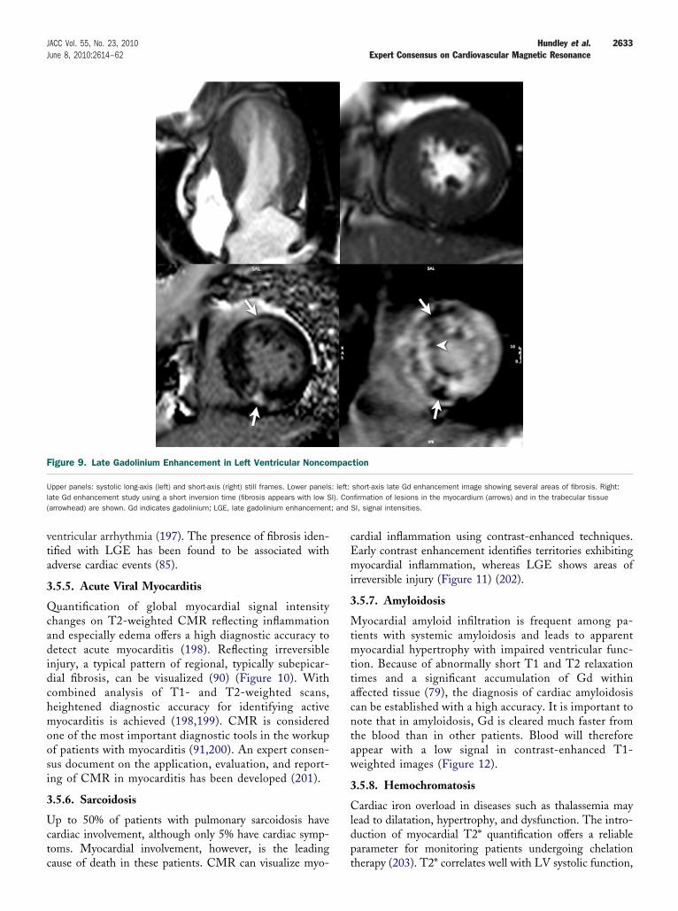
vta
3
Qcadidchmoosi
3
Uctc
cEmi
3
Mtmttacntaw
3
Cldp
F
Ul( ; and
2633JACC Vol. 55, No. 23, 2010 Hundley et al.June 8, 2010:2614–62 Expert Consensus on Cardiovascular Magnetic Resonance
entricular arrhythmia (197). The presence of fibrosis iden-ified with LGE has been found to be associated withdverse cardiac events (85).
.5.5. Acute Viral Myocarditis
uantification of global myocardial signal intensityhanges on T2-weighted CMR reflecting inflammationnd especially edema offers a high diagnostic accuracy toetect acute myocarditis (198). Reflecting irreversible
njury, a typical pattern of regional, typically subepicar-ial fibrosis, can be visualized (90) (Figure 10). Withombined analysis of T1- and T2-weighted scans,eightened diagnostic accuracy for identifying activeyocarditis is achieved (198,199). CMR is considered
ne of the most important diagnostic tools in the workupf patients with myocarditis (91,200). An expert consen-us document on the application, evaluation, and report-ng of CMR in myocarditis has been developed (201).
.5.6. Sarcoidosis
p to 50% of patients with pulmonary sarcoidosis haveardiac involvement, although only 5% have cardiac symp-oms. Myocardial involvement, however, is the leading
igure 9. Late Gadolinium Enhancement in Left Ventricular Noncom
pper panels: systolic long-axis (left) and short-axis (right) still frames. Lower panelsate Gd enhancement study using a short inversion time (fibrosis appears with low Sarrowhead) are shown. Gd indicates gadolinium; LGE, late gadolinium enhancement
ause of death in these patients. CMR can visualize myo- t
ardial inflammation using contrast-enhanced techniques.arly contrast enhancement identifies territories exhibitingyocardial inflammation, whereas LGE shows areas of
rreversible injury (Figure 11) (202).
.5.7. Amyloidosis
yocardial amyloid infiltration is frequent among pa-ients with systemic amyloidosis and leads to apparentyocardial hypertrophy with impaired ventricular func-
ion. Because of abnormally short T1 and T2 relaxationimes and a significant accumulation of Gd withinffected tissue (79), the diagnosis of cardiac amyloidosisan be established with a high accuracy. It is important toote that in amyloidosis, Gd is cleared much faster fromhe blood than in other patients. Blood will thereforeppear with a low signal in contrast-enhanced T1-eighted images (Figure 12).
.5.8. Hemochromatosis
ardiac iron overload in diseases such as thalassemia mayead to dilatation, hypertrophy, and dysfunction. The intro-uction of myocardial T2* quantification offers a reliablearameter for monitoring patients undergoing chelation
tion
short-axis late Gd enhancement image showing several areas of fibrosis. Right:firmation of lesions in the myocardium (arrows) and in the trabecular tissue
SI, signal intensities.
pac
: left:I). Con
herapy (203). T2* correlates well with LV systolic function,

bpqta(
3O
CLr
F
Iaa ium w
F
Lw
2634 Hundley et al. JACC Vol. 55, No. 23, 2010Expert Consensus on Cardiovascular Magnetic Resonance June 8, 2010:2614–62
ut not with liver iron content or serum ferritin. Sincerognosis is mainly determined by cardiac involvement, T2*uantification within the LV myocardium has been showno be a more efficacious marker of cardiac iron involvementnd guidance of chelation therapy than serial liver biopsies203,204).
igure 10. Cardiovascular Magnetic Resonance Tissue Characteri
mages obtained by different sequences in the same 2-chamber view are shown. Panbnormality. Panel C shows increased signal intensity in a T2-weighted image, indicapical blood due to slow blood flow (thin arrow). Panel D visualizes a delayed gadolin
igure 11. Late Gadolinium Enhancement in Arrhythmogenic Right
eft panel: long-axis view of a late gadolinium enhancement study showing a transmural leith the same lesion (arrow).
.5.9. Potential Advantages of CMR Relative tother Imaging Modalities
MR can be used to provide excellent serial assessment ofV and RV function and volumes. In addition, it can
eliably visualize the cardiac apex (95). This is important in
in a Patient With Acute Myocarditis
(diastolic) and B (systolic) indicate a basal-anterior and apical-inferior wall motiondema as a feature of acute injury (arrowheads). Note the increased signal of theashout indicating irreversible injury (arrowheads).
tricular Cardiomyopathy in a Patient With Cardiac Sarcoidosis
zation
els Ating e
Ven
sion in the basal lateral wall (arrow). Right panel: cross-referenced short-axis view
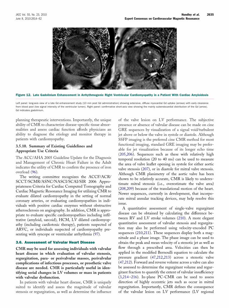
pamap
3A
Taio
SpCecvaptaAs
3
Chrcdtw
ss
opGjSfa(ttvAst(Nri
dtqtsnoflap(bg(dr
F
Lf ive shG
2635JACC Vol. 55, No. 23, 2010 Hundley et al.June 8, 2010:2614–62 Expert Consensus on Cardiovascular Magnetic Resonance
lanning therapeutic interventions. Importantly, the uniquebility of CMR to characterize disease-specific tissue abnor-alities and assess cardiac function affords physicians an
bility to diagnose the etiology and monitor therapy inatients with cardiomyopathy.
.5.10. Summary of Existing Guidelines andppropriate Use Criteria
he ACC/AHA 2005 Guideline Update for the Diagnosisnd Management of Chronic Heart Failure in the Adultndicates the utility of CMR to confirm the presence of ironverload (96).The writing committee recognizes the ACCF/ACR/
CCT/SCMR/ASNC/NASCI/SCAI/SIR 2006 Appro-riateness Criteria for Cardiac Computed Tomography andardiac Magnetic Resonance Imaging for utilizing CMR to
valuate dilated cardiomyopathy in the setting of normaloronary arteries, or evaluating cardiomyopathies in indi-iduals with positive cardiac enzymes without obstructivetherosclerosis on angiography. In addition, CMR is appro-riate to evaluate specific cardiomyopathies including infil-rative (amyloid, sarcoid), HCM, LV dilated cardiomyop-thy (including cardiotoxic therapy), patients suspected ofRVC, or individuals suspected of cardiomyopathy pre-
enting with syncope or ventricular arrhythmia (97).
.6. Assessment of Valvular Heart Disease
MR may be used for assessing individuals with valvulareart disease in which evaluation of valvular stenosis,egurgitation, para- or perivalvular masses, perivalvularomplications of infectious processes, or prosthetic valveisease are needed. CMR is particularly useful in iden-ifying serial changes in LV volumes or mass in patientsith valvular dysfunction.In patients with valvular heart disease, CMR is uniquely
uited to identify and assess the magnitude of valvular
igure 12. Late Gadolinium Enhancement in Arrhythmogenic Right
eft panel: long-axis view of a late Gd enhancement study (10 min post Gd administrom blood pool (low signal intensity of the ventricular lumen). Right panel: confirmatd indicates gadolinium.
tenosis or regurgitation, as well as determine the influence o
f the valve lesion on LV performance. The subjectiveresence or absence of valvular disease can be made on cineRE sequences by visualization of a signal void/turbulent
et above or below the valve in systole or diastole. AlthoughSFP imaging is the preferred cine CMR method for mostunctional imaging, standard GRE imaging may be prefer-ble for jet visualization because of its longer echo time205,206). Sequences such as these with relatively highemporal resolution (20 to 40 ms) can be used to measurehe area of valve leaflet opening in systole for either aorticalve stenosis (207), or in diastole for mitral valve stenosis.lthough CMR planimetry of the aortic valve has been
hown to be relatively accurate, CMR is likely to underes-imate mitral stenosis (i.e., overestimate the valve area)208,209) because of the translational motion of the heart.ewer sequences, currently in development, that incorpo-
ate mitral annular tracking devices, may help resolve thisssue.
A quantitative assessment of single-valve regurgitantisease can be obtained by calculating the difference be-ween RV and LV stroke volumes (210). A more elegantuantitative assessment of valvular stenosis and regurgita-ion may also be performed using velocity-encoded PCequences (210,211). These sequences display both a mag-itude and a phase image. The phase image can be used tobtain the peak and mean velocity of a stenotic jet as well asow through a prescribed area. Velocities can then bepplied in the modified Bernoulli equation to calculate theressure gradient (47,212,213) across a stenotic valve47,212). Forward and reverse volume across a valve can alsoe assessed to determine the regurgitant volume and regur-itant fraction to quantify the extent of valvular insufficiency3,214–216). In-plane PC-CMR can be used to assessirection of highly eccentric jets such as occur in mitralegurgitation. Importantly, CMR defines the consequence
tricular Cardiomyopathy in a Patient With Cardiac Amyloidosis
showing extensive, diffuse myocardial Gd uptake (arrows) with early clearanceort-axis view showing the mainly subendocardial distribution of the Gd (arrow).
Ven
ration)
f the valvular lesion on LV performance (LV regional
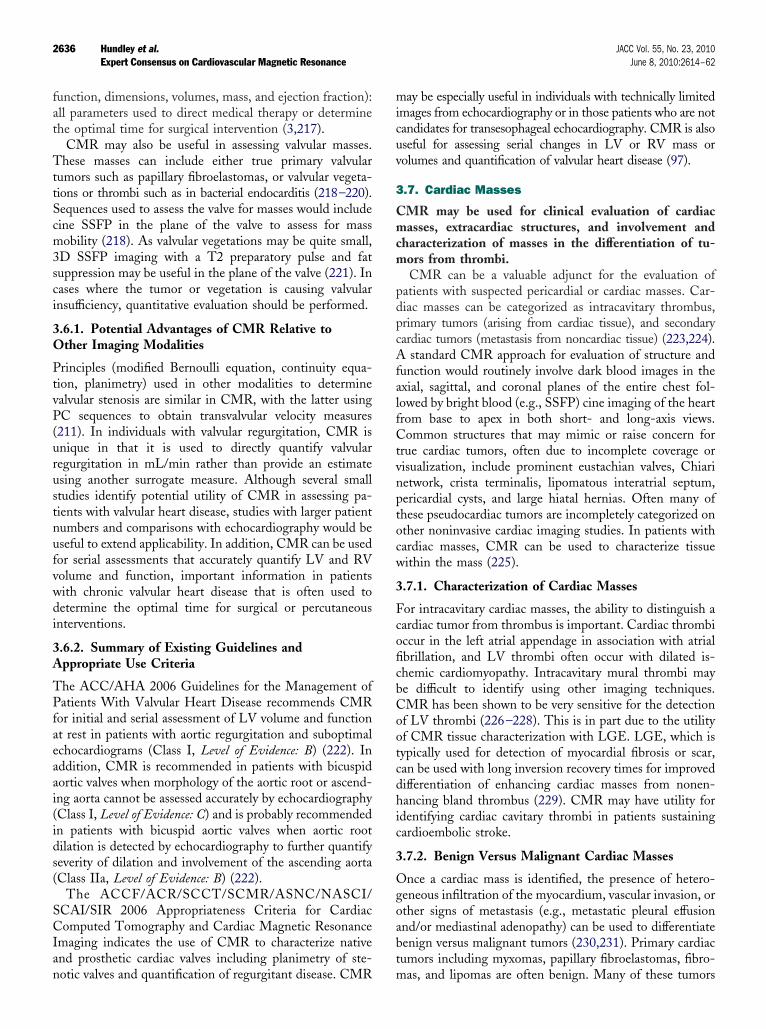
fat
TttScm3sci
3O
PtvP(urustnufvwdi
3A
TPfaeaai(ids(
SCIan
micuv
3
Cmcm
pdpcAfalfCtvnptocw
3
FcoficbCootcdhic
3
Ogoabt
2636 Hundley et al. JACC Vol. 55, No. 23, 2010Expert Consensus on Cardiovascular Magnetic Resonance June 8, 2010:2614–62
unction, dimensions, volumes, mass, and ejection fraction):ll parameters used to direct medical therapy or determinehe optimal time for surgical intervention (3,217).
CMR may also be useful in assessing valvular masses.hese masses can include either true primary valvular
umors such as papillary fibroelastomas, or valvular vegeta-ions or thrombi such as in bacterial endocarditis (218–220).equences used to assess the valve for masses would includeine SSFP in the plane of the valve to assess for massobility (218). As valvular vegetations may be quite small,
D SSFP imaging with a T2 preparatory pulse and fatuppression may be useful in the plane of the valve (221). Inases where the tumor or vegetation is causing valvularnsufficiency, quantitative evaluation should be performed.
.6.1. Potential Advantages of CMR Relative tother Imaging Modalities
rinciples (modified Bernoulli equation, continuity equa-ion, planimetry) used in other modalities to determinealvular stenosis are similar in CMR, with the latter usingC sequences to obtain transvalvular velocity measures
211). In individuals with valvular regurgitation, CMR isnique in that it is used to directly quantify valvularegurgitation in mL/min rather than provide an estimatesing another surrogate measure. Although several smalltudies identify potential utility of CMR in assessing pa-ients with valvular heart disease, studies with larger patientumbers and comparisons with echocardiography would beseful to extend applicability. In addition, CMR can be usedor serial assessments that accurately quantify LV and RVolume and function, important information in patientsith chronic valvular heart disease that is often used toetermine the optimal time for surgical or percutaneous
nterventions.
.6.2. Summary of Existing Guidelines andppropriate Use Criteria
he ACC/AHA 2006 Guidelines for the Management ofatients With Valvular Heart Disease recommends CMR
or initial and serial assessment of LV volume and functiont rest in patients with aortic regurgitation and suboptimalchocardiograms (Class I, Level of Evidence: B) (222). Inddition, CMR is recommended in patients with bicuspidortic valves when morphology of the aortic root or ascend-ng aorta cannot be assessed accurately by echocardiographyClass I, Level of Evidence: C) and is probably recommendedn patients with bicuspid aortic valves when aortic rootilation is detected by echocardiography to further quantifyeverity of dilation and involvement of the ascending aortaClass IIa, Level of Evidence: B) (222).
The ACCF/ACR/SCCT/SCMR/ASNC/NASCI/CAI/SIR 2006 Appropriateness Criteria for Cardiacomputed Tomography and Cardiac Magnetic Resonance
maging indicates the use of CMR to characterize nativend prosthetic cardiac valves including planimetry of ste-
otic valves and quantification of regurgitant disease. CMR may be especially useful in individuals with technically limitedmages from echocardiography or in those patients who are notandidates for transesophageal echocardiography. CMR is alsoseful for assessing serial changes in LV or RV mass orolumes and quantification of valvular heart disease (97).
.7. Cardiac Masses
MR may be used for clinical evaluation of cardiacasses, extracardiac structures, and involvement and
haracterization of masses in the differentiation of tu-ors from thrombi.CMR can be a valuable adjunct for the evaluation of
atients with suspected pericardial or cardiac masses. Car-iac masses can be categorized as intracavitary thrombus,rimary tumors (arising from cardiac tissue), and secondaryardiac tumors (metastasis from noncardiac tissue) (223,224).
standard CMR approach for evaluation of structure andunction would routinely involve dark blood images in thexial, sagittal, and coronal planes of the entire chest fol-owed by bright blood (e.g., SSFP) cine imaging of the heartrom base to apex in both short- and long-axis views.ommon structures that may mimic or raise concern for
rue cardiac tumors, often due to incomplete coverage orisualization, include prominent eustachian valves, Chiarietwork, crista terminalis, lipomatous interatrial septum,ericardial cysts, and large hiatal hernias. Often many ofhese pseudocardiac tumors are incompletely categorized onther noninvasive cardiac imaging studies. In patients withardiac masses, CMR can be used to characterize tissueithin the mass (225).
.7.1. Characterization of Cardiac Masses
or intracavitary cardiac masses, the ability to distinguish aardiac tumor from thrombus is important. Cardiac thrombiccur in the left atrial appendage in association with atrialbrillation, and LV thrombi often occur with dilated is-hemic cardiomyopathy. Intracavitary mural thrombi maye difficult to identify using other imaging techniques.MR has been shown to be very sensitive for the detectionf LV thrombi (226–228). This is in part due to the utilityf CMR tissue characterization with LGE. LGE, which isypically used for detection of myocardial fibrosis or scar,an be used with long inversion recovery times for improvedifferentiation of enhancing cardiac masses from nonen-ancing bland thrombus (229). CMR may have utility for
dentifying cardiac cavitary thrombi in patients sustainingardioembolic stroke.
.7.2. Benign Versus Malignant Cardiac Masses
nce a cardiac mass is identified, the presence of hetero-eneous infiltration of the myocardium, vascular invasion, orther signs of metastasis (e.g., metastatic pleural effusionnd/or mediastinal adenopathy) can be used to differentiateenign versus malignant tumors (230,231). Primary cardiacumors including myxomas, papillary fibroelastomas, fibro-
as, and lipomas are often benign. Many of these tumors
hcmmcltemc
3O
Cocbra
3A
TlC
SCIit
3
CtCfec
sCrItbpflpopdvp
t
csobftssd
htrA
FA
Ptaccn
2637JACC Vol. 55, No. 23, 2010 Hundley et al.June 8, 2010:2614–62 Expert Consensus on Cardiovascular Magnetic Resonance
ave characteristic anatomic locations and specific tissueharacteristics. The most common primary malignant tu-ors of the heart can be classified as cardiac sarcomas (theost common subtype is angiosarcomas), and primary
ardiac lymphomas. These malignant tumors can extendocally and involve the pericardium and, with tissue charac-erization, have indistinct margins and heterogeneous Gdnhancement. First-pass imaging through a cardiac massay help distinguish vascularized lesions, such as renal
arcinoma metastases, from other nonvascular lesions.
.7.3. Potential Advantages of CMR Relative tother Imaging Modalities
MR has been shown to be beneficial for characterizingf cardiac tumors as benign or malignant (232). Inomparison to other imaging modalities, CMR has theenefits of multiplanar image acquisition, high spatialesolution imaging, large field of view, and tissue char-cterization.
.7.4. Summary of Existing Guidelines andppropriate Use Criteria
he writing committee recognizes that no existing guide-ines are established for the evaluation of cardiac mass withMR.The ACCF/ACR/SCCT/SCMR/ASNC/NASCI/
CAI/SIR 2006 Appropriateness Criteria for Cardiacomputed Tomography and Cardiac Magnetic Resonance
maging indicates the use of CMR for evaluation ofntracardiac and extracardiac masses including suspectedumors or LV thrombi (97).
.8. Pericardial Disease (Constrictive Pericarditis)
MR may be used as a noninvasive imaging modalityo diagnose patients with suspected pericardial disease.
MR can provide a comprehensive structural andunctional assessment of the pericardium as well asvaluate the physiological consequences of pericardialonstriction.
In 2 respects, CMR is useful for assessing patientsuspected of constrictive pericardial disease. First, withMR, the entire pericardium can be visualized without
egard to body habitus or prior surgical procedures (233–237).n constriction, a pericardial thickness greater than or equalo 4 mm is abnormal and visually seen. Second, CMR cane used to evaluate the physiologic impact of abnormalericardial thickening. Distension of the hepatic veins andattening of the interventricular septum are signs of accom-anying elevated right-sided pressures. Paradoxical motionf the interventricular septum may be seen as the right-sidedressures equalize or exceed those on the left duringiastole. Real-time cine CMR can be used to evaluate forentricular interdependence to help distinguish constrictiveericarditis from restrictive cardiomyopathy (238,239).Effusive constriction results from a pericardial effusion
hat has become organized or gelatinous (240). The peri- d
ardial space tends not to be homogeneous on various pulseequences. Tagged cine can be used to demonstrate failuref the tags to distort with systole. Pericardial adhesions maye seen with a normal-thickness pericardium and result inunctional constriction. They are best appreciated withagged cine (Figure 13) (241). Rather than the normallippage of the pericardium across the myocardium duringystole, there is tethering of the myocardium, which impairsiastolic filling.Right heart catheterization has been used to identify the
emodynamic consequence of right-sided heart failure, buthe initial criteria that examined end-diastolic pressureelationships have been of limited predictive value (242).bnormal ventricular interdependence can improve the
igure 13. Cardiovascular Magnetic Resonance Findingsssociated With Pericardial Disease
anel A: a short-axis, cine-tagged imaging is provided. Along the posterior wall ofhe left ventricle (white arrow), tag deformation is absent, indicating pericardialdhesions. Panel B: dark blood T1-weighted spin echo images are provided, indi-ating thickened pericardium along the anterior surface of the right ventricle andorresponding tubular deformity of the ventricles. Advanced lung disease is alsooted.
iagnostic accuracy of right heart catheterization (243).
3O
Tdiper
3A
Tlw
SCIpc
3
Cfdhamoaaoa
opacdftecag
3
Ccnsabc
3
Cwtp(uq(FsgFrCic
3
Tf(futtta
3
CP
CtlaifOi
SaasaaaaS(cmt
2638 Hundley et al. JACC Vol. 55, No. 23, 2010Expert Consensus on Cardiovascular Magnetic Resonance June 8, 2010:2614–62
.8.1. Potential Advantages of CMR Relative tother Imaging Modalities
he marked utility of CMR for assessment of pericardialisorders resides in the fact that comprehensive visual-
zation of the LV endocardium may occur, and thehysiologic consequences of abnormal pericardial thick-ning can be obtained without exposure to ionizingadiation (244).
.8.2. Summary of Existing Guidelines andppropriate Use Criteria
he writing committee recognizes that no existing guide-ines are established for assessment for pericardial diseaseith CMR.The ACCF/ACR/SCCT/SCMR/ASNC/NASCI/
CAI/SIR 2006 Appropriateness Criteria for Cardiacomputed Tomography and Cardiac Magnetic Resonance
maging indicates CMR is appropriate for evaluation ofericardial conditions, including pericardial mass and peri-ardial constriction (97).
.9. Congenital Heart Disease
MR may be used for assessing cardiac structure andunction, blood flow, and cardiac and extracardiac con-uits in individuals with simple and complex congenitaleart disease. Specifically, CMR can be used to identifynd characterize congenital heart disease, to assess theagnitude or quantify the severity of intracardiac shunts
r extra cardiac conduit blood flow, and to evaluate theorta and pulmonary arteries to assess the pathologicalnd physiologic consequences of congenital heart diseasen left and right atrial and ventricular function andnatomy.
CMR can be used to characterize 3 important aspectsf patients with congenital heart disease both pre- andostoperatively: the anatomy of the lesion (including thetria, ventricle, and great vessels and their respectiveonnections), the physiology (including cardiac and con-uit blood flow), and the assessment of ventricularunction (in order to determine how the heart is handlinghe abnormal anatomy and/or physiology). CMR isspecially attractive in congenital heart disease whereomplex anatomic details need to be ascertained, as wells in the pediatric age range where ionizing radiation is arave concern.
.9.1. Anatomy
MR has been used to delineate anatomic details ofongenital heart disease for over 2 decades. CMR determi-ation of anatomy has been validated against other goldtandard techniques and often CMR-derived informationlters therapy (245,246). 3D SSFP and Gd imaging haseen shown to be particularly useful in giving an overview of
omplex anatomy in congenital heart disease. (.9.2. Physiology
ine and PC velocity mapping CMR provide physiciansith a noninvasive method to assess the physiologic impor-
ance of consequential heart lesions. For example, theulmonary to systemic flow ratio (Qp/Qs) of shunt lesionse.g., atrial and ventricular septal defects) can be quantifiedsing CMR (48,247,248). PC-CMR can also provideuantification of collateral blood flow in aortic coarctation249,250), determine caval contributions to each lung inontan patients (251), measure cerebral blood flow inuperior-cavopulmonary connections (252), or quantify re-urgitant fractions in patients after repair of tetralogy ofallot (253,254). Newer techniques such as ultrafast, time-
esolved 3D contrast-enhanced MRA expand the utility ofMR to assess physiology in congenital heart disease. This
s particularly useful in assessing disorders of the pulmonaryirculation (Figure 14).
.9.3. Biventricular Function
here are multiple studies reinforcing the accuracy of CMRor biventricular function in congenital heart disease255,256). The high accuracy and reproducibility of CMRor ventricular cavity size and systolic function is particularlyseful in the congenital heart disease population becausehese patients undergo numerous procedures that changehe physiology and may affect ventricular performance dueo cardiopulmonary bypass or deep hypothermic circulatoryrrest (257,258).
.9.4. Congenital Aortic Disease
oarctation, Complete Interruption, andseudo-Coarctation
oarctation of the aorta, representing abnormal placation ofhe tunica media of the posterior aortic wall proximal to theigamentum arteriosum, accounts for approximately 5% ofll congenital heart disease (259). Most coarctations foundn adults are juxtaductal in location and “simple,” beingound in the absence of other cardiovascular abnormalities.
n the other hand, “complex” coarctations are often presentn infancy due to their associated intracardiac anomalies.
Anatomic CMR (combined spin echo and cine GRE orSFP) (114,118,120–122) and/or CE-MRA (121–123)lone has been shown to be comparable to conventionalngiography for delineation of the location and degree oftenosis due to coarctation. A large series of patients withngiographically confirmed congenital obstructive aorticnomalies, including coarctation and interruption, reported
diagnostic sensitivity of 89%, specificity of 84%, andccuracy of 86% for CMR (spin echo and cine GRE orSFP) and 98%, 99%, and 98%, respectively, for CE-MRA260). However, a general superiority of CMR (spin echo,ine, dynamic PC) with CE-MRA over other imagingodalities, including echocardiography, for combined ana-
omic (location and severity of narrowing) and physiologic
trans-coarctation pressure gradient determination, collat-
editncii
pnDptstpfbct
A
TipsasrllgrdvbmCsd
F
TTrwoavR
2639JACC Vol. 55, No. 23, 2010 Hundley et al.June 8, 2010:2614–62 Expert Consensus on Cardiovascular Magnetic Resonance
ral flow measurement) assessment of coarctation has beenemonstrated (114,118,121,126,129,260,261). Accord-
ngly, CMR (262) or CE-MRA (263) can effectively dis-inguish pseudocoarctation (no hemodynamically significantarrowing, poststenotic flow, or collateral vessels) from trueoarctation of the aorta. In addition, CMR can be used todentify aneurysms of the central nervous system circulationn patients with coarctation.
CMR and CE-MRA can also be effectively used to assessostoperative complications for coarctation, including reste-osis and pseudoaneurysm formation (114,121,135,264).irect visualization of collateral vessels by CE-MRA and
ercent increase in flow from proximal to distal descendinghoracic aorta are more reliable indicators of hemodynamicignificance of restenosis following surgical repair of coarc-ation than arm–leg blood pressure gradient (136). Theresence of metallic stents following angioplasty can inter-ere with postintervention assessment of the coarctation sitey CMR/CE-MRA. However, physiologic evaluation ofhanges in collateral circulation before and after interven-
igure 14. Examples of Cardiovascular Magnetic Resonance in Co
he upper left panel is a cine CMR of a patient with double outlet right ventricle demhe lower left panel is a 3-dimensional reconstruction of a single-ventricle patient afior view) and a bilateral bidirectional cavopulmonary connection where the LSVC andhich is a posterior view). The rightward panels are from a patient with tetralogy of Ff the MPA. This technique encodes flow into and out of the imaging plane with dirend the bottom image demonstrates retrograde or regurgitant flow (black). The frameascular magnetic resonance; LPA, left pulmonary artery; LSVC, left superior vena caSVC, right superior vena cava.
ion is still feasible (142). 1
rch Anomalies and Vascular Rings
he failure of embryonic vascular arches to fuse and regressn the usual manner during the formation of the aortic arch,ulmonary arteries, and ductus arteriosus can cause a widepectrum of vascular congenital abnormalities of the aorticrch and its branches (265). These abnormal vasculartructures, especially when they constitute a true vascularing (i.e., double aortic arch, right aortic arch with aberranteft subclavian artery, or mirror-image right aortic arch witheft-sided ligamentum arteriosum) may cause varying de-rees of compression of the trachea and/or esophagus, withesulting symptoms ranging from none to severe stridor,yspnea, cyanosis, and dysphagia. These arch anomalies/ascular rings require specific surgical corrective measuresased on well-delineated anatomic and physiologic assess-ent of the abnormalities by imaging, such as CMR and/orE-MRA (148). CE-MRA, complemented by flow-
ensitive techniques to evaluate stenosis, is very useful foretecting congenital vascular abnormalities (123,129,
ital Heart Disease
ting semilunar valve morphology. The PV is trileaflet, whereas the AoV is bicuspid.tic to pulmonary anastamosis (arrow on leftward image, which is an anteroposte-are connected to the LPAs and RPAs (best visualized on the rightward image,
fter repair with pulmonary regurgitation using through-plane phase-contrast imagingity encoded as white or black; the top image demonstrates antegrade flow (white),e acquired at peak systole and diastole. AoV indicates aortic valve; CMR, cardio-PA, main pulmonary artery; PV, pulmonary valve; RPA, right pulmonary artery; and
ngen
onstrater aor
RSVCallot a
ctionals werva; M
49,164) and diagnosing potentially life-threatening com-

pr
3O
CcwrCvpdocfiicocttatC
3A
TA(wsCfcLsabpe(FmwB(cFod
Mi
iboa
SCIpcvca
3
Cctt(lnp
apoppmtdpip
3
Tpnc(tcirf
adaamn
2640 Hundley et al. JACC Vol. 55, No. 23, 2010Expert Consensus on Cardiovascular Magnetic Resonance June 8, 2010:2614–62
lications, especially airway compression from vascularings (163).
.9.5. Potential Advantages of CMR Relative tother Imaging Modalities
MR is advantageous for the assessment of patients withongenital heart disease because it can visualize structuresithin and external to the heart with minimal impediments
elated to body habitus. In lesions affecting the right heart,MR provides excellent visualization and comprehensive
olume determinations regardless of RV shape. This isarticularly important in patients with congenital heartisease in which abnormalities of pulmonary artery anatomyr blood flow can raise RV afterload. Importantly, CMRan be used to quantify in 3D anatomy, physiology, andunction and, in pediatric patients can be used to acquirenformation relating to congenital heart disease withoutonizing radiation (266,267), or the need for iodinatedontrast (268,269). Because of concerns regarding exposuref pediatric patients to ionizing radiation (270), the writingommittee feels CMR (rather than cardiovascular computedomography) may be preferred to address questions relatedo pediatric congenital heart disease when 1) there is anppropriate indication for tomographic imaging and 2)here is local expertise present to perform and interpret theMR studies.
.9.6. Summary of Existing Guidelines andppropriate Use Criteria
he ACC/AHA 2008 Guidelines for the Management ofdults With Congenital Heart Disease recommends CMR
Class I, Level of Evidence: B) for assessment of patientsith aortic coarctation and (Class I, Level of Evidence: C) for
ubsequent evaluations of repaired aortic coarctations.MR is also recommended (Class I, Level of Evidence: C)
or baseline imaging of patients with pulmonary stenosis. Inenters with adequate expertise, CMR is useful (Class I,evel of Evidence: B) for the initial screening of patients with
uspected congenital coronary anomalies. Patients withrteriovenous fistula that have a continuous murmur shoulde evaluated with CMR (Class I, Level of Evidence: C). Inatients with congenital heart disease, CMR is useful forvaluating patients with suspected pulmonary hypertensionClass I, Level of Evidence: C) and patients with tetralogy ofallot (Class I, Level of Evidence: C). CMR is also recom-ended to evaluate the great arteries and veins in patientsith prior atrial baffle procedures (Class I, Level of Evidence:) and congenitally corrected transposition of the great arteries
Class I, Level of Evidence: C) and managing patients withomplex congenital heart disease (Class I, Level of Evidence: C).inally, CMR is reasonable in patients with arterial switchperations to evaluate the anatomy and hemodynamics in moreetail (Class IIa, Level of Evidence: C) (271).In addition, the ACC/AHA 2008 Guidelines for theanagement of Adults With Congenital Heart Disease
ndicates that CMR provides additional noninvasive imag- i
ng information in any situation in which findings generatedy echocardiography are uncertain. This includes evaluationf patients with intracardiac communication such as atrialnd ventricular septal defects (271).
The ACCF/ACR/SCCT/SCMR/ASNC/NASCI/CAI/SIR 2006 Appropriateness Criteria for Cardiacomputed Tomography and Cardiac Magnetic Resonance
maging indicates that CMR is appropriate to assess com-lex congenital heart disease including anomalies of theoronary circulation, great vessels, cardiac chambers, andalves (97). CMR is particularly useful in this regard inhildren in which exposure to ionizing radiation is to bevoided.
.10. Pulmonary Angiography
E-MRA may be used in patients with a strong suspi-ion of pulmonary embolism in whom the results of otherests are equivocal or for whom iodinated contrast ma-erial or ionizing radiation are relatively contraindicated272). The writing committee agrees that data in theiterature are insufficient to recommend where pulmo-ary CE-MRA should fit into a diagnostic pathway forulmonary embolism.CMR imaging can be used to assess the pulmonary
rteries for acute and chronic thromboembolic disease,ulmonary arterial stenoses (as in congenital heart disease),r acquired pulmonary arterial pathology such as iatrogenicseudoaneurysms. Pulmonary arterial size in patients withulmonary arterial hypertension can also be assessed. Pri-arily, pulmonary CE-MRA is used with the bolus timed
o opacify the pulmonary arteries at the center of k-spaceata acquisition. Time-resolved contrast-enhanced MRA isarticularly useful for imaging the pulmonary vasculature ast allows complete separation of the pulmonary and systemichases.
.10.1. Pulmonary Emboli
here are few studies regarding the utility of data forulmonary CE-MRA for the diagnosis of acute pulmo-ary embolism (273). Although a few prospective single-enter studies assessing CE-MRA have been performed274 –277), no large randomized, controlled, multicenterrial has been reported. Data from these smaller single-enter studies, however, are promising, with the sensitiv-ty for pulmonary CE-MRA to detect pulmonary embolianging from 77% to 100% and the specificity rangingrom 95% to 100%.
Real-time CE-MR pulmonary perfusion methods can bedded to raise the sensitivity for pulmonary embolismetection (275,278). Techniques used for this purpose havelower spatial resolution, which may preclude direct visu-
lization of emboli; however, these methods display seg-ental and subsegmental perfusion defects analogous to
uclear medicine techniques, which can then be used to
ndirectly predict the presence of embolus.
popiitsEcta1
3A
Anue
3
CfcLfisieabop
capcLuv
3
PptIt(mCdcpt
ardmit
hptait
3O
AdncImp
3A
Anp
SCIefvp
3
Ctiwoc
avtoc
2641JACC Vol. 55, No. 23, 2010 Hundley et al.June 8, 2010:2614–62 Expert Consensus on Cardiovascular Magnetic Resonance
The Gd-based MR contrast agent administered forulmonary CE-MRA can potentially be used to passivelypacify the veins of the pelvis and lower extremities torovide information about deep venous thrombosis dur-ng the same examination. The addition of lower extrem-ty venous imaging, especially from the femoral veins tohe popliteals, has the potential to increase the overallensitivity for pulmonary thromboembolic disease (279).xperience with MRA, even without the use of a CMR
ontrast agent, for the detection of proximal deep venoushrombosis and extension into the pelvic veins has shownsensitivity of 94% to 100% and a specificity of 90% to
00% (280 –282).
.10.2. Summary of Existing Guidelines andppropriate Use Criteria
t present, the writing committee recognizes that there areo guidelines or appropriate use criteria highlighting thetility of CMR for assessment of pulmonary artery diseasesxclusive of congenital heart disease.
.11. Atrial Fibrillation
MR may be used for assessing left atrial structure andunction in patients with atrial fibrillation. The writingommittee recognizes that evolving techniques utilizingGE may have high utility for identifying evidence ofbrotic tissue within the atrial wall or an adjoiningtructure. Standardization of protocols and further stud-es are needed to determine if CMR provides a reliableffective method for detecting thrombi in the left atrialppendage in patients with atrial fibrillation. CMR maye useful for identifying pulmonary vein anatomy prior tor after electrophysiology procedures without need foratient exposure to ionizing radiation.When assessing patients with atrial fibrillation, CMR
an be used to determine the size and shape of the lefttrium and to determine pulmonary vein orientation inatients receiving surgical or percutaneous ablation toontrol heart rate and rhythm. As with determinations ofV and RV chamber size and volumes, CMR has beensed to characterize left atrial chamber dimensions andolumes (81,283–289).
.11.1. Preablation Planning
reprocedural CMR can provide the 3D orientation of theulmonary vein and atrium for the purpose of reducing theime necessary to locate ablation sites during the procedure.mportant considerations include the presence of atrialhrombus and variations of pulmonary venous anatomy290). Most of the required planning and follow-up infor-ation may be obtained with a CE-MRA (291–293).MR may prove to be useful for guiding ablation proce-ures (294,295). Time-resolved MRA is the method ofhoice for assessing the pulmonary veins. For “at-risk”atients, where Gd administration is undesirable, noncon-
rast MRA using 3D SSFP is a useful alternative. iPulmonary vein stenosis is a potential complication ofblation by pulmonary vein isolation with an incidenceanging from 1.5% to 42% depending on how stenosis isefined and on the imaging method (296–299). Uncom-only, pulmonary vein occlusion may lead to pulmonary
nfarction. CMR can be used to identify pulmonary infarc-ion (300,301).
There are also emerging data to suggest that CMR mayave a role in determining postablation scar formation inatients with atrial fibrillation. In this strategy, LGEechniques are employed to outline the extent of scarredtrial myocardium after ablation. Acquiring a stack ofmages encompassing the atrium allows for the determina-ion of scar volume (302).
.11.2. Potential Advantages of CMR Relative tother Imaging Modalities
t present, the role of CMR in management pathways foriagnosing left atrial or atrial appendage thrombus (303) isot defined. Further studies are required to determine thelinical role of CMR in identifying left atrial thrombi (304).n centers with expertise, CMR provides an accurateethod to obtain pulmonary vein orientation without ex-
osure to ionizing radiation or iodinated contrast.
.11.3. Summary of Existing Guidelines andppropriate Use Criteria
t present the writing committee recognizes that there areo guidelines established for the use of CMR in assessingatients with atrial fibrillation.The ACCF/ACR/SCCT/SCMR/ASNC/NASCI/
CAI/SIR 2006 Appropriateness Criteria for Cardiacomputed Tomography and Cardiac Magnetic Resonance
maging indicates that CMR is an appropriate test tovaluate pulmonary veins prior to radiofrequency ablationor atrial fibrillation and to identify left atrial and pulmonaryenous anatomy including dimensions of veins for mappingurposes (97).
.12. Peripheral Arterial Disease
MR for peripheral arterial disease (PAD) may be usedo diagnose anatomic location and degree of stenosis ands useful in selecting patients with lower extremity PADho are candidates for intervention. Additionally, MRAf the lower extremities is appropriate for patients withlaudication (305).
PAD has been estimated to affect more than 5 milliondults in the United States (306). CMR provides a nonin-asive method to evaluate peripheral vessels and to identifyhe location and severity of PAD. Proper vascular evaluationf PAD requires illustration from at least the aortic bifur-ation through the distal trifurcation vessels (and pedal arch
n cases of limb-threatening ischemia). This imaging re-
qs
aflsT
CwhMp
Ctfpts3((dciw(b
3OItslsetalMi
rd(ppccCc
aiCag
T
2
C
2 , cardi
TA
Ng
Kg
2
2642 Hundley et al. JACC Vol. 55, No. 23, 2010Expert Consensus on Cardiovascular Magnetic Resonance June 8, 2010:2614–62
uirement results principally from the high incidence ofynchronous and tandem lesions.
Several CMR techniques have evolved over time for thessessment of peripheral arterial disease, notably 2D time ofight (TOF) (72,307,308), and 3D CE-MRA (72). Thetrengths and limitations of the techniques are shown inable 7.Two meta-analyses of the diagnostic performance of
E-MRA for lower extremity arterial evaluation in patientsith suspected or known PAD (Table 8) demonstratedeightened accuracy of CE-MRA over noncontrast 2DRA for detecting and grading the severity of stenoses in
atients with PAD.The current preferred method for performing peripheral
E-MRA is a multistation (“bolus chase”) 3D CE-MRAhat provides an extended field of view coverage (e.g., 1 m)or a single contrast media injection (309–311). For im-roved visualization of the distal extremity (i.e., infrapopli-eal and/or pedal) arteries, the bolus chase MRA can beupplemented by a dedicated separate contrast-enhancedD MRA using traditional timed arterial-phase CE-MRA312) or multiphase, time-resolved CE-MRA methods313,314). The supplement of bolus chase MRA withedicated distal lower extremity MRA (Figure 15), alsoalled “the hybrid” technique (312), has the advantage ofmproved arterial depiction of the infrapopliteal arteries inhich arterial enhancement can often be variable or fast
diabetic patients [315]), and suboptimally depicted usingolus chase MRA alone (316).
able 7. Peripheral CMR: 2D Time of Flight and Contrast-Enhan
Technique Strengths
D TOF ● No intravenous contrast media requirement
E-MRA (i.e., bolus chaseCE-MRA with time-resolved CE-MRA)
● Short examination times● Provides consistently high vascular contrast-to-no
depiction of arterial segments and stenoses● Less susceptible to flow-related artifacts● Provides dynamic assessment of vascular territo
D indicates 2-dimensional; CE-MRA, contrast-enhanced magnetic resonance angiography; CMR
able 8. Meta-Analysis of Contrast-Enhanced Magnetic Resonangiography for Assessment of Peripheral Arterial Disease
Meta-Analysis Study (Reference) Technique No. of Patient
elemans et al. (2000) (71)old standard � DSA
CE-MRA 2 patients/10
2D TOF MRA 344 patients/1
oelemay et al. (2001) (70)old standard � DSA
CE-MRA† 495 patients/1
2D TOF or2D proximal
compression MRA
679 patients/2
D indicates 2-dimensional; CE-MRA, contrast-enhanced magnetic resonance angiography; DSA, digita*In some studies, patients underwent both CE-MRA and noncontrast 2D MRA. †Single study on 20
.12.1. Potential Advantages of CMR Relative tother Imaging Modalities
n prospective studies comparing color duplex ultrasoundo peripheral CE-MRA, CE-MRA was found to be moreensitive and more specific for the detection of arterialuminal narrowings of greater than 50% (317). A pro-pective multicenter trial of patients (318) randomized toither CE-MRA or duplex ultrasound for PAD evalua-ion found the results of both studied to have similarbility to predict changes in disease severity and quality ofife. Importantly, patients that received peripheral CE-
RA experienced a reduction of additional vascularmaging procedures by 42%.
In a prospective study of consecutive patients (319)andomized to either peripheral CE-MRA or 16-etector row cardiac computed tomographic angiographyCTA), CTA was found to be less expensive ($438 peratient) but with no statistically significant differences inatient outcomes (i.e., quality of life). Mean therapeuticonfidence for CE-MRA and CTA were similar andomparable to that for digital subtraction angiography.TA exposed participants to ionizing radiation and ionic
ontrast.A final consideration for comparison of CMR to CTA
nd ultrasound relates to the utility of CMR for character-zing the components of atherosclerotic plaques (Figure 16).MR is able to differentiate proton spin characteristics
ssociated with water, fibrosis, and fat, and thereby distin-uish lipid relative to non-lipid plaque components.
Magnetic Resonance Angiography
Limitations
● Long examination times (greater than 2 hours)● Highly susceptible to flow-related artifacts that may
result in the overestimation of stenoses orerroneously mimic arterial occlusion
tio for reliable
ime-resolved CE-MRA)
● Requires intravenous contrast mediaadministration
ovascular magnetic resonance; and TOF, time of flight.
Angiography Versus Noncontrast 2D Magnetic Resonance
Studies* Sensitivity (%) Specificity (%) Diagnostic Odds Ratio
s 92–100 91–99 7.5
dies 64–100 68–96 4.5
dies 83–100 64–100 2.8
dies 69–100 23–100 1.0
ced
ise ra
ries (t
nce
s and
studie
3 stu
7 stu
0 stu
l subtraction angiography; MRA, magnetic resonance angiography; and TOF, time of flight.subjects evaluated 2D CE-MRA.

3A
TmmaLesfTmPo2b(
irw9
3
Cc
ocsobAmvowolhdt
asotm(ma
bch
FA
B3iadresonance angiography; and mM, millimolar.
2643JACC Vol. 55, No. 23, 2010 Hundley et al.June 8, 2010:2614–62 Expert Consensus on Cardiovascular Magnetic Resonance
.12.2. Summary of Existing Guidelines andppropriate Use Criteria
he ACC/AHA 2005 Practice Guidelines for the Manage-ent of Patients With Peripheral Arterial Disease recom-ends MRA of the extremities: 1) as useful to diagnose
natomic location and degree of stenosis of PAD (Class I,evel of Evidence: A); 2) should be performed with Gdnhancement (Class I, Level of Evidence: B); and 3) useful inelecting patients with lower extremity PAD as candidatesor endovascular intervention (Class I, Level of Evidence: A).he guidelines also indicate that MRA of the extremitiesay be considered: 1) to select patients with lower extremityAD as candidates for surgical bypass and to select the sitesf surgical anastomosis (Class IIb, Level of Evidence: B); and) for post-revascularization (endovascular and surgicalypass) surveillance in patients with lower extremity PADClass IIb, Level of Evidence: B) (305).
In regard to the presentation of claudication, the Amer-can College of Radiology Appropriateness Criteria 2005anks MRA of the lower extremities as highly appropriateith a ranking of 8 on a scale from 1 (least appropriate) to(most appropriate) for patients with claudication (320).
.13. Carotid Arterial Disease
MR may be used for defining the location and extent ofarotid arterial stenoses.
Arterial stenosis, occlusion, dissection, or aneurysm mayccur anywhere from the aortic arch to the intracranialirculation, in 1 or several sites. Current recommendationstate that endarterectomy, when performed in the presencef severe and symptomatic stenosis (i.e., 70% to 99%), cane expected to prevent 1 stroke in 10 cases performed (321).s with peripheral angiography, MRA has in recent yearsade considerable advances for the study of the carotid and
ertebrobasilar circulation. Various implementations of 2Dr 3D TOF MRA with or without CE-MRA have metith intense interest for rapid, flow-independent evaluationf the extracranial carotids (322) and vertebrobasilar circu-ation (323). CE-MRA, however, is demanding machineardware to generate sufficiently high spatial resolutionuring the first pass of a contrast agent, and some results inhe intracranial circulation were disappointing (324).
With the more recent dissemination of 3.0-T CMR unitsnd the development of multichannel radiofrequency (RF)ubsystems that support parallel imaging, the performancef CE-MRA in the carotids has been greatly improved suchhat it rivals CTA and conventional angiography for assess-ent of carotid stenosis (325) and intracranial aneurysms
326,327) with a significant degree of intermodality agree-ent between MRA and both CTA and digital subtraction
ngiography (325).A unique attribute of CMR is the ability to quantify
lood flow. CMR allows for reproducible quantification ofarotid arterial flow, using PC cine imaging. This technique
igure 15. Bolus Chase Contrast-Enhanced Magnetic Resonancengiography
olus chase CE-MRA of the aorta and lower extremity arteries obtained with a–stage table-stepping protocol during infusion of 0.2 mM/kg of gadolinium chelate
n a patient with peripheral arterial disease. There is evidence of sequential moder-te stenoses in the left superficial femoral artery (upper arrow), as well as runoffisease in the left calf (lower arrow). CE-MRA indicates contrast-enhanced magnetic
as previously been shown to represent a reliable estimation
otvtot
pwy
3O
Csbiarm
3A
Taa
3
Capic
cticaamtb
sdtiT
3O
Ca
3A
Taa
3
Ca
TA
A
A
T
D
A
F
Mrcl es repo
2644 Hundley et al. JACC Vol. 55, No. 23, 2010Expert Consensus on Cardiovascular Magnetic Resonance June 8, 2010:2614–62
f cerebral blood flow (328,329). Furthermore, flow quan-ification of the immediate poststenotic region may be ofalue in the determination of peak blood velocity throughhis stenosis, an indicator of the hemodynamic significancef the luminal compromise seen. PC-CMR may also be usedo assess vertebral artery flow in subclavian steal syndrome.
CMR can be used to address certain questions aboutlaque composition (330,331). However, at the time ofriting, imaging of plaque remains a focus of research, notet having found a defined role in clinical decision making.
.13.1. Potential Advantages of CMR Relative tother Imaging Modalities
MR provides the capability to visualize carotid arterialegments and the intracranial carotid system, the vertebro-asilar system, and the aortic arch—important for thedentification of coexistent or primary disease. This isccomplished noninvasively, without exposure to ionizingadiation. During the same examination, quantitative floweasurements may be acquired.
.13.2. Summary of Existing Guidelines andppropriate Use Criteria
he writing committee recognizes that few guidelines orppropriate use criteria are available for the use of CMR forssessing the carotid arteries.
.14. CMR of Thoracic Aortic Disease
MR may be used for defining the location and extent ofortic aneurysms, erosions, ulcers, dissections; evaluatingost-surgical processes involving the aorta and surround-
ng structures, and aortic size blood flow and cardiacycle–dependent changes in area.
CMR imaging techniques (e.g., spin echo, GRE, andine PC; and 3D CE-MRA) permit the assessment of bothhe characteristic anatomic abnormalities and the predispos-ng or resulting pathophysiologic changes (332–335) asso-iated with diseases of the aorta, including: aortic diameters,ortic root visualization, supra-aortic branch assessment,nd recognition of aortic pathologies (e.g., aneurysm, com-unicating dissection, ulcers, noncommunicating dissec-
ion) (336). If surgical repair for thoracic aortic disease has
igure 16. Atherosclerotic Plaques in Cardiovascular Magnetic Re
ultispectral atherosclerotic plaque imaging of the SFA in the same patient as Figuright. The lumen is preserved (long white arrows), yet there is significant atheroscleronsistent with calcification (seen on all 3 image weightings). The large white arrowhipid-rich necrotic core. The brighter areas around the lumen on PDW and T2-W imagral artery; T1-W, T1-weighted; and T2-W, T2-weighted.
een performed, CE-MRA can be used to assess progres-
ion or regression of the responsible disease process oretection of complications of the surgery (e.g., graft infec-ion) (332,337–339). The utility of CMR for providingnformation regarding diseases of the aorta is provided inable 9.
.14.1. Potential Advantages of CMR Relative tother Imaging Modalities
MR can visualize the arterial lumen, assess the aortic wall,nd measure flow in the aorta without ionizing radiation.
.14.2. Summary of Existing Guidelines andppropriate Use Criteria
he writing committee recognizes that few guidelines orppropriate use criteria are available for the use of CMR forssessing the aorta.
.15. Renal Arterial Disease
MR may be used for evaluating renal arterial stenosesnd quantifying renal arterial blood flow.
able 9. Cardiovascular Magnetic Resonance of Thoracicortic Diseases
Disease of AortaUnique Contributions of
Cardiovascular Magnetic Resonance (Reference)
therosclerosis andpenetrating ulcer
● Identify aortic wall pseudoaneurysm,noncommunicating dissection, aortic rupture(340–342)
neurysm ● Etiology (e.g., atherosclerosis, annuloaortic ectasia)(332–334)
● Assess associated changes in aortic valve (i.e.,regurgitation) (343)
● Presurgical planning and postsurgical follow-up
raumatic injury ● Identify hemorrhage within aortic wall● Differentiate partial versus circumferential tears
issection ● Identify acute versus chronic● Locate entry and exit flaps and extent of dissection● Measure flow in true and false lumen● Identify and assess severity of aortic valve
pathology (344,345)● Differentiate intramural hematoma (323,346,347)
ortitis ● Measure aortic wall thickness in response totreatment
● Detection of wall inflammation (348–350)
nce
ith a T1-W image on the left, PDW image in the middle, and T2-W image on theaque in the wall. The black arrows on the T1-W image point to areas of low signalpoint to areas of low signal on the PDW and T2-W images that are consistent withresent fibrous tissue. PDW indicates proton density–weighted; SFA, superficial fem-
sona
e 10 wotic pleads

asaf
1
2
3
4
fia(
3O
Tdipoadrmbaatottr
3A
TafPWmC
t(
4
4
CspcIbmdugmgf
dtsdoaesi
4I
TstfiwpnvitmCrei
trprt
2645JACC Vol. 55, No. 23, 2010 Hundley et al.June 8, 2010:2614–62 Expert Consensus on Cardiovascular Magnetic Resonance
CMR is well suited to assess several aspects of renalrterial disease (any condition which results in irregularity,tenosis, dissection, or aneurysmal dilatation of the renalrteries). Specifically, CMR can be used to determine theollowing:
. The number and location of renal arteries. Multiple renalarteries are common and occur in approximately 24% ofcases with bilateral multiple renal arteries in 5% of thepopulation (351).
. The severity of renal artery stenosis, including thepresence of fibromuscular dysplasia.
. The configuration of the renal blood supply. The inci-dence of horseshoe kidney at necropsy is estimated at1:666 (352). Furthermore, only in 30% of cases is thehorseshoe kidney supplied by a single renal artery to eachside, the majority of cases receiving multiple renalarteries bilaterally, as well as variable arterial supply ofthe midline isthmus (353).
. The presence of renal or adrenal parenchymal masslesions.
Recent studies have estimated the sensitivity and speci-city of 3.0-T MRA in the detection of intra-abdominalrterial stenosis as 100% and greater than 92%, respectively354,355).
.15.1. Potential Advantages of CMR Relative tother Imaging Modalities
he absence of associated ionizing radiation and nonio-inated contrast medium injection reduces potential tox-
cities related to ionic contrast materials, particularly inatients with renal insufficiency (356). CMR offers thepportunity to perform both morphological renal arterialssessment as well as derive complementary flow-relatedata by means of PC flow quantification of individualenal arteries. This combined approach to renal imagingay provide insight into which patients would most
enefit from endovascular intervention (357,358). Inddition to high-resolution 3D large field-of-view datacquisition, renal arterial MRA also allows assessment ofhe renal and adrenal parenchymal tissue for the presencef congenital anomalies or potentially causative occultumors. Time-resolved first-pass perfusion imaging ofhe kidneys may be valuable in identifying significantenovascular lesions (359).
.15.2. Summary of Existing Guidelines andppropriate Use Criteria
he writing committee recognizes that few guidelines forppropriate use criteria are available for the use of CMRor assessing the renal arteries. The ACC/AHA 2005ractice Guidelines for the Management of Patientsith Peripheral Arterial Disease (lower extremity, renal,esenteric, and abdominal aortic) designates CMR a
lass I recommendation as a screening test to establish che diagnosis of renal arterial stenosis (Level of Evidence: B)305).
. CMR Safety
.1. Introduction
MR is generally considered safe, but there are importantafety concerns that fall into 3 general categories: potentialrojectiles in the MR scanner room, implanted cardiovas-ular devices, and issues related to contrast administration.n regard to potential projectiles, it is important to remem-er that the magnet is always “on.” Therefore, ferromagneticaterials entering the MR room are a hazard and can be
rawn into the bore of the scanner with unopposable andnstoppable force. This produces an immediate lethal dan-er to anyone in the scanner or in the path of ferromagneticaterial attracted to the scanner bore. For this reason, local
uidelines and safety policies are developed to guard againsterromagnetic material entering the MR environment.
This section will explore the issues related to implantedevices and MR contrast and present an overview of theypes of devices that are of concern, as well as the underlyingafety considerations for patients with these devices. Sinceevice specifications change frequently, a comprehensive listf CMR compatible or incompatible devices is not possible,nd information on specific devices will need to be obtainedither from the manufacturer’s package inserts or CMRafety Web sites or handbooks. After reviewing devices,ssues related to Gd contrast will be presented.
.2. General Safety Considerations formplanted Devices
here are several reasons that implanted devices may poseafety considerations for patients undergoing CMR. First,he CMR scanner generates a very powerful static magneticeld. Ferromagnetic objects (i.e., those that contain iron)ill interact with the static field and may move in theatient’s body. Nearly all implanted devices, however, areonferromagnetic or only weakly ferromagnetic. Each de-ice must undergo separate testing to determine whether its likely to translate or rotate in the magnetic field. Besideshe static magnetic field, additional smaller and changingagnetic fields, termed gradients, are generated duringMR scanning. These gradient fields may change very
apidly during CMR scanning. Gradient fields can producelectric currents in wires or leads that can potentially resultn arrhythmia.
In addition to magnetic fields, radiofrequency waves areransmitted into the patient by the CMR scanner. Theseadiofrequency waves are absorbed by the body and canroduce slight (less than 1°C) heating of the patient. Withespect to implanted devices, these radiowaves may poten-ially interfere with certain electronic components as well as
ause heating at the tips of implanted wires.
atttpauimeosCt
iigSs
T“
4
DtntC
nCipufCaitnp
4
Mwimtfratb(o(
choopepwifC
4
MlEutirdbpoav
4
Trsgpe(sptgs
T
M
M
M
M
2646 Hundley et al. JACC Vol. 55, No. 23, 2010Expert Consensus on Cardiovascular Magnetic Resonance June 8, 2010:2614–62
Besides the type of devices, there are many variables thatffect the likelihood that a CMR device could be affected byhe CMR scanner. These include the location of the device,he strength of the CMR scanner and, potentially, whetherhe device has been acutely placed or is firmly fixed inosition. Because of these factors, experts in CMR safetynd physics should be consulted when presented with annfamiliar device prior to undergoing CMR. This applies tondividuals with implanted devices receiving any type of
agnetic resonance procedure including the heart, brain,xtremities, or other body/organ/structure/part. If the typef device is unknown, alternatives to performing CMRhould be evaluated. In general, the benefits of undergoingMR must be weighed against the potential risk of injury to
he patient or device failure.Prior to undergoing CMR, patients are screened for both
mplanted cardiovascular devices as well as other types ofmplants. Patients are screened by licensed MR technolo-ists with supervision by a CMR-knowledgeable physician.tandardized screen forms are available (360–362) thathould be completed prior to undergoing CMR.
Medical devices are classified by the American Society foresting and Materials as “MR safe,” “MR conditional,” and
MR unsafe” (Table 10) (363).
.3. CMR Scanning Post Device Implantation
evices that are manufactured from nonferromagnetic ma-erial (300 series stainless steel, titanium, titanium alloy,itinol) that have no electrical or magnetic components andhat have no concern for heating due to CMR may undergoMR scanning immediately after implantation.For devices that are weakly ferromagnetic, CMR safety has
ot been established for every device, and in some cases, theMR scanner could potentially dislodge or move such a device
mmediately after implantation. Devices that are firmly im-lanted into a vessel wall or adjacent tissues are less likely tondergo motion. In the case of heart values in particular, theorces of the heart on the valve are often much greater than theMR forces due to weak ferromagnetism. In general, waiting
fter implantation (e.g., for 6 weeks) may be considered if thiss an option for the patient. For weakly ferromagnetic devices,he risks and benefits of CMR immediately after implantationeed to be considered to determine whether it is necessary or
able 10. Safety Terminology for Implanted Devices
R safe A device that poses no hazards in theMR environment.
R conditional A device that poses no known hazards in a specificMR imaging environment with constraints on theconditions of use. These constraints mayinclude, e.g., the magnetic field strength orspecific absorption rate.
R unsafe An item that is known to pose hazards in allMR environments.
R indicates magnetic resonance.
ossible to defer the CMR scan. a
.4. Coronary Artery and Peripheral Vascular Stents
ost coronary artery and peripheral vascular stents exhibiteak ferromagnetism or are nonferromagnetic. Anchoring
n the vascular wall likely provides protection against move-ent, and further anchoring of the stent may occur due to
issue ingrowth at 6 to 8 weeks after implantation. However,or nonferromagnetic coronary stents, there is no goodationale or clinical data to suggest that a delay is necessaryfter implantation. Data on specific coronary stents suggesthat many of these could be considered CMR safe (364–367)ut not necessarily at the highest (3.0-T) magnetic fields366,368,369). There have been no reports of increased riskf stent subacute or late thrombosis following CMR scans367,370–373).
Drug-eluting stents have the same considerations asonventional stents regarding ferromagnetism (374). Slighteating of the stent (less than 1°C or less than 2°C forverlapping stents) has been reported, but the effect of thisn the drug-eluting properties of the stent is unknown. It isossible that stent heating may be mitigated by a heat-sinkffect of flowing blood in the vessel. A small study ofatients after myocardial infarction who underwent MRithin 2 weeks of stent implantation detected no increased
ncidence of adverse events at 30 days and 6-monthollow-up compared with patients who did not undergoMR (375).
.5. Aortic Stent Grafts
ost aortic stent grafts that have been tested have beenabeled as MR safe with the exception of the Zenith AAAndovascular Graft Stent, which has been labeled as MRnsafe (362,376). The Zenith stent has significant deflec-ion and torque of the stainless steel component of the graftn the magnetic field. Although no adverse events have beeneported with the Zenith stent, there remains a potential forevice migration or vessel damage so that the risks andenefits of CMR examination should be considered in theseatients (377). With other aortic stent grafts (e.g., End-logic AAA or Lifepath AA), there may be significantssociated artifact around the stent or obscuring of theascular lumen due to the metallic components.
.6. Intracardiac Devices
he majority of prosthetic heart valves and annuloplastyings that have been tested have been labeled as MRafe, with a lesser number labeled as MR conditional. Ineneral, the presence of a prosthetic heart valve or annulo-lasty ring is not considered a contraindication to CMRxamination up to 3.0-T at any time after implantation376,378–382). The forces exerted on valve prosthesis areubstantially less than those exerted by the beating heart andulsatile flow (383). The forces required to pull a suturehrough the valve annulus tissue have been shown to bereater than magnetically induced forces up to a fieldtrength of 4.7-T (384). Thus, patients with valve prosthesis
re unlikely to be at risk for valve dehiscence during clinical
Cb(hi
aicblnpeowcal
pC
4
Cir(bhpasWaaifit
4
CmrtdinemPrMbcc
rpC
4T
Rspbr
ahPtspinnw
poPu
4I
Dsrp(mwPipttimp
ccwmtt(me
2647JACC Vol. 55, No. 23, 2010 Hundley et al.June 8, 2010:2614–62 Expert Consensus on Cardiovascular Magnetic Resonance
MR examinations. Associated CMR-related heating haseen determined to be less than 1°C in ex vivo studies378,380,381,385,386); this is likely to be less due to theeat-sink effect of flowing blood. Valve dysfunction due to
nteraction with the magnetic field has not been reported.Cardiac closure and left atrial appendage occluder devices
re either weakly ferromagnetic or nonferromagnetic depend-ng on the materials used (376,387,388). The majority ofardiac closure and occluder devices that have been tested haveeen labeled as MR safe; several that have been tested areabeled as MR conditional (362). Patients with nonferromag-etic cardiac closure and occluder devices may undergo CMRrocedures at any time after implantation. The timing of CMRxamination at 3.0-T or less in patients with cardiac closure orccluder devices that are weakly ferromagnetic should beeighed on a case-by-case basis. For cases in which there is a
lear potential clinical benefit of scanning in the days to weeksfter implantation, the benefits of the MR examination willikely outweigh the risks of the examination.
Sternal wires associated with cardiac surgery/valve re-lacement are not considered to be a contraindication toMR examination.
.7. Inferior Vena Cava Filters
MR examinations of both animals and humans withmplanted inferior vena cava (IVC) filters have thus far noteported complications or symptomatic filter displacement389–394). Most IVC filters that have been tested haveeen labeled as MR safe; the remainder of IVC filters thatave been tested are classified as MR conditional (362). Inatients who have a weakly ferromagnetic IVC filter (Gi-nturco bird nest IVC filter [Cook, Bloomington, Ind],tainless steel Greenfield vena cava filter [Boston Scientific,
atertown, Mass]), consideration should be made to waitt least 6 weeks before performing CMR examination tollow firm implantation of the device. In cases where theres a strong clinical indication for CMR and the device isrmly anchored, the benefits of performing the CMR prioro 6 weeks may outweigh the potential risks.
.8. Embolization Coils
ommonly utilized embolization coils are either nonferro-agnetic or weakly ferromagnetic. Although there is theo-
etical potential for coil heating during a CMR examina-ion, no significant effects were found on the Guglielmietachable coil (GDC) (Boston Scientific) ex vivo (395) or
n patient studies (396). Embolization coils made fromitinol, platinum, or platinum and iridium have beenvaluated and found to be safe for CMR performed atagnetic field strengths of 3.0-T or less (376,397–400).latinum coils implanted in the CNS have not been
eported to cause complications for patients undergoingR. Most embolization coils that have been tested have
een labeled as MR safe; the remainder of embolizationoils that have been tested have been labeled as MR
onditional (362). For weakly ferromagnetic devices, the tisks of performing CMR prior to 6 weeks after coillacement must be considered relative to the benefits ofMR on a case-by-case basis.
.9. Hemodynamic Monitoring andemporary Pacing Devices
etained temporary epicardial pacing leads are relativelyhort in length without large loops. These are felt not toose a significant risk during CMR. No complications haveeen reported as a result of MR scanning for a patient withetained leads (401).
Hemodynamic catheters that contain conducting wiresnd those few temporary transvenous pacing wires thatave been tested have been labeled as MR unsafe (362).atients with pulmonary artery hemodynamic monitoring/
hermodilution catheters (such as the Swan-Ganz catheter)hould not undergo CMR examinations because of theossible associated risks unless labeling information ornstructions for use are provided that permit CMR exami-ations to be performed safely. Nonferromagnetic pulmo-ary artery catheters without electrically conductive path-ays in the catheter are safe for CMR examination.CMR of patients with temporary pacemaker external
ulse generators is not recommended as CMR can alter theperation of an external pulse generator or damage it.acing of the patient during the CMR may also benreliable with a temporary transvenous lead.
.10. Permanent Cardiac Pacemakers andmplantable Cardioverter Defibrillators
ue to the wide prevalence of cardiovascular diseases, aignificant proportion of patients who would normally beeferred for CMR examinations will have permanent cardiacacemakers or implantable cardioverter defibrillatorsICDs). Pacemakers and ICDs contain metal with ferro-agnetic properties, as well as complex electrical systemsith 1 or several leads implanted into the myocardium.otential complications of CMR under these circumstances
nclude damage or movement of the device, inhibition of theacing output, activation of the tachyarrhythmia therapy ofhe device, cardiac stimulation, and heating of the electrodeips (402–408). These factors may lead to clinical sequelaencluding changes in pacing/defibrillation thresholds, pace-
aker ICD dysfunction or damage (including battery de-letion), arrhythmia, or death (404,409,410).A few small clinical trials have been conducted to assess
onditions under which MR examination with these devicesould be conducted safely. Pacemaker-dependent patientsere excluded from these studies, and the heart rhythm wasonitored during the exam. No episodes of pacing above
he upper rate limit or arrhythmias were noted (410),hough 1 patient had a change in device programming411). Another study suggested that ICDs and pacemakersanufactured after the year 2000 are more resistant to the
lectrical and magnetic fields associated with MR examina-
ion at 1.5-T (412). To date, it is likely that several hundred
pmspraacb
aeacirniCcr
4D
TtItCeeacnv
4
Hdercnt
4
GMsfinlt
(
oppeifKsdp(sartitt1rceCaFaacicghpdtatldbOcabd
5
Wdoac
2648 Hundley et al. JACC Vol. 55, No. 23, 2010Expert Consensus on Cardiovascular Magnetic Resonance June 8, 2010:2614–62
atients have undergone MR examination with either pace-akers or ICDs (413–419), and strategies and protocols for
afe pacemaker/ICD scanning during CMR have beenroposed (420,421). As of this writing, no deaths have beeneported under conditions in which patients were deliber-tely scanned and monitored during the MR examination,lthough changes in pacing threshold, programminghanges, need for device reprogramming, and possiblyattery depletion have been reported.Currently, pacemakers available in the United States
re labeled as MR unsafe (362). At present, CMRxamination of patients with pacemakers is discouragednd should only be considered at highly experiencedenters in cases in which there is a strong clinicalndication and where the benefits clearly outweigh theisks. CMR examination of patients with ICDs shouldot be performed unless the center is highly experienced
n both the operation of these devices and in complexMR procedures in the setting of highly compelling
ircumstances where the benefits clearly outweigh theisks.
.11. Retained Transvenous Pacemaker andefibrillator Leads
here are no clinical studies that have specifically addressedhe risk for CMR associated with retained pacemaker orCD leads. Since no radiofrequency chokes are present onhese leads, significant heating of the lead tips may occur.MR in these circumstances is discouraged, and CMR
xamination should only be considered in centers withxpertise in electrophysiology and CMR when there are nolternatives to the CMR examination under compellinglinical circumstances. Similarly, CMR examination shouldot be performed in patients with known retained trans-enous leads that have fractures.
.12. Hemodynamic Support Devices
emodynamic support devices such as ventricular assistevices and intra-aortic balloon pumps are complexlectromagnetic devices containing ferromagnetic mate-ials. Formal CMR testing of these devices has not beenonducted. However, it is believed that these hemody-amic support devices represent absolute contraindica-ions to CMR examination.
.13. Gadolinium Contrast Agents
adolinium contrast agents are frequently used for CE-RA as well as for imaging the heart for LGE, perfu-
ion, or masses. Currently the use of Gd contrast agentsor these purposes is off-label in the United States. Unlikeodinated contrast materials used with radiographic tech-iques, there are different safety issues relating to under-
ying renal function that need to be considered prior toheir administration.
Mild-to-moderate reactions to Gd contrast agents
e.g., hives, shortness of breath) have been reported to cccur in approximately 1 in 5000 patients. Severe ana-hylactic reactions occur in 1 in 250 000 to 300 000atients. Nephrogenic systemic fibrosis (NSF) is anxtremely rare but important complication of Gd admin-stration associated with acute renal failure or severe renalailure due to advanced chronic kidney disease (Nationalidney Foundation Stage 4 or 5 renal failure). NSF is a
cleroderma-like fibrosing entity of the skin (422). Theisease has systemic features that include involvement ofleura, pericardium, lungs, joints, and striated muscleincluding diaphragm and myocardium) (359,423). Be-ides acute renal failure or severe renal failure due todvanced chronic kidney disease (glomerular filtrationate less than 30 mL/min/1.73 m2), other characteristicshat have been implicated with an increased risk for NSFnclude severe liver failure or liver transplant, kidneyransplant, hypercoagulability, deep vein thrombosis, andissue injury secondary to surgical procedures (424). The-year incidence of NSF in the presence of all recognizedisk factors (end-stage renal disease [ESRD], use of Gdontrast, dialysis, and proinflammatory events) has beenstimated to be between 1% (unpublished data, Mayolinic Experience; ISMRM proceedings, Toronto, 2008)
nd 4.6% (425). Over 200 cases have been reported to theood and Drug Administration as of May 2007, but notll are confirmed. Given the total number of Gd contrastpplications, the overall risk of NSF in other groups isonsidered very low. Because of the risk of NSF, screen-ng for reduced renal function prior to CMR should beonsidered in most individuals and particularly in at-riskroups, for example, older patients, individuals withistory of renal disease or dysfunction, or patients with arior renal transplant. Patients with hepatorenal syn-rome in association with severe liver disease, periliverransplant patients, and patients with acute renal failurere typically poor candidates for Gd contrast administra-ion. Patients undergoing peritoneal dialysis have pro-onged retention of Gd contrast agents and their use isiscouraged. The use of Gd in patients with ESRD muste balanced by the significant risk of NSF (3% to 5%).nce informed consent is obtained, using a macrocyclic
helate (like gadoteridol) in the lowest possible dose andvoiding repeat exposure appear reasonable measures,ased on available evidence (426). Postprocedure hemo-ialysis of all patients with ESRD should be considered.
. Summary
ith its advantages in studying patients with cardiovascularisease and ability to provide high-resolution images, CMRffers a suitable mechanism for assessment in various clinicalnd research applications. Table 11 summarizes the writingommittee’s potential indications for the use of CMR in
linical practice situations.
T
H
C
I
M
N
A
C
P
C
P
A
P
C
C
R
Cr
2649JACC Vol. 55, No. 23, 2010 Hundley et al.June 8, 2010:2614–62 Expert Consensus on Cardiovascular Magnetic Resonance
able 11. Summary of Potential Indications for the Use of Cardiovascular Magnetic Resonance
Disease/Condition Recommendations for Use in Clinical Practice
eart failure CMR may be used for assessment of LV and RV size and morphology, systolic and diastolic function, and for characterizing myocardialtissue for the purpose of understanding the etiology of LV systolic or diastolic dysfunction. The writing committee recognizes thepotential capabilities of spectroscopic techniques for acquiring metabolic information of the heart when evaluating individuals withheart failure.
oronary artery disease CMR may be used for identifying coronary artery anomalies and aneurysms and for determining coronary artery patency. In specializedcenters, CMR may be uniquely useful in identifying patients with multivessel coronary artery disease without exposure to ionizingradiation or iodinated contrast medium.
schemic heart disease The combination of CMR stress perfusion, function, and LGE allows the use of CMR as a primary form of testing for● identifying patients with ischemic heart disease when there are resting ECG abnormalities or an inability to exercise,● defining patients with large vessel coronary artery disease and its distribution who are candidates for interventional
procedures, or● determining patients who are appropriate candidates for interventional procedures.
Assessment of LV wall motion after low-dose dobutamine in patients with resting akinetic LV wall segments is useful for identifyingpatients who will develop improvement in LV systolic function after coronary arterial revascularization. The writing committeerecognizes the potential advantages of spectroscopic techniques for identifying early evidence of myocardial ischemia that may or maynot be evident using existing non-CMR methods.
yocardial infarction/scar LGE-CMR may be used for identifying the extent and location of myocardial necrosis in individuals suspected of having or possessingchronic or acute ischemic heart disease.
onischemic cardiomyopathy/myocarditis
CMR may be used for assessment of patients with LV dysfunction or hypertrophy or suspected forms of cardiac injury not related toischemic heart disease. When the diagnosis is unclear, CMR may be considered to identify the etiology of cardiac dysfunction inpatients presenting with heart failure including● evaluation of dilated cardiomyopathy in the setting of normal coronary arteries,● patients with positive cardiac enzymes without obstructive atherosclerosis on angiography,● patients suspected of amyloidosis or other infiltrative diseases,● hypertrophic cardiomyopathy,● arrhythmogenic right ventricular dysplasia, or● syncope or ventricular arrhythmia.
ssessment of valvular heartdisease
CMR may be used for assessing individuals with valvular heart disease in which evaluation of valvular stenosis, regurgitation, para- orperivalvular masses, perivalvular complications of infectious processes, or prosthetic valve disease are needed. CMR may be useful inidentifying serial changes in LV volumes or mass in patients with valvular dysfunction.
ardiac masses CMR may be used for clinical evaluation of cardiac masses, extracardiac structures, and involvement and characterization of masses inthe differentiation of tumors from thrombi.
ericardial disease (constrictivepericarditis)
CMR may be used as a noninvasive imaging modality to diagnose patients with suspected pericardial disease. CMR can provide acomprehensive structural and functional assessment of the pericardium as well as evaluate the physiological consequences ofpericardial constriction.
ongenital heart disease CMR may be used for assessing cardiac structure and function, blood flow, and cardiac and extracardiac conduits in individuals withsimple and complex congenital heart disease. Specifically, CMR can be used to identify and characterize congenital heart disease, toassess the magnitude or quantify the severity of intracardiac shunts or extracardiac conduit blood flow to evaluate the aorta, and toassess the pathological and physiologic consequences of congenital heart disease on left and right atrial and ventricular function andanatomy.
ulmonary angiography CE-MRA may be used in patients with a strong suspicion of pulmonary embolism in whom the results of other tests are equivocal or forwhom iodinated contrast material or ionizing radiation are relatively contraindicated (255). The writing committee agrees that data inthe literature are insufficient to recommend where pulmonary CE-MRA should fit into a diagnostic pathway for pulmonary embolism.
trial fibrillation CMR may be used for assessing left atrial structure and function in patients with atrial fibrillation. The writing committee recognizes thatevolving techniques utilizing LGE may have high utility for identifying evidence of fibrotic tissue within the atrial wall or an adjoiningstructure. Standardization of protocols and further studies are needed to determine if CMR provides a reliable effective method fordetecting thrombi in the left atrial appendage in patients with atrial fibrillation. CMR is recommended for identifying pulmonary veinanatomy prior to or after electrophysiology procedures without need for patient exposure to ionizing radiation.
eripheral arterial disease CMR recommendations for PAD are in agreement with current guidelines and appropriate use criteria.
CMR for PAD1. is recommended to diagnose anatomic location and degree of stenosis of PAD (Class I, Level of Evidence: A);2. should be performed with gadolinium enhancement (Class I, Level of Evidence: B); and3. is useful in selecting patients with lower extremity PAD as candidates for endovascular intervention (Class I,
Level of Evidence: A).
CMR of the extremities may be considered1. to select patients with lower extremity PAD as candidates for surgical bypass and to select the sites of surgical anastomosis
(Class IIb, Level of Evidence: B); and2. for post-revascularization (endovascular and surgical bypass) surveillance in patients with lower extremity PAD (Class IIb,
Level of Evidence: B) (288).
Additionally, MRA of the lower extremities is appropriate for patients with claudication.
arotid arterial disease CMR may be used for defining the location and extent of carotid arterial stenoses.
MR of thoracic aortic disease CMR may be used for defining the location and extent of aortic aneurysms, erosions, ulcers, dissections; evaluating postsurgicalprocesses involving the aorta and surrounding structures, and aortic size blood flow and cardiac cycle–dependent changes in area.
enal arterial disease CMR may be used for evaluating renal arterial stenoses and quantifying renal arterial blood flow.
E-MRA indicates contrast-enhanced magnetic resonance angiography; CMR, cardiovascular magnetic resonance; ECG, electrocardiogram; LGE, late gadolinium enhancement; LV, left ventricular; RV,ight ventricular; MRA, magnetic resonance angiography; and PAD, peripheral arterial disease.

S
AJCDDTPFEME
R
2650 Hundley et al. JACC Vol. 55, No. 23, 2010Expert Consensus on Cardiovascular Magnetic Resonance June 8, 2010:2614–62
taff
merican College of Cardiology Foundationohn C. Lewin, MD, Chief Executive Officerharlene May, Senior Director, Science and Clinical Policyawn R. Phoubandith, MSW, Director, ACCF Clinicalocumentsanja Kharlamova, Associate Director, Science and Clinicalolicyareen Pourhamidi, MS, MPH, Senior Specialist,vidence-Based Medicinearıa Velasquez, Specialist, Science and Clinical Policy
rin A. Barrett, Senior Specialist, Science and Clinical Policy
EFERENCES
1. Saini S, Frankel RB, Stark DD, et al. Magnetism: a primer andreview. AJR Am J Roentgenol. 1988;150:735–43.
2. Pettigrew RI. Dynamic cardiac MR imaging: techniques and appli-cations. Radiol Clin North Am. 1989;27:1183–203.
3. Hundley WG, Li HF, Willard JE, et al. Magnetic resonance imagingassessment of the severity of mitral regurgitation: comparison withinvasive techniques. Circulation. 1995;92:1151–8.
4. Gandy SJ, Waugh SA, Nicholas RS, et al. Comparison of thereproducibility of quantitative cardiac left ventricular assessments inhealthy volunteers using different MRI scanners: a multicentersimulation. J Magn Reson Imaging. 2008;28:359–65.
5. Edelman RR, Chien D, Kim D. Fast selective black blood MRimaging. Radiology. 1991;181:655–60.
6. Haddad JL, Rofsky NM, Ambrosino MM, et al. T2-weighted MRimaging of the chest: comparison of electrocardiograph-triggeredconventional and turbo spin-echo and nontriggered turbo spin-echosequences. J Magn Reson Imaging. 1995;5:325–9.
7. Simonetti OP, Finn JP, White RD, et al. “Black blood” T2-weightedinversion-recovery MR imaging of the heart. Radiology. 1996;199:49–57.
8. Stark DD, Higgins CB, Lanzer P, et al. Magnetic resonance imagingof the pericardium: normal and pathologic findings. Radiology.1984;150:469–74.
9. Didier D, Saint-Martin C, Lapierre C, et al. Coarctation of the aorta:pre and postoperative evaluation with MRI and MR angiography;correlation with echocardiography and surgery. Int J CardiovascImaging. 2006;22:457–75.
10. Duerinckx AJ, Wexler L, Banerjee A, et al. Postoperative evaluationof pulmonary arteries in congenital heart surgery by magneticresonance imaging: comparison with echocardiography. Am Heart J.1994;128:1139–46.
11. Nienaber CA, Rehders TC, Fratz S. Detection and assessment ofcongenital heart disease with magnetic resonance techniques. J Car-diovasc Magn Reson. 1999;1:169–84.
12. Nienaber CA, Spielmann RP, von Kodolitsch Y, et al. Diagnosis ofthoracic aortic dissection: magnetic resonance imaging versus trans-esophageal echocardiography. Circulation. 1992;85:434–47.
13. Nienaber CA, von Kodolitsch Y, Brockhoff CJ, et al. Comparison ofconventional and transesophageal echocardiography with magneticresonance imaging for anatomical mapping of thoracic aortic dissec-tion: a dual noninvasive imaging study with anatomical and/orangiographic validation. Int J Card Imaging. 1994;10:1–14.
14. von Kodolitsch Y, Csosz SK, Koschyk DH, et al. Intramuralhematoma of the aorta: predictors of progression to dissection andrupture. Circulation. 2003;107:1158–63.
15. Utz JA, Herfkens RJ, Heinsimer JA, et al. Valvular regurgitation:dynamic MR imaging. Radiology. 1988;168:91–4.
16. Natori S, Lai S, Finn JP, et al. Cardiovascular function in multi-ethnic study of atherosclerosis: normal values by age, sex, andethnicity. AJR Am J Roentgenol. 2006;186:S357–65.
17. Sechtem U, Pflugfelder PW, Gould RG, et al. Measurement of right
and left ventricular volumes in healthy individuals with cine MRimaging. Radiology. 1987;163:697–702.18. Semelka RC, Tomei E, Wagner S, et al. Normal left ventriculardimensions and function: interstudy reproducibility of measurementswith cine MR imaging. Radiology. 1990;174:763–8.
19. Semelka RC, Tomei E, Wagner S, et al. Interstudy reproducibility ofdimensional and functional measurements between cine magneticresonance studies in the morphologically abnormal left ventricle. AmHeart J. 1990;119:1367–73.
20. Barkhausen J, Ruehm SG, Goyen M, et al. MR evaluation ofventricular function: true fast imaging with steady-state precessionversus fast low-angle shot cine MR imaging: feasibility study.Radiology. 2001;219:264–9.
21. Moon JC, Lorenz CH, Francis JM, et al. Breath-hold FLASH andFISP cardiovascular MR imaging: left ventricular volume differencesand reproducibility. Radiology. 2002;223:789–97.
22. Malayeri AA, Johnson WC, Macedo R, et al. Cardiac cine MRI:quantification of the relationship between fast gradient echo andsteady-state free precession for determination of myocardial mass andvolumes. J Magn Reson Imaging. 2008;28:60–6.
23. Bloomer TN, Plein S, Radjenovic A, et al. Cine MRI using steadystate free precession in the radial long axis orientation is a fastaccurate method for obtaining volumetric data of the left ventricle. JMagn Reson Imaging. 2001;14:685–92.
24. Bloomgarden DC, Fayad ZA, Ferrari VA, et al. Global cardiacfunction using fast breath-hold MRI: validation of new acquisitionand analysis techniques. Magn Reson Med. 1997;37:683–92.
25. Friedrich MG, Schulz-Menger J, Strohm O, et al. The diagnosticimpact of 2D- versus 3D-left ventricular volumetry by MRI inpatients with suspected heart failure. MAGMA. 2000;11:16–9.
26. Bellenger NG, Davies LC, Francis JM, et al. Reduction in sample sizefor studies of remodeling in heart failure by the use of cardiovascularmagnetic resonance. J Cardiovasc Magn Reson. 2000;2:271–8.
27. Bellenger NG, Burgess MI, Ray SG, et al. Comparison of left ventric-ular ejection fraction and volumes in heart failure by echocardiography,radionuclide ventriculography and cardiovascular magnetic resonance;are they interchangeable? Eur Heart J. 2000;21:1387–96.
28. Grothues F, Smith GC, Moon JC, et al. Comparison of interstudyreproducibility of cardiovascular magnetic resonance with2-dimensional echocardiography in normal subjects and in patientswith heart failure or left ventricular hypertrophy. Am J Cardiol.2002;90:29–34.
29. Grothues F, Moon JC, Bellenger NG, et al. Interstudy reproducibil-ity of right ventricular volumes, function, and mass with cardiovas-cular magnetic resonance. Am Heart J. 2004;147:218–23.
30. Pattynama PM, Lamb HJ, van der Velde EA, et al. Left ventricularmeasurements with cine and spin-echo MR imaging: a study ofreproducibility with variance component analysis. Radiology. 1993;187:261–8.
31. Axel L, Dougherty L. MR imaging of motion with spatial modula-tion of magnetization. Radiology. 1989;171:841–5.
32. Zerhouni EA, Parish DM, Rogers WJ, et al. Human heart: taggingwith MR imaging—a method for noninvasive assessment of myocar-dial motion. Radiology. 1988;169:59–63.
33. Edvardsen T, Rosen BD, Pan L, et al. Regional diastolic dysfunctionin individuals with left ventricular hypertrophy measured by taggedmagnetic resonance imaging—the Multi-Ethnic Study of Athero-sclerosis (MESA). Am Heart J. 2006;151:109–14.
34. Fernandes VR, Polak JF, Edvardsen T, et al. Subclinical atheroscle-rosis and incipient regional myocardial dysfunction in asymptomaticindividuals: the Multi-Ethnic Study of Atherosclerosis (MESA).J Am Coll Cardiol. 2006;47:2420–8.
35. Reichek N. MRI myocardial tagging. J Magn Reson Imaging.1999;10:609–16.
36. Rosen BD, Lima JA, Nasir K, et al. Lower myocardial perfusionreserve is associated with decreased regional left ventricular functionin asymptomatic participants of the multi-ethnic study of atheroscle-rosis. Circulation. 2006;114:289–97.
37. Aletras AH, Ding S, Balaban RS, et al. DENSE: displacementencoding with stimulated echoes in cardiac functional MRI. J MagnReson. 1999;137:247–52.
38. Osman NF, Kerwin WS, McVeigh ER, et al. Cardiac motiontracking using CINE harmonic phase (HARP) magnetic resonanceimaging. Magn Reson Med. 1999;42:1048–60.
39. Ingwall J. ATP and the Heart (Basic Science for the Cardiologist).
Norwell, Mass: Kluwer Academic Publishers, 2002.
2651JACC Vol. 55, No. 23, 2010 Hundley et al.June 8, 2010:2614–62 Expert Consensus on Cardiovascular Magnetic Resonance
40. Firmin DN, Nayler GL, Kilner PJ, et al. The application of phase shiftsin NMR for flow measurement. Magn Reson Med. 1990;14:230–41.
41. Moran PR. A flow velocity zeugmatographic interlace for NMRimaging in humans. Magn Reson Imaging. 1982;1:197–203.
42. Hangiandreou NJ, Rossman PJ, Riederer SJ. Analysis of MRphase-contrast measurements of pulsatile velocity waveforms. J MagnReson Imaging. 1993;3:387–94.
43. Hundley WG, Li HF, Hillis LD, et al. Quantitation of cardiacoutput with velocity-encoded, phase-difference magnetic resonanceimaging. Am J Cardiol. 1995;75:1250–5.
44. Markl M, Harloff A, Bley TA, et al. Time-resolved 3D MR velocitymapping at 3T: improved navigator-gated assessment of vascularanatomy and blood flow. J Magn Reson Imaging. 2007;25:824–31.
45. Kuehne T, Yilmaz S, Steendijk P, et al. Magnetic resonance imaginganalysis of right ventricular pressure-volume loops: in vivo validationand clinical application in patients with pulmonary hypertension.Circulation. 2004;110:2010–6.
46. Bogren HG, Buonocore MH, Follette DM. Four-dimensional aorticblood flow patterns in thoracic aortic grafts. J Cardiovasc MagnReson. 2000;2:201–8.
47. Caruthers SD, Lin SJ, Brown P, et al. Practical value of cardiac magneticresonance imaging for clinical quantification of aortic valve stenosis:comparison with echocardiography. Circulation. 2003;108:2236–43.
48. Hundley WG, Li HF, Lange RA, et al. Assessment of left-to-rightintracardiac shunting by velocity-encoded, phase-difference magneticresonance imaging: a comparison with oximetric and indicatordilution techniques. Circulation. 1995;91:2955–60.
49. Weber OM, Higgins CB. MR evaluation of cardiovascular physiol-ogy in congenital heart disease: flow and function. J Cardiovasc MagnReson. 2006;8:607–17.
50. Kwong RY, Schussheim AE, Rekhraj S, et al. Detecting acutecoronary syndrome in the emergency department with cardiac mag-netic resonance imaging. Circulation. 2003;107:531–7.
51. Pérez-Mayoral E, Negri V, Soler-Padros J, et al. Chemistry ofparamagnetic and diamagnetic contrast agents for Magnetic Reso-nance Imaging and Spectroscopy pH responsive contrast agents. EurJ Radiol. 2008;67:453–8.
52. Jahnke C, Nagel E, Gebker R, et al. Prognostic value of cardiac magneticresonance stress tests: adenosine stress perfusion and dobutamine stresswall motion imaging. Circulation. 2007;115:1769–76.
53. Al-Saadi N, Nagel E, Gross M, et al. Noninvasive detection ofmyocardial ischemia from perfusion reserve based on cardiovascularmagnetic resonance. Circulation. 2000;101:1379–83.
54. Cullen JH, Horsfield MA, Reek CR, et al. A myocardial perfusionreserve index in humans using first-pass contrast-enhanced magneticresonance imaging. J Am Coll Cardiol. 1999;33:1386–94.
55. Nagel E, Klein C, Paetsch I, et al. Magnetic resonance perfusionmeasurements for the noninvasive detection of coronary arterydisease. Circulation. 2003;108:432–7.
56. Al-Saadi N, Nagel E, Gross M, et al. Improvement of myocardialperfusion reserve early after coronary intervention: assessment withcardiac magnetic resonance imaging. J Am Coll Cardiol. 2000;36:1557–64.
57. Fenchel M, Franow A, Stauder NI, et al. Myocardial perfusion afterangioplasty in patients suspected of having single-vessel coronaryartery disease: improvement detected at rest-stress first-pass perfusionMR imaging—initial experience. Radiology. 2005;237:67–74.
58. Lauerma K, Virtanen KS, Sipila LM, et al. Multislice MRI inassessment of myocardial perfusion in patients with single-vesselproximal left anterior descending coronary artery disease before andafter revascularization. Circulation. 1997;96:2859–67.
59. Wu KC, Kim RJ, Bluemke DA, et al. Quantification and time courseof microvascular obstruction by contrast-enhanced echocardiographyand magnetic resonance imaging following acute myocardial infarc-tion and reperfusion. J Am Coll Cardiol. 1998;32:1756–64.
60. Yan AT, Gibson CM, Larose E, et al. Characterization of micro-vascular dysfunction after acute myocardial infarction by cardiovas-cular magnetic resonance first-pass perfusion and late gadoliniumenhancement imaging. J Cardiovasc Magn Reson. 2006;8:831–7.
61. Bogaert J, Kalantzi M, Rademakers FE, et al. Determinants andimpact of microvascular obstruction in successfully reperfused ST-segment elevation myocardial infarction: assessment by magnetic
resonance imaging. Eur Radiol. 2007;17:2572–80.62. Hombach V, Grebe O, Merkle N, et al. Sequelae of acute myocardialinfarction regarding cardiac structure and function and their prog-nostic significance as assessed by magnetic resonance imaging. EurHeart J. 2005;26:549–57.
63. Wu KC, Zerhouni EA, Judd RM, et al. Prognostic significance ofmicrovascular obstruction by magnetic resonance imaging in patientswith acute myocardial infarction. Circulation. 1998;97:765–72.
64. Tarantini G, Cacciavillani L, Corbetti F, et al. Duration of ischemia isa major determinant of transmurality and severe microvascular obstruc-tion after primary angioplasty: a study performed with contrast-enhancedmagnetic resonance. J Am Coll Cardiol. 2005;46:1229–35.
65. Schwitter J, Wacker CM, van Rossum AC, et al. MR-IMPACT:comparison of perfusion-cardiac magnetic resonance with single-photon emission computed tomography for the detection of coronaryartery disease in a multicentre, multivendor, randomized trial. EurHeart J. 2008;29:480–9.
66. Sakuma H, Suzawa N, Ichikawa Y, et al. Diagnostic accuracy of stressfirst-pass contrast-enhanced myocardial perfusion MRI comparedwith stress myocardial perfusion scintigraphy. AJR Am J Roentgenol.2005;185:95–102.
67. Schwitter J, Nanz D, Kneifel S, et al. Assessment of myocardialperfusion in coronary artery disease by magnetic resonance: a com-parison with positron emission tomography and coronary angiogra-phy. Circulation. 2001;103:2230–5.
68. Wagner A, Mahrholdt H, Holly TA, et al. Contrast-enhanced MRIand routine single-photon emission computed tomography (SPECT)perfusion imaging for detection of subendocardial myocardial in-farcts: an imaging study. Lancet. 2003;361:374–9.
69. Prakash A, Powell AJ, Krishnamurthy R, et al. Magnetic resonanceimaging evaluation of myocardial perfusion and viability in congenitaland acquired pediatric heart disease. Am J Cardiol. 2004;93:657–61.
70. Koelemay MJ, Lijmer JG, Stoker J, et al. Magnetic resonanceangiography for the evaluation of lower extremity arterial disease: ameta-analysis. JAMA. 2001;285:1338–45.
71. Nelemans PJ, Leiner T, de Vet HC, et al. Peripheral arterial disease:meta-analysis of the diagnostic performance of MR angiography.Radiology. 2000;217:105–14.
72. Yucel EK, Anderson CM, Edelman RR, et al. AHA scientificstatement: magnetic resonance angiography: update on applicationsfor extracranial arteries. Circulation. 1999;100:2284–301.
73. Krinsky GA, Rofsky NM, DeCorato DR, et al. Thoracic aorta:comparison of gadolinium-enhanced three-dimensional MR angiog-raphy with conventional MR imaging. Radiology. 1997;202:183–93.
74. Willerson JT, Scales F, Mukherjee A, et al. Abnormal myocardialfluid retention as an early manifestation of ischemic injury. Am JPathol. 1977;87:159–88.
75. Ingkanisorn WP, Kwong RY, Bohme NS, et al. Prognosis of negativeadenosine stress magnetic resonance in patients presenting to anemergency department with chest pain. J Am Coll Cardiol. 2006;47:1427–32.
76. Rehwald WG, Fieno DS, Chen EL, et al. Myocardial magneticresonance imaging contrast agent concentrations after reversible andirreversible ischemic injury. Circulation. 2002;105:224–9.
77. Kim RJ, Wu E, Rafael A, et al. The use of contrast-enhancedmagnetic resonance imaging to identify reversible myocardial dys-function. N Engl J Med. 2000;343:1445–53.
78. Kwong RY, Chan AK, Brown KA, et al. Impact of unrecognizedmyocardial scar detected by cardiac magnetic resonance imaging onevent-free survival in patients presenting with signs or symptoms ofcoronary artery disease. Circulation. 2006;113:2733–43.
79. Maceira AM, Joshi J, Prasad SK, et al. Cardiovascular magneticresonance in cardiac amyloidosis. Circulation. 2005;111:186–93.
80. Colucci WS, Braunwald E. Pathophysiology of heart failure. In:Zipes DP, Libby P, Bonow R, eds. Braunwald’s Heart Disease: ATextbook of Cardiovascular Medicine. Philadelphia, Pa: ElsevierSaunders, 2005:509–38.
81. Hudsmith LE, Petersen SE, Francis JM, et al. Normal human leftand right ventricular and left atrial dimensions using steady state freeprecession magnetic resonance imaging. J Cardiovasc Magn Reson.2005;7:775–82.
82. Gotte MJ, Germans T, Russel IK, et al. Myocardial strain and torsionquantified by cardiovascular magnetic resonance tissue tagging: stud-ies in normal and impaired left ventricular function. J Am Coll
Cardiol. 2006;48:2002–11.
1
1
1
1
1
1
1
1
1
1
1
1
1
1
1
1
1
1
1
1
1
1
2652 Hundley et al. JACC Vol. 55, No. 23, 2010Expert Consensus on Cardiovascular Magnetic Resonance June 8, 2010:2614–62
83. Paelinck BP, de Roos A, Bax JJ, et al. Feasibility of tissue magneticresonance imaging: a pilot study in comparison with tissue Dopplerimaging and invasive measurement. J Am Coll Cardiol. 2005;45:1109–16.
84. Abdel-Aty H, Zagrosek A, Schulz-Menger J, et al. Delayed enhance-ment and T2-weighted cardiovascular magnetic resonance imagingdifferentiate acute from chronic myocardial infarction. Circulation.2004;109:2411–6.
85. Assomull RG, Prasad SK, Lyne J, et al. Cardiovascular magneticresonance, fibrosis, and prognosis in dilated cardiomyopathy. J AmColl Cardiol. 2006;48:1977–85.
86. Maceira AM, Prasad SK, Khan M, et al. Normalized left ventricularsystolic and diastolic function by steady state free precession cardio-vascular magnetic resonance. J Cardiovasc Magn Reson. 2006;8:417–26.
87. Castillo E, Osman NF, Rosen BD, et al. Quantitative assessment ofregional myocardial function with MR-tagging in a multi-centerstudy: interobserver and intraobserver agreement of fast strain anal-ysis with Harmonic Phase (HARP) MRI. J Cardiovasc Magn Reson.2005;7:783–91.
88. McCrohon JA, Moon JC, Prasad SK, et al. Differentiation of heartfailure related to dilated cardiomyopathy and coronary artery diseaseusing gadolinium-enhanced cardiovascular magnetic resonance. Cir-culation. 2003;108:54–9.
89. Adabag AS, Maron BJ, Appelbaum E, et al. Occurrence andfrequency of arrhythmias in hypertrophic cardiomyopathy in relationto delayed enhancement on cardiovascular magnetic resonance. J AmColl Cardiol. 2008;51:1369–74.
90. Mahrholdt H, Goedecke C, Wagner A, et al. Cardiovascular mag-netic resonance assessment of human myocarditis: a comparison tohistology and molecular pathology. Circulation. 2004;109:1250–8.
91. Skouri HN, Dec GW, Friedrich MG, et al. Noninvasive imaging inmyocarditis. J Am Coll Cardiol. 2006;48:2085–93.
92. Anderson LJ, Westwood MA, Prescott E, et al. Development ofthalassaemic iron overload cardiomyopathy despite low liver ironlevels and meticulous compliance to desferrioxamine. Acta Haematol.2006;115:106–8.
93. Neubauer S, Horn M, Cramer M, et al. Myocardial phosphocreatine-to-ATP ratio is a predictor of mortality in patients with dilatedcardiomyopathy. Circulation. 1997;96:2190–6.
94. Jung WI, Sieverding L, Breuer J, et al. 31P NMR spectroscopydetects metabolic abnormalities in asymptomatic patients with hy-pertrophic cardiomyopathy. Circulation. 1998;97:2536–42.
95. Strohm O, Schulz-Menger J, Pilz B, et al. Measurement of leftventricular dimensions and function in patients with dilated cardio-myopathy. J Magn Reson Imaging. 2001;13:367–71.
96. Hunt SA, Abraham W, Chin M, et al. ACC/AHA 2005 guidelineupdate for the diagnosis and management of chronic heart failure inthe adult: a report of the American College of Cardiology/AmericanHeart Association Task Force on Practice Guidelines (WritingCommittee to Update the 2001 Guidelines for the Evaluation andManagement of Heart Failure). J Am Coll Cardiol. 2005;46:e1–82.
97. Hendel RC, Patel MR, Kramer CM, et al. ACCF/ACR/SCCT/SCMR/ASNC/NASCI/SCAI/SIR 2006 appropriateness criteria forcardiac computed tomography and cardiac magnetic resonance im-aging: a report of the American College of Cardiology FoundationQuality Strategic Directions Committee Appropriateness CriteriaWorking Group, American College of Radiology, Society of Cardio-vascular Computed Tomography, Society for Cardiovascular Mag-netic Resonance, American Society of Nuclear Cardiology, NorthAmerican Society for Cardiac Imaging, Society for CardiovascularAngiography and Interventions, and Society of Interventional Radi-ology. J Am Coll Cardiol. 2006;48:1475–97.
98. Bunce NH, Lorenz CH, Keegan J, et al. Coronary artery anomalies:assessment with free-breathing three-dimensional coronary MR an-giography. Radiology. 2003;227:201–8.
99. Cheitlin MD, De Castro CM, McAllister HA. Sudden death as acomplication of anomalous left coronary origin from the anteriorsinus of Valsalva, a not-so-minor congenital anomaly. Circulation.1974;50:780–7.
00. Gharib A, Ho V, Rosing D, et al. Coronary artery anomalies andvariants: technical feasibility of assessment with coronary MR an-
giography at 3T. Radiology 2008;247:220–227. Abstract.01. McConnell MV, Ganz P, Selwyn AP, et al. Identification ofanomalous coronary arteries and their anatomic course by magneticresonance coronary angiography. Circulation. 1995;92:3158–62.
02. Post JC, van Rossum AC, Bronzwaer JG, et al. Magnetic resonanceangiography of anomalous coronary arteries. A new gold standard fordelineating the proximal course? Circulation. 1995;92:3163–71.
03. Setser R, Arruda J, Weaver JA, et al. Exclusion of coronary arteryanomalies in patients with congenital cardiovascular abnormalitiesusing whole heart approach. J Cardiovasc Magn Reson 2007;9:128–9. Abstract.
04. Taylor AM, Thorne SA, Rubens MB, et al. Coronary artery imagingin grown up congenital heart disease: complementary role of mag-netic resonance and X-ray coronary angiography. Circulation. 2000;101:1670–8.
05. Vliegen HW, Doornbos J, de Roos A, et al. Value of fast gradientecho magnetic resonance angiography as an adjunct to coronaryarteriography in detecting and confirming the course of clinicallysignificant coronary artery anomalies. Am J Cardiol. 1997;79:773–6.
06. Engel HJ, Torres C, Page HL Jr. Major variations in anatomicalorigin of the coronary arteries: angiographic observations in 4,250patients without associated congenital heart disease. Cathet Cardio-vasc Diagn. 1975;1:157–69.
07. Pennell DJ, Sechtem U, Higgins C, et al. Clinical indications forcardiovascular magnetic resonance: consensus panel report 1. J Car-diovasc Magn Reson. 2004;6:727–65.
08. Akagi T, Rose V, Benson LN, et al. Outcome of coronary arteryaneurysms after Kawasaki disease. J Pediatr. 1992;121:689–94.
09. Greil GF, Stuber M, Botnar RM, et al. Coronary magnetic resonanceangiography in adolescents and young adults with Kawasaki disease.Circulation. 2002;105:908–11.
10. Mavrogeni S, Papadopoulos G, Douskou M, et al. Magnetic reso-nance angiography is equivalent to X-ray coronary angiography forthe evaluation of coronary arteries in Kawasaki disease. J Am CollCardiol. 2004;43:649–52.
11. Mavrogeni S, Papadopoulos G, Douskou M, et al. Magnetic reso-nance angiography, function and viability evaluation in patients withKawasaki disease. J Cardiovasc Magn Reson. 2006;8:493–8.
12. Mavrogeni S, Manginas A, Papadakis E, et al. Correlation betweenmagnetic resonance angiography (MRA) and quantitative coronaryangiography (QCA) in ectatic coronary vessels. J Cardiovasc MagnReson. 2004;6:17–23.
13. Bogaert J, Kuzo R, Dymarkowski S, et al. Coronary artery imagingwith real-time navigator three-dimensional turbo-field-echo MRcoronary angiography: initial experience. Radiology. 2003;226:707–16.
14. Botnar RM, Stuber M, Danias PG, et al. Improved coronary arterydefinition with T2-weighted, free-breathing, three-dimensional cor-onary MRA. Circulation. 1999;99:3139–48.
15. Dewey M, Teige F, Schnapauff D, et al. Combination of free-breathing and breathhold steady-state free precession magnetic res-onance angiography for detection of coronary artery stenoses. J MagnReson Imaging. 2006;23:674–81.
16. Jahnke C, Paetsch I, Schnackenburg B, et al. Coronary MR angiog-raphy with steady-state free precession: individually adapted breath-hold technique versus free-breathing technique. Radiology. 2004;232:669–76.
17. Jahnke C, Paetsch I, Nehrke K, et al. Rapid and complete coronaryarterial tree visualization with magnetic resonance imaging: feasibilityand diagnostic performance. Eur Heart J. 2005;26:2313–9.
18. Kim WY, Danias PG, Stuber M, et al. Coronary magnetic resonanceangiography for the detection of coronary stenoses. N Engl J Med.2001;345:1863–9.
19. Maintz D, Aepfelbacher FC, Kissinger KV, et al. Coronary MRangiography: comparison of quantitative and qualitative data fromfour techniques. AJR Am J Roentgenol. 2004;182:515–21.
20. Ozgun M, Hoffmeier A, Kouwenhoven M, et al. Comparison of 3Dsegmented gradient-echo and steady-state free precession coronaryMRI sequences in patients with coronary artery disease. AJR Am JRoentgenol. 2005;185:103–9.
21. Sakuma H, Ichikawa Y, Suzawa N, et al. Assessment of coronary arterieswith total study time of less than 30 minutes by using whole-
heart coronary MR angiography. Radiology. 2005;237:316–21.
1
1
1
1
1
1
1
1
1
1
1
1
1
1
1
1
1
1
1
1
1
1
1
1
1
1
1
1
1
1
1
1
1
1
1
1
1
1
1
1
1
2653JACC Vol. 55, No. 23, 2010 Hundley et al.June 8, 2010:2614–62 Expert Consensus on Cardiovascular Magnetic Resonance
22. Sakuma H, Ichikawa Y, Chino S, et al. Detection of coronary arterystenosis with whole-heart coronary magnetic resonance angiography.J Am Coll Cardiol. 2006;48:1946–50.
23. Sommer T, Hofer U, Meyer C, et al. Submillimeter 3D coronary MRangiography with real-time navigator correction in 107 patients withsuspected coronary artery disease. Rofo. 2002;174:459–66.
24. Hauser T, Yeon SB, Appelbaum E, et al. Multimodality CMRdetection of coronary artery disease in patients with heart failure anddepressed systolic function: superiority of coronary MRI compared tolate gadolinium enhancement. J Cardiovasc Magn Reson. 2009.Abstract in press.
25. Liu X, Zhao X, Huang J, et al. Comparison of 3D free-breathingcoronary MR angiography and 64–MDCT angiography for detec-tion of coronary stenosis in patients with high calcium scores. AJRAm J Roentgenol. 2007;189:1326–32.
26. Aurigemma GP, Reichek N, Axel L, et al. Noninvasive determina-tion of coronary artery bypass graft patency by cine magneticresonance imaging. Circulation. 1989;80:1595–602.
27. Engelmann MG, Knez A, von Smekal A, et al. Noninvasive coronarybypass graft imaging after multivessel revascularisation. Int J Cardiol.2000;76:65–74.
28. Fitzgibbon GM, Kafka HP, Leach AJ, et al. Coronary bypass graftfate and patient outcome: angiographic follow-up of 5,065 graftsrelated to survival and reoperation in 1,388 patients during 25 years.J Am Coll Cardiol. 1996;28:616–26.
29. Goldman S, Copeland J, Moritz T, et al. Saphenous vein graftpatency 1 year after coronary artery bypass surgery and effects ofantiplatelet therapy: results of a Veterans Administration CooperativeStudy. Circulation. 1989;80:1190–7.
30. van Geuns RJ, Wielopolski PA, de Bruin HG, et al. MR coronaryangiography with breath-hold targeted volumes: preliminary clinicalresults. Radiology. 2000;217:270–7.
31. White RD, Caputo GR, Mark AS, et al. Coronary artery bypass graftpatency: noninvasive evaluation with MR imaging. Radiology. 1987;164:681–6.
32. Langerak SE, Kunz P, Vliegen HW, et al. MR flow mapping incoronary artery bypass grafts: a validation study with Doppler flowmeasurements. Radiology. 2002;222:127–35.
33. Langerak SE, Vliegen HW, Jukema JW, et al. Value of magneticresonance imaging for the noninvasive detection of stenosis incoronary artery bypass grafts and recipient coronary arteries. Circu-lation. 2003;107:1502–8.
34. Gibbons RJ, Abrams J, Chatterjee K, et al. ACC/AHA 2002guideline update for the management of patients with chronic stableangina—summary article: a report of the American College ofCardiology/American Heart Association Task Force on PracticeGuidelines (Committee on the Management of Patients WithChronic Stable Angina). J Am Coll Cardiol. 2003;41:159–68.
35. Wilke N, Jerosch-Herold M, Wang Y, et al. Myocardial perfusionreserve: assessment with multisection, quantitative, first-pass MRimaging. Radiology. 1997;204:373–84.
36. Schaefer S, van Tyen R, Saloner D. Evaluation of myocardial perfusionabnormalities with gadolinium-enhanced snapshot MR imaging inhumans. Work in progress. Radiology. 1992;185:795–801.
37. Cury RC, Cattani CA, Gabure LA, et al. Diagnostic performance ofstress perfusion and delayed-enhancement MR imaging in patientswith coronary artery disease. Radiology. 2006;240:39–45.
38. Doyle M, Fuisz A, Kortright E, et al. The impact of myocardial flowreserve on the detection of coronary artery disease by perfusionimaging methods: an NHLBI WISE study. J Cardiovasc MagnReson. 2003;5:475–85.
39. Giang TH, Nanz D, Coulden R, et al. Detection of coronary arterydisease by magnetic resonance myocardial perfusion imaging withvarious contrast medium doses: first European multi-centre experi-ence. Eur Heart J. 2004;25:1657–65.
40. Ishida N, Sakuma H, Motoyasu M, et al. Noninfarcted myocardium:correlation between dynamic first-pass contrast-enhanced myocardialMR imaging and quantitative coronary angiography. Radiology.2003;229:209–16.
41. Kawase Y, Nishimoto M, Hato K, et al. Assessment of coronaryartery disease with nicorandil stress magnetic resonance imaging.Osaka City Med J. 2004;50:87–94.
42. Klem I, Heitner JF, Shah DJ, et al. Improved detection of coronary
artery disease by stress perfusion cardiovascular magnetic resonancewith the use of delayed enhancement infarction imaging. J Am CollCardiol. 2006;47:1630–8.
43. Pilz G, Bernhardt P, Klos M, et al. Clinical implication of adenosine-stress cardiac magnetic resonance imaging as potential gatekeeperprior to invasive examination in patients with AHA/ACC class IIindication for coronary angiography. Clin Res Cardiol. 2006;95:531–8.
44. Plein S, Radjenovic A, Ridgway JP, et al. Coronary artery disease:myocardial perfusion MR imaging with sensitivity encoding versusconventional angiography. Radiology. 2005;235:423–30.
45. Takase B, Nagata M, Kihara T, et al. Whole-heart dipyridamolestress first-pass myocardial perfusion MRI for the detection ofcoronary artery disease. Jpn Heart J. 2004;45:475–86.
46. Nandalur KR, Dwamena BA, Choudhri AF, et al. Diagnosticperformance of stress cardiac magnetic resonance imaging in thedetection of coronary artery disease: a meta-analysis. J Am CollCardiol. 2007;50:1343–53.
47. Wahl A, Paetsch I, Gollesch A, et al. Safety and feasibility ofhigh-dose dobutamine-atropine stress cardiovascular magnetic reso-nance for diagnosis of myocardial ischaemia: experience in 1000consecutive cases. Eur Heart J. 2004;25:1230–6.
48. Nagel E, Lehmkuhl HB, Bocksch W, et al. Noninvasive diagnosis ofischemia-induced wall motion abnormalities with the use of high-dose dobutamine stress MRI: comparison with dobutamine stressechocardiography. Circulation. 1999;99:763–70.
49. Hundley WG, Hamilton CA, Thomas MS, et al. Utility of fast cinemagnetic resonance imaging and display for the detection of myo-cardial ischemia in patients not well suited for second harmonic stressechocardiography. Circulation. 1999;100:1697–702.
50. Baer FM, Smolarz K, Jungehulsing M, et al. Feasibility of high-dosedipyridamole-magnetic resonance imaging for detection of coronaryartery disease and comparison with coronary angiography. Am JCardiol. 1992;69:51–6.
51. Baer FM, Voth E, Theissen P, et al. Coronary artery disease: findingswith GRE MR imaging and Tc-99m-methoxyisobutyl-isonitrileSPECT during simultaneous dobutamine stress. Radiology. 1994;193:203–9.
52. Jahnke C, Paetsch I, Gebker R, et al. Accelerated 4D dobutaminestress MR imaging with k-t BLAST: feasibility and diagnosticperformance. Radiology. 2006;241:718–28.
53. Paetsch I, Jahnke C, Wahl A, et al. Comparison of dobutamine stressmagnetic resonance, adenosine stress magnetic resonance, and aden-osine stress magnetic resonance perfusion. Circulation. 2004;110:835–42.
54. Paetsch I, Jahnke C, Ferrari VA, et al. Determination of interob-server variability for identifying inducible left ventricular wall motionabnormalities during dobutamine stress magnetic resonance imaging.Eur Heart J. 2006;27:1459–64.
55. Pennell DJ, Underwood SR, Ell PJ, et al. Dipyridamole magneticresonance imaging: a comparison with thallium-201 emission tomog-raphy. Br Heart J. 1990;64:362–9.
56. Pennell DJ, Underwood SR, Manzara CC, et al. Magnetic resonanceimaging during dobutamine stress in coronary artery disease. Am JCardiol. 1992;70:34–40.
57. Rerkpattanapipat P, Gandhi SK, Darty SN, et al. Feasibility to detectsevere coronary artery stenoses with upright treadmill exercise mag-netic resonance imaging. Am J Cardiol. 2003;92:603–6.
58. Schalla S, Klein C, Paetsch I, et al. Real-time MR image acquisitionduring high-dose dobutamine hydrochloride stress for detecting leftventricular wall-motion abnormalities in patients with coronaryarterial disease. Radiology. 2002;224:845–51.
59. van Rugge FP, van der Wall EE, de Roos A, et al. Dobutamine stressmagnetic resonance imaging for detection of coronary artery disease.J Am Coll Cardiol. 1993;22:431–9.
60. van Rugge FP, van der Wall EE, Spanjersberg SJ, et al. Magneticresonance imaging during dobutamine stress for detection andlocalization of coronary artery disease. Quantitative wall motionanalysis using a modification of the centerline method. Circulation.1994;90:127–38.
61. Kuijpers D, Ho KY, van Dijkman PR, et al. Dobutamine cardiovas-cular magnetic resonance for the detection of myocardial ischemiawith the use of myocardial tagging. Circulation. 2003;107:1592–7.
62. Sayad DE, Willett DL, Hundley WG, et al. Dobutamine magnetic
resonance imaging with myocardial tagging quantitatively predicts
1
1
1
1
1
1
1
1
1
1
1
1
1
1
1
1
1
1
1
1
1
1
1
1
1
1
1
1
1
1
1
1
1
1
1
1
1
2
2654 Hundley et al. JACC Vol. 55, No. 23, 2010Expert Consensus on Cardiovascular Magnetic Resonance June 8, 2010:2614–62
improvement in regional function after revascularization. Am JCardiol. 1998;82:1149–51.
63. Buchthal SD, den Hollander JA, Merz CN, et al. Abnormalmyocardial phosphorus-31 nuclear magnetic resonance spectroscopyin women with chest pain but normal coronary angiograms. N EnglJ Med. 2000;342:829–35.
64. Weiss RG, Bottomley PA, Hardy CJ, et al. Regional myocardialmetabolism of high-energy phosphates during isometric exercise inpatients with coronary artery disease. N Engl J Med. 1990;323:1593–600.
65. Johnson BD, Shaw LJ, Buchthal SD, et al. Prognosis in women withmyocardial ischemia in the absence of obstructive coronary disease:results from the National Institutes of Health-National Heart, Lung,and Blood Institute-Sponsored Women’s Ischemia Syndrome Eval-uation (WISE). Circulation. 2004;109:2993–9.
66. Antman EM, Anbe DT, Armstrong PW, et al. ACC/AHA guide-lines for the management of patients with ST-elevation myocardialinfarction: a report of the American College of Cardiology/AmericanHeart Association Task Force on Practice Guidelines (Committee toRevise the 1999 Guidelines for the Management of Patients withAcute Myocardial Infarction). J Am Coll Cardiol. 2004;44:e1–211.
67. Zipes DP, Camm AJ, Borggrefe M, et al. ACC/AHA/ESC 2006guidelines for management of patients with ventricular arrhythmiasand the prevention of sudden cardiac death: a report of the AmericanCollege of Cardiology/American Heart Association Task Force onPractice Guidelines and the European Society of Cardiology Com-mittee for Practice Guidelines (Writing Committee to DevelopGuidelines for Management of Patients With Ventricular Arrhyth-mias and the Prevention of Sudden Cardiac Death). J Am CollCardiol. 2006;48:e247–346.
68. Kim RJ, Lima JA, Chen EL, et al. Fast 23Na magnetic resonanceimaging of acute reperfused myocardial infarction. Potential to assessmyocardial viability. Circulation. 1997;95:1877–85.
69. Fieno DS, Kim RJ, Chen EL, et al. Contrast-enhanced magneticresonance imaging of myocardium at risk: distinction between revers-ible and irreversible injury throughout infarct healing. J Am CollCardiol. 2000;36:1985–91.
70. Ibrahim T, Bulow HP, Hackl T, et al. Diagnostic value of contrast-enhanced magnetic resonance imaging and single-photon emissioncomputed tomography for detection of myocardial necrosis early afteracute myocardial infarction. J Am Coll Cardiol. 2007;49:208–16.
71. Kumar A, Abdel-Aty H, Kriedemann I, et al. Contrast-enhancedcardiovascular magnetic resonance imaging of right ventricular in-farction. J Am Coll Cardiol. 2006;48:1969–76.
72. Choi KM, Kim RJ, Gubernikoff G, et al. Transmural extent of acutemyocardial infarction predicts long-term improvement in contractilefunction. Circulation. 2001;104:1101–7.
73. Choi JW, Gibson CM, Murphy SA, et al. Myonecrosis followingstent placement: association between impaired TIMI myocardialperfusion grade and MRI visualization of microinfarction. CatheterCardiovasc Interv. 2004;61:472–6.
74. Ricciardi MJ, Wu E, Davidson CJ, et al. Visualization of discretemicroinfarction after percutaneous coronary intervention associatedwith mild creatine kinase-MB elevation. Circulation. 2001;103:2780–3.
75. Steuer J, Bjerner T, Duvernoy O, et al. Visualisation and quantifi-cation of perioperative myocardial infarction after coronary arterybypass surgery with contrast-enhanced magnetic resonance imaging.Eur Heart J. 2004;25:1293–9.
76. Barbier CE, Bjerner T, Johansson L, et al. Myocardial scars morefrequent than expected: magnetic resonance imaging detects potentialrisk group. J Am Coll Cardiol. 2006;48:765–71.
77. Yan AT, Shayne AJ, Brown KA, et al. Characterization of theperi-infarct zone by contrast-enhanced cardiac magnetic resonanceimaging is a powerful predictor of post-myocardial infarction mor-tality. Circulation. 2006;114:32–9.
78. Kramer CM. The prognostic significance of microvascular obstruc-tion after myocardial infarction as defined by cardiovascular magneticresonance. Eur Heart J. 2005;26:532–3.
79. Reimer KA, Jennings RB. The changing anatomic reference base ofevolving myocardial infarction: underestimation of myocardial collat-eral blood flow and overestimation of experimental anatomic infarctsize due to tissue edema, hemorrhage and acute inflammation.
Circulation. 1979;60:866–76.80. Rahimtoola SH. The hibernating myocardium. Am Heart J. 1989;117:211–21.
81. Lieberman AN, Weiss JL, Jugdutt BI, et al. Two-dimensionalechocardiography and infarct size: relationship of regional wallmotion and thickening to the extent of myocardial infarction in thedog. Circulation. 1981;63:739–46.
82. Rogers WJ Jr., Kramer CM, Geskin G, et al. Early contrast-enhanced MRI predicts late functional recovery after reperfusedmyocardial infarction. Circulation. 1999;99:744–50.
83. Choi CJ, Haji-Momenian S, Dimaria JM, et al. Infarct involutionand improved function during healing of acute myocardial infarction:the role of microvascular obstruction. J Cardiovasc Magn Reson.2004;6:917–25.
84. Ingkanisorn WP, Rhoads KL, Aletras AH, et al. Gadolinium delayedenhancement cardiovascular magnetic resonance correlates with clin-ical measures of myocardial infarction. J Am Coll Cardiol. 2004;43:2253–9.
85. Schulz-Menger J, Abdel-Aty H, Busjahn A, et al. Left ventricularoutflow tract planimetry by cardiovascular magnetic resonance dif-ferentiates obstructive from non-obstructive hypertrophic cardiomy-opathy. J Cardiovasc Magn Reson. 2006;8:741–6.
86. Moon JC, McKenna WJ, McCrohon JA, et al. Toward clinical riskassessment in hypertrophic cardiomyopathy with gadolinium cardio-vascular magnetic resonance. J Am Coll Cardiol. 2003;41:1561–7.
87. Matsunaka T, Hamada M, Matsumoto Y, et al. First-pass myocardialperfusion defect and delayed contrast enhancement in hypertrophiccardiomyopathy assessed with MRI. Magn Reson Med Sci. 2003;2:61–9.
88. Rickers C, Wilke NM, Jerosch-Herold M, et al. Utility of cardiacmagnetic resonance imaging in the diagnosis of hypertrophic cardio-myopathy. Circulation. 2005;112:855–61.
89. Tandri H, Friedrich MG, Calkins H, et al. MRI of arrhythmogenicright ventricular cardiomyopathy/dysplasia. J Cardiovasc Magn Re-son. 2004;6:557–63.
90. Tandri H, Macedo R, Calkins H, et al. Role of magnetic resonanceimaging in arrhythmogenic right ventricular dysplasia: insights fromthe North American arrhythmogenic right ventricular dysplasia(ARVD/C) study. Am Heart J. 2008;155:147–53.
91. Jain A, Tandri H, Calkins H, et al. Role of cardiovascular magneticresonance imaging in arrhythmogenic right ventricular dysplasia.J Cardiovasc Magn Reson. 2008;10:32.
92. Tandri H, Castillo E, Ferrari VA, et al. Magnetic resonance imagingof arrhythmogenic right ventricular dysplasia: sensitivity, specificity,and observer variability of fat detection versus functional analysis ofthe right ventricle. J Am Coll Cardiol. 2006;48:2277–84.
93. Tandri H, Saranathan M, Rodriguez ER, et al. Noninvasive detec-tion of myocardial fibrosis in arrhythmogenic right ventricular car-diomyopathy using delayed-enhancement magnetic resonance imag-ing. J Am Coll Cardiol. 2005;45:98–103.
94. Sen-Chowdhry S, Prasad SK, Syrris P, et al. Cardiovascular magneticresonance in arrhythmogenic right ventricular cardiomyopathy revis-ited: comparison with task force criteria and genotype. J Am CollCardiol. 2006;48:2132–40.
95. Sen-Chowdhry S, Syrris P, Ward D, et al. Clinical and geneticcharacterization of families with arrhythmogenic right ventriculardysplasia/cardiomyopathy provides novel insights into patterns ofdisease expression. Circulation. 2007;115:1710–20.
96. Petersen SE, Selvanayagam JB, Wiesmann F, et al. Left ventricularnon-compaction: insights from cardiovascular magnetic resonanceimaging. J Am Coll Cardiol. 2005;46:101–5.
97. Nazarian S, Bluemke DA, Lardo AC, et al. Magnetic resonanceassessment of the substrate for inducible ventricular tachycardia innonischemic cardiomyopathy. Circulation. 2005;112:2821–5.
98. Abdel-Aty H, Boye P, Zagrosek A, et al. Diagnostic performance ofcardiovascular magnetic resonance in patients with suspected acutemyocarditis: comparison of different approaches. J Am Coll Cardiol.2005;45:1815–22.
99. Gutberlet M, Spors B, Thoma T, et al. Suspected chronic myocar-ditis at cardiac MR: diagnostic accuracy and association with immu-nohistologically detected inflammation and viral persistence. Radiol-ogy. 2008;246:401–9.
00. Liu PP, Yan AT. Cardiovascular magnetic resonance for the diag-nosis of acute myocarditis: prospects for detecting myocardial inflam-
mation. J Am Coll Cardiol. 2005;45:1823–5.
2
2
2
2
2
2
2
2
2
2
2
2
2
2
2
2
2
2
2
2
2
2
2
2
2
2
2
2
2
2
2
2
2
2
2
2
2
2
2
2
2
2
2
2
2
2
2
2655JACC Vol. 55, No. 23, 2010 Hundley et al.June 8, 2010:2614–62 Expert Consensus on Cardiovascular Magnetic Resonance
01. Friedrich M, Sechtem U, Schulz-Menger J, et al. Cardiovascularmagnetic resonance in myocarditis: a JACC white paper. J Am CollCardiol. 2009;53:1475–87.
02. Schulz-Menger J, Wassmuth R, Abdel-Aty H, et al. Patterns ofmyocardial inflammation and scarring in sarcoidosis as assessed bycardiovascular magnetic resonance. Heart. 2006;92:399–400.
03. Pennell DJ. T2* magnetic resonance and myocardial iron in thalas-semia. Ann N Y Acad Sci. 2005;1054:373–8.
04. Anderson LJ, Holden S, Davis B, et al. Cardiovascular T2-star (T2*)magnetic resonance for the early diagnosis of myocardial ironoverload. Eur Heart J. 2001;22:2171–9.
05. Pereles FS, Kapoor V, Carr JC, et al. Usefulness of segmentedTrueFISP cardiac pulse sequence in evaluation of congenital andacquired adult cardiac abnormalities. AJR Am J Roentgenol. 2001;177:1155–60.
06. Suzuki J, Caputo GR, Kondo C, et al. Cine MR imaging of valvularheart disease: display and imaging parameters affect the size of thesignal void caused by valvular regurgitation. AJR Am J Roentgenol.1990;155:723–7.
07. Kupfahl C, Honold M, Meinhardt G, et al. Evaluation of aorticstenosis by cardiovascular magnetic resonance imaging: comparisonwith established routine clinical techniques. Heart. 2004;90:893–901.
08. Djavidani B, Debl K, Lenhart M, et al. Planimetry of mitral valvestenosis by magnetic resonance imaging. J Am Coll Cardiol. 2005;45:2048–53.
09. Djavidani B, Debl K, Buchner S, et al. MRI planimetry for diagnosisand follow-up of valve area in mitral stenosis treated with valvulo-plasty. Rofo. 2006;178:781–6.
10. Didier D, Ratib O, Lerch R, et al. Detection and quantification ofvalvular heart disease with dynamic cardiac MR imaging. Radio-graphics. 2000;20:1279–99.
11. Dall’Armellina E, Hamilton CA, Hundley WG. Assessment ofblood flow and valvular heart disease using phase-contrast cardiovas-cular magnetic resonance. Echocardiography. 2007;24:207–16.
12. Eichenberger AC, Jenni R, von Schulthess GK. Aortic valve pressuregradients in patients with aortic valve stenosis: quantification withvelocity-encoded cine MR imaging. AJR Am J Roentgenol. 1993;160:971–7.
13. Friedrich M, Schulz-Menger J, Dietz R. Magnetic resonance toassess the aortic valve area in aortic stenosis. J Am Coll Cardiol.2004;43:2148–9.
14. Fujita N, Chazouilleres AF, Hartiala JJ, et al. Quantification of mitralregurgitation by velocity-encoded cine nuclear magnetic resonanceimaging. J Am Coll Cardiol. 1994;23:951–8.
15. Gelfand EV, Hughes S, Hauser TH, et al. Severity of mitral andaortic regurgitation as assessed by cardiovascular magnetic resonance:optimizing correlation with Doppler echocardiography. J CardiovascMagn Reson. 2006;8:503–7.
16. Sondergaard L, Lindvig K, Hildebrandt P, et al. Quantification ofaortic regurgitation by magnetic resonance velocity mapping. AmHeart J. 1993;125:1081–90.
17. Helbing WA, Niezen RA, Le CS, et al. Right ventricular diastolicfunction in children with pulmonary regurgitation after repair oftetralogy of Fallot: volumetric evaluation by magnetic resonancevelocity mapping. J Am Coll Cardiol. 1996;28:1827–35.
18. Akins EW, Slone RM, Wiechmann BN, et al. Perivalvular pseudo-aneurysm complicating bacterial endocarditis: MR detection in fivecases. AJR Am J Roentgenol. 1991;156:1155–8.
19. Araoz PA, Mulvagh SL, Tazelaar HD, et al. CT and MR imaging ofbenign primary cardiac neoplasms with echocardiographic correla-tion. Radiographics. 2000;20:1303–19.
20. Caduff JH, Hernandez RJ, Ludomirsky A. MR visualization of aorticvalve vegetations. J Comput Assist Tomogr. 1996;20:613–5.
21. Syed IS, Feng D, Harris SR, et al. MR imaging of cardiac masses.Magn Reson Imaging Clin N Am. 2008;16:137–64.
22. Bonow RO, Carabello BA, Kanu C, et al. ACC/AHA 2006guidelines for the management of patients with valvular heart disease:a report of the American College of Cardiology/American HeartAssociation Task Force on Practice Guidelines (Writing Committeeto Revise the 1998 Guidelines for the Management of Patients WithValvular Heart Disease). J Am Coll Cardiol 2006;48:e1–148.
23. Lam KY, Dickens P, Chan AC. Tumors of the heart. A 20-yearexperience with a review of 12,485 consecutive autopsies. Arch Pathol
Lab Med. 1993;117:1027–31.24. Roberts WC. Primary and secondary neoplasms of the heart. Am JCardiol. 1997;80:671–82.
25. Gulati G, Sharma S, Kothari SS, et al. Comparison of echo and MRIin the imaging evaluation of intracardiac masses. Cardiovasc Inter-vent Radiol. 2004;27:459–69.
26. Mollet NR, Dymarkowski S, Volders W, et al. Visualization ofventricular thrombi with contrast-enhanced magnetic resonance im-aging in patients with ischemic heart disease. Circulation. 2002;106:2873–6.
27. Srichai MB, Junor C, Rodriguez LL, et al. Clinical, imaging, andpathological characteristics of left ventricular thrombus: a comparisonof contrast-enhanced magnetic resonance imaging, transthoracicechocardiography, and transesophageal echocardiography with surgi-cal or pathological validation. Am Heart J. 2006;152:75–84.
28. Weinsaft JW, Kim HW, Shah DJ, et al. Detection of left ventricularthrombus by delayed-enhancement cardiovascular magnetic reso-nance prevalence and markers in patients with systolic dysfunction.J Am Coll Cardiol. 2008;52:148–57.
29. Simonetti OP, Kim RJ, Fieno DS, et al. An improved MR imagingtechnique for the visualization of myocardial infarction. Radiology.2001;218:215–23.
30. Chiles C, Woodard PK, Gutierrez FR, et al. Metastatic involvementof the heart and pericardium: CT and MR imaging. Radiographics.2001;21:439–49.
31. Sparrow PJ, Kurian JB, Jones TR, et al. MR imaging of cardiactumors. Radiographics. 2005;25:1255–76.
32. Hoffmann U, Globits S, Schima W, et al. Usefulness of magneticresonance imaging of cardiac and paracardiac masses. Am J Cardiol.2003;92:890–5.
33. Myers RB, Spodick DH. Constrictive pericarditis: clinical andpathophysiologic characteristics. Am Heart J. 1999;138:219–32.
34. Sechtem U, Tscholakoff D, Higgins CB. MRI of the abnormalpericardium. AJR Am J Roentgenol. 1986;147:245–52.
35. Soulen RL, Stark DD, Higgins CB. Magnetic resonance imaging ofconstrictive pericardial disease. Am J Cardiol. 1985;55:480–4.
36. Talreja DR, Edwards WD, Danielson GK, et al. Constrictivepericarditis in 26 patients with histologically normal pericardialthickness. Circulation. 2003;108:1852–7.
37. Vaitkus PT, Kussmaul WG. Constrictive pericarditis versus restric-tive cardiomyopathy: a reappraisal and update of diagnostic criteria.Am Heart J. 1991;122:1431–41.
38. Francone M, Dymarkowski S, Kalantzi M, et al. Real-time cine MRIof ventricular septal motion: a novel approach to assess ventricularcoupling. J Magn Reson Imaging. 2005;21:305–9.
39. Francone M, Dymarkowski S, Kalantzi M, et al. Assessment ofventricular coupling with real-time cine MRI and its value todifferentiate constrictive pericarditis from restrictive cardiomyopathy.Eur Radiol. 2006;16:944–51.
40. Santarone M, Corrado G, Belloni G. Effusive-constrictive pericar-ditis. Heart. 2000;83:551–6.
41. Kojima S, Yamada N, Goto Y. Diagnosis of constrictive pericarditisby tagged cine magnetic resonance imaging. N Engl J Med. 1999;341:373–4.
42. Shabetai R, Fowler NO, Guntheroth WG. The hemodynamics ofcardiac tamponade and constrictive pericarditis. Am J Cardiol.1970;26:480–9.
43. Hurrell DG, Nishimura RA, Higano ST, et al. Value of dynamicrespiratory changes in left and right ventricular pressures for thediagnosis of constrictive pericarditis. Circulation. 1996;93:2007–13.
44. Hatle LK, Appleton CP, Popp RL. Differentiation of constrictivepericarditis and restrictive cardiomyopathy by Doppler echocardiog-raphy. Circulation. 1989;79:357–70.
45. Fogel MA, Donofrio MT, Ramaciotti C, et al. Magnetic resonanceand echocardiographic imaging of pulmonary artery size throughoutstages of Fontan reconstruction. Circulation. 1994;90:2927–36.
46. Geva T, Vick GW 3rd, Wendt RE, et al. Role of spin echo and cinemagnetic resonance imaging in presurgical planning of heterotaxysyndrome. Comparison with echocardiography and catheterization.Circulation. 1994;90:348–56.
47. Beerbaum P, Korperich H, Barth P, et al. Noninvasive quantificationof left-to-right shunt in pediatric patients: phase-contrast cinemagnetic resonance imaging compared with invasive oximetry. Cir-
culation. 2001;103:2476–82.
2
2
2
2
2
2
2
2
2
2
2
2
2
2
2
2
2
2
2
2
2
2
2
2
2
2
2
2
2
2
2
2
2
2
2
2
2
2
2
2
2
2
2
2656 Hundley et al. JACC Vol. 55, No. 23, 2010Expert Consensus on Cardiovascular Magnetic Resonance June 8, 2010:2614–62
48. Beerbaum P, Korperich H, Gieseke J, et al. Rapid left-to-right shuntquantification in children by phase-contrast magnetic resonanceimaging combined with sensitivity encoding (SENSE). Circulation.2003;108:1355–61.
49. Pujadas S, Reddy GP, Weber O, et al. Phase contrast MR imagingto measure changes in collateral blood flow after stenting of recurrentaortic coarctation: initial experience. J Magn Reson Imaging. 2006;24:72–6.
50. Steffens JC, Bourne MW, Sakuma H, et al. Quantification ofcollateral blood flow in coarctation of the aorta by velocity encodedcine magnetic resonance imaging. Circulation. 1994;90:937–43.
51. Fogel MA, Weinberg PM, Rychik J, et al. Caval contribution to flowin the branch pulmonary arteries of Fontan patients with a novelapplication of magnetic resonance presaturation pulse. Circulation.1999;99:1215–21.
52. Fogel MA, Durning S, Wernovsky G, et al. Brain versus lung:hierarchy of feedback loops in single ventricle patients with superiorcavopulmonary connection. Circulation. 2004;110:II152.
53. Rebergen SA, Chin JG, Ottenkamp J, et al. Pulmonary regurgitationin the late postoperative follow-up of tetralogy of Fallot. Volumetricquantitation by nuclear magnetic resonance velocity mapping. Cir-culation. 1993;88:2257–66.
54. van den Berg J, Hop WC, Strengers JL, et al. Clinical condition atmid-to-late follow-up after transatrial-transpulmonary repair of te-tralogy of Fallot. J Thorac Cardiovasc Surg. 2007;133:470–7.
55. Manning WJ, Wei JY, Fossel ET, et al. Measurement of leftventricular mass in rats using electrocardiogram-gated magneticresonance imaging. Am J Physiol. 1990;258:H1181–6.
56. Markiewicz W, Sechtem U, Kirby R, et al. Measurement of ventric-ular volumes in the dog by nuclear magnetic resonance imaging. J AmColl Cardiol. 1987;10:170–7.
57. Eicken A, Fratz S, Gutfried C, et al. Hearts late after Fontanoperation have normal mass, normal volume, and reduced systolicfunction: a magnetic resonance imaging study. J Am Coll Cardiol.2003;42:1061–5.
58. Fogel MA, Weinberg PM, Chin AJ, et al. Late ventricular geometryand performance changes of functional single ventricle throughoutstaged Fontan reconstruction assessed by magnetic resonance imag-ing. J Am Coll Cardiol. 1996;28:212–21.
59. Jenkins NP, Ward C. Coarctation of the aorta: natural history andoutcome after surgical treatment. QJM. 1999;92:365–71.
60. Gerber BL, Coche E, Pasquet A, et al. Coronary artery stenosis:direct comparison of four-section multi-detector row CT and 3Dnavigator MR imaging for detection—initial results. Radiology.2005;234:98–108.
61. Dewey M, Teige F, Schnapauff D, et al. Noninvasive detection ofcoronary artery stenoses with multislice computed tomography ormagnetic resonance imaging. Ann Intern Med. 2006;145:407–15.
62. Galjee MA, van Rossum AC, Doesburg T, et al. Value of magneticresonance imaging in assessing patency and function of coronaryartery bypass grafts. An angiographically controlled study. Circula-tion. 1996;93:660–6.
63. Molinari G, Sardanelli F, Zandrino F, et al. Value of navigator echomagnetic resonance angiography in detecting occlusion/patency ofarterial and venous, single and sequential coronary bypass grafts. IntJ Card Imaging. 2000;16:149–60.
64. Wintersperger BJ, Engelmann MG, von Smekal A, et al. Patency ofcoronary bypass grafts: assessment with breath-hold contrast-enhanced MR angiography—value of a non-electrocardiographicallytriggered technique. Radiology. 1998;208:345–51.
65. Wolff SD, Schwitter J, Coulden R, et al. Myocardial first-passperfusion magnetic resonance imaging: a multicenter dose-rangingstudy. Circulation. 2004;110:732–7.
66. Benz MG, Benz MW. Reduction of cancer risk associated withpediatric computed tomography by the development of new technol-ogies. Pediatrics. 2004;114:205–9.
67. Brenner D, Elliston C, Hall E, et al. Estimated risks of radiation-induced fatal cancer from pediatric CT. AJR Am J Roentgenol.2001;176:289–96.
68. Modan B, Keinan L, Blumstein T, et al. Cancer following cardiaccatheterization in childhood. Int J Epidemiol. 2000;29:424–8.
69. Wall BF. Radiation protection dosimetry for diagnostic radiology
patients. Radiat Prot Dosimetry. 2004;109:409–19.70. National Cancer Institute. Radiation risks and pediatric computedtomography (CT): a guide for health care providers. Available at:http://www.cancer.gov/cancertopics/causes/radiation-risks-pediatric-CT. Accessed November 18, 2008.
71. Warnes CA, Williams RG, Bashore TM, et al. ACC/AHA 2008guidelines for the management of adults with congenital heartdisease: a report of the American College of Cardiology/AmericanHeart Association Task Force on Practice Guidelines (WritingCommittee to Develop Guidelines on the Management of AdultsWith Congenital Heart Disease). J Am Coll Cardiol. 2008;52:e1–121.
72. Stein PD, Woodard PK, Weg JG, et al. Diagnostic pathways in acutepulmonary embolism: recommendations of the PIOPED II Investi-gators. Radiology. 2007;242:15–21.
73. Stein PD, Woodard PK, Hull RD, et al. Gadolinium-enhancedmagnetic resonance angiography for detection of acute pulmonaryembolism: an in-depth review. Chest. 2003;124:2324–8.
74. Gupta A, Frazer CK, Ferguson JM, et al. Acute pulmonary embo-lism: diagnosis with MR angiography. Radiology. 1999;210:353–9.
75. Kluge A, Luboldt W, Bachmann G. Acute pulmonary embolism tothe subsegmental level: diagnostic accuracy of three MRI techniquescompared with 16-MDCT. AJR Am J Roentgenol. 2006;187:W7–14.
76. Meaney JF, Weg JG, Chenevert TL, et al. Diagnosis of pulmonaryembolism with magnetic resonance angiography. N Engl J Med.1997;336:1422–7.
77. Oudkerk M, van Beek EJ, Wielopolski P, et al. Comparison ofcontrast-enhanced magnetic resonance angiography and conventionalpulmonary angiography for the diagnosis of pulmonary embolism: aprospective study. Lancet. 2002;359:1643–7.
78. Kluge A, Mueller C, Strunk J, et al. Experience in 207 combinedMRI examinations for acute pulmonary embolism and deep veinthrombosis. AJR Am J Roentgenol. 2006;186:1686–96.
79. Stein PD, Fowler SE, Goodman LR, et al. Multidetector computedtomography for acute pulmonary embolism. N Engl J Med. 2006;354:2317–27.
80. Evans AJ, Sostman HD, Witty LA, et al. Detection of deep venousthrombosis: prospective comparison of MR imaging and sonography.J Magn Reson Imaging. 1996;6:44–51.
81. Fraser DG, Moody AR, Morgan PS, et al. Diagnosis of lower-limbdeep venous thrombosis: a prospective blinded study of magneticresonance direct thrombus imaging. Ann Intern Med. 2002;136:89–98.
82. Laissy JP, Cinqualbre A, Loshkajian A, et al. Assessment of deepvenous thrombosis in the lower limbs and pelvis: MR venographyversus duplex Doppler sonography. AJR Am J Roentgenol. 1996;167:971–5.
83. Hudsmith LE, Cheng AS, Tyler DJ, et al. Assessment of left atrialvolumes at 1.5 Tesla and 3 Tesla using FLASH and SSFP cineimaging. J Cardiovasc Magn Reson. 2007;9:673–9.
84. Jarvinen V, Kupari M, Hekali P, et al. Assessment of left atrialvolumes and phasic function using cine magnetic resonance imagingin normal subjects. Am J Cardiol. 1994;73:1135–8.
85. Matsuoka H, Hamada M, Honda T, et al. Measurement of cardiacchamber volumes by cine magnetic resonance imaging. Angiology.1993;44:321–7.
86. Mohiaddin RH, Hasegawa M. Measurement of atrial volumes bymagnetic resonance imaging in healthy volunteers and in patientswith myocardial infarction. Eur Heart J. 1995;16:106–11.
87. Rodevan O, Bjornerheim R, Ljosland M, et al. Left atrial volumesassessed by three- and two-dimensional echocardiography comparedto MRI estimates. Int J Card Imaging. 1999;15:397–410.
88. Sievers B, Kirchberg S, Addo M, et al. Assessment of left atrialvolumes in sinus rhythm and atrial fibrillation using the biplanearea-length method and cardiovascular magnetic resonance imagingwith TrueFISP. J Cardiovasc Magn Reson. 2004;6:855–63.
89. Therkelsen SK, Groenning BA, Svendsen JH, et al. Atrial andventricular volume and function in persistent and permanent atrialfibrillation, a magnetic resonance imaging study. J Cardiovasc MagnReson. 2005;7:465–73.
90. Ghaye B, Szapiro D, Dacher JN, et al. Percutaneous ablation for
atrial fibrillation: the role of cross-sectional imaging. Radiographics.2003;23 Spec No:S19–S33.
2
2
2
2
2
2
2
2
2
3
3
3
3
3
3
3
3
3
3
3
3
3
3
3
3
3
3
3
3
3
3
3
3
3
3
3
3
3
3
2657JACC Vol. 55, No. 23, 2010 Hundley et al.June 8, 2010:2614–62 Expert Consensus on Cardiovascular Magnetic Resonance
91. Dill T, Neumann T, Ekinci O, et al. Pulmonary vein diameterreduction after radiofrequency catheter ablation for paroxysmal atrialfibrillation evaluated by contrast-enhanced three-dimensional mag-netic resonance imaging. Circulation. 2003;107:845–50.
92. Kato R, Lickfett L, Meininger G, et al. Pulmonary vein anatomy inpatients undergoing catheter ablation of atrial fibrillation: lessonslearned by use of magnetic resonance imaging. Circulation. 2003;107:2004–10.
93. Yang M, Akbari H, Reddy GP, et al. Identification of pulmonaryvein stenosis after radiofrequency ablation for atrial fibrillation usingMRI. J Comput Assist Tomogr. 2001;25:34–5.
94. Arentz T, von Rosenthal J, Blum T, et al. Feasibility and safety ofpulmonary vein isolation using a new mapping and navigation systemin patients with refractory atrial fibrillation. Circulation. 2003;108:2484–90.
95. Dickfeld T, Calkins H, Zviman M, et al. Anatomic stereotacticcatheter ablation on three-dimensional magnetic resonance images inreal time. Circulation. 2003;108:2407–13.
96. Chen SA, Hsieh MH, Tai CT, et al. Initiation of atrial fibrillation byectopic beats originating from the pulmonary veins: electrophysio-logical characteristics, pharmacological responses, and effects ofradiofrequency ablation. Circulation. 1999;100:1879–86.
97. Gerstenfeld EP, Guerra P, Sparks PB, et al. Clinical outcome afterradiofrequency catheter ablation of focal atrial fibrillation triggers.J Cardiovasc Electrophysiol. 2001;12:900–8.
98. Haissaguerre M, Jais P, Shah DC, et al. Electrophysiological endpoint for catheter ablation of atrial fibrillation initiated from multiplepulmonary venous foci. Circulation. 2000;101:1409–17.
99. Shah DC, Haissaguerre M, Jais P, et al. Curative catheter ablation ofparoxysmal atrial fibrillation in 200 patients: strategy for presenta-tions ranging from sustained atrial fibrillation to no arrhythmias.Pacing Clin Electrophysiol. 2001;24:1541–58.
00. Kauczor HU, Kreitner KF. MRI of the pulmonary parenchyma. EurRadiol. 1999;9:1755–64.
01. Kessler R, Fraisse P, Krause D, et al. Magnetic resonance imaging inthe diagnosis of pulmonary infarction. Chest. 1991;99:298–300.
02. Peters DC, Wylie JV, Hauser TH, et al. Detection of pulmonary veinand left atrial scar after catheter ablation with three-dimensionalnavigator-gated delayed enhancement MR imaging: initial experi-ence. Radiology. 2007;243:690–5.
03. Mohrs OK, Nowak B, Petersen SE, et al. Thrombus detection in theleft atrial appendage using contrast-enhanced MRI: a pilot study.AJR Am J Roentgenol. 2006;186:198–205.
04. Ohyama H, Hosomi N, Takahashi T, et al. Comparison of magneticresonance imaging and transesophageal echocardiography in detec-tion of thrombus in the left atrial appendage. Stroke. 2003;34:2436–9.
05. Hirsch AT, Haskal ZJ, Hertzer NR, et al. ACC/AHA 2005 practiceguidelines for the management of patients with peripheral arterialdisease (lower extremity, renal, mesenteric, and abdominal aortic): acollaborative report from the American Association for VascularSurgery/Society for Vascular Surgery, Society for CardiovascularAngiography and Interventions, Society for Vascular Medicine andBiology, Society of Interventional Radiology, and the ACC/AHATask Force on Practice Guidelines (Writing Committee to DevelopGuidelines for the Management of Patients With Peripheral ArterialDisease). J Am Coll Cardiol. 2006;47:e1–192.
06. Selvin E, Erlinger TP. Prevalence of and risk factors for peripheralarterial disease in the United States: results from the National Healthand Nutrition Examination Survey, 1999–2000. Circulation. 2004;110:738–43.
07. Baum RA, Rutter CM, Sunshine JH, et al. Multicenter trial toevaluate vascular magnetic resonance angiography of the lowerextremity. American College of Radiology Rapid Technology As-sessment Group. JAMA. 1995;274:875–80.
08. Owen RS, Carpenter JP, Baum RA, et al. Magnetic resonanceimaging of angiographically occult runoff vessels in peripheral arterialocclusive disease. N Engl J Med. 1992;326:1577–81.
09. Foo TK, Ho VB, Hood MN, et al. High-spatial-resolution multi-station MR imaging of lower-extremity peripheral vasculature withsegmented volume acquisition: feasibility study. Radiology. 2001;219:
835–41.10. Ho KY, Leiner T, de Haan MW, et al. Peripheral vascular treestenoses: evaluation with moving-bed infusion-tracking MR angiog-raphy. Radiology. 1998;206:683–92.
11. Meaney JF, Ridgway JP, Chakraverty S, et al. Stepping-tablegadolinium-enhanced digital subtraction MR angiography of theaorta and lower extremity arteries: preliminary experience. Radiology.1999;211:59–67.
12. Pereles FS, Collins JD, Carr JC, et al. Accuracy of stepping-tablelower extremity MR angiography with dual-level bolus timing andseparate calf acquisition: hybrid peripheral MR angiography. Radi-ology. 2006;240:283–90.
13. Andreisek G, Pfammatter T, Goepfert K, et al. Peripheral arteries indiabetic patients: standard bolus-chase and time-resolved MR an-giography. Radiology. 2007;242:610–20.
14. Hahn WY, Hecht EM, Friedman B, et al. Distal lower extremityimaging: prospective comparison of 2-dimensional time of flight,3-dimensional time-resolved contrast-enhanced magnetic resonanceangiography, and 3-dimensional bolus chase contrast-enhanced mag-netic resonance angiography. J Comput Assist Tomogr. 2007;31:29–36.
15. Prince MR, Chabra SG, Watts R, et al. Contrast material traveltimes in patients undergoing peripheral MR angiography. Radiology.2002;224:55–61.
16. Ho VB, Corse WR. MR angiography of the abdominal aorta andperipheral vessels. Radiol Clin North Am. 2003;41:115–44.
17. Leiner T, Kessels AG, Nelemans PJ, et al. Peripheral arterial disease:comparison of color duplex US and contrast-enhanced MR angiog-raphy for diagnosis. Radiology. 2005;235:699–708.
18. de Vries M, Ouwendijk R, Flobbe K, et al. Peripheral arterial disease:clinical and cost comparisons between duplex US and contrast-enhanced MR angiography—a multicenter randomized trial. Radi-ology. 2006;240:401–10.
19. Ouwendijk R, de Vries M, Pattynama PM, et al. Imaging peripheralarterial disease: a randomized controlled trial comparing contrast-enhanced MR angiography and multi-detector row CT angiography.Radiology. 2005;236:1094–103.
20. Sacks D, Bettmann M, Casciani T, et al. Expert panel on cardiovas-cular imaging: claudication appropriateness criteria 2005. Availableat: http://www.acr.org/ac. Accessed June 30, 2008.
21. Beneficial effect of carotid endarterectomy in symptomatic patientswith high-grade carotid stenosis. North American SymptomaticCarotid Endarterectomy Trial Collaborators. N Engl J Med. 1991;325:445–53.
22. Carr JC, Ma J, Desphande V, et al. High-resolution breath-holdcontrast-enhanced MR angiography of the entire carotid circulation.AJR Am J Roentgenol. 2002;178:543–9.
23. Yang CW, Carr JC, Futterer SF, et al. Contrast-enhanced MRangiography of the carotid and vertebrobasilar circulations. AJNRAm J Neuroradiol. 2005;26:2095–101.
24. Wright VL, Olan W, Dick B, et al. Assessment of CE-MRA for therapid detection of supra-aortic vascular disease. Neurology. 2005;65:27–32.
25. Nael K, Villablanca JP, Pope WB, et al. Supraaortic arteries:contrast-enhanced MR angiography at 3.0 T—highly acceleratedparallel acquisition for improved spatial resolution over an extendedfield of view. Radiology. 2007;242:600–9.
26. Nael K, Villablanca JP, Saleh R, et al. Contrast-enhanced MRangiography at 3T in the evaluation of intracranial aneurysms: acomparison with time-of-flight MR angiography. AJNR Am JNeuroradiol. 2006;27:2118–21.
27. Villablanca JP, Nael K, Habibi R, et al. 3 T contrast-enhancedmagnetic resonance angiography for evaluation of the intracranialarteries: comparison with time-of-flight magnetic resonance angiog-raphy and multislice computed tomography angiography. InvestRadiol. 2006;41:799–805.
28. Oktar SO, Yucel C, Karaosmanoglu D, et al. Blood-flow volumequantification in internal carotid and vertebral arteries: comparison of3 different ultrasound techniques with phase-contrast MR imaging.AJNR Am J Neuroradiol. 2006;27:363–9.
29. Tanaka H, Fujita N, Enoki T, et al. Relationship between variationsin the circle of Willis and flow rates in internal carotid and basilararteries determined by means of magnetic resonance imaging withsemiautomated lumen segmentation: reference data from 125 healthy
volunteers. AJNR Am J Neuroradiol. 2006;27:1770–5.
3
3
3
3
3
3
3
3
3
3
3
3
3
3
3
3
3
3
3
3
3
3
3
3
3
3
3
3
3
3
3
3
3
3
3
3
3
3
3
3
3
3
3
3
3
3
3
2658 Hundley et al. JACC Vol. 55, No. 23, 2010Expert Consensus on Cardiovascular Magnetic Resonance June 8, 2010:2614–62
30. Fayad ZA, Sirol M, Nikolaou K, et al. Magnetic resonance imagingand computed tomography in assessment of atherosclerotic plaque.Curr Atheroscler Rep. 2004;6:232–42.
31. Yuan C, Zhao XQ, Hatsukami TS. Quantitative evaluation of carotidatherosclerotic plaques by magnetic resonance imaging. Curr Ath-eroscler Rep. 2002;4:351–7.
32. Fattori R, Nienaber CA. MRI of acute and chronic aortic pathology:preoperative and postoperative evaluation. J Magn Reson Imaging.1999;10:741–50.
33. Russo V, Renzulli M, Buttazzi K, et al. Acquired diseases of thethoracic aorta: role of MRI and MRA. Eur Radiol. 2006;16:852–65.
34. Schmidta M, Theissen P, Klempt G, et al. Long-term follow-up of82 patients with chronic disease of the thoracic aorta using spin-echoand cine gradient magnetic resonance imaging. Magn Reson Imag-ing. 2000;18:795–806.
35. White RD, Higgins CB. Magnetic resonance imaging of thoracicvascular disease. J Thorac Imaging. 1989;4:34–50.
36. Gebker R, Gomaa O, Schnackenburg B, et al. Comparison ofdifferent MRI techniques for the assessment of thoracic aorticpathology: 3D contrast enhanced MR angiography, turbo spin echoand balanced steady state free precession. Int J Cardiovasc Imaging.2007;23:747–56.
37. Gaubert JY, Moulin G, Mesana T, et al. Type A dissection of thethoracic aorta: use of MR imaging for long-term follow-up. Radiol-ogy. 1995;196:363–9.
38. Kawamoto S, Bluemke DA, Traill TA, et al. Thoracoabdominalaorta in Marfan syndrome: MR imaging findings of progression ofvasculopathy after surgical repair. Radiology. 1997;203:727–32.
39. Loubeyre P, Delignette A, Bonefoy L, et al. Magnetic resonanceimaging evaluation of the ascending aorta after graft-inclusionsurgery: comparison between an ultrafast contrast-enhanced MRsequence and conventional cine-MRI. J Magn Reson Imaging.1996;6:478–83.
40. Fayad ZA, Fallon JT, Shinnar M, et al. Noninvasive in vivohigh-resolution magnetic resonance imaging of atherosclerotic le-sions in genetically engineered mice. Circulation. 1998;98:1541–7.
41. Macura KJ, Szarf G, Fishman EK, et al. Role of computed tomog-raphy and magnetic resonance imaging in assessment of acute aorticsyndromes. Semin Ultrasound CT MR. 2003;24:232–54.
42. Troxler M, Mavor AI, Homer-Vanniasinkam S. Penetrating athero-sclerotic ulcers of the aorta. Br J Surg. 2001;88:1169–77.
43. Salanitri GC, Huo E, Miller FH, et al. MRI of mycotic sinus ofvalsalva pseudoaneurysm secondary to Aspergillus pericarditis. AJRAm J Roentgenol. 2005;184:S25–7.
44. Givehchian M, Kramer U, Miller S, et al. Aortic root remodeling:functional MRI as an accurate tool for complete follow-up. ThoracCardiovasc Surg. 2005;53:267–73.
45. Weigel S, Tombach B, Maintz D, et al. Thoracic aortic stent graft:comparison of contrast-enhanced MR angiography and CT angiog-raphy in the follow-up: initial results. Eur Radiol. 2003;13:1628–34.
46. Suzuki T, Mehta RH, Ince H, et al. Clinical profiles and outcomesof acute type B aortic dissection in the current era: lessons from theInternational Registry of Aortic Dissection (IRAD). Circulation.2003;108 Suppl 1:II312–7.
47. Trimarchi S, Nienaber CA, Rampoldi V, et al. Role and results ofsurgery in acute type B aortic dissection: insights from the Interna-tional Registry of Acute Aortic Dissection (IRAD). Circulation.2006;114:I357–64.
48. Murray JG, Manisali M, Flamm SD, et al. Intramural hematoma ofthe thoracic aorta: MR image findings and their prognostic implica-tions. Radiology. 1997;204:349–55.
49. Oliver TB, Murchison JT, Reid JH. Serial MRI in the managementof intramural haemorrhage of the thoracic aorta. Br J Radiol.1997;70:1288–90.
50. Sueyoshi E, Sakamoto I, Fukuda M, et al. Long-term outcome oftype B aortic intramural hematoma: comparison with classic aorticdissection treated by the same therapeutic strategy. Ann Thorac Surg.2004;78:2112–7.
51. Ozkan U, Oguzkurt L, Tercan F, et al. Renal artery origins andvariations: angiographic evaluation of 855 consecutive patients. Di-agn Interv Radiol. 2006;12:183–6.
52. Weizer AZ, Silverstein AD, Auge BK, et al. Determining theincidence of horseshoe kidney from radiographic data at a single
institution. J Urol. 2003;170:1722–6.53. Boatman DL, Cornell SH, Kolln CP. The arterial supply ofhorseshoe kidneys. Am J Roentgenol Radium Ther Nucl Med.1971;113:447–51.
54. Kramer U, Nael K, Laub G, et al. High-resolution magneticresonance angiography of the renal arteries using parallel imagingacquisition techniques at 3.0 T: initial experience. Invest Radiol.2006;41:125–32.
55. Nael K, Saleh R, Lee M, et al. High-spatial-resolution contrast-enhanced MR angiography of abdominal arteries with parallel acqui-sition at 3.0 T: initial experience in 32 patients. AJR Am JRoentgenol. 2006;187:W77–85.
56. Li A, Wong CS, Wong MK, et al. Acute adverse reactions tomagnetic resonance contrast media—gadolinium chelates. Br J Ra-diol. 2006;79:368–71.
57. Dellegrottaglie S, Sanz J, Rajagopalan S. Technology insight: clinicalrole of magnetic resonance angiography in the diagnosis and man-agement of renal artery stenosis. Nat Clin Pract Cardiovasc Med.2006;3:329–38.
58. Michaely HJ, Schoenberg SO, Ittrich C, et al. Renal disease: value offunctional magnetic resonance imaging with flow and perfusionmeasurements. Invest Radiol. 2004;39:698–705.
59. Kucher C, Steere J, Elenitsas R, et al. Nephrogenic fibrosingdermopathy/nephrogenic systemic fibrosis with diaphragmatic in-volvement in a patient with respiratory failure. J Am Acad Dermatol.2006;54:S31–4.
60. Kanal E, Barkovich AJ, Bell C, et al. ACR guidance document forsafe MR practices: 2007. AJR Am J Roentgenol. 2007;188:1447–74.
61. Thomsen HS. Nephrogenic systemic fibrosis: a serious late adversereaction to gadodiamide. Eur Radiol. 2006;16:2619–21.
62. Institute for Magnetic Resonance Safety, Education, and Research homepage. Available at: http://www.MRIsafety.com. Accessed June 19, 2009.
63. ASTM International. Active Standard: ASTM F2503-08 standardpractice for marking medical devices and other items for safety in themagnetic resonance environment. West Conshohocken, Pa: ASTMInternational; 2008.
64. Friedrich MG, Strohm O, Kivelitz D, et al. Behaviour of implantablecoronary stents during magnetic resonance imaging. Int J CardiovascIntervent. 1999;2:217–22.
65. Scott NA, Pettigrew RI. Absence of movement of coronary stentsafter placement in a magnetic resonance imaging field. Am J Cardiol.1994;73:900–1.
66. Shellock FG, Shellock VJ. Metallic stents: evaluation of MR imagingsafety. AJR Am J Roentgenol. 1999;173:543–7.
67. Syed MA, Carlson K, Murphy M, et al. Long-term safety of cardiacmagnetic resonance imaging performed in the first few days afterbare-metal stent implantation. J Magn Reson Imaging. 2006;24:1056–61.
68. Nehra A, Moran CJ, Cross DT III, et al. MR safety and imaging ofneuroform stents at 3T. AJNR Am J Neuroradiol. 2004;25:1476–8.
69. Sommer T, Maintz D, Schmiedel A, et al. High field MR imaging:magnetic field interactions of aneurysm clips, coronary artery stentsand iliac artery stents with a 3.0 Tesla MR system. Rofo. 2004;176:731–8.
70. Gerber TC, Fasseas P, Lennon RJ, et al. Clinical safety of magneticresonance imaging early after coronary artery stent placement. J AmColl Cardiol. 2003;42:1295–8.
71. Porto I, Selvanayagam J, Ashar V, et al. Safety of magnetic resonanceimaging one to three days after bare metal and drug-eluting stentimplantation. Am J Cardiol. 2005;96:366–8.
72. Rutledge JM, Vick GW III, Mullins CE, et al. Safety of magneticresonance imaging immediately following Palmaz stent implant: areport of 3 cases. Catheter Cardiovasc Interv. 2001;53:519–23.
73. Schroeder AP, Houlind K, Pedersen EM, et al. Magnetic resonanceimaging seems safe in patients with intracoronary stents. J CardiovascMagn Reson. 2000;2:43–9.
74. Shellock FG, Forder JR. Drug eluting coronary stent: in vitroevaluation of magnet resonance safety at 3 Tesla. J Cardiovasc MagnReson. 2005;7:415–9.
75. Patel MR, Albert TS, Kandzari DE, et al. Acute myocardialinfarction: safety of cardiac MR imaging after percutaneous revascu-larization with stents. Radiology. 2006;240:674–80.
76. Shellock FG. Reference manual for magnetic resonance safety,implants, and devices. 2006 ed. Los Angeles, Calif: Biomedical
Research Publishing Group; 2006.
3
3
3
3
3
3
3
3
3
3
3
3
3
3
3
3
3
3
3
3
3
3
3
4
4
4
4
4
4
4
4
4
4
4
4
4
4
4
4
4
4
4
4
4
4
4
4
4
4
4
K
2659JACC Vol. 55, No. 23, 2010 Hundley et al.June 8, 2010:2614–62 Expert Consensus on Cardiovascular Magnetic Resonance
77. Graft SZ. Zenith Flex AAA endovascular graft MR information.Available at: http://www.cookmedical.com/ai/content/mmedia/FLX_MRINFO107.pdf. Accessed October 26, 2009.
78. Edwards MB, Taylor KM, Shellock FG. Prosthetic heart valves:evaluation of magnetic field interactions, heating, and artifacts at 1.5T. J Magn Reson Imaging. 2000;12:363–9.
79. Khambadkone S, Coats L, Taylor A, et al. Percutaneous pulmonaryvalve implantation in humans: results in 59 consecutive patients.Circulation. 2005;112:1189–97.
80. Shellock FG, Morisoli SM. Ex vivo evaluation of ferromagnetism,heating, and artifacts produced by heart valve prostheses exposed to a1.5-T MR system. J Magn Reson Imaging. 1994;4:756–8.
81. Shellock FG. Prosthetic heart valves and annuloplasty rings: assess-ment of magnetic field interactions, heating, and artifacts at 1.5Tesla. J Cardiovasc Magn Reson. 2001;3:317–24.
82. Shellock FG. Biomedical implants and devices: assessment of mag-netic field interactions with a 3.0–Tesla MR system. J Magn ResonImaging. 2002;16:721–32.
83. Soulen RL, Budinger TF, Higgins CB. Magnetic resonance imagingof prosthetic heart valves. Radiology. 1985;154:705–7.
84. Edwards MB, Draper ER, Hand JW, et al. Mechanical testing ofhuman cardiac tissue: some implications for MRI safety. J CardiovascMagn Reson. 2005;7:835–40.
85. Pruefer D, Kalden P, Schreiber W, et al. In vitro investigation ofprosthetic heart valves in magnetic resonance imaging: evaluation ofpotential hazards. J Heart Valve Dis. 2001;10:410–4.
86. Randall PA, Kohman LJ, Scalzetti EM, et al. Magnetic resonanceimaging of prosthetic cardiac valves in vitro and in vivo. Am JCardiol. 1988;62:973–6.
87. Bock M, Mohrs OK, Voigtlaender T, et al. MRI safety aspects andartifacts of atrial septal defect and patent foramen ovale occluders at1.5 tesla: a phantom study. Rofo. 2006;178:272–7.
88. Shellock FG, Morisoli SM. Ex vivo evaluation of ferromagnetismand artifacts of cardiac occluders exposed to a 1.5-T MR system. JMagn Reson Imaging. 1994;4:213–5.
89. Bartels LW, Bakker CJ, Viergever MA. Improved lumen visualiza-tion in metallic vascular implants by reducing RF artifacts. MagnReson Med. 2002;47:171–80.
90. Grassi CJ, Matsumoto AH, Teitelbaum GP. Vena caval occlusionafter Simon nitinol filter placement: identification with MR imagingin patients with malignancy. J Vasc Interv Radiol. 1992;3:535–9.
91. Kim D, Edelman RR, Margolin CJ, et al. The Simon nitinol filter:evaluation by MR and ultrasound. Angiology. 1992;43:541–8.
92. Kiproff PM, Deeb ZL, Contractor FM, et al. Magnetic resonancecharacteristics of the LGM vena cava filter: technical note. Cardio-vasc Intervent Radiol. 1991;14:254–5.
93. Liebman CE, Messersmith RN, Levin DN, et al. MR imaging ofinferior vena caval filters: safety and artifacts. AJR Am J Roentgenol.1988;150:1174–6.
94. Watanabe AT, Teitelbaum GP, Gomes AS, et al. MR imaging of thebird’s nest filter. Radiology. 1990;177:578–9.
95. Hartman J, Nguyen T, Larsen D, et al. MR artifacts, heat produc-tion, and ferromagnetism of Guglielmi detachable coils. AJNR Am JNeuroradiol. 1997;18:497–501.
96. Shellock FG, Detrick MS, Brant-Zawadski MN. MR compatibilityof Guglielmi detachable coils. Radiology. 1997;203:568–70.
97. Hennemeyer CT, Wicklow K, Feinberg DA, et al. In vitro evaluationof platinum Guglielmi detachable coils at 3 T with a porcine model:safety issues and artifacts. Radiology. 2001;219:732–7.
98. Marshall MW, Teitelbaum GP, Kim HS, et al. Ferromagnetism andmagnetic resonance artifacts of platinum embolization microcoils.Cardiovasc Intervent Radiol. 1991;14:163–6.
99. Shellock FG, Gounis M, Wakhloo A. Detachable coil for cerebralaneurysms: in vitro evaluation of magnetic field interactions, heating,and artifacts at 3T. AJNR Am J Neuroradiol. 2005;26:363–6.
00. Teitelbaum GP, Bradley WG Jr, Klein BD. MR imaging artifacts,ferromagnetism, and magnetic torque of intravascular filters, stents,and coils. Radiology. 1988;166:657–64.
01. Hartnell GG, Spence L, Hughes LA, et al. Safety of MR imaging inpatients who have retained metallic materials after cardiac surgery.AJR Am J Roentgenol. 1997;168:1157–9.
02. Faris OP, Shein MJ. Government viewpoint: U.S. Food and DrugAdministration: Pacemakers, ICDs and MRI. Pacing Clin Electro-
physiol. 2005;28:268–9.mi
03. Hayes DL, Holmes DR Jr, Gray JE. Effect of 1.5 tesla nuclearmagnetic resonance imaging scanner on implanted permanent pace-makers. J Am Coll Cardiol. 1987;10:782–6.
04. Irnich W, Irnich B, Bartsch C, et al. Do we need pacemakersresistant to magnetic resonance imaging? Europace. 2005;7:353–65.
05. Levine PA. Industry viewpoint: St. Jude Medical: Pacemakers, ICDsand MRI. Pacing Clin Electrophysiol. 2005;28:266–7.
06. Prasad SK, Pennell DJ. Safety of cardiovascular magnetic resonancein patients with cardiovascular implants and devices. Heart. 2004;90:1241–4.
07. Shellock FG, Tkach JA, Ruggieri PM, et al. Cardiac pacemakers,ICDs, and loop recorder: evaluation of translational attraction usingconventional (“long-bore”) and “short-bore” 1.5- and 3.0-Tesla MRsystems. J Cardiovasc Magn Reson. 2003;5:387–97.
08. Smith JM. Industry viewpoint: Guidant: Pacemakers, ICDs, andMRI. Pacing Clin Electrophysiol. 2005;28:264.
09. Avery JK. Loss prevention case of the month. Not my responsibility!J Tenn Med Assoc. 1988;81:523.
10. Martin ET, Coman JA, Shellock FG, et al. Magnetic resonanceimaging and cardiac pacemaker safety at 1.5-Tesla. J Am CollCardiol. 2004;43:1315–24.
11. Gimbel JR, Kanal E, Schwartz KM, et al. Outcome of magneticresonance imaging (MRI) in selected patients with implantable cardio-verter defibrillators (ICDs). Pacing Clin Electrophysiol. 2005;28:270–3.
12. Roguin A, Zviman MM, Meininger GR, et al. Modern pacemakerand implantable cardioverter/defibrillator systems can be magneticresonance imaging safe: in vitro and in vivo assessment of safety andfunction at 1.5 T. Circulation. 2004;110:475–82.
13. Del Ojo JL, Moya F, Villalba J, et al. Is magnetic resonance imagingsafe in cardiac pacemaker recipients? Pacing Clin Electrophysiol.2005;28:274–8.
14. Gimbel JR, Johnson D, Levine PA, et al. Safe performance ofmagnetic resonance imaging on five patients with permanent cardiacpacemakers. Pacing Clin Electrophysiol. 1996;19:913–9.
15. Goldsher D, Amikam S, Boulos M, et al. Magnetic resonanceimaging for patients with permanent pacemakers: initial clinicalexperience. Isr Med Assoc J. 2006;8:91–4.
16. Roguin A, Donahue JK, Bomma CS, et al. Cardiac magneticresonance imaging in a patient with implantable cardioverter-defibrillator. Pacing Clin Electrophysiol. 2005;28:336–8.
17. Sommer T, Vahlhaus C, Lauck G, et al. MR imaging and cardiacpacemakers: in-vitro evaluation and in-vivo studies in 51 patients at0.5 T. Radiology. 2000;215:869–79.
18. Gimbel JR, Kanal E. Can patients with implantable pacemakerssafely undergo magnetic resonance imaging? J Am Coll Cardiol.2004;43:1325–7.
19. Martin ET. Can cardiac pacemakers and magnetic resonance imag-ing systems co-exist? Eur Heart J. 2005;26:325–7.
20. Nazarian S, Roguin A, Zviman MM, et al. Clinical utility and safetyof a protocol for noncardiac and cardiac magnetic resonance imagingof patients with permanent pacemakers and implantable-cardioverterdefibrillators at 1.5 tesla. Circulation. 2006;114:1277–84.
21. Sommer T, Naehle CP, Yang A, et al. Strategy for safe performanceof extrathoracic magnetic resonance imaging at 1.5 tesla in thepresence of cardiac pacemakers in non-pacemaker-dependent pa-tients: a prospective study with 115 examinations. Circulation.2006;114:1285–92.
22. Cowper SE, Su LD, Bhawan J, et al. Nephrogenic fibrosing dermop-athy. Am J Dermatopathol. 2001;23:383–93.
23. Ting WW, Stone MS, Madison KC, et al. Nephrogenic fibrosingdermopathy with systemic involvement. Arch Dermatol. 2003;139:903–6.
24. Mackay-Wiggan JM, Cohen DJ, Hardy MA, et al. Nephrogenicfibrosing dermopathy (scleromyxedema-like illness of renal disease).J Am Acad Dermatol. 2003;48:55–60.
25. Sadowski EA, Bennett LK, Chan MR, et al. Nephrogenic systemicfibrosis: risk factors and incidence estimation. Radiology. 2007;243:148–57.
26. Perazella MA. Current status of gadolinium toxicity in patients withkidney disease. Clin J Am Soc Nephrol. 2009;4:461–9.
ey Words: ACCF/AHA Expert Consensus Document y cardiovascular
agnetic resonance y cardiovascular disease y magnetic resonancemaging y safety.

A2
D
D
D
D
D
D
D
D
D
D
D
D
D
D
2660 Hundley et al. JACC Vol. 55, No. 23, 2010Expert Consensus on Cardiovascular Magnetic Resonance June 8, 2010:2614–62
PPENDIX 1. AUTHOR RELATIONSHIPS WITH INDUSTRY AND OTHER ENTITIES—ACCF/ACR/AHA/NASCI/SCMR010 EXPERT CONSENSUS DOCUMENT ON CARDIOVASCULAR MAGNETIC RESONANCE
Committee Member Consultant Speaker
Ownership/Partnership/
Principal ResearchInstitutional, Organizational,or Other Financial Benefit
ExpertWitness
r. W. Gregory Hundley(Chair)
None ● Pfizer ● MRI Cardiac Services ● BraccoDiagnostics*
None None
r. David A. Bluemke ● Berlex● GE
Healthcare
None None ● Epix Medical* ● Research agreementsbetween Johns HopkinsUniversity and magneticresonance imagingequipment manufacturersSiemens, GE Healthcare,Philips Medical Systems.
None
r. J. Paul Finn ● GEHealthcare
● SiemensMedicalSolutions*
● BerlexLaboratories,Inc.
None None None None
r. Scott D. Flamm ● Vital Images None None ● EagleVisionPharmaceutical,Inc.
● GE Healthcare● Philips Medical
Systems● Siemens
MedicalSolutions
● Tyco/MallinckrodtHealthcare
None None
r. Mark A. Fogel ● Siemens ● Siemens None ● Edwards LifeSciences
● NationalInstitutes ofHealth
● Siemens
None None
r. Matthias G.Friedrich
None None ● Circle CardiovascularImaging Inc., Calgary
None ● Research agreementbetween the LibinCardiovascular Institute(University of Calgary) andSiemens Medical Solutions.No personal financial benefitto disclose.
None
r. Vincent B. Ho None None None ● GE Healthcare*(researchsupport)
None None
r. Michael Jerosch-Herold
None None None None None None
r. Christopher M.Kramer
● SiemensMedicalSolutions*
● Merck/Schering-Plough
None ● GlaxoSmithKline None None
r. Warren J. Manning None None None ● Philips MedicalSystems*
None None
r. Manesh Patel None None None None None None
r. Gerald M. Pohost None None None None None None
r. Arthur E. Stillman None None None None None None
r. Richard D. White None None None ● SiemensMedicalSolutions*
None None

D
T
u
a
o
A
AN
D
D
D
D
D
D
D
D
D
D
2661JACC Vol. 55, No. 23, 2010 Hundley et al.June 8, 2010:2614–62 Expert Consensus on Cardiovascular Magnetic Resonance
Committee Member Consultant Speaker
Ownership/Partnership/
Principal ResearchInstitutional, Organizational,or Other Financial Benefit
ExpertWitness
r. Pamela K.Woodard
None ● GEHealthcare
None ● AstellasPharma*
● GE Healthcare*● Siemens
Medical(researchagreement)
None None
his table represents the relationships of committee members with industry and other entities that were reported by authors to be relevant to this document. These relationships were reviewed and
pdated in conjunction with all meetings and/or conference calls of the writing committee during the document development process. The table does not necessarily reflect relationships with industry
t the time of publication. A person is deemed to have a significant interest in a business if the interest represents ownership of 5% or more of the voting stock or share of the business entity, or
wnership of $10 000 or more of the fair market value of the business entity; or if funds received by the person from the business entity exceed 5% of the person’s gross income for the previous year.
relationship is considered to be modest if it is less than significant under the preceding definition. Relationships in this table are modest unless otherwise noted.
*Significant (greater than $10 000) relationship.
PPENDIX 2. PEER REVIEWER RELATIONSHIPS WITH INDUSTRY AND OTHER ENTITIES—ACCF/ACR/AHA/ASCI/SCMR 2010 EXPERT CONSENSUS DOCUMENT ON CARDIOVASCULAR MAGNETIC RESONANCE
Peer Reviewer Representation Consultant Speaker
Ownership/Partnership/
Principal Research
Institutional,Organizational,
or OtherFinancialBenefit
ExpertWitness
r. James C. Carr Official Reviewer—NorthAmerican Society forCardiovascularImaging
None None None None None None
r. Michael G. DelCore
Official Reviewer—ACCFBoard of Governors
None None None None None None
r. Andre Duerinckx Official Reviewer—American College ofRadiology
None None None ● Philips MedicalSystems
● Schering-Plough
None None
r. Mark J. Eisenberg Official Reviewer—ACCFTask Force on ClinicalExpert ConsensusDocuments
None None None None None None
r. Victor A. Ferrari Official Reviewer—Society forCardiovascularMagnetic ResonanceImaging
None None None None None None
r. Gautham Reddy Official Reviewer—American College ofRadiology
None None None None None None
r. OrlandoSimonetti
Official Reviewer—NorthAmerican Society forCardiovascularImaging
None None ● Siemens Healthcare None None None
r. James E. Udelson Official Reviewer—ACCFBoard of Trustees;American HeartAssociation
None None None None None None
r. Sanjay Kaul Content Reviewer—ACCF Task Force onClinical ExpertConsensusDocuments
None None None None None None
r. DebabrataMukherjee
Content Reviewer—ACCF Task Force onClinical ExpertConsensusDocuments
None None None None None None

D
D
TaoAo
2662 Hundley et al. JACC Vol. 55, No. 23, 2010Expert Consensus on Cardiovascular Magnetic Resonance June 8, 2010:2614–62
Peer Reviewer Representation Consultant Speaker
Ownership/Partnership/
Principal Research
Institutional,Organizational,
or OtherFinancialBenefit
ExpertWitness
r. Robert S.Rosenson
Content Reviewer—ACCF Task Force onClinical ExpertConsensusDocuments
● Abbott● Anthera● AstraZeneca*● Daiichi
Sankyo● LipoScience*● Roche
None ● LipoScience* None ● Grain Board None
r. Jonathan W.Weinsaft
Content Reviewer—*Individual
None None None ● LantheusMedicalImaging
None None
his table represents the relevant relationships with industry and other entities that were disclosed by reviewers at the time of peer review. It does not necessarily reflect relationships with industryt the time of publication. A person is deemed to have a significant interest in a business if the interest represents ownership of 5% or more of the voting stock or share of the business entity, orwnership of $10 000 or more of the fair market value of the business entity; or if funds received by the person from the business entity exceed 5% of the person’s gross income for the previous year.relationship is considered to be modest if it is less than significant under the preceding definition. Relationships in this table are modest unless otherwise noted. Names are listed in alphabetical
rder within each category of review. Participation in the peer review process does not imply endorsement of this document.
*Significant (greater than $10 000) relationship.