2011 Scaffolds for tissue engineering via thermally induced phase separation copia
21
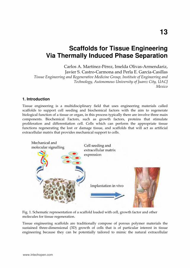
13 Scaffolds for Tissue Engineering Via Thermally Induced Phase Separation Carlos A. Martínez-Pérez, Imelda Olivas-Armendariz, Javier S. Castro-Carmona and Perla E. García-Casillas Tissue Engineering and Regenerative Medicine Group, Institute of Engineering and Technology, Autonomous University of Juarez City, UACJ Mexico 1. Introduction Tissue engineering is a multidisciplinary field that uses engineering materials called scaffolds to support cell seeding and biochemical factors with the aim to regenerate biological function of a tissue or organ, in this process typically there are involve three main components. Biochemical Factors, such as growth factors, proteins that stimulate proliferation and differentiation cell. Cells which can perform the appropriate tissue functions regenerating the lost or damage tissue, and scaffolds that will act as artificial extracellular matrix that provides mechanical support to cells. Fig. 1. Schematic representation of a scaffold loaded with cell, growth factor and other molecules for tissue regeneration. Tissue engineering scaffolds are traditionally compose of porous polymer materials the sustained three-dimensional (3D) growth of cells that is of particular interest in tissue engineering because they can be potentially tailored to mimic the natural extracellular www.intechopen.com
Transcript of 2011 Scaffolds for tissue engineering via thermally induced phase separation copia
13
Scaffolds for Tissue Engineering Via Thermally Induced Phase Separation
Carlos A. Martínez-Pérez, Imelda Olivas-Armendariz, Javier S. Castro-Carmona and Perla E. García-Casillas
Tissue Engineering and Regenerative Medicine Group, Institute of Engineering and Technology, Autonomous University of Juarez City, UACJ
Mexico
1. Introduction
Tissue engineering is a multidisciplinary field that uses engineering materials called scaffolds to support cell seeding and biochemical factors with the aim to regenerate biological function of a tissue or organ, in this process typically there are involve three main components. Biochemical Factors, such as growth factors, proteins that stimulate proliferation and differentiation cell. Cells which can perform the appropriate tissue functions regenerating the lost or damage tissue, and scaffolds that will act as artificial extracellular matrix that provides mechanical support to cells.
Fig. 1. Schematic representation of a scaffold loaded with cell, growth factor and other molecules for tissue regeneration.
Tissue engineering scaffolds are traditionally compose of porous polymer materials the sustained three-dimensional (3D) growth of cells that is of particular interest in tissue engineering because they can be potentially tailored to mimic the natural extracellular
www.intechopen.com
Advances in Regenerative Medicine 276
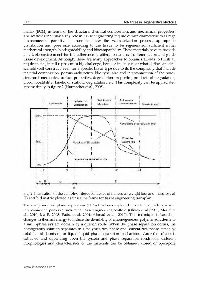
matrix (ECM) in terms of the structure, chemical composition, and mechanical properties. The scaffolds that play a key role in tissue engineering require certain characteristics as high interconnected porosity in order to allow the vascularization process, appropriate distribution and pore size according to the tissue to be regenerated, sufficient initial mechanical strength, biodegradability and biocompatibility. These materials have to provide a suitable environment for the adherence, proliferation and cell differentiation and guide tissue development. Although, there are many approaches to obtain scaffolds to fulfill all requirements, it still represents a big challenge, because it is not clear what defines an ideal scaffold/cell construct, even for a specific tissue type due to its the complexity that include material composition, porous architecture like type, size and interconnection of the pores, structural mechanics, surface properties, degradation properties, products of degradation, biocomopatibility, kinetic of scaffold degradation, etc. This complexity can be appreciated schematically in figure 2 (Hutmacher et al., 2008).
Fig. 2. Illustration of the complex interdependence of molecular weight loss and mass loss of 3D scaffold matrix plotted against time frame for tissue engineering transplant.
Thermally induced phase separation (TIPS) has been explored in order to produce a well interconnected porous structure as tissue engineering scaffold (Olivas et al., 2010; Martel et al., 2010; Ma P. 2008; Patist et al. 2004; Ahmad et al., 2010). This technique is based on changes in thermal energy to induce the de-mixing of a homogeneous polymer solution into a multi-phase system domain by a quench route. When the phase separation occurs, the homogenous solution separates in a polymer-rich phase and solvent-rich phase either by solid–liquid de-mixing or liquid–liquid phase separation mechanism. After the solvent is extracted and depending upon the system and phase separation conditions, different morphologies and characteristics of the materials can be obtained: closed or open-pore
www.intechopen.com
Scaffolds for Tissue Engineering Via Thermally Induced Phase Separation 277
material, spheres, powders, bead-like morphology, etc. Most of the works for tissue engineering seek scaffolds with open pore and well interconnected morphology. One of the most attractive characteristics of TIPS over other techniques is the formation of not only an intrinsically interconnected polymer network, but also an interconnected porous space in one simple process that is scalable, fast and controllable. TIPS is thus a very convenient methodology for fabricating porous materials as scaffold architectures that can be obtained by means of the manipulation of processing parameters and system properties. A variety of polymers scaffolds as Polylactic Acid (Chen J et al., 2010; He L et al., 2009); Polyurethane (Guan J et al.,2005; Martinez et al., 2006; Fromstein et al. 2002), Polycaprolactone[15] and others have been prepared by TIPS technique, also blends of polymers[Martel et al., 2010; Maquet et al., 2001), and composite of polymers with nanohydroxyapatite (Liu et al 2009), Carbon nanotubes (Olivas et al., 2010; Jell et al., 2008) and other have been successfully fabricated.
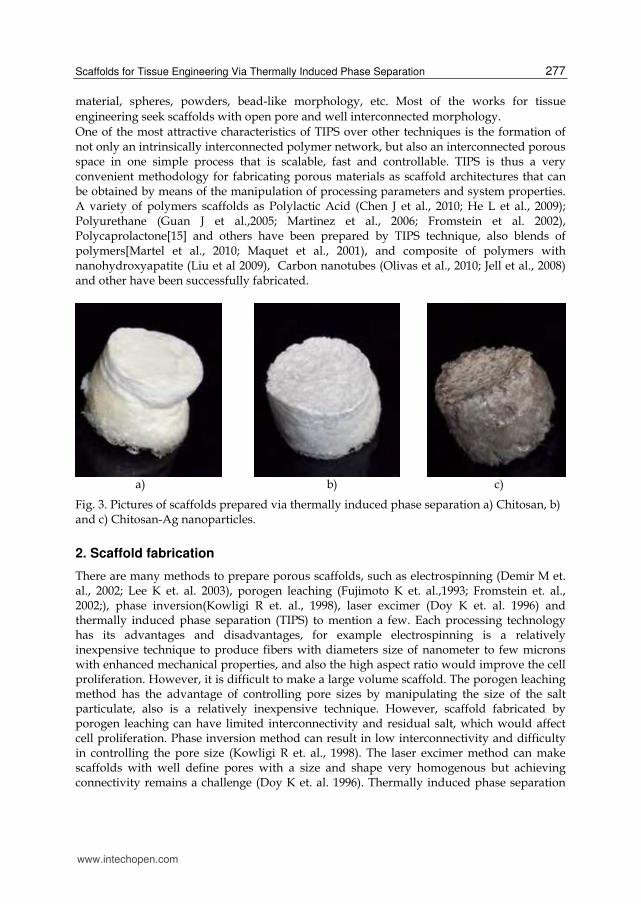
a) b) c)
Fig. 3. Pictures of scaffolds prepared via thermally induced phase separation a) Chitosan, b) and c) Chitosan-Ag nanoparticles.
2. Scaffold fabrication
There are many methods to prepare porous scaffolds, such as electrospinning (Demir M et. al., 2002; Lee K et. al. 2003), porogen leaching (Fujimoto K et. al.,1993; Fromstein et. al., 2002;), phase inversion(Kowligi R et. al., 1998), laser excimer (Doy K et. al. 1996) and thermally induced phase separation (TIPS) to mention a few. Each processing technology has its advantages and disadvantages, for example electrospinning is a relatively inexpensive technique to produce fibers with diameters size of nanometer to few microns with enhanced mechanical properties, and also the high aspect ratio would improve the cell proliferation. However, it is difficult to make a large volume scaffold. The porogen leaching method has the advantage of controlling pore sizes by manipulating the size of the salt particulate, also is a relatively inexpensive technique. However, scaffold fabricated by porogen leaching can have limited interconnectivity and residual salt, which would affect cell proliferation. Phase inversion method can result in low interconnectivity and difficulty in controlling the pore size (Kowligi R et. al., 1998). The laser excimer method can make scaffolds with well define pores with a size and shape very homogenous but achieving connectivity remains a challenge (Doy K et. al. 1996). Thermally induced phase separation
www.intechopen.com
Advances in Regenerative Medicine 278
method offers the ability to control pore size by varying the preparation conditions and also provides means to control pore structure, additionally the scaffold can be molded into a range of shapes and sizes (Jianjun G et. al., 2005; Martinez Perez C et. al. 2006). It is beyond the scope of this chapter to cover all scaffold fabrication techniques available. Hence the aim of this chapter is to provide an overview of the TIPS technique which it has been employed to produce a range of scaffolds for tissue engineering.
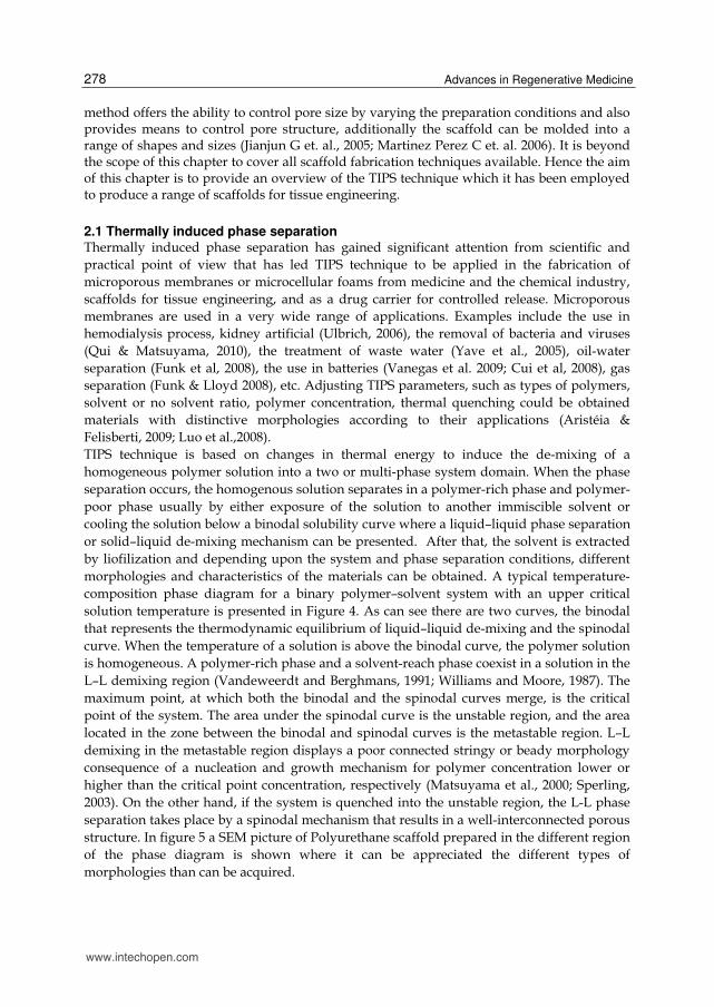
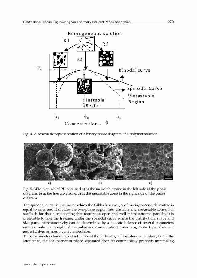
2.1 Thermally induced phase separation Thermally induced phase separation has gained significant attention from scientific and practical point of view that has led TIPS technique to be applied in the fabrication of microporous membranes or microcellular foams from medicine and the chemical industry, scaffolds for tissue engineering, and as a drug carrier for controlled release. Microporous membranes are used in a very wide range of applications. Examples include the use in hemodialysis process, kidney artificial (Ulbrich, 2006), the removal of bacteria and viruses (Qui & Matsuyama, 2010), the treatment of waste water (Yave et al., 2005), oil-water separation (Funk et al, 2008), the use in batteries (Vanegas et al. 2009; Cui et al, 2008), gas separation (Funk & Lloyd 2008), etc. Adjusting TIPS parameters, such as types of polymers, solvent or no solvent ratio, polymer concentration, thermal quenching could be obtained materials with distinctive morphologies according to their applications (Aristéia & Felisberti, 2009; Luo et al.,2008). TIPS technique is based on changes in thermal energy to induce the de-mixing of a homogeneous polymer solution into a two or multi-phase system domain. When the phase separation occurs, the homogenous solution separates in a polymer-rich phase and polymer-poor phase usually by either exposure of the solution to another immiscible solvent or cooling the solution below a binodal solubility curve where a liquid–liquid phase separation or solid–liquid de-mixing mechanism can be presented. After that, the solvent is extracted by liofilization and depending upon the system and phase separation conditions, different morphologies and characteristics of the materials can be obtained. A typical temperature-composition phase diagram for a binary polymer–solvent system with an upper critical solution temperature is presented in Figure 4. As can see there are two curves, the binodal that represents the thermodynamic equilibrium of liquid–liquid de-mixing and the spinodal curve. When the temperature of a solution is above the binodal curve, the polymer solution is homogeneous. A polymer-rich phase and a solvent-reach phase coexist in a solution in the L–L demixing region (Vandeweerdt and Berghmans, 1991; Williams and Moore, 1987). The maximum point, at which both the binodal and the spinodal curves merge, is the critical point of the system. The area under the spinodal curve is the unstable region, and the area located in the zone between the binodal and spinodal curves is the metastable region. L–L demixing in the metastable region displays a poor connected stringy or beady morphology consequence of a nucleation and growth mechanism for polymer concentration lower or higher than the critical point concentration, respectively (Matsuyama et al., 2000; Sperling, 2003). On the other hand, if the system is quenched into the unstable region, the L-L phase separation takes place by a spinodal mechanism that results in a well-interconnected porous structure. In figure 5 a SEM picture of Polyurethane scaffold prepared in the different region of the phase diagram is shown where it can be appreciated the different types of morphologies than can be acquired.
www.intechopen.com
Scaffolds for Tissue Engineering Via Thermally Induced Phase Separation 279
Fig. 4. A schematic representation of a binary phase diagram of a polymer solution.
50 µm
200 µm
a) b) c)
Fig. 5. SEM pictures of PU obtained a) at the metastable zone in the left side of the phase diagram, b) at the inestable zone, c) at the metastable zone in the right side of the phase diagram.
The spinodal curve is the line at which the Gibbs free energy of mixing second derivative is equal to zero, and it divides the two-phase region into unstable and metastable zones. For scaffolds for tissue engineering that require an open and well interconnected porosity it is preferable to take the freezing under the spinodal curve where the distribution, shape and size pore, interconnectivity can be determined by a delicate balance of several parameters such as molecular weight of the polymers, concentration, quenching route, type of solvent and additives as nonsolvent composition. These parameters have a great influence at the early stage of the phase separation, but in the later stage, the coalescence of phase separated droplets continuously proceeds minimizing
B inoda l cu rve
Spino dal Cu rve
curve M etastable R egion
I nstab leRe gion
T c
I1 Ic I2 Co nc ent ration , I
Hom ogeneous solutionR1
R2
R3
www.intechopen.com
Advances in Regenerative Medicine 280
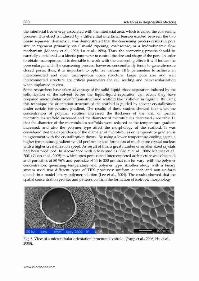
the interfacial free energy associated with the interfacial area, which is called the coarsening process. This effect is induced by a differential interfacial tension exerted between the two phase separated domains. It was demonstrated that the coarsening process results in pore size enlargement primarily via Ostwald ripening, coalescense, or a hydrodynamic flow mechanism (Mooney et al., 1996; Lo et al., 1996). Thus, the coarsening process should be carefully considered as a kinetic parameter to control the size and shape of the pore. In order to obtain macroporous, it is desirable to work with the coarsening effect; it will induce the pore enlargement. The coarsening process, however, concomitantly tends to generate more closed pores; thus, it is important to optimize various TIPS parameters to achieve an interconnected and open macroporous open structure. Large pore size and well interconnected structure are critical parameters for cell seeding and neovascularization when implanted in vivo. Some researchers have taken advantage of the solid-liquid phase separation induced by the solidification of the solvent before the liquid-liquid separation can occur, they have prepared microtubular orientantion-structured scaffold like is shown in figure 6. By using this technique the orientation structure of the scaffold is guided by solvent crystallization under certain temperature gradient. The results of these studies showed that when the concentration of polymer solution increased the thickness of the wall of formed microtubules scaffold increased and the diameter of microtubules decreased ( see table 1), that the diameter of the microtubules scaffolds were reduced as the temperature gradient increased, and also the polymer type affect the morphology of the scaffold. It was considered that the dependence of the diameter of microtubules on temperature gradient is in agreement with the crystallization theory. By using a lower temperature-cooling agent, a higher temperature gradient would perform to lead formation of much more crystal nucleus with a higher crystallization speed. As result of this, a great number of smaller sized crystals had been produced. In Accordance with others studies (Cao Y et al., 2006; Maquet et al., 2001; Guan et al., 2005) in which open porous and interconnected architecture was obtained, and, porosities of 80-96% and pore size of 14 to 250 µm that can be vary with the polymer concentration, quenching temperature and polymer type. Another study with a binary system used two different types of TIPS processes: uniform quench and non uniform quench in a model binary polymer solution (Lee et al., 2004). The results showed that the spatial concentration profiles and patterns confirm the formation of isotropic morphology
Fig. 6. View of a microtubular orientation-structured scaffold. (Yang et al., 2006; Hu et al., 2008).
www.intechopen.com
Scaffolds for Tissue Engineering Via Thermally Induced Phase Separation 281
from a uniform quench while anisotropic morphology forms from non-uniform quench and indicate the lower-temperature regions of the spatial temperature gradient containing higher droplet density, larger droplets size when the phase separation process was allowed to proceed for long period of time, and the morphological analysis of the shape factor shows that the formation of the droplet shape is independent on the spatial temperature gradient.
Polymer /super cooling-temperature
Concentration of polymer in solvent
Porosity (%)
Average density of scaffold (g/cm3)
Average diameter of microtubules (µm)
References
PLGA/-10ºC
5 7 9
90.4 83.8 78.4
0.1316 0.1637 0.2201
120 100 80
(Hu et al.,2008)
PLLA /-20ºC
3 (w/w%) 5 7 9
96 94 92 90
0.051 0.078 0.099 0.128
137 122 117 114
(Yang et al., 2006)
PLGA/-20ºC
3 5 7 9
96 93 92 90
0.051 0.094 0.105 0.128
123 118 113 109
(Yang et al., 2006)
Table 1. Morphology of microtubules orientation-structured scaffolds on concentration polymer
3. Polymer scaffolds prepared via TIPS
As it was mention above that biodegradable polymers are suitable for scaffolds. Synthetic and natural biodegradable materials such as Poly (lactide) (PLA), Poly (L-lactic acid) (PLLA), Poly (D,L-lactic acid-co-glycolide acid) (PDLG), Poly (D,L-lactide) (PDLLA), Poly (lactide-co-glycolide) (PLGA), Poly (lactide-co-caprolactone) (PLCL), Poly (dž-caprolactone) (PCL), Polyurethane (PU), Poly (ether ester urethane) (PEEUU), collagen, gelatin, and chitosan have been used in the fabrication of scaffolds. These have been designed in order to be applied in spinal cord regeneration (Maquet et al., 2001), bone and cartilage regeneration (Barroca, et al. 2010; Olivas et al., 2009), blood vessel (Hu et al., 2008), incontinence problems (Ahmadi et al., 2011), peripheral nerves (Aijun et al., 2005), and perennial fistula repair (Heshaw et al., 2010), among others. These had been performance with appropriate structural and functional properties, with the goal to mimic the extracellular matrix (ECM) of the tissue to be repair. The ECM is a natural scaffold for tissue and organ morphogenesis, maintenance, and reconstruction following injury and it is a vital, dynamic and indispensable component of all tissue and organs. Therefore, it is a challenge for tissue engineering, the construction of artificial structures that lead the constructive remodeling of injured or missing tissues or organs, since it regulate cell behavior, such as attachment, migration, proliferation, and differentiation. Even more, it must have a controlled degradation, interconnected porosity with high porosity, appropriate pore size, and mechanical properties the most closely to the tissue to be repaired.
www.intechopen.com
Advances in Regenerative Medicine 282
Schugens (1996) prepared biodegradable scaffolds of Polylactic Acid (PLA) by the TIPS method using dioxane as solvent. Instead of the L-L demixing mechanism, a solid-liquid mechanism take place due to the solidification of solvent and it gave a highly anisotropic tubular morphology with a ladder-like microporous structure. A porous structure obtained in this manner is highly anisotropic with relatively small pores (Yang et al., 2006). Further studies have indicated that by introducing water as a non-solvent, L–L de-mixing can be realized in the polyester–dioxane–water ternary system, and that macroporous polyester scaffolds can be obtained. The morphology of the resulting scaffold is strongly dependent on the phase separation behavior of the ternary system. By increasing the polymer concentration except when there is no presence of non-solvente the temperature of the cloud point and the temperature where the L-L de-mixing take place increases considerably, also higher the non-solvent concentration, higher the L-L de-mixing temperature (Hua et al., 2002, 2003). The L–L demixing temperature of a crystalline PLLA ternary system is higher than that of amorphous PDLLA and PLGA ternary systems. However, the effect of the molecular weight of the polymer seems to be less prominent (Hua et al., 2003). The phase behavior of the system is also be affected by adding various compounds (Hua et al., 2001; Shin et al., 2005). It has been demonstrated that by adding a surface active material such as Pluronic F127, a tri-block polymeric surfactant, the interfacial energy between two phases can be reduced, effectively stabilizing the porous structure of the scaffolds (Nam & Park, 1999). The addition of NaCl can shift the binodal curve to a higher temperature and therefore create a larger operable domain for spinodal decomposition (Hua et al., 2001). Additionally, researchers have introduced non-solvent as water to induce a liquid-liquid phase separation having a polymer-solvent-water ternary system. A liquid-liquid phase separation occurs when the temperature of polymer solution is decreased and depended of the thermal driving force. The cloud-point temperature was highly dependent on water content, polymer concentration and the freezing point of the system was nearly independent of polymer concentration and water content (Barroca et al., 2010; Chen et al., 2010; Jun et al., 2003). The cloud point temperature increases as the polymer concentration increases and when the molecular weight of the polymer increases because it reduces the polymer-solvent interaction. When a polymer blend is prepared and its polymers have similar solubility parameters the phase separation of the polymers behaves similar as they behave by themselves. When a semi-crystalline polymer is part of the solution, the phase separation is more complicated because the polymer potentially crystallizes involving the gelation temperature as another important parameter in the morphology of the scaffolds because semicrystalline polymers, generally, form a gel as a result of liquid-liquid phase separation. The interlocking of small crystal agglomerates may play a key role for the formation of the gel when the solvent is removed a highly porous structured is obtained. The effects of the quench route on the phase in a study (PLLA-dioxane-water) by maintaining the demixing temperature in the unstable region for a period of time and then quenching to -196ºC, researchers have found that the gelation induced by liquid-liquid demixing is essential to maintain scaffolds with uniform interconnected macroporous. The porosities of the scaffolds decreased with increases of polymer concentration and this occur by the change of the phase separation mechanism and by the extent of the coarsening process involved. The effect of molecular weight and the aging time on the scaffold morphology is important because the morphology with the higher molecular weight polymer was consistently better organized.
www.intechopen.com
Scaffolds for Tissue Engineering Via Thermally Induced Phase Separation 283
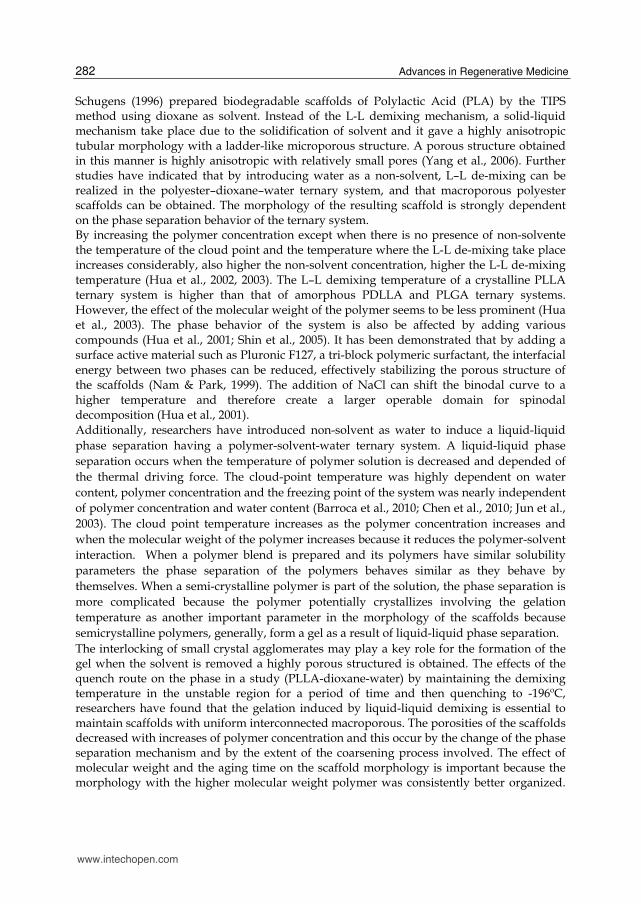
The less uniform pore structure (lower molecular weight) was likely to be caused by coarsening of two separated phases since the viscosity effect is one of the factors responsible for a less organized pore structure. On the other hand, with short aging time, a large pore size due to the large thermodynamic driving forces obtained. The microporous scaffolds have a highly interconnected macroporous range from 20 to 300µm and exhibited isotropic morphology (table 2).
Polymer Polymer
concentration w%
Weight ratios of
solvent/waterPorosity Pore
size Morphology References
PLA 7.5 9010 85/15
80 84
< 5050
Isotropic lacy structure, interconnected pores
(Chen et al., 2010)
PLGA 9 87/13 > 70
Regular and highly interconnected macroporous structure
(Jun et al.,2003)
PLLA 4.5 87/13 20-50 (Tanaka et al.,2008)
PCL/PLLA 8-15 90/10 20-300
Good connective structure
(Tanaka et al.,2008)
PCL 10 87/13 50-100
Good connective structure
(Tanaka et al.,2008)
PLGA/PLLA 9 87/13 50-200
Well interconnected macroporous structure
(Chul et al., 2005)
Table 2. Morphology of scaffolds, water content and polymer concentration.
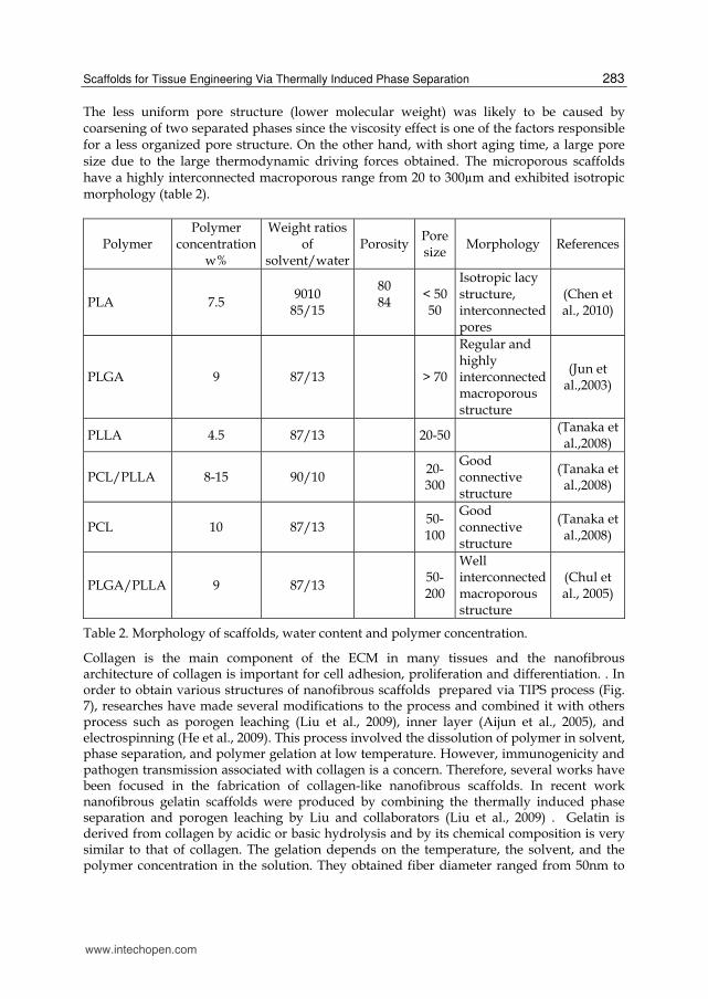
Collagen is the main component of the ECM in many tissues and the nanofibrous architecture of collagen is important for cell adhesion, proliferation and differentiation. . In order to obtain various structures of nanofibrous scaffolds prepared via TIPS process (Fig. 7), researches have made several modifications to the process and combined it with others process such as porogen leaching (Liu et al., 2009), inner layer (Aijun et al., 2005), and electrospinning (He et al., 2009). This process involved the dissolution of polymer in solvent, phase separation, and polymer gelation at low temperature. However, immunogenicity and pathogen transmission associated with collagen is a concern. Therefore, several works have been focused in the fabrication of collagen-like nanofibrous scaffolds. In recent work nanofibrous gelatin scaffolds were produced by combining the thermally induced phase separation and porogen leaching by Liu and collaborators (Liu et al., 2009) . Gelatin is derived from collagen by acidic or basic hydrolysis and by its chemical composition is very similar to that of collagen. The gelation depends on the temperature, the solvent, and the polymer concentration in the solution. They obtained fiber diameter ranged from 50nm to
www.intechopen.com
Advances in Regenerative Medicine 284
500nm, which is on the same scale as natural collagen fibers, the average fiber diameter did not change with different gelatin concentrations. The scaffolds prepared with 7.5% (w/v) gelatin solution had a low density, 97.51% of porosity and well defined macropores with pore size of 250-420µm. During the process water and ethanol solvent mixtures were used to dissolve gelatin. The addition of ethanol was a critical step in creating the nanofibrous structure because when gelatin was dissolved in water alone, it could only form smooth surface structure after phase separation. The addition of certain amount of ethanol in gelatin solution resulted in the formation of gelatin structure after phase separation. Furthermore, when increased the amount of ethanol in the solvent mixture resulted in poor solubility of gelatin. For this reason, the ethanol/water ratio in gelatin solution was very important during the fabrication of nanofibrous. The in vitro analysis indicated the gelatin fibrous scaffolds enhanced cell adhesion and proliferation. In other study the researchers fabricated fiber porous and tubular chitosan scaffolds for guided peripheral nerves and blood vessel tissue regeneration by combining inner layer and TIPS. The scaffolds had a biphasic wall structure, with fibrous inner layer and semipermeable outer layer. The inner diameter was 2.5 mm and outer diameter was 4.5mm. In vitro characterization shows that the scaffolds have a very good cytocompatibility.
Fig. 7. SEM image of nanofibrous structure fabricated by TIPS (Beachley et al. 2010).
He Liu et. al., prepared PLLA nanofibrous scaffolds with 3D macro and microporous structures by liquid-liquid phase separation from PLLA-dioxane-water system showing that aging in the gel status was essential for the formation of a nanofibrous network. The coalescence of the solvent rich drops also occurred when aging was performed in order to reduce the surface tension. This coarsening effect resulted in pore size enlargement. The extent of coarsening affected the size and morphologies of macro or micro pores in the scaffold. High quenching temperature favors the pore enlargement, due to the formation of
www.intechopen.com
Scaffolds for Tissue Engineering Via Thermally Induced Phase Separation 285
larger solvent rich droplets. High water content in the system increase the pore size but also can decrease the pore homogeneity. The gradual addition of water as non solvent increase the gelation point and generate macropores (up to 300 µm) and micropores with a high interconnection. The enlarged pore sizes with the increase of water content is likely due to the combined effects of weaker polymer diluents interaction, larger quenching depth, and lowered viscosity in the co-solvent system. The formation of nanofibrous structure was related to the liquid-liquid phase separation and the crystallization kinetics of polymer affected by the cooling rate.
4. Particulate filled composite scaffolds
Filled composite scaffolds produced by thermally induced phase separation are other alternative materials which exhibit pore anisotropy that it has shown to support migration, adhesion, spreading and viability of cells (Blaker et al., 2005; Barroca et. al. 2010;). Commonly, these scaffolds consist of biodegradable polymer with inorganic particles, such as bioactive glass and hydroxyapatite. The composite has the advantages of the two types of material and for example the bioactivity, control degradation kinetics and the mechanical properties can be improved. Two polymer-diluent zeolite was reported by Funk (Funk CV et. al., 2008) where they showed the that zeolite particle to have a significant effect on the droplet growth and final cell size of that microporous membrane which depend on the particle loading and processing condition. Nanohydroxyapatite-filled PLGA scaffolds were prepared by Huang and collaborators (Huang et al., 2008) by TIPS technique where the influence of Nanohydroxyapatite (NHA) content on the microstructure and properties of the composite was studied. It was found that the pore size of the PLGA/NHA scaffolds decreases with the increase of PLGA concentration and HA content. Also, the mechanical properties and water absorption ability of the composite scaffolds were considerably improved with the content of NHA. PLGA/NHA scaffolds exhibited significantly higher cell growth, alkaline phosphatase activity than PLGA scaffolds, having in their system the best results with 10 wt.% NHA. PLLA was prepared via TIPS with the incorporation of micro and nanohydroxyapatite, NAH had better results that of micrometric size and compare with PLLA scaffolds, NAH/PLLA scaffolds showed highest compressive strength (8.67 MPa) with 85.06% porosity which is comparable to the high end of compressive strength of cancellous bone (2–10 MPa) ( Nejati et al., 2008). Also it was found that the incorporation of NHA, the scaffolds have more regular microarchitecture due to its more interfacial area, surface reactivity and ultra-fine structure. This suggests that the developed nHAP/PLLA scaffold via TIPS fulfill most of the requirements as a suitable bone substitute for bone tissue engineering applications. Carbon Nanotubes (CNT) has also been incorporated in scaffolds for tissue engineering (Jell et al. 2008; Olivas et al. 2010). Jell and collaborators design a route via TIPS to manufacture three-dimensional, highly porous polyurethane containing CNT where the mechanical properties were improved significantly with the addition until 5% of CNT, also the composite scaffolds were not only found to be non-toxic but also induce phenotypic changes that may enhance wound healing and bone formation in vivo. Olivas and coworkers also found that the incorporation of small amount of MWCNT will significantly improve the mechanical properties of the scaffolds.
www.intechopen.com
Advances in Regenerative Medicine 286
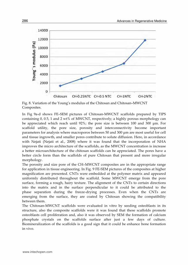
Fig. 8. Variation of the Young’s modulus of the Chitosan and Chitosan–MWCNT Composites.
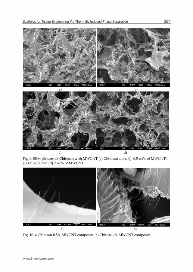
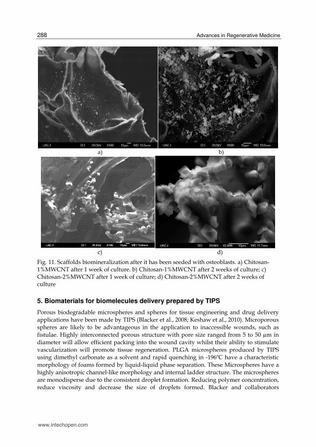
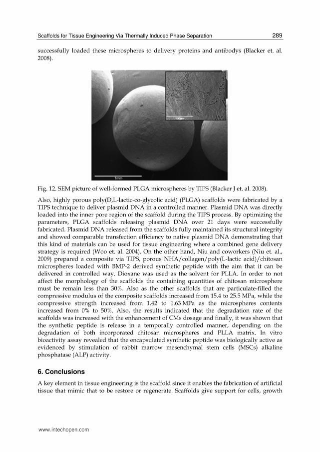
In Fig 9a-d shows FE–SEM pictures of Chitosan-MWCNT scaffolds prepared by TIPS containing 0, 0.5, 1 and 2 wt% of MWCNT, respectively; a highly porous morphology can be appreciated which reach until 92%; the pore size is between 100 and 300 Ǎm. For scaffold utility, the pore size, porosity and interconnectivity become important parameters for analysis where macroporos between 50 and 300 Ǎm are most useful for cell and tissue ingrowth, and smaller pores contribute to solute diffusion. Here, in accordance with Nejati (Nejati et al., 2008) where it was found that the incorporation of NHA improves the micro architecture of the scaffolds, as the MWCNT concentration is increase a better microarchitecture of the chitosan scaffolds can be appreciated. The pores have a better circle form than the scaffolds of pure Chitosan that present and more irregular morphology. The porosity and size pore of the CH–MWCNT composites are in the appropriate range for application in tissue engineering. In Fig. 9 FE-SEM pictures of the composites at higher magnification are presented. CNTs were embedded at the polymer matrix and appeared uniformly distributed throughout the scaffold. Some MWCNT emerge from the pore surface, forming a rough, hairy texture. The alignment of the CNTs to certain directions into the matrix and in the surface perpendicular to it could be attributed to the phase separation during the freeze-drying processes. Even when the CNTs are emerging from the surface, they are coated by Chitosan showing the compatibility between them. The Chitosan-MWCNT scaffolds were evaluated in vitro by seeding osteoblasts in its structure, also the composite scaffolds were it was found that these scaffolds promote osteoblasts cell proliferation and, also it was observed by SEM the formation of calcium phosphate crystals on the scaffolds surface after just a few days of culture. Biomineralization of the scaffolds is a good sign that it could be enhance bone formation in vivo.
www.intechopen.com
Scaffolds for Tissue Engineering Via Thermally Induced Phase Separation 287
a) b)
c) d)
Fig. 9. SEM pictures of Chitosan with MWCNT (a) Chitosan alone b) 0.5 wt% of MWCNT; (c) 1% wt% and (d) 2 wt% of MWCNT.
a) b)
Fig. 10. a Chitosan-0.5% MWCNT composite. b) Chitosa-1% MWCNT composite.
www.intechopen.com
Advances in Regenerative Medicine 288
a) b)
c) d)
Fig. 11. Scaffolds biomineralization after it has been seeded with osteoblasts. a) Chitosan-1%MWCNT after 1 week of culture. b) Chitosan-1%MWCNT after 2 weeks of culture; c) Chitosan-2%MWCNT after 1 week of culture; d) Chitosan-2%MWCNT after 2 weeks of culture
5. Biomaterials for biomelecules delivery prepared by TIPS
Porous biodegradable microspheres and spheres for tissue engineering and drug delivery applications have been made by TIPS (Blacker et al., 2008; Keshaw et al., 2010). Microporous spheres are likely to be advantageous in the application to inaccessible wounds, such as fistulae. Highly interconnected porous structure with pore size ranged from 5 to 50 µm in diameter will allow efficient packing into the wound cavity whilst their ability to stimulate vascularization will promote tissue regeneration. PLGA microspheres produced by TIPS using dimethyl carbonate as a solvent and rapid quenching in -196ºC have a characteristic morphology of foams formed by liquid-liquid phase separation. These Microspheres have a highly anisotropic channel-like morphology and internal ladder structure. The microspheres are monodisperse due to the consistent droplet formation. Reducing polymer concentration, reduce viscosity and decrease the size of droplets formed. Blacker and collaborators
www.intechopen.com
Scaffolds for Tissue Engineering Via Thermally Induced Phase Separation 289
successfully loaded these microspheres to delivery proteins and antibodys (Blacker et. al. 2008).
Fig. 12. SEM picture of well-formed PLGA microspheres by TIPS (Blacker J et. al. 2008).
Also, highly porous poly(D,L-lactic-co-glycolic acid) (PLGA) scaffolds were fabricated by a TIPS technique to deliver plasmid DNA in a controlled manner. Plasmid DNA was directly loaded into the inner pore region of the scaffold during the TIPS process. By optimizing the parameters, PLGA scaffolds releasing plasmid DNA over 21 days were successfully fabricated. Plasmid DNA released from the scaffolds fully maintained its structural integrity and showed comparable transfection efficiency to native plasmid DNA demonstrating that this kind of materials can be used for tissue engineering where a combined gene delivery strategy is required (Woo et. al. 2004). On the other hand, Niu and coworkers (Niu et. al., 2009) prepared a composite via TIPS, porous NHA/collagen/poly(L-lactic acid)/chitosan microspheres loaded with BMP-2 derived synthetic peptide with the aim that it can be delivered in controlled way. Dioxane was used as the solvent for PLLA. In order to not affect the morphology of the scaffolds the containing quantities of chitosan microsphere must be remain less than 30%. Also as the other scaffolds that are particulate-filled the compressive modulus of the composite scaffolds increased from 15.4 to 25.5 MPa, while the compressive strength increased from 1.42 to 1.63 MPa as the microspheres contents increased from 0% to 50%. Also, the results indicated that the degradation rate of the scaffolds was increased with the enhancement of CMs dosage and finally, it was shown that the synthetic peptide is release in a temporally controlled manner, depending on the degradation of both incorporated chitosan microspheres and PLLA matrix. In vitro bioactivity assay revealed that the encapsulated synthetic peptide was biologically active as evidenced by stimulation of rabbit marrow mesenchymal stem cells (MSCs) alkaline phosphatase (ALP) activity.
6. Conclusions
A key element in tissue engineering is the scaffold since it enables the fabrication of artificial tissue that mimic that to be restore or regenerate. Scaffolds give support for cells, growth
www.intechopen.com
Advances in Regenerative Medicine 290
factors, and other biomolecules that eventually will enhance the formation of new tissue in the human body, the scaffold should fulfill several requirements such as nocarcinogenic, nontoxic, nonantigenic, it also must be biocompatible, biodegradable, also is preferred to be bioactive. Besides the biomaterial issues, the structure and morphology of the scaffolds is very important. They should be highly porous, with defined size and shape pore, well interconnected porosity to allow vascularization. Thermally induced phase separation is a technique that can produced scaffolds with the desired characteristics. It can be fabricate in the desired shape and also can be scalable for fabrication of materials with enough quantity and quality. The process can be tuned just adjusting some parameters of the process and give materials with the appropriate morphology and structure for tissue engineering. In the process it can be incorporated another inorganic materials to enhance its properties, even can be loaded with growth factors and others biomolecules that make this technique one of the most adequate for scaffolds fabrication for tissue engineering.
7. References
Ahmad, R.,Mordan N., Forbes A and Day R.(2011) Enhanced attachment, growth and migration of smooth muscle cells on microcarriers produced using thermally induced phase separation. Acta Biomaterialia, 7: 1542-1549.
Aijun, A., Qiang A., Wenling C., Chang Z., Yandao G., Nanming Z., and Xiufang Z. (2005) Fiber-Based Chitosan tubular scaffolds for soft tissue engineering: Fabrication and in vitro evaluation. Tsinghua Science and Technology, 10: 449-453.
Aristéia, J., and Felisberti M. (2009) Pororus polymer structure obtained via the TIPS process from EVOH/PMMA/DMF solutions Journal of Membrane Science, 344: 237-243.
Barroca, N., Daniel-da-Silva S., Vilarinho P., and Fernandes M. (2010) Tailoring the morphology of high molecular weight PLLA scaffolds through bioglass addition. Acta Biomaterialia, 6: 3611-3620.
Beachley V., and Wen X. (2010) Polymer nanofibrous structures: Fabrication, biofunctionalization, and cell interactions. Progress in Polymer Science, 35: 868-892.
Blake, J., Knowles J., and Day R. (2008) Novel fabrication techniques to produce microspheres by thermally induced phase separation for tissue engineering and drug delivery. Acta Biomaterialia, 4: 264-272.
Blaker, J., Maquet V., Jerome R., Boccaccini A., and Nazhat S. (2005) Mechanical properties of highly porous PDLLA/Bioglass composite foams as scaffolds for bone tissue engineering. Acta Biomaterialia, 1: 643-652.
Blaker, J., Nazhat S., maquet V., and Boccaccini A. (2011) Long-term in vitro degradation of PDLLA/Bioglass bone scaffolds in acellular simulated body fluid. Acta Biomaterialia, 7 : 829-840.
Brandt, H. X. (2008). Bioactivation of knitted cellulose scaffolds by strontium. Cellulose, 15, 275-283.
Cao, Y., Mitchell G., Messina A., Price L., Thompson E., Penington A., Morrison W., O´Connor A., Stevens G., and Cooper-White J. (2006) The influence of architecture on degradation into three-dimensional poly(lactic-co-glycolic acid) scaffolds in vitro and in vivo. Biomaterials, 27: 2854-2864.
Chen, J., Ling S., and Tsay R. (2010) A morphological study of porous polylactide scaffolds prepared by termally induced phase separation. Journal of the Taiwan Institute of Chemical Engineers, 41: 229-238.
www.intechopen.com
Scaffolds for Tissue Engineering Via Thermally Induced Phase Separation 291
Chi Yena , Hongyan Hea, James Leea L. , Winston W.S., Ho (2009) Synthesis and characterization of nanoporous polycaprolactone membranes via thermally- and nonsolvent-induced phase separations for biomedical device application Journal of Membrane Science, 343 :180-188.
Chu, K., Sup B., Heung J., Gwan T., Do J., and Sung D. (2005) A facile preparation of highly interconnected macroporous PLGA scaffolds by liquid-liquid phase separation II. Polymer, 46: 3801-3808.
Cui, Z., Xu Y., Zhu L., YuWang J., Xi Z., and Zhu B. (2008) Preparation of PVDF/PEO-PPO-PEO blend microporous memebranes for lithium ion batteries via thermally induced phase separation process. Journal of Membrane Science, 325: 957-963.
Demir , MM, Yilgor I, Yilgor E, Erman B. (2002) Electrospinning of polyurethane fibers. Polymer, 43: 3303-9.
Hutmacher D, Woodfiel T, Dalton P & Lewis J. (2008). Scaffold design and fabrication, In: Tissue Engineering Clemens Van Blitterswijk, pp. 403-454, Academic Press, ISBN:978-0-12-370869-4, Canada.
Doi , K, Nakayama Y, Matsuda T. (1996) Novel compliant and tissuepermeable microporous polyurethane vascular prosthesis fabricated using an excimer laser ablation technique. Journal of Biomedical Materials Research, 31:27-33.
Doi , K. (1996). Novel compliant and tissue permeable microporous polyurethane vascular prótesis fabricated using an excimer laser ablation technique. Journal of Biomedical Materials Research, 31, 27-33.
Fromstein , JD, Woodhouse KA. (2002);Elastomeric biodegradable polyurethane blends for soft tissue application. Journal of Biomaterials Scie Polymer Edn, 13:391-406.
Fujimoto , K, Minato M, Miyamoto S, Kaneko T, Kikuchi H, Sakai K, Okada M, Ikada Y (1993) Porous polyurethane tubes as vascular graft. Journal of Applied Biomaterials, 4:347-54.
Funk , C., and Lloyd D. (2008) Zeolite-filled micropororus mixed matrix (ZeoTIPS) memebranes prediction of gas separation performance.Journal of Membrane Science, 313: 224-231.
Funk , C., Beaver B., and Lloyd D. (2008) Effect of particulate filler on cell size in membranes formed via liquid-liquid thermally induced phase separation. Journal of Membrane Science, 325: 1-5.
Guan, J., Fujimoto K., Sacks M., and Wagner W. (2005) Preparation and characterization of highly pororus biodegradable polyurethane scaffolds for tissue applications. Biomaterials, 26: 3961-3971.
He L. , Zhang Y., Zeng X., Quan D., Liao S., Zeng Y., Lu J., and Ramakrishna S. (2009) Fabrication and characterization of poly (L-lactic acid) 3D nanofibrous scaffolds with controlled architecture by liquid-liquid phase separation from a ternary polymer-solvent system. Polymer, 50: 4128-4138.
Heijkants, R. G. J. C. , van Calck R. V. , de Groot J. H., Pennings A. J. , Schouten A. J. , . van Tienen T. G , Ramrattan N. , Buma P. and R. P. H. Veth (2004) Design, synthesis and properties of a degradable polyurethane scaffold for meniscus regeneration. Journal of Materials Science: Materials in Medicine, 15:423-427.
Hu X. , Shen H., Yang F., Bei J., and Wang S. (2008) Preparation and cell affinity of microtubular orientation-structured PLGA (70/30) blood vessel scaffold. Biomaterials, 29: 3128-3136.
www.intechopen.com
Advances in Regenerative Medicine 292
Hua F. J., G. E. Kim, J. D. Lee, Y. K. Son, and D. S. Lee (2002) Macroporous Poly(L-lactide) Scaffold 1 Preparation of a Macroporous Scaffold by Liquid–Liquid Phase Separation of PLLA–Dioxane–Water System. Journal of Biomedical Materials Research Part B, 63: 161-168.
Hua F. J., T. G. Park, and D. S. Lee, (2003) A Facile Preparation of Highly Interconnected Macroporous Poly(DL-Lactic Acid-Co-Glycolic Acid) (PLGA) Scaffolds by Liquid– Liquid Phase Separation of a PLGA–Dioxane–Water Ternary System. Polymer, 44, 1911-1920.
Huang K, REN J, CHEN,REN T and ZHOU X (2008) Preparation and Properties of Poly(lactide-co-glycolide) (PLGA)/Nano-Hydroxyapatite (NHA) Scaffolds by Thermally Induced Phase Separation and Rabbit MSCs Culture on Scaffolds. Journal of Biomaterials Applications, 22:409-432.
Jell, G., Verdejo R, Safinia L., Shaffer S. P., Stevens M. M., Bismarck A. (2008) Journal of Materials Chemistry, 18: 1865-1872
Jianjun G, Kazuro L. Fujimoto, Michael S. Sacks and William R. Wagner. (2005) Preparation and characteriza-tion of highly porous, biodegradable polyurethane scaffolds for soft tissue applicationsBiomaterials, 26:3961-3971.
Keshaw , H., Thapar N., Burns A., Mordan N., Knowles J., and Forbes A. (2010) Microporous collagen spheres produced via thermally induced phase separation for tissue regeneration. Acta Biomaterialia, 6: 1158-1166.
Kowligi , RR, von Maltzahn WW, Eberhart RC. (1998) Fabrication and characterization of small-diameter vascular prostheses. Journal of Biomedical Materials Research, 22:245-56.
La Carrubba, V., Carfì Pavia F, Brucato V. and Piccarolo S. (2010) International Journal of Materials Forming, 1:567-570.
Lee K ., Chan P., and Feng X. (2004) Mophology development and characterization of the phase-separated structure resulting from the thermal-induced phase separation phenomenon in polymer solutions under a temperature gradient. Chemical Engineering Science, 59: 1491-1504.
Lee KH , Kim HY, RyuYJ, Kim KW, Choi SW. (2003) Mechanical behavior of electrospun fiber mats of poly(vinyl chloride)/ polyurethane polyblends. Journal of Polymer Science Polym Phys, 41:1256-62.
Liu X. , and Phase Ma P. (2009) Separation, pore structure, and properties of nanofibrous gelatin scaffolds. Biomaterials, 30:4094-4103.
Liu X., Smith L., Hu J., and Ma P. (2009) Biomimetic nanofibrous gelatin/apatite composite scaffolds for bone tissue engineering. Biomaterials, 30:2252-2258.
Lo H, Kadiyala S, Guggino E, Leong K (1996) Poly(L-lactic acid)foams with cell seeding and controlled-release capacity Journal of Biomedical Materials Research, 30:475-484.
Luo, B., Li Z., and Wang X. (2008) Formation of anisotropic micoporous isotactic polypropylene (iPP) memebrane via thermally induced phase separation. Desalination, 233:19-31.
Ma PX, Zhang RY (2001) Microtubular architecture of biodegradable polymer scaffolds. Journal of Biomedical Research, 56:469-477.
Ma P X (2008) Biomimetic materials for tissue engineeringAdvanced Drug Delivery Reviews, 60:184-198.
www.intechopen.com
Scaffolds for Tissue Engineering Via Thermally Induced Phase Separation 293
Martel -Estrada A, Martínez-Pérez C.A, Chacón J. G, García-Casillas P, Olivas I (2010) Synthesis and thermo-physical properties of chitosan/poly(dl-lactide-co-glycolide) composites prepared by thermally induced phase separation. Carbohydrate Polymers, 81:775-783.
Martel-Estrada A., Martínez-Pérez C., Chacón-Nava J., García-Casillas P., and Olivas-Armendáriz I. (2011) In vitro bioactivity of chitosan/poly(D,L-lactide-co-glycolide) composites. Materials Letters, 65: 137-141.
Martínez , Pérez C. A., Garcia-Casillas P. E., Martínez-Villafañe A., Romero-García (2006) Porous Biodegradable polyurethane scaffolds prepared by thermally induced phase separation. Journal of Advanced Materials (2006) Special ed. 1, 5-11.
Mooney D, Baldwin D, Such N, Vacanti J, Lauger R. (1996) Novel approach to fabricate porous sponges of poly(D,L-lactic-co-glycolic acid) without the use of organic solvents. Biomaterials, 17:1417-1422.
Nam, Y. S. and T. G. Park,(1999)Biodegradable Polymeric Microcellular Foams by Modified Thermally Induced Phase Separation Method. Biomaterials, 20: 1783-1790.
Nejati E, Mirzadeh H, Zandi M (2008) Synthesis and characterization of nano-hydroxyapatite rods/poly(L-lactide acid) composite scaffolds for bone tissue engineering. Composites: Part A , 39: 1589–1596.
Niu X, Feng Q, Wang M, Guo X and Zheng Q. (2009)Porous nano-HA/collagen/PLLA scaffold containing chitosan microspheres for controlled delivery of synthetic peptide derived from BMP-2 Journal of Controlled Release, 134: 111-117.
Olivas, Armendariz I, García-Casillas P. E, Martínez-Villafañe A., and C.A. Martínez-Pérez C. A. (2009)Synthesis and Characterization of porous polyurethane-chitosan blends Cellular Polymers, 28:159-172.
Olivas , Armendáriz I, García-Casillas P, Martinez-Sánchez R, Villafañe A.M and Martínez-Pérez C.A, (2010) Chitosan/MWCNT composites prepared by thermal induced phase separation. Journal of Alloys and Compounds, 495: 592-595..
Qiu Y., and Matsuyama H. (2010) Preparation and characterization of poly (vinyl butyral) hollow fiber membrane via thermally induced phase separation with diluents polyethylene glycol. Desalination, 257: 117-123.
Rowlands, A. S., Lim S. A., Martin D., Cooper J (2007) Polyurethane-poly(lactic-co-glycolic) acid composite scaffolds fabricated by thermally induced phase separation Biomaterials, 28: 2109-2121.
Shin, K. C., B. S. Kim, J. H. Kim, T. G. Park, J. D. Nam, and D. S. Lee, (2005)A Facile Preparation of highly interconnected macroporous PLGA scaffolds by liquid–liquid phase separation II, Polymer, 46, 3801-3808.
Sperling, L. H. (2003) Concentrated solutions and phase separation behavior’’, Introduction to Physical Polymer Science, John-Wiley and Sons, Singapore.
Tanaka T, Tsuchiya T, Takahashi H, Taniguchi M, Ohara M and Lloyd D (2009) Journal of Chemical Engineering of Japan, 39:144-153.
Tanaka , T., Eguchi S., Saitoh H., Taniguchi M., and Lloyd D. (2008) Microporous foams of polymer blends of poly(L-lactic acid) and poly(caprolactone). Desalination, 234: 175-183.
Ulbricht, M. (2006)Advanced fuctional polymer membranes. Polymer. 47: 2217-2262. Vandeweerdt, P. and H. Berghmans, (1991) Temperature-Concentration Behavior of
Solutions of Polydisperse, Atactic Poly(Methyl Methacrylate) and its Influence on
www.intechopen.com
Advances in Regenerative Medicine 294
the Formation of Amorphous, Microporous Membranes. Macromolecules, 24, 3547 (1991).
Vanegas, M., Quijada R., and Serafini D. (2009) Microporous memebranes prepared via thermally induced phase separation from metallocenic syndiotactic polypropylenes. Polymer, 50: 2081-1086.
Williams, J. M. and Moore J, (1987)Microcellular Foams: Phase Behavior of Poly(4-Methyl-1-Pentene) in Diisopropylbenzene. Polymer, 28:1950-1958.
Woo K, Chul K, Kim S, Jeong J and Park T (2004) Journal of Biomaterials Science Polymer Edn, 15:1341–1353.
Wu L., (2006) Room-temperature¨injection molding/particulate leaching approach for fabrication of biodegradable three-dimensional porous scaffolds. Biomaterials. 27: 185-191.
Xiaohua Liu, Peter X. Ma (2010) The nanofibrous architecture of poly(L-lactic acid) -based functional copolymers. Biomaterials, 31:259-269.
Xiaohua Liua, Youngjun Wona, Peter X. M (2006) Porogen-induced surface modification of nano-fibrous poly(L-lactic acid) scaffolds for tissue engineering Biomaterials, 27:3980-3987.
Xixue , Hu, Hong Shen, Fei Yang, Jianzhong Bei, Shenguo Wang (2008) Preparation and cell affinity of microtubular orientation-structured PLGA(70/30) blood vessel scaffold. Biomaterials 29: 3128-3136.
Xue , Y. (2009).Temperature and loading rate effects on tensile properties of kenaf bast fiber bundles and composite. Composites. Part B , 40: 189-196.
Yang, F., Qu X., Cui W., Bei J., Yu F., Lu S., and Wang S.(2006) Manufacturing and morphology structure of polylactide type microtubules orientation-structured scaffolds. Biomaterials, 27: 4923-4933.
Yave , W., Quijada R., Ulbricht M., and Benavente R. (2005) Syndiotactic polypropylene as potential material for the preparation of porous membranes via thermally induced phase separation (TIPS) process. Polymer, 46: 11582-11590.
www.intechopen.com
��������������� ����������
��������������� ����������� �����
����������������������
����� !�"�#�#�$�%��
������� � &��'
���������������"�� !�(��"�����
���������� �������� !�(��"�����
�������� ���
) �!������*�($+���&�,�-��
���!.��/�+�0�.����12�
������-�3�.�"�* �����
,' �4�5����6��7�����##��
8�94�5����6��7�:�:��::
;;;<� ���' $� <� (
����������
) ���#��"�=>>������ �."�� �����?+�� �����'� %'���
� <:�"�@� �2 �- ���6����7"��'� %'��"�����#�"�*'� ��
,' �4�5�:����:�#������
8�94�5�:����:�#�����
�!� ��>��'�� �%� �� >��%� ����!��(����� ���� ����> + ��� ����.�(��' � %�"�����������������'���� �� >
, (��'�+�"��'�����.�% ��;' ����(( ������!��;���>������� ������>����������A�+�B���%��C�(� �
�'���� %���$������� � ���� ��+�����>+�����%� ������ ��������"�����+��� � %� ��� ����+��������� ���� �
� ��>+ ��� �<�� ��'���� ."�;��;����� !����>�;���$����� >��%� ����!��(����� ��'�%'��%'�� %�(�3 ���!� ���
� ���(�� � %��'���� %���� �����+����'��$��� ������+�1 %� �� %� ��� %<
����� ��� ����
� � ���� �� �������>�� ����'�����' �����; ."�>����>���� �� $��� ��$������'��> �� ;� %4
*�� ��2<�D���� �0�,� ��0"��(�����=��!���2(� ���0"�E�!����<�*��� �*�( ��� ��,�����<��������*�������
6����7<����>> ����> �&���+��� %� ��� %�F���&'�(������ �+����,'������$���� "�2�!� ����� �-�%� ����!�
D����� �"������ ����������� ����� �6��<7"�����4������������������"�� &��'"�2!��������> (4
'��$411;;;<� ���' $� <� (1� .�1��!� ����� ��%� ����!��(����� �1���>> ����> �����+��� %� ��� %�!���
�'�(������ �+����$'������$����