2 6 Multifunctional Zn ONylon 6 nanofiber mats by an electrospinning electrospraying hybrid process...
8
Multifunctional ZnO/Nylon 6 nanofiber mats by an electrospinning–electrospraying hybrid process for use in protective applications This article has been downloaded from IOPscience. Please scroll down to see the full text article. 2011 Sci. Technol. Adv. Mater. 12 055004 (http://iopscience.iop.org/1468-6996/12/5/055004) Download details: IP Address: 152.14.242.16 The article was downloaded on 28/11/2011 at 19:08 Please note that terms and conditions apply. View the table of contents for this issue, or go to the journal homepage for more Home Search Collections Journals About Contact us My IOPscience
Transcript of 2 6 Multifunctional Zn ONylon 6 nanofiber mats by an electrospinning electrospraying hybrid process...
Multifunctional ZnO/Nylon 6 nanofiber mats by an electrospinning–electrospraying hybrid
process for use in protective applications
This article has been downloaded from IOPscience. Please scroll down to see the full text article.
2011 Sci. Technol. Adv. Mater. 12 055004
(http://iopscience.iop.org/1468-6996/12/5/055004)
Download details:
IP Address: 152.14.242.16
The article was downloaded on 28/11/2011 at 19:08
Please note that terms and conditions apply.
View the table of contents for this issue, or go to the journal homepage for more
Home Search Collections Journals About Contact us My IOPscience
IOP PUBLISHING SCIENCE AND TECHNOLOGY OF ADVANCED MATERIALS
Sci. Technol. Adv. Mater. 12 (2011) 055004 (7pp) doi:10.1088/1468-6996/12/5/055004
Multifunctional ZnO/Nylon 6 nanofibermats by an electrospinning–electrospraying hybrid process foruse in protective applicationsNarendiran Vitchuli1, Quan Shi1, Joshua Nowak2, Kathryn Kay3,4,Jane M Caldwell4,5, Frederick Breidt4,5, Mohamed Bourham2,Marian McCord1,6 and Xiangwu Zhang1
1 Fiber and Polymer Science Program, Department of Textile Engineering, Chemistry and Science,North Carolina State University, Raleigh, NC 27695-8301, USA2 Department of Nuclear Engineering, North Carolina State University, Raleigh, NC 27695-7909, USA3 Department of Microbiology, North Carolina State University, Raleigh, NC 27695-7610, USA4 Department of Food Science, North Carolina Agricultural Research Service, North Carolina StateUniversity, Raleigh, NC 27695-7624, USA5 US Department of Agriculture, Agricultural Research Service, North Carolina State University,Raleigh, NC 27695-7624, USA6 Joint Department of Biomedical Engineering, North Carolina State University, Raleigh, NC27695-7115, USA, and University of North Carolina, Chapel Hill, NC, 27599, USA
E-mail: [email protected], [email protected] and xiangwu [email protected]
Received 24 June 2011Accepted for publication 4 August 2011Published 7 September 2011Online at stacks.iop.org/STAM/12/055004
AbstractZnO/Nylon 6 nanofiber mats were prepared by an electrospinning–electrospraying hybridprocess in which ZnO nanoparticles were dispersed on the surface of Nylon 6 nanofiberswithout becoming completely embedded. The prepared ZnO/Nylon 6 nanofiber mats wereevaluated for their abilities to kill bacteria or inhibit their growth and to catalytically detoxifychemicals. Results showed that these ZnO/Nylon 6 nanofiber mats had excellent antibacterialefficiency (99.99%) against both the Gram-negative Escherichia coli and Gram-positiveBacillus cereus bacteria. In addition, they exhibited good detoxifying efficiency (95%) againstparaoxon, a simulant of highly toxic chemicals. ZnO/Nylon 6 nanofiber mats were alsodeposited onto nylon/cotton woven fabrics and the nanofiber mats did not significantly affectthe moisture vapor transmission rates and air permeability values of the fabrics. Therefore,ZnO/Nylon 6 nanofiber mats prepared by the electrospinning–electrospraying hybrid processare promising material candidates for protective applications.
Keywords: electrospinning, electrospraying, Nylon 6, zinc oxide, antibacterial, detoxification
1. Introduction
Electrospun nanofiber mats are characterized by small fiberdiameters, controllable pore sizes and high porosities [1].These properties could be used in protective clothing materialsto efficiently adsorb submicron aerosols such as pathogensand toxic chemicals. By introducing functional materials
into nanofibers, it is also possible to provide electrospunnanofibers with antibacterial and detoxifying properties.
Extensive research on zinc oxide (ZnO) reveals agamut of functionalities such as antifungal properties [2],photocatalysis [3] and UV light absorption [4]. Sawaiet al [5] reported antibacterial properties of ZnO particlesagainst Escherichia coli bacteria. Recently, Prasad et al [6]
1468-6996/11/055004+07$33.00 1 © 2011 National Institute for Materials Science Printed in the UK

Sci. Technol. Adv. Mater. 12 (2011) 055004 N Vitchuli et al
Figure 1. Schematic of the electrospinning–electrospraying hybrid process.
demonstrated that ZnO in the form of nanorods ornanoparticles can detoxify highly toxic chemicals. Therefore,incorporating ZnO into electrospun nanofibers can leadto composite nanofiber mats that have the ability toeffectively kill bacteria and detoxify poisonous chemicals. Ingeneral, the active particles could be dispersed directly inpolymer solution to be electrospun into functional compositenanofibers [7]. However, this process has the limitation ofparticle aggregation in the polymer solution and relatedfiber spinning difficulties. In addition, in the resultantcomposite nanofibers, most nanoparticles are encapsulated inthe polymer matrix and thus are not available for providingactive antibacterial and detoxifying functions.
This paper presents the preparation of novel ZnO/Nylon 6nanofiber mats by an electrospinning–electrospraying hybridprocess. During that process, Nylon 6 nanofibers wereelectrospun from a nylon solution, and simultaneously ZnOnanoparticles were electrosprayed using a ZnO suspension.The ZnO nanoparticles in the resultant nanofiber mats aredispersed on the fiber surface and are exposed to theenvironment; therefore, these functional nanofibers are readilyavailable for active reactions against pathogens and toxicchemicals. This manuscript discusses chemical detoxificationand antibacterial activities of functional ZnO/Nylon 6nanofiber mats. Paraoxon was chosen as a simulant of toxicchemicals [8], and Bacillus cereus and E. coli were forantibacterial property tests [9].
2. Experimental details
2.1. Materials
Nylon 6 pellets, zinc oxide (ZnO, particle size 50 nm),2, 2, 2-tri-fluoro ethanol (TFE), methanol, acetone anddiethyl p-nitro phenyl phosphate (paraoxon) were purchased
from Sigma-Aldrich. All reagents were used without furtherpurification.
2.2. Preparation of ZnO/Nylon 6 nanofiber mats
The ZnO/Nylon 6 functional nanofibers were prepared by anelectrospinning–electrospraying hybrid process as illustratedin figure 1. Nylon 6 was dissolved in TFE solvent at aconcentration of 10 wt%. ZnO nanoparticles (10 wt%) weredispersed in methanol and subjected to ultrasonication for15 min to obtain a homogeneous ZnO suspension. Nylon 6polymer solution and ZnO nanoparticle dispersion wereloaded into two separate syringes and placed side-by-side on atwo-syringe pump. The feed rate was maintained at 1.0 ml h−1
for both syringes. The distance between the syringe needletips and the collector plate was 15 cm. The syringe needleswere positively charged with 20 kV using a high-voltagepower supply (Gamma ES40P-20W/DAM) to electrospinNylon 6 nanofibers and simultaneously electrospray ZnOnanoparticles. The prepared ZnO/Nylon 6 nanofiber matswere vacuum dried overnight to remove residual TFE andmethanol. For comparison, Nylon 6 nanofibers were alsoprepared by electrospinning the Nylon 6 solution undersimilar conditions.
2.3. Characterizations of solution and dispersion propertiesand nanofiber mat structure
The viscosity of Nylon 6 solution was measured using arheometer (StressTech HR, Rheologica Instruments AB) at25 ◦C with assistance of rheoExplorer V5 software. The ionicconductivities of Nylon 6 solution and ZnO suspension weremeasured using Orion model 164 conductivity measuringinstrument (Orion Research Inc., Boston, MA). Themorphology of ZnO/Nylon 6 nanofiber mats was examined
2

Sci. Technol. Adv. Mater. 12 (2011) 055004 N Vitchuli et al
using a JEOL JSM-6400F field emission scanning electronmicroscope (FESEM) at an accelerated voltage of 5 kV.
2.4. Evaluation of antibacterial activity of ZnO/Nylon 6nanofiber mats
The antibacterial activity of ZnO/Nylon 6 nanofiber matswas first evaluated qualitatively using the antibacterialactivity assessment of textile materials AATCC test method147-2004. The E. coli O157:H7 (FSRU-B387) harboringplasmid pSM433 encoding green fluorescent protein (GFP)was prepared by inoculating 5 mL of Luria–Bertani (LB)broth with a bacterial colony and incubated for 18 h at 37 ◦C.Using a 4-mm inoculating loop, 5 parallel streaks of bacterialculture were spread across an LB agar plate without refillingthe loop between streaks. An aseptically cut, rectangularZnO/Nylon 6 nanofiber mat specimen was gently pressedtransversely face-down across the streak area on the agarplate. Plates were evaluated for clearing and interruption ofgrowth along the streak lines. Since the E. coli containedGFP, ultraviolet light (370 nm) was employed to improvevisualization. To obtain consistent results, four replicate plateswere used for the streak method for each nanofiber matsample.
For quantitative analysis of the bactericidal effects ofthe nanofiber mats, we used E. coli O157:H7 (B179), aGram-negative enteric pathogen, and B. cereus (B002), aspore-forming Gram-positive pathogen. The pathogens E. coliB179 and B. cereus B002 were grown in LB agar orbroth and tryptic soy agar (TSA), respectively. The bacterialcells were taken from randomly chosen colonies on agarplates and cultured by inoculating into 5 ml LB broth orTSA broth, then incubated for 18 h at 37 ◦C on a shakerplatform at 200 rpm. The independent replications of eachculture were prepared. After the incubation period, thecells were harvested by centrifugation (5000 rpm, 10 min,4 ◦C, Sorvall RB-5C centrifuge) and resuspended in 5 mlof physiological saline (0.85% NaCl). Cells were diluted to107 CFU (colony forming units) ml−1 and used immediatelyfor testing. During testing, the ZnO/Nylon 6 nanofiber matspecimens (5–8 mg) were aseptically prepared and placed ina 1.5 ml micro-centrifuge tube. The saline cell suspension(0.2 ml) containing approximately 107 CFU ml−1 of the testorganism was injected into the tube, completely coveringthe nanofiber mat. Appropriate positive (cell suspension insaline without nanofiber mat) and negative controls (salinewithout cells, saline plus nanofiber mat, cells plus a Nylon 6nanofiber mat), were also included in the experimentaldesign. Samples were incubated for 24 h at 37 ◦C with gentleagitation at 300 rpm on a shaker platform. After 24 h, cellswere enumerated on LB agar (E. coli) or TSA (B. cereus)plates using a spiral plater (Spiral Biotech Model 4000).After 24 h incubation at 37 ◦C, bacterial colonies on plateswere counted by an automated plate reader (Qcount, SpiralBiotech). The lowest level of detection using this method wasapproximately 102 CFU ml−1. All antibacterial measurementswere conducted in a bio-safety Level 2 laboratory in theUSDA Agricultural Research Service Laboratory, Departmentof Food Science, NC State University, Raleigh, NorthCarolina.
Table 1. Properties of Nylon 6 solution and ZnO suspension.
Measurement Nylon 6 in ZnO in MethanolTFE (10 wt%) (10 wt%)
Zero shear viscosity (cP) 110 ± 5 –Ionic conductivity (µS cm−1) 3.7 ± 0.1 175 ± 3
2.5. Evaluation of detoxification properties of ZnO/Nylon 6nanofiber mats
For detoxification test, a piece of ZnO/Nylon 6 nanofibermat (∼300 mg) was placed in a glass vial, which wasthen filled with 8 ml solution of ∼20 µM concentrationparaoxon in acetone. For comparison, Nylon 6 nanofibermat (∼150 mg) and pure ZnO particles (∼150 mg) werealso loaded in separate vials and filled with 8 ml of theabove-mentioned paraoxon solution. All the vials were storedat room temperature for the detoxifying reaction. Afterspecific time intervals, the solutions were extracted andtested by gas chromatography-mass spectrometry (Agilent5975B GC-MS, Agilent Technologies, USA) to determine theresidual concentration of paraoxon. In the GS-MS spectra,concentration of paraoxon was represented by the area underthe peak.
2.6. Air permeability and moisture vapor transmission ratemeasurements
To evaluate air permeability and moisture vaportransmission rate (MVTR), ZnO/Nylon 6 nanofibers weredeposited onto a 50 : 50 nylon/cotton blend fabric by theelectrospinning–electrospraying hybrid process. The fabricdeposited with pure Nylon 6 electrospun nanofibers andcontrol fabrics without any nanofibers were included in thetest. The areal density of ZnO/Nylon 6 and Nylon 6 nanofibermats were measured by weighing the sample fabric substratebefore and after nanofiber deposition at 20 ◦C and 65%relative humidity.
The air permeability values of ZnO/Nylon 6 andNylon 6 nanofiber mats deposited on fabric substrate weremeasured using Frazier air permeability testing instrument.The measurement was carried out according to the ASTMD737-04 standard test method for air permeability of textilefabrics, with a 1.4 mm orifice, 17.7 cm2 test area, at 762 mmmercury pressure, 20 ◦C, and 65% relative humidity.
The MVTRs of ZnO/Nylon 6 and Nylon 6 nanofibermats deposited onto fabric substrate were measured using theASTM E96-80 standard. Prior to the test, the samples wereconditioned at 20 ◦C and 65% relative humidity for 24 h. TheMVTR values were calculated in units of g m−2 day−1).
3. Results and discussion
3.1. Preparation and characterization of ZnO/Nylon 6nanofiber mats
Table 1 shows the viscosity of ZnO suspension and the ionicconductivities of both Nylon 6 solution and ZnO suspension.The measured ionic conductivity and viscosity values
3

Sci. Technol. Adv. Mater. 12 (2011) 055004 N Vitchuli et al
Figure 2. (a) Photographs of Nylon 6 and ZnO/Nylon 6 nanofiber mats, and FESEM image of (b) Nylon 6 and (c) ZnO/Nylon 6 nanofibermats.
could support a stable electrospinning or electrosprayingprocess [10, 11]. During the hybrid process, the positivelycharged Nylon 6 solution was ejected and underwent astretching-and-whipping process resulting in the formationof a long, thin thread. This stretching-and-whipping processis accompanied by the rapid evaporation of the solvent thatreduces the jet diameter from hundreds of micrometers totens of nanometers. The dry fibers are accumulated on thesurface of the grounded collector forming a non-woven mat ofNylon 6 nanofibers. At the same time, the positively chargedZnO suspension was also sprayed onto the collector and ZnOnanoparticles were captured by Nylon 6 nanofibers.
Figure 2 shows photographs and FESEM images ofZnO/Nylon 6 nanofibers prepared by the electrospinning–electrospraying hybrid process and Nylon 6 nanofibersobtained solely by electrospinning. It is seen that electrospunNylon 6 nanofibers have an average diameter of 210 ± 30 nmand are randomly deposited to form a nonwoven mat. ForZnO/Nylon 6 nanofibers, the average diameter decreases to190 ± 40 nm, and ZnO particles were attached to the nanofibersurface and were distributed throughout the entire mat. ZnOnanoparticles are not encapsulated by the fiber matrix and areexposed to the environment. This is important for utilizingthe functionalities of ZnO and achieving the high antibacterialand detoxifying activities.
3.2. Antibacterial activity of ZnO/Nylon 6 nanofiber mats
Figure 3 shows the qualitative AATCC 147 antibacterialtesting results of Nylon 6 and ZnO/Nylon 6 nanofiber matsagainst E. coli. It is seen that the Nylon 6 nanofiber mat doesnot have antibacterial function and the bacterial streaks growacross the entire nanofiber mat from underneath. In contrast,the ZnO/Nylon 6 nanofiber mat inhibits the growth of E. coliand there are no bacterial streaks under and near the mat.
Quantitative antibacterial assessments were carried outusing both Gram-negative E. coli and Gram-positive B. cereusbacteria. Table 2 shows the concentrations (CFU ml−1) ofbacterial suspensions after being treated with Nylon 6 andZnO/Nylon 6 nanofiber mats for 24 h. The initial bacterialconcentrations were 107 CFU ml−1. It is seen that withNylon 6 nanofiber mats, the concentrations of both E. coliand B. cereus bacteria are not significantly different fromthe control populations. However, for the ZnO/Nylon 6nanofiber mats, the number of colony forming unit dropsbelow the detection level, indicating that the ZnO/Nylon 6nanofiber mats can effectively kill both Gram-negative andGram-positive bacteria in 24 h. The observed reduction inbacterial cell was greater than 4 log CFU ml−1 indicatingthat the ZnO/Nylon 6 nanofiber mats have an antibacterialefficiency of at least 99.99%.
Figure 4 shows the photographs of agar plates of E. colibacterial suspensions exposed to Nylon 6 and ZnO/Nylon 6nanofiber mats for 24 h. For comparison, the photographof the control, which was not exposed to any nanofiber
4

Sci. Technol. Adv. Mater. 12 (2011) 055004 N Vitchuli et al
Figure 3. Agar plates with parallel streaks of green fluorescent protein (GFP) E. coli on: (a) Nylon 6 and (b) ZnO/Nylon 6 nanofiber mats.
Table 2. Antibacterial activities of Nylon 6 and ZnO/Nylon 6nanofiber mats.
Sample E. coli B. cereus[log(CFU ml−1)] [log(CFU ml−1)]
Nylon 6 nanofiber mat 7.18 7.08ZnO/Nylon 6 nanofiber mat 0.0 0.0
mat, is also shown. It is seen that E. coli bacterial colonieshave spread throughout the control plate and the platewith Nylon 6 nanofiber mat, and the Q count readingshowed 107 CFU ml−1. However, for the plate containingZnO/Nylon 6 nanofiber mat-treated bacterial suspension, thebacterial growth was below the detection range of the Q countinstrument. Similar results can be seen from B. cereus testingplates in figure 5.
The antibacterial activity of ZnO/Nylon 6 nanofibermats is due to the uniform presence of ZnO particles onthe nanofiber surface. The antibacterial mechanism of ZnOparticles has been studied and reported in literature [12].Sawai [13] reported that in atmospheric environment, theZnO particles could release active oxygen species which leadto the generation of hydrogen peroxide (H2O2) in aqueousmedia. H2O2 is capable of penetrating the bacterial cells,causing damage to the cell membranes and inhibiting thegrowth of or killing the bacteria. Other proposed mechanismsinclude release of Zn2+, radical oxygen of superoxide orsuperoxide anions (O2−), and hydroxyl radicals (OH) by theZnO slurry [14–18]. Of these, the most likely explanation forthe antibacterial activity of ZnO/Nylon 6 nanofiber mats is thepresence of active radical oxygen species of superoxide anion(O2−) and their strong oxidizing interactions with bacterialcells.
3.3. Detoxification of toxic chemical agent
Figure 6 shows the area counts under the peaks of GC-MSspectra obtained for the paraoxon solutions exposed toNylon 6 and ZnO/Nylon 6 nanofiber mats. Without anynanofiber mats, the GC-MS area count of the paraoxon
solution is approximately 2 million units. That area countdecreases slightly for solutions exposed to Nylon 6 nanofibermat for 15 min, indicating a small decrease in the paraoxonconcentration. This concentration decrease is mainly causedby the absorption of paraoxon into the large surface area ofthe nanofiber mat. From figure 6, it is also seen that exposingthe solution to ZnO/Nylon 6 nanofiber mat can significantlyreduce the paraoxon concentration. In 15 min, the GC-MSarea count decreases from ∼2 million to ∼0.1 million,indicating a detoxification efficiency of approximately 95%.After 15 min, the paraoxon concentration does not changesignificantly. For comparison, the GC-MS area count ofparaoxon solution exposed to pure ZnO powder for 60 min isalso plotted in figure 6. It is seen that the paraoxon solutionexposed to ZnO powder for 60 min shows a concentrationreduction that is similar to solutions exposed to ZnO/Nylon 6nanofiber mats. Therefore, it can be concluded that theexposure of ZnO nanoparticles on the nanofiber surfaces takesfull advantage of their detoxifying capabilities.
The detoxification reaction mechanism could beexplained by dissociative chemisorptions of paraoxon byZnO particles. Rajagopalan et al [19] proposed a destructiveadsorption mechanism in which paraoxon is catalyzed bya metal oxide, which leads to the breakage of P–O bonds.Figure 7 shows the possible detoxification reaction ofparaoxon by a ZnO/nylon nanofiber. Since ZnO particles areexposed on the fiber surface, they can access the paraoxonmolecules and detoxify them by breaking the P–O bonds toform less harmful nitrophenol.
3.4. Air permeability and MVTR
The air permeability and MVTR values are importantparameters of electrospun nanofiber mats for manyapplications [20, 21]. For example, in protective clothing,electrospun nanofiber mats are typically deposited ontotraditional textile fabrics to provide antibacterial and/ordetoxifying functions. In this case, high air permeability andgood moisture vapor transmission behavior are necessaryfor keeping the wearers comfortable and healthy. To further
5

Sci. Technol. Adv. Mater. 12 (2011) 055004 N Vitchuli et al
Figure 4. Agar plates of E. coli bacterial suspensions incubated for 24 h: (a) without nanofiber treatment, (b) treated with Nylon 6 nanofibermat and (c) treated with ZnO/Nylon 6 nanofiber mat.
Figure 5. Agar plates of B. cereus bacterial suspensions incubated for 24 h: (a) without nanofiber treatment, (b) treated with Nylon 6nanofiber mat and (c) treated with ZnO/Nylon 6 nanofiber mat.
Figure 6. GC-MS area counts for paraoxon solutions treated withNylon 6 nanofiber mat, ZnO/Nylon 6 nanofiber mat, and ZnOpowder. For comparison, area count of the paraoxon solutionwithout any treatment is shown at t = 0.
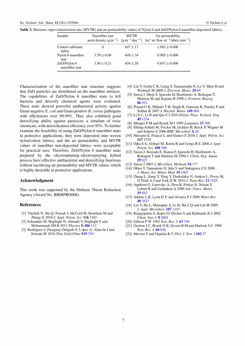
examine the feasibility of using ZnO/Nylon 6 nanofiber matsin protective applications, they were deposited onto wovennylon/cotton fabrics and the air permeability and MVTRvalues of the deposited fabrics were evaluated. Table 3compares the air permeability and MVTR values obtainedfor the control fabric substrate and fabrics deposited withNylon 6 and ZnO/Nylon 6 nanofiber mats. It is seen that thecontrol fabric and the fabric coated with Nylon 6 nanofibermat have comparable MVTR values, which indicates that
Figure 7. Possible detoxifying mechanism of paraoxon byZnO/Nylon 6 nanofibers.
the deposition of Nylon 6 nanofiber mats does not alter themoisture vapor transmission through the fabric structure.This may be due to the small fiber diameter and high porosityof the electrospun nanofiber mat. In addition, depositing aZnO/Nylon nanofiber mat onto the fabric leads to insignificantincrease in MVTR. Therefore, the presence of ZnO particleson electrospun fiber surface does not significantly affect thetransmission of water vapor across the mat. From table 3, it isalso seen that depositing Nylon 6 and ZnO/Nylon 6 nanofibermats onto the fabric only slightly reduces the air permeability.However, the resultant total air permeability is still within anacceptable range for practical applications [22].
4. Summary
ZnO/Nylon 6 nanofiber mats were prepared bythe electrospinning–electrospraying hybrid process.
6
Sci. Technol. Adv. Mater. 12 (2011) 055004 N Vitchuli et al
Table 3. Moisture vapor transmission rate (MVTR) and air permeability values of Nylon 6 and ZnO/Nylon 6 nanofiber-deposited fabrics.
Sample Nanofiber mat MVTR Air permeabilityareal density (g m−2) [g m−2 day−1] [m3 air flow m−2 fabric min−1]
Control substrate 0 647 ± 17 1.082 ± 0.006fabric
Nylon 6 nanofiber 3.59 ± 0.06 649 ± 34 0.905 ± 0.006mat
ZnO/Nylon 6 3.80 ± 0.21 654 ± 28 0.847 ± 0.006nanofiber mat
Characterization of the nanofiber mat structure suggeststhat ZnO particles are distributed on the nanofiber surfaces.The capabilities of ZnO/Nylon 6 nanofiber mats to killbacteria and detoxify chemical agents were evaluated.These mats showed powerful antibacterial activity againstGram-negative E. coli and Gram-positive B. cereus pathogenswith efficiencies over 99.99%. They also exhibited gooddetoxifying ability against paraoxon, a simulant of toxicchemicals, with detoxification efficiency over 95%. To furtherexamine the feasibility of using ZnO/Nylon 6 nanofiber matsin protective applications, they were deposited onto wovennylon/cotton fabrics, and the air permeability and MVTRvalues of nanofiber mat-deposited fabrics were acceptablefor practical uses. Therefore, ZnO/Nylon 6 nanofiber matsprepared by the electrospinning–electrospraying hybridprocess have effective antibacterial and detoxifying functionswithout sacrificing air permeability and MVTR values, whichis highly desirable in protective applications.
Acknowledgment
This work was supported by the Defense Threat ReductionAgency (Award No. BB08PRO008).
References
[1] Vitchuli N, Shi Q, Nowak J, McCord M, Bourham M andZhang X 2010 J. Appl. Polym. Sci. 116 2181
[2] Eskandari M, Haghighi N, Ahmadi V, Haghighi F andMohammadi SH R 2011 Physica B 406 112
[3] Rodriguez J, Paraguay-Delgado F, López A, Alarcón J andEstrada W 2010 Thin Solid Films 519 729
[4] Liu Y, Gorla C R, Liang S, Emanetoglu N, Lu Y, Shen H andWraback M 2000 J. Electron. Mater. 29 69
[5] Sawai J, Shoji S, Igarashi H, Hashimoto A, Kokugan T,Shimizu M and Kojima H 1998 J. Ferment. Bioeng.86 521
[6] Prasad G K, Mahato T H, Singh B, Ganesan K, Pandey P andSekhar K 2007 J. Hazard. Mater. 149 460
[7] Li S C, Li B and Qin Z J 2010 Polym. Plast. Technol. Eng.49 1334
[8] Menger F M and Rourk M J 1999 Langmuir 15 309[9] Ehling-Schulz M, Fricker M, Grallert H, Rieck P, Wagner M
and Scherer S 2006 BMC Microbiol. 6 20[10] Marsano E, Francis L and Giunco F 2010 J. Appl. Polym. Sci.
117 1754[11] Ojha S S, Afshari M, Kotek R and Gorga R E 2008 J. Appl.
Polym. Sci. 108 308[12] Sawai J, Kawada E, Kanou F, Igarashi H, Hashimoto A,
Kokugan T and Shimizu M 1996 J. Chem. Eng. Japan29 627
[13] Sawai J 2003 J. Microbiol. Methods 54 177[14] Ohira T, Yamamoto O, Iida Y and Nakagawa Z E 2008
J. Mater. Sci. Mater. Med. 19 1407[15] Zhang L, Jiang Y, Ding Y, Daskalakis N, Jeuken L, Povey M,
O’Neill A J and York D W 2010 J. Nano Res. 12 1625[16] Applerot G, Lipovsky A, Dror R, Perkas N, Nitzan Y,
Lubart R and Gedanken A 2009 Adv. Funct. Mater.19 842
[17] Adams L K, Lyon D Y and Alvarez P J 2006 Water Res.40 3527
[18] Liu Y, He L, Mustapha A, Li H, Hu Z Q and Lin M 2009J. Appl. Microbiol. 107 1193
[19] Rajagopalan S, Koper O, Decker S and Klabunde K J 2002Chem. Eur. J. 8 2602
[20] Gibson P W 1993 Text. Res. J. 63 749[21] Gretton J C, Brook D B, Dyson H M and Harlock S C 1998
Text. Res. J. 68 936[22] Mavruz S and Ogulata R T 2011 J. Text. I 102 57
7