1616969.pdf - mediaTUM
155
DISSERTATION DER FAKULTÄT FÜR CHEMIE DER TECHNISCHEN UNIVERSITÄT MÜNCHEN PROFESSUR FÜR ZELLULÄRE PROTEINBIOCHEMIE Determinants of intra-membrane client recognition by the molecular chaperone Calnexin Nicolas Blömeke, M.Sc. Vollständiger Abdruck der von der Fakultät für Chemie der Technischen Universität München zur Erlangung des akademischen Grades eines Doktors der Naturwissenschaften genehmigten Dissertation Vorsitzende: 1. Prof. Dr. Cathleen Zeymer Prüfer der Dissertation: 1. Prof. Dr. Matthias Feige 2. Prof. Dr. Johannes Buchner Die Dissertation wurde am 19.07.2021 bei der Technischen Universität München eingereicht und durch die Fakultät für Chemie am 27.10.2021 angenommen.
-
Upload
khangminh22 -
Category
Documents
-
view
0 -
download
0
Transcript of 1616969.pdf - mediaTUM
DISSERTATION DER FAKULTÄT FÜR CHEMIE
DER TECHNISCHEN UNIVERSITÄT MÜNCHEN
PROFESSUR FÜR ZELLULÄRE PROTEINBIOCHEMIE
Determinants of intra-membrane client recognition
by the molecular chaperone Calnexin
Nicolas Blömeke, M.Sc.
Vollständiger Abdruck der von der Fakultät für Chemie der Technischen Universität
München zur Erlangung des akademischen Grades eines Doktors der
Naturwissenschaften genehmigten Dissertation
Vorsitzende: 1. Prof. Dr. Cathleen Zeymer
Prüfer der Dissertation: 1. Prof. Dr. Matthias Feige
2. Prof. Dr. Johannes Buchner
Die Dissertation wurde am 19.07.2021 bei der Technischen Universität München
eingereicht und durch die Fakultät für Chemie am 27.10.2021 angenommen.
EIDESSTATTLICHE ERKLARUNG
Hiermit erkläre ich an Eides statt, dass ich die vorliegende Dissertation
selbstständig und ohne unerlaubte Hilfe angefertigt habe. Ich habe weder
anderweitig versucht, eine Dissertation einzureichen oder eine Doktorprufung
durchzufuhren, noch habe ich diese Dissertation oder Teile derselben einer
anderen Pru fungskommission vorgelegt.
Mu nchen, den (Unterschrift)

Die vorliegende Arbeit wurde zwischen September 2016 und Juli 2021 unter der
Anleitung von Prof. Dr. Matthias Feige an der Fakultät für Chemie der
Technischen Universität in Garching durchgeführt.

Table of Contents
Summary ....................................................................................................... 1
Zusammenfassung ...................................................................................... 3
1 Introduction ............................................................................................... 5
1.1 Membrane proteins – a brief overview .................................................. 5
1.2 Biosynthesis of membrane proteins ...................................................... 6
1.2.1 Alternative routes - SRP-independent ER-targeting ..................... 11
1.2.2 Insertion and topogenesis of membrane proteins at the ER
membrane ............................................................................................. 13
1.2.3 TM helix-helix interactions and the role of sequence motifs in
function and disease ............................................................................. 16
1.2.4 The secretory pathway ................................................................. 20
1.3 Cellular quality control mechanisms for membrane proteins .............. 25
1.3.1 Glycan dependent ER quality control ........................................... 26
1.3.2 Chaperoning in the ER ................................................................. 30
1.3.3 Endoplasmic reticulum-associated protein degradation ............... 36
1.3.3.1 Substrate recognition………………………………………..37
1.3.3.2 Retrotranslocation…………………………………………...40
1.3.3.3 Proteasomal degradation…………………………………...43
1.4 ER stress and the unfolded protein response ..................................... 49
1.5 Transmembrane domain chaperones and intra-membrane quality
control factors ........................................................................................... 51
1.6 Calnexin .............................................................................................. 55
1.6.1 CNX structure and binding sites ................................................... 56
1.6.2 Glycan independent function of CNX and the importance of the
transmembrane domain ........................................................................ 61
2 Aim of studies ......................................................................................... 63
3 Results ..................................................................................................... 65
3.1 Calnexin interacts with transmembrane domains of non-glycosylated
clients ....................................................................................................... 65
3.2 The transmembrane domain of Calnexin binds clients in the
membrane………………………………………………………………………69
3.3 Development of a tool to systematically assess intra-membrane
recognition processes ............................................................................... 71
3.4 Defining intra-membrane recognition motifs for Calnexin ................... 74
3.5 A structural understanding of intra-membrane Calnexin:client
recognition ................................................................................................ 78
3.6 Biological functions of intra-membrane client recognition by Calnexin 82
4 Discussion ............................................................................................... 87
4.1 Principles of intra-membrane chaperoning processes by CNX ........... 87
4.2 Intra-membrane client recognition motifs for CNX .............................. 89
4.3 A conserved recognition motif in the CNX TMD is required for intra-
membrane client recognition ..................................................................... 91
4.4 The protective function of CNX on membrane proteins ...................... 94
4.5 Global analysis of the CNX substrate repertoire ................................. 96
4.6 Final conclusion .................................................................................. 97
5 Materials and methods ......................................................................... 100
5.1 Molecular biology techniques ........................................................... 100
5.1.1 DNA design and modification ..................................................... 100
5.1.2 Cell culture techniques ............................................................... 103
5.1.3 Cell culture based In vivo assays ............................................... 104
5.1.4 Fluorescence Microscopy .......................................................... 105
5.2 Biochemical techniques .................................................................... 107
5.2.1 Protein modification analysis ...................................................... 107
5.2.2 Protein interaction analysis ........................................................ 107
5.2.3 Gel electrophoresis and immuno-blot techniques ...................... 108
5.2.4 Analytical techniques ................................................................. 110
5.3 Data evaluation ................................................................................. 111
5.4 Computational modelling .................................................................. 113
Abbreviations ........................................................................................... 116
References ................................................................................................ 118
Acknowledgements …………………………………………………….……..148
1
Summary Integral membrane proteins (IMPs) are a ubiquitous class of proteins and
account for around one-third of the human proteome. They are permanently
embedded in the lipid membranes surrounding cells and the various intracellular
organelles of a mammalian cell, where they serve numerous important cellular
and physiological tasks. These include transporting hydrophilic molecules and
signals through the lipid bilayers, functioning as metabolic enzymes, or
maintaining cell-cell interactions. Accordingly, IMPs are indispensable for
multicellular organisms in which individual cells function as part of the whole and
thus have to communicate with one another and their environment.
Despite their importance, however, IMPs often cause disease for two reasons.
First, IMPs are particularly vulnerable to incorrect folding and assembly. Their
biogenesis requires several topologically distinct folding events that pose a
particular challenge to the endoplasmic reticulum (ER) folding and quality control
machinery. This includes folding of soluble domains and, defining a membrane
protein, the correct integration and assembly of hydrophobic transmembrane
domains (TMD) inside the lipid bilayer. In addition, mutations that introduce
charges into the TMDs or abolish a polar binding partner, leaving the other
unpaired and exposed, disfavor the integration of TM segments and can
ultimately lead to misfolding and misassembly of a membrane protein. Therefore,
intra-membrane chaperones must guide and control membrane protein structure
formation inside the lipid bilayer. However, mechanistic insights into these
processes remain very limited.
In this study, we use Calnexin (CNX), one of the most abundant ER
chaperones, and putative intra-membrane chaperone as a model to define
principles of intra-membrane chaperoning. We show that CNX binds non-
glycosylated proteins and distinguishes the folded and misfolded state of its
client. Furthermore, we demonstrate that CNX directly recognizes misfolded
clients via its single TMD, regardless of its luminal lectin or C-terminal domain.
By combining experimental and computational approaches, we finally reveal a
protective function of CNX on membrane proteins by identifying an intra-
membrane recognition motif within its TMD that protects clients from premature

2
degradation and appears to be conserved in other intra-membrane chaperones.
At the same time, using an artificial minimal consensus membrane (CoMem)
reporter system, which we have established in this study, we systematically
dissect client signatures that CNX recognizes in the membrane.
In summary, using CNX as an example, we have revealed comprehensive
insights into intra-membrane substrate recognition processes by molecular
chaperones that provide a basis for understanding ER-mediated quality control
of IMPs. As a result, our findings could contribute to a better understanding of
numerous diseases caused by membrane protein-misfolding.
3
Zusammenfassung Integrale Membranproteine (IMPs) sind weit verbreitet und machen etwa ein
Drittel des menschlichen Proteoms aus. Sie sind fest in der Lipidmembran
verankert, die sowohl Zellen als auch die verschiedenen intrazellulären
Organellen einer Säugerzelle umgibt und erfüllen dort wichtige zelluläre und
physiologische Aufgaben. Zu diesen gehören der Transport hydrophiler Moleküle
und Signale durch die Lipiddoppelschicht, die Funktion als Stoffwechselenzym
sowie die Aufrechterhaltung von Zell-Zell-Interaktionen. Daher sind IMPs
unverzichtbar für vielzellige Organismen, bei welchen einzelne Zellen als Teil des
Ganzen fungieren, was eine ständige Kommunikation der Zellen untereinander
sowie mit der Umgebung erfordert.
Trotz ihrer großen Bedeutung verursachen IMPs jedoch häufig Krankheiten.
Der Grund hierfür ist, dass ihre Biogenese mehrere topologisch unterschiedliche
Faltungsereignisse erfordert, die für die Faltungs- und
Qualitätskontrollmaschinerie des endoplasmatischen Retikulums (ER) eine
besondere Herausforderung darstellen. Dies führt dazu, dass IMPs besonders
anfällig gegenüber Fehlern bei der Faltung und Assemblierung sind, welche nicht
nur die Faltung von Intramembrandomänen, sondern – der Definition von
Membranproteinen folgend – auch die korrekte Integration und Assemblierung
von hydrophoben Transmembrandomänen (TMD) innerhalb der
Lipiddoppelschicht umfasst. Darüber hinaus können Mutationen die Integration
von Transmembransegmenten beeinträchtigen, etwa durch das Einführen von
Ladungen oder die Deletion eines polaren Bindungspartner, was schlussendlich
Auswirkungen auf die korrekte Faltung hat. Um Fehlfaltungen entgegenzuwirken
ist es deshalb essentiell, dass Chaperone des Intramembranraums die
Ausbildung der nativen Struktur von Membranproteinen innerhalb der
Lipiddoppelschicht unterstützen und überwachen. Mechanistische Einblicke in
diese Prozesse verschiedener Intramembran-Chaperone sind allerdings sehr
begrenzt.
In dieser Studie verwenden wir Calnexin (CNX), eines der am häufigsten
vorkommenden ER-Chaperone und mutmaßliches Intramembran-Chaperon als
Modellprotein, um die Grundlagen bei Chaperon-Vorgängen im
Intramembranraum zu definieren. Wir zeigen, dass CNX nicht-glykosylierte
4
Membranproteine bindet und bei diesen den gefalteten und fehlgefalteten
Zustand unterscheidet. Darüber hinaus zeigen wir, dass CNX fehlgefaltete
Membranproteine ganz unabhängig von seiner luminalen Lektin oder C-
terminalen Domäne durch die TMD erkennt. Durch die Kombination
experimenteller und computergestützter Ansätze konnten wir schließlich ein
Erkennungsmotiv innerhalb der TMD von CNX identifizieren, das
Membranproteine durch Bindung vor dem vorzeitigen Abbau schützt und in
anderen Chaperonen des Intramembranraums konserviert zu sein scheint.
Gleichzeitig konnten wir mithilfe eines von uns in dieser Studie etablierten,
künstlichen minimal consensus membrane (CoMem) Reportersystems Merkmale
in Transmembrandomänen von Membranproteinen dechiffrieren, die von CNX
innerhalb der Membran erkannt werden.
Zusammenfassend liefert diese Studie am Beispiel von CNX umfassende
Einblicke in Substraterkennungsprozesse molekularer Chaperone innerhalb von
Membranen, die als Grundlage bei der Aufklärung der ER-vermittelten
Qualitätskontrolle von IMP dienen können. Die Erkenntnisse dieser Arbeit
können dadurch zu einem besseren Verständnis zahlreicher Erkrankungen
beitragen, die durch die Fehlfaltung von Membranproteinen verursacht werden.
Membrane proteins – a brief overview
5
1 Introduction In multicellular organisms, each cell has to function as a part of the whole.
Thus, cells have to communicate and interact with their environment while
maintaining cellular and organismal homeostasis. These and other tasks such as
transporting hydrophilic molecules and signals through the lipid bilayers, specific
recognition events in an apolar environment, functioning as metabolic enzymes,
or maintaining cell-cell interactions are mainly dependent on integral membrane
proteins (IMPs), which reside within the lipid membranes that surround cells and
the various intracellular organelles of a mammalian cell. Consequently, IMPs are
indispensable for multicellular structures, which is furthermore highlighted by the
fact that this particular class of proteins accounts for around one-third of the
human proteome (von Heijne and Gavel 1988).
1.1 Membrane proteins – a brief overview
In eukaryotic cells, the biosynthesis of both IMPs and soluble secretory
pathway proteins occurs at the endoplasmic reticulum (ER) (Shao and Hegde
2011). However, IMPs pose more complex challenges to the ER folding and
quality control system than secreted soluble proteins. The formation of their
native state requires multiple topologically distinct folding events. These include
correct folding of soluble domains on both the ER luminal and cytoplasmic side
and, defining a membrane protein, the correct integration as well as the intra and
intermolecular assembly of hydrophobic transmembrane domains (TMD) in the
lipid bilayer destination (Shao and Hegde 2011, Cymer, von Heijne et al. 2015).
During structure formation, interactions between different TM segments
throughout a TM protein significantly contribute to correct integration and
topogenesis. Further complicating these processes, the TMDs of multi-pass
membrane proteins are highly diverse. They can be short or long, only partially
inserted, often contain polar residues, breaks, or kinks, and may exhibit flexibility
in the membrane, which gives rise to complex membrane integration and folding
pathways (Ota, Sakaguchi et al. 1998, Lu, Turnbull et al. 2000, Hessa, White et
al. 2005, Sadlish, Pitonzo et al. 2005, Kauko, Hedin et al. 2010). However,
especially these deviations from ideal hydrophobic membrane anchors allow
Biosynthesis of membrane proteins
6
membrane proteins to fulfill their vast functional repertoire (Illergard, Kauko et al.
2011). At the same time, they render IMPs vulnerable to incorrect folding and
assembly in the lipid bilayer. Moreover, many severe human pathologies are
caused by mutations that disfavor the integration of TM segments by introducing
charges and ultimately result in membrane protein misfolding (Partridge, Therien
et al. 2004, Guerriero and Brodsky 2012, Marinko, Huang et al. 2019). Thus, intra-
membrane chaperones and quality control factors must exist that can recognize
and counteract such threats. Remarkably, many factors located in the ER lumen,
membrane, or cytosol facilitate the highly sophisticated biogenesis of IMPs from
the very beginning and assist IMPs in reaching their native state or degrade upon
failure, as will be described in the following.
1.2 Biosynthesis of membrane proteins
The biosynthesis of membrane proteins is a complex and precisely
coordinated process that occurs either co- or post-translationally and can be
divided into specific but coupled stages:
i) the accurate targeting of the growing polypeptide chain to the ER
membrane
ii) the translocation and integration of TM structures into the lipid bilayer
iii) the formation of the final topology and structure.
This first step in the biogenesis of membrane proteins is generally initiated by
recognizing hydrophobic segments as they emerge from the ribosome and the
targeting of such signal sequences to the ER membrane (Figure 1) (Egea, Stroud
et al. 2005). Despite their diversity in amino acid sequence composition and
length, all ER signal sequences share the same common characteristic: they are
hydrophobic. Importantly, hydrophobic segments once they emerge from the
ribosome do not necessarily represent signal sequences, but can also function
as ER targeting signals, which constitute the first TM helix of a membrane protein.
In such a case, these segments exhibit a greater hydrophobicity than typical
signal sequences, and lack a signal peptide peptidase cleavage. Either way, early
identification and shielding of these hydrophobic segments from the aqueous
cytosol by specific factors is important. One such factor is the nascent
polypeptide-associated complex (NAC) which interacts with short nascent

Biosynthesis of membrane proteins
7
polypeptide chains emerging from the ribosome-nascent chain complex (RNC).
NAC can sense substrates directly upon synthesis by inserting a sensor domain
deep into the ribosomal exit tunnel. As a consequence of polypeptide elongation,
NAC is pushed out of the ribosomal tunnel allowing the recruitment and handover
of the substrate to other targeting factors such as the cytosolic signal recognition
particle (SRP) (del Alamo, Hogan et al. 2011, Gamerdinger, Kobayashi et al.
2019).
SRP is a ribonucleoprotein and one of the most studied protein biogenesis
factors involved in co-translational handling the vast majority of ER-destined
proteins. The most critical functions include recognizing and binding hydrophobic
sequences once they emerge from the exit tunnel of a translating ribosome and
its GTP-dependent interaction with the ER membrane-bound SRP receptor (SR).
Structurally, the SRP consists of one RNA scaffold and six protein subunits, of
which SRP54 is of particular relevance as this subunit mediates signal sequence
recognition and binding. Mainly responsible for recognizing and binding
hydrophobic sequences is a methionine-rich, hydrophobic groove located in the
M-domain of SRP54. When bound to the ribosome, the M-domain is positioned
precisely at the exit tunnel. Cryo-electron microscopy of SRP-RNC assembly
complexes has revealed that in the absence of a substrate this groove is occupied
and autoinhibited by an amphipathic helix originating from the carboxy terminus
of SRP54. It seems conceivable that the helix functions as a hydrophobicity
threshold, only allowing sufficiently hydrophobic signal sequences to be
accommodated in the groove when they emerge from the ribosomal exit tunnel.
Consequently, this leads to a displacement of the helix and repositioning above
the hydrophobic groove whereby the signal sequence is efficiently shielded from
the cytosol (Walter and Blobel 1980, Walter and Blobel 1982, Voorhees and
Hegde 2015, Guna and Hegde 2018).
Beyond that, recognition and binding of SRP causes translational arrest and
guides the RNC to the ER membrane. ER-targeting occurs via an interaction of
the SRP with the membrane-bound heterodimeric SRP receptor (SR), which
consists of a cytoplasmic a subunit that shows remarkable homology with SRP54
and an integral membrane β-subunit. The binding of the SRP to its receptor is
followed by a direct interaction between the RNC complex and the central
Biosynthesis of membrane proteins
8
translocation machinery in the ER membrane, the Sec61 translocon (Egea,
Stroud et al. 2005, Zimmermann, Eyrisch et al. 2011, Dudek, Pfeffer et al. 2015).
Notably, both SRP and SR contain GTPase domains, required for association
and to target the ribosome-nascent chain complex to the Sec61 translocon. Only
correct binding of SRP to the heterodimeric SR leads to extensive interactions of
both GTPases and the formation of a complete active site that is crucial for
reciprocal activation of GTP hydrolysis (Egea, Stroud et al. 2005). This provides
a quality control mechanism that monitors the proper alignment of the ribosomal
exit tunnel with the Sec61 translocation channel and thus shields the emerging
polypeptide chain from the cytosol. Hydrolysis of GTP leads to the dissociation of
the SRP from the ribosome-nascent chain complex, relieving translational arrest
and enabling insertion of the nascent peptide into the translocation channel of
Sec61 and integration of helices into a membrane.
Of note, during or after insertion into the membrane, signal peptides are
cleaved by the membrane-bound signal peptidase (SP). Subsequently, the
following TM segments are then integrated into the membrane. In contrast,
hydrophobic segments functioning as signal anchors are not cleaved but move
laterally out of the Sec61 translocon into the membrane (Egea, Stroud et al. 2005,
Shao and Hegde 2011, Cymer, von Heijne et al. 2015, Dudek, Pfeffer et al. 2015).
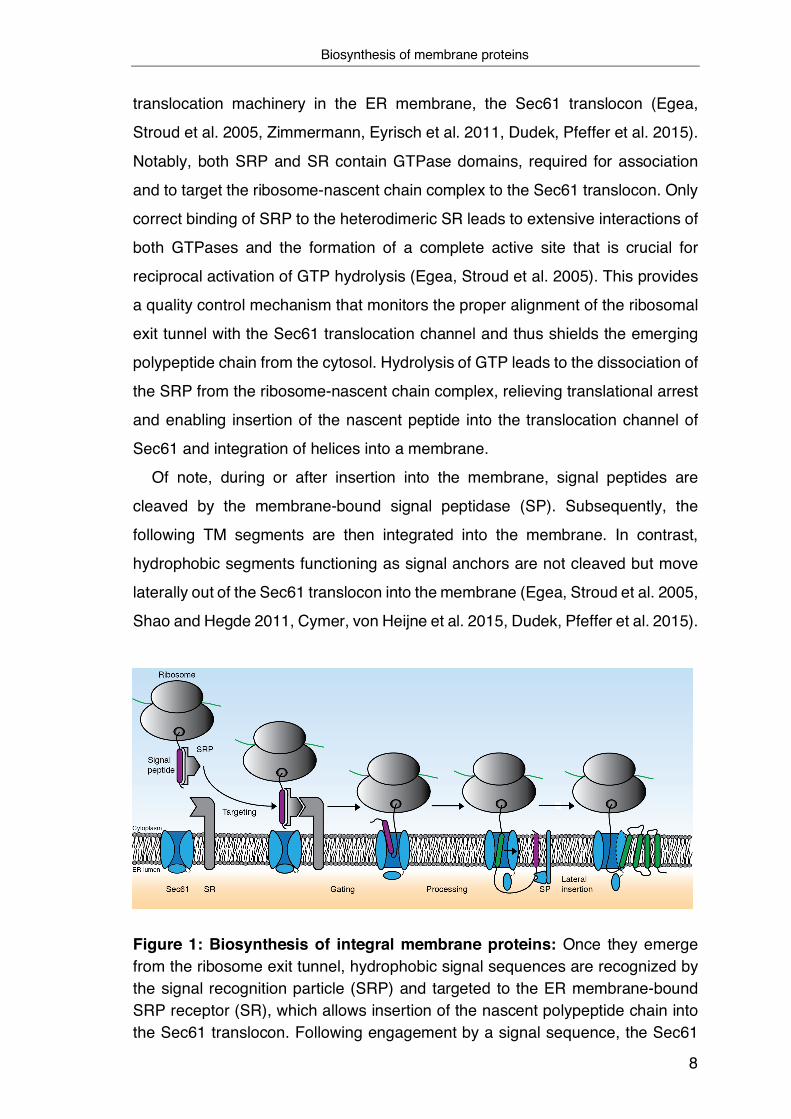
Figure 1: Biosynthesis of integral membrane proteins: Once they emerge from the ribosome exit tunnel, hydrophobic signal sequences are recognized by the signal recognition particle (SRP) and targeted to the ER membrane-bound SRP receptor (SR), which allows insertion of the nascent polypeptide chain into the Sec61 translocon. Following engagement by a signal sequence, the Sec61
Biosynthesis of membrane proteins
9
translocon undergoes a rotational movement that induces the opening of the central pore through the membrane and into the lipid bilayer. During or after insertion into the membrane, signal peptides are cleaved by the signal peptidase (SP), and subsequent TM helices are integrated laterally into the membrane (Figure based on Guerriero and Brodsky 2012).
The second and most decisive step in the biogenesis of a large majority of integral
membrane and soluble proteins is their integration into the membrane, mainly
depending on the ER translocon Sec61. Although it is known that peptides can
also spontaneously integrate into the membrane, this process only applies to
short and highly hydrophobic peptides (Tissier, Woolhead et al. 2002,
Brambillasca, Yabal et al. 2006). The Sec61 itself is a highly conserved
heterotrimeric ER membrane protein complex composed of the essential a and g
subunits and the non-essential b subunit. It is characterized by an aqueous
protein-conducting channel that spans the entire membrane and is formed by TM
helices of Sec61a (Simon and Blobel 1991, Crowley, Liao et al. 1994, Mothes,
Prehn et al. 1994). This channel enables the cell to fulfill two essential cellular
functions: the translocation of soluble proteins across the ER membrane as well
as the integration of TM segments into the surrounding lipid bilayer by lateral
movement through a gate present in the central pore (Shao and Hegde 2011,
Cymer, von Heijne et al. 2015).
In the absence of translation, the Sec61 translocon switches from an open to
a closed conformational state. In this inactive state, the translocon is not only
closed axially by a short Sec61a helix reaching into the central pore, functioning
as a plug, but also laterally. Thus, the permeability barrier of the ER membrane
for small molecules is maintained (Erdmann, Jung et al. 2010, Zimmermann,
Eyrisch et al. 2011). Impermeability of the ER membrane is additionally ensured
by the action of immunoglobulin heavy-chain binding protein (BiP), which binds
and seals the ER luminal side of the translocon pore (Hamman, Hendershot et
al. 1998). Conversely, the molecular understanding of how substrates activate
the Sec61 translocon by causing channel opening for translocation or insertion is
incomplete. It has been revealed that in complex with the RNC, Sec61 undergoes
a rotational movement following the engagement by a signal sequence. As a
consequence of this substrate-induced conformational shift, the Sec61a helix
Biosynthesis of membrane proteins
10
responsible for closing the channel is displaced, which induces the opening of
the central pore across the membrane and into the lipid bilayer (Figure 1). It
seems plausible that for these processes, the hydrophobicity of signal sequences
is a determining criterion since insertion into the lateral gate requires the
disruption of interactions between Sec61a helices that contribute to the formation
of the channel. In fact, this process would be energetically unfavorable for
hydrophilic segments of a polypeptide (Voorhees and Hegde 2015, Voorhees and
Hegde 2016).
Although the Sec61 translocon predominantly mediates the insertion of IMPs,
many additional factors contribute to its function and assist on translocation and
hence the biogenesis of IMPs (Conti, Devaraneni et al. 2015). Amongst these
factors are the translocon-associated protein (TRAP) and the translocating-chain
associating membrane (TRAM). The TRAP complex is formed by four different
proteins that, through constitutive association with Sec61, affect both the
structure of the ribosome-translocon complex and the function of the Sec61
translocon itself (Fons, Bogert et al. 2003, Shao and Hegde 2011). In addition,
TRAP appears to control signal sequence recognition and transmembrane helix
insertion in a substrate-dependent manner. Only recently has it been revealed
that TRAP preferentially binds signal peptides whose amino acid composition
shows a high content of glycine and proline and a slightly below average
hydrophobicity (Nguyen, Stutz et al. 2018). Since hydrophobicity of a signal
sequence, as described above, is a decisive factor for the opening process of the
translocon channel, it is possible that TRAP assists in the channel opening for
particularly those substrates which based on their low hydrophobicity, would only
be inefficiently or improperly translocated (Fons, Bogert et al. 2003, Nguyen,
Stutz et al. 2018). Similar to the function of TRAP, the multipass TM protein
TRAM enhances the integration efficiency of different substrates, however,
preferentially those with relatively hydrophilic TM segments in the early stages of
synthesis (Heinrich, Mothes et al. 2000). Accordingly, it has been suggested that
TRAM plays a vital role during the challenging integration process of multi-
spanning TM proteins. Presumably, TRAM holds several TMDs of nascent
translocating proteins in a temporary transition state before their coordinated
integration into the lipid phase (Do, Falcone et al. 1996, Voigt, Jungnickel et al.
Alternative routes – SRP-independent ER-targeting
11
1996, Heinrich, Mothes et al. 2000, Shao and Hegde 2011). This process seems
to be especially important for proteins with charged or polar residues in their TM
regions as they need to interact with other TM segments to form hydrophobic
complexes where polar residues are buried within the protein´s interior (Illergard,
Kauko et al. 2011). In general, hydrophobicity and length but also helicity of TMDs
of ER targeted IMPs are very diverse and influence the precise mechanism for
insertion into the ER. Therefore, membrane proteins unable to use this default
and highly conserved SRP-dependent Sec61 ER-insertion pathway, like small
secretory protein, tail-anchored (TA) proteins, and proteins that insert into the ER
membrane post-translationally, alternative routes exist, including the GET or the
recently discovered SND pathway in yeast (Hegde and Keenan 2011, Johnson,
Powis et al. 2013, Strzyz 2016).
1.2.1 Alternative routes - SRP-independent ER-targeting TA proteins are characterized by a single TMD domain located near the C-
terminus. As a consequence of this spatial proximity between the TMD and
terminating stop codon, the translation terminates, and the TA-protein is released
from the ribosome before the signal sequence can emerge into the cytosol. As a
consequence, co-translational engagement by the SRP is no longer possible,
which makes recognition and delivery of an emerging nascent chain by an
alternative SRP-independent and post-translationally acting pathway
indispensable. In such situations, the conserved ATPase GET (guided entry of
TA proteins) enables transfers of tail-anchored proteins to the ER (Stefanovic and
Hegde 2007). However, GET alone is not able to capture substrates effectively
but requires assistance. Sgt2 is a chaperone-like protein that recognizes
hydrophobic TA sequences soon after they are released from the ribosome and
subsequently mediates transfer to the homodimeric targeting factor Get3.
Interestingly, like SRP54, Get3 possesses a methionine-rich groove required to
accommodate TA protein targeting signals (Mateja, Szlachcic et al. 2009, Wang,
Brown et al. 2010, Chio, Cho et al. 2017). Finally, following interaction with its
receptor Get1/2, a transmembrane insertase located in the ER membrane, Get3
releases its substrate and allows insertion of the TA protein into the membrane
(Wang, Chan et al. 2014). This pathway, however, is not accessible for TA

Alternative routes – SRP-independent ER-targeting
12
substrate where the TM segment has only moderate hydrophobicity, which
prevents interaction with Get3. In this case, insertion of the TA substrate is
mediated by a different insertase, the transmembrane domain insertase EMC, as
will be described later (Schuldiner, Metz et al. 2008, Borgese 2016, Guna and
Hegde 2018, Shurtleff, Itzhak et al. 2018).
Another pathway for proteins that exhibit downstream instead of amino-
terminal located TMDs and thus do not represent preferred SRP substrates is
mediated by the SND (SRP-independent targeting) pathway. Proteins of this
pathway, namely Snd1, Snd2, and Snd3, were initially discovered in a screen to
identify novel factors that mediate ER targeting independent of SRP or GET.
Unlike the previously described pathways, the SND pathway specifically
recognizes centrally located TM segments within the polypeptide chain of its
substrates. Most likely, once present in the cytosol, signal sequences are co-
translationally captured by the ribosome-associated Snd1 and subsequently
transferred to the ER membrane-embedded and translocon-associated Snd2/3
complex, which not only functions as a receptor for Snd1 but also enables
translocation of the substrate (Aviram, Ast et al. 2016).
In summary, all SRP-independent pathways that have been uncovered share
the same working principle: preinsertional shielding of proteins in the cytosol
followed by rapid targeting to the ER membrane and the subsequent assistance
during integration into the ER membrane. Moreover, the individual pathways can
cooperatively work together or compensate for each other in loss of function
events or as a consequence of cellular stress (Aviram, Ast et al. 2016,
Hassdenteufel, Johnson et al. 2018). Together, this enables the most efficient ER
targeting for the largest possible number of proteins. Interestingly, this
characteristic of mutual compensation does not only seem to affect the various
ways of ER targeting but has also been described for the translocation process
via the Sec61 translocon. In addition to Sec61, yeast can rely on a further,
alternative translocon, namely Ssh1 (SEC61A2 in humans), which functions as a
backup mechanism when the canonical Sec61 is overloaded. Of note, the range
of functions of Ssh1 seems to be far-reaching as specific substrates prefer
translocation via Ssh1, which causes functional overlapping with Sec61 (Finke,
Plath et al. 1996, Spiller and Stirling 2011, Ast and Schuldiner 2013).

Insertion and topogenesis of membrane proteins at the ER membrane
13
However, regardless of whether ER targeting is based on an SRP-dependent or
independent process, one thing becomes apparent: the nature of the primary
protein sequence itself, the amino acid composition, and the degree of
hydrophobicity dictate which mechanism comes into effect for membrane
insertion.
1.2.2 Insertion and topogenesis of membrane proteins at the ER membrane
The final and most complex step in the biogenesis of membrane proteins
following membrane insertion involves forming their topology. In general, the
topology of an integral membrane protein relates to the number of TM helices
present in a transmembrane protein (which allows classification into single- or
multi-spanning TM proteins), and how these helices are oriented relative to each
other and within the membrane with respect to the position of N- and C-terminus
(Figure 2) (von Heijne 2006). In addition, luminal or cytoplasmic domains that
protrude from the hydrophilic backbone of membrane phospholipids and connect
the assembled transmembrane helices make a significant contribution to the
topology of membrane proteins. Considering that fully assembled membrane
complexes are highly dynamic structures, topology formation becomes even
more complicated. Switching between conformations through positional changes
or the partial folding and unfolding of helices within the structure of IMPs is not a
rare phenomenon (von Heijne 2006).
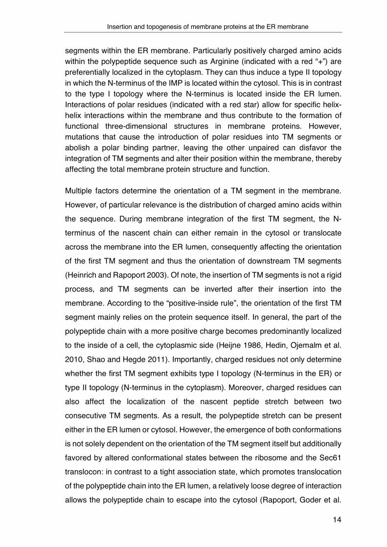
Figure 2: Schematic representation of TM proteins in different conformational states: Depending on the number of TM segments within the ER membrane, IMPs can be classified into single or multi-pass TM proteins. The distribution of charged residues mainly determines the orientation of TM
Insertion and topogenesis of membrane proteins at the ER membrane
14
segments within the ER membrane. Particularly positively charged amino acids within the polypeptide sequence such as Arginine (indicated with a red “+”) are preferentially localized in the cytoplasm. They can thus induce a type II topology in which the N-terminus of the IMP is located within the cytosol. This is in contrast to the type I topology where the N-terminus is located inside the ER lumen. Interactions of polar residues (indicated with a red star) allow for specific helix-helix interactions within the membrane and thus contribute to the formation of functional three-dimensional structures in membrane proteins. However, mutations that cause the introduction of polar residues into TM segments or abolish a polar binding partner, leaving the other unpaired can disfavor the integration of TM segments and alter their position within the membrane, thereby affecting the total membrane protein structure and function.
Multiple factors determine the orientation of a TM segment in the membrane.
However, of particular relevance is the distribution of charged amino acids within
the sequence. During membrane integration of the first TM segment, the N-
terminus of the nascent chain can either remain in the cytosol or translocate
across the membrane into the ER lumen, consequently affecting the orientation
of the first TM segment and thus the orientation of downstream TM segments
(Heinrich and Rapoport 2003). Of note, the insertion of TM segments is not a rigid
process, and TM segments can be inverted after their insertion into the
membrane. According to the “positive-inside rule”, the orientation of the first TM
segment mainly relies on the protein sequence itself. In general, the part of the
polypeptide chain with a more positive charge becomes predominantly localized
to the inside of a cell, the cytoplasmic side (Heijne 1986, Hedin, Ojemalm et al.
2010, Shao and Hegde 2011). Importantly, charged residues not only determine
whether the first TM segment exhibits type I topology (N-terminus in the ER) or
type II topology (N-terminus in the cytoplasm). Moreover, charged residues can
also affect the localization of the nascent peptide stretch between two
consecutive TM segments. As a result, the polypeptide stretch can be present
either in the ER lumen or cytosol. However, the emergence of both conformations
is not solely dependent on the orientation of the TM segment itself but additionally
favored by altered conformational states between the ribosome and the Sec61
translocon: in contrast to a tight association state, which promotes translocation
of the polypeptide chain into the ER lumen, a relatively loose degree of interaction
allows the polypeptide chain to escape into the cytosol (Rapoport, Goder et al.
Insertion and topogenesis of membrane proteins at the ER membrane
15
2004). Besides that, the orientation of a TM segment is furthermore affected by
the distribution of specific upstream or downstream sequence, folding of flanking
domains, and overall length of the hydrophobic sequence and hydrophobicity in
general. For instance, rapidly folding structures in the N-terminal domain of a
nascent polypeptide chain can influence the orientation of a downstream TM
segment in such a way that a type-I orientation is prevented. The reason for this:
In order to adopt this conformation, transport of the already folded domain
through the narrow translocation channel would be required (Denzer, Nabholz et
al. 1995).
In contrast, rather long and hydrophobic TM segments are often found in type-
I orientation because the inversion of TM segments after insertion into the
translocon head-first is slowed down and energetically unfavored with increasing
hydrophobicity of the TM sequence. Ultimately, however, the rate of inversion is
mainly determined by the number of charged amino acids close to the
hydrophobic signal (Goder and Spiess 2003). Although the tremendous structural
variability of TM helices is mainly caused by the distribution of specific amino
acids along the sequence of an IMP, their occurrence and distribution within
distinct TMDs are subject to a certain regularity. Whereas hydrophobic residues
like Ala, Ile, Val, and Leu are more often found directly in the middle of the
membrane, aromatic residues like Tyr and Trp are more likely to be found in the
vicinity or within the lipid-water interface. In contrast, charged or polar residues
are usually found less frequently in membranes, although they have important
functions, as explained below (Ulmschneider, Sansom et al. 2005).
In general, it is assumed that membrane integration is based on a partitioning
process between the hydrophilic environment inside the Sec61 translocon
channel and the hydrophobic environment within the lipid bilayer. Thus, proteins
whose TM segments are strongly hydrophobic should readily partition into the
membrane one by one or in pairs (Rapoport, Goder et al. 2004). This mainly
applies to single-spanning TM proteins (Hessa, Kim et al. 2005). However, as
briefly discussed above, membrane integration is far more complex for multi-
spanning TM proteins. The cause for that is that IMPs need to establish their
correct topology during or after synthesis, which, among others, essentially
depends on interactions between different TM segments.

TM helix-helix interactions and the role of sequence motifs in function and disease
16
1.2.3 TM helix-helix interactions and the role of sequence motifs in
function and disease Interactions between neighboring TM segments based on designated
sequences and motifs are indispensable to establish correct IMP topologies and
functions. Furthermore, these interactions can be an essential prerequisite that
enables membrane integration in the first place. However, failures during
assembly can lead to the exposure of these residues and motifs to the
membrane´s hydrophobic environment, which is often the case with unpaired
polar residues. In such a case, it seems conceivable that similar sequences and
motifs, once exposed, could contribute to the process of substrate recognition by
intra-membrane chaperones, which is also based on a direct interaction between
TM segments. Therefore, helix-helix interactions based on designated
sequences and motifs will be discussed in more detail in the following.
Two major driving forces underlie the folding and assembly of polypeptide
segments within the lipid bilayer: saturation of hydrogen bond donors and
acceptors within the peptide backbone and the interaction of polar residues,
whose presence in the hydrophobic interior of a membrane would otherwise be
unfavorable. Interactions of polar residues allow for specific helix-helix
interactions within the membrane and thus contribute to forming functional three-
dimensional structures in membrane proteins (Figure 2). Remarkably, the
capability of polar residues mediating the association of membrane-embedded
helices is differently pronounced. Whereas amino acids that contain two polar-
side chains such as Asn and Gln strongly promote the interaction between
different TM segments, amino acids like Thr or Ser, which exhibit only one polar
side-chain atom, show a much weaker tendency (Finke, Plath et al. 1996,
Choma, Gratkowski et al. 2000, Gratkowski, Lear et al. 2001, Zhou, Merianos et
al. 2001). Responsible for this effect is the ability to simultaneously function as a
hydrogen donor and acceptor, a characteristic that applies to amino acids
containing two polar atoms such as Asn or Gln in their side chains. However,
stabilization of TM helix-helix interactions through interhelical hydrogen bonds
between polar residues is not always guaranteed. Quite in contrast, these
interactions show a strong positional-dependency of polar residues along with
the TMD (Dawson, Melnyk et al. 2003). Moreover, relying on polar residues for
TM helix-helix interactions and the role of sequence motifs in function and disease
17
the establishment of IMP topologies comes at a cost: TM segments with only
marginal hydrophobicity show poor insertion efficiencies, which could
consequently impair correct integration and folding of IMPs (Hedin, Ojemalm et
al. 2010). However, this effect of polar residues on membrane insertion does not
seem to be generally valid, but rather to be context-dependent. Using suitable in
vitro translation systems, it could be shown that position-specific Asn- or Asp
mediated interactions with an adjacent TM helix can also promote the membrane
insertion efficiency of a marginally hydrophobic transmembrane segment
(Meindl-Beinker, Lundin et al. 2006).
Strikingly, polar residues are very widespread and highly conserved in TM
segments, although they can cause aberrant membrane integration (Illergard,
Kauko et al. 2011). This further underlines the structural and functional relevance
of polar residues in IMP TM segments. Indeed, previous work analyzing the
integration efficiency of individual TM segments revealed that about 25 % of all
human TM helices present in multi-spanning IMPs are predicted to exhibit
unstable membrane integration (DGapp > 0 kcal/mol) in isolation (Hessa, Meindl-
Beinker et al. 2007). This argues for potentially very common folding and
integration problems for IMPs posed by unpaired polar residues. Unfortunately,
hardly anything is known about cellular control and response mechanisms
dealing with such problems during IMP biogenesis. Polar residues in TM
segments are not only a problem encountered during the biogenesis of a large
number of naturally occurring IMPs – they are also a cause for disease (Partridge,
Therien et al. 2004). In fact, mutations that cause the introduction of polar
residues into TM segments or abolish a polar binding partner, leaving the other
unpaired and exposed, are related to various human pathologies. In this regard,
it has been shown that polar residues not only disfavor the integration of TM
segments but can also alter their position within the membrane, thereby affecting
the structure and function of the total membrane protein (Figure 2) (Partridge,
Therien et al. 2004, Hutt, Powers et al. 2009, Guerriero and Brodsky 2012). This
was shown for the Erb-B2 oncogene product, a receptor tyrosine kinase that
belongs to the family of epidermal growth factor receptors. As a result of the
occurrence of a single point mutation in the TMD of the protein in which Val is
exchanged for Glu, Erb-B2 undergoes dimerization and activation, which causes
TM helix-helix interactions and the role of sequence motifs in function and disease
18
a large number of breast and ovarian cancers (Bargmann, Hung et al. 1986,
Weiner, Liu et al. 1989).
As mentioned earlier, the distribution of charged amino acids within the
sequence of a nascent IMP significantly contributes to the topology. In addition,
positively charged amino acids such as Arg and Lys located in the areas between
and within TM segments fulfill essential structural and functional roles in
membrane proteins. Among others, they are required for substrate recognition
and are involved in assembly processes (Oosawa and Simon 1986, Finke, Plath
et al. 1996, Xu, Kakhniashvili et al. 2000, Ding and Wilson 2001). Necessary for
assembly are dimerization processes mainly determined by the protonation state
of the respective Arg or Lys residue residing in a TM segment. While the
formation of hydrogen bonds stabilizes dimerization, an increasing repulsion
contributes to a monomeric state and mainly occurs if positive amino acids are
located directly in the interacting interface of the two helices (Finke, Plath et al.
1996, Therien and Deber 2002). Unfortunately, similar to polar residues,
positively charged residues when introduced into TM regions by mutation are
linked to a large number of genetic diseases such as cystic fibrosis, Charcot-
Marie-Tooth disease, or Wilson´s disease to name just a few (Partridge, Therien
et al. 2002, Partridge, Therien et al. 2004, Fink, Sal-Man et al. 2012, Ruskamo,
Nieminen et al. 2017). In most cases, introducing a positive charge leads to an
impaired interaction or assembly of TMD segments, which consequently affects
the entire structure. Interestingly, most of the mutations that ultimately lead to
diseases result from the introduction of Arg but not Lys residues into the TMDs
(Fink, Sal-Man et al. 2012). However, the influence of positively charged residues
on the structure of an IMPs does not necessarily have to be associated with
adverse effects. Quite in contrast, it seems that ionic interactions of oppositely
charged residues inside the membrane strongly promote the interaction between
adjacent TMDs and thus are essential for the assembly of various IMPs
(Herrmann, Fuchs et al. 2010).
In addition to polar and charged amino acids, aromatic amino acids also play
a role in the interaction of helices. Albeit their role is comparatively low, aromatic
residues show a highly conserved occurrence in TM segments. Due to their
unique structure, they are crucial for many molecular recognition and assembly
TM helix-helix interactions and the role of sequence motifs in function and disease
19
processes in IMPs, such as in amyloid polypeptides or bacterial toxins
(Belbeoc'h, Falasca et al. 2004, Ramachandran, Tweten et al. 2004, Fink, Sal-
Man et al. 2012). Responsible for the interaction between helices containing
aromatic residues is the formation of different high-order clusters, which are also
referred to as non-covalent π – π interactions between planar aromatic rings
(Bernstein and Sun 1996, McGaughey, Gagne et al. 1998, Gazit 2002).
Interestingly, TM-self-assembly is not exclusively promoted by π – π interactions
alone but also by cation – π interactions, which seem to promote tertiary structure
formation and function greatly. Although these interactions are only well
documented for soluble proteins thus far, they also appear to be crucial for the
folding and stability of IMPs. This is because dimerization of TM segments is
greatly enhanced by the interaction of charged residues and hydrophobic
residues such as Lys with Trp, Tyr or Phe (Johnson, Hecht et al. 2007).
Ultimately, besides individual amino acids of different hydrophobicity, charge,
or aromaticity, which cause non-covalent interactions of TM helices alone or in
combination with others, helix-helix interactions are mediated by various highly
conserved interaction motifs present in the TM regions of IMPs. The most
prominent example of many well-known interaction motifs is the GxxG motif, first
discovered in glycophorin A, a membrane protein of the erythrocytes (Lemmon,
Flanagan et al. 1992, Lemmon, Treutlein et al. 1994, Russ and Engelman 2000).
As outlined above, helix-helix interactions are highly diverse and form the basis
for the correct integration and stable interaction of TM segments within the
membrane. Remarkably, however, it is assumed that interaction of TM segments
and the formation of functional structures may occur even before membrane
insertion. Either within the ribosomal exit tunnel, which is capable of promoting
structure formation by providing a confined compartment, or inside the Sec61
translocon channel, which can accommodate more than two TM segments at the
same time and thus in principle enables the promotion of interactions (Rapoport,
Goder et al. 2004, Tu, Khanna et al. 2014, Cymer, von Heijne et al. 2015). In line
with this, it is assumed that TM segments of nascent polypeptide chains can be
retained within the translocation channel of Sec61, even until the translation is
terminated, and then be collectively delivered into the surrounding lipid bilayer.
Although this model contradicts the aforementioned “linear insertion model”, it
The secretory pathway
20
highlights the need for alternative integration mechanisms that can account for
polar or charged residues within helical TM segments that do not follow the
canonical view of a thermodynamically favorable integration of individual TM
segments.
1.2.4 The secretory pathway In order to fulfill their function, most newly made proteins in the membrane or
the lumen of the ER, once correctly folded and assembled, exit the ER and travel
via the secretory pathway to their target destination (Figure 3A). Of particular
relevance for the journey of proteins along this pathway: The Golgi apparatus.
The Golgi is composed of flattened, membrane-enclosed cisternae and
represents an important site for synthesizing oligosaccharides. Furthermore, it
mainly contributes to the sorting and trafficking of ER-derived proteins. In general,
the secretory pathway refers to the selective and efficient trafficking of coated
vesicles loaded with the protein to be transported between the ER and the Golgi
as well as the cell membrane and the lysosomes or any other intracellular
compartment. Although little is known about signals that initiate exit and guide
proteins out of the ER, the exit process itself is a better understood and highly
regulated process restricted to specific, randomly dispersed, ribosome-free
subdomains within the ER membrane, the ER exit sites (ERES) (Figure 3B).
(Watson and Stephens 2005, Peotter, Kasberg et al. 2019). In mammalian cells,
the ER-to-Golgi transport is mediated by Coat Protein complex II (COPII)-coated
vesicles. Depending on whether transmembrane or soluble molecules are
exported from the ER, selection and loading of the respective cargo mediated by
COPII occurs either directly or indirectly with the help of specific cargo receptors.
In both cases, however, COPII causes deformation of the membrane at the ERES
and, consequently, the generation of budded transport vesicles. Following
scission, loaded cargo vesicles are delivered to the ER-Golgi intermediate
compartment (ERGIC) located between the ER and the cis-site of the Golgi
apparatus and mainly responsible for facilitating the sorting of cargo between
these two organelles. Importantly, delivery of the cargo content to the ERGIC
primarily requires the shedding of the COPII coating and subsequently fusion of
the transport vesicles. Thereby, vesicular tubular clusters (VTC) are formed that
The secretory pathway
21
constitute a new compartment and lack ER proteins. (Kuehn, Herrmann et al.
1998, Appenzeller-Herzog and Hauri 2006). In contrast to the first COPII
dependent transport step, forward transport from the ERGIC to the cis-Golgi
network, representing the Golgi complex´s receiving cisternae, does not depend
on COPII. However, migration of the carriers occurs in a microtubule-dependent
process (Watson and Stephens 2005, Peotter, Kasberg et al. 2019).
Once present in the Golgi, secretory cargo traverses the different
compartments of the Golgi and can undergo an ordered series of covalent
modifications, amongst others, by a distinct set of glycosyltransferases and
glycosidases. These enzymes, which are localized in the different compartments
of the Golgi, mainly contribute to the processing and formation of complex
oligosaccharides of glycoproteins originally glycosylated in the ER (Ohtsubo and
Marth 2006). Finally, cargo undergoes repackaging in order to be released and
delivered to the target destination. Although intra-Golgi trafficking is highly
debated, two prevailing models are discussed. On the one hand, the cisternal
maturation model and alternatively the vesicular transport model. According to
the highly dynamic cisternal maturation model, the cargo is stably located in a
given compartment, a newly formed cis cisterna following vesicle fusion, which in
turn functions as a transient carrier and gradually traverses the Golgi stack.
During progression through the stack, various Golgi enzymes of the medial and
then trans cisternae are acquired through COPI vesicles that move in a
retrograde traffic fashion from later to earlier cisternae and cause maturation of
the initial cargo-loaded cisternae. Ultimately, the oldest cisternae, namely the
trans-Golgi network (TGN), disintegrates into secretory vesicles or other types of
carriers (Glick and Luini 2011, Luini 2011). In the vesicular model, the different
compartments of the Golgi from cis to trans are primarily static, and the
characteristic set of Golgi processing enzymes remains unchanged while cargo
proteins in transit move through the cisternae. Unlike the previously described
model, the transport of cargo mediated by COPI vesicles mainly occurs in an
anterograde traffic fashion where cargo is moved forward from one cisterna to
the next in the direction of the TGN (Glick and Luini 2011).
From here, proteins can take different trafficking routes and are transported
either to the endolysosomal system, the plasma membrane, or beyond to the
The secretory pathway
22
extracellular region (Figure 3C). However, not all proteins continue their journey
along the secretion pathway. Some, which reside in the Golgi are retained and
subsequently return to their appropriate cisterna, or, if resident in or escaped the
ER, are transported in a COPI-dependent fashion from the cis-Golgi back to the
ER (Figure 3D)(Schekman and Orci 1996). Due to the numerous possible
destinations a protein can be targeted to after exiting the ER, it becomes evident
that the sorting process at the TGN essentially requires many specialized vesicle
carriers and trafficking pathways.
Required for the retrieval pathway is the presence of an ER retrieval sequence,
which can be a C-terminal cytosolic dilysine (e.g., KKxx or KxKxx) or luminal
KDEL motif, which allows interaction with the COPI coat either directly via distinct
repeat domains or is mediated by a specific KDEL receptor (KDELR) that can be
found in the membrane of the cis-Golgi compartment (Lewis and Pelham 1992,
Orci, Stamnes et al. 1997, Aoe, Lee et al. 1998, Jackson, Lewis et al. 2012). The
binding and release of proteins carrying a KDEL sequence is mainly facilitated
based on varying pH-values established in the different compartments of the
secretory pathway. Whereas the comparatively more acidic pH-value inside the
lumen of the cis-Golgi increases the affinity of the KDELR for the KDEL sequence
and consequently promotes the incorporation of cargo into COPI vesicles, the
neutral pH-value in the ER contributes to the release from the receptor once the
vesicle complex arrives at the ER (Brauer, Parker et al. 2019).
However, besides the KDEL retrieval pathway, other models enable the
anchoring of proteins already in the ER or determining their location in the cell.
For instance, it is assumed that in order to remain in their organelle, ER-resident
proteins, independent of their KDEL signal, bind to each other and form larger
complexes that vesicles can no longer transport. Since the concentration of ER-
resident proteins inside the ER is very high, comparatively low-affinity interaction
would be sufficient for such complexes to arise. Interestingly, Golgi processing
enzymes that function together in the same compartment prevent their passaging
into transport vesicles by the exact same aggregation mechanism (Nilsson,
Slusarewicz et al. 1993, Bruce, Johnson et al. 2002). Retention of Golgi-localized
proteins is furthermore facilitated based on distinct retention motifs as recently, a
conserved KXD/E motif has been discovered that directly interacts with COPI

The secretory pathway
23
(Gao, Cai et al. 2014). Another sorting mechanism, relevant if the cargo involves
membrane proteins, is mediated by the length of their TM region. The TMD of
membrane domains dictates the partitioning process into the membrane of
vesicles and different compartments, but additionally, it seems that the length of
a TMD is optimized to match the thickness of the lipid bilayer in which it resides
(Sharpe, Stevens et al. 2010). Thus, whereas shortening of the TMD can cause
retention, lengthening of this region allows Golgi-localized membrane proteins to
progress in the secretory pathway and relocate via vesicular transport to the
plasma membrane (Bonifacino and Traub 2003, Borgese 2016).
Figure 3: Intracellular protein traffic – the secretory pathway. (A) Once correctly folded and assembled, native membrane and soluble proteins enter ribosome-free subdomains within the ER membrane, the ER exit sites (B), from where their journey along the secretory pathway begins. At the ER exit sites, coatomer protein (COP) II causes deformation of the membrane and, consequently, the budding of cargo-loaded transport vesicles. Following scission, COPII coated cargo vesicles traffic via the ER-Golgi intermediate compartment (ERGIC) to the cis-site of the Golgi apparatus. From here, secretory cargo traverses the different compartments of the Golgi and undergoes an ordered series of covalent modifications mediated by glycosyltransferases and glycosidases. (C) From the TGN, proteins are either transported to the plasma membrane and beyond or, delivered to the endolysosomal system for degradation. (D) In certain cases, however, proteins that are misfolded and
The secretory pathway
24
escaped the ER or ER-resident proteins do not proceed their journey along the secretion pathway, but are instead transported in a COPI-dependent manner from the cis-Golgi back to the ER (Figure based on Ellgaard and Helenius 2003).
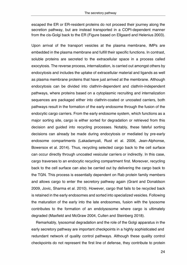
Upon arrival of the transport vesicles at the plasma membrane, IMPs are
embedded in the plasma membrane and fulfill their specific functions. In contrast,
soluble proteins are secreted to the extracellular space in a process called
exocytosis. The reverse process, internalization, is carried out amongst others by
endocytosis and includes the uptake of extracellular material and ligands as well
as plasma membrane proteins that have just arrived at the membrane. Although
endocytosis can be divided into clathrin-dependent and clathrin-independent
pathways, where proteins based on a cytoplasmic recruiting and internalization
sequences are packaged either into clathrin-coated or uncoated carriers, both
pathways result in the formation of the early endosome through the fusion of the
endocytic cargo carriers. From the early endosome system, which functions as a
major sorting site, cargo is either sorted for degradation or retrieved from this
decision and guided into recycling processes. Notably, these fateful sorting
decisions can already be made during endocytosis or mediated by pre-early
endosome compartments (Lakadamyali, Rust et al. 2006, Jean-Alphonse,
Bowersox et al. 2014). Thus, recycling selected cargo back to the cell surface
can occur directly through uncoated vesicular carriers or indirectly. In this case,
cargo traverses to an endocytic recycling compartment first. Moreover, recycling
back to the cell surface can also be carried out by delivering the cargo back to
the TGN. This process is essentially dependent on Rab protein family members
and allows cargo to enter the secretory pathway again (Grant and Donaldson
2009, Jovic, Sharma et al. 2010). However, cargo that fails to be recycled back
is retained in the early endosomes and sorted into specialized vesicles. Following
the maturation of the early into the late endosomes, fusion with the lysosome
contributes to the formation of an endolysosome where cargo is ultimately
degraded (Maxfield and McGraw 2004, Cullen and Steinberg 2018).
Remarkably, lysosomal degradation and the role of the Golgi apparatus in the
early secretory pathway are important checkpoints in a highly sophisticated and
redundant network of quality control pathways. Although these quality control
checkpoints do not represent the first line of defense, they contribute to protein

Cellular quality control mechanisms for membrane proteins
25
homeostasis (Arvan, Zhao et al. 2002, Sun and Brodsky 2019). Nevertheless,
quality control already starts inside the ER and ensures that only correctly folded
proteins can leave the ER. Consequently, this should prevent misfolded or
misassembled proteins from interfering with the function of native proteins if they
are transported onward. Of note, the complex machinery involved in IMP
biogenesis immediately highlights that this process is not straightforward and
error-prone. Therefore, quality control appears to be particularly relevant for this
protein class. Remarkably, however, only very little is known about cellular quality
control mechanisms that detect, signal and repair or degrade intermediates
during the biogenesis of IMPs.
1.3 Cellular quality control mechanisms for membrane proteins Proteins of the secretory pathway, including membrane proteins, are key for
cellular communication. Since they are involved in fundamental cellular functions,
including transmitting information regarding cell division, migration,
differentiation, or even cell death, it is crucial that only correctly folded proteins
exit the ER and reach the cell surface – where control measures are mostly
absent. Once a nascent polypeptide chain that emerges from the ribosome exit
tunnel is translocated from the cytosol across the membrane and inside the ER
lumen, it is received by a unique environment that perfectly suits folding,
assembly, and post-translational modifications (PTMs). Besides a variety of
folding factors important for guiding and monitoring protein folding, the ER itself
provides an exceptional oxidative environment that supports the formation of
disulfide bonds, which contribute to stabilizing the structure of a protein. Folding
factors present in the ER can be classified into folding enzymes that promote or
stabilize protein folding and molecular chaperones, which actively prevent
misfolding by binding and masking aggregation-prone regions. Regardless of
their specialized functioning, however, all folding factors have in common that
they cause newly synthesized proteins to fold more efficiently. In addition, the ER
is equipped with enzymes that mediate N-linked glycosylation (Wei and
Hendershot 1996, Kleizen and Braakman 2004). Notably, the decoration of a
nascent polypeptide chain with sugar moieties is not just a “simple” PTM that is
supposed to increase proteomic diversity but, in the ER, glycosylation is linked to
Glycan dependent ER quality control
26
the folding status of a protein and functions as a quality control mechanism. This
ensures that only proteins that have reached their native conformation can reach
their target destination, and those that fail and persist in their non-native or
incompletely assembled state either be retained or sent to degradation.
Compared to soluble proteins, the topology of IMPs provides an extra
dimension since these proteins are exposed to at least two very distinct
environments, making correct folding a complex and challenging task. While one
part of a membrane protein resides within a membrane´s lipid environment, its
soluble domains on one or both sides of the TM region can face either luminal
structures, the cytosol, or the extracellular space. In addition, the simultaneous
assembly of TMD segments within the membrane involves multiple intermediate
states and can comprise more than 20 TM helices (Houck and Cyr 2012). For
this reason, it is of particular relevance that different types of machinery and
quality control mechanisms exist that ensure functionality in different
environments.
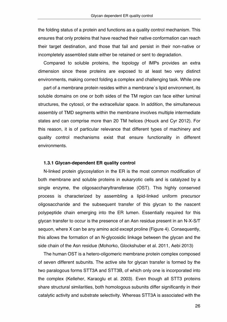
1.3.1 Glycan-dependent ER quality control
N-linked protein glycosylation in the ER is the most common modification of
both membrane and soluble proteins in eukaryotic cells and is catalyzed by a
single enzyme, the oligosaccharyltransferase (OST). This highly conserved
process is characterized by assembling a lipid-linked uniform precursor
oligosaccharide and the subsequent transfer of this glycan to the nascent
polypeptide chain emerging into the ER lumen. Essentially required for this
glycan transfer to occur is the presence of an Asn residue present in an N-X-S/T
sequon, where X can be any amino acid except proline (Figure 4). Consequently,
this allows the formation of an N-glycosidic linkage between the glycan and the
side chain of the Asn residue (Mohorko, Glockshuber et al. 2011, Aebi 2013)
The human OST is a hetero-oligomeric membrane protein complex composed
of seven different subunits. The active site for glycan transfer is formed by the
two paralogous forms STT3A and STT3B, of which only one is incorporated into
the complex (Kelleher, Karaoglu et al. 2003). Even though all STT3 proteins
share structural similarities, both homologous subunits differ significantly in their
catalytic activity and substrate selectivity. Whereas STT3A is associated with the

Glycan dependent ER quality control
27
translocon, can scan for consensus sites and perform glycosylation co-
translationally, STT3B is in a somewhat distant position and carries out
glycosylation post-translationally once the nascent polypeptide is released into
the ER. This is particularly relevant for very C-terminal glycosylation sites (Ruiz-
Canada, Kelleher et al. 2009, Shrimal, Trueman et al. 2013). Thus, STT3B can
modify skipped sequons or compensate for failed transfer reactions missed by
co-translational scanning of the STT3A complex (Shrimal, Cherepanova et al.
2015). This effect is reinforced by the observation that STT3B is more active in
terms of glycopeptide formation and, at the same time, exhibits a reduced
selectivity towards the oligosaccharide donor substrate that is transferred from
the dolichol-pyrophosphate-activated carrier to the substrate protein (Mohorko,
Glockshuber et al. 2011). Although most of the other subunits are highly
conserved, their function, except the N33 subunit, is unknown. Interestingly, N33
can slow down protein folding due to the formation of transient disulfide bonds
with OST substrates. This process is mediated by a membrane-bound
thioredoxin domain facing the lumen of the ER and presumably contributes to an
increase in glycosylation efficiency (Mohorko, Owen et al. 2014).
Besides primarily assisting in protein folding of newly synthesized proteins,
glycosylation serves a multitude of functions inside the ER (Paulson 1989,
Helenius 1994). For example, N-linked glycosylation enables sorting of
glycoproteins, improves solubility, and at the same time reduces aggregation or
dictates degradation as a consequence of trimming. Of particular relevance,
however, N-linked glycans, after processing of the primary oligosaccharide,
represent distinct recognition motifs that can direct folding chaperones such as
Calnexin (CNX) or Calreticulin (CRT) (Mohorko, Glockshuber et al. 2011,
Guerriero and Brodsky 2012).
Glycan processing begins shortly after a precursor oligosaccharide, which is
assembled first in the cytosol and then the ER lumen by several
glycosyltransferases is transferred from its lipid pyrophosphate donor residing in
the ER membrane, dolicholpyrophosphat, to a selected Asn residue present in
the nascent polypeptide chain (Helenius and Aebi 2004, Caramelo and Parodi
2008). In the following, the sequential action of glucosidases I and II results in the
trimming and removal of the two outermost glucose residues of Glc3Man9GlcNAc2
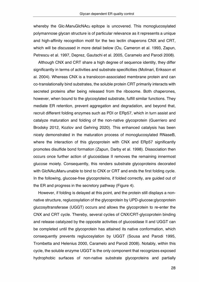
Glycan dependent ER quality control
28
whereby the Glc1Man9GlcNAc2 epitope is uncovered. This monoglucosylated
polymannose glycan structure is of particular relevance as it represents a unique
and high-affinity recognition motif for the two lectin chaperons CNX and CRT,
which will be discussed in more detail below (Ou, Cameron et al. 1993, Zapun,
Petrescu et al. 1997, Deprez, Gautschi et al. 2005, Caramelo and Parodi 2008).
Although CNX and CRT share a high degree of sequence identity, they differ
significantly in terms of activities and substrate specificities (Molinari, Eriksson et
al. 2004). Whereas CNX is a translocon-associated membrane protein and can
co-translationally bind substrates, the soluble protein CRT primarily interacts with
secreted proteins after being released from the ribosome. Both chaperones,
however, when bound to the glycosylated substrate, fulfill similar functions. They
mediate ER retention, prevent aggregation and degradation, and beyond that,
recruit different folding enzymes such as PDI or ERp57, which in turn assist and
catalyze maturation and folding of the non-native glycoprotein (Guerriero and
Brodsky 2012, Kozlov and Gehring 2020). This enhanced catalysis has been
nicely demonstrated in the maturation process of monoglucosylated RNaseB,
where the interaction of this glycoprotein with CNX and ERp57 significantly
promotes disulfide bond formation (Zapun, Darby et al. 1998). Dissociation then
occurs once further action of glucosidase II removes the remaining innermost
glucose moiety. Consequently, this renders substrate glycoproteins decorated
with GlcNAc2Man9 unable to bind to CNX or CRT and ends the first folding cycle.
In the following, glucose-free glycoproteins, if folded correctly, are guided out of
the ER and progress in the secretory pathway (Figure 4).
However, if folding is delayed at this point, and the protein still displays a non-
native structure, reglucosylation of the glycoprotein by UPD-glucose:glycoprotein
glucosyltransferase (UGGT) occurs and allows the glycoprotein to re-enter the
CNX and CRT cycle. Thereby, several cycles of CNX/CRT-glycoprotein binding
and release catalyzed by the opposite activities of glucosidase II and UGGT can
be completed until the glycoprotein has attained its native conformation, which
consequently prevents reglucosylation by UGGT (Sousa and Parodi 1995,
Trombetta and Helenius 2000, Caramelo and Parodi 2008). Notably, within this
cycle, the soluble enzyme UGGT is the only component that recognizes exposed
hydrophobic surfaces of non-native substrate glycoproteins and partially
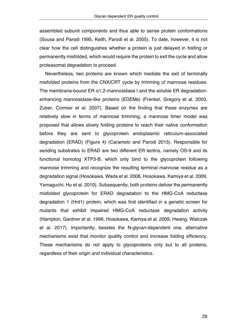
Glycan dependent ER quality control
29
assembled subunit components and thus able to sense protein conformations
(Sousa and Parodi 1995, Keith, Parodi et al. 2005). To date, however, it is not
clear how the cell distinguishes whether a protein is just delayed in folding or
permanently misfolded, which would require the protein to exit the cycle and allow
proteasomal degradation to proceed.
Nevertheless, two proteins are known which mediate the exit of terminally
misfolded proteins from the CNX/CRT cycle by trimming of mannose residues:
The membrane-bound ER α1,2-mannosidase I and the soluble ER degradation-
enhancing mannosidase-like proteins (EDEMs) (Frenkel, Gregory et al. 2003,
Zuber, Cormier et al. 2007). Based on the finding that these enzymes are
relatively slow in terms of mannose trimming, a mannose timer model was
proposed that allows slowly folding proteins to reach their native conformation
before they are sent to glycoprotein endoplasmic reticulum-associated
degradation (ERAD) (Figure 4) (Caramelo and Parodi 2015). Responsible for
sending substrates to ERAD are two different ER lectins, namely OS-9 and its
functional homolog XTP3-B, which only bind to the glycoprotein following
mannose trimming and recognize the resulting terminal mannose residue as a
degradation signal (Hosokawa, Wada et al. 2008, Hosokawa, Kamiya et al. 2009,
Yamaguchi, Hu et al. 2010). Subsequently, both proteins deliver the permanently
misfolded glycoprotein for ERAD degradation to the HMG-CoA reductase
degradation 1 (Hrd1) protein, which was first identified in a genetic screen for
mutants that exhibit impaired HMG-CoA reductase degradation activity
(Hampton, Gardner et al. 1996, Hosokawa, Kamiya et al. 2009, Hwang, Walczak
et al. 2017). Importantly, besides the N-glycan-dependent one, alternative
mechanisms exist that monitor quality control and increase folding efficiency.
These mechanisms do not apply to glycoproteins only but to all proteins,
regardless of their origin and individual characteristics.

Chaperoning in the ER
30
Figure 4: Calnexin mediated folding and quality control of glycosylated proteins inside the ER: Once a nascent polypeptide chain emerges into the ER lumen, the oligosaccharyltransferase (OST) enzyme complex mediates N-linked protein glycosylation. Following the sequential action of glucosidases I and II, the two outermost glucose residues are removed, and CNX recognizes monoglucosylated oligosaccharides. By recruiting different folding enzymes such as ERp57 to the arm domain, CNX assists and catalyzes the maturation and folding of the non-native glycoprotein. Further action of glucosidase II, which removes the remaining innermost glucose moiety, renders the substrate glycoproteins unable to bind to CNX and initiates ER exit of correctly folded glycoproteins. However, if folding is delayed at this point, and the protein still displays a non-native structure, reglucosylation of the glycoprotein by UPD-glucose:glycoprotein glucosyltransferase (UGGT) occurs and allows the glycoprotein to re-enter the CNX cycle. In contrast, permanently misfolded glycoproteins are processed by ER α1,2-mannosidase I, which leads to the removal of the mannose residue in the middle branch of the oligosaccharide. Subsequently, mannose trimmed glycoproteins are recognized by the ER degradation-enhancing mannosidase-like proteins (EDEMs), which targets glycoproteins for ER-associated degradation (ERAD) by the 26S proteasome.
1.3.2 Chaperoning in the ER
Inside the ER lumen, molecular chaperones and folding modulators are highly
abundant and play a central role in the conformational quality control of the
Chaperoning in the ER
31
proteome and folding of immature proteins. Whereas natively folded protein
conformers are not detected by molecular chaperones and free to leave the ER,
only slight deviations from the native conformation can lead to the protein being
recognized as defective or immature, causing transient binding by one or more
of these factors. ER-associated chaperones, however, significantly differ in their
substrate specificity and how they assist in folding and degradation or prevent
aggregation. In the past, numerous ER chaperones have been identified, with the
soluble immunoglobulin heavy chain binding protein (BiP) being a particularly
central player. BiP is mainly involved in ER protein folding and assembly by
shielding hydrophobic residues from the aqueous environment (Buck, Wright et
al. 2007, Behnke, Mann et al. 2016). Thus, masking of hydrophobic patches that
are normally buried inside the hydrophobic core of a native protein by BiP is
required since these unstructured polypeptides are prone to aggregation when
they emerge in the aqueous environment of the ER. Interestingly, however, if the
emerging substrate is a glycoprotein, the association of BiP is often preceded by
an association of the substrate with CNX or CRT, as these chaperones recognize
already short nascent chains of approximately 12 residues length once they
emerge in the ER lumen (Andersson, Nilsson et al. 1996, Lilley and Ploegh 2004,
Hebert and Molinari 2007). Except for glycoproteins that bear a very N-terminally
located N-glycan, this prevents BiP´s assistance to the nascent chain until the
substrate is released by CNX or CRT (Molinari and Helenius 2000, Hebert and
Molinari 2007). However, it is undisputed that BiP is a master regulator because
of its unique role in various processes and functions in the ER (Hendershot 2004).
BiP belongs to the Hsp70 chaperone family and was first discovered based
on its specific interaction with antibody heavy chains (Haas and Wabl 1983). In
addition, it was shown shortly thereafter that BiP preferentially binds relatively
short peptides or aliphatic residues and accommodates them in an energetic
environment that resembles that of a folded protein (Flynn, Pohl et al. 1991). BiP
is formed by two functional domains: a highly conserved N-terminal nucleotide-
binding domain (NBD), with ATPase activity, and a substrate-binding domain
(SBD) that at its C-terminal end contains an α-helical domain that functions as a
lid. Both domains are connected by a flexible hydrophobic linker allowing for
allosteric interaction. Like all Hsp70 family proteins, the binding of BiP to unfolded
Chaperoning in the ER
32
substrates and its release is tightly regulated by its nucleotide bound state. In the
ATP-bound form, BiP favors a conformation in which its NBD and SBD are close
to each other with its lid open. This conformation results in a high on/off rate for
substrate binding, which means that binding of substrates occurs with only low
affinity and allows them to be released and fold. In contrast, following ATP
hydrolysis, BiP reaches a state with low on/off rates for substrate binding,
characterized by a conformational change, whereby NBD and SBD are separated
from one another. In addition, the lid undergoes a relative movement over the
SBD and causes high-affinity binding of the substrate to this domain (Otero, Lizak
et al. 2010, Behnke, Feige et al. 2015).
Notably, the activity of all chaperones is accompanied by numerous co-
chaperones and folding enzymes. This also applies to BiP, whose chaperone
cycle is not only controlled by ATP but also by ER-localized DnaJ (ERdj) proteins
and the nucleotide exchange factors (NEFs) that perform the transition from the
ADP to the ATP bound state of BiP (Misselwitz, Staeck et al. 1998, Behnke, Feige
et al. 2015, Kityk, Kopp et al. 2018). In the ER, this ATPase cycle is regulated by
the NEFs SIL1 and glucose-regulated protein of 170 kDa (Grp170). By
stimulating the release of ADP and allowing new ATP molecules to bind to the
BiP complex, these proteins facilitate the release of substrates due to an opening
of the lid on the SBD (Chung, Shen et al. 2002, Behnke, Feige et al. 2015).
Interestingly, the activity and function of Grp170 seem to be more far-reaching.
Due to its intrinsic chaperone property, Grp170 exhibits important cytoprotective
activity by binding to many unfolded proteins, similar to BiP. In contrast to BiP,
however, Grp170 does not mediate the release of its unfolded substrate in the
presence of ATP (Behnke and Hendershot 2014). Furthermore, it has been
shown that this chaperone is involved in ERAD, where it can function as a
holdase that does not depend on its ATPase activity or interaction with BIP.
Besides that, it has been demonstrated that Grp170 binds to
suppressor/enhancer of Lin-12-like (SEL1L) which is an indispensable
component of the HRD1 ubiquitin ligase complex and consequently links Grp170
activity to the degradation of misfolded substrates (Buck, Kolb et al. 2010, Buck,
Plavchak et al. 2013, Inoue and Tsai 2016).

Chaperoning in the ER
33
Within the BiP cycle, ERdj proteins perform the exact opposite function. Following
binding to ATP-bound BiP, ERdj proteins stimulate ATP hydrolysis and thus
cause greater interaction between BiP and its substrate. In addition, ERdj
proteins can also bind unfolded substrates themselves and assist in the initial
delivery of these to BiP (Misselwitz, Staeck et al. 1998, Braakman and Hebert
2013). Important for these functions and binding of BiP, is a conserved domain
of approximately 70 amino acid residue length, the J domain. Thus, this protein
family is also referred to as J-domain proteins. Importantly, within the J-domain,
a conserved HDP motif is responsible for stimulating the ATPase activity of BiP,
(Liberek, Marszalek et al. 1991, Tsai and Douglas 1996). In the mammalian ER,
at least seven J-domain proteins (ERdj1-7) are known.
Despite all of them supporting the ER´s various functions, their range of
substrates and effect on protein folding and degradation or preference for distinct
binding sites is diverse (Otero, Lizak et al. 2010, Behnke, Mann et al. 2016). The
integral membrane J-proteins ERdj1 and ERdj2 are often associated with the
Sec61 translocon and assist maturation of nascent chains as they connect ER-
luminal BiP functions with the cytosolic translation machinery. While ERdj1
assists in recruiting ATP-bound BiP and support positioning at the translocon
pore to ensure that BiP is available for emerging nascent polypeptide chains
following translocation, ERdj2 together with BiP and Sec62 controls opening of
the Sec61 translocon pore. ERdj3 and ERdj6 function as pro-folding co-
chaperones because they recognize unfolded substrates and maintain them in a
soluble state. In contrast, ERdj4 and ERdj5 have the opposite effect as they bind
aggregation-prone proteins and enhance their degradation. Interestingly, ERdj5
is not only a BiP cofactor but at the same time, also belongs to the family of
protein disulfide-isomerases (PDI), which will be discussed in the next paragraph.
Due to its high reductive potential, ERdj5 has a unique function in reducing
disulfide bonds in ERAD substrates. ERdj7 is the most recently discovered and
less studied ER J-domain protein. However, recent findings indicate that it might
be involved in translocation processes and the degradation of misfolded or
unfolded proteins (Braakman and Hebert 2013, Pobre, Poet et al. 2019, Ohta and
Takaiwa 2020).
Chaperoning in the ER
34
However, the activity of BiP is not only regulated by cofactors or rapid expression
following protein-folding stress but also by various PTMs, including
phosphorylation, acetylation, ubiquitination, AMPylation, and ADP-ribosylation.
Although the understanding of how most of these PTMs events impact the
functions of BiP remains elusive, it has been demonstrated that they are
predominantly localized to the NBD and SBD (Freiden, Gaut et al. 1992, Nitika,
Porter et al. 2020).
Besides the Hsp70 family member BiP, the ER also contains a chaperone of
the Hsp90 family, glucose-regulated protein of 94 kDa, Grp94. Similar to BiP,
nucleotide binding to the N-terminal domain (NTD) of Grp94 and hydrolysis leads
to conformational changes, which influences the opening and closing and,
therefore, the chaperones activity (Argon and Simen 1999, Braakman and Hebert
2013). Interestingly, Grp94 seems especially important for those substrates that
represent complicated folding intermediates since Grp94 only recognizes some
proteins after their release from BiP (Melnick, Dul et al. 1994)
Another important group of ER-resident folding factors is represented by
folding catalysts, of which the oxidoreductase PDI is the most abundant and best
characterized (Wallis and Freedman 2013). This family of enzymes is defined by
the presence of a conserved CXXC active-site motif that serves to form,
isomerize or reduce disulfide bonds. To this end, the cysteines in the CXXC motif
itself can be found in either a reduced or oxidized state (Ellgaard and Ruddock
2005). Disulfide bond linkages within and between constituent chains of a
polypeptide are generally formed in the ER and therefore primarily found in
extracellular, periplasmic, and secreted proteins where they confer structural
stability. Moreover, selective redox regulation of disulfides can be an important
mechanism to control the activity of a protein. Of note, the ER lumen is more
oxidizing than the cytosol and thus provides ideal conditions for formation of
disulfide bonds within polypeptides that are formed once the nascent chain is
post-translationally translocated into the ER lumen (Chen, Helenius et al. 1995,
Margittai and Sitia 2011). At the same time, however, this environment also
induces spontaneous, non-native disulfide bond formation. Remarkably, the
temporary occurrence of non-native disulfide bonds is not always problematic but
may be necessary and arises in different intermediate states of protein maturation

Chaperoning in the ER
35
and folding. Hence, oxidoreductases that assist and enhance oxidative protein
folding and prevent the occurrence of erroneous disulfide bonds in the final native
structure are of great importance (Braakman and Bulleid 2011, Bulleid 2012). The
canonical PDI and its role in oxidative protein folding was discovered more than
50 years ago, when it was shown that PDI is required to catalyze the oxidative
reactivation of reduced ribonuclease A (Venetianer and Straub 1963, Givol,
Goldberger et al. 1964). To date, more than 20 oxidoreductases are known, all
of which exhibit different substrate specificity and redox potentials and thus
catalyze oxidoreduction reactions in different ways (Ellgaard and Ruddock 2005,
Braakman and Bulleid 2011). Moreover, other ER-resident proteins have been
identified that are involved in the PDI-mediated reductive and oxidative protein
folding pathway and contribute to its function. One of these proteins is ER
oxidoreduction 1 (Ero1), which is essentially required to keep PDI in an oxidized
active state in that it transfers disulfide bonds to and accepts electrons from PDI
by the help of its flavin cofactor and the oxidizing power of molecular oxygen
(Pollard, Travers et al. 1998, Sevier and Kaiser 2008, Benham, van Lith et al.
2013).
PDIs, are not just foldases required to accelerate the formation and shuffling
of disulfide bonds. They also actively prevent aggregation of retaining misfolded
and unassembled proteins in a process independent of the presence of disulfides
in a substrate or the CXXC motif in the active site of PDI. In this way, PDI
functions as a true molecular chaperone (Cai, Wang et al. 1994, Quan, Fan et al.
1995, Bottomley, Batten et al. 2001, Hebert and Molinari 2007). In addition, PDIs
are heavily involved in ERAD as they can render terminally misfolded proteins
retrotranslocation-competent. Based on their reductase activity, PDIs can
chaperone the unfolding of ERAD substrates and thus facilitate the dislocation of
these through the likely narrow retrotranslocation channel into the cytoplasm for
proteasomal degradation. Indeed, it has been shown that the cleavage of
disulfide bonds as a prerequisite for efficient retrotranslocation depends on the
activity of ERdj5 (Ushioda, Hoseki et al. 2008, Bulleid 2012, Ellgaard, Sevier et
al. 2018).
Endoplasmic reticulum-associated protein degradation
36
In addition to the quality control system present in the ER lumen, cytosolic
chaperones and ubiquitin ligases exist that assist in the folding and quality control
of polytypic IMPs if they possess cytosolic domains (Houck and Cyr 2012).
1.3.3 Endoplasmic reticulum-associated protein degradation
ER chaperones and chaperone-like proteins that have evolved in the ER aid
folding and scrutinize conformations of their substrate proteins. Despite their
activity, misfolding of individual proteins and the incorrect assembly of multi-
protein complexes are quite common and bear deleterious consequences for the
cell, either due to a loss of function for specific proteins or even a toxic gain of
function, e.g., during aggregate or amyloid formation. For this reason, the ER is
additionally equipped with two interconnected quality control systems that
specifically deal with terminally misfolded or aggregated proteins: A large load of
incompletely folded proteins is recognized by sensors of the unfolded protein
response (UPR). Following activation, the UPR mainly reduces the ER folding
load and causes the transcriptional up-regulation of proteins that increase the
protein folding capacity of the ER, such as the aforementioned ER-resident
chaperones, which will be discussed in more detail below (Ellgaard and Helenius
2003, Guerriero and Brodsky 2012, Gardner, Pincus et al. 2013). In parallel, if
recognized as defective, improperly folded proteins are retrotranslocated back
into the cytoplasm, where they are subject to degradation via ERAD by the
ubiquitin-proteasome system (Ahner and Brodsky 2004, Vembar and Brodsky
2008). Of particular relevance, misfolding of membrane proteins bears
conceptually different scenarios compared to folding abnormalities in soluble
proteins. Degradation must occur regardless of whether the misfolded domain is
located in the ER lumen, the cytoplasmic side of the ER, or in the ER membrane
where entire membrane segments may be destabilized and misintegrated due to
point mutations or fail complex assembly caused by lacking binding partners. As
a result of the various topologies of substrates, ERAD is classified into three
different pathways: ERAD-L refers to the degradation of proteins with a misfolded
ER luminal domain, ERAD-M refers to the degradation of membrane proteins with
a misfolded domain in the ER membrane, and ERAD-C refers to the degradation
ERAD – Substrate recognition
37
of proteins where the misfolded domain is located on the cytoplasmic side of the
ER (Figure 5) (Carvalho, Goder et al. 2006).
Although ERAD is the most common degradation mechanism for removing
numerous normal and misfolded ER-localized proteins, alternative pathways
exist that are not limited to proteins as substrates and important for proteins that
cannot undergo ERAD. Instead of proteasomal degradation, these pathways use
the second most prominent degradation system in the cell: the autophagy-
lysosome system. Autophagic degradation (ER-phagy) enables the disposal of
entire parts of the ER, including its membrane lipids and protein aggregates. This
can be achieved by forming of small ER-derived vesicles or large
autophagosomes that enclose entire fragments of the ER and deliver internal
material like aggregates to the lysosomes. In this way, ER-phagy contributes
significantly to controlling the size of the ER (Wilkinson 2019, Chino and
Mizushima 2020).
1.3.3.1 Substrate recognition in ERAD
In order to become accessible to the proteasome, misfolded ER proteins have
first to be recognized and then be retro-translocated into the cytosol, constituting
two critical steps in ERAD. Although these steps equally apply to all ERAD
pathways, the key challenge for the different ERAD mediators right from the start
is defining the structural basis for recognizing specific substrates and
discriminating if a protein is in the process of folding and maturation or determined
for ERAD. This is particularly challenging if the substrate is a membrane protein.
The reason for this is that there is no uniform structural signature known based
on which a substrate is recognized as defective and degraded (Nakatsukasa and
Brodsky 2008). For instance, whereas exposed hydrophobic residues inside the
ER lumen or on the cytoplasmic side are potentially harmful and recognized by
the ERAD-L or ERAD-C machinery, respectively, hydrophobicity is to be
expected within the membrane, which is why ERAD-M mediators would not
recognize such a signature in transmembrane proteins, but rather recognize
exposed polar residues in orphaned TM segments.
Several factors contribute to recognizing ERAD-L substrates and may also act
on luminal domains of membrane proteins. Recognition of incompletely folded
ERAD – Substrate recognition
38
proteins in the ER lumen is mainly carried out by chaperones of the Hsp70 family
and lectin chaperones (Guerriero and Brodsky 2012). Following binding to
hydrophobic patches, the ER-resident Hsp70 chaperone BiP, with the aid of its
Hsp40 co-chaperones, can mediate the folding or degradation of its clients.
However, besides BiP, PDI might also be implicated in recognizing some ERAD
substrates since depletion of PDI prevents their degradation (Wang and Chang
2003). Immature glycoproteins that are intended for ERAD, on the other hand,
are recognized by their glycosylation status. Together with an unfolded
polypeptide segment, the resulting terminal mannose residues constitute the
degradation signal, recognized by other lectins like OS-9 and XTP3-B as
mentioned before (Hosokawa, Wada et al. 2008, Hosokawa, Kamiya et al. 2009).
Regardless of whether the substrate is a glycoprotein or not and recognized by
OS-9 or BiP, respectively, subsequent association with the lectin SEL1L, a
cofactor of Hrd1, is required to promote degradation (Christianson, Shaler et al.
2008).
Only a few factors have been identified that can assist in the folding and
degradation of terminally misfolded membrane proteins. The ER membrane-
associated E3 ubiquitin-ligase Hrd1 and its homolog cell surface glycoprotein of
78 KDa (gp78) are considered essential proteins required to recognize ERAD-M
substrates and luminal domains of polytypic membrane proteins. Presumably,
recognition and binding of misfolded TM domains is mediated based on
interaction with the TM domain of Hrd1 itself, which contains many hydrophilic
amino acids, most of which are also conserved in gp78 (Sato, Schulz et al. 2009).
Notably, although gp78 on amino acid-sequence level is a homolog to Hrd1, its
interaction network and substrates are largely different. Consistently, the
degradation of orphaned CD3d, an ERAD-M substrate and component of the T-
cell antigen receptor CD3 complex, depends on gp78, however, it does not
require Hrd1 (Fang, Ferrone et al. 2001). Moreover, it has been demonstrated
that both enzymes act sequentially, whereby gp78 is localized downstream, and
its function is coupled to interaction with BAG6, a cytosolic chaperone (Zhang,
Xu et al. 2015). For the process of substrate recognition, Hrd1 forms complexes
with distinct cofactors, including SEL1L, OS-9, and Derlin-family proteins
(Derlins), subsequently promoting the ubiquitination and degradation of the
ERAD – Substrate recognition
39
substrate protein (Houck and Cyr 2012). While SEL1L and OS-9 represent
recognition factors for luminal substrates, Derlin-1 serves as a TM chaperone.
Derlins are multipass membrane proteins with four to six TM helices and
represent inactive members of the Rhomboid superfamily of intra-membrane
proteases. Mammals, contain three members, Derlin-1, Derlin-2, and Derlin-3,
which interact with their clients without cleaving them. By forming complexes with
destabilized TM segments, Derlin-1 functions as a recognition factor for misfolded
TM domains, causing ER retention and subsequent degradation of selected
substrates (Sun, Zhang et al. 2006, Houck and Cyr 2012, Guna and Hegde 2018).
Interestingly, the TM segments of Derlin-1 have only low hydrophobicity due to
several polar and charged residues that might be involved in substrate
recognition processes (Greenblatt, Olzmann et al. 2011). Moreover, it is assumed
that through the cooperative interaction of Derlins together with Hrd1 or gp78 and
other ER ligases, which all have differing specificity, the range of substrates that
are recognized and degraded increases (Houck and Cyr 2012).
In contrast to the aforementioned inactive members, active members of intra-
membrane proteases after selective TMD recognition mediate proteolysis of their
substrate in the plane of the membrane. In mammals, five different rhomboid
proteases are known, most of which are located in the secretory pathway but with
differing functions (Bergbold and Lemberg 2013). Interestingly, the intra-
membrane rhomboid-like 4 RHBDL4 has been shown to localize to the ER,
which, with a few exceptions is free of proteases, as these would counteract the
protein folding taking place in the ER (Fleig, Bergbold et al. 2012). Nevertheless,
cleavage of other ubiquitinated single spanning and polytypic membrane proteins
already in the ER membrane, such as the SST3A paralogue of the OST or
orphaned T-cell receptor (TCR-a) subunits, has been demonstrated.
Furthermore, by direct interaction with the p97 ATPase, RHBDL4 has been linked
to the canonical ERAD pathway. Remarkably, many ERAD substrates of
RHBDL4 have positively charged residues within their TM segments contributing
to recognition (Fleig, Bergbold et al. 2012, Bergbold and Lemberg 2013, Knopf,
Landscheidt et al. 2020).
Recognition processes and quality control in the ER, however, are not free of
errors. Some membrane proteins manage to escape the ER and travel to the

ERAD – Retrotranslocation
40
Golgi, although they have misfolded or unassembled TMDs. In such cases,
sorting factors such as the retention in the ER 1 (RER1) receptor, which is
localized to the cis-Golgi, mediate retrieval back to the ER. Albeit most of its
substrates are unassembled subunits of protein complexes with exposed polar
residues in the TM segments, RER1 is also involved in the ER retention of
misfolded membrane proteins (Sato, Sato et al. 2003, Yamasaki, Hara et al.
2014, Briant, Johnson et al. 2017). This could be demonstrated for a mutant
variant of peripheral myelin protein of 22 KDa (PMP22), which, as a result of
mutation, has a polar residue within the TMD that is exposed to the hydrophobic
environment of the lipid bilayer and recognized by RER1 (Hara, Hashimoto et al.
2014). ERAD-C substrates are mostly recognized by the cytosolic Hsp70 / Hsp
40 chaperone system. Recognition is probably due to the exposure of
hydrophobic patches and a prolonged engagement of a substrate with the
chaperone system, facilitating interaction between the substrate and the ERAD-
C specific yeast ubiquitin ligase, Doa10. Consequently, polyubiquitination by
Doa10 leads to the proteasomal degradation of the substrate in the cytosol, as
will be discussed in more detail below (Nakatsukasa and Brodsky 2008, Sun and
Brodsky 2019).
1.3.3.2 Retrotranslocation
In order to become accessible to the cytosolic proteasome, misfolded proteins
following recognition have to be retrotranslocated from the ER lumen into the
cytosol or extracted from the ER membrane, as in the case for membrane
proteins (Figure 5). Recent studies suggest that prior to retrotranslocation,
specific ERAD substrates, particularly those containing TM domains, are instead
segregated from other ER proteins and exported into specific ER quality control
compartments in a process mediated by the integral ER membrane B cell-
associated protein of 31 kDa (Bap31). Notably, it is assumed that these
specialized ER subdomains represent the site from where substrates destined
for retrotranslocation exit the ER into the cytosol (Wakana, Takai et al. 2008).
The mechanism of how hydrophobic integral membrane substrates can exit the
ER is still ill-defined, with few exceptions (Nakatsukasa and Brodsky 2008).

ERAD – Retrotranslocation
41
Irrespective of whether the misfolded domain is located in the lumen or within the
ER membrane, the export of both types of ER proteins is hindered by the
hydrophobicity of the ER membrane. To overcome this barrier, a proteinaceous
retrotranslocation channel, which allows aberrant proteins to move through the
ER membrane into the cytosol, is required. Providing a channel that enables
polypeptide movement through the ER membrane was first attributed to the
Sec61 translocon. Opposed to its canonical function, it could be shown that
Sec61 mediates the transfer of non-glycosylated major histocompatibility
complex (MHC) class I molecules from the ER to the proteasome based on direct
interaction (Wiertz, Tortorella et al. 1996, Zhou and Schekman 1999). Later
studies using Sec61 mutants with impaired retrotranslocation function could
support this observation. Although ER import was not affected, defects in ERAD
and thus substrate export could be observed, indicating that the Sec61 translocon
can direct two-way translocation processes (Romisch 2017).
Recently, it could be demonstrated that the yeast Derlin Dfm1 is a crucial
component for retrotranslocation. Integral membrane proteins seem to require
Dfm1 for extraction from the ER membrane during ERAD. However, in the
absence of Dfm1, this activity is taken over by Hrd1 (Neal, Jaeger et al. 2018).
Similar activity has been reported for Derlin-1, which explicitly targets movement
of MHC class I molecules through the membrane and consequently causes
degradation via transmembrane domain interaction (Lilley and Ploegh 2004, Ye,
Shibata et al. 2004). Notably, Derlin-1 only has four transmembrane domains
making the independent formation of a protein conducting channel unlikely,
although oligomerization has been observed (Mehnert, Sommer et al. 2014).
Nevertheless, it is assumed that Derlin-1 is essential to initiate translocation by
promoting the insertion into the membrane if the substrate is an ERAD-L
substrate, but then delivers misfolded substrates for ubiquitylation to Hrd1 as
direct interaction of this E3 ligase with the transmembrane segments of Derlin-1
has been shown (Mehnert, Sommer et al. 2014, Romisch 2017). In fact, it has
been suggested that the ubiquitin-ligase Hrd1 itself is another promising
candidate that promotes the movement of misfolded proteins across the ER
membrane. Most of the studies on this behalf have been carried out in yeast but
could show in crosslinking experiments that retrotranslocating ERAD-L
ERAD – Retrotranslocation
42
substrates associate with Hrd1. Moreover, it could be shown that Hrd1 can
promote degradation of ERAD-L substrates even in the absence of the other
membrane components that contribute to the formation of the Hrd1 complex,
such as the homolog of the cofactor of SEL1L (Carvalho, Stanley et al. 2010).
The ability to compensate for the need of its complex partners indicates a central
role of Hrd1 in the ERAD dependent translocation of misfolded proteins across
the ER membrane. It is most likely by forming a membrane conduit or being part
of it since Hrd1 is a multispanning membrane protein consisting of 6 TM
segments. Interestingly, the formation of a retrotranslocation pore is regulated by
cycles of autoubiquitination what causes the pore to open and deubiquitination of
HHrd1 (Vasic, Denkert et al. 2020). Prior substrate ubiquitination,
autoubiquitination of several lysine residues in the RING-finger domain of Hrd1
is required to enable retrotranslocation of membrane bound ERAD-L substrates
as liposome-based reconstitution experiments have revealed (Baldridge and
Rapoport 2016). To counteract the risk of proteasomal degradation that
autoubiquitinated Hrd1 is exposed to, deubiquitination of Hrd1 is catalyzed by a
deubiquitinating enzyme (DUB) (Peterson, Glaser et al. 2019). Although
processes occurring during ERAD-L inside the ER membrane are still under
debate, processes occurring inside the ER lumen are better understood.
Glycosylated ERAD-L substrates that contain a misfolded domain are jointly
recognized by the lectin Yos9 (homolog of OS-9 and XTP3-B), which binds to
terminal mannose residues present on trimmed N-linked glycans, and Hrd3
(homolog of SEL1L), which binds to unstructured segment around the
glycosylation attachment site. Furthermore, both proteins interact with each other
via the luminal domain of Hrd3 and subsequently deliver their substrate to the
Hrd1 translocon (Carvalho, Stanley et al. 2010, Stein, Ruggiano et al. 2014).
In fact, whether a retrotranslocation channel really exists or whether other
mechanisms enable translocation and which factors might be involved still needs
to be further elucidated (Neal, Jaeger et al. 2018). Along the same line, it has
been proposed that lipid rearrangements in the ER membrane causing the
formation of lipid droplets might contribute to a different possible mechanism that
enables exit from the ER (Ploegh 2007).
ERAD – Proteasomal degradation
43
1.3.3.3 Proteasomal degradation
Once a substrate emerges on the cytoplasmic site, ubiquitin ligases and
ubiquitin-conjugating enzymes catalyze polyubiquitination of the respective
substrates (Carvalho, Stanley et al. 2010). Although the ER possesses several
ubiquitin-protein-ligases (25 in mammals) with different selectivities, they are all
subject to the same enzymatic cascade, ultimately leading to substrate
ubiquitination (Fenech, Lari et al. 2020).
Ubiquitin is a highly conserved protein and involved many cellular processes,
including the degradation of proteins and regulation of their activity or cellular
localization. Ubiquitination as a reversible, post-translational method to modify
proteins is based on a multi-stage enzymatic cascade where an isopeptide bond
is formed between the C-terminal glycine residue of ubiquitin and an ɛ-amino
group of a lysine residue present on the client, which causes monoubiquitination
(Figure 5). Notably, the client can be ubiquitin itself. Thus, consecutive addition
of further ubiquitin moieties to a lysine residue of the first ubiquitin molecule can
lead to the formation of a polyubiquitin chain (Ciechanover, Finley et al. 1984,
Finley, Ciechanover et al. 1984). Interestingly, the modification of other residues
such as Lys and Thr could also be observed during ERAD (Brodsky and Skach
2011). The ubiquitination reaction is catalyzed by the sequential but coordinated
action of three different enzymes. The ubiquitin-activating enzyme (E1) is
required for all ubiquitination reactions and initiates ubiquitination in an ATP-
dependent reaction. First, the E1 catalyzes the formation of a high-energy
thioester bond between a conserved Cys residue in its active center and the C-
terminal carboxyl group of ubiquitin. Subsequently, the activated ubiquitin moiety
is transferred to a thiol group in the active center of a ubiquitin-conjugating
enzyme (E2). Finally, following association of the E2 with the RING domain of the
ubiquitin ligase (E3), ubiquitin is transferred from the E2 to the target protein, and
the E3 facilitates the conjugation of ubiquitin with the substrate. Therefore,
substrate recruitment and the specificity of ubiquitination is mediated by ubiquitin
ligases, which explains their frequent occurrence (Pickart 2001, Komander and
Rape 2012). Depending on the activity of ubiquitin-conjugating (Ubc) enzymes
and which of the seven lysine residues present in ubiquitin is used for ubiquitin
linkages, this causes different cellular consequences for the ubiquitylated protein
ERAD – Proteasomal degradation
44
(Ye and Rape 2009). Polyubiquitin chains linked via Lys 48 (K48) represent the
most common type of linkage and are recognized by the 26S proteasome, which
leads to proteasomal degradation of the modified substrate, similar to K29-linked
chains. Notably, proteomic analysis revealed that also K11-linkages might
contribute to the degradation of ERAD substrates (Xu, Duong et al. 2009,
Komander and Rape 2012). In contrast, K63-linked polyubiquitin chains mediate
proteasome independent functions such as protein-protein interactions or various
regulatory functions (Komander and Rape 2012, French, Koehler et al. 2021). In
addition to the homotypic type of linkage, mixed or branched polyubiquitin chains
exist, significantly increasing the complexity of the unique ubiquitin code (French,
Koehler et al. 2021). Beyond that, ubiquitination, as mentioned before, is a
reversible process and can be undone by DUBs which further intensifies this
effect (Jentsch and Rumpf 2007).
Only two ubiquitin ligases exist in yeast, which belong to the RING finger (RNF)
class of E3 ubiquitin ligases and label proteins with polyubiquitin chains
determined for proteasomal degradation. Whereas Hrd1 is required for ERAD-M
or ERAD-L substrates, proteins with misfolded cytosolic domains use a different
ubiquitin ligase, Doa10 (Swanson, Locher et al. 2001, Carvalho, Goder et al.
2006). Ubiquitylation of Doa10 substrates depends on the two E2´s, Ubc6 and
Ubc7, which operate sequentially. Thereby, monoubiquitylation of clients
mediated by the membrane-embedded Ubc6 is a prerequisite for chain
elongation by the cytosolic Ubc7 resulting in K48-linked polyubiquitin chains.
Interestingly, besides Lys acceptor sites, Ubc6 also attaches ubiquitin moieties
to hydroxylated amino acids present in target proteins and synthesizes
particularly K11-linked polyubiquitin chains (Xu, Duong et al. 2009, Weber,
Cohen et al. 2016). The processing of Hrd1 substrates mainly depends on the
activity of Ubc7 and, to a minor degree, the membrane-bound Ubc1 (Zattas and
Hochstrasser 2015). However, both the Hrd1 and Doa10 complex essentially
depend on another factor, coupling of ubiquitin conjugation to ER degradation
protein 1 (Cue1), required for the activation of Ubc7 and thus efficient chain
formation. Following the association with Hrd1, the transmembrane protein Cue1,
recruit’s ubiquitin-bound Ubc7 to the ER membrane via a distinct binding motif
causing its activation and allowing Ubc7 to mediate the formation of K48-linked

ERAD – Proteasomal degradation
45
polyubiquitin chains. The transfer of ubiquitin moieties is stimulated by a CUE
domain present in Cue1, which can bind to the proximal ubiquitin in the elongating
chain and thus positions the bound Ubc7 close to the substrate protein (Kostova,
Mariano et al. 2009, Zattas and Hochstrasser 2015, von Delbruck, Kniss et al.
2016). Interestingly, unassembled Ubc7 is degraded following autoubiquitination
which indicates, that Cue1 is required for the stability of Ubc7 and has a
regulatory function in ubiquitylation (Ravid and Hochstrasser 2007).
Although the ERAD machinery is much better characterized in yeast because
of the low complexity compared to a mammalian system, several E3 and E2
orthologs have been implicated in ERAD in mammalian cells. Besides the best-
studied ER ligases HRD1 and GP78, which are both homologous to Hrd1 as
mentioned before, other E3 ligases are known, which reside in the ER membrane
or cytosol. Among ER membrane-anchored ligases, these include the TEB4 or
MARCH VI ligase, which represent the mammalian homolog of the yeast Doa10
and shares a similar membrane topology, RNF family members such as RMA1
and TRC8 but also Nixin/ZNRF4, which has been demonstrated to be important
in the regulation of calnexin turnover as it mediates ubiquitination of CNX and
thus its proteasomal degradation (Kostova, Tsai et al. 2007, Neutzner, Neutzner
et al. 2011, Claessen, Kundrat et al. 2012). Mammalian E2s include UBE2J1 and
UBE2J2 both are homologous to the yeast Ubc6 and post-translationally insert
into the ER membrane due to high hydrophobicity in their C-terminus. In contrast,
UBE2G1 and UBE2G2 are soluble proteins and correspond to UBC7.
Interestingly, their implication in ERAD is very different. While UBE2G2 is
required for the activity of various ERAD E3s and thus strongly promotes
degradation, UBE2J1 and UBE2J2 are of minor importance. Whether UBE2G1
is involved in ERAD is unclear and still under debate (Kostova, Tsai et al. 2007).
Regardless of which ERAD pathway and E3 ligase is used to conjugate
retrotranslocation substrates with degradation signals for the proteasome, after
polyubiquitination, all paths converge at the cytosolic valosin-containing protein
(VCP)/p97 or its yeast homolog ATPase Cdc48 which is a central component of
the ubiquitin-proteasome system (Richly, Rape et al. 2005, Peterson, Glaser et
al. 2019). Cdc48 is a highly conserved chaperone-like enzyme with critical
regulatory functions in multiple cellular processes that depend on the ability of

ERAD – Proteasomal degradation
46
Cdc48 to recognize and bind ubiquitinated proteins (Jentsch and Rumpf 2007).
Furthermore, Cdc48 can segregate ubiquitinated proteins from unmodified
partners and provides the essential pulling force by which ubiquitinated ERAD
substrates are extracted from macromolecular complexes or the ER membrane
and moved into the cytosol, where they are degraded by the proteasome (Stolz,
Hilt et al. 2011). To fulfill this segregase function Cdc48 depends on its ATPase
activity, which converts chemical into mechanical energy. Sequential cycles of
ATP hydrolysis lead to conformational changes and rotational movement of the
homohexameric ring-shaped Cdc48 complex and thus contribute to a pulling
force. Consequently, this allows the successive movement of polypeptides
through a central pore formed in the center of the ring-shaped structure (Rouiller,
DeLaBarre et al. 2002, Pye, Dreveny et al. 2006, Stolz, Hilt et al. 2011).
The function of Cdc48 is strictly regulated by numerous cofactors that interact
with Cdc48 via highly conserved domains and binding motifs and thus contribute
to the functional diversity. Although Cdc48 can bind ubiquitylated proteins by
itself, the recruitment of Cdc48 to the Hrd1 complex is mediated by the
heterodimeric substrate recruiting cofactors, ubiquitin fusion degradation protein
1 (Ufd1) and nuclear protein localization protein 4 (Npl4). Both cofactors can
recognize polyubiquitin chains with a minimum length of 5 ubiquitin moieties via
their ubiquitin-binding domains and enable efficient recruitment of Cdc48 to Hrd1
(Meyer, Wang et al. 2002, Stein, Ruggiano et al. 2014). Additionally, the
Ufd1/Npl4 dependent binding of polyubiquitin chains leads to an induced ATPase
activity of the Cdc48 complex (Bodnar and Rapoport 2017). However, recruitment
of Cdc48 to the ER membrane is not exclusively dependent on Ufd1/Npl4 but can
be facilitated by different cofactors such as the transmembrane protein Ubx2 that
contains a Ub regulatory X (UBX) motif and binds to the N-terminal domain of
Cdc48 (Schuberth and Buchberger 2005). Furthermore, other N-terminal binding
Cdc48 ER recruiting cofactors have been reported, which are characterized by
the presence of a VCP-interacting (VIM) or VCP-binding (VBM) motif, the latter
motif can also be found in Hrd1 (Boeddrich, Gaumer et al. 2006, Morreale,
Conforti et al. 2009, Stapf, Cartwright et al. 2011) Remarkably, besides its
important segregase activity in ERAD, Cdc48 is also capable of controlling the

ERAD – Proteasomal degradation
47
degree of ubiquitylation of bound clients by substrate processing cofactors
(Jentsch and Rumpf 2007)
In addition to the beforementioned ER-associated ubiquitin ligases, DUBs play
an equally important regulatory role in ERAD mediated quality control, although
they exhibit opposite activity and hence could inhibit the degradation process. In
fact, several ERAD specific DUBs like Ataxin-3 or YOD1/Otu1 have been shown
to associate with Cdc48 (Ernst, Mueller et al. 2009, Liu and Ye 2012). Although
still under debate, the function of Cdc48 associated DUBs might include trimming
of polyubiquitin chains. This can result in an altered affinity of the substrate
towards ubiquitin-binding effectors like the Bag6 holdase complex or facilitate the
entry of retrotranslocation substrates into the narrow Cdc48 pore or (Wang, Liu
et al. 2011, Liu and Ye 2012). In contrast, Ufd2, a polyubiquitylation enzyme,
promotes the extension of short ubiquitin chains of Cdc48 bound substrates.
Remarkably, the extension of ubiquitin chains can be necessary if the
degradation signal is no longer strong enough due to extensive trimming (Richly,
Rape et al. 2005, Liu and Ye 2012). Interestingly, the activity of Ufd2 can be
inhibited by another factor, Ufd3, which competes with Ufd2 for the same Cdc48
binding interface (Rumpf and Jentsch 2006).
Following retrotranslocation and successful release from the membrane,
ubiquitinated proteins are delivered to the 26S proteasome for degradation by the
escort factor Rad23 or its homolog Dsk2, which in mammalian cells is
represented by four ubiquilins, UBQLN1 to UBQLN4 (Figure 5) (Medicherla,
Kostova et al. 2004, Guna and Hegde 2018). The substrate recognition for Rad23
and Dsk2 occurs via a C-terminal ubiquitin-associated domain (UBA) that binds
polyubiquitin chains. A ubiquitin-like domain (UBL), located in the N-terminus of
both shuttle factors, then mediates binding to the 19S particle, one of the two
main subunits of the 26S proteasome. In addition, the proteasome consists of a
barrel-shaped 20S core particle covered on one or both ends by the 19S particle.
While the 20S subunit has proteolytically active sites for the hydrolysis of
ubiquitinated proteins, the 19S particle has regulatory functions required for
substrate processing activities. These include recognizing ubiquitinated
substrates, their deubiquitination, and finally, the ATP-driven unfolding and
translocation of the substrate into the 20S core. Once present in the cylindrical

ERAD – Proteasomal degradation
48
core, different proteases that have trypsin-, chymotrypsin-, and caspase-like
cleavage specificity mediate degradation of the substrate (Bard, Goodall et al.
2018, Greene, Dong et al. 2020).
Figure 5: Key steps in ERAD of membrane proteins: Recognition: Depending on whether the misfolded region (red star) of a membrane protein is located in the ER lumen, on the cytoplasmic side or within transmembrane domains, the recognition is mediated by different recognition factors. Ubiquitination: Following recognition, different ubiquitin ligases are recruited to the substrate and mediate its ubiquitination. The ubiquitination reaction is catalyzed by the sequential but coordinated action of three different enzymes. The ubiquitin-activating enzyme (E1) is required for all ubiquitination reactions and initiates ubiquitination in an ATP-dependent reaction, whereby ubiquitin is transferred to an active site cysteine in a ubiquitin conjugation enzyme (E2). Subsequently, ubiquitin is transferred from the E2 to a lysine residue of the target protein, and a ubiquitin ligase (E3) facilitates the conjugation of ubiquitin with the substrate.
ER stress and the unfolded protein response
49
In yeast, ERAD-M or ERAD-L substrates are mainly ubiquitinated by the E3 ubiquitin ligase Hrd1, while proteins with misfolded cytosolic domains use a different ubiquitin ligase, Doa10. Retrotranslocation: To become accessible to the cytosolic proteasome, misfolded proteins have to be retrotranslocated from the ER lumen into the cytosol or extracted from the ER membrane, in the case for membrane proteins. To overcome the hydrophobicity of the ER membrane, a proteinaceous retrotranslocation channel is formed which allows aberrant proteins to move through the ER membrane. In addition, the p97 / Cdc48 complex converts chemical into mechanical energy through ATP hydrolysis, which allows the segregation of substrates from the membrane. Degradation: Following retrotranslocation and successful release from the membrane, ubiquitinated proteins are delivered to the 26S proteasome for degradation (Figure based on Guerriero and Brodsky 2012).
1.4 ER stress and the unfolded protein response
Degradation of aberrant secretory pathway proteins is mainly carried out by
ERAD or autophagy. However, failure of these cellular degradative processes
can lead to the accumulation of misfolded proteins in the ER and thus cause an
imbalance between the ER folding load and capacity. Furthermore, this disruption
in protein folding homeostasis leads to cellular conditions termed ER stress. To
counter threats emanating from malfolded ER proteins and ultimately to restore
ER proteostasis, cells have evolved highly conserved cellular stress response
mechanisms, referred to as the unfolded protein response (UPR) (Gardner,
Pincus et al. 2013). The first time, such a response mechanism and thus the
prototype of the UPR was described when it could be shown that accumulating,
misfolded proteins in the ER are the trigger for the markedly enhanced
expression of BiP and Grp94 (Kozutsumi, Segal et al. 1988, Mori 2015).
In order to maintain homeostasis, the UPR monitors conditions within the ER
and transmits this information to downstream processes that mainly affect the up-
regulation of specific gene expression programs. UPR activation mediates an
overall increase of ER folding capacity through an enhanced synthesis of pro-
folding chaperones and folding enzymes and an ER expansion by inducing
proteins involved in lipid biosynthesis. In addition, the expression of the ERAD
machinery is upregulated to deal with the accumulation of misfolded proteins. At
the same time, the UPR mediates the reduction of the ER protein folding load
through general translational repression, specific mRNA degradation, and ER

ER stress and the unfolded protein response
50
clearance by increased protein degradation (Guerriero and Brodsky 2012).
Allowing the cell to deal with ER stress conditions, the UPR is considered to be
cytoprotective. However, unresolved stress conditions in the ER indicate that
homeostasis cannot be restored, thus inducing apoptosis (Gardner, Pincus et al.
2013).
In general, the mammalian UPR consist of three different signaling pathways,
which are characterized by the ER-resident TM proteins inositol requiring enzyme
1 (IRE1), protein kinase RNA-like ER kinase (PERK), and activating transcription
factor 6 (ATF6) (Walter and Ron 2011, Gardner, Pincus et al. 2013). Each
pathway possesses a unique mechanism of ER stress sensing in the ER lumen
or membrane and signal transduction in the cytosol that collectively comprises
the UPR.
Under basal conditions, ATF6, which can occur as a monomer, dimer, and
oligomer, is maintained in an inactive state through the binding of BiP to its
luminal domain. Upon stress conditions in the ER, ATF6 is released from BiP,
and its disulfide bonds are remodeled. In this reduced, monomeric state ATF6 is
allowed to relocate to the Golgi apparatus where it is proteolytically processed by
the sequential actions of two membrane-embedded proteases, namely site-1 and
site-2 proteases (S1P and S2P). Remarkably, due to the deoligomerization of
ATF6 in response to ER stress, this branch of the UPR differs significantly from
the other pathways in which oligomerization occurs. Removal of the luminal and
transmembrane domains allows the amino-terminal transcription factor domain
of ATF6 to translocate into the nucleus. Subsequent binding to ER stress
response elements (ERSEs) initiates activation of genes required for UPR
(Maiuolo, Bulotta et al. 2011, Gardner, Pincus et al. 2013).
In contrast, the ER-resident kinase PERK, in response to ER stress
oligomerizes which in turn causes autophosphorylation and thus inactivation of
the eukaryotic translation initiation factor 2 α (eIF2α). This reduces the overall
cellular protein synthesis, and thus the amount of proteins entering the ER is
reduced (Harding, Zhang et al. 1999).
IRE1 is the most conserved and well-characterized signaling component and
present in all eukaryotic cells. It belongs to the family of type I TM proteins
characterized by an amino-terminal ER lumenal domain. In the absence of ER
Transmembrane domain chaperones and intra-membrane quality control factors
51
stress, IRE1 resides as a monomer within the membrane, and its lumenal
domain, which functions as the stress-sensing domain, is stabilized by interaction
with BiP (Okamura, Kimata et al. 2000). Moreover, IRE1 contains a carboxy -
terminal cytoplasmic ribonuclease (RNase) domain which undergoes trans-
autophosphorylation following assembly of IRE1 into higher-order oligomers
upon ER stress and dissociation of BiP (Gardner, Pincus et al. 2013). This results
in the activation of its endonuclease activity and initiates the processing of distinct
mRNA molecules that encode for a transcription factor, termed XBP1 in
mammals. Following translation into its active protein product, XBP1 then
relocates into the nucleus and induces the activation of ERSEs that encode for
proteins of the ER-folding machinery and lipid biosynthesis. Notably, many of the
up-regulated UPR target genes also encode for specific factors involved in the
ERAD machinery (Walter and Ron 2011, Guerriero and Brodsky 2012, Gardner,
Pincus et al. 2013). However, despite them being the most abundant proteins
produced in the ER, how membrane protein misfolding is sensed by the UPR or
ERAD remains mostly unknown.
1.5 Transmembrane domain chaperones and intra-membrane quality
control factors Intra-membrane chaperones and quality control factors that can efficiently
recognize and monitor the assembly status of TMDs directly within the lipid
bilayer to support folding and detect misfolding are a prerequisite for protein
homeostasis of any eukaryotic cell. However, chaperoning of TM segments of
multi-spanning membrane proteins in the plane of the membrane and underlying
molecular mechanisms are poorly characterized so far.
Recently, the conserved, multi-subunit ER membrane protein complex (EMC)
and its function in membrane protein topogenesis has come into focus. EMC
functions as a membrane protein chaperone and TM domain insertase (Guna,
Volkmar et al. 2018). Its chaperoning function was first revealed in a yeast screen
to identify genes required for protein folding in the ER. Interestingly, it could be
observed that in the absence of EMC, misfolded membrane proteins accumulate
(Jonikas, Collins et al. 2009). Later, it could be demonstrated that EMC integrates
tail-anchored proteins with only moderately hydrophobic TMDs post-
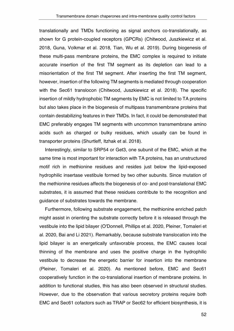
Transmembrane domain chaperones and intra-membrane quality control factors
52
translationally and TMDs functioning as signal anchors co-translationally, as
shown for G protein-coupled receptors (GPCRs) (Chitwood, Juszkiewicz et al.
2018, Guna, Volkmar et al. 2018, Tian, Wu et al. 2019). During biogenesis of
these multi-pass membrane proteins, the EMC complex is required to initiate
accurate insertion of the first TM segment as its depletion can lead to a
misorientation of the first TM segment. After inserting the first TM segment,
however, insertion of the following TM segments is mediated through cooperation
with the Sec61 translocon (Chitwood, Juszkiewicz et al. 2018). The specific
insertion of mildly hydrophobic TM segments by EMC is not limited to TA proteins
but also takes place in the biogenesis of multipass transmembrane proteins that
contain destabilizing features in their TMDs. In fact, it could be demonstrated that
EMC preferably engages TM segments with uncommon transmembrane amino
acids such as charged or bulky residues, which usually can be found in
transporter proteins (Shurtleff, Itzhak et al. 2018).
Interestingly, similar to SRP54 or Get3, one subunit of the EMC, which at the
same time is most important for interaction with TA proteins, has an unstructured
motif rich in methionine residues and resides just below the lipid-exposed
hydrophilic insertase vestibule formed by two other subunits. Since mutation of
the methionine residues affects the biogenesis of co- and post-translational EMC
substrates, it is assumed that these residues contribute to the recognition and
guidance of substrates towards the membrane.
Furthermore, following substrate engagement, the methionine enriched patch
might assist in orienting the substrate correctly before it is released through the
vestibule into the lipid bilayer (O'Donnell, Phillips et al. 2020, Pleiner, Tomaleri et
al. 2020, Bai and Li 2021). Remarkably, because substrate translocation into the
lipid bilayer is an energetically unfavorable process, the EMC causes local
thinning of the membrane and uses the positive charge in the hydrophilic
vestibule to decrease the energetic barrier for insertion into the membrane
(Pleiner, Tomaleri et al. 2020). As mentioned before, EMC and Sec61
cooperatively function in the co-translational insertion of membrane proteins. In
addition to functional studies, this has also been observed in structural studies.
However, due to the observation that various secretory proteins require both
EMC and Sec61 cofactors such as TRAP or Sec62 for efficient biosynthesis, it is

Transmembrane domain chaperones and intra-membrane quality control factors
53
assumed that EMC has a rather regulatory function during the co-translational
translocation mediated by Sec61 (O'Keefe, Pool et al. 2021). A different, novel
protein complex previously shown to cooperate with Sec61 and function as an
intra-membrane chaperone during the co-translational biogenesis of membrane
proteins is the PAT complex.
The heterodimeric PAT complex is characterized by two membrane protein
subunits, coiled-coil domain-containing protein 48 (CCDC47) and Asterix. Both
PAT complex subunits are required for efficient biogenesis of numerous multi-
spanning membrane proteins like GPCRs, however, not for TA-proteins. This was
demonstrated by depletion of either subunit of the complex by siRNA, which
resulted in the loss of the other and ultimately causes reduced biogenesis
(Chitwood and Hegde 2020). Initially, Asterix was identified as a protein
associated with the ER translocon of 10 kDa (PAT10) that engages the first TMD
of the multi-span transmembrane domain protein opsin after its lateral exit from
the Sec61 translocon (Meacock, Lecomte et al. 2002, O'Keefe, Pool et al. 2021).
In addition, it could be demonstrated that the PAT complex preferentially
engages with nascent TM segments containing unshielded hydrophilic amino
acid residues. Remarkably, chaperoning of a substrate by the PAT complex
persists throughout biogenesis even after the translation has ended. However,
once TMDs are assembled correctly and hydrophilic residues are shielded in the
completely folded protein by the native structure, the PAT complex disengages.
Based on the finding that various substrates have different dependencies for
either the EMC or the PAT complex, it is assumed that both membrane
chaperones facilitate membrane protein biogenesis differently (Meacock,
Lecomte et al. 2002, Chitwood and Hegde 2020).
Importantly, although insertases and chaperones are indispensable for
maintaining protein homeostasis, their activity is mainly restricted to the
biogenesis of membrane proteins. However, the subsequent targeting of proteins
to the appropriate membrane can also be error-prone, resulting in the
mislocalization of otherwise correctly folded membrane proteins. Given that many
mislocalized proteins bear deleterious consequences for the cell as they cause
cellular stress and organelle dysfunctions, quality control factors with
mechanistically opposing activities to the EMC are required to remove proteins
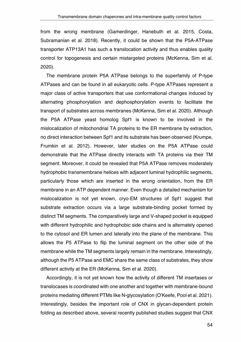
Transmembrane domain chaperones and intra-membrane quality control factors
54
from the wrong membrane (Gamerdinger, Hanebuth et al. 2015, Costa,
Subramanian et al. 2018). Recently, it could be shown that the P5A-ATPase
transporter ATP13A1 has such a translocation activity and thus enables quality
control for topogenesis and certain mistargeted proteins (McKenna, Sim et al.
2020).
The membrane protein P5A ATPase belongs to the superfamily of P-type
ATPases and can be found in all eukaryotic cells. P-type ATPases represent a
major class of active transporters that use conformational changes induced by
alternating phosphorylation and dephosphorylation events to facilitate the
transport of substrates across membranes (McKenna, Sim et al. 2020). Although
the P5A ATPase yeast homolog Spf1 is known to be involved in the
mislocalization of mitochondrial TA proteins to the ER membrane by extraction,
no direct interaction between Spf1 and its substrate has been observed (Krumpe,
Frumkin et al. 2012). However, later studies on the P5A ATPase could
demonstrate that the ATPase directly interacts with TA proteins via their TM
segment. Moreover, it could be revealed that P5A ATPase removes moderately
hydrophobic transmembrane helices with adjacent luminal hydrophilic segments,
particularly those which are inserted in the wrong orientation, from the ER
membrane in an ATP dependent manner. Even though a detailed mechanism for
mislocalization is not yet known, cryo-EM structures of Spf1 suggest that
substrate extraction occurs via a large substrate-binding pocket formed by
distinct TM segments. The comparatively large and V-shaped pocket is equipped
with different hydrophilic and hydrophobic side chains and is alternately opened
to the cytosol and ER lumen and laterally into the plane of the membrane. This
allows the P5 ATPase to flip the luminal segment on the other side of the
membrane while the TM segments largely remain in the membrane. Interestingly,
although the P5 ATPase and EMC share the same class of substrates, they show
different activity at the ER (McKenna, Sim et al. 2020).
Accordingly, it is not yet known how the activity of different TM insertases or
translocases is coordinated with one another and together with membrane-bound
proteins mediating different PTMs like N-glycosylation (O'Keefe, Pool et al. 2021).
Interestingly, besides the important role of CNX in glycan-dependent protein
folding as described above, several recently published studies suggest that CNX
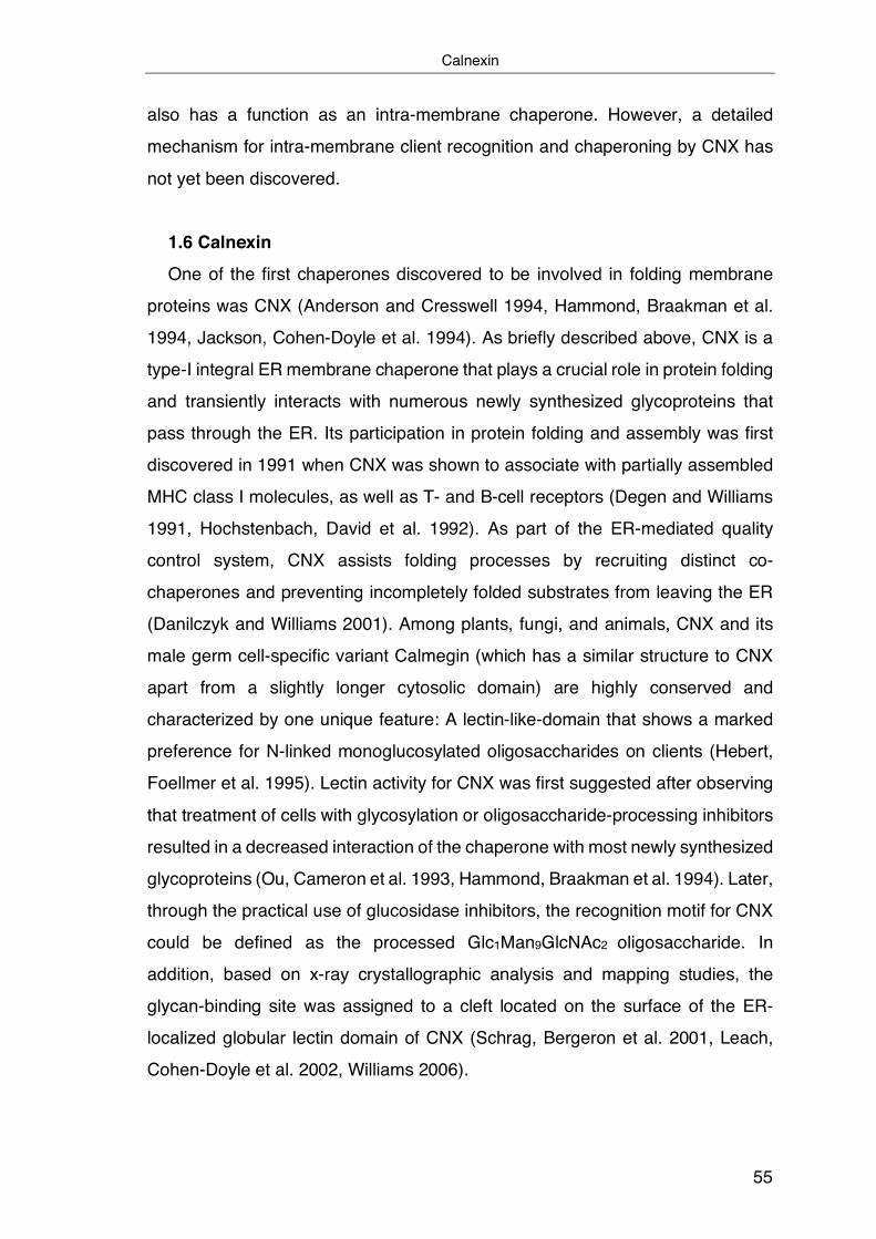
Calnexin
55
also has a function as an intra-membrane chaperone. However, a detailed
mechanism for intra-membrane client recognition and chaperoning by CNX has
not yet been discovered.
1.6 Calnexin One of the first chaperones discovered to be involved in folding membrane
proteins was CNX (Anderson and Cresswell 1994, Hammond, Braakman et al.
1994, Jackson, Cohen-Doyle et al. 1994). As briefly described above, CNX is a
type-I integral ER membrane chaperone that plays a crucial role in protein folding
and transiently interacts with numerous newly synthesized glycoproteins that
pass through the ER. Its participation in protein folding and assembly was first
discovered in 1991 when CNX was shown to associate with partially assembled
MHC class I molecules, as well as T- and B-cell receptors (Degen and Williams
1991, Hochstenbach, David et al. 1992). As part of the ER-mediated quality
control system, CNX assists folding processes by recruiting distinct co-
chaperones and preventing incompletely folded substrates from leaving the ER
(Danilczyk and Williams 2001). Among plants, fungi, and animals, CNX and its
male germ cell-specific variant Calmegin (which has a similar structure to CNX
apart from a slightly longer cytosolic domain) are highly conserved and
characterized by one unique feature: A lectin-like-domain that shows a marked
preference for N-linked monoglucosylated oligosaccharides on clients (Hebert,
Foellmer et al. 1995). Lectin activity for CNX was first suggested after observing
that treatment of cells with glycosylation or oligosaccharide-processing inhibitors
resulted in a decreased interaction of the chaperone with most newly synthesized
glycoproteins (Ou, Cameron et al. 1993, Hammond, Braakman et al. 1994). Later,
through the practical use of glucosidase inhibitors, the recognition motif for CNX
could be defined as the processed Glc1Man9GlcNAc2 oligosaccharide. In
addition, based on x-ray crystallographic analysis and mapping studies, the
glycan-binding site was assigned to a cleft located on the surface of the ER-
localized globular lectin domain of CNX (Schrag, Bergeron et al. 2001, Leach,
Cohen-Doyle et al. 2002, Williams 2006).
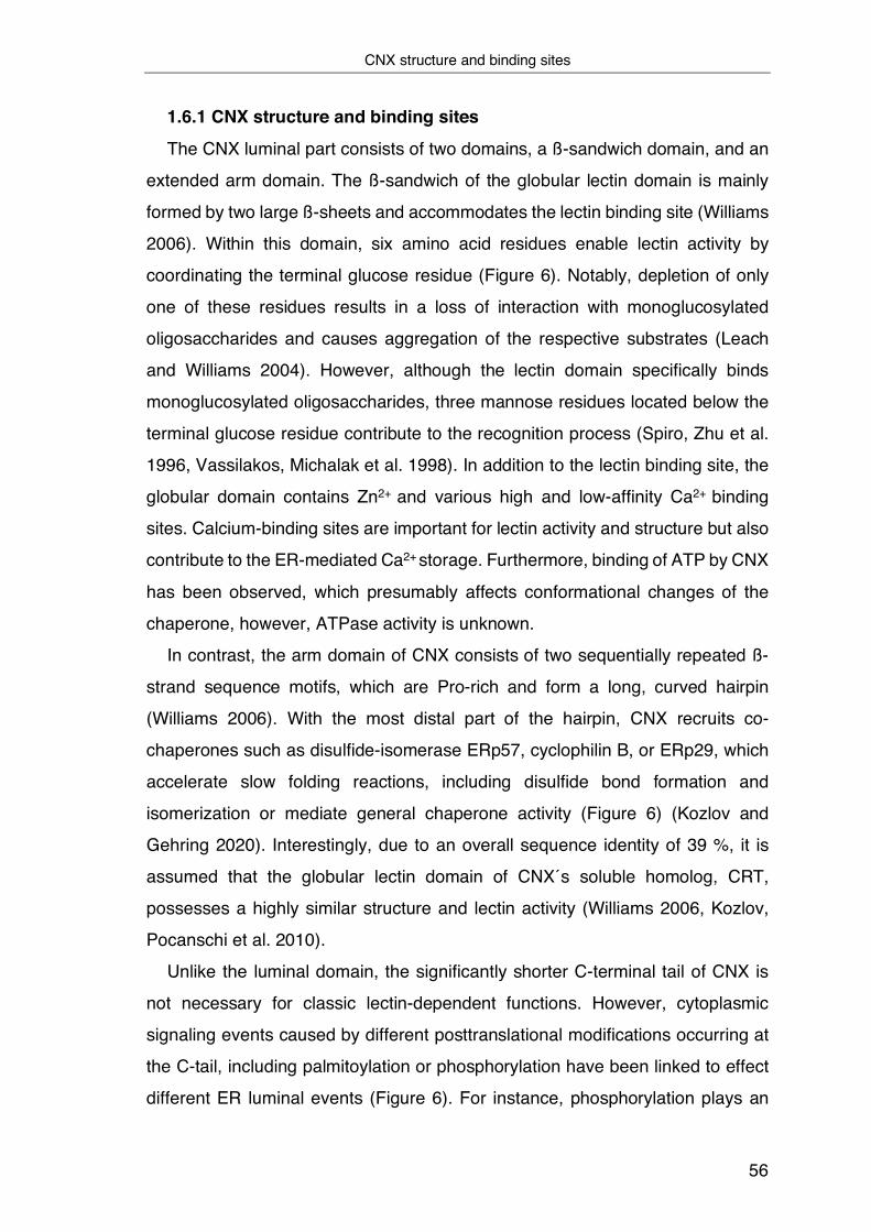
CNX structure and binding sites
56
1.6.1 CNX structure and binding sites
The CNX luminal part consists of two domains, a ß-sandwich domain, and an
extended arm domain. The ß-sandwich of the globular lectin domain is mainly
formed by two large ß-sheets and accommodates the lectin binding site (Williams
2006). Within this domain, six amino acid residues enable lectin activity by
coordinating the terminal glucose residue (Figure 6). Notably, depletion of only
one of these residues results in a loss of interaction with monoglucosylated
oligosaccharides and causes aggregation of the respective substrates (Leach
and Williams 2004). However, although the lectin domain specifically binds
monoglucosylated oligosaccharides, three mannose residues located below the
terminal glucose residue contribute to the recognition process (Spiro, Zhu et al.
1996, Vassilakos, Michalak et al. 1998). In addition to the lectin binding site, the
globular domain contains Zn2+ and various high and low-affinity Ca2+ binding
sites. Calcium-binding sites are important for lectin activity and structure but also
contribute to the ER-mediated Ca2+ storage. Furthermore, binding of ATP by CNX
has been observed, which presumably affects conformational changes of the
chaperone, however, ATPase activity is unknown.
In contrast, the arm domain of CNX consists of two sequentially repeated ß-
strand sequence motifs, which are Pro-rich and form a long, curved hairpin
(Williams 2006). With the most distal part of the hairpin, CNX recruits co-
chaperones such as disulfide-isomerase ERp57, cyclophilin B, or ERp29, which
accelerate slow folding reactions, including disulfide bond formation and
isomerization or mediate general chaperone activity (Figure 6) (Kozlov and
Gehring 2020). Interestingly, due to an overall sequence identity of 39 %, it is
assumed that the globular lectin domain of CNX´s soluble homolog, CRT,
possesses a highly similar structure and lectin activity (Williams 2006, Kozlov,
Pocanschi et al. 2010).
Unlike the luminal domain, the significantly shorter C-terminal tail of CNX is
not necessary for classic lectin-dependent functions. However, cytoplasmic
signaling events caused by different posttranslational modifications occurring at
the C-tail, including palmitoylation or phosphorylation have been linked to effect
different ER luminal events (Figure 6). For instance, phosphorylation plays an

CNX structure and binding sites
57
important role in regulating the calcium concentration in the ER lumen (Roderick,
Lechleiter et al. 2000, Chevet, Smirle et al. 2010).
Figure 6: A structural model of calnexin: Crystal structure analysis reveals a highly asymmetric structure of the ER luminal domain, which consists of a globular lectin domain containing the oligosaccharide-binding site and an elongated arm domain, which is important for the interaction with certain co-chaperones and cofactors. In addition, as a type I integral membrane protein, calnexin has a single transmembrane helix, and its short C-terminal tail undergoes various post-translational modifications that affect ER lumen events and is required for different interaction processes.
Calcium ions function as ubiquitous second messenger and are involved in a
broad spectrum of physiological and pathological processes. Amongst others,
conformational changes of proteins and protein folding per se depends on the
presence of calcium ions (Ca2+), and elevated levels of cytoplasmic Ca2+ control
a multiplicity of cellular signaling (Clapham 2007). In addition, mitochondrial
respiration or gene transcription control mainly relies on spatial and temporal
changes of intracellular Ca2+ levels (Dolmetsch, Xu et al. 1998, Robb-Gaspers,
Burnett et al. 1998). Two opposing factors essentially mediate these repetitive
changes in concentration: Whereas the inositol 1,4,5-trisphosphate receptor
channel (IP3R) is responsible for Ca2+ release from the ER, the sarco

CNX structure and binding sites
58
endoplasmic reticulum calcium ATPase (SERCA)-type Ca2+ ATPase causes the
opposite effect and mediates transport of calcium ions from the cytosol into the
internal ER store (Dawson 1997, MacLennan, Rice et al. 1997). As a recently
published study has demonstrated, CNX fulfills a crucial regulatory function in
this Ca2+ oscillation process, which is dependent on the phosphorylation status
of CNX´s cytosolic domain. Three different consensus sites within its cytosolic
domain allow phosphorylation, and several kinases have been identified in the
phosphorylation of CNX (Wong, Ward et al. 1998). However, only
phosphorylation of Ser562 mediated by the proline-directed kinase (PDK)
appears to exhibit significant functional and regulatory consequences on Ca2+
levels inside the ER. Phosphorylation of Ser562 causes enhanced interaction of
CNX with the C-terminal tail of SERCA 2b, the most prominent isoform of the
SERCA family, resulting in an inhibited Ca2+ uptake in the ER. In contrast, the
IP3 mediated release of Ca2+ causes dephosphorylation of the same site and
most likely also leads to reduced interaction with SERCA 2b, as experiments
analyzing the effect of phosphatase treatment have revealed. Consequently, the
dephosphorylation-dependent dissociation of CNX from SERCA controls the
uptake of Ca2+ in the ER and influences the Ca2+ sensitive protein folding
machinery inside this organelle. Thus, CNX is regulated by a phosphorylation
switch located on the very C-terminal end of the protein, which is highly sensitive
towards Ca2+ and influences ER luminal binding events (Roderick, Lechleiter et
al. 2000).
Of note, the same amino acid residue phosphorylation was furthermore
identified to exhibit a role in ER-mediated quality control. Following ER stress,
this site undergoes enhanced phosphorylation, leading to a prolonged interaction
of CNX with its misfolded substrate and subsequent degradation (Cameron,
Chevet et al. 2009). Moreover, phosphorylation is a prerequisite for CNX
recruitment to translocon-associated ribosomes (RTC, ribosome translocon
complex). In this case, however, phosphorylation depends on the synergistic
action of a different member of the PDK kinase family, namely extracellular-
signal-regulated kinase 1 (ERK-1) together with casein kinase 2 (CK2).
Therefore, it is assumed that the multiplied presence of CNX in the vicinity of the
translocon reinforces protein synthesis through increased glycoprotein folding
CNX structure and binding sites
59
mediated by CNX (Chevet, Wong et al. 1999). Interestingly, this would imply an
effect opposite to that caused by various members of the UPR response, which
mediates a reduction in protein synthesis following the induction of ER stress as
described in a previous section.
However, the enhanced association of CNX with the RTC is not only restricted
to phosphorylation events occurring in CNX C-terminal tail but has also been
observed as a consequence of other post-translational modifications. For
instance, upon palmitoylation of two membrane-proximal cysteine residues,
located in CNX C-terminal tail, by the transmembrane palmitoyltransferase
DHHC6, CNX localizes to the rough ER. At the rough ER, CNX interacts with
components of the RTC and contributes to the formation of a CNX-RTC
supercomplex. Following the successful assembly, CNX then mediates
recruitment of actin which significantly contributes to the stability of the complex.
Importantly, CNX must be both part of the RTC complex and palmitoylated to
capture nascent glycoproteins when they emerge from the translocon co-
translationally (Lakkaraju, Abrami et al. 2012).
Interestingly, palmitoylation modification of identical juxtamembrane cysteine
residues can affect CNX's cellular localization differently, as observed in other
studies. It could be shown that following palmitoylation, CNX is targeted not only
to the rough but also to a different region of the ER, namely the mitochondria-
associated membrane (MAM), which fulfills a critical role in lipid and calcium
metabolism. However, whether and how localization of CNX to different ER
domains based on its palmitoylation status is regulated is not yet known (Lynes,
Bui et al. 2012).
Noteworthy, palmitoylation not only seems to regulate the overall chaperone
activity of CNX or influences its cellular localization but appears to exhibit a direct
effect on the orientation of the cytosolic tail with respect to the CNX TMD, as
molecular dynamics simulations have revealed. Therefore, it seems conceivable
that palmitoylation can significantly change the conformation of CNX, which as a
consequence, would have an impact on the ability of CNX to interact with distinct
substrates in the cytoplasm as well as within the membrane (Lakkaraju, Abrami
et al. 2012).
CNX structure and binding sites
60
In addition to post-translational modifications, which affect the cytoplasmic
domain of CNX and thus exhibit a decisive influence on the function and
localization of calnexin as described above, other modifications of the CNX C-
terminal tail have been reported. For instance, it has been shown that following
both ER stress-inducing and non-stress-inducing apoptotic stimuli, CNX is
proteolytically cleaved, most likely by either caspase-3 or caspase-7, which
recognize a distinct DXXD motif in the carboxy-terminal region of CNX. It is
speculated that the resulting cleavage product can inhibit or a least reduce ER-
stress-induced apoptosis (Takizawa, Tatematsu et al. 2004).
To date, few studies have looked closer at the structure and properties of the
TMD of CNX. Its TMD consists of 21 amino acids that form an α-helix, and the
membrane-embedded region ranges from Trp482 to Cys502. In addition to
primarily non-polar amino acid residues, polar residues including Tyr487 or
Thr490 are present in the TM domain region, but no charged residues can be
found. As a result, the TMD is mostly hydrophobic which leads to stable
membrane integration (ΔGapp = -2.87). Notably, almost centrally located, the CNX
TMD contains a Proline residue. Although proline residues in the center of
transmembrane domains of integral membrane proteins are not uncommon, it
renders their occurrence interesting because proline residues exhibit helix
breaking properties, and their functional role has not been fully elucidated
(Chang, Cheng et al. 1999). What makes the appearance of the proline residue
in the TM region of CNX even more interesting is that this residue is also
conserved in Calmegin, the homologous member of the calnexin family.
Conservation of this residue and the TMD kink caused by it suggests that this
residue might play an important functional role. Indeed, it has been shown that
interaction of CNX with the ribosome-translocon complex depends essentially on
this Pro residue (Lakkaraju, Abrami et al. 2012). In line with this, several recently
published studies indicate that the CNX TM domain is more than just a simple
membrane anchor and contributes an essential function in intra-membrane client
recognition and chaperoning. So far, however, without revealing many details
about the underlying mechanism or potential substrates and biological
implications.
Glycan independent function of CNX and the importance of the transmembrane domain
61
1.6.2 Glycan independent function of CNX and the importance of the
transmembrane domain In addition to the key role of CNX in the folding of ER glycoproteins, a role of
CNX in the quality control of membrane proteins with incorrectly folded or
assembled TM domains has been described. Interestingly, in many of these
studies, binding of CNX to multi-membrane-spanning proteins such as Cx32, the
inositol triphosphate receptor or CFTR occurred in a glycan-independent manner
(Pind, Riordan et al. 1994, Joseph, Boehning et al. 1999, Fontanini, Chies et al.
2005, Coelho, Stahl et al. 2019). This consequently raises the question of how
substrate specificity is mediated if the glycosylation status of a client not solely
determines recognition. Remarkably, although CNX and CRT have almost
identical lectin domains, genetic deletion causes different effects. Whereas
knockout cell lines of either protein are viable, CRT deletion in mice leads to
failures in heart development and prenatal lethality. In contrast, deletion of CNX
strongly affects nerve fibers and causes early postnatal death (Denzel, Molinari
et al. 2002). These and other findings, which could show different substrate
preferences for CNX and CRT indicate that CNX´s topology might dictate
substrate selection. In particular focus: the TM domain of CNX, which is the major
discriminating feature between both ER chaperones. By anchoring Crt in the
membrane, through fusion with the TM domain of CNX, one can change the
spectrum of proteins associated with Crt similar to that of CNX, indicating that
CNX´s TM domain might contribute to client selection (Wada, Imai et al. 1995,
Danilczyk, Cohen-Doyle et al. 2000). Nonetheless, a simple explanation for this
observation could also be the different localization of CNX and CRT in the
membrane versus ER-lumen. For instance, access to the Sec61 translocon or to
glycans that reside near the membrane could change as a result. However, this
cannot account for many observations concerning the client binding of CNX. Only
shortly after CNX was discovered, it could be shown that the observed
association of the chaperone with nascent MHC class I molecules is based on
direct transmembrane interactions (Margolese, Waneck et al. 1993). Later
studies on the proteolipid protein (PLP) showed for the first time that CNX binds
to isolated TM domains. Furthermore, it was shown that CNX directly interacts
with a misfolded or unassembled TM domain of PLP and thus prevents its
Glycan independent function of CNX and the importance of the transmembrane domain
62
degradation (Swanton, High et al. 2003). Similar findings for the quality control
function of CNX during transmembrane domain assembly were obtained from
studies on the tetraspin CD82. Even though CD82 is a multi-span glycoprotein,
CNX selectively prevents ER exit of truncated CD82 lacking the first TM domain;
however, it does not affect full-length CD82. Interestingly, simultaneous co-
expression of the first TM segment with the truncated CD82 variant can partially
reverse retention. This indicates that CNX can distinguish the native and
unassembled status of a membrane protein by directly monitoring the assembly
status within the lipid bilayer (Cannon and Cresswell 2001). Further evidence for
this activity is based on studies of PMP22, where it was shown that CNX interacts
with the first TM segment of PMP22 in a glycan-independent manner (Fontanini,
Chies et al. 2005). Taken together, CNX recognizes and binds to truncated IMP
substrates that lack one or more TM segments – presumably because they are
recognized as misfolded and/or unassembled (Cannon and Cresswell 2001,
Swanton, High et al. 2003, Fontanini, Chies et al. 2005, Wanamaker and Green
2005, Coelho, Stahl et al. 2019). Remarkably, however, the exact details of what
renders a TM domain as “misfolded” and motifs or features that CNX recognizes
are not known.
Aim of studies
63
2 Aim of studies Membrane proteins make up one-third of the human proteome, and their
biosynthesis is based on a series of precisely coordinated events. These include
the translocation and integration of specific TMD structures into the lipid bilayer
and folding domain structures on both the lumenal and cytoplasmic sides of the
ER. Moreover, biosynthesis for multispanning or oligomeric IMPs involves the
correct assembly of TMDs within the bilayer of the ER. Due to these multiple
topologically demanding folding events, integral membrane proteins represent a
particular challenge to the ER folding machinery and quality control system.
Failures of these processes can have deleterious consequences for the cell and
the entire organism. Despite this, knowledge about how molecular chaperones
guide and control the structure formation of membrane proteins in the lipid bilayer
is very limited.
In addition, most studies on the canonical degradation and quality control
pathways have focused on proteins where the misfolded lesion is located either
inside the ER lumen or in the cytosol. However, proteins located within the ER
membrane, containing aberrant TM domains cannot be recognized by the very
same cytosolic or luminal factors. Considering that incorrect folding and assembly
of IMPs and destabilizing mutations that introduce charges within TMDs of IMPs
are linked to various human diseases, only very little is known about factors or
quality control mechanisms that interact with unstable TM helices.
Therefore, this study aims to mechanistically investigate novel intra-membrane
chaperones and quality control mechanisms that, within the lipid bilayer, monitor
membrane protein folding, the assembly status of TMDs, and folding defects in
those. At the same time, we aim to define sequence motifs that constitute a
“misfolded” TM domain and are thus recognizes by intra-membrane chaperones.
Since natural TM proteins, as outlined above, generally possess complex
sequences and structures and may engage multiple topically and functionally
distinct layers of quality control, which precludes an unbiased analysis, the initial
objective of this study is to establish an artificial minimal consensus membrane
(CoMem) reporter system that allows for the systematic analysis of intra-
membrane recognition processes. Then, to define principles of intra-membrane
Aim of studies
64
client recognition, we use Calnexin (CNX), one of the most abundant ER
chaperones, and putative intra-membrane chaperone as a model. In addition to
its canonical function, CNX is known to directly interact with TM helices of multi-
membrane-spanning proteins, although an underlying mechanism is not known.
By combining experimental and computational approaches, we aim to
systematically dissect the intra-membrane recognition process of misfolded
membrane clients by the transmembrane domain of CNX. In detail, we aim to
uncover a structural understanding of client recognition by CNX and the biological
implications of this recognition process.
Hence, this study aims to provide a comprehensive understanding of intra-
membrane substrate recognition by molecular chaperones as a basis for quality
control of IMPs in the ER, which in the future could contribute to a better
understanding of numerous membrane protein-misfolding diseases.

Calnexin interacts with transmembrane domains of non-glycosylated clients
65
3 Results
3.1 Calnexin interacts with transmembrane domains of non-glycosylated
clients Membrane proteins are a major class of proteins produced in the ER. Despite
this, principles of their molecular chaperoning remain ill-defined. CNX is one of
the most abundant ER chaperones and several studies indicate a role for its TMD
in substrate recognition (Cannon and Cresswell 2001, Swanton, High et al. 2003,
Fontanini, Chies et al. 2005, Wanamaker and Green 2005, Korkhov, Milan-Lobo
et al. 2008, Li, Su et al. 2010, Coelho, Stahl et al. 2019). Despite the number of
reports on intra-membrane client recognition by CNX, this process remains very
incompletely understood in terms of mechanisms, range of substrates, and
biological implications. To address this gap in knowledge and provide insights
intro principles of intra-membrane chaperoning processes, we used Connexin 32
(Cx32) as a first model client protein. Cx32 is a four-helix integral membrane
protein (Figure 7A) that forms homo-hexameric connexons. Two of these
connexons embedded in different membranes can dock onto each other to form
a gap junction channel (Pantano, Zonta et al. 2008, Maeda, Nakagawa et al.
2009). Mutations in Cx32 cause X-linked Charcot-Marie-Tooth disease (CMTX),
one of the most common genetic disorders of the peripheral nervous system
(Scherer and Kleopa 2012). A recent study from us has revealed interaction of
Cx32 with CNX in mass spectrometry experiments (Coelho, Stahl et al. 2019).
Cx32 is not glycosylated (Figure 7B) and possesses only very short ER-lumenal
loops (Figure 7A). This renders a potential Cx32:CNX interaction a possible
starting point towards defining intra-membrane client binding by CNX as it can
neither rely on recognition of sugar moieties nor on binding of large ER-lumenal
domains by the CNX lectin domain.
A
C-term
ER-lumen/extracellular
Cytoplasm
Cx32
N-term
TM1 TM4TM3TM2
: I28N
Loop 1: 30 aa Loop 3: 38 aa
: L143P: V140E
: I203N
B
WTN-term Loop 1 Loop 2 Loop 3 C-termNVT site
EndoH - + - + - + - + - +
ER-lumen/extracellular
Cytoplasm
N-term
Loop 1
NNN
NNN
Loop 2NNN
Loop 3
NNN
C-termNNN
25 –
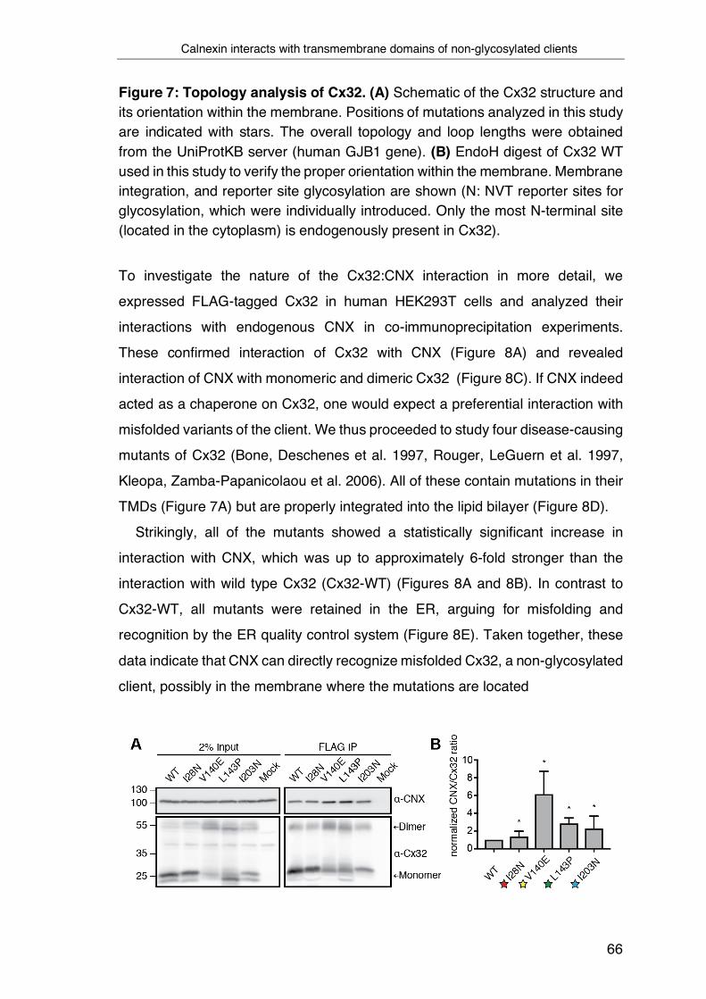
Calnexin interacts with transmembrane domains of non-glycosylated clients
66
Figure 7: Topology analysis of Cx32. (A) Schematic of the Cx32 structure and its orientation within the membrane. Positions of mutations analyzed in this study are indicated with stars. The overall topology and loop lengths were obtained from the UniProtKB server (human GJB1 gene). (B) EndoH digest of Cx32 WT used in this study to verify the proper orientation within the membrane. Membrane integration, and reporter site glycosylation are shown (N: NVT reporter sites for glycosylation, which were individually introduced. Only the most N-terminal site (located in the cytoplasm) is endogenously present in Cx32).
To investigate the nature of the Cx32:CNX interaction in more detail, we
expressed FLAG-tagged Cx32 in human HEK293T cells and analyzed their
interactions with endogenous CNX in co-immunoprecipitation experiments.
These confirmed interaction of Cx32 with CNX (Figure 8A) and revealed
interaction of CNX with monomeric and dimeric Cx32 (Figure 8C). If CNX indeed
acted as a chaperone on Cx32, one would expect a preferential interaction with
misfolded variants of the client. We thus proceeded to study four disease-causing
mutants of Cx32 (Bone, Deschenes et al. 1997, Rouger, LeGuern et al. 1997,
Kleopa, Zamba-Papanicolaou et al. 2006). All of these contain mutations in their
TMDs (Figure 7A) but are properly integrated into the lipid bilayer (Figure 8D).
Strikingly, all of the mutants showed a statistically significant increase in
interaction with CNX, which was up to approximately 6-fold stronger than the
interaction with wild type Cx32 (Cx32-WT) (Figures 8A and 8B). In contrast to
Cx32-WT, all mutants were retained in the ER, arguing for misfolding and
recognition by the ER quality control system (Figure 8E). Taken together, these
data indicate that CNX can directly recognize misfolded Cx32, a non-glycosylated
client, possibly in the membrane where the mutations are located

Calnexin interacts with transmembrane domains of non-glycosylated clients
67
Figure 8: Calnexin binds to non-glycosylated membrane proteins and their individual transmembrane domains. (A) Co-immunoprecipitations of endogenous CNX with FLAG-tagged Cx32 WT and its mutants. Constructs were expressed in HEK293T cells. One representative immunoblot is shown. Monomers and dimers of Cx32 are indicated with arrows. (B) Quantification and statistical analysis of (A). All samples were normalized to WT (mean ± SEM, N ≥ 4, *P value < 0.05, two-tailed Student’s t tests). (C) HEK293T cells were transfected with Cx32 WT or the indicated mutants. Immunoprecipitations of endogenous CNX reveal interactions with monomeric and dimeric Cx32 species. (D) EndoH digest of Cx32 mutants used in this study to verify their proper orientation within the membrane. Schematics of mutations studied, membrane integration, and reporter site glycosylation are shown (N: NVT reporter sites for glycosylation, which were individually introduced. Only the most N-terminal site (located in the cytoplasm) is endogenously present in Cx32). (E) Immunofluorescence images demonstrating localization of Cx32 and its mutants. COS-7 cells were transiently transfected with the indicated FLAG-tagged Cx32 constructs and immunofluorescence microscopy was performed using anti PDI (green) as an ER marker. Detection of Cx32 was performed using anti FLAG antibodies and subsequent staining of primary antibodies with labeled secondary antibodies (magenta). Nuclei were stained with DAPI (blue). All three channels are overlayed. Images are representative of cells from at least three different biological replicates. GJ denotes gap junctions observed in the cells transfected with WT Cx32. Scale bars correspond to 20 µm.
To further test this hypothesis, we individually fused each of the four Cx32 TMD
domains to an antibody light chain constant domain (CL) (Figure 9A). A related
system was recently established to assess chaperone:client interactions for
soluble proteins in a systematic manner (Feige and Hendershot 2013, Behnke,

Calnexin interacts with transmembrane domains of non-glycosylated clients
68
Mann et al. 2016). By using a glycosylation reporter site downstream of the TMD
segment we could verify that the majority of each TMD segment was integrated
into the ER membrane (Figure 9B). The only exception was TMD segment 2
(Figure 9B), which did not integrate properly into the membrane, in agreement
with a recent study (Coelho, Stahl et al. 2019) and the predicted membrane
integration potentials (Figure 9A). Using this system, we probed which of the TM
segments from Cx32 was bound by CNX. As a small portion of the TMD3 and
TMD4 constructs could enter the ER, we replaced the sugar-accepting Asn
residue downstream of each TMD segment by a Gln residue, since our interest
was in glycan-independent interaction with CNX. TMD segment 2 was excluded
from these analyses since it failed to integrate into the membrane. Introducing
mutations into the individual TMD segments also compromised their proper
membrane integration (Figures 9 C and D). We thus focused on analyzing
interactions of CNX with wild type TMD segments 1, 3 and 4 of Cx32. Interaction
was observed for all three TMD segments and among those, by far the strongest
interaction was observed with TMD1 (Figures 9E and 9F). Taken together, our
data clearly show that the chaperone CNX binds to Cx32 in a glycan-independent
manner. Binding increases for Cx32 variants misfolded in the membrane and
CNX can bind individual Cx32 TMD segments.

The transmembrane domain of Calnexin binds clients in the membrane
69
Figure 9: Calnexin binds to individual transmembrane domains of a non-glycosylated client. (A) Schematic of the CL-TMD reporter construct to assess CNX interactions with individual Cx32 TMD segments. The four WT sequences of the Cx32-TMDs are shown. Predicted membrane integration energies are given next to them using DGPred (Hessa, Meindl-Beinker et al. 2007). Sequences of TMD1 and TMD3 were inverted in the respective constructs to reflect the correct orientation within Cx32 (see Figure 7A). (B) EndoH digest of the four CL-TMD reporters to assess their integration and orientation in the membrane. The different detected species of CL-TMD constructs are indicated with arrows. Percentages below the blot indicate the C-terminally glycosylated fraction, giving an estimate of the fraction that fails to integrate into the ER membrane. The asterisk (*) denotes a fraction of cleaved species. Cleavage was also previously observed for similar reporter constructs containing the CL domain, which occurred post-lysis (Behnke, Mann et al. 2016). (C) Predicted ΔGapp values for membrane integration for the isolated TMDs of investigated Cx32 mutants. (D) EndoH digest of the CL-TMD3 domain reporter and its indicated mutants to assess their membrane integration. Percentages below the blot indicate the C-terminally glycosylated fraction, giving an estimate of the fraction that fails to integrate into the ER membrane. (E) Co-immunoprecipitations of endogenous CNX with the different CL-TMD constructs (NVT glycosylation site mutated to QVT). One representative immunoblot is shown. The asterisk (*) denotes a fraction of cleaved species. (F) Quantification and statistical analysis of (F). All samples were normalized to the construct containing TMD1 (mean ± SEM, N ≥ 3, *P value < 0.05, two-tailed Student’s t tests).
3.2 The transmembrane domain of Calnexin binds clients in the membrane
Our studies on Cx32 show that CNX can bind individual TM segments of its
clients. Based on these findings, we next aimed at defining which structural
elements of CNX were necessary for binding. Toward this end, we designed a

The transmembrane domain of Calnexin binds clients in the membrane
70
minimal CNX (minCNX) construct that only contained the CNX TMD and a few
additional C-terminal residues. The ER-lumenal domain of CNX was replaced by
the fluorescent protein mScarlet-I (Figure 10A). To maintain ER retention of the
construct, an endogenous cytosolic di-lysine motif was left in place (Figures 10A
and 23A) (Jackson, Nilsson et al. 1990). ER-localization was confirmed by
fluorescence microscopy (Figure 10B). Using this construct, we could show that
the CNX TMD segment was necessary and sufficient for binding to full-length
Cx32 (Figure 10C). Further extending this finding, we could show that an
indvidual TMD of Cx32 bound to the minCNX construct as well (Figure 10D). In
either case, an ER-retained control construct lacking the CNX TMD region
(minCNXDTMD, Figure 22B) did not bind to the different clients (Figures 10C and
10D). Thus, binding occurred independently of the CNX lectin domain or its C-
terminal tail. Accordingly, the CNX TMD contains the relevant features for client
recognition in the membrane, and this can be recapitulated with single TMD
regions derived from a client protein.
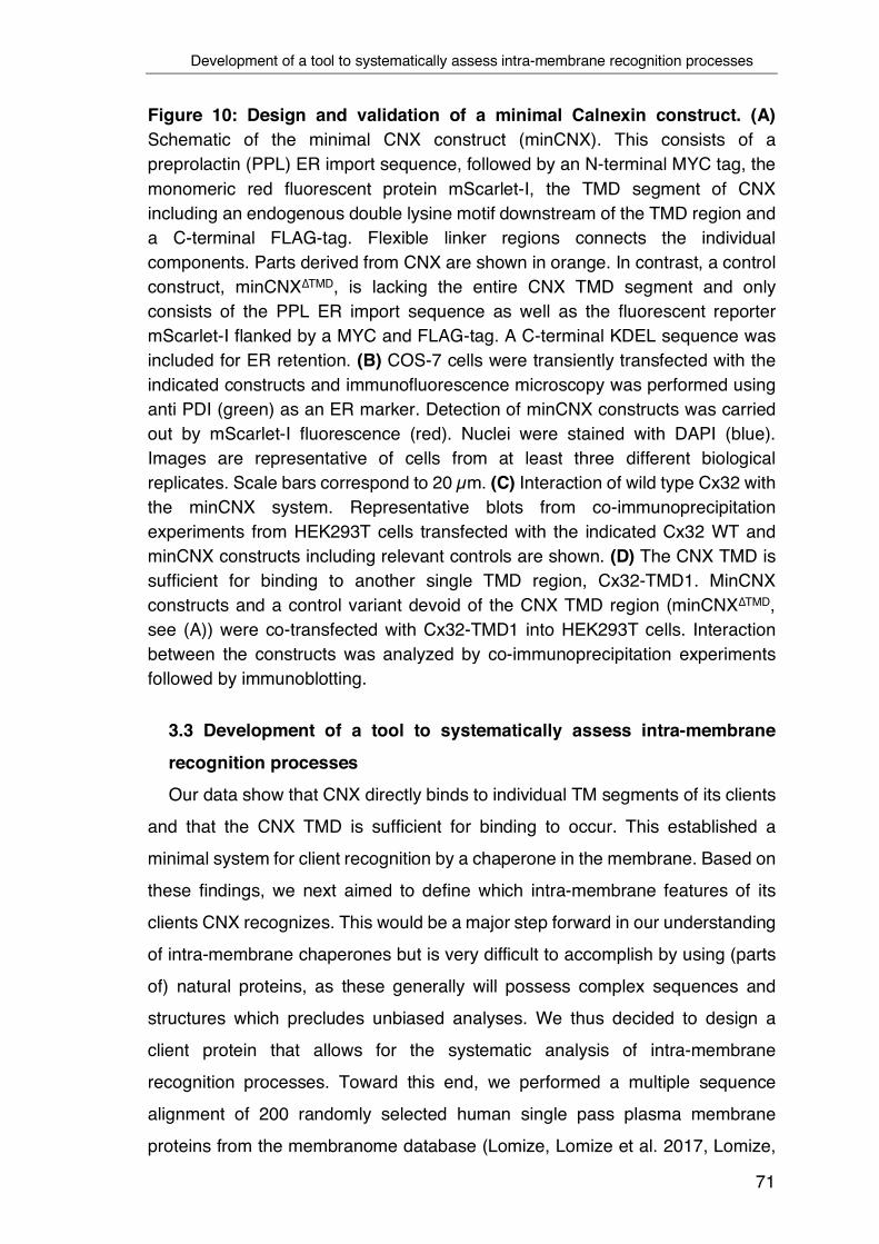
Development of a tool to systematically assess intra-membrane recognition processes
71
Figure 10: Design and validation of a minimal Calnexin construct. (A) Schematic of the minimal CNX construct (minCNX). This consists of a preprolactin (PPL) ER import sequence, followed by an N-terminal MYC tag, the monomeric red fluorescent protein mScarlet-I, the TMD segment of CNX including an endogenous double lysine motif downstream of the TMD region and a C-terminal FLAG-tag. Flexible linker regions connects the individual components. Parts derived from CNX are shown in orange. In contrast, a control construct, minCNX∆TMD, is lacking the entire CNX TMD segment and only consists of the PPL ER import sequence as well as the fluorescent reporter mScarlet-I flanked by a MYC and FLAG-tag. A C-terminal KDEL sequence was included for ER retention. (B) COS-7 cells were transiently transfected with the indicated constructs and immunofluorescence microscopy was performed using anti PDI (green) as an ER marker. Detection of minCNX constructs was carried out by mScarlet-I fluorescence (red). Nuclei were stained with DAPI (blue). Images are representative of cells from at least three different biological replicates. Scale bars correspond to 20 µm. (C) Interaction of wild type Cx32 with the minCNX system. Representative blots from co-immunoprecipitation experiments from HEK293T cells transfected with the indicated Cx32 WT and minCNX constructs including relevant controls are shown. (D) The CNX TMD is sufficient for binding to another single TMD region, Cx32-TMD1. MinCNX constructs and a control variant devoid of the CNX TMD region (minCNX∆TMD, see (A)) were co-transfected with Cx32-TMD1 into HEK293T cells. Interaction between the constructs was analyzed by co-immunoprecipitation experiments followed by immunoblotting.
3.3 Development of a tool to systematically assess intra-membrane
recognition processes Our data show that CNX directly binds to individual TM segments of its clients
and that the CNX TMD is sufficient for binding to occur. This established a
minimal system for client recognition by a chaperone in the membrane. Based on
these findings, we next aimed to define which intra-membrane features of its
clients CNX recognizes. This would be a major step forward in our understanding
of intra-membrane chaperones but is very difficult to accomplish by using (parts
of) natural proteins, as these generally will possess complex sequences and
structures which precludes unbiased analyses. We thus decided to design a
client protein that allows for the systematic analysis of intra-membrane
recognition processes. Toward this end, we performed a multiple sequence
alignment of 200 randomly selected human single pass plasma membrane
proteins from the membranome database (Lomize, Lomize et al. 2017, Lomize,

Development of a tool to systematically assess intra-membrane recognition processes
72
Hage et al. 2018). This gave rise to an average TMD of a transport-competent
protein, which thus can be expected to lack major chaperone recognition sites.
We termed this protein minimal consensus membrane protein (CoMem). The
design of CoMem included a superfolder GFP (sfGFP) moiety for microscopic
localization studies, a C-terminal HA-epitope tag, and two consensus
glycosylation sites to analyze its topology and intracellular transport (Figure 11A).
In agreement with our assumption that, on average, TM segments from single-
pass cell surface TMD proteins are close to optimal, the most frequent amino acid
at most positions turned out to be Leu (Figure 11A). TM Leu-zippers, however,
have a strong self-assembly propensity that would compromise our analyses
(Gurezka, Laage et al. 1999). We thus proceeded with the second most
frequently occurring amino acids, which were also entirely free of unfavorable
residues for a TM sequence (Hessa, Meindl-Beinker et al. 2007). The multiple
sequence alignment resulted in a TMD of 26 amino acid in length in CoMem,
which is in very good agreement with recent studies on average TMD lengths in
the plasma membrane (Sharpe, Stevens et al. 2010, Singh and Mittal 2016).
Interestingly, it was flanked by an N-terminal Pro-residue, breaking the helical
TMD structure (Cordes, Bright et al. 2002), and a C-terminal Lys residue (Figure
11A and Figure 22C). A C-terminal Lys will induce a type I orientation (von Heijne
and Gavel 1988), placing the Lys residue in the cytoplasm, which reflects the
nature of our sequence set (76% of the proteins were type I). Individually mutating
the first or second consensus glycosylation site in CoMem showed that it was
exclusively modified at the first site, which confirms the predicted topology (Figure
11B). Enzymatic deglycosylation with EndoH (which only removes N-linked
sugars not further modified in the Golgi) and with PNGaseF (which removes all
N-linked sugars) further revealed that CoMem glycosylation was EndoH-
resistant, arguing that it was able to traverse the Golgi as expected (Figure 11B).
Localization to the plasma membrane was confirmed by microscopic studies
(Figure 11C). Taken together, CoMem now provided us with an ideal tool to
systematically dissect intra-membrane substrate recognition by CNX.

Development of a tool to systematically assess intra-membrane recognition processes
73
Figure 11: Design and validation of CoMem, a tool to query TM-domain recognition. (A) Schematic of the the minimal consensus membrane protein CoMem which contains a preprolactin (PPL) ER import sequence, a superfolder GFP (sfGFP), a TMD flanked by two individual NVT glycosylation motifs (N1 and N2) and a C-terminal HA-tag. Individual construct components are connected by flexible linker regions. As illustrated, in the predicted topology of CoMem, only the first NVT glycosylation site (N1) is accessible to the ER glycosylation machinery (grey hexagon). The TMD of CoMem was designed on the basis of a multiple sequence alignment of 200 predicted human single pass TMD sequences as illustrated in the sequence logo. Hydrophilic amino acid residues are depicted in blue, neutral ones in green and hydrophobic residues in black. The predicted amino acid sequence with the second highest score was selected as the TMD consensus sequence for CoMem (bold). (B) HEK293T cells were transiently transfected with plasmids expressing CoMem or the indicated variants, where either the first (N1) or the second (N2) NVT glycosylation motif was altered (N to Q mutation), to prevent glycosylation of that specific site. Lysates were treated with or without EndoH or PNGaseF as indicated and analyzed by immunoblotting. N-glycosylation occurs in the ER, complex glycosylation in the Golgi. (C) COS-7 cells were transfected with the indicated constructs and fluorescence microscopy was performed using Sec61 mCherry (red) as an ER marker. CoMem, detected by sfGFP fluorescence (green), is not retained in the ER but localized to the plasma membrane. Nuclei were stained with DAPI (blue). Images are representative of cells from at least three different biological replicates. Scale bars correspond to 20 µm.

Defining intra-membrane recognition motifs for Calnexin
74
3.4 Defining intra-membrane recognition motifs for Calnexin
To dissect features recognized by CNX in the membrane, we first replaced the
central Val residue at position 13 in CoMem by all other 19 amino acids. Of note,
for all of these substitutions, CoMem was still predicted to be stably integrated
into the membrane (Figure 12A) which was experimentally confirmed for several
constructs and different locations of polar amino acids (Figure 12B). Using this
CoMem panel, we first established suitable conditions for co-immunoprecipitaion
experiments (Figure 12C) and then investigated interactions with CNX.
Figure 12: Defining experimental conditions to assess intra-membrane recognition motifs for Calnexin, part I. (A) Schematic of CoMem N1Q where a central Val residue at position 13 within its TMD was replaced by all other 19 amino acids. Free energies for TMD insertion were predicted according to (Hessa, Meindl-Beinker et al. 2007). (B) CoMem and CoMem mutant variants containing amino acid replacements against arginine and asparagine at the indicated positions of the TMD region were transfected into HEK293T cells. Cell lysates were treated with or without EndoH or PNGaseF as indicated and analyzed by immunoblotting. Comparison of the glycosylation pattern with respect to CoMem indicates no misintegration for the different CoMem TMD mutants. (C) CoMem N1Q and CoMem N1Q Arg+13 were transfected into HEK293T cells. Cell lysates were split and exposed to different denaturing temperatures in Lämmli buffer as indicated. Analysis by immunoblotting and fluorescent imaging of sfGFP revealed some aggregation at high temperatures. The two lower species correspond to CoMem N1Q with an unfolded (upper) or a folded (lower, more compact) sfGFP moiety, as can be seen in the fluorescence-imaged gel on the right to detect sfGFP fluorescence.
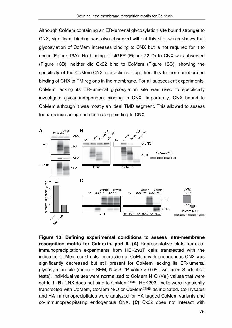
Defining intra-membrane recognition motifs for Calnexin
75
Although CoMem containing an ER-lumenal glycosylation site bound stronger to
CNX, significant binding was also observed without this site, which shows that
glycosylation of CoMem increases binding to CNX but is not required for it to
occur (Figure 13A). No binding of sfGFP (Figure 22 D) to CNX was observed
(Figure 13B), neither did Cx32 bind to CoMem (Figure 13C), showing the
specificity of the CoMem:CNX interactions. Together, this further corroborated
binding of CNX to TM regions in the membrane. For all subsequent experiments,
CoMem lacking its ER-lumenal glycosylation site was used to specifically
investigate glycan-independent binding to CNX. Importantly, CNX bound to
CoMem although it was mostly an ideal TMD segment. This allowed to assess
features increasing and decreasing binding to CNX.
Figure 13: Defining experimental conditions to assess intra-membrane recognition motifs for Calnexin, part II. (A) Representative blots from co-immunoprecipitation experiments from HEK293T cells transfected with the indicated CoMem constructs. Interaction of CoMem with endogenous CNX was significantly decreased but still present for CoMem lacking its ER-lumenal glycosylation site (mean ± SEM, N ≥ 3, *P value < 0.05, two-tailed Student’s t tests). Individual values were normalized to CoMem N1Q (Val) values that were set to 1 (B) CNX does not bind to CoMemDTMD. HEK293T cells were transiently transfected with CoMem, CoMem N1Q or CoMemDTMD as indicated. Cell lysates and HA-immunoprecipitates were analyzed for HA-tagged CoMem variants and co-immunoprecipitating endogenous CNX. (C) Cx32 does not interact with
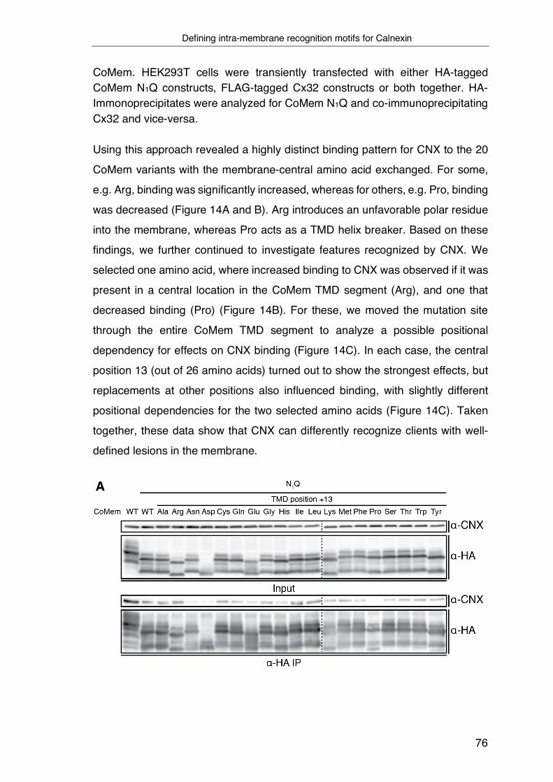
Defining intra-membrane recognition motifs for Calnexin
76
CoMem. HEK293T cells were transiently transfected with either HA-tagged CoMem N1Q constructs, FLAG-tagged Cx32 constructs or both together. HA-Immonoprecipitates were analyzed for CoMem N1Q and co-immunoprecipitating Cx32 and vice-versa.
Using this approach revealed a highly distinct binding pattern for CNX to the 20
CoMem variants with the membrane-central amino acid exchanged. For some,
e.g. Arg, binding was significantly increased, whereas for others, e.g. Pro, binding
was decreased (Figure 14A and B). Arg introduces an unfavorable polar residue
into the membrane, whereas Pro acts as a TMD helix breaker. Based on these
findings, we further continued to investigate features recognized by CNX. We
selected one amino acid, where increased binding to CNX was observed if it was
present in a central location in the CoMem TMD segment (Arg), and one that
decreased binding (Pro) (Figure 14B). For these, we moved the mutation site
through the entire CoMem TMD segment to analyze a possible positional
dependency for effects on CNX binding (Figure 14C). In each case, the central
position 13 (out of 26 amino acids) turned out to show the strongest effects, but
replacements at other positions also influenced binding, with slightly different
positional dependencies for the two selected amino acids (Figure 14C). Taken
together, these data show that CNX can differently recognize clients with well-
defined lesions in the membrane.
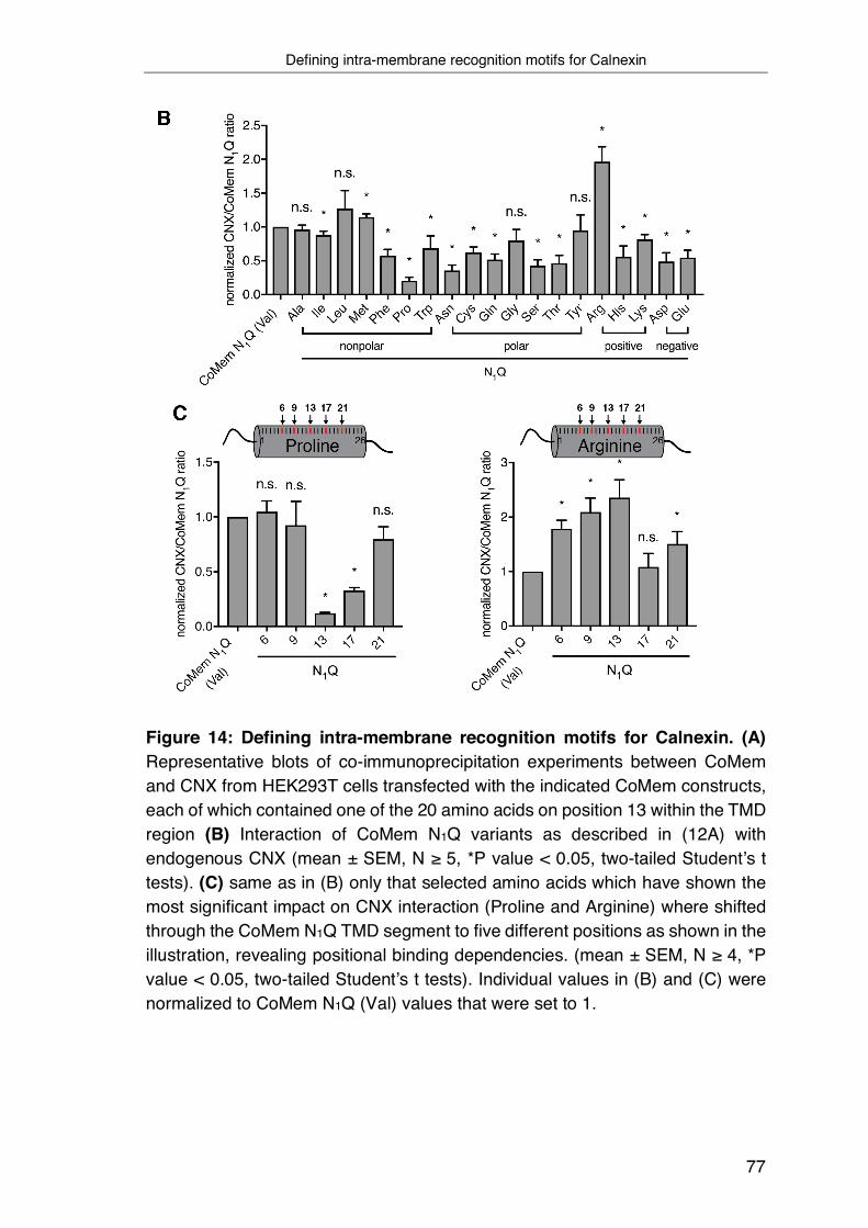
Defining intra-membrane recognition motifs for Calnexin
77
Figure 14: Defining intra-membrane recognition motifs for Calnexin. (A) Representative blots of co-immunoprecipitation experiments between CoMem and CNX from HEK293T cells transfected with the indicated CoMem constructs, each of which contained one of the 20 amino acids on position 13 within the TMD region (B) Interaction of CoMem N1Q variants as described in (12A) with endogenous CNX (mean ± SEM, N ≥ 5, *P value < 0.05, two-tailed Student’s t tests). (C) same as in (B) only that selected amino acids which have shown the most significant impact on CNX interaction (Proline and Arginine) where shifted through the CoMem N1Q TMD segment to five different positions as shown in the illustration, revealing positional binding dependencies. (mean ± SEM, N ≥ 4, *P value < 0.05, two-tailed Student’s t tests). Individual values in (B) and (C) were normalized to CoMem N1Q (Val) values that were set to 1.
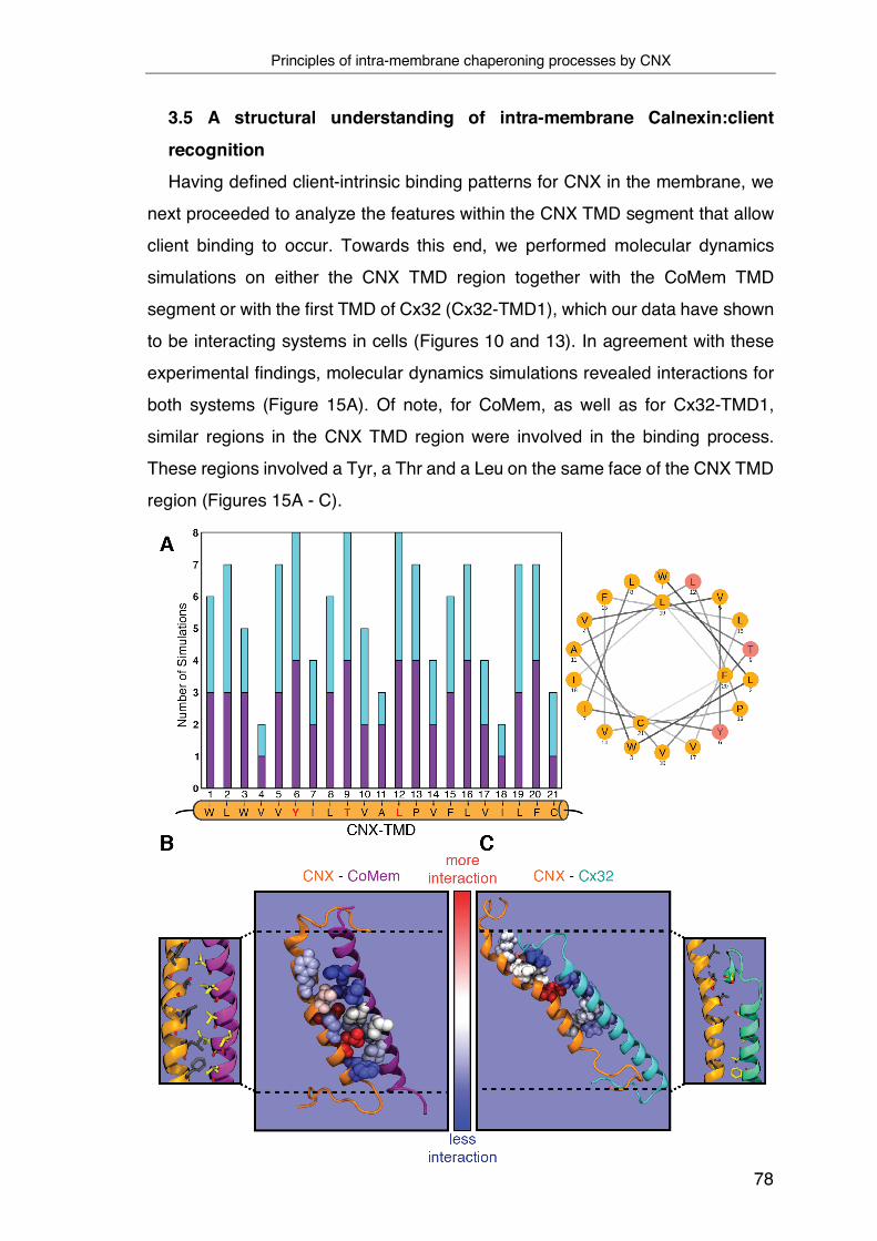
Principles of intra-membrane chaperoning processes by CNX
78
3.5 A structural understanding of intra-membrane Calnexin:client
recognition Having defined client-intrinsic binding patterns for CNX in the membrane, we
next proceeded to analyze the features within the CNX TMD segment that allow
client binding to occur. Towards this end, we performed molecular dynamics
simulations on either the CNX TMD region together with the CoMem TMD
segment or with the first TMD of Cx32 (Cx32-TMD1), which our data have shown
to be interacting systems in cells (Figures 10 and 13). In agreement with these
experimental findings, molecular dynamics simulations revealed interactions for
both systems (Figure 15A). Of note, for CoMem, as well as for Cx32-TMD1,
similar regions in the CNX TMD region were involved in the binding process.
These regions involved a Tyr, a Thr and a Leu on the same face of the CNX TMD
region (Figures 15A - C).

Principles of intra-membrane chaperoning processes by CNX
79
Figure 15: A structural analysis of intra-membrane Calnexin:client recognition. (A) Meta-analysis of MD simulations (N=8) where complex formation between CNX and Cx32-TMD1 or CNX and CoMem was observed. On the x-axis the CNX TMD residues are shown. The height of the bars corresponds with the number of simulations in which a specific CNX TMD residue interacted with the substrate helix CX32-TMD1 (turquoise) or CoMem (violet). Only CNX TMD residues Y17, T20 and L23 interacted with CX32-TMD1 or CoMem in all sampled complexes. (B) Depiction of residues exhibiting most interactions with the partner helix in simulations leading to complexes between CNX and CoMem. (C) Same as (B) but for the CNX-Cx32 simulations. CNX is depicted in orange, while CoMem is shown in purple and Cx32 in turquoise. Residues exhibiting most interactions with the partner helix are colored red, while residues in dark blue formed the least interactions. The dashed line indicates the membrane embedded region. Only residues with interaction frequencies of at least 30% of the top interacting residues are shown. For CNX residue statistics, only simulations in which complex formation was observed have been considered. The CoMem and Cx32 residues depicted here have been selected and colored with respect to how often they form interactions with the highlighted CNX residues (also in simulations in which stable complexes are formed) (Data obtained from Manuel Hitzenberger, Zaccharias Lab).
To assess our computational predictions experimentally, we mutated these
predicted interaction sites to presumably inert Val residues in a CNX construct
that we overexpressed in mammalian cells. This construct was furnished with a
V5 epitope tag for specific immunoprecipitation and overexpression was only
slightly higher than the endogenous CNX level. Strikingly, whereas CoMem and
Cx32-TMD1 co-immunoprecipitated with V5-tagged CNX as expected, mutation
of the YTL-motif, and even of only the Y and T residues to Val, significantly
reduced interactions between CNX and CoMem (Figure 16A). This was even
more pronounced for Cx32-TMD1 (Figure 16B), and when the YTL-motif was
exchanged against Ala instead of Val, a similar reduction in binding was observed
(Figure 16C).
This argues that mutating the interaction site per se, but not the choice of the
residues mutated to, accounts for the effects. Together, these findings confirmed
our simulation data and further corroborated the conserved nature of the
interactions. Of note, when CoMem with its ER-lumenal glycosylation site was
used, mutations in the CNX TMD region did not significantly affect binding,
showing that for this glycosylated simple client, binding via the lectin domain is
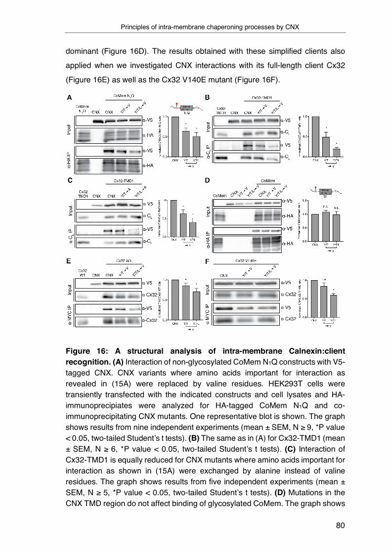
Principles of intra-membrane chaperoning processes by CNX
80
dominant (Figure 16D). The results obtained with these simplified clients also
applied when we investigated CNX interactions with its full-length client Cx32
(Figure 16E) as well as the Cx32 V140E mutant (Figure 16F).
Figure 16: A structural analysis of intra-membrane Calnexin:client recognition. (A) Interaction of non-glycosylated CoMem N1Q constructs with V5-tagged CNX. CNX variants where amino acids important for interaction as revealed in (15A) were replaced by valine residues. HEK293T cells were transiently transfected with the indicated constructs and cell lysates and HA-immunoprecipiates were analyzed for HA-tagged CoMem N1Q and co-immunoprecipitating CNX mutants. One representative blot is shown. The graph shows results from nine independent experiments (mean ± SEM, N ≥ 9, *P value < 0.05, two-tailed Student’s t tests). (B) The same as in (A) for Cx32-TMD1 (mean ± SEM, N ≥ 6, *P value < 0.05, two-tailed Student’s t tests). (C) Interaction of Cx32-TMD1 is equally reduced for CNX mutants where amino acids important for interaction as shown in (15A) were exchanged by alanine instead of valine residues. The graph shows results from five independent experiments (mean ± SEM, N ≥ 5, *P value < 0.05, two-tailed Student’s t tests). (D) Mutations in the CNX TMD region do not affect binding of glycosylated CoMem. The graph shows
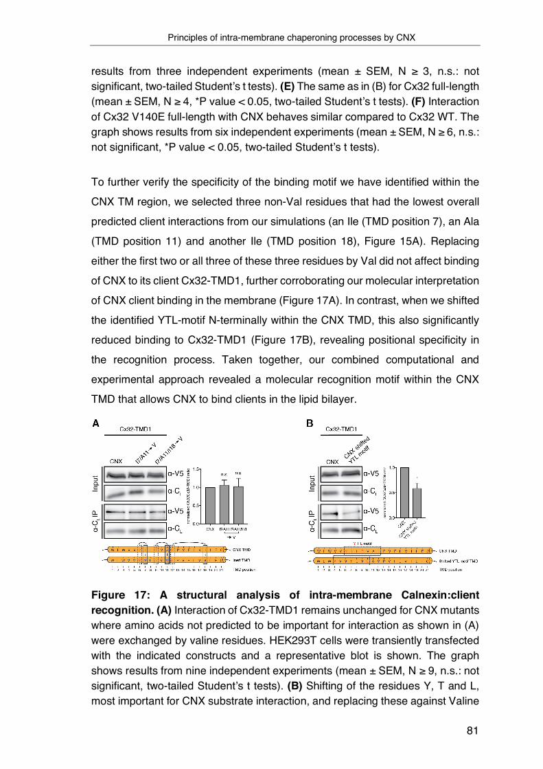
Principles of intra-membrane chaperoning processes by CNX
81
results from three independent experiments (mean ± SEM, N ≥ 3, n.s.: not significant, two-tailed Student’s t tests). (E) The same as in (B) for Cx32 full-length (mean ± SEM, N ≥ 4, *P value < 0.05, two-tailed Student’s t tests). (F) Interaction of Cx32 V140E full-length with CNX behaves similar compared to Cx32 WT. The graph shows results from six independent experiments (mean ± SEM, N ≥ 6, n.s.: not significant, *P value < 0.05, two-tailed Student’s t tests).
To further verify the specificity of the binding motif we have identified within the
CNX TM region, we selected three non-Val residues that had the lowest overall
predicted client interactions from our simulations (an Ile (TMD position 7), an Ala
(TMD position 11) and another Ile (TMD position 18), Figure 15A). Replacing
either the first two or all three of these three residues by Val did not affect binding
of CNX to its client Cx32-TMD1, further corroborating our molecular interpretation
of CNX client binding in the membrane (Figure 17A). In contrast, when we shifted
the identified YTL-motif N-terminally within the CNX TMD, this also significantly
reduced binding to Cx32-TMD1 (Figure 17B), revealing positional specificity in
the recognition process. Taken together, our combined computational and
experimental approach revealed a molecular recognition motif within the CNX
TMD that allows CNX to bind clients in the lipid bilayer.
Figure 17: A structural analysis of intra-membrane Calnexin:client recognition. (A) Interaction of Cx32-TMD1 remains unchanged for CNX mutants where amino acids not predicted to be important for interaction as shown in (A) were exchanged by valine residues. HEK293T cells were transiently transfected with the indicated constructs and a representative blot is shown. The graph shows results from nine independent experiments (mean ± SEM, N ≥ 9, n.s.: not significant, two-tailed Student’s t tests). (B) Shifting of the residues Y, T and L, most important for CNX substrate interaction, and replacing these against Valine
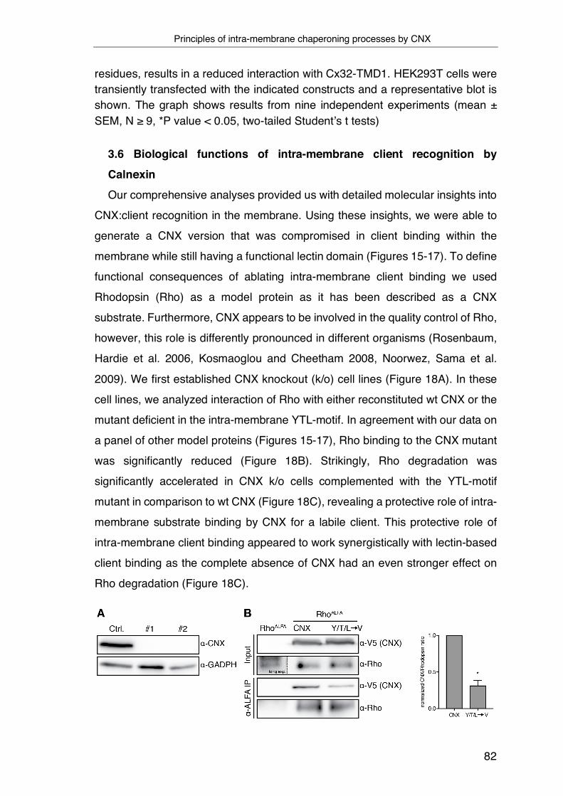
Principles of intra-membrane chaperoning processes by CNX
82
residues, results in a reduced interaction with Cx32-TMD1. HEK293T cells were transiently transfected with the indicated constructs and a representative blot is shown. The graph shows results from nine independent experiments (mean ± SEM, N ≥ 9, *P value < 0.05, two-tailed Student’s t tests)
3.6 Biological functions of intra-membrane client recognition by Calnexin
Our comprehensive analyses provided us with detailed molecular insights into
CNX:client recognition in the membrane. Using these insights, we were able to
generate a CNX version that was compromised in client binding within the
membrane while still having a functional lectin domain (Figures 15-17). To define
functional consequences of ablating intra-membrane client binding we used
Rhodopsin (Rho) as a model protein as it has been described as a CNX
substrate. Furthermore, CNX appears to be involved in the quality control of Rho,
however, this role is differently pronounced in different organisms (Rosenbaum,
Hardie et al. 2006, Kosmaoglou and Cheetham 2008, Noorwez, Sama et al.
2009). We first established CNX knockout (k/o) cell lines (Figure 18A). In these
cell lines, we analyzed interaction of Rho with either reconstituted wt CNX or the
mutant deficient in the intra-membrane YTL-motif. In agreement with our data on
a panel of other model proteins (Figures 15-17), Rho binding to the CNX mutant
was significantly reduced (Figure 18B). Strikingly, Rho degradation was
significantly accelerated in CNX k/o cells complemented with the YTL-motif
mutant in comparison to wt CNX (Figure 18C), revealing a protective role of intra-
membrane substrate binding by CNX for a labile client. This protective role of
intra-membrane client binding appeared to work synergistically with lectin-based
client binding as the complete absence of CNX had an even stronger effect on
Rho degradation (Figure 18C).
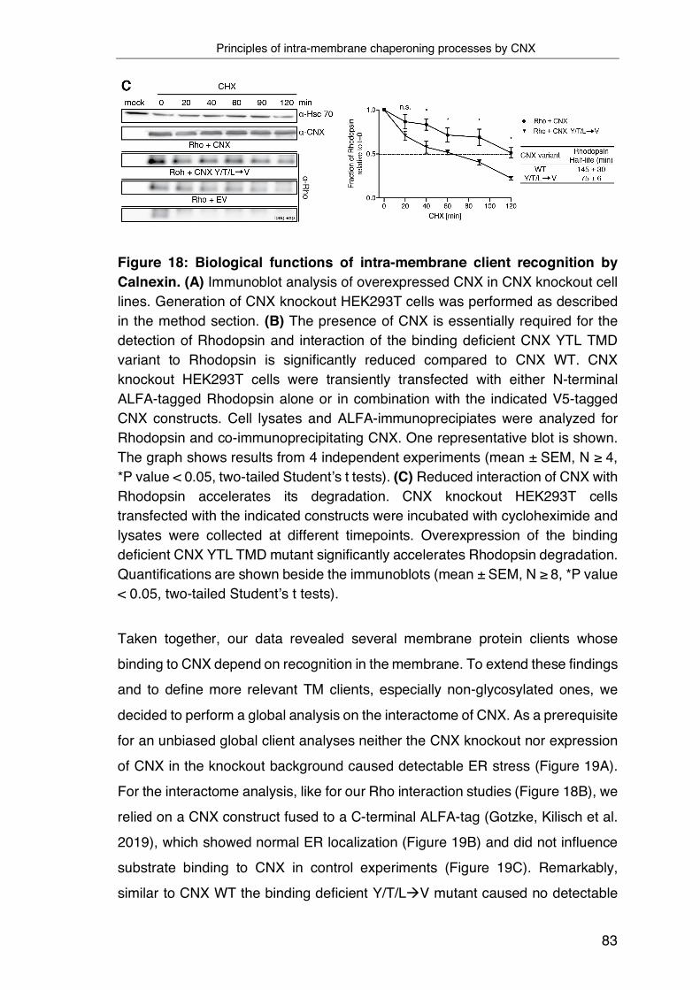
Principles of intra-membrane chaperoning processes by CNX
83
Figure 18: Biological functions of intra-membrane client recognition by Calnexin. (A) Immunoblot analysis of overexpressed CNX in CNX knockout cell lines. Generation of CNX knockout HEK293T cells was performed as described in the method section. (B) The presence of CNX is essentially required for the detection of Rhodopsin and interaction of the binding deficient CNX YTL TMD variant to Rhodopsin is significantly reduced compared to CNX WT. CNX knockout HEK293T cells were transiently transfected with either N-terminal ALFA-tagged Rhodopsin alone or in combination with the indicated V5-tagged CNX constructs. Cell lysates and ALFA-immunoprecipiates were analyzed for Rhodopsin and co-immunoprecipitating CNX. One representative blot is shown. The graph shows results from 4 independent experiments (mean ± SEM, N ≥ 4, *P value < 0.05, two-tailed Student’s t tests). (C) Reduced interaction of CNX with Rhodopsin accelerates its degradation. CNX knockout HEK293T cells transfected with the indicated constructs were incubated with cycloheximide and lysates were collected at different timepoints. Overexpression of the binding deficient CNX YTL TMD mutant significantly accelerates Rhodopsin degradation. Quantifications are shown beside the immunoblots (mean ± SEM, N ≥ 8, *P value < 0.05, two-tailed Student’s t tests).
Taken together, our data revealed several membrane protein clients whose
binding to CNX depend on recognition in the membrane. To extend these findings
and to define more relevant TM clients, especially non-glycosylated ones, we
decided to perform a global analysis on the interactome of CNX. As a prerequisite
for an unbiased global client analyses neither the CNX knockout nor expression
of CNX in the knockout background caused detectable ER stress (Figure 19A).
For the interactome analysis, like for our Rho interaction studies (Figure 18B), we
relied on a CNX construct fused to a C-terminal ALFA-tag (Gotzke, Kilisch et al.
2019), which showed normal ER localization (Figure 19B) and did not influence
substrate binding to CNX in control experiments (Figure 19C). Remarkably,
similar to CNX WT the binding deficient Y/T/LàV mutant caused no detectable

Principles of intra-membrane chaperoning processes by CNX
84
ER stress as well and showed normal ER localization (Figure 19 A and B). The
ALFA-tag is lysine-free and thus allows for the uncompromised use of lysine-
crosslinkers like DSSO in co-immunoprecipitation experiments. CNX has a large
number of well-dispersed lysine’s which should allow for in situ DSSO-
crosslinking (Figure 19D). Indeed, DSSO crosslinking revealed many CNX
crosslinks that can be immunoprecipitated using ALFA-tag nanobodies.
Interestingly, differences can be observed between the crosslinked interactome
of WT CNX and the binding deficient variant (Figure 19E).
Figure 19 Functional analysis of CNX-V5 and CNX-ALFA expression and interaction behavior in the HEK293T CNX KO context. (A) UPR activation was assessed in CNX knockout cells transfected with mock plasmid, and incubated with DMSO (vehicle control), tunicamycin (Tun.), or DTT. CNX, or its binding-deficient mutant were transfected into the CNX knockout cells (right side) and activation of the three branches of the mammalian UPR was assessed. (B) COS-7 cells were transfected with the indicated CNX ALFA-tagged constructs and
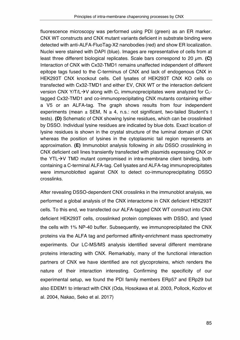
Principles of intra-membrane chaperoning processes by CNX
85
fluorescence microscopy was performed using PDI (green) as an ER marker. CNX WT constructs and CNX mutant variants deficient in substrate binding were detected with anti-ALFA-FluoTag-X2 nanobodies (red) and show ER localization. Nuclei were stained with DAPI (blue). Images are representative of cells from at least three different biological replicates. Scale bars correspond to 20 µm. (C) Interaction of CNX with Cx32-TMD1 remains unaffected independent of different epitope tags fused to the C-terminus of CNX and lack of endogenous CNX in HEK293T CNX knockout cells. Cell lysates of HEK293T CNX KO cells co transfected with Cx32-TMD1 and either EV, CNX WT or the interaction deficient version CNX Y/T/LàV along with CL immunoprecipitates were analyzed for CL-tagged Cx32-TMD1 and co-immunoprecipitating CNX mutants containing either a V5 or an ALFA-tag. The graph shows results from four independent experiments (mean ± SEM, N ≥ 4, n.s.: not significant, two-tailed Student’s t tests). (D) Schematic of CNX showing lysine residues, which can be crosslinked by DSSO. Individual lysine residues are indicated by blue dots. Exact location of lysine residues is shown in the crystal structure of the luminal domain of CNX whereas the position of lysines in the cytoplasmic tail region represents an approximation. (E) Immunoblot analysis following in situ DSSO crosslinking in CNX deficient cell lines transiently transfected with plasmids expressing CNX or the YTLàV TMD mutant compromised in intra-membrane client binding, both containing a C-terminal ALFA-tag. Cell lysates and ALFA-tag immunoprecipitates were immunoblotted against CNX to detect co-immunoprecipitating DSSO crosslinks.
After revealing DSSO-dependent CNX crosslinks in the immunoblot analysis, we
performed a global analysis of the CNX interactome in CNX deficient HEK293T
cells. To this end, we transfected our ALFA-tagged CNX WT construct into CNX
deficient HEK293T cells, crosslinked protein complexes with DSSO, and lysed
the cells with 1% NP-40 buffer. Subsequently, we immunoprecipitated the CNX
proteins via the ALFA tag and performed affinity-enrichment mass spectrometry
experiments. Our LC-MS/MS analysis identified several different membrane
proteins interacting with CNX. Remarkably, many of the functional interaction
partners of CNX we have identified are not glycoproteins, which renders the
nature of their interaction interesting. Confirming the specificity of our
experimental setup, we found the PDI family members ERp57 and ERp29 but
also EDEM1 to interact with CNX (Oda, Hosokawa et al. 2003, Pollock, Kozlov et
al. 2004, Nakao, Seko et al. 2017)

Principles of intra-membrane chaperoning processes by CNX
86
Figure 20 Global analysis of functional interaction partners of CNX. Volcano plots derived from LC-MS/MS analysis of ALFA-tagged CNX, immunoprecipitated in 1% NP-40 buffer from transfected CNX deficient HEK293T cells after DSSO crosslinking and compared to EV control co-IPs. Depicted is an enlarged image section as well as the associated complete volcano plot in the upper right part. Significantly enriched proteins and corresponding -log10 p-values were plotted. The respective proteins are denoted with their Uniprot gene names. CNX is shown in green and membrane proteins with the GO-term annotation “integral to membrane” are highlighted in red. ER proteins with the GO-term annotation “endoplasmic reticulum”, regardless of whether they are localized to the ER membrane or lumen are highlighted in blue. However, only known CNX interaction partners are indicated with their names in the enlarged image section. Cut-off values (solid lines) in the volcano plot were defined as log2 = 1 (2-fold enrichment) and -log10 = (p-value) of 1.3 (Data obtained from Nina Bach, Siebers Lab).
Principles of intra-membrane chaperoning processes by CNX
87
4 Discussion This study describes a novel mechanism of intra-membrane substrate
recognition by molecular chaperones as a basis for quality control of IMPs in the
ER. By structural, mechanistic and systematic analyses we define features within
the TMD of CNX and the TMD of its substrates that allow CNX to bind its clients
in the lipid bilayer and thereby protect them from degradation. Importantly,
efficient intra-membrane substrate recognition by CNX requires the presence of
a certain recognition motif, which appears to be conserved in other intra-
membrane chaperons involved in intra-membrane quality control.
Remarkably, several intra-membrane chaperones are known to interact with
their substrate via direct helix-helix interaction, including the PAT complex or
EMC. In the case of the EMC, it could be shown that this complex has specificity
for multi-pass membrane proteins with polar and slightly hydrophobic TM
domains or those with destabilizing features such as charged or aromatic
residues. To date, however, the detailed mechanism of substrate recognition for
both intra-membrane chaperones is poorly understood (Meacock, Lecomte et al.
2002, Shurtleff, Itzhak et al. 2018, Chitwood and Hegde 2020, Miller-Vedam,
Brauning et al. 2020). Hence, the results of this study could help to elucidate how
intra-membrane chaperones recognize transmembrane defects inside the lipid
bilayer.
In addition, in this study, we show for the first time a global analysis of the
substrate repertoire of CNX.
4.1 Principles of intra-membrane chaperoning processes by CNX
Historically, the function of CNX in recognizing and processing misfolded
substrate proteins has mostly been limited to its lectin function, which resides in
the globular N-domain (Hammond, Braakman et al. 1994, Hebert, Foellmer et al.
1995). In addition to the canonical function of CNX, however, there is increasing
evidence that its transmembrane segment participates in intra-membrane quality
control and chaperoning processes, as direct interaction with various membrane
proteins such as MHC-I molecules, PLP, the tetraspanin CD82, or disease-
related mutants of PMP22 has been shown, directly affecting their degradation,
assembly or targeting to multilamellar bodies (Margolese, Waneck et al. 1993,

Principles of intra-membrane chaperoning processes by CNX
88
Cannon and Cresswell 2001, Swanton, High et al. 2003, Korkhov, Milan-Lobo et
al. 2008, Yamasaki, Hara et al. 2014). It is important that these intra-membrane
recognition processes are not limited to glycoproteins but rather take place in a
lectin-independent manner. Nevertheless, mechanistic insights into the
interactions of CNX's TMD with its clients were lacking. This seems particularly
important considering that incorrect folding and assembly of IMPs and
destabilizing mutations within TMDs are linked to various human diseases. In
addition, most studies of canonical degradation and quality control pathways so
far have focused on proteins where the misfolded lesion is either in the ER lumen
or in the cytosol, but not on factors that interact with unstable TM helices.
Using Cx32 as a model protein, we were able to show in this study that CNX
differentiates between the correctly folded and misfolded state of clients,
indicating that the folding and assembly status of transmembrane segments in
the membrane is the center of the recognition mechanism. Furthermore,
specifically stronger binding to misfolded, CMT1X disease-causing mutants of
Cx32 than WT reveals that CNX functions as a true molecular chaperone for non-
glycosylated membrane proteins like Cx32. Interestingly, when looking at the
individual transmembrane segments in isolation, CNX preferentially binds the first
TMD of Cx32. In full-length Cx32, however, we observe the strongest binding of
CNX to mutants that carry mutations in the third TM segment. This might indicate
that these mutants, although properly integrated into the lipid bilayer, lead to
structural changes or exposure of a buried recognition motif in TM1, which,
consequently, facilitates binding of CNX. To test this hypothesis, it would be
useful to study the direct interaction between CNX and individual TM mutants.
Unfortunately, however, we were not able to study directly the TM mutants
individually, as they were unstable in the membrane. Importantly, Cx32 is not a
glycoprotein; thus, recognition of the chaperone must be lectin-independent.
Indeed, utilizing a minimal CNX construct that only contains the CNX TMD we
could show, that the transmembrane domain of CNX is necessary and sufficient
for binding clients in the membrane independently of the ER-luminal lectin
domain or C-terminal tail. Accordingly, this indicates that the CNX TMD contains
the relevant features essential to detect defective transmembrane domains. As a
Intra-membrane client recognition motifs for CNX
89
result, CNX could play an essential role in the ER-mediated quality control for
many membrane proteins that are neither glycosylated nor have luminal degrons
4.2 Intra-membrane client recognition motifs for CNX Although several intra-membrane chaperones have been identified over the
last few decades, and CNX has also been assigned such a function, the detailed
mechanism of substrate recognition by such factors inside the membrane plane
is poorly understood. In contrast, for example, it has long been known that
particularly exposed hydrophobic residues in the ER lumen or cytosol represent
recognition motifs for luminal ER chaperones or ERAD factors. Furthermore,
particularly relevant, most of the studies carried out analyzing intra-membrane
chaperoning processes used naturally evolved membrane proteins as a model.
However, this excludes an unbiased study on the definition of intra-membrane
recognition motifs since TM proteins may engage multiple topologically and
functionally distinct layers of quality control. Using a more simplified model, a
single pass ER membrane protein reporter system termed CoMem, we were able
to show for the first time in a systematic study under clear and defined conditions
that the CNX TMD can differently recognize non-glycosylated clients with well-
defined lesions in the membrane. Interestingly, our study reveals a highly distinct
binding pattern for CNX to different TM substrates. Although we see significant
changes in the binding pattern for most of the different amino acids that have
been replaced against the central Val residue in the CoMem TM domain, there is
no clear consistent pattern of stronger or weaker interaction within each amino
acid group (non-polar, polar, positively and negatively charged). Nonetheless,
except for Phe, Pro, and Trp, non-polar amino acids seem to have no pronounced
effect on CNX binding. Interestingly enough, Phe and Trp have an aromatic
system, which could explain the difference and Pro, which will be discussed
below, has the property of destabilizing helical structures. At the same time, most
of the polar and charged residues introduced into the central position of the
CoMem TMD seem to reduce the binding of CNX. This is surprising since one
might assume that exposed charged or unpaired polar residues, which on the
one hand fulfill essential structural and functional roles but on the other can be
potentially dangerous to the cell, are specific mediators of interaction with CNX
or other intra-membrane chaperones. However, the most substantial interaction

Intra-membrane client recognition motifs for CNX
90
is observed when the CoMem TMD is equipped with an Arg residue at the central
position, which in comparison to most other amino acids is likely charged in
membranes due to resonance stabilization. In contrast, we observed the weakest
interaction if Pro is in this position. Remarkably, in addition to our in vivo data, MD simulations revealed an altered
binding behavior between the CoMem TMD mutant with a centrally located Arg
residue and CNX. The simulation data shows that this mutant has a different
orientation and, compared to CoMem WT, binds to CNX with the back of its helix.
Although this finding does not directly explain the stronger interaction between
CoMem-Arg and CNX observed in our experiments, it is still interesting.
Additionally, MD simulation revealed that CoMem-Arg resides in a more tilted
position within the membrane compared to CoMem WT and CNX. This is
presumably due to the interaction between the positively charged side chain in
the center of the CoMem-Arg TMD and the negatively charged head groups of
the phospholipid bilayer, which exerts a pulling effect on the helix. In general,
tilting of TMDs within the membrane might contribute to allow for a large
interaction interface between two adjacent helices. In the light of these findings,
it seems conceivable that the simulated tilting effects develop differently when
the charge is shifted from a central position to the distal ends of the TM helix,
whereby the distance of both charges to each other is varied. This is particularly
interesting because only the center of the membrane is completely hydrophobic
and the rest contains a mixture of polar lipid head groups and water molecules.
Hence, this could explain the positional dependence of CNX binding to the
different CoMem-Arg TMD variants observed in our experiments. Unfortunately,
we did not include the CoMem-Lys variant in our MD simulations, but it would be
interesting to see whether this mutant shows a different behavior despite a similar
positive charge in the side chain in the central position, which would contribute to
explaining the difference to the CoMem-Arg mutant. In this context, it appears
additionally interesting that most of the mutations that cause disease result from
the introduction of Arg, but not Lys, residues into TMDs (Fink, Sal-Man et al.
2012).
Similar to the CoMem-Arg variant, we recapitulated our in vivo data for the
CoMem-Pro mutant by MD simulations. Interestingly, MD simulations shows that
this mutant is less successful in developing stable interactions with CNX, which

A conserved recognition motif in the CNX TMD is required for intra-membrane client recognition
91
might explain why we observe less interaction between CoMem-Pro and CNX in
our experiments. It is noteworthy that Pro acts as a helix breaker and introduces
kinks in TM helices, as is the case in CNX´s TM helix. Despite its propensity to
break the helix, this Pro residue, however, is conserved in the CNX family, which
is why it is believed to play an important functional role (Lakkaraju, Abrami et al.
2012). At first, we hypothesized that introducing a Pro residue into the helix of
our reporter system could compensate for the Pro-induced kink in the CNX TM
domain, thus leading to a stronger interaction but the opposite seems to be the
case, as our in vivo and MD simulation data shows. Nonetheless, the position of
the Pro residue and thus the kink introduced into the helix seems to have a
decisive influence on the interaction with CNX because the more central the
position within the membrane, the less interaction with CNX.
Although our simplified reporter system provides ideal conditions for the
systematic definition of intra-membrane recognition motifs for CNX, it is not well
suited to determine the biological consequences of binding. The CoMem reporter
system is an artificially designed protein that lacks important secondary binding
motifs which normally would enable interaction with chaperones or ERAD factors
for example. Consequently, such factors would be excluded when determining
downstream processes. To this end, more complex and naturally evolved model
proteins such as Rho or Cx32 are required. However, although Cx32 has been
shown to interact with CNX, Rho is more attractive as a substrate because of
CNX's involvement in its quality control (Rosenbaum, Hardie et al. 2006). Thus,
Rho was selected to address the biological consequences of CNX binding.
4.3 A conserved recognition motif in the CNX TMD is required for intra-membrane client recognition After we have demonstrated direct helix-helix interactions between either the
CNX TMD and the CoMem TMD segment or with the first TMD segment of Cx32
(Cx32-TMD1) in vivo, we used these interacting systems to define structural
motifs for intra-membrane Calnexin:client recognition by MD simulations.
Strikingly, computational modeling revealed that similar residues which involve a
Tyr, a Thr, and a Leu in the CNX TMD region are required for interaction of CNX
with both substrates. Furthermore, the identified residues reside on the same side
of the CNX TM helix, suggesting that these residues represent a molecular

A conserved recognition motif in the CNX TMD is required for intra-membrane client recognition
92
recognition motif that is xYxxTxxL and enables the CNX TMD to interact with the
substrate helix inside the lipid bilayer. In fact, mutating these residues to
presumably inert Val or Ala residues significantly reduced the interaction between
CNX and different substrates tested in our study, including CoMem, Rho, Cx32,
and Cx32-TMD1, which showed the most drastic effect. Interestingly, the
recognition process seems to be position-dependent since shifting the motif to a
more N-terminal position results in a reduced interaction of CNX with its
substrate.
Based on the observation that the identified residues are sufficient to interact
with many different substrates, we wondered whether the xYxxTxxL interaction
motif is only found in CNX or exists in other membrane-localized factors which
mediate intra-membrane substrate recognition. It is worth mentioning that
although Leu seems to play a role in the CNX-mediated substrate recognition
process, as the deletion of all three residues together causes a stronger
phenotype than the deletion of Tyr and Thr alone, this residue does not seem to
fit the picture. This is because, within TM domains, structural or functional tasks
are mainly mediated by charged or polar amino acids, including Tyr or Thr, but
usually not by non-polar amino acids such as Leu, rendering its effect interesting.
In contrast, aromatic residues such as Tyr are crucial for many molecular
recognition and assembly processes due to their unique structure and thus highly
conserved in TM segments (Ramachandran, Tweten et al. 2004, Fink, Sal-Man
et al. 2012). Furthermore, previous studies have shown that dimerization of TM
segments is significantly increased by cation - π interactions, which could explain
the comparatively strong interaction between CNX and CoMem-Arg (Johnson,
Hecht et al. 2007). Importantly although such a scenario would at first glance
contradict our simulation data, in which we observe a changed topology for the
CoMem-Arg mutant, one has to consider that it is impossible to find every
possible complex between CNX and CoMem-Arg by simulation. For this reason,
we focused our comparison on a truncated xYxxTx recognition motif, in which x
represents non-polar / hydrophobic residues that are important for the residues
Tyr and Thr to be on the same helical face.
Surprisingly, a brief preliminary bioinformatic analysis revealed that different
membrane proteins contain the recognition motif on different positions in their TM
A conserved recognition motif in the CNX TMD is required for intra-membrane client recognition
93
domains, including Asi2, the P5A ATPase and partly EMC4. Of note, although
the recognition motif in EMC4 contains a Asn residue instead of an Thr residue,
this amino acid is also polar and, moreover, faces the same helical side as Tyr.
Protein xYxxTx recognition motif
CNX VYILTV
EMC4 VYLIGNL
Asi2 VYMVTA
P5A ATPase VYLGTL
Important for our study, all of the proteins mentioned above are involved in the
quality control of membrane proteins and bind substrates via their TM domain. At
least for EMC and the P5A ATPase, substrates and TM features such as
hydrophobicity are known that enable recognition, but, similar to CNX, features
in the TM segment of these proteins, which are required for client binding to
occur, are unknown.
The membrane-spanning Asi complex, which consists of Asi1, Asi2, and Asi2,
is the third most important ER-ubiquitin-ligase complex in yeast alongside Hrd1
and Doa10. Interestingly, while Asi1 and Asi3 contain a RING domain, Asi2 does
not have a functional domain but has been shown to recognize misfolded and
mistargeted membrane proteins at the inner nuclear membrane by direct binding
the substrate TMD. Subsequent substrate ubiquitination and Cdc48-dependent
membrane extraction facilitate proteasomal degradation (Natarajan, Foresti et al.
2020). Interestingly, EMC4 composed of three TMDs, is the subunit of the EMC
complex that mediates the insertion of membrane proteins with moderately
hydrophobic transmembrane domains into the ER membrane and is therefore
crucial for the function of the entire complex (Shurtleff, Itzhak et al. 2018).
Although the P5A ATPase preferentially interacts with moderately hydrophobic
transmembrane domains, similar to the EMC, its dislocase activity mediates the
opposite function. After binding directly to the TM segment of a misdirected or
misoriented membrane protein, these are removed from the membrane in an ATP
dependent manner (McKenna, Sim et al. 2020). However, to definitively confirm
the existence of the xYxxTx motif among various intra-membrane chaperons and
The protective function of CNX on membrane proteins
94
quality control factors in the end, a broader bioinformatics approach and further
experimental testing is required.
4.4 The protective function of CNX on membrane proteins To address the biological functions of intra-membrane client recognition by
CNX, we used Rhodopsin as a model substrate. Our data show that if intra-
membrane client recognition by CNX is disturbed, as is the case in the YTLàV
mutant, the binding of Rho to the chaperone is significantly reduced. Strikingly,
caused by this reduced binding, Rho degradation is significantly accelerated,
which argues for a protective role of CNX in intra-membrane chaperoning
processes. The protective function of CNX on Rho becomes more evident if CNX
is completely absent, causing an even stronger effect on Rho degradation and
suggests that CNX mediated intra-membrane and lectin-based client binding
works synergistically. Notably, our observation that CNX is essential for Rho
biogenesis since Rho expression levels in CNX k/o cells are hardly detectable
contradicts previous studies (Rosenbaum, Hardie et al. 2006, Kosmaoglou and
Cheetham 2008). Thus, possible causes for these differences will be discussed
in the following.
Rhodopsin is a member of the GPCR-family and is found in the rod
photoreceptor cells of the retina. During photoactivation by the absorption of light,
Rho converts the energy provided by the photons into a chemical reaction which
ultimately leads to an electrical signal that stimulates signal transduction
processes in the nervous system (Hubbard and Kropf 1958). In addition, during
photoactivation, Rho is subject to structural changes that cause the
rearrangement of specific helices and thus creates a small binding pocket for a
heterotrimeric G-protein on the cytoplasmic side of the receptor, which initiates
the phototransduction cascade (Zhou, Melcher et al. 2012). Importantly, Rho is a
glycoprotein and undergoes N-linked glycosylation at two asparagine residues
near the N-terminus, which are crucial for the correct folding of the receptor.
Mutations in these residues and 120 other known point mutations are
responsible, among others, for autosomal dominant retinitis pigmentosa (ADRP)
and night blindness (Sung, Davenport et al. 1991, al-Maghtheh, Gregory et al.
1993, Kawamura, Colozo et al. 2012). While point mutations affecting the C-
The protective function of CNX on membrane proteins
95
terminus still fold normally, however, cause mistargeting, mutations in the
transmembrane domains lead to protein misfolding and ER retention.
Interestingly, although Rho has been identified as a direct CNX substrate in
different studies, the role of CNX in the quality control of mutant, misfolded Rho
seems to be very different among different organisms. While studies in
Drosophila examining loss of function mutations in CNX suggest that Rho
biogenesis essentially depends on CNX, the chaperone is dispensable for Rho
to mature in mammalian cells (Rosenbaum, Hardie et al. 2006, Kosmaoglou and
Cheetham 2008). It is important to note that Drosophila rhodopsin differs
significantly from that of vertebrates. For example, maturation essentially requires
N-glycosylation of one of the two glycosylation sites, but in its mature form, Rho
is non-glycosylated (Murray, Fliesler et al. 2009). This contrasts with mammalian
Rho, which does not require glycosylation for correct folding (Kaushal, Ridge et
al. 1994, Saliba, Munro et al. 2002). Furthermore, Drosophila CNX only shares
49 % sequence identity with human CNX and differs significantly in its TMD
regions, which all together can explain the observed differences. However, since
we also used a mammalian system in our study, the reason for the different
observations regarding the importance of CNX for the biogenesis of Rho must be
found elsewhere. Indeed, the difference can be explained by the findings of our
study. Results around the group of Kosmaoglou et al. are based on the use of a
functionally deficient CNX cell line in which a conserved Asp required for calcium
ion coordination, two Cys residues involved in a surface-exposed disulfide bond
required for substrate binding and residues that form the glucose binding pocket
are deleted (Denzel, Molinari et al. 2002, Kosmaoglou and Cheetham 2008).
Notably, the truncated CNX version still contains the N-terminal ER-targeting
sequence, the C-terminal ER retention motif, and, more importantly for our
studies, the TMD region. Given that we observe synergistic client binding
activities of CNX with both its lectin domain and TM region, it seems conceivable
that CNX´s TM domain can compensate for a dysfunctional lectin domain. Hence
no effect on Rho biogenesis is observed. Moreover, the exact mechanism of
CNX´s chaperoning function is not known. It is possible that during the
biosynthesis of client proteins, the CNX TMD functions as a placeholder,
shielding problematic residues in order to support the development of the
Global analysis of the CNX substrate repertoire
96
correctly folded state of the substrate. Consequently, in a model like this, the
biological function of CNX is determined even stronger by its TM domain. Future
studies using our minCNX construct, which lacks CNX´s entire globular domain
and only contains the TM domain, could address this question. In this context, it
seems interesting that in Drosophila, the intra-membrane chaperone and
insertase EMC not only interacts with CNX but is also essential for the biogenesis
of Rho (Satoh, Ohba et al. 2015).
Another finding that supports the role of CNX as an intra-membrane chaperone
is the observation that the association of CNX with Rho is highly dependent on
its conformation since CNX preferentially associates with misfolded Rho
compared to the correctly folded WT (Noorwez, Sama et al. 2009). Remarkably,
the strongest association can be observed with mutants containing mutations in
TM regions causing severe misfolding of the entire protein instead of mutations
occurring in the N-terminal region. A similar effect of CNX binding was observed
in studies on Cx32, however, the difference between the correctly folded WT and
a Cx32 TM mutant causing an altered topology of the protein was not as severe
(Coelho, Stahl et al. 2019). Importantly, it has to be considered that in these
scenario´s stronger binding of CNX to mutant Cx32 or Rho is not necessarily
caused via stronger binding to the mutated TMD helix but can also be the result
of general exposure of TMD helix binding sites and the involvement of additional
factors which target the misfolded mutants. Nevertheless, since we also observed
a direct and stronger CNX binding to Cx32 mutants that are still integrated into
the membrane correctly, one would expect faster degradation for such misfolded
TM mutants than the WT in CNX k/o cells co-transfected with our binding deficient
CNX mutant.
4.5 Global analysis of the CNX substrate repertoire Using DSSO-crosslinking coupled to mass spectrometry, we show a global
analysis of the CNX substrate repertoire for the first time. Among the interacting
ER-localized substrates, we find the PDI family members ERp57 and ERp29
besides EDEM1, which functions in the degradation of misfolded ER proteins.
Importantly, for all of these proteins, the interaction with CNX has been well
documented, which validates the results of our analysis (Oda, Hosokawa et al.
Final conclusion
97
2003, Pollock, Kozlov et al. 2004, Nakao, Seko et al. 2017). In addition, we were
able to identify a large number of membrane proteins to interact with CNX.
Remarkably, around half of the identified membrane proteins (51,31%) that
interact with CNX in our mass spectrometry analysis are not glycoproteins,
according to UniProt. This finding indicates that CNX recognizes a variety of
membrane protein clients regardless of their glycosylation state, presumably
through direct interaction with their transmembrane domains. Thus, our global
analysis of CNX´s substrate repertoire supports the various other reports that
describe a lectin-independent function of CNX (Cannon and Cresswell, 2001,
Coelho et al., 2019, Swanton et al., 2003).
Importantly, however, although we can observe a direct interaction between
the TM helix of CNX and a client (CNX:client) in our study, we cannot rule out
whether other co-factors are involved in the client recognition and interaction
process of CNX (CNX:X:client). Therefore, future studies should reveal if client
binding is exclusively mediated by the TM domain of CNX. In addition, it would
be interesting to reveal whether CNX´s protective function that we were able to
observe for Rhodopsin also applies to other interaction partners we have
identified in our MS analysis, as this would foster the role of CNX as an intra-
membrane chaperone for non-glycosylated membrane proteins.
4.6 Final conclusion
In this study, we reveal principles of intra-membrane substrate recognition by the
molecular chaperone CNX that contradict the previous assumption that client
binding of CNX occurs exclusively outside the membrane. We show that CNX
directly recognizes misfolded client proteins via its single TM domain and reveal
that this intra-membrane client recognition process depends on a unique motif
present in the CNX TM domain. Remarkably, the functionality of this motif is not
only a prerequisite for efficient client binding, but also protects clients from being
degraded.
Based on the structural and biochemical data presented in this study, we
propose the following model for the various chaperone activities of CNX (Figure
21).

Final conclusion
98
Figure 21: The combined function of CNX´s lectin and TM domain in the recognition and quality control of misfolded membrane proteins. According to its canonical function in the biogenesis of membrane proteins, CNX binds immature and incompletely folded glycoproteins via a glucose binding site located in its globular domain. In addition, however, CNX functions as an intra-membrane chaperone for non-glycosylated membrane proteins, especially those containing
Final conclusion
99
a misfolded or defective TMD. The recognition of intra-membrane defects essentially depends on the functionality of an interaction motif located in CNX´s TMD and protects clients from degradation. In contrast, a dysfunctional TMD interaction motif with reduced ability for substrate binding within the membrane causes increased instability of the client.
Material and methods
100
5 Materials and methods Unless specified otherwise, chemicals and reagents were obtained from Applied
Biosystems, Bio-Rad, GE Healthcare, Life technologies, Merck, Millipore, New
England Biolabs, Promega, Roche, Serva, Sigma and Thermo Scientific. Sterile
flasks and sterile and de-ionized water as well as sterile solutions were used in
all protocols and experiments described.
5.1 Molecular biology techniques
5.1.1 DNA design and modification
DNA constructs
CoMem WT constructs, minCNX and CNX constructs were obtained from
GeneArt Gene Synthesis (ThermoFisher) in a pcDNA3.4 TOPO expression
vector optimized for mammalian expression. An illustration of the amino acid
sequence and construct details of WT CoMem and minCNX constructs and their
respective controls that lack the TMD domain can be found below (Figure 22 A-
D) The TM sequence of CoMem WT was designed by multiple sequence
alignment of 200 human single pass plasma membrane proteins (152 type I and
48 type II orientation) using Unipro U Gene software whose TM sequences were
obtained from Membranome 2.0 Database. Human CNX sequence was obtained
from UniProt and complemented with a C-terminal epitope tag. For construction
of the minCNX construct, human CNX TM sequence was obtained from UniProt,
its TM helix reconciled according (Hessa, Meindl-Beinker et al. 2007) to avoid
artificially shortening of the TM sequence and finally complemented by
endogenous amino acids flanking the TM region N and C-terminally. Cx32 cDNA
was obtained from Origene and cloned into a pSVL vector (Amersham). CL-Cx32-
TM1 reporter constructs were synthesized by GeneArt Gene Synthesis (Thermo
Fisher) and cloned into a pSVL vector. Monomeric Sec61 mCherry was provided
from Prof. Dr. Linda Hendershot (St. Jude Children Research Hospital, TN, USA).
Individual construct components of the designed proteins and N as well as C-
terminal epitope tags are separated by (GGGS)2, (GSGS)2 or (GGGA)2 linker.
Cloning into mammalian pSVL expression vector was performed using suitable

Material and methods
101
restriction enzymes and T4 DNA ligase (Promega). Introduction of epitope tags
and point mutations was carried out via site-directed mutagenesis PCR using
overlapping, complementary mutagenesis primers. Pfu polymerase (Promega)
reaction and subsequent DpnI (NEB) digest of the template plasmids were
utilized to generate mutated vectors, which were transformed into competent E.
coli cells (DH5⍺, Thermo Fisher) cells for amplification. All constructs were
verified by sequencing prior to use (Eurofins Genomics).
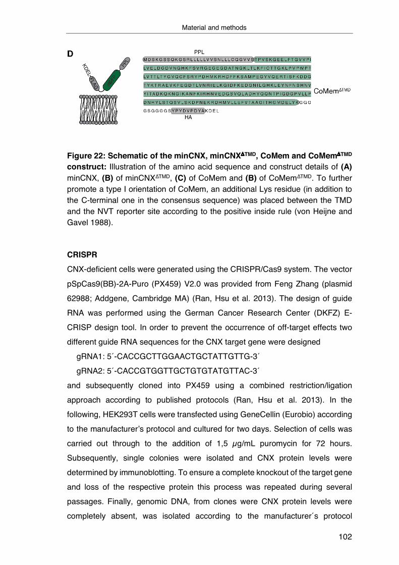
Material and methods
102
Figure 22: Schematic of the minCNX, minCNXDTMD, CoMem and CoMemDTMD construct: Illustration of the amino acid sequence and construct details of (A) minCNX, (B) of minCNX∆TMD, (C) of CoMem and (B) of CoMem∆TMD. To further promote a type I orientation of CoMem, an additional Lys residue (in addition to the C-terminal one in the consensus sequence) was placed between the TMD and the NVT reporter site according to the positive inside rule (von Heijne and Gavel 1988).
CRISPR
CNX-deficient cells were generated using the CRISPR/Cas9 system. The vector
pSpCas9(BB)-2A-Puro (PX459) V2.0 was provided from Feng Zhang (plasmid
62988; Addgene, Cambridge MA) (Ran, Hsu et al. 2013). The design of guide
RNA was performed using the German Cancer Research Center (DKFZ) E-
CRISP design tool. In order to prevent the occurrence of off-target effects two
different guide RNA sequences for the CNX target gene were designed
gRNA1: 5´-CACCGCTTGGAACTGCTATTGTTG-3´
gRNA2: 5´-CACCGTGGTTGCTGTGTATGTTAC-3´
and subsequently cloned into PX459 using a combined restriction/ligation
approach according to published protocols (Ran, Hsu et al. 2013). In the
following, HEK293T cells were transfected using GeneCellin (Eurobio) according
to the manufacturer’s protocol and cultured for two days. Selection of cells was
carried out through to the addition of 1,5 µg/mL puromycin for 72 hours.
Subsequently, single colonies were isolated and CNX protein levels were
determined by immunoblotting. To ensure a complete knockout of the target gene
and loss of the respective protein this process was repeated during several
passages. Finally, genomic DNA, from clones were CNX protein levels were
completely absent, was isolated according to the manufacturer´s protocol
Material and methods
103
(PureLink Genomic DNA Kits, Invitrogen). Subsequently, the genomic
CRISPR/Cas9 target area was amplified by the usage of specific primers and the
obtained amplicons were sequenced and the successful CNX knockout verified
by NGS CRISPR amplicon sequencing (CCIB DNA Core, Massachusetts
General Hospital, Boston, MA).
5.1.2 Cell culture techniques
Cell culture and transient transfections HEK293T (ECACC) and COS-7 (ECACC) cells were cultivated in Dulbecco's
Modified Eagle's Medium ((DMEM), high glucose (Sigma-Aldrich), supplemented
with 10 % (v/v) fetal bovine serum (Biochrom) and 1% (v/v) antibiotic-antimycotic
solution (25 µg/ml amphotericin B, 10 mg/ml streptomycin, 10,000 units of
penicillin (Sigma-Aldrich)) at 37 °C in a 95% humidified atmosphere containing
5% CO2. Transient DNA transfections were performed by chemical transfection
(HEK293T) or via electroporation (COS-7). 24 h prior to transfection, 250.000
HEK293T cells were seeded per p35 plate (pre-coated Corning BioCoat Poly-D-
Lysine 35 mm #354467 or uncoated Nunclon multidish 6 well plates, Thermo
Scientific #140685 coated with 50 µg/ml Poly-D-Lysine solution (Gibco A38904-
01) per well according to manufacturer’s instructions or 275.000 per p60 plate
(Tissue Culture Dish 60, TPP). HEK293T cells were transfected using GeneCellin
(Eurobio) according to the manufacturer’s protocol. For transient transfections
using pcDNA (highly expressing vectors) the amount of DNA was reduced to half
the amount suggest by the manufacturer. When performing co-transfections, the
amount of transfected DNA was divided equally or splitted in a 3:1 ratio (vector
plasmid with weaker promotor:vector plasmid with stronger promotor) in case of
different promotor strength.
For passaging, cells were washed with PBS (Sigma-Aldrich) and subsequently
incubated with trypsin solution (Sigma-Aldrich) for 5 min at 37°C before transfer
in fresh, pre-warmed medium.
Material and methods
104
Cell lysis Cells were harvested 24 h after transfection. All cell lysis steps were performed
on ice or at 4 °C using ice-cold solutions unless otherwise indicated. Cells were
washed twice with PBS and then lysed for 20 min by adding 1 ml (p60) or 500 µl
(p35) lysis buffer (50 mM Tris-HCl pH 7.5, 150 mM NaCl, 0.5 % (w/v) NaDOC
complemented with either 1% (w/v) Digitonin (Sigma) for samples analyzing
membrane-embedded interactions with CoMem and Cx32 constructs or 1% (v/v)
NP-40 (Sigma) for samples analyzing membrane-embedded interactions with
CL-Cx32-TM1 and mScarlet-I-CNX as well as CNX constructs) when conducting
co-immunoprecipitation experiments or NP-40 buffer for other experiments. All
buffers were supplemented with cOmplete protease inhibitor cocktail (Roche)
prior to lysis. The resulting lysate was centrifuged for 15 min at 15,000 x g and
the supernatant was complemented with 5x Laemmli buffer containing 10 % (v/v)
β-mercaptoethanol (reducing). Finally, samples dedicated to the analysis for
membrane proteins were incubated for 30 min at 50°C, except samples involving
Cx32 proteins were incubation was performed for 30 at 37°C. In contrast, sample
lysates for the analysis of soluble proteins were boiled for 10 min at 95°C.
5.1.3 Cell culture based In vivo assays
Cycloheximide chase analysis Cells were prompted to express the indicated constructs overnight. Inhibition of
protein biosynthesis and determination of protein half-live was performed by the
replacement of complete medium with medium containing 50 µg/ml
Cycloheximide (Sigma-Aldrich) 24 h after transfection. Subsequently lysates
were collected at different time points as described below. Negative controls were
prepared in the same manner without the addition of CHX (t = 0 sample).
Induction of ER stress
The induction of UPR was carried out by the addition of 5 µg/ml tunicamycin
(Sigma-Aldrich) in medium and incubation for 6h prior cell lysis or the
supplementation of pre-warmed medium with 10 mM DTT (Sigma-Aldrich) for one
hour. Cleavage analysis of the activating transcription factor 6 (ATF6) based on
Material and methods
105
immunoblotting was performed following immunoprecipitation to increase the
amount of detectable protein. Determination of endogenous phosphorylation
events by immunoblotting (total amount and phosphorylation at Ser51 only) of
the eukaryotic initiation factor 2 (eIF2) was performed following cell lysis using
lysis buffer additionally supplemented with 1 x phosphatase inhibitor mix (Serva).
To assess XBP1 splicing, RNA was extracted using RNeazy Mini Kit (Qiagen) in
RNase free environment. Subsequently RT-PCR of the purified RNA was
performed using OligodT20 Primer (18418020,Thermo Fischer) and SuperScript
III Reverse Transcriptase (18080044, Thermo Fischer). Following amplification
of the XBP1 transcript of the resulting cDNA, XBP1 splicing events were analyzed
on 2% agarose gels. Positive controls were included.
5.1.4 Fluorescence Microscopy
Immunofluorescence
Determination of the intracellular localization of target proteins was carried out by
immunofluorescence microscopy using either electroporated or transiently
transfected COS-7 cells.
Seeding and transfection
Electroporation of COS-7 cells was carried out using a X2 Gemini electroporator
(BTX) according to manufacturer´s instructions and protocol. For transient
transfection of COS-7 cells, 36 µl DMEM containing 3.6 µg DNA were mixed with
1.2 µl TorpedoDNA transfection reagent (ibidi) and incubated for 15 min at RT.
Subsequently, 200 µl of COS-7 cell suspension were added and mixed gently. In
both cases, following electroporation or transient transfection, 30 µl of the
resulting suspension of 4 x 10⁵ cells/ml were applied per inlet of a pre-warmed µ-
Slide IV 0.4 (ibidi) and the µ-Slide was incubated for 3 h at 37 °C, 5% CO2.
According to manufacturer´s instructions, medium was replaced 3 h after
seeding. Therefore, 60 µl complete DMEM were added per reservoir, and the µ-
Slides were incubated for additional 48 h at 37 °C, 5% CO2.

Material and methods
106
Staining For fixation, all liquid was removed from all reservoirs and channels and 60 µl
glyoxal fixation solution (20% EtOH, 7.825% glyoxal, 0.75% acetic acid) were
added to each channel (Richter, Revelo et al. 2018). Samples were then
incubated for 30 min on ice and thereafter further 30 min at RT. The reaction was
quenched by aspirating the fixation solution and adding 60 µl of 100 mM NH4Cl
(Sigma-Aldrich) and subsequent incubation at RT for 20 min. Samples were then
washed twice for 5 min with 100 µl 4 °C PBS. Permeabilization of cells and
simultaneous blocking of epitopes was conducted by the addition of 60 µl of
blocking solution (2.5% BSA (Sigma-Aldrich), 0.1% Triton X-100 in PBS) to the
samples which were then incubated for 5 min at RT following three further
washing steps at RT. As an ER-marker, 30 µl of anti-PDI antibody conjugated to
Alexa Fluor 488 at 1:50 dilution in blocking solution were added and incubated
for 1 h at RT in the dark. The antibody solution was washed out using 100 µl PBS
and the samples were thereafter washed three times. All liquid was removed and
25 µl of DAPI solution (Sigma-Aldrich) (0.01% in PBS) were added to stain Nuclei
and incubated for 2 min. The samples were then washed three times with PBS,
the liquid was aspirated, and mounting medium (ibidi) was added to cover the
inlets of the slides.
Microscopy Imaging was performed on a DMi8 CS Bino inverted widefield fluorescence
microscope (Leica) using a 100 x (NA = 1.4) or 63 x (NA = 1.4) oil immersion
objective. The employed dichroic filters were chosen to image Alexa 488 and
sfGFP (GFP channel; excitation/bandpass: 470/40 nm; emission/bandpass:
525/50 nm), mScarlet-I and Sec61mCherry (TXR channel; excitation/bandpass:
560/40 nm; emission/bandpass: 630/75 nm), or DAPI (excitation/bandpass:
350/50 nm; emission/bandpass: 460/50 nm). For image analysis and processing,
the LAS X (Leica) analysis software and ImageJ (NIH) where used. Adjustments
of acquired images were restricted to homogenous changes in brightness and
contrast over the whole image.

Material and methods
107
5.2 Biochemical techniques
5.2.1 Protein modification analysis
Deglycosylation experiments Determination of N-linked glycan modifications was performed by the use of
deglycosylation enzymes. Samples were digested for 1 h at 37 °C with either
EndoH or EndoHf and PNGase F (NEB) according to the manufacturer’s
instructions. Negative controls were prepared in the same manner without the
addition of enzymes. The digested proteins were thereafter supplemented with
5x Laemmli buffer and 10% (v/v) β-mercaptoethanol and boiled for 10 min at 95
5.2.2 Protein interaction analysis
DSSO Crosslinking
Transiently transfected cells were washed twice with cold PBS prior the addition
of 2mM DSSO (Thermo Fischer) in PBS per sample, diluted from 100 mM stock
DSSO in anhydrous DMSO (Thermo Fischer). Crosslinking was performed on ice
for 1 hour including periodic steps of agitation. Inhibition of the crosslinking
reaction was achieved by washing of the samples with quenching solution (50
mM Tris-HCL pH 8.0) for 15 min. In the following, cells were lysed as described
in the cell lysis part using (50 mM Tris-HCL pH 7.4, 150 mM NaCl, 5% Glycerol,
1 % NP-40 and 1 mM MgCl2) lysis buffer. Subsequently, immunoprecipitation and
immunoblotting was performed.
Co-immunoprecipitation
The qualitative and quantitative detection of protein-protein interaction was
carried out with co-immunoprecipitations (co-IPs). Prior to co-IP of target
proteins, 2% of cell lysate were supplemented with 5x Laemmli buffer (Laemmli
1970) containing 2% (v/v) β-mercaptoethanol. The remaining lysate was then
incubated for 2 h under constant rotation with the appropriate amount of antibody
as presented in the table above and subsequently for 1 h with 30 µl protein A/G
agarose beads (Thermo Fisher). Alternatively, for ALFA or FLAG
immunoprecipitations, 30 µl of ALFA Selector ST Agarose (NanoTag

Material and methods
108
Biotechnologies) or 30 µl of M2 anti-FLAG affinity gel (Sigma-Aldrich), was
applied to the lysate and incubated for 3 h under constant rotation at 4 °C. Beads
were thereafter washed three times with 1 ml NP-40 wash buffer (50 mM Tris/HCl,
pH 7.5, 400 mM NaCl, 0.5 % (w/v) NaDOC and 0.5 % NP-40) or Digitionin wash
buffer (50 mM Tris/HCl, pH 7.5, 150 mM NaCl, and 0.5 % Digitonin) with
centrifugation steps for 5 min at 4°C and 2.500 rpm in between. Proteins
determined for MS were washed two times more in detergent-free wash buffer.
Finally, proteins were then eluted by addition of 2x Laemmli buffer supplemented
with 10% (v/v) β-mercaptoethanol and subsequent incubation at 95 °C for 10 min,
or 30 min at 50 °C and 37 °C respectively as described above.
5.2.3 Gel electrophoresis and immuno-blot techniques
SDS-PAGE and immunoblotting Proteins were separated by SDS-polyacrylamide gel electrophoresis (PAGE) in
7 to 14% polyacrylamide gels. The polyacrylamide gels were then either imaged
directly using a Typhoon 9200 Variable Mode gel scanner (GE Healthcare) (Cy5
filter setting, excitation 633 nm, emission 670 nm, bandwidth 30 nm or GFP
excitation 526 nm, emission 532 nm, short pass) or subsequently western blotted.
Proteins were blotted overnight at 4 °C onto a methanol (Sigma-Aldrich) activated
polyvinylidene difluoride (PVDF) membrane (Biorad) and subsequently, the
membranes were blocked for a minimum of 3h at RT (or overnight at 4 °C) with
Tris-buffered saline supplemented with skim milk powder and Tween-20 (M-
TBST; 25mM Tris/HCl, pH 7.5. 150 mM NaCl, 5% (w/v) skim milk powder, 0.05%
(v/v) Tween-20). Primary antibodies in M-TBST were applied at 4 °C overnight.
After washing (1 x 5 min TBS, 2 x 5 min TBST, 3 x 5 min TBS), the blots were
decorated for 1 h at RT with the respective HRP-conjugated secondary antibody
(Santa Cruz) diluted in M-TBST (at 10-fold lower dilution than the respective
primary antibody). Following incubation with secondary antibody, blots were
washed an extra time according to the scheme mentioned above. Proteins were
then visualized by the detection of chemiluminescence using Amersham ECL
prime solution (GE Life Sciences) and a Fusion Pulse 6 imaging system (Vilber
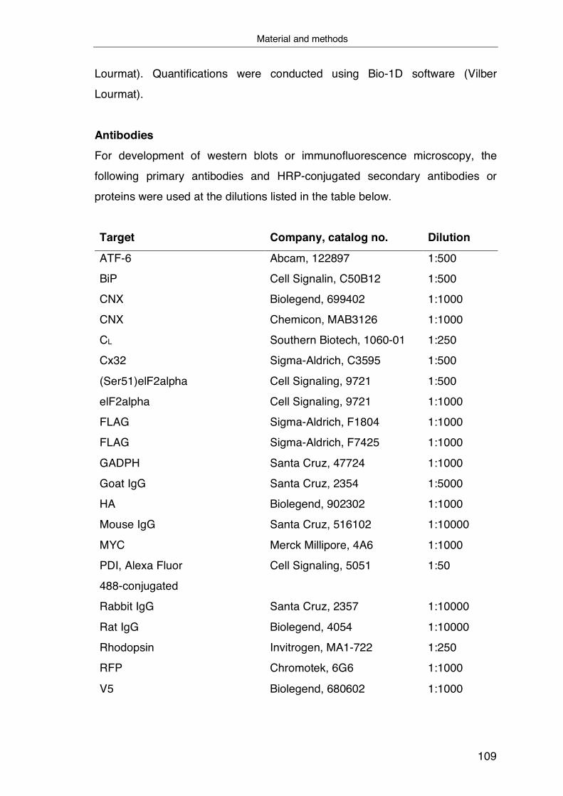
Material and methods
109
Lourmat). Quantifications were conducted using Bio-1D software (Vilber
Lourmat).
Antibodies For development of western blots or immunofluorescence microscopy, the
following primary antibodies and HRP-conjugated secondary antibodies or
proteins were used at the dilutions listed in the table below.
Target Company, catalog no. Dilution
ATF-6
BiP
CNX
Abcam, 122897
Cell Signalin, C50B12
Biolegend, 699402
1:500
1:500
1:1000
CNX Chemicon, MAB3126 1:1000
CL Southern Biotech, 1060-01 1:250
Cx32
(Ser51)elF2alpha
elF2alpha
Sigma-Aldrich, C3595
Cell Signaling, 9721
Cell Signaling, 9721
1:500
1:500
1:1000
FLAG Sigma-Aldrich, F1804 1:1000
FLAG Sigma-Aldrich, F7425 1:1000
GADPH Santa Cruz, 47724 1:1000
Goat IgG Santa Cruz, 2354 1:5000
HA Biolegend, 902302 1:1000
Mouse IgG Santa Cruz, 516102 1:10000
MYC Merck Millipore, 4A6 1:1000
PDI, Alexa Fluor
488-conjugated
Cell Signaling, 5051 1:50
Rabbit IgG Santa Cruz, 2357 1:10000
Rat IgG
Rhodopsin
Biolegend, 4054
Invitrogen, MA1-722
1:10000
1:250
RFP Chromotek, 6G6 1:1000
V5 Biolegend, 680602 1:1000
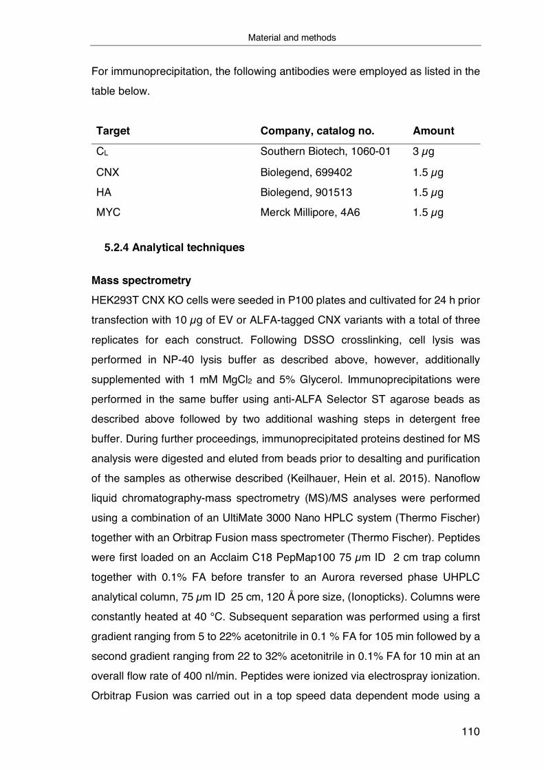
Material and methods
110
For immunoprecipitation, the following antibodies were employed as listed in the
table below.
Target Company, catalog no. Amount
CL Southern Biotech, 1060-01 3 µg
CNX Biolegend, 699402 1.5 µg
HA Biolegend, 901513 1.5 µg
MYC Merck Millipore, 4A6 1.5 µg
5.2.4 Analytical techniques
Mass spectrometry
HEK293T CNX KO cells were seeded in P100 plates and cultivated for 24 h prior
transfection with 10 µg of EV or ALFA-tagged CNX variants with a total of three
replicates for each construct. Following DSSO crosslinking, cell lysis was
performed in NP-40 lysis buffer as described above, however, additionally
supplemented with 1 mM MgCl2 and 5% Glycerol. Immunoprecipitations were
performed in the same buffer using anti-ALFA Selector ST agarose beads as
described above followed by two additional washing steps in detergent free
buffer. During further proceedings, immunoprecipitated proteins destined for MS
analysis were digested and eluted from beads prior to desalting and purification
of the samples as otherwise described (Keilhauer, Hein et al. 2015). Nanoflow
liquid chromatography-mass spectrometry (MS)/MS analyses were performed
using a combination of an UltiMate 3000 Nano HPLC system (Thermo Fischer)
together with an Orbitrap Fusion mass spectrometer (Thermo Fischer). Peptides
were first loaded on an Acclaim C18 PepMap100 75 µm ID 2 cm trap column
together with 0.1% FA before transfer to an Aurora reversed phase UHPLC
analytical column, 75 µm ID 25 cm, 120 Å pore size, (Ionopticks). Columns were
constantly heated at 40 °C. Subsequent separation was performed using a first
gradient ranging from 5 to 22% acetonitrile in 0.1 % FA for 105 min followed by a
second gradient ranging from 22 to 32% acetonitrile in 0.1% FA for 10 min at an
overall flow rate of 400 nl/min. Peptides were ionized via electrospray ionization.
Orbitrap Fusion was carried out in a top speed data dependent mode using a
Material and methods
111
cycle time of 3 s. Full scan (MS1) acquisition (scan range of 300 – 1500 m/z) was
performed in the orbitrap at a defined resolution of 120,000 as well as with an
automatic gain control (AGC) ion target value of 2e5 whereby dynamic exclusion
was set to 60 s. For fragmentation, precursors with a charge state of 2-7 and a
minimum intensity of 5e3 were selected and isolated in the quadrupole using a
window of 1.6 m/z. Subsequent fragment generation was achieved using higher-
energy collisional dissociation (HCD, collision energy: 30%). The MS2 AGC was
adjusted to 1e4 and 50 ms were selected as the maximum injection time for the
ion trap (with inject ions for all available parallelized time enabled). Scanning of
fragments was performed by applying the rapid scan rate.
5.3 Data evaluation
Mass spectrometry evaluation MS raw files were analyzed with MaxQuant software (version 1.6.17.0) with most
default settings and a protein database containing human sequences
(downloaded July 2019 from Uniprot, taxonomy ID: 9606, 74349 entries). The
FLAG tagged version of Calnexin was not implemented. The following parameter
settings were used: PSM and protein FDR 1%; enzyme specificity trypsin/P;
minimal peptide length: 7; variable modifications: methionine oxidation, N-
terminal acetylation; fixed modification: carbamidomethylatio. The minimal
number of unique peptides for protein identification was set to 2. For label-free
protein quantification, the MaxLFQ algorithm was used as part of the MaxQuant
environment: (LFQ) minimum ratio count: 2; peptides for quantification: unique.
Statistical analysis was performed in Perseus (version 1.6.14.0). Proteins
identified only by site, reverse hits or potential contaminants were removed. LFQ
intensities were log2 transformed and data were then filtered for at least two valid
values in the wt group. Then, missing values were imputed from normal
distribution (width: 0.3, down shift: 1.8 standard deviations, mode: over whole
matrix). The replicate groups were compared via a two- sided, two-sample
Student’s t test (S0 = 0, permutation-based FDR method with FDR = 0.05 and
250 randomizations). Enrichment values and corresponding −log10 P values
Material and methods
112
were plotted. Cut offs in the volcano plot were set to 2-fold enrichment and a p-
value of 0,05.
Quantification and statistics Immunoblots were quantified using the Bio-1D software (Vilber Lourmat). Binding
of endogenous CNX to CoMem TMD helices was calculated as the ratio of
intensities of the co-immunoprecipitated protein (CNX) to the overall intensity of
immunoprecipitated CoMem (sum of both glycosylated and nonglycosylated
species – beginning at the molecular weight of HA tagged nonglycosylated
CoMem until the upper protein smear represented by the complex glycosylated
species. In case of CoMem N1Q, the area around the nonglycosylated species
was included into quantification). Subsequently, to facilitate comparability of
individual experiments, formed ratios were each normalized to the CoMem WT
N1Q (CoMem lacking the luminal glycosylation site) dataset of each experiment.
The same approach was used analyzing binding of overexpressed CNX variants
to Cx32 TM1 or Cx32 full length. To determine effects of CHX treatment on the
decay of target proteins, the intensity at each time point was normalized to the
intensity of the untreated sample with no CHX incubation (t= 0h). Half-life
calculations from decay data were carried out by logarithmically linearizing the
CHX decays and determination of the ln(0.5) value. Statistical analyses were
performed using Prism (GraphPad Software). Where indicated, data were
analyzed with two-tailed, unpaired Student’s t-tests and differences were
considered to be statistically significant when p < 0.05.
Sequence analysis TM regions of Cx32 and CoMem WT as well as mutant variants were annotated
by using ΔG prediction server v1.0 (Stockholm University). The same tool was
used to predict the ΔGapp for helix insertions using the full protein scan mode.
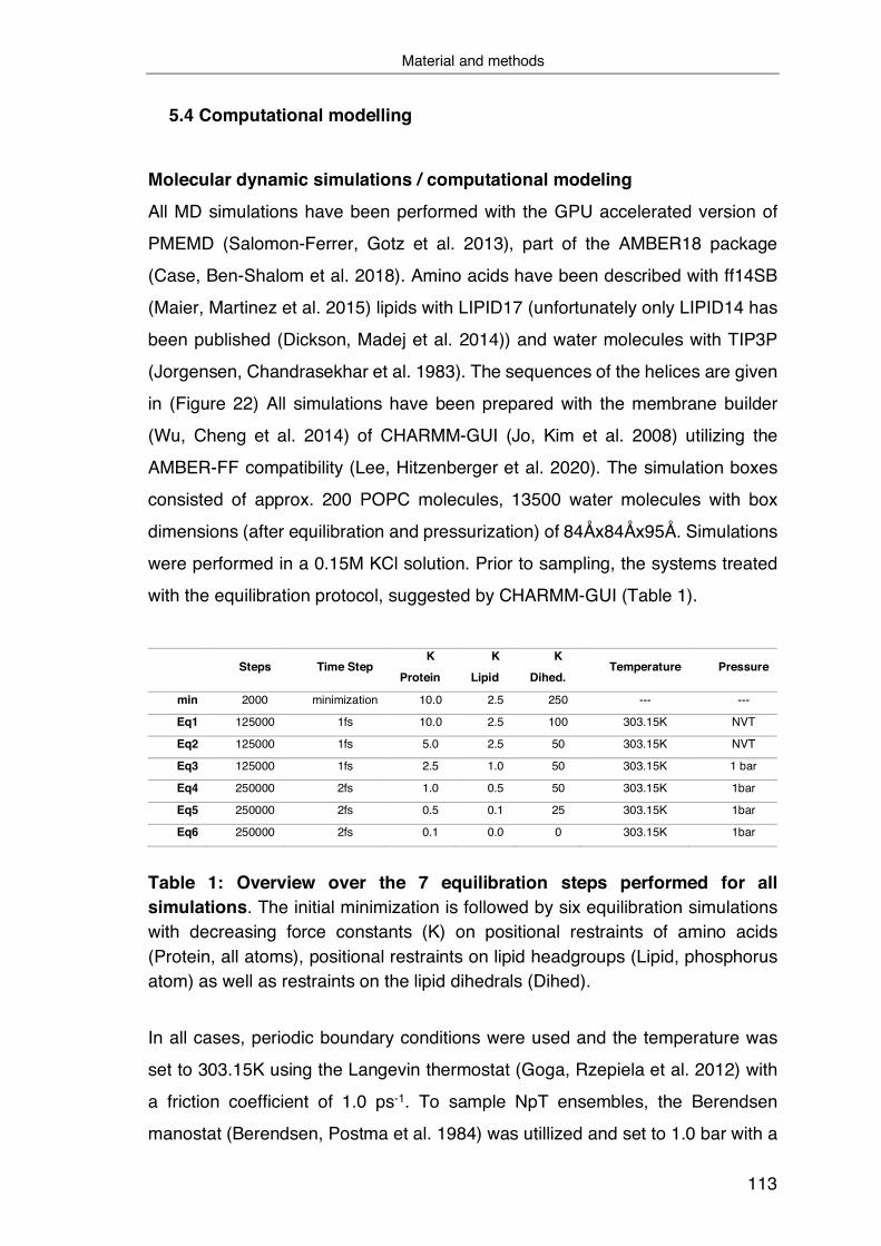
Material and methods
113
5.4 Computational modelling
Molecular dynamic simulations / computational modeling
All MD simulations have been performed with the GPU accelerated version of
PMEMD (Salomon-Ferrer, Gotz et al. 2013), part of the AMBER18 package
(Case, Ben-Shalom et al. 2018). Amino acids have been described with ff14SB
(Maier, Martinez et al. 2015) lipids with LIPID17 (unfortunately only LIPID14 has
been published (Dickson, Madej et al. 2014)) and water molecules with TIP3P
(Jorgensen, Chandrasekhar et al. 1983). The sequences of the helices are given
in (Figure 22) All simulations have been prepared with the membrane builder
(Wu, Cheng et al. 2014) of CHARMM-GUI (Jo, Kim et al. 2008) utilizing the
AMBER-FF compatibility (Lee, Hitzenberger et al. 2020). The simulation boxes
consisted of approx. 200 POPC molecules, 13500 water molecules with box
dimensions (after equilibration and pressurization) of 84Åx84Åx95Å. Simulations
were performed in a 0.15M KCl solution. Prior to sampling, the systems treated
with the equilibration protocol, suggested by CHARMM-GUI (Table 1).
Steps Time Step K
Protein
K
Lipid
K
Dihed. Temperature Pressure
min 2000 minimization 10.0 2.5 250 --- ---
Eq1 125000 1fs 10.0 2.5 100 303.15K NVT
Eq2 125000 1fs 5.0 2.5 50 303.15K NVT
Eq3 125000 1fs 2.5 1.0 50 303.15K 1 bar
Eq4 250000 2fs 1.0 0.5 50 303.15K 1bar
Eq5 250000 2fs 0.5 0.1 25 303.15K 1bar
Eq6 250000 2fs 0.1 0.0 0 303.15K 1bar
Table 1: Overview over the 7 equilibration steps performed for all simulations. The initial minimization is followed by six equilibration simulations with decreasing force constants (K) on positional restraints of amino acids (Protein, all atoms), positional restraints on lipid headgroups (Lipid, phosphorus atom) as well as restraints on the lipid dihedrals (Dihed).
In all cases, periodic boundary conditions were used and the temperature was
set to 303.15K using the Langevin thermostat (Goga, Rzepiela et al. 2012) with
a friction coefficient of 1.0 ps-1. To sample NpT ensembles, the Berendsen
manostat (Berendsen, Postma et al. 1984) was utillized and set to 1.0 bar with a
Material and methods
114
relaxation time of 1ps. All non-bonded interactions have been calculated directly
up to a distance of 9Å, after which electrostatic interactions were treated with the
particle mesh Ewald method (Darden, York et al. 1993), while long range van-
der-Waals interactions were estimated by a dispersion correction model. To
achieve time-steps of 4fs, the Shake algorithm (Andersen 1983) as well as
hydrogen mass repartitioning (Hopkins, Grand et al. 2015) were used.
Prior to building the dimeric simulation systems, the Cnx, Cx32 and CoMem
helices have been simulated in isolation for 1µs in a POPC bilayer in order to
equilibrate the structure and to predict their orientation in the membrane. All
simulation snapshots have been rendered using VMD (Humphrey, Dalke et al.
1996).
Cnx-CoMem dimerisation simulations
Initially, five simulations of one Cnx helix and one CoMem (WT) helix in the same
simulation box, have been performed. Each of the five simulations was started
with different relative orientations of the two helices which have been generated
by hand using VMD. After preparing the simulation systems using CHARMM-
GUI, the systems were equilibrated using the protocol in Table 1. Subsequently,
unrestricted sampling simulations of at least 2.5µs have been performed.
Successful complex formation was only sampled in one of the five simulations.
To improve statistics, two additional independent simulations have been
performed.
These simulations have been started from a snapshot of the successful
simulation in which the two helices were not yet interacting with each other. In
one of these two simulations, a similar complex was formed as in the previous,
successful simulation. At this stage, all simulations have been sampled for at
least 4µs.
To obtain binding statistics on the amino-acid level that are more generally
valid, we also performed simulations with Cnx and two CoMem mutants that
exhibit amino acid alterations in the TMD segment: V13P and V13R.
Several at least 4µs long simulations have been performed for both mutants,
however, only two resulted in the formation of Cnx-CoMem interactions along the
entire TMD: One simulation featuring V13R and one with the V13P mutant. Of
Material and methods
115
interest: While the V13P mutant binds to Cnx in a fashion very similar to WT
CoMem, the V13R mutant uses an alternative interface to bind to Cnx. Cnx, on
the other hand, binds all CoMem variants utilizing basically the same binding
interface. Altogether, 22 µs stim-scale simulations of this system have been
performed. However, for analysis we only selected the 4 simulations that
exhibited broad TMD-TMD interactions between Cnx and CoMem. Data analysis
was performed on 4µs long trajectories in each case.
Cnx-Cx32 dimerization simulations
For this system five, at least 4µs long simulations have been performed. The
simulation set-up was identical to the one used for the Cnx-CoMem system. In
four out of the five cases, we were able to sample at least transient interactions
between the helices, involving the entire TMD of Cnx. These four simulations
were then selected for data analysis on 4µs of the sampled trajectories.

Abbreviations
116
Abbreviations ATF6 Activating transcription factor 6
ATP Adenosine triphosphate
BiP Immunoglobulin heavy-chain binding protein
cDNA Complementary DNA
CHX Cycloheximide
CL Constant domain of the light-chain
CNX Calnexin
COS CV-1 in origin, and carrying the SV40 genetic material
CRT Calreticulin
DMEM Dulbecco´s modified Eagle medium
DNA Deoxyribonucleic acid
DTT Dithioreitol
ER Endoplasmic reticulum
FBS Fetal bovine serum
GADPH Glyceraldehyde 3-phosphate dehydrogenase
Glc Glucose
GlcNac N-Acetylglucosamine
gp78 Cell surface glycoprotein of 78 kDa
HA Hemagglutinin
Hrd1 HMG-CoA reductase degradation
IB Immunoblot
IF Immunofluorescence
IRE1 Inositol-requiring protein
IP Immunoprecipitation
min Minute
mL Milliliter
MW Molecular weight
NaDOC Sodium deoxycholate
Nm Nanometer
OST Oligosaccharyltransferase
PAGE Polyacrilamide gel electrophoresis

Abbreviations
117
PBS Phosphate buffered saline
PCR Polymerase chain reaction
PDI Protein disulfide-isomerase
PERK Protein kinase RNA-like ER kinase
PNGase Peptide:N-glycosidase
PTM Post-translational modification
PVDF Polyvinylidene fluoride
QC Quality control
RIPA Radioimmunoprecipitation assay buffer
RNC Ribosome-nascent chain complex
RT Room temperature
SDS Sodium dodecyl sulfate
SEM Standard error of the mean
SRP Signal recognition particle
TBS Tris-buffered saline
TM Transmembrane
TRAM Translocating chain-associated membrane protein
TRAP Translocon-associated protein
Tm Tunicamycin
UPR Unfolded protein response
WT Wild type
ß-Me ß-mercaptoethanol

References
118
References Aebi, M. (2013). "N-linked protein glycosylation in the ER." Biochim Biophys Acta 1833(11): 2430-2437. Ahner, A. and J. L. Brodsky (2004). "Checkpoints in ER-associated degradation: excuse me, which way to the proteasome?" Trends Cell Biol 14(9): 474-478. al-Maghtheh, M., C. Gregory, C. Inglehearn, A. Hardcastle and S. Bhattacharya (1993). "Rhodopsin mutations in autosomal dominant retinitis pigmentosa." Hum Mutat 2(4): 249-255. Andersen, H. C. (1983). "RATTLE: A “Velocity” Version of the SHAKE Algorithm for Molecular Dynamics Calculations." J Comp Phys 52: 24-34. Anderson, K. S. and P. Cresswell (1994). "A role for calnexin (IP90) in the assembly of class II MHC molecules." EMBO J 13(3): 675-682. Andersson, H., I. Nilsson and G. von Heijne (1996). "Calnexin can interact with N-linked glycans located close to the endoplasmic reticulum membrane." FEBS Lett 397(2-3): 321-324. Aoe, T., A. J. Lee, E. van Donselaar, P. J. Peters and V. W. Hsu (1998). "Modulation of intracellular transport by transported proteins: insight from regulation of COPI-mediated transport." Proc Natl Acad Sci U S A 95(4): 1624-1629. Appenzeller-Herzog, C. and H. P. Hauri (2006). "The ER-Golgi intermediate compartment (ERGIC): in search of its identity and function." J Cell Sci 119(Pt 11): 2173-2183. Argon, Y. and B. B. Simen (1999). "GRP94, an ER chaperone with protein and peptide binding properties." Semin Cell Dev Biol 10(5): 495-505. Arvan, P., X. Zhao, J. Ramos-Castaneda and A. Chang (2002). "Secretory pathway quality control operating in Golgi, plasmalemmal, and endosomal systems." Traffic 3(11): 771-780. Ast, T. and M. Schuldiner (2013). "All roads lead to Rome (but some may be harder to travel): SRP-independent translocation into the endoplasmic reticulum." Crit Rev Biochem Mol Biol 48(3): 273–288. Aviram, N., T. Ast, E. A. Costa, E. C. Arakel, S. G. Chuartzman, C. H. Jan, S. Hassdenteufel, J. Dudek, M. Jung, S. Schorr, R. Zimmermann, B. Schwappach, J. S. Weissman and M. Schuldiner (2016). "The SND proteins constitute an alternative targeting route to the endoplasmic reticulum." Nature 540(7631): 134-138. Bai, L. and H. Li (2021). "Cryo-EM structures of the endoplasmic reticulum membrane complex." FEBS J.
References
119
Baldridge, R. D. and T. A. Rapoport (2016). "Autoubiquitination of the Hrd1 Ligase Triggers Protein Retrotranslocation in ERAD." Cell 166(2): 394-407. Bard, J. A. M., E. A. Goodall, E. R. Greene, E. Jonsson, K. C. Dong and A. Martin (2018). "Structure and Function of the 26S Proteasome." Annu Rev Biochem 87: 697-724. Bargmann, C. I., M. C. Hung and R. A. Weinberg (1986). "Multiple independent activations of the neu oncogene by a point mutation altering the transmembrane domain of p185." Cell 45(5): 649-657. Behnke, J., M. J. Feige and L. M. Hendershot (2015). "BiP and its nucleotide exchange factors Grp170 and Sil1: mechanisms of action and biological functions." J Mol Biol 427(7): 1589-1608. Behnke, J. and L. M. Hendershot (2014). "The large Hsp70 Grp170 binds to unfolded protein substrates in vivo with a regulation distinct from conventional Hsp70s." J Biol Chem 289(5): 2899-2907. Behnke, J., M. J. Mann, F. L. Scruggs, M. J. Feige and L. M. Hendershot (2016). "Members of the Hsp70 Family Recognize Distinct Types of Sequences to Execute ER Quality Control." Mol Cell 63(5): 739-752. Belbeoc'h, S., C. Falasca, J. Leroy, A. Ayon, J. Massoulie and S. Bon (2004). "Elements of the C-terminal t peptide of acetylcholinesterase that determine amphiphilicity, homomeric and heteromeric associations, secretion and degradation." Eur J Biochem 271(8): 1476-1487. Benham, A. M., M. van Lith, R. Sitia and I. Braakman (2013). "Ero1-PDI interactions, the response to redox flux and the implications for disulfide bond formation in the mammalian endoplasmic reticulum." Philos Trans R Soc Lond B Biol Sci 368(1617): 20110403. Berendsen, H. J. C., J. P. M. Postma, W. F. v. Gunsteren, A. DiNola and J. R. Haak (1984). "Molecular Dynamics with Coupling to an External Bath." J Phys Chem A 81: 3684–3690. Bergbold, N. and M. K. Lemberg (2013). "Emerging role of rhomboid family proteins in mammalian biology and disease." Biochim Biophys Acta 1828(12): 2840-2848. Bernstein, E. R. and S. Sun (1996). "Aromatic van der Waals clusters: structure and norigiditiy." J. Phys. Chem 100: 13348-13366. Bodnar, N. and T. Rapoport (2017). "Toward an understanding of the Cdc48/p97 ATPase." F1000Res 6: 1318. Boeddrich, A., S. Gaumer, A. Haacke, N. Tzvetkov, M. Albrecht, B. O. Evert, E. C. Muller, R. Lurz, P. Breuer, N. Schugardt, S. Plassmann, K. Xu, J. M. Warrick,
References
120
J. Suopanki, U. Wullner, R. Frank, U. F. Hartl, N. M. Bonini and E. E. Wanker (2006). "An arginine/lysine-rich motif is crucial for VCP/p97-mediated modulation of ataxin-3 fibrillogenesis." EMBO J 25(7): 1547-1558. Bone, L. J., S. M. Deschenes, R. J. Balice-Gordon, K. H. Fischbeck and S. S. Scherer (1997). "Connexin32 and X-linked Charcot-Marie-Tooth disease." Neurobiol Dis 4(3-4): 221-230. Bonifacino, J. S. and L. M. Traub (2003). "Signals for sorting of transmembrane proteins to endosomes and lysosomes." Annu Rev Biochem 72: 395-447. Borgese, N. (2016). "Getting membrane proteins on and off the shuttle bus between the endoplasmic reticulum and the Golgi complex." J Cell Sci 129(8): 1537-1545. Bottomley, M. J., M. R. Batten, R. A. Lumb and N. J. Bulleid (2001). "Quality control in the endoplasmic reticulum: PDI mediates the ER retention of unassembled procollagen C-propeptides." Curr Biol 11(14): 1114-1118. Braakman, I. and N. J. Bulleid (2011). "Protein folding and modification in the mammalian endoplasmic reticulum." Annu Rev Biochem 80: 71-99. Braakman, I. and D. N. Hebert (2013). "Protein folding in the endoplasmic reticulum." Cold Spring Harb Perspect Biol 5(5): a013201. Brambillasca, S., M. Yabal, M. Makarow and N. Borgese (2006). "Unassisted translocation of large polypeptide domains across phospholipid bilayers." J Cell Biol 175(5): 767-777. Brauer, P., J. L. Parker, A. Gerondopoulos, I. Zimmermann, M. A. Seeger, F. A. Barr and S. Newstead (2019). "Structural basis for pH-dependent retrieval of ER proteins from the Golgi by the KDEL receptor." Science 363(6431): 1103-1107. Briant, K., N. Johnson and E. Swanton (2017). "Transmembrane domain quality control systems operate at the endoplasmic reticulum and Golgi apparatus." PLoS One 12(4): e0173924. Brodsky, J. L. and W. R. Skach (2011). "Protein folding and quality control in the endoplasmic reticulum: Recent lessons from yeast and mammalian cell systems." Curr Opin Cell Biol 23(4): 464-475. Bruce, A., A. Johnson, J. Lewis, M. Raff, K. Roberts and P. Walter (2002). "Molecular Biology of the Cell." New York: Garland Science 4th edition. Buck, T. M., A. R. Kolb, C. R. Boyd, T. R. Kleyman and J. L. Brodsky (2010). "The endoplasmic reticulum-associated degradation of the epithelial sodium channel requires a unique complement of molecular chaperones." Mol Biol Cell 21(6): 1047-1058.
References
121
Buck, T. M., L. Plavchak, A. Roy, B. F. Donnelly, O. B. Kashlan, T. R. Kleyman, A. R. Subramanya and J. L. Brodsky (2013). "The Lhs1/GRP170 chaperones facilitate the endoplasmic reticulum-associated degradation of the epithelial sodium channel." J Biol Chem 288(25): 18366-18380. Buck, T. M., C. M. Wright and J. L. Brodsky (2007). "The activities and function of molecular chaperones in the endoplasmic reticulum." Semin Cell Dev Biol 18(6): 751-761. Bulleid, N. J. (2012). "Disulfide bond formation in the mammalian endoplasmic reticulum." Cold Spring Harb Perspect Biol 4(11). Cai, H., C. C. Wang and C. L. Tsou (1994). "Chaperone-like activity of protein disulfide isomerase in the refolding of a protein with no disulfide bonds." J Biol Chem 269(40): 24550-24552. Cameron, P. H., E. Chevet, O. Pluquet, D. Y. Thomas and J. J. Bergeron (2009). "Calnexin phosphorylation attenuates the release of partially misfolded alpha1-antitrypsin to the secretory pathway." J Biol Chem 284(50): 34570-34579. Cannon, K. S. and P. Cresswell (2001). "Quality control of transmembrane domain assembly in the tetraspanin CD82." EMBO J 20(10): 2443-2453. Caramelo, J. J. and A. J. Parodi (2008). "Getting in and out from calnexin/calreticulin cycles." J Biol Chem 283(16): 10221-10225. Caramelo, J. J. and A. J. Parodi (2015). "A sweet code for glycoprotein folding." FEBS Lett 589(22): 3379-3387. Carvalho, P., V. Goder and T. A. Rapoport (2006). "Distinct ubiquitin-ligase complexes define convergent pathways for the degradation of ER proteins." Cell 126(2): 361-373. Carvalho, P., A. M. Stanley and T. A. Rapoport (2010). "Retrotranslocation of a misfolded luminal ER protein by the ubiquitin-ligase Hrd1p." Cell 143(4): 579-591. Case, D. A., I. Y. Ben-Shalom, S. R. Brozell, D. S. Cerutti, I. T. E. Cheatham, V. W. D. Cruzeiro, T. A. Darden, R. E. Duke, D. Ghoreishi, M. K. Gilson, H. Gohlke, A. W. Goetz, D. Greene, R. Harris, N. Homeyer, Y. Huang, S. Izadi, A. Kovalenko, T. Kurtzman, T. S. Lee, S. LeGrand, P. Li, C. Lin, J. Liu, T. Luchko, R. Luo, D. J. Mermelstein, K. M. Merz, Y. Miao, G. Monard, C. Nguyen, H. Nguyen, I. Omelyan, A. Onufriev, F. Pan, R. Qi, D. R. Roe, A. Roitberg, C. Sagui, S. Schott-Verdugo, J. Shen, C. L. Simmerling, J. Smith, R. Salomon-Ferrer, J. Swails, R. C. Walker, J. Wang, H. Wei, R. M. Wolf, X. Wu, L. Xiao, D. M. York and P. A. Kollman (2018). AMBER 2018. Univ. Calif. San Franc. Chang, D. K., S. F. Cheng, V. D. Trivedi and K. L. Lin (1999). "Proline affects oligomerization of a coiled coil by inducing a kink in a long helix." J Struct Biol 128(3): 270-279.
References
122
Chen, W., J. Helenius, I. Braakman and A. Helenius (1995). "Cotranslational folding and calnexin binding during glycoprotein synthesis." Proc Natl Acad Sci U S A 92(14): 6229-6233. Chevet, E., J. Smirle, P. H. Cameron, D. Y. Thomas and J. J. Bergeron (2010). "Calnexin phosphorylation: linking cytoplasmic signalling to endoplasmic reticulum lumenal functions." Semin Cell Dev Biol 21(5): 486-490. Chevet, E., H. N. Wong, D. Gerber, C. Cochet, A. Fazel, P. H. Cameron, J. N. Gushue, D. Y. Thomas and J. J. Bergeron (1999). "Phosphorylation by CK2 and MAPK enhances calnexin association with ribosomes." EMBO J 18(13): 3655-3666. Chino, H. and N. Mizushima (2020). "ER-Phagy: Quality Control and Turnover of Endoplasmic Reticulum." Trends Cell Biol 30(5): 384-398. Chio, U. S., H. Cho and S. O. Shan (2017). "Mechanisms of Tail-Anchored Membrane Protein Targeting and Insertion." Annu Rev Cell Dev Biol 33: 417-438. Chitwood, P. J. and R. S. Hegde (2020). "An intra-membrane chaperone complex facilitates membrane protein biogenesis." Nature 584(7822): 630-634. Chitwood, P. J., S. Juszkiewicz, A. Guna, S. Shao and R. S. Hegde (2018). "EMC Is Required to Initiate Accurate Membrane Protein Topogenesis." Cell 175(6): 1507-1519 e1516. Choma, C., H. Gratkowski, J. D. Lear and W. F. DeGrado (2000). "Asparagine-mediated self-association of a model transmembrane helix." Nat Struct Biol 7(2): 161-166. Christianson, J. C., T. A. Shaler, R. E. Tyler and R. R. Kopito (2008). "OS-9 and GRP94 deliver mutant alpha1-antitrypsin to the Hrd1-SEL1L ubiquitin ligase complex for ERAD." Nat Cell Biol 10(3): 272-282. Chung, K. T., Y. Shen and L. M. Hendershot (2002). "BAP, a mammalian BiP-associated protein, is a nucleotide exchange factor that regulates the ATPase activity of BiP." J Biol Chem 277(49): 47557-47563. Ciechanover, A., D. Finley and A. Varshavsky (1984). "Ubiquitin dependence of selective protein degradation demonstrated in the mammalian cell cycle mutant ts85." Cell 37(1): 57-66. Claessen, J. H., L. Kundrat and H. L. Ploegh (2012). "Protein quality control in the ER: balancing the ubiquitin checkbook." Trends Cell Biol 22(1): 22-32. Clapham, D. E. (2007). "Calcium signaling." Cell 131(6): 1047-1058. Coelho, J. P. L., M. Stahl, N. Bloemeke, K. Meighen-Berger, C. P. Alvira, Z. R. Zhang, S. A. Sieber and M. J. Feige (2019). "A network of chaperones prevents
References
123
and detects failures in membrane protein lipid bilayer integration." Nat Commun 10(1): 672. Conti, B. J., P. K. Devaraneni, Z. Yang, L. L. David and W. R. Skach (2015). "Cotranslational stabilization of Sec62/63 within the ER Sec61 translocon is controlled by distinct substrate-driven translocation events." Mol Cell 58(2): 269-283. Cordes, F. S., J. N. Bright and M. S. Sansom (2002). "Proline-induced distortions of transmembrane helices." J Mol Biol 323(5): 951-960. Costa, E. A., K. Subramanian, J. Nunnari and J. S. Weissman (2018). "Defining the physiological role of SRP in protein-targeting efficiency and specificity." Science 359(6376): 689-692. Crowley, K. S., S. Liao, V. E. Worrell, G. D. Reinhart and A. E. Johnson (1994). "Secretory proteins move through the endoplasmic reticulum membrane via an aqueous, gated pore." Cell 78(3): 461-471. Cullen, P. J. and F. Steinberg (2018). "To degrade or not to degrade: mechanisms and significance of endocytic recycling." Nat Rev Mol Cell Biol 19(11): 679-696. Cymer, F., G. von Heijne and S. H. White (2015). "Mechanisms of integral membrane protein insertion and folding." J Mol Biol 427(5): 999-1022. Danilczyk, U. G., M. F. Cohen-Doyle and D. B. Williams (2000). "Functional relationship between calreticulin, calnexin, and the endoplasmic reticulum luminal domain of calnexin." J Biol Chem 275(17): 13089-13097. Danilczyk, U. G. and D. B. Williams (2001). "The lectin chaperone calnexin utilizes polypeptide-based interactions to associate with many of its substrates in vivo." J Biol Chem 276(27): 25532-25540. Darden, T., D. York and L. Pedersen (1993). "Particle Mesh Ewald: An N⋅ Log (N) Method for Ewald Sums in Large Systems." J Chem Phys 98 (12): 10089–10092. Dawson, A. P. (1997). "Calcium signalling: how do IP3 receptors work?" Curr Biol 7(9): R544-547. Dawson, J. P., R. A. Melnyk, C. M. Deber and D. M. Engelman (2003). "Sequence context strongly modulates association of polar residues in transmembrane helices." J Mol Biol 331(1): 255-262. Degen, E. and D. B. Williams (1991). "Participation of a novel 88-kD protein in the biogenesis of murine class I histocompatibility molecules." J Cell Biol 112(6): 1099-1115.
References
124
del Alamo, M., D. J. Hogan, S. Pechmann, V. Albanese, P. O. Brown and J. Frydman (2011). "Defining the specificity of cotranslationally acting chaperones by systematic analysis of mRNAs associated with ribosome-nascent chain complexes." PLoS Biol 9(7): e1001100. Denzel, A., M. Molinari, C. Trigueros, J. E. Martin, S. Velmurgan, S. Brown, G. Stamp and M. J. Owen (2002). "Early postnatal death and motor disorders in mice congenitally deficient in calnexin expression." Mol Cell Biol 22(21): 7398-7404. Denzer, A. J., C. E. Nabholz and M. Spiess (1995). "Transmembrane orientation of signal-anchor proteins is affected by the folding state but not the size of the N-terminal domain." EMBO J 14(24): 6311-6317. Deprez, P., M. Gautschi and A. Helenius (2005). "More than one glycan is needed for ER glucosidase II to allow entry of glycoproteins into the calnexin/calreticulin cycle." Mol Cell 19(2): 183-195. Dickson, C. J., B. D. Madej, A. A. Skjevik, R. M. Betz, K. Teigen, I. R. Gould and R. C. Walker (2014). "Lipid14: The Amber Lipid Force Field." J Chem Theory Comput 10(2): 865-879. Ding, P. Z. and T. H. Wilson (2001). "The effect of modifications of the charged residues in the transmembrane helices on the transport activity of the melibiose carrier of Escherichia coli." Biochem Biophys Res Commun 285(2): 348-354. Do, H., D. Falcone, J. Lin, D. W. Andrews and A. E. Johnson (1996). "The cotranslational integration of membrane proteins into the phospholipid bilayer is a multistep process." Cell 85(3): 369-378. Dolmetsch, R. E., K. Xu and R. S. Lewis (1998). "Calcium oscillations increase the efficiency and specificity of gene expression." Nature 392(6679): 933-936. Dudek, J., S. Pfeffer, P. H. Lee, M. Jung, A. Cavalie, V. Helms, F. Forster and R. Zimmermann (2015). "Protein transport into the human endoplasmic reticulum." J Mol Biol 427(6 Pt A): 1159-1175. Egea, P. F., R. M. Stroud and P. Walter (2005). "Targeting proteins to membranes: structure of the signal recognition particle." Curr Opin Struct Biol 15(2): 213-220. Ellgaard, L. and A. Helenius (2003). "Quality control in the endoplasmic reticulum." Nat Rev Mol Cell Biol 4(3): 181-191. Ellgaard, L. and L. W. Ruddock (2005). "The human protein disulphide isomerase family: substrate interactions and functional properties." EMBO Rep 6(1): 28-32. Ellgaard, L., C. S. Sevier and N. J. Bulleid (2018). "How Are Proteins Reduced in the Endoplasmic Reticulum?" Trends Biochem Sci 43(1): 32-43.

References
125
Erdmann, F., M. Jung, P. Maurer, A. Harsman, R. Zimmermann and R. Wagner (2010). "The mammalian and yeast translocon complexes comprise a characteristic Sec61 channel." Biochem Biophys Res Commun 396(3): 714-720. Ernst, R., B. Mueller, H. L. Ploegh and C. Schlieker (2009). "The otubain YOD1 is a deubiquitinating enzyme that associates with p97 to facilitate protein dislocation from the ER." Mol Cell 36(1): 28-38. Fang, S., M. Ferrone, C. Yang, J. P. Jensen, S. Tiwari and A. M. Weissman (2001). "The tumor autocrine motility factor receptor, gp78, is a ubiquitin protein ligase implicated in degradation from the endoplasmic reticulum." Proc Natl Acad Sci U S A 98(25): 14422-14427. Feige, M. J. and L. M. Hendershot (2013). "Quality control of integral membrane proteins by assembly-dependent membrane integration." Mol Cell 51(3): 297-309. Fenech, E. J., F. Lari, P. D. Charles, R. Fischer, M. Laetitia-Thezenas, K. Bagola, A. W. Paton, J. C. Paton, M. Gyrd-Hansen, B. M. Kessler and J. C. Christianson (2020). "Interaction mapping of endoplasmic reticulum ubiquitin ligases identifies modulators of innate immune signalling." Elife 9. Fink, A., N. Sal-Man, D. Gerber and Y. Shai (2012). "Transmembrane domains interactions within the membrane milieu: principles, advances and challenges." Biochim Biophys Acta 1818(4): 974-983. Finke, K., K. Plath, S. Panzner, S. Prehn, T. A. Rapoport, E. Hartmann and T. Sommer (1996). "A second trimeric complex containing homologs of the Sec61p complex functions in protein transport across the ER membrane of S. cerevisiae." EMBO J 15(7): 1482-1494. Finley, D., A. Ciechanover and A. Varshavsky (1984). "Thermolability of ubiquitin-activating enzyme from the mammalian cell cycle mutant ts85." Cell 37(1): 43-55. Fleig, L., N. Bergbold, P. Sahasrabudhe, B. Geiger, L. Kaltak and M. K. Lemberg (2012). "Ubiquitin-dependent intra-membrane rhomboid protease promotes ERAD of membrane proteins." Mol Cell 47(4): 558-569. Flynn, G. C., J. Pohl, M. T. Flocco and J. E. Rothman (1991). "Peptide-binding specificity of the molecular chaperone BiP." Nature 353(6346): 726-730. Fons, R. D., B. A. Bogert and R. S. Hegde (2003). "Substrate-specific function of the translocon-associated protein complex during translocation across the ER membrane." J Cell Biol 160(4): 529-539. Fontanini, A., R. Chies, E. L. Snapp, M. Ferrarini, G. M. Fabrizi and C. Brancolini (2005). "Glycan-independent role of calnexin in the intracellular retention of Charcot-Marie-tooth 1A Gas3/PMP22 mutants." J Biol Chem 280(3): 2378-2387.
References
126
Freiden, P. J., J. R. Gaut and L. M. Hendershot (1992). "Interconversion of three differentially modified and assembled forms of BiP." EMBO J 11(1): 63-70. French, M. E., C. F. Koehler and T. Hunter (2021). "Emerging functions of branched ubiquitin chains." Cell Discov 7(1): 6. Frenkel, Z., W. Gregory, S. Kornfeld and G. Z. Lederkremer (2003). "Endoplasmic reticulum-associated degradation of mammalian glycoproteins involves sugar chain trimming to Man6-5GlcNAc2." J Biol Chem 278(36): 34119-34124. Gamerdinger, M., M. A. Hanebuth, T. Frickey and E. Deuerling (2015). "The principle of antagonism ensures protein targeting specificity at the endoplasmic reticulum." Science 348(6231): 201-207. Gamerdinger, M., K. Kobayashi, A. Wallisch, S. G. Kreft, C. Sailer, R. Schlomer, N. Sachs, A. Jomaa, F. Stengel, N. Ban and E. Deuerling (2019). "Early Scanning of Nascent Polypeptides inside the Ribosomal Tunnel by NAC." Mol Cell 75(5): 996-1006 e1008. Gao, C., Y. Cai, Y. Wang, B. H. Kang, F. Aniento, D. G. Robinson and L. Jiang (2014). "Retention mechanisms for ER and Golgi membrane proteins." Trends Plant Sci 19(8): 508-515. Gardner, B. M., D. Pincus, K. Gotthardt, C. M. Gallagher and P. Walter (2013). "Endoplasmic reticulum stress sensing in the unfolded protein response." Cold Spring Harb Perspect Biol 5(3): a013169. Gazit, E. (2002). "A possible role for pi-stacking in the self-assembly of amyloid fibrils." FASEB J 16(1): 77-83. Givol, D., R. F. Goldberger and C. B. Anfinsen (1964). "Oxidation and Disulfide Interchange in the Reactivation of Reduced Ribonuclease." J Biol Chem 239: PC3114-3116. Glick, B. S. and A. Luini (2011). "Models for Golgi traffic: a critical assessment." Cold Spring Harb Perspect Biol 3(11): a005215. Goder, V. and M. Spiess (2003). "Molecular mechanism of signal sequence orientation in the endoplasmic reticulum." EMBO J 22(14): 3645-3653. Goga, N., A. J. Rzepiela, A. H. de Vries, S. J. Marrink and H. J. Berendsen (2012). "Efficient Algorithms for Langevin and DPD Dynamics." J Chem Theory Comput 8(10): 3637-3649. Gotzke, H., M. Kilisch, M. Martinez-Carranza, S. Sograte-Idrissi, A. Rajavel, T. Schlichthaerle, N. Engels, R. Jungmann, P. Stenmark, F. Opazo and S. Frey (2019). "The ALFA-tag is a highly versatile tool for nanobody-based bioscience applications." Nat Commun 10(1): 4403.
References
127
Grant, B. D. and J. G. Donaldson (2009). "Pathways and mechanisms of endocytic recycling." Nat Rev Mol Cell Biol 10(9): 597-608. Gratkowski, H., J. D. Lear and W. F. DeGrado (2001). "Polar side chains drive the association of model transmembrane peptides." Proc Natl Acad Sci U S A 98(3): 880-885. Greenblatt, E. J., J. A. Olzmann and R. R. Kopito (2011). "Derlin-1 is a rhomboid pseudoprotease required for the dislocation of mutant alpha-1 antitrypsin from the endoplasmic reticulum." Nat Struct Mol Biol 18(10): 1147-1152. Greene, E. R., K. C. Dong and A. Martin (2020). "Understanding the 26S proteasome molecular machine from a structural and conformational dynamics perspective." Curr Opin Struct Biol 61: 33-41. Guerriero, C. J. and J. L. Brodsky (2012). "The delicate balance between secreted protein folding and endoplasmic reticulum-associated degradation in human physiology." Physiol Rev 92(2): 537-576. Guna, A. and R. S. Hegde (2018). "Transmembrane Domain Recognition during Membrane Protein Biogenesis and Quality Control." Curr Biol 28(8): R498-R511. Guna, A., N. Volkmar, J. C. Christianson and R. S. Hegde (2018). "The ER membrane protein complex is a transmembrane domain insertase." Science 359(6374): 470-473. Gurezka, R., R. Laage, B. Brosig and D. Langosch (1999). "A heptad motif of leucine residues found in membrane proteins can drive self-assembly of artificial transmembrane segments." J Biol Chem 274(14): 9265-9270. Haas, I. G. and M. Wabl (1983). "Immunoglobulin heavy chain binding protein." Nature 306(5941): 387-389. Hamman, B. D., L. M. Hendershot and A. E. Johnson (1998). "BiP maintains the permeability barrier of the ER membrane by sealing the lumenal end of the translocon pore before and early in translocation." Cell 92(6): 747-758. Hammond, C., I. Braakman and A. Helenius (1994). "Role of N-linked oligosaccharide recognition, glucose trimming, and calnexin in glycoprotein folding and quality control." Proc Natl Acad Sci U S A 91(3): 913-917. Hampton, R. Y., R. G. Gardner and J. Rine (1996). "Role of 26S proteasome and HRD genes in the degradation of 3-hydroxy-3-methylglutaryl-CoA reductase, an integral endoplasmic reticulum membrane protein." Mol Biol Cell 7(12): 2029-2044. Hara, T., Y. Hashimoto, T. Akuzawa, R. Hirai, H. Kobayashi and K. Sato (2014). "Rer1 and calnexin regulate endoplasmic reticulum retention of a peripheral

References
128
myelin protein 22 mutant that causes type 1A Charcot-Marie-Tooth disease." Sci Rep 4: 6992. Harding, H. P., Y. Zhang and D. Ron (1999). "Protein translation and folding are coupled by an endoplasmic-reticulum-resident kinase." Nature 397(6716): 271-274. Hassdenteufel, S., N. Johnson, A. W. Paton, J. C. Paton, S. High and R. Zimmermann (2018). "Chaperone-Mediated Sec61 Channel Gating during ER Import of Small Precursor Proteins Overcomes Sec61 Inhibitor-Reinforced Energy Barrier." Cell Rep 23(5): 1373-1386. Hebert, D. N., B. Foellmer and A. Helenius (1995). "Glucose trimming and reglucosylation determine glycoprotein association with calnexin in the endoplasmic reticulum." Cell 81(3): 425-433. Hebert, D. N. and M. Molinari (2007). "In and out of the ER: protein folding, quality control, degradation, and related human diseases." Physiol Rev 87(4): 1377-1408. Hedin, L. E., K. Ojemalm, A. Bernsel, A. Hennerdal, K. Illergard, K. Enquist, A. Kauko, S. Cristobal, G. von Heijne, M. Lerch-Bader, I. Nilsson and A. Elofsson (2010). "Membrane insertion of marginally hydrophobic transmembrane helices depends on sequence context." J Mol Biol 396(1): 221-229. Hegde, R. S. and R. J. Keenan (2011). "Tail-anchored membrane protein insertion into the endoplasmic reticulum." Nat Rev Mol Cell Biol 12(12): 787-798. Heijne, G. (1986). "The distribution of positively charged residues in bacterial inner membrane proteins correlates with the trans-membrane topology." EMBO J 5(11): 3021-3027. Heinrich, S. U., W. Mothes, J. Brunner and T. A. Rapoport (2000). "The Sec61p complex mediates the integration of a membrane protein by allowing lipid partitioning of the transmembrane domain." Cell 102(2): 233-244. Heinrich, S. U. and T. A. Rapoport (2003). "Cooperation of transmembrane segments during the integration of a double-spanning protein into the ER membrane." EMBO J 22(14): 3654-3663. Helenius, A. (1994). "How N-linked oligosaccharides affect glycoprotein folding in the endoplasmic reticulum." Mol Biol Cell 5(3): 253-265. Helenius, A. and M. Aebi (2004). "Roles of N-linked glycans in the endoplasmic reticulum." Annu Rev Biochem 73: 1019-1049. Hendershot, L. M. (2004). "The ER function BiP is a master regulator of ER function." Mt Sinai J Med 71(5): 289-297.

References
129
Herrmann, J. R., A. Fuchs, J. C. Panitz, T. Eckert, S. Unterreitmeier, D. Frishman and D. Langosch (2010). "Ionic interactions promote transmembrane helix-helix association depending on sequence context." J Mol Biol 396(2): 452-461. Hessa, T., H. Kim, K. Bihlmaier, C. Lundin, J. Boekel, H. Andersson, I. Nilsson, S. H. White and G. von Heijne (2005). "Recognition of transmembrane helices by the endoplasmic reticulum translocon." Nature 433(7024): 377-381. Hessa, T., N. M. Meindl-Beinker, A. Bernsel, H. Kim, Y. Sato, M. Lerch-Bader, I. Nilsson, S. H. White and G. von Heijne (2007). "Molecular code for transmembrane-helix recognition by the Sec61 translocon." Nature 450(7172): 1026-1030. Hessa, T., S. H. White and G. von Heijne (2005). "Membrane insertion of a potassium-channel voltage sensor." Science 307(5714): 1427. Hochstenbach, F., V. David, S. Watkins and M. B. Brenner (1992). "Endoplasmic reticulum resident protein of 90 kilodaltons associates with the T- and B-cell antigen receptors and major histocompatibility complex antigens during their assembly." Proc Natl Acad Sci U S A 89(10): 4734-4738. Hopkins, C. W., S. L. Grand, R. C. Walker and A. E. Roitberg (2015). "Long-Time-Step Molecular Dynamics through Hydrogen Mass Repartitioning." J Chem Theory Comput 11: 1864–1874. Hosokawa, N., Y. Kamiya, D. Kamiya, K. Kato and K. Nagata (2009). "Human OS-9, a lectin required for glycoprotein endoplasmic reticulum-associated degradation, recognizes mannose-trimmed N-glycans." J Biol Chem 284(25): 17061-17068. Hosokawa, N., I. Wada, K. Nagasawa, T. Moriyama, K. Okawa and K. Nagata (2008). "Human XTP3-B forms an endoplasmic reticulum quality control scaffold with the HRD1-SEL1L ubiquitin ligase complex and BiP." J Biol Chem 283(30): 20914-20924. Houck, S. A. and D. M. Cyr (2012). "Mechanisms for quality control of misfolded transmembrane proteins." Biochim Biophys Acta 1818(4): 1108-1114. Hubbard, R. and A. Kropf (1958). "The Action of Light on Rhodopsin." Proc Natl Acad Sci U S A 44(2): 130-139. Humphrey, W., A. Dalke and K. Schulten (1996). "VMD: visual molecular dynamics." J Mol Graph 14(1): 33-38, 27-38. Hutt, D. M., E. T. Powers and W. E. Balch (2009). "The proteostasis boundary in misfolding diseases of membrane traffic." FEBS Lett 583(16): 2639-2646. Hwang, J., C. P. Walczak, T. A. Shaler, J. A. Olzmann, L. Zhang, J. E. Elias and R. R. Kopito (2017). "Characterization of protein complexes of the endoplasmic
References
130
reticulum-associated degradation E3 ubiquitin ligase Hrd1." J Biol Chem 292(22): 9104-9116. Illergard, K., A. Kauko and A. Elofsson (2011). "Why are polar residues within the membrane core evolutionary conserved?" Proteins 79(1): 79-91. Inoue, T. and B. Tsai (2016). "The Grp170 nucleotide exchange factor executes a key role during ERAD of cellular misfolded clients." Mol Biol Cell 27(10): 1650-1662. Jackson, L. P., M. Lewis, H. M. Kent, M. A. Edeling, P. R. Evans, R. Duden and D. J. Owen (2012). "Molecular basis for recognition of dilysine trafficking motifs by COPI." Dev Cell 23(6): 1255-1262. Jackson, M. R., M. F. Cohen-Doyle, P. A. Peterson and D. B. Williams (1994). "Regulation of MHC class I transport by the molecular chaperone, calnexin (p88, IP90)." Science 263(5145): 384-387. Jackson, M. R., T. Nilsson and P. A. Peterson (1990). "Identification of a consensus motif for retention of transmembrane proteins in the endoplasmic reticulum." EMBO J 9(10): 3153-3162. Jean-Alphonse, F., S. Bowersox, S. Chen, G. Beard, M. A. Puthenveedu and A. C. Hanyaloglu (2014). "Spatially restricted G protein-coupled receptor activity via divergent endocytic compartments." J Biol Chem 289(7): 3960-3977. Jentsch, S. and S. Rumpf (2007). "Cdc48 (p97): a "molecular gearbox" in the ubiquitin pathway?" Trends Biochem Sci 32(1): 6-11. Jo, S., T. Kim, V. G. Iyer and W. Im (2008). "CHARMM-GUI: a web-based graphical user interface for CHARMM." J Comput Chem 29(11): 1859-1865. Johnson, N., K. Powis and S. High (2013). "Post-translational translocation into the endoplasmic reticulum." Biochim Biophys Acta 1833(11): 2403-2409. Johnson, R. M., K. Hecht and C. M. Deber (2007). "Aromatic and cation-pi interactions enhance helix-helix association in a membrane environment." Biochemistry 46(32): 9208-9214. Jonikas, M. C., S. R. Collins, V. Denic, E. Oh, E. M. Quan, V. Schmid, J. Weibezahn, B. Schwappach, P. Walter, J. S. Weissman and M. Schuldiner (2009). "Comprehensive characterization of genes required for protein folding in the endoplasmic reticulum." Science 323(5922): 1693-1697. Jorgensen, W. L., J. Chandrasekhar, J. D. Madura, R. W. Impey and M. L. Klein (1983). "Comparison of Simple Potential Functions for Simulating Liquid Water." J. Chem. Phys. 79 (2): 926–935.

References
131
Joseph, S. K., D. Boehning, S. Bokkala, R. Watkins and J. Widjaja (1999). "Biosynthesis of inositol trisphosphate receptors: selective association with the molecular chaperone calnexin." Biochem J 342 ( Pt 1): 153-161. Jovic, M., M. Sharma, J. Rahajeng and S. Caplan (2010). "The early endosome: a busy sorting station for proteins at the crossroads." Histol Histopathol 25(1): 99-112. Kauko, A., L. E. Hedin, E. Thebaud, S. Cristobal, A. Elofsson and G. von Heijne (2010). "Repositioning of transmembrane alpha-helices during membrane protein folding." J Mol Biol 397(1): 190-201. Kaushal, S., K. D. Ridge and H. G. Khorana (1994). "Structure and function in rhodopsin: the role of asparagine-linked glycosylation." Proc Natl Acad Sci U S A 91(9): 4024-4028. Kawamura, S., A. T. Colozo, L. Ge, D. J. Muller and P. S. Park (2012). "Structural, energetic, and mechanical perturbations in rhodopsin mutant that causes congenital stationary night blindness." J Biol Chem 287(26): 21826-21835. Keilhauer, E. C., M. Y. Hein and M. Mann (2015). "Accurate protein complex retrieval by affinity enrichment mass spectrometry (AE-MS) rather than affinity purification mass spectrometry (AP-MS)." Mol Cell Proteomics 14(1): 120-135. Keith, N., A. J. Parodi and J. J. Caramelo (2005). "Glycoprotein tertiary and quaternary structures are monitored by the same quality control mechanism." J Biol Chem 280(18): 18138-18141. Kelleher, D. J., D. Karaoglu, E. C. Mandon and R. Gilmore (2003). "Oligosaccharyltransferase isoforms that contain different catalytic STT3 subunits have distinct enzymatic properties." Mol Cell 12(1): 101-111. Kityk, R., J. Kopp and M. P. Mayer (2018). "Molecular Mechanism of J-Domain-Triggered ATP Hydrolysis by Hsp70 Chaperones." Mol Cell 69(2): 227-237 e224. Kleizen, B. and I. Braakman (2004). "Protein folding and quality control in the endoplasmic reticulum." Curr Opin Cell Biol 16(4): 343-349. Kleopa, K. A., E. Zamba-Papanicolaou, X. Alevra, P. Nicolaou, D. M. Georgiou, A. Hadjisavvas, T. Kyriakides and K. Christodoulou (2006). "Phenotypic and cellular expression of two novel connexin32 mutations causing CMT1X." Neurology 66(3): 396-402. Knopf, J. D., N. Landscheidt, C. L. Pegg, B. L. Schulz, N. Kuhnle, C. W. Chao, S. Huck and M. K. Lemberg (2020). "Intra-membrane protease RHBDL4 cleaves oligosaccharyltransferase subunits to target them for ER-associated degradation." J Cell Sci 133(6).
References
132
Komander, D. and M. Rape (2012). "The ubiquitin code." Annu Rev Biochem 81: 203-229. Korkhov, V. M., L. Milan-Lobo, B. Zuber, H. Farhan, J. A. Schmid, M. Freissmuth and H. H. Sitte (2008). "Peptide-based interactions with calnexin target misassembled membrane proteins into endoplasmic reticulum-derived multilamellar bodies." J Mol Biol 378(2): 337-352. Kosmaoglou, M. and M. E. Cheetham (2008). "Calnexin is not essential for mammalian rod opsin biogenesis." Mol Vis 14: 2466-2474. Kostova, Z., J. Mariano, S. Scholz, C. Koenig and A. M. Weissman (2009). "A Ubc7p-binding domain in Cue1p activates ER-associated protein degradation." J Cell Sci 122(Pt 9): 1374-1381. Kostova, Z., Y. C. Tsai and A. M. Weissman (2007). "Ubiquitin ligases, critical mediators of endoplasmic reticulum-associated degradation." Semin Cell Dev Biol 18(6): 770-779. Kozlov, G. and K. Gehring (2020). "Calnexin cycle - structural features of the ER chaperone system." FEBS J 287(20): 4322-4340. Kozlov, G., C. L. Pocanschi, A. Rosenauer, S. Bastos-Aristizabal, A. Gorelik, D. B. Williams and K. Gehring (2010). "Structural basis of carbohydrate recognition by calreticulin." J Biol Chem 285(49): 38612-38620. Kozutsumi, Y., M. Segal, K. Normington, M. J. Gething and J. Sambrook (1988). "The presence of malfolded proteins in the endoplasmic reticulum signals the induction of glucose-regulated proteins." Nature 332(6163): 462-464. Krumpe, K., I. Frumkin, Y. Herzig, N. Rimon, C. Ozbalci, B. Brugger, D. Rapaport and M. Schuldiner (2012). "Ergosterol content specifies targeting of tail-anchored proteins to mitochondrial outer membranes." Mol Biol Cell 23(20): 3927-3935. Kuehn, M. J., J. M. Herrmann and R. Schekman (1998). "COPII-cargo interactions direct protein sorting into ER-derived transport vesicles." Nature 391(6663): 187-190. Laemmli, U. K. (1970). "Cleavage of structural proteins during the assembly of the head of bacteriophage T4." Nature 227(5259): 680-685. Lakadamyali, M., M. J. Rust and X. Zhuang (2006). "Ligands for clathrin-mediated endocytosis are differentially sorted into distinct populations of early endosomes." Cell 124(5): 997-1009. Lakkaraju, A. K., L. Abrami, T. Lemmin, S. Blaskovic, B. Kunz, A. Kihara, M. Dal Peraro and F. G. van der Goot (2012). "Palmitoylated calnexin is a key component of the ribosome-translocon complex." EMBO J 31(7): 1823-1835.
References
133
Leach, M. R., M. F. Cohen-Doyle, D. Y. Thomas and D. B. Williams (2002). "Localization of the lectin, ERp57 binding, and polypeptide binding sites of calnexin and calreticulin." J Biol Chem 277(33): 29686-29697. Leach, M. R. and D. B. Williams (2004). "Lectin-deficient calnexin is capable of binding class I histocompatibility molecules in vivo and preventing their degradation." J Biol Chem 279(10): 9072-9079. Lee, J., M. Hitzenberger, M. Rieger, N. R. Kern, M. Zacharias and W. Im (2020). "CHARMM-GUI supports the Amber force fields." J Chem Phys 153(3): 035103. Lemmon, M. A., J. M. Flanagan, J. F. Hunt, B. D. Adair, B. J. Bormann, C. E. Dempsey and D. M. Engelman (1992). "Glycophorin A dimerization is driven by specific interactions between transmembrane alpha-helices." J Biol Chem 267(11): 7683-7689. Lemmon, M. A., H. R. Treutlein, P. D. Adams, A. T. Brunger and D. M. Engelman (1994). "A dimerization motif for transmembrane alpha-helices." Nat Struct Biol 1(3): 157-163. Lewis, M. J. and H. R. Pelham (1992). "Ligand-induced redistribution of a human KDEL receptor from the Golgi complex to the endoplasmic reticulum." Cell 68(2): 353-364. Li, Q., Y. Y. Su, H. Wang, L. Li, Q. Wang and L. Bao (2010). "Transmembrane segments prevent surface expression of sodium channel Nav1.8 and promote calnexin-dependent channel degradation." J Biol Chem 285(43): 32977-32987. Liberek, K., J. Marszalek, D. Ang, C. Georgopoulos and M. Zylicz (1991). "Escherichia coli DnaJ and GrpE heat shock proteins jointly stimulate ATPase activity of DnaK." Proc Natl Acad Sci U S A 88(7): 2874-2878. Lilley, B. N. and H. L. Ploegh (2004). "A membrane protein required for dislocation of misfolded proteins from the ER." Nature 429(6994): 834-840. Liu, Y. and Y. Ye (2012). "Roles of p97-associated deubiquitinases in protein quality control at the endoplasmic reticulum." Curr Protein Pept Sci 13(5): 436-446. Lomize, A. L., J. M. Hage and I. D. Pogozheva (2018). "Membranome 2.0: database for proteome-wide profiling of bitopic proteins and their dimers." Bioinformatics 34(6): 1061-1062. Lomize, A. L., M. A. Lomize, S. R. Krolicki and I. D. Pogozheva (2017). "Membranome: a database for proteome-wide analysis of single-pass membrane proteins." Nucleic Acids Res 45(D1): D250-D255.
References
134
Lu, Y., I. R. Turnbull, A. Bragin, K. Carveth, A. S. Verkman and W. R. Skach (2000). "Reorientation of aquaporin-1 topology during maturation in the endoplasmic reticulum." Mol Biol Cell 11(9): 2973-2985. Luini, A. (2011). "A brief history of the cisternal progression-maturation model." Cell Logist 1(1): 6-11. Lynes, E. M., M. Bui, M. C. Yap, M. D. Benson, B. Schneider, L. Ellgaard, L. G. Berthiaume and T. Simmen (2012). "Palmitoylated TMX and calnexin target to the mitochondria-associated membrane." EMBO J 31(2): 457-470. MacLennan, D. H., W. J. Rice and N. M. Green (1997). "The mechanism of Ca2+ transport by sarco(endo)plasmic reticulum Ca2+-ATPases." J Biol Chem 272(46): 28815-28818. Maeda, S., S. Nakagawa, M. Suga, E. Yamashita, A. Oshima, Y. Fujiyoshi and T. Tsukihara (2009). "Structure of the connexin 26 gap junction channel at 3.5 A resolution." Nature 458(7238): 597-602. Maier, J. A., C. Martinez, K. Kasavajhala, L. Wickstrom, K. E. Hauser and C. Simmerling (2015). "ff14SB: Improving the Accuracy of Protein Side Chain and Backbone Parameters from ff99SB." J Chem Theory Comput 11(8): 3696-3713. Maiuolo, J., S. Bulotta, C. Verderio, R. Benfante and N. Borgese (2011). "Selective activation of the transcription factor ATF6 mediates endoplasmic reticulum proliferation triggered by a membrane protein." Proc Natl Acad Sci U S A 108(19): 7832-7837. Margittai, E. and R. Sitia (2011). "Oxidative protein folding in the secretory pathway and redox signaling across compartments and cells." Traffic 12(1): 1-8. Margolese, L., G. L. Waneck, C. K. Suzuki, E. Degen, R. A. Flavell and D. B. Williams (1993). "Identification of the region on the class I histocompatibility molecule that interacts with the molecular chaperone, p88 (calnexin, IP90)." J Biol Chem 268(24): 17959-17966. Marinko, J. T., H. Huang, W. D. Penn, J. A. Capra, J. P. Schlebach and C. R. Sanders (2019). "Folding and Misfolding of Human Membrane Proteins in Health and Disease: From Single Molecules to Cellular Proteostasis." Chem Rev 119(9): 5537-5606. Mateja, A., A. Szlachcic, M. E. Downing, M. Dobosz, M. Mariappan, R. S. Hegde and R. J. Keenan (2009). "The structural basis of tail-anchored membrane protein recognition by Get3." Nature 461(7262): 361-366. Maxfield, F. R. and T. E. McGraw (2004). "Endocytic recycling." Nat Rev Mol Cell Biol 5(2): 121-132.

References
135
McGaughey, G. B., M. Gagne and A. K. Rappe (1998). "pi-Stacking interactions. Alive and well in proteins." J Biol Chem 273(25): 15458-15463. McKenna, M. J., S. I. Sim, A. Ordureau, L. Wei, J. W. Harper, S. Shao and E. Park (2020). "The endoplasmic reticulum P5A-ATPase is a transmembrane helix dislocase." Science 369(6511). Meacock, S. L., F. J. Lecomte, S. G. Crawshaw and S. High (2002). "Different transmembrane domains associate with distinct endoplasmic reticulum components during membrane integration of a polytopic protein." Mol Biol Cell 13(12): 4114-4129. Medicherla, B., Z. Kostova, A. Schaefer and D. H. Wolf (2004). "A genomic screen identifies Dsk2p and Rad23p as essential components of ER-associated degradation." EMBO Rep 5(7): 692-697. Mehnert, M., T. Sommer and E. Jarosch (2014). "Der1 promotes movement of misfolded proteins through the endoplasmic reticulum membrane." Nat Cell Biol 16(1): 77-86. Meindl-Beinker, N. M., C. Lundin, I. Nilsson, S. H. White and G. von Heijne (2006). "Asn- and Asp-mediated interactions between transmembrane helices during translocon-mediated membrane protein assembly." EMBO Rep 7(11): 1111-1116. Melnick, J., J. L. Dul and Y. Argon (1994). "Sequential interaction of the chaperones BiP and GRP94 with immunoglobulin chains in the endoplasmic reticulum." Nature 370(6488): 373-375. Meyer, H. H., Y. Wang and G. Warren (2002). "Direct binding of ubiquitin conjugates by the mammalian p97 adaptor complexes, p47 and Ufd1-Npl4." EMBO J 21(21): 5645-5652. Miller-Vedam, L. E., B. Brauning, K. D. Popova, N. T. Schirle Oakdale, J. L. Bonnar, J. R. Prabu, E. A. Boydston, N. Sevillano, M. J. Shurtleff, R. M. Stroud, C. S. Craik, B. A. Schulman, A. Frost and J. S. Weissman (2020). "Structural and mechanistic basis of the EMC-dependent biogenesis of distinct transmembrane clients." Elife 9. Misselwitz, B., O. Staeck and T. A. Rapoport (1998). "J proteins catalytically activate Hsp70 molecules to trap a wide range of peptide sequences." Mol Cell 2(5): 593-603. Mohorko, E., R. Glockshuber and M. Aebi (2011). "Oligosaccharyltransferase: the central enzyme of N-linked protein glycosylation." J Inherit Metab Dis 34(4): 869-878. Mohorko, E., R. L. Owen, G. Malojcic, M. S. Brozzo, M. Aebi and R. Glockshuber (2014). "Structural basis of substrate specificity of human oligosaccharyl
References
136
transferase subunit N33/Tusc3 and its role in regulating protein N-glycosylation." Structure 22(4): 590-601. Molinari, M., K. K. Eriksson, V. Calanca, C. Galli, P. Cresswell, M. Michalak and A. Helenius (2004). "Contrasting functions of calreticulin and calnexin in glycoprotein folding and ER quality control." Mol Cell 13(1): 125-135. Molinari, M. and A. Helenius (2000). "Chaperone selection during glycoprotein translocation into the endoplasmic reticulum." Science 288(5464): 331-333. Mori, K. (2015). "The unfolded protein response: the dawn of a new field." Proc Jpn Acad Ser B Phys Biol Sci 91(9): 469-480. Morreale, G., L. Conforti, J. Coadwell, A. L. Wilbrey and M. P. Coleman (2009). "Evolutionary divergence of valosin-containing protein/cell division cycle protein 48 binding interactions among endoplasmic reticulum-associated degradation proteins." FEBS J 276(5): 1208-1220. Mothes, W., S. Prehn and T. A. Rapoport (1994). "Systematic probing of the environment of a translocating secretory protein during translocation through the ER membrane." EMBO J 13(17): 3973-3982. Murray, R. A., S. J. Fliesler and R. M. Al-Ubaidi (2009). "Rhodopsin: the Functional Significance of Asn-Linked Glycosylation and Other Post-translational Modifications." Ophthalmic Genet 30(3): 109-120. Nakao, H., A. Seko, Y. Ito and M. Sakono (2017). "PDI family protein ERp29 recognizes P-domain of molecular chaperone calnexin." Biochem Biophys Res Commun 487(3): 763-767. Nakatsukasa, K. and J. L. Brodsky (2008). "The recognition and retrotranslocation of misfolded proteins from the endoplasmic reticulum." Traffic 9(6): 861-870. Natarajan, N., O. Foresti, K. Wendrich, A. Stein and P. Carvalho (2020). "Quality Control of Protein Complex Assembly by a Transmembrane Recognition Factor." Mol Cell 77(1): 108-119 e109. Neal, S., P. A. Jaeger, S. H. Duttke, C. Benner, K. G. C, T. Ideker and R. Y. Hampton (2018). "The Dfm1 Derlin Is Required for ERAD Retrotranslocation of Integral Membrane Proteins." Mol Cell 69(2): 306-320 e304. Neutzner, A., M. Neutzner, A. S. Benischke, S. W. Ryu, S. Frank, R. J. Youle and M. Karbowski (2011). "A systematic search for endoplasmic reticulum (ER) membrane-associated RING finger proteins identifies Nixin/ZNRF4 as a regulator of calnexin stability and ER homeostasis." J Biol Chem 286(10): 8633-8643. Nguyen, D., R. Stutz, S. Schorr, S. Lang, S. Pfeffer, H. H. Freeze, F. Forster, V. Helms, J. Dudek and R. Zimmermann (2018). "Proteomics reveals signal peptide
References
137
features determining the client specificity in human TRAP-dependent ER protein import." Nat Commun 9(1): 3765. Nilsson, T., P. Slusarewicz, M. H. Hoe and G. Warren (1993). "Kin recognition. A model for the retention of Golgi enzymes." FEBS Lett 330(1): 1-4. Nitika, C. M. Porter, A. W. Truman and M. C. Truttmann (2020). "Post-translational modifications of Hsp70 family proteins: Expanding the chaperone code." J Biol Chem 295(31): 10689-10708. Noorwez, S. M., R. R. Sama and S. Kaushal (2009). "Calnexin improves the folding efficiency of mutant rhodopsin in the presence of pharmacological chaperone 11-cis-retinal." J Biol Chem 284(48): 33333-33342. O'Donnell, J. P., B. P. Phillips, Y. Yagita, S. Juszkiewicz, A. Wagner, D. Malinverni, R. J. Keenan, E. A. Miller and R. S. Hegde (2020). "The architecture of EMC reveals a path for membrane protein insertion." Elife 9. O'Keefe, S., M. R. Pool and S. High (2021). "Membrane protein biogenesis at the ER: the highways and byways." FEBS J. Oda, Y., N. Hosokawa, I. Wada and K. Nagata (2003). "EDEM as an acceptor of terminally misfolded glycoproteins released from calnexin." Science 299(5611): 1394-1397. Ohta, M. and F. Takaiwa (2020). "OsERdj7 is an ER-resident J-protein involved in ER quality control in rice endosperm." J Plant Physiol 245: 153109. Ohtsubo, K. and J. D. Marth (2006). "Glycosylation in cellular mechanisms of health and disease." Cell 126(5): 855-867. Okamura, K., Y. Kimata, H. Higashio, A. Tsuru and K. Kohno (2000). "Dissociation of Kar2p/BiP from an ER sensory molecule, Ire1p, triggers the unfolded protein response in yeast." Biochem Biophys Res Commun 279(2): 445-450. Oosawa, K. and M. Simon (1986). "Analysis of mutations in the transmembrane region of the aspartate chemoreceptor in Escherichia coli." Proc Natl Acad Sci U S A 83(18): 6930-6934. Orci, L., M. Stamnes, M. Ravazzola, M. Amherdt, A. Perrelet, T. H. Sollner and J. E. Rothman (1997). "Bidirectional transport by distinct populations of COPI-coated vesicles." Cell 90(2): 335-349. Ota, K., M. Sakaguchi, G. von Heijne, N. Hamasaki and K. Mihara (1998). "Forced transmembrane orientation of hydrophilic polypeptide segments in multispanning membrane proteins." Mol Cell 2(4): 495-503.
References
138
Otero, J. H., B. Lizak and L. M. Hendershot (2010). "Life and death of a BiP substrate." Semin Cell Dev Biol 21(5): 472-478. Ou, W. J., P. H. Cameron, D. Y. Thomas and J. J. Bergeron (1993). "Association of folding intermediates of glycoproteins with calnexin during protein maturation." Nature 364(6440): 771-776. Pantano, S., F. Zonta and F. Mammano (2008). "A fully atomistic model of the Cx32 connexon." PLoS One 3(7): e2614. Partridge, A. W., A. G. Therien and C. M. Deber (2002). "Polar mutations in membrane proteins as a biophysical basis for disease." Biopolymers 66(5): 350-358. Partridge, A. W., A. G. Therien and C. M. Deber (2004). "Missense mutations in transmembrane domains of proteins: phenotypic propensity of polar residues for human disease." Proteins 54(4): 648-656. Paulson, J. C. (1989). "Glycoproteins: what are the sugar chains for?" Trends Biochem Sci 14(7): 272-276. Peotter, J., W. Kasberg, I. Pustova and A. Audhya (2019). "COPII-mediated trafficking at the ER/ERGIC interface." Traffic 20(7): 491-503. Peterson, B. G., M. L. Glaser, T. A. Rapoport and R. D. Baldridge (2019). "Cycles of autoubiquitination and deubiquitination regulate the ERAD ubiquitin ligase Hrd1." Elife 8. Pickart, C. M. (2001). "Mechanisms underlying ubiquitination." Annu Rev Biochem 70: 503-533. Pind, S., J. R. Riordan and D. B. Williams (1994). "Participation of the endoplasmic reticulum chaperone calnexin (p88, IP90) in the biogenesis of the cystic fibrosis transmembrane conductance regulator." J Biol Chem 269(17): 12784-12788. Pleiner, T., G. P. Tomaleri, K. Januszyk, A. J. Inglis, M. Hazu and R. M. Voorhees (2020). "Structural basis for membrane insertion by the human ER membrane protein complex." Science 369(6502): 433-436. Ploegh, H. L. (2007). "A lipid-based model for the creation of an escape hatch from the endoplasmic reticulum." Nature 448(7152): 435-438. Pobre, K. F. R., G. J. Poet and L. M. Hendershot (2019). "The endoplasmic reticulum (ER) chaperone BiP is a master regulator of ER functions: Getting by with a little help from ERdj friends." J Biol Chem 294(6): 2098-2108.
References
139
Pollard, M. G., K. J. Travers and J. S. Weissman (1998). "Ero1p: a novel and ubiquitous protein with an essential role in oxidative protein folding in the endoplasmic reticulum." Mol Cell 1(2): 171-182. Pollock, S., G. Kozlov, M. F. Pelletier, J. F. Trempe, G. Jansen, D. Sitnikov, J. J. Bergeron, K. Gehring, I. Ekiel and D. Y. Thomas (2004). "Specific interaction of ERp57 and calnexin determined by NMR spectroscopy and an ER two-hybrid system." EMBO J 23(5): 1020-1029. Pye, V. E., I. Dreveny, L. C. Briggs, C. Sands, F. Beuron, X. Zhang and P. S. Freemont (2006). "Going through the motions: the ATPase cycle of p97." J Struct Biol 156(1): 12-28. Quan, H., G. Fan and C. C. Wang (1995). "Independence of the chaperone activity of protein disulfide isomerase from its thioredoxin-like active site." J Biol Chem 270(29): 17078-17080. Ramachandran, R., R. K. Tweten and A. E. Johnson (2004). "Membrane-dependent conformational changes initiate cholesterol-dependent cytolysin oligomerization and intersubunit beta-strand alignment." Nat Struct Mol Biol 11(8): 697-705. Ran, F. A., P. D. Hsu, J. Wright, V. Agarwala, D. A. Scott and F. Zhang (2013). "Genome engineering using the CRISPR-Cas9 system." Nat Protoc 8(11): 2281-2308. Rapoport, T. A., V. Goder, S. U. Heinrich and K. E. Matlack (2004). "Membrane-protein integration and the role of the translocation channel." Trends Cell Biol 14(10): 568-575. Ravid, T. and M. Hochstrasser (2007). "Autoregulation of an E2 enzyme by ubiquitin-chain assembly on its catalytic residue." Nat Cell Biol 9(4): 422-427. Richly, H., M. Rape, S. Braun, S. Rumpf, C. Hoege and S. Jentsch (2005). "A series of ubiquitin binding factors connects CDC48/p97 to substrate multiubiquitylation and proteasomal targeting." Cell 120(1): 73-84. Richter, K. N., N. H. Revelo, K. J. Seitz, M. S. Helm, D. Sarkar, R. S. Saleeb, E. D'Este, J. Eberle, E. Wagner, C. Vogl, D. F. Lazaro, F. Richter, J. Coy-Vergara, G. Coceano, E. S. Boyden, R. R. Duncan, S. W. Hell, M. A. Lauterbach, S. E. Lehnart, T. Moser, T. F. Outeiro, P. Rehling, B. Schwappach, I. Testa, B. Zapiec and S. O. Rizzoli (2018). "Glyoxal as an alternative fixative to formaldehyde in immunostaining and super-resolution microscopy." EMBO J 37(1): 139-159. Robb-Gaspers, L. D., P. Burnett, G. A. Rutter, R. M. Denton, R. Rizzuto and A. P. Thomas (1998). "Integrating cytosolic calcium signals into mitochondrial metabolic responses." EMBO J 17(17): 4987-5000.
References
140
Roderick, H. L., J. D. Lechleiter and P. Camacho (2000). "Cytosolic phosphorylation of calnexin controls intracellular Ca(2+) oscillations via an interaction with SERCA2b." J Cell Biol 149(6): 1235-1248. Romisch, K. (2017). "A Case for Sec61 Channel Involvement in ERAD." Trends Biochem Sci 42(3): 171-179. Rosenbaum, E. E., R. C. Hardie and N. J. Colley (2006). "Calnexin is essential for rhodopsin maturation, Ca2+ regulation, and photoreceptor cell survival." Neuron 49(2): 229-241. Rouger, H., E. LeGuern, N. Birouk, R. Gouider, S. Tardieu, E. Plassart, M. Gugenheim, J. M. Vallat, J. P. Louboutin, P. Bouche, Y. Agid and A. Brice (1997). "Charcot-Marie-Tooth disease with intermediate motor nerve conduction velocities: characterization of 14 Cx32 mutations in 35 families." Hum Mutat 10(6): 443-452. Rouiller, I., B. DeLaBarre, A. P. May, W. I. Weis, A. T. Brunger, R. A. Milligan and E. M. Wilson-Kubalek (2002). "Conformational changes of the multifunction p97 AAA ATPase during its ATPase cycle." Nat Struct Biol 9(12): 950-957. Ruiz-Canada, C., D. J. Kelleher and R. Gilmore (2009). "Cotranslational and posttranslational N-glycosylation of polypeptides by distinct mammalian OST isoforms." Cell 136(2): 272-283. Rumpf, S. and S. Jentsch (2006). "Functional division of substrate processing cofactors of the ubiquitin-selective Cdc48 chaperone." Mol Cell 21(2): 261-269. Ruskamo, S., T. Nieminen, C. K. Kristiansen, G. H. Vatne, A. Baumann, E. I. Hallin, A. Raasakka, P. Joensuu, U. Bergmann, I. Vattulainen and P. Kursula (2017). "Molecular mechanisms of Charcot-Marie-Tooth neuropathy linked to mutations in human myelin protein P2." Sci Rep 7(1): 6510. Russ, W. P. and D. M. Engelman (2000). "The GxxxG motif: a framework for transmembrane helix-helix association." J Mol Biol 296(3): 911-919. Sadlish, H., D. Pitonzo, A. E. Johnson and W. R. Skach (2005). "Sequential triage of transmembrane segments by Sec61alpha during biogenesis of a native multispanning membrane protein." Nat Struct Mol Biol 12(10): 870-878. Saliba, R. S., P. M. Munro, P. J. Luthert and M. E. Cheetham (2002). "The cellular fate of mutant rhodopsin: quality control, degradation and aggresome formation." J Cell Sci 115(Pt 14): 2907-2918. Salomon-Ferrer, R., A. W. Gotz, D. Poole, S. Le Grand and R. C. Walker (2013). "Routine Microsecond Molecular Dynamics Simulations with AMBER on GPUs. 2. Explicit Solvent Particle Mesh Ewald." J Chem Theory Comput 9(9): 3878-3888.
References
141
Sato, B. K., D. Schulz, P. H. Do and R. Y. Hampton (2009). "Misfolded membrane proteins are specifically recognized by the transmembrane domain of the Hrd1p ubiquitin ligase." Mol Cell 34(2): 212-222. Sato, K., M. Sato and A. Nakano (2003). "Rer1p, a retrieval receptor for ER membrane proteins, recognizes transmembrane domains in multiple modes." Mol Biol Cell 14(9): 3605-3616. Satoh, T., A. Ohba, Z. Liu, T. Inagaki and A. K. Satoh (2015). "dPob/EMC is essential for biosynthesis of rhodopsin and other multi-pass membrane proteins in Drosophila photoreceptors." Elife 4. Schekman, R. and L. Orci (1996). "Coat proteins and vesicle budding." Science 271(5255): 1526-1533. Scherer, S. S. and K. A. Kleopa (2012). "X-linked Charcot-Marie-Tooth disease." J Peripher Nerv Syst 17 Suppl 3: 9-13. Schrag, J. D., J. J. Bergeron, Y. Li, S. Borisova, M. Hahn, D. Y. Thomas and M. Cygler (2001). "The Structure of calnexin, an ER chaperone involved in quality control of protein folding." Mol Cell 8(3): 633-644. Schuberth, C. and A. Buchberger (2005). "Membrane-bound Ubx2 recruits Cdc48 to ubiquitin ligases and their substrates to ensure efficient ER-associated protein degradation." Nat Cell Biol 7(10): 999-1006. Schuldiner, M., J. Metz, V. Schmid, V. Denic, M. Rakwalska, H. D. Schmitt, B. Schwappach and J. S. Weissman (2008). "The GET complex mediates insertion of tail-anchored proteins into the ER membrane." Cell 134(4): 634-645. Sevier, C. S. and C. A. Kaiser (2008). "Ero1 and redox homeostasis in the endoplasmic reticulum." Biochim Biophys Acta 1783(4): 549-556. Shao, S. and R. S. Hegde (2011). "Membrane protein insertion at the endoplasmic reticulum." Annu Rev Cell Dev Biol 27: 25-56. Sharpe, H. J., T. J. Stevens and S. Munro (2010). "A comprehensive comparison of transmembrane domains reveals organelle-specific properties." Cell 142(1): 158-169. Shrimal, S., N. A. Cherepanova and R. Gilmore (2015). "Cotranslational and posttranslocational N-glycosylation of proteins in the endoplasmic reticulum." Semin Cell Dev Biol 41: 71-78. Shrimal, S., S. F. Trueman and R. Gilmore (2013). "Extreme C-terminal sites are posttranslocationally glycosylated by the STT3B isoform of the OST." J Cell Biol 201(1): 81-95.
References
142
Shurtleff, M. J., D. N. Itzhak, J. A. Hussmann, N. T. Schirle Oakdale, E. A. Costa, M. Jonikas, J. Weibezahn, K. D. Popova, C. H. Jan, P. Sinitcyn, S. S. Vembar, H. Hernandez, J. Cox, A. L. Burlingame, J. L. Brodsky, A. Frost, G. H. Borner and J. S. Weissman (2018). "The ER membrane protein complex interacts cotranslationally to enable biogenesis of multipass membrane proteins." Elife 7. Simon, S. M. and G. Blobel (1991). "A protein-conducting channel in the endoplasmic reticulum." Cell 65(3): 371-380. Singh, S. and A. Mittal (2016). "Transmembrane Domain Lengths Serve as Signatures of Organismal Complexity and Viral Transport Mechanisms." Sci Rep 6: 22352. Sousa, M. and A. J. Parodi (1995). "The molecular basis for the recognition of misfolded glycoproteins by the UDP-Glc:glycoprotein glucosyltransferase." EMBO J 14(17): 4196-4203. Spiller, M. P. and C. J. Stirling (2011). "Preferential targeting of a signal recognition particle-dependent precursor to the Ssh1p translocon in yeast." J Biol Chem 286(25): 21953-21960. Spiro, R. G., Q. Zhu, V. Bhoyroo and H. D. Soling (1996). "Definition of the lectin-like properties of the molecular chaperone, calreticulin, and demonstration of its copurification with endomannosidase from rat liver Golgi." J Biol Chem 271(19): 11588-11594. Stapf, C., E. Cartwright, M. Bycroft, K. Hofmann and A. Buchberger (2011). "The general definition of the p97/valosin-containing protein (VCP)-interacting motif (VIM) delineates a new family of p97 cofactors." J Biol Chem 286(44): 38670-38678. Stefanovic, S. and R. S. Hegde (2007). "Identification of a targeting factor for posttranslational membrane protein insertion into the ER." Cell 128(6): 1147-1159. Stein, A., A. Ruggiano, P. Carvalho and T. A. Rapoport (2014). "Key steps in ERAD of luminal ER proteins reconstituted with purified components." Cell 158(6): 1375-1388. Stolz, A., W. Hilt, A. Buchberger and D. H. Wolf (2011). "Cdc48: a power machine in protein degradation." Trends Biochem Sci 36(10): 515-523. Strzyz, P. (2016). "Protein translocation: The third route to the ER." Nat Rev Mol Cell Biol 18(1): 3. Sun, F., R. Zhang, X. Gong, X. Geng, P. F. Drain and R. A. Frizzell (2006). "Derlin-1 promotes the efficient degradation of the cystic fibrosis transmembrane conductance regulator (CFTR) and CFTR folding mutants." J Biol Chem 281(48): 36856-36863.

References
143
Sun, Z. and J. L. Brodsky (2019). "Protein quality control in the secretory pathway." J Cell Biol 218(10): 3171-3187. Sung, C. H., C. M. Davenport, J. C. Hennessey, I. H. Maumenee, S. G. Jacobson, J. R. Heckenlively, R. Nowakowski, G. Fishman, P. Gouras and J. Nathans (1991). "Rhodopsin mutations in autosomal dominant retinitis pigmentosa." Proc Natl Acad Sci U S A 88(15): 6481-6485. Swanson, R., M. Locher and M. Hochstrasser (2001). "A conserved ubiquitin ligase of the nuclear envelope/endoplasmic reticulum that functions in both ER-associated and Matalpha2 repressor degradation." Genes Dev 15(20): 2660-2674. Swanton, E., S. High and P. Woodman (2003). "Role of calnexin in the glycan-independent quality control of proteolipid protein." EMBO J 22(12): 2948-2958. Takizawa, T., C. Tatematsu, K. Watanabe, K. Kato and Y. Nakanishi (2004). "Cleavage of calnexin caused by apoptotic stimuli: implication for the regulation of apoptosis." J Biochem 136(3): 399-405. Therien, A. G. and C. M. Deber (2002). "Oligomerization of a peptide derived from the transmembrane region of the sodium pump gamma subunit: effect of the pathological mutation G41R." J Mol Biol 322(3): 583-550. Tian, S., Q. Wu, B. Zhou, M. Y. Choi, B. Ding, W. Yang and M. Dong (2019). "Proteomic Analysis Identifies Membrane Proteins Dependent on the ER Membrane Protein Complex." Cell Rep 28(10): 2517-2526 e2515. Tissier, C., C. A. Woolhead and C. Robinson (2002). "Unique structural determinants in the signal peptides of "spontaneously" inserting thylakoid membrane proteins." Eur J Biochem 269(13): 3131-3141. Trombetta, E. S. and A. Helenius (2000). "Conformational requirements for glycoprotein reglucosylation in the endoplasmic reticulum." J Cell Biol 148(6): 1123-1129. Tsai, J. and M. G. Douglas (1996). "A conserved HPD sequence of the J-domain is necessary for YDJ1 stimulation of Hsp70 ATPase activity at a site distinct from substrate binding." J Biol Chem 271(16): 9347-9354. Tu, L., P. Khanna and C. Deutsch (2014). "Transmembrane segments form tertiary hairpins in the folding vestibule of the ribosome." J Mol Biol 426(1): 185-198. Ulmschneider, M. B., M. S. Sansom and A. Di Nola (2005). "Properties of integral membrane protein structures: derivation of an implicit membrane potential." Proteins 59(2): 252-265.

References
144
Ushioda, R., J. Hoseki, K. Araki, G. Jansen, D. Y. Thomas and K. Nagata (2008). "ERdj5 is required as a disulfide reductase for degradation of misfolded proteins in the ER." Science 321(5888): 569-572. Vasic, V., N. Denkert, C. C. Schmidt, D. Riedel, A. Stein and M. Meinecke (2020). "Hrd1 forms the retrotranslocation pore regulated by auto-ubiquitination and binding of misfolded proteins." Nat Cell Biol 22(3): 274-281. Vassilakos, A., M. Michalak, M. A. Lehrman and D. B. Williams (1998). "Oligosaccharide binding characteristics of the molecular chaperones calnexin and calreticulin." Biochemistry 37(10): 3480-3490. Vembar, S. S. and J. L. Brodsky (2008). "One step at a time: endoplasmic reticulum-associated degradation." Nat Rev Mol Cell Biol 9(12): 944-957. Venetianer, P. and F. B. Straub (1963). "The enzymic reactivation of reduced ribonuclease." Biochim Biophys Acta 67: 166-168. Voigt, S., B. Jungnickel, E. Hartmann and T. A. Rapoport (1996). "Signal sequence-dependent function of the TRAM protein during early phases of protein transport across the endoplasmic reticulum membrane." J Cell Biol 134(1): 25-35. von Delbruck, M., A. Kniss, V. V. Rogov, L. Pluska, K. Bagola, F. Lohr, P. Guntert, T. Sommer and V. Dotsch (2016). "The CUE Domain of Cue1 Aligns Growing Ubiquitin Chains with Ubc7 for Rapid Elongation." Mol Cell 62(6): 918-928. von Heijne, G. (2006). "Membrane-protein topology." Nat Rev Mol Cell Biol 7(12): 909-918. von Heijne, G. and Y. Gavel (1988). "Topogenic signals in integral membrane proteins." Eur J Biochem 174(4): 671-678. Voorhees, R. M. and R. S. Hegde (2015). "Structures of the scanning and engaged states of the mammalian SRP-ribosome complex." Elife 4. Voorhees, R. M. and R. S. Hegde (2016). "Toward a structural understanding of co-translational protein translocation." Curr Opin Cell Biol 41: 91-99. Wada, I., S. Imai, M. Kai, F. Sakane and H. Kanoh (1995). "Chaperone function of calreticulin when expressed in the endoplasmic reticulum as the membrane-anchored and soluble forms." J Biol Chem 270(35): 20298-20304. Wakana, Y., S. Takai, K. Nakajima, K. Tani, A. Yamamoto, P. Watson, D. J. Stephens, H. P. Hauri and M. Tagaya (2008). "Bap31 is an itinerant protein that moves between the peripheral endoplasmic reticulum (ER) and a juxtanuclear compartment related to ER-associated Degradation." Mol Biol Cell 19(5): 1825-1836.
References
145
Wallis, A. K. and R. B. Freedman (2013). "Assisting oxidative protein folding: how do protein disulphide-isomerases couple conformational and chemical processes in protein folding?" Top Curr Chem 328: 1-34. Walter, P. and G. Blobel (1980). "Purification of a membrane-associated protein complex required for protein translocation across the endoplasmic reticulum." Proc Natl Acad Sci U S A 77(12): 7112-7116. Walter, P. and G. Blobel (1982). "Signal recognition particle contains a 7S RNA essential for protein translocation across the endoplasmic reticulum." Nature 299(5885): 691-698. Walter, P. and D. Ron (2011). "The unfolded protein response: from stress pathway to homeostatic regulation." Science 334(6059): 1081-1086. Wanamaker, C. P. and W. N. Green (2005). "N-linked glycosylation is required for nicotinic receptor assembly but not for subunit associations with calnexin." J Biol Chem 280(40): 33800-33810. Wang, F., E. C. Brown, G. Mak, J. Zhuang and V. Denic (2010). "A chaperone cascade sorts proteins for posttranslational membrane insertion into the endoplasmic reticulum." Mol Cell 40(1): 159-171. Wang, F., C. Chan, N. R. Weir and V. Denic (2014). "The Get1/2 transmembrane complex is an endoplasmic-reticulum membrane protein insertase." Nature 512(7515): 441-444. Wang, Q. and A. Chang (2003). "Substrate recognition in ER-associated degradation mediated by Eps1, a member of the protein disulfide isomerase family." EMBO J 22(15): 3792-3802. Wang, Q., Y. Liu, N. Soetandyo, K. Baek, R. Hegde and Y. Ye (2011). "A ubiquitin ligase-associated chaperone holdase maintains polypeptides in soluble states for proteasome degradation." Mol Cell 42(6): 758-770. Watson, P. and D. J. Stephens (2005). "ER-to-Golgi transport: form and formation of vesicular and tubular carriers." Biochim Biophys Acta 1744(3): 304-315. Weber, A., I. Cohen, O. Popp, G. Dittmar, Y. Reiss, T. Sommer, T. Ravid and E. Jarosch (2016). "Sequential Poly-ubiquitylation by Specialized Conjugating Enzymes Expands the Versatility of a Quality Control Ubiquitin Ligase." Mol Cell 63(5): 827-839. Wei, J. and L. M. Hendershot (1996). "Protein folding and assembly in the endoplasmic reticulum." EXS 77: 41-55. Weiner, D. B., J. Liu, J. A. Cohen, W. V. Williams and M. I. Greene (1989). "A point mutation in the neu oncogene mimics ligand induction of receptor aggregation." Nature 339(6221): 230-231.
References
146
Wiertz, E. J., D. Tortorella, M. Bogyo, J. Yu, W. Mothes, T. R. Jones, T. A. Rapoport and H. L. Ploegh (1996). "Sec61-mediated transfer of a membrane protein from the endoplasmic reticulum to the proteasome for destruction." Nature 384(6608): 432-438. Wilkinson, S. (2019). "ER-phagy: shaping up and destressing the endoplasmic reticulum." FEBS J 286(14): 2645-2663. Williams, D. B. (2006). "Beyond lectins: the calnexin/calreticulin chaperone system of the endoplasmic reticulum." J Cell Sci 119(Pt 4): 615-623. Wong, H. N., M. A. Ward, A. W. Bell, E. Chevet, S. Bains, W. P. Blackstock, R. Solari, D. Y. Thomas and J. J. Bergeron (1998). "Conserved in vivo phosphorylation of calnexin at casein kinase II sites as well as a protein kinase C/proline-directed kinase site." J Biol Chem 273(27): 17227-17235. Wu, E. L., X. Cheng, S. Jo, H. Rui, K. C. Song, E. M. Davila-Contreras, Y. Qi, J. Lee, V. Monje-Galvan, R. M. Venable, J. B. Klauda and W. Im (2014). "CHARMM-GUI Membrane Builder toward realistic biological membrane simulations." J Comput Chem 35(27): 1997-2004. Xu, P., D. M. Duong, N. T. Seyfried, D. Cheng, Y. Xie, J. Robert, J. Rush, M. Hochstrasser, D. Finley and J. Peng (2009). "Quantitative proteomics reveals the function of unconventional ubiquitin chains in proteasomal degradation." Cell 137(1): 133-145. Xu, Y., D. A. Kakhniashvili, D. A. Gremse, D. O. Wood, J. A. Mayor, D. E. Walters and R. S. Kaplan (2000). "The yeast mitochondrial citrate transport protein. Probing the roles of cysteines, Arg(181), and Arg(189) in transporter function." J Biol Chem 275(10): 7117-7124. Yamaguchi, D., D. Hu, N. Matsumoto and K. Yamamoto (2010). "Human XTP3-B binds to alpha1-antitrypsin variant null(Hong Kong) via the C-terminal MRH domain in a glycan-dependent manner." Glycobiology 20(3): 348-355. Yamasaki, A., T. Hara, I. Maejima, M. Sato, K. Sato and K. Sato (2014). "Rer1p regulates the ER retention of immature rhodopsin and modulates its intracellular trafficking." Sci Rep 4: 5973. Ye, Y. and M. Rape (2009). "Building ubiquitin chains: E2 enzymes at work." Nat Rev Mol Cell Biol 10(11): 755-764. Ye, Y., Y. Shibata, C. Yun, D. Ron and T. A. Rapoport (2004). "A membrane protein complex mediates retro-translocation from the ER lumen into the cytosol." Nature 429(6994): 841-847. Zapun, A., N. J. Darby, D. C. Tessier, M. Michalak, J. J. Bergeron and D. Y. Thomas (1998). "Enhanced catalysis of ribonuclease B folding by the interaction of calnexin or calreticulin with ERp57." J Biol Chem 273(11): 6009-6012.
References
147
Zapun, A., S. M. Petrescu, P. M. Rudd, R. A. Dwek, D. Y. Thomas and J. J. Bergeron (1997). "Conformation-independent binding of monoglucosylated ribonuclease B to calnexin." Cell 88(1): 29-38. Zattas, D. and M. Hochstrasser (2015). "Ubiquitin-dependent protein degradation at the yeast endoplasmic reticulum and nuclear envelope." Crit Rev Biochem Mol Biol 50(1): 1-17. Zhang, T., Y. Xu, Y. Liu and Y. Ye (2015). "gp78 functions downstream of Hrd1 to promote degradation of misfolded proteins of the endoplasmic reticulum." Mol Biol Cell 26(24): 4438-4450. Zhou, F. X., H. J. Merianos, A. T. Brunger and D. M. Engelman (2001). "Polar residues drive association of polyleucine transmembrane helices." Proc Natl Acad Sci U S A 98(5): 2250-2255. Zhou, M. and R. Schekman (1999). "The engagement of Sec61p in the ER dislocation process." Mol Cell 4(6): 925-934. Zhou, X. E., K. Melcher and H. E. Xu (2012). "Structure and activation of rhodopsin." Acta Pharmacol Sin 33(3): 291-299. Zimmermann, R., S. Eyrisch, M. Ahmad and V. Helms (2011). "Protein translocation across the ER membrane." Biochim Biophys Acta 1808(3): 912-924. Zuber, C., J. H. Cormier, B. Guhl, R. Santimaria, D. N. Hebert and J. Roth (2007). "EDEM1 reveals a quality control vesicular transport pathway out of the endoplasmic reticulum not involving the COPII exit sites." Proc Natl Acad Sci U S A 104(11): 4407-4412.
Acknoledgements
148
Acknoledgements I would particularly like to thank my PhD supervisor, Matthias Feige, who
always supported and accompanied me with good advice and discussions. I am
really grateful for the motivation and the good atmosphere that you created in the
lab. Thanks to you, I successfully managed not to use "Servus" as a greeting and
to keep "Moin" in my vocabulary.
My special thanks go to the entire CPB-lab. Thank you for the great time, the
support (including the one with the many delicious cakes), the discussions and
beautiful memories, both work-related and outside the laboratory, for example at
the Oktoberfest.
I am especially grateful to my family, my sisters, but above all to my mother,
who has supported me a lot all my life. Finally, I want to thank my own family,
Anni and Julius, for the love and happiness they bring into my life. Thank you
Anni for the patience that you have shown, especially in the last, very lab-
intensive months and while writing this thesis.