Enantiomeric Propanolamines as selective N Methyl d -aspartate 2B Receptor Antagonists

1,2,4-Triazol-3-yl-thiopropyl-tetrahydrobenzazepines: A Series of Potent and SelectiveDopamine D3 Receptor Antagonists
Fabrizio Micheli,*,§ Giorgio Bonanomi,§ Frank E. Blaney,‡ Simone Braggio,§ Anna Maria Capelli,# Anna Checchia,§
Ornella Curcuruto,# Federica Damiani,X Romano Di Fabio,§ Daniele Donati,§ Gabriella Gentile,§ Andy Gribble,†
Dieter Hamprecht,§ Giovanna Tedesco,# Silvia Terreni,§ Luca Tarsi,§ Andrew Lightfoot,† Geoff Stemp,∇ Gregor MacDonald,∇Alex Smith,∇ Michela Pecoraro,⊥ Marcella Petrone,§ Ornella Perini,# Jacqui Piner,| Tino Rossi,§ Angela Worby,‡ Maria Pilla,§
Enzo Valerio,§ Cristiana Griffante,§ Manolo Mugnaini,§ Martyn Wood,† Claire Scott,† Michela Andreoli,§ Laurent Lacroix,†
Adam Schwarz,§ Alessandro Gozzi,§ Angelo Bifone,§ Charles R. Ashby, Jr.,@ Jim J. Hagan,† and Christian Heidbreder*,§
Psychiatry Centre of Excellence, Molecular DiscoVery Research, and Safety Assessment, GlaxoSmithKline Medicine Research Centre,Via Fleming 4, 37135 Verona, Italy, Psychiatry Centre of Excellence, Neurology Centre of Excellence, Molecular DiscoVery Research, andSafety Assessment, GlaxoSmithKline Medicine Research Centre, NFSP, Harlow, UK, and Department of Pharmaceutical Sciences, St. John’sUniVersity, Jamaica, New York 11439
ReceiVed May 14, 2007
The discovery of new highly potent and selective dopamine D3 receptor antagonists has recently permittedcharacterization of the role of the dopamine D3 receptor in a wide range of preclinical animal models. Anovel series of 1,2,4-triazol-3-yl-thiopropyl-tetrahydrobenzazepines demonstrating a high level of D3 affinityand selectivity with an excellent pharmacokinetic profile is reported here. In particular, the pyrazolyl derivative35 showed good oral bioavailability and brain penetration associated with high potency and selectivity invitro. In vivo characterization of35 confirmed that this compound blocks the expression of nicotine- andcocaine-conditioned place preference in the rat, prevents nicotine-triggered reinstatement of nicotine-seekingbehavior in the rat, reduces oral operant alcohol self-administration in the mouse, increases extracellularlevels of acetylcholine in the rat medial prefrontal cortex, and potentiates the amplitude of the relativecerebral blood volume response tod-amphetamine in a regionally specific manner in the rat brain.
Introduction
Following the isolation and characterization of the cDNA forthe dopamine D3 receptor,1 subsequent studies indicated thatD3 receptors, as well as D3 receptor mRNA, are primarilylocalized in limbic regions of the rat2,3 and human4-6 brain.This finding led to the postulate that D3 receptors may beinvolved in the pathophysiology of drug addiction and schizo-phrenia. Thus, the high binding affinity of the dopamine D3
receptor to endogenous dopamine, its high expression in themesolimbic system, and its up-regulation in the ventral striatumof schizophrenic patients that are off antipsychotics, as well asits up-regulation in the ventral striatum of cocaine overdosevictims, and in rodents after cocaine self-administration orbehavioral sensitization to cocaine- or nicotine-associated cuessupport the hypothesis that the dopamine D3 receptor is anattractive new target for the pharmacotherapeutic managementof drug addiction and schizophrenia (for reviews see refs 7-11).Recent studies have also shown that selective dopamine D3
receptor antagonists are efficacious in animal models of
cocaine-, nicotine-, alcohol-, and heroin-seeking behaviors.Importantly, in contrast with dopamine D2 receptor antagonists,selective antagonism at dopamine D3 receptors does not elicitcatalepsy, does not affect spontaneous or stimulant-inducedlocomotion, does not increase serum prolactin levels, and doesnot increase dopamine levels in the neostriatum.7
The main medicinal chemistry work reported in this manu-script is related to the modification of the cyclohexyl ethyl linkerof previously reported GSK amide molecules9 with a thio-triazolyl moiety and with a major exploration of the benzazepinescaffold. We herein describe the SAR analysis of a range of1,2,4-triazol-3-yl-thiopropyl-tetrahydrobenzazepines, which hasprovided highly potent and selective dopamine D3 receptorantagonists.
ChemistryDuring the past decade, part of GSK research in CNS drug
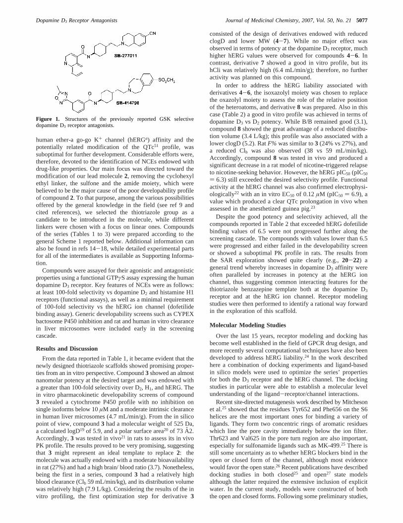
discovery was aimed at the discovery of novel chemical entitiesable to selectively modulate the dopamine D3 receptor. Thesuccessful result of this work led to the well characterizedtrans-N-[4-[2-(6-cyano-1,2,3,4-tetrahydroisoquinolin-2yl)ethyl]-cyclohexyl]-4-quinolininecarboxamide (SB-277011,1)12 andtrans-3-(2-(4-((3-(3-(5-methyl-1,2,4-oxadiazolyl))phenyl)car-boxamido)cyclohexyl)ethyl)-7-methylsulfonyl-2,3,4,5-tetrahy-dro-1H-3-benzazepine (SB-414796,2)13 (Figure 1).
While these molecules proved to be very useful for targetvalidation, part of their overall developability profile, including
* Author to whom correspondence should be addressed. E-mail:[email protected]; [email protected].
§ Psychiatry Centre of Excellence, GlaxoSmithKline Medicine ResearchCentre, Verona, Italy.
# Molecular Discovery Research, GlaxoSmithKline Medicine ResearchCentre, Verona, Italy.
⊥ Safety Assessment, GlaxoSmithKline Medicine Research Centre,Verona, Italy.
∇ Neurology Centre of Excellence, GlaxoSmithKline Medicine ResearchCenter, Harlow, UK.
† Psychiatry Centre of Excellence, GlaxoSmithKline Medicine ResearchCentre, Harlow, UK.
‡ Molecular Discovery Research, GlaxoSmithKline Medicine ResearchCentre, Harlow, UK.
| Safety Assessment, GlaxoSmithKline Medicine Research Centre, Har-low, UK.
@ St. John’s University.X Current address: European Patent Office, Munich, Germany.
a Abbreviations: CPP, conditioned place preference; Ach, acetylcholine;hERG, human ether-a go-go K+ channel; NCE, novel chemical entity; PK,pharmacokinetic; P450, cytochrome P450; hCli, human intrinsic clearance;MW, molecular weight; clogD, calculated logD; PSA, polar surface area;F%, bioavailability; B/B, brain/blood; Clb, blood clearance;Vd, distributionvolume; SDM, site-directed mutagenesis; rCBV, relative cerebral bloodvolume; FLIPR, fluorescent imaging plate reader; GPCR, G-protein coupledreceptor; TM, transmembrane; SPA, scintillation proximity assay; ECG,electrocardiogram.
5076 J. Med. Chem.2007,50, 5076-5089
10.1021/jm0705612 CCC: $37.00 © 2007 American Chemical SocietyPublished on Web 09/15/2007
human ether-a go-go K+ channel (hERGa) affinity and thepotentially related modification of the QTc51 profile, wassuboptimal for further development. Considerable efforts were,therefore, devoted to the identification of NCEs endowed withdrug-like properties. Our main focus was directed toward themodification of our lead molecule2, removing the cyclohexylethyl linker, the sulfone and the amide moiety, which werebelieved to be the major cause of the poor developability profileof compound2. To that purpose, among the various possibilitiesoffered by the general knowledge in the field (see ref 9 andcited references), we selected the thiotriazole group as acandidate to be introduced in the molecule, while differentlinkers were chosen with a focus on linear ones. Compoundsof the series (Tables 1 to 3) were prepared according to thegeneral Scheme 1 reported below. Additional information canalso be found in refs 14-18, while detailed experimental partsfor all of the intermediates is available as Supporting Informa-tion.
Compounds were assayed for their agonistic and antagonisticproperties using a functional GTPγS assay expressing the humandopamine D3 receptor. Key features of NCEs were as follows:at least 100-fold selectivity vs dopamine D2 and histamine H1receptors (functional assays), as well as a minimal requirementof 100-fold selectivity vs the hERG ion channel (dofetilidebinding assay). Generic developability screens such as CYPEXbactosome P450 inhibition and rat and human in vitro clearancein liver microsomes were included early in the screeningcascade.
Results and Discussion
From the data reported in Table 1, it became evident that thenewly designed thiotriazole scaffolds showed promising proper-ties from an in vitro perspective. Compound3 showed an almostnanomolar potency at the desired target and was endowed witha greater than 100-fold selectivity over D2, H1, and hERG. Thein vitro pharmacokinetic developability screens of compound3 revealed a cytochrome P450 profile with no inhibition onsingle isoforms below 10µM and a moderate intrinsic clearancein human liver microsomes (4.7 mL/min/g). From the in silicopoint of view, compound3 had a molecular weight of 525 Da,a calculated logD19 of 5.9, and a polar surface area20 of 73 A2.Accordingly,3 was tested in vivo21 in rats to assess its in vivoPK profile. The results proved to be very promising, suggestingthat 3 might represent an ideal template to replace2: themolecule was actually endowed with a moderate bioavailabilityin rat (27%) and had a high brain/ blood ratio (3.7). Nonetheless,being the first in a series, compound3 had a relatively highblood clearance (Clb 59 mL/min/kg), and its distribution volumewas relatively high (7.9 L/kg). Considering the results of the invitro profiling, the first optimization step for derivative3
consisted of the design of derivatives endowed with reducedclogD and lower MW (4-7). While no major effect wasobserved in terms of potency at the dopamine D3 receptor, muchhigher hERG values were observed for compounds4-6. Incontrast, derivative7 showed a good in vitro profile, but itshCli was relatively high (6.4 mL/min/g); therefore, no furtheractivity was planned on this compound.
In order to address the hERG liability associated withderivatives4-6, the isoxazolyl moiety was chosen to replacethe oxazolyl moiety to assess the role of the relative positionof the heteroatoms, and derivative8 was prepared. Also in thiscase (Table 2) a good in vitro profile was achieved in terms ofdopamine D3 vs D2 potency. While B/B remained good (3.1),compound8 showed the great advantage of a reduced distribu-tion volume (3.4 L/kg); this profile was also associated with alower clogD (5.2). RatF% was similar to3 (24% vs 27%), anda reduced Clb was also observed (38 vs 59 mL/min/kg).Accordingly, compound8 was tested in vivo and produced asignificant decrease in a rat model of nicotine-triggered relapseto nicotine-seeking behavior. However, the hERG pIC50 (pIC50
) 6.3) still exceeded the desired selectivity profile. Functionalactivity at the hERG channel was also confirmed electrophysi-ologically22 with an in vitro EC50 of 0.12µM (pIC50 ) 6.9), avalue which produced a clear QTc prolongation in vivo whenassessed in the anesthetized guinea pig.23
Despite the good potency and selectivity achieved, all thecompounds reported in Table 2 that exceeded hERG dofetilidebinding values of 6.5 were not progressed further along thescreening cascade. The compounds with values lower than 6.5were progressed and either failed in the developability screenor showed a suboptimal PK profile in rats. The results fromthe SAR exploration showed quite clearly (e.g.,20-22) ageneral trend whereby increases in dopamine D3 affinity wereoften paralleled by increases in potency at the hERG ionchannel, thus suggesting common interacting features for thethiotriazole benzazepine template both at the dopamine D3
receptor and at the hERG ion channel. Receptor modelingstudies were then performed to identify a rational way forwardin the exploration of this scaffold.
Molecular Modeling Studies
Over the last 15 years, receptor modeling and docking hasbecome well established in the field of GPCR drug design, andmore recently several computational techniques have also beendeveloped to address hERG liability.24 In the work describedhere a combination of docking experiments and ligand-basedin silico models were used to optimize the series’ propertiesfor both the D3 receptor and the hERG channel. The dockingstudies in particular were able to establish a molecular levelunderstanding of the ligand-receptor/channel interactions.
Recent site-directed mutagenesis work described by Mitchesonet al.25 showed that the residues Tyr652 and Phe656 on the S6helices are the most important ones for binding a variety ofligands. They form two concentric rings of aromatic residueswhich line the pore cavity immediately below the ion filter.Thr623 and Val625 in the pore turn region are also important,especially for sulfonamide ligands such as MK-499.25 There isstill some uncertainty as to whether hERG blockers bind in theopen or closed form of the channel, although most evidencewould favor the open state.26 Recent publications have describeddocking studies in both closed25 and open27 state modelsalthough the latter required the extensive inclusion of explicitwater. In the current study, models were constructed of boththe open and closed forms. Following some preliminary studies,
Figure 1. Structures of the previously reported GSK selectivedopamine D3 receptor antagonists.
Dopamine D3 Receptor Antagonists Journal of Medicinal Chemistry, 2007, Vol. 50, No. 215077

however, most of the docking of the receptor antagonists wasperformed in the closed form of the channel, as only in thisstate could they interact with several of the key aromatic residuessimultaneously. In addition to these docking studies, otherparameters were monitored in the design phase, in particularlipophilicity and basicity.
The ligands were docked manually in the hERG cavitydescribed above, with full conformational flexibility of both theligand and protein taken into consideration. Only low energystates of the former were allowed while the protein side chainrotamer angles were set within the limits defined by the Karplusrotamer library.28 Multiple starting poses were set up which werethen fully minimized. The results for compound10are discussedbelow.
Although the ligand could be docked in a linear conformation,this was only possible immediately below the ion gate, wherevery few interactions with the aromatic residues above werefound. An alternative pose with the ligand oriented along thechannel axis led to an unlikely result where one end of the ligandextended below the bottom of the helical bundle. By contrastmost of the resulting poses had compound10 in a “U-shaped”orientation as seen in Figure 2A. This is undoubtedly due to aninduced fit of the ligand to the aromatic cavity. Thus, not onlydoes the compound exhibit good interactions with all four ofthe Phe656 residues as well as two of the Tyr652s but theintramolecularπ-stacking of the ligand is also entropicallyfavored. From Figure 2A it can be seen that the protonated
Table 1. Functional Activity at the Human Dopamine D3 Receptor and Selectivity for 5-Oxazolyl Derivatives
entry R hD3-GTPγS fpKib hD2-GTPγS fpKi
bhH1-FLIPRc
pKb hERG pIC50 PSA, Å cLogD
1 not applicable 8.4 6.4 6.2 5.7 69 3.82 not applicable 9.2 6.1 NT 5.6 105 2.93 2-methyl -5-quinolinyl 8.7 < 6.1 6.3 6.1 73 5.94 2-thienyl 8.5 6.6 6.4 6.5 60 5.25 2-indolyl 8.9 6.0 5.7 7.3 76 5.16 5-indolyl 8.8 6.2 6.2 7.4 76 5.57 N-methyl-2-pyrrolyl 8.8 5.9 6.0 6.3 65 4.2
a SEM for D3 GTPγS, H1 FLIPR and hERG data sets is(0.1 and for the D2 GTPγS data is(0.2. b fpKi ) functional pKi obtained from the GTPγSfunctional assay.c FLIPR ) fluorescent imaging plate reader.
Table 2. Functional Activity at the Human Dopamine D3 Receptor and Selectivity for 5-Isoxazolyl Derivatives
entry RhD3-GTPγS
fpKib
hD2-GTPγSfpKi
bhH1-FLIPRc
pKb
hERGpIC50 PSA, Å cLogD
8 2-methyl-5-quinolinyl 8.7 6.0 6.2 6.3 73 5.29 2-methyl-6-quinolinyl 9.0 5.7 NT NT 73 5.2
10 2,3-dimethyl-6-quinoxalinyl 8.2 5.2 NT 6.5 86 4.911 Ph 8.8 6.4 7.0 6.8 60 4.912 4-cyano Ph 8.4 6.0 NT 7.7 84 4.513 4-trifluoromethylphenyl 8.5 6.3 5.7 7.2 60 5.814 4-methylphenyl 8.8 6.4 6.5 7.6 60 5.315 2-pyridinyl 8.6 <5.8 6.7 6.6 73 3.816 4-pyridinyl 7.6 6.0 6.2 6.1 73 3.617 2-thienyl 8.9 6.6 7.1 6.9 60 4.518 1-methyl-pyrrol-2-yl 9.1 <6.4 7.4 6.6 65 3.519 2-chlorophenyl 8.6 6.5 6.9 7.2 60 5.020 3-chorophenyl 8.8 6.7 7.4 7.1 60 5.721 4-chlorophenyl 9.2 6.4 7.2 7.6 60 5.622 3,4-dichlorophenyl 9.0 6.3 6.9 8.0 60 6.323 2-fluorophenyl 8.8 5.9 6.6 6.6 60 5.124 Me 7.4 <5.9 6.1 6.1 60 2.725 CF3 8.0 6.8 6.9 6.5 60 3.726 i-Pr 8.0 6.2 7.7 5.9 60 3.527 t-Bu 8.3 6.8 6.5 6.4 60 3.928 cyclopentyl 8.7 6.6 7.1 6.1 60 4.229 4-methyl-cyclohexyl 8.9 6.9 6.7 6.9 60 5.230 tetrahydropyran-4-yl 8.9 5.9 5.8 5.4 69 2.831 2-methoxyphenyl 8.5 7.1 6.7 7.4 69 4.632 3-methoxyphenyl 9.1 8.1 6.9 NT 69 5.033 4-methoxyphenyl 9.4 6.7 NT 7.4 69 5.034 6-methoxy-3-pyridinyl 8.6 5.9 6.60 6.8 82 4.6
a SEM for D3 GTPγS, H1 FLIPR and hERG data sets is(0.1 and for the D2 GTPγS data is(0.2. b fpKi ) functional pKi obtained from the GTPγSfunctional assay.c FLIPR ) fluorescent imaging plate reader.
5078 Journal of Medicinal Chemistry, 2007, Vol. 50, No. 21 Micheli et al.

nitrogen and the triazole and quinoxaline rings all form hydrogenbonds with serines in the pore.
Compound10 was found to adopt a similar “U-shaped”conformation” when docked into a model of the dopamine D3
receptor. As clearly depicted in Figure 2B, the best putativebinding mode showed a strong salt bridge between the basicbenzazepine nitrogen and Asp110 on transmembrane helix 3(TM3). This aspartate is believed to be the primary binding sitefor most basic compounds binding to the monoamine neu-rotransmitter receptors. Hydrogen bond interactions were ob-served between the phenylisoxazole moiety and Ser192 (TM5)and between the quinoxaline nitrogen and Ser182 (secondextracellular loop) whileπ-stacking interactions were seen forthe quinoxalinyl-triazole moiety with Phe345 and His349 (TM6).The D3 selectivity of these compounds can be explained by thehydrogen bond between the triazole ring and Thr368. Thecorresponding residue in the D2 receptor is a phenylalanine (Phe)which would be expected to cause a severe steric clash withthese ligands.
The models, in agreement with the SAR, suggest that incompound10 the same fragments (basic nitrogen; quinoline andoxazole) necessary for potency and selectivity at the dopamine
D3 receptor are also the key features which interact “negatively”with the hERG channel. To further test this hypothesis,derivatives 24-28 were prepared and a clear associationbetween potency and lipophilicity was observed for both targets.To maintain the potency at D3 it was therefore important tokeep these key interactions (Figure 2B) while, at the same time,introducing new features which might reduce activity at thehERG channel.
Medicinal Chemistry Strategies To Address hERG Li-ability. A number of approaches were used to tackle this issue.Among those approaches, two will be described based on theresults of the docking studies and on general knowledge aboutthe hERG channel. In the first one, new molecules weredesigned to increase the hydrophilicity (Figure 3A), by replacingthe heterocyclic substituents of the template on the thiotriazoleend. Favorable results were achieved in terms of reduction ofhERG activity while keeping relatively high potency at thedopamine D3 receptor. For example, compound30 had a verygood in vitro profile with a high potency at the dopamine D3
receptor (pKi ) 8.9) and a 1000-fold selectivity over dopamineD2 and H1 receptors. This high selectivity was also observedwith respect to the hERG channel. Based on this encouraging
Table 3. Functional Activity at the Human Dopamine D3 Receptor and Selectivity for 5-Pyrazolyl Derivatives
entry R R1 nhD3-GTPγS
fpKib
hD2-GTPγSfpKi
bhH1-FLIPRc
pKbhERGpIC50 PSA, Å cLogD
35 2-methyl-5-quinolinyl H 1 8.8 6.5 6.1 5.7 65 5.336 3,4-difluorophenyl H 1 8.8 <6.2 5.7 6.6 52 6.037 4-trifluoromethylphenyl H 1 8.1 6.3 6.3 7.5 52 5.438 4-methyl-1,3-oxazol-5-yl H 1 7.9 <6.1 5.9 5.6 78 3.439 2-methyl-5-quinolinyl H 0 7.8 <6.2 <5.6 5.7 65 5.840 5-methyl-2-pyrazinyl H 0 8.3 <6.2 <5.6 5.7 78 4.241 3,4-difluorophenyl H 0 8.7 <6.2 <5.6 6.3 52 5.842 2-methyl-5-quinolinyl Me 1 8.8 <6.3 5.9 5.9 65 5.6
a SEM for D3 GTPγS, H1 FLIPR and hERG data sets is(0.1 and for the D2 GTPγS data is(0.2. b fpKi ) functional pKi obtained from the GTPγSfunctional assay.c FLIPR ) fluorescent imaging plate reader.
Scheme 1.General Synthetic Procedures for the Preparation of Compounds3-42a
a i: (1) C3H3NH2, N-(3-dimethylaminopropyl)-N-ethylcarbodiimide (EDC), HOBt, CH2Cl2, RT, overnight; (2) refluxing AcOH., cat. Hg(OAc)2, 2 h; ii:(1) TFA; CH2Cl2, RT, 2 h; (2): Br(CH2)3Cl or Br(CH2)2Cl, or Br(CH2)4Cl ,TEA, refluxing THF, 2 h; iii: DMF, cat. NaI, K2CO3, 60 °C, 24 h; iv (1)NH2OH·HCl/Py, RT, 3 h; (2)N-chlorosuccinimide (NCS), DMF, 40°C, 1.5 h; (3) C3H5Cl, TEA, RT, overnight; v: (1) K2CO3, MeOH/H2O, 50°C; 2 h; :Br(CH2)3Cl or Br(CH2)2Cl, TEA, refluxing THF, 2 h; vi (1) C9H18N2O2; LDA; -78 °C to RT; (2) TFA; CH2Cl2, RT, 2 h; (3) : Br(CH2)3Cl or Br(CH2)2Cl,TEA, refluxing THF, 2 h.
Dopamine D3 Receptor Antagonists Journal of Medicinal Chemistry, 2007, Vol. 50, No. 215079

profile, a PK evaluation of30 in the rat was performed.Bioavailability was good (F ) 50%), with low Vd (2.4 L/kg)and low Clb (33 mL/min/kg). Unfortunately, brain penetrationwas poor (B/B) 0.1) and so was the actual brain concentration.Considering the fact that the MW of compound30 was 468 Dawith a similar PSA to compound8 which had a B/B) 3.1(PSA) 69 vs 73 A2), the low brain concentration was probablyrelated to the much reduced clogD (2.8 vs 5.2), unless somespecific efflux mechanism was involved for this particularcompound.
In the second approach, (Figure 3B), the effort was relatedto the reduction ofπ interaction within the hERG channel ofthe isoxazolyl moiety. Accordingly, specific compounds weredesigned to achieve a greater dihedral angle with the benza-zepine moiety, also reducing PSA while maintaining MW andclogD. In house developed computational tools were used toselect the appropriate bioisosteric replacements for the isoxazolylmoiety in the benzazepine class and synthetic feasibility wasused to filter among the different templates. The “top scorer”of this exercise was represented by an N-methylated pyrazolyltemplate, and this result is clearly related to the benzazepinetemplate here described in agreement with the specific SARhere reported.
When compound35 (MW ) 530 Da, clogD) 5.4, PSA)65 A2) was prepared, it showed (Table 3) the desired in vitro
profile and achieved the appropriate selectivity over the hERGchannel. Further exploration of this subseries demonstrated aSAR consistent with previously obtained results: a clear trendwas once again observed between lipophilicity and hERGaffinity. A slight increase in clogD with respect to35 (e.g.,36clogD) 5.5) led to the same dopamine D3 affinity and a slightlyhigher hERG value, while a marked increase in clogD (e.g.,37clogD ) 6.1) was associated with a slight decrease in thedopamine D3 affinity, but a significant increase in the hERGvalue (pIC50 ) 7.5 vs 5.2). Once again, an appropriately chosenhydrophilic substituent (38) led to a slight decrease in thedopamine D3 affinity but to a marked reduction in the hERGpIC50. According to the receptor and channel models, a reductionin the linker length should have provided active compounds,with a slightly reduced hERG activity. Compounds39-41werein agreement with the predicted potencies. Finally, the introduc-tion of anR-methyl group (42) should have been beneficial interms of hERG reduction and actually an in vitro profile similarto 35 was obtained; unfortunately a very high hCli was alsoobserved, probably due to the possible metabolic liability ofthe methyl group.
Compound35 was also endowed with a P450 profile withno inhibition on single isoforms below 10µM and ideal hCli(1.8 mL/min/g liver) and was therefore selected for furthercharacterization. The PK profile in rat showed a moderate Clb
(38 mL/min/kg), acceptableF% (15%), a low-to-moderateVd
(3.7 L/kg), and a good half-life (T1/2 ) 1.7 h); moreover, aspredicted by our calculations, the B/B ratio of35was high (2.5).
Before submission of the compound to in vivo disease models,compound35 was first submitted to hERG electrophysiologydetermination. The reported affinity was 0.43µM (pIC50 ) 6.5),leading to an almost 4-fold improvement with respect tocompound8 in accordance with the computational models. Moreimportantly, when tested in vivo in the anesthetized guinea pigmodel, compound35 showed no QTc prolongation or anycardiac liability despite a high exposure in blood (Cmax of 34.4µg/mL @ 25 mg/kg).
Following these positive results, compound35was thoroughlycharacterized.
Further in Vitro Characterization of Compound 35.Compound35 displayed high selectivity over D2 receptors(Table 4) and a 100-fold selectivity over a wide range ofreceptors and enzymes (see detailed Cerep Report in theSupporting Information). Moreover,35 did not stimulate [35S]-GTPγS binding above basal levels in human recombinantdopamine D3 receptors when tested alone up to a concentrationof 10 µM (Table 4). Derivative35 behaved as a potentcompetitive antagonist with a pKb derived from Schild analysisof 7.93 (slope 1.0), comparable to the value of pKi obtainedalso from filtration binding data (D3 ) 7.99 and D2 ) 5.02)(Table 4). Finally, 35 was tested in a [125I]-7-OH-PIPATcompetition binding assay on brain homogenates from ratnucleus accumbens and olfactory tubercles. In these saturationbinding experiments, [125I]-7-OH-PIPAT bound to a single classof receptor sites with a pKD value of 9.34( 0.11 and aBmax of144( 16 fmol mg-1 protein (n ) 8). Compound35completelydisplaced, in a monophasic manner, the specific [125I]-7-OH-PIPAT binding to rat membrane preparation. A pKi value of8.48( 0.08 (n ) 4) was found, thus confirming a high affinityto the rat dopamine D3 receptor.
Further in Vivo Assays of Compound 35. An in vivo studywas conducted to assess the efficacy of compound35 on theexpression of nicotine-induced conditioned place preference inthe rat. In the CPP paradigm, animals are given an injection of
Figure 2. (A) Interactions of compound10 in a “U-shaped” conforma-tion which is energetically preferred over an extended conformationbecause of increased interactions with aromatic residues in the cavity.The backbone ribbon of the pore domain is included but that of the S5and S6 helices has been removed for clarity. (B) A similar “U-shaped”conformation is found in docking to the dopamine D3 receptor model.Again the backbone has been removed for clarity.
5080 Journal of Medicinal Chemistry, 2007, Vol. 50, No. 21 Micheli et al.

a drug or vehicle and confined or “paired” to a specificenvironment with distinct cues. This pairing of the animal withspecific cues after being given vehicle or drug is repeated.Subsequently, on the test day, animals are not given anytreatment (drug-free) and are allowed to freely explore the CPPapparatus to determine if they prefer an environment in whichthey previously received drug compared to an environment inwhich they previously received vehicle. If the animals approachand spend a significantly greater amount of time in the drug-paired environment, one can reasonably infer that the drug wasappetitive and this appetitive value is coded in the brain and isaccessible in the drug-free state.
Compound35 (1, 3, 10 mg/kg, i.p.) significantly reducednicotine CPP in a dose-dependent manner (one-way ANOVA:F[4,76] ) 31.7; P < 0.0001), thus confirming that it cancompletely reverse the incentive motivational properties ofnicotine (Table 5).
A second study also assessed the efficacy of compound35in preventing reinstatement of nicotine-seeking behavior. Inreinstatement experiments, rats are trained to self-administerdrugs intravenously by pressing a lever. During a subsequent
period, the drug is no longer available, but the rats are free totry to obtain the drug (a period of “extinction training”). Afterextinction of responding, the ability of various events to reinitiatedrug-seeking can be investigated. In both human addicts andanimal reinstatement models, a return to drug use can beprecipitated by three distinct types of stimuli: (1) re-exposureto the drug itself, (2) exposure to environmental cues that hadbeen previously associated with drug intake, and (3) exposureto stress. In this experiment, we showed that compound35 (1,3, 10 mg/kg i.p.) could prevent nicotine-triggered reinstatementof nicotine seeking behavior in a dose-dependent manner. Anoverall ANOVA showed that there was a significant differencebetween treatment groups (F[4,44] ) 5.36, P < 0.001]. TheFisher’s LSD post-hoc test revealed that priming with nicotine-induced a significant increase in active lever pressing comparedto priming with saline (P < 0.001). Furthermore, compound35 (3 and 10 mg/kg) produced a significant decrease in thenumber of active lever presses compared to the vehicle/nicotinegroup (P < 0.05).
To exclude the possibility that these positive results wereinfluenced by any impairment in locomotor activity or motorcoordination (typical effect of mixed D3/ D2 receptor antago-nists),35 was also tested in the rat Rotarod. Compound35 ata high dose of 10 mg/kg i.p. failed to affect enduranceperformance at any of the posttreatment times tested in theRotarod. An ANOVA with a main factor of treatment and arepeated measurements factor of test time showed that therewas no significant effect of treatment (F[2,19] ) 0.1], nosignificant effect of test time (F[2,38] ) 2.02], and no significanttreatment by test time interaction (F[4,38] ) 0.35].
To confirm the ability of the dopamine D3 mechanism to actvs different drugs of abuse, compound35 was also tested onthe expression of cocaine-induced CPP in the rat. As previouslyseen with nicotine, compound35 (1, 3, 10 mg/kg, i.p.) alsosignificantly blocked cocaine CPP in a dose-dependent manner(one-way ANOVA: F[4,36] ) 18.09; P< 0.0001), thus confirm-ing that it can completely reverse the incentive motivationalproperties of cocaine (Table 6).
We have also recently shown that1 (10, 30 mg/kg i.p.)significantly reduced alcohol preference in alcohol Preferring
Figure 3. Alternative medicinal chemistry strategies to address hERG liability.
Table 4. Profile of Compound35 in Human Recombinant DA D3 Radioligand Binding, [35S]GTPγS Binding in Cell Membranes Expressing hDA D3
Receptors, and [125I]-7-OH-PIPAT Competition Binding Assay on Brain Homogenates from Rat Brain
pKi GTPγS pKi filtration binding
hDA D3 hDA D2 hDA D3 hDA D2
pKb,hDA D3
rat native tissue,r DA D3
35 9.0 6.5 8.0 5.0 7.93 8.48
Table 5. Effect of a Single i.p. Administration of Vehicle and35 (1, 3,10 mg/kg) on the Expression of the Conditioned Place PreferenceResponse to 0.4 mg/kg s.c. of (-)-Nicotine in Adult MaleSprague-Dawley Rats
time spent in chambers (min)
treatment pairings drug given on test day paired unpaired
vehicle/vehicle vehicleb 7.55( 0.22a 7.44( 0.22vehicle/nicotine vehicle 10.07( 0.26c 4.92( 0.26vehicle/nicotine 35, 1 mg/kg 7.19( 0.22d 7.80( 0.22vehicle/nicotine 35, 3 mg/kg 6.71( 0.25d 8.29( 0.25vehicle/nicotine 35, 10 mg/kg 7.53( 0.22d 7.47( 0.22
a Each value represents the mean number of minutes spent in eachchamber( SEM. b The vehicle was 1 mL/kg s.c. of deionized, distilledwater for the pairings. The vehicle used on the test day was 1 mL/kg i.p.of 20% hydroxypropyl-â-cyclodextrin.c Significantly greater than vehicle/vehicle pairings and vehicle on the test day,P < 0.0001 ANOVA andStudent-Newman-Keuls test.d Significantly less than vehicle/nicotinepairings+ vehicle on the test day,P < 0.0001, ANOVA and Student-Newman-Keuls test.
Dopamine D3 Receptor Antagonists Journal of Medicinal Chemistry, 2007, Vol. 50, No. 215081

(P) and Non-Preferring (NP) rats in a dose-dependent manner.29
Similarly, 35 (1, 3, 10 mg/kg i.p.) also significantly attenuatedoral operant alcohol self-administration in the C57BL/6N mouse.Our results show that compound35 significantly decreased thenumber of active lever presses associated with alcohol intake(one-way ANOVA: F[3,68] ) 3.86; P< 0.05) without alteringthe number of inactive lever presses (one-way ANOVA:F[3,68]
) 1.19;P ) 0.32).Previous studies have shown that1 significantly improves
the learning deficit produced by the nonselective muscarinicreceptor antagonist scopolamine and the anxiogenic benzodi-azepine inverse agonist FG-7142 without altering the normallearning process in nonimpaired rats.30 Accordingly, the effectof compound35 (3, 10 mg/kg i.p.) on dialysate levels of AChin the rat medial prefrontal cortex was examined. Microdialysissamples were analyzed using liquid chromatography coupledwith tandem mass spectrometry (LC-MS/MS) for the detectionof ACh without the use of acetylcholinesterase inhibitors.31
Compound35 (10 mg/kg i.p.) significantly increased the levelsof ACh up to about 250%. These findings demonstrate thatcompound35, like other selective DA D3 receptor antagonistsfrom different chemical series,30,31 increases extracellular con-centrations of ACh in the rat medial prefrontal cortex and furtherstrengthen the likelihood that selective DA D3 receptor antago-nists may have cognitive enhancing properties.
Finally, in analogy to what was shown in phMRI experimentsin rats after acute pretreatment with1 (i.e., potentiation of the
amplitude of the relative cerebral blood volume (rCBV) responseto d-amphetamine in a regionally specific manner),32 compound35 (20 mg/kg i.p.) potentiated the amphetamine response in theD3-rich NAc, but also in a number of structures outside the focaldistribution of the D3 receptor, and associated with reinstatementof drug seeking behavior, such as the ventral subiculum regionof the hippocampus, the amygdale, and striatum (Figure 4).
Conclusions
Starting from the potent and selective dopamine D3 receptorantagonist previously reported (2), a new series of 1,2,4-triazol-3-yl-thiopropyl-tetrahydrobenzazepines was identified. Rationaldesign and computational tools helped us to overcome hERG-related issues and to design a number of products endowed withhigh affinity and selectivity for the dopamine D3 receptor. Inaddition, selected compounds of this family series proved tohave appropriate developability characteristics (P450, Cli, F%)and good CNS penetration. In particular, compound35showedreduced hERG liability in vitro and no QTc prolongation invivo. Compound35 was shown to be a potent and selectivedopamine D3 receptor antagonist in vivo with no sign oflocomotor liability, a typical effect produced by nonselectiveD3/D2 receptor antagonists.
Provided that in vivo preclinical evidence can be extrapolatedto human, selective DA D3 receptor antagonists belonging tothis new chemical series show the highest promise for thetreatment of drug addiction, psychosis, and schizophrenia.
Figure 4. Region-specific potentiation of phMRI amphetamine (1 mg/kg i.v.) response by acute pretreatment (30 min previously) with35 (20mg/kg i.p.). (a) Statistical map of vehicle/amphetamine [n ) 13] vs 35/amphetamine [n ) 11] (P < 0.01). (b) Mean ((SEM) rCBV changesfollowing amphetamine challenge for vehicle/amphetamine [n ) 13] and35/amphetamine [n ) 11] groups in selected ROIs, including the mPFCas a nonpotentiated control region (t-test veh/amp vs35/amp; *** P < 0.001). Abbreviations: NAc) nucleus accumbens, dCPU) dorsal caudateputamen, Amyg) amygdala, vSub) ventral subiculum, VPThal) ventroposterolateral thalamic nuclei, mPFC) medial prefrontal cortex.
Table 6. Effect of a Single i.p. Administration of Vehicle and35 (1, 3, 10 mg/kg) on the Expression of the Conditioned Place Preference Response to15 mg/kg i.p. of (-)-Cocaine HCl in Adult Male Sprague-Dawley Rats
time spent in chambers (min)
treatment pairings drug given on test day paired unpaired
vehicle/vehicle vehicleb 7.42( 0.40a 7.58( 0.40vehicle/cocaine vehicle 11.26( 0.48c 3.74( 0.48vehicle/cocaine 35, 1 mg/kg 7.58( 0.33d 7.42( 0.33vehicle/cocaine 35, 3 mg/kg 7.06( 0.38d 7.94( 0.38vehicle/cocaine 35, 10 mg/kg 8.14( 0.43d 6.86( 0.43
a Each value represents the mean number of minutes spent in each chamber( SEM. b The vehicle was 1 mL/kg s.c. of deionized, distilled water for thepairings. The vehicle used on the test day was 1 mL/kg i.p. of 20% hydroxypropyl-â-cyclodextrin.c Significantly greater than vehicle/vehicle pairings andvehicle on the test day,P < 0.0001 ANOVA and Student-Newman-Keuls test.d Significantly less than vehicle/nicotine pairings+ vehicle on the test day,P < 0.0001, ANOVA and Student-Newman-Keuls test.
5082 Journal of Medicinal Chemistry, 2007, Vol. 50, No. 21 Micheli et al.

Experimental Section
Biological Test Methods. In Vitro Studies. Human Recom-binant D3 Radioligand Binding Assays. Radioligand bindingstudies using [3H]-FLB-45733 were performed according to thefollowing protocol. Test compounds (10 point, half log seriallydiluted in 5 mM HCl) were incubated with assay buffer (50 mMTRIS, 10 mM NaCl, 5 mM KCl, 1 mM MgCl2, 2 mM CaCl2, pH7.4), 0.2 nM [3H]-FLB-457, and CHO cell membranes containingeither hD3 or rD3 cloned receptors for 45 min at 37°C. Total bindingwas defined using 5 mM HCl, and nonspecific binding was definedusing 10µM haloperidol. The reaction was terminated by rapidfiltration through GF/B filterplates presoaked in double distilledH2O, followed by 3× 1 mL washes with ice cold 50 mM TRISpreset buffer (pH 7.7). Bound radioactivity was determined byscintillation spectrometry.
[35S] GTPγS Functional Binding Assay in Cell MembranesExpressing hD3 Receptors. In vitro functional studies wereperformed according to the following [35S]-GTPγS protocol. Testcompounds (10 point, half log serially diluted in assay buffer) wereincubated with 10µM GDP and CHO cell membranes containingeither hD2 or hD3 cloned receptors for 30 min at 30°C. 100 pM[35S]-GTPγS was added before a second incubation for 30 min at30 °C. Basal binding was defined using assay buffer, andnonspecific binding was defined using 20µM unlabeled GTPγS.The reaction was terminated by rapid filtration through GF/Bfilterplates presoaked in ddH20, followed by 3× 1 mL washeswith ice cold 20 mM HEPES, 10 mM MgCl2 buffer (pH 7.4). Boundradioactivity was determined by scintillation spectrometry.
[125I]-7-OH-PIPAT Competition Binding Assay on BrainHomogenates from Rat Brain.Homogenates from frozen nucleusaccumbens and olfactory tubercles were prepared as described byBurris et al. (1994).34 In saturation experiments, increasing con-centrations of [125I]-7-OH-PIPAT (15 pM to 2 nM) were incubatedwith 12 µg/well of homogenates for 45 min at 37°C in a finalvolume of 200µL of 50 mM Tris-HCl (pH7.0), 50 mM NaCl, 100µM Gpp(NH)p and 0.02% BSA, i.e., conditions which inhibit [125I]-7-OH-PIPAT binding to D2 and 5HT1A receptors.34 Nonspecificbinding was determined by the presence of 1µM 1. In competitionbinding experiments, increasing concentrations of35 (17 pM to 1µM) were incubated as above in the presence of 0.1 nM [125I]-7-OH-PIPAT.
Reactions were stopped by filtration through GF/C 96-well filterplates presoaked in 0.3% polyethylenimine using a cell harvester.Filters were washed three times with 1 mL of ice-cold 50 mM Tris-HCl (pH 7.7), and radioactivity was counted in a microplatescintillation counter (Top Count, Perkin-Elmer).
Radioligand binding data were analyzed by nonlinear regressionanalysis using GraphPad Prism 4.0 (GraphPad Software, CA).Determination ofKD andBmax of [125I]-7-OH-PIPAT from saturationexperiments was assessed by using one-site binding (hyperbola)equation. Curve fitting from competition-binding experiments wasdetermined by using the one-site competition equation afterchecking with theF test (P < 0.05) that the Hill slope in the four-parameter logistic equation was not statistically different from 1.0.Under this condition, IC50 values were converted toKi using theCheng-Prusoff equation.33 Results are expressed as mean pKi (SEM.
[125I]-7-OH-PIPAT ([125I](R)-trans-7-hydroxy-2-[N-propyl-N-(3′-iodo-2′-propenyl)amino]tetralin, 81.4 TBq/mmol) was purchasedfrom Perkin-Elmer Life and Analytical Sciences. Haloperidol andGpp(NH)p (guanylyl-5′-imidodiphosphate) were obtained fromSigma Chemicals.
hERG-3H-Dofetilide Binding Assay. hERG activity wasmeasured using3H-dofetilide binding in a scintillation proximityassay (SPA) format. The activity was measured with a Perkin-ElmerViewlux imager.
H1-FLIPR Assay. A functional response in CHO-hH1 cells wasmeasured using cytoplasmic calcium indicator Fluo-4. The changein cell fluorescence being measured in a FLIPR (λex ) 488 nm,λem ) 540 nm, Molecular Devices, UK).
P450 CYPEX Assay.Inhibition (IC50) of human CYP1A2, 2C9,2C19, 2D6, and 3A4 was determined using Cypex Bactosomesexpressing the major human P450s. A range of concentrations (0.1,0.2, 0.4, 1, 2, 4, and 10µM) of test compound were prepared inmethanol and preincubated at 37°C for 10 min in 50 mM potassiumphosphate buffer (pH 7.4) containing recombinant human CYP450microsomal protein (0.1 mg/mL; Cypex Limited, Dundee, UK) andprobe-fluorescent substrate. The final concentration of solvent wasbetween 3 and 4.5% of the final volume. Following preincubation,NADPH regenerating system (7.8 mg of glucose 6-phosphate, 1.7mg of NADP and 6 units of glucose 6-phosphate dehydrogenase/mL of 2% (w/v) NaHCO3; 25µL) was added to each well to startthe reaction. Production of fluorescent metabolite was thenmeasured over a 10-min time-course using a Spectrafluor plus platereader. The rate of metabolite production (AFU/min) was deter-mined at each concentration of compound and converted to apercentage of the mean control rate using Magellan (Tecansoftware). The inhibition (IC50) of each compound was determinedfrom the slope of the plot using Grafit v5 (Erithacus software, UK).Miconazole was added as a positive control to each plate. CYP450isoform substrates used were ethoxyresorufin (ER; 1A2; 0.5µM),7-methoxy-4-triflouromethylcoumarin-3-acetic acid (FCA; 2C9; 50µM), 3-butyryl-7-methoxycoumarin (BMC; 2C19; 10µM), 4-methylaminomethyl-7-methoxycoumarin (MMC; 2D6; 10µM),diethoxyflourescein (DEF; 3A4; 1µM) and 7-benzyloxyquinoline(7-BQ; 3A4; 25µM). The test was performed in three replicates.
Intrinsic Clearance (Cli) Assay. Intrinsic clearance (Cli) valueswere determined in rat and human liver microsomes. Test com-pounds (0.5µM) were incubated at 37°C for 30 min in 50 mMpotassium phosphate buffer (pH 7.4) containing 0.5 mg microsomalprotein/ mL. The reaction was started by addition of cofactor(NADPH; 8 mg/mL). The final concentration of solvent was 1%of the final volume. At 0, 3, 6, 9, 15, and 30 min an aliquot (50µL) was taken, quenched with acetonitrile containing an appropriateinternal standard and analyzed by HPLC-MS/MS. The intrinsicclearance (Cli) was determined from the first-order eliminationconstant by nonlinear regression using Grafit v5 (Erithacus software,UK), corrected for the volume of the incubation and assuming 52.5mg microsomal protein/g liver for all species. Values for Cli wereexpressed as mL/min/g liver. The lower limit of quantification ofclearance was determined to be when<15% of the compound hadbeen metabolized by 30 min, and this corresponded to a Cli valueof 0.5 mL/min/g liver. The upper limit was 50 mL/min/g liver.
Patch Clamp hERG Electrophysiology Assay.On the days ofexperimentation, a weighed amount of the test substance wasformulated in dimethyl sulfoxide (DMSO; lot no. U10781; Sigma-Aldrich, UK) by shaking, to give a stock concentration of 10 mMof the test compound. The 10 mM stock solution was serially dilutedin DMSO to give further stock solutions of 1 and 0.1 mM. Aliquotsof these stock solutions were added to bath solution to achieve finalperfusion concentrations of 0.1, 1, and 10µM test compound. Thecorresponding vehicle concentration in all test substance perfusionsolutions was 0.1% DMSO. Test substance stock formulations werefreshly prepared on each day of experimentation, stored at roomtemperature, and protected from light.
Reference Substance: E-4031 (batch no. MLE9446; Wako PureChemical Industries Ltd.). Stock solutions of E-4031 (100µM) wereprepared in reverse osmosis water, aliquoted, and stored atapproximately-20 °C until use. On the day of use, the 100µMstock solution was added to bath solution to give a final perfusionconcentration of 100 nM.
Bath and Pipet Solutions: The composition of bath solution(mM): NaCl 137; KCl 4; CaCl2 1.8; MgCl2 1.0;D-glucose 10;N-2-hydroxyethylpiperazine-N′-2-ethanesulfonic acid (HEPES) 10;pH 7.4 with 1 M NaOH. Pipet solution was prepared in batches,aliquoted, and stored frozen until the day of use. The compositionof pipet solution was (mM): KCl 130; MgCl2 1.0; ethylene glycolbis(â-aminoethyl ether)-N,N,N′,N′-tetraacetic acid (EGTA) 5; MgATP5; HEPES 10; pH 7.2 with 1 M KOH.
Study Design: Cells (passage number: 48) were transferred tothe recording chamber and continuously perfused (at approximately
Dopamine D3 Receptor Antagonists Journal of Medicinal Chemistry, 2007, Vol. 50, No. 215083

1-2 mL/min) with bath solution at room temperature. Highresistance seals (seal resistances>1.5 GΩ) were formed betweenthe patch electrodes (resistance range: 1.4-5.5 MΩ) and individualcells. The membrane across the electrode tip was then ruptured,and the whole-cell patch-clamp configuration was established. Oncea stable patch had been achieved, recording commenced in voltage-clamp mode, with the cell initially clamped at-80 mV. Currentswere evoked by stepping the membrane potential to+20 mV andthen to-50 mV (tail current). Test compound at 10, 1, and 0.1µM was used to produce (if any) inhibition of hERG tail currentand therefore to investigate the concentration-response relationship(n ) 3 cells/concentration). The effect of the vehicle (0.1% DMSO)was investigated in 3 cells. The effect of 100 nM E-4031 wasinvestigated in 2 of the vehicle-treated cells to confirm thesensitivity of the test system to an agent known to block hERGcurrent. All perfusion solutions were applied for approximately 10min.
Anesthetized Guinea Pig Model for the Assessment of QTProlongation. Prior to the experiment, six male Hartley guinea pigs(Charles River, France) weighing between 564 and 605 g on theday of the test were anesthetized with urethane (1-1.5 g/kg IP),tracheotomized, artificially ventilated with a tidal volume ofapproximately 1 mL/100 g at a rate of 54 cycles/min, and surgicallyprepared for the experiment (i.e., insertion of catheters in appropriateblood vessels, see below, and placement of surface electrodes forlead II electrocardiogram recording). The animal’s temperature waskept constant between 36.8 and 38.0°C. Once physiologicalparameters were stabilized (approximately 30 min followingsurgery), each animal was given either the vehicle (5% w/v dextrosein aqueous 0.9 w/v sodium chloride) or test compound by theintravenous route (via the jugular vein) over 15 min/treatment.
The following parameters were recorded: arterial pressure (viathe carotid artery), heart rate, electrocardiography (i.e., RR, PQ,QRS, and QT interval duration; the QT interval was corrected forheart rate changes according to Fridericia, Bazett, and Van deWater’s formula). Parameters were recorded prior to dosing toestablish baseline measurements and continuously during theinfusion period, but data were reported every 5 min during eachinfusion period. In addition, blood samples (0.3 mL) were collectedvia the femoral artery at the end of each infusion period fortoxicokinetic evaluations.
In Vivo Studies. All experiments were pre-reviewed andapproved by a local animal care committee in accordance with theguidelines of the “Principles of Laboratory Animal Care” (NIHpublication No 86-23, revised 1985) and with a Project Licensethat was obtained according to Italian law (Art. 7, Legislative Decreeno. 116, Jan 27, 1992), which acknowledges European Directive86/609/EEC on the care and welfare of laboratory animals.
Nicotine CPP. Male Sprague-Dawley rats (200 g at the startof the pairings, Taconic Farms, Germantown, NY) were used inall experiments. Animals were housed two per cage, and there wasno more than a 5% difference in body weight between thecagemates. Animals were kept on a 12 h lights on/12 h lights offschedule (lights on at 09.00 h). Food and water were freelyavailable. The conditioning and testing of all animals was carriedout between 11.00 and 18.00 h. All rats were naive and used onlyonce.
An automated, two-chambered, Plexiglas CPP apparatus wasused as previously described,36-38 with modifications. Briefly, thetwo pairing chambers of the apparatus were identical in dimensions(25 × 14 × 36 cm) and were separated by removable Plexiglasguillotine doors. The pairing chambers were composed of distinctvisual and tactile cues. The walls of one of the pairing chamberswere white with cage bedding on the floor, and the walls of thesecond chamber consisted of alternating white and black boxes (1.2× 1.8 cm) in a chessboard pattern and a Plexiglas floor. The twopairing chambers were separated by a third, neutral connectingtunnel, with one-half of the wall white and the other half with blackand white boxes.
Expression studies with compound35 or vehicle were dividedinto four phases: acclimation, handling, conditioning, and testing.
The animals were not exposed to the chambers prior to the start ofthe pairings. During days 1-3, animals were acclimated to theanimal facility. During handling (days 4-6), animals were trans-ported to the laboratory and handled for 5 min each. Duringconditioning (days 7-14), animals were exposed to once-dailyconditioning sessions. For each conditioning session, animals (10rats per group) were injected with nicotine (0.6 mg/kg s.c. in avolume of 1 mL/kg) or vehicle (1 mL/kg s.c.) and then immediatelyconfined for 30 min in an appropriate cue-specific chamber. Duringconditioning, nicotine was always paired with one cue-specificenvironment, and vehicle was paired with the other; nicotine orvehicle exposure (and appropriate environmental pairing) alternatedfrom day to day. This was done over an 8-day period, i.e., animalswere given four pairings with nicotine, and there was a 24-hseparation between exposure to vehicle and nicotine. The animalsin each group were randomly assigned to a 2× 2 factorial designwith one factor being the pairing chamber and the other factor beingthe order of conditioning. In the counterbalanced procedure, theanimals were randomly assigned to one of the two pairing chambers,so that half of the subjects received the drug in one compartment(white walls with bedding on the floor) and the other half in theother compartment (alternating black and white boxes with a smoothchamber floor). This procedure resulted in the animals receivingequal exposure to the two compartments and, because of randomassignment, controlled for their side preference. Another group of10 animals were paired with vehicle in both chambers of theapparatus. On the test day (day 15), animals were randomly dividedinto 5 groups of 10 and received compound35 (1, 3, 10 mg/kg,i.p.) or vehicle (20% 2-hydroxypropyl-â-cyclodextrin) in the homecage 30 min before they were placed in the apparatus and allowedfree access to both chambers for 15 min. The amount of time spentin each chamber was determined using an automated timing system.
(-)-Nicotine (+)-bitartrate salt was used in all experiments(Sigma-Aldrich Corporation, St. Louis, MO) and was dissolved insterile physiological saline and the pH adjusted to 7.4 with NaOH.The doses of nicotine were expressed as free base. 2-Hydroxypro-pyl-â-cyclodextrin was purchased from Tocris-Cookson Chemical(St. Louis, MO). Data were analyzed with a one-way ANOVA witha main factor of dose. Statistical significance was set at a probabilitylevel of P < 0.05.
Reinstatement of Nicotine-Seeking Behavior.Male ListerHooded rats (Charles River, Germany) were individually housedin a temperature-controlled environment with lights on from 06.00to 18.00 h. During the experiments, water was continuouslyavailable and animals were maintained at a constant body weightof 240-260 g (85% of their ad libitum body weight).
Behavioral testing (self-administration sessions and reinstatementphase) was conducted according to the same methodology previ-ously described and published.39 Compound35 was tested at 1, 3,and 10 mg/kg i.p. (30-min pretreatment time). The vehicle was asolution of or vehicle 20% 2-hydroxypropyl-â-cyclodextrin. Datawere analyzed by a two-way ANOVA with main factors of doseand time and repeated measurements over time. Statistical signifi-cance was set atP < 0.05 for all tests.
Rat Rotarod. Male Lister Hooded rats (Charles River, Germany)were individually housed in a temperature-controlled environmentwith lights on from 06.00 to 18.00 h. During the experiments, waterand food were continuously available. Rats were trained on theaccelerating Rotarod (4-40 rpm over 270 s; 7750, Ugo Basile,Italy) twice daily for two consecutive days. On the test day (day3), rats were treated with compound35 (10 mg/kg, i.p.) (n ) 10rats/group). Rats were then repeatedly tested for their enduranceperformance on the Rotarod 30, 60, and 120 min after treatment.Rotarod latencies were measured with a 300 s cutoff time.
Cocaine CPP.The cocaine CPP procedure was identical to theone described for nicotine except that for each conditioning sessionanimals were injected with (-)-cocaine HCl (15 mg/kg i.p. in avolume of 1 mL/kg) or vehicle (1 mL/kg i.p. of deionized, distilledwater). (-)-Cocaine HCl was purchased from Sigma Chemicals(St. Louis, MO).
5084 Journal of Medicinal Chemistry, 2007, Vol. 50, No. 21 Micheli et al.

Alcohol Self-Administration. Male C57BL/6N mice (CharlesRiver, Italy), weighing approximately 27 g at the start of theirtraining, were used in the present studies. All mice were experi-mentally naive, housed individually in a temperature controlledroom (20-22 °C) with a 12-h light-dark cycle (06.00-18.00 lighton), and had free access to tap water in their home cage.
The experiments were conducted according to the same meth-odology previously described and published.40 The respective effectsof acute treatment with compound35 were analyzed using a two-way ANOVA with main factors of dose and session (pre vs post).The differences between individual means were assessed with thepost-hoc Fisher’s PLSD test. Statistical significance was set at aprobability level ofp < 0.05 for all tests.
Extracellular Levels of Acetylcholine in the Medial PrefrontalCortex (mPFC). Male Sprague-Dawley rats (Charles River, Italy)weighing 250-300 g were group housed at 21( 1 °C with 50%humidity on a 12/12 light-dark cycle (light-on at 6 a.m.);experiments were carried out during the light phase.
The experiments were conducted according to the same meth-odology previously described and published.31 The effect ofcompound35 on extracellular levels of ACh was analyzed byANOVA consisting of a between-subjects factor of treatment anda repeated measurements factor of time. In addition, one-wayANOVA on the area under the curve (AUC) was performed toassess the main effect of drug treatment on ACh levels. The post-hoc Fisher’s protected least significant difference pairwise com-parison test was used where appropriate. Statistical significance wasset at a probability level ofp < 0.05 for all tests.
Pharmacological MRI. Male Sprague-Dawley rats (250 g to350 g; Charles-River, Como, Italy) were scanned in a BrukerBiospec 4.7T MRI scanner under 0.8% maintenance halothaneanaesthesia and neuromuscular blockade; animal preparation andmonitoring, and MRI setup and acquisition, were the same aspublished previously,32 except that the time series scan comprised16 contiguous slices of 1 mm thickness and a lower temporalresolution (80 s). The time series images were sensitized to changesin relative cerebral blood volume (rCBV) by the injection of a bloodpool contrast agent (Endorem, Guerbet, France; 2.67 mL/kg)following 5 reference image frames.32,41 Following 10 min equili-bration, animals were administered either 20 mg/kg compound35(N ) 11) or vehicle saline (N ) 13) i.p., followed 30 min later bya 1 mg/kg i.v.d-amphetamine challenge.
The time series signal changes upond-amphetamine injectionwere converted into rCBV changes on a pixelwise basis.41,42
Following spatial coregistration of the image data, the data weresmoothed with a Gaussian kernel of full-width-half-maximumequivalent to twice the in-plane pixel dimension, and the amphet-amine response amplitude in each pixel was quantified by meansof a general linear model analysis using the AFNI package (v2.23).Bilateral region of interest time courses corresponding to specificanatomical structures were also extracted from each subject andthe rCBV changes quantified in the same way. Differences in theamphetamine response magnitude between the two groups wereassessed usingt-tests.
Chemical Procedures. General. Experimental.NMR spectrawere obtained on Varian INOVA spectrometers (300 MHz, 400MHz, and 500 MHz). Chemical shifts are expressed inδ (ppm)units and peak multiplicity is expressed as follows: singlet (s),doublet (d), doublet of doublets (dd), triplet (t), multiplet (m), broad
singlet (br s), broad multiplet (br m). All mass spectrometricmeasurements were performed using a Micromass Platform LCZ(Waters, Manchester, UK) mass spectrometer operated in positiveelectrospray ionization mode. When LC/MS detection was per-formed, analytical conditions were used as reported in Table 7.
General Synthetic Procedures.For a description of the knownintermediates, please refer to refs 14-18 or to the detailedexperimental parts in the Supporting Information.
To a stirred solution of the appropriately decorated (oxazole,isoxazole, methylpyrazole, respectively, intermediates 44, 47, 50reported in Scheme 1) benzazepine scaffold in THF, containing1.1 equiv of triethylamine, was added 1-bromo-3-chloropropane(or appropriate alkylating agent) (1.1 equiv), and the solution wasrefluxed for 2 h. After workup and chromatographic purification,the resulting alkylating agent (respectively, 45, 48, 51 in Scheme1) was dissolved in DMF. A catalytic amount of NaI was addedand the appropriately substituted thiotriazole added together with1.2 equiv of K2CO3. The temperature was raised to 60°C and thesuspension stirred for 24 h. The compound was purified throughcolumn chromatography (silica gel, Merck) using the appropriatemixture of cyclohexane/AcOEt. Hydrochlorides for biologicaltesting were prepared, when necessary, dissolving the desiredcompounds in dichloromethane; an equimolar amount of HCl (1M solution in Et2O) was added at room temperature, and the solventwas evaporated under reduced pressure to give the desiredcompounds.
3-(3-[4-Methyl-5-(2-methyl-5-quinolinyl)-4H-1,2,4-triazol-3-yl]thio propyl)-7-(5-methyl-1,3-oxazol-2-yl)-2,3,4,5-tetrahydro-1H-3-benzazepine (3).The compound was obtained as a whitefoam (55% yield).1H NMR (DMSO-d6) δ: 2.26 (m, 2H), 2.35 (d,3H), 2.72 (s, 3H), 3.00-3.5 (br m, 10H), 3.43 (s, 3H), 3.69 (br m,2H), 6.96 (m, 1H), 7.35 (d, 1H), 7.55 (d, 1H), 7.72 (dd, 1H), 7.80(m, 2H), 7.91 (t, 1H), 8.17 (d, 1H), 8.26 (br s, 1H), 10.69 (br s,1H). MS.m/z )525 [M + H]+. Anal. (C30H32N6OS·HCl) C, H, N.
7-(5-Methyl-1,3-oxazol-2-yl)-3-(3-[4-methyl-5-(2-thienyl)-4H-1,2,4-triazol-3-yl]thiopropyl)-2,3,4,5-tetrahydro-1H-3-benza-zepine (4).The compound was obtained as a light brown foam(35% yield); 1H NMR (DMSO-d6) δ: 2.2 (m, 2H), 2.39 (s, 3H),3.08 (m, 4H), 3.24 (m, 4H), 3.43 (m, 2H), 3.47 (m, 2H), 3.76 (s,3H), 6.99 (m, 1H), 7.28 (dd, 1H), 7.37 (d, 1H), 7.67 (dd, 1H), 7.76(dd, 1H), 7.8-7.83 (m, 2H), 10.83 (br s, 1H). MS.m/z 466 [M +H]+. Anal. (C24H27N5OS2‚HCl) C, H, N.
3-(3-[5-(1H-Indol-2-yl)-4-methyl-4H-1,2,4-triazol-3-yl]thio-propyl)-7-(5-methyl-1,3-oxazol-2-yl)-2,3,4,5-tetrahydro-1H-3-benzazepine (5).The compound was obtained as a light yellowfoam (66% yield);1H NMR (DMSO-d6) δ: 2.2 (m, 2H), 2.39 (d,3H), 3.05-3.2 (m, 4H), 3.28 (m, 4H), 3.3-3.4 (m, 2H), 3.71 (m,2H), 3.84 (s, 3H), 6.99 (m, 1H), 7.06 (s, 1H), 7.08 (mt, 1H), 7.22(mt, 1H), 7.38 (d, 1H), 7.49 (d, 1H), 7.66 (d, 1H), 7.76 (dd, 1H),7.82 (d, 1H), 10.16 (br s, 1H), 11.88 (s, 1H). MS.m/z 499 [M +H]+. Anal. (C28H30N6OS‚HCl) C, H, N.
3-(3-[5-(1H-Indol-5-yl)-4-Methyl-4H-1,2,4-triazol-3-yl]thio-propyl)-7-(5-methyl-1,3-oxazol-2-yl)-2,3,4,5-tetrahydro-1H-3-benzazepine (6).The compound was obtained as a light brownfoam (71% yield)1H NMR (DMSO-d6) δ: 2.24 (m,2H), 2.39 (d,3H), 3.0-3.8 (m, 12H), 3.67 (s, 3H), 6.58 (m, 1H), 6.99 (m, 1H),7.38 (d, 1H), 7.43 (dd, 1H), 7.5 (m, 1H), 7.58 (d, 1H), 7.77 (dd,1H), 7.82 (m, 1H), 7.91 (d, 1H), 10.41 (br s, 1H), 11.44 (s, 1H).MS. m/z 499 [M + H]+. Anal. (C28H30N6OS‚HCl) C, H, N.
3-(3-[4-Methyl-5-(1-methyl-1H-pyrrol-2-yl)-4 H-1,2,4-triazol-3-yl]thiopropyl)-7-(5-methyl-1,3-oxazol-2-yl)-2,3,4,5-tetrahydro-1H-3-benzazepine (7).The compound was obtained as a brownfoam (35% yield).1H NMR (DMSO-d6) δ: 2.22 (m, 2H), 2.39 (d,3H), 3.0-3.5 (m, 12H), 3.6 (s, 3H), 3.78 (s, 3H), 6.21 (dd, 1H),6.56 (dd, 1H), 6.99 (m, 1H), 7.05 (dd, 1H), 7.38 (d, 1H), 7.76 (dd,1H), 7.82 (d, 1H), 10.43 (br s, 1H). MS.m/z 463 [M + H]+. Anal.(C25H30N6OS‚HCl) C, H, N.
7-(5-Methyl-3-isoxazolyl)-3-(3-[4-methyl-5-(2-methyl-5-quin-olinyl)-4H-1,2,4-triazol-3-yl]thiopropyl)-2,3,4,5-tetrahydro-1H-3-benzazepine (8).The compound was obtained as a yellow foam(30% yield).1H NMR (DMSO-d6) δ: 2.27 (m, 2H), 2.45 (s, 3H),
Table 7
analytical column Zorbax SB-C18,4.6× 50 mm (1.8µm)
mobile phase 5 mM ammonium acetate+ 0.1% formic acid/acetonitrile+ 0.1% formic acid
gradient 97/3f 36/64 v/v in 3.5 minf 10/90 in 3.5 min
flow rate 2.0 mL/mindetection DAD, 210-350 nmMS ES+
Dopamine D3 Receptor Antagonists Journal of Medicinal Chemistry, 2007, Vol. 50, No. 215085

2.79 (br s, 3H), 3.09 (m, 4H), 3.3-3.8 (m, 11H), 6.74 (s, 1H),7.36 (d, 1H), 7.66 (d, 2H), 7.73 (s, 1H), 7.89 (br m, 1H), 8 (br m,1H), 8.25 (br m, 1H), 8.42 (br s, 1H), 10.61 (br s, 1H). MS.m/z525 [M + H]+. Anal. (C30H32N6OS‚HCl) C, H, N.
7-(5-Methyl-3-isoxazolyl)-3-(3-[4-methyl-5-(2-methyl-6-quin-olinyl)-4H-1,2,4-triazol-3-yl]thiopropyl)-2,3,4,5-tetrahydro-1H-3-benzazepine (9).The compound was obtained as a colorless oil:1H NMR (CDCl3) δ: 8.13 (3H, m), 7.92 (1H, d), 7.55 (1H, s),7.49 (1H, d), 7.37 (1H, d) 7.16 (1H, d), 6.26 (1H, s), 3.69 (3H, s),3.40 (2H, t), 2.97 (4H, m), 2.79 (3H, s), 2.68 (6H, m), 2.47 (3H,s), 2.06 (2H, m) MSm/z 525 [M + H]+. Anal. (C30H32N6OS‚HCl)C, H, N.
3-(3-[5-(2,3-Dimethyl-6-quinoxalinyl)-4-methyl-4H-1,2,4-tria-zol-3-yl]thiopropyl)-7-(5-methyl-3-isoxazolyl)-2,3,4,5-tetrahy-dro-1H-3-benzazepine (10).The compound was obtained as a lightyellow foam: 1H NMR (CDCl3) δ:8.19 (1H, s), 8.11 (2H, s), 7.55(1H, s), 7.50 (1H, d), 7.16 (1H, d), 6.26 (1H, s), 3.74 (3H, s), 3.41(2H, t), 2.96 (4H, m), 2.78 (3H, s), 2.77 (3H, s), 2.66 (6H, m),2.47 (3H, s), 2.06 (2H, m) MSm/z540 [M + H]+. Anal. (C30H33N7-OS‚HCl) C, H, N.
7-(5-Methyl-3-isoxazolyl)-3-3-[(4-methyl-5-phenyl-4H-1,2,4-triazol-3-yl)thio]propyl -2,3,4,5-tetrahydro-1H-3-benzazepine (11).The compound was obtained as an off-white foam (70% yield).1HNMR (DMSO-d6) δ: 2.23 (m, 2H), 2.47 (s, 3H), 3.10 (m, 4H),3.29 (t, 2H), 3.3 (m, 2H), 3.4 (m, 2H), 3.64 (s, 3H), 3.72 (br s,2H), 6.75 (s, 1H), 7.37 (d, 1H), 7.58 (m, 3H), 7.68 (dd, 1H), 7.73-7.75 (m, 3H), 10.55 (bs, 1H). MS.m/z 460 [M + H]+. Anal.(C26H29N5OS‚HCl) C, H, N.
4-[4-Methyl-5-(3-[7-(5-methyl-3-isoxazolyl)-1,2,4,5-tetrahy-dro-3H-3-benzazepin-3-yl]propylthio)-4H-1,2,4-triazol-3-yl]-benzonitrile (12). The compound was obtained as a colorlessfoam: 1H NMR (CDCl3) δ: 7.81 (4H, s), 7.54 (1H, s), 7.49 (1H,d), 7.15 (1H, d), 6.26 (1H, s), 3.64 (3H, s), 3.41 (2H, t), 2.95 (4H,m), 2.65 (6H, m), 2.47 (3H, s), 2.05 (2H, m). MS.m/z 485 [M +H]+. Anal. (C27H28N6OS‚HCl) C, H, N.
7-(5-Methyl-3-isoxazolyl)-3-[3-(4-methyl-5-[4-(trifluorometh-yl)phenyl]-4H-1,2,4-triazol-3-ylthio)propyl]-2,3,4,5-tetrahydro-1H-3-benzazepine (13).The compound was obtained as a colorlesssolid (50% yield).1H NMR (CD3OD) δ: 2.37 (m, 2H), 2.5 (d,3H), 3.1-3.3 (m, 4H), 3.4-3.5 (m, 2H), 3.39 (t, 2H), 3.47 (t, 2H),3.76 (s, 3H), 3.87 (m, 2H), 6.58 (m, 1H), 7.39 (d, 1H), 7.7 (dd,1H), 7.73 (d, 1H), 7.96 (m, 4H). MS.m/z 528 [M + H]+. Anal.(C27H28F3N5OS‚HCl) C, H, N.
7-(5-Methyl-3-isoxazolyl)-3-(3-[4-methyl-5-(4-methylphenyl)-4H-1,2,4-triazol-3-yl]thiopropyl)-2,3,4,5-tetrahydro-1H-3-ben-zazepine (14).The compound was obtained as a glassy solid (70%yield). 1H NMR (CD3OD) δ: 2.45 (m, 2H), 2.50 (s, 3H), 2.53 (d,3H), 3.1-3.3 (m, 4H), 3.4-3.54 (m, 6H), 3.82 (s, 3H), 3.8-3.9(br m, 2H), 6.58 (m, 1H), 7.38 (d, 1H), 7.57 (d, 2H), 7.69 (dd,1H), 7.75 (m, 3H). MS.m/z 474 [M + H]+. Anal. (C27H31N5OS‚HCl) C, H, N.
7-(5-Methyl-3-isoxazolyl)-3-(3-[4-methyl-5-(2-pyridinyl)-4H-1,2,4-triazol-3-yl]thiopropyl)-2,3,4,5-tetrahydro-1H-3-benza-zepine (15).The compound was obtained as a colorless oil (50%yield). 1H NMR (DMSO-d6) δ: 2.22 (m, 2H), 2.47 (s, 3H), 3.10(m, 2H), 3.31 (m, 4H), 3.32-3.5 (m, 4H), 3.71 (m, 2H), 3.96 (s,3H), 6.75 (m, 1H), 7.37 (d, 1H), 7.54 (m, 1H), 7.67 (dd, 1H), 7.73(d, 1H), 8.02 (m, 1H), 8.14 (d, 1H), 8.74 (m, 1H), 10.4 (br s, 1H).MS. m/ z 461 [M + H]+. Anal. (C25H28N6OS‚HCl) C, H, N.
7-(5-Methyl-3-isoxazolyl)-3-(3-[4-methyl-5-(4-pyridinyl)-4H-1,2,4-triazol-3-yl]thiopropyl)-2,3,4,5-tetrahydro-1H-3-benza-zepine (16).The compound was obtained as light foam (70% yield).1H NMR (DMSO-d6) δ: 2.23 (m, 2H), 2.47 (s, 3H), 3.1 (m, 4H),3.3 (m, 4H), 3.4 (m, 2H), 3.7 (m, 2H), 3.72 (s, 3H), 6.75 (m, 1H),7.37 (d, 1H), 7.66 (dd, 1H), 7.73 (d, 1H), 7.82 (d, 2H), 8.81 (d,2H), 10.56 (br s, 1H). MS.m/z 461 [M + H]+. Anal. (C25H28N6-OS‚HCl) C, H, N.
7-(5-Methyl-3-isoxazolyl)-3-(3-[4-methyl-5-(2-thienyl)-4H-1,2,4-triazol-3-yl ]thiopropyl)-2,3,4,5-tetrahydro-1H-3-benza-zepine (17).The compound was obtained as an oil (73% yield).1H NMR (DMSO-d6) δ: 2.2 (m, 2H), 2.46 (s, 3H), 3.1 (m, 4H),
3.25 (t, 2H), 3.3 (m, 2H), 3.39 (m, 2H), 3.7 (br m, 2H), 3.76 (s,3H), 6.75 (m, 1H), 7.28 (dd, 1H), 7.36 (d, 1H), 7.65-7.67 (m,2H), 7.73 (d, 1H), 7.81 (dd, 1H), 10.55 (br s, 1H). MS.m/z 466[M + H]+. Anal. (C24H27N5OS2‚HCl) C, H, N.
7-(5-Methyl-3-isoxazolyl)-3-(3-[4-methyl-5-(1-methyl-1H-pyrrol-2-yl)-4 H-1 ,2,4-triazol-3-yl]thiopropyl)-2,3,4,5-tetrahy-dro-1H-3-benzazepine (18).The compound was obtained as a darkoil (83% yield). 1H NMR (DMSO-d6) δ: 2.22 (m, 2H), 2.47 (s,3H), 3.1 (m, 4H), 3.27 (t, 2H), 3.3 (m, 2H), 3.4 (m, 2H), 3.6 (s,3H), 3.71 (br s, 2H), 3.77 (s, 3H), 6.21 (dd, 1H), 6.56 (dd, 1H),6.75 (s, 1H), 7.05 (dd, 1H), 7.37 (d, 1H), 7.66 (dd, 1H), 7.73 (d,1H), 10.57 (br s, 1H). MS.m/z 463 [M + H]+. Anal. (C25H30N6-OS‚HCl) C, H, N.
3-(3-[5-(2-Chlorophenyl)-4-methyl-4H-1,2,4-triazol-3-yl]thio-propyl)-7-(5-methyl-3-isoxazolyl)-2,3,4,5-tetrahydro-1H-3-ben-zazepine (19).The compound was obtained as a colorless oil (40%yield). 1H NMR (CD3OD) δ: 2.4 (m, 2H), 2.5 (s, 3H), 3.1-3.3(m, 4H), 3.4-3.5 (m, 2H), 3.48 m (4H), 3.58 (s, 3H), 3.85 (m,2H), 6.58 (s, 1H), 7.38 (d, 1H), 7.55-7.75 (m, 6H). MS.m/z 494[M + H]+. Anal. (C26H28ClN5OS‚HCl) C, H, N.
3-(3-[5-(3-Chlorophenyl)-4-methyl-4H-1,2,4-triazol-3-yl]thio-propyl)-7-(5-methyl-3-isoxazolyl)-2,3,4,5-tetrahydro-1H-3-ben-zazepine (20).The compound was obtained as a colorless solid(78% yield).1H NMR (CD3OD) δ: 2.34 (m, 2H), 2.5 (s, 3H), 3.1-3.3 (m, 4H), 3.4-3.5 (m, 2H), 3.41 (t, 2H), 3.47 (t, 2H), 3.74 (s,3H), 3.87 (m, 2H), 6.58 (m, 1H), 7.38 (d, 1H), 7.6-7.7 (m, 4H),7.73 (s, 1H), 7.8 (m, 1H). MS.m/z 494 [M + H]+. Anal. (C26H28-ClN5OS‚HCl) C, H, N.
3-(3-[5-(4-Chlorophenyl)-4-methyl-4H-1,2,4-triazol-3-yl]thio-propyl)-7-(5-methyl-3-isoxazolyl)-2,3,4,5-tetrahydro-1H-3-ben-zazepine (21).The compound was obtained as a colorless solid(48% yield).1H NMR (CD3OD) δ: 2.34 (m, 2H), 2.5 (d, 3H), 3.1-3.5 (br m, 6H), 3.38 (t, 2H), 3.46 (t, 2H), 3.72 (s, 3H), 3.9 (br m,2H), 6.58 (m, 1H), 7.38 (d, 1H), 7.64 (m, 2H), 7.7 (dd, 1H), 7.74(m, 3H). MS.m/z 494 [M + H]+. Anal. (C26H28ClN5OS‚HCl) C,H, N.
3-(3-[5-(3,4-Dichlorophenyl)-4-methyl-4H-1,2,4-triazol-3-yl]-thiopropyl)-7-(5-methyl-3-isoxazolyl)-2,3,4,5-tetrahydro-1H-3-benzazepine (22).The compound was obtained as a colorless oil(68% yield).1H NMR (CD3OD) δ: 2.3 (m, 2H), 2.44 (s, 3H), 3.0-3.25 (m, 4H), 3.3-3.5 (m, 6H), 3.72 (s, 3H), 3.8 (m, 2H), 6.51 (s,1H), 7.32 (d, 1H), 7.63 (dd, 1H), 7.68 (m, 2H), 7.81 (d, 1H), 7.95(d, 1H). MS.m/z 528 [M + H]+. Anal. (C26H27Cl2N5OS‚HCl) C,H, N.
3-(3-[5-(3-Fluorophenyl)-4-methyl-4H-1,2,4-triazol-3-yl]thio-propyl)-7-(5-methyl-3-isoxazolyl)-2,3,4,5-tetrahydro-1H-3-ben-zazepine (23).The compound was obtained as a colorless oil (38%yield). 1H NMR (CD3OD) δ: 2.38 (m, 2H), 2.5 (s, 3H), 3.1-3.3(m, 4H), 3.4-3.5 (m, 2H), 3.44 (m, 4H), 3.77 (s, 3H), 3.86 (m,2H), 6.58 (s, 1H), 7.38 (d, 1H), 7.47 (m, 1H), 7.6 (m, 2H), 7.65-7.75 (m, 3H). MS.m/z 478 [M + H]+. Anal. (C26H28FN5OS‚HCl)C, H, N.
3-3-[(4,5-Dimethyl-4H-1,2,4-triazol-3-yl)thio]propyl-7-(5-methyl-3-isoxazolyl)-2,3,4,5-tetrahydro-1H-3-benzazepine (24).The compound was obtained as a colorless oil (52% yield).1H NMR(DMSO-d6) δ: 2.15 (m, 2H), 2.41 (s, 3H), 2.47 (s, 3H), 3.2 (t,2H), 3.51 (s, 3H), 3.0-3.8 (br m, 10H), 6.75 (m, 1H), 7.36 (d,1H), 7.66 (dd, 1H), 7.73 (d, 1H), 10.58 (br s, 1H). MS.m/z 398[M + H]+. Anal. (C21H27N5OS‚HCl) C, H, N.
7-(5-Methyl-3-isoxazolyl)-3-(3-[4-methyl-5-(trifluoromethyl)-4H-1,2,4-triazol-3-yl]thiopropyl)-2,3,4,5-tetrahydro-1H-3-ben-zazepine (25).The compound was obtained as a colorless oil (79%yield). 1H NMR (DMSO-d6) δ: 2.22 (m, 2H), 2.47 (s, 3H), 3.0-3.2 (m, 4H), 3.2-3.5 (m, 6H), 3.7 (m, 2H), 3.69 (s, 3H), 6.75 (m,1H), 7.36 (d, 1H), 7.66 (dd, 1H), 7.73 (d, 1H), 10.59 (br s, 1H).MS. m/z 452 [M + H]+. Anal. (C21H24F3N5OS‚HCl) C, H, N.
7-(5-Methyl-3-isoxazolyl)-3-(3-[4-methyl-5-(1-methylethyl)-4H-1,2,4-triazol-3-yl]thiopropyl)-2,3,4,5-tetrahydro-1H-3-ben-zazepine (26).The compound was obtained as a colorless foam(49% yield). 1H NMR (CD3OD) δ: 1.47 (m, 6H), 2.38(m, 2H),2.5 (s, 3H), 3.1-3.33 (m, 4H), 3.4-3.5 (m, 2H), 3.48 (m, 1H),
5086 Journal of Medicinal Chemistry, 2007, Vol. 50, No. 21 Micheli et al.

3.44 (m, 4H), 3.77 (s, 3H), 3.84 (m, 2H), 6.58 (s, 1H), 7.38 (d,1H), 7.68 (dd, 1H), 7.72 (s, 1H). MS.m/z 426 [M + H]+. Anal.(C23H31N5OS‚HCl) C, H, N.
3-(3-[5-(1,1-Dimethylethyl)-4-methyl-4H-1,2,4-triazol-3-yl]-thiopropyl)-7-(5-methyl-3-isoxazolyl)-2,3,4,5-tetrahydro-1H-3-benzazepine (27).The compound was obtained as a colorless oil(92% yield).1H NMR (DMSO-d6) δ: 1.44 (s, 9H), 2.22 (m, 2H),2.52 (s, 3H), 3.25 (t, 2H), 3.71 (s, 3H), 3.0-3.9 (m, 10H), 6.8 (s,1H), 7.42 (d, 1H), 7.72 (dd, 1H), 7.79 (d, 1H), 10.2 (br s, 1H).MS. m/z 440 [M + H]+. Anal. (C24H33N5OS‚HCl) C, H, N.
3-3-[(5-cyclopentyl-4-methyl-4H-1,2,4-triazol-3-yl)thio]pro-pyl-7-(5-methyl-3-isoxazolyl)-2,3,4,5-tetrahydro-1H-3-benza-zepine (28).The compound was obtained as a colorless foam (52%yield). 1H NMR (CD3OD) δ: 1.90 (m, 6H), 2.29 (m, 2H), 2.38(m, 2H), 2.5 (s, 3H), 3.05-3.25 (m, 4H), 3.4-3.5 (m, 2H), 3.53(m, 1H), 3.4 (m, 4H), 3.76 (s, 3H), 3.84 (m, 2H), 6.58 (s, 1H),7.38 (d, 1H), 7.68 (dd, 1H), 7.72 (br s, 1H). MS.m/z 452 [M +H]+. Anal. (C25H33N5OS‚HCl) C, H, N.
7-(5-Methyl-3-isoxazolyl)-3-(3-[4-methyl-5-(4-methylcyclo-hexyl)-4H-1,2,4-triazol-3-yl]thiopropyl)-2,3,4,5-tetrahydro-1H-3-benzazepine (29).The compound was obtained as a colorlessoil (87% yield). 1H NMR (DMSO-d6) δ: 0.95 (d, 3H), 1.4-1.9(m, 9H), 2.17 (m, 2H), 2.47 (s, 3H), 2.99 (m, 1H), 3.21 (t, 2H),3.0-3.5 (m, 8H),3.52 (s, 3H), 3.69 (br s, 2H), 6.75 (s, 1H), 7.37(d, 1H), 7.66 (dd, 1H), 7.73 (br s, 1H), 10.44 (br s, 1H). MS.m/z480 [M + H]+. Anal. (C27H37N5OS‚HCl) C, H, N.
7-(5-Methyl-3-isoxazolyl)-3-(3-[4-methyl-5-(tetrahydro-2H-pyran-4-yl)-4H-1,2,4-triazol-3-yl]thiopropyl)-2,3,4,5-tetrahydro-1H-3-benzazepine (30).The compound was obtained as a off-whitefoam (52% yield).1H NMR (CD3OD) δ: 1.87 (m, 2H), 1.93 (m,2H), 2.03 (m, 2H), 2.44 (s, 3H), 3.0-3.7 (m, 13H), 3.55 (m, 2H),3.71 (s, 3H), 4.05 (m, 2H), 6.52 (m, 1H), 7.32 (d, 1H), 7.62 (dd,1H), 7.66 (br s, 1H). MS.m/z468 [M + H]+. Anal. (C25H33N5O2S‚HCl) C, H, N.
7-(5-Methyl-3-isoxazolyl)-3-[3-(4-methyl-5-[2-(methyloxy)-phenyl]-4H-1,2,4-triazol-3-ylthio)propyl]-2,3,4,5-tetrahydro-1H-3-benzazepine (31).The compound was obtained as a colorlesssolid (72% yield).1H NMR (CD3OD) δ: 2.41 (m, 2H), 2.0 (s, 3H),3.1-3.3 (m, 4H), 3.4-3.5 (m, 2H), 3.47 (m, 4H), 3.6 (s, 3H), 3.85-3.95 (m, 2H), 3.95 (s, 3H), 6.58 (m, 1H), 7.24 (t, 1H), 7.32 (d,1H), 7.38 (d, 1H), 7.7 (dd, 1H), 7.56 (dd, 1H), 7.68-7.75 (m, 3H).MS. m/z 490 [M + H]+. Anal. (C27H31N5O2S‚HCl) C, H, N.
7-(5-Methyl-3-isoxazolyl)-3-[3-(4-methyl-5-[3-(methyloxy)-phenyl]-4H-1,2,4-triazol-3-ylthio)propyl]-2,3,4,5-tetrahydro-1H-3-benzazepine (32).The compound was obtained as a colorlessfoam (80% yield).1H NMR (CD3OD) δ: 2.35 (m, 2H), 2.5 (s,3H), 3.1-3.3 (m, 4H), 3.4-3.5 (m, 2H), 3.37 (t, 2H), 3.47 (t, 2H),3.73 (s, 3H), 3.9 (m, 2H), 3.9 (s, 3H), 6.58 (s, 1H), 7.18 (m, 1H),7.27 (m, 2H), 7.38 (d, 1H), 7.53 (t, 1H), 7.7 (dd, 1H), 7.73 (s, 1H).MS. m/z 490 [M + H]+. Anal. (C27H31N5O2S‚HCl) C, H, N.
7-(5-Methyl-3-isoxazolyl)-3-[3-(4-methyl-5-[4-(methyloxy)-phenyl]-4H-1,2,4-triazol-3-ylthio)propyl]-2,3,4,5-tetrahydro-1H-3-benzazepine (33).The compound was obtained as a colorlessfoam (69% yield).1H NMR (CD3OD) δ: 2.4 (m, 2H), 2.5 (s, 3H),3.1-3.3 (m, 4H), 3.4-3.5 (m, 2H), 3.46 (m, 4H), 3.79 (s, 3H),3.87 (m, 2H), 3.94 (s, 3H), 6.58 (s, 1H), 7.24 d, 2H), 7.38 (d, 1H),7.7 (dd, 1H), 7.71 (d, 1H), 7.75 (d, 2H). MS.m/z 490 [M + H]+.Anal. (C27H31N5O2S‚HCl) C, H, N.
7-(5-Methyl-3-isoxazolyl)-3-[3-(4-methyl-5-[6-(methyloxy)-3-pyridinyl]-4 H-1,2,4-triazol-3-ylthio)propyl]-2,3,4,5-tetrahydro-1H-3-benzazepine (34).The compound was obtained as a lighthazel foam (59% yield).1H NMR (DMSO-d6) δ: 2.2 (m, 2H), 2.44(s, 3H), 3.0-3.7 (m, 12H), 3.61 (s, 3H), 3.92 (s, 3H), 6.72 (s, 1H),7.02 (d, 1H), 7.35 (d, 1H), 7.63 (dd, 1H), 7.71 (s, 1H), 8.04 (dd,1H), 8.52 (d, 1H), 10.47 (br s, 1H). MS.m/z 491 [M + H]+. Anal.(C26H30N6O2S‚HCl) C, H, N.
7-(1,3-Dimethyl-1H-pyrazol-5-yl)-3-(3-[4-methyl-5-(2-meth-yl-5-quinolinyl)-4H-1,2,4-triazol-3-yl]thiopropyl)-2,3,4,5-tet-rahydro-1H-3-benzazepine (35).The compound was obtained asa yellowish oil (45% yield).1H NMR (DMSO-d6) δ: 2.11 (s, 3H),2.24 (m, 2H), 2.67 (s, 3H), 3.03 (m, 4H), 3.2-3.4 (m, 6H), 3.40
(s, 3H), 3.65-3.75 (m, 2H), 3.7 (s, 3H), 6.09 (s, 1H), 7.29 (m,s3H), 7.33 (br s, 1H), 7.48 (d, 1H), 7.74 (d, 1H), 7.86 (t, 1H), 8.12(d, 1H), 8.18 (d, 1H), 10.6 (br s, 1H). MS.m/z 538 [M + H]+.Anal. (C31H35N7S‚HCl) C, H, N.
3-(3-[5-(3,4-Difluorophenyl)-4-methyl-4H-1,2,4-triazol-3-yl]-thiopropyl)-7-(1,3-dimethyl-1H-pyrazol-5-yl)-2,3,4,5-tetrahydro-1H-3-benzazepine (36).The compound was obtained as a colorlessoil (55% yield). 1H NMR (DMSO-d6) δ: 10.60 (bs,1H), 7.85 (dt,1H), 7.70-7.76 (m, 2H), 7.35 (m, 3H), 6.15 (s, 1H), 3.76 (s, 3H),3.71 (bm, 2H), 3.65 (s, 3H), 3.40-3.20 (m, 6H), 3.09 (bm, 4H),2.23 (m, 2H), 2.17 (s, 3H). MS.m/z 541 [M + H]+. Anal.(C28H31F3N6S‚HCl) C, H, N.
7-(1,3-Dimethyl-1H-pyrazol-5-yl)-3-[3-(4-methyl-5-[4-(trif-luoromethyl)phenyl]-4H-1,2,4-triazol-3-ylthio)propyl]-2,3,4,5-tetrahydro-1H-3-benzazepine (37).The compound was obtainedas a colorless foam (59% yield).1H NMR (DMSO-d6) δ: 10.48(bs,1H), 8.00 (m, 4H), 7.30 (m, 3H), 6.2 (s, 1H), 3.76-3.68 (2s,6H), 3.70 (bm, 2H), 3.50-3.20, 3.10 (bm, 10H), 2.24 (quint., 2H),2.17 (s, 3H). MS.m/z 509 [M + H]+. Anal. (C27H30F2N6S‚HCl)C, H, N.
7-(1,3-Dimethyl-1H-pyrazol-5-yl)-3-(3-[4-methyl-5-(4-meth-yl-1,3-oxazol-5-yl)-4H-1,2,4-triazol-3-yl]thiopropyl)-2,3,4,5-tet-rahydro-1H-3-benzazepine (38).The compound was obtained asa light yellow oil (78% yield).1H NMR (DMSO-d6) δ : 10.25(bs,1H), 8.59 (s, 1H), 7.35 (m, 3H), 6.15 (s, 1H), 3.76 (s, 3H),3.71 (s, 3H), 3.80-3.70 (bm, 2H), 3.40-3.20 (m, 6H), 3.09 (bm,4H), 2.40 (s, 3H), 2.21 (m, 2H), 2.17 (s, 3H). MS.m/z 478 [M +H]+. Anal. (C25H31N7OS‚HCl) C, H, N.
7-(1,3-Dimethyl-1H-pyrazol-5-yl)-3-(2-[4-methyl-5-(2-methyl-5-quinolinyl)-4H-1,2,4-triazol-3-yl]-thioethyl)-2,3,4,5-tetrahy-dro-1H-3-benzazepine (39).The compound was obtained as ayellowish foam (69% yield).1H NMR (DMSO-d6) δ: 10.82 (bs,1H), 8.26 (d, 1H), 8.19 (d, 1H), 7.92 (t, 1H), 7.80 (d, 1H), 7.56 (d,1H), 7.37 (m, 3H), 6.16 (m, 1H), 3.90-3.80 (bm, 2H), 3.77 (s,3H), 3.70 (m, 2H), 3.65 (m, 2H), 3.46 (s, 3H), 3.50-3.10 (bm,6H), 2.74 (s, 3H), 2.17 (s, 3H). MS.m/z 524 [M + H]+. Anal.(C30H33N7S‚HCl) C, H, N.
7-(1,3-Dimethyl-1H-pyrazol-5-yl)-3-(2-[4-methyl-5-(5-methyl-2-pyrazinyl)-4H-1,2,4-triazol-3-yl]thioethyl)-2,3,4,5-tetrahydro-1H-3-benzazepine (40).The compound was obtained as a brownishoil (69% yield). 1H NMR (DMSO-d6) δ: 10.65 (bs, 1H), 9.18 (d,1H), 8.71 (d, 1H), 7.35 (m, 3H), 6.15 (s, 1H), 3.91 (s, 3H), 3.80-3.70 (bm, 2H), 3.76 (s, 3H), 3.66 (m, 2H), 3.58 (m, 2H), 3.40-3.30. MS.m/z 475 [M + H]+. Anal. (C25H30N8S‚HCl) C, H, N.
3-(2-[5-(3,4-Difluorophenyl)-4-methyl-4H-1,2,4-triazol-3-yl]-thioethyl)-7-(1,3-dimethyl-1H-pyrazol-5-yl)-2,3,4,5-tetrahydro-1H-3-benzazepine (41).The compound was obtained as a colorlessoil (85% yield).1H NMR (DMSO-d6) δ: 10.65 (bs, 1H), 7.85 (ddd,1H), 7.63 (m, 1H), 7.35 (m, 3H), 6.15 (s, 1H), 3.80-3.70 (bm,2H), 3.77 (s, 3H), 3.65 (s, 3H), 3.63 (m, 2H), 3.58 (m, 2H), 3.40-3.30 (bm, 2H), 3.15 (bm, 4H), 2.17 (s, 3H). MS.m/z 495 [M +H]+. Anal. (C26H28F2N6S‚HCl) C, H, N.
7-(1,3-Dimethyl-1H-pyrazol-5-yl)-3-(1-methyl-3-[4-methyl-5-(2-methyl-5-quinolinyl)-4H-1,2,4-triazol-3-yl]thiopropyl)-2,3,4,5-tetrahydro-1H-3-benzazepine (42).The compound wasobtained as a yellowish foam (49% yield).1H NMR (DMSO-d6)δ: 10.58 (bs, 1H), 8.50 (bs, 1H), 8.27 (d, 1H), 8.00 (t, 1H), 7.89(d, 1H), 7.67 (d, 1H), 7.28 (m, 3H), 6.09 (s, 2H), 3.7 (s, 3H), 3.60(bm, 2H), 3.60-3.50 (m, 2H), 3.55 (m, 1H), 3.41 (s, 3H), 3.40 (m,1H), 3.22 (m, 1H), 3.15-2.95 (m, 4H), 2.79 (s, 3H), 2.45 (m, 1H),2.11 (s, 3H), 2.00 (m, 1H), 1.32 (d, 3H). MS.m/z 552 [M + H]+.Anal. (C32H37N7S‚HCl) C, H, N.
Computational Modeling. Construction of the hERG Models.The closed state of the hERG channel was modeled based on itshomology with the bacterial Kcsa structure fromStrep. liVidans,solved in 1998 by McKinnon et al.43 Alignment of the pore regionand the S6 helix is obvious from the key GYG motif (GFG inhERG) and the key hinge glycine in S6. The alignment of the S5helices of hERG and Kcsa, however, was not obvious. This wasbased finally on the observation of a highly conserved glutamateat the extracellular side of S5 which is found in most potassium
Dopamine D3 Receptor Antagonists Journal of Medicinal Chemistry, 2007, Vol. 50, No. 215087

channels. An NMR structure of the S5-SS1 loop peptide has beenpublished44 which suggests that this region is largely helical. Thiswas added to the model as a helix-turn-â-strand motif. Thetetrameric ensemble was fully minimized using the CHARMmprogram,45 with an initial 500 steps of Steepest Descent, followedby 5000 steps of ABNR, and a constant dielectric of 5. HelicalH-bonding distance constraints were used to maintain the overallfold of the bundle.
An open state model was also built using the rat Kv1.2 structureas a template.46 The same S5 glutamate, pore GYG, and S6 glycineresidues were used to generate the alignment. Use was made ofthe S5-SS1 loop model constructed for the closed state structure,and the same CHARMm conditions were used for final refinement.
Construction of the Dopamine D3 Receptor Models. Themodel of the dopamine D3 receptor was built using our “in house”program GPCR_Builder.47 This is an automated 7TM receptorhomology modeling program which incorporates a database of allknown human and rodent Family A receptor sequences and aprealigned set of transmembrane domains for these. The modelsare based on the TM domain taken from the crystal structure ofbovine rhodopsin.48 A special set of optimized 3D templates areused to add the extracellular loops. An internal rotamer library isthen applied to all residue side chains except those conservedresidues known to be involved in an important functional role (e.g.,the asparagines in TM1 and TM7, the aspartate on TM2, and theDRY motif on TM3). The program automatically generates aCHARMm script which can be used for model optimization andligand docking. Typically, the same set of conditions as thosedescribed for the hERG models above were used during theminimization stage. A strict alignment between rhodopsin anddopamine D3 gives rise to a very short loop length (two residues)between the disulfide-bonded cysteine in the second extracellularloop and the top of TM5. During the optimization, some unfoldingof the extracellular side of TM5 is therefore allowed by relaxingthe helical distance constraints in this region. This is in keepingwith the reported SCAM results carried out by Javitch et al. on theD2 receptor.49
Manual Docking of Ligands in the Receptor and ChannelModels. The ligands used in this study were built in an extendedconformation using the Spartan program.50 Following initialoptimization with molecular mechanics, further refinement wascarried out at the semiempirical level using the AM1 Hamiltonian.Natural atomic orbital atom-centered charges were then derivedwith a single point calculation at the ab initio Hartree Fock levelusing a 3-21G* basis set. The placement of the ligand in the proteinwas carried out manually with the Quanta program.45 Multiplestarting poses were used, ensuring each time that the ligand wasalways in an energetically acceptable conformation. For each posethe cavity residue side chains were adjusted, where necessary, byapplication of the rotamer library from Karplus and Dunbrack.28
The complex was then optimized using the CHARMm programwith 500 steps of SD followed by 5000 steps of ABNR. In additionto helical distance constraints on the protein, any expected ligand-protein hydrogen bonds were also constrained. Where appropriate,dihedral constraints were applied to the ligand’s planar aromaticcenters. Visualization of the results was carried out using the Quantaprogram, from which the pictures in this paper were derived.
Acknowledgment. We would like to thank all the colleagueswho helped in generating the data reported in this manuscript,in particular, Stefania Beato, Antonio Felici, Laurie Gordon,Phil Green, Alfredo Paio, Daniele Andreotti, Matteo Sartori,Kevin Read, Stefano Fontana, Monica Rigo, Serenella Zambon,Maria Anna Fedrigo, Alberto Ruffo, Claudia Mundi, CarlaMarchioro, Silvia Davalli, and Mahmoud Hamdan.
Note Added after ASAP Publication.This manuscript wasreleased ASAP on September 15, 2007. Due to an oversight,an incomplete list of authors was received by the Journal. Thecorrected version published ASAP on September 20, 2007.
Supporting Information Available: Cross-selectivity profilefor compound 35 and additional chemistry information withsynthetic details for the preparation of the intermediates are herebyreported. This material is available free of charge via the Internetat http://pubs.acs.org.
References(1) Sokoloff, P.; Giros, B.; Martres, M. P.; Bouthenet, M. L.; Schwartz,
J. C. Molecular cloning and characterization of a novel dopaminereceptor (D3) as a target for neuroleptics.Nature1990, 347, 146-151.
(2) Diaz, J.; Levesque, D.; Lammers, C. H.; Griffon, N.; Martres, M.P.; Schwartz, J. C.; Sokoloff, P. Phenotypical characterization ofneurons expressing the dopamine D3 receptor in the rat brain.Neuroscience1995, 65, 731-745.
(3) Diaz, J.; Pilon, C.; Le Foll, B.; Gros, C.; Triller, A.; Schwartz, J. C.;Sokoloff, P. Dopamine D3 receptors expressed by all mesencephalicdopamine neurons.J. Neurosci.2000, 20, 8677-8684.
(4) Gurevich, E. V.; Joyce, J. N. Distribution of dopamine D3 receptorexpressing neurons in the human forebrain: comparison with D2receptor expressing neurons.Neuropsychopharmacology1999, 20,60-80.
(5) Landwehrmeyer, B.; Mengod, G.; Palacios, J. M. Dopamine D3receptor mRNA and binding sites in human brain.Brain Res. Mol.Brain Res.1993, 18, 187-192.
(6) Suzuki, M.; Hurd, Y. L.; Sokoloff, P.; Schwartz, J. C.; Sedvall, G.D3 dopamine receptor mRNA is widely expressed in the human brain.Brain Res.1998, 779, 58-74.
(7) Heidbreder, C. A.; Gardner, E. L.; Xi, Z-X.; Thanos, P. K.; Mugnaini,M.; Hagan, J. J.; Ashby, C. R., Jr. The role of central dopamine D3receptors in drug addiction: a review of pharmacological evidence.Brain Res. ReV. 2005, 49, 77-105.
(8) Joyce, J. N.; Millan, M. J. Dopamine D3 receptor antagonists astherapeutic agents.Drug DiscoVery Today2005, 10, 917-25.
(9) Micheli, F.; Heidbreder, C. Selective dopamine D3 receptor antago-nists: A review 2001-2005.Recent Pat. CNS Drug DiscoVery2006,1, 271-288.
(10) Newman, A. H.; Grundt, P.; Nader, M. A. Dopamine D3 receptorpartial agonists and antagonists as potential drug abuse therapeuticagents.J. Med. Chem.2005, 48, 3663-79.
(11) Heidbreder, C. A.; Andreoli, M.; Marcon, C.; Thanos, P. K.; Ashby,C. R., Jr.; Gardner, E. L. Role of dopamine D3 receptors in theaddictive properties of ethanol.Drugs Today2004, 40, 355-65.
(12) Reavill, C.; Taylor, S. G.; Wood, M. D.; Ashmeade, T.; Austin, N.E.; Avenell, K. Y.; Boyfield, I.; Branch, C. L.; Cilia, J.; Coldwell,M. C.; Hadley, M. S.; Hunter, A. J.; Jeffrey, P.; Jewitt, F.; Johnson,C. N.; Jones, D. N.; Medhurst, A. D.; Middlemiss, D. N.; Nash, D.J.; Riley, G. J.; Routledge, C.; Stemp, G.; Thewlis, K. M.; Trail, B.;Vong, A. K.; Hagan, J. J. Pharmacological actions of a novel, high-affinity, and selective human dopamine D3 receptor antagonist, SB-277011-A.J. Pharmacol. Exp. Ther.2000, 294, 1154-65.
(13) Macdonald, G. J.; Branch, C. L.; Hadley, M. S.; Johnson, C. N.;Nash, D. J.; Smith, A. B.;, Stemp, G.; Thewlis, K. M.; Vong, A. K.;Austin, N. E.; Jeffrey, P.; Winborn, K. Y.; Boyfield, I.; Hagan, J. J.;Middlemiss, D. N.; Reavill, C.; Riley, G. J.; Watson, J. M. Wood,M.; Parker, S. G.; Ashby, C. R., Jr. Design and synthesis oftrans-3-(2-(4-((3-(3-(5-methyl-1,2,4-oxadiazolyl))-phenyl)carboxamido)cy-clohexyl)ethyl)-7-methylsulfonyl-2,3,4,5-tetrahydro-1H-3-benza-zepine (SB-414796): a potent and selective dopamine D3 receptorantagonist.J. Med. Chem.2003, 46, 4952-64.
(14) Hadley, M. S.; Lightfoot, A. P.; MacDonald, G. J.; Stemp, G.Preparation of tetrahydrobenzazepines as modulators of dopamineD3 receptors useful as antipsychotic agents. WO 2002040471, 2001.
(15) Bonanomi, G.; Damiani, F.; Gentile, G.; Hamprecht, D. W.; Micheli,F.; Tarsi, L.; Tedesco, G.; Terreni, S. Preparation of fused benza-zepines having affinity for dopamine D3 receptor. WO 2005118549,2005.
(16) Hamprecht, D.; Micheli, F.; Tarsi, L.; Tedesco, G. Preparation of7-pyrazolylbenzazepines having affinity for D3 receptor. WO2005123717, 2005.
(17) Bonanomi, G.; Cardullo, F.; Damiani, F.; Gentile, G.; Hamprecht,D.; Micheli, F.; Tarsi, L. Preparation of fused benzazepines havingaffinity for the dopamine D3 receptor. WO 2006002928, 2005.
(18) Arista, L.; Bonanomi, G.; Damiani, F.; Hamprecht, D.; Micheli, F.;Tarsi, L.; Tedesco, G. Preparation of tetrahydrobenzazepine deriva-tives as modulators of dopamine D3 receptors. WO 2005087764,2005.
(19) ACD Labs suite is available from http://www.acdlabs.com.(20) Ertl, P.; Rohde, B.; Selzer, P. Fast Calculation of Molecular Polar
Surface Area as a Sum of Fragment-Based Contributions and ItsApplication to the Prediction of Drug Transport Properties.J. Med.Chem.2000, 43, 3714-3717.
5088 Journal of Medicinal Chemistry, 2007, Vol. 50, No. 21 Micheli et al.

(21) The research complied with national legislation and with companypolicy on the Care and Use of Animals and with related codes ofpractice.
(22) Aptuit Limited, 3 Simpson Parkway, Livingston, West Lothian, EH547BH, Scotland. www.aptuit.uk.
(23) CERB, Chemin de Montifault, 18800 Baugy, France. www.cerb.fr.(24) Jamieson, C.; Moir, E. M.; Rankovic, Z.; Wishart, G. Medicinal
chemistry of hERG optimizations: Highlights and hang-ups.J. Med.Chem.2006, 49, 5029-5046.
(25) Mitcheson, J. S.; Chen, J.; Lin, M.; Culberson, C.; Sanguinetti, M.C. A structural basis for drug-induced long QT syndrome.Proc. Natl.Acad. Sci. U.S.A.2000, 97, 12329-333.
(26) Sanguinetti, M. C.; Mitcheson, J. S. Predicting drug-hERG channelinteractions that cause acquired long QT syndrome.Trends Phar-macol. Sci.2005, 26, 119-124.
(27) Oesterberg, F.; Aqvist, J. Exploring blocker binding to a homologymodel of the open hERG K+ channel using docking and moleculardynamics methods.FEBS Lett.2005, 579, 2939-2944.
(28) Dunbrack, R. L., Jr.; Karplus, M. Backbone-dependent rotamer libraryfor proteins. Application to side-chain prediction.J. Mol. Biol.1993,230, 543-74.
(29) Thanos, P. K.; Katana, J. M.; Ashby, C. R., Jr.; Michaelides, M.;Gardner, E. L.; Heidbreder, C. A.; Volkow, N. D. A novel dopamineD3 receptor antagonist (SB-2770110-A) attenuates ethanol consump-tion in ethanol preferring (P) and non-preferring (NP) rats.Pharmacol.Biochem. BehaV. 2005, 81, 190-197.
(30) Laszy, J.; Laszlovszky, I.; Gyertya´n, I. Dopamine D3 receptorantagonists improve the learning performance of memory-impairedrats.Psychopharmacology2005, 179, 567-575.
(31) Lacroix, L. P.; Ceolin, L.; Zocchi, A.; Varnier, G.; Garzotti, M.;Curcuruto, O.; Heidbreder, C. A. Selective dopamine D(3) receptorantagonists enhance cortical acetylcholine levels measured with high-performance liquid chromatography/tandem mass spectrometry with-out anti-cholinesterases.J. Neurosci. Methods2006, 157, 25-31.
(32) Schwarz, A.; Gozzi, A.; Reese, T.; Bertani, S.; Crestan, V.; Hagan,J.; Heidbreder, C.; Bifone, A. Selective dopamine D3 receptorantagonist SB-277011-A potentiates phMRI response to acuteamphetamine challenge in the rat brain.Synapse2004, 54, 1-10.
(33) Okauchi, T.; Suhara, T.; Maeda, J.; Kawabe, K.; Obayashi, S.; Suzuki,K. Effect of endogenous dopamine on endogenous dopamine onextrastriated [(11)C]FLB 457 binding measured by PET.Synapse2001, 41, 87-95.
(34) Burris, K. D.; Filtz, T. M.; Chumpradit, S.; Kung, M. P.; Foulon,C.; Hensler, J. G.; Kung, H. F.; Molinoff, P. B. Characterization of[125I](R)-trans-7-hydroxy-2-[N-propyl-N-(3′-iodo-2′-propenyl)ami-no]tetralin binding to dopamine D3 receptors in rat olfactory tubercle.J. Pharmacol. Exp. Ther.1994, 268, 935-942.
(35) Cheng, Y.; Prusoff, W. H. Relationship between the inhibitionconstant (Ki) and the concentration of inhibitor which causes 50 percent inhibition (I50) of an enzymatic reaction.Biochem. Pharmacol.1973, 22, 3099-3108.
(36) Dewey, S. L.; Brodie, J. D.; Gerasimov, M.; Horan, B.; Gardner, E.L.; Ashby, C. R., Jr. A pharmacologic strategy for the treatment ofnicotine addiction.Synapse1999, 31, 76-86.
(37) Horan, B.; Smith, M.; Gardner, E. L.; Lepore, M.; Ashby, C. R., Jr.(-)-Nicotine produces conditioned place preference in Lewis, but notFischer 344 rats.Synapse1997, 26, 93-94.
(38) Horan, B.; Gardner, E. L.; Dewey, S. L.; Brodie, J. D.; Ashby, C.R., Jr. The selective sigma(1) receptor agonist, 1-(3,4-dimethox-yphenethyl)-4-(phenylpropyl)piperazine (SA4503), blocks the acqui-sition of the conditioned place preference response to (-)-nicotine inrats.Eur. J. Pharmacol.2001, 426, R1-2.
(39) Andreoli, M.; Tessari, M.; Pilla, M.; Valerio, E.; Hagan, J. J.;Heidbreder, C. A. Selective antagonism at dopamine D3 receptorsprevents nicotine-triggered relapse to nicotine-seeking behavior.Neuropsychopharmacology2003, 28, 1272-1280.
(40) Heidbreder, C. A.; Andreoli, M.; Marcon, C.; Gardner, E. L.; Ashby,C. R., Jr. Evidence for the role of dopamine D3 receptors in oraloperant ethanol reinforcement and reinstatement of ethanol seekingresponding in mice.Addict. Biol.2007, 12, 35-50.
(41) Schwarz, A. J.; Reese, T.; Gozzi, A.; Bifone, A. Functional MRIusing intravascular contrast agents: detrending of the relativecerebrovascular (rCBV) time course.Magn. Reson. Imaging2003,21, 1191-1200.
(42) Schwarz, A.; Whitcher, B.; Reese, T.; Gozzi, A.; Bifone, A. InGroup-leVel data-driVen phMRI analysis.Proceedings of the InternationalSociety for Magnetic Resonance in Medicine, 2005, 13, 157.
(43) Kaab, S.; Dixon, J.; Duc, J.; Ashen, D.; Nabauer, M.; Beuckelmann,D. J.; Steinbeck, G.; McKinnon, D.; Tomaselli, G. F. Molecular basisof transient outward potassium current downregulation in human heartfailure: a decrease in Kv4.3 mRNA correlates with a reduction incurrent density.Circulation 1998, 98, 1383-1393.
(44) Torres, A. M.; Bansal, P. S.; Sunde, M.; Clarke, C. E.; Bursill, J.A.; Smith, D. J.; Bauskin, A.; Breit, S. N.; Campbell, T. J.; Alewood,P. F.; Kuchel, P. W.; Vandenberg, J. I. Structure of the HERG K+Channel S5P Extracellular Linker: Role of an Amphipathic .alpha.-Helix in C-Type Inactivation.J. Biol. Chem.2003, 278, 42136-42148.
(45) www.accelrys.com.(46) Magis, C.; Gasparini, D.; Lecoq, A.; Le Du, M. H.; Stura, E.;
Charbonnier, J. B.; Mourier, G.; Boulain, J.-C.; Pardo, L.; Caruana,A.; Joly, A.; Lefranc, M.; Masella, M.; Menez, A.; Cuniasse, P.Structure-Based Secondary Structure-Independent Approach ToDesign Protein Ligands: Application to the Design of Kv1.2Potassium Channel Blockers.J. Am. Chem. Soc.2006, 128, 16190-16205.
(47) Blaney, F. E.; Tennant, M. Computational tools and results in theconstruction of G protein-coupled receptor models. InMembraneProtein Models; Findlay, J. B. C., Ed.; Bios Scientific PublishersLtd., Oxford, UK, 1996; pp 161-176.
(48) Polczewski, K.; Kumasaka, T.; Hori, T.; Behnke, C. A.; Motoshima,H.; Fox, B. A.; Le Trong, I.; Teller, D. C.; Okada, T.; Stenkamp, R.E.; Yamamoto, M.; Miyano, M. 2000. Crystal structure of rhodop-sin: A G protein-coupled receptor.Science2000, 289, 739-745.
(49) Shi, L.; Simpson, M. M.; Ballesteros, J. A.; Javitch, J. A. The FirstTransmembrane Segment of the Dopamine D2 Receptor: Acces-sibility in the Binding-Site Crevice and Position in the Transmem-brane Bundle.Biochemistry2001, 40, 12339-12348.
(50) www.wavefun.com.(51) An undesirable feature of some drugs is their ability to delay cardiac
repolarization, an effect that can be measured as prolongation of theQT interval on the surface electrocardiogram (ECG). Because of itsinverse relationship to heart rate, the QT interval is routinelytransformed (normalized) by means of various formulas into a heartrate independent “corrected” value known as the QTc interval. TheQTc interval is intended to represent the QT interval at a standardizedheart rate of 60 bpm.
JM0705612
Dopamine D3 Receptor Antagonists Journal of Medicinal Chemistry, 2007, Vol. 50, No. 215089
Copyright © 2022 FDOKUMEN


![Regioselective Synthesis of Novel N2- and N4-Substituted 7-Methylpyrazolo[4,5-e][1,2,4]thiadiazines](https://static.fdokumen.com/doc/165x107/63250fdf7fd2bfd0cb0343e1/regioselective-synthesis-of-novel-n2-and-n4-substituted-7-methylpyrazolo45-e124thiadiazines.jpg)