Antidepressant Augmentation Using the N -Methyl- d -Aspartate Antagonist Memantine
Upload
independentCategory
view
0download
0
Enantiomeric Propanolamines as selective N-Methyl-D-aspartate2B Receptor Antagonists†
Yesim A. Tahirovic‡, Matthew Geballe‡, Ewa Gruszecka-Kowalik⊥, Scott J. Myers§, PolinaLyuboslavsky§, Phuong Le§, Adam French§, Hasan Irier§, Woo-baeg Choi⊥, KeithEasterling§, Hongjie Yuan§, Lawrence J. Wilson‡, Robert Kotloski∥, James O. McNamara∥,Raymond Dingledine§, Dennis C. Liotta‡, Stephen F. Traynelis*,§, and James P. Snyder*,‡
Department of Chemistry, Emory University, Atlanta, Georgia, Department of Pharmacology,Emory University School of Medicine, Atlanta, Georgia, Department of Neurobiology, DukeUniversity, Durham, North Carolina, FOB Synthesis, Inc., Emtech Bio (Emory University andGeorgia Tech), Atlanta, Georgia
AbstractEnantiomeric propanolamines have been identified as a new class of NR2B-selective NMDAreceptor antagonists. The most effective agents are biaryl structures, synthesized in six steps withoverall yields ranging from 11–64%. The compounds are potent and selective inhibitors of NR2B-containing recombinant NMDA receptors with IC50 values between 30–100 nM. Potency isstrongly controlled by substitution on both rings and the centrally located amine nitrogen. SARanalysis suggests that well-balanced polarity and chain-length factors provide the greatestinhibitory potency. Structural comparisons based on 3D shape analysis and electrostaticcomplementarity support this conclusion. The antagonists are neuroprotective in both in vitro andin vivo models of ischemic cell death. In addition, some compounds exhibit anticonvulsantproperties. Unlike earlier generation NMDA receptor antagonists and some NR2B-selectiveantagonists, the present series of propanolamines does not cause increased locomotion in rodents.Thus, the NR2B-selective antagonists exhibit a range of therapeutically interesting properties.
IntroductionGlutamate, the principal excitatory neurotransmitter in the central nervous system (CNS),activates at least three subtypes of ionotropic receptors classified by agonist pharmacologyas follows: N-methyl-D-aspartate (NMDAa), α-amino-3-hydroxy-5-methyl-4-isoxazolepropionic acid (AMPA), and kainate receptors. 1–3 NMDA receptors are Ca2+ permeableligand-gated ion channels that are activated after binding of the coagonists glutamate and
†Several of the authors (Y.A.T., J.P.S., D.L., R.D., S.F.T., J.O.M.) are inventors of Emory University owned patent-pendingtechnology associated with these compounds, or have an equity position in companies actively seeking to license these compounds(JPS, DL, RD, SFT)© 2008 American Chemical Society* To whom correspondence should be addressed. For S.F.T.: Phone, +1-404-727-0357; Fax, +1-404-727-0365; [email protected] J.P.S.: Phone, 404-727-2415; Fax, +1-404-727-6586; [email protected].‡Department of Chemistry, Emory University.§Department of Pharmacology, Emory University School of Medicine.∥Department of Neurobiology, Duke University.⊥FOB Synthesis, Inc., Emtech Bio (Emory University and Georgia Tech).Note Added after ASAP Publication. This manuscript was released on August 23, 2008 with an error in Figure 1. The correctversion was posted on September 18, 2008.Supporting Information Available: Spectral information and elemental analysis for compounds. This material is available free ofcharge via the Internet at http://pubs.acs.org.
NIH Public AccessAuthor ManuscriptJ Med Chem. Author manuscript; available in PMC 2011 July 23.
Published in final edited form as:J Med Chem. 2008 September 25; 51(18): 5506–5521. doi:10.1021/jm8002153.
NIH
-PA Author Manuscript
NIH
-PA Author Manuscript
NIH
-PA Author Manuscript

glycine. These ion channels mediate excitatory transmission in the CNS and also play animportant role in synaptic plasticity. Under pathological conditions, overactivation ofNMDA receptors has been hypothesized to contribute to neuronal death, in part by elevatingintracellular divalent ions such as Ca2+ and Zn2+ to cytotoxic levels. Given these importantroles for this receptor, there has been a great deal of interest in developing pharmacologicregulators of this receptor class. 4–7
NMDA receptors are heterooligomeric assemblies of NR1 subunits plus one or more NR2A,NR2B, NR2C, and NR2D subunits.1,2,8–10 Subunit composition and distribution of nativereceptors in adult mammalian brain differs significantly from region to region.11 In addition,each subunit contributes uniquely to gating, and binds different agonists and allostericregulators.2 The NR1 subunit contains the glycine binding site, whereas the NR2 subunitcontains the glutamate binding site.2 In addition, NR2 subunits possess binding sites forallosteric regulators and noncompetitive antagonists such as extracellular Zn2+ andphenylethanolamines such as ifenprodil.12–15 This varied NMDA receptor pharmacologyprovides an opportunity to develop subtype-selective therapeutic drugs.8–11
One of the first subtype-selective NMDA receptor antagonists was ifenprodil, whichselectively inhibits NR2B-containing NMDA receptors.16 A wide range of ifenprodilanalogues have been synthesized, including eliprodil17 and other benzylpiperidines (11120
and 112,18,19 Figure 1). Several additional classes of NR2B-selective NMDA receptorantagonists also have been described including oxamides,21 5-substituted benzimidazoles,22
indole-2-carboxamides,23 benzyl cinnamamidines,24 and other biaryl analogues25,26 (seeFigure 1).
We describe here a structurally distinct class of enantiomeric propanolamines that are potentantagonists of NR2B-containing NMDA receptors. The compounds are both neuroprotectiveand anticonvulsant when tested in vivo.
SynthesisAs shown in Schemes 1 and 2, syntheses of compounds 29–43 (Table 1) were initiated bycombining substituted nitrophenols with (S)-(+)-glycidyl nosylate under basic conditionsusing the procedure described by Kitaori et al.27 to give 2–8 in excellent yields (80–93%).The latter were hydrogenated with poisoned Pd/C28 using a previously publishedprocedure29 to obtain crude mixtures resulting from nitro group reduction and 5–20% ringopening. As a result of instability on silica gel, the reduced product mixtures were useddirectly without purification. Thus, 9–14 were treated with N,N-diisopropyl-N-ethyl amineand the corresponding acid chloride (iii or iv in Scheme 1) to produce compounds 15–27 inmoderate yields. Methyl iodide and potassium carbonate in acetone were subsequently usedto methylate the sulfonylamide nitrogen to give 28. Finally, the side chain was elaborated byopening the epoxide ring with 3,4-dichlorophenylethylamine to furnish 29–42 (i.e., via v inScheme 1).
aAbbreviations: nM, nanomolar; μM, micro molar; mM, millimolar; hERG, human ether-a-go-go; MCAO, middle cerebral arteryocclusion; NMDA, N-methyl D-aspartate; AMPA, α-amino-3-hydroxy-5-methyl-4-isoxazole propionic acid; ROCS, rapid overlay ofchemical structures; EON, electrostatic potential maps; NR2B, N-methyl D-aspartate receptor 2B subunit; ATD, amino terminaldomain; ANOVA, analysis of variance; mg, milligram; kg, kilogram; min, minute; ip, intraperitoneal; LC, liquid chromatography;MS, mass spectroscopy; TLC, thin layer chromatography; NMR, nuclear magnetic resonance; DMSO, dimethylsulfoxide; Hz, hertz;mmol, millimole; DMF, dimethylformamide; mL, milliliter; HPLC, high pressure liquid chromatography; MHz, megahertz; THF,tetrahyrofurane; μm, micrometer; DCM, dichloromethane; MeOH, methanol; MMFF, Merck molecular force field; cRNA,complementary RNA; cDNA, complementary DNA; HEPES, 4-(2-hydroxyethyl)piperazine-1-ethanesulfonic acid; NR1, N-methyl D-aspartate receptor 1; NR2, N-methyl D-aspartate receptor 2; M, molar; EDTA, ethylenediaminetetraacetic acid; D-APV, D-(–)-2-amino-5-phosphonopentanoic acid; CNQX, 6-cyano-7-nitroquinoxaline-2,3-dione; LDH, lactate dehydrogenase; ACSF, artificialcerebrospinal fluid; TTC, 2,3,5-triphenyltetrazolium chloride; PBS, phosphate buffered saline; cm, centimeter; PEG, polyethyleneglycol; nA, nanoamps; MRM, multiple reaction monitoring mode.
Tahirovic et al. Page 2
J Med Chem. Author manuscript; available in PMC 2011 July 23.
NIH
-PA Author Manuscript
NIH
-PA Author Manuscript
NIH
-PA Author Manuscript

The synthesis of 43 is shown in Scheme 2. Compound 44 was prepared by the methodemployed for compound 42 but starting with 2-nitrophenol. To protect both the free amineand the alcohol groups, 44 was treated with benzaldehyde in toluene followed byhydrogenation of the nitro moiety to the corresponding amine (46) with Pd/C (10%).Without purification, the resulting aniline was combined with methanesulfonyl chlorideunder basic conditions at 0 °C and subsequently treated with HCl to give compound 43 in43% yield.
The reaction of compound 15 and the appropriate primary or secondary amine in ethanolgave compounds 47–65 (Table 2) as shown in Scheme 3. Compounds 67–102 (Tables 3 and4) were synthesized by reductive amination from 29 under mild conditions as shown inScheme 4. For example, 37 was prepared in two steps. In the first step, 29 was combinedwith the O-butyryl glycoaldehyde30 and reduced to the corresponding amine with sodiumtriacetoxyborohydride (68a), while in the second step, it was hydrolyzed with sodiummethoxide to deliver 68 (75% yield). Generally, reductive amination with alkyl aldehydesgave higher yields than the aromatic aldehydes. Compounds 103–108 are the (R) mirror-image isomers of 29, 66, 68, 70, 76, and 77. They were prepared similar to the lattercompounds starting with the (R)-glycidyl nosylate. To illustrate the enantiomeric purity ofthe compounds, we selected enantiomers 70 and 106 and prepared the correspondingMosher esters. The clean singlet peak for the methoxy group in each case indicates that thecompounds are enantiomerically pure. The corresponding optical rotations ([α]D
20) are−12.6° and + 12.7°, respectively. Experimental details and the NMR spectra of the Mosheresters are provided in the Supporting Information.
In Vitro Analysis of NMDA Receptor AntagonismTwo electrode voltage clamp recordings from Xenopus oocytes expressing recombinant ratNMDA receptors were used to test for subunit selective inhibition by all experimentalcompounds (Tables 1–6). From these experiments, we determined that 29 is a novel, potent,and selective antagonist at recombinant rat NR1/NR2B receptors (Figure 2A,B). Twoadditional closely related compounds (racemic 66/104) were similarly potent and differedfrom 29 only by addition of a methyl group. Racemic 66/104 (AM-92016) is a potassiumchannel blocker.31 All three compounds (29, 66, 104) were used interchangeably to evaluatethe mechanism by which propanolamines inhibit NR2B-containing recombinant NMDAreceptors. In addition, these three compounds were tested in a number of in vitro and in vivomodels of ischemia, epilepsy, and locomotor activity.
Compound 29 inhibits rat NR1/NR2B current responses with a half-maximally effectiveconcentration of 50 nM (Hill slope 0.7); 29 showed a similar potency for inhibiting humanNR1/NR2B receptors (53 nM). Furthermore, the amino acid identity between rodent andhuman receptors is 98% or better with most changes occurring in a region of the receptor notexpected to be part of the binding pocket for NR2B-selective inhibitors. All subsequentexperiments reported were performed on rat NMDA receptors. Interestingly, 29 did notinhibit receptor function fully but rather showed a maximal inhibition of only 83%. Thelatter is consistent with that of other NR2B-selective antagonists, which act by anoncompetitive mechanism to bring about incomplete inhibition.16,19,20,32–34 Compound 29has no effect on recombinant heterodimeric NMDA receptors that contain other NR2subunits and no detectable effects on recombinant kainate or AMPA receptors (Figure 2A).Furthermore, consistent with other NR2B-selective ligands, inhibition of NR1/NR2Breceptor responses by 29 was not surmountable by increased concentrations of glycine orglutamate (Figure 2C). Inhibition with a similarly potent analogue (racemic mixture of66/104) was voltage-independent (Figure 2D). Two mutations that have been shown toreduce ifenprodil inhibition in NR1/NR2B receptors35,36 also blocked the inhibitory effectsof 66/104. We found that inhibition produced by a single concentration of 66/104 (100 nM)
Tahirovic et al. Page 3
J Med Chem. Author manuscript; available in PMC 2011 July 23.
NIH
-PA Author Manuscript
NIH
-PA Author Manuscript
NIH
-PA Author Manuscript

was reduced from 67.1 ±1.1% in wild type NR1/NR2B receptor to 8.9 ± 5.6% inNR1(H134A)/NR2B (n = 5; p < 0.05; unpaired t test) and 4.2 ± 6.3% in NR1/NR2B(E201R)(n = 7; p < 0.05; unpaired t test).
These data are all consistent with propanolamine 29 and its analogues exerting a negativeallosteric effect on NR2B receptor function through direct interaction with at least a portionof the ifenprodil binding site, which has been proposed to be fully contained in the aminoterminal domain.15,36,37 To directly test whether 29 and its analogues bind to the aminoterminal domain, we evaluated a mouse NR2B subunit in which residues of the aminoterminal domain up to Met394 were replaced by a signal sequence of influenza hemaglutininfollowed by an eight residue FLAG epitope followed by the sequence TRYMRHAVWPR.38
We hypothesized that deletion of the amino terminal domain from this NR2B subunit,NR2B (ΔATD), would render the receptors insensitive to inhibition by 29 and its analogues.Functional properties of this deletion construct are similar to that of wild-type receptors,suggesting that receptor function and structure remain largely intact (data not shown). Asexpected, 29 (0.3–3 mM) had no significant effect on the current response of NR1/NR2B(ΔATD) when activated by maximal concentrations of glutamate and glycine (102.5 ±4.5% of control in 3 μM 29; n = 7).
Structure–Function RelationshipsAn SAR for the propanolamines has been constructed by modifying substituents of the twoterminal phenyl rings and the central nitrogen as shown in 109.
The IC50 values for compounds with varying substitutions on the phenoxy ring (109, R1 andR2) illustrate a remarkably strong preference for a para-methylsulfonamide (Table 1).Movement of the latter to the meta position reduces the potency assessed in the oocyte assayby 10-fold, while ortho placement depletes activity altogether. Similarly, hydrophobicsulfonamide substituents larger than methyl decrease potency by >10-fold, the larger groupscausing the greater loss. Replacement of the sulfonamide with N-acetyl analogues or a nitrogroup reduces the IC50 >100-fold, as does N-methylation. Once the p-NHSO2Me isinstalled, an ortho-F is well tolerated, but meta substitution reduces potency by 10-fold. Atthe other end of the molecule, 3,4-substitution of the phenethyl moiety by combinations offluorine and chlorine (109, X1 and X2) furnish the lowest IC50 values (i.e., highest potencyTable 2). However, a 5–15 fold drop in potency is produced by either the difluoro ordimethyl variations, removal of one of the halogens or F or C at C-2. A variety of othersubstituents, most notably OH and NO2, lead to greater than 100-fold decrease in potency(i.e., increase in IC50). Although the terminal groups have been limited to phenyl rings,within this context the range of acceptable changes is limited to a rather tight pattern: 4-methyl-sulfonamide on the right and 3,4-dichloro on the left. Compound 29 with an IC50 of50 nM corresponds to the lead structure.
Further modifications have focused on the amine nitrogen located unsymmetrically betweenthe aromatic termini (109, R3). Within a series of alkyl derivatives there is a clear sizepreference. The ethyl group and the corresponding alcohol are optimal with only 2–3 foldless activity than 29. However, both the small methyl group and substituents with 3–6carbons exhibit 3–13 fold lower activity (Table 3). The even bulkier isobutyl andcyclohexylmethyl groups drop the potency further relative to the N-ethyl analogue, namely13–100 fold. Analysis of the seven N-substituted propanolamines (29, 66, 67, 69–72) showsa correlation between spacefill volume of the side chain and potency (R = 0.92, p < 0.004),with more sharply decreasing potency beyond n-propyl. The existence of a pocket withlimited size that can accommodate these aliphatic substituents is suggested. A series of N-benzyl derivatives was likewise prepared, many of which retain potencies in the 200–500nM range (Table 4). Small substituents such as F, OH at all positions are tolerated (IC50
Tahirovic et al. Page 4
J Med Chem. Author manuscript; available in PMC 2011 July 23.
NIH
-PA Author Manuscript
NIH
-PA Author Manuscript
NIH
-PA Author Manuscript

values 200–500 nM), including 2,3,4-triflluoro, which has similar potency to 29 (51 nM).Larger moieties on the ring (CH3, CF3 and C-2 OMe) dampen the activity, as do phenethylanalogues. A limited treatment of heterocyclic derivatives (HetCH2) indicates a sampling ofIC50 values in the 200 nM to 4 μM range.
Lastly, the alcohol group located in the propanolamine fragment creates a stereogeniccenter. The enantioselectivity for several representative class members is shown in Table 5.The striking outcome is that for five pairs of enantiomers, the IC50 R/S ratios vary from 1–3.That is, alcohol configuration is not an important determinant of potency. The singlepossible exception corresponds to the enantiomers of 29, the most active series member withan enantiomeric ratio of 3.8. Given the narrow window of differences, these observationswould seem to be one of the many exceptions to Pfeiffer's rule, which states that the higherthe activity of a eutomer, the higher the separation in activities between eutomer anddistomer.39 The receptor pocket that houses this class of molecules is most likely endowedwith a geometry that accommodates the alcohol stereogenicity while binding the remainingsectors of the antagonists in a common fashion.40,41
Shape Comparisons with IfenprodilSix members of the propanolamine series and ifenprodil were examined computationally fortheir similarity in molecular shape and electrostatic potential. Each of the structures listed inTable 6 was subjected to a conformational search using the OMEGA conformationgenerator.42 These conformer pools were then analyzed with the programs ROCS andEON.43,44 ROCS (Rapid Overlay of Chemical Structures) provides a measure of molecularshape complementarity through maximization of shape overlap between two structures.Shape complementarity is calculated by means of a simple Tanimoto comparison (Ts)resulting in a score between 0 (no overlap) and 1.0 (identical shape). A score of Ts > 0.7signifies significant shape complementarity.45,46 EON provides a similar comparison interms of the electrostatic potential distribution overlap between two structures. However,because the procedure does not presently allow optimization of the overlap, EON wasapplied directly to a set of ROCS-aligned structures. EON Tanimoto scores range from – ⅓to 1. A similarity score of Te > 0.2 is considered a significant electrostatic match.45,46 Table6 lists the highest scoring conformation derived from each of the propanolamines andifenprodil after ROCS and EON comparison of OMEGA-generated conformer pools with atemplate conformation of 67. The latter was selected as the template structure following adocking study of 67 to identify a proposed ligand–protein binding complex on a homologymodel of the NR2B ATD subunit. This work will be reported in due course.47 The combinedshape and electrostatic similarity between template and each of the query compounds isdepicted by a structural overlay in Figure 3, while the electrostatic potential maps of thecorresponding conformations are represented in Figure 4. This analysis identified at leastone conformation for each of the query compounds that provides a significant match (Te >0.2) to the electrostatic properties of the query conformation of 67 (Table 6). The onlyexception to a significant shape score within the propanolamine series is 91, the low score(Ts = 0.54) a result of an extra phenyl group by comparison with other members in the set.Nonetheless, the corresponding conformation is still capable of an excellent match on thebasis of electrostatic characteristics (Te = 0.41). Even with inversion of the hydroxylstereocenter (compound 103), a significant correlation of both shape and electrostaticsignatures (Ts = 0.72, Te = 0.36) is achieved, perhaps explaining the surprising lack of
Tahirovic et al. Page 5
J Med Chem. Author manuscript; available in PMC 2011 July 23.
NIH
-PA Author Manuscript
NIH
-PA Author Manuscript
NIH
-PA Author Manuscript

sensitivity to inversion at this location. The shape comparison for ifenprodil (Ts = 0.68) putsit below (but close to) the threshold for a significant shape similarity. Nonetheless, thesubstantial electrostatic similarity (Te = 0.20) denotes a degree of similarity between thesetwo scaffolds despite their atomic differences.
The propanolamine similarity comparisons imply that although changes can be made tomultiple regions of the molecular scaffold, by and large the structures retain the ability toaccess similar conformations and present comparable electrostatic signatures to theirsurroundings. A degree of complementarity with ifenprodil supports the notion that thepropanolamines and ifenprodil may access a common binding site.
Off-Target effects of 29Compound 29 had no significant effect on neuronal voltage-activated currents.Tetrodotoxin-sensitive neuronal Na+ currents recorded at −10 mV were 1.31 ± 0.20 nA incontrol and 1.27 ± 0.18 nA in 3 μM 29 (n = 5; p = 0.23; paired t test). Voltage-gated K+
currents were 0.72 ± 0.13 nA in control and 0.72 ± 0.13 nA in 3 μM 29 (n = 6; p = 0.66;paired t test). The current–voltage relationships for Na+ and K+ currents weresuperimposable at all potentials (data not shown). Additional studies show that 29 does notbind to L-type or N-type calcium channels or to sodium channels (IC50 values > 5–10 μM;data not shown). As with other classes of NR2B-selective antagonists,3,48 29 binds hERGchannels with an IC50 of 0.73 μM, which is ∼15-fold higher the than IC50 for inhibition ofNR1/NR2B receptors (0.050 μM, n = 53). The hERG IC20 for 29 is 174 nM. Compound 29also shows limited binding to α-1 and α-2 adrenergic receptors with IC50 values of 2.4 μMand >3 μM, respectively, as well as the serotonin transporter (>3 μM). However, binding todopamine and norepinephrine transporters was more potent, with estimated IC50 valuesaround 0.5 μM. Binding to both hERG and α-1 adrenergic receptors can be modulated bychanging the R1 substitution of the phenyl ring in the 3 and/or 4 position (Table 7). Asubstituted benzyl group at the R2 position reduces both hERG and α-1 adrenergic binding.In addition, α-1 adrenergic binding is systematically reduced by the R2 group whensubstitution is alkyl (Table 7), whereas these same substitutions have varying effects onhERG binding. The latter suggests that the size of the amine aliphatic substitution caninfluence hERG binding.
In Vitro and in Vivo Analysis of NeuroprotectionNMDA receptor overactivation during neuropathological insult has long been considered animportant contributor to the sequence of events that leads to cell death.49–51 For this reason,there has been interest in utilizing NMDA receptor antagonists as potential neuroprotectantsfor conditions such as ischemic stroke. Initial clinical trials of competitive NMDA receptorblockers as well as channel blockers failed for a number of reasons, including unfavorableside effect profile, dose lowering in an effort to avoid negative side effects, and the inabilityto administer the compounds early enough after the ischemic event to prevent NMDAreceptor-mediated cell death. The latter is thought to occur in the initial hours post ischemia.To avoid some of these problems, subunit selective antagonists have been developed andpursued in both in vitro and in vivo models of neuronal injury. We therefore testedpropanolamine 29 as a representative of this class for neuroprotective actions in two of themost common models of NMDA receptor mediated excitotoxicity: NMDA-mediatedneurotoxicity of cultured rat cortical neurons52,53 and the transient focal ischemia producedby occlusion of the middle cerebral artery (MCAO) model54,55 in mice.
Figure 5A summarizes data showing that propanolamine 29 is neuroprotective againstNMDA receptor mediated cell death in vitro. Cortical neuronal cultures were exposed for 10min to 100 μM NMDA and 10 μM glycine to activate NMDA (but not kainate or AMPA)
Tahirovic et al. Page 6
J Med Chem. Author manuscript; available in PMC 2011 July 23.
NIH
-PA Author Manuscript
NIH
-PA Author Manuscript
NIH
-PA Author Manuscript

receptors selectively. For each experiment, a subset of cultures were treated with NMDA/glycine plus varying concentrations of propanolamine 29. Cultures were subsequentlywashed with saturating concentrations of a non-selective antagonist cocktail that can blockall glutamate receptors (APV, CNQX, see Methods). The culture media was replaced andreturned to the incubator for 24 h. After this time, a spectrophotometric assay was performedto measure release of the stable intracellular enzyme lactate dehydrogenase. This approach isa widely used, reliable measure of cell injury because a robust correlation exists betweencell death and LDH release. Figure 5A shows the extent of neuronal death induced byNMDA treatment as well as the concentration-dependent ability of 29 to reduce cell death,assessed through release of LDH. Our interpretation of these data is that propanolaminesprevent cell death in cultured neurons caused by overactivation of NR2B subunit-containingnative NMDA receptors.
We subsequently tested the effects of 29 on ischemia-induced neuronal death in vivo usingthe MCAO model of transient focal ischemia. Occlusion of the middle cerebral artery wasperformed in C57Bl/6 mice for 30 min, followed by survival for 24 h. At this time, animalswere sacrificed, and the brain cut into 2 mm sections and stained for viable cells using 2,3,5-triphenyltetra-zolium chloride (TTC). Figure 5B shows two sections from mice pretreatedwith either 30 mg/kg of propanolamine 29 ip or vehicle. The infarct volume, which is shownin red, is outlined by a thresholded digital measurement of >30% reduction in stainingintensity. It is clear from these data that 29 is neuroprotective. The right panel summarizesmeasurements from a large number of animals and shows a significant reduction in infarctvolume that is caused by preinjection of 29 at 30 mg/kg (p < 0.05, Mann–Whitney). Thesedata are consistent with results showing other NR2B-selective antagonists areneuroprotective.6,55,56 In addition, it seems unlikely that potential potassium channelblockade by 29 would be neuroprotective because this should lead to further depolarizationof neurons, spike firing, and additional glutamate release. Peak brain concentration of 29(0.17 μM, see below) is 3 times the IC50 for block of NMDAR but insufficient to fully blockmouse hERG channels.
In Vivo Analysis of Anticonvulsant ActivityIt has long been known that NMDA receptor activators induce seizures and that NMDAreceptor antagonists can be anticonvulsant.57,58 Moreover, NR2B-selective NMDA receptorantagonists have shown some anticonvulsant activity in animal models of epilepsy.59,60 Wetherefore sought to test whether the novel compounds described here are anticonvulsant inan in vivo model of electrographic seizures. Figure 5C summarizes data showing the effectsof 29 and 68 or a racemic mixture of compounds 66/104 administered ip at 30 mg/kg on theduration of tonic hind limb extension during electroshock induced seizures in rats. Both 68and racemic 66/104 (30 mg/kg) significantly reduced tonic hind limb extension (Figure 5C).Although 30 mg/kg of the prototypical 29 did not significantly reduce tonic hind limbextension (Figure 5C), a higher dose of 29 (100 mg/kg) significantly reduced tonic hind limbextension duration (68 ± 6.1% of control; p < 0.001; n = 10). A reduction in tonic hind limbextension in the electroshock model is a predictor of clinical effectiveness for drugs ofdiverse structure.61
In Vivo Analysis of Locomotor Activity and Rotorod PerformanceAlthough NMDA receptor antagonists are neuroprotective and anticonvulsant, one persistentcomplication that has impeded clinical development has been a number of serious sideeffects associated with blockade of NMDA receptors, which include psychosis andataxia.62–64 One reliable in vivo test of side effects is the ability of NMDA receptorantagonists to influence locomotor activity, with low doses increasing locomotor activityand the highest doses producing complete ataxia.65,66 Figure 6A shows the locomotor-
Tahirovic et al. Page 7
J Med Chem. Author manuscript; available in PMC 2011 July 23.
NIH
-PA Author Manuscript
NIH
-PA Author Manuscript
NIH
-PA Author Manuscript

stimulating activity of nonselective NMDA receptor channel blockers like aptiganel andcompetitive antagonists such as selfotel (* p < 0.05, ANOVA, posthoc Dunnett's). SomeNR2-selective antagonists (e.g., 111, Figure 1) increase locomotion,67,68 althoughpropanolamines in the series developed here, as characterized by 29, show virtually no effecton locomotor activity at doses (300 mg/kg) that are at least ten times greater than aneffective neuroprotective dose (30 mg/kg). Ifenprodil impaired locomotor activity at highdoses. Three of the four animals in our study died at 300 mg/kg dosages of ifenprodil. Thesedata show that propanolamines induce less locomotor activity changes than other well-studied NR2B-selective antagonists as well as nonselective NMDA receptor antagonists.The lack of locomotor activity of propanolamines compared to the Ro compound (Figure 1)could be due to either unique pharmacokinetic properties, unknown interactions withnonglutamate receptor targets in brain, differences in the magnitude of percent maximalblock of NR2B receptors, or differences of other features of the effects of 111 (Figure 1) onNR1/NR2B receptor function.
In addition, mice were tested for motor coordination in a rotorod assay in Figure 6B. Micewere placed on a rotating spindle that was accelerated from 3 to 35 rpm over 5 min and thelatency to fall was recorded. Mice were tested four times each day for five days (within dayintertrial intervals were 25 min). On day five mice were injected ip with 30 mg/kg 29 20 minprior to the first trial. Like vehicle, 29 does not impair performance on the rotorod. Injectionof the potent, nonselective NMDA antagonist 110 ((+)MK-801)64–66 at 0.3 mg/kg, however,results in a significant impairment in performance that reverses over time, most likely due toclearance of drug from the animals. A higher dose of 110 (0.6 mg/kg) results in completeataxia, and animals are unable to perform the rotorod test on day 5 (not shown).
Plasma and Brain Levels of PropanolaminesIn vivo efficacy for 29, 66, and 104 already suggests that these compounds cross the blood–brain barrier and persist at significant levels for at least 30 min. To verify that othermembers of this class of NR2B-selective antagonist are bioavailable, we evaluated plasmahalf-life and brain/plasma ratio for a range of structurally similar compounds with variedsubstitution on the chain nitrogen. Compounds 67, 68, 70, 77, and 98 were administered torats intravenously (4 mg/kg; n = 3) and 29 administered at 30 mg/kg ip, and the plasmalevels of compounds measured by LC-MS/MS at multiple time points (5, 30, 60, 120, and240 min) following administration (see Methods). The t1/2 (plasma half-life) for thesecompounds ranges from 0.6 to 1.2 h (29 = 1.1 h). There is no apparent correlation of theterminal half-life with the size of the amino alkyl substitution. Compound 70 penetratesbrain well with brain to plasma ratios of 1.1, whereas 29 has a lower brain to plasma ratio(<0.05). Both brain/plasma ratios were relatively constant over time. Because 29 and 70have similar overall structural features, such as the dichlorophenethyl propanolamine andmethane sulfonamide, the reasons for the differences in brain penetration are not obvious.However, the combination of lower total polar surface area (78 versus 87) and higher ClogP(5.3 versus 3.1) for 70 versus 29 may play a role. Following a 30 mg/kg ip dose of 29, brainlevels peaked at 0.17 μM or three times the IC50 for block of NMDAR as measured in theoocyte assay. Interestingly, brain concentrations are sustained even as plasma levels fall byover 4-fold. Thus, at doses of 29 administered for transient ischemia, occupancy of NR2Breceptors by 29 may exceed 60% for 2 h post surgery. Accounting for mouse plasma bindingof 88% (98% in human plasma), the free concentration of 29 in plasma during the ischemicepisode ranged from 1 to 3 μM, and thus plasma levels are 2–3 fold above the IC50 forhERG binding. Although free concentrations of drug in humans could be lower, 29 exhibitsa projected cardiovascular safety margin that is unsuitable as a neuroprotectant in man.Compound 29, however, remains a valuable pharmacological tool for investigating the roleof NR2B receptors in vitro and in preclinical animal studies. Oral bioavailability was not
Tahirovic et al. Page 8
J Med Chem. Author manuscript; available in PMC 2011 July 23.
NIH
-PA Author Manuscript
NIH
-PA Author Manuscript
NIH
-PA Author Manuscript

directly measured, but computational methods (QikProp)77 applied to compounds across theseries predict 29 and racemic 66/104 to have moderate oral absorption potential (>30%)across the GI/blood barrier. This prediction fits with the observed pKa values for 29. ThepKa for the amino and sulfonamide groups was determined to be 9 and 8, respectively. Onthe basis of these numbers, the salt form of this compound should be dominant at low pHvalues, but in equilibrium with the free base at higher pH ranges (6–8).
Summary and ConclusionsIn this study, we describe a series of NR2B selective NMDA receptor antagonists with anumber of unique features. A diverse range of propanolamine derivatives can be prepared aspure enantiomers by straightforward procedures with 11–64% yields in six steps. Thecompounds are similar to the previously described prototypical class ofphenylethanolamines in that they are biaryl structures with a nitrogen-containing chain.However, a significant departure from the structure–activity relationship for ifenprodil andits analogues is observed. There is a very strict requirement for substituents on the terminalaryl rings. For example, only minor variations of the 3,4-dichloro moiety on the phenylethylring and the 4-methylsulfonylamide moiety at the distal phenyl ring are tolerated. Variousalkyl and benzyl nitrogen substituents reduce activity only slightly relative to parent 29, butsufficiently bulky groups reduce potency drastically. Remarkably, the active enantiomers of109 differ in their in vitro potencies with low eutomer/distomer ratios of 0.7–3.8. Thisobservation implies a forgiving binding pocket. Molecular templating of a selection of themost active analogues confirms that the series can adopt a common 3-D shape andhydrophobic–hydrophilic profile. It also provides insight into the diminished R/S ratios bysuggesting that molecular profile is altered very little by inversion at the C-OH stereogeniccenter.
These potent subunit-selective antagonists display a number of intriguing activities in vivo.For example, they are neuroprotective in a mouse model of transient focal ischemia.Compound 29 decreased infarct volume when administered before an ischemic episode.Moreover, some of the compounds were shown to be anticonvulsant in the electroshockmodel of tonic clonic seizures. These two properties are consistent with results from otherNR2B-selective antagonists, raising interest in therapeutic exploitation of this class ofcompound. Propanolamines in this structural series that were tested appear to show littlepropensity to stimulate locomotor activity, suggesting they may exhibit a reduced side effectprofile. If NR2B-selective receptors can be found that are well tolerated, they should proveto be effective therapeutics for a wide range of ischemic insults. NR2B receptor antagonistshave not been studied exhaustively in models of epilepsy, however, they are effective atreducing seizures in a subset of animal models of epilepsy, suggesting that these compoundsmay be effective anticonvulsants in some types of human epilepsy.
Experimental SectionChemistry. General Procedures
All reagents were obtained from commercial suppliers and used without further purification.Reaction progress was monitored by thin layer chromatography (TLC) on precoated glassplates (silica gel 60 F254, 0.25 mm thickness) purchased from EM Science. Flashchromatography was carried out with silica gel 60 (230–400 mesh ASTM) from EMScience. 1H NMR and 13C NMR spectra were recorded on a Varian 400 spectrometer.Unless otherwise specified, all NMR spectra were obtained in deuterated chloroform(CDCl3) or deuterated dimethylsulfoxide (DMSO-d6) and referenced to the residual solventpeak; chemical shifts are reported in parts per million and coupling constants in hertz (Hz).Mass spectra were obtained on either a VG 70-S Nier Johnson or JEOL mass spectrometer.
Tahirovic et al. Page 9
J Med Chem. Author manuscript; available in PMC 2011 July 23.
NIH
-PA Author Manuscript
NIH
-PA Author Manuscript
NIH
-PA Author Manuscript

Elemental analyses were performed by Atlantic Microlab (Norcross, GA) for C, H, and Nand agreed with the proposed structures within ± 0.4% of the theoretical values.
General Method For Preparation of (S)-Glycidyl Substituted Nitrophenyl EtherSubstituted nitrophenol (6.6 mmol) was dissolved in 5 mL anhydrous DMF. Cesium fluoride(19.9 mmol) was added to the reaction. The reaction mixture was stirred for 1 h at roomtemperature, and (S)-glycidyl nosylate (6.6 mmol) was added to the reaction mixture. Thereaction stirred for 24 h at room temperature. Water (150 mL) was added; the solution wasextracted with ethyl acetate. The organic phase was dried over MgSO4 and evaporated. Theresidue was purified with column chromatograph using ethylacetate: hexane (50:50) solventsystem to give the desired product.
(S)-Glycidyl 4-Nitrophenyl ether (2)(93% yield, 99.6% ee, based on chiral HPLC with Chiralcel OD, mp 78–9 °C) as ayellowish solid. The NMR values are the same as those previously reported.27 1H NMR (400MHz, CDCl3) δ2.78 (1H, dd, J = 2.6, 4.9 Hz), 2.95 (1H, t, J = 4.2 Hz), 3.39 (1H, m), 4.0(1H, dd, J = 5.9, 11.2 Hz), 4.38 (1H, dd, J = 2.6, 11.1 Hz), 6.99 (2H, dd, J = 2.4, 6.8 Hz), 8.2(2H, dd, J = 2.4, 6.8 Hz).
General Method For Preparation of (S)-Glycidyl Amino Substituted Phenyl Ether(S)-Glycidyl substituted nitrophenyl ether (2.6 mmol) and 5% Pd/C(en) (10% of the weightof starting material) in 5 mL anhydrous THF was hydrogenated at ambient pressure andtemperature for 3–5 h. The reaction mixture was filtered by using membrane filter (13, 0.22μm) and the filtrate was concentrated in vacuum. The compound was afforded as a crudemixture of amino reduction and ring opening. The isolation of the compound was difficultbecause of the liability of the components of the mixture on silica gel. The product ratio ofthe amino reduction, and ring opening was determined on the basis of the integration ratio ofthe epoxidering protons of amino reduction compound and the methyl proton (δ = 1.25, d, J= 6.4 Hz) of ring opening compound.
(S)-Glycidyl 4-Aminophenyl Ether (9)The product ratio of the amino reduction and ring opening was 94:6 (98% yield). The NMRvalues are the same as those previously reported.29 1H NMR (400 MHz, CDCl3) δ 2.69 (1H,dd, J = 2.4, 4.5 Hz), 2.83 (1H, t, J = 4.5 Hz), 3.26–3.30 (1H, m), 3.43 (2H, brs), 3.83 (1H,dd, J = 5.9, 11.1 Hz), 4.1 (1H, dd, J = 3.1, 11.1 Hz), 6.59 (2H, dd, J = 2.4, 6.8 Hz), 6.72 (2H,dd, J = 2.4, 6.8 Hz).
General Method For Preparation of (S)-Glycidyl N-Alkyl/arylsulfonyl-amino-SubstitutedPhenyl Ether
(S)-Glycidyl amino substituted phenyl ether (2.4 mmol) dissolved in 20 mL anhydrous DCMand N,N-diisopropyl-N-ethylamine (2.6 mmol) was added at 0 °C. After stirring 15 min,alkyl/arylsulfonyl chloride (2.6 mmol) was added dropwise to the reaction mixture at 0 °C.After stirring overnight, the reaction extracted with water and washed with brine. Organicphase dried over magnesium sulfate and evaporated. The residue was purified with flashchromatography using an ethyl acetate:DCM (30:70) solvent system to give the desiredproduct.
(S)-Glycidyl N-Methylsulfonyl-4-aminophenyl Ether (15)White solid (70% yield). 1H NMR (400 MHz, CDCl3) δ 2.77 (1H, dd, J = 2.4, 5.2 Hz), 2.92(1H, t, J = 4.4 Hz), 2.95 (3H, s), 3.34–3.36 (1H, m), 3.92 (1H, dd, J = 5.6, 11.2 Hz), 4.24(1H, dd, J = 2.8, 11.2 Hz), 6.34 (1H, s), 6.91 (2H, dd, J = 2.0, 6.9 Hz), 7.19 (2H, dd, J = 2.0,
Tahirovic et al. Page 10
J Med Chem. Author manuscript; available in PMC 2011 July 23.
NIH
-PA Author Manuscript
NIH
-PA Author Manuscript
NIH
-PA Author Manuscript

6.9 Hz). 13C NMR (100 MHz, CDCl3) δ 39.197, 44.839, 50.305, 69.298, 115.850, 124.814,129.770, 157.182. MS (FAB): 243.00, calcd 243.06
General Method For Preparation of (S)-Glycidyl N-Substituted Acetamidophenyl Ether(S)-Glycidyl 4-aminophenyl ether (2.4 mmol) dissolved in 20 mL anhydrous DCM and N,N-diisopropyl-N-ethylamine (2.6 mmol) was added at 0 °C. After stirring 15 min, substitutedacetyl chloride (2.6 mmol) was added dropwise to the reaction mixture at 0 °C. After stirringovernight, the reaction extracted with water and washed with brine. Organic phase driedover magnesium sulfate and evaporated. The residue was purified with flashchromatography using an ethyl acetate:DCM (30:70) solvent system to give the desiredproduct.
(S)-Glycidyl N-acetamidophenyl Ether (25)White solid, 59% yield. 1H NMR (400 MHz, CDCl3) δ 2.13 (3H, s), 2.74 (1H, dd, J = 3.2,4.8 Hz), 2.90 (1H, t, J = 4.8 Hz), 3.32–3.35 (1H, m), 3.90 (1H, dd, J = 5.6, 11.2 Hz), 4.19(1H, dd, J = 3.2, 11.2 Hz), 6.85 (2H, dd, J = 2.4, 6.8 Hz), 7.38 (2H, dd, J = 2.4, 6.8 Hz), 7.46(1H, brs). 13C NMR (100 MHz, CDCl3) δ 24.49, 44.87, 50.37, 69.21, 115.11, 122.03,131.75, 155.42, 168.58
General Method for Synthesizing Compounds 29–43Appropriate aminophenylether (2.00 mmol) and 3,4-dichlorophenylethylamine (2.00 mmol)were heated under reflux conditions in 20 mL ethanol for 4–24 h. Then solvent wasevaporated and residue was purified with flash chromatography using a dichloromethane:methanol (90:10) solvent system.
(S)-1-(4-Methanesulfonamidephenoxy)-3-(3,4-dichlorophenylethylamino)-2-propanol (29)Colorless oil, 80% yield; [α]D
20= −12.6. 1H NMR (400 MHz, CDCl3) δ 2.75–2.93 (6H, m),2.95 (3H, s), 3.94, (1H, d, Hα, J = 2.4 Hz), 3.96 (1H, s, Hβ), 4.00–4.05 (1H, m), 6.88 (2H,dd, J = 2.0, 6.8 Hz), 7.04 (1H, dd, J = 2.4, 8.0 Hz), 7.18 (2H, dd, J = 2.4, 6.8 Hz), 7.30 (1H,d, J = 2.0 Hz), 7.35 (1H, d, J = 8.4 Hz). 13C NMR (100 MHz, CDCl3) δ 35.82, 39.24, 50.71,51.65, 68.34, 70.91, 115.69, 124.97, 128.39, 129.55, 130, 52, 130.63, 130.86, 140.27,157.36. Compound 29 was dissolved in ethanol and bubbled HCl gas to get the HCl salt ofthe compound 29 as a white solid. MS (FAB): 469.5954, calcd 469.81. Anal.(C18H23Cl3N2O4S)C, H, N.
2-Phenyl-3-(N-3,4-dichlorophenylethylamino)-5-(2-nitrophe-noxy methyl)oxazolidine (45)Compound 44 (2.6 mmol), benzaldehyde (2.96 mmol), and p-toluenesulfonic acid (catalyticamount) were dissolved in 50 mL of toluene and refluxed in a Dean–Stark apparatus for 30h, cooled, and extracted with saturated sodium bicarbonate. The organic layer was driedover MgSO4 and evaporated, yielding yellow oil. It was clean enough for the next step.There was no separation of the stereoisomer. 1H NMR (400 MHz, CDCl3) δ 2.61–2.99(10H, m), 3.56 (1H, dd, J = 2.4, 9.6 Hz), 3.61 (1H, dd, J = 2.4, 8.8 Hz), 3.83 (1H, t, J = 7.2Hz), 4.03 (1H, t, J = 8.4 Hz), 4.20 (2H, dd, J = 4.4, 8.8 Hz), 4.32 (2H, dd, J = 4.0, 10.0 Hz),4.54–4.58 (1H, m), 4.64–4.69 (1H, m), 4.81 (1H, s), 4.94 (1H, s), 6.88 (1H, dd, J = 2.0, 8.0Hz), 6.93 (1H, dd, J = 2.0, 8.4 Hz), 7.04–7.21 (6H, m), 7.26–7.43 (12H, m), 7.51 (1H, d, J =6.8 Hz), 7.55 (1H, d, J = 6.4 Hz), 7.85 (1H, dd, J = 2.0, 8.0 Hz), 7.88 (1H, dd, J = 2.4, 8.8Hz).
2-Phenyl-3-(N-phenylethylamino)-5-(2-aminophenoxymethyl)oxazolidine (46)Compound 45 (2.7 mmol) was dissolved in 30 mL of ethanol, 1.28 mL of 2N sodiumhydroxide, and 0.128 g Pd/C (%10) (10% of the weight of starting material) was added to
Tahirovic et al. Page 11
J Med Chem. Author manuscript; available in PMC 2011 July 23.
NIH
-PA Author Manuscript
NIH
-PA Author Manuscript
NIH
-PA Author Manuscript

the solution. The reaction was hydrogenated at ambient pressure and temperature for 12 h.The reaction mixture was filtered by using membrane filter (13, 0.22 μm), and the filtratewas concentrated in vacuum, leaving yellow oil. This oil was dissolved in DCM andextracted with water, dried over MgSO4, and the solvent removed leaving colorless oil (39%yield). The amine was used directly in the next step without purification.
1-(2-Methanesulfonamidophenoxy)-3-(3,4-dichlorophenylethylamino)-2-propanol (43)Compound 46 (1.05 mmol) was dissolved in DCM and cooled to 0 °C. At 0 °C, N,N-diisopropyl-N-ethylamine (1.15 mmol) and methane sulfonyl chloride (1.15 mmol) wasadded to the reaction. The reaction mixture was stirred at 0 °C for 2 h, then warmed to roomtemperature slowly and stirred at room temperature for another 16 h. Solvent wasevaporated, leaving a yellow-brown oil. The latter was added to 50 mL of 1N HCl solutionand stirred at room temperature for 4 h and extracted with DCM. The water layer wasremoved under reduced pressure, and the resulting solid was recrystallized from ethanol/ether to give the hydrochloride salt of compound 43 (white solid, 43% yield). 1H NMR (400MHz, DMSO-d6) δ 2.34 (3H, s), 2.93–3.39 (6H, m), 3.94–4.10 (2H, m), 4.15–4.30 (1H, m),6.55–7.03 (2H, m), 7.24–7.60 (5H, m), 8.74 (1H, s). 13C-NMR (100 MHz, DMSO-d6) δ39.56, 40.87, 50.99, 51.56, 58.59, 69.88, 114.97, 126.43, 130.26, 130.58, 138, 79, 150.22,150.98, 158.75, 165.48. Anal. (C18H23Cl3N2O4S) C, H, N.
General Method for compound 47–65First, 1.5 mmol of (S)-glycidyl N-substituted-4-aminophenyl ether (15–28) and 1.5 mmol ofsuitable phenyl ethylamine were dissolved in 5 mL of ethanol and refluxed for 6–24 h. Afterrefluxing time, the solvent evaporated and the residue was purified by flash chromatographyusing a dichloromethane:methanol (90:10) solvent system to give the products as colorlessoil (40–90% yield).
(S)-1-(4-Methanesulfonamidophenoxy)-3-(2-chlorophenylethylamino)-2-propanol (47)Yield 62%. 1H NMR (400 MHz, DMSO-d6) δ 2.54–2.79 (6H, m), 2.83 (3H, s), 3.76–3.88(3H, m), 4.92 (1H, brs), 6.87 (2H, d, J = 9.0 Hz), 7.09 (2H, d, J = 9.0 Hz), 7.19 (2H, dd, J =3.3, 6.3 Hz), 7.33 (2H, dt, J = 2.4 8.7 Hz). Anal. (C18H24ClzN2 O4S) C, H, N.
General Method for compound 67–102One mmol of compound 29 and 1 mmol of appropriate aldehyde were dissolved in 10 mL of1,2-dichloroethane and treated with 1.4 mmol of sodium triacetoxyborohydride. Afterstirring overnight at room temperature, the reaction mixture was quenched with saturatedsodium bicarbonate. Water phase was extracted with 1,2-dichloroethane. Organic phasedried over MgSO4 and evaporated. The residue was purified with flash chromatography togive a colorless oil.
(S)-1-(4-Methanesulfonamidophenoxy)-3-(N-ethyl-3,4-dichlorophenylethylamino)-2-propanol (67)
Yield 72%, solvent system for flash chromatography DCM:MeOH (90:10). 1H NMR (400MHz, CDCl3) δ 1.03 (3H, t, J = 7.2 Hz), 2.58–2.80 (8H, m), 2.91 (3H, s), 3.88, (1H, d, Hα, J= 4.8 Hz), 3.90 (1H, s, Hβ), 3.94–3.96 (1H, m), 6.83 (2H, dd, J = 2.4, 6.8 Hz), 6.99 (1H, dd,J = 2.0, 8.4 Hz), 7.16 (2H, dd, J = 2.0, 7.2 Hz), 7.25 (1H, d, J = 2.0 Hz), 7.31 (1H, d, J = 8.0Hz). 13C NMR (100 MHz, CDCl3) δ 15.16, 33.11, 42.24, 48.56, 59.29, 60.12, 66.30, 70.65,116.59, 125.06, 128.57, 129.54, 130.38, 130.75, 130.96, 141.23, 157.56. Compound 67 wasdissolved in ethanol and bubbled HCl gas to get the HCl salt of the compound 67. Anal.(C20H27Cl3N2 O4S) C, H, N.
Tahirovic et al. Page 12
J Med Chem. Author manuscript; available in PMC 2011 July 23.
NIH
-PA Author Manuscript
NIH
-PA Author Manuscript
NIH
-PA Author Manuscript

Molecular ModelingSeven molecular structures (29, 30, 52, 67, 91, 103, and ifenprodil) were geometryoptimized with the MMFF force field69–73 and then subjected to a conformational searchusing OMEGA.42 Care was taken to ensure identical chirality and protonation states for allmolecules. Certain default parameters were changed (ewindow 25.0, maxconfgen 100000,maxconfs 1000, maxtime 75.0, rms 0.5, enumNitrogen false) to ensure a more completeconformer pool for subsequent analysis. The resulting combined libraries of conformers(1000 conformers for the propanolamine structures, 75 for ifenprodil) was then searchedusing ROCS,43 which provides a rapid comparison of 3D molecular shape to a querystructure. The query structure was taken from an MMFF-optimized pose of 67 docked to ahomology model of the NR2B ATD subunit.47 The 75 most shape-congruent structures fromROCS were then analyzed in a second step using the program EON.44 The latter gauges themolecular similarity of two structures by comparing electrostatic potentials of the moleculesin question. The top scoring conformer from the EON analysis was kept as the best match,both in shape and electrostatic nature, to the query structure. These conformers are shown inFigures 3 and 4, and their similarity scores presented in Table 6.
Expression of Glutamate Receptors in Xenopus laevis OocytesAll protocols involving the use of animals were approved by the Emory University or DukeUniversity IACUC. cRNA was synthesized from linearized template cDNA for ratglutamate receptor subunits according to manufacturer specifications (Ambion). Quality ofsynthesized cRNA was assessed by gel electrophoresis, and quantity was estimated byspectroscopy and gel electrophoresis. Stage V and VI oocytes were surgically removed fromthe ovaries of large, well-fed, and healthy Xenopus laevis anesthetized with 3-amino-benzoicacid ethyl ester (3 g/L) as previously described.74 Clusters of isolated oocytes wereincubated with 292 U/mL Worthington (Freehold, NJ) type IV collagenase or 1.3 mg/mLcollagenase (Life Technologies, Gaithersburg, MD; 17018-029) for 2 h in Ca2+-free solutioncomposed of (in mM) 115 NaCl, 2.5 KCl, and 10 HEPES, pH 7.5, with slow agitation toremove the follicular cell layer. Oocytes were then washed extensively in the same solutionsupplemented with 1.8 mM CaCl2 and maintained in Barth's solution composed of (in mM):88 NaCl, 1 KCl, 2.4 NaHCO3, 10 HEPES, 0.82 MgSO4, 0.33 Ca(NO3)2, and 0.91 CaCl2 andsupplemented with 100 μg/mL of gentamycin, 10 μg/mL of streptomycin, and 10 μg/mL ofpenicillin. Oocytes were manually defolliculated and injected within 24 h of isolation with3–5 ng of NR1 subunit and 7–10 ng of NR2 subunit in a 50 nL volume, or 5–10 ng in 50 nLof AMPA or kainate receptor cRNAs, and incubated in Barth's solution at 18 °C for 1–7 d.Glass injection pipettes had tip sizes ranging from 10–20 μm and were backfilled withmineral oil.
Two Electrode Voltage Clamp Recording from Xenopus laevis OocytesTwo electrode voltage-clamp recordings were made 2–7 days postinjection as previouslydescribed.74 Oocytes were placed in a dual-track plexiglass recording chamber with a singleperfusion line that splits in a Y-configuration to perfuse two oocytes. Dual recordings weremade at room temperature (23 °C) using two Warner OC725B two-electrode voltage clampamplifiers, arranged as recommended by the manufacturer. Glass microelectrodes (1–10megaohms) were filled with 300 mM KCl (voltage electrode) or 3 M KCl (currentelectrode). The bath clamps communicated across silver chloride wires placed into each sideof the recording chamber, both of which were assumed to be at a reference potential of 0mV. Oocytes were perfused with a solution comprised of (in mM) 90 NaCl, 1 KCl, 10HEPES, and 0.5 BaCl2; pH was adjusted to 7.3 or 7.6 by addition of 1–3 M NaOH. Oocyteswere recorded under voltage clamp at −40 mV. Final concentrations for control applicationof glutamate (50 μM) plus glycine (30 μM) were achieved by adding appropriate volumesfrom 100 and 30 mM stock solutions, respectively. In addition, 10 μM final EDTA was
Tahirovic et al. Page 13
J Med Chem. Author manuscript; available in PMC 2011 July 23.
NIH
-PA Author Manuscript
NIH
-PA Author Manuscript
NIH
-PA Author Manuscript

obtained by adding a 1:1000 dilution of 10 mM EDTA in order to chelate contaminantdivalent ions such as Zn2+. Concentration–response curves for experimental compoundswere obtained by applying in successive fashion maximal glutamate/glycine, followed byglutamate/glycine plus variable concentrations of experimental compounds. Dose responsecurves consisting of 4–8 concentrations were obtained in this manner. The baseline leakcurrent at −40 mV was measured before and after recording, and the full recording linearlycorrected for any change in leak current. Oocytes with glutamate-evoked responses smallerthan 50 nA were not included in the analysis. The level of inhibition by appliedexperimental compounds was expressed as a percent of the initial glutamate response andaveraged together across oocytes from a single frog. Each experiment consisted ofrecordings from 3 to 10 oocytes obtained from a single frog. Results from 3–6 experimentswere pooled, and the percent responses at antagonist concentrations for each oocyte werefitted by the equation,
where minimum is the residual percent response in saturating concentration of theexperimental compounds, IC50 is the concentration of antagonist that causes half of theachievable inhibition, and nH is a slope factor describing steepness of the inhibition curve.Minimum was constrained to be greater than or equal to 0.
Whole Cell Patch Clamp Recording of Voltage-Activated Currents in NeuronsNeuronal cultures were derived from E17 Sprague–Dawley rat pups. Briefly, cortical tissuewas dissected, transferred into saline containing penicillin/streptomycin and 10 mM HEPES,and incubated in trypsin containing 0.02% DNase at 37 °C for 15 min. Tissue was thentriturated and the supernatant resuspended in B27-supplemented Neurobasal media (Gibco)containing 2 mM L-glutamine and 5% fetal bovine serum. Cells were plated onto poly-D-lysine-coated coverslips, and after three days, the media was replaced with serum-freemedia. Cultures were maintained at 37 °C in a humidified 5% CO2-containing atmosphere.Whole-cell patch clamp recordings (voltage clamp, holding potential −60 mV) from 5- to10-day cultured cortical neurons were made with an Axopatch 200B amplifier (AxonInstruments, Union City, CA) at room temperature (23 °C). The recording chamber wascontinually perfused with recording solution composed of (in mM) 150 NaCl, 3 KCl, 2CaCl2, 1.5 MgCl2, 5.5 glucose, and 10 HEPES (pH 7.4 by NaOH; osmolality adjusted to315 mOsm with sucrose). Thin wall glass pipettes were filled with (in mM) 110 D-gluconate(50% w/w), 110 CsOH (50% w/w), 30 CsCl, 5 HEPES, 4 NaCl, 0.5 CaCl2, 2 MgCl2, 5BAPTA, 2 NaATP, and 0.3 NaGTP (pH adjusted to 7.3 with CsOH and osmolality adjustedto 300 mOsm with sucrose). Recordings were made in the presence of 10 μM bicuculline, 10μM CNQX, and 100 μM DL-APV to block both excitatory and inhibitory synaptictransmission. Drugs were applied by gravity and controlled by manual valves. Voltage-gatedmacroscopic whole cell currents were activated by 100 ms voltage steps from a holdingpotential of −60 mV to between −90 and +50 mV. The sensitivity of Na+ currents to 0.5 μMtetrodotoxin was confirmed at the end of each experiment; K+ channels were recorded in thepresence of 0.5 μM tetrodotoxin to block Na+ channels. We evaluated the mean Na+ currentfrom a number of whole cell recordings at −10 mV, and the mean K+ current at +50 mVusing a paired t test.
In Vitro Assay of Neuronal DeathPrimary dissociated cortical cultures were prepared from Sprague–Dawley rat embryos(E16-E19) as previously described.19 After 9–12 days in culture, pretreatment and treatment
Tahirovic et al. Page 14
J Med Chem. Author manuscript; available in PMC 2011 July 23.
NIH
-PA Author Manuscript
NIH
-PA Author Manuscript
NIH
-PA Author Manuscript

of cells with experimental compounds were performed using buffered artificialcerebrospinal fluid (ACSF) solution (pH 7.6). ACSF was comprised of (in mM) 130 NaCl,3.5 KCl, 2 MgSO4, 1.25 NaH2PO4, 2 CaCl2, 15 NaHCO3, 10 glucose, and 10 HEPES andsaturated with 95%O2/5% CO2. Cells were pretreated with ACSF alone, variableconcentrations of test compound, or D-APV (100 μM) for 15 min. Excitotoxicity wasinduced by treating cultures with NMDA (100 μM) plus glycine (10 μM) at roomtemperature for 10 min in the presence of test compound or D-APV (100 μM). Cells weresubsequently washed twice with fresh medium containing D-APV (100 μM) and CNQX (1–10 μM) to limit the period of excitotoxicity to the 10 min exposure. Rinsed plates werereturned to the incubator in fresh medium without D-APV or CNQX. After 16–24 h,excitotoxic damage was assessed spectrophotometrically measuring the amount of lactatedehydrogenase (LDH) released into the culture medium (Tox-7 kit; Sigma Chemical Co, St.Louis, MO). Released LDH was expressed as the fraction of total LDH present in each well,determined by lysing the cells. Neuroprotection produced by experimental compounds wasquantified by scaling LDH release in wells between minimum and maximum degree of LDHrelease. We defined percent inhibition as
where LDHtreated is the amount of LDH released from wells treated with variableconcentrations of experimental compound 29, LDH-min was the LDH release from wellstreated with ACSF+D-APV, and LDHmax was the maximum LDH released from wellstreated with glutamate and glycine. Cultures in which the NMDA-evoked excitotoxic celldeath was less than 10% were discarded.
Transient Focal IschemiaTransient focal cerebral ischemia was induced in mice by intraluminal middle cerebralartery occlusion (MCAO) with a monofilament suture as previously described.75 MaleC57BL/6 mice (3–5 months old, The Jackson Laboratory) were anesthetized with 2%isoflurane in 98% O2. The rectal temperature was controlled at 37 °C (range 36.5–37.5) witha homeothermic blanket. Relative changes in regional cerebral blood flow were monitoredwith a laser Doppler flowmeter (Perimed). To do this, the probe was glued directly to theskull 2 mm posterior and 4–6 mm lateral of the bregma. An 11 mm 5–0 Dermalon or Look(SP185) black nylon nonabsorbable suture with the tip flame-rounded was introduced intothe left internal carotid artery through the external carotid artery stump until monitoredblood flow was reduced below 20% or stopped (at 10.5–11 mm of suture insertion). After 30min MCA occlusion, blood flow was restored by withdrawing the suture. After 24 hsurvival, the brain was removed and cut into 2 mm sections. The lesion was identified with2% 2,3,5-triphenyltetrazolium chloride (TTC) in PBS at 37 °C for 20 min. The infarct areaof each section was measured using NIH IMAGE (Scion Corporation, Beta 4.0.2 release)and multiplied by the section thickness to give the infarct volume of that section. Thedensity slice option in NIH IMAGE was used to segment the images based on the intensitydetermined as 70% of that in the contralateral undamaged cortex. This standard wasmaintained throughout the analysis in all animals, and only objects at this intensity werehighlighted for area measurement. The area of the lesion, as identified by digital thresholdreductions in TTC staining, was manually outlined. A ratio of the contralateral to ipsilateralhemisphere section volume was multiplied by the corresponding infarct section volume tocorrect for edema. Infarct volume was determined by summing the infarct area times sectionthickness for all sections. C57Bl/6 mice received an intraperitoneal (ip) injection ofcompound 29 10 min before MCA occlusion surgery, resulting in receipt of compound 2930 min before occlusion. A 30 mg/mL stock solution in 50% DMSO was prepared by
Tahirovic et al. Page 15
J Med Chem. Author manuscript; available in PMC 2011 July 23.
NIH
-PA Author Manuscript
NIH
-PA Author Manuscript
NIH
-PA Author Manuscript

adding 30 mg of compound into 0.5 mL of DMSO followed by addition of 0.5 mL of 0.9%saline with vortexing. The working solution for the ip injection solution was 3 mg/mL in0.9% saline (50% v/v DMSO) and was prepared by transferring 0.2 mL of the stock solutioninto a new tube and adding 0.9 mL of DMSO and 0.9 mL of 0.9% saline with vortexing. Adose of 30 mg/kg compound 29 was administered to mice with a 10 mL/kg injectionvolume. The technician performing both the surgical procedure and analysis of stainedsections by NIH IMAGE was blinded from the compound injected.
Maximal Electroshock-Induced SeizuresOne or two drops of 1% lidocaine were placed in each conjunctiva of 100–125 g maleSprague–Dawley rats (Zivic Miller). Approximately 30–60 s later, the animal was picked upgently, restrained gently, and a cup electrode was placed over each cornea and a constantcurrent stimulus (200 mA, 60 Hz, 0.2 s) was administered (Wahlquist Instrument Co., SaltLake City, UT). Seizure onset occurred virtually instantaneously with the onset of currentflow. The duration of tonic hind limb flexion, tonic hind limb extension, and time torecovery were recorded. Measurements of these parameters were made on three sequentialdays. Untreated animals were tested on the first and third day. The second day tested theindicated amounts of experimental compounds administered ip in DMSO. Vehicle-injectedanimals served as negative controls, and carbamazepine treated animals (60 mg/kg) servedas positive controls. The duration of tonic hind limb extension was visually scored, and thedata were analyzed by ANOVA with Bonferroni's t test post hoc. Differences wereconsidered significant if p < 0.05.
Locomotor Activity TestingLocomotor activity was measured using eight Digiscan activity monitors (AccuScanInstruments, Inc., Columbus, OH) with the aid of the VersaMax software (Version 1.30,Omnitech Instruments Inc.).76 Sprague–Dawley rats (100–200 g) were tested in a 40 cm ×40 cm × 30 cm (L × W × H) clear acrylic chamber surrounded by a framework of infraredphotobeams. Each chamber was individually housed in a ventilated, sound-attenuatingcubicle that was illuminated by incandescent light (approximately 45 l×). The infraredphotobeams were in a 16 × 16 array around the bottom of the box and 2.5 cm from the floor.Movements were determined by breaks in photobeams and were converted into locomotoractivity counts with the aid of VersaDat software (Version 1.3; AccuScan Instruments Inc.),which was interfaced with a microcomputer.
On testing days, animals were taken from the colony room and moved to the testing room intheir home cages. Animals were habituated to the testing room for at least 30 min beforethey were placed in activity chambers. Basal activity was measured for 1 h in the activitybox. Animals were subsequently removed and injected with appropriate dose of test agent ipand returned to the activity box for 2 h. Compounds were formulated either in sterile 0.9%saline (selfotel and aptiganel), 5% (2-hydroxypropyl)-β-cylodextrin in H2O (ifenprodil), or10% DMSO in PEG (111 (Figure 1)). Propanolamine 29 was delivered in either 10% DMSOin PEG or 50% DMSO in saline. When DMSO was used for formulation, the compound wasfirst dissolved in DMSO and then diluted to the desired final concentration with PEG orsaline, with vortexing. We analyzed horizontal activity counts (ambulation) and expressedthe measurements as number of photobeam breaks. Vehicle injected animals were run eachday as negative control animals, and 0.3 mg/kg 110, ip in saline, which strongly stimulateslocomotor activity, was used as a positive control each day. A strong locomotor response to110 was observed (38233 ± 7213 counts, n = 11) without exception in all animals on alltesting days.
Tahirovic et al. Page 16
J Med Chem. Author manuscript; available in PMC 2011 July 23.
NIH
-PA Author Manuscript
NIH
-PA Author Manuscript
NIH
-PA Author Manuscript

RotorodMice were tested in the rotorod assay, a well recognized test for sensorimotor function,using a four-chamber Rotamex 4/8 rotorod (Columbus Instruments, Columbus, OH). Thetest is initiated by placing mice on a rotating rod (5 rpm) that was 3.8 cm diameter by 8 cmwide and suspended 30 cm from the floor of a chamber. After 10 s, the rotation isaccelerated from 5 to 35 rpm over a 5 min period. The time the mouse falls from the rod (thelatency time) is recorded automatically with a light-activated sensor in the bottom of thechamber. Animals were tested four times each day for five days, with a within-day intertrialinterval of 25 min and a between-day interval of 24 h. On day 5, mice were randomlyassigned to three groups and injected ip with either vehicle, 0.3 mg/kg 110, or 30 mg/kgpropanolamine 29. Drugs were dissolved in 25% DMSO, 75% saline and injected in a 5 mL/kg volume. The technician conducting the rotorod assay was blinded.
Plasma Half-Life and Brain Exposure of PropanolaminesRats (n = 3 per dose) were administered compounds at a dose of 4 mg/kg in a single bolus ivinfusion (2 mL/kg body weight) via the tail vein, or 30 mg/kg ip, formulated in 2% dimethylacetamide/98% 2-hydroxy-propyl cyclodextrin (5%). For the plasma stability studies, thetest article was prepared in 50% DMSO/50% water such that the final solvent concentrationin plasma did not exceed 0.5% (v/v).
Animals were fasted overnight prior to dose administration and food returned to the animalstwo hours after dosing. Following iv dosing, blood samples (ca. 200 μL) were collected intoseparate tubes containing anticoagulant (K-EDTA) via the orbital plexus at 5, 30, 120, and240 min. Plasma samples were prepared immediately after collection by centrifugation for10 min using a tabletop centrifuge, and the plasma stored at <−20 °C. Brain tissue wasweighed, homogenized on ice in 50 mM phosphate buffer (2 mL per brain) and thehomogenate was stored at <−20 °C. Plasma and brain homogenate samples were extractedby the addition of 5 volumes of cold acetonitrile, mixed well by vortexing, and centrifugedat 4000 rpm for 15 min. The supernatant fractions were analyzed by LC-MS/MS operatingin multiple reaction monitoring mode (MRM). The amount of parent compound in eachsample was calculated by comparing the response of the analyte in the sample to that of astandard curve. Analysis of samples was performed by Ricerca Biosciences, LLC (Concord,OH).
In Vitro Binding StudiesCompounds in Table 7 were evaluated for binding to the human ether-a-go-go potassiumchannel (hERG) expressed in HEK293 cells by displacement of 3[H]-astemizole78 Bindingto the rat α-1 adrenergic receptor in rat brain membranes was determined by displacementof 3[H]-prazosin.79 Binding IC50 values were determined from displacement curves (4–6concentrations, each point in duplicate) fit by a nonlinear, least-squares, regression analysisusing MathIQ (ID Business Solutions Ltd., UK).
Supplementary MaterialRefer to Web version on PubMed Central for supplementary material.
AcknowledgmentsWe thank Drs. Elias Aizenman, Stephen Heinemann, Shigetada Nakanishi, Pierre Paoletti, and Peter Seeburg forsharing cDNA for glutamate receptors and mutants, and Dr. Mark Washburn for assistance in several of thebiological assays. We are also grateful to OpenEye Scientific Software (Santa Fe, NM) for the generous provisionof a no-cost license to use software for the conformational analyses described. This work was supported by NIH-NINDS (NS036654, NS039419 S.T.), NARSAD (S.T.), NIH-NINDS (NS036604, R.D.), NIH-NINDS NS056217
Tahirovic et al. Page 17
J Med Chem. Author manuscript; available in PMC 2011 July 23.
NIH
-PA Author Manuscript
NIH
-PA Author Manuscript
NIH
-PA Author Manuscript

(J.O.M.), the Michael J. Fox Foundation (S.T.), and Parents Against Childhood Epilepsy, Inc. Research GrantProgram (R.D.).
References1. Dingledine R, Borges K, Bowie D, Traynelis SF. The glutamate receptor ion channels. Pharmacol
Rev. 1999; 51:7–61. [PubMed: 10049997]2. Erreger K, Chen P, Wyllie DJA, SF T. Glutamate receptor gating. Crit Rev Neurobiol. 2004;
16:187–224. [PubMed: 15701057]3. Kew JNC, Kemp JA. Ionotropic and metabotropic glutamate receptor structure and pharmacology.
Psychopharmacology. 2005; 179:4–29. [PubMed: 15731895]4. Chenard BL, Menniti FS. Antagonists selective for NMDA receptors containing the NR2B subunit.
Curr Pharm Des. 1999; 5:381–404. [PubMed: 10213801]5. Chazot PL. The NMDA receptor NR2B subunit: a valid therapeutic target for multiple CNS
pathologies. Curr Med Chem. 2004; 11:389–396. [PubMed: 14965239]6. Wang C, Shuaib A. NMDA/NR2B selective antagonists in the treatment of ischemic brain injury.
Curr Drug Targets CNS Neurol Disord. 2005; 4:143–151. [PubMed: 15857299]7. Layton ME, Kelly MJ, Rodzinak KJ. Recent advances in the development of NR2B subtype-
selective NMDA receptor antagonists. Curr Top Med Chem. 2006; 6:697–709. [PubMed:16719810]
8. Moriyoshi K, Masu M, Ishii T, Shigemoto R, Mizuno N, Nakanishi S. Molecular-Cloning andCharacterization of the Rat NMDA Receptor. Nature. 1991; 354:31–37. [PubMed: 1834949]
9. Sugihara H, Moriyoshi K, Ishii T, Masu M, Nakanishi S. Structures and Properties of 7 Isoforms ofthe NMDA Receptor Generated by Alternative Splicing. Biochem Biophys Res Commun. 1992;185:826–832. [PubMed: 1352681]
10. Monyer H, Sprengel R, Schoepfer R, Herb A, Higuchi M, Lomeli H, Burnashev N, Sakmann B,Seeburg PH. Heteromeric NMDA Receptors: Molecular and Functional Distinction of Subtypes.Science. 1992; 256:1217–1221. [PubMed: 1350383]
11. Monyer H, Burnashev N, Laurie DJ, Sakmann B, Seeburg PH. Developmental and RegionalExpression in the Rat-Brain and Functional Properties of 4 NMDA Receptors. Neuron. 1994;12:529–540. [PubMed: 7512349]
12. Choi YB, Lipton SA. Identification and mechanism of action of two histidine residues underlyinghigh-affinity Zn2+ inhibition of the NMDA receptor. Neuron. 1999; 23:171–180. [PubMed:10402203]
13. Low CM, Zheng F, Lyuboslavsky P, Traynelis SF. Molecular determinants of coordinated protonand zinc inhibition of N-methyl-d-aspartate NR1/NR2A receptors. Proc Natl Acad Sci U S A.2000; 97:11062–11067. [PubMed: 10984504]
14. Paoletti P, P-D F, Fayyazuddin A, Le Goff A, Callebaut I, Neyton J. Molecular organization ofzinc binding N-terminal modulatory domain in a NMDA receptor subunit. Neuron. 2000; 28:911–925. [PubMed: 11163276]
15. Perin-Dureau F, Rachline J, Neyton J, Paoletti P. Mapping the binding site of the neuroprotectantifenprodil on NMDA receptors. J Neurosci. 2002; 22:5955–5965. [PubMed: 12122058]
16. Williams K. Ifenprodil Discriminates Subtypes of the N-Methyl-d-Aspartate Receptor: Selectivityand Mechanisms at Recombinant Heteromeric Receptors. Mol Pharmacol. 1993; 44:851–859.[PubMed: 7901753]
17. Scatton, B.; Avenet, P.; Benavides, J.; Carter, C.; Duverger, D.; Oblin, A.; Perrault, G.; Sanger,DJ.; Schoemaker, H. Neuroprotective Potential of the Polyamine site-directed NMDA ReceptorAntagonists: Ifenprodil and Eliprodil. In: Palfreyman, MG.; Reynolds, IJ.; Skolnick, P., editors.Direct and Allosteric Control of Glutamate Receptors. CRC Press; Boca Raton, FL: 1994. p. 139
18. Chenard BL, Bordner J, Butler TW, Chambers LK, Collins MA, Decosta DL, Ducat MF, DumontML, Fox CB, Mena EE, Menniti FS, Nielsen J, Pagnozzi MJ, Richter KEG, Ronau RT, ShalabyIA, Stemple JZ, White WF. (1S,2S)-1-(4-Hydroxyphenyl)-2-(4-Hydroxy-4-Phenylpiperidino)-1-Propanol: A Potent New Neuroprotectant Which Blocks N-Methyl-d-Aspartate Responses. J MedChem. 1995; 38:3138–3145. [PubMed: 7636876]
Tahirovic et al. Page 18
J Med Chem. Author manuscript; available in PMC 2011 July 23.
NIH
-PA Author Manuscript
NIH
-PA Author Manuscript
NIH
-PA Author Manuscript

19. Mott DD, Doherty JJ, Zhang SN, Washburn MS, Fendley MJ, Lyuboslavsky P, Traynelis SF,Dingledine R. Phenyletha-nolamines inhibit NMDA receptors by enhancing proton inhibition. NatNeurosci. 1998; 1:659–667. [PubMed: 10196581]
20. Fischer G, Mutel V, Trube G, Malherbe P, Kew JNC, Mohacsi E, Heitz MP, Kemp JA. Ro25-6981, a highly potent and selective blocker of N-methyl-d-aspartate receptors containing theNR2B subunit. Characterization in vitro. J Pharmacol Exp Ther. 1997; 283:1285–1292. [PubMed:9400004]
21. Barta-Szalai G, Borza I, Bozo E, Kiss C, Agai B, Proszenyak A, Keseru GM, Gere A, Kolok S,Galgoczy K, Horvath C, Farkas S, Domany G. Oxamides as novel NR2B selective NMDAreceptor antagonists. Bioorg Med Chem Lett. 2004; 14:3953–3956. [PubMed: 15225705]
22. McCauley JA, Theberge CR, Romano JJ, Billings SB, Anderson KD, Claremon DA, FreidingerRM, Bednar RA, Mosser SD, Gaul SL, Connolly TM, Condra CL, Xia MH, Cunningham ME,Bednar B, Stump GL, Lynch JJ, Macaulay A, Wafford KA, Koblan KS, Liverton NJ. NR2B-selective N-methyl-d-aspartate antagonists: synthesis and evaluation of 5-substitutedbenzimidazoles. J Med Chem. 2004; 47:2089–2096. [PubMed: 15056006]
23. Borza I, Kolok S, Gere A, Agai-Csongor E, Agai B, Tarkanyi G, Horvath C, Barta-Szalai G, BozoE, Kiss C, Bielik A, Nagy J, Farkas S, Domany G. Indole-2-carboxamides as novel NR2Bselective NMDA receptor antagonists. Bioorg Med Chem Lett. 2003; 13:3859–3861. [PubMed:14552795]
24. Curtis NR, Diggle HJ, Kulagowski JJ, London C, Grimwood S, Hutson PH, Murray F, Richards P,Macaulay A, Wafford KA. Novel N-1-(benzyl)cinnamamidine derived NR2B subtype-selectiveNMDA receptor antagonists. Bioorg Med Chem Lett. 2003; 13:693–696. [PubMed: 12639560]
25. Wright, JL.; Kesten, SR.; Upasani, RB.; LanN, C. 4-Benzylpip-eridinylalkylsulfinyl-substitutedheterocycles and their use as subtype-selective NMDA receptor antagonists. WO 2000000197.2000.
26. Tamiz AP, Cai SX, Zhou ZL, Yuen PW, Schelkun RM, Whittemore ER, Weber E, WoodwardRM, Keana JFW. Structure–activity relationship of N-(phenylalkyl)cinnamides as novel NR2Bsubtype-selective NMDA receptor antagonists. J Med Chem. 1999; 42:3412–3420. [PubMed:10464027]
27. Kitaori K, Furukawa Y, Yoshimoto H, Otera J. CsF in organic synthesis. Regioselectivenucleophilic reactions of phenols with oxiranes leading to enantiopure beta-blockers. Tetrahedron.1999; 55:14381–14390.
28. Sajiki H, Hattori K, Hirota K. The formation of a novel Pd/C-ethylenediamine complex catalyst:chemoselective hydrogenation without deprotection of the O-benzyl and N-Cbz groups. J OrgChem. 1998; 63:7990–7992.
29. Sajiki H, Hattori K, Hirota K. Highly chemoselective hydrogenation with retention of the epoxidefunction using a heterogeneous Pd/C: ethylenediamine catalyst and THF. Chem–Eur J. 2000;6:2200–2204.
30. Choi WB, Wilson LJ, Yeola S, Liotta DC, Schinazi RF. In Situ Complexation Directs theStereochemistry of N-Glycosylation in the Synthesis of Oxathiolanyl and Dioxolanyl NucleosideAnalogs. J Am Chem Soc. 1991; 113:9377–9379.
31. Connors SP, Gill EW, Terrar DA. Actions and Mechanisms of Action of Novel Analogs of Sotalolon Guinea Pig and Rabbit Ventricular Cells. Br j Pharmacol. 1992; 106:958–965. [PubMed:1393293]
32. Ilyin VI, Whittemore ER, Guastella J, Weber E, Woodward RM. Subtype-selective inhibition of N-methyl-d-aspartate receptors by haloperidol. Mol Pharmacol. 1996; 50:1541–1550. [PubMed:8967976]
33. Kew JN, Trube G, Kemp JA. A novel mechanism of activity-dependent NMDA receptorantagonism describes the effect of ifenprodil in rat cultured cortical neurones. J Physiol (London).1996; 497:761–772. [PubMed: 9003561]
34. Kew JNC, Trube G, Kemp JA. State-dependent NMDA receptor antagonism by Ro 8-4304, anovel NR2B selective, non-competitive, voltage-independent antagonist. Br J Pharmacol. 1998;123:463–472. [PubMed: 9504387]
Tahirovic et al. Page 19
J Med Chem. Author manuscript; available in PMC 2011 July 23.
NIH
-PA Author Manuscript
NIH
-PA Author Manuscript
NIH
-PA Author Manuscript

35. Masuko T, Kashiwagi K, Kuno T, Nguyen ND, Pahk AJ, Fukuchi J, Igarashi K, Williams K. Aregulatory domain (R1-R2) in the amino terminus of the N-methyl-d-aspartate receptor: Effects ofspermine, protons, and ifenprodil, and structural similarity to bacterial leucine/isoleucine/valinebinding protein. Mol Pharmacol. 1999; 55:957–969. [PubMed: 10347236]
36. Gallagher MJ, Huang H, Lynch DR. Modulation of the N-methyl-d-aspartate receptor byhaloperidol: NR2B-specific interactions. J Neurochem. 1998; 70:2120–2128. [PubMed: 9572299]
37. Wong E, Ng FM, Yu CY, Lim P, Lim LH, Traynelis SF, Low CM. Expression andcharacterization of soluble amino-terminal domain of NR2B subunit of N-methyl-d-aspartatereceptor. Protein Sci. 2005; 14:2275–2283. [PubMed: 16131656]
38. Rachline J, Perin-Dureau F, Le Goff A, Neyton J, Paoletti P. The micromolar zinc-binding domainon the NMDA receptor subunit NR2B. J Neurosci. 2005; 25:308–317. [PubMed: 15647474]
39. Pfeiffer CC. Optical Isomerism and Pharmacological Action, a Generalization. Science. 1956;124:29–31. [PubMed: 13337345]
40. Mesecar AD, Koshland DE. A new model for protein stereospecificity. Nature. 2000; 408:668.41. Mezzetti A, Schrag JD, Cheong CS, Kazlauskas RJ. Mirror-image packing in enantiomer
discrimination: Molecular basis for the enantioselectivity of B-cepacia lipase toward 2-methyl-3-phenyl-1-propanol. Chem Biol. 2005; 12:427–437. [PubMed: 15850979]
42. Omega, version 2.0. Openeye Scientific Software, Inc; Santa Fe, NM: 2006.43. Hawkins PCD, Skillman AG, Nicholls A. Comparison of Shape-Matching and Docking as virtual
Screening Tools. J Med Chem. 2007; 50:74–82. [PubMed: 17201411]44. EON, version 1.1. OpenEye Scientific Software, Inc; Santa Fe, NM: 2006.45. Nicholls A, MacCuish NE, MacCuish JD. Variable Selection and Model Validation of 2D and 3D
Molecular Descriptors. J Comput-Aided Mol Des. 2004; 18:451–474. [PubMed: 15729846]46. Nicholls A, Grant JA. Molecular shape and electrostatics in the encoding of relevant chemical
information. J Comput-Aided Mol Des. 2005; 19:661–686. [PubMed: 16328855]47. Geballe, M.; Traynelis, S. F.; Snyder, J. P. unpublished.48. McCauley JA, Theberge CR, Romano JJ, Billings SB, Anderson KD, Claremon DA, Freidinger
RM, Bednar RA, Mosser SD, Gaul SL, Connolly TM, Condra CL, Xia MH, Cunningham ME,Bednar B, Stump GL, Lynch JJ, Macaulay A, Wafford KA, Koblan KS, Liverton NJ. NR2B-selective N-Methyl-d-aspartate antagonists: synthesis and evaluation of 5-substitutedbenzimidazoles. J Med Chem. 2004; 47:2089–2096. [PubMed: 15056006]
49. Whetsell WO. Current concepts of excitotoxicity. J Neuropath Exp Neur. 1996; 55:1–13.[PubMed: 8558164]
50. Dirnagl U, Iadecola C, Moskowitz MA. Pathobiology of ischaemic stroke: an integrated view.Trends Neurosci. 1999; 22:391–397. [PubMed: 10441299]
51. Brauner-Osborne H, Egebjerg J, Nielsen EO, Madsen U, Krogsgaard-Larsen P. Ligands forglutamate receptors: design and therapeutic prospects. J Med Chem. 2000; 43:2609–2645.[PubMed: 10893301]
52. Choi DW, Mauluccigedde M, Kriegstein AR. Glutamate Neurotoxicity in Cortical Cell Culture. JNeurosci. 1987; 7:357–368. [PubMed: 2880937]
53. Koh JY, Choi DW. Quantitative Determination of Glutamate Mediated Cortical Neuronal Injury inCell Culture by Lactate-Dehydrogenase Efflux Assay. J Neurosci Methods. 1987; 20:83–90.[PubMed: 2884353]
54. Park CK, Nehls DG, Graham DI, Teasdale GM, Mcculloch J. The Glutamate Antagonist MK-801Reduces Focal Ischemic Brain Damage in the Rat. Ann Neurol. 1988; 24:543–551. [PubMed:2853604]
55. Miyabe M, Kirsch JR, Nishikawa T, Koehler RC, Traystman RJ. Comparative analysis of brainprotection by N-methyl-d-aspartate receptor antagonists after transient focal ischemia in cats. CritCare Med. 1997; 25:1037–1043. [PubMed: 9201058]
56. Dogan A, Rao AM, Baskaya MK, Rao VLR, Rastl J, Donaldson D, Dempsey RJ. Effects ofifenprodil, a polyamine site NMDA receptor antagonist on reperfusion injury after transient focalcerebral ischemia. J Neurosurg. 1997; 87:921–926. [PubMed: 9384405]
Tahirovic et al. Page 20
J Med Chem. Author manuscript; available in PMC 2011 July 23.
NIH
-PA Author Manuscript
NIH
-PA Author Manuscript
NIH
-PA Author Manuscript

57. Bradford H. Glutamate, GABA, and epilepsy. Prog Neurobiol. 1995; 47:477–511. [PubMed:8787032]
58. Kohl BK, Dannhardt G. The NMDA receptor complex: a promising target for novel antiepilepticstrategies. Curr Med Chem. 2001; 8:1275–1289. [PubMed: 11562266]
59. Wlaz P, Ebert U, Loscher W. Anticonvulsant effects of eliprodil alone or combined with theglycine(B) receptor antagonist L-701324 or the competitive NMDA antagonist CGP 40116 in theamygdala kindling model in rats. Neuropharmacology. 1999; 38:243–251. [PubMed: 10218865]
60. Gill R, Alanine A, Bourson A, Buttelmann B, Fischer G, Heitz MP, Kew JNC, Levet-Trafit B,Lorez HP, Malherbe P, Miss MT, Mutel V, Pinard E, Roever S, Schmitt M, Trube G, Wybrecht R,Wyler R, Kemp JA. Pharmacological characterization of Ro 63-1908 (1-[2-(4-hydroxy-phenoxy)-ethyl]-4-(4-methyl-benzyl)-piperidin-4-ol), a novel subtype-selective N-methyl-d-aspartateantagonist. J Pharmacol Exp Ther. 2002; 302:940–948. [PubMed: 12183650]
61. McNamara, JO. Drugs Effective in the Therapies of the Epilepsies. 10th. McGraw Hill; New York:2001. p. 521-547.
62. Ellison G. The N-Methyl-d-Aspartate Antagonists Phencyclidine, Ketamine and Dizocilpine asBoth Behavioral and Anatomical Models of the Dementias. Brain Res Rev. 1995; 20:250–267.[PubMed: 7795658]
63. Thornberg SA, Saklad SR. Review of NMDA receptors and the phencyclidine model ofschizophrenia. Pharmacotherapy. 1996; 16:82–93. [PubMed: 8700797]
64. Andine P, Widermark N, Axelsson R, Nyberg G, Olofsson U, Martensson E, Sandberg M.Characterization of MK-801-induced behavior as a putative rat model of psychosis. J PharmacolExp Ther. 1999; 290:1393–1408. [PubMed: 10454519]
65. Tricklebank MD, Singh L, Oles RJ, Preston C, Iversen SD. The Behavioral Effects of MK-801: AComparison with Antagonists Acting Noncompetitively and Competitively at the NMDAReceptor. Eur J Pharmacol. 1989; 167:127–135. [PubMed: 2550253]
66. Hiramatsu M, Cho AK, Nabeshima T. Comparison of the Behavioral and Biochemical Effects ofthe NMDA Receptor Antagonists, MK-801 and Phencyclidine. Eur J Pharmacol. 1989; 166:359–366. [PubMed: 2553433]
67. Boyce S, Wyatt A, Webb JK, O'Donnell R, Mason G, Rigby M, Sirinathsinghji D, Hill RG,Rupniak NMJ. Selective NMDA NR2B antagonists induce antinociception without motordysfunction: correlation with restricted localisation of NR2B subunit in dorsal horn.Neuropharmacology. 1999; 38:611–623. [PubMed: 10340299]
68. Loschmann PA, De Groote C, Smith L, Wullner U, Fischer G, Kemp JA, Jenner P, Klockgether T.Antiparkinsonian activity of Ro 25-6981, a NR2B subunit specific NMDA receptor antagonist, inanimal models of Parkinson's disease. Exp Neurol. 2004; 187:86–93. [PubMed: 15081591]
69. Halgren TA. Merck molecular force field 0.1. Basis, form, scope, parameterization, andperformance of MMFF94. J Comput Chem. 1996; 17:490–519.
70. Halgren TA. Merck molecular force field 0.2. MMFF94 van der Waals and electrostatic parametersfor intermolecular interactions. J Comput Chem. 1996; 17:520–552.
71. Halgren TA. Merck molecular force field 0.3. Molecular geometries and vibrational frequenciesfor MMFF94. J Comput Chem. 1996; 17:553–586.
72. Halgren TA. Merck molecular force field 0.5. Extension of MMFF94 using experimental data,additional computational data, and empirical rules. J Comput Chem. 1996; 17:616–641.
73. Halgren TA, Nachbar RB. Merck molecular force field 0.4. Conformational energies andgeometries for MMFF94. J Comput Chem. 1996; 17:587–615.
74. Traynelis SF, Burgess MF, Zheng F, Lyuboslavsky P, Powers JL. Control of voltage-independentzinc inhibition of NMDA receptors by the NR1 subunit. J Neurosci. 1998; 18:6163–6175.[PubMed: 9698310]
75. Junge CE, Sugawara T, Mannaioni G, Alagarsamy S, Conn PJ, Brat DJ, Chan PH, Traynelis SF.The contribution of protease-activated receptor 1 to neuronal damage caused by transient focalcerebral ischemia. Proc Natl Acad Sci U S A. 2003; 100:13019–13024. [PubMed: 14559973]
76. Kalinichev M, White DA, Hotlzman SG. Individual differences in locomotor reactivity to a novelenvironment and sensitivity to opioid drugs in the rat. I. Expression of morphine-inducedlocomotor sensitization. Psychopharmacology. 2004; 1777:61–67. [PubMed: 15316716]
Tahirovic et al. Page 21
J Med Chem. Author manuscript; available in PMC 2011 July 23.
NIH
-PA Author Manuscript
NIH
-PA Author Manuscript
NIH
-PA Author Manuscript

77. Jorgensen WL. The Many Roles of Computation in Drug Discovery. Science. 2004; 303:1813–1818. [PubMed: 15031495]
78. Finlayson K, Turnbull L, January CT, Sharkey J, Kelly JS. [3H]Dofetilide binding to HERGtransfected membranes: a potential high throughput preclinical screen. Eur J Pharmacol. 2001;430:147–148. [PubMed: 11698075]
79. Greengrass P, Bremner R. Binding characteristics of 3H-prazosin to rat brain a-adrenergicreceptors. Eur J Pharmacol. 1979; 55:323–326. [PubMed: 37098]
Tahirovic et al. Page 22
J Med Chem. Author manuscript; available in PMC 2011 July 23.
NIH
-PA Author Manuscript
NIH
-PA Author Manuscript
NIH
-PA Author Manuscript

Figure 1.The structures of prototypical NR2B-selective antagonists are shown.
Tahirovic et al. Page 23
J Med Chem. Author manuscript; available in PMC 2011 July 23.
NIH
-PA Author Manuscript
NIH
-PA Author Manuscript
NIH
-PA Author Manuscript

Figure 2.Compound 29 is a potent, noncompetitive NR2B-selective antagonist. (A) Summary of theinhibition of evoked currents produced by 3 μM of 29 on recombinant glutamate receptors.NMDA receptor subunit combinations NR1/NR2A, NR1/NR2B, NR1/NR2C, and NR1/NR2D receptors were activated by 50 μM glutamate plus 30 μM glycine, AMPA receptorsubnunit combinations GluR3, GluR4, and GluR2/3 receptors were activated by 50 μMkainate, kainate receptor subunit GluR6 was activated by 3 nM domoate, and kainatereceptor subunit GluR6/KA1 receptors was activated by 100 μM AMPA. Data are expressedin all cases as percent of control evoked current. Holding potential was −40 mV in all cases.All values are mean ± SEM. Numbers in parentheses is number of oocytes; * indicates p <0.05 (paired t test). (B) The structure of 29 is shown. A composite concentration–effectcurve was generated in oocytes obtained from five different frogs and fitted as described inthe Methods (VHOLD −40 mV). The smooth curve is fitted by a logistic equation (seeMethods); broken line shows fitted minimum response in saturating concentrations of 29.The IC50 value determined from the composite average (33 nM) is similar to the averageIC50 value determined from independent fits to data from each oocyte (50 nM). (C)Blockade by compound 29 is noncompetitive and cannot be surmounted by increasingconcentrations of either glutamate or glycine (n = 3 oocytes each). (D) Block of NMDAreceptor function by racemic 66/104 is independent of membrane potential.
Tahirovic et al. Page 24
J Med Chem. Author manuscript; available in PMC 2011 July 23.
NIH
-PA Author Manuscript
NIH
-PA Author Manuscript
NIH
-PA Author Manuscript

Figure 3.Structural alignment to the query conformation of 67. Each structure is represented by aconformer present in the conformer pool derived from OMEGA that provides the bestcombined alignment to the three-dimensional shape (ROCS) and electrostatic map (EON) 67(green). Despite the significant electrostatic similarities between each compound and thequery structure, compound 91 (middle left) and ifenprodil (bottom left) had a Ts below thethreshold of significance. This is likely due to the additional aromatic substitution on 91 andthe different scaffold of ifenprodil. The compounds are in the following order: top row,query, 52, 29; middle row, 91, 67, 30; bottom row, ifenprodil, 103.
Tahirovic et al. Page 25
J Med Chem. Author manuscript; available in PMC 2011 July 23.
NIH
-PA Author Manuscript
NIH
-PA Author Manuscript
NIH
-PA Author Manuscript

Figure 4.Electrostatic potential maps (EON) of the fitted conformers depicted and ordered as inFigure 3. Despite structural variations within the propanolamine series and in ifenprodil,each compound displays a three-dimensional electrostatic profile similar to the querystructure, even ifenprodil (bottom left), with a completely different scaffold than thepropanolamines, achieves the threshold electrostatic similarity score of 0.2 when comparedto the query structure.
Tahirovic et al. Page 26
J Med Chem. Author manuscript; available in PMC 2011 July 23.
NIH
-PA Author Manuscript
NIH
-PA Author Manuscript
NIH
-PA Author Manuscript

Figure 5.Propanolamines are neuroprotective and anticonvulsant. (A) Left panel: NMDA inducesrelease of LDH, which can be attenuated by 1 μM compound 29. Right panel: compound 29is neuroprotective in a concentration-dependent fashion in cortical cultures treated for 10min with 100 μM NMDA. The neuroprotection produced by 100 μM D-APV wasconsidered to be 100%. (B) Left panel: black and white scan of TTC stained 1 mm sectionsfrom C57B1/6 mice subjected to MCA occlusion for 30 min. Superimposed on this is a reddigital threshold measurement of reductions of more than 30% average intensity ofcontralateral cortex. Animals were injected ip with vehicle (saline/DMSO) or compound 2930 min before MCA occlusion (see Methods). Right panel: summary of the reduction ofinfarct volume by 30 mg/kg compound 29 following transient focal ischemia. Individualdata points show infarct volume from individual animals. Bar graph is mean ± SEM *indicates p < 0.05; Mann–Whitney test. (C) Left panel: ip injection of racemic mixture of66/104 injected a variable time before stimulus caused a reduction in the duration ofelectroshock-induced tonic hindlimb extension. Right panel: summary of effects of 30 mg/kg compound 29, 68, 66/104 administered 30 min pre-electroshock simulus. Numbers inparenthesis indicate the number of rats tested. Symbols are mean ± SEM; * p < 0.05 (t test).
Tahirovic et al. Page 27
J Med Chem. Author manuscript; available in PMC 2011 July 23.
NIH
-PA Author Manuscript
NIH
-PA Author Manuscript
NIH
-PA Author Manuscript

Figure 6.Propanolamines do not alter locomotor activity or rotorod performance to the same extent asother similarly potent NMDA receptor antagonists. (A) The total horizontal activityfollowing ip administration of the indicated dose of the test compounds is shown. The solidline shows the average of the response to vehicle injection; broken lines are the 95%confidence intervals for the vehicle injected animals. Each data point representsmeasurements from mean ±SEM (* indicates p < 0.05 from vehicle, ANOVA, post hocDunnett's). The number of animals tested per drug dose is indicated in the legend. (B) Forrotorod, adult mice were placed on a rotating bar that was accelerated from 5 to 35 rpm over5 min and the latency to fall was recorded for each animal (see Methods). Four trials(intertrial interval = 25 min) were run on five successive days. Training results for all miceshown in gray. On day five, the animals were randomly assigned to either vehicle control (n= 5), 0.3 mg/kg 110 (n = 6), or 30 mg/kg compound 29 (n = 6) delivered ip 20 min beforethe start of the first trial. Day 5 results represent the mean + SEM, *p < 0.05 from control byANOVA, posthoc Dunnett's.
Tahirovic et al. Page 28
J Med Chem. Author manuscript; available in PMC 2011 July 23.
NIH
-PA Author Manuscript
NIH
-PA Author Manuscript
NIH
-PA Author Manuscript
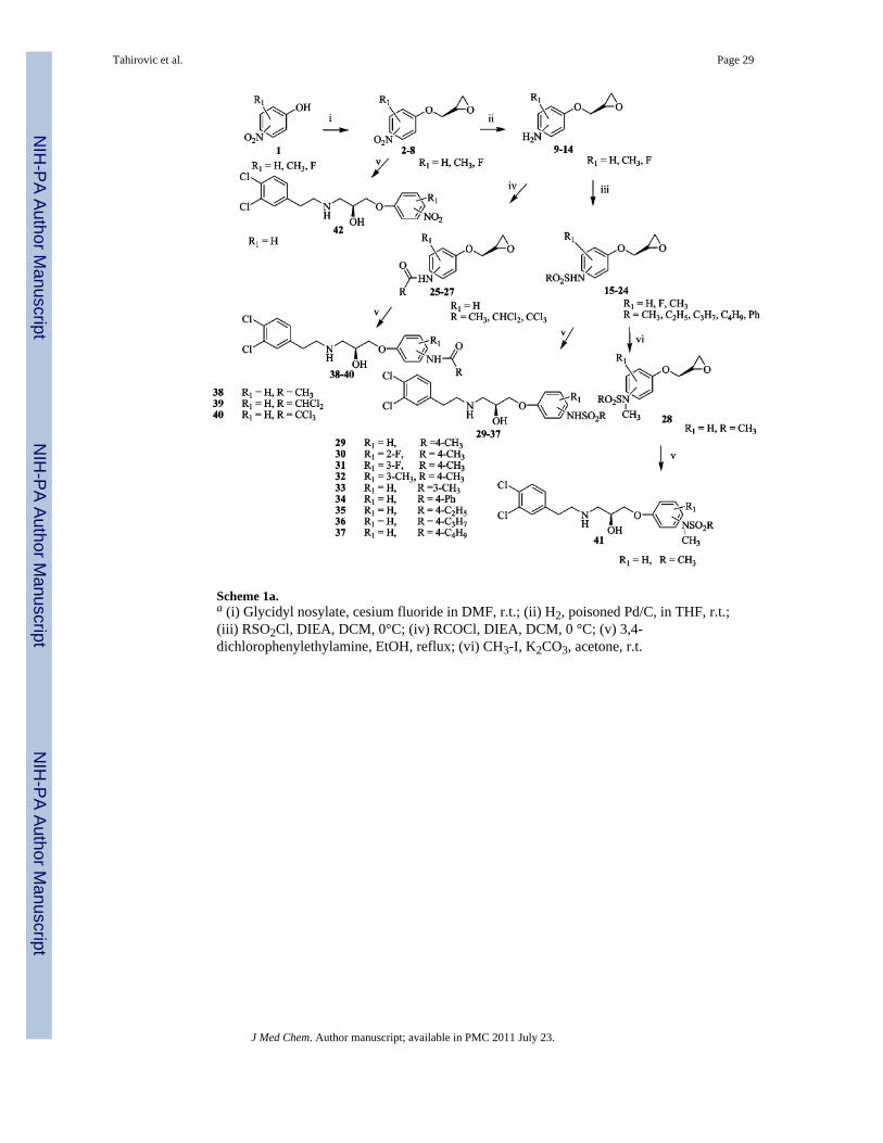
Scheme 1a.a (i) Glycidyl nosylate, cesium fluoride in DMF, r.t.; (ii) H2, poisoned Pd/C, in THF, r.t.;(iii) RSO2Cl, DIEA, DCM, 0°C; (iv) RCOCl, DIEA, DCM, 0 °C; (v) 3,4-dichlorophenylethylamine, EtOH, reflux; (vi) CH3-I, K2CO3, acetone, r.t.
Tahirovic et al. Page 29
J Med Chem. Author manuscript; available in PMC 2011 July 23.
NIH
-PA Author Manuscript
NIH
-PA Author Manuscript
NIH
-PA Author Manuscript

Scheme 2a.a (i) Benzaldehyde, p-toluenesulfonic acid, toluene, reflux; (ii) Pd/C (%10), 2N NaOH,ethanol; (iii) CH3SO2Cl, DIEA, DCM, 0°C, 1N HCl.
Tahirovic et al. Page 30
J Med Chem. Author manuscript; available in PMC 2011 July 23.
NIH
-PA Author Manuscript
NIH
-PA Author Manuscript
NIH
-PA Author Manuscript

Scheme 3.
Tahirovic et al. Page 31
J Med Chem. Author manuscript; available in PMC 2011 July 23.
NIH
-PA Author Manuscript
NIH
-PA Author Manuscript
NIH
-PA Author Manuscript

Scheme 4a.a (i) R-CHO, NaB(OAc)3H, 1,2-dichloroethane, r.t.; (ii) O-butyryl glycoaldehyde,NaB(OAc)3H, 1,2-dichloroethane, r.t.; (iii) NaOMe, MeOH, r.t.
Tahirovic et al. Page 32
J Med Chem. Author manuscript; available in PMC 2011 July 23.
NIH
-PA Author Manuscript
NIH
-PA Author Manuscript
NIH
-PA Author Manuscript

NIH
-PA Author Manuscript
NIH
-PA Author Manuscript
NIH
-PA Author Manuscript
Tahirovic et al. Page 33
Table 1IC50 Values of Propanolamine NR1/NR2B Receptor Antagonists from Variations in thePhenoxy Ring
compd R1 R2 IC50 (±95% CI) (μM)a N
29 H 4-NHSO2CH3 0.050 (0.037, 0.068) 53
30 2-F 4-NHSO2CH3 0.072 (0.052, 0.100) 12
31 3-F 4-NHSO2CH3 0.427 (0.340, 0.535) 11
32 3-CH3 4-NHSO2CH3 0.687 (0.479, 0.986) 11
33 H 3-NHSO2CH3 0.530 (0.221, 1.27) 14
34 H 4-NHSO2Ph 1.45 (0.206, 10.2) 8
35 H 4-NHSO2C2H5 0.587 (0.480, 0.719) 12
36 H 4-NHSO2C3H7 6.10 (4.31, 8.63) 4
37 H 4-NHSO2C4H9 6.79 (5.01, 9.23) 4
38 H 4-NHCOCH3 4.37 (1.81, 10.5) 3
39 H 4-NHCOCHCl2 12.8 (9.06, 17.9) 4
40 H 4-NHCOCCl3 10.6 (7.43, 15.3) 4
41 H 4-N(CH3)SO2CH3 9.91 (3.49, 28.1) 15
42 H 4-NO2 9.38 (5.77, 15.2) 12
43 H 2- NHSO2CH3 12.0 (5.23, 27.6) 17
aFor all compounds in all tables, the mean IC50 value was determined from the fitted concentration-effect curves from individual Xenopus laevis
oocytes recorded under two electrode voltage clamp; oocytes expressed recombinant rat NR1/NR2B receptors. The 95% confidence intervals (CI)determined from log(EC50 values), which has a normal distribution. N is the number of oocytes recorded.
J Med Chem. Author manuscript; available in PMC 2011 July 23.

NIH
-PA Author Manuscript
NIH
-PA Author Manuscript
NIH
-PA Author Manuscript
Tahirovic et al. Page 34
Table 2IC50 Values of Propanolamine NR1/NR2B NMDA Receptor Antagonists Derived fromVariations in the Amino Substitution
compd R IC50 (±95% CI) (μM) N
29 3,4Cl2C6H3(CH2) 2 0.050 (0.037, 0.068) 53
47 2ClC6H4(CH2) 2 0.429 (0.352, 0.522) 16
48 3ClC6H4(CH2) 2 0.628 (0.515, 0.767) 11
49 4ClC6H4(CH2) 2 0.101 (0.080, 0.126) 10
50 2,4Cl2C6 H3(CH2) 2 2.317 (0.726, 7.40) 17
51 3Cl-4FC6H3(CH2) 2 0.397 (0.346, 0.456) 30
52 4Cl-3FC6H3(CH2) 2 0.068 (0.056, 0.082) 16
53 2FC6H4(CH2) 2 0.573 (0.504, 0.653) 11
54 3FC6H4(CH2)2 0.853 (0.643, 1.13) 16
55 4FC6H4(CH2)2 0.748 (0.582, 0.964) 12
56 3,4F2 C6H3(CH2) 2 0.357 (0.308, 0.415) 22
57 2OHC6H4(CH2) 2 2.00 (1.71, 2.33) 4
58 3OHC6H4(CH2) 2 4.43 (3.40, 5.75) 5
59 4OHC6H4(CH2) 2 7.24 (4.33, 12.1) 4
60 3,5(OH)2C6H2(CH2) 2 >100 4
61 3,4(OH)2C6H2(CH2) 2 >100 4
62 3,4(OCH2O)C6H2(CH2) 2 0.951 (0.746, 1.21) 9
63 3,4(CH3)2 C6H3(CH2) 2 0.207 (0.145, 0.295) 12
64 1-naphthyl-CH(CH3) 5.07 (2.06, 12.5) 13
65 4-NO2 C6H4(CH2) 2 1.14 (0.804, 1.63) 10
J Med Chem. Author manuscript; available in PMC 2011 July 23.

NIH
-PA Author Manuscript
NIH
-PA Author Manuscript
NIH
-PA Author Manuscript
Tahirovic et al. Page 35
Table 3IC50 Values of Propanolamine NR1/NR2B NMDA Receptor Antagonists Derived fromVariations in the Amino Alkyl Substitution
compd R IC50 (±95% CI) (μM) N
29 H 0.050 (0.037, 0.068) 53
66 CH3 0.130 (0.091, 0.186) 41
67 C2H5 0.096 (0.065, 0.143) 29
68 C2H4OH 0.122 (0.091, 0.163) 38
69 C3H7 0.635 (0.481, 0.841) 25
70 C4H9 0.421 (0.345, 0.512) 46
71 C5H11 0.659 (0.463, 0.938) 10
72 (CH3) 2CHCH2 1.27 (0.975, 1.65) 30
73 C6H11CH2 9.84 (3.88, 24.9) 33
74 CH(CH3)2 0.603 (0.497, 0.731) 12
75 C6H5CH2 0.177 (0.116, 0.271) 24
J Med Chem. Author manuscript; available in PMC 2011 July 23.

NIH
-PA Author Manuscript
NIH
-PA Author Manuscript
NIH
-PA Author Manuscript
Tahirovic et al. Page 36
Table 4IC50 Values of Propanolamine NR1/NR2B NMDA Receptor Antagonists Derived fromVariations in the Aminomethylaromatic Substitution
compd R I50 (±95% CI) (μM) N
75 C6H5CH2 0.177 (0.116, 0.271) 24
76 2-OHC6H4CH2 0.662 (0.451, 0.973) 45
77 3-OHC6H4CH2 0.238 (0.197, 0.289) 47
78 4-OHC6H4CH2 0.113 (0.095, 0.133) 13
79 2,3-(OH)2C6H3CH2 1.82 (1.23, 2.71) 9
80 2,4-(OH)2C6H3CH2 0.146 (0.099, 0.215) 11
81 2-OCH3C6H4CH2 3.86 (0.622, 24.04) 4
82 2,3-(OCH3)2C6H3CH2 2.79 (1.65, 4.71) 17
83 2,6-(OCH3)2C6H3CH2 11.9 (7.9, 17.8) 11
84 2,3-(-OCH2O-) C6H3CH2 5.01 (2.09, 12.0) 4
85 2-ClC6H4CH2 0.479 (0.406, 0.565) 14
86 2-BrC6H4CH2 0.038 (0.033, 0.045) 8
87 2-FC6H4CH2 0.080 (0.049, 0.130) 22
88 3-FC6H4CH2 0.249 (0.179, 0.347) 16
89 4-FC6H4CH2 0.275 (0.110, 0.682) 15
90 2,6-F2C6H3CH2 0.281 (0.145, 0.546) 12
91 2,3,4-F3C6H2CH2 0.051 (0.029, 0.087) 14
92 2,3,4,5,6-F5PhCH2 0.524 (0.419, 0.655) 31
93 2-CH3C6H4CH2 5.70 (4.08, 7.96) 22
94 4-CH3C6H4CH2 5.41 (2.88, 10.1) 18
95 2-CF3C6H4CH2 3.72 (2.06, 6.71) 21
96 2-NO2C6H4CH2 0.589 (0.399, 0.869) 6
97 2(CH3COO) C6H4CH2 0.500 (0.376, 0.665) 12
98 2-pyridylCH2 2.76 (2.15, 3.55) 12
99 3-pyridylCH2 0.278 (0.233, 0.332) 21
100 2-thiazolylCH2 3.58 (3.02, 4.26) 19
101 C6H5(CH2)2 3.51 (2.08, 5.93) 6
102 2-OHC6H4(CH2)2 0.755 (0.592, 0.964) 12
J Med Chem. Author manuscript; available in PMC 2011 July 23.

NIH
-PA Author Manuscript
NIH
-PA Author Manuscript
NIH
-PA Author Manuscript
Tahirovic et al. Page 37
Table 5IC50 Values of Propanolamine NR1/NR2B NMDA Receptor Antagonists Derived fromVariations in the Stereochemistry
compd stereochemistry R IC50 (±95% CI) (μM) N
29 S H 0.050 (0.037, 0.068) 53
103 R H 0.188 (0.128, 0.277) 10
66 S CH3 0.130 (0.091, 0.186) 41
104 R CH3 0.253 (0.211, 0.303) 11
68 S C2H4OH 0.122 (0.091, 0.163) 38
105 R C2H4OH 0.173 (0.146, 0.205) 12
70 S C4H9 0.421 (0.345, 0.512) 46
106 R C4H9 1.28 (1.06, 1.54) 39
76 S 2-OHC6H4CH2 0.662 (0.451, 0.973) 45
107 R 2-OHC6H4CH2 0.433 (0.309, 0.605) 44
77 S 3-OHC6H4CH2 0.238 (0.197, 0.289) 47
108 R 3-OHC6H4CH2 0.409 (0.327, 0.512) 13
J Med Chem. Author manuscript; available in PMC 2011 July 23.

NIH
-PA Author Manuscript
NIH
-PA Author Manuscript
NIH
-PA Author Manuscript
Tahirovic et al. Page 38
Table 6Highest Scoring Conformation for Each Molecule from EON Comparison
name IC50 (μM) electrostatic Tanimoto score (Te) shape Tanimoto score (Ts)
52 0.065 0.47 0.72
29 0.037 0.47 0.71
91 0.064 0.41 0.54
103 0.219 0.36 0.72
67 0.089 0.36 0.703
30 0.058 0.32 0.71
ifenprodil 0.073 0.20 0.68
J Med Chem. Author manuscript; available in PMC 2011 July 23.

NIH
-PA Author Manuscript
NIH
-PA Author Manuscript
NIH
-PA Author Manuscript
Tahirovic et al. Page 39
Table 7hERG and αl-Adrenergic Receptor Binding of Selected Propanolamines
compd R1 R2 hERG IC50(μM) α1-AdR IC50(μM)
29 3,4-Cl2 H 0.73 2.4
49 4-Cl H 1.39 9.5
52 4-Cl, 3-F H 1.91 >10
56 3,4-F2 H 1.63 >10
63 3,4-(CH3)2 H 2.4 2.2
66 3,4-Cl2 CH3 0.37 1.4
67 3,4-Cl2 C2H5 0.17 3.5
69 3,4-Cl2 C3H7 0.13 nd
70 3,4-Cl2 C4H9 0.07 4.9
72 3,4-Cl2 (CH3)2CHCH2 0.37 >10
75 3,4-Cl2 C6H5CH2 0.43 >10
76 3,4-Cl2 2-OHC6H4CH2 0.83 >10
77 3,4-Cl2 3-OHC6H4CH2 0.78 >10
87 3,4-Cl2 2-FC6H4CH2 1.2 >10
91 3,4-Cl2 2,3,4-F3C6H4CH2 1.7 >10
J Med Chem. Author manuscript; available in PMC 2011 July 23.
Copyright © 2022 FDOKUMEN


![financial management 2b [bsr2b01, fnm02b2] last assessment ...](https://static.fdokumen.com/doc/165x107/631aa5b5c51d6b41aa04e9e3/financial-management-2b-bsr2b01-fnm02b2-last-assessment-.jpg)


![FINANCIAL MANAGEMENT 2B [BSR2B01, FNM02B2] LAST ...](https://static.fdokumen.com/doc/165x107/633c134708bfd70fe20afb65/financial-management-2b-bsr2b01-fnm02b2-last-.jpg)