1. LITERATURE REVIEW - Research UNE
272
Literature Review Page 1 1. LITERATURE REVIEW
-
Upload
khangminh22 -
Category
Documents
-
view
3 -
download
0
Transcript of 1. LITERATURE REVIEW - Research UNE
Literature Review
Page 1
1. LITERATURE REVIEW
Literature Review
Page 2
1. INTRODUCTION
Type 2 Diabetes is a chronic, slowly progressive disease with an inherited
and /or environmental component. Individuals may have a genetic predisposition
to this disease and then particular environmental factors, such as a virus, dietary
factors or lifestyle, or chronic inflammatory conditions, results in the onset of
the disease. Type 2 Diabetes results in a decrease in the production of insulin by
the pancreas, and/or insulin resistance, where the insulin released is not effective
in lowering the blood glucose concentration due to it not allowing the
transportation of glucose into the cells of various tissues throughout the body.
In either case there is the resultant increase in blood glucose concentration
leading to damage of many body systems, especially the blood vessels and
nerves, resulting in retinopathy, nephropathy, neuropathy and heart disease.
Type 2 Diabetes can also increase the risk of miscarriage and congenital
malformations to almost twice that is seen in normal pregnancies (Hampton,
2004).
The prevalence of Type 2 Diabetes is increasing rapidly worldwide,
primarily due to the increase of sedentary lifestyles and obesity (Uusitupa,
2002), with an estimated 177 million people suffering from it worldwide in 2002
(Dunstan et. al., 2002; World Health Organization, 2002). This figure is
expected to reach at least 324 million by the year 2025, which is approximately
Literature Review
Page 3
5.5% of the adult population (Meisinger et. al., 2006a). This increase will occur
mainly in the developing countries, (170%), due to their aging population, the
more sedentary lifestyle and poor diet as they prosper, which also leads to
obesity, compared to developed counties who are expecting an increase of 42%
(Dunstan et. al., 2002; Saudek, 2002). Most people who have the disease in the
developed world are > 65 years of age while those in the developing countries
are between the ages of 45 and 65 years (Bonow & Gheorghiade, 2004). Having
Type 2 Diabetes can shorten life expectancy by as much as fifteen (15) years
(Gillies et. al., 2007). Type 2 Diabetes was also once thought of as a disease of
the aged; however it is now being diagnosed in young adults, adolescents and
even children. In the USA between 30-50% of new cases of Type 2 Diabetes
are diagnosed in youth between the ages of 9 and 19, compared to 2% twenty
years ago (Novick, 2001). In 2002 it was estimated that at least 987,000 deaths
(1.7% total mortality) occurred from Type 2 Diabetes or its complications, and
is the fifth leading cause of death world wide (Roglic et. al., 2005). Many of
those people suffering from diabetes are also unaware of it and hence are not
receiving treatment, leading to more complications, with the proportion of
undiagnosed diabetes being as high or even higher then 50% (Roglic et. al.,
2005). Impaired glucose tolerance also leads to an increased mortality rate of
approximately 40%, independent of whether they progress to diabetes or not and
hence the impact of hyperglycaemia and insulin resistance is larger than that
associated with diabetes alone (Roglic et. al., 2005).
Literature Review
Page 4
In England there are approximately 1.3 million people who have been
diagnosed with Type 2 Diabetes, and approximately 5% of the total National
Health Services resources and up to 10% of hospital inpatient resources are used
for the treatment of people with diabetes (Gillies et. al., 2007). Prevention of
the disorder and maintenance of people diagnosed with Type 2 Diabetes would
certainly lower the costs of this disease to the national budget of England and all
countries. In the United States of America it is estimated that the cost per
diabetic patient is between $4,000 and $5,000 per annum, with costs increasing
as the severity of the disease progresses (Nichols and Brown, 2005). The
national debt is US$92 billion annually in direct costs and an additional US$40
billion in indirect costs such as time off work etc (Petersen and Shulman, 2006).
In Germany the direct costs for the treatment of patients with diabetes is 14.2%
of the total health care costs, with approximately 26.5 million or one third of the
population suffering from the disease (Koster et. al., 2006).
In Australia there are 854,325 individuals that have been diagnosed with
Type 2 Diabetes and registered with the National Diabetes Register, being
3.97% of the total population (Deed, 2009), and it is estimated that another
400,000 who are not registered, have either a pre-diabetic or do not realise that
they have diabetes, this being one of the highest recorded prevalences in the
developed world. The percentage of the population has risen to 3.6% of the
population in the year 2004-2005 (Barit and Cooper, 2009) and is continuing to
rise rapidly. It has also been suggested that the average Australian has about a
Literature Review
Page 5
one in fourteen chance of developing Type 2 Diabetes during their lifetime, and
if impaired fasting glucose and impaired glucose tolerance are included, then
approximately one in four Australians would be classified as having a glucose
uptake disorder (Dunstan et. al., 2002) . The Australian Aboriginal population
have an incidence of Type 2 Diabetes of 9.9% , while that of an older white
population based in the Blue Mountains of New South Wales was 9.3%t, and if
those with an impaired fasting glucose are added it is 15.8% (Cugati, et. al.,
2007), suggesting that it is not just an aboriginal problem in Australia.
The magnitude of the problem is enormous with billions of dollars being
spent annually by most western nations on the treatment of this disease and its
complications. In developed countries Type 2 Diabetes claims about 9% of the
National Health Budget (World Health Organisation, 2002) and in many
countries it may account for 10% or more of the total health budget (Roglic et.
al., 2005). Diabetes is Australia’s sixth leading cause of death from disease and
seventh overall, including accidents (Australian Bureau Statistics, 2000). This
leads to a huge economic burden on the country. Days lost from work due to the
disease are numerous which a major economic burden is also. A study in
Australia has shown that people with diabetes cost approximately 38% more
($1017) than non-diabetics in the hospital system (Clarke et. al., 2006) and this
is an increasing burden on the society as a whole, financially, as well as with
beds being taken from other medical needs.

Literature Review
Page 6
Today there are several medications used to ameliorate the effects of
diabetes and its complications by controlling blood glucose levels (BGL). These
include oral hyperglycaemic drugs such as the sulfonylureas, biguanides and
thiazolidinediones, and insulin which is used in the end stages of Type 2
Diabetes. Although the effects of the hypoglycaemic drugs appear to be
sufficient and BGL are normalised initially, they still tend not to stop the micro-
vascular effects of the disease (Moss et. al., 1994) and their effectiveness
appears to diminish overtime, therefore, an increased amount of a drug, or
combination of drugs, must be taken to have the same effect (Cook et. al.,
2007).
Since people with impaired glucose tolerance have a high risk of
developing Type 2 Diabetes, they need to be diagnosed and preventative
measures put into place as soon as possible. This can be achieved through
regular BGL tests and also looking at other risk factors such as family history,
waist-to-hip circumference ratios and exercise levels and then a change in
lifestyle factors such as diet and exercise should be initiated early, but may also
include some pharmacological agents. Thus, the development of new and
improved medications for prevention of Type 2 Diabetes will also assist in the
real cost of this disease, as well as raising the well being, of those predisposed to
the disease.
The recorded use of plants in the treatment of diabetes dates back to
approximately 1550 BCE (Gray & Flatt, 1997b), and in recent years many
Literature Review
Page 7
people have again turnrd to Natural Therapies as a treatment alternative.
More than 400 traditional herbal remedies used in the treatment of diabetes
by various cultures have been recorded; however only a few of these have
received medical and scientific evaluation to determine effectiveness,
efficacy, side effects and toxicity (Baily and Day, 1989). The World
Health Organisation has recommended that medical and scientific
examinations of such plants be undertaken (World Health Organisation
Expert Committee on Diabetes Mellitus, 1980).
1.1 GLUCOSE HOMEOSTASIS
1.1.1Glucose Regulation
Glucose is the major energy source for all mammalian cells; however the
BGL must be maintained within a very narrow concentration range of 4.5 – 5.0
nml/L in order to avoid its own toxic effects. This level is maintained even with
the variable intake of sugars and use of energy, and the homeostatic control is
achieved by the matching of the flux of glucose into and out of the plasma
through tightly regulated secretion of insulin and glucagon from the pancreatic
islet cells with hyperglycaemia itself acting as a �-cell toxin and bringing about
apoptosis of the ��islet cells resulting in decreased insulin secretion (Chang et.
al., 1996).
Literature Review
Page 8
The homeostatic process begins in the gastrointestinal tract where the
uptake of glucose into the plasma is regulated by transport across the
gastrointestinal endothelium. The homeostatic process is also controlled by the
liver as it regulates BGL by glucose uptake and storage along with production of
glucose in times of need. Other tissues, in particular muscles, which are
involved in the uptake and storage of the glucose, are also extremely important
in maintaining the balance. The balance of uptake and storage is precisely
matched by the output of glucose from glycogenolysis, and gluconeogenesis.
After glucose intake this balance is upset, and the return to homeostasis is
dependent on three processes that occur simultaneously: (1) Insulin secretion by
the �-cells, (2) Glucose uptake by the liver, muscle where it is stored as
glycogen, and adipose tissues (Pilkis & Granner, 1992) or metabolised for
energy, and (3) gluconeogenesis and glycogenolysis are suppressed through
insulin-induced inhibition of glucagon secretion and blockage of the actions of
glucagon on the liver.
1.1.2 Glucose Transport
Skeletal muscle expresses relatively high levels of the glucose transporter
GLUT4, which is responsible for insulin-stimulated glucose transport (Rea &
James, 1997) and low levels of GLUT1, which is responsible for basal
noninsulin-dependent transport (Zorzano et. al., 1996). During fasting states,
GLUT4 is localized mainly in intracellular vesicles, and it is translocated to cell
Literature Review
Page 9
membrane in response to insulin. Once transported into the cell, glucose is
rapidly phosphorylated by hexokinase (HK) to glucose-6-phosphate (G6Pase)
and this keeps the concentration of intracellular glucose low, ensuring the
continual movement of glucose across the plasma membrane (Postic et. al.,
1994; Mandarino et. al., 1995). There are several different hexokinases with
HKI being expressed in all tissues and HKII in insulin sensitive tissues, (Postic
et. al., 1994) with GLUT4 and HKII being involved in insulin mediated glucose
utilization, and GLUT1 and HK1 being involved in basal glucose uptake.
Insulin resistance, where the released insulin is less effective, is a
predominant feature of Type 2 Diabetes and is often an indication of a
prediabetic state. Since glucose transportation is one of the first rate limiting
steps in glucose metabolism and is often down regulated in both skeletal muscle
and adipose tissue in patients with Type 2 Diabetes (Zierath et. al., 1994;
Zierath, 1995), it is suggested that this may play a major role in the development
of impaired glucose uptake. Zierath and colleagues (1996) have shown that
insulin resistance is not due to a deficiency of GLUT4, which is still translocated
to plasma membrane by insulin, however, the defect is likely to be due to an
altered translocation of GLUT4 or in the altered fusion of the GLUT4 vesicles to
the plasma membrane. Intracellular Free Fatty Acids (FFA) block the insulin
mediated activation of GLUT 4 and its translocation, as well as decreases in
intracellular glucose levels possibly due to the interference of the insulin
Literature Review
Page 10
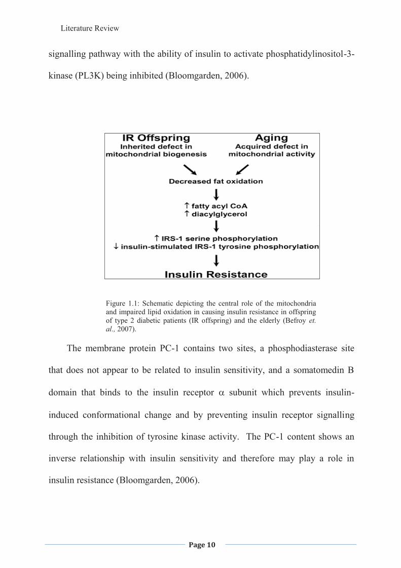
signalling pathway with the ability of insulin to activate phosphatidylinositol-3-
kinase (PL3K) being inhibited (Bloomgarden, 2006).
Figure 1.1: Schematic depicting the central role of the mitochondria and impaired lipid oxidation in causing insulin resistance in offspring of type 2 diabetic patients (IR offspring) and the elderly (Befroy et. al., 2007).
The membrane protein PC-1 contains two sites, a phosphodiasterase site
that does not appear to be related to insulin sensitivity, and a somatomedin B
domain that binds to the insulin receptor � subunit which prevents insulin-
induced conformational change and by preventing insulin receptor signalling
through the inhibition of tyrosine kinase activity. The PC-1 content shows an
inverse relationship with insulin sensitivity and therefore may play a role in
insulin resistance (Bloomgarden, 2006).
Literature Review
Page 11
Signalling through the PI3K pathway is crucial for metabolic responses to
insulin (Alessi & Downes, 1998). The binding of insulin to its receptor
activates the insulin receptor tyrosine kinase, which initiates a cascade of
signalling events. The initial step is the tyrosine phosphorylation of insulin
receptor substrate 1 (IRS1), which then binds to PI3K, which activates its
catalytic unit. The tyrosine phosphorylation of IRS1 is the first signalling step
that has been shown to have reduced sensitivity in the diabetic patient
(Danielsson et. al., 2005). The enzyme AMP-activated protein kinase (AMPK),
which is stimulated by an increase of AMP/ATP, plays a role in several cellular
and metabolic processes during exercise, including the increase of skeletal
muscle fatty acid oxidation, and glucose transport (Sriwijitkamol et. al., 2007).
The lipid products of PI3K activate protein kinase B (Akt), which in turn
mediates many of the metabolic activities of insulin (Kohn et. al., 1996; Martin
et. al., 1996) and may be involved in the events leading to translocation of
GLUT 4 to the cell membrane resulting in the uptake of glucose into the cell
(Cortright & Dohm, 1997). Phosphorylation of AS160, a substrate of Akt, is
required for the translocation of GLUT4 and may be impaired in insulin
resistance (Hakan et. al., 2005).
1.1.3 The Gastrointestinal Tract
Glucose homeostasis begins in the stomach and small intestine, where acute
changes in BGL affect the gastric motor functions. Gastric emptying is slower
Literature Review
Page 12
during hyperglycaemia and accelerated during hypoglycaemia (Horowitz et. al.,
2002), thus absorption of glucose into the portal system from the small intestine,
is affected by the rate of gastric emptying, this being controlled by feedback to
the satiety centre of the hypothalamus via the hormone ghrelin, which also has a
direct effect on the pancreas promoting �-cell regeneration (Irako et. al., 2006).
A diet high in natural fibre will slow gastric emptying and hence delay the
absorption of glucose from the small intestine (Riccardi et. al., 2003).
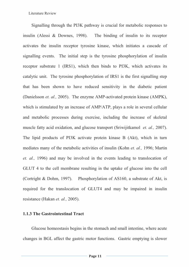
The classical model of glucose absorption across the apical membrane of
the enterocyte in the lumen of the small intestine is via the Na+/glucose co-
transporter (SGLT1) and exits into the portal system across the basolateral
membrane via the facilitative transporter GLUT2 (Reviewed in Kellett and Brot-
Laroche, 2005). Fructose is transported by the specific facilitative transporter,
GLUT5 (Burant et. al., 1992), while GLUT2 transports both fructose and
glucose (Cheeseman, 1993). Initially it was thought that GLUT2 was only
found at the basolaterial membrane: however, it has now been located in the
apical membrane as well (Corpe et. al., 1996).
The glucose transported by SGLT1 promotes insertion of GLUT2 into the
apical membrane within minutes of high glucose levels reaching the lumen of
the small intestine resulting in a much faster and greater absorption of glucose
(Helliwell & Kellett, 2002). The �-glucosidase inhibitors such as acarbose
reduce the breakdown of complex carbohydrates in the gut and possibly
Literature Review
Page 13
decrease the diet induced up-regulation of transport of sugars across the gut wall
(Casirola & Ferraris, 2006).
Figure 1.2: The apical GLUT2 model of intestinal glucose absorption before (A) and after (B) a meal. (Reviewed in Kellett and Brot-Laroche, 2005).
1.1.3.1 Glucagon-like-peptide-1 and Gastric Inhibitory Polypeptide
Glucagon-like-peptide-1 (GLP-1) and gastric inhibitory polypeptide (GIP)
are hormones, produced in the L cells and are released in the stomach
postprandial and enhance glucose-stimulated insulin secretion (Ahren, 1998;
Drucker, 1998), while GLP-1 also inhibits glucagon secretion, delays gastric
emptying and stimulates insulin biosynthesis (Ahren, 1998; Drucker, 1998) and
Literature Review
Page 14
GIP has been shown to increase glucagon secretion in the healthy individual
(Meier et. al., 2003).
GLP-1 is rapidly degraded by the enzyme dipeptidyl peptidase IV in the
stomach and has only very short-term effects, therefore it is not suitable for
treatment of Type 2 Diabetes; however, new analogues have been produced that
have the same effect but a much longer half life and inhibitors of dipeptidy
peptidase IV that decrease the GLP-1 degradation making them feasible in
treatment protocols (Holst 2006). GLP-1 and Exendin-4 (an analogue of GLP-
1) have been shown to increase �-cell mass in adult rodents (Stoffers et. al.,
2000) and Tourrel and colleagues (2001) demonstrated that GLP-1 or Exendin-4
applied during the neonatal period would exert both a long and a short-term
beneficial effect on �-cell mass and glucose homeostasis in mice, though the
mechanism has not yet been elucidated. The use of GLP-1 or Exendin-4 in the
pre-diabetic period delays the onset and limits the severity of Type 2 Diabetes,
because of its �-cell replenishing effect in diabetic mice (Tourrel et. al., 2002),
by possibly increasing �-cell mass allowing the pancreas to continue to produce
insulin for an extended period of time or an increased amount of insulin. Lee
and colleagues (2007) have shown that GLP-1 and Exendin-4 improves insulin
sensitivity by restoration of insulin signalling and also decreases hepatic
gluconeogenesis in diabetic ob/ob mice and the use of inhibitors of GLP-1
breakdown and analogues may be useful in the treatment of Type 2 Diabetes
Literature Review
Page 15
(Todd and Bloom, 2007); however, the long-term effects of these need to be
studied further.
1.1.4 The Pancreas
1.1.4.1 Insulin
Insulin is synthesized in the endoplasmic reticulum of the �-islets and
transported to the Golgi complex, where the insulin-containing secretory
granules are formed through budding, stored until needed and finally secreted by
exocytosis upon stimulation (Reviewed in Wollheim et. al., 1996). Insulin
secretion is highly regulated and responds to very small changes in the BGL.
This occurs by various glucose-derived signals and the potentiating and
inhibitory effects of various endocrine and paracrine influences of other
nutrients (Bock et. al., 2007), hormones and neurotransmitters. The potentiating
and inhibitory effects appear to remain intact during the pre-diabetic state and
after the onset of Type 2 Diabetes (Bock et. al., 2007), however, abnormal
secretion of insulin is seen regularly due to a primary insulin secretion defect,
exhaustion of the pancreas, glucose toxicity or a combination of the above
(Porte 1991).
Insulin secretion, by the �-cell, is coupled to glucose metabolism, with a
major signal being the closure of K+ATP (potassium adenosine triphosphate)-
dependent channels in the cell membrane due to the increased cytosolic ATP
Literature Review
Page 16
with a glucose induced increase in the cytoplasmic ATP/ADP ratio. This ratio
serves as the major regulator of the K+ATP-dependent channels in the cell
membrane, with the closure of these channels depolarising the cell membrane.
This in turn leads to the opening of the voltage-dependent calcium (Ca2+)
channels, raising the intracellular Ca2+ concentration, which results in insulin
secretion (Koster et. al., 2005). Any alteration to the metabolic signalling,
sensitivity of the K+ATP -dependent channels to metabolites or the number of
active channels could disrupt the signalling process and hence alter insulin
secretion. Congenital hyperinsulinism, a rare recessive disorder, which results
in constitutive insulin secretion irrespective of blood glucose levels and a
decrease in K + ATP-dependent channels expression, has been implicated in this
disorder. Conversely, mutations that result in an increased expression of the
K+ATP-dependent channels activity should decrease the glucose sensing by the
�-cell and hence decrease insulin secretion leading to higher blood glucose
levels (Koster et. al., 2005).
Secondary messengers, such as GLP-1 and GIP and neurotransmitters
which increase glucose sensitivity, also play an important role in insulin
secretion (Howell et. al., 1984). The insulin producing �-islets cells are richly
innervated by the autonomic nervous system (Porte et. al., 1990), and the
parasympathetic stimulation increases insulin secretion by the binding of
acetylcholine to muscarinic receptors through the activation of adenylate
Literature Review
Page 17
cyclase-cyclic adenosine monophosphate (cAMP), GIP and GLP-1 and
phospholipase C. Cyclic AMP modulates the influx of Ca2+ through voltage-
gated Ca2+ channels and potentiates Ca2+-dependent exocytotic events which act
independently of the effect on Ca2+ channels (Ammala et. al., 1993), and is also
associated with the conversion of insulin from proinsulin, the prohormone of
insulin (Ahmad et. al., 1991b). Proinsulin is converted in the �-cell to insulin
and C-peptide by a series of proteolytic steps and are both stored in the secretory
granules, and released along with some partially processed and intact proinsulin
(McFarlane, 1991).
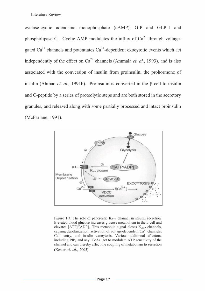
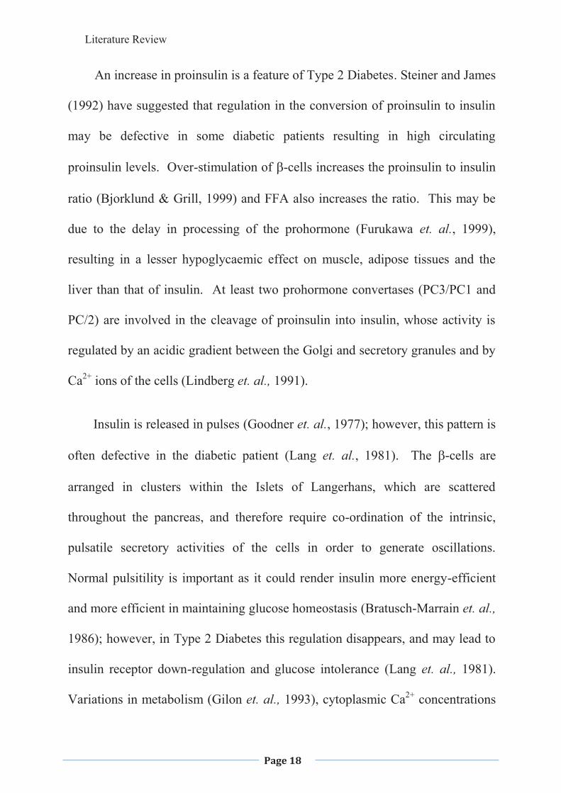
Figure 1.3: The role of pancreatic KATP channel in insulin secretion. Elevated blood glucose increases glucose metabolism in the ß-cell and elevates [ATP]/[ADP]i. This metabolic signal closes KATP channels, causing depolarization, activation of voltage-dependent Ca2+ channels, Ca2+ entry, and insulin exocytosis. Various additional effectors, including PIP2 and acyl CoAs, act to modulate ATP sensitivity of the channel and can thereby affect the coupling of metabolism to secretion (Koster et. al., 2005).
Literature Review
Page 18
An increase in proinsulin is a feature of Type 2 Diabetes. Steiner and James
(1992) have suggested that regulation in the conversion of proinsulin to insulin
may be defective in some diabetic patients resulting in high circulating
proinsulin levels. Over-stimulation of �-cells increases the proinsulin to insulin
ratio (Bjorklund & Grill, 1999) and FFA also increases the ratio. This may be
due to the delay in processing of the prohormone (Furukawa et. al., 1999),
resulting in a lesser hypoglycaemic effect on muscle, adipose tissues and the
liver than that of insulin. At least two prohormone convertases (PC3/PC1 and
PC/2) are involved in the cleavage of proinsulin into insulin, whose activity is
regulated by an acidic gradient between the Golgi and secretory granules and by
Ca2+ ions of the cells (Lindberg et. al., 1991).
Insulin is released in pulses (Goodner et. al., 1977); however, this pattern is
often defective in the diabetic patient (Lang et. al., 1981). The �-cells are
arranged in clusters within the Islets of Langerhans, which are scattered
throughout the pancreas, and therefore require co-ordination of the intrinsic,
pulsatile secretory activities of the cells in order to generate oscillations.
Normal pulsitility is important as it could render insulin more energy-efficient
and more efficient in maintaining glucose homeostasis (Bratusch-Marrain et. al.,
1986); however, in Type 2 Diabetes this regulation disappears, and may lead to
insulin receptor down-regulation and glucose intolerance (Lang et. al., 1981).
Variations in metabolism (Gilon et. al., 1993), cytoplasmic Ca2+ concentrations
Literature Review
Page 19
(Gylfe, et. al., 1991), the intrinsic nervous system (Lang et. al., 1981) and
hormones such as GLP-1 (Bratusch-Marrain et. al., 1986), neural signalling
(Daniel & Henderson, 1967) and �-cell insulin receptor expression (Xu &
Rothenberg, 1998) have all been implicated in the regulation of plasma insulin
oscillations. However, in some Type 2 Diabetic patients the insulin is still
released in pulses indicating that the impaired secretory response to glucose may
be related to impaired metabolism (Lin et. al., 2002).
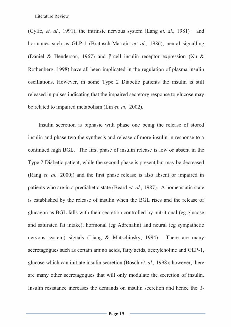
Insulin secretion is biphasic with phase one being the release of stored
insulin and phase two the synthesis and release of more insulin in response to a
continued high BGL. The first phase of insulin release is low or absent in the
Type 2 Diabetic patient, while the second phase is present but may be decreased
(Rang et. al., 2000;) and the first phase release is also absent or impaired in
patients who are in a prediabetic state (Beard et. al., 1987). A homeostatic state
is established by the release of insulin when the BGL rises and the release of
glucagon as BGL falls with their secretion controlled by nutritional (eg glucose
and saturated fat intake), hormonal (eg Adrenalin) and neural (eg sympathetic
nervous system) signals (Liang & Matschinsky, 1994). There are many
secretagogues such as certain amino acids, fatty acids, acetylcholine and GLP-1,
glucose which can initiate insulin secretion (Bosch et. al., 1998); however, there
are many other secretagogues that will only modulate the secretion of insulin.
Insulin resistance increases the demands on insulin secretion and hence the �-
Literature Review
Page 20
cells. This in turn leads to the increase in synthesis of insulin and �-cell
replication and neogenesis. However, any long term increased demands for
insulin secretion can negatively affect �-cells by over-stimulation, resulting in
their eventual apoptosis and hence a decrease in insulin production.
Figure 1.4: Insulin Secretion in normal, Type 1 and Type 2 Diabetic states. (Rang et. al., 2000)
Amylin, a pancreatic polypeptide stored and secreted along with, and with
the same apparent stimuli as insulin (Kahn et. al., 1998), currently does not have
any reported physiological role but has been noted for its absent expression in
Type1 Diabetics, which is expected. Amylin deficiency so far has not been
reported to be associated with any pathophysiological conditions or symptoms
(Gebre-Medhin et. al., 1998); however, amylin replacement therapy helps to
regulate gastric-emptying, produce the feeling of satiety and therefore decreases
food intake, and suppresses the abnormal glucagon release with meals which
1st Phase 2nd Phase
Normal
Type 2 Diabetes
Type 1 Diabetes
BasalLevel
80
60
40
20
0 15 30 45 60 75
Time (min)Glucose
Insu
lin (m
g/m
L)
Literature Review
Page 21
may give an indication of its role in the pathogenesis of Type 2 Diabetes (Owen,
2006).
It has been suggested that as the amylin fibres refold to a �-conformation
and oligomerises to form insoluble fibrils, and then accumulate in the cells
where there is a decline in �-cell function (Westermark, 1994). This folding
may be inhibited by intragranular heterodimer formation with insulin but not
proinsulin; however, if there is irregular processing of the proinsulin then the
inhibition breaks down and the folding occurs. The ineffective processing of
pro-islet amyloid polypeptide may also result in the incorrect folding of the
polypeptide and/or the inability to bind to insulin and hence the deposition of
amyloid (Clark et. al., 1987). The percentage of amylin fibres may determine the
degree of hyperglycaemia in Type 2 Diabetes (Westermark, 1994). In Type 2
Diabetes, insulin secretion decreases with the progression of the disease (UK
Prospective Diabetes Study Group, 1998), although this decrease is not
explained by autoimmune dysfunction or chronological age (Clausson et. al.,
1994). The amylin to insulin ratio increases with age (Edwards et. al., 1996)
which may bring about the decrease in insulin secretion that occurs with age.
This, in turn, may be due to toxicity of amyloid resulting in cell death or loss of
function due to adherence of fibrils to cell membranes interfering with cycling
of membrane proteins and hence secretions (Botto et. al., 1997).
Literature Review
Page 22
Calcium, an intracellular second messenger, is the critical trigger for insulin
exocytosis from �-cells (Wollheim & Sharp, 1981; Wollheim et. al., 1996).
Plasma membrane depolarisation is brought about by the generation of ATP and
other metabolic coupling factors, which promotes Ca2+ influx into the cell, with
the rise in intracellular Ca2+ concentration then triggering the exocytosis of
insulin. The hyperpolarization of the mitochondrial membrane glucose also
increases the Ca2+ concentration, activating Ca2+ sensitive dehydrogenases in the
Krebs cycle producing glutamate, which also aids in the calcium-stimulated
exocytosis (Wollheim et. al., 1996).
1.1.4.2 Glucagon
Glucagon, a hormone produced by the alpha-cells of the pancreas (Unger &
Orci, 1981) and also in small amounts in the L-cells of the intestine and
stomach, acts on the liver when the BGL is low to stimulate glucose production.
Kimball and Murlin, (1924); Grimelius and colleagues (1976), and Orci and
colleagues, (1983) all showed that glucagon works by mobilizing glucose
reserves in the liver through glycogenolysis and gluconeogenesis, preventing
hypoglycaemia as well as stimulating lipolysis and ketogenesis in adipose tissue
providing FFAs for the liver (McGarry, 1979; Carlson et. al., 1993).
The regulation of the alpha cell is complex, with a range of hormonal,
nutrient and neural stimuli all involved in its function, glucose being the most
Literature Review
Page 23
significant, as it profoundly suppresses glucagon secretion in the normal cell.
There are a number of amino acids which will also suppress glucagon secretion,
with arginine being the most effective in humans (Larsson & Ahren, 2000);
however this effect varies from species to species. Insulin acts directly on
glucagon secretion via a local endocrine effect (Stagner & Samols, 1992) while
somatostatin acts by a paracrine effect (Taborsky, 1983).
In Type 2 Diabetes the abnormalities seen in alpha cell function appear to
be a result of impaired glucose sensing or alpha cell resistance, with an increase
in gluconeogenesis being a major outcome. In the Type 2 Diabetic patient the
alpha cell fails to down-regulate its secretion of glucagon in response to
hyperglycaemia as a result of the impaired glucose sensing (Gastaldelli et. al.,
2000).
Miniglucagon, the COOH-terminal fragment processed from glucagon, is a
potent and efficient inhibitor of insulin secretion and impairment of its
metabolism could lead to impaired insulin secretion. The mechanism of action
is thought to be mediated by a pertusses toxin-sensitive G protein linked to a
pathway that involves the potassium channel opening and the resulting
membrane repolarisation (Dalle et. al., 1999).
Literature Review
Page 24
1.1.4.3. Somatostatin
The D cells of the Islets of Langerhans and the hypothalamus secrete
somatostatin, which is known as the growth hormone release-inhibiting factor
and provides a local inhibitory regulatory action on insulin within the islets
(Rang et. al., 2000).
1.1.5. The Liver
The liver plays a major role in maintaining glucose homeostasis. In the
fasting state, the liver, through glycogenolysis, breaks down glycogen to form
glucose, and gluconeogenesis (the synthesis of glucose from substances such as
various amino acids, lactate and pyruvate), releases glucose into the blood to
maintain BGL within the very narrow concentration range of around 5mM
(DeFronzo, 1997). This process is critical to maintain a glucose supply for the
neural tissues, which cannot use other forms of energy. However, uncontrolled
gluconeogenesis results in excessive hepatic glucose production, which is a
major factor in high BGLs (DeFronzo, 1997). Upon glucose absorption into the
portal system, insulin is released and carried to the liver, where it binds with
specific receptors on the hepatocyte. The importance of this can be seen by the
fact that it contains approximately 50% of the insulin binding receptors of the
body, and suppresses the hepatic glucose output and induces glucose uptake, via
the glucose transporters GLUT 2 and GLUT 1 in the liver (DeFronzo, 1997).
Literature Review
Page 25
Gluconeogenesis is a cAMP/protein kinase A (PKA) – dependent process
initiated by glucagon and possibly some of the inflammatory cytokines. This
process is generally inhibited by insulin in a number of places including the
decrease in glucagon secretion from the alpha cells in the pancreas, blocking the
glucagon signalling pathway by activating a cAMP phosphodiesterase. There is
also suppression of key gluconeogenic genes such as phosphoenolpyruvate
carboxykinase (PEPCK), 6Pase and p38 which plays a stimulatory role in
hepatic gluconeogenesis (Cao et. al., 2005) results in decreased glucose
production. Insulin secretion may be decreased in Type 2 Diabetes and
therefore these mechanisms are not inhibited, resulting in a continual increase in
glucose output by the liver resulting in high fasting blood glucose levels (Cao et.
al., 2005).
There is an increasing interest in the role of the liver in the pathogenesis of
Type 2 Diabetes. Liver fat content has been shown to correlate with several
features of insulin resistance in both normal and overweight individuals. This is
independent of Body Mass Index (BMI) (Body mass/Height2), and intra-
abdominal or overall obesity, with a liver enzyme, alanine aminotransferase
(ALT) being used to measure liver fat content and this has been shown to predict
the incident of Type 2 Diabetes (Sattar et. al., 2007). Wannemethee and
colleagues (2005) have shown that both ALT and �-glutamyltransferase (GGT)
is independent predictor’s of Type 2 diabetes; however their role in diabetes has
Literature Review
Page 26
not yet been determined. Elevated ALT and GGT however are still considered
within the “normal” range were predictive of Type 2 Diabetes independent of
obesity and alcohol intake in older men; however, results also indicate that ALT
maybe ethnically based as the results varied in Japanese and Korean men.
Wannemethee and colleagues (2005) suggested that levels of ALT and GGT
correlated with increases of hepatic fat and this may lead to hepatic insulin
resistance; however, when adjusted for insulin resistance there was still a 3-4
fold increase in the development of Type 2 Diabetes. They went on to suggest
that the correlation of ALT and GGT with tumor necrosis factor - � (TNF-�)
and Interleukin-6 (IL-6) may play a role; however, adjustments for this still did
not account for the increased risk. Oxidative stress was put forward as another
possibility; however, further studies need to be carried out to determine if this is
the mechanism.
1.1.6. Skeletal Muscle
Skeletal muscle composes approximately 40% of body mass and accounts
for approximately 75% of the uptake of glucose after glucose absorption which
is converted to glycogen (DeFronzo, 1988) and hence plays a major role in the
maintenance of a normal blood glucose level.
Physical exercise increases insulin-mediated glucose utilization, reflecting
an adaptation in muscle brought about by a local contraction-mediated
Literature Review
Page 27
mechanism. GLUT4 is also stimulated by long-term, low-frequency stimulation
of muscle (Etgen et. al., 1993). Exercise does not alter the receptor function or
the number of receptors but does increase the amount of GLUT4 present within
the cells. This can be seen after a single training session (Dela et. al., 1993; Ren
et. al., 1994), along with bringing about the translocation of GLUT4 to the
plasma membrane (Zorzano et. al., 1996).
Insulin resistance has been linked to inactivity and obesity due to the disuse
of muscles, which, in itself, can lead to obesity and brings about a decrease in
GLUT4 expression; however, GLUT1 expression is enhanced (Castello et. al.,
1993). Intracellular FFA also block the insulin mediated activation of GLUT 4
and its translocation, as well as decreases in intracellular glucose levels possibly
due to the interference of the insulin signalling pathway with the ability of
insulin to activate PI3K being inhibited (Bloomgarden, 2006). Insulin resistance
within the muscle tissue has also been linked to dysregulation of fatty acid
metabolism, due to a genetic defect in the mitochondrial oxidative-
phosphorylation process and is seen as a predictor of insulin resistance in
relatives of Type 2 Diabetic patients (Befroy et. al., 2007).
The membrane protein PC-1 contains two sites, a phosphodiasterase site
that does not appear to be related to insulin sensitivity, and a somatomedin B
domain that binds to the insulin receptor � subunit which prevents insulin-
induced conformational change and by preventing insulin receptor signalling
Literature Review
Page 28
through the inhibition of tyrosine kinase activity. The PC-1 content shows an
inverse relationship with insulin sensitivity and therefore may play a role in
insulin resistance (Bloomgarden, 2006).
Signalling through the PI3K pathway is crucial for metabolic responses to
insulin (Alessi & Downes, 1998). The binding of insulin to its receptor
activates the insulin receptor tyrosine kinase, which initiates a cascade of
signalling events. The initial step is the tyrosine phosphorylation of IRS1,
which then binds to PI3K, which activates its catalytic unit. The tyrosine
phosphorylation of IRS1 is the first signalling step that has been shown to have
reduced sensitivity in the diabetic patient (Danielsson et. al., 2005). The
enzyme AMPK, which is stimulated by an increase of AMP/ATP, plays a role in
several cellular and metabolic processes during exercise, including the increase
of skeletal muscle fatty acid oxidation, and glucose transport (Sriwijitkamol et.
al., 2007). The amount of AMPK within the myocyte is increased with exercise
and this leads to increased mitochondrial biogenesis and function. This, in turn,
leads to increased muscle glucose disposal, and fatty acid oxidation
(Sriwijitkamol et. al., 2007). The lipid products of PI3K activate Akt, which
mediates many of the metabolic activities of insulin (Kohn et. al., 1996; Martin
et. al., 1996) and may be involved in the events leading to translocation of
GLUT 4 to the cell membrane resulting in the uptake of glucose into the cell
(Cortright & Dohm, 1997). Phosphorylation of AS160 is required for the
Literature Review
Page 29
translocation of GLUT4 and may be impaired in insulin resistance (Hakan et.
al., 2005). Bloomgarden (2006) suggests that an increase in intracellular FFA
will prevent this translocation from occurring; especially considering that the
improvement in insulin resistance brought on by exercise could be due to
decreases in the amount of intracellular triglycerides. PTEN is a lipid/protein
phosphatase that can negatively regulate this pathway (Butler et. al., 2002) and
possibly be a factor in the pathogenesis of Type 2 Diabetes. Bandyopadhyay
and colleagues (2005) showed that increased expression of the PI-3 adaptor
subunits p85/55/50 decreased PI3K activity and hence, attenuated insulin
sensitivity in skeletal muscle.
Muscle contraction increases insulin sensitivity, but also stimulates glucose
uptake independent of insulin. The contraction signalling pathway is distinct
from the insulin pathway because the effects of insulin and contractions on
glucose uptake are additive and contraction does not increase insulin receptor
kinase or PI3K activity (Cortright and Dohn, 1997). Cortright and Dohn (1997)
suggest that both the contraction and insulin signalling pathways can be blocked
by calcium channel blockers indicating that the pathways may converge;
however, there are two distinct GLUT4 pools which are being targeted by the
different pathways.
A relationship between the fatty acid composition of skeletal muscle
membrane phospholipid and insulin resistance has been demonstrated (Borkman
Literature Review
Page 30
et. al., 1993; Vessby et. al., 1994), as the greater the percentage of
polyunsaturated fatty acids the better the insulin reaction. Pan and colleagues
(1995) showed a relationship between ‘5 desaturase activity and insulin
resistance, although the mechanism has not been elucidated. Intramyocellular
lipid accumulation has also been associated with whole-body insulin resistance
and defective insulin signalling (Viramaki et. al., 2001); however this is
independent of body weight and physical fitness.
The majority of glucose that enters skeletal muscle cells in response to
inulin is converted to glycogen. In Type 2 Diabetic patients, and in those who
are insulin resistant, glycogen synthesis is severely impaired (DeFronzo, 1997).
Glycogen synthase (GS) is the key insulin-regulated enzyme controlling the rate
of glycogen synthesis (Dent et. al., 1990; Lawrence & Roach, 1997) and if
deficient or ineffective the rate of glycogen synthesis decreases leading to
increased glucose levels.
GLUT5 plays a major role in skeletal muscle uptake of fructose, although
the role of this transporter is not clear at this stage. It has been noted that its
expression is high in diabetic muscle and that Pioglitazone, one of the
Thiazolidinedione (TZD) medications, will decrease the overexpression of
GLUT5 and hence decrease blood glucose levels (Stuart et. al., 2007).
Literature Review
Page 31
1.1.7. Adipose Tissue
Adipose tissue is more than just an energy storage organ, but is a secretory
organ which produces a variety of proteins that influence the metabolism of the
body. There are many hormones and cytokines produced by adipocytes that
effect food intake, carbohydrate and lipid metabolism and energy expenditure,
including resistin, TNF-�, Il-6, plasminogen activator inhibitor 1, angiotensin II,
adiponectin, Acylation stimulating protein (ASP) and Leptin. Each of these
factors interact with each other to increase or decrease body weight, energy
expenditure and insulin resistance.
1.1.7.1. Resistin
Resistin has been reported to contribute to insulin resistance (Berger, 2001;
Flier, 2001; Steppen et. al., 2001a; Steppen et. al., 2001b) and it is suggested
that it may be a key factor linking obesity with insulin resistance, as its
expression and circulating levels are increased in obese, insulin resistant mice
and inhibited by insulin-sensitizing peroxisome proliferators-activated receptors
(PPAR’s) (Steppen et. al., 2001a). However, other groups have found that there
is a fall in resitin in obese and insulin resistant animals (Rajala et. al., 2003).
Resistin levels are elevated in both obesity and genetic models of insulin
resistance (Steppen et. al., 2001a), with its expression specific to white adipose
tissue. It acts by suppressing insulin’s ability to stimulate glucose uptake into
Literature Review
Page 32
adipose cells (Steppen et. al., 2001a). It has been suggested that the mechanism
of the actions of resistin is mediated by signals to the PPAR-� receptor in
insulin-responsive tissues to modulate the insulin-signalling pathway (Steppan
et. al., 2001a). In adipocyte tissue cultures, glucose transport was reduced in
response to insulin in the presence of resistin, while anti-resistin produces the
opposite effect (Kim et. al., 2001). In resitin gene knockout mice a decreased
fasting glucose, improved glucose tolerance and enhanced insulin sensitivity
was observed possibly due to activation of AMPK and reduced gluconeogenesis
enzyme production (Banerjee et. al., 2004).
1.1.7.2. Leptin
Differentiated adipocytes in white adipose tissue, is the major producer of
leptin with the plasma concentration and mRNA expression being directly
related to the severity of obesity (Ahima and Flier, 2000). Insulin and leptin act
within the central nervous system to activate thermogenesis and inhibit food
intake (Havel, 2000). Both hormones act as critical signals for the long term
regulation of energy homeostasis and body adiposity. They do this in part by
activating short-term signals of satiety, possibly through a common signalling
pathway – PI3K (Havel, 2001; Niswender et. al., 2001). Leptin also appears to
have significant effects on hepatic insulin action and peripheral glucose
utilization, possibly mediated through the central nervous system (Barzilai et.
al., 1999; Chinookoswong et. al., 1999). It has been suggested that changes in
Literature Review
Page 33
insulin and glucose levels are what mediates the effects of energy intake on
leptin production by adipocytes (Havel, 2001). Leptin appears to be able to
control TNF-��production and macrophage activation (Loffreda et. al., 1998) as
well as TNF-� and IL-6 stimulating adipocyte leptin production (Abdel-Hafez
et. al., 2002; Lau et. al., 2002). Leptin also improves insulin sensitivity through
activation of AMPK, which in turn controls malonyl-CoA concentrations within
the cell, thereby inhibiting acetyl-CoA carboxylase (Minokoshi et. al., 2002),
leading to a decrease in lipogenesis. Conversely, the leptin-signalling pathway
inhibits insulin signalling and may lead to insulin resistance (Howard and Flier,
2006).
The structure of leptin is similar to that of other cytokines, along with its
receptor-induced signalling pathways leading to the suggestion that it might also
play a role as a pro-inflammatory factor (Otero et. al., 2005) and chronic
inflammation has been linked to insulin resistance.
1.1.7.3. Adiponectin
Adiponectin, a large molecular weight plasma protein, that has at least three
isoforms (low-molecular weight, medium-molecular weight and high-molecular
weight complexes) which are produced and secreted by adipose tissue, and have
been shown to enhance fatty acid oxidation in muscles, which modulates lipid
and glucose metabolism. Plasma adiponectin levels are inversely correlated
Literature Review
Page 34
with the severity of insulin resistance and are now considered to be a major link
between obesity and insulin resistance, with a decrease in the high-molecular
weight isoform and in the high-molecular weight isform-to-total adiponectin
being more highly correlated with glucose intolerance (Nakashima et. al., 2006;
Snijder et. al., 2006). As insulin sensitising agents such as TZD’s increase
adiponection levels (in particular the high-molecular weight form (Nakashima
et. al., 2006)) in humans (Lindsay et. al., 2002), support is given to this theory.
Administration of adionpectin has improved insulin sensitivity in mice models,
while the complete reversal of insulin resistance in lipoatrophic animals required
co-administration of leptin (Yamauchi et. al., 2001).
The mechanism of action of adiponectin has not as yet been elucidated
however, available data suggests that it reduces hepatic glucose production,
increases hepatic insulin sensitivity, and increases muscle glucose utilization.
This may occur by increasing fat oxidation, reducing hepatic fatty acid synthesis
and thereby reducing circulating free fatty acid levels and intramyocellular lipid
accumulation (Snijder et. al., 2006; Qi et. al., 2006). Yamauchi and colleagues
(2002) have suggested that adiponectin increases insulin sensitivity through
activation of AMPK (with the high molecular weight adiponectin being the most
insulin sensitising), and Kadowaki and Yamauchi (2005) show that adiponectin
decreases mRNA expression of phosphoenolpyuravate carboxykinase and
G6Pase, both of which are essential enzymes in gluconeogenesis. Adiponectin
Literature Review
Page 35
appears to also reduce the inflammatory response of TNF-�; however,
adiponectin production in humans is reduced by TNF-� and IL-6 (Bruun et. al.,
2003).
Yokoyama and colleagues (2006) have suggested that adiponectin is
associated with nonoxidative glucose disposal, which is reduced in the Type 2
Diabetic patient and controls insulin sensitivity via glycogen synthesis. Hojlund
and colleagues (2006) suggested that insulin sensitivity is improved by enabling
the switching from lipid to glucose oxidation within the muscle cell and with
excess glucose being stored as glycogen, whereas in the diabetic subject low
adiponectin leads to impaired insulin activation of glycogen synthase. It also
appears to have major anti-inflammatory properties that may play a role in
decreasing insulin resistance and diabetes (Chandran et. al., 2003) and Winer
and colleagues (2006) have shown a relationship between adiponectin levels and
the inflammatory marker, C Reactive Protein (CRP), hence, there may be a link
between low grade inflammation and diabetes. In a study on db/db mice by
Todoric and colleagues (2006), it was noted that when fed a high saturated fat
diet the adiponectin levels were reduced by down regulation of the adiponectin
gene. However, those fed with a low fat diet had a higher level of adiponectin
gene expression, and the inclusion of n-3 series marine polyunsaturated fatty
acids to the diet reversed the down-regulation, indicating a possible role in FFA
metabolism.
Literature Review
Page 36
1.1.7.4. Tumour Necrosis Factor
Tumour Necrosis Factor � is an inflammatory cytokine that when over-
expressed in adipose tissue, as often found in obesity, interferes with insulin
receptor signalling, and is a possible cause of the development of insulin
resistance in obesity (Hotamisligil et. al., 1993). It is also over-expressed in
muscle tissue, in the obese individual (Kern et. al., 1995), possibly bringing
about insulin resistance. Tumour Necrosis Factor � decreases adiponectin
secretion and increases the production of Free Fatty Acids (FFA), increasing the
size of the adipocyte leading to insulin resistance (Bloomgarden, 2006).
Tumour Necrosis Factor � inhibits the phosphorylation of IRS1 in response to
insulin, suppressing insulin action and downstream signalling as well as
inhibiting adiponection expression. Several serine/threonine kinases that are
activated by TNF-� contribute to this inhibition of insulin signalling, including
c-Jun NH2-terminal kinase (JNK), inhibitor of nuclear factor-� B Kinase (IKK)
and protein kinase C-�. JNK has also been implicated in islet cell inflammation
and death leading to �-cell dysfunction and defective insulin production.
Tumour Necrosis Factor � and other inflammatory mediators have been
implicated in other insulin resistance cascades including the activation of
inducible nitric oxide synthase, production of reactive oxygen species,
regulation of suppressor of cytokine signalling proteins and alterations in AMP-
K and mTOR pathways in obesity (Emanuelli et. al., 2001; Perreault & Marette,
Literature Review
Page 37
2001; Furukawa et. al., 2004; Lin et. al., 2004; Khamzina et. al., 2005). Tumour
Necrosis Factor � down regulates the expression of electron transport genes in
visceral and subcutaneous adipose tissue and in the Type 2 Diabetic patient.
This down-regulation is independent of obesity and appears to be specific for
adipose tissue (Dahlman et. al., 2006).
1.1.7.5. Visfatin
Visfatin or pre-B cell colony-enhancing factor is a cytokine that is
expressed in visceral adipose tissue and works synergistically with IL-7 to
promote �-cell precursors as well as increasing adipocyte differentiation. It has
also been noted that it is secreted by activated lymphocytes, monocytes and
neutrophils, and hence, is believed to play a role in innate immunity, though the
mechanism is unknown at this stage (Chen et. al., 2006). The blood levels of
visfatin correlate with obesity and also with a high fat diet and Chen and
colleagues (2006) suggest that this might be important in its action. Intravenous
injection of visfatin would lead to an acute fall in glucose levels in normal mice,
independent of insulin and also in mice chronically infected with an adeno virus
encoding visfatin resulting in high levels of the hormone and significantly lower
blood glucose and insulin levels.
Chen and colleagues (2006) have shown that Visfatin exhibits an insulin
mimetic effect in stimulating glucose uptake by muscle and adipose tissue and in
Literature Review
Page 38
inhibiting gluconeogenesis. Visfatin binds to and activates insulin receptors,
resulting in phosphorylation and activation of downstream signalling molecules,
though there is no competition between insulin and Visfatin for the insulin
receptor, suggesting that they are recognised by different parts of the receptor.
Visfatin also mimics insulin in the insulin transduction pathway, as it induces
tyrosine phosphorylation of insulin receptors IRS-1 and -2, and activation of
PI3K, Akt and MAP kinase (Fukuhara et. al., 2005). Interestingly, serum
visfatin levels increase with progressive �-cell deterioration in Type 2 Diabetic
patients unlike insulin (Lopez-Bermejo et. al., 2006) and may aid in glucose
uptake when insulin secretion is low due to insulin mimicking effects.
1.1.7.6. Other Adipokines
The interaction of complement factors C3, B and D, factor B and adipsin
results in the formation of an ASP within adipocytes, with its main functions
being to increase the efficiency of triacylglycerol synthesis, to stimulate glucose
uptake, to activate diacylglycerol acyltransferase, and inhibit the activity of
hormone sensitive lipase within the adipocyte (Cianflone et. al., 1999). ASP
promotes the storage of energy as fat whereas interfering with ASP production
attenuates lipid storage and leads to obesity resistance and improves insulin
sensitivity (Havel, 2002).
Literature Review
Page 39
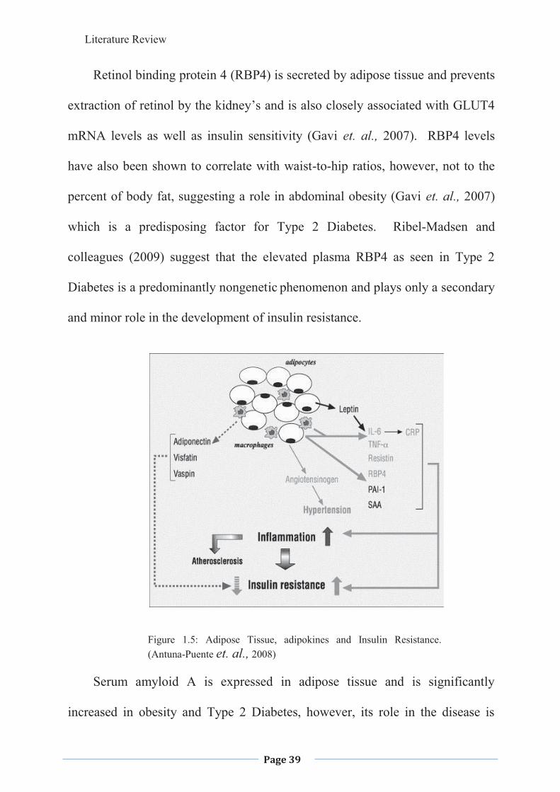
Retinol binding protein 4 (RBP4) is secreted by adipose tissue and prevents
extraction of retinol by the kidney’s and is also closely associated with GLUT4
mRNA levels as well as insulin sensitivity (Gavi et. al., 2007). RBP4 levels
have also been shown to correlate with waist-to-hip ratios, however, not to the
percent of body fat, suggesting a role in abdominal obesity (Gavi et. al., 2007)
which is a predisposing factor for Type 2 Diabetes. Ribel-Madsen and
colleagues (2009) suggest that the elevated plasma RBP4 as seen in Type 2
Diabetes is a predominantly nongenetic phenomenon and plays only a secondary
and minor role in the development of insulin resistance.
Figure 1.5: Adipose Tissue, adipokines and Insulin Resistance. (Antuna-Puente et. al., 2008)
Serum amyloid A is expressed in adipose tissue and is significantly
increased in obesity and Type 2 Diabetes, however, its role in the disease is
Literature Review
Page 40
unknown. It is an acute-phase inflammatory protein and often used as a marker
for coronary events (Leinonen et. al., 2003).
Vaspin is a serine protease-inhibitor strongly expressed by visceral adipose
tissue and is stimulated in mouse (Hida et. al., 2005) and human (Kloting et. al.,
2006) obesity. Insulin or insulin sensitizing drugs will normalise its expression
(Curat et. al., 2006) and if Vaspin is injected into an animal with obesity due to
a high-fat-diet, insulin resistance is improved (Hida et. al., 2005).
1.1.8. Glucose Uptake
There are three main molecular characteristics of �-cell glucose metabolism
that are of importance in the maintenance of homeostasis: (1) The expression of
the high capacity, low affinity glucose transporters GLUT 2 in both the �-cells,
and hepatocytes (Thorens et. al., 1988; Johnson et. al., 1990), which allows
glucose to equilibrate across the plasma membrane, (2) Glucose phosphorylation
to glucose-6-phosphate which is catalysed by glucokinase (GK), which is the
rate-determining step for glycolysis (Reviewed in Matschinsky, 1996), and (3)
Glycolysis produces pyruvate, which in turn is channelled to the mitochondria.
This in turn generates ATP and other factors, which promote insulin secretion
(Schuit et. al., 1997).
Glucokinase (GK), a member of the hexokinase family, and a
phosphorylating enzyme is a crucial component in the control of glucose
Literature Review
Page 41
metabolism in the �-cells and in the hepatocytes (Printz et. al., 1993b). The GK
gene has two distinct promoters specific for the liver and �-cells; the hepatic
promoter is regulated by insulin, whereas the �-cell promoter is constitutively
active. Both GLUT2 and GK have a high Km (Michaelis constant) for glucose
ensuring that glucose uptake is proportional to extracellular glucose
concentrations. In Type 2 Diabetes the GK gene expression and GK activity are
very low, which leads to deficient glucose metabolism in both the �-cells and
the liver (Printz et. al., 1993b). Fructose activates hepatic GK and when
administered to the diabetic patient will partially correct the regulation of
glucose production suggesting that the impaired GK activity substantially
increases glucose production (Hawkins et. al., 2002).
Decreased concentrations of GLUT2 or a decreased expression of GK (Bali
et. al., 1995; Grupe et. al., 1995; Terauchi et. al., 1995) alters the signal given
by high glucose concentrations that regulates insulin secretions, which leads to a
decrease in insulin release.
1.2. TYPE 2 DIABETES
Type 2 Diabetes is the most common of all the metabolic disorders and it is
characterised by insulin resistance (a decrease in the response to insulin by
peripheral tissues) and �-cell dysfunction (inadequate insulin release)
(DeFronzo, 1988), and �-cell apoptosis (Butler et. al., 2003). There is also
Literature Review
Page 42
commonly found an increase in endogenous glucose production and decrease
peripheral glucose uptake (Bogardus et. al., 1984; DeFronzo, 1988), along with
the ability of insulin to promote glycogen synthesis and storage being decreased
(Shulman et. al., 1990; Thorburn et. al., 1990). In 1976 and 1977, Unger and
Orci suggested that diabetes is a bihormonal abnormality: (1) glucagon excess
resulting in the overproduction of glucose and (2) the lack of insulin resulting in
under utilization of glucose.
In populations with a high incidence of Type 2 Diabetes (Prima Indians, the
highest reported incidence in the world; Mexican-Americans and Pacific Islands,
Aboriginal Australians and Torres Strait Islanders and Asian Indians) (Zimmet
et. al., 1984; Ramachandran et. al., 1997; Harris et. al., 1998; Dunstan et. al.,
2002) insulin resistance occurs early in life and precedes glucose intolerance.
The �-cell will release more insulin to compensate for the lack of sensitivity,
however, the �-cells reach a point of exhaustion where they can no longer
secrete enough insulin to compensate for the decreased insulin sensitivity, and
fasting hyperglycaemia and diabetes develops (Kahn & Porte, 1997). Poor
insulin responsiveness to glucose results not only from �-cell insensitivity, but
also from the metabolic derangement of diabetes, that forms a vicious cycle
(Kosaka et. al., 1980). The tissue of initiation and the exact process is not yet
known, however there are four major candidates; �-cells, liver, skeletal muscle
and adipose tissue. Petersen and Shulman (2006) suggest that the origin is the
Literature Review
Page 43
skeletal muscle, where any of the glycogen synthesis pathway steps may be
disrupted and most likely those of insulin-stimulated glucose transport and
phosphorylation. However, Kahn (2008) suggests that �-cell dysfunction and
number or volume of functioning �-cells appears low in those at risk of diabetes
and so may play a role in the initiation of this disease.
Exogenous glucose appears in the circulation at the same rate in individuals
with and without Type 2 Diabetes after ingestion of sugars (McMahon et. al.,
1989a; McMahon et. al., 1989b; Mitrakou et. al., 1992). In normal subjects the
various signals such as hyperinsulinaemia, hyperglycaemia, and neural signals,
act together to inhibit endogenous glucose production (Reviewed in Cherrington
et. al., 1987), however, in Type 2 Diabetic individuals this inhibition is
suppressed and endogenous glucose production continues, which is the major
cause of postprandial hyperglycaemia (Mitrakou et. al., 1992). Mitochondria in
skeletal muscle tend to be small and less in number resulting in reduced
oxidative activity and the level of ATP synthase � which is an essential protein
for respiration. In adipocytes there has been shown a decrease in size and
number of mitochondria leading to a lack of ATP which in turn leads to lipid
biosynthesis, and dysfunction in fatty acid oxidation and respiration (Choo et.
al., 2006).
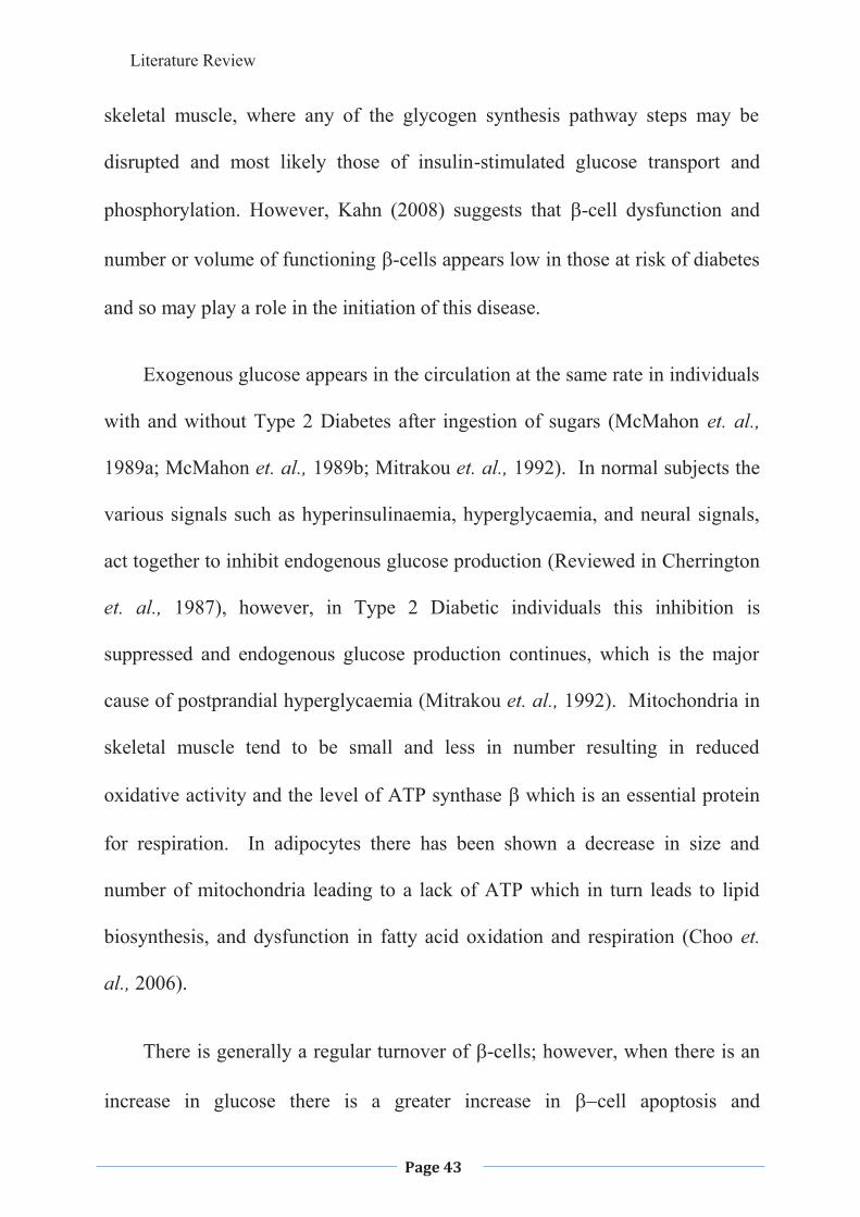
There is generally a regular turnover of �-cells; however, when there is an
increase in glucose there is a greater increase in ��cell apoptosis and
Literature Review
Page 44
regeneration is less, leading to a decreased islet mass within the pancrease. �-
cell mass increases during times of demand such as obesity; however a �-cell
mass decrease has been implicated as one of the factors leading to diabetes
(Kahn, 2008). The long term adaptation of the �-cell to differing conditions
may be initiated by hyperglycaemic excursions, which elicit �-cell production
Figure 1.6: Hypothetical model illustrating the consequence of hyperglycemias on ß-cell production of IL-1ß in parallel with insulin secretion. The paracrine effect of IL-1ß induces Fas engagement, which in the presence of FLIP leads to ß-cell proliferation, differentiation, and increased function. (Donath et. al., 2005).
of interleukin-1�� (Maedler et. al., 2002), followed by FAS up-regulation
(Laybutt et. al.,2003), a decrease in FLICE inhibitory protein which leads to
apoptosis of the cells (Maedler et. al., 2002). Endoplasmic stress (ES) due to
hyperglycaemia and increased lipids may be responsible for some of the �-cell
apoptosis as the �-cell has a highly developed endoplasmic reticulum (ER) and
Literature Review
Page 45
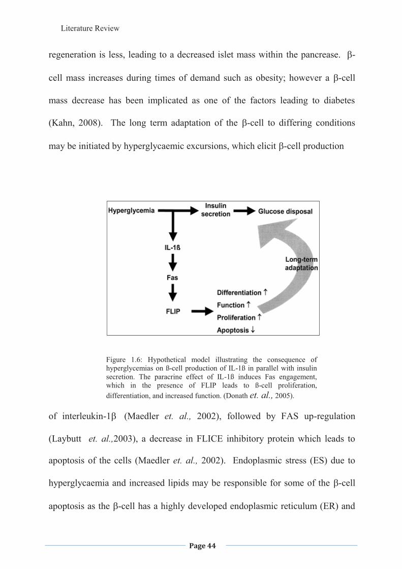
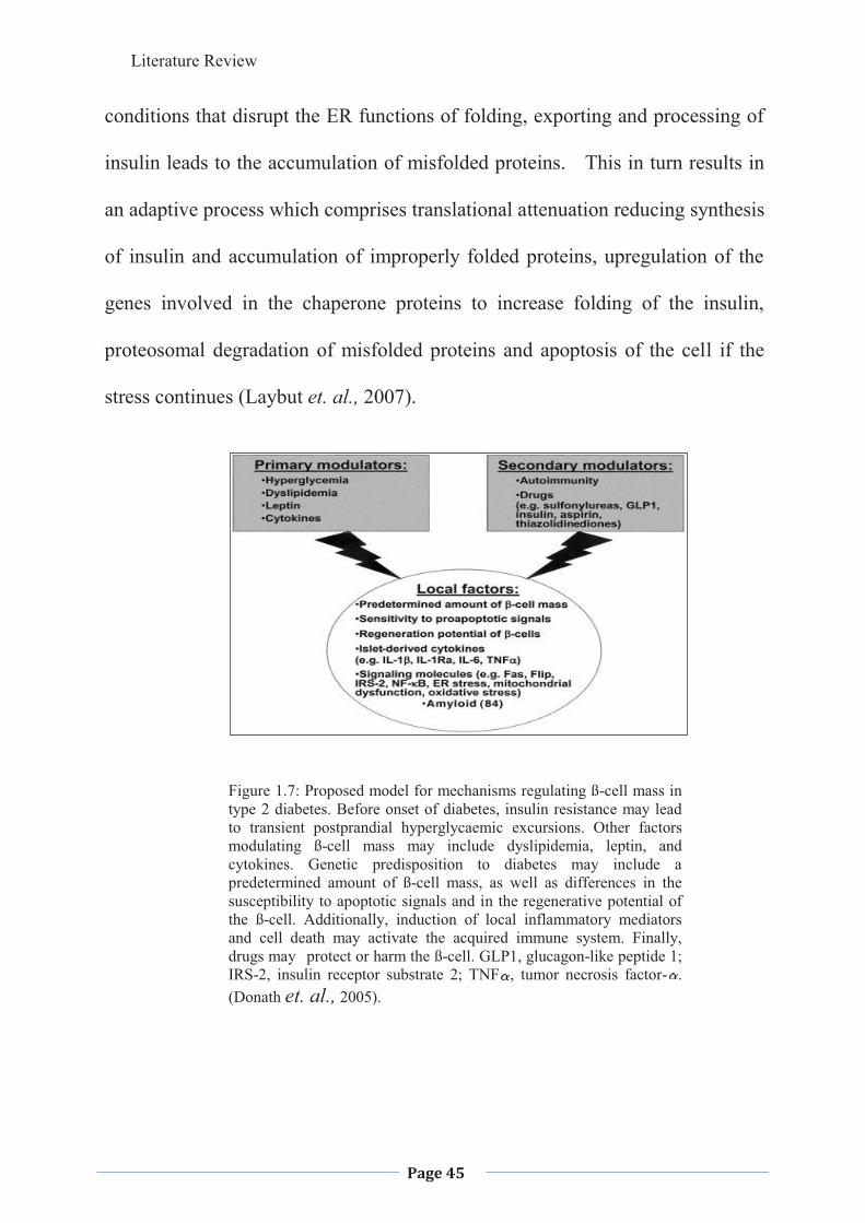
conditions that disrupt the ER functions of folding, exporting and processing of
insulin leads to the accumulation of misfolded proteins. This in turn results in
an adaptive process which comprises translational attenuation reducing synthesis
of insulin and accumulation of improperly folded proteins, upregulation of the
genes involved in the chaperone proteins to increase folding of the insulin,
proteosomal degradation of misfolded proteins and apoptosis of the cell if the
stress continues (Laybut et. al., 2007).
Figure 1.7: Proposed model for mechanisms regulating ß-cell mass in type 2 diabetes. Before onset of diabetes, insulin resistance may lead to transient postprandial hyperglycaemic excursions. Other factors modulating ß-cell mass may include dyslipidemia, leptin, and cytokines. Genetic predisposition to diabetes may include a predetermined amount of ß-cell mass, as well as differences in the susceptibility to apoptotic signals and in the regenerative potential of the ß-cell. Additionally, induction of local inflammatory mediators and cell death may activate the acquired immune system. Finally, drugs may protect or harm the ß-cell. GLP1, glucagon-like peptide 1; IRS-2, insulin receptor substrate 2; TNF , tumor necrosis factor- . (Donath et. al., 2005).
Literature Review
Page 46
1.2.1 Carbohydrates
Diabetes has long been viewed as a disorder of carbohydrate metabolism
due to its hallmark feature of hyperglycaemia, which is the cause of the
associated symptoms such as polyuria, polyphagia and polydipsia (Beiswenger,
2000), and also the long term complications such as nephropathy, neuropathy,
and blindness (UK Prospective Diabetes Study Group, (UKPDS), 1998). The
component of the diet that has the greatest influence on blood glucose is
carbohydrates, while proteins and fats have a less major role, both being able to
slow the absorption of carbohydrates, delaying the peak glycemic response to a
mixed meal (Nuttall and Gannon, 1991), proteins will also augment insulin
release when ingested in a mixed meal, thereby increasing the clearance of
glucose from the blood (van Loon et. al., 2000).
The World Health Organisation now recommends that dietary
carbohydrates be classified according to their glycemic index (FAO/WHO,
1997), which is the measure of the change in blood glucose following ingestion
of carbohydrate-containing foods. The glycemic index is the increase in blood
glucose (over the fasting level) that is observed in the 2 hours following
ingestion of a set amount of carbohydrate in the item and then this value is then
compared with the response to a reference food (usually glucose) containing an
equivalent amount of carbohydrate (Jenkins et. al., 1981). Some foods
(potatoes, pumpkin) result in a marked rise followed by a more or less rapid fall
Literature Review
Page 47
in blood glucose, while other foods (green vegetables) produce a smaller peak
with a more gradual decline in plasma glucose (Institute of Medicine of the
National Academies, 2002). The quantity and type of carbohydrate found in
different foods also influence the postprandial glucose level (Franz et. al., 2002;
Institute of Medicine of the National Academies, 2002) with the specific type of
carbohydrate (starch or sucrose for example) not always predicting its effect on
blood glucose (Wolever et. al., 1994; Foster-Powell and Miller, 1995). The
glycemic index of food is important to understand the effect of that food on
blood glucose levels, however, the amount of that food consumed is also
important, for this reason Salmeron and colleagues (1997a) have suggested the
use of the glycemic load. The glycemic load can be defined as the product of
the glycemic index and the amount of the carbohydrate in the serving and by
summing the glycemic load contributed by individual foods the overall glycemic
load of a meal can be calculated (Salmeron et. al., 1997b).
Epidemiological studies form the basis for the hypothesis that a diet with a
high glycemic load or glycemic index can lead to Type 2 Diabetes (Salmeron et.
al.,1997a; Hu et. al., 2001), however, one study by Salmeron and colleagues
(1997b) showed that both glycemic index or glycemic load were associated with
the risk of Type 2 Diabetes, except when adjusted for cereal fibre intake, and
Meyer and colleagues (2000) showed no correlation between glycemic index or
glycemic load and the development of Type 2 Diabetes. Liu and colleagues
Literature Review
Page 48
(2000) showed that substituting foods with a low glycemic index for those with
a high glycemic index decreased serum insulin and glucose response, and
HbA1c and Urinary C- peptide (a marker for insulin production) in both diabetic
and nondiabetic subjects.
Fructose produces smaller postprandial insulin release than consumption of
glucose-containing carbohydrate along with reducing circulating leptin levels as
leptin production is regulated by insulin response to meals. This combined
effect of lower insulin and leptin could lead to increased weight gain due to
signalling to the brain suggesting still in need of food and hence increased food
consumption, and since fructose is also preferentially metabolized to lipid in the
liver, it could lead to insulin resistance (Elliott et. al., 2002), which is possibly
due to interference with insulin signalling (Lee et. al., 1994). Exposure of the
liver to large quantities of fructose stimulates lipogenesis and the accumulation
of triglycerides contributes to decreased insulin sensitivity and hence hepatic
insulin resistance (Hallfrisch, 1990). Wu and colleagues (2004) also state that
there are long term negative effects of a high fructose diet which can lead to
changes in digestion, absorption, plasma hormone levels (30% reduction in
ghrelin, for example, as well as adiponectin), appetite and hepatic metabolism
leading to the metabolic syndrome. A high fructose diet has also been shown to
increase the amount of C-peptide and lower the number of insulin receptors in
skeletal muscle and liver in mice, as well as decreasing the insulin stimulated
Literature Review
Page 49
autophosphorylation in rats increasing of intracellular triglycerides stores
leading to lipotoxicity and �-cell failure (Catena et. al., 2003). Higashiura and
colleagues (1999) showed a link between a high fructose intake in rats and the
muscle fibre type with a change of Type 1 fibre (low twitch, Oxidative) to Type
2a fibre (fast twitch, Oxidative/Glycolytic), which could be reversed by the
addition of a calcium antagonist. In a human study it was shown that a high
fructose intake increased gluconeogenesis, total glucose output and glucose
cycling and decreased the glucose rate of disappearance leading to insulin
resistance (Dirlewanger et. al., 2000). However, a small amount of fructose is
beneficial in that its metabolite, fructose-1-phosphate increases glucokinase
activity in the liver, which in turn, allows increased glucose sensing and
suppression of gluconeogenesis (van Schaftingen, 1993).
1.2.2. Free Fatty Acids
An acute elevation of FFAs moderately stimulates insulin secretion in
normal and elevated glucose conditions (Malaisse & Malaisse-Lagae, 1968;
Crespin et. al., 1969); however in Type 2 Diatetic individuals there is a long-
term increase in FFAs, which may have severe effects on insulin secretion.
Obesity is characterised by increased levels of FFAs, and also by insulin
resistance (Kolterman et. al., 1980). Free Fatty Acids have a negative effect on
insulin sensitivity in the liver and hence contribute to the production of
Literature Review
Page 50
endogenous glucose, by inhibiting the acute insulin suppression of
glycogenolysis and the elevated levels of plasma FFAs (Boden et. al., 2002).
Petersen and Shulman (2006) suggest that skeletal muscle insulin resistance is
brought about by the loss of insulin activation of IRS-1 associated PI3K activity
in high FFA environment and may be similar in the liver. Cortisol may also
contribute to an increased lipolysis by further inhibiting the antilipolytic effect
of insulin (Frayn et. al., 1996). An increase of FFAs is released from
abdominal adipocytes (abdominal obesity is a major predictor of insulin
resistance and diabetes) into the portal system (Frayn et. al., 1996), which
inhibits insulin sensitivity.
Under normal physiological condition, FFAs sustain insulin release in the
fasting individual and acutely enhance hormone release in the presence of
glucose (McGarry & Dobbins, 1999); however, prolonged exposure to high
concentrations of FFAs has a detrimental effect on �-cells (lipotoxicity) (Zhou
& Grill, 1994; McGarry & Dobbins, 1999). A high level of FFAs reduce insulin
release in response to glucose, suppresses proinsulin biosynthesis and decreases
insulin stores (Bollheimer et. al., 1998; McGarry & Dobbins, 1999), along with
increasing the proinsulin to insulin ratio (Bjorklund & Grill 1999). Increased
FFA levels have been shown to cause apoptosis of �-cells leading to the
development of diabetes in mice (Lee et. al., 1997; Shimabukuro et. al., 1998),
which is brought about by the caspase system along with other possible
Literature Review
Page 51
proteases (Cryns & Yuan, 1998; Johnson, 2000; Lupi et. al., 2002). The
mechanism of apoptosis has not been fully elucidated but include the following
possibilities: increased ceramide production, the activation of the transporter
nuclear factor kB, up regulation of iNOS, increased synthesis of NO, enhanced
formation of NO-derived free radicals, DNA damage (Unger & Zhou, 2001;
Lupi et. al., 2002), the binding of FFA with PPARs is able to elicit several
changes in the expression of many proteins, including some caspases (Chinetti
et. al., 1998) and there is a marked reduction in Bcl-2 mRNA, which is an
apoptosis regulator gene product (Zamzami et. al., 1998). The FFA-induced
inhibition of glucose-stimulated insulin release may be related to the glucose-
free fatty acid (Randle) cycle (Reviewed in Randle et. al., 1988), by substrate
competition, after their oxidation. Free Fatty Acid oxidation is inhibited by
metformin (Patiane et. al., 2000), which results in the increased glucose
metabolism and insulin secretion, while FFA metabolism reduces the activity of
pyruvate dehydrogenase, which alters glucose metabolism (Zhou et. al., 1996).
Lupi and colleagues (2002) have shown that a high level of plasma FFA
decreases both glucose oxidation and utilization, which was more than could be
accounted for by oxidation suggesting that other pathways are affected as well.
Free Fatty Acids induce insulin resistance by impairing the insulin-
signalling pathway at the level of insulin-stimulated glucose transport or
phosphorylation. Dresner and colleagues (1999) suggest that it is the
Literature Review
Page 52
accumulation of metabolites other then fat, such as diacylglycerol (DAG), which
results in insulin resistance. The increase in DAG results in a rise of protein
kinase C (PKC), which is an enzyme that can phosphorylate serine and
threonine residues on both the insulin receptor and the insulin receptor substrate
along with the activation of the nuclear-factor-�B (NF�B) pathway (Itani et. al.,
2002). FFAs may also increase insulin resistance by increasing oxidative stress
with both the PKC and the NF�B pathways being activated by reactive oxygen
species (Griffin et. al., 1999; Ceriello, 2000).
Free Fatty Acids are potent insulin secretagogues and can compensate for
the insulin resistance that they can create with acute elevations stimulating
insulin secretion and prolonged elevations potentiating glucose-stimulated
insulin secretion (Crespin et. al., 1973). However, in those who are first degree
relatives of a Type 2 Diabetic individual, FFAs are unable to compensate for the
insulin resistance that they create suggesting a genetic predisposition to
pancreatic �-cell failure (Stogaard et. al., 2003).
Peroxisome proliferator-activated receptor – gamma (PPAR-�, which is
one of the nuclear transcription factors that regulate the expression of numerous
genes involved in lipid and carbohydrate metabolism, inflammation and vascular
tone, is expressed at its highest concentration in adipose tissue and at a much
lower level in the skeletal muscles and liver (Tontonoz et. al., 1994).
Peroxisome proliferator-activated receptor – gamma exerts it effect by lowering
Literature Review
Page 53
plasma FFA levels, increasing plasma adiponectin levels, and redistributing
visceral fat to subcutaneous depots similar to rosiglitazone (Mayerson et. al.,
2002). Free Fatty Acids are also natural ligands for peroxisome proliferator-
activated receptor (PPAR-�), which is expressed in the liver, muscle, kidney and
heart, which are the organs where fatty acids are oxidised and PPAR��
activation lowers plasma FFA levels through increased fat oxidation (Muoio et.
al., 2002). Specific PPAR-� agonists can lower lipid levels in rats and can
improve their insulin sensitivity (Ye et. al., 2001), however, human studies have
shown either improved (Mussoni, et. al., 2000) or no reaction to specific PPAR-
� agonists (Asplund-Carlson, 1996) A diet high in saturated fats has also been
implicated in a reduction in the number, location, and morphology of
mitochondria which is strongly associated with insulin resistance, due to a
reduction in the expression of genes involved in the oxidative capacity of the
cells such as those of the electron transport chain, mitochondrial protein genes
and those involved in mitochondrial biogenesis (Sparks et. al., 2005;
Ukropcova, et. al., 2007). The healthy individual, with normal body fat content
relies on lipids as their main source of energy during the fasting state and can
switch to glucose oxidation with introduction of insulin, while in the insulin
resistant state the switch to glucose oxidation does not occur due to decreased
mitochondrial oxidative enzyme activity response to insulin (Ukropcova et. al.,
2007).
Literature Review
Page 54
1.2.3 Genetics
One or more lesions in a number of different genes have been shown to
predispose an individual to Type 2 Diabetes and in particular Maturity Onset
Diabetes of the Young (MODY), which is a subset of the Type 2 Diabetes,
however, they generally remain silent. One gene that has been of particular
interest is for the apolipoprotein B, which influences parameters related to
insulin resistance, with its polymorphisms being silent and so the effects must be
mediated through linkage to other genes (Bentzen et. al., 2002). Kovacs and
colleagues (2002) have implicated the ORP 150 gene as a possible link for Type
2 Diabetes in the Prima Indian population, who have a very high incidence of
Type 2 Diabetes, hypothesizing that it plays some role in responding to glucose
levels by altering muscle glucose uptake. Mutations in the gene WFS1 have
been shown to produce �-cell death in a form of diabetes known as Wolfram
Syndrome and Minton and colleagues (2002) have shown that variations of this
gene may lead to susceptibility to Type 2 Diabetes. Siitonen and colleagues
(2006) have shown that a sequence variation in a gene encoding for the
adiponectin receptor 1 has implications for body size and plasma insulin levels
although they appear to be dissociated from each other. The Pro12 allele of the
PPAR-� gene, which, when combined with low physical activity leads to a
higher risk of the disorder (Nelson et. al., 2007). Genetic variation of the
adiponectin locus is a predictor of adiponectin levels and hence insulin
Literature Review
Page 55
resistance and there have been several polymorphism linked to Type 2 Diabetes
(Menzaghi et. al., 2007). Also a haplotype of the CRP gene has been shown to
result in a high serum CRP level, and the incidence of diabetes was much higher
amongst this group of individuals than those individuals who had the three other
common haplotypes. This indicates a possible genetic role in the development
of diabetes (Dehghan et. al., 2007).
MODY is a monogenic dominantly inherited form of Type 2 Diabetes that
is characterised by defective insulin secretion and mutations in five genes have
been implicated in this including the hepatocyte nuclear factor-4� gene and the
hepatocyte nuclear factor-1� gene (Yang et. al., 2002).
First degree relatives of the Type 2 Diabetic patient will often exhibit
symptoms of the pre-diabetic state or metabolic syndrome suggesting that
genetics plays a role in the development of the disease. Karlsson and colleagues
(2006) have suggested that the insulin-mediated glucose uptake from glucose
tolerant individuals who are first degree relatives of diabetic subjects is impaired
and this may lead to impaired glucose tolerance and later to Type 2 Diabetes.
1.2.4. Uterine Environment
There is considerable evidence to show that the intrauterine environment of
a foetus can predispose an individual to diabetes, with the higher the birth-
weight, which is a measure of the intrauterine environment in humans, the less
Literature Review
Page 56
likelihood of a person getting Type 2 Diabetes (Birgisdottir et. al., 2002),
indicating the need for high quality nutrition by the expectant mother is
important in the prevention of Type 2 Diabetes. The intrauterine environment is
argued to change gene expression and lead to physiological or morphological
phenotypes associated with diabetes (Eriksson et. al., 2002), suggesting there is
a link between the Pro12Pro genotype of the PPAR-��gene being associated
with higher fasting insulin concentrations and insulin resistance in babies with a
low birth weight. Intrauterine growth failure leading to a small birth weight is
also shown to affect the number of pancreatic �-cells as well as the function of
these cells (Eriksson et. al., 2002). Lawlor and colleagues (2006) showed that
poor intrauterine growth and premature births were associated with the risk of
developing diabetes in later life, though no understanding of why this occurs has
been elucidated, while Singh and colleagues (2006) showed that children of
diabetic mothers were more likely to develop Type 2 Diabetes then if the father
was diabetic and suggest that this may be the result of mild hyperglycaemia
within the uterine environment, reducing �-cell function.
Insulin-like growth factor-1 (IGF-1) and insulin are essential foetal growth
factors (Hill et. al., 1998) and is also important for growth and development of
�-cells (Vaessen et. al., 2001) and Vaessen and colleagues (2002) have shown
that a mutation in the IGF-1 gene and the GK gene lead to both a predisposition
to Type 2 Diabetes and low birth weight.
Literature Review
Page 57
Early postnatal malnutrition can have major consequences on fetal and
postnatal development in both humans and animals (Gluckman and Harding,
1992; Owens, 1991). Protein/calorie malnutrition in particular or protein
deficiency only in utero in the mother will lead to decreased �-cell mass or islet
volume in off-spring which may be due to a decreased islet neogenesis with
prolonged maternal malnutrition until the end of the lactation period resulting in
up to 70% decrease in �-cell mass. This was not restored at age by normal
feeding after weaning in animals (Blondeau et. al., 1999). Insulin stores and
fasting insulin is also decreased in previously malnourished mice and there is an
impaired secretory response to glucose (Blondeau et. al., 1999).
The mechanism linking low birth rate and glucose intolerance has not yet
been elucidated, however, one suggestion has been that poor nutrition in utero
causes the foetus to become thrifty at a time when �-cell development proceeds
more rapidly, resulting in impaired growth of these cells and, hence, leads to a
predisposition of diabetes later in life (Blondeau et. al., 1999). Interestingly,
this has been shown to lead to decreased plasma insulin but normal plasma
glucose (Garofano et. al., 1998), also maternal protein deficiency does not lead
to impaired glucose tolerance in the adult offspring (Holness and Sugden, 1996).
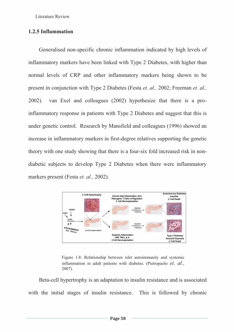
Literature Review
Page 58
1.2.5 Inflammation
Generalised non-specific chronic inflammation indicated by high levels of
inflammatory markers have been linked with Type 2 Diabetes, with higher than
normal levels of CRP and other inflammatory markers being shown to be
present in conjunction with Type 2 Diabetes (Festa et. al., 2002; Freeman et. al.,
2002). van Exel and colleagues (2002) hypothesize that there is a pro-
inflammatory response in patients with Type 2 Diabetes and suggest that this is
under genetic control. Research by Mansfield and colleagues (1996) showed an
increase in inflammatory markers in first-degree relatives supporting the genetic
theory with one study showing that there is a four-six fold increased risk in non-
diabetic subjects to develop Type 2 Diabetes when there were inflammatory
markers present (Festa et. al., 2002).
Figure 1.8: Relationship between islet autoimmunity and systemic inflammation in adult patients with diabetes. (Pietropaolo et. al., 2007).
Beta-cell hypertrophy is an adaptation to insulin resistance and is associated
with the initial stages of insulin resistance. This is followed by chronic
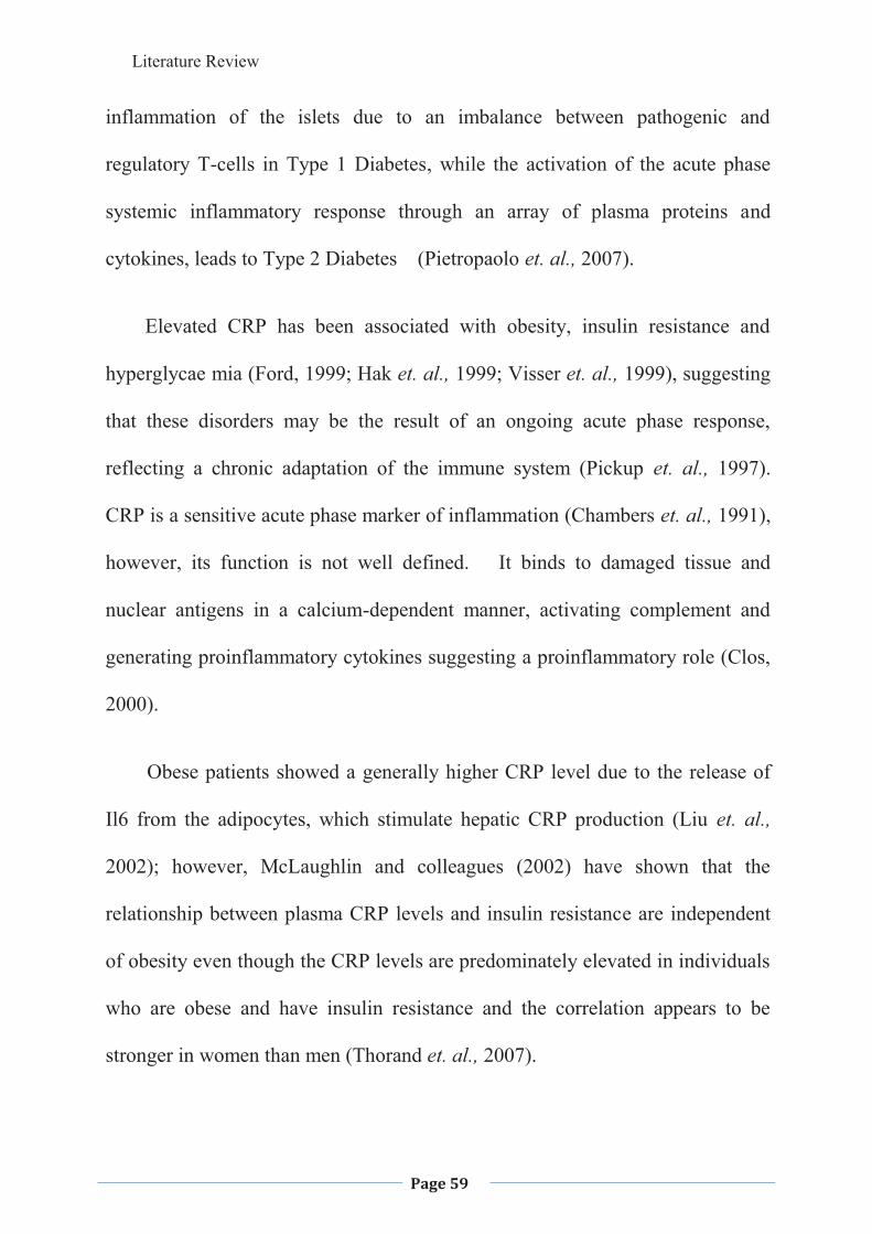
Literature Review
Page 59
inflammation of the islets due to an imbalance between pathogenic and
regulatory T-cells in Type 1 Diabetes, while the activation of the acute phase
systemic inflammatory response through an array of plasma proteins and
cytokines, leads to Type 2 Diabetes (Pietropaolo et. al., 2007).
Elevated CRP has been associated with obesity, insulin resistance and
hyperglycae mia (Ford, 1999; Hak et. al., 1999; Visser et. al., 1999), suggesting
that these disorders may be the result of an ongoing acute phase response,
reflecting a chronic adaptation of the immune system (Pickup et. al., 1997).
CRP is a sensitive acute phase marker of inflammation (Chambers et. al., 1991),
however, its function is not well defined. It binds to damaged tissue and
nuclear antigens in a calcium-dependent manner, activating complement and
generating proinflammatory cytokines suggesting a proinflammatory role (Clos,
2000).
Obese patients showed a generally higher CRP level due to the release of
Il6 from the adipocytes, which stimulate hepatic CRP production (Liu et. al.,
2002); however, McLaughlin and colleagues (2002) have shown that the
relationship between plasma CRP levels and insulin resistance are independent
of obesity even though the CRP levels are predominately elevated in individuals
who are obese and have insulin resistance and the correlation appears to be
stronger in women than men (Thorand et. al., 2007).
Literature Review
Page 60
Figure 1.9: Model of IL-6 actions of potential relevance for the pathogenesis of type 1 and type 2 diabetes. IL-6 is produced by many inflammatory cells, adipose tissue, working muscle, and even the ß-cell. Expression is determined by genetic variation, intrauterine environment, age, and sex steroids. IL-6 induces insulin resistance in adipose tissue and liver and may synergize with proinflammatory cytokines to produce ß-cell damage. IL-6 also regulates energy expenditure probably by the effect on brown adipose tissue via effects on the CNS. EE, energy expenditure; IA, immune activation; IR, insulin resistance; SC, synergy with proinflammatory cytokines (Reviewed in Kristiansen and Mandrup-Poulsen, 2005).
Interleukin-18 (IL-18) has also been implicated in the development of Type
2 Diabetes. Elevated levels of IL-18 and CRP and IL-18 and IL-6 have been
shown to be present in Type 2 Diabetic patients independent of a generalised
pro-inflammatory state (Thorand et. al., 2005) and IL-6 is an independent
marker for insulin resistance independent of gender (Thorand et. al., 2007);
however, increased levels of IL-6 does not appear to have any effect on �-cell
function or insulin resistance in muscle. There is some evidence that IL-6 may
cause insulin resistance in hepatocytes and impaired insulin signalling in
Literature Review
Page 61
adipocytes, as well as impaired action within the Central Nervous System
centres involved in energy metabolism, resulting in obesity, which in turn leads
to insulin resistance (Reviewed in Kristiansen and Mandrup-Poulsen, 2005).
The IL-6 receptor uses the Janus Kinases (JAKs) as intracellular pathways
which may lead to a decrease in the insulin signalling, possibly via the
participation of protein kinases and tyrosine phosphatase activation (Kroder et.
al., 1996) or SOCS interaction (Ricusset et. al., 2004) with the insulin receptor.
Engstrom and colleagues (2005) have shown that Complement C3 and C4,
which are major plasma proteins of the immune system complement pathway
and are increased in response to inflammation at a slower rate then the more
acute phase proteins, have a substantial correlation with obesity and hence show
a possible link with the increased risk of developing Type 2 Diabetes.
1.2.6. Oxidative Stress
Oxidative stress has been linked to the pathogenesis of Type 2 Diabetes
and its complications, and stress proteins have been shown to protect against
oxidative stress. There is a decrease in the heat shock proteins in the muscles of
patients with Type 2 Diabetes and it has been hypothesized that this may be a
link between insulin resistance and Type 2 Diabetes (Kurucz et. al., 2002),
although at this stage the mechanism is not understood. Ferandez and
colleagues (2002) have suggested that the link may be that a bi-directional-iron
affects glucose metabolism and glucose metabolism affects several iron
Literature Review
Page 62
metabolic pathways, showing that increased iron stores predict Type 2 Diabetes
while iron depletion is protective, suggesting that increased levels of iron may
be toxic to cells and also cause inflammation. Evans and colleagues (2003) have
proposed that there is a hyperglycaemia-induced, and possibly a FFA induced,
activation of stress pathways and the production of reactive oxygen and nitrogen
species, that leads also to the development of complications related to both Type
1 and Type 2 Diabetes and also the development and progression of insulin
resistance and impaired insulin secretion. Type 2 Diabetic patients are also
reported to have reduced muscle oxidative enzymes and have shown an increase
in muscle fatigability (Halvatsiotis et. al., 2002). This could lead to a decrease
in the ability of insulin to stimulate glucose uptake.
1.2.7. Microvascular Disease
Type 2 Diabetes has been shown to lead to microvascular disease, however,
some studies suggest that the microvascular disease appears first and may
contribute to the pathogenesis of the diabetic state (Hsueh & Law, 1998; Tooke,
1995). Morris and colleagues (1995) demonstrated microvascular abnormalities
(impaired microvascular reactivity and flow in the skin and skeletal muscles) in
people with Type 2 Diabetes and in people at high risk of developing diabetes,
such as those with impaired glucose tolerance and first degree relatives of
persons with diabetes (Caballero et. al., 1999; Balletshofer et. al., 2000). It has
been suggested that the microvascular alterations may result in a reduced ability
Literature Review
Page 63
of insulin to mediate glucose uptake in skeletal muscles (Wong et. al., 2002); the
alterations may lead to inflammation, which has been linked to diabetes.
Metabolic abnormalities associated with increased platelet sensitivity to
pro-aggregants and decreased platelet sensitivity to endogenous anti-aggregants
has been associated with Type 2 Diabetes (Vinik et. al., 2001; Beckman et. al.,
2002).
Hematocrit has been positively correlated with insulin resistance and
many of the symptoms associated with the metabolic syndrome (high blood
pressure, elevated cholesterol, and serum triglycerides, and low HDL) (Tamariz
et. al., 2003; Nakanishi et. al., 2004). Although the correlation is not known, it
has been suggested that as hematocrit is a major determinant of blood viscosity
(MacRury and Lowe, 1990) and when this is elevated it reduces the blood flow
to skeletal muscle, therefore interfering with insulin-mediated glucose uptake in
skeletal muscle (Baron et. al., 1994).
1.2.8. Obesity
The association between obesity and Type 2 Diabetes has been accepted for
sometime, and although progress has been made, the exact mechanisms have not
been established (Reviewed in Kahn et. al., 1996b). Adipose cells secrete FFAs,
as products of triglyceride metabolism and are the main form in which energy is
transferred from adipose tissues to other sites for metabolic use and have also
Literature Review
Page 64
been implicated in the induction of insulin resistance in tissues other then
adipose tissues (Boden, 1999), by the release of numerous peptides such as
resistin.
An important aspect of obesity is the placement of the adiposity where
subcutaneous and visceral abdominal fat areas are linked to Type 2 Diabetes
whereas other fat stores are not (Taniguchi et. al., 2002); However whole body
obesity and waist circumference and waist to hip ratio, both an indicator of
central obesity, are all strongly related to the risk of the development of diabetes
(Meisinger et. al., 2006a).
Sterol regulatory element binding protein (SREBP-1) is a transcription
factor, which plays many important roles in fatty acid metabolism and
adipogenesis, which is positively modulated by insulin. One SREBP-1 isoform
is commonly seen in adipose tissue of the obese and Type 2 Diabetes, which
may be a possible link between diabetes and obesity (Sewter et. al., 2002).
Hampton (2004) states that there appears to be a vicious cycle where an
obese insulin-resistant woman, has an obese foetus in utero which remains obese
throughout its life, has a greater risk of developing Type 2 Diabetes.
1.2.9. Lifestyle
A sedentary lifestyle has for decades been linked with diabetes, however,
the mechanism has been unclear, although it may be due to inhibition of GLUT4
Literature Review
Page 65
translocation. Many other lifestyle factors have also been implicated in Type 2
Diabetes such as smoking where it has been shown that there is an increased
level of insulin resistance occurring in smokers with and without diabetes
(Targher et. al., 1997; Gunton et. al., 2002; Foy et. al., 2005). Gunton and
colleagues (2002) have shown that cessation of smoking will bring about an
improvement in glycosylated haemoglobin (HbA1c) levels, which may be due to
a change in many lifestyle factors at the same time, or it may be a direct effect
and the improvement parallels the UKPDS metformin group (Gunton et. al.,
2002). Meisinger and colleagues (2006b) have shown that there is a significant
dose-dependent relationship between smoking and in particular the nicotine and
tar intake and the risk of developing Type 2 Diabetes in men; however, the study
was less convincing with women but this may have been due to the low number
of women in the cohort. Smoking is known to produce inflammation and this
itself appears to play an important role in the development of diabetes (Rimm et.
al., 1993; Schmidt et. al., 1999; Manson et. al., 2000; Pradham et. al., 2001).
Caffeine, an adenosine receptor antagonist, when ingested decreases
glucose uptake suggesting the role of adenosine in glucose disposal and a
decrease in carbohydrate storage (Greer et. al., 2001). As coffee is a common
beverage amongst the Western world where Type 2 Diabetes is epidemic, advice
given to people as to the possible connection between caffeine ingestion and
Type 2 Diabetes might be a way of decreasing the incidence of this disease in
Literature Review
Page 66
the future. However, a study undertaken by van Dam and colleagues (2004)
showed that habitual, chronic coffee consumption may reduce the risk of
impaired glucose tolerance and a lower incidence of Type 2 Diabetes, but was
not associated with FBGL or impaired fasting glucose, suggesting that coffee
affects post-load rather then fasting glucose metabolism through increased
insulin sensitivity. They also showed that coffee consumption was cross-
sectionally linked with lower values for markers of insulin resistance such as
inhibition of g6pase activity, decrease in absorption rates by inhibiting the
sodium dependent transporters, and a reduction on oxidative stress, all possibly
due to the chlorogenic acid within the coffee.
The consumption of soft drinks has become a major problem in many
countries, as the high sugar intake has led to an increase in obesity, which in
turn has led to an increased risk of diabetes. Consumption of fruit punch has
also been implicated in this increased risk (Spurgeon, 2004). Sugar-sweetened
soft drinks now contribute 7.1% of total energy intake and represent the largest
single food source of calories in the USA (Block, 2004). The rise in obesity and
Type 2 Diabetes parallels the increase in sugar-sweetened soft drinks (Bray et.
al., 2004), and hence programs to decease the amount of sugars in drinks or
using alternate sweetening agents may decrease this ever growing problem. As
fruit juices do not tend to have the same risk factors associated with Type 2
Diabetes (Schulze et. al., 2004), possibly due to them having a lower glycemic
Literature Review
Page 67
index, and more soluble fibre and possibly other chemicals that can be beneficial
(antioxidants being a major consideration). There also needs be educational
programs to inform and encourage people to alter their choice of
beverage/drinking habits.
There has been a considerable increase in the eating of fast-foods and a
study was undertaken by Pereira and colleagues (2005) to determine if there was
any correlation between this increase and the increase in Type 2 Diabetes and
they suggested that fast food promotes a positive energy balance leading to
obesity and related diseases, due to the large portion sizes generally served in
fast food outlets; palatability, emphasizing taste preferences of sugar, salt and
fat; high energy density and the high glycemic load foods being offered. After a
15 year follow-up on several black and white, male and female Americans it was
observed that eating the average fast-food diet by this population showed a
strong, positive and independent correlation with obesity and insulin resistance
(Pereira et. al., 2005).
1.2.10 Lung Vital Capacity
A low lung vital capacity has been linked to the development of impaired
glucose tolerance, insulin resistance and Type 2 Diabetes in later years. Possible
mechanisms include hypoxemia and inflammation during foetal and early
childhood (Yeh et. al., 2005). Hypoxemia results in increased sympathetic
activity which has been shown to increase the risk of Diabetes; however, this has
Literature Review
Page 68
been debated (Yeh et. al., 2005). Inflammation has been linked with diabetes
and decreased lung function will often result in inflammation as seen in lower
respiratory tract infection, however this link has not been proven either (Yeh et.
al., 2005). Low vital capacity may also interfere with organ development and
the metabolic pathway programming, which, in addition to a genetic
predisposition could lead to insulin resistance (Yeh et. al., 2005).
1.3. ORTHODOX TREATMENT
Therapeutic options that are currently available for the treatment of Type 2
Diabetes include dietary modifications, lifestyle modifications, oral
hypoglycaemic agents and insulin. In early Type 2 Diabetes dietary and
lifestyle modifications are usually the only treatment recommended, especially if
there is a weight problem as well. In many cases this will control the BGLs,
however, when diet modifications no longer exert the required effect, oral
hypoglycaemic agents become very important in the treatment of Type 2
Diabetes.
1.3.1 Diet and Lifestyle
Obesity has been associated with insulin resistance and Type 2 Diabetes,
therefore reducing caloric intake improves glycemic control. Caloric intake is
more important than the actual weight loss, which will improve both insulin
resistance and impaired insulin secretion (De Fronzo, 1988) as once weight loss
Literature Review
Page 69
has occurred, even without weight gain, metabolic control can deteriorate again
because during caloric restriction there is a rapid fall in post-absorptive
endogenous glucose production (Henry et. al., 1986).
Carbohydrates are the cause of increases in blood glucose but it is
important that they are still consumed throughout the day as they are the main
fuel for the body and are the primary fuel for the brain and central nervous
system. Due to this low-carbohydrate diets are not recommended in the
management of diabetes and it is recommended that 45-65% of calories are
derived from carbohydrates with a minimum intake of 130 gm per day for adults
(Institute of Medicine of the National Academies, 2002). Since both the
glycemic index and the amount of carbohydrate consumed have an effect on the
blood glucose level it was found that decreasing the glycemic index of diet
reduced postprandial plasma glucose by the same amount as reducing the
carbohydrate intake (Wolever and Mehling, 2003). Therefore patients with
Type 2 Diabetes should increase foods which are low glycemic and high fibre
over those that are high glycemic and low fibre. These finding were supported
by Lindstrom and colleagues (2006) in the Finnish Diabetes Prevention Study,
where they showed that high-fibre, low-fat diets led to long term weight loss and
a decrease in the risk of Type 2 Diabetes, in those who had impaired glucose
function.
Literature Review
Page 70
The eating of foods high in natural fibre is also important with one study
looking at the Mediterranean Diet showing that natural fibre foods and also
foods that had fibre added both decreased the absorption of glucose from the
intestine but only the natural fibre foods kept plasma glucose levels low
(Riccardi et. al., 2003). The mechanism of this was not elucidated, however, it
was noted that that postprandial plasma acetate was much higher in the natural
fibre rich diets. A diet high in soluble fibre will reduce the need for insulin
(Landin et. al., 1992) and improve glycaemia (Vuksan et. al., 2000a). Studies
with purified viscous fibres demonstrated a reduced rate of gastric emptying
(Holt et. al., 1979; Leeds, et. al.,1981) and small intestine absorption (Blackburn
et. al., 1984; Floune et. al., 1984) resulting in a reduced blood glucose and
insulin response. Levels of insulin and GIP are found to be reduced with a high
fibre diet (Jenkins et. al., 1977; Jenkins et. al., 1979; Jenkins et. al., 1980). The
mechanism of action appeared to be the slowing down of absorption of glucose
rather then a malabsorption (Jenkins et. al., 1977; Jenkins et. al., 1979). High
fibre foods that also have a low glycemic index have shown the greatest
improvement in decreasing HbA1c levels (Simpson et. al., 1981). Suzuki and
colleagues (2002), in a study looking at fruit and vegetable intake in a Japanese
population, suggested that such a diet, rich in antioxidants, might be protective
against Type 2 Diabetes. Weickert and colleagues (2006) showed that purified
insoluble fibre, the predominant fraction of cereal fibre, and consisting of
cellulose and hemicellulose, improved insulin sensitivity if consumed within the
Literature Review
Page 71
daily recommended range. This improvement occurred within three days of
taking the fibre; however, the mechanism of action does remain unexplained.
Weickert and colleagues (2006) showed that the increased insulin sensitivity
was not due to the fermentation process within the colon or an increase in the
amount of GLP-1 produced. Qi and colleagues (2006) have suggested that
dietary cereal fibre increases the circulating adiponectin levels which may be
effective in lowering blood glucose levels. Gentilcore and colleagues (2006)
showed that a small amount of olive oil 30 minutes prior to a carbohydrate meal
decreased gastric emptying, rise of blood glucose and insulin levels, however it
does stimulate the secretion of GLP-1 and GIP thus delaying the onset of
postprandial rise in glucose levels, however, this did not occur to such an extent
if the olive oil was taken with the meal.
Liu and colleagues (2002) showed that the quality and quantity of
carbohydrate intake was directly related to plasma concentrations of CRP, with a
high dietary glycemic load being proportional to the plasma CRP indicating that
a high glycemic load may affect insulin resistance through a proinflammatory
response. O’Riodain and colleagues (1995) showed that elevated concentrations
of insulin and counter-regulatory hormones are directly associated with hepatic
production of CRP. Thomas and Wolever (Reviewed, 2003) have shown that
reducing carbohydrate intake increases postprandial FFAs but does not improve
blood glucose levels, however, low glycemic foods do lower both FFAs and
Literature Review
Page 72
glucose levels. Therefore, it is strongly suggested that a high-carbohydrate but
low glycemic foods be chosen in the diet.
High fat diets have been linked to diabetes and its complications; however
it appears that the type of fat that is eaten may play a major role in these
complications. One study using db/db mice has shown that diets high in
saturated fats tend to decrease adiponectin levels and increase inflammation in
white adipose tissue; however, the use of n-3 series marine polyunsaturated fats
over a prolonger period will return the adiponectin levels to normal and will
assist in the decrease of inflammatory markers such as interleukin-6 (Todoric et.
al., 2006).
Diet is better at controlling BGL if accompanied by exercise, which has
been shown to improve both glucose effectiveness, the ability of
hyperglycaemia to promote glucose disposal at basal levels of insulin, and
insulin sensitivity (Nishida et. al., 2001). Di Loreto and colleagues (2005) have
shown that it is necessary to reach certain energy expenditure levels through
exercise for it to be effective in weight loss and also the decrease of FBGLs. An
increase in energy expenditure is beneficial only to a certain point where any
increased energy expenditure does not appear alter FBGLs, blood pressure,
cholesterol or triglycerides and Jeon and colleagues (2007) have shown that
moderately intensive physical active is all that is needed to reduce the risk of
Type 2 Diabetes; however Sriwijitkamol and colleagues (2007) have suggested
Literature Review
Page 73
that the Type 2 Diabetes, who is also obese need to exercise at a higher intensity
to stimulate the enzyme AMPK and the Akt substrate AS160 to the same level
as that of the non-obese individual. These benefits occurred in the absence of
weight loss so it appears that the important aspect is the aerobic physical
activity, though it is noted that it still may be a result of the change of the
composition of body tissues with the build up of muscle and loss of adipose
tissue (Di Loreta, 2005). Exercise has also been shown to decrease the
circulating levels of Il6 in both lean and obese individuals with and without
diabetes and as chronic inflammation has been linked to diabetes this may be
one way in which an exercise program will also be of benefit (Dekker et. al.,
2007). One area of major concern today is the epidemic of early-onset Type 2
Diabetes and Burns and colleagues (2007) have shown that these individuals,
who are generally obese, are severely insulin resistant and have significant loss
of �- cell function when compared with control subjects matched for age and
obesity; however, neither group responded metabolically to aerobic exercise
training. It has been suggested that this might be due to a susceptibility
genotype or a mitochondrial dysfunction due to chronic lipotoxicity or
hyperglycaemia.
1.3.2. The Sulfonylurea Compounds
The sulfonylurea compounds, such as Glibenclamide, Tolbutamide and
Glipizide, have been available for sometime and used successfully in the
Literature Review
Page 74
treatment of Type 2 Diabetes (Hellman & Taljedal, 1975; Ashcroft & Ashcroft,
1992), as they exert glucose-like action on the K+ATP channels by closing them
and decrease the K+ permeability of the �-cell membrane. This leads to the
depolarisation of the cell, initiation of the voltage-gated Ca2+ influx, and insulin
release (Sturgess et. al., 1985; Zunkler et. al., 1988). Sulfonylurea’s also
enhance exocytosis through enhancement of protein kinase C-dependent
exocytosis, and is additive to the cAMP-dependent effects (Eliasson et. al.,
1996). It leads to a greater expression of GLUT1 (Tordjman et. al., 1989; Wang
et. al., 1989; Tsiani et. al., 1995), which enhances basal glucose uptake, these
medications also have an acute effect on glucose utilization in fat and muscle.
The sulfonylurea’s have a well-characterised profile of side effects, especially
those of a gastric nature and, also, and very importantly hypoglycaemia, which
could be fatal (Prout, 1974). Long-term administration may over stimulate �-
cells, which could lead to an even greater �-cell dysfunction as apoptosis occurs
(Goodman, 2001) though Holman (2006) suggests that they neither increase or
decrease �-cell apoptosis, as well as impairing endothelial function leading to
increased risk for ischemic complications (Del & Pulizzi, 2006). They will
bring about normalisation of the BGL; however, they do not appear to prevent
the micro and macro vascular effects of diabetes due to its inability to maintain a
normal BGL for an extended period of time (Turner et. al., 1999). Also more of
the drug is needed to maintain the homeostatic effect on glucose, as treatment
Literature Review
Page 75
progresses, as there appears to be a development of tolerance to it or often other
groups of drugs need to be added to return the BGL level to normal.
1.3.3. The Biguanides
Biguanides, for example, Metformin, are another class of hypoglycaemic
agents that are in common use today as they do not promote insulin release but
act by increasing glucose utilization in the intestines (Penicaud et. al., 1989),
reducing hepatic glucose output (Meyer et. al., 1967; Jackson et. al., 1987) by,
possibly, interrupting mitochondrial oxidative processes (Kirpichnikov et. al.,
2002) and increasing peripheral glucose utilization (Hother-Nielson et. al., 1989
Galuska et. al., 1994; Fisher et. al., 1995). The increased uptake of glucose is
independent of insulin and is associated with GLUT4 and GLUT1 translocation
to the plasma membrane without affecting the number present (Kozka &
Holman, 1993; Fisher et. al., 1995). Musi and colleagues (2002) showed that
metformin increased AMP-activated protein kinase activity in skeletal muscle of
patients with Type 2 Diabetes, bringing about a higher rate of glucose disposal
and glycogen concentration. However, it does not affect insulin binding or
insulin receptor kinase activity (Fantus & Brosseau, 1986). Rajala and
colleagues (1998) noted that many Type 2 Diabetic patients have microvascular
disease before they are diagnosed with Type 2 Diabetes. Turner and colleagues
(1999) showed that Metformin did not reduce these defects.
Literature Review
Page 76
1.3.4. The Thiazolidinediones
The Thiazolidinediones (TZDs) are a class of antidiabetic drugs, which
enhance insulin sensitivity in muscle and adipose tissues (Henry, 1997), by
acting as a high affinity ligand for the nuclear receptor PPAR- � (Chawla et. al.,
1994; Lehmann, 1995). PPAR-� acts as an adipogenic determination factor
(Barak, 1999; Rosen, 1999), and the TZDs bind to PPAR-� and hence lower
BGL but also increase insulin sensitivity through non-TZD PPAR-� ligands
(Mukherjee, 1997; Wilson et. al., 2000). TZDs act in a way similar to nicotinic
acid in decreasing plasma FFAs, however, without the rebound effect (Chen et.
al., 1999). Petersen and Shulman (2006) suggest that TZDs increase insulin
sensitivity in the adipocyte which results in more efficient storage of fat, which
in turn, results in a redistribution of fat away from the intracellular levels in the
hepatocyte and myocyte. TZDs also play a major role in the development of
adipocytes (Tontonoz et. al., 1994a), and it has been postulated that they
improve insulin sensitivity by redistributing fat from visceral to subcutaneous fat
(de Souza et. al., 2001) and by increasing plasma adiponectin levels (Boden,
2003). TZDs have also been shown to decrease TNF-�, a pro-inflammatory
cytokine produced in adipocytes and plasma CRP levels and increases the level
of adiponectin, which has been shown to increase insulin sensitivity (Staels and
Fruchart, 2005; Hung et. al., 2006). Kubota and colleagues (2006) have shown
that the TZDs work in acting both on adiponectin-dependent and independent
Literature Review
Page 77
pathways in decreasing blood glucose levels, with lower dosages increasing
adiponectin levels and acting on the liver and higher doses improving
adiponectin-independent pathways in skeletal muscle.
Repaglinide, a potent insulin secretagogue that acts on the ATP-sensitive
potassium channels on the �-cell plasma membrane through a distinct binding
site (Gromada et. al., 1995). Repaglinida is important as it works synergistically
with metformin in poorly controlled Type 2 Diabetes, obese patients and can be
substituted for metformin in those who are experiencing gastrointestinal side
effects, without effecting glycaemic control. Cerivatatin, one of this group of
drugs, improves first phase insulin secretion and increased insulin-mediated
glucose uptake (Paniagua et. al., 2002).
1.4 NATURAL THERAPIES AND SUPPLEMENTS
There are many natural therapies and supplements used in the treatment of
diabetes, including ones from India, China, African, South American, Middle
Eastern and Western traditions. Many herbs, for example, Achyrocline
satureiodes (Lam.) DC. (Asteraceae) (Carney et. al., 2002), Ceiba pentandra,
(Ladeji, 2003), Hypoxis hemerocallidea Corm (Hypoxidaceae), (Mahomed and
Ojewole, 2003) and phyllanthus amarus (Raphael et. al., 2002) have had
exploratory work carried out on them and have been shown to decrease FBGLs
without actual mechanisms being elucidated at this stage. While others such as
Literature Review
Page 78
Polygonatum sibiricum (Bensky & Gamble, 1993) have been used by different
folklore medicine groups but there is no evidence of effectiveness, or toxicity.
Grant and colleagues have suggested that although there is some positive
evidence for the use of Chinese herbs for the treatment of Type 2 Diabetes,
there is a lack of trials with relevant details of methods, randomisation, and poor
reporting and other possible bias.
Different herbs work in different ways in reducing BGLs, with some only
being effective in early stages of Type 2 Diabetes, while others are only
effective if taken in certain amounts and others, if taken at certain times. Often
in traditional medicine there is a combination of herbs prescribed due to the
claims that the combination will act synergistically, enhancing the effect of the
single herb, or act to decrease side effects of the herbs. However, this has not
been shown to be efficacious at this stage and much more work needs to be
carried out to show the effectiveness of this.
Glucose uptake by skeletal muscle, liver and adipocytes is decreased in
Type 2 Diabetes, and insulin resistance occurs due to a down-regulation in the
number of insulin receptors and/or the amount of GLUT4 and its translocation.
There are several herbs, such as Bidens pilosa (Alarcon-Aguilar et. al., 2002),
Gymnea sylvestre (Persaud et. al., 1999; Shane-McWhorter, 2001), lyophyllum
decastes (Miura et. al., 2002), Panax Ginseng (Suzuki, 1989) and Scoparia
dulcis (Pari & Venkateswaran, 2002b) which have been shown to upgrade
Literature Review
Page 79
GLUT4 production and translocation and/or increase the number of insulin
receptors on cells and, hence, aid in the maintenance of BGLs.
The amount of insulin secreted when stimulated by increased BGLs due to
glucose absorption or gluconeogenesis is also very important in maintaining
glucose homeostasis. The amount of insulin being secreted can be adjusted by
the amount stored in vesicles, the production of more insulin and also the
maintenance and regeneration of the �-cells. Some herbs have been shown to
act in these ways to decrease BGLs, including Barleria lupulina (Suba et. al.,
2004), Cyclocarya paliuris (Kurihara et. al., 2003), Gymnema sylvestre
(Shanmugasundaram et. al., 1990), Nigella sativa (Kanter et. al., 2003) and
Pterocarpus marsupium (Hii & Howell, 1984).
Gluconeogenesis, or the production of glucose in the liver to maintain
fasting BGLs is important, as some tissues such as the brain and kidneys can
only receive energy from this source. However, in Type 2 Diabetes this is up
regulated and too much glucose is produced. Many herbs, such as Cassia
auriculate (Latha & Pari, 2003a), Gongronema latifolium (Ugochukwu &
Babady, 2003a), Stevia rebaudiana (Malaisse et. al., 1998) and Trigonella
foenum graecum (Vats et. al., 2003) act in normalising the enzymes of
gluconeogenesis thus decreasing the amount of glucose produced, and, hence the
BGLs.
Literature Review
Page 80
Glycyrrhiza uralensis has been shown to exhibit PPAR-� ligand-binding
activity and contains a phenolic Glycyrin, that appears to bind with PPAR-�
receptors (Kuroda et. al., 2003), which are found in insulin-responsive tissues to
modulate one or more steps in the insulin-signalling pathway (Steppan et. al.,
2001a) and thereby decreases BGLs.
The rate of absorption of glucose from the gastrointestinal tract will also
influence BGLs, due to the fact, that if glucose is absorbed slowly and at a more
even rate, then there is less fluctuation in BGLs. Fibre is one of the best known
ways of slowing glucose absorption (Vuksan et. al., 2000a). Some herbs that
also slow down glucose absorption include, Hypoxis hemerocallidea (Mahomed
and Ojewole, 2003), Momordica charantia (Chopra et. al., 1958) and Morus
indica (Andallu & Varadacharyulu, 2002) along with many everyday fruits and
vegetables. Other herbs, such as Casearia esculenta (Joglegar et. al., 1959), and
Cyclocarya paliuris (Kurihara et. al., 2003) also appear to decrease glucose
absorption from the gastrointestinal tract, however, the mechanism has not been
elucidated at this stage.
Inflammation has been linked to Type 2 Diabetes although the exact
mechanism is not known at the moment. Some herbs such as Curcuma longa
(Arun & Nalini, 2002), Scrophularia deserti (Ahmed et. al., 2003) and that
Zingiber officinale (Akhani et. al., 2004) have been shown to exhibit anti-
inflammatory properties and this may be the way that they decrease BGLs.
Literature Review
Page 81
Herbs, such as Brassica juncea (Yokozawa et. al., 2003; Yokozawa et. al.,
2002), Camellia sinensis (Mustata et. al., 2005), Cinnamon (Anderson et. al.,
2004), Curcuma longa (Arun & Nalini, 2002), Phyllanthus amarus (Raphael et.
al., 2002) and of Zingiber officinale Roscoe (Zingiberaceae) (Akhani et. al.,
2004) may aid in the management of Type 2 Diabetes by their anti-oxidant
effect, with many herbs decreasing oxidative stress and, hence, protecting �-
cells, and increasing the ability for insulin to stimulate glucose uptake in
muscles by decreasing their fatigability (Halvatsiotis et. al., 2002), as well as
preventing the development of complications due to the production of reactive
oxygen and nitrogen species (Evans et. al., 1986).
Various supplements have also been used in the treatment of Type 2
Diabetes with some evidence indicating their effectiveness. For example,
calcium (Ghosh & Greenberg, 1995; Bitto et. al., 1997; Hardingham &
Badinging, 1999), and chromium (Wilson et. al., 1995; Reviewed in Anderson,
1998a) are required to maintain the production of insulin and its release.
Chromium (Vincent, 2000), and magnesium (O’Connell, 2001) appear to play a
role in insulin sensitivity and glucose uptake. Vanadium appears to enhance
glucose oxidation and glycogen synthesis and it may affect gluconeogenesis
(O’Connell, 2001). Lipoic acid (LA) or thiotic acid has been shown to possess
anti-oxidant properties (Coleman et. al., 2001) and also to improve skeletal
muscle glucose transport (Ramrath et. al., 1999; Maddux et. al., 2001).
Literature Review
Page 82
Therefore, it may be appropriate to supplement when there are inadequate levels
of these nutrients in the diet. There are also many herbs that contain these
minerals and, hence, it may be through them that they are effective in decreasing
BGLs. Many herbs of the same genus, grown in different areas have varying
effects on BGLs and this may be due to the amount of the minerals in the soil.
Even though some herbs used in folk-medicine, or the alternative treatment
of Type 2 Diabetes have been the subject of some exploratory studies, while
others have been considered more extensively, there is still little reported in
credible scientific literature regarding their efficacy, toxicity or mechanisms of
action. The aim of this study is to screen a number of popular culinary and
traditional folk-medicine herbs, along with some Australian native plants, to
determine their ability to decrease BGL. After considering their ability to
decrease BGL, one herb, Turmeric, and a combination of Traditional Chinese
herbs, Glucostat, were singled out for further study to determine their effect on
various murine tissues, such as cardiac muscles, skeletal muscle, and liver. The
use of culinary herbs has benefit as toxicity studies will not have to be carried
out, because in order to be considered for the study, the herb has already proved
itself safe for human use as it has been used in everyday cooking in many
cultures throughout the world, for many years. This means a human trial can be
carried out more readily, comparing the effects of administration of the herb on
BGLs in individuals with prediabetic insulin resistance, to diabetic individuals
Literature Review
Page 83
not yet on medication (but have consulted with an appropriate medical
profession and have been prescribed a low glycemic index diet), and diabetic
individuals already taking medications.
2. Materials and Methods
Page 84
2. MATERIALS AND METHODS
2. Materials and Methods
Page 85
2.1 MATERIALS Dulbecco’s modified Eagle’s medium (DMEM) and the antibiotic-
antimycotic solution used (10,000 units/ml penicillin G sodium, 10,000 �g/ml
streptomycin sulphate, 25 �g/ml amphotercin B, 0.85% saline) were purchased
from Gibco, Invitogen Pty Ltd., Mount Waverley, Victoria, Australia, 3149.
Standard grade Hamosan Bovine Serum Albumin (BSA) was purchased from
Life Sciences, Australia. Wortmannin and Metformin were purchased from
Sigma Aldrich Pty Ltd., Castle Hill, NSW, Australia, 1765. Human insulin
(Actrapid) was purchased from Novo Nordisk Pharmaceuticals Pty. Ltd., North
Rocks, NSW, Australia, 2151. Tissue culture plates and syringe filters (0.45
�m and 0.22 �m) were purchased from Sarstdet Australia Pty Ltd., South
Australia, Australia, 5095. All chemicals used were of analytical grade, unless
otherwise specified. Glucostat was purchased from Health World Ltd, Eagle
Farm, Queensland, Australia, 4009. Herbs were purchased from Austral Herbs,
Kentucy, N.S.W., Australia, 2350 and the Armidale Tree Group, Armidale,
NSW, Australia, 2350 and Woolworths Supermarket, Armidale, NSW,
Australia, 2350. Fresh herbs were taken from the herb garden of the researcher.
2.2 PREPERATION OF HERBS
Fifty gm of dried or fresh herb was placed in 1 litre of water and allowed to
stand for 24 hours. The extract was filtered through filter paper (Whatman no.1)
and the filtrate was stored at -20oC until used. Immediately before use, the
2. Materials and Methods
Page 86
solution was sterilized through a 0.22 �mm filter to remove any foreign
particles. All herb extractions were prepared at a concentration of 50 gms/L,
(equivalent dry weight/volume), all references to concentrations is based on this
ratio.
Boiled solutions were prepared by bringing 50 ml of the aqueous extract of
the Glucostat and Turmeric to the boil for a period of 10 minutes then being
allowed to cool. The solutions were stored at -4oC until used. Immediately
before use the solution was sterilized through a 0.22 �mm filter to remove any
foreign particles.
Turmeric is often used in the cooking of curries, and other cuisines, so 5
gms of the Turmeric was also dry roasted for 5 minutes nefore placing it into the
100 ml water and let stand overnight. This was then stored at -4oC until used.
Immediately before use the solution was sterilized through a 0.22 �mm filter to
remove any foreign particles.
A diethyl ether extraction of each of the aqueous solutions (Turmeric,
Boiled Turmeric, Glucostat, Boiled Glucostat) was carried out by placing 200 �l
of the individual solutions in each of 3 labelled tubes and adding 1.5 ml diethyl
ethanol. Each tube was vortexed for 10 minutes. The tubes were then placed on
a bed of dry ice until the aqueous portion was frozen. The supernatant was
decanted into steroid assay tubes and placed into the vacuum oven to evaporate
2. Materials and Methods
Page 87
any remaining alcohol. The frozen aqueous portion was reconstituted in 200 �l
of distilled water. All material was stored at -18oC until used.
2.3 ANIMAL ETHICS All animal experiments using mice were approved by the University of New
England Animal Ethics Committee and are in accordance with NH&MRC
guidelines for animal experimentations.
2.4 In Vitro BIOASSAY
2.4.1 ANIMALS AND TISSUE PREPARATION
Male Swiss mice aged 40 - 50 days weighing between 18 – 24 gms,
obtained from the Physiology mice colony were used in all experiments. The
mice were kept in the mouse house at the Physiology breeding house at the
University of New England, Armidale, NSW, Australia, 2351, in cages (size 30
cm X 20 cm X 8 cm) with 5 mice per cage. They were given standard rodent
chow and water ad lib until experimental time and maintained in a room with
temperature set at 210C and light (12:12). They were euthanized by carbon
dioxide (CO2) asphyxiation. Their hearts and livers were removed and placed in
cold phosphate buffered solution (PBS) of a neutral pH. The hearts amd livers
were then cut into 2 ml and 3 ml squares respectively and kept in cold PBS for a
maximum period of 2 hours.
2. Materials and Methods
Page 88
2.4.2 TISSUE CULTURE ASSAY
An in-house tissue culture method was employed to evaluate the effect of
treatments of herbs on tissues involved in glucose homeostasis. Twenty four
hour cell culture plates were used and to each well there was 1 ml DMEM
supplemented with 0.1% BSA and 1% antibiotic-antimycotic solution, and
varying concentrations of glucose (from 5 - 16 mM depending on experiment)
and herbal extract added. Normal saline was added to equalize the volume in
the wells. The first column was left blank as a control. Then 5-6 pieces of the
heart or liver were added to each of the remaining wells, the second column did
not contain any treatment. The plate was incubated at 37oC in a 5% CO2
environment for 24 hours. The media was then stored at -20oC for further
analysis.
Wortmananin, insulin (69.45 nmol/L) and Metformin (100 mM) was also
added to some of the cultures to determine what biochemical pathways might
have been involved and any synergistic/antagonistic effects that may occur.
All the treatments, control and positive control were examined in triplicates
unless otherwise specified.
2.5 GLUCOSE ANALYSIS
The frozen samples were thawed at room temperature and 100 �l of the
media samples was removed and glucose level was determined by the use of the
Dade Chemical analyser (DADE-XL, Dupont, USA). The glucose method is an
2. Materials and Methods
Page 89
adaptation of the hexokinase-glucose-6-phosphate dehydrogenase method and is
more specific than general reducing sugar methods (Kunst, A., et. al., 1983).
2.6 HUMAN ETHICS
All human trials were approved by the University of New England Human
Ethics Committee and are in accordance with NH&MRC guidelines for Human
experimentations.
2.7 CLINICAL TRIALS
2.7.1. Patient Selection.
People who had a fasting blood glucose level of greater then five (>5) as
indicated by recent blood tests ordered by their treating Doctors were asked if
they would be prepared to participate in the study. The procedure was explained
to them and each individual was given the choice of participating or not. There
were 11 patients who completed the Turmeric Study and 18 patients who
completed the Glucostat study. Patients were asked to continue the diets as
prescribed by practitioners.
2.7.2. Medical Protocol
The clinical practitioner took a full medical history from patients, along
with weights and heights. Previous blood tests results were requested to obtain
FBG levels. Pathology Laboratories of the Practitioner and/or patients choice
performed these tests. After the determination of the above levels the
practitioner discussed with the individual patients their options of treatments.
2. Materials and Methods
Page 90
Each individual was asked to record their FBGLs for 2 weeks using their
own hand held monitors or ones donated to the Centre for Bioactive Discovery
in Health and Ageing, University of New England, Armidale, NSW, Australia
2351, by Diabetes Australia, NSW Division. The individuals participating in the
study were asked to take either 2 capsules of Turmeric (500 mg) twice per day
or 1 capsule of Glucostat which contained (Trichosanthes kirilowii (1900 mg),
Polygonatum sibiricum (714.5 mg), Dioscorea oppostia (286 mg), Panax
ginseng (95 mg), and Stevia rebaudiana (47.6 mg)) three times per day as per
suggested dosage of the supplier. During the trial period of 3 months they were
asked to continue taking their FBGLs. At the end of this period people were
asked to stop taking either the Turmeric or the Glucostat and were asked to
continue taking their FBGLs for a further 3 weeks. At the end of this period
those who had responded to treatment were asked to restart the treatment for a
further 3 months taking FBGLs daily. All were asked if they would b willing to
trial the other herb after the initial 4 month period, with a washout period of 2
months.
2.8 DATA ANALYSIS
2.8.1. Tissue Culture Results
Basal glucose uptake was calculated based on the difference in glucose
concentration between control wells with and without tissues. Glucose uptake in
response to various treatments was calculated as a percent variation from basal
2. Materials and Methods
Page 91
levels (% glucose uptake), or percent difference in uptake. All experiments
were repeated thrice (n=3) unless specified.
Experimental data were analysed statistically, using the general linear
model procedure in SAS statistical software (SAS Institute Inc. Cary, NC,
USA). The data was evaluated using 2-way ANOVA followed by Student-
Newman Keuls post hoc test. Values were considered to be significantly
different at p<0.05 and presented as mean +/- standard error (+/- S.E.).
3. Screening of Herbs for Diabetic Effect
Page 92
3. SCREENING OF HERBS FOR DIABETIC EFFECT
3. Screening of Herbs for Diabetic Effect
Page 93
3. INTRODUCTION
The current orthodox medicines used for the management of Type 2
Diabetes, the oral hyperglycaemic drugs, such as the sulfonylureas, biguanides
and thiazolidinediones, appear to be sufficient and BGLs are normalised
initially. However, they still tend not to stop the complications found associated
with the disease (Moss et. al., 1994). The continued application of these
medications appears to lead to reduced effectiveness, with more of the drug or
combination of drugs needing be taken to have the same effect (Moss et. al.,
1994). The use of plants in the treatment of diabetes dates back to
approximately 1550 BCE (Gray & Flatt, 1997b). It has been suggested that
Aloe Vera was used to treat similar symptoms as would be found in diabetes. In
recent years many people are again turning to Herbal and Traditional Therapies
as an alternative treatment. More than 400 traditional herbal remedies used for
the treatment of diabetes, or similar symptoms, by various cultures have been
recorded (See Table 1), however, only a few of these have received medical and
scientific evaluation to determine effectiveness, efficacy, side effects and
toxicity (Baily and Day, 1989). Since many plants have been used in various
cultures for the management of Type 2 Diabetes the World Health Organisation
has recommended that medical and scientific examinations of such plants be
undertaken (World Health Organisation Expert Committee on Diabetes Mellitus,
1980), to determine their effectiveness, efficacy, side effects and toxicity. There
3. Screening of Herbs for Diabetic Effect
Page 94
are also several vitamins and minerals, for example, Vitamins D and E, iron,
nicotinamide, that have been examined as far as their role in ameliorating the
effects of Type 2 Diabetes (see Table 2), however, little is still known about
their efficacy and mechanisms of action.
The ever increasing incidence of Type 2 Diabetes worldwide demands
novel solutions for its management. The treatments used today are expensive,
and many third world countries are unable to afford them due to low health
expenditure, especially with the need for more medications along with
combining of medications. Although the current medications have been able to
slow the progression of the disease process, none have been able to prevent the
progressive loss of insulin production and release, culminating in the need for
exogenous insulin therapy. Therefore research into new and novel substances
that could preserve the integrity of the pancreatic islet cells, and hence the
production of insulin; along with decreasing insulin resistance and enhancement
of other peripheral effects, would be beneficial in the advancement of the
treatment and management of Type 2 Diabetes.
An aqueous extract of the culinary herbs Laurus nobilis tree (Bay Leaves),
Origanum vulgare (Oregano), Curcuma longa Linn (Turmeric), Ocimum
basilicum (Sweet Basil), Lavandula officinalis (English lavender), Thymus
vulgaris (thyme), Cuminum cyminum (Cumin), Coriandrum sativum (coriander),
Metha piperita officinalis (Peppermint), Myristica fragrans (Nutmeg),and
Eugenia caryophyllata (Cloves), along with Stevia rebaundia, and Gymnema
3. Screening of Herbs for Diabetic Effect
Page 95
sylvestre and the Australian plants, Eremophila longofolia (Emu Bush), Grevilla
species leaves and Stenanethum scortecceni stems were screened for their ability
to increase the uptake of glucose in Swiss mice Heart tissue culture. A
combination of Chinese herbs (Glucosat) that is readily available in the
Australian market place was also screened for its ability to increase glucose
uptake in mouse heart muscle. The screening was important to determine which
of the many herbs and plants would be considered for further study at this time.
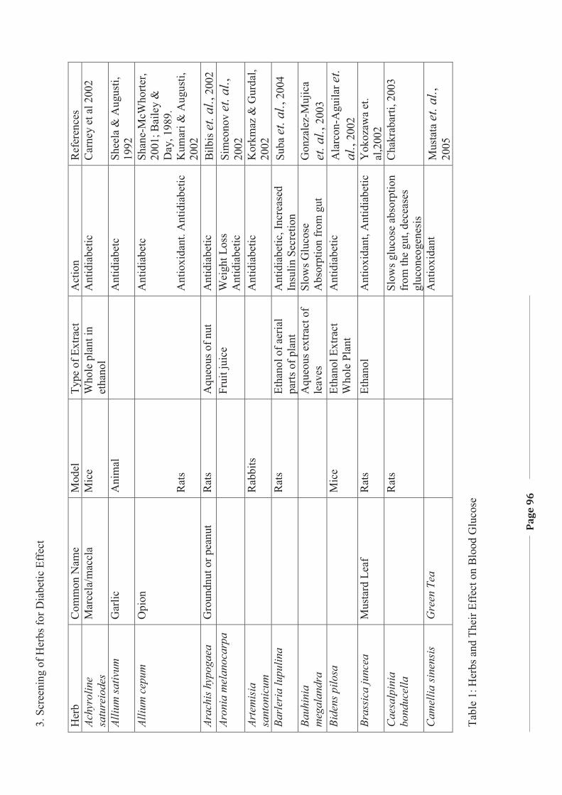
3. S
cree
ning
of H
erbs
for D
iabe
tic E
ffec
t
Page
96
Her
b C
omm
on N
ame
Mod
el
Type
of E
xtra
ct
Act
ion
Ref
eren
ces
Achy
rolin
e sa
ture
iode
s M
arce
la/m
accl
a M
ice
Who
le p
lant
in
etha
nol
Ant
idia
betic
C
arne
y et
al 2
002
Alliu
m sa
tivum
G
arlic
A
nim
al
A
ntid
iabe
tc
Shee
la &
Aug
usti,
19
92
Alliu
m c
epum
O
pion
R
ats
A
ntid
iabe
tc
Ant
ioxi
dant
. Ant
idia
betic
Shan
e-M
cWho
rter,
2001
; Bai
ley
&
Day
, 198
9.
Kum
ari &
Aug
usti,
20
02
Arac
his h
ypog
aea
Gro
undn
ut o
r pea
nut
Rat
s A
queo
us o
f nut
A
ntid
iabe
tic
Bilb
is et
. al.,
200
2 Ar
onia
mel
anoc
arpa
Fr
uit j
uice
W
eigh
t Los
s A
ntid
iabe
tic
Sim
eono
v et
. al.,
20
02
Arte
mis
ia
sant
onic
um
R
abbi
ts
A
ntid
iabe
tic
Kor
kmaz
& G
urda
l, 20
02
Barl
eria
lupu
lina
R
ats
Etha
nol o
f aer
ial
parts
of p
lant
A
ntid
iabe
tic, I
ncre
ased
In
sulin
Sec
retio
n Su
ba et
. al.,
200
4
Bauh
inia
m
egal
andr
a
A
queo
us e
xtra
ct o
f le
aves
Sl
ows G
luco
se
Abs
orpt
ion
from
gut
G
onza
lez-
Muj
ica
et. a
l., 2
003
Bide
ns p
ilosa
Mic
e Et
hano
l Ext
ract
W
hole
Pla
nt
Ant
idia
betic
A
larc
on-A
guila
r et.
al.,
2002
Br
assi
ca ju
ncea
M
usta
rd L
eaf
Rat
s Et
hano
l A
ntio
xida
nt, A
ntid
iabe
tic
Yok
ozaw
a et
. al
,200
2 C
aesa
lpin
ia
bond
ucel
la
R
ats
Sl
ows g
luco
se a
bsor
ptio
n fr
om th
e gu
t, de
ceas
es
gluc
oneo
gene
sis
Cha
krab
arti,
200
3
Cam
ellia
sine
nsis
Gre
en T
ea
Ant
ioxi
dant
M
usta
ta et
. al.,
20
05
Tabl
e 1:
Her
bs a
nd T
heir
Effe
ct o
n B
lood
Glu
cose
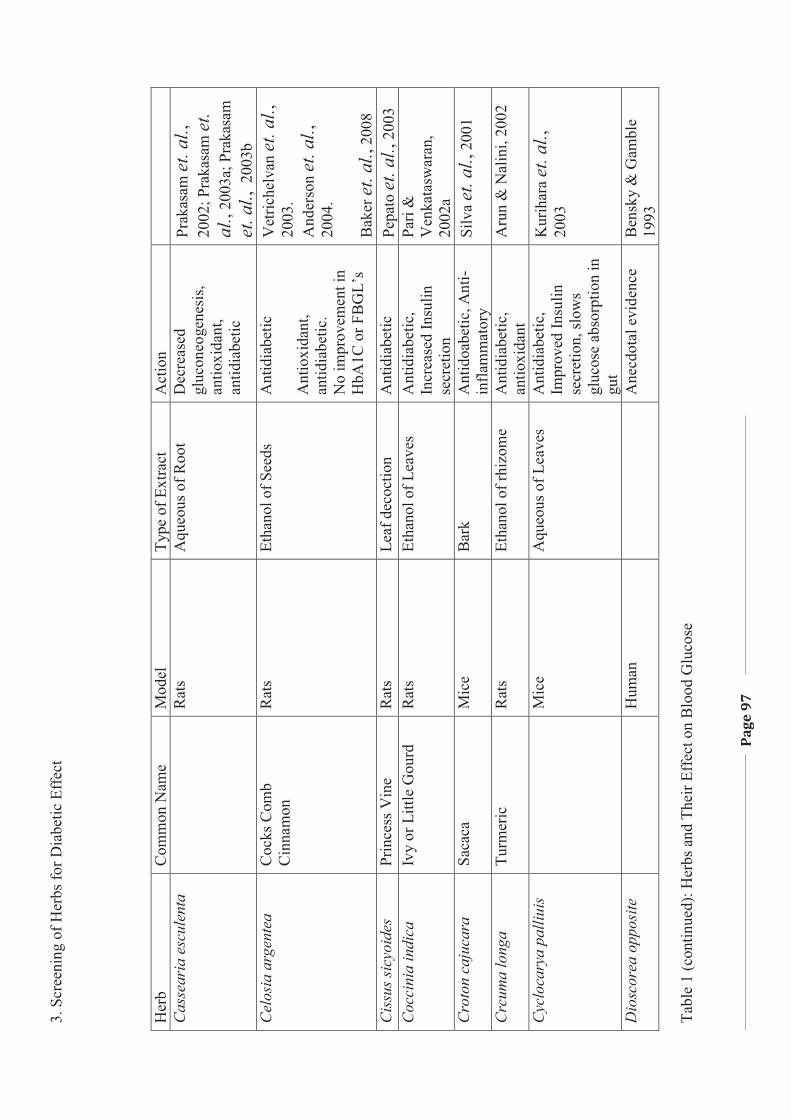
3. S
cree
ning
of H
erbs
for D
iabe
tic E
ffec
t
Page
97
Her
b C
omm
on N
ame
Mod
el
Type
of E
xtra
ct
Act
ion
C
asse
aria
esc
ulen
ta
R
ats
Aqu
eous
of R
oot
Dec
reas
ed
gluc
oneo
gene
sis,
antio
xida
nt,
antid
iabe
tic
Prak
asam
et. a
l.,
2002
; Pra
kasa
m e
t. al
. , 20
03a;
Pra
kasa
m
et. a
l., 2
003b
C
elos
ia a
rgen
tea
Coc
ks C
omb
Cin
nam
on
Rat
s Et
hano
l of S
eeds
A
ntid
iabe
tic
Ant
ioxi
dant
, an
tidia
betic
. N
o im
prov
emen
t in
HbA
1C o
r FB
GL’
s
Vet
riche
lvan
et. a
l.,
2003
. A
nder
son
et. a
l.,
2004
. B
aker
et. a
l., 2
008
Cis
sus s
icyo
ides
Pr
ince
ss V
ine
Rat
s Le
af d
ecoc
tion
Ant
idia
betic
Pe
pato
et. a
l., 2
003
Coc
cini
a in
dica
Iv
y or
Litt
le G
ourd
R
ats
Etha
nol o
f Lea
ves
Ant
idia
betic
, In
crea
sed
Insu
lin
secr
etio
n
Pari
&
Ven
kata
swar
an,
2002
a C
roto
n ca
juca
ra
Saca
ca
Mic
e B
ark
Ant
idoa
betic
, Ant
i-in
flam
mat
ory
Silv
a et
. al.,
200
1
Crc
uma
long
a Tu
rmer
ic
Rat
s Et
hano
l of r
hizo
me
Ant
idia
betic
, an
tioxi
dant
A
run
& N
alin
i, 20
02
Cyc
loca
rya
palli
uis
M
ice
Aqu
eous
of L
eave
s A
ntid
iabe
tic,
Impr
oved
Insu
lin
secr
etio
n, sl
ows
gluc
ose
abso
rptio
n in
gu
t
Kur
ihar
a et
. al.,
20
03
Dio
scor
ea o
ppos
ite
H
uman
Ane
cdot
al e
vide
nce
Ben
sky
& G
ambl
e 19
93
Tabl
e 1
(con
tinue
d): H
erbs
and
The
ir Ef
fect
on
Blo
od G
luco
se
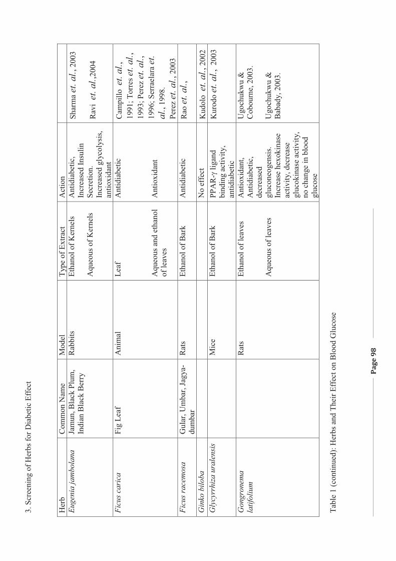
3. S
cree
ning
of H
erbs
for D
iabe
tic E
ffec
t
Page
98
Her
b C
omm
on N
ame
Mod
el
Type
of E
xtra
ct
Act
ion
Eu
geni
a ja
mbo
lana
Ja
mun
, Bla
ck P
lum
, In
dian
Bla
ck B
erry
R
abbi
ts
Etha
nol o
f Ker
nels
A
queo
us o
f Ker
nels
Ant
idia
betic
, In
crea
sed
Insu
lin
Secr
etio
n.
Incr
ease
d gl
ycol
ysis
, an
tioxi
dant
Shar
ma
et. a
l., 2
003
Rav
i et
. al.,
2004
Ficu
s car
ica
Fig
Leaf
A
nim
al
Leaf
A
queo
us a
nd e
than
ol
of le
aves
Ant
idia
betic
A
ntio
xida
nt
Cam
pillo
et.
al.,
1991
; Tor
res e
t. al
., 19
93; P
erez
et. a
l.,
1996
; Ser
rael
ara
et.
al. ,
1998
. Pe
rez
et. a
l., 2
003
Ficu
s rac
emos
a G
ular
, Um
bar,
Jagy
a-du
mba
r R
ats
Etha
nol o
f Bar
k A
ntid
iabe
tic
Rao
et. a
l.,
Gin
ko b
iloba
No
effe
ct
Kud
olo
et. a
l., 2
002
Gly
cyrr
hiza
ura
lens
is
M
ice
Etha
nol o
f Bar
k PP
AR
-� li
gand
bi
ndin
g ac
tivity
, an
tidia
betic
Kur
odo
et. a
l., 2
003
Gon
gron
ema
latif
oliu
m
R
ats
Etha
nol o
f lea
ves
Aqu
eous
of l
eave
s
Ant
ioxi
dant
, A
ntid
iabe
tic,
decr
ease
d gl
ucon
eoge
nsis
. In
crea
se h
exok
inas
e ac
tivity
, dec
reas
e gl
ucok
inas
e ac
tivity
, no
cha
nge
in b
lood
gl
ucos
e
Ugo
chuk
wu
&
Cob
ourn
e, 2
003.
U
goch
ukw
u &
B
abad
y, 2
003.
Tabl
e 1
(con
tinue
d): H
erbs
and
The
ir Ef
fect
on
Blo
od G
luco
se
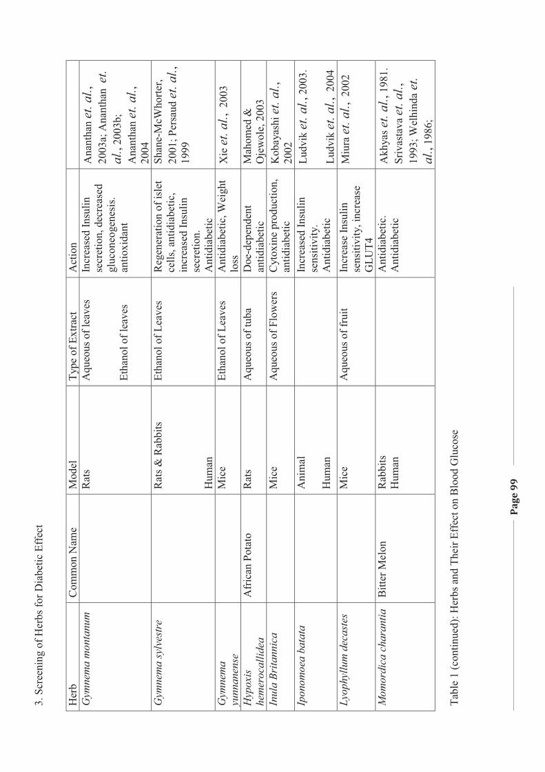
3. S
cree
ning
of H
erbs
for D
iabe
tic E
ffec
t
Page
99
Her
b C
omm
on N
ame
Mod
el
Type
of E
xtra
ct
Act
ion
G
ymne
ma
mon
tanu
m
R
ats
Aqu
eous
of l
eave
s Et
hano
l of l
eave
s
Incr
ease
d In
sulin
se
cret
ion,
dec
reas
ed
gluc
oneo
gene
sis.
antio
xida
nt
Ana
ntha
n et
. al.,
20
03a;
Ana
ntha
n et
. al
. , 20
03b;
A
nant
han
et. a
l.,
2004
G
ymne
ma
sylv
estr
e
Rat
s & R
abbi
ts
Hum
an
Etha
nol o
f Lea
ves
Reg
ener
atio
n of
isle
t ce
lls, a
ntid
iabe
tic,
incr
ease
d In
sulin
se
cret
ion.
A
ntid
iabe
tic
Shan
e-M
cWho
rter,
2001
; Per
saud
et. a
l.,
1999
Gym
nem
a yu
nnan
ense
Mic
e Et
hano
l of L
eave
s A
ntid
iabe
tic, W
eigh
t lo
ss
Xie
et. a
l., 2
003
Hyp
oxis
he
mer
ocal
lidea
A
fric
an P
otat
o R
ats
Aqu
eous
of t
uba
Doe
-dep
ende
nt
antid
iabe
tic
Mah
omed
&
Oje
wol
e, 2
003
Inul
a Br
itann
ica
M
ice
Aqu
eous
of F
low
ers
Cyt
oxin
e pr
oduc
tion,
an
tidia
betic
K
obay
ashi
et.
al.,
20
02
Ipon
omoe
a ba
tata
Ani
mal
H
uman
In
crea
sed
Insu
lin
sens
itivi
ty.
Ant
idia
betic
Ludv
ik et
. al.,
200
3.
Ludv
ik et
. al.,
200
4 Ly
ophy
llum
dec
aste
s
Mic
e A
queo
us o
f fru
it In
crea
se In
sulin
se
nsiti
vity
, inc
reas
e G
LUT4
Miu
ra e
t. al
., 2
002
Mom
ordi
ca c
hara
ntia
B
itter
Mel
on
Rab
bits
H
uman
Ant
idia
betic
. A
ntid
iabe
tic
Akh
yas e
t. al
., 19
81.
Sriv
asta
va et
. al.,
19
93; W
elhi
nda e
t. al
., 19
86;
Ta
ble
1 (c
ontin
ued)
: Her
bs a
nd T
heir
Effe
ct o
n B
lood
Glu
cose
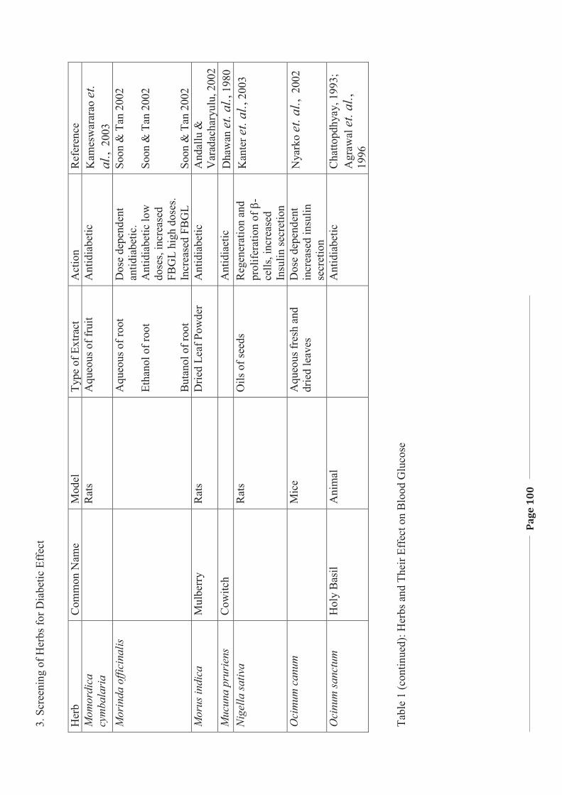
3. S
cree
ning
of H
erbs
for D
iabe
tic E
ffec
t
Page
100
Her
b C
omm
on N
ame
Mod
el
Type
of E
xtra
ct
Act
ion
Ref
eren
ce
Mom
ordi
ca
cym
bala
ria
R
ats
Aqu
eous
of f
ruit
Ant
idia
betic
K
ames
war
arao
et.
al.,
200
3 M
orin
da o
ffici
nalis
A
queo
us o
f roo
t Et
hano
l of r
oot
But
anol
of r
oot
Dos
e de
pend
ent
antid
iabe
tic.
Ant
idia
betic
low
do
ses,
incr
ease
d FB
GL
high
dos
es.
Incr
ease
d FB
GL
Soon
& T
an 2
002
Soon
& T
an 2
002
Soon
& T
an 2
002
Mor
us in
dica
M
ulbe
rry
Rat
s D
ried
Leaf
Pow
der
Ant
idia
betic
A
ndal
lu &
V
arad
acha
ryul
u, 2
002
Muc
una
prur
iens
C
owitc
h
A
ntid
iaet
ic
Dha
wan
et. a
l., 1
980
Nig
ella
sativ
a
Rat
s O
ils o
f see
ds
Reg
ener
atio
n an
d pr
olife
ratio
n of
�-
cells
, inc
reas
ed
Insu
lin se
cret
ion
Kan
ter e
t. al
., 20
03
Oci
mum
can
um
M
ice
Aqu
eous
fres
h an
d dr
ied
leav
es
Dos
e de
pend
ent
incr
ease
d in
sulin
se
cret
ion
Nya
rko
et. a
l., 2
002
Oci
num
sanc
tum
H
oly
Bas
il A
nim
al
A
ntid
iabe
tic
Cha
ttopd
hyay
, 199
3;
Agr
awal
et.
al.,
19
96
Tabl
e 1
(con
tinue
d): H
erbs
and
The
ir Ef
fect
on
Blo
od G
luco
se
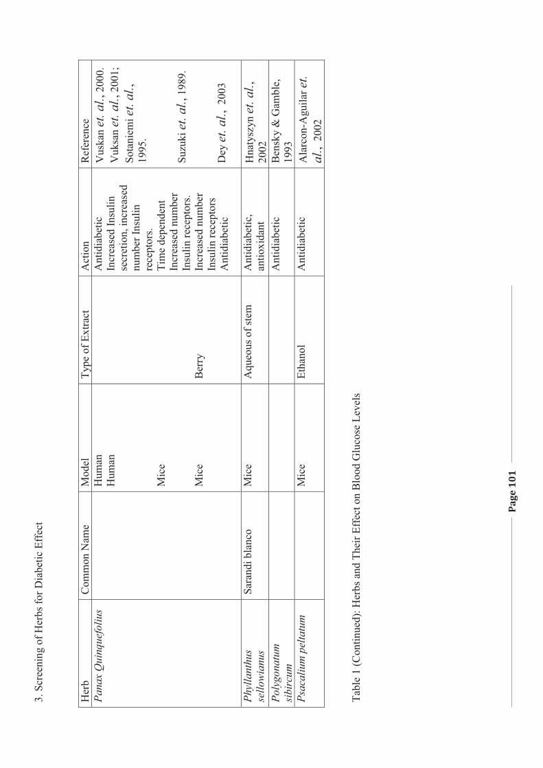
3. S
cree
ning
of H
erbs
for D
iabe
tic E
ffec
t
Page
101
Her
b C
omm
on N
ame
Mod
el
Type
of E
xtra
ct
Act
ion
Ref
eren
ce
Pana
x Q
uinq
uefo
lius
H
uman
H
uman
M
ice
Mic
e
Ber
ry
Ant
idia
betic
In
crea
sed
Insu
lin
secr
etio
n, in
crea
sed
num
ber I
nsul
in
rece
ptor
s. Ti
me
depe
nden
t In
crea
sed
num
ber
Insu
lin re
cept
ors.
Incr
ease
d nu
mbe
r In
sulin
rece
ptor
s A
ntid
iabe
tic
Vus
kan
et. a
l., 2
000.
V
uksa
n et
. al.,
200
1;
Sota
niem
i et.
al.,
1995
. Su
zuki
et. a
l., 1
989.
D
ey et
. al.,
200
3
Phyl
lant
hus
sello
wia
nus
Sara
ndi b
lanc
o M
ice
Aqu
eous
of s
tem
A
ntid
iabe
tic,
antio
xida
nt
Hna
tysz
yn et
. al.,
20
02
Poly
gona
tum
si
birc
um
A
ntid
iabe
tic
Ben
sky
& G
ambl
e,
1993
Ps
acal
ium
pel
tatu
m
M
ice
Etha
nol
Ant
idia
betic
A
larc
on-A
guila
r et.
al.,
200
2 Ta
ble
1 (C
ontin
ued)
: Her
bs a
nd T
heir
Effe
ct o
n B
lood
Glu
cose
Lev
els
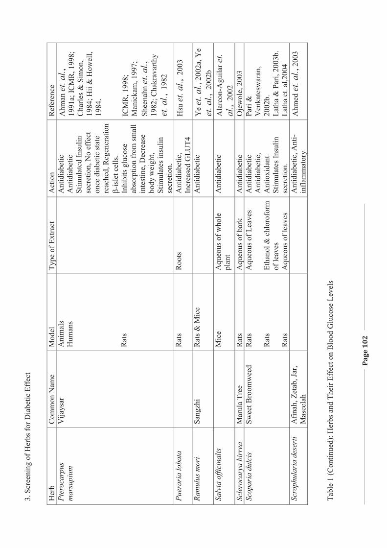
3. S
cree
ning
of H
erbs
for D
iabe
tic E
ffec
t
Page
102
Her
b C
omm
on N
ame
Mod
el
Type
of E
xtra
ct
Act
ion
Ref
eren
ce
Pter
ocar
pus
mar
supi
um
Vija
ysar
A
nim
als
Hum
ans
Rat
s
A
ntid
iabe
tic
Ant
idia
betic
St
imul
ated
Insu
lin
secr
etio
n, N
o ef
fect
on
ce d
iabe
tic st
ate
reac
hed,
Reg
ener
atio
n �-
isle
t cel
ls.
Inhi
bits
glu
cose
ab
sorp
tion
from
smal
l in
test
ine,
Dec
reas
e bo
dy w
eigh
t, St
imul
ates
insu
lin
secr
etio
n.
Ahm
an et
. al.,
19
91a;
ICM
R, 1
998;
C
harle
s & S
imon
, 19
84; H
ii &
How
ell,
1984
. IC
MR
, 19 9
8;
Man
icka
m, 1
997;
Sh
eena
hn et
. al.,
19
82; C
hakr
avar
thy
et. a
l., 1
982
Puer
aria
loba
ta
R
ats
Roo
ts
Ant
idia
betic
, In
crea
sed
GLU
T4
Hsu
et. a
l., 2
003
Ram
ulus
mor
i Sa
ngzh
i R
ats &
Mic
e
Ant
idia
betic
Y
e et
. al.,
200
2a, Y
e et
. al.,
200
2b
Salv
ia o
ffici
nalis
Mic
e A
queo
us o
f who
le
plan
t A
ntid
iabe
tic
Ala
rcon
-Agu
ilar e
t. al
., 2
002
Scle
roca
rya
birr
ea
Mar
ula
Tree
R
ats
Aqu
eous
of b
ark
Ant
idia
betic
O
jew
ole,
200
3 Sc
opar
ia d
ulci
s Sw
eet B
room
wee
d R
ats
Rat
s R
ats
Aqu
eous
of L
eave
s Et
hano
l & c
hlor
ofor
m
of le
aves
A
queo
us o
f lea
ves
Ant
idia
betic
A
ntid
iabe
tic,
Ant
ioxi
dant
. St
imul
ates
Insu
lin
secr
etio
n.
Pari
&
Ven
kate
swar
an,
2002
b.
Lath
a &
Par
i, 20
03b.
La
tha
et. a
l,200
4 Sc
roph
ular
ia d
eser
ti A
finah
, Zet
ab, J
ar,
Mas
eela
h
A
ntid
iabe
tic, A
nti-
infla
mm
ator
y A
hmed
et. a
l., 2
003
Tabl
e 1
(Con
tinue
d): H
erbs
and
The
ir Ef
fect
on
Blo
od G
luco
se L
evel
s
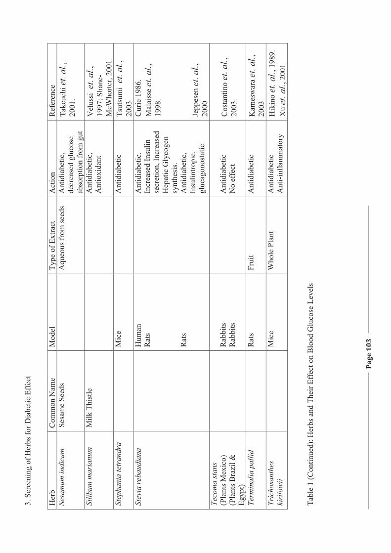
3. S
cree
ning
of H
erbs
for D
iabe
tic E
ffec
t
Page
103
Her
b C
omm
on N
ame
Mod
el
Type
of E
xtra
ct
Act
ion
Ref
eren
ce
Sesa
mum
indi
cum
Se
sam
e Se
eds
A
queo
us fr
om se
eds
Ant
idia
betic
, de
crea
sed
gluc
ose
abso
rptio
n fr
om g
ut
Take
uchi
et. a
l.,
2001
.
Silib
um m
aria
num
M
ilk T
hist
le
Ant
idia
betic
, A
ntio
xida
nt
Vel
ussi
et.
al.,
1997
; Sha
ne-
McW
horte
r, 20
01
Step
hani
a te
tran
dra
M
ice
A
ntid
iabe
tic
Tsut
sum
i et
. al.,
20
03
Stev
ia re
baud
iana
Hum
an
Rat
s R
ats
A
ntid
iabe
tic.
Incr
ease
d In
sulin
se
cret
ion,
Incr
ease
d H
epat
ic G
lyco
gen
synt
hesi
s. A
ntid
iabe
tic,
Insu
lintro
pic,
gl
ucag
onos
tatic
Cur
ie 1
986.
M
alai
sse
et. a
l.,
1998
. Je
ppes
en et
. al.,
20
00
Teco
ma
stan
s (P
lant
s Mex
ico)
(P
lant
s Bra
zil &
Eg
ypt)
R
abbi
ts
Rab
bits
A
ntid
iabe
tic
No
effe
ct
Cos
tant
ino
et. a
l.,
2003
.
Term
inal
ia p
allid
Rat
s Fr
uit
Ant
idia
betic
K
ames
war
a et
. al.,
20
03
Tric
hosa
nthe
s ki
rilo
wii
M
ice
Who
le P
lant
A
ntid
iabe
tic
Ant
i -inf
lam
mat
ory
Hik
ino
et. a
l., 1
989.
X
u et
. al.,
200
1 Ta
ble
1 (C
ontin
ued)
: Her
bs a
nd T
heir
Effe
ct o
n B
lood
Glu
cose
Lev
els
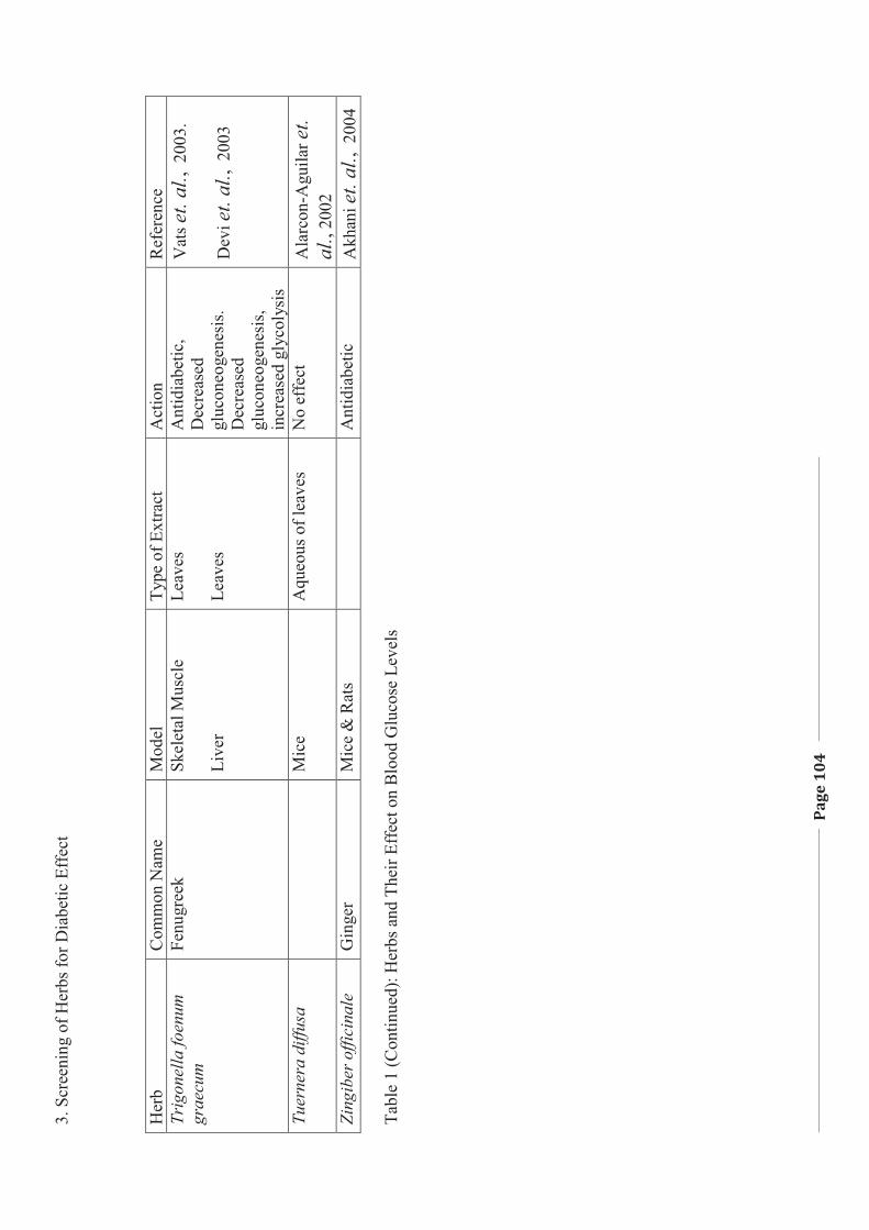
3. S
cree
ning
of H
erbs
for D
iabe
tic E
ffec
t
Page
104
Her
b C
omm
on N
ame
Mod
el
Type
of E
xtra
ct
Act
ion
Ref
eren
ce
Trig
onel
la fo
enum
gr
aecu
m
Fenu
gree
k Sk
elet
al M
uscl
e Li
ver
Leav
es
Leav
es
Ant
idia
betic
, D
ecre
ased
gl
ucon
eoge
nesi
s. D
ecre
ased
gl
ucon
eoge
nesi
s, in
crea
sed
glyc
olys
is
Vat
s et.
al.,
200
3.
Dev
i et.
al.,
200
3
Tuer
nera
diff
usa
M
ice
Aqu
eous
of l
eave
s N
o ef
fect
A
larc
on-A
guila
r et.
al.,
2002
Zi
ngib
er o
ffici
nale
G
inge
r M
ice
& R
ats
A
ntid
iabe
tic
Akh
ani e
t. al
., 2
004
Tabl
e 1
(Con
tinue
d): H
erbs
and
The
ir Ef
fect
on
Blo
od G
luco
se L
evel
s
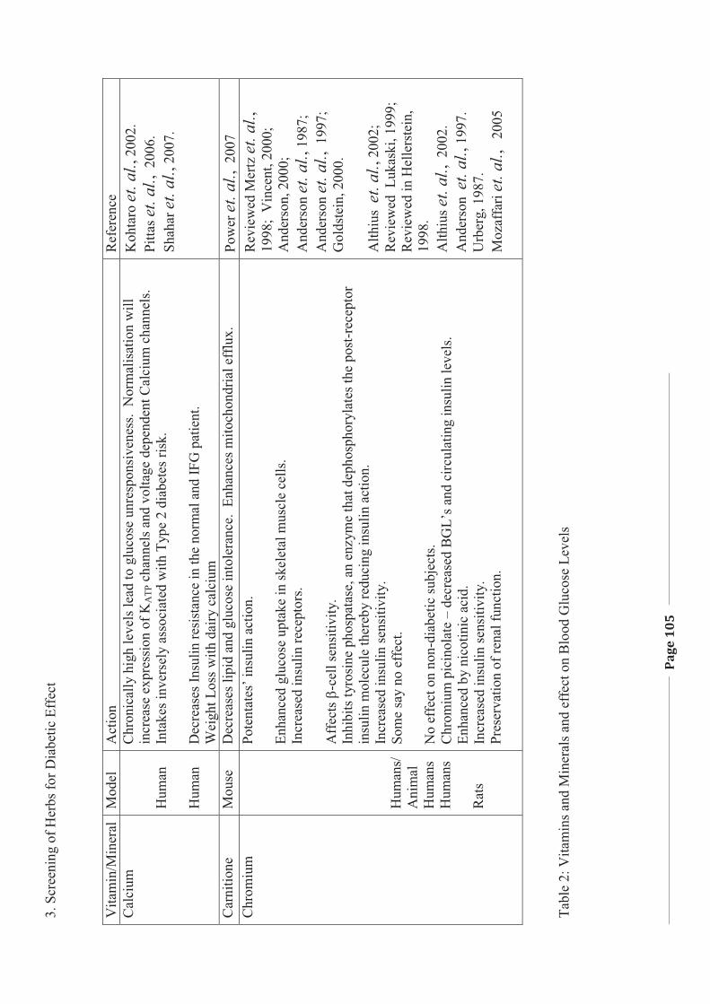
3. S
cree
ning
of H
erbs
for D
iabe
tic E
ffec
t
Page
105
Vita
min
/Min
eral
M
odel
A
ctio
n R
efer
ence
C
alci
um
Hum
an
Hum
an
Chr
onic
ally
hig
h le
vels
lead
to g
luco
se u
nres
pons
iven
ess.
Nor
mal
isat
ion
will
in
crea
se e
xpre
ssio
n of
KA
TP c
hann
els a
nd v
olta
ge d
epen
dent
Cal
cium
cha
nnel
s. In
take
s inv
erse
ly a
ssoc
iate
d w
ith T
ype
2 di
abet
es ri
sk.
Dec
reas
es In
sulin
resi
stan
ce in
the
norm
al a
nd IF
G p
atie
nt.
Wei
ght L
oss w
ith d
airy
cal
cium
Koh
taro
et. a
l., 2
002.
Pi
ttas e
t. al
., 2
006.
Sh
ahar
et. a
l., 2
007.
Car
nitio
ne
Mou
se
Dec
reas
es li
pid
and
gluc
ose
into
lera
nce.
Enh
ance
s mito
chon
dria
l eff
lux.
Po
wer
et. a
l., 2
007
Chr
omiu
m
Hum
ans/
A
nim
al
Hum
ans
Hum
ans
Rat
s
Pote
ntat
es’ i
nsul
in a
ctio
n.
Enha
nced
glu
cose
upt
ake
in sk
elet
al m
uscl
e ce
lls.
Incr
ease
d in
sulin
rece
ptor
s. A
ffec
ts �
-cel
l sen
sitiv
ity.
Inhi
bits
tyro
sine
pho
spat
ase,
an
enzy
me
that
dep
hosp
hory
late
s the
pos
t-rec
epto
r in
sulin
mol
ecul
e th
ereb
y re
duci
ng in
sulin
act
ion.
In
crea
sed
insu
lin se
nsiti
vity
.
Som
e sa
y no
eff
ect.
No
effe
ct o
n n o
n-di
abet
ic su
bjec
ts.
Chr
omiu
m p
icin
olat
e –
decr
ease
d B
GL’
s and
circ
ulat
ing
insu
lin le
vels
. En
hanc
ed b
y ni
cotin
ic a
cid.
In
crea
sed
insu
lin se
nsiti
vity
. Pr
eser
vatio
n of
rena
l fun
ctio
n.
Rev
iew
ed M
ertz
et. a
l.,
1998
; V
ince
nt, 2
000;
A
nder
son,
200
0;
And
erso
n et
. al.,
198
7;
And
erso
n et
. al.,
199
7;
Gol
dste
in, 2
000.
A
lthiu
s et
. al.,
200
2;
Rev
iew
ed L
ukas
ki, 1
999;
R
evie
wed
in H
elle
rste
in,
1998
. A
lthiu
s et.
al.,
200
2.
And
erso
n et
. al.,
199
7.
Urb
erg,
198
7.
Moz
affa
ri et
. al.,
20
05
Ta
ble
2: V
itam
ins a
nd M
iner
als a
nd e
ffec
t on
Blo
od G
luco
se L
evel
s
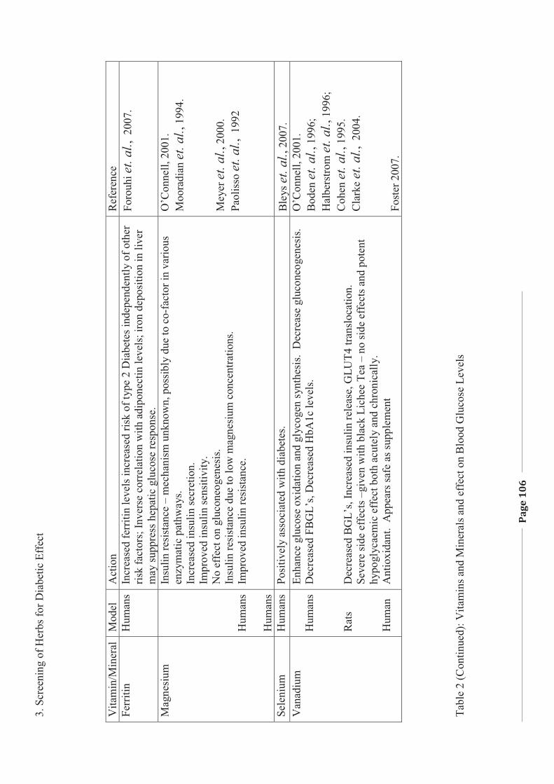
3. S
cree
ning
of H
erbs
for D
iabe
tic E
ffec
t
Page
106
Vita
min
/Min
eral
M
odel
A
ctio
n R
efer
ence
Fe
rriti
n H
uman
s In
crea
sed
ferr
itin
leve
ls in
crea
sed
risk
of ty
pe 2
Dia
bete
s ind
epen
dent
ly o
f oth
er
risk
fact
ors;
Inve
rse
corr
elat
ion
with
adi
pone
ctin
leve
ls; i
ron
depo
sitio
n in
live
r m
ay su
ppre
ss h
epat
ic g
luco
se re
spon
se.
Foro
uhi e
t. al
., 2
007.
Mag
nesi
um
Hum
ans
Hum
ans
Insu
lin re
sist
ance
– m
echa
nism
unk
now
n, p
ossi
bly
due
to c
o-fa
ctor
in v
ario
us
enzy
mat
ic p
athw
ays.
Incr
ease
d in
sulin
secr
etio
n.
Impr
oved
insu
lin se
nsiti
vity
. N
o ef
fect
on
gluc
oneo
gene
sis.
Insu
lin re
sist
ance
due
to lo
w m
agne
sium
con
cent
ratio
ns.
Impr
oved
insu
lin re
sist
ance
.
O’C
onne
ll, 2
001.
M
oora
dian
et. a
l., 1
994.
M
eyer
et. a
l., 2
000.
Pa
olis
so e
t. al
., 1
992
Sele
nium
H
uman
s Po
sitiv
ely
asso
ciat
ed w
ith d
iabe
tes.
Ble
ys et
. al.,
200
7.
Van
adiu
m
Hum
ans
Rat
s H
uman
Enha
nce
gluc
ose
oxid
atio
n an
d gl
ycog
en s
ynth
esis
. D
ecre
ase
gluc
oneo
gene
sis.
D
ecre
ased
FB
GL’
s, D
ecre
ased
HbA
1c le
vels
. D
ecre
ased
BG
L’s,
Incr
ease
d in
sulin
rele
ase,
GLU
T4 tr
ansl
ocat
ion.
Se
vere
side
eff
ects
–gi
ven
with
bla
ck L
iche
e Te
a –
no si
de e
ffec
ts a
nd p
oten
t hy
pogl
ycae
mic
eff
ect b
oth
acut
ely
and
chro
nica
lly.
Ant
ioxi
dant
. A
ppea
rs sa
fe a
s sup
plem
ent
O’C
onne
ll, 2
001.
B
oden
et. a
l., 1
996;
H
albe
rstro
m et
. al.,
199
6;
Coh
en et
. al.,
199
5.
Cla
rke
et. a
l., 2
004.
Fo
ster
200
7.
Tabl
e 2
(Con
tinue
d): V
itam
ins a
nd M
iner
als a
nd e
ffec
t on
Blo
od G
luco
se L
evel
s
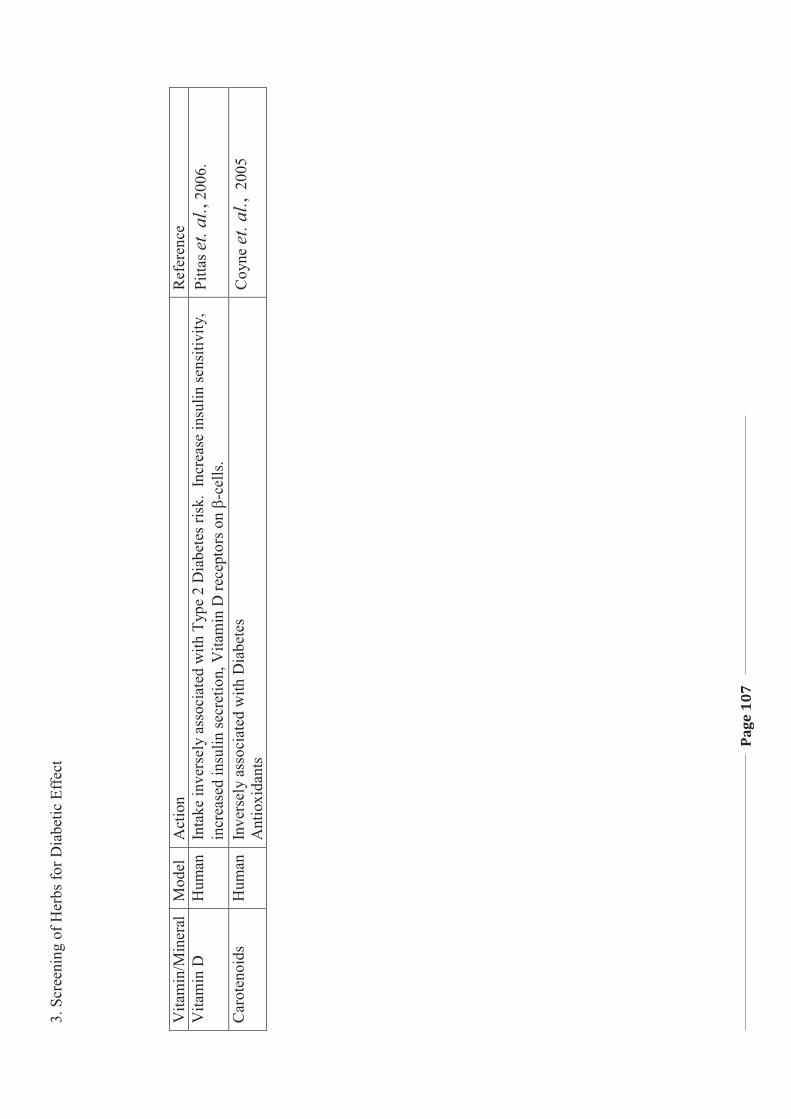
3. S
cree
ning
of H
erbs
for D
iabe
tic E
ffec
t
Page
107
Vita
min
/Min
eral
M
odel
A
ctio
n R
efer
ence
V
itam
in D
H
uman
Inta
ke in
vers
ely
asso
ciat
ed w
ith T
ype
2 D
iabe
tes r
isk.
Inc
reas
e in
sulin
sens
itivi
ty,
incr
ease
d in
sulin
secr
etio
n, V
itam
in D
rece
ptor
s on
�-ce
lls.
Pitta
s et.
al.,
2006
.
Car
oten
oids
H
uman
In
vers
ely
asso
ciat
ed w
ith D
iabe
tes
Ant
ioxi
dant
s C
oyne
et. a
l., 2
005
3. Screening of Herbs for Diabetic Effect
Page 108
3.2. MATERIALS and METHODS
Plant extraction, ethics approval, tissue preparation, and tissue culture
assay; glucose analysis; statistical analysis have been described in Chapter 2 of
this thesis. Each herb was tested in duplicate for this screening.
Curcuma longa Linn (Turmeric) was divided into three groups. One was
purchased as a dry powder from Woolworths in Armidale, NSW, Australia
2350; the second, a dry powder and the third, Turmeric cuts, both purchased
from Austral herbs, Kentucky, NSW, Australia, 2350
3.3. RESULTS
An aqueous extract of the leaves from the Laurus nobilis tree (Bay Leaves)
at the concentration of 10 �g/ml showed an inhibitory effect on the uptake of
glucose by the heart tissue culture with a decrease of 5.4 +/- 1.2% uptake over
the heart tissue itself. The aqueous solution showed a little difference in percent
uptake with 2.28 +/- 0.97% being noted for the 5 �g/ml solution. The 2.5 �g/ml
solution resulted in an increase of 4.13 +/- 1.3%. However, the concentration of
1.25 �g/ml showed an increase uptake of 5.89 +/- 1.54%. As the lower
concentrations elucidated a better uptake, a decrease in concentration was tested
with 1 �g/ml of the Bay Leaf resulting in a 6.75 +/- 0.54% uptake; and at a
concentration of 0.5 �g/ml the percent in uptake was 4.74 +/- 0.26%; and at a
concentration of 0.25 �g/ml it was 2.68 +/- 0.34% (See Figure 3.1B).
3. Screening of Herbs for Diabetic Effect
Page 109
Concentrations of 10, 5, 2.5 and 1.25 �g/ml of aqueous extract of
Origanum vulgare (Oregano), were also examined in mice heart tissue culture
where it was noted that all concentrations had a slight inhibitory effect. The 10
�g/ml concentration of Oregano showed an inhibitory effect of -3.48 +/-
0.895%; while the 5 �g/ml showed an inhibitory effect of -4.77 +/- 1.2%. The
inhibitory effect for the Oregano concentration of 2.5 �g/ml was -2.68 +/- 0.32%
while that of the 1.25 �g/ml was -1.36 +/- 0.54% (See Figure 3.1B).
The Woolworths’ purchased Turmeric showed an increase uptake at all
concentrations. At a concentration of 10 �g/ml the Woolworths’ Turmeric
showed an increased glucose uptake in the mice heart tissue culture of 12.9 +/-
0.85%; while that of a concentration of 5 �g/ml showed an increase uptake of
15.9 +/- 3.7%. At a concentration 2.5 �g/ml there was an increased uptake of
13.3 +/- 2.0% while at 1.25 �g/ml it was 5.5 +/- 1.45%. The Austral Herbs
dried Turmeric powder and the Turmeric cuts, which had been ground down,
showed the same results. At all the concentrations there was an increase in the
glucose uptake of the mice heart tissue culture. However if the Turmeric has
been dry roasted the results are significantly different with results of 2.93 +/-
0.2% at a concentration of 10 �g/ml, 4.44+/- 0.32% at a concentration of
5�g/ml, 1.2 +/- 0.7%; at a concentration of 2.5 �g/ml and 0.47 +/- 0.34% at a
concentration at 1.25 �g/ml (See Figure 3.1A).
3. Screening of Herbs for Diabetic Effect
Page 110
Fresh Ocimum basilicum (sweet basil) increased the glucose uptake of the
mouse heart tissue by 9.96 +/- 0.78%, 10.67 +/- 0.45%, 11.95 +/- 0.34% and
10.53 +/- 0.43% when concentrations of 10 �g/ml, 5 �g/ml, 2.5 �g/ml and 1.25
�g/ml were used respectively; however, dried basil exhibited an increase of only
1.2 +/- 0.24%, 1.7 +/- 0.56%, 1.9 +/- 0.37% and 1.5 +/- 0.67% respectively at
these same concentrations (See Figure 3.1A).
Fresh Lavandula officinalis (English lavender) increased the glucose
uptake of the mouse heart tissue by 7.55 +/- 0.54% at a concentration of 10
�g/ml, 7.27 +/- 0.87% at a concentration of 5 �g/ml, 7.46 +/- 0.38% at a
concentration of 2.5 �g/ml and 7.55 +/- 0.26% at a concentration of 1.25 �g/ml
(see Figure 3.1B).
Dried Thymus vulgaris (thyme) had a decrease of 0.62 +/- 0.34% at the
highest concentration of 10 �g/ml; and then exhibited an increase of 4.7 +/-
0.38%, 7.26 +/- 0.64% and 9.53 +/- 0.12%, as the concentrations decreased from
5 �g/ml, 2.5 �g/ml and 1.25 �g/ml respectively. Due to the increased glucose
uptake at lower concentrations, further studies were considered at even lower
concentrations, and at a concentration of 1 �g/ml of the thyme there was a 9.31
+/- 0.26% uptake; and at a concentration of 0.5 �g/ml the percent uptake was
9.28 +/- 0.53%; and at a concentration of 0.25 �g/ml it was 7.97 +/- 0.46 % (See
Figure 3.1A).
3. Screening of Herbs for Diabetic Effect
Page 111
Cuminum cyminum (Cumin) at concentrations of 10 �g/ml, 5 �g/ml, 2.5
�g/ml and 1.25 �g/ml resulted in an increased up take of glucose by the mouse
heart tissue by 1.68 +/- 0.38%, 1.8 +/- 0.54%, 0.56 +/- 0.17% and 0.3 +/- 0.78%
respectively (See Figure 3.1B).
Dried Coriandrum sativum (coriander) decreased glucose uptake by -6.69
+/- 1.4% at a concentration of 10 �g/ml; however, at lower concentrations, there
was little change in glucose uptake from the control, with a percentage uptake of
2.83 +/- 0.34%, 0.45 +/- 0.64%, and 0.11 +/- 0.48% at the concentrations of 5
�g/ml, 2.5 �g/ml and 1.25 �g/ml respectively (See Figure 3.1B).
Fresh Metha piperita officinalis (Peppermint) showed little change in
glucose uptake by the heart mouse tissue, with a percentage uptake of 0.94 +/-
0.2%, 0.91 +/- 0.17%, 0.84 +/- 0.26%, and 0.94 +/- 0.19% at the concentrations
of 10 �g/ml, 5 �g/ml, 2.5 �g/ml and 1.25 �g/ml respectively (See Figure 3.1B).
Ground Myristica fragrans (Nutmeg) consistently showed an increase
uptake of glucose by the mouse heart tissue, of 4.31 +/- 0.28%, 4.11 +/- .46%,
4.13 +/- 0.2% and 4.69 +/- 0.9% at the concentrations of 10 �g/ml, 5 �g/ml, 2.5
�g/ml and 1.25 �g/ml respectively (See figure 3.1B).
Ground Eugenia caryophyllata (Cloves) exhibited a slight increased uptake
of glucose by the mouse heart tissue ,with a percent increase of 3.69 +/- 0.43%,
3.9 +/- 0.76%, 2.02 +/- 0.54% and 1.58 +/- 0.47% over the ranges of
3. Screening of Herbs for Diabetic Effect
Page 112
concentrations of 10 �g/ml, 5 �g/ml, 2.5 �g/ml and 1.25 �g/ml respectively (See
Figure 3.1A).
Stevia rebaundia exhibited a percent increase of glucose uptake by the
mouse heart tissue, of 9.81 +/- 0.14%, 9.24 +/- 0.56%, 8.88 +/- 0.48% and 8.25
+/- 0.37% at the concentrations of 10 �g/ml, 5 �g/ml, 2.5 �g/ml and 1.25 �g/ml
respectively (See Figure 3.1C).
Gymnema sylvestre resulted in a percent increase of glucose uptake by the
mouse heart tissue, of 12.5 +/- 0.63%, 12.6 +/- 0.75%, 9.87 +/- 0.26%, and 5.6
+/- 0.39% over the concentration range of 10 �g/ml, 5 �g/ml, 2.5 �g/ml and
1.25 �g/ml respectively (See Figure 3.1C).
The Australian plants showed a wide range of results with Eremophila
longofolia (Emu Bush) resulting in an increased glucose uptake by the mouse
heart tissue of 12.3 +/- 0.44%, 13.7 +/- 0.61%, 10.58 +/- 0.39%,and 7.45+/-
1.2% respectively, at the concentrations of 10 �g/ml, 5 �g/ml, 2.5 �g/ml and
1.25 �g/ml (See Figure 3.1C). Grevilla species leaves at lower concentrations,
exhibited a slight increase of glucose uptake in the heart tissue with a 6.78 +/-
0.98% increase at the concentration of 1.25 �g/ml; however, at concentrations of
10 �g/ml, 5 �g/ml, and 2.5 �g/ml respectively, there was a decrease in uptake of
-6.48+/- 1.5%, -3.41 +/- 0.87%, and -2.78 +/- 0.75% (See Figure 3.1C).
Stenanethum scortecceni stems showed little difference in the glucose uptake by
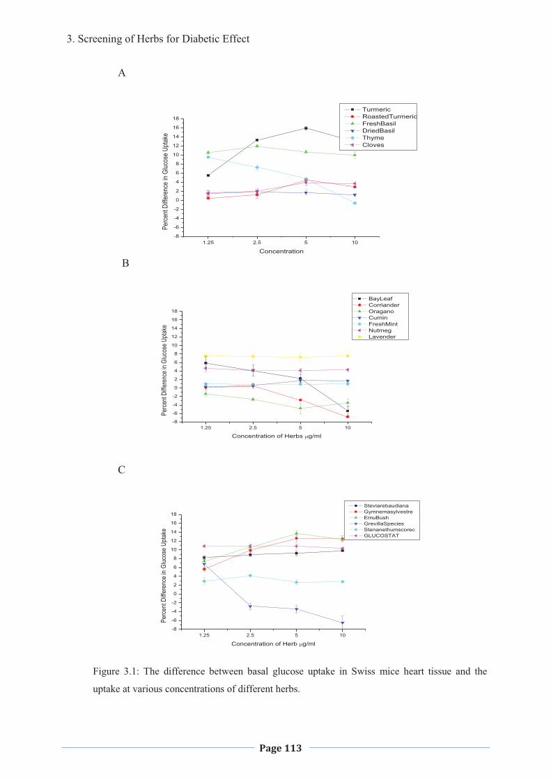
3. Screening of Herbs for Diabetic Effect
Page 113
A
B
1.25 2.5 5 10-8
-6
-4
-2
0
2
4
6
8
10
12
14
16
18
Perc
ent D
iffere
nce
in Gl
ucos
e Up
take
Concentration
Turmeric RoastedTurmeric FreshBasil DriedBasil Thyme Cloves
1.25 2.5 5 10-8
-6
-4
-2
0
2
4
6
8
10
12
14
16
18
Perce
nt Di
ffere
nce i
n Gluc
ose U
ptake
Concentration of Herbs �g/ml
BayLeaf Corriander Oragano Cumin FreshMint Nutmeg Lavender
C
1.25 2.5 5 10-8
-6
-4
-2
0
2
4
6
8
10
12
14
16
18
Perc
ent D
iffere
nce
in Gl
ucos
e Up
take
Concentration of Herb �g/ml
Steviarebaudiana Gymnemasylvestre EmuBush GrevillaSpecies Stenanethumscorec GLUCOSTAT
Figure 3.1: The difference between basal glucose uptake in Swiss mice heart tissue and the
uptake at various concentrations of different herbs.
3. Screening of Herbs for Diabetic Effect
Page 114
the heart tissue over the control at all concentrations, with a percentage
difference of 2.79 +/- 0.37%, 2.66 +/- 0.54%, 4.17 +/- 0.24% and 2.89 +/- 0.86%
(See Figure 3.1C).
Glucostat, which is a combination of the Chinese herbs including
Trichosanthes kirilowii (1900 mg), Polygonatum sibiricum (714.5 mg),
Dioscorea oppostia (286 mg), Panax ginseng (95 mg), and Stevia rebaudiona
(47.6 mg) per capsule, was also screened for its effectiveness in decreasing
glucose levels by the mouse heart tissue. It showed an increase of glucose
uptake by the mouse heart tissue of 10.28 +/- 0.28%, 10.8 +/- 0.54%, 10.91 +/-
0.46% and 10.83 +/- 0.35% at the concentrations of � ��g/ml, 5 �g/ml, 2.5
�g/ml and 1.25 �g/ml respectively (See Figure 3.1C).
Due to the increased uptake of glucose by the mouse heart tissue at the
lower concentrations of the leaves from the Laurus nobilis tree (Bay Leaves) a
decreased concentration was tested with 1 �g/ml of the Bay Leaf, resulting in a
6.75 +/- 0.54% uptake and at a concentration of 0.5 �g/ml the percent in uptake
was 4.74 +/- 0.26% and at a concentration of 0.25 �g/ml it was 2.68 +/- 0.34%
(See Figure 3.2).
Due to the increased glucose uptake at lower concentrations of the dried
Thymus vulgaris (thyme), further studies were considered at lower
concentrations, and at a concentration of 1 �g/ml of the thyme there was a 9.31
+/- 0.26% uptake, and at a concentration of 0.5 �g/ml the percent uptake was
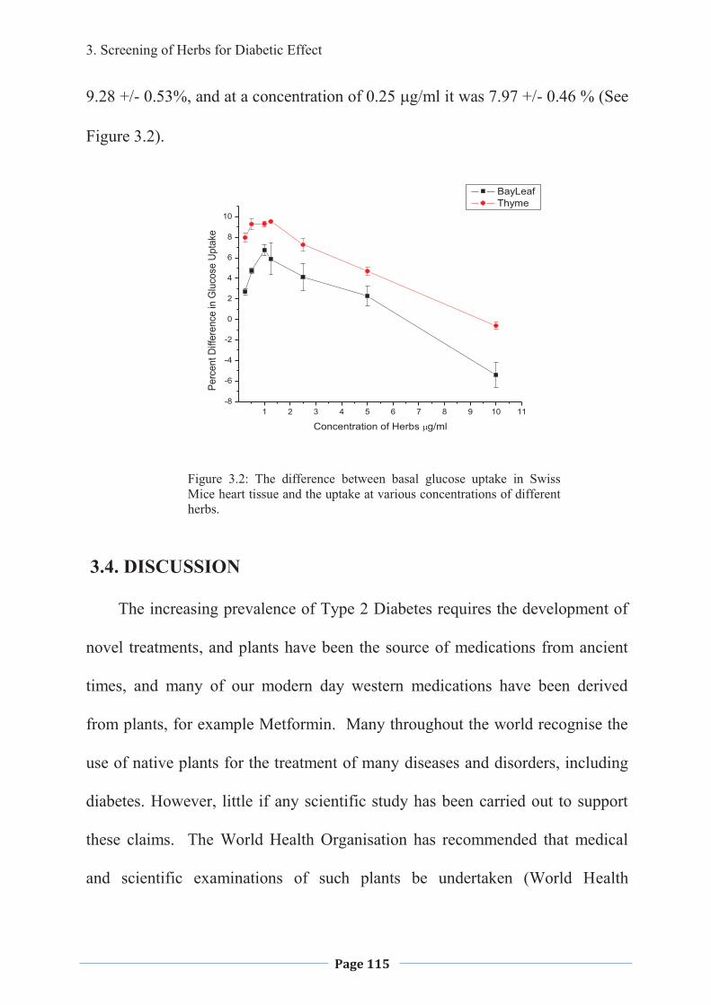
3. Screening of Herbs for Diabetic Effect
Page 115
9.28 +/- 0.53%, and at a concentration of 0.25 �g/ml it was 7.97 +/- 0.46 % (See
Figure 3.2).
1 2 3 4 5 6 7 8 9 10 11-8
-6
-4
-2
0
2
4
6
8
10Pe
rcen
t Diff
eren
ce in
Glu
cose
Upt
ake
Concentration of Herbs �g/ml
BayLeaf Thyme
Figure 3.2: The difference between basal glucose uptake in Swiss Mice heart tissue and the uptake at various concentrations of different herbs.
3.4. DISCUSSION
The increasing prevalence of Type 2 Diabetes requires the development of
novel treatments, and plants have been the source of medications from ancient
times, and many of our modern day western medications have been derived
from plants, for example Metformin. Many throughout the world recognise the
use of native plants for the treatment of many diseases and disorders, including
diabetes. However, little if any scientific study has been carried out to support
these claims. The World Health Organisation has recommended that medical
and scientific examinations of such plants be undertaken (World Health
3. Screening of Herbs for Diabetic Effect
Page 116
Organisation Expert Committee on Diabetes Mellitus, 1980), to identify their
effectiveness in the management of Type 2 Diabetes, in an attempt to use
affordable means for all countries to meet the need of the people, where there is
an ever increasing number of sufferers.
Culinary herbs are used widely around the world to flavour and colour
food; however, little is known concerning the effect that they might be having
on health. It is assumed that they are safe for consumption at the levels that they
are used in cooking, or in fresh form as no serious side effects or death has
resulted from them, unless an allergic reaction has occurred; however, this
appears to occur on only very rare occasions. Many of these herbs may be
advantageous to health; however, little if any scientific evidence has been
forthcoming on the efficacy, efficiency, toxicity; the best method of
administration, the active constituents, and the biochemical pathways/systems
involved of these herbs, to obtain medicinal effects. The effects of preparation of
the herbs may also have and effect on their medicinal value.
A preliminary study, looking at the effects of some of the culinary herbs on
the glucose uptake in Swiss male mice heart tissue, was undertaken. The reason
for this was to determine which herb/s warranted further study, to determine if
they would be of any benefit in the management of Type 2 Diabetes, by
decreasing high FBLs. The use of culinary herbs in the diet would be beneficial,
especially in third world countries as the cost could be kept to a minimum. Of
the herbs tested a few have had some preliminary research undertaken to

3. Screening of Herbs for Diabetic Effect
Page 117
determine effectiveness as far as diabetes treatment however, they have
generally been with ethanolic extracts where as aqueous extracts have been used
in this study and so may be looking at different active ingredients. Some had
not been tested as far as can be determined from a literature search.
The preparation of herbs and tissue culture protocol used through out the
study, was developed based on a number of experiments (data not shown),
focusing on the viability of the heart tissue through the incubation period. The
basal glucose uptake by muscle has been reported as one of the most sensitive
indicators of in vitro functional viability (Dohm et. al., 1988); and therefore in
this study, basal glucose uptake was considered as the indictor for metabolic
viability.
An aqueous extract of the leaves from the Laurus nobilis tree (Bay Leaves)
at higher concentrations, showed an inhibitory effect on the glucose uptake;
however, as the concentration was decreased there was an increase in glucose
uptake, with the maximum being 6.75 +/- 0.54% at a concentration of 1 �g/ml.
At lower concentrations the uptake began to decrease again. The Bay Leaf may
have been toxic in high concentrations, and this may have been why the
inhibitory effect was shown initially. As the concentration decreased the toxicity
also decreased allowing an active ingredient to increase the glucose uptake in
the Swiss mice heart tissue culture. Cinnamtannin B-1, found in Bay Leaves,
has been shown to have any antioxidant effect (Ferreira, et. al., 2006; Bouaziz,
et. al., 2007). This may have resulted in the increased uptake of glucose once the
3. Screening of Herbs for Diabetic Effect
Page 118
toxicity had been diluted out. Fetrow and Avila (2001) showed that Bay Leaf
will increase the action of Insulin. However, as this is a crude extract there
might also be toxic effects present at higher concentrations.
The aqueous extract of Origanum vulgare (Oregano) resulted in an
inhibitory effect on the uptake of glucose by the Swiss mice heart tissue. In a
study carried out by Lemhadari and colleagues (2004), they showed that an
aqueous extract of oregano decreased BGLs in streptozotocin (STZ) rats, after
both a single dose, and also after 15 daily doses. It was also noted that basal
insulin levels were not affected. The difference in our results can be explained
by the fact that we were using crude extract in vitro. Some toxic effects may
have resulted, whereas the previous study was in vivo, and some of the toxic
compounds may have been removed in the digestive process of the rats.
Curcuma longa Linn, more commonly known as Turmeric, is used widely
throughout the world as a flavouring and colouring in cooking. The main
colouring agent of Turmeric is curcumin (diferuloylmethane), and it has been
shown to have a wide range of effects including antihepatotoxic,
antiinflammatory, hypocholesterolemic, choleretic and hydrocholagocic,
hypoglycaemic. It also brings about an increase in gastric mucosal secretion in
rabbits (Arun & Nalini, 2002).
Curcuma longa Linn (Turmeric) was divided into three groups. One was
purchased as a dry powder from Woolworths in Armidale, NSW, Australia,
2350; the second, a dry powder and the third, Turmeric cuts, both purchased
3. Screening of Herbs for Diabetic Effect
Page 119
from Austral Herbs, Kentucky, NSW, Australia, 2350. This was done to
determine if there would be in difference in where it was purchased from and in
what state of processing. The results were all within the same range, so it
appears that the purchase place and the degree of grinding of the rhizome does
not make any difference, to the effect that the Turmeric has on the Swiss Mice
heart tissue. In all three groups and at all concentrations, there was an increase
in the glucose uptake of the Swiss mice heart tissue culture, with the greatest
uptake being at a concentration of 5 �g/ml. There was then a slight decrease in
glucose uptake as the concentration increased, and this may be due to the fact
that a crude extract was being used; hence a toxicity factor entered into the
equation. As Turmeric is used for making curry powder and curries of various
types, it was decided to test the effects of dry roasting the powder on the ability
to increase glucose uptake by the Swiss mice heart tissue. The Turmeric powder
was dry roasted in a pan over high heat for 3 minutes, as would be done in the
cooking process. This resulted in no change of glucose uptake over basal levels.
It can be concluded from this that the dry roasting deactivates the active
constituent within the Turmeric, and implies that the eating of curries in the diet
will not assist in the increase of glucose.
Fresh Ocimum basilicum (Sweet Basil) showed a moderate increase in the
glucose uptake of the mouse heart tissue over all concentrations; however, dried
basil exhibited a negligible increase, suggesting that the drying has inactivated
the active constituents of the herb. The activity of the fresh basil may be due to
3. Screening of Herbs for Diabetic Effect
Page 120
its antioxidant affect, as shown by Gulcin and colleagues (2007); also its anti-
inflammatory affect as shown by Singh (1999).
Fresh Lavandula officinalis (English lavender) increased the glucose uptake
of the mouse heart tissue by approximately 7.55% +/-0.54% at all concentrations
tested. Dried English Lavender was not tested at the time, to determine if this
would decrease the uptake, due to its non-availability. Two studies have
identified antioxidant effects of aqueous extracts of different species of
Lavender, and this may account for the mechanism of how this plant is
decreasing BGls (Hohmann et. al., 1999; Nitzsche et. al., 2004).
Dried Thymus vulgaris (thyme) had a decrease of glucose uptake as the
concentration decreased. Therefore a further study was considered at lower
concentrations. As the concentration decreased there was an increase of glucose
uptake until a dilution of 0.5 µg/ml was reached, when there was a slight fall in
the glucose uptake. These results may be indicative of the antioxidant effect of
the thyme as indicated by Kulisic and colleagues (2007), which showed that
both the essential oils and aqueous tea infusions of thyme, exhibited an
antioxidant effect. This may due to the large amount of polyphenols contained
in the plant. Vigo and colleagues (2004) noted an anti-inflammatory action of
thyme. This may have also played a role in the increase of the glucose uptake.
It was noted that the effect appeared to increase at lower concentration of the
thyme. This may be due to a decrease in a toxic component and purification of
the active compound may yield better results. Further studies would need to be
3. Screening of Herbs for Diabetic Effect
Page 121
carried out on this extract, to determine the active ingredient/s, and to look at its
toxicity.
Cuminum cyminum (Cumin), at all concentrations, resulted in only a small
increase in the glucose uptake in mouse heart tissue. Cuminaldehyde, an aldose
reductase and alpha-glucosidase, found in the seeds of cumin has been shown to
exhibit some effect on glucose levels, but 1.6 – 1.8 percent less effective than
that of quercetin and acarbose (Lee, 2005). However, as the use of an aqueous
solution did not select for this compound, it seems unlikely that this would have
resulted in the slight increase that was found. Roman-Ramos and colleagues
(1995) showed that Cumin acted as an antidiabetic agent in rabbits. Dhandapani
and colleagues (2002) work supported this, and showed that Cumin was more
effective then glibenclamide in the management of Type 2 Diabetes. They also
showed that it exhibited a hypolipidemic effect in rats. This difference may be
due to the current experiments looking at the uptake in mouse heart tissue, rather
then in alloxan treated diabetic rats. Together with many other herbs, cumin
also exhibits antioxidant effects (Satanarayana et. al., 2004). This may have
lead to the increased glucose uptake that was shown in this experiment.
Dried Coriandrum sativum (coriander) decreased glucose uptake at higher
concentrations, in the mouse heart tissue, suggesting toxicity at these levels;
however, at lower concentrations there was a slight increase in uptake. This
may be accounted for by the anti-oxidant effect as described by Satyanarayana
and colleagues (2004) and Apak and colleagues (2006). Gray and Flatt (1999)
3. Screening of Herbs for Diabetic Effect
Page 122
showed that whole coriander exhibited an insulin like activity, and insulin
releasing activity in streptozotocin-induced diabetic mice, as well as increased
glucose uptake and conversion to glycogen. This does not correspond with the
results obtained at this time, which may be due to the crude leaf extract that was
used in current experiment.
Fresh Metha piperita officinalis (Peppermint) showed little change in
glucose uptake by the heart mouse tissue, possibly due to the antioxidant
capacity found in most fresh herbs, although no literature could be found that to
support this.
Ground Myristica fragrans (Nutmeg) consistently showed an increase of
glucose by the mouse heart tissue of approximately 4.5% at all concentrations.
Yang and colleagues (2006) showed that lignans from nutmeg inhibited Protein
Tyrosine Phosphatase 1B. This may enhance the intracellular insulin signalling,
which may account for the consistent increase that was seen. However, further
investigation may determine if this is the mechanism of action.
Ground Eugenia caryophyllata (Cloves) exhibited a slight increased uptake
of glucose uptake by the mouse heart tissue, which may be a result of its
antioxidant capacity (Yadav & Bhatnagar, 2007). Broadhurst and colleagues
(2000) have suggested that cloves exhibited an increase in insulin activity, but
this was removed with the treatment of poly vinylpyrrolidone suggesting that the
active constituent is a phenol, which could also a play a role in the results
obtained in this assay.
3. Screening of Herbs for Diabetic Effect
Page 123
Stevia rebaundia exhibited a 9.24 +/- 0.56% increase of glucose uptake by
the mouse heart tissue, at the concentrations of 5 �g/ml; with a greater increase
at higher concentrations, and a slight decline as concentrations decreased. The
Stevia rebaudiana plant and its constituents have had a controversial history as
far as its antidiabetic effects are concerned (White 1994). Curie (1986) showed
a decreased BGL in humans after administration of 20 gm/day, and postulated
that it was due to an increase in the mitochondrial respiration rate or inhibition
of gluconeogenesis. It has also been shown to exert an antihyperglycaemic,
insulinotropic and glucagonostatic effect in the Type 2 Diabetic GK rat. It is
thought that this might be due to a direct action on �-cells (Jeppesen et. al.,
2000). Experiments performed by Malaisse and colleagues (1998) showed that
in the presence of stevioside, there was an increase of insulin output. However,
it only reached a statistically significant effect at the higher concentrations. It
has also been shown to act as a stimulant on hepatic glycogen synthesis, under
gluconeogenic conditions in rats. The current assay indicates that it also has a
direct action on the muscle cell, and increases the glucose uptake. Although
without further investigations the mechanism of action cannot be determined.
Gymnema sylvestre resulted in an increase of glucose uptake by the mouse
heart tissue of 12.5 +/- 0.63%, at a concentration of 10 �g/ml. This declined as
the concentrations decreased. Gymnema sylvestre has been shown to exhibit
effects on the �-cells, resulting in increased insulin secretion (Sugihara et. al.,
3. Screening of Herbs for Diabetic Effect
Page 124
2000). However, this would not have resulted in the increase in glucose in
current assay. Tominaga and colleagues (1995) looked at the effect of
Gymnema sylvestre on insulin resistance and showed that it was improved;
however, no mechanism of action was elucidated from the results obtained.
Whilst Shanmugasundaram and colleagues (1983) showed that it resulted in an
increase in the activity of the enzymes involved in the insulin-dependent
pathway, which resulted in an increased uptake of glucose.
The Australian plants showed a wide range of results, with Eremophila
longofolia (Emu Bush) resulting in an increased glucose uptake by the mouse
heart tissue. Whilst the Grevilla species leaves at lower concentrations exhibited
a slight increase of glucose uptake; however, at lower concentrations there
appeared to be a toxic affect with a decrease in glucose uptake. Stenanethum
scortecceni stems showed little difference in the glucose uptake by the heart
tissue over the control at all concentrations. Only a small amount of research
has been conducted on Emu Bush, and is continueing in this laboratory to
determine its effectiveness as an antidiabetic. The other plants have never been
considered as a management proposition for diabetes and were included in the
screening process to determine if further investigation should be undertaken at
this time.
Glucostat, which is a combination of the Chinese herbs including
Trichosanthes kirilowii (1900 mg), Polygonatum sibiricum (714.5 mg),
Dioscorea oppostia (286 mg), Panax ginseng (95 mg), and Stevia rebaudiona
3. Screening of Herbs for Diabetic Effect
Page 125
(47.6 mg) per capsule, was also screened for its effectiveness in decreasing
glucose levels by the mouse heart tissue. It showed a moderate (approximately
10% over basal) percentage increase of glucose uptake by the mouse heart
tissue. Some research has been carried out on the individual herbs, but this is
the first time that any research has been carried out on the combination, as far as
can be traced. An exception would be an introductory clinical trial undertaken
as an Honours project (Collins and McFarlane, 2006). When looking at
combinations of herbs, both synergistic and antagonistic effects need to be taken
into consideration. Simply because they are effective individually they may not
be in combination. Of the herbs contained in Glucostat, more work has been
carried out on Trichosanthes kirilowii, Panax ginseng and Stevia rebaudion.
Little research has been carried out on Dioscorea oppostia, while Polygonatum
sibiricum had only had anecdotal evidence as to their effectiveness in the
management of Type 2 Diabetes.
Shan yao from the Dioscorea opposita plant has also been used for
centuries in Traditional Chinese Medicine. It is also the principle herb in a
combination of Chinese herbs (Die-Huang-Wan), which showed increased
insulin sensitivity in rats (Hsu, J., et. al., 2007).
Panex ginseng was shown by Vuksan and collegues (2001) and Sotaniemi
and colleagues (1995) to decrease fasting BGL in both diabetic and non-diabetic
patients. The studies showed an enhancement of insulin release from the
pancreas, and an increase in the number of insulin receptors. This is as a
3. Screening of Herbs for Diabetic Effect
Page 126
possible mechanism of action for the current assay. Suzuki (1989) supported this
by showing in increase in the number of insulin receptors and a resulting
reduction in insulin resistance. Sotaniemi and colleagues (1995) suggested that
an unspecified species of ginseng may be useful in the management of Type 2
Diabetes. This may be due to direct action where there was improved insulin
sensitivity, and increased glycogen storage, independent of diet and physical
activity.
Huangjing or Siberian Solomon’s Seal is from the Polygonatum sibiricum
plant. This herb has been used in Traditional Chinese Medicine for centuries for
the treatment of wasting and thirsting disease. However, little research has been
conducted on its effectiveness (Bensky & Gamble 1993); with one study
showing that after an initial rise in glucose, the herb decreases blood sugar levels
and can inhibit ephedrine-induced hyperglycaemia.
The Stevia rebaudiana plant and its constituents have had a controversial
history as far as its antidiabetic effects are concerned (White 1994). Curie
(1986) showed a decreased BGL after administration of 20 gm/day, and
postulated that it was due to an increase in the mitochondrial respiration rate or
inhibition of gluconeogenesis. It was shown to exert an antihyperglycaemic,
insulinotropic and glucagonostatic effect in the Type 2 Diabetic GK rat, possibly
due to a direct action on �-cells (Jeppesen et. al., 2000).
Gua lou, from the root tubers of the Trichosanthes kirilowii plant, has been
used in Chinese medicine for the ‘Thirsting Disease’ for centuries. Hikino and
3. Screening of Herbs for Diabetic Effect
Page 127
colleagues (1989) found that the non-dialyzable portion decreased BGL in mice.
They isolated five glycans (Trichosans A, B, C, D, and E), all of which showed
hypoglycaemic actions in normal mice. Trichosan A also had this effect in
alloxan-induced hyperglycaemic mice. Xu and colleagues (2001) have shown
that trichosantinius also has anti-inflammatory properties, and this may be the
process that aids in the management of Type 2 Diabetes, as it has now been
shown that inflammation and Type 2 Diabetes are linked.
Many herbs that we use in our kitchens everyday may be beneficial in
maintaining or decreasing our BGLs; however, there seems to be a trend away
today from cooking at home and using these herbs rather relying on fast foods
that contain a lot of fat for flavour. The use of fresh and dried herbs to flavour
our foods may be part of the answer to the diabetic epidemic that is occurring in
the world today, along with the taking of herbs as teas or in other forms, in order
to keep blood glucose levels within the normal range. The results of research
thus far conducted in this and other laboratories indicates that there are many
herbs which may be of benefit in maintaining a healthy FBGL range and in
allowing our bodies to function more effectively and efficiently. Further
research needs to be conducted on all the culinary herbs and many plants around
in the world, to determine their effectiveness, and possible use in the
management of Type 2 Diabetes.
It was determined to conduct further research on Turmeric, as it is a readily
available and cost effective herb which appears to be safe, with no noted major
3. Screening of Herbs for Diabetic Effect
Page 128
side effects if taken properly. If it does result in being effective in decreasing
BGLs, it would be a cost effective way to treat the surge in diabetes in the third
world countries, where cost of production is often a major factor in any
medications. People could cultivate the herb for themselves in many areas of
the world. It was also deyermined to look further at Glucostat; a combination of
Chinese herbs available in Australia and often prescribed by Natural
Therapists/Herbalists for the management of Type 2 Diabetes. To date no
known research has been carried out to its effectiveness as a combination, even
though some of the individual herbs have had some research carried out on
them. As Glucostat is commercially available, it should also not present a safety
issue, when taken according to instructions. There by not preventing any human
trials being undertaken.
4. Effect of Turmeric on Glucose Uptake and Gluconeogenesis
Page 129
4. EFFECT OF TURMERIC ON GLUCOSE
UPTAKE AND GLUCONEOGENESIS
4. Effect of Turmeric on Glucose Uptake and Gluconeogenesis
Page 130
4.1 INTRODUCTION
Curcuma longa Linn, commonly known as Turmeric, is used widely
throughout the world as a flavouring and colouring in cooking, as well as for its
medicinal and nutritional effects, particularly throughout the Asian countries. In
the Ayurvedic Medical system, Turmeric has been used as an anti-inflammatory,
antimicrobial and for many other curative properties (Ammon and Wahl: 1991).
The main colouring agent of Turmeric is curcumin (giving the characteristically
yellow colour to foods), and it has been shown to have a wide range of effects
including antihepatotoxic (Shishodia, et. al., 2005), anti-inflammatory (Srimal,
1997), hypocholesterolemic (SubbaRao et. al., 1970; Patil & Srinivasan, 1971),
choleretic and hydrocholagocic (Ramprasad & Sirsi, 1956), hypoglycaemic
(Arun & Nalini, 2002), antioxidant (Quiles et. al., 1998 Asai et. al., 1999;
Bengmark, 2006), anti-cancer (Huang et. al., 1994; Kuo et. al., 1996),
antimicrobial (Gupta and Ravishankar, 2005), inhibits platelet-derived growth
factor and has wound healing abilities (Kundu, et. al., 2005).
Turmeric and one of its active ingredients urcumin supplemented in the diet of
diabetic rats have been shown to attenuate hyperglycemia, possibly by reducing
glucose influx through the polyol pathway (Arun & Nalini, 2002). Wickenberg
and collaeagues (2010) in a study on healthy subjects showed that Curcumin
increased postprandial serum insulin levels, but not affect plasma glucose
levels, suggesting that it may have an effect on insulin secretion.
It has also been shown to delay cataracts in streptozotocin induced diabetic
cataracts (Suryanarayna 2005) and decrease the renal lesions associated with
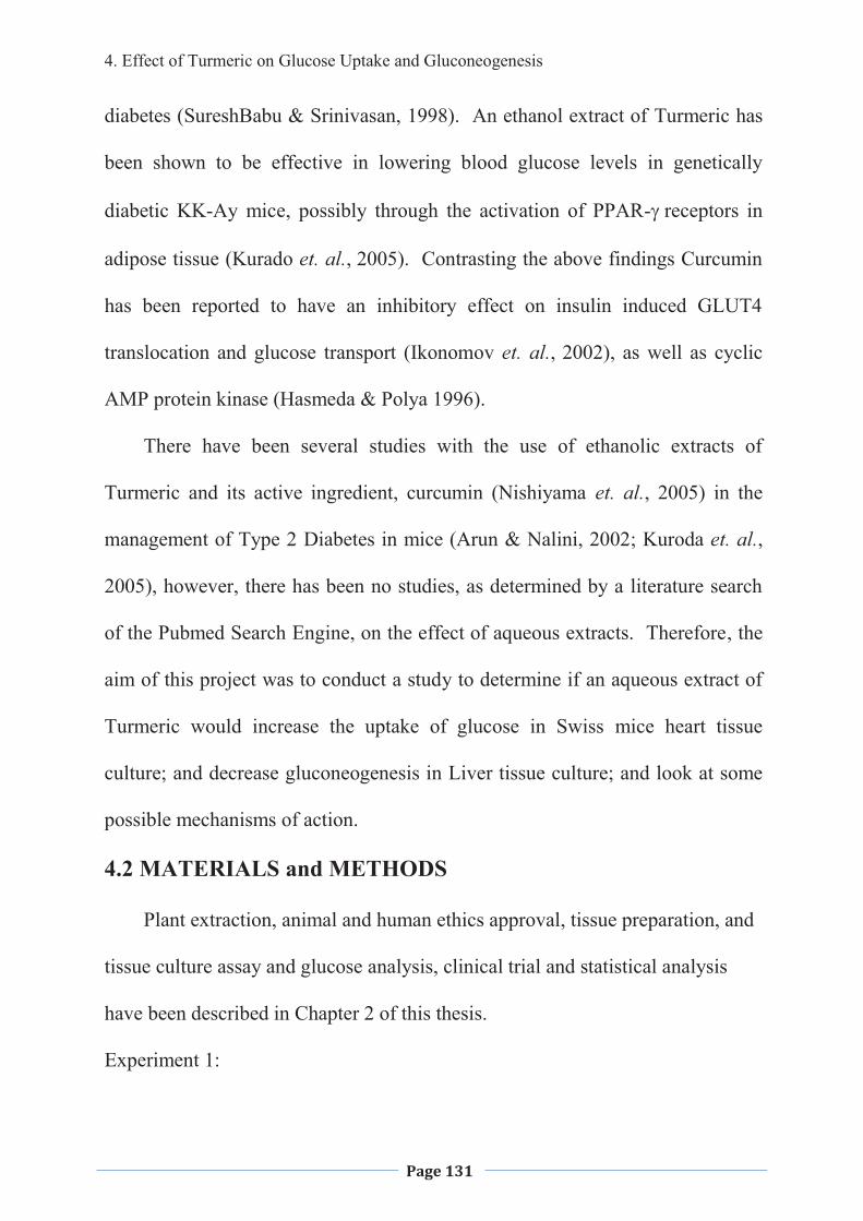
4. Effect of Turmeric on Glucose Uptake and Gluconeogenesis
Page 131
diabetes (SureshBabu & Srinivasan, 1998). An ethanol extract of Turmeric has
been shown to be effective in lowering blood glucose levels in genetically
diabetic KK-Ay mice, possibly through the activation of PPAR-��receptors in
adipose tissue (Kurado et. al., 2005). Contrasting the above findings Curcumin
has been reported to have an inhibitory effect on insulin induced GLUT4
translocation and glucose transport (Ikonomov et. al., 2002), as well as cyclic
AMP protein kinase (Hasmeda & Polya 1996).
There have been several studies with the use of ethanolic extracts of
Turmeric and its active ingredient, curcumin (Nishiyama et. al., 2005) in the
management of Type 2 Diabetes in mice (Arun & Nalini, 2002; Kuroda et. al.,
2005), however, there has been no studies, as determined by a literature search
of the Pubmed Search Engine, on the effect of aqueous extracts. Therefore, the
aim of this project was to conduct a study to determine if an aqueous extract of
Turmeric would increase the uptake of glucose in Swiss mice heart tissue
culture; and decrease gluconeogenesis in Liver tissue culture; and look at some
possible mechanisms of action.
4.2 MATERIALS and METHODS
Plant extraction, animal and human ethics approval, tissue preparation, and
tissue culture assay and glucose analysis, clinical trial and statistical analysis
have been described in Chapter 2 of this thesis.
Experiment 1:
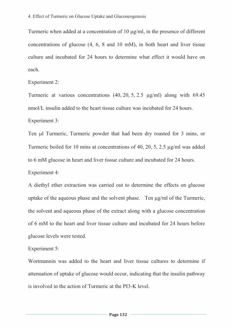
4. Effect of Turmeric on Glucose Uptake and Gluconeogenesis
Page 132
Turmeric when added at a concentration of 10 �g/ml, in the presence of different
concentrations of glucose (4, 6, 8 and 10 mM), in both heart and liver tissue
culture and incubated for 24 hours to determine what effect it would have on
each.
Experiment 2:
Turmeric at various concentrations (� �� �����2.5 µg/ml) along with 69.45
nmol/L insulin added to the heart tissue culture was incubated for 24 hours.
Experiment 3:
Ten �l Turmeric, Turmeric powder that had been dry roasted for 3 mins, or
Turmeric boiled for 10 mins at concentrations of � �� �����2.5 µg/ml was added
to 6 mM glucose in heart and liver tissue culture and incubated for 24 hours.
Experiment 4:
A diethyl ether extraction was carried out to determine the effects on glucose
uptake of the aqueous phase and the solvent phase. Ten �g/ml of the Turmeric,
the solvent and aqueous phase of the extract along with a glucose concentration
of 6 mM to the heart and liver tissue culture and incubated for 24 hours before
glucose levels were tested.
Experiment 5:
Wortmannin was added to the heart and liver tissue cultures to determine if
attenuation of uptake of glucose would occur, indicating that the insulin pathway
is involved in the action of Turmeric at the PI3-K level.

4. Effect of Turmeric on Glucose Uptake and Gluconeogenesis
Page 133
Experiment 6:
Metformin at concentrations of 100 mM was added to � ��g/ml Turmeric with
glucose at a concentration of ��mM to determine what effects the combination
would have on glucose uptake in the heart and liver tissue culture.
4.3 RESULTS
4.3.1 Effect on Glucose Uptake in Heart tissue Culture and decrease in
Glucose Output in Liver tissue culture
Turmeric was added to the heart tissue culture media at a concentration of
10 �g/ml in the presence of different concentrations of glucose (4, 6, 8 and 10
mM) to determine what effect it would have on each. The greatest effect was
when the glucose was at 8 mM with a (P>0.05) significantly increased 24.02 +/-
1.25 percentage difference in uptake over the basal level. At 10 mM glucose
concentration the (P>0.05) significantly increased the percentage uptake by 20.2
+/- 1.98%. While at 6 mM glucose there was a (P>0.05) significant percentage
difference of 15.9 +/- 1.37%, and at 4 mM of glucose there was a negligible
increase (P>0.05) with only a 1.97 +/- 1.25 percentage difference from basal
levels (see Figure 4.1).
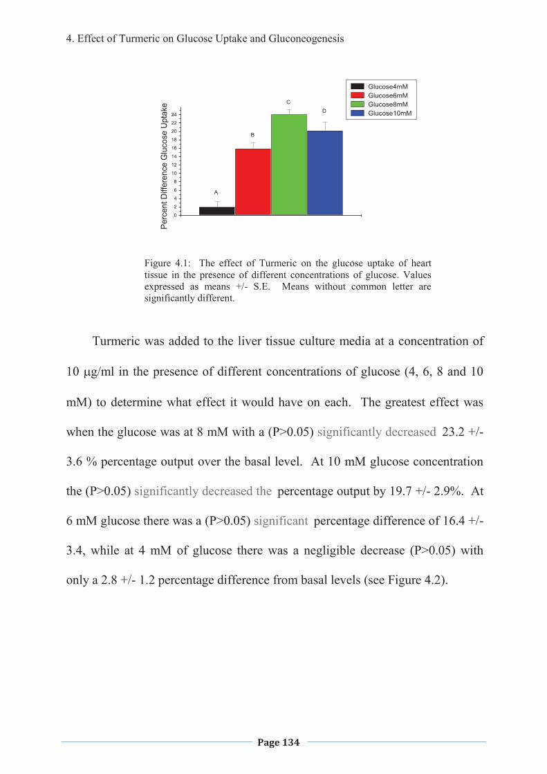
4. Effect of Turmeric on Glucose Uptake and Gluconeogenesis
Page 134
Figure 4.1: The effect of Turmeric on the glucose uptake of heart tissue in the presence of different concentrations of glucose. Values expressed as means +/- S.E. Means without common letter are significantly different.
Turmeric was added to the liver tissue culture media at a concentration of
10 �g/ml in the presence of different concentrations of glucose (4, 6, 8 and 10
mM) to determine what effect it would have on each. The greatest effect was
when the glucose was at 8 mM with a (P>0.05) significantly decreased 23.2 +/-
3.6 % percentage output over the basal level. At 10 mM glucose concentration
the (P>0.05) significantly decreased the percentage output by 19.7 +/- 2.9%. At
6 mM glucose there was a (P>0.05) significant percentage difference of 16.4 +/-
3.4, while at 4 mM of glucose there was a negligible decrease (P>0.05) with
only a 2.8 +/- 1.2 percentage difference from basal levels (see Figure 4.2).
100
2
4
6
8
10
12
14
16
18
20
22
24D
C
B
A
Perc
ent D
iffer
ence
Glu
cose
Upt
ake
Glucose4mM Glucose6mM Glucose8mM Glucose10mM
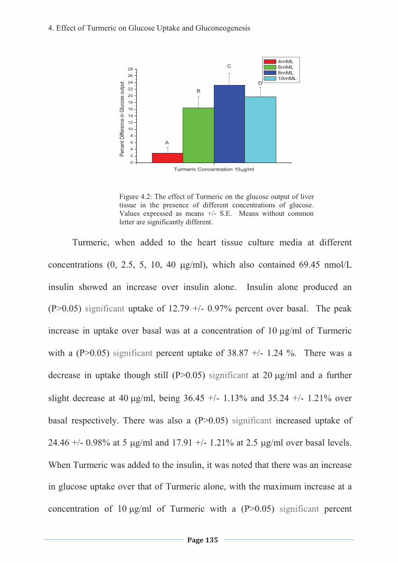
4. Effect of Turmeric on Glucose Uptake and Gluconeogenesis
Page 135
0
2
4
6
8
10
12
14
16
18
20
22
24
26
28
Perc
ent D
iffere
nce
in Gl
ucos
e ou
tput
Turmeric Concentration 10�g/ml
4mML 6mML 8mML 10mML
A
B
C
D
Figure 4.2: The effect of Turmeric on the glucose output of liver tissue in the presence of different concentrations of glucose. Values expressed as means +/- S.E. Means without common letter are significantly different.
Turmeric, when added to the heart tissue culture media at different
concentrations (0, 2.5, 5, 10, 40 �g/ml), which also contained 69.45 nmol/L
insulin showed an increase over insulin alone. Insulin alone produced an
(P>0.05) significant uptake of 12.79 +/- 0.97% percent over basal. The peak
increase in uptake over basal was at a concentration of � ��g/ml of Turmeric
with a (P>0.05) significant percent uptake of 38.87 +/- 1.24 %. There was a
decrease in uptake though still (P>0.05) significant at ��g/ml and a further
slight decrease at � ��g/ml, being 36.45 +/- 1.13% and 35.24 +/- 1.21% over
basal respectively. There was also a (P>0.05) significant increased uptake of
24.46 +/- 0.98% at 5 �g/ml and 17.91 +/- 1.21% at 2.5 �g/ml over basal levels.
When Turmeric was added to the insulin, it was noted that there was an increase
in glucose uptake over that of Turmeric alone, with the maximum increase at a
concentration of � ��g/ml of Turmeric with a (P>0.05) significant percent
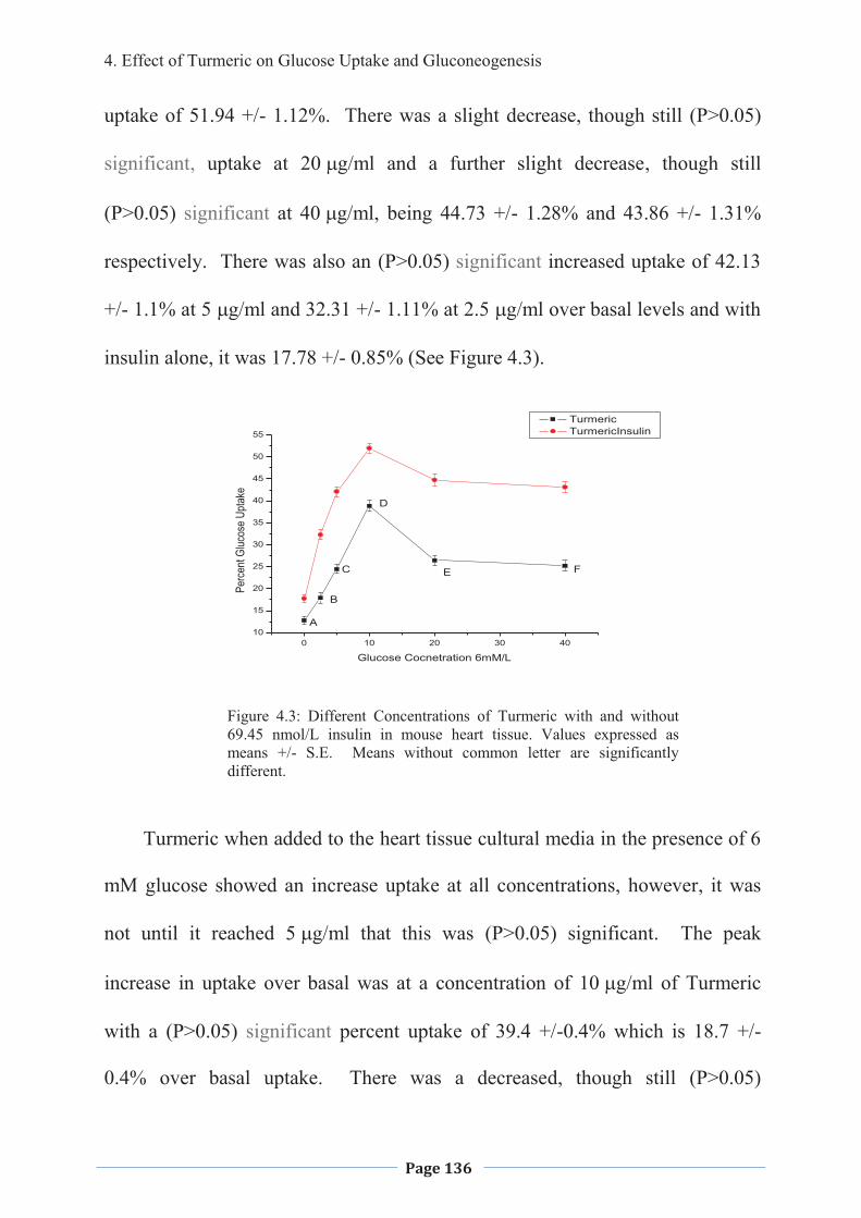
4. Effect of Turmeric on Glucose Uptake and Gluconeogenesis
Page 136
uptake of 51.94 +/- 1.12%. There was a slight decrease, though still (P>0.05)
significant, uptake at ��g/ml and a further slight decrease, though still
(P>0.05) significant at � ��g/ml, being 44.73 +/- 1.28% and 43.86 +/- 1.31%
respectively. There was also an (P>0.05) significant increased uptake of 42.13
+/- 1.1% at 5 �g/ml and 32.31 +/- 1.11% at 2.5 �g/ml over basal levels and with
insulin alone, it was 17.78 +/- 0.85% (See Figure 4.3).
0 10 20 30 4010
15
20
25
30
35
40
45
50
55
Perc
ent G
lucos
e Up
take
Glucose Cocnetration 6mM/L
Turmeric TurmericInsulin
A
B
C
D
E F
Figure 4.3: Different Concentrations of Turmeric with and without 69.45 nmol/L insulin in mouse heart tissue. Values expressed as means +/- S.E. Means without common letter are significantly different.
Turmeric when added to the heart tissue cultural media in the presence of 6
mM glucose showed an increase uptake at all concentrations, however, it was
not until it reached ���g/ml that this was (P>0.05) significant. The peak
increase in uptake over basal was at a concentration of � ��g/ml of Turmeric
with a (P>0.05) significant percent uptake of 39.4 +/-0.4% which is 18.7 +/-
0.4% over basal uptake. There was a decreased, though still (P>0.05)
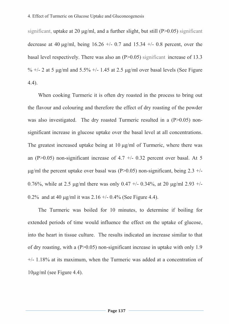
4. Effect of Turmeric on Glucose Uptake and Gluconeogenesis
Page 137
significant, uptake at ��g/ml, and a further slight, but still (P>0.05) significant
decrease at � ��g/ml, being 16.26 +/- 0.7 and 15.34 +/- 0.8 percent, over the
basal level respectively. There was also an (P>0.05) significant increase of 13.3
% +/- 2 at 5 �g/ml and 5.5% +/- 1.45 at 2.5 �g/ml over basal levels (See Figure
4.4).
When cooking Turmeric it is often dry roasted in the process to bring out
the flavour and colouring and therefore the effect of dry roasting of the powder
was also investigated. The dry roasted Turmeric resulted in a (P>0.05) non-
significant increase in glucose uptake over the basal level at all concentrations.
The greatest increased uptake being at � ��g/ml of Turmeric, where there was
an (P>0.05) non-significant increase of 4.7 +/- 0.32 percent over basal. At 5
�g/ml the percent uptake over basal was (P>0.05) non-significant, being 2.3 +/-
0.76%, while at 2.5 �g/ml there was only 0.47 +/- 0.34%, at 20 �g/ml 2.93 +/-
0.2% and at 40 �g/ml it was 2.16 +/- 0.4% (See Figure 4.4).
The Turmeric was boiled for 10 minutes, to determine if boiling for
extended periods of time would influence the effect on the uptake of glucose,
into the heart in tissue culture. The results indicated an increase similar to that
of dry roasting, with a (P>0.05) non-significant increase in uptake with only 1.9
+/- 1.18% at its maximum, when the Turmeric was added at a concentration of
� �g/ml (see Figure 4.4).
4. Effect of Turmeric on Glucose Uptake and Gluconeogenesis
Page 138
15
20
25
30
35
40
45
Per
cent
Glu
cose
Upt
ake
Glucose Concentration 6mM/L
Basal Turmeric DryRoastedTurmer BoiledTurmeric
A
B
C
D
Figure 4.4: The effect of Turmeric in aqueous form, after the Turmeric powder had been dry roasted (3 Mins) and aqueous extract of the Boiled Turmeric (10 mins) on the uptake of glucose in the mouse heart muscle. Values expressed as means +/- S.E. Means without common letter are significantly different.
In the liver tissue culture plates there was an increase in the amount of
glucose in the media in all wells with the liver tissue by itself increasing the
amount of glucose by 9.3 +/- 1.2 % indicating that gluconeogenesis was taking
place. The Turmeric showed a decreased glucose output compared to the basal
at all concentrations, however, it was not until it reached ���g/ml that this was
(P>0.05) significant at a concentration of 6 mM of glucose. The peak decrease
in gluconeogenesis over basal was at a concentration of 10�g/ml of Turmeric
with a (P>0.05) significant percent decrease in output of 4.21 +/- 0.03 %. There
was a (P>0.05) significant decrease in gluconeogenesis at a concentration of
��g/ml Turmeric being a 6.4 +/- 1.3% output. There was also a (P>0.05)
4. Effect of Turmeric on Glucose Uptake and Gluconeogenesis
Page 139
significant decrease with 5 �g/ml of Turmeric which exhibited a 6.2 +/- 0.9 %
output and 8.3 +/- 1 % output at a concentration of 2.5 �g/ml of the Turmeric
(See Figure 4.5).
The dry roasted Turmeric resulted in a (P>0.05) non-significant decrease of
glucose in the culture over the basal level at all concentrations with the greatest
decrease in output being at � �g/ml where there was a decrease of 8.91 +/-
0.04%. At 5 �g/ml the percent output over basal was a (P>0.05) non-significant
7.1+/- 0.76%. At 2.5 �g/ml the difference was only a (P>0.05) non-significant
8.83 +/- 0.34 % over basal. While that of 20 �g/ml was (P>0.05) non-
significant 8.53 +/- 0.2 % over basal and at 40 �g/ml it was a (P>0.05) non-
significant 2.16 +/- 0.4 % (See Figure 4.5).
The Turmeric that was boiled for 10 minutes resulted in a (P>0.05) non-
significant decrease in glucose present in the culture similar to that of dry
roasting with a decrease in output of only 8.71 +/- 0.03% at its maximum (see
Figure 4.5).
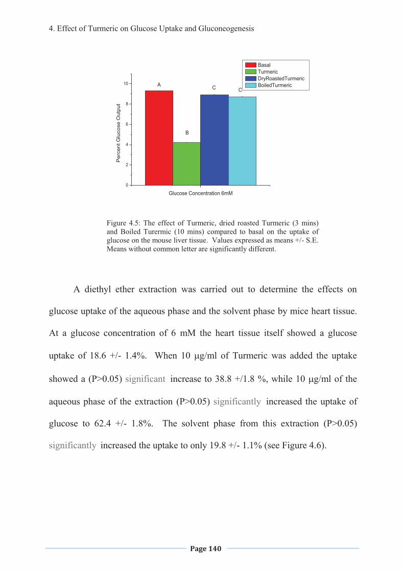
4. Effect of Turmeric on Glucose Uptake and Gluconeogenesis
Page 140
0
2
4
6
8
10
Per
cent
Glu
cose
Out
put
Glucose Concentration 6mM
Basal Turmeric DryRoastedTurmeric BoiledTurmericA
B
C C
Figure 4.5: The effect of Turmeric, dried roasted Turmeric (3 mins) and Boiled Turermic (10 mins) compared to basal on the uptake of glucose on the mouse liver tissue. Values expressed as means +/- S.E. Means without common letter are significantly different.
A diethyl ether extraction was carried out to determine the effects on
glucose uptake of the aqueous phase and the solvent phase by mice heart tissue.
At a glucose concentration of 6 mM the heart tissue itself showed a glucose
uptake of 18.6 +/- 1.4%. When 10 �g/ml of Turmeric was added the uptake
showed a (P>0.05) significant increase to 38.8 +/1.8 %, while 10 �g/ml of the
aqueous phase of the extraction (P>0.05) significantly increased the uptake of
glucose to 62.4 +/- 1.8%. The solvent phase from this extraction (P>0.05)
significantly increased the uptake to only 19.8 +/- 1.1% (see Figure 4.6).
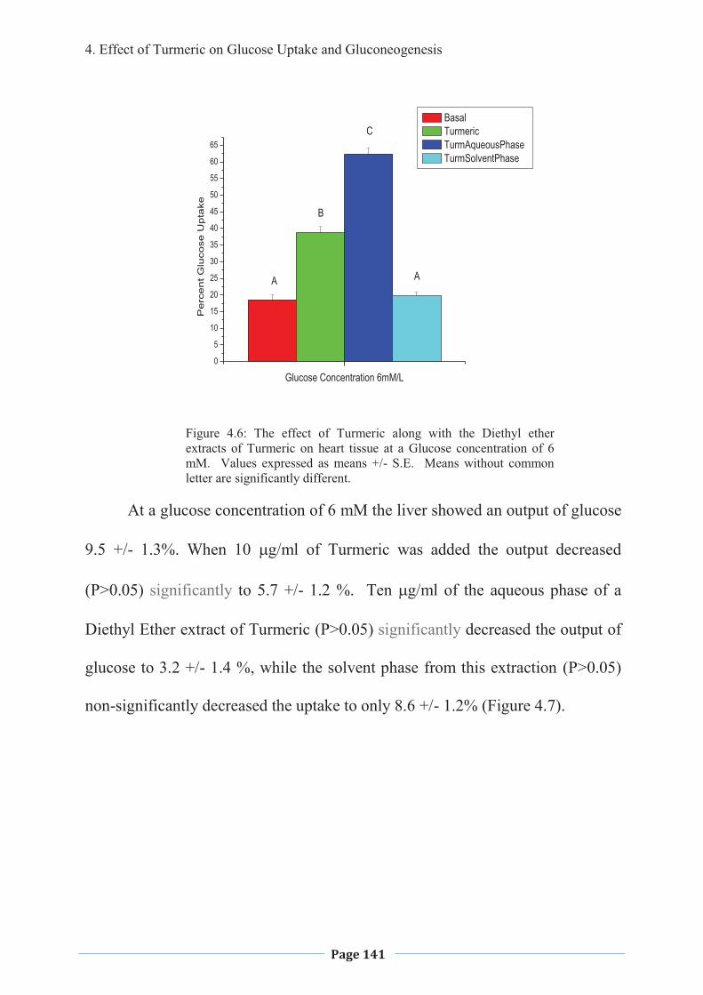
4. Effect of Turmeric on Glucose Uptake and Gluconeogenesis
Page 141
0
5
10
15
20
25
30
35
40
45
50
55
60
65
Pe
rce
nt G
luco
se U
pta
ke
Glucose Concentration 6mM/L
Basal Turmeric TurmAqueousPhase TurmSolventPhase
A
B
C
A
Figure 4.6: The effect of Turmeric along with the Diethyl ether extracts of Turmeric on heart tissue at a Glucose concentration of 6 mM. Values expressed as means +/- S.E. Means without common letter are significantly different.
At a glucose concentration of 6 mM the liver showed an output of glucose
9.5 +/- 1.3%. When 10 �g/ml of Turmeric was added the output decreased
(P>0.05) significantly to 5.7 +/- 1.2 %. Ten �g/ml of the aqueous phase of a
Diethyl Ether extract of Turmeric (P>0.05) significantly decreased the output of
glucose to 3.2 +/- 1.4 %, while the solvent phase from this extraction (P>0.05)
non-significantly decreased the uptake to only 8.6 +/- 1.2% (Figure 4.7).
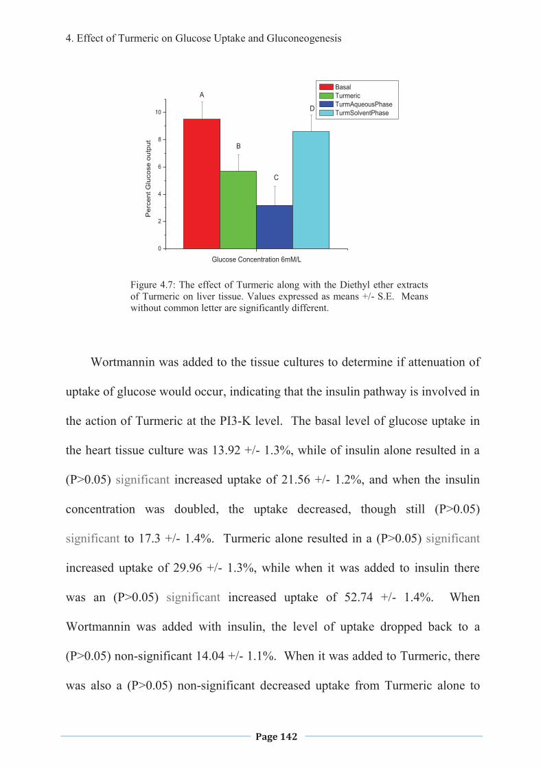
4. Effect of Turmeric on Glucose Uptake and Gluconeogenesis
Page 142
0
2
4
6
8
10
Per
cent
Glu
cose
out
put
Glucose Concentration 6mM/L
Basal Turmeric TurmAqueousPhase TurmSolventPhase
A
B
C
D
Figure 4.7: The effect of Turmeric along with the Diethyl ether extracts of Turmeric on liver tissue. Values expressed as means +/- S.E. Means without common letter are significantly different.
Wortmannin was added to the tissue cultures to determine if attenuation of
uptake of glucose would occur, indicating that the insulin pathway is involved in
the action of Turmeric at the PI3-K level. The basal level of glucose uptake in
the heart tissue culture was 13.92 +/- 1.3%, while of insulin alone resulted in a
(P>0.05) significant increased uptake of 21.56 +/- 1.2%, and when the insulin
concentration was doubled, the uptake decreased, though still (P>0.05)
significant to 17.3 +/- 1.4%. Turmeric alone resulted in a (P>0.05) significant
increased uptake of 29.96 +/- 1.3%, while when it was added to insulin there
was an (P>0.05) significant increased uptake of 52.74 +/- 1.4%. When
Wortmannin was added with insulin, the level of uptake dropped back to a
(P>0.05) non-significant 14.04 +/- 1.1%. When it was added to Turmeric, there
was also a (P>0.05) non-significant decreased uptake from Turmeric alone to
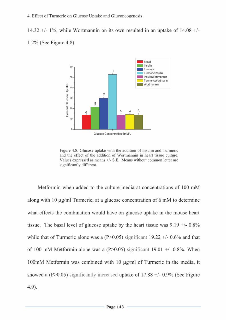
4. Effect of Turmeric on Glucose Uptake and Gluconeogenesis
Page 143
14.32 +/- 1%, while Wortmannin on its own resulted in an uptake of 14.08 +/-
1.2% (See Figure 4.8).
0
10
20
30
40
50
60P
erce
nt G
luco
se U
ptak
e
Glucose Concentration 6mM/L
Basal Insulin Turmeric TurmericInsulin InsulinWortmannin TurmericWortmanni Wortmannin
AAAA
B
C
D
Figure 4.8: Glucose uptake with the addition of Insulin and Turmeric and the effect of the addition of Wortmannin in heart tissue culture. Values expressed as means +/- S.E. Means without common letter are significantly different.
Metformin when added to the culture media at concentrations of 100 mM
along with � ��g/ml Turmeric, at a glucose concentration of ��mM to determine
what effects the combination would have on glucose uptake in the mouse heart
tissue. The basal level of glucose uptake by the heart tissue was 9.19 +/- 0.8%
while that of Turmeric alone was a (P>0.05) significant 19.22 +/- 0.6% and that
of 100 mM Metformin alone was a (P>0.05) significant 19.01 +/- 0.8%. When
100mM Metformin was combined with 10 �g/ml of Turmeric in the media, it
showed a (P>0.05) significantly increased uptake of 17.88 +/- 0.9% (See Figure
4.9).
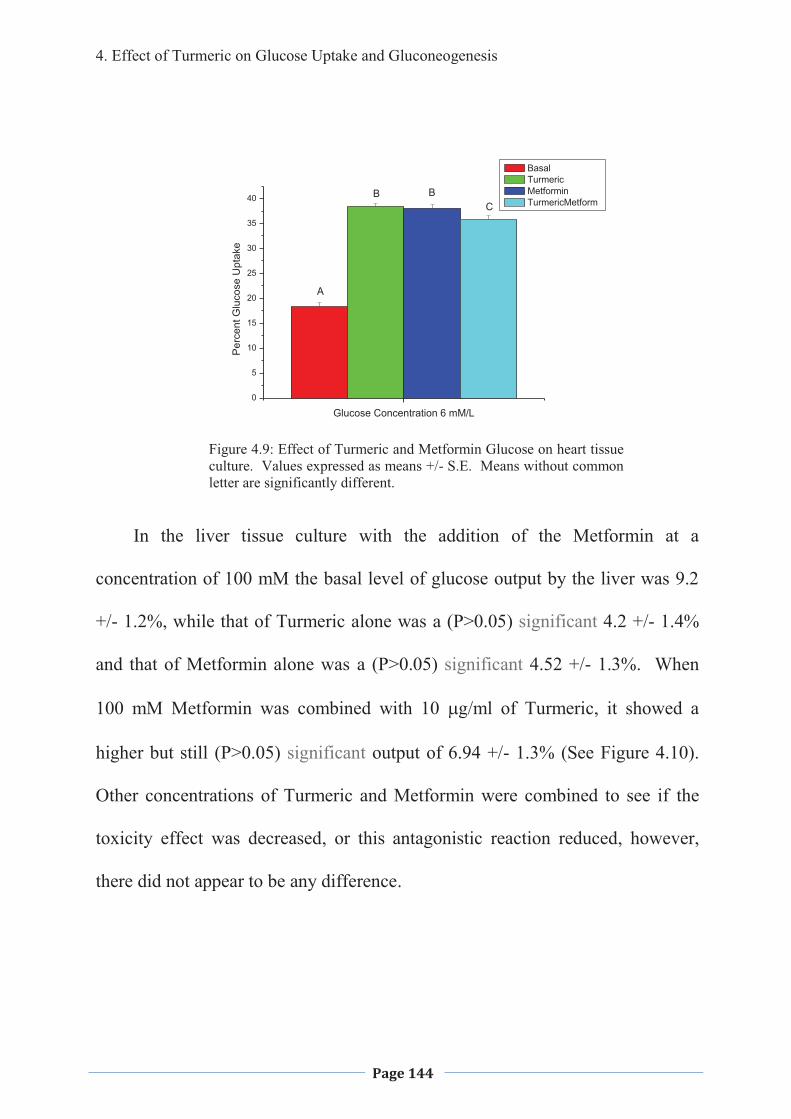
4. Effect of Turmeric on Glucose Uptake and Gluconeogenesis
Page 144
0
5
10
15
20
25
30
35
40
Per
cent
Glu
cose
Upt
ake
Glucose Concentration 6 mM/L
Basal Turmeric Metformin TurmericMetform
A
B BC
Figure 4.9: Effect of Turmeric and Metformin Glucose on heart tissue culture. Values expressed as means +/- S.E. Means without common letter are significantly different.
In the liver tissue culture with the addition of the Metformin at a
concentration of 100 mM the basal level of glucose output by the liver was 9.2
+/- 1.2%, while that of Turmeric alone was a (P>0.05) significant 4.2 +/- 1.4%
and that of Metformin alone was a (P>0.05) significant 4.52 +/- 1.3%. When
100 mM Metformin was combined with 10 �g/ml of Turmeric, it showed a
higher but still (P>0.05) significant output of 6.94 +/- 1.3% (See Figure 4.10).
Other concentrations of Turmeric and Metformin were combined to see if the
toxicity effect was decreased, or this antagonistic reaction reduced, however,
there did not appear to be any difference.
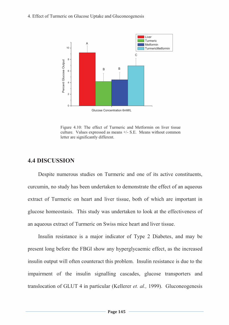
4. Effect of Turmeric on Glucose Uptake and Gluconeogenesis
Page 145
0
2
4
6
8
10
Per
cent
Glu
cose
Out
put
Glucose Concentration 6mM/L
Liver Turmeric Metformin TurmericMetformin
A
B B
C
Figure 4.10: The effect of Turmeric and Metformin on liver tissue culture. Values expressed as means +/- S.E. Means without common letter are significantly different.
4.4 DISCUSSION
Despite numerous studies on Turmeric and one of its active constituents,
curcumin, no study has been undertaken to demonstrate the effect of an aqueous
extract of Turmeric on heart and liver tissue, both of which are important in
glucose homeostasis. This study was undertaken to look at the effectiveness of
an aqueous extract of Turmeric on Swiss mice heart and liver tissue.
Insulin resistance is a major indicator of Type 2 Diabetes, and may be
present long before the FBGl show any hyperglycaemic effect, as the increased
insulin output will often counteract this problem. Insulin resistance is due to the
impairment of the insulin signalling cascades, glucose transporters and
translocation of GLUT 4 in particular (Kellerer et. al., 1999). Gluconeogenesis
4. Effect of Turmeric on Glucose Uptake and Gluconeogenesis
Page 146
also plays a major role in Type 2 Diabetes, as the liver produces glucose for
cells that indicate that they are starving, and the insulin/glucagon action is not
working on turning it on and off. The current management of diabetes looks at
this insulin action, and mimicking its action in order to decrease BGls. This is
done both by increasing the uptake of glucose by tissues throughout the body,
and also decreasing gluconeogenesis. Thus, this study examined the effect of
Turmeric on mice heart tissue in increasing glucose uptake, and also on liver
tissue to decrease gluconeogenesis.
Mice heart tissue was used due to the ease of removal and preparation for
tissue culture. A study was undertaken to determine if it exhibited the same
response as Soleus muscle from the mouse and there was a similar result at all
concentrations of glucose and Turmeric (data not shown). It was therefore
decided to continue the use of the mice heart tissue in all of the studies.
A decision was made to look at the management of those with blood
glucose levels between 5-10 nmol/L; therefore, for the tissue cultures we used 6
mM concentrations of glucose.
This study showed that there was a (P>0.05) significant increased uptake of
glucose into heart tissue and a decrease in glucose output from the liver tissue
when 5 �g/ml and 10 �g/ml of Turmeric was added to the culture. This increase
was slightly higher than that of 100 mM Metformin. However, it was noted that
with increased concentrations of the Turmeric there was a slight decrease,
though still (P>0.05) significant, in uptake and increase in output, which may
4. Effect of Turmeric on Glucose Uptake and Gluconeogenesis
Page 147
have been due to experimental error, or the toxicity of the extract at higher
concentrations as a crude extract was being used. Further studies would need to
be carried out to determine if Turmeric at higher concentrations is toxic to the
heart and liver tissue.
The decrease in the glucose uptake by the heart muscle and increased
output by the liver tissue when using the dry roasted Turmeric (this is typically
how it is prepared when using for curries and most cooking), and also the boiled
aqueous extract was (P>0.05) not significant. This indicates that very high heat
will deactivate the active ingredient/s suggesting the active ingredient/s are
proteins. It also suggests that the eating of curries etc, a popular winter food
throughout the many counties where Type 2 Diabetes is on the increase, will not
aid in the increase of glucose uptake even without looking at the absorption
factors.
A diethyl ether extraction was carried out to look at what type of chemical
might be involved in the increased uptake of glucose by the heart muscle tissue
and glucose output by the liver. The study showed that there was a (P>0.05)
significiantly greater uptake by the heart muscle and decreased glucose output
by the liver when the aqueous phase was used rather than with the whole
aqueous extract. There was little, if any, increase in uptake or decrease in output
over basal, when the solvent phase was used. Curcumin, the chemical that has
been shown to be effective in lowering glucose levels in previous studies
(Hasmeda & Polya 1996; Arun & Nalini, 2002; Ikonomov et. al., 2002; Korado
4. Effect of Turmeric on Glucose Uptake and Gluconeogenesis
Page 148
et. al., 2005; Nishiyama et. al., 2005) would be found in the solvent phase,
indicating that there is a different compound that is bringing about the increased
glucose uptake. Considering the loss of activity when the Turmeric solution was
boiled or the Turmeric was dry roasted before the aqueous extract was made, it
also suggests that the active component is a protein. Interestingly the aqueous
phase increased the uptake to a greater extent than that of the straight Turmeric,
suggesting that there is an ingredient in the extract that may be inhibiting the
uptake, or that the more purified sample was more efficient. Previous studies
have shown that Curcumin has an effect on decreasing BGLs, however, this
study shows that the aqueous phase is producing a different active constituent
which is having a similar effect. Further studies would need to be carried out to
isolate this compound that is bringing about the increase in glucose uptake by
the mouse heart tissue.
The differences in glucose uptake by the mouse heart tissue and deceased
output by the liver over basal at different concentrations of glucose indicates that
Turmeric has little or no effect when the glucose level is within normal range.
However, it increases the uptake and decreases the output when glucose levels
are high, indicating that the taking of Turmeric will not lead to hypoglycaemia,
coma and possible death. Very high levels of glucose were not tested as it was
out of the scope of this study and therefore the effect on higher glucose levels
needs further studies to determine the Turmeric’s effectiveness at these
concentrations.
4. Effect of Turmeric on Glucose Uptake and Gluconeogenesis
Page 149
To undertake a preliminary study looking at the mechanism of action a
maximum dose response of insulin (69.45 nmol/L) and also Wortmannin, which
blocks the insulin pathway at the PI3-K level by inhibiting PI3-kinase (Hausdorf
et. al., 1999) was added to the Turmeric treated heart tissues. The enhanced
uptake when Turmeric and insulin are combined and the decrease when
Turmeric is added at higher concentrations indicate that the Turmeric may be
acting via an insulin-mediated peripheral glucose pathway. This is similar to the
pathway used by the hypoglycaemic drug, Metformin (Bailey, 1988). The
inhibitory effect of Wortmannin on Turmeric supports the theory that the
Turmeric is acting via the classical PI3-kinase pathway.
Today many patients who have Type 2 Diabetes are prescribed Metformin
in the early stages of the disease progression and therefore the interaction
between Turmeric and Metformin was investigated. When Turmeric and
Metformin were placed in heart or liver tissue culture together there was an
antagonistic effect which may be due to increased toxicity of the
Turmeric/Metformin or experimental error with the use of a crude extract of
Turmeric. This may have been due to a drug interaction that needs to be
explored further.
4.5 CONCLUSIONS
An aqueous extract of Turmeric increases the uptake of glucose in mice
heart tissue culture and decrease gluconeogenesis from the mouse liver in a dose
4. Effect of Turmeric on Glucose Uptake and Gluconeogenesis
Page 150
dependent manner��with the optimum concentration being�� ��g/ml. It also
tended to be slightly toxic at higher levels with a decrease in glucose uptake by
heart muscle tissue culture and increased gluconeogenesis in liver tissue culture
over that of the optimum concentration; however, there still was an increase
over the basal rate. The toxicity may be due to the crude extract that was used
or that some active ingredient comes into effect at the higher concentrations so
there can be seen both this toxic effect to the heart and liver tissue as well as an
increased up take of glucose. Dry roasting or boiling of the Turmeric decreases
its ability to increase the uptake of glucose in the mouse heart tissue or decrease
the glucose output in liver tissue culture. This was most likely due to
deactivation of active constituents suggesting that the active constituent/s is a
protein. This is supported by the diethyl ether solvent extract where it is noted
that the aqueous phase increased the uptake of glucose in heart tissue and
decreased output in the liver tissue while the solvent phase did not. The addition
of insulin and Wortmannin indicates that the Turmeric was acting via the insulin
pathway as it is blocked by the Wortmannin at the PI3K level. However, it is
interesting to note that there is an additive effect to insulin at all concentrations
of Turmeric and so suggests entering the insulin pathway at a different point
then glucose-6-phosphate. There is an antagonistic reaction when Turmeric and
Metformin are added together to the heart and liver tissue cultures, therefore,
possibly should not be used in conjunction with each other; however, much
more research needs to be carried out to determine this interaction.
4. Effect of Turmeric on Glucose Uptake and Gluconeogenesis
Page 151
In summary, Turmeric does appear to increase glucose uptake in the mouse
heart muscle and decrease gluconeogenesis in the liver. The active ingredient
appears to be a protein however isolation studies need to be undertaken to
determine exactly what it is. The active ingredient appears to be working
through the insulin pathway and becomes toxic on the addition of Metformin.
There needs to be further studies carried out to determine Turmeric’s active
ingredient/s and their mechanism/s of action.
5. Effect of Glucostat on Glucose Uptake and Gluconeogenesis
Page 152
5. Effect of Glucostat on Glucose Uptake and Gluconeogenesis
Page 153
5. EFFECT OF GLUCOSTAT ON
GLUCOSE UPTAKE
AND GLUCONEOGENESIS
5. Effect of Glucostat on Glucose Uptake and Gluconeogenesis
Page 154
5.1 INTRODUCTION
Glucostat is a combination of the Chinese herbs including Trichosanthes
kirilowii (1900 mg), Polygonatum sibiricum (714.5 mg), Dioscorea oppostia
(286 mg), Panax ginseng (95 mg), and Stevia rebaudiana (47.6 mg) per capsule
that was available from Metagenics, Healthworld Ltd., Eaglefarm, Queensland,
Australia, 4009, for the management of Type 2 Diabetes. After a search of the
literature it was found that there has been some research undertaken on 4 of the
individual herbs, but only one (Collins & McFarlane, 2006) on the combination,
sold as Glucostat.
Panax ginseng from the Panax quinquefolius plant is used as an adaptogen,
aphrodisiac and nourishing stimulant (Ren, 1986). It has been reported to have
many uses, including antidepressant, a demulcent, a diuretic, a sedative and aids
in concentration and memory (Fetrow & Avila 2001). A study by Sotaniemi and
colleagues (1995) and Vuksan and collegues (2001) showed that it decreased
fasting BGL in both diabetic and non-diabetic patients, and enhanced insulin
release from the pancreas. It alone increased the number of insulin receptors as
well as aided in hepatic dysfunction, and hyperlipidemia. Suzuki (1989) showed
a reduction in insulin resistance and an increase in the number of insulin
receptors in mice, while Wallace (1999) showed a reduced fasting BGL after the
administration of Panax ginseng to humans. The effect of Panax ginseng seems
to be dependent on the time taken, with the most effective time being 40 minutes
5. Effect of Glucostat on Glucose Uptake and Gluconeogenesis
Page 155
prior to ingestion of a meal (Vuksan et. al., 2001). In a human trial, Vuksan and
colleagues (2000b) showed that no more than 3 gm of American ginseng was
required to decrease fasting blood glucose levels, irrespective of the time taken
before the meal.
Sotaniemi and colleagues (1995) suggested that an unspecified species of
ginseng improved mood and psychophysical performances, which led to
beneficial lifestyle changes, such as diet and increased physical activity; this
may be useful in the management of Type 2 Diabetes. They also suggested that
there was a direct action as well, in that there was improved insulin sensitivity,
and increased glycogen storage, independent of diet and physical activity.
Suzuki and colleagues (1991) showed that an unspecified ginseng inhibited
gastric secretion in rats, while Onomura and colleagues (1999) have shown that
it decreases glucose and maltose absorption in isolated human and rat
duodenum. Ohnishi and colleagues (1996) found that Chinese ginseng
increased GLUT2 in the livers of normal and diabetic mice, and stimulated
insulin secretion in mice islets and rat pancreas.
The active components of ginseng are believed to be the ginsenosides, a
group of steroidal saponins (Huang 1999; Attele et. al., 2002), which are
distributed in the roots, leaf and berry of the plant. Different parts of the plant
have a distinct ginenoside profile (Attele et. al., 1999) and may have different
pharmacological activity. Attele and colleagues (1999) showed that Panax
ginseng berry significantly improved systemic insulin sensitivity and glucose
5. Effect of Glucostat on Glucose Uptake and Gluconeogenesis
Page 156
homeostasis in mice. However, Dey and colleagues (2003) showed that Panex
ginseng berry exhibited more potent anti-diabetic activity than did the root, and
only the berry exhibited anti-obesity effects in ob/ob mice. The gensenoside Re
plays a role in this action; however, was not associated with body weight
changes, suggesting that there may be other active constituents as well.
Adenosine, which enhances lipogenesis (Okuda & Yoshida, 1980), as well as a
carboxylic acid which also enhanced lipogenesis (Sekiya & Okuda, 1981), have
been identified from an aqueous extract of Panax ginseng; however,
hypoglycaemic activity was not tested. Kimura and colleagues (1981) isolated
DPG-3-2, a water extract and EPG-3-2, a methanolic extract from Panax
ginseng, with EPG-3-2 and DPG-3-2 increasing serum insulin levels in diabetic
mice, yet had no effect on normal mice; however, DPG-3-2 had an additive
effect on insulin release in both diabetic and normal rats. There has also been 5
glycans (Panaxan A-E) isolated from the roots of ginseng (Konno et. al., 1984)
which exhibited hypoglycaemic activity in both normal and alloxan-induced
diabetic mice.
There have been a few side effects reported from the administration of
American ginseng including nausea, headache, dizziness, insomnia, nervousness
and hypertension (Miller, 1998), along with diarrhoea and fatigue which have
been reported in a few isolated cases (reviewed in Vogler et. al., 1999).
However, with each of these reported side effects it is not sure if the American
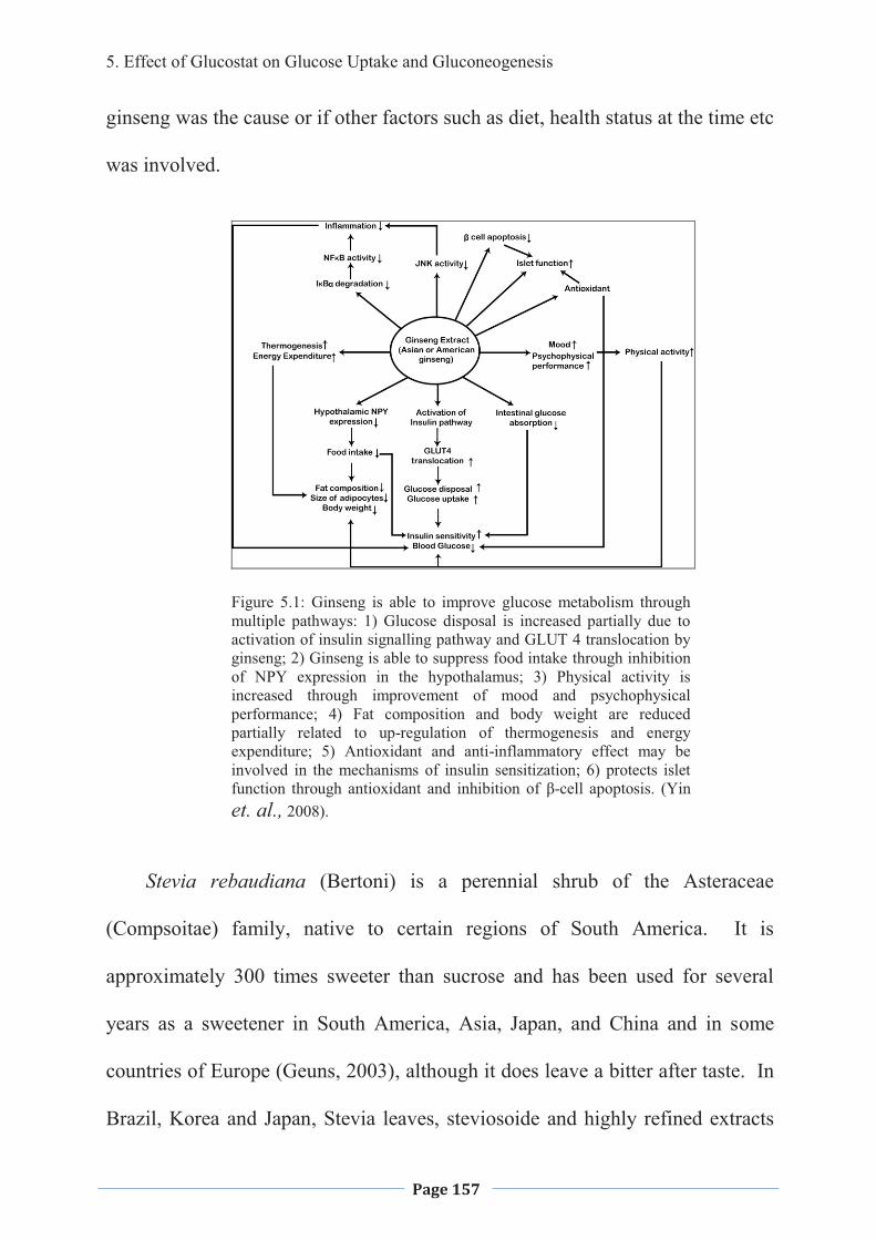
5. Effect of Glucostat on Glucose Uptake and Gluconeogenesis
Page 157
ginseng was the cause or if other factors such as diet, health status at the time etc
was involved.
Figure 5.1: Ginseng is able to improve glucose metabolism through multiple pathways: 1) Glucose disposal is increased partially due to activation of insulin signalling pathway and GLUT 4 translocation by ginseng; 2) Ginseng is able to suppress food intake through inhibition of NPY expression in the hypothalamus; 3) Physical activity is increased through improvement of mood and psychophysical performance; 4) Fat composition and body weight are reduced partially related to up-regulation of thermogenesis and energy expenditure; 5) Antioxidant and anti-inflammatory effect may be involved in the mechanisms of insulin sensitization; 6) protects islet function through antioxidant and inhibition of β-cell apoptosis. (Yin et. al., 2008).
Stevia rebaudiana (Bertoni) is a perennial shrub of the Asteraceae
(Compsoitae) family, native to certain regions of South America. It is
approximately 300 times sweeter than sucrose and has been used for several
years as a sweetener in South America, Asia, Japan, and China and in some
countries of Europe (Geuns, 2003), although it does leave a bitter after taste. In
Brazil, Korea and Japan, Stevia leaves, steviosoide and highly refined extracts
5. Effect of Glucostat on Glucose Uptake and Gluconeogenesis
Page 158
are officially used as a low-calorie sweetener (Kim et. al., 2002; Mizutani and
Tanaka, 2002). The sweet components are believed to be stevioside,
steviolbioside and various rebaudiosides and dulcoside, which occur mainly in
the leaves (Geuns, 2003).
The Stevia rebaudiana plant and its constituents have had a controversial
history as far as its antidiabetic effects are concerned (White 1994). Curie
(1986) showed a decreased BGL after administration of 20 gm/day to a Type 2
Diabetic GK rat, and postulated it was due to an increase in the mitochondrial
respiration rate; or inhibition of gluconeogenesis. As well as an
antihyperglycaemic, insulinotropic and glucagonostatic effect, it is thought that
this might be due to a direct action on �-cells (Jeppesen et al. 2000).
Experiments performed by Malaisse and colleagues (1998) in rats, showed that
in the presence of stevioside there was an increase of insulin output. However,
it only reached a statistically significant effect at the higher concentrations, as
well as acting as a stimulant on hepatic glycogen synthesis under gluconeogenic
conditions. Human trials, using Stevioside, were carried out by Curi (1986),
which showed a significant decrease in BGL by increasing glucose tolerance.
There is controversy over whether Stevia rebaudiana, taken as a
supplement, will have any effect in decreasing FBG as it has been shown that
Stevioside, one of its main active ingredients taken orally, is not absorbed or
only in very small amounts in the human (Bracht et. al., 1985; Yamamto et. al.,
1985; Koyama et. al., 2003), and none of the digestive enzymes of man and
5. Effect of Glucostat on Glucose Uptake and Gluconeogenesis
Page 159
most animals are able to degrade stevioside into its aglycone, steviol (Wingard
et. al., 1980; ; Hutapea et. al., 1997; Koyama et. al., 2001; Koyama et. al.,
2003). However, Stevioside is metabolised to steviol by digestive tract bacteria
and this then is possibly able to be absorbed (Koyama et. al., 2003). It has been
shown that the bacteria in the human gastrointestinal tract are able to transform
stevioside into steviol in vitro; however, it has not been proven that this occurs
in vivo, nor that it is absorbed by the intestines directly into the blood stream
(Hutapea et. al., 1997; Koyama et. al., 2001). Experiments by Wingard and
colleagues (1980) and Nakayama and colleagues (1986) and Koyama and
colleagues (2003) have shown that steviol can be absorbed by the intestines.
There has been some controversial studies concerning the effect of
stevioside on fertility with Planas and Kuae (1968) reporting a decrease of live
birth rates in rats; however, this was refuted by Shiotsu (1996) who carried out
more reliable experiments using many more animals. Melis (1999) suggested a
possible decrease in male fertility, whereas Oliveira-Filho (1989) and colleagues
using the same stevioside dosage, which were very high – 5.3 gms stevioside/kg
body weight - did not find this. Stevioside also does not interfere with the
uptake of other nutrients such as vitamins and minerals from the intestine (Wood
et. al., 1996; Geuns et. al., 2003).
Gua lou, from the root tubers of the Trichosanthes kirilowii plant, has been
used in Chinese medicine for the ‘Thirsting Disease’ for centuries. Hikino and
colleagues (1989) found that the non-dialyzable portion decreased BGL in mice
5. Effect of Glucostat on Glucose Uptake and Gluconeogenesis
Page 160
and they isolated five glycans (Trichosans A, B, C, D, and E), all of which
showed hypoglycaemic actions in normal mice and Trichosan A decreased BGL
in alloxan-induced hyperglycaemic mice. A galactose-binding lectin from the
roots of the plant stimulate the incorporation of D-[3-3H]glucose into lipids in
adipocytes (Ng et. al., 1985); however, it did not inhibit lypolysis in the manner
of insulin inhibiting hormone-induced lipolysis (Ng et. al., 1985; Wong et. al.,
1985). Trichosanthes kirilowii has been used for centuries as an abortificant in
Chinese medicine and Lu and colleagues (2001) believe that this is due to its
ribosome inhibiting property. Xu and colleagues (2001) have shown that
Trichosanthes kirilowii also has anti-inflammatory properties, and this may be
the process that aids in the management of Type 2 Diabetes, as it has now been
shown that inflammation and Type 2 Diabetes are linked (Festa et. al., 2002;
Freeman et. al., 2002). Yeung and colleagues (1987) isolated an
immunosuppressive protein from the root of the Trichosanthes kirilowi which
inhibited ConA-induced transformation in lymphocytes isolated from spleens of
mice, and this protein was a potent inducer of mid-term abortion in mice. This
was supported by Gong and colleagues (2008) when they showed that
trichosanthin may act as an anti-rejection therapy.
Shan yao from the Dioscorea opposita plant has also been used for
centuries in Traditional Chinese Medicine. It is one of the herbs used in Die-
Huang-Wan, often a prescribed treatment for diabetes in China and Japan as
well as other Asian countries. It has been shown to increase insulin secretion in
5. Effect of Glucostat on Glucose Uptake and Gluconeogenesis
Page 161
normal rats as well as decrease FBGls; however, has no effect on streptozotocin-
induced diabetic rats (Hsu et. al., 2007). Hsu and colleagues (2007) showed that
Die-Huang-Wan would decrease the plasma glucose levels in fructose rich chow
fed rats. Dioscorea opposite appeared to be the herb responsible for this, as,
when it was removed from the combination, no increase in insulin secretion or
decrease in FBG would result. Where as if other herbs were removed from the
combination then no lowering of the plasma glucose levels would result,
indicating that Dioscorea opposite is responsible for the increase in whole body
insulin sensitivity. Hao and colleagues (1991) showed that there was an anti-
hyperglycemic effect in mice and suggested that the active ingredients were
polysaccarides.
Huangjing or Siberian Solomon’s Seal is from the Polygonatum sibiricum
plant and has been used in Traditional Chinese Medicine for centuries for the
treatment of wasting and thirsting disease. Kato and Miura (1993) have shown
that a methanol extract of Polygonatum sibiricum decreased blood glucose
without any changes in serum insulin levels, in normal and streptozotocin-
induced diabetic mice. A later study by Kato and colleagues (1994) showed that
an intraperitoneal injection of a methanol extract of a Polygonatum sibiricum
decreased glucose output by the liver in Wistar Fatty rats, most likely by
decreasing GLUT mRNA and associated proteins. Kato and Miura (1993)
isolated an active constituent from the Polygonatum sibiricum which was the
spirostanol glycoside, PO-2; Ahn and colleagues (2006) have isolated 4 steroidal
5. Effect of Glucostat on Glucose Uptake and Gluconeogenesis
Page 162
saponins from a methanol extract, the neosibiricosides A-D (1-4), which they
tested for cytotoxicity against cultured human MCF-7 breast cancer cells, and
showed that 3 and 4 had moderate cytotoxic activity while 1 and 2 had none.
In this study we looked at the mechanism of action for the effectiveness in
glucose uptake in mouse heart tissue and decreasing gluconeogenesis as there
was a significantly increased glucose uptake in heart mouse tissue culture in the
screening process that was carried out and discussed in chapter 3.
5.2 MATERIALS AND METHODS
Plant extraction, animal ethics approval, tissue preparation, and tissue
culture assay and glucose analysis, clinical trial and statistical analysis have
been described in Chapter 2 of this thesis.
Experiment 1:
Glucostat (10 µg/ml) or Boiled Glucostat (10 min) at a concentration of 10
µg/ml and in the presence of different concentrations of glucose (4, 6, 8 and 12
mM) was added to heart or liver tissue culture and incubated for 24 hours.
Experiment 2:
Glucostat and Boiled Glucostat (10 min) at various concentrations (� �� �����2.5
µg/ml) in the presence of 6mM glucose was added to heart or liver tissue culture
and incubated for 24 hours.
Experiment 3:

5. Effect of Glucostat on Glucose Uptake and Gluconeogenesis
Page 163
A diethyl ether extraction was carried out to determine the effects on glucose
uptake of the aqueous phase and the solvent phase. Ten �g/ml of the Glucostat,
the Boiled Glucostat (10 min) and the solvent and aqueous phase of the extract
in the presence of 6 mM glucose was added to the heart or liver tissue culture
and incubated for 24 hours.
Experiment 4:
Wortmannin was added to the heart and liver tissue cultures to determine if
attenuation of uptake of glucose would occur indicating that the insulin pathway
is involved in the action of Turmeric at the PI3-K level.
Experiment 5:
Metformin at concentrations of 100 mM was added to � ��g/ml Glucostat and
Boiled Glucostat (10 min) in the presence of 6 mM glucose to determine what
effects the combination would have on glucose uptake in the heart and liver
tissue culture.
5.2.1. Sheep Studies.
Merino wethers (6) were utilized as the animal model in this study. They
were housed at the University if New England, Armidale, NSW, Australia and
remained in an open paddock until required for experimentation. Throughout
the experimentation period they, were housed in 12 m2 pens and were allowed ad
lib feeding of a blend of White chaff and Lucerne, along with water. The
wethers were weighed at the beginning of the experiment and randomly divided
5. Effect of Glucostat on Glucose Uptake and Gluconeogenesis
Page 164
into 3 groups; control, oral treatment and IV treatment, to nullify the influence
of age and weight.
The first group were administered the equivalent of 1 Glucostat capsule
containing (Trichosanthes kirilowii (1900 mg), Polygonatum sibiricum (714.5
mg), Dioscorea oppostia (286 mg), Panax ginseng (95 mg), and Stevia
rebaudiana (47.6 mg) dissolved in water by 50 ml drenching syringe, 3 times
per day. The second group received the same dosage which had been soaked in
0.9% sodium chloride administered via jugular catheter, 3 times per day. The
control group had 50ml of saline injected via the jugular catheter, 3 times per
day. Each merino received treatments at 8 am, 12 noon and 6 pm for 4 days.
After the 4 days, blood samples were taken from each animal to determine
the basal glucose level. An intravenous dose of glucose (240 mg) was
administered to commence a Glucose Tolerance Test. Blood samples were
taken at 5, 10, 15, 30, 45, 60, 90 and 120 minutes and the plasma collected.
5.2.1.1 Collection of Plasma Samples
Each sheep was fitted with an intravenous catheter inserted into either the
left or right jugular vein. Blood was collected in 10 ml polypropylene tubes,
each containing 20 �L of Lithium heparin (1000 I.U./ml). Immediately after the
sample was taken, the catheter tubing was flushed with 3.8% Sodium Citrate as
an anticoagulant agent. Samples were then centrifuged at 2500-3000 rpm for 20
minutes. The plasma was removed and transferred to labelled 5 ml sample vials
and stored at -20oC until required for assaying.
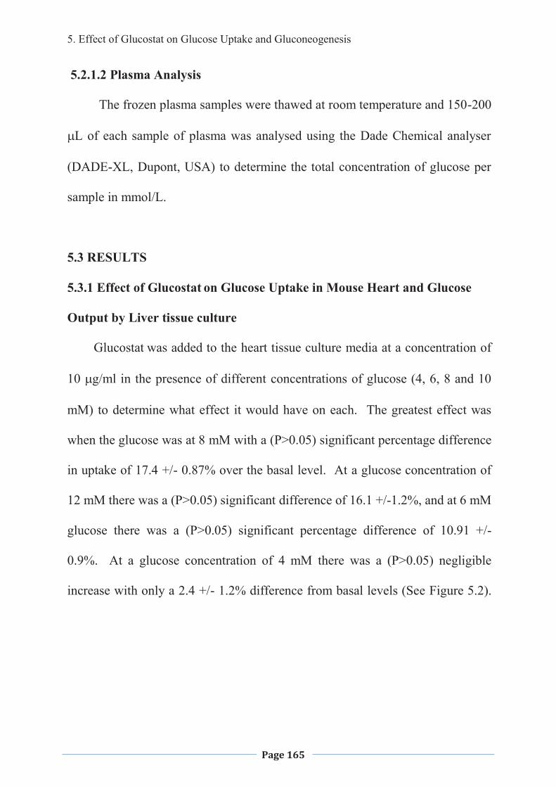
5. Effect of Glucostat on Glucose Uptake and Gluconeogenesis
Page 165
5.2.1.2 Plasma Analysis
The frozen plasma samples were thawed at room temperature and 150-200
�L of each sample of plasma was analysed using the Dade Chemical analyser
(DADE-XL, Dupont, USA) to determine the total concentration of glucose per
sample in mmol/L.
5.3 RESULTS
5.3.1 Effect of Glucostat on Glucose Uptake in Mouse Heart and Glucose
Output by Liver tissue culture
Glucostat was added to the heart tissue culture media at a concentration of
10 �g/ml in the presence of different concentrations of glucose (4, 6, 8 and 10
mM) to determine what effect it would have on each. The greatest effect was
when the glucose was at 8 mM with a (P>0.05) significant percentage difference
in uptake of 17.4 +/- 0.87% over the basal level. At a glucose concentration of
12 mM there was a (P>0.05) significant difference of 16.1 +/-1.2%, and at 6 mM
glucose there was a (P>0.05) significant percentage difference of 10.91 +/-
0.9%. At a glucose concentration of 4 mM there was a (P>0.05) negligible
increase with only a 2.4 +/- 1.2% difference from basal levels (See Figure 5.2).
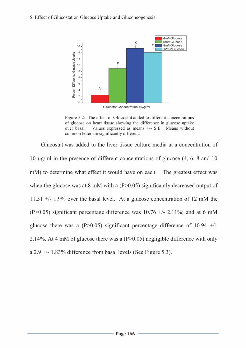
5. Effect of Glucostat on Glucose Uptake and Gluconeogenesis
Page 166
0
2
4
6
8
10
12
14
16
18
Perc
ent D
iffer
ence
Glu
cose
Upt
ake
Glucostat Concentration 10�g/ml
4mMGlucose 6mMGlucose 8mMGlucose 12mMGlucose
A
B
CC
Figure 5.2: The effect of Glucostat added to different concentrations of glucose on heart tissue showing the difference in glucose uptake over basal. Values expressed as means +/- S.E. Means without common letter are significantly different.
Glucostat was added to the liver tissue culture media at a concentration of
10 �g/ml in the presence of different concentrations of glucose (4, 6, 8 and 10
mM) to determine what effect it would have on each. The greatest effect was
when the glucose was at 8 mM with a (P>0.05) significantly decreased output of
11.51 +/- 1.9% over the basal level. At a glucose concentration of 12 mM the
(P>0.05) significant percentage difference was 10.76 +/- 2.11%; and at 6 mM
glucose there was a (P>0.05) significant percentage difference of 10.94 +/1
2.14%. At 4 mM of glucose there was a (P>0.05) negligible difference with only
a 2.9 +/- 1.83% difference from basal levels (See Figure 5.3).
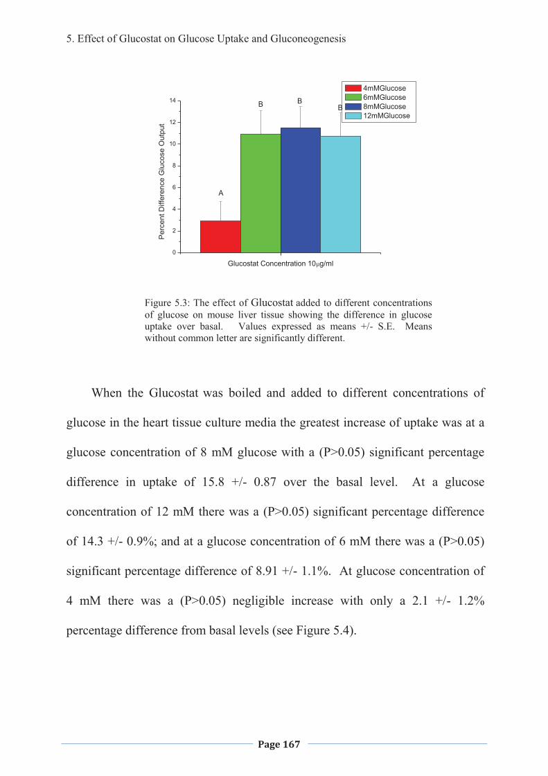
5. Effect of Glucostat on Glucose Uptake and Gluconeogenesis
Page 167
0
2
4
6
8
10
12
14
Per
cent
Diff
eren
ce G
luco
se O
utpu
t
Glucostat Concentration 10�g/ml
4mMGlucose 6mMGlucose 8mMGlucose 12mMGlucose
A
BBB
Figure 5.3: The effect of Glucostat added to different concentrations of glucose on mouse liver tissue showing the difference in glucose uptake over basal. Values expressed as means +/- S.E. Means without common letter are significantly different.
When the Glucostat was boiled and added to different concentrations of
glucose in the heart tissue culture media the greatest increase of uptake was at a
glucose concentration of 8 mM glucose with a (P>0.05) significant percentage
difference in uptake of 15.8 +/- 0.87 over the basal level. At a glucose
concentration of 12 mM there was a (P>0.05) significant percentage difference
of 14.3 +/- 0.9%; and at a glucose concentration of 6 mM there was a (P>0.05)
significant percentage difference of 8.91 +/- 1.1%. At glucose concentration of
4 mM there was a (P>0.05) negligible increase with only a 2.1 +/- 1.2%
percentage difference from basal levels (see Figure 5.4).

5. Effect of Glucostat on Glucose Uptake and Gluconeogenesis
Page 168
0
2
4
6
8
10
12
14
16
18
Per
cent
Diff
eren
t Glu
cose
Upt
ake
Boiled Glucostat Concentration 10�g/ml
4mMGlucose 6mMGlucose 8mMGlucose 12mMGlucose
A
B
C
D
Figure 5.4: The effect of Boiled Glucostat added to different concentrations of glucose on heart tissue showing the difference in glucose uptake over basal. Values expressed as means +/- S.E. Means without common letter are significantly different.
Ten �g/ml of Boiled Glucostat was added with different concentrations of
glucose (4, 6, 8 and 12 mM) to determine what affect it would have on each and
if the boiling of the Glucostat would affect the results. The greatest effect was
with a glucose concentration of 8 mM which showed a (P>0.05) significant
percentage difference of 9.34 +/- 1.76%. At a glucose concentration of 12 mM
there was a (P>0.05) significant percentage difference of 8.83 +/- 1.97%; while
at a glucose concentration of 6 mM there was a (P>0.05) significant percent
difference of 8.96 +/- 1.87%. At a glucose concentration of 4 mM there was a
(P>0.05) negligible increase with only a 2.7 +/- 1.54% difference from basal
levels (See Figure 5.5).
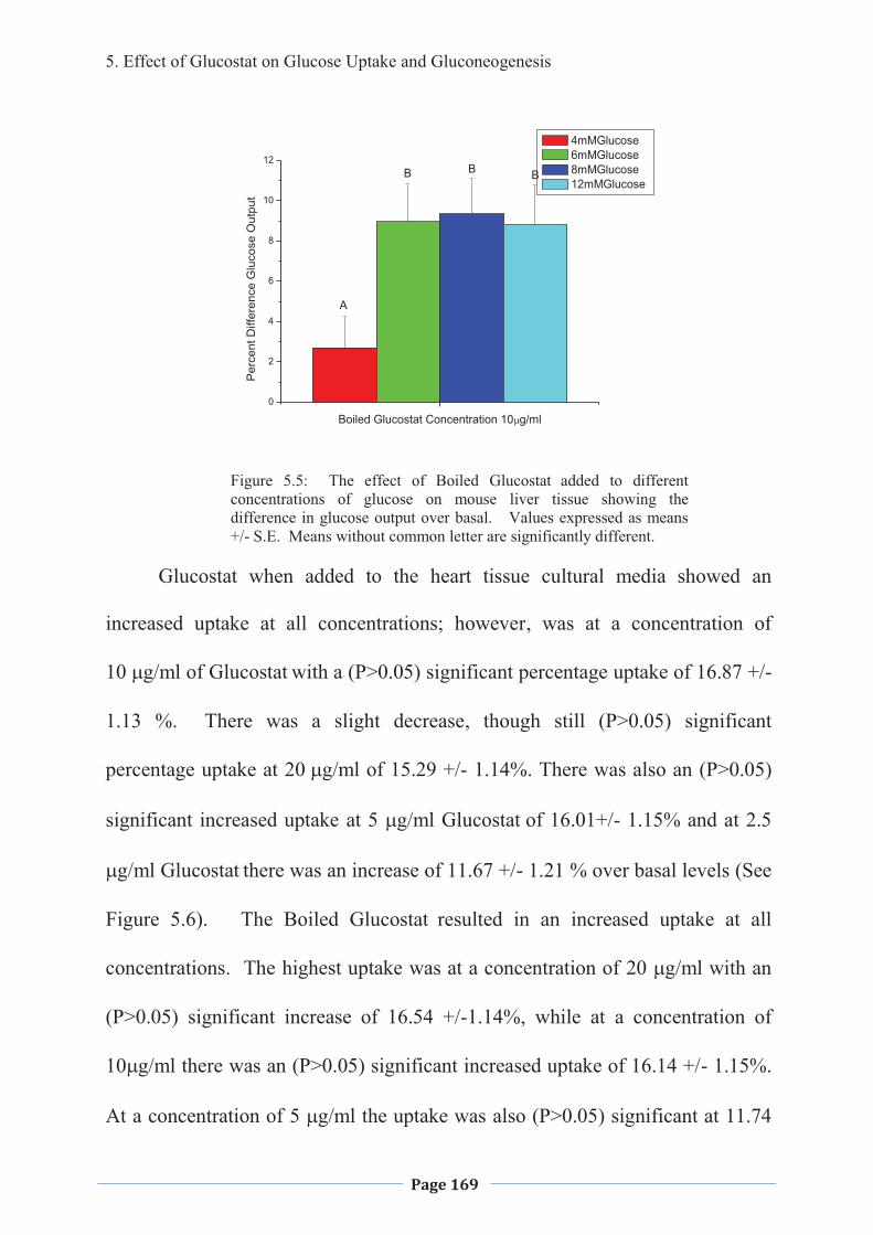
5. Effect of Glucostat on Glucose Uptake and Gluconeogenesis
Page 169
0
2
4
6
8
10
12
Per
cent
Diff
eren
ce G
luco
se O
utpu
t
Boiled Glucostat Concentration 10�g/ml
4mMGlucose 6mMGlucose 8mMGlucose 12mMGlucose
A
B B B
Figure 5.5: The effect of Boiled Glucostat added to different concentrations of glucose on mouse liver tissue showing the difference in glucose output over basal. Values expressed as means +/- S.E. Means without common letter are significantly different.
Glucostat when added to the heart tissue cultural media showed an
increased uptake at all concentrations; however, was at a concentration of
� ��g/ml of Glucostat with a (P>0.05) significant percentage uptake of 16.87 +/-
1.13 %. There was a slight decrease, though still (P>0.05) significant
percentage uptake at ��g/ml of 15.29 +/- 1.14%. There was also an (P>0.05)
significant increased uptake at 5 �g/ml Glucostat of 16.01+/- 1.15% and at 2.5
�g/ml Glucostat there was an increase of 11.67 +/- 1.21 % over basal levels (See
Figure 5.6). The Boiled Glucostat resulted in an increased uptake at all
concentrations. The highest uptake was at a concentration of 20 �g/ml with an
(P>0.05) significant increase of 16.54 +/-1.14%, while at a concentration of
10�g/ml there was an (P>0.05) significant increased uptake of 16.14 +/- 1.15%.
At a concentration of 5 �g/ml the uptake was also (P>0.05) significant at 11.74
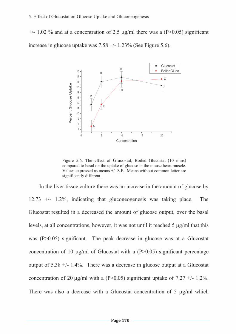
5. Effect of Glucostat on Glucose Uptake and Gluconeogenesis
Page 170
+/- 1.02 % and at a concentration of 2.5 �g/ml there was a (P>0.05) significant
increase in glucose uptake was 7.58 +/- 1.23% (See Figure 5.6).
Figure 5.6: The effect of Glucostat, Boiled Glucostat (10 mins) compared to basal on the uptake of glucose in the mouse heart muscle. Values expressed as means +/- S.E. Means without common letter are significantly different.
In the liver tissue culture there was an increase in the amount of glucose by
12.73 +/- 1.2%, indicating that gluconeogenesis was taking place. The
Glucostat resulted in a decreased the amount of glucose output, over the basal
levels, at all concentrations, however, it was not until it reached ���g/ml that this
was (P>0.05) significant. The peak decrease in glucose was at a Glucostat
concentration of 10 �g/ml of Glucostat with a (P>0.05) significant percentage
output of 5.38 +/- 1.4%. There was a decrease in glucose output at a Glucostat
concentration of ��g/ml with a (P>0.05) significant uptake of 7.27 +/- 1.2%.
There was also a decrease with a Glucostat concentration of 5 �g/ml which
0 5 10 15 20
7
8
9
10
11
12
13
14
15
16
17
18
C
C
B
B
B
B
A
A
Glucostat BoiledGluco
Per
cent
Glu
cose
Upt
ake
Concentration
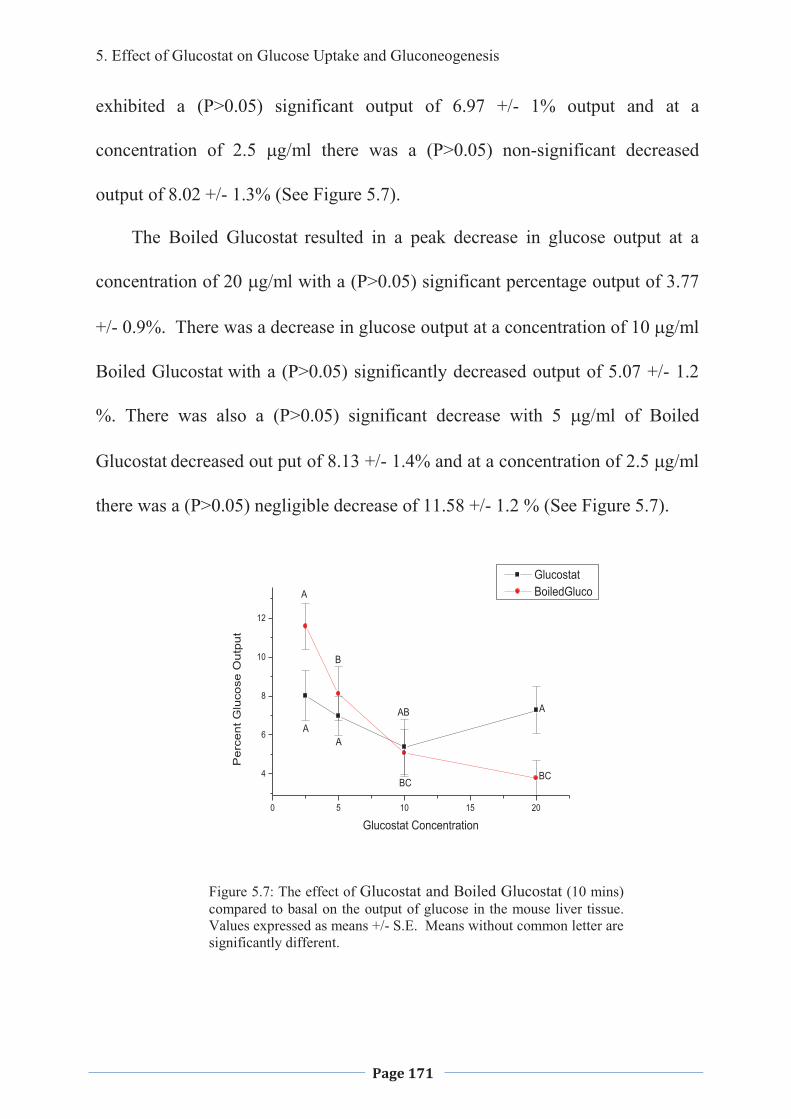
5. Effect of Glucostat on Glucose Uptake and Gluconeogenesis
Page 171
exhibited a (P>0.05) significant output of 6.97 +/- 1% output and at a
concentration of 2.5 �g/ml there was a (P>0.05) non-significant decreased
output of 8.02 +/- 1.3% (See Figure 5.7).
The Boiled Glucostat resulted in a peak decrease in glucose output at a
concentration of 20 �g/ml with a (P>0.05) significant percentage output of 3.77
+/- 0.9%. There was a decrease in glucose output at a concentration of 1 ��g/ml
Boiled Glucostat with a (P>0.05) significantly decreased output of 5.07 +/- 1.2
%. There was also a (P>0.05) significant decrease with 5 �g/ml of Boiled
Glucostat decreased out put of 8.13 +/- 1.4% and at a concentration of 2.5 �g/ml
there was a (P>0.05) negligible decrease of 11.58 +/- 1.2 % (See Figure 5.7).
Figure 5.7: The effect of Glucostat and Boiled Glucostat (10 mins) compared to basal on the output of glucose in the mouse liver tissue. Values expressed as means +/- S.E. Means without common letter are significantly different.
0 5 10 15 20
4
6
8
10
12
BCBC
AB
B
A
A
AA
Glucostat BoiledGluco
Per
cent
Glu
cose
Out
put
Glucostat Concentration
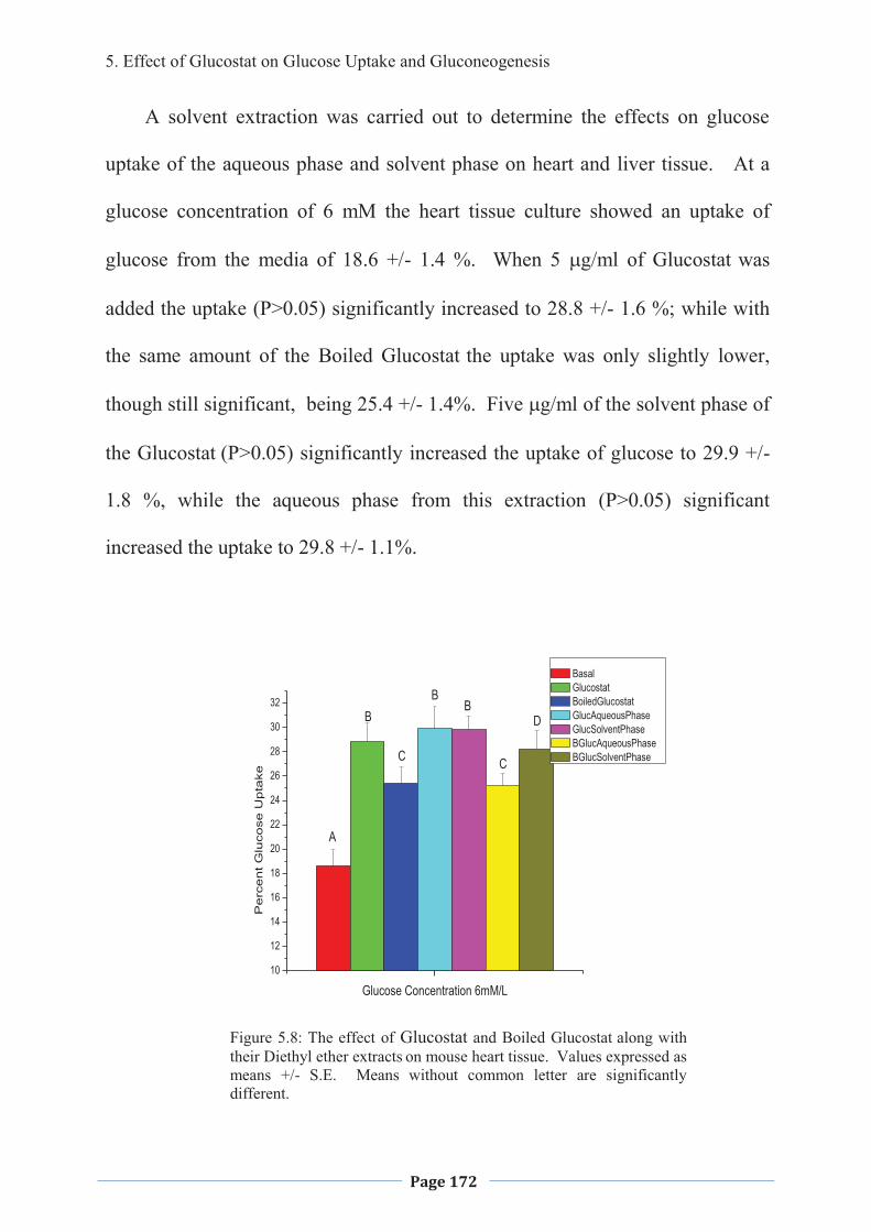
5. Effect of Glucostat on Glucose Uptake and Gluconeogenesis
Page 172
A solvent extraction was carried out to determine the effects on glucose
uptake of the aqueous phase and solvent phase on heart and liver tissue. At a
glucose concentration of 6 mM the heart tissue culture showed an uptake of
glucose from the media of 18.6 +/- 1.4 %. When 5 �g/ml of Glucostat was
added the uptake (P>0.05) significantly increased to 28.8 +/- 1.6 %; while with
the same amount of the Boiled Glucostat the uptake was only slightly lower,
though still significant, being 25.4 +/- 1.4%. Five �g/ml of the solvent phase of
the Glucostat (P>0.05) significantly increased the uptake of glucose to 29.9 +/-
1.8 %, while the aqueous phase from this extraction (P>0.05) significant
increased the uptake to 29.8 +/- 1.1%.
10
12
14
16
18
20
22
24
26
28
30
32
Pe
rce
nt G
luco
se U
pta
ke
Glucose Concentration 6mM/L
Basal Glucostat BoiledGlucostat GlucAqueousPhase GlucSolventPhase BGlucAqueousPhase BGlucSolventPhase
A
BB
B
CC
D
Figure 5.8: The effect of Glucostat and Boiled Glucostat along with their Diethyl ether extracts on mouse heart tissue. Values expressed as means +/- S.E. Means without common letter are significantly different.
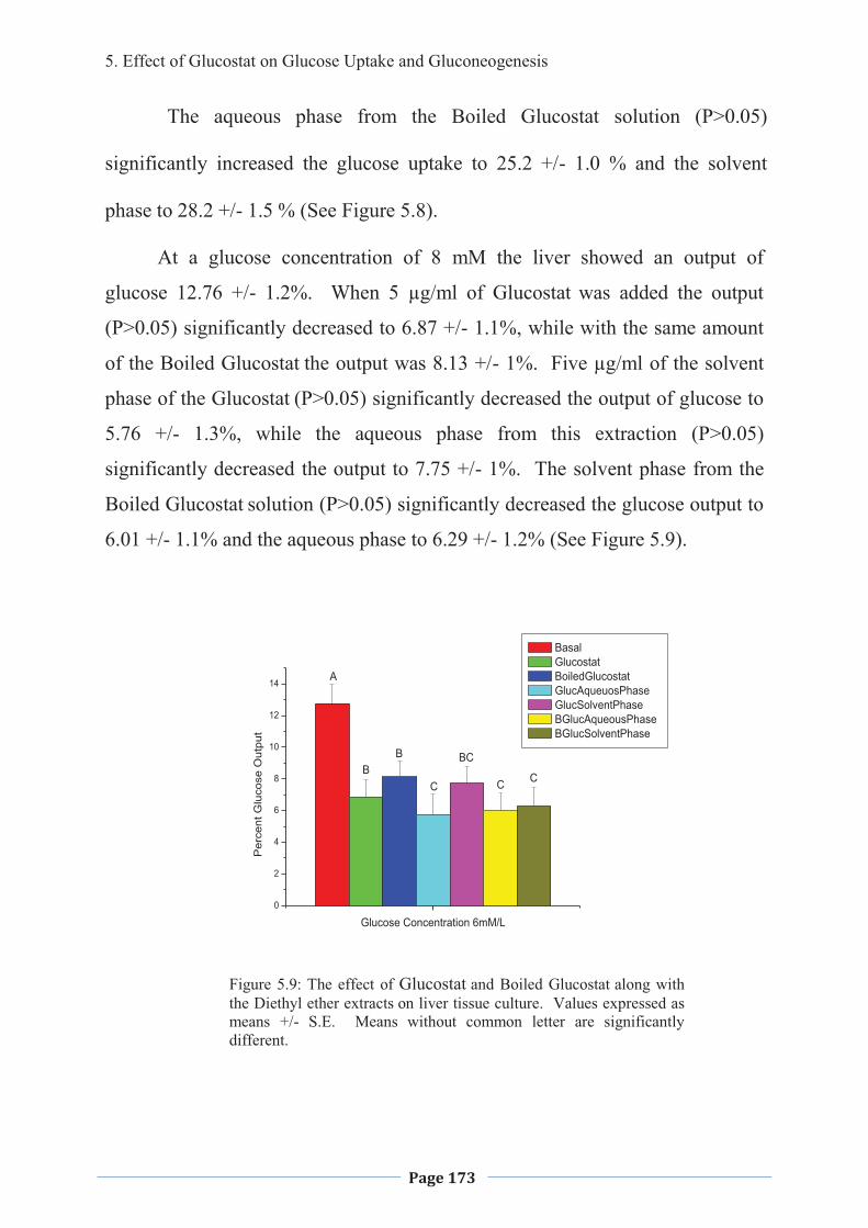
5. Effect of Glucostat on Glucose Uptake and Gluconeogenesis
Page 173
The aqueous phase from the Boiled Glucostat solution (P>0.05)
significantly increased the glucose uptake to 25.2 +/- 1.0 % and the solvent
phase to 28.2 +/- 1.5 % (See Figure 5.8).
At a glucose concentration of 8 mM the liver showed an output of
glucose 12.76 +/- 1.2%. When 5 µg/ml of Glucostat was added the output
(P>0.05) significantly decreased to 6.87 +/- 1.1%, while with the same amount
of the Boiled Glucostat the output was 8.13 +/- 1%. Five µg/ml of the solvent
phase of the Glucostat (P>0.05) significantly decreased the output of glucose to
5.76 +/- 1.3%, while the aqueous phase from this extraction (P>0.05)
significantly decreased the output to 7.75 +/- 1%. The solvent phase from the
Boiled Glucostat solution (P>0.05) significantly decreased the glucose output to
6.01 +/- 1.1% and the aqueous phase to 6.29 +/- 1.2% (See Figure 5.9).
0
2
4
6
8
10
12
14
Per
cent
Glu
cose
Out
put
Glucose Concentration 6mM/L
Basal Glucostat BoiledGlucostat GlucAqueuosPhase GlucSolventPhase BGlucAqueousPhase BGlucSolventPhase
A
BB
C
BC
CC
Figure 5.9: The effect of Glucostat and Boiled Glucostat along with the Diethyl ether extracts on liver tissue culture. Values expressed as means +/- S.E. Means without common letter are significantly different.
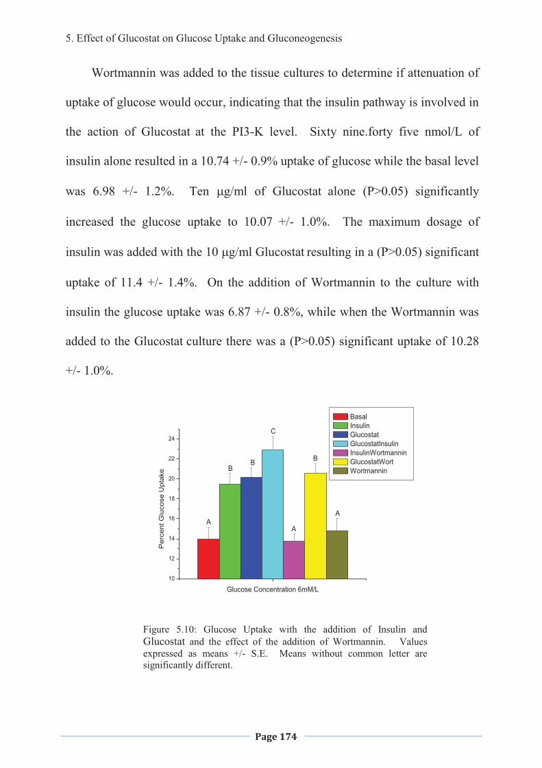
5. Effect of Glucostat on Glucose Uptake and Gluconeogenesis
Page 174
Wortmannin was added to the tissue cultures to determine if attenuation of
uptake of glucose would occur, indicating that the insulin pathway is involved in
the action of Glucostat at the PI3-K level. Sixty nine.forty five nmol/L of
insulin alone resulted in a 10.74 +/- 0.9% uptake of glucose while the basal level
was 6.98 +/- 1.2%. Ten �g/ml of Glucostat alone (P>0.05) significantly
increased the glucose uptake to 10.07 +/- 1.0%. The maximum dosage of
insulin was added with the 10 �g/ml Glucostat resulting in a (P>0.05) significant
uptake of 11.4 +/- 1.4%. On the addition of Wortmannin to the culture with
insulin the glucose uptake was 6.87 +/- 0.8%, while when the Wortmannin was
added to the Glucostat culture there was a (P>0.05) significant uptake of 10.28
+/- 1.0%.
10
12
14
16
18
20
22
24
Per
cent
Glu
cose
Upt
ake
Glucose Concentration 6mM/L
Basal Insulin Glucostat GlucostatInsulin InsulinWortmannin GlucostatWort Wortmannin
AA
A
BB
B
C
Figure 5.10: Glucose Uptake with the addition of Insulin and Glucostat and the effect of the addition of Wortmannin. Values expressed as means +/- S.E. Means without common letter are significantly different.
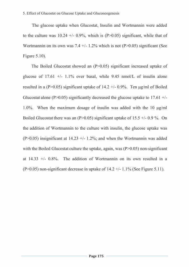
5. Effect of Glucostat on Glucose Uptake and Gluconeogenesis
Page 175
The glucose uptake when Glucostat, Insulin and Wortmannin were added
to the culture was 10.24 +/- 0.9%, which is (P>0.05) significant, while that of
Wortmannin on its own was 7.4 +/- 1.2% which is not (P>0.05) significant (See
Figure 5.10).
The Boiled Glucostat showed an (P>0.05) significant increased uptake of
glucose of 17.61 +/- 1.1% over basal, while 9.45 nmol/L of insulin alone
resulted in a (P>0.05) significant uptake of 14.2 +/- 0.9%. Ten �g/ml of Boiled
Glucostat alone (P>0.05) significantly decreased the glucose uptake to 17.61 +/-
1.0%. When the maximum dosage of insulin was added with the 10 µg/ml
Boiled Glucostat there was an (P>0.05) significant uptake of 15.5 +/- 0.9 %. On
the addition of Wortmannin to the culture with insulin, the glucose uptake was
(P>0.05) insignificant at 14.23 +/- 1.2%; and when the Wortmannin was added
with the Boiled Glucostat culture the uptake, again, was (P>0.05) non-significant
at 14.33 +/- 0.8%. The addition of Wortmannin on its own resulted in a
(P>0.05) non-significant decrease in uptake of 14.2 +/- 1.1% (See Figure 5.11).
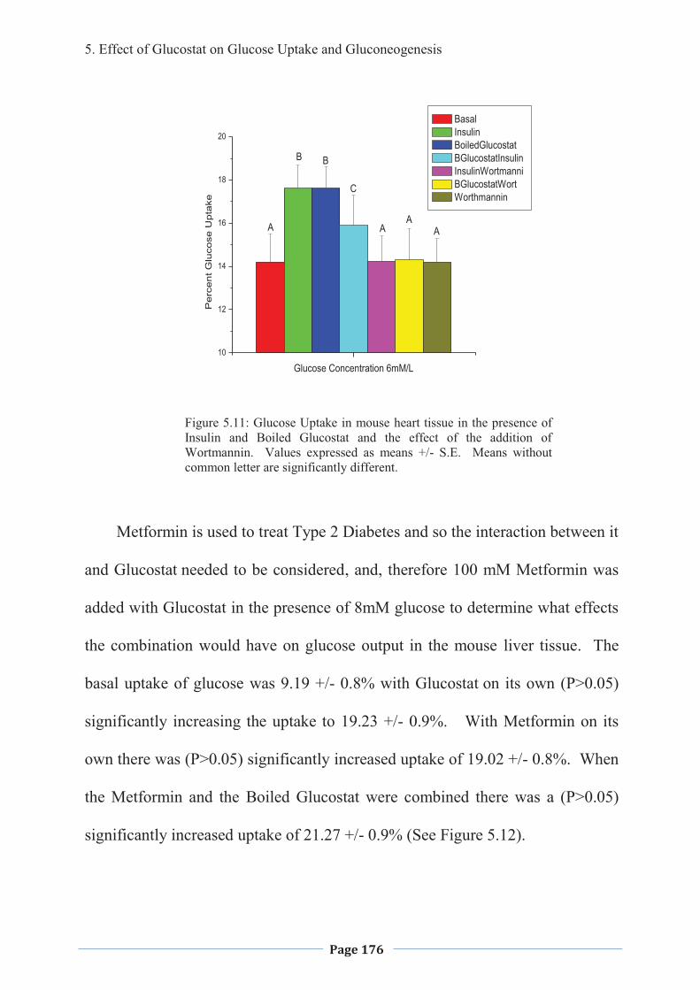
5. Effect of Glucostat on Glucose Uptake and Gluconeogenesis
Page 176
10
12
14
16
18
20
Pe
rce
nt G
luco
se U
pta
ke
Glucose Concentration 6mM/L
Basal Insulin BoiledGlucostat BGlucostatInsulin InsulinWortmanni BGlucostatWort Worthmannin
A AA
A
BB
C
Figure 5.11: Glucose Uptake in mouse heart tissue in the presence of Insulin and Boiled Glucostat and the effect of the addition of Wortmannin. Values expressed as means +/- S.E. Means without common letter are significantly different.
Metformin is used to treat Type 2 Diabetes and so the interaction between it
and Glucostat needed to be considered, and, therefore 100 mM Metformin was
added with Glucostat in the presence of 8mM glucose to determine what effects
the combination would have on glucose output in the mouse liver tissue. The
basal uptake of glucose was 9.19 +/- 0.8% with Glucostat on its own (P>0.05)
significantly increasing the uptake to 19.23 +/- 0.9%. With Metformin on its
own there was (P>0.05) significantly increased uptake of 19.02 +/- 0.8%. When
the Metformin and the Boiled Glucostat were combined there was a (P>0.05)
significantly increased uptake of 21.27 +/- 0.9% (See Figure 5.12).
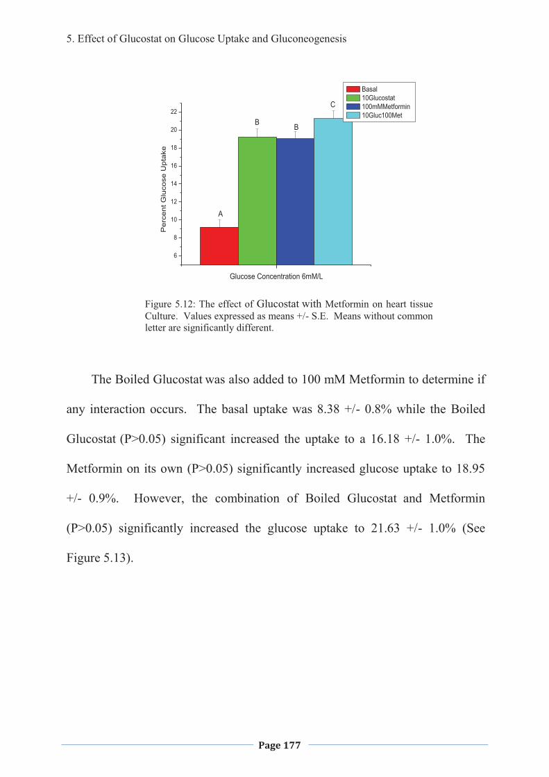
5. Effect of Glucostat on Glucose Uptake and Gluconeogenesis
Page 177
6
8
10
12
14
16
18
20
22
Per
cent
Glu
cose
Upt
ake
Glucose Concentration 6mM/L
Basal 10Glucostat 100mMMetformin 10Gluc100Met
A
BB
C
Figure 5.12: The effect of Glucostat with Metformin on heart tissue Culture. Values expressed as means +/- S.E. Means without common letter are significantly different.
The Boiled Glucostat was also added to 100 mM Metformin to determine if
any interaction occurs. The basal uptake was 8.38 +/- 0.8% while the Boiled
Glucostat (P>0.05) significant increased the uptake to a 16.18 +/- 1.0%. The
Metformin on its own (P>0.05) significantly increased glucose uptake to 18.95
+/- 0.9%. However, the combination of Boiled Glucostat and Metformin
(P>0.05) significantly increased the glucose uptake to 21.63 +/- 1.0% (See
Figure 5.13).
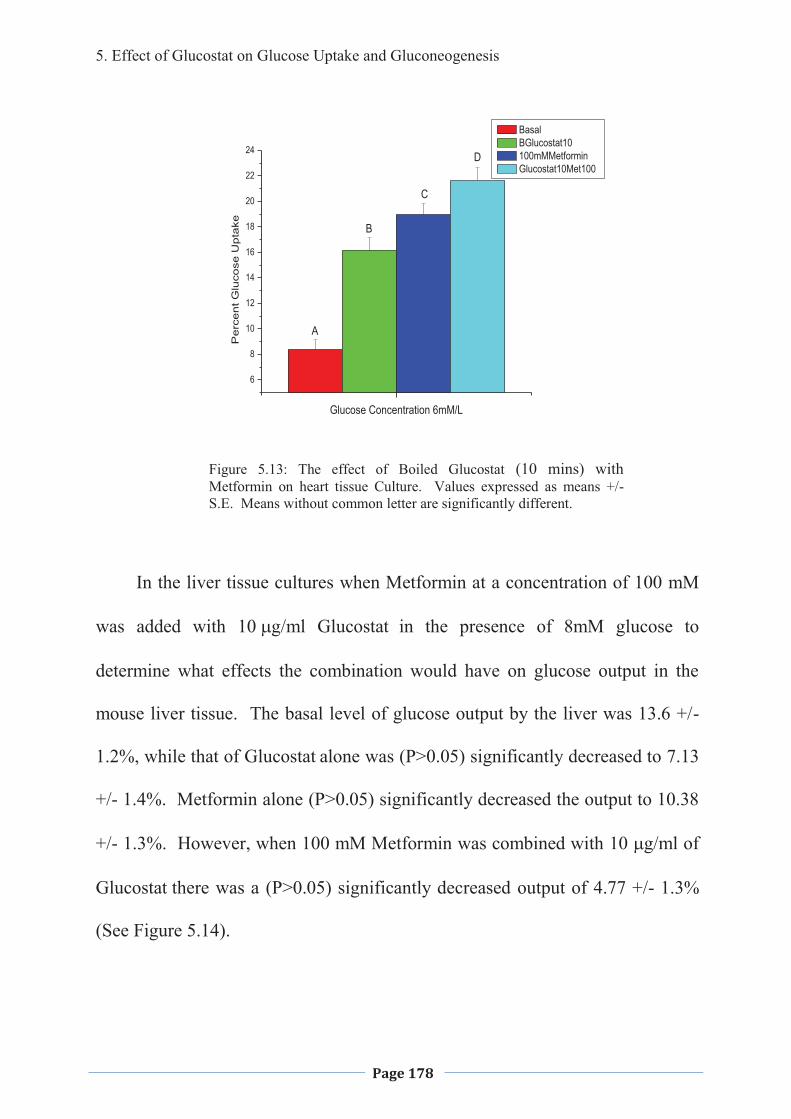
5. Effect of Glucostat on Glucose Uptake and Gluconeogenesis
Page 178
6
8
10
12
14
16
18
20
22
24
Pe
rce
nt G
luco
se U
pta
ke
Glucose Concentration 6mM/L
Basal BGlucostat10 100mMMetformin Glucostat10Met100
A
B
C
D
Figure 5.13: The effect of Boiled Glucostat (10 mins) with Metformin on heart tissue Culture. Values expressed as means +/- S.E. Means without common letter are significantly different.
In the liver tissue cultures when Metformin at a concentration of 100 mM
was added with � ��g/ml Glucostat in the presence of 8mM glucose to
determine what effects the combination would have on glucose output in the
mouse liver tissue. The basal level of glucose output by the liver was 13.6 +/-
1.2%, while that of Glucostat alone was (P>0.05) significantly decreased to 7.13
+/- 1.4%. Metformin alone (P>0.05) significantly decreased the output to 10.38
+/- 1.3%. However, when 100 mM Metformin was combined with 10 �g/ml of
Glucostat there was a (P>0.05) significantly decreased output of 4.77 +/- 1.3%
(See Figure 5.14).
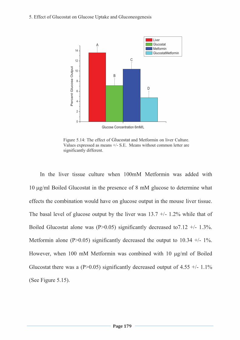
5. Effect of Glucostat on Glucose Uptake and Gluconeogenesis
Page 179
0
2
4
6
8
10
12
14
Per
cent
Glu
cose
Out
put
Glucose Concentration 6mM/L
Liver Glucostat Metformin GlucostatMetformin
A
B
C
D
Figure 5.14: The effect of Glucostat and Metformin on liver Culture. Values expressed as means +/- S.E. Means without common letter are significantly different.
In the liver tissue culture when 100mM Metformin was added with
� ��g/ml Boiled Glucostat in the presence of 8 mM glucose to determine what
effects the combination would have on glucose output in the mouse liver tissue.
The basal level of glucose output by the liver was 13.7 +/- 1.2% while that of
Boiled Glucostat alone was (P>0.05) significantly decreased to7.12 +/- 1.3%.
Metformin alone (P>0.05) significantly decreased the output to 10.34 +/- 1%.
However, when 100 mM Metformin was combined with 10 �g/ml of Boiled
Glucostat there was a (P>0.05) significantly decreased output of 4.55 +/- 1.1%
(See Figure 5.15).
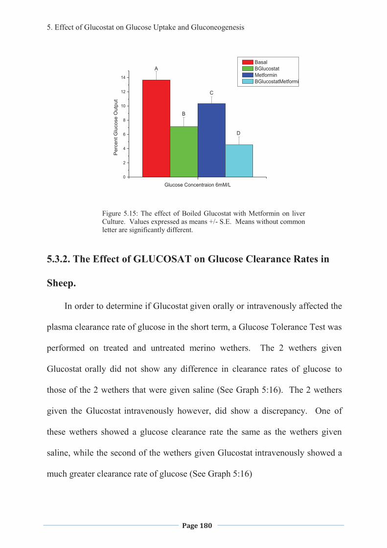
5. Effect of Glucostat on Glucose Uptake and Gluconeogenesis
Page 180
0
2
4
6
8
10
12
14
Per
cent
Glu
cose
Out
put
Glucose Concentraion 6mM/L
Basal BGlucostat Metformin BGlucostatMetformi
A
B
C
D
Figure 5.15: The effect of Boiled Glucostat with Metformin on liver Culture. Values expressed as means +/- S.E. Means without common letter are significantly different.
5.3.2. The Effect of GLUCOSAT on Glucose Clearance Rates in
Sheep.
In order to determine if Glucostat given orally or intravenously affected the
plasma clearance rate of glucose in the short term, a Glucose Tolerance Test was
performed on treated and untreated merino wethers. The 2 wethers given
Glucostat orally did not show any difference in clearance rates of glucose to
those of the 2 wethers that were given saline (See Graph 5:16). The 2 wethers
given the Glucostat intravenously however, did show a discrepancy. One of
these wethers showed a glucose clearance rate the same as the wethers given
saline, while the second of the wethers given Glucostat intravenously showed a
much greater clearance rate of glucose (See Graph 5:16)
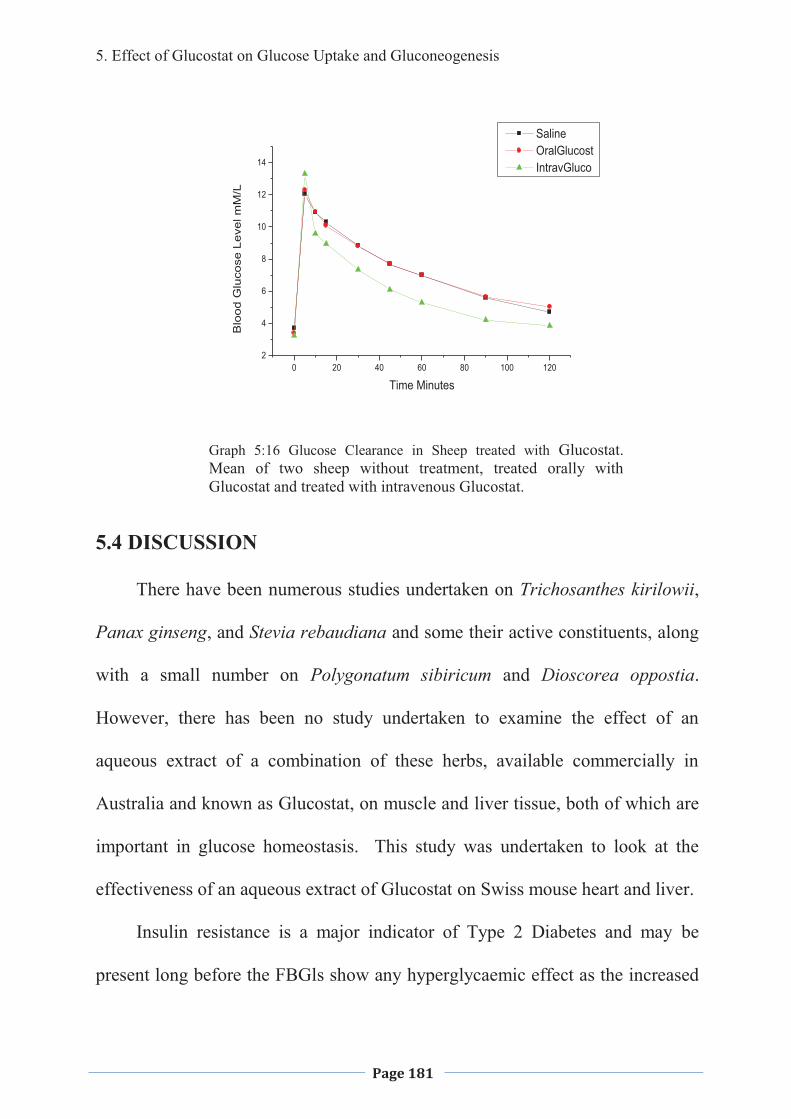
5. Effect of Glucostat on Glucose Uptake and Gluconeogenesis
Page 181
Graph 5:16 Glucose Clearance in Sheep treated with Glucostat. Mean of two sheep without treatment, treated orally with Glucostat and treated with intravenous Glucostat.
5.4 DISCUSSION
There have been numerous studies undertaken on Trichosanthes kirilowii,
Panax ginseng, and Stevia rebaudiana and some their active constituents, along
with a small number on Polygonatum sibiricum and Dioscorea oppostia.
However, there has been no study undertaken to examine the effect of an
aqueous extract of a combination of these herbs, available commercially in
Australia and known as Glucostat, on muscle and liver tissue, both of which are
important in glucose homeostasis. This study was undertaken to look at the
effectiveness of an aqueous extract of Glucostat on Swiss mouse heart and liver.
Insulin resistance is a major indicator of Type 2 Diabetes and may be
present long before the FBGls show any hyperglycaemic effect as the increased
0 20 40 60 80 100 1202
4
6
8
10
12
14
Saline OralGlucost IntravGluco
Blo
od G
luco
se L
evel
mM
/L
Time Minutes
5. Effect of Glucostat on Glucose Uptake and Gluconeogenesis
Page 182
insulin output will often counteract this problem. Insulin resistance is due to the
impairment of the insulin signalling cascades, glucose transport and
translocation of GLUT 4 in particular (Kellerer et. al., 1999). Gluconeogenesis
also plays a major role in Type 2 Diabetes because it becomes uncontrolled and
is not switched off when normal FBGLs are reached (DeFronzo, 1997). The
current management of diabetes looks at the insulin action and mimicking its
action in order to decrease BGLs, both by increasing the uptake of glucose by
tissues throughout the body and also decreasing gluconeogenesis. Thus, this
study examined the effect of Glucostat in increasing glucose uptake by mouse
heart muscle and also on liver tissue to decrease gluconeogenesis.
This study showed that there was a significant increased uptake of glucose
into heart tissue culture and a decrease in glucose output from the liver tissue
when 5 �g/ml and 10 �g/ml of Glucostat was added to the culture and that the
increase was equivalent to that of 100 mM Metformin. However, it was noted
that with increased concentrations of the Glucostat there was a slight decrease in
uptake and increase in output which may have been due to the toxicity of the
extract at higher concentrations as a crude extract was being used.
There was a decrease in the glucose uptake by the heart muscle and
increased output by the liver tissue when using the boiled aqueous extract at
lower concentrations; however, at 10 �g/ml it was approximately the same as
that of the Glucostat and at 20 �g/ml it resulted in a higher uptake by the heart
5. Effect of Glucostat on Glucose Uptake and Gluconeogenesis
Page 183
tissue and a decreased output of glucose by the liver tissue. This suggests that
there are at least two, but may be more active ingredients, involved in these
results. It appeared that heat deactivated one or more of the active ingredients;
however, there were also active ingredients that were not affected by boiling for
10 minutes. The active ingredient/s that were not affected by heat were only
effective at higher concentrations, while the whole Glucostat appeared to be
effective at lower concentrations suggesting that the active ingredient/s were
affected by the boiling process.
A diethyl ether extraction was carried out to look at what type of chemicals
might be involved in the increased uptake of glucose by the heart muscle tissue.
The study showed that there was a slightly higher uptake of glucose in the heart
tissue with both the aqueous phase and the solvent phase of the extract over that
of the whole Glucostat. The glucose output from the liver was less with the
aqueous phase of the extract then that of the solvent phase which was
approximately the same as the whole Glucostat. The aqueous phase of the
Boiled Glucostat showed approximately the same increase in uptake of glucose
by the heart tissue, however, a slightly increased uptake when the solvent phase
was used. In the liver the glucose output was approximately the same for
aqueous phase and solvent phase of the extract, and both produced a lower
output then the pure Boiled Glucostat, indicating that there are a number of
different active ingredients within the combination of herbs. Further studies
would need to be carried out to isolate the compound/s that are bringing about
5. Effect of Glucostat on Glucose Uptake and Gluconeogenesis
Page 184
the increase in glucose uptake by the mouse heart tissue and decreased output by
the liver tissue in the both the unBoiled Glucostat and Boiled Glucostat.
The differences in glucose uptake by the mouse heart tissue and deceased
output by the liver over basal, at different concentrations of glucose indicates
that Glucostat has little or no effect when the glucose level is within normal
range, but increases the uptake and decreases the output when glucose levels are
high. This indicates that the taking of Glucostat will not lead to hypoglycaemia,
coma and possible death. Very high levels of glucose were not tested as it was
out of the scope of this study and therefore the effect on higher glucose levels
needs further studies to determine the Glucostat’s effectiveness at these
concentrations.
A preliminary study looking at the mechanism of action, a maximum dose
response of insulin (69.45 nmol/L) and also Wortmannin, which blocks the
insulin pathway at the P13-K level by inhibiting P13-kinase (Hausdorf et al,
1999) was added to the Glucostat and glucose uptake measured. The enhanced
uptake when Glucostat and insulin are combined indicates that the Glucostat
may be acting via an insulin-mediated peripheral glucose pathway. However, it
was noted that the addition of Wortmannin has no effect on the glucose uptake,
suggesting that the Glucostat is entering the insulin pathway below the P13-
kinase level. Insulin and Boiled Glucostat resulted in approximately the same
glucose uptake by the heart muscle, however, when insulin was added to the
Boiled Glucosat there was an inhibitory effect, suggesting an antagonistic effect.
5. Effect of Glucostat on Glucose Uptake and Gluconeogenesis
Page 185
When the Wortmannin was added to the Boiled Glucostat it was noted that the
uptake is inhibited, suggesting that the active constituents of the Boiled
Glucostat entered the insulin pathway above the PI3-kinase level.
Today many patients who have Type 2 Diabetes are prescribed Metformin
in the early stages of the disease progression and therefore, the interaction
between Glucostat and Metformin was investigated. On their own the Glucostat
and Metformin resulted in approximately the same increased glucose uptake by
the heart tissue and decreased glucose output by the liver. However, when
Glucostat and Metformin were added to the heart or liver tissue cultures together
there was an increased uptake and a decreased output of glucose compared to
either of them alone. On their own the Boiled Glucostat resulted in a decreased
uptake compared to the Metformin, however, in the liver the Boiled Glucostat
resulted in a greater decrease of glucose output than the Metformin. However,
when Glucostat and Metformin were added to the heart or liver tissue cultures
together there was an increased uptake and a decreased output of glucose
compared to either of them alone.
The single sheep experiment showed that Glucostat taken orally did not
have any effect on glucose clearance compared to the treatment with saline.
Glucostat given intravenously, however, showed a greater clearance rate than
those treated with saline. Further testing over a longer period of time needs to
be performed to determine the effect of intravenous injection of Glucostat on
glucose clearance. The Health World Limited Technical Data Sheet for
5. Effect of Glucostat on Glucose Uptake and Gluconeogenesis
Page 186
Glucosat (2001) suggests that there is an increase in glucose clearance in
humans; however, this could not be confirmed in sheep.
5.5 CONCLUSIONS
An aqueous extract of Glucostat increases the uptake of glucose in mice
heart tissue culture and decrease gluconeogenesis from the mouse liver in a
dose-dependent manner��with the optimum concentration being�� �µg/ml. It
also may be slightly toxic at higher levels, with a decrease in glucose uptake by
heart muscle tissue culture and increased gluconeogenesis in liver tissue culture
over that of the optimum concentration. The toxicity may be due to the crude
extract that was used or that some active ingredient comes into effect at the
higher concentrations so there could be seen, both this toxic effect to the heart
and liver tissue as well as an increased uptake of glucose. Boiling of the
Glucostat decreases its ability to increase the uptake of glucose in the mouse
heart tissue or decrease the glucose output in liver tissue culture at lower
concentrations; however, at higher concentrations it increases the uptake and
output over that of the unBoiled Glucostat. The diethyl ether solvent extract
showed that both the aqueous phases and solvent phases of both the Glucostat
and Boiled Glucostat had active ingredients suggesting that there were a number
of different types of active constituents in the Glucostat. The addition of insulin
and Wortmannin indicates that the Glucostat is acting via the insulin pathway,
with some of its active constituents entering below the PI3K level, while some
5. Effect of Glucostat on Glucose Uptake and Gluconeogenesis
Page 187
of its active constituents entered above this level. There was a synergistic
reaction when Glucostat and Boiled Glucostat were added with Metformin to the
heart and liver tissue cultures. This indicates they could possibly be used in
conjunction with each other; however, much more research needs to be carried
out to determine if there are any interactions and increased efficacy and
efficiency.
Glucostat given orally does not appear to have any effect on glucose
clearance in sheep, however, there is an increase in glucose clearance rate when
given intravenously, supporting the in vitro studies.
In summary, Glucostat and Boiled Glucostat both appear to increase glucose
uptake in the mouse heart muscle and decrease gluconeogenesis in the liver.
There appeared to be several active ingredients, however, isolation studies need
to be undertaken to determine exactly what they are. The active ingredients
appeared to be working through the insulin pathway, with some acting above the
PI3K level and others below, and there is a synergistic effect on the addition of
Metformin.
There needs to be further studies carried out to determine the Glucostat’s
active ingredient/s and their mechanism of action.
6. Clinical Trials
Page 188
6. CLINICAL TRIALS
6. Clinical Trials
Page 189
6.1 INTRODUCTION
The current treatment for Type 2 Diabetes is with oral hypoglycaemic
medications, such as Metformin, and at advance stages, insulin. These
medications can all result in various side effects including, abdominal
discomfort, cholestatic jaundice, dermatological reactions, agranulocytosis,
anorexia and metallic taste (Rang & Dale, 1991) and even death, and although
preventative they are not a cure.
Throughout the world many different cultures use different herbal
medicines for the treatment of Type 2 Diabetes and the symptoms associated
with it. This has led to an increasing interest in the use of these traditional
systems in search for a cure. However, little if any scientific evidence is
available to show the efficacy and effectiveness as well as safety of these herbal
medicines.
Ayurveda, a traditional Indian Medical system, advocates a wide range of
herbal medicines either singly or in combination. This system was described in
scripts written in the time of Charaka and Sushrata, during the 6th Century B.C.,
and advocates more than 100 different plant species (Grover & Vats, 2001).
Curcuma longa Linn. has been used in the traditional Ayurevda Medical system
as an anti-inflammatory and antimicrobial and for many other curative
properties (Ammon and Wahl, 1991). The main colouring agent of Turmeric is
curcumin and it has been shown to have a wide range of effects, including
antihepatotoxic (Shishodia, et. al., 2005), anti-inflammatory (Srimal, 1997),
6. Clinical Trials
Page 190
hypocholesterolemic (SubbaRao et. al., 1970; Patil & Srinivasan, 1971),
choleretic and hydrocholagocic (Ramprasad & Sirsi, 1956), hypoglycaemic
(Arun & Nalini, 2002), antioxidant (Quiles et. al., 1998; Asai et. al., 1999;
Bengmark, 2006), anti-cancer (Huang et. al., 1994; Kuo et. al., 1996),
antimicrobial (Gupta and Ravishankar, 2005), inhibits platelet-derived growth
factor and has wound healing abilities (Kundu, et. al., 2005).
Traditional Chinese Medicine, based on Taoist philosophy, proposes that
the human body contains yin, yang and Five Elements (metal, wood, water, fire
and earth), and that a perfect balance amongst all of these within in the body is
required for perfect health (Li et. al., 2004). If there is a imbalance amongst
these from cold, heat, emotions or other factors then disease results. Qi (air) and
blood serve as a communication channel between Yin and Yang and among the 5
elements. The primary aim of any Traditional Chinese Medicine is to restore the
balance and replenish Qi or blood (Tang, 2006). Obesity and Type 2 Diabetes
are a result of Yin deficiency with dryness-heat and needs to be treated by
replenishing Yin (fluid) and evacuating fire (heat) from the body and is
attributed to yin-deficiency diathesis, improper diet, emotional disorders,
overstrain and excessive sexual activities (Li et. al., 2004). Traditional Chinese
Medicine is holistic with an emphasis on whole body care and then removal of
the symptoms. Compound recipes are often used in Traditional Chinese
Medicine, in that every herb provides a specialist function, as well as, form an
integrated function for the treatment of disease, with the suggestion that a single
6. Clinical Trials
Page 191
herb contains many ingredients but they cannot play the role of many herbs
combined together (Li et. al., 2004). Each of the herbs found within Glucostat
act according to Traditional Chinese Medicine by either retoring Qi, or
replenishing Yin or both, for example, Ginseng is used to replenish Qi and
restore Yin.
Glucostat is a combination of the Chinese herbs, including Trichosanthes
kirilowii (1900 mg), Polygonatum sibiricum (714.5 mg), Dioscorea oppostia
(286 mg), Panax ginseng (95 mg), and Stevia rebaudiana (47.6 mg) per capsule.
Apart from some evidence for the effectiveness of single herbs there is no
evidence of the efficacy or efficiency of the combination in lowering of BGL in
humans (see Chapter 5 of this thesis for discussion).
There have been several studies with the use of ethanolic extracts of
Turmeric and its active ingredient, curcumin (Nishiyama et. al,2005) in the
management of Type 2 Diabetes in mice (Arun & Nalini, 2002; Kuroda et. al.,
2005); however, there have been no studies looking at the hypoglycaemic effect
in humans with the use of an aqueous solution. There have also been several
studies on Trichosanthes kirilowii, Panax ginseng, and Stevia rebaudiana, along
with a few on Polygonatum sibiricum and Dioscorea oppostia; however, there
have been none on the anti-diabetic effect of the combination as found in
Glucostat. Therefore, this study looks at the effects of 500 mg powdered
Turmeric root dissolved in hot milk, taken twice per day and 1 gm Glucostat 3
times per day over a 3 month period in humans with a FBG >5.
6. Clinical Trials
Page 192
6.2. MATERIALS AND METHODS
Plant extraction, human ethics approval, clinical trial and statistical analysis
have been described in Chapter 2 of this thesis.
6.2.2. TURMERIC STUDY
Ten individuals, 3 males and 7 females, who had a FBG greater than 5
participated in the study. The ages ranged from 28-75 and BMI ranged from
within normal to morbid obesity, while exercise ranged from low to fairly high.
Eight had a history of Type 1 and/or Type 2 Diabetes in the Family. Seven
were overweight to obese while 3 were within normal weight limits. Three did
not exercise on a regular basis, while 7 of the patients did varying amounts of
both aerobic and resistance exercise on a regular basis. Nine out of the 10 were
on medication, with 5 taking medications for diabetes, 4 on varying amount of
Metformin and 1 on Insulin. Other medications were for pain relief, anti-
inflammatory which was stopped during the trial period, epilepsy, cholesterol,
blood pressure and depression. Eight out of the 10 suffered moderate to severe
arthritis, while only 3 had any history of cardiovascular disease, usually
associated with high blood pressure and cholesterol. Fasting blood glucose
levels ranged from 5.4 to 9.2 mM at the commencement of the study. In order
to determine the effect of Turmeric on FBG levels, patients were asked to take
daily readings with their individual hand held monitors and record the results.
6. Clinical Trials
Page 193
6.2.3. GLUCOSTAT TRIAL
Eighteen individuals, 6 males and 12 females, who had a FBG greater than
5 participated in the study. The ages ranged from 28-75 and BMI ranged from
within normal to morbid obesity, while exercise ranged from low to fairly high.
Twelve had a history of Type 1 and/or Type 2 Diabetes in the Family. Fourteen
were overweight to obese while 4 within normal weight limits. Four did not
exercise on a regular basis, while 14 did varying amounts of both aerobic and
resistance exercise on a regular basis. Ten of the 18 patients were also able to
provide HbA1c readings before and after taking the medication. Seventeen out
of the 18 were on medication, with 5 taking medications for diabetes and 4 on
varying amount of Metformin. Other medications were for pain relief, anti-
inflammatory, epilepsy, cholesterol, blood pressure and depression. Twelve out
of the 18 suffered moderate to severe arthritis, while 15 had a history of
cardiovascular disease, usually associated with high blood pressure and
cholesterol. Fasting blood glucose levels commenced at 5.4 and went as high as
11.9 nmol/L. All stated that diet, as prescribed by their doctors/dieticians and
exercise did not change throughout the trial period except when illness was
present and exercise levels would drop for a short period of time.
6.3. RESULTS
6.3.1. TURMERIC TRIAL
Two patients did not respond to the Turmeric as can be seen in the patients
RW and RM (Figures 6.1 and 6.2). Patient RW is a 54 year old female who was
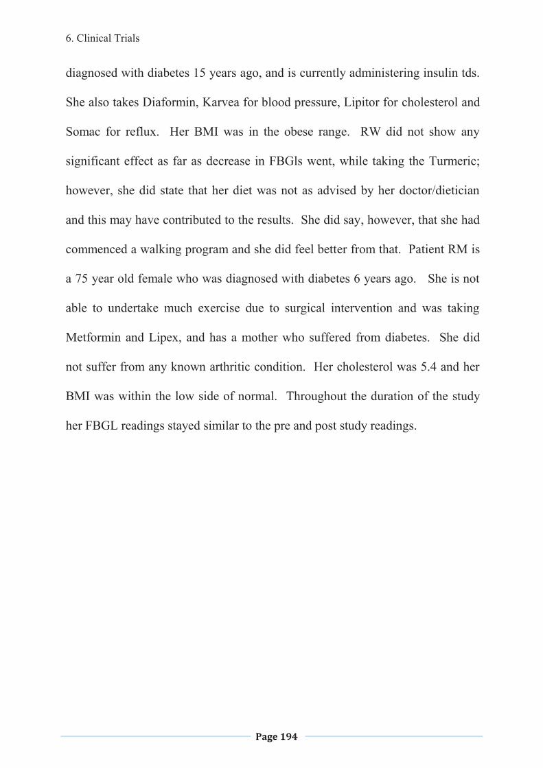
6. Clinical Trials
Page 194
diagnosed with diabetes 15 years ago, and is currently administering insulin tds.
She also takes Diaformin, Karvea for blood pressure, Lipitor for cholesterol and
Somac for reflux. Her BMI was in the obese range. RW did not show any
significant effect as far as decrease in FBGls went, while taking the Turmeric;
however, she did state that her diet was not as advised by her doctor/dietician
and this may have contributed to the results. She did say, however, that she had
commenced a walking program and she did feel better from that. Patient RM is
a 75 year old female who was diagnosed with diabetes 6 years ago. She is not
able to undertake much exercise due to surgical intervention and was taking
Metformin and Lipex, and has a mother who suffered from diabetes. She did
not suffer from any known arthritic condition. Her cholesterol was 5.4 and her
BMI was within the low side of normal. Throughout the duration of the study
her FBGL readings stayed similar to the pre and post study readings.
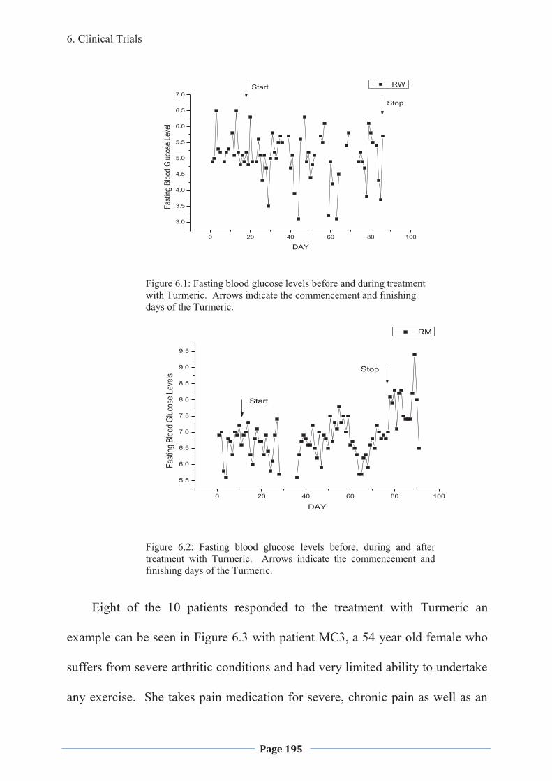
6. Clinical Trials
Page 195
0 20 40 60 80 100
3.0
3.5
4.0
4.5
5.0
5.5
6.0
6.5
7.0
Fasti
ng B
lood
Gluc
ose
Leve
l
DAY
RWStart
Stop
Figure 6.1: Fasting blood glucose levels before and during treatment with Turmeric. Arrows indicate the commencement and finishing days of the Turmeric.
0 20 40 60 80 100
5.5
6.0
6.5
7.0
7.5
8.0
8.5
9.0
9.5
Fasti
ng B
lood
Gluc
ose
Leve
ls
DAY
RM
Start
Stop
Figure 6.2: Fasting blood glucose levels before, during and after treatment with Turmeric. Arrows indicate the commencement and finishing days of the Turmeric.
Eight of the 10 patients responded to the treatment with Turmeric an
example can be seen in Figure 6.3 with patient MC3, a 54 year old female who
suffers from severe arthritic conditions and had very limited ability to undertake
any exercise. She takes pain medication for severe, chronic pain as well as an
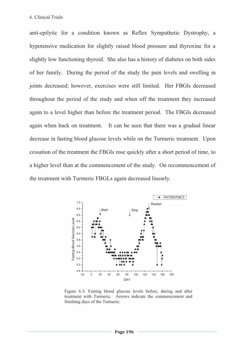
6. Clinical Trials
Page 196
anti-epilytic for a condition known as Reflex Sympathetic Dystrophy, a
hypotensive medication for slightly raised blood pressure and thyroxine for a
slightly low functioning thyroid. She also has a history of diabetes on both sides
of her family. During the period of the study the pain levels and swelling in
joints decreased; however, exercises were still limited. Her FBGls decreased
throughout the period of the study and when off the treatment they increased
again to a level higher than before the treatment period. The FBGls decreased
again when back on treatment. It can be seen that there was a gradual linear
decrease in fasting blood glucose levels while on the Turmeric treatment. Upon
cessation of the treatment the FBGls rose quickly after a short period of time, to
a higher level than at the commencement of the study. On recommencement of
the treatment with Turmeric FBGLs again decreased linearly.
-20 0 20 40 60 80 100 120 140 160 1804.8
5.0
5.2
5.4
5.6
5.8
6.0
6.2
6.4
6.6
6.8
7.0
Fast
ing
Blo
od G
luco
se L
evel
DAY
PATIENTMC3
Start Stop
Restart
Figure 6.3: Fasting blood glucose levels before, during and after treatment with Turmeric. Arrows indicate the commencement and finishing days of the Turmeric.
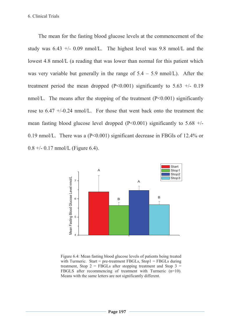
6. Clinical Trials
Page 197
The mean for the fasting blood glucose levels at the commencement of the
study was 6.43 +/- 0.09 nmol/L. The highest level was 9.8 nmol/L and the
lowest 4.8 nmol/L (a reading that was lower than normal for this patient which
was very variable but generally in the range of 5.4 – 5.9 nmol/L). After the
treatment period the mean dropped (P<0.001) significantly to 5.63 +/- 0.19
nmol/L. The means after the stopping of the treatment (P<0.001) significantly
rose to 6.47 +/-0.24 nmol/L. For those that went back onto the treatment the
mean fasting blood glucose level dropped (P<0.001) significantly to 5.68 +/-
0.19 nmol/L. There was a (P<0.001) significant decrease in FBGls of 12.4% or
0.8 +/- 0.17 nmol/L (Figure 6.4).
4
5
6
7
Mea
n Fa
sting
Bloo
d Gl
ucos
e Le
vel n
mol/
L
Start Stop1 Stop2 Stop3
A
A
B B
Figure 6.4: Mean fasting blood glucose levels of patients being treated with Turmeric. Start = pre-treatment FBGLs, Stop1 = FBGLs during treatment, Stop 2 = FBGLs after stopping treatment and Stop 3 = FBGLS after recommencing of treatment with Turmeric (n=10). Means with the same letters are not significantly different.
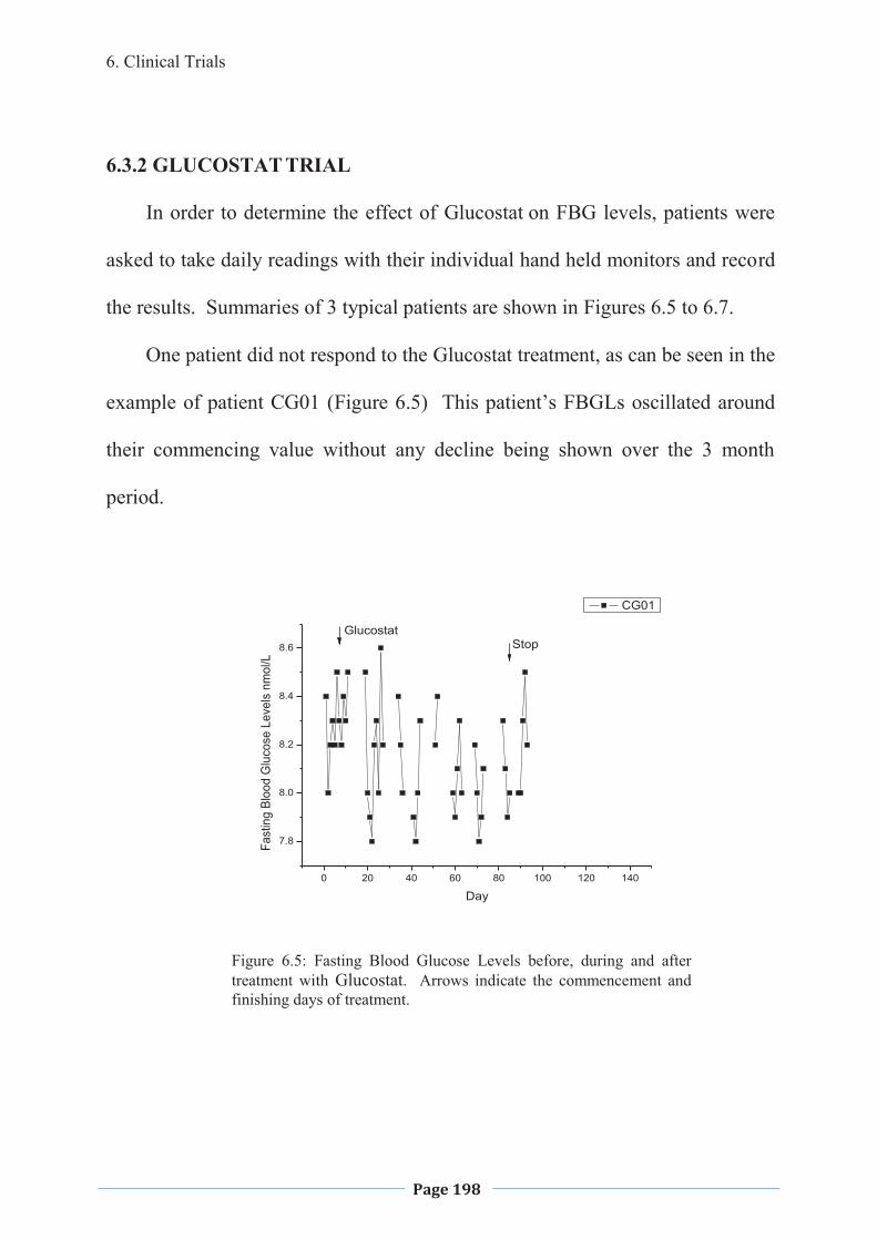
6. Clinical Trials
Page 198
6.3.2 GLUCOSTAT TRIAL
In order to determine the effect of Glucostat on FBG levels, patients were
asked to take daily readings with their individual hand held monitors and record
the results. Summaries of 3 typical patients are shown in Figures 6.5 to 6.7.
One patient did not respond to the Glucostat treatment, as can be seen in the
example of patient CG01 (Figure 6.5) This patient’s FBGLs oscillated around
their commencing value without any decline being shown over the 3 month
period.
0 20 40 60 80 100 120 140
7.8
8.0
8.2
8.4
8.6
Fast
ing
Bloo
d G
luco
se L
evel
s nm
ol/L
Day
CG01
GlucostatStop
Figure 6.5: Fasting Blood Glucose Levels before, during and after treatment with Glucostat. Arrows indicate the commencement and finishing days of treatment.
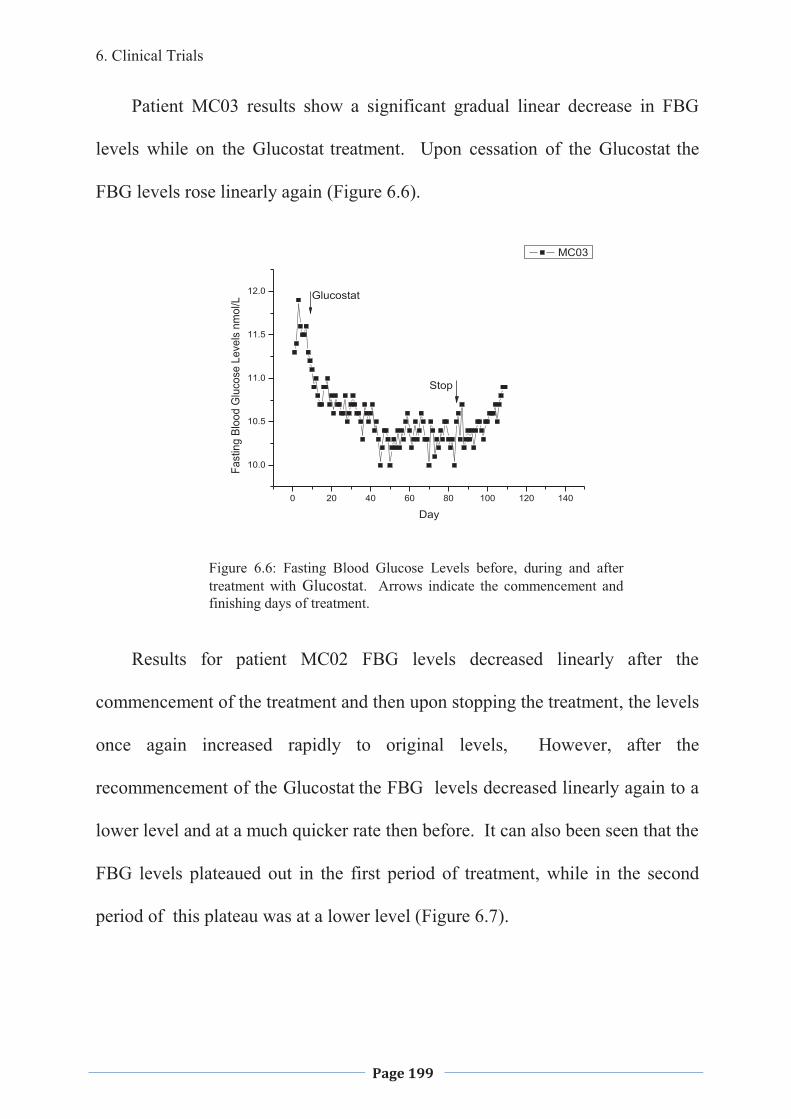
6. Clinical Trials
Page 199
Patient MC03 results show a significant gradual linear decrease in FBG
levels while on the Glucostat treatment. Upon cessation of the Glucostat the
FBG levels rose linearly again (Figure 6.6).
0 20 40 60 80 100 120 140
10.0
10.5
11.0
11.5
12.0
Fast
ing
Bloo
d G
luco
se L
evel
s nm
ol/L
Day
MC03
Glucostat
Stop
Figure 6.6: Fasting Blood Glucose Levels before, during and after treatment with Glucostat. Arrows indicate the commencement and finishing days of treatment.
Results for patient MC02 FBG levels decreased linearly after the
commencement of the treatment and then upon stopping the treatment, the levels
once again increased rapidly to original levels, However, after the
recommencement of the Glucostat the FBG levels decreased linearly again to a
lower level and at a much quicker rate then before. It can also been seen that the
FBG levels plateaued out in the first period of treatment, while in the second
period of this plateau was at a lower level (Figure 6.7).
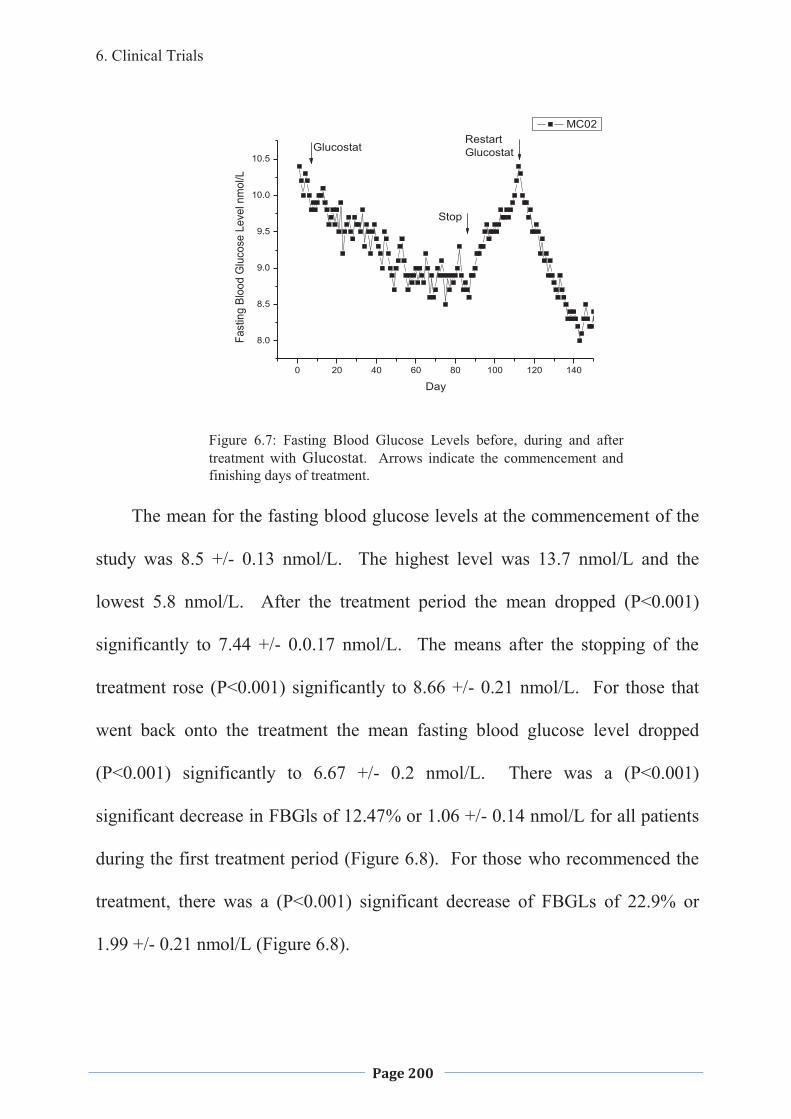
6. Clinical Trials
Page 200
0 20 40 60 80 100 120 140
8.0
8.5
9.0
9.5
10.0
10.5
Fast
ing
Bloo
d G
luco
se L
evel
nm
ol/L
Day
MC02
Glucostat
Stop
RestartGlucostat
Figure 6.7: Fasting Blood Glucose Levels before, during and after treatment with Glucostat. Arrows indicate the commencement and finishing days of treatment.
The mean for the fasting blood glucose levels at the commencement of the
study was 8.5 +/- 0.13 nmol/L. The highest level was 13.7 nmol/L and the
lowest 5.8 nmol/L. After the treatment period the mean dropped (P<0.001)
significantly to 7.44 +/- 0.0.17 nmol/L. The means after the stopping of the
treatment rose (P<0.001) significantly to 8.66 +/- 0.21 nmol/L. For those that
went back onto the treatment the mean fasting blood glucose level dropped
(P<0.001) significantly to 6.67 +/- 0.2 nmol/L. There was a (P<0.001)
significant decrease in FBGls of 12.47% or 1.06 +/- 0.14 nmol/L for all patients
during the first treatment period (Figure 6.8). For those who recommenced the
treatment, there was a (P<0.001) significant decrease of FBGLs of 22.9% or
1.99 +/- 0.21 nmol/L (Figure 6.8).
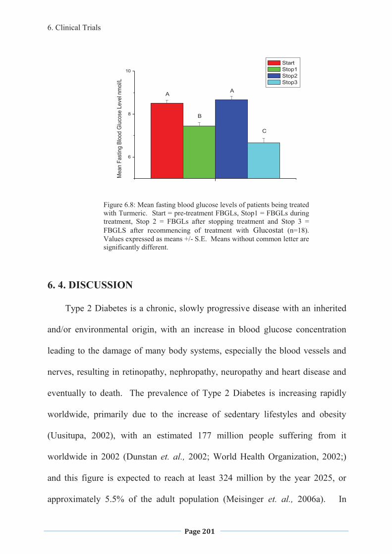
6. Clinical Trials
Page 201
6
8
10
Mea
n Fa
stin
g Bl
ood
Glu
cose
Lev
el n
mol
/L
Start Stop1 Stop2 Stop3
A A
B
C
Figure 6.8: Mean fasting blood glucose levels of patients being treated with Turmeric. Start = pre-treatment FBGLs, Stop1 = FBGLs during treatment, Stop 2 = FBGLs after stopping treatment and Stop 3 = FBGLS after recommencing of treatment with Glucostat (n=18). Values expressed as means +/- S.E. Means without common letter are significantly different.
6. 4. DISCUSSION
Type 2 Diabetes is a chronic, slowly progressive disease with an inherited
and/or environmental origin, with an increase in blood glucose concentration
leading to the damage of many body systems, especially the blood vessels and
nerves, resulting in retinopathy, nephropathy, neuropathy and heart disease and
eventually to death. The prevalence of Type 2 Diabetes is increasing rapidly
worldwide, primarily due to the increase of sedentary lifestyles and obesity
(Uusitupa, 2002), with an estimated 177 million people suffering from it
worldwide in 2002 (Dunstan et. al., 2002; World Health Organization, 2002;)
and this figure is expected to reach at least 324 million by the year 2025, or
approximately 5.5% of the adult population (Meisinger et. al., 2006a). In
6. Clinical Trials
Page 202
Australia alone, there are 854,325 individuals that have been diagnosed with
Type 2 Diabetes and registered with the National Diabetes Register, being
3.97% of the total population (Deed, 2009). It is estimated that another 400,000
who are not registered, are either pre-diabetic or do not realise that they have
diabetes; this being one of the highest recorded prevalence’s in the developed
world. It has also been suggested that the average Australian has about a 1 in 14
chance of developing Type 2 Diabetes during their lifetime, and if impaired
fasting glucose and impaired glucose tolerance are included, than approximately
1 in 4 Australians would be classified as having a glucose uptake disorder
(Dunstan et. al., 2002). The magnitude of the problem is enormous, with
billions of dollars being spent annually by most western nations on the treatment
of this disease and its complications. In developed countries Type 2 Diabetes
claims about 9% of the National Health Budget (World Health Organisation,
2002) and in many countries it may account for 10% or more of the total health
budget (Roglic et. al., 2005).
Today there are several medications used to ameliorate the effects of
diabetes and its complications by controlling blood glucose levels (BGL). These
include oral hyperglycaemic drugs such as the sulfonylureas, biguanides and
thiazolidinediones, and insulin which is used in the end stages of Type 2
Diabetes. Although the effects of the hypoglycemic drugs appear to be
sufficient and BGL are normalised initially, they still tend not to stop the micro-
vascular effects of the disease (Moss et. al., 1994) and their effectiveness
6. Clinical Trials
Page 203
appears to diminish overtime, therefore, an increased amount of a drug, or
combination of drugs, must be taken to have the same effect (Cook et. al.,
2007). Thus the development of new and improved medications for prevention
of Type 2 Diabetes will also assist in the real cost of this disease, as well as
raising the well-being of those predisposed to the disease.
6.4.1. TURMERIC TRIAL
In any study it needs to be determined if the effects seen in vitro will be
carried over to in vivo studies. As Turmeric could be considered safe for human
consumption as it is used widely in cooking throughout the world as flavouring
and colouring it was decided to run an introductory human trial. Patients, who
had an FBGl > 5 were asked too participate in the study. In the Ayurvedic
Medical System Turmeric would traditionally be taken with hot milk (Course
material for Foundations in Ayurvedic Medicine, Metagenics) and therefore the
participants were asked to take the Turmeric with a hot milk drink (tea, coffee,
hot chocolate or hot low fat milk) twice per day.
Fasting blood glucose levels decreased in the patients who suffered from
arthritis and were taking the Turmeric on a regular basis over the trial period.
There was a gradual but (P<0.001) significant decline of 12.4% or 0.8 +/-
0.17nmol/L (Figure 6.4). On cessation of the treatment fasting blood glucose
levels rose quickly and in the majority of cases went back to the same level if
not higher then the original levels. In those patients who recommenced taking
the herbs there was again a (P<0.001) significant decrease in the fasting blood
6. Clinical Trials
Page 204
glucose levels to approximately the same level as when taking the treatment the
first time.
There were 10 individuals who participated in the study and of those who
showed improvement with the treatment of Turmeric, all had arthritis. Arthritis
is a chronic inflammatory condition and now that there is a suggested link
between inflammatory conditions and Type 2 Diabetes (Festa et. al., 2002;
Freeman et. al., 2002; van Exel et. al., 2002), consideration needs to be taken as
to whether the results that were seen were due to the effect of Turmeric acting as
an anti-inflammatory, which has been reported in the literature (Aggarwal and
Sung, 2009). This may act by decreasing pain levels and stiffness in joints, thus,
allowing the patient to undertake more exercise. This in itself would be of value
in decreasing FBGl’s or indirectly by weight loss. It may have also been due to
a direct effect on the uptake of glucose in muscle tissue and the decrease of
gluconeogenesis. All those with arthritis reported that their exercise regime did
not change during the trial period, suggesting a more direct action of the
Turmeric than just allowing greater amounts of exercise. The individual who
was already on Metformin for the management of her Type2 Diabetes exhibited
little, if any, change in her FBGl readings, although, when she stopped taking it
there was a considerable increase over the introductory levels. The individual
who was on insulin injections for her management, showed little difference at all
but she did report that she she did not maintain her diet as set by the
doctor/dietician, often foregoing her treatments and generally did not look after
6. Clinical Trials
Page 205
herself. This individual did report that she was more able to undertake exercise
and increased this a little due to decreased pain and swelling of joints; however,
this still did not prompt her to actually increase her walking or other exercise.
The patient on insulin stated that she felt more able to walk and undertake
exercise after taking the Turmeric and started an exercise program with her
friend; however, she reported often eating inappropriate foods such as chocolate
and sweets which had a massive effect on her blood glucose levels. All patients
with arthritic conditions stated that pain had decreased and one patient who had
psoriasis said that this had decreased; however, they stated their exercise levels
had not changed throughout the trial period.
Since the patients in the introductory clinical trial were asked to take the
Turmeric in hot milk it is important to take into consideration if the hot milk
itself may be influencing blood glucose levels rather then the Turmeric. Pittas
and colleagues (2006) conducted a study looking at the milk intake of women
over an 18 yr period and in particular, analysing the intake of calcium and
Vitamin D. They showed that there was a significant inverse relationship
between total Vitamin D and calcium intake and the risk of developing Type 2
Diabetes; the highest association being when supplements of the dairy products
were taken. This was supported by Liu and colleagues (2006) who showed that
women who consumed low fat dairy products had a decreased chance of
contracting Type 2 diabetes than those who had high fat or no dairy products. It
was suggested that this might have been due to those who consumed a low fat
6. Clinical Trials
Page 206
dairy diet, also generally, had a healthier lifestyle rather than the direct effect on
the Type 2 Diabetes. It was suggested that the decrease may be due to the dairy
products resulting in an increase secretion of the insulinotropic amino acids and
incretin along with the various components of dairy such as calcium, protein and
lactose may increase satiety, reducing the risk of becoming overweight (Liu et.
al., 2006). However, this doesn’t account for the decrease in FBGl’s amongst
the patients trialling the Turmeric as they all reported that their diet didn’t
change. They stated especially that their milk intake didn’t alter except that
some stated they didn’t normally have hot milk but rather cold, so whether
having hot milk affects the absorption of milk components and the secretion of
incretin would need to be investigated further. The reason for taking of the
Turmeric in hot milk was because this is the way traditionally used for taking
Turmeric in the Ayruvedic Medical System. However, how the hot milk aids
the absorption or the effectiveness of the Turmeric or its components is not
understood at this stage and needs further investigation. It was also noted that
one of the patients was lactose intolerant and took the Turmeric with Soya Milk.
This particular individual did show a significant (P<0.05) improvement with the
treatment and therefore the cow’s milk may not play any role at all. Further
studies will need to be carried out to ascertain the best way to take the Turmeric.
Further clinical trials will need to be undertaken to determine the
effectiveness of Turmeric in a range of people, with and without chronic
inflammatory diseases, as well as those who have a higher commencing FBGl as
6. Clinical Trials
Page 207
it is noted, that those participating in this trial started with only slightly higher
then normal levels.
6.4.2. GLUCOSTAT TRIAL
Fasting blood glucose levels decreased in the majority of patients who were
taking the Traditional Chinese Herbs on a regular basis over the trial period.
There was a gradual but (P<0.001) significant decline of 12.47 or 1.06 +/- 0.14
nmol/L during the first treatment period followed by a plateau in those patients
who responded to the treatment. On cessation of the treatment, FBGLs rose
quickly and in the majority of cases went back to the same level if not higher
than the original levels. In those patients who recommenced taking the herbs,
there was again a (P<0.001) significant decrease in the fasting blood glucose
levels. However, it was noted that the decline was much steeper this time and
the plateau occurred at a generally lower level then in the first period of
treatment with a significant (P<0.001) decrease of FBGLs of 22.9% or 1.99 +/-
0.21 nmol/L. Research that has been undertaken on the individual herbs of
Trichosanthes kirilowii (Ng et. al., 1985; Hikino et. al., 1989; Lu et. al., 2001;
Xu et. al., 2001), Panax ginseng (Suzuki, 1989; Sotoniemi et. al., 1995; Vuksan
et. al., 2001) and Stevia rebaudiana (Curie, 1986; White, 1994; Jeppesen et. al.,
2000), Dioscorea opposita (Hsu, J., et. al., 2007), Polygonatum sibiricum
(Bensky & Gamble 1993) along with the chromium (Roginiski & Mertz, 1969;
Anderson, et. al., 1997; Anderson, 1998;), have all been shown to have a blood
6. Clinical Trials
Page 208
glucose lowering effect even though the mechanisms have not been elucidated,
as yet.
6.5. CONCLUSIONS
6.5.1. TURMERIC TRIAL
The clinical trial suggests that Turmeric will aid in the decreasing of
FBGl’s of individuals who have a moderately high FBGL without having any
effect on those with a low to normal level. This is particular in the case of those
who have an arthritic condition, whether this is due to increasing the ability to
exercise or a direct effect needs to be investigated further. There appeared to be
little effect on those who were already on Metformin (n=3) or insulin (n=1);
however, this being too small a number of participants to say this accurately.
The effects may be due to the anti-inflammatory effect, allowing the individual
to increase exercise; however, as the individuals in this study stated that exercise
did not change during the study period there may also be a direct effect. Further
controlled clinical trials would need to be carried out to determine if there is an
increase in exercise or not.
There needs to be further studies carried out to determine Turmeric’s active
ingredient/s and the mechanism/s of action; along with Turmeric’s effect on
humans who have higher blood sugar levels and those who are already on
different medications for the treatment of diabetes.
6. Clinical Trials
Page 209
6.5.2. GLUCOSTAT TRIAL
This combination of Traditional Chinese Herbs and chromium appear to
lower fasting blood glucose levels in the majority of patients, although the
mechanism/s of action is not known at this stage and may include a combination
of factors. These may include increase in insulin output, increased insulin
responsiveness and glucose uptake. The relative importance of the individual
herbs within the combination is also unclear at this stage and further studies
need to be undertaken to determine this.
There needs to be further studies carried out to determine the active
ingredient/s and the mechanism/s of action. Also further studies are needed to
determine Glucostat’s effect on humans who have higher blood sugar levels and
are already on different medications for the treatment of diabetes. Only four
patients were already taking Metformin in this study; however, this number is
too low to determine the effect of the combination in the management of Type 2
Diabetes.
7. General Discussion
Page 210
7. GENERAL DISCUSSION
7. General Discussion
Page 211
7.1. INTODUCTION
The prevalence of Type 2 Diabetes is in epidemic portions worldwide,
primarily due to the increase of sedentary lifestyles and obesity (Uusitupa,
2002), with an estimated 177 million people suffering from the disease
worldwide in 2002 (Dunstan et al., 2002; The World Health Organization,
2002). This figure is expected to reach at least 324 million by the year 2025, or
approximately 5.5% of the adult population (Meisinger et al., 2006). This
increase will occur mainly in the developing countries, (170%), compared to
developed counties who are expecting an increase of 42% (Dunstan et al., 2002;
Saudek, 2002). In 2002 it was estimated that Type 2 Diabetes, or its
complications, was the fifth leading cause of death world wide (Roglic et al.,
2005). The magnitude of the problem is enormous with billions of dollars being
spent annually by most western nations; and in developed countries 9-10% of
the National Health Budget (World Health Organisation, 2002; Roglic et al.,
2005), is spent on Type 2 Diabetes.
The current treatments for Type 2 Diabetes include diet and lifestyle
changes and oral antidiabetics or insulin injections. The current medications can
maintain blood glucose levels within the normal physiological range for short
periods of time; however, they do not appear to be able to maintain this, and
more drugs or combinations of drugs are needed. New and novel treatments
need to be found that mimic the action of insulin on tissues, does not bring on
hypoglycaemia and also maintains its effect.
7. General Discussion
Page 212
Many cultures have herbal medications that have been used for the
treatment of Type 2 Diabetes or similar symptoms with more than 400
traditional herbal remedies having been recorded, however. only a few of these
have received medical and scientific evaluation to determine effectiveness,
efficacy, side effects and toxicity (Baily and Day, 1989). Since many plants
have been used in various cultures for the management of Type 2 Diabetes the
World Health Organisation has recommended that medical and scientific
examinations of such plants be undertaken (World Health Organisation Expert
Committee on Diabetes Mellitus, 1980), to determine their effectiveness,
efficacy, side effects and toxicity. Therefore this study initially screened several
culinary herbs and plants and then looked at the anti-diabetic effect of Turmeric
and a combination of Chinese Herbs that were available commercially in
Australia, where some studies had been performed on the individual herbs but
not in combination.
Turmeric has had several studies carried out showing its beneficial effect on
glucose levels, however none have been with an aqueous extract and no clinical
trials have been noted (SureshBabu & Srinivasan, 1998; Arun & Nalini, 2002;
Gupta and Ravishankar, S., 2005; Korado et. al., 2005; Kundu, et. al., 2005;
Nishiyama et. al., 2005; Shishodia, et. al., 2005; Suryanarayna 2005; Yang, et.
al., 2005; Bengmark, S., 2006). The individual herbs in Glucostat,
Trichosanthes kirilowii, Polygonatum sibiricum, Dioscorea oppostia, Panax
ginseng, and Stevia rebaudiana have all been shown to have hypoglycaemic
7. General Discussion
Page 213
abilities, however, the majority of the studies were carried out with an ethanolic
extraction (Konno et. al., 1984; Curie 1986; Hikino et. al., 1989; Suzuki, 1989;
Suzuki et. al., 1991; Kato and Miura, 1993; Kato et. al., 1994; Sotaniemi et. al.,
1995; Ohnishi et. al., 1996; Malaisse et. al., 1998; Attele et. al., 1999; Onomura
et. al., 1999; Wallace, 1999; Jeppesen et. al., 2000; Vuksan et. al., 2000; Vuksan
et. al., 2001; Dey et. al., 2003; Hsu et. al., 2007), and also the herbs have never
been tested as a combination as found in Glucostat except for one introductory
clinical trial carried out (Collins and McFarlane, 2006). The present study was
designed to look at the aqueous extracts of both the Turmeric and the Glucostat
and begin to explore the mechanism of action as well as running introductory
clinical trials.
7.2. SCREENING OF HERBS
An in-house developed in-vitro bio-assay method was employed in this
study to screen the effects of treatments of an aqueous extract of the culinary
herbs, Laurus nobilis tree (Bay Leaves), Origanum vulgare (Oregano), Curcuma
longa Linn (Turmeric), Ocimum basilicum (Sweet Basil), Lavandula officinalis
(English lavender), Thymus vulgaris (Thyme), Cuminum cyminum (Cumin),
Coriandrum sativum (Coriander), Metha piperita officinalis (Peppermint),
Myristica fragrans (Nutmeg),and Eugenia caryophyllata (Cloves), along with
Stevia rebaundia, and Gymnema sylvestre and the Australian plants, Eremophila
longofolia (Emu Bush), Grevilla species leaves and Stenanethum scortecceni
7. General Discussion
Page 214
stems as well as Glucostat, a combination of traditional Chinese Herbs, on
glucose uptake in mouse heart tissue. Many of the herbs screened were able to
increase the uptake of glucose in mouse heart tissue (Chapter 3). However, it
was decided to look further into Turmeric and Glucostat. Turmeric as it
appeared to have the highest glucose uptake and as a culinary herb and used in
the Ayruvedic Medical System should be safe to run a clinical trial. Glucostat
as it was commercially available, and an introductory clinical trial had already
been undertaken (Collins and McFarlane, 2006).
The pathogenesis of Type 2 Diabetes is largely unknown and very complex;
however, the major components appear to be insulin resistance and �-cell
dysfunction (Reviewed in Defronzo, 1992) as well increased gluconeogenesis.
The current medications for Type 2 Diabetes increase insulin production,
secretion and sensitivity, decrease gluconeogenesis, mimic insulin action, delay
gastric emptying and glucose absorption or alter renal glucose handling.
However, none have been able to prevent the progression of the disease and its
many complications. with the loss of insulin production resulting in the need for
exogenous insulin therapy. Therefore, research is needed to search for new and
novel compounds that will increase glucose uptake, reduce gluconeogenesis and
protect, preserve and increase �-cell mass leading to major advances in the
management of Type 2 Diabetes. Hence, the current advancements of
antidiabetic treatments are focused on plants that have been used for centuries
by different cultures (World Health Organisation Expert Committee on Diabetes
7. General Discussion
Page 215
Mellitus, 1980). The present work was carried out on plants that had potent
effects on glucose uptake and gluconeogenesis. Of the two treatments that
further study was carried out on (Turmeric and Glucostat); the effects have
suggested single or multiple constituents that may act directly or indirectly on
insulin pathways within the muscle cell and liver.
7.3 TUSSUE CULTURE STUDIES USING TURMERIC AND
GLUCOSTAT
Both Turmeric and Glucostat showed a dose-dependent effect on glucose
uptake by the mice heart tissue and output by the mice liver tissue only in
hyperglycaemic conditions but not in normoglycemic, indicating that these
treatments would not lead to hypoglycaemia under normal physiological states.
The enhancement of glucose uptake activity of Turmeric and Glucostat in the
presence of a saturated dose of insulin, suggests these extracts possibly act via
insulin-mediated enhanced peripheral glucose uptake pathways, similar to
Metformin, the well established antidiabetic drug (Meyer et. al., 1967; Jackson
et. al., 1987; Bailey, 1988; Hother-Nielson et. al., 1989; Galuska et. al., 1994;
Fisher et. al., 1995). Interestingly, Wortmannin (a PI3-kinase inhibitor),
inhibited the glucose uptake effects of Turmeric but not Glucostat, suggesting
that Turmeric works on the classical P13-Kinase pathway, entering above the
PI3-kinase level and Glucostat below this level.
Turmeric is often dry roasted when used as a culinary herb in cooking of
such cuisines as curries so it was decided to test for the antidiabetic effect of this
7. General Discussion
Page 216
as well as boiled aqueous extract. The dry roasting of the Turmeric before
extraction and boiling of the extraction removed all activity of the Turmeric as
far as increased glucose uptake by the mice heart tissue and decreased glucose
output by the mice liver tissue. The aqueous phase of the diethyl ether
extraction enhanced the glucose uptake in the mice heart tissue and decreased
the output in the mice liver tissue, suggesting that there had been a purification
or concentration of the active constituent/s by the extraction, while the solvent
phase showed little change over basal levels. Curcumin, the chemical that has
been shown to be effective in lowering glucose levels in previous studies
(Hasmeda & Polya 1996; Arun & Nalini, 2002; Ikonomov et. al., 2002; Korado
et. al., 2005; Nishiyama et. al., 2005) would be found in the solvent phase of the
extract indicating that there is a different compound that is bringing about the
increased glucose uptake. Considering the loss of activity when the Turmeric
solution was boiled or the Turmeric was dry roasted before the aqueous extract
was made, suggests that the active component is a protein. Interestingly, the
non-aqueous portion increased the uptake to a greater extent than that of the
straight Turmeric, suggesting that there is an ingredient in the aqueous portion
that may be inhibiting the uptake or that the more purified sample was more
efficient. Previous studies have shown that Curcumin has an effect on
decreasing BGLs; however, this study showed that the non-aqueous extract was
producing a different active constituent which is having a similar effect, as the
Curcumin would be found in the aqueous portion. Further studies would need to
7. General Discussion
Page 217
be carried out to isolate this compound that brought about the increase in
glucose uptake by the mice heart tissue.
Boiling of the Glucostat resulted in a different pattern than that of the
Turmeric with both the original aqueous extract and the boiled aqueous extract
resulting in increased glucose uptake mice heart tissue culture and decreased
output by the mice liver tissue culture; however, at the lower concentrations the
Boiled Glucostat showed less of an effect than the straight extract but at the
higher concentrations the Boiled Glucostat was more effective. This suggests
that there are a number of different active compounds.
7.4 CLINICAL TRIALS
Often drugs will work in cell culture and/or tissue culture but will not be
effective in humans and/or animals due to absorption or other problems.
Therefore it is necessary to trial all possible medications in animals and then
humans before being able to say that they will be effective. In order to do this, it
is also necessary to be sure there was no major side effects that could lead to
serious illness, injury or even death, however, in this study since Turmeric is
used daily in kitchens throughout the world and Glucostat was a commercially
available product, we did not have such concerns about this especially if
Glucostat, was taken as recommended by the manufacturer.
Individuals who had blood glucose levels greater than 5 were asked to
participate in the study and it was shown that Turmeric was effective in
(P>0.001) significantly decreasing FBGls by 12.4% or 0.8 +/- 0.17 nmol/L.
7. General Discussion
Page 218
The major effect appeared to be in those who were suffering from the
inflammatory condition of arthritis and this may have been due to the anti-
inflammatory effect of Turmeric which allowed the individuals to increase
exercise due to less pain; however, the participants stated that their exercise
regime generally did not change throughout the study period so there appears to
be a direct effect as well. In those suffering from arthritis there was a gradual
decline in glucose concentration after a short period of starting the treatment and
then a gradual rise back to commencing levels or slightly higher after going off
the treatment. For those who went back onto the treatment, there was again a
short delayed response and then a gradual decline back to the same level as
when previously taking the herb. However, Glucostat was effective in all
patients and although the effects took time to become apparent there was a
(P<0.001) significant decrease in the concentration of glucose in the blood. It
was noted that after ceasing the medication that the FBGL rose rapidly to the
pre-treatment levels and in some cases higher, and with those who recommenced
the treatment there was a more rapid decline in FBGLs than during the initial
treatment period.
Both of the clinical trial for Turmeric and for Glucosat was very limited due
to the small number of participants, though there was sufficient to give an
indication that both of these treatments may be of benefit in the management of
Type 2 Diabets. Another limitation of these trials was the inability to monitor
the participants as far as diet and exercise levels. Even though participants
7. General Discussion
Page 219
reported diet and exercise had not changed this was not validated. In order to
determine the longer term effects there also needs to be a much longer term trial
to verify if the treatments will be beneficial on a long term basis.
7.6 SUMMARY
In summary both Turmeric and Glucostat appear to be effective in
decreasing glucose levels in tissue culture, and also in clinical trials where
people have a low to moderate increase in FBGls over the recommended.
However, there needs to be considerabley more research undertaken to isolate
the active constituient/s and their mechanism/s of action. Further controlled
clinical trials need to be undertaken with both Turmeric and Glucostat to
determine the effectiveness in different groups of people, the best mode of
administration of the medication and also to find if they will be effective in
people who are already on medication such as Metformin and those who have a
much higher blood glucose concentration.
8. References
Page 220
8. REFERENCES
8. References
Page 221
Abdel-Hafez, M., H. Yan, et al. (2002). "Adipose-tissue Derived Cytokines Modulate Preadipocyte Differentiation and Leptin Production." Int J. Obes Relat Metab Disord 26: S66.
Aggarwal, B. B. and S. Shishodia (2004). "Suppression of the Nuclear Factor-kappaB Activation Pathway by Spice-derived Phytochemicals: Reasoning for Seasoning." Ann N Y Acad Sci 1030: 434 - 441.
Aggarwal, B. B. and B. Sung (2009). "Pharmacological Basis for the Role of Curcumin in Chronic Disease: an Age-old Spice with Modern Targets." Trends Pharmacol Sci 30(2): 85 - 94.
Agrawal, P., V. Rai, et al. (1996). "Randomized Placebo Controlled Single-Blind Trial of Holly Basil Leaves in Patients with Noninsulin-Dependent Diabetes Mellitus: Coccinia indicia in the Treatment of Patients with Diabetes Mellitus." Int. J. Clin. Pharmacol. Ther. 34: 406 - 409.
Ahima, R. S. and J. S. Flier (2000). "Leptin." Annu Rev Physiol 62: 413 - 437.
Ahmad, F., P. Khalid, et al. (1991a). "Hypoglycaemic Activity of Pterocarpus marsupium Wood." J. Ethnopharm. 35: 71 - 75.
Ahmad, F., P. Khalid, et al. (1991b). "Effect of (-) Epicatechin on cAMP Content, Insulin Release and Conversion of Proinsulin to Insulin in Immature and Mature Rat Islets in vitro." Ind. J. Exp. Bio. 29: 516 - 520.
Ahmad, F., P. Khalid, et al. (1989). "Insulin Activity of Epicatechin." Acta Diabetolgia Latina 26: 291 - 300.
Ahmed, B., A. J. Al-Rehaily, et al. (2003). "Scropolioside-D2 and harpagoside-B: Two New Iridoid Glycosises from Scrophularia deserti and Their Antidiabetic and Antiinflammatory Activity." Biol. Pharm. Bull 26(4): 462 - 467.
Ahn, M., C. Y. Kim, et al. (2006). "Steroidal Saponins from the Rhizomes of Polygonatum sibiricum." J. Nat. Prod. 69: 360 - 364.
Ahren, B. (1998). "Glucagon-Like Peptide 1 (GLP-1) - a Gut Hormone of Potential Interest in the Treatment of Diabetes." Bioass. 20: 642 - 651.
Ahren, B., E. Simonsson, et al. (2002). "Inhibition of Dopepridyl Peptidase IV Improves Metabolic Control over a 4-week Study Period in Type-2 Diabetes." Diab. Care 42(5): 869 - 875.
Akhani, S. P., S. L. Vishwakarma, et al. (2004). "Anti-Diabetic Activity of Zingiber officinale in Streptozotocin-Induced Type 1 Diabetic Rats." J. Pharm. Pharmacol. 56: 101 - 105.
Akhtas, M. S., M. A. Shar, et al. (1981). "Effect of Momordica charantia on Blood Glucose Levels of Normal and Alloxan-diabetic Rabbits." Planta Medica 42: 205-221.
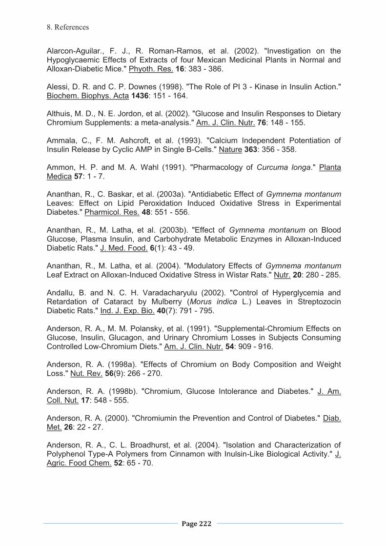
8. References
Page 222
Alarcon-Aguilar., F. J., R. Roman-Ramos, et al. (2002). "Investigation on the Hypoglycaemic Effects of Extracts of four Mexican Medicinal Plants in Normal and Alloxan-Diabetic Mice." Phyoth. Res. 16: 383 - 386.
Alessi, D. R. and C. P. Downes (1998). "The Role of PI 3 - Kinase in Insulin Action." Biochem. Biophys. Acta 1436: 151 - 164.
Althuis, M. D., N. E. Jordon, et al. (2002). "Glucose and Insulin Responses to Dietary Chromium Supplements: a meta-analysis." Am. J. Clin. Nutr. 76: 148 - 155.
Ammala, C., F. M. Ashcroft, et al. (1993). "Calcium Independent Potentiation of Insulin Release by Cyclic AMP in Single B-Cells." Nature 363: 356 - 358.
Ammon, H. P. and M. A. Wahl (1991). "Pharmacology of Curcuma longa." Planta Medica 57: 1 - 7.
Ananthan, R., C. Baskar, et al. (2003a). "Antidiabetic Effect of Gymnema montanum Leaves: Effect on Lipid Peroxidation Induced Oxidative Stress in Experimental Diabetes." Pharmicol. Res. 48: 551 - 556.
Ananthan, R., M. Latha, et al. (2003b). "Effect of Gymnema montanum on Blood Glucose, Plasma Insulin, and Carbohydrate Metabolic Enzymes in Alloxan-Induced Diabetic Rats." J. Med. Food. 6(1): 43 - 49.
Ananthan, R., M. Latha, et al. (2004). "Modulatory Effects of Gymnema montanum Leaf Extract on Alloxan-Induced Oxidative Stress in Wistar Rats." Nutr. 20: 280 - 285.
Andallu, B. and N. C. H. Varadacharyulu (2002). "Control of Hyperglycemia and Retardation of Cataract by Mulberry (Morus indica L.) Leaves in Streptozocin Diabetic Rats." Ind. J. Exp. Bio. 40(7): 791 - 795.
Anderson, R. A., M. M. Polansky, et al. (1991). "Supplemental-Chromium Effects on Glucose, Insulin, Glucagon, and Urinary Chromium Losses in Subjects Consuming Controlled Low-Chromium Diets." Am. J. Clin. Nutr. 54: 909 - 916.
Anderson, R. A. (1998a). "Effects of Chromium on Body Composition and Weight Loss." Nut. Rev. 56(9): 266 - 270.
Anderson, R. A. (1998b). "Chromium, Glucose Intolerance and Diabetes." J. Am. Coll. Nut. 17: 548 - 555.
Anderson, R. A. (2000). "Chromiumin the Prevention and Control of Diabetes." Diab. Met. 26: 22 - 27.
Anderson, R. A., C. L. Broadhurst, et al. (2004). "Isolation and Characterization of Polyphenol Type-A Polymers from Cinnamon with Inulsin-Like Biological Activity." J. Agric. Food Chem. 52: 65 - 70.
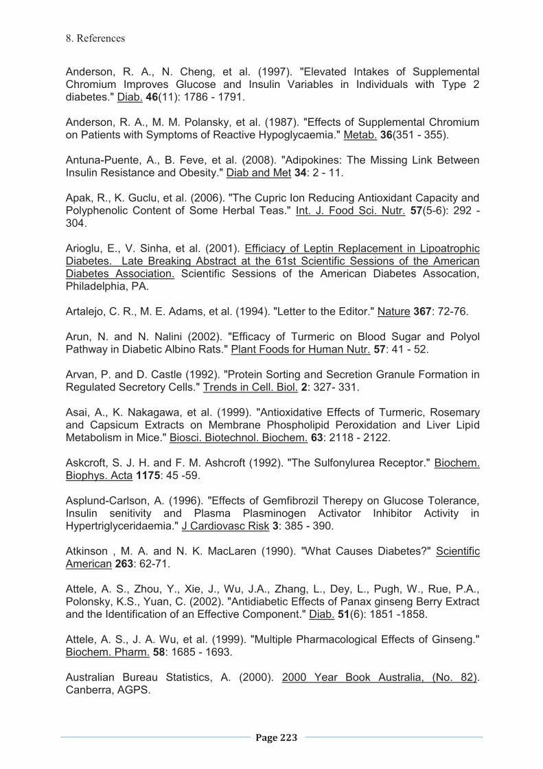
8. References
Page 223
Anderson, R. A., N. Cheng, et al. (1997). "Elevated Intakes of Supplemental Chromium Improves Glucose and Insulin Variables in Individuals with Type 2 diabetes." Diab. 46(11): 1786 - 1791.
Anderson, R. A., M. M. Polansky, et al. (1987). "Effects of Supplemental Chromium on Patients with Symptoms of Reactive Hypoglycaemia." Metab. 36(351 - 355).
Antuna-Puente, A., B. Feve, et al. (2008). "Adipokines: The Missing Link Between Insulin Resistance and Obesity." Diab and Met 34: 2 - 11.
Apak, R., K. Guclu, et al. (2006). "The Cupric Ion Reducing Antioxidant Capacity and Polyphenolic Content of Some Herbal Teas." Int. J. Food Sci. Nutr. 57(5-6): 292 - 304.
Arioglu, E., V. Sinha, et al. (2001). Efficiacy of Leptin Replacement in Lipoatrophic Diabetes. Late Breaking Abstract at the 61st Scientific Sessions of the American Diabetes Association. Scientific Sessions of the American Diabetes Assocation, Philadelphia, PA.
Artalejo, C. R., M. E. Adams, et al. (1994). "Letter to the Editor." Nature 367: 72-76.
Arun, N. and N. Nalini (2002). "Efficacy of Turmeric on Blood Sugar and Polyol Pathway in Diabetic Albino Rats." Plant Foods for Human Nutr. 57: 41 - 52.
Arvan, P. and D. Castle (1992). "Protein Sorting and Secretion Granule Formation in Regulated Secretory Cells." Trends in Cell. Biol. 2: 327- 331.
Asai, A., K. Nakagawa, et al. (1999). "Antioxidative Effects of Turmeric, Rosemary and Capsicum Extracts on Membrane Phospholipid Peroxidation and Liver Lipid Metabolism in Mice." Biosci. Biotechnol. Biochem. 63: 2118 - 2122.
Askcroft, S. J. H. and F. M. Ashcroft (1992). "The Sulfonylurea Receptor." Biochem. Biophys. Acta 1175: 45 -59.
Asplund-Carlson, A. (1996). "Effects of Gemfibrozil Therepy on Glucose Tolerance, Insulin senitivity and Plasma Plasminogen Activator Inhibitor Activity in Hypertriglyceridaemia." J Cardiovasc Risk 3: 385 - 390.
Atkinson , M. A. and N. K. MacLaren (1990). "What Causes Diabetes?" Scientific American 263: 62-71.
Attele, A. S., Zhou, Y., Xie, J., Wu, J.A., Zhang, L., Dey, L., Pugh, W., Rue, P.A., Polonsky, K.S., Yuan, C. (2002). "Antidiabetic Effects of Panax ginseng Berry Extract and the Identification of an Effective Component." Diab. 51(6): 1851 -1858.
Attele, A. S., J. A. Wu, et al. (1999). "Multiple Pharmacological Effects of Ginseng." Biochem. Pharm. 58: 1685 - 1693.
Australian Bureau Statistics, A. (2000). 2000 Year Book Australia, (No. 82). Canberra, AGPS.
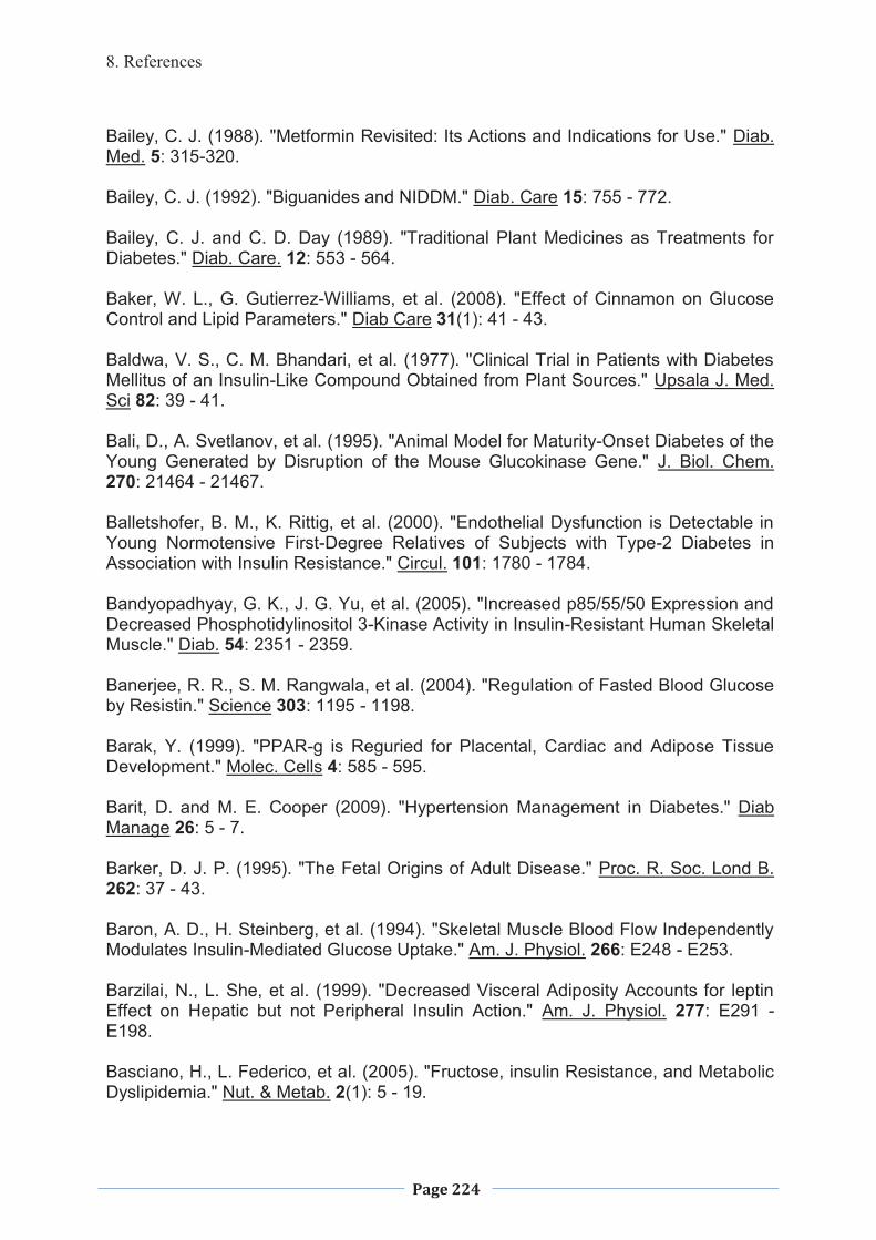
8. References
Page 224
Bailey, C. J. (1988). "Metformin Revisited: Its Actions and Indications for Use." Diab. Med. 5: 315-320.
Bailey, C. J. (1992). "Biguanides and NIDDM." Diab. Care 15: 755 - 772.
Bailey, C. J. and C. D. Day (1989). "Traditional Plant Medicines as Treatments for Diabetes." Diab. Care. 12: 553 - 564.
Baker, W. L., G. Gutierrez-Williams, et al. (2008). "Effect of Cinnamon on Glucose Control and Lipid Parameters." Diab Care 31(1): 41 - 43.
Baldwa, V. S., C. M. Bhandari, et al. (1977). "Clinical Trial in Patients with Diabetes Mellitus of an Insulin-Like Compound Obtained from Plant Sources." Upsala J. Med. Sci 82: 39 - 41.
Bali, D., A. Svetlanov, et al. (1995). "Animal Model for Maturity-Onset Diabetes of the Young Generated by Disruption of the Mouse Glucokinase Gene." J. Biol. Chem. 270: 21464 - 21467.
Balletshofer, B. M., K. Rittig, et al. (2000). "Endothelial Dysfunction is Detectable in Young Normotensive First-Degree Relatives of Subjects with Type-2 Diabetes in Association with Insulin Resistance." Circul. 101: 1780 - 1784.
Bandyopadhyay, G. K., J. G. Yu, et al. (2005). "Increased p85/55/50 Expression and Decreased Phosphotidylinositol 3-Kinase Activity in Insulin-Resistant Human Skeletal Muscle." Diab. 54: 2351 - 2359.
Banerjee, R. R., S. M. Rangwala, et al. (2004). "Regulation of Fasted Blood Glucose by Resistin." Science 303: 1195 - 1198.
Barak, Y. (1999). "PPAR-g is Reguried for Placental, Cardiac and Adipose Tissue Development." Molec. Cells 4: 585 - 595.
Barit, D. and M. E. Cooper (2009). "Hypertension Management in Diabetes." Diab Manage 26: 5 - 7.
Barker, D. J. P. (1995). "The Fetal Origins of Adult Disease." Proc. R. Soc. Lond B. 262: 37 - 43.
Baron, A. D., H. Steinberg, et al. (1994). "Skeletal Muscle Blood Flow Independently Modulates Insulin-Mediated Glucose Uptake." Am. J. Physiol. 266: E248 - E253.
Barzilai, N., L. She, et al. (1999). "Decreased Visceral Adiposity Accounts for leptin Effect on Hepatic but not Peripheral Insulin Action." Am. J. Physiol. 277: E291 - E198.
Basciano, H., L. Federico, et al. (2005). "Fructose, insulin Resistance, and Metabolic Dyslipidemia." Nut. & Metab. 2(1): 5 - 19.
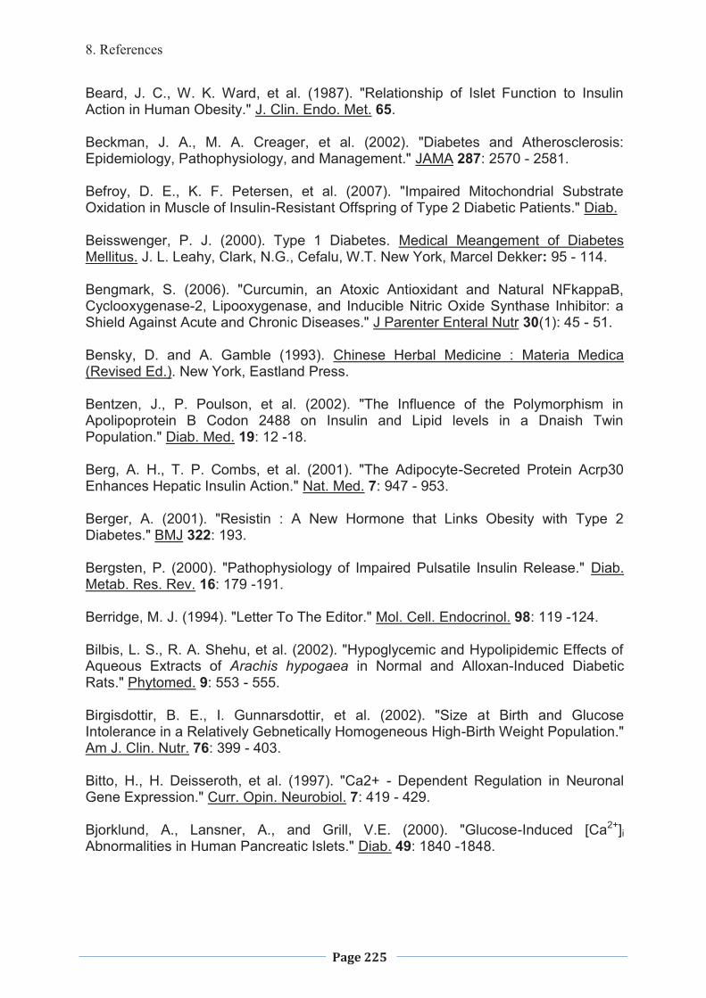
8. References
Page 225
Beard, J. C., W. K. Ward, et al. (1987). "Relationship of Islet Function to Insulin Action in Human Obesity." J. Clin. Endo. Met. 65.
Beckman, J. A., M. A. Creager, et al. (2002). "Diabetes and Atherosclerosis: Epidemiology, Pathophysiology, and Management." JAMA 287: 2570 - 2581.
Befroy, D. E., K. F. Petersen, et al. (2007). "Impaired Mitochondrial Substrate Oxidation in Muscle of Insulin-Resistant Offspring of Type 2 Diabetic Patients." Diab.
Beisswenger, P. J. (2000). Type 1 Diabetes. Medical Meangement of Diabetes Mellitus. J. L. Leahy, Clark, N.G., Cefalu, W.T. New York, Marcel Dekker: 95 - 114.
Bengmark, S. (2006). "Curcumin, an Atoxic Antioxidant and Natural NFkappaB, Cyclooxygenase-2, Lipooxygenase, and Inducible Nitric Oxide Synthase Inhibitor: a Shield Against Acute and Chronic Diseases." J Parenter Enteral Nutr 30(1): 45 - 51.
Bensky, D. and A. Gamble (1993). Chinese Herbal Medicine : Materia Medica (Revised Ed.). New York, Eastland Press.
Bentzen, J., P. Poulson, et al. (2002). "The Influence of the Polymorphism in Apolipoprotein B Codon 2488 on Insulin and Lipid levels in a Dnaish Twin Population." Diab. Med. 19: 12 -18.
Berg, A. H., T. P. Combs, et al. (2001). "The Adipocyte-Secreted Protein Acrp30 Enhances Hepatic Insulin Action." Nat. Med. 7: 947 - 953.
Berger, A. (2001). "Resistin : A New Hormone that Links Obesity with Type 2 Diabetes." BMJ 322: 193.
Bergsten, P. (2000). "Pathophysiology of Impaired Pulsatile Insulin Release." Diab. Metab. Res. Rev. 16: 179 -191.
Berridge, M. J. (1994). "Letter To The Editor." Mol. Cell. Endocrinol. 98: 119 -124.
Bilbis, L. S., R. A. Shehu, et al. (2002). "Hypoglycemic and Hypolipidemic Effects of Aqueous Extracts of Arachis hypogaea in Normal and Alloxan-Induced Diabetic Rats." Phytomed. 9: 553 - 555.
Birgisdottir, B. E., I. Gunnarsdottir, et al. (2002). "Size at Birth and Glucose Intolerance in a Relatively Gebnetically Homogeneous High-Birth Weight Population." Am J. Clin. Nutr. 76: 399 - 403.
Bitto, H., H. Deisseroth, et al. (1997). "Ca2+ - Dependent Regulation in Neuronal Gene Expression." Curr. Opin. Neurobiol. 7: 419 - 429.
Bjorklund, A., Lansner, A., and Grill, V.E. (2000). "Glucose-Induced [Ca2+]i Abnormalities in Human Pancreatic Islets." Diab. 49: 1840 -1848.
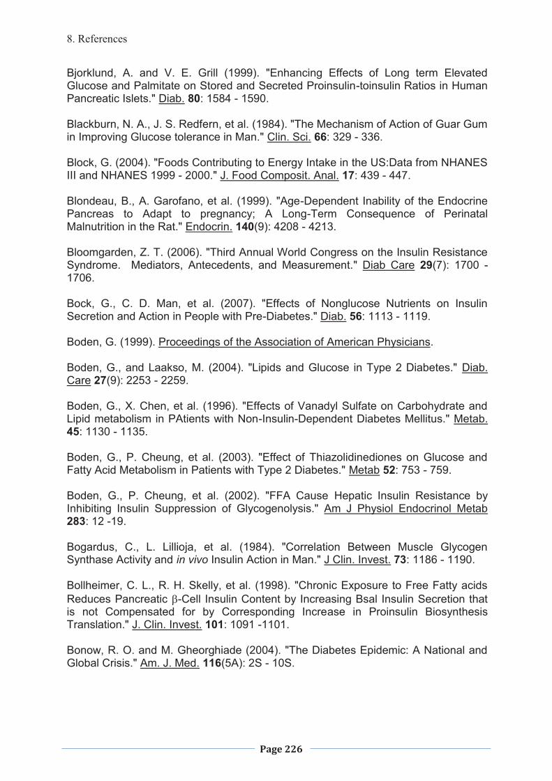
8. References
Page 226
Bjorklund, A. and V. E. Grill (1999). "Enhancing Effects of Long term Elevated Glucose and Palmitate on Stored and Secreted Proinsulin-toinsulin Ratios in Human Pancreatic Islets." Diab. 80: 1584 - 1590.
Blackburn, N. A., J. S. Redfern, et al. (1984). "The Mechanism of Action of Guar Gum in Improving Glucose tolerance in Man." Clin. Sci. 66: 329 - 336.
Block, G. (2004). "Foods Contributing to Energy Intake in the US:Data from NHANES III and NHANES 1999 - 2000." J. Food Composit. Anal. 17: 439 - 447.
Blondeau, B., A. Garofano, et al. (1999). "Age-Dependent Inability of the Endocrine Pancreas to Adapt to pregnancy; A Long-Term Consequence of Perinatal Malnutrition in the Rat." Endocrin. 140(9): 4208 - 4213.
Bloomgarden, Z. T. (2006). "Third Annual World Congress on the Insulin Resistance Syndrome. Mediators, Antecedents, and Measurement." Diab Care 29(7): 1700 - 1706.
Bock, G., C. D. Man, et al. (2007). "Effects of Nonglucose Nutrients on Insulin Secretion and Action in People with Pre-Diabetes." Diab. 56: 1113 - 1119.
Boden, G. (1999). Proceedings of the Association of American Physicians.
Boden, G., and Laakso, M. (2004). "Lipids and Glucose in Type 2 Diabetes." Diab. Care 27(9): 2253 - 2259.
Boden, G., X. Chen, et al. (1996). "Effects of Vanadyl Sulfate on Carbohydrate and Lipid metabolism in PAtients with Non-Insulin-Dependent Diabetes Mellitus." Metab. 45: 1130 - 1135.
Boden, G., P. Cheung, et al. (2003). "Effect of Thiazolidinediones on Glucose and Fatty Acid Metabolism in Patients with Type 2 Diabetes." Metab 52: 753 - 759.
Boden, G., P. Cheung, et al. (2002). "FFA Cause Hepatic Insulin Resistance by Inhibiting Insulin Suppression of Glycogenolysis." Am J Physiol Endocrinol Metab 283: 12 -19.
Bogardus, C., L. Lillioja, et al. (1984). "Correlation Between Muscle Glycogen Synthase Activity and in vivo Insulin Action in Man." J Clin. Invest. 73: 1186 - 1190.
Bollheimer, C. L., R. H. Skelly, et al. (1998). "Chronic Exposure to Free Fatty acids Reduces Pancreatic �-Cell Insulin Content by Increasing Bsal Insulin Secretion that is not Compensated for by Corresponding Increase in Proinsulin Biosynthesis Translation." J. Clin. Invest. 101: 1091 -1101.
Bonow, R. O. and M. Gheorghiade (2004). "The Diabetes Epidemic: A National and Global Crisis." Am. J. Med. 116(5A): 2S - 10S.
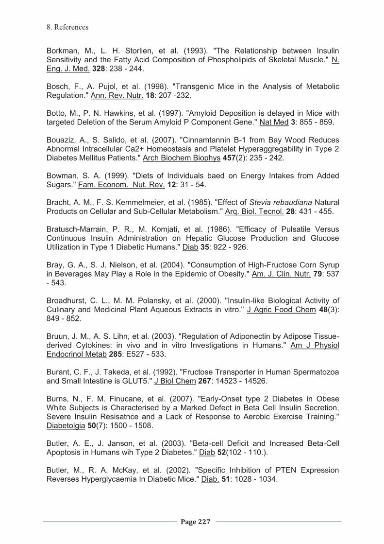
8. References
Page 227
Borkman, M., L. H. Storlien, et al. (1993). "The Relationship between Insulin Sensitivity and the Fatty Acid Composition of Phospholipids of Skeletal Muscle." N. Eng. J. Med. 328: 238 - 244.
Bosch, F., A. Pujol, et al. (1998). "Transgenic Mice in the Analysis of Metabolic Regulation." Ann. Rev. Nutr. 18: 207 -232.
Botto, M., P. N. Hawkins, et al. (1997). "Amyloid Deposition is delayed in Mice with targeted Deletion of the Serum Amyloid P Component Gene." Nat Med 3: 855 - 859.
Bouaziz, A., S. Salido, et al. (2007). "Cinnamtannin B-1 from Bay Wood Reduces Abnormal Intracellular Ca2+ Homeostasis and Platelet Hyperaggregability in Type 2 Diabetes Mellitus Patients." Arch Biochem Biophys 457(2): 235 - 242.
Bowman, S. A. (1999). "Diets of Individuals baed on Energy Intakes from Added Sugars." Fam. Econom. Nut. Rev. 12: 31 - 54.
Bracht, A. M., F. S. Kemmelmeier, et al. (1985). "Effect of Stevia rebaudiana Natural Products on Cellular and Sub-Cellular Metabolism." Arq. Biol. Tecnol. 28: 431 - 455.
Bratusch-Marrain, P. R., M. Komjati, et al. (1986). "Efficacy of Pulsatile Versus Continuous Insulin Administration on Hepatic Glucose Production and Glucose Utilization in Type 1 Diabetic Humans." Diab 35: 922 - 926.
Bray, G. A., S. J. Nielson, et al. (2004). "Consumption of High-Fructose Corn Syrup in Beverages May Play a Role in the Epidemic of Obesity." Am. J. Clin. Nutr. 79: 537 - 543.
Broadhurst, C. L., M. M. Polansky, et al. (2000). "Insulin-like Biological Activity of Culinary and Medicinal Plant Aqueous Extracts in vitro." J Agric Food Chem 48(3): 849 - 852.
Bruun, J. M., A. S. Lihn, et al. (2003). "Regulation of Adiponectin by Adipose Tissue-derived Cytokines: in vivo and in vitro Investigations in Humans." Am J Physiol Endocrinol Metab 285: E527 - 533.
Burant, C. F., J. Takeda, et al. (1992). "Fructose Transporter in Human Spermatozoa and Small Intestine is GLUT5." J Biol Chem 267: 14523 - 14526.
Burns, N., F. M. Finucane, et al. (2007). "Early-Onset type 2 Diabetes in Obese White Subjects is Characterised by a Marked Defect in Beta Cell Insulin Secretion, Severe Insulin Resisatnce and a Lack of Response to Aerobic Exercise Training." Diabetolgia 50(7): 1500 - 1508.
Butler, A. E., J. Janson, et al. (2003). "Beta-cell Deficit and Increased Beta-Cell Apoptosis in Humans wih Type 2 Diabetes." Diab 52(102 - 110.).
Butler, M., R. A. McKay, et al. (2002). "Specific Inhibition of PTEN Expression Reverses Hyperglycaemia In Diabetic Mice." Diab. 51: 1028 - 1034.
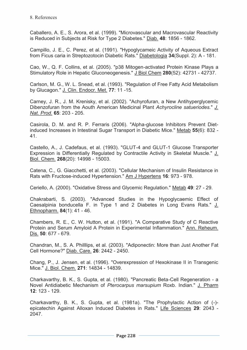
8. References
Page 228
Caballero, A. E., S. Arora, et al. (1999). "Microvascular and Macrovascular Reactivity is Reduced in Subjects at Risk for Type 2 Diabetes." Diab. 48: 1856 - 1862.
Campillo, J. E., C. Perez, et al. (1991). "Hypoglycameic Activity of Aqueous Extract from Ficus caria in Streptozotocin Diabetic Rats." Diabetologia 34(Suppl. 2): A - 181.
Cao, W., Q. F. Collins, et al. (2005). "p38 Mitogen-activated Protein Kinase Plays a Stimulatory Role in Hepatic Gluconeogenesis." J Biol Chem 280(52): 42731 - 42737.
Carlson, M. G., W. L. Snead, et al. (1993). "Regulation of Free Fatty Acid Metabolism by Glucagon." J. Clin. Endocr. Met. 77: 11 -15.
Carney, J. R., J. M. Krenisky, et al. (2002). "Achyrofuran, a New Antihyperglycemic Dibenzofuran from the Aouth American Medicinal Plant Achyrocline satueriodes." J. Nat. Prod. 65: 203 - 205.
Casirola, D. M. and R. P. Ferraris (2006). "Alpha-glucose Inhibitors Prevent Diet-induced Increases in Intestinal Sugar Transport in Diabetic Mice." Metab 55(6): 832 - 41.
Castello, A., J. Cadefaus, et al. (1993). "GLUT-4 and GLUT-1 Glucose Transporter Expression is Differentially Regulated by Contractile Activity in Skeletal Muscle." J. Biol. Chem. 268(20): 14998 - 15003.
Catena, C., G. Giacchetti, et al. (2003). "Cellular Mechanism of Insulin Resistance in Rats with Fructose-induced Hypertension." Am J Hypertens 16: 973 - 978.
Ceriello, A. (2000). "Oxidative Stress and Glycemic Regulation." Metab 49: 27 - 29.
Chakrabarti, S. (2003). "Advanced Studies in the Hypoglycaemic Effect of Caesalpinia bonducella F. in Type 1 and 2 Diabetes in Long Evans Rats." J. Ethnopharm. 84(1): 41 - 46.
Chambers, R. E., C. W. Hutton, et al. (1991). "A Comparative Study of C Reactive Protein and Serum Amyloid A Protein in Experimental Inflammation." Ann. Reheum. Dis. 50: 677 - 679.
Chandran, M., S. A. Philllips, et al. (2003). "Adiponectin: More than Just Another Fat Cell Hormone?" Diab. Care. 26: 2442 - 2450.
Chang, P., J. Jensen, et al. (1996). "Overexpression of Hexokinase II in Transgenic Mice." J. Biol. Chem. 271: 14834 - 14839.
Charkavarthy, B. K., S. Gupta, et al. (1980). "Pancreatic Beta-Cell Regeneration - a Novel Antidiabetic Mechanism of Pterocarpus marsupium Roxb. Indian." J. Pharm 12: 123 - 129.
Charkavarthy, B. K., S. Gupta, et al. (1981a). "The Prophylactic Action of (-)-epicatechin Against Alloxan Induced Diabetes in Rats." Life Sciences 29: 2043 - 2047.
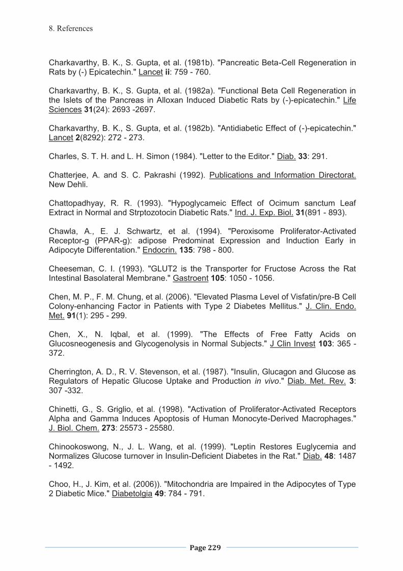
8. References
Page 229
Charkavarthy, B. K., S. Gupta, et al. (1981b). "Pancreatic Beta-Cell Regeneration in Rats by (-) Epicatechin." Lancet ii: 759 - 760.
Charkavarthy, B. K., S. Gupta, et al. (1982a). "Functional Beta Cell Regeneration in the Islets of the Pancreas in Alloxan Induced Diabetic Rats by (-)-epicatechin." Life Sciences 31(24): 2693 -2697.
Charkavarthy, B. K., S. Gupta, et al. (1982b). "Antidiabetic Effect of (-)-epicatechin." Lancet 2(8292): 272 - 273.
Charles, S. T. H. and L. H. Simon (1984). "Letter to the Editor." Diab. 33: 291.
Chatterjee, A. and S. C. Pakrashi (1992). Publications and Information Directorat. New Dehli.
Chattopadhyay, R. R. (1993). "Hypoglycameic Effect of Ocimum sanctum Leaf Extract in Normal and Strptozotocin Diabetic Rats." Ind. J. Exp. Biol. 31(891 - 893).
Chawla, A., E. J. Schwartz, et al. (1994). "Peroxisome Proliferator-Activated Receptor-g (PPAR-g): adipose Predominat Expression and Induction Early in Adipocyte Differentation." Endocrin. 135: 798 - 800.
Cheeseman, C. I. (1993). "GLUT2 is the Transporter for Fructose Across the Rat Intestinal Basolateral Membrane." Gastroent 105: 1050 - 1056.
Chen, M. P., F. M. Chung, et al. (2006). "Elevated Plasma Level of Visfatin/pre-B Cell Colony-enhancing Factor in Patients with Type 2 Diabetes Mellitus." J. Clin. Endo. Met. 91(1): 295 - 299.
Chen, X., N. Iqbal, et al. (1999). "The Effects of Free Fatty Acids on Glucosneogenesis and Glycogenolysis in Normal Subjects." J Clin Invest 103: 365 - 372.
Cherrington, A. D., R. V. Stevenson, et al. (1987). "Insulin, Glucagon and Glucose as Regulators of Hepatic Glucose Uptake and Production in vivo." Diab. Met. Rev. 3: 307 -332.
Chinetti, G., S. Griglio, et al. (1998). "Activation of Proliferator-Activated Receptors Alpha and Gamma Induces Apoptosis of Human Monocyte-Derived Macrophages." J. Biol. Chem. 273: 25573 - 25580.
Chinookoswong, N., J. L. Wang, et al. (1999). "Leptin Restores Euglycemia and Normalizes Glucose turnover in Insulin-Deficient Diabetes in the Rat." Diab. 48: 1487 - 1492.
Choo, H., J. Kim, et al. (2006)). "Mitochondria are Impaired in the Adipocytes of Type 2 Diabetic Mice." Diabetolgia 49: 784 - 791.
8. References
Page 230
Chopra, R. N., I. C. Chopra, et al. (1958). Indigineous Drugs of India. Calcutta, Dhar and Sons Pvt Ltd.
Cianflone, K., M. Maslowska, et al. (1999). "Acylation Stimulating Protein (ASP0, an Adipocyte Autocrine: New Directions." Semin. Cell Dev. Biol. 10: 31 - 41.
Clark, A., D. R. Matthews, et al. (1987). "Pancreatic Islet amyloid and elevated Proinsulin Secretion in Familial Maturity-onset Diabetes." Diab Res Clin Prac 4: 51 - 55.
Clark, A. and M. R. Nilsson (2004). "Islet Amyloid: a Complication of Islet Dysfunction or an Aetiological Factor in Type 2 Diabetes?" Diabetologia 47: 157 - 169.
Clarke, P., C. Kelman, et al. (2006). "Factors Influencing the Cost of Hospital Care for People with Diabetes in Australia." J. Diab. and its Complic. 20: 349 - 355.
Clarke, T. A., E. Heyliger, et al. (2004). "Codelivery of a Tea Extract Prevents Morbidity and Mortality Associated with Oral Canadate therepy in Streprozotocin-Induced diabetic Rats." Metab. 53(9): 1145 - 1151.
Clausson, P., R. Linnarsson, et al. (1994). "Relationship Between Diabetic Duration, metabolic Control and B-Cell Function in a Representative Population of Type 2 Diabetic Patients in Sweden." Diab. Med. 11: 794 - 801.
Clos, T. W. D. (2000). "Function of C-Reactive Protein." Ann. Med. 32: 274 - 278.
Cohen, N., M. Halbertsom, et al. (!995). "Oral Vandayl Sulfate Improves Hepatic and Peripheral Insulin Sensitivy in Patients with Non-Insulin Dependent Diabetes Mellitus." J Clin. Invest. 95: 2501 -2509.
Coleman, M. D., R. C. Eason, et al. (2001). "The Therapeutic Use of Lipoic Acid In Diabetes: A Current Prospective." Envir. Tox. Pharm. 10: 167 - 172.
Collins, M. and J. R. McFarlane (2006). "An Exploratory Study into the Effectiveness of a Combination of Traditional Chinese Herbs in the Management of Type 2 Diabetes." Diab Care 29(4): 1.
Cook, M. N., C. J. Girman, et al. (2007). "Initial Monotherepy with Either Metformin or Sulphonylureas Often Fails to Achieve or Maintain Current Glycaemic Goals in Patients with Type 2 Diabetes in UK Primary Care." Diab Med 24(4): 350 - 358.
Corpe, C. P., M. M. Bassaleh, et al. (1996). "The Regulation of GLUT5 and GLUT2 Activity in the Adaptation of IntestinalBrush-Border Fructose Transport in Diabetes." Pflugers Arch 432: 192 - 201.
Cortright, R. N. and G. L. Dohn (1997). "Mechanism by which Insulin and Muscle Contraction Stimulate Glucose Transport." Can. J. Appl Physiol. 22: 519 - 530.
Costantino, L., L. Raimondi, et al. (2003). "Isolation and Pharmacological Activities of the Tecoma stans Alkaloids." Il Farmco 58: 781 - 785.
8. References
Page 231
Coyne, T., T. I. Ibiebele, et al. (2005). "Diabetes Mellitus and Serum Carotenoids: Findings of a Population-based Study in Queensland, Australia." The Amer. J. Clin. Nut. 82: 685 - 693.
Crespin, S., W. Greenborough, et al. (1969). "Stimulation of Insulin Secretion by Infusion of Free Fatty Acids." J. Clin. Invest. 48: 1934 - 1943.
Crespin, S., W. B. Greenough, et al. (1973). "Stimulation of Insulin Secretion by Long-chain Free Fatty Acids: a Direct Pancreatic Effect." J Clin Invest 52: 1979 - 1984.
Cryns, V. and J. Yuan (1998). "Proteases to Die For." Genes Dev. 12: 1551 - 1570.
Cugati, S., J. J. Wang, et al. (2007). "Ten-year Incidence of Diabetes in Older Australians: The Blue Mountains Eye Study." Med. J. Aust. 186(3): 131 - 135.
Curat, C. A., V. Wegner, et al. (2006). "Macrophages in Human Visceral Adipose Tissue: Increased Accumulation in Obesity and Source of Resistance and Visfatin." Diabetolgia 49: 744 - 777.
Curie, R. (1986). "Effect of Stevia rebaudiana on Glucose Tolerance in Normal Adult Humans." Brazil. J. Med. Biol. Res. 19: 771 - 774.
Dahlman, I., M. Forsgren, et al. (2006). "Downregulation of Electron Transport Chain Genes in Visceral Adipose Tissue in type 2 Diabetes Independent of Obesity and Possibly Involving Tumor Necrosis Factor - �." Diab. 55: 1792 - 1799.
Dalle, S., G. Fonte, et al. (2001). "Miniglucagon (Glucagon 19-29). A Novel Regulator of the Pancreatic Islet Physiology." Diab. 51(2): 406 - 412.
Dalle, S., P. Smith, et al. (1999). "Miniglucagon (glucagon 19-29). A Potent and Efficient Inhibitor of Secretagogue-Induced Insulin Release Through a Ca+ Pathway." J. Biol. Chem. 274: 10860 - 10876.
Daniel, P. M. and J. R. Henderson (1967). "The Effect of Vagal Stimulation on Plasma Insulin and Glucose Levels in the Baboon." J Physiol (Lond) 192: 317 - 327.
Danielsson, A., A. Ost, et al. (2005). "Attentuation of Insulin-Stimulated Insulin Receptor Substrate-1 Serine 307 Phosphorylation in Insulin Resistance of Type 2 Diabetes." J. Biol. Chem. 280(41): 34389 - 34392.
de Boer, I. H., B. Kestenbaum, et al. (2008). "Calcium plus vitamin D supplementation and the risk of incident diabetes in the Women's Health Initiative." Diab Care 31(4): 701 - 7.
de Souza, C. J., M. Eckhardt, et al. (2001). "Effects of Pioglitazone on Adipose Tissue Remodeling Within the Setting of Obesity and Insulin Resistance." Diab 50: 1863 - 1871.
8. References
Page 232
Deed, G. (2009). "National Online Map Launched to Track Diabetes in Australia." Diab Manage 26: 34.
DeFronzo, R. A. (1979). "Glucose Intolerance and Aging, and Evidence for Tissue Insenitivity to Insulin." Diab. 28: 1095 - 1101.
DeFronzo, R. A. (1988). "The Triumvirate: Beta-Cell, Muscle, Liver. A Collusion responsible for NIDDM." Diab. 37: 667 - 687.
DeFronzo, R. A. (1997). "Pathogenesis of Type 2 Diabetes: Metabolic and Molecular Implications for Identifying Diabetes Gene." Diab. Rev. 5: 177 - 269.
Dehghan, A., I. Kardys, et al. (2007). "Genetic Variation, C-Reactive Protein Levels, and Incidence of Diabetes." Diab 56(3): 872 - 878.
Dekker, M. J., S. Lee, et al. (2007). "An Exercise Intervention Without Weight Loss Decreases Circulating Interleakin-6 in Lean and Obese Men with and without Type 2 Diabetes." Metab. Clin. Exper. 56: 332 - 338.
Del, P. S. and N. Pulizzi (2006). "The Place of Sulfonylureas in the Therapy for Type 2 Diabetes Mellitus." Metab. 55(5 Suppl 1): S20 - S27.
Dela, F., A. Handberg, et al. (1993). "GLUT4 and Insulin Receptor Binding and Kinase Activity in Trained Human Muscle Cells." J. Physiol. 469: 615 - 624.
Dent, P., A. Lavoinne, et al. (1990). "The Molecular Mechanisms by which Insulin Stimulates Glycogen Synthesis in Mammalian Skeletal Muscle." Nature 348: 302 - 307.
Devi, B. A., N. Kamalakkannan, et al. (2003). "Supplementation of Fenugreek Leaves to Diabetic Rats. Effect of Carbohydrate Metabolic Enzymes in Daibetic Liver and Kidney." Phyoth. Res. 17: 1231 - 1233.
Dey, L., J. T. Xie, et al. (2003). "Anti-Hyperglycemic Effects of Ginseng: Comparrison Between Root and Berry." Phytomed. 10: 600 - 605.
Dhandapani, S., V. R. Subramanian, et al. (2002). "Hypolipidemic Effect of Cuminum cyminum L. on Alloxan-induced Diabetic Rats." Pharmacol Res. 46(3): 251 - 255.
Dhawan, B. N., M. P. Dubey, et al. (1980). "Screening of Indian Plants for Biologicial Activity, Part IX." Indian Exp. Biol. 18: 594 - 606.
Di Loreto, C., C. Fanelli, et al. (2005). "Make Your Diabetic Patients Walk - Long-Term Impact of Different Amounts of Physical Activity on Type 2 Diabetes." Diab. Care. 28(6): 1295 - 1302.
Di Mario, U. and F. Dotta (1998). "Beta-Cell Autoimmunity." Diab. Metab. Rev. 14: 1 - 2.
8. References
Page 233
Dirlewanger, M., P. Schneiter, et al. (2000). "Effects of Fructose on Hepatic Glucose Metabolism in Humans." Am. J. Physiol. Endocrinol. Metab. 279: E907 - E911.
Dohm, G. L., E. Tapscott, et al. (1988). "An in vitro Human Muscle Preparation Suitable for Metabolic Studies. Decreased Insulin Stimulation of Glucose Transport in Muscle from Morbidity Obese and Diabetis Subjects." J Clin Invest 82: 486 - 494.
Donath, M. Y., J. A. Ehses, et al. (2005). "Mechanisms of �-Cell Death in Type 2 Diabetes." Diab 54: S108 - S113.
Doonan, P. T., T. M. MacDonald, et al. (2002). "Adherence to Prescribed Oral hypoglycaemic Medication in a Population of Patients with Type 2 Diabetes: a Retrospective Cohort Study." Diab. Med. 19: 274 - 278.
Dresner, A., D. Lauren, et al. (1999). "Effects of Free Fatty Acids on Glucose Transport and IRS-1-associated Phosphatidylinositol 3-kinase Activity." J Clin Invest 103: 253 - 259.
Drucker, D. J. (1998). "Glucagon-Like Peptides." Diab. 47: 159 - 169.
Dunning, B. E., J. E. Foley, et al. (2005). "Alpha Cell Function in Health and Disease: Influence of Glucagon-like Peptide-1." Diabetolgia 48: 1700 - 1713.
Dunstan, D. W., P. Z. Zimmer, et al. (2002). "The Raising Prevelance of Diabetes and Impaired Glucose Tolerance." Diab. Care 25: 829 - 835.
Edwards, B. J., H. M. Perry, et al. (1996). "Age-Related Changes in Amylin Secretion." Mech. Biophys. Res. Comm. 86: 39 - 51.
Ekundayo, O., I. Laakso, et al. (1989). "Constituents of the Volatile Oils from Leaves of Ociumum canum Sims,." Flavour and Fragrance J. 4: 17 - 18.
Eliasson, L., E. Renstrom, et al. (1996). "PKC-Dependent Stimulation of Exocytosis by Sulfonyureas in Pancreatic B-Cells." Science 271: 813 - 815.
Elliott, S. S., N. L. Keim, et al. (2002). "Fructose, Weight Gain, and the Insulin Resisatnce Syndrome." Am. J. Clin. Nutr. 76: 911 - 922.
Emanuelli, B., P. Peraldi, et al. (2001). "SOCS-3 Inhibits Insulin Signalling and is Up-regulated in Response to Tumor Necrosis Factor-alpha in the Adipose Tissue of Mice." J Biol Chem 276: 47944 - 47949.
Engstrom, G., B. Heblad, et al. (2005). "Complement C3 Is a Risk Factor for the Development of diabetes - A Population-Based Cohort Study." Diab. 54(2): 570 - 575.
Eriksson, J. G., V. Lindi, et al. (2002). "The effects of the Pro12Ala Polymorphism of the Peroxisome Proliferator-Activated Receptor-��Sensitivity and Insulin Metabolism Interact with Size at Birth." Diab. 51(7): 2321 - 2324.
8. References
Page 234
Etgen, G. J., R. P. Farrar, et al. (1993). "Effects of Chronic Electrical Stimulation on GLUT4 Protein Content in Fast-Twitch Muscle." Am. J. Physiol. 164: R816 - R819.
Evans, J. L., I. D. Goldfine, et al. (1986). "Are Oxidative Stress-Activated Signalling Pathways Mediators of Insulin Resistance and beta-Cell Dysfunction?" Diab. 52: 1 - 8.
Fantus, I. G. and R. Brosseau (1986). "Mechanisms of Action of Metformin: Insulin Receptor and post-Receptor Effects in vitro and in vivo." J. Clin. Endocr. and Met. 63: 898 - 905.
FAO/WHO (1997). Carbohydrates in Human Nutrition. Report of a Joint FAO/WHO Report. Rome, FAO Food and Nutrition: Paper 66.
Fernandez, J. M., A. Lopez-Bermejo, et al. (2002). "Cross-Talk Between Iron Metabolism and Diabetes." Diab. 51(82348 - 2354).
Ferreira, A., C. Proenca, et al. (2006). "The in vitro Screening for Acetylcholinesterase Inhibition and Antioxidant Activity of Medicinal Plants from Portugal." J. Ethnopharm. 108(3): 31 - 37.
Festa, A., R. J. D'Agostino, et al. (2002). "Elevated Levels of Acute-phase Proteins and Plasminogen Activator Inhibitor-1 Predict the Development of Type 2 Diabetes: The Insulin resistance Athererosclerosis Study." Diab. 51(4): 1131 -1137.
Fetrow, C. W. and J. R. Avila (2001). Professional's Handbook of Complementary and Alternative Medicines (2nd Ed.). Pensylvania, Springhouse.
Fisher, Y., J. Thomas, et al. (1995). "Action of Metformin on Glucose Transport and Glucose Transporter GLUT1 and GLUT 4 in Heart Muscle from Healthy and Diabetic Rats." Endocrin. 136: 412 - 420.
Flier, J. S. (2001). "The Missing Link with Obesity." Nature 409: 292 - 293.
Floune, B., N. Vidon, et al. (1984). "Effect of Pectin on Jejunal Glucose absorption and Unstirred Layer thickness in Normal Man." Gut 25: 936 - 941.
Ford, E. S. (1999). "Body Mass Index, Diabetes, and C-Reactive protein Among US Aduls." Diab. Care 1999(22).
Forouhi, N. G., A. H. Harding, et al. (2007). "Elevated Serum Ferritin Levels Predict New-onset Type 2 Diabetes: Results from the EPIC-Norfolk Prospective Study." Diabetolgia 50: 949 - 956.
Foster, T. S. (2007). "Efficacy and Safety of �-Lipoic Acid Supplementation in the Treatment of Symptomatic Diabetic Neuropathy." Diab Ed 33(1): 111 - 117.
Foster-Powell, K. and J. B. Miller (1995). "International Table of Glycemic Index." Am. J. Clin. Nutr. 62: 871S - 890S.
8. References
Page 235
Foy, C. G., R. A. Bell, et al. (2005). "Smoking and Incidence of Diabetes Among U.S. Adults." Diab Care 28(10): 2501 - 2507.
Fran, A. C., B. E. Gordillo, et al. (1998). "The Effect of Two Sequential of Opuntia streptacantha Upon Glycemia." Arch. Invest. Med. 22: 333 - 336.
Franz, M. J., J. P. Bantle, et al. (2002). "Evidence-Based Nutrition Principles and Recommandations for the Treatment and Prevention of Diabetes and Related Complications." Diab. Care 25: 148 - 198.
Frati, A. C., B. E. Gordillo, et al. (1991). "Acute Hypoglycaemic Effects of Optuntia strepacantha Lemiare in NIDDM." Diab. Care 13: 455 - 456.
Frati-Munari, A. C., B. E. Gordillo, et al. (1988). "Hypoglycaemic Effect of Optuntia streptacantha Lemaire in NIDDM." Diab. Care 11: 63 - 66.
Frayn, K. N., C. M. Williams, et al. (1996). "Are Increased Plasma Non-esterified Fatty Acid Concentrations a Risk Marker for Coronary Heart Diseases." Clin. Sci. 90: 243 - 253.
Freeman, D. J., J. Norrie, et al. (2002). "C-Reactive Protein is an Independent Predictor of Risk for the Development of Diabetes in the West of Scotland Coronary Prevention Study." Diab. 51(5): 1596 - 1600.
Fukauhara, A., M. Matsuda, et al. (2005). "Visfain: A Protein Secreted by Visceral Fat that Mimics the Effects of Insulin." Science 307: 426 - 430.
Furukawa, H., R. Carroll, et al. (1999). "Long-term Elevation of Free Fatty Acids Leads to Delayed Processing of Proinsulin and Prohormone Convertase 2 and 3 in the Pancreatic beta-cell Line MIN6." Diab. 48: 1395 - 1401.
Furukawa, S., T. Fujita, et al. (2004). "Increased Oxidative Stress in Obesity and its Impact on Metabolic Syndrome." J Clin Invest 114: 1752 - 1761.
Galuska, D., L. A. Nolte, et al. (1994). "Effect of Metformin on Insulin-Stimulated Glucose Transport in Isolated Muscle Obtained from Patients with NIDDM." Diabetologia 37: 826 - 832.
Garofano, A., P. Czernichow, et al. (1998). "�-Cell Mass and Proliferation Following Late Fetal and Early Postnatal Malnutrrition in the Rat." Diabetologia 41: 1114 - 1120.
Gastaldelli, A., S. Baldi, et al. (2000). "Influence of Obesity and Type 2 Diabetes on Gluconeogenesis and Glucose Output in Humans: a Quantitative Study." Diab 49: 1367 - 1373.
Gavi, S., L. M. Stuart, et al. (2007). "Retinol-Binding Protein 4 is Associated with Insulin Resistance and Body Fat Distribution in Non-obese Subjects without Type 2 Diabetes." J. Clin. Endo. Met. 92(5): 1886 - 1890.
8. References
Page 236
Gebre-Medhin, S., H. Mulder, et al. (1998). "Increased Insulin Secretion and Glucose Tolerance in Mice Lacking Islet Amyloid Polypeptide (amylin)." Biochem Biophys Res Commun 250: 271 - 277.
Gentilcore, D., R. Chaikomin, et al. (2006). "Effects of Fat on Gastric Emptying of, and the Glycemic, Insulin and Incretin responses to, a Carbohydrate Meal in type 2 Diabetes." J. Clin. Endo. Met. 91(6): 20.62 - 2.67.
Geuns, J. M. C. (2003). "Stevioside." Phytochem 64: 913 - 921.
Geuns, J. M. C., R. D. Malheiros, et al. (2003). "Metabolsim of Stevioside by Chickens." J. Agric. Food Chem. 51: 1095 - 1101.
Ghosh, A. and M. E. Greenberg (1995). "Calcium Signalling in Neurons: Molecular Mechanisms and Cellular Consequences." Science 268(5208): 239 - 247.
Gillies, C. L., K. R. Abrams, et al. (2007). "Pharmalogical and Lifestyle Interventions to Prevent or Delay Type 2 Diabetes in People with Impaired Glucose Tolerance: Systematic review and Meta-analysis." BMJ 334(7588): 299 - 308.
Gilon, P., R. M. Shephard, et al. (1993). "Oscillations of Secretion Driven by Oscillations of Cytoplasmic Ca2+ as Evidences In Single Pancreatoc Islets." J Biol Chem 268: 22265 - 22268.
Gluckman, P. and J. Harding (1992). The Regulation of Fetal Growth. Human Growth and Clinical Aspects. M. Hernandez and J. Argente. Amsterdam, Elservier Science: 253 - 286.
Goldstein, B. J. (2000). Enhancement of Post-Receptor Insulin Signalling by Trivalent Chromium in Hepatoma Cells may Involve Differential Inhibition of Specific Protein Tyrosine Phosphatases. 60th Scientific Session, American Diabetes Association.
Gong, Q., D. Deng, et al. (2008). "Trichosanthin, an Extract of Trichosanthes kirilowii, Effectively Prevents Acute Rejection of Major Histocompatibility Complex-mismatched Mouse Skin Allograft." Transplant Proc. 40(10): 3714 - 3718.
Gonzalez-Mujica, F., N. Motta, et al. (2003). "Effects of Bauhinia megalandra Aqueous Leaf Extract on Intestinal Glucose Absorption and Uptake by Enterocyte Brush Border Membrane Vesicles." Fitoterapia 74(1 - 2): 84 - 90.
Goodman, H. M. (2001). The Endocrine Pancreas and Regulation of Metabolism. New York, Oxford University Press Inc.
Goodner, C. J., B. C. Walike, et al. (1977). "Insulin, Glucagon, and Glucose Exhibit Synchronous, Sustained Oscilliations in Fasting Monkeys." Science 195: 177 - 179. Grant, S. J., A. Bensoussan, et al. (2010) “Chinese Herbal Medicine for People with Impaired Glucose Tolerance or Impaired Fasting Blood Glucose Levels.” Cochrane Database Syst. Rev.
8. References
Page 237
Gray, A. and P. R. Flatt (1997). "Evaluation of the Antidiabetic Effects of Coriander (Coriandrum sativum)." Proc Natr Soc 56: 45A. Gray, A. M. and P. R. Flatt (1997a). "Pancreatic and Extra-Pancreatic Effects of the Traditional Anti-Diabetic Plant, Medicago sativa (Lucerne)." Br. J. Nutr. 78: 325 - 334.
Gray, A. M. and P. R. Flatt (1997b). "Nature's Own Pharmacy: The Diabetes Perspective." Proc. Nutr. Soc. 56: 507 - 517.
Gray, A. M. and P. R. Flatt (1999). "Insulin-releasing and Insulin-like Activity of the Traditional Anti-diabetic Plant Coriandrum sativum (Coriander)." Br. J. Nutr. 81(3): 203 - 209.
Greer, F., R. Hudson, et al. (2001). "Caffeine Ingestion Decreases Glucose Disposal During a Hyperinslinemic-Euglycemic Clamp in Sedentary Humans." Diab. 50(10): 2349 - 2354.
Griffin, M. E., M. Marcucci, et al. (1999). "Free Fatty Acid-induced Insulin Resistance is Associated with Activation of Protein Kinase C���and Alterations in the Insulin Signalling Cascade." Diab 48: 1270 - 1274.
Grimelius, I., C. Capella, et al. (1976). "Cytochemical and Ultrastructural Differentiation of Enterglucagon and Pancreatic-Type Glucagon Cells of the Gastro-intestinal Tract." Virchows Arch. 20.
Gromada, J., S. Dissing, et al. (1995). "Effects of the Hyperglycaemic Drugs Repaglinide and Glibenclamide on ATP-sensative Potassium Channels and Cystolic Calcium Levels in B TC3 Cells and Rat Pancreatic B Cells." Diabetologia 38: 1025 - 1032.
Grover, J. K. and V. Vats (2001). "Shifting Paradigm "from Conventional to Alternative Medicine". An Introduction on Traditional Indian Medicine." Asia Pacific Biotech News 5: 28 - 31.
Grupe, A., B. Hultgre, et al. (1995). "Transgenic Knockouts Reveal a Critical requirement for Pancreatic B Cell Glucokinase in Maintaining a Glucose Homeostasis." Cell 83: 69 - 78.
Gulcin, I., M. Elmastas, et al. (2007). "Determination of Antioxidant and Radical Scavenging Activity of Basil (Ocimum basilicum L. Family Lamiaceae) Assayed by Different Methodologies." Phyoth. Res. 21(4): 354 - 361.
Gunton, J. E., L. Davis, et al. (2002). "Cigarette Smoking Affects Glycemic Control in Diabetes." Diab. Care 24(4): 796 - 798.
Gupta, S. and S. Ravishankar (2005). "A Comparison of the Antimicrobial Activity of Garlic, Ginger, Carrot, and Turmeric Pastes against Escherichia coli O157:H7 in Laboratory Buffer and Ground Beef." Foodborne Pathog Dis 2(4): 330 - 340.
Gupta, S. S. (1963). "Effect of Gymnema slyvestre and Pterocarpus marsupium on
8. References
Page 238
Glucose Tolerance in albino Rats." Indian J. Med. Sci. 17: 501 - 505.
Gylfe, E., E. Grapengiesser, et al. (1991). "Propagation of Cytoplasmic Ca2+ Oscillations in Clusters of Pancreatic Beta-cells exposed to Glucose." Cell Calcium 12: 229 - 240.
Hak, A. E., C. D. Stehouwer, et al. (1999). "Assocaitions of C-Reactive Protein with Measures of Obesity, Insulin Resistance, and Subclinical Atherosclerosis in Healthy Middle-Aged Women." Arterioscler. Thromb. Vasc. Biol. 19: 1986 - 1991.
Haken, K. R., J. R. Zierath, et al. (2005). "Insiulin-Stimulated Phosphorylation of the Akt Substrate AS160 is Impaired in Skeletal Muscle of Type 2 Diabetic Subjects." Diab. 54: 1692 - 1697.
Halberstom, M., N. Cohen, et al. (1996). "Oral Vandyl Sulfate Improves Insulin Sensitivity in NIDDM but not in Obese Nondiabetic Subjects." Diab. 45: 659 - 666.
Hallfrisch, J. (1990). "Metabolic Effects of Dietary Fructose." Faseb J 4: 2652 - 2660.
Halvatsiotis, P., K. R. Short, et al. (2002). "Synthesis Rate of Muscle Proteins, Muscle Fuinctions and Amino Acids Kinetics in Type 2 Diabetes." Diab. 51(8): 2395 - 2404.
Hampton, T. (2004). "Maternal Diabetes and Obesity May Have Lifelong Impact on Health of Offspring." JAMA 292: 7.
Hao, Z. Q., B. Q. Hang, et al. (1991). "The Anti-hyperglycemic Effect of Water Decoction of Discorea opposita Thunb. to Experimental Diabetic Mice." J China Parm Uni 22: 158 - 160.
Hardingham, G. E. and H. Badinging (1999). "Calcium as a Versatile Second Messenger in the Control of Gene Expression." Microsc. Res. Tech 46: 348 - 355.
Harris, M. I., K. M. Flegal, et al. (1998). "Prevalence of Impaired Fasting Glucose and Impaired Glucose Tolerance in U.S. Adults; The Third National Health and Nutrition Examination 1988-1994." Diab. Care 21: 518 - 524.
Harris, R. B. (2000). "Leptin: Much More than a Satiety Signal." Annu. Rev. Nutr. 20: 45 - 75.
Hasmeda, M. and G. Polya (1996). "Inhibition of Cyclic AMP-Dependent Protein Kinase by Curcumin." Phytoc. 42: 599 - 605.
Hausdorf, C. J., D. C. Finger, et al. (1999). "Identification of Wortmannin-sensitive Targets in 3T3-L1 Adipcytes. Dissociation of Insulin Stimulated Glucose Uptake and GLUT4 Translocation." J Biol Chem 274: 24677 - 24684.
Havel, P. J. (2000). "Role of Adipose Tissue in Body-Weight Regulation:Mechanisms Regulating Leptin Production and Energy Balance." Proc. Nutr. Soc. 59: 359 - 371.
8. References
Page 239
Havel, P. J. (2001). "Peripheral Signals Conveying Metabolic Information to the Brain: Short Term and Long Term Regulation of Food Intake and Energy Homeostasis." Exp. Biol. Med. 226: 963 - 977.
Havel, P. J. (2002). "Control of Energy Homeostasis and Insulin Action by adipocyte Hormones: Leptin, Acylation Stimulating Protein, and Adiponctin." Curr. Opin. Lipidol. 13: 51 - 59.
Hawkins, M., I. Gabriely, et al. (2002). "Fructose Improves the Ability of Hyperglycemia Per Se to Regulate Glucose Producion in type 2 Diabetes." Diab. 51: 606 - 614.
Hellerstein, M. K. (1998). "Is Chromium Supplementation Effective in Manageing Type II Diabetes." Nut. Rev. 56: 302 - 306.
Helliwell, P. A. and G. L. Kellett (2002). "The Active and Passive Components of Glucose Absorption in Rat Jejunum Under Low and High Perfusion Stress." J Physiol 544: 579 - 589.
Hellman, B., and Taljedal, I.B. (1975). Handbook of Experimental Physiology. A. Hasselblatt, and Bruchhausen, F. Berlin, Springer-Verlag. XXXII: 175 - 194.
Henry, R. J. (1974). Clinical Chemistry Principles and Techniques. New York.
Henry, R. R. (1997). "Thiazolidinediones." Endocinology and Metaboilism 26: 553 - 573.
Henry, R. R., P. Wallace, et al. (1986). "Effects of Weight Loss on Mechanisms of Hyperglycaemia in Obese Non-Insulin-Dependent Diabetes Mellitus." Diab. 35: 990 - 998.
Hida, K., J. Wada, et al. (2005). "Visceral Adipsoe Tissue-derived Serine Protease Inhibitor: A Unique Insulin-sentsitizing Adipocytokine in Obesity." Proc Natl Acad Sci USA 102: 10610 - 10615.
Higashiura, K., N. Ura, et al. (1999). "Alteration in Muscle Fiber Compsotion Linking to Insulin Resistance and Hypertension in Fructose-Fed Rats." Am. J. Hyperten. 12: 596 - 602.
Hii, C. S. T. and S. L. Howell (1985). "Effects of Epicatechin on Rat Islet of Langerhans." Diab. 33: 291 - 296.
Hikino, H., M. Yoshizawa, et al. (1989). "Isolation and Hypoglycaemic Activity of Trichosans A, B, C, D, and E: Glycans of Trichosanthes kirilowii Roots." Planta Medica 55(4): 349 - 350.
Hill, D. J., J. Petrick, et al. (1998). "Growth Factors and the Regulation of Fetal Growth." Diab. Care 21: B60 - B69.
8. References
Page 240
Hnatyszyn, O., J. Mino, et al. (2002). "The Hypoglycemic Effect of Phyllanthus sellowianus Fractions in Streptozotocin-Induced Diabetic Mice." Phytomed. 9(6): 556 - 559.
Hohmann, J., I. Zupko, et al. (1999). "Protective Effects of the Aerial Parts of Salvia officinalis, Melissa officinalis and Lavandula angustifolia and Their Constituents Against Enzyme-dependent and Enzyme-independent Lipid Peroxidation." Planta Medica 65(6): 576 - 578.
Hojlund, K., J. Frystyk, et al. (2006). "Reduced Plasma Adiponectin Concentrations may Contribute to Imapired Insulin Activation of Glycogen Synthase in Skeletal Muscle of Patients with Type 2 Diabetes." Diabetolgia 49: 1283 -1291.
Holman, R. R. (2006). "Long-term Efficacy of Sulfonylureas: A United Kingdon Prospective Diabetes Study Perspective." Metab. 55(5 Suppl 1): S2 - S5.
Holness, M. J. and M. C. Sugden (1996). "Suboptimal Protein Nutrition in Early Life Later Influences Insulin Action in Pregnant Rats." Diabetolgia 39(1): 12 - 21.
Holst, J. J. (2006). "Glucagon-like Peptide-1: from Extract to Agent. The Claude Bernard Lecture, 2005." Diabetologia 49: 253 - 260.
Holt, S., R. C. Heading, et al. (1979). "Effect of Gel Fibre on Gastric Emptying and Absorption of glucose and Paracetamol." Lancet 1: 636 - 639.
Horowitz, M., D. O'Donovan, et al. (2002). "Gastric Emptying in Diabetes: Clinical Significance and Treatment." Diab. Med. 19(3): 177 - 194.
Hotamisligil, G. S. (2005). "Role of Endoplasmic Reticulum Stress and c-Jun NH2-Terminal Kinase Pathways in Inflammation and Origin of Obesity and Diabetes." Diab. 54: S73 - S78.
Hotamisligil, G. S., N. S. Shargill, et al. (1993). "Adipose Expression of Tumor Necrosis Factor-alpha; Direct Role in Obesity-linked Insulin Resistance." Science 259: 87 - 91.
Hother-Nielson, O., O. Schmitz, et al. (1989). "Metformin Improves Peripheral But Not Hepatic Insulin Action in Obese Patients with Type 2 Diabetes." Acta Endocrinologica 120: 257 - 265.
Howard, J. K. and J. S. Flier (2006). "Attenuation of Leptin and Insulin Signalling by SOCS Proteins." Trends Endocrinol Metab Cell. Biol. 17: 365 - 371.
Howell, S. L., P. M. Jones, et al. (1984). "Regulation of Insulin Secretion: The Role of Second messengers." Diabetologia 37: S30 - S35.
Hsieh, M., P. Chan, et al. (2003). "Efficacy and Tolerability of Oral Stevioside in Patients with Mild Essential Hypertension: A Two-Year, Randomized, Placebo-Controlled Study." Clin. Therapeut. 25(11): 2797 - 2808.
8. References
Page 241
Hsu, F., I. Liu, et al. (2003). "Antihyperglycemic Effect of Puerarin in Streptozotocin-Induced Diabetic Rats." J. Nat. Prod. 66(6): 788 - 792.
Hsu, J., J. Wu, et al. (2007). "Dioscorea as the Principal Herb of Die-Huang-Wan, a Widely Used Herbal Mixture in China, for Improvement of Insulin resistance in Fructose-rich Chow-fed Rats." J. Ethnopharm. 112: 577 - 584.
Hsueh, W. A. and R. E. Law (1998). "Diabetes is a Vascular Disease." J. Investig. Med. 46: 387 - 390.
Hu, C. S. T. and S. L. Howell (1985). "Letter to the Editor." Endocrin. 107: 1.
Hu, F. B., J. E. Manson, et al. (2001). "Diet, Lifestyle, and the Risk of Type 2 Diabetes Mellitus in Women." N. Engl. J. Med. 345: 790 - 797.
Huang, K. C. (1999). The Pharmacology of Chinese Herbs. Boca Rato, Florida, CRC Press.
Huang, M. T., Y. R. Lou, et al. (1994). "Inhibitory Effects of Dietary Curcumin on Forestomach, Deuodenal and Colon Carcinogenesis in Mice." Cancer Res. 54: 5841 - 5847.
Hung, Y. J., S. H. Lin, et al. (2006). "Rosiglitazone Improves Insulin Sensivity in Nonobese Sunjects with impaired Glucose Tolerance: The Role of Adiponectin and C-reactive Protein." Metab 55(4): 439 - 444.
Hutapea, A. M., C. Toskulkao, et al. (1997). "Digestion of Stevioside, a Natural Sweetner, by Various Digestive Enzymes." J. Clin. Biochem. Nutr. 23: 177 - 186.
ICMR (1998). "Flexible Dose Open Ttrial of Vijayasar in Cases of Newly-diagnosed Non-insulin-dependent Diabetes Mellitus." Ind. J. Med. Res. 108: 24 - 29.
Ikonomov, O. C., D. Sbrissa, et al. (2002). "Requirement for PIKfyve Enzaymatic Activity in Acute and Long-Term Insulin Cellular Effects." Endo. 143: 4742 - 4754.
Institute of Medicine of the National Academies (2002). Dietary Reference Intakes for Energy, Carbohydrate, Fiber, Fat, Fatty Acids, Cholesterol, Protein and Amino Acids (Marconutrients). Washington, DC, National Academy Press.
Irako, T., T. Akamizu, et al. (2006). "Ghrelin Prevents Development of Diabetes at Adult Age in Streptozotocin-treated Newborn Rats." Diabetolgia 49: 1264 - 1273.
Itani, S. I., N. B. Ruderman, et al. (2002). "Lipid-induced Insulin resistance in Human Muscle is Associated with Changes in Diacylglyverol, Protein Kinase C, and IKB-�." Diab 51: 2005 - 2011.
Jackson, R. A., I. M. Hawa, et al. (1987). "Mechanism of Metformin Action in Non-Insulin-Dependent Diabetes." Diab. 36: 632 - 640.
8. References
Page 242
Jenkins, D. J., A. R. Leeds, et al. (1977). "Decrease in Postprandial Insulin and Glucose Concentrations by Guar and Pectin." Ann. Intern. Med. 86: 20 - 23.
Jenkins, D. J., T. M. Wolever, et al. (1980). "Diabetic Glucose Control, Lipids, and Trace Elements on Long term Guar." BMJ 1: 1353 - 1354.
Jenkins, D. J., T. M. Wolever, et al. (1981). "Glycemic Index of Foods: a Physiological basis for Carbohydrate Exchange." Am. J. Clin. Nutr. 34: 362 - 366.
Jenkins, D. J., T. M. S. Wolever, et al. (1979). "Dietary Fibres, fibre Analogues and glucose Tolerance: Importance of viscosity." BMJ 1: 1392 - 1394.
Jeon, C. Y., R. P. Lokken, et al. (2007). "Physical Activity of Moderate Intensity and Rick of Type 2 Diabetes." Diab Care 30(3): 744 - 752.
Jeppsen, P. B., S. Gregersen, et al. (2000). "Stevioside Acts Directly on Pancreatic Beta Cells to Secrete Insulin: Actions Independent of Cylic Adenosine Monophosphate and Adenosine Triphosphate Sensitive K+ Channel Activity." Metab. 49: 208 - 214.
Joglegar, G. V., N. Y. Chaudhary, et al. (1959). "Studies on the Hypoglycaemic Effects of Casearia esculenta." Indian J. Physiol Pharmacol. 3: 76.
Joglegar, G. V., N. Y. Chaudhary, et al. (1956). "Effect of Indigenous Plant-Extracts on Glucose-Absorption in Mice." Indian J. Physiol. Pharm. 3: 76 - 77.
Johnson, D. E. (2000). "Noncapase Proteases in Apoptosis." Leukemia 14: 1695 - 1703.
Johnson, J. H., C. B. Newgard, et al. (1990). "The High Km Glucose Transporter of Islets of Langerhans is Functionally Similiar to the Low Affinity Transporter of Liver and has an Identical Primary Sequence." J Biol Chem 265(12): 6548 - 6551.
Kadowaki, T. and T. Yamauchi (2005). "Adiponectin and Adiponectin Receptors." Endocr Rev 26: 439 - 451.
Kahn, B. B. (1996a). "Glucose Transport: Pivitol Step in Insulin Action." Diab. 45: 1644 - 1655.
Kahn, C. R., D. Vicnet, et al. (1996b). "Genetics of Non-Insulin-Dependent (Type II) Diabetes Mellitus." Ann. Rev. Med. 47: 509 - 531.
Kahn, S. (1996c). "Regulation of Beta-Cell Function in vivo. From Health to Disease." Diab. Rev. 4: 372 - 389.
Kahn, S., S. Andrikopoulos, et al. (1999). "Islet Amyloid. A Long-Recognised But Under-Apreciated Pathological Feature of Type 2 Diabetes." Diab. 48: 241 - 253.
Kahn, S. E., and Porte, D. (1997). Diabetes Mellitus. D. Porte and R. S. Sherwin. Stamfords, CT, Appleton and Lange: 487 - 512.
8. References
Page 243
Kahn, S. E. (2008). The Critical Role of B-Cell Function and Mass in GLucose Regulations. ADS-ADEA Annual Scietific Meeting, Melbourne, Australia.
Kahn, S. E., D. A. D'Alessio, et al. (1990). "Evidence of Co-Secretion of Islet Amyloid Polypeptide and Insulin by B-Cells." Diab. 39: 634 - 638.
Kahn, S. E., C. Verchere, et al. (1998). "Reduced Amylin release is a Characteristic of Impaired Glucose Tolerance and Type 2 Diabetes in Japanese Americans." Diab 47: 640 - 645.
Kameswara, B., M. M. Kesavula, et al. (2003). "Evaluation of Antidiabetic Effect of Momordica cymbalaria Fruit in Alloxan-Treated Rats." Fitoterapia 74(1 - 2): 7 - 13.
Kanter, M., I. Meral, et al. (2003). "Partial Regeneration/Proliferation of the �-Cells in the Islets of Langerhans by Nigella sativa L. in Streptozotocin-Induced Diabetic Rats." Toyoko J. Exp. Med. 201: 213 - 219.
Karlsson, H. K. R., M. Ahlsen, et al. (2006). "Insulin Signalling and Glucose transport in Skeletal Muscle from First-Degree Relatives of type 2 Diabetic Pateints." Diab. 55: 1283 - 1288.
Kato, A. and T. Miura (1993). "Hypoglycemic Activity of Polygonati Rhizoma in Normal and Diabetic Mice." Biol. Pharm. Bull 16(11): 1118 - 1120.
Kato, A., T. Miura, et al. (1994). "Suppressive Effects of Polygonati Rhizoma on Hepatic Glucose Output, GLUT2 nMRA Expression and Its Protein Content in Rat Liver." Endocrin. 41(2): 139 - 144.
Kato, S., H. Ishida, et al. (1996). "Alteration in Basal and Glucose-Stimulated Voltage-Dependent Ca2+ Channel Activities in Pancreatic �- Cell of Non-Insulin-Dependent Diabetes Mellitus GK Rats." J. Clin. Invest. 97: 2417 - 2425.
Kellerer, M., R. Lammers, et al. (1999). "Insulin Signal Transduction: Possible Mechanisms for Insulin Resistance." Exp Clin Endo & Diab 107: 97 - 106.
Kellett, G. L. and E. Brot-Laroche (2005). "Apical GLUT2 - A Major Pathway of Intestinal Sugar Absorption." Diab. 54: 3056 - 3062.
Kern, P. A., M. Saghizadeh, et al. (1995). "The Expression of Tumor Necrosis Factor in Human Adipose Tissue: Regulation by Obesity, Weight Loss, and Relationship to Lipoprotein Lipase." J Clin Invest 95: 2111 - 2119.
Khamzina, L., A. Veilleux, et al. (2005). "Increased Activation of the Mammalian Target of Rapamycin Pathway in Liver and Skeletal Muscle of Obese Rats: Possible Involvement in Obesity-linked Insulin Resistance." Endo. 146: 1473 - 1481.
8. References
Page 244
Kim, J., Y. H. Choi, et al. (2002). Use of Stevioside and Cultivation of Stevia rebaudiana in Korea. Stevia, the Genus Stevia. Medicinal Plan and Aromatic Plants - Industrial Profiles. A. D. Kinghorn. London and New York, Taylor and Francis. 19: 196 -202.
Kim, K. H., K. Lee, et al. (2001). "A Cysteine-rich Adipose Tissue-specific Secretory Factor Inhibits Adipocyte Differentiation." J Biol Chem 276: 11252 - 11256.
Kimball, C. P. and J. R. Murlin (1924). "Aqueous Extracts of Pancreas. LII. Some Precipitation reaction of Insulin." J. Biol. Chem. 58: 337 - 346.
Kimura, M., I. Waki, et al. (1981). "Effects of Hypoglycemic Components in Ginseng Radix on Blood Insulin Level in Allox Diabetic Mice and on Insulin Release from Perfused Rat Pancreas." J Pharm Dyn 4: 410 - 417.
Kirpichnikov, D., S. I. McFarlane, et al. (2002). "Metformin: An Update." Ann. Intern Med. 137(1): 25 - 33.
Kloting, N., J. Berndt, et al. (2006). "Vaspin Gene Expression in Human Adipose Tissue: Association with Obesity and type 2 Diabetes." Biochem Biophyscal Res Comm 339: 430 - 436.
Kobayashi, T., Q. Song, et al. (2002). "Preventative Effects of the Flowers of Inula britannica on Autoimmune Diabetes in C57BL/KsJ Mice Induced by Multiple Low Doses of Streptozotocin." Phyoth. Res. 16: 377 - 382.
Kohn, A. D., S. A. Summers, et al. (1996). "Expression of a Constitutively Active Akt Ser/Thr Kinase 3T3-L1 Adipocytes Stimulates Glucose Uptake and Glucose Transporter 4 Translocation." J. Biol. Chem. 271: 31372 - 31378.
Kohtaro, M., Y. Masaaki, et al. (2002). "Normalization of Intracellular Ca2+ Induces a Glucose-Responsive State in Glucose-Unresponsive �-Cells." J. Biol. Chem. 28: 25277 - 25282.
Kolterman, O. G., J. Insel, et al. (1980). "Mechanisms of Insulin resistance in Human Obesity." J. Clin. Invest. 65: 1272 - 1284.
Konno, C., K. Sugiyama, et al. (1984). "Isolation and Hypoglycaemic Activity of Panaxans A, B, C, D, and E., Glycans of Panax Ginseng Roots." Planta Medica 50: 434 - 436.
Korkmaz, H. and A. Gurdal (2002). "Effect of Artemisia santonicum L. on Blood Glucose in Normal and Alloxan-Induced Diabetic Rabbits." Phyoth. Res. 16: 675 - 676.
Kosaka, K., T. Kuzuya, et al. (1980). "Increase in Insulin Response After Treatment of Overt Maturity-Onset Diabetes is Independent of the Mode of treatment." Diabetologia 18(1): 23 -28.
8. References
Page 245
Koster, I., L. von Ferber, et al. (2006). "The Cost Burden of Diabetes Mellitus: The Evidence from Germany - the CoDiM Study." Diabetologia 49: 1498 - 1504.
Koster, J. C., M. A. Permutt, et al. (2005). "Diabetes and Insulin Secretion." Diab. 54: 3065 - 3072.
Kovacs, P., Y. X., et al. (2002). "Polymorphisms in the Oxygen-Regulated Protein 150 Gene (ORP150) are Associated with Insulin Resistacne in Pima Indians." Diab. 51: 1618 - 1621.
Koyama, E., K. Kitazawa, et al. (2001). Intestinal Degradation, Absoption, and Hepatic Metabolism of the Glycosidic Sweeteners, Stevia Mixtures. Second IUPA - International Sympsium on Sweeteners., Hiroshima, Japan.
Koyama, E., K. Kitazawa, et al. (2003a). "In Vitro Metabolism of the Glycosidic Sweeteners, Stevia Mixture and the Enzymatically Modiefied Stevia in Human Intestinal Microflora." Food Chem. Toxicol. 41: 359 - 374.
Koyama, E., N. Sakai, et al. (2003b). "Absorption and Metabolism of the Glycoside Sweeteners, Stevia Related Compounds in Human and Rat." Food Chem. Toxicol. 41: 875 - 883.
Kozka, I. J. and G. D. Holman (1993). "Metformin Blocks Downregulation of Cell Surface GLUT4 Caused by Chronic Insulin Treatment of Rat Adipocytes." Diab. 42: 1159 - 1165.
Kristiansen, O. P. and T. Mandrup-Poulsen (2005). "Interleukin-6 and Diabetes. The Good, the Bad, or the Indefferent?" Diab. 54: S114 - S124.
Kroder, G., B. Bossenmaier, et al. (1996). "Tumor Necrosis Factor-alpha and Hyperglycemia-induced Insulin Resistance. Evidence for Different Mechanisms and Different Effects on Insulin Signalling." J Clin Invest 97: 1471 - 1477.
Kubota, N., Y. Terauchi, et al. (2006). "Pioglitazone Ameliorates Insulin Resistance and Diabetes by Both Adiponectin-dependent and -independent Pathways." J Biol Chem 281(13): 8748 - 8755.
Kudolo, G. B., S. Dorsey, et al. (2002). "Effect of the Ingestion of Gingko biloba Extract on Platelet Aggregation and Urinary Prostanoid Excretion in Healthy and type 2 Diabetic Subjects." Thromb. Res. 108(2-3): 151 - 160.
Kulisic, T., A. Krisko, et al. (2007). "The effects of Essentials Oils and Aqueous Tea Infusions of Oregano (Origanum vulgare L. spp. hirtum), Thyme (Thymus vulgaris L.) and Wild Thyme (Thymus serpyllum L.) on the Copper-induced Oxidation of Human Low-density Lipoproteins." Int. J. Food Sci. Nutr. 58(2): 87 - 93.
Kumari, K. and K. T. Augusti (2002). "Antidiabetic and Antioxidant Effects of S-methyl Cysteine Sulfoxide Isolated from Onions (Allium cepa Linn.) as Compared to Standard Drugs in Alloxan Diabetic Rats." Ind. J. Exp. Bio. 40(9): 1005 - 1009.
8. References
Page 246
Kundu, S., T. K. Biswas, et al. (2005). "Turmeric (Curcuma longa) Rhizome Paste and Honey Show Similar Wound Healing Potential: a Preclinical Study in Rabbits." Int J Low Extrem Wounds. 4(4): 205 - 213.
Kunst, A., B. Draeger, et al. (1983). Methods of Enzymatic Analysis. H. U. Bergmeyer. Deerfield, Verleg Chemie. VI: 163 - 172.
Kuo, M. L., T. S. Huang, et al. (1996). "Curcumin, an Antioxidant and Anti-tumor Promotor, Induces Apoptosis in Human Leukemia Cells." Biochimica et Biophysica Acta 1317: 95 - 100.
Kurihara, H., H. Fukami, et al. (2003). "Hypoglycemic Action of Cyclocarya paliurus (Batal.) Iljinskaja in Normal and Diabetic Mice." Biosci. Biotechnol. Biochem. 67(4): 877 - 880.
Kuroda, M., Y. Mimaki, et al. (2005). "Hypoglycemic Effect of Turmeric (Curcuma longa L. Rhizoms) on Genetically Diabetic KK-Ay Mice." Bio & Pharm Bull 28(937 - 939.).
Kuroda, M., Y. Mimaki, et al. (2003). "Phenolics with PPAR-� Ligand-Binding Activity Obtained from Licorice (Glycyrrhiza uralensis Roots) and Ameliorative Effects of Glycyrin on Genetically Diabetic KK-Ay Mice." Bioorgan. Med. Chem. Lett 13: 4267 - 4272.
Kurucz, I., A. Morva, et al. (2002). "Decreased Expression of heat Shock Protein 72 in Skeletal Muscle of Patients with Type 2 Diabetes Correlates with Insulin resistance." Diab. 51(4): 1102 -1109.
Ladeji, O. (2003). "Hypoglycemic Properties of Aquous Bark Extract of Celiba pentandra in Streptozotocin-Induced Diabetic Rats." J. Ethnopharm. 84(23): 139 - 142.
Landin, K., G. Holm, et al. (1992). "Guar Gum Improves Insulin Sensitivity, Blood Lipids, Blood pressure, and Fibrinolysis in Healthy Men." Am. J. Clin. Nutr. 56: 1061 - 1065.
Lang, D. A., D. R. Matthews, et al. (1981). "Brief, Irregular Oscillations of Basal Plasma Insulin and Glucose Concentrations in Diabetic Man." Diab 30: 435 - 439.
Larsson, H. and B. Ahren (2000). "Islet Dysfunction in Insulin Resisatnce Involves Impaired Insulin Secretion and Increased Glucagon Secretion in Postmenopausal Women with Impaired Glucose Toelrance." Diab Care 23: 650 - 657.
Latha, M., and Pari, L. (2003a). "Preventive Effects of Cassia auriculata L. flowers on Brain Lipid Peroxidation in Rats Treated with Streptozotocin." Mol. Cell. Biochem. 243: 23 - 28.
Latha, M., and Pari, L. (2003b). "Modulatory Effect of Scoparia dulcis in Oxidative Stress-Induced Lipid Peroxidation in Streptozotocin Diabetic Rats." J. Med. Food. 6(4): 379 - 386.
8. References
Page 247
Latha, M. and L. Pari (2003c). "Antihypoglycaemic Effect of Cassia auriculata in Experimental Diabetes and its Effects on Key metabolic Enzymes Involved in Carbohydrate Metabolism." Clin. Exp. Pharmacol. Physiol. 30(1-2): 38 - 43.
Latha, M., L. Pari, et al. (2004). "Insulin-Secretagogue Activity and Cytoprotective Role of the Traditional Antidiabetic Plant Scoparia dulcis (Sweet Broomweed)." Life Sciences 75: 2003 - 2014.
Lau, D., H. Yan, et al. (2002). "Adipokines and the Paracrine Control of Their Production in Obesity and Diabetes." Int J. Obes Relat Metab Disord 26: S111.
Laurenz, J. C., C. C. Collier, et al. (2003). "Hypoglycemic Effect of Opuntia lindeimeri Englem. in a Diabetic Pig Model." Phyoth. Res. 17(1): 26 - 29.
Lawlor, D. A., G. D. Smith, et al. (2006). "The Association of Birthweight, Gestational Age and Childhood BMI with type 2 Diabetes: Findings from the Aberdeen Children of the 1950's Cohort." Diabetologia 49: 2614 - 2617.
Lawrence, J. C. and P. J. Roach (1997). "New Insights into the Role and Mechanism of Glycogen Synthase Activation by Insulin." Diab. 46: 541 - 547.
Laybutt, D. R., M. Glandt, et al. (2003). "Critical Reduction in Beta-cell Mass Results in Two Distinct Outcomes Over Time: Adaptation with impaired Glucose Tolerance or Decompensated Diabetes." J Biol Chem 278(2997 - 3005.).
Laybutt, D. R., A. M. Preston, et al. (2007). "Endoplasmic Reticulum Stress Contributes to BEta Cell Apoptosis in Type 2 Diabetes." Diabetolgia 50: 752 - 763.
Le, K. and L. Tappy (2006). "Metabolic Effects of Fructose." Curr. Opin. Clin. Nutr. Metab. Care 9: 469 - 475.
Leatherdale, B. A., R. K. Panesar, et al. (1981). "Improvement in Glucose Tolerance Due to Momordica charantia (Karela)." BMJ 282: 1823 - 1824.
Lee, D., I. Lee, et al. (2006). "A Strong Dose-Response Relation Between Serum Concentrations of Persistent Organic Pollutants and Diabetes." Diabetes Care 29: 1638 - 1644.
Lee, H. S. (2005). "Cuminaldehyde: Aldose Reductase and alpha-Glucosidase Inhibitor Derived from Cuminum cyminum L. Seeds." J Agric Food Chem 53(7): 2446 - 2450.
Lee, M. K., P. D. Miles, et al. (1994). "Metabolic Effects of Troglitazone on Fructose-induced Insulin Resistance in the Rat." Diab 43: 1435 - 1439.
Lee, Y., H. Hirose, et al. (1997). "Increased Lipogenic Capacity of the Islets of Obese Rats. A Role for the Pathogenesis of NIDDM." Diab. 46: 408 - 413.
8. References
Page 248
Lee, Y., S. Shin, et al. (2007). "Glucagon-Like Peptide-1 Gene Therapy in Obese Diabetic Mice Results in Long-Term Cure of Diabetes by Improving Insulin Sensitivy and Reducing Hepatic Gluconeogenesis." Diab. 56: 1671 - 1679.
Leeds, A. R., D. N. L. Ralps, et al. (1981). "Pectin in the Dumping Syndrome: Reduction of Symptoms and Plasma Volume Changes." Lancet 1: 1075 - 1078.
Lehmann, J. M. (1995). "An Antidiabetic Thiazolidinedione is a High Affinty Ligand for the Nuclear Peroxisome Proliferator-Activator Receptor Y (PPAR-g)." J. Biol. Chem. 270: 12953 - 12956.
Leinonen, E., E. Hurt-Camejo, et al. (2003). "Insulin Resistance and Adiposity Correlate with Acute-phase Reaction and Soluble Cell Adhesion Moluecules in Type 2 Diabetes." Atherosclerosis 166: 387 - 394.
Lemhadri, A., N. A. Zeggwagh, et al. (2004). "Anti-hyperglycaemic Activity of the Aqueous Extract of Origanum vulgare Growing Wild in Tafilalet Region." J. Ethnopharm. 92(2-3): 251 - 256.
Li, W. L., H. C. Zheng, et al. (2004). "Natural Medicines Used in the Traditional Chinese Medical System for Therapy of Diabetes Mellitus." J Ethnopharm 92: 1 - 21.
Liang, Y. and F. M. Matschinsky (1994). "Mechanism of Action of Nonglucose Insulin Secretagogues." Ann. Rev. Nut. 14: 59 - 81.
Lin, J. M., M. E. Fabegat, et al. (2002). "Pulsatile Insulin Release from Islets Isolated from 3 subjects with Type 2 Diabetes." Diab. 51(4): 988 - 993.
Lin, Y., A. H. Berg, et al. (2004). "The Hyperglycemia-induced Inflammatory Resposne in Adiptcytes: The role of Reactive Oxygen Species." J Biol Chem 280: 4617 - 4626.
Lindberg, I. and J. C. Hutton (1991). Peptide Biosynthesis and Processing. L. D. Fricker. Boca, Raton., CRC Press: 41 - 173.
Lindsay, R. S., T. Funahashi, et al. (2002). "Adiponectin and Development of type 2 Diabtes in the Pima Indian Population." Lancet 360: 57 - 58.
Lindstrom, L., M. Peltonen, et al. (2006). "High-fibre, Low-fat Diet Predicts Long-term Weight Loss and Decreased Type 2 Diabetes Risk: The Finnish Diabetes Prevention Study." Diabetolgia 49: 912 - 920.
Liu, S., H. K. Choi, et al. (2006). "A Prospective Study of Dairy Intake and the Risk of Type 2 Daibetes in Women." Diab Care 29(7): 1579 - 1584.
Liu, S., J. E. Manson, et al. (2002). "Relation Between a Diet with a High Glycemic Load and Plasma Concentrations of High-sensitivty C-Reactive protein in Middle-Aged Women." Am. J. Clin. Nutr. 75: 492 - 498.
8. References
Page 249
Liu, S., W. C. Willett, et al. (2000). "A Prospective Study of Dietary Glycemic Load, Carbohydrate Intake, and Risk of Coronary Heart Disease in US Women." Am. J. Clin. Nutr. 71: 1455 - 1466.
Loffreda, S., S. Q. Yang, et al. (1998). "Leptin Regulates Proinflammatory Immune Responses." FASEB 12: 57 - 65.
Lopez-Bermejo, A., B. Chico-Julia, et al. (2006). "Serum Visfatin Increases with Progressive Beta-cell Deterioration." Diab 55: 2871 - 1875.
Lu, Y., X. Xia, et al. (2001). "The interactions of Trichosanthius with Supported Phospolipid Membranes Studied by Plasma Resonance." Biochem Biophys Acta 6: 308 - 316.
Ludvik, B., Neuffer, B., Pacini, G. (2004). "Efficacy of Ipomoea batatas (Caiapo) On Diabetes Control in Type 2 Dibetic Sunjects Treated with Diet." Diab. Care 27(2): 436 - 440.
Ludvik, B., W. Waldhausl, et al. (2003). "Mode of Acion of Ipomoea batatas (Caiapo) in Type 2 Diabetic Patients." Metab. 52: 875 - 880.
Lukaski, H. C. (1999). "Chromium as a Supplement." Ann. Rev. Nut. 19: 279 -302.
Lupi, R., F. Dotta, et al. (2002). "Prolonged Exposure to Free Fatty acids has Cytostatic and Pro-Apoptotic Effects on Human Pancratic Islets." Diab. 51: 1437 - 1442.
MacRury, A. D. and G. D. Lowe (1990). "Blood Rheology in Diabetes Mellitus." Diab. Med. 7: 285 - 291.
Maddux, B. A., W. See, et al. (2001). "Protection Against Oxidative Stress-Induced Insulin Resistacne in Rat L6 Muscle Cells by Micromolar Concentrations of Alpha-Lipoic Acid." Diab. 50(2): 404 - 410.
Maedler, K., A. Fontana, et al. (2002). "FLIP Switches Fas-mediated Glucose Signaling in Humans Pancreatic � Cells from Apoptosis to Cell Replication." Proc Natl Acad Sci USA 99: 8236 - 8241.
Maedler, K., P. Sergeev, et al. (2004). "Glucose-induced Beta-cell Production of Interleukin-1beta Contributes to Glucotoxicity in Human Pancreatic Islets." J Clin Invest 110: 851 - 860.
Mahomed, I. M. and J. A. O. Ojewole (2003). "Hypoglycemic Effect of Hypoxis hemerocallidea Corm (African Potato) Aqueous Extract in Rats." Meth. Find Exp. Clin. Pharmacol. 25(8): 617 - 623.
Malaisse, W. J. and F. Malaisee-Lagae (1968). "Stimulation of Insulin by Non-Carbohydrate Metabolites." J. Lab. Clin. Med. 72: 438 - 448.
8. References
Page 250
Malaisse, W. J., A. Vanonderbergen, et al. (1998). "Effects of Artifical Sweeteners on Insulin release and Cationic Fluxes in Rat Pancreatic Islets." Cell. Signal. 10(10): 727 - 733.
Mandarino, L. J., R. L. Printz, et al. (1995). "Regulation of Hexokinase II and Glycogen Synthase mRNA Protein and Activity in Human Muscle." Am. J. Physiol. 269: E701 - E708.
Manickam, M. (1997). "Antihyperglycemic Activity of Phenolics from Pterocarpus marsupium." J. of Nat. Prod. 60: 609 - 610.
Mansfield, M. W., D. M. Heywood, et al. (1996). "Circulating Levels of Factor VII, Fibrinogen, and von Willebrand Factor and Features of Insulin Resistance in First-Degree Relatives of patients with NIDDM." Circulat. 94: 2171 - 2176.
Manson, J. E., U. A. Ajani, et al. (2000). "A Prospective Study of Cigarette Smoking and the Incidence of Diabetes Mellitus among US Male Physicians." Am. J. Med. 109: 538 - 542.
Martin, S. S., D. W. Rose, et al. (1996). "Phosphatidylinositol 3-Kinase is Necessary and Sufficient for Insulin-Stimulated Stress Fiber Breakdown." Endocin. 137: 5045 - 5054.
Matschinsky, F. and I. Sweet (1996). "Annotated Questions and Answers About Glucose metabolism and Insulin Secretion of Beta Cells." Diab. Rev. 4: 130 -144.
Matschinsky, F. M. (1996). "A Lesson in Metabolic Regulation Inspired by the Glucokinase Glucose Senor Paradigm." Diab. 45: 223 - 241.
Matsui, M., Sofuni, T., and Nohmi, T. (1996). "Reginally -Targeted Mutagenesis by Metabolically-Activated Steviol: DNA Sequence Analysis of Steviol-Induced Mutants of Guanine Phosphoribosyltransferase (gpt) Gene of Salmonella typhinurium TM677." Mutagenesis 11: 565 - 572.
Matsui, M., K. Matsui, et al. (1996). "Evaluation of the Genotoxicity of Stevioside and Steviol using six in vitro and one in vivo Mutagenicity Assays." Mutagenesis 11: 573 - 579.
Mayerson, A. B., R. S. Hundal, et al. (2002). "The Effects of Rosiglitazone on Insulin Sensitivty, Lipolysis, and Hepatic and Skeltal Muscle Triglyeride Content in patients with Type 2 Diabetes." Diab 51: 797 - 802.
McFarlane, J. (1991). "Proinsulin: Biosynthesis, Conversion, Assay Methods and Clinical Studies." Clin. Biochem. 6: 447 - 454.
McGarry, J. D. (1979). "New Perspective of Ketogenesis." Diab. 28: 517 - 523.
McGarry, J. D. and R. L. Dobbins (1999). "Fatty Acid Lipotoxicity and Insulin Secretion." Diabetologia 42: 128 - 138.
8. References
Page 251
McGarry, J. D. and D. W. Foster (1980). "Regulation of Fatty acid Oxidation and Ketone Body Production." Ann. Rev. Biochem. 49: 395 - 420.
McLaughlin, T., F. Abbasi, et al. (2002). "Differentiation Between Obesity and Insulin Resistance in the Association with C-Reactive Protein." Circul. 106: 2908 - 2912.
McMahon, M., H. M. Marsh, et al. (1989a). "Comparision of the Pattern of Postprandial Carbohydrate Metabolism After Ingestion of a glucose Drink or Mixed Meal." Endocin. Metab. 68: 647 - 653.
McMahon, M., H. M. Marsh, et al. (1989b). "Effects of Basal Insulin supplementation on Disposition of Mixed Meal in Obese Pateints with NIDDM." Diab. 38: 291 - 303.
Meier, J. J., B. Gallwitz, et al. (2003). "Gastric Inhibitory Polypeptide (GIP) Dose-dependently Stimulates Glucagon Secretion in Healthy Human Subjects at Euglycaemia." Diabetolgia 46(798 - 801.).
Meisinger, C., A. Doring, et al. (2006a). "Body Fat Distribution and Risk of Type 2 Diabetes in the General Population: are there Differences Between Men and Women? The MONICA/KORA Augsburg Cohort Study." Am J Clin Nutr 84: 483 - 489.
Meisinger, C., A. Doring, et al. (2006b). "Association of Cigarette Smoking and Tar and Nictotine Intake with Development of type 2 Diebeta Mellitus in Men and Women from the General Population: The MONICA/KORA Augsburg Cohort Study." Diabetolgia 49: 1770 - 1776.
Melis, M. S. (1999). "Effects of Chronic Administration of Stevia rebaudiana on Fertility in Rats." J. Ethnopharm. 167: 157 -161.
Menzaghi, C., V. Trischitta, et al. (2007). "Genetic Influences of Adiponectin on Insulin Resistance, Type 2 Diabetes, and Cardiovascular Disease." Diab 56(5): 1198 - 1209.
Mertz, W. (1998). "Interaction of Chromium with Insulin: A Progress Report." Nut. Rev 56: 174 - 177.
Metagencis (2001). Data Sheet. Eagle Farm, Queensland, Australia, Health World Ltd.
Meyer, F., M. Ipaktchi, et al. (1967). "Specific Inhibition of Gluconeogenisis by Biguanides." Nature 213: 203 - 204.
Meyer, K. A., L. H. Kushi, et al. (2000). "Carbohydrates, Diatary Fiber and the Incident of Type 2 Diabetes in Older Women." Am. J. Clin. Nutr. 7: 921 - 930.
Miller, L. G. (1998). "Herbal Medicinals: Selected Clinical Considerations Focusing on Known or Potential Drug-Herb Interactions." Arch. Intern. Med. 158: 2200 - 2211.
8. References
Page 252
Minokoshi, Y., Y. B. Kim, et al. (2002). "Leptin Stimulates Fatty-acid Oxidation by Activating AMP-activated Protein Kinase." Nature 415: 339 - 343.
Minton, J. A., A. T. Hattersley, et al. (2002). "Association Studies of Genetic Variation in the WFS1 Gene and Type 2 Diabetes in UK Populations." Diab. 51(4): 1287 - 1290.
Mitrakou, A., D. Kelly, et al. (1992). "Role of Reduced Suppression of Glucose Production and diminshed Early Release in Impaired Glucose Tolerance." N. England J. Med. 326: 22 - 29.
Miura, T., A. Kato, et al. (1995). "Effect of Polygonati Rhizoma on Blood Glucose and Facilitative Glucoe Transporter Isoform 3 (GLUT2) mRNA Expression in Wistar Fatty Rats." Biol. Pharm. Bull 18(4): 624 - 625.
Miura, T. and M. Kato (1995). "The Difference in Hypoglycemic Action Between Polgonati Rhizoma and Polygonati Officinalis Rhizoma." Biol. Pharm. Bull 18(11): 1605 - 1606.
Miura, T., M. Kubo, et al. (2002). "Antidibetic Activity of Lyophyllum decastes in Genetically Type 2 Diabetic Mice." Biol. Pharm. Bull 25(9): 1234 - 1237.
Mizutani, K. and O. Tanaka (2002). Use of Stevia rebaudiana Sweeteners in Japan. Stevia, The Genus Stevia. Medicinal and Aromatic Plants - Industrial Profiles. A. D. Kinghorn. London and New York, Taylor and Francis. 19: 178 - 195.
Mooradian, A. D., M. Failla, et al. (1994). "Selected Vitamins and Minerals in Diabetes." Diab. Care 17: 464 - 479.
Morris, S. J., A. C. Shore, et al. (1995). "Responses of the Skin Microcirculation to Acetylcholine and Sodium Nitroprusside in Pateints with NIDDM." Diabetologia 38: 1337 - 1344.
Moss, S. E., R. Klein, et al. (1994). "The Association of Glycaemia ad Cause specific Mortality in a Diabetic Population." Arch. Intern. Med. 154: 2473 - 2479.
Mozaffari, M. S., C. Patel, et al. (2005). "Effects of Chronic Chromium Picolinate Treatment in Uninephrectomized Rat." Metab 54(9): 1243 - 1249.
Mukherjee, R. (1997). "Sensitisation of Diabetic and Obese Mice to Insulin by Retinoid X Receptor Agonists." Nature 386: 407 - 410.
Muoio, D. M., J. M. Way, et al. (2002). "Peroxisome Proliferator-activated Receptor-� Regulates Fatty Acid Utilization in Priamry Human Skeltal Muscle Cells." Diab 51: 901 - 909.
Musi, N., M. F. Hirshman, et al. (2002). "Metformin Increases AMP-Activated Protein Kinase Activity in Skeletal Muscle of Subjects with Type 2 Diabetes." Diab. 51(7): 2074 - 2081.
8. References
Page 253
Mussoni, L., L. Mannucci, et al. (2000). "Effects of Gemfibrozil on Insulin Sensitivity and on Haemostatic Variables in Hypertriglyceridemic Patients." Atherosclerosis 148: 397 - 406.
Mustata, G. T., M. Rosca, et al. (2005). "Paradoxial Effects of Green Tea (Camellia Sinensis) and Antioidant Vitamins in Diabetic Rats." Diab. 54(2): 517 - 526.
Nakanishi, N., K. Suzuki, et al. (2004). "Heamatocrit and Risk of Development of Type 2 Diabetes Mellitus in Middle-Aged Japanese Men." Diab. Med. 21: 476 - 482.
Nakashima, R., N. Kamei, et al. (2006). "Decreased Total and High Molecular Weight Adiponectin are Independent Risk Factors for the Development of Type 2 Diabetes in Japanese-Americans." J. Clin. Endo. Met. 91(10): 3873 - 3877.
Nakayama, K., D. Kasahara, et al. (1986). "Absorption, Distribution, Metabolism and Excretion of Stevioside in Rats." J. Food Hyg. Soc. Jpn. 27: 1 - 8.
Nelson, T. L., T. E. Fingerlin, et al. (2007). "Association of the Peroxisome Proliferator-activated Receptor Gamma Gene with type 2 diabetes Mellitus Varies by Physical Activity among Non-Hispanic Whites from Colorado." Metab. 56(3): 388 - 393.
Ng, T. B., C. M. Wong, et al. (1985). "Effects of Trichosanthes kirilowii Lectin on Lipolysis and Lipogenesis in Isolated Rat and Hamster Adipocytes." J. Ethnopharm. 14(1): 93 - 98.
Ng, T. B. and H. W. Yeung (1985). "Hypoglycemic Constituents of PANAX GINSENG." Gen Pharmacol 16: 549 - 552.
Nichols, G. A. and J. B. Brown (2005). "Higher Medical Care Costs Accompany Impaired Fasting Glucose." Diab Care 28(9): 2223 - 2229.
Nilufar, N. (1993). Medicinal Plants in the Treatment of Diabetes. Traditional Medicine. B. Mukherjee. New Dehli, Oxford and IBH Publishing Co.,: 205 209.
Nishda, Y., Y. Higka, et al. (2001). "Effect of Mild Exercise Training on Glucose Effectiveness in Healthy Men." Diab. 24(6): 1008 - 1013.
Nishiyama, T., T. Mae, et al. (2005). "Curcuminoids and Sesquiterpenoids in Turmeric (Curcuma longa L.) Suppress an Increase in Blood Glucose Level in Type 2 Diabetic KK-Ay Mice." J Agric Food Chem. 53(4): 959 - 963.
Niswender, K. D., G. I. Morton., et al. (2001). "Phosphatidylinositol 3-Kinase Signalling is Required for Leptin-Induced Anorexia." Nature 413: 794 - 795.
Nitzsche, A., S. V. Tokalov, et al. (2004). "Chemical and Biological Characterization of Cinnamic Acid Derivatives from Cell Cultures of Lavender (Lavandula officinalis) Induced by Stress and Jasmonic Acid." J. Agric. Food Chem. 52(10): 2915 - 2923.
8. References
Page 254
Novick, J. (2001). Mover Heart disease, Diabetes is on Its Way, Health Science, National Health Association, Aummitter.
Nuttall, F. Q. and M. C. Gannon (1991). "Plasma Glucose and Insulin response to Macronutrients in Nondiabetic and NIDDM Subjects." Diab. Care 14: 824 - 838.
Nyarko, A. K., H. Asare-Anane, et al. (2002). "Extracts of Ocimum canum Lowers Blood Glucose and Facilitates Insulin Release by Isolated Pancreatic �-islet Cells." Phyotomed. 9: 346-351.
O'Connell, B. (2001). "Selected vitamins and Minerals in the Management of Diabetes." Diab. Spect. 14: 133 - 148.
O'Dea, K. (2000). "Westernisation, Insulin Resistance and Diabetes in Australian Aborigines." Med. J. Aust. 155: 538 - 564.
Ohnishi, Y., S. Takagi, et al. (1996). "Effect of GInseng radix on GLUT 2 Protein Content in Mouse Liver in Normal and Epinephrine-Induced Hyperglycemic Mice." Biol. Pharm. Bull 19: 1238 - 1240.
Ojewole, J. A. O. (2003). "Hypoglycemic Effect of Sclerocarya birrea {(A. Rich.) Hochst.} [Anacardiaceae] Stem-bark Aqueous Extract in Rats." Phytomed. 10(8): 675 - 681.
Okuda, H. and R. Yoshida (1980). Studies on the Effects of Ginseng Components on Diabetes Mellitus. 3rd Int. Ginseng Symposium, Korea.
Oliveira-Filho, R. M., O. A. Uehara, et al. (1989). "Chronic Administration of Aquous Extract of Stevia rebaudiana (Bert.) in Rats: Endocrine Effects." Gen. Pharmac. 20: 187 - 191.
Onomura, M., H. Tsukada, et al. (1999). "Effects of Ginseng radix on Sugar Absorption in the Small Intestine." Am. J. Chin. Med. 27: 347 - 354.
Orci, L., C. Bordi, et al. (1983). Glucagon 1. p. J. LeFebvre. Berlin, Springer: 57 - 80.
O'Riordian, M. G., J. A. Ross, et al. (1995). "Insulin and Counterregulatory Hormones Influence Acute-Phase Protin Production in Human Hepatocytes." Am. J. Physiol. 269: E323 - E330.
Otero, M., R. Lago, et al. (2004). "Leptin, From Fat to Inflammation: Old Question and New Insights." FEBS Lett 579(295 - 301.).
Owen, S. K. (2006). "Amylin Replacement Therapy in Patients with Insulin-Requiring Type 2 Diabetes." The Diab. Ed. 32(S3): 105S - 110S.
Owens, J. (1991). "Endocrine and Substarte Control of Fetal Growth; Placental and Maternal Influences and Insulin-Like Growth Factors." Reprod. Fertil. Dev. 3: 501 - 517.
8. References
Page 255
Pan, D. A., S. Lillioja, et al. (1995). "Skeletal Muscle Membrane Lipid Composition is related to Adiposity and Insulin Action." J. Clin. Invest. 96: 2802 - 2808.
Paniagua, J. A., J. lopez-Miranda, et al. (2002). "Cerivastatin Improves Insulin Sensitivity and Insulin Secretion in Early-Stage Obese Type 2 Diabetes." Diab. 51(8): 2596 - 2603.
Paolisso, G., S. Sgambato, et al. (1992). "Daily Magnesium Supplements Improve Glucose Handling in Elderly Subjects." Am. J. Clin. Nutr. 55: 1161 - 1167.
Pari, L., and Venkateswaran, S. (2002a). "Protective Effect of Conicca indica on Changes in the Fatty Acid Composition in Streptozotocin Induced Diabetic Rats." Pharmazie. 58(6): 409 - 412.
Pari, L. and S. Venkateswaran (2002b). "Hypoglycaemic Activity of Scoparia dulcis L. Extract in Alloxan Induced Hyperglycaemic Rats." Phyoth. Res. 16: 662 - 664.
Patiane, G., S. Piro, et al. (2000). "Metformin resores Insulin Secretion altered By Chronic Exposure to Free fatty Acids or High Glucose: A Direct Metformin Effect On Pancreatic Beta-Cells." Diab. 19: 735 - 740.
Patil, T. N. and M. Srinivasan (1971). "Hypocholesteremic Effect of Curcumin in Induced Hypercholesteremic Rats." Ind. J. Exp. Bio. 9: 167.
Penicaud, L., Y. Hitier, et al. (1989). "Hypoglycameic effect of metformin in Genetically Ibese (fa/fa) Rats Results from an Increased Utilization of Blood Glucose by the Intestine." Biochem. J. 262: 881 - 885.
Pepato, M. T., A. M. Baviero, et al. (2003). "Cissus sicyoides (Princess Vine) in Long-term Treatment of Streproztcin-diabetic Rats." Biotechnol. Appl. Biochem. 37: 15 - 20.
Pereira, M. A., A. I. Kartashov, et al. (2005). "Fast-Food Habits, weight Gain, and Insulin Resistance (the CARDIA Study): 15-year Prospective Analysis." Lancet 365(9453): 36 - 42.
Perez, C., Canal, J.R., and Torres, M.D. (2003). "Experimental Diabetes Treated with Ficus carica: Effect on Oxidative Stress Parameters." Acta Diabetol. 40(1): 3 - 8.
Perez, C., E. Dominguez, et al. (1996). "A Study on the Glycemic Balance in Streptozotocin Diabetic Rats Treated with Aqueous Extract of Ficus carica Leaves." Phytother. Res 10: 82 - 86.
Perreault, M. and A. Marette (2001). "Targeted Disruption of Inducible Nitric Oxide Synthase Preotects Against Obesity-linked Insulin resistance in Muscle." Nat Med 7: 1138 - 1143.
Persaud, S. J., H. al-Majed, et al. (1999). "Gymnema sylvestre Stimulates Insulin Release in vitro By Increasing Membrane permeability." J. Endocrinol. 163: 207 - 212.
8. References
Page 256
Petersen, K. F. and G. I. Shulman (2006). "Etiology of Insulin Resistance." The Amer. J. Med. 119: 105 - 165.
Pickup, J. C., M. B. Mattock, et al. (1997). "NIDDM as a Disease of the Innate Immune System: Association of Acute-Phase Reactants and Interleukin-6 with metabolic Syndrome 6." Diabetologia 40: 1286 - 1292.
Pietropaolo, M., E. Barinas-Mitchell, et al. (2007). "The Heterogeneity of Diabetes." Diab 56: 1189 - 1197.
Pilkis, S. J. and D. K. Granner (1992). "Molecular Physiology of the Regulation of Hepatic Gluconeogenesis and Glycolosis." Ann. Rev. Physiol. 54: 885 - 909.
Pittas, A. G., B. Dawson-Hughes, et al. (2006). "Vitamin D and Calcium Intake in Relation to Type 2 Diabetes in Women." Diab Care 29(3): 650 -656.
Planas, G. M. and J. Kuae (1968). "Contraceptive Properties of Stevia rebaudiana." Science 162: 1007.
Polonsky, K. S., J. Sturis, et al. (1996). "Non-Insulin-Dependent Diabetes Mellitus: A Genetically Programmed Failure of the Beta Cell to Compensate for Insulin Resisatnce." N. England J. Med. 334: 777 - 783.
Porte, D. (1991). "Beta-cells in Type II Diabetes Mellitus." Diab 40: 166 - 180.
Porte, D. and S. C. Woods (1981). "Regulation of Food Intake and Body Weight by Insulin." Diabetologia 20: 274 - 279.
Porte, D. and S. C. Woods (1990). Diabetes Mellitus: Theory and Practice. H. Rifkin, and Porte, D. New York, Elservier: 175 - 197.
Postic, C., A. Leturque, et al. (1994). "Development and Regulation of Glucose Transporter and Hexokinase Expression in Rats." Am. J. Physiol. 266: E548 - E559.
Power, R. A., M. W. Hulver, et al. (2007). "Carnitine Revisited: Potential Use as Adjuctive Treatment in Diabetes." Diabetolgia 50: 824 - 832.
Pradham, A. D., J. E. Manson, et al. (2001). "C-Reactive Protein, Interleukin 6, and the Risk of Developing Type 2 Diabetes Mellitus." JAMA 286: 327 - 334.
Prakasam, A., S. Sethupathy, et al. (2002). "Antihyperglycemic Effect of Casearia esculienta Root Extracts in Streptozotocin-Induced Diabetic Rats." Pharmazie. 57(11): 758 - 760.
Prakasam, A., S. Sethupathy, et al. (2003a). "Glycaemic Control by Casearia esculenta - a Short Duration Study in Albino Rats." Pharmazie. 58(1): 49 - 52.
8. References
Page 257
Prakasam, A., S. Sethupathy, et al. (2003b). "Effect of Casearia esculenta Root Extract onBlood Glucose and Plasma Antioxident Status in Streptozotocin Diabetic Rats." Pol. J. Pharmacol. 55: 43 - 49.
Printz, R. L., Magnuson, M.A., and Granner, D.K. (1993a). "Mammalian Glucokinase." Ann. Rev. Nutr. 13: 463 - 496.
Printz, R. L., S. Kock, et al. (1993b). "Hexokinase II mRNA and Gene Structure, Regulation by Insulin, and Evolution." J. Biol. Chem. 268: 5209 - 5219.
Prout, T. E. (1974). Proccedings VIII Congress of the International Diabetes Federation, Amsterdam, Excerpta Medica.
Qi, L., J. B. Meigs, et al. (2006). "Dietary Fibers and Glycemic Load, Obesity, and Plasma Adiponectin Levels in Women with Type 2 Diabetes." Diab. Care 29(7): 1501 - 1505.
Quiles, J. L., C. Aguilera, et al. (1998). "An Ethanolic-aqueous Extraxt of Curcuma longa Decreases the Susceptibility of Liver Microsomes and Mitocondria to Lipid Peroxidation in Atherosclerotic Rabbits." Biofactors 8(51 - 57.).
Rajala, M. W., S. Obici, et al. (2003). "Adipose-derived Resistin and Gut-derived Resistin-like Molecule-beta Selectivity Impair Insulin Action on Glucose Production." J Clin Invest 111: 225 - 230.
Rajala, U., M. Laakso, et al. (1998). "Prevalence of Retinopathy in People with Diabetes, Impaired Glucose Tolerance, and Normal Glucose Tolerance." Diab. Care 21: 1664 - 1669.
Ramachandran, A., C. Snehalatha, et al. (1997). "Rising Prevealence of NIDDM in the Urban Population of India." Diabetologia 40: 232 - 237.
Ramprasad, C. and M. Sirsi (1956). "Studies on Indian Medicinal Plants: Curcuma longa Linn - Effect of Curcumin and Essential Oils of C-Longa on Biles Secretion." J Sci Ind Res 15c: 262.
Ramrath, S., H. J. Tritschlet, et al. (1999). "Stimulation of Cardiac Glucose Transport by Thiotic Acid and Insulin." Hormone Metab. Res. 31: 632 - 635.
Randle, P. J., A. L. Kerbey, et al. (1988). "Mechanisms of Decreasing Glucose Oxidation in Diabetes and Starvation: Role of Lipid Fuels Hormones." Dieb. Metab. Rev. 4: 623 - 638.
Rang, H. P. and M. M. Dale (1991). The Endocrine System Pharmacology., Longman Group.
Rang, H. P., M. M. Dale, et al. (2000). Pharmacology. Edinburgh, Churchill Livingstone.
8. References
Page 258
Rao, B. K., S. P., et al. (2003). "Antidiabetic Activity of Terminalia pallida fruit in Alloxan Induced Diabetic Rats." J. Ethnopharm. 85(1): 169 - 172.
Rao, R. B., T. Murugesan, et al. (2002). "Glucose Lowering Efficacy of Ficus racemosa Bark Extract in Normal and Alloxan Diabetic Rats." Phyoth. Res. 16: 590 - 592.
Raphael, K. R., M. C. Sabu., et al. (2002). "Hypoglycemic Effect of Methanol Extract of Phyllanthus amarus Schum & Thonn on Alloxan Induced Diabetes Mellitus in Rats and Its Relation with Antoxidant Potential." Ind. J. Exp. Bio. 40(8): 905 - 909.
Ravi, K., B. Ramachandran, et al. (2004). "Effect of Eugenia jambolana Seed Kernel on Antioxidant Defense System in Streptozotocin-Induced Diabetes in Rats." Life Sci. 75: 2717 - 2731.
Rea, S. and D. E. James (1997). "Moving GLUT4. The Biogenesis and Trafficking of GLUT4 Storage Vesicles." Diab. 46: 1667 - 1677.
Reaven, E. P., G. Gold, et al. (1979). "Effect of Age on Glucose Stimulated Glucose Release." J. Clin. Invest. 64: 591 - 599.
Ren, J. M., C. F. Semenkovich, et al. (1994). "Exercise Induces Rapid Increase in GLUT4 Expression, Glucose transport, and Insulin Stimulated Glycogen Storage in Muscle." J. Biol. Chem. 269: 14396 - 14401.
Ren, S. (1986). Chinese Materia Medica Dictionary. 1 ed. School JNM, Ed. Shanghai, Shanghai Scientific & Technical Publishers.
Ribel-Madsen, R., M. Friedrichsen, et al. (2009). "Retinol-Binding Protein 4 in Twins. Regulatory Mechanisms and Impact of Circulating and Tissue Expression Levels on Insulin Secretion and Action." Diab 58: 54 - 60.
Riccardi, G., G. Clemente, et al. (2003). "Glycemic Index and local Foods and Diets: The Mediterranean Experience." Nut. Rev. 61(5): S56 - S60.
Ricusset, J., K. Bouzakri, et al. (2004). "SOCS (Suppressor of Cytokine Signalling)-3 Expression and Insulin Resistance in Skeletal Muscle in Obese and Type 2 Diabetic Patients." Diab 53: 2232 - 2341.
Rimm, E. B., J. E. Manson, et al. (1993). "Ciragette Smoking and the Risk of Diabetes in Women." Am. J. Public Health 83: 211 - 214.
Roginiski, E. E. and W. Mertz (1969). "Effects of Chromium (III) Supplementation on Glucose and Amino Acid Metabolism in Rats Fed a Low-Protein Diet." J. Nut. 97: 525 - 530.
Roglic, G., N. Unwin, et al. (2005). "The Burden or Mortality Attributable to Diabetes." Diab Care 28(9): 2130 - 2135.
8. References
Page 259
Roman-Ramos, R., J. L. Flores-Saenz, et al. (1995). "Anti-hyperglycemic Effect of Some Edible Plants." J. Ethnopharm. 48(1): 25 - 32.
Rosen, E. D. (1999). "PPAR-g is required for the Differentation of Adipose Tissue in vivo and in vitro." Molecules and Cells 4: 611 - 617.
Salmeron, J., A. Ascherio, et al. (1997b). "Dietary Fiber, Glycemic Load, and the Risk of NIDDM in Men." Diab. Care 20: 545 - 550.
Salmeron, J., J. E. Manson, et al. (1997a). "Dietary Fiber, Glycemic Load, and the Risk of Non-Insulin-Dependent Diabetes Mellitus in Women." JAMA 277: 472 - 477.
Sattar, N., A. McConnachie, et al. (2007). "Serial Metabolic Measurements and Conversion to Type 2 Diabetes in the West of Scotland Coronary Prevention Study." Diab. 56(4): 984 - 991.
Sattar, N., O. Scherbakova, et al. (2004). "Elevated Alanien Aminotransferase Predicts New-Onset Type 2 Diabetes Independently of Classical Risk Factors, metabolic Syndrome, and C- Reactive Protein in the west Scotland Coronary Prevention Study." Diab. 53(11): 2855 - 2860.
Satyanarayna, S., K. Sushruta, et al. (2004). "Antioxidant Activity of the Aqueous Extracts of Spicy Food Additives -- Evaluation and Comparision with Ascorbic Acid in in-vitro Systems." J. Her Pharacother. 4(2): 1 - 10.
Saudek, C. D. (2002). "Progress and promise of Diabetes Research (Editorial)." JAMA 287: 1582 - 2583.
Schmidt, M. I., B. B. Duncan, et al. (1999). "Markers of Inflammation and Prediction of Diabetes Mellitus in Adults (Atherosclerosis Risk in Communities Study): A Cohort Study." Lancet 353: 1649 - 1652.
Schuit, F., A. D. Vos, et al. (1997). "Metabolic Fate of Glucose in Purified Islet Cells." J. Biol. Chem. 272: 18572 - 18579.
Schulze, M. B., J. E. Manson, et al. (2004). "Sugar-Sweetened Beverages, Weight Gain, and Incidence of Type 2 Diabetes in Young and Middle-Aged Women." JAMA 292: 927 - 934.
Sekiya, K. and H. Okuda (1981). Purification of an Anti-lipolytic (Insulin-Like) Substance from Panax ginseng. Symp, Wakanyaku.
Serraclara, A., F. Hawkins, et al. (1998). "Hypoglycemic Action of an Oral Fig-Leaf Decoction in Type-1 Diabetic Patients." Diabet. Res. Clin. Pract. 39: 19 - 22.
Sewter, C., D. Berger, et al. (2002). "Human Obesity and Type 2 Diabetes are Associated with Alterations in SHEBP1 Isoform Expression that are Reproduced ex vivo by Tumour Necrosis Factor - Alpha." Diab. 51(4): 1035 - 1041.
8. References
Page 260
Shahar, D. R., R. Abel, et al. (2007). "Does Dairy Calcium Intake Enhance Weight Loss Among Overweight Diabetic Patients." Diab Care 30(3): 485 - 489.
Shane-McWhorter, L. (2001). "Biological Complementary therapies: A Focus on Botanical Products in Diabetes." Diab. Spect. 14: 199 - 208.
Shanmugasundaram, E. R. B., G. Rajeswari, et al. (1990). "Gymnema sylvestre Leaf Extract in the Control of Blood Glucose in Insulin-Dependent Diabetes Mellitus." J. Ethnopharm. 30: 281 - 294.
Shanmugasundaram, K. R., C. Panneerselvam, et al. (1983). "Enzyme Changes and Glucose Utilization in Diabetic Rabbits: the Effect of Gymnema sylvestre, R.Br." J. Ethnopharm. 7(2): 205 - 234.
Shapiro, K. and W. C. Gong (2002). "Natural Products used for Diabetes." J. Am. Pharm. 42: 217 - 226.
Sharma, R. D. (1986). "Effect of Fenugreek Seeds and leaves on Blood Glucose and Serum Insulin Responses in Human Subjects." Nutr. Res. 6: 1353 - 1364.
Sharma, R. D. and T. C. Raghuram (1990). "Hypoglycaemic Effects of Fenugreek Seeds in Non-Insulin Dependent Diabetic Subjects." Nutr. Res. 10: 731 - 739.
Sharma, S. B., A. Nasir, et al. (2003). "Hypoglycaemic and Hypolipidemic Effect of Ethanolic Extract of Seeds of Eugenia jambolana in Alloxan-induced Diabetic Rabbits." J. Ethnopharm. 85(2 - 3): 201 - 206.
Sheela, C. G. and K. T. Augusti (1992). "Anti-diabetic Effects of S-allyl Cysteine Sulphoxie Isolated from Garlic Allium sativum Linn." Indian Exp. Biol. 30: 523 - 526.
Sheenhan, E. W., M. A. Zemaitis, et al. (1982). "A Constituent of Pterocarpus marsupium (-) Epicatchin, as a Potential Antidiabetic Agent." J. Nat. Prod. 46: 232 - 234.
Shimabukuro, M., M. Higa, et al. (1998). "Lipoapoptosis in Beta Cells of Obese Prediabetic fa/fa Rats. Role of Serine Palmitoyltransferease Overexpression." J. Biol. Chem. 279: 32187 - 32490.
Shiotsu, S. (1996). "Fertility Study of Stevia Decoction in Rats." Tech. J. Food Chem. 4: 108 - 113.
Shishodia, S., A. M. Gutierrez, et al. (2005). "N-(4-hydroxyphenyl)retinamide Inhibits Invasion, Suppresses Osteoclastogenesis, and Potentiates Apoptosis through Down-regulation of I(kappa)B(alpha) kinase and Nuclear Factor-kappaB-regulated Gene Pproducts." Cancer Res. 65(20): 9555 - 95565.
Shulman, G. I., D. L. Rothman, et al. (1990). "Quantitation of Muscle Glycogen Synthesis in Normal Subjects and Subjects with Non-Insulin Dependent Diabetes by C Nuclear Magnetic Resonance Spectroscopy." N. England J. Med. 322: 223 - 228.
8. References
Page 261
Siitonen, N., L. Pulkkinen, et al. (2006). "Association of Sequence Variations in the Gene Encoding Adiponectin Receptor 1 (ADIPOR1) with Body Size and Insulin Levels. The Finnish Diabetes Prevention Study." Diabetologia 49(8): 1795 - 1805.
Silva, R. M., F. A. Santos, et al. (2001). "Blood Glucose- and Triglyceride-Lowering Effect of Trans-dehydrocrotonin, a Diterpene from Croton cajucara Benth., in Rats." Diab. Obes. Met. 3: 452 - 456.
Simeonov, S. B., N. P. Botushanov, et al. (2002). "Effects of Aronia melanocarpa Juice as Part of the Dietary Regime in Patients with Diabetes Mellitus." Folia Med. 44(3): 20 -23.
Simpson, H. C. R., R. W. Simpson, et al. (1981). "A High Carbohydrate Leguminous Fibre Diet Improves All Aspects of Diabetic Control." Lancet 1: 1 - 5.
Singh, R., E. Pearson, et al. (2006). "Reduced Beta Cell Function in Offspring of Mothers with Young-onset Type 2 Diabetes." Diabetolgia 49: 1876 - 1880.
Singh, S. (1999). "Mechanism of Action of Antiinflammatory Effect of Fixed Oil of Ocimum basilicum Linn." Indian Exp. Biol. 37(3): 248 - 252.
Slavin, J., D. Jacobs, et al. (1997). "Whole-Grain Consumption and Chronic Disease: Proctective Mechanisms." Nutr. Cancer 27: 14 - 21.
Sniderman, A. D., M. Maslowska, et al. (2000). "Of Mice and Men (and women) and the Acylation-Stimulating Protein Pathway." Curr. Opin. Lipidol. 11: 192 - 296.
Snijder, M. B., R. J. Heine, et al. (2006). "Association of Adiponectin Levels With Incident Impaired Glucose Metabolism and Type 2 Diabetes in Older Men and Women." Diab. Care 29(11): 2498 - 2503.
Soon, Y. Y. and B. K. Tan (2002). "Evaluation of the Hypoglycemic and Anti-Oxidant Activities of Morinda officinalis in Streptozotocin-Induced Diabetic Rats." Singapore Med. J. 43(2): 77 - 85.
Sotaniemi, E. A., E. Haapakoski, et al. (1995). "Ginseng Therapy in Non-Insulin Dependent Diabetic Patients: Effects on Psychlophysical Performance, Glucose Homeostasis, Serum Lipids, Serum Aminoterminalpropeptide Concentration, and Body Weight." Diab. Care 18: 1373 - 1375.
Sparks, L. M., H. Xie, et al. (2005). "A High-Fat Diet Coodinately Downregulates Genes Required for Mitochondrial Oxidative Phosphorylation in Skeletal Muscle." Diab. 54: 1926 -1933.
Spurgeon, D. (2004). "Sweetened Drinks Increase Women's Chances of Having Diabetes." BMJ 329: 530.
Srimal, R. C. (1997). "Turmeric: A Brief Review of Medicinal Properties." Fitoterapia 68: 483 - 493.
8. References
Page 262
Srivastava, Y., H. Ventatakrisna-Bhatt, et al. (1993). "Antidiabetic and Adoptogenic Properties of Momordica charantia extract." Phyto. Res. 7: 285 - 289.
Sriwijitkamol, A., D. K. Coletta, et al. (2007). "Effect of Acute Exercise on AMPK Signalling in Skeletal Muscle of Subjects With Type 2 Diabetes." Diab 56(3): 836 - 848.
Staels, B. and J. C. Fruchart (2005). "Therapeutic Roles of Peroxisome Proliferator-Activated Receptor Agonists." Diab. 54: 2460 -2470.
Stagner, J. I. and E. Samols (1992). "The Vascular Order of Islet Cellular Perfusion in the Human Pancreas." Diab 41: 93 - 97.
Steiner, D. F. and D. E. James (1992). "Cellular and Molecular Biology of the Beta-Cell." Diabetologia 35: S41 - S48.
Steppen, C. M., S. T. Bailey, et al. (2001b). "The Hormone Resistin Links Obestiy to Diabetes." Nature 409: 307 - 312.
Steppen, C. M., E. J. Brown, et al. (2001a). A Family of Tissue-Specific Resistin-Like Molecules. USA, Proceedings of the National Acedemy of Science. 98: 502 - 506.
Stoffers, D. A., T. J. Kieffer, et al. (2000). "Insulinotropic Glucagon-Like Peptide 1 Agonists Stimulate Expression of Homeodomain Protein IDX-1 and Increase Islet Size in Mouse Pancreas." Diab. 49: 741 - 748.
Storgaard, H., C. B. Jensen, et al. (2003). "Insulin Secretion after Short- and Long-term Low-grade Free Fatty acid Infusion in Men with Increased Risk of Developing Type 2 Diabetes." Metab 52: 885 - 894.
Stuart, C. A., M. A. E. Howell, et al. (2007). "Overexpression of GLUT5 in Diabetic Muscle Is Reversed by Pioglitazone." Diab Care 30(4): 925 - 931.
Sturgess, N., M. L. J. Ashcroft, et al. (1985). "The Sulfonylurea Receptor may be an ATP-Sensitive Potassium Channel." Lancet 2: 474 - 475.
Suba, V., R. Murugesan, et al. (2004). "Antidiabetic Potential of Barleria lupulina Extract in Rats." Fitoterapia 75(1): 1 - 4.
SubbaRao, D., N. Chanderasekhara, et al. (1970). "Effects of Curcumin on Serum and liver Sholesterol levels in the Rat." J. Nut. 100: 137.
Sugihara, Y., H. Nojima, et al. (2000). "Antiperglycemic Effects of Gymnemic Acid IV, a Compound Derived from Gymnema sylvestre Leaves in Streptozotocin-diabetic Mice." J. Asian Nat Prod Res 2(4): 321 - 327.
SureshBabu, P. and K. Srinivasan (1998). "Amerlioration of Renal Lesions Associated with Diabetes by Dietary Curcumin in Streptozotocin Diabetic Rats." Mol. Cell. Biochem. 181.
8. References
Page 263
Suryanarayana, S. (2005). "Curcumin and Turmeric Delay Streptozoticin-Induced Diabetic Cataract in Rats." Invest Opthalmol Vis Sci 46: 2092 - 2099.
Suzuki, K., Y. Ito, et al. (2002). "Relationship Between Serum Carotenoids and Hyperglycemia: a Population-Based Cross-Sectional Study." J. Epidemiol. 12: 357 - 366.
Suzuki, Y. (1989). "Mechanisms of Hypoglycameic Activity of Panaxans A and B, glycans of Panax Ginseng Roots. Effects on Plasma Levels, Secrtion, Sensitvity and Binding of Insulin in Mice." Phyoth. Res. 3: 20 - 24.
Suzuki, Y., Y. Ito, et al. (1991). "Effects of Ttissue Cultured Ginseng on Gastric Solution an Pepsin Activity." Yakugaku Zasshi 111: 770 - 774.
Swanston-Flatt, S. K., C. D. Day, et al. (1990). "Traditional Plant Treatments for Diabetes. Studeis in Normal and Streptozotocin Diabetic Mice." Diabetolgia 33(8): 462 - 464.
Taborsky, G. J. J. (1983). "Evidence of a Paracrine Role for Pancreatic Somatastatin in vivo." Am J Physiol 245: E598 - E603.
Takahashi, N., Y. Yoshida, et al. (2009). "Cucurbitacin D isolated from Trichosanthes kirilowii Induces Apoptosis in Human Hepatocellular Carcinoma Cells in vitro." Int Immunopharmacol. 9(4): 508 - 513.
Takeuchi, H., L. Y. Mooi, et al. (2001). "Hypoglycemic Effect of a Hot-Water Extract from Defatted Desame (Sesamum indicum L.) Seed on the Blood Glucose Level in Genetically Diabetic KK-Ay Mice." Biosci. Biotechnol. Biochem. 65(10): 2318 - 2321.
Tamariz, L., H. Young, et al. (2003). "Blood Viscosity is a Risk Factor for Incident of type 2 Diabetes Mellitus: The Atherosclerosis Risk in Communities Study (ARIC)." Diab. 52(Suppl. 1): A230.
Tang, X. (2006). Theories of Traditional Chinese Medicine. Shanghai, Shanghai University of traditional Chinese Medicine Press.
Taniguchi, A., Y. Nakai, et al. (2002). "Relationship of Regional Adiposity to Insulin Resisatcne and serum Triglyceride Levels in Nonobese Japanese Type 2 Diabetic Patients." Metab. 51(5): 544 - 548.
Targher, G., M. Alberiche, et al. (1997). "Cigarette Smoking and Insulin Resistance in Pateints with Non-Insulin Dependent Diabetes Mellitus." J. Clin. Endocrinol. Met. 82: 3619 - 3624.
Terauchi, Y., H. Sakura, et al. (1995). "Panctreatic Beta-Cell-Specific Targeted Disruption of Gluokinase Gene. Diabetes Mellitus Due to Defective Insulin Secretion to Glucose." J. Biol. Chem. 270: 30253 - 30256.
Thomas, M. S. and D. M. Wolever (2003). "Carbohydrate and the Regulation of Blood Glucose and Metabolism." Nut. Rev. 61(5): S40 - S48.
8. References
Page 264
Thorand, B., J. Baument, et al. (2007). "Sex Differene in the Prediction of Type 2 Diabetes by Inflammatory Markers - Results from the MONICA/KORA Augsburg case-cohort study, 1984 - 2002." Diab Care 30(4): 854 - 860.
Thorand, B., H. Kolb, et al. (2005). "Elevated Levels of Interleukin-18 Predict the Development of Type 2 Diabetes." Diab. 54: 2932 - 2938.
Thorburn, A. W., B. Gumbiner, et al. (1990). "Intracellular Glucose Oxidation and Glycogen Synthase Activity are Reduced in Non-Insulin-Dependent (Type II) Diabetes Indpendent of Impaired Glucose Uptake." J. Clin. Invest. 85: 522 - 529.
Thorens, B., H. K. Sarkar, et al. (1988). "Cloning and Functional Experession in Bacteria of a Novel Glucose Transporter Present in Liver, Intestine, Kidney, and beta-pancreatic Islet Cells." Cell 55(2): 281 - 290.
Todd, J. F. and S. R. Bloom (2007). "Incretins and Other Peptides in the Treatment of Diabetes." Diab. Med. 24: 223 - 232.
Todoric, J., M. Loffler, et al. (2006). "Adipose Tissue Inflammation Induced by High-Fat Diet in Obese Diabetic Mice is Prevented by n-3 Polyunsaturated Fatty Acids." Diabetologia 49: 2109 - 2119.
Tominaga, M., M. Kimura, et al. (1995). "Effects of Seishin-renshi-in and Gymnems sylvestre on Insulin Resistance in Streptozotocin-induced Diabetic Rats." Diab Res Clin Pract 29(1): 11 - 17.
Tontonoz, P., E. Hu, et al. (1994). "mPPAR-g2: Tissue-Specific Regulator of an Adipocyte Enhancer." Gen. in Dev. 8: 1224 - 1234.
Tontonoz, P., E. Hu, et al. (1994a). "Stimulation of Adipogenesis in Fibroblasts by PPAR gamma 2, a lipid-activated Transcription Factor." Cell 79: 1147 - 1156.
Tooke, J. E. (1995). "Microvascular Function in Human Diabetes; a Physiological Perspective." Diab. 44: 721 - 726.
Tordjman, K. M., K. A. Leingang, et al. (1989). Differential Regulation of Two Distinct Glucose Transporter Species Expressed in 3T3-L1 Adipocytes: Effects of Chronic Insulin and Tolbutamide Treatment. USA, Proceedings of the National Academy of Science. 86: 7761 - 7765.
Torres, M. D., E. Dominguez, et al. (1993). "Hypoglycemic and Hypolipidemic Activity of an Aqueous Extract from Ficus carica in Streptozotocin Diabetic Rats." Diabetologia 36 (Suppl.1): 181.
Tourrel, C., D. Bailbe, et al. (2002). "Persistant Improvement of type 2 Diabetes in the Goto-Kakizaki Rat Model by Expansion of the Beta-Cell Mass During the Prediabetic Period with Glucagon-Like-Peptide-1 or Exendin-4." Diab. 51(5): 1443 - 1452.
8. References
Page 265
Tourrel, C., D. Bailbe, et al. (2001). "Glucagon-Like-Peptide-1 and Exendin-4 Stimulate �-Cell Neogenesis in Strepttozotcin-Treated Newborn Rats Resulting in Persistently Improves Glucose Homeostasis at Adult Age." Diab. 50(7): 1562 - 1570.
Trivedi, C. P. (1963). "Observations on the Effect of Some Indigenous Drugs on Blood Sugar Level of Normal and Diabetic Rabbits." Ind. J. Physiol. Pharm. 7: 11 - 12.
Tsiani, E., T. Ramlal, et al. (1995). "Stimulation of Glucose Uptake and Increased Plasma Membrane Content of glucose Transporter in L6 Skeletal Muscle Cells by the Sulfonylureas Gliclazide and Glyburide." Endocrinol. 136: 2505 - 2512.
Tsutsumi, T., S. Kobayashi, et al. (2003). "Anti-Hyperglycemic Effect of Fangchinoline Isolated from Stephania tetrandra Radix in Streptozotocin-Diabetic Mice." Biol. Pharm. Bull. 26(3): 313 - 317.
Turner, R. C., C. A. Cull, et al. (1999). "Glycemic Control with Diet, Sulfonylurea, Metformin, or Insulin in Patients with Type 2 Diabetes Mellitus: Progressive Requirement for Multiple Therapies (UKPDS 49): UK Prospective Diabetes Study (UKPDS) Group." JAMA 281: 2005 - 2012.
Ugochukwu, N. H., and Babady, N.E. (2003). "Antihyperglycemic Effect of Aqueous and Ethanolic Extracts of Gongronema latifolium Leaves on Glucose and Glycogen Metabolism in Livers of Normal and Streptozotocin-Induced Diabetic Rats." Life Sciences 73: 1925 - 1938.
Ugochukwu, N. H. and M. K. Cobourne (2003). "Modification of Renal Oxidative Stress and Lipid Peroxidation in Streptozotocin-Induced Diabetic Rats Treated with Extracts from Gongronema latifolium Leaves." Clin. Chem. Acta. 336: 73 - 81.
UKPDSG (1998). "Intensive Blood Glucose Control with Sulphonylurease or Insulin Compared with Conventional Treatment and Risk of Complications in Patients with Type 2 Diabetes (UKPDS 33)." Lancet 352: 837 - 853.
Ukropcova, B., O. Sereda, et al. (2007). "Family History of Diabetes Links Impaired Substrate Switching and Reduced Mitochondrial Content in Skeletal Muscle." Diab 56(3): 720 - 727.
Unger, R. H. (1991). "Diabetic Hyperglycaemia: Link to Impaired Glucose Treansport in Pancreatic B-Cells." Science 251: 1200 - 1205.
Unger, R. H. and L. Orci (1976). "Physiology and Pathophysiology of glucagon." Physiol. Rev. 56: 778 - 826.
Unger, R. H. and L. Orci (1977). "Role of Glucagon in Diabetes." Arch. Intern. 137: 482 - 491.
Unger, R. H. and L. Orci (1981). "Glucagon and the A Cell: Physiology and Pathophysiology (Second of Two Parts)." N. Engl. J. Med. 304: 1575 - 1580.
8. References
Page 266
Unger, R. H. and Y. T. Zhou (2001). "Liptoxicity of Beta-Cells in Obesity and Other Causes of Fatty Acid Spillover." Diab. 50(1): S118 - S121.
Urberg, M. (1987). "Evidence for Synergism Between Chromium and Nicotinic Acid in the Control of Glucose Tolerance in Elderly Humans." Metab. 36(9): 896 - 899.
Uusitupa, M. (2002). "Lifestyles Matter in the Prevention of Type 2 Diabetes." Diab. Care 25: 1650 - 1651.
Vaessen, N., P. Heutink, et al. (2001). "A Polymorphism in the Gene for IGF-1: Functional Properties and Risk of type 2 Diabetes and Myocardial Infarction." Diab. 50: 637 - 642.
Vaessen, N., J. A. Janseen, et al. (2002). "Association between Genetic Variation in the Gene for Insulin-Like Growth Factor-1 and Low Birthweight." Lancet 359: 1036 - 1037.
van Dam, R. M., J. M. Dekker, et al. (2004). "Coffee Consumption and Incidence of impaired Fasting Glucose, Impaired Glucose Tolerance, and Type 2 Diabetes: The Hoorn Study." Diabetologia 47: 2152 - 2159.
van Exel, E., J. Gussekloo, et al. (2002). "Low Production Capacity of Interleukin-10 Associates with the Metabolic Syndrome and type 2 Diabetes: The Leiden 85-Plus Study." Diab. 51(4): 1088 - 1092.
van Loon, L. J., W. H. Saris, et al. (2000). "Plasma Insulin Responses after Ingestion of Different Amino Acid or Protein Mixtures with Carbohydrate." Am.J. Clin. Nutr. 72: 96 - 105.
van Schaftingen, E. (1993). "Glycolysis Revisited." Diabetolgia 36: 581 - 588.
Vanschoonbeek, K., B. J. W. Thomassen, et al. (2006). "Cinnamon Supplementation Does Not Improve Glycemic Control in Postmenopausal Type 2 Diabetes Patients." Nut. & Dis. 136(4): 977 - 980.
Vats, V., S. P. Yadav, et al. (2003). "Effect of T. foenumgraecum on Glucose Content of Tissues and the Key Enzymes of Carbohydrate Metabolism." J. Ethnopharm. 85(2 - 3): 237 - 242.
Velussi, M., A. M. Cernigio, et al. (1997). "Long Term (12 Months) Treatment with an Anti-Oxidant Drug (Silymarin) is Effective in Hyperinsulinemia, Exogenous Insulin Need and Malondialdehyde Levels in Cirrhotic Diabetic Patients." J. Hepatol. 26: 871 - 879.
Venkateswaran, S. and L. Pari (2003). "Effect of Coccinia indica Leaves on Antioxidant Status in Streptozotocin-Induced Diabetic Rats." J. Ethnopharm. 84(2 - 3): 163 - 168.
8. References
Page 267
Vessby, B., S. Tenbald, et al. (1994). "Insulin Sensitivity is Related to the Fatty acid Composition of serum Lipids and Skeletal Muscle Phospolipids in 70-Year-Old Men." Diabetologia 37: 1044 - 1050.
Vetrichelvan, T., M. Jegadeesan, et al. (2002). "Anti-Diabetic Activity of Alcoholic Extract of Celosia argentea Linn. Seeds in Rats." Biol. Pharm. Bull 25(4): 536 - 528.
Vigo, E., A. Cepeda, et al. (2004). "In-vitro Anti-Inflammatory Effect of Eucalyptus globulus and Thymus vulgaris: Nitric Oxide inhibition in J774A.1 Murine Macrophages." J. Pharm. & Pharmacol. 56(2): 257 - 263.
Vincent, J. (2000). "The Biochemisty of Chromium." J. Nut. 130: 715 - 718.
Vinik, A. L., T. Erbas, et al. (2001). "Platelet Dysfunction in Type 2 Diabetes." Diab. Care 24: 1476 - 1485.
Viramaki, A., E. Korsheninnikova, et al. (2001). "Intramyocellular Lipid is Associated with Resistance to in vivo Insulin Actions on Glucose Uptake, Antlipolysis and Early Insulin Signalling in Human Skeltetal Muscle." Diab. 50: 2337 - 2343.
Visser, M., L. M. Bouter, et al. (1999). "Elevated C-Reactive protein Levels in Overweight and Obese Adults." JAMA 181: 2131 - 2135.
Vogler, B. K., M. H. Pittler, et al. (1999). "The Efficiacy of Ginseng: a Systematic Review of Randonmized Clinical Trials." Eur. J. Clin. Pharmacol. 55: 567 - 575.
Vuksan, V., J. L. Sievenpiper, et al. (2000a). "Beneficial Effects of Viscous Dietary Fiber from Konjac-Mannan in Subjects with the Insulin Resistance Syndrome." Diab. Care 23(1): 9 - 14.
Vuksan, V., J. L. Sievenpiper, et al. (2001). "American ginseng (Panax quinquefolius L.) Attenuates Prostpradndial Glycaemia in a TIme-Dependent but Not Dose-Dependent Manner in Healthy Individuals." Am J. Clin. Nutr. 73: 753 - 758.
Vuksan, V., M. P. Stavro, et al. (2000b). "Similiar Postprandial Glycemic Reductions with Escalation of Dose and Administration Timer of American Ginseng in type 2 Diabetes." Diab. Care 23(9): 1221 - 1226.
Waki, I., H. Kyo, et al. (1982). "Effects of a Hypoglycemic Component of Geinseng radix on Insulin Biosynthesis in Normal and Diabetic Animals." J. Pharmaco-biodyn. 5: 547 - 554.
Wallace, E. C. (1999). "Diabetic Epidemic." Energy Times 9(4): 24 - 28.
Wang, P. H., D. Moller, et al. (1989). "Coordinate Regulation of Glucose Transporter Function, Number and Gene Expression by Insulin and Sulfonyureas in L6 Rat Skeletal Muscle Cells." J. Clin. Invest. 84: 62 - 67.
8. References
Page 268
Wannamethee, S. G., A. G. Shaper, et al. (2005). "Hepatic Enzymes, the Metabolic Syndrome, and the Risk of type 2 Diabetes in Older Men." Diab Care 28: 2913 - 2918.
Weickert, M. O., M. Mohlig, et al. (2006). "Cereal Fiber Omproves Whole-Body Insulin Sensitivity in Overweight and Obese Women." Diab Care 29(4): 775 - 780.
Welhinda, J., E. H. Karunanayake, et al. (1986). "Effect of Momordica charantia on the Glucose Tolerance in Maturity Onset Diabetes." J. Ethnopharm. 17: 277 - 282.
Westermark, P. (1994). "Amyloid and Polypeptide Hormones: What is Their Relationship?" Internat. J. Experim. Clin. Invest. 1: 47 - 60.
Weyer, C., T. Funahashi, et al. (2001). "Hypoadiponectinemia in obesity and Type 2 Diabetes: Close association with Insulin Resistance and Hyperinsulinemia." J. Clin. Endocrinol. Metab. 86: 1930 - 1935.
White, J. R. (1994). "Oral Use of a Topical Preparation Containing an Extract of Stevia rebauduana and the Chrysantemum flower in the Management of Hyperglycemia." Diab. Care 17(8): 940. Wickenberg, J., S Ingemansson, et al. (2010) “Effects of Curcuma longa (Turmeric) on Postprandial Plasma Glucose and Insulin in Healthy Subjects.” Nutrition Journal 10: 43. Wilson, B. E. and A. Gondy (1995). "Effects of Chromium Supplementation on Fasting Insulin Levels and Lipid Paremeters in Healthy, Non-obese Young Subjects." Diab. Res. Clin. Pract. 28: 179 - 184.
Wilson, T. M., P. J. Brown, et al. (2000). "The PPAR's" From Orphan Receptors to Drug Discovery." J. Med. Chem. 2000: 527 - 550.
Winer, J. C., T. L. Zern, et al. (2006). "Adiponectin in Childhood and Adolescent Obesity and Its Association with Inflammatory MArkers and Components of the Metabolic Syndrome." J. Clin. Endo. Met. 91(11): 4415 - 4423.
Wingard, R. E., J. P. Brown, et al. (1980). "Intestinal Degradation and Absorption of the Glycosidic Sweetners Stevioside and Raubioside A." A. Experientia 36: 519 - 520.
Wolever, T. M. and C. Mehling (2003). "Long-Term Effect of Varying the Source or Amount of Dietary Carbohydrate on Postprandial Plasma Glucose, Insulin, Triacylgercol, and Free Fatty Acids Concentrations in Subjects with Impaired Glucose Tolerance." Am. J. Clin. Nutr. 77: 612 - 621.
Wolever, T. M., P. M. Nguyen, et al. (1994). "Determinants of Diet Glycemic Index Calculated Retrospectively from Diet Records of 342 Individuals with Non-Insulin-Dependent Diabetes Mellitus." Am. J. Clin. Nutr. 59: 1265 - 1269.
Wollheim, C. B., J. Lang, et al. (1996). "The Exocytotic Process of Insulin Secretion
8. References
Page 269
and its Regulation by Ca+ and G-Proteins." Diab. Rev. 4: 276 - 297.
Wollheim, C. B. and G. W. Sharp (1981). "Regulation of Insulin Release by Calcium." Physiol. Rev. 61: 914 - 973.
Wong, C. M., H. W. Yeung, et al. (1985). "Screening of Trichosanthes kirilowii, Momordica charantia and Cucurbita maxima (Family Cucurbitaceae) for Compounds with Antilipolytic Activity." J. Ethnopharm. 13(3): 313 - 321.
Wong, T. Y., R. Klein, et al. (2002). "Retinal Arteriolar Narrowing and Risk of Diabetes in Middle-Aged Persons." JAMA 287(19): 2528 - 2540.
Wood, D. J., A. Lirette, et al. (1996). "The effect of Stevia as a Food Sweetener on Weight Gain and Feed Consumption of Broiler Chickens." Can. J. Animal Sci. 76(2): 267 - 269.
World Health Organisation (1980). Expert Committee on Diabetes Mellitus: Second Report. Technical Report Series Number 646. Geneva, World Health Organisation: 61.
World Health Organisation (2002). Fact Sheet Number 236. Fact Sheets. W. H. Organisation, World Health Organisation.
Wu, T., E. Giovannucci, et al. (2004). "Fructose, Glycemic Load, and Quantity and Quality of Carbohydrate in relation to Plasma C-peptide Concentrations in US Women." Am J Clin Nutr 80: 1043 - 1049.
Xie, J. T., A. Wang, et al. (2003). "Anti-Diabetic Effects of Gymnema yunnanense Extract." Pharmcol. Res. 47(4): 323 - 329.
Xie, J. T., Y. Zhou, P., et al. (2002). "Ginseng Berry Reduces Blood Glucose and Body Weight in db/db Mice." Phytomed. 9(3): 254 - 258.
Xu, G. G. and P. L. Rothenberg (1998). "Insulin Receptor Signalling in the Beta-cell Influences Gene Expression and Insulin Content: Evidence for Autocrine Beta-cell Regulation." Diab 47: 1243 - 1252.
Xu, W., W. Hou, et al. (2001). "Inhibition of Th1 and Enhancement of Th2 - Initiating Cytokine Chemotaxis in Trichosanthium Treated Macrophages." Biochem. Biophys. Res. Commum. 284(1): 168 - 172.
Yadav, A. S. and D. Bhatnagar (2007). "Modulatory Effect of Spice Extracts on Iron-induced Lipid Peroxidation in Rat Liver." Biofactors 29(2-3): 147 - 157.
Yajnik, C. S., R. F. Smith, et al. (1984). "Fasting Plasma Magnesium Concentrations and Glucose Disposal in Diabetes." BMJ 81: 892 - 897.
Yamamoto, N. S., K. Bracht, et al. (!985). "Effect of Steviol and Its Structural Analogues on Glucose Production and Oxygen Uptake in Rat Renal Tubules." Experientia 41: 55 - 57.
8. References
Page 270
Yamauchi, T., J. Kamon, et al. (2002). "Adiponectin Stimulates Glucose Utilization and Fatty-acid Oxidation by Activating AMP-activated Protein Kinase." Nat Med 8: 1288 - 1295.
Yamauchi, T., J. Kamon, et al. (2001). "The Fat Derived Hormone Adiponectin Reverses Insulin Resistance Associated with Both Lipoatrophy and Obesity." Nat. Med. 7: 941 - 996.
Yang, Q., K. Yamagata, et al. (2002). "Hepatocyte Nuclear Facot-1� Modulates Pancreatic �-Cell Growth by Regulating the Expression of Insulin-Like Growth Factor-1 in INS-1 Cells." Diab. 51: 1785 - 1792.
Yang, S., M. K. Na, et al. (2006). "Inhibition of Protein Tyrosine Phophatase 1B by Lignans from Myristica fragans." Phyoth. Res. 20(8): 680 - 682.
Yang, W. S., W. J. Lee, et al. (2001). "Weight Reduction Increases Plasma Levels of an Adipose-Derived Anti-Inflammatory Protein, Adiponectin." J. Clin. Endocrinol. Metab. 86: 3815 - 3819.
Yaworsky, K., R. Somar, et al. (2000). "Engagement of the Insulin-Sensitive Pathway in the Stimulation of Glucose Transport of Alpha-Lipoic Acid in 3T3 - L1 Adipocytes." Diabetologia 43: 294 - 303.
Ye, F., Z. F. Shen, et al. (2002a). "Experimental Treatment of Complications in Alloxan Diabetic Rats with Alpha-Glucosidase Inhibitor from the chinese Medicinal Herb Ramulus mori." Yao Xue Xue Bao. 37(2): 108 - 112.
Ye, F., Z. F. Shen, et al. (2002b). "Alpha-glucosidase Inhibition from a Chinese Medical Herb (Ramulus mori) in Normal and Diabetic Rats and Mice." Phytomed. 9: 161 - 166.
Ye, J. M., P. J. Doyle, et al. (2001). "Peroxisome Proliferator-activated Receptor (PPAR)-� Activation Lowers Muscle Lipids and Improves Insulin Sensitivity in High-fat Fed Rats: Comparision with PPAR-� Activation." Diab 50: 411 - 417.
Yeh, G. Y., D. M. Eisenberg, et al. (2003). "Systematic Review of Herbs and Dietary Supplements for Glycemic Control in Diabetes." Diab. Care 26: 1277 - 1295.
Yeh, H., N. M. Punjabi, et al. (2005). "Vital Capacity as a Predictor of Incident Type 2 Diabetes." Diab. Care 28(6): 1472 - 1479.
Yeung, H. W., S. P. Poon, et al. (1987). "Isolation and Characterization of an Immunosupressive Protein from Trichosanthes kirilowii Root Tubers." Immunpharm. Immunotox. 9(1): 25 - 46.
Yin, J., H. Zhang, et al. (2008). "Traditional Chinese Medicine in Treatment of Metabolic Syndrome." Endo Met & Immune Dis - Drug Targ 8: 99 - 111.
Yokoyama, H., M. Emoto, et al. (2006). "Plasma Adiponectin Level is Associated with
8. References
Page 271
Insulin-stimulated Nonoxidative Glucose Disposal." J. Clin. Endo. Met. 91(1): 290 - 294.
Yokozawa, T., Kim, H.Y., Cho, E.J., Yamabe, N., and Choi, J.S. (2003). "Protective Effects of Mustard Leaf (Brassica juncea) against Diabetic Oxidative Stress." J. Nutr. Sci. Vitaminol. 49: 87 - 93.
Yokozawa, T., H. Y. Kim, et al. (2002). "Antioxidant Effects of Isorhamnetin 3,7-Di-O��-D-glucopyranoside Isolated from Mustard Leaf (Brassica juncea) in Rats with Streptozotocin-Induced Diabetes." J. Agric. Food Chem. 50: 5490 - 5495.
Young, Y., S. Min-Jeong, et al. (2003). "Chronic Administration of � - Lipoic Acid Prevents Diabetes Mellitus in Obese OLETF Rats." Diab. 52: 123.
Zamzami, N., C. Brener, et al. (1998). "Subcellular and Submitochrondrial Mode of Action of bel-2-like Oncoproteins." Oncogene 16: 2263 - 2282.
Zhou, Y. P., Berggren, P.O. and Grill, V.E. (1996). "A Fatty Acid Induced Decrease in Pyruvate Dehydrogenase Activity is an Important Determinat of � - Cell Dysfunction in the Obese Diabetic db/db Mouse." Diab. 45: 580 - 586.
Zhou, Y. P. and V. E. Grill (1994). "Long Term Exposure of Rat Pancreatic Islets to Fatty Acid Enhibits Glucose Insuced Insulin Secretion and Biosynthesis Through Glucose Fatty Acid Cycle." J. Clin Invest. 93: 870 - 876.
Zierath, J. R. (1995). "In Vitro Studies of Human Skeletal Muscle: Hormaonal and Metabolic Regulation of Glucose Transport." Acta Physologica Scandinavia 155: 1 - 96.
Zierath, J. R., D. Galuska, et al. (1994). "Effects of Glycaemia on Glucose Transport in Isolated Skeletal Muscle from Pateints with NIDDM: in Vitro Reversal of Muscular Insulin Resistance." Diabetologia 37: 270 - 277.
Zierath, J. R., L. He, et al. (1996). "Insulin Action on Glucose Transport and Plasma GLUT4 Content in Skeletal Muscle from Pateints with NIDDM." Diabetologia 39: 1180 - 1189.
Zimmett, P., H. King, et al. (1984). "The High Prevalence of Diabetes Mellitus, Impaired Glucose Tolerance and Diabetic Retinopathy in Nauru - the 1982 Survey." Diab. Res. 1: 13 - 18.
Zorzano, A., P. Munoz, et al. (1996). "Insulin-Induced Redistribution of GLUT4 Glucose Carriers in the Muscle Fibre. In the Search of GLUT4 Trafficking Pathways." Diab. 45: S70 - S81.
Zorzano, A., T. Santalucia, et al. (1998). "Searching for Ways to Upregulate GLUT4 Glucose Transporter Expression in Muscle." Gen. Pharmacol. 31: 705 - 713.
Zunkler, B. J., S. Lenzen, et al. (1988). "Concentration-Dependent Effects of
8. References
Page 272
Tolbutamide, Meglitnide, Glipizide, Glibenclamide and Diazoxide on ATP-Regulated K+ Currents in Pancreatic B-Cells." Naunyn Schmiedibergs Arch. Pharmacol. 337: 225 - 230.