
A literature review
11
University of Southern Denmark Skin cancer associated genodermatoses A literature review Schierbeck, Juliane; Vestergaard, Tine; Bygum, Anette Published in: Acta Dermato-Venereologica DOI: 10.2340/00015555-3123 Publication date: 2019 Document version: Final published version Document license: CC BY-NC Citation for pulished version (APA): Schierbeck, J., Vestergaard, T., & Bygum, A. (2019). Skin cancer associated genodermatoses: A literature review. Acta Dermato-Venereologica, 99(4), 360-369. https://doi.org/10.2340/00015555-3123 Go to publication entry in University of Southern Denmark's Research Portal Terms of use This work is brought to you by the University of Southern Denmark. Unless otherwise specified it has been shared according to the terms for self-archiving. If no other license is stated, these terms apply: • You may download this work for personal use only. • You may not further distribute the material or use it for any profit-making activity or commercial gain • You may freely distribute the URL identifying this open access version If you believe that this document breaches copyright please contact us providing details and we will investigate your claim. Please direct all enquiries to [email protected] Download date: 11. Jan. 2022
-
Upload
khangminh22 -
Category
Documents
-
view
1 -
download
0
Transcript of A literature review

University of Southern Denmark
Skin cancer associated genodermatoses
A literature reviewSchierbeck, Juliane; Vestergaard, Tine; Bygum, Anette
Published in:Acta Dermato-Venereologica
DOI:10.2340/00015555-3123
Publication date:2019
Document version:Final published version
Document license:CC BY-NC
Citation for pulished version (APA):Schierbeck, J., Vestergaard, T., & Bygum, A. (2019). Skin cancer associated genodermatoses: A literaturereview. Acta Dermato-Venereologica, 99(4), 360-369. https://doi.org/10.2340/00015555-3123
Go to publication entry in University of Southern Denmark's Research Portal
Terms of useThis work is brought to you by the University of Southern Denmark.Unless otherwise specified it has been shared according to the terms for self-archiving.If no other license is stated, these terms apply:
• You may download this work for personal use only. • You may not further distribute the material or use it for any profit-making activity or commercial gain • You may freely distribute the URL identifying this open access versionIf you believe that this document breaches copyright please contact us providing details and we will investigate your claim.Please direct all enquiries to [email protected]
Download date: 11. Jan. 2022
Act
aDV
Act
aDV
Advan
ces
in d
erm
ato
logy a
nd v
en
ere
olo
gy
Acta
Derm
ato
-Ven
ere
olo
gic
a
doi: 10.2340/00015555-3123Journal Compilation © 2019 Acta Dermato-Venereologica.
REVIEW ARTICLE
This is an open access article under the CC BY-NC license. www.medicaljournals.se/actaActa Derm Venereol 2019; 99: 360–369
360
SIGNIFICANCEThis article reviews hereditary skin syndromes that cause an increased risk of skin cancer development. It is im-portant for physicians treating skin cancer to be aware of hereditary causes, especially when examining patients with multiple cancerous lesions with no obvious explanation. This article describes clinical features, genetic descriptions and management suggestions for hereditary syndromes associated with skin cancer, and includes clinical images from our practice.
Skin cancer has become the most common type of can-cer worldwide as a result of environmental exposure and medical treatments. A small group of patients are genetically predisposed to skin cancer and this article is intended as a diagnostic tool when encountering pa-tients with multiple skin cancer lesions. The disorders are described with clinical characteristics, genetics and management. The most common syndromes asso-ciated with basal cell carcinoma are: Gorlin–Goltz syn-drome, Rombo syndrome, and Bazex-Dupré-Christol syndrome. Multiple squamous cell carcinomas can be related to: xeroderma pigmentosum, Ferguson-Smith, Muir-Torre syndrome, Mibelli-type porokeratosis, keratitis-ichthyosis-deafness syndrome, Rothmund-Thomson syndrome, Bloom syndrome, and epidermo-dysplasia verruciformis. Malignant melanoma can be inherited, as in familial atypical multiple mole mela-noma syndrome.
Key words: genodermatoses; skin cancer; basal cell carcinoma; squamous cell carcinoma; hereditary skin cancer.
Accepted Jan 16, 2019; E-published Jan 17, 2019
Acta Derm Venereol 2019; 99: 360–369.
Corr: Juliane Schierbeck, Department of Dermatology and Allergy Centre, Odense University Hospital, Vesterbro 116, 1th, DK-5000 Odense C, Den-mark. E-mail: [email protected]
Skin cancer is the most common type of cancer worldwide, with more than 15,000 patients annually in
Denmark (which has a population of ~5.8 million) (1). Skin cancer is often caused by environmental exposure to ultraviolet radiation (UVR), immunosuppressive therapy or radiotherapy.
A small, and often overlooked, group of patients are genetically predisposed to develop skin cancer, sometimes associated with internal malignancies. These hereditary skin conditions, or genodermatoses, are often clustered, with multiple family members showing symptoms, although de novo mutations are also not uncommon. Awareness of these disorders is therefore essential for early diagnosis and treatment, as well as for identification of potentially affected family members. Early identification and diagnosis is crucial to the outcome and prognosis. Skin symptoms are easier to recognize, whereas visceral malignancies are more difficult and slower to identify. The first healthcare practitioners to see and treat these patients are general practitioners and dermatologists, who have a responsibility in recognizing and facilitating further genetic investigations and primary care, as described below.
This literature review focuses on hereditary causes of basal cell carcinoma (BCC), squamous cell carcinoma (SCC) and malignant melanoma (MM). The review will serve as a tool in diagnosing and treating patients with multiple skin cancers.
HEREDITARY BASAL CELL CARCINOMA
Gorlin–Goltz syndromeGorlin–Goltz syndrome (GGS), also known as Gorlin syndrome, naevoid basal cell carcinoma syndrome or multiple naevoid basal cell epithelioma, jaw cysts and bifid rib syndrome, is an autosomal dominant condition causing unusual facial appearances (mandibular prognathia, lateral displacement of the inner canthus, frontal and biparietal bossing), dental cysts, palmar pits and a predisposition for BCC. Other cardinal features are calcification of the falx cerebri, medulloblastoma, kyphoscoliosis, rib anomalies, cleft lip/palate, eye anomalies, milia and syndactyly (2). Two major and 1 minor criteria or 1 major and 3 minor criteria are necessary to determine the diagnosis, as shown in Table I.
Binkley & Johnson were the first to suggest a correlation between dental cysts, partial agenesis of the corpus callosum, a bifid rib, an ovarian fibroma and epithelioma adenoides cysticum in 1951 (3). It was, however, the oral pathologist and human geneticist Robert J. Gorlin and the dermatologist Robert Goltz, who, in 1960, published and described the specific syndrome, which consists of multiple naevoid basal cell epitheliomas, jaw cysts and bifid ribs (4).Molecular genetics and pathophysiology. Recent studies in molecular genetics have proven GGS to be caused by mutations in the PTCH1 gene on chromosome 9q22, the PTCH2 gene on chromosome 1p32, or the SUFU gene on chromosome 10q24-q25, encoding for proteins in the
Skin Cancer Associated Genodermatoses: A Literature ReviewJuliane SCHIERBECK, Tine VESTERGAARD and Anette BYGUMDepartment of Dermatology and Allergy Centre, Odense University Hospital, Odense, Denmark
Act
aDV
Act
aDV
Advan
ces
in d
erm
ato
logy a
nd v
en
ere
olo
gy
Acta
Derm
ato
-Ven
ere
olo
gic
a
361Skin cancer associated genodermatoses
Acta Derm Venereol 2019
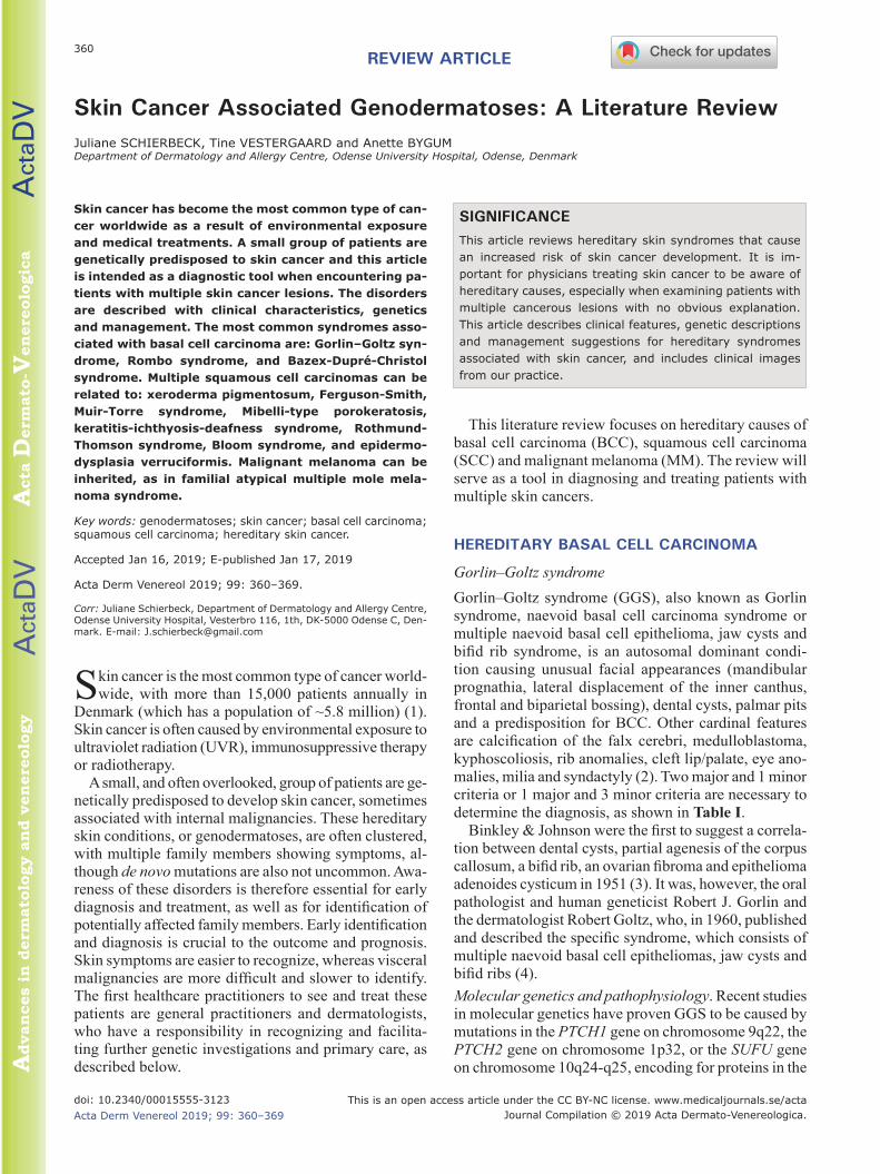
hedgehog signalling pathway, controlling growth and tissue development. The condition is autosomal dominant with complete penetrance, although approximately 10% of patients with GGS do not develop BCCs (5). The prevalence is reported to be 1 in 56,000–164,000 people, with an even sex distribution.Characteristics. Patients with GGS often start developing BCCs between puberty and the age of 35 years, but other cutaneous characteristics, such as palmar pits, should also raise suspicion. The primary diagnostic criteria are odontogenic keratocysts, palmoplantar pits, calcification of the falx cerebri, medulloblastoma, a first-degree relative with GGS and multiple BCCs (2). Minor criteria include rib anomalies, other specific skeletal malformations including macrocephaly, cleft lip and/or palate, ovarian/cardiac fibroma, lymphomesenteric cysts and ocular abnormalities. Palmar pits in childhood are considered a very strong indicator, along with skeletal abnormalities, such as bifid ribs, frontal bossing and hypertelorism. Patients with GGS may develop from a few BCCs to several hundreds of BCCs during their lifetime (Fig. 1 A, B).
Rombo syndromeMichaelsson, Olsson & Westermark were the first to describe Rombo syndrome (RS), in 1981, when they described a family with vermiculate atrophoderma, milia, hypotrichosis, trichoepitheliomas, BCCs and peripheral vasodilation with cyanosis (6). The condition is extremely rare and only a few cases have been reported since then.Molecular genetics and pathophysiology. The genetic mutation in RS has yet to be determined, but maletomale transmission has been described in a family with transmission through 4 generations, suggesting autosomal dominant inheritance (7).Characteristics. The syndrome presents in childhood with a reticular pattern of skin atrophy on the cheeks, preauricular area and forehead, along with cyanotic reddening of the skin. In adulthood whitishyellowish, milialike papules develop, along with telangiectatic
vessels. Defective or completely missing eyelashes and eyebrows are also seen in adulthood. BCCs develop in the 3rd or 4th decade of life and are a consistent complication throughout life (8) (Fig. 1 C).
Bazex-Dupré-Christol syndromeIn 1964 Bazex, Dupré & Christol first described the condition Bazex-Dupré-Christol syndrome (BDCS) as an Xlinked dominant syndrome affecting hair follicles and skin, along with an increased risk of developing BCCs. The condition is very rare; only approximately 20 families have been reported (9). Molecular genetics and pathophysiology. A recent study has revealed that BDCS might be caused by mutations in the ARCT1 gene, resulting in an aberrant activation of the Hedgehog signalling pathway (10). BCCs have been described in the first decade of life, although they most commonly present in the second decade onwards. The female to male ratio is 2:1 (11).Characteristics. Patients with BDCS are generally diagnosed based on a combination of hypotrichosis, multiple milia primarily on the face, follicular atrophoderma and multiple BCCs. Hypohidrosis and facial hyperpigmentation are also described as earlyonset manifestations. The follicular atrophoderma is located mainly on the dorsa of the hands, giving a characteristic orangepeel appearance. BCCs are reported as early as the age of 3 years, but often develop in the second or third decade of life, typically in sunexposed areas, such as the head and neck (9, 12) (Fig. 1 D).
Fig. 1. (A, B) Patient with Gorlin–Goltz syndrome and multiple basal cell carcinomas (BCC) of the scalp and neck. This patient has had more than 600 tumours removed. (C) Hypotrichosis as seen in Rombo syndrome. (D) Milia on the cheek, as seen in Bazex-Dupré-Christol syndrome.
Table I. Diagnostic criteria for Gorlin–Goltz syndrome
Major criteria• Excessive numbers of basal cell carcinomas out of proportion with prior sun
exposure and skin type or <20 years of age • Odontogenic keratocysts of the jaws prior to 20 years of age • Palmar or plantar pitting • Lamellar calcification of the falx cerebri • Medulloblastoma, typically desmoplastic • First-degree relative with Gorlin–Goltz syndrome
Minor criteria• Rib anomalies • Other specific skeletal malformations and radiological changes (i.e. vertebral
anomalies, kyphoscoliosis, short fourth metacarpals, postaxial polydactyly) • Macrocephaly • Cleft lip and/or palate • Ovarian/cardiac fibroma • Lymphomesenteric cysts• Ocular abnormalities (i.e. strabismus, hypertelorism, congenital cataracts,
glaucoma, coloboma)

Act
aDV
Act
aDV
Advan
ces
in d
erm
ato
logy a
nd v
en
ere
olo
gy
Acta
Derm
ato
-Ven
ere
olo
gic
a
J. Schierbeck et al.362
www.medicaljournals.se/acta
Management. Treatment of patients with multiple BCCs should be performed in a multidisciplinary approach, including dermatology, plastic surgery, ophthalmology and, in some cases, dentistry and otolaryngology (13). Since patients may develop multiple BCCs, the treatment should primarily include nonsurgical methods, and, for patients with multiple or aggressive BCCs, treatment with a hedgehog inhibitor might be indicated (6). When the suspicion of a genodermatosis arises, clinical identification and genetic investigation is crucial in dermatological treatment and regulation. Annual checkups should be offered and tailored to all predisposed patients. There are no perfect solutions, but prompt management of smaller BCCs can improve the cosmetic outcome.
Non-surgical methods, such as imiquimod, 5-fluorouracil (5FU), photodynamic therapy (PDT) and cryotherapy, should always be considered as first-line treatments. Some patients develop hundreds of BCCs in a lifetime and every treatment should therefore be as discreet as possible.Imiquimod is produced as a patientapplied cream and works by enhancing the innate arm of the immune system through tolllike receptor 7 (TLR7) commonly involved in pathogen recognition. Cells activated by imiquimod via TLR-7 secrete cytokines (primarily interferon-α, interleukin-6, and tumour necrosis factor-α (TNF-α)). Other cell types activated by imiquimod include natural killer cells, macrophages and Blymphocytes. This treatment is used for actinic keratosis (AK), morbus Bowen (MB) and BCC.5-FU cream is used for AK and BCC. It acts primarily as a thymidylate synthase inhibitor. Interrupting the action of this enzyme blocks the synthesis of thymidine, which is a nucleoside required for DNA replication. 5FU therefore causes rapidly dividing cancerous cells to undergo cell apoptosis as a result of thymidine depletion.PDT is used in a variety of medical conditions, mostly localized BCC, AK and MB. A photosensitizing agent is applied on the affected skin and will be absorbed by the impaired cells. When exposed to light, the cells produce radicals and reactive oxygen species, including singlet oxygen (O2), hydroxyl radicals (•OH) and superoxide (O2−) ions. With sufficient oxidative damage, the result is targeted cell death.
For more delimited or larger tumours, curettage or surgical excision is necessary. The goal should be to maintain a cosmetically acceptable outcome while treating the underlying malignancy.
Other rare syndromes linked to development of BCC There are other rare syndromes linked to an increased risk of development of BCC. Their symptoms somewhat overlap the previously mentioned syndromes, and genetic investigation often leads to the correct diagnosis. These are listed in Table II.
HEREDITARY SQUAMOUS CELL CARCINOMA
Xeroderma pigmentosumXeroderma pigmentosum (XP) was first described in 1874 by Hebra & Kaposi (14). XP, which means “dry pigmented skin”, is an autosomal recessive disorder with a high tumour burden. In 1968 James Cleaver described the genetic cause as a defect in DNA repair. The incidence is approximately 1 in 250,000 newborns, and it is an ultrarare condition with a very few cases in smaller countries (15). Molecular genetics and pathophysiology. Multiple genes have been identified as the cause of XP, all of them associated with nucleotide excision repair (NER). NER plays an essential role in the correction of UVinduced DNA damage, thereby preventing skin cancer. The diagnosis is based on family history, clinical findings and biallelic genetic mutations in the following genes: XPA, XPB, XPC, XPD, XPF, XPG or POLH. The median age at onset of BCC and SCC is approximately 8 years, more than 50 years earlier than in the general population (14). XP patients have an estimated 10,000fold increased risk of nonmelanoma skin cancer (NMSC) and a 2,000fold increased risk of melanoma below the age of 20 years (16).
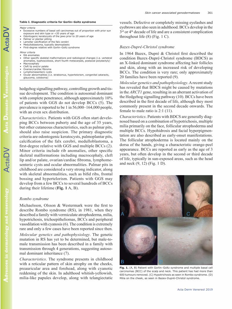
A clinically similar variant XPV is primarily found in Europe and the USA, but unlike the other XP mutations, this variant is characterized by a failure in errorfree translational synthesis past DNA photoproducts (16).Characteristics. XP is characterized by severe photosensitivity and premature skin ageing, along with pigmentary changes and a high risk of skin malignancies, such as BCC, SCC and MM. The first indication of XP is often seen after sunexposure, causing severe sunburn, which takes days or weeks to heal. The reaction can occur even in the first weeks of life, and is often misinterpreted as neglect or labelled wrongly as cellulitis or impetigo (17). Without sun protection, the skin ages and becomes dry, rough and atrophic (18). Small hypopigmented spots and telangiectasia can present later on. Optical photophobia is often present (Fig. 2 A, B).Management. Although there are individual variations, the prognosis of XP depends on minimizing sunexposure and detecting skin changes in their earliest stages. Smaller preneoplasms and localized BCCs can be treated with cryotherapy or topically with 5FU or imiquimod (16). Larger skinlesions and SCCs should always be treated surgically, preferably with Moh’s surgery or with irrefutable free margins due to the high risk of recurrence. XP
Table II. List of other genodermatoses associated with basal cell carcinoma
• Oley syndrome (possibly a subtype of Bazex-Dupré-Christol syndrome)• Schöpf-Schulz-Passarge syndrome• Cartilage-hair hypoplasia• Cowden syndrome• Hermansky-Pudlak syndrome• Muir-Torre syndrome• Brooke-Spiegler syndrome
Act
aDV
Act
aDV
Advan
ces
in d
erm
ato
logy a
nd v
en
ere
olo
gy
Acta
Derm
ato
-Ven
ere
olo
gic
a
363Skin cancer associated genodermatoses
Acta Derm Venereol 2019
patients also have increased risk of ocular abnormalities, along with neurological defects, and should be referred to specialized investigation at the time of diagnosis. Regular complete skin examinations are essential in early detection of precancerous skin changes and malignancies. This can improve the quality of life and, in the best case, also increase life expectancy (16).
Ferguson-Smith syndromeIn 1934 the Scottish dermatologist J. Ferguson-Smith described a correlation between certain symptoms in a family of Scottish miners. He depicted a condition with early onset of multiple selfhealing squamous epitheliomas (MSSE) (17). In 1971 his son, geneticist M. FergusonSmith, ascertained the genetic inheritance as an autosomal dominant genodermatosis (19). Molecular genetics and pathophysiology. The genetic mutation was shown, in 2005, to reside in the TGFBR1 and TGFBR2 genes, which are both tumour suppressor genes. The mutations cause an inactivation of the TGFβ-pathway, triggering uncontrolled cell growth (20). The mechanism of spontaneous healing has yet to be clarified.Characteristics. Patients with MSSE often show symptoms early in life, presenting skin tumours highly suspicious of malignancy. The tumours usually present within 3–4 weeks, growing from a 1–2 mm red papule to a pearly nodule with central keratosis, visually imitating a keratoacanthoma. Biopsies cannot differentiate between SCC and MSSE tumours, thus a thorough history and examination of the skin is important. The tumours spontaneously selfheal after 2–3 months leaving a small recessed scar. The tumours have a tendency to local tissue invasion, but will never metastasize (20) (Fig. 2 C, D). Management. Surgical intervention is often performed due to the histological suspicion of SCC, and if treated early on, is a good solution. However, mutilating surgery
should be avoided. PDT can be used as an alternative to excision, whereas some studies have proposed that radiotherapy might worsen the condition. This statement, however, has not been substantiated by original references and does not concur with our personal experience. Tumours, situated outside the risk areas (eyes, nose, ears and mouth) can be left to selfheal, although this should be done under regular clinical supervision. Again, the goal is to keep the patient tumourfree with the best cosmetic result.
Muir-Torre syndromeIn 1967 the British physician Edgar G. Muir noted the correlation between several keratoacanthomas and the development of multiple internal malignancies at a young age (21). One year later the American dermatologist Douglas P. Torre described the same findings at the New York Dermatologic Society (22).
MuirTorre syndrome (MTS) is a rare autosomal dominant condition with high penetrance and variable expression, thought to be a subtype of hereditary nonpolyposis colorectal cancer (HNPCC). HNPCC occurs in approximately 1:350 live births, and MTS is seen in approximately 9.2% of these (22). MTS has a male to female ratio of 3:2.Molecular genetics and pathophysiology. MTS is caused by mutations in the MLH1, MSH2, and MSH6 genes, involved in DNA mismatch repair (23). As for other genodermatoses, MTS is caused by a defects in the DNA mismatch repair, affecting rapidly dividing cells, in particular viscera and skin. Subsequently resulting in an increased risk of NMSC and visceral malignancies.Characteristics. Patients with MTS present with a high amount of sebaceous neoplasms: adenomas, epitheliomas, and carcinomas. Skin lesions initially present as painless, slowgrowing, pink or yellow nodules, often
Fig. 2. (A, B) Patient with xeroderma pigmentosum with hyperpigmentation of sun-exposed areas, impaired hearing and dry, atrophic skin. Rare diseases, Taylor & Francis. (C, D) Skin tumours in patients with Ferguson-Smith. (C) Upper lip. (D) A finger post self-healing. (E, F) Poikiloderma in a young patient with Rothmund-Thomson syndrome. Written permission from the patient is obtained to publish these photos. Figures A and B are published after permission from Taylor & Francis (50).

Act
aDV
Act
aDV
Advan
ces
in d
erm
ato
logy a
nd v
en
ere
olo
gy
Acta
Derm
ato
-Ven
ere
olo
gic
a
J. Schierbeck et al.364
www.medicaljournals.se/acta
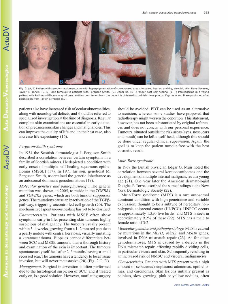
with central umbilication or ulceration. Most of the tumours are benign, but the sebaceous carcinoma can be aggressive, resulting in local invasion and metastases. Keratoacanthomas in MTS usually display sebaceous traits and multiple keratoacanthomas should give rise to further investigation.
Patients with MTS are also prone to develop malignancies of the colon and genitourinary tract (Fig. 3 D, E). The diagnosis is based on at least one sebaceous gland tumour (adenoma, epithelioma, or carcinoma) and one internal malignancy. The mean age of onset of sebaceous neoplasms is 53 years, but it can present as early as the age of 21 years (23).Management. Patients should be offered the same strict screening for colorectal carcinoma and other malignancies as patients with HNPCC. This includes frequent and early colonoscopies, mammograms, dermatological evaluation, and imaging of the abdomen and pelvis (24). Treatment of skin malignancies consists of oral isotretinoin, which has been found to prevent tumour development (24). Tumour excision or curettage is often needed for curative tumour treatment
Mibelli-type hereditary porokeratosisIn 1893 the Italian dermatologist Vittorio Mibelli published a case report of a young man, “age 21 [years], unmarried, of a welltodo family of Parma” with what he termed “porokeratosis” (25). Six clinical variants of porokeratosis are now recognized: porokeratosis of Mibelli, disseminated superficial porokeratosis, disseminated superficial actinic porokeratosis, porokeratosis plantaris et palmaris disseminata, punctate porokeratosis and linear porokeratosis (25).
Molecular genetics and pathophysiology. Porokeratosis is considered a premalignant condition. All types of porokeratosis can undergo malignant transformation, most commonly, into SCC and less commonly into BCC. All forms share common features of cornoid lamella on histological examination. The lesions are thought to be due to cellular clones exhibiting varying degrees of dysplasia. Immunosuppressive diseases and drugs, burn wounds and exposure to UVR are all known contributing factors (26) (Fig. 3 C).Characteristics. The skin lesions present as keratotic papules or annular plaques that expand centrifugally with an elevated keratotic border. When circumferentially involving the digits, it can induce pseudoainhum. Most lesions are asymptomatic, but ulcerative lesions have been described (26).Management. Patients should be offered regular checkups and be advised to use sun protection and avoid excessive sunlight. Upon suspicion, lesions should be biopsied. If the lesion has not undergone malignant transformation, excision is curative. Classical porokeratosis of Mibelli can be treated successfully with imiquimod cream (27). Surgical interventions and cryotherapy may be preferred when the use of topical agents is difficult or contraindicated.
Keratitis-ichthyosis-deafness syndromeIn 1915 the Canadian dermatologist Frederick S. Burns first described the condition as a combination of congenital atypical ichthyosiform erythrokeratoderma, palmoplantar keratosis, and sensorineural hearing loss in a 16yearold boy. In 1981 Skinner et al. reviewed 18 affected patients and proposed the name keratitis
Fig. 3. (A, B) A 31-year-old man with keratitis-ichthyosis-deafness (KID) syndrome, alopecia and keratosis. (C) Mibelli-type hereditary porokeratosis of the abdomen. (D, E) Pale, keratotic nodule on the columella of a patient with Muir-Torre syndrome. Written permission from the patient is obtained to publish these photos.

Act
aDV
Act
aDV
Advan
ces
in d
erm
ato
logy a
nd v
en
ere
olo
gy
Acta
Derm
ato
-Ven
ere
olo
gic
a
365Skin cancer associated genodermatoses
Acta Derm Venereol 2019
ichthyosisdeafness (KID) syndrome to describe the 3 main symptoms (28). It is a rare congenital disorder of ectoderm with approximately 100 reported cases in the literature (29).Molecular genetics and pathophysiology. KID syndrome belongs to the connexin disorders caused by heterozygous missense mutations in the connexin-26 gene, GJB2, or the connexin-30 gene, GJB6 (29). GJB2 and BJB6 are gap junction proteins expressed in ectodermderived epithelia of the inner ear, cornea and epidermis, which explain the constellation of pathological findings.Characteristics. KID syndrome is characterized by deafness, erythroderma, hyperkeratotic plaques and often keratitis. Alopecia and an increased susceptibility to infections, viral, bacterial and fungal are also common. Patients are sometimes seen with a follicular occlusion triad (dissecting cellulitis of the scalp, cystic acne, and hidradenitis suppurativa) along with follicular tumours and SCC (Fig. 3 A, B).Management. The essential issue is early diagnosis of infections and NMSC. Lifelong followup is recommended to assure early diagnosis of malignant tumours, especially SCC of the hyperkeratotic skin and mucosa. Systemic retinoids can reduce the hyperkeratosis and may reduce the incidence of skin cancer (30).
Rothmund-Thomson syndromeRothmund-Thomson syndrome (RTS) was first described in 1868 by the German ophthalmologist Rothmund as a combination of poikiloderma, growth retardation and juvenile cataract (31). In 1936 the British dermatologist Thomson added 3 similar cases. There is an increased risk of osteosarcoma and this diagnosis should always be considered when encountering a patient with osteosarcoma, poikiloderma and NMSC.Molecular genetics and pathophysiology. Two types of RTS have been proposed based on the clinical presentation and possible genetic mutation in the RECQL4 gene (31). It encodes for the RECQ helicase, which is responsible for correcting doublestranded DNA breaks. The loss of this gene leads to an accumulation of unrepaired DNA damage. RTSI is an autosomal recessive heterogeneous disorder with unknown aetiology. In RTSII there is a homozygous or compound heterozygous defect in RECQL4. The condition is considered very rare and only approximately 300 patients have been recorded in medical literature (32). Characteristics. The severity and amount of symptoms displayed in individual patients are highly variable, but the skin, hair, nails and teeth are first to alter. The hair is often thin, brittle and sparse, and the nails are typically dystrophic with pachyonychia as a frequent sign. Dental aberrations include microdontia, rudimentary or hypoplastic teeth and disorders of dental breakthrough.
A diagnostic hallmark is the erythematous, oedematous, blistering facial rash often acquired between the age of 3 and 6 months, later involving the extremities and buttocks. The rash eventually reaches a chronic phase of poikiloderma and lesions of hypo and hyperpigmentation, telangiectatic vessels and punctate atrophy persisting throughout life (32).
The mildest variant is RTSI, characterized by poikiloderma, juvenile cataract and ectodermal dysplasia (Fig. 2 E, F). RTSII also causes poikiloderma, congenital bone defects, increased risk of osteosarcoma in childhood and SCC in young adults (33). Growth deficiency and skeletal defects are the second major criteria, almost exclusively seen in RTSII as a result of the RECQL4 mutation. Xrays can be helpful to visualize defects and detect more subtle anomalies, such as radial aplasia or hypoplasia, osteopaenia, patellar ossification defects and destructive bone lesions (32).Management. All patients diagnosed with RTS should be managed by a multidisciplinary team, including a dermatologist, an oncologist, an ophthalmologist and an orthopaedic surgeon. Patients should be offered lifelong followup, including regular skin examinations to screen for SCC/BCC, and advice with regard to sun care. With regular screening and treatment, these patients have the same lifespan expectancy as the background population (33).
Bloom syndromeBloom syndrome (congenital telangiectatic erythema, BS) is a rare autosomal recessive disorder. David Bloom described the condition in 1954 in a series of patients with telangiectatic facial erythema and dwarfism (34). The overall prevalence is unknown, but in the Ashkenazi Jewish population it is estimated to be approximately 1 in 48,000 live births. There seems to be a slight majority of males (35).Molecular genetics and pathophysiology. The cause is believed to be a mutation in the BML gene, controlling the enzyme RecQL3 responsible for restoring malfunctioning replication forks during DNA replication. This defect consequently causes genetic instability due to pathological DNA exchanges between parallel chromatids, which may lead to malignancy (36). This may cause skin cancer, most often SCC, but also other malignancies of the upper and lower gastrointestinal and urinary tract. Characteristics. Patients with BS are often characterized by severe growth retardation, highpitched voices and reduced subcutaneous layer of fat, causing their muscles to appear more prominent. The patients often seek medical assistance due to recurrent infections, diabetes, chronic pulmonary disease and a predisposition to cancer development. Dermatological features include severe photosensitivity, poikiloderma and erythematous telangiectasia. SCC accounts for 14% of all tumours in
Act
aDV
Act
aDV
Advan
ces
in d
erm
ato
logy a
nd v
en
ere
olo
gy
Acta
Derm
ato
-Ven
ere
olo
gic
a
J. Schierbeck et al.366
www.medicaljournals.se/acta
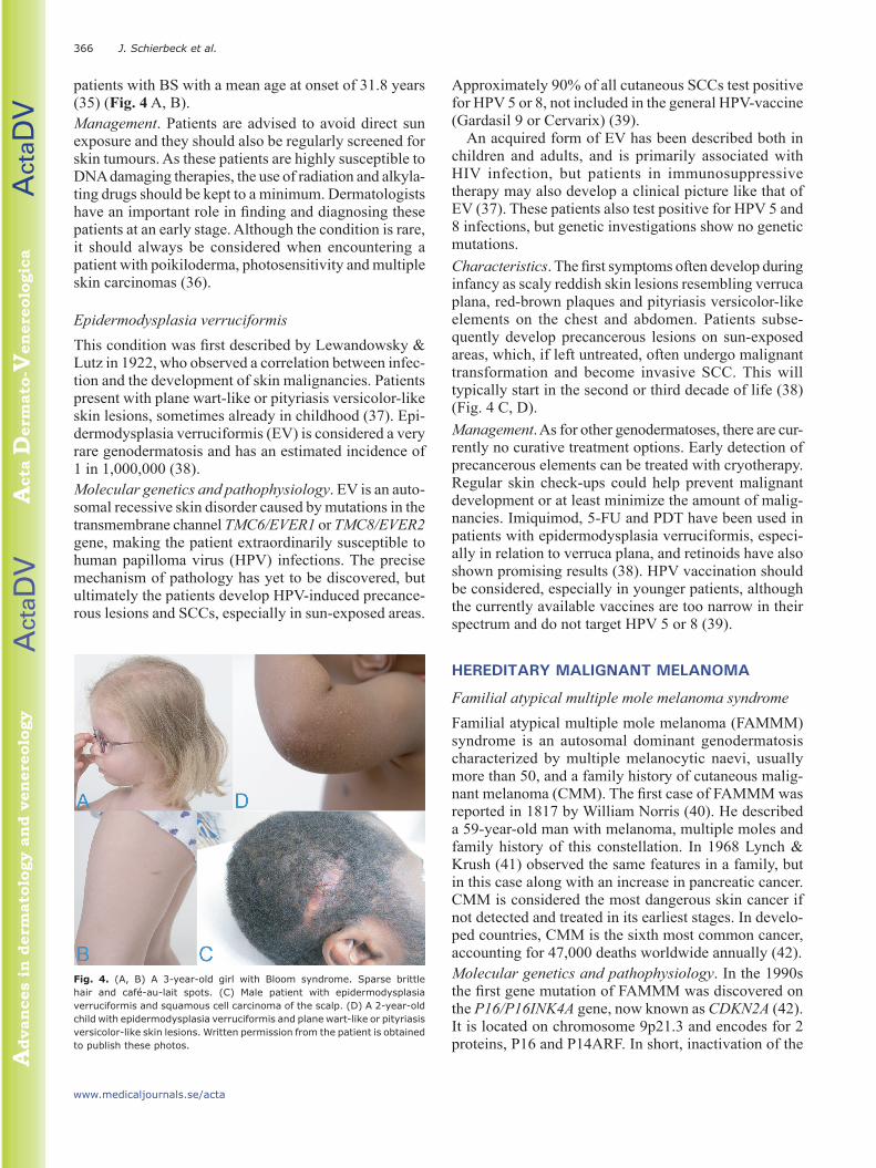
patients with BS with a mean age at onset of 31.8 years (35) (Fig. 4 A, B).Management. Patients are advised to avoid direct sun exposure and they should also be regularly screened for skin tumours. As these patients are highly susceptible to DNA damaging therapies, the use of radiation and alkylating drugs should be kept to a minimum. Dermatologists have an important role in finding and diagnosing these patients at an early stage. Although the condition is rare, it should always be considered when encountering a patient with poikiloderma, photosensitivity and multiple skin carcinomas (36).
Epidermodysplasia verruciformisThis condition was first described by Lewandowsky & Lutz in 1922, who observed a correlation between infection and the development of skin malignancies. Patients present with plane wartlike or pityriasis versicolorlike skin lesions, sometimes already in childhood (37). Epidermodysplasia verruciformis (EV) is considered a very rare genodermatosis and has an estimated incidence of 1 in 1,000,000 (38).Molecular genetics and pathophysiology. EV is an autosomal recessive skin disorder caused by mutations in the transmembrane channel TMC6/EVER1 or TMC8/EVER2 gene, making the patient extraordinarily susceptible to human papilloma virus (HPV) infections. The precise mechanism of pathology has yet to be discovered, but ultimately the patients develop HPVinduced precancerous lesions and SCCs, especially in sunexposed areas.
Approximately 90% of all cutaneous SCCs test positive for HPV 5 or 8, not included in the general HPVvaccine (Gardasil 9 or Cervarix) (39).
An acquired form of EV has been described both in children and adults, and is primarily associated with HIV infection, but patients in immunosuppressive therapy may also develop a clinical picture like that of EV (37). These patients also test positive for HPV 5 and 8 infections, but genetic investigations show no genetic mutations.Characteristics. The first symptoms often develop during infancy as scaly reddish skin lesions resembling verruca plana, redbrown plaques and pityriasis versicolorlike elements on the chest and abdomen. Patients subsequently develop precancerous lesions on sunexposed areas, which, if left untreated, often undergo malignant transformation and become invasive SCC. This will typically start in the second or third decade of life (38) (Fig. 4 C, D).Management. As for other genodermatoses, there are currently no curative treatment options. Early detection of precancerous elements can be treated with cryotherapy. Regular skin checkups could help prevent malignant development or at least minimize the amount of malignancies. Imiquimod, 5FU and PDT have been used in patients with epidermodysplasia verruciformis, especially in relation to verruca plana, and retinoids have also shown promising results (38). HPV vaccination should be considered, especially in younger patients, although the currently available vaccines are too narrow in their spectrum and do not target HPV 5 or 8 (39).
HEREDITARY MALIGNANT MELANOMA
Familial atypical multiple mole melanoma syndromeFamilial atypical multiple mole melanoma (FAMMM) syndrome is an autosomal dominant genodermatosis characterized by multiple melanocytic naevi, usually more than 50, and a family history of cutaneous malignant melanoma (CMM). The first case of FAMMM was reported in 1817 by William Norris (40). He described a 59yearold man with melanoma, multiple moles and family history of this constellation. In 1968 Lynch & Krush (41) observed the same features in a family, but in this case along with an increase in pancreatic cancer. CMM is considered the most dangerous skin cancer if not detected and treated in its earliest stages. In developed countries, CMM is the sixth most common cancer, accounting for 47,000 deaths worldwide annually (42).Molecular genetics and pathophysiology. In the 1990s the first gene mutation of FAMMM was discovered on the P16/P16INK4A gene, now known as CDKN2A (42). It is located on chromosome 9p21.3 and encodes for 2 proteins, P16 and P14ARF. In short, inactivation of the
Fig. 4. (A, B) A 3-year-old girl with Bloom syndrome. Sparse brittle hair and café-au-lait spots. (C) Male patient with epidermodysplasia verruciformis and squamous cell carcinoma of the scalp. (D) A 2-year-old child with epidermodysplasia verruciformis and plane wart-like or pityriasis versicolor-like skin lesions. Written permission from the patient is obtained to publish these photos.

Act
aDV
Act
aDV
Advan
ces
in d
erm
ato
logy a
nd v
en
ere
olo
gy
Acta
Derm
ato
-Ven
ere
olo
gic
a
367Skin cancer associated genodermatoses
Acta Derm Venereol 2019
CDKN2A gene disturbs the TP53 tumoursuppressor pathways and thereby enhances proliferation and reduces apoptosis, increasing the risk of malignant transformation (43). The mutation has a reduced penetrance and a geographically variable expressivity. In 2003 Hayward described the correlation between mutations in the gene coding for cyclin-dependent kinase 4 (CDK4) and hereditary MM (44). This mutation makes CDK4 insensitive to inhibition by the protein P16, and the genetic outcome is similar to that of CDKN2Amutations. Germline mutations of CDK4 are, however, considered very rare in FAMMM (44).
It is estimated that approximately 5–12% of all malignant melanomas are hereditary, and approximately 40% of these are caused by CDKN2A mutations. The majority of familial MM is therefore either caused by an unknown mutation or has no genetic mutation and is probably a result of a shared sun exposure and susceptible skin types (42). Characteristics. FAMMM is a clinical diagnosis based on objective findings and only supported by genetic testing. The diagnostic criteria are shown in Table III. Most FAMMM patients have a familial history of MM, but are often not aware of their own increased risk. CMM is commonly detected between the second and third decade of life and patients frequently experience more than 1 melanoma in their lifetime (41) (Fig. 5).Management. Dermatological screening for CMM in FAMMM patients should be offered at an early age, with a baseline complete skin examination including scalp, oral mucosa, genital area and nails, given that CMM can progress even in the early teens. Exact evaluation of naevi can be difficult even with regular dermoscopy; consequently other objective measurements, such as digital dermoscopy and computer image analysis, have been introduced to reduce diagnostic errors. Thorough counselling on sunexposure and protection is essential in the prophylactic treatment of this patient group. Treatment of verified MM should follow national guidelines with regards to excision margins, sentinel node diagnostics, PET/CT screenings, etc. (45).
BAP-1 mutationOther melanomapredisposing gene mutations have been revealed, the most prominent of them being the BRCA1associated protein1 (BAP-1). Melanocytic BAP-1 mutated atypical intradermal tumours (MBAITs) were first described as a distinct entity in 2011 by Wiesner et al.
(46) . It is considered an autosomal dominant syndrome caused by germline mutations of BAP-1, characterized by a high penetrance of melanocytic neoplasms. So far 4 distinct cancers have been linked to BAP-1 mutations; CMM, uveal melanoma, malignant mesothelioma and renal cell carcinoma (47). Molecular genetics and pathophysiology. BAP-1 was first described as a binding partner of BRCA1. Its cellular role is not fully explained, but has been proposed in the DNA damage response, as well as in regulation of apoptosis, senescence, and the cell cycle (47).Characteristics. Patients with this syndrome typically develop skincoloured or tanned, elevated tumours, measuring approximately 5 mm, in the second decade of life (48). More tumours often develop over time, but the number of tumours can vary greatly from patient to patient. Uveal melanoma is the most common malignancy in families with BAP-1 mutations with a mean age of onset of 50 years, but patients as young as 16 years have been reported (47). These patients tend to have more aggressive cancers, with higher tumour staging and a higher risk of metastasis.Management. Annual dermatological and ophthalmological follow up is recommended, in combination with annual abdominal ultrasounds. Magnetic resonance imaging (MRI) every 2 years could be considered, as well as annual physical examinations focusing on the risk of mesothelioma.
Patients should be given the same sunprotection guidelines as patients with CDKN2A/CDK4 mutations, as previously mentioned.
Table III. Diagnostic criteria of familial atypical multiple mole melanoma
1. Malignant melanoma in 1 or more first- or second-degree relatives2. High total-body naevi count (often >50), including some of which are clinically atypical (asymmetric, raised, colour variation present, of variable sizes)3. Naevi with certain histological features on microscopy*
*Architectural disorder with asymmetry, subepidermal fibroplasia, and lentiginous melanocytic hyperplasia with spindle or epithelioid melanocytes gathering in nests of variable size and fusing with adjacent rete ridges to form bridges; variable dermal lymphocyte infiltration and the ”shouldering” phenomenon, wherein intraepidermal melanocytes extend alone or in groups beyond the main dermal component.
Fig. 5. A 36-year-old man presenting with multiple naevi due to familial atypical multiple mole melanoma syndrome.

Act
aDV
Act
aDV
Advan
ces
in d
erm
ato
logy a
nd v
en
ere
olo
gy
Acta
Derm
ato
-Ven
ere
olo
gic
a
J. Schierbeck et al.368
www.medicaljournals.se/acta
Other conditions associated with malignant melanoma Several other mutations disposing to both cancer and melanoma have been discovered recently. In particular, 3 mutations have been associated with MM; microphthalmiaassociated transcription factor (MITF), protection of telomeres 1 (POT-1) and the telomerase reverse transcriptase (TERT) mutations (48, 49). Germline mutations in the MITF gene are thought to increase the risk of MM, as it represents a melanocytic lineage-specific transcription factor.
Both POT-1 and TERT are telomerasecontrolling genes, and mutations in these can cause uncontrollable cell cycles and malignant transformations.
Other syndromes, where the association with melanoma plays a less dominant role, exist. These include xeroderma pigmentosum (described above), Cowden syndrome (PTEN mutations), LiFraumeni syndrome (TP53 mutations) and hereditary breast cancer (BRCA 1/2 mutations) (49).
CONCLUSION
Genodermatoses with skin cancer predispositions are, in general, very rare, and prompt treatment and referral to genetic investigation is of utmost importance. The skin is often the first organ to display symptoms, and dermatologists therefore have a greater responsibility in early examination and executing appropriate treatment plans. Extracutaneous symptoms include pancreatic and gastrointestinal cancer, bone abnormalities, teeth malformations, neurological deficits and cognitive impairment. The abovementioned syndromes have many similarities, and are often thought of in the same context. Exact genetic investigation is therefore crucial in establishing the correct diagnosis and treatment, especially in relation to extracutaneous symptoms.
REFERENCES1. Lamberg A, Sølvsten H, Lei U, Vinding GR, Stender IM, Jemec
GB, et al. The Danish Nonmelanoma Skin Cancer Dermato-logy Database. Clin Epidemiol 2016; 8: 633–636.
2. Larsen AK, Mikkelsen DB, Hertz JM, Bygum A. Manifestations of Gorlin-Goltz syndrome. Dan Med J 2014; 61: A4829.
3. Binkley GW, Johnson HH Jr. Epithelioma adenoides cysticum; basal cell nevi, agenesis of the corpus callosum and dental cysts; a clinical and autopsy study. AMA Arch Dermatol Syphilol 1951; 63: 73–84.
4. Gorlin RJ, Goltz RW. Multiple nevoid basal-cell epithelioma, jaw cysts and bifid rib. A syndrome. N Engl J Med 1960; 262: 908–912.
5. Joshi P, Deshmukh V, Golgire S. Gorlin Goltz Syndrome. Dent Res J (Isfahan) 2012; 9: 100–106.
6. Michaelsson G, Olsson E, Westermark P. The Rombo syn-drome: a familial disorder with vermiculate atropho-derma, milia, hypotrichosis,trichoepitheliomas, basal cell carcinomas and peripheral vasodilation with cyano-sis. Acta Derm Venereol 1981; 61: 497–503.
7. van Steensel MA, Jaspers NG, Steijlen PM. A case of Rom-bo syndrome. Br J Dermatol 2001; 144: 1215–1218. Note:
Erratum: Br J Dermatol 2002; 146: 715. 8. Bazex A, Dupre A, Christol B. Génodermatose complexe de
type indétermine associant une hypotrichose, un état atro-phodermique généralisé et des dégénérescences cutanées multiples (épithéliomas-basocellulaires). Bull Soc Fr Derma-tol Syph 1964; 71: 206.
9. Abuzahra F, Parren LJ, Frank J. Multiple familial and pig-mented basal cell carcinomas in early childhood – Bazex-Dupré-Christol syndrome. J Eur Acad Dermatol Venereol 2012; 26: 117–121.
10. Bal E, Park H-S, Belaid-Choucair Z, Kayserili H, Naville M, Madrange M, et al. Mutations in ACTRT1 and its enhancer RNA elements lead to aberrant activation of Hedgehog sig-naling in inherited and sporadic basal cell carcinomas. Nat Med 2017; 23: 1226–1233.
11. Castori M, Castiglia D, Passarelli F, Paradisi M. Bazex-Dupré-Christol syndrome: an ectodermal dysplasia with skin ap-pendage neoplasms. Eur J Med Genet 2009; 52: 250–255.
12. AlSabbagh M, Baqi M. Bazex-Dupré-Christol syndrome: re-view of clinical and molecular aspects. Int J Dermatol 2018; 57: 1102–1106.
13. Mello RN, Kahn Z, Choudry U. A multidisciplinary approach to the successful management of Gorlin syndrome. J Surg Case Rep 2017; 6: rjw224.
14. Hebra F, Kaposi M. Xeroderma, parchment skin. In On di-seases of the skin including exanthemata. Volume III. New Sydenham Soc 1874; 61: 252–258.
15. Kraemer KH, DiGiovanna JJ. Forty years of research on xeroderma pigmentosum at the US National Institutes of Health. Photochem Photobiol 2015; 91: 452–159.
16. Lehmann AR, Schubert S, Emmert S. Xeroderma Pigmento-sum: Diagnostic procedures, interdisciplinary patient care, and novel therapeutic approaches. J Dtsch Dermatol Ges 2014; 12: 867–871.
17. Lehmann AR, McGibbon D, Stefanini M. Xeroderma pigmen-tosum. Orphanet J Rare Dis 2011; 6: 70.
18. Smith JF. A case of multiple primary squamous-celled carcino-mata of the skin in a young man, with spontaneous healing. Br J Dermatol 1934; 46: 267–272.
19. Ferguson-Smith MA, Wallace DC, James ZH, Renwick JH. Multiple self-healing squamous epithelioma. Birth Defects 1971; 7: 157–163.
20. Broesby-Olsen S, Bygum A, Gerdes AM, Brandrup F. Multi-ple self-healing squamous epithelioma of Ferguson-Smith: observations in a Danish family covering four generations. Acta Derm Venereol 2008; 88: 52–56.
21. Muir EG, Bell AJ, Barlow KA. Multiple primary carcinomata of the colon, duodenum, and larynx associated with kerato-acanthomata of the face. Br J Surg 1967; 54: 191–195.
22. Torre D. Multiple sebaceous tumors. Arch Dermatol 1968; 98: 549–551.
23. John AM, Schwartz RA. Muir-Torre syndrome (MTS): An update and approach to diagnosis and management. J Am Acad Dermatol 2016; 74: 558–566.
24. Graefe T, Wollina U, Schulz H, Burgdorf W. Muir–Torre syn-drome – treatment with isotretinoin and interferon alpha-2a can prevent tumour development. Dermatol 2000; 200: 331–333.
25. Schamroth JM, Zlotogorski A, Gilead L. Porokeratosis of Mibelli. Overview and review of the literature. Acta Derm Venereol 1997; 77: 207–213.
26. Ferreira FR, Santos LD, Tagliarini FA, Lira ML. Porokeratosis of Mibelli – literature review and a case report. An Bras Dermatol 2013; 88: 179–182.
27. Gu CY, Zhang CF, Chen LJ, Xiang LH, Zheng ZZ. Clinical analysis and etiology of porokeratosis. Exp Ther Med 2014; 8: 737–741.
28. Skinner BA, Greist MC, Norins AL. The keratitis, ichthyosis, and deafness (KID) syndrome. Arch Dermatol 1981; 117: 285–289.
29. Bygum A, Betz R, Kragballe K, Steiniche T, Peeters N, Wuyts W, Nöthen M. KID Syndrome: report of a Scandinavian pa-tient with connexin-26 gene mutation. Acta Derm Venereol 2005; 85: 152–155.

Act
aDV
Act
aDV
Advan
ces
in d
erm
ato
logy a
nd v
en
ere
olo
gy
Acta
Derm
ato
-Ven
ere
olo
gic
a
369Skin cancer associated genodermatoses
Acta Derm Venereol 2019
30. Chandra S, Bygum A. Successful treatment with alitretinoin of dissecting cellulitis of the scalp in keratitis-ichthyosis-deafness syndrome. Acta Derm Venereol 2013; 93: 473–474.
31. Rothmund A. Uber Cataracte in Verbindung mit einer eigent-huemlichen Hautdegeneration. Albrecht von Graefes Arch Klin Exp Ophthalmol 1868; 14: 159–244.
32. Wang LL, Plon SE. Rothmund-Thomson Syndrome. 1999 Oct 6 [updated 2016 Aug 11]. In: Adam MP, Ardinger HH, Pagon RA, et al., editors. Seattle, WA: University of Washington, Seattle: GeneReviews®; 1993–2018.
33. Larizza L, Roversi G, Volpi L. Rothmund-Thomson syndrome. Orphanet J Rare Dis 2010; 5: 2.
34. Bloom D. Congenital telangiectatic erythema resembling lupus erythematosus in dwarfs; probably a syndrome entity. AMA Am J Dis Child 1954; 88: 754–758.
35. Arora H, Chacon AH, Choudhary S, McLeod MP, Meshkov L, Nouri K, et al. Bloom syndrome. Int J Dermatol 2014; 53: 798–802.
36. Karow JK, Wu L, Hickson ID. RecQ family helicases: roles in cancer and aging. Curr Opin Genet Dev 2000; 10: 32–38.
37. de Jong SJ, Imahorn E, Itin P, Uitto J, Orth G, Jouanguy E, et al. Epidermodysplasia verruciformis: inborn errors of immunity to human beta-papillomaviruses. Front Microbiol 2018; 9: 1222.
38. Burger B, Itin PH. Epidermodysplasia verruciformis. Curr Probl Dermatol 2014; 45: 123–131.
39. Vinzón SE, Rösl F. HPV vaccination for prevention of skin cancer. Hum Vaccin Immunother 2015; 11: 353–357.
40. Norris W. The first reported case of familial atypical multiple melanoma mole syndrome/A case of fungoid disease. Edin Med Surg J 1820; 16: 562–565. Available from: https://www.ncbi.nlm.nih.gov/books/NBK7025/.
41. Lynch H.T., Krush A.J. Hereditary and malignant melanoma: implications for early cancer detection. Can Med Assoc J.
1968;99:789–792.42. Soura E, Eliades PJ, Shannon K, Stratigos AJ, Tsao H. Here-
ditary melanoma: Update on syndromes and management: genetics of familial atypical multiple mole melanoma syn-drome. J Am Acad Dermatol 2016; 74: 395–407.
43. Eckerle Mize D, Bishop M, Resse E, Sluzevich J. Familial Atypical Multiple Mole Melanoma Syndrome. Cancer Syn-dromes. Bethesda, MD: National Center for Biotechnology Information; 2009.
44. Hayward NK. Genetics of melanoma predisposition. Oncogene 2003; 22: 3053–3062.
45. Garbe C, Peris K, Hauschild A, Saiag P, Middleton M, Bastholt L, et al. Diagnosis and treatment of melanoma. European consensus-based interdisciplinary guideline – update 2016. Eur J Cancer 2016; 63: 201–217.
46. Wiesner T, Obenauf AC, Murali R, Fried I, Griewank KG, Ulz P, et al. Germline mutations in BAP1 predispose to melanocytic tumors. Nat Genet 2011; 43: 1018–1021.
47. Rai K, Pilarski R, Cebulla CM, Abdel-Rahman MH. Compre-hensive review of BAP1 tumor predisposition syndrome with report of two new cases. Clin Genet 2016: 89: 285–294.
48. Soura A, Eliades PJ, Shannon K, Stratigos AJ, Tsao H. Here-ditary melanoma: update on syndromes and management – emerging melanoma cancer complexes and genetic coun-seling. J Am Acad Dermatol 2016; 74: 411–420.
49. Leachman SA, Lucero OM, Sampson JE, Cassidy P, Bruno W, Queirolo P, et al. Identification, genetic testing, and ma-nagement of hereditary melanoma. Cancer Metastasis Rev 2017; 36: 77–90.
50. Kralund HH, Ousager L, Jaspers NG, Raams A, Pedersen EB, Gade E, Bygum A. Xeroderma Pigmentosum-Trichothio-dystrophy overlap patient with novel XPD/ERCC2 mutation. Rare Dis. 2013;1:e24932. doi: 10.4161/rdis.24932. eCol-lection 2013.


![Cultural Governance: a literature review [p. 2]](https://static.fdokumen.com/doc/165x107/6312a178b22baff5c40ec300/cultural-governance-a-literature-review-p-2.jpg)