<•/ 'W'' : ,.-.>. •; v'' '•'.;>. Supported in part...
161
Number 1 A semiannual journal of research devoted to Helminthology and all branches of Parasitology ,V.' ' ••:!•- "('''•••' : <•/ 'W'' : ,.-.>. •; v '' '•'.;>. Supported in part by the 'BraytonH. Ransom Memorial Trust Fund , •,,. ^CONTENTS -.,.,;/.,; , ,'.^\, J ;; RADOMSKI, A. A., D. A. OSBORN, D. B. PENCE, M. /Il NELSON, AND R. J. ,-' , WARREN. Visceral helminths from an expanding insular, population of the long- nosed .armadillo (Dasypus noverricinctus) r. „ _ ...... ^»;.. r .., ..... _ _.., ..... ' 1 ' KRrrSKY, D. C. ANDW. A. BOEOER, Neoiropical Monpgenea. 16. New species of oviparous Gyrodactylidea with proposal of Npthogyrodactyltis gen. n. (Oogyro- dactylidae) .... :..i..._! . , ..._.-_..._..._^ ..__^. „ '7 GARDNER, S; L., S.J. UPTON, C. R. LAMBERT, AND O. C. JORDAN. Redescription of ' lEimeria ^come/r(Rastegaieff, 1930) from Myrmecopftaga tridactyla, and a first report from Bolivia , T . . „.. .„. .„„.. !...„._ ._.^<. „ 116 TELFORD, S. R., JR., D. J. FORRESTER, S. D. WRIGHT, M. E. ROELKE, S. A. FERENC, AND J. W.^McCowN. The identity and!prevalence of trypanosomes jn white- tailed deer (Odocoileus virginianus) from southern Rorida ,„ „_._._ _. .„„„ 19 FONT, W. F. Life cycle ofAmphimerus elongatus (Trematoda: Opisthorehiidae). .. 24 MADHAVI, R. AND U. SHAMEEM. Cercaria chilkaensis II, a new zoogonid cercaria \ from the snail, Nassarius orissaensis, from Chilka.Lake, India .: .... c. .\^.-. 2\\, !T. AND T. KIFONE. An Monorchjidae) from the loach, Cobitis ibiwae, in Japan _... J . .__. 35 Cox, W. T. ANDiG. L. HENDRICKSON. Observations on the life cycle of Proteocephalus : , tumidocollus (Cestoda: Proteocephalidae) in steelliead trout, Onchorhynchus my- kiss .._._»lL.^___...i.. ..;^:^ ...... 1 ... ..^...:.»..^....^.., ^__._mll!._,_:^_ 39 GSrrwooD, D. J. AND W. ,R. LUSBY. Sterol composition of the qorn root lesiori ''-- nematode, Pratylenctius agilis, and corn root cultures .... : „ . _^.._ 43 (Continued)on Outside Back Cover) Copyright © 2011, The Helminthological Society of Washington Copyright © 2011, The Helminthological Society of Washington
-
Upload
khangminh22 -
Category
Documents
-
view
1 -
download
0
Transcript of <•/ 'W'' : ,.-.>. •; v'' '•'.;>. Supported in part...

Number 1
A semiannual journal of research devoted toHelminthology and all branches of Parasitology
,V.' ' • • : ! • - " ( ' ' ' • • • ' : <•/ 'W'' : ,.-.>. •; v ' ''•'.;>. Supported in part by the
'BraytonH. Ransom Memorial Trust Fund ,
• , , . ^CONTENTS - . , . , ; / . , ; , , '.^\, J ;;
RADOMSKI, A. A., D. A. OSBORN, D. B. PENCE, M. /Il NELSON, AND R. J. ,-', WARREN. Visceral helminths from an expanding insular, population of the long-
nosed .armadillo (Dasypus noverricinctus) r. „ _......̂ »;..r..,....._ _..,.....' 1 'KRrrSKY, D. C. AND W. A. BOEOER, Neoiropical Monpgenea. 16. New species of
oviparous Gyrodactylidea with proposal of Npthogyrodactyltis gen. n. (Oogyro-dactylidae) ....:..i..._! . , ..._.-_..._..._^ ..__ .̂ „ '7
GARDNER, S; L., S.J. UPTON, C. R. LAMBERT, AND O. C. JORDAN. Redescription of 'lEimeria ^come/r(Rastegaieff, 1930) from Myrmecopftaga tridactyla, and a firstreport from Bolivia ,T . . „.. .„. .„„.. !...„._ ._.^<. „ 116
TELFORD, S. R., JR., D. J. FORRESTER, S. D. WRIGHT, M. E. ROELKE, S. A. FERENC,AND J. W.^McCowN. The identity and!prevalence of trypanosomes jn white-tailed deer (Odocoileus virginianus) from southern Rorida ,„ „_._._ _. .„„„ 19
FONT, W. F. Life cycle ofAmphimerus elongatus (Trematoda: Opisthorehiidae). .. 24MADHAVI, R. AND U. SHAMEEM. Cercaria chilkaensis II, a new zoogonid cercaria\ from the snail, Nassarius orissaensis, from Chilka.Lake, India .:....c. .\^.-. 2\\, !T. AND T. KIFONE. Anapfilaeorchis hamajimai gen. et sp. n. (Trematoda:
Monorchjidae) from the loach, Cobitis ibiwae, in Japan _... J . .__. 35Cox, W. T. ANDiG. L. HENDRICKSON. Observations on the life cycle of Proteocephalus : ,
tumidocollus (Cestoda: Proteocephalidae) in steelliead trout, Onchorhynchus my-kiss .._._»lL.̂ ___...i.. ..;̂ :̂ ...... 1 ... ..^...:.»..^....^.., ^__._mll!._,_:^_ 39
GSrrwooD, D. J. AND W. ,R. LUSBY. Sterol composition of the qorn root lesiori''-- nematode, Pratylenctius agilis, and corn root cultures .... : „ . _^.._ 43
(Continued)on Outside Back Cover)
Copyright © 2011, The Helminthological Society of WashingtonCopyright © 2011, The Helminthological Society of Washington

THE HELMINTHOLOGICAL SOCIETY OF WASHINGTON , . : ,
\ THE SOCIETY meets once a month from October through May foMhe presentation and discussion- ^ of papers in any and all branches of parasitplogy-or Delated sciences. All interested persons are invited
... .to attend. . ' j y ; > / » . ^ ' ., "•• •_ • \.Persons Interested in .membership in the Helminthological Society 'of Washington may obtain
application blanks in recent issues of THE JOURNAL. A year's subscription to the Journal is includedin the annual dues. , /, , / , •''"' ''.- ( .\ '-vV.' " ' ( • • . ' ' • ' " " ' ' •
,/>vv' , /<. ' • • ' ",:.-' : 7- —v,- " • - ;'^V • i v, . -,.:,. •' ' '• ^'~':'i ; - , ' !V. / OFFICERS OF THE SOCIETY FOR 1991' / . ' . ' ' ̂ ";
'. President: NANCY £>. PACHECO ' * \ r - ; ?• ~Vice President: ;RUTH M. KULSTAD /.. ,,. •/Corresponding Secretary-Treasurer: DAVID J. CHITWOOD • .. , '-<• \ ' JRecording Secretary: MARK C. JENKINS . ' - ' 1 -] ~ ,. , ;Archivist/Librarian: PATRICIA A.: PILITT ' , \ , V, .: ' ' - ' - . ' ,
, ,. Custodian of Back Issues: J. RALPH LICHTENFELS -' $ 1" , — ;.Representative to the Washington Academy vf Sciences: KENDALL G.^'POWERS.. « ' " " , v .
-Representative to the American Society of Parasitologists: ERIG P. HOBERG, l/ , ,' Executive Committee Members-at-Large: JOAN E. JACKSON, 1991 " '
.v- DANTE S.ZARLENGA, 1991 :';/;.' ft' '•'• ^ (•;' - ' ' - YUPIN CHAROENVIT, 1992 ; v \\ - •
' •..•;;'1'.'.\.i'-\; ) • ' • . . . ' • ' . 'V . . - \HYUNLILLEHOJ, 1592 •'"'•,: >''>•,''.{.Immediate Past President: JOHN H. CROSS i " > \ \
THE JOURNAL OF THE HELMINTHOLOGICAL SOCIETY OF WASHINGTON i
THE JOURNAL, is published serriiannually at Lawrence, Kansas by the Helminthological Societyof Washington. Papers need not be presented at a meeting to be pubh' shed iri> the Jburnal.
MANUSCRIPTS should be sent to the EDITOR, Ralph P. Eckerlin, Natural Sciences Division,Northern Virginia Community College, Annandale, VA 22Q03., Manuscripts must be typewritten,doubjeispaced, and in -finished form. The original and'two copies are required. Photocopies of drawingsmay be ̂ submitted for review purposes but glossy .prints of halftones /are required; originals will berequested after acceptance of the manuscript. Papers are accepted with the 'understanding that theywill be published only in the Journal. ; < - 7 - ' ^ •!'l\ REPRINT;S may be ordered *froit> the PRINTER at the same time the corrected proof is returned
to the " ' ~ ' '• ''f'"AUTHORS' CONTRIBUTIONS to publication costs (currently $40/pg for members, actual cost/(
pg currently $80, ̂ br non-members) will be'billed by Allen Pf ess and are.payable to the SOCIETY1., /BACK VOLUMES of ;the JournaLare available. Inquiries concerning 'back volumes > and currentsubscriptions should be directed to the business office. , -*, - :\- ' \
•••. BUSINESS OFFICE. The Society's business office is at Lawrence, Kansas. All inquiries concerningsubscriptions or back issues and all payments for dues, subscriptions, and back issues should beaddressed to: Helminthological SocietyofWaShirigtonj % Allen Press, Inc., 1041 New Hampshire St.,Lawrence, Kansas 66044, U.S.A. / , > , ,. , ' ' . - , / <, •••
''•:-.'*-)''••: • ' ' , ' : " ;x> '••{{' ' • " . ; ; • ' ' • -^- ; - l j '-v1.1. ;('";> '•> ;V"" '""'^ 'v ^" "X-1 v;r N ' > • . ' • • ;,\ EDITORIAL BOARD -•• . , . ; . : - ! . ' S.: ; . Vv
' V \ i v ; v RALPH P. ECKERLIN, Editor />> - (. ':' (\\2
i ROY C. ANDERSON.;RAYMOND M, CABLERONALD PAYERA. MORGAN GOLDENSHERMAN S. H^NDRIXROBIN N. HUETTELDANNY B. PENCEJOSEPH F. URBAN , x, >
MICHAEL R. BAKER, DANIEL R. ̂ BROOKS ^xo^GILBERT F. OTTOROBIN M. OVERSTREETMARY H. PRITCHARDROBERT L. RAUSCHHARLEY G. SHEFFIELDDENNIS A: THONEY
••-,.;"", 1993 %;-'/'•• f ' j :
DWIGHT D. BOWMANRAYMOND H. FETTERER ,WILLIAM F, FONT , A 'JOHN C, HOLMESJ. RALPH LICHTENFELSJOHN S. MACKIEWICZ '•>BRENT B. NICKQL \.VASSIL1OS THEODORIDES
The Helrninthblogieal Sqciety of Washington 1991 '
. - -"• v;\ ' '. ••:.v./ • ' " i ' . \N 1049-233X
THIS PUBLICATION IS PRINTED/ON AC|b-FREE PAPER.
Copyright © 2011, The Helminthological Society of WashingtonCopyright © 2011, The Helminthological Society of Washington

J. Helminthol. Soc. Wash.58(1), 1991, pp. 1-6
Visceral Helminths from an Expanding Insular Population of theLong-nosed Armadillo (Dasypus novemcinctus)
ANDREW A. RADOMSKi,1 DAVID A. OsBORN,1 DANNY B. PENCE,2-4
MARTIN I. NELSON,3 AND ROBERT J. WARREN3
1 Department of Range and Wildlife Management, Texas Tech University, Lubbock, Texas 79409,2 Department of Pathology, Texas Tech University Health Sciences Center, Lubbock, Texas 79430, and3 School of Forest Resources, The University of Georgia, Athens, Georgia 30602
ABSTRACT: Long-nosed armadillos (Dasypus novemcinctus) (N = 117) from Cumberland Island, Georgia wereinfected with encysted cystacanths of Macracanthorhynchus ingens and Centrorhynchus sp. and larvae ofPhysa-loptera sp. Ninety-five, 56, and <3% of the hosts had 1, 2, and 3 species of helminths, respectively. Thefrequency distribution pattern of each helminth species was aggregated; overdispersion was homogeneous acrosshost sex and season variables. Abundances of all helminth species collectively, and Physaloptera sp. individually,were significantly greater in the warm versus cool seasons. As a corollary to Brown's (1984) theory, we proposethat a host population at the periphery of its geographic range may have fewer species and lower abundancesof helminths and/or lack a defined helminth community when compared to a conspecific population at thehost's epicenter of origin. This may partially explain why a community of species of adult helminths has notdeveloped in the recently colonized population of armadillos on Cumberland Island. Other reasons for thisvacant niche may include (1) the unique physiology of armadillos may preclude them as a suitable definitivehost for helminths already on the island; (2) there are no other related hosts on the island with a communityof helminth species that could infect the armadillo; and (3) the founders of this armadillo population were notinfected prior to colonizing the island.
KEY WORDS: Brown's theory, Dasypus novemcinctus, geographic barrier, helminth community ecology, hel-minth survey, host colonization, insular host population, long-nosed armadillo, range periphery, physiologicalbarrier, unsuitable host, vacant niche.
The range of the long-nosed armadillo (Das-ypus novemcinctus Linnaeus) extends from thesouthern United States through Mexico and Cen-tral America into South America as far south asUruguay (McBee and Baker, 1982). Armadilloswere first reported in the United States in 1854from southern Texas (Bailey, 1905). Subsequent-ly, they have expanded their range northwardand eastward from the Texas population andnorthward from expanding populations intro-duced in Florida in the early 1920's (Cleveland,1970). Active invasion at a rate of 4—10 km/yrhas extended the present distribution of arma-dillos from central Kansas eastward to SouthCarolina (Humphrey, 1974; Mayer, 1989).
Invasion of a species into a new locality maybe more successful if the colonizing species isfree from specific pathogenic parasites infectingit in the original habitat. Possible examples ofthis in species that have invaded North Americainclude house sparrows (Passer domesticus) andstarlings (Sturnus vulgaris), both of which havefewer species of parasites than their Europeancounterparts (Dobson and May, 1984). Although
4 To whom reprint requests should be addressed.
the long-nosed armadillo recently has colonizeda large area in the southeastern United Slates,the few helminth surveys indicate that the in-vading population has only a fraction of the hel-minth species (Taber, 1945; Chandler, 1946,1954) reported in this host from its native rangein Central and South America.
Although islands are favored study-sites ofecologists, there are few studies (Kisielewska,1970) on the acquisition of helminths by invad-ing insular vertebrate hosts. Specimens collectedin conjunction with other studies on the long-nosed armadillo population of Cumberland Is-land, a barrier island on the coast of Georgia,provided a unique opportunity to determine therecruitment and establishment of helminths in arecently colonized insular host population. Ourobjectives were to examine the structure (com-position and abundances of species) and pattern(frequency distribution, species richness, effectsof extrinsic and intrinsic variables) of the assem-blage of species of helminths that have been ac-quired by this armadillo population since it wasintroduced to the island; specifically, if a com-munity of helminth species has become estab-lished within this host.
1
Copyright © 2011, The Helminthological Society of WashingtonCopyright © 2011, The Helminthological Society of Washington

JOURNAL OF THE HELMINTHOLOGICAL SOCIETY
Materials and Methods
Study areaThe 25-km-long x 1-9-km-wide, or 9,400-ha (in-
cluding salt marshes) Cumberland Island, is the mostsouthern and largest of Georgia's barrier islands. Lo-cated in Camden County, Georgia (30°48'N, 81°26'W),Cumberland Island is separated by a linear distance ofabout 2 km from the mainland by the CumberlandRiver and Cumberland Sound (Hillestad et al., 1975).In 1972, Cumberland Island was designated a NationalSeashore and incorporated into the National Park Sys-tem; prior to this, it was privately owned. The island'snatural history and ecology have been described indetail by Hillestad et al. (1975) and summarized byPence et al. (1988b).
Armadillos were first reported on Cumberland Is-land in 1973 (Hillestad et al., 1975); they are nowabundant in most vegetated upland habitats. This in-sular population of armadillos became established ei-ther through human introduction or by natural inva-sion from the nearby mainland where armadillos werepresent at least since 1954 (Humphrey, 1974). Otherintroductions to the island during the past 10-20 yrinclude feral populations of swine, horses, and cattle(Hillestad et al., 1975).
Collection of dataIn conjunction with other physiological and ecolog-
ical studies by the junior authors (RJ.W. and M.N.),117 armadillos (67 males and 50 females; 110 adults,7 juveniles < 1 yr old) were collected from CumberlandIsland by shooting during 4-7-day collection periodsin late March (N = 28), mid-June (N = 31), late August(N = 29) and mid-December (N = 29) 1987. The Juneand August collections were the warm season sample,while the March and December collections were thecool season sample. About 80% of the armadillos werecollected in oak-palmetto and oak-pine habitats. Thesehabitats comprise 61% of the island's forested area(Hillestad et al., 1975). Carcasses were weighed, sexed,and immediately frozen (Pence et al., 1988a). Age wasbased on the assumption that individuals weighing <3.0kg were < 1 yr old (McNab, 1980). All internal organswere examined; helminths were removed, identified,and quantified. Thin blood smears, prepared in dupli-cate immediately following death of the armadillos,were stained 10 min with phosphate-buffered Giemsa(pH 7.2) after fixation in 100% methanol for 1 min;they were examined microscopically for filariid nem-atodes. Necropsy techniques followed the proceduresoutlined by Wobeser and Spraker (1980), except thatvisual examination of organ contents was supple-mented by repeated washings and sedimentation inconica\s followed by examination of the sedi-ment with a dissecting microscope. Contents of thestomach were removed for studies on food habits andnutritional ecology.
Nematodes were fixed briefly in glacial acetic acid,stored in a mixture of 70% ethyl alcohol with 8% glycer-ine, and examined in glycerine wet mounts. Acantho-cephalans were fixed and stored in alcohol-formalin-acetic acid (AFA) solution, stained with Semichon'sacetocarmine, cleared in oil of wintergreen, and mount-ed in Canada balsam. Representative specimens of the
helminth species collected in this study are depositedin the U.S. National Parasite Collection (Beltsville,Maryland 20705, USA; accession numbers 80826-80828).
Analysis of data
Overdispersion was indicated when the variance wassignificantly larger than the mean (by chi-square anal-ysis) in the frequency distribution of the respectivehelminth species, and was defined by the negative bi-nomial parameter k (Bliss and Fisher, 1953). Homo-geneity in the values of A; generated from the helminthspecies' frequency distributions across host and sea-sonal variables were calculated by the method outlinedin Wallace and Pence (1986) as modified from Blissand Fisher (1953).
The main and interactive effects of 2 independenthost variables (sex and season) on the numbers of in-dividuals (abundances) of each species of helminth wereexamined with a factorial ANOVA, and for all speciescollectively with a subsequent MANOVA (PROC GLM,SAS; SAS Institute, Inc., 1985). Potential factors in-fluencing abundances of these helminths were sex, sea-son, and sex-season. Age could not be considered as avariable in the analysis because the sample size of thecohort of animals < 1 yr old collected during the warmseason was too small.
DefinitionsThe terms prevalence, intensity, mean intensity, and
abundance follow the definitions of Margolis et al.(1982). Overdispersion is defined by Bliss and Fisher(1953); the term is used herein to describe aggregatedhelminth frequency distributions as outlined by Wal-lace and Pence (1986). Helminth community hereinrefers to an assemblage of helminth species occupyinga certain site (habitat) within the host.
Results
The helminth fauna
Two larval acanthocephalans (Macracantho-rhynchus ingens (Linstow, 1879) Meyer, 1933and Centrorhynchus sp.) encysted as cystacanthson the serosal surface of the gastrointestinal tractand in the mesenteries, and a larval nematode(Physaloptera sp.) from the lumen of the smalland large intestines, were found. This is the firstreport of Centrorhynchus sp. from the long-nosedarmadillo. Table 1 lists the prevalences, inten-sities, and abundances of these helminth species.Armadillos were infected with none to 3 (x =2.2) species of helminths. The number of ar-madillos infected with 1,2, and 3 species of hel-minths were 111 of 117 (95%), 66 of 117 (56%),and 3 of 117 (<3%), respectively. There were6,363 helminths collected from the armadillos;abundances ranged from 0 to 481 (x = 14.6 ±5.0). Microfilariae were not observed in the bloodsmears.
Copyright © 2011, The Helminthological Society of WashingtonCopyright © 2011, The Helminthological Society of Washington

OF WASHINGTON, VOLUME 58, NUMBER 1, JANUARY 1991
Table 1. Visceral helminths of an insular population of long-nosed armadillos from Cumberland Island, Georgia.
Species of helminths
Macracanthorhynchus ingensCentrorhynchus sp.Physaloptera sp.
Prevalence
Numberinfected/Number
examined
11/1175/117
75/117
Intensity
% x ±
95 47.64 2.2
65 22.5
SE*
±++
7.30.77.6
Range
0-4320-50-481
Abundance
X±
41.10.1
14.6
SE*
± 6.4±0.3± 5.0
Total
4,62811
1,724
* Mean ± standard error.
Helminth dispersion patterns
As characteristic of an overdispersed distri-bution (Bliss and Fisher, 1953), the variance wassignificantly larger than the mean for the fre-quency distributions of helminth individuals ineach of the 3 species (Table 2). As indicated byBliss and Fisher (1953) and outlined by Wallaceand Pence (1986), the low values (< 1.0) for thenegative binomial parameter k in each of thesespecies, indicated aggregation within the hostpopulation and within each of the 2 host sub-populations delineated by host sex and seasonvariables. However, values of k were not signif-icantly different across these host subpopulationswhen compared to the average (expected) valuefor the entire dataset (Table 2); thus, the degreeof overdispersion was homogeneous across thesesubgroups of the host population.
Effects of intrinsic and extrinsic variablesThe main effect of season accounted for sig-
nificant differences in the numbers of individualsof all helminth species collectively (MANOVA)and individually (factorial ANOVA) for a singlespecies (Table 3). Respectively, this resulted fromthe greater collective abundance of both M. in-
gens and Physaloptera sp. and the significantlygreater abundance of Physaloptera sp. in the warmversus cool seasons. The number of helminthindividuals collected during the warm and coolseasons were 4,148 and 2,364, respectively. Meanabundances were 48.2 ± 9.5,0.1 ± 0.1, and 20.9± 8.6 versus 33.7 ± 8.8, 0.1 ±0.1, and 8.0 ±4.8 for M. ingens, Centrorhynchus sp., andPhysaloptera sp., respectively.
The number of individuals of M. ingens variedacross host sexes. There were significantly greaterabundances of M. ingens in females (61.6 ± 12.7)than males (25.9 ± 5.6).
Discussion
All the helminths in armadillos from Cum-berland Island were larvae, most of which wereencysted and frequently were dead and calcified.The adults of M. ingens occur in carnivores, es-pecially raccoons (Procyon lotor) (Petrochenko,1958). The definitive hosts of Centrorhynchusspp. are terrestrial birds, especially raptors (Pe-trochenko, 1958). Adult Physaloptera spp. occurin many species of wild and domestic mammals(Levine, 1968). The frequency distributions andabundances of these larval species tend to follow
Table 2. Values of k as an inverse measure of overdispersion for 3 species of helminths across 4 major categoryvariables delineated by host sex and season from the 117 sample dataset of the long-nosed armadillo fromCumberland Island, Georgia.
Season
Cool
Species of helminth
Macracanthorhynchus sp.Centrorhynchus sp.Physaloptera sp.
Total(117)
0.36*0.470.07*
Male(31)
0.47*0.160.06*
Female(26)
0.36*
0.26*
Warm
Male(36)
0.35*
0.12*
Female(24)
0.35*0.100.12*
Heterogeneity
TotalX2
0.050.510.55
P
>0.05>0.05>0.05
: Variance significantly larger than mean as determined by chi-square analysis of the frequency distribution.
Copyright © 2011, The Helminthological Society of WashingtonCopyright © 2011, The Helminthological Society of Washington

JOURNAL OF THE HELMINTHOLOGICAL SOCIETY
Table 3. F values generated by MANOVA and fac-torial ANOVA for main and interactive effects of hostsex and season factors across the 117 sample datasetof rank abundances for 3 species of helminths in thelong-nosed armadillo from Cumberland Island, Geor-gia.
MANOVATotal helminth species
Factorial ANOVAMacracanthorhynchus
ingensCentrorhynchus sp.Physaloptera sp.
Sex
1.68
4.75*0.010.41
Season
3.59*
3.71*0.046.73*
Sex xSeason
0.93
0.022.570.17
* Significant at P < 0.05.
similar patterns to those described for popula-tions of adult helminths in other host species (i.e.,Wallace and Pence, 1986). Obviously, the ar-madillo is a paratenic host for these helminths,which probably were acquired incidentallythrough ingestion of arthropod intermediate hostsand after immigration to the island. Because just3 species of larval helminths were found, andonly 2 of these were encysted at the same site,the armadillo population on Cumberland Islandhas not yet acquired a denned helminth com-munity.
The theory of Brown (1984), which interpretsthe relationship between abundance and distri-bution of animal species, was extrapolated tohelminth communities by Fedynich et al. (1986)in order to help explain the disparity of helminthspecies in a beaver (Castor canadensis) popula-tion at the southern periphery of its native range.While the stable high population densities at thehost's epicenter of origin tend to support largernumbers of more numerous species of helminths(a well-developed helminth community), the un-stable lower population densities of a host speciesat the periphery of its range may support fewernumbers and species of helminths (diminishedor no helminth community). This may partiallyexplain the lack of species diversity and lownumbers of helminths in the long-nosed arma-dillo from the United States, and, specifically,the absence of a helminth community in the ar-madillo population from Cumberland Island,which is at the extreme northeastern peripheryof its range.
Vertebrate hosts at the extreme periphery of
their range may outdistance many of their hel-minth parasites; however, some hosts such asraccoons in Saskatchewan (Hoberg and McGee,1982) and coyotes (Canis latrans) in Tennessee(Pence, 1989) can acquire new species from tax-onomically related host species in the new lo-cality as replacements for those lost from theiroriginal helminth community. Because adult hel-minths were not found, it appears that the ar-madillo is not a suitable definitive host for anyhelminth species already established on Cum-berland Island. The long-nosed armadillo is theonly extant representative of the mammalian or-der Xenarthra (Edentata) in North America(Cleveland, 1970). Lack of closely related hostspecies with established helminth communitiesgreatly reduces the chances for acquisition of adulthelminths by an invading species (Pence, 1989).This we define as (1) the unsuitable host hy-pothesis.
There are at least 52 species of adult helminthsin the long-nosed armadillo and related speciesfrom Central and South America (Chandler,1946). Although only 3 species of adult hel-minths were found in this host from Texas, therewere several encysted larval acanthocephalan andnematode species (Chandler, 1946). Unfortu-nately, we could not sample armadillos from themainland adjacent to Cumberland Island. How-ever, if a helminth community or even individ-ual species of adult helminths did occur in ar-madillos on the mainland, then those hosts thatemigrated to the island must not have been in-fected with sufficient numbers of helminths toestablish and/or maintain a helminth commu-nity. This we propose as (2) the geographic bar-rier hypothesis.
A final tentative explanation for the absenceof a helminth community in armadillos on Cum-berland Island is that the unique physiologicalfeatures of the Dasypodidae may prevent estab-lishment, growth, and/or reproduction of certaincommon helminth species that have little hostspecificity and are otherwise widely distributedacross many different mammalian host taxa.These unique features include low body temper-atures (31°-38°C) and a physiology similar topoikilotherms (Chandler, 1954) in which ther-moregulatory mechanisms include shivering,changing posture, and probably vasoconstriction(Galbreath, 1982). This we term (3) the physi-ological barrier hypothesis.
The ramifications of a host population without
Copyright © 2011, The Helminthological Society of WashingtonCopyright © 2011, The Helminthological Society of Washington

OF WASHINGTON, VOLUME 58, NUMBER 1, JANUARY 1991
a defined helminth community lends itself tomany potential studies. The armadillos on Cum-berland Island are unique because this host pop-ulation coexists on an island with other definedfaunal components that may be reservoirs forhelminths that could potentially infect them.Certainly, further studies are warranted to de-termine which of the above hypotheses (the cor-ollary to Brown's theory, or our hypotheses 1, 2,and/or 3), or combination thereof, are valid ex-planations of why a helminth community failedto establish in an invading host population at theperiphery of its native range.
AcknowledgmentsWe wish to thank Brent Nickol for identifi-
cation of the larval M. ingens. This study wassupported in part by Texas Tech UniversityHealth Sciences Center, Department of Pathol-ogy and by Mclntire-Stennis Project GEO-0030-MS-N.
Literature Cited
Bailey, V. 1905. Biological survey of Texas. UnitedStates Department of Agriculture, Bureau of Bi-ological Survey, North America Fauna 25:1-222.
Bliss, C. I., and R. A. Fisher. 1953. Fitting the neg-ative binomial distribution to biological data. Bi-ometrics 9:176-200.
Brown, J. H. 1984. On the relationships betweenabundance and distribution of species. AmericanNaturalist 124:255-279.
Chandler, A. C. 1946. Helminths of armadillos(Dasypus novemcinctus') in eastern Texas. TheJournal of Parasitology 32:237-241.
. 1954. The armadillo (Dasypus novemcinc-tus'): a review of its natural history, ecology, anat-omy and reproductive physiology. Pages 43-66 inR. V. Talmage and G. D. Buchanon, eds. RiceInstitute Pamphlet, Monograph in Biology. RiceUniversity, Houston, Texas.
Cleveland, A. G. 1970. The current geographic dis-tribution of the armadillo in the United States.Texas Journal of Science 22:90-92.
Dobson, A. P., and R. M. May. 1984. Patterns ofinvasion by pathogens and parasites. Pages 58-76in H. A. Mooney and J. A. Drake, eds. Ecology ofBiological Invasions of North America and Ha-waii. Springer-Verlag, New York.
Fedynich, A. M., D. B. Pence, and R. L. Urubek. 1986.Helminth fauna of beaver from central Texas.Journal of Wildlife Diseases 22:579-582.
Galbreath, G. J. 1982. Armadillo Dasypus novem-cinctus. Pages 71-79 in J. A. Chapman and G. A.Feldhammer, eds. Wild Mammals of NorthAmerica: Biology, Management, and Economics.The Johns Hopkins University Press, Baltimore,Maryland.
Hillestad, H. O., J. R. Bozeman, A. S. Johnson, C. W.Berisford, and J. I. Richardson. 1975. The ecol-ogy of the Cumberland Island National Seashore,Camden County, Georgia. Technical Report Se-ries No. 75-5. Georgia Marine Science Center,University of Georgia, Athens, Georgia. 299 pp.
Hoberg, E. P., and S. G. McGee. 1982. Helminthparasitism in raccoons, Procyon lotor hirtus Nel-son and Goldman, in Saskatchewan. CanadianJournal of Zoology 60:53-57.
Humphrey, S. R. 1974. The zoogeography of the nine-banded armadillo (Dasypus novemcinctus) in theUnited States. BioScience 24:457-462.
Kisielewska, K. 1970. Ecological organization of in-testinal helminth groupings in Clethrionomys glar-eolus (Schreb.) (Rodentia). II. An attempt at anintroduction of helminths of C. glareolus from theBialowieza National Park into an island of theBeldany Lake (Mazurian Lakeland). Acta Parasi-tologica Polonica 18:149-162.
Levine, N. D. 1968. Nematode Parasites of DomesticAnimals and of Man. Burgess Publishing Co.,Minneapolis, Minnesota. 600 pp.
Margolis, L. G., G. W. Esch, J. C. Holmes, A. M.Kuris, and G. A. Shad. 1982. The use of eco-logical terms in parasitology (Report of an ad hoccommittee of the American Society of Parasitol-ogists). The Journal of Parasitology 70:735-746.
Mayer, J. J. 1989. Occurrence of the nine-bandedarmadillo, Dasypus novemcinctus (Mammalia:Edentata), in South Carolina. Brimleyana 15:1-5.
McBee, K., and R. J. Baker. 1982. Dasypus novem-cinctus. Mammalian Species 162:1-9.
McNab, B. K. 1980. Energetics and the limits to atemperate distribution in armadillos. Journal ofMammalogy 61:606-627.
Pence, D. B. 1989. Helminth community of mam-malian hosts: concepts at the infracommunity,component and compound community levels.Pages 234-260 in G. W. Esch, A. O. Bush, and J.M. Aho, eds. Parasite communities—Patterns andProcess. Chapman and Hall, London.
, J. M. Aho, A. O. Bush, A. G. Canaris, J. A.Conti, W. R. Davidson, T. A. Dick, G. W. Esch,T. Goater, W. Fitzpatrick, D. J. Forrester, J. C.Holmes, W. B. Samuel, J. M. Kinsella, J. Moore,R. L. Rausch, W. Threlfall, and T. A. Wheeler.1988a. Critical comments on a recent letter to theeditors regarding the use of frozen carcasses inparasite surveys. The Journal of Parasitology 74:197-198.
, R. J. Warren, and C. R. Ford. 1988b. Thehelminth community of an insular population offeral swine. Journal of Wildlife Diseases 24:105-112.
Petrochenko, V. I. 1958. Acanthocephala of Domes-tic and Wild Animals. Vol. II. K. I. Skrjabin, ed.Vsesoyuznoe obshchestvo gel'mintologov. Aka-demiya Nauk SSSR, Moskva. (Translated fromRussian [1971] by Israel Program for ScientificTranslations Ltd., Keter Press, Jerusalem. 478 pp.)
SAS Institute, Inc. 1985. SAS User's Guide: Statis-tics. Version 5. SAS Institute, Inc., Gary, NorthCarolina. 956 pp.
Copyright © 2011, The Helminthological Society of WashingtonCopyright © 2011, The Helminthological Society of Washington

Taber, F. W. 1945. Contribution on the life historyand ecology of the nine-banded armadillo. Journalof Mammalogy 26:211-226.
Wallace, B. M., and D. B. Pence. 1986. Populationdynamics of the helminth community from mi-grating blue-winged teal: loss of helminths without
replacement on the wintering grounds. CanadianJournal of Zoology 64:1765-1773.
Wobeser, G. A., and T. R. Spraker. 1980. Post-mor-tem examination. Pages 89-98 in S. D. Schemnitz,ed. Wildlife Management Techniques Manual. TheWildlife Society, Washington, D. C.
MEETING NOTICES
IV International Congress on Malaria and Babesiosis. 13-17 August 1991, Copacabana Palace Hotel,Rio de Janeiro, Brazil. For information write:
Dr. Claudio Tadeu Daniel RibeiroDepartment of ImmunologyOzwaldo Cruz Institute, FIOCRUZAv. Brasil, 4365CEP: 21045Rio de Janeiro, RJ, BRAZIL
XIII International Congress for Tropical Medicine and Malaria. 29 November-4 December 1992,The Ambassador Jomtien Hotel, Cholburi, Thailand. For information write:
XIII International Congress for Tropical Medicineand Malaria
% The Faculty of Tropical MedicineMahidol University420/6 Rajvithi RoadBangkok, 10400 THAILAND
Copyright © 2011, The Helminthological Society of WashingtonCopyright © 2011, The Helminthological Society of Washington

J. Helminthol. Soc. Wash.58(1), 1991, pp. 7-15
Neotropical Monogenea. 16. New Species of Oviparous Gyrodactylidea withProposal of Nothogyrodactylus gen. n. (Oogyrodactylidae)
DELANE C. KniTSKY1 AND WALTER A. BoEGER2'31 College of Health-Related Professions, Idaho State University, Pocatello, Idaho 83209,2 Universidade Federal Rural do Rio de Janeiro, Caixa Postal 74918, Itacuruca, Mangaratiba, Rio de Janeiro23860, Brazil, and3 Investigator, Conselho Nacional Desenvolvimento Cientifico e Tecnologico (CNPq)
ABSTRACT: Four new species of oviparous Gyrodactylidea (Oogyrodactylidae) are described from siluriformfishes from an unnamed stream flowing through the Bairro de Sao Jorge, Manaus, Amazonas, Brazil: Phaner-othecium harrisi sp. n. from Plecostomus plecostomus (Linnaeus) (Loricariidae); and Nothogyrodactylus clavatus,N. atnazonicus, and N. plaesiophallus spp. n., all from Ancistrus sp. (Loricariidae). Nothogyrodactylus gen. n. isproposed for oviparous forms possessing 1 or more accessory sclerites associated with the male copulatory organ.The diagnosis of Phanerothecium Kritsky and Thatcher, 1977, is emended. Notes on hatching and hatchedlarvae of P. harrisi are included, and the haptoral sclerites, egg, and copulatory organ of Oogyrodactylus farlowellaeHarris, 1983, are figured.
KEY WORDS: Monogenea, taxonomy, morphology, systematics, Oogyrodactylidae, Gyrodactylidea, Phaner-othecium harrisi sp. n., Phanerothecium caballeroi, Nothogyrodactylus clavatus sp. n., Nothogyrodactylus ama-zonicus sp. n., Nothogyrodactylus plaesiophallus sp. n., Oogyrodactylus farlowellae, Nothogyrodactylus gen. n.,Ancistrus sp., Plecostomus plecostomus, Loricariidae.
Harris (1983) first recognized oviparity inmembers of the Gyrodactylidea. He proposedthe Oogyrodactylidae for his new species, Oogy-rodactylus farlowellae and Phanerothecium ca-balleroi Kritsky and Thatcher, 1977, both fromNeotropical siluriform fishes. Four new speciesof oviparous gyrodactylideans, collected fromloricariid catfishes, are described herein, andNothogyrodactylus gen. n. is proposed for thosepossessing 1 or more accessory sclerites sup-porting the copulatory organ.
Materials and Methods
Loricariid hosts, Ancistrus sp. and Plecostomus ple-costomus (Linnaeus), were collected by throw-net froma small unnamed stream within the Bairro de Sao Jorge,Manaus, Amazonas, Brazil, on 8 January 1989. Spec-imens of individual host species were treated collec-tively with a 1:4,000 formalin solution for removal ofgyrodactylideans (Putz and Hoffman, 1963). Hel-minths were collected from host washings and preparedfor study according to procedures of Mizelle and Krit-sky (1967). Some specimens were mounted in Malm-berg's medium (Malmberg, 1956) or Gray and Wess'medium (Humason, 1979) for study of sclerotizedstructures; Gomori's trichrome was used to stain hap-toral bars (Kritsky et al., 1978) and features of internalorgans. Illustrations were prepared with the aid of acamera lucida or microprojector. Measurements, all inmicrometers, were made with a filar micrometer ac-cording to procedures of Mizelle and Klucka (1953);the average is followed by the range and number (N)of specimens measured in parentheses. Type specimenswere deposited in the helminth collections of the In-
stituto Nacional de Pesquisas de Amazonia (INPA)(holotypes only), the Institute Oswaldo Cruz (IOC) (1paratype of each species), the U.S. National Museum(USNM) (balance of paratypes), the University of Ne-braska State Museum (HWML) (1 paratype each ofNothogyrodactylus amazonicus and N. plaesiophallus;2 each of TV. clavatus and Phanerothecium harrisi), theBritish Museum (Natural History) (BM[NH]) (1 para-type of each species), and the Zoological Institute,U.S.S.R. Academy of Sciences, Leningrad (ZIAC) (1paratype of each species). For comparative purposes,the following type specimens were examined: holotype,4 paratypes, Oogyrodactylus farlowellae Harris, 1983(BM[NH] 1982.3.30.1-9); 5 paratypes, O. farlowellae(USNM 77095); and holotype, 9 paratypes, Phanero-thecium caballeroi Kritsky and Thatcher, 1977 (USNM73407, 73408, 73409, 73410).
Phanerothecium Kritsky and Thatcher, 1977
EMENDED DIAGNOSIS: Gyrodactylidea, Oogy-rodactylidae. Body divisible into cephalic region,trunk, peduncle, haptor. Tegument thin, smooth.Two cephalic lobes terminal, each containingportions of head organs, spike sensilla (Lyons,1969). Two bilateral groups of unicellular ce-phalic glands posterolateral, dorsal to level ofpharynx. Eyes absent. Mouth ventral, subter-minal; pharynx muscular, glandular, comprising2 subhemispherical bulbs, anterior bulb with ex-trusive papillae; esophagus short; intestinal ceca(2) lacking diverticula, terminating blindly inposterior trunk. Worms oviparous, protandrous;testis becoming nonfunctional as female repro-ductive system develops. Gonads tandem, in-
Copyright © 2011, The Helminthological Society of WashingtonCopyright © 2011, The Helminthological Society of Washington

JOURNAL OF THE HELMINTHOLOGICAL SOCIETY
tercecal; testis postovarian (when present). Twoseminal vesicles: primary (proximal) vesicle ly-ing anterolateral to uterus; secondary (distal) ves-icle with delicate wall, situated immediatelyproximal to cirral sac. Cirral sac containing ter-minal coiled portion of vas deferens, serving as"prostatic" reservoir; free prostatic reservoir ly-ing ventral to cirral sac, draining large prostaticmass dorsal to terminal male genitalia, openingat male genital pore by short duct. Cirrus extru-sive, may be sclerotized. Ovary submedian; sem-inal receptacle incorporated into ovary, fre-quently with marginal pouches, usually filled withsperm; oviduct short; ootype surrounded by largeglandular mass; uterus delicate, lying diagonallyin anterior trunk; uterine pore dextroventral,separate from male pore; vagina absent. Uteruscontaining 1 or more eggs; egg with single prox-imial filament; egg filaments embedded in amor-phous cap. Vitellaria comprising numerous fol-licles in trunk; each follicle emptying individuallyinto ventral bilateral collecting ducts by shortfollicular duct; follicles in 4 longitudinal rows inposterior trunk, anterior follicles reduced innumber. Haptor gyrodactyloid, ventrally con-cave, with pair of ventral anchors lying on centrallobe; deep bar, superficial bar present; superficialbar lacking shield. Hooks 16, marginal, similarin shape, size, arranged radially in haptor. Par-asitic on external surfaces of Neotropical siluri-form fishes.
TYPE SPECIES: Phanerothecium caballeroiKritsky and Thatcher, 1977 (syn. P. caballeroiforma minor of Kritsky and Thatcher, 1977),from Cephalosilurus zungaro (Humboldt), Pim-elodidae, from Colombia, South America.
OTHER SPECIES: Phanerothecium harrisi sp.n. from Plecostomus plecostomus (Linnaeus),Loricariidae, from unnamed stream in the Bairrode Sao Jorge, Manaus, Amazonas, Brazil; Phan-erothecium sp. (syn. P.caballeroi forma major ofKritsky and Thatcher, 1977), from Cephalosilu-rus zungaro (Humboldt), Pimelodidae, from Co-lombia, South America.
REMARKS: Harris (1983) utilized charactersof the cirrus and seminal vesicle to separateOogyrodactylus from Phanerothecium. Includedamong these were the "unarmed" nature of thecirrus except for a terminal sclerotized ring inOogyrodactylus (cirrus sclerotized throughout inPhanerothecium), the absence of a "cirral sac"in Oogyrodactylus (present in Phanerothecium),presence of a "penis bulb" in Oogyrodactylus(absent in Phanerothecium), presence of a uni-
formly elongate seminal vesicle in Oogyrodac-tylus (irregularly saccate in Phanerothecium), anda vas deferens emptying into the center of theseminal vesicle in Oogyrodactylus (unknown inPhanerothecium).
Our finding of P. harrisi and examination oftype specimens of O. farlowellae and P. cabal-leroi suggest that these characters may not bereliable for separation of the 2 genera. We con-firm the presence of a ring (sclerotized?) at thetip of the cirrus in some specimens of O. farlow-ellae, while cirral sclerotization is absent in para-types of P. caballeroi designated "forma major"by Kritsky and Thatcher (1977). Presence or ab-sence of cirral sclerotization becomes even lessimportant diagnostically with the discovery of P.harrisi, which also lacks the feature.
Species of Phanerothecium possess a pyriformcirral sac (as described by Kritsky and Thatcher,1977) containing various amounts of the ter-minal vas deferens and prostatic secretions. Whilethis structure is reported to be absent in O. far-lowellae, the types of this species clearly showthat the "penis" (term used by Harris, 1983) pos-sesses essentially the same, but modified, struc-ture as that of Phanerothecium. In Oogyrodac-tylus, the proximal diameter of the bulb isreduced, prostatic secretions are absent, and athick external wall is present; the terminal coiledportion of the vas deferens is enclosed within anoptically light central cavity in the cirral sac ofO. farlowellae (Fig. 28). Absence of a cirral sac,therefore, cannot be used to differentiate the 2genera, although some of the morphologic dif-ferences (absence of prostatic secretions, thick-ness of the wall, and proximal diameter) appearto be of generic importance.
Harris (1983) described a thick-walled "penisbulb" in O. farlowellae lying perpendicular to thecirral sac and into which the vas deferens emptiesby an expanded diameter of its lumen; the "penisbulb" connects to the cirral sac by a narrowedor constricted segment of the wall of the maleduct. This structure undoubtedly is homologousto the secondary (distal) seminal vesicle de-scribed in species of Phanerothecium and Noth-ogyrodactylus (nobis). In Oogyrodactylus farlow-ellae, the wall of the homologue is relatively thick(Fig. 28) and may represent a character for dis-tinguishing this genus from Phanerothecium andNothogyrodactylus.
Because point of entry of the proximal vasdeferens into the seminal vesicle is unknown inall species of Phanerothecium, the last 2 char-
Copyright © 2011, The Helminthological Society of WashingtonCopyright © 2011, The Helminthological Society of Washington

OF WASHINGTON, VOLUME 58, NUMBER 1, JANUARY 1991
acters used by Harris (1983) to differentiate thegenera are not useful. Determination of a saccateor nonsaccate seminal vesicle depends on thecourse of the proximal vas deferens and its pointof entry into the seminal vesicle. Furthermore,shape of the seminal vesicle has questionablediagnostic merit in this case since all availablespecimens of O. farlowellae are excessively flat-tened, which may have resulted in the defor-mation of this soft internal structure.
Kritsky and Thatcher (1977) described 2 mor-phologic forms of Phanerothecium caballeroibased primarily on size of the anchor. Our ex-amination of the type specimens, many of whichhave been damaged from previous use, suggeststhat these authors were dealing with more than1 species. This is supported by the finding thatspecimens of the form they designated "minor"possess extensive sclerotization of the cirrus (seefigs. 1,2 in Kritsky and Thatcher, 1977), whilethose of "major" apparently lack this attribute(fig. 5 in Kritsky and Thatcher, 1977). In addi-tion, haptoral sclerites of all members of theOogyrodactylidae are remarkably similar, andrelatively small differences may constitute spe-cific traits. Kritsky and Thatcher (1977) desig-nated "minor" as the type form for P. caballeroi.We propose that "major" not be included in thistaxon since it is likely a distinct species of Phan-erothecium on the basis of cirral and anchor mor-phology. However, description and naming of"major" should await new collections that wouldprovide differential information on morphologyof the egg, haptoral and copulatory structures.
Harris' (1983) statement that the types ofPhanerothecium caballeroi are likely immaturespecimens is justified. In most, the testis is welldeveloped while the vitellaria are poorly to notdeveloped. Both features would be expected inyoung individuals of a protandrous species.
Phanerothecium harrisi sp. n.(Figs. 1-6)
HOST: Plecostomus plecostomus (Linnaeus),Loricariidae.
TYPE SPECIMENS: Holotype, INPA PA 333;paratypes, IOC 32704, USNM 81042, 81044,HWML 32809, BM(NH) 1990.2.20.4, ZIACN11681. An egg cluster is deposited as a voucherin the USNM (81043).
DESCRIPTION (based on 46 specimens): Bodyfusiform, 1,203 (954-1,502; N = 13) long; great-est width 189 (152-230; N = 14) near midlengthor in anterior half. Cephalic lobes well devel-
oped; head organs conspicuous; each group ofcephalic glands comprising about 5 cells. Great-est pharyngeal width 66 (55-83; N= 13). Testisspherical, variable in size; primary seminal ves-icle large, spherical; secondary seminal vesiclesigmoid; cirral sac pyriform; cirrus lacking scler-otization. Ovary spherical, 87 (69-104; N = 8)in diameter; seminal receptacle spherical, mar-ginal pouches usually visible; oviduct indistinct.Uterus with a maximum of 22 eggs in 1 or 2groups defined as egg groups with coalesced fil-ament caps; egg 162 (146-179; N = 7) long, 44(33-51; N = 4) wide, elongate ovate, with mod-erately long filament slightly expanded proxi-mally; filament cap encompassing filaments ofseveral to many eggs. Vitelline follicles absentdextrally anterior to ootype; single vitelline col-lecting duct extending anteriorly on left side.Haptor 108 (87-125; N = 9) long, 104 (83-124;TV = 5) wide. Anchor 70 (67-74; TV = 13) long,with elongate robust superficial root, short deeproot, relatively straight shaft, recurved point; basewidth 17 (15-18; TV = 5). Superficial bar 25 (23-28; N = 9) long, slightly expanded on each end;deep bar 30 (24-34; N = 10) long, filamentous,inserting near or into deep root. Hooklet 5-6 (N= 7) long, with slanted shaft, blunt thumb; shankslender, with delicate ventral keel; hook 33 (31-36; N = 13) long; shank ligament present; FHloop l/5 shank length.
REMARKS: Phanerothecium harrisi differsfrom P. caballeroi in morphology of the haptoralarmament and in the relative length of the vasdeferens enclosed in the cirral sac (long in P.caballeroi; short in P. harrisi). In addition, thecirrus of P. harrisi lacks sclerotization (presentin P. caballeroi). This species is named for Dr.P. D. Harris, University of Birmingham, Bir-mingham, U.K., in recognition of his work onthe Gyrodactylidea.
When mature worms with eggs in utero wereplaced in small petri dishes containing streamwater, parent worms readily deposited egg clus-ters that adhered to the glass surface by the stickyfilament caps. In near-term eggs, the larva is sit-uated in an inverted U with cephalic and hap-toral ends directed toward the proximal (fila-mented) end of the egg. After 5-6 days (roomtemperature-air conditioned), eggs hatched re-leasing nonciliated larvae with 16 fully devel-oped hooks arranged radially in the haptor. Theanchor points were present, but other features ofthe anchor, including the shaft, base and roots,and the bar, showed no development. The newly
Copyright © 2011, The Helminthological Society of WashingtonCopyright © 2011, The Helminthological Society of Washington

10 JOURNAL OF THE HELMINTHOLOGICAL SOCIETY
6Figures 1-6. Phanerothecium harrisi sp. n. 1. Ventral view of holotype. 2. Egg. 3. Egg cluster. 4. Lateral view
of cephalic and anterior trunk (paratype). 5. Hook. 6. Anchor/bar complex. All figures are drawn to respectivescales.
Copyright © 2011, The Helminthological Society of WashingtonCopyright © 2011, The Helminthological Society of Washington

OF WASHINGTON, VOLUME 58, NUMBER 1, JANUARY 1991 11
hatched larva possessed a pharynx and lackedeyes; a primordium of the intestinal ceca waspresent, but the remaining internal organs, in-cluding those of the reproductive system, werenot visible. The life cycle and post-hatching de-velopment of P. harrisi are similar to those re-ported for Oogyrodactylus farlowellae by Harris(1983). In addition, sequential development ofthe haptoral sclerites of P. harrisi is concordantwith that reported for Gyrodactylus spp. by Miz-elle and Kritsky (1967).
Nothogyrodactylus gen. n.DIAGNOSIS: Gyrodactylidea, Oogyrodactyli-
dae. Body divisible into cephalic region, trunk,peduncle, haptor. Tegument thin, smooth. Ce-phalic lobes (2) terminal, each containing por-tions of head organ, spike sensilla. Cephalic glandsunicellular, in 2 bilateral groups posterolateral,dorsal to level of pharynx. Eyes absent. Mouthventral, subterminal; pharynx muscular, glan-dular, comprising 2 subhemispherical bulbs;esophagus short; intestinal ceca (2) lacking di-verticula, terminating blindly in posterior trunk.Worms oviparous, protandrous. Gonads tan-dem, intercecal; testis postovarian. Primary(proximal) seminal vesicle large, lying sinistralto uterus; secondary (distal) vesicle proximal tocirral sac, with thin wall. Proximal vas deferens,prostate not observed. Cirral sac poorly denned;cirrus extrusive, muscular; 1-3 accessory copu-latory sclerites present. Ovary submedian; spher-ical seminal receptacle filled with sperm, lyingon or just beneath surface of ovary; ootype sur-rounded by large glandular mass; uterus with thinwall; uterine pore dextro ventral at level of cirralsac; vagina absent. Uterus containing a maxi-mum of 1 egg; egg with single proximal filament;egg filament embedded in amorphous cap. Vi-tellaria comprising numerous follicles in trunk,each emptying ventrally into bilateral vitellinecollecting duct by short follicular duct; follicleslying in 4 longitudinal rows in posterior trunk,anterior follicles reduced in number especiallyon right side. Haptor gyrodactyloid, ventrallyconcave, with pair of ventral anchors, deep bar,superficial bar, 16 hooks; superficial bar lackingshield; hooks marginal, similar in shape and size,arranged radially in haptor. Parasitic on externalsurfaces of Neotropical siluriform fishes.
TYPE SPECIES: Nothogyrodactylus clavatus sp.n. from Ancistrus sp., Loricariidae, from un-named stream, Bairro de Sao Jorge, Manaus,Brazil.
OTHER SPECIES: Nothogyrodactylus amazon-
icus and N. plaesiophallus spp. n., both from An-cistrus sp., Loricariidae, from unnamed streamin Bairro de Sao Jorge, Manaus, Amazonas, Bra-zil.
REMARKS: This genus differs from Oogyro-dactylus and Phanerothecium by possessing 1 ormore accessory copulatory sclerites associatedwith the cirral sac. The cirral sac is poorly de-fined, and the cirrus comprises an elongate mus-cular organ lacking sclerotization. The genericname is from Greek (nothos = spurious or false).
Nothogyrodactylus clavatus sp. n.(Figs. 7-13)
HOST: Ancistrus sp., Loricariidae.TYPE SPECIMENS: Holotype, INPA PA 330;
paratypes, IOC 32705, USNM 81039, HWML32808, BM(NH) 1990.2.20.3, ZIAC Nl 1680.
DESCRIPTION (based on 30 specimens): Bodyfusiform, 823 (724-938; AT = 10) long; greatestwidth 133 (110-158; TV = 14) in anterior half.Cephalic lobes, head organs well developed; ce-phalic glands comprising 2 groups of about 5 cellseach. Greatest pharyngeal width 52 (43-64; N =13). Testis subovate, degenerated to small fusi-form sac in older individuals; primary seminalvesicle spherical, large; secondary vesicle fusi-form; cirral sac elongate, coiled or bent dextralto secondary seminal vesicle. Three accessorysclerites: first 34 (28-41; N = 5) long, cup- orclub-shaped with terminal spined handle; second44 (39-49; N = \\) long, rod-shaped, with ter-minal hook, subterminal spine; third 29 (24-33;N = 9) long, foliform. Ovary spherical, 65 (50-98; N = 11) in diameter; seminal receptacle withthick wall; oviduct short. Egg elongate ovate, 121(113-129; AT = 5) long, 40 (37-43; AT = 5) wide,with moderately long proximal filament slightlyexpanded proximally; filament cap urn-shaped.Bilateral vitelline collecting ducts extend into an-terior trunk both sinistrally and dextrally; vitel-line follicles further reduced on anterior rightside. Haptor 90 (79-108; N = 9) long, 101 (82-116; AT = 7) wide. Anchor 60 (52-63; N = 15)long, robust, with elongate superficial root, shortdeep root, bent shaft, straight recurved point;base 16 (13-19; A7 = 8) wide. Superficial bar 24(22-26; N = 11) long, platelike, with slightly en-larged ends; deep bar 24 (21-27; N = 9) long,filamentous, uniting with or near deep anchorroot. Hooklet 5-6 (N = 17) long, with slantedshaft, blunt thumb; shank slender, ventral keeldelicate; ligament delicate; hook 32 (28-35; N =19) long; FH loop about ^ shank length.
Copyright © 2011, The Helminthological Society of WashingtonCopyright © 2011, The Helminthological Society of Washington

12 JOURNAL OF THE HELMINTHOLOGICAL SOCIETY
Figures 7-13. Nothogyrodactylus clavatus gen. et sp. n. 7. Holotype (ventral view). 8,9. Accessory copulatorysclerites. 10. Egg. 11. Hook. 12. Anchor/bar complex. 13. Ventral view of young specimen (paratype). Figures8-12 are drawn to the 25-jim scale; others as indicated.
Copyright © 2011, The Helminthological Society of WashingtonCopyright © 2011, The Helminthological Society of Washington

OF WASHINGTON, VOLUME 58, NUMBER 1, JANUARY 1991 13
REMARKS: Nothogyrodactylus clavatus is thetype species of the genus. The specific name isfrom Neolatin (clavatus = club-shaped) and re-fers to the shape of 1 of the accessory copulatorysclerites.
Nothogyrodactylus amazonicus sp. n.(Figs. 14-18)
HOST: Ancistrus sp., Loricariidae.TYPE SPECIMENS: Holotype, INPA PA 331;
paratypes, IOC 32706, USNM 81040, HWML32806, BM(NH) 1990.2.20.1, ZIAC N11678.
DESCRIPTION (based on 8 adult specimens):Body fusiform, 681 (625-753; TV = 3) long; great-est width 109 (99-142; TV = 5) usually in posteriortrunk. Cephalic lobes, head organs well devel-oped; cephalic glands comprising 2 bilateralgroups of 4-5 cells each. Greatest pharyngealwidth 43 (30-49; N= 6). Testis, primary seminalvesicle not observed; secondary seminal vesicleindistinct; cirral sac short, with thickened wallnear midlength; single accessory sclerite 31 (28-34; N = 4) long, rod-shaped, with hinged hook-like termination. Ovary spherical, 54 (48-58; N= 5) in diameter; seminal receptacle with thickwall, oviduct indistinct. Egg 109 (102-116; N =4) long, 34 (31-35; TV = 4) wide, ellipsoidal, withshort proximal filament noticeably flared prox-imally; filament cap urn-shaped. Vitelline folli-cles absent on right side of anterior trunk; dextralcollecting duct absent anterior to ovary. Haptor71 (67-74; TV = 3) long, 78 (76-79; N= 3) wide.Anchor 51 (48-53; TV = 3) long, delicate, withslightly curved shaft, recurved point, elongatesuperficial root, short deep root; base 14(11-16;TV = 2) wide. Superficial bar 17 (16-18; TV = 5)long, platelike, with slightly enlarged ends; deepbar 21 (19-23; N = 5) long, filamentous. Booklet5-6 (TV = 2) long, with slanted shaft, truncatethumb; shank slender, keel absent; ligament del-icate; hook 62 (60-65; TV = 4) long; FH loopapproximately J/6 shank length.
REMARKS: This species differs from TV. plae-siophallus and TV. clavatus by possessing a singlehook-shaped copulatory sclerite. The specificname reflects the drainage basin from which thespecies was collected.
Nothogyrodactylus plaesiophallus sp. n.(Figs. 19-24)
HOST: Ancistrus sp., Loricariidae.TYPE SPECIMENS: Holotype, INPA PA 332;
paratypes, IOC 32707, USNM 81041, HWML32807, BM(NH) 1990.2.20.2, ZIAC Nl 1679.
DESCRIPTION (based on 9 adults, 1 subadult):Body 993 (870-1,301; TV = 7) long, fusiform; great-est width 162 (144-179; TV= 7) at level of ootype.Cephalic lobes, head organs well developed; bi-lateral groups of cephalic glands comprising about5 cells each. Greatest pharyngeal width 62 (39-74; TV = 7). Testis subovate. Primary seminalvesicle spherical, with thick wall; secondary sem-inal vesicle fusiform; cirral sac muscular, withproximal loop. Two accessory sclerites: first 41(32-47; TV = 9) long, bar-shaped, with bladeliketermination at right angle to bar; second 38 (32-49; TV= 9) long, foliform, with thickened marginsand spined termination. Ovary pyriform, 61 (57-66; TV = 5) in greatest diameter; seminal recep-tacle with thick wall, spherical; oviduct short.Egg 137 (122-147; TV = 4) long, 46 (44-48; TV =4) wide, ellipsoidal, with short proximal fila-ment; filament flared proximally; filament capurn-shaped. Vitelline follicles, collecting ductswell developed in both anterior and posteriortrunk. Haptor 129 (113-152; N = 7) long, 135(112-155; TV= 4) wide. Anchor 66 (51-72; TV =10) long, delicate, with robust superficial root,short deep root, slightly bent shaft, elongate re-curved point; base 19 (16-23; TV = 6) wide. Su-perficial bar 29 (27-33; TV = 5) long, subrectan-gular; deep bar 36 (33-37; TV = 4) long,filamentous, uniting with anchor near deep root.Hooklet 6-7 (TV = 4) long, with slanted shaft,blunt thumb; shank slender, keel narrow; liga-ment delicate; hook 39 (35-42; TV = 9) long; FHloop approximately '/4 shank length.
REMARKS: Nothogyrodactylus plaesiophallusdiffers from TV. clavatus and TV. amazonicus bypossessing 2 copulatory sclerites. It resembles TV.clavatus by having a foliform copulatory scleriteand TV. amazonicus by having a short flared fil-ament on the egg. The specific name is fromGreek (plaisos = crooked or bent + phallos =penis) and refers to the shape of the cirral sac.
Oogyrodactylus farlowellae Harris, 1983(Figs. 25-28)
HOST: Farlowella amazonum Gunther, Lor-icariidae.
SPECIMENS STUDIED: Holotype, BM(NH)1982.3.30.1; 9 paratypes, BM(NH) 1982.3.30.2-9, USNM 77095.
REMARKS: Morphologic details of the egg,anchor, and hook of O. farlowellae are presentedin Figures 25-27 for comparative purposes. Theegg possesses a short pointed proximal filament
Copyright © 2011, The Helminthological Society of WashingtonCopyright © 2011, The Helminthological Society of Washington

14 JOURNAL OF THE HELMINTHOLOGICAL SOCIETY
24
14
Figures 14-24. 14-18. Nothogyrodactylus amazonicus sp. n. 14. Holotype (ventral view). 15. Accessory cop-ulatory sclerite. 16. Hook. 17. Egg. 18. Anchor/bar complex. 19-24. Nothogyrodactylus plaesiophallus sp. n. 19.Egg. 20. Accessory copulatory sclerites. 21. Anchor/bar complex. 22. Anchor. 23. Hook. 24. Ventral view ofholotype. All figures are drawn to respective scales except Figures 15, 16, 18, 20-23 (25-^im scale).
embedded in a reduced filament cap; the distalmargin of the egg is tapered to a blunt point. Thesuperficial anchor root of O. farlowellae is com-paratively longer than those of all other oogy-rodactylids. While hook shanks are relatively ro-bust, they apparently lack a ventral keel.
Discussion
Harris (1983) utilized the following to diag-nose the Oogyrodactylidae: presence of 1) pro-tandry and oviparity, 2) separate male and fe-
male genital pores, 3) well-developed vitellaria,4) a tubular, weakly sclerotized cirrus, 5) unciliat-ed larvae, and absence of 6) a vagina. These fea-tures either represent symplesiomorphies of theGyrodactylidea or are primitive characters sharedby the Gyrodactylidea and its sister taxon, theDactylogyridea (see Boeger, 1988). Thus, it doesnot appear that the Oogyrodactylidae is sup-ported by any apomorphic characters, suggestingthat this taxon is paraphyletic. If the accessorycopulatory sclerites in Nothogyrodactylus are
Copyright © 2011, The Helminthological Society of WashingtonCopyright © 2011, The Helminthological Society of Washington

OF WASHINGTON, VOLUME 58, NUMBER 1, JANUARY 1991 15
28
Figures 25-28. Oogyrodactylus farlowellae Harris, 1983. 25. Egg. 26. Anchor. 27. Hook. 28. Cirral sac andsecondary seminal vesicle (holotype). Figures are drawn to respective scales.
homologues of the accessory piece of dactylo-gyrideans, their absence in the Gyrodactylidae,Oogyrodactylus and Phanerothecium providesfurther support for paraphyly of the Oogyro-dactylidae. Thus, our acceptance of the Oogy-rodactylidae as denned by Harris (1983) is pro-visional pending phylogenetic analysis of theentire order.
Acknowledgments
The authors wish to thank the following in-dividuals and agencies for support of this study:Dr. Robert C. Anderson, Idaho State University,for help during collection of hosts; Michel Jeguand Lucia Py-Danial, INPA, for identification ofhosts; Drs. J. Ralph Lichtenfels, USNM, and Da-vid I. Gibson, BM(NH), for loan of type speci-mens; the Institute Nacional de Pesquisas daAmazonia, Manaus, Brazil, for accommodationsduring visits of the senior author to the AmazonBasin. This study was supported by grants fromISU-FRC (632), CNPq and FAPERJ.
Literature Cited
Boeger, W. A. 1988. Studies on the phylogeny, co-evolution, and taxonomy of the class Monoge-noidea Bychowsky, 1937. Ph.D. Dissertation, Ida-ho State University, Pocatello. 188 pp.
Harris, P. D. 1983. The morphology and life-cycleof the oviparous Oogyrodactylus farlowellae gen.et sp. nov. (Monogenea, Gyrodactylidea). Para-sitology 87:405-420.
Humason, G. L. 1979. Animal Tissue Techinques.W. H. Freeman and Company, San Francisco. 661pp.
Kritsky, D. C., P. D. Leiby, and R. J. Kayton. 1978.A rapid stain technique for the haptoral bars ofGyrodactylus species (Monogenea). Journal ofPar-asitology 64:172-174.
, and V. E. Thatcher. 1977. Phanerotheciumgen. nov. and Fundulotrema gen. nov. Two newgenera of viviparous Monogenoidea (Gyrodactyl-idae), with a description of P. caballeroi sp. nov.and a key to the subfamilies and genera of thefamily. Exerta Parasitologica en memoria del doc-tor Eduardo Caballero y Caballero, Instituto deBiologic (Mexico), Publicaciones especiales 4:53-60.
Lyons, K. M. 1969. Compound sensilla in monoge-nean skin parasites. Parasitology 59:625-636.
Malmberg, G. 1956. Om forekomsten av Gyrodac-tylus pa svenska fiskar. Skrifter utgivna av SodraSveriges Fiskeriforening, Arsskrift 1956, pp. 19-76.
Mizelle, J. D., and A. R. Klucka. 1953. Studies onmonogenetic trematodes. XIV. Dactylogyridaefrom Wisconsin fishes. American Midland Nat-uralist 49:720-733.
, and D. C. Kritsky. 1967. Studies on mono-genetic trematodes. XXX. Five new species of Gy-rodactylus from the Pacific tomcod, Microgadusproximus (Girard). Journal of Parasitology 53:263-269.
Putz, R. E., and G. L. Hoffman. 1963. Two newGyrodactylus (Trematoda: Monogenea) from cyp-rinid fishes with synopsis of those found on NorthAmerican fishes. Journal of Parasitology 49:559-566.
Copyright © 2011, The Helminthological Society of WashingtonCopyright © 2011, The Helminthological Society of Washington
J. Helminthol. Soc. Wash.58(1), 1991, pp. 16-18
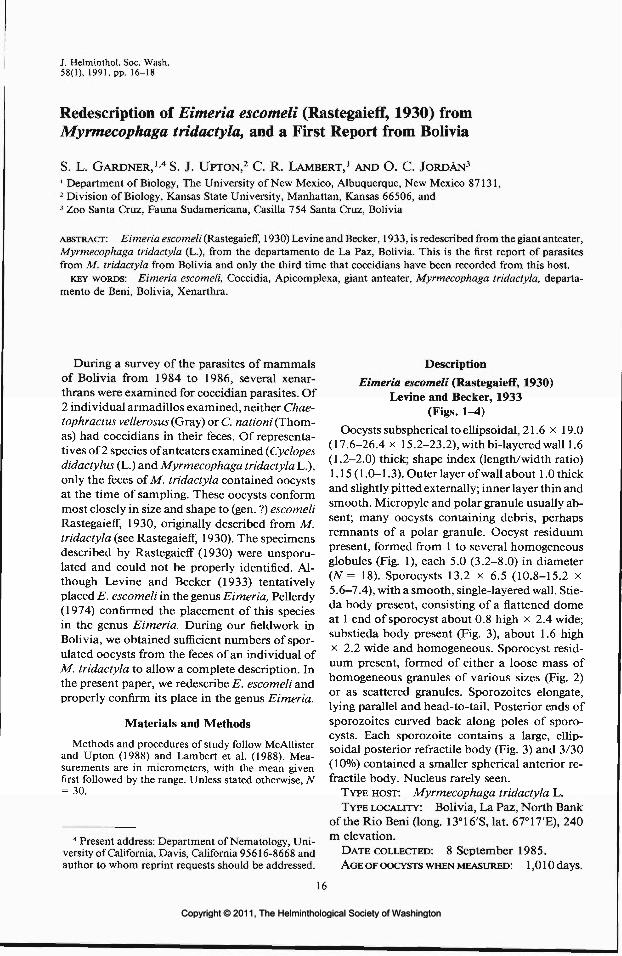
Redescription of Eimeria escomeli (Rastegaieff, 1930) fromMyrmecophaga tridactyla, and a First Report from Bolivia
S. L. GARDNER,1'4 S. J. UPTON,2 C. R. LAMBERT,1 AND O. C. JORDAN3
1 Department of Biology, The University of New Mexico, Albuquerque, New Mexico 87131,2 Division of Biology, Kansas State University, Manhattan, Kansas 66506, and3 Zoo Santa Cruz, Fauna Sudamericana, Casilla 754 Santa Cruz, Bolivia
ABSTRACT: Eimeria escomeli (Rastegaieff, 1930) Levine and Becker, 1933, is redescribed from the giant anteater,Myrmecophaga tridactyla (L.), from the departamento de La Paz, Bolivia. This is the first report of parasitesfrom M. tridactyla from Bolivia and only the third time that coccidians have been recorded from this host.
KEY WORDS: Eimeria escomeli, Coccidia, Apicomplexa, giant anteater, Myrmecophaga tridactyla, departa-mento de Beni, Bolivia, Xenarthra.
During a survey of the parasites of mammalsof Bolivia from 1984 to 1986, several xenar-thrans were examined for coccidian parasites. Of2 individual armadillos examined, neither Chae-tophractus vellerosus (Gray) or C. nationi (Thom-as) had coccidians in their feces. Of representa-tives of 2 species of anteaters examined (Cyclopesdidactylus (L.) and Myrmecophaga tridactyla L.),only the feces of M tridactyla contained oocystsat the time of sampling. These oocysts conformmost closely in size and shape to (gen. ?) escomeliRastegaieff, 1930, originally described from M.tridactyla (see Rastegaieff, 1930). The specimensdescribed by Rastegaieff (1930) were unsporu-lated and could not be properly identified. Al-though Levine and Becker (1933) tentativelyplaced E. escomeli in the genus Eimeria, Pellerdy(1974) confirmed the placement of this speciesin the genus Eimeria. During our fieldwork inBolivia, we obtained sufficient numbers of spor-ulated oocysts from the feces of an individual ofM. tridactyla to allow a complete description. Inthe present paper, we redescribe E. escomeli andproperly confirm its place in the genus Eimeria.
Materials and Methods
Methods and procedures of study follow McAllisterand Upton (1988) and Lambert et al. (1988). Mea-surements are in micrometers, with the mean givenfirst followed by the range. Unless stated otherwise, TV= 30.
4 Present address: Department of Nematology, Uni-versity of California, Davis, California 95616-8668 andauthor to whom reprint requests should be addressed.
Description
Eimeria escomeli (Rastegaieff, 1930)Levine and Becker, 1933
(Figs. 1^4)Oocysts subspherical to ellipsoidal, 21.6 x 19.0
(17.6-26.4 x 15.2-23.2), withbi-layered wall 1.6(1.2-2.0) thick; shape index (length/width ratio)1.15(1.0-1.3). Outer layer of wall about 1.0 thickand slightly pitted externally; inner layer thin andsmooth. Micropyle and polar granule usually ab-sent; many oocysts containing debris, perhapsremnants of a polar granule. Oocyst residuumpresent, formed from 1 to several homogeneousglobules (Fig. 1), each 5.0 (3.2-8.0) in diameter(N = 18). Sporocysts 13.2 x 6.5 (10.8-15.2 x5.6-7.4), with a smooth, single-layered wall. Stie-da body present, consisting of a flattened domeat 1 end of sporocyst about 0.8 high x 2.4 wide;substieda body present (Fig. 3), about 1.6 highx 2.2 wide and homogeneous. Sporocyst resid-uum present, formed of either a loose mass ofhomogeneous granules of various sizes (Fig. 2)or as scattered granules. Sporozoites elongate,lying parallel and head-to-tail. Posterior ends ofsporozoites curved back along poles of sporo-cysts. Each sporozoite contains a large, ellip-soidal posterior refractile body (Fig. 3) and 3/30(10%) contained a smaller spherical anterior re-fractile body. Nucleus rarely seen.
TYPE HOST: Myrmecophaga tridactyla L.TYPE LOCALITY: Bolivia, La Paz, North Bank
of the Rio Beni (long. 13°16'S, lat. 67°17'E), 240m elevation.
DATE COLLECTED: 8 September 1985.AGE OF OOCYSTS WHEN MEASURED: 1,010 days.
16
Copyright © 2011, The Helminthological Society of WashingtonCopyright © 2011, The Helminthological Society of Washington

17
C
Figures 1-3. Photomicrographs of sporulated oocysts of coccidia recovered from the feces of Myrmecophagatridactyla. Bar = 10.8 pm. 1. Sporulated oocyst showing oocyst residuum consisting of several homogeneousglobules (*). 2. Note sporocyst residuum composed of a loose mass of homogeneous granules. 3. Sporocysts withhomogeneous substieda body (arrow), and large ellipsoid posterior refractile body (*). Note thick, externallypitted outer wall.
MATERIAL DEPOSITED: Phototype ^photo-graphs, see Bandoni and Duszynski, 1988) ofsporulated oocysts USNM Helm. Coll. No.81056.
COLLECTOR: O. C. Jordan, personal field cat-alog number OCJ/85-169.
Discussion
Little information is available concerning coc-cidian parasites of Xenarthra in the Neotropics.Up to the present time, only 3 species of Eimeriahave been described from anteaters, 1 from thesloth (Bradypus sp.) and 4 from armadillos. Rep-resentatives of this group of mammals occur onlyin the Nearctic and Neotropical regions (Simp-son, 1980); thus, no comparisons will be madewith coccidians described from any of the phol-idote anteaters occurring in other biogeographicregions.
Eimeria escomeli differs from E. tamanduaeLainson, 1968, from Tamandua tetradactyla (L.)in being smaller and without a highly refractilespherule in the stieda body (Lainson, 1968). Theoocysts of E. escomeli are smaller than E. cyclo-pei Lainson and Shaw, 1982, from Cyclopes di-dactylus (L.), and possess an oocyst residuum.
Eimeria escomeli can be recognized as distinctfrom E. travassoi Da Cunha and Muniz, 1928,from Euphractus and Dasypus spp. and from E.dacunhai Levine, 1984, from Cabassous sp. and
Figure 4. Line drawing of Eimeria escomeli recov-ered from feces of Myrmecophaga tridactyla. Bar = 3ion.
Copyright © 2011, The Helminthological Society of WashingtonCopyright © 2011, The Helminthological Society of Washington

18
Chaetophractus sp. in having much smaller oo-cysts and smaller sporocysts (Da Cunha andMuniz, 1928). Eimeria escomeli differs from E.cabassusi Carini, 1933, from Cabassons sp. inhaving ellipsoidal to subspherical oocysts and awell-developed oocyst residuum, and also differsfrom E. choloepi Lainson and Shaw, 1982, fromCholoepus didactylus (L.) in having an oocystresiduum and well-developed stieda and sub-stieda bodies (Carini, 1933; Lainson and Shaw,1982).
Acknowledgments
This work was supported in large part by theNational Science Foundation (grant nos. BSR-8612329 to S. L. Gardner, T. L. Yates, and D.W. Duszynski; BSR-8408923 to T. L. Yates; andBSR-8316740 to S. Anderson), the AmericanMuseum of Natural History, The Museum ofSouthwestern Biology, and the Tinker Founda-tion. Financial support was also obtained fromthe Latin American Institute, Student ResearchAllocations Committee, and Graduate ResourceAllocations Committee of the University of NewMexico. Thanks also to T. L. Yates, J. Cook, L.Ruidas, N. Olds, and P. Desjeux for assistancein the field.
Literature Cited
Bandoni, S. M., and D. W. Duszynski. 1988. A pleafor improved presentation of type material for coc-cidia. Journal of Parasitology 74:519-523.
Carini, A. 1933. Sur deux nouvelles Eimeria recon-trees dans 1'intestin d'un jeune tatou. Annales deParasitologie 11:469-474.
Da Cunha, A. M., and J. Muniz. 1928. Surunnoveausporozoaire, parasite du tatou. Compte Rendus dela Societe Bresilienne de Biologie 98:624-627.
Lainson, R. 1968. Parasitological studies in BritishHonduras III. Some coccidial parasites of mam-mals. Annals of Tropical Medicine and Parasitol-ogy 62:252-259.
, and J. J. Shaw. 1982. Coccidia of brazilianedentates: Eimeria cyclopei n. sp. from the silkyanteater, Cyclopes didactylus (L.) and Eimeriacholoepi n. sp. from the two-toed sloth, Choloepusdidactylus (L.). Systematic Parasitology 4:269-278.
Lambert, C. R., S. L. Gardner, and D. W. Duszynski.1988. Coccidia (Apicomplexa: Eimeriidae) fromthe subterranean rodent Ctenomys opimus Wagner(Ctenomyidae) from Bolivia, South America.Journal of Parasitology 74:1018-1022.
Levine, N. D., and E. R. Becker. 1933. A catalog andhost-index of the species of the coccidian genusEimeria. Iowa State College Journal of Science 8:83-106.
McAllister, C. T., and S. J. Upton. 1988. Eimeriatrachemydis n. sp. (Apicomplexa: Eimeriidae) andother eimerians from the red-eared slider, Trache-mys scripta elegans (Reptilia: Testudines), innorthcentral Texas. Journal of Parasitology 74:1014-1017.
Pellerdy, L. P. 1974. Coccidia and Coccidiosis, 2nded. Verlag Paul Parrey, Berlin. 959 pp.
Rastegaieff, F. E. F. 1930. Zur Frage iiber Coccidienwilder Tiere. Archiv fur Protistenkunde 71:377-404.
Simpson, G. G. 1980. Splendid Isolation, the CuriousHistory of South American Mammals. Yale Uni-versity Press, New Haven. 266 pp.
Copyright © 2011, The Helminthological Society of WashingtonCopyright © 2011, The Helminthological Society of Washington

J. Helminthol. Soc. Wash.58(1), 1991, pp. 19-23
The Identity and Prevalence of Trypanosomes in White-tailed Deer(Odocoileus virginianus) from Southern Florida
SAM R. TELFORD, JR.,1 DONALD J. FORRESTER,! SCOTT D. WRIGHT,lMELODY E. RoELKE,1 SUSAN A. FERENC,1 AND JAMES W. McCowN2
1 Department of Infectious Diseases, College of Veterinary Medicine, University of Florida,Gainesville, Florida 32611 and2 Florida Game and Fresh Water Fish Commission, 566 Commercial Boulevard, Naples, Florida 33942
ABSTRACT: Trypanosoma cervi Kingston and Morton, 1975, is reported from white-tailed deer, Odocoileusvirginianus (Zimmermann), in Collier, Dade, and Monroe counties of southern Florida. Annual prevalencesfrom thin blood smears averaged 21% (range 7^1%) from 1984 to 1989. Fall prevalence averaged 93% (range85-100%) when determined by culture of blood in a monophasic liquid medium, whereas examination of thinfilms averaged only 18% in these years. Examination of thick blood smears proved comparably effective whencompared to blood culture, resulting in the detection of 98% of infections diagnosed by blood culture.
KEY WORDS: Trypanosoma cervi, white-tailed deer, Odocoileus virginianus, prevalence, diagnostic techniques,Florida.
The trypanosome of white-tailed deer in theUnited States has been identified as Trypano-soma cervi Kingston and Morton, 1975. Infecteddeer have been found from central Florida northto West Virginia and west to Arkansas (Kingstonand Crum, 1977; Davidson et al., 1983). How-ever, no data are available on the prevalence ofthis parasite in deer populations from the south-ernmost portion of the range in Florida.
Since 1984 we have participated in an ongoingcooperative study of white-tailed deer popula-tions in southern Florida with the Florida Gameand Fresh Water Fish Commission (FGFWFC)and the National Park Service (NFS). Trypano-somes were found often in high prevalence andthis study describes the identity and prevalenceof the trypanosomes as determined by exami-nation of blood films and blood cultures.
Materials and Methods
Blood samples were obtained from a total of 238white-tailed deer collected during 1984-1989 in Col-lier, Dade, and Monroe counties, Florida. Deer sam-ples comprised 50 males and 188 females, representing39 fawns (age 3-11 mo), 51 yearlings (12-23 mo), and148 adults (over 23 mo). Between October 1984 andOctober 1986, 9-25 deer were sampled during each of3 seasons: spring (March), summer (June-August), andfall (October-November). From 1987 to 1989, 27-50deer were obtained during fall only. Cardiac blood sam-ples were taken immediately after the deer were killedby gunshot at night by FGFWFC or NFS biologistsand were refrigerated in anticoagulant tubes (EDTA orsodium citrate) until the following morning. Severalthin blood films were prepared from whole blood ofeach deer, air-dried, and fixed in absolute methanol.Additionally, in 1989 standard thick films consisting
of a circular drop of blood 5-8 mm in diameter andthin films of blood centrifuged for hematocrit deter-minations were prepared. These latter films, obtainedfrom a limited number of deer, made both from thevicinity of the buffy coat and from the packed end ofthe hematocrit tube, were fixed and stained as standardthin films. All 1989 thin films were stained on the dayof preparation for 1 hr with Giemsa (Harleco, EMDiagnostic Systems, Inc., Gibbstown, New Jersey) ata dilution of 1:9, pH of 7.0 or 7.2. Thick films wereair-dried for at least 24 hr and then placed face sidedown for 1 hr in a dish containing dilute Giemsa stain(1:20), which hemolyzed the erythrocytes. Slides wererinsed gently with distilled or bottled drinking waterand air-dried. An additional thin film from each deerwas stained by the Leukostat procedure (Fisher Sci-entific, Orangeburg, New York), which gave resultscomparable to Giemsa staining when done on the dayof preparation. The thin blood films collected from1984 to 1988 were generally poorly stained, and mostrequired destaining and restaining when unstained du-plicates were not available. Poorly stained films weredestained for 10 min in 95% acid-ethanol, neutralizedby immersion in 95% alkaline-ethanol, and washed fora few minutes in running tap water. Old, unstained andnewly destained thin films were then immersed inGiemsa stain in an acetone-methanol-distilled watersolution buffered to pH 6.4 for 2 hr, using an unpub-lished procedure developed by R. B. Kimsey, Univer-sity of California, Davis. This generally gave resultscomparable to material stained when fresh. All smearswere screened for trypanosomes at 160 x, covering theentire smear with special attention paid to the "tails"and edges of the films. In infections of high intensity,trypanosomes could be found anywhere on the film,but in infections of low intensity they were most oftenon the periphery. A 5-ml sample of blood from eachdeer collected in October 1985, 1986, 1988, and 1989was inoculated into 2-5 ml of a monophasic liquidmedium (Sadigursky and Brodskyn, 1986). The cul-tures, maintained at room temperature, were examined5-7 days following inoculation, and in the case of ap-
19
Copyright © 2011, The Helminthological Society of WashingtonCopyright © 2011, The Helminthological Society of Washington

20 JOURNAL OF THE HELMINTHOLOGICAL SOCIETY
Table 1. A comparison of mensural values for trypanosomes of white-tailed deer from southern Florida withthose of Trypanosoma cervi.
Measurements in pm
Source
S. FloridaMeanRange
SE USA*MeanRange
Type host (elk)fMeanRange
PK
12.66-22
13.55-27
12.25-20
KN
7.65-16
8.32-14
7.04-9
PN
20.213-31
22.014-34
19.411-32
NA
28.619-39
26.312^1
24.820-30
BL
47.832-65
48.231-74
45.432-56
FF
6.10-12
9.53-21
6.63-11
L
53.939-76
57.540-83
52.040-61
W
9.44-14
5.63-15
4.63-8
* From Kingston and Crum (1977).t From Kingston and Morton (1975).
parent negatives, several times for up to 3 mo there-after. Thin films of the culture forms were preparedfrom 3 positive samples collected in October 1989, andfixed and stained similarly to thin blood films.
A series of 25 trypomastigotes with clear diagnosticcharacters were selected from slides collected from 1986to 1989. These were measured by calibrated ocularmicrometer at l,000x under oil immersion. Taxo-nomic characters utilized were those of Kingston andMorton (1975) and are abbreviated in Tables 1 and 2as follows: PK, posterior end to kinetoplast; KN, nu-cleus to kinetoplast; PN, posterior end to nucleus; NA,nucleus to anterior end; BL, total length minus freeflagellum; FF, free flagellum; L, total length; W, width;NI, nuclear index, PN/NA; and KI, kinetoplast index,PN/KN. Nucleus location was measured to the centerof the nucleus, while width refers to the greatest breadthof the trypanosome exclusive of the undulating mem-brane. This latter character may differ in its measure-ment by other authors. The free flagellum was not mea-sured unless its entire length was evident. Alltrypanosomes measured were trypomastigote forms andare compared below only with bloodstream trypo-mastigote data of other authors. Chi-square tests wereused to compare prevalences. Representative bloodfilms have been deposited in the U.S. National ParasiteCollection, Beltsville, Maryland (accession nos. 81000-81002).
Results
Identity of the trypanosomesComparison of mensural data of the trypano-
somes from the population of deer in southernFlorida (Table 1) with those published by King-ston and Morton (1975) and Kingston and Crum(1977) demonstrated clearly that they were con-specific with Trypanosoma cervi, and distinctfrom Trypanosoma theileri (Table 2). In T. cervithere was a smaller ratio of the distance from theposterior end-nucleus to the kinetoplast-nucleus
distance (KI), a lower mean ratio between thedistance posterior end-kinetoplast to body length(PK/BL), and a greater mean ratio of the nucleus-anterior end to the body length (NA/BL). Giventhe difference in measurement technique, i.e., theuse of an ocular micrometer (present study) in-stead of projected photomicrographs measuredwith a calibrated map reader, the concordanceof the data was remarkable. Only the mean widthmeasurement (W) differed appreciably, and thiswas probably a combination of fixation or con-figuration artifacts and the inclusion of both slen-der and broad forms (Kingston and Crum, 1977)in the sample. Three trypanosomes of the mea-sured 25 could be described as long, slender forms;the remainder of the sample was broad in ap-pearance. The critical diagnostic characters of thesouthern Florida sample (Table 2) either werecompletely within the range of variation de-scribed for T. cervi or overlapped at one end ofthe range. No attempt was made to measurebloodstream or cultural epimastigote forms be-cause of their variability in shape. The ratio oftrypomastigote (T) to epimastigote (E) forms in-dicated highly significant (P < 0.0001) seasonaldifferences: in summer 1985,6T:31 E;in pooledfall samples from 1984 to 1988, 25 T: 6 E; andpooled samples from 5 deer in fall 1989, 53 T:2E.
Prevalence
Thin films were the only samples available forevery sampling period (Table 3). Using only thedata obtained from those films, we found sig-nificant differences between the prevalence of
Copyright © 2011, The Helminthological Society of WashingtonCopyright © 2011, The Helminthological Society of Washington

OF WASHINGTON, VOLUME 58, NUMBER 1, JANUARY 1991 21
Table 2. A comparison of diagnostic characters for trypanosomes of white-tailed deer from southern Floridawith those of Trypanosoma cervi and Trypanosoma theileri.
Source FF:BL NI KI PK/BL NA/BL
S. Florida
MeanRange
SE USA*
Mean
Type host (elk)f
MeanRange
Broad forms
MeanRange
Slender forms
MeanRange
1:6.84.8-12.6
1:5.7
1:6.9
1:8.57.0-9.3
1:8.66.3-13.8
0.710.5-1.1
0.88
0.780.5-1.3
0.910.7-1.1
0.840.5-1.1
T. cervi
2.771.9-4.6
2.86
2.772.8-3.6
T. theileri§
5.684.4-5.0
4.003.6-4.1
0.260.2-0.4
0.28|
0.270.2-0.4
0.390.3-0.5
0.350.2-0.5
0.600.5-0.9
0.55|
0.550.5-0.6
0.510.5-0.6
0.520.5-0.6
* From Kingston and Crum (1977).t From Kingston and Morton (1975).t Calculated from data of Kingston and Crum (1977).§ Saisawa et al. (1933), quoted by Kingston and Morton (1975).
trypanosomes during fall of 1984 (44%) andspring of 1985 (0%, P = 0.04), between springof 1985 (0%) and summer of 1985 (56%, P =0.04), and between summer of 1985 (56%) andsummer of 1986 (8%, P = 0.05). There were nosignificant differences (P = 0.15) when all 3 sum-mer samples were compared with each other.Spring samples from 1985 (0%) and 1986 (15%)did not differ significantly (P = 0.50), and therewere no differences among the samples collectedin fall of each year (P = 0.37). The seasonal andannual prevalence of T. cervi in southern Florida,
as determined from the examination of thin bloodfilms (Table 3), was likely a considerable under-estimate as suggested by the results of blood cul-ture (Table 4). In the fall of 1989, when thickfilms and centrifuged thin films were prepared,4 diagnostic methods were employed. Both thickfilms and culture methods were very sensitivefor detecting infection by T. cervi and resultedin prevalences of 98 and 100% in comparison to16 and 15%, respectively, detected by normaland centrifuged thin blood films. The latter didnot prove to be better for the detection of try-
Table 3. Seasonal and annual prevalences of Trypanosoma cervi determined from thin blood films of white-tailed deer from southern Florida, 1984-1989.
Year
198419851986198719881989
Totals
No.deer
exam.
01220000
32
Spring(Mar.)
% pos.
015
—_
—9
Summer(June-Aug.)
95%C.I.
0-305^tO
—
——
3-27
No.deer
exam.
132512000
50
%pos.
3856
8
———40
95%C.I.
16-6835-75
1̂ 10
———
26-56
Fall(Oct.-Nov.)
No.deer
exam.
91514274150
156
%pos.
442077
221617
95%C.I.
17-766-491-392-26
11-398-31
12-25
No.deer
exam.
225246274150
238
Totals
%pos.
4133117
221621
95%C.I.
21-6420-484-252-26
11-398-31
16-28
Copyright © 2011, The Helminthological Society of WashingtonCopyright © 2011, The Helminthological Society of Washington

22 JOURNAL OF THE HELMINTHOLOGICAL SOCIETY
Table 4. Prevalences of Trypanosoma cervi in white-tailed deer in southern Florida during October as de-termined by thin blood films vs. blood culture tech-niques.
% positive by
Year
1985198619881989
Totals
No. deerexamined
15144150
120
Thin bloodfilm
207
221618
Bloodculture
939285
100*93
* This value represents results of cultures from 46 deer; cul-tures from 4 other deer were contaminated.
panosome infection than thin films of uncentri-fuged whole blood. More importantly, the use ofthick films can provide an adequate survey meth-od when cultures cannot be used, as they resultedin the diagnosis of 98% of the 46 infections foundby culture of fresh blood and all 4 of the positivesamples for which the cultures were contami-nated. By using thin films or centrifuged bloodsample techniques, only 15-16% of the positivesamples were detected, which proved to be highlysignificant (P < 0.001) from either thick film orculture methods.
Discussion
The identification of T. cervi from white-taileddeer in southern Florida was not surprising giventhe broad distribution of this parasite in NorthAmerican cervids (Kingston and Morton, 1975).Because deer in southern Florida interminglefreely with range cattle in the Collier County studyarea, it was possible that infection by Trypano-soma theileri, which is known in Florida (Risticand Trager, 1958), might occur in the deer. How-ever, none of the trypanosomes found on bloodsmears was T. theileri. Trypanosoma cervi ap-pears to be host specific to cervids, as suggestedby the failure of Kingston and Morton (1975) toinfect cattle with T. cervi from elk. It is unlikelythat deer are infected from transmission of T.theileri among sympatric cattle herds.
In 1985 the very high prevalence (56%) of try-panosomes on thin blood films from the summersamples (June and August) indicates that trans-mission probably occurred following the Marchsample, when no infections were found. Ten of14 (71%) June-August infections had blood-stream epimastigotes present, while only 5 had
trypomastigotes. The ratio of trypomastigote toepimastigote forms in this summer sample, whencompared to pooled fall samples from 1984 to1988 and the 1989 fall sample, also supportssummer transmission of T. cervi in southernFlorida. Epimastigotes are the multiplicative stageof T. cervi in the bloodstream and in culture(Kingston and Morton, 1975; Kingston andCrum, 1977). The presence of epimastigotes innumbers greater than those of trypomastigotesshould indicate either a recently acquired activeinfection or recrudescence of a chronic infection.In the related T. theileri, reproduction also occursin the epimastigote stage, and when infectionbegins, fairly high parasitemias may be apparent(Hoare, 1972). After 2—4 wk, parasitemias can-not be detected by microscopic examination ofblood films, but the blood remains infective forup to 1 yr. There is no lasting immunity. In thetemperate zone, reinfection may occur in springwhen horse-flies again appear. The available datafor T. cervi suggest similar epidemiology. In 1986,prevalence of T. cervi in spring was twice that ofthe subsequent summer and fall. This could re-flect annual variation in the seasonal abundanceof the presumed tabanid fly vectors (Kingston,1981), with tabanid appearance earlier in springthat year than usual. Thirty-five of the 118 spe-cies of tabanid flies known from Florida (Jonesand Anthony, 1964) are considered to be eco-nomically important on the basis of abundance,and 26 of these reach peak densities betweenApril and July, the period during which increasedprevalence of T. cervi and epimastigotes wasfound. The small sample from the summer of1986 (N = 12) may have been partially respon-sible for the discrepant prevalence pattern ob-served that year. The October samples for 1985-1989 show prevalences, based on thin bloodsmears, varying from 7% to 22%, and by culturefrom 86% to 100%. Utilizing thin blood films,Davidson et al. (1983) found maximum preva-lence (61%) of T. cervi from July to September,with a drop to one-half that level from Octoberto December (30%). They also reported the high-est prevalence of epimastigote forms on bloodfilms taken from July to September. Their sam-ples came from central Florida north to WestVirginia and Arkansas, where the climatic pat-tern differs considerably from subtropical south-ern Florida. Our data are consistent with thoseof Davidson et al. (1983) and suggest that therelatively insensitive thin blood film technique
Copyright © 2011, The Helminthological Society of WashingtonCopyright © 2011, The Helminthological Society of Washington

OF WASHINGTON, VOLUME 58, NUMBER 1, JANUARY 1991 23
may provide better evidence of fluctuations inintensity due to transmission and infection phasethan do the highly sensitive methods of cultureor thick blood films. The prevalence of T. cerviappears to be high wherever adequate sampleshave been examined (Kistner and Hanson, 1969;Stuht, 1975; Davidson et al., 1983).
Acknowledgments
J. C. Roboski, J. C. Roof, M. R. Ramsey, E.D. Land, D. S. Maehr, and other personnel ofthe Florida Game and Fresh Water Fish Com-mission and D. Jansen, J. Snyder, O. L. Bass,and T. R. Smith, and others of the U.S. NationalPark Service are acknowledged for their assis-tance in collecting deer and blood samples. Thetechnical assistance of J. H. Bogue, S. J. Tucker,and P. K. Humphlett is gratefully acknowledged.This work was supported by contracts from theU.S. National Park Service and Florida Gameand Fresh Water Fish Commission, and is a con-tribution of Federal Aid to Wildlife Restoration,Florida Pittman-Robertson Project W-41. Flor-ida Agricultural Experiment Stations Journal Se-ries No. R-00464.
Literature Cited
Davidson, W. R., C. B. Crow, J. M. Crum, and R. R.Gerrish. 1983. Observations on Theileria cerviand Trypanosoma cervi in white-tailed deer (Odo-coileus virginianus) from the southeastern United
States. Proceedings of the Helminthological So-ciety of Washington 50:165-169.
Hoare, C. A. 1972. The Trypanosomes of Mammals.A Zoological Monograph. Blackwell ScientificPublications, Oxford and Edinburgh. 749 pp.
Jones, C. M., and D. W. Anthony. 1964. The Ta-banidae of Florida. United States Department ofAgriculture Technical Bulletin Number 1295. 85PP-
Kingston, N. 1981. Protozoan parasites. Pages 193-236 in W. R. Davidson, ed. Diseases and Parasitesof White-tailed Deer. Miscellaneous PublicationNumber 7, Tall Timbers Research Station, Tal-lahassee, Florida.
, and J. Crum. 1977. Trypanosoma cervi King-ston and Morton, 1975 in white-tailed deer, Odo-coileus virginianus, in the southeastern UnitedStates. Proceedings of the Helminthological So-ciety of Washington 44:179-184.
-, and J. K. Morton. 1975. Trypanosoma cervisp. n. from elk (Cervus canadensis) in Wyoming.Journal of Parasitology 61:17-23.
Kistner, T. P., and W. L. Hanson. 1969. Trypano-somiasis in white-tailed deer. Bulletin of the Wild-life Disease Association 5:398-399.
Ristic, M., and W. Trager. 1958. Cultivation at 37°Cof a trypanosome (Trypanosoma theilerf) from cowswith depressed milk production. Journal of Pro-tozoology 5:146-148.
Sadigursky, M., and C. I. Brodskyn. 1986. A newliquid medium without blood and serum for cul-ture of hemoflagellates. American Journal ofTropical Medicine and Hygiene 35:942-944.
Stuht, J. N. 1975. Morphology of trypanosomes fromwhite-tailed deer and wapiti in Michigan. Journalof Wildlife Diseases 11:256-262.
Copyright © 2011, The Helminthological Society of WashingtonCopyright © 2011, The Helminthological Society of Washington
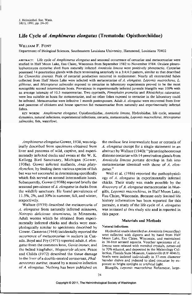
J. Helminthol. Soc. Wash.58(1), 1991, pp. 24-30
Life Cycle of Amphimerus elongatus (Trematoda: Opisthorchiidae)
WILLIAM F. FONTDepartment of Biological Sciences, Southeastern Louisiana University, Hammond, Louisiana 70402
ABSTRACT: Life cycle of Amphimerus elongatus and seasonal occurrence of cercariae and metacercariae werestudied in Half Moon Lake, Eau Claire, Wisconsin from September 1982 to November 1984. Oculate pleuro-lophocercous cercariae shed from naturally infected Amnicola limosa were positively phototactic. Cercariaepossessed 14 penetration glands with ducts terminating anteriorly in a 3:4:4:3 pattern, similar to that describedfor Clonorchis sinensis. Peak of cercarial production occurred in midsummer. Nearly all centrarchid fishescollected from Half Moon Lake were infected with metacercariae of A. elongatus. Lepomis macrochirus, L.gibbosus, and Micropterus salmoides exposed to cercariae in laboratory experiments proved to be the mostsusceptible second intermediate hosts. Prevalence in experimentally infected juvenile bluegills was 100% withan average intensity of 10.5 metacercariae. Two cyprinids, Pimephales promelas and Rhinichthys cataractae,were less suitable as hosts for metacercariae, and no other fishes exposed to cercariae in the laboratory couldbe infected. Metacercariae were infective 1 month postexposure. Adult A. elongatus were recovered from liverand pancreas of chickens and house sparrows fed metacercariae from naturally and experimentally infectedfishes.
KEY WORDS: Amphimerus elongatus, Opisthorchiidae, Amnicola limosa, Hydrobiidae, life cycle, seasonaldynamics, natural infections, experimental infections, cercaria, metacercaria, Lepomis macrochirus, Micropterussalmoides, fish, waterfowl.
Amphimerus elongatus Gower, 1938, was orig-inally described from specimens obtained fromliver and pancreas of wild, captive, and experi-mentally infected ducks and swans at the W. K.Kellogg Bird Sanctuary, Michigan (Gower,1938a). Gower infected mallards, Anas platy-rhynchos, by feeding them various species offishbut was not successful in determining specificallywhich fish served as second intermediate hosts.Subsequently, Gower (1938b) reported briefly theseasonal prevalence of A. elongatus in ducks fromthe wildlife sanctuary. He found prevalences of11.5%, 2%, and 30% in summer, fall, and winter,respectively.
Wallace (1939) described the metacercaria ofA. elongatus from naturally infected minnows,Notropis deliciosus stramineus, in Minnesota.Adult worms which he obtained from experi-mentally infected chicks and ducks were mor-phologically similar to specimens described byGower. Cameron (1944) incidentally reported theoccurrence of metacercariae in suckers in Can-ada. Boyd and Fry (1971) reported adult A. elon-gatus from the common loon, Gavia immer, andthe belted kingfisher, Megaceryle alcyon. Penceand Childs (1972) described the tissue damageto the liver of a double-crested cormorant, Phal-acrocorax auritus, caused by a massive infectionof A. elongatus. Nothing has been published on
the mollusc first intermediate host or cercaria ofA. elongatus except for a single statement in anabstract by Wallace (1940): "pleurolophocercousdistome cercariae with 14 penetration glands fromAmnicola limosa portata develop in fish intometacercariae identical with those of A. elon-gatus."
Weil et al. (1986) reported the pathophysiol-ogy of A. elongatus in experimentally infectedchicks. Their study was made possible by thediscovery of A. elongatus metacercariae in blue-gills, Lepomis macrochirus, in Half Moon Lake,Eau Claire, Wisconsin. Because only limited lifehistory information has been reported for thisparasite, a study of the life cycle of A. elongatuswas initiated at this study site and is reported inthis paper.
Materials and Methods
Natural infectionsHydrobiid snails identified as Amnicola limosa (Say)
were collected with dipnets and by hand from HalfMoon Lake, Eau Claire, Wisconsin, and maintainedin 38-liter aerated aquaria. Voucher specimens of A.limosa were relaxed with menthol crystals, preservedin 70% ethanol, and deposited in the Malacology Col-lection, Florida State Museum, Gainesville (UF 40463).Snails were isolated individually in 37-mm diameterSlender dishes and induced to shed cercariae by ex-posure to bright sunlight or artificial light.
Bluegills, Lepomis macrochirus Rafinesque, large-
24
Copyright © 2011, The Helminthological Society of WashingtonCopyright © 2011, The Helminthological Society of Washington

25
mouth bass, Micropterus salmoides (Lacepede), whitecrappie, Pomoxis annularis Rafinesque, black bull-heads, Ictalurus melas (Rafinesque), and northern pike,Esox lucius Linnaeus, were collected from Half MoonLake with dipnets, castnets, seines, or by ice fishing inwinter. Fins and musculature were examined for meta-cercariae. Survival of metacercariae in dead fish main-tained at 4°C was determined by microscopic obser-vation of movement of the whole worm within the cystand by observation of flame cell motility.
A limited search for natural definitive hosts of A.elongatus was made by examining 9 hunter-killed ducksfrom north central Wisconsin; however, no birds fromHalf Moon Lake, located in the city park, were col-lected. Observations on birds foraging in the lake weremade in all seasons of the year.
Experimental infectionsFish collected from locations that were determined
to be free from A. elongatus by examination of at least10 specimens of each fish species, were exposed ingroups of 20 to more than 1,000 cercariae in 4-literaquaria for 24 hr. Bluegill, Lepomis macrochirus,pumpkinseed, L. gibbosus (Linnaeus), black bullhead,Ictalurus melas, longnose dace, Rhinichthys cataractae(Valenciennes), and emerald shiner, Notropis atherin-oides Rafinesque, were collected in Fall Creek, town ofFall Creek, Wisconsin. Fantail darter, Etheostoma fla-bellare Rafinesque, Johnny darter, E. nigrurn Rafines-que, and mudminnow, Umbra limi (Kirtland), werecollected in O'Neil Creek, 5 km N Chippewa Falls,Wisconsin. Fathead minnow, Pimephales promelasRafinesque, and largemouth bass, Micropterus sal-moides, were collected in Frontage Road Pond, 3 kmS of Eau Claire. Brook stickleback, Culaea inconstans(Kirtland), and creek chub, Semotilis atromaculatus(Mitchell), were collected in Little Niagara Creek, EauClaire. After exposure, fish were maintained in aerated38-liter aquaria and fed flake fish food. Fish were ex-amined for metacercariae at various intervals from day1 through day 68 postexposure (PE).
Metacercariae from both naturally infected and ex-perimentally infected fishes were fed to young chicks,adult chickens, house sparrows, mice, rats, and cats inthe laboratory.
Parasite morphologyCercariae were studied alive with the aid of bright
field, phase contrast, and differential interference con-trast optics. Neutral red and Nile blue were employedas vital stains. Twenty cercariae were fixed in steaming10% formalin, mounted under a floating coverslip, andmeasured using an ocular micrometer.
Metacercarial cysts from naturally infected fish weremeasured alive under floating coverslip (N = 20). Over100 metacercariae from experimentally infected fishwere measured at various intervals postexposure.Metacercariae were excysted artificially at 38°C in asolution of trypsin and bile salts (Irwin, 1983). Ex-cysted metacercariae and adults were killed withsteaming (160°C) AFA, stained with Semichon's car-mine, and mounted in Permount. Unless otherwiseindicated, measurements are expressed in microme-ters, the range followed by mean in parentheses. Draw-ings were made with a microprojector.
Results
Cercaria (Figs. 1-3)
Body spinose anteriorly, oculate, pleurolopho-cercous, 193-243 (215) long by 53-75 (66) wide;with paired sensillae on lateral margins, 1 pairnear oral sucker, 1 pair near midbody, 4 pairs inhindbody (Fig. 1). Oral sucker, protrusible fromtegumental hood 28-38 (35) long by 25-33 (29)wide bearing 3 rows of small teeth (Fig. 2) onoral sucker, 8 per row anteriormost to 3 per rowposteriormost. Acetabulum rudimentary, in teg-umental fold at midbody. Genital anlage over-lapping acetabulum. Pharynx situated mediallybetween eyespots; esophagus and ceca undiffer-entiated. Fourteen penetration glands, stainingweakly, clustered medially between ventral suck-er and pharynx; ducts terminating anterior tooral sucker forming 3:4:4:3 pattern. Thirteen to17 pairs of granular cystogenous glands stainingintensely with vital stains, occupying dorsolat-eral margins of mid- and hindbody. Wall oftranslucent excretory vesicle epithelial with 2 rowsof conspicuous spherical nuclei; ovoid, boundedanteriorly by genital anlage and posteriorly bytail insertion. Excretory ducts and flame cell pat-tern obscured by coarse granules of cystogenousgland cells. Tail elongate 400-475 (435) long by28-38 (33) wide, entirely covered by dorsoven-tral finfold with numerous transverse striations(Fig. 3). Finfold widest in midregion of dorsalmargin.
Cercariae emerged from the snail, Amnicolalimosa (Hydrobiidae), in the laboratory underthe stimulus of bright light, with emergence com-mencing within 15 min of stimulation. Cercariaewere positively phototactic. They remained mo-tionless, suspended with body oriented down-ward, and slowly settled in the water column.They rose in the water column by occasionalbursts of swimming activity, simultaneouslymoving toward the light. Cercariae were able toswim actively for approximately 24 hr. Mori-bund cercariae lost their tails and crawled weaklyon the bottom prior to death.
Metacercaria
Measurements of metacercariae encysted innaturally infected bluegills averaged 185 by 170with a parasite cyst wall 12 thick. Dimensionsof cysts and morphology of excysted metacercar-iae were similar to those described by Wallace(1939).
Copyright © 2011, The Helminthological Society of WashingtonCopyright © 2011, The Helminthological Society of Washington

26 JOURNAL OF THE HELMINTHOLOGICAL SOCIETY
Figures 1-3. Cercaria of Amphimerus elongatus from Amnicola limosa, Half Moon Lake, Eau Claire, Wis-consin. 1. Body of cercaria, ventral view. Scale = 50 /*m. 2. Rasping tooth from oral hood. Scale = 4 pm. 3.Resting posture of cercaria, lateral view. Scale = 110 tan.
Adult
No naturally infected definitive hosts werefound during this study. Confirmation of theidentity of Amphimerus elongatus was based uponadult specimens obtained from experimentallyinfected hosts.
Natural infections in snails
Amnicola limosa from Half Moon Lake ini-tially shed Amphimerus elongatus cercariae in
fall 1982. Amnicola limosa displayed a seasonalpopulation cycle in Half Moon Lake similar tothat reported for the species in Rhode Island(Kesler, 1980). Most snails lived for 1 year. Snailspresent after ice melted in April were hatched inJuly of the previous summer. As lake tempera-tures rose (18°C in mid-May), egg laying com-menced and continued for 2 mo. Snails ceasedlaying eggs and began dying in July when tem-peratures reached 26°C. Few adult snails sur-vived into the fall. Juvenile snails grew rapidly
Copyright © 2011, The Helminthological Society of WashingtonCopyright © 2011, The Helminthological Society of Washington

OF WASHINGTON, VOLUME 58, NUMBER 1, JANUARY 1991 27
Table 1. Prevalence of Amnicola limosa from HalfMoon Lake, Eau Claire, Wisconsin shedding Amphi-merus elongatus cercariae.
Table 2. Distribution of Amphimerus elongatus meta-cercariae in one bluegill, Lepomis macrochirus (stan-dard length = 45 mm) collected in Half Moon Lake,Eau Claire, Wisconsin on 15 October 1983.
Date
27 Sep 1982
24 Apr 198325 May 1983
6 Jun 19836 Jul 19839 Jul 1983
21 Jul 198330 Jul 198312 Aug 19834 Sep 19832 Oct 1983
7 Apr 198429 May 19844 Jun 1984
13 Jun 198419 Jun 198426 Jun 1984
6 Jul 198419 Jul 198426 Jul 198415 Aug 198415 Sep 19848 Oct 1984
N beexamined
100
20072
200341
5014454727272
1016411913413713920063
14710872
144
N hershedding
3
04
156010392610
10
00568
16135
12812
Preval n(%)
3
068
1820274814
10
00446
127
Number ofn
Site in fish
Caudal finDorsal fin (soft)Dorsal fin (spiny)Anal finPectoral fin (left)Pectoral fin (right)Pelvic finsMuscle (caudal peduncle)Muscle (all other)
Total
ictacer- Percent-cariae age
85 4937 218 5
19 112 13 20 09 5
11 6
174 100
late May and peaking in July, was similar in bothyears of this study, although prevalence of in-
8 fected snails was much higher in 1983 than in8711
1984.
Natural infections in fishesRlnepills J .pnomis mnrrn,rhiriJM and larpe-
and by late fall some attained adult size. Finalgrowth of this generation resumed in spring andegg laying by this generation was initiated in May.
The seasonal dynamics of cercarial productionwas closely related to the 1-yr life span of Am-nicola limosa. Few snails shed Amphimerus elon-gatus cercariae in April, May, and June of both1983 and 1984, but by late July prevalence ofsnails shedding A. elongatus peaked (Table 1).The remaining adult snails continued to shedcercariae in August and into the fall; however,so few adult snails were still alive at this timethat cercarial production was greatly reduced. Byfall the size of the smallest specimens of re-maining adults overlapped the size of the largestsnails belonging to the new generation. There-fore, prevalence of snails shedding cercariae couldnot be determined separately for the 2 genera-tions. It could be ascertained qualitatively, how-ever, that the only snails that were shedding cer-cariae were unequivocally old adults. These werethe largest specimens and had heavily eroded andencrusted shells. Similarly, none of the snails be-longing to the most recent generation shed A.elongatus. The pattern of cercarial shedding byAmnicola limosa, i.e., emergence beginning in
mouth bass, Micropterus salmoides, were the onlyfishes in Half Moon Lake that harbored Amphi-merus elongatus metacercariae. Half Moon Lakewas not a typical locality to determine the rangeof host specificity of A. elongatus in a truly nat-ural environment, because the fishes of the lakewere exterminated with rotenone by the Wis-consin Department of Natural Resources (DNR).Black bullheads, Ictalurus melas (N = 12), north-ern pike, Esox lucius (N = 5), and white crappie,Pomoxis annularis (N= 1), restocked in the lakeby DNR, were not infected. No fish of the fam-ilies Cyprinidae or Catostomidae were collectedin Half Moon Lake despite extensive effort.
Adult bluegills and bass typically were heavilyinfected with metacercariae. Prevalence in blue-gills was 97% (N = 185) and bass was 92% (N =25). Intensities of 100-300 or more metacercar-iae were common. Because of the large numbersof metacercariae, only estimates were made formost fish. However, careful count of cysts in 1bluegill (4 5-mm standard length) harboring atypical infection was made to show the actualnumber and distribution of metacercariae (Table2).
In July and August, adult bluegill and bassharbored both fully developed metacercariae andnewly encysted, incompletely developed meta-
Copyright © 2011, The Helminthological Society of WashingtonCopyright © 2011, The Helminthological Society of Washington

28 JOURNAL OF THE HELMINTHOLOGICAL SOCIETY
cercariae, indicating that reinfection was possi-ble. Newly hatched bluegill and bass in July hadan average prevalence of approximately 20% withtypically only 1 or 2 small, incompletely devel-oped metacercariae per infected fish. By October,young-of-the-year bluegill and bass harboredmetacercariae considered to be fully developed,although prevalence and intensity remained low.
The effect of temperature on the viability ofmetacercariae encysted in dead bluegills was de-termined in order to ascertain whether infectionsmight be acquired by scavengers as well as pred-atory definitive hosts. Metacercariae did not sur-vive freezing at — 10°C for 24 hr. After 48 hr,100% of metacercariae in dead fish refrigeratedat 4°C remained viable, and 50% were able tosurvive for 12 days at this temperature.
Natural infections in birds
Incidental to this study 5 mallards, Anasplaty-rhynchos Linnaeus, and 4 blue-winged teal, A.discors Linnaeus, collected within 50 mi of HalfMoon Lake, were examined but were not in-fected with A. elongatus. Because this lake waslocated in a city park in the center of Eau Claire,Wisconsin, it was not possible to obtain birdsfor necropsy. However, observations of birdpopulations were made in order to understandbetter the ecology of A. elongatus. The lake wasused by piscivorous summer residents such asherons and kingfishers, and numerous waterfowlconsidered spring and fall migrants were ob-served. Furthermore, the city aerated the centerof the lake during the winter, and this ice-freearea attracted huge flocks of waterfowl, whichwould ordinarily not use the lake from Novem-ber through early April.
Geographic distribution ofAmphimerus elongatus
Although the determination of the geographicdistribution of Amphimerus elongatus in Wis-consin was beyond the scope of this study, a fewbluegills in backwaters of the Red Cedar River,where Amnicola limosa also occurred, were in-fected with metacercariae. Low prevalence andintensity of metacercarial infections precludedan analysis of host specificity in other fishes fromthe Red Cedar River. Several thousand individ-uals of the only other hydrobiid snail in the area,Somatogyrus depressus (Tryon), were examinedfor cercariae, but none was infected with A. elon-gatus. Species of fish from numerous localities
where hydrobiid snails were absent were not in-fected.
Experimental infections in fishes
Of the various species of fishes exposed to cer-cariae in the laboratory, only centrarchid fisheswere readily infected with A. elongatus. Viablemetacercariae were also obtained from severalfathead minnows. Only 1 of 20 longnose daceharbored 1 viable metacercaria when examined24 days postexposure. No other species of fishexposed to cercariae of A. elongatus hosted viablemetacercariae for more than 7 days. Most in-susceptible fish hosts showed no evidence of in-fection after exposure to over 50 cercariae perfish, but a small percentage of creek chubs, dace,and emerald shiners harbored a few dead, en-cysted specimens surrounded by a host cellularcapsule 1 or more days PE. To illustrate differ-ences in susceptibility, in one experiment 20specimens each of fathead minnows, juvenilebluegills, and juvenile black bullheads were ex-posed, in separate containers, to equal numbersof all cercariae shed by 35 Amnicola limosa. Allbluegills became infected, with a mean intensityof 10.5 metacercariae. Prevalence in fatheadminnows was 20% with a mean intensity of 2.3.No black bullheads became infected.
Development of metacercariae was investi-gated in experimentally infected juvenile blue-gills maintained at 22°C. On day 5 PE, cysts mea-sured 123-152 (137) by 62-82 (71) with a parasitecyst wall 7.5 thick. A host-induced cellular cap-sule was already present around the cyst. Thebrownish pigmented worms moved activelywithin the cyst. Eyespots, oral sucker, and emptyexcretory bladder were clearly visible through thecyst wall. By day 15 PE, excretory concretionswere apparent in the bladder, and the brown pig-mentation of the body was markedly reduced.On day 20 PE, the ceca were completely devel-oped and filled with cecal platelets (^'disk-shaped concretions" of Wallace). The excretorybladder was made conspicuous by black concre-tions that completely filled the lumen. Cystsmeasured 180-225 (210) by 100-113 (103). Onday 29 PE, metacercariae were identical with ful-ly developed specimens from naturally infectedbluegills and bass. Anatomical features, such asthe pharynx and oral and ventral suckers, couldbe observed and measured in encysted speci-mens. Eyespots were no longer visible. These 29-day-old laboratory-reared metacercariae, when
Copyright © 2011, The Helminthological Society of WashingtonCopyright © 2011, The Helminthological Society of Washington

OF WASHINGTON, VOLUME 58, NUMBER 1, JANUARY 1991 29
fed to chicks, developed into adults in liver andpancreas that were morphologically identical tospecimens described by Gower (1938a). Viablecysts were found in centrarchids and fatheadminnows up to 68 days PE at which time ex-periments were terminated.
Experimental infections in birds and mammals
Only avian hosts became infected when birdsand mammals were fed metacercariae from nat-urally infected Half Moon Lake bluegills in thelaboratory. Mice, rats, and cats were completelyrefractory to infection. Adult specimens were ob-tained in chickens and in house sparrows. One-day-old unfed chicks typically attained higherlevels of infection than older chickens, but all agegroups were successfully infected. Adults wererecovered from both liver and pancreas in allhosts harboring more than 1 worm. Adult wormsconformed to the description of Gower (1938a),and ontogenetic changes from metacercariae weresimilar to those described by Wallace (1939).
Discussion
The occurrence of 14 penetration glands in thecercaria of Amphimerus elongatus confirms thepreliminary description of the cercariae fromAmnicola limosa in Minnesota (Wallace, 1940).The 3:4:4:3 pattern of ducts formed by the 14penetration glands indicates that the cercaria ofA. elongatus is most similar to that ofClonorchissinensis (Komiya and Suzuki, 1964).
The life cycle of Amphimerus elongatus is sim-ilar to that of most members of the family Opis-thorchiidae in which fishes serve as second in-termediate hosts and piscivorous birds ormammals are definitive hosts for adults parasit-izing the liver and pancreas. Both natural andexperimental infections of A. elongatus indicatedthat adults are specific to avian hosts. Thus, it isunlikely that the species represents a public healththreat, in spite of its abundance in fishes com-monly consumed by humans. Susceptibility ofhouse sparrows to infection was in agreementwith the broad host specificity reported by otherworkers for avian hosts (Gower, 1938a; Boydand Fry, 1971; Pence and Childs, 1972). Infec-tion of nonpiscivorous birds, like house sparrowsand chickens, indicated that natural host rangemay be limited by avian feeding ecology ratherthan by host physiology.
Seasonal dynamics in Half Moon Lake wereclosely associated with the seasonal population
cycle of the first intermediate host, Amnicola li-mosa. The large size of the population of A li-mosa, and the high prevalence of snails infected\vithAmphimerus elongatus, resulted in high cer-carial production in the lake. Peak cercarialemergence, however, was confined to a relativelybrief but intense period in July, immediately pri-or to death of the large majority of adult snails.It was interesting that the timing of maximumcercarial emergence meant that most young-of-the-year centrarchids in the lake receive only lightinfections during their first summer of life. Re-infection of older age class fishes is responsiblefor most metacercarial infections. These dynam-ics may be driven by extrinsic factors such assnail ecology and water temperature. However,if juvenile fishes are killed by heavy infectionsof metacercariae during their first few months oflife, this intense selective pressure may be themost important factor responsible for the evo-lution of the seasonal pattern that exists in HalfMoon Lake.
The high prevalence and intensity of A. elon-gatus in Half Moon Lake may be a natural phe-nomenon, or may be exacerbated by human in-tervention. Because a portion of the lake was keptice-free during winter, and birds were fedthroughout this period by city residents, the lakesupported a much larger waterfowl populationfor a longer duration than would occur naturally.Greater than normal abundance of metacercariaein fishes may be a result of increased use of thelake by waterfowl, and concomitantly greateramounts of egg-laden feces entering the lake toinfect snails. Because the potential pathogenicityof A. elongatus adults in birds is of serious con-cern (Gower, 1938a; Pence and Childs, 1972;Weil et al., 1986), the possible consequences ofincreased exposure of birds to this parasite dueto modification of natural conditions, such aslimiting the diversity of fish species in a lake orincreasing waterfowl population density, must beconsidered.
Acknowledgments
Gratitude is expressed to Dr. Darwin D. Wit-trock for help in collecting fish used in the study.I thank him and Dr. Michael R. Weil for theirinterest, advice, and encouragement during thisinvestigation. Dr. Fred G. Thompson confirmedthe identification of Amnicola limosa. This re-search was funded in part by a grant from theUniversity of Wisconsin-Eau Claire.
Copyright © 2011, The Helminthological Society of WashingtonCopyright © 2011, The Helminthological Society of Washington
30
Literature Cited
Boyd, E. M., and A. E. Fry. 1971. Metazoan parasitesof the eastern belted kingfisher, Megaceryle alcyonalcyon. Journal of Parasitology 57:150-156.
Cameron, T. W. M. 1944. The morphology, taxon-omy, and life history of Metorchis conjunctus(Cobbold, 1860). Canadian Journal of Research,Section D, Zoological Sciences 22:6-16.
Gower, W. C. 1938a. Studies on the trematode par-asites of ducks in Michigan with special referenceto the mallard. Memoir, Agricultural ExperimentStation, Michigan State College 3:1-94.
. 1938b. Seasonal abundance of some parasitesof wild ducks. Journal of Wildlife Management 2:223-232.
Irwin, S. W. B. 1983. In vitro excystment of the meta-cercaria of Maritrema arenaria (Digenea: Micro-phallidae). International Journal for Parasitology13:191-196.
Kesler, D. H. 1980. Seasonal abundance of Amnicolalimosa (Hydrobiidae) eggs and individuals in aRhode Island pond. The Nautilus 94:25-26.
Komiya, Y., and N. Suzuki. 1964. Biology of Clonor-chis sinensis. Pages 552-721 in K. Morishita, Y.Komiya, and H. Matsubayashi, eds. Progress ofMedical Parasitology in Japan, Vol. I. Meguro Par-asitology Museum, Tokyo.
Pence, D. B., and G. E. Childs. 1972. Pathology ofAmphimerus elongatus (Digenea: Opisthorchi-idae) in the liver of the double-crested cormorant.Journal of Wildlife Diseases 8:221-224.
Wallace, F. G. 1939. The metacercaria of Amphi-merus elongatus Gower (Trematoda: Opisthor-chiidae). Journal of Parasitology 25:491^194.
. 1940. Studies on two species of liver flukes.(Abstract). Journal of Parasitology 26(Supplement):37.
Weil, M. R., W. F. Font, and B. W. Schwandt. 1986.Enzyme activity in livers from chickens (Gallusdomesticus) experimentally infected with the di-genetic trematode Amphimerus elongatus (Trema-toda: Opisthorchiidae). Comparative Biochemis-try and Physiology 856:847-850.
Copyright © 2011, The Helminthological Society of WashingtonCopyright © 2011, The Helminthological Society of Washington

J. Helminthol. Soc. Wash.58(1), 1991, pp. 31-34
Cercaria chilkaensis II, a New Zoogonid Cercaria from theSnail, Nassarius orissaensis, from Chilka Lake, India
R. MADHAVI AND U. SHAMEEMDepartment of Zoology, Andhra University, Waltair, India
ABSTRACT: A new zoogonid cercaria, Cercaria chilkaensis II sp. n., from the snail, Nassarius orissaensis (Preston),from Chilka Lake, a brackish water habitat in India, is described. Comparison of its characteristics with thoseof the 7 previously described zoogonid cercariae indicates the closest resemblance to the cercaria of Diphtherosto-mum brusinae Stossich, 1904.
KEY WORDS: Cercaria chilkaensis II sp. n., Chilka Lake, Zoogonidae, cercaria, Nassarius orissaensis, India.
Larval stages of the family Zoogonidae haveso far not been reported from the Indian Oceanarea, although adult zoogonids are known frommarine fishes of the Bay of Bengal (Madhavi,1979). The present report provides a descriptionof a new zoogonid cercaria, Cercaria chilkaensisII, from the prosobranch snail, Nassarius oris-saensis (Preston), collected from Chilka Lake, abrackish water habitat in Orissa state, India. C.chilkaensis I was described as a new cystopho-rous cercaria from the same locality (Shameemetal., 1990).
Materials and Methods
Monthly samples of N. orissaensis were collectedfrom Chilka Lake and examined for larval trematodeinfections. The snails were isolated individually in petridishes containing diluted seawater and examined onthe following day for the presence of cercarial infec-tions.
Details of cercariae were studied by examining live,freshly liberated cercariae mounted on a slide. Neutralred was used as a vital stain. Measurements were takenfrom cercariae killed in nearly boiling seawater of sa-linity 10%o and mounted freely floating under a cov-erglass. Measurements, given in micrometers, are basedon 8 specimens and specify the range and mean, thelatter given in parentheses. The figure was drawn withthe help of a camera lucida from a heat killed cercariaand details were added freehand.
ResultsInfection with the zoogonid cercaria was found
only once during August 1986 when 1 of 30 snailsemitted tailless cercariae. Salinity of the lake atthat time was 5%o. None of the 200 snails ex-amined during the period April 1986 to July 1986was infected. The cercariae emerged from thesnail during daytime, and were found firmly ad-hering to the bottom of the glass container bytheir sticky posterior ends. They were very active
and remained alive for 48 hr at room tempera-ture in water of 10%o salinity.
Cercaria chilkaensis II sp. n.(Fig. 1)
DESCRIPTION: Cercariaeum, body oval, 280-300 (290) long, 80-100 (90) wide. Tegument thickwith large, thornlike, retrorse spines. Suckers welldeveloped, oral sucker terminal, 56-60 x 42-44in size, with small triangular stylet 10-12 (11)long. Ventral sucker posterior to midlevel of body,protrusible 56-60 (58) in diameter. Prepharynxshort, pharynx 36—40 x 20-24 in size, esophagusnarrow, 48-50 long, gut bifurcating anterior toventral sucker, ceca terminating at midlevel ofhindbody. Six penetration glands in 2 groups of3 each located near intestinal bifurcation, eachgland irregular in shape with prominent nucleusand fine granular cytoplasm, staining deeply withneutral red. Penetration gland ducts in 2 narrowbundles, opening through pores situated on ei-ther side of stylet. Excretory bladder globular,with sphincter adjacent to excretory pore, blad-der wall cells if present, indistinct. Two pairs ofmain excretory tubules enter bladder anterolat-erally. Flame cell formula 2[(2 + 2) + (2 + 2)]= 16. Two genital primordia present, 1 anteriorto and another posterior to ventral sucker. Bodycontaining abundant refringent granules, render-ing body opaque.
HOST: Nassarius orissaensis.LOCALITY: Chilka Lake.PREVALENCE: 3.3% in August 1986.SPECIMENS DEPOSITED: Zoological Survey of
India, Calcutta accession number W1/7756.
DiscussionThe cercaria here described is the eighth spe-
cies of larval zoogonids reported worldwide (Ta-ble 1). It most closely resembles the cercaria of
31
Copyright © 2011, The Helminthological Society of WashingtonCopyright © 2011, The Helminthological Society of Washington
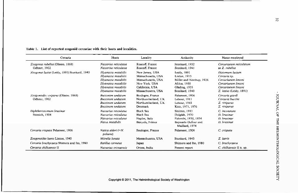
Tab
le 1
. L
ist
of r
epor
ted
zoo
gon
id c
erca
riae
wit
h th
eir
hos
ts a
nd
loc
alit
ies.
Cer
cari
a
Zoog
onus
rub
ellu
s (O
lsso
n, 1
868)
Odh
ner,
190
2
Zoog
onus
las
ius
(Lei
dy,
1891
) Stu
nkar
d, 1
940
Zoog
onoi
des
vivi
paru
s (O
lsso
n, 1
868)
Odh
ner,
190
2
Dip
hthe
rost
omum
bru
sina
eSt
ossi
ch,
1904
Cer
cari
a cr
ispa
ta P
else
neer
, 1 9
06
Zoog
onoi
des
laev
is L
into
n, 1
940
Cer
cari
a br
achy
caec
a S
him
ura
and
Ito,
198
0
Cer
cari
a ch
ilkae
nsis
II
Hos
ts
Nas
sari
ns r
etic
ulat
usN
assa
rius
ret
icul
atus
Illy
anas
sa m
utab
ilis
Illy
anas
sa
mut
abili
sIl
lyan
assa
m
utab
ilis
Illy
anas
sa
mut
abili
sIl
lyan
assa
m
utab
ilis
Illy
anas
sa m
utab
ilis
Buc
cinu
m u
ndat
umB
ucci
num
un
datu
mB
ucci
num
un
datu
mB
ucci
num
un
datu
m
Nas
sari
us r
etic
ulat
usN
assa
rius
ret
icul
atus
Nas
sari
us r
etic
ulat
usN
assa
mut
abili
s
Nat
ica
alde
ri (
=N
.po
liand
)
Mitr
ella
lun
ata
Bat
illus
cor
nutu
s
Nas
sari
us o
riss
aens
is
Loc
ality
Ros
coff
, Fr
ance
Ros
coff
, Fr
ance
New
Jer
sey,
USA
Mas
sach
uset
ts,
USA
Mas
sach
uset
ts,
USA
New
Yor
k, U
SAC
alif
orni
a, U
SAM
assa
chus
etts
, U
SA
Bou
logn
e, F
ranc
eN
orth
umbe
rlan
d, U
KN
orth
umbe
rlan
d, U
KD
enm
ark
Bla
ck S
eaB
lack
Sea
Nap
les,
Ita
lyB
anyu
ls,
Fran
ce
Bou
logn
e, F
ranc
e
Mas
sach
uset
ts,
USA
Japa
n
Ori
ssa,
Ind
ia
Aut
hori
ty
Stun
kard
, 19
32St
unka
rd,
1941
Lei
dy,
1891
Lin
ton,
191
5M
iller
and
Nor
thup
, 19
26A
fric
a, 1
930
Gla
ding
, 19
35St
unka
rd,
1940
Pels
enee
r, 19
06L
ebou
r, 1
911
Leb
our,
191
8K
oie,
197
1, 1
976
Sini
tzin
, 19
11D
olgi
kh,
1970
Pal
ombi
, 19
30,
1934
Bay
ssad
e-D
ufou
r an
dM
ailla
rd,
1974
Pels
enee
r, 1
906
Stun
kard
, 19
43
Shim
ura
and
Ito,
198
0
Pres
ent
repo
rt
Nam
e em
ploy
ed
Cer
cari
aeum
ret
icul
atum
as Z
. ru
bellu
s
Dis
tom
um l
asiu
mC
erca
ria
sp.
Cer
cari
aeum
lin
toni
Cer
cari
aeum
lin
toni
Cer
cari
aeum
lin
toni
Z.
lasi
us (L
eidy
, 18
91)
Cer
cari
a gi
ardi
Cer
cari
a bu
ccin
iZ
. vi
vipa
rus
Z.
vivi
paru
s
C.
inco
nsta
nsD
. bru
sina
eD
. bru
sina
eD
. bru
sina
e
C. c
risp
ata
Z.
laev
is
C. b
rach
ycae
ca
C. c
hilk
aens
is I
I n.
sp.
Copyright © 2011, The Helminthological Society of WashingtonCopyright © 2011, The Helminthological Society of Washington

OF WASHINGTON, VOLUME 58, NUMBER 1, JANUARY 1991 33
Figure 1. Cercaria chilkaensis II sp. n. from thesnail, Nassarius orissaensis, ventral view.
D. brusinae, especially in having 3 pairs of pen-etration glands. The number is much higher inthe remaining species; there are 6-8 pairs of pen-etration glands in the cercariae of Z. rubellus andZ. laevis, the former of which has well organizedrudiments of testes and ovary. The cercaria ofZ. viviparus has numerous penetration glands,and the posterior part of the body forms a prom-inent disc covered with spines. In Cercariabrachycaeca described by Shimura and Ito (1980)there are 6 pairs of penetration glands, and ceca
are short and stout, terminating in the equatorialplane of the body.
The above comparison suggests that the pres-ent form may represent a species congeneric withD. brusinae. Adults of such a species occur in thecat fish, Tachysurus thalassinus, from the lakewhere infected snails were collected, and meta-cercarial cysts referable to the genus Diphthero-stomum were found attached to the mantle foldof TV. orissaensis. Experimental work is neededto link the various stages in the life cycle.
Acknowledgments
We thank the Indian Council of AgriculturalResearch for providing funds to complete thiswork.
Literature Cited
Africa, C. M. 1930. The excretory system of Cer-cariaeum lintoni Miller, 1926. Journal of Parasi-tology 17:14-17.
Bayssade-Dufour C., and C. Maillard. 1974. Che-totaxie de quatre cercaires d'Allocreadioidea.Annales de parasitologie Humaine et Comparee 49:521-554.
Dolgikh, A. V. 1970. Trematode larvae, parasites ofBlack Sea mollusc Nassa reticulata var. pontica.Pages 122-138 in "Bentos." "Naukova DUMKA,"Kiev, U.S.S.R. (1965).
Glading, B. 1935. The life cycle of Cercariaeum lin-toni Miller and Northup. Journal of Parasitology21:434.
Koie, M. 1971. On the histochemistry and ultrastruc-ture of the tegument and associated structures ofthe cercaria of Zoogonoides viviparus in the firstintermediate host. Ophelia 9:165-206.
. 1976. On the morphology and life history ofZoogonoides viviparus (Olsson, 1868) Odhner, 1902(Trematoda, Zoogonidae). Ophelia 15:1-14.
Lebour, M. V. 1911. A review of the British marinecercariae. Parasitology 4:416-456.
. 1918. A trematode larva from Buccinum un-datum and notes on trematodes from post-larvalfish. Journal of the Marine Biological Association,U.K. 11:514-517.
Leidy, J. 1891. Notices of Entozoa. Proceedings ofAcademy National Sciences, Philadelphia 42:410-418.
Linton, E. 1915. Note on trematode sporocysts andcercariae in marine molluscs of the Woods Holeregion. Biological Bulletin 28:198-209.
Madhavi, R. 1979. Digenetic trematodes from ma-rine fishes of Waltair coast, Bay of Bengal. FamilyZoogonidae. Rivista di Parassitologia 40:249-259.
Miller, H. M., and F. E. Northup. 1926. The seasonalinfestation of Nassa obsoleta (Say) with larvaltrematodes. Biological Bulletin 50:490-508.
Palombi, A. 1930. II ciclo biologico di Diphtherosto-mum brusinae Stossich (Trematode digenetico:fam. Zoogonidae Odhner). Pubblicazione dellaStazione Zoologica di Napoli 10:111-149.
Copyright © 2011, The Helminthological Society of WashingtonCopyright © 2011, The Helminthological Society of Washington

34
. 1934. Gli stadi larvali dei trematodi del Gul-fo di Napoli. I. Contribute olio studio della mor-fologia, biologia e sistematica della cercarie ma-rine. Pubblicazione della Stazione Zoologica diNapoli 14:51-94.
Pelseneer, P. 1906. Trematodes parasites de mol-lusques marins. Bulletin Scientifique de la Franceet de la Belgique 40:161-186.
Shameem, U., N. Narasimha Rao, and R. Madhavi.1990. Cercaria chilkaensis No.I a new cytopho-rous cercaria from the snail, Stenothyra blanfordi-ana from Chilka lake, India. Journal of NaturalHistory 24:261-270.
Shimura, S., and J. Ito. 1980. Two new cercariae,Cercaria brachycaeca n. sp. and Cercaria misak-iana n. sp. from top shells, Batillus cornutus andMarmarostoma stenogyrum, with notes of theireffects on the hosts. Japanese Journal of Parasi-tology 29:69-76.
Sinitzin, D. 1911. Parthenogenetic generation oftrematodes and its progeny in molluscs of the BlackSea. (Translation.) Records of the Imperial Acad-emy of Sciences, St. Petersburg 30:127 pp.
Stunkard, H. W. 1932. Some larval trematodes fromthe coast in the region of Roscoff. Finnistere. Par-asitology 24:321-343.
. 1940. Life history studies and specific deter-mination in the trematode genus Zoogonus. Jour-nal of Parasitology 26(Suppl.):33-34.
. 1941. Specificity and host-relations in thetrematode genus Zoogonus. Biological Bulletin 81:205-214.
. 1943. The morphology and life history of thedigenetic trematode, Zoogonoides laevis Linton,1940. Biological Bulletin 85:227-237.
Scientist of the Year
From the Director, United States Department of Agriculture,Agricultural Research Services
Jitender P. Dubey, Research Microbiologist, Zoonotic Diseases Laboratory, Livestock & PoultrySciences Institute, has been selected as the Beltsville Area Scientist of the Year for 1990. Dr. Dubeyhas distinguished himself as a leader in parasitology research nationally and internationally. He isespecially well known for his research on Toxoplasma, a protozoan parasite that infects manydomestic animals, cattle, swine, sheep, goats, and horses, resulting in disease and substantial eco-nomic losses. He has documented the complex life cycle of the parasite, studied its epidemiologyin livestock, and developed methods for rendering meat products noninfectious from the standpointof public health.
Dr. Dubey's research extends broadly beyond his contributions on toxoplasmosis. For example,he recently discovered a new genus of parasites (Neospord) of potentially great economic importance.He has developed a diagnostic test to detect infections in animals and man and is assisting the Stateof California in diagnosing an outbreak in dairy cattle.
Dr. Dubey received a Ph.D. from the School of Medicine, University of Sheffield, England, in1966. He worked at Montana State University prior to joining ARS at Beltsville in 1982. Hisresearch is documented in over 350 publications. Dr. Dubey has received a number of prestigiousawards, including the Distinguished Veterinary Parasitologist Award from the American Associationof Veterinary Parasitology.
Copyright © 2011, The Helminthological Society of WashingtonCopyright © 2011, The Helminthological Society of Washington

J. Helminthol. Soc. Wash.58(1), 1991, pp. 35-38
Anapalaeorchis hamajimai gen. et sp. n. (Trematoda: Monorchiidae)from the Loach, Cobitis biwae, in Japan
TAKAHIRO FujiNO1 AND TEUI KiFUNE2
1 Department of Parasitology, Faculty of Medicine, Kyushu University, Fukuoka 812, Japan and2 Department of Parasitology, School of Medicine, Fukuoka University, Fukuoka 814-01, Japan
ABSTRACT: Anapalaeorchis hamajimai gen. et sp. n. (Digenea: Monorchiidae: Asymphylodorinae) is describedfrom gravid specimens from the digestive tract of a freshwater fish, Cobitis biwae, collected in the TokigawaRiver, Saitama Prefecture, central Japan. Anapalaeorchis gen. n. is proposed for a species possessing cecaterminating midway in the hindbody, an acetabulum larger than the oral sucker, 2 tandem, nearly contiguoustestes, a bipartite seminal vesicle, a trilobed ovary, and vitellaria in paired compact bunches.
KEY WORDS: Anapalaeorchis gen. n., Anapalaeorchis hamajimai sp. n., Digenea, Monorchiidae, Asymphy-lodorinae, Cobitis biwae.
In the course of examination of the digestivetract of the freshwater fish, Cobitis biwae Jordanand Snyder, taken from the Tokigawa River, Sai-tama Prefecture, central Japan, we found a mon-orchiid trematode with characters that did notagree with any existing genus. This trematodewas found to be new and is here described as anew genus.
Materials and Methods
A total of 128 specimens were collected from thedigestive tract of the loach, Cobitis biwae, in the To-kigawa River, Saitama Prefecture, central Japan from29 April 1985 to 27 August 1988. Of these, 46 gravidspecimens were designated as type specimens. Descrip-tions and measurements were based on these speci-mens. The specimens were fixed in 70% ethanol underthe pressure of a cover glass, stained with carmine, oreosin and hematoxylin, or only hematoxylin, andmounted. Measurements, given in micrometers unlessotherwise stated, were made with the aid of a filarmicrometer. The figure was drawn using a Nikon pro-file projector (6CT2). For scanning electron micros-copy (SEM), worms and eggs were rinsed well withRinger's saline and fixed for 4 hr in 5% glutaraldehydein 0.1 M phosphate buffer (pH 7.3) and postfixed for3 hr in phosphate-buffered osmium tetroxide. Afterdehydration with a graded series of ethanol, the spec-imens were dried in a critical point drying apparatus,coated with gold in a JEOL FC-1100 ion-sputteringapparatus, and photographed with a JEOL JSM-U3scanning electron microscope operated at 15 kV.
ResultsAnapalaeorchis gen. n.
DIAGNOSIS: Body fusiform or spatulate, spi-nous. Oral sucker subterminal. Esophagus fairlylong, slender. Ceca extending halfway throughhindbody. Acetabulum circular, larger than oralsucker, in forebody. Testes double, tandem, nearlycontiguous, ovoid to subtriangular, in hindbody.
Cirrus eversible, spined. Cirrus pouch claviform.Seminal vesicle bipartite, pars prostatica well dif-ferentiated. Ovary pretesticular, trilobed. Uterusfilling space in hindbody. Terminal organ re-ceiving metraterm terminally. Vitellaria laterallypaired postacetabular compact bunches, equa-torial.
TYPE SPECIES: Anapalaeorchis hamajimaisp. n.
Anapalaeorchis hamajimai sp. n.(Figs. 1-8)
DESCRIPTION WITH CHARACTERS OF THE GENUS:Body fusiform or spatulate, 910 (700-1,110) inlength and 290 (240-330) in maximum width(Figs. 1—4); covered entirely with minute spines,which become thinner and smaller posteriorly(Figs. 5, 6). Oral sucker circular, subterminal,122 (106-144) in diameter. Prepharynx not pres-ent. Pharynx oval, 65 (52-76) by 67 (58-79).Esophagus elongate, 205 (200-210) long. Cecaterminating at level of anterior testis or posteriorend of ovary. Acetabulum greatly larger than oralsucker, 168 (132-199) by 162 (136-189), situ-ated one-third of body length from anterior ex-tremity; sucker width ratio, 1:1.3—1.4. Testesnearly touching, tandem; anterior testis ovoid tosubtriangular, 104 (89-124) long by 90 (76-101)wide; posterior testis ovoid, elongate, 127 (87-145) long by 94 (69-110) wide. Cirrus eversible,spined. Cirrus pouch club-shaped, seminal ves-icle bipartite, pars prostatica well differentiated.Ovary trilobed, 110 (79-143) long by 114 (73-162) wide, slightly posterior to middle of body,closely located to anterior testis. Genital porenear left margin of body, slightly anterior to levelof posterior edge of acetabulum. Uterus sur-
35
Copyright © 2011, The Helminthological Society of WashingtonCopyright © 2011, The Helminthological Society of Washington

36 JOURNAL OF THE HELMINTHOLOGICAL SOCIETY
Figure 1. Anapalaeorchis hamajimai, holotype, ven-tral view. Bar = 200 fim. O, ovary; T, testes.
rounding ovary and testes, filling hindbody.Vitellaria in lateral clusters of several follicles atmidbody; right vitelline mass longer than left.Terminal organ receiving metraterm terminally.Eggs elliptical, operculate, embryonated, 29.3(26.4-31.2) x 16.6 (15.4-17.2) (N = 100, liveeggs) (Fig. 7). Egg shell surface coarse with shal-low irregular pits or depressions (Fig. 8). Excre-tory system not observed.
TYPE MATERIALS: Holotype and 6 paratypesare deposited in the collection of the NationalScience Museum, Tokyo (NSMT, PI 3776); 4paratypes in the United States National MuseumHelminthological Collection (USNM Helm. Coll.No. 81141); 35 paratypes in the collection of the
Department of Parasitology, Faculty of Medi-cine, Kyushu University (PDKU T001-T035).
HOST: Sand loach, Cobitis biwae Jordan andSnyder.
HABITAT: Stomach (juvenile worms) and in-testine (adult worms).
ETYMOLOGY: This species is named after Prof.Fusanori Hamajima who collected and donatedthe present material.
Discussion
According to Yamaguti (1971), the monor-chiid subfamily Asymphylodorinae includes 3genera, Palaeorchis Szidat, 1943, Asymphylo-dora Looss, 1899, and Triganodistomum Simer,1929. The new genus Anapalaeorchis is similarto Palaeorchis in that the double testes are un-equal in size and the vitellaria are compact, butis easily separable in tandemly arranged testes,a trilobed ovary, and an acetabulum that is largerthan the oral sucker. The testes of A. hamajimaiare in tandem and almost touching, whereas allthe species of Palaeorchis have testes that arejuxtaposed and clearly separated. Anapalaeorchisis also similar to Asymphylodora in its large ac-etabulum and long esophagus. Asymphylodora,however, is different from Anapalaeorchis inhaving a single testis. Yamaguti (1938) erecteda new species, Asymphylodora japonica, parasiticin the freshwater fish, Cyprinus carpio. He in-cluded some worms from the small intestine ofCobitis biwae, which were much smaller thanthose from C. carpio. We had the loan of Ya-maguti's specimens from Cobitis biwae pre-served in Meguro Parasitological Museum, To-kyo (No. 22034). Although these specimensmounted on 1 slide are too old to make surewhether the testis is single or separated, the spec-imens are found to have some features identifi-able to A. japonica: the longer ceca extendingover the posterior end of the testis and the moreextended vitellaria than in Anapalaeorchis ha-majimai. The main similarity of Anapalaeorchisto Triganodistomum is that in both genera thetestes are arranged in tandem. The testes in Tri-ganodistomum, however, are distinctly separat-ed, whereas those of Anapalaeorchis are close toeach other. The ceca reach to near the posteriorextremity of the body in Triganodistomum,whereas the ceca terminate in the mid-hindbodyin Anapalaeorchis. Triganodistomum has farmore extensive vitellaria than has Anapalaeor-chis.
Copyright © 2011, The Helminthological Society of WashingtonCopyright © 2011, The Helminthological Society of Washington

OF WASHINGTON, VOLUME 58, NUMBER 1, JANUARY 1991 37
Figures 2-8. 2-4. Anapalaeorchis hamajimai, H&E stain. Bar = 200 jtm. 2. Holotype, gravid adult. 3. Para-type, gravid adult. 4. Juvenile. 5, 6. A. hamajimai, SEM of body. 5. Whole body in ventral view. Bar = 50 nm.6. Tegumental spines, ventral aspect in middle region. Bar = 5 tan. 7, 8. A, hamajimai, eggs. 7. Light micrographof whole eggs. Bar = 20 jum. 8. SEM of operculum and part of egg shell. Bar = 1 jum.
Copyright © 2011, The Helminthological Society of WashingtonCopyright © 2011, The Helminthological Society of Washington

38
Schell (1973) erected the genus Neopaleorchisbased upon the new species, N. catostomi, whichinhabits the intestine of a coarsescale sucker, Ca-tostomus macrocheilus. Anapalaeorchis bears su-perficial resemblance to Neopaleorchis in havingtandem testes; however, in Neopaleorchis, theseminal vesicle is folded, but not bipartite, theoral sucker and acetabulum are nearly equal,vitellaria are postcecal and contiguous dorsal touterus and testes, and eggs are small and nu-merous.
AcknowledgmentsWe are greatly indebted to Prof. Fusanori Ha-
majima of National Defense Medical College forthe collection and donation of the present ma-terial. Our sincere thanks are also due to Dr.Shunya Kamegai of Meguro Parasitological Mu-seum, Tokyo, and Dr. Susumu Saito of Yama-
gata University for their interests and valuablesuggestions; to Dr. Koichi Fukuda of NationalDefense Medical College for his help in preparingspecimens; and to Dr. Atsuo Ichihara of MeguroParasitological Museum, Tokyo, for the loan ofYamaguti's specimens ofAsymphylodorajapon-ica.
Literature CitedSchell, S. C. 1973. The life history of Neopaleorchis
catostomi gen. et sp. n. (Trematoda: Monorchi-idae), an intestinal parasite of the coarsescale suck-er, Catostomus macrocheilus Girard. Journal ofParasitology 59:463^168.
Yamaguti, S. 1938. Studies on the Helminth Faunaof Japan. Part 21. Trematodes of Fishes IV. Pub-lished by the author. 139 pp.
. 1971. Synopsis of Digenetic Trematodes ofVertebrates. Keigaku Publishing Co., Tokyo. 1,074pp.
Copyright © 2011, The Helminthological Society of WashingtonCopyright © 2011, The Helminthological Society of Washington

J. Helminthol. Soc. Wash.58(1), 1991, pp. 39-42
Observations on the Life Cycle of Proteocephalus tumidocollus(Cestoda: Proteocephalidae) in Steelhead Trout,Oncorhynchus mykiss
WILLIAM T. Cox1 AND GARY L. HENDRICKSONDepartment of Fisheries, Humboldt State University, Arcata, California 95521
ABSTRACT: The life cycle of Proteocephalus tumidocollus in a brackish water, aquacultural pond involvessteelhead trout (Oncorhynchus mykiss) as definitive hosts, and freshwater (Diaptomus sp., Paracyclops fimbriatuspoppei) or benthic marine (Halicyclops sp.) copepods as intermediate hosts. Steelhead trout do not feed oncopepods directly. Instead, they consume the macroalga Enteromorpha sp. with epiphytic copepods attached.Between May 1981 and July 1982, there were no statistically significant monthly variations in prevalence, meanintensity, or relative density of P. tumidocollus in steelhead trout. However, there were marked decreases inmean intensity and relative density and a marked increase in the percentage of cestodes that were gravid inJanuary to March 1982 when Enteromorpha sp. was absent from the pond.
KEY WORDS: Proteocephalus tumidocollus, Proteocephalidae, Cestoda, life cycle, seasonality, steelhead trout,Oncorhynchus mykiss, California.
Wagner (1953) described Proteocephalus tu-midocollus from rainbow and brook trout fromthe California Trout Company, Mentone, Cali-fornia. Five reports of P. tumidocollus have sincebeen published (Hicks and Threlfall, 1973; Ha-nek and Molnar, 1974; Arthur et al., 1976; Ma-mer, 1978; Arai and Mudry, 1983).
Wagner (1954) studied the life cycle of P. tumi-docollus in freshwater. He fed its eggs to the co-pepods Cyclops vernalis, Eucyclops agilis, Eu-cyclops speratus, and Tropocyclops prasinus,which became infected. He was unable to infectthe copepods Macrocyclops albidus and Para-cylops fimbriatus poppei nor several other inver-tebrates. Procercoids developed rapidly at 20°Cand became infective to trout at 9 to 19 dayspostinfection (PI). Trout definitive hosts becameinfected by eating copepod intermediate hosts.Second intermediate hosts were not required, al-though large trout could become infected by eat-ing small infected trout.
Proteocephalus tumidocollus is common insteelhead trout, Oncorhynchus mykiss (Wal-baum), in the brackish water ponds of the ArcataWaste water Aquaculture Project, Arcata, Cali-fornia. While freshwater species of Proteocepha-lus are common and well studied, brackish waterspecies are rare and poorly known (Wardle andMcLeod, 1952; Yamaguti, 1959; Freze, 1965;Schmidt, 1986). The Arcata system offered an
1 Present address: California Department of Fish andGame, Fish Disease Laboratory, 2111 Nimbus Road,Rancho Cordova, California 95670.
unusual opportunity to examine the seasonalityand life cycle of a Proteocephalus sp. in brackishwater.
Materials and Methods
Infected steelhead trout were obtained from SouthPond (0.6 ha, avg. depth 2 m, avg. salinity 14.5 ppt),Arcata Wastewater Aquaculture Project, Arcata, Cal-ifornia. Uninfected steelhead trout fry were obtainedfrom the Humboldt State University Fish Hatchery.Fish were held in 19-3 8-liter aquaria supplied with11°-17°C aerated, filtered water. Potential invertebratehosts were sieved from South Pond (brackish water),the adjacent oxidation pond of the Arcata sewage treat-ment plant (freshwater), or from the adjacent Butcher'sSlough on Humboldt Bay (seawater). Invertebrates werecultured in 2-19-liter aquaria (11°-17°C) and fed yeastand flaked fish food.
Seasonal occurrence of P. tumidocollus in steelheadtrout from South Pond was determined from fish col-lected by angling at weekly intervals from 12 May 1981to 31 July 1982. Seven to 20 fish were obtained at eachsampling. Fish total length and weight were recordedprior to necropsy. Necropsy procedures were standard(Hoffman, 1967). Food items in stomachs offish hostswere analyzed quantitatively using percent composi-tion by weight (Bowen, 1983).
Adult cestodes were placed in 0.7% saline and al-lowed to shed eggs for use in life history studies. Eggswere used to expose cultured invertebrates, which wereexamined for developing procercoids from 30 min to86 days after initial exposure. In addition, 150 wildAnisogammams confervicolus (Stimpson) (Amphipo-da) from South Pond were examined for natural pro-cercoid infections.
To infect experimental fish definitive hosts, inver-tebrates from cultures were fed to hatchery-reared (ces-tode-free) steelhead trout fry. Twenty-five fish were fedAnisogammams confervicolus (Amphipoda), and 25 fishwere fed a combination of Corophium spinicorne
39
Copyright © 2011, The Helminthological Society of WashingtonCopyright © 2011, The Helminthological Society of Washington

40 JOURNAL OF THE HELMINTHOLOGICAL SOCIETY
Table 1. Levels of infection of Proteocephalus tumidocollus and percentage of cestodes gravid in steelhead trout,Oncorhynchus mykiss, from South Pond, Arcata Wastewater Aquaculture Project, California, May 1981 to July1982.
Month
1981
MayJuneJulyAugustSeptemberOctoberNovemberDecember
1982
January:):February:):March:}:AprilMayJuneJuly
No. of fishexamined
14128
16181820
7
13128
16201816
No. of fishinfected (%)
6 (42.8)3 (25.0)4 (50.0)2(12.5)8 (44.4)8 (44.4)6 (30.0)2 (28.6)
4 (30.8)6 (50.0)1 (12.5)7 (43.8)
10 (50.0)7 (38.9)4 (25.0)
Meanintensity*
8.57.75.81.57.54.8
17.75.0
6.02.52.0
30.019.710.78.2
Relativedensityf
3.61.92.90.23.32.15.31.4
1.81.20.3
13.19.84.22.1
Range in nos.per infection
2-286-72-41-21-371-102-844-6
1-111-521-661-1251^81-25
Percentage ofcestodes gravid
74.513.013.00
25.092.119.870.0
10093.3
10096.229.457.30
* Total number of cestodes in sample 4- number of infected hosts in sample.f Total number of cestodes in sample -f- total number of hosts in sample.t No Enteromorpha sp. in South Pond.
Stimpson (Amphipoda) and Gnorimosphaeroma ore-gonensis (Dana) (Isopoda). Twenty-five fish were feda mixture of small Copepoda, including Diaptomns sp.,1 planktonic and 1 benthic species of Halicyclops,Paracyclopsfimbriatuspoppei(Rehberg), and Tigriopuscalifornicus (Baker). Seventy-two fish were held as con-trols and were fed a commercial trout pellet. After 8wk, all fish were killed and examined for cestodes. Inaddition, 26 three-spined sticklebacks (Gasterosteusaculeatus L.) from South Pond were examined for ces-todes to see if they might be capable of passing juvenilecestodes on to steelhead trout via predation.
Results
Proteocephalus tumidocollus was recoveredfrom the pyloric ceca of 78 of 216 (36.1%) steel-head trout examined from South Pond duringthe seasonal survey. It was present in all monthsof the year. There was no marked seasonal vari-ation in prevalence (Table 1). Relative densityand mean intensity seemed to vary seasonallywith peaks in November 1981 and May 1982;however, these variations were not statisticallysignificant by analysis of variance (P < 0.05).There was a marked decrease in mean intensityand relative density in January to March 1982when Enteromorpha sp. was absent from SouthPond. Prevalence, mean intensity, and relativedensity all increased as fish total length increased.Gravid cestodes occurred in all but 2 mo and
were most prevalent in winter, particularly inthose months when Enteromorpha sp. was ab-sent from South Pond (Table 1).
Analysis of stomach contents from 68 steel-head trout from South Pond showed that on apercent composition by weight basis, they ate:Corophium spinicorne (Amphipoda) 36.70%, A.confervicolus (Amphipoda) 29.90%, detritus11.90%, Enteromorpha sp. (macroalga) 4.80%,mysids (Crustacea) 3.60%, G. aculeatus (fish)2.50%, G. oregonensis (Isopoda) 2.12%, filamen-tous algae 1.90%, earwigs (terrestrial insects)1.70%, Diptera 1.41%, unidentifiable fish larvae1.20%, annelids 1.60%, unidentifiable inverte-brates 0.36%, crabs 0.30%, and copepods 0.01%.Enteromorpha sp. was eaten whenever it wasavailable in South Pond (May to December 1981,April to July 1982).
Experimental infections were established in thecopepods Diaptomus sp., P. fimbriatus poppei,and a benthic species of Halicyclops. The fresh-water copepod Acanthocyclops (A.) vernalis (Fi-scher) was also experimentally infected. Thisspecies enters South Pond when the pond is filledusing a mixture of treated sewage effluent andseawater. However, it cannot tolerate the salinityin South Pond for very long. Anisogammarusconfervicolus (Amphipoda), C. spinicorne (Am-
Copyright © 2011, The Helminthological Society of WashingtonCopyright © 2011, The Helminthological Society of Washington

OF WASHINGTON, VOLUME 58, NUMBER 1, JANUARY 1991 41
phipoda), G. oregonensis (Isopoda), T. califor-nicus (Copepoda), and a planktonic species ofHalicyclops (Copepoda) were refractory to ex-perimental infection. None of 150 wild A. con-fervicolus examined from South Pond was in-fected.
Three of 25 hatchery-reared steelhead trout fedcopepods (Diaptomus sp., 1 planktonic and 1benthic species of Halicyclops, P. fimbriatus pop-pel, T. californicus) from exposed cultures hadimmature P. tumidocollus at 56 days PL One fishhad 3 cestodes and 3 fish had 1 immature cestodeeach. All cestodes were attached to the pyloricceca. None of the 25 fish fed exposed A. confer-vicolus, none of the 25 fish fed exposed C. spin-icorne and G. oregonensis, and none of the 72control fish was infected. None of 26 three-spinedsticklebacks examined from South Pond har-bored cestodes even though smaller sticklebacksfed almost exclusively on copepods.
Discussion
Both marine (benthic Halicyclops sp.) andfreshwater (Diaptomus sp., P. fimbriatus poppei)copepods are suitable intermediate hosts for Pro-teocephalus tumidocollus in South Pond. In ad-dition, the entire life cycle takes place in brackishwater. This utilization of a marine host andbrackish water is unusual among species ofPro-teocephalus (Wardle and McLeod, 1952; Ya-maguti, 1959; Freze, 1965; Schmidt, 1986).
Copepod intermediate hosts of P. tumidocollusare not an important item in the diet of steelheadtrout definitive hosts in South Pond (0.01% byweight). However, the benthic Halicyclops sp.and P. fimbriatus poppei are epiphytic on themacroalga Enteromorpha sp. Enteromorpha sp.accounted for up to 30.1% by weight of the dietof steelhead trout in some months (4.8% overthe entire study). Thus, steelhead trout in SouthPond most likely become infected by incidentallyingesting copepods as they feed on Enteromor-pha sp. This incidental mode of infection has notto our knowledge been reported previously.
There were no statistically significant monthlyvariations in prevalence, mean intensity, or rel-ative density of P. tumidocollus in steelhead troutin South Pond. However, there was a markeddecrease in both mean intensity and relative den-sity, and a marked increase in the percentage ofcestodes recovered from steelhead trout that weregravid in January to March 1982 when Entero-morpha sp. was absent from South Pond. In the
absence of Enteromorpha sp., steelhead trout donot eat significant numbers of copepods. Hence,new infections do not occur. Measures of infec-tion intensity are reduced and cestodes from old-er infections tend to become gravid. Thus, sea-sonal cycles in abundance or maturity of P.tumidocollus in steelhead trout in South Ponddepend largely on the seasonal occurrence of En-teromorpha sp. Enteromorpha sp. is normallypresent in South Pond as long as South Pondremains sufficiently brackish. When there is suf-ficient rain to dilute South Pond (most likely inthe winter rainy season), Enteromorpha sp. diesback and in some years disappears altogether.
Acknowledgments
We are grateful to Dr. George H. Allen forgenerously allowing us to work at the ArcataWaste water Aquaculture Project. This study is aportion of a thesis submitted by the senior authorto the Department of Fisheries, Humboldt StateUniversity, Arcata, California in partial fulfill-ment of the Degree of Master of Science.
Literature Cited
Arai, H. P., and D. R. Mudry. 1983. Protozoan andmetazoan parasites of fishes from the headwatersof the Parsnip and McGregor rivers, British Co-lumbia: a study of possible parasite transfauna-tions. Canadian Journal of Fisheries and AquaticSciences 40:1676-1684.
Arthur, J. R., L. Margolis, and H. P. Arai. 1976.Parasites of fishes of Aishihik and Stevens lakes,Yukon Territory, and potential consequences oftheir interlake transfer through a proposed waterdiversion for hydroelectrical purposes. Journal ofthe Fisheries Research Board of Canada 33:2489-2499.
Bowen, S. H. 1983. Quantitative description of thediet. Pages 325-336 in L. A. Nielsen and D. L.Johnson, eds. Fisheries Techniques. AmericanFisheries Society, Bethesda, Maryland.
Freze, V. I. 1965. Proteocephalata in Fish, Amphib-ians, and Reptiles. Essentials of Cestodology, Vol.5. Akademiya Nauk S.S.S.R. Gel'mintologichesk-aya Laboratoriya. (Israel Program for ScientificTranslations, Jerusalem. 1969. 597 pp.)
Hanek, G., and K. Molnar. 1974. Parasites of fresh-water and anadromous fishes from Matamek Riv-er System, Quebec. Journal of the Fisheries Re-search Board of Canada 31:1135-1139.
Hicks, F. J., and W. Threlfall. 1973. Metazoan par-asites of salmonids and coregonids from coastalLabrador. Journal of Fish Biology 5:399—415.
Hoffman, G. L. 1967. Parasites of North AmericanFreshwater Fishes. University of California Press,Berkeley. 486 pp.
Mamer, B. E. 1978. The parasites of trout in north-west Washington. Journal of Parasitology 64:314.
Copyright © 2011, The Helminthological Society of WashingtonCopyright © 2011, The Helminthological Society of Washington

42
Schmidt, G. D. 1986. CRC Handbook of TapewormIdentification. CRC Press, Inc., Boca Raton, Flor-ida. 675 pp.
Wagner, E. D. 1953. A new species of ProteocephalusWeinland, 1858, (Cestoda), with notes on its lifehistory. Transactions of the American Microscop-ical Society 72:364-369.
. 1954. The life history of Proteocephalus tu-
midocollus Wagner, 1953, (Cestoda), in rainbowtrout. Journal of Parasitology 40:489^498.
Wardle, R. A., and J. A. McLeod. 1952. The Zoologyof Tapeworms. University of Minnesota Press,Minneapolis. 780 pp.
Yamaguti, S. 1959. Systema Helminthum. Vol. 2.The Cestodes of Vertebrates. Interscience Pub-lishers, Inc., New York. 860 pp.
Copyright © 2011, The Helminthological Society of WashingtonCopyright © 2011, The Helminthological Society of Washington

J. Helminthol. Soc. Wash.58(1), 1991, pp. 43-50
Sterol Composition of the Corn Root Lesion Nematode,Pratylenchus agilis, and Corn Root Cultures
DAVID J. CmxwooD1 AND WILLIAM R. LusBY2
1 Nematology Laboratory, U.S. Department of Agriculture, Agricultural Research Service, Building 467,BARC-East, Beltsville, Maryland 20705 and2 Insect Hormone Laboratory, U.S. Department of Agriculture, Agricultural Research Service, Building 467,BARC-East, Beltsville, Maryland 20705
ABSTRACT: Sterols from mixed stages of the corn root lesion nematode, Pratylenchus agilis, and uninfectedcorn root cultures were analyzed by gas chromatography-mass spectrometry. Twenty-eight sterols were identifiedin P. agilis, including 9 not previously detected in nematodes. The major sterols were 24-ethylcholest-22-enol,24-ethylcholesta-5,22-dienol, 24-methylcholestanol, 24-ethylcholestanol, isofucostanol, and 24-ethylcholesterol.The principal corn root sterols were 24-ethylcholesta-5,22-dienol, 24-methylcholesterol, 24-ethylcholesterol,isofucosterol, and cycloartenol. Therefore, the major metabolic transformation of sterols by P. agilis was sat-uration of the sterol nucleus. In addition, very small amounts of 4a-methylsterols were biosynthesized by P.agilis. The 4-methylation pathway is unique to nematodes and was previously demonstrated only in free-livingRhabditida. The sterol composition of P. agilis is remarkably different from that of other nematodes in its verysmall relative percentage of cholesterol (0.4% of the total nematode sterol).
KEY WORDS: corn, lesion nematode, lipid, maize, nematode, Pratylenchus agilis, sterol, Zea mays.
The development of new control strategies forparasitic nematodes could exploit biochemicaldifferences between them and their vertebrate orplant hosts. One of the few such areas is steroidmetabolism; unlike higher animals and plants,nematodes possess a nutritional requirement forsterol as a result of their lack of de novo sterolbiosynthesis (for review: Chitwood et al., 1986).Sterols have 2 major functions; they are integralcomponents of cell membranes as well as met-abolic precursors of steroid hormones (Nes andMcKean, 1977). Interest in nematode sterol bio-chemistry has intensified in recent years becauseof the discovery that several azasteroids and al-iphatic amines strongly disrupt growth and de-velopment and also inhibit sterol metabolism innematodes (Chitwood, 1987).
Because free-living nematodes are easily prop-agated in artificial media, they have been utilizedin most investigations involving metabolism ofradiolabeled dietary sterols by nematodes (Chit-wood et al., 1986). The difficulty in in vitro cul-ture of parasitic nematodes has restricted inves-tigation of sterol metabolism in these organismsto comparison of sterol compositions of host andparasite. As expected, cholesterol is the majorsterol of mammalian parasites (Cole and Krus-berg, 1967b; Castro and Fairbairn, 1969; Barrettet al., 1970; Fulk and Shorb, 1971; Chung et al.,1986).
A significant biochemical difference betweenhigher animals and plants is that plant sterols
usually contain alkyl (i.e., methyl or ethyl) sub-stituents at C-24 of the sterol side chain (Nes andMcKean, 1977). Comparison of the sterols ofplant-parasitic nematodes and their hosts has in-dicated that plant parasites are similar to free-living nematodes in that both groups can removethe C-24 alkyl groups of plant sterols (Chitwood,1987). Unequivocal proof in the case of plantparasites, however, would involve experimentswith radiolabeled plant sterols in an in vitro sys-tem. Because large quantities of the corn rootlesion nematode, Pratylenchus agilis Thome andMalek, 1968, can be readily obtained from cornroot explant cultures, we examined the sterols ofthis plant parasite and its host in order to deter-mine if it was a suitable candidate for radiotracerstudies of dealkylation of plant sterols by phyto-parasitic nematodes.
Materials and MethodsSeeds of Zea mays cv. lochief were surface sterilized
by immersion in distilled water for 2 hr and then 1.3%sodium hypochlorite containing 0.1% (v:v) Tween 80for 25 min. The sterilized seeds were germinated for 4days on 1.5% water agar in culture dishes, and then2.5-cm root pieces were transferred to culture dishescontaining Gamborg's B5 medium formulated with1.5% agar and 2.0% sucrose, and lacking cytokinins orauxins (Huettel, 1990). The agar had been extractedpreviously with methanol and chloroform: methanol2:1 (v:v, twice each) to reduce the high amounts ofendogenous cholesterol in commercial agar (Nes, 1987)to an acceptable level (20.6 ng of cholesterol per gramdry weight as determined by subsequently described
43
Copyright © 2011, The Helminthological Society of WashingtonCopyright © 2011, The Helminthological Society of Washington

44 JOURNAL OF THE HELMINTHOLOGICAL SOCIETY
Table 1. Sterol content of Pmtylenchus agilis and rootcultures of Z. mays.
% dry weight
P. agilisZ. mays
Lipid
14.63.9
Sterol
0.030.83
_ % sterolesterified
26.8*
* Not determined.
methods). One-week-old root cultures were inoculatedwith agar plugs from similar cultures containing sterileP. agilis. Nematodes from 8-week-old cultures main-tained at 25°C were harvested with modified Baermannfunnels, floated on 30% sucrose, rinsed 5 times withdistilled water, frozen immediately, and lyophilized.
Lipids were extracted from lyophilized nematodes(2.0-2.2 g dry wt) by homogenization in a Ten-Broecktissue grinder 3 times with chloroform: methanol 2:1and were purified by partition against 0.85% NaCl(Folch et al., 1957). Neutral lipids were separated frompolar lipids and were fractionated into various lipidclasses by column chromatography on silica (Chitwoodet al., 1985). The lipid fractions containing steryl estersand free sterols were saponified separately in meth-anolic KOH (Chitwood et al., 1985); the liberated4-desmethylsterols and 4-methylsterols were purifiedby Florisil column chromatography (Chitwood et al.,1987c).
Roots from uninfected cultures were gently removedwith forceps, frozen, and lyophilized. Extraction of roots(0.3-0.5 g dry wt) was identical to that of nematodes,except a Virtis homogenizer was used. The root lipidswere saponified directly without prior fractionation ona silica column. The 4-methylsterols and 4-desmethyl-sterols were isolated as described above. As a precau-tionary measure, any potentially tightly bound sterolswere removed from the extraction residue by saponi-fication of the residue and analyzed identically to theroot extracts.
Sterols were analyzed by gas-liquid chromatography(GLC) on a DB-1 fused silica capillary column and anOV-17 packed glass column as described previously(Chitwood et al., 1987c). Following preliminary anal-ysis, the sterols were acetylated, the steryl acetates werepurified on Florisil columns and analyzed by GLC, andthen tentative identifications were confirmed by gaschromatography-mass spectrometry (GC-MS, Chit-wood et al., 1987c). The steryl acetates were also frac-tionated according to degree and position of unsatur-ation by chromatography on columns of silicaimpregnated with AgNO3 and analyzed by GLC (Chit-wood et al., 1987c).
Results are the means of two replicates.
Results
Gravimetric analysis indicated that lipid con-stituted approximately 15% of the dry weight ofP. agilis; subsequent quantification of sterols byGLC revealed that sterol comprised only a verysmall proportion of the lipid extract (Table 1).
Table 2. Gas-liquid chromatographic relative reten-tion times (RRT's) of sterols from Pratylenchus agilisand Zea mays, expressed relative to cholesterol. GLCwas performed on a temperature-programmed DB-1fused silica capillary column (14 m x 0.25-mm i.d.,0.25-jtm film) and on an isothermally operated packedglass column containing 2.0% OV-17 stationary phase.
Sterol DB-1 OV-17
CholesterolCholestanol24-Methylcholesta-5 , 22-dienolLathosterol24-Methylcholest-22-enol24-Methylcholesta-5,23-dienol24-Methylenecholesterol24-Methylenecholestanol24-Methylcholesterol24-Methylcholestanol24-Ethylcholesta-5 ,22E-dienol24-Ethylcholest-22E-enol24-Methylcholesta-5,24(25)-dienol24-Ethylcholesta-5,23-dienol24-Ethylcholesta-7,22-dienolFucosterol24-Ethylcholesterol24-EthylcholestanolIsofucosterolIsofucostanol24-Ethylcholesta-5,24(25)-dienol24-Ethyllathosterol24-Ethylcholest-24(25)-enol24Z-EthylidenelathosterolObtusifoliol
4a,24-Dimethylcholestanol
4a-Methyl-24-ethylcholest-22-enol
4a-Methyl-24-ethylcholestanol
4a-Methylisofucostanol
4a-Methyl-24Z-ethylidenelathosterol
Cycloartanol
Cycloeucalenol
Cycloartenol
24-Methylenecycloartanol
1.001.021.111.121.141.251.26.30.30.33.40.43.47.53.57.59
1.601.631.661.691.771.811.811.891.52
(1.49)*1.58
(1.53)1.71
(1.67)2.10(1.91)2.07
d-95)2.26
(2.12)1.71
(1.60)1.74
(1.69)1.86
(1.73)2.14
(1.99)
.00
.02
.14
.19
.17
.38
.37
.39
.33
.35
.45
.47
.67
.67
.70
.76
.67
.69
.85
.88
.98
.962.042.201.56
(1.50)1.52
(1.48)1.66
(1.61)1.87
(1.87)2.11
(2.06)2.52
(2.44)1.67
(1.53)1.88
(1.78)2.05
(1.85)2.28
(2 06)
* Values in parentheses are RRT's of steryl acetates reiativcto cholesteryl acetate. Trivial and systematic narres: choles-terol, cholest-5-en-3/3-ol; cholestano!, 5a-cholestan-3/3-ol;lathosterol, 5a-cholest-7-en-3/3-ol; fucosterol, 24E-ethylidene-cholest-5-en-3(3-ol; isofucosterol, 24Z-ethylidenecholest-5-er-3/3-ol; isofucostanol, 24Z-ethylidene-5«-cholestan-3/?-ol; ob-tusifoliol, 4a, 14-dimethyl-24-methylene-5a-cholest-S-en-3j3-c';cycloartanol, 4,4,14-trimethyl-9/J,19-cyclo-5a-cholestan-3,d-ol;Cycloeucalenol, 4a, 14-dimethyl-24-methylene-9/3,19-cyclo-5a-cholestan-3/3-ol; cycloartenol, 4.4,14-trimethyl-9|3.!9-cyclo-5a-cholest-24-en-3/3-ol.
Copyright © 2011, The Helminthological Society of WashingtonCopyright © 2011, The Helminthological Society of Washington
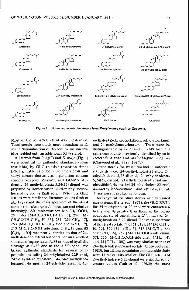
OF WASHINGTON, VOLUME 58, NUMBER 1, JANUARY 1991 45
Cholesterol 24-Methylcholesterol 24-Ethylcholesterol 24-Ethylcholesta-5l22-dierK)l
24-Methylchdestanol 24-Ethylcholestanol 24-Ettiylcholest-22-enol
Isofucosterol 4a,24-Dimethylcholestanol 4a-Methyl-24-ethylcholestanol 4a-Methyl-24-ethylcholest-22-enol
teofucostarol 4a-Methylisofucostanol Cycloartenol Obtusifoliol
Figure 1. Some representative sterols from Pratylenchus agilis or Zea mays.
Most of the nematode sterol was unesterified.Total sterols were much more abundant in Z.mays. Saponification of the root extraction res-idue yielded only an additional 0.1% sterol.
All sterols from P. agilis and Z. mays (Fig. 1)were identical to authentic standards (whenavailable) by GLC relative retention times(RRT's, Table 2) of both the free sterols andsteryl acetate derivatives, argentation columnchromatographic behavior, and GC-MS. Au-thentic 24-methylcholesta-5,24(25)-dienol wasprepared by isomerization of 24-methylenecho-lesterol by iodine (Salt et al., 1986). Its GLCRRT's were similar to literature values (Itoh etal., 1982) and the mass spectrum of the sterylacetate (mass/charge m/z [structure and relativeintensity]: 380 [molecular ion M+-CH3COOH,27], 365 [M-CH3COOH-CH3, 5], 296 [M-CH3COOH-C6HU-H, 53], 281 [296-CH3, 17],253 [M-CH3COOH-C9H17 side chain-2H, 19],213 [M-CH3COOH-side chain-C3H6,17], and 83[C6HU, 100]) was nearly identical to that of 24-methylenecholesterol but contained an abundantside chain fragment at m/z 83 produced by ally liecleavage at C-22 due to the A24(25)-bond. Welacked authentic standards for several com-pounds, including 24-ethylcholest-22E-enol,24Z-ethylidenelathosterol, 4a,24-dimethylcho-lestanol, 4a-methyl-24-ethylcholestanol, 4a-
methyl-24Z-ethylidenelathosterol, cycloartanol,and 24-methylenecycloartanol. These were in-distinguishable by GLC and GC-MS from thesame compounds previously identified by us inHeterodera zeae and Meloidogyne incognita(Chitwood et al., 1985, 1987c).
Other sterols for which we lacked authenticstandards were 24-methylcholest-22-enol, 24-ethylcholesta-5,23-dienol, 24-ethylcholesta-5,24(25)-dienol, 24-ethylcholest-24(25)-dienol,obtusifoliol, 4a-methyl-24-ethylcholest-22-enol,4a-methylisofucostanol, and cycloeucalenol.These were identified as follows.
As is typical for other sterols with saturatedring systems (Patterson, 1971), the GLC RRT'sfor 24-methylcholest-22-enol were characteris-tically slightly greater than those of the corre-sponding sterol containing a A5-bond, i.e., 24-methylcholesta-5,22-dienol. The mass spectrumof the steryl acetate (442 [M+, 18], 344 [M-C7H13-H, 29], 329 [344-CH3, 3], 315 [M-C9H17 sidechain-2H, 36], 257 [M-CH3COOH-side chain,57], 215 [M-CH3COOH-side chain-C3H6, 14],and 55 [C4H7, 100]) was very similar to that of24-ethylcholest-22-enyl acetate (Chitwood et al.,1985), but all ions containing an intact side chainwere 14 mass units smaller. The GLC RRT's of24-ethylcholesta-5,23-dienol were similar to lit-erature values (Itoh et al., 1982); the mass
Copyright © 2011, The Helminthological Society of WashingtonCopyright © 2011, The Helminthological Society of Washington

46 JOURNAL OF THE HELMINTHOLOGICAL SOCIETY
spectrum of the acetate (394 [M-CH3COOH,100], 379 [M-CH3COOH-CH3, 7], 283 [M-CH3COOH-C8H15, 16], 253 [M-CH3COOH-C10H19 side chain-2H, 22], and 213 [M-CH3COOH-side chain-C3H6, 11]) included adiagnostic loss of C8H15 resulting from allyliccleavage of the C-20(22) bond. The GLC RRT'sfor 24-ethylcholesta-5,24(25)-dienol were simi-lar to literature values (Itoh et al., 1982). Themass spectrum of the steryl acetate (394 [M-CH3COOH, 32], 379 [M-CH3COOH-CH3, 6],296 [M-CH3COOH-C7H13-H, 100], 281 [296-CH3, 23], 253 [M-CH3COOH-C10H19 side chain-2H, 18], and 213 [M-CH3COOH-side chain-C3H6, 9]) was similar to a literature spectrum(Kim et al., 1988), contained a more abundantside chain fragment at m/z 97 (analogous to them/z 83 fragment in the spectrum of 24-meth-ylcholesta-5,24(25)-dienyl acetate), but was oth-erwise indistinguishable from the spectra of fu-costeryl and isofucosteryl acetates. The RRT'sof 24-ethylcholest-24(25)-enol were slightlygreater than RRT's of 24-ethylcholesta-5,24(25)-dienol. The mass spectrum of the A24(25)-acetatewas indistinguishable from that of its A24(28)-iso-mer, i.e., authentic isofucostanyl acetate (Chit-wood et al., 1987c).
The fact that the RRT's of the newly identified4a-methylsterols were greater as free sterols thanas the acetate derivatives is indicative of the pres-ence of a 4a-methyl substituent (Patterson, 1971).The RRT's of obtusifoliyl acetate (Table 2) andits mass spectrum (468 [M+, 28], 453 [M-CH3,64], 393 [M-CH3COOH-CH3, 22], 369 [M-CH3-C6HU-H, 9], 343 [M-C9H17 side chain, 4], 309[M-CH3COOH-CH3-C6HH-H, 11], 287 [M-sidechain-CH3-Dring, 19], 227 [287-CH3COOH, 14],and 55 [C4H7, 100]) were similar to literaturevalues (Itoh et al., 1978, 1982; Staphylakis andGegiou, 1985; Rahier et al., 1989). The massspectrum included ions produced by loss of aC6H!, fragment resulting from allylic cleavage ofthe C-22 bond. The mass spectrum of 4a-methyl-24-ethylcholest-22-enyl acetate (470 [M+, 28],455 [M-CH3, 2], 427 [M-C3H7, 2], 367 [M-CH3COOH-C3H7, 17], 358 [M-C8H15-H, 24], 343[M-C8H15-H-CH3, 4], 329 [M-C10H19 side chain-2H, 23], 271 [M-CH3COOH-side chain, 33], 229[M-CH3COOH-side chain-C3H6, 8], and 55[C4H7, 100]) included ions produced by the sidechain fragmentations characteristic of A22-sterols(Lenfant et al., 1967; Hutchins et al., 1970) andnuclear ions (229, 271) characteristic of 4a-
methylsterols. The mass spectrum of 4a-meth-ylisofucostanyl acetate (470 [M+, 4], 455 [M-CH3,1], 372 [M-C7H13-H, 100], 357 [M-C7H13-H-CH3,12], 329 [M-C10H19 side chain-2H, 6], 312 [M-CH3COOH-C7H13-H, 4], 269 [M-CH3COOH-sidechain-2H,3],230[M-CH3COOH-sidechain-C3H5,27], and 229 [M-CH3COOH-side chain-C3H6,18]) was similar to a literature spectrum (Pirettiand Viviani, 1989) and virtually identical to thatof authentic isofucostanyl acetate (Chitwood etal., 1987b), but all ions containing an intact ste-roid nucleus were 14 mass units higher. The GLCRRT's of cycloeucalenyl acetate (Table 2) andits mass spectrum (468 [M+, 4], 453 [M-CH3, 7],408 [M-CH3COOH, 62], 393 [M-CH3COOH-CH3, 37], 300 [M-CH3COOH-Aring, 6], 283 [M-CH3COOH-C9HI7 side chain, 8], 281 [M-CH3COOH-side chain-2H, 7], and 55 [C4H7,100]) agreed with literature values (Itoh et al.,1978, 1982; Staphylakis and Gegiou, 1985; Ra-hier et al., 1989). The mass spectrum includedthe loss of the A ring characteristic of 9/3,19-cyclopropyl sterols (Rahier and Benveniste,1989).
The major sterols of corn root cultures were24-ethylcholesta-5,22E-dienol, 24-methylcho-lesterol, and 24-ethylcholesterol (Table 3). In ad-dition to these 3 sterols, P. agilis contained 24-ethylcholest-22E-enol as its major sterol andsubstantial quantities of 24-methylcholestanol,24-ethylcholestanol, and isofucostanol. The freeand esterified sterol fractions of P. agilis werequalitatively similar but quantitatively different;the steryl esters contained a much greater pro-portion of cycloartenol and 24-methylenecy-cloartenol.
Discussion
The lipid content of P. agilis (15%) was lessthan that found by Krusberg (1967) in Pratylen-chus penetrans (25%) propagated on alfalfa cal-lus. The small percentage of dry weight of P.agilis as sterol (0.03%) is within the range re-ported in other phytoparasitic nematodes: Glo-bodera tabacum solanacearum (0.01%), Meloi-dogyne incognita (0.01%), M. arenaria (0.01%),Rotylenchulus reniformis, (0.02%), Heteroderazeae (0.05%), and Ditylenchus dipsaci (0.06%)(Cole and Krusberg, 1967a; Svoboda and Re-bois, 1977; Orcutt et al., 1978; Chitwood et al.,1985, 1987c). These values are lower than therange of 0.2%-0.6% reported in the animal-par-
Copyright © 2011, The Helminthological Society of WashingtonCopyright © 2011, The Helminthological Society of Washington

OF WASHINGTON, VOLUME 58, NUMBER 1, JANUARY 1991 47
Table 3. Relative percentages of sterols in root cultures of Zea mays and in free sterol and steryl ester fractionsfrom Pratylenchus agilis.
P. agilis
CholesterolCholestanol24-Methylcholesta- 5 ,22-dienolLathosterol24-Methylcholest-22E-enol24-Methylcholesta-5,23-dienol24-Methylenecholesterol24-Methylenecholestanol24-Methylcholesterol24-Methylcholestanol24-Ethylcholesta-5,22E-dienol24-Ethylcholest-22E-enol24-Methylcholesta-5,24(25)-dienol24-Ethylcholesta-5 ,23-dienol24-Ethylcholesta-7,22E-dienolFucosterol24-Ethylcholesterol24-EthylcholestanolIsofucosterolIsofucostanol24-Ethylcholesta-5,24(25)-dienol24-Ethyllathosterol24-Ethylcholest-24(25)-enol24Z-EthylidenelathosterolObtusifoliol4a,24-Dimethylcholestanol4a-Methyl-24-ethylcholest-22E-enol4a-Methyl-24-ethylcholestanol4a-Methylisofucostanol4a-Methyl-24Z-ethylidenelathosterolCycloartanolCycloeucalenolCycloartenol24-Methylenecycloartanol
* Less than 0.05%.t None detected.
Free sterols
0.30.40.10.10.1
—0.20.42.3
11.024.634.2
——0.4—4.5
10.21.85.2
Trace0.30.10.80.10.60.40.60.20.1—0.10.70.2
Steryl esters
0.70.4
TraceTraceTrace
—0.10.24.74.8
21.918.4
—
—0.1—4.8
11.92.15.1-0.9—3.00.20.30.40.20.4
Trace——
16.13.3
Z. mays
0.2Trace*
0.3Trace-t0.11.20.1
14.20.5
60.31.40.10.20.30.19.00.64.30.10.20.1_
0.20.1—
——
—0.10.30.43.91.7
asitic nematodes Ascaris suum, Nippostrongylusbrasiliensis, Strongyloides ratti, and Trichostron-gylus colubriformis (Cole and Krusberg, 1967b;Barrett, 1968, 1969; Tarr and Fairbairn, 1973;Chung et al., 1986). A small fraction of the sterolsin P. agilis are esterified, as in H. zeae, Meloi-dogyne spp., and the free-living nematodes Cae-norhabditis elegans and Panagrellus redivivus(Chitwood et al., 1984, 1985, 1987c; Salt et al.,1989). The composition of the free and esterifiedsterol fractions from P. agilis were qualitativelyidentical but quantitatively different. Most no-tably, the steryl esters were rich in cycloartenoland 24-methylenecycloartanol, 2 important in-termediates in de novo sterol biosynthesis inhigher plants (Nes and McKean, 1977). These
two 4,4,14-trimethylsterols are probably not uti-lized by nematodes, as a related 4,4,14-trimeth-ylsterol, lanosterol, does not support growth andreproduction in C. elegans or the insect-parasiticnematode, Steinernema feltiae (Chitwood et al.,1987a; Ritter, 1988). Perhaps P. agilis and othernematodes isolate nonutilizable sterols by se-questering them as steryl esters.
The major sterols of P. agilis are 24-ethylcho-lest-22-enol, 24-ethylcholesta-5,22-dienol, 24-methylcholestanol, 24-ethylcholestanol, isofu-costanol, and 24-ethylcholesterol. The principalsterols of corn root cultures are 24-ethylcholesta-5,22-dienol, 24-methylcholesterol, 24-ethylcho-lesterol, and isofucosterol. Our results supportother reports of 24-ethylcholesta-5,22-dienol
Copyright © 2011, The Helminthological Society of WashingtonCopyright © 2011, The Helminthological Society of Washington

48 JOURNAL OF THE HELMINTHOLOGICAL SOCIETY
(stigmasterol) as the predominant sterol of Z.mays roots and 24-ethylcholesterol (sitosterol)and 24-methylcholesterol as other abundantsterols (Rohmer et al., 1972; Bladocha and Ben-veniste, 1983). The most striking difference be-tween P. agilis and its host is in the percentageof stanols (i.e., sterols without double bonds inthe ring system). Less than 3% of the corn rootsterols were stanols, whereas approximately two-thirds of the sterols from P. agilis were stanols.Although selective uptake of the small quantitiesof plant stanols cannot be excluded, it seems like-ly that P. agilis can hydrogenate the A5-bonds ofcorn sterols to produce a variety of stanols. Twofree-living nematodes, Turbatrix aceti and P.redivivus, reduce radiolabeled dietary cholesterolto form cholestanol (Chitwood et al., 1987b), andcomparison of sterols of hosts and parasite in-dicate that S. feltiae, G. tabacum solanacearum,H. zeae, and Meloidogyne spp. also saturate A5-bonds (Chitwood et al., 1985, 1987b; Morrisonand Ritter, 1986; Orcutt et al., 1978).
One interesting aspect of sterol metabolism infree-living Rhabditida is their production of sig-nificant quantities of 4a-methylsterols (Chit-wood et al., 1984, 1987b). This 4-methylationpathway does not occur in any other class oforganisms. Interestingly, we identified very smallamounts of several 4«-methylstanols in P. agilis:4a-methyl-24-ethylcholest-22-enol, 4a,24-di-methylcholestanol, 4a-methyl-24-ethylcholes-tanol, and 4a-methylisofucostanol. Their smallrelative percentage (less than 2% of the total P.agilis sterol) makes any conclusion about theirbiosynthetic origin speculative. However, theirendogenous biosynthesis by P. agilis is suggestedby the facts that we were unable to isolate themfrom corn roots and that they are the 4-methylderivatives of the 4 major products of sterol bio-synthesis in P. agilis, i.e., the 4 abundant stanols.
Many compounds that inhibit sterol metabo-lism and growth and development in free-livingnematodes inhibit the C-24 dealkylation path-way (Chitwood, 1987). Because free-living nem-atodes remove the C-24 methyl and ethyl groupsof plant sterols and because the sterols of all otherplant-parasitic nematodes examined consist of5%-50% cholesterol, plant-parasitic nematodesare thought similarly to dealkylate phytosterols(Chitwood, 1987). P. agilis, however, is distin-guished from other nematodes by the small(0.4%) relative percentage of cholesterol and maylack the ability to dealkylate phytosterols.
The following sterols previously undetected innematodes were identified in P. agilis in thisinvestigation: 24-methylcholest-22-enol, 24-methylcholesta-5,24(25)-dienol, 24-ethylcholes-ta-5,23-dienol, 24-ethylcholesta-5,24(25)-dienol,24-ethylcholest-24(25)-enol, 4a-methyl-24-ethylcholest-22-enol, 4a-methylisofucostanol,obtusifoliol, and cycloeucalenol. We are unawareof identification in Z. mays of the following:24-methylenecholestanol, 24-methylcholesta-5,24(25)-dienol, 24-ethylcholesta-5,23-dienol,and 24-ethylcholesta-5,24(25)-dienol. Sterolswith A24(25)- or A23-bonds have been implicatedin the biosynthesis of the major sterols of corn(Zakelj and Goad, 1983), although few of thesehave been identified previously in Z. mays.
The sterol composition of P. agilis reflects thediversity of sterol metabolism in nematodes.There is no evidence for phytosterol dealkylationin P. agilis, in contrast to other plant-parasiticand free-living nematodes. Instead, P. agilis sat-urates the nucleus of its dietary sterols; the spe-cific value to nematodes of stanol production ispresently unknown. Further examination of ste-rol metabolism in taxonomically distant orderscould reveal even greater diversity in sterol bio-chemistry between nematodes and their hosts.
Acknowledgments
We thank Patricia Oakley and Dawn Harrisonfor technical assistance and Dr. R. N. Huettelfor the original culture of P. agilis. Mention of atrade name or proprietary product does not con-stitute endorsement by the U.S. Department ofAgriculture.
Literature Cited
Barrett, J. 1968. Lipids of the infective and parasiticstages of some nematodes. Nature 218:1267-1268.
. 1969. The effect of ageing on the metabolismof the infective larvae of Strongyloides ratti Sand-ground, 1925. Parasitology 59:3-17.
-, G. D. Cain, and D. Fairbairn. 1970. Sterolsin Ascaris lumbricoides (Nematoda), Macracan-thorhynchus hirudinaceus and Moniliformis du-bius (Acanthocephala), and Echinostoma revolu-tum (Trematoda). Journal of Parasitology 56:1004-1008.
Bladocha, M., and P. Benveniste. 1983. Manipula-tion by tridemorph, a systemic fungicide, of thesterol composition of maize leaves and roots. PlantPhysiology 71:756-762.
Castro, G. A., and D. Fairbairn. 1969. Carbohydratesand lipids in Trichinella spiralis larvae and theirutilization in vitro. Journal of Parasitology 55:51-58.
Copyright © 2011, The Helminthological Society of WashingtonCopyright © 2011, The Helminthological Society of Washington

OF WASHINGTON, VOLUME 58, NUMBER 1, JANUARY 1991 49
Chitwood, D. J. 1987. Inhibition of steroid or hor-mone metabolism or action in nematodes. Pages122-130 in J. A. Veech and D. W. Dickson, eds.Vistas on Nematology. Society of Nematologists,Hyattsville, Maryland.
, P. A. Hut/ell, and W. R. Lusby. 1985. Sterolcomposition of the corn cyst nematode, Heter-odera zeae, and corn roots. Journal of Nematology17:64-68.
, R. Lozano, and W. R. Lusby. 1986. Recentdevelopments in nematode sterol biochemistry.Journal of Nematology 18:9-17.
-, M. J. Thompson, and J. A.Svoboda. 1987a. Metabolism and function ofsterols in nematodes. Pages 200-217 in G. Fullerand W. D. Nes, eds. Ecology and Metabolism ofPlant Lipids. ACS Symposium Series 325. Amer-ican Chemical Society, Washington, D.C.
—, W. R. Lusby, R. Lozano, M. J. Thompson, andJ. A. Svoboda. 1984. Sterol metabolism in thenematode Caenorhabditis elegans. Lipids 19:500-506.
-, and T. A. Salt. 1987b. Sterol metab-olism in the nematodes Panagrellus redivivus, Tur-batrix aceti and Caenorhabditis elegans. Compar-ative Biochemistry and Physiology 86B: 103-107.
-, M. A. McClure, M. F. Feldlaufer, W. R. Lusby,and J. E. Oliver. 1987c. Sterol composition andecdysteroid content of eggs of the root-knot nem-atodes Meloidogyne incognita and M. arenaria,Journal of Nematology 19:352-360.
Chung, W.-L., E. J. Parish, and L. W. Bone. 1986.Sex steroid content and metabolism in Tricho-strongylus colubriformis (Nematoda). Journal ofParasitology 72:326-329.
Cole, R. J., and L. R. Krusberg. 1967a. Sterol com-position of the nematodes Ditylenchus triformisand Ditylenchus dipsaci, and host tissues. Exper-imental Parasitology 21:232-239.
, and . 1967b. Sterols in Ascaris lum-bricoides. Comparative Biochemistry and Physi-ology 21:109-114.
Folch, J., M. Lees, and G. H. Sloane Stanley. 1957.A simple method for the isolation and purificationof total lipides from animal tissues. Journal ofBiological Chemistry 226:497-509.
Fulk, W. K., and M. S. Shorb. 1971. Lipids of maleand female Ascaridia galli at different ages. Journalof Parasitology 57:840-844.
Huettel, R. N. 1990. Monoxenic culturing of plantparasitic nematodes using carrot discs, callus tis-sues, and root-explants. Pages 163-172 in B. M.Zuckerman, W. F. Mai, and L. R. Krusberg, eds.Plant Nematology Laboratory Manual, revised ed.University of Massachusetts Agricultural Experi-ment Station, Amherst.
Hutchins, R. F. N., M. J. Thompson, and J. A. Svo-boda. 1970. The synthesis and the mass andnuclear magnetic resonance spectra of side chainisomers of cholesta-5,22-dien-3/3-ol and cholesta-5,22,24-trien-3/3-ol. Steroids 15:113-120.
Itoh, T., T. Ishii, T. Tamura, and T. Matsumoto. 1978.Four new and other 4«-methylsterols in the seedsof Solanaceae. Phytochemistry 17:971-977.
, H. Tani, K. Fukushima, T. Tamura, and T.
Matsumoto. 1982. Structure-retention relation-ship of sterols and triterpene alcohols in gas chro-matography on a glass capillary column. Journalof Chromatography 234:65-76.
Kim, S. K., T. Akihisa, T. Tamura, T. Matsumoto, T.Yokota,andN.Takahashi. 1988. 24-Methylene-25-methylcholesterol in Phaseolus vulgaris seed:structural relation to brassinosteroids. Phyto-chemistry 27:629-631.
Krusberg, L. R. 1967. Analyses of total lipids andfatty acids of plant-parasitic nematodes and hosttissues. Comparative Biochemistry and Physiol-ogy 21:83-90.
Lenfant, M., E. Zissman, and E. Lederer. 1967. Bio-synthesis of the ethyl side chain of stigmasterolderivatives by the slime mould Dictyostelium dis-coideum. Tetrahedron Letters 1967:1049-1052.
Morrison, A. H., and K. S. Ritter. 1986. Effects ofhost insect sterols on the development and sterolcomposition ofSteinernemafeltiae. Molecular andBiochemical Parasitology 19:135-142.
Nes, W. D. 1987. Biosynthesis and requirement forsterols in the growth and reproduction of Oomy-cetes. Pages 304-328 in G. Fuller and W. D. Nes,eds. Ecology and Metabolism of Plant Lipids. ACSSymposium Series 325. American Chemical So-ciety, Washington, D.C.
Nes, W. R., and M. L. McKean. 1977. Biochemistryof Steroids and Other Isopentenoids. UniversityPark Press, Baltimore.
Orcutt, D. M., J. A. Fox, and C. A. Jake. 1978. Thesterol, fatty acid, and hydrocarbon composition ofGlobodera solanacearum. Journal of Nematology10:264-269.
Patterson, G. W. 1971. Relation between structureand retention time of sterols in gas chromatog-raphy. Analytical Chemistry 43:1165-1170.
Piretti, M. V., and R. Viviani. 1989. Presence of 4a-methylsterols in the bivalve mollusc Scapharcainaequivalvis (Bruguiere): their significance andmetabolism. Comparative Biochemistry andPhysiology 936:753-756.
Rahier, A., and P. Benveniste. 1989. Mass spectralidentification of phytosterols. Pages 223-250 inW. D. Nes and E. J. Parish, eds. Analysis of Sterolsand Other Biologically Significant Steroids. Aca-demic Press, San Diego.
, M. Taton, and P. Benveniste. 1989. Cycloeu-calenol-obtusifoliol isomerase: structural require-ments for transformation or binding of substratesand inhibitors. European Journal of Biochemistry181:615-626.
Ritter, K. S. 1988. Steinernema feltiae (=Neoaplec-tana carpocapsae): effect of sterols and hypolipi-demic agents on development. Experimental Par-asitology 67:257-267.
Rohmer, M., G. Ourisson, and R. D. Brandt. 1972.Hydrosoluble complexes of sterols, sterol estersand their precursors from Zea mays L. EuropeanJournal of Biochemistry 31:172-179.
Salt, T. A., D. J. Chitwood, and W. R. Lusby. 1989.Sterol metabolism in the nematode Panagrellusredivivus. Lipids 24:325-328.
, R. Lozano, W. R. Lusby, D. J. Chitwood, andM. J. Thompson. 1986. 24-Methyl-23-dehydro-
Copyright © 2011, The Helminthological Society of WashingtonCopyright © 2011, The Helminthological Society of Washington

50
cholesterol: a new sterol intermediate in C-24 de-methylation from the nematodes Panagrellus redi-vivus and Caenorhabditis elegans! Steroids 48:451-460.
Staphylakis, K., and D. Gegiou. 1985. Free, esteri-fied, and glucosidic sterols in cocoa butter. Lipids20:723-728.
Svoboda, J. A., and R. V. Rebois. 1977. Sterol com-position of Rotylenchulus reniformis and its hostplant, cotton. Journal of Nematology 9:286. (Abstr.)
Tarr, G. E., and D. Fairbairn. 1973. Glycerides, wax-es and sterols in ovaries of Ascaris lumbricoides(Nematoda). Lipids 8:303-310.
Zakelj, M., and L. J. Goad. 1983. Observations onthe biosynthesis of 24-methylcholesterol and 24-ethylcholesterol by Zea mays. Phytochemistry 22:1931-1936.
Copyright © 2011, The Helminthological Society of WashingtonCopyright © 2011, The Helminthological Society of Washington

J. Helminthol. Soc. Wash.58(1), 1991, pp. 51-56
Protozoan, Helminth, and Arthropod Parasites of theSported Chorus Frog, Pseudacris clarkii (Anura: Hylidae),from North-central Texas
CHRIS T. MCALLISTERRenal-Metabolic Lab (151-G), Department of Veterans Affairs Medical Center,4500 S. Lancaster Road, Dallas, Texas 75216
ABSTRACT: Thirty-nine juvenile and adult spotted chorus frogs, Pseudacris clarkii, were collected from 3 countiesof north-central Texas and examined for parasites. Thirty-three (85%) of the P. clarkii were found to be infectedwith 1 or more parasites, including Hexamita intestinalis Dujardin, 1841, Tritrichomonas augusta Alexeieff,1911, Opalina sp. Purkinje and Valentin, 1840, Nyctotherus cordiformis Ehrenberg, 1838, Myxidium serotinumKudo and Sprague, 1940, Cylindrotaenia americana Jewell, 1916, Cosmocercoides variabilis (Harwood, 1930)Travassos, 1931, and Hannemania sp. Oudemans, 1911. All represent new host records for the respectiveparasites. In addition, a summary of the 36 species of amphibians and reptiles reported to be hosts of Cylin-drotaenia americana is presented.
KEY WORDS: Anura, Cosmocercoides variabilis, Cylindrotaenia americana, Hannemania sp., Hexamita in-testinalis, Hylidae, intensity, Myxidium serotinum, Nyctotherus cordiformis, Opalina sp., prevalence, Pseudacrisclarkii, spotted chorus frog, survey, Tritrichomonas augusta.
The spotted chorus frog, Pseudacris clarkii(Baird, 1854), is a small, secretive, hylid anuranthat ranges from north-central Kansas south-ward through central Oklahoma and Texas tonortheastern Tamaulipas, Mexico (Conant,1975). The species inhabits marshy areas of openprairie grasslands and edges of woodland. Littleinformation is available on the ecology and nat-ural history of P. clarkii (Pierce and Whitehurst,1990), and even less is known about its parasites.Kuntz and Self (1944) examined 3 P. clarkii fromComanche County, Oklahoma, for helminth par-asites and reported Glypthelmins quieta (Staf-ford, 1900) Stafford, 1905, in a single frog. Tomy knowledge, nothing else has been publishedregarding either ecto- or endoparasites of thisfrog. The purposes of this paper are to report theidentity, prevalence, and intensity of parasitesinfecting P. clarkii in north-central Texas, andto provide a summary of the amphibians andreptiles of the world known to be hosts of Cylin-drotaenia americana.
Materials and Methods
Between May 1986 and April 1988, and again duringMarch 1990, 39 juvenile and adult P. clarkii (24 males,15 females; x ± SE snout-vent length [SVL] = 28.7 ±0.4, range = 24—33 mm) were collected from Dallas (TV= 14), Hood (N = 12), and Somervell (TV = 13) countiesof north-central Texas and examined for parasites.Specimens were either taken by hand by overturninglimestone rocks or were captured with dipnets in tem-porary ponds during spring breeding activities. Frogswere placed in plastic freezer bags on ice and trans-
ported to the laboratory where they were killed withan overdose of Nembutal®. Necropsy and parasitetechniques are identical to the methods of McAllister(1987) and McAllister and Upton (1987a, b), exceptthat cestodes were stained with Semichon's acetocar-mine and larval chiggers were fixed in situ with 10%formalin, sectioned at 7 /am, and stained with hema-toxylin and eosin counterstain. Voucher specimens offrogs have been deposited in the Arkansas State Uni-versity Museum of Zoology (ASUMZ 5948, 5966, 5969,5977-5979,6015-6016, 7069-7072, 8515, 8578, 8628-8634, 8637, 8683-8688, 8691). Parasites have beendeposited in the U.S. National Parasite Collection,USDA, Beltsville, Maryland 20705 as follows: Hex-amita intestinalis (USNM 81062), Tritrichomonas au-gusta (USNM 81063), Opalina sp. (USNM 81059),Nyctotherus cordiformis (USNM 81060), Myxidiumserotinum (USNM 81061), Cylindrotaenia americana(USNM 81057-81058), Cosmocercoides variabilis(USNM 81065), Hannemania sp. (USNM 81064).
Results and Discussion
Thirty-three (85%) of the P. clarkii harbored1 or more parasites (Table 1); all are new hostrecords. None of the P. clarkii was found to beinfected with coccidial parasites in the feces andthe blood was negative for intraerythrocytic ortrypanosomal hematozoa. Of frogs from the 3separate county locations, 14 (100%) from Dal-las, 11 (92%) from Hood, and 8 (62%) from Som-ervell counties were infected.
The cosmopolitan flagellates, Hexamita intes-tinalis Dujardin, 1841, and Tritrichomonas au-gusta Alexeieff, 1911, were the most commonparasites of P. clarkii. Both species have beenreported previously from a number of amphib-
51
Copyright © 2011, The Helminthological Society of WashingtonCopyright © 2011, The Helminthological Society of Washington

52 JOURNAL OF THE HELMINTHOLOGICAL SOCIETY
Table 1. Parasites found in Pseudacris clarkii from north-central Texas.
Parasite
Protozoa
MastigophoraHexamita intestinalisTritrichomonas augusta
OpalinataOpalina sp.
CiliophoraNyctotherus cordiformis
MyxozoaMyxidium serotinum
Platyhelminthes
CestoideaCylindrotaenia americana
Nematoda
Cosmocercoides variabilis
Acari
Hanncmania sp. (larvae)
Site of infection*
CO, RECO, RE
CO
CO
GB
SI
RE
DE
Prevalencef
33/39 (85%)33/39 (85%)
17/39(44%)
2/39 (5%)
23/39 (59%)
3/39 (8%)
2/39 (5%)
11/39(28%)
* Abbreviations: CO, colon; DE, dermis; GB, gall bladder; RE, rectum; SI, small intestine,t Number infected/number examined (Margolis et al., 1982).
ians (Buttrey, 1954; Frank, 1984), includingBrimley's chorus frogs, P. brirnleyi, from NorthCarolina (Brandt, 1936) and western chorus frogs,P. triseriata triseriata, from Ohio (Odlaug, 1954).
The endocommensal Opalina sp. Purkinje andValentin, 1840, was found in nearly half of thefrogs examined. McAllister (1987) reported that51 of 52 (98%) Strecker's chorus frogs, P. streck-eri streckeri, from Dallas County, Texas, wereinfected with Opalina sp. Interestingly, the opa-linids noted herein were morphologically indis-tinguishable from those of sympatric P. s. streck-eri. However, as noted by McAllister (1987),specific identification was not possible.
Only 5% of the P. clarkii were found to beinfected with Nyctotherus cordiformis Ehrenberg,1838. However, a 10-fold higher prevalence ofTV. cordiformis was reported for P. s. streckeri(McAllister, 1987), as 54% were infected. Thisciliate has been reported from other Pseudacrisspp. (Walton, 1964).
Spores and trophozoites of Myxidium seroti-num Kudo and Sprague, 1940, were found inmore than half of the frogs. A moderately highprevalence of this myxozoan was also reportedfor P. s. streckeri (McAllister, 1987). Additionalamphibians from north-central Texas have beenreported previously to harbor M. serotinum, in-cluding smallmouth salamanders, Ambystomatexanum (McAllister and Upton, 1987b), and 3
species of toads, Bufo spp. (McAllister et al.,1989).
Adult nematotaeniid tapeworms, Cylindro-taenia americana Jewell, 1916, were found in 3frogs, 1 each collected on 16 March and 10 April1987 and on 22 June 1986 from Dallas (30 mmSVL male, ASUMZ 8684), Hood (25 mm SVLmale, ASUMZ 7071), and Somervell (30 mmSVL male, ASUMZ 6016) counties, respectively.The mean intensity was 2.7 (range = 1-6) wormsper host. This cestode has been reported previ-ously from Gastrophryne olivacea (McAllister andUpton, 1987b) and Ambystoma texanum (Mc-Allister and Upton, 1987a) from north-centralTexas. None of the P. s. streckeri examined byMcAllister (1987) was found to be infected withC. americana, although sympatric salamandersand other frogs harbored the parasite. Cylindro-taenia americana has been reported from otheramphibians and 2 reptiles from North and SouthAmerica, Europe, and Asia, including 9 speciesof salamanders, 25 species of frogs and toads, 1skink, and 1 snake (Table 2).
Joyeux (1924) reported C. americana from thepuddle frog, Phrynobatrachus (syn. Arthroleptis)ogoensis Boulenger, 1906, and mascarene grassfrog, Ptychadena mascareniensis (syn. Rana ae-quiplicatd) Werner, 1898, from Mozambique,Africa. Harwood (1932) questioned the identityof Joyeux's material and Joyeux (in Baer, 1933)
Copyright © 2011, The Helminthological Society of WashingtonCopyright © 2011, The Helminthological Society of Washington

Table 2. Amphibians and reptiles of the world reported to be hosts of Cylindrotaenia americana Jewell, 1916.
Host
AmphibiaCaudata
AmbystomatidaeAmbystoma texanum
PlethodontidaeDesmognathus fuscus
D. monticola
D. quadramaculataD. ochrophaeus
Plethodon cinereusP. glutinosi4SP. jordaniP. richmondi
AnuraPelobatidae
Scaphiopus multiplicatusLeptodactylidae
Leptodactylus ocellatus
HylidaeAcris crepitans
A. gryllns
Hyla arboreaH. arenicolorH. squirellaPseudacris clarkiiP. triseriata
BufonidaeBufo americanusB. canorusB. compactilisB. ictericusB. mar inns
B. microscaphusB. terrestrisB. typhoniusB. woodhousiiMelanophryniscus stelzneri
RanidaeRana auroraR. catesbeiana
R. pipiens
R. pretiosaR. septentrionalis
MicrohylidaeGastrophryne olivacea
ReptiliaSauria
ScincidaeScincella lateralis
SerpentesColubridae
Ptyas mucosus
Locality
Texas
North CarolinaNew YorkTennesseeNorth CarolinaNorth CarolinaTennesseeNorth CarolinaTennesseeTennesseeNorth CarolinaTennessee
Mexico
Argentina, Brazil
Illinois, MichiganNebraskaIowaTexasTexasOklahomaCzechoslovakiaUtahTexasTexasTexas
IowaCaliforniaMexicoBrazilColombiaEcuadorUtahSE United StatesEcuadorVirginiaUruguay
Oregon, WashingtonMassachusettsVirginiaIllinois, Michigan, NebraskaOregon, WashingtonIowaOregon, WashingtonMaine
Texas
TexasFlorida
Burma
Reference
McAllister and Upton, 1987b
Mann, 1932Fischthal, 1955Dunbar and Moore, 1979Goateretal., 1987Goateretal., 1987Dunbar and Moore, 1979Goateretal., 1987Dunbar and Moore, 1979Dunbar and Moore, 1979Dyer, 1983Dunbar and Moore, 1979
Walton, 1940
Savazzini, 1929
Jewell, 1916Brooks, 1976aUlmer and James, 1976McAllister and Upton, 1987aHarwood, 1932Trowbridge and Hefley, 1934Prokopic, 1957Parry and Grundmann, 1965Harwood, 1932McAllister, this studyHarwood, 1932
Ulmer and James, 1976Ingles, 1936; Walton, 1941Walton, 1940Stumpf, 1981/1982a, 1981/1982bBrooks, 1976bDyer, 1986Parry and Grundmann, 1965Jewell, 1916Dyer, 1986Campbell, 1968Mane-Garzon and Gonzalez, 1978
Lehmann, 1965Rankin, 1945Campbell, 1968Jewell, 1916Lehmann, 1965Ulmer and James, 1976Lehmann, 1965Bouchard, 1951
McAllister and Upton, 1987a
Harwood, 1932Brooks, 1972
Meggitt, 1931
Copyright © 2011, The Helminthological Society of WashingtonCopyright © 2011, The Helminthological Society of Washington

54 JOURNAL OF THE HELMINTHOLOGICAL SOCIETY
synonymized it with Bariettajaegerskioeldi (Jan-icki, 1928)Hsu, 1935. However, Mettrick( 1953)assigned Joyeux's specimens to B. janicki (Hil-my, 1936) Douglas, 1958 (see also Fischthal andAsres, 1970). Nevertheless, Yamaguti (1959)recognizes them as B. jaegerskioeldi, whileSchmidt (1986) lists Arthroleptis as a host of C.americana. Dyer (1986) was apparently unawareof this confusion although he did suggest that theidentity of the Joyeux's African form of C amer-icana with the American form seemed unlikely.In view of this controversy over Joyeux's ma-terial, neither of the above hosts is listed in Ta-ble 2.
Two nematodes, Cosmocercoides variabilis(Harwood, 1930) Travassos, 1931, were eachfound in 2 P. clarkii (26 mm SVL male, ASUMZ8691; 28 mm SVL female, uncatalogued) col-lected on 7 March 1987 and 18 March 1990 inSomervell and Hood counties, respectively.Pseudacris clarkii could represent an accidentalhost since other sympatric anurans (i.e., Bufospp. and G. olivaced) have been reported to havea higher prevalence of C. variabilis (McAllisterand Upton, 1987a; McAllister et al., 1989),whereas the more closely related P. s. streckerihas not been found to be infected (McAllister,1987).
Larval intradermal mites, Hannemania sp.Oudemans, 1911, infested 8 (57%) P. clarkii fromDallas, 1 (8%) from Hood, and 2 (15%) fromSomervell counties. It is not known why there issuch a great disparity in prevalence among the3 localities. Unengorged or partially engorgedlarvae were encapsulated by host dermal con-nective tissue. The majority of capsules was foundon the undersides of legs, on the venters, andnear the cloacal openings of frogs. Because onlylarvae were found, it was not possible to deter-mine specific identity. However, H. multifemo-rala Loomis, 1956, has been reported previouslyfrom neighboring Erath County on Great Plainsnarrowmouth toads, Gastrophryne olivacea, H.dunni Sambon, 1928, is known from easternTexas on dusky salamanders, Desmognathus au-ricularus and G. olivacea, and H. eltoni (syn. H.penetrans Ewing, 1931) has been reported fromBexar County on southern leopard frogs, Ranasphenocephala (Loomis, 1956). In addition,Kuntz and Self (1944) reported H. eltoni on G.olivacea from Comanche County, Oklahoma, andDuszynski and Jones (1973) reported 9 speciesof frogs and toads from New Mexico harboringHannemania sp.
In conclusion, most of the protozoan parasitesof P. clarkii are shared with sympatric P. s.streckeri, while neither the helminth nor arthro-pod parasites are shared. Furthermore, a com-mon helminth of the region in other anurans(Cylindrotaenia americana) is reported to havea low prevalence in P. clarkii but apparently isnot harbored by P. s. streckeri. An ecologicalexplanation for these comparative data is elusiveat present; however, it is similar to that reportedby Upton and McAllister (1988) who noted thatseveral Eimeria spp. infect P. s. streckeri but notP. clarkii from the same aquatic environment.
Acknowledgments
I thank S. J. Upton for examining specimensfor coccidia, K. L. Fry and G. Roberts for allow-ing me access to their ranches, and the TexasParks and Wildlife Department for ScientificCollecting Permit No. SPO44. The comments ofan anonymous reviewer improved the manu-script.
Literature Cited
Baer, J. G. 1933. Contribution a 1'etude de la faunehelminthologique africaine. Revue Suisse de Zoo-logie40:31-84.
Bouchard, J. L. 1951. The platyhelminths parasit-izing some northern Maine Amphibia. Transac-tions of the American Microscopical Society 70:245-250.
Brandt, B. B. 1936. Parasites of certain North Car-olina Salientia. Ecological Monographs 6:490-532.
Brooks, D. R. 1976a. Parasites of amphibians of theGreat Plains. Part 2. Platyhelminths of amphibi-ans in Nebraska. Bulletin of the University of Ne-braska State Museum 10:65-92.
. 1976b. Five species of platyhelminths fromBufo marinus L. (Anura: Bufonidae) in Colombiawith descriptions ofCreptotrema lynchi sp. n. (Di-genea: Allocrediidae) and Glypthelmins robustussp. n. (Digenea: Macroderoididae). Journal of Par-asitology 62:429-433.
Brooks, G. R. 1972. Intestinal parasites of the lizardLygosoma later ale. Quarterly Journal of the Flor-ida Academy of Sciences 35:8-14.
Buttrey, B. W. 1954. Morphological variations inTritrichomonas augusta (Alexeieff) from Amphib-ia. Journal of Morphology 94:125-164.
Campbell, R. J. 1968. A comparative study of theparasites of certain Salientia from Pocahontas StatePark, Virginia. Virginia Journal of Science 19:13-20.
Conant, R. 1975. A Field Guide to Reptiles and Am-phibians of Eastern and Central North America.Houghton Mifflin, Boston. 429 pp.
Dunbar, J. R., and J. D. Moore. 1979. Correlationsof host specificity with host habitat in helminthsparasitizing the plethodontids of Washington
Copyright © 2011, The Helminthological Society of WashingtonCopyright © 2011, The Helminthological Society of Washington

OF WASHINGTON, VOLUME 58, NUMBER 1, JANUARY 1991 55
County, Tennessee. Journal of the TennesseeAcademy of Science 54:106-109.
Duszynski, D. W., and K. L. Jones. 1973. The oc-currence of intradermal mites, Hannemania spp.(Acarina: Trombiculidae), in anurans in NewMexico with a histological description of the tissuecapsule. International Journal for Parasitology 3:531-538.
Dyer, W. G. 1983. A comparison of the helminthfauna of two Plethodon jordani populations fromdifferent altitudes in North Carolina. Proceedingsof the Helminthological Society of Washington 50:257-260.
. 1986. Cestodes of some Ecuadorian am-phibians and reptiles. Proceedings of the Helmin-thological Society of Washington 53:182-183.
Fischthal, J. H. 1955. Ecology of worm parasites insouthcentral New York salamanders. AmericanMidland Naturalist 53:176-183.
, and M. Asres. 1970. Two cestodes of am-phibians from Ethiopia. Proceedings of the Hel-minthological Society of Washington 37:203-204.
Frank, W. 1984. Non-hemoparasitic protozoans.Pages 259-384 in G. L. Hoff, F. L. Frye, and E.R. Jacobson, eds. Diseases of Amphibians andReptiles. Plenum Publishing Corporation, NewYork.
Goater, T. M., G. W. Esch, and A. O. Bush. 1987.Helminth parasites of sympatric salamanders:ecological concepts at infracommunity, compo-nent and compound community levels. AmericanMidland Naturalist 118:289-300.
Harwood, P. D. 1932. The helminths parasitic in theAmphibia and Reptilia of Houston, Texas andvicinity. Proceedings of the United States NationalMuseum 81:1-74.
Ingles, L. G. 1936. Worm parasites of California am-phibians. Transactions of the American Micro-scopical Society 55:73-92.
Jewell, M. E. 1916. Cylindrotaenia americana nov.sp. from the cricket frog. Journal of Parasitology2:181-192.
Joyeux, C. 1924. Recherches sur le cycle evolutif desCylindrotaenia. Annales de Parasitologie Hu-maine et Comparee 2:74-81.
Kuntz, R. E., and J. T. Self. 1944. An ecological studyof the metazoan parasites of the Salientia of Co-manche County, Oklahoma. Proceedings of theOklahoma Academy of Sciences 25:35-38.
Lehmann, D. L. 1965. Intestinal parasites of north-western amphibians. Yearbook of the AmericanPhilosophical Society 1965:284-285.
Loomis, R. B. 1956. The chigger mites of Kansas.University of Kansas Science Bulletin 37:1195-1443.
Mane-Garzon, F., and L. E. Gonzalez. 1978. Gor-goderina darwini n. sp. Digenea parasito de la ve-jiga urinaria del sapito de Darwin Melanophryn-iscus stelzneri (Weyenbergh). Revista de Biologiadel Uruguay 6:39-43.
Mann, D. R. 1932. The ecology of some North Car-olina salamanders with special reference to theirparasites. Unpublished M.S. Thesis, Duke Uni-versity, Durham, North Carolina. 50 pp.
Margolis, L., G. W. Esch, J. C. Holmes, A. M. Kuris,and G. A. Schad. 1982. The use of ecological
terms in parasitology (report of an ad hoc com-mittee of the American Society of Parasitologists).Journal of Parasitology 68:131-133.
McAllister, C. T. 1987. Protozoan and metazoanparasites of Strecker's chorus frog, Pseudacrisstreckeri streckeri (Anura: Hylidae), from north-central Texas. Proceedings of the Helmintholog-ical Society of Washington 54:271-274.
, and S. J. Upton. 1987a. Parasites of the GreatPlains narrowmouth toad (Gastrophryne olivaced)from northern Texas. Journal of Wildlife Diseases23:686-688.
, and . 1987b. Endoparasites of thesmallmouth salamander, Ambystoma texanum(Caudata: Ambystomatidae) from Dallas County,Texas. Proceedings of the Helminthological So-ciety of Washington 54:258-261.
-, and D. B. Conn. 1989. A comparativestudy of endoparasites in three species of sym-patric Bufo (Anura: Bufonidae), from Texas. Pro-ceedings of the Helminthological Society of Wash-ington 56:162-167.
Meggitt,F. J. 1931. On cestodes collected in Burma.Part II. Parasitology 23:250-253.
Mettrick, D. F. 1953. Some cestodes of reptiles andamphibians from the Rhodesias. Proceedings ofthe Zoological Society of London 141:239-250.
Odlaug, T. O. 1954. Parasites of some Ohio Am-phibia. Ohio Journal of Science 54:126-128.
Parry, J. E., and A. W. Grundmann. 1965. Speciescomposition and distribution of the parasites ofsome common amphibians of Iron and Washing-ton counties, Utah. Proceedings of the Utah Acad-emy of Sciences, Arts and Letters 42:271-279.
Pierce, B. A., and P. H. Whitehurst. 1990. Pseudacrisdarkii. Pages 458.1^58.3 in D. M. Hillis, ed. Cat-alogue of American Amphibians and Reptiles. So-ciety for the Study of Amphibians and Reptiles,American Museum of Natural History, New York.
Prokopic, J. K. 1957. Helminthofaune nasich zab.Ceskoslovenska Parasitologie 4:249-262.
Rankin, J. S., Jr. 1945. An ecological study of thehelminth parasites of amphibians and reptiles ofwestern Massachusetts and vicinity. Journal ofParasitology 31:142-150.
Savazzini, L. A. 1929. La Cylindrotaenia americanaen nuestro Leptodactylus ocellatus. Semana Medi-ca 36:868-870.
Schmidt, G. D. 1986. CRC Handbook of TapewormIdentification. CRC Press, Inc., Boca Raton, Flor-ida. 675 pp.
Stumpf, I. V. K. 1981/1982a. Evolutionary cycle ofCylindrotaenia americana Jewell, 1916 (Cyclo-phyllidean: Nematotaeniid) in Bufo ictericus, Spix,1824. Acta Biologica Paranaense 10-11:31-40.
. 1981/1982b. Biological aspects of Cylindro-taenia americana Jewell, 1916 (Cyclophyllidean:Nematotaeniid) in Bufo ictericus, Spix, 1824. ActaBiologica Paranaense 10-11:41-52.
Trowbridge, A. H., and H. M. Hefley. 1934. Prelim-inary studies on the parasitic fauna of Oklahomaanurans. Proceedings of the Oklahoma Academyof Sciences 14:16-19.
Ulmer, M. J., and H. A. James. 1976. Studies on thehelminth fauna of Iowa II. Cestodes of amphibi-
Copyright © 2011, The Helminthological Society of WashingtonCopyright © 2011, The Helminthological Society of Washington

56
ans. Proceedings of the Helminthological Societyof Washington 43:191-200.
Upton, S. J., and C. T. McAllister. 1988. The coc-cidia (Apicomplexa: Eimeriidae) of Anura, withdescriptions of four new species. Canadian Journalof Zoology 66:1822-1830.
Walton, A. C. 1940. Notes on amphibian parasites.Proceedings of the Helminthological Society ofWashington 7:87-91.
—. 1941. Notes on some helminths from Cali-fornia Amphibia. Transactions of the AmericanMicroscopical Society 60:53-57.
1964. The parasites of Amphibia. WildlifeDisease WD-63-4, on microcard. 28 pp.
Yamaguti, S. 1959. Systema Helminthum. II. TheCestodes of Vertebrates. Interscience Publishers,New York. 860 pp.
Obituary Notice
GERALD D. SCHMIDT
12 March 1934-17 October 1990Member Since 1964
Editorial Board 1974-1978
Obituary Notice
Louis JOHN OLIVIER
3 November 1913-16 November 1990Member Since 1941
President 1950Vice President 1949
Executive Committee 1956-1957Editorial Board 1952-1962
Obituary Notice
JAMES ALLEN SCOTT
2 June 1898-18 August 1990Member Since 1922Life Member 1990
Copyright © 2011, The Helminthological Society of WashingtonCopyright © 2011, The Helminthological Society of Washington

J. Helminthol. Soc. Wash.58(1), 1991, pp. 57-68
Lists of Larval Worms from Marine Invertebrates of thePacific Coast of North America
HILDA LEI CHINGHydra Enterprises Ltd., P.O. Box 2184, Vancouver, British Columbia, Canada V6B 3V7
ABSTRACT: Immature stages of 73 digenetic trematodes are listed by their families, marine invertebrate hosts,and localities and then cross listed according to their molluscan hosts. The list contains many new host records,and larval stages of 15 digenetic trematodes were newly recorded from gastropods from British Columbia andCalifornia. A list of immature stages of 14 cestodes, acanthocephalans, and nematodes is also included.
KEY WORDS: Digenea, Cestoda, Acanthocephala, Nematoda, marine mollusks, invertebrate hosts, Pacificcoast, North America.
Although immature stages are recognized tobe as important as adults in the descriptions ofparasites, few studies focus on larval helminthsin marine invertebrates. Stunkard (1983) pointedout the difficulties in conducting studies on ma-rine parasites but the necessity to do so shouldconcern all biologists interested in making in-ventories of parasites and in conserving biodi-versity (Pritchard, 1989). The marine ecosystemhas become particularly vulnerable to physicalchanges which in turn can affect both host andparasite populations. My studies of larval dige-netic trematodes began in 1957 at Friday Har-bor, Washington. Since then, many parasites werefound in marine invertebrates from British Co-lumbia to California. Additional sampling wasnot possible, or when recent collections were at-tempted, snails and clams were found to be scarcedue to oil pollution, or successive changes in thephysical environment, or the use of lead-basedpaints on boats. In this paper I provide lists oflarval stages of digenetic trematodes, cestodes,acanthocephalans, and nematodes along withtheir hosts and localities.
Methods and Materials
Snails, clams, and other invertebrates have been col-lected by the author at Friday Harbor, Washington(WA), and Vancouver, British Columbia (BC). EricCabot collected subtidal snails at Saturnina Island, BC;W. P. Sousa provided infected Cerithidea snails fromBolinas Lagoon, California (CA), and G. M. Ruiz gavespecimens from clams from Bodega and San Franciscobays, CA. Mollusks were isolated to find free-swim-ming parasites and then dissected to obtain larval stages.The parasites were studied alive with the use of vitaldyes. For permanent preparations, they were usuallyfixed in hot seawater, transferred to alcohol-formalin-acetic acid and stored in 70% ethanol. They were stainedwith Semichon's carmine and mounted in permount.Most of the measurements were made on preservedspecimens. Measurements are given in micrometersunless otherwise indicated with the mean in parenthe-ses. Drawings were made with the aid of a cameralucida.
Stages that I have reported for the first time aredesignated with mihi. Host and distribution recordsnot previously published are indicated in the list withasterisks. The names of the molluscan hosts and com-mon names when given are cited according to Abbott(1974) and those of other invertebrates are cited ac-cording to Austin (1985). Accession numbers are givenfor specimens in the United States National MuseumHelminthological Collections.
List 1. Larval Digeneans from Marine Invertebrates from the North American Pacific Coast
Class TREMATODASubclass DIGENEA
Order STRIGEIFORMES
Family CYATHOCOTYLIDAE1. Mesostephanus appendiculatus (Ciurea, 1916) Lutz,
1935Stage: Furcocercous cercariaLocation: Sporocyst in mantleHost: Cerithidea californica Haldeman, California
Horn Shell
Localities: Upper Newport Bay, Bolinas Lagoon,CA
Records: Martin (1961, 1972), Ching and Sousa,1986*
USNM Helm. Coll. No. 810662. Cyathocotylid species
Stage: Furcocercous cercariaLocation: Sporocyst in digestive glandHost: Cerithidea californicaLocalities: Upper Newport Bay, Bolinas Lagoon,
CA
57
Copyright © 2011, The Helminthological Society of WashingtonCopyright © 2011, The Helminthological Society of Washington

58 JOURNAL OF THE HELMINTHOLOGICAL SOCIETY
SOjum
Figures 1-7. Marine digeneans from gastropod hosts. 1. Hemiuroid cercaria from Alvania compacta. 2.Enenterid cercaria from A. compacta. 3. Metacercaria of Microphallus sp. from A. compacta. 4. Microphallidcercaria from Littorina scutulata. 5. Renicolid cercaria from Cerithidea californica. 6. Body of lepocreadiidcercaria from Nitidella carinata. 7. Tail of lepocreadiid cercaria.
Records: Martin (1972), Ching and Sousa, 1986*USNM Helm. Coll. No. 81067
Family SCHISTOSOMATIDAE
Anstrobilharzia variglandis (Miller and Northup,1926)Penner, 1953
Stage: Furcocercous cercariaLocation: Sporocyst in gonadsHost: Ilyanassa obsoleta (Say), Eastern Mud Nassa
Locality: Vancouver, BCRecord: Ching, 1964*, identification according to
Stunkard and Hinchliffe (1952)4. Austrobilharzia sp.
Stage: Furcocercous cercariaLocation: Sporocyst in digestive glandHost: Cerithidea californicaLocality: Upper Newport Bay, CARecord: Martin (1972)
Copyright © 2011, The Helminthological Society of WashingtonCopyright © 2011, The Helminthological Society of Washington
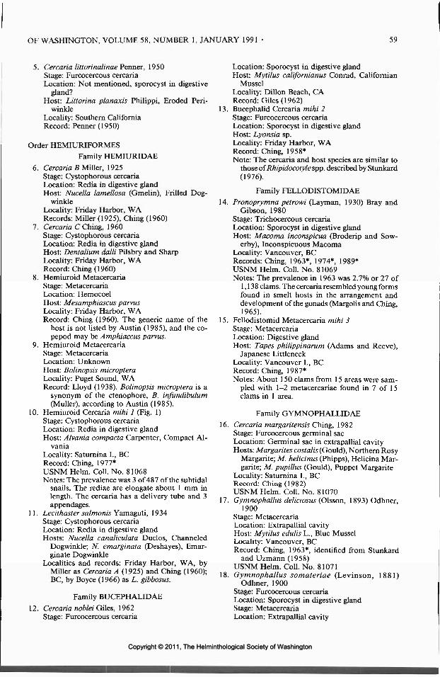
OF WASHINGTON, VOLUME 58, NUMBER 1, JANUARY 1991 59
5. Cercaria littorinalinae Penner, 1950Stage: Furcocercous cercariaLocation: Not mentioned, sporocyst in digestive
gland?Host: Littorina planaxis Philippi, Eroded Peri-
winkleLocality: Southern CaliforniaRecord: Penner (1950)
Order HEMIURIFORMESFamily HEMIURIDAE
6. Cercaria B Miller, 1925Stage: Cystophorous cercariaLocation: Redia in digestive glandHost: Nucella lamellosa (Gmelin), Frilled Dog-
winkleLocality: Friday Harbor, WARecords: Miller (1925), Ching (1960)
7. Cercaria C Ching, 1960Stage: Cystophorous cercariaLocation: Redia in digestive glandHost: Dentalium dalli Pilsbry and SharpLocality: Friday Harbor, WARecord: Ching (1960)
8. Hemiuroid MetacercariaStage: MetacercariaLocation: HemocoelHost: Mesamphiascus parvusLocality: Friday Harbor, WARecord: Ching (1960). The generic name of the
host is not listed by Austin (1985), and the co-pepod may be Amphiaccus parvus.
9. Hemiuroid MetacercariaStage: MetacercariaLocation: UnknownHost: Bolinopsis micropteraLocality: Puget Sound, WARecord: Lloyd (1938). Bolinopsis microptera is a
synonym of the ctenophore, B. infundibulum(Muller), according to Austin (1985).
10. Hemiuroid Cercaria mihi 1 (Fig. 1)Stage: Cystophorous cercariaLocation: Redia in digestive glandHost: Alvania compacta Carpenter, Compact Al-
vaniaLocality: Saturnina I., BCRecord: Ching, 1977*USNM Helm. Coll. No. 81068Notes: The prevalence was 3 of 487 of the subtidal
snails. The rediae are elongate about 1 mm inlength. The cercaria has a delivery tube and 3appendages.
11. Lecithaster salmonis Yamaguti, 1934Stage: Cystophorous cercariaLocation: Redia in digestive glandHosts: Nucella canaliculata Duclos, Channeled
Dogwinkle; N. emarginata (Deshayes), Emar-ginate Dogwinkle
Localities and records: Friday Harbor, WA, byMiller as Cercaria A (1925) and Ching (1960);BC, by Boyce (1966) as L. gibbosus.
Family BUCEPHALIDAE
12. Cercaria noblei Giles, 1962Stage: Furcocercous cercaria
Location: Sporocyst in digestive glandHost: Mytilus californianus Conrad, Californian
MusselLocality: Dillon Beach, CARecord: Giles (1962)
13. Bucephalid Cercaria mihi 2Stage: Furcocercous cercariaLocation: Sporocyst in digestive glandHost: Lyonsia sp.Locality: Friday Harbor, WARecord: Ching, 1958*Note: The cercaria and host species are similar to
those ofRhipidocotylespp. described by Stunkard(1976).
Family FELLODISTOMIDAE
14. Pronoprymna petrowi (Layman, 1930) Bray andGibson, 1980
Stage: Trichocercous cercariaLocation: Sporocyst in digestive glandHost: Macoma inconspicua (Broderip and Sow-
erby), Inconspicuous MacomaLocality: Vancouver, BCRecords: Ching, 1963*, 1974*, 1989*USNM Helm. Coll. No. 81069Notes: The prevalence in 1963 was 2.7% or 27 of
1,138 clams. The cercaria resembled young formsfound in smelt hosts in the arrangement anddevelopment of the gonads (Margolis and Ching,1965).
15. Fellodistomid Metacercaria mihi 3Stage: MetacercariaLocation: Digestive glandHost: Tapes philippinarum (Adams and Reeve),
Japanese LittleneckLocality: Vancouver I., BCRecord: Ching, 1987*Notes: About 150 clams from 15 areas were sam-
pled with 1-2 metacercariae found in 7 of 15clams in 1 area.
Family GYMNOPHALLIDAE
16. Cercaria margaritensis Ching, 1982Stage: Furcocercous germinal sacLocation: Germinal sac in extrapallial cavityHosts: Margarites costalis (Gould), Northern Rosy
Margarite; M. helicinus (Phipps), Helicina Mar-garite; M. pupillus (Gould), Puppet Margarite
Locality: Saturnina I., BCRecord: Ching (1982)USNM Helm. Coll. No. 81070
17. Gymnophallus deliciosus (Olsson, 1893) Odhner,1900
Stage: MetacercariaLocation: Extrapallial cavityHost: Mytilus edulis L., Blue MusselLocality: Vancouver, BCRecord: Ching, 1963*, identified from Stunkard
and Uzmann (1958)USNM Helm. Coll. No. 81071
18. Gymnophallus somateriae (Levinson, 1881)Odhner, 1900
Stage: Furcocercous cercariaLocation: Sporocyst in digestive glandStage: MetacercariaLocation: Extrapallial cavity
Copyright © 2011, The Helminthological Society of WashingtonCopyright © 2011, The Helminthological Society of Washington

60 JOURNAL OF THE HELMINTHOLOGICAL SOCIETY
Host for 2 stages: Macoma inconspicuaLocality: Vancouver, BCRecords: Ching (1973), 1963*, 1976* for cercariaUSNM Helm. Coll. Nos. 81072, 81073
19. Gymnophallus sp.Stage: MetacercariaLocation: Extrapallial cavityHosts: Cryptomya californica (Conrad), California
Glass Mya; Mya arenaria (L.), Soft-shell ClamRecord: Ching, 1962*, identified from Stunkard
and Uzmann( 1958)USNM Coll. No. 81074
20. Lacunovermis conspicuus Ching, 1965Stage: Furcocercous cercariaLocation: Sporocyst in digestive glandStage: MetacercariaHost for 2 stages: Macoma inconspicuaLocality: Vancouver, BCRecord: Ching (1965a)
21. Meiogymnophallus multigemmulus Ching, 1965Stage: MetacercariaLocation: Sporocyst in digestive glandHost: Macoma inconspicuaLocality: Vancouver, BCRecord: Ching (1965a)
22. Parvatrema borealis Stunkard and Uzmann, 1958Stage: Furcocercous cercariaLocation: Sporocyst in digestive glandStage: MetacercariaLocation: Extrapallial cavityHosts for 2 stages: Nuticola tantilla (Gould), Tan-
tilla Transennella; Gemma gemma (Totten),Amethyst Gem Clam
Localities and records: Friday Harbor, WA, byChing, 1959*; San Francisco Bay, CA, by Ogles-by (1965); Bodega Bay, CA, by Ching and Ruiz,1986*
USNM Helm. Coll. No. 8107523. Parvatrema obscurus (Ching, 1960) James, 1964
Stage: MetacercariaLocation: Digestive glandHosts: "Collisella" scabra (Gould), Rough Lim-
pet; Lottia digitalis (Rathke), Fingered LimpetLocalities: Bodega Bay to San Nicholas I., CARecord: Ching and Grosholz (1988)USNM Helm. Coll. No. 79923
24. Gymnophallid MetacercariaStage: MetacercariaLocation: Gills, mantleHost: Crassostrea gigas (Thunberg), Giant Pacific
OysterLocalities: Broughton I., Fanny Bay, Vancouver
I., BCRecord: Bower, 1987, pers. comm., Pacific Bio-
logical Station, Nanaimo, BC
Order PLAGIORCHIIFORMESFamily ACANTHOCOLPIDAE
25. Cercaria foliatae Miller, 1925Stage: Oculate cercariaLocation: Sporocyst in digestive glandHost: Ceratostoma foliatum (Gmelin), Foliated
Thorn PurpuraLocality: Friday Harbor, WARecord: Miller (1925). This cercaria is placed in
this family because of its resemblance to thecercaria of Stephanostomum baccatum de-scribed by Wolfgang (1955) and the close rela-tionship of the hosts.
Family ENENTERIDAE26. Enenterid Cercaria mihi 4 (Fig. 2)
Stage: Tail-less cercariaLocation: Redia in digestive glandHost: Alvania compactaLocality: Saturnina I., BCRecord: Ching, 1978*USNM Helm. Coll. No. 81076Notes: A single infection was found in 487 snails.
The cercaria appears to be similar to that ofNeophasis lagenformis as described by Koie(1973). Some opecoelid-like genera with bodyspination including Neophasis have been placedin this family by Gibson and Bray (1982).
Family MICROPHALLIDAETribe MARITREMATIDI
27. Maritrema gratiosum Nicoll, 1907Stage: MetacercariaLocation: HemocoelHost: Balanus glandula DarwinLocality: Vancouver, BCRecord: Ching (1978)
28. Maritrema laricola Ching, 1962Stage: XiphidiocercariaLocation: Sporocyst in digestive glandHosts: Littorina scutulata Gould, Checkered Per-
iwinkle; L. sitkana Philippi, Sitka PeriwinkleStage: MetacercariaLocation: Digestive glandHosts: Hemigrapsus nudus (Dana), H. oregonensis
(Dana)Localities: Vancouver, BC; Bodega Bay, CARecords: Ching (1962b), Ching, 1986*
29. Maritrema megametrios Deblock and Rausch,1968
Stage: MetacercariaLocation: HemocoelHosts: Telorchestia traskiana (Simpson), Gnori-
mosphaeroma oregonense DanaLocality: Vancouver, BCRecord: Ching (1974)USNM Helm. Coll. No. 81077
30. Maritrema pacifica Ching, 1974Stage: MetacercariaLocation: HemocoelHost: Megalorchestia corniculata (Stout)Locality: Santa Barbara, CARecord: Ching (1974)USNM Helm. Coll. No. 81078
31. Probolocoryphe uca (Sarkisian, 1957) Yamaguti,1971
Stage: XiphidiocercariaLocation: Sporocyst in digestive glandHost: Cerithidea californicaStage: MetacercariaLocation: MusclesHost: Uca crenulata (Lockington)Localities: Upper Newport Bay, Bolinas Lagoon,
CA
Copyright © 2011, The Helminthological Society of WashingtonCopyright © 2011, The Helminthological Society of Washington
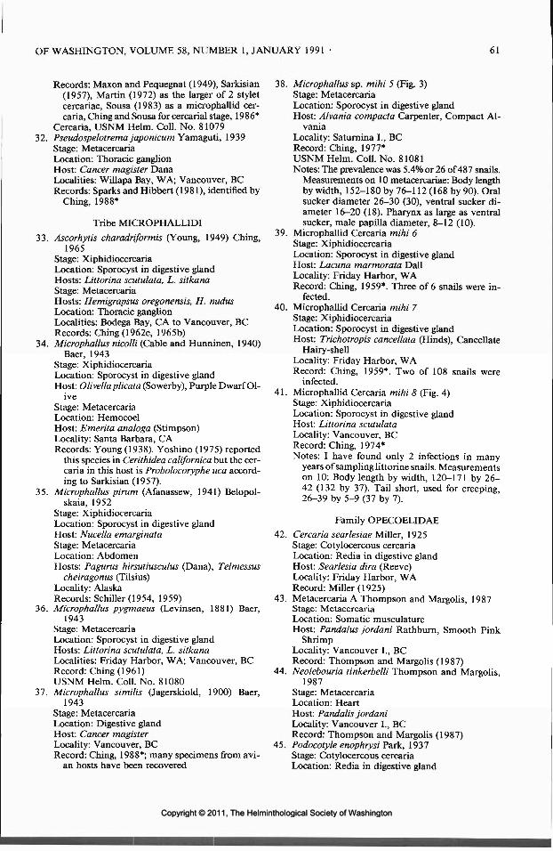
OF WASHINGTON, VOLUME 58, NUMBER 1, JANUARY 1991 61
Records: Maxon and Pequegnat (1949), Sarkisian(1957), Martin (1972) as the larger of 2 styletcercariae, Sousa (1983) as a microphallid cer-caria, Ching and Sousa for cercarial stage, 1986*
Cercaria, USNM Helm. Coll. No. 8107932. Pseudospelotrema japonicum Yamaguti, 1939
Stage: MetacercariaLocation: Thoracic ganglionHost: Cancer magister DanaLocalities: Willapa Bay, WA; Vancouver, BCRecords: Sparks and Hibbert (1981), identified by
Ching, 1988*
Tribe MICROPHALLIDI
33. Ascorhytis charadriformis (Young, 1949) Ching,1965
Stage: XiphidiocercariaLocation: Sporocyst in digestive glandHosts: Littorina scutulata, L. sitkanaStage: MetacercariaHosts: Hemigrapsus oregonensis, H. nudusLocation: Thoracic ganglionLocalities: Bodega Bay, CA to Vancouver, BCRecords: Ching (1962c, 1965b)
34. Microphallus nicolli (Cable and Hunninen, 1940)Baer, 1943
Stage: XiphidiocercariaLocation: Sporocyst in digestive glandHost: Olivella plicata (Sowerby), Purple Dwarf Ol-
iveStage: MetacercariaLocation: HemocoelHost: Emerita analoga (Stimpson)Locality: Santa Barbara, CARecords: Young (1938). Yoshino (1975) reported
this species in Cerithidea californica but the cer-caria in this host is Probolocoryphe uca accord-ing to Sarkisian (1957).
35. Microphallus pirum (Afanassew, 1941) Belopol-skaia, 1952
Stage: XiphidiocercariaLocation: Sporocyst in digestive glandHost: Nucella emarginataStage: MetacercariaLocation: AbdomenHosts: Pagurus hirsutiusculus (Dana), Telmessus
cheiragonus (Tilsius)Locality: AlaskaRecords: Schiller (1954, 1959)
36. Microphallus pygmaeus (Levinsen, 1881) Baer,1943
Stage: MetacercariaLocation: Sporocyst in digestive glandHosts: Littorina scutulata, L. sitkanaLocalities: Friday Harbor, WA; Vancouver, BCRecord: Ching (1961)USNM Helm. Coll. No. 81080
37. Microphallus similis (Jagerskiold, 1900) Baer,1943
Stage: MetacercariaLocation: Digestive glandHost: Cancer magisterLocality: Vancouver, BCRecord: Ching, 1988*; many specimens from avi-
an hosts have been recovered
38. Microphallus sp. mihi 5 (Fig. 3)Stage: MetacercariaLocation: Sporocyst in digestive glandHost: Alvania compacta Carpenter, Compact Al-
vaniaLocality: Saturnina I., BCRecord: Ching, 1977*USNM Helm. Coll. No. 81081Notes: The prevalence was 5.4% or 26 of 487 snails.
Measurements on 10 metacercariae: Body lengthby width, 152-180 by 76-112 (168 by 90). Oralsucker diameter 26-30 (30), ventral sucker di-ameter 16-20 (18). Pharynx as large as ventralsucker, male papilla diameter, 8-12 (10).
39. Microphallid Cercaria mihi 6Stage: XiphidiocercariaLocation: Sporocyst in digestive glandHost: Lacuna marmorata DallLocality: Friday Harbor, WARecord: Ching, 1959*. Three of 6 snails were in-
fected.40. Microphallid Cercaria mihi 7
Stage: XiphidiocercariaLocation: Sporocyst in digestive glandHost: Trichotropis cancellata (Hinds), Cancellate
Hairy-shellLocality: Friday Harbor, WARecord: Ching, 1959*. Two of 108 snails were
infected.41. Microphallid Cercaria mihi 8 (Fig. 4)
Stage: XiphidiocercariaLocation: Sporocyst in digestive glandHost: Littorina scutulataLocality: Vancouver, BCRecord: Ching, 1974*Notes: I have found only 2 infections in many
years of sampling littorine snails. Measurementson 10: Body length by width, 120-171 by 26-42 (132 by 37). Tail short, used for creeping,26-39 by 5-9 (37 by 7).
Family OPECOELIDAE42. Cercaria searlesiae Miller, 1925
Stage: Cotylocercous cercariaLocation: Redia in digestive glandHost: Searlesia dira (Reeve)Locality: Friday Harbor, WARecord: Miller (1925)
43. Metacercaria A Thompson and Margolis, 1987Stage: MetacercariaLocation: Somatic musculatureHost: Pandalus jordani Rathburn, Smooth Pink
ShrimpLocality: Vancouver L, BCRecord: Thompson and Margolis (1987)
44. Neolebouria tinkerbelli Thompson and Margolis,1987
Stage: MetacercariaLocation: HeartHost: Pandalis jordaniLocality: Vancouver L, BCRecord: Thompson and Margolis (1987)
45. Podocotyle enophrysi Park, 1937Stage: Cotylocercous cercariaLocation: Redia in digestive gland
Copyright © 2011, The Helminthological Society of WashingtonCopyright © 2011, The Helminthological Society of Washington

62 JOURNAL OF THE HELMINTHOLOGICAL SOCIETY
Host: Lacuna marmorata DallLocality: Saturnina I., BCRecord: Ching(1979)
46. Opecoelid Cercaria mihi 9Stage: Stumpy-tailed cercariaLocation: Sporocyst in digestive glandHosts: Margarites sp., M. helicinusLocalities: Friday Harbor, WA; Saturnina I., BCRecords: Ching, 1957*, 1977*Note: The cercaria has a short tail with 5-6 glands
forming a striated appearance at the tip whichis unlike the tails of the cotylocercous cercariaelisted above.
47. Opecoelid MetacercariaStage: MetacercariaLocation: HemocoelHost: Traskorchestia traskianaLocality: Vancouver, BCRecord: Ching, 1977*
48. Opecoelid MetacercariaStage: MetacercariaLocation: Abdominal musclesHost: Crangon alaskensis LockingtonLocality: Sinclair Inlet, WARecord: Morado and Sparks (1983). I have placed
this metacercaria in the family Opecoelidae.
Family MONORCHIIDAE49. Telolecithuspugetensis Lloyd and Guberlet, 1932
Stage: Brevifurcate cercariaLocation: Sporocyst in gonadsHost: Nutricola tantilla (Gould)Stage: MetacercariaLocation: Extrapallial cavityHosts: TV. tantilla; Macoma nasuta, Conrad Bent-
nose Macoma; Tellina nuculoides (Reeve),Salmon Tellin
Localities: Yaquina Bay, OR; Friday Harbor, WARecord: DeMartini and Pratt (1964)
Family RENICOLIDAE50. Renicola buchanani Martin and Gregory, 1951
Stage: Magnacercous cercariaLocation: Sporocyst in mantleHost: Cerithidea californicaLocalities: Upper Newport Bay, Bolinas Lagoon,
CARecords: Martin and Gregory (1951), Martin
(1955), Yoshino (1975) as Renicola sp., ChingandSousa, 1986*
USNM Helm. Coll. No. 8108251. Renicola cerithidicola Martin, 1971
Stage: Gymnocephalous cercariaLocation: Sporocyst in digestive glandHost: Cerithidea californicaRecord: Martin (1971)
52. Renicola thaidus Stunkard, 1964Stage: XiphidiocercariaLocation: Sporocyst in digestive glandHost: Nucella lamellosaStage: MetacercariaLocation: Digestive glandHost: Mytilus edulisLocality: Vancouver, BCRecords: Ching, 1957*, 1961*, 1969*, Stunkard
(1964)
53. Renicolid Cercaria mihi 10 (Fig. 5)Stage: XiphidiocercariaLocation: Sporocyst in digestive glandHost: Cerithidea californicaLocality: Bolinas Lagoon, CARecord: Ching and Sousa, 1986*USNM Helm. Coll. No. 81083Notes: Sporocyst with yellow pigment, sparsely
filled with 2-14 (8) cercariae, 676 by 298 inlength and width. Cercarial body finely spined,179-230 by 58-78 (199 by 69). Suckers equalin size, about 30 in diameter. Short stylet pres-ent. Excretory bladder U-shaped with long stem,arms lateral to ventral sucker. Flame cells 2[(3+ 3 + 3) + (3 + 3 + 3)]. Tail length by width,110-150 by 13-16 (133 by 15).
Order LEPOCREADIIFORMESFamily LEPOCREADIIDAE
54. Lepocreadiid Cercaria mihi 11 (Figs. 6, 7)Stage: Trichocercous cercariaLocation: Redia in digestive glandHost: Nitidella carinata (Hinds), Carinate Dove-
shellLocality: Saturnina I., BCRecord: Ching, 1979*USNM Helm. Coll. No. 81084Notes: The prevalence was 5% or 4 of 73 snails.
The cercaria has a trumpet-shaped oral sucker,prominent eye spots, and excretory bladderreaching to the ventral sucker. The tail has 36pairs of setae.
Order ECHINOSTOMATIFORMESFamily NOTOCOTYLIDAE
55. Catatropis johnstoni Martin, 1956Stage: Oculate, monostome cercariaLocation: Redia in digestive glandHost: Cerithidea californicaLocality: Upper Newport Bay, Bolinas Lagoon,
CARecords: Martin (1955, 1956), Yoshino (1975),
Sousa (1983)56. Notocotylid Cercaria mihi 12 (Fig. 8)
Stage: Oculate, monostome cercariaLocation: Redia in digestive glandHost: Littorina scutulataLocality: Vancouver, BCRecords: Ching (1962a), 1977*USNM Helm. Coll. No. 81085Notes: The prevalence was 0.49% or 21 of 4,322
snails sampled in 1977-1978 from 3 locationsin Vancouver. Measurements on 10 cercariae:Body length by width, 330-595 by 145-244 (419by 216). Tail length by width, 376-508 by 33-66 (422 by 43). Three eye spots present.
Family ECHINOSTOMATIDAE57. Acanthoparyphium spinulosum Johnston, 1917
Stage: Cercaria with collar spinesLocation: Redia in gonadsHost: Cerithidea californicaStage: Metacercaria
Copyright © 2011, The Helminthological Society of WashingtonCopyright © 2011, The Helminthological Society of Washington

OF WASHINGTON, VOLUME 58, NUMBER 1, JANUARY 1991 63
50jum
8
50jjm
10
50jjm
Figures 8-11. Marine digeneans from gastropod hosts. 8. Notocotylid cercaria from Littorina scutulata. 9.Echinostomatid cercaria of Himasthla sp. from L. scutulata. 10. Heterophyid cercaria from Batillaria zonalis.11. Heterophyid cercaria from Bittium escherichtii.
Location: Radular pocketsHost: Cerithidea californicaLocalities: Upper Newport Bay, Bolinas Lagoon,
CARecords: Martin and Adams (1960, 1961), Yoshi-
no (1975), Sousa (1983), Chingand Sousa, 1986*USNM Helm. Coll. No. 81086
58. Acanthopharyphium sp.Stage: Cercaria with collar spinesLocation: Redia in gonadsHost: Cerithidea californicaLocality: Upper Newport Bay, CARecord: Martin (1972)
59. "Echinoparyphium" sp.Stage: Cercaria with collar spinesLocation: Redia in gonad, digestive gland
Host: Cerithidea californicaLocalities: Upper Newport Bay, Bolinas Lagoon,
CARecords: Martin (1972), Sousa (1983), Ching and
Sousa, 1986*. Martin (1972) placed this echi-nostome in this genus, but the 31 spines aroundthe head collar are arranged in a single ring dor-sally and laterally, with 2 clustered at each sideventrally.
USNM Helm. Coll. No. 8108760. Himasthla rhigedana Dietz, 1909
Stage: Oculate cercaria with collar spinesLocation: Redia in digestive glandHost: Cerithidea californicaLocalities: Upper Newport Bay, Bolinas Lagoon,
CA
Copyright © 2011, The Helminthological Society of WashingtonCopyright © 2011, The Helminthological Society of Washington

64 JOURNAL OF THE HELMINTHOLOGICAL SOCIETY
Records: Adams and Martin (1960, 1963), Yoshi-no (1975), Sousa (1983), Ching and Sousa, 1986*
USNM Helm. Coll. No. 8108861. Himasthla sp. Cercaria mihi 13 (Fig. 9)
Stage: Cercaria with collar spinesLocation: Redia in digestive glandHost: Littorina scutulataLocalities: Friday Harbor, WA; Vancouver, BCRecords: Ching (1962a as echinostome I), 1975-
1978*, 1988*USNM Helm. Coll. No. 81089Notes: The prevalence was 2.5% or 21 of 835 snails
at Vancouver and Friday Harbor from 1975 to1978. At 1 lagoon site at Friday Harbor in 1988,11% or 23 of 207 snails were infected. Cercarialbody spined, with head collar of 29 spines insingle continuous row, with 2 ventrally in eachcorner. Body length by width, 352^54 by 123-217 (400 by 158). Oral sucker 51-59 by 51-56(57 by 52), ventral sucker, 64-70 by 66-102 (75by 82). Tail length by width, 237-409 by 33-57(319 by 41).
62. Echinostomatid MetacercariaStage: Metacercaria with collar spinesLocation: Gut, mantle, base of gillsHost: Protothaca staminea (Conrad), Common
Pacific LittleneckLocality: Sooke, BCRecord: Bower, 1986* pers. comm., Pacific Bio-
logical Station, Nanaimo, BC
Family PHILOPHTHALMIDAE63. Cloacitrema michiganensis Mclntosh, 1938
Stage: Megalurous cercariaHost: Cerithidea californicaLocality: Upper Newport Bay, CARecords: Robinson (1952), Martin (1955, 1972),
LeFloreetal. (1985)64. Parorchis acanthus (Nicoll, 1906) Nicoll, 1907
Stage: Megalurous cercariaLocation: Redia in digestive glandHost 1: Cerithidea californicaLocalities: Upper Newport Bay, Bolinas Lagoon,
CARecords: Martin (1955, 1972), Yoshino (1975),
Sousa (1983), Ching and Sousa, 1986*USNM Helm. Coll. No. 81090Host 2: Nucella lamellosa, N. emarginataLocality: Thetis I., BCRecord: Ching (1978)USNM Helm. Coll. No. 81091
Family PSILOSTOMIDAE
65. Psilostomum magniovum Ching, 1980Stage: Cercaria with caudal fin foldLocation: Redia in digestive glandHost: Littorina scutulataStage: MetacercariaLocation: Extrapallial cavityHost: Mytilus edulis L., Blue MusselLocality: Vancouver, BCRecord: Ching (1980)
Order OPISTHORCHIIFORMESFamily HETEROPHYIDAE
66. Cercaria purpuracauda Miller, 1925Stage: Magnacercous cercariaLocation: Redia in digestive glandHost: Bittium escherichtii (Middendorf), Giant Pa-
cific Coast BittiumLocality: Friday Harbor, WARecord: Miller (1925)
67. Cryptocotyle lingua (Creplin, 1825) Fischoeder,1903
Stage: Pleurolophocercous cercariaLocation: Redia in gonadsHost: Littorina scutulataLocality: Vancouver, BCRecord: Ching (1978)USNM Helm. Coll. No. 81092
68. Euhaplorchis californiensis Martin, 1950Stage: Pleurolophocercous cercariaLocation: Gonad (redial stage in digestive gland)Host: Cerithidea californicaLocalities: Upper Newport Bay, Bolinas Lagoon,
CARecords: Martin (1950a, 1955), Yoshino (1975),
Sousa (1983), Ching and Sousa, 1986*USNM Helm. Coll. No. 81093
69. Parastictodora hancocki Martin, 1950Stage: Pleurolophocercous cercariaLocation: Redia in gonadsHost: Cerithidea californicaLocality: Upper Newport Bay, CARecords: Martin (1950b, 1955)
70. Phocitremoides ovale Martin, 1950Stage: Pleurolophocercous cercariaLocation: Redia in gonadsHost: Cerithidea californicaLocalities: Upper Newport Bay, Bolinas Lagoon,
CARecords: Martin (1950c), Ching and Sousa, 1986*USNM Helm. Coll. No. 81094
71. Pygidiopsoides spindalis Martin, 1951Stage: Pleurolophocercous cercariaLocation: Redia in gonadsHost: Cerithidea californicaLocality: Upper Newport Bay, CARecord: Martin (1951)
72. Heterophyid Cercaria mihi 14 (Fig. 10)Stage: Pleurolophocercous cercariaLocation: Redia in gonadsHost: Batillaria zonalis (Bruguiere) Cumings, False
CerithLocality: Vancouver, BCRecords: Ching, 1978*, 1989*USNM Helm. Coll. No. 81095Notes: The prevalence was 10% or 8 of 77 in 1978
and 22% or 12 of 54 in 1989 in 2 sites in BC.Measurements of 10 cercariae: Body finely-spined, length by width, 139-211 by 66-99 (184by 80). Tail with finfold ending in small fanaround the tip, length by width, 297—475 by 17-36 (366 by 27). Oral sucker with subterminalopening with glandular openings, crenulatedwalls, 25^6 by 31-37 (31 by 36). Eyespots al-
Copyright © 2011, The Helminthological Society of WashingtonCopyright © 2011, The Helminthological Society of Washington

OF WASHINGTON, VOLUME 58, NUMBER 1, JANUARY 1991 65
most square. Penetration glands 14 in total. Apleurolophocercous cercaria reported from thishost in California by Whitlatch (1974) may bethe same species.
73. Heterophyid Cercaria mihi 15 (Fig. 11)Stage: Pleurolophocercous cercariaLocation: Redia in gonadsHost: Bittium escherichtiiLocality: Vancouver, BCRecord: Ching, 1974*
USNM Helm. Coll. No. 81096Notes: The prevalence was 9.6% or 5 of 52 snails.
Measurements on 9 cercariae: Body length bywidth, 164-188 by 60-66; tail with short an-terior folds, fin folds continuing around bent tip,length by width, 248^09 by 25-30. Oral suckerwith 8 spines on the dorsal edge followed by 5tubular spines in the opening of the mouth. Pen-etration glands filling body, about 16 in total.
List 2. Molluscan Hosts of Larval Digeneans of the North American Pacific Coast(Name of mollusc: number of larval digenean in List 1)
GASTROPODA:
Alvanla compacta: 10, 26, 38Batillaria zonalis: 72Bittium escherichtii: 66, 73Ceratostoma foliatum: 25Cerithidea californica: 1, 2, 4, 31, 50, 51, 53, 55, 57,
58, 59,60,63,64,68,69, 70, 71"Collisella" scabra: 23Ilyanassa obsoleta: 3Lacuna marmorata: 39, 45Littorina planaxis: 5Littorina scutulata: 28, 33, 36, 41, 56, 61, 65, 67Littorina sitkana: 28, 33, 36Lottia digitalis: 23Margarites costalis: 16Margarites helicinus: 16, 46Margarites pupillus: 16Margarites sp.: 46Nitidella carinata: 54Nucella canaliculata: 11Nucella emarginata: 11, 35, 64Nucella lamellosa: 6, 52, 64
Olive I la p Heat a: 34Searlesia dira: 42Trichotropis cancellata: 40
SCAPHOPODA:Dentalium dalli: 1
BIVALVIA:
Crassostrea gigas: 24Cryptomya californica: 19Gemma gemma: 22Lyonsia sp.: 13Macoma inconspicua: 14, 18, 20, 21Macoma nasuta: 49Mya arenaria: 19Mytilus californianus: 12Mytilus edulis: 17, 52, 65Nuticola tantilla: 22, 49Protothaca staminea: 62Tapes philippinarum: 15Tellina nuculoides: 49
List 3. Larval Tapeworms, Acanthocephalans, and Round worms from Marine Invertebrates of theNorth American Pacific Coast
Class CESTODAOrder TETRAPHYLLIDEA
Family PHYLLOBOTHRIIDAE1. Anthobothrium sp.
Stage: LarvaLocation: FootHosts: Tresus nuttalli (Conrad), Pacific Gaper; Ma-
coma nasuta, Bent-nose Macoma; Protothacastaminea (Conrad), Common Pacific Littleneck
Locality: Elkhorn Slough, CARecord: MacGinitie and MacGinitie (1968)
2. Echeneibothrium sp.Stage: Plerocercoid larvaLocation: Mantle, footHost: Protothaca stamineaLocality: Humboldt Bay, CARecord: Sparks and Chew (1966)
3. Echeneibothrium sp.Stage: LarvaLocation: FootHost: Protothaca laciniataLocality: Morro Bay, CARecord: Warner and Katkansky (1969a)
4. Echeneibothrium sp.Stage: Smaller larvaLocation: Peripheral and mantle areaHost: Protothaca stamineaLocality: Humboldt Bay, CARecord: Warner and Katkansky (1969b)
5. Echeneibothrium sp.Stage: Larger larvaLocation: VisceraHost: Protothaca stamineaLocality: Humboldt Bay, CARecord: Warner and Katkansky (1969b)
Copyright © 2011, The Helminthological Society of WashingtonCopyright © 2011, The Helminthological Society of Washington

66 JOURNAL OF THE HELMINTHOLOGICAL SOCIETY
Order TETRARHYNCHIDEAFamily TETRARHYNCHIDAE
6. Tetrarhynchus sp. 1 and 2Stage: LarvaeLocation: MuscleHost: Dosidicus gigasLocality: Newport Bay, CARecord: MacGinitie and MacGinitie (1968)
Order CYCLOPHYLLIDEAFamily HYMENOLEPIDAE
7. Hymenolepid Cysticercoid mihi 16Stage: CysticercoidLocation: HemocoelHost: Traskorchestia traskianaLocality: Newport Bay, ORRecord: Ching, 1974*
Phylum ACANTHOCEPHALAOrder PALAEACANTHOCEPHALA
Family POLYMORPHIDAE1. Polymorphic kenti Van Cleave, 1947
Stage: CystacanthLocation: HemocoelHost: Emerita analoga (Stimpson)Locality: OregonRecord: Reish (1950)
2. Profilicollis botulus (Van Cleave, 1916) Meyer, 1913Stage: CystacanthLocation: Rectal cecaHost: Hemigrapsus oregonensisLocalities: Bodega Bay, CA to Vancouver, BCRecord: Ching (1989)
3. Unidentified CystacanthStage: CystacanthLocation: MidgutHost: Paralithodes camtschaticus (Tilesius)Locality: AlaskaRecord: Sparks (1987)
Phylum NEMATODAOrder ASCARIDIDA
Family ANISAKIDAE1. Contracaecum sp.
Stages: Juvenile and adultLocation: HemocoelHost: Pandalis borealisLocality: Open ocean, BCRecord: Margolis and Butler (1954)
Order SPIRURIDAFamily GNATHOSTOMATIDAE
2. Echinocephalus pseudouncinatus Millemann, 1963Stage: JuvenileLocation: FootHost: Haliotis corrugata WoodLocality: San Clemente I., CARecord: Millemann (1963)Host: Centrostephanus coronatusLocation: GonadsLocality: Southern CARecord: Pearse and Timm (1971)Location: Adductor muscle
Host: Argopecten aequisulcatus (Carpenter)Locality: Baja California, MexicoRecord: McLean (1983)
Family CYSTIDICOLIDAE3. Ascarophis sp. 1 and 2
Stage: JuvenilesLocation: HemocoelHosts: Pagurus samuelis (Stimpson), P. granosi-
manus (Stimpson)Locality: Bodega Bay, CARecord: Poinar and Thomas (1976)
4. Ascarophis sp.Stage: JuvenileLocation: HemocoelHosts: Pachycheles pubescens Holmes, P. rudis,
Pugettia producta (Randall), Hemigrapsus ore-gonensis
Locality: Bodega Bay, CARecords: Poinar and Thomas (1976), Poinar and
Kuris(1976)
Literature Cited
Abbott, R.T. 1974. American Seashells, 2nd ed. VanNostrand Reinhold, New York. 663 pp.
Adams, J. E., and W. E. Martin. 1960. Life historyof Himasthla sp., an echinostome trematode.Journal of Parasitology 46(Supplement):15.
, and . 1963. Life cycle of Himasthlarhigedana Dietz, 1909 (Trematoda: Echinosto-matidae). Transactions of the American Micro-scopical Society 82:1-6.
Austin, W. C. 1985. An Annotated Checklist of Ma-rine Invertebrates in the Cold Temperate North-east Pacific. 3 vols. Khoyatan Marine Laboratory,British Columbia. 682 pp.
Boyce, N. 1966. The parasites of central British Co-lumbia pink salmon during their early sea life, withspecial notes on the trematode Lecithaster gib-bosus. Fisheries Research Board of Canada 26:813-820.
Ching, H. L. 1960. Studies on three hemiuroid larvaeof Friday Harbor, Washington. Journal of Para-sitology 46:663-670.
. 1961. Three trematodes from the harlequinduck. Canadian Journal of Zoology 39:373-376.
. 1962a. Six larval trematodes from the snail,Littorina scutulata Gould from San Juan Island,U.S.A., and Vancouver, B.C. Canadian Journal ofZoology 40:675-676.
. 1962b. The description and life cycle of Mar-itrema laricola sp. n. (Trematoda: Microphalli-dae). Canadian Journal of Zoology 41:881-888.
. 1962c. The life cycle and bionomics of Levin-seniella charadriformis Young, 1949 (Trematoda:Microphallidae). Canadian Journal of Zoology 41:889-899.
. 1965a. Life cycle of Lacunovermis conspicuusn. gen., n. sp. and Meiogymnophallus multigem-mulus n. gen., n. sp. (Gymnophallidae: Tremato-da) from Macoma inconspicua and diving ducksfrom Vancouver, Canada. Proceedings of the Hel-minthological Society of Washington 32:53-63.
Copyright © 2011, The Helminthological Society of WashingtonCopyright © 2011, The Helminthological Society of Washington

OF WASHINGTON, VOLUME 58, NUMBER 1, JANUARY 1991 67
—. 1965b. Systematic notes on some NorthAmerican microphallid trematodes. Proceedingsof the Helminthological Society of Washington 32:140-148.
—. 1973. Description of Gymnophallus soma-teriae (Levinsen, 1881) from Macoma inconspicuaand diving ducks from Vancouver, Canada. Ca-nadian Journal of Zoology 51:901-906.
—. 1974. Two new species of Maritrema (Trem-atoda: Microphallidae) from the Pacific Coast ofNorth America. Canadian Journal of Zoology 52:865-869.
—. 1978. New marine hosts for Parorchis acan-thus, Cryptocotyle lingua, Maritrema megame-trios, and M. gratiosum, trematodes of birds fromBritish Columbia, Canada. Canadian Journal ofZoology 56:1877-1879.
—. 1979. The life cycle ofPodocotyle enophrysiPark, 1937 (Trematoda: Opecoelidae). CanadianJournal of Zoology 57:1341-1344.
—. 1980. The life cycle and description of Psi'-lostomum magniovum n. sp. (Trematoda: Psilo-stomidae). Proceedings of the HelminthologicalSociety of Washington 47:179-185.
. 1982. Description of germinal sacs of a gym-nophallid trematode, Cercaria margaritensis sp.n. in the extrapallial fluid of subtidal snails (Mar-garites spp.) in British Columbia. Canadian Jour-nal of Zoology 60:516-520.
—. 1989. Profilicollis botulus (Van Cleave, 1916)from diving ducks and shore crabs of British Co-lumbia. Journal of Parasitology 75:33-37.
-, and E. Grosholz. 1988. Occurrence of a meta-
Kroyer, by a nematode, Contracaecum sp. Journalof Parasitology 40:649-655.
-, and H. L. Ching. 1965. Review of the trem-
cercaria (Trematoda: Gymnophallidae) in ac-maeid gastropods, Lottia digitalis and "Collisella"scabra. Proceedings of the Helminthological So-ciety of Washington 55:104-105.
DeMartini, J. D., and I. Pratt. 1964. The life cycleof Telolecithuspugetensis Lloyd and Guberlet, 1932(Trematoda: Monorchidae). Journal of Parasitol-ogy 50:101-105.
Gibson, D. I., and R. A. Bray. 1982. A study andreorganization of Plagioporus Stafford, 1904 (Di-genea: Opecoelidae) and related genera, with spe-cial reference to forms from European Atlanticwaters. Journal of Natural History 16:529-559.
Giles, D. E. 1962. New bucephalid cercaria from themussel Mytilus calif or nianus. Journal of Parasi-tology 48:293-295.
Koie, M. 1973. The host-parasite interface and as-sociated structures of the cercaria and adult Neo-phasis lagenformis (Lebour, 1910). Ophelia 12:205-219.
LeFlore, W. B., H. S. Bass, and W. E. Martin. 1985.The life cycle of Cloacitrema michiganensis Mc-Intosh, 1938 (Trematoda: Philophthalmidae).Journal of Parasitology 71:28-32.
Lloyd, L. C. 1938. Some digenetic trematodes fromPuget Sound fish. Journal of Parasitology 24:117-132.
MacGinitie, G. E., and N. MacGinitie. 1968. NaturalHistory of Marine Animals, 2nd ed. McGraw-Hill,New York. 523 pp.
Margolis, L., and T. H. Butler. 1954. An unusualand heavy infection of a prawn, Pandalis borealis
atode genera Bacciger and Pentagramma (Fellodi-stomatidae and description of P. petrowi (Layman,1930) n. comb, from marine fishes of the Pacificcoast of Canada. Canadian Journal of Zoology 43:381^05.
Martin, W. E. 1950a. Euhaplorchis californiensis n.g., n. sp. Heterophyidae, Trematoda, with noteson its life cycle. Transactions of the American Mi-croscopical Society 69:194-209.
. 1950b. Parastictodora hancocki n. gen. n. sp.(Trematoda: Heterophyidae) with observations onits life cycle. Journal of Parasitology 36:360-370.
. 1950c. Phocitremoides ovale n. g., n. sp.(Trematoda: Opisthorchiidae) with observationson its life cycle. Journal of Parasitology 36:552-558.
. 1951. Pygidiopsis spindalis n. gen. n. sp. (Het-erophyidae: Trematoda), and its second interme-diate host. Journal of Parasitology 37:297-300.
. 1955. Seasonal infections of the snail, Cer-ithidea californica Haldeman with larval trema-todes. Essays in the Natural Sciences in honor ofCapt. Allan Hancock, pp. 203-210. University ofCalifornia Press, Los Angeles.
. 1956. The life cycle of Catatropis johnstonin. sp. (Trematoda: Notocotylidae). Transactionsof the American Microscopical Society 75:117-128.
. 1961. Life cycle of Mesostephanus appendic-ulata (Ciurea, 1916) Lutz, 1935 (Trematoda:Cyathocotylidae). Pacific Science 15:278-281.
. 1971. Larval stages of renicolid trematodes.Transactions of the American Microscopical So-ciety 90:188-194.
. 1972. An annotated key to the cercariae thatdevelop in the snail Cerithidea californica. Bulletinof the Southern California Academy of Science 71:39^3.
, and J. E. Adams. 1960. Life cycle of Acan-thoparyphium spinulosum Johnston, 1917 (Echi-nostomatidae). Journal of Parasitology46(Supplement): 3 5.
, and . 1961. Life cycle of Acanthopa-ryphium spinulosum Johnston, 1917 (Echinosto-matidae). Journal of Parasitology 47:777-782.
-, and V. Gregory. 1951. Cercaria buchananin. sp., an aggregating marine trematode. Trans-actions of the American Microscopical Society 70:359-362.
Maxon, M. G., and W. E. Pequegnat. 1949. Cercariaefrom upper Newport Bay. Journal of Entomologyand Zoology 41:30-55.
McLean, N. 1983. An echinocephalid nematode inthe scallop Argopecten aequisulcatus (Mollusca:Bivalvia). Journal of Invertebrate Pathology 42:273-276.
Millemann, R. E. 1963. Studies on the taxonomy andlife history of echinocephalid worms (Nematoda:Spiruroidea) with a complete description ofEchi-nocephalus pseudouncinatus Millemann, 1951.Journal of Parasitology 49:754-764.
Miller, H. M. 1925. Larval trematodes of certain
Copyright © 2011, The Helminthological Society of WashingtonCopyright © 2011, The Helminthological Society of Washington

68
marine gastropods from Puget Sound. Publicationsof the Puget Sound Biological Station, Universityof Washington 5:75-89.
Morado, J. F., and A. K. Sparks. 1983. Infection ofnervous tissue of shrimp, Crangon alaskensis, bytrematode metacercariae. Journal of InvertebratePathology 42:421^23.
Oglesby, L. C. 1965. Parvatrema borealis (Tremato-da) in San Francisco Bay. Journal of Parasitology51:582.
Pearse, J. S., and R. W. Timm. 1971. Juvenile nem-atodes (Echinocephalus pseudouncinatus) in thegonads of sea urchins (Centrostephanus coronatus)and their effect on host gametogenesis. BiologicalBulletin 140:95-103.
Penner, L. R. 1950. Cercaria littorinalinae sp. nov.,a dermatitis-producing schistosome cercaria fromBatillaria minima (Gmelin). Journal of Parasitol-ogy 36:466-472.
Poinar, G. O., Jr., and A. M. Kuris. 1976. JuvenileAscarophis (Spirurida: Nematoda) parasitizing in-tertidal decapod Crustacea in California with noteson prevalence and effects on host growth and sur-vival. Journal of Invertebrate Pathology 26:375-382.
, and G. M. Thomas. 1976. Occurrence of As-carophis (Nematoda: Spiruridea) in Callianassacaliforniensis Dana and other decapod crusta-ceans. Proceedings of the Helminthological Soci-ety of Washington 43:28-33.
Pritchard, M. H. 1989. Biodiversity bill. AmericanSociety of Parasitologists Newsletter 11:29.
Reish, D. J. 1950. Preliminary note in the life cycleof the acanthocephalan, Polymorphic kenti VanCleave, 1947. Journal of Parasitology 36:496.
Robinson, H. W. 1952. A preliminary report on thelife cycle of Cloacitrema michiganensis Mclntosh,1938 (Trematoda). Journal of Parasitology 38:368.
Sarkisian, L. N. 1957. Maritrema uca, new species(Trematoda: Microphallidae) from the fiddler crabUca crenulata (Lockington). Wasman Journal ofBiology 15:35^8.
Schiller, E. L. 1954. Studies on the helminth faunaof Alaska XVII. Notes on the intermediate stagesof some helminth parasites of the sea otter. Bio-logical Bulletin 106:107-121.
. 1959. Observations on the morphology andlife cycle of Microphallus pirum (Afanas'ev, 1941).Transactions of the American Microscopical So-ciety 78:65-76.
Sousa, W. P. 1983. Host life history and the effectof parasitic castration on growth: a field study ofCerithidea californica Haldeman (Gastropoda:Prosobranchia) and its trematode parasites. Jour-nal of Experimental Marine Biology and Ecology73:273-296.
Sparks, A. K. 1987. The red king crab (Paralithodescamtschaticd) as a host for juvenile acanthoceph-alans. Journal of Invertebrate Pathology 50:166-168.
—, and K. K. Chew. 1966. Gross infestation ofthe littleneck clam, Venerupis staminea with a lar-val cestode (Echeneibothrium sp.). Journal of In-vertebrate Pathology 8:413^16.
-, and J. Hibbert. 1981. A trematode meta-cercaria encysted in the nerves of the Dungenesscrab, Cancer magister. Journal of Invertebrate Pa-thology 38:88-93.
Stunkard, H. W. 1964. Studies on the trematode ge-nus Renicola: observations on the life-history,specificity, and systematic position. BiologicalBulletin 126:467^89.
. 1976. The life cycles, intermediate hosts, andlarval stages of Rhipidocotyle transversale Chand-ler, 1935 and Rhipidocotyle lintoni Hopkins, 1954:life cycles and systematics of bucephalid trema-todes. Biological Bulletin 150:294-317.
. 1983. The marine cercariae of the WoodsHole, Massachusetts region, a review and a revi-sion. Biological Bulletin 164:143-162.
, and M. C. Hinchliffe. 1952. The morphologyand life-history of Microbilharzia variglandis(Miller and Northrup, 1926) Stunkard and Hinch-liffe, 1951, avian blood flukes whose larvae cause"swimmer's itch" of ocean beaches. Journal ofParasitology 38:248-265.
-, and J. R. Uzmann. 1958. Studies on digenetictrematodes of the genera Gymnophallns and Par-vatrema. Biological Bulletin 115:276-302.
Thompson, A. B., and L. Margolis. 1987. Descriptionof Neolebouria tinkerbelli n. sp. (Trematoda: Di-genea: Opecoelidae) from experimental fish hosts,and of metacercaria of N. tinkerbelli and an un-identified digenean from Pandalis jordanii (De-capoda: Penaeidae) from the Pacific coast of Can-ada. Canadian Journal of Zoology 65:188-193.
Warner, R. W., and S. C. Katkansky. 1969a. Infes-tation of the rough-sided littleneck clam, Proto-thaca laciniata in Morro Bay, California, with lar-val cestodes (Echeneibothrium sp.). Journal ofInvertebrate Pathology 13:125-128.
, and . 1969b. Infestation of the clamProtothaca staminea by two species of tetraphyl-lidean cestodes (Echeneibothrium spp.). Journal ofInvertebrate Pathology 13:129-133.
Whitlatch, R. B. 1974. Studies on the populationecology of the salt marsh gastropod Batillaria zo-nalis. Veliger 17:47-55.
Wolfgang, R. W. 1955. Studies of the trematodeStephanostomum baccatum (Nicoll, 1907). III. Itslife cycle. Canadian Journal of Zoology 33:113-128.
Yoshino, T. P. 1975. A seasonal and histological studyof larval Digenea infecting Cerithidea californica(Gastropoda: Prosobranchia) from Goleta Slough,Santa Barbara, California. Veliger 18:156-161.
Young, R. T. 1938. The life history of a trematode(Levinseniella cruzfT) from the shore birds, Limosafedoa and Catoptrophorus semipalmatus inorna-tus. Biological Bulletin 74:319-329.
Copyright © 2011, The Helminthological Society of WashingtonCopyright © 2011, The Helminthological Society of Washington

J. Helminthol. Soc. Wash.58(1), 1991, pp. 69-81
Light and Electron Microscopical Study of a Bacterial Parasite from theCyst Nematode, Heterodera glycines
RICHARD M. SAYRE,1 WILLIAM P. WERGiN,2 TSUTOMU NiSHizAWA,3AND MORTIMER P. STARR4-5
1 Nematology Laboratory, Plant Sciences Institute, Beltsville Agricultural Research Center, USDA, ARS,Beltsville, Maryland 20705,2 Electron Microscopy Laboratory, Natural Resources Institute, Beltsville Agricultural Research Center,USDA, ARS, Beltsville, Maryland 20705,3 Laboratory of Nematology, National Institute of Agro-Environmental Sciences, Tsukuba,Ibaraki 305, Japan, and4 Department of Microbiology, University of California, Davis, California 95616
ABSTRACT: The host range and morphology of a mycelial and endospore-forming bacterium (CNP) that par-asitizes the cyst nematode, Heterodera elachista, differentiates it from 2 related species, Pasteuria penetrans andPasteuria thornei, parasites of Meloidogyne spp. and Pratylenchus spp., respectively. In cross-inoculation ex-periments with the 3 bacteria, the CNP parasitized the nematodes Heterodera glycines and Globodera rostochiensisbut not species in either Meloidogyne or Pratylenchus. The rhomboidal shape of the sporangia of Pasteuria thorneidistinguishes this species from the other 2 bacteria, which have cup-shaped sporangia. The results of the cross-inoculation studies and differences in the fine structure of the cup-shaped sporangia suggest that CNP shouldbe assigned to a new species within the genus Pasteuria.
KEY WORDS: plant-parasitic nematodes, soil bacterium, Pasteuria spp., soybeans, morphology, scanning elec-tron microscopy (SEM), transmission electron microscopy (TEM).
A recent revision of the genus Pasteuria delin-eated 2 species, Pasteuria penetrans Starr andSay re, 1988, sensu stricto and Pasteuria thorneiStarr and Sayre, 1988 (Starr and Sayre, 1988;Sayre and Starr, 1989), which are parasites of theroot-knot nematode, Meloidogyne incognita(Kofoid and White, 1919) Chitwood, 1949, andthe root-lesion nematode, Pratylenchus brachy-urus (Godfrey, 1929) Filipjev and SchuurmansStekhoven, 1941, respectively. Recently, anotherpossible member of the genus Pasteuria was foundby Nishizawa (1984) parasitizing Heteroderaelachista Oshima, 1974. Following his initial ob-servation, Nishizawa (1986) associated the fieldpresence of the bacterium with a decline in thepopulation of the cyst nematode. Confirmationof this phenomenon was noted at the TochigiAgricultural Experiment Station where soybeanbreeding lines were being screened for cyst nem-atode resistance (Nishizawa, 1987).
The present study uses light and electron mi-croscopy to compare the morphology of this re-cently discovered bacterium, which parasitizescyst nematodes with that of Pasteuria penetransand Pasteuria thornei, 2 species of bacteria thatparasitize some species in the genera Meloido-
Deceased 29 April 1989.
gyne and Pratylenchus, respectively. Althoughthe nomenclature of the new bacterium will bepresented at a later time, in this study the isolatethat parasitizes cyst nematodes in the genera Het-erodera and Globodera will be referred to as the"cyst nematode parasite" or CNP.
Materials and Methods
TerminologyThe fine structure of the spore wall is used to delin-
eate the species of Pasteuria taxonomically. To avoidconfusion, in this study we have used the terminologyof Sussman and Halvorson (1966) who described Neu-rospora spp., and that of Iterson (1988) who discussedand clarified the terminology of Gram-positive bac-teria. Briefly, the following terms will be used: (1) "en-dospore" refers to the single asexual spore that devel-ops within a sporangium and is surrounded by anexosporium; (2) "endospore wall" is the structure thatsurrounds the central protoplast or core; (3) "plasmamembrane" is the innermost structure that apposes thewall; (4) "cortex" is the multilayered inner spore walland the outer spore wall; and (5) "perispore" refers tothe limiting structures that include the wall layer formedoutside the exosporium.
Sources of nematodesFor laboratory bioassays, pure populations of several
different species of plant-parasitic nematodes wereneeded. These populations had the following charac-teristics: (1) the second stage juveniles (J2) were healthy,vigorous, and capable of root penetration; (2) the ma-jority of the J2 were the same physiological age; and
69
Copyright © 2011, The Helminthological Society of WashingtonCopyright © 2011, The Helminthological Society of Washington

70 JOURNAL OF THE HELMINTHOLOGICAL SOCIETY
(3) the juveniles belonged to identified species and rac-es of root-knot or soybean cyst nematodes. Populationswith these characteristics were obtained from asepticcultures of root explants (Lauritis et al., 1982, 1983a,b; Huettel and Rebois, 1985). They consisted of Me-loidogyne incognita, Pratylenchus brachyurus, Heter-odera zeae Koshy, Swarup, and Sethi, 1971, and 4 racesofffeterodera glycines Ichinohe, 1952. The nematodeswere harvested from the cultures using a modifiedBaermann funnel technique (Niblack and Huang, 1985).In addition to the aseptic cultures, the following nem-atodes were maintained in greenhouse pot cultures: M.incognita on tomato cultivars (cvs.), Marglobe or TinyTim; M. hapla on a strawberry cv., Sunrise; M.javanicaon a tomato cv., Marglobe; H. glycines on a soybeancv., Kent; H. zeae on a corn cv., Iowa Chief; and P.brachyurus on a peanut cv., Florarunner. Details forrearing M. incognita and P. brachyurus were reportedpreviously (Sayre and Wergin, 1977; Sayre et al., 1988).H. glycines was maintained in the greenhouse on eithersoybeans (cv. Kent) or adzuki beans, Vigna angularis(Willd.) Ohwi and H. Ohashi (USDA accession 416742).Cysts were extracted from the soil (Ayoub, 1980) andplaced on fine-mesh nylon in a Baermann funnel. Thehatching J2 were collected from the funnel over a 2- to4-day period. Also, examinations of soil samples frompreviously reported field plot studies of Nishizawa(1987) had isolated 10 additional, commonly occur-ring, genera or species of plant-parasitic nematodes.These nematodes, which included Aphelenchoides sp.,Aphelenchus sp., Helicotylenchus sp., Globodera ros-tochiensis, Paratylenchussp., Paratrichodorus porosus,Rotylenchulus reniformis, Tylenchorhynchus sp., Ty-lenchulus semipenetrans, and Tylenchus sp., were alsoexamined for the presence of CNP sporangia on theircuticular surfaces.
Sources of the Pasteuria spp.Methods for the axenic cultivation of Pasteuria pe-
netrans have not been reported; however, all isolatesof the bacteria were maintained on their respectivenematode hosts by the senior author. The cultivationprocedure of Stirling and Wachtel (1980) was used tomaintain Pasteuria penetrans on M. incognita infectingtomato plants (cv. Tiny Tim). Tomato roots, whichwere infested with root-knot nematodes parasitized byPasteuria penetrans, were air-dried and ground into afine powder. Samples of the powder were rewetted andground with a pestle and mortar to release the endo-spores. The bacteria were separated from the plantdebris by filtering the suspension through a 500-meshscreen that allowed passage of the sporangia and sub-sequent collection by centrifugation of the Pasteuriapenetrans endospores. The source of Pasteuria thorneiendospores was parasitized J2 and adults of P. brachy-urus (Sayre et al., 1988; Starr and Sayre, 1988). Theinfected nematodes were extracted from the roots ofpeanut (cf. Florarunner) in a mist chamber (Chapman,1957). A second source of these endospores was air-dried soil from pots containing peanut roots that wereinfected with nematodes parasitized by Pasteuriathornei. When healthy J2 and adults of P. brachyurusmigrated through the contaminated soil, a high pro-portion of the nematodes were encumbered with en-dospores of the bacterium.
CNP endospores were obtained from cysts of H. gly-cines that had been imported from Japan (Nishizawa,1986). To initiate a greenhouse culture in Beltsville,the cysts were crushed in tap water. The number ofendospores in this aqueous suspension was measuredwith a hemocytometer and adjusted to about 1,000 perml. Juveniles of H. glycines (race 3), which were ob-tained from soybean (cv. Kent) root explant cultures,were suspended in tap water at a concentration of 100per ml. A 5-ml suspension of J2 was then filtered througha 47-mm diameter membrane filter (Millipore 0.3-Mmpore size). The filter, which retained the J2, was in-verted onto the surface of a 3-ml endospore suspensionin a 50-mm diameter Petri dish (Falcon; 1006). Thedish was then shaken (50 rpm) for 24—48 hr to allowthe endospores to attach to the J2. The suspension,consisting of the J2 and their attached endospores, wasthen pipetted around the roots of soybean seedlingsthat were grown in 40-mm diameter Cone-Tainers (RayLeacher Cone-Tainers, Canby, Oregon 97013) con-taining autoclaved Norfolk loamy sand. Forty-five to60 days later, the entire plant was removed from thecontainer and the soil was washed from the root sys-tem. A dissecting microscope was used to examine theroot system for parasitized immature females and ma-ture cysts (Ayoub, 1980). Visual selection and removalof parasitized cysts were difficult because less than 10%of the cysts were diseased. However, the diseased fe-males and cysts were generally smaller, duller, or moreopaque than healthy cysts. The opacity resulted frombacterial and/or fungal colonizers of cysts. In addition,the diseased cysts usually sank to the bottom of thesampling dishes while the healthy cysts generally float-ed on the surface. Finally, the presence of the normalreproductive structures in adult females (i.e., uterus,ovaries, and eggs), or the presence of eggs in cysts,indicated that the bacterial parasite CNP was not pres-ent.
Light microscopy of CNPCysts parasitized by CNP were crushed and mounted
on a microscope slide in a drop of tap water (Southey,1986). Photomicrographs of CNP were taken with anautomatic exposure 3 5-mm camera attached to a com-pound microscope (NIKON Microphot F-X) fitted withan interference contrast system. Negatives were ob-tained on Kodak Tri-X Pan film that was processed inMicrodol-X. Morphometric data were obtained bymeasuring the images in printed enlargements of thephotomicrographs.
Previous studies (Sayre and Starr, 1985; Starr andSayre, 1988) indicated that the measurements of en-dospores observed by scanning electron microscopy(SEM) were smaller than those obtained by light mi-croscopy. This difference resulted from shrinkage thatoccurred during fixation, dehydration, and critical-pointdrying procedures that are required for SEM obser-vation. Consequently, sources of the morphometric datain the present study are designated by the instrumentsused to obtain the values, e.g., light microscopy data(LM), scanning electron microscopy data (SEM), andtransmission electron microscopy data (TEM) (Table1). All measurements are in micrometers 0*m) unlessstated otherwise.
Copyright © 2011, The Helminthological Society of WashingtonCopyright © 2011, The Helminthological Society of Washington
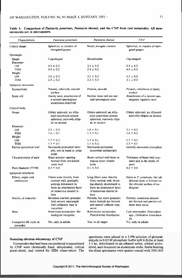
OF WASHINGTON, VOLUME 58, NUMBER 1, JANUARY 1991 • 71
Table 1. Comparison of Pasteuria penetrans, Pasteuria thornei, and the CNP from cyst nematodes. All mea-surements are in micrometers.
Characteristic
Colony shape
SporangiaShapeDiameter
LMTEM
HeightLMTEM
Episporic structuresExosporium
Stem cell
Central bodyShape
DiameterLMTEM
HeightLMTEM
Partial epicortical wall
Pasteuria penetrans
Spherical, to clusters ofelongated grapes
Cup-shaped
4.5 ± 0.33.4 ± 0.2
3.6 ± 0.32.5 ± 0.2
Present, relatively smoothsurface
Rarely seen; attachment ofa second sporangiumsometimes observed
Oblate spheroid, an ellip-soid sometimes almostspherical, narrowly ellip-tic in section
2.1 ± 0.21.4 ±0.1
1.7 ± 0.21.1 ± 0.1Surrounds protoplast later-
Pasteuria thornei
Small, elongate clusters
Rhomboidal
3.5 ± 0.22.4 ± 0.2
3.1 ±0.22.2 ± 0.2
Present, smooth
Neither stem cell nor sec-ond sporangium seen
Oblate spheroid, an ellip-soid sometimes almostspherical, narrowly ellip-tic in section
1.6 ±0.11.3 ± 0.1
1.5 ±0.11.1 ±0.2Surrounds protoplast
CNP
Spherical, to clusters of elon-gated grapes
Cup-shaped
5.3 ± 0.34.4 ± 0.3
4.3 ± 0.33.1 ±0.3
Present, velutinous to hairysurface
Attachment of a second spo-rangium regularly seen
Oblate spheroid, an ellipsoidnarrowly elliptic in section
2.1 ± 0.21.6 ± 0.2
1.7 ±0.11.3 ±0.1Entirely surrounds protoplast
Characteristics of pore
Pore diameter (TEM)
Episporal structuresFibers, origin and
orientation
Matrix, at maturity
Host
Completes life cycle injuveniles
ally, not in basal or polarareas
Basal annular openingformed from thickenedouter wall
0.3 ± 0.0
Fibers arise directly fromcortical wall, graduallyarching downward toform an attachment layerof numerous shorter fi-bers
Becomes coarsely granular;lysis occurs; sporangialwall collapses; base isvacuolate
Root-knot nematodes: Me-loidogyne incognita
No, only in adults
somewhat sublaterally
Basal cortical wall thins toexpose inner centralbody
0.1 ± 0.0
Long fibers arise directlyfrom cortical wall, bend-ing sharply downward toform an attachment layerof numerous shorter fi-bers
Persists, but more granular;some strands are formedand partial collapse mayoccur
Root-lesion nematodes:Pratylenchus brachyurus
Yes, in all stages
Thickness of basal wall con-stant and is the depth ofpore
0.2 ± 0.0
Same as P. penetrans, but ad-ditional layer is formed onthe obverse surface of en-dospore
Persists, numerous strandsare formed and partial col-lapse may occur
Cyst nematodes: Heteroderaspp., Globodera rostochien-sis
No, only in adults
Scanning electron microscopy of CNP
Nematodes that had been encumbered or parasitizedby CNP were chemically fixed, dehydrated, criticalpoint-dried, and coated for SEM observation. The
specimens were placed in a 3.0% solution of glutaral-dehyde in 0.05 M phosphate buffer (pH 6.8) for at least1.5 hr, dehydrated in an ethanol series, critical point-dried, and mounted on aluminum stubs. Stubs bearingthe dried specimens were sputter coated with 200-300
Copyright © 2011, The Helminthological Society of WashingtonCopyright © 2011, The Helminthological Society of Washington

72 JOURNAL OF THE HELMINTHOLOGICAL SOCIETY
A of gold-palladium and then examined with a HitachiS430 or S530 scanning electron microscope operatingat 15 or 20 kV. In addition, other specimens werecoated with about 30 A of platinum (Wergin and Sayre,1988; Wergin et al., 1988) and observed in a HitachiS900 low voltage field emission SEM at 1-2 kV.
Measurements of endospores from SEM micro-graphs were made either from anaglyphs of digitizedimages that were collected and viewed with a Kevex8000 energy dispersive X-ray analyzer equipped withan image-analysis software program, or from stereo-graphic images that were viewed with a 4-mirror ste-reoscope having a floating light-point attachment. A10° tilt was used to record the stereo images. The im-age-analysis program or use of the formula Z = P/2Msin(Q/2) (where P = parallax value, M = magnification,and Q = tilt angle) was used to obtain the Z measure-ments (Wergin, 1985).
Transmission electron microscopy (TEM) of CNPFor TEM, single developing females of H. glycines
parasitized by CNP were hand-picked from soybeanroots and placed in a small drop of tap water. Thewater was mixed with molten 3.0% water-agar at 50°C.After the agar solidified, 0.3-0.5-mm3 blocks of agar,each containing a single diseased individual, weretransferred to a vial of 3.0% glutaraldehyde in 0.05 Mphosphate buffer (pH 6.8). Following fixation for 3 hr,the specimens were washed in several changes of freshbuffer, postfixed in 2.0% osmium tetroxide for 2 hr,and dehydrated in an acetone series. Finally, the agarblocks were infiltrated with a low-viscosity resin (Spurr,1969). Silver-gray sections of selected nematodes werecut on a Sorvall MT2 ultramicrotome with a diamondknife and then mounted on uncoated copper grids (75by 100 mesh). The sections were stained for 10 minwith 2.0% aqueous uranyl acetate and then for 5 minwith 3.0% lead citrate. The stained thin sections wereviewed with a Hitachi H500 transmission electron mi-croscope operating at 75 kV with a 30-jim aperture.
Micrographs of the CNP were compared to those ofPasteuria penetrans and Pasteuria thornei, which hadbeen prepared by the same procedures as described inearlier studies (Sayre et al., 1983; Sayre and Starr, 1985;Sayre et al., 1988; Starr and Sayre, 1988).
Host specificityHost specificity was determined by the attachment
of bacterial endospores to the cuticles of the J2 speciesindicated in Table 2. The J2 from cultures were countedin a Hawksley chamber, adjusted to 300 per ml, andadded to the endospore suspension in a 50-mm Petridish. To prepare suspensions of the infective endo-spores of CNP, diseased cysts were hand-picked fromthe roots of soybean and crushed in 5 ml of water toliberate the sporangia. The resulting suspension wasfiltered through a 500-mesh wire sieve to remove cu-ticular debris. The number of sporangia in the suspen-sion was adjusted with a hemocytometer to 5 x 10⁵
per ml. Finally, 5-ml portions of the suspension wereadded to equal volumes of tap water containing 1,000J2-
A shallow pan containing the J2 and spores was placedon a rotary shaker (50 rpm) and aerated for 24-48 hr.Then, the J2 were examined with a Leitz inverted mi-
croscope at 250 x to determine whether endosporeshad attached to the cuticles of the J2. When 3-10 en-dospores had attached to 10% or more of the J2, thelarvae were pipetted around the roots of host plants todetermine whether the bacterium would complete itslife cycle on a particular nematode host. After 45 days,the seedlings were harvested and their roots were ex-amined for diseased nematode cysts.
Results
Light microscopy
The original Japanese isolate of CNP, whichwas initially found parasitizing H. elachista, wasused in all experiments. This Japanese isolatealso parasitized 2 additional hosts: G. rostochien-sis and H. glycines, which were reinfected withCNP to maintain a source of the bacterium.Crushing diseased cysts of the soybean nema-tode, H. glycines, in tap water on microscopeslides released various stages of the life cycle ofCNP, which could be examined and photo-graphed (Fig. 1). LM measurements of the de-veloping bacterium (i.e., immature through ma-ture sporangia) were made from positivephotographic prints and compared to those ob-tained from the 2 other species in the genus Pas-teuria (Table 1). Generally, 25 separate mea-surements were taken of a particular stage, andare presented as the mean plus or minus the stan-dard deviation. The mature sporangia exhibitedthe most distinguishing characteristics of thebacterial isolates; therefore, their features weremeasured and described (Table 1).
Sporangia were usually found in the opaquecysts (Fig. 2). The mean number of mature spo-rangia extracted per female was 4.4 x 10s. Thehighly refractile central spore body was oblatespheroid, with axes of 2.1 ± 0.2 by 1.7 ± 0.1.The enveloping lenticular-shaped endospores,which were found attached to the J2 of H. glycines(Fig. 3), measured 5.3 in diameter. Their height,from the margin of the dome of the central bodyto the base of the sporangium, was 4.0 (Fig. 2).The early, but less frequently found, quartet stageof a developing sporangium had a diameter of1.8 and measured 3.2 in height from the pointof attachment to its distal end (Fig. 4). Similarly,the more mature quartets (Fig. 5) measured 3.0in diameter and were 3.5 high. In the doubletconfiguration, the individual sporangium had adiameter of 4.1 and a height of 3.9.
Scanning electron microscopy
Two types of scanning electron microscopes(SEM), a conventional SEM and a low-voltage
Copyright © 2011, The Helminthological Society of WashingtonCopyright © 2011, The Helminthological Society of Washington

OF WASHINGTON, VOLUME 58, NUMBER 1, JANUARY 1991 73
field-emission (LVFE) SEM, were used in thisstudy. The LVFE SEM has a resolution approx-imately 10 times greater than the conventionalSEM (Wergin et al., 1988). Mature endosporesof CNP, including their associated perisporicremnants and episporal structures, were foundadhered to the surfaces of J2 (Figs. 6, 7) and alsoto the adult males ofH. glycines (Figs. 8, 9). Theendospores measured about 4.2 in diameter. Twodistinct forms of endospores have been observedfor CNP (Figs. 6, 7); this observation is similarto that existing in P. penetrans. In 1 form, thesurface of the endospore is covered by perisporicwalls; in the other, the episporic wall structureis exposed. In CNP, the surface appears to becovered by a velutinous membrane, the exo-sporium. This membrane may be the remnantsof the sporangium that had encompassed the sur-face of the endospore but was not sloughed (Figs.6, 7). When this material is shed, the exposedsurface can be resolved into 2 distinct compo-nents: a central body, averaging 1.5 in diameterand 1.2 in height, and a surrounding episporicmatrix composed of fine fibers (Fig. 7). In P.penetrans, the central body and the related struc-tures measured about 3.3 in diameter and 2.3 inheight. Wrinkled perisporic walls covered the P.penetrans endospores. The central body in P. pe-netrans was 1.3 in diameter and 1.0 in height;the episporic fibers measured approximately 1.0in width.
Transmission electron microscopy of CNP—vegetative growth
The developmental stages (vegetative myce-lium, early and late stages of endospore forma-tion, and mature sporangia) of CNP may occursimultaneously in the infected soybean nema-tode host (Fig. 10). The dichotomously branch-
Table 2. Host specificity of RKP (the Pasteuria spe-cies from root-knot nematodes) and CNP (the cyst nem-atode parasite from cyst nematodes) as scored by at-tachments of endospores to nematode J2.
Nematode
Aphelenchoides sp.Aphelenchus sp.Helicotylenchus sp.Heterodera elachistaH. glycinesH. zeaeGlobodcra rostochiensisMcloidogync haplaM. incognitaM. javanicaParatylenchus sp.Paratrichodorus porosusPratylenchus brachyurusP. coffeaeP. penetransP. vulnusRotylenchulus reniformisTylenchorhynchus sp.Tylenchulus semipenetransTylenchus sp.
Meloi-dogyne-isolate
Stage* (RKP)
J & AJ & AJ & AJ2
J2
J2
J2
J2 +J2 +J2 +J & AJ & AJ & AJ & AJ & AJ & AJ & AJ & AJ & AJ & A
Hetero-dera-isolate(CNP)
—--++-+-------------
* J, Juveniles; J2, juveniles, second stage; A, adults.
ing hyphal colonies of CNP were septate; thehyphae were bounded by a compound wall, about0.04 thick, that had outer and inner membranes(Fig. 11). The inner membrane, which opposedthe septa, delineated individual cells. Meso-somes were frequently found associated with sep-ta during the vegetative growth (Fig. 11).
Transmission electron microscopy of CNP—endospore formation
In CNP, the sporulation process involved theterminal cells of the microcolonies. These ter-
Figures 1-5. Light photomicrographs of the cyst nematode parasite (CNP) from the soybean cyst nematode,Heterodera glycines. 1. Numerous sporangia of the CNP released from a crushed cyst. 2. Sporangia of CNPshowing their highly refractile central bodies. 3. A juvenile of H. glycines with several attached endospores ofthe CNP. 4. Quartets of early vegetative sporangia. 5. Quartets of late vegetative sporangia that are undergoingendosporogenesis.
Figures 6-9. Scanning electron micrographs of endospores of the cyst nematode parasite (CNP) that areattached to Heterodera glycines. 6. CNP endospore attached to a nematode juvenile. The velutinous exosporiumcovers the central body and its surrounding peripheral fibers. 7. CNP endospore attached to a juvenile of H.glycines. The endospore has lost its exosporium revealing the central endosporal body and the surroundingperipheral fibers. 8. CNP endospore attached near the extended copulatory spicules of a male nematode of H.glycines. 9. CNP endospore attached to the anterior of an adult male nematode. The endospore blocks theamphidial opening near the slightly protruding stylet.
Copyright © 2011, The Helminthological Society of WashingtonCopyright © 2011, The Helminthological Society of Washington

74 JOURNAL OF THE HELMINTHOLOGICAL SOCIETY
Copyright © 2011, The Helminthological Society of WashingtonCopyright © 2011, The Helminthological Society of Washington

OF WASHINGTON, VOLUME 58, NUMBER 1, JANUARY 1991 75
Copyright © 2011, The Helminthological Society of WashingtonCopyright © 2011, The Helminthological Society of Washington

76 JOURNAL OF THE HELMINTHOLOGICAL SOCIETY
minal cells enlarged and became more ovate (Fig.12). The protoplasts of the ovate cells, whichinitially consisted of a granular matrix of ribo-somes, gradually exhibited well-defined organ-elles. During this early transformation, trans-verse membranes formed within the developingsporangia (Fig. 12). These membranes separatedone-third of the upper/distal end of the sporan-gium, or the developing forespore, from the low-er/basal perisporic portion (Fig. 13).
In the later stages of CNP endosporogenesis,the upper end condensed into an electron-densecentral core that became encircled by the mul-tilayered wall (Fig. 14). The electron-translucentperipheral region developed laterally with re-spect to the central body; in later stages, the fibersemerged within these lateral structures, the mul-tilayered walls of the endospore thickened (Fig.14), and the central body of the endospore be-came more ellipsoidal. Because the basal matrixof P. thornei does not degrade, its sporangiumretains a rhomboidal shape at maturity whenobserved in cross section (Fig. 17).
Transmission electron microscopy of CNP—mature sporangia
In the basal peripheral region of the CNP spo-rangia, differentiated fibrous strands intermin-gled with electron-dense granules. Subsequently,the CNP sporangia retained a somewhat lentic-ular shape in lateral views (Fig. 15). This shapepersisted until the outer sporangial wall degrad-ed, thereby exposing the velutinous exosporium(Fig. 6). As a result, the endospore with its ac-companying fibers becomes crescent-shaped (Fig.16) and similar in appearance to the endosporesof P. penetrans. The mature central body of theCNP endospore is broadly elliptical with axesmeasuring 1.6±0.2byl.3± 0.1. One differencebetween CNP and the other 2 species is the oc-currence in the former of an electron-opaquegranular layer, or epicortical wall, that surroundsthe cortical layer except for an interruption atthe basal pore. This structure is not present inthe basal or polar areas in P. penetrans, and onlyoccurred in the sublateral areas of P. thornei en-
Figures 10-12. Transmission electron micrographs of the cyst nematode parasite (CNP) from the cyst of thesoybean cyst nematode, Heterodera glycines. 10. Mycelial colonies (MC) and sporangial stages of the CNPextruded from a cyst of H. glycines. Note the simultaneous occurrence of all life stages of the bacterium in thisnematode. 11. A portion of a mycelial colony showing early vegetative stages of the CNP. The hyphal walls,which are septate, appear to bifurcate at the margins of the colony. Mesosomes are associated with the septa.12. An early sporangial stage of the CNP extruded from a cyst of H. glycines. A septum delineates the apicalforespore of the sporangium from the basal parasporal cell. The compound cell wall of the developing sporangiumis composed of an outer and inner membrane.
Figures 13-15. Transmission electron micrographs of the cyst nematode parasite (CNP) from the soybeancyst nematode, Heterodera glycines. 13. An electron-opaque body has formed within the forespore. Surroundingthe spherical body is a membrane that will condense and contribute to the multilayered wall of the matureendospore. Note the 2 lateral electron-translucent areas that will develop into peripheral fibers. Lysis of cellsbetween the sporangium and the rest of the microcolony allows separation. 14. A sporangium with nearly matureendospore. This endospore, whose multilayered wall has formed, has discernable peripheral fibers. 15. A crosssection of a mature sporangium from H. glycines. This section shows final stages in differentiation of the endosporethat include the formation of an encircling membrane or exosporium and the emergence of the peripheral fibers.Granular material fills the basal portion of the sporangium. The sporangium retains a somewhat lenticular shapein lateral view until the old sporangial wall is fully degraded.
Figures 16, 17. Transmission electron micrographs comparing the endospores of the cyst nematode parasite(CNP) and Pasteuria thornei. 16. Cross section of an endospore of the CNP in which the sporangial wall hascompletely degraded leaving the central body and peripheral fibers surrounded by the velutinous exosporium(EX) and a partial hirsute layer (HC), which have separated from the attachment layers (AL). 17. Cross sectionof a mature sporangium of P. thornei, found parasitizing Pratylenchus brachyurus, a root-lesion nematode. Thecentral body of the endospore is nearly spherical. The partial electron-opaque layer (arrows) on the obversesurface of the endospore is unique to this bacterial species. The cortical walls gradually narrow at the base toform a pore unlike that found in Pasteuria penetrans or that found in parasites of the cyst nematodes.
Copyright © 2011, The Helminthological Society of WashingtonCopyright © 2011, The Helminthological Society of Washington

OF WASHINGTON, VOLUME 58, NUMBER 1, JANUARY 1991 77
«•• - /."V '5fi»t.' M-2W-'^it ' VF^M • »&f*WV '«m ^si/K-v
AWW^'-'AV-^*c^J^C^ .̂,. ̂ &<P
1,•-:•'• ,::*s$ ^»F-,-. ̂, fws :̂wsffli|̂ Pfe
**r i
-.
1.0pm
Copyright © 2011, The Helminthological Society of WashingtonCopyright © 2011, The Helminthological Society of Washington

78 JOURNAL OF THE HELMINTHOLOGICAL SOCIETY
Copyright © 2011, The Helminthological Society of WashingtonCopyright © 2011, The Helminthological Society of Washington

OF WASHINGTON, VOLUME 58, NUMBER 1, JANUARY 1991 79
Copyright © 2011, The Helminthological Society of WashingtonCopyright © 2011, The Helminthological Society of Washington

80 JOURNAL OF THE HELMINTHOLOGICAL SOCIETY
dospores. The outer wall of CNP at the endo-spore equator measured 0.2 in thickness, andbecame somewhat thinner toward the base of thecentral body. A partial layer, hirsute in appear-ance and originating from the basal adhesion lay-er of the central body, was unique to CNP (Fig.16); it has not been observed in members of thegenus Pasteuria.
Life cycle of CNP
The stages in the life cycle of CNP are similarto those of P. penetrans (Table 1). The mor-phology of all stages of CNP, except the maturesporangium, was similar to that of P. penetrans.In addition, the stages in CNP development arein synchrony with those of its cyst nematodehost, a relationship that was also found in P.penetrans. This synchrony was absent in P.thornei, which completed its life cycle in all stagesof the host nematode.
CNP also differed from P. penetrans in thatendospores of the former attached to the adultmales of H. glycines, but males of M. incognitawere never observed encumbered by endospores.However, the completion of the life cycle of thebacterium within the males of either nematodespecies has not been observed.
Host range studies of CNP
In the host range studies of CNP, the attach-ment of endospores to the respective host J2 wasused as the criterion for susceptibility to the bac-terial disease (Fig. 3). The hosts were restrictedto species of root-knot and cyst nematodes (Ta-ble 2). The endospores of CNP attached to thecyst nematodes but not to root-knot nematodesor to other plant-parasitic or free-living species.
Discussion
Preparatory methods for light microscopic(LM) observations caused least shrinkage to en-dospores compared to those used in scanning(SEM) and transmission electron microscopy(TEM). Consequently, LM measurements wereconsidered more indicative of the true morpho-logical values. Unfortunately, the small size ofthe endospore (i.e., 3.5-5.3), and its inherent re-sistance to staining, limited the LM observationsto a few external measurements (Table 1). Nev-ertheless, these few measurable differences, plusthe distinct host range of the bacterial species asexpressed by attachment to specific nematodespecies, were sufficient indicators of isolate dif-ferences and helped to delineate and characterize
the CNP isolate. Another characteristic that dis-tinguished bacterial isolates at the LM level wastheir differential abilities to develop in the ju-venile stages of nematodes. Pasteuria thorneicompleted its entire life cycle in all stages of thenematode P. brachyurus (Sayre et al., 1988). Theother species examined could not. Also, the num-ber of sporangia of P. penetrans in females of theroot-knot nematode was 4 times greater than thenumber of CNP sporangia in the cyst nematodes.Only a few hundred sporangia were formed ineach stage of P. brachyurus. These differences arepartly due to the smaller volume of vermiformand cyst nematodes compared to that of the swol-len root-knot nematode female.
The preparatory methods for SEM caused moreshrinkage than those for LM; however, resolu-tion and definition of surface features were farsuperior with SEM compared to LM. In partic-ular, LVFE SEM revealed details of the externalmorphology of sporangia and endospores of CNPthat had not been resolved in the earlier SEMstudies of the other isolates. The episporic wallcoverings of P. penetrans were found to be rel-atively smooth, while those of CNP were velu-tinous to hairy. Additionally, SEM revealed dif-ferences in the attachment of episporic wall tothe endospore that were not evident in the LMphotomicrographs.
In TEM investigations, some fine structuralfeatures were common to all species of Pasteuriaand the CNP (Table 1). Mycelial cell walls of allisolates were typical of the Gram-positive bac-teria. During vegetative cell division, meso-somes were often associated with developing septain all species. The stages of endogenous sporeformation, which were typical of Gram-positiverod bacteria, also occurred in all members ofPasteuria that have been examined.
Some qualitative differences associated withspore coats of CNP separated this isolate fromP. penetrans and P. thornei. The epicortical sporecoat of CNP completely surrounded its centralprotoplast, but analogous coats were only par-tially complete in the other 2 species.
Other differences among the endospores of theCNP and Pasteuria spp. were the shape and depthof the basal pore. In P. penetrans, the wall thick-ened to form a doughnut-shaped structure thatsurrounded the pore. In P. thornei, the wall ta-pered to form the opening at the base of theendospore. In CNP, the wall of the endosporeneither tapered nor appreciably thickened; thus,the depth of the pore reflected the thickness of
Copyright © 2011, The Helminthological Society of WashingtonCopyright © 2011, The Helminthological Society of Washington

OF WASHINGTON, VOLUME 58, NUMBER 1, JANUARY 1991 81
the wall. In addition to this feature, the CNP hada partial wall, which formed on the obverse faceof the endospore, that appeared to arise from theendospore attachment layer. These morphologicdifferences, as revealed by LM, SEM, and TEM,suggest that the CNP isolate is a new species ofPasteuria.
Acknowledgments
We are grateful to Drs. D. Sturhan and L. Franclfor their critical readings of this manuscript; toPhoebe Betty Starr for skilled bibliographic andredactional assistance; and to C. Pooley, G. H.Kaminski, R. W. Reise, and S. V. Blohm for theirproficient technical assistance.
Literature Cited
Ayoub, S. M. 1980. Plant Nematology an Agricul-tural Training Aid. State of California, Depart-ment of Food and Agriculture, Sacramento. 157pp.
Chapman, R. A. 1957. The effects of aeration andtemperature on the emergence of species of Pra-tylenchus from roots. Plant Disease Reporter 41:836-841.
Huettel, R. N., and R. V. Rebois. 1985. Culturingplant-parasitic nematodes using root explants.Pages 155-158 in B. M. Zuckerman, W. F. Mai,and M. B. Harrison, eds. Laboratory Manual forPlant Nematology. Massachusetts AgriculturalExperiment Station, Amherst.
Iterson, W. V., ed. 1988. Inner Structures of Bacteria.Van Nostrand Reinhold Co., New York. 302 pp.
Lauritis, J. A., R. V. Rebois, and L. S. Graney. 1982.Technique for gnotobiotic cultivation of Heter-odera glycines Ichinohe on Glycine max (L.) Merr.Journal of Nematology 14:422-424.
, , and . 1983a. Life cycle of Het-erodera zeae Kozky, Swarup and Sethi on Zeamays L. axenic root explants. Journal of Nema-tology 15:115-119.
-, and . 1983b. Development ofHeterodera glycines Ichinohe on soybean, Glycinemax (L.) Merr., under gnotobiotic conditions.Journal of Nematology 15:272-281.
Niblack, T. R., and R. S. Huang. 1985. Extractingnematodes from soil and plant tissue, exercise 34.Pages 201-206 in B. M. Zuckerman, W. F. Mai,and M. B. Harrison, eds. Laboratory Manual forPlant Nematology. Massachusetts AgriculturalExperiment Station, Amherst.
Nishizawa, T. 1984. Effects of two isolates of Bacilluspenetrans for control of root-knot nematodes andcyst nematodes. Proceedings First InternationalCongress of Nematology. Pp. 60-61.
. 1986. On an isolate of Pasteuria penetransparasitic to cyst nematodes. Revue de Nemato-logie 9:303-304.
. 1987. A decline phenomenon in a populationof the upland cyst nematode, Heterodera elachista,caused by a bacterial parasite, Pasteuria penetrans.Journal of Nematology 19:546.
Sayre, R. M., R. L. Gherna, and W. P. Wergin. 1983.Morphological and taxonomic reevaluation ofPasteuria ramosa Metchnikoff 1888 and "Bacilluspenetrans" Mankau 1975. International Journalof Systematic Bacteriology 33:636-649.
, and M. P. Starr. 1985. Pasteuria penetrans(ex Thorne 1940) nom. rev., comb, n., sp. n., amycelial and endospore-forming bacterium par-asitic in plant-parasitic nematodes. Proceedings ofthe Helminthological Society of Washington 52:149-165.
, and . 1989. Genus Pasteuria Metch-nikoff 1888, 166AL emend. Sayre and Starr 1985.149, Starr and Sayre 1988a, 27 (Nom. Cons. Opin.61 Jud. Comm. 1986, 119. Not Pasteuria in thesense of Henrici and Johnson (1935), Hirsch (1972),or Staley (1973); see Starr et al. (1983) and JudicialCommission (1986). Pages 2601-2614 in S. T.Williams et al., eds. Bergey's Manual of SystematicBacteriology. Vol. 4. Williams and Wilkins, Bal-timore.
-, A. M. Golden, W. P. Wergin, and B.Y. Endo. 1988. Comparison of Pasteuria pene-trans from Meloidogyne incognita with a relatedmycelial and endospore-forming bacterial parasitefrom Pratylenchus brachyurus. Proceedings of theHelminthological Society of Washington 55:28-49.
-, and W. P. Wergin. 1977. Bacterial parasiteof a plant nematode: morphology and ultrastruc-ture. Journal of Bacteriology 129:1091-1101.
Southey, J. F., ed. 1986. Laboratory Methods forWork with Plant and Soil Nematodes, 6th ed.[Reference Book 402, Ministry of Agriculture,Fisheries and Food, United Kingdom.] Her Ma-jesty's Stationery Office, London. 202 pp.
Spurr, A. R. 1969. A low viscosity epoxy resin em-bedding medium for electron microscopy. Journalof Ultrastructural Research 26:31-34.
Starr, M. P., and R. M. Sayre. 1988. Pasteuriathornei sp. nov. and Pasteuria penetrans sensustricto emend., mycelial and endospore-formingbacteria parasitic, respectively, on plant-parasiticnematodes of the genera Pratylenchus and Meloi-dogyne. Annals de FInstitut Pasteur/Microbiolo-gie 139:11-31.
Stirling, G. R., and M. F. Wachtel. 1980. Mass pro-duction of Bacillus penetrans for the biologicalcontrol of root-knot nematodes. Nematologica 26:308-312.
Sussman, A. S., and H. O. Halvorson. 1966. Spores:Their Dormancy and Germination. Harper andRow, New York. 354 pp.
Wergin, W. P. 1985. Three-dimensional imagery andquantitative analysis in SEM studies of nema-todes. Agriculture, Ecosystems, and Environment12:317-334.
, and R. M. Sayre. 1988. Applications of lowvoltage field emission SEM in nematology. Jour-nal of Nematology 20:663.
-, and T. W. Reilly. 1988. Low-voltagefield-emission scanning electron microscopy ap-plications in nematology. Proceedings of the 46thAnnual Meeting of the Electron Microscopy So-ciety of America. Pp. 420-421.
Copyright © 2011, The Helminthological Society of WashingtonCopyright © 2011, The Helminthological Society of Washington

J. Helminthol. Soc. Wash.58(1), 1991, pp. 82-84
On the Geographical Distribution and Parasitism ofRhabditis (Peloderd) orbitalis (Nematoda: Rhabditidae)
FRANZ SCHULTE 1 AND GEORGE O. POINAR, JR.21 AG Evolutionsbiologie, Institut fur Zoologie der FU, Konigin-Luise-StraBe 1-3, 1000 Berlin 33,Federal Republic of Germany and2 Department of Entomology, University of California, Berkeley, California 94720
ABSTRACT: Successful crossings between Californian and European populations proved the nematode Rhabditis(Peloderd) orbitalis to be of holarctic distribution. Larvae of this species occur in the lachrymal fluid of smallrodents, whereas its free-living (microbivorous) stages live in the nesting material. No pathogenic effects wereobserved in rodents infected by the nematode larvae. Interfertility of the 2 separated populations is discussedon the basis of stabilizing conditions in the nematode's microhabitat in both the Old and the New Worlds.
KEY WORDS: nematodes, Rhabditis (Peloderd) orbitalis, larval parasitism, nest fauna, hybridization, bioge-ography.
Rhabditis (Peloderd) orbitalis Sudhaus andSchulte, 1986, are microbotrophic nematodes inthe nests of arvicolid and, to a lesser extent, mu-rid rodents. Adults, developmental stages, anddauer juveniles can easily be extracted from thathabitat and grown in the laboratory (Schulte,1988). Third-stage infective juveniles, however,enter the orbital fluid of the rodents and remainas parasites for several days (Schulte, 1989). Thephenomenon has attracted interest since its firstdiscovery (Morozov, 1955; Stammer, 1956) andadditional reports have documented similar eyenematodes resembling R. orbitalis (often con-fused with R. strongyloides [Schneider, I860])from numerous different hosts and sites in theNorthern Hemisphere.
During the summer of 1989, the authors wereable to establish laboratory cultures of 2 Cali-fornian and 1 European isolates of this species.The present paper reports on the results of ex-perimental crosses between Californian and Ger-man isolates.
Materials and Methods
Adults of the suspected R. orbitalis were extractedusing a Baermann funnel from material collected fromnests of voles (Microtus sp.) at the University of Cal-ifornia Field Station in Hopland, California (about 700m elevation).
Cultures of the nematode were established on 2%agar plates containing small pieces of calf liver andmaintained at room temperature. Additional nema-todes were removed from the conjunctival sacs of aspecimen of Microtus californicus (Peale) livetrappednear the Sagehen Creek Field Station near Truckee,California (elevation approx. 2,000 m) in the SierraNevada. Nematodes from the lachrymal fluid of botheyes (left eye 43, right eye 56) were transferred to agar
plates. Two mature females survived and were cross-bred with males from the Hopland strain.
The European isolate was collected by the Baermannmethod from nest material of the European meadowvole, Microtus arvalis (Pallas), at the Attendorn area(Northrhine-Westfalia, Western Germany; elevationabout 350 m). It contained adults and developmentalstages of R. orbitalis in great numbers that were usedto start a laboratory population.
To test for interfertility, males and females of eachstrain were raised separately and males of one strainwere combined with females of another strain in theratio of 5 females to 10 males on an agar dish in adroplet of bacterial suspension. Mating and egg-layingwere monitored with a dissecting microscope. Five rep-licates were conducted of each of 2 reciprocal crosses:American strain males x European strain females andEuropean strain males x American strain females.
Results
Appearance of eye infection
With a little experience it is possible to deter-mine the presence of the nematodes in situ inthe field. Eyes parasitized by rhabditid nematodelarvae in California have exactly the same ap-pearance as those of European voles: the movingnematodes appear as an undulating glisteningmass on the eye's surface and can be seen readilywith a low power hand lens, especially when theskin at the corner of the eye is drawn back. Oc-casionally, some larvae near the inner angle ofthe eye leave the lachrymal fluid and wave one-third of their bodies back and forth in the air.These larvae nearly always resume developmentwhen removed from the eye and transferred toan agar plate seeded with bacteria. In general,"older" larvae (i.e., living in the orbital fluid formore than 5 days) show a tendency to stay at the
82
Copyright © 2011, The Helminthological Society of WashingtonCopyright © 2011, The Helminthological Society of Washington

83
Table 1. Reports of parasitic larvae of Rhabditis orbitalis m the lachrymal fluid of small rodents.
Locality Host species Author(s)
A) North America
1. Eskimo Point (Hudson Bay)
2. Missoula (Montana)
3. California4. Point Barrow (Alaska)5. Black Hills (S. Dakota)
B) Europe
6. Central Europe (Western Bohemia[CSSR]; Berlin, Erlangen, Vogels-berg, Freiburg, Sauerland [Germa-ny])
7. Great Britain (Oxford, Slapton Ley,Ascot)
C) Asia
8. Nantou Hsien (Taiwan)
9. Novosibirsk Province (USSR)
Dicrostonyx groenlandicus (Traill), Lem-mus sibiricus (Kerr)
Clethrionomys gapperi (Vigors), Microtuslongicaudus (Merriam)
Microtus californicus (Peale)Lemmus sibiricusClethrionomys sp.
Apodemus flavicollis (Melchior), A. sylvati-cus (L.), A. agrarius (L.), Arvicola terres-tris (L.), Clethrionomys glareolus (Schre-ber), Mus musculus (L.), Pitymussubterraneus (de Selys-Longchamps),Microtus agrestis (L.), M. arvalis (Pallas)
Apodemus sylvaticus, Clethrionomys glareo-lus, Microtus agrestis, M. arvalis
Apodemus sylvaticus, Rattus norvegicus(Berkenhout)
Apodemus flavicollis, Arvicola terrestris,Clethrionomys glareolus, Lagurus lagu-rus Pallas, Micromys minutus Pallas,Microtus agrestis, M. arvalis, M. oecono-mus (Pallas), Pitymus subterraneus
Cliff etal. (1978)
Kinsella(1967)
Poinar (1965), present studyRausch(1952)Trapido, pers. comm.
Helm (1974), Prokopic et al.(1974), Stammer (1956),Schulte (1989), present study
Canning et al., 1973, Hominickand Aston (1981), Poinar(1965), Trapido, pers. comm.
Cross and Santana (1974)
Morozov(1955)
bottom of the conjunctival sac while "younger"larvae can be seen moving across the surface ofthe eye.
Under natural conditions, there is no sign ofinflammation of the eyes or surrounding tissuescaused by the nematode larvae even in hosts withhundreds of nematodes present. Since the nem-atode microhabitat is the "outside world" of theorbital fluid surface film rather than the host tis-sue, even high numbers of larvae may be toler-ated after being sterilized by exsheathment(Schulte, 1989). It is not known whether the host'svision is handicapped by the nematodes them-selves or their movements. However, no notice-able changes were observed in the behavior ofinfected voles or mice kept in the laboratory.
Cross-mating experiments
Females reared from eye larvae from the Sage-hen Creek area were fertilized by males fromHopland, resulting in a laboratory culture whichis now referred to as the "American strain." In-dividually raised males and females were cross-bred with the European R. orbitalis. All crosses
were successful except for one of American maleswith European females where worms were over-come by fungal infection.
These results confirm the conspecificity of thedifferent strains and, furthermore, the holarcticdistribution of the species, Rhabditis (Peloderd)orbitalis.
Geographical distribution by earlier records
Unidentified small nematode "eye larvae" werefirst briefly mentioned by Rausch (1952) in asurvey on the parasites of lemmings, Lemmussibiricus (Kerr). First detailed descriptions camefrom Morozov (1955), who found similar larvaein 9 different species of small rodents in the No-vosibirsk Province (USSR). Further recordshitherto published are listed in Table 1.
With reference to the successful crossings be-tween European and North American popula-tions presented above, we would further expectto find R. orbitalis throughout the NorthernHemisphere wherever environmental conditionsare suitable.
Copyright © 2011, The Helminthological Society of WashingtonCopyright © 2011, The Helminthological Society of Washington

0/1
84
Discussion
Rhabditis orbitalis is a larval parasite that isstrictly associated with a rodent host: withoutthe opportunity of entering the orbital fluid, pop-ulations of this nematode would become"trapped" in the decaying nesting material andsooner or later perish. However, its highly spe-cialized third-stage (infective) larvae allow thisspecies to use the orbital fluid as an unexploitednutrient source and become distributed to newhabitats (i.e., rodents' nests) at the same time(Schulte, 1989).
Although the parasitism of R. orbitalis is oblig-atory in this particular respect, the species canbe grown indefinitely on any bacteria in the lab-oratory like many other nematodes of the genusRhabditis. This offers the rare opportunity tocross-breed populations from widely separatedlocalities and/or different hosts. Apart from mor-phological identification characters, conspecifici-ty can thus be tested directly. This becomes amatter of particular interest as several speciesclosely related to JR. orbitalis proved to be mor-phologically nearly identical sibling species (Sud-hauset al., 1987).
Because R. orbitalis is found in both parts ofthe Northern Hemisphere, Eurasia and NorthAmerica, the association must have evolved be-fore the continents were separated by the Beringstrait. As in many other patterns of holarctic an-imal distribution, closely related species of ar-vicolid rodent species (Microtus spp., Lemmusspp., Clethrionomys spp.), occur in both the Oldand the New Worlds (Thenius, 1980). Since eco-logical conditions in the separated continents arevery much the same, there is no noticeable di-vergence in the evolution of the 2 populationsof R. orbitalis, as shown by the hybridizationstudies reported here.
The place of origin ofR. orbitalis remains un-known, but a possible hypothesis is that this par-asitic association may have evolved among col-onies of arctic lemmings. This is probable on thebasis of a continuously high level of humidityoffered there for the microbotrophic stages.
Acknowledgments
This study was supported by a postdoctoralgrant given to F. Schulte by the German Scien-tific Research Foundation (DFG). The authorswish to thank G. M. Thomas (UC Berkeley) forhis valuable technical assistance.
Literature Cited
Canning, E. U., F. E. G. Cox, N. A. Croll, and K. M.Lyons. 1973. The natural history of Slapton Leynature reserve. VI. Studies on the parasites. FieldStudies 3:681-718.
Cliff, G. M., R. C. Anderson, and F. F. Mallory. 1978.Dauerlarvae of Pelodera strongyloides (Schneider1860) (Nematoda: Rhabditidae) in the conjunc-tival sacs of lemmings. Canadian Journal of Zo-ology 56:2117-2121.
Cross, J. H., and F. J. Santana. 1974. Pelodera stron-gyloides (Schneider) in the eye orbits of Taiwanrodents. Chinese Journal of Microbiology 7:137-138.
Helm, S. 1974. Untersuchungen iiber Rhabditisstrongyloides (Dujardin) im Vergleich zu anderenArten der Gattung Rhabditis (Nematoda). MastersThesis, Justus-Liebig-Universitat GieBen. 52 pp.
Hominick, W. M., and A. J. Aston. 1981. Associationbetween Pelodera strongyloides (Nematoda:Rhabditidae) and wood mice, Apodemus sylvati-cus. Parasitology 83:67-73.
Kinsella, J. M. 1967. Helminths of microtinae inwestern Montana. Canadian Journal of Zoology43:269-274.
Morozov, J. F. 1955. [On the knowledge of the hel-minth fauna of rodents and insectivores in theUSSR.] Ph.D. Thesis, Helminthological Labora-tory, Academy of Sciences, Moscow. 346 pp. (InRussian.)
Poinar, G. O., Jr. 1965. Life history of Peloderastrongyloides (Schneider) in the orbits of muridrodents in Great Britain. Proceedings of the Hel-minthological Society of Washington 32:148-151.
Prokopic, J., V. Barus, and Z. Hodokova. 1974. Pre-liminary report on the incidence of larvae of thefamily Rhabditidae (Nematoda) in the eyes of ro-dents. Folia Parasitologica (Praha) 21:189-192.
Rausch, R. 1952. Studies on the helminth fauna ofAlaska XI. Helminth parasites of microtine ro-dents. Taxonomic considerations. Journal of Par-asitology 38:418^42.
Schulte, F. 1988. Larvaler "Triphanismus" und dieEvolution alternativer Entwicklungszyklen bei ei-nem saprobionten Nematoden: Rhabditis (Pelode-ra) orbitalis (Nematoda) als Larvalparasit im Augevon Mausen. Zeitschrift fur Zoologische Syste-matik und Evolutionsforschung 26:237-249.
. 1989. Life history of Rhabditis (Pelodera)orbitalis—a larval parasite in the eye orbits of ar-vicolid and murid rodents. Proceedings of the Hel-minthological Society of Washington 56:1-7.
Stammer,H.J. 1956. DieParasitendeutscherKlein-sauger. Verhandlungen der Deutschen Zoologi-schen Gesellschaft (Suppl. 19):362-390.
Sudhaus, W., F. Schulte, and W. M. Hominick. 1987.A further sibling species of Rhabditis (Pelodera)strongyloides (Nematoda): Rhabditis (P.) cutanean. sp. from the skin of wood mice (Apodemus syl-vaticus). Revue de Nematologie 10:319-326.
Thenius, E. 1980. Grundziige der Faunen-und Ver-breitungsgeschichte der Saugetiere, 2nd ed. G. Fi-scher, Stuttgart. 375 pp.
Copyright © 2011, The Helminthological Society of WashingtonCopyright © 2011, The Helminthological Society of Washington

J. Helminthol. Soc. Wash.58(1), 1991, pp. 85-99
Dactylogyrids (Platyhelminthes: Monogenea) of Labeo(Teleostei: Cyprinidae) from West African Coastal Rivers
jEAN-FRANgOIS GUEGAN AND ALAIN LAMBERTLaboratoire de Parasitologie Comparee, Unite Associee au Centre National de la Recherche Scientifique(U.R.A. 698), Universite des Sciences et Techniques du Languedoc, Place E. Bataillon,F-34095 Montpellier, Cedex 5, France
ABSTRACT: Dactylogyrids from Labeo parvus Boulenger, 1902, L. alluaudi Pellegrin, 1933, and L. rouaneti,Daget, 1962, were studied in Atlantic coastal basins in West Africa. Nine species (6 new) of Dactylogyridaewere found: Dactylogyrus longiphallus Paperaa, 1973, D. falcilocus Guegan, Lambert, and Euzet, 1988, andDogielius kabaensis sp. n. from L. parvus populations in coastal rivers of Guinea, Sierra Leone, and Liberia;Dactylogyrus longiphalloides sp. n. and Dogielius kabaensis sp. n. from L. alluaudi in the river Bagbwe in SierraLeone; Dactylogyrus sematus sp. n., D. jucundus sp. n., D. omega sp. n., and Dogielius rosumplicatus sp. n. fromL. rouaneti in the Konkoure system in Guinea. Dactylogyrus brevicirrus Paperna, 1973, characteristic of L.parvus in the large Sahel-Sudan basins, was not found in coastal rivers of Guinea, Sierra Leone, and Liberia.Labeo alluaudi from the rivers Cavally and Nipoue in Cote d'lvoire and Liberia were not parasitized.
Comparison of branchial monogeneans in different populations of L. parvus in West Africa shows that thereare 2 host groups. The first consists of host populations in Guinean coastal basins, characterized by Dactylogyruslongiphallus, D. falcilocus, and Dogielius kabaensis sp. n. The second comprises the other populations in adjacentbasins, marked by Dactylogyrus brevicirrus, whose presence is interpreted as a host switching. Importance ofisolation phenomena in monogeneans and their cyprinid hosts is discussed.
KEY WORDS: West Africa, Guinean ridge, Dactylogyridae, Dactylogyrus longiphallus, Dactylogyrus brevicirrus,Dactylogyrus falcilocus, Dactylogyrus longiphalloides sp. n., Dactylogyrus sematus sp. n., Dactylogyrus jucundussp. n., Dactylogyrus omega sp. n., Dogielius kabaensis sp. n., Dogielius rosumplicatus sp. n., Cyprinidae, Labeo,parasite specificity, parasite communities, host switching, endemism, refugia, biogeography.
Monopisthocotylean monogeneans on speciesof Labeo (Teleostei: Cyprinidae) are known inAfrica through the studies of Price and Yurkie-wicz (1968) in Rhodesia, Price et al. (1969) inSouth Africa, Paperna (1969, 1973, 1979) inUganda, Kenya, Tanzania, and Ghana, and inMali (Guegan et al., 1988, 1989), in Guinea, Si-erra Leone, and Cote d'lvoire (Guegan and Lam-bert, 1990). Labeo is represented in West Africaby 7 species (Leveque and Daget, 1984). Somehave a large distribution: L. senegalensis Valen-ciennes, 1842, in Chad, Niger, Senegal, Gambia,Volta river basins and several short coastal ba-sins of West Africa; L. coubie Riippell, 1832,from the Nile to the Senegal, in the Volta, in theSahel basins, and in several coastal systems; L.parvus Boulenger, 1902, in Zaire, Chad, Senegal,Gambia, Volta, Niger, Oueme, Mono, and nu-merous small Atlantic systems. Other specieshave a more limited distribution: L. roseopunc-tatus Paugy, Guegan, and Agnese, 1990, fromthe middle Niger in Mali and the upper Baoulein the Senegal basin; L. rouaneti Daget, 1962,from the Konkoure basin in the Republic ofGuinea; L. alluaudi Pellegrin, 1934, from theCavally (Cess) and the Nipoue in Cote d'lvoire
and Liberia: distribution of this species has beenextended to the Bagbwe (Sewa) in Sierra Leoneby Paugy (pers. comm.); L. djourae Blache andMiton, 1960, from the upper Benoue (Niger ba-sin) in Cameroon. Finally, 2 species are knownonly by the types: L. curriei Fowler, 1919, in theSaint Paul river in Liberia and L. brachypomaGunther, 1863 (type locality unknown), proba-bly from western Nigeria (Reid, 1985).
Monogeneans from Labeo coubie, L. senega-lensis, L. roseopunctatus, and from populationsof L. parvus in the Niger and the Senegal basins,have been discussed by Guegan et al. (1988,1989). In the present paper, the parasites of L.alluaudi, L. rouaneti, and L. parvus in coastalbasins in West Africa are reported. The first 2hosts are well defined taxonomically. The third,L. parvus, consists of individuals belonging to aspecies complex (Reid, 1985). Jegu and Leveque(1984) and Daget and Iltis (1965) stressed theaffinities between the different species describedas synonymous while accepting that the varia-tions observed in the different populations mightbe caused by morphological or geographical vari-ability (Jegu and Leveque, 1984). However, werecently recognized a new species of Labeo, L.
85
Copyright © 2011, The Helminthological Society of WashingtonCopyright © 2011, The Helminthological Society of Washington

86 JOURNAL OF THE HELMINTHOLOGICAL SOCIETY
Figure 1. Distribution of the different stations sampled in West Africa (1-35). Niger basin: 1 and 2 Milo; 3Tinkisso. Senegal basin: 4 Bating. Tomine basin: 5 Tomine (or Rio Corubal). Fatala basin: 6 Fatala. Konkourebasin: 7 Konkoure; 8 Kakrima. Little Scarcies basin: 9 Kaba; 10 and 11 Mongo. Rokel basin: 12 and 13 Rokel.Bagbwe basin: 14 Bagbwe. Loffa basin: 15 Loffa. Nipoue basin: 16,17 Nipoue (or Cess); 19 Boan. Cavally basin:18 Cavally. Sassandra basin: 20 Sassandra; 21 Feredougouba. Bandama basin: 22 Bandama blanc. Comoe basin:23 Comoe. Volta basin: 24 Wawa; 25 Oti. Todjie basin: 26 Todjie. Togo lake system: 27 Haho; 28 Sio. Monobasin: 29 and 30 Mono; 31 Anie; 32: Amou; 33: Aou; 34: Na. Oueme basin: 35 Oueme.
roseopunctatus Paugy et al., 1990, based on 3simultaneous and independent approaches: par-asite, allozyme, and morphometric data.
The coastal rivers in West Africa which flowfrom the Guinean ridge to the Atlantic Oceanform a separate catchment unit (Grove, 1985)characterized by a large number of endemic fish-es (Daget, 1962). Isolation offish populations inthese basins is a good model for studying evo-lutionary biology of host-parasite relationshipsand may provide answers to the following ques-tions: Are branchial monogeneans of endemicspecies of Labeo vicarious forms of those foundin species of Labeo in peripheral basins? Do smallendemic host populations affect the branchialparasite communities? These questions are ap-propriate with regard to the strict host specificityexhibited by the Dactylogyridae (Guegan et al.,1988, 1989).
Geographical Framework,Materials and Methods
Collection sites were in several river systems in WestAfrica (Fig. 1). The systems consisted of the upperNiger basin (Milo and Tinkisso rivers in Guinea), andupper Senegal basin (Bafing, Baoule, and Bakoye riversin Guinea), and of a series of Atlantic coastal basins.
Rivers and tributaries are located on the coastal slopeof the Guinean ridge which runs from the Fouta Djalonmountains (Guinea) to the Nimba mountains (frontierbetween Guinea, Cote d'lvoire, and Liberia). A site onthe Mariti Iga river, an endorheic basin in the Sudan,is included but not illustrated on the map (Fig. 1).
In addition the following cyprinid specimens wereexamined for parasites from the collection of the Ich-thyology Laboratory of the MNHN (Museum Nationald'Histoire Naturelle, Paris): Labeo alluaudi (MNHN1979-128) Cavally river, Boan tributary near Danane(19), Cote d'lvoire; Labeo rouaneti (MNHN 1988-1889)Konkoure system, Kakrima river near Kasseri (8), Re-public of Guinea; Labeo parvus (MNHN 1988-1888)Niger system, upper Milo near Konsankoro (2), Re-public of Guinea, (MNHN 1981-902) Tomine river atGaoual (5), Republic of Guinea, (MNHN 1988-1887)Konkoure basin, Kakrima tributary near Kasseri (8),Republic of Guinea, (MNHN 1988-1886) Little Scar-cies basin, Mongo river near Marela (10), Republic ofGuinea, (MNHN 1988-1885) Little Scarcies system,Kaba river near Kouloundala (9), Republic of Guinea,(MNHN 1982-1170, 1982-1172) Loffa river near Ma-centa (15), Republic of Guinea, (MNHN 1981-900)Comoe system, Comoe (23), Cote d'lvoire; Wawa river(24), lake Volta system, Ghana, (MNHN 1981-912)Oti river (25), lake Volta system, Ghana; Todjie river(26), Ghana; Haho river (27), Lake Togo system, Togo,(MNHN 1982-959) Sio at Kali (28), Lake Togo system,Togo, (MNHN 1981-909) Mono system, Mono atKpessi (29), Togo, (MNHN 1989-1033) Mono system,Mono near Tchamba (30), Togo, (MNHN 1989-1036)
Copyright © 2011, The Helminthological Society of WashingtonCopyright © 2011, The Helminthological Society of Washington

OF WASHINGTON, VOLUME 58, NUMBER 1, JANUARY 1991 87
Figure 2. Measurements of haptoral pieces used in this work. 1. Dogielius hainuli. 2. Dactylogyrus hamuli.3. Transverse bars. a. Overall hook length, b. Length of shaft and inner root (1). b. Length of shaft (2). c. Lengthof outer root. d. Length of inner root. e. Length of the tip. W, width of transverse bar; X, length of transversebar.
Mono system, Anie river at Anie (31), Togo, (MNHN1989-1060) Mono system, Amou river at Amou-Oblo(32), Togo, (MNHN 1989-1058) Mono system, Aouat Aou-Losso (33), Togo, Mono system, Na river atParatao (34), Togo, (MNHN 1989-1061) and (MNHN1981-957) Mariti Iga river near Juba, Sudan. Mono-geneans from these hosts were mounted directly inBerlese liquid and observed with a microscope.
Five field missions were carried out in West Africaand specimens of the following species were collected:Labeo alluaudi (MNHN 1988-1949) Nipoue river atGbe-Nda (17), Cote d'lvoire; Bagbwe river at Yfin (14),Sierra Leone, no collection code; Labeo rouaneti(MNHN 1988-1898, 1988-1899), Konkoure system,Konkoure near Telimele (7), Republic of Guinea; La-beo parvus, Niger system, Milo at Boussoule (1), Re-public of Guinea, Fish not deposited, (MNHN 1988-1892) Niger system, upper Tinkisso near Dabola (3),Republic of Guinea, (MNHN 1988-1894, 1989-980)Senegal system, upper Baling near Sokotoro (4), Re-public of Guinea, (MNHN 1989-984) Fatala system,Fatala near Mabe (6), Republic of Guinea, (MNHN1988-1895, 1989-985) Konkoure system, Konkourenear Telimele (7), Republic of Guinea, (MNHN 1989-983) Little Scarcies system, Mongo at Moussai'a (11),Sierra Leone, (MNHN 1988-1896, 1988-1897) LittleScarcies system, Kaba river near Kouloundala (9), Re-public of Guinea, (MNHN 1988-1893) Little Scarciessystem, Mongo at Marela (10), Republic of Guinea,(MNHN 1989-982) Rokel river at Bumbuna (12), Si-erra Leone, (MNHN 1989-1001) Rokel river at Kon-dembaya (13), Sierra Leone, (MNHN 1989-1002, 1989-1003) Bagbwe river at Yfin (14), Sierra Leone, Loffariver near Macenta (15), Guinea, Fish not conserved,
(MNHN 1989-1045) Nipoue at Toyebli (16), Coted'lvoire, (MNHN 1989-1047) Nipoue at Gbe-Nda (17),Cote d'lvoire (MNHN 1989-1044, 1989-1051) Cav-ally at Sahoubli (18), Cote d'lvoire, (MNHN 1989-1025) Sassandra system, Sassandra at Semien (20), Coted'lvoire, Sassandra system, Feredougouba tributarynear Touba (21), Cote d'lvoire, Fish not conserved,(MNHN 1989-1024) Bandama blanc near Maraba-diassa (22), Cote d'lvoire, Oueme near Porto-Novo(35), Benin, Fish not conserved. These fish were caughtwith gill nets and cast-nets. Various species of Labeowere identified by ORSTOM (Institut Fran9ais de Re-cherche pour le Developpement en Cooperation) ich-thyologists. Nomenclature of Leveque and Daget (1984)was used. The various individuals studied were de-posited as vouchers at the Laboratoire d'lchtyologieGenerate et Appliquee, MNHN, Paris. The gills of fish-es caught during field missions were placed in tubes(Eppendorf type), labelled, referenced, and then placedin liquid nitrogen in a cryogenic container. Liver andskeletal muscle were sampled in the same fish to studythe polymorphism of enzyme proteins. Transport ofthe samples to France was effected either in an insu-lated box or a cryogenic container. In the laboratory,the detached gill arches were immersed in fresh waterfor examination under a binocular microscope. Themonogeneans collected were fixed between slide andcover slip in Malmberg's mixture (glycerine and am-monium picrate). The preparations were sealed, andthe parasites examined under a microscope. The scler-otized haptoral parts and the copulatory apparatus weredrawn with the aid of a drawing tube. Naming andnumbering of haptoral parts were in conformity withthose adopted at ICOPA IV (Euzet and Prost, 1981).
Copyright © 2011, The Helminthological Society of WashingtonCopyright © 2011, The Helminthological Society of Washington

88 JOURNAL OF THE HELMINTHOLOGICAL SOCIETY
EOCN
Figure 3. Dactylogyrus longiphallus Paperna, 1973. A. Haptoral apparatus. B. Vagina. C. Male copulatoryorgan. I to VII. Marginal booklets.
Hamuli measurements of Dactylogyrus were thoseproposed by Gussev (1962) (Fig. 2). Slide b measure-ments of Dogielius species were denned in a slightlydifferent manner (Fig. 2). All measurements are in mi-crometers (Mm). In all cases in which there was a doubtas regards to the value of the morpho-anatomical cri-terion, analysis of variance (ANOVA) was applied toa set of quantitative variables corresponding to mea-surements of haptoral parts (a, b, c, d, e, X, W) (Fig.2) and of the copulatory apparatus (accessory piecelength, penis length). The results obtained for 2 dis-criminant variables to within the threshold of securityare shown by a scatter diagram. Analyses were carriedout using BIOMECO Version 3.2 supplied by CEPE-CNRS Biometry Group, route de Mende, BP 5051,34033 Montpellier CEDEX, France.
Results and Descriptions
Dactylogyrus longiphallus Paperna, 1973(Fig. 3)
TYPE HOST AND LOCALITY: Labeo victorianus;Victoria lake, Uganda and Nzoia river, Victorialake system, Kenya (Paperna, 1973).
OTHER RECORDS: L. forskali, Albert lake,Uganda; Barbus cf. kersteni, Mobuku river,Monts Ruwenzori, Lake Georges system, Ugan-da; B. altianalis, Kazinga channel, Uganda; La-beo sp. 1 and Labeo sp. 2, Ruaha river, Tanzania(Paperna, 1973, 1979).
NEW RECORDS: Labeo parvus, localities 2—4,6-7, 9-16, 18, 20-26, 29, 31-32, 34-35, andMariti Iga river, Sudan.
SPECIMENS: Syntypes M.T. 35.702 (Paperna,1973).
REMARKS (based on 30 specimens in toto):Body length 390 (280-480); greatest width 70
(40-80) usually at level of ovary. Hamuli lengths:a: 38 (35^1); b: 23 (20-26); c: 2-3; d: 16 (14-18); e: 11 (10-13). Dorsal transverse bar 16-19long, vestigial ventral bar 5-8 long. Hook lengths:I, II, III, IV: 14-17; V, VI, VII: 16-20; 4A: 7-9.Male copulatory apparatus (38^5) long, con-sisting of basal ampulla (diameter 6-8) followedby narrow, tubular penis tapering distally. Ac-cessory piece (31^0) fixed to basal ampulla,forming a fork, 1 branch of which continued byfinely sclerotized sheath, serving as penis guide.Vagina slightly and irregularly sclerotized nearvaginal pore.
This species occurs on Labeo parvus in nu-merous basins in West Africa. Paperna (1973,1979) reported Dactylogyrus longiphallus on L.victorianus in Uganda and Kenya, on L. forskali,Barbus cf. kersteni, B. altianalis in Uganda andon 2 unclassified Labeo in Tanzania. Papernarevealed considerable morphometric variationin haptoral parts and in the copulatory appara-tus. The individuals presented here are of similarsize to the parasites of L. victorianus from theNzoia river (Lake Victoria system) in Kenya.The specific diversity of the hosts studied (some-times unclassified), and their varied biogeo-graphical origins, suggest that a complex of par-asite species exists. However, no criterionappeared to us to be sufficiently weighty to dis-criminate a new species.
Copyright © 2011, The Helminthological Society of WashingtonCopyright © 2011, The Helminthological Society of Washington

OF WASHINGTON, VOLUME 58, NUMBER 1, JANUARY 1991 • 89
Figure 4. Dactylogyrus falcilocus Guegan, Lambert, and Euzet, 1988. A. Haptoral apparatus. B. Vagina. C.Male copulatory organ. I to VII. Marginal booklets. Drawn after specimens from Labeo parvus from the LittleScarcies.
Dactylogyrus brevicirrus Paperna, 1973
TYPE HOST AND LOCALITY: Labeo victorianus;Victoria lake, Uganda (Paperna, 1973).
OTHER RECORDS: L. victorianus, Nzoia river,lake Victoria system, Kenya; L. forskali and Ba-rilius niloticus, lake Albert (Mobutu), Ugandaand Zaire; Barbus perince, Sonso river, lake Al-bert system, Uganda; B. neglectus, lake Edward,Uganda; B. kersteni, Jinja bay, lake Victoria,Uganda; B. altianalis, Nzoia river, Kenya; Labeocylindricus, Ruaha river, Tanzania (Paperna,1973, 1979).
NEW RECORDS: Labeo parvus, localities 3-5,18, 20, 22-23, 25-26, 29, 31, 35.
SPECIMENS: Holotype, paratypes M.T. 35.706(Paperna, 1973).
Dactylogyrus falcilocusGuegan, Lambert, and Euzet, 1988
(Fig. 4)
TYPE HOST AND LOCALITY: Labeo coubie; Ni-ger system at Bamako, Mali.
OTHER RECORDS: Labeo coubie, Senegal sys-tem, Baoule river at Missira and Dlaba, Mali(Guegan et al., 1988).
NEW RECORDS: Labeo parvus, localities 1,3,45 7^ 9_14, 22; Barbus wurtzi, localities 9-11.
SPECIMEN: Holotype MNHN 264 HC.DESCRIPTION (based on 20 specimens in toto):Body length 400 (320-520); greatest width 80
(60-100) usually at level of ovary. Hamuli lengths:a: 36 (34-39); b: 23 (22-24); c: 1-3; d: 16 (14-18); e: 12-14. Dorsal transverse bar 23-27 long,2-4 wide. Hook length (all pairs) 14-17; 4A: 7-10. Male copulatory apparatus (20-24) consist-ing of basal ampulla followed by a fine penis (18-23). Sclerotized bifid accessory piece (15-20) fixedto ampulla: proximal branch forming elbow; dis-tal part in shape of small gutter with raised edges.Vagina with finely sclerotized wall, forming apocket prolonged by hollow tube (10-14).
REMARKS: Specimens collected on Labeoparvus in numerous basins are similar to Dac-tylogyrus pseudanchoratus micronchus Paperna,1979, from unidentified Labeo in the Ruaha riv-er in Tanzania, except for the presence of a scler-otized vagina. These parasites are identified asD. falcilocus Guegan, Lambert, and Euzet, 1988,reported previously as specific to L. coubie in themiddle Niger and upper Senegal basins. Dactylo-gyrus falcilocus is absent from L. parvus, whichis sympatric with L. coubie in these 2 basins(Guegan et al., 1988). This monogenean is foundon another cyprinid, Barbus wurtzi, in the Little
Copyright © 2011, The Helminthological Society of WashingtonCopyright © 2011, The Helminthological Society of Washington

90 JOURNAL OF THE HELMINTHOLOGICAL SOCIETY
20
form on Bar
form on Labeo
Length of anchor (jim)
Figure 5. Scatter diagram representing length ofanchor versus length of dorsal bar for 2 forms of Dac-tylogyrus falcilocus: form on Labeo coubie (Niger andSenegal basins) and Labeo parvus (short coastal rivers),and form on Barbus wurtii (Little Scarcies basin) (P< 0.05).
Scarcies basin of Guinea and Sierra Leone (Gue-gan and Lambert, 1990; Leveque and Guegan,1990). A biometric analysis carried out on theparasites occurring on L. coubie, L. parvus, andB. wurtzi resulted in 2 distinct morphotypes: the
first is that of individuals found on Labeo andthe second on Barbus (Fig. 5).
Dogielius kabaensis sp. n.(Fig. 6)
TYPE HOST AND LOCALITY: Labeo parvus; Lit-tle Scarcies system, Kaba at Kouloundala, Re-public of Guinea (locality 9).
OTHER RECORDS: L. parvus, localities 6-7,10-11, 13-15; Labeo alluaudi, locality 14.
SPECIMENS: Holotype MNHN 454 HC, slideTj 241; paratype MNHN 454 HC, slide Tj 241bis.
DESCRIPTION (based on 19 specimens in toto):See Table 1. Male copulatory apparatus short,
with double accessory piece: first piece initiallythin, spoon-shaped with edges slightly raised;second piece more sclerotized, bifid distally. Pe-nis slightly incurvate, tubular slides in elbowformed by outermost branch of accessory piece.Vagina forming small pocket, with finely scler-otized walls.
ETYMOLOGY: The specific name is derivedfrom the name of the tributary where the specieswas initially found.
REMARKS: Paperna (1979) characterized 2subspecies of Dogielius junorstrema Price and
Eo
Figure 6. Dogielius kabaensis sp. n. A. Haptoral apparatus. B. Vagina. C. Male copulatory organ. I to VII.Marginal booklets.
Copyright © 2011, The Helminthological Society of WashingtonCopyright © 2011, The Helminthological Society of Washington

OF WASHINGTON, VOLUME 58, NUMBER 1, JANUARY 1991 91
Table 1. Measurements of Dogielius kabaensis sp. n.in relation to the host species.
Locality:
Body lengthBody widthAnchor lengths a
be
Dorsal bar lengthDorsal bar widthHook lengths I
IIIIIIVVVIVII4A
Copulatory organlength
Accessory piecelength
Penis length
Form onLabeo parvus
(N= 14)Coastal rivers
370 (33CM20)70 (60-80)33 (30-36)43 (41^5)16(14-17)
50-565-7
16-1816-1816-1816-1820-2220-2220-22
8-10
28-31
19-2222-30
Form onLabeo alluaudi
(N=5)Bagbwe river
320 (280-380)60 (50-80)
28-3034-3717-2040-424-5
16-1816-1816-1816-1818-2018-2018-207-10
30-32
20-2425-30
o O.parvus
• D. kabaensis
30 35 40 45LENGTH b of ANCHOR (jiml
Figure 7. Scatter diagram of length b of anchorversus length of dorsal bar for 2 species of Dogielius:D. parvus from Labeo parvus (Niger and Senegal ba-sins) and D. kabaensis sp. n. from L. parvus in shortcoastal rivers (Guinea, Sierra Leone, and Liberia) (P< 0.05).
Yurkiewicz, 1968: D. junorstrema victorianusfrom L. victorianus and Barbus altianalis in Ken-ya; D. junorstrema ruahae from L. cylindricusand an unclassified Labeo in Tanzania. Similar-ities of morphology and size of individuals de-scribed here and D. junorstrema victorianus Pa-perna, 1979, are apparent. Paperna (1979) feltthat these morphs belong to the same species andthat they are encountered in different hosts indifferent basins. These worms also have affinitieswith Dogielius parvus Guegan, Lambert, and Eu-zet, 1989, from Labeo parvus in the Niger andthe Senegal basins (Guegan et al., 1989). Presentspecimens differ from D. parvus in the mor-phology of the accessory piece, the presence of asclerotized vagina and size of the haptoral pieces(Fig. 7). Distinction is made between forms ofD. kabaensis sp. n. occurring on L. parvus andL. alluaudi. We are of the opinion that they rep-resent a complex of species which is difficult tocharacterize with usual anatomical criteria.
Dactylogyrus longiphalloides sp. n.(Fig. 8)
TYPE HOST AND LOCALITY: Labeo alluaudi;Bagbwe river near Yfin, Sierra Leone (locality14).
SPECIMENS: Holotype MNHN 80 HF slide 95Ti; paratype MNHN 80 HF slide 96 Ti.
DESCRIPTION (based on 7 specimens in toto):Body length 370 (250-460); greatest width 60
(40-80) usually at level of ovary. Hamuli lengths:a: 43 (40-45); b: 25 (24-28); c: 1-3; d: 20 (19-21); e: 12-14. Dorsal transverse bar 18 (17-20)long, 3-5 wide; vestigial ventral bar length 8-10long. Hook lengths: I, II: 16 (15-18); III, IV: 15-16; V, VI, VII: 18(17-19); 4A: 8-10. Male cop-ulatory apparatus 50 (45-52), forming a tubularpenis 40-46 long which becomes thinner towardsits extremity. Bifid accessory piece 48-54 long,2 branches connected by a fine sclerotized for-mation. Vagina finely sclerotized near the pore.
ETYMOLOGY: The species name is derivedfrom the similarity of the copulatory organ tothat of Dactylogyrus longiphallus.
REMARKS: Dactylogyrus longiphalloidesfound in Labeo alluaudi from the Bagbwe in Si-erra Leone resemble Dactylogyrus longiphallusPaperna, 1973, from L. parvus in numerous ba-sins in West Africa. Although they are of thesame morphological type, the specimens pre-sented differ from D. longiphallus insofar as theyhave larger haptoral features (length of a, lengthof d, lateral booklets V, VI, VII), and copulatoryapparatus. A scatter diagram is presented in whichdistinction can be made between the 2 groups
Copyright © 2011, The Helminthological Society of WashingtonCopyright © 2011, The Helminthological Society of Washington

92 JOURNAL OF THE HELMINTHOLOGICAL SOCIETY
Figure 8. Dactylogyrus longiphalloides sp. n. A. Haptoral apparatus. B. Vagina. C. Male copulatory organ.I to VII. Marginal booklets.
(D. longiphallus from L. parvus and D. longi-phalloides from L. alluaudi) (Fig. 9). The factthat there is no overlapping between measure-ments of the genital parts leads us to considerthat the 2 groups are well isolated.
Paperna (1979) observed similar variations ac-cording to host species and basin. He deducedthat individuals of D. longiphallus from differentisolated populations belong to a species complex(cf. discussion on Dactylogyrus longiphallus). Thesimultaneous presence of D. longiphallus on L.parvus and of D. longiphalloides on L. alluaudiin the Bagbwe basin in Sierra Leone suggests tous that there are 2 distinct but related species,each associated with a different but related hostspecies. This monogenean was not found in Ni-poue and Cavally L. alluaudi populations at theLiberia-Cote d'lvoire border.
Dactylogyrus sematus sp. n.(Fig. 10)
TYPE HOST AND LOCALITY: Labeo rouaneti;Konkoure system, Konkoure river near Teli-mele, Republic of Guinea (locality 7).
OTHER RECORDS: L. rouaneti, locality 8.SPECIMENS: Holotype MNHN 450 HC, slide
Tj 237; paratype 450 HC, slide Tj 237 bis.DESCRIPTION (based on 26 specimens in toto):Body length 370 (220-670); greatest width 70
(40-110) usually at level of ovary. Hamuli lengths:
a: 41 (39^43); b: 25 (23-27); c: (3-5); d: 20 (18-21); e: 14 (13-15). Dorsal transverse bar 19-23long, 3-5 wide; vestigial transverse ventral bar8-10 long. Hook lengths: I, II: 16-19; III, IV:
55
45
40
0 0D longiphalloides
* * * * . *
4 4 4 * D longiphallus
**
35 40 45
Length a of anchor ( i
50
Figure 9. Scatter diagram representing length a ofanchor versus length of penis for 2 species of Dactylogy-rus: D. longiphallus from Labeo parvus (short coastalrivers) and D. longiphalloides sp. n. from L. alluaudi(Bagbwe river) (P < 0.01).
Copyright © 2011, The Helminthological Society of WashingtonCopyright © 2011, The Helminthological Society of Washington

OF WASHINGTON, VOLUME 58, NUMBER 1, JANUARY 1991 93
EoCN
Figure 10. Dactylogyrus sematus sp. n. A. Haptoral apparatus. B. Vagina. C. Male copulatory organ. I toVII. Marginal booklets.
15-17; V, VI, VII: 19-23; 4A: 8-10. Male cop-ulatory apparatus starts with oval basal ampullafrom which long, fine, convoluted penis origi-nates (35—42). Sclerotized accessory piece at-tached to basal ampulla by stem, possessingproximal elbow, distal bifid expansion. Vaginais finely sclerotized near vaginal pore.
ETYMOLOGY: The species name is derivedfrom the accessory piece, which is simple andbare in comparison to other species (Latin se-matus, half full, half empty).
REMARKS: The male copulatory apparatus ofthis species is of the same morphotype as that ofspecies of Dactylogyrus from Barbus spp. insouthern Cameroon (Birgi and Lambert, 1987).However, details of morphology and size differin parasites from Labeo rouaneti. Dactylogyrussematus do not resemble any known parasite fromLabeo or from great Barbus in West Africa.
Dactylogyrus jucundus sp. n.(Fig. 11)
TYPE HOST AND LOCALITY: Labeo rouaneti;Konkoure system, Konkoure river near Teli-mele, Republic of Guinea (locality 7).
OTHER RECORDS: L. rouaneti, locality 8. L.parvus, localities 4, 7, and 14.
SPECIMENS: Holotype MNHN 451 HC, slideTj 238; paratype MNHN 451 HC, slide Tj 238bis.
DESCRIPTION (based on 15 specimens in toto):
Body length 490 (250-630), greatest width 90(40-120) usually at level of ovary. Hamuli lengths:a: 50 (47-55); b: 30 (26-33); c: 4 (3-6); d: 26(22-29); e: 17-18. Dorsal transverse bar 22-26long; vestigial ventral transverse bar 8-10 long.Hook lengths: I, II, III, IV: 18-20; V, VI, VII:20-22; 4A: 8-10. Male copulatory apparatus (26-30) has an oval basal ampulla with long (40-47),curved penis. Sclerotized accessory piece (26-30)attached to ampulla by stem, finely sclerotizedfirst shaft and characteristic main swelling form-ing a beak at the distal extremity. Vagina finelysclerotized near the pore.
ETYMOLOGY: The species name is derivedfrom Latin, jucundus, charming, appealing.
REMARKS: The general morphology of themale copulatory apparatus is similar to that ofDactylogyrus helicophallus Paperna, 1973, foundon Labeo forskali (type host) in Uganda, L. vic-tor ianus in Kenya and in 2 unclassified speciesof Labeo from the Ruaha river in Tanzania (Pa-perna, 1973, 1979). However, penis and acces-sory piece morphology and booklet size are dif-ferent. The genital morphology of these parasitesalso resembles that of D. longiphallus gracilisPaperna, 1979, from Barbus ablabes in Ghana(Paperna, 1979). However, D. helicophallus andD. longiphallus gracilis are distinctly differentfrom D. jucundus in the size of the male copu-latory apparatus and hamuli morphology and size.We report a sclerotized vagina, a feature not
Copyright © 2011, The Helminthological Society of WashingtonCopyright © 2011, The Helminthological Society of Washington

94 JOURNAL OF THE HELMINTHOLOGICAL SOCIETY
Figure 11. Dactylogyrus jucundus sp. n. A. Haptoral apparatus. B. Vagina. C. Male copulatory organ. I toVII. Marginal booklets.
mentioned in the original descriptions of D. he-licophallus and D. longiphallus gracilis. Dacty-logyrus jucundus possesses a copulatory appara-tus accessory piece of the same type as D.decaspirus Guegan, Lambert, and Euzet, 1988, aparasite of L. coubie, and D. senegalensis Pa-perna, 1969, found on L. senegalensis from theNiger and Senegal basins (Paperna, 1969; Gue-gan et al., 1988). However, individuals examinedin this study differed in that they had a shorter,non-spiral penis.
Dactylogyrus omega sp. n.(Fig. 12)
TYPE HOST AND LOCALITY: Labeo rouaneti;Konkoure system, Konkoure river near Teli-mele, Republic of Guinea (locality 7).
OTHER RECORDS: L. rouaneti, locality 8. L.parvus, localities 14 and 20.
SPECIMENS: Holotype MNHN 452 HC, slideTj 239; paratype MNHN 452 HC, slide Tj 239bis.
DESCRIPTION (based on 31 specimens in toto):Body length 530 (360-830); greatest width 100
(70-120) usually at level of ovary. Hamuli lengths:a: 35 (32-36); b: 28 (26-30); c: 5 (3-6); d: 12(10-14); e: 14 (13-15). Dorsal transverse bar 24-28 long, 4—6 wide. Small longitudinal ventral
sclerification (4-6). Hook lengths: I: 18-20; II:20-22; III, IV, V, VI, VII: 15-16; 4A: 8-10. Malecopulatory apparatus (35^42) has large basal am-pulla from which runs a hollow penis (diameter2-3), forming a loose single spiral. Ampulla bearsfinely sclerotized accessory piece in the shape ofan elongated ring lying on either side of the cir-rus. No sclerotized vagina observed.
ETYMOLOGY: The species name is derivedfrom the similarity of the penis to the Greek let-ter a
REMARKS: This parasite is found on Labeorouaneti, mainly in the Konkoure basin. A lowprevalence is found on L. parvus. Morphologyof male copulatory apparatus is similar to thatof Dactylogyrus cyclocirrus on L. cylindricus(type host) in Tanzania, on L. victorianus in Ken-ya, and in L. coubie and L. senegalensis in Ghana(Paperna, 1973). The haptoral pieces of our spec-imens are very similar to measurements of wormsdescribed on L. victorianus from the Nzoia riverin Kenya. Paperna (1979) reported that individ-uals of D. cyclocirrus varied morphometricallydepending on collection site and host species. D.cyclocirrus also was observed on Labeo senega-lensis in West Africa (Guegan et al., 1988), butthe parasite was specific to this host species.Monogeneans from L. rouaneti (Konkoure ba-
Copyright © 2011, The Helminthological Society of WashingtonCopyright © 2011, The Helminthological Society of Washington
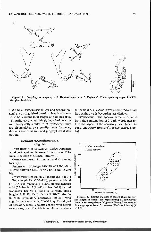
OF WASHINGTON, VOLUME 58, NUMBER 1, JANUARY 1991 95
Figure 12. Dactylogyrus omega sp. n. A. Haptoral apparatus. B. Vagina. C. Male copulatory organ. I to VII.Marginal booklets.
sin) and L. senegalensis (Niger and Senegal ba-sins) are distinguished based on length of trans-verse bars versus total length of hamulus (Fig.13). Although the individuals described here aremorphologically similar to D. cyclocirrus, theyare distinguished by a smaller penis diameter,different size of hamuli and geographical distri-bution.
Dogielius rosumplicatus sp. n.(Fig. 14)
TYPE HOST AND LOCALITY: Labeo rouaneti;Konkoure system, Konkoure river near Teli-mele, Republic of Guinea (locality 7).
OTHER RECORDS: L. rouaneti and L. parvus,locality 8.
SPECIMENS: Holotype MNHN 453 HC, slideTj 240; paratype MNHN 453 HC, slide Tj 240bis.
DESCRIPTION (based on 24 specimens in toto):Body length 330 (250^50); greatest width 80
(70-90) usually at level of ovary. Hamuli lengths:a: 34 (32-36); b: 43 (41^5); e: 16 (15-18). Dorsaltransverse bar 50-57 long, 8-10 wide. Hooklengths: I, II, III, IV, V, VI, VII: 20-22; 4A: 7-8. Male copulatory apparatus (30-36), withslightly incurvate penis, 25-30 long. Distal partof accessory piece is palette-shaped with lateralextensions, one of which is an elbow in which
the penis slides. Vagina is well sclerotized aroundits opening, walls becoming less distinct.
ETYMOLOGY: The species name is derivedfrom the combination of 2 Latin words that re-flect the aspect of the accessory piece (plico, tobend, and rosum from rodo, deckle-edged, shab-by).
Labeo senegalensis
Labeo rouaneti
3O 35 40
LENGTH of ANCHOR (jjm)
Figure 13. Scatter diagram of length of anchor ver-sus length of dorsal bar representing D. cyclocirrusfrom Labeo senegalensis (Niger and Senegal basins) andD. omega sp. n. from L. rouaneti (Konkoure basin) (P< 0.01).
Copyright © 2011, The Helminthological Society of WashingtonCopyright © 2011, The Helminthological Society of Washington

96 JOURNAL OF THE HELMINTHOLOGICAL SOCIETY
Figure 14. Dogielius rosumplicatus sp. n. A. Haptoral apparatus. B. Vagina. C. Male copulatory organ. I toVII. Marginal booklets.
REMARKS: This monogenean resembles Do-gielius harpagatus Guegan, Lambert, and Euzet,1989, and D. clavipenis Guegan, Lambert, andEuzet, 1989, from Labeo coubie in the Niger andSenegal basins (Guegan et al., 1989). It displaysthe same morphological type of accessory piecebut differs in hamuli size and morphology of thecopulatory apparatus.
Discussion
Dactylogyridae described herein from Labeo(Teleostei: Cyprinidae) in coastal river basins inWest Africa are different from those of Labeo inSahel-Sudan basins in Niger and in the Senegalbasin in Mali (Guegan et al., 1988, 1989). Affin-ities between the different species of Labeo andthose observed in their parasitofauna are com-pared. With regard to biogeographical features,we noted presence of monogeneans in popula-tions of L. parvus in the large Sahel-Sudan basins,in Cote d'lvoire systems, in Ghana, Togo andBenin systems, and their absence in the shortcoastal basins running from the Guinean ridgein Guinea, Sierra Leone, and Liberia. The dis-tribution of Labeo parvus (Fig. 15a) and 2 speciesof dactylogyrids, Dactylogyrus longiphallus andD. brevicirrus (Fig. 15b), are compared. Themonogenean populations of L. parvus from the
Guinean short coastal basins differ from popu-lations of adjacent basins in that D. brevicirrusis absent. It is presumed that this is the result ofa "host switching" followed by speciation on anew host (Guegan and Agnese, 1990). A com-parable phenomenon is observed for monoge-neans on cyprinid species, genus Barbus, wherethere is exclusion of very primitive parasite formson B. petitjeani, B. sacratus, and B. parawaldronifrom small coastal basins and their replacementby more derived species in barbels of the B. bynnigroup in peripheral basins (Guegan and Lambert,1990; Leveque and Guegan, 1990). The varia-tions in parasitism observed between the differ-ent populations of the L. parvus complex suggestthat fishes in the Guinean zone have not beenaffected by invasion of D. brevicirrus. The par-asitism observed is certainly similar to the orig-inal parasitofauna of the ancestors) of L. parvus.Parasite distribution defines 2 different biogeo-graphical regions marked by isolated river sys-tems. Our study concluded that there are con-siderable differences in the parasitofauna in these2 host populations which are separated by a nat-ural barrier. This supports Pellegrin's (1908) hy-pothesis distinguishing L. obscurus limited to afew basins in Guinea and Sierra Leone, from allthe other populations of L. parvus. However, it
Copyright © 2011, The Helminthological Society of WashingtonCopyright © 2011, The Helminthological Society of Washington

OF WASHINGTON, VOLUME 58, NUMBER 1, JANUARY 1991 97
A L. parvus
L. rouaneti
L. alluaudi
12
D. longiphallus
O D. brevicirrus
Figure 15. Area of distribution of 2 species and 1 complex of species of Labeo: L. rouaneti, L. alluaudi, andL. parvus complex in West Africa (Fig. 15a), and distribution of 2 species of Dactylogyrus: D. longiphallus andD. brevicirrus identified on L. parvus complex (Fig. 15b).
Copyright © 2011, The Helminthological Society of WashingtonCopyright © 2011, The Helminthological Society of Washington

98 JOURNAL OF THE HELMINTHOLOGICAL SOCIETY
is impossible to specify the degree of divergenceacquired by the various populations of the L.parvus complex since only a study of breedingwould enable evaluation of their taxonomic lev-els.
Labeo rouaneti, which is considered to be phy-logenetically close to L. coubie (Reid, 1985), dis-plays a branchial parasitofauna that was not aprecise reflection of the assumed affinity of thesehosts. Dactylogyrus jucundus sp. n. and Dogieliusrosumplicatus sp. n. are similar to 2 derived par-asite forms found on L. coubie. The same doesnot apply to the other monogeneans present onL. rouaneti: Dactylogyrus omega sp. n. is mor-phologically close to D. cyclocirrus described onseveral cyprinids in East Africa (Paperna, 1973,1979), and which we report in West Africa onL. senegalensis (Guegan et al., 1988); D. sematussp. n. belongs to a completely original type ofdactylogyrid monogenean from West Africanspecies of Labeo since it is similar to certainDactylogyrus on small Barbus in southern Cam-eroon (Birgi and Lambert, 1987). In a biogeo-graphical study on the ichthyological commu-nities of the various basins in West Africa,Hugueny (1989) demonstrated the faunistic sim-ilarities between Guinea and the Cameroon-Ga-bon area. The presence of D. sematus sp. n. in aGuinean basin supports this hypothesis.
The monogeneans found on L. alluaudi in Si-erra Leone are closely related to those on L. par-vus populations in short coastal basins. It can beimagined that the parasites evolved in a concom-ittant way, speciation of the fish being followedby that of the monogeneans. On the basis of theparasite criterion, we accept the theory put for-ward by Reid (1985) who classified L. alluaudias part of the L. parvus group. The absence ofmonogeneans in the L. alluaudi populations ofthe rivers Cavally and Nipoue might be ex-plained by the ecological conditions of this fish(small rapids in the upper course of the river).
Acknowledgments
This research was carried out as part of a mul-tidisciplinary program (P.E.D.A.L.O.: Poissonsd'Eau Douce d'Afrique de 1'Ouest) financed byC.N.R.S.-P.I.R.E.N. and ORSTOM. The au-thors thank staff and researchers at the OR-STOM Laboratoire d'Hydrobiologie at Bamako(Mali). Thanks are due Professor Louis Euzet andDoctor Christian Leveque, responsible for theprogram. We also thank Doctor Jean Lou Jus-
tine, MNHN (Worms), Paris for his help andtime in the deposition and registration of spec-imen types and paratypes.
Literature Cited
Birgi, E., and A. Lambert. 1987. Les Dactylogyridae(Plathelminthes, Monogenea) des Cyprinidae dugenre Barbus du sud-Cameroun. Revue d'Hydro-biologie Tropicale 20:37-48.
Daget, J. 1962. Les poissons du Fouta-Djalon et dela basse-Guinee. Memoire de 1'Institut Francaisd'Afrique Noire 65:1-210.
, and A. Iltis. 1965. Poissons de Cote d'lvoire(eaux douces et saumatres). Memoire de 1'InstitutFran9ais d'Afrique Noire 74:1-385.
Euzet, L., and C. Combes. 1980. Les problemes de1'espece chez les animaux parasites in Les prob-lemes de 1'espece dans le regne animal. Memoirede la Societe Zoologique de France 40:239-283.
, and M. Prost. 1981. Report of the meetingon Monogenea: problems of systematics biologyand ecology. Proceedings of the Fourth Interna-tional Congress of Parasitology (ICOPA IV), Re-view of Advances in Parasitology, pp. 1003-1004.
Grove, A. T. 1985. The physical evolution of the riverbasins. Pages 21-61 in A. T. Grove, ed. The Nigerand its Neighbours. Environmental History andHydrobiology, Human Use and Health Hazardsof the Major West African Rivers. A. A. BalkemaPublishers, The Netherlands.
Guegan, J. F., and J. F. Agnese. 1990. Host phylog-eny infers parasite evolutionary events: the caseof monogenean gill parasite of Labeo (Teleostei,Cyprinidae). Canadian Journal of Zoology. (Inpress.)
, and A. Lambert. 1990. Twelve new speciesof dactylogyrids (Plathelminthes, Monogenea) fromWest African barbels (Teleostei, Cyprinidae) withsome biogeographic implications. Systematic Par-asitology. (In press.)
-, and L. Euzet. 1988. Etude des Mono-genes des Cyprinidae du genre Labeo en Afriquede 1'Ouest.—I. Genre Dactylogyrus Diesing, 1850.Revue d'Hydrobiologie Tropicale 21:135-151.
—, , and . 1989. Etude des Mono-genes des Cyprinidae du genre Labeo en Afriquede FOuest—II. Genre Dogielius Bykhowsky, 1936.Revue d'Hydrobiologie Tropicale 22:35—48.
Gussev, A. V. 1962. Key to parasites of freshwaterfish of the U.S.S.R. In I. E. Bykhowskaya-Pavlov-skaya. Akademiia Nauk, S.S.S.R. 919 pp. (Trans-lated from Russian by Israel Program for ScientificTranslations.)
Hugueny, B. 1989. West African rivers as biogeo-graphic islands: species richness of fish commu-nities. Oecologia 79:236-243.
Jegu, M., and C. Leveque. 1984. Les especes voisinesou synonymes de Labeo parvus (Pisces, Cyprini-dae) en Afrique de FOuest. Cybium 8:45-58.
Leveque, C., and J. Daget. 1984. Cyprinidae. Pages217-342 in Daget, Gosse, and Thys van den Au-denaerde, eds. CLOFFA I: Check-List of theFreshwater Fishes of Africa. MRAC-ORSTOMPublishers, Paris.
Copyright © 2011, The Helminthological Society of WashingtonCopyright © 2011, The Helminthological Society of Washington

OF WASHINGTON, VOLUME 58, NUMBER 1, JANUARY 1991 99
, and J. F. Guegan. 1990. Les grands Barbus(Teleostei, Cyprinidae) d'Afrique de 1'Ouest. Re-vue d'Hydrobiologie Tropicale 23:41-64.
Paperna, I. 1969. Monogenetic trematodes of the fishof the Volta basin and South-Ghana. Bulletin de1'Institut Fran?ais d'Afrique Noire 31:840-880.
. 1973. New species of Monogenea (Vermes)from African freshwater fish. A preliminary re-port. Revue de Zoologie et de Botanique Afri-caines 87:505-518.
1979. Monogenea of inland water fish in Af-rica. Annales du Musee Royal d'Afrique Centrale226:1-131.
Paugy, D., J. F. Guegan, and J. F. Agnese. 1990.Three simultaneous and independent approachesto the characterization ofLabeo roseopunctatus sp.n. (Teleostei, Cyprinidae) in West Africa. Cana-dian Journal of Zoology 68:1124-1131.
Pellegrin, J. 1908. Poissons recueillis par le Dr. Wurtzen Guinee francaise. Description de quatre especesnouvelles. Bulletin du Museum National d'His-toire Naturelle de Paris 14:204-209.
Price, C. E., K. S. Korach, and R. McPott. 1969. Themonogenean parasites of African species.—V. Twonew Dactylogyrus species from Natal cyprinids.Revue de Zoologie et de Botanique Africaines 79:273-279.
, and W. J. Yurkiewicz. 1968. The monoge-nean parasites of African fishes.—VIII. A re-eval-uation of the genus Dogielius Bykhowsky, 1936with the description of a new species. Revista Ibe-rica Parasitologia 28:467-472.
Reid, G. M. 1985. A revision of African species ofLabeo (Pisces, Cyprinidae) and a redefinition ofthe genus. Thesis Zoologicae, Braunschweig Ver-lag von J. Cramer 6:1-322.
DIAGNOSTIC PARASITOLOGY COURSE
Department of Preventive Medicine and BiometricsUniformed Services University of the Health Sciences
Bethesda, Maryland 20814
29 July-9 August 1991
This course will consist of a series of lectures and laboratory sessions covering the diagnosis ofparasitic infections of humans. In addition to the examination of specimens, participants will beable to practice various methods used in the diagnosis of intestinal, blood, and tissue parasiticdiseases. Parasitic diseases encountered throughout the world will be included. Slide presentationsand video tapes will be available for study. The course will be held on the University's campus,utilizing up-to-date lecture rooms and laboratory facilities. Microscopes will be available on a loanbasis, and laboratory supplies will be provided. Certain reference specimens will also be availablefor personal use.
For further information, contact Dr. John H. Cross, (301) 295-3139; Dr. Edward H. Michelson,(301) 295-3138; or Ms. Ellen Goldman, (301) 295-3129.
The total cost for the 2-week course is $800. U.S. government and military personnel may takethe course at a reduced rate. Those interested should register as soon as possible, as the number ofstudents will be limited. CME credits will be available for this course. Previous laboratory experienceis recommended.
Copyright © 2011, The Helminthological Society of WashingtonCopyright © 2011, The Helminthological Society of Washington
J. Helminthol. Soc. Wash.58(1), 1991, pp. 100-109
Early Development of Larval Taenia polyacantha inExperimental Intermediate Hosts
OSAMU FUJITA,1 YUZABURO OKU,1 MUNEHIRO OKAMOTO,1 HlROSHI SATO,1HONG KEAN Ooi,2 MASAO KAMIYA/ AND ROBERT L. RAuscn3
1 Department of Parasitology, Faculty of Veterinary Medicine, Hokkaido University,Sapporo 060, Japan,2 Department of Pharmacology and Molecular Science, School of Medicine, Johns Hopkins University,Baltimore, Maryland 21205, and3 Department of Comparative Medicine, School of Medicine, University of Washington,Seattle, Washington 98195
ABSTRACT: The early larval development and migration route of Taenia polyacantha were examined using oralinoculation of oncospheres into red-backed voles and Mongolian gerbils. The larvae were recovered in the wallof the small intestine and in the mesenteric lymph nodes by 5 days postinfection (PI) and from the peritonealcavity after 6 days PI. These results suggest that the larval cestodes developed initially in the wall of the smallintestine and the mesenteric lymph nodes, and later migrated to the peritoneal cavity. Although the developmentof the parasite was quite similar in the 2 host species, pathological changes were different. In Mongolian gerbils,these changes were slight, but in red-backed voles, they were marked and fatal. In addition to oral inoculation,hatched oncospheres were injected intraperitoneally and subcutaneously into red-backed voles, Mongoliangerbils, and AKR/J mice. Larval development took place at the injection sites in gerbils and mice, but wasdelayed and abnormal. Some of the parasites in the injection site showed abnormal numerous budding. Highpathogenicity was shown after subcutaneous and intraperitoneal injection as well as after oral inoculation.
KEY WORDS: Taenia polyacantha, rodents, migration route, larval development, abnormal development,histopathology, pathogenicity.
The larval Taenia polyacantha Leuckart, 1856,proliferates asexually and the metacestodes arefound in the peritoneal and pleural cavities ofrodents (Schiller, 1953; Rausch, 1959a; MuraiandTenora, 1973;Slais, 1973; Wigeretal., 1974;Tenora et al., 1979). Rausch and Fay (1988a)reported that the migration of postoncospheralstages was via the portal vein to the liver, andthat initial larval development and formation ofthe primary vesicle took place in the liver. Theproduction of secondary vesicles occurred aftermigration to the peritoneal cavity. Fujita et al.(1990) reported on the susceptibility and mor-tality of various small mammals to infection byT. polyacantha after oral inoculation of eggs. Red-backed voles and cotton rats showed high mor-tality at the early phase of infection. In thosehosts, the sites of early larval development andthe production of primary and secondary vesicleswere different from those reported by Rauschand Fay (1988a).
In this study we examined the route of migra-tion and development of the larvae, and assessedpathological changes in red-backed voles andMongolian gerbils. In order to determine wheth-er the postoncospheral development of T. poly-acantha is pathogenic, oncospheres were inoc-
ulated parenterally in red-backed voles, AKR/Jmice, and Mongolian gerbils.
Materials and Methods
CESTODE: Metacestodes were isolated from north-ern voles, Microtus oeconomus, trapped at Savoonga,St. Lawrence Island, Alaska, in 1988 and 1989. In thelaboratory, they were then transplanted surgically intothe peritoneal cavity of Mongolian gerbils, Merionesunguiculatus, which were maintained for 3 months.Eight mature metacestodes were then administeredorally to a cestode-free mongrel dog. Gravid segmentsexpelled in feces of the dog were collected daily andstored at 4°C in saline containing penicillin, strepto-mycin, and fungizone (Mitchell et al., 1977; Williamset al., 1981). Eggs from the gravid segments were usedwithin 1 month of collection.
EXPERIMENTAL ANIMALS: Mongolian gerbils andAKR/J inbred mice were bred and maintained in ourlaboratory. Red-backed voles, Clethrionomys rufoca-nus bedfordiae, were trapped in a shelter-belt near Sap-poro City, Hokkaido, and maintained in our laboratoryat 22-24°C.
EXPERIMENT 1 (observations on early larval migra-tion): Red-backed voles (18 3; age not determined)and Mongolian gerbils (15 $, 9 9; 4-8 wk old) wereused. Individual hosts were inoculated with 5,000 to100,000 eggs by stomach-tube under light ether an-esthesia, and killed by exsanguination under ether an-esthesia at 12 hr postinfection (PI) and at 24-hr inter-vals up to 6 days PI; animals that died were examinedat different intervals. Washings from the peritoneal
100
Copyright © 2011, The Helminthological Society of WashingtonCopyright © 2011, The Helminthological Society of Washington
101
cavity were examined for larval cestodes by means ofa dissecting microscope (at 35 x). The recovered larvaewere fixed in 10% formalin and mounted in glycerinjelly. The small intestine was divided into 5 equal parts,and the middle portion of each, and all abdominal andthoracic organs, were fixed in 10% buffered neutralformalin and embedded in paraffin by standard meth-ods. Paraffin sections, cut at 4-6 /urn, were stained withhematoxylin-eosin (HE) and Alcian blue-periodic acid-Schiff(AB-PAS) for microscopic examination. Sixteento 20 serial sections of organs in which larval cestodeswere found were studied, and the maximal lengths andwidths of the vesicles were recorded. For electron mi-croscopy, the small intestines of rodents of both specieswere fixed on day 5 PI in 3.0% glutaraldehyde in 0.1M cacodylate buffer, and then postfixed in 1.0% os-mium tetroxide in 0.1 M cacodylate buffer. The seg-ments of the intestines were dehydrated in graded eth-anol and embedded in Quetol-812. Ultrathin sectionswere cut and stained with uranyl acetate and lead ci-trate, and subsequently observed and photographedwith an HITACHI HU-12A electron microscope.
EXPERIMENT 2 (parenteral injection of oncospheres):Mongolian gerbils (6 <5, 18 9; 6-8 wk old), AKR/J
mice (8 3, 7 2; 4 wk old), and red-backed voles (5 S, 32; age not determined) were used. The experimentaldesign is shown in Table 1. The oncospheres werehatched with artificial intestinal fluid (Silverman, 1954);1,000 or 20,000 oncospheres were injected subcuta-neously or intraperitoneally in sterile saline. Individualanimals were killed by exsanguination on days 19 and21 PI and autopsied immediately after death. Sites ofinjection were washed with saline and examined forpresence of vesicles using a dissecting microscope. Ves-icles recovered were fixed in 10% formalin and stainedwith Schneider's aceto-carmine for morphological ex-amination. If gross lesions were observed in the tissues,sections were examined microscopically.
To compare larval development in the subcutaneoustissue and in the peritoneal cavity, and to define rel-ative pathogenicity, Mongolian gerbils also were in-oculated orally with 300 or 500 eggs.
Results
Experiment 1
Early larval development was discerned onlyin the wall of the small intestine and in the mes-enteric lymph nodes in voles and gerbils. Av-erage size of the developing larvae and their lo-cations are shown in Table 2.
RED-BACKED VOLES: Developing larvae werefound in the lamina propria of the small intestinebetween 12 hr PI and 5 days PI. They were pres-ent in the submucosa or passing through the wallsof the lymphatic lacteal on the third day PI (Figs.1, 2). On day 4 PI, they had penetrated furtherand were recovered mainly in the submucosa andmuscularis, as well as having reached the afferentlymph vessels and the peripheral and marginalsinuses of the mesenteric lymph nodes. After 5days PI, some larvae were found in the peritoneal
Table 1. The number of eggs or oncospheres of Taeniapolyacantha in each inoculation route.
Dose ofinoculum
No. of hostsexamined
Oral inoculation (shelled eggs)
Mongolian gerbils 300500
Subcutaneous injection (oncospheres*)
Mongolian gerbils 1,000AKR/J inbred mice 1,000Red-backed voles 20,000
Intraperitoneal injection (oncospheres*)
Mongolian gerbils 1,000AKR/J inbred mice 1,000Red-backed voles 20,000
* Oncospheres were hatched artificially.
cavity, and from the sixth day PI they were nolonger observed in the wall of the small intestineor mesenteric lymph nodes.
By 2 days PI, the early vesicle contained sev-eral prominent nuclei and PAS-positive gran-ules, and was always surrounded by a halo-like,amorphous area, about 4—8 /im in width, withinwhich was AB-positive material. Concurrent witha great increase in size of larva between days 2and 3 PI, the amorphous area faded out almostcompletely. At this time, marked cellular differ-entiation occurred; a central cavity began to de-velop and the primary vesicle was formed. A fewprimary vesicles with buds were recovered in thewall of the small intestine on day 5 PI. On day6 PI, the vesicles in the peritoneal cavity hadproduced some buds, and numerous host cellscovered their surfaces (Fig. 3).
Between days 2 and 3 PI, slight hemorrhageand infiltration by inflammatory cells, mainlyneutrophils and mononuclear cells, were ob-served in the wall of the small intestine and, atthis time, blood-tinged ascitic fluid began to ac-cumulate. These changes, especially extensivehemorrhage and purulent inflammation mainlysurrounding the vesicles, became prominent inthe wall of the small intestine, and thereafter inthe mesenteric lymph nodes (Figs. 4, 5). Electronmicroscopically, it was determined that numer-ous neutrophils adhered closely to the surface ofthe tegument of the vesicles, and the number ofcytoplasmic granules decreased (Fig. 6). The vol-ume of ascitic fluid increased. After 4 days PI,inflammatory cells, mainly neutrophils accu-mulated in the mesenteric lymph nodes and
Copyright © 2011, The Helminthological Society of WashingtonCopyright © 2011, The Helminthological Society of Washington

102 JOURNAL OF THE HELMINTHOLOGICAL SOCIETY
Table 2. Size and location of early larval stage of Taenia polyacantha in gerbils and voles.
Days postinfection
Host
Gerbil
Vole
Size Gtm)*Location**
Size Oim)*Location**
0.5
21.0 x 14.8SI (LP)MLN
22.2 x 14.6SI (LP)
1
22.2 x 17.8SI (LP)MLN
23.8 x 16.3SI (LP)
2
38.0 x 25.2SI (LP)MLN
36.8 x 27.8SI (LP)
61.5 xSI(LPMLN
85.7 xSI(LP
3
35.5~ SM)
62.8~ SM)
* Average maximum length and width of each parasite measured in histological sections.** SI = small intestine; LP = lamina propria; SM = submucosa; M = muscularis; MLN = mesenteric lymph node; PC
peritoneal cavity.*** External budding.
spleen, along with hemorrhage, and the numberof lymphocytes, decreased in these lymphatic or-gans.
MONGOLIAN GERBILS: By 3 days PI, devel-oping larvae were recovered in the lamina pro-pria and submucosa of the small intestine, es-pecially in the jejunum and ileum. After 12 hrPI, they were found in the afferent lymph vesselsand marginal sinuses of the mesenteric lymphnodes. On day 6 PI, vesicles were found in theperitoneal cavity.
The early larval development in gerbils wassimilar to that in the red-backed voles, with thelarvae showing great enlargement between days2 and 3 PI, and with the beginning developmentof a central cavity and primary vesicles. On day6 PI, vesicles recovered from the peritoneal cav-ity displayed some buds.
Microscopically, a slight infiltration by inflam-matory cells was evident in the lamina propriaand submucosa of the small intestine after day1 PI. Small accumulations of inflammatory cellswere scattered focally in the dilated marginal si-nus and capsule of the mesenteric lymph nodes,especially around the larvae, between days 4 and6 PI (Fig. 7). A slight accumulation of turbidascitic fluid with fibrin occurred on day 5. After4 days, a few neutrophils adhered to the surfacesof the vesicles, and on day 5, their pseudopodswere loosely attached to the fragments of micro-villi (Fig. 8).
Experiment 2
Almost all of the Mongolian gerbils inoculatedorally with 300 and 500 eggs were alive until thescheduled date of autopsy, but almost all thoseinjected parenterally with 1,000 and 20,000 on-cospheres died before 21 days PI. Development
and pathological changes associated with theroutes of inoculation are described below.
Oral inoculation
MONGOLIAN GERBILS: Ten to 58 (x = 31) sec-ondary vesicles were recovered from the peri-toneal cavity in 5 of 12 infected gerbils necrop-sied on days 19 and 21 PI. Most of the larvaeon day 19, ranging from 1.8 to 2.4 mm in lengthand 1.6 to 1.8 mm in diameter, had developedrostellar cones, and about half of them exhibited2 rows of developing rostellar hooks (Fig. 9).Macroscopically, the infected gerbils exhibited aslight accumulation of turbid ascitic fluid, withfibrous adhesions on the serosa of abdominalorgans.
Subcutaneous injection
MONGOLIAN GERBILS: Larval cestodes wererecovered from the injection site of 3 of 4 gerbilsthat died between 12 and 19 days PI. Most ofthose by day 19 PI had formed single secondaryvesicles, 88-272 jon in length and 80-152 (j.m indiameter; some still consisted of aggregations ofsecondary vesicles with less than 35 buds, andranged from 368 to 552 /urn in length by 208 to360 nm in diameter (Fig. 10). An accumulationof nuclei was observed at the distal ends of de-tached secondary vesicles. At the site of injec-tion, marked hemorrhage and edema were noted,and microscopically extensive necrosis and in-filtration by eosinophils and neutrophils wereobserved in the dermis, subcutaneous tissues, andunderlying skeletal muscles.
AKR/J MICE: Four mice died between 9 and19 days PI. Vesicles were recovered from theinjection sites of 6 of 8 animals. On days 9 and11 PI all had formed single vesicles, apparently
Copyright © 2011, The Helminthological Society of WashingtonCopyright © 2011, The Helminthological Society of Washington

OF WASHINGTON, VOLUME 58, NUMBER 1, JANUARY 1991 103
Table 2. Continued.
Days postinfection
4
99.0 x 54.6MLN
100.6 x 65.6SI (LP - M),MLN
5
123.4 x 65.0MLN
159.2 x 80.8***SI (LP ~ M),MLN, PC
990 xMLNPC
335 xPC
6
340***
212***
a primary vesicle, and most of those on days 19and 21 PI had abnormally developed aggrega-tions of secondary vesicles, with 32 to more than50 buds; the aggregations ranged from 440 to1,180 ^m in length, and 200 to 980 nm in di-ameter. Nuclei had accumulated at the distal endsof almost all secondary vesicles, and a few weredeveloping invaginated canals (Fig. 11). Grosslesions at sites of injection were similar to thosein gerbils: extensive necrosis, hemorrhage, and aslight accumulation of inflammatory cells be-tween the epidermis and underlying skeletalmuscles were observed microscopically.
RED-BACKED VOLES: Two of 4 infected volesdied between 5 and 8 days PI. They exhibitedmild ascites with blood; extensive edema andslight necrosis were observed in the subcutane-ous tissue. Larval cestodes were found in only 1vole, which died on day 5 PI. In that animal,numerous ecchymoses were present on the serosaof the small intestine. Microscopically, the larvaeconsisted of primary vesicles ranging from 60 to112 jiim in length, and 40 to 64 jum in diameter.They were found mainly in the hemorrhagic le-sions between the lamina propria and the mus-cularis. Slight hemorrhage and focal degenerativechanges also occurred in the mesenteric lymphnodes.
Intraperitoneal injection
MONGOLIAN GERBILS: All gerbils died be-tween 15 and 17 days PI, and larval cestodeswere recovered from the peritoneal cavities. Al-most all of the larval cestodes on day 17 formedsingle primary vesicles or 2 secondary vesicles,which were rounded and subspherical in form,and ranged from 112 to 416 pm in length and96 to 224 /nm in diameter. Some had formedabnormally minute aggregations of secondaryvesicles, with 16 to more than 50 buds, whichranged from 384 to 584 yum by 240 to 442 p,m
in diameter; their surfaces were covered with nu-merous host cells (Fig. 12). Macroscopically, slightyellowish or turbid ascitic fluid was present, andall abdominal organs were congested.
AKR/J INBRED MICE: Six of the 7 infectedmice died between 11 and 15 days PI; the lastwas necropsied on day 21 PI. Developing larvaewere recovered from the peritoneal cavities of 6mice. By day 15, the larvae had formed singleor numerous secondary vesicles, all of similarsize, with the aggregations ranging from 384 to1,096 Aim in length and 252 to 704 nm in di-ameter. Accumulations of nuclei at the distal endsof the vesicles had formed on day 12 PI. On day21 PI, about half of the vesicles had detached,and some had developed invaginated canalswhich ranged from 184 to 216 i*m in length by104 to 128 pm in diameter.
RED-BACKED VOLES: Three of the 4 infectedvoles died by the seventh day PI. They exhibitedmild ascites, with the fluid stained by blood, andall of the abdominal organs were congested. Slighthemorrhages had occurred in the mesentericlymph nodes, but no larvae could be found mi-croscopically in those lesions. Primary vesicleswith central cavities were found under the phren-ic serosa of the liver in only 1 vole dead on day5 PI; they ranged from 136 to 200 fim in lengthby 72 to 112 ^im in diameter. Focal accumula-tions of inflammatory cells and necrosis sur-rounded the larvae (Fig. 13). In the hepatic lymphnodes, a decrease in the number of lymphocyteshad occurred, caused by focal degenerative changeand hemorrhage.
Discussion
After the eggs of taeniids are ingested, the on-cospheres hatch and become activated in thesmall intestine, after which they penetrate intothe mucosal epithelium. The usual route of mi-gration of the oncospheres is via the portal veinor lymphatic vessels. For example, it is wellknown that the oncospheres of Taenia hydati-gena, T. taeniaeformis, T. pisiformis, and Echi-nococcus multilocularis migrate to the liver viathe portal vein (Olsen, 1974). Concerning T.polyacantha in Microtus oeconomus, Rausch andFay (1988a) reported that oncospheres migratefrom the small intestine to the liver via the portalvein, and that early larval development takesplace in the liver. Furthermore, Dorosz (1968)and Tenora et al. (1988) reported that metaces-todes of T. polyacantha are localized under the
Copyright © 2011, The Helminthological Society of WashingtonCopyright © 2011, The Helminthological Society of Washington

104 JOURNAL OF THE HELMINTHOLOGICAL SOCIETY
V
? » i-S..
tf*'*(T
Figures 1-3. Larval Taenia polyacantha in red-backed voles. 1. Oncospheres situated in the lamina propria ofthe small intestine at 12 hr PI. AB-PAS stain. Scale bar = 20 fim. 2. The embryo passing through the wall ofthe lymphatic lacteal from the lamina propria, day 3. HE stain. Scale bar = 50 tan. 3. Developing secondaryvesicles in the peritoneal cavity, day 6. Scale bar — 100 pm.
hepatic serosa of naturally infected hosts. Thesereports suggest that the liver is the typical site ofearly development of the larval cestode. How-ever, in the rodents used in the present study,early development of the larvae was found totake place in the wall of the small intestine andin the mesenteric lymph nodes. Fujita et al. (1990)reported that larval T. polyacantha in cotton rats,Sigmodon hispidus, were recovered from the sameorgans as in gerbils and red-backed voles on days8 and 10 PI. It is suggested that the oncosphereshad a predilection for the wall of the small in-testine and the mesenteric lymph nodes, and thatthose sites were peculiar to T. polyacantha. Sincea few larvae were recovered from the liver of ared-backed vole inoculated intraperitoneally, theliver of that rodent appears also to be a site ofdevelopment of the metacestode. Many larvaewere situated in the afferent lymph vessels andmarginal sinus of the mesenteric lymph nodes ingerbils and red-backed voles. We considered that
the larvae could not migrate farther via the lym-phatic system, because their size was too greatrelative to the diameter of the lymph vessels. Themigration route of oncospheres in the presentstudy was different from that reported by Rauschand Fay (1988a) in Microtus oeconomus. Theisolates of T. polyacantha used in both investi-gations were evidently the same, having beenobtained from voles, M. oeconomus, collectedon St. Lawrence Island, in the Bering Sea. Thedifferences in migration routes of the onco-spheres appear to be related to differences in spe-cies of rodents used as experimental intermediatehosts.
Budding of the primary vesicles in the wall ofthe small intestine and in the mesenteric lymphnodes was observed by us from day 5 PI. Rauschand Fay (1988a) suggested that budding occurredafter the early-stage vesicles had migrated fromthe liver into the peritoneal cavity; they reportedthat the larvae in the liver on day 5 PI were only
Figures 4-6. Pathological changes caused by larval Taenia polyacantha in the red-backed vole, day 5 PI. 4.Extensive hemorrhage and accumulation of numerous inflammatory cells surrounding the budding larva (arrow-head) in the wall of the small intestine. HE stain. Scale bar = 100 fan. 5. Marked decrease in the number oflymphocytes caused by extensive hemorrhage and degenerative changes in tissues around the larval cestode(arrowhead) in the mesenteric lymph node. AB-PAS stain. Scale bar = 100 pm. 6. Transmission electronmicrograph of the larval cestode in the injured muscularis of the small intestine. Numerous neutrophils (N) andmacrophages (M) closely attached to the surface of the vesicle (P). Scale bar = 5 nm.
Copyright © 2011, The Helminthological Society of WashingtonCopyright © 2011, The Helminthological Society of Washington

OF WASHINGTON, VOLUME 58, NUMBER 1, JANUARY 1991 105
ni:'r* V
' • •.-* • ••• TV- £-*v • , • ' ' " ̂ M '̂4;s>.'•;>':*$- -i*- > v- - , •• Mgv •• l|; 1:l J|§;» ,̂ Ar;S v>;' ;̂ 3BIS•' "':>i^gi , vr^;^^;;Xi.;; . ; -"-;..:.. ^y^^^^
i^^,;^,.^,-;--rr"^ '." v
V;. • c * ̂ ,,. ' , r*'N ,-.*.":-; •jw?:\^r%> !&-•"•
\^f&, ;-V$>fe: \* » r/'.f-';»^SifiSK:">* ^^"'^f"^* ;^;^M-»--^^ , - , . . - .
i '
5- .̂43»c*
Copyright © 2011, The Helminthological Society of WashingtonCopyright © 2011, The Helminthological Society of Washington
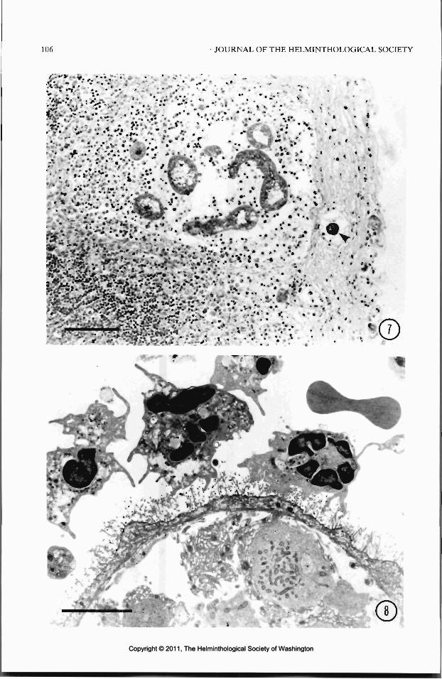
106 JOURNAL OF THE HELMINTHOLOGICAL SOCIETY
Copyright © 2011, The Helminthological Society of WashingtonCopyright © 2011, The Helminthological Society of Washington

OF WASHINGTON, VOLUME 58, NUMBER 1, JANUARY 1991 107
19 and 22 ̂ m in greater diameter. In the presentstudy, the average size of larvae on day 5 PI was123 Mm in length and 65 nm in diameter; thus,they were much larger than those described byRausch and Fay (1988a).
Although the early development of the larvalcestode was quite similar in gerbils and red-backed voles, it produced different pathologicalchanges in the 2 hosts. In the gerbils, the tissue-response was slight, but in red-backed voles,marked purulent inflammation occurred, andnumerous neutrophils attached closely to the teg-umental surface of the larvae. Such changes inred-backed voles were not proportional to thenumber of eggs inoculated. Rausch and Fay(1988b) suggested that infections involving morethan 5 eggs might be fatal, because of the inflam-matory response evoked by the migration of thevesicles into the peritoneal cavity. The patho-genicity of other taeniid cestodes, i.e., T. twitch-elli and T. mustelae is proportional to the num-ber of eggs inoculated and the lesions caused bymigrations of the larvae (Freeman, 1956; Rausch,1959b). We considered from the present resultsthat the high degree of pathogenicity and the highmortality in red-backed voles might be due tothe greater susceptibility of that host, and markedpathological changes caused by the developmentand migrations of the larvae were the main causeof death. Since larvae developing in the subcu-taneous tissues also were quite pathogenic, theypossibly secrete some deleterious substances.Shield et al. (1973) reported that 8-day larvae ofT. pisiformis exhibited PAS-positive secretorycells of large size, which had some effect in themigration of the larvae. In the larval stage of T.polyacantha, numerous PAS-positive cells ofsmall size were observed, but the larger cells de-scribed by Shield were not observed. The mech-anisms of pathogenesis in the case of T. poly-acantha remain unclear.
When oncospheres of T. hydatigena, T. sagi-nata, T. ovis, and Echinococcus granulosus wereinoculated subcutaneously and intraperitoneallyinto the intermediate hosts, development of thelarvae took place at the site of inoculation (Gem-
Figure 9. Advanced stage of secondary vesicle,showing the rostellar cone (arrow) and developing hooks,day 19 PI, from a Mongolian gerbil inoculated orally.Schneider's aceto-carmine stain. Scale bar =100 /im.
mell, 1962; Williams and Colli, 1970; Slais andMachnicka, 1976). In the present investigation,the development of the larval T. polyacanthatook place at the injection sites in Mongoliangerbils and AKR/J mice, and involved the ab-normal budding of numerous vesicles of smallsize. In M. oeconomus, fewer than 16 buds wereusually produced (Rausch and Fay, 1988a). Par-enteral inoculation of oncospheres may deprivethe larva of appropriate stimuli and nutrients fornormal development. A few larvae recoveredfrom the peritoneal cavity of the intraperito-neally inoculated AKR/J mouse, however, weresimilar on day 21 PI to those from orally inoc-
Figures 7, 8. Larval Taenia polyacantha in the mesenteric lymph node of Mongolian gerbils, day 5 PI. 7.Focal accumulation of inflammatory cells surrounding the vesicles in the marginal sinus and capsule. Vesicle inthe afferent lymph vessel (arrowhead). AB-PAS stain. Scale bar = 100 /im. 8. Transmission electron micrographof the vesicle in the marginal sinus. A few neutrophils cover the surface of the vesicle, and their pseudopodsextend to fragments of the microvilli. Scale bar = 5 ion.
Copyright © 2011, The Helminthological Society of WashingtonCopyright © 2011, The Helminthological Society of Washington

108 JOURNAL OF THE HELMINTHOLOGICAL SOCIETY
Figures 10-13. Larval Taenia polyacantha from parenterally inoculated rodents. 10. Proliferation of minutesecondary vesicles, subcutaneous tissue of a Mongolian gerbil on day 19 PI. Schneider's aceto-curmine stain.Scale bar = 50 fan. 11. Vesicle with numerous buds, subcutaneous tissue of AKR/J mouse, day 19 PI. Schneider'saceto-carmine stain. Scale bar = 30 fan. Inset shows early formation of an invaginated canal in a secondaryvesicle (arrowhead). 12. Proliferation of secondary vesicles, peritoneal cavity of a Mongolian gerbil, day 17 PI.Schneider's aceto-carmine stain. Scale bar = 50 nm. 13. Accumulation of inflammatory cells, with focal necrosis,around an early vesicle (arrowhead), beneath the phrenic serosa of the liver of a red-backed vole that died onday 5 PI. HE stain. Scale bar = 50 nm.
ulated hosts (Fujita et al., 1990). Many larvaewere recovered in the wall of the small intestineof a red-backed vole that had been inoculatedsubcutaneously. This finding cannot now be ex-plained, but the possibility of the migration ofthe oncospheres cannot be excluded.
Acknowledgments
We thank Drs. Masashi Ohbayashi, Peter B.Gathura, and Matthew C. Playford for their sug-gestions and for reading this manuscript. Thiswork was supported by grants from the Ministryof Education, Science and Culture, Japan (Nos.02404083, 63044005 and 63480086).
Literature Cited
Dorosz, J. 1968. Helminth parasites of small rodentsliving in areas irrigated by urban sewage of Wro-claw. Acta Parasitologica Polonica 16:375-396.
Freeman, R. S. 1956. Life history studies on Taeniamustelae Gmelin, 1790 and the taxonomy of cer-tain taenioid cestodes from Mustelidae. CanadianJournal of Zoology 34:219-242.
Fujita, O., Y. Oku, H. K. Ooi, M. Kamiya, and R. L.Rausch. 1990. Susceptibility of rodents to Tae-nia polyacantha and their mortality along the courseof infection. Japanese Journal of Parasitology 39:284-289.
Gemmell, M. A. 1962. Natural and acquired im-munity factors inhibiting penetration of some hex-acanth embryos through the intestinal barrier. Na-ture (London) 194:701-702.
Mitchell, G. F., J. W. Coding, and M. D. Rickard.1977. Studies on immune responses to larval ces-todes in mice. Increased susceptibility of certainmouse strains and hypothymic mice to Taeniataeniaeformis and analysis of passive transfer ofresistance with serum. Australian Journal of Ex-perimental Biology and Medical Science 55:165-186.
Murai, E., and F. Tenora. 1973. Some taeniid species(Cestoidea) parasitizing vertebrates (Rodentia,
Copyright © 2011, The Helminthological Society of WashingtonCopyright © 2011, The Helminthological Society of Washington

OF WASHINGTON, VOLUME 58, NUMBER 1, JANUARY 1991 109
Carnivora, Strigiformes) in Hungary. Acta Zoolo-gica Academiae Scientiarum Hungarica 19:125-132.
Olsen, O. W. 1974. Animal Parasites. Their Biologyand Ecology, 3rd ed. University Park Press, Bal-timore. 525 pp.
Rausch, R. L. 1959a. Studies on the helminth faunaof Alaska. XXXV. On the identity of certain ces-todes (Taeniidae) from foxes. Proceedings of theHelminthological Society of Washington 26:125-131.
. 1959b. Studies on the helminth fauna ofAlaska. XXXVI. Parasites of the wolverine, Gulogulo L., with observations on the biology of Taeniatwitchelli Schwartz, 1924. Journal of Parasitology45:465^184.
, and F. H. Fay. 1988a. Postoncospheral de-velopment and cycle of Taenia polyacanthaLeuckart, 1856 (Cestoda: Taeniidae). First part.Annales de Parasitologie Humaine et Comparee63:263-277.
-, and . 1988b. Postoncospheral devel-opment and cycle of Taenia polyacantha Leuckart,1856 (Cestoda: Taeniidae). Second part. Annalesde Parasitologie Humaine et Comparee 63:334-348.
Schiller, E. L. 1953. Studies on the helminth faunaof Alaska. XV. Some notes on the cysticercus ofTaenia polyacantha Leuckart, 1856, from a vole(Microtus oeconomus operarius Nelson). Journalof Parasitology 39:344-347.
Silverman, P. H. 1954. Studies on the biology ofsome tapeworms of genus Taenia. I. Factors af-fecting hatching and activation of taenid ova, and
some criteria of their viability. Annals of TropicalMedicine and Parasitology 48:207-215.
Shield, J. M., D. D. Heath, and J. D. Smyth. 1973.Light microscope studies on the early develop-ment of Taenia pisiformis cysticerci. InternationalJournal for Parasitology 3:471-480.
Slais, J. 1973. Functional morphology of cestodelarvae. Pages 395^480 in B. Dawes, ed. Advancesin Parasitology. Vol. 11. Academic Press, London.
, and B. Machnicka. 1976. Appearance of atemporary hook anlage in the early developmentof Taenia saginata Goeze, 1782. Zoologischer An-zeiger (Leipzig) 196:85-92.
Tenora, F., R. Wiger, and V. Barus. 1979. Seasonaland annual variations in the prevalence of hel-minths in a cyclic population of Clethrionomysglareolus. Holarctic Ecology 2:176-181.
, L. Beranek, and M. Stanek. 1988. Larvocystsof the cestode T. polyacantha (Leuckart, 1856)parasitizing Oryctolagus cuniculus. Folia Parasi-tologica (Praha) 35:21-22.
Wiger, R., L. Lien, and F. Tenora. 1974. Studies onthe helminth fauna of Norway. XXXIII. Tetrati-rotacnia polyacantha (Leuckart, 1856) Abuladze1964, a parasite of Clethrionomys glareolus inNorway. Norwegian Journal of Zoology 22:61-64.
Williams, J. F., A. M. Shearer, and M. M. Ravitch.1981. Differences in susceptibility of rat strainsto experimental infection with Taenia taeniaefor-mis. Journal of Parasitology 67:540-547.
, and C. W. Colli. 1970. Primary cystic infec-tion with Echinococcus granulosus and Taenia hy-datigena in Meriones unguiculatus. Journal of Par-asitology 56:509-513.
Copyright © 2011, The Helminthological Society of WashingtonCopyright © 2011, The Helminthological Society of Washington

J. Helminthol. Soc. Wash.58(1), 1991, pp. 110-115
Helminth Parasites of Llamas (Lama glama) in the Pacific Northwest1
LORA G. RlCKARD AND JANELL K. BlSHOP
College of Veterinary Medicine, Oregon State University, Corvallis, Oregon 97331-4802
ABSTRACT: A survey of helminth parasites of 18 llamas from the Pacific Northwest was conducted betweenFebruary 1986 and September 1989. A total of 16 species (9 genera) of nematodes and 1 species of trematodewas recovered. The most prevalent species of nematodes were in the genera Camelostrongylus and Trichostrongy-lus followed by Nematodirus, Trichuris, Capillaria, and Cooperia. Species in the genera Haemonchus, Ostertagia,and Oesophagostomum were rare. The only trematode species present was Fasdola hepatica. This is the firststudy of the helminth fauna of llamas in North America, and it defines what may be the typical speciescomposition present in these animals.
KEY WORDS: llama, Lama glama, survey, helminths, Camelostrongylus, Trichostrongylus, Nematodirus, Tri-churis, Capillaria, Cooperia, Haemonchus, Ostertagia, Oesophagostomum, Fasdola hepatica.
Information available from around the worldindicates the llama (Lama glama (L.)) is host toa wide variety of external and internal parasites(Zawadowsky and Zvaguintzev, 1933; Vasquezetal., 1956;Jansen, 1959; Chavez and Guerrero,1960; Guerrero and Rojas, 1970; Gorman et al.,1986). However, scientific knowledge regardingthe health care of llamas in the United States hasnot kept pace with the explosion in the animals'popularity. This is particularly true with regardto parasitic diseases. Giardia sp., Toxoplasmagondii, and 4 species ofEimeria (Riemann et al.,1974; Kiorpes et al., 1987; Rickard and Bishop,1988) are the only protozoan parasites identifiedthus far in llamas. Fasdola hepatica and Fascio-loides magna are the only species of trematodescurrently known to infect llamas (Conboy et al.,1988; Cornick, 1988). The nematode parasitesinclude Parelaphostrongylus tennis, which hasbeen associated with neurologic disease in bothguanacos (Lama guanicoe) and llamas (Brown etal., 1978; Baumgartner et al., 1985; Krogdahl etal., 1987), and at least 5 genera of gastrointestinalnematodes (Bishop and Rickard, 1987). How-ever, other than Nematodirus battus, the actualspecies composition of these helminths is un-known. This study reports the results of post-mortem examinations of 18 llamas from the Pa-cific Northwest providing basic information onthe helminths present in these animals.
Materials and Methods
Between February 1986 and September 1989, nec-ropsies for parasites were performed on 18 llamas at
1 Published as Oregon Agricultural Experiment Sta-tion Technical Paper No. 9196.
the Oregon State University Diagnostic Laboratory.All animals were either donated or admitted to theCollege of Veterinary Medicine, Oregon State Univer-sity for reasons unrelated to parasitism. Most of theanimals were from the Willamette Valley of westernOregon. The remainder were from northern and centralOregon and western Washington. Animals ranged inage from 6 mo to approximately 15 yr.
Necropsy procedures were as follows: the gastroin-testinal tract was ligated in situ and the componentparts (compartment 3 of the forestomach, small intes-tine, and cecum with 1 m of large intestine) removed.Each was opened into separate containers and the con-tents collected. The mucosal surface was then strippedby hand 3 times and the washings added to the contentswhich were brought to known volumes and appropriateduplicate aliquots (1 to be examined, the second forbackup) taken. For compartment 3 and the small in-testine, each aliquot was washed with tap water througha 400-mesh (37.5-jtm opening) sieve. The material re-tained on the screen was backwashed into a dish andpreserved in 10% neutral buffered formalin. For thececum/large intestine, a 100-mesh (ISO-Aim opening)sieve was used. Compartment 3 was further processedby soaking it in tap water for a minimum of 14 hr atroom temperature. The mucosal surface was strippedagain and the washings added to the incubation fluid.Aliquots were collected as per compartment 3 contents.The major airways and smaller bronchioles of the lungswere opened and visually examined for the presenceof nematodes. The liver was sliced into large chunksand squeezed to express any flukes that may have beenpresent. The chunks were then cut into approximately1-cm3 pieces and incubated a minimum of 12 hr intap water. The pieces were then washed under runningtap water over a 400-mesh sieve. The fluid from theincubation was then washed through the same sieveand the material retained was backwashed into a dishand preserved as above.
The small intestine and cecum/large intestine wereexamined from all 18 animals. Compartment 3 from17 of the 18 animals was examined with 14 of theseincubated in tap water. Livers from only 8 and lungsfrom only 6 animals were available for examination.
Parasites were removed from 1 of the 2 aliquots fromeach organ and from each liver sample, identified to
110
Copyright © 2011, The Helminthological Society of WashingtonCopyright © 2011, The Helminthological Society of Washington

I l l
species, and the total number of each species calculat-ed. Liver flukes were enumerated by counting the wholeflukes, anterior ends, and posterior ends separately.Total numbers were then calculated by adding thenumbers of whole flukes to the number of anterior orposterior pieces, whichever was greater. Identificationsof helminths were based on the descriptions and keysprovided in the following: Orloff, 1933; Skrjabin et al.,1954; Sommerville, 1956; Lichtenfels, 1977; Levine,1980; Soulsby, 1982; Durette-Desset, 1983; Lichten-fels and Pilitt, 1983; Lichtenfels et al., 1986, 1988;Rickard and Bishop, 1991. Identification of female Ca-melostrongylus mentulatus was also based on the syn-lophe (Rickard, unpubl. data).
Representative specimens of adult nematodes re-covered have been deposited with the USNM Hel-minthological Collection (Nos. 80824, 81324-81335).
Results
Sixteen species of nematodes comprising 9genera were recovered from the llamas along with1 species of trematode (Table 1). The most prev-alent nematodes were in the genera Camelostron-gylus and Trichostrongylus, whereas species ofNematodirus, Trichuris, Capillaria, and Coope-ria were less often encountered. Species referableto Haemonchus, Ostertagia, and Oesophagosto-mum were rare.
Discussion
It is apparent from the present study that lla-mas are host to a wide variety of nematode spe-cies. Camelostrongylus mentulatus has not beenconsidered to be common in the United Statesand has only been reported from camels (Ca-melus dromedarius and C. bactrianus) in zoosand blackbuck (AntHope cervicaprd) on privateranches in Texas (Canavan, 1929; Jaskoski andWilliamson, 1958; Thornton et al., 1973a; Av-erbeck et al., 1981). However, this parasite hasa wide host range throughout the rest of the worldhaving been reported in both species of camels,domestic sheep (Ovis aries), domestic goats(Capra hircus), Saiga antelope (Saiga tartarica),goitred gazelle (Gazella subgutturosd), red deer(Cervus elaphus), as well as alpacas (Lamapacos)and llamas (Rogers, 1939; Bhalerao, 1942; Jan-sen, 1959; Guerrero, I960; Copland, 1965;Guerrero and Chavez, 1967; Dunn, 1968; Er-molova, 1968; Hernandez et al., 1980). Giventhe high prevalence of C. mentulatus in the pres-ent study and the widespread transportation ofllamas within the United States, this parasite mayactually be more common here than previouslybelieved.
The pathogenicity and potential for cross-
transmission of C. mentulatus has also been doc-umented (Thornton et al., 1973b; Beveridge etal., 1974; Hilton et al., 1978; Flach and Sewell,1987). The pathogenic effects of this parasite inblackbuck include chronic emaciation and death.Examination of infected abomasa revealed le-sions consistent with parasitic gastritis. Trans-mission experiments indicate that C. mentulatuscan be transferred from blackbuck and camelsto domestic sheep and goats but not to cattle(Thornton et al., 1973b; Beveridge et al., 1974).Pathologic changes present in the sheep weresimilar to those caused by Teladorsagia spp. withextensive mucous metaplasia, loss of parietal cells,elevated abomasal pH, elevated Na+ concentra-tions in the abomasal contents, and elevatedplasma pepsinogen levels. Although it was notwithin the scope of this study to determine theeffects of parasites on llamas, gross lesions werenoticed in compartment 3 of some animals in-fected with C. mentulatus. Small, raised nodulesup to 5 mm in diameter, often containing a cen-tral depression, were observed on the mucosalsurface. The lesions were generally confined tothe distal portion of compartment 3. These ob-servations, although limited, may indicate thatC. mentulatus, which parallels Teladorsagia spp.in pathogenicity and development in sheep, maycause a similar disease syndrome in the llama.
It is interesting to note that in those areas ofSouth America in which alpacas have been ex-amined, the prevalence of Ostertagia spp. is muchgreater than that for C. mentulatus (23. l0/o-59%and 2.3%-3%, respectively; Chavez and Guer-rero, 1965; Chavez et al., 1967). However, in thellamas in the present study, the reverse was foundto occur. The reasons for this reversal cannot bedetermined at this time, but possible causes in-clude: 1) lack of exposure of the llamas to Os-tertagia; 2) competitive inhibition by C. men-tulatus; 3) host factors favoring 1 parasite overthe other; 4) lack of exposure of the alpacas toC. mentulatus due to an overall low prevalencein South America; and 5) differences in hus-bandry including grazing practices and anthel-mintic treatment.
The predominant species of Trichuris presentin the llamas was T. tennis. This nematode wasfirst described from a camel in the Houston Zoo(Chandler, 1930). We recently redescribed thisnematode from llamas and noted that the reportwas the second of this species in North Americaand the first in these free-ranging animals (seeRickard and Bishop, 1991). In contrast to T.
Copyright © 2011, The Helminthological Society of WashingtonCopyright © 2011, The Helminthological Society of Washington

112 JOURNAL OF THE HELMINTHOLOGICAL SOCIETY
Table 1. Species composition, intensity, range, and prevalence of the helminths recovered from llamas.
Parasite (USNM No.) Intensity* Minimum Maximum Prevalence*
Nematodes
Camelostrongylns mentulatus 2C. mentulatus $C. mentulatus adults, total (81324)C. mentulatus L4fC. mentulatus E4|Ostertagia ostertagi 2O. ostertagi $O. ostertagi adults, total (81325)Ostertagia L4
Ostertagia/ Camelostrongylus E4§Ostertagia/ Camelostrongylus L3§-||Haemonchus sp. 3Trichostrongylus spp. 2 (81329)T. axeiS (81332)T. v/m'/ms 3(8 1331)T. longispicularis $ (81330)Trichostrongylus adults, totalTrichostrongylus L4
Nematodirus spp. 2N. helvetianus & (81326)N. spathiger 6 (81334)N. filicollis 6Nematodirus adults, totalNematodirus L4
Nematodirus E4
Nematodirus L3
Cooperia spp. 2C. oncophora6(8\335)C. surnabada $ (81327)Cooperia adults, totalCooperia L4
Cooperia E4
Capillaria sp. adults (81333)Capillaria sp. larvaeTrichuris tennis adults (80824)T. discolor 2T. skrjabini 2Trichuris spp., adultsUTrichuris adults, totalTrichuris larvaeOesophagostomum venulosum adults (81328)
Trematodes
Fasciola hepatica
1,7811,3463,1271,158
45,773254109278
—8,545
——
419233160330629127
1,2391,268
136—
2,468780264
—300240
50452
50
—46
—84——7
85384
-
202
242
2286
14—70—
—24
2020
22
202020
—602020—4020202020—
1
—1
—-3311
-
11,8909,850
21,7407,440
182,710500280780100
17,0208020
2,920548561
1,0604,641
48011,44010,180
340160
21,8603,600
88012048050080
8608020
12010
56911
11569102
7
38
76%76%76%47%27%12%18%18%6%
12%6%6%
72%33%22%22%78%22%61%56%28%
6%61%33%28%
6%22%22%11%28%11%6%
56%6%
61%6%6%
11%61%17%11%
11%
* Intensity = mean number of parasites per infected host; prevalence = percent of individuals of a host species infected witha particular parasite.
f L4 = late fourth-stage larvae.t E4 = early fourth-stage larvae.§ In mixed Ostertagia-Camelostrongylus infections, third-stage and early fourth-stage larvae of the two species could not be
reliably differentiated.|| L3 = third-stage larvae.11 Species identification could not be determined due to the condition of the specimens.
tenuis, T. ovis is the most commonly reportedspecies in South American camelids (Guerrero,1960; Chavez and Guerrero, 1960, 1965); how-ever, we did not recover this species from anyof the animals examined. The predominance of
T. tenuis indicates that this species is probablythe typical trichurid present in llamas in the Pa-cific Northwest and possibly the United Statesas well.
Both O. venulosum and Haemonchus sp. were
Copyright © 2011, The Helminthological Society of WashingtonCopyright © 2011, The Helminthological Society of Washington

OF WASHINGTON, VOLUME 58, NUMBER 1, JANUARY 1991 113
found infrequently in the llamas, paralleling ob-servations of the parasites in alpacas (Chavez andGuerrero, 1965; Chavez et al., 1967). The lackof infection with Haemonchus in alpacas wasattributed to the poor survival of larvae in theareas where the animals lived. Poor larval sur-vival was considered to be due to adverse cli-matic conditions (low temperatures and inade-quate humidity). However, Haemonchus spp. andO. venulosum are present in sheep and cattle fromthe same areas in which the llamas originated(Malczewski et al., 1975; Richards et al., 1987a;Hoberg et al., 1988). Consequently, host factorsmay play an important role in limiting infectionswith these parasites.
Neither Strongyloides spp. nor N. battus werefound in the llamas examined in the present studyalthough eggs of both have been recorded onstandard fecal examinations from llamas in thestudy area (Bishop and Rickard, 1987). Nema-todirus lamae, the typical nematodirid species ofSouth American camelids, was not among thespecies of Nematodirus found in the present sur-vey, nor was Lamanema chavezi, which is con-sidered to be the most pathogenic nematodepresent in llamas and alpacas. Lungworms werealso absent even though Dictyocaulus sp. is oftenfound in lamoids in South America (Chavez andGuerrero, 1960, 1965); however, their absenceis likely due to the small sample size.
South American camelids are host to a varietyof cestodes, including both metacestodes andadult stages (Taenia hydatigena, Echinococcusgranulosus, Moniezia expansa, M. benedeni)(Chavez and Guerrero, 1960, 1965). Fecal ex-aminations of over 500 animals in Oregon in-dicated Moniezia sp. can be found in llamas butis very rare (<1% positive) (Rickard, unpubl.data). In the present study, neither adults norcysts of tapeworms were found in the animalsexamined. However, the role of llamas as an in-termediate host for E. granulosus should be care-fully considered in those areas of North Americain which this parasite is found.
Fasciola hepatica was present in only 1 of the9 livers examined. The overall prevalence of liv-er flukes in llamas in North America is currentlyunknown. Sedimentation of fecal samples fromapproximately 250 llamas in Oregon showed< 1 % to be positive for eggs of F. hepatica, where-as an earlier survey showed 6% of 180 fecal sam-ples to be positive (Rickard, unpubl. data). Theonly other report of F. hepatica in llamas in theUnited States was from Texas (Cornick, 1988).
Development of more accurate serologic tests,such as the ELISA or dot-ELISA, would aid indelineating the overall prevalence of liver flukesin llamas and their potential impact on the llamaindustry.
With the exceptions of C. mentulatus and T.tennis, the nematodes encountered in the llamasare also common parasites of cattle and sheep inthe study area (Malczewski et al., 1975; Richardset al., 1987a, b; Hoberg et al., 1988; Rickard etal., 1989) as well as across the United States(Levine, 1980). This indicates that the llama mustbe considered in management and control prac-tices where these animals share common ground.In addition, the exchange of parasites among lla-mas and other exotic ruminants such as theblackbuck must also be considered. This is es-pecially important in game parks and privateranches where free-ranging, susceptible animalsshare common habitat.
Acknowledgments
We thank Drs. Gary L. Zimmerman and Wil-liam J. Foreyt for use of their laboratories. Wealso thank Drs. Stan Snyder, Olaf Hedstrom, andBradford B. Smith for their cooperation in sam-ple collections. The technical assistance of Dr.Eric P. Hoberg, Donna M. Mulrooney, Mary K.Schuette, Janelle Walker Yancey, Amy M. Deck-er, Janet A. Whitaker, and Christine Snyder isappreciated. Critical review of early versions ofthe manuscript by Drs. Hoberg, Smith, and Zim-merman is greatly appreciated.
Literature Cited
Averbeck, G. A., J. C. Scholtthuer, and J. G. Hinueber.1981. (Camelostrongylus mentulatus) infection ina camel (Camelus dromedarius): a case report.Journal of Zoo Animal Medicine 12:24-26.
Baumgartner, W., A. Zajac, B. L. Hull, F. Andrews,and F. Garry. 1985. Parelaphostrongylosis in lla-mas. Journal of the American Veterinary MedicalAssociation 187:1243-1245.
Beveridge, I., I. K. Barker, M. D. Rickard, and J. D.Burton. 1974. Experimental infection of sheepwith Camelostrongylus mentulatus and associatedgastritis. Australian Veterinary Journal 50:36-37.
Bhalerao, G. D. 1942. On some trichostrongyles ofdomestic ruminants in India. Indian Journal ofVeterinary Science and Animal Husbandry 12:24-29.
Bishop, J. K., and L. G. Rickard. 1987. Fecal surveyof llamas (Lama glama) in Oregon: incidental re-covery of Nematodirus battus. Journal of theAmerican Veterinary Medical Association 191:1579-1581.
Brown, T. T., Jr., H. E. Jordan, and C. N. Deniorest.
Copyright © 2011, The Helminthological Society of WashingtonCopyright © 2011, The Helminthological Society of Washington

114 JOURNAL OF THE HELMINTHOLOGICAL SOCIETY
1978. Cerebrospinal parelaphostrongylosis in lla-mas. Journal of Wildlife Diseases 14:441-444.
Canavan, W. P. M. 1929. Nematode parasites ofvertebrates in the Philadelphia Zoological Gardenand vicinity. Parasitology 21:63-102.
Chandler, A. C. 1930. Specific characters in the genusTrichuris with a description of a new species, Tri-churis tenuis, from a camel. Journal of Parasitol-ogy 16:198-206.
Chavez, C., and C. Guerrero. 1960. Ecto y endopara-sitos identificados en el Departmento de Parasito-logia de la Facultad de Medicina Veterinaria (1947-1960). Revista Facultad de Medicina Veterinaria,Lima 15:45-68.
, and . 1965. Parasites and parasitic dis-eases of Lama pacos (alpacas). Foreign Agricul-tural Research Grant Project, University of SanMarcos, Lima, Peru.
-, J. Alva, and J. Guerrero. 1967. Elparasitismo gastrointestinal en alpaca. Revista dela Facultad de Medicina Veterinaria 21:9-19.
Conboy, G. A., T. D. O'Brien, and D. L. Stevens. 1988.A natural infection of Fascioloides magna in allama (Lama glamd). Journal of Parasitology 74:345-346.
Copland, J. W. 1965. Ostertagia mentulata recordedin New South Wales. Australian Veterinary Jour-nal 41:27.
Cornick, J. J. 1988. Gastric squamous cell carcinomaand fascioliasis in a llama. Cornell Veterinarian78:235-241.
Dunn, A. M. 1968. The wild ruminant as reservoirhost of helminth infection. Symposia of the Zoo-logical Society of London 24:221-248.
Durette-Desset, M.-C. 1983. Keys of genera of thesuperfamily Trichostrongyloidea. In: R. C. An-derson and A. G. Chabaud, eds. CIH Keys to theNematode Parasites of Vertebrates. Vol. 10. Com-monwealth Agricultural Bureaux, Farnham Royal,Bucks, England.
Ermolova, E. N. 1968. Helminth fauna of sheep ina southeast Kazakhstan oblast. Pages 258-261. In:N. D. Levine, ed. Natural Nidality of Diseases andQuestions of Parasitology. University of IllinoisPress, Urbana. (Translated by F. K. Pious, Jr.)
Flach, E. J., and M. M. H. Sewell. 1987. Gastro-intestinal nematodiasis in blackbuck (Antilope cer-vicaprd) at Edinburgh Zoo. Journal of Zoo AnimalMedicine 18:56-61.
Gorman, T. R., V. Riveros, A. Acaino, D. R. Salas, andE. R. Thiermann. 1986. Helminthiasis and toxo-plasmosis among exotic mammals at t^e SantiagoNational Zoo. Journal of the America^ * eterinaryMedical Association 189:1068-1070.
Guerrero, C. 1960. Helmintos en vicunas (Vicugnavicugna). Revista Facultad de Medicina Veteri-naria, Lima 15:103-105.
, and C. A. Chavez. 1967. Helmintos comuni-cados por primera vez en alpaca (Lama pacos) conuna descripcion de Spiculopteragia peruvianus n.sp. Boletin Chileno Parasitologia 22:147-150.
-, and J. E. Rojas. 1969. Graphinema auchenia
I. Navarrete. 1980. Parasites del ciervo (Cervuselaphus) en Cordoba. I. Primera relacion. RevistaIberica de Parasitologia 40:93-106.
Hilton, R. J., I. K. Barker, and M. D. Rickard. 1978.Distribution and pathogenicity during develop-ment of Camelostrongylus mentulatus in the ab-omasum of sheep. Veterinary Parasitology 4:231-242.
Hoberg, E. P., G. L. Zimmerman, L. G. Rickard, andD. J. Schons. 1988. Efficacy of febantel againstnaturally acquired gastrointestinal nematodes incalves, and recognition of Oesophagostomum ven-ulosum in Oregon cattle. American Journal of Vet-erinary Research 49:1540-1542.
Jansen, J. 1959. Auchenia glama and Antilope cer-vicapra, new hosts for some Trichostrongylidae.Journal of Parasitology 45:509.
Jaskoski, B. J., and W. M. Williamson. 1958. A fatalnematodiasis in the camel. Journal of the AmerianVeterinary Medical Association 132:35-36.
Kiorpes, A. L., C. E. Kirkpatrick, and D. D. Bowman.1987. Isolation of Giardia from a llama and fromsheep. Canadian Journal of Veterinary Research51:277-280.
Krogdahl, D. W., J. P. Thilsted, and S. K. Olsen. 1987.Ataxia and hypermetria caused by Parelapho-strongylus tenuis infection in llamas. Journal ofthe American Veterinary Medical Association 190:191-193.
Levine, N. D. 1980. Nematode Parasites of DomesticAnimals and of Man, 2nd ed. Burgess PublishingCompany, Minneapolis. 477 pp.
Lichtenfels, J. R. 1977. Differences in cuticular ridgesamong Cooperia spp. of North American rumi-nants with an illustrated key to species. Proceed-ings of the Helminthological Society of Washing-ton 44:111-119.
, and P. A. Pilitt. 1983. Cuticular ridge pat-terns of Nematodirus (Nematoda: Trichostrongy-loidea) parasitic in domestic ruminants of NorthAmerica, with a key to species. Proceedings of theHelminthological Society of Washington 50:261-274.
-, and M. B. Lancaster. 1988. Cuticularridge patterns of seven species of Ostertagiinae(Nematoda) parasitic in domestic ruminants. Pro-ceedings of the Helminthological Society of Wash-ington 55:77-86.
-, and L. F. LeJambre. 1986. Cuticular
n. gen., n. sp. (Nematoda) en auquenidos. BoletinChileno Parasitologia 24:134-136.
Hernandez, S., F. Martinez, R. Calero, T. Moreno, and
ridge patterns ofHaemonchus contortus and Hae-monchus placei (Nematoda: Trichostrongyloidea).Proceedings of the Helminthological Society ofWashington 53:94-101.
Malczewksi, A., R. B. Wescott, D. M. Spratling, andJ. R. Gorham. 1975. Internal parasites of Wash-ington cattle. American Journal of Veterinary Re-search 36:1671-1675.
Orloff, I. W. 1933. Sur la reconstruction de la sys-tematique de genre Ostertagia Ransom 1907. An-ales de Parasitologie 11:96-114.
Richards, L. S., G. L. Zimmerman, E. P. Hoberg, D.J. Schons, and S. W. Dawley. 1987a. Theanthel-mintic efficacy of netobimin against naturally ac-quired gastrointestinal nematodes in sheep. Vet-erinary Parasitology 26:87-94.
Copyright © 2011, The Helminthological Society of WashingtonCopyright © 2011, The Helminthological Society of Washington

OF WASHINGTON, VOLUME 58, NUMBER 1, JANUARY 1991 115
, , D. W. Weber, S. W. Dawley, M. J.Nelson, and D. J. Schons. 1987b. Anthelminticefficacy of netobimin against naturally acquiredgastrointestinal nematodes in yearling heifers.Veterinary Parasitology 26:79-85.
Rickard, L. G., and J. K. Bishop. 1988. PrevalenceofEimeria spp. (Apicomplexa: Eimeriidae) in Or-egon llamas. Journal of Protozoology 35:335-336.
, and . 1991. Redescription of Trichuristennis Chandler, 1930 from Oregon llamas (Lamaglamd) with a key to the species of Trichuris pres-ent in North American ruminants. Journal of Par-asitology. (In press.)
-, G. L. Zimmerman, E. P. Hoberg, P. W. Lock-wood, D. W. Weber, and R. Miller. 1989. Effi-cacy of the morantel sustained release trilaminatematrix against gastrointestinal nematodes in beefcalves. Veterinary Parasitology 33:125-133.
Riemann, H. P., D. E. Behymer, M. E. Fowler, T.Schulz, A. Lock, J. G. Orthoefer, S. Silverman,and C. E. Franti. 1974. Prevalence of antibodiesto Toxoplasma gondii in captive exotic mammals.Journal of the American Veterinary Medical As-sociation 165:798-800.
Rogers, W. P. 1939. Nematode parasites of sheep inwestern Australia. Journal of Helminthology 17:151-158.
Skrjabin, K. I., N. P. Shikhobalova, and R. S. Shul'ts.
1954. Essentials of Nematodology. Vol. 3.Trichostrongylids of Animals and Man. Academyof Sciences of the USSR, Moscow. (Translated bythe Israel Program for Scientific Translations, 1960,704 pp.)
Sommerville, R. I. 1956. A note on the specific iden-tity of Trichostrongylus longispicularis Gordon,1933. Journal of Helminthology 30:21-24.
Soulsby, E. J. L. 1982. Helminths, Arthropods andProtozoa of Domesticated Animals, 7th ed. Leaand Febiger, Philadelphia. 809 pp.
Thornton, J. E., T. J. Galvin, R. R. Bell, and C. W.Ramsey. 1973a. Parasites of the blackbuck an-telope (Antilope cervicapra) in Texas. Journal ofWildlife Diseases 9:160-162.
, , , and . 1973b. Trans-missibility of gastrointestinal nematodes fromblackbuck antelope to cattle, sheep, and goats.Journal of the American Veterinary Medical As-sociation 163:554-555.
Vasquez, M., C. Marchinares, and J. Rojas. 1956.Lama glama (llama)—nuevo hospedero para Os-tertagia ostertagi y Ostertagia lyrata. Centre Na-tional de Patologia Animal Revista 5:42-56.
Zawadowsky, M. M., and S. N. Zvjaguintzev. 1933.The seasonal fluctuation in the number of eggs ofNematodirus sp. in feces. Journal of Parasitology19:269-279.
Copyright © 2011, The Helminthological Society of WashingtonCopyright © 2011, The Helminthological Society of Washington
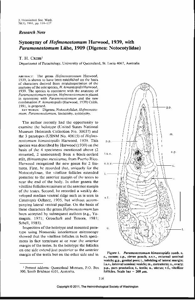
J. Helminthol. Soc. Wash.58(1), 1991, pp. 116-117
Research Note
Synonymy of Hofmonostomum Harwood, 1939, withParamonostomum Liihe, 1909 (Digenea: Notocotylidae)
T. H. CRIBB1
Department of Parasitology, University of Queensland, St. Lucia 4067, Australia
ABSTRACT: The genus Hofmonostomum Harwood,1939, is shown to have been established on the basisof characters derived from misinterpretation of theanatomy of the sole species, H. himantopidis Harwood,1939. The species is consistent with the anatomy ofParamonostomum species. Hofmonostomum is placedin synonymy with Paramonostomum and the newcombination P. himantopidis (Harwood, 1939) Cribb,1991, is proposed.
KEY WORDS: Digenea, Notocotylidae, Hofmonosto-mum, Paramonostomum, taxonomy, synonymy.
The author recently had the opportunity toexamine the holotype (United States NationalMuseum Helminth Collection No. 30037) andthe 3 paratypes (USNM No. 40615) of Hofmo-nostomum himantopidis Harwood, 1939. Thisspecies was described by Harwood (1939) on thebasis of the 4 specimens mentioned above (2mounted, 2 unmounted) from a black-neckedstilt, Himantopus mexicanus, from Puerto Rico.Harwood recognized the new genus for 2 fea-tures. First, he recorded that, uniquely for theNotocotylinae, the vitelline follicles extendedposterior to the anterior margin of the testes tonear the end of the body. In other genera thevitelline follicles terminate at the anterior marginof the testes. Second, he recorded a weakly de-veloped median ventral ridge such as is seen inCatatropis Odhner, 1905, but without accom-panying lateral ventral papillae. On the basis ofthese characters the genus Hofmonostomum hasbeen accepted by subsequent authors (e.g., Ya-maguti, 1971; Groschaft and Tenora, 1981;Schell, 1985).
Inspection of the holotype and mounted para-type using Nomarski interference microscopyshowed that the vitelline follicles in both speci-mens in fact terminate at or near the anteriormargin of the testes. In the holotype the follicleson one side extend just posterior to the anteriormargin of the testis but on the other side and in
g-p.
P.P.
c.p.
1 Present address: Queensland Museum, P.O. Box300, South Brisbane 4101, Australia.
Figure 1. Paramonostomum himantopidis comb. n.c., cecum; c.p., cirrus pouch; e.s.v., external seminalvesicle; g.p., genital pore; i., infolding of lateral margin;i.s.v., interval seminal vesicle; m., metraterm; o., ovary;p.p., pars prostatica; t., testis; u., uterus; v.f., vitellinefollicles. Scale bar = 200 fan.
116
Copyright © 2011, The Helminthological Society of WashingtonCopyright © 2011, The Helminthological Society of Washington

117
the mounted paratype they terminate close to theanterior margin of the testes. In the holotype,and to a lesser extent in the paratype, the lateralmargins of the specimen are curved inwards andinclude portions of the lobed margin of the testes.This evidently produced the impression of vi-telline follicles lateral to the testes which is, how-ever, clearly not the case. The mounted paratypeis figured here (Fig. 1) as it is the less ambiguousspecimen.
In neither mounted specimen was a medianventral ridge of the type seen in Catatropis spe-cies discerned. In the holotype the ventral surfaceis somewhat folded, and presumably as an arti-fact of fixation, has an irregular ridge visible inplaces. This does not appear to be a genuinestructure. No ridge was visible at all on themounted paratype. Given that Nomarski mi-croscopy was used, it is reasonable to believe thata ridge would have been observed if present.There was no sign of a median ridge on eitherof the unmounted paratypes which were exam-ined under a dissecting microscope.
In view of the observations outlined above,the 2 characters that were used to distinguishHofmonostomum are not valid. Alternative dis-tinguishing characters were not found. Conse-quently, the genus cannot be distinguished fromthe large, cosmopolitan genus ParamonostomumLiihe, 1909. This genus is characterized by thecomplete absence of ventral surface ridges andpapillae and has the vitelline follicles terminatingat the anterior margin of the testes. I thereforepropose that Hofmonostomum should be con-sidered a junior synonym of Paramonostomum
and that the new combination Paramonosto-mum himantopidis (Harwood, 1939)Cribb, 1991,be recognized.
The question of the validity of P. himantopidiswith respect to other Paramonostomum speciesis beyond the scope of the present study. Thegenus contains about 50 nominal species, manyof which are separated by only the most super-ficial of characters. It seems not at all improbablethat critical review will show P. himantopidis tobe either a junior or senior synonym of existingParamonostomum combinations. It is notewor-thy that P. himantopidis appears not to have beenrecorded since its original description (see Hi-nojos and Canaris, 1988).
Thanks are due to Dr. J. R. Lichtenfels of theUSNM for kindly lending type specimens.
Literature Cited
Groschaft, J., and F. Tenora. 1981. Reorganisationof suborder Notocotylata (Trematoda). ActaScientiarum Naturalium Academiae ScientiarumBohemoslovacae Brno 15:1—46.
Harwood, P. D. 1939. Notes on Tennessee helminths.IV. North American trematodes of the subfamilyNotocotylinae. Journal of the Tennessee Academyof Science 14:332-340, 421^37.
Hinojos, J. G., and A. G. Canaris. 1988. Metazoanparasites of Himantopus mexicanus Muller (Aves)from southwestern Texas, with a checklist of hel-minth parasites from North America. Journal ofParasitology 74:326-331.
Schell,S.C. 1985. Handbook of Trematodes of NorthAmerica North of Mexico. University Press ofIdaho, Moscow, Idaho. 263 pp.
Yamaguti, S. 1971. Synopsis of the Digenetic Trem-atodes of Vertebrates. Keigaku Publishing Co.,Tokyo. 1,074 pp.
Copyright © 2011, The Helminthological Society of WashingtonCopyright © 2011, The Helminthological Society of Washington

J. Helminthol. Soc. Wash.58(1), 1991, pp. 118-121
Research Note
The Response of Tubifex tubifex (Oligochaeta: Tubificidae)to a Second Infection with Glaridacris catostomi(Cestoidea: Caryophyllaeidae)
CHERYL C. COURTNEY1 AND BRUCE M. CHRisxENSEN2
1 Department of Fisheries and Wildlife, Utah State University, Logan, Utah 84322-5210 and2 Department of Veterinary Science, University of Wisconsin, Madison, Wisconsin 53706
ABSTRACT: The response of the aquatic oligochaete,Tubifex tubifex, to primary and secondary infection bythe caryophyllaeid cestode, Glaridacris catostomi, wasinvestigated by comparing rates of parasite mortalitybetween the 2 infections. Tubifex tubifex that lost thefirst infection of G. catostomi became infected aftersecond exposure to the cestode, but rate of parasitemortality did not increase. Over an 8-day period, bothcumulative parasite mortality and percentage of hoststhat lost their caryophyllaeid infection were greater forthe first infection (90% and 59%) than for the second(46% and 2%). A cellular response, involving the en-capsulation of metacestodes by host coelomocytes, ac-counted for part of the high caryophyllaeid mortalityobserved.
KEY WORDS: Cestoidea, Caryophyllidea, Tubifici-dae, metacestode.
Much of the literature on annelid immunityhas involved the response of terrestrial earth-worms to tissue grafts (Cooper, 1986), the coe-lomocytes active in the phagocytosis process(Stein et al., 1977), and characterization of an-tibacterial molecules (Valembois et al., 1986).Additionally, studies have been conducted onthe encapsulation of parasitic organisms in an-nelids. Poinar and Thomas (1975) described theencapsulation of the nematode, Rhabditis pellio,in the coelom of the earthworm, Aporrectodeatrapezoides, which involves the encasement ofthe parasite in host coelomocytes and formationof multiple capsules or "brown bodies." A sim-ilar reaction was observed by Calentine et al.(1970) in tubificid oligochaetes infected with car-yophyllaeid cestodes. Host coelomocytes at-tached to the metacestodes, but no "brown bod-ies" developed. This cellular response againstcaryophyllaeids was reported for a number oftubificids including Tubifex tubifex Miiller, 1774.This species was used in the present study tocompare rates of parasite mortality between afirst infection with Glaridacris catostomi Cooper,1920, and a second infection with the same car-yophyllaeid species.
Laboratory reared oligochaetes were experi-mentally infected by 24-hr exposure to embryo-nated cestode eggs in mud-free water (1,200-1,800 eggs per 25 ml of water), under continuousaeration, at a temperature range of 18-21°C. Ces-tode eggs were obtained from the laboratory ofRobert Calentine, University of Wisconsin-Riv-er Falls. Immediately after exposure to cestodeeggs, infected annelids were separated from un-infected ones. Infected annelids were identifiedby the presence of oncospheres in the coelom.All annelids used in experiments were main-tained under aeration in 100-ml glass beakerscontaining initially sterile mud and fed a diet ofTetramin® flaked fish food. At various dayspostexposure (PE), wet mounts of infected an-nelids were examined microscopically at lOOxand/or 430 x with bright field or phase contrastoptics and the number of cestodes present in thecoelom counted.
In 2 separate trials, 1 -wk-old T. tubifex wereexposed for the first time to G. catostomi. Theindividuals that became infected were designatedas group 1 (first infection). Approximately 2lh-wk-old oligochaetes in group 1 that lost theircestodes (the cestodes died) were reexposed asecond time to G. catostomi. The oligochaetesthat became reinfected were designated as group2 (second infection). At the same time as group2, another group of approximately 21/2-wk-oldannelids, designated the control group, was ex-posed to G. catostomi for the first time. Datawere combined from both trials and rates of par-asite mortality were compared among all 3 groups.
Cumulative host and parasite mortality andpercentage of hosts that lost their caryophyllaeidinfection were compared among the 3 groupsusing R x C contingency tables with chi-squareanalysis. Student's Mest was used to comparegroup means at the various days PE. Differencesin each of these analyses were considered signif-icant at P < 0.05.
118
Copyright © 2011, The Helminthological Society of WashingtonCopyright © 2011, The Helminthological Society of Washington

119
12-1
10-
DAYS PE
Figure 1. Mean parasite intensities of Glaridacris catostomi in Tubifex tubifex for group 1 (first infection),group 2 (second infection), and a control group (same age as group 2). Bars represent the standard error of themean. The numbers above each bar represent total number of parasites followed by total number of infected hostsin parentheses.
Overall, mean parasite intensity (Fig. 1) de-creased over time for each group (e.g., from 2.23at 1 day PE to 1.55 at 8 days PE in group 1, from10.59 at 1 day PE to 7.68 at 8 days PE in group2, and from 7.82 at 1 day PE to 6.38 at 8 daysPE in the control group). Mean parasite inten-sities were higher in the second infection com-pared to the first infection. Only at 1 day PE wasthe mean parasite intensity for the second infec-tion significantly larger than for the control group.
By 8 days PE, 90% of the cestodes were deadin group 1 hosts and 46% in group 2 (Fig. 2A).A significantly higher number of G. catostomi-infected T. tubifex lost their metacestodes in group1 compared to group 2, 59% and 2%, respectively(Fig. 2C). Only 2% of control oligochaetes wereno longer infected at 8 days PE (Fig. 2C). Hostmortality was similar for all 3 groups (Fig. 2B).
Evidence of immunologic memory in annelidswas provided by Cooper (1968) in his studies ontissue graft rejection in terrestrial earthworms.Secondary rejection of allografts and xenograftsby earthworms was specific and enhanced (Coo-per, 1968) and this memory was also transferra-ble with "sensitized" coelomocytes (Hosteller andCooper, 1974). We attempted to measure the
potential for an adaptive immune response inaquatic oligochaetes by using a natural parasiteof the annelid. However, on second exposure toG. catostomi in T. tubifex we did not find thatthe rate of parasite death increased. Thus, weconcluded that the response exhibited by thishost to the parasite is non-adaptive. Even after35 days, percentage of hosts that lost their in-fection after second exposure was still lower thanthat for controls (5% vs. 18%). Parasite mortalitywas significantly higher in controls (73%) thanin group 2 (61%) at 35 days PE. There appearsto be no difference between susceptibility to in-fection on second exposure to G. catostomi forT. tubifex that previously lost the infection andthose still infected with the parasite (see Calen-tine, 1967).
The high rate of parasite mortality seen in thisstudy can be partially explained by a cellular re-sponse. We observed the encapsulation reactionin G. catostomi-inkcted T. tubifex that was re-ported by Calentine et al. (1970). Host coelomo-cytes attached to the metacestode and eventuallyencased it. Because of the infrequency of the re-sponse (observed 4 times), and because we ob-served the death of older and larger metacestodes
Copyright © 2011, The Helminthological Society of WashingtonCopyright © 2011, The Helminthological Society of Washington

120 JOURNAL OF THE HELMINTHOLOGICAL SOCIETY
100-
80-
60-
40-
20-
0
D GROUP 10 GROUP 2m CONTROL
2 4
DAYS PE
100-
80-
60-
40-
20-
0
DAYS PE
2 100 i
o£ 80-z,_ 60-
2 40-
CO
& 2°-oI n . LL
I-.— i
M
-
M
DAYS PE
Figure 2. Percent cumulative parasite mortality (A),host mortality (B), and hosts that lost their caryophyl-laeid infection (C) in Glaridacris catostomi-infected Tu-bifex tubifex for group 1 (first infection), group 2 (sec-ond infection), and a control group (same age as group2).
without attachment of host cells, we believe thatthe encapsulation reaction alone is not the causeof the high parasite mortality seen in this study.Other mechanisms may be operating in this host-parasite system. The possible role of humoralimmune components interacting with the coe-lomocyte population should be investigated.
Terrestrial earthworms possess humoral fac-tors that are capable of agglutination and opso-nization. A natural hemolysin of Eisenia fetidaandrei shows lytic activity against sheep red blood
cells (Roch, 1979) as well as antibacterial activitytoward pathogenic soil bacteria (Lassegues et al.,1981; Valembois et al., 1982). Valembois et al.(1986) characterized the antibacterial moleculesin this earthworm that are responsible for neu-tralization of pathogenic bacteria. Four proteinsmediated hemolytic activity in the coelomic fluidbut only 1 protein was involved in this processin the cocoon albumen. Stein and Cooper (1981)found that phagocytosis of yeast by neutrophilsof Lumbricus terrestris was enhanced when theyeast were first washed with cell-free coelomicfluid. Whether the coelomic fluid of aquatic oli-gochaetes has similar immune capabilities is notknown.
Age is an important component of the G. ca-tostomi-T. tubifex association (Courtney andChristensen, 1988). Most infected tubificids donot become sexually mature (Sekutowicz, 1934;Kulakovskaya, 1962; Calentine, 1965, 1967) orthe breeding period is delayed (Kennedy, 1969).Since young annelids are more susceptible to in-fection, the caryophyllaeid may be applying somemechanism that delays sexual maturity (e.g., lib-erating molecules that inhibit maturation of thegonads). A delay in host maturity could also berelated to the depletion of host nutrients by theparasites. As a consequence of this, there is notenough energy available for reproductive growth.Additionally, the amount of space utilized bythese cestodes may physically obstruct the de-velopment of the gonads. Thirty-day-old T. tu-bifex can have a body length of 25 mm or more(Kosiorek, 1974). The average length of an in-fective metacestode of G. catostomi is 1.2 mm(Calentine, 1967).
Equally important in the present study is theinitial parasite intensity that can enhance thesuitability of T. tubifex as a host (see Courtneyand Christensen, 1987). When T. tubifex wassubjected to a second exposure to G. catostomi,the initial parasite burden was higher and thepercentage of hosts that lost their infection lower.If this phenomenon also occurs in tubificids innature, then caryophyllaeids that parasitize pre-viously infected hosts have a much higher prob-ability of surviving to the infective stage. Anincreased susceptibility to infection can result ina higher parasite burden.
The increased susceptibility to caryophyllaeidinfection could be a consequence of a reductionin the number of coelomocytes that occurred inthe first infection. A reduced coelomocyte pop-
Copyright © 2011, The Helminthological Society of WashingtonCopyright © 2011, The Helminthological Society of Washington

OF WASHINGTON, VOLUME 58, NUMBER 1, JANUARY 1991 121
ulation in conjunction with depletion of humoralfactors may account for a greater susceptibilityto infection in group 2 compared to age-matchedcontrols. In the snail, Helix pomatia, a decreasein the number of circulating blood cells was seenafter injection of foreign particles. Additionally,the number of circulating blood cells can differin snails of different ages with the number in-creasing in larger snails (Siminia, 1981). Furtherresearch is needed to determine the physiologicaldifferences between younger (more susceptible)annelids and older (less susceptible) annelids, andthe immunocompetence of each.
We gratefully acknowledge Robert L. Calen-tine for providing the initial cultures and cestodeeggs used in these studies. We also thank DanielSutherland for reviewing the manuscript. Thisresearch was supported by the College of Agri-culture and Life Sciences, University of Wiscon-sin-Madison.
Literature Cited
Calentine, R. L. 1965. The biology and taxonomy ofBiacetabulum (Cestoda: Caryophyllaeidae). Jour-nal of Parasitology 51:243-248.
. 1967. Larval development of four caryophyl-laeid cestodes. Iowa Academy of Science 72:418-424.
-, B. M. Christensen, and L. A. Christensen.1970. Specificity of caryophyllaeid cestodes fortheir intermediate hosts. Journal of Parasitology56:346-349.
Cooper, E. L. 1968. Transplantation immunity inannelids. I. Rejection of xenografts exchanged be-tween Lumbricus terrestris and Eisenia foetida.Transplantation 6:322-337.
. 1986. Evolution of histoincompatibility.Pages 139-150 in M. Brehelin, ed. Immunity inInvertebrates: Cells, Molecules, and Defense Re-actions. Springer-Verlag, Berlin.
Courtney, C. C., and B. M. Christensen. 1987. Host-parasite relationships of caryophyllaeid cestodesand aquatic oligochaetes: I. Host longevity andparasite intensity. Journal of Parasitology 73:1124—1132.
, and B. M. Christensen. 1988. Host-parasiterelationships of caryophyllaeid cestodes and aquaticoligochaetes: II. Effects of host age and mixed in-fections. Journal of Parasitology 74:573-581.
Hostetter, R. K., and E. L. Cooper. 1974. Earthwormcoelomocyte immunity. Pages 91-107 in E. L.Cooper, ed. Contemporary Topics in Immuno-biology. Invertebrate Immunology. Vol. 4. Ple-num Press, New York.
Kennedy, C. R. 1969. Seasonal incidence and devel-opment of the cestode Caryophyllaeus laticeps(Pallas) in the River Avon. Parasitology 59:783-794.
Kosiorek, D. 1974. Development cycle of Tubifextubifex Miill. in experimental culture. Polskie Ar-chiwum Hydrobiologii 21:411-422.
Kulakovskaya, O. P. 1962. Development of Cary-ophyllaeidae (Cestoda) in an intermediate host.Zoologicheskii Zhurnal 41:986-992. (Russian text;English summary.)
Lassegues, M., P. Roch, P. Valembois, and N. Davant.1981. Action de quelques souches bacteriennestelluriques sur le Lombricien Eisenia fetida andrei.Compte Rendus de 1'Academie des Sciences ParisD 292:731-734.
Poinar, G. O., and G. M. Thomas. 1975. Rhabditispellio Schneider (Nematoda) from the earthworm,Aporrectodea trapezoides Duges (Annelida). Jour-nal of Nematology 7:374-379.
Roch, P. 1979. Protein analysis of earthworm coe-lomic fluid: 1) Polymorphic system of the naturalhemolysis of Eisenia fetida andrei. Developmentaland Comparative Immunology 3:599-608.
Sekutowicz, St. 1934. Untersuchungen zur Entwick-lung und Biologic von Caryophyllaeus laticeps(Pall.). Memoires de 1'Academie Polonaise desSciences et des Lettres, Serie B, Science Naturelles6:11-26.
Siminia, T. 1981. Gastropods. Pages 191-232 in N.A. Ratcliffe and A. F. Rowley, eds. InvertebrateBlood Cells. Vol. 1. Academic Press, Inc., London.
Stein, E. A., and E. L. Cooper. 1981. The role ofopsonins in phagocytosis by coelomocytes of theearthworm, Lumbricus terrestris. Developmentaland Comparative Immunology 5:415-425.
, R. R. Avtalion, and E. L. Cooper. 1977. Thecoelomocytes of the earthworm Lumbricus terres-tris: morphology and phagocytic properties. Jour-nal of Morphology 153:476-477.
Valembois, P., P. Roch, and M. Lassegues. 1986. An-tibacterial molecules in annelids. Pages 74-93 inM. Brehelin, ed. Immunity in Invertebrates: Cells,Molecules, and Defense Reactions. Springer-Ver-lag, Berlin.
, , , and P. Cassand. 1982. An-tibacterial activity of the hemolytic system fromthe earthworm Eisenia fetida andrei. Journal ofInvertebrate Pathology 40:21-27.
Copyright © 2011, The Helminthological Society of WashingtonCopyright © 2011, The Helminthological Society of Washington

J. Helminthol. Soc. Wash.58(1), 1991, pp. 122-125
Research Note
Differences between the Energy Metabolism of Trichinella spiralisand Trichinella pseudospiralis
KRYSTYNA BOCZONDepartment of Biology and Medical Parasitology, Karol Marcinkowski Academy of Medicine,Fredry 10, 61-701, Poznan, Poland
ABSTRACT: Comparative biochemical investigationsshowed the existence of diiferences between the en-zyme kinetics of Trichinella spiralis and Trichinellapseudospiralis. The higher affinity of succinate dehy-drogenase-fumarate reductase complex to fumarate inTrichinella pseudospiralis is discussed in relation to thebranched anaerobic-type electron transport systemsuggested in this parasite.
KEY WORDS: Nematoda, Trichinella spiralis, Trich-inella pseudospiralis, enzyme kinetics.
At present, the genus Trichinella appears morecomplex than was thought in the past, and com-parative biochemical investigations could behelpful in understanding its existing diversity.
The larvae of Trichinella pseudospiralis, whichin contrast to Trichinella spiralis larvae do notbecome "trapped" in host muscles due to en-capsulation, move continously, thus altering theentire muscle fiber of the host (Hulinska et al.,1984). So the differences in bioenergetic metab-olism between the motile, unencapsulated T.pseudospiralis larvae and motionless encapsu-lated T. spiralis seem to be especially interesting.
Considering the electrophoretic analysis of tis-sue protein as a taxonomic tool, a comparativestudy of the soluble protein profile of 7 Trichi-nella isolates was performed (Fukumoto et al.,1987). They observed that the pattern of 6 of theisolates clearly differed from that of T. pseudospi-ralis. From the zymograms of 8 enzymes inves-tigated in the same study, 7 of them in T. pseu-dospiralis were clearly different from otherisolates. More recently, Pozio et al. (1989) pre-sented a study of a gene-enzyme system and itshowed a high genetic discontinuity in the genusTrichinella with 7 distinct gene pools. In addi-tion, analysis of the principal components of 51Trichinella isolates and 5 biological and physicalvariables confirmed the presence of specific clus-ters among species in the genus Trichinella (LaRosaetal., 1989).
Generally speaking, T. pseudospiralis larvaeseem to stimulate a weaker immunological re-sponse by the host, show a lower metabolic ac-
tivity of some glycolytic enzymes (Boczon, 1986),and their excretory-secretory products uncouplethe host muscle mitochondria during the encys-tation phase of muscle infection to a lesser extentthan T. spiralis excretory-secretory products(Boczon et al., 1989).
Recent biochemical investigations on meta-bolic differences between T. spiralis and T. pseu-dospiralis indicated similarities in their suscep-tibility to chemotherapeutic attack (Boczon etal., 1984) and in their prostaglandin content(Hadas and Staude-Adamczewska, 1990), but dif-ferences in their bioenergetic metabolism. Forexample, T. pseudospiralis mitochondria, in con-trast to T. spiralis, have a cytochrome oxidase(OX) that is partially insensitive to KCN, butmitochondria that are poorly coupled and ableto oxidize exogenous NADH. All these featuresmay suggest the presence of an alternative path-way in the electron transport system of T. pseu-dospiralis (Boczon, 1985). Qualitative and quan-titative comparative determinations of quinonesrevealed about 4 times greater amount of bothubiquinone and rhodoquinone in T. pseudospi-ralis than in T. spiralis (Boczon, 1985).
This report presents the results of kinetic mea-surements of succinate dehydrogenase-fumaratereductase (SDH-FR) complex and some cyto-chrome c oxidoreductases in mitochondria of T.pseudospiralis larvae and compares these datawith those obtained in T. spiralis.
Larvae of T. pseudospiralis Garkavi, 1974,originally isolated in USSR from a raccoon, wereisolated from rat muscles by pepsin digestion(Gursch, 1948) lasting 1 hr. After several wash-ings in water and 0.9% NaCl, larvae were ho-mogenized in sucrose (0.25 M, Tris-HCl 0.03 M,pH 7.3) medium. The mitochondrial fraction wasobtained and the activities of cytochrome c ox-idoreductases were measured as described pre-viously (Hryniewiecka et al., 1970; Boczon andMichejda, 1978). For the measurements of SDHand FR activity the mitochondria were frozenand thawed 3 times. The SDH (EC 1.3.99.1) ac-
122
Copyright © 2011, The Helminthological Society of WashingtonCopyright © 2011, The Helminthological Society of Washington
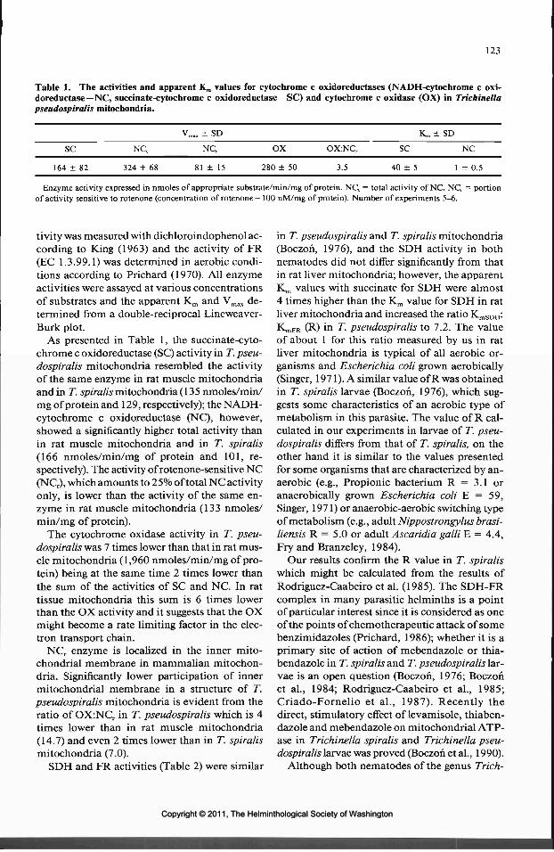
123
Table 1. The activities and apparent Km values for cytochrome c oxidoreductases (NADH-cytochrome c oxi-doreductase—NC, succinate-cytochrome c oxidoreductase—SC) and cytochrome c oxidase (OX) in Trichinellapseudospiralis mitochondria.
SC NC,
164 ± 82 324 ± 68
Vm.x ± SD
NCr OX
81 ± 15 280 ± 50
OX:NCr
3.5
K ™ :
SC
40 ± 5
t SD
NC
1 ± 0.5
Enzyme activity expressed in nmoles of appropriate substrate/min/mg of protein. NC, = total activity of NC. NCr = portionof activity sensitive to rotenone (concentration of rotenone—100 nM/mg of protein). Number of experiments 5-6.
tivity was measured with dichloroindophenol ac-cording to King (1963) and the activity of FR(EC 1.3.99.1) was determined in aerobic condi-tions according to Prichard (1970). All enzymeactivities were assayed at various concentrationsof substrates and the apparent Km and Vmax de-termined from a double-reciprocal Lineweaver-Burk plot.
As presented in Table 1, the succinate-cyto-chrome c oxidoreductase (SC) activity in T. pseu-dospiralis mitochondria resembled the activityof the same enzyme in rat muscle mitochondriaand in T. spiralis mitochondria (135 nmoles/min/mg of protein and 129, respectively); the NADH-cytochrome c oxidoreductase (NC), however,showed a significantly higher total activity thanin rat muscle mitochondria and in T. spiralis(166 nmoles/min/mg of protein and 101, re-spectively). The activity of rotenone-sensitive NC(NCr), which amounts to 25% of total NC activityonly, is lower than the activity of the same en-zyme in rat muscle mitochondria (133 nmoles/min/mg of protein).
The cytochrome oxidase activity in T. pseu-dospiralis was 7 times lower than that in rat mus-cle mitochondria (1,960 nmoles/min/mg of pro-tein) being at the same time 2 times lower thanthe sum of the activities of SC and NC. In rattissue mitochondria this sum is 6 times lowerthan the OX activity and it suggests that the OXmight become a rate limiting factor in the elec-tron transport chain.
NCr enzyme is localized in the inner mito-chondrial membrane in mammalian mitochon-dria. Significantly lower participation of innermitochondrial membrane in a structure of T.pseudospiralis mitochondria is evident from theratio of OX:NCr in T. pseudospiralis which is 4times lower than in rat muscle mitochondria(14.7) and even 2 times lower than in T. spiralismitochondria (7.0).
SDH and FR activities (Table 2) were similar
in T. pseudospiralis and T. spiralis mitochondria(Boczon, 1976), and the SDH activity in bothnematodes did not differ significantly from thatin rat liver mitochondria; however, the apparentKm values with succinate for SDH were almost4 times higher than the K,,, value for SDH in ratliver mitochondria and increased the ratio K^^:K^PR (R) in T. pseudospiralis to 7.2. The valueof about 1 for this ratio measured by us in ratliver mitochondria is typical of all aerobic or-ganisms and Escherichia coli grown aerobically(Singer, 1971). A similar value of R was obtainedin T. spiralis larvae (Boczon, 1976), which sug-gests some characteristics of an aerobic type ofmetabolism in this parasite. The value of R cal-culated in our experiments in larvae of T. pseu-dospiralis differs from that of T. spiralis, on theother hand it is similar to the values presentedfor some organisms that are characterized by an-aerobic (e.g., Propionic bacterium R = 3.1 oranaerobically grown Escherichia coli E = 59,Singer, 1971) or anaerobic-aerobic switching typeof metabolism (e.g., adult Nippostrongylus brasi-liensis R = 5.0 or adult Ascaridia galli E = 4.4,Fry and Branzeley, 1984).
Our results confirm the R value in T. spiraliswhich might be calculated from the results ofRodriguez-Caabeiro et al. (1985). The SDH-FRcomplex in many parasitic helminths is a pointof particular interest since it is considered as oneof the points of chemotherapeutic attack of somebenzimidazoles (Prichard, 1986); whether it is aprimary site of action of mebendazole or thia-bendazole in T. spiralis and T. pseudospiralis lar-vae is an open question (Boczon, 1976; Boczonet al., 1984; Rodriguez-Caabeiro et al., 1985;Criado-Fornelio et al., 1987). Recently thedirect, stimulatory effect of levamisole, thiaben-dazole and mebendazole on mitochondrial ATP-ase in Trichinella spiralis and Trichinella pseu-dospiralis larvae was proved (Boczon et al., 1990).
Although both nematodes of the genus Trich-
Copyright © 2011, The Helminthological Society of WashingtonCopyright © 2011, The Helminthological Society of Washington

124 JOURNAL OF THE HELMINTHOLOGICAL SOCIETY
Table 2. The comparison of the V^ and apparent Km
values of succinate dehydrogenase (SDH) and fumaratereductase (FR) in Trichinella pseudospiralis.
SDH ± SD FR ± SD
51 ± 1329 + 6
7.0 ± 1.84.0 ± 1.0 7.2
All explanations as in Table 1.
inella live in the same well-oxygenated biotopemuscles, their mitochondria seem to differ in anumber of bioenergetic features: 1) T. pseudospi-ralis mitochondria in comparison to T. spiralismitochondria revealed lower participation of theinner mitochondrial membrane in their function,which may result in loose coupling of oxidativephosphorylation, 2) in T. pseudospiralis a partialinsensitivity of OX to KCN and oxidation of theexogenous NADH may suggest the participationof an alternative pathway in energy generation(Boczon, 1985), 3) a high affinity of SDH-FRcomplex to fumarate and low affinity to succinatemay be indicative of the greater involvment inT. pseudospiralis than in T. spiralis in the gen-eration of some A^ by site I of oxidative phos-phorylation due to the activity of FR, despite thesame activity of FR in both nematodes, and ma-lic enzyme (Ward et al., 1969; Boczon, 1986).
Some participation of fumarate reductase inthe generation of the transmembrane potentialAi^ by site I of oxidative phosphorylation withoutinvolvment of oxygen, as proved in Ascaris byChojnicki et al. (1987), was suggested recently inT. spiralis by Michejda et al. (1989). Those re-sults, obtained with T. spiralis, support our pre-vious view (Boczon, 1985) that the respiratorymetabolism of T. spiralis larvae, although beingmuch more aerobic than that of Ascaris, mayalso partially involve the anaerobic generationof Ai/' by site I of oxidative phosphorylation dueto the activity of FR and malic enzyme, the sys-tem operating anaerobically in most helminths.
This report was published with the support ofthe Brayton H. Ransom Memorial Trust Fund.
Literature Cited
Boczon, K. 1976. Bioenergetics of Trichinella spiralislarvae and effect of some anthelmintics on succi-nate dehydrogenase of Trichinella spiralis mito-chondria. Pages 589-597 in H. van den Bossche,ed. Second International Symposium on the Bio-chemistry of Parasites and Host-Parasite Rela-tionship. Elsevier/North Holland Biomedical Press,Amsterdam.
—. 1985. Bioenergetics of Trichinella spiralisspiralis and Trichinella spiralis pseudospiralis.Wiadomosci Parazytologiczne 31:237-251.
—. 1986. The role of malic enzyme in the car-bohydrate metabolism of Trichinella spiralis spi-ralis and Trichinella spiralis pseudospiralis. Inter-national Journal for Parasitology 16:435—440.
—, and J. W. Michejda. 1978. Electron transportin mitochondria of Trichinella spiralis larvae. In-ternational Journal for Parasitology 8:507-513.
—, W. Olba, and M. Olaszek. 1984. The influ-ence of some anthelmintics on the bioenergeticmetabolism of Trichinella spiralis and Trichinellapseudospiralis. Biochemical Pharmacology 33:2523-2525.
—, Z. Uszynska, F. Rodriguez-Caabeiro, A. Cria-do-Fornelio, and C. de Armas-Serra. 1990. Mi-tochondrial ATP-ase a target point of anthelmin-tics in Trichinella spiralis and Trichinellapseudospiralis. Journal of Parasitology 76. (Inpress.)
-, E. Wandurska, and K. Imilkowska. 1989.Mitochondrial ATP-ase as a measure of uncoup-ling of rat muscle mitochondria in experimentalinfection of Trichinella spiralis and Trichinellapseudospiralis. Wiadomosci Parazytologiczne 35:435-448.
Chojnicki, K., M. Budzynska, P. Wolanska, and J. W.Michejda. 1987. Membrane potential in mito-chondria of Ascaris. Acta Parasitologica Polonica32:67-78.
Criado-Fornelio, A., F. Rodriguez-Caabeiro, and A. Ji-menez-Gonzalez. 1987. The mode of action ofsome benzimidazole drugs on Trichinella spiralis.Parasitology 95:61-70.
Fry, M., and E. P. Branzeley. 1984. NADH fumaratereductase and succinate dehydrogenase in mito-chondria of Ascaridia galli and Nippostrongylusbrasiliensis. Comparative Biochemistry and Phys-iology 77:143-151.
Fukumoto, S., M. Takechi, H. Kamo, and T. Yama-guchi. 1987. Comparative studies on solubleprotein profiles and isozyme patterns of sevenTrichinella isolates. Parasitology Research 73:352-357.
Gursch, O. F. 1948. Effect of digestion and refrig-eration on the ability of Trichinella spiralis to in-fect rats. Journal of Parasitology 34:394-395.
Hadas, E., and M. Stande-Adamczewska. 1990.Prostaglandins of Trichinella spiralis and Trichi-nella pseudospiralis. Acta Parasitologica Polonica35. (In press.)
Hryniewiecka, L., K. Boczon, and J. W. Michejda.1970. Trichinella spiralis: enzymes reducing andoxidizing cytochrome c in rat muscles. Experi-mental Parasitology 28:544-550.
Hulinska, D., M. Grim, and B. M. MacKinnon. 1984.Alterations of muscle fibers of mice experimen-tally infected with Trichinella spiralis, Trichinellapseudospiralis and Trichinella nativa. Pages 157-163 in Proceedings of the Sixth International Con-ference on Trichinellosis. Val Morin, Canada.
King, E. 1963. Reconstitution of respiratory chainenzyme system. The Journal of Biological Chem-istry 12:4032^036.
La Rosa, G., E. Pozio, and P. Rossi. 1989. New tax-
Copyright © 2011, The Helminthological Society of WashingtonCopyright © 2011, The Helminthological Society of Washington

OF WASHINGTON, VOLUME 58, NUMBER 1, JANUARY 1991 125
onomic contribution to the genus Trichinella Rail-liet, 1895. II. Multivariate analysis on genetic andbiological data. Pages 83-88 in C. E. Tanner, A.Martinez-Fernandez, and F. Bolas-Fernandez, eds.Trichinellosis: Proceedings of the Seventh Inter-national Conference on Trichinellosis. ConsejoSuperior de Investigaciones Cientificas Press, Ma-drid.
Michejda, J. W., K. Chojnicki, and K. Boczon, 1989.Energy transduction in mitochondria of Trichi-nella spiralis. Acta Parasitologica Polonica 34:76-86.
Pozio, E., G. La Rosa, P. Rossi, and K. D. Murrell.1989. New taxonomic contribution to the genusTrichinella (Owen, 1835). I. Biochemical identi-fication of seven clusters by gene-enzyme systems.Pages 76-82 in C. E. Tanner, A. Martinez-Fer-nandez, and F. Bolas-Fernandez, eds. Trichinel-losis: Proceedings of the Seventh InternationalConference on Trichinellosis. Consejo Superior deInvestigaciones Cientificas Press, Madrid.
Prichard, R. K. 1970. Mode of action of the anthel-mintic thiabendazole in Haemonchus contortus.Nature 228:684-685.
. 1986. The pharmacology of anthelmintics inlivestock. Pages 473^82 in M. J. Howell, ed. Par-asitology Quo Vadit?. ICOPA VI, Brisbane, Aus-tralia, Australian Academy of Sciences.
Rodriguez-Caabeiro, F., A. Criado-Fornelio, and A. Ji-menez-Gonzalez. 1985. A comparative study ofthe succinate dehydrogenase-fumarate reductasecomplex in the genus Trichinella. Parasitology 91:577-583.
Singer, T. P. 1971. Evolution of the respiratory chainand its flavoproteins. Pages 203-219 in E. Schof-feniels, ed. Biochemical Evolution and Origin ofLife. North Holland Publishing Company, Am-sterdam, New York, Oxford.
Ward, C. W., G. A. Castro, and D. Fairbain. 1969.Carbon dioxide fixation and phosphoenolpyru-vate metabolism in Trichinella spiralis larvae.Journal of Parasitology 55:67-81.
J. Helminthol. Soc. Wash.58(1), 1991, pp. 125-127
Research Note
Differentiation between Mixed Infections of Ancylostoma caninumand Ancylostoma duodenale in Dogs Using an In Vitro Assayfor the Resumption of Feeding by Third-stage Infective Larvae
J. M. HAWDON AND G. A. SCHADDepartment of Pathobiology, University of Pennsylvania, Philadelphia, Pennsylvania 19104
ABSTRACT: The identification of 2 mixed hookworminfections in dogs by using differences of larval in vitrofeeding behavior is reported. In the first case, infectivethird-stage larvae (L3) from a putative Ancylostomaduodenale infection were compared to larvae of knownA. duodenale and A. caninum infections in a resump-tion of feeding time course experiment. The secondcase involved comparison of feeding behavior of L3'sfrom a second suspect infection with L3's from a knownA. caninum infection at 24 hr. In both cases, the suspectlarvae exhibited feeding behavior similar to known A.caninum larvae, suggesting accidental infection with A.caninum. Upon necropsy, the suspect infections con-tained adults of both species. This technique has po-tential application in differentiation of closely relatednematode species in several areas of parasitology.
KEY WORDS: Ancylostoma caninum, Ancylostomaduodenale, infective larvae, in vitro feeding.
As long ago as 1924, epidemiologists had hopedto distinguish among the infective larvae of thedog hookworm, Ancylostoma caninum (Erco-lani, 1859), and the human hookworms, Ancy-lostoma duodenale (Dubini, 1843) and Necator
americanus (Stiles, 1903). They concluded thatalthough N. americanus is distinguishable fromthe others on morphological grounds, the 2 An-cylostoma species are not (Schuurmans Stekho-ven and Schuurmans Stekhoven-Meyer, 1924;Komiya and Yasuraoka, 1966). In our labora-tory, both species of Ancylostoma have beenmaintained in dogs for more than 15 yr (Schad,1979), and on rare occasions donor animals haveinadvertently been infected with larvae of thewrong species, or with a mixed inoculum. Untilrecently, dogs harboring putatively pure infec-tions, but with atypical prepatent periods or eggcounts, had to be sacrificed, and the adult hook-worms recovered and examined morphologi-cally to determine the true nature of the infection.This is a very expensive procedure due to thehigh cost of purpose-bred dogs. A reliable meth-od of distinguishing between the larvae of the 2hookworms would obviate the unnecessary ter-mination of an infection. We have found that
Copyright © 2011, The Helminthological Society of WashingtonCopyright © 2011, The Helminthological Society of Washington
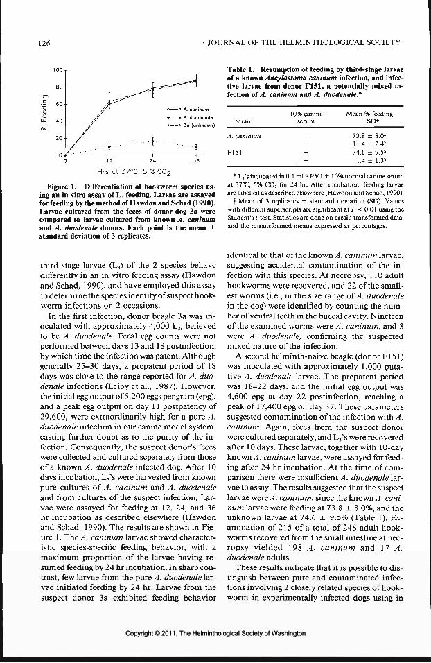
126 JOURNAL OF THE HELMINTHOLOGICAL SOCIETY
100-
80-
60-
40-
20-
Table 1. Resumption of feeding by third-stage larvae_ ^ J of a known Ancylostoma caninum infection, and infec-
^ I tive larvae from donor F151, a potentially mixed in-T ^̂ --̂ ""̂ I fection of A. caninum and A. duodenale*
// 1 • • I A duo'Zl 10% CaninC Mean % feedin8/ . , , . Strain serum ± SDf
// » » 3d (unknown)
/
, A. caninum + 73.8. . ! . . - • • ^ j - 11.4
F151 + 74.60 12 24 36 - 1.4
± 8.0-± 2.4"+ 9.5"± 1.3"
Mrs at 37°C, 5 % C02
Figure 1. Differentiation of hookworm species us-ing an in vitro assay of L3 feeding. Larvae are assayedfor feeding by the method of Hawdon and Schad (1990).Larvae cultured from the feces of donor dog 3a werecompared to larvae cultured from known A. caninumand A. duodenale donors. Each point is the mean ±standard deviation of 3 replicates.
third-stage larvae (L3) of the 2 species behavedifferently in an in vitro feeding assay (Hawdonand Schad, 1990), and have employed this assayto determine the species identity of suspect hook-worm infections on 2 occasions.
In the first infection, donor beagle 3a was in-oculated with approximately 4,000 L3, believedto be A. duodenale. Fecal egg counts were notperformed between days 13 and 18 postinfection,by which time the infection was patent. Althoughgenerally 25-30 days, a prepatent period of 18days was close to the range reported for A. duo-denale infections (Leiby et al., 1987). However,the initial egg output of 5,200 eggs per gram (epg),and a peak egg output on day 11 postpatency of29,600, were extraordinarily high for a pure A.duodenale infection in our canine model system,casting further doubt as to the purity of the in-fection. Consequently, the suspect donor's feceswere collected and cultured separately from thoseof a known A. duodenale infected dog. After 10days incubation, L3's were harvested from knownpure cultures of A. caninum and A. duodenaleand from cultures of the suspect infection. Lar-vae were assayed for feeding at 12, 24, and 36hr incubation as described elsewhere (Hawdonand Schad, 1990). The results are shown in Fig-ure 1. The A. caninum larvae showed character-istic species-specific feeding behavior, with amaximum proportion of the larvae having re-sumed feeding by 24 hr incubation. In sharp con-trast, few larvae from the pure A. duodenale lar-vae initiated feeding by 24 hr. Larvae from thesuspect donor 3 a exhibited feeding behavior
* L3's incubated in 0.1 ml RPMI ±10% normal canine serumat 37°C, 5% CO2 for 24 hr. After incubation, feeding larvaeare labelled as described elsewhere (Hawdon and Schad, 1990).
f Mean of 3 replicates ± standard deviation (SD). Valueswith different superscripts are significant at P < 0.01 using theStudent's Mest. Statistics are done on arcsin transformed data,and the retransformed means expressed as percentages.
identical to that of the known A. caninum larvae,suggesting accidental contamination of the in-fection with this species. At necropsy, 110 adulthookworms were recovered, and 22 of the small-est worms (i.e., in the size range of A. duodenalein the dog) were identified by counting the num-ber of ventral teeth in the buccal cavity. Nineteenof the examined worms were A. caninum, and 3were A. duodenale, confirming the suspectedmixed nature of the infection.
A second helminth-naive beagle (donor Fl 51)was inoculated with approximately 1,000 puta-tive A. duodenale larvae. The prepatent periodwas 18-22 days, and the initial egg output was4,600 epg at day 22 postinfection, reaching apeak of 17,400 epg on day 37. These parameterssuggested contamination of the infection with A.caninum. Again, feces from the suspect donorwere cultured separately, and L3's were recoveredafter 10 days. These larvae, together with 10-dayknown A. caninum larvae, were assayed for feed-ing after 24 hr incubation. At the time of com-parison there were insufficient A. duodenale lar-vae to assay. The results suggested that the suspectlarvae were A. caninum, since the known A. cani-num larvae were feeding at 73.8 ± 8.0%, and theunknown larvae at 74.6 ± 9.5% (Table 1). Ex-amination of 215 of a total of 248 adult hook-worms recovered from the small intestine at nec-ropsy yielded 198 A. caninum and 17 A.duodenale adults.
These results indicate that it is possible to dis-tinguish between pure and contaminated infec-tions involving 2 closely related species of hook-worm in experimentally infected dogs using in
Copyright © 2011, The Helminthological Society of WashingtonCopyright © 2011, The Helminthological Society of Washington

OF WASHINGTON, VOLUME 58, NUMBER 1, JANUARY 1991 127
vitro feeding as an assay. Since, to our knowl-edge, A. duodenale is maintained in dogs at only2 locations worldwide (the other being the In-stitute of Parasitic Diseases, Hangzhou, Peoples'Republic of China), the need for such a techniquein a laboratory situation is probably uncommon.However, this technique may prove useful in ep-idemiological and epizootological studies. Forexample, it may be possible to identify mixedinfections of the sympatric human hookwormsA. duodenale and A. ceylanicum (Looss, 1911),whose larvae are morphologically very similar(Yoshida, 1971), based on in vitro differences infeeding behavior. In the cases reported here,where 1 species was more successful in estab-lishing in the host than the other, feeding be-havior resembled that of the dominant species.However, when co-occurring parasites are sim-ilarly vigorous, the feeding behavior of larvalpopulations derived from a mixed infectionshould be intermediate. Secondly, this techniquemay be useful in field situations where soil ecol-ogists or epidemiologists have no practical meth-od to distinguish among the common congenericancylostomes (A. duodenale, A. caninum, A. tu-baeforme Zeder, 1800, and A. braziliense de Fa-ria, 1910) of humans and their usual companionanimals. In industrialized countries, this is par-ticularly important in parks, playgrounds, andother public open spaces, where irresponsible petownership with attendant fecal pollution raisesthe possibility of invasion by nematode larvaeassociated with cutaneous and visceral larvamigrans in humans. Finally, adaptation of thistechnique to other parasites may aid in the non-destructive differentiation of closely related spe-cies inhabiting the same host.
These experiments also suggest that the 2 An-cylostoma species may resume feeding in re-sponse to different host signals during infection,or more likely, that the larvae respond to a spe-cies-specific host stimulus. Although this strainof A. duodenale is adapted to the dog (Leiby etal., 1987), larvae may require a signal specific tohuman serum in order for a large proportion ofthem to resume feeding in vitro. Comparativeinvestigations of the resumption of feeding inlarvae of various hookworm species are currentlyunderway in this laboratory.
We thank Lan Han for expert technical assis-tance in maintaining the parasites. This work wassupported in part by NIH grant AI-22662.
Literature Cited
Hawdon, J. M., and G. A. Schad. 1990. Serum stim-ulated feeding in vitro by third stage infective lar-vae of the canine hookworm Ancylostoma cani-num. Journal of Parasitology 76:394-398.
Komiya, Y., and K. Yasuraoka. 1966. The biology ofhookworms. Progress of Medical Parasitology inJapan 3:1-86.
Leiby, D. A., H. M. S. El Naggar, and G. A. Schad.1987. Thirty generations of Ancylostoma duo-denale in laboratory-reared beagles. Journal ofParasitology 73:844-848.
Schad, G. A. 1979. Ancylostoma duodenale: main-tenance through six generations in helminth-naivepups. Experimental Parasitology 47:246-253.
Schuurmans Stekhoven, J. H., and A. W. SchuurmansStekhoven-Meyer. 1924. Voorbereidende on-derzoekingen met betrekking tot de mijnwormenvan mensch en bond. Tijdschrift vergelijkendeGeneeskunde: 101-119.
Yoshida, Y. 1971. Comparative studies on Ancylos-toma braziliense and Ancylostoma ceylanicum. II.The infective larval stage. Journal of Parasitology57:990-992.
Copyright © 2011, The Helminthological Society of WashingtonCopyright © 2011, The Helminthological Society of Washington

J. Helminthol. Soc. Wash.58(1), 1991, p. 128
Research Note
Treatment of Bertiellosis in Macaca fascicularis with Praziquantel
G. A. CONDER, P. A. ROEHM, D. A. DUPREY,
S. S. JOHNSON, AND P. J. PAGANOParasitology Research, Drug Safety Research, and Research Animal Support and Laboratory Services,The Upjohn Company, Kalamazoo, Michigan 49001
ABSTRACT: A single Macaca fascicularis recently re-ceived from the Philippines was found to harbor Ber-tiella studeri. To eliminate the infection, the monkeywas treated with praziquantel in a single oral dose at5 mg/kg of body weight. Two strobilae but no scoliceswere found in the feces following treatment. Bertiellastuderi were not present at necropsy 1 mo after treat-ment. It appears that praziquantel can be added to thelist of drugs which can be used to control bertiellosis.
KEY WORDS: Macaca fascicularis, Bertiella studeri,praziquantel, monkey, tapeworm.
During routine fecal examination of a newlyreceived group of monkeys (Macaca fascicularis)from the Philippines, 1 animal was found to har-bor Bertiella studeri (Blanchard, 1891) Stiles andHassall, 1902. The monkey was treated with pra-ziquantel to eliminate the infection. Praziquantelwas used because it has become the drug of choicein treating cestode infections over the last severalyears (Harriett, 1988;KingandMahmoud, 1989).It is highly efficacious against both adult andlarval tapeworms and is extremely well tolerated(Andrews et al., 1983). To our knowledge, how-ever, the drug has never been tried against B.studeri.
Praziquantel (Droncit canine tablets) was ad-ministered to the infected monkey in a single oraldose at 5 mg/kg of body weight. Feces were mon-itored for the next 48 hr following treatment,during which time 2 strobilae were observed tohave been passed. Scolices were not found in thefeces. No additional proglottids were observedin the feces at any later time after treatment.About 1 mo following treatment, the monkeywas found to have tuberculosis and was sacrificedby an overdose of sodium pentobarbital to pro-tect the colony and caretakers. At necropsy, noB. studeri were found. Thus, it appears that pra-ziquantel was effective in eliminating B. studerifrom the monkey. No adverse effects of treat-ment with praziquantel were observed.
A number of drugs have been reported to beeffective in treating bertiellosis in animals andhumans (Dissanaike et al., 1977; Schutze, 1977;
Duwel et al., 1981; Graber and Gevrey, 1981;Jueco, 1982; Imamkuliev et al., 1983; Bandyo-padhyay and Manna, 1987). It appears that pra-ziquantel can be added to the list of drugs whichcan be used to control bertiellosis.
Literature Cited
Andrews, P., H. Thomas, R. Pohlke, and J. Seubert.1983. Praziquantel. Medicinal Research Reviews3:147-200.
Bandyopadhyay, A. K., and B. Manna. 1987. Thepathogenic and zoonotic potentiality of Bertiellastuderi. Annals of Tropical Medicine and Parasi-tology 81:465-466.
Dissanaike, A. S., V. Thomas, and N. Nagappan. 1977.Bertiella studeri (Blanchard, 1891) Stiles and Has-sall, 1902 infection in a child—first case from Ma-laysia. Southeast Asian Journal of Tropical Med-icine and Public Health 3:421-422.
Duwel, D., E. Weinman, and R. Kirsch. 1981. ZurPanacur®—Behandlung des Prosthenorchis- undBertiella-befalls bei Affen. Pages 247-251 in R.Ippen and H. D. Schroder, eds. Erkrankungen derZootiere. Verhandlungsbericht des XXIII Inter-nationalen Symposiums iiber die Erkrankungender Zootiere, Mulhouse, 24-28 June 1981. Aka-demie Verlag, Berlin, GDR.
Graber, M., and J. P. Gevrey. 1981. Parasites in-ternes des primates de la Republique Democra-tique du Congo (d'apres la collection Cassard-Chambron 1956-1960). Role pathogene-diagnos-tic-prophylaxie. Revue d'Elevage et de MedecineVeterinaire des Pays Tropicaux 34:27—41.
Harnett, W. 1988. The anthelmintic action of pra-ziquantel. Parasitology Today 4:144-146.
Imamkuliev, K. D., M. I. Alekseeva, Yu. P. Gorbunova,and G. G. Belikova. 1983. Bertielliasis: descrip-tion of an imported case in USSR. MeditsinskayaParazitologiya i Parazitarnye Bolezni 4:77-78.
Jueco, N. L. 1982. Bertiella infection. Pages 215-216in L. Jacobs and P. Arambulo, eds. CRC Hand-book Series in Zoonoses. Section C: Parasitic Zoo-noses. Vol. 1. CRC Press, Inc., Boca Raton, Flor-ida.
King, C. H., and A. A. F. Mahmoud. 1989. Drugsfive years later: praziquantel. Annals of InternalMedicine 110:290-296.
Schutze, H. R. 1977. Diagnose und Therapie parasi-tarer Infektionen bei Affen. Praktische Tierarzt 58:53-56.
128
Copyright © 2011, The Helminthological Society of WashingtonCopyright © 2011, The Helminthological Society of Washington

J. Helminthol. Soc. Wash.58(1), 1991, pp. 129-130
Research Note
Intensity of Neobenedenia girellae (Monogenea: Capsalidae) on theHalfmoon, Medialuna californiensis (Perciformes: Kyphosidae),Examined Using a New Method for Detection
INGO H. GAIDA AND PATRICK FROSTDepartment of Biology, University of California (UCLA), Los Angeles, California 90024-1606
ABSTRACT: The intensity and prevalence of infesta-tion of Neobenedenia girellae (Hargis, 1955) Yamaguti,1963, were examined in the halfmoon, Medialuna cal-iforniensis (Steindachner). The halfmoon is a new hostfor this parasite, and its presence on this fish near SantaCatalina Island, California, extends its known geo-graphical distribution. Two methods of examinationwere compared, 1 of which was a far more effectiveway of detecting non-microscopic N. girellae. Our re-sults suggest that female M. californiensis have a sig-nificantly higher intensity of infestation than males.There appears to be no relationship between adult hostsize and intensity of infestation.
KEY WORDS: Monogenea, Capsalidae, intensity,Neobenedenia girellae, Kyphosidae, Medialuna cali-forniensis, prevalence, California.
The halfmoon, Medialuna californiensis(Steindachner), is a kyphosid fish common offsouthern California. It ranges from VancouverIsland to the Gulf of California, but is rare northof Point Conception, California (Eschmeyer etal., 1983). The halfmoon is often found in rockyareas and in association with kelp forests, andranges in depth from the surface to 40 m (Esch-meyer et al., 1983). Little is known about theoccurrence of helminth parasites, particularlymonogeneans, in northeastern Pacific kyphosids(Hargis, 1955). Herein we report the first findingof a monogenean on the halfmoon.
Forty-one halfmoons were obtained by hookand line and gill net between 19 and 29 October1988, from 3 locations off Santa Catalina Island,California: Big Fisherman Cove, Blue CavernCove, and off Bird Rock (all at approximately33°27'N, 118°29'W). All specimens were trans-ferred in buckets to the Catalina Marine ScienceCenter where they were held in tanks (for up to8 hr) until examined for monogeneans.
Each fish was killed by severing the spinal cordbehind the head, and then measured and sexed.The skin, fins, buccal cavity, and opercular cavitywere examined for monogeneans. Two differentmethods of examination were employed on thefreshly killed fish. The first 24 fish obtained were
examined visually (without magnification), whilethe next 17 fish obtained were examined visuallyafter the application of alcohol-formalin-aceticacid (AFA), a fixative listed by Cailliet et al.(1986). The AFA was applied via pipette ontothe entire external surface of the fish. The AFAturned the monogeneans opaque, facilitating theirdiscovery. All monogeneans were fixed in AFAfor 24 hr and then transferred to 70% ethanol.Forty voucher specimens from 9 different hostswere deposited in the collection of the HaroldW. Manter Laboratory, University of NebraskaState Museum (HWML 32711-32719). The ex-ternal body surface of each fish was examinedfor any gross, physical damage caused by theparasites.
All monogeneans found were the same species,Neobenedenia girellae (Hargis, 1955) Yamaguti,1963, according to criteria presented by Crane(1972). Neobenedenia girellae is a capsalidmonogenean that parasitizes a variety of marineteleosts, including Girella nigricans (Ayres), Lep-tocottus armatus Girard, and Semicossyphuspulcher (Ayres) off La Jolla, California and Myc-toperca pardalis (=Mycteroperca rosacea (Streets))off Baja California (Yamaguti, 1963). The oc-currence of N. girellae on Medialuna califor-niensis constitutes a new host and geographicrecord (69 km northern extension of range).
We compared the 2 methods of examinationfor parasites to determine whether they were sig-nificantly different in terms of number of para-sites detected per fish. A Mann-Whitney t/-testcomparing the AFA and visual-only methodsshowed a highly significant difference betweenthem (Us = 336.5, P < 0.001). Figure 1 showsthe number of fish having a given number ofparasites for the 2 methods of examination. Amean intensity of 2.6 parasites per fish and aprevalence of 75% was found using the visual-only method, whereas a mean of 11.8 parasitesper fish and a prevalence of 94% was found usingthe AFA method. These results indicate that the
129
Copyright © 2011, The Helminthological Society of WashingtonCopyright © 2011, The Helminthological Society of Washington

130
6- = QVISUAL INSPECTION
BAFA METHOD
10 20 30 40NUMBER OF MONOGENEANS
50
Figure 1. Comparison of numbers of Neobenedeniagirellae found on Medialuna californiensis using thevisual-only and AFA methods of examination.
AFA method is more effective in detecting N.girellae than is the visual-only method. The vi-sual-only method is less efficient than the AFAmethod for 2 reasons. First, these parasites arenaturally transparent, allowing individuals toblend in with the transparent mucus covering ofthe fish. Second, many of the parasites occurredunderneath the scales of the fish, making themvirtually impossible to detect without the aid ofAFA. We suggest that future studies utilize theAFA method of detection for N. girellae. Thismethod should be used in conjunction with amicroscope in order to detect juvenile (100-200/urn) N. girellae. The AFA method may be usefulfor the detection of other species of monogeneansparasitic on fishes, but this warrants further study.
Because of the significant difference betweenthe 2 methods, only the data from the AFA meth-od were analyzed statistically. A Mann-WhitneyC/-test revealed that female fish had a signifi-cantly higher intensity of infestation than males(U, = 50.5, P < 0.025). Prevalence was 92% formales and 100% for females. No significant cor-relation between size and intensity of infestationwas found using Spearman's coefficient of rankcorrelation (df = 15, P > 0.05). Size related dif-ferences in intensity of infestation due to dietaryand immunity changes have often been reportedfor parasite-host interactions (Noble et al., 1989).Future studies on the diet and immunity of the
halfmoon are therefore in order. A relationshipdoes indeed exist between host sex and intensityof infestation. Females have a significantly great-er intensity of infestation than do males. Differ-ences may result from behavioral differences be-tween males and females that make females morevulnerable, or females may be exuding a chem-ical attractant.
Visual examination of the external surface ofthe specimens revealed no apparent adverse ef-fects due to the presence of Neobenedenia girel-lae. Nigrelli (1947) found that death occurred inmany marine teleosts due to heavy infestationsof Neobenedenia melleni. In this study, half-moons that were heavily infested (up to 42 par-asites) showed no obvious adverse effects.
This study was conducted during the 1988UCLA Catalina Marine Biology Quarter. Wethank the Catalina Marine Science Center stafffor the use of their facilities. R. Gibson providedstatistical assistance and D. G. Buth providedcomments on an earlier draft of the note.
Literature Cited
Cailliet, G. M., M. S. Love, and A. W. Ebeling. 1986.Fishes: A Field and Laboratory Manual on theirStructure, Identification, and Natural History.Wadsworth Publishing Company, Belmont, Cal-ifornia. 194 pp.
Crane, J. W. 1972. Systematics and new species ofmarine Monogenea from California. WasmannJournal of Biology 30:109-166.
Eschmeyer, W. N., E. S. Herald, and H. Hammann.1983. A Field Guide to Pacific Coast Fishes ofNorth America. Houghton Mifflin Company, Bos-ton. 336 pp.
Hargis, W. J., Jr. 1955. A new species of Benedenia(Trematoda: Monogenea) from Girella nigricans,the opaleye. Journal of Parasitology 41:48-50.
Nigrelli, R. F. 1947. Susceptibility and immunity ofmarine fishes to Benedenia (=Epibdelld) melleni(MacCallum), a monogenetic trematode. III. Nat-ural hosts in the West Indies. Journal of Parasi-tology 33(Suppl. 6):25.
Noble, E. R., G. A. Noble, G. A. Schad, and A. J.Maclnnis. 1989. Parasitology: The Biology ofAnimal Parasites. Lea and Febiger, London, 574pp.
Yamaguti, S. 1963. Systema Helminthum. Vol. IV.Monogenea and Aspidocotylea. Interscience Pub-lishers, New York. 699 pp.
Copyright © 2011, The Helminthological Society of WashingtonCopyright © 2011, The Helminthological Society of Washington

J. Helminthol. Soc. Wash.58(1), 1991, pp. 131-137
Research Note
Adherence of Eosinophils to the Epicuticle of Infective Juveniles ofAnisakis simplex (Nematoda: Anisakidae)
THOMAS L. DEARDORFF,' RICHARD E. JoNES,2 AND STEPHEN G. KAYES2-3
1 Fishery Research Branch, U.S. Food and Drug Administration, Box 158,Dauphin Island, Alabama 36528 and2 Department of Structural and Cellular Biology, University of South Alabama College of Medicine,Mobile, Alabama 36688
ABSTRACT: We previously have observed that eosin-ophils were prominent in chronic granulomas aroundthird-stage juveniles of Anisakis simplex surgically im-planted in laboratory mice and destruction of the ju-veniles within the host tissues was initiated by day 14postinfection (PI). Based on histology, the mechanismunderlying destruction of these juveniles appeared toinvolve the interaction of eosinophils on the helminth'scuticle. To test this hypothesis, eosinophils were har-vested by bronchoalveolar lavage from CBA/J micepreviously infected with a closely related ascaridoidand added to cultures of juvenile worms in the presenceor absence of sera from A. simplex-immune mice. Ad-herence of eosinophils occurred only in the presenceof A. simplex-immune serum taken 14 or 21 days PIfrom mice infected with 10 worms. Transmission elec-tron microscopy revealed active degranulation of eo-sinophils onto the epicuticle of the parasite. Destruc-tion of the cuticular surface of the juveniles byeosinophils was not evident after 24 hr of in vitroculture. Apparently, eosinophils lack the ability to killthe juveniles. Damage to the cuticle, however, was ob-served in areas associated with macrophage adherence.
KEY WORDS: parasitic nematode, Anisakis simplex,Toxocara canis, CBA/J mice, eosinophil adherence,epicuticle, juvenile-eosinophil interactions, antibody.
Eosinophils represent a prominent cellularcomponent of the inflammatory response sur-rounding tissue-invading helminths and havebeen demonstrated in vivo to adhere to infectivejuvenile parasites. Linkage of eosinophils to in-fective juveniles leads to degranulation of thetoxic contents of the cells and often results indamage to the surface or killing of the parasites(James and Colley, 1978; Kazura and Grove,1978; Ramalho-Pinto et al., 1978; McLaren andRamalho-Pinto, 1979; Kazura and Aikawa, 1980;Desakorn et al., 1987).
One ascaridoid parasite, Toxocara canis, thecanine roundworm, apparently can avoid thedeleterious effects of the immune-mediated ad-herence of eosinophils. Badley et al. (1987) sug-
3 Corresponding author.
gested that infective juveniles of this nematodeavoided the toxic effects of eosinophils by rapidlysloughing areas of its epicuticle in contact withthe granulocyte; thus, the juveniles continuallywere releasing surface antigens. This method ofevading the host's immune attack might explainthe ability of juvenile T. canis to migrate withrelative impunity through the visceral organs ofthe body. In contrast, Jones et al. (1990) showedthat juvenile Anisakis simplex did not survive invivo beyond 3 weeks postinfection (PI). UsingSEM and TEM, we analyzed the ability of eo-sinophils to adhere to the epicuticle of A. sim-plex.
Third-stage juveniles (L3) of A. simplex wereremoved from the viscera of several species ofPacific rockfishes (Sebastes spp.) collected fromthe Pacific Northwest in 1989. L3 were isolatedfrom fish viscera by the pepsin-hydrochloric acidprocess (Deardorff and Throm, 1988), rinsedseveral times in sterile PBS, and maintained atambient temperature in RPMI-1640 tissue cul-ture medium (Sigma Chemical Co., St. Louis,Missouri) containing 50 Mg gentamicin sulfate/ml (Sigma).
Eggs, isolated from mature T. canis, were cul-tured in 0.1 N H2SO4 for 28 days in the dark atroom temperature to allow for embryonation.Following microscopic verification that eggs con-tained infective second-stage juveniles, the cul-tures were stored at 4°C until needed. Three micewere placed under mild ether anesthesia and in-fected with 250 eggs by gastric intubation. Theseprocedures are described in detail by Kayes (1984)and Kayes et al. (1986).
Female CBA/J mice were selected for our ex-periments because previous reports demonstrat-ed this species is a good experimental model forhuman anisakiasis (Jones et al., 1990) and pro-vides an excellent source of eosinophils (Kayeset al., 1987). CBA/J mice, obtained from the
131
Copyright © 2011, The Helminthological Society of WashingtonCopyright © 2011, The Helminthological Society of Washington

132 JOURNAL OF THE HELMINTHOLOGICAL SOCIETY
Jackson Laboratories (Bar Harbor, Maine), weremaintained on standard rodent Chow and waterad libitum in the Animal Health and Resourcesfacility of the College of Medicine, University ofSouth Alabama. These facilities meet the guide-lines established by the National Institutes ofHealth in the Guide for the Care and Use of Lab-oratory Animals. All mice were obtained asweanlings (weight, 12-15 g) and allowed a min-imum of 7 days to acclimate to these facilitiesprior to inclusion into the experiments.
L3 of A. simplex were rinsed in sterile RPMI-1640 without antibiotics prior to placement intothe abdominal cavity of the mice. Mice wereanesthetized with ether vapor; a surgical incisionwas made over the lateral peritoneal cavity; 2,5, or 10 viable and undamaged L3 were placedin the abdominal cavity; and the wound wasclosed with 9-mm stainless steel wound clips.Surgical implantation was selected over gavagetechniques as the method of infection because ofthe size of the mice in relation to the large L3.Mice were necropsied at days 14 and 21 PI andsera were collected and pooled for each group ofmice. Antibody liters for the immune mouse sera(IMS) and normal mouse serum (NMS) were de-termined using an ELISA assay specific to IgG-and IgM-A simplex excretory-secretory prod-ucts (ASEX) following the methods adopted byKayesetal. (1985).
For removal of complement, the procedure ofCapron et al. (1981) was followed. Briefly, 100mM EDTA was added to tubes from each groupand the tubes were incubated at 37°C. After 1hr, sera were removed, dialyzed against Dulbec-co balanced saline solution (DBSS; GIBCO, LongIsland, New York) for 2 hr at 4°C, and subse-quently dialyzed twice against 0.85% NaCl for 2and 12 hr, respectively.
On day 14 PI, eosinophils were isolated fromthe lungs of infected mice following the proce-dures for bronchoalveolar lavage (BAL) as out-lined by Kayes et al. (1986, 1987) with slightmodification. Briefly, mice were exsanguinatedunder ether vapor, the thoracic cavity was opened,and the trachea exposed. A MINISET® 21-gauge,1.9-cm needle with 9-cm-long tubing (BaxterLaboratories, Inc., McGaw Park, Illinois) wasinserted into the trachea and a silk ligature wassecured around both the trachea and needle. A1-cc syringe containing 0.5 ml of DBSS with 60U of heparin/ml was attached to the secured nee-dle, and the contents were infused into the lungs
and immediately aspirated back into the syringe.The syringe was detached from the needle andthe infusate collected in a 5-ml sterile culturetube maintained at 0-2°C. The process was re-peated until the culture tube was full. Collectedinfusate was centrifuged at 400 g for 10 min andresuspended in RPMI-1640. The BAL procedurewas selected over peritoneal lavage because ityields relatively pure populations of eosinophilsand does not need to be elicited with chronicirritants such as proteose peptone.
An aliquot of 9.0 x 10⁶ cells collected by BALwas treated as described by James and Colley(1978). Briefly, the eosinophils were exposed toa 5-mg/ml solution of bovine pancreatic trypsin(Type 1, 2X crystallized, Sigma) in DBSS, pH7.6. Cells were suspended at a ratio of 0.1 ml ofpacked cells to 2 ml of enzyme solution and wereincubated at 37°C for 45 min. Soybean trypsininhibitor (Type 1 -S, Sigma), mixed at 15 mg ofinhibitor to 5 mg trypsin, terminated the tryp-sinization process. Eosinophils were washed 3times in RPMI-1640 prior to use. Three viablejuvenile A. simplex were introduced into sepa-rate 16-mm-diameter tissue culture wells (Co-star, Cambridge, Massachusetts) containing 1.5x 10⁶ cells in 300 n\f RPMI-1640. Each wellthen received an additional 200 n\f IMS, NMS,or RPMI-1640. Three additional culture wellswere set up with similar components as men-tioned above but with frozen worms substitutedfor viable worms.
Alternatively, cytophilic antibodies were re-moved from a second aliquot of 9.0 x 10⁶ cellsby incubation in DBSS, pH 4, for 1 min andwashed 3 times in RPMI-1640. Viable wormswere exposed to identical conditions as group 1.All cultures were placed in a 37°C incubator with5% CO2 in air for 24 hr; after incubation theywere removed and fixed.
Worms were fixed directly in the culture wellswith cold 0.1 M phosphate-buffered 3% (v/v)glutaraldehyde and refrigerated for 24 hr. Evi-dence of cellular adherence to A. simplex wasobserved using an inverted microscope. Speci-mens for scanning electron microscopy (SEM)were dehydrated, critical point-dried in liquidcarbon dioxide, mounted on a specimen stub,coated with gold palladium, and examined witha Philips 501 scanning electron microscope. Fortransmission electron microscopy (TEM), L3 withattached eosinophils were cut with a razor bladeinto small pieces, postfixed in 1% osmium te-
Copyright © 2011, The Helminthological Society of WashingtonCopyright © 2011, The Helminthological Society of Washington

OF WASHINGTON, VOLUME 58, NUMBER 1, JANUARY 1991 133
Figures 1-4. 1-3. Adherence of eosinophils on epicuticle of Anisakis simplex third-stage juvenile. 1. A crosssection of worm showing stratification of cells on cuticle. Bar represents 20 jum. 2. Scanning electron photomi-crograph showing aggregation of cells. Bar represents 50 pm. 3. Scanning electron photomicrograph of aneosinophil showing extrusion of pseudopod (arrow). Bar represents 2 nm. 4. Scanning electron photomicrographshowing normal surface topography in areas where eosinophils were removed. Cuticle on right shows normalcuticle without eosinophil contact. Bar represents 4 jum.
troxide for 2 hr, dehydrated in ethanol, treatedwith acetone, and embedded in Spurr's low vis-cosity resin. Thin sections were cut with a dia-mond knife, mounted on uncoated copper grids,stained with uranyl acetate and lead citrate, andexamined with a Philips 301 electron micro-scope.
For light microscopic analysis, 1 -nm thick ori-entation sections were mounted on glass slidesand stained with toluidine blue.
Cells recovered by BAL were approximately
95% eosinophils, as determined by light micro-scopic observation of stained smears. Macro-phages represented a small portion of the re-maining 5% of the cell population. These findingswere subsequently confirmed by SEM and TEMexamination.
Eosinophils attached to the surface of the L3
in the presence of A. simplex-immune serum re-gardless of the method used to remove the cy-tophilic antibodies (Figs. 1-3). Eosinophil at-tachment was predominantly on 1 side of viable
Copyright © 2011, The Helminthological Society of WashingtonCopyright © 2011, The Helminthological Society of Washington

134 JOURNAL OF THE HELMINTHOLOGICAL SOCIETY
Copyright © 2011, The Helminthological Society of WashingtonCopyright © 2011, The Helminthological Society of Washington

OF WASHINGTON, VOLUME 58, NUMBER 1, JANUARY 1991 135
Figure 7. Cross section of juvenile Anisakis simplex showing intimate contact with macrophage and areas ofcuticular alteration (arrows). Bar represents 6 /Kin-
ith the preponderance of the cells affixednear the anterior extremity of the L3. The vig-orous motility of these worms in culture, asviewed through an inverted microscope, re-moved attached cells on the surface. Adherencewas reduced in cultures with NMS and lackingin cultures deficient in IMS. Cells generally at-tached to the epicuticle in large aggregations, of-ten several layers thick (Figs. 1, 2). No damageto the surface could be seen in areas where cellshad been attached but were no longer present(Fig. 4). TEM revealed attached eosinophils andmacrophages strictly conforming to the surfacetopography of the juveniles (Fig. 5). Followingattachment, eosinophil granules were seen invacuoles of plasma membranes juxtaposed to theepicuticle (Fig. 6). Some cuticular damage wasevident at the site of macrophage attachment(Fig. 7).
The anterior extremity of the L3, where largenumbers of eosinophils were attached, corre-sponds with the areas where ASEX are released.Our findings are consistent with those ofSakana-ri et al. (1988) who showed that the excretorypore and opening of the dorsal gland of theesophagus were involved in the in vivo releaseof ASEX in humans.
Even though we observed no damage to thecuticle attributable to eosinophils and immuneserum, we did notice cuticular alterations asso-ciated with the attachment of antibody-sensi-tized macrophages. This suggest that these cellscould be involved in the killing of the juveniles.Several researchers have observed damage or cy-tocidal events to parasites mediated by macro-phages (Haque et al., 1980; Ouaissi et al., 1981;Yenetal., 1986a, b).
Eosinophils that were recovered from T. canis-
Figures 5, 6. TEM micrographs of eosinophils attached to epicuticle of juvenile Anisakis simplex. 5. Cellularadherence closely conforming to the annulated contours of cuticle. Bar represents 5 /*m. 6. Eosinophil in processof degranulation. Hollow arrows indicate eosinophil granules being released into extracellular pockets and solidarrow shows crystalloid core apparently being released at tip of pseudopod. Bar represents 7 ion.
Copyright © 2011, The Helminthological Society of WashingtonCopyright © 2011, The Helminthological Society of Washington
136 JOURNAL OF THE HELMINTHOLOGICAL SOCIETY
infected mice and used in adherence experimentswith A. simplex without first removing cytophilicanti-T. canis antibodies were minimally adher-ent. They displayed specificity for their anisakidtargets only after their surfaces were stripped ofcytophilic antibodies and reconstituted with anti-A. simplex antibodies. Whether the same anti-bodies that mediate eosinophil adherence alsomediate eosinophil cytotoxicity remains un-known.
Sera from mice infected with T. canis mediateadherence of eosinophils to the surface of sec-ond-stage juveniles of T. canis and the adsorp-tion of these sera with homologous excretory-secretory (ES) products completely abolished thisability (Badley et al., 1987). Even though electronmicroscopy showed these cells to be tightly ad-herent to the parasite surface, they were unableto mediate a cy to toxic reaction against these ju-veniles. Because a diifusely granular material wasformed on the surface of the epicuticle, it waspostulated that the release of ES-like productsprevented the killing of juveniles.
We observed no comparable granular producton the worm surface. This observation is peculiarin light of our previous finding that surgicallyimplanted juveniles are rapidly killed (Jones etal., 1990) and our present electron microscopicimages which indicate essentially no damage tothe epicuticle after eosinophil degranulation onthe surface of juvenile A. simplex. We conclude,therefore, that the eosinophil is not the majorassassin of juvenile ascaridoids as seen with in-fections of juvenile schistosomes (Ramalho-Pin-to et al., 1978; McLaren and Ramalho-Pinto,1979; Vadas et al., 1980) and Trichinella spiralis(Kazura and Grove, 1978; Kazura and Aikawa,1980; Butterworth and Richardson, 1985).
In conclusion, we determined that eosinophilsattach to and degranulate on juveniles of A. sim-plex, IMS was required to induce maximum at-tachment, and the epicuticle lacked damage atthe site of eosinophil adherence. These reportsare similar to those of Badley et al. (1987) for T.canis. Our findings, however, principally differfrom Badley et al. (1987) because juveniles of A.simplex did not detach areas of their epicuticlethat were in contact with the eosinophils, eventhough our worms were exposed to the cells 47times longer than theirs. Toxocara canis appar-ently has evolved this cuticular-sloughing phe-nomenon to avoid the host's immune response.Thus, in naturally occurring infections in hu-mans or experimental infections in our mouse
model, juveniles of T. canis can complete theircomplex tissue migration with relative impunity.In contrast, juveniles of A simplex, whether in-fecting humans or CBA/J mice, do not survive.This may be the result of their inability to sloughtheir cuticle and the cytotoxic effects associatedwith the adherence of macrophages.
We gratefully acknowledge Ms. Barbara Hyde(University of South Alabama EM Center, Mo-bile) for technical assistance. Fishes were col-lected by FDA inspector Mr. Dick Throm (Se-attle District) with the cooperation of Mr. MikeKinkade and the staff at Bornstein Seafood Inc.,Bellingham, Washington, and Mr. John W.McCallum and the staff at The Pacific SalmonCo., Inc., Seattle, Washington. This study wassupported in part by N.I.H. Grant Al 19968 andAmerican Heart Association, Alabama Affiliate,Inc., Grant No. 870034 awarded to S.G.K.
Literature Cited
Badley, J. E., R. B. Grieve, J. H. Hockey, and L. T.Glickman. 1987. Immune-mediated adherenceof eosinophils to Toxocara canis infective larvae:the role of excretory-secretory antigens. ParasiteImmunology 9:133-143.
Butterworth, A. E., and B. A. Richardson. 1985. Fac-tors affecting the levels of antibody- and comple-ment-dependent eosinophil-mediated damage toschistosomula of Schistosoma mansoni in vitro.Parasite Immunology 7:119-131.
Capron, M., H. Bazin, M. Joseph, and A. Capron.1981. Evidence for IgE-dependent cytotoxicity byrat eosinophils. Journal of Immunology 126:1764-1768.
Deardorff, T. L., and R. Throm. 1988. Commercialblast-freezing of third-stage Anisakis simplex lar-vae encapsulated in salmon and rockfish. Journalof Parasitology 74:600-603.
Desakorn, V., R. Suntharasamai, S. Pukrittayakamee,S. Migasena, and D. Bunnag. 1987. Adherenceof human eosinophils to infective filariform larvaeof Necator americanus in vitro. Southeast AsianJournal of Tropical Medicine and Public Health18:66-72.
Haque, A., M. A. Ouaissa, M. Capron, and A. Capron.1980. Macrophage cytotoxicity by IgE against mi-crofilaria of Dipetalonema viteae in vitro. Clinicaland Experimental Immunology 40:487-495.
James, S. L., and D. G. Colley. 1978. Eosinophil-mediated destruction of Schistosoma mansoni eggsin vitro. II. The role of cytophilic antibody. Cel-lular Immunology 38:35—47.
Jones, R. E., T. L. Deardorff, and S. G. Kayes. 1990.Anisakis simplex: histopathological changes in ex-perimentally infected CBA/J mice. ExperimentalParasitology 70:305-313.
Kayes, S. G. 1984. Spleen cell responses in experi-mental murine toxocariasis. Journal of Parasitol-ogy 70:522-529.
, R. E. Jones, and P. E. Omholt. 1986. Pul-
Copyright © 2011, The Helminthological Society of WashingtonCopyright © 2011, The Helminthological Society of Washington

OF WASHINGTON, VOLUME 58, NUMBER 1, JANUARY 1991 137
monary granuloma formation in murine toxocari-asis: transfer of granulomatous hypersensitivityusing bronchoalveolar lavage cells. Journal of Par-asitology 74:950-956.
—, , and . 1987. Use of bronchoal-veolar lavage to compare local pulmonary im-munity with the systemic immune response ofToxocara cams-infected mice. Infection and Im-munity 55:2132-2136.
-, P. E. Omholt, and R. B. Grieve. 1985. Im-mune responses of CBA/J mice to graded infec-tions with Toxocara canis infective larvae. Infec-tion and Immunity 48:697-703.
Kazura, J. W., and M. Aikawa. 1980. Host defensemechanisms against Trichinella spiralis infectionin the mouse; eosinophil mediated destruction ofnew-born larvae in vitro. Journal of Immunology124:355-361.
, and D. I. Grove. 1978. Stage-specific anti-body dependent eosinophil-mediated destructionof Trichinella spiralis. Nature 274:355-361.
McLaren, D. J., and F. J. Ramalho-Pinto. 1979. Eo-sinophil-mediated killing of schistosomula ofSchistosoma mansoni in vitro: synergistic effect ofantibody and complement. Journal of Immunol-ogy 123:1431-1438.
Ouaissi, M. A., A. Haque, and A. Capron. 1981. Di-petalonema viteae. Ultrastructural study of the invitro interaction between rat macrophages and mi-
crofilariae in the presence of IgE antibody. Para-sitology 82:55-68.
Ramalho-Pinto, F. J., D. J. McLaren, and S. R. Smith-ers. 1978. Complement-mediated killing ofschistosomula of Schistosoma mansoni by rat eo-sinophils in vitro. Journal of Experimental Med-icine 147:147-156.
Sakanari, J. A., H. M. Loinaz, T. L. Deardorff, R. B.Raybourne, J. H. McKerrow, and J. G. Frierson.1988. Intestinal anisakiasis: a case diagnosed bymorphologic and immunologic methods. Ameri-can Journal of Clinical Pathology 90:107-113.
Vadas, M. A., A. E. Butterworth, B. Sherry, A. Dessein,M. Hogan, D. Bout, and J. R. David. 1980. In-teractions between human eosinophils and schis-tosomula of Schistosoma mansoni. I. Stable andirreversible antibody-dependent adherence. Jour-nal of Immunology 124:1441-1448.
Yen, P. K. F., P. G. Holt, N. F. Stanley, J. M. Papa-dimitriou, and T. Robertson. 1986a. In vitro an-tibody-mediated macrophage activity on Breinliamacropi microfilariae. II. Ultrastructural and vid-eo recording investigations of adherence and cy-totoxicity. Parasite Immunology 8:201-216.
, , , and K. J. Turner. 1986b. Invitro antibody-mediated macrophage activity onBreinlia macropi microfilariae. I. Adherence andcytotoxicity. Parasite Immunology 8:139-147.
J. Helminthol. Soc. Wash.58(1), 1991, pp. 137-140
Research Note
Sphyranura euryceae (Monogenea) on Eurycea spp. (Amphibia: Caudata),from Northcentral Arkansas
CHRIS T. MCALLISTER/ STANLEY E. TRAUTH,2 AND LAWRENCE W. HnsrcK2
1 Renal-Metabolic Laboratory (151-G), Department of Veterans Affairs Medical Center,4500 S. Lancaster Road, Dallas, Texas 75216 and2 Department of Biological Sciences, Arkansas State University, State University, Arkansas 72467
ABSTRACT: Sphyranura euryceae Hughes and Moore,1943 (Polystomatoidea: Sphyranuridae) was found onthe external gills, skin, and gular folds of 10/10 larvalcave salamanders, Eurycea lucifuga, and 10/10 neo-tenic graybelly salamanders, E. multiplicata griseogas-ter, in northcentral Arkansas. This represents the sec-ond time S. euryceae has been reported and constitutesnew host and distributional records for the parasite. Asummary is presented on the Sphyranura spp. fromcaudate amphibians. Based on morphological similar-ities and the opinions of earlier workers, the synonymyof S1. polyorchis Alvey, 1936, with 51. osleri Wright,1879, is provisionally supported.
KEY WORDS: cave salamander, Eurycea lucifuga, E.multiplicata griseogaster, graybelly salamander, Mono-genea, Polystomatoidea, prevalence, Sphyranura eu-
ryceae, S. oligorchis, S. osleri, S. polyorchis, Sphyranu-ridae, synonymy.
Wright (1879) described Sphyranura oslerifrom the gills and mouth cavity of mudpuppies,Necturus lateralis (syn. of N. maculosus). Wrightand Macallum (1887) provided additional in-formation about the species. Since then, addi-tional Sphyranura spp. have been described orreported from North American hosts (Alvey,1933a, b, 1936; Hughes and Moore, 1943; Cog-gins and Sajdak, 1982). The purpose of this noteis to report Sphyranura euryceae Hughes and
Copyright © 2011, The Helminthological Society of WashingtonCopyright © 2011, The Helminthological Society of Washington

138 JOURNAL OF THE HELMINTHOLOGICAL SOCIETY
Figures 1-4. Scanning electron micrographs of Sphyranura euryceae infesting neotenic Eurycea multiplicatagriseogaster from Arkansas. 1. Worm attached to gill arch (GA) and gill rakers (GR) showing oral sucker (OS)and haptor (HA). 2. Closer view of worm (SE) on gill rakers (GR). 3. Attachment of haptor (HA) on gill arch.4. Higher magnification of haptor (HA); note marginal hook (MH) attached to gill arch (GA).
Copyright © 2011, The Helminthological Society of WashingtonCopyright © 2011, The Helminthological Society of Washington

OF WASHINGTON, VOLUME 58, NUMBER 1, JANUARY 1991 139
Table 1. Species of Sphyranura reported from North American Caudata.
Sphyranuraspp.
S. euryceae
S. oligorchisS. osleri
S. polyorchis*
Host(s)
Eurycea tynerensis; E. multiplicatagriseogaster, E. lucifuga
Necturus maculosusN. maculosus
N. maculosus
Localities
Oklahoma; Arkansas
PennsylvaniaCanada; Wisconsin
Pennsylvania
Reference(s)
Hughes and Moore, 1943; McAllisteret al., this report
Alvey, 1933a, b, 1936Wright, 1879; Wright and Macallum,
1887; Coggins and Sajdak, 1982Alvey, 1936
* May be a synonym of S. osleri.
Moore, 1943, on 2 species of plethodontid sal-amanders from Arkansas and to comment onpossible synonymy of S. polyorchis Alvey, 1936.
During January 1990, 10 larval cave salaman-ders, Eurycea lucifuga Rafinesque, 1822, and 10neotenic graybelly salamanders, E. multiplicatagriseogaster Moore and Hughes, 1941, were col-lected 2.4 km W Lakeway ofFHwy 14 at Chap-man Spring, Marion County. Salamanders werecaptured by hand, placed in individual plasticbags, and on return to the laboratory (within 24hr), were killed by immersion in a dilute chlore-tone solution, fixed in 10% formalin, and storedin 70% ethanol. Monogeneans were noted on allsalamanders and some were teased from gill fil-aments, gular folds, or skin to be flattened undergentle cover glass pressure. Specimens were laterstained with acetocarmine, dehydrated throughan ethanol series, cleared in xylene, and mountedin Permount®. Techniques used to prepare tis-sues of other infested salamanders for SEM fol-lowed routine procedures and included dehydra-tion in an ethanol series with amyl acetatetransition solvent. Specimens were dried in aSamdri critical point dryer and coated with gold/palladium in a Hummer IV sputter coaler from2 to 5 min. A JEOL 100 CXIITEM-SCAN elec-tron microscope was used to view gills and skinat an accelerating voltage of 20 kV. Represen-tative specimens of S. euryceae have been de-posited in the USNM Helminthological Collec-tion, USDA, Beltsville, Maryland 20705, asUSNM 81047. Voucher specimens of salaman-ders have been deposited in the Arkansas StateUniversity Museum of Zoology (ASUMZ) as fol-lows: E. lucifuga (ASUMZ 15348-15357) and E.m. griseogaster (ASUMZ 15338-15347, 15358-15359).
Of the 20 salamanders examined, all were har-boring at least 1 polystomatid monogenean iden-tified as Sphyranura euryceae (Hughes and
Moore, 1943). Numerous worms were observedattached to gill arches and rakers of individualsalamanders and are shown in Figures 1—4. FiveS. euryceae had the following characteristics andmeasurements (mean followed by the range inmicrometers [urn] in parentheses): total bodylength 1,620 (800-2,400); maximum width 420(300-600); haptor 463 (269-767) wide by 259(191-355) in maximum length; caudal sucker di-ameter 137 (111-197); oral sucker width 203(155-284) by 186 (153-225) long (N = 4); spher-oid to subspheroidal testes 7 (4-8) in number,102 (78-120) wide by 77 (53-98) long (TV = 6);uterus obscured in 2 specimens and occupied bya single, heavy-shelled, egg 190 (145-217) wideby 268 (254-282) long (N = 3).
Hughes and Moore (1943) reported S. eury-ceae was found on 45 of 90 Oklahoma salaman-ders, Eurycea tynerensis Moore and Hughes,1939, from Cherokee County, Oklahoma. Thetype locality of S. euryceae (Pea Vine Creek nearTahlequah) is approximately 225 km WSW ofthe locality reported herein.
Although there have been several studies onthe helminth parasites of Eurycea lucifuga in-volving 370 salamanders (Landewe, 1963; Dyerand Brandon, 1973; Dyer and Peck, 1975; Castleet al., 1987), Sphyranura spp. was not reported.Perhaps this is a consequence of surveying onlymetamorphosed terrestrial adults, since speciesof Sphyranura have been reported previously toinfest only aquatic larval or neotenic salaman-ders having external gills (Table 1).
Four species of Sphyranura are known fromNorth American caudate amphibians (Table 1).However, the validity of S. polyorchis Alvey,1936, was questioned by Price (1939) who notedthat the subtle differences between S. osleri and5". polyorchis, claimed by Alvey (1936), probablydo not justify the recognition of 2 separate spe-cies. This view was later reiterated by Hughes
Copyright © 2011, The Helminthological Society of WashingtonCopyright © 2011, The Helminthological Society of Washington

140 JOURNAL OF THE HELMINTHOLOGICAL SOCIETY
and Moore (1943). According to Price (1939)there are no specimens of S. poly orchis availableand except for differences in number of testes(20-23 in S. poly orchis and 12-16 in S. osleri)and in the supposed absence of spines on largehaptoral hooks of S. polyorchis, the 2 species areessentially the same in all other characteristicsand measurements. Compelling evidence for rec-ognizing distinct species is not evident and it ispossible that the differences noted by Alvey (1936)may be due to individual variation. Therefore,the synonymy originally proposed by Price (1939)is provisionally supported until specimens of S.polyorchis can be rediscovered and examined.
S.E.T. thanks the Arkansas Game and FishCommission for Scientific Collecting Permit No.831.
Literature Cited
Alvey, C. H. 1933a. Sphyranura oligorchis n. sp. fromNecturus maculosus. Journal of Parasitology 20:140.
. 1933b. The life cycle of Sphyranura oligor-chis. Journal of Parasitology 20:140.
1936. The morphology and development ofthe monogenetic trematode Sphyranura oligorchis(Alvey, 1933) and the description of Sphyranurapolyorchis n. sp. Parasitology 28:229-259.
Castle, M. D., D. A. Strohlein, and B. M. Christensen.1987. Helminth parasites of the cave salamander,
Eurycea lucifuga, from western Kentucky. Pro-ceedings of the Helminthological Society of Wash-ington 54:269-270.
Coggins, J. R., and R. A. Sajdak. 1982. A survey ofhelminth parasites in the salamanders and certainanurans from Wisconsin. Proceedings of the Hel-minthological Society of Washington 49:99-102.
Dyer, W. G., and R. A. Brandon. 1973. Helminthsof three sympatric species of cave-dwelling sala-manders in southern Illinois. Transactions of theIllinois State Academy of Science 66:23-29.
, and S. B. Peck. 1975. Gastrointestinal par-asites of the cave salamander, Eurycea lucifugaRafinesque, from the southeastern United States.Canadian Journal of Zoology 53:52-54.
Hughes, R. C., and G. A. Moore. 1973. Sphyranuraeuryceae, a new polystomatid monogenean flukefrom Eurycea tynerensis. Transactions of theAmerican Microscopical Society 62:286-292.
Landewe, J. E. 1963. Helminth and arthropod par-asites of salamanders from southern Illinois. Un-publ. M.S. Thesis, Southern Illinois University,Carbondale. 47 pp.
Price, E. W. 1939. North American monogenetictrematodes. IV. The family Polystomatidae (Po-lystomatoidea). Proceedings of the Helmintholog-ical Society of Washington 6:80-92.
Wright, R. R. 1879. Contributions to American hel-minthology. No. I. Proceedings of the CanadianInstitute, Toronto 1:54-75.
, and A. B. Macallum. 1887. Sphyranura os-leri: a contribution to American helminthology.Journal of Morphology
J. Helminthol. Soc. Wash.58(1), 1991, pp. 140-142
Research Note
Long-term Storage of Hookworm Infective Larvae in Buffered SalineSolution Maintains Larval Responsiveness to Host Signals
J. M. HAWDON AND G. A. SCHADDepartment of Pathobiology, University of Pennsylvania, Philadelphia, Pennsylvania 19104
ABSTRACT: Third-stage larvae (L3's) of Ancylostomacaninum stored in water exhibited a decline in thenumber of larvae that resumed feeding in response tocanine serum, whereas those stored in copro-culturefor the same amount of time failed to show this decline.When L3's were stored for 39 days in BU, a Caenorhab-ditis elegans handling buffer, they retained the abilityto resume feeding. Short (<24 hr) storage in water hadno effect on feeding.
KEY WORDS: Ancylostoma caninum, host signals,hookworm, infective larvae.
During investigations of the resumption offeeding by infective hookworm larvae (L3's) whenexposed to host-mimicking conditions in vitro(Hawdon and Schad, 1990), we have observed amarked decrease in the proportion of larvae re-sponding to a feeding stimulus when the larvaewere first stored in water. Larvae that remainedin copro-culture for the same length of time failedto exhibit this decline in the proportion feeding,
Copyright © 2011, The Helminthological Society of WashingtonCopyright © 2011, The Helminthological Society of Washington

OF WASHINGTON, VOLUME 58, NUMBER 1, JANUARY 1991 141
Table 1. Effect of storage conditions on the feeding ofthird-stage larvae of the hookworm Ancylostoma can-mum.
Table 2. Effect of axenization in distilled water andBU on the resumption of feeding by third-stage larvaeof Ancylostoma caninum in vitro.
Storage conditions
None39 days in culture39 days in dH2O39 days in BUf
Mean % feeding ± SD*
61.9 ± 8.9"70.1 ± 4.1'23.3 ± 0.6"68.0 ± 2.6'
* Mean of 3 replicates; SD = standard deviation. Values withdifferent superscripts are significantly different at P < 0.05using Student's Mest on arcsin transformed data.
t BU = 50 mM Na2HPO4/22 mM KH2PO4/70 mM NaCl.See text for details.
suggesting that conditions in culture were morefavorable for maintaining responsiveness to thehost signals encountered during infection. Onepossible explanation for these observations wasthe difference in solute concentration in the stor-age media. Therefore, an experiment was de-signed to test this hypothesis.
Ancylostoma caninum L3's were recoveredfrom copro-cultures by a Baermann technique,and washed 3 times in sterile distilled water(dH2O). The pellet was suspended in 20 ml ofsterile dH2O containing penicillin (100 U/ml),streptomycin (100 Mg/ml), and tetracycline (1 mg/ml) for axenization. The worms were transferredto sterile glass petri dishes and incubated over-night at 25°C. After axenization, the larvae werecounted and divided into 3 equal groups. Onegroup was washed 3 times with sterile dH2O,while a second was washed 3 times with sterileBU, a buffer used in the handling of the free-living nematode, Caenorhabditis elegans (50 mMNa2HPO4/22 mM KH2PO4/70 mM NaCl, pH6.8; Clokey and Jacobson, 1986). Antibiotics werenot included in the wash solutions. The final pel-lets were suspended in 20 ml of the appropriatesolution supplemented with penicillin (100 U/ml), streptomycin (100 jug/ml), and amphotericinB (5 jiig/ml), and transferred to sterile glass petriplates. The plates were incubated for 39 days at25°C, with a light: dark cycle of 11:13. To es-tablish a baseline feeding activity, the third groupwas incubated under host-like conditions im-mediately after axenization and assayed for feed-ing 24 hr later as described previously (Hawdonand Schad, 1990). After the 39-day incubationperiod, the dH2O- and BU-groups were also in-cubated and assayed for feeding as above. Larvaeharvested from 39-day-old copro-cultures (i.e.,
Axenizationmedium Serum
dH2O +-
BU +-
Mean % feeding± SD*
60.4 ± 4.1"3.3 ± 0.6"
61.4 ± 4.1"1.7 ± 1.6"
* SD = standard deviation; N = 3. Values with differentsuperscripts are significantly different at P < 0.05.
cultured from the same fecal sample) served ascontrols. The results are shown in Table 1. Theproportion of larvae stored in BU that were feed-ing did not differ (P > 0.05) from that observedamong larvae assayed immediately (i.e., no treat-ment), or those recovered directly from 39-daycopro-cultures. In sharp contrast, a markedlysmaller proportion of larvae fed after storage indH2O. There also appeared to be higher mor-tality in water-stored larvae, although this wasnot evaluated quantitatively.
To determine if even the short axenization inwater adversely affected feeding, larvae from asingle batch of copro-cultures were either axe-nized in dH2O or BU for 14 hr and then assayedfor feeding. There was no significant difference(P > 0.05) in the proportion of larvae feeding(Table 2), indicating that processing larvae indH2O for a short period (<24 hr) had no effecton their ability to resume feeding.
These results suggest that storage in the bufferBU instead of distilled water may maintain theinfectivity of hookworm larvae stored for ex-tended periods of time. Third-stage larvae ofhookworms lose infectivity with storage, gener-ally thought to be the result of decreased meta-bolic rates and lipid reserves associated with ag-ing (Clark, 1969; Croll and Matthews, 1973).Storage in near-isotonic solutions may conserveenergy by decreasing the activity of the excretoryampulla, a structure used in maintaining waterbalance (Weinstein, 1952; Croll et al., 1972).However, Croll (1972) suggests that maintenanceof water balance requires minimal energy expen-diture, and that lipid use in Ancylostoma tubae-forrne L3's is approaching maximum at NaClconcentrations equivalent to that of BU. Alter-natively, storage in distilled water may induce aquiescent or moribund state in which larvae areunable to respond to host signals upon infection.
Copyright © 2011, The Helminthological Society of WashingtonCopyright © 2011, The Helminthological Society of Washington

142 JOURNAL OF THE HELMINTHOLOGICAL SOCIETY
Conversely, storage in BU may keep the larvaewithin their "activity sphere" (Croll, 1972), i.e.,the optimum physiological parameters for activ-ity and survival, thereby allowing them to re-main responsive to environmental signals. In-deed, larvae of A. ceylanicum and Necatoramericanus stored in BU for 3-4 wk were capableof causing patent infections in hamsters, al-though their infectivity compared to water-storedlarvae was not examined. The percentage ofhookworm L3's that resume feeding in vitro hasnot been correlated directly with infectivity, butthe level of feeding exhibited by larvae stored inBU suggests that these larvae retain the abilityto respond to the host signals encountered duringinfection.
We thank Lan Han for expert technical assis-tance in maintaining the parasite. This work wassupported in part by NIH Grants 5RO1-AI-22662 and BRSG 507RRB5464.
Literature Cited
Clark, F. E. 1969. Ancylostoma caninum: food re-serves and changes in chemical composition with
age in third stage larvae. Experimental Parasitol-ogy 24:1-8.
Clokey, G. V., and L. A. Jacobson. 1986. The au-tofluorescent "lipofuscin granules" in the intesti-nal cells of Caenorhabditis elegans are secondarylysosomes. Mechanisms of Ageing and Develop-ment 35:79-94.
Croll, N. A. 1972. Energy utilization of infective An-cylostoma tubaeforme larvae. Parasitology 64:355-368.
, and B. E. Matthews. 1973. Activity, ageing,and penetration of hookworm larvae. Parasitology66:279-289.
-, L. Slater, and J. M. Smith. 1972. Ancylos-toma tubaeforme: osmoregulatory ampulla of lar-vae. Experimental Parasitology 31:356-360.
Hawdon, J. M., and G. A. Schad. 1990. Serum-stim-ulated feeding in vitro by third stage infective lar-vae of the canine hookworm Ancylostoma cani-num. Journal of Parasitology 76:394—398.
Weinstein, P. P. 1952. Regulation of water balanceas a function of the excretory system of the filari-form larvae of Nippostrongylus muris and Ancy-lostoma caninum. Experimental Parasitology 1:363-376.
J. Helminthol. Soc. Wash.58(1), 1991, pp. 142-146
Research Note
Helminths of Three Toads, Bufo alvarius, Bufo cognatus (Bufonidae), andScaphiopus couchii (Pelobatidae), from Southern Arizona
STEPHEN R. GOLDBERG1 AND CHARLES R. BuRSEY2
1 Department of Biology, Whittier College, Whittier, California 90608 and2 Department of Biology, Pennsylvania State University, Shenango Valley Campus,147 Shenango Avenue, Sharon, Pennsylvania 16146
ABSTRACT: The gastrointestinal tracts and lungs of 3toad species were examined for helminths. Examina-tion of 95 Bufo alvarius revealed the presence of thenematodes Aplectana itzocanensis Bravo Hollis, 1943,Physaloptera sp. Rudolphi, 1819, Physocephalus sp.Diesing, 1861, Oswaldocruzia pipiens Walton, 1929,the cestode Nematotaenia dispar (Goeze, 1782) Liihe,1899, in the gastrointestinal tract, and the nematodeRhabdias americanus Baker, 1978, in the lungs. Bufocognatus (N = 21) had the nematodes A. itzocanensis,O. pipiens, Physaloptera sp., and the cestode Distoicho-metra bufonis Dickey, 1921, in the gastrointestinal tract.The nematode R. americanus was found in the lungs.Scaphiopus couchii (N = 76) had the nematodes Aplec-tana incerta Caballero, 1949, and O. pipiens, and thecestode D. bufonis in the digestive trace. No helminthswere found in the lungs of S. couchii.
KEY WORDS: Nematoda, Aplectana incerta, Aplec-tana itzocanensis, Physaloptera sp., Physocephalus sp.,Oswaldocruzia pipiens, Rhabdias americanus, Cesto-da, Distoichometra bufonis, Nematotaenia dispar,prevalence, intensity, survey, Bufonidae, Bufo alvarius,Bufo cognatus, Pelobatidae, Scaphiopus couchii.
The Colorado River toad, Bufo alvarius, theGreat Plains toad, Bufo cognatus, and Couch'sspadefoot, Scaphiopus couchii, occur sympatri-cally in southern Arizona. Bufo alvarius rangesacross southern Arizona and extreme south-western New Mexico to northwest Sinaloa, Mex-ico from sea level to 1,610 m. Bufo cognatus hasa geographic range extending from extreme
Copyright © 2011, The Helminthological Society of WashingtonCopyright © 2011, The Helminthological Society of Washington
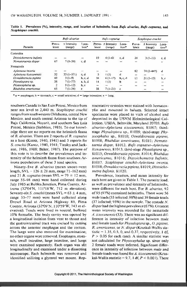
OF WASHINGTON, VOLUME 58, NUMBER 1, JANUARY 1991 143
Table 1. Prevalence (%), intensity, range, and location of helminths from Bufo alvarius, Bufo cognatus, andScaphiopus couchii.
Parasite
Bufo alvarius Bufo cognatus Scaphiopus couchii
Preva- x Intensity Loca-lence (range) tion*
Preva- x Intensity Loca-lence (range) tion*
Preva- x Intensity Loca-lence (range) tion*
Cestoidea
Distoichometra bufonisNematotaenia dispar
Nematoda
Aplectana incertaAplectana itzocanensisOswaldocruzia pipiensPhysaloptera sp.Physocephalus sp.Rhabdias americanus
67
5249382
65
7 (1-39)
33(1-371)3 (1-9)7 (1-77)9(1-12)7 (1-34)
c, d
c, db, c, da, b, db, ce
19
53814—38
4(1-6)
1(1)6(1-17)1(1)
—7(1-21)
c, d 20 3(1-11) c, d
- 82 74(2-447) dd - - -b,c, d 11 21(1-13) b,cb - - -_ _ _ _
e — — —
* a = esophagus; b = stomach; c = small intestine; d = large intesstine; e = lung.
southern Canada to San Luis Potosi, Mexico fromnear sea level to 2,440 m. Scaphiopus couchiiranges from southwestern Oklahoma, central NewMexico, and south central Arizona to the tip ofBaja California, Nayarit, and southern San LuisPotosi, Mexico (Stebbins, 1985). To our knowl-edge there are no reports on the helminth faunaof B. alvarius. There are 2 reports of B. cognatusas a host (Kuntz, 1940, 1941) and 5 reports forS. couchii (Kuntz, 1940, 1941;Tinsley and Jack-son, 1986, 1988; Baker, 1987). The purpose ofthis note is to describe the prevalences and in-tensity of the helminth fauna from southern Ar-izona populations of these 3 toad species.
Ninety-five B. alvarius (mean snout-ventlength, SVL = 126 ± 21 mm, range 71-162 mm)and 21 B. cognatus (mean SVL = 79 ± 12 mm,range 55-98 mm) were hand collected duringJuly 1985 at Robles Junction, Pima County, Ar-izona (32°04'N, 111°18'W, 712 m elevation).Seventy-six S. couchii (mean SVL = 63 ± 4 mm,range 55-77 mm) were hand collected alongDrexel Road at Arizona Highway 89, PimaCounty, Arizona (32°09'N, 110°59'W, 743 m el-evation). Toads were fixed in neutral, buffered10% formalin. The body cavity was opened bya longitudinal incision from vent to throat andthe gastrointestinal tract was excised by cuttingacross the anterior esophagus and the rectum.The lungs were also removed for examination;no other organs were removed. Esophagus, stom-ach, small intestine, large intestine, and lungswere examined separately. Each organ was slitlongitudinally and examined under a dissectingmicroscope. Each helminth was removed andidentified utilizing a glycerol wet mount. Rep-
resentative cestodes were stained with hematox-ylin and mounted in balsam. Selected intactspecimens were placed in vials of alcohol anddeposited in the USNM Helminthological Col-lection, USD A, Beltsville, Maryland 20705: Bufoalvarius-Aplectana itzocanensis, 81007; third-stage Physaloptera sp., 81009; third-stage Phy-socephalus sp., 81010; Oswaldocruzia pipiens,81008; Rhabdias americanus, 81011; Nemato-taenia dispar, 81012. Bufo cognatus-Aplectanaitzocanensis, 81013; third-stage Physaloptera sp.,81015; Oswaldocruzia pipiens, 81014; Rhabdiasamericanus, 81016; Distoichometra bufonis,81017. Scaphiopus couchii-Aplectana incerta,81018; Oswaldocruzia pipiens, 81019; Distoicho-metra bufonis, 81020.
Prevalence, location, and mean intensity foreach host are given in Table 1. The parasite load,as well as prevalence and intensity of helminths,were different for each host. For B. alvarius, 92of 95 (97%) contained helminths. There were 56male toads (55 infected; 98%) and 39 female toads(37 infected; 95%) in the sample. The cestode N.dispar had the highest prevalence (67%). Greatestmean intensity was recorded for the nematodeA. itzocanensis (33). There was no significant dif-ference in intensity of infection between maleand female toads for Physaloptera sp., O. pipiens,R. americanus, or 7Y. dispar (Kruskal-Wallis sta-tistic = 1.35, 0.5, 0, and 0.37, respectively, 1 df,P > 0.05 for each case). A similar statistic wasnot calculated for Physocephalus sp. since only2 female toads were infected. Significant differ-ence in intensity of infection between male andfemale toads was found for A. itzocanensis (Krus-kal-Wallis statistic = 9.7, ldf,P< 0.001). There
Copyright © 2011, The Helminthological Society of WashingtonCopyright © 2011, The Helminthological Society of Washington

144 JOURNAL OF THE HELMINTHOLOGICAL SOCIETY
was no correlation between total number of hel-minths and SVL (r = -0.08).
For B. cognatus, 14 of 21 (6 7%) contained hel-minths. There were 6 male toads (4 infected;67%) and 15 female toads (10 infected; 67%) inthe sample. Mean intensity for total helminthload was 10 (1-65). There was no significant dif-ference in intensity of total infection betweenmale and female toads (Kruskal-Wallis statistic= 0.5, 1 df, P > 0.05). Parasite species statisticswere not calculated because of our small samplesize: 1 female infected with A. itzocanensis; 1male, 2 females infected with Physaloptera sp.;1 male, 7 females infected with O. pipiens; 1male, 3 females infected with D, bufonis; 2 males,6 females infected with R. americanus. The nem-atodes O. pipiens and R. americanus had equallyhigh prevalences (38%). Greatest mean intensitywas recorded for R. americanus (7). There wasno correlation between total number of hel-minths and SVL (r = 0.04).
For S. couchii, 69 of 76 (91%) contained hel-minths. There were 49 male toads (43 infected,88%) and 27 female toads (26 infected, 96%) inthe sample. Highest prevalence and greatest meanintensity were recorded for A. incerta (82%, 74,respectively). In contrast to B. alvarius and B.cognatus, no nematodes were found in the lungsof S. couchii. There was no significant differencein intensity of infection between male and femaletoads for A. incerta, O. pipiens, or D. bufonis(Kruskal-Wallis statistic = 1.4, 0.35, and 1.46,respectively, 1 df, P > 0.05 for each case). Therewas no correlation between total helminth num-bers and SVL (r = 0.04).
The most prevalent parasite was the ascaridnematode, A. incerta. It was originally describedby Caballero (1949) from Bufo marinus fromChiapas, Mexico and has been redescribed fromtype specimens by Baker (1985). It has not beenpreviously reported from other hosts. Likewise,A. itzocanensis was originally described by BravoHollis (1943) from Scaphiopus multiplicatus fromPuebla, Mexico. It has also been redescribed byBaker (1985) from Bufo woodhousii woodhousiiand has been reported in B. marinus from CostaRica (Brenes and Bravo Hollis, 1959) and Ve-racruz, Mexico (Caballero Deloya, 1974).
Species of the genus Physaloptera occur in thestomachs of a variety of terrestial vertebrates.However to our knowledge, no cases of parasit-ism of toads by adult physalopterans have been
reported. We noted third-stage physalopteransin both B. alvarius and B. cognatus but not in S.couchii. Third-stage physalopterans have alsobeen reported from the toads Bufo microscaphusand B. woodhousii from Utah (Parry and Grund-mann, 1965) and Bufo americanus from Ohio(Ashton and Rabalais, 1978). Kuntz (1940) re-ported the family Physalopteridae as occurringin Bufo speciosus (=compactilis) and B. cognatusbut did not name the stage.
Alicata (1935) reported species of the genusPhysocephalus occur in the stomachs of swine,mice, and rats, in the esophagus and rumen ofruminants, and in the crop of chickens. They areof accidental occurrence in other mammals, birds,reptiles, and amphibians as third-stage larvae(Alicata, 1935). They have not been previouslyreported in toads. Physocephalus sp. requires aninsect intermediate host.
Oswaldocruzia pipiens has frequently been en-countered in amphibians and is widely distrib-uted in North America. Baker (1978a) studiedthe life cycle of O. pipiens in the frog Rana syl-vatica and reported prevalence to vary between61% and 62%. He considered 33% of the R. syl-vatica to have patent infections. Intensity variedfrom 1 to 45, but most infected frogs contained1-5 worms. Our prevalence and intensities fallwithin Baker's ranges. Oswaldocruzia pipiens isinfective as third-stage larvae which migrate outof fecal masses into the water column. Bufoamericanus exposed to contaminated frog feceshave become infected (Baker, 1978a). In addi-tion, infection has been reported in Bufo wood-housii fowleri by Brandt (1936), Rankin (1945),Campbell (1968); B. americanus by Ashton andRabalais (1978); Bufo houstonensis by Thomaset al. (1984); and Scaphiopus holbrookii by Brandt(1936).
According to Baker (1987), North Americanbufonids are apparently infected by a single spe-cies of Rhabdias, R. americanus, which was de-scribed from B. americanus by Baker (1978b).Baker (1987) suggested that reports of Rhabdiasbufonis in B. americanus and B. woodhousii fow-leri from eastern North America (Reiber et al.,1940; Fantham and Porter, 1948; Campbell,1968; Williams and Taft, 1980) should be re-ferred to R. americanus. Rhabdias sp. was listedfor B. speciosus (=compactilis) by Kuntz (1940).
The cestode N. dispar is listed in Schmidt (1986)as occurring in the Bufonidae, Ranidae, Sala-
Copyright © 2011, The Helminthological Society of WashingtonCopyright © 2011, The Helminthological Society of Washington

OF WASHINGTON, VOLUME 58, NUMBER 1, JANUARY 1991 145
mandridae, Hylidae, and Gekkonidae. It has beenreported from the North American toads B.americanus by von Linstow (1899), Bufo terres-tris by Walton (1939), and B. speciosus (=com-pactilis) and S. couchii by Kuntz (1940).
Distoichometra bufonis was originally de-scribed from B. terrestris (=lentiginosus) byDickey (1921). It has been reported from Sca-phiopus sp., B. terrestris, and B. woodhousii fow-leri by Douglas (1958), from S. holbrookii byBrandt (1936), from B. speciosus (=compactilis)and S. couchii by Kuntz (1940), and from Bufodebilis debilis and B. woodhousii woodhousii byMcAllister et al. (1989). Hardin and Janovy(1988) reported prevalences of 70-100%, withmean intensities of 2.7-14.8 for D. bufonis fromsamples of B. woodhousii from Nebraska. Theprevalence reported in our study was much low-er, 29% and 20%, respectively, for B. cognatusand S. couchii. The mean intensities for thesetoads, 4.0 and 3.0, respectively, are within thevalues established by Hardin and Janovy (1988).
None of the parasites found in this study wereunique to B. alvarius, B. cognatus, or S. couchii,but those from B. alvarius are new host records.Kuntz (1940) reported both D. bufonis and N.dispar to occur in S. couchii and only N. disparin B. cognatus. We found D. bufonis to occur inboth B. cognatus and S. couchii. Nematotaeniadispar was reported only from B. alvarius. Wecannot explain these host differences.
The toads in this study were collected fromlocalities within the Sonoran desert where theiractivity is tied to the summer rains which nor-mally begin in July and end in late August. Theyare inactive and remain underground the rest ofthe year. Whether the differential gender infec-tion rates in B. alvarius by A. itzocanensis are anartifact of this sample or a reflection of micro-habitat differences remains to be determined.
From Baker (1987) and McAllister et al. (1989)we calculated an average of 4.0 (±5.1 SD) speciesof nematodes (1-22) for 52 species of the familyBufonidae and a mean of 1.7 (±1.9 SD) speciesof nematodes (1-7) for 10 species of the familyPelobatidae. The toads in this study fell withinthe range of what might be expected for membersof their respective families. Additional work willbe required to determine the reasons for the dif-ferences that we report in the helminth fauna ofthese sympatric species.
We thank Thomas A. Bienz, Gregory S. Fel-
zien, and Adrian Sales for assistance in collectionof parasites, Michael R. Baker for confirmationof the genus Rhabdias, and 2 anonymous re-viewers for their constructive comments.
Literature Cited
Alicata, J. E. 1935. Early developmental stages ofnematodes occurring in swine. U.S. DepartmentAgriculture Technical Bulletin 489. 96 pp.
Ashton, A. D., and F. C. Rabalais. 1978. Helminthparasites of some anurans of northwestern Ohio.Proceedings of the Helminthological Society ofWashington 45:141-142.
Baker, M. R. 1978a. Development and transmissionof Oswaldocruzia pipiens Walton, 1929 (Nema-toda: Trichostrongylidae) in amphibians. Cana-dian Journal of Zoology 56:1026-1031.
. 1978b. Morphology and taxonomy of Rhab-dias spp. (Nematoda: Rhabdiasidae) from reptilesand amphibians from southern Ontario. CanadianJournal of Zoology 56:2127-2141.
. 1985. Redescription of Aplcctana itzocanen-sis and A. incerta (Nematoda: Cosmocercidae) fromamphibians. Transactions of the American Mi-croscopical Society 104:272-277.
-. 1987. Synopsis of the Nematoda parasitic inamphibians and reptiles. Memorial University ofNewfoundland, Occasional Papers in Biology 11:1-325.
Brandt, B. B. 1936. Parasites of certain North Car-olina Salientia. Ecological Monographs 6:491-532.
Bravo Hollis, M. 1943. Dos nuevos nematodos para-sitos de anuros del Sur de Puebla. Anales de In-stitute de Biologia Universidad Nacional del Me-xico, Series Zoologia 14:69-78.
Brenes, R. R., and M. Bravo Hollis. 1959. Helmintosde la Republica de Costa Rica. VIII. Nematoda 2.Algunos nematodos de Bufo marinus marinus (L.)y algunas consideraciones sobre los generos Oxy-somatium y Aplectana. Revista de Biologia Trop-ical 7:35-55.
Caballero, E. 1949. Estudios helmintologicos de laregion onchocercosa de Mexico y de la Republicade Guatemala. Nematoda, 5 Parte. Anales del In-stituto de Biologia Universidad Nacional de Mexi-co, Series Zoologia 20:279-292.
Caballero Deloya, J. 1974. Estudio helmintologicode los animales silvestres de la estacion de biologiatropical "Los Tuxtlas," Veracruz. Nematoda I. Al-gunos nematodos parasites de Bufo horribilisWiegmann, 1833. Anales de Institute de BiologiaUniversidad Nacional del Mexico, Series Zoologia45:45-50.
Campbell, R. A. 1968. A comparative study of theparasites of certain Salientia from Pocahontas StatePark, Virginia. Virginia Journal of Science 19:13-20.
Dickey, L. B. 1921. A new amphibian cestode. Jour-nal of Parasitology 7:129-136.
Douglas, L. T. 1958. The taxonomy of nematotaeniidcestodes. Journal of Parasitology 44:261-273.
Copyright © 2011, The Helminthological Society of WashingtonCopyright © 2011, The Helminthological Society of Washington
146 JOURNAL OF THE HELMINTHOLOGICAL SOCIETY
Fantham, H. B., and A. Porter. 1948. The parasiticfauna of vertebrates in certain Canadian fresh wa-ters, with some remarks on their ecology, structureand importance. Proceedings of the Zoological So-ciety of London 117:609-649.
Hardin, E. L., and J. Janovy, Jr. 1988. Populationdynamics of Distoichometra bufonis (Cestoda: Ne-matotaeniidae) in Bufo woodhousii. Journal ofParasitology 74:360-365.
Kuntz, R. E. 1940. A study of anuran parasites ofComanche County, Oklahoma. M.S. Thesis, Uni-versity of Oklahoma, Norman. 93 pp.
. 1941. The metazoan parasites of some Okla-homa Anura. Proceedings of the Oklahoma Acad-emy of Science 21:33-34.
McAllister, C. T., S. J. Upton, and D. B. Conn. 1989.A comparative study of endoparasites in three spe-cies of sympatric Bufo (Anura: Bufonidae), fromTexas. Proceedings of the Helminthological So-ciety of Washington 56:162-167.
Parry, J. E., and A. W. Grundmann. 1965. Speciescomposition and distribution of the parasites ofsome common amphibians of Iron and Washing-ton Counties, Utah. Proceedings of the UtahAcademy of Science, Arts and Letters 42:271-279.
Rankin, J. S. 1945. An ecological study of the hel-minth parasites of amphibians and reptiles ofwestern Massachusetts and vicinity. Journal ofParasitology 31:142-150.
Reiber, R. J., E. E. Bryd, and M. V. Parker. 1940.
Certain new and already known nematodes fromAmphibia and Reptilia. Lloydia 3:125-144.
Schmidt, G. D. 1986. Handbook of Tapeworm Iden-tification. CRC Press, Boca Raton, Florida. 675PP-
Stebbins, R. C. 1985. A Field Guide to Western Rep-tiles and Amphibians. Houghton Mifflin Com-pany, Boston. 336 pp.
Thomas, R. A., S. A. Nadler, and W. L. Jagers. 1984.Helminth parasites of the endangered Houstontoad, Bufo houstonensis Sanders 1953 (Amphibia,Bufonidae). Journal of Parasitology 70:1012-1013.
Tinsley, R. C., and H. C. Jackson. 1986. Intestinalmigration in the life-cycle of Pseudodiplorchisamericanus (Monogenea). Parasitology 93:451-469.
, and . 1988. Pulsed transmission ofPseudodiplorchis americanus (Monogenea) be-tween desert hosts (Scaphiopus couchii). Parasi-tology 97:437^152.
von Linstow. 1899. Bericht iiber die wissenschaftli-chen Leistungen in der Naturgeschichte der Hel-minthen in Jahre 1893. Archiv fur Naturge-schichte, Berlin 60:207-248.
Walton, A. 1939. The cestodes as parasites of Am-phibia. Contributions from the Biological Labo-ratories of Knox College 64:1-31.
Williams, D. D., and S. J. Taft. 1980. Helminths ofanurans from NW Wisconsin. Proceedings of theHelminthological Society of Washington 47:278.
J. Helminthol. Soc. Wash.58(1), 1991, pp. 146-149
Research Note
Gastrointestinal Helminths of the Reticulate Gila Monster,Heloderma suspectum suspectum (Sauria: Helodermatidae)
STEPHEN R. GOLDBERG1 AND CHARLES R. BuRSEY2
1 Department of Biology, Whittier College, Whittier, California 90608 and2 Department of Biology, Pennsylvania State University, Shenango Valley Campus,147 Shenango Avenue, Sharon, Pennsylvania 16146
ABSTRACT: Examination of the gastrointestinal tractof 110 Heloderma suspectum suspectum Cope, 1869,revealed the presence of 1 cestode, Oochoristica whi-tentoni Steelman, 1939, and 2 nematode species, Os-waldocruzia pipiens Walton, 1929, and Skrjabinopteraphrynosoma Ortlepp, 1922. Helminth prevalence andmean intensity were 12% and 9.9, respectively. Thesefindings represent new host records.
KEY WORDS: Cestoda, Oochoristica whitentoni,Nematoda, Oswaldocruzia pipiens, Skrjabinopteraphrynosoma, Helodermatidae, Heloderma suspectumsuspectum, Gila monster.
The Gila monster, Heloderma suspectum Cope,1869, is found from extreme southwestern Utah
and southern Nevada through southern Arizonaand southwestern New Mexico to northern Sina-loa, Mexico from sea level to about 1,520 m(Stebbins, 1985). The few reports of parasitismin this species mostly concern filariae. Smith(1910) recovered 4 adult filariae which he namedFilaria mitchelli; they were reassigned to the ge-nus Piratuba by Chabaud and Frank (196la).Hannum (1941) described adult and microfilariaof Chandlerella corophila which were subse-quently reassigned to the genus Splendidofdariaby Yamaguti (1961). Ryerson (1949) reportedbut did not identify microfilariae from 2 Gila
Copyright © 2011, The Helminthological Society of WashingtonCopyright © 2011, The Helminthological Society of Washington

OF WASHINGTON, VOLUME 58, NUMBER 1, JANUARY 1991 147
Table 1. Number, prevalence, and intensity of gastrointestinal helminths in 110 Heloderma suspectum suspectum.
Year
H. suspectum
Male Female Oo. whitentoni Os. pipiens
S. phrynosoma
Adult Third stage
196419651966196719691985TotalPrevalence
6131231
3469
51112
1—1241
Mean intensity (range)
1-——452%2.5 (1-4)
46720——26
1175%
19.5(1-67)
18——-9
1_
9
——-10
5%3.2(1-8)
monsters, 1 of which had adult filariae in theheart and aorta. Stabler and Schmittner (1958)recovered sheathed microfilariae which theynamed Microfilaria stahnkei (synonymized withPiratuba mitchelli by Chabaud and Frank[1961 a]). In addition, Chabaud and Frank (1961 b)described Macdonaldius andersoni from adult fi-lariae found in abdominal arteries. Mahrt (1979)found microfilariae in H. suspectum that resem-bled Macdonaldius seetae which occur in snakes.Griner (1983) observed but did not identify fi-lariae in the ventricle of a Gila monster. Goldbergand Bursey (1990a) redescribed the microfilariaof Piratuba mitchelli. The only other helminthsthat have been reported are an unidentified ces-tode and an unidentified acanthocephalan byBogert and Del Campo (1956). The purpose ofthis report is to describe the prevalence and in-tensity of gastrointestinal helminths in an Ari-zona population of H. suspectum suspectum.
One hundred ten specimens were examined(mean snout-vent length, SVL = 244 ± 51 mmSD; range 110-344 mm). All were from the vi-cinity of Tucson, Pima County, Arizona (32°13'N,110°58'W, elevation 701 m) and were collectedover several years (Table 1). The body cavity wasopened and the esophagus, stomach, small in-testine, and large intestine were slit longitudi-nally and examined under a dissecting micro-scope. Nematodes were identified using glycerolwet mounts; proglottids of cestodes were stainedwith hematoxylin. One cestode species, Oocho-ristica whitentoni, and 2 nematode species, Os-waldocruzia pipiens and Skrjabinoptera phry-nosoma, were recovered. Representativespecimens were deposited in the U.S. NationalParasite Collection (Beltsville, Maryland 20705):Oochoristica whitentoni (2 strobilae in alcohol),
USNM Helm. Coll. No. 81198; Oswaldocruziapipiens (6 males, 6 females in alcohol), 81199;Skrjabinoptera phrynosoma (3 males, 3 females,81200, and 6 third-stage larvae, 81201, all inalcohol).
Prevalence, location, and intensity for eachspecies are given in Table 1. Oochoristica whi-tentoni was found in the small intestines of 1male and 1 female lizard. Oswaldocruzia pipienswas recovered from the small intestines of 4 maleand 2 female lizards. Adult Skrjabinoptera phry-nosoma occurred in the stomachs of 2 male and1 female lizards; while third-stage larvae wererecovered from 1 male and 2 female lizards. Thefrequency of infection was the same for femaleand male lizards (female, 5/41 = 12%; male, 8/69= 12%) but there was significant difference inparasite load (Kruskal-Wallis statistic = 3.9, 1df, P < 0.05); mean intensity for females was 1.6and for males, 15.1. There was no significantdifference in infection rates between the lizardsof the 1964-1967 collection and the 1985 col-lection (Kruskal-Wallis statistic = 1.17, 1 df, P> 0.05).
Thirteen of the 66 described species of Oocho-ristica occur in North America; 8 have been re-covered from lizards (Schmidt, 1986). Based uponour update of the key developed by Hughes(1940), only 1 species, O. whitentoni, has char-acteristics similar to the specimens that we re-covered. The strobilae of our specimens aver-aged 300 by 2.0 mm with 230 proglottids and115 testes compared to 275 by 1.3 mm with 211proglottids and 100-150 testes for O. whitentoni.Oochoristica whitentoni was originally describedfrom a land tortoise, Terrapene triunguis (=or-nata), collected near Stillwater, Oklahoma(Steelman, 1939) and has also been reported from
Copyright © 2011, The Helminthological Society of WashingtonCopyright © 2011, The Helminthological Society of Washington

148 JOURNAL OF THE HELMINTHOLOGICAL SOCIETY
a false iguana, Ctenosaura pectinata, from Mexi-co (Schmidt, 1986). Insects and mites serve asintermediate hosts (Schmidt, 1986).
Oswaldocruzia pipiens is the only trichostron-gyloid to infect reptiles in the United States (Ba-ker, 1987). It is a common parasite occurringmainly in amphibians, but has also been foundin lizards: Anolis carolinensis by Conn and Etges(1984); Leiolopisma laterale, Eumeces fasciatus,Sceloporus undulatus by Harwood (1932); Ger-rhonotus multicarinatus by Goldberg and Bursey(1990b), and the turtles: Terrapene Carolina andTerrapene ornata by Ernst and Ernst (1975).
Skrjabinoptera phrynosoma has been recov-ered from a number of lizard species from NorthAmerica (Baker, 1987). Its life cycle in the Texashorned lizard, Phrynosoma cornutum, has beenelucidated by Lee (1957); the intermediate hostis the ant, Pogonomyrmex barbatus.
None of the helminths reported here is hostspecific for helodermatids, but all are new hostrecords. The low levels of infection and what isknown about the life cycles of these helminthssuggests accidental infections. Bogert and DelCampo (1956) noted that compared with otherlizards, helodermatids appeared remarkably freeof intestinal parasites and suggested that diet maybe a factor in their lack of gastrointestinal hel-minths. Since eggs, fledgling birds and nestlingmammals make up much of their diet, Bogertand Del Campo (1956) reasoned that these younganimals would be less likely to be infected withlarval stages and subsequently helodermatidswould ingest fewer parasites. Because their dietis relatively insect free, helodermatids have fewchances of infections by helminths with insectintermediate hosts. However, the inclusion ofcarrion in their diet (Stebbins, 1985) increasesthe probability of ants and beetles being con-sumed which may contain larval helminths.
We thank Charles H. Lowe (University of Ar-izona, Department of Ecology and EvolutionaryBiology) for assisting us in this study and twoanonymous reviewers for their helpful com-ments. Douglas Booth and Gregory S. Felzienassisted in recovery of parasites.
Literature Cited
Baker, M. R. 1987. Synopsis of the Nematoda par-asitic in amphibians and reptiles. Memorial Uni-versity of Newfoundland, Occasional Papers inBiology 11:1-325.
Bogert, C. M., and R. M. Del Campo. 1956. TheGila
monster and its allies. The relationships, habits,and behavior of the lizards of the family Heloder-matidae. Bulletin of the American Museum ofNatural History, New York 109:1-238.
Chabaud, A. G., and W. Frank. 196la. Les filairesde 1'heloderme. (Note additive.) Annales de Para-sitologie Humaine et Comparee 36:804-805.
, and . 1961b. Nouvelle filaire parasitedes arteres de Y Heloderma suspectum Cope: Mac-donaldiusandersonin. sp. (Nematodes, Onchocer-cidae). Annales de Parasitologie Humaine et Com-paree 36:127-133.
Conn, D. B., and F. J. Etges. 1984. Helminth para-sites of Anolis carolinensis (Reptilia: Lacertilia)from southeastern Louisiana. Proceedings of theHelminthological Society of Washington 51:367-369.
Ernst, E. M., and C. M. Ernst. 1975. New hosts andlocalities for turtle helminths. Proceedings of theHelminthological Society of Washington 42:176-178.
Goldberg, S. R., and C. R. Bursey. 1990a. Redes-cription of the microfilaria, Piratuba mitchelli(Smith) (Onchocercidae) from the Gila monster,Heloderma suspectum Cope (Helodermatidae).Southwestern Naturalist. (In press.)
, and . 1990b. Helminths of the SanDiego Alligator lizard (Gerrhonotus multicarina-tus webbi (Anguidae). Journal of Wildlife Diseases26:297-298.
Griner, L. A. 1983. Pathology of Zoo Animals. Zoo-logical Society of San Digeo, San Diego, Califor-nia. 608 pp.
Hannum, C. A. 1941. Nematode parasites of Arizonavertebrates. Ph.D. Dissertation, University ofWashington, Seattle. 153 pp.
Harwood, P. D. 1932. The helminths parasitic in theAmphibia and Reptilia of Houston, Texas, andvicinity. Proceedings of the U.S. National Muse-um 81:1-71.
Hughes, R. C. 1940. The genus Oochoristica Liihe,1898. American Midland Naturalist 23:368-381.
Lee, S. H. 1957. The life cycle of Skrjabinopteraphrynosoma (Ortlepp) Schulz, 1927 (Nematoda:Spiruroidea), a gastric nematode of Texas hornedtoads, Phrynosoma cornutum. Journal of Parasi-tology 43:66-75.
Mahrt, J. L. 1979. Hematozoa of lizards from south-eastern Arizona and Isla San Pedro Nolasco, Gulfof California, Mexico. Journal of Parasitology 65:972-975.
Ryerson, D. L. 1949. A preliminary survey of rep-tilian blood. Journal of Entomology and Zoology41:49-55.
Schmidt, G. D. 1986. Handbook of Tapeworm Iden-tification. CRC Press, Boca Raton, Florida. 675PP-
Smith, A. J. 1910. A new filarial species (F. mitchelli,n.s.) found in Heloderma suspectum, and its larvaein a tick parasitic upon the Gila monster. Univer-sity of Pennsylvania Medical Bulletin 23:487-497.
Stabler, R. M., and S. M. Schmittner. 1958. A mi-crofilaria from the Gila monster. Journal of Para-sitology 44(Supplement):33-34.
Copyright © 2011, The Helminthological Society of WashingtonCopyright © 2011, The Helminthological Society of Washington

OF WASHINGTON, VOLUME 58, NUMBER 1, JANUARY 1991 149
Stebbins, R. C. 1985. A Field Guide to Western Rep-tiles and Amphibians. Houghton Mifflin Com-pany, Boston. 336 pp.
Steelman, G. M. 1939. Oochoristica whitentoni, anew anoplocephalid cestode from a land tortoise.Journal of Parasitology 25:479-482.
Yamaguti,S. 1961. SystemaHelminthum. TheNem-atodes of Vertebrates. Vol. 3, Part 1. IntersciencePublishers, New York. 679 pp.
J. Helminthol. Soc. Wash.58(1), 1991, pp. 149-151
Research Note
Attraction of Amoebocytes to Cyclocoelum oculeum RediaeEntering the Snail Host
STEPHEN J. TAFT AND PATRICIA GASQUEDepartment of Biology, University of Wisconsin-Stevens Point, Stevens Point, Wisconsin 54481
ABSTRACT: Sixteen-millimeter cinephotomicrogra-phy was used to study the attraction of amoebocytesto Cyclocoelum oculeum rediae entering the snail host,Gyraulus parvus. As the redia pushed against the liningof the peripheral blood sinus, amoebocytes emergedfrom among epithelial cells lining the sinus 10-14 /miaway and migrated to the area of penetration within7-10 sec. When the redia broke through the sinus, theamoebocytes attached.
KEY WORDS: Trematoda, Cyclocoelidae, Cyclocoe-lum oculeum, Gyraulus parvus, redia, amoebocytes, 16-mm cinephotomicrography.
Cyclocoelum oculeum miracidia each containa fully-formed redia (Palm, 1963). After attach-ment of the miracidium to the snail host, theactivated redia leaves the miracidium within anhour and enters the snail. While studying thisprocess using cinephotomicrography, it was not-ed that amoebocytes were attracted to the site ofredial penetration.
Even though there is an extensive literature onamoebocytes and their relationship to helminths(Bayne, 1983; Cheng et al., 1969; Joky et al.,1985; Sullivan, 1988), there has never been anyin vivo filming of the attraction and attachmentof these cells to entering parasites.
Adult C. oculeum miracidia were collected fromthe nasal cavities and orbits of American coots(Fulica americand) and placed in sterilizedaquarium water. Their uteri were dissected outand teased apart to release miracidia. Miracidiawere then placed along with the recently dis-sected head-foot of Gyraulus parvus on a cleanmicroscope slide in a drop of water and mounted
with a coverslip. Filming was accomplished us-ing a Bolex 16-mm movie camera with attachedNikon Cine Autotimer mounted on a Zeiss Uni-versal Microscope. Photographs were taken at1 -sec intervals using Kodak Plus-X positive film.Selected frames were printed as negatives to re-tain detail. Line drawings from the same frameswere produced to further clarify the relation-ships.
The most common attachment site of the mi-racidium was to the snail's tentacle. This organis covered by ciliated epithelium underlain bydense connective tissue, peripheral blood sinus-es, and a central artery. After attachment, theapical papilla of the miracidium elongated andpushed against the connective tissue surroundingthe peripheral blood sinus. Following this theenclosed redia became very active. Eventuallythe apical papilla retracted into the miracidiumand the redia broke through the miracidial mem-branes, moved past the retracted papilla, andpushed up against the peripheral blood sinuscausing it to invaginate. Apparently aiding in thebreakdown of the miracidial membranes and snailtissues were substances produced by the redialesophageal glands. These glands can be seen de-creasing in volume as the redia escaped the mira-cidium and entered the snail. On numerous oc-casions while filming, and also when makingobservations, 1-3 amoebocytes could be seenemerging from among epithelial cells lining theblood sinus. One such example is depicted inPlate I, Figures 1-6 and Plate II, Figures 1-6
Copyright © 2011, The Helminthological Society of WashingtonCopyright © 2011, The Helminthological Society of Washington
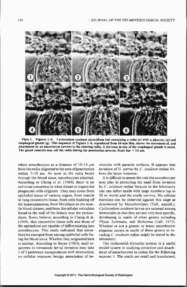
150 JOURNAL OF THE HELMINTHOLOGICAL SOCIETY
'
Plate I. Figures 1-6. Cyclocoelum oculeum miracidium (m) containing a redia (r) with a pharynx (p) andesophageal glands (g). This sequence of Figures 1-6, reproduced from 16-mm film, shows the movement of, andattachment by an amoebocyte (arrow) to the entering redia. A decrease in size of the esophageal glands is noted.The gland contents may aid the redia during the penetration process. Scale bar = 14 jmi.
where amoebocytes at a distance of 10-14 /^mfrom the redia migrated to the area of penetrationwithin 7-10 sec. As soon as the redia brokethrough the blood sinus, amoebocytes attached.According to Cheng et al. (1969), there is nouniversal consensus in what tissues or organs thephagocytic cells originate. They may come fromepithelial tissue of various organs, from mantleor lung connective tissue, from cells budding offthe hepatopancreas, from fibroblasts in the man-tle blood sinuses, and from the cellular reticulumfound in the wall of the kidney near the pericar-dium. Some believe, according to Cheng et al.(1969), that connective tissue cells and those ofthe epithelium are capable of differentiating intoamoebocytes. This study indicated that amoe-bocytes emerged from among epithelial cells lin-ing the blood sinus. Whether they originated hereis unclear. According to Bayne (1983), snail re-sponses to trematode larval invasion may take1 of 3 pathways: encapsulation with destruction;no cellular response; benign association of he-
mocytes with parasite surfaces. It appears thatinvasion of G. parvus by C. oculeum rediae fol-lows the latter response.
It is difficult to assess the role the amoebocytesmay play in protecting the snail from invasionby C. oculeum rediae because in the laboratoryone can infect snails with large numbers (up to30 or more) and the snails survive. No cellularreactions can be observed against this stage asdetermined by histochemistry (Taft, unpubl.).Cyclocoelum oculeum larvae are unusual amongtrematodes in that they are not very host specific,developing in snails of other genera includingPhysa, Lymnaea, and Helisoma (Taft, 1972).Whether or not a greater or lesser amoebocyteresponse occurs in snails of these genera to in-vading C. oculeum rediae might be tested in thelaboratory.
The cyclocoelid-G.yraw/w.s' system is a usefulmodel system in studying attraction and attach-ment of amoebocytes to rediae for the followingreasons: 1. The snails are small and translucent;
Copyright © 2011, The Helminthological Society of WashingtonCopyright © 2011, The Helminthological Society of Washington
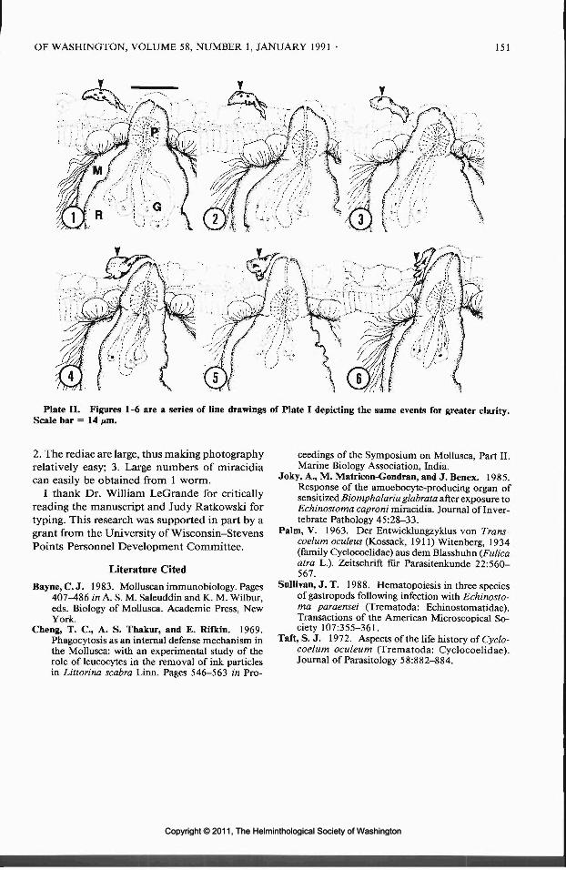
OF WASHINGTON, VOLUME 58, NUMBER 1, JANUARY 1991 151
Plate II. Figures 1-6 are a series of line drawings of Plate I depicting the same events for greater clarity.Scale bar = 14 pm.
2. The rediae are large, thus making photographyrelatively easy; 3. Large numbers of miracidiacan easily be obtained from 1 worm.
I thank Dr. William LeGrande for criticallyreading the manuscript and Judy Ratkowski fortyping. This research was supported in part by agrant from the University of Wisconsin-StevensPoints Personnel Development Committee.
Literature Cited
Bayne, C. J. 1983. Molluscan immunobiology. Pages407^86 in A. S. M. Saleuddin and K. M. Wilbur,eds. Biology of Mollusca. Academic Press, NewYork.
Cheng, T. C., A. S. Thakur, and E. Rifkin. 1969.Phagocytosis as an internal defense mechanism inthe Mollusca: with an experimental study of therole of leucocytes in the removal of ink particlesin Littorina scabra Linn. Pages 546-563 in Pro-
ceedings of the Symposium on Mollusca, Part II.Marine Biology Association, India.
Joky, A., M. Matricon-Gondran, and J. Benex. 1985.Response of the amoebocyte-producing organ ofsensitized Biomphalaria glabrata after exposure toEchinostoma caproni miracidia. Journal of Inver-tebrate Pathology 45:28-33.
Palm, V. 1963. Der Entwicklungzyklus von Trans-coelum oculens (Kossack, 1911) Witenberg, 1934(family Cyclocoelidae) aus dem Blasshuhn (Fulicaatra L.). Zeitschrift fur Parasitenkunde 22:560-567.
Sullivan, J. T. 1988. Hematopoiesis in three speciesof gastropods following infection with Echinosto-ma paraensei (Trematoda: Echinostomatidae).Transactions of the American Microscopical So-ciety 107:355-361.
Taft,S. J. 1972. Aspects of the life history of Cyclo-coelum oculeum (Trematoda: Cyclocoelidae).Journal of Parasitology 58:882-884.
Copyright © 2011, The Helminthological Society of WashingtonCopyright © 2011, The Helminthological Society of Washington
J. Helminthol. Soc. Wash.58(1), 1991, pp. 152-155
Research Note
A Survey for Trichinosis in Selected Predatory and Scavenger Birds inMontana, with an Evaluation of the Infectivity of TwoMammalian Trichinella spiralis Isolates in Birds
DAVID E. WORLEY, ROLANDO H. ESPINOSA, AND FLOYD M. SEESEEVeterinary Molecular Biology Laboratory, Montana State University, Bozeman, Montana 59717
ABSTRACT: Muscle samples from 103 free-rangingpredatory and scavenger birds of 27 species from west-ern Montana were digested and examined for Trichi-nella larvae during the period from 1971 to 1988. Fam-ilies represented included accipiters, falcons, andstrigids; golden and bald cages, red-tailed and Cooper'shawks, and great horned owls predominated. Addi-tional specimens of fish- or carrion-feeders such asherons, loons, ospreys, crows, ravens, and turkey vul-tures also were examined. No infected individuals werefound. Attempts to induce experimental infections ina short-eared owl, long-eared owl, and crow with T.spiralis isolates from a grizzly bear and fisher also wereunsuccessful. The apparent absence of avian trichinosisin the wild bird population and the inability to induceexperimental infections in captive birds suggest thatbirds play little or no obvious role in the epizootiologyof sylvatic trichinosis in the high plains/Rocky Moun-tain region of western Montana.
KEY WORDS: Trichinella spiralis, avian infections,cross-transmission.
Although Trichinella spiralis (Owen, 1835)probably has the broadest host range of any hel-minth (Gould, 1970; Despommier, 1983), undernatural conditions the various T. spiralis sub-species are thought to be infective only in mam-mals (Gould, 1970; Despommier, 1983). The re-lated species T. pseudospiralis, originallydescribed from the raccoon Procyon lotor in Da-gestan, Northern Caucasus (Garkavi, 1972), isbelieved to be a normal parasite of birds (Mi-roshnichenko, 1976; Shaikenov, 1980), althoughunder experimental conditions mice and otherrodents are susceptible (Tomasovicova, 1975).Subsequent avian records of T. pseudospiralishave extended the known distribution of the spe-cies to Spain (Calero et al., 1978) and the UnitedStates (Wheeldon et al., 1983).
Although the natural occurrence of T. pseu-dospiralis has been demonstrated on 3 conti-nents, its distribution and the species involvedin transmission are virtually unknown. The pri-mary purpose of the present study was to screentissues from a series of raptorial and scavengerbirds for the presence of Trichinella larvae in a
region of high trichinosis endemicity in mam-mals (Worley et al., 1974, 1982). A secondaryobjective was to evaluate the infectivity of 2mammalian T. spiralis isolates in birds likely tobe exposed to this infection via their normalfeeding behavior as predators or scavengers onknown mammalian reservoirs of T. spiralis inwestern Montana, northern Wyoming, and east-ern Idaho.
Samples of breast, thigh, or other muscle wereobtained from eagles, hawks, owls, and otherraptors submitted to the Raptor RehabilitationProgram at Montana State University but foundunsuitable for release. Other avian specimenswere provided by the Montana Department ofFish, Wildlife and Parks or were obtained fromroad kills. The classification used was that of theA.O.U. Check-list of North American Birds, 6thed. One 25-g muscle sample was examined inmost instances. Occasionally, tissues from 2 siteswere screened for larvae.
Tissues were removed from some birds shortlyafter death. In most instances, carcasses werefrozen for several months before muscle sampleswere removed and processed. Tissues were cutinto small pieces with scissors prior to commi-nuting with an Omnimixer. Homogenized tissuethen was digested in 1% pepsin-0.7% HC1 for 5-12 hr at 37°C under constant agitation. The di-gested material was washed on a 200-mesh screento remove soluble debris prior to examinationof the retained material for Trichinella larvaewith a dissecting microscope.
The infectivity of 2 mammalian T. spiralis iso-lates in birds was tested by oral inoculation of 2owls and a crow with larvae originally isolatedfrom a grizzly bear (Ursus arctos) and a fisher(Martespennanti) in western Montana. Both bio-types were maintained in laboratory-reared deermice (Peromyscus maniculatus*) at the MontanaState University Animal Resources Center. Lar-val suspensions obtained by peptic digestion ofskinned, eviscerated mouse carcasses were ad-
152
Copyright © 2011, The Helminthological Society of WashingtonCopyright © 2011, The Helminthological Society of Washington

153
Table 1. Wild birds negative for Trichinella spiralis larval tissue infections in western Montana (1971-1988).
Family Species No. examined
GaviidaeArdeidaeCygnidaeCathartidaeAccipitridae
PandionidaeFalconidae
Strigidae
CaprimulgidaePicidaeCorvidae
Common loon (Gavia immer)Great blue heron (Ardea herodias)Trumpeter swan (Olor buccinator)Turkey vulture (Cathartes aura)Goshawk (Accipiter gentilis)Sharp-shinned hawk (Accipiter striatus)Cooper's hawk (Accipiter cooperii)Red-tailed hawk (Buteo jamaicensis)Rough-legged hawk (Buteo lagopus)Ferruginous hawk (Buteo regalis)Golden eagle (Aquila chrysaetos)Bald eagle (Haliaeetus leucocephalus)Marsh hawk (Circus cyaneus)Osprey (Pandion haliaetus)Gyrfalcon (Falco rusticolus)Prairie falcon (Falco mexicanus)Peregrine falcon (Falco peregrinus)American kestrel (Falco sparverius)Great horned owl (Bubo virginianus)Great gray owl (Strix nebulosa)Long-eared owl (Asia otus)Short-eared owl (Asia flammeus)Saw-whet owl (Aegolius acadicus)Common nighthawk (Chordeiles minor)Northern nicker (Colaptes auratus)Common raven (Corvus corax)Common crow (Corvus brachyrhynchos)
6721
175311324
3432111111
ministered via stomach tube in physiological sa-line solution. Recipient birds were maintainedin individual cages for 26—45 days postinocula-tion prior to euthanasia. At necropsy, tissue sam-ples were collected from wing, thigh, gastrocne-mius, and pectoralis muscle, when available, forevaluation for larvae.
A total of 103 birds was examined for Trich-inella muscle larvae between 1971 and 1988 (Ta-ble 1). Included in this series were 27 speciesrepresenting 11 families: 9 accipiters, 4 falcons,and 5 owls. Additional fish or carrion feederssuch as herons, loons, ospreys, crows, ravens,and vultures were examined when available. Afew insectivorous or planktonic feeders also wereincluded, i.e., trumpeter swan, nighthawk, andflicker on the basis of possible involvement ofinsects or other invertebrates in transmission ofT. spiralis.
No evidence of Trichinella muscle larvae wasseen in any of the material examined. Becauseof the limited availability of most species, thestatus of avian trichinosis in western Montanaremains unclear. However, the absence of infec-tion in 65 individuals of 5 major raptorial species
(golden and bald eagle, red-tailed hawk, Cooper'shawk, and great horned owl) suggests that thesebirds appear to play no demonstrable role in bi-ological transmission of Trichinella sp. in a re-gion where this parasite occurs widely as a syl-vatic infection in mammalian hosts (Worley etal., 1974; Worley and Greer, 1982). The absenceof infection in the Cooper's hawk is pertinentalso in view of the first North American reportof T. pseudospiralis in this species in California(Wheeldon et al., 1983). Further, the absence ofthe parasite in species such as the great hornedowl, Bubo virginianus, which is known to feedon the common skunk, Mephitis mephitis (Bent,1938), a known host of Trichinella in westernMontana (Worley and Greer, 1982), suggests thatskunk-to-owl transmission may be unlikely tooccur as a result of natural exposure via preda-tion or scavenger feeding behavior. On the otherhand, Zimmermann and Hubbard (1963) foundthat trichinosis occurred as a low level infectionin the great horned owl in Iowa.
Limited experimental evidence from the pres-ent study confirmed the inability of 2 mamma-lian isolates to develop in birds (Table 2). No
Copyright © 2011, The Helminthological Society of WashingtonCopyright © 2011, The Helminthological Society of Washington

154 JOURNAL OF THE HELMINTHOLOGICAL SOCIETY
Table 2. Experimental inoculation of birds with mammalian Trichinella spiralis larvae.
Host
Short-eared owl (Asia flammeus)
Long-eared owl (Asia otus)
Crow (Corvus brachyrhynchos)
Larvaldose Source of isolate
865 fisher (Martes pennanti)
8,100 grizzly bear ( Ursus arctos)
10,000 fisher (Martes pennanti)
Muscles examinedpostmortem
wingpectoralisthighgastrocnemiuswingpectoralisthighgastrocnemiuswingthighgastrocnemius
Tissuelarvae
recovered*
00000000000
Via peptic digestion of muscle samples 26-45 days postinoculation.
tissue invasive stages were found in either of 2owls or 1 crow given single oral doses of 86 5-10,000 larvae. The inability to induce infectionsthat progressed past the enteral stage in birdsgiven T. spiralis isolates from mammals is con-sistent with most previous observations. Doerrand Schmidt (1930) attempted unsuccessfully totransmit T. spiralis from rodents to hens. Au-gustine (1933) found that some Trichinella lar-vae given to baby chicks survived in the intes-tinal tract to produce migratory tissue larvae, butthey died in the musculature within a short time.Matoff (1938) reported that pigeons were also anunsuitable host for rodent-derived Trichinella.On the other hand, Nemeseri (1968) found thatby lowering the natural resistance of chickens bystarvation or by feeding an inadequate diet, itwas possible to induce disseminated tissue in-fections, and these larvae were infective to whiterats. Overall, the preponderance of experimentalevidence suggests that T. spiralis isolates derivedfrom pigs or rodents are not infective to galliformor columbiform birds.
Results of the present experimental study com-plement these findings by demonstrating thatsome strigiform and passeriform species also arerefractory to infection with T. spiralis isolatesfrom wild carnivores. The possibility that raptorssuch as eagles and certain hawks that travel ex-tensively during seasonal migrations could trans-port muscle larvae mechanically to establish newsylvatic foci is an additional consideration thatwas outside the scope of the present study. Hence,the role of birds in the epizootiology of trichi-nosis in the western U.S.A. may be associatedwith their ability to disseminate infective larvae
of mammalian Trichinella biotypes rather thanas biological reservoirs in the traditional pred-ator/scavenger-prey cycle.
We acknowledge the assistance of MauritzSterner and Dan Palmisciano in obtaining andprocessing tissue specimens for the study. Con-tribution no. J-2318, Veterinary Molecular Bi-ology Laboratory, Montana State University,Bozeman, Montana.
Literature Cited
Augustine, D. L. 1933. Experimental trichinosis inchicks. Science 78:608-609.
Bent, A. C. 1938. Life histories of North Americanbirds. Pages 306-307 in U.S. National Museum,Smithsonian Institution Bulletin 170.
Calero, R., F. Martinez, S. Hernandez, and I. Acosta.1978. Parasitacion de Buteo buteo (Aves: Accipit-ridae) por Trichinella sp. en el Parque de Jerez dela Frontera. Revista Iberica de Parasitologia 38:135-138.
Despommier, D. D. 1983. Biology. Pages 75-142 inW. C. Campbell, ed. Trichinella and Trichinosis.Plenum Press, New York.
Doerr, R., and G. W. Schmidt. 1930. ExperimentelleBeinflussung der Trichinenwanderung. Die natiir-liche Immunitat des Huhnes. Zentralblatt fur Bak-teriologie. I. Originate 115:427^30.
Garkavi, B. L. 1972. [Trichinella species from Pro-cyan lotor.] Pages 53-54 in Materials of the Ail-Union Conference on the Problems of Trichinel-losis in Man and Animals, 30 May-1 June. Aka-demiya Nauk Litovskoi SSR, Vilnius.
Gould, S. E. 1970. Trichinosis in Man and Animals.Charles C Thomas, Springfield, Illinois. 540 pp.
Matoff, K. 1938. Ziir Frage der Muskeltrichinellosebeim Gefliigel. Zeitschrift fur Infektionskrankhei-ten Haustiere 54:116-134.
Miroshnichenko, L. S. 1976. [The susceptibility ofbirds to trichinelliasis.] Pages 86-90 in Materialydokladov k II-ol zhivotnykh (27-28 Maya 1976).
Copyright © 2011, The Helminthological Society of WashingtonCopyright © 2011, The Helminthological Society of Washington

OF WASHINGTON, VOLUME 58, NUMBER 1, JANUARY 1991 155
Akademiya Nauk Litovskoi SSR, Institut Zoologiii Parazitologii, Vilnius, USSR.
Nemeseri, L. 1968. Invasionversuche beim Geflugelmit Trichinella spiralis. Helminthologia 8/9:417-420.
Shaikenov, B. 1980. Spontaneous infection of birdswith TrichinellapsendospiralisGarkavi, 1972. Fo-lia Parasitologica 27:227-230.
Tomasovicova, O. 1975. Poultry—a new host ofTrichinella pseudospiralis (Garkavi, 1972). Biolo-gia (Bratislava) 30:821-826.
Wheeldon, E. B., T. A. Dick, and T. A. Schulz. 1983.First report of Trichinella spiralis var. pseudospi-ralis in North America. Journal of Parasitology69:781-782.
Worley, D. E., J. C. Fox, J. B. Winters, and K. R.Greer. 1974. Prevalence and distribution ofTrichinella spiralis in carnivorous mammals in theUnited States northern Rocky Mountain region.Pages 597-602 in Proceedings of the Third Inter-national Conference on Trichinellosis.
, and K. R. Greer. 1982. Trichinosis in wildcarnivores. Pages 224-227 in E. T. Thorne, ed.Disease of Wildlife in Wyoming. Bulletin 9, Wy-oming Game and Fish Commission, Cheyenne.
Zimmermann,W.J.,andE.D.Hubbard. 1963. Wild-life reservoirs of Trichinella spiralis. Pages 194-199 in Proceedings 100th Annual Meeting Amer-ican Veterinary Medical Association.
IN MEMORIAM
J. ALLEN SCOTT1898-1990
James Allen Scott is among the last of a groupof distinguished parasitologists who studied un-der Hegner, Stoll, Root, and Cort of the JohnsHopkins University School of Hygiene and Pub-lic Health in the first quarter of this century. Hedied at home after an extended illness on 18August 1990.
Scotty was born in Binghamton, New York,on 2 June 1898. He attended high school in Bing-hamton and after graduation spent 2 years atWyoming Seminary in Kingston, Pennsylvania.He spent a short time in the U.S. Army as aChief Bugler (he enjoyed telling of his experi-ences as a bugler) and then enrolled in WesleyanUniversity, Middleton, Connecticut, and grad-uated with a B.A. in 1922 and an M.A. in 1924.He taught for a brief time at the University ofVermont before entering Johns Hopkins Uni-versity in 1925. He received his Sc.D. in 1927under the tutelage of W. W. Cort and carried outmonumental studies on hookworm in dogs andcats. He remained at Johns Hopkins as an in-structor and research associate in helminthology,and in 1929 he became a member of the fieldstaff in the International Health Division of theRockefeller Foundation and carried out field re-search in Egypt. He worked in Egypt until 1936and carried out extensive studies on hookwormand schistosomiasis. Upon his return from Egypt,he spent a year at Johns Hopkins writing up hisresearch. His work in Egypt has become classic
and is often referred to today in that country,especially his comprehensive studies on schis-tosomiasis. In 1937 he was called again to anoverseas assignment where he worked on schis-tosomiasis in Venezuela until 1940. Again hereturned to Johns Hopkins to write up his work.From 1941 to 1944 he was a Visiting Professorat Ohio State University, worked on malaria andhookworm in Georgia, and then put his statis-tical expertise to use for awhile at the U.S. CensusBureau.
In 1944 Scotty joined the faculty at the Uni-versity of Texas Medical Branch in Galveston,Texas, where he became a Professor of Statisticsand Epidemiology in the Department of Preven-tive Medicine. He established the Laboratory ofHelminth Research and carried out continuousstudies on the cotton rat filaria Litomosoides ca-rinii. These studies were some of the earliest tobe done on filarial immunity. He established agraduate program in helminthology, and amonghis students were Leroy J. Olson, N. Ted Briggs,and John H. Cross. A number of post-doctoralstudents also had the opportunity to work withScotty: Etta Mae MacDonald, Haig Najarian,Ahmed Zien Eldin, among the many.
In 1962 and after the publication of over 100papers, Scotty left academics and bench researchto join the staff of the National Institutes ofHealth. He became a Health Science Adminis-trator and Chief of the Special Research Grants
Copyright © 2011, The Helminthological Society of WashingtonCopyright © 2011, The Helminthological Society of Washington

156
J. Allen Scott in Egypt in the 1930's.
Program and later Chief of the Parasitology andMedical Entomology Branch. In this latter ca-pacity, he helped many budding, as well as estab-lished, investigators in their quest for NIH grantsupport.
Scotty served on committees of many nationaland international organizations. He was a con-sultant on Inter-American Affairs for the StateDepartment, a member of the WHO Expert Ad-visory Panel on Parasitic Diseases and a WHOConsultant on Schistosomiasis. He belonged tomany scientific societies and served on the coun-cils of the American Society of Tropical Medi-cine and Hygiene and the American Society ofParasitology, and was active in the Helmintho-logical Society of Washington.
Those of us who studied or were associatedwith J. Allen Scott were very fortunate for theexperience. He gave us a plethora of knowledgeof parasites and parasitic diseases, as well as anin depth exposure to international health. He willalways be remembered by us and all who hadthe opportunity to have been associated with him.
He is survived by his daughter, Lois Scott Deanof Alexandria, Virginia, 4 grandchildren, and 8great-grandchildren.
JOHN H. CROSSUniformed Services Universityof the Health SciencesBethesda, Maryland
Copyright © 2011, The Helminthological Society of WashingtonCopyright © 2011, The Helminthological Society of Washington

CALL FOR PAPERS
Rescheduled Student Presentation Competition
The Helminthological Society of Washington is sponsoring the second Student Presentation Com-petition during its monthly meeting on 13 March 1991, at the Walter Reed Army Institute ofResearch, Washington, D.C.
Eligibility: An undergraduate or graduate student registered in a college or university degree pro-gram at the time of the presentation is eligible to compete for this award.
Conditions: Although multiple authorship is allowed, the project on which the paper is based mustbe substantially that of the student. The student must be the senior author and present the paper.
The presentation must be on a parasitological subject.A student may compete with only a single presentation.An abstract, which is limited to a single, double-spaced, typewritten page, must be provided. The
abstract page also must contain the title, author(s), and institutional affiliation(s).Presentation will be limited to 10 minutes. There will be approximately 5 minutes for questions
and discussion between each presentation.Membership in the Helminthological Society of Washington is not required.
Deadlines:
1 Feb 1991 Submission of abstract as described above together with a completed applicationform signed by an advisor or university official certifying the student status of the proposedpresenter of the paper.
14 Feb 1991 Notification of acceptance of paper for presentation at the 13 March 1991 meeting.13 March 1991 Student Presentation Competition at the 618th Meeting of the Helminthological
Society of Washington.
Selection of Presentations: A maximum of 8 student presentations will be selected for the com-petition. The selection of an abstract will be based on the organization of the abstract, originalityof the work described, and its potential contributions to parasitology.
Judging of Presentations: The presentations will be evaluated by a panel of judges on the followingbases: organization, techniques, originality, contribution, interpretation of results, and knowledgeof the subject.
Awards: Monetary awards in the amounts of $300, $200, and $100 will be presented to the first,second, and third place winners, respectively. In addition, the Society will waive page charges if thefirst place manuscript is accepted for publication in the Journal of the Helminthological Society ofWashington. Further, the first place winner will receive a 1-year subscription to the Journal ofParasitology.
Application: Submit abstract and completed application form to:
W. Patrick Carney, Ph.D.Department of Preventive Medicine and BiometricsUniformed Services University of the Health Sciences4301 Jones Bridge RoadBethesda, Maryland 20889-4799(202) 295-3701FAX (202) 295-3431
The application form should include the title of the paper, the name, address, and telephone numberof the student competitor, the student's signature, and the signature of an advisor or universityofficial. The student's signature verifies that the research is original work performed by a graduateor undergraduate student.
Copyright © 2011, The Helminthological Society of WashingtonCopyright © 2011, The Helminthological Society of Washington

ANNIVERSARY AWARD RECIPIENTS
)* Edna M. BuhrerMildred A. Doss.A • ;
* Allen Mclntosh"itfesseH. Christie
Gilbert F, Otto* George R. LaRue* William W. Cort* Gerard Dikmans* Benjamin Schwartz•WillardH. Wright ,
Aurel O. Foster ;Carlton M. HermanMay Belle Chitwood
*,Elvio H. SadunE. J. Lawson SoulsbyDavid R. Lincicome
1960 i1961196219641965196619661967v19691969197019711972197319741975
Margaret A. Stirewalt* Leo A. Jachowski, Jr.* Horace W. Stunkard
Kenneth C. Kates*'Everett E. Wehr
O. Wilford OlsenFrank D. EnzieLloyd E. RozeboomLeon Jacobs -v
Harley G. Sheffield Hv
A. Morgan Golden \s S. Diamond ,'••
Everett L. SchillerMilford N. Lunde •J. Ralph Lichtenfels
. A. James Haley '; >\5
19761977197819794980198119821983198419851986198719881989'1990
* George R.xLaRueVladimiriS. Ershov
* Norman R. Stoll•'Horace W. Stunkard
HONORARY MEMBERS
1959196219761977
Justus; F. MuellerJohn F. A. SprentBernard BezubikHugh M. GordonE. J. Lawson Soulsby
19781979198019811990
* W. E, Chambers .* Nathan A; Cobb*;Howard Grawley*>Winthrpp'D. Foster
CHARTER MEMBERS 1910* Philip E. Garrison* Joseph Goldberger* Henry W. Graybill
* Maurice C. Hall* Albert'Hassall* George F. Leonard
* Charles A.*Brayton H,* Charles W.
PfenderRansomStiles
*j Maurice C.'Hall* Albert Hassall* Charles W. Stiles ;
*iPaul Bartsch >* HenryvE. Ewing- ~\\* William W. Cort '
\£ Gerard Dikmans ,V* Jesse R. Christie C'
* Gotthold'Steiner/! , ,'•'Emmett W. Price \* Eloise B. Cram !
N'.*,Gerald Thome'* Allen Mclntosh* Edna M. Buhrer \* Benjamin G.-Chitwood
Aurel p. FosterGilbert F.'Otto
* Thebdor von BrandMay Belle Chitwood ,Carltpn M. Herman
•Lloyd E. Rozeboom* Albert L.^Taylor f}-_
David R. Lincicome
193119311931'.19371945,19521953195619561956'1956196119631963'196819721972
, 1'975/1975.'1975197519751976'
^Margaret A. Stirewalt* Willard H. Wjight* Benjamin Schwartz
Mildred A. Doss* Everett E. Wehr
Marion M. Farr- ,John T. Lucker, Jr.1Qeorge W. Luttermoser
* John S. Andrews* Leo A. Jachowski, Jr.
KennethVC. Kates ;
Francis G.-TrombaA: James Haley "] )-Paul.C. Beaver/Raymond M. Cabl^ -Harry Herlich "' ~,:Glenn L. HoffmanRobert E. Kuntz .Raymond V. ReboisFrank W._Douvres
1 Thomas K. SawyerLeon Jacobs '>: , ( ? . ' / •
•* J.Allen Scott "\h H . Shaw . , '
19761976197619771977197?1979197919801981
: 1981',198319841986198619871988.19881988198919891'99019901990
* Deceased.
Copyright © 2011, The Helminthological Society of WashingtonCopyright © 2011, The Helminthological Society of Washington

VOLUME 58 <IANUARYX1991: / NUMBER 1
CONTENTS ' • ' - . , . ' • ' . .
/ : (Continuedfrom Front Coyer) ..--,
MCALLISTER, C, T. Protozoan, helminth, and arthropod parasites of th^spotted chorus frog, Pseudacrisclarkii,(AnuTa: Hylidae), from north^eentral Texas II „ _. 51
CHING, H. L. Lists of larval worms from marine invertebrates of the Pacific coast of North Amer-ica ;._j_^__'. ... L.^.-....J- .....„„....;, 1....J J : .. ._.. '. „. 5 7 •••*
SAYRE, R. M., W. P. W.ERGIN, T. NISHIZAWA, AND M. P. STARR. Light and electron microscopicalstudy of a bacterial parasite from the cyst nematode, Heteroderaglycines __.. 69
SCHULTE, F. AND G. O. PoiNAR, JR. On the geographical distribution and parasitism of Rhabditis(Pelodera) orbitalis (Nematoda: Rhabditidae)..__ .:. „. 1—__ „ ........ 82
GUEGAN, J.-F. AND A. LAMBERT. Dactylogyrids (Platyhelminthes: Monogenea) ofLabeo (Teleostei:Cyprinidae) from West African coastal rivers : 85
FUJITA, O., Y. OKU, M. OKAMOTO, H. SATO, H. K. Ooi, M. KAMIYA, AND R, L; RAUSCH. Earlydevelopment of larval Taenia polyacantha in experimental intermediate hosts ._ -,. 100
RICKARD, L. G. AND J. K. BISHOP. Helminth parasites of llamas (Lama glamd) in the Pacific North-west ^T™ L. ::. : ~ '. 1 110
; | ( RESEARCH NOTES ': ' -V
CRIBB, T. H. Synonymy of Hofmonostomum Harwood, 1939, with Paramonostomum Luhe, 1909(Digenea: Notocotylidae) —„ ._......_..._ .— _ . ^ i 16
COURTNEY, C. C. AND B. M. CHRISTENSEN. The response of Tubifex tubifex (Oligochaeta: Tubificidae)to a second infection ynfo'Glaridacris catosto.mi (Cestoidea: Caryophyllaeidae) _.. 1 . 118
BOCZON, K. Differences between the energy metabolism of Trichinella spiralis and Trichinella pseu-/dospiralis ,. - . ; . . , . . ' ; 122 .. " •
HAWDON, j. M. AND G. A. SCHAD. Differentiation between mixed infections of Ancylostoma caninumc and Ancylostoma duodenale in dogs using an in vitro assay for the resumption of feeding by third-
stage infective larvae _ .....: ;.._^_ . .„ .._ 125VCONDER, G. A., P. A. ROEHM, D. A. DUPREY, S. S. JOHNSON, AND P. J. PAGANO. Treatment of
bertiellosis in Macaca fascicularis/'with praziquantel . .„_. •_.___: 128GAIDA, I. H. A>tD P. FROST. Intensity of/Neobenedenia girellae (Monogenea: Capsalidae) on the ( '
ha\fmoon.,AJediaIuna ca/z/br/Jze/wz5;(Perciformes: KyphosidaeJ^examined using a new method fordetection „.. „.,..... _...̂ ^ '. . /' „ ,.„:._:„. ~ !.„„, 129
DEARDORFF, T. L., R. E. JONES, AND S. G. KAYES. Adherence of eosinophils to the epicuticle ofinfective juveniles of A nisakis simplex (Nematoda: Anisakidae) ....„._......._„: . -.,. 131
MCALLISTER, C. T., S. ;E. TRAUTH, AND L. W. HINCK. Sphyranura euryceae (Monogenea) on Euryceaspp. (Amphibia: Gaudata), from northcentral Arkansas ........... ~...... ^ _.. .. „. 137
HAWDON, J. M. AND G. A. SCHAD. ., Long-term storage of hookworm infective larvae unbuffered saUnesolution maintains larval responsiveness to host signals ^ _..; .„.. . :_.. 140
GOLDBERG, S. R. AND C. R. BURSEY. , Helminths of three toads, Bufo alvarius, Bufp cognatus (Bufon-idae), and Scaphippus douchii (Pelobatidae), from southern Arizona.. „...._. _._.. .. ...„.„. 142
GOLDBERG, S. R. AND C. R. BURSEY. Gastrointestinalhelminths of the teticulate Gila; monster, -Helo-derma suspecfum suspectum (Sauria: Helodermatidae) .._...! ......^ ,. ; .1.... „. 146
TAFT, S. J. AND/P. GASQUE. Attraction of amoebocytes to Cyclocoelum oculeum rediae entering the ;snail host ..._v.~ .̂ ..... ~ .'. - --; ~ -~~~...-*_ ; _. .„... 149
WORLEY, D. E., R. H. ESPINOSA, AND F. M. SEESEE. A survey for trichinosis in selected predatory andscavenger birds in Montana, with an evaluation of the infectivity of two mammalian Trichinellaspiralis isolates in birds .;...——. ,......:.„... .I...;....,.....;.......... ,. _.... ;......... _>. 152
,0,, •;/ - [^ . ANNOUNCEMENTS
Meeting Notices. ;.̂ _...,.._... _ r ...~. ._:......__;: „.„_: 6USDA Scientist of the Year .. ~ .̂......... ....^ A_ 34Obituaries ... ... „„.„ .1 '. 56^Diagnostic Parasitology'Course _i '. .. J...i_ .̂™_,™.__ , 99\Jn Memoriam . • ..„. .....: ; „... .„. ;i_l__: __^.v. _:. 1 .... ; 155,Call for Papers11991 Student Presentation Competition _..._.-,̂ C-^__... ll̂ a, ...._.._......̂ 157 ]
Date of publication, 21 February 1991•' . , / - * * * •'. .
PRINTED BY ALLEN PRESS, INC., LAWRENCE, KANSAS 66044, U.S.A.Copyright © 2011, The Helminthological Society of WashingtonCopyright © 2011, The Helminthological Society of Washington