- ResearchOnline@JCU - James Cook University
210
ResearchOnline@JCU This file is part of the following work: Condon, Kelly (2019) Management of Betanodavirus infection in Queensland giant grouper, Epinephelus lanceolatus (Bloch). PhD Thesis, James Cook University. Access to this file is available from: https://doi.org/10.25903/er4h%2Dmh91 Copyright © 2019 Kelly Condon. The author has certified to JCU that they have made a reasonable effort to gain permission and acknowledge the owners of any third party copyright material included in this document. If you believe that this is not the case, please email [email protected]
-
Upload
khangminh22 -
Category
Documents
-
view
0 -
download
0
Transcript of - ResearchOnline@JCU - James Cook University

ResearchOnline@JCU
This file is part of the following work:
Condon, Kelly (2019) Management of Betanodavirus infection in Queensland
giant grouper, Epinephelus lanceolatus (Bloch). PhD Thesis, James Cook
University.
Access to this file is available from:
https://doi.org/10.25903/er4h%2Dmh91
Copyright © 2019 Kelly Condon.
The author has certified to JCU that they have made a reasonable effort to gain
permission and acknowledge the owners of any third party copyright material
included in this document. If you believe that this is not the case, please email

Management of Betanodavirus infection in Queensland giant grouper, Epinephelus
lanceolatus (Bloch)
Thesis submitted by
Kelly Condon
BSc Hons Class 1
October 2019
For the degree of Doctor of Philosophy
in
Natural and Physical Sciences
College of Public Health, Medical and Veterinary Science
James Cook University
Image courtesy Dr Richard Knuckey
Juvenile Epinephelus lanceolatus

i
STATEMENT OF ACCESS DECLARATION
I wish for this work to be embargoed for 12 months after which I grant the University a
permanent non-exclusive licence to store, display or copy any or all of the thesis, in all forms of
media, for use within the University, and to make the thesis freely available online to other
persons or organisations.
The reason for the embargo is: Commercial
Signature: Date: 28.10.19

ii
STATEMENT OF SOURCES DECLARATION
I declare that this thesis is my own work and has not been submitted in any form for another
degree or diploma at any university or other institution of tertiary education. Information
derived from the published or unpublished work of others has been acknowledged in the text
and a list of references is given
Every reasonable effort has been made to gain permission and acknowledge the owners of
copyright material. I would be pleased to hear from any copyright owner who has been
omitted or incorrectly acknowledged.

iii
STATEMENT OF SOURCES ELECTRONIC COPY DECLARATION
I, the undersigned, the author of this work, declare that the electronic copy of this thesis
provided to the James Cook University Library is an accurate copy of the print thesis
submitted, within limits of the technology available.

iv
DECLARATION OF ETHICS
The research presented and reported in this thesis was conducted within the guidelines for
research ethics outlined in the National Statement on Ethics Conduct in Research Involving
Human (1999), The Joint NHMCR/AVCC Statement and Guidelines on Research Practice (1997),
the James Cook University Policy on Experimentation Ethics, Standard Practices and Guidelines
(2001), and the James Cook University Statement and Guidelines on Research Practice (2001).
The research methodology received clearance from the James Cook University
Experimentation Ethics Review (Approval numbers A2256 and A2370).
v
STATEMENT OF CO-AUTHORS
Condon K., Bochow S., Ariel E., and Miller T., (2019) Complete sequence of Betanodavirus from
Australian barramundi, Lates calcarifer. Microbiology Resource Announcements 8.
https://doi.org./10.1128/MRA.00081-19
Author Contribution
Kelly Condon Extracted the virus from Lates calcarifer
Extracted viral RNA
Sequenced genome using PCR
Performed cloning procedures
Performed the bioinformatics analysis of the genome
Prepared manuscript drafts
Shaun Bochow Assisted with sample collection and field trip
Assisted with TNA extraction and PCR
Assisted with cloning
Assisted with manuscript preparation
Ellen Ariel Manuscript preparation
Terrance Miller Assisted with funding proposal preparation
Manuscript revision

vi
STATEMENT OF THE CONTRIBUTION OF OTHERS
This study was funded by No Noda Pty Ltd.
I was the recipient of an Australian Postgraduate Award.
Funding was provided in the form of competitive grants from the JCU CPHMS Graduate
Research Scheme Grants- to aid publication.
Tropical Finfish Pty. Ltd. Provided grouper for experimental challenge.
Dr Ellen Ariel provided editorial support to all manuscripts and guidance and editing to support
completion of this thesis.
Professor Dean Jerry provided editorial support to manuscript production.
Dr Darren Pickering, JCU Cairns, provided training in protein expression.
Dr Shaun Bochow, assisted with field trips, sample collection, extraction of nucleic acid, cloning
procedures and provided editorial support to manuscript production.
Dr Richard Knuckey and Adam Reynolds, assisted with the provision of, transport, sedation and
vaccination of fish.
The FRDC AAHS provided student fee offset to attend the Forth FRDC Australasian Aquatic
Animal Health Conference in Cairns 2017.

vii
ACKNOWLEDGEMENTS
Thank you to James Cook University for the Australian Postgraduate Award and the conditions
attached.
This manuscript is the culmination of three years of investigation intertwined with an
additional year while I tried to navigate towards my place in the world. When I commenced
this project, I was under no illusion that I was studying “Science”. I did not consider myself a
student in “Philosophy”. In fact, I think I would have been at a loss to accurately define
philosophy. In preparing the first page of this manuscript, as I wrote the words “For the degree
of Doctor of Philosophy in Natural and Physical Sciences”, I stopped to consider if I could
legitimately make such a claim. (Following ”PhD write up digression path # 1353”, calculated as
a conservative estimate of once day during the period of this study). Wikipedia defines
philosophy as “the study of general and fundamental questions about existence, knowledge,
values, reason, mind and language.” (Does anyone go beyond Google or Wikipedia anymore?
#1354). Those listed in my acknowledgments have contributed towards my scientific studies.
Each has also richly contributed to lessons in existence, values, reason, mind and, at times,
language. You have taught me answers to questions I had not even asked. With sincere
gratitude, I thank you for your presence in my days, short or long, during this work.
Although I am pleased with the achievements of this study, I hold a weight of disappointment
that they were not achieved in a manner to benefit those that set down this path with me.
Thank you to staff of FinFish Enterprises and No Noda Pty. Ltd. To Peter Hay, Alan Wigan,
Richard Knuckey, and Adam Reynolds, although we set out with good intent, enthusiasm and
knowledge, we didn’t quite reach our planned destination. I am grateful for your support in
the journey and pleased that although on different paths, we are still armed with good intent,
enthusiasm and knowledge albeit with an added pinch of cynicism, humour and frustration. No
road is too long in good company.
Thank you, Dr Leigh Owens. Although I did not finish this project with you, your passion in
Aquatic Animal Health ignited the spark that has fuelled my life-long interest in the field. The
most important step is to take the first one.
Thank you, Dr Ellen Ariel. I am grateful for your willingness to accommodate me within your
very busy student load. Your candid editorial advice is greatly appreciated, as was the use of
multi-coloured pens and European positivity. A successful woman is one who can build a firm
foundation with the bricks others have thrown at her.

viii
Thank you, Professor Dean Jerry. Finally, thesis submitted. Thank you for helping me step
down the path that I choose. Leadership is not about titles, positions or awards. It’s about one
life influencing and inspiring another. I’m grateful for the influence and inspiration.
Thank you, my friends, in Aquatic Animal Health, Dr Ian Anderson, Dr Shaun Bochow, Andrew
Fisk and Dr Nick Moody. Although we seem to focus much of our lives trying to limit the spread
and effects of disease, I am grateful you each spread laughter and happiness my way. “With
mirth and laughter let old wrinkles come.”
Thank you to my dad, Max and mum, Robyn (gone but not forgotten). The hard thing about
being a parent that pushes the child into places they do not want to go, is that it takes decades
for the child to appreciate the push came from the mind and heart. Give me money, treasures
and an inheritance? You gave me life and happy memories
To Robert. You did nothing, but an older and therefore wiser sister knows that’s to be
expected from a younger brother. Knowing you’re only a text away always brightens my day.
My family, husband Dean and daughters Trinity and Tarlia. I appreciate the support you have
provided during my studies. You have soldiered on with a part-time wife and mother for many
years and thankfully, this time is at an end. Although you are always the sun in my sky and star
in my night, I look forward to feeling your radiance for much longer in my day again. I have
missed you terribly. You’re in my heart, you’re in my soul, and you’ll be my breath till I grow
old.
To all of the Aquaculture farmers in regional QLD that battle through the rigors of farming.
Thank you for the seafood. Wishing you a time in the future with less challenge from disease.
You’ve got a friend in me.
I once explained to my youngest daughter that scientists are like the detectives of nature. We
spend time trying to investigate problems and looking for evidence. I now realise that the
study of natural science is more like interviewing a difficult suspect. Mother Nature will always
answer a question with another question. To that end, to quote another, that is older and
therefore wiser than I:
“The scientist is not a person who gives the right answers, they’re the one who asks the right
questions”-Claude Levi-Strauss.
And so, we begin...
ix
ABSTRACT
Australia was among the first countries to report the emergence of viral encephalopathy and
retinopathy (VER), observed as mass mortality of larval fish from marine aquaculture during
1987-1990. The viral aetiology of the disease was not identified until 1997 by which time
nervous necrosis virus (NNV) became one of the first aquatic pathogens considered significant
by the OIE. Unfortunately, the disease emerged during a period when transboundary
biosecurity controls governing the transport of live aquatic organisms was poor. Today, the
almost global distribution of the virus, led to its exclusion as a notifiable disease within the
modern OIE framework in 2003/2004. In Australia, VER remains a notifiable disease of finfish
and is the major impediment to the expansion and development of grouper aquaculture. Gaps
in bodies of knowledge that are critical to understanding the disease hinder the management
of VER in grouper aquaculture in Australia. A review of the literature is discussed in Chapter 1
and formed the platform for defining the aims of this thesis. This project aimed to improve
knowledge about NNV in North Queensland and develop strategies to prevent the severe
economic losses NNV causes grouper aquaculture in Australia.
At the commencement of this project, there were no complete genome sequences of NNV
collected from grouper in Australia. The National Centre for Biotechnology Information (NCBI)
database contained only two complete sequences from VER outbreaks in Australia. Neither
strain originated from Queensland, which is the region of Australia with sufficient
infrastructure and environmental parameters to support a grouper aquaculture industry. Only
one sequence was from a tropical species.
This project has improved the knowledge of Betanodavirus strains present in Northern
Queensland. Complete mRNA sequences of NNV were collected from three naturally occurring
VER outbreaks in marine aquaculture farms of barramundi Lates calcarifer, gold-spotted
grouper Epinephelus coioides and giant Queensland grouper, Epinephelus lanceolatus (Chapter
3) herein referred to collectively as the North Queensland Australia (NQAus) NNV strains.
Phylogenetic comparison of the NQAus NNV genome sequences to reference strains from the
four recognised Betanodavirus species determined all three strains were members of the
Redspotted grouper nervous necrosis virus species (RGNNV) (Chapter 3). With this finding,
RGNNV continues to be the only Betanodavirus species known to be associated with VER in
Australian fish species. Comparative analysis of the NQAus NNV strains with other strains
sourced from Australian fish species indicated remarkable conservation of the RGNNV genome
both temporally and geographically. The RNA 1 and RNA 2 segments of the NQAus NNV strains
collected in this study retained more than 97% homology to other NNV strains collected from

x
tropical species in Australia and 97-98% homology to the genome of the original RGNNV strain
isolated from Japan (SGwak97) in 1997. The significant genetic conservation of the RNA 2
segment across all of the Australian strains of NNV, provided confidence that a vaccine that
targeted the capsid protein would be applicable across a broad geographic range in Australia.
The high level of conservation of capsid protein sequence in the RGNNV species also suggested
that a vaccine effective against any of the three strains studied in this project could have a
potential global market. The low variance of RNA 2 temporally, indicates there is potentially
low risk of viral mutation and vaccine escape over time.
A comprehensive review of the literature discussing the functional motifs of the Betanodavirus
was conducted (Chapter 4). Positions of the functional motifs to Protein A and the capsid
protein were mapped on schematic diagrams. Review and identification of the multiple motifs
across an entire genome have not been reported previously from any strain of Betanodavirus.
The motifs that have been identified as critical for viral replication and associated with
virulence were retained by the NQAus NNV strains. Confirmation of the motifs ensured the
strains used in this study retained the virulence factors reported in the literature and were
suitable strains for future studies within a context of ensuring that any successful outcomes
from this study should be translatable to industry.
Examination of the genome sequences of the NQAus NNV strains collected in this study also
indicated the RT-qPCRs developed by Hick & Whittington, (2010) would theoretically be
acceptable to monitor the viral genome copy number throughout this study. PCR amplicons
produced from RNA 1 and RNA 2 segments of the viral extracts were cloned to produce
standard control plasmids for the qR1T and qR2T RT-qPCR assays described by Hick &
Whittington (2010). The RT-qPCR assays of Hick & Whittington (2010) were implemented
within the laboratory to support the subsequent project activities (Chapter 5).
Two prophylactics, namely a vaccine and a dsRNA construct were prepared to target and
prevent disease caused by the NNV strain obtained from a VER outbreak in farmed gold spot
grouper, Epinephelus coioides (Ec2NQAus) (Chapter 6). The vaccine was based on expressed
capsid protein produced from the recombinant insertion of the mRNA of RNA 2 of Ec2NQAus
NNV into a bacterial expression system. The dsRNA targeted nucleotide (nt) region 722 to 738
of the RNA 2 segment.
An experimental exposure model for application to test the efficacy of the prophylactic
measures was tested (Chapter 7). Exposure models that are representative of the natural
infection route are preferred to test the efficacy of prophylactic measures. In a novel study,
waterborne challenge via co-circulation with diseased fish along with co-infection with the
marine leech, Zeylanicobdella arugamensis was tested as an infection model. During a 40-day
xi
trial, despite habitation within a shared recirculation system containing ten fish that displayed
VER following exposure to Ec2NQAus RGNNV via IM challenge, none of the juvenile
groupers E.lanceolatus exposed by co-circulation succumbed to VER. Furthermore, the viral
genome was not detected by RT-qPCR from leeches collected from any tank or from tissues
collected from E.lanceolatus that were exposed by co-circulation. The inability to induce VER
via waterborne challenge despite the addition of leech infestation, lead to the adoption of
intramuscular injection of viral extract as a challenge model for subsequent studies.
The prophylactic measures, including vaccine and dsRNA constructs, were tested for efficacy
to prevent VER in juvenile E.lanceolatus (~18 g body mass) following IM challenge with
Ec2NQAus NNV viral extract (Chapter 8). In an initial trial, the dsRNA appeared to have no
impact in preventing the severity of disease following challenge. The initial trial indicated that
improvement in survival with vaccination was modest. The vaccinated groups of fish displayed
between 43-53% cumulative morbidity compared to 88% morbidity in dsRNA exposed groups.
This modest improvement of 35-45% reduction in morbidity indicated the vaccine formulation
presented some potential as a preventative measure. Also, the RT-qPCRs qR1T and qR2T were
applied to trace the viral copy number of RGNNV during the progression of disease following
experimental challenge (Chapter 8). Both RT-qPCR assays detected viral genome before the
onset of clinical signs at a cycle threshold value range of 31.8-36.8 (qR1T) and 29.9-45 (qR2T).
Both RT-qPCR assays detected viral genome with a cycle threshold range of 12.9 to 19.5 (qR1T)
and 11.1 to 19.0 (qR2T) during the peak period of morbidity. Fish that did not display signs of
disease were positive for the detection of viral genome indicating the vaccine may improve
tolerance to the infection rather than preventing infection. However robust conclusions
regarding fish resistance or tolerance cannot be determined based on RT-qPCR analysis. There
was relatively few vaccinated fish over 50g that succumbed to VER.
A mass spawning event with eight potential parents produced the cohort of fish used in the
experimental challenge trials. Multilocus sequence analysis (MLSA) using microsatellite
markers was applied to vaccinated fish to investigate the influence of parentage on
survival/mortality (Chapter 9). Parentage did not coincide with improved survival or increased
mortality within the studies conducted.
Assessment of vaccine efficacy on juvenile fish can be flawed if conducted before the
development of essential components of the fish immune system, therefore E.lanceolatus
larger than 50g body weight were used to reassess the efficacy of vaccination (Chapter 10). An
additional vaccine based on heat killed cell culture Ec2NQAus NNV was also evaluated. The
refined strategy indicated improved protection against VER in the slightly larger fish. Only 20%-

xii
23% of the E.lanceolatus that were vaccinated displayed signs of VER which was a marked
improvement compared to the placebo vaccinated groupers (93% morbidity).
Unfortunately, an investigation into the mechanism of improved protection is beyond the
scope of this study. RT-qPCR analysis detected NNV genome in all three groups of groupers
challenged with viral extract. NNV genome was detected in groupers that did and did not
display signs of VER. Because detection by RT-qPCR does not indicate viability of virus further
conclusions regarding the protective effect of the vaccines are not proposed.
This work has filled significant gaps in understanding the management of Betanodavirus in
aquaculture of grouper in Australia, namely:
the acquisition of the complete mRNA of three strains of NNV collected from VER
outbreaks in aquaculture systems in North Queensland;
compiling the collection of works that describe the functional motifs of the
Betanodavirus genome;
the preparation of a vaccine that reduced expression of VER to 20%-23% following
experimental challenge with Ec2NQAus NNV strain;
noting there is a mechanism that relates to body weight that affects vaccine efficacy
and
extending the fit for purpose application of two RT-qPCR assays developed by Hick &
Whittington (2010)to track the pathogenesis of NNV in grouper.
Legacy outcomes from this project are the continued contribution to research into the
management of disease in tropical aquaculture systems. Specifically, towards FRDC project
2018:098 which is field trials to test the efficacy of expressed recombinant capsid protein
vaccine to prevent VNN in the grow-out aquaculture of Epinephelus lanceolatus. In addition,
the application of the RT-qPCRs of Hick & Whittington (2010) in this project extended the fit
for purpose of those assays beyond those initially described on tissues from barramundi and
Australian bass and was applied to support NATA accreditation of a laboratory, JCU AquaPATH,
to ISO17025 in the field of animal health. The establishment of the AquaPATH laboratory
ensures that aquaculture industries in Northern Queensland have access to quality assured,
rapid, high throughput, and quantitative molecular detection assays to help manage the risk
posed by pathogens of aquaculture species.
“From little things, big things grow” (Kelly and Carmody, 1991).
xiii
Table of Contents
STATEMENT OF ACCESS DECLARATION .....................................................................................i
STATEMENT OF SOURCES DECLARATION .................................................................................ii
STATEMENT OF SOURCES ELECTRONIC COPY DECLARATION .................................................. iii
DECLARATION OF ETHICS ........................................................................................................ iv
STATEMENT OF Co-AUTHORS ................................................................................................... v
STATEMENT OF THE CONTRIBUTION OF OTHERS .................................................................... vi
ACKNOWLEDGEMENTS .......................................................................................................... vii
ABSTRACT……. ......................................................................................................................... ix
LIST OF FIGURES ..................................................................................................................... xx
LIST OF TABLES ..................................................................................................................... xxii
ABBREVIATIONS .................................................................................................................. xxiv
List of Appendices .............................................................................................................. xxvii
CHAPTER 1. Literature Review .............................................................................................28
1.1 Introduction ..................................................................................................... 29
1.2 Emergence of VER in Australia.......................................................................... 30
1.3 Viral Taxonomy ................................................................................................ 30
1.4 Viral genome characteristics............................................................................. 31
1.5 Phylogenetic comparison of Betanodavirus ..................................................... 32
1.6 Viral Nervous Necrosis: The Disease ................................................................. 34
1.6.1 Emergence of VER: ............................................................................................34
1.6.2 Host range: .......................................................................................................35
1.6.3 Transmission of Betanodavirus ..........................................................................36
1.6.4 Progression of Betanodavirus infection .............................................................36
1.6.5 Pathogenesis of Betanodavirus Infection and VER disease .................................37
1.7 Managing VER in grow out fish aquaculture ..................................................... 41
1.8 VER in Grow-Out Aquaculture systems. What determines Betanodavirus
infection vs VER disease? .......................................................................................................44

xiv
1.9 Modulation of the fish immune system. What other mechanisms could
Betanodaviruses employ to combat the host immune response? ..........................................47
1.10 Concluding comments ...................................................................................... 48
CHAPTER 2. General materials and methods .......................................................................50
2.1 Introduction ..................................................................................................... 50
2.2 Molecular biology ............................................................................................. 50
2.2.1 Collection of Betanodavirus positive material ....................................................50
2.2.2 Reverse transcription reaction/ cDNA synthesis ................................................51
2.2.3 Nucleic acid extraction ......................................................................................51
2.2.4 Polymerase chain reaction ................................................................................51
2.2.5 Gel Electrophoresis ...........................................................................................53
2.2.6 Preparation of viral extract from natural VER outbreaks. ...................................53
2.2.7 Cloning and sequencing of PCR products ...........................................................54
2.2.8 Bioinformatics analysis ......................................................................................54
2.2.9 Real-time PCR of viral extract ............................................................................54
2.3 Husbandry and handling of groupers................................................................ 55
2.3.1 Husbandry of groupers ......................................................................................55
2.3.2 Anaesthesia of groupers ....................................................................................55
2.3.3 Intramuscular injection of groupers ...................................................................56
2.3.4 Pre-trial experiments.........................................................................................57
2.4 Results………………………………………… ..................................................................57
2.4.1 Cloning and sequencing of PCR products ...........................................................57
2.4.2 Real-time PCR of viral extract ............................................................................57
2.4.3 Pre-trial experiments.........................................................................................58
2.5 Conclusion ........................................................................................................ 58
CHAPTER 3. Phylogenetic comparison of Betanodavirus genomes collected
from Viral Encephalopaty and Retinopathy outbreaks in North
Queensland ......................................................................................................59
3.1 Introduction ..................................................................................................... 60
3.2 Materials and Methods .................................................................................... 64
3.2.1 Collection of samples from natural VER outbreaks ............................................64
3.2.2 Sequencing of Betanodavirus from natural disease outbreaks ...........................64

xv
3.2.3 Additional Sequences obtained for phylogenetic analysis ..................................64
3.2.4 Sequence alignment ..........................................................................................66
3.3 Results……………………………………………… ............................................................. 67
3.3.1 RNA-1................................................................................................................67
3.3.2 RNA-2................................................................................................................71
3.4 Discussion…………………………………… ................................................................... 75
3.5 Conclusion ........................................................................................................ 76
CHAPTER 4. Review and identification of the functional motifs of the
Betanodavirus genome .....................................................................................77
4.1 Introduction ..................................................................................................... 78
4.1.1 Viral Replication Characteristics ........................................................................78
4.1.2 Expression of Viral Proteins ...............................................................................81
4.1.3 Replication of Viral RNA segments .....................................................................92
4.1.4 Formation of viral particles ................................................................................94
4.1.5 Exit of viral particles ..........................................................................................95
4.1.6 Undescribed mechanisms ..................................................................................96
4.1.7 The importance of the functional motifs in the context of this study .................97
4.2 Materials and Methods .................................................................................... 97
4.2.1 Genome annotation and illustration ..................................................................97
4.2.2 Identification of critical motifs in the E.coioides RGNNV viral extract strain. ......97
4.3 Results……………………………………………… ............................................................. 98
4.3.1 Genome annotation and illustration ..................................................................98
4.4 Discussion…………………………………… ................................................................. 103
4.5 Conclusion ...................................................................................................... 106
CHAPTER 5. Development of qPCR Standard control material to allow initiation
of validation of RT-qPCR assays to detect Australian strains of
Redspotted grouper nervous necrosis virus (RGNNV). ...................................107
5.1 Introduction ................................................................................................... 108
5.2 Materials and Methods .................................................................................. 109
5.2.1 Preparation of plasmid control for quantitative real-time polymerase chain
reaction. .......................................................................................................................109
5.2.2 Standard curve preparation from plasmid controls ..........................................110

xvi
5.2.3 RT-qPCR analysis of standard control serial dilutions .......................................111
5.3 Results………………………………………… ................................................................ 111
5.3.1 Confirmation of sequence of the RNA 1 and RNA 2 plasmid.............................111
5.3.2 Plate count of CFU of each dilution of plasmid controls ...................................112
5.3.3 Quantitative real-time polymerase chain reaction ...........................................112
5.4 Discussion…………………………………………. .......................................................... 113
5.5 Conclusion ...................................................................................................... 114
CHAPTER 6. Development of prophylactic measures to prevent Viral
Encephalopathy and Retinopathy (VER) .........................................................116
6.1 Introduction ................................................................................................... 116
6.1.1 Live Virus Vaccine ............................................................................................117
6.1.2 Inactivated-Virus Vaccine Live Virus Vaccine....................................................117
6.1.3 DNA vaccine: recombinantly expressed viral protein .......................................117
6.1.4 Viral Like particle (VLP) Vaccine .......................................................................118
6.1.5 Cell-free vaccine production ............................................................................119
6.1.6 Consideration of path for approval of use of a vaccine against VER .................119
6.1.7 dsRNA as an alternative or complement to vaccines ........................................120
6.2 Materials and Methods .................................................................................. 123
6.2.1 Nucleic acid extraction and reverse transcriptase polymerase chain reaction ..123
6.2.2 Cloning into replication plasmids .....................................................................123
6.2.3 Confirmation of clone sequence: .....................................................................123
6.2.4 Cloning into expression vector and protein expression ....................................123
6.2.5 Purification of capsid protein...........................................................................124
6.2.6 Preparation of vaccine.....................................................................................124
6.2.7 Preparation of dsRNA to target RGNNV genome. ............................................125
6.3 Results……………………………………………. ............................................................. 125
6.3.1 dsRNA construct design ...................................................................................125
6.3.2 PCR Amplification of the mRNA encoding the Capsid Protein ..........................126
6.3.3 Purification of capsid protein...........................................................................126
6.4 Discussion……………………………………………......................................................... 127
6.5 Conclusion ...................................................................................................... 127
CHAPTER 7. Experimental challenge via co-habitation with marine leech,
Zenylanicobdella arugamensis .......................................................................128

xvii
7.1 Introduction ................................................................................................... 128
7.2 Materials and Methods .................................................................................. 130
7.2.1 Culture of marine leech, Zeylanicobdella arugamensis. ...................................130
7.2.2 Fish culture, viral challenge and co-circulation system .....................................130
7.2.3 Nucleic acid extraction, CDNA synthesis, RT-qPCR. ..........................................131
7.3 Results……………………………………………… ........................................................... 132
7.3.1 Expression of VER in juvenile Epinephelus.lanceolatus. ....................................132
7.3.2 Detection of RGNNV by RT-qPCR .....................................................................132
7.4 Discussion…………………………………….. ............................................................... 133
7.5 Conclusion ...................................................................................................... 134
CHAPTER 8. Testing efficacy OF prophylatic measures against Viral
Encephalopathy and Retinopathy (VER) in Epinephelus lanceolatus
via experimental challenge .............................................................................135
8.1 Introduction ................................................................................................... 136
8.2 Materials and Methods .................................................................................. 136
8.2.1 Fish husbandry ................................................................................................136
8.2.2 Experimental design and vaccination ...............................................................137
8.2.3 Experimental Challenge with RGNNV extract ...................................................137
8.2.4 Monitoring of fish health and euthanasia ........................................................138
8.2.5 Nucleic acid extraction ....................................................................................139
8.2.6 Analysis by RT-qPCR using qR1T and qR2T .......................................................139
8.3 Results………………………………………… ................................................................ 140
8.3.1 Expression of VER following experimental challenge of juvenile grouper with
RGNNV extract and efficacy of protection of each preventative treatment. ..................140
8.3.2 Body weight of fish ..........................................................................................142
8.3.3 Detection and quantification of RGNNV by RT-qPCR assay...............................142
8.4 Discussion……………………………………. ................................................................ 144
8.5 Conclusion ...................................................................................................... 148
CHAPTER 9. ROLE of parentage in survival from Viral Encephalopathy and
Retinopathy in Epinephelus lanceolatus .........................................................149
9.1 Introduction ................................................................................................... 150
9.2 Materials and Methods .................................................................................. 151
xviii
9.2.1 Source of E.lanceolatus for family assignment .................................................151
9.2.2 Nucleic acid extraction ....................................................................................152
9.2.3 Quantitative RT-PCR to detect RGNNV ............................................................152
9.2.4 Confirmation of suitability of family assignment primers. ................................152
9.2.5 Gel electrophoresis .........................................................................................153
9.2.6 Family assignment by microsatellite markers ..................................................153
9.3 Results………………………………………… ................................................................ 154
9.3.1 Gel electrophoresis to confirm primer suitability .............................................154
9.3.2 Quantitative RT-PCR for the detection of RGNNV genome. ..............................154
9.3.3 Family assignment by microsatellite markers ..................................................155
9.4 Discussion………………………………………. ............................................................. 155
9.5 Conclusion ...................................................................................................... 157
CHAPTER 10. Testing efficacy of vaccine to prevent VER following challenge with
RGNNV in larger fish .......................................................................................158
10.1 Introduction ................................................................................................... 159
10.2 Materials and Methods .................................................................................. 160
10.2.1 Experimental E.lanceolatus ..........................................................................160
10.2.2 Expressed recombinant viral capsid Vaccine production...............................160
10.2.3 Heat-killed cell culture derived vaccine production ......................................160
10.2.4 Vaccination of Fish .......................................................................................161
10.2.5 Experimental Challenge with RGNNV extract ...............................................161
10.2.6 Monitoring of fish health and euthanasia .....................................................162
10.2.7 Nucleic acid extraction .................................................................................163
10.2.8 Quantitative RT-PCR to detect RGNNV .........................................................163
10.3 Results…………………………………………… .............................................................. 163
10.3.1 Expression of VER following experimental challenge of juvenile grouper with
RGNNV extract and efficacy of protection of each prophylactic measure. .....................163
10.3.2 Detection of RGNNV by RT-qPCR assay ........................................................164
10.3.3 Quantification of RGNNV by RT-qPCR assay .................................................166
10.4 Discussion ....................................................................................................... 168
10.5 Conclusion ...................................................................................................... 171
CHAPTER 11. Summary of findings and direction for future work........................................172
11.1 Summary of Findings ...................................................................................... 172

xix
11.2 Future work beyond this study ....................................................................... 176
11.3 Implications of findings from this study ......................................................... 178
APPENDIX 1 Media and Buffers ............................................................................................ 180
APPENDIX 2: Family assignment raw data............................................................................ 182
APPENDIX 3: Updated analysis of dsRNA design by IDT design analysis Tool. ...................... 183
APPENDIX 4: Publications and Dissemination from this Thesis ............................................ 184
REFERENCE LIST .................................................................................................................... 187

xx
LIST OF FIGURES
Figure 2-1: Image of Pond culture and E.coioides collected from a natural outbreak
of VER . ...............................................................................................................53
Figure 2-2: Sedated juvenile E.lanceolatus with location of IM vaccine injection
site indicated (Red arrow) ...................................................................................56
Figure 2-3: Cumulative Morbidity (%) of E.lanceolatus v days post challenge during
the three pilot studies conducted prior to challenge trials to confirm
viability of viral extract. .......................................................................................58
Figure 3-1: Unrooted phylogenetic tree produced from partial T4 region (289 nt)
of RNA 2 reproduced from Moody et al. (2009) including strain
identifiers and source of strain. ...........................................................................62
Figure 3-2: Summary of strain classification based on NNV RNA 2 sequences
derived from Australian fish species (including strain reference
identified and source). Modified from Hick et al. (2013) ......................................63
Figure 3-3: Neighbour-joining tree of mRNA RNA 1 segment of the Betanodavirus
from this study aligned against the reference species strains of NNV...................67
Figure 3-4: Neighbour-joining tree of mRNA RNA 1 segment of Australian strains
of Betanodavirus aligned against the strains collected from this study ................68
Figure 3-5: Neighbour-joining tree of mRNA RNA 2 segment of the Betanodavirus
from this study aligned against the reference species strains of NNV
including species and NCBI reference. .................................................................71
Figure 3-6: Neighbour-joining tree of mRNA RNA 2 segment of Australian strains
of Betanodavirus from the NCBI database aligned against the strains
collected from this study .....................................................................................72
Figure 4-1: Schematic drawing of RNA 1, mRNA of Protein A indicating location
(number) , function and amino acid sequence of functional motifs
described in the literature ...................................................................................88
Figure 4-2: Schematic drawing of RNA 2 mRNA of Capsid Protein indicating
location (number) , function and amino acid sequence of functional
motifs described in the literature ........................................................................91
Figure 4-3: Nucleotide sequence and translated amino acid sequences of
Australian strains of NNV aligned with Ito species specific region ......................102
Figure 5-1: RT-qPCR amplification curves of qR1T (a) and qR2T (b) ........................................113
Figure 6-1: Alignment of the positive strand of the dsRNA construct designed to
target the mRNA that encodes the LSTND motif.) ..............................................125

xxi
Figure 6-2: Image from gel electrophoresis screening of plasmid extracts for DNA
sequence equivalent to the capsid protein mRNA .............................................126
Figure 7-1: Diagrammatic illustration of the experimental aquarium system for co-
circulation .........................................................................................................131
Figure 7-2: Cumulative morbidity (%) of fish from each tank following IM challenge
of Tank C fish v days post viral challenge. ..........................................................132
Figure 8-1: (A) Image of juvenile E.lanceolatus with tag and (B). multiple tagged
fish within a recovery tank after injection with RGNNV extract ..........................137
Figure 8-2: Graphic illustration of experimental tank design ..................................................138
Figure 8-3: Cumulative morbidity of fish displaying signs of VER from each
experimental group following IM challenge with RGNNV extract. ......................141
Figure 8-4: Scatter plot of mass of individual fish from each treatment group v
days when euthanased due to morbidity for each day post challenge
by IM injection of RGNNV. .................................................................................142
Figure 8-5: Calculated copy number of RNA 1 (Black columns) and RNA 2 (Red
columns) segment per mg of tissue detected by qR1T and qR2T assay
for each experimental treatment group ............................................................144
Figure 9-1: Gel electrophoresis image of amplicons...............................................................154
Figure 10-1: Diagram depicting experimental tank system. ...................................................162
Figure 10-2: Cumulative morbidity (%) of fish with different vaccine formulations
versus days post-challenge with NNV extract by IM injection. ...........................164
Figure 10-3: Number of groupers positive and negative for the detection of
RGNNV by RT-qPCR from each experimental group with observation
of presence (VER clinical signs) or absence of clinical signs (No clinical
signs) of disease at the time of euthanasia. .......................................................166
Figure 10-4: Average Cycle threshold (Ct) values of positive detections by qR2T
from eyes collected from grouper from each experimental group
according to the presence or absence of clinical signs of VER. ...........................167

xxii
LIST OF TABLES
Table 2-1: Description of the primer and probe sequences used in this thesis. ........................52
Table 3-1: Betanodavirus Type Species, Serotypes and Thiery cluster assignments
based on conserved capsid protein amino acid ....................................................61
Table 3-2: Details of Reference strains and sequences of NNV type species used in
this study ............................................................................................................65
Table 3-3: Details of Australian sourced NNV sequences used in this study .............................66
Table 3-4: Percentage Homology between mRNA sequences of RNA 1 segments
from the three NNV strains sequenced in this study (El2NQAus,
Lc2NQAus and Ec2NQAus) in comparison to the NNV type species. .....................69
Table 3-5: Percentage Homology and number of nucleotide differences between
mRNA sequences of RNA 1 segments from NNV strains sequenced in
this study (El2NQAus, Lc2NQAus and Ec2NQAus) in comparison to
the other Australian-sourced strains of NNV .. .....................................................70
Table 3-6: Percentage Homology between mRNA sequences of RNA 2 segments
from NNV strains sequenced in this study (El2NQAus, Lc2NQAus and
Ec2NQAus) in comparison to the NNV type species. ............................................73
Table 3-7: Percentage Homology and number of nucleotide differences between
mRNA sequences of RNA 2 segments from NNV strains sequenced in
this study (El2NQAus, Lc2NQAus and Ec2NQAus) in comparison to
the other Australian sourced strains of NNV ........................................................74
Table 4-1: Conserved motifs critical for replication of the RGNNV genome
identified from translated mRNA of RNA 1 obtained from this study. ................100
Table 4-2: Conserved motifs critical for replication of the RGNNV genome
identified from translated mRNA of RNA 2 obtained in this study ......................101
Table 5-1: Details of primer sequences used to produce RT-qPCR control
sequences. ........................................................................................................110
Table 5-2: Primer and Probe sequences used in RT-PCR assays..............................................111
Table 6-1: Shared homology between anti GHp&0 iRNA and Spotted grouper RNA
1 segment (NCBI reference KP455643.1). ..........................................................121
Table 7-1: Cycle threshold values from each RT-qPCR from eye and leech samples
collected from the three experimental groups at various days post
challenge. ..........................................................................................................133
Table 8-1: Summary of the experimental design indicating description of
prophylactic measures, IM challenge, number of fish in each group

xxiii
and number of fish sampled at morbidity or within planned schedules
in the absence of clinical signs of VER. ...............................................................138
Table 8-2: Number of fish sampled or euthanased during the experimental
challenge including experimental group and day of
sampling/euthanasia post challenge. .................................................................141
Table 8-3: Average Cycle threshold value of qR1T and qR2T from analysis on eye
tissue from viral challenged fish. .......................................................................143
Table 8-4: Details of species, age and body weight from previous studies involving
vaccination of grouper against NNV or bacteria. ................................................148
Table 9-1: Details of primer sequences applied to MLST analysis to determine
family assignment .............................................................................................153
Table 10-1: Timeline of age of fish days post hatch at acclimation, vaccination,
booster and challenge with viral extract. ...........................................................161
Table 10-2: Summary of experimental design indicating experimental groups,
description, number of fish in the trial and number of fish sampled at
morbidity or at scheduled time points during the experimental trial .................162
Table 10-3: Number and percentage (%) of groupers positive and negative for the
detection of RGNNV genome by RT-qPCR from each experimental
group and number of groupers displaying clinical signs of VER or no
clinical signs of VER at the time of euthanasia. ..................................................165
Table 10-4: Cycle threshold values of each RT-qPCR assay from eye of groupers
from experimental group by days post viral challenge .......................................167
Table 10-5: Summary of the age, weight, water temperature (⁰C) and reported
survival of groupers from published studies that have investigated
vaccination against pathogens in groupers ........................................................169

xxiv
ABBREVIATIONS
aa
AHNNV
ATP
avSFV
B1
B2
Amino Acid
Atlantic Halibut Nervous Necrosis Virus (Betanodavirus)
Adenosine Tri-phosphate
Avirulent Semliki Forest Virus
Protein B1 of Nodavirus
Protein B2 of Nodavirus
BLAST Basic local alignment search tool
bp
BSL
Base pair of nucleotides
Biosecurity Sciences Laboratory, Queensland Government
cDNA
CNS
CPE
DI-RNA
DGNNV
Complementary DNA
Central nervous system
Cytopathic effect
Differential interfering RNA
Dragon Grouper Nervous Necrosis Virus
DNA
Dpc
dph
dpi
DSCAM
dsRNA
EDTA
EIPA
eEF
ER
FAT
FHV
GF-1
GGNNV
GL-av
HK
HIV
hpi
Deoxyribonucleic acid
Days post challenge
Days post hatch
Days post infection
Down syndrome cell adhesion molecule
Double stranded RNA
Ethylenediaminetetraacetic acid
5-(N-ethyl-N-isopropyl) amiloride
Eukaryotic elongation factor
Endoplasmic Reticulum
Fluorescent antibody test
Flock house Virus (Alphanodavirus)
Grouper fin cell line
Greasy grouper nervous necrosis virus
Grouper liver cell line
Heat-Killed
Human Immunodeficiency Virus
Hours post infection

xxv
Hsp
ICTV
IFN
Heat shock Protein (40 or 70 or 90 as different proteins)
International Committee for Taxonomy of Viruses
Interferon
IM
IP
IRF
Intramuscular, referring to site of injection into muscle
Intraperitoneal, referring to site of injection into peritoneal cavity
Interferon regulatory factor
Kb
LB
MDA-5
MLS
MLSA
MTase-GTase
Kilobase (kbp = 1,000 base pairs)
Lysogeny broth
Melanoma Differentiation-association protein-5
Mitochondrial Location/Localisation signal
Multi-loci sequence analysis
Methyltransferase-guanylyl transferase
MTD
mRNA
Mx
Mitochondrial Transmembrane domain
Messenger RNA
Myxovirus resistance protein
NCBI
NES
NF-ĸB
National Centre for Biotechnology Information
Nuclear Export Signal
Nuclear factor-kappa light chain enhancer of activated B cells
NLS
NNV
Nuclear Location/Localisation signal
Nervous Necrosis Virus
NoV
NS
Nodamura Virus (Alphanodavirus)
Non-structural protein
nt Nucleotide
OIE
ORFs
PC
PCR
PE
pi
Office Internationales des Epizootics
Open reading frames
Phosphatidylcholine
Polymerase chain reaction
Phosphatidylethanolamine
Post injection/post infection
ppt Parts per thousand
qPCR
RAC
rCP
RdRp
Quantitative/real-time polymerase chain reaction
Ribosome-associating complex
Recombinant Capsid Protein
RNA dependant RNA polymerase
xxvi
RGNNV
RIG
Red-spotted grouper Nervous Necrosis Virus (Betanodavirus)
Retinoic acid inducible gene
RNA Ribonucleic acid
RNAi RNA interference
RT-
RT-PCR
SE
SGIV
siRNA
Reverse transcriptase
Reverse transcriptase polymerase chain reaction
Spongiform Encephalopathy
Singapore Grouper Iridovirus
Short interfering RNA
ssDNA
SJNNV
STAT
SSN
ssRNA
Tat
TCID
TLR
TMD
TNT
TBSV
TOM
Single stranded DNA
Striped Jack Nervous Necrosis Virus (Betanodavirus)
Signal transducer activator of transcription
Striped Snakehead (cell line)
Single stranded RNA
Transactivator of transcription
Tissue culture infectious dose
Toll-like receptor
Transmembrane Domain
Terminal nucleotransferase
Tomato Bushy Stunt Virus
The outer mitochondrial membrane
TPNNV
TRIM
Tris
Tiger Puffer Nervous Necrosis Virus (Betanodavirus)
Tripartite Motif-containing protein
Trisaminomethane
UTR
VER
V-miRNA
VNN
VLP
Untranslated region
Viral Encephalopathy and Retinopathy
Viral encoded micro RNA
Viral Nervous Necrosis
Viral like particles
VRC
WHO
WhNV
ZLE
Viral Replication Complex
World Health Organisation
Wuhan Nodavirus
Zebrafish cell line

xxvii
LIST OF APPENDICES
Appendix 1 Media and Buffers
Appendix 2 Raw Data of Family Assignment analysis
Appendix 3 Updated analysis of dsRNA by IDT design tool
Appendix 4 Publication and Dissemination of works from this thesis.

28
CHAPTER 1. LITERATURE REVIEW
Background
Aims of this Chapter
In Cairns, North Queensland, The Company One is one of the most efficient commercial
grouper hatcheries in the world, with annual production of several million fingerlings.
Commercial production of grouper in grow-out aquaculture production in Australia is
restricted by the severe economic losses caused by Viral Encephalopathy and
Retinopathy (VER) syn., Viral Nervous Necrosis (VNN) disease outbreaks.
Groupers do not have the restricted period of susceptibility and VER outbreaks have
been reported in fish up to 3 years old.
In recent years, as the industry has attempted to expand, the rapid mass mortality of
grouper in grow-out pond production have occurred with sufficient frequency to
threaten the economic viability of the industry in Australia.
Vaccines that protect against virulent strains of viruses are widely adopted as an
effective strategy to prevent losses due to viral diseases in many fish aquaculture
systems.
Despite reported success in experimental systems, there are no commercially available
vaccines to prevent VER in Australia.
Provide current state of knowledge of Betanodavirus
Discuss aspects of host biology that have an association with VER
Discuss options for the management of Betanodavirus infections in aquaculture
Define the research questions and aims of this thesis
29
1.1 Introduction
Grouper, Epinephelus spp., are an important marine fish aquaculture species in many
countries, particularly Asia (Rimmer and Glamuzia 2017). In 2017 the global aquaculture
production of grouper was 147 379 tonnes (value 0.7 US$ Billion) (FAO, 2018). The majority of
the world’s aquaculture production of grouper is from China and Indonesia (Rimmer and
Glamuzia 2017). However, due to their rapid growth rates and strong market value, grouper
have potential to be a profitable aquaculture species in Australia (Knuckey, 2015). In Cairns,
North Queensland, The Company One is one of the most efficient commercial grouper
hatcheries in the world, with annual production of several million fingerlings. However, the
majority of the fingerlings produced are exported into Asia for grow out aquaculture
production (Knuckey pers. comm. 2019).
In recent years, as the industry has attempted to expand into grow out production, the rapid
mass mortality of grouper due to Viral Encephalopathy and Retinopathy (VER) syn., Viral
Nervous Necrosis (VNN) have occurred with sufficient frequency to threaten the economic
viability of the industry in Australia. Commercial production of grouper in grow-out
aquaculture production in Australia is restricted by the severe economic losses caused by VER
disease outbreaks (Knuckey pers. comm. 2019). Management of VER in grouper hatchery
systems can be achieved through the implementation of strict biosecurity protocols that
prevent the entry of Betanodavirus into the culture system. The strategies impose increased
costs to production but are not practical to prevent VER outbreaks in grouper grow-out
farming systems such as sea cage or pond cultures.
Other management strategies must be developed for grouper grow out and pond systems.
Forty years of research into VER has resulted in the development of cell culture replication
systems, serological and molecular detection techniques and vaccination and novel anti-VER
preventatives (Hick et al., 2010; Hick et al., 2011; Tanaka et al., 2001) . However, in Australia,
there is no commercially available preventative or treatment to manage VER outbreaks.
Pharmaq™ and HIPRA recently commenced commercial sale of a VNN vaccine in the
Mediterranean to protect European sea bass against NNV. However, neither vaccines are
approved for import into Australia and the suitability of the vaccine to prevent VER in grouper
is untested.
Vaccines that protect against virulent strains of viruses are widely adopted as an effective
strategy to prevent losses due to viral diseases in many fish aquaculture systems. However,
despite the long history of VER in Australia, knowledge of the strains of Betanodavirus that
cause VER in Northern Australia is limited. The paucity of knowledge of Betanodavirus in
30
Australia along with gaps in the knowledge about critical aspects of grouper biology, including
antiviral immunity, creates a situation whereby evidence-based management of VER is very
difficult. This review will discuss the emergence of VER; characteristics of viral taxonomy and
replication; and host/environmental factors that are believed to associate with VER outbreaks.
Knowledge of such aspects can be applied to develop evidence-based strategies to manage
VER in grouper grow out systems in Australia.
1.2 Emergence of VER in Australia
Members of the genus Betanodavirus cause the disease Viral Encephalopathy and Retinopathy
(VER) syn. Viral Nervous Necrosis (VNN) (OIE, 2018). The disease emerged in Australasia,
Europe and North America during 1985 to 1989 and has been reported from wild and cultured
freshwater and marine fish in all continents except South America and Antarctica. The National
Centre for Biotechnology Information (NCBI) database contains 1200+ nucleotide accessions of
Betanodavirus sourced from over 220 fish species and 30+ countries (Condon et al., 2019). Ten
gene sequences of Betanodavirus have been published in NCBI from Australian fish
(www.ncbi.nlm.nih.gov accessed 12.2.2019). In Queensland, VER outbreaks occur in
commercial larval barramundi, Lates calcarifer (Bloch, 1790) and giant grouper, Epinephelus
lanceolatus (Bloch, 1790) hatcheries. The World Organisation for Animal Health, Office
International des Epizooties (OIE) delisted VER in 2004, as notifiable disease, due to the
worldwide distribution failing to meet one of the defining criteria of a restricted geographical
host range (OIE, 2004). Despite the de-listing, VER is notifiable in Australia and an impediment
to successful culture of a number of highly susceptible fish species worldwide.
1.3 Viral Taxonomy
The Nodaviridae consists of the genera, Betanodavirus, which infect fish and Alphanodavirus,
which infect insects. Members of the Nodaviridae also infect crustaceans but taxonomic
divisions recognising the crustacean-infecting species have not occurred. Four species of
Betanodavirus are officially recognised by the International Committee for the Taxonomy of
Viruses (ICTV) namely Striped jack nervous necrosis virus (SJNNV), Barfin flounder nervous
necrosis virus (BFNNV), Tiger puffer nervous necrosis virus (TPNNV) and Red spotted grouper
nervous necrosis virus (RGNNV) (ICTV//www.ictvonline.org./virustaxonomy.asp). The species
names represent the host species of the original viral isolate and are supported by variation in
the viral genomic sequence. An additional viral strain isolated from Turbot, proposed as a new
Betanodavirus species, Turbot Nervous Necrosis Virus (TNNV), displays variation in genomic

31
sequence from the four recognised species however formal recognition of TNNV as another
species has not occurred (Johansen et al., 2004,ICTV//www.ictvonline.org./virustaxonomy.asp)
The viral species were originally proposed to have strong host specificity. However, excluding
TPNNV, the different genotypes can infect a variety of fish species (Thiery et al., 2004). The
viral species were also originally observed to have species-specific temperature dependency
(Iwamoto et al., 1999) however, variation from the original temperature distributions are
known to occur and RGNNV exhibits the greatest temperature tolerance (Panzarin et al.,
2016). The taxonomic distribution of the 1400+ sequences published in NCBI nucleotide
database includes SJNNV (182), RGNNV (389) BFNNV (76), TPNNV (5) and other strains which
have not been formerly classified into species divisions (www.ncbi.nlm.nih.gov. accessed
18.8.19).
1.4 Viral genome characteristics
Nodaviruses possess a small linear single stranded bi-segmented positive sense RNA (+ss RNA)
genome contained within an approximate 25 to 35 nm un-enveloped capsid of icosahedral
symmetry (Venter and Schneemann 2008). Nodaviruses replicate exclusively in the cytoplasm.
Virions are stable between pH 2 to 9 and resistant to heating at 56 °C for 30 min (Frerichs et
al., 2000). The Nodaviridae possess one of the smallest animal infecting viral genomes.
Genomes are approximately 4.5 kb nucleotides (nt) consisting of a Segment 1 (RNA 1) of 3.1kb
nt and Segment 2 (RNA 2) 1.4kb nt. Both segments possess a 5’ end methylated cap that
assists in recruiting the eukaryotic translation machinery to translate viral proteins (Mori et al.,
1992). Both segments lack a 3’ poly a tail but the RNAs are protected by an unknown moiety
(Venter & Schneemann 2008).
The RNA 1 (3.1kb nt) contains the mRNA for the ~ 1000 amino acid (aa) Protein A encoded by
nt 79 to 3027( ). A sub-genomic RNA, termed RNA3, not packaged into virions, is synthesised
from the 3’ end of the RNA 1 segment. RNA 3 consists of ~387 nt and encodes the B1 (111 aa)
and B2 (72 aa) proteins (Venter & Schneemann 2008; Toffolo et al., 2007). The B1 protein is
translated in the same reading frame as Protein A and is encoded by the 336nt of the 3’
terminus of the RNA 1(nt 2688 to 3027). The B2 protein requires a +1-reading frame shift for
translation compared to B1 and is encoded by 227 nt on the RNA 1 (nt 2753 to 2980)
(Biacchesi 2011; Venter & Schneemann 2008). RNA 3 also acts as a transactivator in the
replication of RNA 2. Paradoxically, the replication of RNA 2 results is the cessation of
replication of RNA 3 (Venter & Schneemann 2008). RNA 2 (1.4kb nt) contains the mRNA for the
capsid protein. In the Alphaviruses the capsid protein is ~ 430 aa compared to 338 aa of the
Betanodaviruses (Venter & Schneemann 2008). How the nodavirus Viral RNA interacts to

32
infect and cause disease in vertebrates is not completely understood. Knowledge of viral
replication processes will aid in developing novel anti-viral therapies to limit disease.
1.5 Phylogenetic comparison of Betanodavirus
Phylogenetic studies are useful to identify virulence factors and produce epidemiological
models to understand viral transmission pathways. Initial phylogenetic studies of the
Betanodaviruses were based on the viral capsid protein or RNA 2 and indicated strong nt and
aa distinction between the species (Nishizawa et al., 1997). However, the presence of
reassortments between RGNNV, SJNNV and BFNNV suggests the phylogenetic studies should
consider both the RNA 1 and RNA 2 (Toffan et al., 2017 and Oliveira et al., 2009).
Phylogenetic comparisons between the Betanodavirus strains initially occurred through
analysis of the nt sequence of RNA 2 or its translated capsid protein sequence. The RNA 2 of
TPNNV and SJNNV regions are identical in nt length. BFNNV and RGNNV lack 6 bases at
position 713 to 718 nt of the RNA 2 strand. Comparative similarity of a T2 region (nt 155 to
1030) within the RNA 2 strand between the different species was 75.8 % or more at nt and
80.9 % or more at aa level. Within T2 a highly conserved (>93 %) 134 aa region and highly
variable T4 (62 %) 81 aa region was identified (Nishizawa et al., 1995). The T4 (nt 604 to 1030),
T2 and base insertion or deletion at nt 713 to 718 were proposed as a site for species
differentiation of the Betanodaviruses. The conclusions of that analysis supported the division
of the 4 species originally defined by host species (Nishizawa et al., 1995).
Comparison of the T4 region in a phylogenetic analysis of 25 Betanodavirus isolates collected
in Japan resulted in the divergence of the Japanese isolated Betanodaviruses into 4 clusters
containing 95 % or greater nt sequence similarity (Nishizawa et al., 1997). The clusters were
defined as TPNNV, SJNNV, BFNNV and RGNNV. The majority of NNV isolates from Japanese
flounder aligned within the RGNNV species and only a single isolate (JF95Hok) aligned with the
BFNNV species (Nishizawa et al., 1997). Japan remains the only country to report VER isolates
from all four of the recognised Betanodavirus species. Phylogenetic analysis of the region
consisting of RNA 2 nt 169 to 987 was conducted on Betanodaviruses isolated from cultured
fish in Korea (Cha et al., 2007). The classification supported the 4 species previously identified
and proposed an additional 5th group consisting of a single isolate from a Turbot from Norway
(TNNV-Norway AJ608266) (Johansen et al., 2004).
Using deduced aa analysis encoded by the T4 region of 44 Betanodavirus isolates from various
countries in Europe, Asia and the Mediterranean, a different classification nomenclature was
proposed (Thiery et al., 2004). The classification consisted of 4 clusters and 5 subtypes namely
Ia and Ib (RGNNV); IIa, IIb, IIc (BFNNV); III (TPNNV) and IV (SJNNV). The clustering within each
33
group was more related to the geographical source of the isolate than the host species (Thiery
et al., 2004). Cherif et al. (2009) applied the phylogenetic clusters of Thiery et al. (2004) to
investigate a number of NNV isolates in D. labrax and sea bream Sparus aurata from Tunisia.
All isolates clustered within the RGNNV genotype. Four of the isolates were obtained from
temperatures 15 to 19 °C, which is outside that typically observed for the RGNNV. In a novel
report, nine different sequences were observed within a single farm. In contrast to previous
observations, geno-grouping of the fish Betanodaviruses appeared to reflect an adaptation to
a range of temperatures rather than geographic location or host specificity (Cherif et al., 2009).
Recognising some discrepancy in the classification of Betanodavirus strains based on T4, some
researchers report phylogenetic analysis including both RNA 1 and RNA 2 of the Betanodavirus
genome (Toffolo et al., 2007). Analysis of the RNA 1 nt 121 to 1050 of SJNNV identified 25
unique RNA 1. Analysis comparing RNA 2 from nt 388 to 894 of SJNNV that contained the
species-specific 6 nt insert region identified 22 unique sequences. Phylogenetic division of the
Betanodaviruses was possible using either RNA 1 or RNA 2 however, the divisions strongly
contrasted (Toffolo et al., 2007). Using the RNA 1 strand the SJNNV, BFNNV and RGNNV or
clades IV, II and I phylogenetic groupings were statistically well-supported (Toffolo et al.,
2007). A sister group relationship was proposed for the TPNNV, BFNNV and SJNNV or clades III,
II and IV. Two of the Iberian isolates clustered within the SJNNV/IV when grouped by analysis
of RNA 1. The same two were positioned in the RGNNV/I cluster when grouped by analysis of
RNA 2. There was an absence of any evidence of recombination between different RNA 1
segments however; recombination in RNA 2 between different isolates was observed. Toffolo
et al. (2007) proposed both RNA 1 and RNA 2 must be considered for phylogenetic purposes
with RNA 1 possibly being a better marker to assess the origin of a single isolate.
Phylogenetic relationships between VER strains detected in the Iberian Peninsula, collectively
termed IBNNV, indicated the presence of re-assortment between Betanodavirus species
(Olveira et al., 2009). Comparing RNA 1, all IBNNV isolates clustered within 97 % nt sequence
homology to the RGNNV species (Olveira et al., 2009). Comparison of RNA 2 indicated
divergence with only 1 of the IBNNV strains aligning within the RGNNV isolates and the
remaining 6 displaying stronger similarity to the SJNNV species. Genomic analysis of
Betanodaviruses from cultured fish species in Malaysia also considered RNA 1 and RNA 2
(Ransangan & Manin, 2012). All of the studied Betanodaviruses from Malaysia presented as
nine clusters within the RGNNV species. Unlike Toffolo et al. (2007) the clustering was
consistent between RNA 1 and RNA 2 analysis, which is expected in the absence of
reassortment between multiple NNV species.

34
The limitations of the T4 region as a classification tool were not recognised until after 2004.
Many of the sequences contained in the NCBI (1999 to 2004) are not complete and make
retrospective phylogenetic studies with newly detected NNV strains difficult. A recent
phylogenetic study was conducted comparing 189 RNA 1 Betanodavirus sequences (32
RGNNV, 154 BFNNV, 1 TPNNV and 2 SJNNV) and 73 RNA 2 Betanodavirus sequences (54
RGNNV, 8 BFNNV, 1 TPNNV and 10 SJNNV) (He & Teng, 2015). During the period the isolates
were collected, the RNA 1 had a mean nt substitution rate of 3.60 per 10 000 nt per year
compared to RNA 2 of 3.69. Within the RGNNV types substitution rates of 4.28 and 3.79 per 10
000 per year were calculated for the RNA 1 and 2 respectively (He & Teng, 2015). Taxonomic
divisions proposed by Nishizawa were supported by the analysis with the identification of
subclades within the genotypes. Using the RNA 1, the RGNNV could be divided into 3
subclades compared to 6 subclades using RNA 2 (He & Teng, 2015).
To develop more effective means to understand the epidemiology of VER a greater quantity of
genomic sequence information of the isolates is required. An understanding of the
contributions the genomic sequences make in the progression of disease (pathogenesis) of
VER could assist in developing strategies to overcome the disease.
1.6 Viral Nervous Necrosis: The Disease
1.6.1 Emergence of VER:
VER, originally termed Whirling disease or Summer disease, emerged as a disease causing up
to 88 % mortality in intensive larval fish hatcheries during 1983 to 1989 (Gallet de Saint Aurin,
Raymond & Vianas, 1989). Clinical signs of VER in larval fish include loss of appetite, darkened
colour, erratic and spiral swimming and hyperinflation of the swim bladder. In larval rearing
systems, clinical signs are usually followed by mass mortality over 2 to 14 days (Moody &
Crane, 2012).
Histopathology of VER disease involves extensive vacuolation of the brain and retinal tissues.
Viral particles were reported in association with VER histopathology in larval production
systems of, Dicentrarchus labrax in the French Caribbean Islands (Gallet de Saint Aurin et al.,
1989), turbot, Scopthalmus maximus in Norway (Bloch et al., 1991), Japanese parrotfish,
Oplegnathus fasciatus in Japan (Yoshikoshi & Inoue 1990), and barramundi, Lates calcarifer in
Australia and Tahiti (Glazebrook et al., 1990) (Renault et al., 1991).
Purification and partial characterisation of viral particles from striped jack Pseudocaranx
dentex suffering VER was reported in 1992 (Mori et al., 1992). Based on the viral
characteristics VER was proposed to be caused by new member of the Nodaviridae designated

35
Striped Jack Nervous Necrosis Virus (SJNNV) (Mori et al., 1992). SJNNV was the first nodavirus
to be purified from a vertebrate during a time when no member of the Nodaviridae had been
isolated from outside Australasia (Johnson et al., 2000). Experimental infection trials
confirmed SJNNV as the aetiological agent of VER in P.dentex (Arimoto et al., 1993). Nishizawa
(1995) provided viral genome and aa analysis to support the classification of NNV as a member
of the Nodaviridae. Betanodavirus was officially accepted as a new genus in the family
Nodaviridae in 1997 (ICTVonline.org/proposals/Ratification_1997.pdf). SJNNV became the
type species of the Betanodavirus genus. Other species include Barfin Flounder Nervous
Necrosis Virus (BFNNV), Redspotted Grouper Nervous Necrosis Virus (RGNNV) and Tiger Puffer
Nervous Necrosis Virus (TPNNV).
1.6.2 Host range:
Betanodavirus isolates have been collected from larval and juvenile stages of over 157 marine
and freshwater fish species in tropical and temperate climates (www.ncbi.nih.gov/). VER
disease outbreaks have been reported from farmed cultures and wild fisheries (Koohkan et al.,
2012) (Breuil et al., 2000). In experimental infection trials, susceptibility of fish species has
been classified as highly susceptible, less susceptible or resistant to NNV (Furusawa et al.,
2007). A similar pattern occurs in wild fisheries. The highly susceptible species display
mortalities of 80 to 100 % in either larval or larval and later life stages. Most of the highly
susceptible species are only susceptible to VVN during early larval stages. Except, the groupers,
Epinephelus sp. and European seabass, Dicentrarchus labrax, are susceptible beyond the larval
stages. Groupers up to 3 years old have been recorded with VER (Tanaka et al., 1998). Notably,
there have been no reports of VER in Atlantic salmon, Salmo salar, despite the geographical
presence of NVV where they are cultured and the fish being reportedly susceptible to
experimental infection with BFNNV via intraperitoneal injection (Korsnes et al., 2005). Also
notable in the Northern Australian aquaculture context is the absence of reported disease
outbreaks in cobia, Rachycentron canadum which are being experimentally grown within
marine systems in the same geographic range as barramundi and grouper. Chu et al. (2013)
reported a case study of mortality exceeding 80% in cobia from 3 farms in Malaysia during
2007. Fish displayed signs of VER which included the mass mortality and abnormal spiral
swimming behaviour and “flashing” and were positive for the detection of NNV by RT-PCR
(Chu et al., 2013). No similar reports have occurred from cobia aquaculture despite, in some
circumstances in Australia, cobia being cultured in pond cages immediately adjacent to
grouper suffering a VER outbreak (unpublished data).
36
Betanodaviruses have also been detected from a number of non-piscine hosts including the
spiny lobster Panulirus versicolor (Gomez et al., 2006), Artemia sp, Copepod Tigriopus
japonicas, shrimp Acetesinte medius, Charybdid crab Charybdis bimaculata, Southern
humpback shrimp Pandalus hypsinotus , Mediterranean mussel Mytilus galloprovincialis
(Gomez et al., 2008) and Japanese common squid Todarodes pacificus (Gomez et al., 2010).
The detections were not associated with any clinical signs of disease and the role these
alternate hosts play in the transmission of Betanodaviruses to fish species has not been
demonstrated. There is increased awareness of the biosecurity threats posed by the
bioaccumulation of NNV in molluscs as a reservoir for infection to cultured fish (Volpe et al.,
2017). Re-isolation of RGNNV from the hepatopancreas and detection of virus in surrounding
water following experimental exposure to RGNNV was recently demonstrated from the Manila
clam Ruditapes philippinarum (Volpe et al., 2017).
1.6.3 Transmission of Betanodavirus
VER has been experimentally induced in susceptible fish species through exposure to viral
extracts via multiple routes including intramuscular injection (IM), intraperitoneal injection
(IP), intranasal inoculation (IN), water bath exposure and co-habitation with infected
individuals. Betanodavirus RNA was detected in developing embryos of E. coioides by PCR and
in-situ RT-PCR suggesting a vertical transmission pathway within the hatchery system (Kuo et
al., 2012). Likewise, BFNNV with a copy number of 103 per sample was detected from the
gonads, eggs and fertilized eggs of Pacific cod Gadus macrocephalus (Mao et al., 2015). In the
same study, food sources including Artemia, Chlorella and enhancer limacinum were negative
for detection of Betanodavirus by PCR. In contrast, Whittington (2012) reported NNV was
transmitted horizontally, from unknown source but not vertically in a larval barramundi
hatchery system. Individual fish from susceptible species which survive Betanodavirus
infection have been demonstrated to carry the virus. The sub-clinically infected fish are
believed to act as a reservoir source for horizontal transmission of the virus to other fish.
Within hatchery systems, VER has been effectively managed through strict management
protocols which involve the quarantining larval fish production by disinfection of incoming
water, fertilised eggs, hatchery equipment and workers. In the fish species that are only
susceptible during larval stages, this strategy is very effective. For species, including groupers
and European sea bass, which have a longer period of susceptibility that are cultured in
outdoor pond or cage systems, the management of VER continues to be an issue.
1.6.4 Progression of Betanodavirus infection

37
In natural infections, the pathways involved in the spread of Betanodavirus from the site of
infection to the nervous tissues have not been determined. Brain-infecting viruses can spread
to the brain by either axonal transport from the skin or muscle to dorsal root ganglion or
anterior horn cells and then to neurons and the CNS or can spread to the brain via the blood
(Lu et al., 2008). Betanodavirus has been detected in the epithelial cells of skin and in the
intestinal epithelium with concurrent detection in the nerve cells of the brain in the early stage
of NNV infection (Chi, 2011). Infection via intranasal swab was reported to proceed through
the nasal epithelium, pass through the olfactory nerve and olfactory bulb and invade the
olfactory lobe (Chi, 2011). The pathway of Betanodavirus spread in zebrafish is proposed to
occur via the blood (Lu et al., 2008). In contrast, no RGNNV or SJNNV was detected in blood
samples in experimentally infected fish despite positive detection in the brain, spinal cord and
eye of E. moora and Striped Jack, P. dentax (Banu et al., 2007). Ikenaga et al. (2002) observed
SGNNV injected into the retinal ganglion cells was transported in the optic nerve to the axon
terminal of the brain, where the virus proceeded to spread various regions of the brain. The
virus can be consistently isolated from the eye and brain and with varying success from fin
tissue, spleen, liver, stomach and heart (Chi, 2011). Brain and eye are the target tissues for
virus isolation procedures in disease investigations (Moody & Crane, 2012). Histopathological
changes in fish displaying clinical disease symptoms involve vacuolation of the nervous tissues
with the absence of any notable inflammatory immune response (Tanaka et al., 2004). For
specific descriptions of histopathology refer to the descriptions by Tanaka et al. (2004).
1.6.5 Pathogenesis of Betanodavirus Infection and VER disease
A number of factors reportedly modulate the expression of VER disease. Age of fish, salinity,
water temperature and water sediment load are proposed to influence the development of
disease. How the factors influence progression to disease is unknown. Environmental
conditions are proposed to play a play a key role in switch form a carrier-state to acute phase
mortality (Kara et al., 2014).
1.6.5.1 Age of fish
Larval fish are particularly susceptible to VER (Moody & Crane, 2012). In larval stages of highly
susceptible marine species, mortalities of 80 to 100 % occur over 4 to 6 days from the onset of
clinical signs (OIE, 2019). The period of larval susceptibility varies between species. Although
other stressful age-dependent developmental events may contribute, presumably an
immature immune system in the fish contributes to the mass deaths in the presence of
Betanodavirus. Jamarillo et al. (2017) reported an age susceptibility pattern in juvenile

38
barramundi whereby only larval stages are highly susceptible to NNV. Juvenile barramundi,
older than 5 weeks of age, develop a subclinical infection, whereas younger cohorts suffer
clinical disease and mass mortality (Jaramillo et al., 2017). The immune pathway that may
confer this change is susceptibility is unknown. The report of Jaramillo et al. (2017) is the only
study to demonstrate the age-related conversion from susceptibility to a tolerance of RGNNV.
Few studies encompass the development of immune function and Betanodavirus infection and
VER disease. Some studies have detected and monitored changes in specific immune factors.
In grouper, E. coioides, Down syndrome cell adhesion molecule (DSCAM) A and B and
Myxovirus resistance protein (Mx) were monitored by qPCR for 27 days post-hatch (dph).
DSCAM A and B displayed a modulating 7-day cycle peaking at 4- and 11-days post-hatch (Yew
et al., 2012). Mx was detected at minimal levels until 6 dph whereby it continued to increase
until 9 to 12 dph before reaching relatively stable levels for the next 14 days (Yew et al., 2012).
Interferon-α was not detected in larval zebrafish during the time they display high
susceptibility to VER disease (Lu et al., 2008). In Epinephelus bruneus, although primary
lymphoid organs are present from days 1 to 12, the development of the immune system in is
incomplete until 30 dph (Huang & Han, 2015). “Complete” immune system development was
determined by histology of tissues rather than demonstration of functional capability (Huang
& Han, 2015). The age-related factors which favour VER disease outbreaks require further
investigation.
1.6.5.2 Water Temperature
1.6.5.2.1 Temperatures associated with natural disease outbreaks:
In vitro the four Betanodavirus species display temperature range specificity.
Disease outbreaks in the wild and experimental infections with strains of Betanodavirus
indicate temperature is a modulating factor in VER disease (Ciulli et al., 2006). The modulating
effects of temperature occur at ranges which were beyond those reported for in vitro growth.
In the wild, a BFNNV strain has caused disease at temperatures between 4 to 15 °C (Tanaka et
al., 1998). A SJNNV strain induced disease in striped jack larvae in water temperatures ranging
from 18 to 27 °C (Tanaka et al., 1998). Natural outbreaks of disease caused by RGNNV disease
tends to occur at both increased and decreased temperatures. In grouper species, increased
mortalities from VER have been reported to occur with temperature decreases below 22 °C
even though the RGNNV strain does not replicate well at this temperature range in vitro (Kara
et al., 2014). A sudden drop in temperature induced VER in D. labrax which displayed no
clinical signs when held at higher temperatures (Thiery et al., 1999). Vertical transmission of
VER can occur in D. labrax at temperature below 15 °C (Breuil et al., 2002). In vitro growth of a

39
RGNNV strain isolated from the Adriatic Sea displayed growth over a temperature range of 15
to 30 °C (Ciulli et al., 2006). Conversely in the Asian continent, grouper mortalities due to VER
were noted in Japan and Taiwan when water temperatures exceeded 25 °C and 29 °C
respectively (Kara et al., 2014). In an outbreak of VER caused by a RGNNV strain in Italy, the
highest mortalities were observed in fish held at highest temperatures (28 to 30 °C). Lower
mortalities were observed and ceased when the farmer lowered the temperature below 23 °C
(Bovo et al., 2011).
1.6.5.2.2 Temperatures associated with experimental challenge:
In experimental infections some variation from temperatures has occurred in SJNNV and
RGNNV and recombinants of these strains (Souto et al., 2015b). Immersion exposure of
Senegalese sole to a RGNNV RNA 1/SJNNV RNA 2 natural re-assortment at a dose of 105 TCID50
had reduced mortality and clinical signs at 16 °C compared to fish held in 18 °C and 22 °C
(Souto et al., 2015b). Only 8.3 % mortality was recorded in infected fish held at 16 °C
compared with >80 % at the other temperatures (Souto et al., 2015b). The detection of RNA 1
indicated significantly greater copy number at 22 °C and 18 °C (1010) compared at 16 °C (106 to
108) over the 30-day trial. Groupers infected at 16 °C and subsequently exposed to
temperature increased to 22 °C displayed mortalities approaching 100 % (Souto et al., 2015b).
Hyperactivity increased in fish 4 to 5 days after a temperature increase and earlier for fish
which had been acclimated to 16 °C for longer than 15 days. Viral copies were detected in all
fish infected at 16 °C however, viral load displayed changes over time and decreased from 2.7
x 109 at 45 to 66 days post-challenge (dpc). Viral copy number in fish at 22 °C was in the 109 to
1010 range depending on the period of time which the fish had originally been held at 16 °C
(Souto et al., 2015b). Viral loads of 107 to 108 were observed in fish which died at 16 °C
indicating that increased viral load did not cause the mortality.
Challenge of larval fish with RGNNV at 28 °C induced 100 % mortality at 50 to 80 hpi (Lio-Po &
de la Pena 2004). An experimental challenge of E. akaara juveniles with RGNNV caused 100 %
mortality at 24 to 28 °C. At 16 to 20 °C mortality was reduced to 57 to 61 % and the time to
onset of clinical signs was delayed. In addition, viral antigen was detected among survivors 50-
dpc (Lio-Po & de la Pena 2004). Similar results were reported when juvenile sevenband
grouper, E. septemfasciatus, were experimentally exposed to a RGNNV extract at different
temperatures (Tanaka et al., 1998). In the experiment of Tanaka et al. (1998) none of the fish
which survived RGNNV and were later held at 20 or 24 °C displayed a positive result using
fluorescent antibody testing (FAT) against the capsid protein at 50 days post-challenge (dpc).
In contrast, fish held at both the higher or lower temperatures were positive for VER by FAT.

40
Experimental exposure to a greasy grouper NNV (GGNNV) strain induced 100% mortality in
larval grouper at temperatures ranging from 24 to 32 °C (Chi et al., 1999). Fish held at a
constant temperature of 28 °C displayed 100 % mortality by 50 hours post-challenge (hpc). In
contrast, fish held at ambient temperature which ranged from 28 to 24 °C displayed 100 %
mortality after 80 hpc (Chi et al., 1999). An RGNNV strain caused increased mortality in larval
Australian bass M.novemaculata reared at 22 °C compared to 17 °C (Jaramillo et al., 2015).
Testing on older resistant fish revealed no change in mortality occurred with increased
temperature (Jaramillo et al., 2015).
Temperature is a major factor for stimulating asymptomatic carriers to become symptomatic
fish (Kara et al., 2014). VER was induced in sub-clinically infected zebrafish by increased water
temperature (25 to 32°C) and crowding (10 to 40 fish/L) (Binesh 2014). The changes in
mortality rate could be due to an alteration in fish anti-viral immunity induced by temperature
rather than temperature modulated alterations on Betanodavirus replication (Tanaka et al.,
1998). Breuil et al. (2000) reported the detection of antibodies to nodavirus in the serum of
adult D.labrax for 2 years post infection with antibody levels declining over winter. The effect
of age and temperature on antiviral immunity was investigated in zebrafish (Dios et al., 2010).
In larval Zebrafish, known to be susceptible to VER, anti-viral immune response genes
including Mx, MDA-5, Interferon- (IFN-1), Toll-like Receptor 3 (TLR-3), Interferon regulatory
factor 3 (IRF3) and Interferon- ϒ (IFN-ϒ), were very sensitive to low temperature (Dios et al.,
2010). Further investigation is required to elucidate the relationship between temperature and
VER disease events.
1.6.5.3 Salinity:
VER occurs in freshwater, brackish and marine environments indicating a large salinity
tolerance of the Betanodaviruses. Two strains of VER isolated from D. labrax in the
Mediterranean lost infectivity after 6 months storage at 15 °C in freshwater compared with a
TCID50 of 104 to 4.5 when stored in seawater of 20 or 37 ppt (Frerichs et al., 2000). An RGNNV
strain caused varying levels of disease when injected into freshwater fish (Furuzawa et al.,
2007). According to some grouper farmers, mortality due to VER can be downregulated by
decreasing the salinity of the sea water (Chi, 2011). In an experimental infection with a
grouper NNV strain, mortality was reduced in fish acclimated at 15 ppt compared to those held
at 30 ppt (Chen, 2014).
41
1.6.5.4 Organic load and Sediment
Verbal reports from hatchery workers suggest VER outbreaks are preceded by periods of high
sediment in inlet water (unpublished pers comms numerous aquaculturists). The association
has not been investigated but could involve a number of mechanisms. Firstly, Betanodavirus
particles shed by wild fish may directly attach to estuarine sediments which then enter the
hatchery system during periods of high turbidity. The adsorption and detachment of enteric
viruses in estuarine sediment is influenced by salinity and organic matter (LaBelle & Gerba,
1979). No similar studies have been conducted on Betanodaviruses. In addition, higher
sediment loads may induce stress in larval fish which could downregulate components of the
antiviral immune system which protect against infection. High sediment loads negatively affect
the efficacy of ozone and UV sterilisation protocols used to prevent VER. Levels of pesticides or
other environmental contaminants are known to have adverse effects on the immune
functioning of fish (Bols et al., 2001). The pesticides diuron, atrazine, hexazinone, tebuthiuron
and ametryn have been detected in the Great Barrier Reef waters with greatest concentration
in estuarine waters which are the intake waters for the vast majority of tropical finfish
aquaculture systems in Northern Queensland (http://reefrescueresearch.com.au/news/183-
pesticide-dynamics-in-the-gbr.html).
1.7 Managing VER in grow out fish aquaculture
A major impediment to the successful management of VER in the grow-out culture of some
fish species, groupers and D. labrax, is caused by the lack of understanding into the initiation
of the disease. It is unknown if VER disease outbreaks on grow-out farms are caused by
exposure of naïve individuals to Betanodavirus or a result of changes in the sub-clinical fish-
virus dynamic to favour progression of disease. The noting of factors associated with VER
disease suggests some form of immune modulation must be involved. Regardless, considering
the wide host range, presence of sub-clinical carriers and the wide environmental tolerance of
the virus, if the first scenario is the cause of VER outbreaks on grow out farms it is unlikely
exposure to Betanodaviruses can be prevented in open pond systems. Hence, managing the
culture conditions to favour fish survival in the presence of Betanodavirus may be a more
appropriate strategy. Attempts to manage Betanodavirus, and generally fish viral infections, in
grow out aquaculture systems can be broadly categorised as either the production of:
anti-viral compounds which target virus replication or
Immuno-modulators which target fish immune function.

42
1.7.1.1 Anti-Viral Compounds
The study of anti-viral compounds for application to fish aquaculture is very limited. With the
exception of ribavirin, the anti-viral compounds which have been reported have only been
investigated in vitro and none have been approved for use as commercially available and
registered for use in aquaculture in Australia. Considering the lengthy process involved in
registering compounds for aquaculture applications and the negative regard consumers have
for chemical compounds in food, it is unlikely an anti-viral therapy will be available to
aquaculture in the near future. In addition, considering the long culture period (6-10 months)
of some grow out systems, it is likely multiple doses of any effective compound would be
required. Nonetheless, research into such therapies may contribute towards improved
understanding of the host-virus dynamic.
Some compounds which inhibit endosomal acidification have been reported to inhibit RGNNV-
induced CPE e.g. NH4Cl, chloroquine and bafilomycin A1 (Adachi et al., 2007). Neuraminidase
and tunicamycin blocked the attachment of DGNNV to SSN-1 cells (Liu et al., 2005).
Ribavirin is a known inhibitor of RNA viruses and used as an anti-viral drug to combat human
viral infections. Ribavirin (25 µM) inhibited Betanodavirus induced CPE by 54 % in GF-1 cells
(Huang et al., 2015). Expression of RNA 1 and RNA 2, detected by RT-PCR, were decreased by
approximately 80 % (Huang et al., 2015). The interaction between ribavirin and Betanodavirus
was hypothesised to occur with Protein A at the region between AA 585 to 744. Based on
comparisons of ribavirin binding with foot and mouth disease virus Protein A specific RGNNV-
ribavirin binding residues were proposed to be aa K586, F589, D590, N655, S646 and G647. All
of the residues except N655 are located on the conserved motifs of the viral RNA dependant
RNA polymerase motifs (Huang et al., 2015). Ribavirin was demonstrated to reduce mortality
in Salmo salar caused by infectious salmon anaemia virus (ISAV) by 90% (Rivas-Aravena et al.,
2011). However, the drug is not approved for use in aquaculture.
The development of a cell-based screening platform to assess anti-Betanodavirus compounds
allowed the detection of forty-three compounds which reduced RGNNV–induced CPE by at
least 50% (Huang & Han 2015). Only proadifen hydrochloride, which is a known inhibitor of
neuronal nitric oxide synthase (NOS1) and cytochrome P-450, was further investigated.
Effective compounds were listed and broadly categorised as antibacterial, antifungal,
antimalarial, enzyme inhibitors, hormones, membrane transport modulators, neurotransmitter
agents, phytochemicals, sterols, peripheral nervous system agents and the colouring agent
erythrosine sodium. Interestingly, erythrosine sodium, syn. erythrosine B (FD&C Red No. 3) has
been demonstrated to be a non-discriminant potent inhibitor of the protein-protein

43
interactions between: Tumor Necrosis Factor (TNF) -Receptor-TNFα, epidermal growth factor
receptor (EGF-R–E) and cluster of differentiation (CD) CD40-CD154 (Ganesan et al., 2011). The
mode of action of erythrosine sodium on Betanodavirus replication has not been determined.
Considering erythrosine sodium is approved for use as a food dye this compound may be a
promising anti-viral compound for aquaculture.
Although the expense and unfavourable acceptance by consumers may limit the application of
anti-virals to aquaculture applications, the knowledge gained from studies using anti-virals
contribute to a greater understanding of the host-Betanodavirus dynamic.
1.7.1.2 Immuno-modulation of the Fish host
In open pond or cage aquaculture, options that modulate the fish immune system will be more
achievable than attempts to directly managing the virus to prevent VER. Unfortunately, this
study is being attempted when there is limited knowledge about the grouper anti-viral
immune system. Nonetheless, there is some reported success in preventing VER via
modulation of the fish immune system.
1.7.1.2.1 Stimulation of the Non-specific (Cellular) immune pathway
Early attempts to improve survival of fish following Betanodavirus challenge using immune
stimulating compounds reported success. The compounds were essentially “mimics” of
pathogens which stimulated the non-specific immune pathways recognition by pathogen
recognition receptors (PRRs). The intramuscular (IM) delivery of poly I:C and interferon have
been reported to prevent VER if delivered during “appropriate time frames” (Kuo et al., 2016).
Unfortunately, the “appropriate time frames” were a short period prior to Betanodavirus
exposure or the appearance of clinical signs which could be unknown in a fish culture
situation. Additionally, the protective period provided by such compounds was a short lived,
24 to 48 hours (Kuo et al., 2016).
1.7.1.2.2 Stimulation of the specific (Humoral) Immune pathway
Many report the successful reduction in mortalities due to NNV infection with an experimental
vaccine. Vaccination is discussed in greater detail in Chapter 6. However, it is important to
note that despite a long history of reported success in experimental systems, there are no
vaccines commercially available in Australia or Asia. Pharmaq™ and HIPRA commenced the
sale of the only vaccine against NNV available to Europe in 2018. Considering the demand and
sizeable commercial value, the absence of a commercially available vaccine against NNV may
indicate that the management of VER is more complicated than vaccination alone.

44
1.8 VER in Grow-Out Aquaculture systems. What determines
Betanodavirus infection vs VER disease?
An increasing body of data indicates changes or differences in immune function in the fish host
contribute to the expression of clinical VER disease. Reports noting histopathological
differences in carrier and diseased fish suggest a role of immunity. Vacuolation of the nervous
tissue is invariably associated with fish displaying disease symptoms. The abundance of
vacuolation of the nervous tissues and the absence of notable inflammatory response in the
neural tissues is associated with other vertebrate diseases. Spongiform encephalopathy (SE),
caused by prions PrPsc, is also characterised by histopathology by the vacuolation of neurons
and adjacent glial cells, and the absence of any inflammation or immune response to the agent
(Murray et al., p.599). One of the earliest descriptions of VER pathology noted the similarity
with the histopathology induced during SE (Gallet de Saint Aurin et al., 1989). The observation
that vacuolation, associated with an absence of any inflammation or immune response, leads
to death is also reported in Semliki Forest Virus (SFV) infections (Fazakerley et al., 2006).
Experimental inoculation of immuno-competent mice with an avirulent strain of SFV (avSFV)
results in a strong inflammatory immune response (Fazakerley et al., 2006). Early necrotic cell
death of oligodendrocytes, followed by apoptotic cell death of uninfected cells lead to the
clearance of the virus within 18 days pi and the absence of vacuolation of the nervous tissues
(Fazakerley et al., 2006). However, athymic nu/nu mice, which are deficient in the production
of cytotoxic-T cells, display vacuolation of the nervous tissue and die when injected with AvSFV
(Fazakerley et al., 2006). In such cases, priming of the immune system by IP injection of poly
(I:C) abrogates viral infection in the neural tissues. Experimental exposure of mice to Herpes
Simplex Virus (HSV) also involves immune v non-immune histopathology, which reflects death
vs survival (Grubor-Bauk et al., 2008). Mice which are deficient in the expression of natural
killer T-cells (NKT) display vacuolation of neurons (Grubor-Bauk et al., 2008). The brains of
immune-competent mice display a mononuclear inflammatory infiltrate (Grubor-Bauk et al.,
2008). Treatment with activated CD8+T cells abrogated the infection in immune-incompetent
mice (Grubor-Bauk et al., 2008).
Similarly, with VER, there are a small number of reports in which “healthy fish” are verified to
be VER positive but not displaying vacuolation in the brain. Gjessing et al. (2009) reported
histopathological changes in the nervous tissues from “healthy” Atlantic cod that included a
“diffuse cellular reaction”, few vacuoles, and VER immune-positively staining macrophage-like
cells near nervous tissue. E. septemfasciatus which survived experimental VER challenge when
sampled 2 months post-infection did not display vacuolation in the brain or retina, but the

45
tissues reacted positively by FAT (Lopez-Jimena et al., 2011). Similar results were reported
from an RGNNV outbreak in Italy (Bovo et al., 2011). Hybrid bass (Morone saxatilis x Morone
chrysops), held on a farm that experienced VER displayed low-level mortality (20 %) and
inconsistent results for the detection of the virus. Whittington (2012) detected VER in
subclinical carriers by qPCR and stated “histopathology can no longer be used as the sole
screening test” for VER due to the absence of histopathological changes in subclinical carrier
fish. However, this conclusion is challengeable because the detection of NNV by qPCR does not
necessarily indicate the presence of a viable virus.
Comparative analysis of the expression of immune factors also demonstrates a correlation
between the development of immune competence and survival from Betanodavirus infection.
Differential immune responses to VER in zebrafish juveniles compared to adults has been
reported (Lu et al., 2008). Acute VER disease in larvae and juveniles is reportedly due to
inactive interferon response in contrast to an active innate immune response during persistent
infection in the adult stages (Lu et al., 2008). Interferon-α (IFN-α) and Mx were expressed in
adult zebrafish brains within 4 to 20 hpi following injection with VER (Lu et al., 2008). In
contrast, neither gene was expressed in larval fish with the same exposure. Adult fish were
refractory to VER infection, and larval fish displayed 100 % mortality within 72 to 96 hpi.
Injection of 1000U of IFN-α2a (Roche) into larval zebrafish 48 hours before VER injection
protected against VER infection for 48 hpi (Lu et al., 2008). In developing human embryos,
neuronal differentiation is associated with improved cellular responses to IFN1 and also an
increase in type 1 interferon pathway components namely interferon regulatory factor (IRF-9)
and interferon-α/β receptor βchain IFNAR2 (Farmer et al., 2013). The overexpression of signal
transducer activator of transcription-2 (STAT 2) and interferon-α/β receptor βchain (IFNAR2)
recapitulates the neuronal differentiation-dependant changes involving IFN-1 (Farmer et al.,
2013). Farmer et al. (2013) also noted that these developmental events might contribute to
the age-dependent encephalitis caused by SFV. Regarding Betanodavirus, Huang et al. (2015)
and Lu et al. (2008) noted changes in the expression of STAT-1/3 but neither measured STAT-
2.
Additional studies report transcriptome analysis of the immune responses of carrier vs
susceptible fish. Poisa-Beiro et al. (2008) investigated the interferon response to VER
infected D. labrax and S.aurata. In S.aurata, which are not highly susceptible to VER, NNV was
detected at high levels by RT-PCR in the blood but declined 3 dpi. High levels of NNV were
detected in the brain 3 dpi. Up-regulation in the expression of TNF-α and Interleukin 1 (IL-1)

46
were detected in the brain of sea bream 3 dpi once the virus reached the target organ.
Expression of Mx was 1300-fold in brain 3 days pi. Measurements from D. labrax exhibited a
similar pattern; however, a lower expression of Mx (10x) was noted compared to controls.
Thanasaksiri et al. (2014) investigated the effect of temperature on the expression of Mx and
survival of sevenband grouper E.septemfasciatus to experimental infection with RGNNV.
Rearing fish at temperatures between 15 to 30 °C for 2 weeks before experimental infection
did not affect RGNNV (JN662462) RNA 2 copy number. However, there were differences in the
relative levels of Mx mRNA expressed in the head kidney with rearing temperature and also in
survival of fish for 14 days post-exposure to RGNNV. How the Mx expression levels in the head
kidney compare to those in the brain, which is the target organ for NNV replication is
unknown. Survival of fish was not improved at any temperature other than 25 °C (compared to
15, 20 and 30°C) even though RGNNV copy numbers were 10 to 100-fold lower at the other
temperatures.
Nie et al. (2015) measured the involvement of Retinoic Acid Inducible Gene (RIG)-1 in Nuclear
factor-kappa light chain enhancer of activated B cells (NF-ĸB) and IFN signalling in antiviral
signalling. A RIG-1 homolog from zebrafish larva (DrRIG-1) was stimulated by poly I:C resulting
in the significant activation of NF-kappaB, TNF-α, IL-8, Mx, Interferon Stimulate Gene -15 (ISG-
15) and viperin. However, the knockdown of Tripartite motif-containing protein-25 (TRIM 25)
which is a pivotal activator for RIG-1 receptors suppressed the induced activation of the IFN
signalling. (Nie et al., 2015). TRIM 25 has 3-N terminal domains, a coiled-coil domain (CCD) and
a C-terminal domain (SPRY). To avoid IFN production, the NS1 of influenza virus interacts with
the CCD domain of TRIM 25 to block RIG-1 ubiquitination. Another TRIM, TRIM 21 also
stimulates the transcription factor pathways of NF-kappaB, AP-1, IRF3, IRF5 and IRF7.
Antibody-coated viral particles activate TRIM 21, which binds to the Fc receptor and targets
virions for degradation. The targeting of TRIMs by Betanodavirus would successfully mitigate
both a humoral (antibody) and innate immune response. However, there are no published
reports relating to TRIM and Betanodavirus infection.
47
1.9 Modulation of the fish immune system. What other mechanisms
could Betanodaviruses employ to combat the host immune
response?
A review of the immune response to Betanodavirus infections of marine fish was recently
published (Chen et al., 2014). NNV is initially recognised by the innate immune system which
through the detection of pathogen-associated molecular patterns (PAMPS), activates a range
of intracellular signals to stimulate both an innate and cell-mediated immune response. The
detection of antibodies with specificity against RGNNV in healthy fish indicates the humoral
immune pathway is also activated. A neutralising antibody, RG-M18 mAB, required aa 195 to
202 (VNVSVLCR) of the capsid protein as the minimal epitope for NNV recognition (Chen et al.,
2015). In the case of groupers and D. labrax, the immune responses, although ensuring
survival, are insufficient to remove the virus, suggesting Betanodaviruses possess additional
unknown mechanisms to avoid a functional immune response. The B1 and B2 proteins have
been demonstrated to manipulate some components of immune pathways. However,
mechanisms which allow Betanodaviruses, within the limitations of a small viral genome and
the absence of other translated viral proteins, to avoid all of the immune pathways and persist
in sub-clinical carrier fish have not been determined.
The importance of viral encoded microRNA (V-miRNA) is recognised as a potent mechanism
used by viruses to achieve viral replication, persistence, immune evasion and cellular
transformation (Cullen 2009; Grundhoff & Sullivan 2011; Tycowski et al., 2015). V-miRNAs
represent a genomically efficient way for viruses to regulate host immune responses. A V-
miRNA could target multiple genes in the same host or a highly conserved gene in multiple
hosts (Weber et al., 2004). Additionally, V-miRNAs are known to target the expression of viral
replication process and are involved in the Singapore grouper Iridovirus which encodes at least
16 V-miRNAs the functions of which are being studied (Guo et al., 2013). In addition, nine v-
miRNAs have been detected during replication of the fish-infecting Megalocytivirus (Zhang et
al., 2014).
Nodavirus V-miRNAs have not been reported. However, the replication of short incomplete
copies of Differential interfering-RNA (DI-RNA) by the RdRp has been detected in the early
phases of nodavirus replication in vitro. During RNA replication, Flock house virus (FHV)
produces, ~ 400bp dsRNA (DI-RNA) from the 5’ terminus of RNA 1 which serves as a Dicer-2
substrate (Aliyari et al., 2008). Pyrosequencing of the dsRNA formed 4 days post-FHV infection
detected 4371 small RNAs the majority of which have strong homology and presumably target
48
a region comprising the first 400nt of RNA 1 (Aliyari 2008). No functional roles for the DI-RNAs
have been demonstrated.
I propose the mass replication of the DI-RNAs could be an intermediate step in the production
of V-miRNA which could serve to downregulate specific host genes such as those which
regulate immune function or to regulate nodavirus replication. The lower expression of Protein
A from RNA 1 compared to the capsid protein of RNA 2 despite both strands being transcribed
in equal amounts supports the concept the RNA 1 transcripts could have functions beyond
translation to protein A.
1.10 Concluding comments
The management VNN infections in grouper grow out culture is difficult. There are critical gaps
in knowledge in many areas of the Betanodavirus-fish-environment dynamic that hamper
progress towards preventing VER. Nonetheless, many fish diseases have been effectively
managed by vaccination prior to the attainment of understanding of the mechanics of a
specific pathogen-host dynamic (Gravningen et al., 2008). However, with the data presently
available, there is poor understanding of the variation of strains of NNV that cause VER in
tropical fish species in Australia. Insufficient knowledge of NNV strains represents a risk to the
successful development of vaccines to prevent VER. Also, the paucity of information about the
development of the grouper immune system creates a degree of uncertainty around the
scheduled delivery of vaccines or other prophylactic measures. Finally, there has been no
assessment to determine if the current RT-qPCRs that have been used to study NNV in
barramundi and Australian Bass in Australia are fit for purpose to study NNV in groupers.
This project aims to improve knowledge about the VNN strains that cause VER in North
Queensland and apply this knowledge to the development of therapies to limit the impact of
VER on grouper aquaculture. The project also aims to determine if the RT-qPCR (qR2T) (Hick &
Whittington 2010) that is the current Australian New Zealand Standard Protocol for the
detection of NNV, and an additional assay RT-qPCR (qR1T) (Hick & Whittington, 2010) are fit
for purpose to study NNV in grouper. Finally, the project aims to test the efficacy of
vaccination and dsRNA as treatments to prevent VER outbreaks in the Queensland giant
grouper Epinephelus lanceolatus.
49
The research questions posed by this study:
1. What strains of Betanodavirus are associated with VER outbreaks of tropical marine
fish species in Northern Queensland?
2. Is RT-qPCR a useful tool to assist in the management of VER in giant Queensland
grouper, Epinephelus lanceolatus in Australia?
3. How effective is vaccination or dsRNA designed against the endemic strains of NNV in
preventing disease?
How the project aims were achieved:
1. Positioning this research, an overview of VER disease in finfish.
Achieved in Chapter 1: Literature Review
2. Develop the laboratory skills required to complete project activities.
Achieved in Chapter 2: General Material and Methods
3. Obtain full sequence of the Betanodavirus strains and determine the level of variation in
strains that cause VER in Northern Queensland.
Achieved in Chapter 3: Phylogenetic analysis of NNV strains in Australia.
4. Investigate the genome sequences of the Betanodavirus strains and identify the presence of
motifs that are critical for the development of prophylactic measures.
Achieved in Chapter 4: Review of functional motifs of the Betanodavirus genome
5. Implement qPCR analysis as a tool for the quantitative detection of Betanodavirus.
Achieved in Chapter 5: Development of qPCR Standard controls.
6. Develop prophylactic measures of experimental vaccine and dsRNA to confer protection
against Betanodavirus that is present in NQ.
Achieved in Chapter 6. Development of prophylactic measures against NNV
7. Determine if exposure to RGNNV by cohabitation with/without co-exposure to a marine
leech, Zeylanicobdella arugamensis leads to VER
Achieved in Chapter 7: Experimental challenge via co-habitation with marine leech
8. Measure the efficacy of the therapies to prevent VER via experimental challenge.
Achieved in Chapter 8: Measuring efficacy of prophylactic measures against NNV
9. Determine if survival to experimental challenge is related to parentage assignment
Achieved Chapter 9: Role of family in survival of VER in E.lanceolatus
10. Refine the vaccination strategy to prevent VER via experimental challenge
Achieved in Chapter 10: Refinement of experimental vaccination against NNV

50
CHAPTER 2. GENERAL MATERIALS AND METHODS
Background
This thesis is a culmination of three years of investigation.
Some of the procedures were conducted as a single task that align with multiple
chapters of work.
Some procedures that were repeated many times were standardised as an
operational procedure.
Aims of this Chapter
Describe the laboratory protocols that align with multiple chapters of work.
Describe the protocols that were conducted as standard operational procedures
throughout this thesis.
2.1 Introduction
This thesis is a compilation of three years of research. Throughout the project there were a
number of protocols that were repeated many times. Standard operational protocols and
laboratory worksheets were prepared for tasks that were repeated many times. In addition,
one procedure, namely the preparation of viral extract, was completed once, yet utilised many
times. The aim of this chapter is to describe the protocols that were performed as standard
operational procedures and preparation of the viral extract used throughout this study.
2.2 Molecular biology
2.2.1 Collection of Betanodavirus positive material
Betanodavirus extract was prepared from 3 separate, natural VER outbreaks that occurred in
North Queensland fish farms during 2015-2016. Whole fish including gold spot grouper,
Epinephelus coioides (Hamilton, 1822), juvenile barramundi, Lates calcarifer, and sub-adult
giant grouper Epinephelus lanceolatus were supplied from commercial fish farms suffering VER
outbreaks.
51
Moribund fish were euthanized by lethal overdose of AQUI-S® and frozen at -20˚C. The
Biosecurity Sciences Laboratory (BSL), Queensland Government Veterinary Laboratory,
subsequently diagnosed VER. Eye tissue, up to a maximum of 500mg, was aseptically removed
from fish displaying clinical signs and subjected to total viral nucleic acid extraction using the
High Pure™ Viral Nucleic Acid kit (Roche) according to the manufacturer’s instructions.
Remaining tissue was stored at -20˚C.
2.2.2 Reverse transcription reaction/ cDNA synthesis
Complementary DNA (cDNA) was reverse transcribed (RT) from RNA 1 and RNA 2 segments in
nucleic acid extracts. cDNA was synthesized using a Maxygene thermal cycler (Axygen) in a 20
µL reaction using random hexamers with the Tetro™ cDNA synthesis kit (Bioline) according to
the manufacturer’s instructions. The reverse transcriptase (RT) thermal cycle consisted of
incubation of 10 min at 25⁰C; 30 min at 45 ⁰C and an RT deactivation of 5 min at 85⁰C.
2.2.3 Nucleic acid extraction
Nucleic acid purification was conducted on 400 µL of viral extracts or dissected eye and brain
tissue (maximum of 400mg) from clinically diseased fish. Extraction was completed using the
High Pure™ Viral Nucleic Acid kit (Roche 11858874001) according to the manufacturer’s
instructions. Purified nucleic acid was eluted into 100 µL RNAse free water and stored at -20°C.
2.2.4 Polymerase chain reaction
Multiple PCRs were performed using a Maxygene thermal cycler (Axygen). PCRs were
performed as a 20 µL reaction with 2.5 µL of cDNA and 100 nM primer (Hick & Whittington
2010) (see Table 1) in a MyFi™ DNA Polymerase Mix according to the manufacturer’s
instructions ( BIO-21117 Bioline, NSW). The thermal cycle profile consisted of a 3 min
incubation at 95⁰C followed by 45 cycles of 15 s denaturation at 95⁰C, 30 s of annealing
temperature (Table 2-1 Tm primer), a 30 s extension at 72⁰C; and a final extension step of 5
min at 72⁰C.
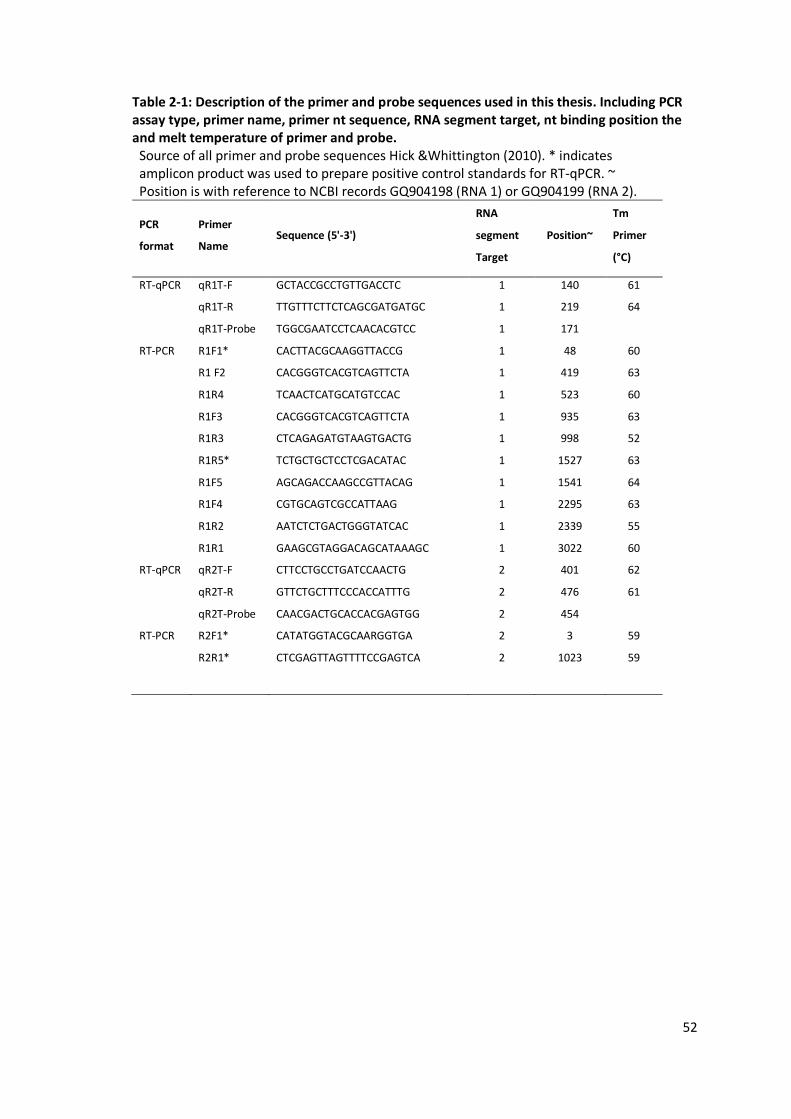
52
Table 2-1: Description of the primer and probe sequences used in this thesis. Including PCR assay type, primer name, primer nt sequence, RNA segment target, nt binding position the and melt temperature of primer and probe.
Source of all primer and probe sequences Hick &Whittington (2010). * indicates amplicon product was used to prepare positive control standards for RT-qPCR. ~ Position is with reference to NCBI records GQ904198 (RNA 1) or GQ904199 (RNA 2).
PCR
format
Primer
Name Sequence (5'-3')
RNA
segment
Target
Position~
Tm
Primer
(°C)
RT-qPCR qR1T-F GCTACCGCCTGTTGACCTC 1 140 61
qR1T-R TTGTTTCTTCTCAGCGATGATGC 1 219 64
qR1T-Probe TGGCGAATCCTCAACACGTCC 1 171
RT-PCR R1F1* CACTTACGCAAGGTTACCG 1 48 60
R1 F2 CACGGGTCACGTCAGTTCTA 1 419 63
R1R4 TCAACTCATGCATGTCCAC 1 523 60
R1F3 CACGGGTCACGTCAGTTCTA 1 935 63
R1R3 CTCAGAGATGTAAGTGACTG 1 998 52
R1R5* TCTGCTGCTCCTCGACATAC 1 1527 63
R1F5 AGCAGACCAAGCCGTTACAG 1 1541 64
R1F4 CGTGCAGTCGCCATTAAG 1 2295 63
R1R2 AATCTCTGACTGGGTATCAC 1 2339 55
R1R1 GAAGCGTAGGACAGCATAAAGC 1 3022 60
RT-qPCR qR2T-F CTTCCTGCCTGATCCAACTG 2 401 62
qR2T-R GTTCTGCTTTCCCACCATTTG 2 476 61
qR2T-Probe CAACGACTGCACCACGAGTGG 2 454
RT-PCR R2F1* CATATGGTACGCAARGGTGA 2 3 59
R2R1* CTCGAGTTAGTTTTCCGAGTCA 2 1023 59

53
2.2.5 Gel Electrophoresis
PCR amplicons were visualized by agarose gel electrophoresis. Agarose (Cat. 9010B, Scientifix,
Victoria) gel was prepared and run in 1x TAE (40 mM Tris, 20 mM acetic acid, and 1 mM EDTA)
buffer was stained with 0.05 μL ml-1 Gel Red (Cat. 41003, Biotium, Fisher Biotec, Australia).
The gels were loaded with 20 μL of PCR product and subjected to 60 min at 100 volts with a
Hyper Ladder 1 kb DNA ladder (BIO-33025, Bioline, NSW) (1 % gel). The gels were visualised on
a UV transilluminator.
2.2.6 Preparation of viral extract from natural VER outbreaks.
Viral extract for experimental challenge was prepared from the E.coioides VER outbreak.
Figure 2.1 displays image of pond culture and fish collected during the VER outbreak. The fish,
of average body mass of ~600 g were cultured in cages in commercial earth ponds in North
Queensland, Australia. Viral extract was prepared by separately pooling the brain and eyes
from several fish into sterile phosphate buffered saline (PBS) (Sigma Aldrich®). Brain tissue (8.3
g) was suspended in 57 mL and eye (18.9 g) was suspended in 100 mL of sterile PBS. Tissue in
PBS was frozen and thawed thrice from -25˚C to 4˚C, and homogenized using an Ultra-Turrax T
25 (IKA works) at 20,000 rpm for 5 min. Tubes of tissue were held in crushed ice throughout
the homogenization. Tissue homogenate was clarified in a Sorval RC 6+ centrifuge (Thermo
Scientific) using a F12s-6 x 500 LEX rotor for 10 min at 610 g at 4˚C. The supernatant was
removed and further clarified by centrifugation for 10 min at 3 803 g at 4 ˚C. The supernatant
was filtered through 0.45 µm and 0.22 µm filters (Sartorius). The extract was confirmed free of
culturable bacteria by inoculation onto sheep blood agar and overnight incubation at 28 ˚C.
Separate filtered supernatants from each tissue, herein termed viral extract, was stored in
sterile 50 mL centrifuge tubes at -20˚C.
Figure 2-1: Pond culture and E.coioides collected from a natural outbreak of VER used to prepare viral extract. (a) Fish were held in cages within 1-hectare earthen ponds containing estuarine water. (b). Euthanased gold-spotted grouper, E.coioides, that exhibited signs of VER. Fish displayed erratic swimming and hyperinflation of swim bladder but appeared otherwise healthy.
54
2.2.7 Cloning and sequencing of PCR products
PCR amplicons were excised from gels using individual scalpel blades (SBLDCL, Livingstone,
NSW) and purified using an Agarose Gel DNA Extraction Kit (Bioline). Purified amplicons were
cloned into competent Escherichia coli JM109 cells (A1360, Promega, NSW) using the pGEM-T
Easy Vector System (A1380, Promega, NSW), or One shot TOP10 chemically competent E. coli
using the pCR4-TOPO TA vector (K4575-01, Life Technologies, VIC) as per manufacturer’s
instructions. Transformed E. coli were grown for 16 - 18 hours at 37 °C on lysogeny broth (LB)
(Bertani, 1951) agar (Appendix 1) supplemented with 100 g mL-1 or 50 μg mL-1 ampicillin (Cat.
A9393-5G Sigma-Aldrich, NSW) for pGEM™ and TOPO™ clones respectively. Three white
colonies were selected from each agar plate and cultured at 150 rpm (Bioline incubator shaker
8500 Edwards Instruments, NSW) for 16 - 18 hours in lysogeny broth (Bertani, 1951) (Appendix
1) supplemented with 100 g mL-1 or 50 g mL-1 ampicillin (A9393-5G, Sigma-Aldrich, NSW) for
pGEM™ and TOPO™ cells respectively. Plasmid DNA was extracted using a High Pure™ Plasmid
Isolation kit (11754777001 Roche, NSW) as per manufacturer’s instruction. Plasmid extracts
were submitted for Sanger sequencing to Macrogen Inc. (Seoul, Korea) or the Australian
Genome Research Facility (Brisbane, Australia) for sequencing analysis. Bioinformatic analysis
was conducted to confirm Betanodavirus genome sequence and assess suitability of the
plasmids for application in subsequent qPCR analysis.
2.2.8 Bioinformatics analysis
Sequencing data was analysed using Geneious 9.1 or Geneious Prime (created by Biomatters,
available from http://www.geneious.com), and the tools available at National Centre for
Biotechnology Information (NCBI). These included Basic Local Alignment Search Tool (BLASTn
and BLASTx) and Open Reading Frame (ORF) finder tool using the default parameters of all of
the programs (http://www.ncbi.nlm.nih.gov).
2.2.9 Real-time PCR of viral extract
All cDNA reactions were performed as previously described in 2.1.3. For quantification of viral
copy number, real-time PCR was performed using a Rotor Gene 6000 system (Qiagen®). All
qPCRs were performed as separate assays in 20 µL reactions using SensiFAST™ Probe No ROX
master mix (Bioline) according to the manufacturer’s instructions including 2.5 µL of cDNA the
primer (200 nM) and probe (5 nM) sequences ( Hick & Whittington, 2010; see Table 1). The
thermal cycle profile consisted of a 3 min incubation at 95 ⁰C followed by 45 cycles of 15 s
denaturation at 95 ⁰C, 30 s annealing and extension at 60 ⁰C.

55
2.3 Husbandry and handling of groupers
2.3.1 Husbandry of groupers
For experimental challenges, juvenile giant Queensland grouper, Epinephelus lanceolatus,
were provided by a commercial hatchery, The Company One (Cairns, Australia). Fish were held
under the conditions approved within the JCU institutional animal ethics permits outlined in
the declaration of ethics (page iv). Experimental animals were held in recirculation systems
that consisted of 10 replicate systems of 2 x 300 L tanks with aeration, water circulation and
connected to a biological filter containing 200L of filter material and bio-wheels. The entire
system was housed in an experimental room with 12/12h controlled light/dark cycle and water
temperature was maintained between 24-28˚C. Saltwater was obtained from the Australian
Institute of Marine Science. Water was collected from the ocean and subjected to 5
treatments which included 5mm screen within a high-density polyethylene basket; a timex
hydroclone that removed particles down to 120 µm; Arkal spin filter 120 µm discs;
fractionation and a final ultrafiltration to 0.04 µm. Seawater concentration ranged from 26 to
35 ppt. Fish were fed twice daily ad lib on commercial fish feed pellet appropriate to the size of
the fish (Ridley AgriProducts Pty Ltd). Fish were acclimated for two weeks prior to
commencement of any experimental procedures.
2.3.2 Anaesthesia of groupers
2.3.2.1 Sedation of groupers
Fish were sedated to Stage 2 of fish anaesthesia (Coyle et al., 2004) using AQUI-S® anaesthetic
(AQUI-S® New Zealand Ltd).
Briefly fish were placed in a 40L Nally®Bin containing 30ppt seawater containing a working
solution of 15-20mg/L of AQUI-S®. For sedation, fish were held for sedation until they reached
a state of reduced swimming and breathing and partial loss of equilibrium. Fish retained
reaction to touch stimuli. Fish were sedated with AQUI-S® (AQUI-S) during all procedures that
were assessed as causing minor distress, which were principally all processes involving
injection.
For recovery fish were placed into a 40L Nally®Bin containing 30ppt seawater and gently
assisted to swim in a figure 8 direction ensuring they were always moving in a forward-facing
motion. When fish escape behaviour progressed beyond gentle, they were released into the
secondary container in the recovery tank. The secondary container consisted of a 20 L plastic
container with numerous small holes drilled into the container to allow water flow. When an
entire tank consignment had been subject to procedures, they were transferred to their

56
experimental holding tank using the secondary container. Aeration of water was provided
throughout the procedure.
2.3.2.2 Euthanasia of groupers
Fish were sedated to Stage 4 of fish anaesthesia (Coyle et al., 2004) using AQUI-S®. Briefly fish
were placed in a 10L Bucket containing 30ppt seawater containing a working solution of 15-
20mg/L of AQUI-S. For euthanasia, fish were held until death. Aeration of water was provided
throughout the procedure.
2.3.3 Intramuscular injection of groupers
All injection procedures, including vaccination and challenge with viral extract, were
conducted as intramuscular (IM) injection on sedated fish. Fish were injected into the dorsal
musculature using a sterile 25g x 5/8” Microlance™ needle (Becton Dickinson Ireland) attached
to a 1mL Terumo® syringe (Terumo Philippines). The site of injection was at the posterior
dorsal junction between the white and black marking on the juvenile E.lanceolatus (Figure 2.2).
To prevent possible skin infection, a drop of Betadine® solution (10% povidone-iodine) was
placed on the injection site immediately prior to and following injection. To reduce irritation to
skin, fish were handled with clean wet hands and placed on a clean wet cotton clot during
injection. The approximate length from the anterior end of the bottom jaw to the end of the
tail was measured with a ruler. (Figure 2.2). Approximate weight of fish was measured with a
portable 2kg digital kitchen scale (Propert).
.
Figure 2-2: Sedated juvenile E.lanceolatus with location of IM vaccine injection site indicated (Red arrow) and reference points of body positions (jaw to tail) used to determine
approximate body length. Image from fish used in experimental trial described in Chapter 8.

57
2.3.4 Pre-trial experiments
Prior to the commencement of each experimental challenge discussed in Chapters 7, 8 and 10,
two experimental procedures were conducted namely:
1. Confirmation of VNN free status of experimental fish.
2. Confirmation of pathogenicity of the viral extract.
Confirmation of VNN free status was determined by conducting RT-qPCR (as discussed in 2.1.9)
analysis on two pools of brain and eye tissue from ten fish using qR2T i.e. four RT-qPCR
samples including two samples each of pooled brain from five individual fish and pooled eye
from five individual fish.
Confirmation of pathogenicity of the viral extract was confirmed by challenging ten fish from
each experimental cohort with 100µl IM injection of RGNNV extract prepared as described in
2.1.6. Viral extract was injected in the approximate position of the vaccine injection indicated
in Figure 2.2. The ten fish were held in the same experimental circulation systems under the
conditions described in 2.2.1. Fish were monitored twice daily for signs of adverse health and
euthanized by overdose to Aqui-S when more than one sign of VER, including erratic
swimming, hyperinflation of swim bladder, disorientated floating, absence of feeding or severe
lethargy became evident. Analysis by RT-qPCR was conducted on pooled brain and eye tissue
as described in 2.1.9 to confirm detection of high copy number of RGNNV genome.
2.4 Results
2.4.1 Cloning and sequencing of PCR products
Results of cloning and sequencing of PCR products are discussed in Chapter 3: Phylogenetic
comparison of Betanodavirus genomes collected from North Queensland.
2.4.2 Real-time PCR of viral extract
Analysis of viral extract by RT-qPCR detected RGNNV with a Ct value of 13.95 (qR1T) and 10.64
(qR2T) which equates to an estimated calculated viral copy number of 1.99 x 10⁷ and 1.07 x 10⁸
copies mL-1 respectively. The average between each of the two copy number calculations, 6.36
x 107 copies mL-1 was accepted in subsequent calculations for preparing viral extract for
challenge studies. Viral extract was diluted to an approximate copy number of 6.36 x 10⁴ mL-1
in sterile PBS for challenge studies. Further discussion of RT-qPCR analysis is provided in
Chapters 7, 8 and 10.
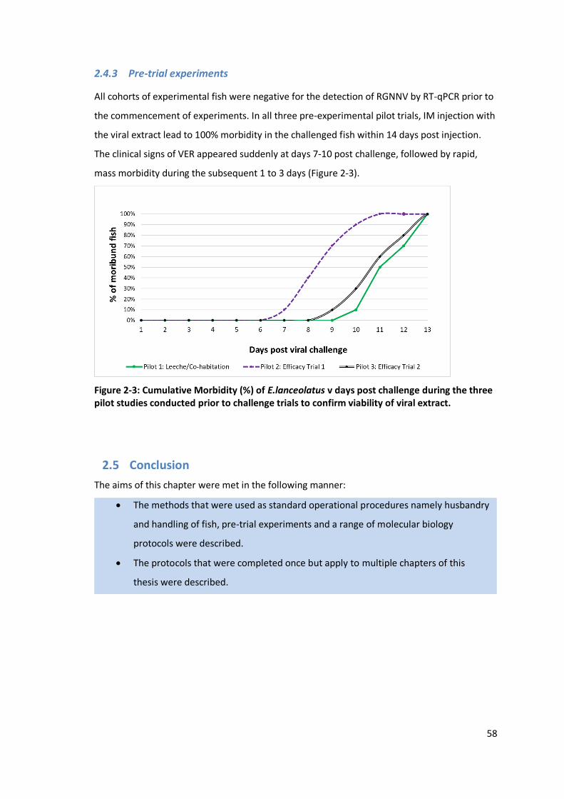
58
2.4.3 Pre-trial experiments
All cohorts of experimental fish were negative for the detection of RGNNV by RT-qPCR prior to
the commencement of experiments. In all three pre-experimental pilot trials, IM injection with
the viral extract lead to 100% morbidity in the challenged fish within 14 days post injection.
The clinical signs of VER appeared suddenly at days 7-10 post challenge, followed by rapid,
mass morbidity during the subsequent 1 to 3 days (Figure 2-3).
Figure 2-3: Cumulative Morbidity (%) of E.lanceolatus v days post challenge during the three pilot studies conducted prior to challenge trials to confirm viability of viral extract.
2.5 Conclusion
The aims of this chapter were met in the following manner:
The methods that were used as standard operational procedures namely husbandry
and handling of fish, pre-trial experiments and a range of molecular biology
protocols were described.
The protocols that were completed once but apply to multiple chapters of this
thesis were described.

59
CHAPTER 3. PHYLOGENETIC COMPARISON OF BETANODAVIRUS
GENOMES COLLECTED FROM VIRAL ENCEPHALOPATY AND
RETINOPATHY OUTBREAKS IN NORTH QUEENSLAND
Background
Aims of this Chapter
At the commencement of this study there were 2 complete sequences of
Betanodavirus RNA 1 and RNA 2 from Australian species, neither of which originated
from North Queensland.
The capsid protein, which is encoded by RNA 2, has been demonstrated as the
antigenic target of grouper antibodies to protect against VER.
There is variation in the capsid protein across the Betanodaviruses.
Knowledge of the genome sequences and level of variation in the RNA 2 of
Betanodavirus strains that cause VER in Northern Queensland is required to guide
decisions of vaccine development and design of dsRNA.
Knowledge of the genome sequences of RNA 1 is required to guide decisions for
dsRNA design to attempt NNV knockdown.
PCR amplicons from RNA 1 and RNA 2 are required to prepare synthetic positive
controls for the RT-qPCRs.
Obtain a viable viral extract from a natural VER outbreak in Northern Queensland.
Obtain RT-qPCR amplicons from the NNV strains associated with VER outbreaks in
North Queensland.
Determine the complete sequence of both mRNA segments of Betanodavirus
associated with natural VER outbreaks in marine finfish grow-out systems in
Northern Queensland.
Compare the complete sequences of mRNA 1 to other sequences in the NCBI
database to determine the species of Betanodavirus associated with VER in North
Queensland.
Compare the complete sequences of RNA 2 to other sequences in the NCBI
database to determine conservation of the capsid protein temporally and
geographically.
60
3.1 Introduction
Knowledge of the RNA 2 segment sequence that encodes the capsid protein of strains causing
VER in Australian grouper is critical to multiple decisions regarding vaccine formulation. In
addition to ensuring targeted protection against Betanodavirus strains that are associated with
VER outbreaks, herein referred to as NNV strains, knowledge of capsid protein sequence
assists in determining:
the level of variation in NNV strains that cause VER;
the potential geographic market of an effective vaccine and
the duration for which the vaccine could be expected to be effective considering the
typically rapid mutation rate of RNA viruses.
Although North Queensland was among the first regions in the world to report VER outbreaks,
there are few genome sequences of Australian NNV strains. At the commencement of this
study, there were only two complete genome sequences of Betanodavirus from Australian
species within the NCBI database, neither of which originated from Queensland (Hick &
Whittington, 2010). There are a further eight partial sequences of RNA 2 segments collected
from Australian species (Moody et al., 2009).
The current species classification of Betanodavirus recognised by the ICTV, named with the
original host names of striped jack, red-spotted grouper, barfin flounder and tiger puffer was
initially based on analysis of a 606nt conserved region, termed T2 and a 289nt highly variable
region, termed T4 of the RNA 2 (Nishizawa et al., 1997). The species divisions align with three
distinct serotype divisions determined by antibody reaction to the capsid protein which is
encoded by RNA 2 (Nishizawa et al., 1997, Johansen et al., 2004 and Mori et al., 2003) (Table
3.1). Numerous serological studies have reported a lack of in vitro and in vivo cross-reactivity
between two species namely the RGNNV and SJNNV (Mori et al., 2003; Chi et al., 2003 and
Pascoli et al., 2019). However, there is some cross reactivity between RGNNV and BFNNV
(Mori et al., 2003).
In recognition of an expanded host range beyond those of the original species names, Thiery et
al. (2004) proposed a division of the Betanodavirus into subgroup clusters based on analysis of
the highly variable region (T4) of RNA 2. The cluster proposal retained TPNNV and SJNNV as
distinct single clusters, Clusters III and IV respectively but further differentiated the RGNNV
into two, namely Ia and Ib and BFNNV into three: IIa, IIb and IIc. The clusters, based on
nucleotide comparison of the T4 region, also tended to align with temperature and geographic
origin (Thiery et al., 2004) (Table 3.1).
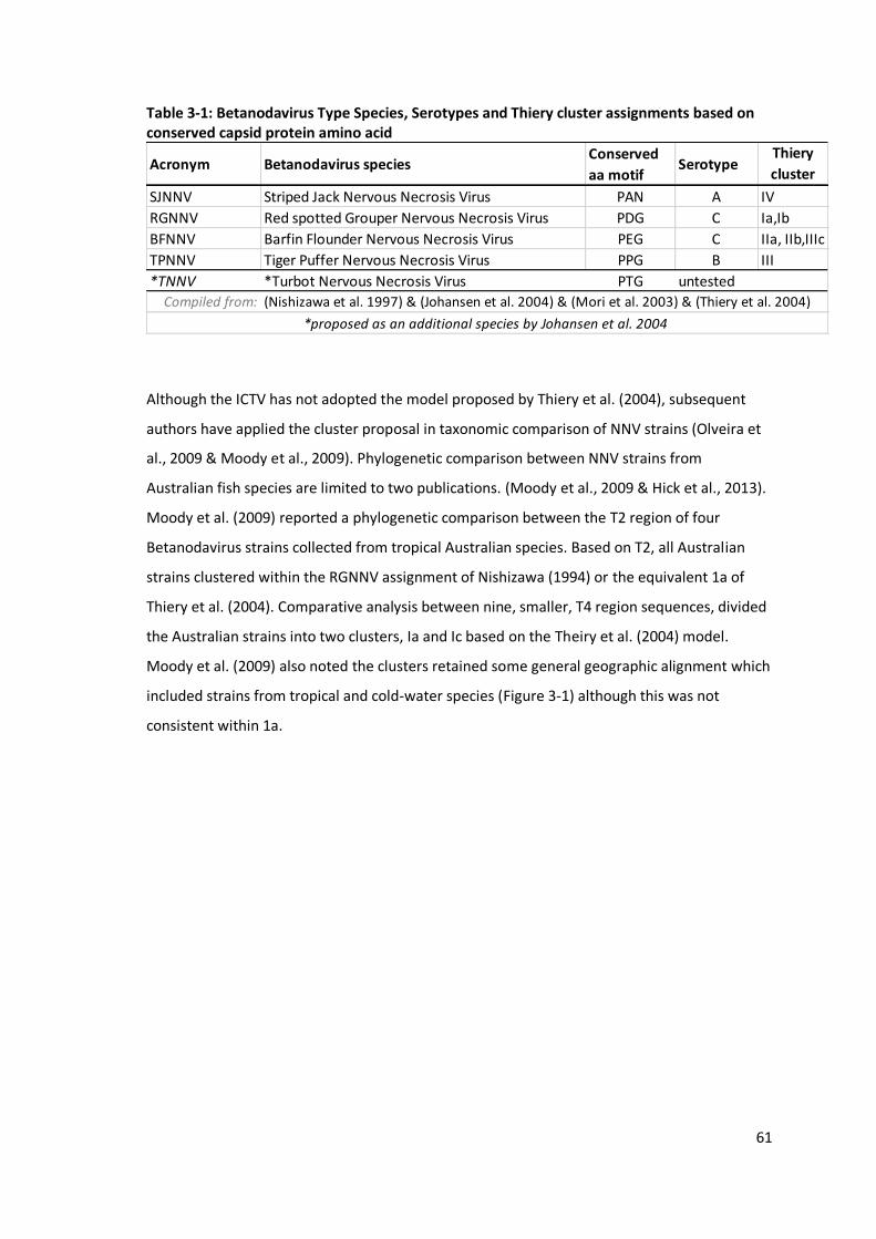
61
Table 3-1: Betanodavirus Type Species, Serotypes and Thiery cluster assignments based on conserved capsid protein amino acid
Although the ICTV has not adopted the model proposed by Thiery et al. (2004), subsequent
authors have applied the cluster proposal in taxonomic comparison of NNV strains (Olveira et
al., 2009 & Moody et al., 2009). Phylogenetic comparison between NNV strains from
Australian fish species are limited to two publications. (Moody et al., 2009 & Hick et al., 2013).
Moody et al. (2009) reported a phylogenetic comparison between the T2 region of four
Betanodavirus strains collected from tropical Australian species. Based on T2, all Australian
strains clustered within the RGNNV assignment of Nishizawa (1994) or the equivalent 1a of
Thiery et al. (2004). Comparative analysis between nine, smaller, T4 region sequences, divided
the Australian strains into two clusters, Ia and Ic based on the Theiry et al. (2004) model.
Moody et al. (2009) also noted the clusters retained some general geographic alignment which
included strains from tropical and cold-water species (Figure 3-1) although this was not
consistent within 1a.
Acronym Betanodavirus speciesConserved
aa motifSerotype
Thiery
cluster
SJNNV Striped Jack Nervous Necrosis Virus PAN A IV
RGNNV Red spotted Grouper Nervous Necrosis Virus PDG C Ia,Ib
BFNNV Barfin Flounder Nervous Necrosis Virus PEG C IIa, IIb,IIIc
TPNNV Tiger Puffer Nervous Necrosis Virus PPG B III
*TNNV *Turbot Nervous Necrosis Virus PTG untested
*proposed as an additional species by Johansen et al. 2004
Compiled from: (Nishizawa et al. 1997) & (Johansen et al. 2004) & (Mori et al. 2003) & (Thiery et al. 2004)
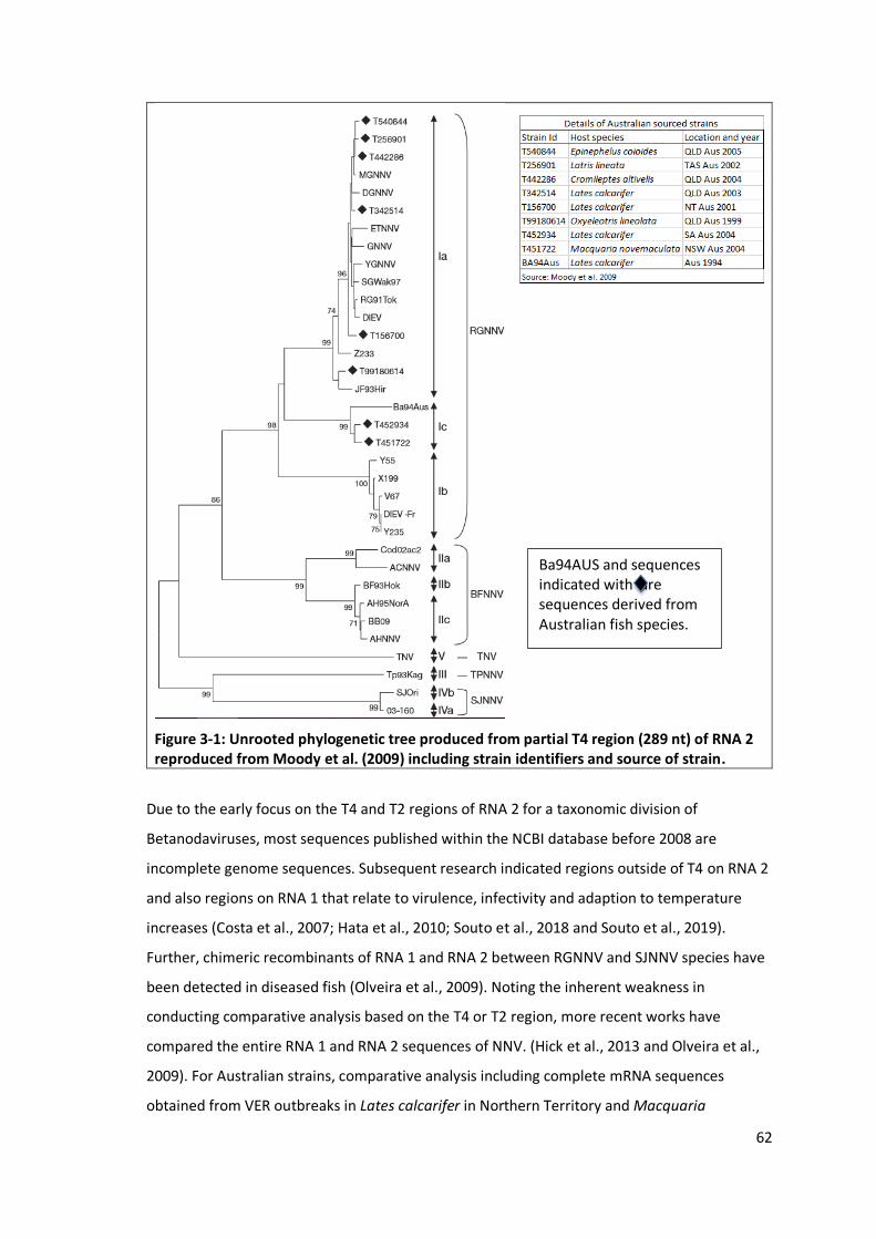
62
Figure 3-1: Unrooted phylogenetic tree produced from partial T4 region (289 nt) of RNA 2 reproduced from Moody et al. (2009) including strain identifiers and source of strain.
Due to the early focus on the T4 and T2 regions of RNA 2 for a taxonomic division of
Betanodaviruses, most sequences published within the NCBI database before 2008 are
incomplete genome sequences. Subsequent research indicated regions outside of T4 on RNA 2
and also regions on RNA 1 that relate to virulence, infectivity and adaption to temperature
increases (Costa et al., 2007; Hata et al., 2010; Souto et al., 2018 and Souto et al., 2019).
Further, chimeric recombinants of RNA 1 and RNA 2 between RGNNV and SJNNV species have
been detected in diseased fish (Olveira et al., 2009). Noting the inherent weakness in
conducting comparative analysis based on the T4 or T2 region, more recent works have
compared the entire RNA 1 and RNA 2 sequences of NNV. (Hick et al., 2013 and Olveira et al.,
2009). For Australian strains, comparative analysis including complete mRNA sequences
obtained from VER outbreaks in Lates calcarifer in Northern Territory and Macquaria
Ba94AUS and sequences indicated with are sequences derived from Australian fish species.

63
novemaculata in New South Wales have been reported. (Hick et al., 2013). The authors formed
a similar conclusion to Moody et al. (2009) regarding the taxonomic division of Australian NNV
strains with an exception of aligning the Ba94Aus strain, collected from barramundi, within the
tropical strains 1a rather than 1c temperate strains. (Figure 3.2). Although the divisions of
Australian NNV strains tend to align with geographic source, the strain collected from striped
trumpeter, Latris lineata, from Tasmania aligns more to the tropical species division. It is
possible the divisions between 1a and 1c in Australian strains relate to other factors such as
marine v freshwater systems. The striped trumpeter is a marine species whereas the
Australian bass, M.novemaculata is a freshwater species. Although barramundi, L.calcarifer are
naturally found in tropical waters, as a euryhaline species, it has been translocated into
temperate regions in Australia where is cultured within systems that have access to naturally
heated spring water or apply heated spring water to warm marine systems. There was no
information in the NCBI database entry to indicate if the strain from barramundi collected
from South Australia (T452934) was from a marine or freshwater system.
Figure 3-2: Summary of strain classification based on NNV RNA 2 sequences derived from Australian fish species (including strain reference identified and source). Modified from Hick et al. (2013). The “tropical” strains are aligned within 1a cluster (Black rectangle) and “temperate” strains aligned within 1c cluster (blue rectangle).
Whilst there has been no detection of any Betanodavirus strain other than RGNNV in Australia,
the division of strains into clusters indicates a degree of variability between Australian NNV
strains. The scarcity of complete sequence data of tropical NNV strains within prior work and
the large financial cost of vaccine production, necessitates careful investigation into the
complete genome sequences of strains causing VER in tropical grouper species and other
64
species within their geographic range of grouper culture. This study reports on the complete
mRNA sequence of Betanodavirus associated with natural VER outbreaks in marine finfish
grow-out systems in Northern Queensland and their phylogenetic position within the
collection of NNV strains affecting Australian fish species. Knowledge of the complete mRNA
sequences of the strains of NNV that cause VER in grouper in North Queensland is required to
ensure the therapies to be prepared within this project specifically target those strains that are
present in Northern Queensland.
3.2 Materials and Methods
3.2.1 Collection of samples from natural VER outbreaks
Samples of brain and eye were collected from fish from three aquaculture farms in Northern
Queensland displaying clinical signs of VER. Viral Encephalopathy and Retinopathy was
confirmed by the Queensland Government Biosecurity Sciences Veterinary Diagnostic
laboratory (BSL) (farmers personal comms.). In this study, RT-PCR and sequencing analysis was
performed to confirm VER. A summary of the sample source is provided in Table 3.3.
3.2.2 Sequencing of Betanodavirus from natural disease outbreaks
Total viral nucleic extracts were prepared by dissecting eye and brain tissues from frozen fish
collected during natural VER outbreaks. Extraction was completed using the High Pure™ Viral
Nucleic Acid kit (Roche 11858874001) according to the manufacturer’s instructions. Cloning
and sequencing was conducted as previously described in 2.1.3 to 2.1.7 using the primers
details in Table 2.1 (Section 2.1.4).
3.2.3 Additional Sequences obtained for phylogenetic analysis
Sequences of RNA 1 and RNA 2, were compared with strains obtained from the NCBI GenBank
(http://www.ncbi.nlm.nih.gov/) nucleotide database including reference strains of
Betanodavirus species and similar geographic source (Australia). Sequences of NNV from each
of the reference strains of Betanodavirus species are indicated in Table 3.2. Sequences of NNV
obtained from Australian fish were collected from the NCBI data base and other researchers as
indicated in Table 3.3.
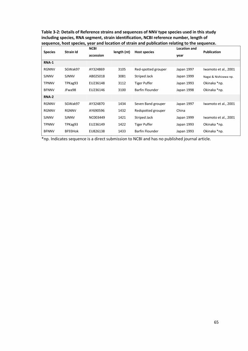
65
Table 3-2: Details of Reference strains and sequences of NNV type species used in this study including species, RNA segment, strain identification, NCBI reference number, length of sequence, host species, year and location of strain and publication relating to the sequence.
Species Strain Id NCBI
accession length (nt) Host species
Location and
year Publication
RNA-1
RGNNV SGWak97 AY324869 3105 Red-spotted grouper Japan 1997 Iwamoto et al., 2001
SJNNV SJNNV AB025018 3081 Striped Jack Japan 1999 Nagai & Nishizawa np.
TPNNV TPKag93 EU236148 3112 Tiger Puffer Japan 1993 Okinaka *np.
BFNNV JFwa98 EU236146 3100 Barfin Flounder Japan 1998 Okinaka *np.
RNA-2
RGNNV SGWak97 AY324870 1434 Seven Band grouper Japan 1997 Iwamoto et al., 2001
RGNNV RGNNV AY690596 1432 Redspotted grouper China
SJNNV SJNNV NC003449 1421 Striped Jack Japan 1999 Iwamoto et al., 2001
TPNNV TPKag93 EU236149 1422 Tiger Puffer Japan 1993 Okinaka *np.
BFNNV BF93Hok EU826138 1433 Barfin Flounder Japan 1993 Okinaka *np.
*np. Indicates sequence is a direct submission to NCBI and has no published journal article.

66
Table 3-3: Details of Australian sourced NNV sequences used in this study including species, RNA segment, strain, NCBI reference number, length of sequence, host species, year and location of strain and publication relating to the sequence.
Strain Id NCBI
accession length (nt) Host species Location and year Publication
RNA-1
P14-10380 KT390712 2871 E. lanceolatus QLD Aus 2014 Agnithortri et al. (2015)
Lc3NQAus MH181161 3090 Lates calcarifer QLD Aus 2013 Condon et al. (2019)
MnNNV12/06 GQ402012 2998 M. novemaculata NSW Aus 2006 Hick et al. (2013)
LcNNV_09/07 GQ402010 2998 Lates calcarifer NT Aus 2007 Hick et al. (2013)
Ec2NQAus
3090 E. coioides QLD Aus 2015 This study
El1NQAus
3090 E. lanceolatus QLD Aus 2013 This study
RNA-2
T540844 EF591369 606 E. coioides QLD Aus 2005 Moody et al. (2009)
T256901 EF591370 289 Latris lineata TAS Aus 2002 Moody et al. (2009)
T442286 EF591371 832
Cromileptes
altivelis QLD Aus 2004 Moody et al. (2009)
T342514 EF591372 832 Lates calcarifer QLD Aus 2003 Moody et al. (2009)
T156700 EF591367 289 Lates calcarifer NT Aus 2001 Moody et al. (2009)
T99180614 EF591368 832
Oxyeleotris
lineolata QLD Aus 1999 Moody et al. (2009)
T452938 EF591366 294 Lates calcarifer SA Aus 2004 Moody et al. (2009)
T451722 EF591365 294 M. novemaculata NSW Aus 2004 Moody et al. (2009)
LcNNV_09/07 GQ402011 1017 Lates calcarifer NT Aus 2007 Hick et al. (2013)
MnNNV12/06 GQ402013 1017 M. novemaculata NSW Aus 2006 Hick et al. (2013)
P14-10380 KT390714 990 E. lanceolatus QLD Aus 2014 Agnithortri et al. (2015)
Lc3NQAus MH017207 1017 Lates calcarifer QLD Aus 2013 Condon et al. (2019)
Ec2NQAus
E. coioides QLD Aus 2015 This study
El1NQAus
E. lanceolatus QLD Aus 2013 This study
E. indicates Epinephelus M. indicates Macquaria. Sequences obtained within this study are
indicated in bold.
3.2.4 Sequence alignment
Sequencing data was analysed as previously described in 2.1.6 and 2.1.7. The depth of
sequence coverage was a minimum of 3 sequences reads per nucleotide over the entire
published sequence. Phylogenetic comparison between sequences was conducted using the
Geneious v9.1 software, using the default algorithm settings. Neighbourhood joining trees
were prepared using the Tamura-Nei genetic distance model (Biomatters
http://www.geneious.com).

67
3.3 Results
Complete mRNA sequences were obtained from the Lates calcarifer (ref Lc3NQAus) ,
Epinephelus coioides (ref Ec2NQAus) and Epinephelus lanceolatus (ref El3NQAus) VER
outbreaks. The sequences from the Lates calcarifer outbreak were lodged in the NCBI
nucleotide database as accessions MH181161 (RNA 1) and MH017207 (RNA 2) (Condon et al.,
2019). Phylogenetic analysis of RNA 1 and RNA 2 indicate all three NNV strains collected in this
study display greatest homology to the RGNNV species.
3.3.1 RNA-1
Comparison of the complete mRNA of the RNA 1 segment from the VNN strains collected in
this study indicate the strains display strong nucleotide sequence homology (99-97%) to each
other (Refer to Table 3.4). Comparison with the Betanodavirus species type strains indicates all
3 NQAus strains display strongest homology to the RGNNV reference strain (95-97%) with
lesser homology to BFNNV (82%), SJNNV (82%) and TPNNV (82%) reference strains. (Figure 3.3
and Table 3.4). Comparison with the other Australian strains in the NCBI database indicates
strong homology between all of the Australian strains (95-98%) and the original RGNNV
isolated from Sevenband grouper in Japan in 1997 (Table 3.5).
Created by Geneious Prime (Biomatters) http://www.geneious.com. Strains coloured pink are RGNNV aligned.
Figure 3-3: Neighbour-joining tree of mRNA RNA 1 segment of the Betanodavirus from this study aligned against the reference species strains of NNV.

68
Figure 3-4: Neighbour-joining tree of mRNA RNA 1 segment of Australian strains of Betanodavirus determined by the Tamura-Nei genetic distance model Created by Geneious Prime Biomatters http://www.geneious.com. Strains indicated in red were sourced within a ~500km coastal zone in North Queensland.

69
Table 3-4: Displaying Percentage Homology between mRNA sequences of RNA 1 segments from the three NNV strains sequenced in this study (El2NQAus, Lc2NQAus and Ec2NQAus) in comparison to the NNV type species RGNNV, SJNNV, BFNNV and TPNNV including details of NCBI reference, host species and location and year of collection.
Sequence Source details This study Species allocation and reference identifiers
Strain NCBI ref. Length
(nt) Host species Location
El2NQAus Ec2NQAus Lc3NQAus AY324869 AB025018 EU236148 EU236146
RGNNV SJNNV TPNNV BFNNV
El2NQAus 3030 E.lanceolatus QLD Aus
2015 97.27 99.17 96.30 82.17 82.73 82.50
Ec2NQAus 3029 E.coioides QLD Aus
2014 97.27 97.06 97.55 82.09 82.61 82.81
Lc3NQAus MH181161 3090 L.calcarifer QLD Aus
2013 99.17 97.06 95.69 81.95 82.53 82.04
Redspotted Grouper NNV
(RGNNV) AY324869 3105
Sevenband grouper
Japan 1997
96.30 97.55 95.69 82.12 82.62 82.83
Striped Jack NNV (SJNNV)
AB025018 3081 Striped Jack Japan 1999
82.17 82.09 81.95 82.12 82.62 83.08
Tiger Puffer NNV (TPNNV)
EU236148 3112 Tiger Puffer Japan 1993
82.73 82.61 82.53 82.62 82.62 91.26
Barfin Flounder NNV (BFNNV)
EU236146 3101 Barfin
Flounder Japan 1993
82.50 82.81 82.04 82.83 83.08 91.26
Strains obtained in this study are indicated in bold and grey shading.
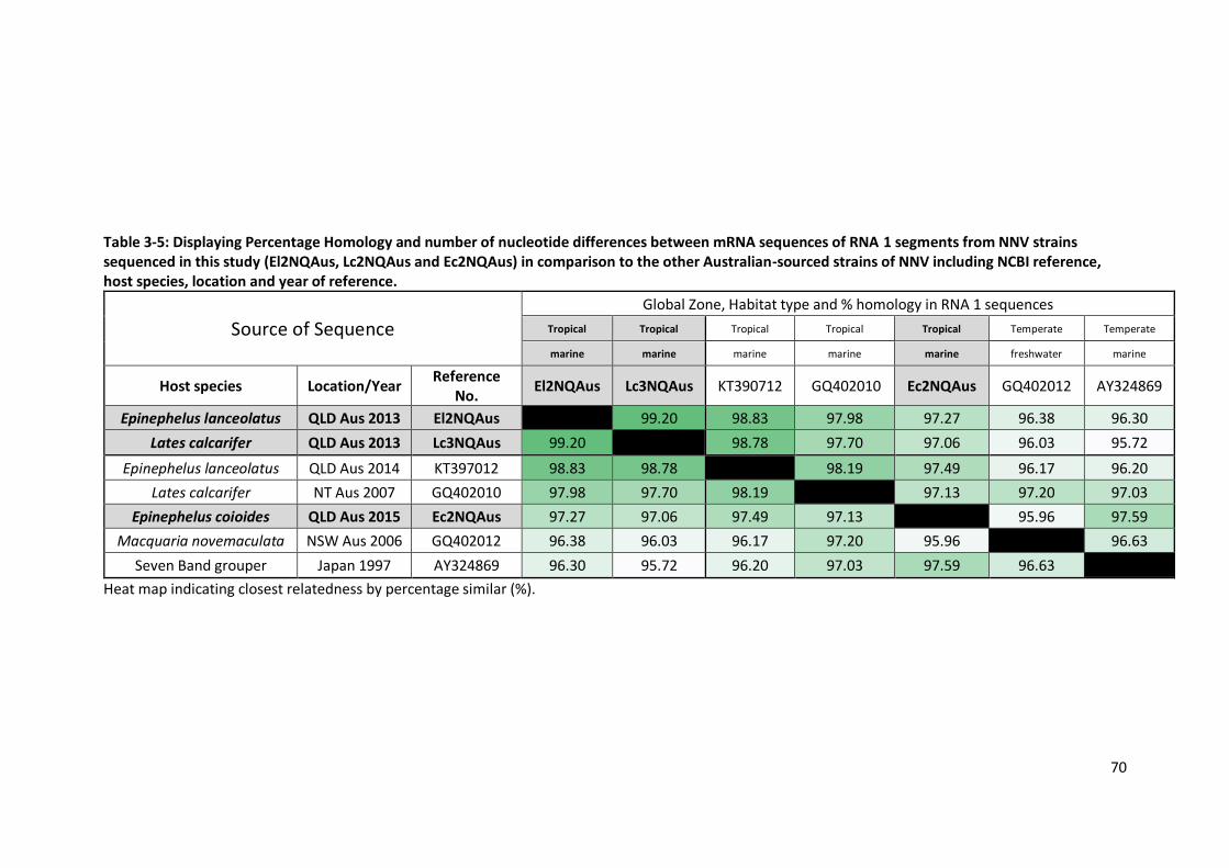
70
Table 3-5: Displaying Percentage Homology and number of nucleotide differences between mRNA sequences of RNA 1 segments from NNV strains sequenced in this study (El2NQAus, Lc2NQAus and Ec2NQAus) in comparison to the other Australian-sourced strains of NNV including NCBI reference, host species, location and year of reference.
Source of Sequence
Global Zone, Habitat type and % homology in RNA 1 sequences
Tropical Tropical Tropical Tropical Tropical Temperate Temperate
marine marine marine marine marine freshwater marine
Host species Location/Year Reference
No. El2NQAus Lc3NQAus KT390712 GQ402010 Ec2NQAus GQ402012 AY324869
Epinephelus lanceolatus QLD Aus 2013 El2NQAus 99.20 98.83 97.98 97.27 96.38 96.30
Lates calcarifer QLD Aus 2013 Lc3NQAus 99.20 98.78 97.70 97.06 96.03 95.72
Epinephelus lanceolatus QLD Aus 2014 KT397012 98.83 98.78 98.19 97.49 96.17 96.20
Lates calcarifer NT Aus 2007 GQ402010 97.98 97.70 98.19 97.13 97.20 97.03
Epinephelus coioides QLD Aus 2015 Ec2NQAus 97.27 97.06 97.49 97.13 95.96 97.59
Macquaria novemaculata NSW Aus 2006 GQ402012 96.38 96.03 96.17 97.20 95.96 96.63
Seven Band grouper Japan 1997 AY324869 96.30 95.72 96.20 97.03 97.59 96.63
Heat map indicating closest relatedness by percentage similar (%).
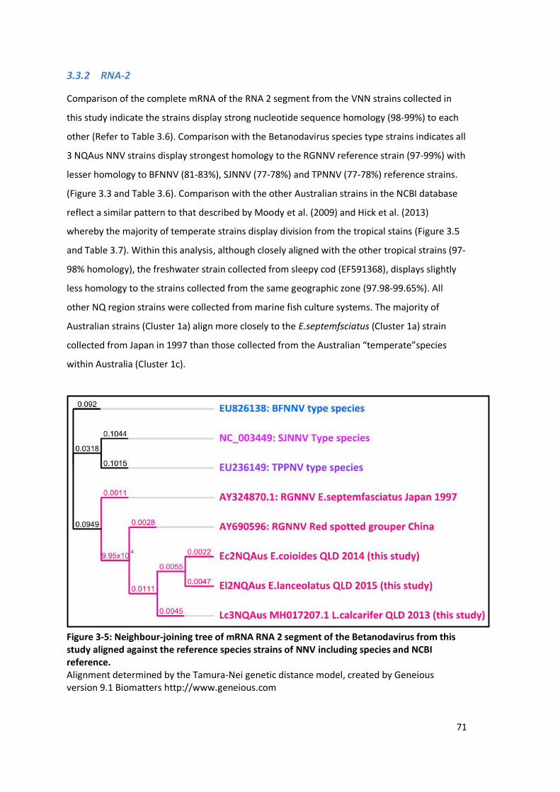
71
3.3.2 RNA-2
Comparison of the complete mRNA of the RNA 2 segment from the VNN strains collected in
this study indicate the strains display strong nucleotide sequence homology (98-99%) to each
other (Refer to Table 3.6). Comparison with the Betanodavirus species type strains indicates all
3 NQAus NNV strains display strongest homology to the RGNNV reference strain (97-99%) with
lesser homology to BFNNV (81-83%), SJNNV (77-78%) and TPNNV (77-78%) reference strains.
(Figure 3.3 and Table 3.6). Comparison with the other Australian strains in the NCBI database
reflect a similar pattern to that described by Moody et al. (2009) and Hick et al. (2013)
whereby the majority of temperate strains display division from the tropical stains (Figure 3.5
and Table 3.7). Within this analysis, although closely aligned with the other tropical strains (97-
98% homology), the freshwater strain collected from sleepy cod (EF591368), displays slightly
less homology to the strains collected from the same geographic zone (97.98-99.65%). All
other NQ region strains were collected from marine fish culture systems. The majority of
Australian strains (Cluster 1a) align more closely to the E.septemfsciatus (Cluster 1a) strain
collected from Japan in 1997 than those collected from the Australian “temperate”species
within Australia (Cluster 1c).
Figure 3-5: Neighbour-joining tree of mRNA RNA 2 segment of the Betanodavirus from this study aligned against the reference species strains of NNV including species and NCBI reference. Alignment determined by the Tamura-Nei genetic distance model, created by Geneious version 9.1 Biomatters http://www.geneious.com

72
Figure 3-6: Neighbour-joining tree of mRNA RNA 2 segment of Australian strains of Betanodavirus from the NCBI database aligned against the strains collected from this study Determined by the Tamura-Nei genetic distance model created by Geneious version 9.1 using the default parameters. Biomatters http://www.geneious.com. Strains indicated in red were sourced within a ~500km coastal zone in North Queensland.
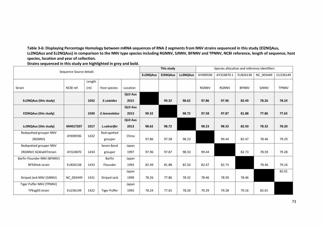
73
Table 3-6: Displaying Percentage Homology between mRNA sequences of RNA 2 segments from NNV strains sequenced in this study (El2NQAus, Lc2NQAus and Ec2NQAus) in comparison to the NNV type species including RGNNV, SJNNV, BFNNV and TPNNV, NCBI reference, length of sequence, host species, location and year of collection. Strains sequenced in this study are highlighted in grey and bold.
Sequence Source details This study Species allocation and reference identifiers
Ec2NQAus El2NQAus Lc3NQAus AY690596 AY324870.1 EU826138 NC_003449 EU236149
Strain NCBI ref.
Length
(nt) Host species Location RGNNV RGNNV BFNNV SJNNV TPNNV
Ec2NQAus (this study)
1032 E.coioides
QLD Aus
2015
99.32 98.62 97.86 97.96 82.49 78.26 78.24
El2NQAus (this study)
1030 E.lanceolatus
QLD Aus
2013 99.32
98.72 97.58 97.87 81.88 77.86 77.65
Lc3NQAus (this study) MH017207 1017 L.calcarifer
QLD Aus
2013 98.62 98.72
98.23 98.33 82.50 78.32 78.20
Redspotted grouper NNV
(RGNNV) AY690596 1432
Red-spotted
grouper China
97.86 97.58 98.23
99.44 82.47 78.46 79.29
Redspotted grouper NNV
(RGNNV) SGWak97strain AY324870 1434
Seven Band
grouper
Japan
1997 97.96 97.87 98.33 99.44
82.73 78.59 79.28
Barfin Flounder NNV (BFNNV)
BF93Hok strain EU826138 1433
Barfin
Flounder
Japan
1993 82.49 81.88 82.50 82.47 82.73
78.46 79.16
Striped Jack NNV (SJNNV) NC_003449 1431 Striped Jack
Japan
1999 78.26 77.86 78.32 78.46 78.59 78.46
82.01
Tiger Puffer NNV (TPNNV)
TPKag93 strain EU236149 1422 Tiger Puffer
Japan
1993 78.24 77.65 78.20 79.29 79.28 79.16 82.01

74
Heat map indicating closest relatedness by percentage similar (%) and number of different nucleotides. Sequences obtained in this study are indicated in
bold and grey shading. Differences and homology of sequences collected in this study are enclosed by the rectangle.
Table 3-7: Displaying Percentage Homology and number of nucleotide differences between mRNA sequences of RNA 2 segments from NNV strains sequenced in this study (El2NQAus, Lc2NQAus and Ec2NQAus) in comparison to the other Australian sourced strains of NNV from the NCBI database including Host species, NCBI reference, location and year of collection.
Strains sequenced in this study are highlighted in grey and bold. Temperate Tropical Tropical Tropical Tropical Tropical Tropical Tropical Tropical Tropical Tropical Tropical Temperate Temperate Temperate Zone
marine marine marine marine marine marine marine marine marine marine marine freshwater freshwater not recorded freshwater Habitat
Host species Location/Year Reference No. EF591370 EF591371 EF591367 EF591369 EF591372 KT397014 El2NQAus Lc3NQAus Ec2NQAus GQ402011 AY324870 EF591368 EF591365 EF591366 GQ402013
Latris lineata TAS Aus 2002 EF591370 99.65 98.62 99.31 99.31 99.31 98.96 98.27 98.96 98.62 98.62 96.19 85.81 85.81 85.81
Cromileptes altivelis QLD Aus 2004 EF591371 1 98.96 99.51 99.4 99.16 99.16 98.2 98.68 98.56 99.04 97.24 86.05 86.05 92.19
Lates calcarifer NT Aus 2001 EF591367 4 3 98.62 98.62 98.62 98.27 97.58 98.27 97.92 97.92 96.89 86.51 86.51 86.51
Epinephelus coioides QLD Aus 2005 EF591369 2 3 4 99.51 99.18 99.18 98.19 98.68 98.52 99.18 96.87 85.81 85.81 90.59
Lates calcarifer QLD Aus 2003 EF591372 2 5 4 3 99.28 99.28 98.32 98.8 98.68 99.16 97.36 86.05 86.05 92.43
Epinephelus lanceolatus QLD Aus 2014 KT397014 2 7 4 5 6 99.7 98.89 99.39 98.89 99.19 97.12 85.71 86.4 92.22
Epinephelus lanceolatus QLD Aus 2013 El2NQAus 3 7 5 5 6 3 98.72 99.32 98.62 97.87 97.12 85.37 86.05 92.33
Lates calcarifer QLD Aus 2013 Lc3NQAus 5 15 7 11 14 11 13 98.62 98.13 98.33 96.15 84.69 85.37 91.84
Epinephelus coioides QLD Aus 2015 Ec2NQAus 3 11 5 8 10 6 7 14 98.53 97.96 96.88 85.37 86.05 92.33
Lates calcarifer NT Aus 2007 GQ402011 4 12 6 9 11 11 14 19 15 98.82 96.76 85.71 86.4 92.33
Seven Band grouper Japan 1997 AY324870.1 4 8 6 5 7 8 22 17 21 12 97.48 85.71 85.71 92.33
Oxyeleotris lineolata QLD Aus 1999 EF591368 11 23 9 19 22 24 24 32 26 27 21 87.76 87.08 91.83
Macquaria novemaculata NSW Aus 2004 EF591365 41 41 39 41 41 42 43 45 43 42 42 36 98.64 97.96
Lates calcarifer SA Aus 2004 EF591366 41 41 39 41 41 40 41 43 41 40 42 38 4 97.96
Macquaria novemaculata NSW Aus 2006 GQ402013 41 65 39 57 63 77 78 83 78 78 78 68 6 6
% h
om
olo
gy
Number of differences in sequence
Source of SequenceGlobal Zone, Habitat type, Reference No. and Matrix of homology (%) and Difference in RNA 2 sequences
75
3.4 Discussion
Prior to this study, knowledge of the VNN strains present in Australia, were not sufficient to
allow robust decisions to support vaccine development nor design targeted dsRNA for
attempted knockdown of NNV. The acquisition of complete genome sequences of RNA 1 and
RNA 2 collected from strains associated with VER outbreaks in marine aquaculture systems in
North Queensland indicate the highest level of conservation to the RGNNV. Unfortunately,
comprehensive comparison of RNA 1 segments across Australian species are limited. There are
only three RNA 1 segment sequences from Australian species in the NCBI database ( Hick &
Whittington 2010; Agnihotri et al., 2015). Nonetheless, within the comparison containing six
sequences, the RNA 1 segments from Australian strains of NNV display a very high level of
conservation, exceeding 97%, across the tropical geographic, and 96% across temporal zones.
Similarly, the sequences of RNA 2 segments of NNV are highly conserved between strains that
have been associated with VER outbreaks in Australia since 2001. High conservation extends
not only across Australian strains but also across diverse geographic (Japan, China and the
Mediterranean) and temporal sources (earliest strains collected in 1997). Previous researchers
have noted the conservation of the capsid protein. The high level of conservation of RNA 2
which contains the mRNA for the capsid protein, the antigen target for vaccine preparation,
indicates a vaccine against the Australian strains could have wider global application.
Temporally, the capsid protein gene has a low evolutionary rate (Thiery et al., 2004). In
particular, the RNA 2 sequences obtained in this study display 97.8 to 98.3% homology to the
RNA 2 segment of the type species for RGNNV, namely SGWak97, that was collected from
E.septemfasciatus in Japan in 1997 (Iwamoto, 2001). The NQAus NNV strains collected in this
study have less than 20 nucleotide differences from the original RGNNV strain or less than 1.31
changes per 1000 nt per year. Considering the nucleotide differences translate to very few
changes in the capsid protein during the past 17+ years, it is also likely a vaccine will be
suitable for a long period of time. Further investigation regarding the conservation of the
protein motifs will be discussed in the next chapter.

76
3.5 Conclusion
The following outcomes and conclusions were drawn from this Chapter:
Data presented in this chapter was published:
Condon K., Bochow S., Ariel E., and Miller T., (2019) Complete sequence of Betanodavirus from
Australian barramundi, Lates calcarifer. Microbiology Resource Announcements 8.
https://doi.org./10.1128/MRA.00081-19
NNV was confirmed to be present in high copy number in the viral extract.
Complete sequence of Betanodavirus RNA 1 and RNA 2 was obtained from three
VER outbreaks in North Queensland.
Comparative phylogenetic analysis indicates the strains are of the RGNNV species.
Bioinformatic analysis indicates the RNA 1 and RNA 2 sequence and theoretically,
the translated proteins are highly conserved in the RGNNV species associated with
VER in tropical marine species in North Queensland.
Comparative analysis with RGNNV from the NCBI database indicates the RNA 1 and
2 sequence and theoretically, translated proteins display remarkable conservation
across a broad global species, geographic and temporal range.
Sequence of mRNA 2 was obtained for application to produce standard RT-qPCR
control plasmids and recombinant protein expression.
Sequence of mRNA 1 was obtained which indicated the strains causing VER are pure
RGNNV genomes and the lack of chimeric recombination with other NNV strains.
Gene sequence of RNA 1 was obtained for application to produce standard RT-qPCR
control plasmids.

77
CHAPTER 4. REVIEW AND IDENTIFICATION OF THE FUNCTIONAL
MOTIFS OF THE BETANODAVIRUS GENOME
Background
Amino acid motifs that are critical to the production of neutralising antibodies
against RGNNV in grouper have been identified in the capsid protein.
Amino acid motifs that are linked with increased virulence, or critical to viral
replication of Betanodavirus have been identified in the capsid Protein, Protein 1
and B1 and B2 proteins.
Amino acid motifs that are linked with host species specificity have been
identified in the RGNNV capsid protein structures.
Confirmation the strain of RGNNV to be used in this study contains virulence-
associated motifs is required for vaccine preparation and to have confidence in
the translation of the project results to industry application.
Knowledge of the functional motifs is required to direct targeted knockdown of
the RGNNV using dsRNA.
Aims of this Chapter
Confirm that motifs which are critical targets of neutralising for antibodies are
present in the viral extract.
Confirm that the motifs which are critical targets for neutralising antibodies will
be encoded by the gene sequence used for recombinant protein expression and
vaccine preparation.
Confirm the viral extract being used in this project contains the factors that have
been identified to be associated with virulence.
Determine a region/s in the RGNNV genome to target for knockdown with dsRNA.

78
4.1 Introduction
Although the previous chapter identified a remarkable level of conservation in the genome of
RGNN strains, there are also studies that identify significant loss in virulence associated with
single amino acid mutations (Souto et al., 2015). Furthermore, there are a number of short
amino acid motifs that have been determined to be critical to the production of neutralising
antibodies in grouper species (Chen et al., 2015). In other fish species, including Senegalese
sole, single amino acid mutations in the capsid protein have been demonstrated to
significantly affect viral replication kinetics in SJNNV (Hata et al., 2010 and Souto et al., 2015).
To ensure an effective translation of this project’s outcomes to the grouper aquaculture
industry, it is important to ensure the therapies and challenges being conducted within this
project are performed using the most virulent and competent viral strains. The production of a
vaccine against a viral strain of reduced virulence would not be effective as a long-term
management tool to prevent VER in grouper farms, in fact, it may contribute to the selection
of more virulent strains within culture systems (Kennedy et al., 2016).
In addition to the preparation of a vaccine, this project is attempting to apply dsRNA to
prevent VER outbreaks. The targeted knockdown of portions of the viral genome that are
critical to replication or disease is a fundamental requirement for the successful application of
dsRNA as a therapy. A range of functional motifs have been identified in the Alphanodavirus
and Betanodavirus genome.
This work summarises the findings of previous researchers that have identified functional
activity of specific regions of Nodavirus genomes. Considering the paucity of data available on
Betanodaviruses, some Alphanodaviruses are included as a model for explaining the general
replication characteristics of the Betanodavirus.
The aim of this chapter is to identify motifs of the Betanodavirus genome that are critical for
viral replication and virulence in grouper species and confirm the presence of such in the
NQAus NNV strains that are being investigated in this project. The collective findings are
illustrated in Figure 4.1 Schematic representation of the functional motifs for the
Betanodavirus Protein A (p.85) and Figure 4.2 Schematic representation of the functional
motifs of the Betanodavirus capsid protein (p.88) (Condon, this work).
4.1.1 Viral Replication Characteristics
Although the small size of the nodavirus genome would suggest a simple replication process
the mechanism of viral and gene replication and expression involves a sophisticated regulatory
system that is not completely understood (Petrillo et al., 2013). The restricted protein

79
encoding capacity of the small genome means the nodaviruses must utilise many host-
regulatory processes to complete the replication cycle (Castorena et al., 2010). Successful viral
replication, in the case of nodaviruses, requires completion of several critical actions. The
processes are presented as:
Attachment to susceptible host cells
Entry of viral particle to host cells
Formation of the replication complex
Expression of viral proteins
Replication of viral RNA segments
Formation of viral particles
Exit of viral particles from host cell.
The processes do not occur as separate steps but rather a regulated system and presumably
the inhibition of any of these critical actions could form the basis for the development of new
treatments or therapies. For clarity, the processes will be discussed separately.
4.1.1.1 Attachment to susceptible host cells
All non-enveloped viruses use receptor mediated endocytosis to enter host cells (Chang & Chi
2015; Smith & Helenius 2004). Three reports indicate the Betanodavirus mode of attachment
to host cells (Liu et al., 2005) (Ito et al., 2008) (Chang & Chi 2015). A Sialic acid moiety of
unknown location on the capsid of RGNNV is required for cell infectivity in vitro (Liu et al.,
2005).
4.1.1.2 Entry of viral particles to host cells
Although the mode of viral attachment is unknown, the mode of entry of the nodaviruses into
cells has been determined. The Betanodavirus, Dragon Grouper NNV (DGNNV), penetrates
Striped Snakehead (SSN-1) cells via a spherical pit and membrane-ruffling pathway suggestive
of the use of macro- and micropinocytosis pathways (Liu et al., 2005). The inhibition of DGNNV
replication was blocked in vitro by chlorpromazine, which suggests clathrin-mediated
endocytosis as an entry pathway (Liu et al., 2005). This is the common pathway of cell entry by
many unenveloped viruses and requires an acidic environment in the endosome (Liu et al.,
2005). 5-(N-ethyl-N-isopropyl) amiloride (EIPA) is a selective inhibitor of sodium/hydrogen ion
exchange, disrupts glucose-induced acidification and is a micropinocytosis-specific inhibitor
(Huang et al., 2014). EIPA reduced cell death in vitro and significantly reduced the expression
of both RNA 1 and RNA 2 of RGNNV. The detection of RNA 2 was affected more significantly

80
than RNA 1 by EIPA (Huang et al., 2014). Following entry into cells, nodaviruses form viral
replication complexes (VRCs) (Nagy & Pogany 2012).
4.1.1.3 Formation of the Replication Complex
All the characterised +ssRNA viruses assemble a viral replication complex (VRC) (Nagy &
Pogany 2012). The expression of viral proteins, replication of the viral genome and packaging
of viral particles occurs within the cell at a viral replication complex (VRC). The nodavirus VRC
is located at the mitochondria and results in conformational changes in the organelle to form
spherules, which are “viral replicating factories” (Kopek et al., 2007). The spherules appear
between the outer and inner mitochondrial membranes and are invaginations of the outer
mitochondrial membrane with interiors connected to the cytoplasm through small open necks
(Kopek et al., 2007). Motifs on Protein A are responsible for the location of the VRC at the
mitochondria. In the Alphavirus, Flock House Virus (FHV), spherule formation does not occur
unless the viral RNA dependant RNA polymerase (RdRp) is active and a replication competent
FHV RNA strand is present (Kopek et al., 2010).
Docking of the Betanodavirus VRC in the mitochondrion relies on interaction between Protein
A, secondary structures in the viral RNA strands and the modulation of host cellular co-factors
(Weeks et al., 2010; Young et al., 2003). The interactions are not fully understood however,
host cellular co-factors including heat shock proteins (Hsp) and phospholipids have been
implicated. Heat shock protein 90 (Hsp90) and heat shock protein 70 (Hsp70) were recognised
as important cytosolic chaperones for docking onto a special domain on the import receptor of
the outer mitochondrial membrane (TOM) specifically Tom70 (Young et al., 2003). Hsp40,
Hsp70 and Hsp90 have been implicated as important co-factors for nodavirus replication
(Weeks et al., 2010). Deletion of a SSZ1 gene in yeast that encodes an atypical Hsp70, involved
in translational fidelity as part of the ribosome-associating complex (RAC), resulted in a 30-fold
increase in FHV3 RNA 3 accumulation (Weeks et al., 2010). Deletion of similar genes encoding
RAC Hsp40 reduced FHV accumulation (Weeks et al., 2010).
Hsp70 and Hsp90 have been implicated as important co-chaperones facilitating replication in
several other RNA virus infections including human immunodeficiency virus (HIV) , Influenza A,
severe acute respiratory syndrome (SARS) and hepatitis C (Mine et al., 2012). A plant infecting,
+ssRNA virus, tomato bushy stunt virus (TBSV) like the Alphanodaviruses, replicates in yeast
(Nagy 2015). In yeast, the formation of the VRC by TBSV requires the recruitment of a number
of cellular co-factors including Heat shock protein 70 (Hsp70), proteosomal Rpn1 1p
metalloprotease, eukaryotic elongation factor 1A (eEF1A), endosomal sorting complexes
required for transport protein (Vps23p ESCRT), 60 Bro1p ESCRT-associated protein, Vps4p AA

81
A+ ATPase and Cdc34p E2 ubiquitin conjugating 61 enzyme (Nagy 2015). In addition, TBSV
channels sterols and phospholipids to the sites of replication (Nagy 2015). The activation of
TBSV RNA polymerase within the VRC was demonstrated to require Hsp70 and was enhanced
by the phospholipids phosphatidylethanolamine (PE) and phosphatidylcholine (PC) (Pogany &
Nagy 2015). The role of lipids in nodavirus VRCs has been reported as essential but rarely
investigated (Castorena et al., 2010) (Qui et al., 2014a). FHV replication in Drosophila S2 cells in
vitro was associated with the upregulation of several lipid metabolism genes (Castorena et al.,
2010). The down regulation of chaperone containing T-complex proteins (Cct), Cct1 and Cct2,
that encode essential enzymes for phosphatidylcholine biosynthesis, supressed FHV RNA
replication (Castorena et al., 2010). In contrast, formation of the VRC by WhNV was not
improved with PE exposure (Qui et al., 2014a). WhNV Protein A was enhanced by 1, 1′, 2, 2′-
tetraoleoyl cardiolipin (CL), 1, 2-dioleoyl-sn-glycero-3-phosphate (PA) and 1, 2-dioleoyl-sn-
glycero-3 – (phosphoro-rac-(1-glycerol)) (PG) which are present in mitochondrial membranes
(Qui et al., 2014a). Further research into the role of cellular chaperones in Betanodavirus and
other viral infections is required.
4.1.2 Expression of Viral Proteins
In eukaryotic cells, many of the processes of RNA replication and capping which allow efficient
protein translation to occur are contained within the nucleus (Ahola & Karlin, 2015).
Considering their cytoplasmic location, nodaviruses must encode their own enzymes to ensure
viral RNA replication and protein translation success (Ahola & Karlin, 2015). Nodaviruses
express 4 proteins during replication namely Protein A, B1 and B2 and the capsid protein-α. In
the positive sense ssRNA viruses, the RNA dependant RNA polymerase (RdRp) is always
translated first (Miller & Koev, 2000). Protein A, B1 and B2 are expressed early in the infection
stage and capsid protein-α later. Functional motifs of the RNA 1 strand are illustrated by Figure
4.1
4.1.2.1 Protein A:
Protein A is ~1000 aa and contains multiple functional domains including nucleolar locating
domains, mitochondrial targeting domains (MTD/MLS), a transmembrane domain (TMD), a
RdRp, a self-interacting domain and an RNA capping domain (Bai et al., 2011). Additionally,
upstream of the RdRp domain is a hypothetical methyltransferase-guanylyl transferase
(MTase-GTase) region, which includes the MTD/MLs and TMD (Ahola & Karlin 2015). Wang et
al. (2013) demonstrated Protein A also displays N-terminal transferase activity. Unlike many of
82
the +ssRNA viruses Protein A is produced as a single replication protein that is not cleaved into
several functional proteins.
4.1.2.2 Nucleolar Localising domains of Protein A
The C-terminus of Protein A (aa 972 to 978) is similar in aa sequence (RPRRQRR) to the nuclear
location signal (NLS) of HIV-1 Trans-activator of Transcription (Tat) and Rev proteins (Mezeth
et al., 2007). No reason for a nuclear localisation signal in Protein A has been proposed. In
addition, no Nuclear Export Signal (NES) has been proposed in the nodavirus genome (Guo et
al., 2003a).
4.1.2.3 Mitochondrial-interacting proteins of Protein A
Early expression of Protein A coincides with the formation of the nodavirus viral replicating
complex (VRC) (Miller & Ahlquist 2002). The VRC complex locates to the mitochondria via a
mitochondrial locating signal (MLS) and embeds within the mitochondria via the mitochondrial
transmembrane domain (TMD) (Miller & Ahlquist 2002). The RdRp is maintained in close
association with both the spherule and the RNA strand by RNA binding motifs and RdRp self-
interacting motifs. Many of these tasks occur in vitro through amino acids within the MLS and
MTD motif. In the nodavirus group, the mode of membrane association by Protein A is taxon
specific (Ahola & Karlin 2015). In FHV the mitochondrial location signal (MLS), contained within
the N-terminal end of Protein A (aa 1 to 46), is responsible for locating the VRC at the
mitochondrion (Miller & Ahlquist 2002). Genetic replacement of the mitochondrial localisation
sequence (MLS) region of FHV Protein A with an endoplasmic reticulum (ER) targeting
sequence results in the formation of the VRC at the ER with increased viral protein production
in vitro (Miller et al., 2003). Once localised to the mitochondria, a region within the N-terminal
end of Protein A, termed the transmembrane domain (TMD) becomes inserted in the
intermembrane space of the mitochondria and the C-terminus is exposed to the cytoplasm
(Miller & Ahlquist 2002). In FHV, the TMD is predicted to be aa 15 to 36
LLVGIATVSGCGAVVYCIS (Miller & Ahlquist 2002). One or more other regions, which were C-
terminal to aa 230, also possessed some MLS/TMD activity (Miller & Ahlquist 2002).
Recently, both N-and C-terminal TMDs were studied (Qui et al., 2014b). In Wuhan nodavirus
(WhNV), a self-interaction of Protein A occurs between aa 1 to 254 and 255 to 480. The
interaction occurred between 3 amino acid regions. Homotypic interactions between the same
regions were noted for aa 1 to 254 (75 %) and 255 to 240 (55 %). The region aa 481-659 also
formed heterotrophic interactions between aa 1 to 254 (30 %) and aa 255 to 480 (22 %). The
self-interaction of Protein A was lost when mutations were created at K91A, W92A, R93A,

83
S163, R165A and Y169A. The mitochondrial membrane binding was not affected by the
mutations (Qui et al., 2014b). Amino acid motifs between aa 660 to 839, which is within the
RdRP domain, and aa 840 to 1014 of Protein A also display RNA binding activity that has not
been fully investigated (Qui et al., 2014a).
Protein A of GGNNV localised to intracellular membrane compartments in the cytoplasm (Guo
et al., 2004). Bioinformatic analysis identified two hydrophobic aa residues that could
hypothetically act as TMDs at aa 153 to 173 and 229 to 249 (Guo et al., 2004). The region
consisting of aa 215 to 255 was demonstrated to contain an MLS and a TMD (229 to 249) (Guo
et al., 2004). The MLS was not determined within the aa 215 to 255 region. The TMD mode of
association was proposed be through embedding into the phospholipid bilayer of the
organelle. In Atlantic Halibut NNV, four hypothetical transmembrane domains were identified
(Mezeth et al., 2007). The regions consisted of aa residues 6 to 26, 148 to 169, 225 to 247 and
647 to 665. Regions aa 6 to 26 and aa 225 to 246 were demonstrated to be MLS (Mezeth et al.,
2007). Proteins comprising aa 225 to 246, if expressed alone, localised to the golgi apparatus in
some cells (Mezeth et al., 2007).
4.1.2.4 MTase-GTase domain of Protein A
In close association with the MLS and MTD is a proposed MTase-GTase domain. A MTase-
GTase domain is essential for RNA capping and is related to the formation of spherules (Ahola
& Karlin 2015). Alphaviruses, another family of +ssRNA viruses that replicate in the cytoplasm,
contain a unique type of RNA capping enzyme that has combined methyltransferase-guanylyl
transferase (MTase-GTase) activity. The MTase-GTase structure, rather than the sequence, is
proposed to convey the functional properties of the enzyme. The N-terminal moiety of the
Alphanodavirus- Nodamura Virus Protein A (aa 1 to 460) is homologous in functional
secondary structure to the Alphavirus MTase-GTase and contains a hypothetical membrane-
associating region (Ahola & Karlin 2015). MTase-GTase is comprised of the N-terminal core and
the downstream “iceberg region” region (Ahola & Karlin 2015). The “iceberg region” is
synonymous with the MLS/MTD domain already discussed. Ahola and Karlin (2015) did not
refer to the region as a domain because it did not form a separate unit. Specific mutations
within either the core or iceberg region can abolish the MTase-GTase activity.
In the Betanodaviruses, a genus-specific insertion contributes to membrane association and
mitochondrial targeting or “Iceberg region” (Ahola & Karlin 2015). This occurs in an area
between aa 108 to 255 and concurs with the results of Guo et al. (2004). In FHV the C-terminal
aa 1 to 200 were demonstrated to possess the ability to interact with components of the
Protein A (Dye et al., 2005). Specifically, mutations to aa N203, W220, W222 and S231 induced

84
significant reduction in Protein A self-binding and RNA replication (Dye et al., 2005). Mutations
introduced at aa Y207, W215 and E227 did not affect Protein A self-binding but did reduce
RNA replication to 8, 5 and 4 % of wild type (WT) respectively (Dye et al., 2005). The Y207,
W222 and S231 are conserved at the same location in the Betanodaviruses however; the E227
is replaced with M227. The W222, S231 and M227 could relate in function as an MLS for the
downstream MTD. Guo et al. (2004) or Ahola & Karlin (2015) did not solely differentiate the
MLS function.
Between the MTD/MLS and self-binding motifs and RdRP motif of Protein A is a region that
relates to temperature sensitivity in the Betanodaviruses (Hata et al., 2010). Temperature
sensitivity of the Betanodaviruses involves a combination of RNA 1 and RNA 2. The region on
the RNA 2 has not been investigated. The region on the SJNNV RNA 1 genome was located
from nt 84 to 1419 or aa 1 to 445 Protein A (Hata et al., 2010). More specifically, the region nt
1088 to 1419, aa 335 to 445 was particularly important in the temperature sensitivity of
RGNNV (Hata et al., 2010). Souto et al. (2019) demonstrated point mutations of nucleotide
sequence of SJNNV RNA 1 that lead to single specific amino acid changes to resemble that of
RGNNV RNA 1 at aa 41, 48, 218, 223, 238 and 239 lead to reduced infectivity of the
recombinant strain compared to the wild type SJNNV at 25 °C in experimental challenge in
sole. The authors were not able to test if the aa changes improved infectivity at the RGNNV
optimal temperature of 30°C due to the inability of sole to tolerate the higher water
temperature.
4.1.2.5 RNA dependant RNA polymerase of Protein A
In the positive sense ssRNA viruses, the RNA dependant RNA polymerase (RdRp) is always
translated first (Miller & Koev 2000). Betanodavirus RdRP was detected in the barramundi cell
line from 12 hours post infection (hpi) with peak expression at 24 hpi and declining expression
48 to 72 hours hpi (Wu et al., 2010). In Dicentrarchus labrax, Protein A of GGNNV was detected
from 5 hpi, increased to 14 hpi and stabilised until 23 hpi whereby it slightly declined (Guo et
al., 2004). The RdRp has an essential requirement for Mn2+ (1mM optimal and above 4mM
inhibitory and an optimal performance at pH 8 to 9. (Guo et al., 2004). The nodavirus RdRP
contain 6 motifs that are preserved in many ssRNA virus RdRps (Johnson et al., 2001). The
conserved RdRp motifs have been identified in the Betanodavirus RdRP (Johnson et al., 2001).
The RdRp can replicate the nodavirus genome in the absence of cellular co-factors (Wang et
al., 2013). However, protein translation and virion formation rely on the formation of the VRC.
The RdRp recognises the nodavirus genome via cis-acting elements and secondary structures
rather than any sequence specific recognition factor (Wang et al., 2013).

85
4.1.2.6 Terminal Transferase in Protein A
The RdRp is unable to replicate the viral genome if more than 3 nt are removed from the 3’
end of the negative strand of RNA 1 sequence (Wang et al., 2013). Protein A has terminal
nucleotransferase (TNT) activity that adds nt to the 3’ sequence if 2 nt are missing (Wang et al.,
2013). The TNT recognises the last 191nt of +/-RNA 1. Adding additional nucleotides to the 3’
of the 191 nt sequence does not affect RdRp activity provided the addition be not too long
such as that of an 18nt poly a tail. Like the RdRp, the TNT relies on the presence of the 3’ end
of the positive or negative strand of RNA 1 and the presence of the GDD motif (Wang et al.,
2013). The location of the TNT motif on Protein A has not been determined.
4.1.2.7 B1 Protein
B1 is a 111 aa protein. B1 is dispensable for RNA replication in mammalian and yeast cells in
vitro (Chen et al., 2009). In Betanodaviruses, B1 cDNA and B1 expression was detected at 12
hours, peaked at 24 hours and declined over 72 hours post-transfection in 3 fish cell lines
(grouper liver cells (GL-av), grouper fin cells (GF-1) and zebrafish liver cells (ZLE) (Chen et al.,
2009). B1 was demonstrated to be a novel anti-necrotic protein that may serve to maintain
mitochondrial function to allow viral replication (Chen et al., 2009). How B1 prevents cell
death has not been determined. However, it has been demonstrated to not be a member of
the Bcl-2 family, which is a host-encoded antagonist of apoptosis (Chen et al., 2009).
The aa 1 to 70 are highly homologous across the RGNNV, AHNNV and SJNNV strains studied by
Chen et al. (2009). A N-glycosylation site is present at aa 16 to 19 (NKTS) as is a nuclear
locating sequence (NLS) between aa 33 to 38 (PRRARAA). The PRRARAA aa sequence is
retained by the grouper isolated RGNNV genotypes but present as a PRRART in the AHNNV
and SJNNV genotypes. An additional NLS is located at aa 66 to 70 (KRPRR) (Chen et al., 2009).
Although not demonstrated to be a result of the proposed NLS, B1 linked to a reporter protein
were observed to be transported to the nucleus (Chen et al., 2009). The purpose of the nuclear
transport has not been discussed in literature. Investigation into functional motifs in B1
identified possible protein modification sites including a N’myristoylation site (aa 30 to 55
GGVTAI), a protein kinase C phosphorylation site (aa 58 to 60 SRR), a protein kinase II
phosphorylation site (66 to 69 TVIE) and a mitochondrial specific sequence (45 to 52 TFVISHAA
A) (Su et al., 2009). Amino acid sequence between the Nodaviruses was not conserved within
the hypothetical protein modification sites proposed by Su et al. (2009). When the nucleotide
sequence identity of the B1 region RGNNV TN1 strain (EU118118) was compared with other
species, there was a 92 to 96 % homology with other grouper isolated strains, 81 % with
86
AHNNV (AJ401165), and 70 % with SJNNV (AB025018) (Chen et al., 2009). No functional
explanation for the homology between strains was proposed.
4.1.2.8 B2 Protein
B2 is a 75 aa multifunctional protein whose role in nodavirus replication has not been fully
exposed (Petrillo et al., 2013). The protein is not required for viral RNA replication however it
is required for accumulation of the viral RNA and the production of infectious virions (Settles &
Friesen, 2008). B2 of RGNNV was expressed in grouper liver cells (GL-av) 12 hpi with increased
expression between 24 and 72 hpi (Su et al., 2009). The B2 protein has been demonstrated to
possess dsRNA-binding activity, pro-apoptosis properties, be involved in the formation of the
VRC and promote translation of the capsid protein (Venter and Schneemann, 2008). There is
little aa homology between B2 of the Alpha-and Betanodaviruses however due to their
functional similarity some homologous regions have been identified (Ou et al., 2007). B2 plays
a critical role in the silencing of the RNA interference pathway, which is involved in innate
immune responses (Su et al., 2009). B2 binds dsRNA to both prevent the cleavage of long
dsRNA by Dicer-2 and inhibit the loading of short interfering RNAs (siRNA) into the RNA
silencing complex (RISC).
B2 recognition of dsRNA occurs in a sequence independent manner. The mode of recognition
relies on an A-type duplex RNA, specifically 2 successive minor grooves with an intervening
major groove on the other side of the RNA duplex (Venter & Schneemann, 2008). The dsRNA
binding activity of B2 is based on electrostatic forces (Petrillo et al., 2013). Mutation of 3
positively charged aa including R36, L47and L62 to A or D reduced or inhibited the ability of
FHV B2 to bind dsRNA. In the presence of the mutant viruses, the FHV RNA accumulated in
cytoplasmic granules, preventing the translation of the RNA into protein (Petrillo et al., 2013).
A substitution mutation in NoV B2 of aa R59 was defective in binding both 44bp dsRNA and 21
nt siRNA (Aliyari et al., 2008).
B2 also acts as a structural component of the VRC to limit the effect of the iRNA pathway to
combat viral production (Aliyari et al., 2008). In RGNNV a mitochondrial targeting sequence
(MTS) of B2 (aa 41 to 50) is reported as RTFVISAHAA (Su & Hong 2010). Residues critical to
maintaining the dsRNA binding ability of Betanodaviruses have been identified (Fenner et al.,
2007). Arginine (R) residues, R26, R53 and R60 are essential for the binding of short 40-bp
dsRNA (Fenner et al., 2007). R53 and R60 in GGNNV were essential for the accumulation of
long (600bp) dsRNA in vitro (Fenner et al., 2007). An R55, which is only present in AHNNV, and
GGNNV not SJNNV was also identified to have dsRNA binding ability (Fenner et al., 2007). The
R53 and R60 were confirmed to be critical for B2 dsRNA binding and RNAi-inhibition in a

87
grouper isolated VER strain in Taiwan (Ou et al., 2007). Four aa residues were identified as
necessary for the mitochondrial targeting of an RGNNV strain namely V44, I45, R52 and R53
(Su & Hong 2010). Although no nuclear localisation signal has been identified Fenner et al.
(2006) noted the accumulation of GGNNV B2 within the nucleus during the late stages of
GGNNV infection.
Comparative analysis of the 27 B2 Betanodavirus sequences published on NCBI reveal
homology of 51/70 aa (73 %) across the entire protein (this work). Within B2 the first 50 aa are
highly conserved displaying homology of 42/51aa (82 %). A motif of unknown significance at aa
5-19 is completely conserved in the N-terminal arm of the Betanodavirus B2 aa 5-
QQAIDQHLVELEQLF-19 (this work). The E14 residue along with D24 facilitates long dsRNA
binding (Fenner et al., 2007). In addition, the aa 16-20 LEQL is conserved between the
Betanodaviruses and NoV (Ou et al., 2007 and this work). The MTS, aa 41 to 50 (Su & Hong
2010), is almost completely conserved in the Betanodaviruses. All SJNNV species have a S48
motif rather than H48. Three strains have a single aa change within the motif that has not
been linked with any clinical change in VER. Only 8/24 (33 %) aa of the C-terminal end of B2
are conserved. Conserved motifs are R53, L54, L57, R60, P52, E70, P71 and M72 (This work).
The R59 motif identified by Aliyari et al. (2008) as essential for NoV B2 is conserved in all the
25 Betanodavirus B2s except the BFNNV strains. An A69 motif is conserved between FHV, NoV
and the Betanodaviruses, except AHNV. The effect of variation in these sequences on virulence
or species specificity was not proposed.
In vitro monitoring of capsid protein demonstrated B2 is necessary for efficient translation of
the capsid protein (Petrillo et al., 2013). The translational efficiency was linked to B2 playing a
role in ribosomal occupancy of RNA 2 (Petrillo et al., 2013). B2 has also been demonstrated to
possess pro-apoptotic activity (Su et al., 2009). B2 upregulated the expression of Bax which is
an antagonist of apoptosis leading to mitochondria-mediated necrotic cell death at the late to
mid stage (24 to 48 hpi) of viral replication of RGNNV (Su et al., 2009).
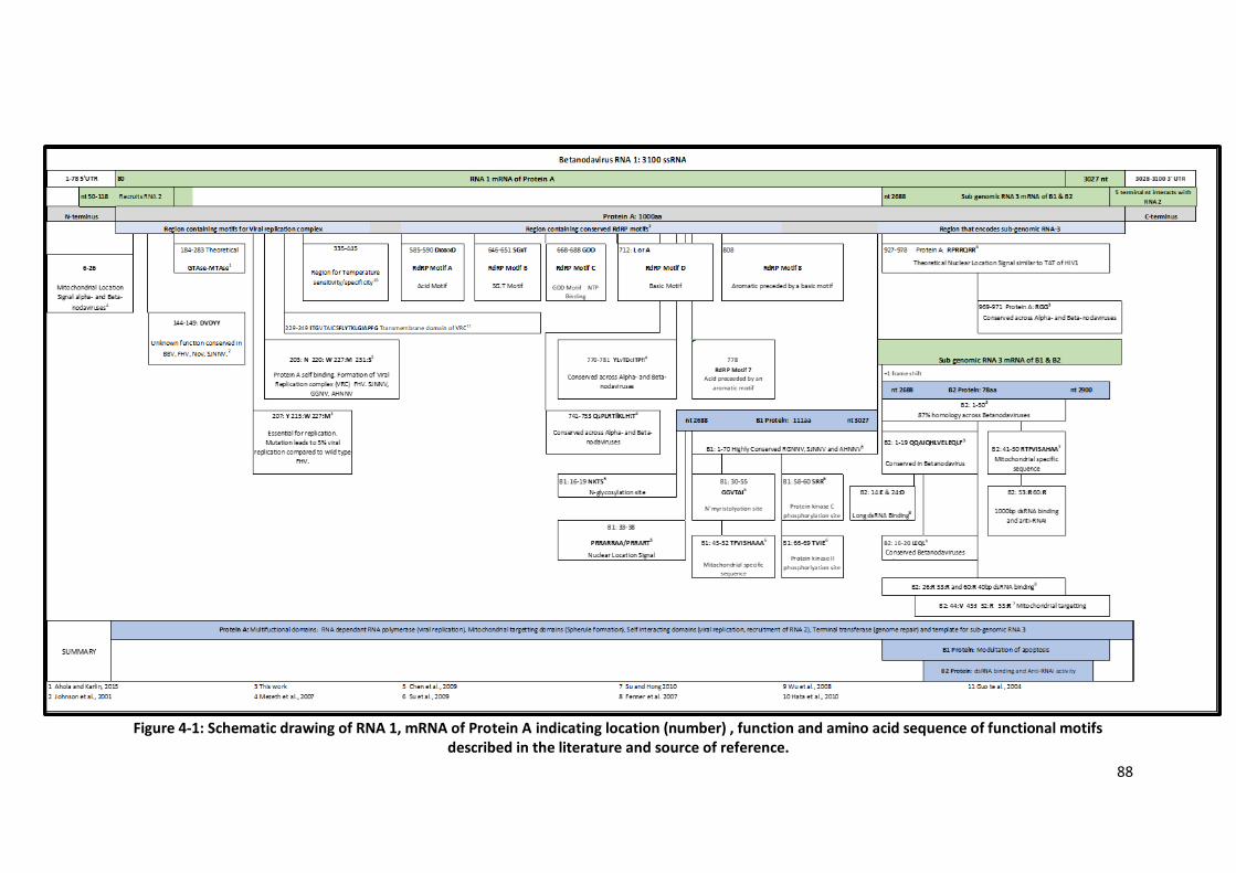
88
Figure 4-1: Schematic drawing of RNA 1, mRNA of Protein A indicating location (number) , function and amino acid sequence of functional motifs described in the literature and source of reference.
89
4.1.2.9 Capsid Protein
The RNA 2/capsid protein of the Betanodavirus has been proven to determine host range
(Iwamota et al., 2004). The Betanodavirus capsid protein has also been reported to possess
Nuclear Location signal activity, pro-apoptosis activity and contain a motif that relates to
species-specific virulence and the recognition of virus by neutralising antibodies (Guo et al.,
2003a; Ito et al., 2008 and Chen et al., 2015). A schematic diagram of the Betanodavirus capsid
protein is presented in Figure 4.2.
The Betanodavirus capsid protein consists of 340 aa. The translation of the capsid protein
requires both UTRs of RNA 2 and the 5’UTR contains a Kozak sequence of ACAA TGG
(3ANNATGG+4) (Huang et al., 2007). The capsid protein has a mw of ~37 to kDa which is
slightly higher than that predicted by their amino acid sequences (Lin et al., 2001). The higher
molecular weight is consistently observed and could possibly be caused by host cell factors
causing polyadenylation in vitro (Huang et al., 2007). Expression of the capsid protein was
reported to increase from 12 to 72 hpi and was significantly greater than the peak RdRP
expression (Wu et al., 2010). Studies with recombinantly expressed mouse grouper NNV
(MGNNV) indicated the capsid protein packages multiple RNA strands ranging in size from 100
to 4500 nt (Guo et al., 2003b). Expression of the capsid protein of GGNNV in vitro induced
activation of caspase-3-like and caspase-8-like proteases which ultimately leads to apoptosis
via the “extrinsic” pathway (Guo et al., 2003b). The induction of apoptosis serves as a
mechanism to release progeny virus from infected cells (Guo et al., 2003b).
The Betanodavirus capsid protein displays an N-terminal arm aa 1 to 52, a highly conserved
region T2 aa 83 to 216, a variable region T4 aa 235 to 351 and an area referred to as the C-
terminal arm aa 288 to 338. The N-terminal aa 23 to 31 RRRANNRRR of the capsid protein
from GGNNV were demonstrated to perform nucleolar localisation (NLS) functions in both
mammalian (Cos-7) and Asian seabass D.labrax (SB) cell lines (Guo et al., 2003a). The N-
terminal aa 2 to 10 RKGEKKLAK of GGNNV capsid protein also displayed NLS activity (Guo et
al., 2003b). No role of the NLS in the replication of the Betanodavirus genome has been
proposed. The N-terminal arm of the capsid protein contains a high percentage of basic
residues and is involved in binding the negatively charged phosphate backbone of the viral
genomic RNA within the capsid (Lu & Lin 2003). There are two stretches of RRR residues and
D75 residue. The D75 residue represents a catalytic residue (Grotmol et al., 2000). The RRR
residues are involved in the binding of the viral genomic RNA to the internal capsid wall of the
host cell. The aa R23 to 25 and R 29-31 were critical in the formation of viral like particles (Lu &
90
Lin 2003). Also, within the N-terminal arm aa 127 to 140 is the highly conserved DxxDxD motif,
which in the Betanodaviruses is FLPDPTDNDHTFDA, is responsible for calcium binding (Wu et
al., 2008).
Within the highly variable T4 region a 3 aa motif (254 to 256) is completely conserved within
each NNV genotype (Nishizawa et al., 1997). The motif is responsible for neutralising epitopes
that are associated with viral infectivity (Ransangan & Manin 2012). The species present as 3
serotypes by analysis with monoclonal antibodies raised against the capsid protein (Mori et al.,
2003). The amino acid motifs and serotypes were discussed in Chapter 3 (refer to Table 3.1).
The RNA 2 and/or the encoded coat protein controls host specificity in SJNNV and RGNNV
(Iwamoto et al., 2004). Recombinant studies combining RNA 1 and different RNA 2 regions of
SJNNV and RGNNV identified regions on RNA 2 which related to virulence (Ito et al., 2008).
Virulence in striped jack and seven band groupers was retained when the RNA 2 segment nt
693 to 1054 (RGNNV) and 694 to 1061 (SJNNV) was retained respectively. In vitro all the viral
hybrids replicated indicating the loss in infectivity was related to a host factor rather than
critical mutation in the virus. Significantly, in vitro activity was demonstrated via transfection
with lipofectamine which facilitates entry of DNA or RNA to cells (Cardarelli et al., 2016) and
by-passes the requirement of attachment and entry to cells. The loss of virulence in vivo could
indicate the regions identified by Ito et al. (2008) relate to attachment and entry of host cells.
However, other mechanisms could also account for the variation in infectivity between in vivo
and in vitro studies.
In RGNNV the region between nt 694 to 758 (aa 223 to 245) appears to affect species
specificity. In SJNNV, the region between nt 695 to 765 affects species specificity (Ito et al.,
2008). The nt and aa homology of the two viruses within the critical regions were 60 and 55 %
respectively. How the region influences species specificity is unknown. Near this region a
combination of either both or two single amino acid mutations of S247A or S270N in a SJNNV
capsid protein lead to mortality of 60 % compared to 100 % compared to wild type virus in
experimental infection of Senegalese sole (Souto et al., 2015). In vitro, the viral mutations
reached similar viral titres albeit with slower replication kinetics (Souto et al., 2015a).
The capsid protein may also have yet to be discovered functions. The formation of the VRC and
the expression of B2 provide a mechanism that protects the viral RNA during synthesis
following the expression of Protein A. However, no mechanism for the protection of the RNA 1
strand prior to translation to Protein A has been proposed. Considering the capsid protein is
the only viral-derived protein present in the host cell prior to Protein A translation it is likely
the capsid protein performs other roles yet to be described.

91
Figure 4-2: Schematic drawing of RNA 2 mRNA of Capsid Protein indicating location (number) , function and amino acid sequence of functional motifs
described in the literature and reference source.

92
4.1.3 Replication of Viral RNA segments
Replication of the Nodavirus RNA involves the production of multiple RNA species in addition
to RNA 1, 2 and 3 (Venter & Schneemann 2008). Although packaged as separate strands,
during replication, the RNA 1 and 2 are covalently linked as head to tail monomers (Rosskopf
et al., 2010). Additional RNA species have been described as defective interfering RNAs (DI-
RNAs) and RNA dimers (Venter & Schneemann 2008). RNA dimers consist of head to tail
junctions of RNAs 1, 2, 3 or 2 and 3 in both positive and negative strand RNA. DI-RNAs consist
of genomic RNAs with internal deletions or sequence rearrangements. A 634 nt DI-RNA of FHV
(DI 634) accumulates to higher levels than viral genomic RNAs (Dasgupta et al., 2003). No
function has been demonstrated for the DI-RNA. An additional negative sense intermediary
formed in the early replication cycle accounts for approximately 1 % of the RNAs replicated
(Venter & Schneemann 2008). The production of the negative sense intermediary is
independent of an external RNA primer and in the case of WhNV involves the recognition of
the last 191nt on the 3’ of RNA 1 positive sense or last 201nt on the negative sense RNA 1
(Wang et al., 2013). A D.labrax NNV (termed DIEV in the publication) intermediate, minus
strand RNA, was detected in cell culture at 96 hours post-inoculation (hpi). Other researchers
report the detection of minus strand RNA 1 of GGNNV in cell culture 12 hpi (Adachi et al.,
2007).
How the RNA 1 strand locates and becomes stabilised in the cell prior to packaging in the
capsid protein is not established beyond occurring within the VRC. The B2 protein has been
shown to be in close association with the RNA 1 and plays a role in the protection of RNA 1
from the RNA interference (RNAi) machinery. B2 is produced well in excess of RNA 1 and could
perform additional roles in the replication of the Nodaviruses (Petrillo et al., 2013).
Within the virion, RNA 1 and 2 of FHV form a single RNA that has been observed in gel
electrophoresis. A covalent link is proposed to occur through a small section with nucleotide
homology between the 3’ end of RNA 1 and 5’ end of RNA 2. Deletion of 5 nt from the 3’ end
of RNA 1 prevents RNA synthesis in the case of FHV (Ball 1995). A single nt substitution of G at
2960 to T in FHV leading to an amino acid change from R to L in the RNA 3 promoter region
lead to an absence of B2 and significant downregulation in the translation of the FHV coat
protein α (Petrillo et al., 2013). The recruitment of RNA 1 by Protein A at the VRC in WhNV was
demonstrated to rely on the presence of a stem loop structure with nt 50 to 118 of RNA 1
being critical (Qui et al., 2014a). The binding ability was increased with increased Protein A
concentration with the minimal ratio of Protein A: RNA 1 being 10: 1 (Qui et al., 2014a). The

93
recruitment of RNA 2 by Protein A in the VRC was also dependant on the presence of a stem
loop structure that is located in nt 123 to 164 of RNA 2 (Qui et al., 2014a).
4.1.3.1 RNA 1
Betanodavirus RNAs are of similar function and structure to that of the Alphanodaviruses. RNA
1 is the mRNA for the RdRP (Protein A). The sub-genomic RNA 3 is also present on the 3’ end of
the RNA 1 and is the mRNA for the B1 and B2 proteins. Both the RNA 1 and 2 molecules are
required for infectivity and the viral strands are self-replicating in vitro in the absence of
complete virions. The presence of RNA 1 and RNA 2 was detected in vitro at 24 hours post-
infection (hpi) and RNA 3 at 96 hours pi (Delsert et al., 1997). Delsert et al. (1997) further
noted that the RdRp was associated with the mature capsid. The RNA 1 nt sequence of
Betanodaviruses displays ~80 % homology across the genus. Within each species/genotype
approximately 90 % nt homology exists although it should be noted that a large proportion of
the strains in the NCBI database have not been assigned to genotype level. Within the first
~40nt of RNA 1 of the Betanodaviruses a TPNNV specific insert exists at nt 27 to 34
(UAACUGAA). The insert is positioned in the UTR of RNA 1. No reference to the insert has been
located in the published literature. As TPNNV has not been reported since 2007, it could be
inferred that TPNNV displays low virulence and adaptability compared to the RGNNV, SJNNV
and BFNNV.
4.1.3.2 RNA 2
The RNA 2 strand consists of ~ 1435 nt and contains one open reading frame (nt 27 to 1043)
flanked by a 5’ 26 nt non–coding/untranslated region (NCR/UTR) and a 3’ 392nt UTR (Huang et
al., 2007). Secondary structures within the RNA 2 C-terminal region (UTR), also referred to as
“cis-acting elements”, are required for RNA 2 replication. The secondary structures in UTRs are
required by the RdRP of many RNA viruses to allow the recognition and processing of RNA in
viral replication (Taufer et al., 2008). Conserved secondary structures were predicted by
computer modelling to be present in the 3’ terminus of RNA 2 in the nodaviruses including
FHV, NoV, SJNNV and GGNNV (Taufer et al., 2008). The 50 nt of the 3’ terminal RNA 2 of FHV
were sufficient to direct the replication of complementary RNA (Albarino et al., 2003).
Similarly, in NoV, 50 nt (1287 to 1336) at the 3’ end of the RNA 2 is sufficient to direct the
RdRP to replicate a complementary RNA (Rosskopf et al., 2010). RNA 2 also forms head-to-tail
dimers during RNA replication (Qia et al., 2011). The dimers are approximately the same size
as the RNA 1 and can be difficult to distinguish on gel electrophoresis (Qia et al., 2011). The

94
RNA dimers of NoV form between the 5’ terminal 17 nt and the 3’ terminal 54 nt of RNA 2
(Rosskopf et al., 2010).
4.1.3.3 RNA 3
The replication of RNA 3 has been described for the Alphanodaviruses. The replication of RNA
3 does not involve an intermediary negative sense sequence. In the Alphanodaviruses Flock
House Virus (FHV), Pariacoto Virus (PaV), Nodamura Virus (NoV) and Wuhan Nodavirus
(WhNV), RNA 1 and sub genomic RNA3 (sgRNA3) form homodimers during replication (Qia et
al., 2011). It is proposed that RNA 3 binds to viral RNA to prevent the formation of
incompatible dsRNA that may affect the formation of the capsid (Schneemann 2006). The
homodimers consist of negative strand RNA 1 and positive strand sgRNA3 which results in the
initiation of sgRNA3 transcription. The promoter for transcription of sgRNA3 is located on the
negative strand of RNA 1. In the case of WhNV, the promoter is located within nt 2758 and
2769 on RNA 1. The production of sgRNA3 also requires the presence of secondary structure
that includes the transcription start site of B1 (Qia et al., 2011).
Transcription of RNA 2 suppresses the transcription of sgRNA3 (Qia et al., 2011). The
suppression of sgRNA3 by RNA 2 is proposed to occur through RNA 2 having 3 regions of
nucleotide complementarity to the negative strand of RNA 1, which both restrict the formation
of the secondary structure of RNA 1 and also bind directly to the sgRNA3 promoter region (Qia
et al., 2011, Wang 2010). By annealing to the sgRNA3 promoter site on the negative to strand
RNA 1, RNA 2 impairs the recognition of the sgRNA3 promoter by the viral RdRp (Wang et al.,
2013). The mechanisms of replication of RNA 3 in the Betanodaviruses have not been
described.
4.1.4 Formation of viral particles
Nodaviruses do not form empty capsids but package RNA into the capsid being either viral or
seemingly random in origin with the final product having approximately the same
sedimentation rate as that of the nodavirus virion (Gopal et al., 2014). The capsid protein
packages one molecule of RNA 1 and RNA 2 into progeny particles. The RNA 3 is not packaged
into virions (Gopal et al., 2014). RNA packaging into virions occurs approximately 30 minutes
after synthesis of the capsid protein. Optimal capsid formation in the Betanodaviruses occurs
at pH 8.0 that correlates well with the normal pH of ocean water (pH 8.4) (Lin et al., 2001). The
capsid consists of 180 copies of the capsid protein α (Gopal et al., 2014).
The Betanodavirus capsid protein is produced as a single protein. Functional motifs have been
identified on the capsid at aa 23 to 25, 29 to 31, 83 to 216, 127 to 140, 187 to 201 and C-

95
terminal aa 4 to 11 (Lu & Lin 2003). At the N-terminal the RRR motifs at aa 29 to 31 and less so
aa 23 to 25 and were critical for viral particle formation in vitro (Lu & Lin 2003). Wang (2010)
investigated the role of 35 residues at the N-terminus of the DGNNV. R30 and R31 are
important for particle formation and particle stability (Wang 2010). Effects of R29 were
negligible (Wang 2010).
The middle region of the capsid protein aa 83 to 216 is proposed as the site of folding to form
the T=3 capsid. Within this region a conserved aa 140 to 147, AL/FQATRGA, is encoded by less
frequently present codons and may serve to slow the ribosomal traffic rate to avoid
interference with the folding of the capsid protein (He & Teng 2015). Folding of the single
protein to form a functional capsid involves disulphide bonding at highly conserved residues
C187 and C201 (Krondiris & Sideris 2002). Amino acid analysis of the coat protein of the
Betanodaviruses indicates the DxxDxD motif, which is important for capsid formation, is
positioned at aa 127 to 140. Within aa 127 to 140 of DGNNV aa 130 to 135 of the capsid
protein was identified as the calcium binding ligand. Point mutations of aa D130, D133 and
D135 resulted in a 50, 100 and 50 % reduction in viral particles respectively (Wu et al., 2008).
Motifs C115 and C201 are essential for capsid formation of DGNNV and for thermal stability of
viral particles (Wang 2010).
In the DGNNV capsid protein, N-and C-terminal motifs are involved in the viral particle
formation (Lu and Lin 2003). Deletion of the C-terminal aa 4 to 11 (328GTVCTRVD335) reduced
viral particle formation to just 4.5 % of wild type (Lu and Lin 2003). Although viral replication
and packaging occurs in membranes associated with the mitochondria, the capsid protein lacks
MLS (Mezeth et al., 2007). Signals for mitochondrial targeting maybe present in the UTR of
RNA 2 (Mezeth et al., 2007). The covalent linkage proposed by Rosskpf et al. (2010) between
the RNA 1 and RNA 2 would facilitate localisation of the capsid protein to both the
mitochondria and replicated RNA 1 and 2. How the ssRNA viruses in general regulate viral
production with protein synthesis and capsid formation and packaging has not been
determined. A form of viral sensing of cellular co-factors in the sub-cellular environment is
proposed to regulate the actions within the VRC (Nagy 2015).
4.1.5 Exit of viral particles
The exit of viral particles from the host cell by Betanodaviruses has not been extensively
studied. The fore-mentioned induction of apoptosis by either the capsid protein or B2 would
facilitate the release of progeny virus from infected cells. The action of B2 to upregulate
expression of the pro-apoptotic gene Bax which induced loss of mitochondrial membrane
potential and mediated necrotic cell death of 44 % of cells at 72 hpi, is a mechanism that
96
would facilitate exit of viral particles (Chen et al., 2009). Although cell-to-cell transport is
reported for many neurotropic viruses, no such movement of viral particles between cells has
been demonstrated in the Betanodaviruses.
4.1.6 Undescribed mechanisms
Acknowledging the limitation of a small genome, the Betanodaviruses complete infection and
replication in a very efficient manner. Although the untranslated region of RNA-2 represents a
relatively large proportion of the genome (~10% of whole genome and ~30% of RNA 2
segment), limited attention has been directed to the role of the UTR in viral replication.
Recently Souto et al. (2018) demonstrated the importance of the 50 terminal nucleotides of
the 3’ end of the genome in forming the stem loop structure that is essential for replication.
The nt region 1398-1421 was broadly identified as more critical in forming the stem loop
structure and mutations at 1408 and 1408-1412 but not 1412 alone. Through in vitro studies,
the functional role of 1408-14212 was demonstrated to affect the interaction with the 3’
NCR/UTR of RNA 2 (Souto et al., 2018). Moreover, the significant attenuation of virulence in
Senegalese sole through mutation of 1408-1412 indicated the region also plays a role in
interaction for RNA 2 with host cellular proteins (Souto et al., 2018).
The importance of viral encoded microRNA (V-miRNA) is recognised as a potent mechanism
used by viruses to achieve viral replication, persistence, immune evasion and cellular
transformation (Cullen 2009; Grundhoff & Sullivan 2011; Tycowski et al., 2015). V-miRNAs
represent a genomically efficient way for viruses to regulate host immune responses. A V-
miRNA could target multiple genes in the same host or a highly conserved gene in multiple
hosts. Additionally, V-miRNAs are known to target the expression of viral replication process
and are involved in the Singapore grouper Iridovirus which encodes at least 16 V-miRNAs, the
functions of which are being studied (Guo et al., 2013). In addition, nine v-miRNAs have been
detected during replication of the fish-infecting Megalocytivirus (Zhang et al., 2014).
Nodavirus V-miRNAs have not been reported. However, the replication of short incomplete
copies of Differential interfering-RNA (DI-RNA) by the RdRp has been detected in the early
phases of nodavirus replication in vitro. During RNA replication, FHV produces, ~ 400bp ds RNA
(DI-RNA) from the 5’ terminus of RNA 1 which serves as a Dicer-2 substrate (Aliyari et al.,
2008). Pyrosequencing of the dsRNA formed 4 days post-FHV infection detected 4371 small
RNAs the majority of which have strong homology and presumably target a region comprising
the first 400nt of RNA 1 (Aliyari 2008). No functional roles for the DI-RNAs have been
demonstrated. The mass replication of the DI-RNAs could be an intermediate step in the
production of V-miRNA which could serve to downregulate specific host genes such as those

97
which regulate immune function or to regulate nodavirus replication. The lower expression of
Protein A from RNA 1 compared to the capsid protein of RNA 2 despite both strands being
transcribed in equal amounts supports a proposal that portions of RNA 1 may have regulatory
roles conferred by the RNA rather than translated protein.
4.1.7 The importance of the functional motifs in the context of this study
There are few studies investigating the functional motifs of the Betanodavirus genome that
discuss multiple motifs across multiple Betanodavirus strains. Rather many studies investigate
a small portion of the genome in a single strain of NNV. Although this review has identified a
large number of motifs that are important for replication, it must be noted that the images
prepared are a collective representation from many articles. The aim of this chapter is to
confirm the presence or absence of the many functional motifs identified in the literature in
the three NQAus NNV strains of RGNNV obtained within this study. Confirmation of the
functional motifs serves a three-fold purpose namely:
1. It confirms the presence of the motifs noted from literature are present in strains
beyond those used in the published reports.
2. It ensures the viral extract being used in this project to test therapies is theoretically
competent and virulent.
3. It ensures the therapies developed in this project are targeted and tested against a
competent and virulent strain of RGNNV.
4.2 Materials and Methods
4.2.1 Genome annotation and illustration
The aforementioned critical motifs identified by previous researchers were annotated into the
complete RNA 1 and RNA 2 segments obtained from sequencing using the Geneious
bioinformatic program. Although the Geneious program is a very useful tool for bioinformatics,
the illustration of the overall genome annotation is not easily reproduced in printable,
readable single page format from Geneious. Hence, additional schematic maps of RNA 1 and
RNA 2 were prepared (Figures 4-1 and 4-2).
4.2.2 Identification of critical motifs in the E.coioides RGNNV viral extract strain.
The critical motifs identified by previous workers were annotated into the RGNNV species type
strain using the Geneious bioinformatic program (Biomatters, available from
http://www.geneious.com). The sequences of the viral segments obtained from the three
NQAus NNV strains described in Chapter 6 were aligned against the RGNNV reference genome
described in Chapter 3 using a Geneious global alignment with default parameters. The region
98
of the RNA 2 that translates into the species specificity region identified by Ito et al.(2008)
were compared for all of the Australian strains described in Chapter 3. The conservation or
lack thereof motifs in the strains collected in this study were recorded.
4.3 Results
4.3.1 Genome annotation and illustration
A summary of the functional motifs from the translated amino acid sequence from the
Betanodavirus are illustrated in Figures 4.1 and 4.2.
4.3.1.1 RNA 1, mRNA of Protein A
All of the motifs that are translated from the mRNA of RNA 1 and identified in the literature as
critical for viral replication in the RGNNV species were conserved in each of the three strains
investigated in this study. (Table 4.1). The motifs that have been identified as essential for viral
replication complex formation were 100% conserved in the species sequenced in this study (aa
6 through to 249 in Table 4.1). A common aa motif of SGxxxxV was retained between the
RGNNV, and all of the Betanodaviruses and some Alphanodaviruses in the Mitochondrial
location signal motif (aa 6-26 in Table 4.1). The RdRP motifs, where identifiable, were
conserved (aa 594-740 in Table 4.1). Two regions in the Protein A sequence (aa750-761 and
780-791) were also 100% conserved across the three strains sequenced in this study. No
reference to the potential function of these motifs has been identified (Refer to Table 4.1). All
motifs proposed as critical in the B1 and B2 protein were also conserved (Table 4.1). The TVIE
motif proposed as a protein kinase II phosphorylation site (aa 66 to 69) identified by Su et al.
(2009) was not identified in any of the strains or the RGNNV reference sequence.
4.3.1.2 RNA 2, mRNA of Capsid Protein
All of the motifs that are translated from the mRNA of RNA 2 and identified in the literature as
critical for viral replication in the RGNNV species were conserved in each of the three strains
investigated in this study (Table 4.1). Most critically to this project, the minimal essential
epitopes required for antibody production in grouper namely, aa 181-212 VNVSVLCR were
identified (Chen et al., 2015). The region of the RNA 2 identified by Ito (2008) which relates to
species specificity of the RGNNV strains was conserved between the RGNNV reference strain
and all 3 NQAus NNV strains collected in this study (Figure 4.3). Within the Ito et al. (2008)
region, a difference of 3aa across all of the Australian collected sequences was identified
(Figure 4.3). Immediately adjacent to the region of Ito et al. (2008), an additional motif GAVF

99
was also conserved within the strains of this study and the RGNNV reference genome (Figure
4.3). A variant motif of GAIF was present in some of the strains (Figure 4.3).

100
Table 4-1: Conserved motifs critical for replication of the RGNNV genome identified from translated mRNA of RNA 1 obtained from this study. Including strain, function, amino acid sequence and location on the protein.
Function Stem loop structure Mitochondrial Location Signal
aa motif 6 – 26
Sequence in literature DVDYY MRRFEFALARMSGAAFCVYTGYRLLTSKWLADRVEDYRQRVI
Ec2NQAus E.coioides this study 160-164 DVDYY MRRFEFALARMSGAAFCVYTGYRLLTSKWLADRVEDYRQRVI
El3NQAus E.lanceolatus this study 160-164 DVDYY MRRFEFALARMSGAAFCVYTGYRLLTSKWLADRVEDYRQRVI
MI181161 L.calcarifer this study 160-164 DVDYY MRRFEFALARMSGAAFCVYTGYRLLTSKWLADRVEDYRQRVI
FunctionTransmembrane Domain of
Viral Replication ComplexA: Acid motif B: SG.T motif
C: GDD motif: NTP
bindingD: Basic motif Motif 7 & 8
aa motif 229 – 249 594-599 655-660 695-697 740
Sequence in literature ITGVTAICSFLYTKLGIAPFG DxxxxD SGxxxT GDD Lysine or Arginine
Ec2NQAus E.coioides this study ITGVTAICSFLYTKLGIAPFG DYSKFD SGSALT GDD L
El3NQAus E.lanceolatus this study ITGVTAICSFLYTKLGIAPFG DYSKFD SGSALT GDD L
MI181161 L.calcarifer this study ITGVTAICSFLYTKLGIAPFG DYSKFD SGSALT GDD L
Functionaa motif
Sequence in literature
Ec2NQAus E.coioides this study
El3NQAus E.lanceolatus this study
MI181161 L.calcarifer this study
Function N-glycosylation site Nuclear Location signal
Hypothetical
Nuclear Location
signal
Protein kinase C
phosphorylation
site
Protein kinase II
phosphorylation siteNuclear Location signal
aa motif 869-899 912-918 984-988 929-931 66 - 69 946-950
Sequence in literature NKTS PRRARAA RPRRQRR SRR TVIE KRPRR
Ec2NQAus E.coioides this study NKTS PRRARAA RQRRR KRSRR not found KRPRR
El3NQAus E.lanceolatus this study NKTS PRRARAA RQRRR RRSRR not found KRPRR
MI181161 L.calcarifer this study NKTS PRRARAA RQRRR RRSRR not found KRPRR
Function Conserved in all the Betanodaviruses Conserved with NoV40bp (short) dsRNA binding
motifs
Mitochondrial specific
sequence
aa motif 5-20 16 - 20 26 41 - 50
Sequence in literature QQAIDQHLVELEQLF LEQL R26 RTFVISAHAA
Ec2NQAus E.coioides this study QQAIDQHLVELEQLF LEQL R27 RTFVISAHAA
El3NQAus E.lanceolatus this study QQAIDQHLVELEQLF LEQL R27 RTFVISAHAA
MI181161 L.calcarifer this study QQAIDQHLVELEQLF LEQL R27 RTFVISAHAA
Function N’myristolyation site Mitochondrial specific sequenceaa motif 30 -55 38-47 R60 V44 I45 R52
Sequence in literature GGVTAI TFVISHAAA R60 V44 I45 R52
Ec2NQAus E.coioides this study 26-31 GGVTAI TFVISHAAA R60 V44 I45 R52
El3NQAus E.lanceolatus this study 26-31 GGVTAI TFVISHAAA R60 V44 I45 R52
MI181161 L.calcarifer this study 26-31 GGVTAI TFVISHAAA R60 V44 I45 R52
Function Mitochondrial targetting 1000bp (long) ds RNA binding motifs and anti- RNAi
aa motif 53 53,60
Sequence in literature R53 R53 R60
Ec2NQAus E.coioides this study R53 R53 R60
El3NQAus E.lanceolatus this study R53 R53 R60
MI181161 L.calcarifer this study R53 R53 R60
Sequence Id.
Protein A
B2 Protein (72aa) 1 frame shift from Protein A mRNA continued
Mitochondrial targettingSequence Id.
69
A69
A69
A69
A69
59
R59
R59
R59
R59
B2 Protein (72aa) 1 frame shift from Protein A mRNA continued
Description not
sufficient for alignment
Conserved with NoV but not BFNNV Conserved with NoV, FHV but not AHNV
780-791
YLvTDclTPfI
YLVTDSKTPFIG
YLVTDSKTPFIG
YLVTDSKTPFIG
750-761
QsPLRTllKLHtT
QSPLRTLLKLHTT
QSPLRTLLKLHTT
Important for Viral Replication complex formation. Essential for replication, point mutations lead to
reduced virulence.
184-229
DDSVHYRVAGGKDVRHRIWNYNQNTMYVCSRPRGFWANLMQILRD
DDSVHYRVAGGKDVRHRIWNYNQNTMYVCSRPRGFWANLMQILRD
DDSVHYRVAGGKDVRHRIWNYNQNTMYVCSRPRGFWANLMQILRD
DDSVHYRVAGGKDVRHRIWNYNQNTMYVCSRPRGFWANLMQILRD
Sequence Id.
Sequence Id.
B1 Protein (111aa) same reading frame as Protein A
B2 Protein (72aa) 1 frame shift from Protein A mRNA
Protein A
Unknown functionUnknown function
QSPLRTLLKLHTT
E14, D24
E14, D24
E14, D24
Long dsRNA binding
E14, D24
Sequence Id.
Sequence Id.
Sequence Id. 14,24
RdRP conserved motifs
Protein A
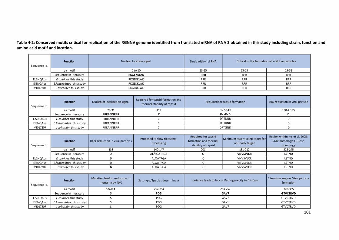
101
Function Binds with viral RNA
aa motif 23-25 23-25 29-31
Sequence in literature RRR RRR RRR
Ec2NQAus E.coioides this study RRR RRR RRR
El3NQAus E.lanceolatus this study RRR RRR RRR
MI017207 L.calcarifer this study RRR RRR RRR
Function Nucleolar localization signalRequired for capsid formation and
thermal stability of capsid50% reduction in viral particle
aa motif 23-31 115 130 & 135
Sequence in literature RRRANNRRR C D
Ec2NQAus E.coioides this study RRRANNRRR C D
El3NQAus E.lanceolatus this study RRRANNRRR C D
MI017207 L.calcarifer this study RRRANNRRR C D
Function 100% reduction in viral particlesProposed to slow ribosomal
processing
Required for capsid
formation and thermal
stability of capsid
Minimum essential epitopes for
antibody target
Region within Ito et al. 2008;
SGIV homology; GTPAse
homology
aa motif 133 140-147 201 181-212 223-245
Sequence in literature D AL/FQATRGA C VNVSVLCR LSTND
Ec2NQAus E.coioides this study D ALQATRGA C VNVSVLCR LSTND
El3NQAus E.lanceolatus this study D ALQATRGA C VNVSVLCR LSTND
MI017207 L.calcarifer this study G ALQATRGA C VNVSVLCR LSTND
FunctionMutation lead to reduction in
mortality by 40%Serotype/Species determinant
C terminal region. Viral particle
formation
aa motif S247>A 252-254 328-335
Sequence in literature S PDG GTVCTRVD
Ec2NQAus E.coioides this study S PDG GTVCTRVD
El3NQAus E.lanceolatus this study S PDG GTVCTRVD
MI017207 L.calcarifer this study S PDG GTVCTRVD
GAVF
GAVF
GAVF
Required for capsid formation
127-140
DxxDxD
DPTDND
DPTDND
DPTGND
Critical in the formation of viral like particles
Sequence Id.
Sequence Id.
Sequence Id.
Nuclear location signal
2 to 10
RKGEKKLAK
RKGEKKLAK
RKGEKKLAK
RKGEKKLAK
Variance leads to lack of Pathogenecity in D.labrax
254-257
GAVF
Sequence Id.
Table 4-2: Conserved motifs critical for replication of the RGNNV genome identified from translated mRNA of RNA 2 obtained in this study including strain, function and amino acid motif and location.

102
Figure 4-3: Nucleotide sequence and translated amino acid sequences of Australian strains of NNV aligned with Ito species specific region including strain identification,
nucleotide and translated protein sequence and arrows indicating points of variance. Prepared using Geneious Prime. Biomatters http://www.geneious.com.
103
4.4 Discussion
Acknowledging previous works and targeting therapies that are directed at critical motifs may
improve the efficacy in preventing VER outbreaks. This study reviewed the collection of
literature on the functional motifs of the Betanodavirus genome and confirmed the majority of
the motifs that have been identified as critical have been conserved in the NQAus RGNNV,
strains collected in this project. No similar review and confirmation of the motifs across an
entire RGNNV Betanodavirus genome has been published. The conservation of motifs
identified from the translated mRNA-1 indicate all of the motifs considered essential for viral
replication have been retained by the strains in this study. Specifically, the mitochondrial
location signals and motifs that are essential for the formation of the viral replication complex
were identified. The RdRP motifs were also conserved. Individual motifs that locate the B1 and
B2 proteins to the VRC and nucleus and also ensure binding of dsRNA to prevent the
degradation of the viral genome by host factors were also identified. Interestingly, four
additional motifs were identified to be conserved between the species in this study and the
RGNNV reference genome. Two motifs namely 750-761aa QSPLRTLLKLHTT and 780-791aa
YLVTDSKTPFIG were identified by Johnson et al. (2001) to be conserved in both
Alphanodaviruses and Betanodaviruses (specifically: Flock house virus, Nodamura virus,
SJNNV, Black beetle virus and Pariacoto virus) but had not previously been identified in the
RGNNV. In the same study the authors also identified aa 969-971 RGG motif conserved across
the same species but the RGG motif was not identified in this study. No function for any of the
three motifs were proposed. The other two motifs noted as highly conserved in this study are
aa 5-20 QQAIDQHLVELEQLF of the B2 protein and within this motif a LEQL motif. The LEQL is
conserved within the Betanodaviruses and the Alphavirus, Nodamura Virus, which significantly
has a wide host range including insects, pigs, suckling mice and hamsters (Gant et al., 2014).
No function of this motif has been reported in the literature relating to Nodavirus replication.
However, an LEQL motif is an important epitope recognised by the endoplasmic reticulum
aminopeptidase (ERAP 1) to target Major Histocompatibility complex class 1 presented
epitopes for degradation. ERAP 1 is stimulated by interferon (Hearn et al., 2009). The LEQL
motif is also an important epitope of TRIM 21 which is a member of the Tripartite motif
superfamily (Al-Majdoub et al., 2013). TRIM 21, in particular, is expressed upon interferon
stimulation and is also an important component of innate immunity and neurological disorders
(Al-Majdoub et al., 2013). Kim et al. (2017) reported the upregulation of TRIM 21, 29, 39, 25,
14, 16 and 47 following infection of Sevenband grouper to NNV. In vitro upregulation of TRIM

104
21 in human microglial cells attenuated the replication of Japanese encephalitis (JE), via
regulation of the type 1 interferon response (Manocha et al., 2014). JE is similar to NNV in that
it affects the brain of host species. Damage due to inflammation is associated with mortality in
many neurotropic viruses. Additionally, TRIM 21 is activated by antibody-coated viral particles,
binding to the Fc receptor and targets virions for degradation. It is possible the LEQL motif of
the B2 performs roles not currently identified to prevent both the innate and adaptive immune
response to RGNNV infection. The targeting of TRIMs by Betanodavirus B2 would be a very
efficient mechanism to successfully mitigate both a humoral (antibody) and innate immune
response. However, no work has been published relating to TRIM and B2 protein. Petrillo et al.
(2013) noted, B2 is produced well in excess of RNA 1 and could perform additional roles in the
replication of the Nodaviruses.
The majority of the motifs proposed to be critical for viral replication that are associated with
the translated RNA 2 were also identified in all three strains of RGNNV studied. Importantly for
the vaccine development, the motifs of the capsid protein that were identified to be critical for
neutralising antibody production in grouper were present in all three strains obtained in this
study (Chen et al., 2015, discussed in Chapter 1.8). The region identified by Ito et al. (2008) to
confer virulence in host species was also conserved between all strains collected within this
study. Notably comparison with all of the Australian collected strains indicated a variance of 3
amino acids within the Ito et al. (2008) region. Specifically, at aa 233-237 an LSTND motif is
present in all of the strains identified as “tropical (1a) strains” in Chapter 3. Whereas LATSD
was conserved in the sequences assigned to the 1c cluster (Section 3.1). No mechanism for
how the sequences may impact on host or environmental selection have been identified. Also,
immediately adjacent to the PDG motif which confers the Betanodavirus serotype assignment
an additional motif variation was noted between the Australian strains. Within Australian
RGNNV strains, the four strains that slightly differentiate from the other Australian strains
based on nucleotide comparison of RNA 2 (Chapter 3) display a GAIF motif. Interestingly, the
tropically freshwater cultured sleepy cod, O.lineolata sourced strain displayed a hybrid pattern
of motifs. The O.lineolata strain retained the LSTND motifs of the tropical 1a strains yet shared
the GAIF motif of the “temperate Ic strains”, proposed by this worker to possibly reflect a
freshwater habitat rather than temperate v tropical division (Chapter 3). Investigating how
these motifs affect virulence to different species, or the same species within different habitats
is beyond this study. However, confirmation that the strains in this study have the retained of
the factors that have been identified to affect viral replication or host specificity provides
confidence to advance further work using the strains collected. Confirmation of the protein

105
motifs is rarely considered in experimental challenge experiments. Although analysis
completed in Chapter 3 indicated the RGNNV genome segments were highly conserved,
between 13-21 nucleotide differences were noted between the Australian strains collected in
this study and also the RGNNV reference genome (Table 3.2). Considering a single nucleotide
variation could also translate to change in a critical protein motif, it was important to confirm
the conservation of the motifs in the strains being used in this project. Variation on a single
motif could lead to reduced virulence, which if were transferred into the project would reduce
the effectiveness in translation of the outcomes of this study to industry. Lin et al. (2007)
reported a relative percent survival of 80-90% in 35 dph E.septemfasciatus larvae when
challenged by bath exposure to VVN at following 17 days of feeding with Artemia that been
orally loaded with E.coli expressing a recombinant NNV capsid protein (Lin et al., 2007).
However, the control fish displayed between 44-69% survival which is unusually high
compared to similar reports of experimental challenge with this species. High survivals in
control groups following RGNNV exposure have not been reported in any other research
publication involving larval grouper. Despite the impressive survival and obvious economic
benefit of this approach as a vaccination strategy, these results have not been translated to a
commercial product. Although it cannot be confirmed, it is possible the strain of RGNNV used
in the study was less virulent, possibly through lacking a motif that confers increased virulence.
From the analysis completed in this chapter, considering all of the strains hosted the critical
motifs, and the volume of infectious material available, the RGNNV extract prepared from
E.coioides (Ec2NQAus) is considered an acceptable source of NNV for the remaining study in
this project (and is henceforth referred to as the RGNNV viral extract).

106
4.5 Conclusion
The aims of this chapter were met in the following manner:
Data from this Chapter is planned for publication entitled “Review of the functional motifs of the Betanodavirus genome”.
The motifs demonstrated to critically impact on viral replication or host specificity in
Betanodaviruses have been identified.
The motifs have been confirmed to be present in the strains used within this
project.
The production of a recombinant vaccine based on the RNA 2 sequence of the
E.coioides RGNNV strain will contain the antigenic epitopes that were identified as
critical to the production of neutralising antibodies by grouper recognised by Chen
et.al., (2015)
dsRNA can be specifically designed to target functional motifs that are confirmed to
be present in the strain used in this project.

107
CHAPTER 5. DEVELOPMENT OF QPCR STANDARD CONTROL
MATERIAL TO ALLOW INITIATION OF VALIDATION OF RT-
QPCR ASSAYS TO DETECT AUSTRALIAN STRAINS OF
REDSPOTTED GROUPER NERVOUS NECROSIS VIRUS
(RGNNV).
Background
Hick & Whittington, (2010) reported the development of RT-qPCR, qR2T, that
detects RNA 2 of RGNNV in two Australian sourced NNV strains.
The qR2T assay is the assay recommended in the Australian and New Zealand
Standard Diagnostic Protocols for the detection of NNV.
The assay has not been validated as fit for purpose on grouper tissues.
There are various novel biotechnology applications that aim to produce NNV
vaccine via delivery of the viral capsid protein.
Some biotech applications aim to produce an antigen that can be delivered via
oral dispersal of the antigen within larval fish feeds.
The widespread dispersal of RNA 2 constructs into the fingerling production
systems will confound the current method of verification of freedom from RGNNV
with the RT-qPCR targeting the RNA 2 segment.
Due to discovery of chimeric reassortment between RNA 1 and RNA 2 of SJNNV
and RGNNV some researchers advocate the application of qPCRs that target both
viral segments to study VER outbreaks.
Hick & Whittington (2010) reported an additional RT-qPCR that detects RNA 1 of
RGNNV but did not extensively validate the assay.
Aims of this Chapter
Develop RT-qPCR control material to assist in the validation of the currently
recommended Australian and New Zealand Standard Diagnostic Protocol RT-qPCR
that targets RNA 2 for the detection of the RGNNV from grouper brain and eye
tissue.
Develop RT-qPCR control material to assist in the validation of an additional assay
that targets the RNA 1 for the detection of the RGNNV genome from grouper
brain and eye tissue.

108
5.1 Introduction
The absence of a sound understanding of the factors that trigger VER disease outbreaks in
grouper grow-out aquaculture, currently limits management options to the stocking of ponds
with fingerlings that are free of NNV. The development of reverse transcriptase quantitative
polymerase chain reaction (RT-qPCR) assays has significantly improved the opportunity for
industry to obtain NNV free fish (RT-qPCR negative) and rapidly diagnose a VER disease
outbreak (RT-qPCR high load positive). Detection by RT-qPCR is the only method currently
recommended for all applications including targeted surveillance of larvae, juveniles and adult
fish and presumptive and confirmatory diagnosis of VER disease (Anon. OIE 2018). The OIE
Aquatic Animal Manual describes two RT-qPCR assays to detect NNV that have undergone
significant validation namely the qR2T ( Hick & Whittington 2010) and RNA 2 assay (Anon. OIE
2018). The area targeted by each assay overlap on the redspotted grouper nervous necrosis
virus (RGNNV) RNA 2 segment. The qR2T assay, is incorporated into the Australian New
Zealand Standard Diagnostic Procedures (ANZSDP) for the detection of NNV (Hick &
Whittington, 2010; Moody & Crane 2012; Anon. OIE 2018). The RNA 2 assay was developed by
the OIE Reference Laboratory for VER and has been adopted for proficiency testing across five
European laboratories (Panzarin et al., 2010; Anon. OIE 2018;). Both assays are reported to
detect all NNV species although qR2T is noted to optimally target RGNNV ( Hick & Whittington,
2010 and Anon OIE 2018).
With their emergence as high value aquaculture species, the economic impact of VER in sea
bass aquaculture has prompted the development of two commercially available vaccines to
protect against VER (ALPHA JECT micro® 1 Noda PHARMAQ and ICTHIOVAC® VNN HIPRA
Laboratories). The ALPHA JECT micro® 1 Noda vaccine is a cell culture- derived- formalin-
inactivated antigen reported to induce effective immunity against the RGNNV genotype. The
ICTHIOVAC® VNN is inactivated Betanodavirus strain 1103 (HIPRA Laboratories
https://www.hipra.com/portal/en/hipra/animalhealth/species/fish ). To avoid stress
associated with injectable vaccines, several biotech applications have been attempted with
oral delivery of recombinant NNV capsid protein (Cho et al., 2017 and Gonzalez-Silvera et al.,
2019). Oral vaccination, through consumption of recombinant yeast, Saccharomyces cerevisiae
expressing RGNNV capsid protein induced the production of neutralizing antibodies in
Epinephelus septemifasciatus (Thunberg, 1793) (Cho et al., 2017). The inclusion of TOPO®
transformed E. coli with RNA 2 segment of RGNNV into diet was recently reported to provide
protection to sea bass against viral challenge with RGNNV (Gonzales-Silvera et al., 2019).
109
Although oral delivery offers advantages of practicality of delivery and reduced handling of
fish, it may lead to inaccurate assessment of fish health status if the recombinant RNA 2
genome segments are detected by RT-qPCR. The potential development of orally delivered
vaccines based on expressed proteins from RNA 2 constructs necessitates an RT-qPCR
targeting the RNA 1 segment to differentiate between NNV infected fish and those exposed to
orally delivered recombinant capsid proteins. Further, the discovery of strains of NNV that
consist of chimeric recombination of RGNNV and SJNNV segments highlights a need to apply
assays that detect both RNA segments in epidemiology studies, particularly in zones where
more than one genus of NNV is endemic (Panzarin et al., 2016).
RT-qPCRs that detect the RNA 1 have been described but not widely adopted in Australia (
Hick & Whittington, 2010 and Baud et al., 2015). Neither the qR1T or qR2T assay was validated
for application on grouper tissues. The OIE Aquatic Animal Health Manual notes that it is
important to revalidate an assay when it is being applied beyond the scope of the original
intended purpose of the assay such as application to an additional host species (OIE Anon.
2014). The recommendations of the OIE are also adopted by authorities in Australia that
oversee quality control in testing for aquatic animal pathogens namely, the sub-committee for
aquatic animal health (SCAAH) and the National Association of Testing Authorities (NATA). The
validation of an assay is a step-wise process. Initial steps determine if the assay platform is the
best fit for purpose format. For reasons of quantitation, high throughput, rapid turnaround,
sensitivity and specificity, RT-qPCR is the assay of choice for many applications in aquatic
disease management. An initial step in the implementation of an assay is to produce stable
qPCR controls, of sufficient volume and stability that can be applied to monitor the
performance of the assay over time (Fig 1. Anon OIE 2017).
The aim of this chapter is to develop the standard positive controls for the qR1T and qR2T
assays of Hick & Whittington, (2010). These controls are required to support further
application of the assays within the activities of this project to assess the effectiveness of
prophylactic measures against RGNNV; to gain a better understanding of the pathogenesis of
VER in grouper and to validate the assays as suitable for application beyond the scope which
was demonstrated by Hick & Whittington (2010).
5.2 Materials and Methods
5.2.1 Preparation of plasmid control for quantitative real-time polymerase chain
reaction

110
Synthetic control for quantitative real-time polymerase chain reaction was prepared from PCR
amplicons obtained from NNV infected fish. Positive PCR amplicons were produced using the
primers R1F1/R1R5 and R2F1/R2R1 described by Hick & Whittington (2010) (Table 5.1). The
amplicons were visualised and cut from agarose gels as described in Chapter 2.1.4-2.1.7. The
PCR product was cleaned using a High Pure PCR Product Purification Kit (11732668001 Roche,
NSW) as per manufacturer’s instructions. Purified PCR product was cloned into One Shot TOPO
chemically competent E. coli using the pCR4-TOPO TA vector (Cat. K4575-01 Life Technologies,
VIC) as per manufacturer's instructions. Transformed E. coli were grown overnight at 37 °C on
lysogeny broth (LB) (Bertani, 1951) agar supplemented with 50 g mL-1 ampicillin (Cat. A9393-
5G Sigma-Aldrich, NSW). Three white colonies were picked from the agar and grown overnight
at 37 °C in LB supplemented with 50 g mL-1 ampicillin (Cat. A9393-5G Sigma-Aldrich, NSW)
and shaken (Bioline incubator shaker 8500 Edwards Instruments, NSW) at 150 rpm. Plasmid
DNA was extracted using a High Pure Plasmid Isolation kit (Cat. 11754777001 Roche, NSW) as
per manufacturer's instruction. Plasmid extracts were submitted to Macrogen Inc. (Seoul,
Korea) for sequencing. Sequence analysis confirmed the respective plasmids were the RNA 1
and RNA 2 derived products. The clones and extracts became the positive plasmid controls for
the quantitative real-time PCRs (qPCR).
Table 5-1: Details of primer sequences used to produce RT-qPCR control sequences including assay type, primer name, primer sequence, primer target, primer position and expected size of positive amplicon.
5.2.2 Standard curve preparation from plasmid controls
A standard curve was prepared for the qPCR using the positive plasmid controls generated
from section 5.2.1. The clones were cultured for 18 hours in 30 mL of LB supplemented with 50
μg mL-1 ampicillin (Sigma-Aldrich) at 150 rpm (Bioline incubator shaker 8500). A 100 μL aliquot
of culture was added to 12 x 10-fold serial dilutions while the 30 mL culture was frozen at -80
°C to prevent further bacterial growth. Copy number was calculated by plating triplicate 20 μL
qR1T R1F1* CACTTACGCAAGGTTACCG 1 1
R1R5* TCTGCTGCTCCTCGACATAC 1 1525
qR2T R2F1* CATATGGTACGCAARGGTGA 2 3
R2R1* CTCGAGTTAGTTTTCCGAGTCA 2 1023
amplicon
size (bp)
Assay
namePrimer Name Sequence (5'-3')
RNA segment
TargetPosition~
Source of All primer sequences Hick & Whittington (2010)
~ Position with reference to NCBI MI181161/RNA-1 or MH017207/RNA-2
1515
1020

111
aliquots of each dilution on LB agar plates supplemented with 50 μg mL-1 ampicillin (Sigma-
Aldrich) and incubating for 18 hours. The number of white colony forming units (CFU) were
counted for each dilution and used to calculate the plasmid copy number in the 30 mL culture,
assuming one CFU represents one plasmid copy.
5.2.3 RT-qPCR analysis of standard control serial dilutions
The 30 mL culture that was frozen during the CFU counting procedure was thawed and
plasmid DNA extracted and eluted in 100 μL elution buffer as previously described (3.3.4). A 50
μL aliquot of DNA extract was used to prepare 9 x 10-fold serial dilutions in DEPC-treated
water (BIO-38030, Bioline) and stored at -20 °C for qPCR. A 2.5 μL aliquot of each serial dilution
was used as template in the qR1T and qR2T RT-qPCRs to construct the quantified standard
curve. The qPCR mix consisted of 2.5 μL DNA extract in Bioline SensiFAST probe No ROX mix
(BIO-86050) prepared according to the manufacturer’s specifications, containing Forward and
Reverse Primer (20 pmol) and Probe (5 pmol) per reaction (Table 5.2). The qPCR was
completed on a Qiagen Rotor-Gene™ with thermal cycling consisting of 95 °C for 3 mins
followed by 40 cycles of 95 °C for 15 s and 60°C for 25 sec. Fluorescence was acquired at the
60°C for 25 sec step of each cycle using the green and yellow filters of the machine.
Table 5-2: Primer and Probe sequences used in RT-PCR assays including assay type, primer name, primer sequence, RNA target, position on target and primer melt temperature.
5.3 Results
5.3.1 Confirmation of sequence of the RNA 1 and RNA 2 plasmid
Plasmid sequences were confirmed to be 100% homologous to the E.coioides RGNNV strains
discussed in Chapter 3. Although shorter sequences would have been sufficient to act as qPCR
PCR format Primer Name Sequence (5'-3')
RNA
segment
Target
Position~ Tm Primer
RT-qPCR qR1T-F GCTACCGCCTGTTGACCTC 1 140 61
qR1T-R TTGTTTCTTCTCAGCGATGATGC 1 219 64
qR1T-Probe TGGCGAATCCTCAACACGTCC 1 171
RT-qPCR qR2T-F CTTCCTGCCTGATCCAACTG 2 401 62
qR2T-R GTTCTGCTTTCCCACCATTTG 2 476 61
qR2T-Probe CAACGACTGCACCACGAGTGG 2 454
Source of All primer and probe sequences Hick & Whittington (2010)
~ Position with reference to NCBI GQ904198/RNA-1 or GQ904199/RNA-2
112
controls, the longer sequences were selected to align more closely with the characteristic of
viral sequence that would be present in host tissues during viral replication.
5.3.2 Plate count of CFU of each dilution of plasmid controls
A number of the serial dilutions of plasmid contained too many colonies to allow accurate
counting of colonies. The plates that contained less than 100 colonies were used to calculate
the plasmid CFUs. Plate counts indicated the stock cultures of each plasmid contained ~ 12.6 x
109 (RNA-1) and 13.8 x 10⁹ (RNA-2) CFU mL-1.
5.3.3 Quantitative real-time polymerase chain reaction
The target sequences were detected in the standard curve dilutions from 100 to 109 range for
qR1T and 100 to 108 range for qR2T. The standard curves prepared displayed a strong linear
correlation between estimated plasmid copy number and Ct value (R2 value=0.99 in each
assay). (Figure 5.1). The qR1T assay had a reaction efficiency of 98% which was slightly less
than that of qR2T (99%) (Figure 5.1). The Cycle threshold value, plasmid copy number and
typical standard series amplification curve for the RNA 1 and RNA 2 segment plasmid control
are provided in Figure 5.1.

113
Figure 5-1: RT-qPCR amplification curves of qR1T (a) and qR2T (b) and linear correlation between Ct value v plasmid copy number determined from bacterial plate counts of qR1T
(c) and qR2T plasmid (d) controls.
5.4 Discussion
The data collected in this study indicates the RT-qPCR assays that detect RNA 1 (qR1T) and
RNA 2 (qR2T) perform consistently to accurately detect the specific segments of the Ec2NQAus
genome on the standard constructed DNA plasmid controls. The operational range of each
assay detected a minimum of ~10 copies of the plasmid target. In this study, consistent Ct
values were obtained for plasmid dilutions targeted by each assay across a gene copy number
ranging from ~10 to 108 copies mL-1. Although a calculated copy number greater than 108 is
detected by both assays, there is considerable variation in the calculated copy number at
higher concentration of plasmid (data not shown) and higher concentrations were not
included in the standard curve regressions. As the calculated copy number that applies to such
high concentration of plasmid genome would only be detected in severe VER outbreaks where
management decisions are not reliant on accurate discrimination between a 108 or 109 or 10
copies of viral genome, the omission of the high copy number control within the standard
curve is not expected to be a major impediment to the application of either RT-qPCR assay.
The OIE devotes a chapter in the Manual of Diagnostic Tests for Aquatic Animals to discuss the
principles and methods of validation of diagnostic assays for infectious diseases (OIE 2019).

114
Whilst Hick & Whittington, (2010) validated the performance of the qR2T assay against the
criteria discussed in the OIE Aquatic Manual, their analysis was based on cell cultures and
barramundi derived tissue samples. In addition, very limited assessment of the qR1T assay that
targeted the RNA 1 was performed. Neither assay has been reportedly applied to grouper
species ( Hick & Whittington 2010). The OIE recommends validation and verification of assay
performance is conducted when analysis is applied to tissue matrix that is beyond that which
the assay was validated (OIE 2017).
One of the initial steps in assay validation is the implementation of standard positive controls
(OIE 2017). This current work has developed a plasmid containing the respective target
sequences to act as a standard control for each assay. The use of a constructed plasmid as a
standard curve control reduces laboratory biosecurity risk by avoiding a need for staff to
handle viable viral particles and also negates a need for cell culture capability within a
laboratory. However, further monitoring of assay performance on a similar analyte matrix to
that which it will most typically be applied, namely eye and brain tissue of known NNV status,
is required for assay validation (OIE 2017). The stepwise approach to assay validation which
has been adopted in this project is typical within the framework recommended by a number of
authorities responsible for animal health laboratory standards including the OIE, the sub-
committee for animal health laboratory standards (SCAAHL) and the National Association of
Testing Authorities (NATA) in Australia (OIE 2017). The development of the standard controls
discussed in this chapter are a crucial requirement for managing the quality of the quantitative
data that will be collected in this project. Further application of the assays on grouper brain
and eye tissues to monitor the pathogenesis of VER and to assess the efficacy of prophylactic
measures against RGNNV will be discussed within subsequent chapters of this thesis.
5.5 Conclusion
The following outcomes were achieved in this chapter:
Standard plasmid controls for RT-qPCRs to detect RNA 1 (qR1T) and RNA 2 (qR2T) of
RGNNV described by Hick & Whittington, (2010) were developed.
Both assays, qR1T and qR2T were demonstrated to detect the respective standard
plasmid controls.
The linear regression of standard curve preparations was determined for each
assay, with high correlation between Ct value and calculated copy number.

115
Data collected in this chapter contributed to the validation documentation to support
application by the JCU AquaPATH laboratory for NATA accreditation in the field of animal
health. Scope of application detection of viruses by nucleic acid detection.
116
CHAPTER 6. DEVELOPMENT OF PROPHYLACTIC MEASURES TO
PREVENT VIRAL ENCEPHALOPATHY AND RETINOPATHY (VER)
Background
Aims of this Chapter
6.1 Introduction
The development of a vaccine that prevents VER outbreaks in E.lanceolatus is a primary goal of
this study. Vaccination strategies that prevent VER have been reported with varying success in
experimental settings (Tanaka et al., 2001;Nishiwawa et al., 2012; Kai and Chi 2008). Various
antigen presenting configurations have been developed to produce vaccines against VER.
Antigen configurations include the presentation of live virus (Nishizawa et al., 2012),
inactivated virus (Kai and Chi 2008), recombinantly expressed DNA or capsid protein (Tanaka
et al., 2001), provision of virus-like particles in live Artemia (Lin et al., 2007) recombinantly
expressed virus-like particles (VLPs)( Lai et al., 2014) and most recently, viral capsid protein
expressed in a cell-free system (Kim et al., 2015). All strategies report improved survival
measured over a period of weeks. Some studies don’t include viral challenge but rather detect
Grouper in grow out pond culture are susceptible to NNV for the entire duration of
pond stocking.
VER outbreaks in pond grow out impose severe mortality and economic losses that
threaten the viability of the grouper aquaculture industry in Australia.
A number of experimental vaccines to prevent VER have been reported.
There is no VER vaccine available for use in Australia.
The capsid protein has been demonstrated as the target of neutralising antibodies.
Treatment with dsRNA has been proposed as a treatment for difficult to prevent
viral diseases.
To develop a recombinant clone and express RGNNV capsid protein.
To prepare a vaccine containing the recombinantly expressed capsid protein.
To develop a dsRNA construct to knock-down replication of RGNNV.
117
the expression of immune components following vaccination (Lai et al.2014). Despite the
reported progress, there is no vaccine approved for use or commercially available for use in
Australia.
Previous research into immunization and the potential of dsRNA as an additional option to
prevent VER is discussed within this chapter. In addition, the methods used to prepare
experimental therapies that will be evaluated as a means to prevent VER within this project,
namely a vaccine and dsRNA constructs, are described.
6.1.1 Live Virus Vaccine
“Vaccinating” fish with live virus at a lower rearing temperature has been reported as an
effective way to reduce mortality to subsequent Betanodavirus exposure (Nishizawa et al.,
2012). Seven-band grouper “vaccinated” with live RGNNV at 104.3 TCID50 displayed no
mortalities at 17 °C compared with ~47 % mortality in fish held at 20 °C and 93-100 % in fish
reared at 23 °C and 26 °C respectively (Nishizawa et al., 2012). NNV titres varied over the 21-
day trial period at the different rearing temperatures. Nishizawa et al. (2012) hypothesised the
slower replication of NNV in fish at lower water temperature enabled sufficient time for the
fish to mount an effective immune response before NNV reached critical a threshold level of
1010 TCID50 and death occurred. The long-term outcome of this vaccination strategy was not
investigated.
6.1.2 Inactivated-Virus Vaccine
Protection from VER has been reported through the use of vaccination based on the IM or IP-
injection of formalin-inactivated (Yamashita et al., 2009) and binary ethylenimine (BEI)-
inactivated (Kai & Chi 2008) RGNNV. Survival, antibody presence and antibody titre in the
formalin-inactivated vaccinated fish exposed to challenge dose of 105 TCID50 was dependant
on the dose of RGNNV in the vaccine. Yamashita et al. (2009) did not measure the neutralising
antibody level in vaccinated fish over a period beyond 28 days. As NNV is believed to be
endemic in the waterways around Japan, fish would likely been exposed to NNV which would
serve as a natural booster in immunity (Yamashita et al., 2009).
6.1.3 DNA vaccine: recombinantly expressed viral protein
Protection from NNV infection has been reported through the use of vaccines based on
recombinantly expressed Betanodavirus capsid protein since 2000/2001 (Nakai 2002 and
Tanaka et al., 2001). Tanaka et al. (2001) reported the successful vaccination of seven band
grouper (28 g) against RGNNV using IM. injected recombinantly expressed coat protein (60 µg
118
fish-1) and challenged by intramuscular injection 20 days post vaccination (dpv). Vaccinated
fish that were challenged with a dose of 104.4-5.4 TCID50 displayed 65 % mortality compared to
the control fish which suffered 100 % mortality within 4 days post challenge (dpc) (Tanaka et
al., 2001). Vaccinated fish exposed to a lower challenge dose 103.4 TCID50 had 10 % mortality
compared to 85 % mortality in the unvaccinated group. Results of fluorescent antibody testing
(FAT) on the lower challenge dose survivors revealed only 1/18 of the vaccinated fish was
positive for the detection of VER capsid protein compared to 1/3 control fish. Neutralising
antibody titres were detected in the vaccinated fish peaking at 1:400-1:500 at days 20 to 30
and declining to 1:200 to 1:260 at days 70 to 110 dpv (Tanaka et al., 2001). The minimum
neutralising antibody giving protection is approximately 1:200 to 300 (Yamashita et al., 2009).
The application of recombinantly expressed capsid protein to protect against VER continues to
be reported with no advance towards the production of a commercial vaccine (Vimal et al.,
2014a). Most recently a vaccine based on an IM injection (50 µg fish-1) of recombinantly
expressed RGNNV capsid was reported to provide 76 % relative percent survival (RPS) to
juvenile L.calcarifer (10 to 15 g) (Vimal et al., 2014a). No mortalities occurred in the vaccinated
group until 16 days post-VER challenge. Mortality in the non-vaccinated group was 80 % at the
same time point. Notably mortalities in the vaccinated group were on an upward trend from
day 22 until when the trial was terminated at day 30 post-VER challenge. Serum collected from
vaccinated fish displayed anti-viral activity capable of neutralising 50µl of 102 TCID50 in D.
labrax kidney cells (Vimal et al., 2014a). Using the same plasmid attached to chitosan-
tripolyphosphate (CS/TPP) in an oral vaccination (feed) survival of ~50 % was achieved in L.
calcarifer following injection with RGNNV (Vimal et al., 2014b). Immunofluorescent detection
of the plasmid demonstrated the nanoparticles were delivered to the gills, heart, intestine,
muscle and liver. Antibodies against RGNNV were detected in serum diluted 1:1000 (Vimal et
al., 2014b). Unlike the previous report, all of the fish injected with RGNNV displayed relatively
good survival until day 15 (~70 %) suggesting the exposure to plasmid offered some degree of
protection against VER. However, from day 15 mortality in all of the control groups displayed a
marked increase which reached approximately 80 to 90 % at day 30 pi. The mortality in the
CS/TPP “vaccinated” group displayed an upward trend at day 30 pi. Considering the upward
trend observed in both experiments at 30 dpi the long-term protection offered by the vaccines
is questionable, but could perhaps be improved by boosting.
6.1.4 Viral Like particle (VLP) Vaccine
Viral like particles (VLP) formed by the expression of RGNNV RNA 2 in E. coli have been used as
experimental vaccines to prevent VER. VLPs, IM-injected (1 to 10 µg-1 fish) into malabar
119
grouper and dragon grouper (average body weight {abw}~20g) lead to the production of
specific antibodies within 4 weeks post-injection (Liu et al., 2006). The innate immune
response of grouper following exposure to RGNNV VLPs was measured in orange-spotted
grouper E. coioides (0.36 g) (Lai et al., 2014). VLP injection (1.5 µgg-1 FBW) was demonstrated
to be effective in the production of neutralising antibodies. Titres detected within one-week
post-exposure could neutralise more than 108 TCID50 of virus in vitro (Lai et al., 2014). Changes
in the expression of humoral (CD4, MHC11a) and cellular (TCR-β, MHC1a, CD8, CCC3) immunity
factors, members of the antiviral pathway (Mx, TNFR14, ISP16), and the cellular chaperone
HSP90, were recorded in various organs. The immune factors measured from the brain and
eyes displayed minor increases in expression levels. Measurements in the kidney, spleen and
liver displayed varying responses, none of which were prolonged beyond 48 hours (Lai et al.,
2014). As the fish were not exposed to viral challenge, the significance of the differential
immune response data cannot be determined. The lack of detectable changes in the brain and
eye could indicate the VLPs were never presented to those organs. Nonetheless, the data
provides some indication the capsid protein of RGNNV stimulates both cellular and humoral
immune pathways.
6.1.5 Cell-free vaccine production
The commercial production of vaccines based on recombinant technologies is reportedly
hindered by the tedious and labour-intensive processes involved in production (Kim et al.,
2015). Kim et al. (2015) reported such issues can be overcome through the production of a
RGNNV capsid based vaccine using cell-free protein synthesis (rNNV-CP). Seven band groupers
(20 g) were injected with rNNV-Cp (20µg fish-1) followed by injection with NNV 102.8 TCID50 ,
two weeks later. Seventeen days post-VER challenge 10% of vaccinated fish displayed
mortality comparing favourably to 50 % of unvaccinated fish (Kim et al., 2015).
6.1.6 Consideration of path for approval of use of a vaccine against VER
The Australian Pesticides and Veterinary Medicines Authority (APVMA) regulates the
registration of veterinary vaccines in Australia (https://apvma.gov.au/node/1108). The
registration process is lengthy, and in some cases, requires demonstration of compliance
across multiple authorities. Vaccines containing cell-culture derived antigen must be prepared
in compliance with Australian Government Department of Agriculture and APVMA (pers.
comms from APVMA). There are no fish cell lines in Australia with approval for vaccine
production. Consequently, in Australia, the development of vaccines containing antigens

120
produced from recombinant expressed viral proteins is underway and considered an attractive
longer-term approach to viral vaccine production of fish species (Norwood, 2018).
6.1.7 dsRNA as an alternative or complement to vaccines
Many vaccines have been reported with varying success in reducing mortality due to VER.
However, all report the reduction, rather than elimination, of NNV titres over the short term
(Kim et al., 2015). Based on previous reports, successful vaccination of grouper although
preventing disease expression, results in the production of sub-clinical carriers following
exposure to NNV (Kim et al., 2015). Considering the factors which lead to VER in grow out
grouper systems are unknown; vaccination may not be the remedy to the challenge NNV poses
to aquaculture. If disease in grow out grouper is caused when naïve individuals are exposed to
Betanodavirus, vaccination will be a useful management tool. However, if disease outbreaks
are due to other modulating factor/s inducing sub-clinical fish to express disease, vaccination
may not be the solution to prevent losses due to VER in grouper grow out aquaculture. The
limited availability of any commercially produced vaccine against VER, despite their reported
successful application in experimental conditions since 2001, suggests the pathogenesis of VER
disease in grouper grow-out culture is more complex than anticipated.
Attempts to combat human RNA viral infections have led to the development of vaccines by a
variety of protocols. Although often safe and effective in the short term, many fail to be
effective long-term and require multiple doses which are not cost effective. The production of
live-attenuated vaccines, although overcoming the need for boosting, presents some risk in
that RNA viral genomes are particularly unstable and vaccine strains could become virulent.
Additionally, in human applications, there is a lack of success in producing vaccines which are
protective against a variety of viruses that, like VNN, display phases of latency such as Epstein
Barr virus, Human Immunodeficiency Virus and Herpes Simplex Virus (HSV) or a range of
viruses that sequester in neural tissues such as Dengue Fever, Zika virus and West Nile virus.
The use of specific microRNAs, termed dsRNAs, is emerging as effective means to overcome
the limitations of vaccines (Heiss et al., 2010). Successful downregulation of a multitude of
viruses has been reported in vitro using synthetic RNA (sRNA) to prime the RNAi pathway
vaccines (Heiss et al., 2010). There are no reported attempts to down-regulate Betanodavirus
replication using sRNA. However, when Grouper Heat Shock Protein 70 (GHsp70), which was
discussed in Chapter 3 as an important site for viral attachment to cells, was silenced with
iRNA, NNV expression of RNA 2, as measured by SYBR green RT-qPCR, was reduced (Chang &
Chi 2015). Although Chang & Chi (2015) report the target of the iRNA was GHsp70 the

121
designed iRNA (iRNA sequence: CGGUGUUCCUCAGAUUGA) conserved regions of homology
with many RGNNV RNA 1 sequences (eg. KP455643.1) (Table 6.1). Successful knockdown of
NNV RNA may have been achieved by direct action of the iRNA on NNV replication rather than
knockdown of the critical Hsp70.
Table 6-1: Shared homology between anti GHSP-70 iRNA and Spotted grouper RNA 1
segment (NCBI reference KP455643.1), including homologous sequence, nucleotide position
and orientation and functional motif of NNV genome (if known).
Source of sequence and alignment Details of homology to RGNNV genome
GHsp70 iRNA SGNNV (KP455643.1) (RNA1) nt/nt % Function on VNN Protein A if known
2 GGTGTTCC 9 550 GGTGTTCC 557 8/8 +/+ 100 Region associated with temperature sensitivity
9 CTCAGAT 15 248 CTCAGAT 254 7/7 +/+ 100 Region associated with temperature sensitivity
10 TCAGATT 16 745 TCAGATT 739 7/7 +/- 100 Mitochondrial location signal of Mezeth
4 TGTTCCT 10 1663 TGTTCCT 1657 7/7 +/- 100
12 AGATTGA 18 2354 AGATTGA 2360 7/7 +/+ 100
5 GTTCCTC 11 2963 GTTCCTC 2957 7/7 +/+ 100 B1/B2
Effective prevention or treatment of viruses requires the dsRNA to have exact complementary
alignment to the viral target gene sequence to ensure the target sequence is loaded into the
RNAi silencing complex (RISC) and template degraded rather than translated into protein
(Chakraborty et al., 2017). It is equally important to ensure that the gene in target is essential
for viral replication or virulence and the dsRNA does not have off-target effects such as down-
regulating host genes that are required to retain health (Chakraborty et al., 2017).
In Chapter 4 the processes known to occur during replication of NNV and the genome regions
that translate to the functional motifs of the genomes of the NNV strain being used in this
study were identified. As discussed in Chapter 4, the mRNA 1 is the first strand translated
during viral replication. The initial stages of viral replication occur within viral replicating
complexes that are contained within the mitochondrion. Also, during the early stage of viral
replication, the B2 protein is expressed. B2 binds short and long dsRNA and inhibits iRNA
activity presumably by preventing any viral genomic material that is not within the VRC from
being targeted by Dicer-1 and also abrogate the genome recognition by Argonaute and
prevent loading of viral genome into the RNA Silencing Complex for degradation. B2 is
downregulated when the capsid protein mRNA is upregulated. Additionally, the capsid protein
isn’t reported to locate within the protection of VRC.
Because of the forementioned processes this study considered the capsid protein mRNA to be
theoretically more accessible during viral replication and therefore it was considered a target
for dsRNA. Specifically, the region that encoded the LSTND motif, contained within the species
122
specificity region identified by Ito et al. (2008). The motif was retained by the RGNNV
reference genome and the NQAus NNV strains that display virulence to the tropical grouper
species in Northern Queensland (Chapter 4.3 and 4.4). Examination of the nucleotide region of
this motif indicates, changes to host specificity associated with this region may not be related
to the translated amino acids but alternately affect species specificity through action a viral
micro-RNA that targets host immune factors (Weber et al., 2004; Workenhe et al.2010; Shen
et al.2015; Tycowski et al.2015). An align two sequences function within the NCBI tools using
the nucleotide sequence that encodes this region namely, CTTTCCACAAATGACTTCAAGTC
indicated the first 12 nt sequence encoding for LSTND is conserved with Singapore Grouper
Iridovirus https://blast.ncbi.nlm.nih.gov/Blast.cgi. A BLASTx search with the nucleotide
sequence that encodes this region reveals a 22nt region (CTTTCCACAAATGACTTCAAGTC)
22/23nt homology in the plus/minus orientation to Haplochromis and teleost species
interferon-induced very large GTPase 1-like mRNA. Additionally, smaller 8 to 12 nt regions of
this sequence display 100% homology to the mRNA of various immune factors of Epinephelus
sp. including RAB7 GTPase (JQ08543.1), TLR3 (HQ857748.1), Tumour necrosis factor 2
(HQ011926.1), interleukin enhancer binding protein factor 2 (HM185492.1) beta-2
macroglobulin (HQ441036.1). The homology may be coincidental, however Mx, which is a
GTPase is one of the immune factors known to be significantly upregulated and associated
with survival to NNV in sea bream S.aurata) (Poisa-Beiro et al. (2008) mentioned p.44 Chapter.
1.8). Carballo et al. (2016) demonstrated prior infection of D.labrax with SJNNV (which has
variance to RGNNV at this nucleotide- aa region) increased survival to RGNNV exposure from
24% to 96%. Mx was significantly upregulated in the SJNNV exposed fish compared to the
RGNNV exposed fish. No mechanism for the ensued protection or differential immune
response was provided. There is a reported lack of cross-protection by neutralising antibodies
between SJNNV and RGNNV (Pascoli et al., 2019). One mechanism of improved survival against
SJNNV may be a loss in ability by the SJNNV to downregulate Mx through mismatch to the
target gene if this region acts as a v-miRNA.
Extending on the novel use microRNA to combat viral infections, Heiss et al. (2010) recently
reported that engineering of a neurotropic flavivirus to include microRNAs (mir-9 or mir-124a)
that are exclusively expressed in neural tissue, was sufficient to restrict their replication in the
CNS of immunodeficient mice. The application of microRNA to combat Betanodavirus warrants
further investigation.

123
The aims of this investigation are twofold namely:
1. Prepare the expressed capsid protein from the Ec2NQAus NNV strain described in
Chapters 2 through to 4.
2. Prepare dsRNA constructs designed to down regulate targeted portions of the virus
will be employed in viral challenge studies (Ec2NQAus NNV).
6.2 Materials and Methods
6.2.1 Nucleic acid extraction and reverse transcriptase polymerase chain reaction
Nucleic acid extraction and RT-PCR was performed as described in 2.1.2 to 2.1.4 using primers
R2F1/R2R1 ( Hick & Whittington 2010) (indicated by * in Table 2.1).
6.2.2 Cloning into replication plasmids
PCR products of complete mRNA of RNA 2 were cloned into TOPO vectors as previously
described in Chapter 2.1.7.
6.2.3 Confirmation of clone sequence
Ten plasmid extracts containing the DNA sequence of the complete mRNA strand of RNA 2
segment were screened by PCR using the primers R2F1/R2R1 ( Hick & Whittington 2010)
(indicated by * in Table 2.1). Amplicons of the expected size were cut from agarose gels and
purified using the Bioline gel PCR purification kit. Amplicons were subjected to PCR
amplification using the primers that contained an additional EcoR1 target (on the R2F1 primer)
and a Hind111 target (on the R2R1 primer). The amplified segment was ligated into the PRSET
B expression backbone that contained T7 promoter, Lac operon, ribosome binding site state
codon, capsid protein sequence, stop codon and T7 terminator (GeneART ThermoFisher)
according to the manufacturer’s instructions. An aliquot of the purified PCR product was
submitted to AGRF for sanger sequencing analysis. Sequence was confirmed to have 100%
homology to the EcNQAus NNV strain and also contain the T7 promoter, Lac operon, ribosome
binding site state codon, capsid protein sequence, stop codon and T7 terminator in
appropriate location to ensure correct reading frame for protein expression (codon
optimisation).
6.2.4 Cloning into expression vector and protein expression
The DNA amplicon containing the N-terminal histone tag, T7 promoter, Lac operon, ribosome
binding site state codon, capsid protein sequence, stop codon and T7 terminator were cloned
into BL21 (D3) Chemically competent E.coli following the manufacturer’s instructions for heat
shock method (ThermoFisher C60003). Five cultures were prepared from single cloned
colonies and incubated in 50ml SOC media under protein induction by 0.4 mM IPTG for 6
124
hours. After incubation, 1ml of culture was subjected to plasmid extraction and subjected to
PCR analysis using the T7/T3 primer set. Three cultures that were contained high copy number
of the target amplicon (determined by correct size) were harvested by low speed
centrifugation at 5000g x 10 mins, resuspended in 5ml RNAse free PBS and frozen at -20°C.
6.2.5 Purification of Capsid Protein
Protein was extracted from the frozen cultures prepared as described in 6.2.6. Protein was
extracted using the B-PER®Bacterial Protein extraction reagent as per the manufacturer’s
instructions (90079 ThermoFisher Scientific). Briefly, 10mL of B-PER II Reagent, containing 20ul
of lysozyme and 20ul of DNase was added to the bacterial pellet and incubated at RT for 10-
mins. Solutions were centrifuged at 15, 000 g x 15mins. Total protein extracts were viewed
with sodium dodecyl sulfate polyacrylamide gel electrophoresis (SDS PAGE). Fifty microlitres of
each protein extract were mixed with 2 x loading dye and loading buffer and boiled for 5 mins.
Samples were loaded to a polyacrylamide gel and the electrophoresis chamber (BioRad),
containing Tris-glycerine-SDS buffer was subjected provided with electrical current at 150 V for
~ 1 hour. Gels were fixed in silver fixative and washed 5 times for 1 minute. Gels were then
stained by 10-minute incubation in silver nitrate solution (0.1% AgNO3 in distilled water),
washed three times in distilled water and developed for sufficient time to allow resolution of
bands (by eye). Gels were rinsed three times in 6.25% of acetic acid solution (in distilled
water). Gels were patted dry and photographed with I-phone. I-phone images were
downloaded and edited to display the protein ladder and the rows that allowed visualisation
of protein bands. The soluble capsid protein fraction was purified from the total protein
fraction by affinity chromatography using nickel-nitrilotriacetic acid (Ni-NTA) spin purification
kits (3mL) according to the manufacturer’s instructions (88229 ThermoFisher Scientific). The
concentration of purified protein was determined using the Pierce BCA protein assay kit
following the 96 well plate protocol (23225 ThermoFisher Scientific) following the
manufacturer’s instructions.
6.2.6 Preparation of Vaccine
Vaccine and Placebo vaccine were prepared using Freund’s incomplete adjuvant. Viral vaccine
was prepared by combining 15ml of purified capsid protein diluted to 500 mg mL-1 with sterile
phosphate buffered saline (PBS) and 15ml Freund’s incomplete adjuvant (Sigma F5506). The
mixture was homogenised using a sterile homogenized using an Ultra-Turrax T 25 (IKA works)
at 20,000 rpm in 3 x 10-minute intervals minutes until adjuvant emulsified. Triplicate tubes of
vaccine were prepared. The mixture was contained within 3 x 50ml sterile tubes and held
within a beaker containing ice during the homogenisation process. Placebo vaccine was

125
prepared by combing 15ml of sterile PBS and Freund’s incomplete adjuvant (Sigma F5506) and
homogenising until emulsification as described for preparation of vaccine. Vaccine was stored
at 3°C overnight prior to use.
6.2.7 Preparation of dsRNA to target RGNNV genome.
Constructs of synthetic dsRNA were purchased from Integrated DNA technologies. A dsRNA
construct with homology to the prawn virus Penaeus merguiensis hepandensovirus PmeDV
which had no homology to NNV was used as the non-specific dsRNA construct, and herein
refered to as the non-NNV construct (Owens et al., 2015). The NNV spec designed and
purchased from the Integrated DNA technologies custom dicer-substrate siRNA design tool
from the region of RNA 2 nt 721 to 780 of Ec2NQAus.
(https://sg.idtdna.com/site/order/designtool/index/DSIRNA_PREDESIGN). The design tool
selected the sequence cacaaaugacuucaaguccauccuc as the positive sense strand as a construct
design. Sense and anti-sense contructs were purchased and prepared according to the
manufacturers’ instructions. Briefly, individual constructs were provided as tubes containing
lyophilised ssRNA constructs 20 x 10 nmol (minimum yeild) (NNV specific) or 3 x 10 nmol
(minimum yeild non-NNV specific) individual positive and negative strand constructs.
Constructs were resuspended in RNAse free water at a concentration of 100pmol µl-1.. On the
day of injection into experimental fish, the constructs were combined in equal volume and
incubated for 2 hours to allow annealing to dsRNA. Fish were injected IM with 100µl of 50pmol
µl-1 dsRNA.
6.3 Results
6.3.1 dsRNA Construct design
The alignment of the positive strand of the dsRNA corresponds to the RNA 2 segment nt 722 to
738 which is the mRNA encoding the LSTND motif of the viral capsid protein (Figure 6.1).
Figure 6-1 Alignment of the positive strand of the dsRNA construct designed to target the
mRNA that encodes the LSTND motif. Alignment illustrated using Geneious prime
(Biomatters.)
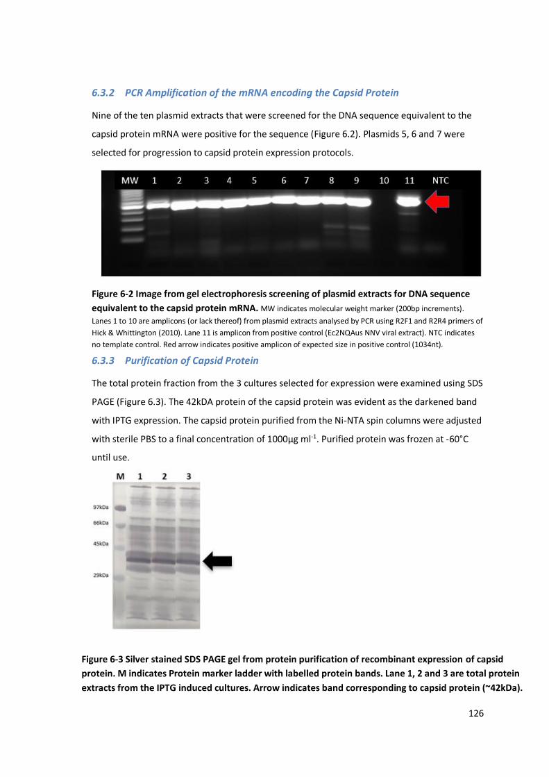
126
6.3.2 PCR Amplification of the mRNA encoding the Capsid Protein
Nine of the ten plasmid extracts that were screened for the DNA sequence equivalent to the
capsid protein mRNA were positive for the sequence (Figure 6.2). Plasmids 5, 6 and 7 were
selected for progression to capsid protein expression protocols.
Figure 6-2 Image from gel electrophoresis screening of plasmid extracts for DNA sequence
equivalent to the capsid protein mRNA. MW indicates molecular weight marker (200bp increments).
Lanes 1 to 10 are amplicons (or lack thereof) from plasmid extracts analysed by PCR using R2F1 and R2R4 primers of
Hick & Whittington (2010). Lane 11 is amplicon from positive control (Ec2NQAus NNV viral extract). NTC indicates
no template control. Red arrow indicates positive amplicon of expected size in positive control (1034nt).
6.3.3 Purification of Capsid Protein
The total protein fraction from the 3 cultures selected for expression were examined using SDS
PAGE (Figure 6.3). The 42kDA protein of the capsid protein was evident as the darkened band
with IPTG expression. The capsid protein purified from the Ni-NTA spin columns were adjusted
with sterile PBS to a final concentration of 1000µg ml-1. Purified protein was frozen at -60°C
until use.
Figure 6-3 Silver stained SDS PAGE gel from protein purification of recombinant expression of capsid
protein. M indicates Protein marker ladder with labelled protein bands. Lane 1, 2 and 3 are total protein
extracts from the IPTG induced cultures. Arrow indicates band corresponding to capsid protein (~42kDa).
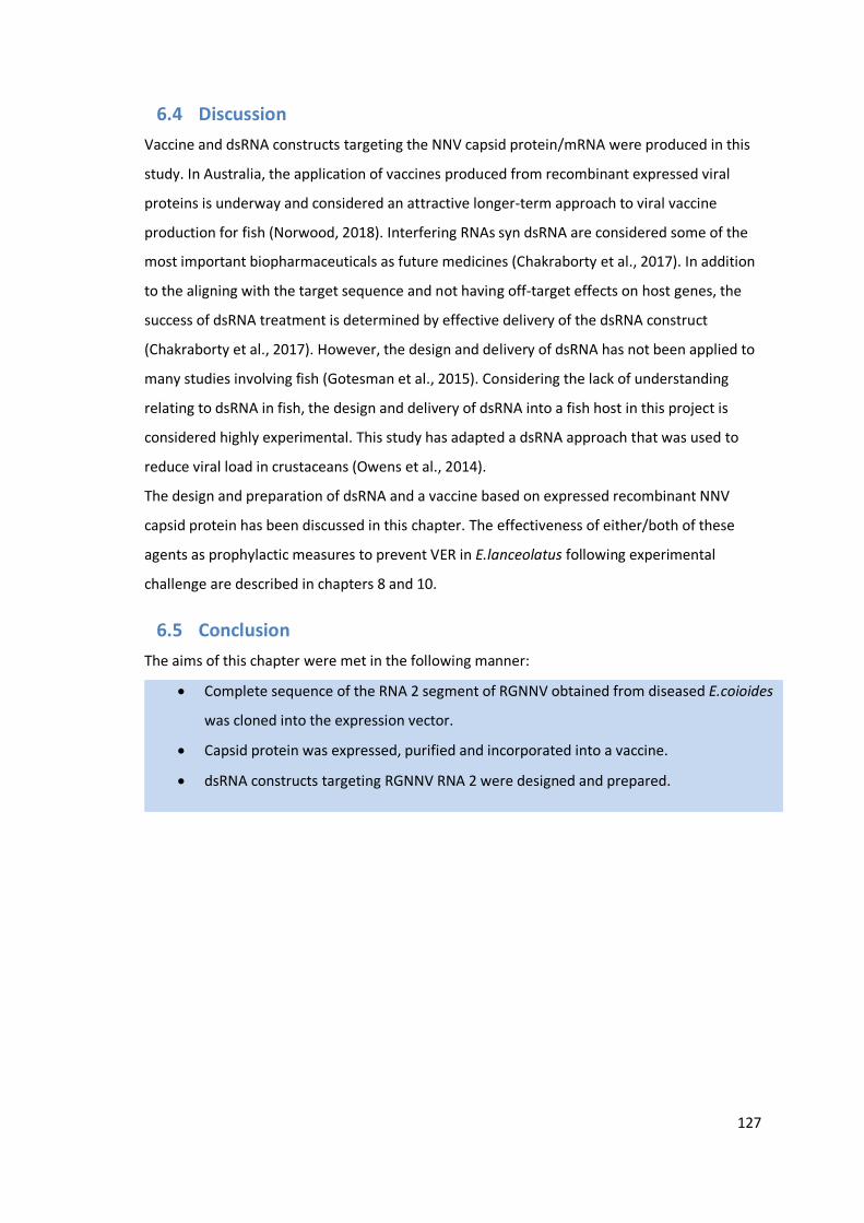
127
6.4 Discussion
Vaccine and dsRNA constructs targeting the NNV capsid protein/mRNA were produced in this
study. In Australia, the application of vaccines produced from recombinant expressed viral
proteins is underway and considered an attractive longer-term approach to viral vaccine
production for fish (Norwood, 2018). Interfering RNAs syn dsRNA are considered some of the
most important biopharmaceuticals as future medicines (Chakraborty et al., 2017). In addition
to the aligning with the target sequence and not having off-target effects on host genes, the
success of dsRNA treatment is determined by effective delivery of the dsRNA construct
(Chakraborty et al., 2017). However, the design and delivery of dsRNA has not been applied to
many studies involving fish (Gotesman et al., 2015). Considering the lack of understanding
relating to dsRNA in fish, the design and delivery of dsRNA into a fish host in this project is
considered highly experimental. This study has adapted a dsRNA approach that was used to
reduce viral load in crustaceans (Owens et al., 2014).
The design and preparation of dsRNA and a vaccine based on expressed recombinant NNV
capsid protein has been discussed in this chapter. The effectiveness of either/both of these
agents as prophylactic measures to prevent VER in E.lanceolatus following experimental
challenge are described in chapters 8 and 10.
6.5 Conclusion
The aims of this chapter were met in the following manner:
Complete sequence of the RNA 2 segment of RGNNV obtained from diseased E.coioides
was cloned into the expression vector.
Capsid protein was expressed, purified and incorporated into a vaccine.
dsRNA constructs targeting RGNNV RNA 2 were designed and prepared.
128
CHAPTER 7. EXPERIMENTAL CHALLENGE VIA CO-HABITATION
WITH MARINE LEECH, ZENYLANICOBDELLA ARUGAMENSIS
Background
Challenge of fish via injection does not represent the natural pathway of
transmission of NNV in natural disease outbreaks.
Experimental reproduction of VER outbreaks simulating a more natural route of
exposure, namely co-habitation with infected individuals or water borne exposure
does not yield consistent results.
Although naturally occurring disease outbreaks are reported following periods of
“stress”, a trigger that consistently induces VER in aquaculture systems has not
been identified.
In temperate finfish aquaculture, leeches have been proposed as vectors of viral
disease.
Infestation by the marine leech, Zeylanicobdella arugamensis, has been reported
to affect the health of grouper within earthen ponds in North Queensland.
Some authors report the detection of NNV from blood of infected fish.
Whilst Z.arugamensis have been reported to be vectors of marine fish
trypanosomes, there has been no reported investigation of their role in
transmission of viruses.
Aims of this Chapter
Investigate if RGNNV can be transmitted to fish held in a co-circulation system
with fish exposed to by IM challenge.
Investigate if exposure to the marine leech, Zeylanicobdella arugamensis, in
combination with co-habitation with RGNNV infected fish leads to RGNNV
infection or VER outbreaks in juvenile E.lanceolatus.
Investigate if the marine leech, Zeylanicobdella arugamensis, has potential to act
as a vector of RGNNV.
7.1 Introduction
Although injection methods induce a consistent expression of VER, they are artificial and do
not reflect a natural infection path leading to outbreaks of VER (Kim et al., 2018). Ideally,

129
challenges to assess the efficacy of vaccines should mimic the natural path of infection. Whilst
natural disease outbreaks, which presumably do not involve an intramuscular injection of
NNV, often lead to mass mortality, the reproduction of VER via co-habitation with infected
animals and water borne exposure is inconsistent and leads to reduced levels of mortality and
disease in fish that are aged beyond the larval stages (Anderson & Moody 2004, Kim et al.,
2018). Anderson and Oakey (2008) stated: “Our feeling is that the project test results do not
support the suggestion that nodavirus infections are readily transmitted from one adult
barramundi to another sharing the same tank.” Attempted bath exposure of freshwater fish
species (silver perch, golden perch and sleepy cod and Barcoo grunter (6 and 12-week-old) to
VNN did not display any signs of infection despite morbidity of 46%, 15%, 98% and 7% when
fish were exposed via injection (Anderson and Moody 2004). RGNNV has been isolated in cell
culture and detected by RT-qPCR in water containing fish with clinical VER (Jaramillo et al.,
2017). Hodneland et al. (2011) added brain homogenate from severely infected sea bass to an
aquarium containing 15 sea bass (40g) and co-habitated fish with IM injected sea bass (1:3
ratio infected: cohabitated). None of the cohabitated fish displayed clinical signs or were
positive for the detection of RGNNV RNA 2 by RT-qPCR during the 5-week incubation period.
Whereas 85% of the IM infected fish died within 10 days. Fish exposed to brain homogenate in
the water displayed 33% mortality from day 17-35 post exposure (Hodneland et al., 2011).
The inconsistency in disease expression via water exposure to viruses, including NNV, in
experimental systems compared to the process that leads to natural disease outbreaks is
proposed to be due to the presence of “a stress event” as a precursor to trigger natural
disease outbreaks. The “stress events” remain largely un-investigated beyond simple
manipulations of water temperature and salinity or retrospective studies that identify risk
factors (Juniar et al., 2018). Artificial attempts to mimic stress and induce immunosuppression
of Atlantic cod, Gadus morhua by injection with prednisolone-acetate failed to induce disease
following IM challenge with NNV isolated from Atlantic halibut, Hippoglossus hippoglossus
(Korsnes et al., 2009). Conversely, exposure to tributyltin, a component of boat hull anti-foul,
lead to increased mortality, with lower exposure dose to RGNNV in Japanese medaka Oryzias
latipes (Kitamura et al., 2017).
Parasite infestations are frequently encountered in marine fish culture systems and, in high
numbers, may place a stress burden on host fish. Infestation of grouper and barramundi by the
marine leech Zeylanicobdella arugamensis have occurred within marine farm grow-out
systems in Northern Queensland (Vaughan, 2018). This leech has a wide geographic range and
infection of marine groupers have been reported from Philippines, Sri Lanka, Malaysia,

130
Singapore, India, Japan, Iran and China (Murwantoko et al., 2018). In temperate fish farming
systems leeches have been implicated as vectors of some viruses (Salimi and Abdi 2016 and
Steinbauer et al., 2019). Although no viral transmission studies have been conducted with the
species, Z.arugamensis has been demonstrated to be an efficient vector of the marine
trypanosome, Trypanosoma nudigobbi (Hayes et al., 2014.) The case of VER in cobia described
by Chu et al. (2013) noted the presence of Z.arugamensis along with monogeneans (Benedenia
sp.) and copepods (Caligus sp.). Yet, there was no suggestion of any role the parasites may
have played in relation to the mortalities and there are no published reports of attempts to
detect viruses from leeches from tropical finfish aquaculture systems (PubMed, Science Direct,
Google scholar, Scopus databases accessed 2.08.19). In addition to directly transferring
viruses, the feeding action of leech and fish scraping and rubbing behavioural in response to
leech infestation can cause disruption of the protective skin-scale-mucus barrier. Disruption of
the physical barrier of skin, scales and mucus by parasites render fish susceptible to secondary
infection by disease-causing organisms such as bacteria, fungi and viruses (Francis-Floyd,
2018). Dermal abrasion prior to water borne exposure to Vibrio harveyi was required to induce
disease in an experimental challenge of juvenile hybrid groupers E.fuscoguttatus x
E.lanceolatus (abw 4.15g) (Shen et al., 2017). Infestation with Z.arugamensis, and secondary
infection with pathogenic bacteria such as Vibrio alginolyticus has been associated with
mortalities in grouper (Ravi & Yahaya, 2017). The aim of this study is to investigate potential
experimental challenge models for future studies to test the efficacy of therapies developed in
Chapter 6 to prevent VER. Models include exposure to RGNNV through a shared circulation
system (co-circulation). The potential of the marine leech, Z. arugamensis to compromise the
skin-mucus barrier, act as “a stress factor” or vector to induce VER under co-circulation will
also be investigated.
7.2 Materials and Methods
7.2.1 Culture of marine leech, Zeylanicobdella arugamensis
Culture of marine leech Z. arugamensis was kindly conducted by JCU PhD Candidate Dr David
Vaughan. Approximately 100 leeches were provided for the experiment. Prior to exposure to
the experimental fish, leeches were removed from the E. coioides host fish and starved for 24
hours.
7.2.2 Fish culture, viral challenge and co-circulation system
Thirty juvenile groupers, E.lanceolatus were dispersed evenly into a system consisting of 3 x
60L tanks that were immediately adjacent to each other and sharing the same biological

131
filtration and recirculation system. There was no shield to prevent aerosol spread between
tanks. (Figure 7.1). Water, temperature, light and fish feeding conditions were as described
previously in section 2.2. For the viral challenge, ten fish from one tank (Tank C Figure 7.1)
were sedated and challenged by IM injection of 100 uL of RGNNV viral extract (6.36 x 10⁴ mL-1)
as described in 2.2.2. Fish from the RGNNV challenged tanks began displaying clinical signs of
VER from day 8 post IM injection. One fish was sampled from the IM challenge group (Tank C)
at day 4 prior to the onset of clinical signs. When fish began displaying signs of VER at day 8,
fifty leeches Zeylanicobdella arugamensis were added to two of the tanks, namely the RGNNV
challenged tank (Tank C Figure 7.1) and another tank on the same recirculation system (Tank A
Figure 7.1). The leeches had been starved for 24 hours prior to their addition to the tanks
(provided by Dr David Vaughan). Fish were observed twice daily for signs of VER. When fish
displayed multiple signs of disease (two clinical signs e.g. not feeding and inflated swim
bladder), they were removed from the system and euthanased as described in 2.2.1. Any
leeches that were attached to euthanased fish were removed and stored in separate 1.5ml
RNase free microcentrifuge tubes and frozen at -20°C until the end of the experiment. The
experiment was terminated at 40 days post IM challenge. All remaining fish were euthanased
at day 40 post IM challenge. All leeches present on fish or in the tank system were stored in
pools of 5 animals and frozen at -20°C.
Figure 7-1: Diagrammatic illustration of the experimental aquarium system for co-circulation
Each tank (A, B, C) contained 10 fish within a 60-litre tank. Water circulated between the tanks from a shared bio-filter (D). Tank A fish were exposed to 50 Z.arugamensis from the onset of clinical signs of VER in Tank C fish. Tank B fish had no exposure to Z.arugamensis nor RGNNV other than that which occurred within the recirculation system. Tank C fish were exposed to an IM challenge of RGNNV and at the onset of clinical signs were exposed to 50 Z.arugamensis.
7.2.3 Nucleic acid extraction, CDNA synthesis, RT-qPCR.
Total nucleic acid (TNAs) was extracted from whole eye and whole marine leeches collected
from fish following the protocol previously described in 2.1.2. cDNA synthesis and qR1T and
D

132
qR2T, were conducted on TNAs as previously described in 2.1.3 and 5.2.3. Leeches were
extracted in pools of 5 collected from fish when they were euthanased.
7.3 Results
7.3.1 Expression of VER in juvenile E.lanceolatus.
The fish that were challenged by IM injection of viral extract (Tank C) displayed clinical signs of
VER (Figure 7.2). Clinical signs of VER, including hypertrophy of the swim bladder, abnormal
swimming behaviour and lack of feeding were evident in the RGNNV injected + leech exposed
group from 8 days post IM challenge (Figure 7.2). No clinical signs of VER were evident in fish
from either of the co-circulating tanks for the 40 days of the experiment (Figure 7.2). Leeches
were observed attached to the fish in Tank A and C (leeches added to the tanks) but no
leeches were observed in the Tank B (no leeches added to the tank) during the course of the
experiment.
Figure 7-2 Cumulative morbidity (%) of fish from each tank following IM challenge of Tank C fish v days post viral challenge. Tank A contained fish exposed to leeches and on co-circulation with IM
challenged fish in Tank C. Tank B contained fish on circulation with IM challenged fish in Tank C. Tank C contained fish challenged with IM injection of viral extract and exposed to leeches from the onset of clinical signs of VER.
7.3.2 Detection of RGNNV by RT-qPCR
RGNNV was detected using qR1T and qR2T from the eye tissue of all fish in the IM challenged +
leech experimental group (Table 7.1). RGNNV was detected in the eye tissue of the grouper
that was killed at day 4 post challenge (4 days prior to clinical signs). RGNNV was not detected
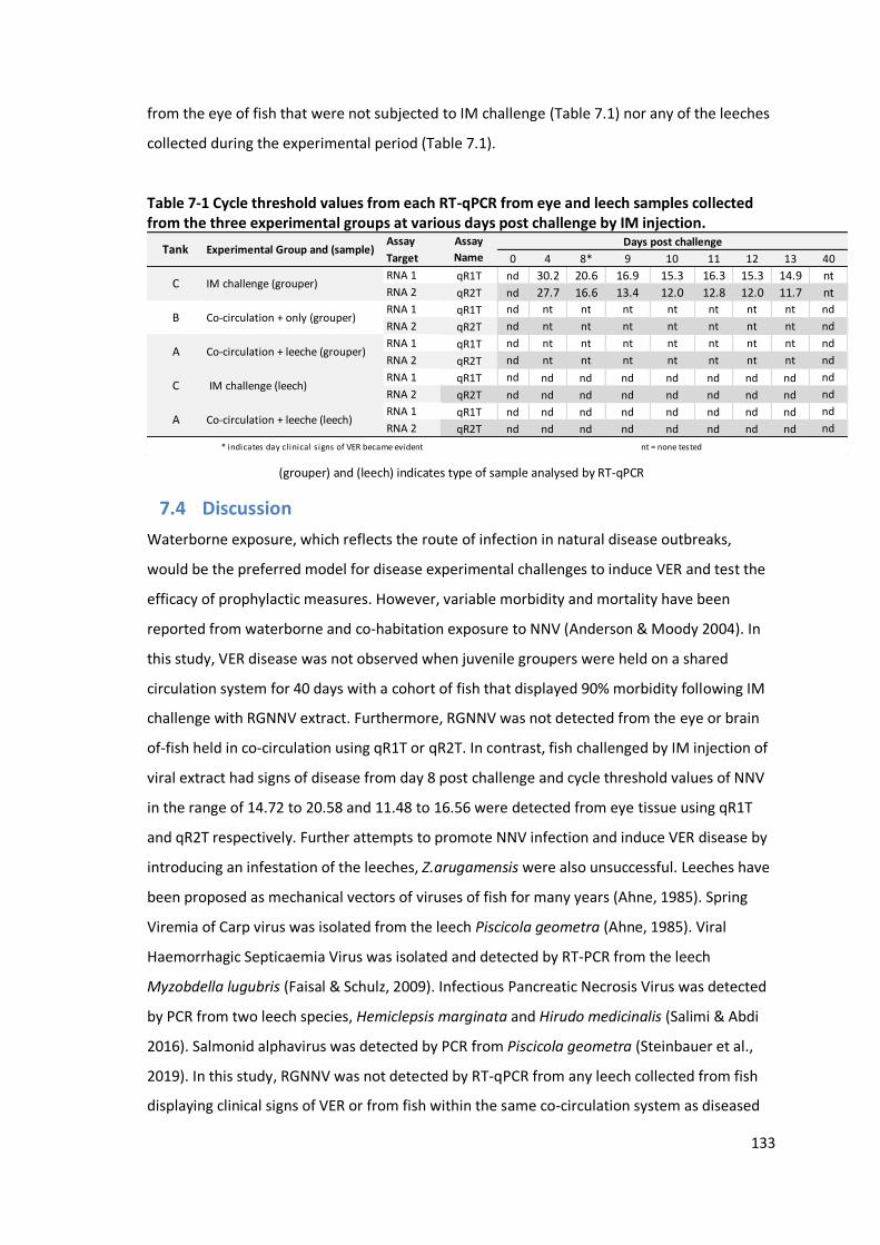
133
from the eye of fish that were not subjected to IM challenge (Table 7.1) nor any of the leeches
collected during the experimental period (Table 7.1).
Table 7-1 Cycle threshold values from each RT-qPCR from eye and leech samples collected from the three experimental groups at various days post challenge by IM injection.
(grouper) and (leech) indicates type of sample analysed by RT-qPCR
7.4 Discussion
Waterborne exposure, which reflects the route of infection in natural disease outbreaks,
would be the preferred model for disease experimental challenges to induce VER and test the
efficacy of prophylactic measures. However, variable morbidity and mortality have been
reported from waterborne and co-habitation exposure to NNV (Anderson & Moody 2004). In
this study, VER disease was not observed when juvenile groupers were held on a shared
circulation system for 40 days with a cohort of fish that displayed 90% morbidity following IM
challenge with RGNNV extract. Furthermore, RGNNV was not detected from the eye or brain
of-fish held in co-circulation using qR1T or qR2T. In contrast, fish challenged by IM injection of
viral extract had signs of disease from day 8 post challenge and cycle threshold values of NNV
in the range of 14.72 to 20.58 and 11.48 to 16.56 were detected from eye tissue using qR1T
and qR2T respectively. Further attempts to promote NNV infection and induce VER disease by
introducing an infestation of the leeches, Z.arugamensis were also unsuccessful. Leeches have
been proposed as mechanical vectors of viruses of fish for many years (Ahne, 1985). Spring
Viremia of Carp virus was isolated from the leech Piscicola geometra (Ahne, 1985). Viral
Haemorrhagic Septicaemia Virus was isolated and detected by RT-PCR from the leech
Myzobdella lugubris (Faisal & Schulz, 2009). Infectious Pancreatic Necrosis Virus was detected
by PCR from two leech species, Hemiclepsis marginata and Hirudo medicinalis (Salimi & Abdi
2016). Salmonid alphavirus was detected by PCR from Piscicola geometra (Steinbauer et al.,
2019). In this study, RGNNV was not detected by RT-qPCR from any leech collected from fish
displaying clinical signs of VER or from fish within the same co-circulation system as diseased
Assay
Target 0 4 8* 9 10 11 12 13 40
RNA 1 qR1T nd 30.2 20.6 16.9 15.3 16.3 15.3 14.9 nt
RNA 2 qR2T nd 27.7 16.6 13.4 12.0 12.8 12.0 11.7 nt
RNA 1 qR1T nd nt nt nt nt nt nt nt nd
RNA 2 qR2T nd nt nt nt nt nt nt nt nd
RNA 1 qR1T nd nt nt nt nt nt nt nt nd
RNA 2 qR2T nd nt nt nt nt nt nt nt nd
RNA 1 qR1T nd nd nd nd nd nd nd nd nd
RNA 2 qR2T nd nd nd nd nd nd nd nd nd
RNA 1 qR1T nd nd nd nd nd nd nd nd nd
RNA 2 qR2T nd nd nd nd nd nd nd nd nd
nt = none tested
C
B
A
C
A
Tank
IM challenge (leech)
Co-circulation + leeche (leech)
Days post challenge
* indicates day clinical signs of VER became evident
Experimental Group and (sample)Assay
Name
IM challenge (grouper)
Co-circulation + only (grouper)
Co-circulation + leeche (grouper)
134
fishes. Presumably, one of the requirements for leeches to act as mechanical vectors of viral
infections relies on the presence of the virus in the blood cells of infected fish. Reports on the
detection of NNV from the blood of infected fish are inconsistent. Korsnes et al. (2009)
reported the detection of NNV from the blood of Atlantic cod, Gadus morhua. Yet, no
detection from blood cells have been reported by others (Valero et al., 2018). The inability to
induce RGNNV infection or VER disease via co-circulation with diseased fish, either with or
without an additional burden of leech infestation indicates waterborne transmission of NNV is
not a robust model for future studies to test the efficacy of prophylactic measures. Although
injection challenge by-passes one of the primary immune barriers of the fish, investigation into
efficacy of prophylactic measures developed in Chapter 6 are proposed to be conducted using
IM challenge with RGNNV
7.5 Conclusion
The following outcomes and conclusions were discussed in this chapter:
RGNNV was not transmitted to naïve fish on a co-circulation system with VER
diseased fish during a 40-day experiment.
Exposure of fish to the marine leech Zeylanicobdella arugamensis in addition to co-
circulation with VER affected fish did not lead to infection with RGNNV in juvenile
E.lanceolatus.
The inability to detect RGNNV from leeches which fed on VER affected animals
indicates Z.arugamensis is not likely to be an effective vector of RGNNV.
As VER was not induced via co-circulation or co-exposure to a leech “stress event”,
no effective method of water borne challenge has been identified for future trials.
Although not mimicking the natural route of infection, challenge by IM injection is
the only consistent challenge model available for continued testing of the efficacy of
vaccine to prevent VER within the time frames of this project.

135
CHAPTER 8. TESTING EFFICACY OF PROPHYLATIC MEASURES
AGAINST VIRAL ENCEPHALOPATHY AND RETINOPATHY (VER)
IN EPINEPHELUS LANCEOLATUS VIA EXPERIMENTAL
CHALLENGE
Background
Aims of this Chapter
A vaccine containing the recombinantly expressed capsid protein of RGNNV
has been prepared for assessment as a prophylactic measure to prevent VER
(Chapter 6).
dsRNA designed to target RGNNV RNA 2 has been prepared as an
experimental compound to prevent VER (Chapter 6).
Due to reported inconsistency to induce VER by waterborne or co-habitation,
challenge by IM injection with RGNNV is the preferred method of challenge
within the scope of this project.
The pathogenesis of VER from exposure to disease expression has not been
well described in grouper.
Synthetic plasmid controls have been prepared to support monitoring of the
qR1T and qR2T assays towards both tracking the pathogenesis of VER and
validation of both assays for application on grouper tissues.
To test the efficacy of vaccine and dsRNA preparations to prevent infection
and disease in juvenile E. lanceolatus following IM challenge with RGNNV viral
extract.
To assess the performance of the qR1T and qR2T assays to detect RGNNV
from grouper derived brain and eye tissue following experimental challenge
by IM injection with RGNNV.
To track the pathogenesis of RGNNV from exposure to disease expression by
applying analysis using RT-qPCR.

136
8.1 Introduction
In grouper aquaculture, stocking of ponds with NNV-free fingerlings does not prevent
economically catastrophic VER outbreaks during grow-out stages of production. There is an
urgent need to develop prophylactic measures to combat NNV infection in grouper grow-out
systems. Despite numerous reports of successful vaccination against NNV in various
experimental formats, there are few commercial transfers for application by industry.
The management of VER in grouper aquaculture is hampered by gaps in knowledge of both
the development of the juvenile grouper immune system and the pathogenesis of VER. This
study has prepared a vaccine and dsRNA as potential therapies to prevent RGNNV infection.
Although monitoring of the juvenile grouper immune system is beyond the scope of this study,
tracing the progression of the disease following IM challenge is possible with RT-qPCR. Despite
the need to improve knowledge and the availability of validated, sensitive, specific,
quantitative, rapid turnover and high throughput RT-qPCR assays, very few published reports
have applied RT-qPCR to investigate the pathogenesis of VER (Jaramillo et al., 2017; Hodneland
et al., 2011; Lopez-Jimena et al., 2011; Panzarin et al., 2010). Only one article details the
application of RT-qPCR to grouper species (Kim et al., 2018).
8.2 Materials and Methods
8.2.1 Fish husbandry
Six hundred juvenile giant grouper, E.lanceolatus (abw 18g 𝜎 ~2 g, range 11.3 g to 23.6 g; aged
84 days post hatch) were provided by a commercial hatchery. Water temperature ranged
between 24 to 28 ⁰C and salinity ranged from 26 to 35 ppt. Fish were weighed, tagged with
streamer tags to allow individual identification (Hallprint) (Refer to Figure 8.1) and fed ad lib
twice daily with a commercial fish feed pellet (Ridley AgriProducts Pty Ltd). Fish were
acclimated for two weeks prior to commencement of experimental procedures. Two weeks
(98 dph) and six weeks (126 dph) after acquisition, fish were injected with the experimental
treatments. All fish from the same experimental group were held in the same tank system until
IM challenge. Fish were sedated for all treatments involving injection, according to JCU Animal
Ethics requirements using AQUI-S® (AQUI-S).

137
Figure 8-1. (A) Image of juvenile E.lanceolatus with tag and (B). multiple tagged fish within a recovery tank after injection with RGNNV extract
8.2.2 Experimental design and vaccination
Fish were distributed into ten experimental groups. The experimental groups were duplicates
of five experimental treatments that were subsequently challenged with either PBS or RGNNV
extract. The five groups consisted of treatments prepared as described in Chapter 6. In
summary the experimental treatments were: (1) Placebo vaccinated with Adjuvant + PBS, (2)
vaccinated with recombinant expressed capsid protein (500mg mL-1) + adjuvant, (3) vaccinated
with recombinant expressed capsid protein (500mg mL-1) + adjuvant and a dsRNA (50pmol µl-1
dsRNA) construct with homology to RGNNV, (4) injected with a dsRNA (50pmol µl-1 dsRNA)
construct with homology to RGNNV, (5) injected with a dsRNA (50pmol µl-1 dsRNA) construct
with no homology to RGNNV. All injections were the same approximate body position as
described in Chapter 2.2.3.
8.2.3 Experimental Challenge with RGNNV extract
Two weeks after the booster (140 dph) treatments or equivalent placebo treatments, fish
were challenged with virus by either intramuscular injection with 50 µL RGNNV extract
(calculated copy number 6.36 x 10⁴ mL-1 of viral extract prepared in 2.1.6) or 50 µL sterile PBS.
Fish were then redistributed into tanks with six fish from each of the five treatment groups
were placed into replicated tank systems. The experimental groups are described in Table 8-1
and illustrated in Figure 8.2.

138
Table 8-1 Summary of the experimental design indicating description of prophylactic measures, IM challenge, number of fish in each group and number of fish sampled at morbidity or within planned schedules in the absence of clinical signs of VER.
Figure 8-2: Graphic illustration of experimental tank design Ten duplicate systems consisting of 2 x 300 litre tanks for housing fish, were equipped with 1 x 300 litre tank containing ~100 litres of bio-wheels with constant water flow 1200L hr-1 and constant aeration. Each tank contained 6 fish from each of 5 treatments. Fish were individually tagged with T bar anchor tags to indicate experimental treatments.
8.2.4 Monitoring of fish health and euthanasia
Fish were monitored twice daily for signs of adverse health. For the first six days post IM
challenge, prior to the onset of clinic signs of VER, one fish from each treatment type was
removed from each tank system and euthanased by Aqui-S overdose (Table 8.1). When fish
At morbidity Scheduled*
PBS 60 0 10
RGNNV Extract 60 41 4
PBS 60 0 10
RGNNV Extract 60 32 9
PBS 60 0 10
RGNNV Extract 60 26 8
PBS 60 0 10
RGNNV Extract 60 54 4
PBS 60 0 10
RGNNV Extract 60 54 4
Sampling schedule
* indicates at scheduled days in the absence of clinical signs of VER
Anti-RGNNV ds RNA
Non-specific ds-RNA
Number of
fish Description
IM injection of 100µL adjuvant/PBS 1:1
IM injection of 100µL adjuvant/PBS:dsRNA
(RGNNV)
IM injection of 100µL adjuvant/RGNNV capsid
protein
IM injection of 100µL PBS:ds RNA (RGNNV)
IM injection of 100µL PBS:ds RNA
Experimental Group Challenged 50 uL
i.m. injection
Placebo vaccine
Vaccine & Anti-RGNNV dsRNA
Vaccine
139
displayed more than one sign of VER they were euthanased. Euthanasia was conducted by
Aqui-S overdose as described in 2.2.1. Euthanased fish were weighed, frozen whole in
individually labelled plastic bags and stored at -20°C along with their identification tags. After
20 days post challenge, when morbidity had reached plateau phase, all surviving fish were
collected from the RGNNV challenged tanks, weighed and consolidated into two of the
experimental tank systems. Fish that displayed a clinical sign of VER, mostly darkened colour or
inflated swim bladders, yet continued to feed were placed in the “clinical sign” system and fish
that did not display signs of VER were placed in the “no clinical sign” system. The “clinical sign”
fish were removed from the “no clinical sign” fish to avoid injury and stress. When held in the
same tanks, the “clinical sign” fish were actively attacked and outcompeted for feed by the “no
clinical sign” fish. The condition of fish in the consolidated systems was monitored for a further
80 days post IM challenge. During the 80-day period the streamer tags became damaged and
were actively removed by the fish so accurate tracking of the individual treatments was, in
many cases not possible. After 80 days all fish in the “clinical signs” tank were euthanased and
half of the fish in the “no clinical signs” tank were euthanased. Euthanased fish were weighed
and stored as previously described. Thirty-five fish with “no clinical signs”, which had retained
their tags were retained for longer term monitoring of growth. All of the retained fish were
from one of the two vaccinated treatment groups. Tags were removed in an effort to reduce
injury from fish trying to eat each other’s tags. Grouping of the “no clinical signs” fish was
required because the growth and subsequent aggression and territorial behaviour of the larger
fish prevented retaining all of the survivors under conditions that would comply with Animal
Ethics Permits. Throughout the remainder of the project, fish with “no clinical signs” were
euthanased when fish growth caused overcrowding and the fish could not be maintained in
conditions that complied with the ethical treatment of animals for scientific purposes. Ten fish
from each of the experimental groups that were challenged with PBS were euthanased at
scheduled intervals. One fish from each group was euthanased at days 0,2,3,4, and a further 6
fish from each group were euthanased at day 20 (Refer to Table 8-1).
8.2.5 Nucleic acid extraction
Total nucleic acid was extracted from eye as described in 2.1.2.
8.2.6 Analysis by RT-qPCR using qR1T and qR2T
Analysis by RT-qPCR using qR1T and qR2T was conducted on tissue extracts as described in
2.1.9.

140
8.3 Results
8.3.1 Expression of VER following experimental challenge of juvenile grouper with
RGNNV extract and efficacy of protection of each preventative treatment.
There was 100% survival and no adverse reaction evident in any of the five groups of fish
challenged with PBS injection for the duration of the trial period. Within the RGNNV
challenged groups, fish fed and behaved normally for five days post challenge. Six days post
challenge, a small number of fish displayed some typical signs of VER. Clinical signs included
inflation of swim bladder, lethargy, erratic swimming behavior, darkened body color and flared
opercula. The number of fish with multiple clinical signs increased markedly between days 8 to
10 post challenge after which morbidity plateaued (Table 8.2). Exposure to dsRNA (specific and
non-specific design) was not effective in reducing morbidity in juvenile E.lanceolatus following
IM challenge with viral extract. The groups that received the capsid protein within either
vaccine formulation (vaccine alone, or vaccine + dsRNA) had reduced total numbers of fish
displaying VER compared to the placebo vaccinated groups and the groups which received
dsRNA in either form (Figure 8.3). By day 20 post viral challenge, the group that received
vaccine and vaccine with anti-RGNNV dsRNA displayed 43% and 53% cumulative morbidity
compared to the placebo vaccinated (68%) and either form of dsRNA injected (88% and 88%)
(Figure 8.1). Observation of body mass of moribund and surviving fish from the RGNNV
challenge indicates a trend whereby larger vaccinated fish tended to survive the RGNNV
challenge. However, as there were small numbers of larger fish and body weight was not
considered in the experimental design no statistical analysis was attempted.
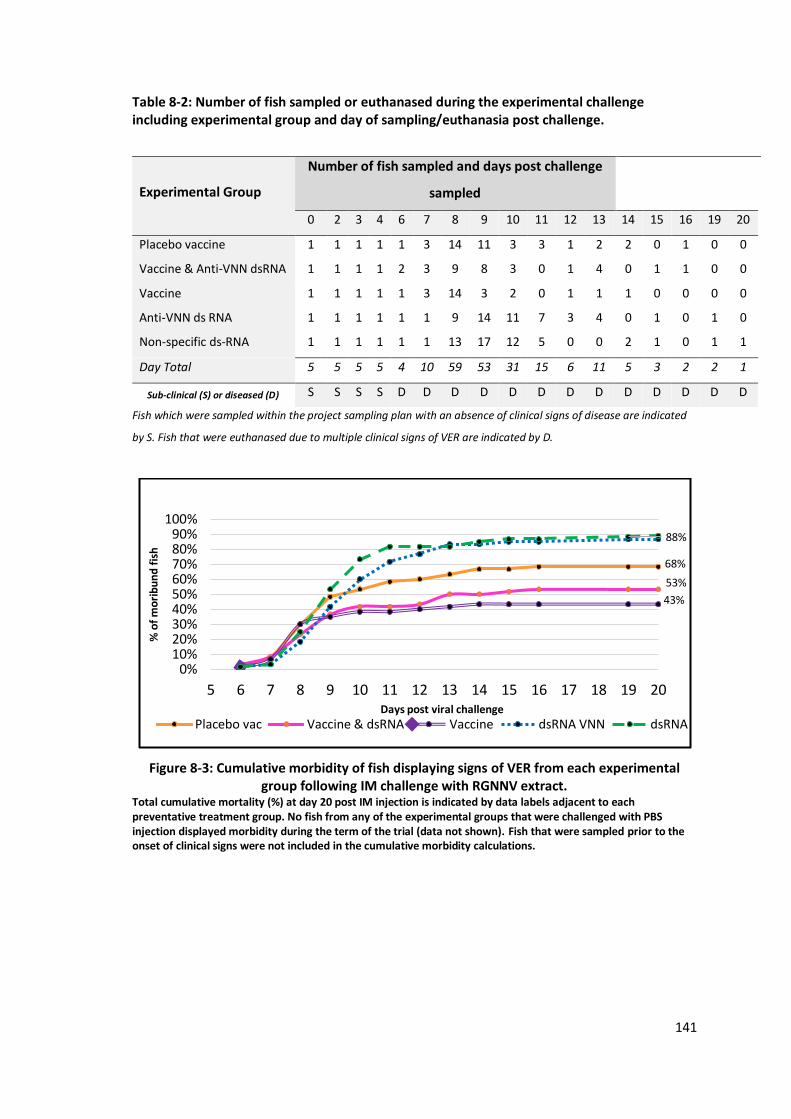
141
Table 8-2: Number of fish sampled or euthanased during the experimental challenge including experimental group and day of sampling/euthanasia post challenge.
Fish which were sampled within the project sampling plan with an absence of clinical signs of disease are indicated
by S. Fish that were euthanased due to multiple clinical signs of VER are indicated by D.
Figure 8-3: Cumulative morbidity of fish displaying signs of VER from each experimental group following IM challenge with RGNNV extract.
Total cumulative mortality (%) at day 20 post IM injection is indicated by data labels adjacent to each preventative treatment group. No fish from any of the experimental groups that were challenged with PBS injection displayed morbidity during the term of the trial (data not shown). Fish that were sampled prior to the onset of clinical signs were not included in the cumulative morbidity calculations.
68%
53%
43%
88%
0%10%20%30%40%50%60%70%80%90%
100%
5 6 7 8 9 10 11 12 13 14 15 16 17 18 19 20
% o
f m
ori
bu
nd
fis
h
Days post viral challenge
Placebo vac Vaccine & dsRNA Vaccine dsRNA VNN dsRNA
Experimental Group
Number of fish sampled and days post challenge
sampled
0 2 3 4 6 7 8 9 10 11 12 13 14 15 16 19 20
Placebo vaccine 1 1 1 1 1 3 14 11 3 3 1 2 2 0 1 0 0
Vaccine & Anti-VNN dsRNA 1 1 1 1 2 3 9 8 3 0 1 4 0 1 1 0 0
Vaccine 1 1 1 1 1 3 14 3 2 0 1 1 1 0 0 0 0
Anti-VNN ds RNA 1 1 1 1 1 1 9 14 11 7 3 4 0 1 0 1 0
Non-specific ds-RNA 1 1 1 1 1 1 13 17 12 5 0 0 2 1 0 1 1
Day Total 5 5 5 5 4 10 59 53 31 15 6 11 5 3 2 2 1
Sub-clinical (S) or diseased (D) S S S S D D D D D D D D D D D D D

142
8.3.2 Body weight of fish
The body weight of fish that were euthanased with clinical signs of VER were recorded (Figure
8.4).
Figure 8-4: Scatter plot of mass of individual fish from each treatment group v days when euthanased due to morbidity for each day post challenge by IM injection of RGNNV. Notably only 3 fish that received vaccine that were over 50 g in body weight were euthanased.
8.3.3 Detection and quantification of RGNNV by RT-qPCR assay
There was no detection of RGNNV genome by either assay from any fish challenged with PBS
injection. RGNNV genome was detected with both assays from eye of viral challenged fish
from day two post challenge, four days prior to the onset of signs of disease, until and beyond
day 19 post challenge (Table 8-3). Assay qR1T, which targets the RNA-1 segment had higher Ct
values than qR2T, which targets the RNA-2 segment (Table 8-3).

143
Table 8-3: Average Cycle threshold value of qR1T and qR2T from analysis on Eye tissue from viral challenged fish.
The calculated copy number of RGNNV genome detected per mg of eye tissue by each assay
ranged from 0.02 which was detected at day 2 post challenge, to 8.3 x10⁵ which was detected
at day 11 post challenge. (Figure 8-5). Mass of eye tissue ranged from 125 mg to 347 mg
(average 180.4 sd. 49.9 mg). Prior to onset of clinical signs, the calculated copy number was
less than 103 mg-1. From the onset of clinical signs at day 6, the calculated copy number of
RGNNV mg-1 of eye tissue markedly increased in both assays. Detection by qR2T was
consistently ten-fold higher than that of qR1T (Figure 8-5). Prior to the onset of clinical signs at
day 6, the calculated copy number mg-1 was less in the two groups exposed to vaccine
compared to the other three treatment groups. However, during the period of peak morbidity
calculated copy number was comparable between the five treatment groups.
Assay
Target 0 2 3 4 6* 7 8 9 11 13 15 19
RNA 1 qR1T nd nd 37.7 26.7 19.7 16.1 16.2 17.2 15.5 16.7
RNA 2 qR2T nd 45.0 30.0 29.5 15.3 19.0 11.6 12.9 11.3 11.9
RNA 1 qR1T nd 31.8 nd 31.7 21.1 18.8 17.3 16.5 18.0 15.5 19.3
RNA 2 qR2T nd 29.9 28.8 29.6 16.8 15.7 12.4 11.9 13.4 10.8 14.4
RNA 1 qR1T nd 36.8 34.9 34.5 nd nd 16.0 15.8 15.5 16.4
RNA 2 qR2T nd 30.8 30.4 31.4 30.7 30.9 11.5 11.4 10.8 11.8
RNA 1 qR1T nd nd 35.7 25.4 17.6 19.5 15.7 15.8 16.3 16.0 14.7 16.8
RNA 2 qR2T nd 45.0 29.7 22.6 14.8 15.2 11.1 11.4 11.7 11.4 10.8 11.8
RNA 1 qR1T nd nd 31.1 28.9 19.2 12.9 16.1 15.5 16.1 16.0
RNA 2 qR2T nd 35.8 27.3 34.9 14.9 16.4 11.6 10.6 11.0 11.3
* indicates day clinical signs of VER became evident
Days post challenge
Vaccine
Anti-VNN ds RNA
Non-specific ds-RNA
Experimental GroupAssay
Name
Placebo vaccine
Vaccine & Anti-VNN
dsRNA

144
Figure 8-5: Calculated copy number of RNA 1 (Black columns) and RNA 2 (Red columns)
segment per mg of tissue detected by qR1T and qR2T assay for each experimental treatment
group (ds-RGNNV RNA; dsRNA, Vaccine, Vaccine +ds RGNNV RNA and Placebo) v days post
challenge by IM injection.
8.4 Discussion
The application of RT-qPCR to quantify the detection of RGNNV genome segments RNA 1 and
RNA 2 in response to IM challenge with RGNNV extract following exposure to experimental
prophylactic measures was discussed within this chapter.
The two RT-qPCR assays, qR1t and qR2T displayed similar sensitivity to detect RGNNV genome.
Both assays detected RGNNV genome segments from day 2 post-challenge albeit with a high
cycle threshold value (Ct) and very low calculated copy number per mg tissue. The frequency
of detection and calculated copy number per mg of eye tissue from both assays dramatically
increased in all experimental groups from day 6 post challenge, where clinical signs became

145
evident. Detection of RGNNV before the clinical disease was reported previously from
experimental challenges of juvenile sea bass after day one post-infection (Dalle Valle et al.,
2005). Souto et al. (2018) reported calculated copy number of RNA 1 and RNA 2 segments by
RT-qPCR in vitro of log 105.5-8.5 copies mL-1 within 24 hours post-inoculation yet an absence of
cellular pathological effect (CPE) until after 48hours.
Although RT-qPCR has significantly improved the ability to quantitatively determine viral
replication and better understand the expression of VER, few publications have applied RT-
qPCR for a purpose other than detection in disease outbreaks. Comparison of the levels of
RGNNV detection associated with disease previous reports is difficult. Many reports that have
applied RT-qPCR to experimental infection with NNV have presented results in different
formats including cycle threshold (Ct) value (Hodneland et al., 2011 and Panzarin et al., 2010,
Agnihotri et al., 2015), copies mL-1 (Kim et al., 2018), copies per 200ng total RNA (Dalle Valle et
al., 2005), copies per 4 µg total RNA (Lopez-Jimena et al., 2011), log10 viral copies ( Hick &
Whittington, 2010) or log10 viral copies g-1 brain tissue (Souto et al., 2018). Only one of the
prementioned studies were conducted on groupers (Kim et al., 2018).
In this experiment, average Ct values from eyes collected from the onset of clinical signs, days
6 to day 19 post challenge were 16.8 (s.d 1.67) (RNA 1) and 12.8 (s.d 2.14) (RNA 2)
respectively. Brain samples from clinically diseased grouper had a Ct value of ca 10 (RNA 2)
(Agnihotri et al., 2015), sea bass had a median Ct value of ca 15.0 (RNA 2) (Hodneland et al.,
2011) and a range of Ct value from 9.91 to 18.6 (RNA 2) (Parnzarin et al., 2010). Other authors
report calculated copy numbers of 109 mL-1 (RNA 1) to 106 mL-1 (RNA 2) (Kim et al., 2016) or 105
(RNA 1) and 107 (RNA 2) per 200ng total RNA (Dalla Valle et al., 2005).
In this investigation, although both assays detected the RGNNV genome, the calculated copy
number of qR1T was slightly less than qR2T. An increased calculated copy number of RNA 2
compared to RNA 1 has been noted previously where the number of copies of RNA 1 was 100
times less than those of RNA 2 from the brain of sea bass from days 3 to 14 post experimental
challenge (Dalle Valle et al., 2005). It is possible the differences in levels of detection are
related to variable transcription of the RNA 1 and RNA 2 gene segments as infection
progresses. As RNA 1 encodes the mRNA for the RNA-dependant RNA polymerase, the
replication and translation of RNA 1 is required for transcription of RNA 2. The time course of
sample collection in this investigation may not be sufficient to resolve shorter-term variation in
transcription of RNA 1 and RNA 2 during the early stage of progression towards disease. The
reduced efficiency in assays that detect RNA 1 may also relate to secondary structure
interaction between RNA 1 and its translated form, Protein A, that contribute to the formation
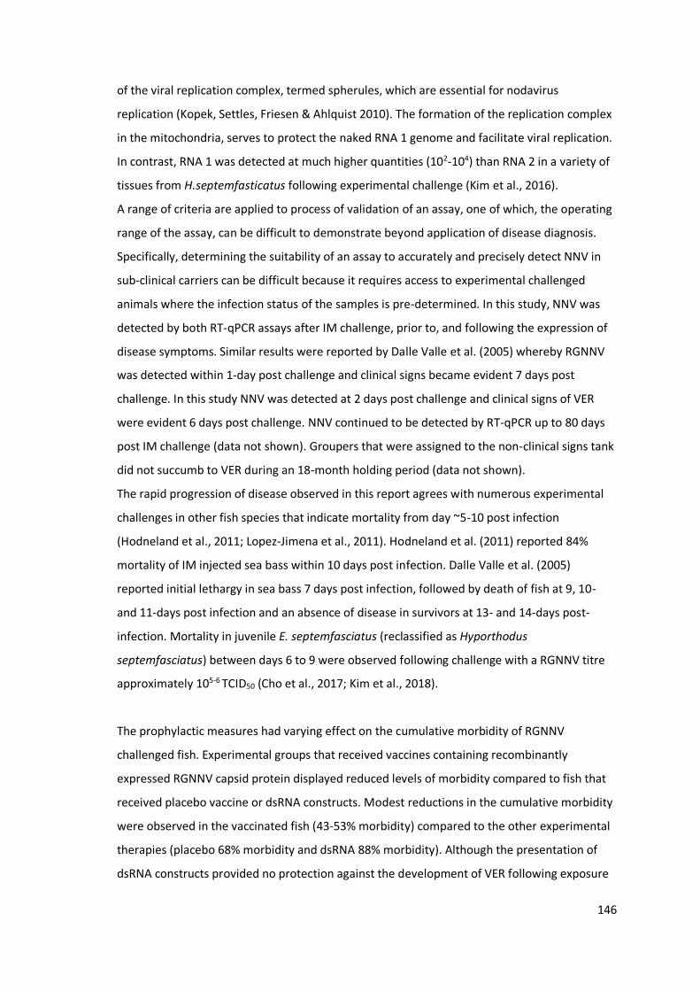
146
of the viral replication complex, termed spherules, which are essential for nodavirus
replication (Kopek, Settles, Friesen & Ahlquist 2010). The formation of the replication complex
in the mitochondria, serves to protect the naked RNA 1 genome and facilitate viral replication.
In contrast, RNA 1 was detected at much higher quantities (102-104) than RNA 2 in a variety of
tissues from H.septemfasticatus following experimental challenge (Kim et al., 2016).
A range of criteria are applied to process of validation of an assay, one of which, the operating
range of the assay, can be difficult to demonstrate beyond application of disease diagnosis.
Specifically, determining the suitability of an assay to accurately and precisely detect NNV in
sub-clinical carriers can be difficult because it requires access to experimental challenged
animals where the infection status of the samples is pre-determined. In this study, NNV was
detected by both RT-qPCR assays after IM challenge, prior to, and following the expression of
disease symptoms. Similar results were reported by Dalle Valle et al. (2005) whereby RGNNV
was detected within 1-day post challenge and clinical signs became evident 7 days post
challenge. In this study NNV was detected at 2 days post challenge and clinical signs of VER
were evident 6 days post challenge. NNV continued to be detected by RT-qPCR up to 80 days
post IM challenge (data not shown). Groupers that were assigned to the non-clinical signs tank
did not succumb to VER during an 18-month holding period (data not shown).
The rapid progression of disease observed in this report agrees with numerous experimental
challenges in other fish species that indicate mortality from day ~5-10 post infection
(Hodneland et al., 2011; Lopez-Jimena et al., 2011). Hodneland et al. (2011) reported 84%
mortality of IM injected sea bass within 10 days post infection. Dalle Valle et al. (2005)
reported initial lethargy in sea bass 7 days post infection, followed by death of fish at 9, 10-
and 11-days post infection and an absence of disease in survivors at 13- and 14-days post-
infection. Mortality in juvenile E. septemfasciatus (reclassified as Hyporthodus
septemfasciatus) between days 6 to 9 were observed following challenge with a RGNNV titre
approximately 105-6 TCID50 (Cho et al., 2017; Kim et al., 2018).
The prophylactic measures had varying effect on the cumulative morbidity of RGNNV
challenged fish. Experimental groups that received vaccines containing recombinantly
expressed RGNNV capsid protein displayed reduced levels of morbidity compared to fish that
received placebo vaccine or dsRNA constructs. Modest reductions in the cumulative morbidity
were observed in the vaccinated fish (43-53% morbidity) compared to the other experimental
therapies (placebo 68% morbidity and dsRNA 88% morbidity). Although the presentation of
dsRNA constructs provided no protection against the development of VER following exposure

147
to RGNNV extract in this experiment, the technology should not be discounted as a tool to
better understand and manage VNN. The application of dsRNA as a therapeutic has been
reported for the prevention of viral diseases of crustaceans (Owens et al., 2014). Although the
technology faces a number of hurdles there is at least one US patent claiming successful
application of dsRNA to prevent disease in an aquaculture species namely for the prevention
of Infectious Myonecrosis Virus in shrimp and other invertebrates (Loy et al.US patent No.
US8822427B2). This project attempted delivery of naked dsRNA, which was not protected
from degradation. The delivery of unprotected dsRNA has been reported previously (Owens et
al., 2014; ). One of the main problems in the development of siRNA for therapeutic use is the
need for a delivery method that prevents the degradation of siRNAs by cellular nucleases
(Chernikov et al., 2019). Adjustments to the delivery, concentration, target sequence and
duration of priming may lead to improvements in the efficacy of dsRNA prevent VER.
Considering the range of variables that require refinement in dsRNA, a more ethical approach
investigating dsRNA as a therapy would be to refine the target design in cell culture systems
before further trials are commenced in fish. Approaches using dsRNA to knockdown fish
viruses have been successfully applied to prevent the expression of the major capsid protein of
the tiger frog Iridovirus in fathead minnow cells (Xie et al., 2005). However, further work with
cell culture systems are beyond the resource and short time frame of this study.
The modest reduction in morbidity in vaccinated fish compared to unvaccinated fish was a
promising result. The results suggest that some level protection is provided by the vaccine.
However, the protection wasn’t consistent across the vaccinated cohort.
Body mass was also noted to be variable within the experimental groups. Very few vaccinated
fish with a body mass over 50g displayed morbidity following challenge with viral extract.
There has been no reported effect of fish size on the efficacy of vaccination in grouper. There
is a large range in body mass across the studies that have investigated vaccination against NNV
in grouper (Table 8-4) but so far, no statistically analysed effect of body weight on vaccination.
The design of this experiment prevents robust statistical analysis of effect of body weight on
vaccination.

148
Table 8-4 Details of species, age and body weight from previous studies involving vaccination of grouper against NNV or bacteria.
Within the literature there is very little information about the development of a competent
humoral immune system in grouper in relation to age or body mass. Mao et al. (2012)
reported with detectable antibodies to VNN and the oval shaped thymus of 4-month old (~112
dph) fish was clearly visible and there were amounts of T lymphocytes present. However, no
protective functionality of the antibodies or T-lymphocytes was demonstrated. The differential
morbidity of vaccinated fish may be related to factors other than body weight. However, few
of those factors can be investigated with the available data.
8.5 Conclusion
The aims of this chapter were met in the following manner:
Species Age (dph) body mass (g)
E.coioides from 1 to 18 dnp Lin et al. 2007
E.coioides dnp 0.36 Lai et al., 2014
E.lanceolatus dnp 20 Liu et al., 2006
E.malabaricus dnp 20 Liu et al., 2006
E.septemfasciatus 28 Tanaka et al. 2001
E.coioides dnp 33 Mo et al., 2019
E.coioides dnp 32 Huang et al., 2014b
E.coioides dnp 35 Atujona et al., 2019
E.septemfasciatus dnp 75 Yamashita et al. 2009
E.septemfasciatus dnp 80 Oh et al. 2012
Details of Groupers in the StudiesReference
dnp = detail not provided
The efficacy of vaccine and dsRNA preparations to prevent infection and disease in
juvenile E. lanceolatus following IM challenge with RGNNV viral extract were
assessed.
The performance of the qR1T and qR2T assays to detect RGNNV from grouper
derived brain and eye tissue following experimental challenge by IM injection with
RGNNV.
RT-qPCR analysis detected RNA 1 and RNA 2 NNV segments from grouper tissues
prior to the onset of disease, during the period of peak morbidity and for a period
when morbidity numbers had plateaued.

149
CHAPTER 9. ROLE OF PARENTAGE IN SURVIVAL FROM VIRAL
ENCEPHALOPATHY AND RETINOPATHY IN
EPINEPHELUS LANCEOLATUS
Background
Initial attempts with vaccination led to a modest improvement in survival of E.
lanceolatus following RGNNV challenge.
Grouper are aggregative spawners. The juveniles used in the experimental
challenge were produced in a mass spawning event with 7 potential parent
contributions.
Evidence of genetic resistance to NNV is reported in some fish species.
There is very limited evidence of genetic resistance to NNV in grouper.
Modest efficacy of the vaccine to protect against VER could be related to
parentage rather than a protective effect of vaccination.
Aims of this Chapter
Investigate if vaccinated fish that survived RGNNV (resistant) have a different
parental assignment than fish that succumbed to VER disease (susceptible)
following RGNNV challenge.

150
9.1 Introduction
The juvenile E.lanceolatus used in the trial described in Chapter 8 were derived from a mass
spawning containing eight parent fish. It is possible the differential survival of vaccinated fish
compared to the other experimental groupers was influenced an unknown parentage-linked
genetic element.
Genetic selection for resistance to disease is often pursued as a management strategy to
reduce the impact of disease outbreaks in aquaculture. Successful breeding of families with
resistance to infectious pancreatic necrosis virus (IPNV), salmon alphavirus and infectious
salmon anaemia virus (ISAV) have been reported in salmon aquaculture (Yanez et al., 2014).
Previous studies with grouper species identified a relationship between major
histocompatibility complex (MHC11) polymorphism and resistance to Singapore grouper
Iridovirus (SGIV) in 40-60g E. coioides whereby allele (EPOC-DBB*1001) (NCBI JN980423) was
significantly associated with resistance (Min et al., 2016).
There is limited evidence of genetic resistance to NNV in grouper species. Although there are
rumoured reports of cross-species hybrid groupers being less susceptible to VNN, there are no
published reports identifying genetic markers for resistance to VNN in hybrids or within a
single grouper species. In L.calcarifer, a single study with larval fish identified five significant
quantitative trait loci (QTL) for resistance to VNN which accounted for 2.2 to 4.1% reduced
calculated copy number and 2.2-3.3 % survival time as phenotypic traits that indicated
resistance (Liu et al., 2016). The authors proposed genetic resistance to NNV in L.calcarifer to
be complex and controlled by the accumulation of small effects of many loci (Lui et al., 2016).
A significant improvement in VNN resistance was noted in a cohort of 3-month-old L.calcarifer
that possessed a variant (410_417) in the 3’ untranslated region (UTR) of Receptor-
transporting protein 3 (Rtp3) (Liu et al., 2017). Approximately 22% of fish that survived water
bath challenge to RGNNV displayed the variant 410_417 compared to approximately 13% of
the fish that succumbed to VER (Liu et al., 2017). Unfortunately, the samples were collected 5
days post challenge when mortality across the cohort reached 50%. In numerous studies,
including that indicated in Chapter 8, mortality in susceptible cohorts typically exceeds 50%
and consequently, a number of the fish included in the “survival group” may have succumbed
to infection within a further 1- or 2-days post sampling. An analysis of survivors after the 80%
mortality phase, or when the mortality curve reached a plateau stage, would have provided
greater confidence in the significance of this allele.
Selective breeding programs for highly pathogenic diseases typically rely on binary assessment
of susceptibility and resistance via disease-challenge testing (Houston 2017). The earliest

151
studies phenotypically determined “susceptible” as those individuals that succumbed to
disease and “resistant” as those that survived challenge. More recently, determination of
“resistance” has been refined to indicate a blocking of reproduction of the pathogen, namely
survival and an absence of detection of the pathogen (Yanez et al., 2014). Survivors, where the
pathogen can be detected are termed “tolerant” (Yanez et al., 2014).
Although it was not the intent of the challenge trial discussed in Chapter 8, the production of
the juveniles used in this project from a mass spawning event may have inadvertently
introduced a variable genetic element to the vaccine efficacy experiment. As a precautionary
measure, prior to further assessment or attempted optimisation of the vaccination process,
some assessment of the parentage of the experimental challenge fish is warranted.
Fortunately, Bright et al. (2016) published multi locus sequencing analysis (MLSA) using
microsatellite markers to determine parentage based on E.lanceolatus from The Company One
broodstock. The aim of this investigation is to apply the markers of Bright et al. (2016) to
determine the parentage of a selection of the vaccinated fish that were susceptible to RGNNV
and those that survived RGNNV challenge.
9.2 Materials and Methods
9.2.1 Source of E.lanceolatus for family assignment
All the experimental challenge fish used in the family assignment were from the vaccinated
group. Nine of the experimental challenge fish that were euthanased with signs of VER were
included in the analysis as the susceptible group. Six of the experimental challenge fish, which
did not display signs of VER, were euthanased at day 20 post challenge after the mortality
curve had plateaued from the peak morbidity period between days 7 to 10 post challenge
(Figure 7.2). As one of the aims of the efficacy investigation was to determine long term
survival, only a small number of fish were euthanased for this investigation. Although
parentage assignment could be conducted on fin clips, samples of brain or eye were required
to assess the RGNNV status and determine if the survivors were resistant or tolerant to
infection.

152
9.2.2 Nucleic acid extraction
Total nucleic acid was extracted from eye tissue of the experimental challenge fish as
previously described in 2.1.2. Total nucleic acid was extracted from fin clips from the adult
grouper that contributed to the experimental challenge cohort. Fin clips were extracted as
previously described in 2.1.2.
9.2.3 Quantitative RT-PCR to detect RGNNV
Quantitative RT-PCR applying the qR2T assay to detect RGNNV was conducted as previously
described in 8.2.6.
9.2.4 Confirmation of suitability of family assignment primers.
Eight primer pairs were selected for parentage assignment by polymorphic microsatellite
analysis (Bright et al., 2016). Prior to submission to AGRF a preliminary assessment of primer
suitability was conducted against the six nucleic acid extracts from fish that did not display
signs of VER by applying touchdown PCR described by Bright et. al (2016). Briefly, PCR was
conducted on a Maxygene thermal cycler (Axygen) in a 20 µL reaction with 2.5 µL of TNA
extract and 0.5 µM each primer in a MyFi™ DNA polymerase Mix according to the
manufacturer’s instructions (BIO-21117 Bioline, NSW). The thermal cycle profile consisted of a
3 min incubation at 95⁰C followed by 20 cycles of 30 s denaturation at 95⁰C, 30 s of annealing
temperature 62⁰C (decreasing by 0.5 ⁰C per cycle) and 45 s extension at 72⁰C followed by 15
cycles of 30 s denaturation at 95⁰C, 30 s of annealing temperature 50⁰C and 45 s extension at
72⁰C; and a final extension step of 10 min at 72⁰C.

153
Table 9-0-1: Details of primer sequences applied to MLST analysis to determine family
assignment
9.2.5 Gel electrophoresis
PCR amplification was confirmed by agarose gel electrophoresis as previously described in
2.5.3. The gels were loaded with 20 μL of PCR product and subjected to 60 min at 100 volts
with a Hyper Ladder™ 50bp DNA ladder (BIO-33054, Bioline, NSW) (1 % gel). The gels were
visualised and photographed on a UV trans illuminator.
9.2.6 Family assignment by microsatellite markers
Following confirmation of suitability of primers, total nucleic acid extracts from 7 parent and
15 experimental challenge fish, were submitted to the Australian Genome Research Facility
(AGRF) for fine mapping and custom genotyping services applying the primers of Bright et al.
(2016). Results of allele counts for each marker were provided by AGRF.
Set Id. Locus Orientation Sequence (5'-3') Size Tm
F TGT AAA ACG ACG GCC AGT TTT GCC TTT CCT AGA CTT AT
R CAT CAC ATG ATT CCT TTC TAT
F TTC CAC AGC AAT TAG CAG CA
R TTT CCT CCC ACA GTT CCA AAG
F TCA GCA AGC ACT TTT TGG AC
R TGC TTC CTT CAG TGC ATC AG
F TGC CCC TCC GAC AAC TAA TA
R AAC GGG ACT TGT GGT TTT TG
F TGT AAA ACG ACG GCC AGT GCT CGA AGA TGA GCT GGA AG
R AAG GTG CTG CTC CTG CTT T
F TGT AAA ACG ACG GCC AGT ACC ATG CAT AAA TGC CCA CT
R GCT CTC TGT CTC GCA AGG AT
F TGT AAA ACG ACG GCC AGT TCT GTG CTG ATG CCG ACT AC
R CCG TGT TTG CAC ACT CTC TG
F TGT AAA ACG ACG GCC AGT TCA ATG TGT GCA AAC GCT GTA
R CAA CAT GGC CGA AAC CTA AT
C
B
G
Source of Al l primer sequences Bright et. a l . (2016)
Tm, anneal ing temperature of Primer (°C)
H
A
F
E
D
60
58
50
56
56
61
60
148-162
146-200
184-218
An8
An25
An31
ELMS007
ELMS009
ELMS019
An2
An4
61
325-341
257-300
377-385
226-250
192-210

154
9.3 Results
9.3.1 Gel electrophoresis to confirm primer suitability
The primers of Bright et al. (2016) amplified products in the six extracts used in the pilot
assessment. Refer to Figure 9.1 for image of gel electrophoresis.
Figure 9-1: Gel electrophoresis image of amplicons
Produced by application of the eight primers A to H) of Bright et al. (2016) on extracts from the six fish (Fish 1 to
6) that did not display signs of VER to determine family assignment in E.lanceolatus. Refer to Table 8.1 for primer
sequences.
9.3.2 Quantitative RT-PCR for the detection of RGNNV genome.
All samples collected from juvenile fish were positive for the detection of the RGNNV genome.
Cycle threshold values ranged from 23 to 30 (Table 9.2). The RT-qPCR for detection of RGNNV
RNA 2 was positive for all samples indicating, that the fish which did not succumb to VER
should be considered tolerant rather than resistant to infection.
155
Table 9-2: Family assignment and qR2T Cycle threshold value (Ct) of vaccinated fish that displayed clinical signs or succumbed to VER following IM challenge to RGNNV extract.
9.3.3 Family assignment by microsatellite markers
Allele counts from each sample for each marker were provided by the Australian Genome
Research Facility (Appendix 2). Five reactions out of 175 failed to amplify. Family assignment
was performed based on allele sizes generated. The data provided by Markers An31 (primer
set G) and Elms19 (primer set F) had very little resolution power and were not considered in
the family assignment (Appendix 2). Family assignment analysis indicated 4 out of 6 fish that
did not display clinical signs of VER were from Male #2 and Female #2. The remaining 2 fish
that did not display clinical signs of VER were from Male #2 and Female #3 and Male #2 and
Female #4. Within the susceptible fish 7 out of 9 fish had the same parentage assignment as
those of the tolerant group.
9.4 Discussion
Although the experimental challenge study was not intended as an investigation into genetic
selection for disease, due to the aggregate spawning behaviour of grouper, the modest
survivals evident in the vaccinated groups of RGNNV challenged animals may have related to a
different parentage rather than an efficacy of the vaccine i.e. a genetic trait. Although some
correlative studies investigating genetic traits and survival to NNV have been reported in other
species, no genetic resistance to NNV has been demonstrated in groupers. Studies linking
genetic resistance to VER in other species have indicated the mechanisms of resistance are
likely complex (Liu et al., 2016). This also applies generally to research reporting on genetic
Sample Id. Clinical disease/No clinical signs Female Male
Fish-1 No clinical signs 27 F3 M2
Fish-2 No clinical signs 27 F2 M2
Fish-3 No clinical signs 27 F2 M2
Fish-4 No clinical signs 30 F2 M2
Fish-5 No clinical signs 27 F2 M2
Fish-6 No clinical signs 30 F4 M2
Fish 7 Clinical disease 24 F3 M2
Fish 8 Clinical disease 25 F2 M2
Fish 9 Clinical disease 23 F3 M2
Fish 10 Clinical disease 23 F2 M2
Fish 11 Clinical disease 23 F3 M3
Fish 12 Clinical disease 25 F3 M1
Fish 13 Clinical disease 22 F4 M2
Fish 14 Clinical disease 25 F2 M2
Fish 15 Clinical disease 24 F3 M2
Parentage assignmentqR2T Ct
Sample information

156
resistance to viral disease in salmon (Yanez et al., 2014). Genetic resistance to disease typically
involves detection of combinations of traits that are complicated to measure (Yanez et al.,
2014). The interaction between multiple immune mechanisms, viral variants and
environmental fluctuation make identification of traits that link to disease resistance very
difficult (Yanez et al., 2014). Investigation of such traits was both beyond the scope of this
project and restricted by a void in the understanding of mechanisms of the grouper immune
system and its differential development with growth. Nonetheless, working within the
limitations of knowledge and resources available at the time of this study, investigation
comparing parentage of the vaccinated fish that were susceptible to RGNNV (euthanased due
to clinical disease signs) against those animals that were tolerant of RGNNV (survivors) was
conducted. Quantitative RT-PCR to detect RGNNV indicated the survivor were positive for the
detection of NNV genome and were hence considered a tolerant phenotype for parentage
analysis. Working with the MLST markers Bright et al. (2016) applied to successfully assign
parentage to 574 offspring from a broodstock cohort from The Company One stock, this study
investigated parentage of tolerant v susceptible individuals. The parentage assignment was
comparable between both cohorts whereby two individuals from Male #2 and Female #2
dominated both the tolerant and susceptible groups. Within the restriction of the small
number of samples analysed, the results of the parentage assignment were in agreement with
those observed by Bright et al. (2016) whereby, a dominant male sired the majority of
offspring and although polygamous mating was observed, the same dominant male
predominantly spawned with a particular female. Whilst Bright et al. (2016) reported all of the
markers except ELMS7 were useful for parentage assignment, this study noted that ELMS19
also had poor power in determining parentage assignment. This study demonstrates the
modest improvements in survival following challenge with RGNNV in the vaccinated groups
that were described in Chapter 8 were not simply caused by differential parentage within the
experimental cohort. Further investigation into the optimisation of the vaccine and vaccination
strategy to prevent VER in E.lanceolatus is therefore supported.
157
9.5 Conclusion
The following outcomes and conclusions were described in this chapter:
RT-qPCR detected RGNNV in the eye of fish that survived RGNNV and those that
succumbed to VER following IM challenge.
Surviving fish are therefore considered tolerant rather than resistant to infection by
RGNNV.
Results of Multilocus sequence analysis indicate the fish that were tolerant to RGNNV
following IM challenge were of the same parentage as fish that were susceptible to
RGNNV following IM challenge.
The modest improvements in survival observed in vaccinated fish were not due to
variable parentage within the cohort produced to due to the grouped spawning
production of the experimental cohort.
Further investigation into the efficacy of vaccination to prevent VER following exposure
to RGNNV is validated.
158
CHAPTER 10. TESTING EFFICACY OF EXPERIMENTAL VACCINE TO
PREVENT VER FOLLOWING CHALLENGE WITH NERVOUS
NECROSIS VIRUS IN LARGER FISH
Background
Initial attempts with vaccination led to a modest improvement in survival of
E.lanceolatus following RGNNV challenge.
Some fish species display age related resistance to VER.
Successful vaccination relies on the fish having attained a competent adaptive
immune system.
Many of the vaccinated fish that were tolerant of RGNNV challenge were a larger
body weight than the fish that succumbed to disease.
Delaying vaccination and challenge until fish are slightly larger (and therefore
older) may improve the efficacy of vaccination as a strategy to prevent VER.
The time frame for approval of a recombinant expressed capsid protein vaccine is
likely to take a number of years.
An emergency/research permit vaccine could be obtained in a shorter time frame
based on heat-killed cell culture produced antigen.
Aims of this chapter
Investigate if a refined vaccination strategy, on larger fish >50g, improves efficacy
of vaccination to prevent VER.
Investigate if heat-killed cell culture derived vaccine on larger fish is an effective
measure to prevent VER.

159
10.1 Introduction
The Australian Pesticides and Veterinary Medicines Authority (APVMA) regulates the
registration of veterinary vaccines in Australia (https://apvma.gov.au/node/1108). The
registration process is lengthy, and in some cases, requires demonstration of compliance
across multiple authorities. Vaccines containing cell-culture derived antigen must be prepared
in compliance with Australian Government Department of Agriculture and APVMA (pers.
comms from APVMA). Vaccines containing expressed recombinant antigen must address the
standards regulated by Office of the Gene Technology Regulator (OGTR) and APVMA (pers.
comms from APVMA). Although the path to registration for commercial production is long,
there is provision for the registration of an experimental vaccine to provide interim or
emergency protection (https://apvma.gov.au/node/1108).
Experimental vaccines are typically autogenous vaccines prepared from a pathogen isolated
from a specific location and administered to other stock at risk within the same location
(https://apvma.gov.au/node/1108). The several years that may be required to register a
vaccine within the APVMA is not compatible with the immediate need for the current grouper
aquaculture industry to prevent the economic losses caused by VER outbreaks in pond grow-
out systems. For reasons of upscale and difficulty with biosecurity compliance requirements, a
cell culture-derived vaccine is not the preferred vaccine format for future commercial
preparations; however, if effective, the format provides an interim measure to prevent losses
due to VER for the period required to meet compliance for commercial registration of
expressed recombinant capsid protein vaccine. Therefore, this study investigated an
autogenous cell culture-derived vaccine for effectiveness to prevent VER.
The observation of increased survival in larger vaccinated fish described in Chapter 8 may be
due to several factors, one of which is the larger fish were in better condition and hence
responded to vaccination more efficiently. The absence of improved survival in the non-
vaccinated fish of similar body weight in the experimental trial (Chapter 8) indicates that
survival following challenge was not a bodyweight-viral dose relationship. Although
understanding the mechanism between bodyweight and vaccination efficacy is not within the
scope or resource budget of this study, further investigation into a relationship is possible.
Researchers that apply experimental procedures to animals are obligated to ensure the
ethical, humane and responsible use of animals in their care. One aspect of that obligation is
to minimise the number of animals used (NHMRC, 2013). To minimise the number of fish used
within experimental processes, testing the efficacy of the autogenous vaccine on juvenile
E.lanceolatus was combined with testing the efficacy of the recombinant vaccine on larger

160
body weight fish. This chapter investigates the efficacy of vaccination, using a heat-killed viral-
cell culture antigen or expressed recombinant capsid protein as antigen to prevent VER in
juvenile grouper with a minimal body weight of 50g.
10.2 Materials and Methods
10.2.1 Experimental E.lanceolatus
Juvenile E.lanceolatus, (116 days post hatch), were provided by a commercial supplier, Fin Fish
Enterprises (Australia). Two hundred fish, produced from a single mass spawn were used in the
investigation. Fish were housed under the same conditions as described in section 2.3,
including at a water temperature range of 24-28°C, until all fish had a body weight exceeding
50g.
10.2.2 Expressed recombinant viral capsid Vaccine production
The vaccine produced from expressed recombinant protein was prepared as described in
section 6.2.7. For this trial the purified protein was retrieved after approximately 9 months
storage at -80°C.
10.2.3 Heat-killed cell culture derived vaccine production
Heat-killed cell culture derived vaccine (HK-vaccine) was prepared from 2 x 15 ml E 11 striped
Snakehead whole fry tissue (SSN) cell cultures (Nakai, European Collection of Authenticated
cell cultures Sigma 01110916) inoculated with RGNNV extract (Iwamoto et al., 1999 and 2000).
Briefly, one ml of the RGNNV extract was inoculated onto two confluent monolayers of SSN-
E11cells that were prepared in 15cm2 cell culture flasks (Corning), supplemented with Leibovitz
L-15 media containing glutamine (Sigma L1518) and 10% Foetal Bovine Serum (Sigma F6178).
The flasks were incubated at 28°C for 3-4 days, until CPE extended across the entire
monolayers. The cultures were then frozen and thawed 3 times and filtered through 0.45µM
and 0.22µM filters. The culture supernatant was stored in a sterile 50ml tube at -20°C. RT-
qPCR analysis to detect NNV was conducted on a total nucleic acid extract prepared from a
400µl aliquot of supernatant as described in section 2.2.9. NNV was detected by the qR1T PCR
with a Ct value of 10.38 which equates to a calculated copy number of 2.29 x 10⁸ copies mL-1.
The viral culture supernatant was subjected to 40-minute incubation at 60°C (Labnet) to
deactivate the virus (Frerichs et al., 2000). Vaccine was prepared by mixing 15ml of the
deactivated viral culture with 15ml of Fruends’ incomplete adjuvant and homogenising the
mixture as previously described for the other vaccine formulations (Section 6.2.7). Prior to
commencement of the experiment a pilot study was conducted to confirm no adverse impact
of the heat-killed cell culture vaccine. Fourteen days after delivery of the grouper, ten
161
groupers were anaesthetised, injected with 100µl of HK-vaccine (as described in section 2.3.3
and observed for 14 days to monitor any adverse effect of vaccine. After 14 days of no notable
adverse reaction, the experiment was commenced as described below. The ten fish were
maintained throughout the duration of the experiment, however, no longer term effects of
vaccine were observed.
10.2.4 Vaccination of Fish
One hundred and eighty fish were divided into 3 experimental treatment groups namely: (1)
Placebo vaccinated with Adjuvant + PBS, (2) vaccinated rCP vaccine), (3) vaccinated with HK-
vaccine. Fish were anaesthetised and injected with vaccines as described previously (section
2.3.2 and 2.3.3). A timeline of procedures and description of the experimental procedures is
outlined in Tables 10.1 and 10.2. Briefly, fish were vaccinated four weeks after receipt, with
100µL vaccine by IM injection by the same protocol described in section 2.3.3, followed by a
booster four weeks after vaccination (Table 10-1). A further ten fish were included as negative
controls. The negative control fish were injected (IM) with placebo vaccine following the same
protocol and schedule as the experimental vaccine group fishes.
Table 10-1: Timeline of age of fish days post hatch at acclimation, vaccination, booster and challenge with viral extract.
Acclimation Vaccination Booster Challenge
Days of this experiment 0 28 56 70
Age of fish (days post hatch) 116 144 172 186
10.2.5 Experimental Challenge with RGNNV extract
Two weeks after the booster (186 dph) vaccine or equivalent placebo vaccine, fish were
anaesthetised, weighed and challenged with either intramuscular injection with 50 µL RGNNV
extract (calculated copy number 6.36 x 10⁴ mL-1 of viral extract, prepared in 2.2.6) or 50 µL
sterile PBS. One hundred and eighty fish were challenged with the viral extract described in
section 2.3.3. The ten negative control fish, that were vaccinated with placebo vaccine
(adjuvant: PBS 1:1 ration), were challenged with IM injection of PBS following the same
protocol as the NNV challenged fishes. Ten fish from each vaccine type that were challenged
with viral extract were then distributed into tanks (n=30 fish per tank) (Table 10-2 & Figure 10-
2). The negative control, PBS challenged fish were held in a separate recirculation system

162
within the same experimental room. The tank system of the negative control fish was a
duplicate system of the fish challenged with viral extract (Refer to section 2.3.1).
Table 10-2: Summary of experimental design indicating experimental groups, description, number of fish in the trial and number of fish sampled at morbidity or at scheduled time points during the experimental trial
Figure 10-1: Diagram depicting experimental tank system. Three replicate systems each consisting of
dual 200 litre tanks on a co-circulation system each containing 10 fish from each experimental group (placebo vaccine= blue fish; recombinant capsid protein vaccine= yellow fish; Heat-killed cell-culture vaccine= pink fish) making a total of 30 fish in each tank. The tanks on the top row were sampled at morbidity or at the end of the trial period and the tanks on the bottom row were sampled on schedule weekly, at morbidity or at the end of the trial period.
10.2.6 Monitoring of fish health and euthanasia
Fish were monitored twice daily for signs of adverse health. Fish were euthanased when more
than one sign of VER was observed. In the absence of signs of disease, a fish from each of the
experimental groups was euthanased every seven days for the first three weeks of the study.
Euthanasia was conducted by Aqui-S overdose as described in 2.2.3. Euthanased fish were
weighed, frozen whole in individually labelled plastic bags and stored at -20°C along with their
identification tags. After 40 days post challenge all fish were euthanased with an overdose of
At morbidity Scheduled*
Placebo vaccine IM injection of 100µL adjuvant/PBS 1:1 RGNNV Extract 60 57 3
Recombinant Capsid protein
vaccine (rCP) IM injection of 100µL adjuvant/ rCP-RGNNV 1:1
RGNNV Extract60 6 54
Heat-killed cell culture vaccine
(HK)
IM injection of 100µL adjuvant/heat-killed cell
culture 1:1 RGNNV Extract60 5 55
Placebo vaccine (Negative
control) IM injection of 100µL adjuvant/PBS 1:1
PBS10 0 10
* indicates at scheduled days in the absence of clinical signs of VER
Experimental Group DescriptionChallenged 50µL i.m.
injection
Number of
fish
Sampling schedule

163
Aqui-S (as described section 2.2.3). The clinical state of all fish was recorded at the time of
euthanasia.
10.2.7 Nucleic acid extraction
Total nucleic acid was extracted from a whole eye from each euthanased fish as described in
sections 2.2.1 t0 2.2.3. Fish that were euthanased on schedule and at morbidity were
subjected to nucleic acid extraction and quantitative RT-PCR.
10.2.8 Quantitative RT-PCR to detect RGNNV
Quantitative RT-PCR applying the qR1T and qR2T assay to detect RGNNV was conducted as
previously described in section 8.2.6.
10.3 Results
10.3.1 Expression of VER following experimental challenge of juvenile grouper with
RGNNV extract and efficacy of protection of each prophylactic measure.
The average body weight (abw) of groupers on the day of challenge was 103.5 g (𝜎 20.39 g)
(data not shown). There was 100% survival and no adverse reaction evident in any of the
groupers challenged with PBS injection. Within the NNV challenged groups, fish fed and
behaved normally for six days post challenge. Seven days post challenge, a small number of
fish displayed some typical signs of VER. Clinical signs included inflation of swim bladder,
lethargy, erratic swimming behavior, darkened body color and flared opercula. The number of
fish with multiple clinical signs increased markedly between days 8 to 10 post-challenge after
which morbidity plateaued (Figure 10-2). The groups that received either vaccine formulation
had reduced total numbers of fish displaying VER compared to the placebo vaccinated groups
(Figure 10.2). By day 20 post viral challenge, 17% (HK-vaccine) and 20% (rCP-vaccine) of the
vaccinated fish succumbed to VER compared to 93% of the placebo vaccinated (Figure 10.2).
To remove any impact of scheduled euthanasia on measurements of survival or morbidity, the
morbidity curves were only calculated from the tanks that were sampled in response to
morbidity (n=30 each experimental group).

164
Figure 10-2: Cumulative morbidity (%) of fish with different vaccine formulations (placebo: blue line; heat-killed: pink line and recombinant capsid protein: yellow line) versus days
post-challenge with NNV extract by IM injection.
10.3.2 Detection of RGNNV by RT-qPCR assay
There was no detection of NNV genome using qR1T or qR2T from any fish challenged with PBS
injection. Within the groups that were challenged with RGNNV there were a variable number
of positive detections of NNV by the RT-qPCRs (Table 10-3). There was 100% concordance
between qR1T and qR2T assay results (Table 10-4). The number of detections of NNV by RT-
qPCR was different to the number of groupers that displayed signs of VER (Table 10-3).
Within the group that was vaccinated with placebo vaccine and challenged with RGNNV, fifty-
seven (97%) fish were positive for the detection of NNV by both assays. Only three fish from
the placebo vaccinated group survived challenge, did not display signs of VER and were also
negative by both assays for the detection of RGNNV when euthanased at day 40 (Table 10-3;
Figure 10-3).
Within the rCP-vaccinated group that was and challenged with RGNNV, twenty-three (38%)
were positive for the detection of RGNNV by both assays (Table 10-3). Only six of the fish from
the rCP-vaccine that were positive for the detection of RGNNV also displayed clinical signs of
VER. A further 17 groupers were positive for the detection of RGNNV by RT-qPCR with an
absence of clinical signs of disease (Table 10-3) (Figure 10-3). Thirty-seven fish that were rCP-

165
vaccinated and challenged with RGNNV were negative for the detection of RGNNV by RT-qPCR
(Table 10-3; Figure 10-3).
Eleven of the groupers that received HK-vaccine, were positive for the detection of NNV by
both RT-qPCR assays (n=60 fish each group; Table 10-3; Figure 10-3). Only five of the groupers
from the HK-vaccine group had clinical signs of VER (Table 10-3; Figure 10-3). Forty-nine of the
fish that received with HK-vaccine were negative for the detection of RGNNV by both RT-qPCR
assays (Table 10-3; Figure 10-3).
Table 10-3: Number and percentage (%) of groupers positive and negative for the detection of RGNNV genome by RT-qPCR from each experimental group and number of groupers displaying clinical signs of VER or no clinical signs of VER at the time of euthanasia.
Experimental Group RT-qPCR Result VER clinical signs
Number Positive
% Positive
Number Negative
% Negative
Number Positive
% Positive
Number Negative
% Negative
Placebo vaccine 57 95% 3 5% 57 95% 3 5% Heat-killed vaccine 11 18% 49 82% 5 8% 55 92% Recombinant capsid protein vaccine 23 38% 37 62% 6 10% 56 93%
NB. Data calculated on all fish sampled including fish sampled according to schedule.

166
Figure 10-3: Number of groupers positive and negative for the detection of RGNNV by RT-qPCR from each experimental group with observation of presence (VER clinical signs) or absence of clinical signs (No clinical signs) of disease at the time of euthanasia. n=60 each group.
10.3.3 Quantification of RGNNV by RT-qPCR assay
RGNNV genome was detected with both RT-qPCR assays (qR1T and qR2T) from eye of virus-
challenged fish from day 7 until day 40 when the trial was terminated (Table 10-4). Assay
qR1T, which targets the RNA-1 segment had slightly higher Ct values than qR2T, which targets
the RNA-2 segment (Table 10-4). RGNNV was detected by qR2T with lower Ct value in the fish
that succumbed to VER than fish that did not display disease signs (Figure 10-4).
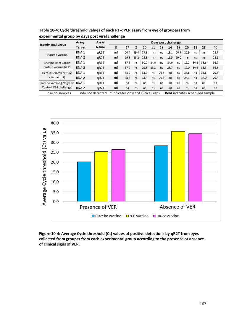
167
Table 10-4: Cycle threshold values of each RT-qPCR assay from eye of groupers from
experimental group by days post viral challenge
Figure 10-4: Average Cycle threshold (Ct) values of positive detections by qR2T from eyes collected from grouper from each experimental group according to the presence or absence of clinical signs of VER.
Assay
Target 0 7* 8 10 11 13 14 18 20 21 28 40
RNA 1 qR1T nd 20.4 19.4 27.8 ns ns 18.1 20.9 20.9 ns ns 28.7
RNA 2 qR2T nd 19.8 18.2 25.3 ns ns 16.5 19.0 ns ns ns 28.5
RNA 1 qR1T nd 37.5 ns 30.0 34.0 ns 34.0 ns 19.2 34.9 33.6 36.7
RNA 2 qR2T nd 37.2 ns 29.8 33.3 ns 33.7 ns 19.0 34.6 33.3 36.3
RNA 1 qR1T nd 38.9 ns 33.7 ns 26.8 nd ns 33.6 nd 33.6 29.8
RNA 2 qR2T nd 38.6 ns 33.4 ns 26.5 nd ns 28.3 nd 36.0 29.4
RNA 1 qR1T nd nd ns ns ns ns nd ns ns nd nd nd
RNA 2 qR2T nd nd ns ns ns ns nd ns ns nd nd nd
ns= no samples nd= not detected * indicates onset of clinical signs Bold indicates scheduled sample
Placebo vaccine ( Negative
Control: PBS challenge)
Heat-killed cell culture
vaccine (HK)
Assay
Name
Days post challengeExperimental Group
Placebo vaccine
Recombinant Capsid
protein vaccine (rCP)

168
10.4 Discussion
In this study, vaccination of juvenile groupers with average body weight (abw) greater than
50g was associated with 17-20% morbidity (80-83% survival) following IM challenge with
RGNNV (average body weight at challenge 103.5 g, (𝜎 20.39 g)). In contrast, 93% of the
unvaccinated fish succumbed to VER following challenge with viral extract (7% survival). The
survival of vaccinated fish in this trial was improved compared to the initial experimental trial
discussed in Chapter 8. The mechanism that conferred improved efficacy of vaccination on the
larger fish is unknown.
The acquisition of competence of the adaptive immune system is a prerequisite for successful
vaccination. Excluding groupers, in many fish species, only the larval stages are highly
susceptible to NNV (OIE, 2018). Despite the general acceptance of age-related susceptibility to
NNV in juvenile barramundi, there is only a single report investigating the NNV susceptibility
transition period (Jaramillo et al., 2017). In barramundi, juveniles older than 5 weeks of age
develop a subclinical infection, whereas younger cohorts suffer clinical disease and mass
mortality (Jaramillo et al., 2017). There is no characterisation of the mechanism that ensures
resistance (Jaramillo et al., 2017) and a recent review of vaccination as a preventative strategy
against VER did not refer to the development of the fish immune system (Hazreen et al., 2019).
Due to their typically low economic value, juvenile fish are often the subject of disease
challenge and vaccine efficacy experiments. Although less expensive, and logistically more
easily managed, challenges with juvenile fish may fail to consider conditions that affect the
development and expression of adaptive antiviral immunity. Many studies are driven by a
need for commercial outcomes and focussed on the production of a vaccine, rather than the
basic science of grouper immunity. The FAO generally recognise that the lack of understanding
of immune function in host species is one of the factors that reduce the ability to manage
disease in aquaculture species (FAO 2018).
Foundational studies into vaccination of salmon identified the significant influence of water
temperature on immune function in fish and discusses the term degree days (DD) as a unit to
represent the interaction between number of days post-vaccination and water temperature
(DD = water temperature (C⁰) x number of days) (Holm et al., 2014 in Gudding et al., 2014). DD
has also become an increasingly popular method for explaining variation in fish growth and
development (Chezik et al., 2014). Recently, the body weight and water temperature have
been demonstrated to be important in fish immunization (Soto et al., 2014).
The Biological Theory of Relativity (BTR) provides a more sophisticated framework for the
study of a range of biological processes in marine ectotherms (Neuheimer 2019). The theory is

169
proposed to characterise biological phenomena, as a function of the variable interaction
between water temperature and time with the biological variability of an organism (size of
organism, age of organism, maturity of organism and environmental conditions of organisms’
habitat amongst others). For example, in the context of vaccination: the biological process of
expression of effective adaptive antiviral immune response following vaccination in grouper
would be studied in relation to grouper age, species, size, nutritional status, (biological
variations) stocking density, salinity, water quality (environmental variations) and water
temperature and time (biological time). The recognition and study of the multitude of
interactions between biological time and biological variability allow the identification of the
critical factors for a prescribed biological function (Neuheimer 2019). Adoption of the BTR
approach increases the ability of researchers to make predictions about biological functions
and ultimately allow researchers to manipulate the interaction to achieve a desired biological
outcome (Neuheimer 2019). Although using different terms, the BTR approach is similar to the
concept proposed by Stentiford et al. (2017) that disease outbreaks in aquaculture are likely
the result of a complex interaction between host genetics-environment-pathogen and likely
requires a multidisciplinary approach. The Biological Theory of Relativity and the
multidisciplinary approach (Stentiford et al., 2017) are recent concepts and there has been no
concerted approach to record the range of factors that constitute “biological time” or
“biological variation” or multitude of factors that may influence the development of immune
competence or initiate VER disease outbreaks in juvenile groupers.
Although age and water temperature are considered important factors influencing the
development of adaptive immunity in fish, few of the studies that investigate vaccination
against NNV in grouper describe the age of fish or water temperature used in experimental
studies (Table 10-5), although fish bodyweight is provided in the description of experimental
groupers (Table 10-5). Review of the grouper vaccination studies indicates this project,
amongst others, may have serendipitously been conducted across a transitional period of
“biological time” required for the attainment of adaptive immune competence in juvenile
cultured grouper (Table 10-5). Studies that report greater than 60% survival in groupers
following vaccination and challenge, (Table 10-5) (Atujona et al., 2019; Yamashita et al., 2009;
Oh et al., 2012 and this study: Chapter 10), involved larger bodyweight juveniles than the fish
in less successful studies that report lower survival following vaccination and challenge (Mo et
al., 2019 and this study Chapter 8)(Refer to Table 10-5).
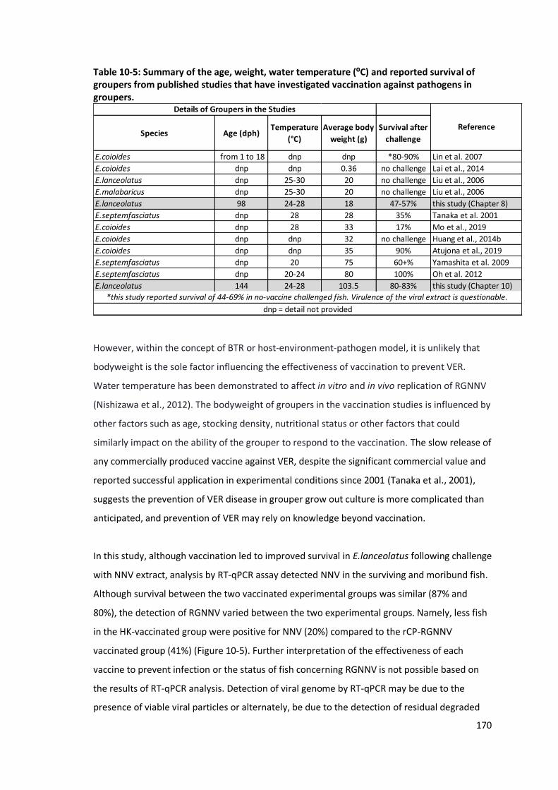
170
Table 10-5: Summary of the age, weight, water temperature (⁰C) and reported survival of groupers from published studies that have investigated vaccination against pathogens in groupers.
However, within the concept of BTR or host-environment-pathogen model, it is unlikely that
bodyweight is the sole factor influencing the effectiveness of vaccination to prevent VER.
Water temperature has been demonstrated to affect in vitro and in vivo replication of RGNNV
(Nishizawa et al., 2012). The bodyweight of groupers in the vaccination studies is influenced by
other factors such as age, stocking density, nutritional status or other factors that could
similarly impact on the ability of the grouper to respond to the vaccination. The slow release of
any commercially produced vaccine against VER, despite the significant commercial value and
reported successful application in experimental conditions since 2001 (Tanaka et al., 2001),
suggests the prevention of VER disease in grouper grow out culture is more complicated than
anticipated, and prevention of VER may rely on knowledge beyond vaccination.
In this study, although vaccination led to improved survival in E.lanceolatus following challenge
with NNV extract, analysis by RT-qPCR assay detected NNV in the surviving and moribund fish.
Although survival between the two vaccinated experimental groups was similar (87% and
80%), the detection of RGNNV varied between the two experimental groups. Namely, less fish
in the HK-vaccinated group were positive for NNV (20%) compared to the rCP-RGNNV
vaccinated group (41%) (Figure 10-5). Further interpretation of the effectiveness of each
vaccine to prevent infection or the status of fish concerning RGNNV is not possible based on
the results of RT-qPCR analysis. Detection of viral genome by RT-qPCR may be due to the
presence of viable viral particles or alternately, be due to the detection of residual degraded
Species Age (dph)Temperature
(°C)
Average body
weight (g)
Survival after
challenge
E.coioides from 1 to 18 dnp dnp *80-90% Lin et al. 2007
E.coioides dnp dnp 0.36 no challenge Lai et al., 2014
E.lanceolatus dnp 25-30 20 no challenge Liu et al., 2006
E.malabaricus dnp 25-30 20 no challenge Liu et al., 2006
E.lanceolatus 98 24-28 18 47-57% this study (Chapter 8)
E.septemfasciatus dnp 28 28 35% Tanaka et al. 2001
E.coioides dnp 28 33 17% Mo et al., 2019
E.coioides dnp dnp 32 no challenge Huang et al., 2014b
E.coioides dnp dnp 35 90% Atujona et al., 2019
E.septemfasciatus dnp 20 75 60+% Yamashita et al. 2009
E.septemfasciatus dnp 20-24 80 100% Oh et al. 2012
E.lanceolatus 144 24-28 103.5 80-83% this study (Chapter 10)
Details of Groupers in the Studies
Reference
dnp = detail not provided
*this study reported survival of 44-69% in no-vaccine challenged fish. Virulence of the viral extract is questionable.
171
viral genome or non-infectious particles (Gu et al., 2014). Although RT-qPCR is useful for
tracking the increase in the detection of viral genome from infection to disease expression, its
ability to determine the carrier or recovery status of grouper following exposure to NNV is
restricted. Viral isolation in a compatible cell culture system is the definitive proof of the
presence or absence of viable viral isolation may have improved (Schnurr 2006). Analysis of
tissue samples by virus isolation may have improved interpretation of the RT-qPCR results in
this project. Inclusion of tissue analysis by viral isolation has been incorporated into
subsequent research that extends beyond this project.
Regardless of the interpretation of the RT-qPCR results, this study demonstrated that
increased survival of juvenile groupers, E.lanceolatus following experimental challenge with
RGNNV viral extract, was markedly improved, with either HK-vaccine (83% survival) or rCP-
vaccine (80% survival) antigens compared to placebo vaccinated groupers (7% survival).
Survival in vaccinated grouper following NNV challenge was improved in this study compared
to the modest survival of 43-53% of vaccinated grouper in the trial described in Chapter 8 that
studied smaller bodyweight groupers. This study indicates the significant losses imposed on
the grouper aquaculture by the endemic strains of RGNNV present in Northern Queensland
may be preventable with vaccination. However, the efficacy of vaccination to protect against
VER requires further optimisation, as within the conditions of this study, the efficacy of
vaccination was influenced by grouper bodyweight. Although further research is required
before commercialisation of the vaccine, the results of this study are very promising and
provide confidence to support further investment in vaccine development.
10.5 Conclusion
The following outcomes were described in this chapter:
Vaccination against RGNNV on larger fish >50g abw displayed improved protection
against VER.
Vaccination of E.lanceolatus with a vaccine derived from either recombinant
expressed capsid protein or heat-killed cell culture antigen led to increased survival
(80-83%)/ reduced morbidity (20-17%) compared to placebo vaccinated control fish
(7% survival/ 93% morbidity) after 40 days post challenge.
Bodyweight, or some factor that is reflected as bodyweight, appears to affect the
efficacy of vaccination to prevent VER.
172
CHAPTER 11. SUMMARY OF FINDINGS AND DIRECTION FOR
FUTURE WORK
11.1 Summary of Findings
This work improved understanding of the management of Betanodavirus infections in the
culture of grouper in Australia. The project aimed to answer three broad research questions.
The following summary presents the discoveries relating to the research questions:
To improve knowledge in the strains of Betanodavirus associated with VER outbreaks the
following was investigated
The species of NNV that were associated with three natural outbreaks of VER in
marine aquaculture systems in Northern Queensland were determined
The level of variation/conservation between strains that have been associated
with VER in Northern Queensland and Australia was determined
The complete sequences of mRNA of RNA 1 and RNA 2 were obtained from three disease
outbreaks of marine finfish aquaculture ventures in Northern Queensland (collectively referred
to as NQAus NNV). The NQAus NNV strains obtained from outbreaks of VER in tropical marine
fish species in Northern Queensland were all RGNNV species. Phylogenetic analysis of both the
RNA 1 and RNA 2 segments did not detect any chimeric recombination with any other NNV
species such as SJNNV or BFNNV. RGNNV continues to be the only species of Betanodavirus
detected from Australian fish. Comparison with other Australian-collected RGNNV strains
indicated highest homology to the strains collected from tropical marine species rather than
temperate or freshwater species.
Comparative analysis with other strains in the NCBI database demonstrated the remarkable
conservation of the RGNNV genome, both temporally and geographically. The RNA 1 and RNA
2 segments of the three NQAus NNV strains retained 97-98% homology to the original RGNNV
strain isolated from Japan (SgWAk97) in 1997. The high level of conservation of the genome
with other RGNNV strains had implications for vaccine design.
Research Question 1: What strains of Betanodavirus are associated with VER
outbreaks of tropical marine fish species in Northern Queensland?
173
To assess the usefulness of qPCR as a management tool the following topics were investigated:
The qR2T assay described by Hick & Whittington, which is also the current ANZSDP to
detect NNV from grouper was applied to grouper derived tissue.
The qR1T assay that targets RNA 1, designed by Hick & Whittington to detect NNV,
was applied to grouper derived tissue.
Positive amplicons produced from RNA 1 and RNA 2 were cloned to produce standard control
plasmids for the qR1T and qR2T RT-qPCR assays described by Hick & Whittington (2010). The
RT-qPCRs were applied to trace the viral copy number of RGNNV during the progression of
disease in the experimental challenge. Both RT-qPCR assays detected viral genome before the
onset of clinical signs at a cycle threshold values ranging from 45 to 22.6. During the period of
peak morbidity, viral genome was detected by both RT-qPCR assays with a cycle threshold
range of 10.6 to 21.1. Both assays detected NNV genome prior to the onset of VER disease,
during peak period of morbidity and also up to 80 days post IM challenge. There were no false
negative results obtained from either assay in this investigation when samples were sourced
from grouper displaying clinical signs of VER. Both assays detected NNV in every tissue sample
derived from fish that displayed several signs of VER. Based on known challenge status, there
were no false positives. Neither assay detected NNV in E.lanceolatus tissues from any of the
PBS- challenged groups that were not exposed to NNV.
RT-qPCR was fit for purpose for application on brain and eye tissues of E.lanceolatus in
situations that required rapid turnaround, high throughput, and quantitative detection of
RGNNV genome such as during a during disease outbreaks or to demonstrate freedom from
NNV infection. Both the qR1T and qR2T assays developed by Hick & Whittington (2010) are fit
for purpose for the detection of RGNNV genome in eye and brain tissue collected from
E.lanceolatus.
However, the interpretation of the results of RT-qPCR analysis must consider that detection of
genomic material does not indicate viability of virus. As such, the RT-qPCR assay was not useful
to assist in determining the nature of protection provided by the vaccines. In this study,
vaccinated fish survived and did not display signs of VER. However, viral genome was detected
from eye and brain tissues from the vaccinated fish that did not show signs of disease
following IM challenge. It is not possible to determine if the viral genome that was detected by
Research Question 2: Is qPCR a useful tool to assist in the management of VER in giant
Queensland grouper, Epinephelus lanceolatus in Australia?

174
the RT-qPCR assays was viable virus. Consequently, it is not possible to determine if
vaccination lead to the production of fish that were immune and not-susceptible to the virus
or were sub-clinical carriers with increased tolerance. RT-qPCR is a useful tool to assist in the
management of VER in giant grouper. However, it is not the only analysis platform required to
understand and improve management of the disease.
Vaccine and dsRNA were prepared that targeted the Ec2NQAus strain of NNV. The efficacy of
the therapies to prevent VER were assessed by IM challenge of juvenile E.lanceolatus with viral
extract.
In an initial trial, the IM delivery of naked dsRNA designed against Ec2NQAus strain of NNV did
not reduce severity of morbidity following IM viral challenge and no further investigation into
dsRNA was conducted. Although this trial was unsuccessful, a range of variables could be
adjusted to improve the potential of dsRNA as a treatment or preventative. A more recent
accessing of the IDT dsRNA design tool indicates the anti-NNV dsRNA will cross react with six
transcripts from the human genome (Refer to Appendix 3). Notably four of the transcripts are
members of the C-type lectin domain family 4 member A (CLEC4A) gene which is involved in
the regulation of immune reactivity (uniport.org/uniprot/Q9UMR7) and speculatively, may
also be an important component of the grouper immune system. Until the knowledge of the
grouper immune system is improved, research seeking to apply dsRNA to prevent VER will be
very difficult.
This study demonstrated vaccination is effective in preventing VER following IM challenge with
viral extract. The effectiveness of vaccine appears to be affected by fish body weight. In an
initial trial, involving fish with average body weight (~18g), the vaccinated groups of
E.lanceolatus displayed between 43-53% cumulative morbidity after challenge with NNV
compared to 88% morbidity in dsRNA exposed groupers. The modest reduction of morbidity
by 35-45% indicated the vaccine formulation presented some potential as a preventative
measure. There was a trend for vaccinated fish over 50g to have reduced morbidity.
In a subsequent study, E.lanceolatus with average body weight over 50g were vaccinated and
challenged. The outcome was increased protection against VER in the slightly larger fish with
the vaccinated groups experiencing 20-23% morbidity compared to the 93% morbidity in
placebo vaccinated groupers.
Research Question 3: How effective is vaccination or dsRNA designed against the
endemic strains of NNV in preventing disease?

175
During the term of the project additional research questions were posed as important
considerations to achieve the project aims. The additional discoveries are summarised here:
Ideally, the efficacy of therapies to prevent VER is determined via a challenge model that is
most representative of the natural infection route. Although waterborne transmission of
RGNNV is the most likely path of transmission leading to natural VER outbreaks there is limited
success applying waterborne transmission to experimental challenges. In a novel approach,
waterborne challenge via co-circulation with diseased fish along with co-infection with the
marine leech, Zeylanicobdella arugamensis was tested as an infection model. During a 40-day
trial, despite habitation within a shared recirculation system containing 10 fish that displayed
clinical signs of VER following exposure to RGNNV via IM challenge, none of juvenile
groupers E.lanceolatus exposed/non-exposed to the marine leech Z.arugamensis succumbed
to a VER disease outbreak. Furthermore, RGNNV was not detected by RT-qPCR from leeches
collected from any of the tanks. The inability to induce VER via waterborne challenge despite
the addition of leech infestation, lead to the adoption of intramuscular injection of RGNNV
extract as a challenge model for all subsequent studies.
Recognising that the cohort of fish used in the experimental challenge were produced from a
mass spawning event with eight potential parents, the influence of parentage on
survival/mortality was investigated by applying Multi-loci sequencing analysis. Parentage did
not coincide with improved survival or increased mortality within the studies
conducted. Although other genetic factors may be involved, the variance in survival between
vaccinated and non-vaccinated fish was not a direct reflection of different parentage between
individuals.
Inclusion of leeches to improve waterborne transmission of RGNNV
Role of parentage in survival to VER?
Additional discoveries

176
Understanding the replication characteristics of Betanodavirus may allow targeted approaches
to prevent VER. This study completed a review of the functional motifs of the Betanodaviruses,
mapped them to a schematic diagram of the mRNA segments and sought to confirm the
presence or absence of the motifs in the NQAus NNV strains. Review and confirmation of the
multiple motifs across an entire genome have not been reported previously from any strain of
Betanodavirus.
The three strains of NQAus NNV obtained in this study retain the functional motifs of the
Betanodavirus genome that are critical for viral replication and associated with most virulence.
The confirmation of the presence of the essential motifs indicated that outcomes of this
research based on the NNV strains obtained would likely be valid for translation to industry
applications.
11.2 Future work beyond this study
Improved delivery of viral antigen
Along with previous reports, this project succeeded in demonstrating protective immunity
against NNV using either expressed recombinant RGNNV capsid protein or heat-killed RGNNV
grown in cell culture as a vaccine antigen. Whilst the results are encouraging, further
refinement of the vaccination process is possible. Although vaccination is an effective measure
to prevent disease in cultured fish species, the requirement of handling to complete injection,
imposes a level of stress on the fish and increased costs to production. A vaccine that could be
delivered as an oral antigen would avoid the risk of handling stress. A factor reducing the
effectiveness of oral vaccination is the inability to deliver antigen of sufficient quantity and
integrity to effectively stimulate immune memory. As the minimal epitope for immune
stimulation has been identified to be quite small, the delivery of antigen via recombinant
bacteria, virus or algae may provide an optimal vaccination strategy. Some experimental
vaccination applying the improved delivery have been reported this year. Gonzalez-Silvera et
al. (2019) reported oral vaccination of sea bass using direct delivery of E.coli expressing the
capsid protein with no protein purification.
Identification and conservation of the functional motifs of Betanodavirus

177
Improved understanding of the development of grouper immunity
Perhaps the most pressing issue to improve the management of VER in grouper is to identify
the mechanisms of the immune system development in grouper that are involved in response
to RGNNV and viruses generally. This would allow a more multidisciplinary and systematic
approach to understanding VER expression and likely, other viral diseases. The need to better
understand the immune function of aquaculture species is a factor limiting disease
management across the multitude of tropical species (FAO 2018). There is an urgent need for
research investment into understanding the mechanisms of immune function. Whilst such
studies will not lead to production of commercialised products, they will provide the
foundational knowledge that can be applied to adopt a more logical approach to improve
health and reduce factors that impede fish immunity in aquaculture. For example, improved
understanding of immune development may identify changes to husbandry practises or
improve diet formulation that increase grouper health.
Investigate impact of environmental parameters on grouper immunity
Although the efficacy of vaccines can be accurately demonstrated in experimental challenge
facilities, there are a multitude of other factors in grow out systems that may adversely affect
fish immune function and ultimately contribute to VER outbreaks. Considering the
metrological and agricultural conditions that dominate the locations prioritised for
aquaculture development in Northern Australia, increased understanding of the following
should be considered:
Effect of temperature and salinity on grouper immune function.
Effect of contamination of marine water ways with agricultural pesticides/herbicides
on grouper immune function.
Refinement of grouper grow out diet formulation to support optimal grouper immune
function.
Investigate the roles of microRNA in the replication and management of NNV.
Whilst the presentation of dsRNA constructs provided no protective effect against the
development of VER following exposure to RGNNV extract in this experiment, the technology
should not be discounted as a tool to better understand NNV. The paradox of NNV is that one
of the smallest viral genomes is capable of causing disease in such a range of fish species, with
worldwide distribution, across temperate and tropical and marine and freshwater culture
systems. Reverting to basic principles for how the Betanodavirus genome achieves such
infectivity highlights further paradox in the nature of this virus. Namely why, considering the
178
requirement for the virus to be very efficient within the constraints of its genome size, would
the 403 nt of UTR of RNA 2 (~28% of the RNA 2 strand) that is not translated to produce the
viral capsid protein, be retained in the genome if it served no purpose? In the absence of
protein translation, a potential role of non-translated genomic sequence is viral and/or host
gene regulation. Whilst, I have not been able to investigate this particular topic, I believe it is
an important question to raise and the knowledge gained by understanding any potential roles
of v-miRNA in Betanodavirus may provide a useful model to improve knowledge in other RNA
viruses such as Influenza. The improved efficiency and affordability of next generation
sequences techniques provides an unprecedented potential for researchers to investigate the
complexities of viral-host interactions.
11.3 Implications of findings from this study
Although the viral aetiology of VER in Australian fish species was described by JCU researchers,
Dr John Glazebrook and Steve de Beer (1990), research into the disease did not continue at
JCU until this present study.
This study has provided a strong basis for future work in the management of VER in grouper
and has been applied towards the fast tracking of an NNV vaccine under FRDC project 2018-
098: “Vaccination for emergency and long-term control of nodavirus in Australian marine
aquaculture”. The project aims to apply the demonstrated application of recombinantly
expressed capsid protein-vaccine within a commercial field study and acquire data to support
both the commercialisation of a vaccine and registration for approval by the Australian
Pesticides and Veterinary Medicines Authority.
The protocols to implement RT-qPCRs into a laboratory setting within this research project
were audited against the international standard for testing and measurement (ISO 17025)
within the field of animal health by the National Association of Testing Authorities (NATA). The
laboratory, JCU AquaPATH, attained NATA accreditation in June 2018 at the James Cook
University Townsville Campus. The specific scope of accreditation is the “detection of viruses
by nucleic acid detection” specifically, the detection of Nervous Necrosis Virus. The facility is
one of only three laboratories present in Northern Australia and one of only two universities
nationally, that hold this scope of accreditation. The establishment of JCU AquaPATH on the
James Cook University Campus provides core capability that will support future research in
tropical aquatic animal health at James Cook University.
“From little things, big things grow” (Kelly and Carmody, 1991)

179

180
APPENDIX 1 MEDIA AND BUFFERS
Lysogeny broth (LB)
Component Mass (grams litre-1)
Yeast 5
Tryptone 10
Sodium chloride 5
LB agar
Component Mass (grams litre-1)
Yeast 5
Tryptone 10
Sodium chloride 5
Bacteriological agar 15
Blood agar
Component Mass (grams litre-1)
Enzymatic digest of Casein 7.5
Enzymatic Digest of animal tissue 7.5
Liver digest 2.5
Yeast extract 5
Sodium chloride 5
Bacteriological agar 12g
Sheep’s blood 100 mL
Preparation of stock solutions for cloning media
Component Volume to prepare Mass Stock concentration
Isopropyl β-D-1-
thiogalactopyranoside (IPTG)
5 mL 0.12 g 500 mM
5-bromo-4-chloro-3-indolyl-β-D-
galactopyranoside (X-Gal)
2 mL (in dimethylformamide) 0.1 g 50 mg mL-1
Additions of for LB media and Agar
Component Stock concentration Final required Dilution of stock required
(IPTG) 500 mM 0.5 mM 1000
(X-Gal) 50 mg mL-1 80 ug mL-1 625
Ampicillin a 100000 ug mL-1 100 ug mL-1 1000
Ampicillin b 100000 ug mL-1 50 ug mL-1 2000
a pGEM T Easy; b TOPO TA Cloning
181
TAE Buffer (50x) (1 Litre)
242 g Tris base
57.1 ml glacial acetic acid
37.2 g Na2EDTA·2H2O (2 mM) H2O to 1 litre.
For working solution add 200ml to 9800ml H2O.
SDS PAGE 10× Tris glycine buffer
10 g SDS
30.3 g Tris
144.1g Glycine
Dissolve ingredient in 800 ml distilled water and adjust volume to 1 litre
Dilute 100ml in adjusted volume to 1 litre with distilled water.
SDS PAGE Silver Fixative
500ml Methanol
50ml Acetic Acid
400ml Distilled water
Mix solutions and adjust volume to 1 litre
SDS PAGE Development Solution
20 g Na2CO3
400µl Formalin
800ml+ Distilled water
Dissolve ingredient in 800 ml distilled water and adjust volume to 1 litre
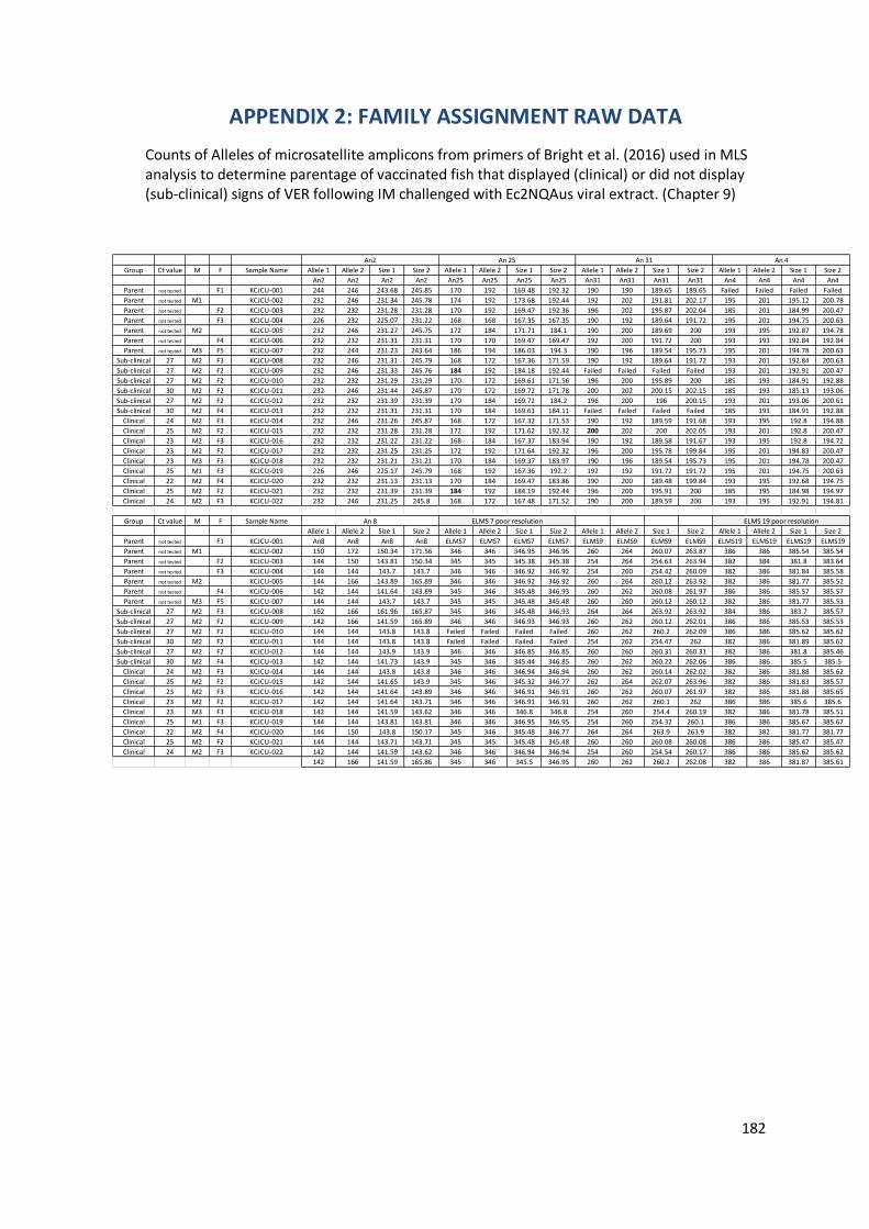
182
APPENDIX 2: FAMILY ASSIGNMENT RAW DATA
Counts of Alleles of microsatellite amplicons from primers of Bright et al. (2016) used in MLS analysis to determine parentage of vaccinated fish that displayed (clinical) or did not display (sub-clinical) signs of VER following IM challenged with Ec2NQAus viral extract. (Chapter 9)
Group Ct value M F Sample Name Allele 1 Allele 2 Size 1 Size 2 Allele 1 Allele 2 Size 1 Size 2 Allele 1 Allele 2 Size 1 Size 2 Allele 1 Allele 2 Size 1 Size 2
An2 An2 An2 An2 An25 An25 An25 An25 An31 An31 An31 An31 An4 An4 An4 An4
Parent not tested F1 KCJCU-001 244 246 243.68 245.85 170 192 169.48 192.32 190 190 189.65 189.65 Failed Failed Failed Failed
Parent not tested M1 KCJCU-002 232 246 231.34 245.78 174 192 173.68 192.44 192 202 191.81 202.17 195 201 195.12 200.78
Parent not tested F2 KCJCU-003 232 232 231.28 231.28 170 192 169.47 192.36 196 202 195.87 202.04 185 201 184.99 200.47
Parent not tested F3 KCJCU-004 226 232 225.07 231.22 168 168 167.35 167.35 190 192 189.64 191.72 195 201 194.75 200.63
Parent not tested M2 KCJCU-005 232 246 231.27 245.75 172 184 171.71 184.1 190 200 189.69 200 193 195 192.87 194.78
Parent not tested F4 KCJCU-006 232 232 231.31 231.31 170 170 169.47 169.47 192 200 191.72 200 193 193 192.84 192.84
Parent not tested M3 F5 KCJCU-007 232 244 231.23 243.64 186 194 186.03 194.3 190 196 189.54 195.73 195 201 194.78 200.63
Sub-clinical 27 M2 F3 KCJCU-008 232 246 231.31 245.79 168 172 167.36 171.59 190 192 189.64 191.72 193 201 192.84 200.63
Sub-clinical 27 M2 F2 KCJCU-009 232 246 231.33 245.76 184 192 184.18 192.44 Failed Failed Failed Failed 193 201 192.91 200.47
Sub-clinical 27 M2 F2 KCJCU-010 232 232 231.29 231.29 170 172 169.61 171.56 196 200 195.89 200 185 193 184.91 192.88
Sub-clinical 30 M2 F2 KCJCU-011 232 246 231.44 245.87 170 172 169.72 171.78 200 202 200.15 202.15 185 193 185.13 193.06
Sub-clinical 27 M2 F2 KCJCU-012 232 232 231.39 231.39 170 184 169.72 184.2 196 200 196 200.15 193 201 193.06 200.61
Sub-clinical 30 M2 F4 KCJCU-013 232 232 231.31 231.31 170 184 169.61 184.11 Failed Failed Failed Failed 185 193 184.91 192.88
Clinical 24 M2 F3 KCJCU-014 232 246 231.26 245.87 168 172 167.32 171.53 190 192 189.59 191.68 193 195 192.8 194.88
Clinical 25 M2 F2 KCJCU-015 232 232 231.28 231.28 172 192 171.62 192.32 200 202 200 202.05 193 201 192.8 200.47
Clinical 23 M2 F3 KCJCU-016 232 232 231.22 231.22 168 184 167.37 183.94 190 192 189.58 191.67 193 195 192.8 194.72
Clinical 23 M2 F2 KCJCU-017 232 232 231.25 231.25 172 192 171.64 192.32 196 200 195.78 199.84 195 201 194.83 200.47
Clinical 23 M3 F3 KCJCU-018 232 232 231.21 231.21 170 184 169.37 183.97 190 196 189.54 195.73 195 201 194.78 200.47
Clinical 25 M1 F3 KCJCU-019 226 246 225.17 245.79 168 192 167.36 192.2 192 192 191.72 191.72 195 201 194.75 200.63
Clinical 22 M2 F4 KCJCU-020 232 232 231.13 231.13 170 184 169.47 183.86 190 200 189.48 199.84 193 195 192.68 194.75
Clinical 25 M2 F2 KCJCU-021 232 232 231.39 231.39 184 192 184.19 192.44 196 200 195.91 200 185 195 184.98 194.97
Clinical 24 M2 F3 KCJCU-022 232 246 231.25 245.8 168 172 167.48 171.52 190 200 189.59 200 193 195 192.91 194.81
Group Ct value M F Sample Name
Allele 1 Allele 2 Size 1 Size 2 Allele 1 Allele 2 Size 1 Size 2 Allele 1 Allele 2 Size 1 Size 2 Allele 1 Allele 2 Size 1 Size 2
Parent not tested F1 KCJCU-001 An8 An8 An8 An8 ELMS7 ELMS7 ELMS7 ELMS7 ELMS9 ELMS9 ELMS9 ELMS9 ELMS19 ELMS19 ELMS19 ELMS19
Parent not tested M1 KCJCU-002 150 172 150.34 171.56 346 346 346.95 346.95 260 264 260.07 263.87 386 386 385.54 385.54
Parent not tested F2 KCJCU-003 144 150 143.81 150.34 345 345 345.38 345.38 254 264 254.63 263.94 382 384 381.8 383.64
Parent not tested F3 KCJCU-004 144 144 143.7 143.7 346 346 346.92 346.92 254 260 254.42 260.09 382 386 381.84 385.58
Parent not tested M2 KCJCU-005 144 166 143.89 165.89 346 346 346.92 346.92 260 264 260.12 263.92 382 386 381.77 385.52
Parent not tested F4 KCJCU-006 142 144 141.64 143.89 345 346 345.48 346.93 260 262 260.08 261.97 386 386 385.57 385.57
Parent not tested M3 F5 KCJCU-007 144 144 143.7 143.7 345 345 345.48 345.48 260 260 260.12 260.12 382 386 381.77 385.53
Sub-clinical 27 M2 F3 KCJCU-008 162 166 161.96 165.87 345 346 345.48 346.93 264 264 263.92 263.92 384 386 383.7 385.57
Sub-clinical 27 M2 F2 KCJCU-009 142 166 141.59 165.89 346 346 346.93 346.93 260 262 260.12 262.01 386 386 385.53 385.53
Sub-clinical 27 M2 F2 KCJCU-010 144 144 143.8 143.8 Failed Failed Failed Failed 260 262 260.2 262.09 386 386 385.62 385.62
Sub-clinical 30 M2 F2 KCJCU-011 144 144 143.8 143.8 Failed Failed Failed Failed 254 262 254.47 262 382 386 381.89 385.62
Sub-clinical 27 M2 F2 KCJCU-012 144 144 143.9 143.9 346 346 346.85 346.85 260 260 260.31 260.31 382 386 381.8 385.46
Sub-clinical 30 M2 F4 KCJCU-013 142 144 141.73 143.9 345 346 345.44 346.85 260 262 260.22 262.06 386 386 385.5 385.5
Clinical 24 M2 F3 KCJCU-014 144 144 143.8 143.8 346 346 346.94 346.94 260 262 260.14 262.02 382 386 381.88 385.62
Clinical 25 M2 F2 KCJCU-015 142 144 141.65 143.9 345 346 345.32 346.77 262 264 262.07 263.96 382 386 381.83 385.57
Clinical 23 M2 F3 KCJCU-016 142 144 141.64 143.89 346 346 346.91 346.91 260 262 260.07 261.97 382 386 381.88 385.65
Clinical 23 M2 F2 KCJCU-017 142 144 141.64 143.71 346 346 346.91 346.91 260 262 260.1 262 386 386 385.6 385.6
Clinical 23 M3 F3 KCJCU-018 142 144 141.59 143.62 346 346 346.8 346.8 254 260 254.4 260.19 382 386 381.78 385.51
Clinical 25 M1 F3 KCJCU-019 144 144 143.81 143.81 346 346 346.95 346.95 254 260 254.32 260.1 386 386 385.67 385.67
Clinical 22 M2 F4 KCJCU-020 144 150 143.8 150.17 345 346 345.48 346.77 264 264 263.9 263.9 382 382 381.77 381.77
Clinical 25 M2 F2 KCJCU-021 144 144 143.71 143.71 345 345 345.48 345.48 260 260 260.08 260.08 386 386 385.47 385.47
Clinical 24 M2 F3 KCJCU-022 142 144 141.59 143.62 346 346 346.94 346.94 254 260 254.54 260.17 386 386 385.62 385.62
142 166 141.59 165.86 345 346 345.5 346.95 260 262 260.2 262.08 382 386 381.87 385.61
ELMS 19 poor resolution
An2 An 25 An 31 An 4
An 8 ELMS 7 poor resolution

183
APPENDIX 3: UPDATED ANALYSIS OF DSRNA DESIGN BY IDT
DESIGN ANALYSIS TOOL.
Updated output from IDT dsRNA design analysis tool. Accessed 29.9.19. Tool indicates dsRNA
would target a number of genes of human (A image) and mouse (B image) origin. A lack of
suitable knowledge in fish immune gene or microRNA prevents analysis against fish.

184
APPENDIX 4: PUBLICATIONS AND DISSEMINATION FROM THIS
THESIS
Work published as part of this thesis:
Condon K., Bochow S., Ariel E., and Miller T., (2019) Complete sequence of Betanodavirus from
Australian barramundi, Lates calcarifer. Microbiology Resource Announcements 8.
https://doi.org./10.1128/MRA.00081-19

185
Proposed publications from this thesis
Condon K, Ariel E and Jerry D (2020) The functional motifs of the Betanodavirus genome.
Condon K, Bochow S, Reynolds A, Knuckey R and Ariel E (2019) Real-time RT-PCR detection of
Redspotted Grouper Nervous Necrosis Virus (RGNNV) in experimental challenge of
Epinephelus lanceolatus following exposure to recombinant vaccine and dsRNA constructs.

186
Non-Peer Reviewed Additional Outputs of Doctor Studies
Appendix 3.1: The Management of Betanodavirus infections in QLD giant grouper
E.lanceolatus. Presented to the 3rd Australasian Aquatic Animal Health FRDC Conference Cairns
2017.

187
REFERENCE LIST
Adamek M., Rasuch G., Brogden G. and Streinhagen D. (2014) Small interfering RNA treatment
can inhibit Cyprnid herpesvirus 3 associated cell death in vitro. Polish Journal of
Veterinary Sciences 17(4) 733-735.
Adachi K, Ichinose T, Takizawa N, Watanabe K, Kitazato K, and Kobayashi N (2007) Inhibition of
Betanodavirus infection by inhibitors of endosomal acidification. Archives of Virology
152: 2217-2224.
Ahola M, and Karlin D (2015) Sequence analysis reveals a conserved extension in the capping
enzyme of the alphavirus supergroup and a homologous domain in nodavirus. Biology
Direct 10: 16.
Agnihotri K., Pease B. and Chong R. (2016) Molecular analysis of RNA 1 and RNA 2 sequences
from a Betanodavirus isolated from giant grouper (Epinephelus lanceolatus) in
Australia. Virology Reports 6, 25-31.
Al-Majdoub M, Koy C, Lorenz P, Thiesen H, and Glocker M (2013) Mass spectrometric and
peptide chop characterisation of an assembled epitope: analysis of a polyclonal
antibody model serum directed against the Sjorgren/systemic lupus erthyematosus
autoantigen TRIM 21. Journal of Mass Spectrometry. DOI10.1002/jms.3208
Alcami A, Koszinowski UH (2000) Viral mechanisms of immune evasion. Trends in Microbiology
8: 410–418.
Aliyari R, Wu Q, Li H, Wang X, Li F, Green L, Han C, Li W and Ding S (2008) Mechanism of
Induction and Suppression of anti-viral immunity directed by virus-derived small RNAs
in Drosophila. Cell Host and Microbe 4(4) 387-397.
Anderson I and Moody N (2004) The effect of barramundi nodavirus on important freshwater
fishes. FRDC project No.1999/205 Downloaded online 15/3/19
http://www.frdc.com.au/project?id=1715
Arimoto M, Mori K, Nakai T, Muroga K and Furusawa I (1993) Pathogenicity of the causative
agent of viral nervous necrosis disease in striped jack, Pseudocaranx dentex (Bloch &
Schneider). Journal of Fish Diseases 16: 461-469.
Atujona D, Huang Y, Wang Z, Jian J, and Hai S (2019) Vibrio harveyi (VIRB11) recombinant
vaccine development against vibriosis in orange-spotted grouper (Epinephelus
coioides) Aquaculture Research50(9):2628-2634.
Bai H, Wang Y, Li X, Mao H, Li Y, Han S, Shi Z and Chen Z (2011) Isolation and characterisation
of a novel Alphanodavirus. Virology Journal 8: 311.
188
Ball A (1995) Requirements for the self-directed replication of Flock House Virus RNA 1. Journal
of Virology 69(2): 720-727.
Banu G, Mori K, Arimoto M and Nakai T (2007) Fate of Betanodaviruses in the experimentally
infected natural and non-natural host. Bangladesh Journal of Microbiology 24(2): 100-
104.
Baranowski E, Ruiz-Jarabo M, Domingo E (2001) Evolution of cell recognition by viruses.
Science 292: 1102–1105.
Biacchesi S (2011) The reverse genetics applied to fish RNA viruses. Veterinary Research 42:
12.
Binesh C (2014) Elevation of temperature and crowding trigger acute viral nervous necrosis in
zebra fish, Brachydanio rerio (Hamilton-Buchanan), sub-clinically infected with
Betanodavirus. Journal of Fish Diseases 37: 279-282.
Bloch B, Gravningen K and Larsen J (1991) Encephalomyelitis among turbot associated with a
picornavirus-like agent. Diseases of Aquatic Organisms 10:65-70.
Bochow S., Condon K., Elliman J. and Owens L. (2015) First complete genome of an
Ambidensovirus; Cherax quadricarinatus Densovirus, from freshwater crayfish Cherax
quadricarinatus. Marine Genomics 24, 301-312.
Bolhassani A and Rafati S (2008) Heat-shock proteins as powerful weapons in vaccine
development. Expert Reviews: Vaccines 7(8) 1185-1199.
Bols N, Brubacher J, Ganassin R, Lee L (2001) Exo-toxicology and innate immunity.
Developmental and Comparative Immunology 25: 853-873.
Bovo G, Gustinelli A, Quaglio F, Gobbo F, Panzarin V, Fusaro A, Mutinelli F, Caffara M and
Fioravanti M (2011) Viral encephalopathy and retinopathy outbreak in freshwater fish
farmed in Italy. Diseases of Aquatic Organisms 96 (1): 45-54.
Breuil G, Pepin J, Castric J, Fauvel C and Thiery R (2000) The detection of serum antibodies
against nodavirus in wild and farmed adult D.labrax: application to the screening of
broodstock in D.labrax hatcheries. Bulletin of the Association of Fish Pathologists 20(3)
95-100.
Breuil G, Pepin JFP, Boscher S, Thiery R (2002) Experimental vertical transmission of nodavirus
from broodfish to eggs and larvae of the sea bass, Dicentrarchus labrax. Journal of Fish
Diseases 25: 697–702.
Bright D, Reynolds A, Nguyen N, Knuckey R, Knibb W, and Elizur A (2016) A study into the
parental assignment of the communal spawning protogynous hermaphrodite, giant
grouper (Epinephelus lanceolatus). Aquaculture 459:19-25.

189
Bromage E, (2004) The humoral immune response of Lates calcarifer to Streptococcus iniae.
Doctorate of Philosophy thesis. James Cook University.
Buchon N, Vaury C (2006) RNAi: a defensive RNA-silencing against viruses and transposable
elements. Heredity 96: 195–202.
Carballo C, Garcia-Rosado E, Borreo J and Alonso M (2016) SJNNV down-regulates RGNNV
replication in European sea bass by the induction of the type 1 interferon system. Vet
Res. 47(6) doi: 10.1186/s13567-015-0304-y
Cardarelli F, Digiacomo L, Marchini C, Amicia A, Salomone F, Fiume G, Rossetta A, Gratton E,
Pozzi D and Caracciolo G (2016) The intracellular trafficking mechanism of
Lipofectamine-based transfection reagents and its implication for gene delivery.
Scientific Reports 6 (25879). https://doi.org/10.1038/srep25879
Castorena K, Stapleford K and Miller D (2010) Complementary transcriptomic, lipidomic and
targeted functional genetic analyses in cultured Drosophila cells highlight the role of
glycerophospholipid metabolism in Flock House virus RNA replication. BMC genomics
11: 183 doi: 10.1186/1471-2164-11-183.
Cha S, Do J, Lee N, An E, Kim Y, Kim J, Park J (2007) Phylogenetic analysis of betanodaviruses
isolated from cultured fish in Korea. Diseases of Aquatic Organisms 77: 181-189.
Chakraborty C, Sharma A, Sharma G, Doss C and Lee S (2017) Therapeutic miRNA and siRNA:
Moving from bench to clinic as next generation medicine. Molecular therapy: Nucleic
acids 8:132-143.
Chang J and Chi S (2015) GHSC70 is involved in the cellular entry of Nervous Necrosis Virus.
Journal of Virology 89: 61-70.
Chen N, Yoshimura M, Guan H, Wang T, Misumi Y, Lin C, Chuankhayan P, Nakagaw A, Chan S,
Tsukihara T, Chen T and Chen C (2015) Crystal structures of a piscine Betanodavirus:
Mechanisms of capsid assembly and viral infection. PLOS Pathog. 11:e1005203 PMID
26491970
Chen C., Wu M., Huang Y., Chen C., and Chang C. (2015) Recognition of linear B-cell epitope of
Betanodavirus coat protein RG-M18 neutralising mAB inhibits giant grouper nervous
necrosis virus (GGNNV) infection. PLOS ONE DOI:10.1371/journal.pone.0126121, 1-17.
Chen X (2014) The salinity effect on giant grouper innate immunity, Na-K-2Cl-co-transporter
(NKCC2) and Betanodavirus replication. Department of Life Science Thesis. Taiwan
University, 1-54.
190
Chen, SU and Hong (2009) Betanodavirus non-structural protein B1: a novel anti-necrotic
death factor that modulates cell death in early replication cycle in fish cells. Virology
385(2): 444-454.
Chen Y, Wang T and Chen T (2014) Immunity to Betanodavirus infections of marine fish.
Developmental and Comparative Immunology 43: 174-183.
Chen C, Wu M, Huang Y, Cheng C and Chang C (2015) Recognition of linear B-cell epitope of
Betanodavirus coat protein by RG-M18 Neutralizing mAB inhibits giant grouper
nervous necrosis virus (GGNNV) infection. PLoS ONE 10(5): e0126121.
doi:10.1371/journal.pone.0126121
Cherif N, Thiery R, Castric J, Biacchesi S, Bremont M, Thabti F, Limem L and Hammami S (2009)
Viral encephalopathy and retinopathy of Dicentrarchus labrax and Sparus aurata
farmed in Tunisia. Veterinary Research Communication 33: 345-353.
Chernikov I, Vlassov V and Chernolovskaya E (2019) Current development of siRNA
bioconjugates: from research to the clinic. Frontiers in Pharmacology.
https://doi.org/10.3389/fphar.2019.00444
Chezik K, Lester N, and Venturelli P (2014) Fish growth and degree days I:selecting a base
temperature for a within-population study. Canadian Journal of Fisheries and Aquatic
Science 71: 47-55.
Chi S, Lin S, Su H, and Hu W (1999) Temperature effect on nervous necrosis virus infection in
grouper cell line and in grouper larvae. Virus Research 63(1-2): 107-114.
Chi S, Shieh J and Lin S (2003) Genetic and antigenic analysis of Betanodaviruses isolated from
aquatic organisms in Taiwan. Diseases of Aquatic Organisms 55: 221-228.
Chi S (2011) Prophylaxis of Nervous Necrosis Virus in Groupers. International Symposium on
Grouper Culture Nov 8-11 2011.
Chiang Y, Kinoshita M, Maekawa S, Kulkarni A, Lo C, Yoshiura Y, Wang H and Aoki T (2017)
TALENS-mediated gene disruption of mysotatin produces a larger phenotype of
medaka with an apparently compromised immune system. Fish & Shellfish
Immunology 48: 212-220.
Cho S., Kim H., Lan N., Han H., Lee D., Hwang J., Kwon M., Kang B., Han S., Moon H., Kang H.
and Kim H. (2017) Oral vaccination through voluntary consumption of the convict
grouper Epinephelus septemfasciatus with yeast producing the capsid protein of
redspotted grouper nervous necrosis virus. Veterinary Microbiology 204: 159-164.
doi:10.1016/J/vetmic.2017.04.022

191
Chu K, Abdulla A, Abdullah S and Bakar R (2013) A case study on the mortality of Cobia
(Rachycentron canadum) cultured in Traditional Cages. Tropical Life Sciences Research
24(2): 77-84.
Ciulli S, Gallardi D, Scagliarini A, Battilani M, Hedrick R and Prosperi S (2006) Temperature
dependency of Betanodavirus infection in SSN-1 cell line. Diseases of Aquatic
Organisms 68: 26-165.
Condon K., Bochow S., Ariel E., and Miller T., (2019) Complete sequence of Betanodavirus from
Australian barramundi, Lates calcarifer. Microbiology Resource Announcements 8.
https://doi.org./10.1128/MRA.00081-19
Costa J, Adams A, Bron J, Thompson K, Starkey W, Richards R (2007) Identification of B-cell
epitopes on the Betanodavirus capsid protein. Journal of Fish Diseases 30: 419-426.
Cutrín JM, Dopazo CP, Thiéry R, Leao P, Olveira JG, Barja JL, Bandín I (2007) Emergence of
pathogenic betanodaviruses belonging to the SJNNV genogroup in farmed fish species
from the Iberian Peninsula. Journal of Fish Diseases 30: 225–232.
Dasgupta R, Cheng L, Bartholomay L and Christensen B (2003) Flock house virus replicates and
expresses green fluorescent protein in mosquitoes. Journal of General Virology 84(7):
1789-1797.
Delsert C, Morin N and Comps M (1997) Fish Nodavirus lytic cycle and semi permissive
expression in mammalian and fish cell cultures. Journal of Virology 71(7): 5673-5677.
Dios S, Romero A, Chamorro R, Figueras A, and Novoa B, (2010) Effect of the temperature
during anti-viral immune response ontogeny in teleosts. Fish and Shellfish Immunology
29: 1019-1027.
Dye B, Miller D, and Ahlquist P (2005) In Vivo self-interaction of Nodavirus RNA replicase
Protein A revealed by fluorescence resonance energy transfer. Journal of Virology
79(14): 8909-8919.
FAO: Food and Agriculture Organisation of the United Nations (2018) The State of world
fisheries and aquaculture. Meeting the sustainable development goals. Rome.
Farmer J, Altschaefl K, O’Shea S and Miller D (2013)Activation of the Type 1 interferon pathway
is enhanced in response to human neuronal differentiation. PLoS ONE 8(3): e58813
doi: 10.1371/journal.pone0058813
Fazakerley J, Cotterill C, Lee G and Graham A (2006) Virus tropism, distribution, persistence
and pathology in the corpus callosum of the Semliki Forest virus-infected mouse brain:
a novel system to study virus-oligodendrocyte interactions. Neuropathology and
Applied Neurobiology 32: 397-409.

192
Fenner BJ, Thiagarajan R, Chua HK, Kwang J (2006) Betanodavirus B2 is an RNA interference
antagonist that facilitates intracellular viral RNA accumulation. Journal of Virology 80:
85–94.
Fenner B, Goh W and Kwang J (2007) Dissection of double to stranded RNA binding protein B2
from Betanodavirus. Journal of Virology 81(11): 5449-5459.
Francis-Floyd R, (2018) Stress: Its role in fish disease. Circular 919.UF/IFAS Extension service,
University of Florida.
Frerichs GN, Rodger HD, Peric Z (1996) Cell culture isolation of piscine neuropathy nodavirus
from juvenile D.labrax. Journal of General Virology 77: 2067–2071.
Frerichs G, Tweedie A, Starkey W and Richards R (2000) Temperature, pH and electrolyte
sensitivity, and heat, UV and disinfectant inactivation of D.labrax (Dicentrarchus
labrax) neuropathy nodavirus. Aquaculture 185: 13-24.
Furusawa R, Okinaka Y, Uematsu K, Nakai T (2007) Screening of freshwater fish species for
their susceptibility to a Betanodavirus. Diseases of Aquatic Organisms 77: 119-125.
Gallet de Saint Aurin D, Raymond J and Vianas V (1989) Marine finfish pathology: specific
problems and research in the French West Indies. Advances in Tropical Aquaculture
Tahiti Aquacop IFREMER. 143-160.
Ganesan L, Margolles-Clark E, Song Y and Buchwald P (2011) The food colorant erythrosine is a
promiscuous protein-protein interaction inhibitor. Biochemical Pharmacology 81(6):
810-818.
Gant V, Morneo S and Johnson K (2014) Two membrane-associated regions within the
Nodamura Virus RNA-dependent RNA polymerase are critical for both Mitochondrial
localization and RNA replication. Journal of Virology 88(11):5912-5926.
Gjessing M, Kvellestad A, Ottesen K and Falk K (2009) Nodavirus provokes subclinical
encephalitis and retinochoroiditis in adult farmed Atlantic cod, Gadus morhua L.
Journal of Fish Diseases 32(5): 421-431.
Glazebrook J, Heasman M and de Beer S (1990) Picorna-like viral particles associated with
mass mortalities in larval barramundi, Lates calcarifer Bloch. Journal of Fish Diseases
13: 245-249.
Gomez D, Lim D, Baeck G, Youn H, Shin N, Youn H, Hwang C, Park J and Park S (2006) Detection
of Betanodaviruses in apparently health aquarium fishes and invertebrates. Journal of
Veterinary Science 7(4): 369-374.
193
Gomez D, Baeck G, Kim J, Chorseca C, and Park S (2008) Molecular detection of
Betanodaviruses from apparently healthy wild marine invertebrates. Journal of
Invertebrate Pathology 97: 197-202.
Gomez D, Matsuoka S, Mori K, Okinaka Y, Park S, and Nakai T (2009) Genetic analysis and
pathogenicity of Betanodavirus isolated from wild redspotted grouper Epinephelus
akaara with clinical signs. Archives of Virology 154: 343-346.
Gomez D, Mori K, Okinaka Y, Nakai T and Park S (2010) Trash fish can be a source of
Betanodavirus for cultured marine fish. Aquaculture 302: 158-163.
Gonzales-Silvera D., Guardiola F., Espinosa C., Chaves-Pozo E., Esteban M., and Cuesta A.
(2019) Recombinant nodavirus vaccine produced in bacteria and administered without
purification elicits humoral immunity and protects European sea bass against
infection. Fish and Shellfish Immunology 88, 458-463.
Gopal R, Venter P and Schneemann A (2014) Differential segregation of nodaviral coat protein
and RNA into progeny virions during mixed infection with FHV and NoV. Virology 454-
455(1):280-290
Gotesman M., Soliman H., Besch R., and El-Matbouli M. (2015) Inhibition of spring viraemia of
carp virus replication in an Epithelioma papulosum cyprinid cell line by RNAi. Journal of
Fish Diseases 38(2), 197-207. doi: 10.1111/jfd.12227
Gravningen K, Sakai M, Mishiba T and Fujimoto T (2008) The efficacy and safety of an oil-based
vaccine against Photbacterium damsela subsp. piscicida in yellowtail (Seriola
quinqueradiatia): A field study. Fish and Shellfish immunology 24(5):523-529.
Grotmol S, Nerland A, Biering E, Totland G and NishizawaT (2000) Characterisation of the
capsid protein gene from nodavirus strain affecting the Atlantic halibut (Hippoglosssus
hippoglosssus) and design of an optimal reverse-transcriptase polymerase chain
reaction detection (RT-PCR) assay. Diseases of Aquatic Organisms 39, 79–88.
Grundoff A and Sullivan C (2011) Virus-encoded microRNAs. Virology 411(2):325-343.
Grubor-Bauk B, Arthur J and Mayrhofer G (2008) Importance of NKT cells in resistance to
Herpes simplex virus, fate of virus-infected neurons and level of latency in mice.
Journal of Virology 82(22): 11073-11083.
Gu X, Davis R, Walsh S, Melville L and Kirkland P (2014) Longitudinal study of the detection of
Bluetongue virus in bull semen and comparison of real-time polymerase chain reaction
assays. Journal of Veterinary Diagnostic Investigation 26(1):18-26.
Guo YX, Dallmann K, Kwang J (2003a) Identification of nucleolus localization signal of
Betanodavirus GGNNV protein alpha. Virology 306: 225–235.

194
Guo YX, Wei T, Dallman K and Kwang J (2003b) Induction of caspase-dependant apoptosis by
Betanodavirus GGNNV and demonstration of protein alpha as an apoptosis inducer.
Virology 308(1): 74-82.
Guo, Chan and Kwang (2004) Membrane association of greasy grouper nervous necrosis virus
Protein A and characterisation of its mitochondrial localisation targeting signal. Journal
of Virology 78(12): 6498-6508.
Guo C, Yan Y, Cui H, Huang X and Qin Q (2013) miR-homoHSV of Singapore grouper Iridovirus
(SGIV) inhibits expression of the SGIV Pro-apoptotic factor LITAF and attenuates cell
death. PLoS ONE 8(12) e83027. doi:10.1371/journal.pone.0083027
Hata N, Okinaka Y, Iwamoto T, Kawato Y, Mori K and Nakai T (2010) Identification of RNA
regions that determine temperature sensitivities in Betanodaviruses. Archives of
Virology 155(10): 1597-1606.
Harikrishnan R, Balasundaram C and Heo M (2010) Molecular studies, disease status and
prophylactic measures in grouper aquaculture: Economic importance, diseases and
immunology. Aquaculture 309(1-4): 1-14.
Hayes P, Lawton S, Smit N, Gibson W and Davies A (2014) Morphological and molecular
characterization of a marine fish trypanosome form South Africa, including its
development in a leech vector. Parasites & Vectors 7 (50)
https:/doi.org/10.1186/1756-3305-7-50
Hazreen-Nita M, Azxila A, Mukai Y, Firaus-Nawi M, and Nur-Nazifah M (2019) A review of
Betanodavirus vaccination as preventative strategy to viral nervous necrosis (VNN)
disease in grouper. Aquaculture International. 1-13 https://doi.org/10.1007/s10499-
019-00410-5
Hearn A, York I and Rock K (2009) The specificity of trimming of MHC class-I presented
peptides in the endoplasmic reticulum. The journal of Immunology 183(9):5526-5536.
He M and Teng C (2015) Divergence and codon usage bias of Betanodavirus, a neurotropic
pathogen in fish. Molecular phylogenetics and evolution. 38: 137-142.
Heiss B, Maximova O and Pletnev G (2010) Insertion of microRNA targets into flavivirus
genome alters its highly neurovirulent phenotype. Journal of Virology 84(4): 1464-
1472.
Hick P and Whittington R (2010) Optimisation and validation of a real-time reverse
transcriptase-polymerase chain reaction assay for detection of Betanodavirus. Journal
of Virological Methods 163: 368-377.
195
Hick P, Tweedie A and Whittington R (2011) Optimization of Betanodavirus culture and
enumeration in striped snakehead fish cells. Journal of Veterinary Diagnostic
Investigation. 23(3): 465-475.
Hick P, Gore K and Whittington R (2013) Molecular epidemiology of Betanodavirus-Sequence
analysis strategies and quasispecies influence outbreak source attribution. Virology
436(1): 15-23.
Hodneland K., Garcia R., Balbuena J., Zarza C. and Fouz B. (2011) Real-time RT-PCR detection of
Betanodavirus in naturally and experimentally infected fish from Spain. Journal of Fish
Diseases 34, 189-202 doi:10.1111/k.1365-2761.2010.01227.x
Houston r (2017) Future direction in breeding for disease resistance in aquaculture species.
Brazilian Journal of Animal Science 46(6): 545-551.
Holm A, Rippke B, and Noda K (2014) Legal requirements and authorisation fish vaccines, Eds.
Gudding R, Lillehaug A and Evensen O. Fish Vaccination. John Wiley & Sons.
Huang J, Li L, Weng S and He J (2007) High expression of capsid protein of redspotted grouper
nervous necrosis virus in an avian cell line requires viral RNA 2 non-coding regions.
Journal of Fish Diseases 30: 439-444.
Huang Y, Chen Y, Wang P, Tsai M, Yeh C, Liang H, and Chen S (2014 b) Efficacy of a formalin-
inactivated vaccine against Streptococcus iniae infection in the farmed grouper,
Epinephelus coioides by intraperitoneal immunization. Vaccine 32(51): 7014-7020.
Huang Y, Huang X, Ouyang Z, Wei S, Guo C and Qin Q (2014) Development of a new cell line
from the snout of giant grouper, E.lanceolatus (Bloch), and its application in Iridovirus
and nodavirus pathogenesis. Aquaculture 432: 265-272.
Huang C and Han Y (2015) Determining anti-Betanodavirus compounds through a GF-1 cell-
based screening platform. Antiviral Research 105: 47-53. DOI: 10.1111/jfd.12398
Huang Y, Lin T, Peng C, Chan N, and Han Y (2015) Strong inhibition of Betanodavirus replication
by ribavirin targeting RNA-dependent RNA polymerase. Journal of Fish Diseases
39(5):691-623.
Ikenaga T, Tatecho Y, Nakai T and Uematsu K (2002) Betanodavirus as a novel trans-neuronal
tracer for fish. Neuroscience Letters 331(1): 55-59.
Ito Y, Okinaka Y, Mori K, Sugaya T, Nishioka T, Oka M and Nakai T (2008) Variable region of
Betanodavirus RNA 2 is sufficient to determine host specificity. Diseases of Aquatic
Organisms. 79: 199-205.
Iwamoto T, Mori K, Arimoto M, Nakai T (1999) High permissivity of the fish cell line SSN-1 for
piscine nodaviruses. Diseases of Aquatic Organisms. 39: 37–47.

196
Iwamoto T, Mise K, Mori K, Arimoto M, Nakai T, Okuno T (2001) Establishment of an infectious
RNA transcription system for striped jack nervous necrosis virus, the type species of
the betanodaviruses. Journal of General Virology 82: 2653–2662.
Iwamoto T, Okinaka Y, Mise K, Mori K, Arimoto M, Okuno T, Nakai T (2004) Identification of
host-specificity determinants in betanodaviruses by using reassortants between
striped jack nervous necrosis virus and sevenband grouper nervous necrosis virus.
Journal of Virology 78: 1256–1262.
Iwamoto T, Mise K, Takeda A, Okinaka Y and others (2005) Characterization of striped jack
nervous necrosis virus sub genomic RNA3 and biological activities of its encoded
protein B2. Journal of General Virology 86: 2807–2816.
Jaramillo D, Fielder S, Hick P, Go J and Whittington R (2015) Modulating factors in viral nervous
necrosis. The role of host age and water temperature on disease outcome based on an
Australian bass laboratory infection model. WA 2015 abstracts. World Aquaculture
Society
Jaramillo D., Hick P., and Whittington R. (2017) Age dependency of nervous necrosis virus
infection in barramundi, Lates calcarifer (Bloch). Journal of Fish Diseases 40 (8), 1089-
1101
Johansen R, Sommerset I, Torud B, Korsnes K, Hjortaa s M, Nilsen F, Nerland A and Dannevig B
(2004) Characterization of nodavirus and viral encephalopathy and retinopathy in
farmed turbot, Scophthalmus maximus. Journal of Fish Diseases 27(10): 591-601.
Juniar E, Kurniasih K and Sumiarto B (2018) Risk factors of a viral nervous necrosis disease in
grouper (Epinephelus spp.) cultured in Bintan district, Indonesia. Veterinary World
11(11): 1558-1563.
Kara H, Chaoui L, Derbal F, Zaidi R, Boisseson C, Baud M and Bigarre L (2014) Betanodavirus-
associated mortalities of adult wild groupers Epinephelus marginatus (Lowe) and
Epinephelus costae (Steindachner) in Algeria. Journal of Fish Diseases 37(3): 273-278.
Kelly P and Carmody K (1991) From little things big things grow. Bloodlines. EMI Festival.
Kennedy D, Kurath G, Brito I, Purcell M, Read A, Winton J and Wargo A (2016) Potential drivers
of virulence in evolution in aquaculture. Evolutionary Applications 9(2): 344-354.
Kim J, Kim J, Kim W, and Oh M (2015) Development of a recombinant protein vaccine based on
cell-free protein synthesis for seven band grouper E.septemfasciatus against Nervous
Necrosis Virus. Journal of Microbiology and Biotechnology 25(4): doi:
10.4014/jmb.1507.07004. [Epub ahead of print]
197
Kim J, Kim J, Oh M (2017) Characterisation of the Transcriptome and gene expression of brain
tissue in Sevenband grouper, Hyporthodus septemfasciatus in response to NNV
infection. Genes8(1):31 doi:10.3390/genes8010031
Kim J, Kim S, Kim J, Kim W and Oh Y (2018) Distribution of nervous necrosis virus (NNV) in
infected sevenband grouper, Hyporthodus septemfasciatus by intramuscular infection
or immersion challenge. Aquaculture 489: 1-8.
Kitamura S, Akizuki M, Song J and Nakayama K (2017) Tributyltin exposure increases mortality
of nodavirus infected Japanese medaka, Oryzias latipes larvae. Mar Pollut. Bull.124(2):
835-838.
Knuckey R (2015) IN Bateman D. Super grouper fish grown in Cairns attract international
demand. Cairns Post Feb 18 edition.
Korsnes K, Devold M, Nerland A and Nylund A (2005) Viral Encephalopathy and Retinopathy
(VER) in Atlantic salmon Salmo salar after intraperitoneal challenge with a nodavirus
from Atlantic halibut Hippoglossus. Diseases of Aquatic Organisms 30: 7-15.
Korsnes K, Devold , Nerland A and Nylund A (2009) Tissue trophism of nervous necrosis virus in
Atlantic cod, Gadus morhua L., After intraperitoneal challenge with a nodavirus from
Atlantic halibut Hippoglossus. Diseases of Aquatic Organisms 68:7-15.
Koohkan O, Abdi R, Zorriehzahra J and Sharifpoor I (2012) Acute mortality of Liza klunzingeri in
Persian Gulf and Oman Sea associated with Nervous Necrosis. Comparative Clinical
Pathology 23(2) doi 10.1007/s00580-012-1625-0.
Kopek B, Perkins G, Miller D, Ellisman M and Ahlquist P (2007) Three-dimensional analysis of a
viral replication complex reveals a virus-induced mini-organelle. PLoS Biol 5(9) e220.
PMCID: PMC 1945040 doi: 10.1371/journal.pbio.0050220com
Kopek B, Settles E, Friesen P and Ahlquist P (2010) Nodavirus Induced membrane
rearrangement in replication complex assembly requires replicase protein A, RNA
templates and polymerase activity. Journal of Virology 84(24): 12492-12503.
Krondiris J and Sideris D (2002) Intramolecular disulphide bonding is essential for
Betanodavirus coat protein formation. Journal of General Virology 83(9):2211-2214.
Kuo H, Wang T, Hsu H, Chen P, Lee S, Chen Y, Tsai T, Wang C, Ku H, Lee G, Chen T (2012)
Nervous Necrosis Virus replicates following the embryo development and dual
infection with Iridovirus at juvenile stage grouper. PLoS ONE 7(4): e36183.doi:
10.1371/journal.pone.0036183
198
Kuo H, Goh L, Lu W, and Kong Z (2016) Delivery of grouper interferon by Chitosan-modified
poly(lactic-co-glycolic acid) nanoparticles to protect against nervous necrosis virus
infection. Journal of Nanoscience and Nanotechnology 16(7):7521-7529.
LaBelle R and Gerba C (1979) Influence of pH, salinity and organic matter on the adsorption of
enteric viruses to estuarine sediment. Applied and Environmental Microbiology 38(1)
93-101.
Lai Y, Jin B, Xu Y, Huang L, Huang R, Zhang Y, Kwang J, He J, Xie J (2014) Immune responses of
orange-spotted grouper, Epinephelus coioides, against virus-like particles of
Betanodavirus produce in Escherichia coli. Veterinary Immunology and
Immunopathology 157: 87-96.
Lio-Po and de la Pena (2004) Viral Diseases in Diseases of cultured groupers. In Nagasawa and
Cruz-Lacierda (Eds) South East Asian Fisheries Development Centre Philippines.
Lin C, Lu M, Tang L, Liu W, Chao C, Lin C, Krishna N, Johnson J, and Schneemann A (2001)
Characterisation of virus like particles in a recombinant Baculoviral system expressing
the capsid protein of a fish Nodavirus. Virology 290: 50-58.
Lin C, Lin J, Chen M, and Yang H (2007) An oral nervous necrosis virus caccine that induces
protective immunity in larvae of grouper (Epinephelus coioides).Aquaculture 268:265-
273.
Liu W, Hsu CH, Hong YR, Wu SC and others (2005) Early endocytosis pathways in SSN-1 cells
infected by dragon grouper nervous necrosis virus. Journal of General Virology 86:
2553–2561.
Liu P, Wang L, Wan Z, Ye B, Huang S, Wong S, and Yeu G. (2016) Mapping QTL for resistance
against viral nervous necrosis disease in Asian Seabass. Marine Biotechnology 18(1):
107-116.
Liu P., Wang L, Ye B, Huang S, Wong S, and Yue G (2017) Characterisation of a novel disease
resistance gene rtp3 and its association with VNN disease resistance in Asian seabass.
Fish and Shellfish Immunology 61: 61-67.
Lopez-Jimena B, Garcia-Rosadao E, Thompson K, Adams A, Infante C, Borrego J, Alonso M
(2011) Distribution of red-spotted nervous necrosis virus (RGNNV) antigens in nervous
and non-nervous organs of D.labrax, Dicentrarchus labrax, during the course of an
experimental challenge. Journal of Veterinary Science 13(4): 355-362.
Lopez-Jimena B., Carmen Alonso M., Thompson K., Adams A., Infante C., Castro D., Borrego J.,
Garcia-Rosado E. (2011) Tissue distribution of Red Spotted Grouper Nervous Necrosis

199
Virus (RGNNV) genome in experimentally infected juvenile European seabass
(Dicentrarchus labrax). Veterinary Microbiology 154: 86-95.
Loy D, Bartholomay L, Harris D, Scura E and Kamrud. (2012) Methods and compositions to
protect aquatic invertebrates from disease. US Patent: US8822427B2.
https://patents.google.com/patent/US8822427B2/en
Lu MW, Liu W, Lin CS (2003) Infection competition against grouper nervous necrosis virus by
virus-like particles produced in Escherichia coli. Journal of General Virology 84: 1577–
1582.
Lu W and Lin C (2003) Involvement of the terminus of grouper Betanodavirus capsid protein in
virus-like particle assembly. Archives of Virology 148: 345-355.
Lu M. Chao Y, Guo T, Santi N, Evensen O, Kasani S, Hong J, and Wu J (2008) The interferon
response is involved in nervous necrosis virus acute and persistent infection in
zebrafish infection model. Molecular Immunology 45: 1146-1152.
Lui W, Hsu CH, Chang CY, Chen H and Lin C (2006) Immune response against grouper nervous
necrosis virus vaccination of virus-like particles. Vaccine 24: 6282-6287.
Manocha G, Mushra R, Shrama N, Kumaway K, Basu A, and Singh S (2014) Regulatory role of
TRIM21 in the type-I interferon pathway in Japanese encephalitis virus-infected human
microglial cells. Journal of Neuroinflammation 11(24): 1123-1129
Mao M, Wen S, Paralvarez to Marin A, Li H, Jiang J, Jiang Z, Li X, Sun H, and Lu H (2015)
Evidence for and characterisation of nervous necrosis virus infection in Pacific cod
(Gadus macrocephalus). Archives in Virology 160: 2237-2242.
Marshall D, Schneemannn A (2001) Specific packaging of nodaviral RNA 2 requires the N-
terminus of the capsid protein. Virology 285: 165–175.
Mezeth K, Nylund S, Henriksen H, Patel S, Nerland A and Szilvay A (2007) RNA-dependent RNA
polymerase from Atlantic Halibut Nodavirus contains two signals for localization to the
mitochondria. Virus Research 130: 47-52.
Miller D and Ahlquist P (2002) Flock house virus RNA polymerase is a transmembrane protein
with amino-terminal sequences sufficient for mitochondrial localization and
membrane insertion. Journal of Virology 76(191): 9856-9867.
Miller D and Koev G (2000) Synthesis of sub-genomic RNAs by positive strand RNA viruses.
Virology 273(1):1-8.
Miller D, Schwartz M, Dye B, and Ahlquist P (2003) Engineered retargeting of Viral RNA
replication complexes to an alternative intracellular membrane. Journal of Virology
77(22): 12193-12202.

200
Min Y, Jingguang W, Pengfei L, Shina W, Youhua H, and Qiwei Q (2016) MHC polymorphism
and disease resistance in Singapore grouper iridovirus (SGIV) in the orange-spotted
grouper, Epinephelus coioides Sci. Bull. 61(9): 693-699.
Mine A, Kyodo K, Tajima Y, Kusumanegara K, Taniguchi T, Kaido M, Mise K, Taniguchi H and
Okuno T (2012) Differential roles of Hsp70 and Hsp90 in the assembly of the replicase
complex of a positive sense RNA plant virus. Journal of Virology 86 (22): 12091-12104.
Mo Z, Xu S, Cassidy-Hanley D, Li Y, Kolbin D, Frick J, Li A, Clark T and Dan X (2019)
Characterisation and immune regulation role of an immobilisation antigen from
Cryptocaryon irritans on groupers. Scientific Reports 9: 1029.
Moody N. and Crane M. (2012) Betanodavirus Infections of Finfish. Australia and New Zealand
Standard diagnostic Procedure, 2014. Accessed on-line 16/3/2019.
http://www.agriculture.gov.au/SiteCollectionDocuments/animal/ahl/ANZSDP-
Betanodavirus.pdf
Moody N, Horwood P, Reynolds A, Mahony T, Anderson I and Oakey J (2009) Phylogenetic
analysis of Betanodavirus isolates from Australian finfish. Diseases of Aquatic
Organisms 87(3): 151-160.
Mori K., Nakai T, Muroga K, Arimoto M, Mushaiake K and Furusawa I (1992) Properties of a
new virus belonging to Nodaviridae found in larval striped jack Pseudocaranx dentex
with Nervous Necrosis. Virology 187: 368-371.
Mori K, Mangyoku T, Iwamoto T, Arimoto M, Tanaka S, and Nakai T, (2003) Serological
relationships among genotypic variants of Betanodavirus. Diseases of Aquatic
Organisms 57: 19-26.
Murray P, Rosenthal K, Pfaller M (2012) Unconventional Slow Viruses: Prions in Medical
Microbiology 7th Edition. Elsevier Health Sciences.
Murwantoko, Negoro S, Isnansetyo A, and Zafran (2018) Short Communication: Identification
of marine leech and assessment of its prevalence and intensity on cultured hybrid
groupers (Epinephelus sp.). Biodiversitas 19(5): 1798-1804.
DOI:10.13057/biodiv/d190529
Nair S, and Diamond M (2015) Innate immune interactions within the central nervous system
modulate pathogenesis of viral infections. Current opinion in Immunology 36: 47-53.
Nakai, T. (2002). Diagnostic and preventive practices for viral nervous necrosis (VER). In Y. Inui
& E. R. Cruz-Lacierda (Eds.), Disease Control in Fish and Shrimp Aquaculture in
Southeast Asia-Diagnosis and Husbandry Techniques: Proceedings of the SEAFDEC to
OIE Seminar to Workshop on Disease Control in Fish and Shrimp Aquaculture in

201
Southeast Asia to Diagnosis and Husbandry Techniques, 4 to 6 December 2001, Iloilo
City, Philippines (pp. 80-89). Tigbauan, Iloilo, Philippines: SEAFDEC Aquaculture
Department.
Nagy P and Pogany J (2012) The dependence of viral RNA replication on co-opted host factors.
Nature Reviews: Microbiology 10: 137-149.
Nagy P (2015) Viral sensing of the subcellular environment regulates the assembly of new viral
replicase complexes during the course of infection. Journal of Virology 89(10: 5196-
5199 doi: 10.1128/JVI.02973
Neuheimer A (2019) The pace of life: Time, temperature and a biological theory of relativity.
BioRxiv. The reprint server for biology. Cold Spring Harbor Laboratory.
https://doi.org/10.1101/609446.
NHMRC: National Health and Medical Research Council (2013) Australian code for the care and
use of animals for scientific purposes, 8th Edition. Canberra: National Health and
Medical Research Council.
Ni P and Kao C (2013) Non-encapsidation activities of the capsid proteins of positive-strand
RNA viruses. Virology 446 (1-2): 123-132.
Nie L, Zhang Y, Dong W, Xiang L and Shao J (2015) Involvement of zebrafish RIG-1 in NF-KB and
IFN signalling pathways: Insights into functional conservation of RIG-1 in antiviral
innate immunity. Development and comparative immunology 48: 95-101.
Nishizawa T, Mori K, Furuhashi M, Nakai T, Furusawa I, Muroga K (1995) Comparison of the
coat protein α genes of five fish nodaviruses, the causative agents of viral nervous
necrosis in marine fish. Journal of General Virology 76: 1563–1569.
Nishizawa T, Furuhashi M, Nagai T, Nakai T, Muroga K (1997) Genomic classification of fish
nodaviruses by molecular phylogenetic analysis of the coat protein α gene. Applied
Environmental Microbiology 63: 1633–1636.
Nishizawa T, Gye H, Takami I and Oh M-J (2012) Potentiality of a live vaccine with nervous
necrosis virus (NNV) for sevenband grouper Epinephelus septemfasciatus at a low
rearing temperature. Vaccine 30(6): 1056-1063.
Norwood C. (2018) Salmon get ready for their ‘flu shots’. FISH Fisheries Research and
Development Corporation News 26:4, 22-24.
Odegard A, Kwan M, Walukeiwicz H, Banerjee M, Schneemann A and Johnson J (2009) Low
endocytic pH and capsid protein auto cleavage are critical components of Flock House
Virus cell entry. Journal of Virology 83(17): 8628-8637.

202
OIE, World Organisation for Animal Health (2004) Report of the meeting of the bureau of the
OIE Aquatic Animal Health Standards Commission Paris, 11-15 October 2004.
OIE, World Organisation for Animal Health (2014) Chapter 3.6.3 Development and
optimisation of nucleic acid detection assays. OIE Validation recommendations.
OIE, World Organisation for Animal Health (2017) Chapter 1.1.2Principles and methods of
validation of diagnostic assays for infectious diseases. OIE manual of Diagnostic Tests
for Aquatic Animals.
OIE, World Organisation for Animal Health (2018) Chapter 2.3.12 Viral Encephalopathy and
Retinopathy. Manual of Diagnostic Tests for Aquatic Animals. OIE. 12/10/2018 version
http://www.oie.int/index.php?id=2439&L=0&htmfile=chapitre_viral_encephalopathy_
retinopathy.htm
OIE, World Organisation for Animal Health (2019) Chapter 1.1.2 Principles and methods of
validation of diagnostic assays for infectious diseases. Manual of Diagnostic Tests for
Aquatic Animals. OIE. 28/06/2019 version.
http://www.oie.int/fileadmin/Home/eng/Health_standards/aahm/current/chapitre_v
alidation_diagnostics_assays.pdf
Olveira J, Souto S, Dopazo C, Thiery R, Barja J and Bandin I (2009) Comparative analysis of both
genomic segments of Betanodaviruses isolated from epizootic outbreaks in farmed fish
species provides evidence for genetic reassortments. Journal of General Virology 90:
2940-2951.
Owens L., Condon K., Rai P. and Karunasagar I. (2014) Diet-delivery of therapeutic RNA
interference in live Escherichia coli against pre-existing Penaeus merguiensis
hepandensovirus in Penaeus merguiensis. Aquaculture 437, 360-365.
Ou M, Chen Y, Jeny M, Chu C, Yang H, and Chen T, (2007) Identification of critical residues in
nervous necrosis virus B2 for dsRNA-binding and RNAi-inhibiting activity by
bioinformatics analysis and mutagenesis. Biochemical and Biophysical Research
Communications 36: 634-640.
Panking R., Bautista N., Jesus-Ayson E., and Reyes O. (2010) Protective immunity against Viral
Nervous Necrosis (VER) in brown-marbled grouper (Epinephelus fuscogutattus)
following vaccination with inactivated Betanodavirus. Fish and Shellfish Immunology
28(4), 525-533.
Panzarin V., Patarnello P., Mori A., Rampazzo E., Cappellozza E., Bovo G. and Cattoli G. (2010)
Development and validation of a real-time taqman PCR assay for the detection of
203
Betanodavirus in clinical specimens. Archives of Virology 155 (8), 1193-1203.
https://doi.org/10.1007/s00705-010-0701-5
Panzarin V., Toffan A., Abbadi M., Buratin A., Mancin M., Braaen S., Olsen C., Bargelloni L.,
Rimstad E., and Cattoli G. (2016) Molecular Basis for Antigenic Diversity of Genus
Betanodavirus. PLOS one V11(7):e0158814 doi: 10.1371/journal.pone.0158814
PMC4954670
Pascoli F., Guazzo A., Buratin A., Toson M., Buonocore F., Scapigliati G. and Toffan A. (2019)
Lack of in vivo cross-protection of two different Betanodavirus species RGNNV and
SJNNV in European sea bass Dicentrachus labrax. Fish and Shellfish Immunology 85:
85-89. https://doi.org/10.1016/j.fsi.2017.10.033
Petrillo J, Venter P, Short J, Gopal R, Deddouche S, Lamiable O, Imler J and Schneemann A
(2013) Cytoplasmic granule formation and translation inhibition of Nodaviral RNAs in
the absence of the double-stranded RNA binding Protein B2. Journal of Virology
87(24): 13409-13421.
Pogany J and Nagy P (2015) Activation of Tomato Bushy Stunt Virus RNA to dependent RNA
polymerase by cellular heat shock protein 70 is enhanced by phospholipids in vitro.
Journal of Virology 89(19): 5714-5723.
Poisa-Beira L, Dios S, Montes A, Aranguren R, Figureras A, Novoa B (2008) Nodavirus increases
the expression of Mx and inflammatory cytokines in fish brain. Molecular Immunology
45: 218-225.
Qui Y, Miao M, Wang Z, Liu Y, Yang J, Xia H, Li X, Qin C, Hu Y and Zhou X (2014a) The RNA
binding of protein A from Wuhan nodavirus is mediated by mitochondrial membrane
lipids. Virology 462-463: 1-13.
Qui Y, Wang Z, Liu Y, Han Y, Miao M, Qi N, Yang J, Xia H, Li H, Qin C, Hu Y and Zhou X (2014b)
The self-interaction of a nodavirus replicase is enhanced my mitochondrial membrane
lipids. PLoS One. (9)2: e89628 PMCID: PMC3934934 10.1371/journal.pone.0089628
Ransangan J and Manin B (2012) Genome analysis of Betanodavirus from cultured marine fish
species in Malaysia. Veterinary Microbiology 156 (1-2): 16-44.
Ravi R and Yahay Z (2017) Zeylanicobdella arugamensis, the marine leech from cultured
crimson snapper (Lutjanus erythropterus), Jerejak Island, Penang, Malaysia. Asian
Pacific Journal of Tropical Biomedicine 7(5): 473-477.
https://doi.org/10.1016/j.apjtb.2017.01.018

204
Renault T, Haffner P, Baudin L, Breuil G and Bonami J (1991) Mass mortality in hatchery-reared
sea bass (Lates calcarifer) larvae associated with the presence in the brain and retina
of virus-like particles. European Journal of Fish Pathologists 11: 68-73.
Rivas-Aravena A, Vallejos-Vidal E, Cortez-San Martin M, Reyes-Lopez F, Tello M, Mora P,
Sandino A and Spencer E (2011) Inhibitory effect of a nucleotide analog on Infectious
Salmon Anaemia Virus infection. Journal of Virology 85(16): 8037-8045.
Rimmer M and Glamuzina B (2017) A review of grouper (Family Serranidae: Subfamily
Epinephelinae) aquaculture from a sustainability science perspective. Reviews in
Aquaculture 11(1): 58-87.
Rossmann MG, He Y, Kuhn RJ (2002) Picornavirus-receptor interactions. Trends in
Microbiology 10: 324–331.
Rosskopf J, Upton J, Rodart L, Romero T, Leung M, Taufer M and Johnson K (2010) A 3’
terminal stem-loop structure in Nodamura virus RNA 2 forms an essential cis-acting
signal for RNA replication. Virus Research 150: 12-21.
Salimi B and Abdi K (2016) Detection of infectious pancreatic necrosis virus from the leeches
Hemiclepsis marginata and Hirudo medicinalis. Journal of Aquatic Animal Health 28(4):
209-213.
Schnurr D (2006) Diagnosis of Viral infections by viral isolation and identification by direct
detection. IN: Hutto C (eds) Congenital and perinatal infections. Infectious disease.
Humana Press.
Smith AE and Helenius A (2004) How viruses enter animal cells. Science 304: 237–242.
Schneemann A (2006) The structural and functional role of RNA in Icosahedral Virus Assembly.
Annual Reviews in Microbiology 60: 51-67.
Settles E and Friesen P (2008) Flock House Virus induces apoptosis by depletion of Drosophila
Inhibitor of Apopotosis protein (DIAP1). Journal of Virology 82 (3): 1378-1388.
Shen W, Au P, Shi B, Smith N, Dennis E, Zhou C, and Wang M, (2015) Satellite RNAs interfere
with the function of viral RNA silencing suppressors. Frontiers in Plant Science http:
//dx.doi.org/10.3389/fpls.2015.00281
Shen G, Shi C, Fan C, Jia D, Wang S, Xie G, Li G, Mo Z, Huang J (2017) Isolation, identification
and pathogenicity of Vibrio harveyi, the causal agent of skin ulcer disease in juvenile
hybrid groupers Epinephelus fuscoguttataus x Epinephelus lanceolatus. Journal of Fish
Diseases 40(10): 1351-1362
Soto E, Brown N, Gardenfors Z, Yount S, Revan S, Francis S, Kearney M and Camus A (2014)
Effect of size and temperature at vaccination on immunization and protection

205
conferred by a live attenuated Francisella noatunensis immersion vaccine in red hybrid
tilapia. Fish and Shellfish Immunology 41(2): 593-599,
Souto S, Merour E, Biacchese S, Bremont M, Olveira J and Bandin I (2015a) In vitro and In vivo
characterisation of molecular determinants of virulence in re-assortant Betanodavirus.
Journal of General Virology 96(6): 1287-1296.
Souto S, Oliveira J, and Bandin I (2015b) Influence of temperature on Betanodavirus infection
in Senegalese sole (Solea senegalensis). Veterinary Microbiology. 179(3-4):162-167.
Souto S, Olveira J, Dopazo C, Borrego J. and Bandin I (2018) Modification of Betanodavirus
Virulence by Substitutions in the 3’Terminal region of RNA 2. Journal General Virology
99(9): 1210-1220 doi: 10.1099/jgv.0.001112 PMID:30041710
Souto S, Salgado L, Olveira J and Bandin I (2019) Amino acidic substitutions in the polymerase
N-terminal region of a reassortant Betanodavirus strain causing poor adaptation to
temperature increase. Veterinary Research 50(1):50 https://doi.org/10.1186/s13567-
019-0669-4
Steinbauer P, Stagg H, Bohn B, Kape E, Mcintosh R and Munro E (2019) First reported outbreak
of sleeping disease in Alsatian charr. Bulletin of the European association of Fish
Pathologists. 39(1): 3-13.
Stentiford G, Sritunyalucksana K, Flegel T, Williams B, Withyachumnarnkul B, Itsathitphaisarn O
and Bass D (2017) New Paradigms to help solve the global aquaculture disease crisis.
PLOS Pathog 13(2):e1006160. https://doi.org/10.1371/journal.ppat.1006160
Su Y, Wu J, and Hong J (2009) Betanodavirus non to structural protein B2: A novel necrotic
death factor that induces mitochondria-mediated cell death in fish cells. Virology
385(1): 143-154.
Su Y and Hong J (2010) Betanodavirus B2 causes ATP depletion-induced cell death via
mitochondrial targeting and complex II inhibition in vitro and in vivo. Journal of
Biological Chemistry 285(51): 39801-39810.
Tanaka S, Aoki H and Nakai T (1998) Pathogenicity of the nodavirus detected from sevenband
grouper Epinephelus septemfasciatus. Fish Pathology 33(1): 31-36.
Tanaka S, Mori K, Arimoto M, Iwamoto T and Nakai (2001) Protective immunity of sevenband
grouper, Epinephelus septemfasciatus Thunberg, against experiment viral nervous
necrosis. Journal of Fish Diseases 24(1): 15-22.
Tanaka S, Takagi M and Miyazaki T (2004) Histopathological studies on viral nervous necrosis
of sevenband grouper, Epinephelus septemfasciatus Thunberg, at the grow-out stage.
Journal of Fish diseases 27: 385-399.

206
Taufer M, Leung M, Soloria T, Licon A, Mireles D, Araiza R and Johnson K (2008) RNAVlab: A
virtual laboratory for studying RNA secondary structures based on grid computing
technology. Parallel Computing. 34: 661-680.
Thanasaksiri K, Sakai N, Yamashita H, Hirono I and Kondo H (2014) Influence of temperature on
Mx expression profiles and the protection of sevenband grouper, Epinephelus
septemfasciatus, against red-spotted grouper nervous necrosis virus (RGNNV) after
poly (I:C) injection. Fish and Shellfish Immunology 40(2): 441-445.
Thiery R, Arnauld C, and Delsert C (1999) Two isolates of D.labrax, Dicentrarchus labrax L.,
nervous necrosis virus with distinct genomes. Journal of Fish Diseases 22: 201-207.
Thiéry R, Cozien J, de Boisséson C, Kerbart to Boscher S, Névarez L (2004) Genomic
classification of new Betanodavirus isolates by phylogenetic analysis of the coat
protein α gene suggests a low host-fish species specificity. Journal of General Virology
85: 3079–3087.
Toffolo V, Negrisolo E, Maltese C, Bovo G, Belvedere P, Colombo L, and Dalla Valle L (2007)
Phylogeny of betanodaviruses and molecular evolution of their RNA polymerase and
coat protein α. Molecular Phylogenetics and Evolution 43: 298-308.
Tycowski K, Guo Y, and Lee N (2015) Viral non-coding RNAs: more surprises. Genes and
development 29: 567-584.
Valero Y, Mokrani D, Chaves-Pozo E, Mrizcun M, Oumouna M, Meseguer J, Esteban M and
Cuesta A (2018) Vaccination with UV-inactivated nodavirus partly protects European
sea bass against infection , while inducing few changes in immunity. Developmental
and Comparative Immunology. 86: 171-179.
Vaughan D (2018) Cleaner shrimp as biocontrols in aquaculture. PhD thesis. James Cook
University.
Vendramin N, Patarnello P, Toffan A, Panzarin V, Cappellozza E, Tedesco P, Terlizzi A, Terregino
C, Cattoli G, (2013) Viral encephalopathy and retinopathy in groupers (Epinephelus
spp.) in southern Italy: a threat for wild endangered species. Biomed Central
Veterinary Research 9: 9-20. doi: 10.1186/1746-6148-9-20
Venter P and Schneemann A (2008) Recent insights into the biology and biomedical
applications of Flock House Virus. Cellular and Molecular Life Sciences 65(17): 2675-
2687. PMCID: PMC2536769
Vimal S, Madan N, Farook M, Nambi K, Majeed S, Rajumar T, Venu S, Thirunavukkarasu A and
Sahul Hameed A (2014a) Production of recombinant vaccine using capsid gene of

207
nodavirus to protect Asian D.labrax, Lates calcarifer (Bloch 1970). Aquaculture 418-
0419: 148-154.
Vimal S, Majeed A, Nambi K, Madan N, Farook M, Venkatesan C, Taju G, Venu S, Subburaj R,
Thirunavukkarasu A and Sahul Hameed A (2014b) Delivery of DNA vaccine using
chitosan-tripolyphosphate nanoparticles in Asian D.labrax, Lates calcarifer (Bloch
1970). Aquaculture 410-420: 240-256.
Volpe E, Pagnini N, Serratore P, and Ciulii S (2017) Fate of redspotted grouper nervous necrosis
virus (RGNNV) in experimentally challenged Manila clam, Ruditapes phillipinarum.
Diseases of Aquatic Organisms 125: 53-61.
Wang C, Hsu C, Wu Y, Hui M, Chang W, Cheng R, and Lin C (2010) Roles of cysteines Cys115
and Cys201 in the assembly and thermostability of grouper Betanodavirus particles.
Virus Genes 41(1): 73-80.
Wang H, Ji D, Shao J, and Zhang S (2012) Maternal transfer and protective role of antibodies in
zebrafish Danio rerio. Molecular Immunology. 51(3-4): 332-336.
Wang Z, Qui Y, Liu Y, Qi N, Si J, Xia X, Wu D, Hu Y, and Zhou X (2013) Characterisation of a
Nodavirus replicase revealed a de Novo initiation mechanism of RNA synthesis and
Terminal Nucleotidyltransferase Activity. Journal of Biological Chemistry 288(43):
30785-30801.
Weber F, Kochs G, Haller O (2004) Inverse interference: how viruses fight the interferon
system. Viral Immunology 17: 498–515.
Weeks S, Shield W, Sahi C, Craig E, Rospert S and Miller D (2010b) A targeted analysis of
cellular chaperones reveals contrasting roles for heat shock protein 70 in flock house
virus replication. Journal of Virology 84(1): 330-339.
Whittington R (2012) 2008/041 Aquatic Animal Health Subprogram: tools for investigation of
the nodavirus carrier state in marine, euryhaline and freshwater fish and control of
NNV through integrated management. Fisheries Research and Development
Corporation and The University of Sydney ISBN: 978-1-74210-301-3. Downloaded on
line 16/3/19 http://www.frdc.com.au/project/2008-041
Workenhe S, Rise M, Kibenge M and Kibenge F (2010) The fight between the teleost fish
immune response and the aquatic viruses. Molecular Immunology 47: 2525-2536.
Wu Y, Hsu C, Wang C, Liu W, Chang W, and Lin C (2008) Role of the DxxDxD motif in the
assembly and stability of Betanodavirus particles. Archives of Virology 153: 1633-1642.
208
Wu Y, Lu Y and Chi S (2010) Anti-viral mechanism of barramundi Mx against Betanodavirus
involves the inhibition of viral RNA synthesis through the interference of RdRp. Fish
and Shellfish Immunology 28: 467-475.
Wu W, Wang Z, Xia H, Xia H, Liu Y, Qui Y, Liu Y, Hu Y and Zhou X (2014) Flock House Virus RNA
Polymerase Initiates RNA Synthesis De Novo and Possesses a Terminal Nucleotidyl
Transferase Activity. PLOS one. DOI: 10.1371/journal.pone.0086876
Xie J, Lu L, Deng M, Weng S, Zhu J, Wu Y, Gan L, Chan S, and He J. (2005) Inhibition of reporter
gene and Iridovirus-tiger frog virus in fish cell by RNA interference. Virology 338, 43-
52.
Yanez J, Houston R and Newman S (2014) Genetics and genomics of disease resistance in
salmonid species. Frontiers in Genetics 5: 415. PMC4245001
Yamashita H, Mori K, Kuroda A and Nakai T (2009) Neutralising antibody level for protection
against Betanodavirus infection in seven band groupers, E.septemfasciatus (Thunberg)
immunized with an inactivated virus vaccine. Journal of Fish Diseases 32(9): 767-775.
Yoshikoshi K, Inoue K (1990) Viral nervous necrosis in hatchery to reared larvae and juveniles
of Japanese parrotfish, Oplegnathus fasciatus (Temminck & Schlegel). Journal of Fish
Diseases 13: 69–77.
Young J, Hoogenraad N and Hartl F (2003) Molecular chaperones Hsp90 and Hsp70 deliver pre-
proteins to the mitochondrial import receptor Tom70. Cell 112(1): 41-50.
Zhang B, Zhang J and Sun L (2014) In-depth profiling and analysis of host and viral microRNAs
in Japanese flounder infected with megalocytivirus reveal involvement of microRNAs
in host-virus interaction in teleost fish. BMC genomics 15(1): 878 doi: 10.1186/1471-
2164-15-878
Zou J and Secombes C (2011) Teleost fish interferons and their role in immunity.
Developmental and Comparative Immunology 35: 1376-1387.