


Bahasa
Halaman
Hukum


Resting Regulatory CD4 T Cells: A Site of HIV Persistencein Patients on Long-Term Effective Antiretroviral TherapyTu-Anh Tran1, Marie-Ghislaine de Goer de Herve1, Houria Hendel-Chavez2, Bamory Dembele1, Emilie Le
Nevot2, Karim Abbed2, Coralie Pallier4, Cecile Goujard1,3, Jacques Gasnault1,3, Jean-Francois
Delfraissy1,3, Anne-Marie Balazuc5, Yassine Taoufik1,2*
1 INSERM U802, Universite Paris 11, Le Kremlin Bicetre, France, 2 Unite d’Immunologie Biologique, Assistance Publique-Hopitaux de Paris, Hopital Bicetre, Le Kremlin-
Bicetre, France, 3 Service de Medecine Interne, Assistance Publique-Hopitaux de Paris, Hopital Bicetre, Le Kremlin-Bicetre, France, 4 Laboratoire de Virologie, Assistance
Publique-Hopitaux de Paris, Hopital Bicetre, Le Kremlin-Bicetre, France, 5 Institut Pasteur, Paris, France
Abstract
Background: In HIV-infected patients on long-term HAART, virus persistence in resting long-lived CD4 T cells is a majorbarrier to curing the infection. Cell quiescence, by favouring HIV latency, reduces the risk of recognition and cell destructionby cytotoxic lymphocytes. Several cell-activation-based approaches have been proposed to disrupt cell quiescence andthen virus latency, but these approaches have not eradicated the virus. CD4+CD25+ regulatory T cells (Tregs) are a CD4+ T-cell subset with particular activation properties. We investigated the role of these cells in virus persistence in patients onlong-term HAART.
Methodology/Principal Findings: We found evidence of infection of resting Tregs (HLADR2CD692CD25hiFoxP3+CD4+ Tcells) purified from patients on prolonged HAART. HIV DNA harbouring cells appear more abundant in the Treg subset thanin non-Tregs. The half-life of the Treg reservoir was estimated at 20 months. Since Tregs from patients on prolonged HAARTshowed hyporesponsiveness to cell activation and inhibition of HIV-specific cytotoxic T lymphocyte-related functions uponactivation, therapeutics targeting cell quiescence to induce virus expression may not be appropriate for purging the Tregreservoir.
Conclusions: Our results identify Tregs as a particular compartment within the latent reservoir that may require a specificapproach for its purging.
Citation: Tran T-A, de Goer de Herve M-G, Hendel-Chavez H, Dembele B, Le Nevot E, et al. (2008) Resting Regulatory CD4 T Cells: A Site of HIV Persistence inPatients on Long-Term Effective Antiretroviral Therapy. PLoS ONE 3(10): e3305. doi:10.1371/journal.pone.0003305
Editor: Peter Sommer, Institut Pasteur Korea, Republic of Korea
Received March 25, 2008; Accepted August 27, 2008; Published October 1, 2008
Copyright: � 2008 Tran et al. This is an open-access article distributed under the terms of the Creative Commons Attribution License, which permits unrestricteduse, distribution, and reproduction in any medium, provided the original author and source are credited.
Funding: The work was supported by grants from INSERM, ANRS, SIDACTION and Universite Paris 11.
Competing Interests: The authors have declared that no competing interests exist.
* E-mail: [email protected]
Introduction
Although highly active antiretroviral therapy (HAART) gener-
ally suppresses HIV replication to undetectable plasma levels for
prolonged periods of time, it fails to eradicate the virus.
Interruption of HAART almost invariably leads to rebound viral
replication. This raises the question of how non resistant, non
defective HIV can persist during long-term HAART. This issue
was resolved in part by the identification of a small, stable pool of
resting CD4+ T cells latently infected by replication-competent
HIV [1–3]. This reservoir is mainly composed of cells with a
memory phenotype [2,4,5], of which a significant proportion are
HIV-specific [6,7]. Cell quiescence, by favouring HIV latency,
reduces the risk of recognition by cytotoxic CD8+ T cells and the
risk of host cell destruction by direct viral cytopathogenic effects.
CD4+CD25+ regulatory T cells (Tregs) are a CD4+ T-cell subset
with particular activation properties [8,9]. Forkhead transcription
factor (FoxP3) gene expression is required for their development
and function [10]. In vitro, these cells are unresponsive to
conventional T-cell stimuli such as anti-CD3, but co-stimulation
by CD28 cross-linking or with interleukin 2 (IL-2) may overcome
this anergy at least in part [11,12]. In humans and mice, these cells
constitutively express CD25 and have suppressive effects on T and
B cells [8,13]. Multiorgan autoimmune disorders result when this
population is removed in normal mice and when FoxP3 is mutated
in both humans and mice [10]. Tregs also inhibit CD4 and CD8 T
cell immune responses to several pathogens, including Leishmania,
hepatitis C virus, and HIV [14–21]. During SIV infection, they
could be depleted from the intestinal lamina propria [22]. Human
Treg cells can be infected by HIV and permit its replication
[18,23]. We investigated the role of these cells in HIV persistence
in patients on long-term HAART.
Results
Highly purified CD25hiHLADR2CD4+ small size T cells were
obtained by cell sorting. For each patient tested, the CD25hi
sorting gate in HLADR2CD4+ small size lymphocytes was pre-
defined on the basis of intracellular Foxp3 expression (.99%
Foxp3+) (see Fig. 1A and methods). Expression of FoxP3 was
confirmed by RT-PCR analysis (Fig. 1B) Expression of the
activation markers CD30, intracellular CD40 ligand and CD69 in
PLoS ONE | www.plosone.org 1 October 2008 | Volume 3 | Issue 10 | e3305

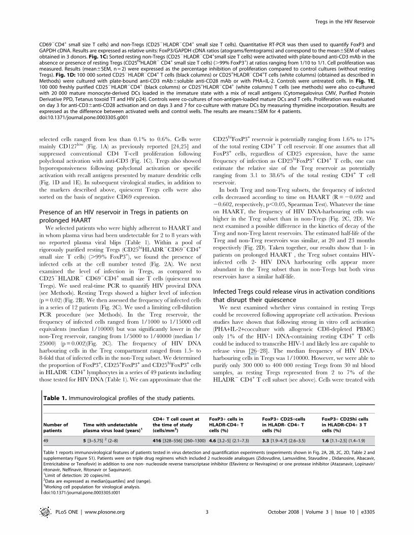
Figure 1. Definition of the resting Treg and non-Treg working cell populations. Fig. 1A: FoxP3 expression was examined by flowcytometry following cell permeabilization of PBMCs isolated from HIV-infected patients on long-term HAART (see Methods). FoxP3, CD25 and CD127expression was analyzed in the HLADR2 CD4+ small lymphocyte gate. In Fig. 1B, mRNA was extracted from resting Tregs (CD25hiHLADR2
Tregs in the HIV Reservoir
PLoS ONE | www.plosone.org 2 October 2008 | Volume 3 | Issue 10 | e3305
selected cells ranged from less than 0.1% to 0.6%. Cells were
mainly CD127low (Fig. 1A) as previously reported [24,25] and
suppressed conventional CD4 T-cell proliferation following
polyclonal activation with anti-CD3 (Fig. 1C). Tregs also showed
hyporesponsiveness following polyclonal activation or specific
activation with recall antigens presented by mature dendritic cells
(Fig. 1D and 1E). In subsequent virological studies, in addition to
the markers described above, quiescent Tregs cells were also
sorted on the basis of negative CD69 expression.
Presence of an HIV reservoir in Tregs in patients onprolonged HAART
We selected patients who were highly adherent to HAART and
in whom plasma virus had been undetectable for 2 to 8 years with
no reported plasma viral blips (Table 1). Within a pool of
rigorously purified resting Tregs (CD25hiHLADR2CD692CD4+
small size T cells) (.99% FoxP3+), we found the presence of
infected cells at the cell number tested (Fig. 2A). We next
examined the level of infection in Tregs, as compared to
CD252HLADR2 CD692CD4+ small size T cells (quiescent non
Tregs). We used real-time PCR to quantify HIV proviral DNA
(see Methods). Resting Tregs showed a higher level of infection
(p = 0.02) (Fig. 2B). We then assessed the frequency of infected cells
in a series of 12 patients (Fig. 2C). We used a limiting cell-dilution
PCR procedure (see Methods). In the Treg reservoir, the
frequency of infected cells ranged from 1/1000 to 1/15000 cell
equivalents (median 1/10000) but was significantly lower in the
non-Treg reservoir, ranging from 1/5000 to 1/40000 (median 1/
25000) (p = 0.002)(Fig. 2C). The frequency of HIV DNA
harbouring cells in the Treg compartment ranged from 1.5- to
8-fold that of infected cells in the non-Treg subset. We determined
the proportion of FoxP3+, CD25+FoxP3+ and CD25hiFoxP3+ cells
in HLADR2CD4+ lymphocytes in a series of 49 patients including
those tested for HIV DNA (Table 1). We can approximate that the
CD25hiFoxP3+ reservoir is potentially ranging from 1.6% to 17%
of the total resting CD4+ T cell reservoir. If one assumes that all
FoxP3+ cells, regardless of CD25 expression, have the same
frequency of infection as CD25hiFoxP3+ CD4+ T cells, one can
estimate the relative size of the Treg reservoir as potentially
ranging from 3.1 to 38.6% of the total resting CD4+ T cell
reservoir.
In both Treg and non-Treg subsets, the frequency of infected
cells decreased according to time on HAART (R = 20.692 and
20.602, respectively, p,0.05, Spearman Test). Whatever the time
on HAART, the frequency of HIV DNA-harbouring cells was
higher in the Treg subset than in non-Tregs (Fig. 2C, 2D). We
next examined a possible difference in the kinetics of decay of the
Treg and non-Treg latent reservoirs. The estimated half-life of the
Treg and non-Treg reservoirs was similar, at 20 and 23 months
respectively (Fig. 2D). Taken together, our results show that 1- in
patients on prolonged HAART , the Treg subset contains HIV-
infected cells 2- HIV DNA harbouring cells appear more
abundant in the Treg subset than in non-Tregs but both virus
reservoirs have a similar half-life.
Infected Tregs could release virus in activation conditionsthat disrupt their quiescence
We next examined whether virus contained in resting Tregs
could be recovered following appropriate cell activation. Previous
studies have shown that following strong in vitro cell activation
(PHA+IL-2+cocculture with allogeneic CD8-depleted PBMC)
only 1% of the HIV-1 DNA-containing resting CD4+ T cells
could be induced to transcribe HIV-1 and likely less are capable to
release virus [26–28]. The median frequency of HIV DNA-
harbouring cells in Tregs was 1/10000. However, we were able to
purify only 300 000 to 400 000 resting Tregs from 30 ml blood
samples, as resting Tregs represented from 2 to 7% of the
HLADR2 CD4+ T cell subset (see above). Cells were treated with
CD692CD4+ small size T cells) and non-Tregs (CD252HLADR2CD4+ small size T cells). Quantitative RT-PCR was then used to quantify FoxP3 andGAPDH cDNA. Results are expressed as relative units: FoxP3/GAPDH cDNA ratios (atograms/femtograms) and correspond to the mean6SEM of valuesobtained in 3 donors. Fig. 1C: Sorted resting non-Tregs (CD252HLADR2CD4+small size T cells) were activated with plate-bound anti-CD3 mAb in theabsence or presence of resting Tregs (CD25hiHLADR2 CD4+ small size T cells) (.99% FoxP3+) at ratios ranging from 1/10 to 1/1. Cell proliferation wasmeasured. Results (mean6SEM, n = 2) were expressed as the percentage inhibition of proliferation compared to control cultures (without restingTregs). Fig. 1D: 100 000 sorted CD252HLADR2CD4+ T cells (black columns) or CD25+HLADR2CD4+T cells (white columns) (obtained as described inMethods) were cultured with plate-bound anti-CD3 mAb6soluble anti-CD28 mAb or with PHA+IL-2. Controls were untreated cells. In Fig. 1E,100 000 freshly purified CD252HLADR2CD4+ (black columns) or CD25+HLADR2CD4+ (white columns) T cells (see methods) were also co-culturedwith 20 000 mature monocyte-derived DCs loaded in the immature state with a mix of recall antigens (Cytomegalovirus CMV, Purified ProteinDerivative PPD, Tetanus toxoid TT and HIV p24). Controls were co-cultures of non-antigen-loaded mature DCs and T cells. Proliferation was evaluatedon day 3 for anti-CD36anti-CD28 activation and on days 3 and 7 for co-culture with mature DCs by measuring thymidine incorporation. Results areexpressed as the difference between activated wells and control wells. The results are means6SEM for 4 patients.doi:10.1371/journal.pone.0003305.g001
Table 1. Immunovirological profiles of the study patients.
Number ofpatients
Time with undetectableplasma virus load (years)1
CD4+ T cell count atthe time of study(cells/mm3)
FoxP3+ cells inHLADR-CD4+ Tcells (%)
FoxP3+ CD25+cellsin HLADR- CD4+ Tcells (%)
FoxP3+ CD25hi cellsin HLADR-CD4+ 3 Tcells (%)
49 5 [3–5.75] 2 (2–8) 416 [328–556] (260–1300) 4.6 [3.2–5] (2.1–7.3) 3.3 [1.9–4.7] (2.6–3.5) 1.6 [1.1–2.5] (1.4–1.9)
Table 1 reports immunovirological features of patients tested in virus detection and quantification experiments (experiments shown in Fig. 2A, 2B, 2C, 2D, Table 2 andsupplementary Figure S1). Patients were on triple drug regimens which included 2 nucleoside analogues (Zidovudine, Lamuvidine, Stavudine , Didanosine, Abacavir,Emtricitabine or Tenofovir) in addition to one non- nucleoside reverse transcriptase inhibitor (Efavirenz or Nevirapine) or one protease inhibitor (Atazanavir, Lopinavir/ritonavir, Nelfinavir, Ritonavir or Saquinavir).1Limit of detection: 20 copies/ml.2Data are expressed as median[quartiles] and (range).3Working cell population for virological analysis.doi:10.1371/journal.pone.0003305.t001
Tregs in the HIV Reservoir
PLoS ONE | www.plosone.org 3 October 2008 | Volume 3 | Issue 10 | e3305

PHA+IL-2, a condition that permitted detectable Treg prolifer-
ation (Fig. 1D). We used 150 000 to 200 000 cells in both the
untreated control well and the PHA+IL-2 activated well. Direct
coculture of Tregs with allogeneic CD8-depleted HIV negative
PBMC was not used, due to the suppressive effects of Tregs on
proliferation of conventional CD4 T cells (Fig. 1C). As shown in
Table 2, no spontaneous virus release was detected in resting
Tregs, despite the use of a sensitive RT-PCR (limit of detection of
20 copies/ml). This control indicated the non-productive state of
cell infection in resting Tregs. Following 3 weeks cell activation,
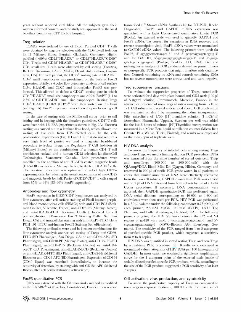
Figure 2. Presence of an HIV reservoir in resting Tregs in patients on prolonged HAART. Fig. 2A: DNA was extracted and analysed byPCR for HIV Env and GAPDH expression from resting Tregs (CD25hiHLADR2 CD692CD4+ small size T cells) (.99% FoxP3+). Positive and negativecontrols were total CD4+ T cells from HIV+ and HIV2 individuals, respectively. Similar results were obtained in 4 other patients tested. Fig. 2B:quantitation of HIV DNA by real-time PCR as described in Methods, in resting Tregs and non-Tregs (CD252HLADR2 CD692CD4+ small size T cells).Results are expressed as atogramms of HIV DNA per 100 fentogramms of GAPDH. Results correspond to the mean6SEM of values obtained in 6patients. Statistical comparison involved the Wilcoxon Signed Rank Test. Fig. 2C: frequencies of HIV DNA harbouring cells assessed by a limiting celldilution procedure (see Methods) in resting Tregs and non-Tregs from 12 patients. Statistical comparison involved the Wilcoxon Signed Rank Test.Fig. 2D: values of the percentages of HIV DNA harbouring cells in resting Tregs and resting non-Tregs (log10 scale) plotted against time underHAART. The equations of the tendency curves Y = Ae-lt are indicated. The half life of latent Treg and non T reg reservoirs T1/2 (months) wasdetermined as follows: ln2/l.doi:10.1371/journal.pone.0003305.g002
Tregs in the HIV Reservoir
PLoS ONE | www.plosone.org 4 October 2008 | Volume 3 | Issue 10 | e3305

HIV RNA production was found in cell culture supernatants in 7
out of 21 patients tested (33%) (Table 2).
We could re-obtain blood samples in 4 patients in whom HIV
RNA production could be detected from 200 000 Tregs. Cells
were activated as above but supernatants instead of RT-PCR
testing were used to infect PHA+IL-2 activated allogeneic CD8-
depleted PBMC from HIV seronegative subjects (see methods).
High amounts of HIV P24 were found in supernatants (.400 pg/
ml) for patients tested, indicating effective virus amplification on
allogeneic PBMC. As controls, we also tested in the same
conditions of cell number (200 000 cells) and cell activation,
resting non-Tregs. Viral RNA production was detected following
PHA+IL-2 activation of 200 000 resting non-Tregs in few patients
(3 out of 23 patients) (Table 2). Viral amplification following
addition of supernatants on PHA+IL-2 activated allogeneic CD8-
depleted HIV negative PBMC could be obtained in 2 out of 3
patients (HIV P24.400 pg/ml). One patient was negative despite
detectable HIV RNA in initial cell culture supernatant. Overall
these results suggest that infected Tregs could release virus in
activation conditions that disrupt their quiescence and anergy.
Of interest, recent reports suggested that histone deacetylase
inhibitors could lead to virus expression without full cell activation
[29]. We tested the effect of valproic acid on 150 000 to 200 000
quiescent Tregs from patients under long-term HAART. Cells
were activated for a period of 7 days. No viral RNA was detected
in untreated wells (not shown). No significant expression of the
activation marker HLADR was found following cell treatment
with valproic acid for 7 days (not shown). In the patients tested,
valproic acid led to very low but detectable virus production (see
methods) in a sensitive quantitative RT-PCR (detection limit, 20
viral RNA copies/ml) in 5 of 10 patients tested (log10 copies/ml in
HIV RNA positive supernatant: 1.4660.16, mean6sem) and
without detectable cell proliferation (not shown). The low level of
viral RNA in supernatants, likely, was related to the limited
amplification of virus produced by the rare in vivo infected cells,
due to the limited effects of valproic acid on activation of
uninfected cells. However, the keypoint is that valproic acid can
potentially lead to virus expression in latently infected Tregs,
therefore making them visible for cytotoxic CD8 T cells, through
formation of MHC class I-virus peptide complexes.
Targeting the Treg reservoir: specific constraintsApproaches based on cell activation have been proposed to
diminish or eliminate the HIV lymphocyte reservoir in patients on
prolonged HAART. However, in addition to the hyporesponsive-
ness of Tregs, which may limit cell activation and virus expression,
activated Tregs exert suppressive effects on both CD4+ and CD8+
T cells [20,21,30,31]. Granzyme B is directly involved in CTL-
mediated killing, and we found that CD25+CD4+ T-cell depletion
strongly increased granzyme B release by CD8+ T cells following
nonspecific activation with PMA+ionomycin (Fig. 3A). Depletion
of CD25+CD4+ T cells also significantly increased granzyme B
secretion in response to 15-mer overlapping HIV peptide pools
corresponding to the RT, P24, and Nef gene products (Fig. 3B).
These peptides could have activated specific Tregs, which then
exerted their suppressive effects. Alternatively, this suppressive
effect may be related to the existence, among untreated peripheral
blood mononuclear cells (PBMCs), of already activated Tregs that
could be continuously stimulated through interaction with self
peptides. This latter possibility may explain the high proportion of
HLADR+ cells among Tregs shown by ex vivo flow cytometry
Table 2. Infected Tregs could release virus in activation conditions that disrupt their quiescence.
Untreatedresting Tregs
Resting TregsPHA+IL-2
Untreated restingnon-Tregs
Resting non-TregsPHA+IL-2
200 000 purified cells
Patients with virus producing cells (limit of detection = 20 copies/ml) 0/21 7/21 0/23 3/23
HIV RNA (log10copies/ml) in positive supernatants (mean6sem) - 3.2761.48 - 2.7060.52
150 000 to 200 000 resting Tregs (CD25hiHLADR2 CD692CD4+ small size T cells) (.99% FoxP3+) were activated with PHA+IL-2 or left untreated (controls). Resting non-Tregs (CD252HLADR2 CD692CD4+ small size T cells) were tested at the exact cell number of 200 000/well. At day 21 of culture, supernatants were assayed for HIV RNAby quantitative RT-PCR (limit of detection of 20 copies/ml).doi:10.1371/journal.pone.0003305.t002
Figure 3. Activated Tregs inhibit granzyme B secretion by CD8T cells. PBMC and CD4+ CD25+ cell-depleted PBMCs were assayed inthree patients by ELISPOT for granzyme B secretion, followingactivation with PMA-ionomycin (Fig. 3A) or with overlapping HIVpeptide pools corresponding to the p24, reverse transcriptase and Nefregions (Fig. 3B). Results shown in Fig. 3B are numbers of spots per100 000 CD8+ T cells. Results correspond to the mean6SEM of resultsobtained in 4 patients.doi:10.1371/journal.pone.0003305.g003
Tregs in the HIV Reservoir
PLoS ONE | www.plosone.org 5 October 2008 | Volume 3 | Issue 10 | e3305

analysis (the median percentage of HLADR+ cells among
CD25hiFoxP3+ CD4+ T cells was 51%, quartiles [46–59%],
n = 15 patients on HAART for at least 2 years) (not shown).
Activated Tregs might therefore create an environment in which
CD8+ T cells are unable to fully exert their cytotoxic functions. In
contrast, resting Tregs (HLADR2 Tregs without exogenous
activation) have no significant inhibitory effects on granzyme B
secretion by activated CD8+ T cells (Fig. 4A). Histone deacetylase
inhibitors could lead to virus expression without full cell activation
[29] (see above). This could be an approach of potential interest
for reducing the resting Treg reservoir. We next examined
whether resting Tregs but expressing HIV peptides were
susceptible to CD8+ T-cell cytolysis. Owing to the low frequency
of infected cells and specific CD8+ T cells, we used the following
strategy. CD4+-depleted PBMCs were first screened for reactivity
to several pools of overlapping peptides corresponding to the Gag,
Pol, and Nef sequences (see Methods). The most reactive
overlapping peptide pool was then used to amplify specific CTL
for testing against peptide-loaded autologous resting Tregs and
resting non-Tregs (see methods). As shown in Fig. 4B, similar levels
of apoptosis were found in the Treg and non-Treg subsets. This
suggested that resting Tregs expressing HIV peptides may be as
susceptible as non-Tregs to CD8+ T-cell cytotoxicity.
Discussion
In this study we identified a virus reservoir among resting
regulatory CD4+ T cells in patients on long-term effective HAART.
The frequency of HIV DNA harbouring cells was higher in the
resting Treg subset as compared to the resting non Treg subset.
HIV productive cycle occurs preferentially in activated cells. In
vitro, Tregs are anergic or hyporesponsive as compared with
conventional CD4+ T cells. Tregs share several biochemical features
with anergic cells [32]. However, several studies in mouse models
suggest that, in vivo, at least some Tregs can be activated to
proliferate as strongly as conventional CD4+ T cells [33], possibly
because the higher levels of IL-2 available in vivo are able to reverse
the previously mentioned biochemical defects [32]. However,
impaired IL-2 production during active HIV infection [34,35]
could cause Tregs to behave in vivo as in vitro. Such Treg
hyporesponsiveness may potentially favour blockades in the viral
cycle at the integration phase, leading to post-integration viral
latency (see supplementary Figure S1). Moreover, FoxP3 which is
expressed in Tregs was recently shown to inhibit HIV-1 LTR
activation by targeting the NFkB pathway [36]. We also found that
resting Tregs were intrinsically sensitive to specific CD8+ T cell-
mediated cytotoxicity. However, upon activation, these cells
inhibited CD8+ T-cell cytotoxic-associated functions such as
granzyme B secretion. By helping Tregs escape from CTLs, this
process could favour the survival of productively infected activated
Tregs, allowing them to return to a quiescent state. HAART may
promote the persistence of HIV or opportunistic agent-specific
Tregs in the resting state, as the likelihood of re-encountering their
specific antigens becomes very low.
The decrease in the size of the Treg reservoir in patients on
HAART was similar to that of non-Tregs. This suggested that the
factors leading to a higher level of infection in the Treg subset as
compared with non-Tregs mainly act by favouring Treg reservoir
formation, instead of slowing its decay. However, the process
could be more complex: the apparent half-lives of the Treg and
non-Treg reservoirs could be the result of additive, distinct and
even antagonistic phenomena, including infected-cell decay in the
two subsets, as well as virus replenishment through residual
replication [37,38]. Ramratnam et al. recently reported a shorter
half-life of the latent reservoir in patients on intensified HAART
(which may reduce residual viral replication) than in patients on
standard HAART [38].
It has been shown in patients on long-term HAART that activated
CD4+ T cells harbour actively replicating virus and, ex vivo, release
virus spontaneously. Such ongoing virus replication, despite
HAART, may continuously replenish the latent reservoir [28].
The half-life of the latent Treg and non-Treg reservoirs was
estimated at approximately 20 and 23 months, respectively. This
half-life was shorter than the approximately 44 months for the
whole latent reservoir previously reported [39], although it is of the
same order of magnitude, indicating that eradication of this
reservoir in patients on continuous and effective HAART would
take several decades.
The time of HAART initiation during the course of infection
could be of importance, as a recent study [40] showed a shorter
half-life of the latent lymphocyte reservoir in patients who were
placed on HAART very rapidly (less than 5 months after
seroconversion) following the onset of symptoms of primary HIV
infection. This was not the case of the patients in our study, who
started HAART from approximatively 2 years following the
diagnosis of infection.
In vitro activation conditions that partly overcome Treg anergy
led to detectable production of virus from 200 000 cells in a
significant proportion of patients tested, which suggests that Tregs
could potentially release virus in vivo and contribute to the viral
rebound observed upon HAART withdrawal. The question of
how to purge this reservoir, should therefore be addressed. Specific
constraints related to the biology of Tregs may limit the
therapeutic options. Cell-activation-based approaches, with IL-2
or anti-CD3, have been proposed to purge the latent reservoir.
Despite some early encouraging results, these approaches have not
eradicated the virus [41]. The rationale was that cell activation
may induce expression of the virus in latently infected cells,
whereas HAART would avoid de novo cell infection. Thus,
unveiled, latently infected cells could be targeted by cytotoxic T
lymphocytes or destroyed by direct cytopathic effects [41,42].
Whether IL-2 can directly activate Tregs in vivo remains to be
determined. In vitro, in contrast to the combination of PHA+IL-2,
IL-2 alone did not lead to detectable virus production by Tregs
(not shown) or to significant proliferation. In cancer patients, IL-2
immunotherapy led to expansion of Tregs with potent suppressive
activity in vitro [43]. Such an effect could be related to increased
generation of Tregs in the thymus [44]. In the peripheral
compartment, exogenous IL-2 may help to break virus latency
only in Tregs that undergo physiological antigen stimulation, by
favouring their proliferation. This effect may represent only a
small fraction of latently infected resting Tregs. Moreover,
activated Tregs may inhibit specific CD8+ T-cell cytotoxic
functions. The addition of activating anti-CD3 to IL-2 might
increase the activation of the latent Treg reservoir. However, in
addition to adverse effects related to non-specific generalized
immune activation, this approach would not overcome the
inhibition of CD8+ T cell-mediated cytotoxicity by activated
Tregs. The effect of IL-7 on virus reactivation in the latent Treg
reservoir remains to be determined. In patients on HAART, this
cytokine was effective at triggering HIV-1 reactivation in CD8-
depleted PBMCs and CD252HLADR2 resting CD4+ T lympho-
cytes [45]. Partial cell activation and proliferation could be one of
the mechanisms by which IL-7 acts on virus latency [45]. Recent
data show that IL-7 increases the survival of murine Tregs by
inhibiting apoptosis but has no effect on their proliferation [46].
Moreover, in human Tregs, IL-7 receptor (CD127) is downreg-
ulated [24,25] (see also Fig. 1A).
Tregs in the HIV Reservoir
PLoS ONE | www.plosone.org 6 October 2008 | Volume 3 | Issue 10 | e3305

Histone deacetylase inhibitors such as valproic acid were
recently shown to trigger virus expression in vitro in resting
CD4+ T cells, without full cell activation [47], and to reduce the
latent reservoir in vivo [29]. However, the hopes raised by this
finding were recently tempered by the results of other studies
showing no clear effect of valproic acid on the size of the latent
reservoir in vivo [48,49]. Further studies are needed to clarify the
real impact of valproic acid. Nonetheless, such therapeutics
directly targeting virus quiescence, instead of conventional
approaches that aim to disrupt cell quiescence, appear, at least
in theory, more appropriate to purge the Treg latent reservoir,
because they could potentially bypass the barriers that protect
HIV within this compartment (i.e., hyporesponsiveness and
inhibition of CD8+ T cell functions upon activation). Indeed,
quiescent Tregs expressing HIV peptides were as sensitive to
CD8+ T-cell cytotoxicity as non-Tregs. We also found that
valproic acid was able to induce detectable virus production by
latently infected Tregs in vitro, warranting further investigations of
the potential impact of histone deacetylase inhibitors on the latent
Treg reservoir in vivo.
Together, these results identify Tregs as a particular compart-
ment within the virus reservoir that may require a specific
approach for its purging.
Methods
Patients and healthy donorsPatients with chronic HIV-1 infection were recruited on the
basis of plasma viral load values ,20 copies/ml (Amplicor
Ultrasensible, Roche Diagnostics, Meylan, France) for at least 2
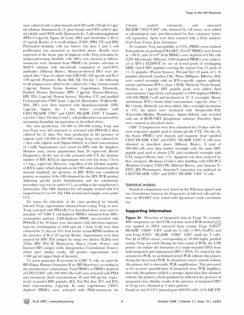
Figure 4. Quiescent Tregs are sensitive to specific CD8+ T cell cytotoxicity. Fig. 4A: CD25+ cell-depleted PBMCs were activated with PMA-inonomycin then extensively washed. Untreated HLADR2CD25+CD4+ T cells were then added at a physiological ratio. Cells were assayed by ELISPOTfor granzyme B. Results correspond to the mean6SEM of results obtained in 2 patients. In Fig. 4B, HIV-specific CTL were co-cultured withCD25+HLADR2CD4+ and CD252HLADR2CD4+ T cells loaded with HIV peptides. Controls were co-cultures of CD4+ T-cell subsets without peptides.Apoptosis was analyzed following annexin V staining on CD25hiHLADR2CD4+ and CD252HLADR2CD4+ T cells. Results are expressed as the differencein the percentage of annexin V-positive cells in the presence and absence of HIV peptides. Results correspond to the mean6SEM of results obtainedfor 7 patients. Statistical comparison involved the Wilcoxon Signed Rank Test.doi:10.1371/journal.pone.0003305.g004
Tregs in the HIV Reservoir
PLoS ONE | www.plosone.org 7 October 2008 | Volume 3 | Issue 10 | e3305
years without reported viral blips. All the subjects gave their
written informed consent, and the study was approved by the local
bioethics committee (CPP Bicetre hospital).
Treg isolationPBMCs were isolated by use of Ficoll. Purified CD4+ T cells
were obtained by negative selection with the CD4 T-cell isolation
kit II (Miltenyi Biotec, Bergisch Gladbach, Germany). Highly
purified (.99%) CD252HLADR2 or CD252HLADR2CD692
CD4 T cells and CD25hiHLADR2 or CD25hiHLADR2 CD692
CD4 small size T-cells were obtained by cell sorting (FacsAria,
Beckton Dickinson, CA) or (MoFlo, DakoCytoformation, Carpin-
teria, CA). For each patient, the CD25hi sorting gate in HLADR2
CD4+ small lymphocytes was pre-defined on the basis of Foxp3
expression. Briefly, a 4 color flow cytometry analysis of cell surface
CD4, HLADR, and CD25 and intracellular FoxP3 was per-
formed. This allowed to define a CD25hi sorting gate in which
CD4+HLADR2 small size lymphocytes are .99% FoxP3+ (1.1 to
2.5% of CD4+HLADR2 small size lymphocytes. Resting Tregs
CD4+HLADR2(CD692)CD25hi were then sorted on this basis
(see Fig. 1A). FoxP3 expression was checked again following cell
sorting.
In the case of sorting with the MoFlo cell sorter, prior to cell
sorting and in keeping with the biosafety guidelines, CD4+ T cells
were fixed with 1% PFA (experiments shown in Fig. 2A). FacsAria
sorting was carried out in a laminar flow hood, which allowed the
sorting of live cells from HIV-infected cells. In the cell-
proliferation experiments (Fig. 1D and 1E), that do not require a
very high level of purity, we used a magnetic bead-based
procedure to isolate Tregs: the Regulatory T Cell Isolation kit
(Miltenyi Biotec) or the combination of a human CD4 T cell
enrichment cocktail and a human CD25 selection kit (StemCell
Technologies, Vancouver, Canada). Both procedures were
modified by the addition of anti-HLADR-coated magnetic beads
(HLA-DR microbeads, Miltenyi Biotec) to deplete HLADR+ cells.
The isolation procedure was optimized to select high CD25-
expressing cells, by reducing the usual concentration of anti-CD25
and magnetic beads by half. Purity of CD25+CD4+ T cells ranged
from 85% to 93% (83–90% FoxP3 expression).
Antibodies and flow cytometryFoxP3 expression in CD25+CD4+ lymphocytes was analyzed by
flow cytometry after cell-surface staining of Ficoll-isolated periph-
eral blood mononuclear cells (PBMCs) with anti-CD4-PC5 (Beck-
man Coulter, Villepinte, France), anti-CD25-PE (Miltenyi Biotec),
and anti-HLADR-ECD (Beckman Coulter), followed by cell
permeabilization (eBioscience FoxP3 Staining Buffer Set, San
Diego, CA) and intracellular staining with anti-FoxP3-FITC (clone
PCH 101; FITC anti-human FoxP3 Staining Set, eBioscience).
The following antibodies were used in 4-colour combinations for
flow cytometric analysis and/or cell sorting of Tregs: anti-CD69-
FITC (BD Pharmingen, San Diego, CA) or anti-CD69-APC (BD
Pharmingen), anti-CD30-PE (Miltenyi Biotec), anti-CD127-PE (BD
Pharmingen), anti-CD4-PC5 (Beckman Coulter) or anti-CD4-
perCP (BD Pharmingen), anti-HLADR-ECD (Beckman Coulter)
or anti-HLADR-FITC (BD Pharmingen), anti-CD25-PE (Miltenyi
Biotec) or anti-CD25-APC (BD Pharmingen). Expression of CD154
(CD40 ligand) was examined intracellularly, to increase the
sensitivity of detection, by staining with anti-CD154-APC (Miltenyi
Biotec) after cell permeabilization (eBioscience).
FoxP3 quantitative PCRRNA was extracted with the Chomoczinsky method as modified
in the RNABleH kit (Eurobio, Courtaboeuf, France), then reverse
transcribed (1st Strand cDNA Synthesis kit for RT-PCR, Roche
Diagnostics). FoxP3 and GAPDH mRNA expression was
quantified with a Light Cycler-based quantitative kinetic PCR
(Roche). An external scale was used to quantify GAPDH and
FoxP3 cDNA. To correct for variations in RNA recovery and
reverse transcription yield, FoxP3 cDNA values were normalized
to GAPDH cDNA values. The following primers were used: for
FoxP3, 59-agaggacttcctcaagca-39 and 59-gctgccagcagctacgatg-39
and for GAPDH, 59-ggtgaaggtcggagtcaacgga-39 and 59-gagg-
gatctcgctcctggaaga-39 (Proligo, Boulder, CO, USA). Gel and
melting curve analysis of PCR products showed no primer dimers
or non-specific PCR products that might interfere with quantita-
tion. Controls containing no RNA and controls containing RNA
but no reverse transcriptase were always used and were negative.
Treg suppressive functionsTo evaluate the suppressive properties of Tregs, sorted cells
were activated for 5 days with plate-bound anti-CD3 mAb (100 ml
of 5 mg/ml solution) (Immunotech, Marseille, France) in the
absence or presence of non-Tregs at ratios ranging from 1/10 to
1/1. Cell subsets were sorted as described above. Cell proliferation
was evaluated on day 5 by measuring thymidine incorporation.
Fifty microliters of 1/50 [H3]thymidine solution (1 mCi/ml)
(Amersham Pharmacia, Uppsala, Sweden) per well was added
for the last 8 hours of culture. [H3]Thymidine incorporation was
measured in a Micro Beta liquid scintillation counter (Micro Beta
Counter Plus, Wallac, Turku, Finland), and results were expressed
as the mean cpm of triplicate wells.
HIV DNA analysisTo assess the frequency of infected cells among resting Tregs
and non-Tregs, we used a limiting dilution PCR procedure. DNA
was extracted from the same number of sorted quiescent Tregs
and non-Tregs (100 000 to 200 000 cells) with the
QIAampHDNA Blood Mini Kit (Qiagen, Hilden, Germany) and
recovered in 200 ml of sterile PCR-grade water. In all patients, to
check that similar amounts of DNA were effectively recovered
from the two cell subsets, GAPDH quantitative PCR was carried
out on 2 ml of DNA extracted from both subsets by use of a Light
Cycler procedure. If necessary, DNA concentrations were
adjusted, then GAPDH quantitative PCR was performed again.
DNA serial dilutions corresponding to 40 000 to 1 000 cell
equivalents were then used per PCR. HIV PCR was performed
in a 50 ml volume under the following conditions: 0.25 pM/ml of
each primer, 2.5 mM MgCl2, 0.2 mM dNTPs, 1.5 U Taq
Platinum, and buffer (Invitrogen, Carlsbad, CA). The following
primers targeting the HIV V3 loop between the C2 and V4
regions of gp120 were used: 59-acacatggaattaggccagt-39 and 59-
ctgccacatgtttataatttg-39 (MWG-Biotech AG, Ebersberg, Ger-
many). The sensitivity of the PCR ranged from 1 to 5 attograms
of purified specific PCR product, which suggested a sensitivity
from 2 to 8 copies.
HIV DNA was quantified in sorted resting Tregs and non-Tregs
by a real-time PCR procedure [50]. Results were expressed as
normalized values (attograms of HIV DNA per 100 femtograms of
GAPDH). In most cases, we obtained a significant amplification
curve for the 1 attogram point of the external scale (made of
serially diluted purified specific PCR product), which, according to
the size of the PCR product, suggested a PCR sensitivity of at least
7 copies.
Cell activation, virus production, and cytotoxicityTo assess the proliferative capacity of Tregs as compared to
non-Tregs in response to stimuli, 100 000 cells from each subset
Tregs in the HIV Reservoir
PLoS ONE | www.plosone.org 8 October 2008 | Volume 3 | Issue 10 | e3305
were cultured with 1) plate-bound anti-CD3 mAb (100 ml of 5 mg/
ml solution; Immunotech), 2) plate-bound anti-CD3 mAb+1 mg/
ml soluble anti-CD28 mAb (Immunotech), 3) phytohemagglutinin
(PHA 0.5 mg/ml; Sigma, St Louis, MO) and interleukin 2 (IL-2;
25 ng/ml, Roche) or 4) recall antigen- (CMV, PPD, TT) and HIV
P24-loaded dendritic cells (see below). On days 3 and 7, cell
proliferation was measured as described above. Results were
expressed as the mean cpm of triplicate wells. Treg activation by
antigen-presenting dendritic cells (DCs) was assessed as follows:
monocytes were obtained from PBMCs by positive selection in
MACS columns with microbeads conjugated to anti-CD14
antibodies (Miltenyi Biotec). Monocyte-derived DCs were ob-
tained after 7 days of culture with GM-CSF (100 ng/ml) and IL-4
(100 ng/ml) (Peprotec, Rocky Hill, NJ). On day 7, the following
recall antigens were added to the cultures for 1 day: tetanus toxoid
(1 mg/ml, Statens Serum Institute, Copenhagen, Denmark),
Purified Protein Derivative PPD (1 mg/ml, Pasteur-Merieux),
HIV P24 (5 mg/ml, Protein Science, Meriden, CT) and purified
Cytomegalovirus CMV lysate (1 mg/ml, Biowittaker, Walkersville,
MA). DCs were then matured with lipopolysaccharide (LPS;
1 mg/ml, Sigma) for 1 day before co-culture with
CD25+HLADR2CD4+ and CD252HLADR2CD4+ T lympho-
cytes for 7 days. On days 3 and 7, cell proliferation was assessed by
measuring thymidine incorporation as described above.
For virus production, 150 000 to 200 000 resting Tregs and
non-Tregs were left untreated or activated with PHA+IL-2 then
cultured for 21 days. For virus production in the presence of
valproic acid, 150 000 to 200 000 resting Tregs were cultured for
7 days with valproic acid (Sigma-Aldrich) at a final concentration
of 1 mM. Supernatants were tested for HIV with the Amplicor
Monitor assay (mean quantitation limit 20 copies/ml, Roche
Diagnostics). In the case of cells activated with VA, the absolute
number of HIV RNA in supernatants was very low from 1.34 to
1.7 log10 copies/ml. However, regardless of the absolute number
of RNA copies (which depends on the OD values obtained with an
internal standard), the presence of HIV RNA was considered
positive or negative if the OD obtained for the HIV PCR product
(following specific probe hybridization and the colorimetric
procedure) was over or under 0.15, according to the manufacture’s
instructions. The ODs obtained for cell samples treated with VA
ranged from 0.3 to 0.8. The ODs of untreated samples were under
0.15.
To assess the infectivity of the virus produced by latently
infected Tregs, supernatants obtained from resting Tregs or non-
Tregs activated with PHA+IL-2 as described above were used to
inoculate 106 CD8 T cell-depleted PBMCs obtained from HIV-
seronegative patients. CD8-depleted PBMC pre-activated with
PHA+IL-2 for 48 hours were inoculated with infectious superna-
tants by centrifugation at 2500 rpm for 1 hour. Cells were then
cultured for 21 days in 10% fetal bovine serum-RPMI medium in
the presence of IL-2 (25 ng/ml, Roche). Supernatants were then
assayed for HIV P24 antigen by using two distinct ELISA tests
(Vidas HIV P24 II, Biomerieux, Marcy l’etoile, France; and
Innotest HIV antigen mAb, Innogenetics, Courtaboeuf, France),
which gave similar results. All positive supernatants were
.400 pg/ml (upper limit of linearity).
To assess granzyme B secretion by CD8+ T cells, we used the
BD Elispot Human Granzyme B set (BD Bioscience) according to
the manufacturer’s instructions. Total PBMCs or PBMCs depleted
of CD25+CD4+ cells (100 000 cells/well) were activated with PMA
and ionomycin (final concentrations 50 and 500 ng/ml, respec-
tively) or pooled HIV peptides (RT, Nef, Pol, Env, P17 and P24;
final concentration, 2 mg/ml). In some experiments, CD25-
depleted PBMCs were activated with PMA-ionomycin for
2 hours, washed extensively, and untreated
HLADR2CD25+CD4+ cells, obtained by cell sorter, were added
at physiological ratio (pre-determined by flow cytometry before
cell separation). Spots were then counted with a Zeiss analyzer
(Carl Zeiss Vision, Jena, Germany).
To examine Treg susceptibility to CTL, PBMCs were isolated
from patients on prolonged HAART, 506106 PBMCs were frozen
at 280uC, and 106106 fresh PBMCs were depleted of CD4 cells
(CD4 Microbeads, Miltenyi). CD4-depleted PBMCs were subject-
ed to IFN-c ELISPOT by use of several pools of overlapping
MHC class-I HIV peptides covering the regions Gag (11 pools of
11–15 peptides) (Protein Science), Pol and Nef (10 pools of 5–10
peptides) (Intracell, London, UK). Plates (Millipore, Billerica, MA)
were coated overnight with an IFN-c-specific capture antibody
(mouse anti-human IFN-c, clone 1-D1K, Mabtech, Nacka Strand,
Sweden) at 1 mg/ml. HIV peptide pools were added (final
concentration 2 mg/ml for each peptide) to CD4-depleted PBMCs
(100 000 PBMC/well) and incubated at 37u C overnight. Mouse
anti-human IFN-c–biotin (final concentration 1 mg/ml, clone 7-
B6-1 biotin, Mabtech) was then added. After overnight incubation
at 4uC, the plates were incubated with Streptavidin–PAL
(Extravidin-Alkaline Phosphatase, Sigma-Aldrich) and revealed
with use of BCIP/NBT phosphatase substrate (Eurobio). Spots
were counted as described above.
CD8+ T lymphocytes were then stimulated for 10 days with the
most responsive peptide pool to obtain specific CTL. On day 10,
the frozen PBMCs were thawed, and magnetic bead -purified
CD25+HLADR2CD4+ and CD252HLADR2CD4+ T cells were
obtained as described above (Miltenyi Biotec). A total of
200 000 cells were then loaded overnight with the same HIV
peptide pool used to obtain CTL and cocultured overnight with
CTL (target/effector ratio 1/1). Apoptosis was then analyzed by
flow cytometry (Beckman Coulter) after labelling with CD4-PC5
(Beckman Coulter), CD25-PE (Miltenyi Biotec), and Annexin V-
FITC (BD Pharmingen). Annexin-V expression was analyzed on
CD25hiHLADR2CD4+ and CD252HLADR2CD4+ T cells.
Statistical analysisStatistical comparisons were based on the Wilcoxon signed rank
test. Correlations between the frequencies of infected cells and the
time on HAART were tested with Spearman’s rank correlation
test.
Supporting Information
Figure S1 Detection of integrated virus in Tregs: To examine
HIV integration, an Alu/LTR real-time nested PCR method [51]
was applied to DNA extracted from resting Tregs (CD25hi
HLADR2 CD692 CD4+ small size T cells) (.99% FoxP3+) and
non-Tregs (CD252 HLADR2 CD692 CD4+ small size T cells).
Five ml of DNA extract, corresponding to 50 000 highly purified
resting Tregs was used. During the first round of PCR, the LTR
primer can initiate the formation of a single-stranded DNA from
both integrated and unintegrated HIV-1 DNA. To control for this
asymmetric PCR, we performed nested PCR without Alu primers
during the first-round PCR. In all patients tested, controls without
Alu primers led to detectable PCR amplification. This prevented
us for accurate quantification of integrated virus. PCR amplifica-
tion with Alu primers yielded a stronger signal than that obtained
without Alu primers, which qualitatively indicated the presence of
integrated virus. Similar results of the presence of integrated HIV
in Tregs were obtained in 3 other patients.
Found at: doi:10.1371/journal.pone.0003305.s001 (4.04 MB TIF)
Tregs in the HIV Reservoir
PLoS ONE | www.plosone.org 9 October 2008 | Volume 3 | Issue 10 | e3305

Acknowledgments
We thank MT Rannou, M Mole and all the nurses at the HIV/AIDS
Daycare Unit of the Department of Internal Medicine, Bicetre Hospital; H
Kieffer (Cell sorting platform, Pasteur Institute); N Idri (Virology
Laboratory, Bicetre Hospital); L Meyer, Dr R Cheng (Epidemiology and
Public Health Service, Bicetre hospital); P Sonigo and C Ladroux (ICGM,
Cochin Hospital) for their help. We are indebted to the patients who
consented to participate in the study.
Author Contributions
Conceived and designed the experiments: TAT DGMG HC BD ELN KA
JFD AMB YT. Performed the experiments: TAT DGMG HC BD ELN
KA CP JG AMB. Analyzed the data: TAT DGMG HC BD ELN KA CP
CG JFD JG AMB YT. Contributed reagents/materials/analysis tools: CG.
Wrote the paper: TAT DGMG YT.
References
1. Finzi D, Hermankova M, Pierson T, Carruth LM, Buck C, et al. (1997)
Identification of a reservoir for HIV-1 in patients on highly active antiretroviral
therapy. Science 278: 1295–1300.
2. Chun TW, Carruth L, Finzi D, Shen X, DiGiuseppe JA, et al. (1997)
Quantification of latent tissue reservoirs and total body viral load in HIV-1
infection. Nature 387: 183–188.
3. Wong JK, Hezareh M, Gunthard HF, Havlir DV, Ignacio CC, et al. (1997)
Recovery of replication-competent HIV despite prolonged suppression of
plasma viremia. Science 278: 1291–1295.
4. Lambotte O, Demoustier A, de Goer MG, Wallon C, Gasnault J, et al. (2002)
Persistence of replication-competent HIV in both memory and naive CD4 T cell
subsets in patients on prolonged and effective HAART. Aids 16: 2151–2157.
5. Pierson T, McArthur J, Siliciano RF (2000) Reservoirs for HIV-1: mechanisms
for viral persistence in the presence of antiviral immune responses and
antiretroviral therapy. Annu Rev Immunol 18: 665–708.
6. Demoustier A, Gubler B, Lambotte O, de Goer MG, Wallon C, et al. (2002) In
patients on prolonged HAART, a significant pool of HIV infected CD4 T cells
are HIV-specific. Aids 16: 1749–1754.
7. Douek DC, Brenchley JM, Betts MR, Ambrozak DR, Hill BJ, et al. (2002) HIV
preferentially infects HIV-specific CD4+ T cells. Nature 417: 95–98.
8. Sakaguchi S (2004) Naturally arising CD4+ regulatory T cells for immunologic
self-tolerance and negative control of immune responses. Annu Rev Immunol
22: 531–562.
9. O’Garra A, Vieira P (2004) Regulatory T cells and mechanisms of immune
system control. Nat Med 10: 801–805.
10. Ziegler SF (2006) FOXP3: Of Mice and Men. Annu Rev Immunol 24: 209–226.
11. Baecher-Allan C, Brown JA, Freeman GJ, Hafler DA (2001) CD4+CD25high
regulatory cells in human peripheral blood. J Immunol 167: 1245–1253.
12. Takahashi T, Kuniyasu Y, Toda M, Sakaguchi N, Itoh M, et al. (1998)
Immunologic self-tolerance maintained by CD25+CD4+ naturally anergic and
suppressive T cells: induction of autoimmune disease by breaking their anergic/
suppressive state. Int Immunol 10: 1969–1980.
13. Lim HW, Hillsamer P, Banham AH, Kim CH (2005) Cutting edge: direct
suppression of B cells by CD4+ CD25+ regulatory T cells. J Immunol 175:
4180–4183.
14. Belkaid Y, Piccirillo CA, Mendez S, Shevach EM, Sacks DL (2002)
CD4+CD25+ regulatory T cells control Leishmania major persistence and
immunity. Nature 420: 502–507.
15. Cabrera R, Tu Z, Xu Y, Firpi RJ, Rosen HR, et al. (2004) An
immunomodulatory role for CD4(+)CD25(+) regulatory T lymphocytes in
hepatitis C virus infection. Hepatology 40: 1062–1071.
16. Weiss L, Donkova-Petrini V, Caccavelli L, Balbo M, Carbonneil C, et al. (2004)
Human immunodeficiency virus-driven expansion of CD4+CD25+ regulatory T
cells, which suppress HIV-specific CD4 T-cell responses in HIV-infected
patients. Blood 104: 3249–3256.
17. Aandahl EM, Michaelsson J, Moretto WJ, Hecht FM, Nixon DF (2004) Human
CD4+ CD25+ regulatory T cells control T-cell responses to human
immunodeficiency virus and cytomegalovirus antigens. J Virol 78: 2454–2459.
18. Kinter AL, Hennessey M, Bell A, Kern S, Lin Y, et al. (2004) CD25(+)CD4(+)
regulatory T cells from the peripheral blood of asymptomatic HIV-infected
individuals regulate CD4(+) and CD8(+) HIV-specific T cell immune responses
in vitro and are associated with favorable clinical markers of disease status. J Exp
Med 200: 331–343.
19. Rouse BT, Sarangi PP, Suvas S (2006) Regulatory T cells in virus infections.
Immunol Rev 212: 272–286.
20. Kinter A, McNally J, Riggin L, Jackson R, Roby G, et al. (2007) Suppression of
HIV-specific T cell activity by lymph node CD25+ regulatory T cells from HIV-
infected individuals. Proc Natl Acad Sci U S A 104: 3390–3395.
21. Kinter AL, Horak R, Sion M, Riggin L, McNally J, et al. (2007) CD25+regulatory T cells isolated from HIV-infected individuals suppress the cytolytic
and nonlytic antiviral activity of HIV-specific CD8+ T cells in vitro. AIDS Res
Hum Retroviruses 23: 438–450.
22. Chase AJ, Sedaghat AR, German JR, Gama L, Zink MC, et al. (2007) Severe
depletion of CD4+ CD25+ regulatory T cells from the intestinal lamina propria
but not peripheral blood or lymph nodes during acute simian immunodeficiency
virus infection. J Virol 81: 12748–12757.
23. Oswald-Richter K, Grill SM, Shariat N, Leelawong M, Sundrud MS, et al.
(2004) HIV infection of naturally occurring and genetically reprogrammed
human regulatory T-cells. PLoS Biol 2: E198.
24. Seddiki N, Santner-Nanan B, Martinson J, Zaunders J, Sasson S, et al. (2006)
Expression of interleukin (IL)-2 and IL-7 receptors discriminates between human
regulatory and activated T cells. J Exp Med 203: 1693–1700.
25. Liu W, Putnam AL, Xu-Yu Z, Szot GL, Lee MR, et al. (2006) CD127
expression inversely correlates with FoxP3 and suppressive function of human
CD4+ T reg cells. J Exp Med 203: 1701–1711.
26. Hermankova M, Siliciano JD, Zhou Y, Monie D, Chadwick K, et al. (2003)
Analysis of human immunodeficiency virus type 1 gene expression in latently
infected resting CD4+ T lymphocytes in vivo. J Virol 77: 7383–7392.
27. Finzi D, Blankson J, Siliciano JD, Margolick JB, Chadwick K, et al. (1999)
Latent infection of CD4+ T cells provides a mechanism for lifelong persistence of
HIV-1, even in patients on effective combination therapy. Nat Med 5: 512–
517.
28. Chun TW, Nickle DC, Justement JS, Large D, Semerjian A, et al. (2005) HIV-
infected individuals receiving effective antiviral therapy for extended periods of
time continually replenish their viral reservoir. J Clin Invest 115: 3250–3255.
29. Lehrman G, Hogue IB, Palmer S, Jennings C, Spina CA, et al. (2005) Depletion
of latent HIV-1 infection in vivo: a proof-of-concept study. Lancet 366:
549–555.
30. Camara NO, Sebille F, Lechler RI (2003) Human CD4+CD25+ regulatory cells
have marked and sustained effects on CD8+ T cell activation. Eur J Immunol
33: 3473–3483.
31. Piccirillo CA, Shevach EM (2001) Cutting edge: control of CD8+ T cell
activation by CD4+CD25+ immunoregulatory cells. J Immunol 167:
1137–1140.
32. Li L, Godfrey WR, Porter SB, Ge Y, June CH, et al. (2005) CD4+CD25+regulatory T-cell lines from human cord blood have functional and molecular
properties of T-cell anergy. Blood 106: 3068–3073.
33. von Boehmer H (2003) Dynamics of suppressor T cells: in vivo veritas. J Exp
Med 198: 845–849.
34. Younes SA, Yassine-Diab B, Dumont AR, Boulassel MR, Grossman Z, et al.
(2003) HIV-1 viremia prevents the establishment of interleukin 2-producing
HIV-specific memory CD4+ T cells endowed with proliferative capacity. J Exp
Med 198: 1909–1922.
35. Iyasere C, Tilton JC, Johnson AJ, Younes S, Yassine-Diab B, et al. (2003)
Diminished proliferation of human immunodeficiency virus-specific CD4+ T
cells is associated with diminished interleukin-2 (IL-2) production and is
recovered by exogenous IL-2. J Virol 77: 10900–10909.
36. Grant C, Oh U, Fugo K, Takenouchi N, Griffith C, et al. (2006) Foxp3 represses
retroviral transcription by targeting both NF-kappaB and CREB pathways.
PLoS Pathog 2: e33.
37. Ramratnam B, Mittler JE, Zhang L, Boden D, Hurley A, et al. (2000) The decay
of the latent reservoir of replication-competent HIV-1 is inversely correlated
with the extent of residual viral replication during prolonged anti-retroviral
therapy. Nat Med 6: 82–85.
38. Ramratnam B, Ribeiro R, He T, Chung C, Simon V, et al. (2004) Intensification
of antiretroviral therapy accelerates the decay of the HIV-1 latent reservoir and
decreases, but does not eliminate, ongoing virus replication. J Acquir Immune
Defic Syndr 35: 33–37.
39. Siliciano JD, Kajdas J, Finzi D, Quinn TC, Chadwick K, et al. (2003) Long-term
follow-up studies confirm the stability of the latent reservoir for HIV-1 in resting
CD4+ T cells. Nat Med 9: 727–728.
40. Chun TW, Justement JS, Moir S, Hallahan CW, Maenza J, et al. (2007) Decay
of the HIV reservoir in patients receiving antiretroviral therapy for extended
periods: implications for eradication of virus. J Infect Dis 195: 1762–1764.
41. Yang QE (2004) Eradication of HIV in infected patients: some potential
approaches. Med Sci Monit 10: RA155–165.
42. Geeraert L, Kraus G, Pomerantz RJ (2007) Hide-and-Seek: The Challenge of
Viral Persistence in HIV-1 Infection. Annu Rev Med.
43. Ahmadzadeh M, Rosenberg SA (2006) IL-2 administration increases CD4+CD25(hi) Foxp3+ regulatory T cells in cancer patients. Blood 107: 2409–2414.
44. Malek TR, Bayer AL (2004) Tolerance, not immunity, crucially depends on IL-
2. Nat Rev Immunol 4: 665–674.
45. Wang FX, Xu Y, Sullivan J, Souder E, Argyris EG, et al. (2005) IL-7 is a potent
and proviral strain-specific inducer of latent HIV-1 cellular reservoirs of infected
individuals on virally suppressive HAART. J Clin Invest 115: 128–137.
46. Harnaha J, Machen J, Wright M, Lakomy R, Styche A, et al. (2006) Interleukin-
7 is a survival factor for CD4+ CD25+ T-cells and is expressed by diabetes-
suppressive dendritic cells. Diabetes 55: 158–170.
Tregs in the HIV Reservoir
PLoS ONE | www.plosone.org 10 October 2008 | Volume 3 | Issue 10 | e3305

47. Ylisastigui L, Archin NM, Lehrman G, Bosch RJ, Margolis DM (2004) Coaxing
HIV-1 from resting CD4 T cells: histone deacetylase inhibition allows latentviral expression. Aids 18: 1101–1108.
48. Siliciano JD, Lai J, Callender M, Pitt E, Zhang H, et al. (2007) Stability of the
latent reservoir for HIV-1 in patients receiving valproic acid. J Infect Dis 195:833–836.
49. Sagot-Lerolle N, Lamine A, Chaix ML, Boufassa F, Aboulker JP, et al. (2008)Prolonged valproic acid treatment does not reduce the size of latent HIV
reservoir. Aids 22: 1125–1129.
50. Lambotte O, Taoufik Y, de Goer MG, Wallon C, Goujard C, et al. (2000)
Detection of infectious HIV in circulating monocytes from patients on prolonged
highly active antiretroviral therapy. J Acquir Immune Defic Syndr 23: 114–119.
51. Brussel A, Sonigo P (2003) Analysis of early human immunodeficiency virus type
1 DNA synthesis by use of a new sensitive assay for quantifying integrated
provirus. J Virol 77: 10119–10124.
Tregs in the HIV Reservoir
PLoS ONE | www.plosone.org 11 October 2008 | Volume 3 | Issue 10 | e3305
Top Related
Copyright © 2022 FDOKUMEN

