


Bahasa
Halaman
Hukum

Published on Web Date: October 15, 2009
r 2009 American Chemical Society 86 DOI: 10.1021/cn900011n |ACS Chem. Neurosci. (2010), 1, 86–94
pubs.acs.org/acschemicalneuroscience Letter
Lipid Bilayer Membrane-Triggered PresynapticVesicle Assembly
Gopakumar Gopalakrishnan,†,^, ),3 Peter Thostrup,‡, ) Isabelle Rouiller,§
Anna Lisa Lucido,^, ) Wiam Belkaı̈d,^, ) David R. Colman,*,^, ) andR. Bruce Lennox*,†, ),3
†Department of Chemistry, McGill University, 801 Sherbrooke Street West, H3A 2K6 Montreal, Canada, ‡Department ofPhysics, McGill University, 3600 University Street, H3A 2T8 Montreal, Canada, §Department of Anatomy & Cell Biology,McGill University, 3640 University Street, H3A 2B2 Montreal, Canada, ^Montreal Neurological Institute & Hospital,McGill University, 3801 University Street, H3A 2B4 Montreal, Canada, )McGill Program in Neuroengineering,McGill University, Montreal, Canada, and 3FQRNT Centre for Self-Assembled Chemical Structures (CSACS), McGill University,Montreal, Canada
wnThis paper contains enhanced objects available on the Internet at http://pubs.acs.org/acschemicalneuroscience.
Abstract
The formation of functional synapses on artificial sub-strates is a very important step in the development ofengineered in vitro neural networks. Spherical supportedbilayer lipid membranes (SS-BLMs) are used here asa novel substrate to demonstrate presynaptic vesicleaccumulation at an in vitro synaptic junction. Confocalfluorescence microscopy, cryo-transmission electron mi-croscopy (cryo-TEM), and fluorescence recovery afterphotobleaching (FRAP) experiments have been used tocharacterize the SS-BLMs. Conventional immunocyto-chemistry combined with confocal fluorescence micro-scopy was used to observe the formation of presynapticvesicles at the neuron-SS-BLM contacts. These resultsindicate that lipid phases may play a role in the observedphenomenon, in addition to the chemical and electrostaticinteractions between the neurons and SS-BLMs. Thebiocompatibility of lipid bilayers along with their mem-brane tunability makes the suggested approach a useful“toolkit” for many neuroengineering applications includ-ing artificial synapse formation and synaptogenesis in vivo.
Keywords: Lipid membranes, supported bilayers,neurons, synapse, neurodegenerative diseases,regenerative medicine
The complexity of the central nervous system(CNS) makes it difficult to repair damagedneuronal pathways using conventional tissue
engineeringapproaches (1).This challengehas stimulateda number of new approaches (2). Critical components ofany succcessful engineered neural network will includecontrolled neurite outgrowth guidance, the developmentof interneuronal contacts on specific artificial substrates,and the formation of functional synapses at the synapticjunctions. We have recently shown that presynapticvesicle assembly at an in vitro synaptic junction doesnot require postsynaptic factors (3). Two decades agoBurry et al. showed that presynaptic-like endings canform when axons are directed onto polylysine (PL)-coated latex beads (4). Our interest in forming artificialsynapses in vitro prompted a detailed study of thisconfiguration and variants thereof. Functional synapsesare indeed formed following adhesion of axon terminalsof hippocampal neurons when they are cocultured withpoly-D-lysine (PDL)-coated latex beads to axonal shafts(3). This work allows us to explore the range of factorsthat are important in synapse formation in vitro.Synthetic lipid bilayer membranes are interesting candi-dates in this regard due to their close structural similaritywith pre- and postsynaptic membranes (5).
This paper describes our observation that phospholi-pids in the form of solid supported bilayer lipid mem-branes (SS-BLMs) formed on silica beads (6, 7), whenrationally designed, can be used as an artificial substratefor in vitro synapse formation. This observation not onlyis interesting indevelopingpotential substrates for in vitrosynapse formation but also will provide us new insightsinto the role that physical and mechanical properties of
Received Date: August 28, 2009
Accepted Date: October 1, 2009
r 2009 American Chemical Society 87 DOI: 10.1021/cn900011n |ACS Chem. Neurosci. (2010), 1, 86–94
pubs.acs.org/acschemicalneuroscience Letter
cell membranes may play at synapses. SS-BLMs derivedfrom synthetic lipid bilayers offer many opportunities totailor the physical, chemical, and functional proper-ties of an ideal artificial substrate (8, 9). When livingcells interact with these SS-BLM-derived substrates,the induced cell responses will be dictated by the chem-ical and physical properties of the lipid bilayer mem-brane used as well as the particular cell type (10).Although Groves et al. have used SS-BLMs as scaffoldsfor incorporating neurologically active neuroligin-1molecules to target β-neurexin receptors in ligand-receptor interaction studies (9), in particular for immuno-synapse formation studies (11), their role in tuningneuron-scaffold interactions has not yet been reported.
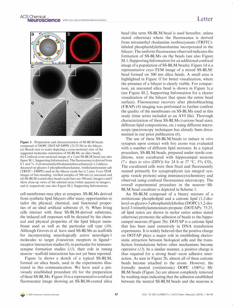
Figure 1a shows a sketch of a typical SS-BLM,formed on silica beads, used in the experiments illus-trated in this communication. We have used a pre-viously established procedure (6) for the preparationof these SS-BLMs.Figure 1b is a representative confocalfluorescence image showing an SS-BLM-coated silica
bead (the term SS-BLM/bead is used hereafter, unlessstated otherwise) where the fluorescence is derivedfrom tetramethyl rhodamine isothiocyanate (TRITC)-labeled phosphatidylethanolamine incorporated in thebilayer. Theuniform fluorescence observed indicates theformation of SS-BLMs on the beads (see also FigureSI.1, Supporting Information for anadditional confocalimage of a population of SS-BLM/beads). Figure 1d is arepresentative cryo-TEM image of a mixed SS-BLM/bead formed on 500 nm silica beads. A small area ishighlighted in Figure 1f for better visualization, wherethe presence of a bilayer is clearly visible. For compar-ison, an uncoated silica bead is shown in Figure 1c,e(see Figure SI.2, Supporting Information for a clearervisualization of the bilayer that spans the entire beadsurface). Fluorescence recovery after photobleaching(FRAP) (9) imaging was performed to further confirmthe quality of the membranes on SS-BLMs used in thisstudy (time series included as an AVI file). Thoroughcharacterization of these SS-BLMs (various bead sizes;different lipid compositions, etc.) using different micro-scopy/spectroscopy techniques has already been docu-mented in our prior publication (6).
The use of these SS-BLM/beads to induce in vitrosynapses upon contact with live axons was evaluatedwith a number of different lipid mixtures. In a typicalprocedure, SS-BLM/beads, prepared under sterile con-ditions, were cocultured with hippocampal neurons(7þ days in vitro (DIV)) for 24 h at 37 �C, 5% CO2.The cocultured cells were then fixed and fluorescentlystained primarily for synaptophysin (an integral syn-aptic vesicle protein) using immunocytochemistry andobserved using confocal fluorescence microscopy. Theoverall experimental procedure in the neuron-SS-BLM/bead coculture is depicted in Scheme 1.
An SS-BLM composed of a binary mixture of azwitterionic phospholipid and a cationic lipid (1,2-dio-leoyl-sn-glycero-3-phosphatidylcholine (DOPC)/1,2-dio-leoyl-3-trimethylammonium-propane (DOTAP); 75:25,all lipid ratios are shown in molar ratios unless statedotherwise) promotes the adhesion of beads to the hippo-campal neurons (Figure 2b). DOTAP is a cationic lipidthat has been used extensively in DNA transfectionexperiments. It is widely believed that the positive chargeon DOTAP plays a major role in establishing electro-static attraction between biological cells and the trans-fection formulations before other mechanisms becomeoperative (12). In a similar manner, a positive charge isthus required for a strong bead-axon adhesive inter-action. As seen in Figure 2b, almost all of these cationicbeads become attached to the axons. However, theformally neutral (zwitterionic) DOPC (100%) SS-BLM/beads (Figure 2a) are almost completely removedby washing steps indicating that the adhesion establishedbetween the neutral SS-BLM/beads and the neurons is
Figure 1. Preparation and characterization of SS-BLM/beadscomposed of DOPC/DOTAP/DPPE (25:25:50) in the bilayer.(a) Sketch (not to scale) depicting a cross-sectional view of thesuggested molecular orientation of SS-BLMs on silica beads.(b) Confocal cross-sectional image of a 5 μm SS-BLM/bead (see alsofigure SI.1, Supporting Information). The fluorescence is derived from0.1 mol % N-(6-tetramethylrhodaminethiocarbamoyl)-1,2-dihexa-decanoyl-sn-glycero-3-phosphoethanolamine, triethylammonium salt(TRITC-DHPE) used in the bilayer (scale bar is 2 μm). Cryo-TEMimages of free-standing, vitrified samples of 500 nm (c) uncoated and(d) SS-BLM-coated silica beads (scale bars are 100 nm). Images e and fshow close-up views of the selected areas (white squares) in images cand d, respectively (see also Figure SI.2, Supporting Information).
r 2009 American Chemical Society 88 DOI: 10.1021/cn900011n |ACS Chem. Neurosci. (2010), 1, 86–94
pubs.acs.org/acschemicalneuroscience Letter
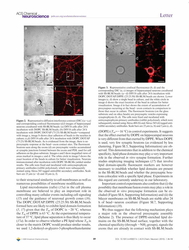
weak or negligible (10b). In both cases, however, no signifi-cant neurite responses are observed (Figures 2d,f). Asdescribed later, only when adhesion occurs do otherfunctionalities associated with the lipid bilayer induceany neural responses with an axonal membrane.
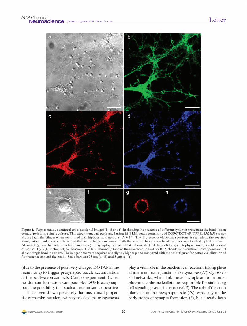
Polylysine adsorbed onto latex beads also exerts pre-synaptic vesicle recruitment at the points of contact (3, 4).In addition to its known capability to effect strongadhesion with cell membranes, PDL-induced neuriteresponses are likely through some formof chemoselectiveprocess via the primary amine moieties. We thus intro-duced primary amines into the SS-BLMvia a phosphati-dylethanolamine lipid (PE lipid). Little effect was notedwhen a ternary lipid mixture (DOPC/DOTAP/(1,2-di-palmitoyl-sn-glycero-3-phosphoethanolamine (DPPE);50:25:25) was used in neuronal culture (data notshown) in place of the 75:25 DOPC/DOTAP SS-BLM(Figure 2d). However, when the DPPE concentrationwas increased to 50 mol % in the ternary lipid mixture(DOPC/DOTAP/DPPE; 25:25:50; Figures 1d,f and 3)the beads are effective in the recruitment of presynapticvesicles at the bead-axon contacts. Most of the beads inthe cell culture are positive for synaptophysin labeling(Figure 3), indicating that accumulation of presynapticvesicles has occurred at the point of contact between theaxons and the SS-BLM/beads.
In order to track the presence of other impor-tant synaptic proteins at the bead-axon contacts, weperformed a triple staining of our SS-BLM/bead-hippocampal neuron coculture. Figure 4 clearly showsthat actin filaments (13) (panels b and f), which areimportant cytoskeletal components involved in manysynaptic processes including signal transduction andsynaptic vesicle trafficking, and bassoon (14) (panels dand h), an important scaffolding protein present atpresynaptic active zones, are present at bead-axon
contacts in addition to synaptophysin (Figures 3 and4c,g). This was observed only in the case of SS-BLM/beads having a membrane composition of DOPC/DOTAP/DPPE (25:25:50). Control experiments per-formed using SS-BLM/beads having a membranecomposition of DOPC/DOTAP (75:25) were negativefor all three of these synaptic proteins (Figure SI.3,Supporting Information) when cocultured with hip-pocampal neurons. Immunostaining of PSD-95, apostsynaptic scaffolding protein, has been performedto verify whether postsynaptic factors were present(Figure SI.4, Supporting Information). The resultimplies, as in our prior work (3), that exclusive for-mation of postsynaptic terminals at the bead-axoncontacts is absent. These observations are in goodagreement with our earlier report that PDL-coatedlatex beads could trigger the formation of func-tional presynaptic endings at the bead-axon contacts(3). The presence of DPPE is clearly playing a rolein the promotion of actin cytoskeletal networkingat the bead-axon junctions and possibly in the sub-sequent recruitment of other synaptic proteins such assynaptophysin and bassoon. This is intriguing and isthe subject of our ongoing study in the further under-standing of the chemical/mechanical interplay of sy-naptic membranes at in vitro synaptic junctions.
The observation of presynaptic vesicle clustering atthe bead site is very promising especially given theongoing attempts to designmore suitable and patternedsubstrates for in vitro neuroengineering applications.To this end, a better understanding of the underlyingmolecular mechanisms of synapse formation (14) iscrucial, especially because recent observations suggestthat even in the absence of postsynaptic factors, func-tional presynapses can be formed onto tailored sub-strates (3). SS-BLMs are clearly a viable substrate due
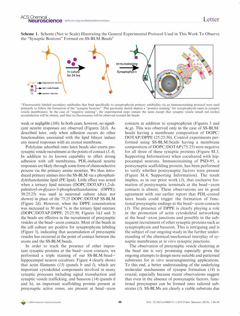
Scheme 1. Scheme (Not to Scale) Illustrating the General Experimental Protocol Used in This Work To Observethe “Synaptic Boutons” Formed on SS-BLM/Beadsa
aFluorescently labeled secondary antibodies that bind specifically to synaptophysin primary antibodies via an immunostaining protocol were usedprimarily to follow the formation of the “synaptic boutons”. This particular sketch depicts a “positive staining” for synaptophysin (seen in synapticvesicle membranes). In the case of “negative staining”, the experimental steps remain the same except that synaptic vesicle (small red circles)accumulation will be absent, and thus no fluorescence will be observed around the beads.
r 2009 American Chemical Society 89 DOI: 10.1021/cn900011n |ACS Chem. Neurosci. (2010), 1, 86–94
pubs.acs.org/acschemicalneuroscience Letter
to their structural similarity to cellmembranes as well asnumerous possibilities of membrane modification.
Lipid microdomains (rafts) (15a) in the cell plasmamembrane are believed to play an important role incontrolling many cellular events including cell signaling(15) and the guidance of neuronal growth cones (16).The DOPC/DOTAP/DPPE (25:25:50) SS-BLM/beadsformed here are likely to exhibit lipid domain formation(5, 17) given that the Tm of DOPC is -20 �C, whereasthe Tm of DPPE is 63 �C. At the experimental tempera-ture of 37 �C, lipid phase separation is thus likely to occur(17a). In order to observe whether a PE lipid whose Tm iscloser to thematrixDOPCwould produce similar results,we used 1,2-dioleoyl-sn-glycero-3-phosphoethanolamine
(DOPE) (Tm=-16 �C) in control experiments. It suggeststhat the effect exerted byDOPEonhippocampal neuronsis verydifferent from that exertedbyDPPE.WhenDOPEis used, very few synaptic boutons (as evidenced by lessclustering, Figure SI.5, Supporting Information) are ob-served. This demonstrates that in addition to the chemicalspecificity, lipidphasedomainsmayplayavery importantrole in the observed in vitro synapse formation. Furtherstudies employing imaging techniques (17) that involvelipid domain-specific fluorescent markers are howevernecessary to establish whether lipid domains are presentin the SS-BLM/beads and whether the presynaptic bou-tons colocalize with a specific lipid phase. Experiments inthis regard are currently underway in our laboratory.
Important control experiments also confirm that (i) thepossibility thatmembrane fusion eventsmayplay a role inthe observed in vitro presynapse formation can be ex-cluded (Figure SI.6, Supporting Information) and (ii) thebilayer membranes on SS-BLM/beads are stable after 24h of bead-neuron coculture (Figure SI.7, SupportingInformation) (18).
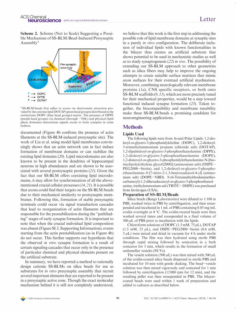
We thus suggest that the following mechanism playsa major role in the observed presynaptic assembly(Scheme 2). The presence of DPPE-enriched lipid do-mains on the SS-BLM/bead surface, together with thechemical specificity (through-NH2 groups), signals theaxons that are already in contact with SS-BLM/beads
Figure 3. Representative confocal fluorescence (b, d) and thecorresponding DIC (a, c) images of hippocampal neurons coculturedwith SS-BLM/beads: (a-d) DIV18 cells after 24 h incubation withDOPC/DOTAP/DPPE (25:25:50) SS-BLM/beads are shown. Lowerimages (c, d) show a single bead in culture, and the white circle inimage d shows the exact location of the bead in culture for bettervisualization. Image d in fact shows the extent of accumulation ofpresynapses occurring at the bead-axon contacts in comparison tothose that occur in culture. The fluorescent boutons (on the glasssubstrate and on silica beads) are presynaptic vesicles expressingsynaptophysin (b, d). The cells were fixed and incubated withantisynaptophysin primary antibodies (rabbit polyclonal), which weresubsequently stainedusingAlexa-488 (b) andAlexa-543 (d) tagged anti-rabbit secondary antibodies. Scale bars are 25 μm (a, b) and 5 μm (c, d).
Figure 2. Representative diffusion interference contrast (DIC) (a-c,e)and corresponding confocal fluorescence (d,f) images of hippocampalneurons cocultured with SS-BLM/beads: (a) DIV16 cells after 24 hincubation with DOPC SS-BLM/beads; (b) DIV18 cells after 24 hincubation with DOPC/DOTAP (75:25) SS-BLM/beads;comparedwith image a, image b shows clear adhesion of beads to the neurites inculture; (c,d) DIV14 cells after 24 h incubation with DOPC/DOTAP(75:25) SS-BLM/beads. As is evidenced from the image, there is nopresynaptic response at the bead-axon contact sites. The fluorescentboutons seen along the axons (d) are presynaptic vesicles accumulatedat synaptic junctions formed between the axons and PDL used for celladhesionon the glass substrate. Images e and f showmagnified views ofareas marked in images c and d. The white circle in image f shows theexact location of the beads in culture for better visualization. Neuronsimmunostained after incubation with DOPC SS-BLMs yielded similarresults. The cells were fixed and incubated with antisynaptophysinprimary antibodies (rabbit polyclonal), which were subsequentlystained using Alexa-543 tagged antirabbit secondary antibodies. Scalebars are 25 μm (a-d) and 10 μm (e, f).
r 2009 American Chemical Society 90 DOI: 10.1021/cn900011n |ACS Chem. Neurosci. (2010), 1, 86–94
pubs.acs.org/acschemicalneuroscience Letter
(due to thepresence of positively chargedDOTAP in themembrane) to trigger presynaptic vesicle accumulationat the bead-axon contacts. Control experiments (whenno domain formation was possible; DOPE case) sup-port the possibility that such a mechanism is operative.
It has been shown previously that mechanical proper-
ties ofmembranes alongwith cytoskeletal rearrangements
play a vital role in the biochemical reactions taking placeat intermembrane junctions like synapses (11). Cytoskel-etal networks, which link the cell cytoplasm to the outerplasma membrane leaflet, are responsible for stabilizingcell signaling events in neurons (13). The role of the actinfilaments at the presynaptic site (19), especially at theearly stages of synapse formation (3), has already been
Figure 4. Representative confocal cross-sectional images (b-d and f-h) showing the presence of different synaptic proteins at the bead-axoncontact points in a single culture. This experiment was performed using SS-BLM/beads containing of DOPC/DOTAP/DPPE, 25:25:50 (as perFigure 3), in the bilayer when cocultured with hippocampal neurons (DIV 14). The fluorescence clustering (boutons) is seen along the neuritesalong with an enhanced clustering on the beads that are in contact with the axons. The cells are fixed and incubated with (b) phalloidin-Alexa-488 (green channel) for actin filaments, (c) antisynaptophysin/R-rabbit-Alexa-543 (red channel) for synaptophysin, and (d) antibassoon/R-mouse-Cy-5 (blue channel) for bassoon. TheDIC channel (a) shows the exact locations of SS-BLM/beads in the culture. Lower panels (e-f)show a single bead in culture. The images herewere acquired at a slightly higher plane comparedwith the other figures for better visualization offluorescence around the beads. Scale bars are 25 μm (a-d) and 5 μm (e-h).
r 2009 American Chemical Society 91 DOI: 10.1021/cn900011n |ACS Chem. Neurosci. (2010), 1, 86–94
pubs.acs.org/acschemicalneuroscience Letter
documented (Figure 4b confirms the presence of actinfilaments at the SS-BLM-induced presynaptic site). Thework of Liu et al. using model lipid membranes convin-cingly shows that an actin network can in fact induceformation of membrane domains or can stabilize theexisting lipid domains (20). Lipid microdomains are alsoknown to be present in the dendrites of hippocampalneurons in high abundances and are shown to be asso-ciated with several postsynaptic proteins (21). Given thefact that our SS-BLM offers coexisting lipid microdo-mains, it may allow for synchronization of all the above-mentioned crucial cellular processes (14, 21). It is possiblethat axons could find their targets on the SS-BLM/beadsdue to their mechanical similarity to postsynaptic mem-branes. Following this, formation of stable presynapticterminals could occur via signal transduction cascadesthat lead to reorganization of actin filaments that areresponsible for the prestabilization during the “pathfind-ing” stages of early synapse formation. It is important tonote that when the crucial individual lipid compositionwas absent (Figure SI.3, Supporting Information), eventsstarting from the actin prestabilization (as in Figure 4b)do not occur. This further supports our hypothesis thatthe observed in vitro synapse formation is a result ofcertain signaling cascades that occur only in the presenceof particular chemical and physical elements present onthe artificial substrate.
In summary, we have reported a method to rationallydesign cationic SS-BLMs on silica beads for use assubstrates for in vitro presynaptic assembly that recruitseveral important elements that are reported tobepresentin a presynaptic active zone. Though the exact molecularmechanism behind it is still not completely understood,
we believe that this work is the first step in addressing thepossible role of lipidmembrane domains at synaptic sitesin a purely in vitro configuration. The deliberate inclu-sion of individual lipids with known functionalities inthe bilayer thus creates an artificial substrate thatshows potential to be used in mechanistic studies as wellas to study synaptogenesis (22) in vivo. The possibility ofextending our SS-BLM approach to other geometriessuch as silica fibers may help to improve the ongoingattempts to create suitable surface matrices that mimicaxon surfaces for their eventual artificial myelination.Moreover, combining neurologically relevantmembraneproteins (1a), CNS specific receptors, or both ontoSS-BLMscaffolds (9,11),whicharemoreprecisely tunedfor their mechanical properties, would be a step towardfunctional induced synapse formation (23). Taken to-gether, the biocompatibility and membrane tunabilitymake these SS-BLM/beads a promising candidate forneuroengineering applications.
Methods
Lipids UsedThe following lipids were from Avanti Polar Lipids: 1,2-dio-
leoyl-sn-glycero-3-phosphatidylcholine (DOPC), 1,2-dioleoyl-3-trimethylammonium propane (chloride salt) (DOTAP),1,2-dipalmitoyl-sn-glycero-3-phosphoethanolamine (DPPE),1,2-dioleoyl-sn-glycero-3-phosphoethanolamine (DOPE),1,2-disteroyl-sn-glycero-3-phosphatidylethanolamine-N-[bio-tinyl(poly(ethylene glycol)2000)] (ammonium salt) (DSPE-PEG2000-biotin), and 1,2-dioleoyl-sn-glycero-3-phospho-ethanolamine-N-[7-nitro-2-1,3-benzoxadiazol-4-yl] (ammo-nium salt) (DOPE-NBD). N-(6-Tetramethylrhodaminethio-carbamoyl)-1,2-dihexadecanoyl-sn-glycero-3-phosphoethanol-amine, triethylammoniumsalt (TRITC-DHPE)waspurchasedfrom Invitrogen (USA).
Preparation of SS-BLM/BeadsSilica beads (Bangs Laboratories) were diluted to 1:100 in
PBS, washed twice in PBS by centrifugation, and then resus-pended and incubated in 1mL of PBS containing 0.05mg/mLavidin overnight at 4 �C. The avidin-treated beads were thenwashed several times and resuspended in a final volume of500 μL of PBS prior to incubation with the lipids.
Chloroform solutions ofDOPC (1.5mM, 75 μL), DOTAP(1.5 mM, 25 μL), and DSPE-PEG2000-biotin (0.6 mM,5 μL) were mixed and dried in vacuum for 4 h under sterileconditions. The film was then hydrated using sterile PBSthrough rapid mixing followed by sonication in a bathsonicator for 3 min, which results in the formation of smallunilamellar vesicles (SUVs).
The vesicle solution (500 μL) was then mixed with 500 μLof the avidin-coated silica beads dispersed in sterile PBS andincubated for 10 min with gentle shaking. The bead-vesiclesolution was then mixed vigorously and sonicated for 1 minfollowed by centrifugation (12 000 rpm for 12 min), and theresulting pellet was then resuspended in PBS. The bilayer-coated beads were used within 1 week of preparation andadded to cultures as described below.
Scheme 2. Scheme (Not to Scale) Suggesting a Possi-bleMechanism of SS-BLM/Bead-Induced PresynapticAssemblya
a SS-BLM/beads first adher to axons via electrostatic attraction pro-videdby the cationic lipidDOTAP (green head groups) distributed in thezwitterionic DOPC (blue head groups) matrix. The presence of DPPE(purple head groups) via chemical (through -NH2) and physical (lipidphase domains) interactions signals axons to form synapses in somefashion.
r 2009 American Chemical Society 92 DOI: 10.1021/cn900011n |ACS Chem. Neurosci. (2010), 1, 86–94
pubs.acs.org/acschemicalneuroscience Letter
Primary Cultures of Rat Hippocampal NeuronsCultures of dissociated rat hippocampal neurons were
prepared using a modification of a protocol described byBanker (25). Hippocampi were dissected from E17/18 em-bryos, treated with 0.25% trypsin at 37 �C followed byDulbecco’s modified Eagle medium (DMEM)-10% horseserum, and mechanically dissociated with a plastic Pasteurpipet. The dissociated neurons were plated at a density of(2.0-2.5) � 104 cm-1 on, unless otherwise stated, poly-D-lysine (Sigma) coated glass coverslips in serum-free neuro-basal medium supplemented with L-glutamine andB-27.One-third of the medium was replaced every 2-3 days. All culturemedia were purchased from Gibco (Invitrogen).
Coculture with Silica BeadsNeurons were cultured to various stages of development
(between 7 and 21DIV) before the addition of beads. The lipidbilayer coated beads were suspended in sterile PBS, pH 7.4,and added dropwise to the neurons at a concentration of (1.0-1.5)�105 beads/coverslip. The bead/cell coculture was incu-bated for 24 h in a humidified 5% CO2 atmosphere at 37 �C.Immunocytochemistry
Cells were fixed with 4% paraformaldehyde in phosphatebuffer (PB), pH7.4, for 25min, incubated in blocking solution(Tris-buffered saline (TBS), pH 7.4, containing 4% normaldonkey serum (NDS) and 0.1%Triton-X100) for 30min, andthen in primary antibodies diluted in TBS containing 0.1%Triton-X100 and 0.5% normal donkey serum (NDS), over-night at 4 �C with gentle shaking. Cells were washed in TBS,incubated in Alexa-488/Alexa-543/Cy-5 (as appropriately)coupled secondary antibodies (in TBS-0.5%, NDS), washed3� in TBS, and mounted on glass slides. Primary antibodiesused were rabbit polyclonal anti-synaptophysin (Invitrogen,predilute; 1:10), mouse monoclonal anti-bassoon (Assay De-signs, Ann Arbor,MI), and all secondary antibodies (species-specific, highly cross-adsorbed IgG) were purchased fromInvitrogen and used at a dilution of 1:200. For actin labeling,Alexa 488-phalloidin (Molecular Probes) was used (1:50) inthe secondary antibody dilution buffer. The stained samples(on glass coverslips) were mounted using GelTol on micro-scopic slides and sealed.
Confocal MicroscopyAll the fluorescence images were obtained using a Zeiss
LSM 510 META confocal microscope (Carl Zeiss AG,Germany) with λex 488 nm/λem LP>505 nm or BP 505-550 IR, λex 543 nm/λem LP>565 nm, and λex 633 nm/λemLP > 685 nm optical combinations appropriately forneuron/bead cultures and lipid bilayers in different experi-ments. The images were obtained with a 63� oil immersionobjective on an inverted microscope along with the corre-sponding brightfield (diffusion interference contrast or DIC)image.
Cryo-TEMFivemicroliters of a solution of silica beads (both uncoated
and lipid coated) was added to copper Quantifoil R2/2 grids(Electron Microscopy Sciences, Hatfield, PA). Samples wereblotted and frozen hydrated by plunging into a bath of liquidethane slush (26). They were stored under liquid nitrogentemperature (-78 �C) until transfer to a 626 single tilt cryo-transfer system (Gatan Inc.) and observed with a FEI G2 F20
cryo-STEM microscope operated at 200 kV (FEI, Inc.).Images were recorded under low-dose conditions on a GatanUltrascan 4k�4k digital (CCD) camera system camera at anominal magnification of 80� at a defocus level of 2 μm.Images of silica beads uncoated and coated with lipids weretaken for direct comparisonunder the exact same conditions ofmagnification and defocus.
Acknowledgment
The authors thank Dr. A. S. Dhaunchak (MNI &H,McGillUniversity) for helpful discussions and Lucas Medwell forassistance in the laboratory.
Supporting Information Available
Additional data including extended figures. This material isavailable free of charge via the Internet at http://pubs.acs.org.
Author Information
Corresponding Author*E-mail: [email protected]; [email protected].
Author Contributions
G.G, D.R.C., and R.B.L designed the experiments. G.G.and I.R. performed the experiments. P.T., A.L.L., andW.B.supported the experiments. G.G., I.R., D.R.C., and R.B.L.analyzed the experimental results. G.G. and R.B.L. wrotethemanuscript, and all the authors contributed to the editingof the manuscript.
Funding Sources
This work is supported by a grant (no.RMF-79028) from theRegenerative Medicine and Nanomedicine Initiative of theCanadian Institutes of Health Research (CIHR) to R.B.LandD.R.C and aCIHRgrant (no.RMF-86693) to I.R. I.R isrecipient of a CIHR New Investigator Award. The McGillProgram in NeuroEngineering is supported by CIHR andtheMinistry of Industry ofCanada (aCentre of Excellence inCommercialization and Research Award to the MNI&H),Rio Tinto Alcan, and The Molson Foundation.
References
1. (a) Shapiro, L., Love, J., and Colman, D. R. (2007)Adhesion molecules in the nervous system: Structural in-sights into function and diversity. Annu. Rev. Neurosci. 30,451–474. (b) Park, D. H., Eve, D. J., Borlongan, C. V., Klasko,S. K., Cruz, L. E., and Sanberg, P. R. (2009) From the basics toapplication of cell therapy, a steppingstone to the conquest ofneurodegeneration: A meeting report. Med. Sci. Monit. 15,RA23–RA31. (c) Agrawal, S., and Schaffer, D. V. (2005) In situstem cell therapy: Novel targets, familiar challenges. TrendsBiotechnol. 23, 78–83.
2. Pluchino, S., Zanotti, L., Brini, E., Ferrari, S., andMartino, G. (2009) Regeneration and repair in multiplesclerosis: The role of cell transplantation. Neurosci. Lett.456, 101–106. Sharp, J., and Keirstead, H. S. (2009) Stem cell-
r 2009 American Chemical Society 93 DOI: 10.1021/cn900011n |ACS Chem. Neurosci. (2010), 1, 86–94
pubs.acs.org/acschemicalneuroscience Letter
based cell replacement strategies for the central nervous system.Neurosci. Lett. 456, 107–111. Tatard, V. M., Menei, P., Benoit,J. P., and Montero-Menei, C. N. (2005) Combining polymericdevices and stem cells for the treatment of neurological disorders:A promising therapeutic approach. Curr. Drug Targets 6, 81–96. Silva, G. A. (2006) Neuroscience nanotechnology: Progress,opportunities and challenges. Nat. Rev. Neurosci. 7, 65–74.Orive, G., Anitua, E., Pedraz, J. L., and Emerich, D. F. (2009)Biomaterials for promoting brain protection, repair and regene-ration. Nat. Rev. Neurosci. 10, 682–692.
3. Lucido,A. L., Sanchez, F. S., Thostrup, P.,Kwiatkowski,A. V., Ortiz, S. L., Gopalakrishnan, G., Liazoghli, D.,Belkaid, W., Lennox, R. B., Grutter, P., Garner, C. C.,and Colman, D. R. (2009) Rapid assembly of functionalpresynaptic boutons triggered by adhesive contacts J. Neurosci.29, 12449-12466.
4. Burry, R. W. (1980) Formation of apparent presynapticelements in response to poly-basic compounds. Brain Res.184, 85–98. Burry, R. W. (1983) Postnatal rat neurons formapparent pre-synaptic elements on polylysine-coated beads invivo. Brain Res. 278, 236–239.
5. Cevc, G., Marsh, D. (1987) Phospholipid Bilayers: Physi-cal Principles and Models, Wiley-VCH, New York. Gennis,R. B. (1989) Biomembranes: Molecular Structure and Func-tion, Springer-Verlag, New York. Mouritsen, O. G. (2005) Life-as a Matter of Fat. The Emerging Science of Lipidomics,Springer Verlag, Heidelberg.
6. Gopalakrishnan, G., Rouiller, I., Colman, D. R., andLennox, R. B. (2009) Supported bilayers formed fromdifferent phospholipids on spherical silica substrates.Langmuir 25, 5455–5458.
7. Troutier, A.-L., and Ladavi�ere, C. (2007) An overviewof lipid membrane supported by colloidal particles. Adv.Colloid Interface Sci. 133, 1–21. Mornet, S., Lambert, O.,Duguet, E., and Brisson, A. (2005) The formation of supportedlipid bilayers on silica nanoparticles revealed by cryoelectronmicroscopy. Nano Lett. 5, 281–285.
8. Terrettaz, S.,Ulrich,W.-P.,Guerrini, R., Verdini, A., andVogel, H. (2001) Immunosensing by a synthetic ligand-gatedion channel. Angew. Chem., Int. Ed. 40, 1740–1743.
9. Baksh,M.M., Dean, C., Pautot, S., DeMaria, S., Isacoff,E., and Groves, J. T. (2005) Neuronal activation by GPI-linked neuroligin-1 displayed in synthetic lipid bilayer mem-branes. Langmuir 21, 10695–10698.
10. (a) Gopalakrishnan, G., Danelon, C., Izewska, P.,Prummer, M., Bolinger, P.-Y., Geissb€uhler, I., Demurtas,D.,Dubochet, J., andVogel,H. (2006)Multifunctional lipid/quantum dot hybrid nanocontainers for controlled targetingof live cells. Angew. Chem., Int. Ed. 45, 5478–5483. (b) Thid,D., Holm, K., Eriksson, P. S., Ekeroth, J., Kasemo, B., and Gold,J. (2008) Supported phospholipid bilayers as a platform forneural progenitor cell culture. J. Biomed. Mater. Res. 84A,940–953.
11. Groves, J. T. (2005) Molecular organization and signaltransduction at intermembrane junctions. Angew. Chem.,Int. Ed. 44, 3524–3538.
12. (a) Felgner, P. L., Gadek, T. R., Holm, M., Roman, R.,Chan, H. W., Wenz, M., Northrop, J. P., Ringold, G. M.,and Danielsen, M. (1987) Lipofection- a highly efficient,
lipid-mediated DNA-transfection procedure. Proc. Natl.Acad. Sci. U.S.A. 84, 7413–7417. (b) Wrobel, I., and Collins,D. (1995) Fusion of cationic liposomes with mammalian-cellsoccurs after endocytosis. Biochim. Biophys. Acta-Biomem-branes 1235, 296–304. (c) Simberg, D., Weisman, S., Talmon,Y., and Barenholz, Y. (2004) DOTAP (and other cationic lipids):Chemistry, biophysics, and transfection. Crit. Rev. Ther. DrugCarrier Syst. 21, 257–317.
13. Janmey, P. A. (1998) The cytoskeleton and cell signaling:component localization andmechanical coupling.Physiol. Rev.78, 763–781. Sobue, K., and Kanda, K. (1989) Alpha-actinins,calspectin (brain spectrin or fodrin), and actin participate in adhe-sion and movement of growth cones.Neuron 3, 311–319. Gomez,T. M., and Zheng, J. Q. (2006) The molecular basis for calcium-dependent axon pathfinding. Nat. Rev. Nerosci. 7, 115–125.
14. Ziv, N. E., and Garner, C. C. (2004) Cellular andmolecular mechanisms of presynaptic assembly. Nat. Rev.Neurosci. 5, 385–399.
15. (a) Munro, S. (2003) Lipid rafts: Elusive or illusive? Cell115, 377–388. (b) Foster, L. J., De Hoog, C. L., and Mann, M.(2003) Unbiased quantitative proteomics of lipid rafts revealshigh specificity for signaling factors. Proc. Natl. Acad. Sci.U.S.A. 100, 5813–5818. (c) Pike, L. J. (2003) Lipid rafts:Bringing order to chaos. J. Lipid Res. 44, 655–667. (d) Simons,K., and Toomre, D. (2000) Lipid rafts and signal transduc-tion. Nat. Rev. Mol. Cell. Biol. 1, 31–39. (e) Golub, T.,Wacha, S., and Caroni, P. (2004) Spatial and temporal controlof signaling through lipid rafts. Curr. Opin. Neurobiol. 14,542–550.
16. Wen, Z., andZheng, J. Q. (2006)Directional guidance ofnerve growth cones. Curr. Opin. Neurobiol. 16, 52–58.
17. (a) Bagatolli, L. A., and Gratton, E. (2000) Two photonfluorescence microscopy of coexisting lipid domains ingiant unilamellar vesicles of binary phospholipid mixtures.Biophys. J. 78, 290–305. (b) Korlach, J., Schwille, P., Webb,W. W., and Feigenson, G. W. (1999) Characterization of lipidbilayer phases by confocal microscopy and fluorescence correla-tion spectroscopy. Proc. Natl. Acad. Sci. U.S.A. 96, 8461–8466. (c) Hac, A. E., Seeger, H. M., Fidorra, M., and Heimburg,T. (2005) Diffusion in two-component lipid membranes - afluorescence correlation spectroscopy and monte carlo simula-tion study. Biophys. J. 88, 317–333.
18. In order to further support our suggested mechanismand to exclude other possible mechanisms, we tested whetherany membrane fusion related process plays a role. This isbecause PEG molecules are known to have fusogenic effectswhen present in either of themembranes that are undergoingmembrane fusion (10a, 24). The results imply that SS-BLM/beads made up of the fusogenic DOPC/DOTAP/DPPE-PEG2000 (25:25:50) mixture (10a) were negative for synapto-physin labeling (Figure SI.6, Supporting Information). Thissuggests that membrane fusion events may be excluded. Thoughthe presence of DOTAP together with DPPE could arguably leadto membrane fusion, lipid exchange, and endocytosis mediatedmechanisms (12c), our control experiments suggest that othertypes of mechanisms are more likely operative. This is furthersupported by the presence of intact bilayers on the SS-BLMsafter 24 h of incubation with hippocampal neurons (Figure SI.7,Supporting Information). Being supported on a solid supportcould very well reduce the possibility of fusion/endocytosis ofthe SS-BLM/beads.
r 2009 American Chemical Society 94 DOI: 10.1021/cn900011n |ACS Chem. Neurosci. (2010), 1, 86–94
pubs.acs.org/acschemicalneuroscience Letter
19. Cingolani, L., and Goda, Y. (2008) Nat. Rev. Neurosci.9, 344–356. Dillon, C., and Goda, Y. (2005) Actin in action: Theinterplay between the actin cytoskeleton and synaptic efficacy.Annu. Rev. Neurosci. 28, 25–55. Dent, E., and Gertler, F. B.(2003) The actin cytoskeleton: Integrating form and function atthe synapse.Neuron 40, 209–227. Sankaranarayanan, S., Alturi,P., and Ryan, T. (2003) Actin has a molecular scaffolding, notpropulsive, role in presynaptic function. Nat. Neurosci. 6, 127–135.
20. Liu, A. P., and Fletcher, D. A. (2006) Actin polymeri-zation serves as a membrane domain switch in model lipidbilayers. Biophys. J. 91, 4064–4070.
21. Hering, H., Lin, C.-C., and Sheng, M. (2003) Lipid raftsin the maintenance of synapses, dendritic spines, and surfaceAMPA receptor stability. J. Neurosci. 23, 3262–3271.
22. Benson, D. L., Colman, D. R., and Huntley, G. W.(2001) Molecules, maps and synapse specificity. Nat. Rev.Neurosci. 2, 899–909.
23. Dustin, M. L., and Colman, D. R. (2002) Neural andimmunological synaptic relations. Science 298, 785–789.
24. Lentz, B. R. (1994) Polymer-induced membrane-fusion-potential mechanism and relation to cell-fusion events.Chem. Phys. Lipids 73, 91–106. Hui, S. W., Kuhl, T. L., Guo,Y. Q., and Israelachvili, J. (1999) Use of poly(ethylene glycol) tocontrol cell aggregation and fusion. Colloids Surf., B 14, 213–222.
25. Goslin, K., Asmussen, H., Banker, G. (1998) CulturingNerve Cells (Banker, G., Goslin, K., Ed.), 2nd ed., pp 339-370,MIT-Press, Cambridge, MA.
26. Dubochet, J., Adrian, M., Chang, J. J., Homo, J. C.,Lepault, J., McDowall, A. W., and Schultz, P. (1988) Cryo-electron microscopy of vitrified specimens. Q. Rev. Biophys.21, 129–228.
Top Related
Copyright © 2022 FDOKUMEN

