


Bahasa
Halaman
Hukum

Review began 02/24/2021 Review ended 03/19/2021 Published 03/22/2021
© Copyright 2021Bhaduri et al. This is an open accessarticle distributed under the terms of theCreative Commons Attribution LicenseCC-BY 4.0., which permits unrestricteduse, distribution, and reproduction in anymedium, provided the original author andsource are credited.
Isolated Fracture of the Acromion Process: A CaseReportIndranil Bhaduri , Rajesh Thakur , Sachin Kumar , Manoj K. Rajak
1. Department of Joint Replacement and Orthopedics, Tata Main Hospital, Jamshedpur, IND
Corresponding author: Indranil Bhaduri, [email protected]
AbstractFracture of the acromion process is an uncommon injury which is often diagnosed late. Though, usuallymanaged conservatively, the indications for surgery in these fractures are very specific. A 52-year-old activeman attended the out-patient department of our hospital following an injury to the right shoulder. An X-rayrevealed a Type II, minimally displaced fracture of the base of the acromion process. Conservativemanagement was attempted initially, which was converted to surgical stabilization after six weeks when itwas noticed that the fracture had failed to unite and had progressed to become a displaced Type III fracture.Post-operative period was uneventful with a gradual return to the pre-injury level of function of the rightshoulder, which was assessed by the Constant Score as well as the University of California Los Angeles(UCLA) shoulder score. The satisfaction with the final functional outcome was assessed by the UCLAshoulder score. Clinicians must look actively for acromion process fractures in all shoulder injuries.Minimally displaced fractures should be regularly followed up for displacement and sub-acromial spacecompromise. Although acromion fractures are usually treated conservatively, albeit a higher non-union rate,they should be treated surgically in the event of displacement or sub-acromial space reduction, in order toachieve good functional recovery.
Categories: Orthopedics, TraumaKeywords: surgical case report, acromion process fracture, late surgical intervention, constant score
IntroductionFractures of the acromion process of the scapula are extremely rare, comprising only 3%-5% of all shoulderinjuries and about 7%-8% of scapular fractures [1]. This fracture has seen a renewed interest amongorthopedic trauma surgeons in recent times [2]. This has been mainly because of the functional compromisethese injuries cause due to the shoulder impingement concomitant to a reduced sub-acromion space seen inuntreated displaced fractures [3-6]. Also, the propensity of undisplaced acromion process fractures todisplace, over time, due to the weight of the suspended upper limb might result in sub-acromion spacecompromise [7]. This case report was an attempt to present the functional outcome of operative treatmentof an isolated minimally displaced acromion process fracture following 16 months of follow up after failureof attempted conservative management for six weeks.
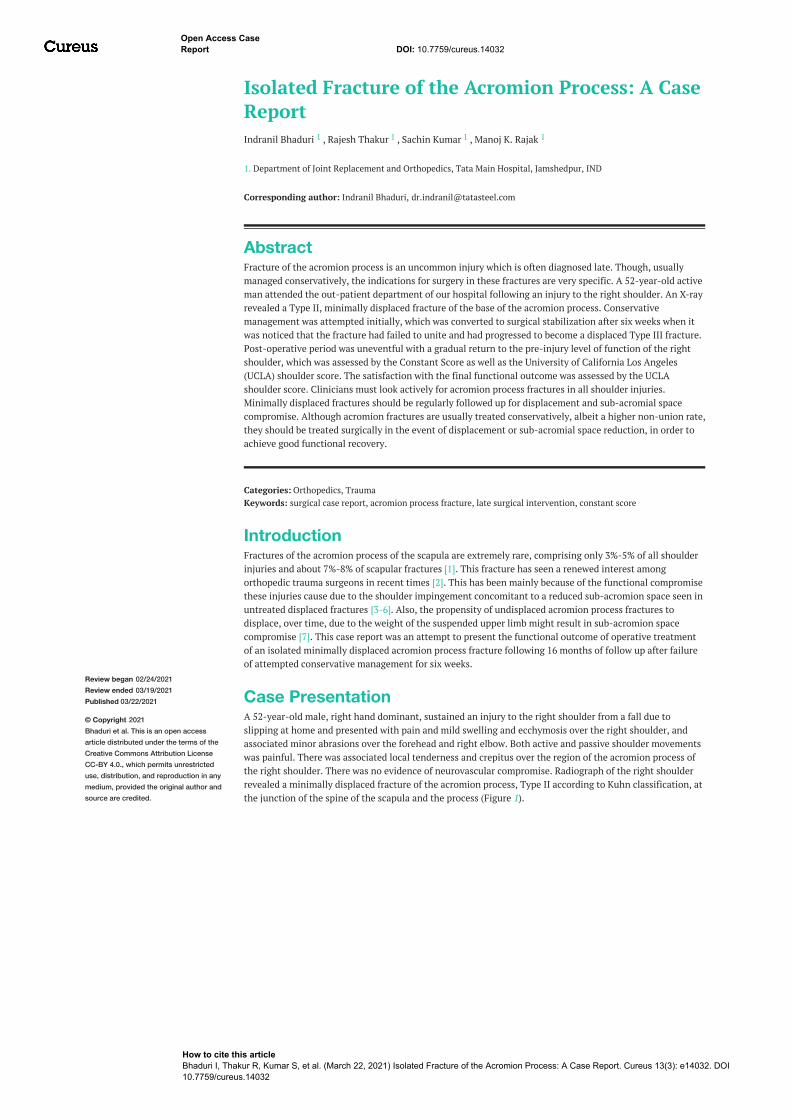
Case PresentationA 52-year-old male, right hand dominant, sustained an injury to the right shoulder from a fall due toslipping at home and presented with pain and mild swelling and ecchymosis over the right shoulder, andassociated minor abrasions over the forehead and right elbow. Both active and passive shoulder movementswas painful. There was associated local tenderness and crepitus over the region of the acromion process ofthe right shoulder. There was no evidence of neurovascular compromise. Radiograph of the right shoulderrevealed a minimally displaced fracture of the acromion process, Type II according to Kuhn classification, atthe junction of the spine of the scapula and the process (Figure 1).
1 1 1 1
Open Access CaseReport DOI: 10.7759/cureus.14032
How to cite this articleBhaduri I, Thakur R, Kumar S, et al. (March 22, 2021) Isolated Fracture of the Acromion Process: A Case Report. Cureus 13(3): e14032. DOI10.7759/cureus.14032
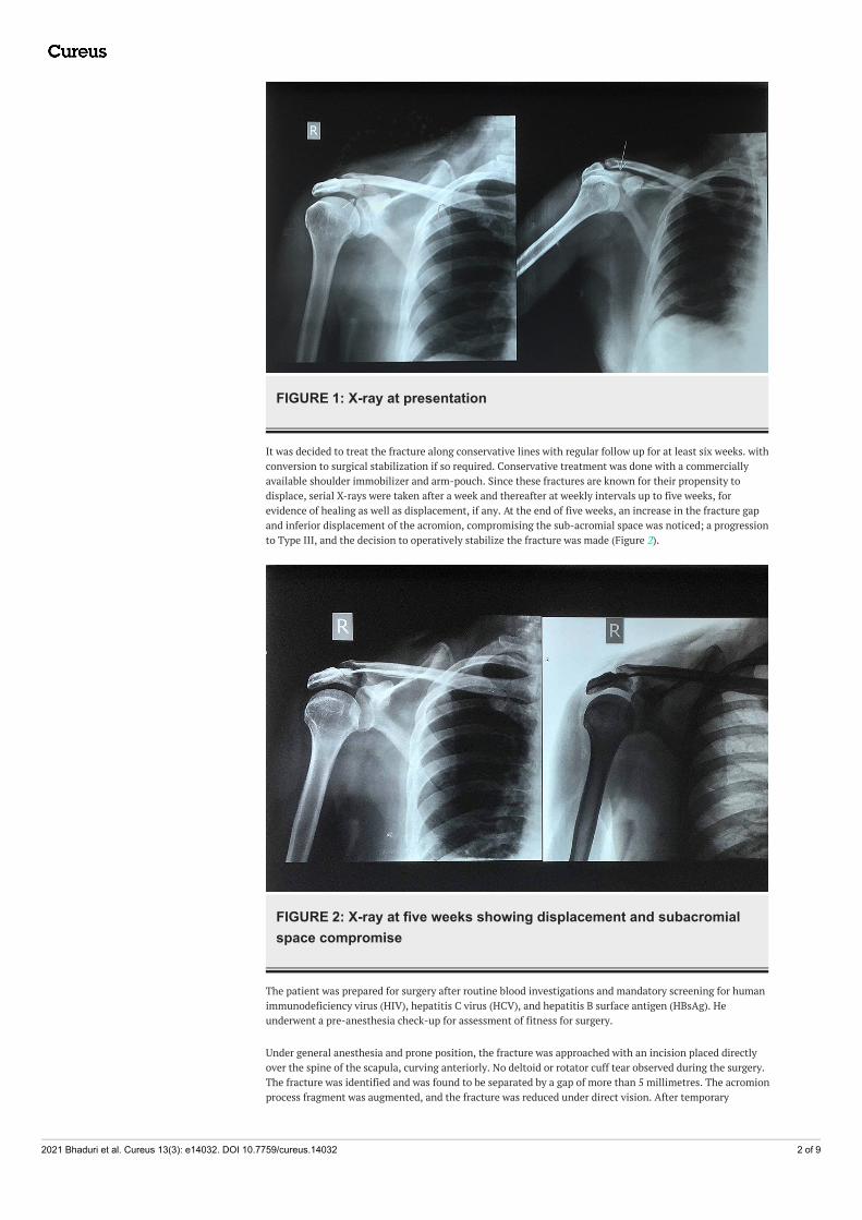
FIGURE 1: X-ray at presentation
It was decided to treat the fracture along conservative lines with regular follow up for at least six weeks. withconversion to surgical stabilization if so required. Conservative treatment was done with a commerciallyavailable shoulder immobilizer and arm-pouch. Since these fractures are known for their propensity todisplace, serial X-rays were taken after a week and thereafter at weekly intervals up to five weeks, forevidence of healing as well as displacement, if any. At the end of five weeks, an increase in the fracture gapand inferior displacement of the acromion, compromising the sub-acromial space was noticed; a progressionto Type III, and the decision to operatively stabilize the fracture was made (Figure 2).
FIGURE 2: X-ray at five weeks showing displacement and subacromialspace compromise
The patient was prepared for surgery after routine blood investigations and mandatory screening for humanimmunodeficiency virus (HIV), hepatitis C virus (HCV), and hepatitis B surface antigen (HBsAg). Heunderwent a pre-anesthesia check-up for assessment of fitness for surgery.
Under general anesthesia and prone position, the fracture was approached with an incision placed directlyover the spine of the scapula, curving anteriorly. No deltoid or rotator cuff tear observed during the surgery.The fracture was identified and was found to be separated by a gap of more than 5 millimetres. The acromionprocess fragment was augmented, and the fracture was reduced under direct vision. After temporary
2021 Bhaduri et al. Cureus 13(3): e14032. DOI 10.7759/cureus.14032 2 of 9
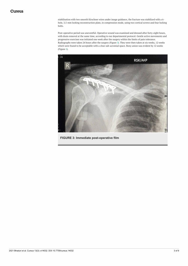
stabilisation with two smooth Kirschner wires under image guidance, the fracture was stabilized with a 6-hole, 3.5-mm locking reconstruction plate, in compression mode, using two cortical screws and four lockingbolts.
Post-operative period was uneventful. Operative wound was examined and dressed after forty-eight hours,with drain removal at the same time, according to our departmental protocol. Gentle active movements andprogressive exercises was initiated one week after the surgery within the limits of pain tolerance.Radiographs were taken 24 hours after the surgery (Figure 3). They were then taken at six weeks, 12 weekswhich were found to be acceptable with a clear sub-acromial space. Bony union was evident by 12 weeks(Figure 4).
FIGURE 3: Immediate post-operative film
2021 Bhaduri et al. Cureus 13(3): e14032. DOI 10.7759/cureus.14032 3 of 9
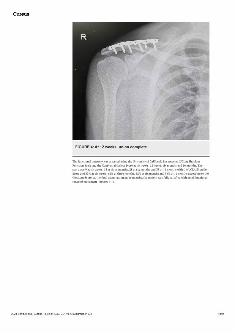
FIGURE 4: At 12 weeks; union complete
The functional outcome was assessed using the University of California Los Angeles (UCLA) ShoulderFunction Scale and the Constant (Murley) Score at six weeks, 12 weeks, six months and 16 months. Thescore was 9 at six weeks, 15 at three months, 28 at six months and 33 at 16 months with the UCLA ShoulderScore and 33% at six weeks, 62% at three months, 82% at six months and 98% at 16 months according to theConstant Score. At the final examination, at 16 months, the patient was fully satisfied with good functionalrange of movement (Figures 5-7).
2021 Bhaduri et al. Cureus 13(3): e14032. DOI 10.7759/cureus.14032 4 of 9
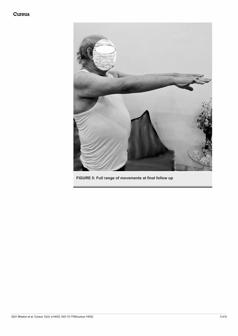
FIGURE 5: Full range of movements at final follow up
2021 Bhaduri et al. Cureus 13(3): e14032. DOI 10.7759/cureus.14032 5 of 9
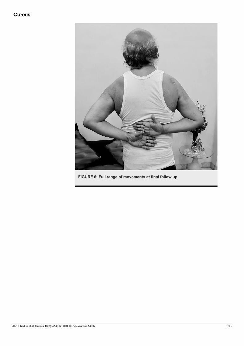
FIGURE 6: Full range of movements at final follow up
2021 Bhaduri et al. Cureus 13(3): e14032. DOI 10.7759/cureus.14032 6 of 9
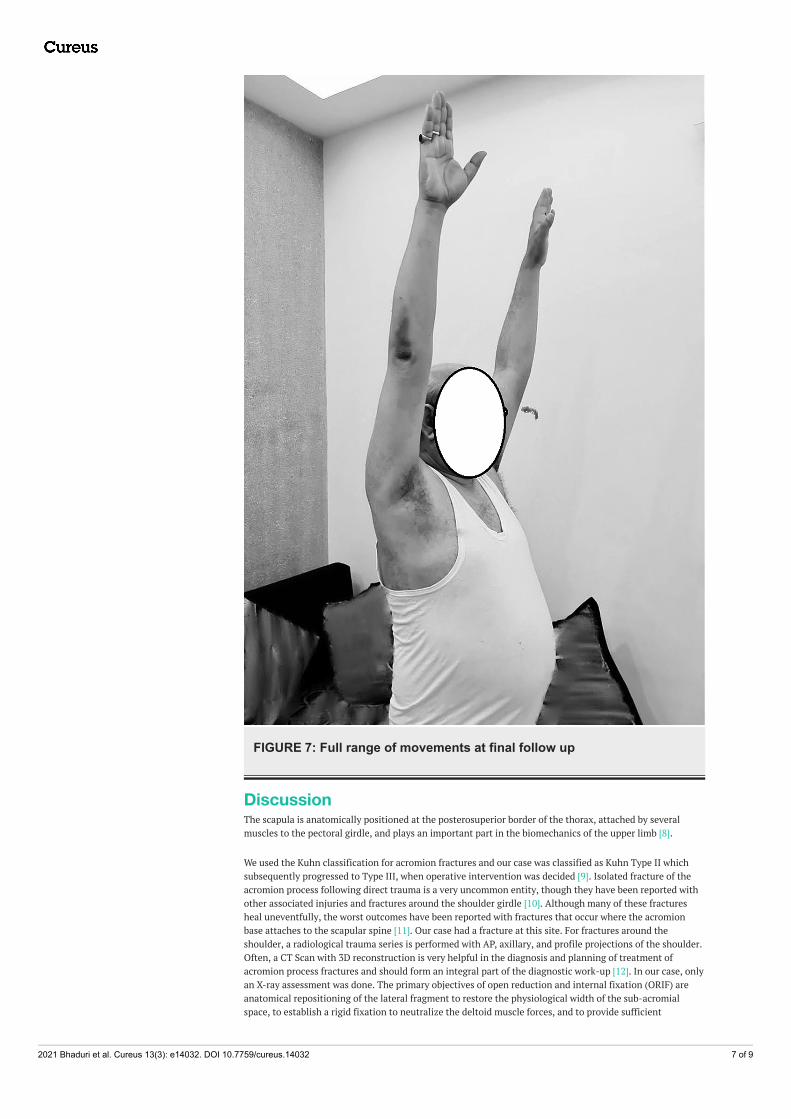
FIGURE 7: Full range of movements at final follow up
DiscussionThe scapula is anatomically positioned at the posterosuperior border of the thorax, attached by severalmuscles to the pectoral girdle, and plays an important part in the biomechanics of the upper limb [8].
We used the Kuhn classification for acromion fractures and our case was classified as Kuhn Type II whichsubsequently progressed to Type III, when operative intervention was decided [9]. Isolated fracture of theacromion process following direct trauma is a very uncommon entity, though they have been reported withother associated injuries and fractures around the shoulder girdle [10]. Although many of these fracturesheal uneventfully, the worst outcomes have been reported with fractures that occur where the acromionbase attaches to the scapular spine [11]. Our case had a fracture at this site. For fractures around theshoulder, a radiological trauma series is performed with AP, axillary, and profile projections of the shoulder.Often, a CT Scan with 3D reconstruction is very helpful in the diagnosis and planning of treatment ofacromion process fractures and should form an integral part of the diagnostic work-up [12]. In our case, onlyan X-ray assessment was done. The primary objectives of open reduction and internal fixation (ORIF) areanatomical repositioning of the lateral fragment to restore the physiological width of the sub-acromialspace, to establish a rigid fixation to neutralize the deltoid muscle forces, and to provide sufficient
2021 Bhaduri et al. Cureus 13(3): e14032. DOI 10.7759/cureus.14032 7 of 9
compression on the fracture for proper bone healing.
Kuhn has advocated surgical stabilization of Type III fractures that compromise the sub-acromial space,symptomatic stress fractures, and painful non-unions [9]. Depending on age, activity, and general conditionof the patient, internal fixation is recommended in grossly displaced fractures of the acromion and coracoidprocess as concluded by Bauer et al. [13]. Hess et al. also concluded in their study that patientcharacteristics, such as activity level, might be a relevant parameter when selecting a treatment strategy.Early fixation may be the most sensible way to treat working adults who need to avoid long absences fromwork [14]. Our patient was an active middle-aged man with field job, so we decided to operate after givingconservative treatment a fair trial. Hill and his co-workers advocated surgical intervention for sub-acromialimpingement, symptomatic non-unions, open fractures, displacement of more than 1 cm, and disruption ofthe superior shoulder suspensory complex [15]. Acromion fractures, based on fracture configuration, havebeen surgically treated with implants ranging from cancellous screws [7,15], narrow 3.5 mm dynamiccompression plates and cortical screws [15], and locking plates and screws [16] plain or threaded Kirschnerwires [17,18] and tension band [19]. Fixation with K-wires is not recommended because it may cause earlyimplant failure and stable reconstruction may not be achieved after surgery [15]. Although acromionfractures have been fixed with pre-bent clavicle reconstruction plates [18], we decided to stabilize thefracture with a 3.5 mm locked reconstruction plate in compression mode to obtain a stable construct. We didnot observe any complications related to either the operative procedure or the implants used, in our case.Postoperative recovery was uneventful. Functional recovery was assessed using the UCLA Shoulder FunctionScale and the Constant (Murley) Score and compared to tabulations at the end of each follow up to recordimprovement.
ConclusionsA high degree of awareness must be exhibited by the clinician while evaluating a patient with shouldertrauma, who should be carefully examined for possible scapular process fractures. Although fractures of theacromion process are commonly treated conservatively, surgery should be offered to patients who showfeatures of impingement and radiological evidence of compromise of the sub-acromial clear space. The non-union rate with conservative treatment, although relatively high, is often not overtly painful nor is itlimiting to reasonable shoulder function, especially in the elderly or less active patients. Surgicalstabilization appears to be a more suitable option of treatment for physically active patients who are morelikely to present with symptomatic non-unions. In this paper, we report an uncommon case of an isolatedtraumatic acromion process fracture which became displaced during the course of conservative managementand the treatment strategy had to be revised to surgical stabilization. Despite the delay in surgicalstabilization, the functional outcome was good.
Additional InformationDisclosuresHuman subjects: Consent was obtained or waived by all participants in this study. Conflicts of interest: Incompliance with the ICMJE uniform disclosure form, all authors declare the following: Payment/servicesinfo: All authors have declared that no financial support was received from any organization for thesubmitted work. Financial relationships: All authors have declared that they have no financialrelationships at present or within the previous three years with any organizations that might have aninterest in the submitted work. Other relationships: All authors have declared that there are no otherrelationships or activities that could appear to have influenced the submitted work.
References1. Bartonicek J: Scapular fractures. Rockwood and Green’s Fractures in Adults, 8th Edition. Court-Brown CM,
Hickman J (ed): Wolters Kluwer, Netherlands; 2015. 1:1478.2. Cole PA, Shafiq B: Scapula fractures: open reduction internal fixation. Master Techniques in Orthopaedic
Surgery. Wiss DA (ed): Lippincott Williams & Wilkins, Philadelphia; 2006. 1:15-36.3. Ada JR, Miller ME: Scapular fractures. Analysis of 113 cases . Clin Orthop Relat Res. 1991, 269:174-180.4. Nordquist A, Peterson C: Fracture of the body, neck, or spine of the scapula. A long-term follow-up study .
Clin Orthop Relat Res. 1992, 283:139-144.5. Ganger EM, Ludwig PM, Wijdecks CA, Cole PA: Pre- and postoperative function after scapula malunion
reconstruction: a novel kinematic technique. J Orthop Trauma. 2013, 27:e186-e191.10.1097/BOT.0b013e318271b8e2
6. Cole PA, Talbot M, Schirmer J, Schroder LK, Anavian J: Extra-articular malunions of the scapula: acomparison of functional outcome before and after reconstruction. J Orthop Trauma. 2011, 25:649-656.10.1097/BOT.0b013e31820af67f
7. Kim DS, Yoon YS, Kang DH: Comparison of early fixation and delayed reconstruction after displacement inpreviously nondisplaced acromion fractures. Orthopaedics. 2010, 33:392. 10.3928/01477447-20100429-11
8. Chaurasia BD: B D Chaurasia's Human Anatomy Regional and Applied: Upper Limb & Thorax, 8th Edition .CBS Publishers and Distributors, New Delhi; 2019.
9. Kuhn JE, Blasier RB, Carpenter JE: Fractures of the acromion process: a proposed classification system . JOrthop Trauma. 1994, 8:6-13. 10.1097/00005131-199402000-00002
10. Lantry JM, Roberts CS, Giannoudis PV: Operative treatment of scapular fractures: a systematic review .
2021 Bhaduri et al. Cureus 13(3): e14032. DOI 10.7759/cureus.14032 8 of 9
Injury. 2008, 39:271-283. 10.1016/j.injury.2007.06.01811. Wahlquist TC, Hunt AF, Brahman JP: Acromial base fractures after reverse total shoulder arthroplasty:
report of five cases. J Shoulder Elbow Surg. 2011, 20:1178-1183. 10.1016/j.jse.2011.01.02912. Beckman NM, Sanhaji L, Chinapuvvula NR, West OC: Imaging of traumatic shoulder girdle injuries . Radiol
Clin N Am. 2019, 57:809-822. 10.1016/j.rcl.2019.02.01313. Bauer G, Fleischmann W, Dussler E: Displaced scapular fractures: indication and long-term results of open
reduction and internal fixation. Arch Orthop Trauma Surg. 1995, 114:215-219. 10.1007/BF0044426614. Hess F, Zettl R, Welter J, Smolen D, Knoth C: The traumatic acromion fracture: review of the literature,
clinical examples and proposal of a treatment algorithm. Arch Orthop Trauma Surg. 2019, 139:651-658.15. Hill BW, Anavian J, Jacobson AR, Cole PA: Surgical management of isolated acromion fractures: technical
tricks and clinical experience. J Orthop Trauma. 2014, 28:e107-e113. 10.1097/BOT.000000000000004016. Zhu J, Pan Z, Zheng R, Lan S: Perpendicular double-plate fixation with locking system for acromion pedicle
fracture. Acta Ortop Bras. 2016, 24:107-110. 10.1590/1413-78522016240214169117. Ogawa K, Naniwa T: Fractures of the acromion and the lateral scapular spine . J Shoulder Elbow Surg. 1997,
6:544-548. 10.1016/s1058-2746(97)90087-218. Nasab SAM: Isolated displaced fracture of the acromion: a rare case report and the consequence of
treatment by open reduction and pin fixation. Arch Trauma Res. 2013, 1:184-186. 10.5812/atr.876219. Beliën H, Biesmans H, Steenwerckx A, Bijnens E, Dierickx C: Prebending of osteosynthesis plate using 3D
printed models to treat symptomatic os acromiale and acromial fracture. J Exp Ortop. 2017, 4:34.10.1186/s40634-017-0111-7
2021 Bhaduri et al. Cureus 13(3): e14032. DOI 10.7759/cureus.14032 9 of 9
Top Related
Copyright © 2022 FDOKUMEN

