


Bahasa
Halaman
Hukum

Resource
Genome-wide Analysis of the HostIntracellular Network that RegulatesSurvival of Mycobacterium tuberculosisDhiraj Kumar,1 Lekha Nath,1 Md. Azhar Kamal,1 Ankur Varshney,1 Avinash Jain,1 Sarman Singh,2 and Kanury V.S. Rao1,*1Immunology Group, International Centre for Genetic Engineering and Biotechnology, Aruna Asaf Ali Marg, New Delhi 110067, India2Division of Clinical Microbiology, Department of Laboratory Medicine, All India Institute of Medical Sciences, Ansari Nagar,
New Delhi 110029, India
*Correspondence: [email protected] 10.1016/j.cell.2010.02.012
SUMMARY
We performed a genome-wide siRNA screen to iden-tify host factors that regulated pathogen load inhuman macrophages infected with a virulent strainof Mycobacterium tuberculosis. Iterative rounds ofconfirmation, followed by validation, identified 275such molecules that were all found to functionallyassociate with each other through a dense networkof interactions. This network then yielded to a molec-ular description of the host cell functional modulesthat were both engaged and perturbed by the path-ogen. Importantly, a subscreen against a panel offield isolates revealed that the molecular composi-tion of the host interface varied with both genotypeand the phenotypic properties of the pathogen. Ananalysis of these differences, however, permittedidentification of those host factors that were invari-antly involved, regardless of the diversification inadaptive mechanisms employed by the pathogen.Interestingly, these factors were found to predomi-nantly function through the regulation of autophagy.
INTRODUCTION
Tuberculosis continues to prevail as the major cause of mortality
around the world, despite implementation of control programs
and the availability of effective drugs. The causative agent,
Mycobacterium tuberculosis (Mtb), is an obligate human path-
ogen that primarily targets macrophages. After infection, intra-
cellular mycobacteria are predominantly distributed between
the early and late phagosomal compartments, with some also
escaping into the cytoplasm (van der Wel et al., 2007). Their
survival in the hostile intracellular milieu is facilitated through
the dynamic modulation of a range of cellular processes. These
include inhibition of pathways involved in the fusion of the phag-
osome with lysozomes, antigen presentation, apoptosis, and the
activation of bactericidal responses (Koul et al., 2004). In addi-
tion, manipulation of the host cellular machinery also provides
for access to essential nutrients (Pandey and Sassetti, 2008;
Schaible and Kaufmann, 2005). While this pervasive influence
clearly supports that the pathogen engages the molecular
components of the cell in a broad network of interactions (Young
et al., 2008), the details of this network are yet incomplete. In this
connection, mycobacterial interference with the host signaling
machinery has been extensively studied and pathways that are
attenuated to enable the infection have been defined. The focus
of these investigations, however, has been primarily restricted to
events regulating pathogen entry and subsequent endocytosis
(Koul et al., 2004; Vergne et al., 2004). The larger issue of mech-
anisms that mediate stabilization of the infection has remained
unexplored.
To identify host factors regulating an established infection, we
performed a genome-wide siRNA screen against host proteins in
human cells that were first infected with a virulent strain of Mtb.
Several host factors were identified whose expression levels
proved critical for maintenance of the intracellular pathogen
load. An analysis of these targets with their functional associa-
tions yielded a comprehensive description of the host molecular
interface, and its constituent functional modules, that involved in
crosstalk with the intracellular pathogen. By employing a panel
of diverse field isolates, we also probed how this interface
responded to the broad diversification in mechanisms of patho-
genesis that Mtb exhibits in the human population. Surprisingly,
the host determinants of intracellular pathogen load varied
significantly across the isolates, suggesting that the clade iden-
tity of Mtb has an important bearing on defining its host factor
dependencies. Nonetheless, by integrating the results, we could
extract the obligate—host-specified—survival axis that was
independent of the adaptive variations that the pathogen
exploits in the field. Interestingly, this axis was largely comprised
of members that regulated the process of autophagy.
RESULTS
Identification of Host Proteins that Regulate Mtbin Human MacrophagesWe employed the SMARTPool human whole-genome small
interfering RNA (siRNA) library to target a total of 18,174 genes
as a mixture of four siRNAs per target gene. Human macro-
phage-like THP-1 cells were first infected with H37Rv and then
Cell 140, 731–743, March 5, 2010 ª2010 Elsevier Inc. 731
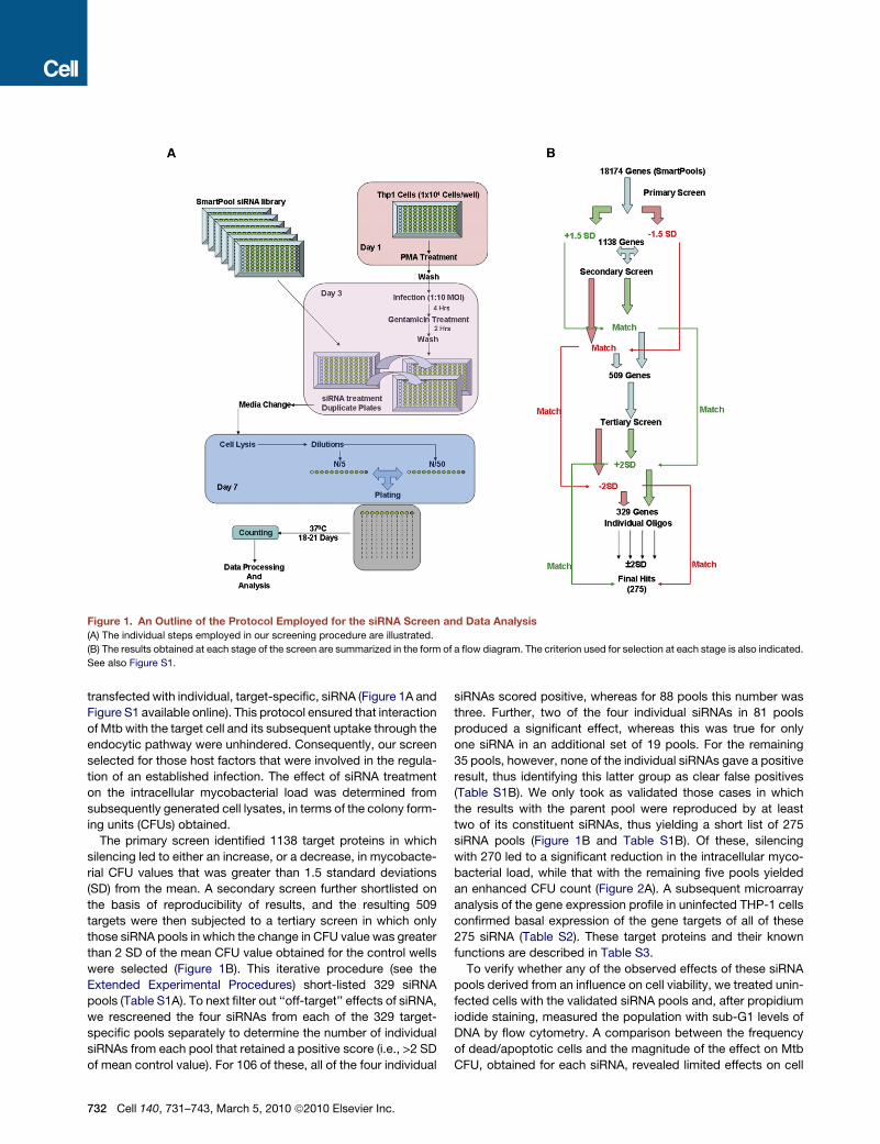
Figure 1. An Outline of the Protocol Employed for the siRNA Screen and Data Analysis
(A) The individual steps employed in our screening procedure are illustrated.
(B) The results obtained at each stage of the screen are summarized in the form of a flow diagram. The criterion used for selection at each stage is also indicated.
See also Figure S1.
transfected with individual, target-specific, siRNA (Figure 1A and
Figure S1 available online). This protocol ensured that interaction
of Mtb with the target cell and its subsequent uptake through the
endocytic pathway were unhindered. Consequently, our screen
selected for those host factors that were involved in the regula-
tion of an established infection. The effect of siRNA treatment
on the intracellular mycobacterial load was determined from
subsequently generated cell lysates, in terms of the colony form-
ing units (CFUs) obtained.
The primary screen identified 1138 target proteins in which
silencing led to either an increase, or a decrease, in mycobacte-
rial CFU values that was greater than 1.5 standard deviations
(SD) from the mean. A secondary screen further shortlisted on
the basis of reproducibility of results, and the resulting 509
targets were then subjected to a tertiary screen in which only
those siRNA pools in which the change in CFU value was greater
than 2 SD of the mean CFU value obtained for the control wells
were selected (Figure 1B). This iterative procedure (see the
Extended Experimental Procedures) short-listed 329 siRNA
pools (Table S1A). To next filter out ‘‘off-target’’ effects of siRNA,
we rescreened the four siRNAs from each of the 329 target-
specific pools separately to determine the number of individual
siRNAs from each pool that retained a positive score (i.e., >2 SD
of mean control value). For 106 of these, all of the four individual
732 Cell 140, 731–743, March 5, 2010 ª2010 Elsevier Inc.
siRNAs scored positive, whereas for 88 pools this number was
three. Further, two of the four individual siRNAs in 81 pools
produced a significant effect, whereas this was true for only
one siRNA in an additional set of 19 pools. For the remaining
35 pools, however, none of the individual siRNAs gave a positive
result, thus identifying this latter group as clear false positives
(Table S1B). We only took as validated those cases in which
the results with the parent pool were reproduced by at least
two of its constituent siRNAs, thus yielding a short list of 275
siRNA pools (Figure 1B and Table S1B). Of these, silencing
with 270 led to a significant reduction in the intracellular myco-
bacterial load, while that with the remaining five pools yielded
an enhanced CFU count (Figure 2A). A subsequent microarray
analysis of the gene expression profile in uninfected THP-1 cells
confirmed basal expression of the gene targets of all of these
275 siRNA (Table S2). These target proteins and their known
functions are described in Table S3.
To verify whether any of the observed effects of these siRNA
pools derived from an influence on cell viability, we treated unin-
fected cells with the validated siRNA pools and, after propidium
iodide staining, measured the population with sub-G1 levels of
DNA by flow cytometry. A comparison between the frequency
of dead/apoptotic cells and the magnitude of the effect on Mtb
CFU, obtained for each siRNA, revealed limited effects on cell
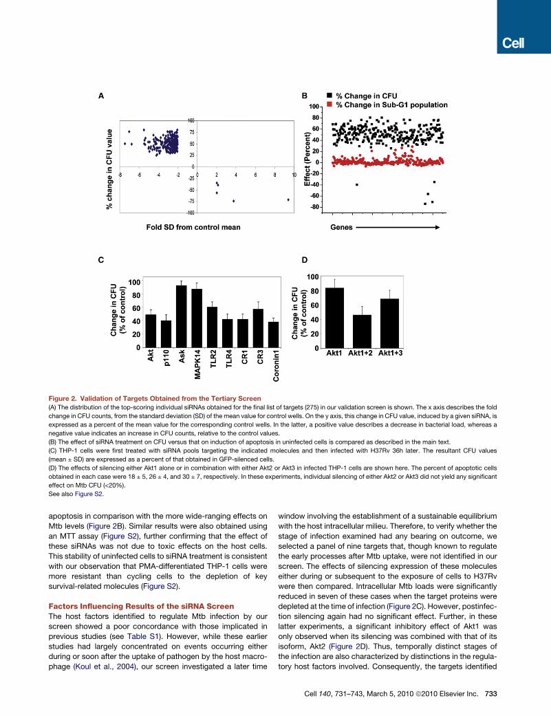
Figure 2. Validation of Targets Obtained from the Tertiary Screen(A) The distribution of the top-scoring individual siRNAs obtained for the final list of targets (275) in our validation screen is shown. The x axis describes the fold
change in CFU counts, from the standard deviation (SD) of the mean value for control wells. On the y axis, this change in CFU value, induced by a given siRNA, is
expressed as a percent of the mean value for the corresponding control wells. In the latter, a positive value describes a decrease in bacterial load, whereas a
negative value indicates an increase in CFU counts, relative to the control values.
(B) The effect of siRNA treatment on CFU versus that on induction of apoptosis in uninfected cells is compared as described in the main text.
(C) THP-1 cells were first treated with siRNA pools targeting the indicated molecules and then infected with H37Rv 36h later. The resultant CFU values
(mean ± SD) are expressed as a percent of that obtained in GFP-silenced cells.
(D) The effects of silencing either Akt1 alone or in combination with either Akt2 or Akt3 in infected THP-1 cells are shown here. The percent of apoptotic cells
obtained in each case were 18 ± 5, 26 ± 4, and 30 ± 7, respectively. In these experiments, individual silencing of either Akt2 or Akt3 did not yield any significant
effect on Mtb CFU (<20%).
See also Figure S2.
apoptosis in comparison with the more wide-ranging effects on
Mtb levels (Figure 2B). Similar results were also obtained using
an MTT assay (Figure S2), further confirming that the effect of
these siRNAs was not due to toxic effects on the host cells.
This stability of uninfected cells to siRNA treatment is consistent
with our observation that PMA-differentiated THP-1 cells were
more resistant than cycling cells to the depletion of key
survival-related molecules (Figure S2).
Factors Influencing Results of the siRNA ScreenThe host factors identified to regulate Mtb infection by our
screen showed a poor concordance with those implicated in
previous studies (see Table S1). However, while these earlier
studies had largely concentrated on events occurring either
during or soon after the uptake of pathogen by the host macro-
phage (Koul et al., 2004), our screen investigated a later time
window involving the establishment of a sustainable equilibrium
with the host intracellular milieu. Therefore, to verify whether the
stage of infection examined had any bearing on outcome, we
selected a panel of nine targets that, though known to regulate
the early processes after Mtb uptake, were not identified in our
screen. The effects of silencing expression of these molecules
either during or subsequent to the exposure of cells to H37Rv
were then compared. Intracellular Mtb loads were significantly
reduced in seven of these cases when the target proteins were
depleted at the time of infection (Figure 2C). However, postinfec-
tion silencing again had no significant effect. Further, in these
latter experiments, a significant inhibitory effect of Akt1 was
only observed when its silencing was combined with that of its
isoform, Akt2 (Figure 2D). Thus, temporally distinct stages of
the infection are also characterized by distinctions in the regula-
tory host factors involved. Consequently, the targets identified
Cell 140, 731–743, March 5, 2010 ª2010 Elsevier Inc. 733
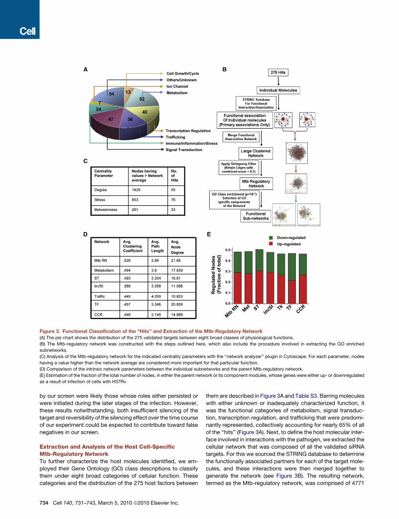
Figure 3. Functional Classification of the ‘‘Hits’’ and Extraction of the Mtb-Regulatory Network
(A) The pie chart shows the distribution of the 275 validated targets between eight broad classes of physiological functions.
(B) The Mtb-regulatory network was constructed with the steps outlined here, which also include the procedure involved in extracting the GO enriched
subnetworks.
(C) Analysis of the Mtb-regulatory network for the indicated centrality parameters with the ‘‘network analyzer’’ plugin in Cytoscape. For each parameter, nodes
having a value higher than the network average are considered more important for that particular function.
(D) Comparison of the intrinsic network parameters between the individual subnetworks and the parent Mtb-regulatory network.
(E) Estimation of the fraction of the total number of nodes, in either the parent network or its component modules, whose genes were either up- or downregulated
as a result of infection of cells with H37Rv.
by our screen were likely those whose roles either persisted or
were initiated during the later stages of the infection. However,
these results notwithstanding, both insufficient silencing of the
target and reversibility of the silencing effect over the time course
of our experiment could be expected to contribute toward false
negatives in our screen.
Extraction and Analysis of the Host Cell-SpecificMtb-Regulatory NetworkTo further characterize the host molecules identified, we em-
ployed their Gene Ontology (GO) class descriptions to classify
them under eight broad categories of cellular function. These
categories and the distribution of the 275 host factors between
734 Cell 140, 731–743, March 5, 2010 ª2010 Elsevier Inc.
them are described in Figure 3A and Table S3. Barring molecules
with either unknown or inadequately characterized function, it
was the functional categories of metabolism, signal transduc-
tion, transcription regulation, and trafficking that were predomi-
nantly represented, collectively accounting for nearly 65% of all
of the ‘‘hits’’ (Figure 3A). Next, to define the host molecular inter-
face involved in interactions with the pathogen, we extracted the
cellular network that was composed of all the validated siRNA
targets. For this we sourced the STRING database to determine
the functionally associated partners for each of the target mole-
cules, and these interactions were then merged together to
generate the network (see Figure 3B). The resulting network,
termed as the Mtb-regulatory network, was comprised of 4771
nodes and 51,677 edges (interactions) (Tables S5 and S6). A
clustering coefficient (CC) of 0.526 was obtained, which is
consistent with a ‘‘small-world’’ architecture that facilitates
easy information flow between the component nodes (Barabasi
and Oltvai, 2004). A randomization of this network through 100
iterations where links were arbitrarily shuffled without altering
their number resulted in a 20-fold reduction in the clustering
coefficient (0.03 ± 0.0007), with a concomitant reduction also
in the mean path length (from 4.0 to 2.8 ± 0.003). Thus, the
topological features of the Mtb-regulatory network were not
randomly derived, but rather represent unique properties. Addi-
tionally, the low frequency of our ‘‘hits’’ among the nodes with
a higher than average value for the measured centrality parame-
ters also reiterated that this network was not biased toward the
siRNA-identified targets (Figure 3C, Table S6).
Modular Composition of the Mtb-Regulatory NetworkWe further dissected the Mtb-regulatory network to examine its
constituent functional modules. With the exception of ion
channel-related activities, the other functional groups described
in Figure 3A were identified to form discrete subnetworks that
were significantly overrepresented (p < 10�3). The subnetworks
obtained for each of these GO classes are depicted in Table
S6, and the two most enriched ones were those for the functions
of metabolism and signal transduction. This is consistent with
the fact that, in addition to sourcing nutrients, intracellular
survival of Mtb requires the pathogen to modulate multiple
host cellular functions that are under the regulatory control of
the signaling machinery (Schaible and Kaufmann, 2005; Warner
and Mizrahi, 2007). Similarly, the relatively large size of the
module corresponding to the host cell transcription regulatory
network is also in keeping with earlier findings that infection
causes extensive alterations in the host cell transcriptome (Nau
et al., 2002).
A comparison of the intrinsic network properties of the indi-
vidual subnetworks with that of the parent Mtb-regulatory
network revealed that these parameters were only slightly
affected (Figure 3D). Additionally, the parent network and its
subnetworks all displayed a broad distribution in node degree
(Table S6), confirming the modular organization of the Mtb-regu-
latory network.
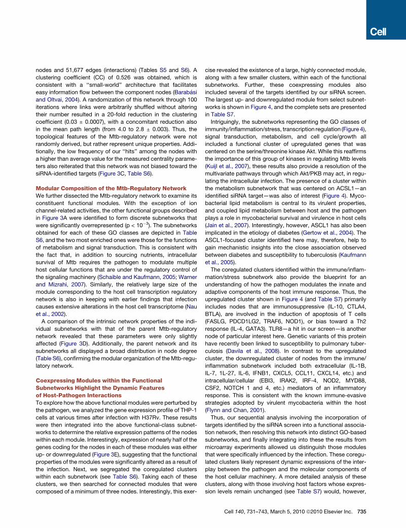
Coexpressing Modules within the FunctionalSubnetworks Highlight the Dynamic Featuresof Host-Pathogen InteractionsTo explore how the above functional modules were perturbed by
the pathogen, we analyzed the gene expression profile of THP-1
cells at various times after infection with H37Rv. These results
were then integrated into the above functional-class subnet-
works to determine the relative expression patterns of the nodes
within each module. Interestingly, expression of nearly half of the
genes coding for the nodes in each of these modules was either
up- or downregulated (Figure 3E), suggesting that the functional
properties of the modules were significantly altered as a result of
the infection. Next, we segregated the coregulated clusters
within each subnetwork (see Table S6). Taking each of these
clusters, we then searched for connected modules that were
composed of a minimum of three nodes. Interestingly, this exer-
cise revealed the existence of a large, highly connected module,
along with a few smaller clusters, within each of the functional
subnetworks. Further, these coexpressing modules also
included several of the targets identified by our siRNA screen.
The largest up- and downregulated module from select subnet-
works is shown in Figure 4, and the complete sets are presented
in Table S7.
Intriguingly, the subnetworks representing the GO classes of
immunity/inflammation/stress, transcription regulation (Figure 4),
signal transduction, metabolism, and cell cycle/growth all
included a functional cluster of upregulated genes that was
centered on the serine/threonine kinase Akt. While this reaffirms
the importance of this group of kinases in regulating Mtb levels
(Kuijl et al., 2007), these results also provide a resolution of the
multivariate pathways through which Akt/PKB may act, in regu-
lating the intracellular infection. The presence of a cluster within
the metabolism subnetwork that was centered on ACSL1—an
identified siRNA target—was also of interest (Figure 4). Myco-
bacterial lipid metabolism is central to its virulent properties,
and coupled lipid metabolism between host and the pathogen
plays a role in mycobacterial survival and virulence in host cells
(Jain et al., 2007). Interestingly, however, ASCL1 has also been
implicated in the etiology of diabetes (Gertow et al., 2004). The
ASCL1-focused cluster identified here may, therefore, help to
gain mechanistic insights into the close association observed
between diabetes and susceptibility to tuberculosis (Kaufmann
et al., 2005).
The coregulated clusters identified within the immune/inflam-
mation/stress subnetwork also provide the blueprint for an
understanding of how the pathogen modulates the innate and
adaptive components of the host immune response. Thus, the
upregulated cluster shown in Figure 4 (and Table S7) primarily
includes nodes that are immunosuppressive (IL-10, CTLA4,
BTLA), are involved in the induction of apoptosis of T cells
(FASLG, PDCD1LG2, TRAF6, NOD1), or bias toward a Th2
response (IL-4, GATA3). TLR8—a hit in our screen—is another
node of particular interest here. Genetic variants of this protein
have recently been linked to susceptibility to pulmonary tuber-
culosis (Davila et al., 2008). In contrast to the upregulated
cluster, the downregulated cluster of nodes from the immune/
inflammation subnetwork included both extracellular (IL-1B,
IL-7, 1L-27, IL-6, IFNB1, CXCL5, CCL11, CXCL14, etc.) and
intracellular/cellular (EBI3, IRAK2, IRF-4, NOD2, MYD88,
CSF2, NOTCH 1 and 4, etc.) mediators of an inflammatory
response. This is consistent with the known immune-evasive
strategies adopted by virulent mycobacteria within the host
(Flynn and Chan, 2001).
Thus, our sequential analysis involving the incorporation of
targets identified by the siRNA screen into a functional associa-
tion network, then resolving this network into distinct GO-based
subnetworks, and finally integrating into these the results from
microarray experiments allowed us distinguish those modules
that were specifically influenced by the infection. These coregu-
lated clusters likely represent dynamic expressions of the inter-
play between the pathogen and the molecular components of
the host cellular machinery. A more detailed analysis of these
clusters, along with those involving host factors whose expres-
sion levels remain unchanged (see Table S7) would, however,
Cell 140, 731–743, March 5, 2010 ª2010 Elsevier Inc. 735
Figure 4. Identification of Coexpressing
Modules within the Functional Subnetworks
Figure shows the largest coexpressing clusters
from select functional subnetworks identified in
Figure 3. Clusters shown in red are upregulated
and those in green downregulated after infection
of THP-1 cells with H37Rv. The slope for each
gene, from a plot of the expression data at various
time points, was calculated to group the up- and
downregulated nodes. Genes with a slope higher
than 0.15 were considered to be upregulated,
and those with a slope lower than �0.15 were
taken as being downregulated. Nodes in bold
with names in yellow are from our list of validated
targets from the screening data, and the nodes
referred to in the text are highlighted in blue. See
also Table S7.
be required before more definitive interpretations can be made
on host factor-dependent regulation of the pathogen.
Defining a Filtration Window by Using a Panelof Mtb Clinical IsolatesMtb exhibits a broad spectrum of genotypic and phenotypic
diversification in the human population. To date, at least six
main lineages and 15 sublineages of this pathogen have been
described (Brudey et al., 2006; Gagneux et al., 2006). Further,
the clinical presentation of this infection can dynamically shift
between states of latent, persistent, and active infection in which
the pathogen can again potentially display a spectrum of pheno-
typic variations that extend from drug-sensitive to multiple-drug
(MDR-Mtb), and even extensive drug, resistance (Ginsberg and
Spigelman, 2007). It is, however, unknown whether such varia-
736 Cell 140, 731–743, March 5, 2010 ª2010 Elsevier Inc.
tions also involve alterations in the mech-
anisms that support intracellular survival
of the pathogen.
To explore this, we extended our
studies to include a panel of seven inde-
pendent field isolates of Mtb that collec-
tively represented a range of phenotypic
and genotypic variations. For example,
these isolates displayed distinct patterns
of drug resistance against the four front-
line drugs used for the treatment of tuber-
culosis (Figure 5A). While three of these
isolates belonged to the class of MDR-
Mtb, exhibiting resistance to any three
of these four drugs, the resistance profile
describing each isolate differed from that
of the other two. The remaining four
isolates were also similarly distinguished
in that they were each resistant to just
a single drug, but again in a manner that
was exclusive of the other isolates in
this subgroup (Figure 5A). In addition to
drug-sensitivity profiles, these Mtb
isolates also defined a heterogeneous
group of genotypes, collectively representing four distinct line-
ages (Figure 5A and Figure S3).
The isolates also displayed diverse growth properties in THP-1
cells, with apparent growth rates that were either significantly
higher (JAL2287) or lower than that of H37Rv (Figure 5B). Impor-
tantly, this variation was specific to infected cells since these
isolates demonstrated broadly comparable growth rates in
extracellular cultures (Figure 5C). Although differences in
apparent growth rates presumably reflect differences in the
balance achieved between mycobacterial replication and killing,
differences in intracellular rates of accumulation in THP-1 cells
have been shown to correlate with epidemiological evidence
for strain virulence (Park et al., 2006; Theus et al., 2006). Thus,
variations in the apparent growth rates seen in Figure 5B also
imply variations in the in vivo virulence properties of these
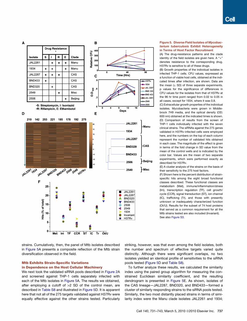
Figure 5. Diverse Field Isolates of Mycobac-
terium tuberculosis Exhibit Heterogeneity
in Terms of Host Factor Recruitment
(A) The drug-resistance patterns and the clade
identity of the field isolates are given here. A ‘‘+’’
denotes resistance to the corresponding drug.
H37Rv is sensitive to all of these drugs.
(B) Growth properties of the individual isolates in
infected THP-1 cells. CFU values, expressed as
a function of viable host cells, obtained at the indi-
cated times after infection, are shown. Data are
the mean (± SD) of three separate experiments.
p values for the significance of differences in
CFU values for the isolates from that of H37Rv at
the 96 hr time point ranged from 0.02 to 0.05 in
all cases, except for 1934, where it was 0.8.
(C) Extracellular growth properties of the individual
isolates. Mycobacteria were grown in Middle-
brock 7H9 media, and the optical density (OD;
600 nm) obtained at the indicated times is shown.
(D) Comparison of results from the screen of
THP-1 cells individually infected with the seven
clinical strains. The siRNAs against the 275 genes
validated in H37Rv-infected cells were employed
here, and the numbers on the top of each column
represent the number of validated hits obtained
in each case. The magnitude of the effect is given
in terms of the fold change in SD value from the
mean of the control wells and is indicated by the
color bar. Values are the mean of two separate
experiments, which were performed exactly as
described for H37Rv.
(E) A cluster analysis of the strains on the basis of
their sensitivity to the 275 host factors.
(F) Shown here is the percent distribution of strain-
specific hits among the eight broad functional
classes described. These functional classes are
metabolism (Met), immune/inflammation/stress
(Im), transcription regulation (TF), cell growth/
cycle (CCR), signal transduction (ST), ion channel
(IC), trafficking (Tr), and those with presently
unknown or inadequately characterized function
(Ot/U). Results for the subset of 74 host proteins
that served as a common requirement for all the
Mtb strains tested are also included (Invariant).
See also Figure S3.
strains. Cumulatively, then, the panel of Mtb isolates described
in Figure 5A presents a composite reflection of the Mtb strain
diversification observed in the field.
Mtb Exhibits Strain-Specific Variationsin Dependence on the Host Cellular MachineryWe next took the validated siRNA pools described in Figure 2A
and screened against THP-1 cells separately infected with
each of the Mtb isolates in Figure 5A. The results we obtained,
after employing a cutoff of >2 SD of the control mean, are
described in Table S8 and illustrated in Figure 5D. It is apparent
here that not all of the 275 targets validated against H37Rv were
equally effective against the other strains tested. Particularly
striking, however, was that even among the field isolates, both
the number and spectrum of effective targets varied quite
distinctly. Although there were significant overlaps, no two
isolates yielded an identical profile of sensitivities to the siRNA
pools tested (Figure 5D and Table S8).
To further analyze these results, we calculated the similarity
index using the paired group algorithm for measuring the con-
strained Euclidean similarity coefficient, and the resulting
dendrogram is presented in Figure 5E. As shown, isolates of
the CAS lineage—JAL2287, BND320, and BND433—formed a
cluster of similarly responding strains to the siRNA pools tested.
Similarly, the two most distantly placed strains in terms of simi-
larity index were the Manu clade isolates JAL2261 and 1934;
Cell 140, 731–743, March 5, 2010 ª2010 Elsevier Inc. 737
whereas the remaining strains 2506 (Beijing) and 2549 (Miscella-
neous) were located in between these two extreme groups
(Figure 5E). Thus, distinctions in clade identity appear to serve
as the primary determinant for the observed differences in the
spectrum of host molecules recruited in Figure 5D. However,
differences in phenotypic properties such as replication rates
are also likely to contribute toward these distinctions. Notably,
the strain-specific variations seen here also raise the possibility
that at least some of these isolates may exhibit dependencies
on host factors that are redundant in the context of H37Rv infec-
tion. Such factors would be missed in our present study.
Identifying the Mtb Strain-Independent RegulatoryAxis of the Host CellA closer inspection of Figure 5D revealed that, in spite of the
differences, 74 of the siRNA pools were commonly effective in
reducing intracellular loads of all the strains tested. That is, the
host factors targeted by these siRNA pools were invariantly
required regardless of drug resistance profile, virulence proper-
ties, and clade identity (listed in Table S9). To examine the nature
of similarities and distinctions in host factor dependencies
between these Mtb isolates, we probed for any alterations in
bias toward the host cellular functional modules. Interestingly,
the percent distribution of the ‘‘hits’’ for each strain—including
H37Rv—among the functional groups described in Figure 3A
remained constant, indicating that the relative extent of utiliza-
tion of the different functional modules of the host cell was
unchanged across these strains (Figure 5F). This was equally
true for the 74 protein targets that were invariantly involved for
all the strains (Figure 5F), supporting the relevance of each of
these functional classes in regulating intracellular Mtb levels.
The Mtb Strain-Independent RegulatoryAxis Functions by Regulating AutophagyRecent experiments have demonstrated that stimulation of
autophagy (or, xenophagy) in infected macrophages, either by
physiological or pharmacological means, severely reduces
viability of intracellular Mtb (Gutierrez et al., 2004; Levine and
Deretic, 2007). Consistent with this, we also found that pharma-
cological activation of autophagy, at 48 hr after the infection,
significantly reduced intracellular Mtb load in THP-1 cells
(Figure 6A). Importantly, these results extended the earlier find-
ings by establishing the susceptibility of Mtb to xenophagy
during this later window of infection. We therefore asked whether
the observed effects of depletion of at least some of the identi-
fied host targets, on Mtb survival, could in fact be mediated
through the activation of xenophagic pathways. To test this,
we subjected H37Rv-infected cells to siRNA-mediated silencing
of each of the 74 host molecules that were identified as the
strain-independent set of Mtb-regulatory factors in Figure 5D
(and Table S9). After this, we determined whether the effects of
target protein silencing on intracellular Mtb levels could be
reversed by 3-methyladenine (3MA), the classical inhibitor of
autophagy (Seglen and Gordon, 1982). By taking a 50% reversal
of the effects of a given siRNA as the cutoff for significance, we
found that inclusion of 3MA significantly attenuated the effects of
44 of the 74 target-specific siRNAs tested (Figure 6B, Table S9).
This suggests that the Mtb-inhibitory effects that result from
738 Cell 140, 731–743, March 5, 2010 ª2010 Elsevier Inc.
depletion of over half of the Mtb strain-independent regulatory
factors identified by our screen are likely to be mediated through
the induction of xenophagic pathways. The mechanism of action
resulting from depletion of the remaining 30 targets, however,
awaits clarification.
Although the antimicrobial properties of IFN-g also derive
in part from its ability to stimulate autophagy in host cells
(MacMicking et al., 2003; Singh et al., 2006), Mtb-infected human
macrophages are desensitized to activation by this cytokine
(Ting et al., 1999). This desensitization facilitates persistence of
the pathogen even in tuberculosis patients expressing high
levels of IFN-g in pleural fluid (Flynn, 1999). Consistent with
this, stimulation of H37Rv-infected THP-1 cells with IFN-g had
only a marginal (<15%) affect on pathogen viability, although
these cells continued to express high levels of the IFN-g receptor
(data not shown). Thus, attenuation of cytokine responsiveness
of the host cell by Mtb also probably includes attenuation of
IFN-g-dependent activation of xenophagic pathways.
The 3MA-sensitive host factors identified in Figure 6B prob-
ably represent negative regulators of xenophagy since it was
the silencing of expression of these molecules that induced
suppression/elimination of intracellular Mtb. This could be
experimentally verified by our subsequent findings that silencing
of these 44 targets in infected cells resulted in an increase in
levels of LC3-II - the marker for autophagosomes (Figure S4).
Therefore, to determine how levels of the genes for these 44
proteins were influenced by Mtb infection, we reanalyzed our
expression data (Figure 4). Thirteen of these were indeed upre-
gulated after infection of THP-1 cells, whereas expression levels
of the remainder were either unchanged (20), or suppressed (11)
(Figure 6C). It is possible that select upregulation of only a small
subset of these factors is sufficient to inhibit the autophagic
response of the host cell. A similar explanation may also account
for the results of our parallel examination of the public database
(NCBI, GEO) for the expression levels of these 44 genes in
macrophages stimulated with IFN-g. Here again only some of
these genes (19) were downregulated, with four of these being
common to the group of genes induced by H37Rv infection
(Figure 6C).
The poor overlap between the xenophagy-related genes sup-
pressed by IFN-g and those induced by H37Rv infection led us to
explore whether the suppression of any one of these host factors
would potentiate IFN-g responsiveness of the infected host cell.
We depleted the targets listed in Figure 6C in H37Rv-infected
cells and then stimulated these cells with IFN-g. The effect on
intracellular Mtb load was then compared with that obtained in
the absence of cytokine stimulation. Interestingly stimulation
with IFN-g resulted, in several instances, in an enhanced reduc-
tion in CFU that was at least 1.5-fold greater than that obtained
in response to siRNA treatment alone (Figure 6D). Since IFN-g
alone had only a marginal influence (<15%), these results reveal
the synergistic effect that can be achieved by combining
the depletion of select xenophagy-related genes with cytokine
stimulation.
Thus, activation of xenophagic pathways through the down-
modulation of negative regulators in the host macrophage
provides an effective mechanism for elimination of the intracel-
lular Mtb in a strain-independent manner. Also significant is our
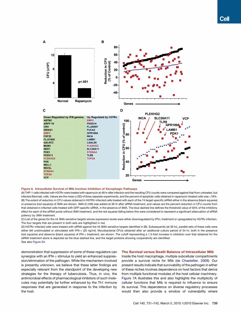
Figure 6. Intracellular Survival of Mtb Involves Inhibition of Xenophagic Pathways
(A) THP-1 cells infected with H37Rv were treated with rapamycin at 48 hr after infection and the resulting CFU counts were compared against that from untreated, but
infected (Normal), cells. Values are the mean (±SD) of three separate experiments, and the percent of apoptotic cells obtained in rapamycin-treated cells was <18%.
(B) The extent of reduction in CFU values obtained in H37RV-infected cells treated with each of the 74 target-specific siRNA either in the absence (black squares)
or presence (red squares) of 3MA are shown. 3MA (5 mM) was added at 36 hr after siRNA treatment, and values are the percent reduction in CFU counts from
that obtained in infected cells treated with GFP-specific siRNA, in the absence of 3MA. The blue dashed line defines the threshold value of 50% of the inhibitory
effect for each of the siRNA pools without 3MA treatment, and the red squares falling below this were considered to represent a significant attenuation of siRNA
potency by 3MA treatment.
(C) List of the genes for the 44 3MA-sensitive targets whose expression levels were either downregulated by IFNg treatment or upregulated by H37Rv infection.
The four targets that are present in both sets are highlighted in red.
(D) H37Rv-infected cells were treated with siRNA against the 44 3MA-sensitive targets identified in (B). Subsequently (at 36 hr), parallel sets of these cells were
either left unstimulated or stimulated with IFN-g (20 ng/ml). Mycobacterial CFUs obtained after an additional culture period of 54 hr, both in the presence
(red squares) and absence (black squares) of IFN-g treatment, are shown. The cutoff representing a 1.5-fold increase in inhibition over that obtained for the
siRNA treatment alone is depicted as the blue dashed line, and the target proteins showing cooperativity are identified.
See also Figure S4.
demonstration that suppression of some of these regulators can
synergize with an IFN-g stimulus to yield an enhanced suppres-
sion/elimination of the pathogen. While the mechanism involved
is presently unknown, we believe that these latter findings are
especially relevant from the standpoint of the developing new
strategies for the therapy of tuberculosis. Thus, in vivo, the
antimicrobial effects of pharmacological inhibitors of such mole-
cules may potentially be further enhanced by the Th1 immune
responses that are generated in response to the infection by
the host.
The Survival versus Death Balance of Intracellular MtbInside the host macrophage, multiple subcellular compartments
provide a survival niche for Mtb (de Chastellier, 2009). Our
present results indicate that survivability of the pathogen in either
of these niches involves dependence on host factors that derive
from multiple functional modules of the host cellular machinery.
Figure 7A illustrates this and also highlights the multiplicity of
cellular functions that Mtb is required to influence to ensure
its survival. This dependence on diverse regulatory processes
would then also provide a window of vulnerability where
Cell 140, 731–743, March 5, 2010 ª2010 Elsevier Inc. 739
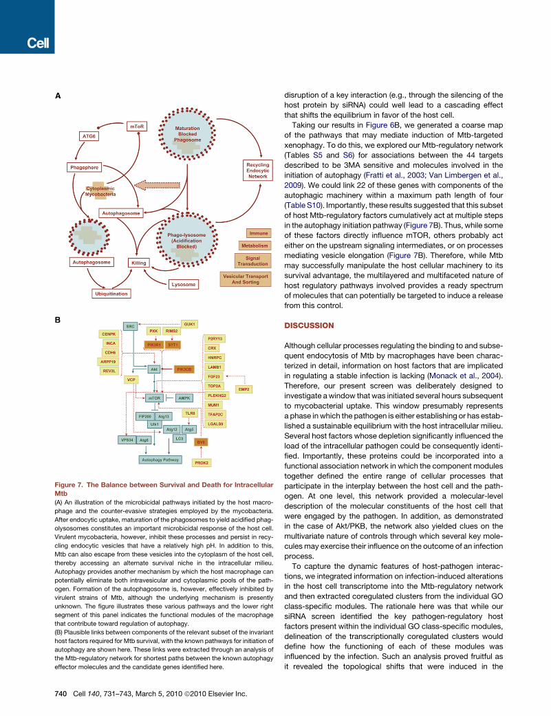
Figure 7. The Balance between Survival and Death for Intracellular
Mtb
(A) An illustration of the microbicidal pathways initiated by the host macro-
phage and the counter-evasive strategies employed by the mycobacteria.
After endocytic uptake, maturation of the phagosomes to yield acidified phag-
olysosomes constitutes an important microbicidal response of the host cell.
Virulent mycobacteria, however, inhibit these processes and persist in recy-
cling endocytic vesicles that have a relatively high pH. In addition to this,
Mtb can also escape from these vesicles into the cytoplasm of the host cell,
thereby accessing an alternate survival niche in the intracellular milieu.
Autophagy provides another mechanism by which the host macrophage can
potentially eliminate both intravesicular and cytoplasmic pools of the path-
ogen. Formation of the autophagosome is, however, effectively inhibited by
virulent strains of Mtb, although the underlying mechanism is presently
unknown. The figure illustrates these various pathways and the lower right
segment of this panel indicates the functional modules of the macrophage
that contribute toward regulation of autophagy.
(B) Plausible links between components of the relevant subset of the invariant
host factors required for Mtb survival, with the known pathways for initiation of
autophagy are shown here. These links were extracted through an analysis of
the Mtb-regulatory network for shortest paths between the known autophagy
effector molecules and the candidate genes identified here.
740 Cell 140, 731–743, March 5, 2010 ª2010 Elsevier Inc.
disruption of a key interaction (e.g., through the silencing of the
host protein by siRNA) could well lead to a cascading effect
that shifts the equilibrium in favor of the host cell.
Taking our results in Figure 6B, we generated a coarse map
of the pathways that may mediate induction of Mtb-targeted
xenophagy. To do this, we explored our Mtb-regulatory network
(Tables S5 and S6) for associations between the 44 targets
described to be 3MA sensitive and molecules involved in the
initiation of autophagy (Fratti et al., 2003; Van Limbergen et al.,
2009). We could link 22 of these genes with components of the
autophagic machinery within a maximum path length of four
(Table S10). Importantly, these results suggested that this subset
of host Mtb-regulatory factors cumulatively act at multiple steps
in the autophagy initiation pathway (Figure 7B). Thus, while some
of these factors directly influence mTOR, others probably act
either on the upstream signaling intermediates, or on processes
mediating vesicle elongation (Figure 7B). Therefore, while Mtb
may successfully manipulate the host cellular machinery to its
survival advantage, the multilayered and multifaceted nature of
host regulatory pathways involved provides a ready spectrum
of molecules that can potentially be targeted to induce a release
from this control.
DISCUSSION
Although cellular processes regulating the binding to and subse-
quent endocytosis of Mtb by macrophages have been charac-
terized in detail, information on host factors that are implicated
in regulating a stable infection is lacking (Monack et al., 2004).
Therefore, our present screen was deliberately designed to
investigate a window that was initiated several hours subsequent
to mycobacterial uptake. This window presumably represents
a phase in which the pathogen is either establishing or has estab-
lished a sustainable equilibrium with the host intracellular milieu.
Several host factors whose depletion significantly influenced the
load of the intracellular pathogen could be consequently identi-
fied. Importantly, these proteins could be incorporated into a
functional association network in which the component modules
together defined the entire range of cellular processes that
participate in the interplay between the host cell and the path-
ogen. At one level, this network provided a molecular-level
description of the molecular constituents of the host cell that
were engaged by the pathogen. In addition, as demonstrated
in the case of Akt/PKB, the network also yielded clues on the
multivariate nature of controls through which several key mole-
cules may exercise their influence on the outcome of an infection
process.
To capture the dynamic features of host-pathogen interac-
tions, we integrated information on infection-induced alterations
in the host cell transcriptome into the Mtb-regulatory network
and then extracted coregulated clusters from the individual GO
class-specific modules. The rationale here was that while our
siRNA screen identified the key pathogen-regulatory host
factors present within the individual GO class-specific modules,
delineation of the transcriptionally coregulated clusters would
define how the functioning of each of these modules was
influenced by the infection. Such an analysis proved fruitful as
it revealed the topological shifts that were induced in the
subnetworks, to bias toward a favorable outcome for the
pathogen. This was typified by observed modulation of the
immune/inflammation/stress subnetwork wherein suppression
of inflammatory response mediators was further exacerbated
through a concomitant induction of anti-inflammatory and other
immunosuppressive molecules. In a similar vein, the targeted
regulation of enzymes within the lipid metabolism module is
also expected to contribute toward pathogen sustenance by
promoting a nutritive environment and/or providing an additional
route to the manipulation of macrophage innate immune re-
sponses (Gutierrez et al., 2009; Schaible and Kaufmann, 2005).
Significantly, over half of the validated targets identified by our
siRNA screen were also transcriptionally modulated, suggesting
a proactive control by Mtb over at least some of the host factors
that are critical for its survival. Thus, these collective results
provide a snapshot of the calibration in functioning of the indi-
vidual host cell modules that the pathogen enforces as its
survival strategy. A detailed analysis of the coregulated path-
ways should clearly yield a more global perspective on path-
ogen-induced regulation of host cell responses.
The phenotypic and genotypic diversifications exhibited by
most, if not all, pathogens clearly represent diverse adaptation
mechanisms that are engendered in the context of the target
host population. However, the question of whether and how
such adaptations translate at the level of redefining the dynamics
of host-pathogen interactions has remained unanswered. At
least as shown here for Mtb, this question appears to be partic-
ularly relevant for understanding the variability of mechanisms
that can support the virulence properties of the pathogen. This
was exemplified by the panel of field isolates tested, where
each member depended upon a unique spectrum of host
molecules for its intracellular persistence. These differences
were primarily guided by distinctions in clade identity, though
lesser contributions due to phenotypic variations are also likely.
However, such differences notwithstanding, the distribution of
Mtb strain-specific host factors between the modules describing
diverse cellular functions remained relatively constant. This
implies that regardless of the Mtb genotype or phenotype, estab-
lishment of a successful infection involves an obligatory depen-
dence on the spectrum of cellular activities represented by these
modules.
A particular highlight of our experiments comparing different
clinical isolates was the identification of a subset of 74 host
proteins whose presence constituted an invariant requirement
for optimal survival of all Mtb strains tested. That is, this subset
defined an obligate survival axis for Mtb that was resistant to
at least the range of adaptive variations of pathogen that were
explored in this study. The functional attributes of this axis could
be further resolved by our discovery that over half of its constit-
uents acted through the regulation of autophagy. These findings
underscore the idea that regardless of the broad spectrum of
variants that Mtb exhibits in the human population, its persis-
tence in the host cellular environment predominantly hinges on
its ability to modulate the process of autophagy. Further, our
subsequent results both from experiments examining synergy
with IFNg and from a pathway mapping exercise support the
idea that these mediators collectively interfere at multiple stages
of the autophagy-inducing pathways.
Thus, in summary, our present detailing of host molecules
involved during the stabilization of Mtb infection provides insight
into the molecular mechanisms that facilitate persistence of
pathogen in the host cell. Further, although we detected diver-
gence in pathogenetic mechanisms being exploited by distinct
field isolates, the invariant host-specific components of this
interplay could nonetheless be successfully captured. Of special
emphasis in this regard is our subsequent delineation that the
majority of these latter components constituted regulatory
components of a common antimicrobial module of the host
cell. We believe that this finding has important implications,
given that an additional avenue for long-term persistence of
mycobacteria in the host cell likely involves a stochastic distribu-
tion of its population between various intracellular niches (Young
et al., 2008). Under such circumstances, then, autophagy pro-
vides a unifying mechanism by which each of these individual
subpopulations can simultaneously be eliminated. Finally, we
also note a natural corollary of these observations that several
of the host factors identified here also provide attractive targets
for the development of drugs against tuberculosis.
EXPERIMENTAL PROCEDURES
A detailed description of the methodologies employed for various aspects of
this study and the optimization experiments for arriving at the screening proce-
dure are provided in the Extended Experimental Procedures. All experiments
with M. tuberculosis were approved by the Institutional Biosafety Committee
and performed in a biosafety level III facility. The isolates BND320, BND433,
JAL2261, and JAL2287 were a kind gift from V.M. Katoch (National Jalma Insti-
tute for Leprosy and other Mycobacterial Diseases), and the spoligotyping
results of all the isolates used here was provided by S. Kulkarni (Bhabha
Atomic Research Centre[ BARC]).
siRNA Library
Dharmacon siGENOME SMARTpool siRNA Library for the complete human
genome comprising of 18,174 targets was procured from Thermo Scientific.
The SMARTpool library consists of a pool of four different oligos for each target
gene. For validation studies, all the four oligos for each genes used in the first
library were procured separately and tested individually.
Infection of Cells and the siRNA Screen
THP-1 cells were cultured in RPMI 1640 (GIBCO Laboratories) supplemented
with 10% FCS (Hyclone) and were maintained between 2 3 105 and 10 3 105
cells per ml at 37�C in a humidified, 5% CO2 atmosphere. Before infection,
cells were plated in 96 well plates at 1 3 104 cells per well and differentiated
with PMA (5 ng/ml) for a period of 24 hr. The detailed procedure for infection
of cells, the siRNA screen, and analysis of the resultant data is described in
the Extended Experimental Procedures. CFUs were calculated from the actual
colony counts obtained. Counts were multiplied with the dilution factor and the
volume of the diluted sample used for plating. CFU values were expressed on
a per ml basis.
RNA Isolation and Microarray Analysis
RNA was isolated from either uninfected THP-1 cells or cells infected with
H37Rv for 16, 48, 90 hr. A one-color microarray-based gene expression anal-
ysis was performed by hybridization against a human whole-genome array
consisting of probes for 44,000 genes (Agilent). For each time point, the hybrid-
ization was performed on duplicate sets of cells. For uninfected cells, we used
A/P calls as an indicator of the presence of a given transcript. To define alter-
ations in the level of a given transcript after infection, we considered a gene to
be upregulated if the signal log ratio between the infected and the uninfected
sample was higher than 1 (>2-fold increase) and the detection p value of the
infected sample was higher than 0.95. Similarly, a gene was considered to
Cell 140, 731–743, March 5, 2010 ª2010 Elsevier Inc. 741
be downregulated if the signal log ratio was less than �1 (>2-fold decrease)
and the detection p value of the reference sample was higher than 0.95.
Further, only those genes that presented a consistent change in both the bio-
logical repeats were taken as being differentially expressed. The results of our
microarray analysis are available in the GEO repository (accession number
GSE19052).
Extraction of the Mtb-Regulatory Network and Its Analysis
Host-specific targets validated by our siRNA screen were used to build the
Mtb-regulatory network. Each of these 275 molecules was individually
searched in the STRING database (http://string.embl.de/) for its functional
associations. STRING is a database of both known and predicted protein-
protein interactions. These include direct (physical) and indirect (functional)
associations, which are derived from four separate sources: genomic context,
high-throughput experiments, coexpression, and prior knowledge. We
selected all interactions/associations available for a given node showing
a combined score of more than 0.7. The scores given in the STRING database
define the confidence limit for each described interaction/association. A
combined score of 0.7 is recommended as the high stringency criterion by
the database. The functional associations identified for all of the molecules
were then merged together and imported into Cytoscape 2.6.1 as a two-
column network (Mtb-regulatory network).
GO analysis of the Mtb-regulatory network was performed with the Bingo
2.3 plugin in Cytoscape 2.6.1. Only categories with a very low p value
(<10�5) were considered as enriched in the network as determined by Hyper-
geometric statistical test employing the Benjamini and Hochberg false
discovery rate correction (Maere et al., 2005).
ACCESSION NUMBERS
The GEO accession number for the gene expression analysis reported in this
paper is GSE19052.
SUPPLEMENTAL INFORMATION
Supplemental Information includes Extended Experimental Procedures, four
figures, and ten tables and can be found with this article online at doi:10.
1016/j.cell.2010.02.012.
ACKNOWLEDGMENTS
This study was funded in part by the Department of Biotechnology, Govern-
ment of India, and in part by the Institute of Life Sciences, Hyderabad. We
are grateful to V. Biswas, M. Jain, N. Karwal, R. Kashikar, S. Kumari, A. Kumar,
K. Midha, M.K. Midha, S. Seth, S. Sharma, M. Sharma, Z. Sidiqui, A. Srivas-
tava, and S. Tirpattiwar for technical assistance during the various stages of
the RNA interference screen. We also thank S. Kulkarni (BARC) for spoligotyp-
ing analysis of the field isolates used in this study.
Received: May 29, 2009
Revised: October 2, 2009
Accepted: February 8, 2010
Published: March 4, 2010
REFERENCES
Barabasi, A.L., and Oltvai, Z.N. (2004). Network biology: understanding the
cell’s functional organization. Nat. Rev. Genet. 5, 101–113.
Brudey, K., Driscoll, J.R., Rigouts, L., Prodinger, W.M., Gori, A., Al-Hajoj, S.A.,
Allix, C., Aristimuno, L., Arora, J., Baumanis, V., et al. (2006). Mycobacterium
tuberculosis complex genetic diversity: mining the fourth international spoligo-
typing database (SpolDB4) for classification, population genetics and epide-
miology. BMC Microbiol. 6, 23.
Davila, S., Hibberd, M.L., Hari Dass, R., Wong, H.E., Sahiratmadja, E.,
Bonnard, C., Alisjahbana, B., Szeszko, J.S., Balabanova, Y., Drobniewski, F.,
742 Cell 140, 731–743, March 5, 2010 ª2010 Elsevier Inc.
et al. (2008). Genetic association and expression studies indicate a role of
toll-like receptor 8 in pulmonary tuberculosis. PLoS Genet. 4, e1000218.
de Chastellier, C. (2009). The many niches and strategies used by pathogenic
mycobacteria for survival within host macrophages. Immunobiology, in press.
Published online March 2, 2009.
Flynn, J.L. (1999). Why is IFN-gamma insufficient to control tuberculosis?
Trends Microbiol. 7, 477–478, author reply 478–479.
Flynn, J.L., and Chan, J. (2001). Immunology of tuberculosis. Annu. Rev.
Immunol. 19, 93–129.
Fratti, R.A., Chua, J., Vergne, I., and Deretic, V. (2003). Mycobacterium
tuberculosis glycosylated phosphatidylinositol causes phagosome maturation
arrest. Proc. Natl. Acad. Sci. USA 100, 5437–5442.
Gagneux, S., DeRiemer, K., Van, T., Kato-Maeda, M., de Jong, B.C.,
Narayanan, S., Nicol, M., Niemann, S., Kremer, K., Gutierrez, M.C., et al.
(2006). Variable host-pathogen compatibility in Mycobacterium tuberculosis.
Proc. Natl. Acad. Sci. USA 103, 2869–2873.
Gertow, K., Pietilainen, K.H., Yki-Jarvinen, H., Kaprio, J., Rissanen, A.,
Eriksson, P., Hamsten, A., and Fisher, R.M. (2004). Expression of fatty-acid-
handling proteins in human adipose tissue in relation to obesity and insulin
resistance. Diabetologia 47, 1118–1125.
Ginsberg, A.M., and Spigelman, M. (2007). Challenges in tuberculosis drug
research and development. Nat. Med. 13, 290–294.
Gutierrez, M.G., Master, S.S., Singh, S.B., Taylor, G.A., Colombo, M.I., and
Deretic, V. (2004). Autophagy is a defense mechanism inhibiting BCG and
Mycobacteriumtuberculosissurvival in infectedmacrophages. Cell119, 753–766.
Gutierrez, M.G., Gonzalez, A.P., Anes, E., and Griffiths, G. (2009). Role of lipids
in killing mycobacteria by macrophages: evidence for NF-kappaB-dependent
and -independent killing induced by different lipids. Cell. Microbiol. 11,
406–420.
Jain, M., Petzold, C.J., Schelle, M.W., Leavell, M.D., Mougous, J.D., Bertozzi,
C.R., Leary, J.A., and Cox, J.S. (2007). Lipidomics reveals control of Mycobac-
terium tuberculosis virulence lipids via metabolic coupling. Proc. Natl. Acad.
Sci. USA 104, 5133–5138.
Kaufmann, S.H., Cole, S.T., Mizrahi, V., Rubin, E., and Nathan, C. (2005). Myco-
bacterium tuberculosis and the host response. J. Exp. Med. 201, 1693–1697.
Koul, A., Herget, T., Klebl, B., and Ullrich, A. (2004). Interplay between myco-
bacteria and host signalling pathways. Nat. Rev. Microbiol. 2, 189–202.
Kuijl, C., Savage, N.D., Marsman, M., Tuin, A.W., Janssen, L., Egan, D.A.,
Ketema, M., van den Nieuwendijk, R., van den Eeden, S.J., Geluk, A., et al.
(2007). Intracellular bacterial growth is controlled by a kinase network around
PKB/AKT1. Nature 450, 725–730.
Levine, B., and Deretic, V. (2007). Unveiling the roles of autophagy in innate
and adaptive immunity. Nat. Rev. Immunol. 7, 767–777.
MacMicking, J.D., Taylor, G.A., and McKinney, J.D. (2003). Immune control of
tuberculosis by IFN-gamma-inducible LRG-47. Science 302, 654–659.
Maere, S., Heymans, K., and Kuiper, M. (2005). BiNGO: a Cytoscape plugin to
assess overrepresentation of gene ontology categories in biological networks.
Bioinformatics 21, 3448–3449.
Monack, D.M., Mueller, A., and Falkow, S. (2004). Persistent bacterial
infections: the interface of the pathogen and the host immune system. Nat.
Rev. Microbiol. 2, 747–765.
Nau, G.J., Richmond, J.F., Schlesinger, A., Jennings, E.G., Lander, E.S., and
Young, R.A. (2002). Human macrophage activation programs induced by
bacterial pathogens. Proc. Natl. Acad. Sci. USA 99, 1503–1508.
Pandey, A.K., and Sassetti, C.M. (2008). Mycobacterial persistence requires
the utilization of host cholesterol. Proc. Natl. Acad. Sci. USA 105, 4376–4380.
Park, J.S., Tamayo, M.H., Gonzalez-Juarrero, M., Orme, I.M., and Ordway,
D.J. (2006). Virulent clinical isolates of Mycobacterium tuberculosis grow
rapidly and induce cellular necrosis but minimal apoptosis in murine macro-
phages. J. Leukoc. Biol. 79, 80–86.
Schaible, U.E., and Kaufmann, S.H. (2005). A nutritive view on the host-path-
ogen interplay. Trends Microbiol. 13, 373–380.
Seglen, P.O., and Gordon, P.B. (1982). 3-Methyladenine: specific inhibitor of
autophagic/lysosomal protein degradation in isolated rat hepatocytes. Proc.
Natl. Acad. Sci. USA 79, 1889–1892.
Singh,S.B.,Davis,A.S.,Taylor,G.A.,andDeretic,V. (2006).Human IRGMinduces
autophagy to eliminate intracellular mycobacteria. Science 313, 1438–1441.
Theus, S.A., Cave, M.D., Eisenach, K., Walrath, J., Lee, H., Mackay, W.,
Whalen, C., and Silver, R.F. (2006). Differences in the growth of paired
Ugandan isolates of Mycobacterium tuberculosis within human mononuclear
phagocytes correlate with epidemiological evidence of strain virulence. Infect.
Immun. 74, 6865–6876.
Ting, L.M., Kim, A.C., Cattamanchi, A., and Ernst, J.D. (1999). Mycobacterium
tuberculosis inhibits IFN-gamma transcriptional responses without inhibiting
activation of STAT1. J. Immunol. 163, 3898–3906.
van der Wel, N., Hava, D., Houben, D., Fluitsma, D., van Zon, M., Pierson, J.,
Brenner, M., and Peters, P.J. (2007). M. tuberculosis and M. leprae translo-
cate from the phagolysosome to the cytosol in myeloid cells. Cell 129,
1287–1298.
Van Limbergen, J., Stevens, C., Nimmo, E.R., Wilson, D.C., and Satsangi, J.
(2009). Autophagy: from basic science to clinical application. Mucosal Immu-
nol 2, 315–330.
Vergne, I., Chua, J., Singh, S.B., and Deretic, V. (2004). Cell biology of myco-
bacterium tuberculosis phagosome. Annu. Rev. Cell Dev. Biol. 20, 367–394.
Warner, D.F., and Mizrahi, V. (2007). The survival kit of Mycobacterium tuber-
culosis. Nat. Med. 13, 282–284.
Young, D., Stark, J., and Kirschner, D. (2008). Systems biology of persistent
infection: tuberculosis as a case study. Nat. Rev. Microbiol. 6, 520–528.
Cell 140, 731–743, March 5, 2010 ª2010 Elsevier Inc. 743
Top Related
Copyright © 2022 FDOKUMEN

