


Bahasa
Halaman
Hukum

Busulphan-Cyclophosphamide Cause Endothelial Injury,Remodeling of Resistance Arteries and EnhancedExpression of Endothelial Nitric Oxide SynthaseSulaiman Al-Hashmi1, Piet J. M. Boels2,3, Fahad Zadjali4, Behnam Sadeghi1, Johan Sallstrom2, Kjell
Hultenby5, Zuzana Hassan1,6, Anders Arner2, Moustapha Hassan1,6*
1 Experimental Cancer Medicine (ECM), Department of Laboratory Medicine, Karolinska Institutet, Stockholm, Sweden, 2 3Ph_S Biomedical, Stockholm, Sweden, 3 Division
Genetic Physiology, Department of Physiology and Pharmacology, Karolinska Institutet, Stockholm, Sweden, 4 Department of Molecular Medicine and Surgery (MMK),
CMM, Karolinska Institutet, Stockholm, Sweden, 5 EMIL, Department of Laboratory Medicine, Karolinska Institutet, Stockholm, Sweden, 6 Clinincal Research Center,
Karolinska University Hospital-Huddinge, Stockholm, Sweden
Abstract
Stem cell transplantation (SCT) is a curative treatment for malignant and non malignant diseases. However, transplantation-related complications including cardiovascular disease deteriorate the clinical outcome and quality of life. We haveinvestigated the acute effects of conditioning regimen on the pharmacology, physiology and structure of large elasticarteries and small resistance-sized arteries in a SCT mouse model. Mesenteric resistance arteries and aorta were dissectedfrom Balb/c mice conditioned with busulphan (Bu) and cyclophosphamide (Cy). In vitro isometric force development andpharmacology, in combination with RT-PCR, Western blotting and electron microscopy were used to study vascularproperties. Compared with controls, mesenteric resistance arteries from the Bu-Cy group had larger internal circumference,showed enhanced endothelium mediated relaxation and increased expression of endothelial nitric oxide synthase (eNOS).Bu-Cy treated animals had lower mean blood pressure and signs of endothelial injury. Aortas of treated animals had ahigher reactivity to noradrenaline. We conclude that short-term consequences of Bu-Cy treatment divergently affect largeand small arteries of the cardiovascular system. The increased noradrenaline reactivity of large elastic arteries was notassociated with increased blood pressure at rest. Instead, Bu-Cy treatment lowered blood pressure via augmentedmicrovascular endothelial dependent relaxation, increased expression of vascular eNOS and remodeling toward a largerlumen. The changes in the properties of resistance arteries can be associated with direct effects of the compounds onvascular wall or possibly indirectly induced via altered translational activity associated with the reduced hematocrit andshear stress. This study contributes to understanding the mechanisms that underlie the early effects of conditioningregimen on resistance arteries and may help in designing further investigations to understand the late effects on vascularsystem.
Citation: Al-Hashmi S, Boels PJM, Zadjali F, Sadeghi B, Sallstrom J, et al. (2012) Busulphan-Cyclophosphamide Cause Endothelial Injury, Remodeling of ResistanceArteries and Enhanced Expression of Endothelial Nitric Oxide Synthase. PLoS ONE 7(1): e30897. doi:10.1371/journal.pone.0030897
Editor: Joseph Najbauer, City of Hope National Medical Center and Beckman Research Institute, United States of America
Received August 29, 2011; Accepted December 23, 2011; Published January 27, 2012
Copyright: � 2012 Al-Hashmi et al. This is an open-access article distributed under the terms of the Creative Commons Attribution License, which permitsunrestricted use, distribution, and reproduction in any medium, provided the original author and source are credited.
Funding: This work was supported by the Swedish Cancer Foundation, Swedish Children Cancer Society, Swedish Research Council, and Swedish Heart-LungFoundation. The funders had no role in study design, data collection and analysis, decision to publish, or preparation of the manuscript. The study was designedin an academic institution without infleuces of any commercial source. The grants were handed to The Karolinska Institutet and were used in buying materials,animals and antibodies. 3Ph_S Biomedical, Sweden is the new address of Dr. Boels, however, the whole study was conducted at the Department of Physiologyand Pharmacology at the Karolinska Institutet, where Dr. Boels is employed. 3Ph_S Biomedical, Sweden is an academic consulting orgnization without anyeconomic interest.
Competing Interests: P. Boels is a founder of 3Ph (3Ph_S Biomedical, Sweden). This does not alter the authors’ adherence to all the PLoS ONE policies onsharing data and materials.
* E-mail: [email protected]
Introduction
Stem cell transplantation (SCT) is an important treatment for
several malignant disorders including leukemia and solid tumors,
as well as for non-malignant conditions such as metabolic and
genetic diseases. The number of stem cell-transplanted patients is
constantly increasing due to the broader applicability and
ameliorated clinical outcome. SCT requires an intensive prepar-
ative conditioning regimen consisting of total body irradiation
(TBI), chemotherapy, or a combination of both [1,2,3]. Despite a
continuous improvement of SCT, several complications such as
sinusoidal obstructive syndrome (SOS), graft versus host disease
(GVHD), cardiac toxicity and treatment-related mortality are still
major limiting factors. These factors are important in the
determination of long term outcomes f SCT [4]. Although cardiac
toxicity associated with SCT is a rare event, it is important in
pediatric patients [5]. The cardiovascular events, including cardiac
toxicity, heart failure and hypertension have been reported after
systemic anticancer treatment [6] and after SCT with a frequency
of 1–9% [5,7,8,9,10]. Nevertheless, the mechanisms underlying
these complications have not been fully clarified. Several factors
such as the conditioning regimen, infections and alterations in the
immune system have been recently addressed and related to late
cardiovascular problems [11,12]. Injury to the vascular system
may lead to fatal organ dysfunction involving the cardiovascular
[9,13] or respiratory systems [14].
PLoS ONE | www.plosone.org 1 January 2012 | Volume 7 | Issue 1 | e30897
Although cardiovascular complications have been reported
mainly after allogeneic hematopoietic SCT, several reports have
shown arterial dysfunction following autologous SCT, with a high
incidence shortly after the transplantation [15,16]. The high
frequency of cardiovascular complications might indicate that the
type and intensity of the conditioning regimen may play a role in
their pathophysiology. In mouse models, the late complications
appear to be less frequent in the syngeneic compared to the
allogeneic settings [14].
Conditioning regimen prior to SCT aims to provide a space for
the transplanted cells through myeloablation as well as to suppress
the recipient’s immune system in order to avoid rejection. About
one half of the patients undergoing SCT are conditioned with
chemotherapy without irradiation. Conventional anticancer che-
motherapy has been correlated to serious complications such as
cardiac infarction [17,18], pulmonary arterial hypertension [19]
and increased mortality [20]. During conditioning regimen, the
patients are treated with higher doses of cytostatics compared to
conventional chemotherapy. Busulphan (Bu) and cyclophospha-
mide (Cy) are alkylating agents commonly used in conditioning
regimen prior to SCT [21,22]. Cy is also used in many cancer
treatment protocols [23] and in low doses in the treatment of
several autoimmune diseases [24,25]. Treatment with Cy has been
related to cardiac toxicity and other types of tissue damage, e.g.,
hemorrhagic cystitis [26,27]. Soon after the introduction of Cy in
conditioning regimen, the onset of cardiotoxicity was reported by
several authors [28,29]. Moreover, several investigations have
shown a positive correlation between the dose of Cy and severity
of cardio toxicity [30]. The symptoms usually appear 10 to 20
years after SCT in patients with long term survival [31], but
cardiac failure has been reported also within weeks of Cy exposure
[32]. Bu, on the other hand, has not been associated with vascular
toxicity. Nevertheless, it has been suggested that Bu can be a
possible cause for pericardial fibrosis [33].
Since vascular alterations such as the damage of endothelial
cells [34] or smooth muscles might occur long before the clinical
manifestations of toxicity, it is difficult to establish the causative
relationship between the treatment and cardiovascular side effects
in human. Reports on cardiovascular toxicities after SCT in
humans entail mainly case reports and retrospective patient
studies, including post-mortem examinations. Thus, other treat-
ments given concomitantly with chemotherapy, inheritance for
cardiovascular diseases, life style or diet have to be considered. To
our knowledge there are no systematic studies investigating the
early onset or mechanisms underlying the cardiovascular damage
that might occur during or soon after conditioning regimen using
chemotherapy prior to SCT.
In this study, we assessed the early effects of Bu-Cy conditioning
regimen prior to SCT on the vascular system in a mouse model
using clinically relevant doses and conditions. Following the in vivo
treatment with Bu-Cy, the effect on arterial microscopic,
biochemical and reactivity properties was examined in vitro and
we report significant changes in mechanical properties and in
endothelial relaxant function of resistance arteries.
Materials and Methods
Animals and treatmentFemale Balb/c mice (8–12 weeks old) were purchased from
Scanbur, Sollentuna, Sweden. The animals were allowed to
acclimatize for 1–2 weeks before the start of the experiments.
Animals were kept in individual ventilated cages and fed standard
food and water ad libitum in the pathogen-free part of the local
animal house under controlled conditions. Air was filtered using
HEPA filters; humidity, temparature and light/dark cycles were
maintained at 55%65%, 2162uC and 12/12 h, respectively. All
experiments were approved by Stockholm South Ethics Commit-
tee and conformed to the Swedish laws and European regulations
on animal welfare (Approval S 57-08, S 183-10).
Busulphan (Bu, Sigma-Aldrich, Stockholm, Sweden), 20 mg/kg
body weight, was injected intraperitoneally once daily for four
consecutive days. After Bu administration, the animals received
intraperitoneal injections of Cyclophosphamide (Cy, Sigma-
Aldrich), 100 mg/kg body weight, once daily for two consecutive
days, according to a previously published protocol [35]. Care was
taken to inject caudally and contra-laterally to the side from which
the small mesenteric arteries were taken (upper left quadrant of the
abdomen). Control animals underwent the same injection
procedure with PBS (phosphate buffered saline). The animals
were sacrificed by cervical dislocation five days after the last
injection and their body weight was recorded. The heart, aorta
and mesentery were harvested in ice-cold Ca2+-free physiological
salt solution (Ca2+-free PSS) and further dissected within 3 hours
as described below. The heart was trimmed of non-cardiac tissues
and structures and the wet weights (left and right ventricles) were
registered. The dry weight of the hearts was also assessed after
immersing the hearts in liquid nitrogen and thereafter vacuum
drying overnight.
Vessel isolation and mounting for isometric in vitro forceregistration
Mechanical properties and in vitro pharmacological reactivity
were examined in ring-formed preparations of the thoracic aorta
and the mesenteric microvasculature. Intestinal resistance
arteries of the 2nd and 3rd branching order (running respectively
perpendicular to or semi-parallel with the surface of the intestine)
were used in the present experiments. All vessels were carefully
freed of adhering fat, connective tissue and the accompanying
vein.
A similar procedure was followed for dissection of ring
preparations from the distal part of the thoracic aorta. The
segment length of the mesenteric artery and aortic preparations
was approximately 2 mm. Two stainless steel wires (diameter
40 mm) were inserted into the lumen of the mesenteric artery
preparations, taking care not to overstretch the vessel longitudi-
nally or extensively scrape the luminal side of the preparation. The
stainless steel wires were then mounted parallel onto two specimen
holders, one attached to a force transducer and the other to a one-
dimensional micrometer-calibrated vernier.
The aorta rings were mounted on two parallel pins (0.2 mm
diameter) in 5 mL organ baths of a Multi Wire Myograph system
(DMT A/S, Aarhus, Denmark) essentially as previously described
[36]. Dissection and mounting was performed in ice-cold Ca2+-
free PSS (composition, see below) and was finished within 3 hours
after animal sacrifice.
The Ca2+ free physiological salt solution contained in mM:
NaCl 119, KCl 4.7, MgCl2, 1.2, KH2PO4, 1.2, NaHCO3, 25,
glucose 11, Na2EDTA 0.03. The solutions were continuously
gassed with 95%O2/5%CO2 giving a pH of 7.4 at 37uC.
Determination of length-force relationshipAfter mounting, all preparations were stretched to slack
circumference (largest circumference whereby no passive force
is manifest, reference slack internal circumference, ICref) by
careful adjustment of the vernier. Thereafter the preparations
were equilibrated in Ca2+-containing PSS (2.5 mM CaCl2 added)
for at least 30 minutes. The arteries were then activated every
8 min for 60 seconds with a high-K+ solution (high-K+ PSS,
Busulphan-Cyclophophamide Effect on Microarteries
PLoS ONE | www.plosone.org 2 January 2012 | Volume 7 | Issue 1 | e30897
Ca2+-containing PSS with isotonic replacement of Na+ with
125 mM K+). The high K+-induced contraction was relaxed by
re-application of pre-warmed and pre-oxygenated Ca-PSS
between contractions. For mesenteric arteries, active force was
recorded at the peak of contraction (within 15 s after activation).
For the aorta, active force was recorded 60 s after activation since
no clearly discernable initial peak was obtained. First, two
contractions were obtained at ICref. Active force was calculated
by subtracting force at peak or at 60 s from the passive force
obtained just before the activation period (see below). Passive
tension was recorded prior to activation with high K+ and prior
to the step – increase of circumference. This procedure (60 s
activation, 5 min relaxation, stretch, 2 min relaxation) was
repeated until the active force was no longer increasing after
which the circumference was brought back to its previous optimal
value (ICopt), (cf. also original record in Figure 1A). All
preparations were then allowed to equilibrate for at least
30 min before further experimentation. Active and passive wall
tension values were calculated from respective force values and
the segment length at ICopt (determined using a microscope with
an ocular scale).
Assessment of contractile and relaxant responsesThe thromboxane A2 agonist U46619 (161025 M), noradren-
aline (1610210–161024 M) (Sigma-Aldrich Sweden AB, Stock-
holm, Sweden) were applied to resting preparations cumulatively
with log-unit increases of concentration. The maximum active
force at each concentration was determined and normalized to
the maximal high-K+ induced tension recorded for each
preparation.
Acetylcholine (1610210–161024 M), sodium nitroprusside
(1610210–161024 M) and forskolin (1610210–161025 M) (Sig-
ma-Aldrich) induced relaxations were initiated from a stable
contraction induced by noradrenaline at a concentration of 1–
30 mM, giving a stable initial tension level. The relaxant
compounds were applied cumulatively with log-unit step increases
in concentration. The extent of relaxation was expressed relative
to the stable tension recorded before the vasodilators were added.
In each experiment the basal force level in the fully relaxed state
was recorded at the end of the experiment in the presence of
1 mM sodium nitroprusside and 0.2 mM papaverine (Sigma-
Aldrich) in Ca2+ free PSS.
Blood pressureThe mice were anaesthetized with isoflurane (2.6%; Univentor
400, AgnThos, Stockholm, Sweden) and placed on a servo-
controlled heating pad maintaining body temperature at 37.5uC.
Blood pressure was measured with a fiberoptic transducer (Samba
420/360, Samba sensors AB, Vastra Frolunda, Sweden) inserted
in the left carotid artery. After 15 minutes of stabilization, the
pressure was continuously sampled for 10 minutes for later
analysis in Chart v4.2 (AD Instruments Ltd, Oxford, UK). The
augmented pressure was calculated as the systolic pressure minus
the pressure at the augmentation point [37].
Expression of the endothelial nitric oxide synthaseThe mRNA expression was determined using RT-PCR. Total
RNA was isolated from 3–5 mg of frozen mesenteric artery and
aorta using the RNeasy Mini Kit (Qiagen, Valencia, CA, USA)
according to the manufacturer’s protocol. Total RNA (62 ng from
mesenteric artery and 180 ng from aorta) was reverse-transcribed
using the iSCRIPTTM cDNA synthesis kit (Bio-Rad, CA, USA).
cDNA samples were amplified using 26 SYBR Green PCR
Master Mix (Bio-Rad) at optimal concentrations (10 nmol/L) of
primers in a total reaction volume of 20 mL under the conditions
recommended by the manufacturer. Expression levels of genes
were normalized to that of ribosomal RNA S18 to control for
input gene. Samples were assayed in duplicate and expression
profiles were generated using the comparative Ct method
implemented in the Applied Biosystems 7500 Real-Time PCR
System. The following primers were used (59to39): eNOS F:
CCTTCCGCTACCAGCCAGA, eNOS R: CAGAGATCTT-
CACTGCATTGGCTA, S18 F: CGCGGTTCTATTTTGTT-
GGT and S18 R: AGTCGGCATCGTTTATGGTC. These
results were confirmed by running the final product of the RT-
PCR in 2.5% agarose gel.
For Western blotting analysis, 3–5 mg of arterial tissue was
homogenized in 100 mL ice-cold buffer containing: 50 mM
Tris.HCl (pH 7.5), 150 mM NaCl, 0.5% NP-40, 5 mM EDTA,
1 mM Na3VO4, 20 mM NaF and protease inhibitors cocktail
(Roche, Mannheim, Germany), 1 mM dithiothreitol (DTT) and
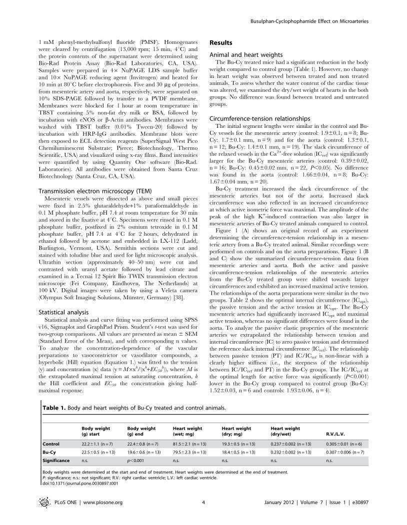
Figure 1. Circumference-tension relationships in control and Bu-Cy treated animals. (A) Original recording of force responses of amesenteric artery from a Bu-Cy treated mouse. The vessel circumference was changed stepwise (arrows) between contractions induced by high-K+.Maximum active force at the peak of contraction (Stars) and passive force at the end of the relaxation period (squares) immediately before the nextcircumference step were determined. When the vessel had been stretched to a circumference above the optimal (ICopt), it was returned to ICopt forfurther pharmacological experimentation. (B and C) show mean values of passive (squares) and active (circles) tension plotted against vesselcircumference in resistance arteries (B; controls n = 4; Bu-Cy: n = 5) and aorta (C; controls n = 3; Bu-Cy: n = 6). Open and filled symbols show data ofcontrols and Bu-Cy animals, respectively.doi:10.1371/journal.pone.0030897.g001
Busulphan-Cyclophophamide Effect on Microarteries
PLoS ONE | www.plosone.org 3 January 2012 | Volume 7 | Issue 1 | e30897
1 mM phenyl-methylsulfonyl fluoride (PMSF). Homogenates
were cleared by centrifugation (13,000 rpm; 15 min, 4uC) and
the protein contents of the supernatant were determined using
Bio-Rad Protein Assay (Bio-Rad Laboratories, CA, USA).
Samples were prepared in 46 NuPAGE LDS sample buffer
and 106 NuPAGE reducing agent (Invitrogen) and heated for
10 min at 80uC before electrophoresis. Five and 30 mg of proteins
from mesenteric artery and aorta, respectively, were separated on
10% SDS-PAGE followed by transfer to a PVDF membrane.
Membranes were blocked for 1 hour at room temperature in
TBST containing 5% non-fat dry milk or BSA, followed by
incubation with eNOS or b-Actin antibodies. Membranes were
washed with TBST buffer (0.01% Tween-20) followed by
incubation with HRP-IgG antibodies. Membrane blots were
then exposed to ECL detection reagents (SuperSignal West Pico
Chemiluminescent Substrate; Pierce; Biotechnology, Thermo
Scientific, USA) and visualized using x-ray films. Band intensities
were quantified by using Quantity One software (Bio-Rad.
Laboratories). All antibodies were obtained from Santa Cruz
Biotechnology (Santa Cruz, CA, USA).
Transmission electron microscopy (TEM)Mesenteric vessels were dissected as above and small pieces
were fixed in 2.5% glutaraldehyde+1% paraformaldehyde in
0.1 M phosphate buffer, pH 7.4 at room temperature for 30 min
and stored in the fixative at 4uC. Specimens were rinsed in 0.1 M
phosphate buffer, postfixed in 2% osmium tetroxide in 0.1 M
phosphate buffer, pH 7.4 at 4uC for 2 hours, dehydrated in
ethanol followed by acetone and embedded in LX-112 (Ladd,
Burlington, Vermont, USA). Semithin sections were cut and
stained with toludine blue and used for light microscopic analysis.
Ultrathin section (approximately 40–50 nm) were cut and
contrasted with uranyl acetate followed by lead citrate and
examined in a Tecnai 12 Spirit Bio TWIN transmission electron
microscope (Fei Company, Eindhoven, The Netherlands) at
100 kV. Digital images were taken by using a Veleta camera
(Olympus Soft Imaging Solutions, Munster, Germany) [38].
Statistical analysisStatistical analysis and curve fitting was performed using SPSS
v16, Sigmaplot and GraphPad Prism. Student’s t-test was used for
two-group comparisons. All values are presented as mean 6 SEM
(Standard Error of the Mean), and with corresponding n values.
To analyze the concentration-dependence of the vascular
preparations to vasoconstrictor or vasodilator compounds, a
hyperbolic (Hill) equation (Equation 1.) was fitted to the tension
(y) and concentration (x) data (y = M6xh/(xh+EC50h)), where M is
the extrapolated maximal tension at saturating concentration, h
the Hill coefficient and EC50 the concentration giving half-
maximal response.
Results
Animal and heart weightsThe Bu-Cy treated mice had a significant reduction in the body
weight compared to control group (Table 1). However, no change
in heart weight was observed between treated and non treated
animals. To assess whether the water content of the cardiac tissue
was altered, we examined the dry/wet weight of hearts in the both
groups. No difference was found between treated and untreated
groups.
Circumference-tension relationshipsThe initial segment lengths were similar in the control and Bu-
Cy vessels for the mesenteric artery (control: 1.960.1, n = 8; Bu-
Cy: 1.760.1 mm, n = 9) and for the aorta (control: 1.360.1,
n = 12; Bu-Cy: 1.460.1 mm, n = 19). The slack circumference of
the relaxed vessels in the Ca2+-free solution (ICref) was significantly
larger for the Bu-Cy mesenteric arteries (control: 0.3960.02,
n = 16; Bu-Cy: 0.4560.02 mm, n = 22, P,0.05). No difference
was found in the aorta (control: 1.6660.04, n = 8; Bu-Cy:
1.6760.04 mm, n = 20).
Bu-Cy treatment increased the slack circumference of the
mesenteric arteries but not of the aorta. Increased slack
circumference was also reflected in an increased circumference
at which active isometric force was maximal. The amplitude of the
peak of the high K+-induced contraction was also larger in
mesenteric arteries of Bu-Cy treated animals compared to control.
Figure 1 (A) shows an original record of an experiment
determining the circumference-tension relationship in a mesen-
teric artery from a Bu-Cy treated animal. Similar recordings were
performed on controls and on the aorta preparations. Figure 1 (B
and C) show the summarized circumference-tension data from
mesenteric arteries and aorta. Both the active and passive
circumference-tension relationships of the mesenteric arteries
from the Bu-Cy treated group were shifted towards larger
circumferences and exhibited an increased maximal active tension.
The relationships of the aorta preparations were similar in the two
groups. Table 2 shows the optimal internal circumference (ICopt),
the passive tension and the active tension at ICopt. The Bu-Cy
mesenteric arteries had significantly increased ICopt and maximal
active tension, whereas no significant differences were found in the
aorta. To analyze the passive elastic properties of the mesenteric
arteries we extrapolated the relationship between tension and
internal circumference (IC) to zero passive tension and determined
the reference slack internal circumference (ICref). The relationship
between passive tension (PT) and IC/ICref is non-linear with a
clearly higher stiffness (i.e., the steepness of the relationship
between IC/ICref and PT) in the Bu-Cy groups. The IC/ICref at
the optimal length for active force was significantly (P,0.001)
lower in the Bu-Cy group compared to control group (Bu-Cy:
1.5260.03, n = 6 and controls: 1.9360.06, n = 4).
Table 1. Body and heart weights of Bu-Cy treated and control animals.
Body weight(g) start
Body weight(g) end
Heart weight(wet; mg)
Heart weight(dry; mg)
Heart weight(dry/wet) R.V./L.V.
Control 22.261.1 (n = 7) 22.460.8 (n = 7) 81.562.1 (n = 13) 19.360.5 (n = 13) 0.23760.002 (n = 13) 0.30560.01 (n = 6)
Bu-Cy 22.560.5 (n = 13) 19.660.6 (n = 13) 79.562.3 (n = 13) 18.460.5 (n = 13) 0.23260.002 (n = 13) 0.30760.006 (n = 7)
Significance n.s. p,0.001 n.s. n.s. n.s. n.s.
Body weights were determined at the start and end of treatment. Heart weights were determined at the end of treatment.P: significance; n.s.: not significant; R.V.: right cardiac ventricle; L.V.: left cardiac ventricle.doi:10.1371/journal.pone.0030897.t001
Busulphan-Cyclophophamide Effect on Microarteries
PLoS ONE | www.plosone.org 4 January 2012 | Volume 7 | Issue 1 | e30897
Pharmacological reactivityThe circumference-tension data showed that passive tension at
optimal circumference (ICopt) was similar in the control and Bu-Cy
vessels, both for mesenteric arteries and aorta. We therefore
stretched the preparations to passive tensions close to that at ICopt
for examination of effects of contractile and relaxant agonists. We
normalized the contractile responses to the high-K+ tension,
determined for each preparation.
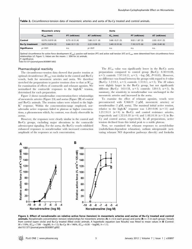
Figure 2 shows noradrenaline concentration-force relationships
of mesenteric arteries (Figure 2A) and aortas (Figure 2B) of control
and Bu-Cy animals. The tension values were related to the high-
K+ response. Within the concentration-range employed, nor-
adrenalin active responses failed to plateau at higher concentra-
tions, a phenomenon which, by contrast, was clearly observable in
aortas.
However, the responses were clearly similar in the control and
Bu-Cy groups, excluding major alterations in the contractile
adrenoceptor signaling. For the aorta, the Bu-Cy vessels exhibited
enhanced responses to noradrenaline with increased contraction
amplitude of the responses at each concentration.
The EC50 value was significantly lower in the Bu-Cy aorta
preparations compared to control group (Bu-Cy: 8.3060.08,
n = 5; controls: 7.9760.11, n = 5; 2log (M), P,0.05). However,
no difference was found between the groups with regard to h value
(Bu-Cy: 1.260.1, n = 5; controls: 1.360.1, n = 5). The M values
were slightly larger in the Bu-Cy group, but not significantly
different (Bu-Cy: 145618, n = 5; controls: 12063; n = 5). In
summary, the sensitivity to noradrenaline was unchanged in the
mesenteric arteries and increased in the aorta.
To examine the effect of relaxant agonists, vessels were
precontracted with U46619 (1 mM, mesenteric arteries) or
noradrenaline (1 mM, aorta). The maximal initial active tension,
relative to the high-K+ response was 1.2060.06 (n = 11) and
1.2160.11 (n = 6) in Bu-Cy and control resistance arteries,
respectively and 1.5260.18 (n = 6) and 1.4860.16 (n = 3) in Bu-
Cy and control aortas, respectively. In all preparations, active
tension declined from this initial peak to a stable plateau.
Next, we examined the relaxant responses to acetylcholine
(endothelium-dependent relaxation), sodium nitroprusside (acti-
vating relaxant NO dependent pathways directly) and forskolin
Table 2. Circumference-tension data of mesenteric arteries and aorta of Bu-Cy treated and control animals.
Mesenteric artery Aorta
ICopt (mm) PT (mN/mm) AT (mN/mm) ICo (mm) PT (mN/mm) AT (mN/mm)
Control 0.57960.019 (4) 0.7360.12 (4) 1.6860.11 (4) 3.6860.21 (3) 9.0261.87 (3) 2.0360.51 (3)
Bu-Cy treatment 0.67560.014 (5) 0.6660.11 (5) 2.2260.08 (5) 3.4060.10 (6) 7.1860.75 (6) 2.0460.40 (6)
Significance p,0.01 n.s. p,0.01 n.s. n.s. n.s.
Optimal circumference for active force development (ICopt); passive wall tension (PT) and active wall tension (AT) at ICopt, were determined from circumference forcerelationships (cf. Figure 1). Values are the means 6 SEM for (n) animals.P: significance;doi:10.1371/journal.pone.0030897.t002
Figure 2. Effect of noradrenalin on relative active force (tension) in mesenteric arteries and aortas of Bu-Cy treated and controlanimals. Noradrenalin concentration–tension relationships for mesenteric arteries (A; n = 6 in each group) and aorta (B; n = 5 in each group). Vesselsfrom control (open circle) and Bu-Cy treated (filled circle) animals. A hyperbolic equation (see Results) was fitted to mean values in B (Control:M = 119%, EC50 = 7.99 2log(M), h = 1.3; Bu-Cy: M = 146%, EC50 = 8.30 2log(M), h = 1.1).doi:10.1371/journal.pone.0030897.g002
Busulphan-Cyclophophamide Effect on Microarteries
PLoS ONE | www.plosone.org 5 January 2012 | Volume 7 | Issue 1 | e30897
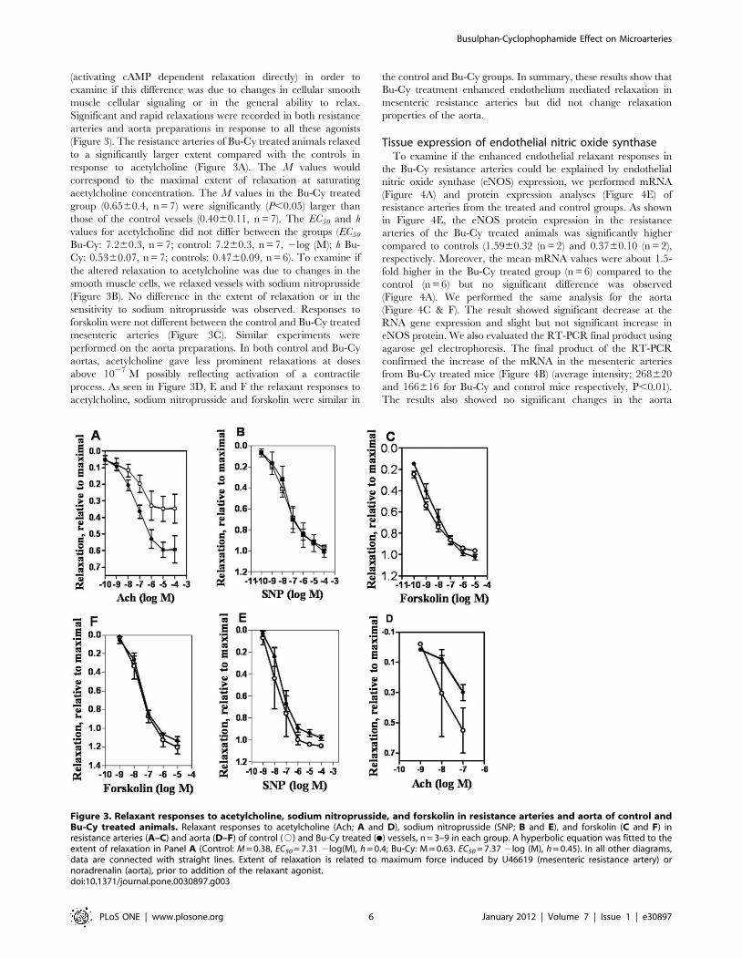
(activating cAMP dependent relaxation directly) in order to
examine if this difference was due to changes in cellular smooth
muscle cellular signaling or in the general ability to relax.
Significant and rapid relaxations were recorded in both resistance
arteries and aorta preparations in response to all these agonists
(Figure 3). The resistance arteries of Bu-Cy treated animals relaxed
to a significantly larger extent compared with the controls in
response to acetylcholine (Figure 3A). The M values would
correspond to the maximal extent of relaxation at saturating
acetylcholine concentration. The M values in the Bu-Cy treated
group (0.6560.4, n = 7) were significantly (P,0.05) larger than
those of the control vessels (0.4060.11, n = 7). The EC50 and h
values for acetylcholine did not differ between the groups (EC50
Bu-Cy: 7.260.3, n = 7; control: 7.260.3, n = 7, 2log (M); h Bu-
Cy: 0.5360.07, n = 7; controls: 0.4760.09, n = 6). To examine if
the altered relaxation to acetylcholine was due to changes in the
smooth muscle cells, we relaxed vessels with sodium nitroprusside
(Figure 3B). No difference in the extent of relaxation or in the
sensitivity to sodium nitroprusside was observed. Responses to
forskolin were not different between the control and Bu-Cy treated
mesenteric arteries (Figure 3C). Similar experiments were
performed on the aorta preparations. In both control and Bu-Cy
aortas, acetylcholine gave less prominent relaxations at doses
above 1027 M possibly reflecting activation of a contractile
process. As seen in Figure 3D, E and F the relaxant responses to
acetylcholine, sodium nitroprusside and forskolin were similar in
the control and Bu-Cy groups. In summary, these results show that
Bu-Cy treatment enhanced endothelium mediated relaxation in
mesenteric resistance arteries but did not change relaxation
properties of the aorta.
Tissue expression of endothelial nitric oxide synthaseTo examine if the enhanced endothelial relaxant responses in
the Bu-Cy resistance arteries could be explained by endothelial
nitric oxide synthase (eNOS) expression, we performed mRNA
(Figure 4A) and protein expression analyses (Figure 4E) of
resistance arteries from the treated and control groups. As shown
in Figure 4E, the eNOS protein expression in the resistance
arteries of the Bu-Cy treated animals was significantly higher
compared to controls (1.5960.32 (n = 2) and 0.3760.10 (n = 2),
respectively. Moreover, the mean mRNA values were about 1.5-
fold higher in the Bu-Cy treated group (n = 6) compared to the
control (n = 6) but no significant difference was observed
(Figure 4A). We performed the same analysis for the aorta
(Figure 4C & F). The result showed significant decrease at the
RNA gene expression and slight but not significant increase in
eNOS protein. We also evaluated the RT-PCR final product using
agarose gel electrophoresis. The final product of the RT-PCR
confirmed the increase of the mRNA in the mesenteric arteries
from Bu-Cy treated mice (Figure 4B) (average intensity; 268620
and 166616 for Bu-Cy and control mice respectively, P,0.01).
The results also showed no significant changes in the aorta
Figure 3. Relaxant responses to acetylcholine, sodium nitroprusside, and forskolin in resistance arteries and aorta of control andBu-Cy treated animals. Relaxant responses to acetylcholine (Ach; A and D), sodium nitroprusside (SNP; B and E), and forskolin (C and F) inresistance arteries (A–C) and aorta (D–F) of control (#) and Bu-Cy treated (N) vessels, n = 3–9 in each group. A hyperbolic equation was fitted to theextent of relaxation in Panel A (Control: M = 0.38, EC50 = 7.31 2log(M), h = 0.4; Bu-Cy: M = 0.63. EC50 = 7.37 2log (M), h = 0.45). In all other diagrams,data are connected with straight lines. Extent of relaxation is related to maximum force induced by U46619 (mesenteric resistance artery) ornoradrenalin (aorta), prior to addition of the relaxant agonist.doi:10.1371/journal.pone.0030897.g003
Busulphan-Cyclophophamide Effect on Microarteries
PLoS ONE | www.plosone.org 6 January 2012 | Volume 7 | Issue 1 | e30897
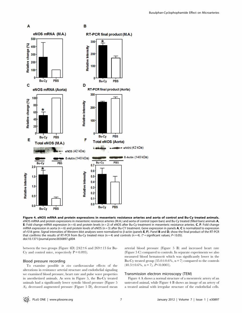
between the two groups (Figure 4D) (24266 and 269613 for Bu-
Cy and control mice, respectively P = 0.095).
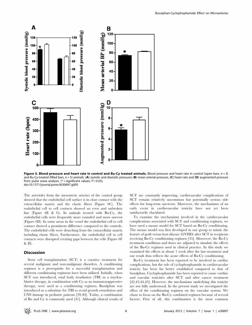
Blood pressure recordingTo examine possible in vivo cardiovascular effects of the
alterations in resistance arterial structure and endothelial signaling
we examined blood pressure, heart rate and pulse wave properties
in anesthetized animals. As seen in Figure 5, the Bu-Cy treated
animals had a significantly lower systolic blood pressure (Figure 5
A), decreased augmented pressure (Figure 5 D), decreased mean
arterial blood pressure (Figure 5 B) and increased heart rate
(Figure 5 C) compared to controls. In separate experiments we also
measured blood hematocrit which was significantly lower in the
Bu-Cy treated group (35.060.6%, n = 7) compared to the controls
(40.360.6%, n = 7), P,0.0001).
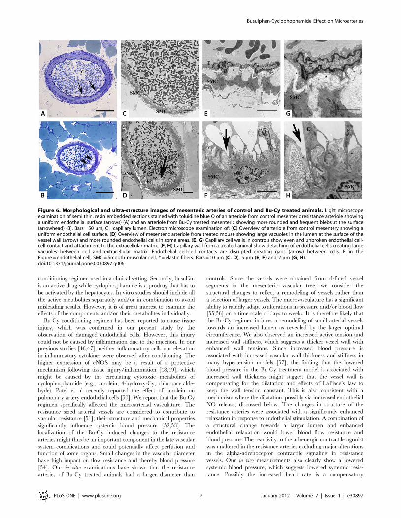
Transmission electron microscopy (TEM)Figure 4 A shows a normal structure of a mesenteric artery of an
untreated animal, while Figure 4 B shows an image of an artery of
a treated animal with irregular structure of the endothelial cells.
Figure 4. eNOS mRNA and protein expressions in mesenteric resistance arteries and aorta of control and Bu-Cy treated animals.eNOS mRNA and protein expressions in mesenteric resistance arteries (M.A.) and aorta of control (open bars) and Bu-Cy treated (filled bars) animals A,E: Fold change mRNA expression (n = 6) and protein levels (n = 2) of eNOS after Bu-Cy treatment in mesenteric resistance arteries. C, F: Fold changemRNA expression in aorta (n = 6) and protein levels of eNOS (n = 3) after Bu-CY treatment. Gene expression in panels A, C is normalized to expressionof rS18 gene. Signal intensities of Western blot analyses were normalized to b-actin (panels E, F). Panel B and D, show the final product of the RT-PCRthat confirms the results of RT-PCR from Bu-Cy treated mice (n = 4) and controls (n = 4). (* = significant values; P,0.05).doi:10.1371/journal.pone.0030897.g004
Busulphan-Cyclophophamide Effect on Microarteries
PLoS ONE | www.plosone.org 7 January 2012 | Volume 7 | Issue 1 | e30897
The arterioles from the mesenteric arteries of the control group
showed that the endothelial cell surface is in close contact with the
extracellular matrix and the elastic fibers (Figure 6C). The
endothelial cell to cell contacts showed an even and unbroken
line (Figure 6E & G). In animals treated with Bu-Cy, the
endothelial cells were frequently more rounded and more uneven
(Figure 6D). In some areas in the vessel the endothelial cell to cell
contact showed a prominent difference compared to the controls.
The endothelial cells were detaching from the extracellular matrix
including elastic fibers. Furthermore, the endothelial cell to cell
contacts were disrupted creating gaps between the cells (Figure 6F
& H).
Discussion
Stem cell transplantation (SCT) is a curative treatment for
several malignant and non-malignant disorders. A conditioning
regimen is a prerequisite for a successful transplantation and
different conditioning regimens have been utilized. Initially, when
SCT was introduced, total body irradiation (TBI) as a myeloa-
blative therapy, in combination with Cy as an immunosuppressive
therapy, were used as a conditioning regimen. Busulphan was
introduced as a substitute for TBI to avoid growth retardation and
CNS damage in pediatric patients [39,40]. Today, a combination
of Bu and Cy is commonly used [41]. Although clinical results of
SCT are constantly improving, cardiovascular complications of
SCT remain relatively uncommon but potentially serious side
effects for long-term survivors. Moreover, the mechanisms of an
early event in cardiovascular toxicity have not yet been
satisfactorily elucidated.
To examine the mechanisms involved in the cardiovascular
complications associated with SCT and conditioning regimen, we
have used a mouse model for SCT based on Bu-Cy conditioning.
The mouse model was first developed in our group to mimic the
feature of graft versus host disease (GVHD) after SCT in recipients
receiving Bu-Cy conditioning regimen [35]. Moreover, the Bu-Cy
treatment conditions and doses are adjusted to simulate the effects
of the Bu-Cy regimen used in clinical practice. In this study we
examined the effects at about 1 week after the last treatment and
our result thus reflects the acute effects of Bu-Cy conditioning.
Bu-Cy treatment has been reported to be involved in cardiac
complications, but the role of cyclophosphamide in cardiovascular
toxicity has been far better established compared to that of
busulphan. Cyclophosphamide has been reported to cause cardiac
and vascular toxicities after SCT and after cancer treatment
[42,43,44,45]. However, the mechanisms underlying this toxicity
are not fully understood. In the present study we investigated the
effect of the conditioning regimen on the vascular system. We
chose to focus on the Bu-Cy combined regimen because of several
factors. First of all, this combination is the most common
Figure 5. Blood pressure and heart rate in control and Bu-Cy treated animals. Blood pressure and heart rate in control (open bars, n = 3)and Bu-Cy treated (filled bars, n = 5) animals. (A) systolic and diastolic pressures (B) mean arterial pressure, (C) heart rate and (D) augmented pressurefrom pulse wave analysis. (* = significant values; P,0.05).doi:10.1371/journal.pone.0030897.g005
Busulphan-Cyclophophamide Effect on Microarteries
PLoS ONE | www.plosone.org 8 January 2012 | Volume 7 | Issue 1 | e30897
conditioning regimen used in a clinical setting. Secondly, busulfan
is an active drug while cyclophosphamide is a prodrug that has to
be activated by the hepatocytes. In vitro studies should include all
the active metabolites separately and/or in combination to avoid
misleading results. However, it is of great interest to examine the
effects of the components and/or their metabolites individually.
Bu-Cy conditioning regimen has been reported to cause tissue
injury, which was confirmed in our present study by the
observation of damaged endothelial cells. However, this injury
could not be caused by inflammation due to the injection. In our
previous studies [46,47], neither inflammatory cells nor elevation
in inflammatory cytokines were observed after conditioning. The
higher expression of eNOS may be a result of a protective
mechanism following tissue injury/inflammation [48,49], which
might be caused by the circulating cytotoxic metabolites of
cyclophosphamide (e.g., acrolein, 4-hydroxy-Cy, chloroacetalde-
hyde). Patel et al recently reported the effect of acrolein on
pulmonary artery endothelial cells [50]. We report that the Bu-Cy
regimen specifically affected the microarterial vasculature. The
resistance sized arterial vessels are considered to contribute to
vascular resistance [51]; their structure and mechanical properties
significantly influence systemic blood pressure [52,53]. The
localization of the Bu-Cy induced changes to the resistance
arteries might thus be an important component in the late vascular
system complications and could potentially affect perfusion and
function of some organs. Small changes in the vascular diameter
have high impact on flow resistance and thereby blood pressure
[54]. Our in vitro examinations have shown that the resistance
arteries of Bu-Cy treated animals had a larger diameter than
controls. Since the vessels were obtained from defined vessel
segments in the mesenteric vascular tree, we consider the
structural changes to reflect a remodeling of vessels rather than
a selection of larger vessels. The microvasculature has a significant
ability to rapidly adapt to alterations in pressure and/or blood flow
[55,56] on a time scale of days to weeks. It is therefore likely that
the Bu-Cy regimen induces a remodeling of small arterial vessels
towards an increased lumen as revealed by the larger optimal
circumference. We also observed an increased active tension and
increased wall stiffness, which suggests a thicker vessel wall with
enhanced wall tensions. Since increased blood pressure is
associated with increased vascular wall thickness and stiffness in
many hypertension models [57], the finding that the lowered
blood pressure in the Bu-Cy treatment model is associated with
increased wall thickness might suggest that the vessel wall is
compensating for the dilatation and effects of LaPlace’s law to
keep the wall tension constant. This is also consistent with a
mechanism where the dilatation, possibly via increased endothelial
NO release, discussed below. The changes in structure of the
resistance arteries were associated with a significantly enhanced
relaxation in response to endothelial stimulation. A combination of
a structural change towards a larger lumen and enhanced
endothelial relaxation would lower blood flow resistance and
blood pressure. The reactivity to the adrenergic contractile agonist
was unaltered in the resistance arteries excluding major alterations
in the alpha-adrenoceptor contractile signaling in resistance
vessels. Our in vivo measurements also clearly show a lowered
systemic blood pressure, which suggests lowered systemic resis-
tance. Possibly the increased heart rate is a compensatory
Figure 6. Morphological and ultra-structure images of mesenteric arteries of control and Bu-Cy treated animals. Light microscopeexamination of semi thin, resin embedded sections stained with toluidine blue O of an arteriole from control mesenteric resistance arteriole showinga uniform endothelial surface (arrows) (A) and an arteriole from Bu-Cy treated mesenteric showing more rounded and frequent blebs at the surface(arrowhead) (B). Bars = 50 mm, C = capillary lumen. Electron microscope examination of: (C) Overview of arteriole from control mesentery showing auniform endothelial cell surface. (D) Overview of mesenteric arteriole from treated mouse showing large vacuoles in the lumen at the surface of thevessel wall (arrow) and more rounded endothelial cells in some areas. (E, G) Capillary cell walls in controls show even and unbroken endothelial cell-cell contact and attachment to the extracellular matrix. (F, H) Capillary wall from a treated animal show detaching of endothelial cells creating largevacuoles between cell and extracellular matrix. Endothelial cell-cell contacts are disrupted creating gaps (arrow) between cells. E in theFigure = endothelial cell, SMC = Smooth muscular cell, * = elastic fibers. Bars = 10 mm (C, D), 5 mm (E, F) and 2 mm (G, H).doi:10.1371/journal.pone.0030897.g006
Busulphan-Cyclophophamide Effect on Microarteries
PLoS ONE | www.plosone.org 9 January 2012 | Volume 7 | Issue 1 | e30897
mechanism for a lower resistance. It should be noted that the
hematocrit is lowered after Bu-Cy treatment (present study, [58]),
which would further lower flow resistance due to decreased blood
viscosity.
We report an upregulation of endothelial nitric oxide synthase
(eNOS) in the wall of resistance arteries after Bu-Cy treatment.
This is most likely the main mechanism behind the enhanced
acetylcholine induced relaxation in the vessels. The link between
eNOS expression and blood pressure is well established [59] and
the increased eNOS expression and enhanced endothelial
responses can constitute a mechanism for the lowered blood
pressure after the Bu-Cy regimen. The link between Bu-Cy
treatment and altered eNOS expression remains to be examined.
It is also possible that the compounds influence the cellular
signaling in the vascular endothelium or the production of NO
directly. This could be due to damage caused by the Cy
metabolites, e.g., acrolein which has been reported to increase
NO production [60]. The lowered hematocrit and anemia can be
further contributing factors. Erythropoietin can affect eNOS
expression in different ways [61,62] and eNOS expression is
increased in animal models of sickle cell anemia [63]. The
endothelial function and eNOS expression in the aorta were less
affected by the Bu-Cy treatment (P = 0.095), which can reflect
differences in the endothelial sensitivity between vessel types or in
the in vivo exposure to the compound. Another explanation can be
the earlier /faster transcription and protein synthesis in the aorta
compared to the mesenteric artery. It should be noted that NO
signaling affects vascular structure [64] and the altered NO
signaling can be secondary to the structural injury in the resistance
arteries. Recently, Perry et al. have reported that bone marrow-
derived endothelial progenitor cells do not restore normal arterial
endothelium in young transplanted endothelial nitric-oxide
synthase deficient or wild type mice [65]. We provide morpho-
logical data after treatment with Bu-Cy and report effects on
endothelial cells in the mesenteric arteries with detachment and
formation of gaps (Figure 6). Such endothelial gaps may allow for
uncontrolled leak of fluids from the lumen of arterioles into the
extracellular matrix of the mesenteric arteries resulting in edema.
The link between the morphological changes and the increased
eNOS expression in correlation with increased endothelium
mediated relaxation warrant further investigation. A change in
endothelial function and integrity may also be involved in other
types of tissue damage such as the hemorrhagic cystitis following
cyclophosphamide treatment [26,27]. Recently, Zeng et al have
reported an increased number of circulating endothelial cells
during the early phases of conditioning using methotrexate/
cyclophosphamide or Bu-Cy as a sign of vascular endothelium
injury in a transplantation mouse model [66,67].
The large elastic artery aorta was not affected in a manner
similar to that of resistance arteries. These larger vessels would
mainly contribute to pulse wave properties and possibly load on
the heart. No structural changes could be detected on the basis of
our mechanical experiments and the endothelial function was
unaffected. The sensitivity to noradrenalin was increased suggest-
ing that these vessels could have an increased tone and wall tension
in vivo, particularly during episodes of enhanced sympathetic drive
or increased plasma concentrations of catecholamines. This
change might have an effect in some physiological or pathophys-
iological situations with high adrenergic tone, but since we could
not detect an increase in blood pressure, a major effect on vascular
compliance of the altered adrenergic signaling in resting conditions
can be excluded. In the clinical setting, hypertension has been
reported after SCT. The mechanism underlying the differential
effects of By-Cy on large and small arteries is unknown; it might
relate to the bioavailability or metabolism of the drugs in the
different vessels.
Our data indicate enhanced endothelial relaxation and
alterations of endothelial structure in resistance arteries. These
effects appear to be dominant over the enhanced adrenergic
responses of the aorta: the net result being a lower blood pressure.
Most likely, this reflects an early event in the cardiovascular
changes induced by Bu-Cy conditioning. It is possible that longer
observation periods, beyond the period focused upon in this study,
would reveal the development of hypertension as a result of further
changes in endothelial function in resistance arteries as well as a
larger impact of the large arterial reactivity changes. Another
explanation of the hypertension observed in patients is that
patients during SCT are treated with multiple drugs including
calcineurin inhibitors that are known for their hypertensive effect.
In the present investigation, using the current regimen and
observation period, we could not detect any cardiac weight
changes. This may indicate that major alterations in cardiac gross
structure or edema formation have not developed. Blood pressure
was lower and the heart rate higher in treated animals compared
to controls, suggesting an autonomic stimulation of the heart
possibly as a result of lowered vascular resistance, as discussed
above. However, a longer observation period and examination of
cardiac ultrastructure together with functional data are warranted
to fully assess possible cardiovascular changes.
In conclusion, we have shown that Bu-Cy treatment has a
significant acute impact on the vascular system with selectivity for
the smaller resistance arteries controlling blood pressure. After
treatment, the vessels become larger and have increased
endothelium mediated relaxation, most likely via an increased
expression of eNOS. Interestingly, the increased eNOS expression
and endothelium mediated relaxation were associated with altered
endothelial structure possibly affecting wall permeability. The
endothelium performs several functions in the vascular wall. Our
analysis of vessel structure suggests that the barrier function can be
altered, possibly associated with an increased risk for with
extravasation or edema formation in some vascular beds. At the
same time, the endothelial relaxation of vascular tone is enhanced,
showing that the structural changes do not impair the NO
mediated relaxant function, but rather leads to an upregulation. In
contrast, structure and endothelial function of larger arteries are
less affected, although these vessels have increased reactivity to
adrenergic agonists which may contribute to increased wall
stiffness, and under high adrenergic tone may result in increased
cardiac load.
High doses of alkylating drugs such as cyclophosphamide and
ifosfamide may result in reversible heart failure and life-
threatening arrhythmias, while the antimetabolites 5-fluorouracil
and capecitabine were shown to induce myocardial ischemia.
Moreover, anthracyclines such as daunorubicin and doxorubicin
were shown to be involved in the development of cardiomyopathy.
Introducing Cy to the conditioning regimen prior to SCT
increased the onset of cardiotoxicity [28,29], and a positive
correlation between the dose of Cy and severity of cardio toxicity
was reported [30]. Unfortunately, the symptoms usually appear 10
to 20 years after SCT in long term survivals [31], but cardiac
failure has also been reported within weeks of Cy exposure
[32].This is the first study to show that Bu-Cy conditioning prior
to SCT causes vascular injury and remodeling, and that it alters
reactivity of different vascular segments divergently. These results
may contribute to a better understanding of cardiovascular
complications reported after SCT in order to enhance prophylac-
tic treatment strategies and/or to optimize conditioning regimen
using other drugs.
Busulphan-Cyclophophamide Effect on Microarteries
PLoS ONE | www.plosone.org 10 January 2012 | Volume 7 | Issue 1 | e30897

Acknowledgments
The authors would like to acknowledge Professor Christer Sylven and Dr.
Karl-Henrik Grinnemo for valuable discussions.
Author Contributions
Conceived and designed the experiments: SA-H PJMB AA ZH MH.
Performed the experiments: SA-H PJMB FZ BS JS KH ZH. Analyzed the
data: SA-H PJMB FZ BS JS KH ZH AA MH. Contributed reagents/
materials/analysis tools: SA-H PJMB FZ BS JS KH ZH AA MH. Wrote
the paper: SA-H PJMB FZ BS JS KH ZH AA MH.
References
1. Gyurkocza B, Rezvani A, Storb RF (2010) Allogeneic hematopoietic cell
transplantation: the state of the art. Expert Rev Hematol 3: 285–299.
2. Jenq RR, van den Brink MR (2010) Allogeneic haematopoietic stem cell
transplantation: individualized stem cell and immune therapy of cancer. Nat Rev
Cancer 10: 213–221.
3. Ringden O, Le Blanc K (2005) Allogeneic hematopoietic stem cell transplan-
tation: state of the art and new perspectives. APMIS 113: 813–830.
4. Naik S, Wong R, Arai S, Brown J, Laport G, et al. (2011) Long-term outcomes
in patients with high-risk myeloid malignancies following matched related donor
hematopoietic cell transplantation with myeloablative conditioning of BU,
etoposide and CY. Bone Marrow Transplant 46: 192–199.
5. Cazin B, Gorin NC, Laporte JP, Gallet B, Douay L, et al. (1986) Cardiac
complications after bone marrow transplantation. A report on a series of 63
consecutive transplantations. Cancer 57: 2061–2069.
6. Senkus E, Jassem J (2011) Cardiovascular effects of systemic cancer treatment.
Cancer Treat Rev 37: 300–311.
7. Bearman SI, Petersen FB, Schor RA, Denney JD, Fisher LD, et al. (1990)
Radionuclide ejection fractions in the evaluation of patients being considered for
bone marrow transplantation: risk for cardiac toxicity. Bone Marrow Transplant
5: 173–177.
8. Murdych T, Weisdorf DJ (2001) Serious cardiac complications during bone
marrow transplantation at the University of Minnesota, 1977–1997. Bone
Marrow Transplant 28: 283–287.
9. Tichelli A, Passweg J, Wojcik D, Rovo A, Harousseau JL, et al. (2008) Late
cardiovascular events after allogeneic hematopoietic stem cell transplantation: a
retrospective multicenter study of the Late Effects Working Party of the
European Group for Blood and Marrow Transplantation. Haematologica 93:
1203–1210.
10. Majhail NS, Challa TR, Mulrooney DA, Baker KS, Burns LJ (2009)
Hypertension and diabetes mellitus in adult and pediatric survivors of allogeneic
hematopoietic cell transplantation. Biol Blood Marrow Transplant 15:
1100–1107.
11. Armenian SH, Bhatia S (2008) Cardiovascular disease after hematopoietic cell
transplantation–lessons learned. Haematologica 93: 1132–1136.
12. Armenian SH, Sun CL, Francisco L, Steinberger J, Kurian S, et al. (2008) Late
congestive heart failure after hematopoietic cell transplantation. J Clin Oncol 26:
5537–5543.
13. Ghobrial IM, Bunch TJ, Caplice NM, Edwards WD, Miller DV, et al. (2004)
Fatal coronary artery disease after unrelated donor bone marrow transplanta-
tion. Mayo Clin Proc 79: 403–406.
14. Uderzo C, Pillon M, Tridello G, Dini G, Urban C, et al. (2009) Cardiac and
pulmonary late effects do not negatively influence performance status and non-
relapse mortality of children surviving five yr after autologous hematopoietic cell
transplantation: report from the EBMT Paediatric Diseases and Late Effects
Working Parties. Pediatr Transplant 13: 719–724.
15. Sohn SK, Kim JG, Kim DH, Lee KB (2003) Cardiac morbidity in advanced
chronic myelogenous leukaemia patients treated by successive allogeneic stem
cell transplantation with busulphan/cyclophosphamide conditioning after
imatinib mesylate administration. Br J Haematol 121: 469–472.
16. Tichelli A, Bucher C, Rovo A, Stussi G, Stern M, et al. (2007) Premature
cardiovascular disease after allogeneic hematopoietic stem-cell transplantation.
Blood 110: 3463–3471.
17. Doll DC, Yarbro JW (1992) Vascular toxicity associated with antineoplastic
agents. Semin Oncol 19: 580–596.
18. Doll DC, Yarbro JW (1994) Vascular toxicity associated with chemotherapy and
hormonotherapy. Curr Opin Oncol 6: 345–350.
19. Limsuwan A, Pakakasama S, Rochanawutanon M, Hong-eng S (2006)
Pulmonary arterial hypertension after childhood cancer therapy and bone
marrow transplantation. Cardiology 105: 188–194.
20. Berthou C, Devergie A, D’Agay MF, Sonsino E, Scrobohaci ML, et al. (1991)
Late vascular complications after bone marrow transplantation for dyskeratosis
congenita. Br J Haematol 79: 335–336.
21. Ferry C, Socie G (2003) Busulfan-cyclophosphamide versus total body
irradiation-cyclophosphamide as preparative regimen before allogeneic hema-
topoietic stem cell transplantation for acute myeloid leukemia: what have we
learned? Exp Hematol 31: 1182–1186.
22. Santos GW (1989) Busulfan (Bu) and cyclophosphamide (Cy) for marrow
transplantation. Bone Marrow Transplant 4 Suppl 1: 236–239.
23. Busse D, Kroemer HK, Sperker B, Murdter TE (1998) [Treatment of cancer
with cytostatics: results with high dose therapy with cyclophosphamide and drug
targeting with doxorubicin]. Pharm Unserer Zeit 27: 216–222.
24. Luznik L, Bolanos-Meade J, Zahurak M, Chen AR, Smith BD, et al. (2010)High-dose cyclophosphamide as single agent, short-course prophylaxis of graft-
versus-host disease. Blood.
25. Luznik L, Fuchs EJ (2010) High-dose, post-transplantation cyclophosphamide to
promote graft-host tolerance after allogeneic hematopoietic stem cell transplan-tation. Immunol Res.
26. Cho KH, Hyun JH, Chang YS, Na YG, Shin JH, et al. (2010) Expression ofnitric oxide synthase and aquaporin-3 in cyclophosphamide treated rat bladder.
Int Neurourol J 14: 149–156.
27. Oter S, Korkmaz A, Oztas E, Yildirim I, Topal T, et al. (2004) Inducible nitric
oxide synthase inhibition in cyclophosphamide induced hemorrhagic cystitis inrats. Urol Res 32: 185–189.
28. Santos GW, Sensenbrenner LL, Burke PJ, Colvin M, Owens AH, Jr., et al.(1971) Marrow transplanation in man following cyclophosphamide. Transplant
Proc 3: 400–404.
29. Storb R, Buckner CD, Dillingham LA, Thomas ED (1970) Cyclophosphamide
regimens in rhesus monkey with and without marrow infusion. Cancer Res 30:
2195–2203.
30. Goldberg MA, Antin JH, Guinan EC, Rappeport JM (1986) Cyclophosphamide
cardiotoxicity: an analysis of dosing as a risk factor. Blood 68: 1114–1118.
31. Tichelli A, Bhatia S, Socie G (2008) Cardiac and cardiovascular consequences
after haematopoietic stem cell transplantation. Br J Haematol 142: 11–26.
32. Ayash LJ, Wright JE, Tretyakov O, Gonin R, Elias A, et al. (1992)
Cyclophosphamide pharmacokinetics: correlation with cardiac toxicity andtumor response. J Clin Oncol 10: 995–1000.
33. Terpstra W, de Maat CE (1989) Pericardial fibrosis following busulfan
treatment. Neth J Med 35: 249–252.
34. Takatsuka H, Takemoto Y, Yamada S, Wada H, Tamura S, et al. (2000)
Complications after bone marrow transplantation are manifestations of systemic
inflammatory response syndrome. Bone Marrow Transplant 26: 419–426.
35. Sadeghi B, Aghdami N, Hassan Z, Forouzanfar M, Rozell B, et al. (2008)
GVHD after chemotherapy conditioning in allogeneic transplanted mice. BoneMarrow Transplant 42: 807–818.
36. Spiers A, Padmanabhan N (2005) A guide to wire myography. Methods MolMed 108: 91–104.
37. Agabiti-Rosei E, Mancia G, O’Rourke MF, Roman MJ, Safar ME, et al. (2007)Central blood pressure measurements and antihypertensive therapy: a consensus
document. Hypertension 50: 154–160.
38. Park CB, Asin-Cayuela J, Camara Y, Shi Y, Pellegrini M, et al. (2007) MTERF3
is a negative regulator of mammalian mtDNA transcription. Cell 130: 273–285.
39. Bunin N, Aplenc R, Kamani N, Shaw K, Cnaan A, et al. (2003) Randomized
trial of busulfan vs total body irradiation containing conditioning regimens forchildren with acute lymphoblastic leukemia: a Pediatric Blood and Marrow
Transplant Consortium study. Bone Marrow Transplant 32: 543–548.
40. Hassan M, Ehrsson H, Ljungman P (1996) Aspects concerning busulfan
pharmacokinetics and bioavailability. Leuk Lymphoma 22: 395–407.
41. Gupta T, Kannan S, Dantkale V, Laskar S (2011) Cyclophosphamide plus total
body irradiation compared with busulfan plus cyclophosphamide as aconditioning regimen prior to hematopoietic stem cell transplantation in
patients with leukemia: a systematic review and meta-analysis. Hematol Oncol
Stem Cell Ther 4: 17–29.
42. Wang X, Zhang J, Xu T (2009) Cyclophosphamide-evoked heart failure involves
pronounced co-suppression of cytoplasmic thioredoxin reductase activity andnon-protein free thiol level. Eur J Heart Fail 11: 154–162.
43. Yeh ET, Tong AT, Lenihan DJ, Yusuf SW, Swafford J, et al. (2004)Cardiovascular complications of cancer therapy: diagnosis, pathogenesis, and
management. Circulation 109: 3122–3131.
44. Zver S, Zadnik V, Bunc M, Rogel P, Cernelc P, et al. (2007) Cardiac toxicity of
high-dose cyclophosphamide in patients with multiple myeloma undergoingautologous hematopoietic stem cell transplantation. Int J Hematol 85: 408–414.
45. Albini A, Pennesi G, Donatelli F, Cammarota R, De Flora S, et al. (2010)Cardiotoxicity of anticancer drugs: the need for cardio-oncology and cardio-
oncological prevention. J Natl Cancer Inst 102: 14–25.
46. Al-Hashmi S, Hassan Z, Sadeghi B, Rozell B, Hassan M (2011) Dynamics of
early histopathological changes in GVHD after busulphan/cyclophosphamideconditioning regimen. Int J Clin Exp Pathol 4: 596–605.
47. Sadeghi B, Al-Hashmi S, Hassan Z, Rozell B, Concha H, et al. (2010) Expansionand activation kinetics of immune cells during early phase of GVHD in mouse
model based on chemotherapy conditioning. Clin Dev Immunol 2010: 142943.
48. Clapp BR, Hirschfield GM, Storry C, Gallimore JR, Stidwill RP, et al. (2005)
Inflammation and endothelial function: direct vascular effects of human C-
reactive protein on nitric oxide bioavailability. Circulation 111: 1530–1536.
Busulphan-Cyclophophamide Effect on Microarteries
PLoS ONE | www.plosone.org 11 January 2012 | Volume 7 | Issue 1 | e30897
49. Antoniades C, Cunnington C, Antonopoulos A, Neville M, Margaritis M, et al.
(2011) Induction of Vascular GTP-Cyclohydrolase I and Endogenous Tetra-hydrobiopterin Synthesis Protect Against Inflammation-Induced Endothelial
Dysfunction in Human Atherosclerosis. Circulation 124: 1860–1870.
50. Patel JM, Block ER (1993) Acrolein-induced injury to cultured pulmonary arteryendothelial cells. Toxicol Appl Pharmacol 122: 46–53.
51. Rosei EA, Rizzoni D (2005) Pathophysiology and clinical meaning of smallresistance artery remodeling. Curr Hypertens Rep 7: 79–80.
52. Mulvany MJ (2002) Small artery remodeling and significance in the
development of hypertension. News Physiol Sci 17: 105–109.53. Mulvany MJ (2008) Small artery remodelling in hypertension: causes,
consequences and therapeutic implications. Med Biol Eng Comput 46: 461–467.54. Peterson L (1966) Physical Factors Which Influence Vascular Caliber and Blood
Flow. Circulation Research.55. Boels PJ, Arner A, Malmqvist U, Uvelius B (1994) Structure and mechanics of
growing arterial microvessels from hypertrophied urinary bladder in the rat.
Pflugers Arch 426: 506–515.56. Pries AR, Secomb TW, Gaehtgens P (1996) Biophysical aspects of blood flow in
the microvasculature. Cardiovasc Res 32: 654–667.57. Stumpe KO, Ludwig M, Heagerty AM, Kolloch RE, Mancia G, et al. (1995)
Vascular wall thickness in hypertension: the Perindopril Regression of Vascular
Thickening European Community Trial: PROTECT. Am J Cardiol 76:50E–54E.
58. Molyneux G, Andrews M, Sones W, York M, Barnett A, et al. (2011)Haemotoxicity of busulphan, doxorubicin, cisplatin and cyclophosphamide in
the female BALB/c mouse using a brief regimen of drug administration. CellBiol Toxicol 27: 13–40.
59. Shesely EG, Maeda N, Kim HS, Desai KM, Krege JH, et al. (1996) Elevated
blood pressures in mice lacking endothelial nitric oxide synthase. Proc Natl AcadSci U S A 93: 13176–13181.
60. Korkmaz A, Topal T, Oter S (2007) Pathophysiological aspects of cyclophos-
phamide and ifosfamide induced hemorrhagic cystitis; implication of reactiveoxygen and nitrogen species as well as PARP activation. Cell Biol Toxicol 23:
303–312.61. Kanagy NL, Perrine MF, Cheung DK, Walker BR (2003) Erythropoietin
administration in vivo increases vascular nitric oxide synthase expression.
J Cardiovasc Pharmacol 42: 527–533.62. Wang XQ, Vaziri ND (1999) Erythropoietin depresses nitric oxide synthase
expression by human endothelial cells. Hypertension 33: 894–899.63. Kaul DK, Liu XD, Fabry ME, Nagel RL (2000) Impaired nitric oxide-mediated
vasodilation in transgenic sickle mouse. Am J Physiol Heart Circ Physiol 278:H1799–1806.
64. Moreau P, Takase H, Kung CF, van Rooijen MM, Schaffner T, et al. (1995)
Structure and function of the rat basilar artery during chronic nitric oxidesynthase inhibition. Stroke 26: 1922–1928; discussion 1928–1929.
65. Perry TE, Song M, Despres DJ, Kim SM, San H, et al. (2009) Bone marrow-derived cells do not repair endothelium in a mouse model of chronic endothelial
cell dysfunction. Cardiovasc Res 84: 317–325.
66. Zeng L, Jia L, Xu S, Yan Z, Ding S, et al. (2010) Vascular endothelium changesafter conditioning in hematopoietic stem cell transplantation: role of cyclophos-
phamide and busulfan. Transplant Proc 42: 2720–2724.67. Zeng L, Yan Z, Ding S, Xu K, Wang L (2008) Endothelial injury, an intriguing
effect of methotrexate and cyclophosphamide during hematopoietic stem celltransplantation in mice. Transplant Proc 40: 2670–2673.
Busulphan-Cyclophophamide Effect on Microarteries
PLoS ONE | www.plosone.org 12 January 2012 | Volume 7 | Issue 1 | e30897
Top Related
Copyright © 2022 FDOKUMEN

