


Bahasa
Halaman
Hukum


The
Journ
al o
f Exp
erim
enta
l M
edic
ine
ARTICLE
JEM © The Rockefeller University Press $15.00
Vol. 204, No. 3, March 19, 2007 497–510 www.jem.org/cgi/doi/10.1084/jem.20061780
497
Blood-borne naive lymphocytes continuously enter secondary lymphoid organs (SLOs), such as peripheral lymph nodes (PLNs) and spleen, where they screen APCs in search of their cog-nate antigen. In absence of infl ammation, lym-phocytes leave PLNs after an average dwell time of 12–24 h to enter lymphatic vessels lo-cated in the lymph node medulla (1, 2). Eff erent lymph vessels carry lymphocytes back into the blood circulation from where they reinitiate
their recirculation pathway. Their long life span and constant migration through SLOs make lymphocytes one of the most motile mammalian cell types (1, 2). Together with ad-hesion receptor molecules, two subfamilies of lymphocyte-expressed G protein–coupled re-ceptors (GPCRs) are known to guide lympho-cyte traffi cking: chemokine receptors (CKR), including CCR7, CXCR4, and CXCR5, and the sphingosine-1-phosphate (S1P) receptor 1, S1P1 (1, 2).
SLO-expressed homeostatic chemokines, such as the CCR7 ligands CCL19 and CCL21, the CXCR5 ligand CXCL13, and the CXCR4
A central role for DOCK2 during interstitial lymphocyte motility and sphingosine-1-phosphate–mediated egress
César Nombela-Arrieta,1 Thorsten R. Mempel,2 Silvia F. Soriano,1 Irina Mazo,2 Matthias P. Wymann,3 Emilio Hirsch,4 Carlos Martínez-A.,5 Yoshinori Fukui,6 Ulrich H. von Andrian,2 and Jens V. Stein1
1Theodor Kocher Institute, University of Bern, CH-3012 Bern, Switzerland2The Center for Blood Research and Department of Pathology, Harvard Medical School, Boston, MA 021153Department of Clinical Biological Sciences, Institute of Biochemistry and Genetics, University of Basel,
CH-4003 Basel, Switzerland4Department of Genetics, Biology and Biochemistry, University of Turin, 10060 Turin, Italy5Department of Immunology and Oncology, National Center for Biotechnology-CSIC, 28049 Madrid, Spain6PRESTO, Japan Science and Technology Agency, and Division of Immunogenetics, Department
of Immunobiology and Neuroscience, Medical Institute of Bioregulation, Kyushu University, Fukuoka, 812-8582, Japan
Recent observations using multiphoton intravital microscopy (MP-IVM) have uncovered an
unexpectedly high lymphocyte motility within peripheral lymph nodes (PLNs). Lymphocyte-
expressed intracellular signaling molecules governing interstitial movement remain largely
unknown. Here, we used MP-IVM of murine PLNs to examine interstitial motility of lym-
phocytes lacking the Rac guanine exchange factor DOCK2 and phosphoinositide-3-kinase
(PI3K)𝛄, signaling molecules that act downstream of G protein–coupled receptors, including
chemokine receptors (CKRs). T and B cells lacking DOCK2 alone or DOCK2 and PI3K𝛄
displayed markedly reduced motility inside T cell area and B cell follicle, respectively. Lack
of PI3K𝛄 alone had no effect on migration velocity but resulted in increased turning angles
of T cells. As lymphocyte egress from PLNs requires the sphingosine-1-phosphate (S1P)
receptor 1, a G𝛂i protein–coupled receptor similar to CKR, we further analyzed whether
DOCK2 and PI3K𝛄 contributed to S1P-triggered signaling events. S1P-induced cell migra-
tion was signifi cantly reduced in T and B cells lacking DOCK2, whereas T cell–expressed
PI3K𝛄 contributed to F-actin polymerization and protein kinase B phosphorylation but not
migration. These fi ndings correlated with delayed lymphocyte egress from PLNs in the
absence of DOCK2 but not PI3K𝛄, and a markedly reduced cell motility of DOCK2-defi cient
T cells in close proximity to efferent lymphatic vessels. In summary, our data support a
central role for DOCK2, and to a lesser extent T cell–expressed PI3K𝛄, for signal transduc-
tion during interstitial lymphocyte migration and S1P-mediated egress.
CORRESPONDENCE
Jens V. Stein:
Abbreviations used: ANOVA,
analysis of variance; CKR,
chemokine receptor; GPCR,
G protein–coupled receptor;
HEV, high endothelial venule;
MP-IVM, multiphoton intravital
microscopy; o.n., overnight;
PI3K, phosphoinositide-3-kinase;
PKB, protein kinase B; PLN,
peripheral lymph node; RGS,
regulator of G protein signaling;
S1P, sphingosine-1-phosphate;
SLO, secondary lymphoid organ;
WGA, wheat germ agglutinin.
C. Nombela-Arrieta and T.R. Mempel contributed equally to
this work.
The online version of this article contains supplemental material.
498 DOCK2-MEDIATED LYMPHOCYTE MOTILITY AND EGRESS | Nombela-Arrieta et al.
ligand CXCL12, fulfi ll two functions during lymphocyte traffi cking. First, they are presented on high endothelial ven-ules (HEVs) where they promote fi rm arrest of blood-borne lymphocytes (1–5). Second, chemokines organize lymphoid tissue by creating specifi c microenvironments. Transmigrated CCR7high T cells move to the PLN paracortex correlating with CCL21 and CCL19 expressed at high concentrations in this area, whereas CXCR5-expressing B cells accumulate in follicles where high levels of CXCL13 are present (1–3). Accordingly, mice lacking homeostatic chemokines or their receptors show disturbed SLO architecture, with poorly de-veloped T and B cell areas (6–9).
The recent establishment of multiphoton intravital microscopy (MP-IVM) has allowed investigators to directly visualize the dynamics of lymphocyte motility within their microenvironments of primary and secondary lymphoid tis-sues (10–15). Somewhat unexpectedly, most nonactivated naive lymphocytes display a highly motile behavior that has been described as random walk (11–15). In fact, lymphocytes appear to use stromal cell networks as guidance cues for their migration paths (16). Continuous lymphocyte motility may have evolved to increase “antigen scanning effi ciency” and, as a consequence, the likelihood of productive APC– lymphocyte encounters. It is currently unclear whether che-mokines or additional tissue-derived factors are responsible for lymphocyte motility observed in lymphoid tissue. Inter-estingly, in two-dimensional in vitro assay systems, chemo-kines can increase random motility in apparent absence of gradients (17, 18), and studies in B cells lacking the Gαi2 sub-unit and regulator of G protein signaling (RGS)1 have un-covered a central role for GPCR signaling during interstitial migration (19).
In the absence of infl ammation, lymphocytes exit PLNs through lymphatic sinuses located in the medulla of PLNs (1, 2). S1P receptor 1 (S1P1) is a Gαi-coupled receptor, which, together with its ligand, has been implicated in the regulation of lymphocyte egress (1, 20). First evidence for a role of S1P came from pharmacological studies using an S1P analogue, FTY720, which in its phosphorylated form (FTY720-P) acts as a strong agonist on four out of fi ve known S1P receptors, including S1P1 (20, 21). FTY720 administration leads to a severe block of lymphocyte egress from PLNs, with con-comitant blood lymphopenia (21, 22). In lymphocytes, FTY720 induces S1P1 internalization and a long-lasting state of nonresponsiveness to S1P (23, 24). Likewise, S1P1-defi cient thymocytes are unable to egress from the thymus, and when adoptively transferred into recipient mice, mature S1P1-defi cient thymocytes accumulate in PLNs but are un-able to exit these or any other SLO (24, 25). Despite its im-portance, very little is known about S1P-induced intracellular signaling pathways in lymphocytes.
DOCK2 is a hematopoietic cell–specifi c member of the CDM (CED-5 in Caenorhabditis elegans, DOCK180 in man, and myoblast city in Drosophila melanogaster) family of pro-teins, which functions as a guanine exchange factor for the small GTPase Rac (26–28). Rac is a key regulator of cell
motility by controlling F-actin polymerization and lamelli-podia formation (29). In the absence of DOCK2, chemokine-induced F-actin polymerization, directed in vitro migration to chemokine gradients, and short-term homing are strongly impaired in T and B cells, despite similar CKR surface expres-sion levels (30, 31). A DOCK2-independent, phosphoinositide-3-kinase (PI3K)-dependent residual chemotactic response nonetheless exists in lymphocytes, which during in vitro mi-gration requires PI3Kγ in T cells, whereas the PI3Kδ isoform appears to be predominant in B cells (31, 32). Accordingly, PI3Kγ-defi cient T but not B cells show a mild reduction in their homing effi ciency to SLOs. T cells lacking both DOCK2 and PI3Kγ show a virtually abolished migration to chemo-kines in vitro and have a further reduced homing capacity toward SLOs (31).
Although the roles of DOCK2 and PI3Kγ during che-mokine-mediated lymphocyte migration in vitro and lym-phocyte homing from blood to SLOs in vivo are well described, it was unclear whether and how these molecules would aff ect velocity and directionality of interstitially mi-grating lymphocytes. We have therefore investigated the role of both molecules by directly comparing the migratory behavior of adoptively transferred control, DOCK2−/−, PI3Kγ−/−, or DOCK2−/− × PI3Kγ−/− T and B cells inside the PLNs of anesthetized mice using MP-IVM, as described previously (33). Our fi ndings show that cells lacking DOCK2 display a strongly reduced interstitial motility with decreased translocation speed and increased turning angles, resulting in a low motility coeffi cient. In contrast, T cell directionality was only mildly aff ected in the absence of PI3Kγ. Finally, we also address the function of DOCK2 and PI3Kγ in S1P-stimulated T and B cells in vitro and during lymph node egress in vivo, using egress assays and MP-IVM of PLN medulla.
RESULTS
Intranodal motility of T cells in the absence of DOCK2
and PI3K𝛄T cell migration along chemokine gradients in vitro, as well as homing to SLOs in vivo, requires DOCK2-induced Rac activa-tion (30), with a residual PI3Kγ-dependent contribution (31, 32). To investigate how the lack of DOCK2 and/or PI3Kγ infl u-enced interstitial motility of lymphocytes that had successfully entered lymphoid tissue, we adoptively transferred purifi ed control T cells together with DOCK2−/−, PI3Kγ−/−, or DOCK2−/− × PI3Kγ−/− T cells diff erentially labeled with CFSE or CMTMR. To compensate for the reduced accumula-tion of DOCK2-defi cient T cells in PLNs (30, 31), two to four times more of these cells were injected compared with control cells. After 15–22 h, direct imaging of the T cell area of popliteal lymph nodes was performed using MP-IVM (33), and the in situ interstitial migratory behavior of transferred control and gene-defi cient lymphocytes was compared during 30-min periods within the same fi eld of view. Flow cytometry analysis of trans-ferred control and DOCK2-defi cient lymphocytes recovered from PLNs revealed that both populations consisted of 70–90% L-selectinhigh CD44low LFA-1intermediate CD45RBhigh cells,
JEM VOL. 204, March 19, 2007 499
ARTICLE
indicative of bona fi de naive lymphocytes. The remaining cells were L-selectinhigh CD44high LFA-1intermediate CD45RBhigh cen-tral memory–like T cells, which were more frequent in the absence of DOCK2 (12 ± 4% vs. 29 ± 2% control and DOCK2−/− T cells, respectively; Fig. S1, available at http://www.jem.org/cgi/content/full/jem.20061780/DC1). We did not observe a bimodal migratory behavior within control or DOCK2- defi cient lymphocyte populations in our experi-ments (see below), suggesting that naive and central memory–like populations have comparable motility.
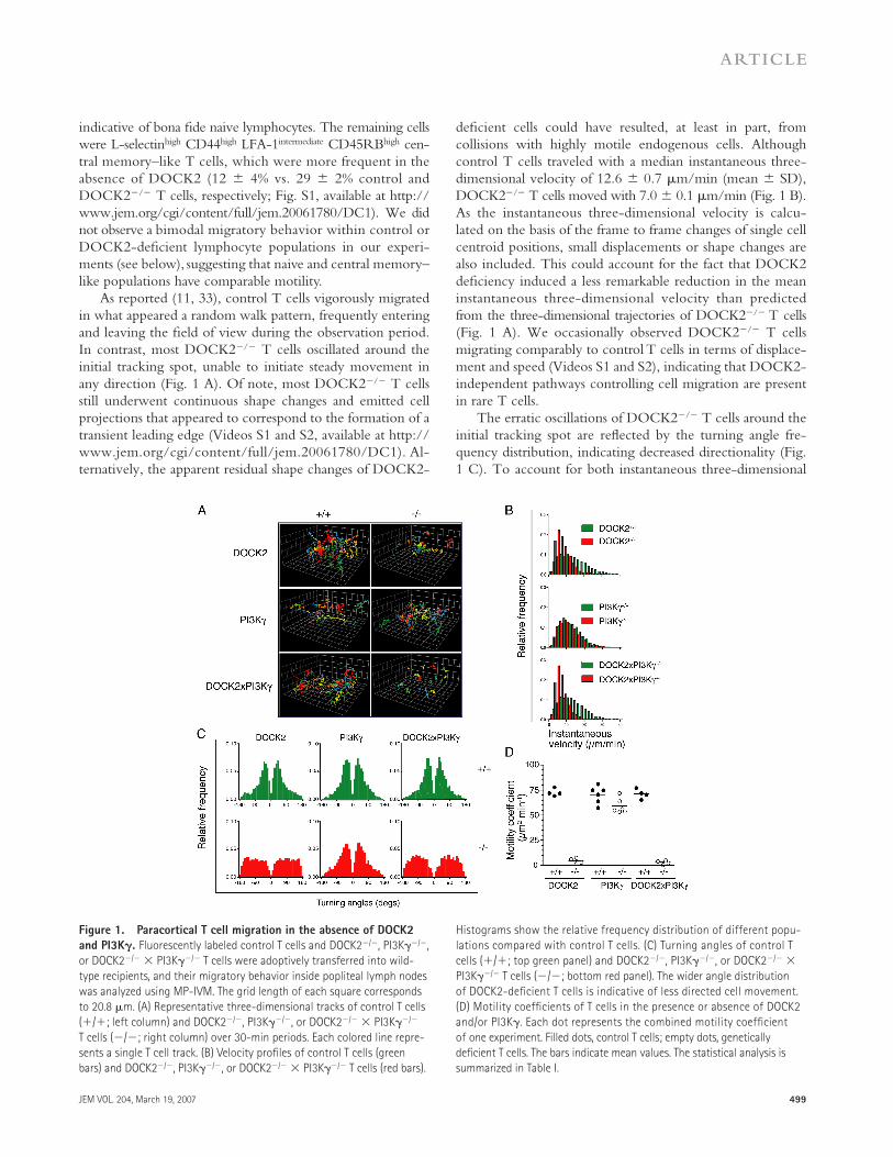
As reported (11, 33), control T cells vigorously migrated in what appeared a random walk pattern, frequently entering and leaving the fi eld of view during the observation period. In contrast, most DOCK2−/− T cells oscillated around the initial tracking spot, unable to initiate steady movement in any direction (Fig. 1 A). Of note, most DOCK2−/− T cells still underwent continuous shape changes and emitted cell projections that appeared to correspond to the formation of a transient leading edge (Videos S1 and S2, available at http://www.jem.org/cgi/content/full/jem.20061780/DC1). Al-ternatively, the apparent residual shape changes of DOCK2-
defi cient cells could have resulted, at least in part, from collisions with highly motile endogenous cells. Although control T cells traveled with a median instantaneous three-dimensional velocity of 12.6 ± 0.7 μm/min (mean ± SD), DOCK2−/− T cells moved with 7.0 ± 0.1 μm/min (Fig. 1 B). As the instantaneous three-dimensional velocity is calcu-lated on the basis of the frame to frame changes of single cell centroid positions, small displacements or shape changes are also included. This could account for the fact that DOCK2 defi ciency induced a less remarkable reduction in the mean instantaneous three-dimensional velocity than predicted from the three-dimensional trajectories of DOCK2−/− T cells (Fig. 1 A). We occasionally observed DOCK2−/− T cells migrating comparably to control T cells in terms of displace-ment and speed (Videos S1 and S2), indicating that DOCK2-independent pathways controlling cell migration are present in rare T cells.
The erratic oscillations of DOCK2−/− T cells around the initial tracking spot are refl ected by the turning angle fre-quency distribution, indicating decreased directionality (Fig. 1 C). To account for both instantaneous three-dimensional
Figure 1. Paracortical T cell migration in the absence of DOCK2
and PI3K𝛄. Fluorescently labeled control T cells and DOCK2−/−, PI3Kγ−/−,
or DOCK2−/− × PI3Kγ−/− T cells were adoptively transferred into wild-
type recipients, and their migratory behavior inside popliteal lymph nodes
was analyzed using MP-IVM. The grid length of each square corresponds
to 20.8 μm. (A) Representative three-dimensional tracks of control T cells
(+/+; left column) and DOCK2−/−, PI3Kγ−/−, or DOCK2−/− × PI3Kγ−/−
T cells (−/−; right column) over 30-min periods. Each colored line repre-
sents a single T cell track. (B) Velocity profi les of control T cells (green
bars) and DOCK2−/−, PI3Kγ−/−, or DOCK2−/− × PI3Kγ−/− T cells (red bars).
Histograms show the relative frequency distribution of different popu-
lations compared with control T cells. (C) Turning angles of control T
cells (+/+; top green panel) and DOCK2−/−, PI3Kγ−/−, or DOCK2−/− ×
PI3Kγ−/− T cells (−/−; bottom red panel). The wider angle distribution
of DOCK2-defi cient T cells is indicative of less directed cell movement.
(D) Motility coeffi cients of T cells in the presence or absence of DOCK2
and/or PI3Kγ. Each dot represents the combined motility coeffi cient
of one experiment. Filled dots, control T cells; empty dots, genetically
defi cient T cells. The bars indicate mean values. The statistical analysis is
summarized in Table I.

500 DOCK2-MEDIATED LYMPHOCYTE MOTILITY AND EGRESS | Nombela-Arrieta et al.
velocity and turning angle distribution, we determined the motility coeffi cient for control and DOCK2−/− T cells as a measure of area scanning effi ciency per time unit (Fig. 1 D) (12). In the absence of DOCK2, the motility coeffi cient de-creased to 4.8 ± 2.8 μm2/min, refl ecting the scarce displace-ments of DOCK2−/− T cells. Control T cells, on the other hand, had an �15-fold higher motility coeffi cient of 72.9 ± 3.7 μm2/min, in line with previous reports (13, 33). All MP-IVM motility parameters are summarized in Table I.
To analyze the role of PI3Kγ during lymphocyte intra-nodal motility, we performed MP-IVM experiments com-paring purifi ed control and PI3Kγ−/− T cells. In six 30-min videos analyzed, PI3Kγ−/− T cells showed no signifi cant reduction of the median instantaneous three-dimensional velocity (PI3Kγ+/+ T cells, 11.5 ± 0.8 μm/min; PI3Kγ−/− T cells, 11.0 ± 1.3 μm/min; Fig. 1 B and Video S3, which is available at http://www.jem.org/cgi/content/full/jem.20061780/DC1). However, a more thorough compari-son of turning angles uncovered a slight shift toward wider turning angles in the absence of PI3Kγ. This observation in-dicates that PI3Kγ−/− T cells undergo more abrupt direc-tional changes during interstitial migration compared with control cells (Fig. 1 C), which is refl ected in the minor but signifi cant reduction of the PI3Kγ−/− T cell motility coeffi -cient (59.2 ± 7.3 compared with 70.6 ± 8.5 μm2/min of control cells; Fig. 1 D and Table I).
In homing experiments, both DOCK2 and, to a minor extent, PI3Kγ contribute to effi cient T cell accumulation in PLNs (31). We set out to analyze the combined eff ect of these signaling molecules on lymphocyte motility inside PLNs. Adoptively transferred DOCK2−/− × PI3Kγ−/− T cells behaved comparably to T cells lacking only DOCK2, remaining essentially stationary during the observation period (Table I and Videos S4 and S5, which are available at http://www.jem.org/cgi/content/full/jem.20061780/DC1). Col-lectively, these observations reveal a central role for DOCK2, and to a lesser extent PI3Kγ, during signal transduction of
tissue-derived promigratory signals causing effi cient intersti-tial T cell motility.
DOCK2 but not PI3K𝛄 controls B cell motility inside
the follicles
B cell migration to chemokines in vitro requires DOCK2 and a PI3K isoform diff erent from PI3Kγ (31, 32). We therefore investigated the infl uence of DOCK2 on B cell random motil-ity using MP-IVM. In the absence of DOCK2, B cell homing to PLNs is further diminished compared with DOCK2−/− T cells due to the B cell–specifi c requirement for DOCK2 during effi cient integrin activation (31). However, when trans-ferring three to fi ve times more purifi ed DOCK2−/− B cells compared with control B cells, we were able to observe a suf-fi cient number of gene-defi cient cells for a meaningful analysis of their migratory behavior (Table I). As for T cells, transferred control and DOCK2-defi cient B cells were >90% L-selectinhigh (not depicted).
15–22 h after transfer, most transferred B cells, regardless of DOCK2 expression, were detected in external areas of the lymph nodes in close proximity to the subcapsular sinus, where B cell follicles are located. Compared with T cells, control B cells displayed a lower median instantaneous three-dimensional velocity (7.9 ± 0.8 μm/min). An exami-nation of frequency histograms of three-dimensional veloc-ities revealed a reduction in DOCK2−/− B cell translocation speed to a mean velocity of 5.2 ± 0.9 μm/min (Fig. 2 B and Table I). Importantly, individual three-dimensional cell tracks refl ect the severely compromised displacement of DOCK2−/− B cells during the observation period, although some cells were observed emitting cell membrane projec-tions, similar to DOCK2−/− T cells (Fig. 2 A). The contin-uous changes in direction and oscillations around the initial tracking spot resulted in increased turning angles and a strongly reduced motility coeffi cient (1.7 ± 0.9 μm2/min for DOCK2−/− B cells vs. 23.7 ± 4.1 μm2/min for control cells; Fig. 2, C and D).
Table I. Motility parameters of control and DOCK2- and/or PI3Kγ-defi cient T and B cells
Control DOCK2−/− Control PI3K𝛄−/− Control DOCK2−/− × PI3K𝛄−/−
T cells
Number of experiments 4 6 4
Number of tracks 842 370 198 299 196 86
3D velocity (μm min−1) 12.6 ± 0.7 7.0 ± 0.1a 11.5 ± 0.8 11.0 ± 1.3 12.2 ± 1.4 6.2 ± 0.5a
Turning angle distribution narrow broad narrow slightly broader narrow broad
Motility coeffi cient (μm2 min−1) 72.9 ± 3.7 4.8 ± 2.8a 70.6 ± 8.5 59.2 ± 7.3a 71.3 ± 4.9 3.2 ± 1.9a
B cells
Number of experiments 4 6 4
Number of tracks 133 35 134 83 243 59
3D velocity (μm min−1) 7.9 ± 0.8 5.2 ± 0.9a 8.1 ± 0.3 7.5 ± 0.7 8.3 ± 0.2 5.9 ± 0.6a
Turning angle distribution narrow broad narrow narrow narrow broad
Motility coeffi cient (μm2 min−1) 23.7 ± 4.1 1.7 ± 0.9a 21.9 ± 5.2 24.3 ± 3.2 23.6 ± 5.4 2.5 ± 1.0a
aSignifi cantly lower three-dimensional velocity or motility coeffi cient compared with control lymphocytes (P < 0.05).

JEM VOL. 204, March 19, 2007 501
ARTICLE
In contrast, PI3Kγ−/− B cell motility was indistinguish-able from control B cells. A thorough analysis did not reveal signifi cant diff erences in mean instantaneous three-dimen-sional velocity, turning angles, or motility coeffi cients (24.3 ± 3.2 μm2/min for PI3Kγ−/− B cells vs. 21.9 ± 5.2 μm2/min for control cells; Fig. 2, A–D). Moreover, we did not detect an ad-ditional eff ect when the migratory behavior of DOCK2−/− × PI3Kγ−/− B cells was compared with DOCK2−/− single defi -cient B cells (Fig. 2, A–D). In summary, these observations support a central role for DOCK2 during interstitial B cell migration (Video S6, available at http://www.jem.org/cgi/content/full/jem.20061780/DC1). The absence of PI3Kγ, on the other hand, did not aff ect B cell motility within lym-phoid tissue.
S1P-induced protein kinase B (PKB) activation, F-actin
polymerization, and migration in the absence of DOCK2
and PI3K
Effi cient lymphocyte egress from PLNs depends on S1P1, a Gαi-coupled receptor expressed on naive lymphocytes (1, 24). The concomitant expression of CKR and S1P1 on the same cell type and the involvement of DOCK2 and PI3Kγ dur-ing lymphocyte homing to PLNs and interstitial migration
prompted us to investigate a role for these molecules in S1P-induced signal transduction. In cell lines, S1P has been de-scribed to induce PI3K, Rac, and ERK activation, as well as cell migration, among other downstream eff ects (34–36). We fi rst evaluated PKB phosphorylation as an indirect mea-sure for PI3K activity in control and genetically modifi ed lymphocytes. As S1P1 surface levels are highly susceptible to down-regulation by S1P in the lymph and blood (37), we allowed lymphocytes to recover S1P1 expression after overnight (o.n.) incubation in fatty acid–free medium. Under these conditions, both control and genetically defi -cient lymphocytes exhibited comparable S1P1 surface levels (Fig. S2 A, available at http://www.jem.org/cgi/content/full/jem.20061780/DC1). Upon stimulation with che-mokines or S1P, phosphorylated PKB was readily detected in control lymphocytes as well as in PI3Kγ−/− B cells. DOCK2−/− lymphocytes had reduced but detectable lev-els of phosphorylated PKB after S1P stimulation (Fig. 3 A). In contrast, PKB did not undergo phosphorylation in PI3Kγ−/− T cells after chemokine or S1P addition (Fig. 3 A). These data suggest that S1P activates PKB in a PI3Kγ- dependent manner in T cells and via a diff erent mechanism in B cells.
Figure 2. Follicular B cell migration in the absence of DOCK2
and PI3K𝛄. Control B cells were adoptively transferred with
DOCK2−/−, PI3Kγ−/−, or DOCK2−/− × PI3Kγ−/− B cells, and their mi-
gratory behavior was analyzed inside B cell follicles using MP-IVM as
in Fig. 1. (A) Representative three-dimensional tracks of control B cells
(+/+; left column), DOCK2−/−, PI3Kγ−/−, or DOCK2−/− × PI3Kγ−/−
B cells (−/−; right column) during 30-min observation periods.
Each colored line represents a single B cell track. (B) Velocity pro-
files of control B cells (green bars) and DOCK2−/−, PI3Kγ−/−, or
DOCK2−/− × PI3Kγ−/− B cells (red bars). Histograms show the relative
frequency distribution of different populations compared with control
B cells. (C) Turning angles of control B cells (+/+; top green panel)
and DOCK2−/−, PI3Kγ−/−, or DOCK2−/− × PI3Kγ−/− mice B cells (−/−;
bottom red panel). (D) Motility coeffi cients of B cells in the presence or
absence of DOCK2 and/or PI3Kγ. Each dot represents the motility co-
effi cient of one experiment. Filled dots, control B cells; empty dots,
genetically defi cient B cells. The bars indicate mean values. The statis-
tical analysis is summarized in Table I.

502 DOCK2-MEDIATED LYMPHOCYTE MOTILITY AND EGRESS | Nombela-Arrieta et al.
We next assessed Rac-mediated F-actin formation in ab-sence of DOCK2 or PI3K. Control lymphocytes displayed a robust F-actin polymerization after S1P addition (Fig. 3 B), which was blocked by prior incubation with FTY720-P or pertussis toxin (Fig. S2 B and not depicted). Of note, the S1P1-specifi c agonist SEW2871 also inhibited S1P-mediated F-actin formation as early as 15 min of preincubation (Fig. S2 B). We observed substantial F-actin polymerization in DOCK2−/− T but not B cells (Fig. 3 B). Under these con-ditions, F-actin formation in DOCK2−/− T cells was only signifi cantly lower at the earliest time point measured (52%
reduction at 5 s after S1P addition; P < 0.05). When F-actin polymerization was examined using freshly isolated lympho-cytes, however, DOCK2-defi cient T cells did not respond to S1P (Fig. S2 C), despite comparable S1P1 mRNA levels as assessed by quantitative RT-PCR (not depicted).
The incomplete inhibition of F-actin formation in DOCK2-defi cient T cells suggested the existence of alterna-tive pathways. PI3K is known to mediate detectable F-actin formation in chemokine-stimulated T cells (31). When o.n. serum-starved control lymphocytes were pretreated with the PI3K inhibitor Wortmannin, S1P-induced actin polymeri-zation at later time points (15–30 s) was signifi cantly lower in T cells, although the initial F-actin polymerization peak was comparable to untreated cells (Fig. S2 C). S1P-triggered F-actin polymerization was also signifi cantly reduced in PI3Kγ-defi cient T cells at later time points (15–30 s; P < 0.05), but not at the earliest time point measured (5 s; Fig. 3 B). To assess the combined eff ect of DOCK2 and PI3K defi ciency downstream of S1P receptors, we blocked PI3K activity in DOCK2-defi cient lymphocytes. Wortmannin treatment of DOCK2−/− T cells abolished residual S1P-induced F-actin polymerization, suggesting that in lympho-cytes, DOCK2 and PI3K independently lead to Rac activity important for actin cytoskeleton remodeling (Fig. S2 B). As predicted from this result, the combined lack of DOCK2 and PI3Kγ in T cells completely eliminated F-actin forma-tion in vitro (Fig. 3 B).
We performed in vitro chemotaxis assays to S1P to exam-ine the functional consequences of reduced F-actin forma-tion in the absence of DOCK2 and PI3Kγ. Control T cells exhibited a small but reproducible migration (Fig. 3 C) that could be blocked after pretreatment with FTY720-P (not de-picted), in line with previous fi ndings (38). Despite their re-duced F-actin formation, PI3Kγ−/− T cells migrated to S1P comparably to control T cells, whereas only rare DOCK2−/− T cells accumulated in the lower chamber during the chemo-taxis assay (Fig. 3 C). When compared with background mi-gration, there was nonetheless a detectable migratory response in the absence of DOCK2, which was not present in T cells lacking both DOCK2 and PI3Kγ (Fig. 3 C). In B cells, no migration was observed in the absence of DOCK2, whereas PI3Kγ defi ciency did not alter migration (Fig. 3 C and Fig. S2 D). Collectively, these in vitro data implicate DOCK2 and PI3Kγ (in T cells) in the signaling cascade leading to F-actin formation and support a role for DOCK2 for effi cient in vitro migration to S1P. PKB phosphorylation induced by S1P involves PI3Kγ in T cells and an alternative PI3K iso-form in B cells.
Impaired lymphocyte egress from PLNs in the absence
of DOCK2 but not PI3K𝛄To determine whether DOCK2 defi ciency aff ected lympho-cyte exit from PLNs in vivo, we performed egress experiments in which fluorescently labeled control and DOCK2−/− lymphocytes were adoptively transferred into wild-type re-cipient mice and allowed to accumulate in PLNs for 4 or 20 h.
Figure 3. DOCK2 and PI3K𝛄 transmit signals downstream of S1P
receptors. (A) Flow cytometric analysis of PKB phosphorylation 30 s after
the addition of S1P (1 μM fi nal concentration; red line) or 1 min after the
addition of CXCL12 (100 nM fi nal concentration; green line) in control,
DOCK2−/−, or PI3Kγ−/− T and B cells using phosphorylated PKB-specifi c
antibody. The black line represents background staining in the absence of
stimuli. (B) Flow cytometric analysis of F-actin polymerization after S1P
addition (500 nM fi nal concentration) in control, DOCK2−/−, PI3Kγ−/−,
and DOCK2−/− × PI3Kγ−/− T and B cells. Data are presented as normal-
ized mean fl uorescence intensity of FITC-Phalloidin binding, with the “0”
time point = 100. Each value corresponds to the mean ± SD of at least
three independent experiments. au, arbitrary units. (C) Chemotaxis of
control, DOCK2−/−, PI3Kγ−/−, and DOCK2−/− × PI3Kγ−/− T and B cells to
25 nM S1P for 4 h at 37°C. Signifi cant migration to S1P over medium is
marked with an asterisk (P < 0.05). Interconnected bars indicate signifi -
cant difference (P < 0.05; ANOVA). Data represent mean ± SD of three
independent experiments.
JEM VOL. 204, March 19, 2007 503
ARTICLE
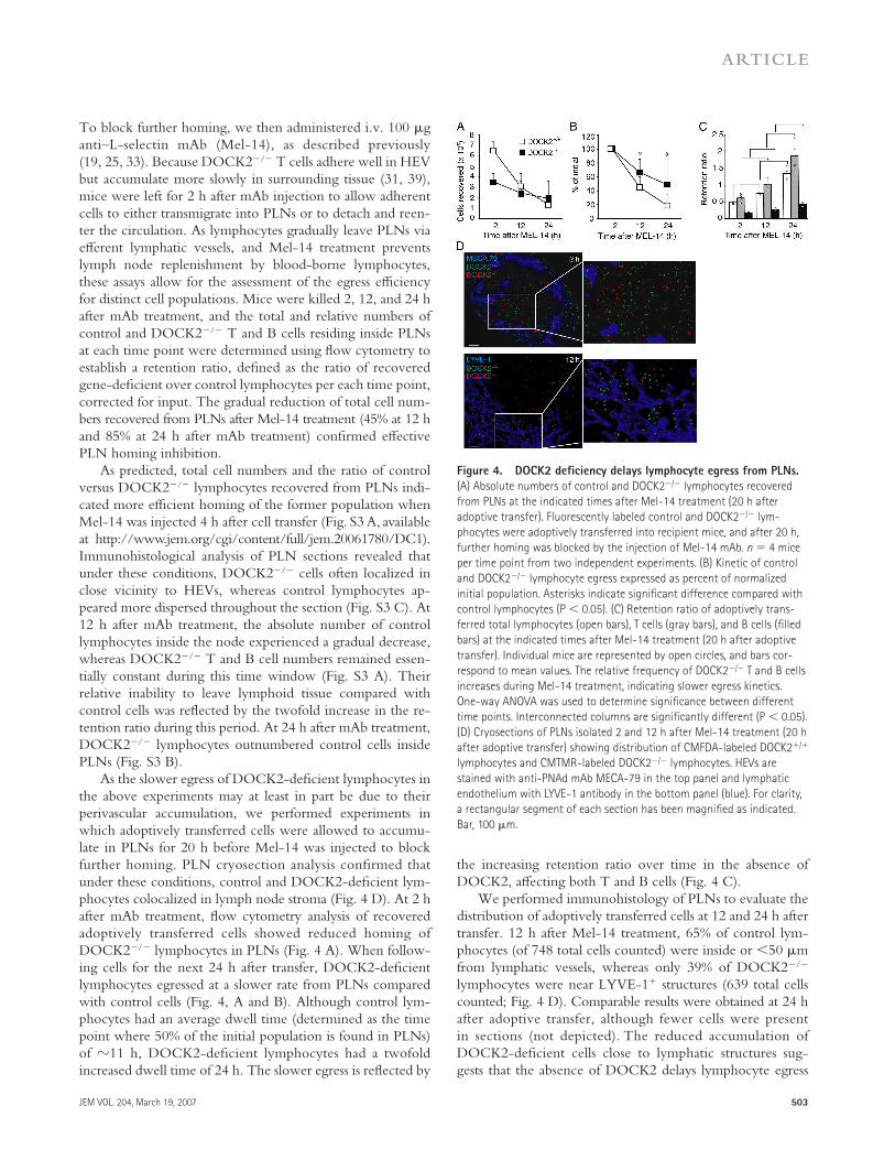
To block further homing, we then administered i.v. 100 μg anti–L-selectin mAb (Mel-14), as described previously (19, 25, 33). Because DOCK2−/− T cells adhere well in HEV but accumulate more slowly in surrounding tissue (31, 39), mice were left for 2 h after mAb injection to allow adherent cells to either transmigrate into PLNs or to detach and reen-ter the circulation. As lymphocytes gradually leave PLNs via eff erent lymphatic vessels, and Mel-14 treatment prevents lymph node replenishment by blood-borne lymphocytes, these assays allow for the assessment of the egress effi ciency for distinct cell populations. Mice were killed 2, 12, and 24 h after mAb treatment, and the total and relative numbers of control and DOCK2−/− T and B cells residing inside PLNs at each time point were determined using fl ow cytometry to establish a retention ratio, defi ned as the ratio of recovered gene-defi cient over control lymphocytes per each time point, corrected for input. The gradual reduction of total cell num-bers recovered from PLNs after Mel-14 treatment (45% at 12 h and 85% at 24 h after mAb treatment) confi rmed eff ective PLN homing inhibition.
As predicted, total cell numbers and the ratio of control versus DOCK2−/− lymphocytes recovered from PLNs indi-cated more effi cient homing of the former population when Mel-14 was injected 4 h after cell transfer (Fig. S3 A, available at http://www.jem.org/cgi/content/full/jem.20061780/DC1). Immunohistological analysis of PLN sections revealed that under these conditions, DOCK2−/− cells often localized in close vicinity to HEVs, whereas control lymphocytes ap-peared more dispersed throughout the section (Fig. S3 C). At 12 h after mAb treatment, the absolute number of control lymphocytes inside the node experienced a gradual decrease, whereas DOCK2−/− T and B cell numbers remained essen-tially constant during this time window (Fig. S3 A). Their relative inability to leave lymphoid tissue compared with control cells was refl ected by the twofold increase in the re-tention ratio during this period. At 24 h after mAb treatment, DOCK2−/− lymphocytes outnumbered control cells inside PLNs (Fig. S3 B).
As the slower egress of DOCK2-defi cient lymphocytes in the above experiments may at least in part be due to their perivascular accumulation, we performed experiments in which adoptively transferred cells were allowed to accumu-late in PLNs for 20 h before Mel-14 was injected to block further homing. PLN cryosection analysis confi rmed that under these conditions, control and DOCK2-defi cient lym-phocytes colocalized in lymph node stroma (Fig. 4 D). At 2 h after mAb treatment, fl ow cytometry analysis of recovered adoptively transferred cells showed reduced homing of DOCK2−/− lymphocytes in PLNs (Fig. 4 A). When follow-ing cells for the next 24 h after transfer, DOCK2-defi cient lymphocytes egressed at a slower rate from PLNs compared with control cells (Fig. 4, A and B). Although control lym-phocytes had an average dwell time (determined as the time point where 50% of the initial population is found in PLNs) of �11 h, DOCK2-defi cient lymphocytes had a twofold increased dwell time of 24 h. The slower egress is refl ected by
the increasing retention ratio over time in the absence of DOCK2, aff ecting both T and B cells (Fig. 4 C).
We performed immunohistology of PLNs to evaluate the distribution of adoptively transferred cells at 12 and 24 h after transfer. 12 h after Mel-14 treatment, 65% of control lym-phocytes (of 748 total cells counted) were inside or <50 μm from lymphatic vessels, whereas only 39% of DOCK2−/− lymphocytes were near LYVE-1+ structures (639 total cells counted; Fig. 4 D). Comparable results were obtained at 24 h after adoptive transfer, although fewer cells were present in sections (not depicted). The reduced accumulation of DOCK2-defi cient cells close to lymphatic structures sug-gests that the absence of DOCK2 delays lymphocyte egress
Figure 4. DOCK2 defi ciency delays lymphocyte egress from PLNs.
(A) Absolute numbers of control and DOCK2−/− lymphocytes recovered
from PLNs at the indicated times after Mel-14 treatment (20 h after
adoptive transfer). Fluorescently labeled control and DOCK2−/− lym-
phocytes were adoptively transferred into recipient mice, and after 20 h,
further homing was blocked by the injection of Mel-14 mAb. n = 4 mice
per time point from two independent experiments. (B) Kinetic of control
and DOCK2−/− lymphocyte egress expressed as percent of normalized
initial population. Asterisks indicate signifi cant difference compared with
control lymphocytes (P < 0.05). (C) Retention ratio of adoptively trans-
ferred total lymphocytes (open bars), T cells (gray bars), and B cells (fi lled
bars) at the indicated times after Mel-14 treatment (20 h after adoptive
transfer). Individual mice are represented by open circles, and bars cor-
respond to mean values. The relative frequency of DOCK2−/− T and B cells
increases during Mel-14 treatment, indicating slower egress kinetics.
One-way ANOVA was used to determine signifi cance between different
time points. Interconnected columns are signifi cantly different (P < 0.05).
(D) Cryosections of PLNs isolated 2 and 12 h after Mel-14 treatment (20 h
after adoptive transfer) showing distribution of CMFDA-labeled DOCK2+/+
lymphocytes and CMTMR-labeled DOCK2−/− lymphocytes. HEVs are
stained with anti-PNAd mAb MECA-79 in the top panel and lymphatic
endothelium with LYVE-1 antibody in the bottom panel (blue). For clarity,
a rectangular segment of each section has been magnifi ed as indicated.
Bar, 100 μm.

504 DOCK2-MEDIATED LYMPHOCYTE MOTILITY AND EGRESS | Nombela-Arrieta et al.
without completely abolishing it. Similarly, when we isolated thoracic duct lymph and PLNs from recipient mice 36 h after adoptive transfer, approximately two to three times fewer DOCK2−/− T cells compared with control T cells were found in lymph when normalized to the population recov-ered from PLNs (not depicted).
Based on the decreased F-actin formation by PI3Kγ−/− T cells in vitro, we also wanted to address whether and to what extent PI3Kγ was implicated in lymphocyte dwell time under physiological conditions. To this end, we performed similar PLN egress experiments comparing control and PI3Kγ−/− lymphocytes. As reported (31, 32), we detected a small but reproducible reduction in PLN homing by PI3Kγ-defi cient lymphocytes as compared with coinjected wild-type cells (Fig. 5 A). Nonetheless, the rates of control and PI3Kγ−/− T and B cell egress were comparable throughout the entire observation period, as illustrated by a similar de-crease in absolute cell numbers and similar retention ratios at all time points analyzed (Fig. 5, A and B). Interstitial distribu-tion of transferred cells with respect to MECA-79+ or LYVE-1+ vessels was always comparable in control and PI3Kγ−/− lym-phocytes at all time points analyzed (not depicted).
Finally, we adoptively transferred DOCK2 × PI3Kγ- defi cient lymphocytes and performed egress assays to address whether residual egress observed in DOCK2-defi cient lym-phocytes depended on PI3K activity. Lack of PI3Kγ did not further delay egress of DOCK2-defi cient lymphocytes, iden-tifying the absence of DOCK2 as the major cause for de-creased lymphocyte egress (Fig. 5 C). Similar results were obtained when DOCK2−/− lymphocytes were pretreated with Wortmannin before adoptive transfer (not depicted).
Thus, DOCK2 but not PI3Kγ defi ciency increases lymph node dwell time in vivo by slowing lymphocyte egress from PLNs.
T cell motility in PLN medulla is reduced in the absence
of DOCK2
Despite the reduced responsiveness of DOCK2−/− lympho-cytes to S1P in vitro, it remained unclear whether the increased lymph node dwell time of DOCK2-defi cient T cells was due to low sensitivity to S1P-mediated egress signals in addition to the overall reduced accumulation in medullary cords. We therefore directly investigated the migratory behavior of con-trol and DOCK2-defi cient T cells in the medullar region of PLNs, where S1P-dependent lymphocyte egress takes place (21, 24, 37). Explanted PLNs were immobilized with the hilus facing the objective, whereas eff erent lymphatic vessels were identifi ed by labeling with fl uorescent wheat germ agglutinin (WGA), as described previously (40). We analyzed cell motil-ity of adoptively transferred T cells 15–65 μm below the cap-sule in close proximity to WGA-labeled vessels during 30-min observation periods.
Control T cells exhibited continuous motility with an average speed of 8.8 ± 2.1 μm/min (Fig. 6, B and C, and Video S7, which is available at http://www.jem.org/cgi/content/full/jem.20061780/DC1). These cells occasionally entered and left the lymphatic vascular lumen (Fig. 6 A), as verifi ed by careful assessment of volume-rendered sections (not depicted). In contrast, much fewer DOCK2- defi cient T cells were observed in the medullar region, in line with our immunohistological data (Fig. 4 D). DOCK2−/− T cells in medullary cords were even less motile as in the paracortex (average velocity, 3.7 ± 1.6 μm/min; P < 0.01 compared with control T cells) and remained stationary during the ob-servation period (Fig. 6, A–C, and Video S8, which is available at http://www.jem.org/cgi/content/full/jem.20061780/DC1). The diff erent motility is refl ected in shortened average track lengths in the absence of DOCK2 (Fig. 6 B). DOCK2 is therefore required for effi cient motility near eff erent lym-phatic vessels in the medulla.
DISCUSSION
Observations using MP-IVM have provided important insights into the remarkable interstitial motility of lympho-cytes, which migrate hundreds of micrometers every day during their long life span (10, 12–15, 28). The data pre-sented here provide novel information about intracellular signaling factors required for effi cient interstitial T and B cell motility within and egress from PLNs, and uncover a central role for the lymphocyte-expressed Rac guanine exchange factor DOCK2. First, we demonstrate that DOCK2 is needed for eff ective T and B cell motility inside paracortex and follicles, whereas PI3Kγ helps maintain temporal direc-tionality during interstitial migration of T cells. Second, our fi ndings show roles for both DOCK2 and PI3K during S1P-triggered signaling, but only DOCK2 is important for effi cient egress from PLNs. In combination with previous
Figure 5. PI3K𝛄 defi ciency does not affect lymphocyte egress
from PLNs. (A) Total numbers of PI3Kγ+/+ and PI3Kγ−/− cells present in
PLNs at 2, 12, and 24 h after Mel-14 mAb treatment (4 h after adoptive
transfer). Data represent mean ± SD from at least three mice from two
independent experiments. (B) Retention ratio of PI3Kγ+/+ and PI3Kγ−/−
T and B cells at 2, 12, and 24 h after cell transfer. Each circle represents
individual mice, and bars depict mean values. Shown are transferred total
lymphocytes (open bars), T cells (gray bars), and B cells (fi lled bars) at the
indicated times after Mel-14 mAb treatment. No signifi cant difference in
ratio became apparent. (C) Retention ratio of DOCK2−/− and DOCK2−/− ×
PI3Kγ−/− T and B cells. 20 h after cell transfer, further homing was
blocked by Mel-14 mAb administration, and mice were killed at 2 and
20 h later. Each circle represents individual mice, and bars depict mean
values. Shown are transferred total lymphocytes (open bars), T cells (gray
bars), and B cells (fi lled bars) at the indicated times after Mel-14 mAb
treatment. Data are from two independent experiments with four mice
per time point. No signifi cant difference in ratio became apparent.

JEM VOL. 204, March 19, 2007 505
ARTICLE
fi ndings regarding chemokine-induced migration (30, 31), the results presented here suggest a dominant role for DOCK2 over PI3K in intracellular signaling pathways that control central aspects of lymphocyte recirculation: homing, interstitial migration, and egress.
DOCK2 and PI3Kγ have well-described roles in CKR signaling in lymphocytes. Both contribute, in a largely inde-pendent manner, to migratory responses of T lymphocytes toward chemokine gradients in vitro (30–32, 41). The strongly reduced lymphocyte motility in the absence of DOCK2 underlines its nonredundant role for effi cient cell movement induced by tissue-derived promigratory factors present in lymph stroma. Migration requires cell polarization and translocation of polarized cells (42). In vitro, DOCK2−/− lymphocytes show defective polarization in response to a uniform chemokine gradient (31). It is currently unclear whether within interstitial tissue DOCK2−/− cells fail to po-larize in response to tissue-derived promigratory factors and/or whether DOCK2 is additionally required for effi cient
translocation. The presence of rare fast-moving DOCK2−/− T cells may indicate that even in the absence of DOCK2, a threshold polarization state can be achieved that allows for effi cient translocation. Although the large majority of adoptively transferred cells found in PLNs correspond to phenotypically naive lymphocytes, we cannot exclude that occasional fast T cells represent subsets of central memory cells in which migration is mediated by DOCK2-independent pathways. The minor defect of PI3Kγ-defi cient T cells in maintaining temporal directionality is reminiscent of the aberrant PI3Kγ−/− neutrophil migration toward increasing concentrations of chemoattractant. Similar to interstitially migrating T cells, PI3Kγ−/− neutrophils display only mildly decreased translocation speeds but are unable to form a persistent leading edge due to ineff ective recruitment of pleckstrin homology domain–containing intracellular factors stabilizing the leading edge (43, 44).
The precise tissue-derived factors responsible for random lymphocyte motility within lymphoid tissue remain un-known. Numerous genetically modifi ed mouse strains and the conspicuous expression pattern in locally distinct micro-environments point to a central role for chemokines in the overall organization of SLOs (1–3). Fibroblastic reticular cells and follicular dendritic cells form three-dimensional net-works in T and B cell areas, respectively, and are the major chemokine-producing cells in lymphoid tissue (6, 45), allow-ing to speculate that chemokines on the surface of stromal cell networks contribute to continuous lymphocyte motility (16). In vitro, lymphocytes are generally nonmotile (12), al-though recent reports describe the induction of detectable random lymphocyte migration by homeostatic chemokines (17, 18). Furthermore, directed migration of activated B cells toward the T–B cell border area was found to depend on CCR7 and a CCL21 gradient (46). Similarly, the CKR CCR5 expressed on CD8 cells under proinfl ammatory con-ditions allows these cells to approach APC-CD4 clusters more effi ciently (47), although factors promoting random motility were not investigated in these studies. To date, there is nonetheless no direct evidence that homeostatic chemo-kines such as CCL19 or CCL21 are responsible for interstitial random motility. The major factor compounding a useful in situ analysis of the role of CKR during interstitial migration lies in their central role during recruitment of blood-borne lymphocytes into SLOs. For example, genetic ablation of CCR7 or its ligands results in loss of recruitment of naive T lymphocytes into PLNs, making it diffi cult to analyze their role in interstitial migration (8, 9). Because chemokines pre-sent in lymph nodes induce internalization of their cognate receptors in vitro (48), an unsolved question is how these fac-tors can promote continuous migration observed in vivo.
Besides chemokines, other tissue-derived factors may contribute to interstitial cell motility. Dendritic cells secrete the lipid mediator thromboxane A2, which enhances T cell chemokinesis in vitro by binding TP, a Gαi-linked GPCR (49). Of note, TP levels are higher on T cells than on B cells, correlating with increased motility of the former population (49).
Figure 6. Medullar T cell migration in the absence of DOCK2.
(A) Image sequence of control and DOCK2-defi cient T cells proximal to
efferent WGA+ lymphatic vessels (green, outline labeled white for greater
clarity). A control T cell entering WGA-labeled lymphatic vessel is shown,
whereas DOCK2-defi cient T cells remained stationary during the observa-
tion period. The asterisks mark the initial tracking spot. Time is in minutes
and seconds. Bar, 10 μm. (B) Representative three-dimensional tracks of
control and DOCK2−/− T cells tracked for 30 min. 10 tracks are shown for
each genotype. Scale is in micrometers. (C) Velocity profi le of control and
DOCK2-defi cient medullar T cells. 886 control T cell and 138 DOCK2−/−
T cell tracks were analyzed from four to fi ve independent videos.
506 DOCK2-MEDIATED LYMPHOCYTE MOTILITY AND EGRESS | Nombela-Arrieta et al.
Finally, DOCK2 also acts downstream of the TCR (50), raising the possibility that non-GPCR surface receptors may participate in T cell interstitial motility. The identifi ca-tion of DOCK2 as a key player of continuous lymphocyte movement does therefore not further clarify the role of che-mokines or additional tissue-derived factors in this process. However, recent data identifi ed several molecules involved in GPCR signaling as important factors in promoting ran-dom B cell migration inside SLOs, as B cells lacking Gαi2 or RGS1 have strongly impaired or increased interstitial motil-ity, respectively (19). The data presented here are in line with these observations, as DOCK2 and PI3Kγ act down-stream of CKR, and DOCK2−/− mice show evidence of markedly hypotrophic SLOs, with disturbed T and B cell compartmentalization (30). The close correlation between in vitro responsiveness to chemokines and interstitial motility in the absence of Gα12, RGS1, DOCK2, and PI3Kγ is noteworthy and suggests a strong overlap between CKR-mediated intracellular signaling pathways with those mediat-ing random motility.
Absence of DOCK2 aff ects overall displacement of T and B cells, severely impairing the capacity of lymphocytes to screen extensive areas within their respective microenvi-ronments. Recent in vitro data suggest that random motility increases effi cient lymphocyte activation by facilitating en-counters with CD3/28-coated beads or APCs (17, 18). An interesting question is therefore how tissue motility correlates with immune response initiation in vivo. However, studying the impact of reduced motility on immune response initia-tion in the absence of DOCK2 is hampered by the fact that it also acts downstream of TCR, resulting in reduced prolifera-tion in vitro in response to irradiated splenocytes as APCs (50). It is thus diffi cult to directly assess the role of APC–T cell encounter effi ciency for early onset or magnitude of immune responses in this model.
Lymphocyte egress from PLNs requires signals triggered by S1P, which binds to fi ve widely expressed GPCRs, S1P1–5 (1, 20, 34, 35). The fi rst indication of participation of S1P re-ceptors during lymphocyte recirculation came from studies using FTY720, an immunosuppressive agent sharing struc-tural homology with S1P (21). Orally administered FTY720 becomes rapidly phosphorylated by S1P kinase 2 (51, 52) and, after agonistic binding to S1P receptors, induces a continuous loss of S1P1 surface expression on lymphocytes through receptor internalization and degradation (23, 37). It is currently controversial whether pharmacological com-pounds blocking or stimulating S1P1 signaling act mainly on lymphocytes or lymphatic endothelial cells, as S1P1 expressed on lymphatic endothelium may participate in lymphocyte exit control through regulation of endothelial barrier prop-erties (40, 53, 54). The interpretation of pharmacological studies is complicated by our observation that SEW2871, which acts as an S1P1-specifi c agonist in certain cell lines (55), behaves as a functional antagonist in lymphocytes, simi-lar to FTY720-P (Fig. S2 B). Pharmacological agonists and antagonists need thus to be carefully analyzed for their eff ects
on primary cells to obtain a clearer picture on their eff ect on potential target cell populations.
Several observations clearly indicate that lymphocyte- expressed S1P1 is absolutely required for physiological lym-phocyte exit from lymphoid tissues. S1P1-defi cient mature thymocytes are retained in thymus and, upon adoptive trans-fer, in PLNs (24, 25, 56). In addition, a series of elegant ex-periments demonstrated that CD69 expressed on recently activated lymphocytes is critically involved in down-regulation of lymphocyte-expressed S1P1, resulting in effi cient trapping of antigen-specifi c lymphocytes in infl amed PLNs (57). CD69 levels do not diff er between control and DOCK2- defi cient lymphocytes (not depicted), excluding a role for this pathway in controlling the dwell time of DOCK2-defi cient cells. As lymphocytes are able to migrate toward S1P in vitro and S1P1 surface levels gradually increase with lymphocyte dwell time in PLNs (24, 37, 38), the simplest working hy-pothesis is that egress is a chemotactic response to S1P. This hypothesis is consistent with the recent observation that lym-phocyte movement from medullar cords into lymph and blood requires S1P lyase. S1P lyase activity may create an S1P concentration gradient between lymphatic fl uid and intranodal stroma, thus establishing a guidance cue for egressing lymphocytes (58). Of note, FTY720 also inhibits S1P lyase, which may further contribute to lymphocyte sequestration (59).
In line with decreased migration to S1P in vitro, multi-photon imaging showed a strongly reduced motility of me-dullar DOCK2-defi cient T cells, which probably refl ects a defect in the transmission of S1P-derived promigratory egress signals. This is supported by the observation that migration of lymphocytes in medulla is controlled by diff erent molecular cues as paracortical migration refl ected by slower migratory speed and increased susceptibility to pharmacological com-pounds altering S1P signaling (40, 54). It has to be cautioned, however, that our use of explanted lymph nodes results in an interruption of lymphatic fl ow, which may alter the physio-logical migratory behavior, especially concerning diapedesis into lymphatic vessels.
Our data show that in fatty acid–starved T cells stimu-lated by S1P, DOCK2 mediates early F-actin polymeriza-tion, whereas PI3K activity appears more important for a second, delayed wave of F-actin formation. It is interesting to note that the fi rst wave of F-actin formation correlates with effi cient migration to S1P in vitro and egress in vivo, whereas the lack of PI3K-dependent, slower F-actin forma-tion does not strongly reduce lymphocyte migration or egress. The relation of two separate F-actin formation path-ways and their physiological function is at present uncertain. T cells express mainly two of the fi ve known S1P receptors, S1P1 and S1P4, although only S1P1 appears to be necessary for effi cient egress (1). It is possible that S1P1 and S1P4 show preferential intracellular coupling to DOCK2 and PI3Kγ, respectively, with DOCK2-Rac activity mediating a more robust migration. However, this needs to be experimentally addressed. It is also interesting to note that freshly isolated
JEM VOL. 204, March 19, 2007 507
ARTICLE
DOCK2- but not PI3Kγ-defi cient T cells had a strongly re-duced S1P-induced F-actin formation, which may indicate that serum starvation does not represent the appropriate physiological context for measuring S1P responsiveness. Finally, it is important to note that DOCK2- and PI3K- independent pathways controlling lymphocyte exit clearly exist, which merits further investigation.
An interesting question is how lymphocytes compute various extracellular stimuli resulting in a given physiological outcome such as random interstitial motility versus egress. Our data suggest that both CKR and S1P1 share important molecular players mediating migration, which could point to a central role for surface receptor expression levels, as has been suggested (37). However, in contrast to CKR and S1P1 expressed in cell lines (36, 55), S1P did not induce effi cient ERK phosphorylation in primary lymphocytes, indicating that S1P1 and CKR share some but not all signal transduction pathways (Fig. S2 E). Within lymphoid tissue, chemokines are abundant while S1P levels are low (58, 60), and in in vitro assays, lymphocytes respond much more avidly to chemo-kines than to S1P. It is thus likely that in addition to regulat-ing S1P1 surface expression, other mechanisms are necessary for successful lymphocyte egress, such as a state of temporary unresponsiveness to tissue-derived factors, including chemo-kines. High levels of S1P, as may be present in close proximity to eff erent lymphatic vessels, were found to suppress chemo-tactic responses of CD4 cells to homeostatic chemokines (61), providing a working model to address the balance of random interstitial motility and egress.
In summary, we show that within PLNs, T and B cells require DOCK2 for extensive screening of their respective microenvironments, while PI3Kγ helps maintaining tempo-ral directionality during interstitial T cell migration. Further-more, our data show an important role for DOCK2, and to a minor extent PI3K activity, for S1P-induced F-actin poly-merization and migration. Reduced S1P responsiveness in the absence of DOCK2 likely contributes to the increased dwell time of DOCK2-defi cient lymphocytes within PLNs. The simultaneous targeting of homing (30, 31), interstitial migration, and egress is probable to induce a permanent immuno compromised state in mice lacking DOCK2. In combination with impaired TCR signaling, it is perhaps not surprising that deletion of DOCK2 enables long-term sur-vival in a murine model of cardiac allograft transplant (62). We therefore consider DOCK2 an interesting therapeutic target to simultaneously alter various processes controlling lymphocyte traffi cking and function while not completely shutting down the immune system.
MATERIALS AND METHODS
Reagents and mice. AMCA-conjugated anti–rat IgM and Alexa350-con-
jugated anti–rabbit Ig were from Jackson ImmunoResearch Laboratories.
Anti–LYVE-1 was purchased from RELIATech. Anti–mouse S1P1 poly-
clonal rabbit Ig directed against the N and C terminus was provided by
S. Mandala and E.J. Quackenbush (Merck Research Laboratories, Rahway,
NJ). All other antibodies were from BD Biosciences or affi nity purifi ed from
hybridoma supernatant (Mel-14; Nanotools). S1P and human collagen type IV
were purchased from Sigma-Aldrich. CMTMR (CellTracker orange),
CMFDA (CellTracker green), and CFSE were from Invitrogen. SEW2871
was from Calbiochem.
6–12-wk-old control, DOCK2−/−, PI3Kγ−/−, and DOCK2−/− ×
PI3Kγ−/− mice in a C57BL/6 background were used for all experiments.
CD45.1+ C57BL/6 used in some reconstitution experiments were from
The Jackson Laboratory. All experiments were performed in accordance
with National Institutes of Health (NIH) guidelines and approved by the
Committees on Animal Care and Use of Harvard Medical School, the CBR
Institute for Biomedical Research, and the Kanton of Bern.
MP-IVM of popliteal PLNs. Single cell suspensions were obtained from
the PLNs, mesenteric lymph nodes, and spleens of control and genetically
defi cient mice. Alternatively, in some experiments (control and PI3Kγ−/−
T cells, and control, DOCK2−/−, PI3Kγ−/−, and DOCK2−/− × PI3Kγ−/−
B cells), cells were isolated from irradiated CD45.1+ mice reconstituted with
CD45.2+ bone marrow cells from control, DOCK2−/−, PI3Kγ−/−, and
DOCK2−/− × PI3Kγ−/− mice, respectively. When required, endogenous
CD45.1+ cells were eliminated by negative selection with anti-CD45.1–
coated magnetic beads (Miltenyi Biotec). In all experiments, T and B cell
isolation was performed by negative immunomagnetic cell sorting (Miltenyi
Biotec) with purity yields of >95% for T cells and >90% for B cells. Purifi ed
control, DOCK2−/−, PI3Kγ−/−, or DOCK2−/− × PI3Kγ−/− T or B cells
were fl uorescently labeled with 5 μM CFSE or 10 μM CMTMR for 15 min
at 37°C, washed, and injected i.v. into sex-matched 6-wk-old C57BL/6 re-
cipient mice. Numbers of transferred lymphocytes (2–10 × 106 cells) were
calculated according to the homing ability of each subset to allow suffi cient
accumulation of cells inside the node for visualization. When we checked
the phenotype of control and DOCK2-defi cient adoptively transferred T or
B lymphocytes by fl ow cytometry, we found that >99% of transferred cells
in PLNs were either Thy1.2+ or B220+, with >70% corresponding to bona
fi de naive lymphocytes (Fig. S1). 15–22 h after transfer, recipient mice were
anesthetized and the right popliteal lymph node was surgically exposed, as
described previously (33). Multiphoton imaging was performed with an
Olympus BX50WI fl uorescence microscope equipped with a 20× objective
and a Bio-Rad Radiance 2000 MP Confocal/Multiphoton microscopy sys-
tem, controlled by Lasersharp software (Bio-Rad Laboratories). For multi-
photon excitation and second harmonic generation, a Ti:sapphire laser with
a 10-W MilleniaXs pump laser (Tsunami; Spectra-Physics) was tuned to
800 nm. For four-dimensional analysis of cell migration, stacks of 16 square
x-y sections were acquired every 15 s during 30 min with electronic zooming
up to 6× to provide image volumes of 60 μm in depth. Emitted light and
second harmonic signals were detected through 400/40-nm, 525/50-nm, and
620/100-nm bandpass fi lters with nondescanned detectors to generate three-
color images. Sequences of image stacks were transformed into volume-
rendered four-dimensional movies using Volocity software (Improvision),
which was also used for semi-automated tracking of cell motility in three
dimensions. From x, y, and z coordinates of cell centroids, parameters of
cellular motility were calculated as described previously (33). In brief, the
instantaneous three-dimensional velocity is the cellular velocity between
two time points, whereas the turning angle describes the angle between the
two velocity vectors before and after a measurement time point (12). Both
parameters are determined independently for each cell track.
S1P-induced PKB and ERK phosphorylation, F-actin polymeriza-
tion, and migration. Lymphocyte isolation and functional experiments
were performed in RPMI supplemented with 1 mg/ml of fatty acid–free
BSA, L-Gln, NaPyruvate, β-mercaptoethanol, and PenStrep (RPMI-BSA).
After o.n. incubation in RPMI-BSA at 37°C, 7% CO2, lymphocytes were
stimulated with 500 nM S1P or 100 nM CXCL12, permeabilized, and incu-
bated with anti-phosphoPKB or anti-phosphoErk (Cell Signaling Labora-
tories). As secondary antibodies, we used biotinylated anti–rabbit IgG
(Jackson ImmunoResearch Laboratories), followed by APC-conjugated
streptavidin (BD Biosciences). Comparable results were obtained using
Western blot analysis (unpublished data).
508 DOCK2-MEDIATED LYMPHOCYTE MOTILITY AND EGRESS | Nombela-Arrieta et al.
Actin polymerization assays were performed as described previously
(31). In brief, single cell suspensions of total lymphocytes (107 cells/ml) were
stimulated with 500 nM S1P, and aliquots were taken at indicated time
points, followed by immediate fi xation in 4% paraformaldehyde for 15 min.
Cells were washed with PBS and stained for Thy1.2+ and B220+ cells, per-
meabilized, labeled with FITC-Phalloidin (Invitrogen), and analyzed by
fl ow cytometry using CELLQuest software (Becton Dickinson). In some
experiments, lymphocytes were treated with 0.1 μg/ml pertussis toxin
(Sigma-Aldrich), 0.5 μM Wortmannin (Calbiochem), 5 μM SEW2871, or
0.5 μM FTY720 or FTY720-P (provided by V. Brinkmann, Novartis,
Basel, Switzerland, and S. Mandala and E.J. Quackenbush, respectively) for
15 min or 2 h at 37°C, 7% CO2, before S1P stimulation.
Chemotaxis assays were performed using Transwell chambers (5-μm
pore size; CoStar). Filters were coated o.n. at 4°C with a 10 μg/ml solution
of human collagen type IV, washed twice with 500 μl PBS, and dried
before use. Uncoated fi lter inserts were used with comparable results. 106
lymphocytes in 100 μl RPMI-BSA was placed in the top chamber and al-
lowed to migrate to 25 nM S1P for 4 h at 37°C, 7% CO2. This concentra-
tion was found to be optimal for both control and DOCK2−/− lymphocytes
over a range from 10 nM to 1 μM S1P (unpublished data). The percentage
of total migrated cells was determined by fl ow cytometry comparing with a
precalibrated bead standard (Sigma-Aldrich). After migration, input and
migrated populations were labeled for T and B cells to calculate percentages
of subset migration.
Determination of S1P1 mRNA levels and surface expression. Total
RNA was isolated from freshly isolated CD4+ T cells according to the man-
ufacturer’s instructions (QIAGEN). Quantitative RT-PCR was performed
using the universal probe library system (Roche Applied Science). For S1P1
surface labeling, lymphocytes were incubated o.n. in RPMI-BSA and
labeled with anti–mouse N-terminal S1P1-specifi c rabbit Ig, followed by
biotinylated anti–rabbit Ig and streptavidin-APC. As controls, we used
C-terminal S1P1 or S1P4-specifi c rabbit Ig and FTY720-P pretreatment.
Lymph node egress assays. Total lymphocytes were isolated from the
spleens and PLNs of control and genetically defi cient mice and fl uorescently
labeled using 0.3 μM CMFDA or 1.5 μM CMTMR for 45 min, 5% CO2,
37°C in RPMI supplemented with 10% FCS, L-Gln, NaPyruvate, β-mer-
captoethanol, and PenStrep (CM-R). Depending on the homing ability of
diff erent subsets, 2–4 × 10⁷ cells of each cell population were mixed in 200
μl CM-R and injected i.v into age- and sex-matched C57BL/6 recipient
mice. 4 or 20 h after lymphocyte transfer, mice received i.v. injections of
Mel-14 (100 μg/mouse) to prevent further homing. Mice were killed 2, 12,
and 24 h after mAb injection for assessment of the rate of egress of injected
lymphocytes from PLNs. SLOs and blood were collected, labeled with anti-
Thy1.2 and anti-B220, and the numbers of adoptively transferred T and B
cells were determined using fl ow cytometry. For each organ, absolute num-
bers of each lymphocyte subset were calculated by using a precalibrated bead
standard. A lymph node retention ratio was determined for each time point
as the ratio of (percent recovered gene-defi cient lymphocytes/percent re-
covered control lymphocytes) × correction factor for the input population
ratio, as described previously (31). One lymph node from each mouse was
snap frozen for immunohistological analysis.
Immunohistology. 8-μm sections of frozen PLNs were fi xed for 10 min
in 2% paraformaldehyde, washed in PBS, blocked with FCS, and stained
with anti-PNAd (MECA-79) or anti–LYVE-1 antibody. AMCA-conjugated
anti–rat IgM or Alexa350-conjugated anti–rabbit Ig were used as secondary
antibody. Preparations were observed using a fl uorescence microscope
(Nikon). For determination of cell distribution, adoptively transferred lympho-
cytes were considered close to LYVE-1+ structures at a distance of <50 μm.
Multiphoton imaging of PLN medulla. CMTMR-labeled control or
DOCK2−/− T cells (2–10 × 106 cells/mouse) were adoptively transferred
into C57BL/6 mice 16–24 h before isolation of PLNs (axillary, brachial, and
inguinal). Explanted PLNs were labeled for 5 h with AlexaFluor488-conju-
gated WGA on ice as described previously (40). Labeled PLNs were care-
fully immobilized on a glass slide with the hilus facing up and imaged using
a Trimscope MP system (LaVision) equipped with a MaiTai HP Ti:Sa laser
(Spectraphysics). During image acquisition, PLNs were kept at 35–37°C and
continuously superfused with oxygenated (95% O2, 5% CO2) bicarbonate
buff ered solution (130 mM NaCl, 2.5 mM KCl, 1.3 mM NaH2PO4, 26 mM
NaHCO3, 1 mM MgCl2, 2 mM CaCl2, and 10 mM glucose, pH 7.4, when
equilibrated with a mixture of 95% O2 and 5% CO2). Image stacks (200 ×
200 × 40 μm) between 10 and 65 μm below the lymph node surface were
taken every 15 s during 30 min with a z-step size of 4 μm, using 840 nm
excitation wave length. Fluorescent and second harmonic generation signals
were collected using 455/50 and 525/50 bandpass fi lters or 560LP fi lters and
analyzed using Volocity software. The average track velocity was calculated
as total track length over tracking time.
Statistical analysis. The Student’s t test or one-way analysis of variance
(ANOVA) was used for statistical analysis (Prism or InStat; GraphPad Soft-
ware). Signifi cance was set at P < 0.05.
Online supplemental material. Fig. S1 shows a fl ow cytometric analysis
of the phenotype of adoptively transferred wild-type and DOCK2−/−
T cells. Fig. S2 shows S1P1 surface expression levels of control and gene-
defi cient lymphocytes, as well as S1P-induced F-actin formation, B cell
migration, and ERK phosphorylation. Fig. S3 depicts PLN egress of con-
trol and DOCK2−/− lymphocytes after an initial 4-h homing period.
Supplemental videos show interstitial motility of paracortical control and
DOCK2−/− T cells (Videos S1 and S2), control and PI3Kγ−/− T cells
(Video S3), control and DOCK2−/− × PI3Kγ−/− T cells (Videos S4 and
S5), and follicular control and DOCK2−/− B cells (Video S6). Videos S7
and S8 depict migration of medullar control and DOCK2−/− T cells,
respectively. The online supplemental material is available at http://www.jem
.org/cgi/content/full/jem.20061780/DC1.
We are grateful to Teresa de la Cueva, Luis Almonacid, and Dr. Angel Zaballos (CNB,
Madrid, Spain) for excellent technical advice and help; Dr. Markus Gräler (Hannover
University) and Lawrence R. Shiow (University of California, San Francisco) for useful
advice on S1P experiments; Dr. Volker Brinkmann (Novartis) for providing FTY720; and
Drs. Elizabeth J. Quackenbush and Suzanne Mandala (Merck) for FTY720-P, anti–mouse
S1P receptor antibody, and critical reading of the manuscript.
This work was partially supported by EU grants as well as the Foundation Lilly
and the Spanish Ministry of Education and Science awarded to the Department of
Immunology and Oncology (CNB). This work was supported by Swiss National
Foundation grant 3100-107510 and Marie Curie Excellence grant 2005-025405
(to J.V. Stein). C. Nombela-Arrieta was a recipient of EMBO short-term fellowship
ASTF 181-04. T.R. Mempel received NIH training grant T32-HL-066987-04.
The authors have no confl icting fi nancial interests.
Submitted: 18 August 2006
Accepted: 31 January 2007
R E F E R E N C E S 1. Cyster, J.G. 2005. Chemokines, sphingosine-1-phosphate, and
cell migration in secondary lymphoid organs. Annu. Rev. Immunol. 23:127–159.
2. von Andrian, U.H., and T.R. Mempel. 2003. Homing and cellular traffi c in lymph nodes. Nat. Rev. Immunol. 3:867–878.
3. Stein, J.V., and C. Nombela-Arrieta. 2005. Chemokine control of lymphocyte traffi cking: a general overview. Immunology. 116:1–12.
4. Scimone, M.L., T.W. Felbinger, I.B. Mazo, J.V. Stein, U.H. Von Andrian, and W. Weninger. 2004. CXCL12 mediates CCR7-indepen-dent homing of central memory cells, but not naive T cells, in peripheral lymph nodes. J. Exp. Med. 199:1113–1120.
5. Ebisuno, Y., T. Tanaka, N. Kanemitsu, H. Kanda, K. Yamaguchi, T. Kaisho, S. Akira, and M. Miyasaka. 2003. Cutting edge: the B cell che-mokine CXC chemokine ligand 13/B lymphocyte chemoattractant is expressed in the high endothelial venules of lymph nodes and Peyer’s

JEM VOL. 204, March 19, 2007 509
ARTICLE
patches and aff ects B cell traffi cking across high endothelial venules. J. Immunol. 171:1642–1646.
6. Ansel, K.M., V.N. Ngo, P.L. Hyman, S.A. Luther, R. Forster, J.D. Sedgwick, J.L. Browning, M. Lipp, and J.G. Cyster. 2000. A chemo-kine-driven positive feedback loop organizes lymphoid follicles. Nature. 406:309–314.
7. Förster, R., A.E. Mattis, E. Kremmer, E. Wolf, G. Brem, and M. Lipp. 1996. A putative chemokine receptor, BLR1, directs B cell migration to defi ned lymphoid organs and specifi c anatomic compartments of the spleen. Cell. 87:1037–1047.
8. Förster, R., A. Schubel, D. Breitfeld, E. Kremmer, I. Renner-Muller, E. Wolf, and M. Lipp. 1999. CCR7 coordinates the primary immune response by establishing functional microenvironments in secondary lymphoid organs. Cell. 99:23–33.
9. Nakano, H., T. Tamura, T. Yoshimoto, H. Yagita, M. Miyasaka, E.C. Butcher, H. Nariuchi, T. Kakiuchi, and A. Matsuzawa. 1997. Genetic defect in T lymphocyte-specifi c homing into peripheral lymph nodes. Eur. J. Immunol. 27:215–221.
10. Bousso, P., and E.A. Robey. 2004. Dynamic behavior of T cells and thymocytes in lymphoid organs as revealed by two-photon microscopy. Immunity. 21:349–355.
11. Miller, M.J., S.H. Wei, I. Parker, and M.D. Cahalan. 2002. Two-photon imaging of lymphocyte motility and antigen response in intact lymph node. Science. 296:1869–1873.
12. Sumen, C., T.R. Mempel, I.B. Mazo, and U.H. von Andrian. 2004. Intravital microscopy: visualizing immunity in context. Immunity. 21:315–329.
13. Wei, S.H., I. Parker, M.J. Miller, and M.D. Cahalan. 2003. A stochastic view of lymphocyte motility and traffi cking within the lymph node. Immunol. Rev. 195:136–159.
14. Dustin, M.L. 2004. Stop and go traffi c to tune T cell responses. Immunity. 21:305–314.
15. Huang, A.Y., H. Qi, and R.N. Germain. 2004. Illuminating the land-scape of in vivo immunity: insights from dynamic in situ imaging of secondary lymphoid tissues. Immunity. 21:331–339.
16. Bajénoff , M., J.G. Egen, L.Y. Koo, J.P. Laugier, F. Brau, N. Glaichenhaus, and R.N. Germain. 2006. Stromal cell networks regu-late lymphocyte entry, migration, and territoriality in lymph nodes. Immunity. 25:989–1001.
17. Kaiser, A., E. Donnadieu, J.P. Abastado, A. Trautmann, and A. Nardin. 2005. CC chemokine ligand 19 secreted by mature dendritic cells in-creases naive T cell scanning behavior and their response to rare cognate antigen. J. Immunol. 175:2349–2356.
18. Stachowiak, A.N., Y. Wang, Y.C. Huang, and D.J. Irvine. 2006. Homeostatic lymphoid chemokines synergize with adhesion ligands to trigger T and B lymphocyte chemokinesis. J. Immunol. 177:2340–2348.
19. Han, S.B., C. Moratz, N.N. Huang, B. Kelsall, H. Cho, C.S. Shi, O. Schwartz, and J.H. Kehrl. 2005. Rgs1 and Gnai2 regulate the entrance of B lymphocytes into lymph nodes and B cell motility within lymph node follicles. Immunity. 22:343–354.
20. Rosen, H., G. Sanna, and C. Alfonso. 2003. Egress: a receptor- regulated step in lymphocyte traffi cking. Immunol. Rev. 195:160–177.
21. Mandala, S., R. Hajdu, J. Bergstrom, E. Quackenbush, J. Xie, J. Milligan, R. Thornton, G.J. Shei, D. Card, C. Keohane, et al. 2002. Alteration of lymphocyte traffi cking by sphingosine-1-phosphate receptor agonists. Science. 296:346–349.
22. Brinkmann, V., M.D. Davis, C.E. Heise, R. Albert, S. Cottens, R. Hof, C. Bruns, E. Prieschl, T. Baumruker, P. Hiestand, et al. 2002. The immune modulator FTY720 targets sphingosine 1-phosphate receptors. J. Biol. Chem. 277:21453–21457.
23. Gräler, M.H., and E.J. Goetzl. 2004. The immunosuppressant FTY720 down-regulates sphingosine 1-phosphate G-protein-coupled receptors. FASEB J. 18:551–553.
24. Matloubian, M., C.G. Lo, G. Cinamon, M.J. Lesneski, Y. Xu, V. Brinkmann, M.L. Allende, R.L. Proia, and J.G. Cyster. 2004. Lymphocyte egress from thymus and peripheral lymphoid organs is dependent on S1P receptor 1. Nature. 427:355–360.
25. Halin, C., M.L. Scimone, R. Bonasio, J.M. Gauguet, T.R. Mempel, E. Quackenbush, R.L. Proia, S. Mandala, and U.H. von Andrian. 2005.
The S1P-analog FTY720 diff erentially modulates T cell homing via HEV: T cell-expressed S1P1 amplifi es integrin activation in peripheral lymph nodes but not in Peyer’s patches. Blood. 106:1314–1322.
26. Brugnera, E., L. Haney, C. Grimsley, M. Lu, S.F. Walk, A.C. Tosello-Trampont, I.G. Macara, H. Madhani, G.R. Fink, and K.S. Ravichandran. 2002. Unconventional Rac-GEF activity is mediated through the Dock180-ELMO complex. Nat. Cell Biol. 4:574–582.
27. Cote, J.F., and K. Vuori. 2002. Identifi cation of an evolutionarily con-served superfamily of DOCK180-related proteins with guanine nucleo-tide exchange activity. J. Cell Sci. 115:4901–4913.
28. Reif, K., and J. Cyster. 2002. The CDM protein DOCK2 in lympho-cyte migration. Trends Cell Biol. 12:368–373.
29. Etienne-Manneville, S., and A. Hall. 2002. Rho GTPases in cell biology. Nature. 420:629–635.
30. Fukui, Y., O. Hashimoto, T. Sanui, T. Oono, H. Koga, M. Abe, A. Inayoshi, M. Noda, M. Oike, T. Shirai, and T. Sasazuki. 2001. Haematopoietic cell-specifi c CDM family protein DOCK2 is essential for lymphocyte migration. Nature. 412:826–831.
31. Nombela-Arrieta, C., R.A. Lacalle, M.C. Montoya, Y. Kunisaki, D. Megias, M. Marques, A.C. Carrera, S. Manes, Y. Fukui, A.C. Martinez, and J.V. Stein. 2004. Diff erential requirements for DOCK2 and phos-phoinositide-3-kinase gamma during T and B lymphocyte homing. Immunity. 21:429–441.
32. Reif, K., K. Okkenhaug, T. Sasaki, J.M. Penninger, B. Vanhaesebroeck, and J.G. Cyster. 2004. Cutting edge: diff erential roles for phosphoino-sitide 3-kinases, p110gamma and p110delta, in lymphocyte chemotaxis and homing. J. Immunol. 173:2236–2240.
33. Mempel, T.R., S.E. Henrickson, and U.H. Von Andrian. 2004. T-cell priming by dendritic cells in lymph nodes occurs in three distinct phases. Nature. 427:154–159.
34. Spiegel, S., and S. Milstien. 2002. Sphingosine 1-phosphate, a key cell signaling molecule. J. Biol. Chem. 277:25851–25854.
35. Spiegel, S., and S. Milstien. 2003. Sphingosine-1-phosphate: an enig-matic signalling lipid. Nat. Rev. Mol. Cell Biol. 4:397–407.
36. Durand, C.A., J. Westendorf, K.W. Tse, and M.R. Gold. 2006. The Rap GTPases mediate CXCL13- and sphingosine1-phosphate-induced chemotaxis, adhesion, and Pyk2 tyrosine phosphorylation in B lympho-cytes. Eur. J. Immunol. 36:2235–2249.
37. Lo, C.G., Y. Xu, R.L. Proia, and J.G. Cyster. 2005. Cyclical modula-tion of sphingosine-1-phosphate receptor 1 surface expression during lymphocyte recirculation and relationship to lymphoid organ transit. J. Exp. Med. 201:291–301.
38. Graeler, M., and E.J. Goetzl. 2002. Activation-regulated expression and chemotactic function of sphingosine 1-phosphate receptors in mouse splenic T cells. FASEB J. 16:1874–1878.
39. Shulman, Z., R. Pasvolsky, E. Woolf, V. Grabovsky, S.W. Feigelson, N. Erez, Y. Fukui, and R. Alon. 2006. DOCK2 regulates chemokine-triggered lateral lymphocyte motility but not transendothelial migration. Blood. 108:2150–2158.
40. Wei, S.H., H. Rosen, M.P. Matheu, M.G. Sanna, S.K. Wang, E. Jo, C.H. Wong, I. Parker, and M.D. Cahalan. 2005. Sphingosine 1-phosphate type 1 receptor agonism inhibits transendothelial mi-gration of medullary T cells to lymphatic sinuses. Nat. Immunol. 6:1228–1235.
41. Ward, S.G. 2004. Do phosphoinositide 3-kinases direct lymphocyte navigation? Trends Immunol. 25:67–74.
42. Mañes, S., R. Ana Lacalle, C. Gomez-Mouton, and A.C. Martínez. 2003. From rafts to crafts: membrane asymmetry in moving cells. Trends Immunol. 24:320–326.
43. Hannigan, M., L. Zhan, Z. Li, Y. Ai, D. Wu, and C.K. Huang. 2002. Neutrophils lacking phosphoinositide 3-kinase gamma show loss of directionality during N-formyl-Met-Leu-Phe-induced chemotaxis. Proc. Natl. Acad. Sci. USA. 99:3603–3608.
44. Li, Z., M. Hannigan, Z. Mo, B. Liu, W. Lu, Y. Wu, A.V. Smrcka, G. Wu, L. Li, M. Liu, et al. 2003. Directional sensing requires G beta gamma-mediated PAK1 and PIX alpha-dependent activation of Cdc42. Cell. 114:215–227.
45. Luther, S.A., H.L. Tang, P.L. Hyman, A.G. Farr, and J.G. Cyster. 2000. Coexpression of the chemokines ELC and SLC by T zone stromal cells
510 DOCK2-MEDIATED LYMPHOCYTE MOTILITY AND EGRESS | Nombela-Arrieta et al.
and deletion of the ELC gene in the plt/plt mouse. Proc. Natl. Acad. Sci. USA. 97:12694–12699.
46. Okada, T., M.J. Miller, I. Parker, M.F. Krummel, M. Neighbors, S.B. Hartley, A. O’Garra, M.D. Cahalan, and J.G. Cyster. 2005. Antigen-engaged B cells undergo chemotaxis toward the T zone and form motile conjugates with helper T cells. PLoS Biol. 3:e150.
47. Castellino, F., A.Y. Huang, G. Altan-Bonnet, S. Stoll, C. Scheinecker, and R.N. Germain. 2006. Chemokines enhance immunity by guiding naive CD8+ T cells to sites of CD4+ T cell-dendritic cell interaction. Nature. 440:890–895.
48. Bardi, G., M. Lipp, M. Baggiolini, and P. Loetscher. 2001. The T cell chemokine receptor CCR7 is internalized on stimulation with ELC, but not with SLC. Eur. J. Immunol. 31:3291–3297.
49. Kabashima, K., T. Murata, H. Tanaka, T. Matsuoka, D. Sakata, N. Yoshida, K. Katagiri, T. Kinashi, T. Tanaka, M. Miyasaka, et al. 2003. Thromboxane A2 modulates interaction of dendritic cells and T cells and regulates acquired immunity. Nat. Immunol. 4:694–701.
50. Sanui, T., A. Inayoshi, M. Noda, E. Iwata, M. Oike, T. Sasazuki, and Y. Fukui. 2003. DOCK2 is essential for antigen-induced transloca-tion of TCR and lipid rafts, but not PKC-theta and LFA-1, in T cells. Immunity. 19:119–129.
51. Kharel, Y., S. Lee, A.H. Snyder, L. Sheasley-O’neill, M.A. Morris, Y. Setiady, R. Zhu, M.A. Zigler, T.L. Burcin, K. Ley, et al. 2005. Sphingosine kinase 2 is required for modulation of lymphocyte traffi c by FTY720. J. Biol. Chem. 280:36865–36872.
52. Zemann, B., B. Kinzel, M. Muller, R. Reuschel, D. Mechtcheriakova, N. Urtz, F. Bornancin, T. Baumruker, and A. Billich. 2006. Sphingosine kinase type 2 is essential for lymphopenia induced by the immuno-modulatory drug FTY720. Blood. 107:1454–1458.
53. Singer, I.I., M. Tian, L.A. Wickham, J. Lin, S.S. Matheravidathu, M.J. Forrest, S. Mandala, and E.J. Quackenbush. 2005. Sphingosine-1-phos-phate agonists increase macrophage homing, lymphocyte contacts, and endothelial junctional complex formation in murine lymph nodes. J. Immunol. 175:7151–7161.
54. Sanna, M.G., S.K. Wang, P.J. Gonzalez-Cabrera, A. Don, D. Marsolais, M.P. Matheu, S.H. Wei, I. Parker, E. Jo, W.C. Cheng, et al. 2006. Enhancement of capillary leakage and restoration of lymphocyte egress by a chiral S1P(1) antagonist in vivo. Nat. Chem. Biol. 2:434–441.
55. Jo, E., M.G. Sanna, P.J. Gonzalez-Cabrera, S. Thangada, G. Tigyi, D.A. Osborne, T. Hla, A.L. Parrill, and H. Rosen. 2005. S1P1-selective in vivo-active agonists from high-throughput screening: off -the-shelf chemical probes of receptor interactions, signaling, and fate. Chem. Biol. 12:703–715.
56. Allende, M.L., J.L. Dreier, S. Mandala, and R.L. Proia. 2004. Expression of the sphingosine 1-phosphate receptor, S1P1, on T-cells controls thymic emigration. J. Biol. Chem. 279:15396–15401.
57. Shiow, L.R., D.B. Rosen, N. Brdickova, Y. Xu, J. An, L.L. Lanier, J.G. Cyster, and M. Matloubian. 2006. CD69 acts downstream of inter-feron-alpha/beta to inhibit S1P1 and lymphocyte egress from lymphoid organs. Nature. 440:540–544.
58. Schwab, S.R., J.P. Pereira, M. Matloubian, Y. Xu, Y. Huang, and J.G. Cyster. 2005. Lymphocyte sequestration through S1P lyase inhibition and disruption of S1P gradients. Science. 309:1735–1739.
59. Bandhuvula, P., Y.Y. Tam, B. Oskouian, and J.D. Saba. 2005. The immune modulator FTY720 inhibits sphingosine-1-phosphate lyase activity. J. Biol. Chem. 280:33697–33700.
60. Stein, J.V., A. Rot, Y. Luo, M. Narasimhaswamy, H. Nakano, M.D. Gunn, A. Matsuzawa, E.J. Quackenbush, M.E. Dorf, and U.H. von Andrian. 2000. The CC chemokine thymus-derived chemotactic agent 4 (TCA-4, secondary lymphoid tissue chemokine, 6Ckine, exodus-2) triggers lymphocyte function–associated antigen 1–mediated arrest of rolling T lymphocytes in peripheral lymph node high endothelial ven-ules. J. Exp. Med. 191:61–76.
61. Graeler, M., G. Shankar, and E.J. Goetzl. 2002. Cutting edge: suppression of T cell chemotaxis by sphingosine 1-phosphate. J. Immunol. 169:4084–4087.
62. Jiang, H., F. Pan, L.M. Erickson, M.S. Jang, T. Sanui, Y. Kunisaki, T. Sasazuki, M. Kobayashi, and Y. Fukui. 2005. Deletion of DOCK2, a regulator of the actin cytoskeleton in lymphocytes, suppresses cardiac allograft rejection. J. Exp. Med. 202:1121–1130.
Top Related
Copyright © 2022 FDOKUMEN

