Impaired Generation of Transit-Amplifying Progenitors in the ...
Upload
independentCategory
view
0download
0
JOURNAL OF NEUROTRAUMAVolume 18, Number 4, 2001Mary Ann Liebert, Inc.
Zinc or Copper Deficiency-Induced Impaired InflammatoryResponse to Brain Trauma May Be Caused by the
Concomitant Metallothionein Changes
MILENA PENKOWA,1 MERCEDES GIRALT,2 PERNILLE S. THOMSEN,1
JAVIER CARRASCO,2 and JUAN HIDALGO2
ABSTRACT
The role of zinc- and copper-deficient diets on the inflammatory response to traumatic brain injury(TBI) has been evaluated in adult rats. As expected, zinc deficiency decreased food intake and bodyweight gain, and the latter effect was higher than that observed in pair-fed rats. In noninjuredbrains, zinc deficiency only affected significantly lectin (increasing) and glial fibrillary acidic pro-tein (GFAP) and Cu,Zn-superoxide dismutase (Cu,Zn-SOD) (decreasing) immunoreactivities (irs).In injured brains, a profound gliosis was observed in the area surrounding the lesion, along withsevere damage to neurons as indicated by neuron specific enolase (NSE) ir, and the number of cellsundergoing apoptosis (measured by TUNEL) was dramatically increased. Zinc deficiency signifi-cantly altered brain response to TBI, potentiating the microgliosis and reducing the astrogliosis,while increasing the number of apoptotic cells. Metallothioneins (MTs) are important zinc- and cop-per-binding proteins in the CNS, which could influence significantly the brain response to TBI be-cause of their putative roles in metal homeostasis and antioxidant defenses. MT-I1 II expression wasdramatically increased by TBI, and this response was significantly blunted by zinc deficiency. TheMT-III isoform was moderately increased by both TBI and zinc deficiency. TBI strongly increasedoxidative stress levels, as demonstrated by malondialdehyde (MDA), protein tyrosine nitration(NITT), and nuclear factor kB (NF-kB) levels irs, all of which were potentiated by zinc deficiency.Further analysis revealed unbalanced expression of prooxidant and antioxidant proteins besidesMT, since the levels of inducible nitric oxide synthase (iNOS) and Cu,Zn-SOD were increased anddecreased, respectively, by zinc deficiency. All these effects were attributable to zinc deficiency, sincepair-fed rats did not differ from normally fed rats. In general, copper deficiency caused a similarpattern of responses, albeit more moderate. Results obtained in mice with a null mutation for theMT-I 1 II isoforms strongly suggest that most of the effects observed in the rat brain after zinc andcopper deficiencies are attributable to the concomitant changes in the MT expression.
Key words: apoptosis; Cu,Zn-SOD; gliosis; iNOS; MDA; MT-KO; NF-kB; NITT; oxidative stress;TUNEL
447
1Department of Medical Anatomy, The Panum Institute, University of Copenhagen, Copenhagen, Denmark.2Departmento de Biología Celular, de Fisiología y de Inmunología, Unidad de Fisiología Animal, Facultad de Ciencias, Uni-
versidad Autónoma de Barcelona, Bellaterra, Barcelona, Spain.
INTRODUCTION
ZINC AND COPPER are essential metals for normal de-velopment and function of biological systems, in-
cluding those of humans (Vallee, 1988; Cousins, 1985;Uauy et al., 1998; Keen et al., 1998; Vallee, 1993). Thesemetals are essential for the normal function of the CNSand have been related to several human neurodegenera-tive diseases, but much remains to be understood abouttheir metabolism, roles and association with metal-bind-ing proteins (Frederickson et al., 1984; Nalbandyan,1983; Cuajungco and Lees, 1997).
Metallothioneins (MTs) are important zinc- and cop-per-binding proteins (Hamer, 1986; Kagi and Schaffer,1988; Bremner, 1987; Sewell et al., 1995) that could havean essential role in the metabolism of both metals in thenormal and injured CNS. In rodents, there are four MTisoforms, MT-I to MT-IV (Palmiter et al., 1992; Quaifeet al., 1994). In the CNS, MTs occur in the isoforms MT-I, MT-II, and MT-III. MT-I1II are regulated coor-dinately by metals and hormones (Yagle and Palmiter,1985), and are localized mainly in astrocytes, microglia,leptomeningeal cells, ependyma, and choroid plexus epithelium (Penkowa and Moos, 1995; Penkowa et al.,1997; Masters et al., 1994b; Young et al., 1991). The reg-ulation of MT-III differs substantially from that of MT-I1II and is poorly understood (Naruse et al., 1994;Imagawa et al., 1995; Palmiter et al., 1992).
The intercerebral expression of MT-I1II is clearly up-regulated during pathological conditions induced bytrauma (Penkowa and Moos, 1995; Penkowa et al.,1999a,c), immobilization stress (Hidalgo et al., 1990),kainic acid–induced seizures (Zheng et al., 1995; Daltonet al., 1995), excitotoxic NMDA cortex damage (Hidalgoet al., 1997; Acarin et al., 1999b), and administration of6-aminonicotinamide (Penkowa et al., 1997, 1999b). Fur-thermore, MT-I1II expression is increased in the myelin-deficient jimpy mouse (Vela et al., 1997) and in severalhuman adult neurodegenerative disorders such as Alz-heimer’s disease (AD) and Pick’s disease (Duguid et al.,1989; Nakajima and Suzuki, 1995), and amyotrophic lat-eral sclerosis (ALS; Sillevis Smitt et al., 1992), as wellas in aging (Suzuki et al., 1992) and after brain ischaemia(Neal et al., 1996). MT-III was discovered unexpectedlyas a factor decreased in AD and initially was namedgrowth inhibitory factor due to its inhibitory effect uponnerve cell growth/survival in vitro (Uchida et al., 1991).MT-III is also decreased in ALS, which is characterizedby the loss of motorneurons (Uchida, 1994). However,the downregulation of MT-III during AD has not beenthoroughly confirmed (Erickson et al., 1994; Carrasco etal., 1999). A number of animal models have shown thatMT-III mRNA and/or protein levels are significantly al-
tered during CNS damage (Acarin et al., 1999a; Carrascoet al., 1999; Penkowa et al., 1999b,c; Anezaki et al., 1995;Yuguchi et al., 1995a,b; Hozumi et al., 1995, 1996; In-uzuka et al., 1996). Taken together, these studies stronglysuggest that MTs are important proteins in the brain forcoping with the tissue damage caused by a wide array offactors and diseases. In a recent study using MT-I1II–deficient mice (Penkowa et al., 1999a), we have indeeddemonstrated that MT-I1II are essential for a normalwound healing and neuronal survival after traumaticbrain injury (TBI). In accordance, overexpression of MT-I reduces the extent of tissue loss and vascular edemaand improve functional outcome following focal cerebralischemia (van Lookeren Campagne et al., 1999). It hasalso been demonstrated that MT-III is important for cop-ing with kainic acid–induced neuronal damage in theCA3 hippocampal field (Erickson et al., 1997).
In the present report, we have characterized the CNSresponse to TBI in rats fed with zinc- or copper-deficientdiets, and in normal and MT-I1II knock-out mice. Theresults in rats strongly suggest that the altered glial re-sponses and neuronal survival caused by the metal-defi-cient diets are related to the concomitant changes of theMT levels. In support of this possibility were the resultsobtained in MT-I1II knock-out mice. Thus, the presentresults indicate that zinc- and copper-deficient diets sig-nificantly compromise the capacity of the CNS to copewith injury, likely because of an unbalanced antioxidantstatus where MTs appear to have a major role.
MATERIALS AND METHODS
Experimental Procedures
Rats. Sprague-Dawley male rats, weighing about 60 gand being 22 days old at the beginning of the experiment,were used. The rats were divided randomly in five groups,which were given a semisynthetic diet (Panlab SL,Barcelona, Spain) for 14 days consisting of either a nor-mal diet containing 43.3 mg Zn/kg and 6.5 mg Cu/kg(n 5 6); a zinc-deficient diet containing 1.9 mg Zn/kg(n 5 6); or a copper-deficient diet containing 0.8 mgCu/kg (n 5 6). These rats were allowed to eat ad libitum.The timing and metal dosages have been shown previ-ously to cause significant metal deficiencies (Gasull et al., 1994; Bremner et al., 1987; Cousins, 1985). Theamount of food of each diet ingested by all rats was con-trolled every day. Additional pair-fed rats (eating normaldiet) were also used for the rats fed with either the zinc-or the copper-deficient diets to differentiate between theeffects caused by the metal deficiency from those of thevoluntary food restriction.
At the end of the feeding period, three rats from each
PENKOWA ET AL.
448

group were lesioned as follows under tribromethanolanesthesia, while the rest served as controls. The skullover the right fronto-parietal cortex was exposed, and afocal cryo injury on the surface of the skull was producedduring 60 sec with dry ice pellets (278°C). This methodis highly reproducible in both rats and mice (Penkowaand Moos, 1995; Carrasco et al., 1999; Penkowa et al.,1999a,c). The animals were housed in cages with free ac-cess to food (except otherwise stated) and water. Someof the rats were used for MT-III in situ hybridizationanalysis and were handled differently.
Mice. 129/SvJ and metallothionein-I1II knock-out(Masters et al., 1994a) adult mice (n 5 3) were subjectedto a cryolesion for 30 sec. These animals were fed onlythe normal diet.
Fixation. Lesioned rats and mice were killed 3 and 2days postlesion (3 and 2 dpl), respectively, along withthe unlesioned animals. Rats and mice were deeply anes-thetized with Brietal and cardially perfused with isotonesaline plus heparine (0.9% NaCl added 3 mL/L, heparine5,000 IU/mL), followed by perfusion with Zamboni’s fix-ative for 5–10 min, pH 7.4. Afterwards, the brains weredissected and immersions fixed in Zamboni’s fixative for4 h, pH 7.4, followed by dehydration in graded alcoholsand xylol, before the brains were embedded in paraffinand cut in 10-mm coronal sections for immunohisto-chemistry, histochemistry, and in situ detection of DNAfragmentation/TUNEL labeling.
Cellular counts. In addition to morphological analysis,cellular counts of all the variables analyzed were carriedout from a 1 mm2 area for statistical evaluation of the re-sults. To this end, positively stained cells, defined as cellswith staining of the soma, or in the case of NF-kB andTUNEL, cells with nuclear staining, were counted in theborder of the lesion, where gliosis is prominent (see boxesin Fig. 1A,B). In unlesioned animals, countings were car-ried out in the corresponding cortex area.
For routine histological evaluation, toluidine bluestainings were made of all the used rats.
Histochemistry
Biotinylated tomato lectin from the Lycopersicon es-culentum (code L9389; Sigma, St. Louis, MO), 1:500,was used as a marker for cells of the myelo-monocyticcell lineages, such as macrophages/microglia, as well asa marker for vessels. The lectin was developed usingstreptavidin-biotin-peroxidase complex (code K377;StreptABComplex/HRP, Dakopatts, Glostrup, DK) pre-pared according to the manufacturer’s recommendations
with further dilution 1:4 for 30 min at room temperature.The reaction product was visualized using 0.015% H2O2
in DAB/TBS (TBS:0.05M TRIS, pH 7.4, 0.15M NaCl),with DAB as a chromogen.
Immunohistochemistry
Sections were rehydrated in graded alcohols and incu-bated in 1.5% H2O2 (3 mL of H2O2 in 200 distilled H2O)to quench endogenous peroxidase, followed by incuba-tion with pronase E (protease type XIV, Sigma (St. Louis,MO) No. P5147, 0.025 g dissolved in 50 mL of TBS) for10 min, pH 7.4, at 37°C. Afterwards, sections were boiledin citrate buffer, pH 9.1 or pH 6.0 for 10 min, followedby incubation in 10% goat serum in TBS/Nonidet with0.01% Nonidet P-40 (TBS/Nonidet) for 20 min at roomtemperature. Afterwards, sections were incubated over-night with one of the following primary antibodies: polyclonal rabbit anti-cow GFAP 1:250 (code Z334,Dakopatts, DK; as a marker for astrocytes); polyclonalrabbit anti-human NSE, 1:1,000 (code PC237; Cal-biochem, San Diego, CA) (as a marker for neurons); polyclonal rabbit anti-rat MT-I1II 1:500 (Gasull et al.,1993, 1994); polyclonal rabbit anti-rat MT-III 1:1,000(Carrasco et al., 1999); polyclonal rabbit anti-nitrotyro-sine (NITT; as a marker for peroxynitrite-induced nitra-tion of tyrosine residues) 1:100 (code NITT 12-A; AlphaDiagnostic Int., San Antonio, TX); polyclonal rabbit anti-malondialdehyde (MDA; marking a byproduct of fattyacid peroxidation) 1:100 (code MDA 11-S; Alpha Diag-nostic Int.); monoclonal mouse anti-human NF-kB 1:100(code 1697838; Boehringer Mannheim, Mannheim, Ger-many); polyclonal rabbit anti-mouse iNOS 1:100 (codeSA200; Biomol Res. Lab., Plymouth Meeting, PA);monoclonal mouse anti-human Cu/Zn-SOD 1:50 (codeS2147; Sigma). The primary antibodies were detected using biotin-conjugated mouse anti-rabbit IgG (codeB3275; Sigma) 1:400 or biotin-conjugated anti-mouseIgG (code B8774; Sigma) 1:200 for 30 min at room tem-perature. These secondary antibodies were detected byStreptABComplex/HRP and visualized by using DAB.
In order to evaluate the extent of nonspecific bindingof the antisera in the immunohistochemical experiments,1:100 to 1:1,000 of normal rabbit or mouse serum or ratserum or just the preincubation agent was substituted forthe primary antibody step described above. Results wereconsidered only if these controls were negative.
In Situ Detection of DNA Fragmentation
Terminal deoxynucleotidyl transferase (TdT)–medi-ated deoxyuridine triphosphate (dUTP)–biotin nick endlabelling (TUNEL) staining was performed after tissueprocessing as mentioned above. Sections were deparaf-
ZINC, COPPER DEFICIENCIES, AND INFLAMMATORY RESPONSE
449
finized and incubated with 20 mg/mL proteinase K(Sigma) for 5 min to strip off nuclear proteins. TUNELwas accomplished using the Apoptag Plus, In Situ Apop-tosis Detection Kit code S7101-KIT; Oncor, Gaithers-burg, MD). After immersion in equilibration buffer for10 min, sections were incubated with TdT and dUTP-digoxigenin in a humidified chamber at 37°C for 1 h andthen incubated in the stop/wash buffer at 37°C for 30 minto stop the reaction. After washing in PBS buffer, the sections were incubated in antidigoxigenin-peroxidase so-lution for 30 min. Afterwards, DAB was used as chro-mogen, and the sections were counterstained with methyl-green. Negative control sections were treated similarly butincubated in the absence of TdT enzyme, dUTP-digoxi-genin, or anti-digoxigenin antibody. We also comparedour sections with positive control slides (code S7115; On-cor, Gaithersburg, MD). Furthermore, we evaluated mor-phologic criteria for apoptosis too, since the TUNEL isknown to be able to stain necrotic cells also.
Double and Triple TUNEL-ImmunofluorescenceHistochemistry
To detect the type of cells undergoing apoptosis, sec-tions were first incubated with ApopTag In Situ Apopto-sis Detection Kit (TUNEL) linked with fluorescein (codeS7110-KIT; Oncor) prepared following manufacturer’srecommendations. Afterwards, sections were incubatedovernight with either monoclonal mouse anti-human neu-rofilament protein (NF) 1:250 (code M762; Dakopatts,DK) or with both Texas Red labeled tomato lectin fromthe Lycopersicon esculentum 1:50 (code L-9139; Sigma)and polyclonal rabbit anti-cow GFAP 1:250 (code Z 334;Dakopatts, DK) simultaneously. Anti-NF antibodies weredetected by using goat anti-mouse IgG linked with TexasRed 1:50 (code 1030-07; Southern Biotechnology Associ-ates, Inc., Birmingham, AL) and anti-GFAP antibodieswere detected by using goat anti-rabbit IgG linked withaminomethylcoumarin (AMCA) 1:20 (code W0478;Dakopatts, DK) for 30 min at room temperature. The sec-tions were embedded in 20 mL of fluorescent mounting(code S3023; Dakopatts, DK) and kept in darkness at 4°C.
MT-I1II Radioimmunoassay
Liver MT-I1II levels were measured by radioim-munoassay as previously described (Gasull et al., 1993).Briefly, livers were homogenized in a Potter-Elvehjemwith ice-cold 10 mM Tris-HCl, pH 8.2, containing 250mM sucrose, 10 mM sodium azide, 10 mM 2-MSH, and0.1 mM phenyl methyl sulfonyl fluoride. The ho-mogenate was centrifuged at 50,000g for 20 min at 4°C,and the supernatant was stored at 220°C until assay. All
samples to be directly compared were processed simul-taneously. The antibody cross-reacts with MT-I and MT-II but not with MT-III (Gasull et al., 1994).
MT-III In Situ Hybridization
In situ hybridization for MT-III mRNA was performedon additional, unlesioned rats (n 5 3–4), which were killedby cervical dislocation and the brains immediately frozenin liquid nitrogen and stored at 280°C. In order to avoidcross-hybridization with MT-I and MT-II mRNAs, wehave used a specific DNA fragment of 153 bp that con-tains the coding regions from the terminal 15 amino acidsand the 39 untranslated regions until the poly G stretch ofMT-III mRNA (generously provided by Dr. G.K. An-drews, Department of Biochemistry, University of KansasMedical Center, Kansas City, KS). The MT-III cDNA waslabeled with (35S) a-UTP by in vitro transcription.
Preparation of sense and antisense probes and the insitu hybridization procedure were performed as previ-ously described (Carrasco et al., 1998; Hernández et al.,1997). Autoradiography was performed exposing the film(Hyperfilm-MP; Amersham, Buckinghamshire, U.K.) tothe slides for several days. All sections to be comparedwere prepared simultaneously and exposed to the sameautoradiographic film.
Cytosolic Zinc and Copper Levels
Zinc and copper levels measured by atomic absorptionspectrophotometry. Brains were homogenized with PBSusing a Politron, and the homogenate was centrifuged at50,000g for 20 min at 4°C, and the supernatant was storedat 220°C until assay. All samples to be directly com-pared were processed simultaneously.
Statistical Analysis
Results were evaluated by two-way analysis of vari-ance (ANOVA), with lesion and type of diet as main fac-tors. When the effect of the lesion was significant, one-way ANOVA followed by post-hoc comparisons of themeans were carried out for both unlesioned and lesionedrats. In the mice experiment, the Student t test was used.
RESULTS
Effect of the Diets on Food Consumption and Body Weight Gain
The rats fed either zinc- or copper-deficient diets wereclinically unaffected, as determined from their ability towalk and run. However, the rats fed with the zinc-defi-cient diet showed a significant and continuous decrease
PENKOWA ET AL.
450
in their food consumption from day 5 on the diet andthroughout the whole dietary period, while the copper-deficient diet did not affect food consumption (Table 1).Consequently, a pair-fed group for the zinc-deficient dietgroup was established, which was given the equivalentamount of food eaten by the zinc-deficient rats but withnormal zinc levels.
As could be expected from the food consumption, therats fed with the zinc-deficient diet showed a decreasedbody weight gain (Table 1). This decrease was more se-vere than that of the pair-fed rats, indicating that zinc de-ficiency causes specific effects on growth that cannot beexplained by the reduced food consumption it causes. Thecopper-deficient diet did not affect body weight gain sig-nificantly. Brain zinc levels were significantly decreasedby the zinc-deficient diet, whereas copper levels tendedto be decreased by the copper-deficient diet (Table 1).
Following the cryolesion, rats were able to walk andeat as the unlesioned control rats. By gross examinationof the brain lesion, a focal hemorrhagic injury was seenon the right cerebral hemisphere. In toluidine blue–stained sections, the freeze lesion was seen as a necroticarea without neuronal cells. Instead, numerous smallmononuclear cells were observed inside of the lesion. Onthe contralateral side, the parenchyma was unaffected.
Effect of the Diets on Glial Responses andNeuronal Affectation
The histological examination of the brains of the zincpair-fed rats revealed that their responses were not dif-ferent from those of normally fed rats. Thus, the resultsof the zinc pair-fed rats in the brains are not shown. Incontrast, these rats did differ from control rats regardingthe liver results, and those results are therefore shown.
In unlesioned rats, the observed microglial cells wereramified, while round monocytic macrophages were vir-tually absent as determined from histochemical lectinstainings (not shown). Following the lesion, a dramaticincrease in the number of lectin1 round and amoeboid
macrophages was seen at the lesion site of all the exam-ined rats (Figs. 1 and 3). In normally fed rats, the lesionzone was filled with round hypertrophic macrophages,while, in the parenchyme surrounding the lesion, bothround and amoeboid macrophages were seen (Fig. 1A,C).In zinc-deficient rats, the number of round hypertrophicmacrophages encircling the lesion was further increased(Figs. 1B,D and 3). However, macrophages were almostabsent from the lesion center and thus remained at thecircumference of the lesion. Additionally, in the paren-chyme below the injured area, numerous lectin1 macro-phages were observed. Thus, both the number and distribution of activated macrophages in lesioned zinc-deficient rats were different from those of normally fedrats. In copper-deficient rats, the macrophage responsewas comparable to that of normally fed rats (Fig. 3).
In unlesioned rats, stellate astrocytes of grey and whitematter were equally distributed in normally fed and di-etary zinc- and copper-restricted rats, as determined byusing GFAP immunoreactivity (not shown). However, asmall decrease of the GFAP1 cells was observed in thecortex of the zinc-deficient rats (Fig. 3). Following thelesion, all rats exhibited reactive astrocytosis (Figs. 1 and3). The reactive astrocytes showed hypertrophy withthickening and retraction of cell processes in normallyfed rats (Fig. 1E). Zinc deficiency resulted in a signifi-cantly decreased astrogliosis around the lesion site, andthe majority of cells had long thin processes (Figs. 1Fand 3). Again, the response of the rats fed a copper-de-ficient diet was comparable to that of normally fed rats(Fig. 3).
Neurons were detected thoroughly in the parenchymeby NSE immunostaining, and no apparent differences inshape and general morphology between the differentgroups of unlesioned rats were observed (not shown). Inlesioned rats and in line with previous studies (Penkowaet al., 1999a,c), a dramatic decrease of the number ofNSE-positive cells was observed in the border of the lesion (Fig. 3). Neither zinc nor copper deficiencieschanged that effect at 3 days postlesion.
ZINC, COPPER DEFICIENCIES, AND INFLAMMATORY RESPONSE
451
TABLE 1. EFFECT OF ZINC- AND COPPER-DEFICIENT DIETS ON SOME PHYSIOLOGICAL VARIABLES
Body weight gain Food consumption Liver MT Brain zinc Brain copperGroup (g/12 days) (g/rat/day 12) (mg/g) (mg/g) (mg/g)
Control 72.0 6 5.9 28.4 6 1.4 2.10 6 0.13 9.20 6 0.25 2.92 6 0.66Zinc-deficient 26.5 6 3.0*,** 15.9 6 1.1* 1.40 6 0.24*,** 7.65 6 0.15* 1.72 6 0.18Copper-deficient 68.3 6 9.8 29.7 6 1.1 2.14 6 0.10 8.70 6 0.17 1.44 6 0.21Zinc pair-fed 54.1 6 2.9* (15.9) 5.88 6 1.09* 8.60 6 0.53 2.52 6 1.46
Results are mean 6 SEM. Zinc pair-fed rats were always given the amount of food consumed by the zinc-deficient rats, but of anormal diet. *p , 0.05 versus control rats; **p , 0.05 versus pair-fed rats.

PENKOWA ET AL.
452
FIG. 1. Lectin, GFAP, and TUNEL stainings following the lesion. (A) In lesioned normally fed rats, activated microglia andbrain macrophages appear at the lesion site. (B) In lesioned zinc-deficient rats, activated microglia and brain macrophages wereseen around the lesion. Inside of the lesion, some brain macrophages also appeared. However, macrophages remained at the bor-derline of the injured area in all sections examined. (C) Higher magnification of the diamond in A, showing many round andamoeboid brain macrophages at the border of the lesioned necrotic area. (D) Higher magnification of the diamond in B, show-ing numerous round or amoeboid macrophages. (E) Normal rats display GFAP1 reactive astrocytes around the lesion (arrows).(F) Zinc-deficient rats also show GFAP1 reactive astrocytes following the lesion (arrow). However, the number and size of theastrocytes are decreased compared to that of normal rats (small arrows). (G) TUNEL staining of normally fed rats showing apop-totic cells at the lesion site (L). (H) TUNEL staining of zinc-deficient rats showing an increased number of apoptotic cells com-pared to that of normal rats. Cellular counts were carried out in several animals for statistical purposes (see Fig. 3). Bar 5 355mm (A,B), 22 mm (C–H).
The putative additional damage caused by the metal-de-ficient diets during TBI was further examined by analyz-ing the number of cells undergoing apoptosis (as deter-mined by using TUNEL). While no differences wereobserved between the unlesioned rats, a dramatic increaseof the number of cells engaged in apoptosis was observedafter TBI, and both zinc and copper (to a lower extent) de-ficiencies potentiated that effect (Figs. 1G,H, 2, and 3).
Since TUNEL1 cells may be apoptotic as well as necrotic,care was taken to count TUNEL1 cells, which also ful-filled the morphologic criteria for apoptosis, namely, com-paction of chromatin into uniformly dense masses, cellshrinkage, and formation of apoptotic bodies. TUNEL re-sults were confirmed with other apoptosis-related variablessuch as ssDNA, ICE, and caspase-3 (not shown).
By using double and triple immunofluorescence his-
ZINC, COPPER DEFICIENCIES, AND INFLAMMATORY RESPONSE
453
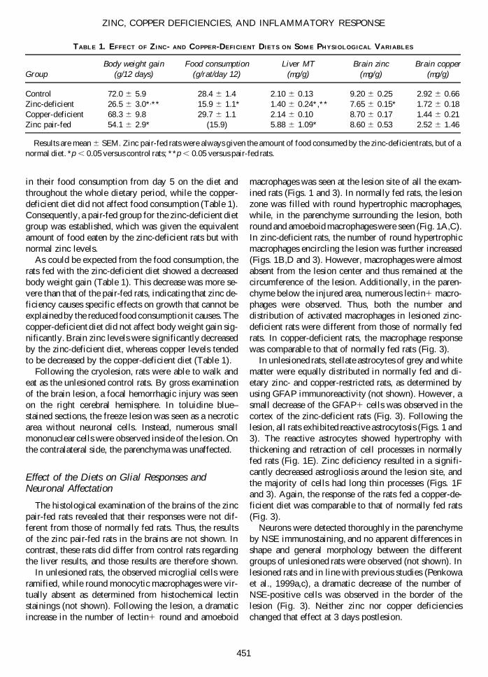
FIG. 2. Double immunofluorescent labeling of TUNEL (green) and NSE (red) in normally fed (A) and zinc-deficient rats (B)after lesioning. Triple immunofluorescent labeling of TUNEL (green), lectin1 macrophages (red) and GFAP1 astrocytes (blue)in normally fed (C) and zinc-deficient (D) rats. Results shown correspond to the borderline of the injured area.
tochemistry, we found that the TUNEL1 cells were neu-rons, astrocytes, and microglia/macrophages situatedaround the lesion (Fig. 2). In zinc-deficient rats, the num-ber of TUNEL1 cells was increased for neurons (Fig.2A,B), but astrocytes and microglia/macrophages alsodisplayed a higher apoptotic signal (Fig. 2C,D).
The Metal-Deficient Diets Alter the Response of the Metallothionein Isoforms to TraumaticBrain Injury
In the brain of unlesioned rats, the expression of MT-I1II was confined to ependymal cells, meninges, glialimitans, and some gray matter astrocytes, and no sig-nificant effect of the diets was noticed, although the zinc-deficient diet tended to decrease the number of MT-I1II–positive cells. Following the lesion, normal fed rats
increased MT-I1II expression in activated macrophagesand reactive astrocytes widely distributed at the basis ofthe lesion (Figs. 4 and 5). In lesioned zinc-deficient rats,the expression of MT-I1II was mildly upregulated com-pared to normally fed rats (Figs. 4B and 5). MT-I1II ex-pression of macrophages and reactive astrocytes situatedaround the injury in copper-deficient rats was onlyslightly lower than that of normally fed rats (Fig. 5). Asexpected (Gasull et al., 1994; Bremner et al., 1987), liverMT-I1II levels were decreased by zinc deficiency, es-pecially when compared with the pair-fed rats, whichshow a clear upregulation compared to normally fed rats(Table 1). This is in contrast to the brain, since no effectof food restriction on MT-I1II levels was observed (notshown).
In the brain of unlesioned rats, the expression of MT-III was confined to ependymal cells, meninges, glia lim-
PENKOWA ET AL.
454
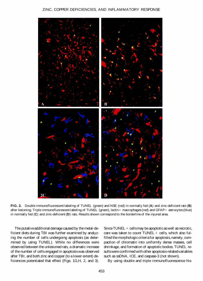
FIG. 3. Immunohistochemical cell countings of lectin, GFAP, NSE, and TUNEL in the brain cortex of normally fed rats andrats fed zinc- and copper-deficient diets (cells/mm2). Counting was carried out in the border of the lesion where prominent glialresponse occurs (see diamonds in Fig. 1A,B for localization) or in the normal cortex in unlesioned rats. Cellular countings shownare mean 6 SE (n 5 3 independent rats per group). The lesion affected significantly (p , 0.001) all the variables analyzed. Sep-arate one-way ANOVA followed by post-hoc comparison of the means were the carried out for unlesioned and lesioned rats.w Significant effect (p , 0.05) of the zinc-deficient diet versus the corresponding normally fed rats. m Significant effect (p , 0.05)of the copper-deficient versus the corresponding normally fed rats.
itans, perivascular cells, and some astrocytes scattered incortex. Following the lesion, the immunoreactivity ofMT-III was increased in all rats examined (Figs. 4 and5). Normal fed rats upregulated their MT-III expression
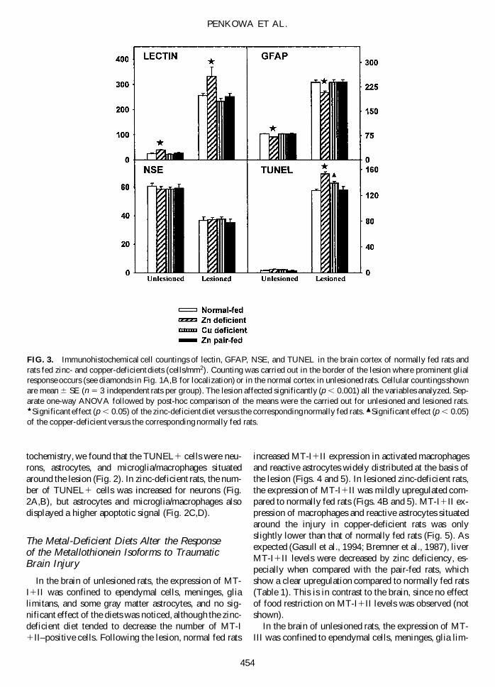
in round and amoeboid macrophages around and insideof the lesion (Fig. 4C). Some reactive astrocytes werealso MT-III1. In lesioned zinc-deficient rats, the MT-IIIexpression was significantly increased compared to that
ZINC, COPPER DEFICIENCIES, AND INFLAMMATORY RESPONSE
455
FIG. 4. MT-I1II and MT-III immunostainings of freeze-lesioned normally fed and zinc-deficient rats. (A) MT-I1II im-munoreactivity in the parenchyma surrounding the lesion site of normal rats. (B) MT-I1II immunoreactivity in the parenchymasurrounding the lesion site of zinc-deficient rats, showing a decreased number of MT-I1II expressing cells, which are also de-creased in size compared to those of normally fed rats. (C) MT-III expression was mildly increased around and inside of the le-sion of normally fed rats. (D) Zinc-deficient rats increased the MT-III immunoreactivity postlesional when compared to that ofnormally fed rats. Numerous round macrophages and astrocytes surrounding the injury were expressing MT-III (arrows), and thedistribution of MT-III positive cells matched that of lectin-positive cells. Also inside of the lesion, some round macrophages wereexpressing MT-III. Bar 5 25 mm (A,B), 22 mm (C,D).
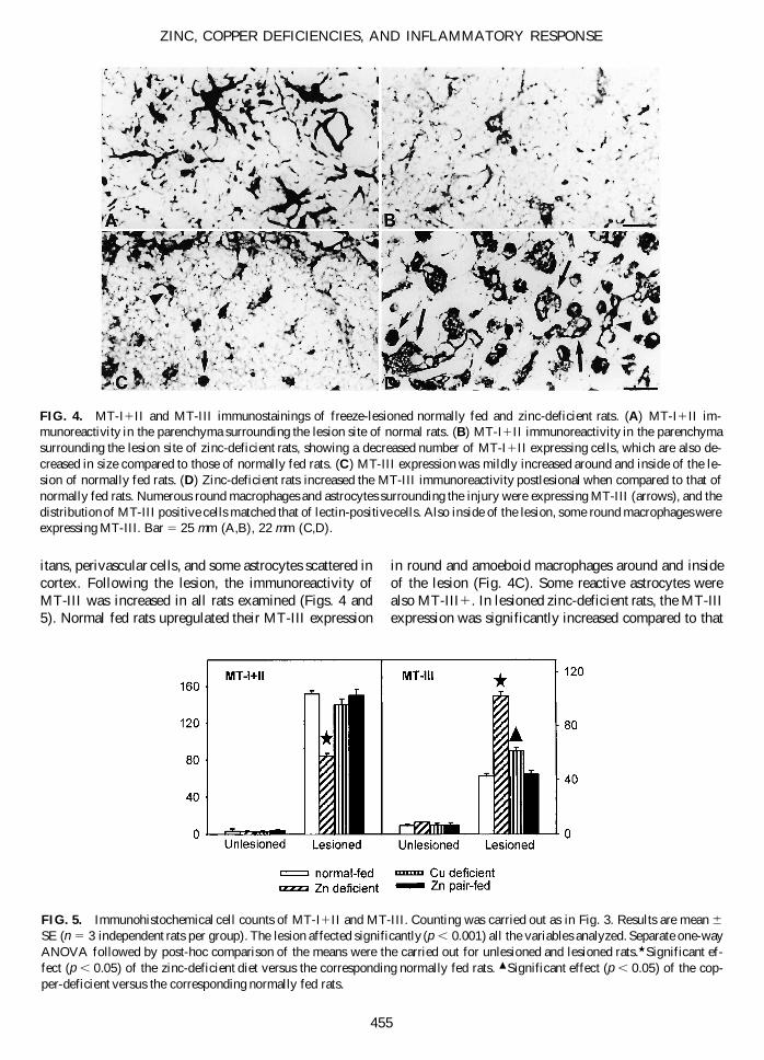
FIG. 5. Immunohistochemical cell counts of MT-I1II and MT-III. Counting was carried out as in Fig. 3. Results are mean 6
SE (n 5 3 independent rats per group). The lesion affected significantly (p , 0.001) all the variables analyzed. Separate one-wayANOVA followed by post-hoc comparison of the means were the carried out for unlesioned and lesioned rats.w Significant ef-fect (p , 0.05) of the zinc-deficient diet versus the corresponding normally fed rats. m Significant effect (p , 0.05) of the cop-per-deficient versus the corresponding normally fed rats.
of normally fed rats (Figs. 4D and 5). Copper deficientrats displayed a similar albeit more moderate response to TBI.
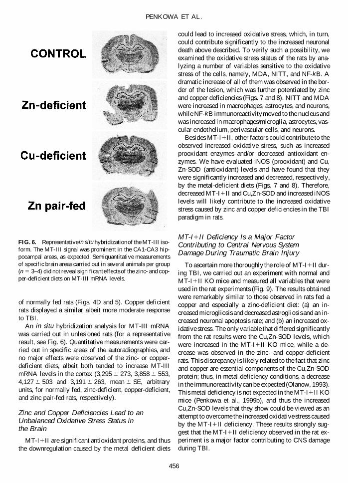
An in situ hybridization analysis for MT-III mRNAwas carried out in unlesioned rats (for a representativeresult, see Fig. 6). Quantitative measurements were car-ried out in specific areas of the autoradiographies, andno major effects were observed of the zinc- or copper-deficient diets, albeit both tended to increase MT-IIImRNA levels in the cortex (3,295 6 273, 3,858 6 553,4,127 6 503 and 3,191 6 263, mean 6 SE, arbitraryunits, for normally fed, zinc-deficient, copper-deficient,and zinc pair-fed rats, respectively).
Zinc and Copper Deficiencies Lead to anUnbalanced Oxidative Stress Status in the Brain
MT-I1II are significant antioxidant proteins, and thusthe downregulation caused by the metal deficient diets
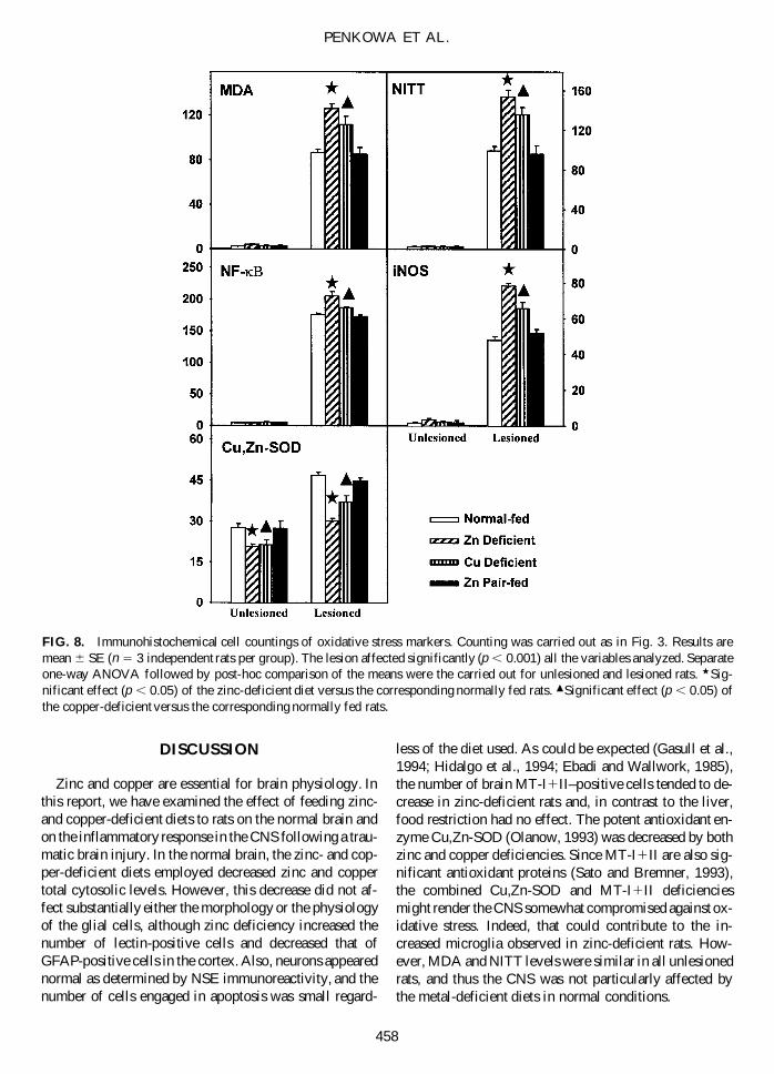
could lead to increased oxidative stress, which, in turn,could contribute significantly to the increased neuronaldeath above described. To verify such a possibility, weexamined the oxidative stress status of the rats by ana-lyzing a number of variables sensitive to the oxidativestress of the cells, namely, MDA, NITT, and NF-kB. Adramatic increase of all of them was observed in the bor-der of the lesion, which was further potentiated by zincand copper deficiencies (Figs. 7 and 8). NITT and MDAwere increased in macrophages, astrocytes, and neurons,while NF-kB immunoreactivity moved to the nucleus andwas increased in macrophages/microglia, astrocytes, vas-cular endothelium, perivascular cells, and neurons.
Besides MT-I1II, other factors could contribute to theobserved increased oxidative stress, such as increasedprooxidant enzymes and/or decreased antioxidant en-zymes. We have evaluated iNOS (prooxidant) and Cu,Zn-SOD (antioxidant) levels and have found that theywere significantly increased and decreased, respectively,by the metal-deficient diets (Figs. 7 and 8). Therefore,decreased MT-I1II and Cu,Zn-SOD and increased iNOSlevels will likely contribute to the increased oxidativestress caused by zinc and copper deficiencies in the TBIparadigm in rats.
MT-I1II Deficiency Is a Major FactorContributing to Central Nervous System Damage During Traumatic Brain Injury
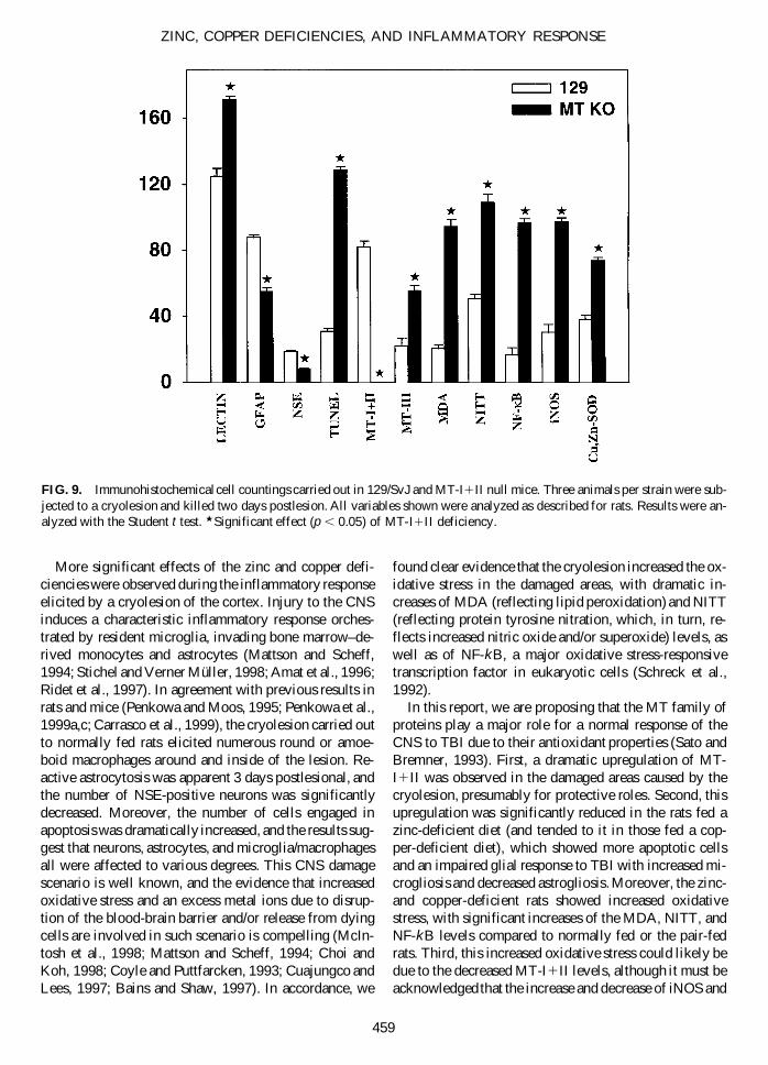
To ascertain more thoroughly the role of MT-I1II dur-ing TBI, we carried out an experiment with normal andMT-I1II KO mice and measured all variables that wereused in the rat experiments (Fig. 9). The results obtainedwere remarkably similar to those observed in rats fed acopper and especially a zinc-deficient diet: (a) an in-creased microgliosis and decreased astrogliosis and an in-creased neuronal apoptosis rate; and (b) an increased ox-idative stress. The only variable that differed significantlyfrom the rat results were the Cu,Zn-SOD levels, whichwere increased in the MT-I1II KO mice, while a de-crease was observed in the zinc- and copper-deficientrats. This discrepancy is likely related to the fact that zincand copper are essential components of the Cu,Zn-SODprotein; thus, in metal deficiency conditions, a decreasein the immunoreactivity can be expected (Olanow, 1993).This metal deficiency is not expected in the MT-I1II KOmice (Penkowa et al., 1999b), and thus the increasedCu,Zn-SOD levels that they show could be viewed as anattempt to overcome the increased oxidative stress causedby the MT-I1II deficiency. These results strongly sug-gest that the MT-I1II deficiency observed in the rat ex-periment is a major factor contributing to CNS damageduring TBI.
PENKOWA ET AL.
456
FIG. 6. Representative in situ hybridization of the MT-III iso-form. The MT-III signal was prominent in the CA1-CA3 hip-pocampal areas, as expected. Semiquantitative measurementsof specific brain areas carried out in several animals per group(n 5 3–4) did not reveal significant effects of the zinc- and cop-per-deficient diets on MT-III mRNA levels.
ZINC, COPPER DEFICIENCIES, AND INFLAMMATORY RESPONSE
457
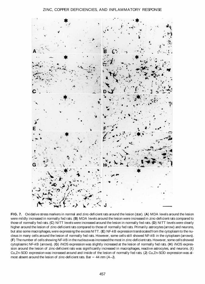
FIG. 7. Oxidative stress markers in normal and zinc-deficient rats around the lesion (star). (A) MDA levels around the lesionwere mildly increased in normally fed rats. (B) MDA levels around the lesion were increased in zinc-deficient rats compared tothose of normally fed rats. (C) NITT levels were increased around the lesion in normally fed rats. (D) NITT levels were clearlyhigher around the lesion of zinc-deficient rats compared to those of normally fed rats. Primarily astrocytes (arrow) and neurons,but also some macrophages, were expressing the excess NITT. (E) NF-kB expression translocated from the cytoplasm to the nu-cleus in many cells around the lesion of normally fed rats. However, some cells still showed NF-kB in the cytoplasm (arrows).(F) The number of cells showing NF-kB in the nucleus was increased the most in zinc-deficient rats. However, some cells showedcytoplasmic NF-kB (arrows). (G) iNOS expression was slightly increased at the lesion of normally fed rats. (H) iNOS expres-sion around the lesion of zinc-deficient rats was significantly increased in macrophages, reactive astrocytes, and neurons. (I)Cu,Zn-SOD expression was increased around and inside of the lesion of normally fed rats. (J) Cu,Zn-SOD expression was al-most absent around the lesion of zinc-deficient rats. Bar 5 44 mm (A–J).
DISCUSSION
Zinc and copper are essential for brain physiology. Inthis report, we have examined the effect of feeding zinc-and copper-deficient diets to rats on the normal brain andon the inflammatory response in the CNS following a trau-matic brain injury. In the normal brain, the zinc- and cop-per-deficient diets employed decreased zinc and coppertotal cytosolic levels. However, this decrease did not af-fect substantially either the morphology or the physiologyof the glial cells, although zinc deficiency increased thenumber of lectin-positive cells and decreased that ofGFAP-positive cells in the cortex. Also, neurons appearednormal as determined by NSE immunoreactivity, and thenumber of cells engaged in apoptosis was small regard-
less of the diet used. As could be expected (Gasull et al.,1994; Hidalgo et al., 1994; Ebadi and Wallwork, 1985),the number of brain MT-I1II–positive cells tended to de-crease in zinc-deficient rats and, in contrast to the liver,food restriction had no effect. The potent antioxidant en-zyme Cu,Zn-SOD (Olanow, 1993) was decreased by bothzinc and copper deficiencies. Since MT-I1II are also sig-nificant antioxidant proteins (Sato and Bremner, 1993),the combined Cu,Zn-SOD and MT-I1II deficienciesmight render the CNS somewhat compromised against ox-idative stress. Indeed, that could contribute to the in-creased microglia observed in zinc-deficient rats. How-ever, MDA and NITT levels were similar in all unlesionedrats, and thus the CNS was not particularly affected bythe metal-deficient diets in normal conditions.
PENKOWA ET AL.
458
FIG. 8. Immunohistochemical cell countings of oxidative stress markers. Counting was carried out as in Fig. 3. Results aremean 6 SE (n 5 3 independent rats per group). The lesion affected significantly (p , 0.001) all the variables analyzed. Separateone-way ANOVA followed by post-hoc comparison of the means were the carried out for unlesioned and lesioned rats. w Sig-nificant effect (p , 0.05) of the zinc-deficient diet versus the corresponding normally fed rats. m Significant effect (p , 0.05) ofthe copper-deficient versus the corresponding normally fed rats.
More significant effects of the zinc and copper defi-ciencies were observed during the inflammatory responseelicited by a cryolesion of the cortex. Injury to the CNSinduces a characteristic inflammatory response orches-trated by resident microglia, invading bone marrow–de-rived monocytes and astrocytes (Mattson and Scheff,1994; Stichel and Verner Müller, 1998; Amat et al., 1996;Ridet et al., 1997). In agreement with previous results inrats and mice (Penkowa and Moos, 1995; Penkowa et al.,1999a,c; Carrasco et al., 1999), the cryolesion carried outto normally fed rats elicited numerous round or amoe-boid macrophages around and inside of the lesion. Re-active astrocytosis was apparent 3 days postlesional, andthe number of NSE-positive neurons was significantlydecreased. Moreover, the number of cells engaged inapoptosis was dramatically increased, and the results sug-gest that neurons, astrocytes, and microglia/macrophagesall were affected to various degrees. This CNS damagescenario is well known, and the evidence that increasedoxidative stress and an excess metal ions due to disrup-tion of the blood-brain barrier and/or release from dyingcells are involved in such scenario is compelling (McIn-tosh et al., 1998; Mattson and Scheff, 1994; Choi andKoh, 1998; Coyle and Puttfarcken, 1993; Cuajungco andLees, 1997; Bains and Shaw, 1997). In accordance, we
found clear evidence that the cryolesion increased the ox-idative stress in the damaged areas, with dramatic in-creases of MDA (reflecting lipid peroxidation) and NITT(reflecting protein tyrosine nitration, which, in turn, re-flects increased nitric oxide and/or superoxide) levels, aswell as of NF-kB, a major oxidative stress-responsivetranscription factor in eukaryotic cells (Schreck et al.,1992).
In this report, we are proposing that the MT family ofproteins play a major role for a normal response of theCNS to TBI due to their antioxidant properties (Sato andBremner, 1993). First, a dramatic upregulation of MT-I1II was observed in the damaged areas caused by thecryolesion, presumably for protective roles. Second, thisupregulation was significantly reduced in the rats fed azinc-deficient diet (and tended to it in those fed a cop-per-deficient diet), which showed more apoptotic cellsand an impaired glial response to TBI with increased mi-crogliosis and decreased astrogliosis. Moreover, the zinc-and copper-deficient rats showed increased oxidativestress, with significant increases of the MDA, NITT, andNF-kB levels compared to normally fed or the pair-fedrats. Third, this increased oxidative stress could likely bedue to the decreased MT-I1II levels, although it must beacknowledged that the increase and decrease of iNOS and
ZINC, COPPER DEFICIENCIES, AND INFLAMMATORY RESPONSE
459
FIG. 9. Immunohistochemical cell countings carried out in 129/SvJ and MT-I1II null mice. Three animals per strain were sub-jected to a cryolesion and killed two days postlesion. All variables shown were analyzed as described for rats. Results were an-alyzed with the Student t test. w Significant effect (p , 0.05) of MT-I1II deficiency.
Cu,Zn-SOD, respectively, could equally contribute. Andfourth, to more directly establish the role of MT-I1II dur-ing TBI, we carried out an experiment with MT-I1II nullmice. The results indicated that the absence of MT-I1IIcaused an impaired glial response, increased apoptosis,and potentiated oxidative stress, all of which were re-markably similar to the effects caused by the zinc andcopper deficient diets in the rat experiments. These re-sults strongly suggest that most of the effects caused bythe metal deficiencies can be causally linked to the MT-I1II downregulation. The magnitude of the changes ob-served in the MT-I1II null mice is higher than in the ratsfed metal-deficient diets, which seems logical since thelatter only have a reduction but not a total lack of theMT-I1II proteins. Thus, MT-I1II appear to be of majorimportance for the CNS coping with TBI. Consistent withsuch a role, MT-I1II levels have been observed to be in-creased in several human neurodegenerative disorders aswell as in a number of experimental models of brain dam-age (for review, see Hidalgo et al., 1997). Interesting, ox-idative stress has been involved in several human neu-rodegenerative diseases (Bains and Shaw, 1997; Coyleand Puttfarcken, 1993; Hensley et al., 1997; Shohami etal., 1997).
Another finding of this study was the specific re-sponses of the rather CNS specific isoform, MT-III. Inline with previous brain damage models (Hildalgo et al.,1997), the cryo lesion of the CNS of normal rats pro-duced a mild MT-III upregulation in inflammatory cellssurrounding the lesioned area. Since MT-III has an in-hibitory effect on neuronal survival in vitro (Uchida etal., 1991; Erickson et al., 1994), it has been speculatedthat the increase of MT-III levels after brain damage re-flects attempts to limit neuronal sprouting (Yuguchi etal., 1995a). However, further studies have shown thatMT-III has indeed a neuroprotective role, at least in theCA3 hippocampal area after kainic acid–induced seizures(Erickson et al., 1997). A promoting role of astrocyte mi-gration has also been described in vitro (Carrasco et al.,1999). Somewhat surprisingly, MT-III protein levelswere strongly increased in the lesioned zinc-deficient ratsand moderately increased in copper-deficient rats. Thecomparison of the MT-III changes versus those of MT-I1II suggest a sort of compensatory response of MT-IIIbecause of a decrease in the MT-I1II response. This hasalso been observed in IL-6–deficient mice after TBI(Penkowa et al., 1999c) and in MT-I1II–deficient miceafter administration of the glial toxin 6-aminonicoti-namide (Penkowa et al., 1999b). The mechanisms un-derlying these effects are unknown, and their putativephysiological importance needs further experiments.
The present results demonstrate that zinc and copperare important for cellular activation/recruitment and for
expression of factors and enzymes synthesized during in-flammation in CNS. Not only the glial responses but alsothe cellular survival are significantly altered by the defi-ciency of these essential heavy metals. Most of the ef-fects described in this paper could be attributable to theaccompanying changes of MT-I1II protein levels, likelybecause of their antioxidant functions. This and previousstudies are identifying an emerging essential role of thisfamily of proteins in the CNS.
ACKNOWLEDGMENTS
The study was supported by The Novo NordiskFonden, Dansk Epilepsi Selskabs Forskingsfond, Dir. Ja-cob Madsens og Hustrus Fond, Fonden af 17.12.1981,Gerda og Aage Haensch’s Fond, Leo Nielsen og HustrusLegat, CICYT SAF96-0189, PSPGC PM98-0170, andFundación “La Caixa” 97/102-00 (J.H.). J. Carrasco issupported by a fellowship of the CIRIT, FI/96-2613.
REFERENCES
ACARIN, L., CARRASCO, J., GONZÁLEZ, B., et al. (1999a).Expression of growth inhibitory factor (metallothionein-III)mRNA and protein following immature brain injury. J. Neu-ropathol. Exp. Neurol. 58, 389–397.
ACARIN, L., GONZÁLEZ, B., HIDALGO, J. et al. (1999b).Primary cortical glial reaction versus secondary thalamicglial response in the excitotoxically injured young brain. As-troglial response and metallothionein expression. Neuro-science 92, 827–839.
AMAT, J., ISHIGURO, H., NAKAMURA, K., et al. (1996).Phenotypic diversity and kinetics of proliferating microgliaand astrocytes following cortical stab wounds. Glia 16, 368–382.
ANEZAKI, T., ISHIGURO, H., HOZUMI, I., et al. (1995). Ex-pression of growth inhibitory factor (GIF) in normal and in-jured rat brains. Neurochem. Int. 27, 89–94.
BAINS, J., and SHAW, C. (1997). Neurodegenerative disor-ders in humans: the role of glutatahione in oxidative stress–mediated neuronal death. Brain Res. Rev. 25, 335–358.
BREMNER, I. (1987). Interactions between metallothioneinand trace elements. Prog. Food Nutr. Sci. 11, 1–37.
BREMNER, I., MORRISON, J.N., WOOD, A.M., et al. (1987).Effects of changes in dietary zinc, copper and selenium sup-ply and of endotoxin administration on metallothionein I con-centrations in blood cells and urine in the rat. J. Nutr. 117,1595–1602.
CARRASCO, J., HERNÁNDEZ, J., GONZÁLEZ, B., et al.(1998). Localization of metallothionein-I and -III expressionin the CNS of transgenic mice with astrocyte-targeted ex-pression of interleukin 6. Exp. Neurol. 153, 184–194.
PENKOWA ET AL.
460
CARRASCO, J., GIRALT, M., MOLINERO, A., et al. (1999).Metallothionein (MT)–III: generation of polyclonal antibod-ies, comparison with MT-I1II in the freeze lesioned rat brainand in a bioassay with astrocytes, and analysis of Alzhei-mer’s disease brains. J. Neurotrauma 16, 1115–1129.
CHOI, D., and KOH, J. (1998). Zinc and brain injury. Annu.Rev. Neurosci. 21, 347–375.
COUSINS, R.J. (1985). Absorption, transport, and hepatic me-tabolism of copper and zinc: special reference to metallo-thionein and ceruloplasmin. Physiol. Rev. 65, 238–309.
COYLE, J., and PUTTFARCKEN, P. (1993). Oxidative stress,glutamate, and neurodegenerative disorders. Science 262,689–695.
CUAJUNGCO, M., and LEES, G. (1997). Zinc metabolism inthe brain: relevance to human neurodegenerative disorders.Neurobiol. Dis. 4, 137–169.
DALTON, T., PAZDERNIK, T.L., WAGNER, J., et al. (1995).Temporalspatial patterns of expression of metallothionein-Iand -III and other stress related genes in rat brain after kainicacid–induced seizures. Neurochem. Int. 27, 59–71.
EBADI, M., and WALLWORK, J. (1985). Zinc-binding pro-teins (ligands) in brains of severely zinc-deficient rats. Biol.Trace Elem. Res. 7, 129–139.
ERICKSON, J.C., SEWELL, A.K., JENSEN, L.T., et al.(1994). Enhanced neutrophic activity in Alzheimer’s diseasecortex is not associated with down-regulation of metallo-thionein-III (GIF). Brain Res. 649, 297–304.
ERICKSON, J.C., HOLLOPETER, G., THOMAS, S.A., et al.(1997). Disruption of the metallothionein-III gene in mice:analysis of brain zinc, behavior, and neuron vulnerability tometals, aging, and seizures. J. Neurosci. 17, 1271–1281.
FREDERICKSON, C., HOWELL, G., and KASARKIS, E.(1984). The Neurobiology of Zinc, Part A. Alan R. Liss: NewYork.
FU, K., TOMITA, T., SARRAS, M., et al. (1998). Metallo-thionein protects against cerulein-induced acute pancreatitis:analysis using transgenic mice. Pancreas 17, 238–246.
GASULL, T., REBOLLO, D.V., ROMERO, B., et al. (1993).Development of a competitive double antibody radioimmuno-assay for rat metallothionein. J. Immunoassay 14, 209–225.
GASULL, T., GIRALT, M., HERNANDEZ, J., et al. (1994).Regulation of metallothionein concentrations in rat brain: ef-fect of glucocorticoids, zinc, copper, and endotoxin. Am. J.Physiol. 266, E760–E767.
HAMER, D.H. (1986). Metallothionein. Annu. Rev. Biochem.55, 913–951.
HANADA, K., SAWAMURA, D., TAMAI, K., et al. (1998).Novel function of metallothionein in photoprotection: me-tallothionein-null mouse exhibits reduced tolerance againstultraviolet B injury in the skin. J. Invest. Dermatol. 111, 582–585.
HENSLEY, K., CARNEY, J.M., STEWART, C.A., et al.(1997). Nitrone-based free radical traps as neuroprotectiveagents in cerebral ischaemia and other pathologies. Int. Rev.Neurobiol. 40, 299–317.
HERNÁNDEZ, J., CARRASCO, J., ARBONÉS, M.L., et al.(1997). IFN-gR2/2 mice show an enhanced liver and brainmetallothionein I1II response to endotoxin but not to im-mobilization stress. J. Endotoxin Res. 4, 363–370.
HIDALGO, J., BORRAS, M., GARVEY, J.S., et al. (1990).Liver, brain, and heart metallothionein induction by stress. J.Neurochem. 55, 651–654.
HIDALGO, J., GARCIA, A., OLIVA, A.M., et al. (1994). Ef-fect of zinc, copper and glucocorticoids on metallothioneinlevels of cultured neurons and astrocytes from rat brain.Chem. Biol. Interact. 93, 197–219.
HIDALGO, J., CASTELLANO, B., and CAMPBELL, I.L.(1997). Regulation of brain metallothioneins. Curr. TopicsNeurochem. 1, 1–26.
HOZUMI, I., INUZUKA, T., HIRAIWA, M., et al. (1995).Changes of growth inhibitory factor after stab wounds in ratbrain. Brain Res. 688, 143–148.
HOZUMI, I., INUZUKA, T., ISHIGURO, H., et al. (1996). Im-munoreactivity of growth inhibitory factor in normal rat brainand after stab wounds—an immunocytochemical study us-ing confocal laser scan microscope. Brain Res. 741, 197–204.
IMAGAWA, M., ISHIKAWA, Y., SHIMANO, H., et al.(1995). CTG triplet repeat in mouse growth inhibitory factor/metallothionein III gene promoter represses the transcrip-tional activity of the heterologous promoters. J. Biol. Chem.270, 20898–20900.
INUZUKA, T., HOZUMI, I., TAMURA, A., et al. (1996). Pat-terns of growth inhibitory factor (GIF) and glial fibrillaryacidic protein relative level changes differ following left mid-dle cerebral artery occlusion in rats. Brain Res. 709, 151–131.
KAGI, J.H., and SCHAFFER, A. (1988). Biochemistry of me-tallothionein. Biochemistry 27, 8509–8515.
KANG, Y., CHEN, Y., YU, A., et al. (1997). Overexpressionof metallothionein in the heart of transgenic mice suppressesdoxorubicin cardiotoxicity. J. Clin. Invest. 100, 1501–1506.
KEEN, C., URIU-HARE, J., HAWK, S., et al. (1998). Effectof copper deficiency on prenatal development and pregnancyoutcome. Am. J. Clin. Nutr. 67, 1003S–1011S.
KONDO, Y., RUSNAK, J., HOYT, D., et al. (1997). Enhancedapoptosis in metallothionein null cells. Mol. Pharmacol. 52,195–201.
LAZO, J.S., KONDO, Y., DELLAPIAZZA, D., et al. (1995).Enhanced sensitivity to oxidative stress in cultured embry-onic cells from transgenic mice deficient in metallothioneinI and II genes. J. Biol. Chem. 270, 5506–5510.
LIU, J., LIU, Y., HABEEBU, S., et al. (1998). Metallothionein(MT)–null mice are sensitive to cisplatin-induced hepato-toxicity. Toxicol. Appl. Pharmacol. 149, 24–31.
ZINC, COPPER DEFICIENCIES, AND INFLAMMATORY RESPONSE
461
LIU, J., LIU, Y., HARTLEY, D., et al. (1999). Metallothionein-I/II knockout mice are sensitive to acetaminophen-inducedhepatoxicity. J. Pharmacol. Exp. Ther. 289, 580–586.
MASTERS, B.A., KELLY, E.J., QUAIFE, C.J. et al. (1994a).Targeted disruption of metallothionein I and II genes in-creases sensitivity to cadmium. Proc. Natl. Acad. Sci. U.S.A.91, 584–588.
MASTERS, B.A., QUAIFE, C.J., ERICKSON, J.C., et al.(1994b). Metallothionein III is expressed in neurons that se-quester zinc in synaptic vesicles. J. Neurosci. 14, 5844–5857.
MATTSON, M., and SCHEFF, S. (1994). Endogenous neuro-protection factors and traumatic brain injury: mechanisms ofaction and implications for therapy. J. Neurotrauma 11, 3–33.
MCINTOSH, T., JUHLER, M., and WIELOCH, T. (1998).Novel pharmacologic strategies in the treatment of experi-mental traumatic brain injury. J. Neurotrauma 15, 731–769.
NALBANDYAN, R. (1983). Copper in brain. Neurochem. Res.8, 1211–1232.
NARUSE, S., IGARASHI, S., FURUYA, T., et al. (1994).Structures of the human and mouse growth inhibitory fac-tor–encoding genes. Gene 144, 283–287.
NEAL, J.W., SINGHRAO, S.K., JASANI, B., et al. (1996). Im-munocytochemically detectable metallothionein is expressedby astrocytes in the ischaemic human brain. Neuropathol.Appl. Neurobiol. 22, 243–247.
OLANOW, C. (1993). A radical hypothesis for neurodegener-ation. Trends Neurosci. 16, 439–444.
PALMITER, R.D., FINDLEY, S.D., WHITMORE, T.E., et al.(1992). MT-III, a brain-specific member of the metallo-thionein gene family. Proc. Natl. Acad. Sci. U.S.A. 89, 6333–6337.
PENKOWA, M., and MOOS, T. (1995). Disruption of theblood-brain interface in neonatal rat neocortex induces a tran-sient expression of metallothionein in reactive astrocytes.Glia 13, 217–227.
PENKOWA, M., HIDALGO, J., and MOOS, T. (1997). In-creased astrocytic expression of metallothioneins I1II inbrain stem of adult rats treated with 6-aminonicotinamide.Brain Res. 774, 256–259.
PENKOWA, M., CARRASCO, J., GIRALT, M., et al. (1999a).CNS wound healing is severely depressed in metallothionein-I1II deficient mice. J. Neurosci. 19, 2535–2545.
PENKOWA, M., GIRALT, M., MOOS, T., et al. (1999b). Im-paired inflammatory response to glial cell death in geneti-cally metallothionein-I and -II deficient mice. Exp. Neurol.156, 149–164.
PENKOWA, M., MOOS, T., CARRASCO, J., et al. (1999c).Strongly compromised inflammatory response to brain injuryin interleukin-6 deficient mice. Glia 25, 343–357.
QUAIFE, C.J., FINDLEY, S.D., ERICKSON, J.C., et al.(1994). Induction of a new metallothionein isoform (MT-IV)
occurs during differentiation of stratified squamous epithe-lia. Biochemistry 33, 7250–7259.
RIDET, J.L., MALHOTRA, A., and GAGE, F. (1997). Reac-tive astrocytes: cellular and molecular cues to biologicalfunction. Trends Neurosci. 20, 570–577.
SATO, M., and BREMNER, I. (1993). Oxygen-free radicalsand metallothionein. Free Radic. Biol. Med. 14, 325–337.
SCHRECK, R., ALBERMANN, K., and BAEURELE, P.(1992). Nuclear factor kB: an oxidative stress–responsivetranscription factor of eukaryotic cells (a review). Free Radic.Res. Commun. 17, 221–237.
SEWELL, A.K., JENSEN, L.T., ERICKSON, J.C., et al.(1995). Bioactivity of metallothionein-3 correlates with itsnovel beta domain sequence rather than metal binding prop-erties. Biochemistry 34, 4740–4747.
SHOHAMI, E., BAIT-YANNAL, E., HOROWITZ, M., et al.(1997). Oxidative stress in closed-head injury: brain antiox-idant capacity as an indicator of functional outcome. J. Cereb.Blood Flow Metab. 17, 1007–1019.
SILLEVIS SMITT, P.A., BLAAUWGEERS, H.G., TROOST,D., et al. (1992). Metallothionein immunoreactivity is in-creased in the spinal cord of patients with amyotrophic lat-eral sclerosis. Neurosci. Lett. 144, 107–110.
STICHEL, C., and VERNER MÜLLER, H. (1998). Experi-mental strategies to promote axonal regeneration after trau-matic central nervous system injury. Prog. Neurobiol. 56,119–148.
SUZUKI, K., NAKAJIMA, K., KAWAHARADA, U., et al.(1992). Metallothionein in the human brain. Acta Histochem.Cytochem. 25, 617–622.
UAUY, R., OLIVARES, M., and GONZALEZ, M. (1998). Es-sentiality of copper in human. Am. J. Clin. Nutr. 67, 952S–959S.
UCHIDA, Y. (1994). Growth-inhibitory factor, metallothio-nein-like protein, and neurodegenerative diseases. Biol. Sig-nals 3, 211–215.
UCHIDA, Y., TAKIO, K., TITANI, K., et al. (1991). Thegrowth inhibitory factor that is deficient in the Alzheimer’sdisease brain is a 68 amino acid metallothionein-like protein.Neuron 7, 337–347.
VALLEE, B.L. (1988). Zinc: biochemistry, physiology, toxi-cology and clinical pathology. Biofactors 1, 31–36.
VALLEE, B. (1993). The biochemical basis of zinc physiol-ogy. Physiol. Rev. 73, 79–117.
VAN LOOKEREN CAMPAGNE, M., THIBODEAUX, H.,CAIMS, B., et al. (1999). Transgenic mice overexpressingmetallothionein I show protection against focal cerebral isch-emia. J. Cereb. Blood Flow Metab. 19, S138.
VELA, J.M., HIDALGO, J., GONZÁLEZ, B., et al. (1997). In-duction of metallothionein in astrocytes and microglia in thespinal cord from the myelin-deficient jimpy mouse. BrainRes. 767, 345–355.
PENKOWA ET AL.
462
YAGLE, M.K., and PALMITER, R.D. (1985). Coordinate reg-ulation of mouse metallothionein I and II genes by heavymetals and glucocorticoids. Mol. Cell. Biol. 5, 291–294.
YOUNG, J.K., GARVEY, J.S., and HUANG, P.C. (1991). Glialimmunoreactivity for metallothionein in the rat brain. Glia 4,602–610.
YUGUCHI, T., KOHMURA, E., YAMADA, K., et al. (1995a).Expression of growth inhibitory factor mRNA following cor-tical injury. J. Neurotrauma 12, 299–306.
YUGUCHI, T., KOHMURA, E., YAMADA, K., et al. (1995b).Changes in growth inhibitory factor mRNA expression com-pared with those in c-jun mRNA expression following facialnerve transection. Mol. Brain Res. 28, 181–185.
ZHENG, H., BERMAN, N.E., and KLAASEN, C.D. (1995).Chemical modulation of metallothionein I and III mRNA inmouse brain. Neurochem. Int. 27, 43–58.
Address reprint requests to:Juan Hidalgo, Ph.D.
Departmento de Biología Celular, de Fisiología y de Immunología
Unidad de Fisiología AnimalFacultad de Ciencias
Universidad Autónoma de BarcelonaBellaterra, Barcelona, Spain 08193
E-mail: [email protected]
ZINC, COPPER DEFICIENCIES, AND INFLAMMATORY RESPONSE
463
Copyright © 2022 FDOKUMEN