Bio-Assessing of Environmental Pollution via Monitoring of Metallothionein Level Using...
16
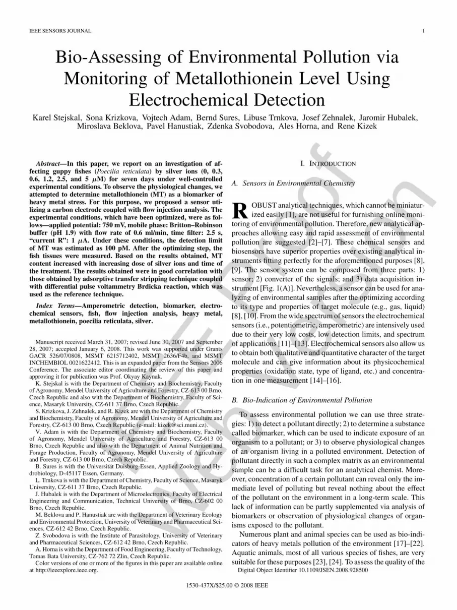
IEEE Proof Web Version IEEE SENSORS JOURNAL 1 Bio-Assessing of Environmental Pollution via Monitoring of Metallothionein Level Using Electrochemical Detection Karel Stejskal, Sona Krizkova, Vojtech Adam, Bernd Sures, Libuse Trnkova, Josef Zehnalek, Jaromir Hubalek, Miroslava Beklova, Pavel Hanustiak, Zdenka Svobodova, Ales Horna, and Rene Kizek Abstract—In this paper, we report on an investigation of af- fecting guppy fishes (Poecilia reticulata) by silver ions (0, 0.3, 0.6, 1.2, 2.5, and 5 M) for seven days under well-controlled experimental conditions. To observe the physiological changes, we attempted to determine metallothionein (MT) as a biomarker of heavy metal stress. For this purpose, we proposed a sensor uti- lizing a carbon electrode coupled with flow injection analysis. The experimental conditions, which have been optimized, were as fol- lows—applied potential: 750 mV, mobile phase: Britton–Robinson buffer (pH 1.9) with flow rate of 0.6 ml/min, time filter: 2.5 s, “current R”: 1 A. Under these conditions, the detection limit of MT was estimated as 100 pM. After the optimizing step, the fish tissues were measured. Based on the results obtained, MT content increased with increasing dose of silver ions and time of the treatment. The results obtained were in good correlation with those obtained by adsorptive transfer stripping technique coupled with differential pulse voltammetry Brdicka reaction, which was used as the reference technique. Index Terms—Amperometric detection, biomarker, electro- chemical sensors, fish, flow injection analysis, heavy metal, metallothionein, poecilia reticulata, silver. Manuscript received March 31, 2007; revised June 30, 2007 and September 28, 2007; accepted January 6, 2008. This work was supported under Grants GACR 526/07/0808, MSMT 6215712402, MSMT 2636/F4b, and MSMT INCHEMBIOL 0021622412. This is an expanded paper from the Sensors 2006 Conference. The associate editor coordinating the review of this paper and approving it for publication was Prof. Okyay Kaynak. K. Stejskal is with the Department of Chemistry and Biochemistry, Faculty of Agronomy, Mendel University of Agriculture and Forestry, CZ-613 00 Brno, Czech Republic and also with the Department of Biochemistry, Faculty of Sci- ence, Masaryk University, CZ-611 37 Brno, Czech Republic. S. Krizkova, J. Zehnalek, and R. Kizek are with the Department of Chemistry and Biochemistry, Faculty of Agronomy, Mendel University of Agriculture and Forestry, CZ-613 00 Brno, Czech Republic (e-mail: [email protected]). V. Adam is with the Department of Chemistry and Biochemistry, Faculty of Agronomy, Mendel University of Agriculture and Forestry, CZ-613 00 Brno, Czech Republic and also with the Department of Animal Nutrition and Forage Production, Faculty of Agronomy, Mendel University of Agriculture and Forestry, CZ-613 00 Brno, Czech Republic. B. Sures is with the Universität Duisburg-Essen, Applied Zoology and Hy- drobiology, D-45117 Essen, Germany. L. Trnkova is with the Department of Chemistry, Faculty of Science, Masaryk University, CZ-611 37 Brno, Czech Republic. J. Hubalek is with the Department of Microelectronics, Faculty of Electrical Engineering and Communication, Technical University of Brno, CZ-602 00 Brno, Czech Republic. M. Beklova and P. Hanustiak are with the Department of Veterinary Ecology and Environmental Protection, University of Veterinary and Pharmaceutical Sci- ences, CZ-612 42 Brno, Czech Republic. Z. Svobodova is with the Institute of Parasitology, University of Veterinary and Pharmaceutical Sciences, CZ-612 42 Brno, Czech Republic. A. Horna is with the Department of Food Engineering, Faculty of Technology, Tomas Bata University, CZ-762 72 Zlin, Czech Republic. Color versions of one or more of the figures in this paper are available online at http://ieeexplore.ieee.org. I. INTRODUCTION A. Sensors in Environmental Chemistry R OBUST analytical techniques, which cannot be miniatur- ized easily [1], are not useful for furnishing online moni- toring of environmental pollution. Therefore, new analytical ap- proaches allowing easy and rapid assessment of environmental pollution are suggested [2]–[7]. These chemical sensors and biosensors have superior properties over existing analytical in- struments fitting perfectly for the aforementioned purposes [8], [9]. The sensor system can be composed from three parts: 1) sensor; 2) converter of the signals; and 3) data acquisition in- strument [Fig. 1(A)]. Nevertheless, a sensor can be used for ana- lyzing of environmental samples after the optimizing according to its type and properties of target molecule (e.g., gas, liquid) [8], [10]. From the wide spectrum of sensors the electrochemical sensors (i.e., potentiometric, amperometric) are intensively used due to their very low costs, low detection limits, and spectrum of applications [11]–[13]. Electrochemical sensors also allow us to obtain both qualitative and quantitative character of the target molecule and can give information about its physicochemical properties (oxidation state, type of ligand, etc.) and concentra- tion in one measurement [14]–[16]. B. Bio-Indication of Environmental Pollution To assess environmental pollution we can use three strate- gies: 1) to detect a pollutant directly; 2) to determine a substance called biomarker, which can be used to indicate exposure of an organism to a pollutant; or 3) to observe physiological changes of an organism living in a polluted environment. Detection of pollutant directly in such a complex matrix as an environmental sample can be a difficult task for an analytical chemist. More- over, concentration of a certain pollutant can reveal only the im- mediate level of polluting but reveal nothing about the effect of the pollutant on the environment in a long-term scale. This lack of information can be partly supplemented via analysis of biomarkers or observation of physiological changes of organ- isms exposed to the pollutant. Numerous plant and animal species can be used as bio-indi- cators of heavy metals pollution of the environment [17]–[22]. Aquatic animals, most of all various species of fishes, are very suitable for these purposes [23], [24]. To assess the quality of the Digital Object Identifier 10.1109/JSEN.2008.928500 1530-437X/$25.00 © 2008 IEEE
Transcript of Bio-Assessing of Environmental Pollution via Monitoring of Metallothionein Level Using...
IEEE
Pro
of
Web
Ver
sion
IEEE SENSORS JOURNAL 1
Bio-Assessing of Environmental Pollution viaMonitoring of Metallothionein Level Using
Electrochemical DetectionKarel Stejskal, Sona Krizkova, Vojtech Adam, Bernd Sures, Libuse Trnkova, Josef Zehnalek, Jaromir Hubalek,
Miroslava Beklova, Pavel Hanustiak, Zdenka Svobodova, Ales Horna, and Rene Kizek
Abstract—In this paper, we report on an investigation of af-fecting guppy fishes (Poecilia reticulata) by silver ions (0, 0.3,0.6, 1.2, 2.5, and 5 M) for seven days under well-controlledexperimental conditions. To observe the physiological changes, weattempted to determine metallothionein (MT) as a biomarker ofheavy metal stress. For this purpose, we proposed a sensor uti-lizing a carbon electrode coupled with flow injection analysis. Theexperimental conditions, which have been optimized, were as fol-lows—applied potential: 750 mV, mobile phase: Britton–Robinsonbuffer (pH 1.9) with flow rate of 0.6 ml/min, time filter: 2.5 s,“current R”: 1 A. Under these conditions, the detection limitof MT was estimated as 100 pM. After the optimizing step, thefish tissues were measured. Based on the results obtained, MTcontent increased with increasing dose of silver ions and time ofthe treatment. The results obtained were in good correlation withthose obtained by adsorptive transfer stripping technique coupledwith differential pulse voltammetry Brdicka reaction, which wasused as the reference technique.
Index Terms—Amperometric detection, biomarker, electro-chemical sensors, fish, flow injection analysis, heavy metal,metallothionein, poecilia reticulata, silver.
Manuscript received March 31, 2007; revised June 30, 2007 and September28, 2007; accepted January 6, 2008. This work was supported under GrantsGACR 526/07/0808, MSMT 6215712402, MSMT 2636/F4b, and MSMTINCHEMBIOL 0021622412. This is an expanded paper from the Sensors 2006Conference. The associate editor coordinating the review of this paper andapproving it for publication was Prof. Okyay Kaynak.
K. Stejskal is with the Department of Chemistry and Biochemistry, Facultyof Agronomy, Mendel University of Agriculture and Forestry, CZ-613 00 Brno,Czech Republic and also with the Department of Biochemistry, Faculty of Sci-ence, Masaryk University, CZ-611 37 Brno, Czech Republic.
S. Krizkova, J. Zehnalek, and R. Kizek are with the Department of Chemistryand Biochemistry, Faculty of Agronomy, Mendel University of Agriculture andForestry, CZ-613 00 Brno, Czech Republic (e-mail: [email protected]).
V. Adam is with the Department of Chemistry and Biochemistry, Facultyof Agronomy, Mendel University of Agriculture and Forestry, CZ-613 00Brno, Czech Republic and also with the Department of Animal Nutrition andForage Production, Faculty of Agronomy, Mendel University of Agricultureand Forestry, CZ-613 00 Brno, Czech Republic.
B. Sures is with the Universität Duisburg-Essen, Applied Zoology and Hy-drobiology, D-45117 Essen, Germany.
L. Trnkova is with the Department of Chemistry, Faculty of Science, MasarykUniversity, CZ-611 37 Brno, Czech Republic.
J. Hubalek is with the Department of Microelectronics, Faculty of ElectricalEngineering and Communication, Technical University of Brno, CZ-602 00Brno, Czech Republic.
M. Beklova and P. Hanustiak are with the Department of Veterinary Ecologyand Environmental Protection, University of Veterinary and Pharmaceutical Sci-ences, CZ-612 42 Brno, Czech Republic.
Z. Svobodova is with the Institute of Parasitology, University of Veterinaryand Pharmaceutical Sciences, CZ-612 42 Brno, Czech Republic.
A. Horna is with the Department of Food Engineering, Faculty of Technology,Tomas Bata University, CZ-762 72 Zlin, Czech Republic.
Color versions of one or more of the figures in this paper are available onlineat http://ieeexplore.ieee.org.
I. INTRODUCTION
A. Sensors in Environmental Chemistry
R OBUST analytical techniques, which cannot be miniatur-ized easily [1], are not useful for furnishing online moni-
toring of environmental pollution. Therefore, new analytical ap-proaches allowing easy and rapid assessment of environmentalpollution are suggested [2]–[7]. These chemical sensors andbiosensors have superior properties over existing analytical in-struments fitting perfectly for the aforementioned purposes [8],[9]. The sensor system can be composed from three parts: 1)sensor; 2) converter of the signals; and 3) data acquisition in-strument [Fig. 1(A)]. Nevertheless, a sensor can be used for ana-lyzing of environmental samples after the optimizing accordingto its type and properties of target molecule (e.g., gas, liquid)[8], [10]. From the wide spectrum of sensors the electrochemicalsensors (i.e., potentiometric, amperometric) are intensively useddue to their very low costs, low detection limits, and spectrumof applications [11]–[13]. Electrochemical sensors also allow usto obtain both qualitative and quantitative character of the targetmolecule and can give information about its physicochemicalproperties (oxidation state, type of ligand, etc.) and concentra-tion in one measurement [14]–[16].
B. Bio-Indication of Environmental Pollution
To assess environmental pollution we can use three strate-gies: 1) to detect a pollutant directly; 2) to determine a substancecalled biomarker, which can be used to indicate exposure of anorganism to a pollutant; or 3) to observe physiological changesof an organism living in a polluted environment. Detection ofpollutant directly in such a complex matrix as an environmentalsample can be a difficult task for an analytical chemist. More-over, concentration of a certain pollutant can reveal only the im-mediate level of polluting but reveal nothing about the effectof the pollutant on the environment in a long-term scale. Thislack of information can be partly supplemented via analysis ofbiomarkers or observation of physiological changes of organ-isms exposed to the pollutant.
Numerous plant and animal species can be used as bio-indi-cators of heavy metals pollution of the environment [17]–[22].Aquatic animals, most of all various species of fishes, are verysuitable for these purposes [23], [24]. To assess the quality of the
Digital Object Identifier 10.1109/JSEN.2008.928500
1530-437X/$25.00 © 2008 IEEE

IEEE
Pro
of
Web
Ver
sion
2 IEEE SENSORS JOURNAL
Fig. 1. (A) Typical arrangement of electrochemical sensor: (a) sensor, (b) measuring device, and (c) data acquistion. (B) Scheme of environmental wasting byheavy metals. Environmental samples are collected and processed for electroanalytical determination according to current protocol. The analysis of one individualis sufficient, which is advantageous for screening programs assessing environment quality [56]–[58].
environment investigations, i.e., the changes in behavior, mor-phology, habitation, or changes of basic morphometric proper-ties (body weight, color, length, etc.), are often used. All thesedata are only of qualitative character and are hard to obtain dueto requirements on large population of the target specie and thetime period of the experiment. Therefore, one may suggest thatthe most reliable investigations about the effect of heavy metalson an aquatic environment can be proposed using fish speciesupplemented with analyzing a certain biomarker.
C. Biomarkers
It has been shown that low-molecular peptides and/or pro-teins rich in cysteine can be considered as biomarkers, referringto various types of pollutants including heavy metals [25]–[28].Metallothioneins (MT) as low molecular cysteine rich proteinsbelong in the group of molecules [29]. Their molecular weight iswithin the range from 6 to 10 kDa. Due to their affinity to heavymetals, they are involved in their detoxifying and in maintainingof heavy metals homeostasis. MT contains two binding domains
and , which are composed from cysteine clusters. In thispaper, we report on investigations affecting the guppy (Poeciliareticulata) by silver ions for seven days under well-controlledexperimental conditions, because silver ions pose a threat toaquatic organisms due to their very high toxicity [30]. To ob-serve the physiological changes we determined MT. For thispurpose, we proposed a simple and rapid electrochemical de-tection utilizing carbon electrode coupled with flow injectionanalysis.
II. MATERIAL AND METHODS
A. Chemicals
Rabbit liver MT (MW 7143), containing 5.9 % Cd and0.5 % Zn, was purchased from Sigma Aldrich (St. Louis,MO). Tris(2-carboxyethyl)phosphine (TCEP) was produced byMolecular Probes (Evgen, OR). Co(NH ) Cl and other chem-icals used were purchased from Sigma Aldrich (Sigma-Aldrich,
USA) in ACS purity unless noted otherwise. Stock standardsolutions were prepared with ACS water (Sigma-Aldrich,USA) and stored in the dark at 20 C. Working standardsolutions were prepared daily by dilution of the stock solutions.All solutions were filtered through 0.45- m Nylon filter discs(Millipore, Billerica, MA) prior to electrochemical analysis.The pH value was measured using WTW inoLab Level 3 withterminal Level 3 (Weilheim, Germany), controlled by a per-sonal computer program (MultiLab Pilot; Weilheim, Germany).The pH-electrode (SenTix-H, pH 0–14/3 mol/dm KCl) wascalibrated by set of WTW buffers (Weilheim, Germany).
B. Experimental Model
Guppy fishes (Poecilia reticulata), 2 or 3 months old, wereexposed to silver nitrate, always seven individuals per dose 0,0.3, 0.6, 1.2, 2.5, or 5 M. The experiment lasted seven days(168 h); a fish from each experimental variant was sampled perday. The experimental conditions such as pH value of the so-lution where the fishes were kept constant, oxygen concentra-tion, and temperature were monitored during the experiment.The oxygen concentration varied within the range from 1.7 to4.0 mg/l, the pH level from 6.34 to 7.00, and the temperaturefrom 20.2 to 21.5 C during the seven-day-long experiment. Thesampled fish was killed by CO and washed one time with dis-tilled water and one time with 0.5 M EDTA.
C. Preparation of Biological Samples for ElectrochemicalAnalysis
The sampled fishes (approximately 0.2 g) were frozen withliquid nitrogen and spread in mortar, and then exactly 1000 l of0.2 M phosphate buffer (pH 7.2) was added to the homogenizedsample. The obtained homogenate was transferred into test-tubeand vortexed for 15 min at 4 C (Vortex Genie). The supernatantwas subsequently heat treated. Briefly, the sample was kept at99 C in a thermomixer (Eppendorf 5430, Germany) for 15 minwith occasional stirring and then cooled to 4 C. The denaturedhomogenates were centrifuged at 4 C, 15 000 for 30 min

IEEE
Pro
of
Web
Ver
sion
STEJSKAL et al.: BIO-ASSESSING OF ENVIRONMENTAL POLLUTION VIA MONITORING METALLOTHIONEIN LEVEL 3
(Eppendorf 5402, Germany). Other experimental details relatedto this methodology are described in [31]–[33].
D. Adsorptive Transfer Stripping Technique (AdTS) CoupledWith Differential Pulse Voltammetry (DPV) Brdicka Reaction
Electrochemical measurements were performed using anAUTOLAB analyser (EcoChemie, The Netherlands) connectedto VA-Stand 663 (Metrohm, Switzerland), using a standardcell with three electrodes. The three-electrode system consistedof a hanging mercury drop electrode as working electrode, anAg/AgCl/3 M KCl reference electrode, and a carbon counterelectrode. For smoothing and baseline correction the softwareGPES 4.4 supplied by EcoChemie was employed. The Brdickasupporting electrolyte containing 1 mmol/dm Co(NH )Cl and 1 mol/dm ammonia buffer (NH (aq) + NH Cl,pH ) was used; surface-active agent was not added. AdTSDPV Brdicka reaction parameters were as follows: an initialpotential of 0.6 V, an end potential 1.6 V, a modulationtime 0.057 s, a time interval 0.2 s, a step potential of 1.05 mV,a modulation amplitude of 250 mV, E V. Temperatureof supporting electrolyte was 4 C. For other experimentalconditions see [33].
E. Flow Electrochemical Measurement
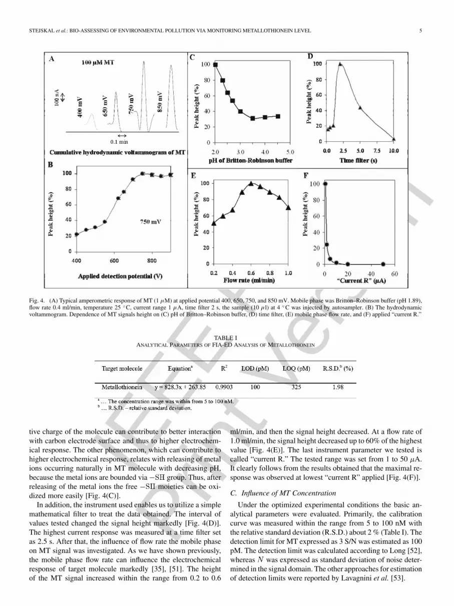
A flow injection analysis with electrochemical detection(FIA-ED) system consisted of a solvent delivery pump oper-ating in the range of 0.001–9.999 ml/min (Model 582 ESAInc., Chelmsford, MA), a guard cell (Model 5020 ESA,USA), a reaction coil (1 m), and an electrochemical detector.The electrochemical detector (ED) includes one low volumeflow-through analytical cell (Model 5040, ESA, USA), whichconsists of a glassy carbon working electrode, a palladium elec-trode as a reference electrode, an auxiliary carbon electrode,and a Coulochem III as a control module. The sample (5 l)was injected via autosampler (Model 540 Microtiter HPLC,ESA, USA). The obtained data were treated with CSW 32 soft-ware. The experiments were carried out at room temperature(22 C). A glassy carbon electrode was polished mechanicallyby 0.1 m of alumina (ESA Inc., USA) and sonicated at roomtemperature for 5 min using a Sonorex Digital 10 P Sonicator(Bandelin, Berlin, Germany) at 40 W.
III. RESULTS AND DISCUSSION
The investigation of affecting various organisms by abi-otic factors directly in their environment is rather difficult[32], [34]–[37]. Therefore, scientists have been suggestingexperimental models to simplify this monitoring with use ofbiomarkers as whole organisms, enzymes, or proteins [38]. Aswe mentioned above, the level of MT in an animal organismincreases with increasing concentration of the toxic abioticfactor, most of all, of heavy metal ions [39]–[43]. Here, we fo-cused on suggesting sensor methodology allowing monitoringof this biomarker, as experimental model guppy fishes treatedwith silver ions were used. The experimental scheme is shownin Fig. 1(B).
A. Electrochemical Measurement of Metallothionein Contentby Brdicka Reaction
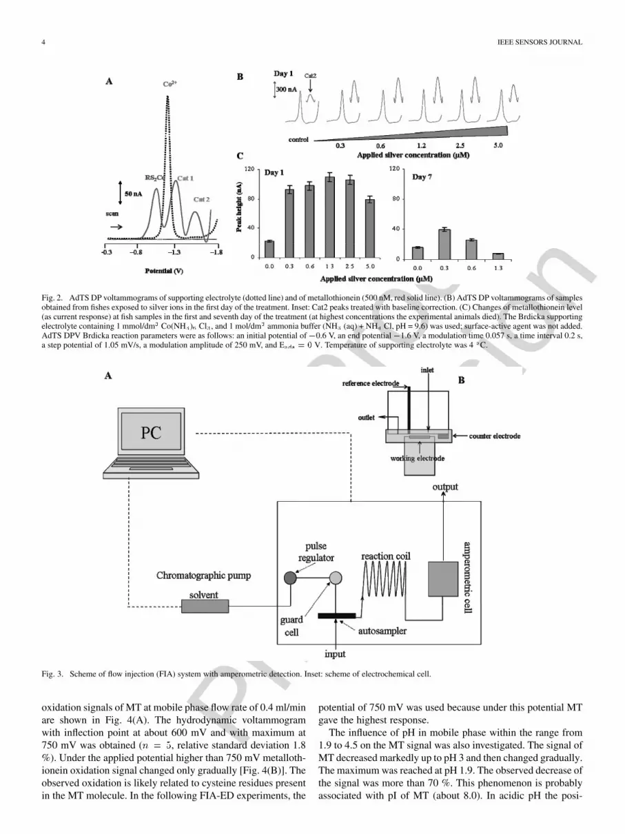
Previously, we analyzed metallothionein content in widerange of biological samples using electrochemical methodsmeasured catalytic signal, especially methods called Brdickareaction and peak H [32], [33], [38], [42]. In spite of the factthat peak H is more sensitive than a Brdicka reaction signal,a Brdicka reaction is suitable for routine analysis [26], [27],[33]. Therefore, we utilized a Brdicka reaction to detect MTin the biological samples of interest. MT measured using aBrdicka reaction gave the signals as follows: RS2Co, Cat1 andCat2 [Fig. 2(A)]. For quantification of MT in the real samplesCat2 signals were used [33]. MT content increased more thantwofold at all experimental variants exposed to silver ionscompared to control in first day of the exposition [Fig. 2(B)].The highest MT levels were determined in the second and thirdday of the exposition, and then the levels changed gradually. Inthe last day of the exposition the marked decrease of MT con-tent at all experimental variants occurred [Fig. 2(C)], whereasexperimental animals at the highest doses of silver ions (2.5and 5 M) died before the end of the experiment. The changesin behavior caused by metabolic disruption as a consequence ofsilver ions action in vital organs were observable at the treatedfishes, which was in good agreement with the published papers[44]–[46]. The other details devoted to this phenomenon willbe published elsewhere.
As it is shown, the MT level corresponds very well with silverions contamination in water. Although the Brdicka reaction is avery sensitive method, it is not suitable for further miniaturiza-tion and application directly in the environment. This lack of itsapplication is associated with working mercury electrode, whichis absolutely unacceptable to be utilized for environmental on-line monitoring. Therefore, the method of metallothionein de-tection in the flow system on glassy carbon electrode was de-signed.
B. Flow Injection Analysis With Electrochemical Detection(FIA-ED) Using Glassy Carbon Electrode to Analyze Thiols
It has been shown that thiols can be analyzed using a carbonelectrode as the working one [4], [21], [47]–[50]. The signals ofthe thiols related to oxidation of their cysteine residues were ob-served. Moreover, the shifting of the potential of the thiol signalsare assigned to bond heavy metal ions in their molecule [47].Nevertheless, to our knowledge, MT has not been measured byFIA-ED yet.
Influence of Applied Potential, Flow Rate, and Mobile PhaseComposition on Electrochemical Response of MT: MT wasmeasured using FIA with a one-channel electrochemical de-tector and the sample was injected using an autosampler (ESA,USA); see Fig. 3. A Britton–Robinson buffer under roomtemperature was used as mobile phase. At working potentialof 400 mV, the mobile phase flow rate of 0.4 ml/min metal-lothionein (100 M) gave a very well-developed oxidationsignal [Fig. 4(A)]. To optimize the amperometric detectionof MT in a flow system the influence of applied potential onoxidation signal of MT was studied. The potential within therange from 400 to 900 mV was applied on the workingand the amperometric response was recorded. The obtained

IEEE
Pro
of
Web
Ver
sion
4 IEEE SENSORS JOURNAL
Fig. 2. AdTS DP voltammograms of supporting electrolyte (dotted line) and of metallothionein (500 nM, red solid line). (B) AdTS DP voltammograms of samplesobtained from fishes exposed to silver ions in the first day of the treatment. Inset: Cat2 peaks treated with baseline correction. (C) Changes of metallothionein level(as current response) at fish samples in the first and seventh day of the treatment (at highest concentrations the experimental animals died). The Brdicka supportingelectrolyte containing 1 mmol/dm Co(NH ) Cl , and 1 mol/dm ammonia buffer (NH (aq) + NH Cl, pH = 9.6) was used; surface-active agent was not added.AdTS DPV Brdicka reaction parameters were as follows: an initial potential of �0.6 V, an end potential�1.6 V, a modulation time 0.057 s, a time interval 0.2 s,a step potential of 1.05 mV/s, a modulation amplitude of 250 mV, and E = 0 V. Temperature of supporting electrolyte was 4 C.
Fig. 3. Scheme of flow injection (FIA) system with amperometric detection. Inset: scheme of electrochemical cell.
oxidation signals of MT at mobile phase flow rate of 0.4 ml/minare shown in Fig. 4(A). The hydrodynamic voltammogramwith inflection point at about 600 mV and with maximum at750 mV was obtained ( , relative standard deviation 1.8%). Under the applied potential higher than 750 mV metalloth-ionein oxidation signal changed only gradually [Fig. 4(B)]. Theobserved oxidation is likely related to cysteine residues presentin the MT molecule. In the following FIA-ED experiments, the
potential of 750 mV was used because under this potential MTgave the highest response.
The influence of pH in mobile phase within the range from1.9 to 4.5 on the MT signal was also investigated. The signal ofMT decreased markedly up to pH 3 and then changed gradually.The maximum was reached at pH 1.9. The observed decrease ofthe signal was more than 70 %. This phenomenon is probablyassociated with pI of MT (about 8.0). In acidic pH the posi-

IEEE
Pro
of
Web
Ver
sion
STEJSKAL et al.: BIO-ASSESSING OF ENVIRONMENTAL POLLUTION VIA MONITORING METALLOTHIONEIN LEVEL 5
Fig. 4. (A) Typical amperometric response of MT (1 �M) at applied potential 400, 650, 750, and 850 mV. Mobile phase was Britton–Robinson buffer (pH 1.89),flow rate 0.4 ml/min, temperature 25 C, current range 1 �A, time filter 2 s, the sample (10 �l) at 4 C was injected by autosampler. (B) The hydrodynamicvoltammogram. Dependence of MT signals height on (C) pH of Britton–Robinson buffer, (D) time filter, (E) mobile phase flow rate, and (F) applied “current R.”
TABLE IANALYTICAL PARAMETERS OF FIA-ED ANALYSIS OF METALLOTHIONEIN
tive charge of the molecule can contribute to better interactionwith carbon electrode surface and thus to higher electrochem-ical response. The other phenomenon, which can contribute tohigher electrochemical response, relates with releasing of metalions occurring naturally in MT molecule with decreasing pH,because the metal ions are bounded via group. Thus, afterreleasing of the metal ions the free moieties can be oxi-dized more easily [Fig. 4(C)].
In addition, the instrument used enables us to utilize a simplemathematical filter to treat the data obtained. The interval ofvalues tested changed the signal height markedly [Fig. 4(D)].The highest current response was measured at a time filter setas 2.5 s. After that, the influence of flow rate the mobile phaseon MT signal was investigated. As we have shown previously,the mobile phase flow rate can influence the electrochemicalresponse of target molecule markedly [35], [51]. The heightof the MT signal increased within the range from 0.2 to 0.6
ml/min, and then the signal height decreased. At a flow rate of1.0 ml/min, the signal height decreased up to 60% of the highestvalue [Fig. 4(E)]. The last instrument parameter we tested iscalled “current R.” The tested range was set from 1 to 50 A.It clearly follows from the results obtained that the maximal re-sponse was observed at lowest “current R” applied [Fig. 4(F)].
C. Influence of MT Concentration
Under the optimized experimental conditions the basic an-alytical parameters were evaluated. Primarily, the calibrationcurve was measured within the range from 5 to 100 nM withthe relative standard deviation (R.S.D.) about 2 % (Table I). Thedetection limit for MT expressed as 3 S/N was estimated as 100pM. The detection limit was calculated according to Long [52],whereas was expressed as standard deviation of noise deter-mined in the signal domain. The other approaches for estimationof detection limits were reported by Lavagnini et al. [53].

IEEE
Pro
of
Web
Ver
sion
6 IEEE SENSORS JOURNAL
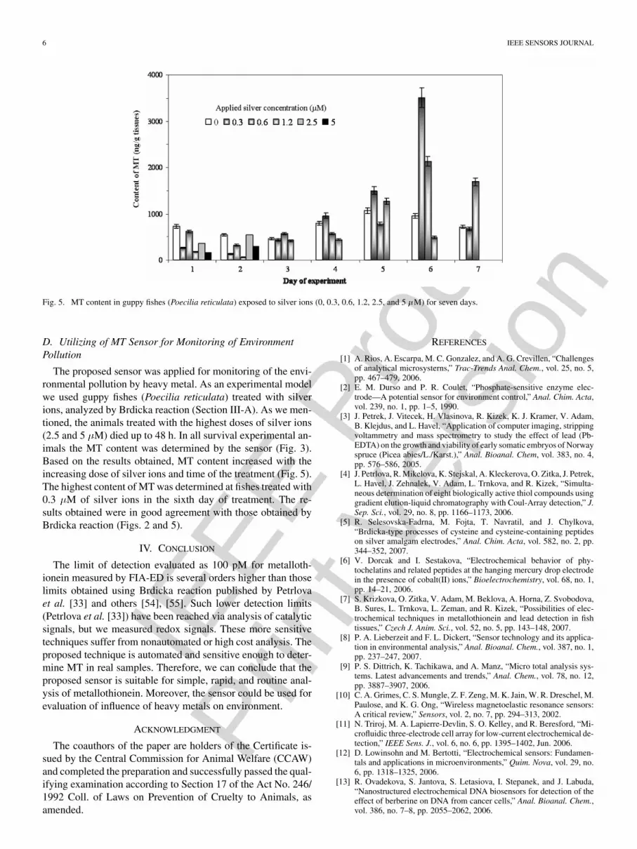
Fig. 5. MT content in guppy fishes (Poecilia reticulata) exposed to silver ions (0, 0.3, 0.6, 1.2, 2.5, and 5 �M) for seven days.
D. Utilizing of MT Sensor for Monitoring of EnvironmentPollution
The proposed sensor was applied for monitoring of the envi-ronmental pollution by heavy metal. As an experimental modelwe used guppy fishes (Poecilia reticulata) treated with silverions, analyzed by Brdicka reaction (Section III-A). As we men-tioned, the animals treated with the highest doses of silver ions(2.5 and 5 M) died up to 48 h. In all survival experimental an-imals the MT content was determined by the sensor (Fig. 3).Based on the results obtained, MT content increased with theincreasing dose of silver ions and time of the treatment (Fig. 5).The highest content of MT was determined at fishes treated with0.3 M of silver ions in the sixth day of treatment. The re-sults obtained were in good agreement with those obtained byBrdicka reaction (Figs. 2 and 5).
IV. CONCLUSION
The limit of detection evaluated as 100 pM for metalloth-ionein measured by FIA-ED is several orders higher than thoselimits obtained using Brdicka reaction published by Petrlovaet al. [33] and others [54], [55]. Such lower detection limits(Petrlova et al. [33]) have been reached via analysis of catalyticsignals, but we measured redox signals. These more sensitivetechniques suffer from nonautomated or high cost analysis. Theproposed technique is automated and sensitive enough to deter-mine MT in real samples. Therefore, we can conclude that theproposed sensor is suitable for simple, rapid, and routine anal-ysis of metallothionein. Moreover, the sensor could be used forevaluation of influence of heavy metals on environment.
ACKNOWLEDGMENT
The coauthors of the paper are holders of the Certificate is-sued by the Central Commission for Animal Welfare (CCAW)and completed the preparation and successfully passed the qual-ifying examination according to Section 17 of the Act No. 246/1992 Coll. of Laws on Prevention of Cruelty to Animals, asamended.
REFERENCES
[1] A. Rios, A. Escarpa, M. C. Gonzalez, and A. G. Crevillen, “Challengesof analytical microsysterns,” Trac-Trends Anal. Chem., vol. 25, no. 5,pp. 467–479, 2006.
[2] E. M. Durso and P. R. Coulet, “Phosphate-sensitive enzyme elec-trode—A potential sensor for environment control,” Anal. Chim. Acta,vol. 239, no. 1, pp. 1–5, 1990.
[3] J. Petrek, J. Vitecek, H. Vlasinova, R. Kizek, K. J. Kramer, V. Adam,B. Klejdus, and L. Havel, “Application of computer imaging, strippingvoltammetry and mass spectrometry to study the effect of lead (Pb-EDTA) on the growth and viability of early somatic embryos of Norwayspruce (Picea abies/L./Karst.),” Anal. Bioanal. Chem, vol. 383, no. 4,pp. 576–586, 2005.
[4] J. Petrlova, R. Mikelova, K. Stejskal, A. Kleckerova, O. Zitka, J. Petrek,L. Havel, J. Zehnalek, V. Adam, L. Trnkova, and R. Kizek, “Simulta-neous determination of eight biologically active thiol compounds usinggradient elution-liquid chromatography with Coul-Array detection,” J.Sep. Sci., vol. 29, no. 8, pp. 1166–1173, 2006.
[5] R. Selesovska-Fadrna, M. Fojta, T. Navratil, and J. Chylkova,“Brdicka-type processes of cysteine and cysteine-containing peptideson silver amalgam electrodes,” Anal. Chim. Acta, vol. 582, no. 2, pp.344–352, 2007.
[6] V. Dorcak and I. Sestakova, “Electrochemical behavior of phy-tochelatins and related peptides at the hanging mercury drop electrodein the presence of cobalt(II) ions,” Bioelectrochemistry, vol. 68, no. 1,pp. 14–21, 2006.
[7] S. Krizkova, O. Zitka, V. Adam, M. Beklova, A. Horna, Z. Svobodova,B. Sures, L. Trnkova, L. Zeman, and R. Kizek, “Possibilities of elec-trochemical techniques in metallothionein and lead detection in fishtissues,” Czech J. Anim. Sci., vol. 52, no. 5, pp. 143–148, 2007.
[8] P. A. Lieberzeit and F. L. Dickert, “Sensor technology and its applica-tion in environmental analysis,” Anal. Bioanal. Chem., vol. 387, no. 1,pp. 237–247, 2007.
[9] P. S. Dittrich, K. Tachikawa, and A. Manz, “Micro total analysis sys-tems. Latest advancements and trends,” Anal. Chem., vol. 78, no. 12,pp. 3887–3907, 2006.
[10] C. A. Grimes, C. S. Mungle, Z. F. Zeng, M. K. Jain, W. R. Dreschel, M.Paulose, and K. G. Ong, “Wireless magnetoelastic resonance sensors:A critical review,” Sensors, vol. 2, no. 7, pp. 294–313, 2002.
[11] N. Triroj, M. A. Lapierre-Devlin, S. O. Kelley, and R. Beresford, “Mi-crofluidic three-electrode cell array for low-current electrochemical de-tection,” IEEE Sens. J., vol. 6, no. 6, pp. 1395–1402, Jun. 2006.
[12] D. Lowinsohn and M. Bertotti, “Electrochemical sensors: Fundamen-tals and applications in microenvironments,” Quim. Nova, vol. 29, no.6, pp. 1318–1325, 2006.
[13] R. Ovadekova, S. Jantova, S. Letasiova, I. Stepanek, and J. Labuda,“Nanostructured electrochemical DNA biosensors for detection of theeffect of berberine on DNA from cancer cells,” Anal. Bioanal. Chem.,vol. 386, no. 7–8, pp. 2055–2062, 2006.

IEEE
Pro
of
Web
Ver
sion
STEJSKAL et al.: BIO-ASSESSING OF ENVIRONMENTAL POLLUTION VIA MONITORING METALLOTHIONEIN LEVEL 7
[14] E. Bakker and Y. Qin, “Electrochemical sensors,” Anal. Chem., vol.78, no. 12, pp. 3965–3983, 2006.
[15] J. Wang, “Amperometric biosensors for clinical and therapeutic drugmonitoring: A review,” J. Pharm. Biomed. Anal., vol. 19, no. 1–2, pp.47–53, 1999.
[16] D. Potesil, R. Mikelova, V. Adam, R. Kizek, and R. Prusa, “Changeof the protein p53 electrochemical signal according to its structuralform—Quick and sensitive distinguishing of native, denatured, and ag-gregated form of the “guardian of the genome”,” Protein J., vol. 25, no.1, pp. 23–32, 2006.
[17] T. Giordani, L. Natali, B. E. Maserti, S. Taddei, and A. Cavallini,“Characterization and expression of DNA sequences encoding putativetype-II metallothioneins in the seagrass Posidonia oceanica,” PlantPhysiol., vol. 123, no. 4, pp. 1571–1581, 2000.
[18] I. Sochova, J. Hofman, and I. Holoubek, “Using nematodes in soil eco-toxicology,” Environ. Int., vol. 32, no. 3, pp. 374–383, 2006.
[19] C. Leyval, K. Turnau, and K. Haselwandter, “Effect of heavy metal pol-lution on mycorrhizal colonization and function: Physiological, eco-logical and applied aspects,” Mycorrhiza, vol. 7, no. 3, pp. 139–153,1997.
[20] R. Mikelova, J. Baloun, J. Petrlova, V. Adam, L. Havel, J. Petrek, A.Horna, and R. Kizek, “Electrochemical determination of Ag-ions in en-vironment waters and their action on plant embryos,” Bioelectrochem-istry, vol. 70, no. 2, pp. 508–518, 2007.
[21] V. Supalkova, D. Huska, V. Diopan, P. Hanustiak, O. Zitka, K. Ste-jskal, J. Baloun, J. Pikula, L. Havel, J. Zehnalek, V. Adam, L. Trnkova,M. Beklova, and R. Kizek, “Electroanalysis of plant thiols,” Sensors,vol. 7, no. 6, pp. 932–959, 2007.
[22] V. Supalkova, J. Petrek, J. Baloun, V. Adam, K. Bartusek, L. Trnkova,M. Beklova, V. Diopan, L. Havel, and R. Kizek, “Multi-instrumentalinvestigation of affecting of early somatic embryos of Spruce by cad-mium(II) and lead(II) ions,” Sensors, vol. 7, no. 5, pp. 743–759, 2007.
[23] B. Sures, “Environmental parasitology: Relevancy of parasites in mon-itoring environmental pollution,” Trends Parasitol., vol. 20, no. 4, pp.170–177, 2004.
[24] F. Lessire, A. Delaunois, P. Gustin, and M. Ansay, “Biomarkers andbioindicators in vertebrates: Importance in evaluation of quality of anecosystem,” Ann. Med. Vet., vol. 141, no. 4, pp. 281–290, 1997.
[25] L. P. Smirnov, I. V. Sukhovskaya, and N. N. Nemova, “Effects of en-vironmental factors on low-molecular-weight peptides of fishes: A re-view,” Russ. J. Ecol., vol. 36, no. 1, pp. 41–47, 2005.
[26] B. Raspor, Z. Dragun, M. Erk, D. Ivankovic, and J. Pavicic, “Is thedigestive gland of Mytilus galloprovincialis a tissue of choice for esti-mating cadmium exposure by means of metallothioneins?,” Sci. TotalEnviron., vol. 333, no. 1–3, pp. 99–108, 2004.
[27] J. Pavicic, D. Ivankovic, C. Lucu, B. Hamer, M. Erk, M. Tusek-Znidaric, and I. Falnoga, “Ecotoxicological evaluation of metalloth-ionein level in selected tissues of estuarine invertebrates,” Toxicol.Lett., vol. 164, pp. S163–S163, 2006.
[28] M. Fojta, M. Fojtova, L. Havran, H. Pivonkova, V. Dorcak, and I. Ses-takova, “Electrochemical monitoring of phytochelatin accumulation innicotiana tabacum cells exposed to sub-cytotoxic and cytotoxic levelsof cadmium,” Anal. Chim. Acta, vol. 558, no. 1–2, pp. 171–178, 2006.
[29] J. H. R. Kagi and A. Schaffer, “Biochemistry of metallothionein,” Bio-chemistry, vol. 27, no. 23, pp. 8509–8515, 1988.
[30] J. W. Gorsuch and S. J. Klaine, “Toxicity and fate of silver in the en-vironment,” Environmental Toxicolology Chemistry, vol. 17, no. 4, pp.537–538, 1998.
[31] B. Raspor, M. Paic, and M. Erk, “Analysis of metallothioneins by themodified brdicka procedure,” Talanta, vol. 55, no. 1, pp. 109–115,2001.
[32] R. Kizek, J. Vacek, L. Trnkova, B. Klejdus, and L. Havel, “Applicationof catalytic reactions on a mercury electrode for electrochemical de-tection of metallothioneins,” Chem. Listy, vol. 98, no. 4, pp. 166–173,2004.
[33] J. Petrlova, D. Potesil, R. Mikelova, O. Blastik, V. Adam, L. Trnkova,F. Jelen, R. Prusa, J. Kukacka, and R. Kizek, “Attomole voltammetricdetermination of metallothionein,” Electrochim. Acta, vol. 51, no. 24,pp. 5112–5119, 2006.
[34] S. Yazgan, H. Horn, and H. D. Isengard, “Honey as bio indicatorby screening the heavy metal content of the environment,” Dtsch.Lebensm.-Rundsch., vol. 102, no. 5, pp. 192–194, 2006.
[35] D. Potesil, J. Petrlova, V. Adam, J. Vacek, B. Klejdus, J. Zehnalek, L.Trnkova, L. Havel, and R. Kizek, “Simultaneous femtomole determina-tion of cysteine, reduced and oxidized glutathione, and phytochelatinin maize (Zea mays L.) kernels using high-performance liquid chro-matography with electrochemical detection,” J. Chromatogr. A, vol.1084, no. 1–2, pp. 134–144, 2005.
[36] C. Singer, S. Zimmermann, and B. Sures, “Induction of heat shockproteins (hsp70) in the zebra mussel (Dreissena polymorpha) followingexposure to platinum group metals (platinum, palladium and rhodium):Comparison with lead and cadmium exposures,” Aquat. Toxicol., vol.75, no. 1, pp. 65–75, 2005.
[37] B. Sures, “How parasitism and pollution affect the physiological home-ostasis of aquatic hosts,” J. Helminthol., vol. 80, no. 2, pp. 151–157,2006.
[38] J. Kukacka, J. Petrlova, R. Prusa, V. Adam, B. Sures, M. Beklova, J.Havel, and R. Kizek, “Changes of content of glutathione and metalloth-ionein at plant cells and invertebrate treated by platinum group metals,”Faseb J., vol. 20, no. 5, pp. A1196–A1196, 2006.
[39] V. Adam, J. Petrlova, D. Potesil, J. Zehnalek, B. Sures, L. Trnkova,F. Jelen, and R. Kizek, “Study of metallothionein modified electrodesurface behavior in the presence of heavy metal ions-biosensor,” Elec-troanalysis, vol. 17, no. 18, pp. 1649–1657, 2005.
[40] V. Adam, J. Zehnalek, J. Petrlova, D. Potesil, B. Sures, L. Trnkova,F. Jelen, J. Vitecek, and R. Kizek, “Phytochelatin modified electrodesurface as a sensitive heavy-metal ion biosensor,” Sensors, vol. 5, no.1–2, pp. 70–84, 2005.
[41] M. Strouhal, R. Kizek, J. Vecek, L. Trnkova, and M. Nemec, “Electro-chemical study of heavy metals and metallothionein in yeast Yarrowialipolytica,” Bioelectrochemistry, vol. 60, no. 1–2, pp. 29–36, 2003.
[42] R. Kizek, L. Trnkova, and E. Palecek, “Determination of metalloth-ionein at the femtomole level by constant current stripping chronopo-tentiometry,” Anal. Chem., vol. 73, no. 20, pp. 4801–4807, 2001.
[43] J. Vacek, J. Petrek, R. Kizek, L. Havel, B. Klejdus, L. Trnkova, andF. Jelen, “Electrochemical determination of lead and glutathione in aplant cell culture,” Bioelectrochemistry, vol. 63, no. 1–2, pp. 347–351,2004.
[44] C. Hogstrand, C. M. Wood, N. R. Bury, R. W. Wilson, J. C. Rankin,and M. Grosell, “Binding and movement of silver in the intestinal ep-ithelium of a marine teleost fish, the European flounder (Platichthysflesus),” Comp. Biochem. Physiol. C-Toxicol. Pharmacol., vol. 133, no.1–2, pp. 125–135, 2002.
[45] C. M. Wood, R. C. Playle, and C. Hogstrand, “Physiology and mod-eling of mechanisms of silver uptake and toxicity in fish,” Environ-mental Toxicology Chemistry, vol. 18, no. 1, pp. 71–83, 1999.
[46] C. Hogstrand and C. M. Wood, “Toward a better understanding of thebioavailability, physiology and toxicity of silver in fish: Implicationsfor water quality criteria,” Environmental Toxicology Chemistry, vol.17, no. 4, pp. 547–561, 1998.
[47] I. Sestakova, M. Kopanica, L. Havran, and E. Palecek, “Constant cur-rent chronopotentiometric stripping analysis of Cd-metallothionein oncarbon and mercury electrodes. Comparison with voltammetry,” Elec-troanalysis, vol. 12, no. 2, pp. 100–104, 2000.
[48] I. Sestakova and P. Mader, “Voltammetry on mercury and carbon elec-trodes as a tool for studies of metallothionein interactions with metalions,” Cell. Mol. Biol., vol. 46, no. 2, pp. 257–267, 2000.
[49] M. Tomschik, L. Havran, M. Fojta, and E. Palecek, “Constant cur-rent chronopotentiometric stripping analysis of bioactive peptides atmercury and carbon electrodes,” Electroanalysis, vol. 10, no. 6, pp.403–409, 1998.
[50] J. Limson and T. Nyokong, “Voltammetric behavior of cysteine andmetallothionein on cobalt(II) tetrasulfonated phthalocyanine modifiedglassy carbon electrodes,” Electroanalysis, vol. 9, no. 3, pp. 255–260,1997.
[51] B. Klejdus, J. Vacek, V. Adam, J. Zehnalek, R. Kizek, L. Trnkova, andV. Kuban, “Determination of isoflavones in soybean food and humanurine using liquid chromatography with electrochemical detection,” J.Chromatogr. B, vol. 806, no. 2, pp. 101–111, 2004.
[52] G. L. Long and J. D. Winefordner, “Limit of detection,” Anal. Chem.,vol. 55, no. 7, pp. A712–A724, 1983.
[53] I. Lavagnini, R. Antiochia, and F. Magno, “A calibration-basemethod for the evaluation of the detection limit of an electrochemicalbiosensor,” Electroanalysis, vol. 19, no. 11, pp. 1227–1230, 2007.
[54] M. El Hourch, A. Dudoit, and J. C. Amiard, “An optimization pro-cedure for determination of metallothionein by square wave cathodicstripping voltammetry: Application to marine worms,” Anal. Bioanal.Chem., vol. 378, no. 3, pp. 776–781, 2004.

IEEE
Pro
of
Web
Ver
sion
8 IEEE SENSORS JOURNAL
[55] G. Alvarez-Llamas, M. R. F. de la Campa, and A. Sanz-Medel, “Met-allothionein isoforms separation and cadmium speciation by capillaryelectrophoresis with ultraviolet and quadrupole-inductively coupledplasma mass spectrometric detection,” Anal. Chim. Acta, vol. 448, no.1–2, pp. 105–119, 2001.
[56] A. E. S. de Vives, S. Moreira, S. M. B. Brienza, J. G. S. Medeiros, M.Tomazello, O. Zucchi, and V. F. do Nascimento, “Monitoring of the en-vironmental pollution by trace element analysis in tree-rings using syn-chrotron radiation total reflection X-ray fluorescence,” Spectroc. ActaPt. B-Atom. Spectr., vol. 61, no. 10–11, pp. 1170–1174, 2006.
[57] D. R. Livingstone, “Biotechnology and pollution monitoring—Use ofmolecular biomarkers in the aquatic environment,” J. Chem. Technol.Biotechnol., vol. 57, no. 3, pp. 195–211, 1993.
[58] R. van der Oost, J. Beyer, and N. P. E. Vermeulen, “Fish bioaccumula-tion and biomarkers in environmental risk assessment: A review,” En-viron. Toxicol. Pharmacol, vol. 13, no. 2, pp. 57–149, 2003.
Karel Stejskal is a student of biochemistry at Masaryk University, Brno, CzechRepublic.
His research interests include the study of plant and animal response to heavymetals.
Sona Krizkova is working toward the Ph.D. degree in molecular biology atMasaryk University, Brno, Czech Republic.
Her research interests include determination of metallothionein by immuno-chemical and electrochemical methods and its application.
Vojtech Adam is working toward the Ph.D. degree in molecular biology atMasaryk University, Brno, Czech Republic.
His research interests include analytical chemistry and molecular biology incancer diagnostics.
Bernd Sures is a Professor at the Universität Duisburg-Essen, Essen, Germany.He is Head of the Research Group of Applied Zoology and Hydrobiology at Uni-versität Duisburg-Essen. His research interests include ecology, parasitology,and ecotoxicology.
Libuse Trnkova is an Associate Professor at Masaryk University, Brno, CzechRepublic. Her research interests include electrochemical analysis of peptides,proteins, and nucleic acids. She is a cofounder of elimination voltammetry.
Josef Zehnalek is an Associate Professor at Mendel University of Agricultureand Forestry, Brno, Czech Republic. His research interests include investigationof affecting plant and animal metabolism by heavy metals.
Jaromir Hubalek is an Assistant Professor at the Technical University of Brno,Brno, Czech Republic. His research activities include designing of miniaturizedmicroelectrodes and application of nanoparticles and nanomaterials for elec-trodes construction.
Miroslava Beklova is a Professor at the University of Veterinary and Pharma-ceutical Sciences, Brno, Czech Republic. Her research interests include ecotox-icology and monitoring of environmental pollution.
Pavel Hanustiak is working toward the Ph.D. degree at the University of Vet-erinary and Pharmaceutical Sciences, Brno, Czech Republic.
His research interests include secondary plant metabolites and ecotoxicology.
Zdenka Svobodova is a Professor and Head of the Department of Toxicology,University of Veterinary and Pharmaceutical Sciences, Brno, Czech Republic.Her research interests include veterinary and food toxicology.
Ales Horna is an Associate Professor at Tomas Bata University, Zlin, CzechRepublic. He promoted Radanal, Ltd. His research interests include suggestingof analytical methods for determination of health-benefit compounds in food.
Rene Kizek is an Associate Professor at Mendel University of Agriculture andForestry, Brno, Czech Republic. His research interests include application ofelectrochemical methods to the molecular biochemistry.

IEEE
Pro
of
Prin
t Ver
sion
IEEE SENSORS JOURNAL 1
Bio-Assessing of Environmental Pollution viaMonitoring of Metallothionein Level Using
Electrochemical DetectionKarel Stejskal, Sona Krizkova, Vojtech Adam, Bernd Sures, Libuse Trnkova, Josef Zehnalek, Jaromir Hubalek,
Miroslava Beklova, Pavel Hanustiak, Zdenka Svobodova, Ales Horna, and Rene Kizek
Abstract—In this paper, we report on an investigation of af-fecting guppy fishes (Poecilia reticulata) by silver ions (0, 0.3,0.6, 1.2, 2.5, and 5 M) for seven days under well-controlledexperimental conditions. To observe the physiological changes, weattempted to determine metallothionein (MT) as a biomarker ofheavy metal stress. For this purpose, we proposed a sensor uti-lizing a carbon electrode coupled with flow injection analysis. Theexperimental conditions, which have been optimized, were as fol-lows—applied potential: 750 mV, mobile phase: Britton–Robinsonbuffer (pH 1.9) with flow rate of 0.6 ml/min, time filter: 2.5 s,“current R”: 1 A. Under these conditions, the detection limitof MT was estimated as 100 pM. After the optimizing step, thefish tissues were measured. Based on the results obtained, MTcontent increased with increasing dose of silver ions and time ofthe treatment. The results obtained were in good correlation withthose obtained by adsorptive transfer stripping technique coupledwith differential pulse voltammetry Brdicka reaction, which wasused as the reference technique.
Index Terms—Amperometric detection, biomarker, electro-chemical sensors, fish, flow injection analysis, heavy metal,metallothionein, poecilia reticulata, silver.
Manuscript received March 31, 2007; revised June 30, 2007 and September28, 2007; accepted January 6, 2008. This work was supported under GrantsGACR 526/07/0808, MSMT 6215712402, MSMT 2636/F4b, and MSMTINCHEMBIOL 0021622412. This is an expanded paper from the Sensors 2006Conference. The associate editor coordinating the review of this paper andapproving it for publication was Prof. Okyay Kaynak.
K. Stejskal is with the Department of Chemistry and Biochemistry, Facultyof Agronomy, Mendel University of Agriculture and Forestry, CZ-613 00 Brno,Czech Republic and also with the Department of Biochemistry, Faculty of Sci-ence, Masaryk University, CZ-611 37 Brno, Czech Republic.
S. Krizkova, J. Zehnalek, and R. Kizek are with the Department of Chemistryand Biochemistry, Faculty of Agronomy, Mendel University of Agriculture andForestry, CZ-613 00 Brno, Czech Republic (e-mail: [email protected]).
V. Adam is with the Department of Chemistry and Biochemistry, Facultyof Agronomy, Mendel University of Agriculture and Forestry, CZ-613 00Brno, Czech Republic and also with the Department of Animal Nutrition andForage Production, Faculty of Agronomy, Mendel University of Agricultureand Forestry, CZ-613 00 Brno, Czech Republic.
B. Sures is with the Universität Duisburg-Essen, Applied Zoology and Hy-drobiology, D-45117 Essen, Germany.
L. Trnkova is with the Department of Chemistry, Faculty of Science, MasarykUniversity, CZ-611 37 Brno, Czech Republic.
J. Hubalek is with the Department of Microelectronics, Faculty of ElectricalEngineering and Communication, Technical University of Brno, CZ-602 00Brno, Czech Republic.
M. Beklova and P. Hanustiak are with the Department of Veterinary Ecologyand Environmental Protection, University of Veterinary and Pharmaceutical Sci-ences, CZ-612 42 Brno, Czech Republic.
Z. Svobodova is with the Institute of Parasitology, University of Veterinaryand Pharmaceutical Sciences, CZ-612 42 Brno, Czech Republic.
A. Horna is with the Department of Food Engineering, Faculty of Technology,Tomas Bata University, CZ-762 72 Zlin, Czech Republic.
Color versions of one or more of the figures in this paper are available onlineat http://ieeexplore.ieee.org.
I. INTRODUCTION
A. Sensors in Environmental Chemistry
R OBUST analytical techniques, which cannot be miniatur-ized easily [1], are not useful for furnishing online moni-
toring of environmental pollution. Therefore, new analytical ap-proaches allowing easy and rapid assessment of environmentalpollution are suggested [2]–[7]. These chemical sensors andbiosensors have superior properties over existing analytical in-struments fitting perfectly for the aforementioned purposes [8],[9]. The sensor system can be composed from three parts: 1)sensor; 2) converter of the signals; and 3) data acquisition in-strument [Fig. 1(A)]. Nevertheless, a sensor can be used for ana-lyzing of environmental samples after the optimizing accordingto its type and properties of target molecule (e.g., gas, liquid)[8], [10]. From the wide spectrum of sensors the electrochemicalsensors (i.e., potentiometric, amperometric) are intensively useddue to their very low costs, low detection limits, and spectrumof applications [11]–[13]. Electrochemical sensors also allow usto obtain both qualitative and quantitative character of the targetmolecule and can give information about its physicochemicalproperties (oxidation state, type of ligand, etc.) and concentra-tion in one measurement [14]–[16].
B. Bio-Indication of Environmental Pollution
To assess environmental pollution we can use three strate-gies: 1) to detect a pollutant directly; 2) to determine a substancecalled biomarker, which can be used to indicate exposure of anorganism to a pollutant; or 3) to observe physiological changesof an organism living in a polluted environment. Detection ofpollutant directly in such a complex matrix as an environmentalsample can be a difficult task for an analytical chemist. More-over, concentration of a certain pollutant can reveal only the im-mediate level of polluting but reveal nothing about the effectof the pollutant on the environment in a long-term scale. Thislack of information can be partly supplemented via analysis ofbiomarkers or observation of physiological changes of organ-isms exposed to the pollutant.
Numerous plant and animal species can be used as bio-indi-cators of heavy metals pollution of the environment [17]–[22].Aquatic animals, most of all various species of fishes, are verysuitable for these purposes [23], [24]. To assess the quality of the
Digital Object Identifier 10.1109/JSEN.2008.928500
1530-437X/$25.00 © 2008 IEEE

IEEE
Pro
of
Prin
t Ver
sion
2 IEEE SENSORS JOURNAL
Fig. 1. (A) Typical arrangement of electrochemical sensor: (a) sensor, (b) measuring device, and (c) data acquistion. (B) Scheme of environmental wasting byheavy metals. Environmental samples are collected and processed for electroanalytical determination according to current protocol. The analysis of one individualis sufficient, which is advantageous for screening programs assessing environment quality [56]–[58].
environment investigations, i.e., the changes in behavior, mor-phology, habitation, or changes of basic morphometric proper-ties (body weight, color, length, etc.), are often used. All thesedata are only of qualitative character and are hard to obtain dueto requirements on large population of the target specie and thetime period of the experiment. Therefore, one may suggest thatthe most reliable investigations about the effect of heavy metalson an aquatic environment can be proposed using fish speciesupplemented with analyzing a certain biomarker.
C. Biomarkers
It has been shown that low-molecular peptides and/or pro-teins rich in cysteine can be considered as biomarkers, referringto various types of pollutants including heavy metals [25]–[28].Metallothioneins (MT) as low molecular cysteine rich proteinsbelong in the group of molecules [29]. Their molecular weight iswithin the range from 6 to 10 kDa. Due to their affinity to heavymetals, they are involved in their detoxifying and in maintainingof heavy metals homeostasis. MT contains two binding domains
and , which are composed from cysteine clusters. In thispaper, we report on investigations affecting the guppy (Poeciliareticulata) by silver ions for seven days under well-controlledexperimental conditions, because silver ions pose a threat toaquatic organisms due to their very high toxicity [30]. To ob-serve the physiological changes we determined MT. For thispurpose, we proposed a simple and rapid electrochemical de-tection utilizing carbon electrode coupled with flow injectionanalysis.
II. MATERIAL AND METHODS
A. Chemicals
Rabbit liver MT (MW 7143), containing 5.9 % Cd and0.5 % Zn, was purchased from Sigma Aldrich (St. Louis,MO). Tris(2-carboxyethyl)phosphine (TCEP) was produced byMolecular Probes (Evgen, OR). Co(NH ) Cl and other chem-icals used were purchased from Sigma Aldrich (Sigma-Aldrich,
USA) in ACS purity unless noted otherwise. Stock standardsolutions were prepared with ACS water (Sigma-Aldrich,USA) and stored in the dark at 20 C. Working standardsolutions were prepared daily by dilution of the stock solutions.All solutions were filtered through 0.45- m Nylon filter discs(Millipore, Billerica, MA) prior to electrochemical analysis.The pH value was measured using WTW inoLab Level 3 withterminal Level 3 (Weilheim, Germany), controlled by a per-sonal computer program (MultiLab Pilot; Weilheim, Germany).The pH-electrode (SenTix-H, pH 0–14/3 mol/dm KCl) wascalibrated by set of WTW buffers (Weilheim, Germany).
B. Experimental Model
Guppy fishes (Poecilia reticulata), 2 or 3 months old, wereexposed to silver nitrate, always seven individuals per dose 0,0.3, 0.6, 1.2, 2.5, or 5 M. The experiment lasted seven days(168 h); a fish from each experimental variant was sampled perday. The experimental conditions such as pH value of the so-lution where the fishes were kept constant, oxygen concentra-tion, and temperature were monitored during the experiment.The oxygen concentration varied within the range from 1.7 to4.0 mg/l, the pH level from 6.34 to 7.00, and the temperaturefrom 20.2 to 21.5 C during the seven-day-long experiment. Thesampled fish was killed by CO and washed one time with dis-tilled water and one time with 0.5 M EDTA.
C. Preparation of Biological Samples for ElectrochemicalAnalysis
The sampled fishes (approximately 0.2 g) were frozen withliquid nitrogen and spread in mortar, and then exactly 1000 l of0.2 M phosphate buffer (pH 7.2) was added to the homogenizedsample. The obtained homogenate was transferred into test-tubeand vortexed for 15 min at 4 C (Vortex Genie). The supernatantwas subsequently heat treated. Briefly, the sample was kept at99 C in a thermomixer (Eppendorf 5430, Germany) for 15 minwith occasional stirring and then cooled to 4 C. The denaturedhomogenates were centrifuged at 4 C, 15 000 for 30 min

IEEE
Pro
of
Prin
t Ver
sion
STEJSKAL et al.: BIO-ASSESSING OF ENVIRONMENTAL POLLUTION VIA MONITORING METALLOTHIONEIN LEVEL 3
(Eppendorf 5402, Germany). Other experimental details relatedto this methodology are described in [31]–[33].
D. Adsorptive Transfer Stripping Technique (AdTS) CoupledWith Differential Pulse Voltammetry (DPV) Brdicka Reaction
Electrochemical measurements were performed using anAUTOLAB analyser (EcoChemie, The Netherlands) connectedto VA-Stand 663 (Metrohm, Switzerland), using a standardcell with three electrodes. The three-electrode system consistedof a hanging mercury drop electrode as working electrode, anAg/AgCl/3 M KCl reference electrode, and a carbon counterelectrode. For smoothing and baseline correction the softwareGPES 4.4 supplied by EcoChemie was employed. The Brdickasupporting electrolyte containing 1 mmol/dm Co(NH )Cl and 1 mol/dm ammonia buffer (NH (aq) + NH Cl,pH ) was used; surface-active agent was not added. AdTSDPV Brdicka reaction parameters were as follows: an initialpotential of 0.6 V, an end potential 1.6 V, a modulationtime 0.057 s, a time interval 0.2 s, a step potential of 1.05 mV,a modulation amplitude of 250 mV, E V. Temperatureof supporting electrolyte was 4 C. For other experimentalconditions see [33].
E. Flow Electrochemical Measurement
A flow injection analysis with electrochemical detection(FIA-ED) system consisted of a solvent delivery pump oper-ating in the range of 0.001–9.999 ml/min (Model 582 ESAInc., Chelmsford, MA), a guard cell (Model 5020 ESA,USA), a reaction coil (1 m), and an electrochemical detector.The electrochemical detector (ED) includes one low volumeflow-through analytical cell (Model 5040, ESA, USA), whichconsists of a glassy carbon working electrode, a palladium elec-trode as a reference electrode, an auxiliary carbon electrode,and a Coulochem III as a control module. The sample (5 l)was injected via autosampler (Model 540 Microtiter HPLC,ESA, USA). The obtained data were treated with CSW 32 soft-ware. The experiments were carried out at room temperature(22 C). A glassy carbon electrode was polished mechanicallyby 0.1 m of alumina (ESA Inc., USA) and sonicated at roomtemperature for 5 min using a Sonorex Digital 10 P Sonicator(Bandelin, Berlin, Germany) at 40 W.
III. RESULTS AND DISCUSSION
The investigation of affecting various organisms by abi-otic factors directly in their environment is rather difficult[32], [34]–[37]. Therefore, scientists have been suggestingexperimental models to simplify this monitoring with use ofbiomarkers as whole organisms, enzymes, or proteins [38]. Aswe mentioned above, the level of MT in an animal organismincreases with increasing concentration of the toxic abioticfactor, most of all, of heavy metal ions [39]–[43]. Here, we fo-cused on suggesting sensor methodology allowing monitoringof this biomarker, as experimental model guppy fishes treatedwith silver ions were used. The experimental scheme is shownin Fig. 1(B).
A. Electrochemical Measurement of Metallothionein Contentby Brdicka Reaction
Previously, we analyzed metallothionein content in widerange of biological samples using electrochemical methodsmeasured catalytic signal, especially methods called Brdickareaction and peak H [32], [33], [38], [42]. In spite of the factthat peak H is more sensitive than a Brdicka reaction signal,a Brdicka reaction is suitable for routine analysis [26], [27],[33]. Therefore, we utilized a Brdicka reaction to detect MTin the biological samples of interest. MT measured using aBrdicka reaction gave the signals as follows: RS2Co, Cat1 andCat2 [Fig. 2(A)]. For quantification of MT in the real samplesCat2 signals were used [33]. MT content increased more thantwofold at all experimental variants exposed to silver ionscompared to control in first day of the exposition [Fig. 2(B)].The highest MT levels were determined in the second and thirdday of the exposition, and then the levels changed gradually. Inthe last day of the exposition the marked decrease of MT con-tent at all experimental variants occurred [Fig. 2(C)], whereasexperimental animals at the highest doses of silver ions (2.5and 5 M) died before the end of the experiment. The changesin behavior caused by metabolic disruption as a consequence ofsilver ions action in vital organs were observable at the treatedfishes, which was in good agreement with the published papers[44]–[46]. The other details devoted to this phenomenon willbe published elsewhere.
As it is shown, the MT level corresponds very well with silverions contamination in water. Although the Brdicka reaction is avery sensitive method, it is not suitable for further miniaturiza-tion and application directly in the environment. This lack of itsapplication is associated with working mercury electrode, whichis absolutely unacceptable to be utilized for environmental on-line monitoring. Therefore, the method of metallothionein de-tection in the flow system on glassy carbon electrode was de-signed.
B. Flow Injection Analysis With Electrochemical Detection(FIA-ED) Using Glassy Carbon Electrode to Analyze Thiols
It has been shown that thiols can be analyzed using a carbonelectrode as the working one [4], [21], [47]–[50]. The signals ofthe thiols related to oxidation of their cysteine residues were ob-served. Moreover, the shifting of the potential of the thiol signalsare assigned to bond heavy metal ions in their molecule [47].Nevertheless, to our knowledge, MT has not been measured byFIA-ED yet.
Influence of Applied Potential, Flow Rate, and Mobile PhaseComposition on Electrochemical Response of MT: MT wasmeasured using FIA with a one-channel electrochemical de-tector and the sample was injected using an autosampler (ESA,USA); see Fig. 3. A Britton–Robinson buffer under roomtemperature was used as mobile phase. At working potentialof 400 mV, the mobile phase flow rate of 0.4 ml/min metal-lothionein (100 M) gave a very well-developed oxidationsignal [Fig. 4(A)]. To optimize the amperometric detectionof MT in a flow system the influence of applied potential onoxidation signal of MT was studied. The potential within therange from 400 to 900 mV was applied on the workingand the amperometric response was recorded. The obtained
IEEE
Pro
of
Prin
t Ver
sion
4 IEEE SENSORS JOURNAL
Fig. 2. AdTS DP voltammograms of supporting electrolyte (dotted line) and of metallothionein (500 nM, red solid line). (B) AdTS DP voltammograms of samplesobtained from fishes exposed to silver ions in the first day of the treatment. Inset: Cat2 peaks treated with baseline correction. (C) Changes of metallothionein level(as current response) at fish samples in the first and seventh day of the treatment (at highest concentrations the experimental animals died). The Brdicka supportingelectrolyte containing 1 mmol/dm Co(NH ) Cl , and 1 mol/dm ammonia buffer (NH (aq) + NH Cl, pH = 9.6) was used; surface-active agent was not added.AdTS DPV Brdicka reaction parameters were as follows: an initial potential of �0.6 V, an end potential�1.6 V, a modulation time 0.057 s, a time interval 0.2 s,a step potential of 1.05 mV/s, a modulation amplitude of 250 mV, and E = 0 V. Temperature of supporting electrolyte was 4 C.
Fig. 3. Scheme of flow injection (FIA) system with amperometric detection. Inset: scheme of electrochemical cell.
oxidation signals of MT at mobile phase flow rate of 0.4 ml/minare shown in Fig. 4(A). The hydrodynamic voltammogramwith inflection point at about 600 mV and with maximum at750 mV was obtained ( , relative standard deviation 1.8%). Under the applied potential higher than 750 mV metalloth-ionein oxidation signal changed only gradually [Fig. 4(B)]. Theobserved oxidation is likely related to cysteine residues presentin the MT molecule. In the following FIA-ED experiments, the
potential of 750 mV was used because under this potential MTgave the highest response.
The influence of pH in mobile phase within the range from1.9 to 4.5 on the MT signal was also investigated. The signal ofMT decreased markedly up to pH 3 and then changed gradually.The maximum was reached at pH 1.9. The observed decrease ofthe signal was more than 70 %. This phenomenon is probablyassociated with pI of MT (about 8.0). In acidic pH the posi-
IEEE
Pro
of
Prin
t Ver
sion
STEJSKAL et al.: BIO-ASSESSING OF ENVIRONMENTAL POLLUTION VIA MONITORING METALLOTHIONEIN LEVEL 5
Fig. 4. (A) Typical amperometric response of MT (1 �M) at applied potential 400, 650, 750, and 850 mV. Mobile phase was Britton–Robinson buffer (pH 1.89),flow rate 0.4 ml/min, temperature 25 C, current range 1 �A, time filter 2 s, the sample (10 �l) at 4 C was injected by autosampler. (B) The hydrodynamicvoltammogram. Dependence of MT signals height on (C) pH of Britton–Robinson buffer, (D) time filter, (E) mobile phase flow rate, and (F) applied “current R.”
TABLE IANALYTICAL PARAMETERS OF FIA-ED ANALYSIS OF METALLOTHIONEIN
tive charge of the molecule can contribute to better interactionwith carbon electrode surface and thus to higher electrochem-ical response. The other phenomenon, which can contribute tohigher electrochemical response, relates with releasing of metalions occurring naturally in MT molecule with decreasing pH,because the metal ions are bounded via group. Thus, afterreleasing of the metal ions the free moieties can be oxi-dized more easily [Fig. 4(C)].
In addition, the instrument used enables us to utilize a simplemathematical filter to treat the data obtained. The interval ofvalues tested changed the signal height markedly [Fig. 4(D)].The highest current response was measured at a time filter setas 2.5 s. After that, the influence of flow rate the mobile phaseon MT signal was investigated. As we have shown previously,the mobile phase flow rate can influence the electrochemicalresponse of target molecule markedly [35], [51]. The heightof the MT signal increased within the range from 0.2 to 0.6
ml/min, and then the signal height decreased. At a flow rate of1.0 ml/min, the signal height decreased up to 60% of the highestvalue [Fig. 4(E)]. The last instrument parameter we tested iscalled “current R.” The tested range was set from 1 to 50 A.It clearly follows from the results obtained that the maximal re-sponse was observed at lowest “current R” applied [Fig. 4(F)].
C. Influence of MT Concentration
Under the optimized experimental conditions the basic an-alytical parameters were evaluated. Primarily, the calibrationcurve was measured within the range from 5 to 100 nM withthe relative standard deviation (R.S.D.) about 2 % (Table I). Thedetection limit for MT expressed as 3 S/N was estimated as 100pM. The detection limit was calculated according to Long [52],whereas was expressed as standard deviation of noise deter-mined in the signal domain. The other approaches for estimationof detection limits were reported by Lavagnini et al. [53].
IEEE
Pro
of
Prin
t Ver
sion
6 IEEE SENSORS JOURNAL
Fig. 5. MT content in guppy fishes (Poecilia reticulata) exposed to silver ions (0, 0.3, 0.6, 1.2, 2.5, and 5 �M) for seven days.
D. Utilizing of MT Sensor for Monitoring of EnvironmentPollution
The proposed sensor was applied for monitoring of the envi-ronmental pollution by heavy metal. As an experimental modelwe used guppy fishes (Poecilia reticulata) treated with silverions, analyzed by Brdicka reaction (Section III-A). As we men-tioned, the animals treated with the highest doses of silver ions(2.5 and 5 M) died up to 48 h. In all survival experimental an-imals the MT content was determined by the sensor (Fig. 3).Based on the results obtained, MT content increased with theincreasing dose of silver ions and time of the treatment (Fig. 5).The highest content of MT was determined at fishes treated with0.3 M of silver ions in the sixth day of treatment. The re-sults obtained were in good agreement with those obtained byBrdicka reaction (Figs. 2 and 5).
IV. CONCLUSION
The limit of detection evaluated as 100 pM for metalloth-ionein measured by FIA-ED is several orders higher than thoselimits obtained using Brdicka reaction published by Petrlovaet al. [33] and others [54], [55]. Such lower detection limits(Petrlova et al. [33]) have been reached via analysis of catalyticsignals, but we measured redox signals. These more sensitivetechniques suffer from nonautomated or high cost analysis. Theproposed technique is automated and sensitive enough to deter-mine MT in real samples. Therefore, we can conclude that theproposed sensor is suitable for simple, rapid, and routine anal-ysis of metallothionein. Moreover, the sensor could be used forevaluation of influence of heavy metals on environment.
ACKNOWLEDGMENT
The coauthors of the paper are holders of the Certificate is-sued by the Central Commission for Animal Welfare (CCAW)and completed the preparation and successfully passed the qual-ifying examination according to Section 17 of the Act No. 246/1992 Coll. of Laws on Prevention of Cruelty to Animals, asamended.
REFERENCES
[1] A. Rios, A. Escarpa, M. C. Gonzalez, and A. G. Crevillen, “Challengesof analytical microsysterns,” Trac-Trends Anal. Chem., vol. 25, no. 5,pp. 467–479, 2006.
[2] E. M. Durso and P. R. Coulet, “Phosphate-sensitive enzyme elec-trode—A potential sensor for environment control,” Anal. Chim. Acta,vol. 239, no. 1, pp. 1–5, 1990.
[3] J. Petrek, J. Vitecek, H. Vlasinova, R. Kizek, K. J. Kramer, V. Adam,B. Klejdus, and L. Havel, “Application of computer imaging, strippingvoltammetry and mass spectrometry to study the effect of lead (Pb-EDTA) on the growth and viability of early somatic embryos of Norwayspruce (Picea abies/L./Karst.),” Anal. Bioanal. Chem, vol. 383, no. 4,pp. 576–586, 2005.
[4] J. Petrlova, R. Mikelova, K. Stejskal, A. Kleckerova, O. Zitka, J. Petrek,L. Havel, J. Zehnalek, V. Adam, L. Trnkova, and R. Kizek, “Simulta-neous determination of eight biologically active thiol compounds usinggradient elution-liquid chromatography with Coul-Array detection,” J.Sep. Sci., vol. 29, no. 8, pp. 1166–1173, 2006.
[5] R. Selesovska-Fadrna, M. Fojta, T. Navratil, and J. Chylkova,“Brdicka-type processes of cysteine and cysteine-containing peptideson silver amalgam electrodes,” Anal. Chim. Acta, vol. 582, no. 2, pp.344–352, 2007.
[6] V. Dorcak and I. Sestakova, “Electrochemical behavior of phy-tochelatins and related peptides at the hanging mercury drop electrodein the presence of cobalt(II) ions,” Bioelectrochemistry, vol. 68, no. 1,pp. 14–21, 2006.
[7] S. Krizkova, O. Zitka, V. Adam, M. Beklova, A. Horna, Z. Svobodova,B. Sures, L. Trnkova, L. Zeman, and R. Kizek, “Possibilities of elec-trochemical techniques in metallothionein and lead detection in fishtissues,” Czech J. Anim. Sci., vol. 52, no. 5, pp. 143–148, 2007.
[8] P. A. Lieberzeit and F. L. Dickert, “Sensor technology and its applica-tion in environmental analysis,” Anal. Bioanal. Chem., vol. 387, no. 1,pp. 237–247, 2007.
[9] P. S. Dittrich, K. Tachikawa, and A. Manz, “Micro total analysis sys-tems. Latest advancements and trends,” Anal. Chem., vol. 78, no. 12,pp. 3887–3907, 2006.
[10] C. A. Grimes, C. S. Mungle, Z. F. Zeng, M. K. Jain, W. R. Dreschel, M.Paulose, and K. G. Ong, “Wireless magnetoelastic resonance sensors:A critical review,” Sensors, vol. 2, no. 7, pp. 294–313, 2002.
[11] N. Triroj, M. A. Lapierre-Devlin, S. O. Kelley, and R. Beresford, “Mi-crofluidic three-electrode cell array for low-current electrochemical de-tection,” IEEE Sens. J., vol. 6, no. 6, pp. 1395–1402, Jun. 2006.
[12] D. Lowinsohn and M. Bertotti, “Electrochemical sensors: Fundamen-tals and applications in microenvironments,” Quim. Nova, vol. 29, no.6, pp. 1318–1325, 2006.
[13] R. Ovadekova, S. Jantova, S. Letasiova, I. Stepanek, and J. Labuda,“Nanostructured electrochemical DNA biosensors for detection of theeffect of berberine on DNA from cancer cells,” Anal. Bioanal. Chem.,vol. 386, no. 7–8, pp. 2055–2062, 2006.

IEEE
Pro
of
Prin
t Ver
sion
STEJSKAL et al.: BIO-ASSESSING OF ENVIRONMENTAL POLLUTION VIA MONITORING METALLOTHIONEIN LEVEL 7
[14] E. Bakker and Y. Qin, “Electrochemical sensors,” Anal. Chem., vol.78, no. 12, pp. 3965–3983, 2006.
[15] J. Wang, “Amperometric biosensors for clinical and therapeutic drugmonitoring: A review,” J. Pharm. Biomed. Anal., vol. 19, no. 1–2, pp.47–53, 1999.
[16] D. Potesil, R. Mikelova, V. Adam, R. Kizek, and R. Prusa, “Changeof the protein p53 electrochemical signal according to its structuralform—Quick and sensitive distinguishing of native, denatured, and ag-gregated form of the “guardian of the genome”,” Protein J., vol. 25, no.1, pp. 23–32, 2006.
[17] T. Giordani, L. Natali, B. E. Maserti, S. Taddei, and A. Cavallini,“Characterization and expression of DNA sequences encoding putativetype-II metallothioneins in the seagrass Posidonia oceanica,” PlantPhysiol., vol. 123, no. 4, pp. 1571–1581, 2000.
[18] I. Sochova, J. Hofman, and I. Holoubek, “Using nematodes in soil eco-toxicology,” Environ. Int., vol. 32, no. 3, pp. 374–383, 2006.
[19] C. Leyval, K. Turnau, and K. Haselwandter, “Effect of heavy metal pol-lution on mycorrhizal colonization and function: Physiological, eco-logical and applied aspects,” Mycorrhiza, vol. 7, no. 3, pp. 139–153,1997.
[20] R. Mikelova, J. Baloun, J. Petrlova, V. Adam, L. Havel, J. Petrek, A.Horna, and R. Kizek, “Electrochemical determination of Ag-ions in en-vironment waters and their action on plant embryos,” Bioelectrochem-istry, vol. 70, no. 2, pp. 508–518, 2007.
[21] V. Supalkova, D. Huska, V. Diopan, P. Hanustiak, O. Zitka, K. Ste-jskal, J. Baloun, J. Pikula, L. Havel, J. Zehnalek, V. Adam, L. Trnkova,M. Beklova, and R. Kizek, “Electroanalysis of plant thiols,” Sensors,vol. 7, no. 6, pp. 932–959, 2007.
[22] V. Supalkova, J. Petrek, J. Baloun, V. Adam, K. Bartusek, L. Trnkova,M. Beklova, V. Diopan, L. Havel, and R. Kizek, “Multi-instrumentalinvestigation of affecting of early somatic embryos of Spruce by cad-mium(II) and lead(II) ions,” Sensors, vol. 7, no. 5, pp. 743–759, 2007.
[23] B. Sures, “Environmental parasitology: Relevancy of parasites in mon-itoring environmental pollution,” Trends Parasitol., vol. 20, no. 4, pp.170–177, 2004.
[24] F. Lessire, A. Delaunois, P. Gustin, and M. Ansay, “Biomarkers andbioindicators in vertebrates: Importance in evaluation of quality of anecosystem,” Ann. Med. Vet., vol. 141, no. 4, pp. 281–290, 1997.
[25] L. P. Smirnov, I. V. Sukhovskaya, and N. N. Nemova, “Effects of en-vironmental factors on low-molecular-weight peptides of fishes: A re-view,” Russ. J. Ecol., vol. 36, no. 1, pp. 41–47, 2005.
[26] B. Raspor, Z. Dragun, M. Erk, D. Ivankovic, and J. Pavicic, “Is thedigestive gland of Mytilus galloprovincialis a tissue of choice for esti-mating cadmium exposure by means of metallothioneins?,” Sci. TotalEnviron., vol. 333, no. 1–3, pp. 99–108, 2004.
[27] J. Pavicic, D. Ivankovic, C. Lucu, B. Hamer, M. Erk, M. Tusek-Znidaric, and I. Falnoga, “Ecotoxicological evaluation of metalloth-ionein level in selected tissues of estuarine invertebrates,” Toxicol.Lett., vol. 164, pp. S163–S163, 2006.
[28] M. Fojta, M. Fojtova, L. Havran, H. Pivonkova, V. Dorcak, and I. Ses-takova, “Electrochemical monitoring of phytochelatin accumulation innicotiana tabacum cells exposed to sub-cytotoxic and cytotoxic levelsof cadmium,” Anal. Chim. Acta, vol. 558, no. 1–2, pp. 171–178, 2006.
[29] J. H. R. Kagi and A. Schaffer, “Biochemistry of metallothionein,” Bio-chemistry, vol. 27, no. 23, pp. 8509–8515, 1988.
[30] J. W. Gorsuch and S. J. Klaine, “Toxicity and fate of silver in the en-vironment,” Environmental Toxicolology Chemistry, vol. 17, no. 4, pp.537–538, 1998.
[31] B. Raspor, M. Paic, and M. Erk, “Analysis of metallothioneins by themodified brdicka procedure,” Talanta, vol. 55, no. 1, pp. 109–115,2001.
[32] R. Kizek, J. Vacek, L. Trnkova, B. Klejdus, and L. Havel, “Applicationof catalytic reactions on a mercury electrode for electrochemical de-tection of metallothioneins,” Chem. Listy, vol. 98, no. 4, pp. 166–173,2004.
[33] J. Petrlova, D. Potesil, R. Mikelova, O. Blastik, V. Adam, L. Trnkova,F. Jelen, R. Prusa, J. Kukacka, and R. Kizek, “Attomole voltammetricdetermination of metallothionein,” Electrochim. Acta, vol. 51, no. 24,pp. 5112–5119, 2006.
[34] S. Yazgan, H. Horn, and H. D. Isengard, “Honey as bio indicatorby screening the heavy metal content of the environment,” Dtsch.Lebensm.-Rundsch., vol. 102, no. 5, pp. 192–194, 2006.
[35] D. Potesil, J. Petrlova, V. Adam, J. Vacek, B. Klejdus, J. Zehnalek, L.Trnkova, L. Havel, and R. Kizek, “Simultaneous femtomole determina-tion of cysteine, reduced and oxidized glutathione, and phytochelatinin maize (Zea mays L.) kernels using high-performance liquid chro-matography with electrochemical detection,” J. Chromatogr. A, vol.1084, no. 1–2, pp. 134–144, 2005.
[36] C. Singer, S. Zimmermann, and B. Sures, “Induction of heat shockproteins (hsp70) in the zebra mussel (Dreissena polymorpha) followingexposure to platinum group metals (platinum, palladium and rhodium):Comparison with lead and cadmium exposures,” Aquat. Toxicol., vol.75, no. 1, pp. 65–75, 2005.
[37] B. Sures, “How parasitism and pollution affect the physiological home-ostasis of aquatic hosts,” J. Helminthol., vol. 80, no. 2, pp. 151–157,2006.
[38] J. Kukacka, J. Petrlova, R. Prusa, V. Adam, B. Sures, M. Beklova, J.Havel, and R. Kizek, “Changes of content of glutathione and metalloth-ionein at plant cells and invertebrate treated by platinum group metals,”Faseb J., vol. 20, no. 5, pp. A1196–A1196, 2006.
[39] V. Adam, J. Petrlova, D. Potesil, J. Zehnalek, B. Sures, L. Trnkova,F. Jelen, and R. Kizek, “Study of metallothionein modified electrodesurface behavior in the presence of heavy metal ions-biosensor,” Elec-troanalysis, vol. 17, no. 18, pp. 1649–1657, 2005.
[40] V. Adam, J. Zehnalek, J. Petrlova, D. Potesil, B. Sures, L. Trnkova,F. Jelen, J. Vitecek, and R. Kizek, “Phytochelatin modified electrodesurface as a sensitive heavy-metal ion biosensor,” Sensors, vol. 5, no.1–2, pp. 70–84, 2005.
[41] M. Strouhal, R. Kizek, J. Vecek, L. Trnkova, and M. Nemec, “Electro-chemical study of heavy metals and metallothionein in yeast Yarrowialipolytica,” Bioelectrochemistry, vol. 60, no. 1–2, pp. 29–36, 2003.
[42] R. Kizek, L. Trnkova, and E. Palecek, “Determination of metalloth-ionein at the femtomole level by constant current stripping chronopo-tentiometry,” Anal. Chem., vol. 73, no. 20, pp. 4801–4807, 2001.
[43] J. Vacek, J. Petrek, R. Kizek, L. Havel, B. Klejdus, L. Trnkova, andF. Jelen, “Electrochemical determination of lead and glutathione in aplant cell culture,” Bioelectrochemistry, vol. 63, no. 1–2, pp. 347–351,2004.
[44] C. Hogstrand, C. M. Wood, N. R. Bury, R. W. Wilson, J. C. Rankin,and M. Grosell, “Binding and movement of silver in the intestinal ep-ithelium of a marine teleost fish, the European flounder (Platichthysflesus),” Comp. Biochem. Physiol. C-Toxicol. Pharmacol., vol. 133, no.1–2, pp. 125–135, 2002.
[45] C. M. Wood, R. C. Playle, and C. Hogstrand, “Physiology and mod-eling of mechanisms of silver uptake and toxicity in fish,” Environ-mental Toxicology Chemistry, vol. 18, no. 1, pp. 71–83, 1999.
[46] C. Hogstrand and C. M. Wood, “Toward a better understanding of thebioavailability, physiology and toxicity of silver in fish: Implicationsfor water quality criteria,” Environmental Toxicology Chemistry, vol.17, no. 4, pp. 547–561, 1998.
[47] I. Sestakova, M. Kopanica, L. Havran, and E. Palecek, “Constant cur-rent chronopotentiometric stripping analysis of Cd-metallothionein oncarbon and mercury electrodes. Comparison with voltammetry,” Elec-troanalysis, vol. 12, no. 2, pp. 100–104, 2000.
[48] I. Sestakova and P. Mader, “Voltammetry on mercury and carbon elec-trodes as a tool for studies of metallothionein interactions with metalions,” Cell. Mol. Biol., vol. 46, no. 2, pp. 257–267, 2000.
[49] M. Tomschik, L. Havran, M. Fojta, and E. Palecek, “Constant cur-rent chronopotentiometric stripping analysis of bioactive peptides atmercury and carbon electrodes,” Electroanalysis, vol. 10, no. 6, pp.403–409, 1998.
[50] J. Limson and T. Nyokong, “Voltammetric behavior of cysteine andmetallothionein on cobalt(II) tetrasulfonated phthalocyanine modifiedglassy carbon electrodes,” Electroanalysis, vol. 9, no. 3, pp. 255–260,1997.
[51] B. Klejdus, J. Vacek, V. Adam, J. Zehnalek, R. Kizek, L. Trnkova, andV. Kuban, “Determination of isoflavones in soybean food and humanurine using liquid chromatography with electrochemical detection,” J.Chromatogr. B, vol. 806, no. 2, pp. 101–111, 2004.
[52] G. L. Long and J. D. Winefordner, “Limit of detection,” Anal. Chem.,vol. 55, no. 7, pp. A712–A724, 1983.
[53] I. Lavagnini, R. Antiochia, and F. Magno, “A calibration-basemethod for the evaluation of the detection limit of an electrochemicalbiosensor,” Electroanalysis, vol. 19, no. 11, pp. 1227–1230, 2007.
[54] M. El Hourch, A. Dudoit, and J. C. Amiard, “An optimization pro-cedure for determination of metallothionein by square wave cathodicstripping voltammetry: Application to marine worms,” Anal. Bioanal.Chem., vol. 378, no. 3, pp. 776–781, 2004.

IEEE
Pro
of
Prin
t Ver
sion
8 IEEE SENSORS JOURNAL
[55] G. Alvarez-Llamas, M. R. F. de la Campa, and A. Sanz-Medel, “Met-allothionein isoforms separation and cadmium speciation by capillaryelectrophoresis with ultraviolet and quadrupole-inductively coupledplasma mass spectrometric detection,” Anal. Chim. Acta, vol. 448, no.1–2, pp. 105–119, 2001.
[56] A. E. S. de Vives, S. Moreira, S. M. B. Brienza, J. G. S. Medeiros, M.Tomazello, O. Zucchi, and V. F. do Nascimento, “Monitoring of the en-vironmental pollution by trace element analysis in tree-rings using syn-chrotron radiation total reflection X-ray fluorescence,” Spectroc. ActaPt. B-Atom. Spectr., vol. 61, no. 10–11, pp. 1170–1174, 2006.
[57] D. R. Livingstone, “Biotechnology and pollution monitoring—Use ofmolecular biomarkers in the aquatic environment,” J. Chem. Technol.Biotechnol., vol. 57, no. 3, pp. 195–211, 1993.
[58] R. van der Oost, J. Beyer, and N. P. E. Vermeulen, “Fish bioaccumula-tion and biomarkers in environmental risk assessment: A review,” En-viron. Toxicol. Pharmacol, vol. 13, no. 2, pp. 57–149, 2003.
Karel Stejskal is a student of biochemistry at Masaryk University, Brno, CzechRepublic.
His research interests include the study of plant and animal response to heavymetals.
Sona Krizkova is working toward the Ph.D. degree in molecular biology atMasaryk University, Brno, Czech Republic.
Her research interests include determination of metallothionein by immuno-chemical and electrochemical methods and its application.
Vojtech Adam is working toward the Ph.D. degree in molecular biology atMasaryk University, Brno, Czech Republic.
His research interests include analytical chemistry and molecular biology incancer diagnostics.
Bernd Sures is a Professor at the Universität Duisburg-Essen, Essen, Germany.He is Head of the Research Group of Applied Zoology and Hydrobiology at Uni-versität Duisburg-Essen. His research interests include ecology, parasitology,and ecotoxicology.
Libuse Trnkova is an Associate Professor at Masaryk University, Brno, CzechRepublic. Her research interests include electrochemical analysis of peptides,proteins, and nucleic acids. She is a cofounder of elimination voltammetry.
Josef Zehnalek is an Associate Professor at Mendel University of Agricultureand Forestry, Brno, Czech Republic. His research interests include investigationof affecting plant and animal metabolism by heavy metals.
Jaromir Hubalek is an Assistant Professor at the Technical University of Brno,Brno, Czech Republic. His research activities include designing of miniaturizedmicroelectrodes and application of nanoparticles and nanomaterials for elec-trodes construction.
Miroslava Beklova is a Professor at the University of Veterinary and Pharma-ceutical Sciences, Brno, Czech Republic. Her research interests include ecotox-icology and monitoring of environmental pollution.
Pavel Hanustiak is working toward the Ph.D. degree at the University of Vet-erinary and Pharmaceutical Sciences, Brno, Czech Republic.
His research interests include secondary plant metabolites and ecotoxicology.
Zdenka Svobodova is a Professor and Head of the Department of Toxicology,University of Veterinary and Pharmaceutical Sciences, Brno, Czech Republic.Her research interests include veterinary and food toxicology.
Ales Horna is an Associate Professor at Tomas Bata University, Zlin, CzechRepublic. He promoted Radanal, Ltd. His research interests include suggestingof analytical methods for determination of health-benefit compounds in food.
Rene Kizek is an Associate Professor at Mendel University of Agriculture andForestry, Brno, Czech Republic. His research interests include application ofelectrochemical methods to the molecular biochemistry.