Whole-genome consensus sequence analysis of a South African rotavirus SA11 sample reveals a mixed...
12
1 23 Archives of Virology Official Journal of the Virology Division of the International Union of Microbiological Societies ISSN 0304-8608 Volume 158 Number 5 Arch Virol (2013) 158:1021-1030 DOI 10.1007/s00705-012-1559-5 Whole-genome consensus sequence analysis of a South African rotavirus SA11 sample reveals a mixed infection with two close derivatives of the SA11-H96 strain Luwanika Mlera, Hester G. O’Neill, Khuzwayo C. Jere & Alberdina A. van Dijk
-
Upload
independent -
Category
Documents
-
view
1 -
download
0
Transcript of Whole-genome consensus sequence analysis of a South African rotavirus SA11 sample reveals a mixed...
1 23
Archives of VirologyOfficial Journal of the VirologyDivision of the International Union ofMicrobiological Societies ISSN 0304-8608Volume 158Number 5 Arch Virol (2013) 158:1021-1030DOI 10.1007/s00705-012-1559-5
Whole-genome consensus sequence analysisof a South African rotavirus SA11 samplereveals a mixed infection with two closederivatives of the SA11-H96 strain
Luwanika Mlera, Hester G. O’Neill,Khuzwayo C. Jere & Alberdina A. vanDijk

1 23
Your article is protected by copyright and
all rights are held exclusively by Springer-
Verlag Wien. This e-offprint is for personal
use only and shall not be self-archived
in electronic repositories. If you wish to
self-archive your article, please use the
accepted manuscript version for posting on
your own website. You may further deposit
the accepted manuscript version in any
repository, provided it is only made publicly
available 12 months after official publication
or later and provided acknowledgement is
given to the original source of publication
and a link is inserted to the published article
on Springer's website. The link must be
accompanied by the following text: "The final
publication is available at link.springer.com”.

ORIGINAL ARTICLE
Whole-genome consensus sequence analysis of a South Africanrotavirus SA11 sample reveals a mixed infection with two closederivatives of the SA11-H96 strain
Luwanika Mlera • Hester G. O’Neill •
Khuzwayo C. Jere • Alberdina A. van Dijk
Received: 13 September 2012 / Accepted: 18 October 2012 / Published online: 23 December 2012
� Springer-Verlag Wien 2012
Abstract Whole-genome, sequence-independent ampli-
fication and 454� pyrosequencing of a rotavirus SA11 cell
culture sample with an unknown passage history yielded
consensus sequences of twelve complete genome seg-
ments. Two distinct sequences for genome segment 8
(encoding NSP2) were present, indicating a mixed infec-
tion with two rotavirus SA11 strains. The genotypes of the
viruses were G3-P[2]-I2-R2-C5-M5-A5-Nx-T5-E2-H5,
where x was either 5 or 2. The strains were named RVA/
Simian-tc/ZAF/SA11-N5/1958/G3P[2] and RVA/Simian-
tc/ZAF/SA11-N2/1958/G3P[2]. The genotype (N2) and
sequence of genome segment 8 of RVA/Simian-tc/ZAF/
SA11-N2/1958/G3P[2] were identical to that of the bovine
rotavirus O agent. Five novel amino acids were detected in
minor population variants of three genome segments.
Genome segment 1 (VP1) has a high nucleotide substitu-
tion rate, but the substitutions are synonymous. Distance
matrices and Bayesian molecular clock phylogenetics
showed that SA11-N2 is a reassortant containing genome
segment 8 from the O agent, whereas SA11-N5 is a very
close derivative of the prototype SA11-H96.
Introduction
Rotavirus, a member of the family Reoviridae, is respon-
sible for most cases of severe diarrhoea in children and the
young of a variety of animals globally [16, 43]. The virus
has a triple-layered capsid that encloses a genome of 11
double-stranded RNA (dsRNA) segments. The genome
segments encode six structural proteins, VP1–VP4, VP6
and VP7, and six non-structural proteins, NSP1–NSP6
[16]. The segmented nature of the dsRNA genome facili-
tates genetic reassortment during mixed infections, which
can cause the generation of novel phenotypes [52]. Point
mutations, genome segment rearrangement and intergeno-
type recombination also contribute to rotavirus genome
diversity [7, 22, 26, 57].
Simian agent 11 (SA11) was isolated from an overtly
healthy monkey in 1958 by Dr. Hubert Malherbe at the
National Institute of Virology, Johannesburg, South
Africa [33, 34]. Due to its ability to replicate very well in
cell culture, rotavirus SA11 became a model for rotavirus
biological studies, such as investigating the replication
cycle and determining the function of proteins encoded by
the genome segments [15, 38]. As a result, the strain was
distributed to various laboratories worldwide [15, 30, 44,
47, 54]. Subsequently, genome heterogeneity was descri-
bed for some of the SA11 genome segments, namely
genome segments 4 (VP4), 5 (NSP1), 8 (NSP2) and 7
(NSP3) [30, 46, 54]. Heterogeneity was observed in
electrophoretic mobility patterns [46, 47], which was
subsequently shown to be the result of nucleotide
sequence variations [54]. It is believed that of all the
known SA11 derivatives, the strain RVA/Simian-tc/ZAF/
SA11-H96/1958/G3P[2], referred to in short as SA11-
H96, was derived from the original 1958 isolate [34, 38,
54].
Electronic supplementary material The online version of thisarticle (doi:10.1007/s00705-012-1559-5) contains supplementarymaterial, which is available to authorized users.
L. Mlera � H. G. O’Neill � K. C. Jere � A. A. van Dijk (&)
Biochemistry Division, North-West University,
Private Bag X6001, Potchefstroom 2520, South Africa
e-mail: [email protected]
H. G. O’Neill
Department of Microbial Biochemical and Food Biotechnology,
University of the Free State, P. O. Box 339, Bloemfontein 9300,
South Africa
123
Arch Virol (2013) 158:1021–1030
DOI 10.1007/s00705-012-1559-5
Author's personal copy

To date, all the nucleotide sequences of SA11 rotavirus
strains deposited in GenBank since the early 1980s were
determined using Sanger sequencing. The sequences were
either generated directly from PCR amplicons of the genome
segments or from amplicons that were first cloned into plas-
mids, followed by sequencing [4, 29, 54]. SA11-H96 is the
prototype rotavirus SA11 strain and is considered the most
likely representative of a typical simian rotavirus group [38].
However, no consensus sequence, i.e., the sequence of the
most viable and predominant genome of the viral population
variants [8, 9], has yet been determined for the strain and its
variants. Consensus sequence determination is now possible
with sequence-independent whole-genome amplification
[49] and next-generation sequencing [35, 50, 60]. In this
study, the whole-genome consensus sequence of a cell-cul-
tured rotavirus SA11 sample, of which the passage history
was unknown, was determined by sequence-independent
genome amplification and 454� pyrosequencing. Twelve
complete consensus genome segment sequences, indicative
of a mixed infection, were obtained from the sample.
Bayesian molecular clock evolutionary analysis was applied
to further characterise the virus sample by determining the
lineage diversification of SA11 derivatives.
Materials and methods
Cells and virus sample
A vial containing a cell-culture-adapted sample labelled
rotavirus SA11 was received from the Diarrhoeal Pathogens
Research Unit, University of Limpopo (Pretoria, South
Africa). The sample was received with an unknown passage
history. The cell line in which the virus had been propagated
before was also not known. The virus sample was activated
with 10 lg/ml porcine trypsin IX (Sigma, St Louis, USA)
prior to infecting MA104 cells. MA104 cells were main-
tained in Dulbecco’s modified Eagle’s medium (DMEM)
(Hyclone, Utah, USA) supplemented with 1 % non-essential
amino acids (Lonza, Maryland, USA), 1 % penicillin/
streptomycin/amphotericin (PSA; Lonza). Virus propaga-
tion was performed in DMEM supplemented with 1 % non-
essential amino acids (NEAA; Lonza, Maryland, USA), 1 %
antibiotics and 1 lg/ml porcine trypsin IX (Sigma).
Sequence-independent whole-genome amplification
and 454� pyrosequencing
The virus was quantified by plaque assay. Confluent
MA104 cells in a 6-well plate (NuncTM) were infected with
serially diluted virus for 1 h at room temperature, followed
by the addition of 1X MEM overlay containing 0.75 %
agarose, 1 % PSA, 1 % NEAA and porcine trypsin IX [1].
The plaque assays were incubated at 37 �C and 5 % CO2
for up to seven days. A second passage was performed in a
75-cm2 flask (NuncTM) at a multiplicity of infection of\1,
followed by extraction of viral dsRNA using TRIzol
Reagent (Invitrogen, California, USA). dsRNA was puri-
fied by precipitation of single-stranded RNA (ssRNA) in
2 M LiCl at 4 �C for 16 h. Single-stranded RNA was
pelleted by centrifugation at 16,000 9 g for 30 min at
4 �C. The dsRNA-containing supernatant was further
purified with a MinElute kit (QIAGEN, Hilden, Germany)
following the manufacturer’s instructions. To facilitate
sequence-independent whole-genome amplification, pure
dsRNA was ligated to a primer, PC3-T7 loop, as described
previously [49]. cDNA synthesis was performed with
AMV reverse transcriptase (Fermentas, St. Leon-Rot,
Germany) as described before [49]. Whole-genome
amplification was performed using Phusion High Fidelity
DNA polymerase (Finnzymes, Vantaa, Finland) and a
primer complementary to the PC3-T7-loop, PC2, [49].
Amplified cDNA was purified using a QIAquick PCR
Purification Kit (QIAGEN, Hilden, Germany). Pyrose-
quencing was performed at Inqaba BiotecTM (Pretoria,
South Africa), using 454�, GS FLX Titanium technology
[35] (Roche, Mannheim, Germany).
Sequence assembly and analysis
Sequence assembly was performed with the SeqMan Pro
assembler in Lasergene� v8.1.2 (DNASTAR�, Madison,
USA). Determination of the consensus sequence was per-
formed by identifying the majority of nucleotides at each
position as described before [25, 40]. Existing SA11
nucleotide sequences were obtained from GenBank using
BLAST (NCBI). Alignments were performed with
Sequence Viewer v6.4 (CLC Bio) and the mVISTA visu-
alisation module [19]. Distance matrix inference of phy-
logeny was performed using MEGA software v 5.05 [55,
56]. Accession numbers of the consensus sequences of the
SA11 genome segments determined in this study and those
retrieved from GenBank are listed in Supplementary
Table 1.
Bayesian molecular clock evolutionary analysis
The consensus sequence spanning the open reading frame
(ORF) of each genome segment and the corresponding ORF
nucleotide sequences retrieved from GenBank were used to
determine genome segment divergence by molecular clock
evolutionary analysis. The ORF sequences were aligned
using the BioEdit sequence alignment editor [20]. Bayesian
phylogenetic reconstructions were performed by the Markov
chain Monte Carlo (MCMC) method in Bayesian Evolu-
tionary Analysis by Sampling Trees (BEAST) software
1022 L. Mlera et al.
123
Author's personal copy
v1.6.1 [12]. Molecular clock evolutionary analysis in
BEAST was performed with the Hasegawa Kishino Yano
substitution model [21] with gamma distributed rate var-
iation, uncorrelated lognormal relaxed clock model and a
coalescent constant size tree prior. The HKY substitution
model was selected by testing with jModelTest v 0.1.1
[48]. Four separate MCMC analytical runs, at 100 million
generations per run, were performed for each genome
segment. Data from the four runs were combined using
LogCombiner v1.6.1. The combined runs were analysed
using Tracer v1.5 (http://tree.bio.ed.ac.uk/tracer). Maxi-
mum clade trees were annotated using TreeAnnotator
v1.6.1 and visualised with FigTree v1.3.1 (http://tree.bio.
ed.ac.uk/figtree).
Results and discussion
Determination of the consensus whole-genome
sequence of an SA11 sample, assignment of genotype
constellation and comparison to other SA11 sequences
in GenBank
Pyrosequencing of the sequence-independent amplified
rotavirus SA11 genome generated 29,849 reads of
approximately 400 bp each, which were assembled into
contigs. Twelve full-length consensus genome segment
sequences were obtained. For genome segment 8 (NSP2),
two distinct consensus sequences were obtained, whereas a
single consensus sequence was obtained for the other 10
genome segments. One consensus genome segment 8
sequence was a typical SA11-like sequence and was
assigned the N5 genotype. It was identical to that of RVA/
Simian-tc/ZAF/SA11-H96/1958/G3P[2]. The second con-
sensus genome segment 8 sequence was DS-1-like [36] and
assigned the N2 genotype. Two sets of sequences of the
genome segments encoding VP6 and NSP1–NSP5 of RVA/
Simian-tc/ZAF/SA11-H96/1958/G3P[2] are available in
GenBank. The sequences were determined by two different
groups, one in India [14] and the other in the USA [54].
The consensus sequence of genome segment 8 of SA11-N5
was identical to the sequence of RVA/Simian-tc/ZAF/
SA11-H96/1958/G3P[2] sequenced in the USA [49]. The
N2-genotyped genome segment 8 sequence was identical
to that of RVA/Simian-tc/ZAF/SA11-Both/1958/G3P[2].
Strain RVA/Simian-tc/ZAF/SA11-Both/1958/G3P[2] acquired
its genome segment 8 from the bovine rotavirus O
(Offal) agent (G8P[1]) as a result of reassortment between
SA11-H96 and the bovine rotavirus O agent [54]. The
bovine rotavirus O agent was isolated from an abattoir in
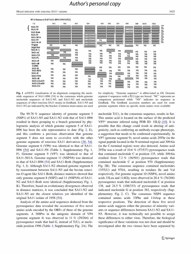
1965 [34]. Visualisation of the alignment of the two gen-
ome segment 8 sequences with mVISTA showed that the
two sequences were substantially different (Fig. 1).
Therefore, it was concluded that the virus sample that was
analysed was a mixture of two rotavirus SA11 strains.
Traditionally, the detection of mixed infections could be
missed with Sanger sequencing. The sequences of the other
10 genome segments of the viruses were identical. Since no
additional rotavirus O agent sequences were present, it was
hypothesised that if there were any other reassortants
between SA11-N5 and the rotavirus O agent, they may
have been selected out during propagation in cell culture.
The viruses were named RVA/Simian-tc/ZAF/SA11-N2/
1958/G3P[2] and RVA/Simian-tc/ZAF/SA11-N5/1958/
G3P[2]. The full genotypes assigned using the RotaC v2.0
web tool [32] was G3-P[2]-I2-R2-C5-M5-A5-Nx-T5-E2-
H5, where x was either 2 or 5. From here onwards, only the
common laboratory names of the SA11 rotavirus deriva-
tives will be used for simplicity. The average depth of
pyrosequencing coverage of the genome segments was
367-fold. Genome segment 1 had the lowest coverage of
125-fold, while genome segment 8 of SA11-N2 had the
highest coverage of 610-fold. Only genome segments 1 and
3 had an average depth of coverage lower than 200-fold
(Table 1).
The 50-terminal end sequences obtained for all the
genome segments of SA11-N5 and SA11-N2 were
50-GGC(U/A)7-, as expected for group A rotaviruses [58].
The consensus genome segments had various 30-terminal
sequences. The 30-terminal sequences obtained for the
consensus genome segment 1 (VP1), 3 (VP3), 4 (VP4), 6
(VP6), 8 (NSP2) and 9 (VP7) were the typical 30-terminal
end sequences, i.e., -UGUGACC-30 [61]. The consensus
genome segment 2 (VP2) contained the sequence -UAUGA
CC-30 at the 30-terminal end. This 30-terminal end sequence
was also reported previously for genome segments 2 (VP2)
and 10 (NSP4) of the rotavirus DS-1 strain [36, 40]. The
atypical 30-terminal sequences, UGUGAACC-30 and -UG
UGGCC-30, which were obtained for the consensus gen-
ome segments 5 (NSP1) and 7 (NSP3), were identical to
the 30-terminal sequences determined previously for these
genome segments in SA11 strains [39, 41, 45, 54]. The
atypical terminal sequence, -UGUGAACC-30, was found
to reduce the efficiency of dsRNA synthesis and genome
segment expression [45].
Nucleotide differences were observed in all genome
segments when the consensus genome segment sequences
of SA11-N5, SA11-N2 and nucleotide sequences of gen-
ome segments of SA11 derivatives available in GenBank
were compared to each other. Nucleotide sequence simi-
larity profiling indicated that the closest nucleotide genome
segment sequences in GenBank to those of SA11-N5 and
SA11-N2 were as follows: With the exception of genome
segments 4 (VP4) and 5 (NSP1), the consensus nucleotide
sequence of the other nine genome segments of SA11-N5
and SA11-N2 were identical to the sequence of a
Mixed infection with rotavirus SA11 variants 1023
123
Author's personal copy

corresponding genome segment of one of five different
SA11 derivatives (Table 1). Distance matrices showed that
the consensus sequences of eight genome segments of
SA11-N5 and SA11-N2 were most similar to those of
SA11-H96 (Supplementary Fig. 1). Genome segments 1
(VP1), 2 (VP2), 6 (VP6) 7 (NSP3), 8 (N5 typed NSP2) 9
(VP7) 10 (NSP4) and 11 (NSP5/6) were identical to those
of SA11-H96 (Supplementary Fig. 1). Genome segment 3
(VP3) was 100 % identical to that of SA11-5S, while
genome segment 4 (VP4) was 99.6 % identical to that of
SA11-Both (Table 1; Supplementary Fig. 1, C and D). The
nucleotide sequence identities between the genome seg-
ments 3 (VP3) and 5 (NSP1) were 99.96 % and 99.78 %
identical to the corresponding segments of SA11-H96,
respectively (Supplementary Fig. 1, C and E). A 99.61 %
nucleotide identity was observed between the consensus
genome segment 4 sequence of SA11-N5, SA11-N2 and
the corresponding nucleotide sequences of SA11-H96 and
other SA11 strains with a P[2] genotype (Fig. 1; Supple-
mentary Fig. 1, D). In contrast, only 77 % nucleotide
identity was observed between the consensus genome
segment 4 and SA11 strains with a P[1] genotype (Fig. 1).
Therefore, genome segment 4 of SA11-N2 and SA11-N5
did not reassort with that of the O agent as is the case for
some SA11 P[1] strains such as SA11-5S, SA11-5N and
SA11-30-1A.
Table 1 Comparison of the consensus nucleotide and deduced amino acid sequences of SA11-N5 and SA11-N2 to sequences of SA11-H96 [45],
the representative rotavirus SA11 strain
Genome
segment
Size
(bp)
Amino
acids in
coding
region
Average
depth of
coveragea
(-fold)
Nucleotide
similarity to
rotavirus SA11
derivatives
Molecular
clock rateb
(CVc)
Nucleotide differences between
SA11-N5, SA11 N2 and SA11-
H96d
Amino acid differences
(SA11-H96c?SA11-
N2/SA11-N5)
Point
variations
Insertions Deletions
1 (VP1) 3302 1088 125 SA11-H96
(100 %)
1.3 910-4
(1.3)
None None None None
2 (VP2) 2693 882 609 SA11-H96
(100 %)
7.5 9 10-5
(0.5)
None None None None
3 (VP3) 2591 835 156 SA11-5S
(100 %)
1.4 9 10-6
(1.1)
2 None None 650L?H
4 (VP4) 2362 775 358 SA11-Both
(99.6 %)
1.4 9 10-5
(0.3)
9 None None 72T?M, 157P?S,
187A?G, 261F?L,
332Y?S, 366V?M,
388S?Pe
5 (NSP1) 1614 496 225 SA11-H96
(99.7 %)
4.8 9 10-5
(1.4)
8 None None 36E?A,
84-86QQL?RTV,
96L?Q, 137K?L,
188E?D
6 (VP6) 1356 397 585 SA11-5 N
(100 %)
2.1 9 10-5
(0.6)
3 None None None
7 (NSP3) 1105 313 305 SA11-H96
(100 %)
1.3 9 10-4
(0.3)
2 None None None
8
(NSP2)N21059 317 610 SA11-Both
(100 %)
ND 205 None None 45 amino acid
differences
8
(NSP2)N51059 317 265 SA11-H96
(100 %)
1.9 9 10-4
(0.7)
None None None None
9 (VP7) 1063 326 487 SA11-30/1A
(100 %)
2.9 9 10-5
(0.4)
1 1 None 29I?Te, 309V?Ae
10 (NSP4) 751 175 495 SA11-H96
(100 %)
ND 4 None None 33F?Le, 93G?D,
114D?Ge
11 (NSP5/
6)
667 198/93 429 SA11-Both
(100 %)
8.5 9 10-5
(0.5)
1 None None None
a The depth of coverage represents the number of pyrosequence reads that were obtained per specific region of each genome segmentb Nucleotide substitution/site/yearc Coefficient of variationd Small and co-workers [45]e Novel amino acid residues detected in this study
ND: not determined due to too few nucleotide sequences (genome segment 8) or zero-length interior branches (genome segment 10)
1024 L. Mlera et al.
123
Author's personal copy
The 99.78 % sequence identity of genome segment 5
(NSP1) of SA11-N5 and SA11-N2 with that of SA11-H96
resulted in them grouping to a branch generated by phy-
logenetic analysis of which genome segment 5 of SA11-
H96 has been the sole representative to date (Fig. 2, E),
and this confirms a previous observation that genome
segment 5 does not seem to co-evolve with the other
genome segments of rotavirus SA11 derivatives [38, 54].
Genome segment 6 (VP6) was identical to that of SA11-
H96 [54] and SA11-5N (Table 1; Supplementary Fig. 1,
F). Genome segment 9 (VP7) was identical to that of
SA11-30/1A. Genome segment 11 (NSP5/6) was identical
to that of SA11-H96 [54] and SA11-Both (Supplementary
Fig. 1, I). Although SA11-N2 obtained genome segment 8
by reassortment between SA11-N5 and the bovine rotavi-
rus O agent like SA11-Both, distance matrices showed that
only genome segment 8 (NSP2) and 11 (NSP5/6) of SA11-
N2 and SA11-Both were identical (Supplementary Fig. 1,
K). Therefore, based on evolutionary divergences observed
in distance matrices, it was concluded that SA11-N2 and
SA11-N5 are the closest relatives yet identified of the
original SA11 isolate of 1958 [54].
Analysis of the amino acid sequences deduced from the
pyrosequence data revealed the occurrence of five novel
amino acids encoded by the ORFs of three of the genome
segments. A 388Pro in the antigenic domain of VP4
(genome segment 4) was observed in 11 % (29/264) of
pyrosequence reads that had G, instead of T(U), at nucle-
otide position 1996 (Table 1; Supplementary Fig. 2A). The
nucleotide T(U), in the consensus sequence, results in Ser.
This amino acid is located on the surface of the predicted
VP5* structure inferred using PDB ID: 1SLQ [10]. It is
possible that this change could result in altering of anti-
genicity, such as conferring an antibody-escape phenotype,
a suggestion that needs to be confirmed experimentally. In
VP7 (genome segment 9), novel amino acids 29Thr (in the
signal peptide located in the N-terminal region) and 309Ala
(in the C-terminal region) were also detected. Amino acid
29Thr was a result of 10.6 % (57/537) pyrosequence reads
that contained nucleotide C at position 135, while 309Ala
resulted from 7.2 % (36/501) pyrosequence reads that
contained nucleotide C at position 970 (Supplementary
Fig. 2B). The consensus sequence contained nucleotides
135T(U) and 970A, resulting in residues Ile and Val
respectively. For genome segment 10 (NSP4), novel amino
acids 33Leu and 114Gly were observed in 28.4 % (76/268)
pyrosequence reads that indicated nucleotide C at position
138, and 24.5 % (108/333) of pyrosequence reads that
indicated nucleotide G at position 382, respectively (Sup-
plementary Fig. 2, C). The consensus NSP4 sequence
contained amino acids 33Phe and 114Asp at these
respective positions. The detection of these five novel
amino acids suggests either the presence of minority vari-
ants or sequence differences between SA11-N2 and SA11-
N5. However, it was technically not possible to assign
these differences to either virus. Therefore, the biological
significance of these variations was not clear and should be
investigated after the two viruses have been separated by
Fig. 1 mVISTA visualisation of an alignment comparing the nucle-
otide sequences of SA11-H96 [54] to the consensus whole-genome
nucleotide sequences of SA11-N5, and SA11-N2 and nucleotide
sequences of other rotavirus SA11 strains in GenBank. SA11-N5 and
SA11-N2 are indicated by the bracket. Common strain names are used
for simplicity. ‘‘Genome sequence’’ is abbreviated as GS. Genome
segment 4 sequences with a P[1] type are boxed. ‘‘NC’’ represents no
comparison performed while ‘‘NS’’ indicates no sequence in
GenBank. The GenBank accession numbers are used for some
genome segments where no specific strain names were available
Mixed infection with rotavirus SA11 variants 1025
123
Author's personal copy

several rounds of plaque purification. No amino acid dif-
ferences were observed in the deduced protein sequences
of NSP2 (SA11-N5) and NSP5/6 (SA11-N2 and SA11-N5)
sequences when compared to the respective deduced amino
acid sequences of SA11-H96. The above-described results,
combined with a lack of passage history, prompted further
characterisation of the viruses by the application of
molecular clock evolutionary analysis of SA11-N2 and
SA11-N5.
Bayesian molecular clock evolutionary analysis
and phylogenetic relationships
Molecular clock phylogenetic analysis was carried out for
all 12 consensus genome segment sequences of SA11-N5
and SA11-N2 but failed for 2 of the genome segments. In
the case of genome segment 8, there were only three
nucleotide sequences available in GenBank for the N2
genotype. It was therefore not possible to perform molec-
ular clock phylogenetic analysis with so few sequences.
The genome segment 10 molecular clock results were
excluded from the analysis because zero-length interior
branches [6] were present in the phylogenetic trees and
could not be corrected. For the other ten genome segments,
rates of nucleotide substitution were in the range of
1.4 9 10-6–1.9 9 10-4 nucleotide substitutions/site/year
(Table 1). The coefficient of variation for the nucleotide
substitution rates was 0.3–1.4 (Table 1). The low CV
indicates that significant nucleotide substitution variations
occur within the branches. Therefore, the use of a relaxed
molecular clock model as opposed to a strict molecular
clock was valid [27]. The lowest nucleotide substitution
rate was observed for genome segment 3 (VP3), while the
highest was observed for genome segment 1 (VP1)
(Table 1). High nucleotide substitution rates were also
observed for genome segment 8 (NSP2) of SA11-N5 and
genome segment 7 (NSP3) of both SA11-N5 and SA11-N2
(Table 1). The high rate of evolution of genome segment 1
was unexpected, since VP1 is a RNA-dependent RNA
polymerase (RdRP) which is vital for ensuring viral rep-
lication. However, as with other RdRPs, sequence analysis
of rotavirus genome segment 1 (VP1) revealed that the
region encoding the basic right hand structure motif of
RdRPs is highly conserved and that the variations in the
A Genome segment 1 (VP1) B Genome segment 2 (VP2)
C Genome segment 3 (VP3) D Genome segment 4 (VP4)
RVA/Simian-tc/ZAF/SA11-30-19/1958/G3P[1]
RVA/Simian-tc/ZAF/SA11/1958/G3P[X] (1)
RVA/Simian-tc/ZAF/SA11-Both/1958/G3P[X]
RVA/Simian-tc/ZAF/SA11-5S/1958/G3P[2]
RVA/Simian-tc/ZAF/SA11-5N/1958/G3P[1]
RVA/Simian-tc/ZAF/SA11/1958/G3P[1] (2)
RVA/Simian-tc/ZAF/SA11-H96/1958/G3P[2] (3)
♦RVA/Simian-tc/ZAF/SA11-N2/1958/G3P[2]
RVA/Simian-tc/ZAF/SA11-30-1A/1958/G3P[1]8.7
5
7.8
♦RVA/Simian-tc/ZAF/SA11-N5/1958/G3P[2]
RVA/Simian-tc/ZAF/SA11-5N/1958/G3P[1]
RVA/Simian-tc/ZAF/SA11-C13/1958/G3P[X]
RVA/Simian-tc/ZAF/SA11-Both/1958/G3P[2]
RVA/Simian-tc/ZAF/SA11-TSF-B/1958/G3P[X]
RVA/Simian-tc/ZAF/SA11-TSF-A/1958/G3P[X]
RVA/Simian-tc/ZAF/SA11-tsC/1958/G3P[X]
♦RVA/Simian-tc/ZAF/SA11-N2/1958/G3P[2]
RVA/Simian-tc/ZAF/SA11-H96/1958/G3P[2] (3)
RVA/Simian-tc/ZAF/SA11-30-1A/1958/G3P[1]
RVA/Simian-tc/ZAF/SA11-30-19/1958/G3P[1]
RVA/Simian-tc/ZAF/SA11-5S/1958/G3P[1]
10
41.9
22.9
82.2
♦RVA/Simian-tc/ZAF/SA11-N5/1958/G3P[2]
RVA/Simian-tc/ZAF/SA11-Ramig Lab/1958/G3P[X]
♦RVA/Simian-tc/ZAF/SA11-N2/1958/G3P[2]
RVA/Simian-tc/ZAF/SA11-SEM/1958/G3P[2]
RVA/Simian-tc/ZAF/SA11-Both/1958/G3P[2]
RVA/Simian-tc/ZAF/SA11-H96/1958/G3P[2] (3)
RVA/Simian-tc/ZAF/SA11-S1/1958/G3P[2]
50
148.7
107.0
67.6
♦RVA/Simian-tc/ZAF/SA11-N2/1958/G3P[2]
RVA/Simian-tc/ZAF/SA11-L2/1958/G3P[2]
RVA/Simian-tc/ZAF/SA11-5S/1958/G3P[1]
♦RVA/Simian-tc/ZAF/SA11-N2/1958/G3P[2]
♦RVA/Simian-tc/ZAF/SA11-N5/1958/G3P[2]
RVA/Simian-tc/ZAF/SA11-H96/1958/G3P[1] (3)
RVA/Simian-tc/ZAF/SA11-5S/1958/G3P[1]
RVA/Simian-tc/ZAF/SA11/1958/G3P[1] (4)
RVA/Simian-tc/ZAF/SA11-Both/1958/G3P[1]
RVA/Simian-tc/ZAF/SA11-tsB/1958/G3P[1]
RVA/Simian-tc/ZAF/SA11-30-19/1958/G3P[1]
RVA/Simian-tc/ZAF/SA11-30-1A/1958/G3P[1]
1818.5
250
Fig. 2 Maximum clade credibility trees constructed with Bayesian
MCMC framework, in BEAST software, to depict the Bayesian
molecular clock phylogenetic relationships between SA11-N2, SA11-
N5 and rotavirus SA11 sequences obtained from GenBank. Branch
times (years) are indicated for some selected branches. The bars
below each tree indicate time scales. Rotavirus SA11-N2 and SA11-
N5 are indicated by a black diamond. SA11-H96, which is considered
the representative of the SA11 strains, is shaded grey. Two SA11-H96
sequences were available in GenBank for genome segments 5
(NSP1), 6 (VP6), 7 (NSP3), 8 (NSP2) and 11 (NSP5/6). Sequences
indicated with (3) were sequenced in the USA by Small and co-
workers [54], and those indicated with (5) were sequenced in India by
Dutta and co-workers [14]. Accession numbers of sequences associ-
ated with unspecified strains are as follows: (1): AF015955; (2):
DQ457016; (4): X16062; (6): AF290881; (7): AF290883; (8):
X00421; (9): GU550506 (10): M87502; (11): AF306493 and (12):
M28347
1026 L. Mlera et al.
123
Author's personal copy

sequences are generally located close to the C- and N-ter-
minal regions [59]. A report describing an analysis of the
evolution of the VP1 of ovine rotaviruses in comparison to
other rotavirus group A strains also showed that amino acid
variations mainly occurred in the N- and C-terminal regions
of the RdRP [5]. Therefore, an analysis of the nucleotide
substitution rate for the region spanning nucleotide posi-
tions 778–2610, encoding amino acids at positions 260–870
was performed. This part of VP1 contains a unique RdRP
region at the N-terminal end, followed by the fingers, palm
and thumb regions of the polymerase domain [31, 42, 59].
The nucleotide substitution rate obtained for this poly-
merase-encoding region was 9.9 9 10-5 nucleotide sub-
stitutions/site/year with a CV of 0.6. This nucleotide
substitution rate was lower than the overall rate of
1.3 9 10-4 nucleotide substitutions/site/year. However,
the Ka/Ks ratio of non-synonymous to synonymous
nucleotide substitution rates [23, 63] obtained for the
genome segment 1 ORF was 0.6, suggesting that synony-
mous substitutions were favoured and that genome segment
1 is under pressure to conserve the VP1 sequence. For
genome segment 11 (encoding NSP5/6), an overall muta-
tion rate of 5 9 10-5 per replicated base was reported for
porcine rotavirus CC86 strain [3]. In this study, we
obtained a similar rate of 8.5 9 10-5 for genome segment
11 of the rotavirus SA11 strains. Mutation rates vary
across the genome [13], and the difference between the
evolution rate of the lowest rate and highest rate observed
in this study was *100-fold, but it is not clear why
the difference was so high. However, the general rate of
E Genome segment 5 (NSP1) F Genome segment 6 (VP6)
G Genome segment 7 (NSP3) H Genome segment 8 (NSP2; N5 genotype)
♦RVA/Simian-tc/ZAF/SA11-N5/1958/G3P[2]
♦RVA/Simian-tc/ZAF/SA11-N2/1958/G3P[2]
123.1
34.2
50
RVA/Simian-tc/ZAF/SA11-PL/1958/G3P[X]
29.2
RVA/Simian-tc/ZAF/SA11-FEM/1958/G3P[X]
RVA/Simian-tc/ZAF/SA11-Both/1958/G3P[2]
RVA/Simian-tc/ZAF/SA11-H96/1958/G3P[2] (3)
RVA/Simian-tc/ZAF/SA11-4F/1958/G3P[X] (7)
RVA/Simian-tc/ZAF/SA11-4F/1958/G3P[X] (6)
142.6
RVA/Simian-tc/ZAF/SA11-H96/1958/G3P[2] (5)
10
10.4
10.2
9.0
♦RVA/Simian-tc/ZAF/SA11-N2/1958/G3P[2]
RVA/Simian-tc/ZAF/SA11-4F/1958/G3P[X] (9)
RVA/Simian-tc/ZAF/SA11-5S/1958/G3P[1]
RVA/Simian-tc/ZAF/SA11-H96/1958/G3P[2] (3)
RVA/Simian-tc/ZAF/SA11-Both/1958/G3P[2]
RVA/Simian-tc/ZAF/SA11-5N/1958/G3P[1]
RVA/Simian-tc/ZAF/SA11-30-1A/1958/G3P[1]
RVA/Simian-tc/ZAF/SA11-4F/1958/G3P[X] (7)
RVA/Simian-tc/ZAF/SA11-30-19/1958/G3P[1]
RVA/Simian-tc/ZAF/SA11-C14/1958/G3P[X]
RVA/Simian-tc/ZAF/SA11-L2Ao123/1958/G3P[2]RVA/Simian-tc/ZAF/SA11-H96/1958/G3P[2] (5)
♦RVA/Simian-tc/ZAF/SA11-N5/1958/G3P[2]
RVA/Simian-tc/ZAF/SA11-L2/1958/G3P[2]RVA/Simian-tc/ZAF/SA11-L2Ga613/1958/G3P[2]
5
RVA/Simian-tc/ZAF/SA11-H96/1958/G3P[2] (5)
RVA/Simian-tc/ZAF/SA11-Both/1958/G3P[2]
♦RVA/Simian-tc/ZAF/SA11-N2/1958/G3P[2]
♦ RVA/Simian-tc/ZAF/SA11-N5/1958/G3P[2]
RVA/Simian-tc/ZAF/SA11-H96/1958/G3P[2] (3)
RVA/Simian-tc/ZAF/SA11-30-19/1958/G3P[2]
RVA/Simian-tc/ZAF/SA11-5N/1958/G3P[2]
RVA/Simian-tc/ZAF/SA11/1958/G3P[2] (8)RVA/Simian-tc/ZAF/SA11-pAR61/1958/G3P[2]
RVA/Simian-tc/ZAF/SA11-tsG/1958/G3P[2]
RVA/Simian-tc/ZAF/SA11-Ramig/1958/G3P[2]
RVA/Simian-tc/ZAF/SA11-30-1A/1958/G3P[2]
RVA/Simian-tc/ZAF/SA11-5S/1958/G3P[2]
5
96
24
3.8
4.7
5.8
RVA/Simian-tc/ZAF/SA11-tsE/1958/G3P[X]
RVA/Simian-tc/ZAF/SA11-R/1958/G3P[X]
RVA/Simian-tc/ZAF/SA11-P/1958/G3P[X]
RVA/Simian-tc/ZAF/SA11-30-1A/1958/G3P[1]
RVA/Simian-tc/ZAF/SA11-H96/1958/G3P[2] (3)
RVA/Simian-tc/ZAF/SA11-5S/1958/G3P[1]
RVA/Simian-tc/ZAF/SA11-30-19/1958/G3P[1]
RVA/Simian-tc/ZAF/SA11-5N/1958/G3P[1]
RVA/Simian-tc/ZAF/SA11-H96/1958/G3P[2] (5)
♦RVA/Simian-tc/ZAF/SA11-N5/1958/G3P[2]
I Genome segment 9 (VP7) J Genome segment 11 (NSP5/6)
50
145
44
65
5
RVA/Simian-tc/ZAF/SA11/1958/G3P[X] (10)
RVA/Simian-tc/ZAF/SA11-Both/1958/G3P[2]
RVA/Simian-tc/ZAF/SA11/1958/G3P[X]
RVA/Simian-tc/ZAF/SA11/1958/G3P[X]
RVA/Simian-tc/ZAF/SA11-H96/1958/G3P[2] (3)
RVA/Simian-tc/ZAF/SA11-30-19/1958/G3P[1]
RVA/Simian-tc/ZAF/SA11-5S/1958/G3P[1]
RVA/Simian-tc/ZAF/SA11-30-1A/1958/G3P[1]
♦RVA/Simian-tc/ZAF/SA11-N2/1958/G3P[2]
RVA/Simian-tc/ZAF/SA11-5N/1958/G3P[1]
♦RVA/Simian-tc/ZAF/SA11-N5/1958/G3P[2]
RVA/Simian-tc/ZAF/SA11-Both/1958/G3P[X]
RVA/Simian-tc/ZAF/SA11/1958/G3P[X] (11)
RVA/Simian-tc/ZAF/SA11-5N/1958/G3P[1]
♦ RVA/Simian-tc/ZAF/SA11-N2/1958/G3P[2]
RVA/Simian-tc/ZAF/SA11-30-19/1958/G3P[1]
♦RVA/Simian-tc/ZAF/SA11-N5/1958/G3P[2]
RVA/Simian-tc/ZAF/SA11-H96/1958/G3P[1] (5)
RVA/Simian-tc/ZAF/SA11-5S/1958/G3P[1]
RVA/Simian-tc/ZAF/SA11-H96/1958/G3P[2] (3)
RVA/Simian-tc/ZAF/SA11-30-1A/1958/G3P[1]
RVA/Simian-tc/ZAF/SA11/1958/G3P[X] (12)
Fig. 2 continued
Mixed infection with rotavirus SA11 variants 1027
123
Author's personal copy

evolution was comparable to the range of rates of evo-
lution of dsRNA viruses, i.e., *1 9 10-4–1 9 10-6
nucleotide substitutions/site/year [2, 13, 24, 53]. Interest-
ingly, higher nucleotide substation rates of 1.36 9
10-3–4.78 9 10-3 were reported recently for the genomes
of group B rotaviruses [28].
The time-scaled phylogeny and maximum clade credi-
bility (MCC) trees depicted in Fig. 2 confirmed that the most
recent common ancestor for genome segments 1 (VP1), 2
(VP2), 6 (VP6) and 7 (NSP3) of SA11-N5 and SA11-N2
were the cognate genome segments from SA11-H96 [54].
For genome segments 5 (NSP1), 8 (NSP2 with genotype N5)
and 11 (NSP5/6), the most recent common ancestor for
genome segments of SA11-N5 and SA11-N2 genome seg-
ments was the SA11-H96 strain characterised in India by
Dutta and co-workers [14]. In one of the few reports on
molecular clock studies involving rotaviruses, genome
segment 4 was found to evolve at a high rate of 0.58 9 10-3
nucleotide substitutions/site/year [24]. Sequences analysed
in the report of Jenkins and co-workers [24] were, however,
obtained from various field strains. For cell-culture-adapted
rotavirus SA11 strains (genotyped P[2]), we determined a
slower evolutionary rate of 1.4 9 10-5 nucleotide substi-
tutions/site/year (Table 1). The MCC tree of genome seg-
ment 4 indicated that SA11-N2 and SA11-N5 were closely
related to SA11-SEM (Fig. 2D).
The evolutionary rate obtained using genome segment 7
open reading frames was 1.3 9 10-4 nucleotide substitu-
tions/site/year, with a coefficient of variation (CV) of 0.3
(Table 1). The substitution rate calculated for the SA11
rotavirus is in accordance with the estimated average rate
of 7.9 9 10-4 nucleotide substitutions/site/year in all
genome segments, except for segments 1, 3 and 10 for a
human group B rotavirus [62]. The MCC tree for genome
segment 7 indicated that SA11-N2 and SA11-N5 are clo-
sely related to strain SA11-H96 [54] (Fig. 2G). SA11-5S is
also closely related to SA11-N2 and SA11-N5 (Fig. 2G).
Strain SA11-4F followed a separate and distinct evolu-
tionary path from that of SA11-5S (Fig. 2G).
The rate of evolution for genome segment 9 (VP7) was
2.9 9 10-5 nucleotide substitutions/site/year with a CV of
0.4 (Table 1). The frequency of appearance of monoclonal-
antibody-resistant variants in vitro has been estimated to be
approximately 10-5 [57]. The evolutionary rate obtained in
this study was lower than the rate estimated for human G9
rotavirus strains [37]. Rates of 1.87 9 10-3 substitutions/
site/year for G9 rotaviruses and 1.66 9 10-3 substitutions/
site/year for G12 rotaviruses have been reported [37].
Similarly, a nucleotide substitution rate of 1.57 9 10-3
was reported for the VP7-encoding genome segment 9 of
group B rotaviruses [51]. Matthijnssens and co-workers
attributed the high evolutionary rate they observed for
various G9 and G12 rotavirus strains to the immunological
pressure on genome segment 9 (VP7) [18, 57]. The same
conclusion could also be drawn for the high rate observed
for genome segment 4 (VP4) by Jenkins and co-workers
[24]. Genome segment 9 (VP7) nucleotide sequences of
SA11-N2 and SA11-N5 and those in GenBank are from
cell-culture-adapted strains that would only be under the
pressure of the cellular innate immunity and culture con-
ditions. This pressure on the genome is presumably less
than the total biological pressure exerted during in vivo
infections and possibly the reason for the relatively lower
evolutionary rate observed in this study. The inherent and
greater nucleotide sequence diversity between the G9 and
G12 strains used in the analyses of Matthijnssens and co-
workers is also expected to increase the calculated nucle-
otide substitution rate. For instance, the genome segment 9
(VP7) of the G9 porcine strain RVA/Pig-tc/KOR/
PRG9121/2006/G9P[7] is 93 % identical to that of the
human RVA/human-wt/BEL/BE2001/2009/G9P[6] strain
which also has a G9 genotype (data not shown), whereas
the G3 nucleotide sequences of SA11 derivatives analysed
in this study were 99–100 % identical (Supplementary
Fig. 1). However, computational estimation of evolution-
ary rates could result in variations from evolution rates
determined experimentally.
Overall, a higher degree of divergence was observed
following molecular clock evolutionary analysis in com-
parison to distance matrices (Fig. 2; Supplementary Fig. 1).
The distance matrices indicate very close relationships
between the rotavirus SA11 derivatives, but the MCC trees
show more divergent relationships among the strains. This
difference is attributed to the different approaches of the
two methods for reconstructing phylogenetic relationships.
The divergences also highlight sequence differences that
could have been introduced during cell culture in the many
laboratories that sequenced the rotavirus SA11 genome
segments. However, Bayesian phylogenetic analysis in
MCMC is more statistical (probabilistic), and sampling of
trees is in proportion to their phylogenetic likelihood,
thereby producing the most credible or consensus tree
[11, 12, 17]. On the other hand, distance matrix calculations
do not assume a molecular clock in the analysis [17].
In summary, the whole-genome consensus sequences of
two rotavirus SA11 strains in a mixed sample were deter-
mined by sequence-independent genome amplification and
454� pyrosequencing. Two distinct genome segment 8
(encoding NSP2) nucleotide sequences were present in the
same sample. This indicates that the virus sample was a
mixture of SA11-N5 and SA11-N2. The SA11-N2 strain
acquired a bovine rotavirus genome segment 8 due to re-
assortment with the bovine rotavirus O agent. The ability to
detect the virus mixture in the same sample is another
example of the advantage of next-generation sequencing
over traditional sequencing methods. Genome segment 4
1028 L. Mlera et al.
123
Author's personal copy

(VP4) sequences of SA11-N2 and SA11-N5 did not reas-
sort with that of the bovine rotavirus O agent as in other
rotavirus SA11 variants. Although SA11-N2 obtained
genome segment 8 (NSP2) by reassortment like SA11-
Both, SA11-N2 and SA11-Both are not the same. There-
fore, the bovine rotavirus O agent must also have
co-infected with SA11-N5, resulting in the generation of
SA11-N2, as described previously for SA11-Both. How-
ever, the absence of additional rotavirus O agent sequences
indicates that other reassortants, if any exist, may have
been selected out during passage in cell culture or that their
concentrations were too low to be detected. The five novel
amino acids detected in VP4, VP7 and NSP4 might either
reflect the occurrence of minority variants or actual
sequence differences between SA11-N2 and SA11-N5.
Based on evolutionary divergences observed in distance
matrices and molecular clock phylogenetic analysis, it was
concluded that SA11-N2 and SA11-N5 are very close
derivatives of the original SA11-H96 isolated in 1958 in
South Africa. SA11-N2 is a reassortant carrying genome
segment 8 (NSP2) from the O agent on the SA11-H96
backbone, whereas SA11-N5 contains a true SA11-like
genome and is the closest derivative of SA11-H96 that has
yet been identified.
Acknowledgments This study was funded by a Senior Fellowship
awarded to HGO (I/84 015) by the European Foundation Initiative
for Neglected Tropical Diseases (EFINTD). HGO also received a
postdoctoral fellowship from the North-West University (Potchefst-
room, South Africa). LM received a PhD bursary (09/41) from the
Poliomyelitis Research Foundation, South Africa. We thank Mrs.
I. Peenze, Diarrhoeal Pathogens Research Unit, University of
Limpopo (Pretoria, South Africa), for kindly providing the rotavirus
SA11 sample, and Dr. M. Coetzee (Department of Genetics,
University of Pretoria, South Africa) for assistance with molecular
clock analysis.
References
1. Arnold M, Patton JT, McDonald SM (2009) Culturing, storage,
and quantification of rotaviruses. Curr Protoc Microbiol
15:15C.3.1–15C.3.24
2. Barr JN, Fearns R (2010) How RNA viruses maintain their
genome integrity. J Gen Virol 91:1373–1387
3. Blackhall J, Fuentes A, Magnusson G (1996) Genetic stability of
a porcine rotavirus RNA segment during repeated plaque isola-
tion. Virology 225:181–190
4. Both GW, Mattick JS, Bellamy AR (1983) Serotype-specific
glycoprotein of simian 11 rotavirus: Coding assignment and gene
sequence. Proc Natl Acad Sci USA 80:3091–3095
5. Chen Y, Zhu W, Sui S, Zhang X, Hu S (2009) Sequence and
evolutionary analysis of VP1 gene of ovine rotavirus NT. Wei
Sheng Wu Xue Bao 49:1055–1062
6. Coddington J, Scharff N (1994) Problems with zero-length
branches. Cladistics 10:415–423
7. Desselberger U (1996) Genome rearrangements of rotaviruses.
Adv Virus Res Volume 46:69–95
8. Domingo E, Martın V, Perales C, Grande-Perez A, Garcıa-Arri-
aza J, Arias A (2006) Quasispecies: Concept and implications for
virology. In: Domingo E (ed) Springer, Berlin, pp 51–82
9. Domingo E, Sheldon J, Perales C (2012) Viral quasispecies
evolution. Microbiol Mol Biol Rev 76:159–216
10. Dormitzer PR, Nason EB, Prasad BV, Harrison SC (2004)
Structural rearrangements in the membrane penetration protein of
a non-enveloped virus. Nature 430:1053–1058
11. Drummond A, Rambaut A, Shapiro B, Pybus O (2005) Bayesian
coalescent inference of past population dynamics from molecular
sequences. Mol Biol Evol 22:1185–1192
12. Drummond A, Rambaut A (2007) BEAST: Bayesian evolution-
ary analysis by sampling trees. BMC Evol Biol 7:214
13. Duffy S, Shackelton LA, Holmes EC (2008) Rates of evolu-
tionary change in viruses: patterns and determinants. Nat Rev
Genet 9:267–276
14. Dutta D, Chattopadhyay S, Bagchi P, Halder UC, Nandi S,
Mukherjee A, Kobayashi N, Taniguchi K, Chawla-Sarkar M
(2011) Active participation of cellular chaperone Hsp90 in reg-
ulating the function of rotavirus nonstructural protein 3 (NSP3).
J Biol Chem 286:20065–20077
15. Estes MK, Graham DY, Gerba CP, Smith EM (1979) Simian
rotavirus SA11 replication in cell cultures. J Virol 31:810–815
16. Estes MK, Kapikian AZ (2007) Rotaviruses. In: Knipe DM,
Howley PM, Griffin DE, Lamb RA, Martin MA, Roizman B,
Straus SE (eds) Fields virology. Kluwer Health/Lippincott,
Philadelphia, pp 1917–1974
17. Felsenstein J (1988) Phylogenies from molecular sequences:
Inference and reliability. Annu Rev Genet 22:521–565
18. Flores J, Sears J, Green KY, Perez-Schael I, Morantes A, Daoud
G, Gorziglia M, Hoshino Y, Chanock RM, Kapikian AZ (1988)
Genetic stability of rotaviruses recovered from asymptomatic
neonatal infections. J Virol 62:4778–4781
19. Frazer KA, Pachter L, Poliakov A, Rubin EM, Dubchak I (2004)
VISTA: computational tools for comparative genomics. Nucleic
Acids Res 32:W273–W279
20. Hall TA (1999) BioEdit: a user-friendly biological sequence
alignment editor and analysis program for Windows 95/98/NT.
Nucleic Acids Symp Ser 41:95–98
21. Hasegawa M, Kishino H, Yano T (1985) Dating of the human-
ape splitting by a molecular clock of mitochondrial DNA. J Mol
Evol 22:160–174
22. Hundley F, McIntyre M, Clark B, Beards G, Wood D, Chrystie I,
Desselberger U (1987) Heterogeneity of genome rearrangements
in rotaviruses isolated from a chronically infected immunodefi-
cient child. J Virol 61:3365–3372
23. Hurst LD (2002) The Ka/Ks ratio: diagnosing the form of
sequence evolution. Trends in Genet 18:486–487
24. Jenkins GM, Rambaut A, Pybus OG, Holmes EC (2002) Rates of
molecular evolution in RNA viruses: a quantitative phylogenetic
analysis. J Mol Evol 54:156–165
25. Jere KC, Mlera L, O’Neill HG, Potgieter AC, Page NA, Seheri ML,
van Dijk AA (2011) Whole genome analyses of African G2, G8, G9,
and G12 rotavirus strains using sequence-independent amplification
and 454� pyrosequencing. J Med Virol 83:2018–2042
26. Jere KC, Mlera L, Page NA, van Dijk AA, O’Neill HG (2011)
Whole genome analysis of multiple rotavirus strains from a single
stool specimen using sequence-independent amplification and
454� pyrosequencing reveals evidence of intergenotype genome
segment recombination. Infect Genet Evol 11:2072–2082
27. Koopmans LH, Owen DB, Rosenblatt JI (1964) Confidence
intervals for the coefficient of variation for the normal and log
normal distributions. Biometrika 51:25–32
28. Lahon A, Walimbe AM, Chitambar SD (2012) Full genome
analysis of group B rotaviruses from western India: genetic
relatedness and evolution. J Gen Virol 93:2252–2266
Mixed infection with rotavirus SA11 variants 1029
123
Author's personal copy

29. Liu M, Estes MK (1989) Nucleotide sequence of the simian
rotavirus SA11 genome segment 3. Nucleic Acids Res 17:7991
30. Lopez S, Arias CF (1992) Simian rotavirus SA11 strains. J Virol
66:1832
31. Lu X, McDonald SM, Tortorici MA, Tao YJ, Vasquez-Del
Carpio R, Nibert ML, Patton JT, Harrison SC (2008) Mechanism
for coordinated RNA packaging and genome replication by
rotavirus polymerase VP1. Structure 16:1678–1688
32. Maes P, Matthijnssens J, Rahman M, Van Ranst M (2009) RotaC:
a web-based tool for the complete genome classification of group
A rotaviruses. BMC Microbiol 9:238
33. Malherbe H, Harwin R (1963) The cytopathic effects of vervet
monkey viruses. S Afr Med J 37:407–411
34. Malherbe HH, Strickland-Cholmley M (1967) Simian virus SA11
and the related O agent. Arch Gesamte Virusforsch 22:235–245
35. Margulies M, Egholm M, Altman WE, Attiya S, Bader JS,
Bemben LA, Berka J, Braverman MS, Chen Y-J, Chen Z, Dewell
SB, Du L, Fierro JM, Gomes XV, Godwin BC, He W, Helgesen
S, Ho CH, Irzyk GP, Jando SC, Alenquer MLI, Jarvie TP, Jirage
KB, Kim J-B, Knight JR, Lanza JR, Leamon JH, Lefkowitz SM,
Lei M, Li J, Lohman KL, Lu H, Makhijani VB, McDade KE,
McKenna MP, Myers EW, Nickerson E, Nobile JR, Plant R, Puc
BP, Ronan MT, Roth GT, Sarkis GJ, Simons JF, Simpson JW,
Srinivasan M, Tartaro KR, Tomasz A, Vogt KA, Volkmer GA,
Wang SH, Wang Y, Weiner MP, Yu P, Begley RF, Rothberg JM
(2005) Genome sequencing in microfabricated high-density
picolitre reactors. Nature 437:376–380
36. Matthijnssens J, Ciarlet M, Heiman E, Arijs I, Delbeke T,
McDonald SM, Palombo EA, Iturriza-Gomara M, Maes P, Patton
JT, Rahman M, Van Ranst M (2008) Full genome-based classi-
fication of rotaviruses reveals a common origin between human
Wa-like and porcine rotavirus strains and human DS-1-like and
bovine rotavirus strains. J Virol 82:3204–3219
37. Matthijnssens J, Heylen E, Zeller M, Rahman M, Lemey P, Van
Ranst M (2010) Phylodynamic analyses of rotavirus genotypes
G9 and G12 underscore their potential for swift global spread.
Mol Biol Evol 27:2431–2436
38. Matthijnssens J, Taraporewala ZF, Yang H, Rao S, Yuan L, Cao
D, Hoshino Y, Mertens PP, Carner GR, McNeal M, Sestak K,
Van Ranst M, Patton JT (2010) Simian rotaviruses possess
divergent gene constellations that originated from interspecies
transmission and reassortment. J Virol 84:2013–2026
39. Mitchell DB, Both GW (1990) Conservation of a potential metal
binding motif despite extensive sequence diversity in the rota-
virus nonstructural protein NS53. Virology 174:618–621
40. Mlera L, Jere KC, van Dijk AA, O’Neill HG (2011) Determi-
nation of the whole-genome consensus sequence of the prototype
DS-1 rotavirus using sequence-independent genome amplifica-
tion and 454� pyrosequencing. J Virol Methods 175:266–271
41. Mossel EC, Ramig RF (2002) Rotavirus genome segment 7
(NSP3) is a determinant of extraintestinal spread in the neonatal
mouse. J Virol 76:6502–6509
42. Ogden KM, Johne R, Patton JT (2012) Rotavirus RNA poly-
merases resolve into two phylogenetically distinct classes that
differ in their mechanism of template recognition. Virology
431:50–57
43. Parashar UD, Gibson CJ, Bresse JS, Glass RI (2006) Rotavirus
and severe childhood diarrhea. Emerg Infect Dis 12:304–306
44. Patton JT, Stacy-Phipps S (1986) Electrophoretic separation of
the plus and minus strands of rotavirus SA11 double-stranded
RNAs. J Virol Methods 13:185–190
45. Patton JT, Taraporewala Z, Chen D, Chizhikov V, Jones M,
Elhelu A, Collins M, Kearney K, Wagner M, Hoshino Y, Gouvea
V (2001) Effect of intragenic rearrangement and changes in the 30
consensus sequence on NSP1 expression and rotavirus replica-
tion. J Virol 75:2076–2086
46. Pereira HG, Azeredo RS, Fialho AM, Vidal MN (1984) Genomic
heterogeneity of simian rotavirus SA11. J Gen Virol 65(Pt
4):815–818
47. Pereira HG, Gouvea VS, Fialho AM (1986) A comparison of
simian rotavirus SA11 preparations maintained in different lab-
oratories. Mem Inst Oswaldo Cruz 81:389–393
48. Posada D (2008) jModelTest: Phylogenetic model averaging. Mol
Biol Evol 25:1253–1256
49. Potgieter AC, Page NA, Liebenberg J, Wright IM, Landt O, van
Dijk AA (2009) Improved strategies for sequence-independent
amplification and sequencing of viral double-stranded RNA
genomes. J Gen Virol 90:1423–1432
50. Radford AD, Chapman D, Dixon L, Chantrey J, Darby AC, Hall
N (2012) Application of next-generation sequencing technologies
in virology. J Gen Virol 93:1853–1868
51. Rahman M, Hassan ZM, Zafrul H, Saiada F, Banik S, Faruque
AS, Delbeke T, Matthijnssens J, Van Ranst M, Azim T (2007)
Sequence analysis and evolution of group B rotaviruses. Virus
Res 125:219–225
52. Ramig RF (1997) Genetics of the rotaviruses. Annu Rev
Microbiol 51:225–255
53. Sanjuan R, Nebot MR, Chirico N, Mansky LM, Belshaw R
(2010) Viral mutation rates. J Virol 84:9733–9748
54. Small C, Barro M, Brown TL, Patton JT (2007) Genome heter-
ogeneity of SA11 rotavirus due to reassortment with ‘‘O’’ agent.
Virology 359:415–424
55. Tamura K, Nei M, Kumar S (2004) Prospects for inferring very
large phylogenies by using the neighbor-joining method. Proc
Natl Acad Sci USA 101:11030–11035
56. Tamura K, Peterson D, Peterson N, Stecher G, Nei M, Kumar S
(2011) MEGA5: molecular evolutionary genetics analysis using
maximum likelihood, evolutionary distance, and maximum par-
simony methods. Mol Biol Evol 28:2731–2739
57. Taniguchi K, Urasawa S (1995) Diversity in rotavirus genomes.
Semin Virol 6:123–131
58. Tortorici MA, Shapiro BA, Patton JT (2006) A base-specific
recognition signal in the 50 consensus sequence of rotavirus plus-
strand RNAs promotes replication of the double-stranded RNA
genome segments. RNA 12:133–146
59. Vasquez-del Carpio R, Morales JL, Barro M, Ricardo A, Spencer
E (2006) Bioinformatic prediction of polymerase elements in the
rotavirus VP1 protein. Biol Res 39:649–659
60. Voelkerding KV, Dames SA, Durtschi JD (2009) Next-generation
sequencing: from basic research to diagnostics. Clin Chem
55:641–658
61. Wentz MJ, Patton JT, Ramig RF (1996) The 3’-terminal con-
sensus sequence of rotavirus mRNA is the minimal promoter of
negative-strand RNA synthesis. J Virol 70:7833–7841
62. Yang J-H, Kobayashi N, Wang Y-H, Zhou X, Li Y, Zhou D-J, Hu
Z-H, Ishino M, Alam MM, Naik TN, Ahmed MU (2004) Phy-
logenetic analysis of a human group B rotavirus WH-1 detected
in China in 2002. J Med Virol 74:662–667
63. Yang Z, Bielawski JP (2000) Statistical methods for detecting
molecular adaptation. Trends Ecol Evol 15:496–503
1030 L. Mlera et al.
123
Author's personal copy