Bipolar Precharger for Hybrid Electrostatic Filtration Systems
Upload
independentCategory
view
3download
0
¹ WILEY-VCH Verlag GmbH, Weinheim, Germany
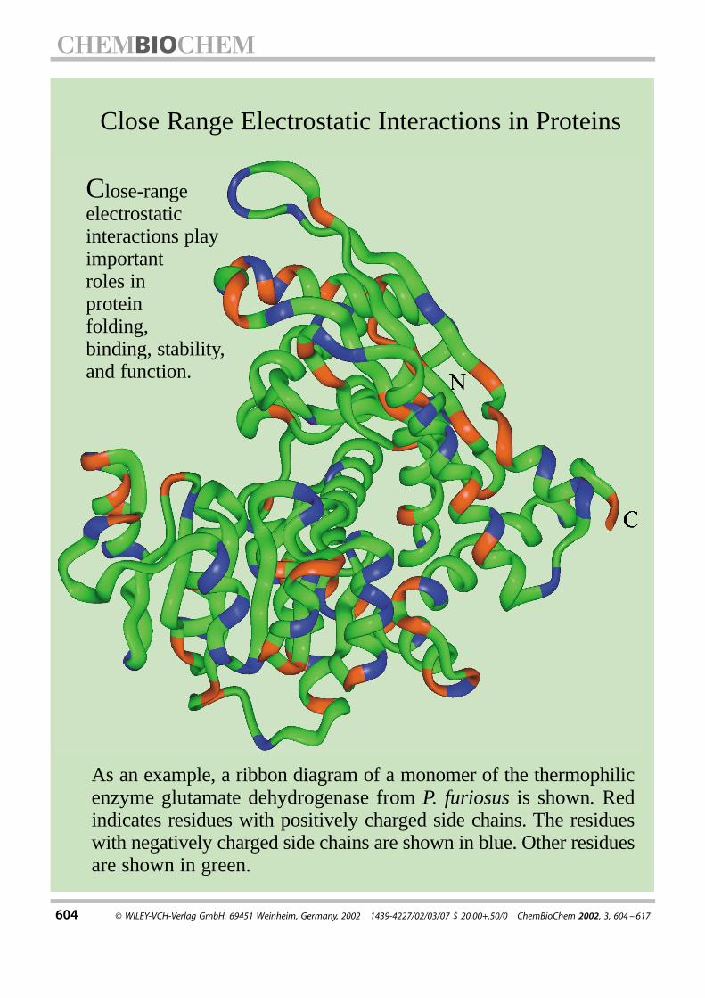
Near and far : Specific interactions play important roles inprotein folding, binding, flexibility, stability, and function. Close-range electrostatic interactions between oppositely chargedresidues at different distances are focused on here. The role ofsalt bridges (see picture) in stabilizing or destabilizing proteinsis debated.
S. Kumar, R. Nussinov*
604 ± 617
Close-Range ElectrostaticInteractions in Proteins
REPRINT
604 ¹ WILEY-VCH-Verlag GmbH, 69451 Weinheim, Germany, 2002 1439-4227/02/03/07 $ 20.00+.50/0 ChemBioChem 2002, 3, 604 ±617
Close Range Electrostatic Interactions in Proteins
Close-rangeelectrostaticinteractions playimportant roles inprotein folding,binding, stability, and function.
As an example, a ribbon diagram of a monomer of the thermophilicenzyme glutamate dehydrogenase from P. furiosus is shown. Redindicates residues with positively charged side chains. The residueswith negatively charged side chains are shown in blue. Other residuesare shown in green.

ChemBioChem 2002, 3, 604 ± 617 ¹ WILEY-VCH-Verlag GmbH, 69451 Weinheim, Germany, 2002 1439-4227/02/03/07 $ 20.00+.50/0 605
Close-Range Electrostatic Interactions in ProteinsSandeep Kumar[c] and Ruth Nussinov*[a, b]
Two types of noncovalent bonding interactions are present inprotein structures, specific and nonspecific. Nonspecific interac-tions are mostly hydrophobic and van der Waals. Specificinteractions are largely electrostatic. While the hydrophobic effectis the major driving force in protein folding, electrostatic inter-actions are important in protein folding, stability, flexibility, andfunction. Here we review the role of close-range electrostaticinteractions (salt bridges) and their networks in proteins. Saltbridges are formed by spatially proximal pairs of oppositelycharged residues in native protein structures. Often salt-bridgingresidues are also close in the protein sequence and fall in the samesecondary structural element, building block, autonomous foldingunit, domain, or subunit, consistent with the hierarchical model forprotein folding. Recent evidence also suggests that charged andpolar residues in largely hydrophobic interfaces may act as hotspots for binding. Salt bridges are rarely found across protein parts
which are joined by flexible hinges, a fact suggesting that saltbridges constrain flexibility and motion. While conventionalchemical intuition expects that salt bridges contribute favorablyto protein stability, recent computational and experimentalevidence shows that salt bridges can be stabilizing or destabilizing.Due to systemic protein flexibility, reflected in small-scale side-chainand backbone atom motions, salt bridges and their stabilitiesfluctuate in proteins. At the same time, genome-wide, amino acidsequence composition, structural, and thermodynamic compar-isons of thermophilic and mesophilic proteins indicate that specificinteractions, such as salt bridges, may contribute significantlytowards the thermophilic ±mesophilic protein stability differential.
KEYWORDS:
electrostatic interactions ¥ flexibility ¥ protein folding ¥protein structures ¥ salt bridges
1. Introduction
Specific interactions play important roles in protein folding,binding, flexibility, stability, and function. Here we focus onclose-range electrostatic interactions. Throughout this review,we refer to a pair of oppositely charged residues (Asp or Glu withArg, Lys, or His) as an ion pair. An ion pair is defined as a saltbridge if the centroids of the side-chain charged-group atoms inthe residues lie within 4.0 ä of each other and at least one pair ofAsp or Glu side-chain carbonyl oxygen and side-chain nitrogenatoms of Arg, Lys, or His are also within this distance. Such adefinition ensures that the oppositely charged residues in a saltbridge are spatially close and interact.[1, 2]
2. The Role of Specific Interactions in ProteinFolding/Binding
The initial fast step in protein folding is the hydrophobiccollapse, which leads to the molten globule (MG) state. In thenext step, specific interactions are optimized; this shifts theequilibrium toward the native state. Specific electrostaticinteractions play a critical role in reaching the native fold. Agood example is the �-lactalbumin (�-LA). If the Ca2� ion isremoved, �-LA is observed to be in its molten globule state. Atlow pH values (around 2.0) �-LA also exists in the molten globulestate, referred to as the acid state (the A state).[3a] Other examplesinclude nucleic acid binding proteins, which on their own arefrequently disordered. However, upon binding to the DNA (orRNA) they become stabilized.[3b] Alternatively, in some cases,depending on the location and charge, local disorder may be
observed. A nice example is that of the adenine binding domainin dihydrofolate reductase. By itself, this domain is unstable.However, when bound to other domains, or to its NADPHnucleotide cofactor, it is stabilized.[3c]
The hierarchical model is among the several models proposedfor protein folding. In this model folding initiates locally. Localfolded elements associate in a step-wise fashion to yield thenative structure.[4] Formation of salt bridges is consistent withhierarchical protein folding. Recently, we have analyzed adatabase of 222 salt bridges from 36 nonhomologous mono-meric protein crystal structures solved to high resolution (1.6 äor better). For approximately half of these salt bridges, thenumber of intervening residues was �10. Hence, the oppositelycharged residues that form salt bridges are often near each otherin the amino acid sequences of the proteins.[2a] Many chargedresidues that form salt bridges have helical conformations. In �
[a] Prof. R. NussinovIntramural Research Support Program–SAICNCI-Frederick, Building 469, Room 151Frederick, MD 21702 (USA)Fax: (�1) 301-846-5598E-mail : [email protected]
[b] Prof. R. NussinovSackler Institute of Molecular MedicineDepartment of Human Genetics and Molecular MedicineSackler School of MedicineTel Aviv UniversityTel Aviv, 69978 (Israel)
[c] Dr. S. KumarLaboratory of Experimental and Computational BiologyNCI-Frederick, Building 469, Room 151Frederick, MD 21702 (USA)

R. Nussinov and S. Kumar
606 ChemBioChem 2002, 3, 604 ±617
helices, negatively charged residues, Asp and Glu, are favored atthe N terminus, and at positions in the first turn. Positivelycharged residues, Lys and His, are favored at or near the Cterminus. These residues have an important role in helixtermination and in the formation of helix-capping motifs. Inthe middle of � helices, oppositely charged residue pairs tend to
have greater propensities to occur at adjacent positions (i, i�1)or on the same face (i, i� 3,4). However, the middle positions of� helices are neutral (neither favored nor avoided) for mostcharged residues and the average propensities for oppositelyand like charged residue pairs to occur at (i, i�1,2,3,4) positionsare similar.[5] Nevertheless, in surveys of �-helical structures,[6](i,i�3,4) salt-bridge pairs are observed. In �-helical peptides, saltbridges and their networks stabilize the helical structure tovarying extents.[7] There have been relatively fewer studies onsalt-bridge formation in � sheets. However, these studies alsoshow that salt bridges stabilize the � sheets to similar extents asthe � helices.[8] These studies also indicate that salt bridges areoften formed in protein secondary structural elements.A study on the conservation of salt bridges in different protein
families has shown that buried salt bridges are more likely to beconserved than the surface exposed ones.[9] Electrostatic inter-actions are optimized locally and appear to evolve in thedirection of avoiding electrostatic repulsions. Nevertheless,buried unsatisfied charges exist, and their destabilizing effecthas been controversial. While some studies have suggested thatsuch charges may considerably destabilize protein structures,others have suggested that their apparent destabilizing effect isalleviated by local unfolding, reorientation of backbone chargedgroups, and penetration of water. Interestingly, with regard tooptimization of electrostatic interactions, formation of additionalsalt bridges is secondary.[10] In addition to pair-wise salt bridges,more complex associations of the charged residues in proteinsare also observed. Musafia et al.[11] have carried out a statisticalanalysis of complex salt bridges (involving at least three chargedresidues) in 94 proteins. They find that complex salt bridges areoften formed, a fact indicating a tendency of the chargedresidues to form cooperative networks. These salt bridgenetworks are more often found at subunit ± subunit interfaces.Arginine, which contains guanidium group in its side chain, actsas a connector in such networks.Oliveberg and Fersht[12] have developed a method to study
transition-state structures in the folding pathway, by using theproton-titration behavior of charged protein residues. They findthat a partially buried salt bridge Arg69±Asp93 in Barnase isformed early in the folding process. Electrostatic interactionsmay also be important kinetically. A triple mutant of the Arcrepressor dimer which replaces a triad of charged residues withhydrophobic amino acids has been shown to fold faster than thewild type. Hence, formation of buried polar interactions may be aslow step in protein folding.[13] Theoretical calculations indicatethat electrostatic interactions affect unfolding rates of thermo-philic and mesophilic rubredoxins.[14] Torshin and Harrison[15]
have suggested that centroids of positive and negative chargesmay match protein folding cores detected by hydrogen-exchange experiments.Recently, Nussinov and co-workers have proposed a hierarch-
ical model for protein folding. In this model, folding initiateslocally. First, building blocks consisting of 15 or more residues areformed. A building block consists of a single secondary structureor a set of contiguous secondary structures (super-secondarystructures). The conformation of a building block seen in thenative protein structure may (or may not) be same as the most
Ruth Nussinov, born in 1943, is aprofessor in the Department of HumanGenetics, School of Medicine, Tel AvivUniversity, Israel, and a senior scientistat the National Cancer Institute–Frederick, Maryland, USA. She receivedher BSc degree in Microbiology atWashington University, Seattle, USA, in1967, and her MSc in Biochemistry in1968 from Rutgers University. Shereceived her PhD in Biochemistry fromRutgers University in 1977. Dr. Nussi-nov was a fellow at the Weizmann Institute and a Visiting Scientistin the Chemistry Department at the University of California,Berkeley, and in the Biochemistry Department at Harvard Univer-sity. She joined the Medical School at Tel Aviv in 1985 as anassociate professor, and in 1990 became a full professor. Herassociation with National Institutes of Health began in 1983, firstwith the National Institute of Child Health and Human Develop-ment, and, since 1985, with the National Cancer Institute. She hasauthored or coauthored more than 200 scientific papers. Until1990, her papers addressed nucleic acid sequence and structureand protein ± nucleic acid interactions. In 1990 she switched toproteins; her research currently focuses on protein folding andbinding.
Sandeep Kumar, born in 1968, hasbeen a postdoctoral visiting fellow atLaboratory of Experimental and Com-putational Biology, the National CancerInstitute–Frederick, Maryland, USA,since January 1998. He obtained hisBSc(Hons) Physics degree from theUniversity of Delhi, India, in 1989. Buthe then became more inclined towardsbiology and, hence, obtained an MSc inMolecular Biology and Biotechnologyfrom the G.B. Pant University of Agri-culture and Technology, Pant Nagar, India, in 1992. In the sameyear, he joined the Molecular Biophysics Unit at the Indian Instituteof Science, Bangalore, India, for his doctoral studies. In 1998, hewas awarded a PhD degree in recognition of his work on sequenceand structural relationships in � helices. In his postdoctoral work,he has focused on issues related with protein stability and the roleof close-range electrostatic interactions in proteins. His researchinterests focus on understanding sequence ± structure ± functionrelationships in proteins. So far, he has been an author or coauthorof 25 technical papers.

Protein Electrostatic Interactions
ChemBioChem 2002, 3, 604 ± 617 607
populated conformation of the corresponding isolated peptidefragment in solution. Mutual conformational selection leads to acombinatorial assembly of the building blocks into hydrophobicfolding units. A hydrophobic folding unit contains a sufficientlylarge buried hydrophobic core and is capable of independentthermodynamic existence. One or more hydrophobic foldingunits associate to form domains. Domains then associate intosubunits and subunits associate to yield the protein quaternarystructure. Dissection of proteins into their anatomical partsyields information on their most likely protein folding path-ways.[16] Analysis of salt bridges and hydrogen bonds shows thatmost of these interactions are formed within building blocks,hydrophobic folding units, domains, or subunits, rather thanacross these,[17] consistent with the model.Protein folding and binding are similar processes. Their
difference is in the absence of chain connectivity.[18a] The energylandscape of the bound molecule can be described by fusing thefolding funnels of the constituent unbound molecules.[18d] Justlike in protein folding, close-range electrostatic interactions playimportant roles in protein ±protein binding. Tsai et al.[16a,b, 19]
have created a nonredundant data set of protein ±proteininterfaces. Analyses performed on 362 structurally unrelatedprotein ±protein interfaces and 57 symmetry-related oligomericinterfaces indicate that a higher proportion of charged and polarresidues are buried at protein ±protein interfaces than in theprotein core. However, protein ±protein interfaces are poorer incharged residues than the protein surface. The hydrophobiceffect measured in terms of the buried nonpolar surface area issmaller at the interfaces than in the protein cores. Althoughvariable, nevertheless, the hydrophobic effect is dominant in themajority of protein ± protein interfaces, as in protein cores.Analysis of protein ±protein interfaces shows more hydrogenbonds and salt bridges across the interfaces. However, thegeometries of these interactions are less optimal at theinterfaces than in the cores, and water mediates such inter-actions in the interfaces to a greater extent. Salt bridges formedacross protein ± protein interfaces are mostly stabilizing and thenumber of such interactions is correlated with the binding freeenergy.[20]
Protein ±protein interfaces often contain ™hot spots∫ forbinding. The residues forming hot spots contribute moretowards the free energy of binding than the residues outsidethese hot spots. The role of the surrounding hydrophobicresidues is to occlude bulk water. Analysis of alanine-scanningmutants has shown that hot spots are enriched in tryptophan,tyrosine, and arginine. An analysis of 11 families of interfacesshowed that although overall the binding sites are hydrophobic,they contain conserved polar residues hot spots.[21] Electrostaticcomplementarity between the individual molecules furtheroptimizes binding.[22] Inclusion of electrostatic terms in thebinding free energy function of the molecular docking programsresults in a better performance. If electrostatics-based filters areused in screening the docking results for protein ±proteincomplexes, the chances of finding the native or near-nativesolutions are improved.[23] This however is the case only if theinitial solutions are already in near-native positions. On the otherhand, if solutions submitted to electrostatic calculations are far
from the binding sites, such calculations do not provide efficientfiltering. Recently, electrostatic interactions have also beenimplicated in precipitation of soluble proteins upon aggregationinduced by amyloids.[24]
3. Specific Interactions and Protein Flexibility
Proteins and protein ±protein complexes show a continuousspectrum of flexibility. Formation of specific interactions, such asclose-range electrostatic interactions, appears to shift theequilibrium toward the native state[25] and to constrain back-bone flexibility.[26] A molecular dynamics study on cytochrome b5(cytb5)[27] has indicated a periodic dynamic behavior of the cytb5surface. A cleft is formed, which enables access to the prostheticgroup heme through a hydrophobic channel. A salt bridge and adisulfide bond introduced into mutants prevented the openingof this cleft.Proteins exhibit two types of flexibilities, systemic and
segmental.[17a] Systemic flexibility refers to small-scale fluctua-tions in side-chain and main-chain atoms of the proteins in theirnative states. Systemic flexibility is distributed throughout theprotein. The time-scale of systemic protein flexibility is fast. Onthe other hand, segmental flexibility refers to the motion of onepart of the protein molecule with respect to the other inresponse to a molecular event related to the protein function.The motion is mostly restricted to a small segment of theprotein, such as a hinge. Segmental protein flexibility has slowertime scales. Protein movements due to segmental mobility aremuch larger than the movements due to systemic flexibility.Systemic protein flexibility can be studied by comparing
conformational isomers of the proteins. The ensembles ofprotein conformations can be obtained either by computersimulations or by study of the nuclear magnetic resonance(NMR). Both have advantages and disadvantages. In simulations,the computational resources and time required to sufficientlysample the conformational space of the proteins are still quiteexpensive. A force field to accurately simulate the behavior ofthe protein in solution is essential. In NMR spectroscopy, it isdifficult to separate artifacts due to the structure calculationprotocol from genuine protein motion. However, the samplingof protein conformational space obtained by simulations and byNMR measurements usually shows a good qualitative agree-ment. Protein flexibility can also be judged from atomic B factorsin the protein crystal structures. The availability of multipleprotein structures for the same protein is valuable in studyingprotein flexibility. In our studies,[2b,c] we have used NMR con-former ensembles and protein crystal structures to studysystemic protein flexibility. Figure 1 provides an example ofprotein flexibility and illustrates individual conformers in theNMR conformer ensemble of the Escherichia coli chemotaxisprotein CheY as well as its crystal structures. The fluctuations inatomic coordinates of the charged residues due to systemicprotein flexibility lead to fluctuations in their locations in theprotein and in the geometries of the close-range interactionsformed by the charged residues and consequently to fluctua-tions in their stabilities. Our studies show that, due to systemicprotein flexibilities, the salt bridges seen in the protein crystal

R. Nussinov and S. Kumar
608 ChemBioChem 2002, 3, 604 ±617
Figure 1. An example of protein flexibility. a) 46 individual conformers in theNMR conformer ensemble of E. coli chemotaxis protein, CheY. The atomiccoordinates of the individual conformers were obtained from the Protein DataBank (PDB)[47] entry 1cey. b) Ribbon diagrams showing the superposition of twocrystal structures of CheY. The Mg2�-bound form of CheY (PDB entry 1chn) isshown in red and the Mg2�-deficient form (PDB entry 3chy) is shown in green.Close-range electrostatic interactions relate to protein flexibility.
structures may break and reform easily in different conformers ofthe protein. The identities of the charged residue pairs formingthe salt bridges may also fluctuate across different conformers.Segmental protein flexibility can be studied by using ™open∫
and ™closed∫ (active or inactive) conformations of proteins. Here,using crystal structures containing open and closed conforma-tions, we have studied the interactions between the movingparts that are joined by hinges. The moving part can be a
fragment (building block), a domain, or a subunit. Our analysisshows that electrostatic interactions are limited between mov-ing protein parts. However, substantial nonpolar, buried surfacearea could still be present between the two parts both in openand in closed conformations.[17b]
Protein flexibility is observed in protein ±protein, protein ±ligand, enzyme± substrate, and antigen ± antibody binding.These are frequently assigned into two binding modes, lock-and-key and induced-fit.[28] Complexes of hen egg whitelysozyme (HEL) with anti-HEL antibodies have been studied forthe role of electrostatic interactions across the antigen ± anti-body interface. Formation of salt bridges by Lys97 (HEL) withAsp32 and Asp96 of (HyHEL-10VH) contribute to the specificityof the antigen ± antibody association and entropically stabilizethe complex.[29] Residues in the catalytic triad of Rhizomucormiehei lipase are involved in a larger electrostatic networkaround the active site. This network stabilizes the active sitegeometry and is conserved in the lipase family.[30] The presenceof electrostatic interactions across the protein ±protein interfaceresults in specificity in complex formation. However, thestructural plasticity facilitated by their limited presence alsoserves useful purposes. The recognition of several ligands by asingle molecule has important immunological consequences.For example, the Fc fragment of IgG binds to many ligands,including protein A, protein G, rheumatoid factor, and neonatalFc receptor.[31]
Another issue in protein flexibility (rigidity) relates to thermaladaptation of proteins.[32] We found that salt bridges and theirnetworks increase in thermophilic proteins as compared to theirmesophilic homologues. In one family, the homologous ther-mophilic and mesophilic glutamate dehydrogenases, there is agreater formation of salt bridges and their networks around theactive site of the thermophilic glutamate dehydrogenase. Thisobservation appears reasonable. The thermophilic protein has agreater need to protect its active site from the larger disorder athigh temperatures.[32] Consistently, we have also compared thelocations of salt-bridge-forming residues in the crystal structuresof citrate synthase from thermophilic, mesophilic, and psychro-philic organisms. Thermophilic and psychrophilic citrate syn-thases are more similar to each other in a sequence- andstructure-wise manner and contain a larger number salt bridgesthan their mesophilic homologue. However, in the thermophiliccitrate synthase the salt bridges and their networks are locatedcloser to the active site, while in the psychrophilic citratesynthase they are located further from the active site.[32d]
4. Free Energy Contribution of ElectrostaticInteractions towards Protein Stability
The electrostatic description of proteins is considerably morethan a list of close-range electrostatic interactions. Long-rangeelectrostatic interactions also play an important role in thestability of proteins and in protein ±protein complexes. Further-more, the total electrostatic energy calculations also includeterms for self-energy and local polarity. Protein relaxation andreorganization also affects the charge ± charge interactions andthe dielectric constants. Several methodologies for computing

Protein Electrostatic Interactions
ChemBioChem 2002, 3, 604 ± 617 609
the overall free energy contribution due to electrostatics havebeen developed.[33] Such calculations are important for relatingprotein structure with function. The focus of these is to calculatepKa shifts in the ionizable side chains in catalytically importantresidues and to compute redox potentials and the electrostaticcontribution to binding free energies for protein ±protein,protein ± ligand, enzyme± substrate, and antigen ± antibody in-teractions. For example, see the excellent papers by Warshel andco-workers.[33a,d, 34] Work in this direction is also the focus in manyother groups.[35]
During protein folding/binding, the charged groups in theproteins desolvate as their environments change from aqueous(water) to largely nonpolar solvent. This desolvation process isenergetically unfavorable and the charged residues pay de-solvation energy penalties as the protein folds. Consistently,burial of polar groups in proteins results in a decrease in the heatcapacity change between native and denatured states.[36] Eventhough biochemical intuition suggests that electrostatics isstabilizing towards folded proteins or bound complexes, failureto pay the desolvation energy penalty may result in a netdestabilizing effect.[22a,e, 37] Optimization of charge ± charge inter-actions leads to substantial improvement in binding electro-statics.[22b] In the GCN4 leucine zipper, binding of the two helicesimproves the intrahelical electrostatic interactions, althoughtheir overall contribution remains destabilizing.[22a] Estimates ofthe desolvation penalty paid by the charged residues andscreening of charge ± charge interactions in the protein mediumdepend upon the value of the dielectric constant used for theprotein. In classical electrostatics, the media are assumed to behomogeneous and thus have a single dielectric constant.However, the proteins are nonhomogeneous and differentregions of the proteins have different polarizabilities. Forexample, the charges at or near the protein surface mayexperience a different dielectric constant than the chargesburied in the protein core. Estimates of the effective dielectricconstant experienced by a salt bridge and the energeticcontributions by the salt bridge need to take into account theeffect of protein relaxation and reorganization of the polargroups.[33d, 34j] Hence, the statement that close-range electro-static interactions such as salt bridges are stabilizing towardsproteins is controversial. There is considerable theoretical andexperimental evidence both in favor and against this state-ment.[1b, 2, 17, 20b, 22, 33d, 34j±n, 37, 38] In many instances, these interac-tions contribute only marginally towards protein stability (ordestability).[6a, 39] Warshel and co-workers[34k±n] have observedthat ion pairs may not be stabilizing in a low dielectricenvironment and reorganization of the polar environment inthe protein may help to stabilize them. Similar observations havealso been made by others.[37, 38h]
To understand the electrostatic properties of a protein inaqueous solution, one needs an accurate description of theprotein (solute), the water (solvent), and the interaction betweenthe protein and the water. There are different methods that canbe used to provide this description. One of the most commonlyused is the continuum electrostatics approach. It is based onclassical electrostatics. In the continuum electrostatics approach,the protein is described in atomic detail, but water is only
described in terms of its bulk properties.[40] Essentially, we followthe method described by Hendsch and Tidor in an excellentpaper.[37] This method calculates the free energy of a salt bridgerelative to a computer mutation of the salt-bridging residues totheir hydrophobic isosteres. A hydrophobic isostere of a chargedresidue is the charged residue with its side-chain functional-group atomic charges set to zero. This method has been widelyused in the literature.[1b, 2, 17, 32b, 37, 39a]
The total electrostatic free energy of a salt bridge, ��Gtot , canbe partitioned into three terms. ��Gdslv is the sum of theunfavorable desolvation penalties incurred by the individual saltbridging residues due to the change in their environment fromwater to the protein interior. ��Gbrd is the favorable bridgeenergy due to the interaction of charged side-chain functionalgroups with each other. ��Gprt represents the interaction of thesalt-bridging side chains with the charges in the rest of theprotein. The stability of a salt bridge can also be measured by theassociation energy, ��Gassoc . It refers to the desolvation of thewhole salt bridge and the interaction between the salt-bridgingside chains, but it does not consider the interaction of the saltbridge with the rest of the protein. Though not part of ��Gtot ,��Gassoc is useful, since it measures the free energy changeassociated with bringing two charged residues from a solvent ofhigh dielectric medium into a low dielectric protein mediumwithout regard to the other changes in the protein. Hence, theassociation between the charged residues forming the saltbridge would be stabilizing if the interaction between thecharged residues is strong enough to overcome the desolvationenergy penalty paid by the salt bridge.Electrostatic calculations involve a numerical solution of the
Poisson ±Boltzmann equation with a finite difference approx-imation. These calculations can be performed by using theDELPHI package developed by Honig and co-workers.[41] Theuse of the PARSE3 set of partial atomic charges and radiiallows the experimental data to be reproduced for a wide rangeof small organic molecules and ions representing amino acidside chains.[42] In each case, the protein molecule is mapped onto a three-dimensional grid. A rough calculation with themolecule occupying a smaller volume of the grid providesboundary conditions for the more focused calculations.[43] Inorder to improve the accuracy of these calculations, it isrecommended that the protein structure is appropriatelyoptimized.[44]
We have identified 222 nonequivalent salt bridges in 36nonhomologous monomeric protein crystal structures.[2a] Thisdatabase captures the salt bridges in all structural contexts.Approximately one third of the salt bridges are buried in theprotein core, that is, both salt-bridging charged residues are�20% solvent exposed. The remaining two-thirds are classifiedas solvent exposed. Less than one-tenth of the salt bridges arenetworked, to form four triads and three tetrads. The remainingare isolated salt bridges. Continuum electrostatic calculationsshow that most (�86%) of the salt bridges are stabilizing withrespect to their hydrophobic isosteres. These include most of theburied salt bridges. Most stabilizing salt bridges contain at leastone hydrogen bond between the atoms in their side-chaincharged groups.

R. Nussinov and S. Kumar
610 ChemBioChem 2002, 3, 604 ±617
The electrostatic strengths (electrostatic free energy contri-bution towards protein stability) of the 222 salt bridges show awide variation. On average, the electrostatic strength of the saltbridges appears to be determined by two terms with oppositesigns, namely, ��Gdslv and ��Gbrd . The term ��Gprt has asecondary, but not minor, contribution. Hence, the interactionbetween side-chain charged groups of the salt-bridging residuesis primarily responsible for overcoming the unfavorable de-solvation energy paid by the salt-bridging residues. However, inthe case of the networked residues, both ��Gbrd and ��Gprthave similar magnitudes. All networked salt bridges are stabiliz-ing. The stabilizing salt bridges pay smaller desolvation energypenalties and have stronger ��Gbrd and ��Gprt terms than thedestabilizing ones. The hydrogen-bonded salt bridges arestronger than the non-hydrogen-bonded ones.The buried salt bridges in our database are more stabilizing
than the exposed ones, although they pay higher desolvationenergy penalties. This observation runs counter to the currentlyaccepted view in the literature that burial of charged residuesdestabilizes the protein.[13, 38e±h, 45] However, recent evidencesuggests that buried charged residues in the proteins occurmore frequently than previously thought. Furthermore, a singleburied charged residue may also be stabilizing.[46] The rationalefor these observations is as follows. Even though a chargedresidue pays a large desolvation energy penalty due to burial inthe low dielectric protein core, its interaction with the othercharged residues and (or) with the protein-backbone chargedatoms is also much stronger due to lesser solvent screening. Thisis due to the difference in the dielectric constants of water andthe protein core. If we take the value of dielectric constant to be80 for water and 4 for the protein core, the electrostaticinteraction between two oppositely charged residues would betwenty times stronger in the protein core than on the proteinsurface, provided that otherwise nothing has changed.Geometrical orientation of the salt-bridging side-chain
charged groups (salt-bridge geometry) plays a critical role indetermining salt-bridge stability. Salt bridges with favorablegeometries are likely to be stabilizing anywhere in the proteinstructure. Salt-bridge geometry is characterized by (1) thedistance (r) between the side-chain charged-group centroidsand (2) the angular orientation (�) of the side-chain chargedgroups in the two salt-bridging residues. This is the anglebetween two unit vectors. Each unit vector joins a C� atom and aside-chain charged-group centroid in a charged residue. Figure 2shows a polar plot of salt-bridge geometries in our database.Since all the salt bridges have good geometries, a majority ofthem is stabilizing.A consequence of the fluctuations in atomic coordinates of
the protein, due to flexibility, is that the charged residues withinthe pairs move with respect to each other in different con-formers of the protein. The location of the charged residues inthe protein is also affected. Hence, it can be expected thatsystemic protein flexibility will affect the electrostatic strength ofthe interacting charged residues. Using the NMR conformerensembles, we have analyzed the electrostatic strengths of intra-and interhelical ion pairs and a five-residue ion-pair network(IPN-5) in individual conformers in the NMR ensemble and in the
Figure 2. Polar plot showing the orientation of salt-bridging side chains in 222salt bridges. The distance between charged-group centroids in the salt bridges isplotted along the radius of the plot. The angle is between the two unit vectorsjoining C� atoms with their respective charged-group centroids for the salt-bridging residues. The observed data points are indicated by the symbols 'x'. Thesalt bridges in our database have good geometry. Hence, most of them arestabilizing.
average energy-minimized structure of the c-Myc-Max leucinezipper (Protein Data Bank (PDB)[47] entries 1a93 and 2a93). All theion pairs and the ion-pair network show extensive conformer-dependent fluctuations in their electrostatic strengths andgeometries, as well as the location of the charged residues.However, the most surprising observation was that each ion pair,as well as the ion pair network (IPN-5), interconverted betweenbeing stabilizing and being destabilizing.[2b] This indicates thatthe overall electrostatic contribution of the ion pairs towardproteins is conformer-population dependent.To probe this issue further, we have performed an extensive
analysis of electrostatic strengths of 22 ion pairs in NMRconformer ensembles of 11 different proteins.[2c] These ion pairsform salt bridges in the crystal structures, in the average energy-minimized structures, or in the ™most representative∫ conformerof the proteins. We again found conformer-dependent fluctua-tion in the electrostatic contributions of these ion pairs. Most ofthe ion pairs interconverted between being stabilizing anddestabilizing in different protein conformers. The observedfluctuations reflected the variabilities in the ion-pair geometriesas well as the location of the charged residues in differentconformers of the proteins. We also found that the salt bridgesseen in the crystal structures could easily break and reform indifferent conformers. Ion pairs which do not form salt bridges inthe crystal structures were often seen to form salt bridges inconformers of the ensembles. Hence, both the identity of thecharged residues that form close-range electrostatic interactions
Protein Electrostatic Interactions
ChemBioChem 2002, 3, 604 ± 617 611
and the electrostatic strengths of these interactions are con-former-population dependent. These observations appear rea-sonable if we realize that the energy landscapes of the proteinsare dynamic and shift in response to the changes in the protein'senvironments.[18] These fluctuations are seen not only in NMRconformers but also in different crystal structures of the sameprotein.The fluctuations observed in ion-pair geometries and in their
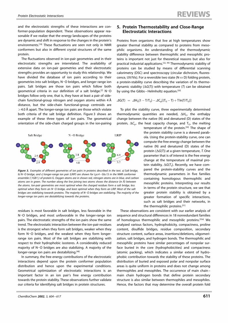
electrostatic strengths are interrelated. The availability ofextensive data on ion-pair geometries and their electrostaticstrengths provides an opportunity to study this relationship. Wehave divided the database of ion pairs according to theirgeometries into salt bridges, N�O bridges, and longer range ionpairs. Salt bridges are those ion pairs which follow bothgeometrical criteria in our definition of a salt bridge.[2] N�Obridges follow only one, that is, they have at least a pair of side-chain functional-group nitrogen and oxygen atoms within 4 ädistance, but the side-chain functional-group centroids are�4.0 ä apart. The longer-range ion pairs are those which violateboth criteria of the salt bridge definition. Figure 3 shows anexample of these three types of ion pairs. The geometricalorientation of the side-chain charged groups in the ion-pairing
residues is most favorable in salt bridges, less favorable in theN�O bridges, and most unfavorable in the longer-range ionpairs. The electrostatic strengths of the ion pairs show the sametrend. The electrostatic interaction between the ion-pair residuesis the strongest when they form salt bridges, weaker when theyform N�O bridges, and the weakest when they form longer-range ion pairs. Most of the salt bridges are stabilizing withrespect to their hydrophobic isosteres. A considerably reducedmajority of N�O bridges are also stabilizing. A majority of thelonger-range ion pairs are destabilizing.[48]
In summary, the free energy contributions of the electrostaticinteractions depend upon the protein conformer populationdistribution and hence upon the experimental conditions.Geometrical optimization of electrostatic interactions is animportant factor in an ion pair's free energy contributiontowards the protein stability. These observations further validateour criteria for identifying salt bridges in protein structures.
5. Protein Thermostability and Close-RangeElectrostatic Interactions
Proteins from organisms that live at high temperatures showgreater thermal stability as compared to proteins from meso-philic organisms. An understanding of the thermodynamicstability difference between thermophilic and mesophilic pro-teins is important not just for theoretical reasons but also forpractical industrial applications.[32, 49] Thermodynamic stability ofproteins can be studied by means of differential scanningcalorimetry (DSC) and spectroscopy (circular dichroism, fluores-cence, UV/Vis). For a reversible two state (N�D) folding protein,a protein-stability curve describing the variation of its thermo-dynamic stability (�G(T)) with temperature (T) can be obtainedby using the Gibbs ±Helmholtz equation.[50]
�G(T) � �HG(1�T/TG)��Cp[(TG�T)�T ln(T/TG)]
To plot the stability curve, three experimentally determinedthermodynamic quantities are needed, �HG, the enthalpychange between the native (N) and denatured (D) states of theprotein, �Cp, the heat capacity change, and TG, the melting
temperature of the protein.[50] The shape ofthe protein stability curve is a skewed parab-ola. Using the protein-stability curve, one cancompute the free energy change between thenative (N) and denatured (D) states of theprotein (�G(T)) at a given temperature, T. Oneparameter that is of interest is the free energychange at the temperature of maximal pro-tein stability, �G(TS). Recently, we have com-pared the protein-stability curves and thethermodynamic parameters in five familiescontaining homologous thermophilic andmesophilic proteins. Interpreting our resultsin terms of the protein structure, we see thatgreater protein stability is obtained by agreater formation of specific interactions,such as salt bridges and their networks, inthe thermophilic proteins.[51]
These observations are consistent with our earlier analysis ofsequence and structural differences in 18 nonredundant familiesof homologous thermophilic and mesophilic proteins.[32a] Weanalyzed various factors, hydrophobicity, compactness, prolinecontent, disulfide bridges, residue composition, secondarystructure content, surface areas, insertions/deletions, oligomeri-zation, salt bridges, and hydrogen bonds. The thermophilic andmesophilic proteins have similar percentages of nonpolar sur-face buried in the core (hydrophobicities) and compactness(atomic packing), which indicates a similar extent of hydro-phobic contribution towards the stability of these proteins. Thedistribution of buried and exposed polar and nonpolar surfaceareas is quite uniform in proteins and does not change amongthermophiles and mesophiles. The occurrence of main chain ±main chain hydrogen bonds that define protein secondarystructure is also similar between thermophiles and mesophiles.Hence, the factors that may determine the overall protein fold
Figure 3. Examples of different geometries of ion pairs in proteins described in the text. a) Salt bridge,b) N�O bridge, and c) longer-range ion pair (LRIP) are shown for Lys4 ±Glu15 in the NMR conformerensemble (�1GB1) of protein G. Oxygen atoms are in shown red, nitrogen atoms are in blue, and carbonatoms are in green. The number along the line joining two atoms shows the distance (in ä) betweenthe atoms. Ion-pair geometries are most optimal when the charged residues form a salt bridge, lessoptimal when they form an N�O bridge, and least optimal when they form an LRIP. Most of the saltbridges are stabilizing towards proteins. The majority of N�O bridges are stabilizing. The majority of thelonger-range ion pairs are destabilizing towards the proteins.
R. Nussinov and S. Kumar
612 ChemBioChem 2002, 3, 604 ±617
have similar values for thermophilic and mesophilic proteins.Proline content, insertions/deletions, and oligomerization do notshow consistent trends between thermophiles and mesophiles.On the other hand, the amino acid distributions in thermophilesand mesophiles are significantly different, despite the highsequence identities among thermophiles and mesophiles in ourdatabase.[32a] Thermophilic proteins appear to favor residueswith larger side chains and to avoid thermolabile residues. Anincrease in electrostatic interactions (salt bridges and sidechain ± side chain hydrogen bonds) in thermophiles as com-pared to their homologous mesophiles is the most consistenttrend. These sequence and structural features may simulta-neously raise �HG and lower �Cp of a thermophilic protein ascompared to its mesophilic homologue, thereby resulting in thegreater thermodynamic stability of the thermophilic protein.We have compared the electrostatic strengths of salt bridges
in glutamate dehydrogenase from a hyperthermophile (Pyro-coccus furiosus) and mesophile (Clostridium symbiosum).[32b]
Pyrococcus furiosus glutamate dehydrogenase (PfGDH) is ex-tremely thermostable, with its melting temperature being113 �C. The mesophilic Clostridium symbiosum glutamate dehy-drogenase (CsGDH) shares 34% sequence identity with PfGDHand the monomers of the two proteins superimpose with a rootmean square deviation (RMSD) of 1.38 ä. In both organisms, thebiochemically active GDH is a homohexamer. However, CsGDHhas a half life of only 20 minutes at 52 �C and its meltingtemperature is 55 �C. Previously[32a] we found an increase in salt-bridge formation in PfGDH. Continuum electrostatic calculationsperformed on monomers of PfGDH and CsGDH show that thesalt bridges in PfGDH are highly stabilizing. The salt bridges inCsGDH are marginally stabilizing. Salt bridges in PfGDH formextensive salt-bridge networks. Due to this, the interactions ofcharged side chains in the salt-bridge-forming residues with therest of protein are almost as significant as the interaction ofthese side chains with each other. Hence, the cooperative natureof electrostatic interactions may lead to increased stability ofPfGDH.Analyses based on protein sequences from complete ge-
nomes of thermophilic and hyperthermophilic organisms andcomparisons of sequence/structural properties of homologousthermophilic and mesophilic proteins have also consistentlyindicated a significant increase in the proportion of chargedresidues for the thermophiles.[52] The improvement in electro-statics for thermophiles is reflected in alleviation of electrostaticrepulsions, increased occurrence of ion pairs and their networks,and geometrical optimization of charged residue positions toyield a favorable energetic contribution towards protein stabil-ity.[53] Different protein families may optimize these factorsdifferently. In the following section we present a detailed casestudy of the molecular basis of greater thermostability ofThermus thermophilus ribonuclease H.
6. Molecular Analysis of Thermostability ofThermus thermophilus Ribonuclease H
Ribonuclease H is a single domain protein. Its function is tocleave DNA±RNA hybrids. T. thermophilus ribonuclease H
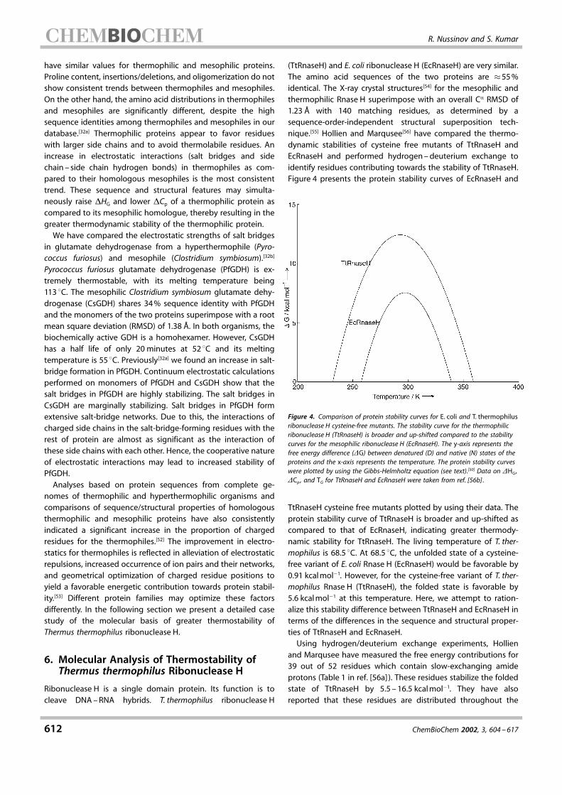
(TtRnaseH) and E. coli ribonuclease H (EcRnaseH) are very similar.The amino acid sequences of the two proteins are �55%identical. The X-ray crystal structures[54] for the mesophilic andthermophilic Rnase H superimpose with an overall C� RMSD of1.23 ä with 140 matching residues, as determined by asequence-order-independent structural superposition tech-nique.[55] Hollien and Marqusee[56] have compared the thermo-dynamic stabilities of cysteine free mutants of TtRnaseH andEcRnaseH and performed hydrogen±deuterium exchange toidentify residues contributing towards the stability of TtRnaseH.Figure 4 presents the protein stability curves of EcRnaseH and
Figure 4. Comparison of protein stability curves for E. coli and T. thermophilusribonuclease H cysteine-free mutants. The stability curve for the thermophilicribonuclease H (TtRnaseH) is broader and up-shifted compared to the stabilitycurves for the mesophilic ribonuclease H (EcRnaseH). The y-axis represents thefree energy difference (�G) between denatured (D) and native (N) states of theproteins and the x-axis represents the temperature. The protein stability curveswere plotted by using the Gibbs-Helmholtz equation (see text).[50] Data on �HG,�Cp , and TG for TtRnaseH and EcRnaseH were taken from ref. [56b] .
TtRnaseH cysteine free mutants plotted by using their data. Theprotein stability curve of TtRnaseH is broader and up-shifted ascompared to that of EcRnaseH, indicating greater thermody-namic stability for TtRnaseH. The living temperature of T. ther-mophilus is 68.5 �C. At 68.5 �C, the unfolded state of a cysteine-free variant of E. coli Rnase H (EcRnaseH) would be favorable by0.91 kcalmol�1. However, for the cysteine-free variant of T. ther-mophilus Rnase H (TtRnaseH), the folded state is favorable by5.6 kcalmol�1 at this temperature. Here, we attempt to ration-alize this stability difference between TtRnaseH and EcRnaseH interms of the differences in the sequence and structural proper-ties of TtRnaseH and EcRnaseH.Using hydrogen/deuterium exchange experiments, Hollien
and Marqusee have measured the free energy contributions for39 out of 52 residues which contain slow-exchanging amideprotons (Table 1 in ref. [56a]). These residues stabilize the foldedstate of TtRnaseH by 5.5 ± 16.5 kcalmol�1. They have alsoreported that these residues are distributed throughout the
Protein Electrostatic Interactions
ChemBioChem 2002, 3, 604 ± 617 613
protein structure and that thermostability is achieved in adelocalized manner. The sequence alignment of EcRnaseH andTtRnaseH shows that 19 out of the 39 residues are conservedbetween the two proteins, while the remaining 20 residues aremutated. Ishikawa et al.[54b] have suggested that replacement ofLys95 in EcRnaseH by Gly100 in TtRnaseH contributes to proteinthermostability by relieving steric hindrance. Lys95 in EcRnaseHis in a left-handed helical (�L) conformation. The formation of anintramolecular disulfide bond also contributes to the stability ofTtRnaseH.[57]
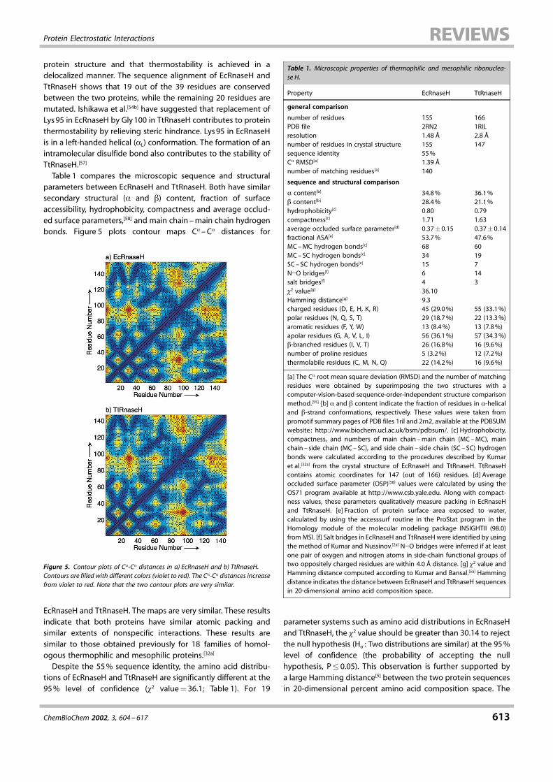
Table 1 compares the microscopic sequence and structuralparameters between EcRnaseH and TtRnaseH. Both have similarsecondary structural (� and �) content, fraction of surfaceaccessibility, hydrophobicity, compactness and average occlud-ed surface parameters,[58] and main chain ±main chain hydrogenbonds. Figure 5 plots contour maps C� ±C� distances for
Figure 5. Contour plots of C�-C� distances in a) EcRnaseH and b) TtRnaseH.Contours are filled with different colors (violet to red). The C�-C� distances increasefrom violet to red. Note that the two contour plots are very similar.
EcRnaseH and TtRnaseH. The maps are very similar. These resultsindicate that both proteins have similar atomic packing andsimilar extents of nonspecific interactions. These results aresimilar to those obtained previously for 18 families of homol-ogous thermophilic and mesophilic proteins.[32a]
Despite the 55% sequence identity, the amino acid distribu-tions of EcRnaseH and TtRnaseH are significantly different at the95% level of confidence (�2 value�36.1; Table 1). For 19
parameter systems such as amino acid distributions in EcRnaseHand TtRnaseH, the �2 value should be greater than 30.14 to rejectthe null hypothesis (Ho : Two distributions are similar) at the 95%level of confidence (the probability of accepting the nullhypothesis, P� 0.05). This observation is further supported bya large Hamming distance[5] between the two protein sequencesin 20-dimensional percent amino acid composition space. The
Table 1. Microscopic properties of thermophilic and mesophilic ribonuclea-se H.
Property EcRnaseH TtRnaseH
general comparison
number of residues 155 166PDB file 2RN2 1RILresolution 1.48 ä 2.8 änumber of residues in crystal structure 155 147sequence identity 55%C� RMSD[a] 1.39 änumber of matching residues[a] 140
sequence and structural comparison
� content[b] 34.8% 36.1%� content[b] 28.4% 21.1%hydrophobicity[c] 0.80 0.79compactness[c] 1.71 1.63average occluded surface parameter[d] 0.37� 0.15 0.37�0.14fractional ASA[e] 53.7% 47.6%MC±MC hydrogen bonds[c] 68 60MC±SC hydrogen bonds[c] 34 19SC± SC hydrogen bonds[c] 15 7N�O bridges[f] 6 14salt bridges[f] 4 3�2 value[g] 36.10Hamming distance[g] 9.3charged residues (D, E, H, K, R) 45 (29.0%) 55 (33.1%)polar residues (N, Q, S, T) 29 (18.7%) 22 (13.3%)aromatic residues (F, Y, W) 13 (8.4%) 13 (7.8%)apolar residues (G, A, V, L, I) 56 (36.1%) 57 (34.3%)�-branched residues (I, V, T) 26 (16.8%) 16 (9.6%)number of proline residues 5 (3.2%) 12 (7.2%)thermolabile residues (C, M, N, Q) 22 (14.2%) 16 (9.6%)
[a] The C� root mean square deviation (RMSD) and the number of matchingresidues were obtained by superimposing the two structures with acomputer-vision-based sequence-order-independent structure comparisonmethod.[55] [b] � and � content indicate the fraction of residues in �-helicaland �-strand conformations, respectively. These values were taken frompromotif summary pages of PDB files 1ril and 2rn2, available at the PDBSUMwebsite: http://www.biochem.ucl.ac.uk/bsm/pdbsum/. [c] Hydrophobicity,compactness, and numbers of main chain ±main chain (MC±MC), mainchain ± side chain (MC±SC), and side chain ± side chain (SC ± SC) hydrogenbonds were calculated according to the procedures described by Kumaret al.[32a] from the crystal structure of EcRnaseH and TtRnaseH. TtRnaseHcontains atomic coordinates for 147 (out of 166) residues. [d] Averageoccluded surface parameter (OSP)[58] values were calculated by using theOS71 program available at http://www.csb.yale.edu. Along with compact-ness values, these parameters qualitatively measure packing in EcRnaseHand TtRnaseH. [e] Fraction of protein surface area exposed to water,calculated by using the accesssurf routine in the ProStat program in theHomology module of the molecular modeling package INSIGHTII (98.0)fromMSI. [f] Salt bridges in EcRnaseH and TtRnaseH were identified by usingthe method of Kumar and Nussinov.[2a] N�O bridges were inferred if at leastone pair of oxygen and nitrogen atoms in side-chain functional groups oftwo oppositely charged residues are within 4.0 ä distance. [g] �2 value andHamming distance computed according to Kumar and Bansal.[5a] Hammingdistance indicates the distance between EcRnaseH and TtRnaseH sequencesin 20-dimensional amino acid composition space.

R. Nussinov and S. Kumar
614 ChemBioChem 2002, 3, 604 ±617
proportion of the charged residues (Asp, Glu, His, Lys, and Arg)increases in TtRnaseH (33.1%) as compared to the proportion ofthese residues in EcRnaseH (29%) by 4.1%. The proportion ofpolar uncharged residues (Asn, Gln, Ser, and Thr) decreases inTtRnaseH as does the proportion of the thermolabile residues(Cys, Met, Asn, and Gln). The apolar residues (Gly, Ala, Val, Leu,and Ile) occur with similar proportions in TtRnaseH (34.3%) andEcRnaseH (36.1%). The proportion of proline residues increasesin TtRnaseH. In general, it appears that TtRnaseH favors residueswith longer side chains, such as Glu, Leu, and Arg.Consistent with the increase in the occurrence of charged
residues is the formation of a larger number of close-rangeelectrostatic interactions. The crystal structure of TtRnaseH (PDBentry 1RIL, 2.8 ä resolution with coordinates for 147 out of 166residues[54b])has 17 ion pairs. Fourteen of these are N�O bridgesand the remaining three are salt bridges. It also contains two ion-pair networks, a hexad (six-residue network) and a tetrad (four-residue network). The crystal structure of EcRnaseH (PDB entry2RN2, 1.48 ä resolution with coordinates for all 155 residues[54a] )has 10 ion pairs (6N�O bridges and 4 salt bridges) and two ion-pair networks, a pentad and a triad. Figure 6 highlights thelocation of charged residues in TtRnaseH and EcRnaseH.The sequence alignment of EcRnaseH and TtRnaseH[54b]
indicates eight positions where apolar residues have beenreplaced by charged residues. These substitutions are L2R, M50K,V54E, I66D, V101R, L136E, A139R, and L146K. We have computedthe electrostatic free energy contribution (��Gelec) for six ofthese eight charged residues towards the stability of TtRnaseHby using continuum electrostatic calculations based on themethod described by Tidor and co-workers.[53a] In this procedure,��Gelec for a charged residue consists of two terms, ��Gdslv and��Gint . ��Gdslv is the desolvation energy penalty paid by thecharged residue and ��Gint is the free energy of the electrostaticinteraction between the charged residue and all the charges inthe rest of the proteins. Hence,
��Gelec � ��Gdslv���Gint
The remaining two substitutions L2R and L146K fall at the Nand C termini of TtRnaseH. Since the atomic coordinates for theadjoining residues are missing in the crystal structure ofTtRnaseH, we did not calculate the ��Gelec for these substitu-tions.Five out of the six charged residue substitutions in TtRnaseH
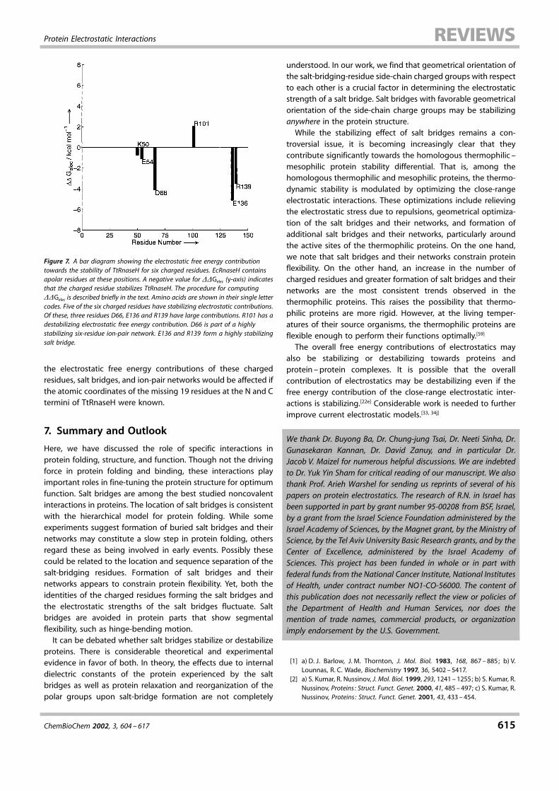
have stabilizing electrostatic free energy contributions withrespect to their hydrophobic isosteres (Figure 7). Out of the five,three residues, D66 in strand D and E136 and R139 in helix �V,appear to have large electrostatic stabilization. D66 is also part ofa six-residue ion-pair network (hexad; formed by residues R2, R4,E64, D66, R115, and R117) in TtRnaseH. Glu136 and Arg139 forma salt bridge. This salt bridge is stabilizing towards TtRnaseH by�5.6 kcalmol�1. Of the remaining two residues with stabilizing��Gelec values, K50 is part of a four-residue ion-pair network(tetrad formed by E39, R46, K50, and D102) and E54 forms a saltbridge with K57 in TtRnaseH. The salt bridge E54 ±K57 stabilizesTtRnaseH by �1.1 Kcalmol�1. Both ion-pair networks, the hexadand the tetrad, have stabilizing electrostatic contributions. The
Figure 6. Ribbon diagrams showing the distribution of charged residues ina) T. thermophilus ribonuclease H (TtRnaseH) and b) E. coli ribonuclease H (EcR-naseH) crystal structures (PDB codes: 1RIL and 2RN2, respectively). All chargedresidues are shown in ball-and-stick representation. The positively chargedresidues are shown in red and the negatively charged residues are shown in blue.The ribbon for all other residues is shown in green.
electrostatic free energy contribution by the hexad (R2, R4, E64,D66, R115, and R117) in TtRnaseH is �7.8 kcalmol�1 and that forthe tetrad (E39, R46, K50, and D102) is �2.0 kcalmol�1. Hydro-gen-exchange experiments by Hollien and Marqusee[56a] alsoindicate stabilizing roles for four of these residues. Arg101 hasdestabilizing electrostatic free energy contribution (��Gelec��2.04 kcalmol�1) towards TtRnaseH. Arg101 lies at the Nterminus of helix �IV [54b] and does not form a salt bridge or ionpair. Arginine residues are avoided at positions near the �-helix Nterminus.[5a] Hence, a mutation of R101 may further enhance thestability of TtRnaseH.The electrostatic free energy values reported here for single
charged residues, salt bridges, and ion-pair networks correspondto room temperature, pH 7.0, and zero ionic strength. At theliving temperature of T. thermophilus, these values are expectedto further decrease by approximately 1 kcalmol�1 due to thereduced hydration free energy changes for the residues, reduceddielectric constant for water, and reduced solvent screening ofthe electrostatic interactions.[32b, 35c] This indicates that ��Gelecvalues would be less destabilizing for R101 and more stabilizingfor K50, E54, D66, E136, and R139 in TtRnaseH. It is not clear how
Protein Electrostatic Interactions
ChemBioChem 2002, 3, 604 ± 617 615
Figure 7. A bar diagram showing the electrostatic free energy contributiontowards the stability of TtRnaseH for six charged residues. EcRnaseH containsapolar residues at these positions. A negative value for ��Gelec (y-axis) indicatesthat the charged residue stabilizes TtRnaseH. The procedure for computing��Gelec is described briefly in the text. Amino acids are shown in their single lettercodes. Five of the six charged residues have stabilizing electrostatic contributions.Of these, three residues D66, E136 and R139 have large contributions. R101 has adestabilizing electrostatic free energy contribution. D66 is part of a highlystabilizing six-residue ion-pair network. E136 and R139 form a highly stabilizingsalt bridge.
the electrostatic free energy contributions of these chargedresidues, salt bridges, and ion-pair networks would be affected ifthe atomic coordinates of the missing 19 residues at the N and Ctermini of TtRnaseH were known.
7. Summary and Outlook
Here, we have discussed the role of specific interactions inprotein folding, structure, and function. Though not the drivingforce in protein folding and binding, these interactions playimportant roles in fine-tuning the protein structure for optimumfunction. Salt bridges are among the best studied noncovalentinteractions in proteins. The location of salt bridges is consistentwith the hierarchical model for protein folding. While someexperiments suggest formation of buried salt bridges and theirnetworks may constitute a slow step in protein folding, othersregard these as being involved in early events. Possibly thesecould be related to the location and sequence separation of thesalt-bridging residues. Formation of salt bridges and theirnetworks appears to constrain protein flexibility. Yet, both theidentities of the charged residues forming the salt bridges andthe electrostatic strengths of the salt bridges fluctuate. Saltbridges are avoided in protein parts that show segmentalflexibility, such as hinge-bending motion.It can be debated whether salt bridges stabilize or destabilize
proteins. There is considerable theoretical and experimentalevidence in favor of both. In theory, the effects due to internaldielectric constants of the protein experienced by the saltbridges as well as protein relaxation and reorganization of thepolar groups upon salt-bridge formation are not completely
understood. In our work, we find that geometrical orientation ofthe salt-bridging-residue side-chain charged groups with respectto each other is a crucial factor in determining the electrostaticstrength of a salt bridge. Salt bridges with favorable geometricalorientation of the side-chain charge groups may be stabilizinganywhere in the protein structure.While the stabilizing effect of salt bridges remains a con-
troversial issue, it is becoming increasingly clear that theycontribute significantly towards the homologous thermophilic ±mesophilic protein stability differential. That is, among thehomologous thermophilic and mesophilic proteins, the thermo-dynamic stability is modulated by optimizing the close-rangeelectrostatic interactions. These optimizations include relievingthe electrostatic stress due to repulsions, geometrical optimiza-tion of the salt bridges and their networks, and formation ofadditional salt bridges and their networks, particularly aroundthe active sites of the thermophilic proteins. On the one hand,we note that salt bridges and their networks constrain proteinflexibility. On the other hand, an increase in the number ofcharged residues and greater formation of salt bridges and theirnetworks are the most consistent trends observed in thethermophilic proteins. This raises the possibility that thermo-philic proteins are more rigid. However, at the living temper-atures of their source organisms, the thermophilic proteins areflexible enough to perform their functions optimally.[59]
The overall free energy contributions of electrostatics mayalso be stabilizing or destabilizing towards proteins andprotein ±protein complexes. It is possible that the overallcontribution of electrostatics may be destabilizing even if thefree energy contribution of the close-range electrostatic inter-actions is stabilizing.[22e] Considerable work is needed to furtherimprove current electrostatic models.[33, 34j]
We thank Dr. Buyong Ba, Dr. Chung-jung Tsai, Dr. Neeti Sinha, Dr.Gunasekaran Kannan, Dr. David Zanuy, and in particular Dr.Jacob V. Maizel for numerous helpful discussions. We are indebtedto Dr. Yuk Yin Sham for critical reading of our manuscript. We alsothank Prof. Arieh Warshel for sending us reprints of several of hispapers on protein electrostatics. The research of R.N. in Israel hasbeen supported in part by grant number 95-00208 from BSF, Israel,by a grant from the Israel Science Foundation administered by theIsrael Academy of Sciences, by the Magnet grant, by the Ministry ofScience, by the Tel Aviv University Basic Research grants, and by theCenter of Excellence, administered by the Israel Academy ofSciences. This project has been funded in whole or in part withfederal funds from the National Cancer Institute, National Institutesof Health, under contract number NO1-CO-56000. The content ofthis publication does not necessarily reflect the view or policies ofthe Department of Health and Human Services, nor does themention of trade names, commercial products, or organizationimply endorsement by the U.S. Government.
[1] a) D. J. Barlow, J. M. Thornton, J. Mol. Biol. 1983, 168, 867 ±885; b) V.Lounnas, R. C. Wade, Biochemistry 1997, 36, 5402 ± 5417.
[2] a) S. Kumar, R. Nussinov, J. Mol. Biol. 1999, 293, 1241 ± 1255; b) S. Kumar, R.Nussinov, Proteins : Struct. Funct. Genet. 2000, 41, 485 ± 497; c) S. Kumar, R.Nussinov, Proteins: Struct. Funct. Genet. 2001, 43, 433 ±454.
R. Nussinov and S. Kumar
616 ChemBioChem 2002, 3, 604 ±617
[3] a) E. A. Permyakov, L. J. Berliner, FEBS Lett. 2000, 473, 269 ±274; b) P. E.Wright, H. J. Dyson, J. Mol. Biol. 1999, 293, 321 ±331; c) Y. Y. Sham, B. Ma,C. J. Tsai, R. Nussinov, Protein Sci. 2001, 10, 135 ± 148.
[4] a) R. L. Baldwin, G. D. Rose, Trends Biochem. Sci. 1999, 24, 26 ± 33; b) R. L.Baldwin, G. D. Rose, Trends Biochem. Sci. 1999, 24, 77 ± 84.
[5] a) S. Kumar, M. Bansal, Proteins: Struct. Funct. Genet. 1998, 31, 460 ± 76;b) ™Geometry and sequence correlation studies on �-helices in globularproteins∫: S. Kumar, PhD thesis, Indian Institute of Science, Bangalore,India, 1997; c) D. Gandini, L. Gogioso, M. Bolognesi, D. Bordo, Proteins:Struct. Funct. Genet. 1996, 24, 439 ± 449.
[6] a) M. Sundaralingam, Y. C. Sekharudu, N. Yathindra, V. Ravichandran,Proteins : Struct. Funct. Genet. 1987, 2, 64 ± 71; b) D. Walther, P. Argos,Protein Eng. 1996, 9, 471 ± 478.
[7] a) P. C. Lyu, P. J. Gans, N. R. Kallenbach, J. Mol. Biol. 1992, 223, 343 ±350;b) M. J. Bodkin, J. M. Goodfellow, Protein Sci. 1995, 4, 603 ± 612; c) S.Marqusee, R. L. Baldwin, Proc. Natl. Acad. Sci. USA 1987, 84, 8898 ± 8902;d) C. A. Olson, E. J. Spek, Z. Shi, A. Vologodskii, N. R. Kallenbach, Proteins :Struct. Funct. Genet. 2001, 44, 123 ± 132.
[8] a) C. A. Blaise, J. M. Berg, Biochemistry 1997, 36, 6218 ± 6222; b) J. S.Merkel, J. M. Sturtevant, L. Regan, Structure 1999, 7, 1333 ± 1343.
[9] O. Schueler, H. Margalit, J. Mol. Biol. 1995, 248, 125 ± 135.[10] a) M. T. Oliva, J. Moult, Protein Eng. 1999, 12, 727 ± 735; b) V. Z. Spassov,
B. P. Atanasov, Proteins : Struct. Funct. Genet. 1994, 19, 222 ± 229.[11] B. Musafia, V. Buchner, D. Arad, J. Mol. Biol. 1995, 254, 761 ± 770.[12] a) M. Oliveberg, A. R. Fersht, Biochemistry 1996, 35, 2726 ± 2737; b) M.
Oliveberg, A. R. Fersht, Biochemistry 1996, 35, 6795 ± 6805.[13] C. D. Waldburger, T. Jonsson, R. T. Sauer, Proc. Natl. Acad. Sci. USA 1996, 93,
2629 ± 2634.[14] S. Cavagnero, D. A. Debe, Z. H. Zhou, M. W. W. Adams, S. I. Chan,
Biochemistry 1998, 37, 3369 ± 3376.[15] I. Y. Torshin, R. W. Harrison, Proteins: Struct. Funct. Genet. 2001, 43, 353 ±
364.[16] a) C. J. Tsai, R. Nussinov, Protein Sci. 1997, 6, 24 ± 42; b) C. J. Tsai, R.
Nussinov, Protein Sci. 1997, 6, 1426 ± 1437; c) C. J. Tsai, J. V. Maizel, R.Nussinov, Protein Sci. 1999, 8, 1591 ± 1604; d) C. J. Tsai, J. V. Maizel, R.Nussinov, Proc. Natl. Acad. Sci. USA 2000, 97, 12038 ± 12043; e) S. Kumar,Y. Y. Sham, C. J. Tsai, R. Nussinov, Biophys. J. 2001, 80, 2439 ± 2454; f) C. J.Tsai, B. Ma, S. Kumar, H. Wolfson, R. Nussinov, Crit. Rev. Biochem. Mol. Biol.2001, 36, 399 ± 433.
[17] a) S. Kumar, H. Wolfson, R. Nussinov, IBM J. Res. Dev. 2001, 45(3/4), 513 ±523; b) N. Sinha, S. Kumar, R. Nussinov, Structure 2001, 9, 1165 ± 1181.
[18] a) D. Xu, C. J. Tsai, R. Nussinov, Folding Des. 1998, 3, R71-R80; b) C. J. Tsai, S.Kumar, B. Ma, R. Nussinov, Protein Sci. 1999, 8, 1181 ± 1190; c) B. Ma, S.Kumar, C. J. Tsai, R. Nussinov, Protein Eng. 1999, 12, 713 ± 720; d) S. Kumar,B. Ma, C. J. Tsai, H. Wolfson, R. Nussinov, Cell Biochem. Biophys. 1999, 31,141 ± 164; e) C. J. Tsai, B. Ma, R. Nussinov, Proc. Natl. Acad. Sci. USA 1999,96, 9970 ± 9972; f) S. Kumar, B. Ma, C. J. Tsai, N. Sinha, R. Nussinov, ProteinSci. 2000, 9, 10 ± 19; g) B. Ma, S. Kumar, C. J. Tsai, Z. Hu, R. Nussinov, J.Theor. Biol. 2000, 203, 383 ± 387; h) N. Sinha, R. Nussinov, Proc. Natl. Acad.Sci. USA 2001, 98, 3139 ±3144.
[19] a) C. J. Tsai, S. L. Lin, H. J. Wolfson, R. Nussinov, Protein Sci. 1997, 6, 53 ± 64;b) C. J. Tsai, S. L. Lin, H. J. Wolfson, R. Nussinov, Crit. Rev. Biochem. Mol. Biol.1996, 31, 127 ± 152; c) C. J. Tsai, S. L. Lin, H. J. Wolfson, R. Nussinov, J. Mol.Biol. 1996, 260, 604 ± 620.
[20] a) D. Xu, C. J. Tsai, R. Nussinov, Protein Eng. 1997, 10, 999 ± 1012; b) D. Xu,S. L. Lin, R. Nussinov, J. Mol. Biol. 1997, 265, 68 ± 84.
[21] a) T. Clackson, J. A. Wells, Science 1995, 267, 383 ±386; b) A. A. Bogan, K. S.Thorn, J. Mol. Biol. 1998, 280, 1 ± 9; c) Z. Hu, B. Ma, H. J. Wolfson, R.Nussinov, Proteins : Struct. Funct. Genet. 2000, 39, 331 ± 342; d) B. Ma, H. J.Wolfson, R. Nussinov, Curr. Opin. Struct. Biol. 2001, 11, 364 ± 369.
[22] a) Z. S. Hendsch, B. Tidor, Protein Sci. 1999, 8, 1381 ± 1392; b) L.-P. Lee, B.Tidor, Protein Sci. 2001, 10, 362 ± 377; c) A. J. McCoy, V. C. Epa, P. M.Colman, J. Mol. Biol. 1997, 268, 570 ±584; d) M. Vijaykumar, K.-Y. Wong, G.Schreiber, A. R. Fersht, A. Szabo, H.-X. Zhou, J. Mol. Biol. 1998, 278, 1015 ±1024; e) F. B. Sheinerman, R. Norel, B. Honig, Curr. Opin. Struct. Biol. 2000,10, 153 ± 159.
[23] a) J. G. Mandell, V. A. Roberts, M. E. Pique, V. Kotlovyi, J. C. Mitchell, E.Nelson, I. Tsigelny, L. F. Ten Eyck, Protein Eng. 2001, 14, 105 ±113; b) R.Norel, F. Sheinerman, D. Petrey, B. Honig, Protein Sci. 2001, 10, 2147 ±2161.
[24] T. Konno, Biochemistry 2001, 40, 2148 ± 2154.
[25] C. J. Tsai, B. Ma, Y. Y. Sham, S. Kumar, R. Nussinov, Proteins: Struct. Funct.Genet. 2001, 44, 418 ± 427.
[26] D. Sahal, P. Balaram, Biochemistry 1986, 25, 6004 ± 6013.[27] a) E. M. Storch, V. Daggett, Biochemistry 1995, 34, 9682 ± 9693; b) E. M.
Storch, V. Daggett, Biochemistry 1996, 35, 11596 ± 11604; c) E. M. Storch,V. Daggett, W. M. Atkins, Biochemistry 1999, 38, 5054 ± 5064; d) E. M.Storch, J. S. Grinstead, A. P. Campbell, V. Daggett, W. M. Atkins, Biochem-istry 1999, 38, 5065 ± 5075.
[28] a) E. Fischer, Ber. Dtsch. Chem. Ges. 1894, 27, 2985 ± 2991; b) D. E.Koshland, Jr. , Proc. Natl. Acad. Sci. USA 1958, 44, 98 ± 123.
[29] a) M. Shiroishi, A. Yokata, K. Tsumoto, H. Kondo, Y. Nishimiya, K. Horii, M.Matsushima, K. Ogasahara, K. Yutani, I. Kumagai, J. Biol. Chem. 2001, 276,23042 ± 23050; b) S. J. Smith-Gill, T. B. Lavoie, C. R. Mainhart, J. Immunol.1984, 133, 384 ± 393; c) L. N. W. Kam-Morgan, S. J. Smith-Gill, M. G. Taylor,L. Zhang, A. C. Wilson, J. F. Kirsch, Proc. Natl. Acad. Sci. USA 1992, 90,3958 ± 3962; d) E. A. Padlan, E. W. Silverton, S. Sheriff, G. H. Cohen, S. J.Smith-Gill, D. R. Davies, Proc. Natl. Acad. Sci. USA 1989, 86, 5938 ± 5942.
[30] S. Herrgard, C. J. Gibas, S. Subramaniam, Biochemistry 2000, 39, 2921 ±2930.
[31] E. J. Sundberg, R. A. Mariuzza, Structure 2000, 8, R137-R142.[32] a) S. Kumar, C. J. Tsai, R. Nussinov, Protein Eng. 2000, 13, 3, 179 ± 191; b) S.
Kumar, B. Ma, C. J. Tsai, R. Nussinov, Proteins: Struct. Funct. Genet. 2000, 38,4, 368 ± 383; c) S. Kumar, R. Nussinov, Cell. Mol. Life Sci. 2001, 58, 9, 1216 ±1233; d) S. Kumar, R. Nussinov, unpublished results.
[33] a) A. Warshel, A. Papazyan, Curr. Opin. Struct. Biol. 1998, 8, 211 ± 217;b) D. J. Tobias, Curr. Opin. Struct. Biol. 2001, 11, 253 ± 261; c) T. Simonson,Curr. Opin. Struct. Biol. 2001, 11, 243 ± 252; d) Y. Y. Sham, I. Muegge, A.Warshel, Biophys. J. 1998, 74, 1744 ±1753.
[34] a) A. Warshel, J. Aqvist, Chem. Scr. 1989, 29A, 75 ± 83; b) A. Warshel, J.Aqvist, Annu. Rev. Biophys. Biophys. Chem. 1991, 20, 267 ± 298; c) J. D.Madura, Y. Nakajima, R. M. Hamilton, A. Wierzbicki, A. Warshel, Struct.Chem. 1996, 7, 131 ± 138; d) F. S. Lee, Z.-T. Chu, M. B. Bolger, A. Warshel,Protein Eng. 1992, 5, 215 ± 228; e) Y. Y. Sham, Z. T. Chu, H. Tao, A. Warshel,Proteins : Struct. Funct. Genet. 2000, 39, 393 ± 407; f) F. S. Lee, Z. T. Chu, A.Warshel, J. Comp. Chem. 1993, 14, 161 ± 185; g) I. Muegge, H. Tao, A.Warshel, Protein Eng. 1997, 10, 1363 ± 1372; h) I. Muegge, T. Schweins, A.Warshel, Proteins: Struct. Funct. Genet. 1998, 30, 407 ± 423; i) Z. Z. Fan, J. K.Hwang, A. Warshel, Theor. Chem. Acc. 1999, 103, 77 ± 80; j) C. N. Schutz, A.Warshel, Proteins : Struct. Funct. Genet. 2001, 400 ± 417; k) A. Warshel, S. T.Russell, Q. Rev. Biophys. 1984, 17, 283 ± 421; l) A. Warshel, Biochemistry1981, 20, 3167 ± 3177; m) A. Warshel, S. T. Russell, A. K. Churg, Proc. Natl.Acad. Sci. USA 1984, 81, 4785 ± 4789; n) J. K. Hwang, A. Warshel, Nature1988, 334, 270 ±272.
[35] a) R. Luo, M. S. Head, J. Moult, M. K. Gilson, J. Am. Chem. Soc. 1998, 120,6138 ± 6146; b) J. Antosiewicz, J. A. McCammon, M. K. Gilson, Biochemistry1996, 35, 7819 ± 7833; c) A. H. Elcock, J. A. McCammon, J. Mol. Biol. 1998,280, 731 ± 748; d) P. H. Hunenberger, V. Helms, N. Narayana, S. S. Taylor,J. A. McCammon, Biochemistry 1999, 38, 2358 ± 2366; e) J. Lamotte-Brasseur, A. Dubus, R. C. Wade, Proteins 2000, 40, 23 ± 28; f) N. Froloff, A.Windemuth, B. Honig, Protein Sci. 1997, 6, 1293 ± 1301; g) C. J. Gibas, P.Jambeck, S. Subramaniam, Methods (San Diego) 2000, 20, 292 ±309;h) E. G. Alexov, M. R. Gunner, Biophys. J. 1997, 74, 2075 ± 2093; i) E. G.Alexov, M. R. Gunner, Biochemistry 1999, 38, 8253 ± 8270.
[36] V. V. Loladze, D. N. Ermolenko, G. I. Makhatadze, Protein Sci. 2001, 10,1343 ± 1352.
[37] Z. S. Hendsch, B. Tidor, Protein Sci. 1994, 3, 211 ± 226.[38] a) A. Horovitz, A. R. Fersht, J. Mol. Biol. 1992, 224, 733 ± 740; b) S.
Marqusee, R. T. Sauer, Protein Sci. 1994, 3, 2217 ± 2225; c) K. Pervushin, M.Billeter, G. Siegal, K. W¸thrich, J. Mol. Biol. 1996, 264, 1002 ± 1012; d) L.Xiao, B. Honig, J. Mol. Biol. 1999, 289, 1435 ± 1444; e) S. Dao-Pin, U. Sauer,H. Nicholson, B. W. Matthews, Biochemistry 1991, 30, 7142 ± 7153; f) S.Dao-Pin, D. E. Anderson, W. A. Baase, F. W. Dahlquist, B. W. Matthews,Biochemistry 1991, 30, 11521 ± 11529; g) C. D. Waldburger, J. F. Schild-bach, R. T. Sauer, Nat. Struct. Biol. 1995, 2, 122 ± 128; h) B. H. Honig, W. L.Hubell, Proc. Natl. Acad. Sci. USA 1984, 81, 5412 ± 5416.
[39] a) X. Barril, C. Aleman, M. Orozco, F. J. Luque, Proteins : Struct. Funct. Genet.1998, 32, 67 ± 79; b) U. C. Singh, Proc. Natl. Acad. Sci. USA 1988, 85, 4280 ±4284; c) L. Serrano, A. Horovitz, B. Avron, M. Bycroft, A. R. Fersht,Biochemistry 1990, 29, 9343 ± 9352; d) E. J. Spek, A. H. Bui, M. Lu, N. R.Kallenbach, Protein Sci. 1998, 7, 2431 ± 2437.
[40] B. H. Honig, A. Nicholls, Science 1995, 268, 1144 ± 1149.

Protein Electrostatic Interactions
ChemBioChem 2002, 3, 604 ± 617 617
[41] a) M. K. Gilson, A. Rashin, R. Fine, B. H. Honig, J. Mol. Biol. 1985, 183, 503 ±516; b) M. K. Gilson, K. A. Sharp, B. H. Honig, J. Comput. Chem. 1988, 9,327 ± 335; c) M. K. Gilson, B. H. Honig, Nature 1987, 330, 84 ±86; d) M. K.Gilson, B. H. Honig, Proteins: Struct. Funct. Genet. 1988, 4, 7 ± 18; e) K. A.Sharp, B. H. Honig, Annu. Rev. Biophys. Biophys. Chem. 1990, 19, 301 ±332; f) B. H. Honig, K. A. Sharp, A. Yang, J. Phys. Chem. 1993, 97, 1101 ±1109.
[42] a) D. Sitkoff, K. A. Sharp, B. H. Honig, J. Phys. Chem. 1994, 98, 1978 ± 1988;b) A. Radzicka, R. Wolfenden, Biochemistry 1988, 27, 1664 ± 1670.
[43] I. Klapper, R. Hagstrom, R. Fine, K. A. Sharp, B. H. Honig, Proteins : Struct.Funct. Genet. 1986, 1, 47 ± 59.
[44] J. E. Nielsen, K. V. Anderson, B. Honig, R. W. W. Hooft, G. Klebe, G. Vriend,R. C. Wade, Protein Eng. 1999, 12, 657 ± 662.
[45] a) W. C. Wimley, K. Gawrisch, T. P. Creamer, S. H. White, Proc. Natl. Acad. Sci.USA 1996, 93, 2985 ± 2990; b) S. Miller, J. Janin, A. M. Lesk, C. Chothia, J.Mol. Biol. 1987, 196, 641 ± 656; c) D. Eisenberg, A. D. McLachlan, Nature1986, 319, 199 ±203.
[46] a) T. Kajander, P. C. Kahn, S. H. Passila, D. C. Cohen, L. Lehtio, W. Adolfsen, J.Warwicker, U. Schell, A. Goldman, Structure 2000, 8, 1203 ± 1214; b) A.Warshel, Proc. Natl. Acad. Sci. USA 1978, 75, 5250 ± 5254; c) A. Giletto, C. N.Pace, Biochemistry 1999, 38, 13379 ±13384.
[47] a) F. C. Bernstein, T. F. Koetzle, G. J. Williams, E. E. Shimanouchi, M. Tasumi,J. Mol. Biol. 1977, 112, 535 ± 542; b) H. M. Berman, J. Westbrook, Z. Feng, G.Gilliland, T. N. Bhat, H. Weissig, I. N. Shindyalov, P. E. Bourne, Nucleic AcidsRes. 2000, 28, 235 ± 242.
[48] S. Kumar, R. Nussinov, Biophys. J. 2002, in press.[49] R. Sterner, W. Liebl, Crit. Rev. Biochem. Mol. Biol. 2001, 36, 39 ± 106.
[50] a) W. Becktel, J. A. Schellman, Biopolymers 1987, 26, 1859 ± 1877; b) P. L.Privalov, Crit. Rev. Biochem. Mol. Biol. 1990, 25, 281 ± 305.
[51] S. Kumar, C. J. Tsai, R. Nussinov, Biochemistry 2001, 40, 14152± 14165[52] a) C. Cambillau, J.-M. Claverie, J. Biol. Chem. 2000, 275, 32383 ± 32386;
b) S. Chakravarty, R. Varadarajan, FEBS Lett. 2000, 470, 65 ± 69; c) P. J.Haney, J. H. Badger, G. L. Buldak, C. I. Reich, C. R. Woese, G. J. Olsen, Proc.Natl. Acad. Sci. USA 1996, 3578 ± 3583.
[53] a) S. Spector, M. Wang, S. A. Carp, J. Robblee, Z. S. Hendsch, R. Fairman, B.Tidor, D. P. Raleigh, Biochemistry 2000, 39, 872 ±879; b) G. R. Grimsley, K. L.Shaw, L. R. Fee, R. W. Alston, B. M. Huyghues-Despointes, R. L. Thurlkil,J. M. Scholtz, C. N. Pace, Protein Sci. 1999, 8, 1843 ±1849; c) V. V. Loladze, B.Ibarra-olero, J. M. Sanchez-Ruiz, G. I. Makhatadze, Biochemistry 1999, 38,16419 ± 16423; d) J. M. Sanchez-Ruiz, G. I. I. Makhatadze, Trends Biotech-nol. 2001, 19, 132 ± 135.
[54] a) K. Katayanagi, M. Miyagawa, M. Matsushima, M. Ishikawa, S. Kanaya, M.Ikehara, T. Matsuzaki, K. Morikawa, Nature 1990, 347, 306 ± 309; b) K.Ishikawa, M. Okumura, K. Katayanagi, S. Kimura, S. Kanaya, H. Nakamura,K. Morikawa, J. Mol. Biol. 1993, 230, 529 ± 542.
[55] C. J. Tsai, S. L. Lin, H. Wolfson, R. Nussinov, J. Mol. Biol. 1996, 260, 604 ± 620.[56] a) J. Hollien, S. Marqusee, Proc. Natl. Acad. Sci. USA 1999, 96, 13674 ±
13678; b) J. Hollien, S. Marqusee, Biochemistry 1999, 38, 3831 ± 3836.[57] N. Hirano, M. Haruki, M. Morikawa, S. Kanaya, Biochemistry 1998, 37,
12640 ± 12648.[58] P. J. Fleming, F. M. Richards, J. Mol. Biol. 2000, 299, 487 ± 498.[59] R. Jaenicke, Proc. Natl. Acad. Sci. USA 2000, 97, 2962 ± 2964.
Received: September 7, 2001 [A293]
Copyright © 2022 FDOKUMEN