Mast cells aggravate sepsis by inhibiting peritoneal macrophage phagocytosis
Upload
independentCategory
view
0download
0
Controversies
Section Editor: Ralf Paus, Hamburg
What is the physiological function of mastcells?
M.Maurer, T. Theoharides, R.D.Granstein,
S. C. Bischoff, J. Bienenstock, B. Henz,
P. Kovanen, A. M. Piliponsky, N. Kambe,
H. Vliagoftis, F. Levi-Schaffer, M. Metz,
Y. Miyachi, D. Befus, P. Forsythe,
Y. Kitamura and S. Galli
Maurer M, Theoharides T, Granstein RD, Bischoff SC, Bienenstock J, Henz B,Kovanen P, Piliponsky AM, Kambe N, Vliagoftis H, Levi-Schaffer F, Metz M,Miyachi Y, Befus D, Forsythe P, Kitamura Y, Galli S. What is the physiologicalfunction of mast cells?
Exp Dermatol 2003: 12: 886–910. # Blackwell Munksgaard, 2003
Abstract: Under physiological conditions, skin mast cells preferentially localize aroundnerves, blood vessels and hair follicles. This observation, which dates back to PaulEhrlich, intuitively suggests that these enigmatic, multifacetted protagonists of naturalimmunity are functionally relevant to many more aspects of tissue physiology than justto the generation of inflammatory and vasodilatory responses to IgE-dependentenvironmental antigens. And yet, for decades, mainstream-mast cell research has beendominated by a focus on the – undisputedly prominent and important – mast cellfunctions in type I immune responses and in the pathogenesis and management ofallergic diseases. Certainly, it is hard to believe that the very large and rather selectivelydistributed number of mast cells in normal, uninflamed, non-infected, non-traumatizedmammalian skin or mucosal tissue is simply hanging around there lazily day and night,just to wait for the odd allergen or parasite-associated antigen to come by so the mastcell can finally swing into action. Indeed, the past decade has witnessed a renaissance ofmast cell research ‘beyond allergy’, along with a more systematic exploration of thesurprisingly wide range of physiological functions that mast cells may be involved in.The current debate sketches many of the exciting new horizons that have recently comeinto our vision during this intriguing, ongoing search.
Introduction
Mast cells have various functions. However, not only mast cellsthemselves but also we scientists do not know what is the phy-siological and what is the pathological function. A given mast cellfunction plays a physiological role in some situations, but thesame function may play a pathological role in other situations. Ina sense, the mast cell might be compared with an excellent actor:the actor may beautifully play the role of either a good or evilcharacter.
Probably, the most well-defined function of mast cells is that ofan effector of IgE-dependent immediate hypersensitivity reac-tions. This plays a physiological role in the rejections of a certainspecies of ticks which infect skin (1). However, the samereaction against tick antigens may play a pathological role ininfantile asthma and atopic dermatitis. In most industrial coun-tries, dermal tick infestation is rare in humans, whereas infantileasthma and atopic dermatitis are common. Therefore, the role ofmast cells in the IgE-dependent hypersensitivity reaction againsttick antigens is considered to be pathological by most peopleliving in industrial countries. If dermal infestation by such a
kind of ticks is prevalent, the role of mast cells might beconsidered to be physiological.
Taken together, the specific physiological or pathological rolesof mast cells appear to be influenced and defined by the individualpossessing mast cells and the environment that the individuallives in.
Yukihiko KitamuraDepartment of Pathology
Osaka University Medical SchoolRoom C-2
Yamada-okaSuita
Osaka, 565-0871Japan
E-mail: [email protected]
References
1. Matsuda H et al. J Immunol 1990: 144: 259–262.
Experimental Dermatology 2003: 12: 886–910 Copyright # Blackwell Munksgaard 2003Blackwell Munksgaard . Printed in Denmark
EXPERIMENTAL DERMATOLOGY
ISSN 0906-6705
886

Viewpoint 1
No doubt man, as well as every other animal, presents struc-tures, which seem to our limited knowledge, not to be now of anyservice to him.
Charles Darwin, Descent of man, 1872
Why has evolution chosen to provide us with a cell that appearsnot only to be of no service to us, but which can be most annoy-ing (e.g. itching, wheezing, running nose, . . .) and sometimesharmful (anaphylactic shock) as the key effector cell in allergicreactions? This so-called ‘riddle of the mast cell’ (1) has fascinatedscientists ever since Paul Ehrlich’s first detailed description ofmast cells (MCs) in 1878 (2). Yet it took more than a hundredyears before we could actually be sure that MCs are good forsomething and before we could start to characterize such physio-logical functions of MCs.The problem was not a lack of smart people thinking about the
problem or the absence of valuable hypotheses to be tested. Onthe contrary, many renowned scientists have added their view onwhat MCs are good for to the long list started by Ehrlich himself,who proposed that MCs may modulate the growth of solidtumors, arguing that MCs were particularly abundant in thevicinity of ‘neoplastic foci’ (3). Other interesting hypotheses ori-ginated from the observations that tissueMCs are unique becausethey are fairly long-lived cells capable of undergoing multiplecycles of de- and regranulation, and that MCs are preferentiallylocated at the host–environment interface such as the skin or thegut. Yet other authors have suggested physiological functions ofMCs based on the findings that MCs produce, store, and releasean abundant array of mediators, some of which have nothing todo with the initiation of allergic reactions, and that MCs can beactivated by many various signals independent of immuno-globulin E (IgE) (Table 1).Coming up with theory after theory on the true and original
function of MCs must have been fun and exciting (and prettysafe), yet frustrating, because there was no way of disproving orproving any of them, which was precisely the problem. What MCaficionados lacked for more than one hundred years were notvisionary hypotheses but a model to test them. Such a model,under the best of circumstances, would permit the investigation ofhypothetical MC functions in vivo using animals that differedsolely in lacking, or containing, MC populations, thus facilitatingthe study of a supposedly MC-dependent condition both in theabsence and in the presence of MCs. Ideally, having found thatthe condition in question is impaired in the absence of MCs, sucha model would then allow for attempts to repair this defect byadoptively transferring MCs to ‘MC-empty’ animals.Such a model was reported by Galli, Kitamura, and coworkers
in 1985 (4). Mice with mutations at c-kit, such as KitW/KitW–v
mice, virtually lack tissue MCs but otherwise have largelynormal hematopoiesis, essentially normal B- and T-cell functions,hemostasis, and leukocyte populations (5,6). Also, geneticallyMC-deficient KitW/KitW–v mice can be repaired selectivelyand both systemically or locally of their MC deficiency byreconstitution with MCs obtained from congenic Kitþ/þ mice(5). Today, anyone who is interested in what MCs are goodfor can turn to this model (or MC-deficient rats) and findout. In an extension of the original model described above,we and others have recently shown that MC-deficient micemay also be used to pinpoint the contribution of single MCproducts in MC-mediated reactions (7,8). MC-deficient KitW/KitW–v mice may be reconstituted with MCs derived frommice deficient for a certain MC product, i.e. tumor necrosisfactor-a (TNFa), to then be compared for differences inMC-mediated reactions with KitW/KitW–v mice that have
received adoptive transfer of normal MCs. Because suchmice differ only in having MCs that can, or cannot, produceand release TNFa, any differences in MC-mediated reactionsfound in these mice must be attributed to this MC mediator(Fig. 3).MC-deficient mice have been extensively used to characterize
the role of MCs in numerous pathological conditions, includingvarious models of allergic diseases and other inflammatory dis-orders (9), while much less use has been made of this model toidentify the functions of MCs under physiological conditions.However, some hypotheses of physiological MC functions havebeen put to the test, and MCs are now known to be beneficial totheir host (at least if that host is a mouse) in at least than twosettings: 1) host defense and 2) tissue repair and remodeling.
Mast cells – pivotal players in host defense
MCs meet all basic requirements needed to be important playersin acquired immunity: they are capable of phagocytizing, proces-sing, and presenting antigens to T cells and of modulating T- andB-cell responses such as lymphocyte growth, recruitment, andproduction of Igs (10). Also, MCs have been studied in greatdetail with respect to mechanisms of activation, in particular viaFceRI, the high-affinity receptor for IgE (11). Because IgEproduction is substantially up-regulated during immuneresponses to infections with intestinal parasites, where MCpopulations undergo considerable expansion, the question ofwhether MCs are involved in acquired immunity to parasiteswas one of the first issues investigated inMC-deficient mice. In1985, Nawa and coworkers (12) reported that KitW/KitW–v miceinfected with parasitic nematodes show greater and more persist-ent peak larval counts and slower worm expulsion thannormalþ/þlitter mates. In addition, the reconstitution of con-nective tissue and mucosal MC populations in KitW/KitW–v
mice effectively restored protective antiparasite immuneresponses in these mice (12,13). Using MC-deficient miceand IgE-deficient mice, Watanabe et al. (14) were later able
Table 1. Selected hypotheses of physiological mast cell functions
Hypothetical mast cell function Author Year
Protection from cancer Ehrlich 1877Phagocytosis of pathogens Metchnikoff 1892Endocrine function Cajal 1896Lipid metabolism Ciaccio 1913Vitamin metabolism Tuma 1928Calcium metabolism Pautrier 1931Tissue growth and cell proliferation Sylven 1941Blood clotting and coagulation Baeckeland 1950Hair growth Montagna 1951Hemopoiesis Messerschmitt 1955Local tissue detoxification Higginbotham 1956Regulation of blood pressure Keller 1957pH regulation Caselli 1958Temperature regulation LeBlanc 1959Aging Spicer 1960Response to stress West 1962Fixation of blood-borne particles Selye 1963Sweat secretion Szabo 1964Peripheral ‘memory bank’ Padawer 1978
Controversies
887
to show that both MCs and IgE participate in worm expul-sion from the intestine.
MCs are not only beneficial in acquired immunity to parasitesof the gut but also of the skin: MC-deficient KitW/KitW–v micereportedly show impaired resistance against infestations withlarval Haemaphysalis longicornis ticks, which are associated withincreases in serum IgE levels of more than 100-fold (15). In aseries of elegant experiments, Matsuda et al. (16) were able toadoptively transfer protective acquired immunity to KitW/KitW–v
mice by treatment with immune sera obtained from infected mice,but only when such KitW/KitW–v mice had received intracuta-neous injections of Kitþ/þ mouse-derived MCs prior to infection.Thus, one of the physiological functions of MCs (and IgE) is toprovide resistance and acquired immunity to parasites by mount-ing protective immediate hypersensitivity reactions against para-sitic antigens (10).
If MC-induced inflammation is a good thing when trying to ridthe host of worms in the context of acquired immune responses,why should it not also help in dealing with infections by otherpathogens, like bacteria, which are mainly controlled by innateimmune responses (17)? Recent studies by Malaviya et al. (18)and by Echtenacher et al. (19) indicate that MCs not onlypromote host defense responses to enterobacteria, but are alsonecessary for mice to survive infections by such pathogens. InMC-deficient mice, intraperitoneal reconstitution with MCs sub-stantially reduced death and morbidity from cecal ligation andpuncture (CLP, a model of acute septic peritonitis), and thisprotection by adoptive transfer of MC was wiped out by theapplication of neutralizing anti-TNFa antibodies (19). Incollaboration with Michael Carroll and his colleagues, wehave recently found that mice deficient in complement compo-nents 3 (C3–/– mice) or 4 (C4–/– mice) – compared to wild-typemice – were much more sensitive to the lethal effects of CLP (20).C3–/– mice subjected to CLP also exhibited diminished degranu-lation of peritoneal MC, reduced intraperitoneal TNFa levels,less neutrophil recruitment, and impaired clearance of bacteria.All of these defects, as well as the increased mortality of C3–/–
mice after CLP, were improved upon treatment of C3–/– micewith purified C3 protein (20). In subsequent experiments invol-ving complement receptor-deficient mice (CD21/CD35–/– mice,deficient in complement receptors 1 and 2) and CD19–/–
mice, we were able to show that the engagement of CD21/CD35 along with CD19 on the MC surface by C3 fragmentscontribute to MC activation in the CLP model (21). Interest-ingly, our work done in collaboration with Tanya Mayadas andassociates showed that complement receptor 3-deficient mice(CD11b/CD18–/– mice) also exhibit markedly reduced survivalfollowing CLP. However, this increased susceptibility of CD11b/CD18–/– mice to bacterial infections may be the result of reducednumbers of MCs in these mice (22).
To test whether it could actually be beneficial to have increasednumbers of MCs when dealing with bacterial infections, we haveanalyzed whether repetitive administrations of stem cell factor(SCF), a potent growth factor for MCs (6), could influence thesurvival of mice subjected to CLP (23). We found that suchtreatment not only increased the number of peritoneal MCs inC57BL/6 mice but also significantly improved the ability of thesemice to survive CLP: the more SCF treatment increased MCpopulations, the more it improved survival. Experiments onMC-reconstituted KitW/KitW–v mice confirmed that the pro-tective effect of SCF treatment reflected, at least in part, theactions of SCF on MCs. Moreover, we found that SCF-treated mice did not appear to be at a substantially increasedrisk for death when IgE-dependent systemic anaphylaxis wasinduced by intraperitoneal challenge with specific antigen (23).That MCs are important, indeed essential, to the initiationand orchestration of innate and acquired immune responseswere striking findings: not only did MCs appear to finally begood for something, but in some instances their presence orabsence was a matter of life or death. To no surprise have
these observations sparked interest in investigating the role of MCsin infections by various other pathogens, including viruses (24) andintracellular protozoan parasites such as Toxoplasma gondii andLeishmania spp. (8,25), studies that will surely expand our under-standing of MCs as players in host defense.
Mast cells – key effector cells in tissue repair andremodeling
Turning to KitW/KitW–v mice for answers to whether MCs arerequired under physiological conditions or not for the mainte-nance of tissue homeostasis, revealed no detectable gross deficitsin MC-deficient mice, even at sites which host large MC popula-tions in congenicþ/þmice. Organs that undergo substantial andcontinued structural remodeling, namely hair follicles and bones,may be exceptions to that rule. MC-deficient mice have hair ofnormal length and texture, and hair follicles in these mice appearlargely normal when studied by histomorphometry. However,hair follicle cycling, the continued progression of hair fol-licles through periods of rest and growth, which is associatedwith tremendous architectural changes of the skin, includingproteolysis, angiogenesis, and nerve supply rearrangement, issignificantly impaired in the absence of MCs: KitW/KitW–v miceexhibit markedly reduced hair follicle growth after the hairfollicle’s switch from rest to hair production, and hair folliclecycling is also significantly retarded when the hair-producingfollicle regresses into the resting stage of its cycle (26,27).
Along the same line, we found that bones of MC-deficient miceare no different from bones in wild-type mice when assessed forbone density or microstructure (28). However, femurs of KitW/KitW–v mice differ from those of wild-type mice in bone mass andgeometry, in that they are lighter and of thinner shape, resultingin increased fragility (28). When subjected to a cycle of boneremodeling, KitW/KitW–v mice exhibited (a) delayed onset ofremodeling, (b) decreased duration and extent of the activeformation phase, and (c) diminished synthesis of new bone matrix(29). These findings indicate that while MCs are not required inmaintaining homeostasis in quiescence, they do play an impor-tant role in processes that require tissue remodeling. This view issupported by reports that indicate a pivotal role of MCs inprotection from injuries induced by various exogenous agentsincluding acute and chronic damage induced by ultraviolet radia-tion (30,31), carcinogenous compounds (32), toxic substances(33), foreign bodies (8), or mechanical wounding (34).
If we are asked then (as we are) what MCs are good for underphysiological conditions, we would have to say: MCs appear tobe good for keeping conditions physiological (Fig. 1). That is,they protect the host from trauma not only in the context ofinfection, but also in many instances where tissue remodelingand repair is required as a response to changes induced by endo-genous or exogenous mechanisms.
Fishing for functions . . .What to look for next
Let us finally, fully in line with mastocytephiliacs’ tradition of(unwarranted) speculation, point out what we believe MC-defi-cient mice should and will be used for next in exploring physio-logical functions of MCs, namely to pinpoint their role aspartners of sensory nerves (SNs).
As of yet, MCs remain widely under-appreciated by the rapidlyemerging field of neuroimmunology (35). At first sight, thisappears understandable given that MCs and nerve cells arederived from different lineages and are localized, for the mostpart, in different organs. However, many arguments suggest thatMCs and SNs may be viewed as a functional unit: 1) MCs andnerves share a number of activating signals, for some of whichboth cells express receptors [e.g. vanilloids (36)]. 2) Both MCsand SNs respond to stimulation by exteriorizing granules loaded
Maurer et al.
888

with preformedmediators (¼degranulation), 3)many ofwhich areproduced by both cells (NGF, neuropeptides, and endothelin-1).4) MCs and SNs can be long lived (with a life span that corres-ponds to that of their host) and 5) undergo repetitive cycles ofde- and regranulation. 6) In the skin, MCs are preferentially colo-calized with SNs (37) and in some instances are found to beinnervated directly (38–40). Similarly, MCs have been describedto reside within SNs (41,42). 7) Furthermore, MCs can bepotently activated by SN products such as substance P (43) andendothelin-1 (44). Many MC products including serotonin (45)and tryptase (46) have been shown to activate SN activation,while histamine can up- and down-regulate SN activity, depend-ing on what histamine receptors are targeted (47–49). In addition,MC proteases reportedly both activate and degrade nerve pro-ducts by enzymatic cleavage (50,51). 8) MC/SN crosstalk has alsobeen implicated in the regulation of MC proliferation, migration,and differentiation, histamine and cytokine production, and prim-ing to other stimulatory signals (52,53), and MCs products arethought to modulate SN development, degeneration, and regen-eration after trauma (54–56). 9) Finally, MCs and SNs have beensuggested to cooperate in a number of pathological and physio-logical processes such as the regulation of hair follicle cyclingand development (57), wound healing (35), responses to stress(58), and the pathogenesis of inflammatory diseases (59).In our view, a better understanding of how MCs and SNs
interact will be crucial to the elucidation of where such bi-direc-tional crosstalk is of relevance under physiological and patholo-gical conditions (60). Recently described models of selectivecutaneous denervation (61) and the generation of mice deficientin neuropeptides or neuropeptide receptors and MCs should helpto better characterize 1) how MC and SN activation is modu-lated, 2) how SNs control MC functions and vice versa, and 3)if and how MCs make use of SNs in inducing inflammation(Fig. 2).
Undoubtedly, MC-deficient mice have been and will be a mostuseful and unique tool in elucidating the physiological (andpathological) functions of MCs, indispensable for working outwhere and when and how MCs are beneficial or harmful to thehost. Yet, as lucky as we are to be able to exploit such a (literally)black and white model (Fig. 3), there are two important caveats:1) MC-deficient mice will not reveal everything MCs are goodfor. While these mice allow for the identification of settings whereMCs are required and indispensable, they will not help us tocharacterize the contribution of MCs to reactions where MCsare involved, yet the lack of MCs will not result in detectabledeficits, maybe because other cell populations take over MCsand compensate for their functions. 2) One must be careful inextrapolating from the murine system to the human situation.Fortunately, MCs in mice and humans are quite similar regardinglocalization and distribution, mediator production, storage,and release, as well as mechanisms of activation and functions.Yet, careful studies in humans are required for confirmationof findings made in the mouse model.What MCs are good for has finally become a question that can
be answered. Undoubtedly, there is more than one raison d’etrefor MCs and the testing of existing and new hypotheses willcontinue to rapidly increase our understanding of their functionsin health and disease. The riddle of the MC is not yet solved, butnow we do have the tools to do so.
Marcus Maurer,Martin Metz
Department of DermatologyJohannes Gutenberg-University of Mainz
Langenbeckstrasse 1D-55131 Mainz
GermanyE-mail: [email protected]
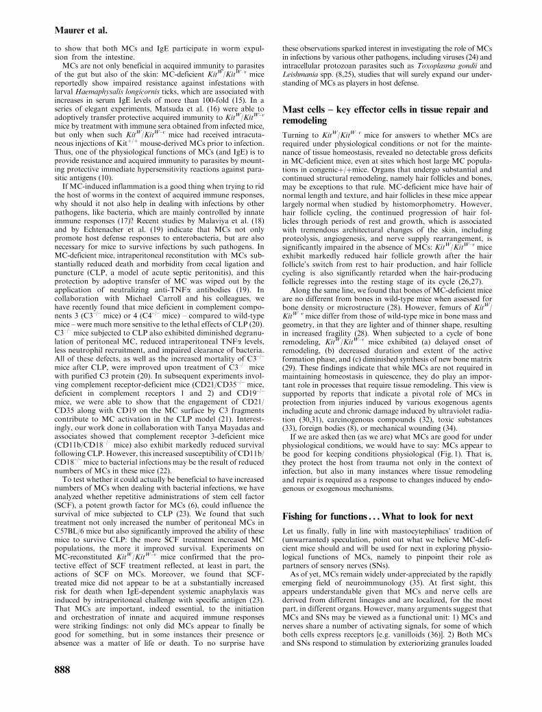
MC
ParasiteToxic
substances BacteriaMechanicalwounding
UVradiation
Carcinogenous compounds
DamageControl
Injury induced by:
TNFα
Histamine
LeukotrienesMIP2
ProteasesHeparin VEGF
Complement-R
Neuropeptide-R Immunoglobulin-R
Endothelin-R
Activation Activation
Figure 1. Physiological function of mastcells.
Controversies
889

Figure 3. Mouse model to test hypo-thetical mast cell functions.
SN
MC-mediated inflammationNeurogenic inflammation
Vanilloids
?
?
ActivationStabilization
RegenerationDegenerationDevelopment
Selected signals inMC/SN crosstalk
Proteases
Histamine
Serotonin
TNFα
?
?
SP, VIP
ET-1
NGF
DegranulationProliferationChemotaxis
DifferentiationMediator
production
Activation
Hair follicle cycling and developmentWound healing, Tissue homeostasis
?
MC
Figure 2. The mast cell nerve connection.Hints to its role in homeostasis anddisease.
Maurer et al.
890

References
1. Riley J F. Lancet 1954: 1: 841.2. Ehrlich P. Arch Mikr Anat 1878: 13: 263–277.3. Ehrlich P. Arch Anat Physiol 1879: 3: 166–169.4. Nakano T et al. J Exp Med 1985: 162: 1025–1043.5. Galli S J, Kitamura Y. Am J Pathol 1987: 127: 191–198.6. Galli S J et al. Adv Immunol 1994: 55: 1–96.7. Biedermann T et al. J Exp Med 2000: 192: 1441–1452.8. von Stebut E et al. Blood 2003: 101: 210–215.9. Galli S J. Int Arch Allergy Immunol 1997: 113: 14–22.10. Henz B M et al. Exp Dermatol 2001: 10: 1–10.11. Nadler M J et al. Adv Immunol 2000: 76: 325–355.12. Nawa Y et al. Parasite Immunol 1985: 7: 429–438.13. Khan A I et al. Int J Parasitol 1993: 23: 551–555.14. Watanabe N et al. Parasite Immunol 1994: 16: 137–144.15. Matsuda H et al. J Parasitol 1987: 73: 155–160.16. Matsuda H et al. J Immunol 1990: 144: 259–262.17. Galli S J et al. Curr Opin Immunol 1999: 11: 53–59.18. Malaviya R et al. Nature 1996: 381: 77–80.19. Echtenacher B et al. Nature 1996: 381: 75–77.20. Prodeus A P et al. Nature 1997: 390: 172–175.21. Gommerman J L et al. J Immunol 2000: 165: 6915–6921.22. Rosenkranz A R et al. J Immunol 1998: 161: 6463–6467.23. Maurer M et al. J Exp Med 1998: 188: 2343–2348.24. Marone G et al. Int Arch Allergy Immunol 2001: 125: 89–95.25. Henderson W R, Chi E Y. J Infect Dis 1998: 177: 1437–1443.26. Maurer M et al. Lab Invest 1997: 77: 319–332.27. Maurer M et al. J Invest Dermatol 1995: 104: 578a.28. Cindik E D et al. Technol Health Care 2000: 8: 267–275.29. Silberstein R et al. Bone 1991: 12: 227–236.30. Ikai K et al. J Invest Dermatol 1985: 85: 82–84.31. Gonzalez S et al. Photochem Photobiol 1999: 70: 248–253.32. Tanooka H et al. J Natl Cancer Inst 1982: 69: 1305–1309.33. Higa A et al. Gastroenterol Jpn 1991: 26: 277–282.34. Weller K et al. Arch Dermatol Res 2001: 293: 79.35. Gottwald T et al. Wound Repair Regen 1998: 6: 8–20.
36. Biro T et al. Blood 1998: 91: 1332–1340.37. Botchkarev V A et al. Arch Dermatol Res 1997: 289:
292–302.38. Stach W. Zeitschr f mikr-anat Forschung 1961: 67:
257–280.39. Wiesner-Menzel L et al. Acta Derm Venereol (Stockh)
1981: 61: 465–469.40. Newson B et al. Neuroscience 1983: 10: 565–570.41. Sugiura H et al. Acta Derm Venereol (Stockh) 1992: 176:
74–76.42. Johnson D et al. J Neuropathol Exp Neurol 1991: 50:
227–234.43. Paus R et al. Arch Dermatol Res 1995: 287: 500–502.44. Metz M et al. J Invest Dermatol 2001: 117: 439a.45. Carlton S M, Coggeshall R E. Brain Res 1997: 763:
271–275.46. Steinhoff M et al. Nat Med 2000: 6: 151–158.47. Hua X Y, Yaksh T L. J Neurosci 1993: 13: 1947–1953.48. Saria A et al. Am Rev Respir Dis 1988: 137: 1330–1335.49. Ohkubo T et al. Eur J Pharmacol 1995: 273: 83–88.50. Caughey G H et al. J Pharmacol Exp Ther 1988: 244:
133–137.51. Nakano A et al. J Immunol 1997: 159: 1987–1992.52. Ansel J C et al. J Immunol 1993: 150: 4478–4485.53. Levi-Montalcini R et al. Trends Neurosci 1996: 19:
514–520.54. MacDonald S M et al. J Neurochem 1981: 36: 9–16.55. Olsson Y. Acta Neurol Scand 1967: 43: 365–374.56. Blennerhassett M G et al. Cell Tissue Res 1991: 265:
121–128.57. Paus R et al. J Invest Dermatol Symp Proc 1997: 2: 61–68.58. Singh L K et al. Brain Behav Immun 1999: 13: 225–239.59. Bienenstock J et al. Am Rev Respir Dis 1991: 143:
S55–S58.60. Downing J E, Miyan J A. Immunol Today 2000: 21:
281–289.61. Maurer M et al. Arch Dermatol Res 1998: 290: 574–578.
Viewpoint 2
Skin is the largest area of the body in constant contact with theoutside world. Environmental and physical stressors are thereforefirst picked up by the skin; similarly, emotional stressors may findquick expression on the skin as a way of subconsciously disconti-nuing or modifying behavior. The skin is richly endowed withsensory nerve endings, stimulation of which activates local mastcells (1). One of the molecules released from nerve endingsis substance P (SP) that has been shown to lead to mast cell-dependent granulocyte infiltration (2–4), possibly throughtumor necrosis factor-a (TNF-a) that induces the expressionof endothelial leukocyte adhesion molecule-1 (5).Another molecule that activates skin mast cells is corticotro-
pin-releasing hormone (CRH), the main factor released from thehypothalamus but also released form dorsal root ganglia (DRGs)under stress (6). For instance, we showed that intradermal admin-istration of CRH activated skin mast cells and increased vascularpermeability (7); the same was true for the structurally relatedpeptide urocortin (Ucn) (8), and this action was more potent thanall other peptides tested (Fig. 1). This process was receptormediated and did not occur in mast cell-deficient mice (7,8).
Stimulated skin mast cells could trigger more release of CRHand/or Ucn from DRG (6), skin elements (9), or local immunecells (10), further stimulating mast cells directly or through therelease of neuropeptides (11) (Fig. 1). Mast cell-derived vasoac-tive, pro-inflammatory, and neurosensitizing molecules (12) couldthen act on keratinocytes, endothelial cells, or neurons to releasemore such molecules, leading to chronic inflammation. Thus, theskin appears to contain the basic elements of a functional equiva-lent of the hypothalamic–pituitary–adrenal (HPA) axis, whichhas been termed the ‘skin–stress response system’ (9). CRHregulates the HPA axis by acting on two types of receptors,CRH-R1 and CRH-R2a, R2b; both of these are primarilyfound in the brain, but CRH-R2b has also been identified outsidethe brain (13). CRH itself is also found outside the brain (6) andhas been postulated to have pro-inflammatory actions throughthe activation of mast cells (14,15). CRH and CRH-R geneexpression has been documented in rodent (16) and human skin(17,18). CRH and/or Ucn could also act on capillaries directly, asCRH has been shown to be produced by (19) and act on endothe-lial cells (20,21).
Controversies
891

Increasing evidence indicates that stress influences pathophy-siological processes (22), especially inflammation, through neuro-peptides (23,24), cytokines, or other chemical mediators (25).For instance, psychological factors increase the morbidity ofallergic reactions (26) and that of many dermatoses (27–29),such as atopic dermatitis (29–31) and alopecia areata (32,33).Acute stress is well known to precipitate flushing and itchingin systemic mastocytosis patients (34–36). We showed thatacute stress (37) can trigger mast cell degranulation andincrease vascular permeability in rodent skin; in fact, activatedmast cells were found in close proximity to CRH-positiveelements. This effect was blocked by depleting sensory nervesof their SP content with neonatal capsaicin administration orby pretreatment with a neurotensin (NT) receptor antagonist,implying the involvement of NT and SP. Plasma extravasationin the skin was also elicited by antidromic stimulation of thelumbosacral dorsal roots in the rat (38). While chronic stressis typically known to attenuate immune processes, acute stresswas recently shown to stimulate them (39,40). For instance, itled to re-distribution of leukocytes from the blood to the skin(41) and enhanced delayed hypersensitivity reactions (42). Infact, even the involvement of the sympathetic system in stressis recast in view of recent findings indicating that it, too, mayhave bidirectional effects (43).
Murine skin mast cells are juxtaposed to nerve endings duringhair follicle formation (44) and in alopecia, where nerve endingswere found to be SP positive (45). Similarly, the expression ofCRH and CRH-R in murine skin was associated with the haircycle (46). A case in point is alopecia areata (33,47,48), which isstrongly associated with atopy (47). We showed that skin biopsiesfrom affected scalp areas from patients with stress-induced alope-cia areata expressed intense signal only for CRH-2b; receptormessage was seen specifically around hair follicles, while unaf-fected scalp areas from patients and normal controls showedonly sparse distribution of faint signal (49). Activation of CRHreceptors could lead to local inflammation. In fact, there isincreased inflammatory infiltrate around the affected hair folli-cles in alopecia (50,51); this is notable for increased numbers ofmast cells (50,52–56), many of which are activated (45,50). Mastcell infiltration and/or proliferation may be triggered byRANTES released from other immune cells (57) and by nervegrowth factor (NGF) (58) released from nerve endings or leu-kocytes (59,60).
Aside from their well-known role in allergy, mast cells haveemerged as versatile effector cells in inflammation (61), neuroim-munoendocrine processes (11), and homeostasis (62). The pro-posed role of mast cells as a universal sensor in the skin issupported by increasing evidence suggesting a similar functionfor brain mast cell regulation of the blood–brain barrier (63,64)and of the HPA axis (65,66). The median eminence of
the hypothalamus is particularly rich in mast cells (67–69)that contain most of the histamine (70), leukotrienes (71), andinterleukin-6 (IL-6) (72) in this critical region. Hypothalamicmastcells (73) are located close to nerve endings containing CRH (74)and can be activated by acute stress (75). Stimulation of hypotha-lamic mast cells appears to occur by ‘intragranular activation’ with-out the massive degranulation typical of anaphylaxis (76) and couldlead to selective or differential release of mediators (77,78), espe-cially cytokines such as IL-6 (79). In fact, we recently showed thatIL-1 can stimulate selective synthesis and release of IL-6 withoutdegranulation through a unique vesicular mechanism (80). Hista-mine (81) and cytokines derived from skin mast cells (82) canthen trigger CRH release (83), leading to HPA activation, or canact as CRH-independent activators of the HPA axis (84) (Fig. 2).Moreover, acute stress was shown to increase local release of CRHin rat skin (85). The CRH could derive either from local nerveendings or from mast cells themselves; as they were recently shownto synthesize and release both CRH and urocortin (86). CRH couldact on mast cells directly, or through NK-1 receptors or endothelialcells (88), possibly with different effects. It was recently shown thatCRH-1 receptor was involved in the modulation of chronic contactdermatitis (89).
Hypothalamic mast cells, and their counterparts in the skin,appear to act as sensors of stressful events with bidirectionalregulation of the HPA axis and its equivalent regulatory systemin the skin. A pathological process may reflect dysregulation ofthese systems due to either underlying processes or excessivestimulation or both. Hans Selye, who wrote definitive works onstress (90) and mast cells (91), would be pleased with the emergingintimate relationship between the two.
Acknowledgements
This work was supported in part by grant number AR47652 fromthe US NIH and Theta Biomedical Consulting and DevelopmentCo., Inc (Brookline, MA). The possible therapeutic use of CRHreceptor antagonists alone, or in combination with mast cellsecretion inhibitors, in stress-induced dermatoses is covered byUS Patent number 6020305 awarded to TCT, pending Europeanapplication number 058898/0148 filed by TCT, and US PatentApplication number 02/771, 669 filed by TCT.
Theoharis C. Theoharides, PhD, MDDepartment of Pharmacology and Experimental Therapeutics
Tufts University School of Medicine136 Harrison Avenue
Boston, MA 02111, USAE-mail: [email protected]
10–5 M β-endorphin
10–5 M SRIP
10–5 M CGRP
10–5 M Neurotensin
10–5 M VIP
10–5 M CRH
10–5 M Ucn
10–5 M ACTH
10–5 M SP
Saline
Figure 1. Photograph of rat skin showingvascular permeability in response tointravenously administered equimolarconcentrations (0.01mM) of variousneuropeptides(ACTH,adrenocorticotrop-in hormone; CGRP, calcitonin-generelated peptide; CRH, corticotropin-releasing hormone; saline, 0.9% NaCl;SP, substance P; SRIF, somatotropinrelease inhibitory factor; Ucn, urocortin;VIP, vasoactive intestinal peptide).
Maurer et al.
892

References
1. Kiernan J A. Q J Exp Physiol 1972: 57: 311–317.2. Wiesner-Menzel L et al. Acta Derm Venereol (Stockh)
1981: 61: 465–469.3. Yano H et al. J Clin Immunol 1989: 84: 1276–1286.4. Matsuda et al. J Immunol 1989: 142: 927–931.5. Walsh et al. Proc Natl Acad Sci USA 1991: 88: 4220–4224.6. Chrousos G P. N Engl J Med 1995: 332: 1351–1362.7. Theoharides T C et al. Endocrinology 1998: 139: 403–413.8. Singh L K et al. J Pharmacol Exp Ther 1999: 288:
1349–1356.9. Slominski A et al. Physiol Rev 2000: 80: 979–1020.10. Bamberger C M et al. J Clin Endocrinol Metab 1998: 83:
708–711.11. Theoharides T C. Int J Tissue React 1996: 18: 1–21.12. Fanciullacci M et al. Cephalalgia 1991: 11: 240–241.13. Lovenberg T W et al. Endocrinology 1995: 136: 4139–4142.14. Karalis K et al. Science 1991: 254: 421–423.15. Karalis K et al. J Neuroimmunol 1997: 72: 131–136.16. Slominski A et al. Biochim Biophys Acta 1996: 1289:
247–251.17. Slominski A J et al. J Clin Endocrinol Metab 1998: 83:
1020–1024.18. Slominski A et al. FASEB J 2001: 15: 1678–1693.19. Simoncini T et al. J Clin Endocrinol Metab 1999: 84:
2802–2806.
20. Clifton V L et al. J Clin Endocrinol Metab 1995: 80:2888–2893.
21. Fleisher-Berkovich S et al. Eur J Pharmacol 1998: 353:297–302.
22. Rabkin J G, Struening E L. Science 1976: 194: 1013–1020.23. Wallengren J et al. Acta Derm Venereol (Stockh) 1986: 66:
23–28.24. Rabier M J et al. J Invest Dermatol 1993: 100: 132–136.25. Sternberg E M et al. Ann Intern Med 1992: 117: 854–866.26. Weil CM et al. Pediatrics 1999: 104 (6): 1274–1280.27. Orfan N A et al. Ann Allergy 1993: 71: 205–216.28. Katsarou-Katsari A et al. Int J Immunopathol Pharmacol
1999: 12: 7–11.29. Van Moffaert M. Psychother Psychosom 1992: 58: 125–136.30. Reichlin S. N Engl J Med 1993: 329: 1246–1253.31. Champion R H, Parish W E. Textbook of Dermatology.
Philadelphia: Blackwell, 1979: 349–361.32. Gupta M A et al. Acta Derm Venereol (Stockh) 1997: 77:
296–298.33. De Waard-Vanderspek F B et al. Clin Exp Dermatol 1989:
14: 429–433.34. Metcalfe D D. J Invest Dermatol 1991: 96: 2–4.35. Horan R F, Austen K F. J Invest Dermatol 1991: 96:
5s–14s.36. Theoharides T C et al. Endocrinology 1998: 139: 403–413.37. Singh L K et al. Brain Behav Immun 1999: 13: 225–239.38. Pinter E, Szolcsanyi J. Neuroscience 1995: 68: 603–614.
STRESS
CRH
DORSOMEDIAL PONS
StressEnvironmental
HYPOTHALAMUSIL-1 IL-6
IL-6
IL-1
Paraventricularnucleus
Ascendingexcitatory input
Spinal cord
Spinal cord
DRG
Sympatheticchain
Descending inhibitoryprojections
Descending excitatoryprojections
CRH/Ucn
CR
H/U
cn
L MC SkinFigure 2. Schematic representation of thehypothesized effect of central or localstress on skin mast cell activation,corticotropin-releasing hormone (CRH)/urocortin (Ucn) release, and localinflammatory mediator release. DRG,dorsal root ganglion; IL-1, interleukin-1; IL-6, interleukin-6; L, lymphocyte;LT, leukotriene; MC, mast cell.
Controversies
893

39. Dhabhar F S, McEwen B S. Brain Behav Immun 1997: 11:286–306.
40. Elenkov I J et al. Ann N Y Acad Sci 1999: 876: 1–11.41. Dhabhar F S et al. J Immunol 1995: 154: 5511–5527.42. Dhabhar F S, McEwen B S. Proc Natl Acad Sci USA
1999: 96: 1059–1064.43. Elenkov I J et al. Pharmacol Rev 2000: 52: 595–638.44. Botchkarev V A et al. Arch Dermatol Res 1997: 289:
292–302.45. Toyoda M et al. Br J Dermatol 2001: 144: 46–54.46. Roloff B et al. FASEB J 1998: 12: 287–297.47. Muller S A, Winkelmann R K. Arch Dermatol 1963: 88:
290–297.48. Bertolino A P. Postgrad Med 2000: 107: 81–90.49. Katsarou-Katsari A et al. Dermatology 2001: 203: 157–161.50. Jaworsky C et al. Br J Dermatol 1992: 127: 239–246.51. Friedman P S. Br J Dermatol 1981: 105: 153–157.52. Lattanand A, JohnsonW C. J Cutan Pathol 1975: 2: 58–70.53. OkunMR,Donnellan B. J Invest Dermatol 1972: 59: 211–224.54. Iribarren C et al. JAMA 2000: 283: 2546–2551.55. Christoph T et al. Br J Dermatol 2000: 142: 862–873.56. Sueki H et al. Acta Derm Venereol 1999: 79: 347–350.57. Conti P et al. FASEB J 1998: 12: 1693–1700.58. Matsuda H et al. J Exp Med 1991: 174: 7–14.59. Bienenstock J et al. Int Arch Allergy Appl Immunol 1987:
82: 238–243.60. Santambrogio L et al. J Immunol 1994: 153: 4488–4495.61. Rozniecki J J, Prusinski A. Cephalalgia 1991: 11: 146–147.62. Gurish M F, Austen K F. J Exp Med 2001: 194:F1–F6.63. Theoharides T C. Life Sci 1990: 46: 607–617.64. Esposito P et al. Brain Res 2001: 888: 117–127.65. Gadek-Michalska A et al. Agents Actions 1991: 32:
203–208.
66. Matsumoto I et al. J Exp Med 2001: 194: 71–78.67. Edvinsson L et al. Neurology 1977: 27: 878–884.68. Panula P et al. Proc Natl Acad Sci USA 1984: 81:
2572–2576.69. Pollard H et al. Brain Res 1976: 118: 509–513.70. Prell G D, Green J P. Annu Rev Neurosci 1986: 9:
209–254.71. Miyamoto T et al. FEBS Lett 1987: 216: 123–127.72. Spinedi E et al. Neuroendocrinology 1992: 56: 46–53.73. Pang X et al. Neuroscience 1996: 73: 889–902.74. Rozniecki J J et al. Brain Res 1999: 849: 1–15.75. Theoharides T C et al. Endocrinology 1995: 136: 5745–5750.76. Dimitriadou V et al. Neuroscience 1990: 39: 209–224.77. Theoharides T C et al. Nature 1982: 297: 229–231.78. Kops S K et al. Cell Tissue Res 1990: 262: 415–424.79. Leal-Berumen I et al. J Immunol 1994: 152: 5468–5476.80. Kandere-Grzybowska K et al. J Immunol (in press).81. Kjaer A et al. Eur J Endocrinol 1998: 139: 238–243.82. Horsmanheimo L et al. Br J Dermatol 1994: 131: 348–353.83. Navarra P et al. Endocrinology 1991: 128: 37–44.84. Bethin K E et al. Proc Natl Acad Sci USA 2000: 97:
9317–9322.85. Lytinas M et al. Int Arch Allergy Immunol 2003: 130 (3):
224–231.86. Kempuraj D et al. Endocrinology (in press).87. Kandere-Grzybowska K et al. Brain Res 2003: 980 (2):
213–220.88. Esposito P et al. Brain Res 2003: 968 (2): 192–198.89. Kaneko K et al. Exp Dermatol 2003: 12 (1): 47–52.90. Selye H. The Stress of Life. 2nd ed. New York: McGraw-
Hill, 1978.91. Selye H. The Mast Cells. London: Butterworth Inc., 1965:
17–568.
Viewpoint 3
Mast cells (MCs) are connective tissue cells found most abun-dantly in the skin, lung, and gastrointestinal tract. Two types ofMCs can be distinguished. The MCT phenotype contains tryptasealone, while the MCTC phenotype contains chymase and tryptase(1). Both subtypes can be found in a given tissue.
Within the skin, MCs occur most prominently just below thedermal–epidermal junction (2) and are concentrated aroundappendages, nerves, and blood vessels (3). Cutaneous MCs werethe subject of a recent review in this journal (4). This articlesummarized the evidence that cutaneous MCs are involved inthe initiation of immunity and host defenses (4). Although MCshave classically been thought of as playing a role in host defensethrough recruitment of cells and proteins from the vascularcompartment, a role for cutaneous MCs in specific immunityis suggested by their ability to phagocytose and take up antigen(5), their expression of major histocompatibility complex(MHC) class I molecules, and their ability to express MHCclass II molecules under some circumstances (6–9). Furthermore,anatomic contacts between MCs and lymphocytes have beendemonstrated by electron microscopy, and MCs have beenshown to traffic to lymph nodes in contact hypersensitivity(CHS) reactions (10,11). MCs have also been shown to expressa number of costimulatory molecules and integrins (1,4,12–14).Additionally, MCs are capable of releasing several cytokines(4,15–17), as previously discussed in detail [4]. The spectrum ofcytokines expressed appears to vary depending on the maturity
state of the MCs and the tissue of residence (1). The ability toexpress these various cytokines, of course, suggests a role ininfluencing immune reactions. Of particular significance, pre-sentation of antigens (including bacterial antigens) by MCs to Tcells has been demonstrated (18,19). It has also been reportedthat MC-deficient mice have reduced survival and are less effi-cient in clearing enterobacterial infections compared to wild-typecontrol mice (1,17,19) and that MC-deficient mice were found tohave increased morbidity and mortality after ligation and punc-ture of the cecum (20). Additionally, injection of normal mice (butnotMC-deficient mice) with stem cell factor (a potentMC growthfactor) increased survival after cecal ligation and puncture (21).
The observations summarized above [and reviewed in detail inRef. (4)] suggest an important role for MCs in specific and innatehost immune functions. However, recent studies have suggestedthat MCs play a crucial role in the down-regulation of immuneresponses and induction of tolerance after exposure of skinto ultraviolet radiation (UVR). This essay discusses the pos-sibility that MCs have an important physiologic function inmediating cutaneous immune suppression following exposureto UVR.
Much work has been performed over the past two decadesexamining the mechanisms by which UVR induces suppressionof immunity (22–24). Of particular importance is the putative roleof UVR-induced immune suppression in the development ofcutaneous malignancies.
Maurer et al.
894

It is now clear that exposure to UVR and, particularly,mid-rangeUVR (UVB radiation, 280–320nm) produces immuno-suppression in experimental animals and humans (22–24). Further-more, substantial evidence from animal models andcircumstantial evidence in humans suggest that this immunesuppression plays a role in skin cancer development. Thus, itmust be asked why evolution would select for such mechan-isms to exist? The answer to this question is, of course,highly speculative. However, it is well known that exposureto UVR exacerbates autoimmune conditions such as lupuserythematosus and exposure to doses of UVR sufficient todamage or kill cutaneous cells will cause release of variousautoantigens where they may, hypothetically, be available forimmunologic recognition and induction of autoimmunity.Thus, one may speculate that the evolution of mechanismsby which UVR down-regulates immune mechanisms mayserve to prevent the development of autoimmune states sub-sequent to exposure to sunlight.The immunosuppressive effects of UVR have been well
reviewed in recent years (22–24). Exposure of certain strains ofmice to low doses of UVB radiation (insufficient to cause grosschanges in the skin) followed by immunization with an epicuta-neously applied hapten at the site of irradiation leads to a greatlysuppressed CHS response. Furthermore, adoptive transfer of Tcells from these mice to naıve recipients suppresses the inductionof CHS in these secondary hosts in a hapten-specific manner. Athigher doses of exposure (sufficient to cause gross changes in theskin), sensitization to an epicutaneously applied hapten even at anon-radiated site leads to suppressed CHS along with the pre-sence of transferable, hapten-specific suppressor T cells.In the high-dose regimen, similar suppression is observed uponsubcutaneous immunization with protein antigens. The sequenceof events that leads to these immunosuppressive effects is com-plex and appears to involve chromophores that include trans-urocanic acid and DNA as well as mediators including tumornecrosis factor-a (TNFa), interleukin-10 (IL-10), prostaglandins,and cis-urocanic acid. With regard to MCs, TNFa and IL-10content may be of particular interest. Experiments utilizingneutralizing antisera to TNFa and/or IL-10 indicate that releaseof TNFa (presumably from cutaneous elements) subsequent toexposure to UVR plays a crucial role in inhibiting the inductionof CHS in both the local and systemic models, while release ofIL-10 (also presumably from cutaneous sources) is responsible forsuppression of the induction of delayed-type hypersensitivity(DTH) to an injected protein (25–27). Additionally, there isevidence that IL-10 plays a role in the establishment of toleranceto epicutaneously applied haptens after UVR exposure (28,29).Recently, a series of experiments have suggested an important
role for MCs in these events. Most interestingly, interactionsbetween MCs and elements of the nervous system appear to beinvolved in UVR-mediated immune suppression. Langerhans’ cellsare anatomically closely associated with unmyelinated nerveswithin the epidermis, and MCs are also frequently found inassociation with nerves (30–33). It has been recently demon-strated by immunohistochemical and immuno-ultrastructuralanalysis that a plexus of axons surrounds superficial dermalMCs and extends into the overlying epidermal layer withintimate associations with epidermal Langerhans’ cells (34).Interestingly, capsaicin (which releases neuropeptides fromnerves) applied to human skin induced release of chymasewithin 6 h and induction of E-selectin in adjacent microvas-cular epithelium, consistent with release of substance P fromaxons with subsequent stimulation of cytokine-mediated mastcell-endothelial interaction (34). However, application ofcapsaicin to human skin grafted onto immunodeficient mice,and thus lacking innervation, failed to show similar findings(34). These results were interpreted as demonstrating that
unmyelinated axons unite epidermal Langerhans’ cells anddermal MCs.Evidence of a role for neuron-derived products modulating
cutaneous immunity comes from a number of observations.First, treatment of mice and rats with capsaicin to deplete skinof sensory neuropeptides was shown to lead to exaggerated con-tact and DTH responses, suggesting that sensory neuropeptideson balance are inhibitory (35–37). Second, treatment in vitroof epidermal cell populations containing Langerhans’ cells withthe neuropeptide calcitonin gene-related peptide (CGRP)resulted in decreased antigen-presenting capability in severalessays (30,38). Third, injection of CGRP intradermally fol-lowed by epicutaneous application of a hapten to theinjected site resulted in a suppressed level of CHS (38). Asa whole, these data strongly suggest a regulatory functionfor the nervous system with regard to cutaneous immunefunction.The first report suggesting that a product of nerves may play a
role in UVR-induced immunosuppression was from Gillardonand colleagues (39), who reported that topical administration ofthe CGRP inhibitor CGRP8�37 prior to exposure to UVR pre-vented UVR-induced immune suppression in mice. In addition, itwas found that topical application of CGRP reduced the densityof I-Aþ epidermal cells in a manner similar to exposure to UVBradiation. Separately, another group demonstrated that adminis-tration of an inhibitor of CGRP systemically prevented the sys-temic suppression of CHS induced by high-dose UVB radiationexposure (40).How doMCs fit into UVB radiation-induced immunosuppres-
sion? This question may have been answered by a series ofexperiments performed by Streilein and his colleagues (41,42).These investigators demonstrated that intradermal administra-tion of CGRP8�37 prior to UVR exposure and subsequent epi-cutaneous application of hapten at the injected and irradiated siteleads to a normal CHS response, while injection of diluent aloneprior to irradiation and sensitization yields a suppressed CHSresponse (43). They then demonstrated that the suppression ofthe induction of CHS observed after immunization at sitesinjected intradermally with CGRP does not occur inMC-deficientmice (43).As a whole, these data suggest that MCs play a very significant
role in the neurocutaneous immune axis. While these cells haveclassically been thought of as anaphylactic effector cellscausing unwanted effects due to hypersensitivity responses, arole for involvement in immune reactions seems evident.Particularly exciting is the evidence that these cells mayplay a crucial role in UVR-induced immunosuppression. Inthis hypothesis (Fig. 1), it would appear that exposure of theskin to UVB radiation induces the release of CGRP fromcutaneous nerves. CGRP then acts on MCs to cause degra-nulation and release of IL-10 and TNFa, which then initiatea further sequence of events leading to abnormal antigenpresentation with altered recognition of encountered anti-gens, decreased immune expression, and a state of specifictolerance. Hypothetically, this down-regulation may involvetumor antigens on incipient neoplasms and thus may play arole in the development of cutaneous malignancies. Thepossibility that MCs play a role in the down-regulation ofimmunity to antigens encountered through other sites wherethese cells are prevalent, such as the lung and gastrointest-inal tract, deserves study.
Richard D. GransteinDepartment of Dermatology
Joan and Sanford I. Weill Medical College of Cornell UniversityNew York, NY, USA
E-mail: [email protected]
Controversies
895

References
1. Church M K, Clough G F. Ann Allergy Asthma Immunol1999: 83: 471–475.
2. Cowen T et al. Br J Dermatol 1979: 100: 635–640.3. Eady R A J et al. Br J Dermatol 1979: 100: 623–633.4. Henz B M et al. Exp Dermatol 2001: 10: 1–10.5. Czarnetzki B M. Scand J Immunol 1982: 15: 581–586.6. Frandji P et al. J Immunol 1993: 151: 6318–6328.7. Wong G et al. Proc Natl Acad Sci USA 1982: 79:
6989–6993.8. Banovac K et al. Immunol Invest 1989: 18: 901–906.9. Friedman M M, Kaliner M. J Allergy Clin Immunol 1985:
76: 70–82.10. Wang H-W et al. J Clin Invest 1998: 102: 1617–1626.11. Tedla N et al. J Immunol 1998: 161: 5663–5672.12. Valent P et al. Proc Natl Acad Sci USA 1991: 88:
3339–3342.13. Love K S et al. Cell Immunol 1996: 170: 85–90.14. Babina M et al. Cell Adhes Commun 1999: 7: 195–209.15. Bradding P, Holgate S T. Crit Rev Oncol Hematol 1999:
31: 119–133.16. Artuc M et al. Exp Dermatol 1999: 8: 1–16.17. Malaviya R et al. Nature 1996: 381: 77–80.18. Frandij P et al. Eur J Immunol 1996: 26: 2517–2528.19. Malaviya R et al. J Immunol 1996: 156: 1490–1496.20. Echternacher B et al. Nature 1996: 381: 75–77.21. Maurer M et al. J Exp Med 1998: 188: 2343–2348.
22. Grabbe S, Granstein R D. Chem Immunol 1994: 58:291–313.
23. Beissert S, Granstein R D. Crit Rev Biochem Mol Biol1996: 31: 381–404.
24. Ullrich S E. J Dermatol Sci 2000: 23 (Suppl. 1): S10–S12.25. Kurimoto I, Streilein J W. Exp Dermatol 1999: 8: 495–500.26. Rivas J M, Ullrich S E. J Leukoc Biol 1994: 56: 769–775.27. Shreedhar B et al. J Immunol 1998: 160: 3783–3789.28. Niizeki H, Streilein J W. J Invest Dermatol 1997: 109:
25–30.29. Kitazawa T, Streilein J W. J Invest Dermatol 2000: 115:
942–948.30. Hosoi J et al. Nature 1993: 363: 159–163.31. Gaudillere A et al. Br J Dermatol 1996: 135: 343–344.32. Naukkarinen A et al. Arch Dermatol Res 1991: 283: 433–437.33. Hagforsen E et al. Arch Dermatol Res 2000: 292: 269–274.34. Egan C L et al. J Cutan Pathol 1998: 25: 20–29.35. Nilsson G, Ahlstedt S. Int Arch Allergy Appl Immunol 1989:
90: 256–260.36. Girolomoni G, Tigelaar R E. J Immunol 1990: 145:
1105–1112.37. Veronesi B et al. Toxicol Appl Pharmacol 1998: 153: 243–249.38. Asahina A et al. J Immunol 1995: 154: 3056–3061.39. Gillardon F et al. Eur J Pharmacol 1995: 293: 395–400.40. Garssen J et al. Photochem Photobiol 1998: 68: 205–210.41. Streilein J W et al. Keio J Med 1999: 48: 22–27.42. Streilein J W et al. Ann N Y Acad Sci 1999: 885: 196–208.43. Niizeki H et al. J Immunol 1997: 159: 5183–5186.
CUTANEOUS NERVES
TNFα AND IL-10
CHANGES IN LOCAL CUTANEOUS APC FUNCTION LEADINGTO DOWN-REGULATION OF IMMUNITY AND INDUCTION OFTOLERANCE
SKIN
CIRCULATING TNFα AND IL-10 RESULT INCHANGES IN DISTANT APC FUNCTION LEADINGTO DOWN-REGULATION OF IMMUNITY ANDINDUCTION OF TOLERANCE
MAST CELLS
UVR
CGRP
Figure 1. Hypothetical pathway showinginvolvement of mast cells in ultravioletradiation (UVR)-induced immune sup-pression. APC, antigen-presenting cell;CGRP, calcitonin gene-related peptide;IL-10, interleukin-10; TNFa, tumornecrosis factor-a.
Maurer et al.
896

Viewpoint 4
Mast cells are generally associated with sneezing, wheezing, itch-ing, allergic reaction in general, and other inflammatory events.In the past, we learnt a lot about the mechanisms of allergicdiseases and the role of mast cells in the pathophysiology ofhypersensitivity reactions (1–5). It became clear that mast cellsnot only release histamine and eicosanoids upon crosslinking ofsurface-bound immunoglobulin E (IgE) by allergen but alsomany other mediators not readily associated with inflammation.Examples are Th2 cytokines such as interleukin-3 (IL-3), IL-5,and IL-13, tissue-modulating factors such as basic fibroblastgrowth factor (bFGF) and transforming growth factor-b (TGF-b), and proteoglycans. Other mast cell-derived mediators such astumor necrosis factor-a (TNF-a) are found elevated duringinflammatory processes but may also induce neutrophil recruit-ment, known to be of importance, e.g., for host defense againstbacteria (6–9).Mast cell biology becomes even more complex when realizing
that not only the number of mast cell products but also thenumber of mast cell agonists is huge and by far not restricted toagents inducing IgE crosslinking, such as allergen. We now knowthat particular cytokines such as stem cell factor (SCF) and IL-4are important regulators of the development of mast cell frommyeloid progenitors and for the function of mature mast cells (9).SCF induces mast cell proliferation, prevents mast cell apoptosis,and enhances IgE-dependent mediator and cytokine release. Tosome extent, SCF at higher concentrations also induces mediatorrelease; however, the in vivo significance of this in vitro finding isunclear yet. Most of these SCF effects on mature mast cells areenhanced by IL-4 (10). IL-3 has been described as another impor-tant mast cell cytokine in rodents, but these data could not beconfirmed for human mast cells so far (11). However, own
unpublished data indicate that IL-3 in synergism with SCF mayalso regulate human mast cell functions (submitted for publica-tion). Apart from cytokines, other mast cell regulators have beenidentified including parasites, bacteria, adhesion factors andextracellular matrix proteins, nitric oxide, as well as neuro-transmitters and neurotrophins (12–19). Apart from such chem-ical mediators, physical changes such as irradiation and pHchanges may affect mast cell function (20,21). These observa-tions strongly suggest that the function of mast cells cannot berestricted to their pathophysiological role in allergy and inflam-mation. Oddly enough, our knowledge on physiological func-tions of mast cells is very limited compared to the enormousliterature on pathophysiological functions. The question why wehave mast cells has to be answered.There is increasing body of evidence that mast cells play a role
in host defense against different kinds of injuries includingmechanical injuries such as ischemia and infection either bybacteria or by helminthes (12,13,21). In some cases, mast cellactivation may be accompanied by an aggravated tissue dysfunc-tion as described in circulation disorders causing ischemia or inpostinflammatory tissue remodeling leading to fibrosis (21–23).However, the same mechanisms may be of importance in woundhealing, regulation of tissue perfusion, and induction of hostdefense mechanisms (9,24,25). An intriguing example is therole of mast cells in host defense against bacteria. Mastcells, besides macrophages, are an important source ofTNF-a, known to belong to the so-called pro-inflammatory cyto-kines. TNF-a is released by mast cells upon stimulation withIgE-crosslinking allergen or gram-negative bacteria typicallylocated in the gut (6–8). One may argue that mast cellscontribute to the destructive inflammatory process during
Bacteria Allergen
Mast cellHistamineLTC4ChymaseVLA-4
Bloodvessel
Epithelium
TNF-α PNM
SecretionPermeability
Tissue remodelingRepair processes
Fibrosis
TGF-βbasic FGF
HistamineLTC4PGD2
Defenseagainst
bacteria
Eo-sinophil
IL-5
Defenseagainst
parasites
Regulation ofsecretion, motility,permeability,and inflammation
Regulation ofcell recruitment,
vascularpermeability,coagulation,
and fibrinolysis
Fibroblasts
Nervecells
LymphocytesRegulation ofthe specific immune system
Histamine, PGD2Proteases
Figure 1. Physiological mast cell functions– current understanding. For details seetext.
Controversies
897

bacterial infection, but animal studies have clearly shown thatthe opposite is true. Mortality following bacterial peritonitisis increased in mast cell-depleted animals (Wv/Wv mice), andmost likely, this is due to the lack of TNF-a produced byintraperitoneal mast cells. This mast cell-derived TNF-aseems to be crucial for the neutrophil recruitment to thesite of inflammation, which is required for overcoming theinfection (26,27). Although these animal data are very stimu-lating, they have to be confirmed for the human situation.We could show that gram-negative bacteria induce TNF-aproduction in human intestinal mast cells in vitro. TheTNF production is accompanied by histamine secretion, butin contrast to IgE crosslinking by allergen, the bacteria do notinduce leukotriene C4 production, suggesting an IgE-inde-pendent mechanism (9). Other studies revealed that bacter-ium–mast cell interactions are not restricted to an inductionof TNF-a, because bacteria may be opsonized by mast cellsthat are capable of presenting bacterial antigen effectively toCD4 and CD8-positive T cells (28). Studies in our laboratoryare in progress to address the question whether these rodentdata hold true for human mast cells.
Mast cells are typically located nearby external and internalbody surfaces such as skin, mucosa, and blood vessels (1,4,9,14).It is tempting to speculate that mast cells exert some physiologicalfunctions at these sites. Again, mast cells may have defensefunctions at these strategically important sites, but there isevidence that mast cells may act there in more general termsalso without infection. In particular in the gut, it has beenshown that mast cells regulate multiple organ functions such asfluid and electrolyte secretion by epithelial cells, motor functionsof smooth muscle cells, and integrative functions mediated by theenteric nervous system (29,30). In addition, some recent studieshave indicated that mast cells are a unique source of heparin andother coagulation-regulating factors (31–33). These properties,together with the mentioned anatomical vicinity and functionalinteraction between mast cells and endothelial cells, suggest thatmast cells also play a role in regulating blood vessel function,which is of importance for the development of cardiovasculardisease (14,34). Because mast cells located at barrier sitesrespond to multiple physical changes and chemical/biologicalmediators, this cell type may be regarded as a cellular ‘watchdog’,enabling quick reactions to any harmful event or even to anyevent.
If interpreted in this manner, mast cells belong to the innateimmune system that recognizes multiple molecules and events,and mast cells interact with many other systems such as thespecific immune system, the epithelial and endothelial system,the would healing machinery, and the nervous system (16,34–36). Our current knowledge on the physiological functions ofmast cells is summarized in Fig. 1. Within this context, it becomesclear that mast cells are primarily ‘physiological cells’ exertingmultiple functions that have to be unraveled in future studies.While the mast cell fulfills all these functions, mistakes may
occur, leading to more or less harmful consequences such asallergy and many other diseases.
S. C. BischoffDepartment of Internal Medicine
Medical School of HannoverHannover, Germany
References
1. Church M K, Levi Schaffer F. J Allergy Clin Immunol 1997:99: 155–160.
2. Ahmed T, Fuchs G J. J Diarrhoeal Dis Res 1997: 15:211–223.
3. Gui X Y. J Gastroenterol Hepatol 1998: 13: 980–989.4. Bischoff S C et al. Int Arch Allergy Immunol 2000: 121:
270–283.5. Bischoff S C. Allergy. 2nd ed. London:Mosby, 2000; 127–140.6. Bradding P et al. Clin Exp Allergy 1995: 25: 406–415.7. Ohkawara Y et al. Am J Respir Cell Mol Biol 1992: 7:
385–392.8. Bischoff S C et al. Gut 1999: 44: 643–652.9. Bischoff S C. Mast Cells and Basophils. San Diego, CA:
Academic Press, 2000; S541–S565.10. Bischoff S C et al. Proc Natl Acad Sci USA 1999: 96:
8080–8085.11. Valent P et al. J Immunol 1990: 145: 3432–3437.12. Shaikh N et al. J Immunol 1997: 158: 3805–3812.13. Arock M et al. Infect Immun 1998: 66: 6030–6034.14. Mierke C et al. J Exp Med 2000: 192: 801–811.15. Forsythe P et al. Int Immunopharmacol 2001: 1: 1525–1541.16. Williams R M et al. Chem Immunol 1995: 61: 208–235.17. Nilsson G et al. Eur J Immunol 1997: 27: 2295–2301.18. Tam S Y et al. Blood 1997: 90: 1807–1820.19. Marshall J S et al. J Immunol 1999: 162: 4271–4276.20. Kronauer C et al. Inflamm Res 2001: 50 (Suppl. 2): S44–S46.21. Kanwar S, Kubes P. Am J Physiol 1994: 267: G316–G321.22. Armbrust O T et al. J Hepatol 1997: 26: 1042–1054.23. Gelbmann C M et al. Gut 1999: 45: 210–217.24. King T et al. Dig Dis Sci 1992: 37: 490–495.25. Artuc M et al. Exp Dermatol 1999: 8: 1–16.26. Echtenacher B et al. Nature 1996: 381: 75–77.27. Malaviya R et al. Nature 1996: 381: 77–80.28. Mecheri S, David B. Immunol Today 1997: 18: 212–215.29. Crowe S E et al. Gut 1997: 41: 785–792.30. Wood J D et al. Gut 1999: 45 (Suppl. 2): 6–16.31. Sillaber C et al. J Immunol 1999: 162: 1032–1041.32. Cho S H et al. J Immunol 2000: 165.33. Tchougounova E, Pejler G. FASEB J 2001: 2763–2765.34. Kelley J L et al. Mol Med Today 2000: 6: 304–308.35. Gruber B L. Int Rev Immunol 1995: 12: 259–279.36. Galli S J, Wershil B K. Nature 1996: 381: 21–22.
Viewpoint 5
Mast cells are distributed throughout all the tissues of the body.In many of these, they are associated with nerves and in turn havea functional distribution and proximity to blood vessels. Theyseem to be distributed freely in some cavities such as the perito-
neal cavity, but it is generally agreed that they do not circulate ina recognizable, morphological form in the blood. Nevertheless,they are largely derived from stem cells in the bone marrow andreach their final form in the tissues under the influence of local
Maurer et al.
898

factors such as stem cell factor interleukin-3 (IL-3), IL-4 and IL-9. Different functional subset of mast cells are encountered indifferent tissues. The subsets are associated with different phar-macological responses to both stabilizing and degranulatingagents, and the cells are characterized by different sets ofresponses which include synthesis and secretion of proteolyticenzymes such as tryptase and chymase and products of arachi-donic acid metabolism such as prostaglandins and leukotrienes.Consequently because of this vast heterogeneity of cell type,
distribution, anatomic association and function, the physiologicalrole of mast cells may be different in different tissues even under thesame challenge. Therefore, the answer to the question posed at thebeginning would be determined by the nature of the stimulation,the site in which it occurred, the extent and duration of the stimulusand the determination of whether the stimulus was acute or pro-longed and was accompanied by other events such as those inducedby different forms of inflammation.Nevertheless, it is possible to identify a series of physiological
functions that appear to be important.
Control of mast cell and immunoglobulin E levels
Activation and degranulation of mast cells appears to be asso-ciated with the notion that this physiological event, no matterhow it occurs, promotes a general increase in the total body massof mast cells (1). There is also a relationship between mast cellmass and total amount of immunoglobulin E (IgE), the majorallergic antibody that binds to mast cells through a high-affinityreceptor (2). Conceptually, this leads to the notion that control ofmast cell activation leads to control of IgE levels and reciprocally,control of IgE levels as may now occur by treatment in vivo withanti-IgE will control mast cell mass.
Blood vessel tone and coagulation
Mast cells are not just present for the promotion of wheezing,sneezing and itch. Indeed, their association with blood vesselssuggests that they are intimately involved in maintaining bloodvessel tone, vascular permeability and angiogenesis (3). In addi-tion, mast cells contain a variety of glycosaminoglycans such asheparin which have major physiological effects on coagulationand various cellular activities (4). Undoubtedly, mast cells areinvolved in perfusion of tissues by blood and in the regulation ofblood pressure through these means. Mast cell activation has alsobeen suggested to be a key factor in the initiation of migrane (5).
Resistance to infection
Mast cells are significantly involved in host resistance to infection(6). The presence of functional mast cells is associated withrecovery from and expulsion of nematodes in the intestine, butthe absence of mast cells simply prolongs the infection leading toeventual expulsion, presumably by other means. Evidence forinvolvement in host resistance comes from experiments involvingcecal ligation and puncture, where prevention of morbidity andmortality is associated with adequate mast cell function, primarilythrough the secretion of tumour necrosis factor a (TNFa), whichin turn promotes IL-8 synthesis and polymorphonuclear leuko-cyte influx.
Allergy and inflammation
Mast cells are involved in allergy through a variety of complexevents which involve synthesis and release of both preformedmediators such as histamine, serotonin, prostaglandins, leuko-trienes, TNFa, etc. as well as a whole series of cytokines, enzymesand so on which may or may not be preformed (7). The events
which occur are caused both directly by mediators such as hista-mine or indirectly through complex central and peripheral events.These end up causing increased vasodilatation, local secretion bymucosal tissues, increase in smoothmuscle reactivity and loweringthe threshold for subsequent stimulation (8).
Communication with the nervous system
Mast cells are involved in bidirectional communication withnerves (9). Mast cells are universally found in close proximity tonerves of a variety of sorts. Factors released from mast cells cancommunicate with nerves and hence via local ganglia to the spinalcord and then to the brain. Mast cells can also locally promoteaxon reflexes. Thus, they can act as sensory receptors for noxiousagents such as allergens, toxins, etc. and convey accurate infor-mation, as to the encounter with these agents, its nature, extentand location, to the central nervous system (CNS) (10). In turn,the CNS can use the same mast cells associated with nerves to beactivated as targets of efferent nervous regulatory pathways.These processes are not only found to occur in allergy, but alsoin delayed hypersensitivity reactions and in response to bacter-ial toxins such as that of Clostridium difficile (11).
Involvement in behaviour
There is a large literature describing the involvement of mast cellsand various brain processes. Suffice to say, mast cells are foundassociated with blood vessels throughout the brain and especiallyin the meninges (12). They seem also to be involved in beha-vioural activity such as the courting behaviour of doves (13).Surprisingly, mast cells are found in abundance around the pitui-tary gland and are thought to act as an immune ‘gate’ forhypothalamic-pituitary-adrenal axis activity (14). The effects ofstress which are largely mediated through corticotropin-releasingfactor, either locally or centrally synthesized seem to be mediatedthrough nerves and mast cells (15). Pavlovian conditioning hasalso been shown to be able to promote mast cell degranulationthrough as yet unknown mechanisms (16).
Tissue repair
Increased numbers of mast cells are always found at sites of tissuerepair and fibrosis (3). Their exact activity in this promotion andregulation of this process is not clear.
Summary
Mast cells have multiple functions; however, most of these func-tions are not essential for life, as various mast cell-deficient strainsof mice and rats seem to have normal life spans (17). However,mast cells can be regarded as the conductors of the symphony ofinformation, which is being constantly gathered both in the per-iphery and in the centre. They are masters and mistressesof coordination and integration, but they have the added dimen-sion of remarkable varied effector functions. When one adds totheir capacity to be motile or sessile depending on local circum-stances, they emerge as the kings and queens of varied repertoireamongst all the cells in the body. Thus, mast cells may be thoughtto be important ancillary aids against infection, promoters oftissue repair, regulators of blood vessel tone, permeability andangiogenesis, important mediators of stress and allergy, regula-tors of neuroendocrine function, purveyors of sensory informa-tion to the central nervous system, and as effector targets forefferent nervous pathways. Lastly, but by no means least, theyappear to act as a switchboard for the regulation of inflamma-tion.
Controversies
899

John BienenstockDepartments of Medicine and Pathology & Molecular Medicine
McMaster University1200 Main Street West, HSC-3N26
HamiltonOntario
L8N 3Z5 CanadaE-mail: [email protected]
References
1. Marshal J S et al. J Immunol 1990: 144: 1886–1892.2. Yamaguchi M et al. J Immunol 1999: 162: 5455–5465.3. Metcalfe D D et al. Physiol Rev 1997: 77: 1033–1079.4. Zehnder J L, Galli S. J Nat 1999: 400: 714–715.5. Theohardies T C. Perspect Biol Med 1983: 26: 672–675.
6. Abraham S N, Malaviya R. Adv Exp Med Biol 2000: 479:91–105.
7. Wedemeyer J et al. Curr Opin Immunol 2000: 12: 624–631.8. Janiszewski J et al. Am J Physiol 1994: 267: C138–C145.9. Beinenstock J et al. Am Rev Respir Dis 1991: 143:
S55–S58.10. Beinenstock J, Marone G, ed. Mast Cells and Basophils.
Academic Press, New York, 2000: 313–323.11. Pothoulakis C, LaMont J T. Am J Physiol Gastrointest
Liver Physiol 2001: 280: G178–G183.12. Persinger M A. Behav Neural Biol 1979: 25: 380–386.13. Silverman A J et al. Proc Natl Acad Sci USA 1994: 91:
3695–3699.14. Matsumoto I et al. J Exp Med 2001: 194: 71–78.15. Theoharides T C et al. Endocrinology 1998: 139: 403–413.16. MacQueen G et al. Science 1989: 243: 83–85.17. Galli S J et al. Science 1992: 664: 69–88.
Viewpoint 6
Due to the ubiquitous distribution of mast cells in diverse tissuesand their presence throughout the animal kingdom, they areparticularly well equipped to contribute to the maintenance oftissue integrity and function. Awareness of this has been onlyslowly emerging until recently, when mast cells have been shownto function as mediators of pathological reactions such asanaphylaxis, type-I allergen presentation, delayed-type hyper-sensitivity, natural immunity against bacteria and parasites,and tissue repair and fibrosis (1,2).
In this forum of controversies, I would like to present evidencethat mast cells play a key function in tissue physiology, focussingon their direct or indirect contribution to epithelial and connec-tive tissue growth.
Mast cell mediators and epithelial growth
Clear data to this effect emerged several years ago in the field ofhair growth where several lines of evidence pointed to a role ofmast cells during the initiation of the murine hair follicle cycle (3).In additional studies, mast cell-derived mediators like histamine,serotonin and nerve growth factor (NGF) were implicated in theregulation of murine hair growth (4–6). Some doubt as to thenormality of these observations lurk, however, in the back of mymind, as in the model employed, anagen is induced by plucking ofhair and thus by mechanical trauma with an associated massivemast-cell degranulation. Possibly, tissue traumatization and asso-ciated mast-cell-dependent processes in the context of woundhealing may therefore be operative in the model employed (7).
Nevertheless, numerous other compelling lines of evidencehave emerged in recent years that mast cells are able to inducehair growth as well as epithelial proliferation and differentiationin general. This holds in particular with regard to a number ofmast cell mediators. Histamine is the most prominent amongthem and an obvious candidate for a physiological role, sincethis preformed and readily released vasoactive amine inducesincreased blood flow and leakiness of small vessels, allowing forthe constant renewal of tissue fluid and thus the nutrition ofdiverse organs. Via the up-regulation of adhesion molecules onendothelial cells, histamine can also induce the immigration offormed elements from the blood which would then engage intissue surveillance and renewal. Beyond that, we could recentlyshow that at low concentrations which are likely to prevail underphysiological conditions, histamine functions also as an epithelial
growth factor (8), as suggested already earlier by demonstrationof epidermal keratinocyte proliferation in murine skin (6).
Furthermore, histamine up-regulates human keratinocyteexpression of several major integrins (a2, a3, a6 and b1 chains)at mRNA and partly also at protein level (8), a process that isviewed as an indication of keratinocyte differentiation (9).Expression of the differentiation-related dioxin receptor AhRand of the proliferation marker PCNA was also markedlyincreased under these conditions (8,10). Finally, partial inhibitionby an H1 receptor-specific antihistamine suggests that histamineis also one of the mast-cell mediators contributing to fibroblastproliferation (11).
Mast cells and connective tissue homeostasis
Besides histamine, human mast cells also store several proteasesin their secretory granules, specifically tryptase, chymase, carboxy-peptidase A and cathepsin G (12), enzymes that are implicated indefence against helminthic, allergic, cardiovascular and chronicinflammatory diseases. Beyond this, these molecules are able toinactivate mediators of inflammation such as neuropeptides andnumerous cytokines (13). Specifically, chymase can activatecollagenase and stromelysin, destruct vitronectin and fibronectin,and induce fibroblast proliferation (13,14), suggesting an import-ant role of this mast-cell-specific protease in tissue matrix turnoverand renewal. Normally, several protease inhibitors within theconnective tissue ensure tissue homeostasis by inhibiting excessiveactivities of this enzyme in the immediate mast cell environment.Such natural inhibitors are not known for mast cell-specifictryptase which is less active than chymase in connective tissueremodelling and fibroblast proliferation (13). It can, however,also stimulate type-I collagen synthesis (15) and, at low concen-trations, it can’t induce keratinocyte integrin expression just likehistamine and thus epithelial differentiation (8).
Mast cell-derived growth factors affect numerousother cell types
In addition to the molecules discussed so far, mast cells are able tosecrete numerous other mediators that can affect the growth ofcells residing within epithelial and connective tissues (Table 1).Among these, interleukin-8 (IL-8) is of special interest, as it is also
Maurer et al.
900

stored within mast-cell granules and thus readily available evenon minor stimulation of the cells (16,17). Lipid mediators withkeratinocyte growth-promoting activity like leukotrienes are notpreformed, but they are generated within seconds via enzymaticaction on membrane-associated arachidonic acid. The remainingmast-cell-derived cytokines and growth factors affecting kerati-nocyte growth (see Table 1), including the recently describedHB-EGF and several members of the FGF family (8), are synthe-sized de novo on appropriate stimulation, a process that takes atleast several hours (18). As none of these factors are detectablein freshly isolated skin mast cells, their direct contribution toskin physiology via mast cells is unlikely. Interestingly, they mayhowever, be generated secondarily in neighbouring cells viarapidly released mast-cell mediators, as, e.g. recently shown fortryptase-induced FGF-2 and histamine-induced FGF-7 (KGF)synthesis in human fibroblasts (8).Besides their well-established role in the maintenance of epithe-
lial and connective tissue integrity, mast cells can also contributevia several mediators to endothelial growth, ensuring thus theneeded tissue vascularization and proper nutrition. Furthermore,they produce several factors that stimulate melanocyte growthand melanin production (Table 1) and thus the necessary protec-tion against UV-induced damage. Their important role in thesenormal physiological functions is underlined by the fact thatmany of the mediators listed are preformed within mast-cellgranules (Table 1), including the three major isotypes of VEGF(19).The picture of an all-round activity of mast cells in tissue
physiology is completed in that mast-cell products can also affectnerve cell growth and differentiation via NGF, Langerhans cellmigration and maturation via granulocyte/macrophage-colony stimulating factor and IL-4 and lymphocyte hominginto the epidermis and proliferation via several distinct chemo-kines and cytokines (2).
Mast cell interact with numerous cells directly
On viewing the data discussed so far in perspective, one is struckby the emerging picture of mast cells acting within a network oralong several axes involving all major tissue cell types. This isparticularly evident for stem cell factor (SCF) which, in the skin,can be generated by keratinocytes, endothelial cells, fibroblasts,mast cells and melanocytes and which stimulates melanocytes andmast cells via its c-Kit receptor on these cells (1,20). Like IL-8,which can also be produced by several of these same cell typesand for which mast cells also express specific chemokine receptors(21), SCF can thus stimulate its own secretion by mast cells in anautocrine fashion. This raises a number of questions as to theimportance of mast cells vs. other cell types in the maintenance of
tissue growth and homeostasis. The same holds for the manydifferent mediators potentially participating in these activities.Redundance of nature ensuring normal tissue function may wellaccount for what seems puzzling at first sight.
Physiological stimuli of mast cell mediators
An important remaining issue is the clarification of the types ofstimuli that might regulate mast-cell function in normal physiol-ogy, as in most of the literature cited so far, artificial laboratoryconditions prevailed, with a veritable bombardment of the cellswith agents inducing mast cell mediator release. For the skin,several possible, more physiological stimuli are readily apparent.The various stimuli inducing mast cell mediator release underphysiological conditions are C3a/C5a, UV light, substance P,bacterial products, aMSH, IL-8, NGF and SCF. Thus, consider-ing the fact that the skin and other tissues are continuouslybombarded by pressure or shearing forces, neurosensory effectormechanisms may have an important impact on low-level mast-cellsecretion that should be operative in a more normal physiologicalsetting. Substance P is a possible player in this game, asare proopiomelanocortin-derived peptides like amelanocytestimulating hormone (MSH) both of which induce histaminesecretion even at very low concentrations (16,22). As a result ofconstantly impacting UV light on the exposed skin surface,release of mediators like aMSH and SCF from keratinocytesmust be considered as another likely scenario (23). Furthermore,the potent mast cell secretagogues C3a and C5a are known toalso be constantly and rapidly generated in response tominor trauma, and finally products of the bacterial flora locatedin the upper layers of the epidermis or defensins produced byepithelial cells encompass mast-cell secretagogues as well(24,25) and may thus contribute to mast-cell function in normalphysiology.
Conclusion: Mast cells are important players intissue physiology
To sum up, mast cells are veritable powerhouses of growth factorproduction, as they can potentially generate and release theirnumerous potent mediators also under physiological conditions.The cells are thus superbly equipped to maintain tissue growth,differentiation and survival, either directly or through interactionwith major cell types in their immediate surrounding. The unrav-elling of further details as to the conditions under which thesediverse mast cell functions become operative in the context oftissue physiology remains an enticing future challenge.
Table 1. Mast-cell-derived factors likely to be involved in cutaneous physiology
Keratinocyte growth and differentiation factors Fibrogenic factors Angiogenic factors Melanocyte growth factors
Histamine* Platelet-derived growth factor (PDGF) VEGF121*, 165*, 189*, 206 SCF*Tryptase* Tryptase* FGF-2 IL-8*IL-3, IL-6, IL-8* Chymase* PDGF Melanocyte growth stimulating
activity/growth related oncogoneTGF-b TGF-b TGF-b FGF-2FGF-2, -5, -7, -10 FGF-2 GM-CSFHB-EGF IL-4 TNF-a*EGF Histamine* IL-3, IL-8*Leukotrienes (B4, C4, D4) Carboxypeptidase A Histamine*
Heparin*Tryptase*
Mediators are either present in a preformed state*, generated rapidly, as holds for the lipid mediators like leukotrienes, or synthesized several hours afterappropriate stimulation of the cells (1).GM-CSF, granulocyte/macrophage-colony stimulating factor; IL, interleukin; TGF, transforming growth factor, TNF, tumour necrosis factor.
Controversies
901

Beate M. Henz, MDProfessor of Dermatology
Department of Dermatology and AllergyCharite, Humboldt University
Berlin, GermanyE-mail: [email protected]
References
1. Artuc M et al. Exp Dermatol 1999: 8: 1–16.2. Henz B M et al. Exp Dermatol 2001: 10: 1–10.3. Paus R et al. Dev Biol 1994: 163: 230–240.4. Paus R et al. Br J Dermatol 1994: 130: 174–180.5. Paus R et al. Arch Dermatol Res 1995: 287: 500–502.6. Maurer M et al. J Derm Sci 1997: 16: 79–84.7. Hermes B et al. J Invest Dermatol 2001: 16: 79–84.8. Artuc M et al. J Invest Dermatol 2000: 115: 543.9. Watt F M, Hertle M D. Keratinocyte integrins. In: Leigh
E M, Lane E B, Watt F M, eds. The Keratinocyte Hand-book. Cambridge: University Press, 1994: 153–164.
10. Wanner R et al. Biochem Biophys Res Commun 1995:209: 706–711.
11. Abe M et al. J Allergy Clin Immunol 2000: 106: S72–S77.12. Li L et al. The phenotypic similarities and differences
between human basophils and mast cells. In: Marone G,Lichtenstein L M, Galli S J, eds. Mast Cells and Basophilsin Physiology, Pathology and Host Defense. San Diego:Academic Press 2000: 97–116.
13. Algermissen B et al. Exp Dermatol 1999: 8: 193–198.14. Lees M et al. Eur J Biochem 1994: 223: 171–177.15. Cairns J A, Walls A F. J Clin Invest 1997: 99: 1313–1321.16. Gibbs B F et al. Exp Dermatol 2001: 10: 312–320.17. Moller A et al. J Immunol 1993: 151: 3261–3266.18. Moller A et al. Immunology 1998: 93: 289–295.19. Grutzkau A et al. Mol Biol Cell 1998: 9: 875–884.20. Grabbe J et al. Arch Dermatol Res 1994: 287: 78–84.21. Lippert U et al. J Immunol 1998: 161: 2600–2608.22. Grutzkau A et al. Biochem Biophys Res Commun 2000:
278: 14–19.23. Luger T A et al. J Invest Dermatol Symp Proc 1997: 2:
87–93.24. Lagunoff D et al. Annu Rev Pharmacol Toxicol 1983: 23:
331–351.25. Van Westering S et al. J Allergy Clin Immunol 1999: 104:
1131–1138.
Commentary 7
This commentary focuses on the question ‘what – if any – is thephysiological function of coronary mast cells?’ Our laboratoryhas investigated the role of mast cells in the pathogenesis ofcoronary atherosclerosis and its clinical complications (1). Thepathogenesis of this chronic and long-lasting inflammatory dis-ease involves the inner layer of the arterial wall, the intima (2). Inits early stage, the disease is characterized by the accumulation ofintracellular lipid (fatty streaks), in its later stages by extracellularaccumulation of lipid (atheromas or atherosclerotic plaques) (3),and in its final clinically relevant stages by erosion or rupture of theplaques, with ensuing thrombotic complications (4). For thedefinition of a physiologic role for the mast cell in coronaryatherogenesis, we have attempted to identify the crossroads atwhich the physiology of the mast-cell system meets with thepathology of the atherosclerotic process.
Regarding a physiologic role of mast cells in the normal humanarterial intima, we have been puzzled by what is meant by ‘phy-siologic’ or ‘normal’. This same uncertainty may be felt by otherscientists who are searching for a possible role of mast cells inother normal tissues. Although we still lack detailed studies onthe age- and disease progression-related temporal sequence inwhich mast cells appear in the human arterial intima, it seemsthat, at the stage of lesion initiation, no mast cells or only a fewmast cells are present in the intima (5). Thus, in the normalarterial intima, the average density of mast cells is only about1 cell/mm2, a number which contrasts sharply with that in thenormal skin, which has been reported to be, on average, 50 cells/mm2 (6). From the paucity of mast cells in the normal coronaryintima, we conclude that these cells have little, if any, physiologi-cal role in this tissue site.
When the arterial intima is converted from a normal into apathologic tissue, macrophages, T lymphocytes, and also mastcells arrive on the scene. In such lesions, the number of mast cellsmay be locally up to 10–50 or even to 100 cells/mm2, and most ofthem show some degree of degranulation, i.e. they are activated(7). Unfortunately, we still have only a very rough idea of thephenotypic characteristics of mast cells in the plaque: withimmunohistochemistry, we have shown that they contain heparinand tryptase, and sometimes also chymase and tumor necrosis
factor-a (8). Also, more studies are required for identifying thefactors that regulate the different aspects of the life and death(immigration, differentiation, activation, and apoptosis) of amast cell in the human coronary plaque. Thus, a very limitedknowledge on the life cycle and actions of mast cells in thecoronary plaques is available. Actually, a driving force forhypothesis generation have been the results obtained with ratserosal mast cells in cell culture and the results of biochemicalexperiments with purified mast-cell proteases. They, togetherwith immunohistochemical data, have to guide us further in oursearch for a role of mast cells in the pathogenesis of athero-sclerosis.
However, we have realized that, depending on the state of thedisease, these potent effector cells may possess either disease-promoting or disease-inhibiting properties. Among the hypothe-tical disease-promoting properties is the potential ability of theheparin and neutral proteases released from activated mast cellsto induce and maintain accumulation of intracellular cholesterolin early atherosclerotic lesions (9). Not only would the mast cellsincrease the uptake of cholesterol by intimal cells, but they wouldalso inhibit the release of cholesterol from cholesterol-loadedcells. Similarly, degradation of the extracellular matrix by theneutral proteases released from activated mast cells and ensuingexposure of the subendothelial thrombogenic surface woulddefinitely be harmful (10). In contrast, once the thrombogenicsubendothelial layers have been exposed, the heparin proteogly-cans released from activated mast cells may effectively preventcollagen-induced platelet aggregation (11), and hence preventthe formation of an occluding thrombus with ensuing myocar-dial damage. Thus, the mast cells in human coronary plaquesappear to be at first harmful and then beneficial, but they onlymake good the harm that they have caused.
If we assume that the mast cells in coronary plaques are, afterall, normal (in contrast to, at least, malignant mast cells), we mustconclude that, in the human atherosclerotic plaque, physiologiccells reside in a pathologic environment. We need to knowwhether such mast cells have participated in the worsening ofthe pathologic environment or whether the pathologic environ-ment was responsible for their local accumulation. Most
Maurer et al.
902

likely, the disease has progressed by a series of mutually perpe-tuating events. Then both of the above suggestions would becorrect.In summary, in human coronary plaques, mast cells may accel-
erate certain stages and prevent other stages in the disease pro-gression. Probably, we should regard their disease-preventingfunctions as physiologic and their disease-aggravating functionsas pathologic. Maybe, this functional dichotomy could then alsobe applied to the vast number of sentinel mast cells present in thebody surfaces where they react to foreign invaders. Whereas theinitial activation of mast cells in the normal surface tissue isphysiologic, their chronic activation in a setting of prolongedinflammation may be either physiologic or pathologic.
Petri T. KovanenWihuri Research Institute
Kalliolinnantie 400140 Helsinki
FinlandE-mail: [email protected]
References
1. Kovanen P T. Chem Immunol 1995: 62: 132–170.2. Ross R. N Engl J Med 1999: 340: 115–126.3. Stary H C et al. Circulation 1995: 92: 1355–1374.4. Virmani R et al. Arterioscler Thromb Vasc Biol 2000: 20:
1262–1275.5. Kaartinen M et al. Circulation 1994: 90: 1669–1678.6. Hawkins R A et al. Ann Intern Med 1985: 102: 182–186.7. Kovanen P T et al. Circulation 1995: 92: 1084–1088.8. Kaartinen M et al. Circulation 1996: 94: 2787–2792.9. Kovanen P T. Curr Opin Lipidol 1996: 7: 281–286.10. Kovanen P T. Mast cells in atherosclerotic human coron-
ary arteries: implications for coronary fatty streak forma-tion, plaque ulceration and control of local haemostaticbalance. In: Marone G, Lichtenstein L M, Galli S J, eds.Mast Cells and Basophils. London: Academic Press, 2000:479–495.
11. Lassila R et al. Arterioscler Thromb Vasc Biol 1997: 17:3578–3587.
Commentary 8
Mast cells: from disease to physiology
Mast cells are bone marrow-derived cells that are well-recognizedas triggers of the allergic process. The allergic process is initiatedwhen the antigen causes the aggregation of occupied high affinityimmunoglobulin E (IgE) receptors (FceRI) expressed on the mastcell surface. This event is followed by the immediate releaseof pro-inflammatory mediators, such as histamine and tryp-tase, and the de novo synthesis and consequent release ofprostaglandins and leukotrienes, a stage known as the earlyphase of allergy. Recent works have also clearly indicatedthe contribution of mast cells to the development of the lateand chronic phases of allergy through the release of cyto-kines, chemokines, and other mediators that can influencethe recruitment, activation, and survival of inflammatorycells (1), such as eosinophils (2), CD4þ T cells (3), basophils(4), neutrophils (5), and macrophages (6). Furthermore, mastcells have been shown to be related to the pathogenesis of anumber of fibrotic conditions developing in the lung, such asidiopatic lung fibrosis and bleomycin or radiation-inducedfibrosis in the skin such as scleroderma, hypertrophic scars,keloids, and chronic graft vs. host disease (cGvHD) in the intes-tine such as Crohn’s disease, and postoperative peritonealadhesions (7–9).Mast cells have also been linked to the growth of new blood
vessels through the production of angiogenic factors, such asvascular endothelial growth factor, heparin, basic fibroblastgrowth factor, transforming growth factor, tumor necrosis factor(TNF)-a, and interleukin (IL)-8 (10). Mast cells might, therefore,also contribute to the development of pathological conditions, inwhich blood vessel formation is present, such as tumor growthand others (10).New evidences are indicating that mast cells are also involved
in human immunodeficiency (HIV)-1 infection. The HIV-1 gly-coprotein gp120 stimulates IL-4 and IL-13 release frommast cellsvia gp120 interaction with the VH3 region of IgE. In addition, theTat protein, secreted by HIV-1-infected cells, is a potent chemoat-tractant for FceRIþ cells and up-regulates the expression ofCCR3 on their surface. Mast cells also express the CXCR4 and
CCR5 receptors and can be infected in vitro by M-trophic HIV-1strains (11).Taking into consideration all these factors, the original ques-
tion posed in this commentary: ‘What is the physiological role ofmast cells?’ is not simple to answer.This commentary will describe in detail mast-cell contribution
to physiological conditions in which consistent evidences demon-strate that mast cells take part in innate and adaptative immunityas well as in tissue repair.
Mast cells and innate immunity
Innate defense mechanisms against foreign invaders includemechanical barriers, secreted products, and inflammatorycells. Innate immunity present at all times in normal individualsdoes not distinguish among microorganisms of different speciesand does not change in intensity upon re-exposure (12). Mast cellsare mainly found in the host–environment interface from whichmicroorganisms can penetrate mucosa and submucosa of thegastrointestinal tract, lamina propia of the respiratory tract,skin, and peritoneum (13). Therefore, mast cells can constituteone of the first inflammatory cells establishing a direct contactwith the invading pathogen.Nevertheless, the role of mast cells in innate immunity has been
clearly demonstrated with the assistance of the ‘unlucky’ mast-cell-deficient KitW/KitW–v mice. These mice do not express afunctional receptor for stem cell factor (SCF) (14), a criticalregulator of mast-cell development and survival (15). In addition,KitW/KitW–v mice are anemic and lacking tissue mast cells, germcells, melanocytes, and interstitial cells of Cajal (16). Mast-celldeficiency of KitW/KitW–v mice can be repaired by directly inject-ing mast cells of wild-type origin into the so-called ‘knock-in’mice. Using this model, it has been shown that during a bacterialinfection, mast cells secrete TNF-a to recruit neutrophils to theinfection site (17). In addition to these studies, Maurer et al.found that repetitive administration of SCF can improve thesurvival of normal mice subjected to cecal ligation and puncture(CLP), a model of acute bacterial peritonitis (18).
Controversies
903

Using the CLP model, a mechanism has also been suggested,by which the mast-cell population is increased during a bacterialinfection. Rosenkranz et al. have demonstrated that peritonealmast cells express Mac-1 (CD11b/CD18 and CR3), a b2 integrinwith a role in the migration of mast-cell progenitors by theirinteraction with endothelium. However, it cannot be ruled outthat Mac-1 also has a significant influence on the proliferationand survival of differentiated mast cells (19).
How mast cells are activated to release TNF-a and to initiatethe resolution of the bacterial infection still remains unclear.Mast cells can be triggered during bacterial infections bydirect and indirect mechanisms. For example, mast cells aredirectly activated by interaction of CD48, a glycosylphos-phatidylinositol-anchored molecule expressed in mast cells,and FimH, a bacterial adhesin expressed in many membersof the Enterobacteriaceae, such as E. coli, Klebsiella pnumo-niae, Serratia marcescens, and Salmonella Typhimurium (20).In the indirect mechanism, mast-cell response seems to bemediated through bacterial activation of the host’s comple-ment system, because in vivo inflammatory response to CLP wassignificantly reduced in C4 or C3-deficient mice comparedwith wild-type mice (21). The resolution of CLP has alsobeen shown to require high levels of IgM (22). In fact, IgMis a potent activator of complement that results in opsoniza-tion of the bacterial surface with C3d fragments that in turnactivate complement receptor-bearing cells.
Both direct and indirect mechanisms could be amplified by theactivation of coreceptors, such as CD21/CD35/CD19, suggestingnew mechanisms for the regulation of mast-cell activity in thecontext of bacterial infections (23).
Mast cells in acquired immunity
The adaptative immune system is directed specifically to theinfection agent. This system is inactive until stimulated by aspecific infection. Afterwards, the adaptative immune system isaltered in its intensity and response time upon re-exposure.
The role of mast cells in acquired immunity has been shown tobe regulated by IL-3 that is probably derived from T cells thatrecognize the infecting agent. IL-3 contributes to an increase inthe number of tissue mast cells, enhances basophil production,and increases immunity in mice infected with the intestinal nema-tode Stronglyoides venezuelensis (24). However, recent studieshave indicated that IL-3 is probably not the only factor respon-sible for the induction of mast-cell response against parasites.Indeed, it has been demonstrated that IL-4 and IL-13 arerequired for the mast-cell response that induces the expulsionof Trichinella spiralis and the cytokine responses that induceintestinal mastocytosis (25).
These findings suggest that the pattern of cytokines that inducemastocytosis during a worm infection varies according to the typeof parasite. In fact, it has been observed that gastrointestinalnematodes such as T. spiralis and Trichuris muris, which livewithin cells, induce considerably more interferon (IFN)-g pro-duction than gastrointestinal nematodes that have an entirelyextracellular existence, such as Nippostrongylus brasiliensis andS. venezuelensis (26).
Another controversial issue in the context of acquired immu-nity is the role of mast cells as antigen-presenting cells (APC).Human mast cells have been shown to constitutively expressMHC class I molecules (27) and can be induced to expressMHC class II molecules after stimulation with IFN-g andTNF-a (28). The functionality of MHC class II molecules wasinvestigated byDimitriadouV et al. They found thatMHCclass IImolecules are expressed in the human mast-cell line HMC-I andbind the staphylococcal enterotoxin A (SEA) significantly. Theligation of MHC class II molecules with specific monoclonalantibodies or with SEA leads to ultrastructural changes thatsuggest mast-cell degranulation (29). Nevertheless, additional
studies must be done in order to better understand the physio-logical relevance of this finding.
Mast cells in the repair process: physiological roleof mast cells in wound healing
The repair process is a normal physiological response to injuryand generally leads to the restoration of the normal structure andfunction of a tissue. In certain disorders, the repair process leadsfinally to an altered restitution of tissue structure and functionthat is associated with the development of remodeling and fibro-sis. As mentioned above, mast cells are related to the pathogenesisof many fibrotic disorders. However, mast cells have also beenshown to participate in the physiological wound repair. Thephysiological role of mast cells in this process could be demon-strated in in vitro and in vivo models of wound healing. Woundhealing is a very complex event that involves interactions ofvarious cell types, such as lymphocytes, monocytes, epithelialcells, and fibroblasts. Three main overlapping phases have beenidentified in tissue response to injury: inflammation, granulationtissue and matrix formation, and remodeling. During granulationtissue formation, fibroblasts proliferate and migrate into woundspace (30). We have demonstrated in vitro that mast cells influ-ence the wound healing process by increasing this fibroblastmigration and proliferation (31). This effect is partially mediatedby histamine that acts onH2-receptors on fibroblasts (32). Besideshistamine, mast-cell-derived IL-4 was also found to stimulatefibroblasts to proliferate and migrate (33). We have recentlyfound that nerve growth factor that is found preformed in mastcells (34), selectively increases fibroblast migration and a-smoothmuscle actin expression (35).
New evidences are indicating that skin mast cells and mast-cellchymase are important for the formation of granulation tissueand synthesis of collagen fibers that occur at the edges of thewound in the burn-healing model in mice (36). This finding issupported by the fact that burn wound repair is delayed or poorlyregulated in mast-cell-deficient mice (36). In addition, it has alsobeen suggested that tryptase and chymase-positive mast cells(MCTC) do indeed participate in tissue remodeling during theearly stages of this process of wound closure. Afterwards, thistype of mast cells disappear and an increase of tryptase-positivemast cells (MCT), possibly infiltrating cells, is detected. In fact,the monocyte chemoattractant protein-1 seems to be responsiblefor mast-cell migration (33). Therefore, the oncoming MCT areprobably related to the later stages of wound healing and evenfibrosis (37).
In conclusion, we have provided consistent evidences for aphysiological role of mast cells in the processes of innate andaquired immunity as well as in wound healing.
The fact that mast cells are present in the tissues of all normalindividuals leads us to think that their role as important cells inthe regulation of physiological processes might be wider thanwhat we discussed in this paper.
Innate Immunity
Acquired Immunity
Wound Healing
Angiogenesis
Allergy
Fibrosis
Angiogenesis
HIV-1
Disease Physiology
Mast Cell
Figure 1. Some examples of different mast cell roles in health anddisease.
Maurer et al.
904

Adrian M Piliponsky,Francesca Levi-Schaffer
Department of PharmacologySchool of PharmacyFaculty of Medicine
The Hebrew UniversityJerusalem 91120
IsraelE-mail: [email protected]
References
1. Galli S J, Williams C M M. J Allergy Clin Immunol 2000:105: 847–859.
2. De Monchy J G et al. Am Rev Respir Dis 1985: 131:373–376.
3. Robinson D et al. J Allergy Clin Immunol 1993: 92:313–324.
4. Guo C B et al. Am J Respir Cell Mol Biol 1994: 10:384–390.
5. Koh Y Y et al. Am J Respir Cell Mol Biol 1993: 8:493–499.
6. Calhoun W J et al. Am Rev Respir Dis 1993: 147:1465–1471.
7. Claman H N. Mast cells and fibrosis. In: Kaliner M A,Metcalfe D D, eds. The Mast Cell in Health andDisease. New York: Marcel Dekker Inc, 1993: 653–667.
8. Atkins F M, Clark R A. Arch Dermatol 1987: 123:191–193.
9. Levi-Schaffer F et al. Exp Hematol 1997: 25:238–245.
10. Levi-Schaffer F, Pe’er J. Clin Exp Allergy 2001: 31:521–524.
11. Marone G et al. Trends Immunol 2001: 22: 229–232.12. Sell Stewart. Immunology. In: Sell S, Berkower I, Max E
E, eds. Immunology, Immunopathology and Immunity.Stamford: Appleton and Lange, 1996: 3–22.
13. Church M K, Levi-Schaffer F. J Allergy Clin Immunol1997: 99: 155–160.
14. Nocka K et al. EMBO J 1990: 9: 1805–1813.15. Iemura A et al. Am J Pathol 1994: 144: 321–328.16. Broudy VC. Blood 1997: 90: 145–1364.17. Malaviya R et al. Nature 1996: 381: 77–80.18. Maurer M et al. J Exp Med 1998: 188: 2343–2348.19. Rosenkranz A R et al. J Immunol 1998: 161: 6463–6467.20. Malaviya R et al. Proc Natl Acad Sci 1999: 96: 8110–8115.21. Prodeus A P et al. Nature 1997: 390: 172–175.22. Boes M et al. J Exp Med 1998: 188: 2381–2386.23. Gommerman J L et al. J Immunol 2000: 165: 6915–6921.24. Lantz C S et al. Nature 1998: 392: 90–93.25. Urban J F Jr et al. J Immunol 2000: 164: 2046–2052.26. Finkelman F D et al. Annu Rev Immunol 1997: 15:
505–533.27. Daeron M, Voisin G A. Immunology 1979: 38: 447–458.28. Grabbe J et al. J Dermatol Sci 1997: 16: 67–73.29. Dimitriadou V et al. J Leukoc Biol 1998: 64: 791–799.30. Clark RA. J Am Acad Dermatol 1985: 13: 701–725.31. Levi-Schaffer F, Kupietzky A. Exp Cell Res 1990: 188:
42–49.32. Kupietzky A, Levi-Schaffer F. Inflamm Res 1996: 45:
176–180.33. Trautman A et al. J Pathol 2000: 190: 100–106.34. Leon A et al. Proc Natl Acad Sci 1994: 9: 3739–3743.35. Micera A et al. Proc Natl Acad Sci 2001: 98: 6162–6167.36. Nishikori Y et al. Arch Dermatol Res 1998: 290: 553–560.37. Hermes B et al. J Invest Dermatol 2000: 114: 51–55.
Commentary 9
Mast cells work as a ‘Koban’, a residential police office distrib-uted widely in the society for the maintenance of communityorder.Mast cells are widely distributed in the body, especially at
serosal and mucosal surfaces of gastrointestinal tract, conjunctivamembrane of eyes and surface of airways, which are the frontierdefensive lines against the invasion of various foreign enemiesinto our body. Moreover, mast cells are resident cells of submu-cosal and dermal connective tissue keeping a constant distancewith each other as if preparing for invaders breaking through thefirst defence lines. From their distribution, we can easily speculatethat mast cells play important roles in host defence against bac-teria and parasite infections by releasing various kinds of media-tors (1,2). For example, histamine released immediately aftertheir stimulation induces vasodilatation, increased vasoperme-ability and smooth muscle contraction resulting in edema atlocal affected sites. The localization of mast cells near vesselsseems ideal for the enhancement of local permeability byreleased histamine together with certain newly generatedlipid mediators and cytokines with vasoactive propertiessuch as vascular endothelial growth factor (3). As a result,trespassers from outside were made floating in the cell inter-stitial fluid, washed out into lymphodactal tracts and finallytrapped by lymph nodes. For the protection systems againstouter world, we have not only innate immunity but alsomuch more specialized, acquired immunity. Using the lattersystem efficiently, invaders must be recognized by antigen-
presenting cells. Mast cell-induced local edema seems verypractical to make invaders encounter the antigen-presentingcells at trapped lymph nodes.By using genetically mast-cell-deficient W/Wv mice, Malaviya
et al. (4) found that mast cells may take a part in absolvingintranasal or intraperitoneal infection with pathogenic Klebsiellapneumoniae. This effect was correlated with an ability of mastcells to produce tumour necrosis factor-a (TNF-a) and recruitneutrophils to the site of infection. Reconstitution of the W/Wv
mice with purified mast cells improved the bacterial clearancerates back to normal levels by restoring TNF-a production andneutrophil recruitment. Echtenacher et al. (5) also used W/Wv
mice in a model of acute septic peritonitis induced by surgicalcaecum ligation and puncture. When this treatment was per-formed in W/Wv mice, a striking 100% mortality occurred com-pared with the 25% mortality in control mice. Reconstitutionwith bone marrow-derived mast cells before surgery appeared toprotect W/Wv mice from the lethal infection. This effect wasmediated by TNF-a because the injection of its neutralizing anti-bodies suppressed the mast-cell-mediated protection, while injec-tion of TNF-a protected unreconstituted W/Wv mice fromperitonitis. These two papers proposed the hypothesis that patho-genic bacteria invading tissues activated the mast cells to releasestored TNF-a, which then induced the rapid recruitment of neu-trophils resulting in the early clearance of bacteria. However,there is a big enigma remaining in these studies with W/Wv
mice. Even if these mast-cell-deficient mice were maintained in
Controversies
905
conventional condition, they are quite healthy and never die frominfections, suggesting thatmast-cell contribution to innate immun-ity is, if any, restricted and may not be essential for threateningof our survival.
Once the fight was over, the tissue must be repaired to preparefor the next chance of invasion. Tryptase, a neutral proteasespecifically stored in human mast-cell granules, induces fibroblastproliferation and type-I collagen synthesis (6,7). Mast cells alsoproduce some fibrogenic cytokines such as transforming growthfactor-b and basic fibroblast growth factor, indicating that mastcells contribute to fibrosis (8,9). On the other hand, mast cells canproduce matrix metalloproteinases and may also contribute tofibrolysis (10,11). Although the most relevant biologic substratesof tryptase remain uncertain, tryptase can cleave fibronectin as itssubstrate especially at acidic pH optimum (12), which supportsthe hypothesis that mast cells can take a part in the absorption ofdegraded matrix after inflammation. Thus, mast cells contributeto maintain the tissue healthy, as ‘Koban’ policemen keep clean-ing around their community. However, we know that mast cellsare not only cells that have such functions related to the tissueremodelling and that other types of cells can manage fibrosis andfibrolysis more efficiently than mast cells.
What is, then, the indispensable function of mast cells? Todayin developed countries, mast cells are considered to work only forevoking of allergic disorders, such as allergic rhinitis, urticariaand probably asthma and atopic dermatitis. Therefore, some iro-nists may infer that the importance of mast cells is just for feedingallergologists, who get more patients visiting their office year byyear because people are suffering from allergic disorders.
Gastrointestinal stromal tumour (GIST) expresses a mutatedtype of Kit tyrosine kinase (13), leading to autophosphorylationof the receptor for stem cell factor (SCF), which is the onlygrowth factor that by itself supports the growth and differentia-tion of human mast cells in vitro. The tyrosine kinase inhibitorSTI-S71 (Imatinib Mesilate), which is employed for thetreatment of PRþ leukemia, is reported to be effective for thetreatment of GIST by inhibiting the signal transduction throughKit. W/Wv mice, mentioned above as genetically mast-cell-deficient mice, are in fact the mice having little activity of Kit.Human mast cells are highly dependent on the signal throughKit and they will die within a few days after the withdrawal of
SCF (14,15). By using such kinds of drugs, we can even terminateall the mast cells in our body as the possible treatment of allergicdisorders. However, mast cells are not simple troublemakers inallergic reactions but are smart guards for maintenance of theorder in our body by protecting from invaders, scavenging debrisafter fights and keeping our tissue clean and comfortable throughremodelling. Who can declare that we do not need ‘Koban’system anymore because we have developed more powerfuland effective defence systems? ‘Koban’ can still provide closeand quick services in our society because they reside in thecommunity, as do mast cells in our body.
Naotomo Kambe, Yoshiki MiyachiDepartment of Dermatology
Kyoto University Graduate School of MedicineKyoto 606-8507
JapanE-mail: [email protected]
References
1. Matsuda H et al. J Parasitol 1985: 71: 443–448.2. Matsuda H et al. J Parasitol 1987: 73: 155–160.3. Boesiger J et al. J Exp Med 1998: 21: 1135–4115.4. Malaviya R et al. Nature 1996: 381: 77–80.5. Echtenacher B et al. Nature 1996: 381: 75–77.6. Abe M et al. J Allergy Clin Immunol 2000: 106:
S78–S84.7. Gruber B L et al. J Immunol 1997: 158: 2310–2317.8. Kanbe N et al. J Allergy Clin Immunol 2000: 106:
S85–S90.9. Qu Z et al. Am J Pathol 1995: 147: 564–573.
10. Kanbe N et al. Eur J Immunol 1999: 29: 2645–2649.11. Tanaka A et al. Blood 1999: 94: 2390–2395.12. Ren S et al. J Immunol 1997: 159: 3540–3548.13. Hirota S et al. Science 1998: 279: 577–580.14. Kambe N et al. Blood 2001: 97: 2045–2052.15. Yanagida M et al. Blood 1995: 86: 3705–3714.
Commentary 10
Mast cells (MCs) are widely known as major effectors of inflam-mation. However, dramatic responses elicited by explosive MCdegranulation have overshadowed evidence that, through subtleactivities, MCs orchestrate a plethora of physiological processes.MCs can affect cell maturation, differentiation, survival, prolif-eration, and migratory, secretory and contractile responses. Theyexhibit several phenotypes, with differences primarily inmediator content and responsiveness to stimuli. This heterogene-ity is induced by microenvironmental factors that tailor geneexpression in MCs to enable functions specific to a particularorgan or tissue site. Plasticity of phenotype and function allowsMCs to be sensitive, tissue-specific regulators of physiologicalprocesses.
Although these concepts are not new, data are limited andoften based on associations between MC numbers and mediatorlevels, rather than on mechanistic investigations. Future advancesmust consider the selected release of individual mediators at lowerlevels than in anaphylactic degranulation and tissue specificresponsiveness, perhaps mediated by site-specific heterogeneity
of receptor expression. MC activities in reproductive physiologyprovide excellent examples of such complexities in the modula-tion of cycling, abortion, parturition and menstruation.
Estrogen levels correlate with MC numbers in tissues (1).Furthermore, estrogen (2,3) and lutenizing hormone (LH) (4)administrations both increase MC numbers. MCs in the genitaltract are activated through tissue-specific signals, such as estro-gens and LH, and also through corticotropin-releasing hormone,endothelin-1, neuropeptides such as substance P and embryo-derived factors (5).
Several MC mediators modulate reproductive physiology. His-tamine increases cystic ovarian follicle formation, and LH-mediated histamine release from MCs might induce polycysticovarian disease in an estrogen receptor knockout mouse (6).EndometrialMCs release proteases that can activatematrixmetal-loproteases (MMP) (7); MMP levels increase during the lateproliferative phase and cause focal tissue breakdown duringmenstruation. MCs are the main source of iNOS in the uterus(8); expression is regulated by estrogens and fluctuating during
Maurer et al.
906

gestation. Early in gestation, nitric oxide promotes smoothmuscle relaxation and increases uterine blood flow, whereaslater it induces cervical ripening (9).MCs also appear to translate signals for premature and stress-
induced abortions. MCs adjacent to smooth muscle increaseuterine contractility through histamine and serotonin, MC pro-teases and protease-activated receptors. Uterine MC numbersand histamine content increase late in pregnancy (2) when myo-metrium becomes hyper-responsive to histamine and serotonin(10). MC mediators increase gap junctional conductance insmooth muscle and could therefore coordinate uterine contrac-tions at parturition (11).Research has been hampered by the lack of good animal
models. W/Wv and Sl/Sld mice are infertile because c-kit/SCFare important for germ cell development. Microphthalmic mice(mi/mi) that also lack MCs have abnormal ovulation and showuterine inversion during delivery; ovulation can be corrected bybone marrow transplantation but the cells responsible are notknown (12).Thus, MCs appear to play diverse roles in reproductive phy-
siology. Their primary function is to orchestrate a variety ofphysiological processes in the genital tract. Moreover, we couldhave easily developed similar conceptual overviews of MC func-tions for other systems, including bone growth, developmentand remodelling; hypothalamic–pituitary neuroendocrine path-ways; intestinal secretion and motility, etc. Given this broadervision of physiologic MC activation and functions, future experi-ments must incorporate more mechanistic rather than merelydescriptive approaches.
Harissios Vliagoftis,Paul Forsythe,
Dean BefusDepartment of Medicine
University of AlbertaEdmonton
AlbertaT6G 2S2 Canada
E-mail: [email protected]
References
1. Levier R R, Spaziani E. Exp Cell Res 1966: 41: 244–252.2. Padilla L H et al. Cell Mol Biol 1990: 36: 93–100.3. Gaytan F et al. J Androl 1989: 10: 351–358.4. Jones R E Comp Biochem Physiol 1994: 108: 555–559.5. Cocchiara R et al. Mol Hum Reprod 1996: 2: 781–791.6. Dupont S et al. Development 2000: 127: 4277–4291.7. Zhang J et al. Biol Reprod 1998: 59: 693–703.8. Huang J et al. J Leukoc Biol 1995: 57: 27–35.9. Ledingham M A et al. BJOG 2000: 107: 581–593.10. Gonzalez R et al. Gen Pharmacol 1994: 25: 1607–1610.11. Garfield R E et al. Semin Perinatol 1995: 19: 41–51.12. Watanabe H et al. Biol Reprod 1997: 57: 1394–1400.
Commentary 11
Mast cells and basophils in immunoglobulinE-associated disorders
There is now little doubt that tissue mast cells (and basophils, acirculating granulocyte that can produce a spectrum of mediatorsthat is similar but not identical to that of mast cells) can promotepathology, which, in certain circumstances (e.g. anaphylaxis), caneven result in death (1–5). Moreover, evidence is increasing thatmast cells can contribute importantly not only to the early eventsbut also to the ‘late phase’ and chronic features of allergicasthma, an increasingly prevalent disorder associated with enor-mous morbidity and significant mortality (1–7).The ability of mast cells and basophils to contribute to both
acute and chronic aspects of asthma and other immunoglobulin E(IgE) antibody-associated disorders can be understood, at least inpart, in terms of the products that these cells release upon stimu-lation with IgE and specific antigen. The reaction of bi- or multi-valent antigen with IgE bound to the surface of mast cells orbasophils activates mediator release by inducing aggregation ofthe cells’ high-affinity IgE receptors (FceRI) (8–10). Mast cellsand basophils can secrete mediators that are either preformed andgranule associated (e.g. histamine, proteoglycans and neutralproteases) or are synthesized de novo (e.g. leukotriene C4 [LTC4],platelet-activating factor [PAF] and prostaglandin D2 [PGD2] (inmast cells only)) (1–5). Furthermore, mouse or human mast cellsrepresent potential sources of many cytokines, chemokines andgrowth factors with effects on inflammation, immunity, hemato-poiesis, tissue remodeling and other diverse biological processes[e.g. interleukin-1 (IL-1), IL-3, IL-4, IL-5, IL-6, IL-8, IL-10, IL-11, IL-13, IL-16, tumor necrosis factor-a (TNF-a), bFGF, VPF/
VEGF, transforming growth factor-b (TGF-b) and many C-Cchemokines, including macrophage inflammatory protein-1a(MIP-1a) and monocyte chemoattractant protein-1 (MCP-1)](1–5,11); by contrast, the spectrum of basophil-derivedcytokines appears to be more limited but includes IL-4 andIL-13 (12–14).While such information helps us to explain how mast cells and
basophils can produce pathology, it does not tell us why these cellscontinue to exist and, in the case of mast cells, populate almost allvascularized tissues. To address this fascinating question, we mustfirst consider what qualifies as compelling evidence for an import-ant role for mast cells or basophils in health or disease.
Genetic approaches for studies of mast-cell orbasophil function
Because the strongest evidence for the roles of individual cells inbiological responses in vivo usually is derived from in vivo studies,efforts to ascertain whether, and to what extent, mast cells orbasophils actually contribute to specific physiological or patho-logical processes would be greatly facilitated by being able toanalyze those processes in animals that differ only in containingor not containing the cell type of interest. At present, there is noanimal model that exhibits a selective and complete deficiency inbasophils. However, there is a very useful animal model for theanalysis of mast-cell function that employs genetically mast-cell-deficient WBB6F1-KitW/KitW–v mice (W/Wv mice) and the con-genic normal mice.
Controversies
907

W/Wv mice exhibit markedly reduced c-kit receptor functiondue to spontaneous mutations in both copies of c-Kit (15,16).Adult W/Wv mice have less than 1% the wild-type levels ofcutaneous mast cells, and ordinarily have no detectable mastcells in the peritoneal cavity, respiratory system, gastrointestinaltract or other sites (17). These mice also have other abnormalitiesdue to the lack of adequate c-kit function, including a virtuallycomplete lack of germ cells, interstitial cells of Cajal and cuta-neous melanocytes, a moderate anemia, and additional, moresubtle, abnormalities (4,16,17).
However, the mast-cell activity inW/Wv mice can be selectivelyreconstituted by the adoptive transfer of genetically compatible,in vitro-derived mast cells; such mast cells can be generated in atleast two ways: first, cultured mast cells (CMCs) can be producedfrom the bone marrow or other hematopoietic cells of either thecongenic wild-type mice or the various mutant, transgenic orknockout mice (18); or, second, embryonic stem cell-derivedmast cells (ESMCs) can be generated directly from embryonicstem cells (19,20). Such in-vitro-derived mast cells can be admi-nistered by intravenous, intraperitoneal, or intradermal injection,or by direct injection into the anterior wall of the stomach, thusproducing so-called ‘mast cell knock-in’ mice (1,5,21–23). Experi-ments with such mice should include attempts to ascertain thenumbers, anatomical distribution, and, in some cases, the pheno-type of the adoptively transferred mast cells, as these features maynot necessarily be identical to those of the mast cell populations inthe same anatomical compartments in wild-type mice. Neverthe-less, this approach permits one to assess directly how the expres-sion of biological responses in vivo differs in wild-type mice(WBB6F1þ/þ mice) or in WBB6F1-W/Wv mice in the presenceor absence of adoptively transferred wild-type mast cells ormast cells with certain genetic alterations (including ‘embryoniclethal’ mutations) that affect mast-cell phenotype or function(1,18–20).
Unfortunately, there is no analogous ‘basophil knock-in’ sys-tem available at this time. Although IL-3–/– mice fail to developthe striking basophilia that occurs in response to infections withcertain intestinal nematodes, IL-3–/– mice typically express nor-mal (albeit very low) baseline levels of bone marrow basophilsand circulating basophils (21). As a result, the normal expressionin these mice of a biological response that is thought to involvebasophils could reflect the residual function of the small numbersof basophils present in these animals.
Functions of mast cells and basophils in acquiredand innate immunity
It appears very likely that mast cells and basophils contributeimportantly to acquired immunity against certain pathogens,especially those that can induce a T-helper 2 (Th2)-type, IgE-associated immune response. While the strongest evidence sup-ports a role for mast cells and basophils in acquired immunity toectoparasites (ticks) (22–24), these cells also probably representimportant effectors in immune responses to helminths (21).Recent work shows that mast cells can also play a critical rolein innate immunity to bacterial infection (25–29) and that mastcells and basophils can be activated by viral proteins (30,31). Inboth acquired and innate immunity, one (but probably not theonly) mechanism by which mast cells contribute to host defense isby functioning as ‘sentinels’ that detect invading pathogens andthen help orchestrate protective local inflammatory/immuneresponses (32). Indeed, this ability to respond to a very localizedsignal and to amplify greatly the inflammatory response to thatsignal probably represents a fundamentally important aspect ofmast-cell function. Moreover, evidence is accumulating that mastcells and basophils may also mediate immunoregulatory func-tions, both through their ability to produce certain cytokinesand by other mechanisms (1–5,10,11,33).
Diverse mechanisms can elicit mast-cell function
In addition to IgE and antigen, a large variety of other agents caninduce activation and mediator and/or cytokine release frommast cells and/or basophils. These agents include products ofpathogens (bacteria, viruses, and parasites), products of comple-ment activation, some neuropeptides and neurotrophins, certainhormones, endothelins, products of leukocytes, components ofvenoms and other toxic substances, several cytokines and chemo-kines, various types of physical and chemical injury, and manyothers (1–5,16,30,32), perhaps including ‘‘monomeric’’ IgE itself(10,34,35). In mice, mast cells also can be activated by IgG1antibody-dependent mechanisms, that can recruit mast cells toparticipate in immune responses that occur independently of IgE(1,5,9,10), including models of ‘autoimmune’ disorders (36,37).
Notably, many of these IgE-independent mechanisms of mast-cell activation can induce a pattern of mediator and/or cytokinerelease that differs in composition, magnitude, or kinetics fromthat induced by IgE and antigen in the same cell populations [e.g.some bacterial products induce mast cells to release certain cyto-kines, but not stored mediators, whereas certain neuropeptidesinduce the release of stored products preferentially over productsthat must be synthesized de novo (1–5,16,30,32)].
The large number of agents that can induce mast-cell and/orbasophil-mediator/cytokine release, when taken together withthe large number of settings in which such mast-cell orbasophil-activating agents might be produced and the diversefunctions of the various mediators, cytokines and growthfactors that can be elaborated by mast cells and basophils,has generated an enormous number of hypotheses about howmast cells and/or basophils might contribute to health anddisease [(1–6,16,30,32,36,37) and other essays in this volume].Moreover, mast cells have been reported to express functionsthat appear to be independent of their ability to releasebiologically active products [whether in association with classicaldegranulation or via other mechanisms, such as vesiculartransport (38)]. These additional functions include phagocyto-sis (39), the endocytic uptake of exogenous substances (someof which may be subsequently released) (40) and antigenpresentation (33); these represent some of the additionalpotential mechanisms by which mast cells can participate inpathological or physiological processes.
What do mast cells and basophils contribute tobiological responses that are not involved in hostdefense?
This question has evoked speculation and controversy ever sincePaul Ehrlich’s description of some of the histochemical stainingcharacteristics of these cells in the late nineteenth century (41,42).Space does not permit a detailed discussion of all of the currenthypotheses in this area [of which there are many (1–6,10,16,30,32,36,37) and the other essays in this volume aboutthe possible physiological functions of mast cells], hence I willinstead outline a few general principles. The functions of mastcells and basophils in vivo will fall into three general categories:(1) responses in which the cell is essential (i.e. without that cell, noresponse will occur); (2) responses in which the cell can make asignificant contribution (e.g. by altering the threshold for initia-tion, or the kinetics, intensity, anatomical distribution and/orduration of the entire response or of particular aspects ofthe response), but the cell is not essential for the occurrence ofthe response; and (3) responses in which the cell has functionsthat are redundant (i.e. largely overlapping) with those of otherelements.
In the case of mast cells, their essential or contributory func-tions in various biological responses can be revealed by appro-priate experiments in ‘mast cell knock-in mice’. However, in the
Maurer et al.
908

third type of response (in which the cell’s relevant function(s) canalso be supplied by other participants), one would have to be ableto manipulate selectively the involvement of the other importantelements, as well as the mast cell, to identify the redundantlyexpressed role(s). Moreover, mast cells are long-lived cells thatcan participate in multiple cycles of activation over time and arelikely to undergo complex alterations in phenotypic and func-tional characteristics in response to a variety of circumstances(1–5). These include: (1): prior exposure to a series of stimuli offunctional activation (e.g. multiple rounds of IgE-dependent or -independent activation); (2) ‘priming’ (i.e. by agents that do notthemselves activate mediator release, but alter the cell suscept-ibility to other agents that can activate mediator release); (3)exposure to many microenvironmental changes (e.g. alterationsin local levels of growth factors, and other molecules, includingIgE itself, that can affect mast-cell biology during inflammatory,reparative, and immunological responses); and/or (4) exposure todiverse systemic factors that can influence mast-cell biology,including certain growth factors or hormones (1–5,10,34,35).Moreover, as many complex physiological, adaptive, or patholo-gical responses may be associated with the expression ofdifferent factors that can influence mast cells at different timesduring the course of the response, mast cells may have distinctroles in these processes at various times during their naturalhistory.It is likely that the number of biological responses in which
mast cells and/or basophils have contributory or redundant func-tions exceeds those in which either cell is essential. Accordingly,some of these functions may be revealed only by carefully (orfortuitously) designed and executed experiments. A recent exam-ple is the recognition that mast cells can contribute significantlyto multiple important characteristics of asthma in certain mousemodels of the disorder, but that some of these mast-cell-depen-dent effects are not observed in other asthma models that employdifferent (generally stronger) procedures of antigen sensitizationand/or challenge (6,7).Given the economy of nature, it is to be expected that mast
cells (and basophils, which are likely to express some physiologi-cal functions that overlap with those of mast cells) can expressessential, contributory or redundant functions in many of thediverse physiological and pathological responses in which thesecells have been implicated. Moreover, all of the cell-surface recep-tors and secreted products of mast cells and basophils, as well asthose functions of the cells that are independent of their ability torelease biologically active products (of which there may be many),probably have specific roles in at least some of these settings.Remarkably, there have been few studies employing mast cell
knock-in mice to address the roles of mast cells in physiological oradaptive responses. However, other lines of evidence suggest thatmast cells may express beneficial functions by limiting the patho-logic effects of certain endogenous or exogenous agents (otherthan pathogens). The potential mechanisms by which mast cellsmight influence such processes include: (1) helping to initiate,orchestrate, and/or resolve inflammatory responses that eliminateor contain the pathologic agent; (2) directly or indirectly alteringthe structure, biodistribution, or metabolism of biologicallyactive endogenous or endogenous molecules [e.g. it should beemphasized that we are just beginning to understand the possiblefunctions of the many serine proteases that can be produced bymast cells (43)]; (3) directly or indirectly influencing the pheno-type/function of the many potential target cells in the tissues inwhich the pathology occurs (including fibroblasts and myofibro-blasts, epithelial cells, endothelial cells, smooth, striated or cardiacmuscle, and lymphoid and hematopoietic cells, as well asnerves); or (4) other functions, such as the internalization andintracellular sequestration and/or degradation of the potentiallytoxic agent.In the search for physiological or adaptive mast-cell functions,
it is attractive to hypothesize that many roles that have beenidentified for mast cells in pathological reactions reflect the dis-
ordered, nonadaptive, expression of complex cell–cell interactionsthat evolved to promote homeostasis or host defense. For exam-ple, studies inW/Wv mice whose mast-cell deficiency was repaired(albeit non-selectively) by adoptive transfer of wild-type bonemarrow cells indicate that mast cells can promote epithelialchloride secretion in a model of intestinal anaphylaxis, probablyin part via bidirectional interactions with enteric nerves (44), canpromote colonic mucin release in a mouse model of immobiliza-tion stress (45) and can contribute to both neutrophil recruitmentand intestinal fluid secretion in a model of enteritis that is inducedby Clostridium difficile toxin A (46). Studies in mast-cell knock-inmice show that mast cells can significantly contribute to theenhanced vascular permeability and tissue swelling, leukocyterecruitment, and local fibrin deposition in response to injectionsof substance P (47,48). These, and many of the other studiesconducted with mast cell knock-in mice, may well contain cluesabout the roles of mast cells in health, as well as in disease.Clearly, there are still more questions than answers about the
‘physiological’ (as well as the pathological) functions of mast cellsand basophils. Happily, in the case of the mast cell at least, wenow have approaches that not only will help us to define whetherand to what extent mast cells actually contribute to diverse bio-logical responses in vivo, but will permit us to explore exactly howthey express such functions. While these certainly are encouragingdevelopments, past experience suggests that the mast cell, whichhas fascinated so many scientists for so long, will continue tosurprise us.
Stephen J. Galli, MDDepartment of Pathology
Stanford University Medical Center300 Pasteur Drive L-235Stanford, CA 94305-5324
USAE-mail: [email protected]
References
1. Galli S J, Lantz C S. Allergy. In: Paul W E, ed. Funda-mental Immunology, 4th edn. Philadelphia: Lippincott-Raven Press, 1999: 1137–1184.
2. Schwartz L B, Huff T F. Biology of mast cells. Reed C E, EllisE F, Yunginger J W, Adkinson N F J, Busse W W, eds.Allergy: Principles and Practice, 5th edn. St. Louis: Mosby-Year Book 1998: 261–276.
3. Metcalfe D D et al. Physiol Rev 1997: 77: 1033–1079.4. Galli S J. Curr Opin Hematol 2000: 7: 32–39.5. Williams C M M, Galli S J. The diverse potential effector
and immunoregulatory roles of mast cells in allergicdisease. J Allergy Clin Immunol 2000: 105: 847–859.
6. Kobayashi T et al. J Immunol 2000: 164: 3855–3861.7. Williams C M M, Galli S J. J Exp Med 2000: 192: 455–462.8. Beaven M A, Metzger H. Immunol Today 1993: 14:
222–226.9. Turner H, Kinet J-P. Nature 1999: 402: B24–B30.10. Kawakami T, Galli S J. Nat Rev Immunol 2002, 773–786.11. Sayama K et al. BMC Immunol 2002: 3: 5 [Article URL:
http://www.biomedcentral.com/1471-2172/3/5].12. Brunner T et al. J Exp Med 1993: 177: 605–611.13. MacGlashan D Jr et al. J Immunol 1994: 152: 3006–3016.14. Li H et al. J Immunol 1996: 156: 4833–4838.15. Nocka K et al. EMBO J 1990: 9: 1805–1813.16. Galli S J et al. Adv Immunol 1994: 55: 1–96.17. Kitamura Y et al. Blood 1978: 52: 447–452.18. Nakano T et al. J Exp Med 1985: 162: 1025–1043.19. Tsai M et al. Proc Natl Acad Sci USA 2000: 97:
9186–9190.20. Tsai M et al. Int J Hematol 2002: 75: 345–349.
Controversies
909

21. Lantz C S et al. Nature 1998: 392: 90–93.22. Brown S J et al. J Immunol 1982: 129: 790–796.23. Steeves E B, Allen J R. Int J Parasitol 1990: 20: 655–667.24. Matsuda H et al. J Immunol 1990: 144: 259–262.25. Malaviya R et al. Nature 1996: 381: 77–80.26. Echtenacher B et al. Nature 1996: 381: 75–77.27. Malaviya R et al. Proc Natl Acad Sci USA 1999: 96:
8110–8115.28. Prodeus A P et al. Nature 1997: 390: 172–175.29. Maurer M et al. J Exp Med 1998: 188: 2343–2348.30. Patella V et al. Human mast cells and basophils in immune
responses to infectious agents. In: Marone G, LichtensteinL M, Galli S J, eds. Mast Cells and Basophils. San Diego:Academic Press, 2000: 397–418.
31. Patella V et al. J Immunol 2000: 164: 589–595.32. Galli S J et al. Curr Opin Immunol 1999: 11: 53–59.33. Mecheri S, David B. Immunol Today 1997: 18: 212–215.34. Asai K et al. Immunity 2001: 14: 791–800.
35. Kalesnikoff J et al. Immunity 2001: 14: 801–811.36. Lee D M et al. Science 2002: 297: 1689–1692.37. Robbie-Ryan M et al. J Immunol 2003: 170: 1630–1634.38. Dvorak A M et al. Lab Invest 1981: 44: 174–191.39. Padawer J. Experientia 1968: 24: 471–472.40. Dvorak A M et al. Am J Pathol 1985: 118: 425–438.41. Ehrlich P. Beitrage zur Theorie und Praxis der histolo-
gischen Farbung: Leipzig, 1878: Thesis.42. Ehrlich P. Uber die spezifischen Granulationen des
Blutes. Archives of Anatomy and Physiology, 1879: 571[Abstract].
43. Huang C et al. J Clin Immunol 1998: 18: 169–183.44. Perdue M H et al. J Clin Invest 1991: 87: 687–693.45. Castagliuolo I et al. Am J Physiol 1998: 274:
G1094–G1100.46. Wershil B K et al. Gastroenterology 1998: 114: 956–964.47. Matsuda H et al. J Immunol 1989: 142: 927–931.48. Yano H et al. J Clin Invest 1989: 84: 1276–1286.
Maurer et al.
910
Copyright © 2022 FDOKUMEN