What is asbestos and why is it important? Challenges of defining and characterizing asbestos
73
This article was downloaded by: [University of Central Florida] On: 05 March 2012, At: 11:42 Publisher: Taylor & Francis Informa Ltd Registered in England and Wales Registered Number: 1072954 Registered office: Mortimer House, 37-41 Mortimer Street, London W1T 3JH, UK International Geology Review Publication details, including instructions for authors and subscription information: http://www.tandfonline.com/loi/tigr20 What is asbestos and why is it important? Challenges of defining and characterizing asbestos Brian R. Strohmeier a , J. Craig Huntington a , Kristin L. Bunker a , Matthew S. Sanchez a , Kimberly Allison a & Richard J. Lee a a RJ Lee Group, Inc., Monroeville, PA, USA Available online: 21 Apr 2010 To cite this article: Brian R. Strohmeier, J. Craig Huntington, Kristin L. Bunker, Matthew S. Sanchez, Kimberly Allison & Richard J. Lee (2010): What is asbestos and why is it important? Challenges of defining and characterizing asbestos, International Geology Review, 52:7-8, 801-872 To link to this article: http://dx.doi.org/10.1080/00206811003679836 PLEASE SCROLL DOWN FOR ARTICLE Full terms and conditions of use: http://www.tandfonline.com/page/terms-and- conditions This article may be used for research, teaching, and private study purposes. Any substantial or systematic reproduction, redistribution, reselling, loan, sub-licensing, systematic supply, or distribution in any form to anyone is expressly forbidden. The publisher does not give any warranty express or implied or make any representation that the contents will be complete or accurate or up to date. The accuracy of any instructions, formulae, and drug doses should be independently verified with primary sources. The publisher shall not be liable for any loss, actions, claims, proceedings, demand, or costs or damages whatsoever or howsoever caused arising directly or indirectly in connection with or arising out of the use of this material.
-
Upload
independent -
Category
Documents
-
view
2 -
download
0
Transcript of What is asbestos and why is it important? Challenges of defining and characterizing asbestos

This article was downloaded by: [University of Central Florida]On: 05 March 2012, At: 11:42Publisher: Taylor & FrancisInforma Ltd Registered in England and Wales Registered Number: 1072954 Registeredoffice: Mortimer House, 37-41 Mortimer Street, London W1T 3JH, UK
International Geology ReviewPublication details, including instructions for authors andsubscription information:http://www.tandfonline.com/loi/tigr20
What is asbestos and why is itimportant? Challenges of defining andcharacterizing asbestosBrian R. Strohmeier a , J. Craig Huntington a , Kristin L. Bunker a ,Matthew S. Sanchez a , Kimberly Allison a & Richard J. Lee aa RJ Lee Group, Inc., Monroeville, PA, USA
Available online: 21 Apr 2010
To cite this article: Brian R. Strohmeier, J. Craig Huntington, Kristin L. Bunker, Matthew S.Sanchez, Kimberly Allison & Richard J. Lee (2010): What is asbestos and why is it important?Challenges of defining and characterizing asbestos, International Geology Review, 52:7-8, 801-872
To link to this article: http://dx.doi.org/10.1080/00206811003679836
PLEASE SCROLL DOWN FOR ARTICLE
Full terms and conditions of use: http://www.tandfonline.com/page/terms-and-conditions
This article may be used for research, teaching, and private study purposes. Anysubstantial or systematic reproduction, redistribution, reselling, loan, sub-licensing,systematic supply, or distribution in any form to anyone is expressly forbidden.
The publisher does not give any warranty express or implied or make any representationthat the contents will be complete or accurate or up to date. The accuracy of anyinstructions, formulae, and drug doses should be independently verified with primarysources. The publisher shall not be liable for any loss, actions, claims, proceedings,demand, or costs or damages whatsoever or howsoever caused arising directly orindirectly in connection with or arising out of the use of this material.

International Geology ReviewVol. 52, Nos. 7–8, July–August 2010, 801–872
ISSN 0020-6814 print/ISSN 1938-2839 online© 2010 Taylor & FrancisDOI: 10.1080/00206811003679836http://www.informaworld.com
TIGR0020-68141938-2839International Geology Review, Vol. 1, No. 1, Feb 2010: pp. 0–0International Geology ReviewWhat is asbestos and why is it important? Challenges of defining and characterizing asbestos
International Geology ReviewB.R. Strohmeier et al.Brian R. Strohmeier*, J. Craig Huntington, Kristin L. Bunker, Matthew S. Sanchez, Kimberly Allison and Richard J. Lee
RJ Lee Group, Inc., Monroeville, PA, USA
(Accepted 18 January 2010)
Asbestos is a term used to describe a group of six fibrous silicate minerals whose uniqueset of properties has led to widespread use in a variety of commercial products. Asbes-tos is also commonly associated with potential disease, increasing government regula-tion, and the upward spiralling costs associated with asbestos abatement and litigation.Yet what exactly is asbestos? The term is in common use and has often been incorrectlyapplied to many elongated or fibre-shaped mineral particles. However, it has becomeimportant to be more precise: which elongated or fibre-shaped mineral particles shouldbe defined as asbestos and which analytical methods should be used to make an accurateidentification? This review article is intended to highlight differences among the variousmineral particles identified as asbestos and to address controversies that have arisenfrom the use of the term by a wide range of interested parties. Historical information andsummaries of the latest research trends are presented for various academic and profes-sional communities, including geologists, medical doctors and health researchers, regu-latory professionals, and legal professionals, in order for them to better understandasbestos-related issues as they consider potential solutions to specific questions.
Keywords: asbestos; fibre; cleavage fragment; amphibole; chrysotile; regulations;health
IntroductionThe naturally occurring minerals that have collectively become known as ‘asbestos’ havebeen used for thousands of years owing to their unique fibrous characteristics of flexibility,high tensile strength, large surface area to mass ratio, electrical resistance, and resistanceto heat and chemical degradation. The industrial/commercial world that mines andprocesses those materials uses the term ‘asbestos’ to refer to a group of six naturallyoccurring fibrous silicate minerals mined for the distinctive properties listed above.Asbestos was incorporated into thousands of industrial and commercial products begin-ning in the middle of the nineteenth century, and asbestos-bearing products became ubiquitousin modern society. The term ‘asbestos’ took on a different connotation in the twentiethcentury, when it became evident that airborne asbestos inhalation could cause pulmonarydiseases, including asbestosis, lung cancer, and mesothelioma. Scientists, particularlythose involved in the identification of asbestos, use a mineralogical definition that allowsdistinction between the several ‘asbestos’ minerals. They base the distinctions between
*Corresponding author. Email: [email protected]
Dow
nloa
ded
by [
Uni
vers
ity o
f C
entr
al F
lori
da]
at 1
1:42
05
Mar
ch 2
012

802 B.R. Strohmeier et al.
these unique minerals with fibrous morphologies on their crystal structures and composi-tions, the usual and acceptable way of characterizing any mineral particle. With the adventof a range of sampling techniques and analyses by high-resolution methods, it is now pos-sible to identify samples of single particles or aggregates, which are less than a fewmicrons in size, wherever they occur, e.g. airborne particles in response to health concernsrelated to dust inhalation.
This article is in two parts: Part I presents the physical and chemical scientific detailsthat are the basis for the mineralogical definition of asbestos and the analytical methodsused to distinguish asbestos from other minerals and man-made fibres. ‘Asbestos’ hasbeen studied for over 100 years; however, the term ‘asbestos’ has been misapplied in somepublished literature. The intent of this article is to address those discrepancies. Becauseairborne particles are extremely small, typically less than 0.5 μm in diameter, accurateidentification, especially in dusts, is not a trivial matter. Therefore, the sophisticated ana-lytical methods used for identification are presented in Part I. Part II discusses the interac-tion of asbestos and man, including a brief history of its health effects and currentregulatory issues so that the controversies that have arisen can be highlighted. In spite ofextensive published research and other literature on asbestos and its health effects, thereremain unresolved scientific, medical, and regulatory issues, such as the relative healtheffects among the several asbestos minerals, asbestos fibres, and elongated fragments ofthe same minerals. These health aspects and other regulatory issues are discussed in Part II.This article also contains a table of abbreviations and a glossary of terms useful for under-standing discussions regarding asbestos and other amphibole minerals.
Part I: Asbestos mineralogy and analytical techniquesAsbestos, related asbestiform minerals, and definitionsA mineral is a homogeneous, naturally occurring, inorganic, crystalline element or com-pound with a characteristic or ideal chemical composition, a known three-dimensionalcrystal structure, and a distinct mineral species name (Campbell et al. 1977; Skinner et al.1988). Minerals with similar or essentially the same (or comparable) structures and relatedcompositions are known as members of ‘mineral groups’, and each of these groups has aname. There are three silicate mineral groups that commonly exhibit fibrous morphology:the serpentine, amphibole, and zeolite mineral groups. Several minerals of the first twomineral groups have species that have been mined or are currently mined for commercialuse. Due to the highly elongated morphology or fibrous ‘habit’ of these silicate minerals,they are specifically labelled ‘asbestiform’ (Skinner et al. 1988).
All of the asbestos minerals are naturally formed – they are not man-made. Six asbesti-form minerals are currently regulated as asbestos by the US Federal government (USCode of Federal Regulations 2003) – chrysotile, from the serpentine mineral group, andfive minerals from the amphibole group: crocidolite (riebeckite asbestos), amosite(cummingtonite-grunerite asbestos), anthophyllite asbestos, tremolite asbestos, and actin-olite asbestos. See Table 1 for further details. The regulated minerals in the amphibolegroup must be designated as asbestos because the same minerals also occur in non-asbesti-form morphologies. There are many amphibole mineral species in the group, and most neveroccur with an asbestiform habit. The term ‘asbestiform’ describes the unusual crystallizationmorphology that these minerals display when formed as aggregates of thin, hair-like fibres.
Figure 1 shows hand-sized specimens of asbestiform and non-asbestiform pairs of thesix regulated asbestos minerals. The asbestiform mineral habit illustrated on the left of
Dow
nloa
ded
by [
Uni
vers
ity o
f C
entr
al F
lori
da]
at 1
1:42
05
Mar
ch 2
012

International Geology Review 803
each pair is contrasted with the massive non-asbestiform mineral habit on the right.Although the corresponding pairs shown in Figure 1 are of the same mineral species, thenon-asbestiform minerals are not asbestos; the physical expression or morphology is key.The asbestiform morphology is a special type of fibrosity in which the fibres exhibit finefibre thickness, flexibility, separability, and general parallel arrangement of fibres enmasse. These asbestiform minerals are usually found as mineral aggregates concentratedin veins or slip fractures in certain rocks, which makes them easily seen and mined if inhigh enough concentration (Skinner et al. 1988). In addition to the six regulated asbestos
Table 1. The six regulated asbestos minerals.
Regulatory name Mineral name Mineral group Ideal chemical formula
Chrysotile Chrysotile Serpentine Mg3Si2O5(OH)4Tremolite asbestos Tremolite Amphibole Ca2Mg5Si8O22(OH)2Actinolite asbestos Actinolite Amphibole Ca2(Mg,Fe2+)5Si8O22(OH)2Anthophyllite asbestos Anthophyllite Amphibole Mg7Si8O22(OH)2Crocidolite Riebeckite Amphibole Na2 Si8O22(OH)2Amosite Cummingtonite-grunerite Amphibole (Mg,Fe2+)7Si8O22(OH)2
Fe Fe32+, 2
3+
Figure 1. Hand specimens of the six asbestos minerals and their non-asbestiform counterparts.(Note the ability of asbestiform particles to be easily separated into smaller particles relative to therock mass.)
Chrysotile
Asbestiform Rock RockAsbestiform
Antigorite Anthophyllite asbestos Anthophyllite
Crocidolite Riebeckite Tremolite asbestos Tremolite
Amosite Cummingtonite-grunerite Actinolite asbestos Actinolite
Dow
nloa
ded
by [
Uni
vers
ity o
f C
entr
al F
lori
da]
at 1
1:42
05
Mar
ch 2
012

804 B.R. Strohmeier et al.
minerals, 388 minerals (including 92 silicate and aluminosilicate species) are known tooccur, at least occasionally, in fibrous form (Skinner et al. 1988). Only a few of thesefibrous minerals occur with an asbestiform habit. The term ‘fibrous’ as distinct fromasbestiform merely describes the habit of many minerals to be observed as long, thin par-ticles; they are inorganic, but there are many naturally occurring organic fibres such as thecommon protein, collagen, which is of biologic origin.
Asbestiform versus non-asbestiform particle characteristicsFigure 2 illustrates the significant differences between asbestiform and non-asbestiformamphibole tremolite using a higher magnification technique – polarized light microscopy(PLM). The original habit of the mineral tremolite is blocky or prismatic (Figure 2c); aftercrushing (Figure 2d), the mineral does not exhibit the long, curved, very thin fibres ofasbestiform tremolite (Figure 2a) but rather forms smaller blocky amphibole cleavagefragments (Figure 2d). Crushing the asbestos fibres does not form cleavage fragments, butforms only numerous finer fibres (Figure 2b), which retain their aspect ratio as the bundlesare broken apart. Aspect ratio is the ratio of a particle’s length to its thickness or width.Asbestiform fibres typically have aspect ratios greater than 20:1; the aspect ratio of theasbestiform tremolite in Figure 2a and b is many times greater than 20:1.
Chrysotile, the serpentine mineral group asbestos mineral, has a remarkable rolledsheet silicate structure, which always forms as individual fibrils and aggregates or fibre
Figure 2. PLM images of tremolite asbestos fibres from North Carolina and New York showingthe distinctive morphology before and after crushing. Tremolite asbestos (a) as received and (b) aftercrushing. Non-asbestiform tremolite particle from New York (c) as received and (d) after crushing.(The field of view = 1 mm for all images.)
Dow
nloa
ded
by [
Uni
vers
ity o
f C
entr
al F
lori
da]
at 1
1:42
05
Mar
ch 2
012

International Geology Review 805
bundles (Bernstein and Hoskins 2006). Individual chrysotile fibrils are exceedingly thin,about 200Å (0.02 μm) in diameter (Figure 3), and are not visible except at ultrahigh mag-nification. Aggregates of the fibrils make thin fibres with a diameter of approximately0.1 μm (Figure 3). The lengths observed for chrysotile can vary from less than 1 μm for anindividual fibril to well over 10 cm (many orders of magnitude difference, and the latter isvisible to the naked eye) for fibre bundles (Virta 2002; Ross and Nolan 2003). Commercialchrysotile consists of fibres and bundles that usually exhibit diameters from 0.1 to 100 μmand aspect ratios from a minimum of 20 to greater than 1000 (Virta 2002). When chrysotilefibre bundles are disaggregated, as happens during milling and grinding operations, thebundles may break down to produce single fibrils. Because chrysotile only forms withasbestiform morphology, it is always classified as asbestos.
By contrast, the five regulated amphibole minerals are only considered to be asbestosif they crystallize as thin hair-like fibres, i.e. with asbestiform morphology. Amphiboleasbestos fibres typically vary in width from 0.1 to slightly greater than 1 μm and vary inlength from a few micrometres to several centimetres (Ross and Nolan 2003). However,the vast majority of amphibole minerals commonly found in rocks occur as shown inFigure 2 with blocky, prismatic, or acicular morphologies. Acicular particles are straightand elongated, with a needle-like shape, and the particle may be bounded laterally and ter-minated with the crystal faces typical of the amphibole mineral group (Skinner et al.1988). Many amphiboles are associated with the other industrial minerals, talc and ver-miculite (Ilgren 2004). Prismatic and acicular amphiboles are not asbestos nor asbestiformand are not regulated as asbestos. In comparison to asbestiform fibres, prismatic particlesgenerally have widths ≥1 μm and aspect ratios less than 10:1, and they typically exhibitcrystal faces or cleavage traces. Cleavage is the property of an individual mineral to fracture
Figure 3. TEM image of chrysotile that illustrates the unique morphology of the fibrils of thisserpentine group mineral. Sectioned across the fibrillar length, alternate silicate and Mg-containingcrystallographic layers roll onto themselves forming a central hole and hollow cylinders (fibrils).Image Credit: Yada (1967, Figure 7, p. 707).
Dow
nloa
ded
by [
Uni
vers
ity o
f C
entr
al F
lori
da]
at 1
1:42
05
Mar
ch 2
012

806 B.R. Strohmeier et al.
or break, preferentially along specific planes of weakness typical of the crystallographiccharacter of the mineral. Cleavage fragments are smaller pieces of the non-asbestiformamphibole mineral. Especially in microscopic view, they show the sharp stepped sides ofthe cleavage planes and blunt or angular terminations (Figure 2c and d). Cleavagefragments therefore can be distinguished from asbestos fibres based on their morphologyand relatively small aspect ratios using PLM or higher magnification, e.g. transmissionelectron microscopy (TEM) and scanning electron microscopy (SEM).
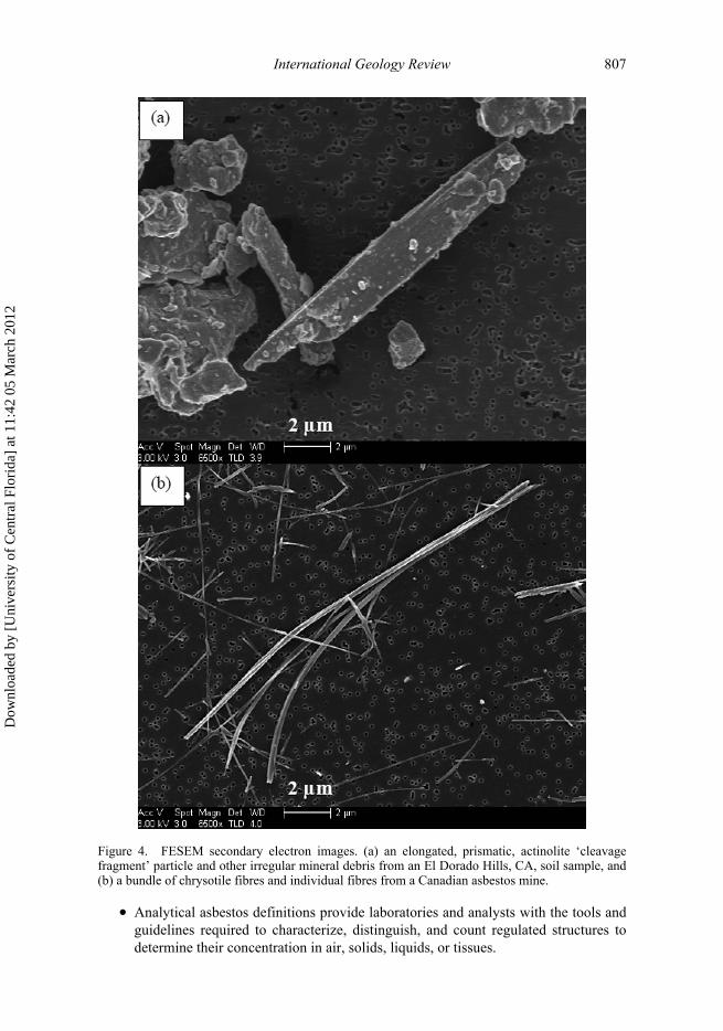
Figure 4 illustrates two field emission scanning electron microscopy (FESEM)secondary electron images comparing a prismatic amphibole cleavage fragment (Figure 4a) toa chrysotile fibre bundle displaying a splayed end (Figure 4b). The cleavage fragment width,approximately 2.2 μm, is more than 10 times greater than the width of the chrysotile fibrebundle and more than 100 times greater than the width of an individual chrysotile fibril. Notealso that the chrysotile fibre bundle (Figure 4b) exhibits curvature or splayed ends whichare not inherent in cleavage fragments.
The asbestiform habit can be defined microscopically by the following morphologicalcharacteristics (Perkins and Harvey 1993).
• Mean aspect ratio between 20:1 and 100:1, higher for fibres longer than 5 μm.• Very thin fibrils, usually less than 0.5 μm in width.• Two or more of the following:
� parallel fibres occurring in bundles;� fibre bundles displaying splayed ends;� matted masses of individual fibres; and� fibres showing curvature.
The mineralogical community usually expands these optically based morphological char-acteristics to include additional information available by electron microscopy-related tech-niques such as selected area electron diffraction (SAED) to determine a particle’scrystallographic characteristics and energy dispersive X-ray spectroscopy (EDS or EDX)to determine elemental composition. In summary, there are measurable and quantifiablemineralogical distinctions between asbestiform and non-asbestiform minerals. Unfortu-nately, these distinctions have not been accepted by regulatory organizations and have notbeen incorporated into the adopted regulations as discussed below and in Part II.
Commercial, regulatory, and other asbestos definitionsOver the years, asbestos has typically been defined and used in at least four different mannersdepending on the specific context (Glenn et al. 2008).
• Commercial asbestos definitions highlight the properties of asbestos that impartcommercial value, such as high tensile strength, low thermal and electrical conduc-tivity, high heat resistance, and high mechanical and chemical durability.
• Regulatory asbestos definitions are generally intended for occupational settings andidentify asbestos minerals and asbestos-containing materials (ACMs) to be regu-lated from those that should not be regulated.
• Mineralogical and geological asbestos definitions distinguish asbestiform mineralsfrom non-asbestiform particles (e.g. elongated single-crystal minerals and cleavagefragments) based on their crystal structure, chemistry, morphology, and/or mecha-nism of formation employing traditional analysis techniques which are presentedseparately below.
Dow
nloa
ded
by [
Uni
vers
ity o
f C
entr
al F
lori
da]
at 1
1:42
05
Mar
ch 2
012
International Geology Review 807
• Analytical asbestos definitions provide laboratories and analysts with the tools andguidelines required to characterize, distinguish, and count regulated structures todetermine their concentration in air, solids, liquids, or tissues.
Figure 4. FESEM secondary electron images. (a) an elongated, prismatic, actinolite ‘cleavagefragment’ particle and other irregular mineral debris from an El Dorado Hills, CA, soil sample, and(b) a bundle of chrysotile fibres and individual fibres from a Canadian asbestos mine.
Dow
nloa
ded
by [
Uni
vers
ity o
f C
entr
al F
lori
da]
at 1
1:42
05
Mar
ch 2
012
808 B.R. Strohmeier et al.
These asbestos definitions are not consistent, and difficulties arise when attempting healthrisk assessments. For example, the commercial asbestos definition focuses on the econom-ically desirable properties that historically made the different asbestos minerals useful forimproving the physical properties of commercial products. However, the commercialdefinition by itself does not offer the precision that a mineralogist would use to permitaccurate scientific distinction between what constitutes an ‘asbestos’ mineral fibre andwhat does not, or that an analyst would ask when counting the number of fibres, or theamount in weight or volume per cent in a sample. Mineralogy and geology were two of thefirst scientific disciplines to describe asbestos, and to those groups, the term means thatthe material has a specific fibrous form, i.e asbestiform (Gunter et al. 2007). ‘Fibre’ is atextural term meaning that the material looks, and more importantly behaves, like a fibre,e.g. the material can curve and bend under force in contrast to cleavage fragments, whichresult from splitting of single crystals on grinding for example.
In contrast to the commercial and mineralogical definitions of asbestos, the regulatorydefinitions were created to characterize hazardous airborne particles that could be releasedfrom asbestos raw materials or manufactured products. The Occupational Safety andHealth Administration (OSHA) is responsible for regulating asbestos in the US workplace.The six minerals that are classified and regulated as asbestos by the OSHA include oneserpentine and five (of approximately 80) amphibole minerals. Chrysotile asbestos hasbeen the most commonly used form of asbestos in manufactured products (Clinkenbeardet al. 2002; Ross and Nolan 2003; Lee et al. 2008b).
Regulators have defined the term ‘fibre’ in various ways based on particle aspect ratioand the method used to conduct the analyses. For example, a specific particle is typicallyconsidered to be a fibre if it has an aspect ratio greater than 3:1 by light microscopy orgreater than 5:1 by electron microscopy (Gunter et al. 2007). Particle aspect ratio criteriawere never meant to define asbestos but were developed as counting criteria for use inindustrial settings where the source of the airborne fibres was a commercial asbestos prod-uct. The regulatory community developed these counting criteria to determine whether afibrous particle met certain health-based concerns (Gunter et al. 2007). Unfortunately, insome laboratories, aspect ratio has become the primary or sole means of asbestos fibredefinition. This practise is at odds with the definitions used by mineralogists and with riskmodels that do not define asbestos according to simple shape characteristics (Gunter et al.2007). The use of a dimensionless parameter such as aspect ratio does not recognize theactual length and width dimensions of the fibre or particle and is, therefore, of little or nouse when discussing exposure or toxicological outcome (Wylie et al. 1993). An arbitrarilydefined small aspect ratio, e.g. 3:1, will not only include all asbestos fibres, whose ratio istypically greater than 20:1, but will also include many other non-asbestos elongated min-eral particles, especially in the mixed mineral dusts found in a typical natural environment(Lee et al. 2008b).
Winchite and richterite are two amphibole minerals that may occur with asbestiformmorphology. The amphibole mineral group is characterized by complex elemental substi-tution within the crystal lattice that designates the amphibole structures. Except for the fiveregulated amphiboles discussed above, there are a few fibrous amphibole minerals that werenever classified or regulated as asbestos, because there were no known commercial asbesti-form mineral deposits. These amphiboles were not incorporated in manufactured productsand, as a result, were not regulated. However, some of these non-regulated amphiboles dogrow with asbestiform morphologies and occur as impurities in otherwise non-asbestos oredeposits. For example, at the vermiculite mine near Libby, MT, a small percentage (less than10%) of non-regulated winchite [(NaCa)Mg4(Al,Fe3+)Si8O22(OH)2] and richterite
Dow
nloa
ded
by [
Uni
vers
ity o
f C
entr
al F
lori
da]
at 1
1:42
05
Mar
ch 2
012

International Geology Review 809
[Na(NaCa)(Mg,Fe2+)5Si8O22(OH)2] amphibole particles, which crystallized as asbesti-form fibres, contaminated the vermiculite [(Mg,Ca,K,Fe2+)3(Si,Al,Fe3+)4O10(OH)2·4H2O]ore (Ross and Nolan 2003; Gunter et al. 2007). These asbestiform amphiboles caused ser-ious pulmonary health problems and deaths among former Libby vermiculite miners (McDon-ald et al. 2004; Bandli and Gunter 2006). Because of the potential health effects associatedwith asbestos and other asbestiform minerals, it is important to accurately identify asbesti-form amphibole minerals by establishing their chemical composition and structure.
Fluoro-edenite: a newly identified calcic amphiboleFluoro-edenite {NaCa2Mg5Si7AlO22F2} is a member of the calcic amphibole mineralgroup and was first identified in 2001 (Gianfagna and Oberti 2001). It occurs in bothprismatic and fibrous morphologies in volcanic rocks on the flank of Mt Etna nearBiancavilla, Sicily, Italy (Paoletti et al. 2000; Soffritti et al. 2004; Burragato et al.2005; Gianfagna et al. 2007). Fluoro-edenite found within the Mt Calvario stonequarry occurs as fibres, with lengths ranging from 12 to 40 μm and widths rangingfrom 0.4 to 1 μm (note the aspect ratio) (Burragato et al. 2005). These fibres were orig-inally identified as tremolite/actinolite but were later found to be a distinctive amphib-ole fluoro-edenite.
A recent TEM study of amphiboles from Biancavilla also found tremolite asbestosassociated with the fluoro-edenite (Andreozzi et al. 2007). The amphiboles were identi-fied by TEM using EDS and SAED analyses. The tremolite asbestos was characterizedby fibres thinner than 0.1 μm with very high aspect ratios (greater than 50:1). Forexample, Figure 5 shows TEM and FESEM images obtained for a tremolite asbestosfibre found in a sample of fluoro-edenite from Biancavilla. The fibre has a length of∼17 μm and a width of ∼0.2 μm (aspect ratio = ∼85:1). For the several amphibole mineralsfound in this geographic site, which ranged from fibrous to prismatic morphology,TEM/EDS data demonstrated that the aluminium content of the crystals was correlatedwith fibre width. The asbestiform fibres have very low aluminium content (less than 0.3aluminium atoms per formula unit) in comparison to the prismatic edenite particles,which contained much higher aluminium contents (greater than 0.5 aluminium atomsper formula unit). These findings correspond to data published in mineralogical com-pendia (Dorling and Zussman 1987; Deer et al. 1997; Verkouteren and Wylie 2000).
Asbestiform fluoro-edenite and tremolite from Biancavilla, as well as asbestiformwinchite and richterite from Libby, MT, were never associated with commercialasbestos deposits, but rather occurred as asbestiform amphibole contaminates associ-ated with the building stone in Biancavilla or in vermiculite ore deposits in Libby. Ascontaminates, they received little attention until their health effects became apparentmany years after initial exposure. Future investigations on the different fibres in Bian-cavilla, for example, will be needed to clarify the health effects of these minerals(Andreozzi et al. 2007).
Erionite – a fibrous zeoliteZeolites are a common mineral group composed of hydrated aluminosilicates of alkali andalkaline earth metals that can have both fibrous and non-fibrous morphologies. One of thefibrous zeolites is erionite {[Na2K2CaMg]4–5[Al9Si27O72]·27H2O}. Erionite usuallyoccurs as thin fibres having a woolly appearance. Fibrous zeolites, such as erionite, are notclassified as asbestos or asbestiform. However, erionite was determined to induce a high
Dow
nloa
ded
by [
Uni
vers
ity o
f C
entr
al F
lori
da]
at 1
1:42
05
Mar
ch 2
012

810 B.R. Strohmeier et al.
incidence of malignant pleural mesothelioma through environmental exposures to respira-ble fibres of erionite in the Cappadocian region of central Anatolia in Turkey (Dogan2003). The volcanic tuffs in this region contain respirable fibres of erionite, and these tuffshave been excavated and quarried to provide caves and building materials for homes.Although there are significant amounts of tremolite asbestos and chrysotile in the region,extensive studies have concluded that the erionite was the probable cause of the observedmesotheliomas (Emri et al. 2002; Dogan 2003; US DHHS 2005; Dogan et al. 2006;Carbone et al. 2007; Dikensoy 2008; WDNR 2009).
In general, zeolite-type materials have useful physical and chemical properties andare widely employed in industry. At one time, erionite was used commercially as ametal impregnated catalyst in the hydrocarbon-cracking process because of its opencrystal structure. It has now been replaced by synthetic, non-fibrous zeolites (WDNR2009). A minor and probably unintentional use of erionite-rich blocks was as buildingmaterials (WDNR 2009). For example, there are homes made of erionite-rich blocks inOregon and weigh-stations made of the same materials in Nevada (Dogan 2003). Theuse of erionite to increase soil fertility and to control odours in livestock production hasalso been studied (WDNR 2009). Deposits of fibrous erionite also occur naturally inweathered volcanic ash within the western USA, including Arizona, Nevada, Oregon,California, and Utah, where they present a potential environmental exposure risk(Dogan 2003).
Figure 5. TEM image and FESEM secondary electron images of a tremolite asbestos fibre in afluoro-edenite sample from Biancavilla, Sicily, Italy. (a) TEM full particle image, (b) FESEM fullparticle image, (c) FESEM image of particle left end, and (d) FESEM image of particle right end.
Dow
nloa
ded
by [
Uni
vers
ity o
f C
entr
al F
lori
da]
at 1
1:42
05
Mar
ch 2
012

International Geology Review 811
Synthetic inorganic fibresAsbestos is not the only inorganic fibre that has been used to impart strength, fire resist-ance, thermal insulation, or electrical insulation in manufactured products. Syntheticfibrous inorganic materials have become commonplace in our everyday lives. There are awide variety of man-made fibres in the environment such as mineral ‘wool’ and fibresmade of glass, ceramics, and organic polymers (Blake et al. 1998; Carpenter and Wilson1999; Bernstein 2007). The utility of these fibrous materials continues to spur the develop-ment of new types of fibres and new applications. For example, glass fibres or fibreglasshas become a major construction material for insulating residential and commercial build-ings. Glass fibres have also replaced copper wire in some intercontinental telephonesystems. High temperature refractory fibres are used in industrial furnace applications.Carbon and graphite fibre composites are used to make tennis racket frames and golfclubs. Synthetic inorganic fibrous materials have largely replaced asbestos and havebecome commonplace.
Since the recognition of asbestos as a carcinogen, there has been a worldwide concernover whether any synthetic fibres are carcinogenic, and significant research into thetoxicity and biological activity of fibrous materials with different chemistries has beenundertaken. Similar to studies of asbestos minerals, this research has demonstrated that thepotential for lung disease is strongly related to the size and biopersistence of long, thinfibres, which are small enough to be deposited deep into the lung (Blake et al. 1998;Carpenter and Wilson 1999; Bernstein 2007).
Analytical methodologiesHistorical evolution of asbestos analysisOne of the most important aspects of dealing with asbestos minerals is their proper identi-fication and characterization. Many different procedures were developed over the past 100years, commencing with personal observations of hand samples to today where we havethe ability to analyse the extremely fine respirable airborne fibres. Beginning with PLMand continuing through TEM, analytical techniques were devised to identify asbestos in abulk sample of mixed minerals, soils, or construction materials. These techniques are nowused to count asbestos fibres collected on an air sample filter in an occupational setting orduring an asbestos abatement project (Walton 1982; Baron 1994; Santee and Lott 2003).Historically, the development of current methods focused on the analysis of commercial-grade chrysotile asbestos found in workplaces where asbestos was being manipulated orprocessed (Walton 1982). Several techniques, now obsolete, were used for asbestos meas-urements until the late 1960s (Paulus 1942; Santee and Lott 2003). Before this time, it wasnot widely recognized that the fibrous nature of asbestos was intimately related to itstoxicity; therefore, these early techniques typically involved collecting airborne particlesand counting all ‘large’ particles (length ≥1 μm) at low magnification by optical micros-copy (Paulus 1942). Thermal precipitators, impactors, impingers, and electrostatic precip-itators were used to sample suspected airborne asbestos particles (Walton 1982; Baron1994). Note that no attempt was made to accurately determine the mineral species.
In the early 1960s, air filter collection of particulates and analysis were first conductedin the UK and later in the USA (Ayer et al. 1965). As studies on asbestos-induced diseaseincreased, cellulose-based membrane filter sampling was applied and higher magnifica-tion (approximately 400× magnification) phase contrast microscopy (PCM) was initiatedfor counting fibres (Edwards and Lynch 1968; Walton 1982; Baron 1994). The PCM
Dow
nloa
ded
by [
Uni
vers
ity o
f C
entr
al F
lori
da]
at 1
1:42
05
Mar
ch 2
012

812 B.R. Strohmeier et al.
method involves drawing air through a mixed cellulose ester (MCE) filter to capture anyparticles with a focus on airborne asbestos fibres. A wedge-shaped portion of the filter isplaced on a glass microscope slide and made transparent so that an area (‘field’) can beviewed by PCM. All of the fibres meeting the defined criteria for asbestos were countedand were considered to be a measure of the airborne asbestos concentration.
Because the toxicity of asbestos appears to be related primarily to fibre length andwidth, analytical methods focus on providing information on those parameters, as well ason the total number of fibres and mineral type. In general, samples are visualized using anoptical or electron microscope by trained technicians. Fibres and any other particles aretypically viewed on filters at magnifications specified by the method used and countedaccording to the regulatory rules and capabilities of each method. The primary asbestoscharacterization techniques in use today include PCM, PLM, and TEM, with growinginterest in SEM. These techniques are described in more detail below.
Phase contrast microscopyIn 1970, the first regulatory PCM method for asbestos was established to assess industrialexposures and evaluate airborne fibres in the workplace where commercial asbestos wasin use (Martonik et al. 2001). In 1977, the National Institute for Occupational Safety andHealth (NIOSH) issued the first PCM method – the Physical and Chemical AnalyticalMethod (P&CAM) (NIOSH 1977), which was updated (NIOSH 7400) in 1994 followingstudies that showed variability in earlier determinations due in part to the qualities of themicroscopes (NIOSH 1994). The NIOSH 7400 PCM method specified sample collectionprocedures, filter and microscope qualities, and counting protocols. The NIOSH (1977)and NIOSH 7400 PCM methods both arbitrarily count as fibres all particles visible in themicroscope that are at least 5 μm long and have a minimum aspect ratio of 3:1 (Walton1982; NIOSH 1989b, 1994). In workplaces where asbestos was mined, processed, or used,it was a safe assumption that the majority of particles fitting the simple counting ruleswere asbestos. Unfortunately, the dimensional criteria of the counting rules have beenincorrectly used by some as a de facto definition of asbestos.
There are four main advantages of PCM over other methods (OSHA 1988):
• the technique is specific to fibres and excludes non-fibrous particles from the ana-lysis;
• the technique is inexpensive compared to electron microscopy techniques and doesnot require specialized knowledge to carry out the analysis for total fibre counts;
• the analysis is relatively quick and can be performed on-site for rapid determinationof air concentrations of asbestos fibres; and
• the technique has continuity with the epidemiological studies that have been per-formed on samples over a long time span so that estimates of expected disease canbe inferred from past determinations of asbestos exposures.
The main disadvantage of PCM is that it does not positively identify asbestos fibres. Otherparticles with fibrous morphology, or satisfying the dimension criteria which are notasbestos, may be included in the count unless differential counting is performed. Itrequires a great deal of experience to adequately differentiate asbestos from non-asbestosfibres. This is an important limitation when the method is used in settings where fibre con-centrations with a significant non-asbestos fraction may occur. In such cases, positiveidentification of asbestos must be verified by PLM or electron microscopy techniques. A
Dow
nloa
ded
by [
Uni
vers
ity o
f C
entr
al F
lori
da]
at 1
1:42
05
Mar
ch 2
012

International Geology Review 813
further disadvantage of PCM is that it will not resolve the finest asbestos fibres encoun-tered during an analysis (approximately 0.02–0.25 μm diameter); so, for some exposures,more fibres may be present than are actually counted. Therefore, the PCM method is onlyan index of exposure and uses the assumption that the detected particles are correlatedwith the fibres actually causing disease (Baron 1994). The primary purpose of the stand-ardized PCM methods was never to discriminate between asbestos and non-asbestosfibres, but only to monitor and control the airborne commercial asbestos fibres in order toreduce the incidence of disease. Since its adoption, the PCM method has become thegenerally accepted technique used for exposure and risk assessments from which doseresponse assessments are derived (Walton 1982; Bailey 2004; Berman 2006). However, itshould be noted that the PCM minimum 3:1 aspect ratio was not based on any scientificdefinition of asbestos characteristics or the toxicological significance of the ratio but sim-ply reflected a need to improve consistency in ‘exposure’ measurements by analysts fromdifferent laboratories. A dimensional criterion specifying a minimum aspect ratio of 3:1for particles longer than 5 μm is not valid for the analysis of mixed mineral dusts simplybecause in most natural environments there are too many non-asbestos particles thatwould fit this simple definition. Especially in mixed mineral environments, the standardPCM methods do not provide enough information to differentiate between asbestos andnon-asbestos mineral particles.
The PCM technique can be extended beyond the standard methods to provide addi-tional information concerning airborne fibres in mixed mineral environments., i.e. toaddress the wide spectrum of particles that may be present in airborne samples. The parti-cles can range from short, wide fibres to very long, thin fibres, and there is a general con-sensus among health experts that long, thin fibres present more of a health risk than short,wide fibres. However, a controversy exists concerning the particles that fall in the middleof this length–width continuum. The risk of these intermediate-sized fibres is not wellunderstood. Because the health effects of the intermediate-sized fibres are not establishedand the contribution to disease from very thin non-PCM countable fibres is not quantified,there is uncertainty as to how to handle these particles. Should the intermediate-sizedparticles be differentiated from the long, thin asbestos fibres? If it is found that sorting ofthe particle population is necessary from a risk perspective, what is the most cost-effectivemethod to achieve this goal? Some type of screening method is necessary as an initial stepin the analytical process of mixed mineral environments. The ultimate goal of the screen-ing step would be to provide information on the size distribution of the particles andfibres. If no high-risk fibres are detected, then no additional analysis may be necessary. Ifan elevated population of potentially high-risk fibres is discovered, the most appropriatetechnique to accurately measure and unequivocally identify the presence of asbestos willneed to be used.
The American Society for Testing and Materials (ASTM) recently implemented ascreening method based on the PCM technique for determining an index of occupationalexposure to airborne fibres in mines, quarries, or other locations where ore may beprocessed or handled (ASTM 2006). ASTM recognized and addressed the complexity ofanalysing for asbestos in such mixed mineral dust atmospheres by developing a rapidscreening optical protocol. This protocol preserves the information obtained in the con-ventional PCM analysis but adds discriminate analysis to identify samples with significantnumbers of long, thin fibres. The method provides an estimate of the fraction of countedfibres that may be asbestos by classifying the fibres (longer than 5 μm and an aspect ratioof 3:1) into three groups: (1) fibres that show curvature, splayed ends, or appearance ofbundles; (2) fibres that are longer than 10 μm or thinner than 1.0 μm; and (3) all other
Dow
nloa
ded
by [
Uni
vers
ity o
f C
entr
al F
lori
da]
at 1
1:42
05
Mar
ch 2
012

814 B.R. Strohmeier et al.
countable fibres. If an elevated content of long thin fibres is detected optically, the ASTMmethod recommends supplemental electron microscopy analysis for verification. Thistype of approach, which differentiates particles of different size ranges and different phys-ical characteristics, is the first step in screening mixed mineral samples.
Following the screening step for mixed mineral environments, additional analysesmay be necessary to accurately measure and unequivocally identify asbestos. Althoughthe complete chemical, morphological, and crystallographic analysis of every particle in amixed mineral sample would be ideal, it may not be realistic because of time and costlimitations. Maximum effort needs to be focused on the identification and classification ofthe particles that pose the most risk. More uncertainty might be acceptable with a fullidentification of the particles discriminating those that pose less of a risk. The challengefor scientists and future policy makers will be to streamline and efficiently organize thesteps most appropriate for analysing fibres that present the most risk in airborne samplesof mixed mineral dusts.
Polarized light microscopyOptical microscopy, specifically PLM, has been used to analyse rocks and minerals forwell over 100 years, and it is a well-known analytical procedure (McCrone 1980; Baron1994; Bloss 1999; Santee and Lott 2003; Gunter 2004; Gunter et al. 2007). Tables of theoptical properties of the many hundreds of mineral species, as determined by oil immersiontechniques, were first published in 1900 (Larsen and Bermen 1934). Originally used forexamining thin sections of rocks or mounts of mineral grains, PLM is now used for deter-mining the mineral content of materials that may include asbestos such as soils, buildingmaterials, raw materials used in various manufacturing processes, and ore/host rock sam-ples; this is different than the PCM analysis of MCE filters discussed above. For PLManalysis of bulk samples, it is assumed that the sample being analysed has been properlycollected, documented, and is representative of some larger population of material. TheUS Government arbitrarily classifies a material as ‘asbestos-containing’ if the concentration ofasbestos exceeds 1% by weight.
In 1982, the Environmental Protection Agency (EPA) issued an Interim Method (USEPA 1982) that created a uniform procedure for identification and quantification of asbes-tos in bulk building materials using the PLM. The method, published in the Code ofFederal Regulations (currently at Appendix E to Subpart E, 40 CFR 763), required the useof ‘point counting’ or modal analysis techniques to quantify the asbestos content of thematerial. Alternatively, a visual estimation technique can be used for quantification(NIOSH 1989a; Crane 1992). NIOSH recommends using visual estimation of the sampleby PLM to quantify the amount of asbestos.
Recognizing that there were problems with established protocols for the analysis ofbulk and raw materials, several states issued their own PLM methods. The California AirResources Board (CARB) issued Method 435 for use in determining the asbestos contentin serpentine aggregate in storage piles, on conveyor belts, and on surfaces such as roads,shoulders, and parking lots (CARB 1991). The State of New York issued a PLM methodthat utilizes a stratified point count method for quantification of the asbestos content ofmaterials with ‘substantial amounts of asbestos’, but says a visual estimation of thecontent is acceptable (ELAP 1990).
An optical procedure published by the European Union (EU) was specifically designedfor the determination of low concentrations of asbestos in bulk materials (Schneider et al.1997). The procedure is similar to that of OSHA in that it uses a combination of PLM with
Dow
nloa
ded
by [
Uni
vers
ity o
f C
entr
al F
lori
da]
at 1
1:42
05
Mar
ch 2
012

International Geology Review 815
PCM. However, the EU method specifies a number of procedures to use in removing thematrix material, thus improving the precision and accuracy of the asbestos determination.
The standard method using PLM for asbestos analyses in the USA is the EPA 600Test Method (Perkins and Harvey 1993). This method was developed for asbestos deter-mination in bulk materials or bulk commercial products. In general, the bulk material isexamined for sample heterogeneity. Macro characteristics such as obvious layering inthe material, colour, fibrous components, and general appearance are noted. The sam-ples are ground, teased, or chemically treated to disassociate the fibres from the matrixmaterial. Multiple grain mounts are prepared and analysed. If asbestos is observed, thetype is identified by refractive index measurements, usually with the aid of central stopdispersion staining as described by Bloss (1999). After identification of asbestos type ortypes, it becomes necessary to quantify the asbestos content. This is done by visual areaestimation or for low-level concentrations; a 400-point count is usually employed forquantification.
There are appreciable errors associated with visual area estimation because of analystbias and sample heterogeneity. In addition, the regulated limit of asbestos in bulk materi-als is always expressed in weight per cent, but the measured asbestos content is based onvolume or area per cent, creating an additional source of error. Differences in a particle’sdensity and size will affect analytical results, but depending on the percentage of asbestospresent, this error may or may not be significant. For example, a bulk material containing20% by weight asbestos with an error of 5% still exceeds the Federal OSHA allowableregulatory limit of no greater than 1 wt.%, but for materials containing very lowconcentrations of asbestos, a relatively small error may be the difference between requiredabatement and no action.
An example of the errors associated with the use of PLM methods is shown in theNational Volunteer Lab Accreditation Program (NVLAP) proficiency testing and accredi-tation programme, which is administered by the National Institute for Standards and Technol-ogy (NIST) (Richmond and Faison 2003). Proficiency testing takes place bi-annually toaccredit and reaccredit laboratories for asbestos identification and estimation. The resultsfor four samples from the 231 laboratories participating in the August 2007 proficiencytesting (NVLAP 2007) showed that, for the qualitative part of the analysis, 3% of the laborato-ries incorrectly identified asbestos in Sample 1 and 0.4% of the laboratories incorrectly identi-fied asbestos in Sample 2. No laboratories misidentified the type of asbestos in the twosamples containing chrysotile. However, the ‘acceptable range for the analysis’ over-lapped the regulatory limit of 1.0%, i.e. Sample 3 was an ACM and yet within analyticaluncertainty it could be reported as under the regulatory limit, whereas Sample 4 wasbelow the 1.0% limit and yet within analytical uncertainty it could be reported as over theregulatory limit.
Spindle stage-assisted PLM for the characterization of asbestosThe spindle stage is an accessory to the PLM and provides the ability to rotate a particleabout a horizontal axis with respect to the plane of the microscope stage (Bloss 1981, 1999;Gunter 2004; Gunter et al. 2004; Dyar and Gunter 2008). For routine PLM asbestos analysis,such as the EPA 600 method (Perkins and Harvey 1993), the spindle stage has no practicaluse. However, for studies requiring detailed optical characterization of the mineral species,the spindle stage is invaluable. In brief, the spindle stage allows for dimensional and opticalcharacterization of single crystals or aggregates, with the added benefit of being able toanalyse the same particle with X-ray diffraction and electron beam instruments.
Dow
nloa
ded
by [
Uni
vers
ity o
f C
entr
al F
lori
da]
at 1
1:42
05
Mar
ch 2
012

816 B.R. Strohmeier et al.
Verkouteren and Wylie (2000) studied the variations in the amphibole group tremoliteto ferro-actinolite, a solid solution series (changes in Fe content of the minerals), bydetermining the unit cell, composition, in association with optical properties, and habit. Theoptical properties were analysed using a spindle stage. In the study of 35 samples classified asasbestiform, 19 contained fibres of sufficient size to measure the three principal refractiveindices of a mineral particle (ie a, b, and g), whereas the remaining 16 samples each exhib-ited anomalous optical properties. A later study by these authors explored the anomalousproperties of asbestiform amphibole said to have a ‘byssolitic’ habit (Verkouteren andWylie 2002). They defined byssolitic as:
. . . samples that occur as single fibers, sometimes loosely aligned, that have a vitreous lusterand are easily reduced to a powder by hand grinding. Individual ‘byssolitic’ fibers are often tab-ular in cross-section with well-developed (100) faces and widths of at least a few micrometers.
These byssolitic particles are fibrous amphiboles but do not meet the dimensional width cri-teria of the asbestiform habit. Using the spindle stage, Verkouteren and Wylie (2002) char-acterized the anomalous optical extinction of byssolitic fibres and concluded that theseproperties were most likely a result of twinning (a characteristic of some mineral forms) onthe (100) crystal plane. A study by Sanchez et al. (2008) examined the optical characteristicsof tremolite samples with differing morphologies. The study found that fibres of narrowwidth, resulting from crystal growth rather than cleavage, exhibited near-zero extinctionangles, similar to the results reported by Verkouteren and Wylie (2002) and distinct fromlarger crystalline size particles where the angle expected is greater than 10 degrees.
Brown and Gunter (2003) studied the optical properties of the winchite-richteriteseries of amphiboles from the former vermiculite mine near Libby, MT, the NIST 1867SRM tremolite, and a tremolite from the University of Idaho collection and found thatboth the NIST and Libby amphiboles were predominantly flattened on (100). Anotherobservation on the NIST tremolite was that upon rotation around the spindle axis, somefibres, which originally appeared to be single crystals, were in fact fibre bundles. Hence,the spindle stage enables greater accuracy in describing the natural morphological expres-sion of mineral particles. Bandli and Gunter (2001) also used a spindle stage to performoptical, single-crystal X-ray diffraction, and compositional characterization on elevenamphibole particles from the Libby mine. By employing the spindle stage mount, theyobtained refractive indices, as well as unit cell data, and elemental composition on thesame Libby amphibole particles with TEM techniques, thus allowing them to determinesubtle correlations in physical properties. For instance, they found that, as the particlestook on more ‘asbestos-like’ morphological properties, the partial birefringence (i.e. b–g)of the particles decreased, and the particles exhibited anomalous optical properties. TheirSEM examination showed that the (100) face was commonly expressed by the fibrousamphiboles and suggested this crystal morphology was an expression of different atoms inthe crystal structure being exposed; these compositional/structural distinctions may berelated to the relative difference in adverse health effects of asbestiform amphiboles com-pared to non-fibrous massive amphibole analogues.
More work is needed on the characterization of the fibrous amphiboles coupled withstudies by medical researchers to better understand the pathology of asbestos-related dis-eases. The spindle stage is advantageous for accurately delineating the mineral phase.Detailed work, such as those presented by Verkouteren and Wylie (2000) and Brown andGunter (2003), could potentially give new insights and provide a foundation for improvedanalytical procedures as well as provide insights into mechanisms that lead to the hazards
Dow
nloa
ded
by [
Uni
vers
ity o
f C
entr
al F
lori
da]
at 1
1:42
05
Mar
ch 2
012

International Geology Review 817
and risks associated with asbestos. Bandli and Gunter (2001) also employed the spindlestage for characterizing the length, width, and thickness of a particle facilitated by lookingat the particle in different orientations and also for transferring the particle to an SEM forcollection of additional data. Sanchez (2007) and Gunter et al. (2007) also used thismethod to show the differing morphology of amphiboles from Libby, MT.
Transmission electron microscopyTEM provides particle projection images in the magnification range 1000× to1,000,000×, which allow the determination of particle shape and identification of thecrystal structure of even the smallest asbestos fibres (Walton 1982; Baron 1994; Santeeand Lott 2003). Particle crystal structural data are determined through SAED and, whencombined with EDS, establish elemental composition allowing accurate mineral identifi-cation (Walton 1982; Baron 1994; Santee and Lott 2003) (SAED and EDS are describedin more detail below). Figure 6 shows a TEM image of a single magnesio-riebeckiteasbestos fibre, with a length of approximately 10 μm and a width of approximately0.07 μm (aspect ratio of approximately 143:1). The particle was found in an air samplefrom Libby, MT, and was identified as magnesio-riebeckite based on SAED and EDSmeasurements. The particle’s high aspect ratio, parallel smooth sides, and perpendicularends are characteristic of asbestos fibres and show slight curvature suggesting flexibility.TEM is widely regarded in the USA as the most reliable technique for asbestos analysisowing to its high image resolution, electron diffraction, and chemical identification capa-bilities (Samudra et al. 1977; Walton 1982; NIOSH 1989b; Baron 1994; Santee and Lott2003; Glenn et al. 2008).
TEM can be used for the analysis of bulk air and water samples (Walton 1982; Baron1994; Santee and Lott 2003). Airborne fibre samples for TEM analysis are typically
Figure 6. TEM image of a single magnesio-riebeckite asbestos fibre from a Libby, MT, air sample.(Fibre length ∼10 μm, width ∼0.07 μm, aspect ratio ∼143:1.)
Dow
nloa
ded
by [
Uni
vers
ity o
f C
entr
al F
lori
da]
at 1
1:42
05
Mar
ch 2
012

818 B.R. Strohmeier et al.
collected onto an MCE membrane or polycarbonate (PC) membrane filter (Baron 1994).For the MCE filter type, the filter is chemically collapsed to form a smooth upper surfaceon which the collected fibres are trapped. The filter is etched using a low-temperatureplasma asher (Federal Register, 198740 CFR 763, Appendix A to Subpart E) exposing anyfibres that are trapped on or near the surface of the filter. The filter is coated with a thinconductive carbon film embedding the particles collected on the filter surface. The filter isthen dissolved using an acetone vapour technique, creating an identical carbon replica ofthe filter with the particles embedded into it. The carbon film replica can be transferred toa copper TEM locator grid ready for TEM analysis. This sample preparation method isreferred to as the direct transfer approach because fibres are transferred to the carbon filmwith minimal disturbance (Baron 1994). An alternate approach, referred to as the indirecttransfer technique, is to liberate the particles from the filter by either sonication, dissolv-ing the filter in an appropriate solvent, or ashing the entire filter in a low-temperaturefurnace. Once the particles are liberated, they are then suspended in a measured portion ofpH-adjusted distilled water, sonicated (an ultrasonic bath) briefly to disperse the particlesin the suspension, and an aliquot of the suspension is then deposited onto either a PC orMCE filter for final transfer to the grid carbon film (Baron 1994). With the indirecttechnique, the optimum particle loading for TEM analysis can be obtained; however,the sonication and suspension process can change the apparent size distribution of theparticles and fibres by breaking apart agglomerates or asbestos bundles into singlefibrils potentially causing erroneous results in population-based analytical protocols(Baron 1994).
Evaluations of ambient air samples for asbestos were first performed in the 1970susing electron microscopy, and the first recognized EPA TEM procedure for air sampleswas written by Samudra et al. (1977). The method, revised in 1984, is known as theYamate Method (Yamate et al. 1984) and, although never officially published by the EPA,it ‘became the de facto standard analytical TEM procedure for airborne measurements inthe United States’ according to a report by the Health Effects Institute (HEI-AR 1992).The first fully promulgated air protocol produced by the EPA was a TEM method fortesting the cleanliness of air in schools following abatement of asbestos-containing build-ing materials. Under the authority of the Asbestos Hazard Emergency Response Act(AHERA), the EPA developed a rapid TEM method for use in clearance testing at abatementsites (Federal Register 1987). The method specified sample collection procedures andrequired a direct transfer preparation method. To reduce the analysis time, the AHERAmethod did not require recording of fibre dimensions, but did require listing the fibres aseither greater than 5 μm or less than 5 μm in length. One important change of the AHERAmethod (Federal Register 1987) over the Draft Yamate Method (Yamate et al. 1984) wasthe increase in minimum aspect ratio from 3:1 to 5:1. Many experts on the AHERA com-mittee argued for 10:1 or 20:1 as the minimum, but the decision was not acceptable to thecommittee members who were anxious to accumulate and use the maximum amount ofdata available over the longest time exposure to estimate risk. In addition, a minimumlength for asbestos fibres (0.5 μm) was specified for the first time in order to improve thereproducibility of fibre counts between different analysts and/or laboratories. Recognizingthat not all airborne fibres are asbestos and that the OSHA regulated only asbestos fibres,NIOSH independently issued a TEM asbestos method in 1989, NIOSH 7402 (NIOSH1989b), which was designed for use in conjunction with PCM [i.e. NIOSH 7400 (NIOSH1994)] to allow the determination of the asbestos versus non-asbestos proportion of countablePCM fibres. The NIOSH 7402 method specifies a magnification comparable to the magnifi-cation used in the optical microscope and counts fibres longer than 5 μm, wider than
Dow
nloa
ded
by [
Uni
vers
ity o
f C
entr
al F
lori
da]
at 1
1:42
05
Mar
ch 2
012

International Geology Review 819
0.25 μm, and with an aspect ratio of at least 3:1. OSHA permits the use of the NIOSH7402 method when analysing air samples for OSHA compliance purposes (whenperformed in conjunction with PCM). An international TEM analytical method, ISO10312, was also developed for testing commercial mineral species (ISO 1995).
The TEM methods described above are best suited for counting short fibres. There areseveral factors that contribute to the poor statistics for long, thin fibres when analysing anasbestos population of fibres via TEM. In an ambient airborne asbestos fibre population,the fibre length distribution can vary widely depending on the source, but typically 1–10%of the fibres are longer than 5 μm in length and only 0.1–1% of the fibres are longer than10 μm (Chatfield 1983). As a result, the measurement of all particles, as current TEMmethods require, creates an intrinsically higher uncertainty for the concentration of long,thin fibres than for the fibres less than 5 μm in length. Further, there is an increasedlikelihood that fibres 10 μm or longer in length will intersect a sample grid bar, making itdifficult to accurately determine the fibre total length (Dehoff and Rhines 1968; Yamateet al. 1984). These problems add uncertainty to the proportion of fibres longer than 10 μmthat may be missed during a routine analysis.
Inherent inaccuracy in the measurement of the concentration of long, thin fibres canlead to increased uncertainty in the risk estimates. To measure the length of fibres longerthan 20 μm with the same precision as the width is measured, magnifications between1000× and 10,000× are needed. This will necessitate accurate calibration of the TEMscreen through all magnifications used for analysis, not just the scanning magnification.Low magnifications are required to measure fibre length and high magnifications arerequired to measure fibre width as well as to characterize surface texture and the nature ofthe fibre ends (ASTM 2002). Therefore, to avoid the issues discussed above, SEM can bemore easily used to search for fibres longer than 10 μm in length. Such measurements areaccomplished much more readily in a modern digital SEM than in a conventional TEMbecause of the relative ease of rapidly switching between low and high magnifications,and because sample grid bars are not typically present with an SEM sample preparation(Goldstein et al. 2003).
The usefulness of the TEM is limited in mixed mineral environments because of thenature of the TEM image. A TEM image is a projection of the specimen created from theelectrons that pass through the specimen (see Figures 5–7) (Williams and Carter 1996).The actual shape of the particle, as seen by TEM analysis, is the projection of the overallparticle shape; however, the actual three-dimensional particle shape may be very differentfrom what is observed in the TEM image. SEM can be used to obtain more detailedinformation compared to TEM on the true particle shape and morphology. SEM providescomplementary information to TEM for asbestos characterization and is discussed below.
SAED and EDS with TEMSAED and EDS are used in conjunction with TEM during the analysis of asbestos andother mineral particles (Baron 1994; Santee and Lott 2003; Gunter et al. 2007). SAED isaccomplished by focusing the electron beam on selected particles and capturing the resultingdiffraction pattern photographically, which corresponds to the specific and unique diffrac-tion characteristics of the sample’s crystal structure. SAED patterns obtained for unknownparticles can be measured and matched to published diffraction data for known mineralspecies.
EDS is a qualitative and quantitative analytical technique whereby the electron beamcauses the emission of characteristic X-rays that provide an elemental ‘fingerprint’ of the
Dow
nloa
ded
by [
Uni
vers
ity o
f C
entr
al F
lori
da]
at 1
1:42
05
Mar
ch 2
012

820 B.R. Strohmeier et al.
composition of the imaged particles. The use of SAED and EDS in conjunction with TEMfor identifying commercial chrysotile asbestos fibres in occupational settings is widelypractised and accepted. However, a greater level of scientific rigour must be applied whenusing these techniques for mineral speciation in mixed mineral environments. The SAEDpatterns and EDS spectra for amphibole asbestos minerals are similar to many non-asbestosminerals. The methods commonly employed leave it to the expertise of the analyst to iden-tify and evaluate non-asbestos particle interferences (Van Orden et al. 2008).
SAED analysis of chrysotile is relatively straightforward because the diffraction pat-tern is unique. By comparison, obtaining SAED patterns of amphibole structures is not asstraightforward and can be a complex and time-consuming process depending on the levelof analysis required (Longo 1990). In addition, the SAED patterns for amphiboles areoften similar to other minerals. For example, an SAED pattern exhibiting a row of evenlyspaced reflection spots of around 5.3Å has been improperly used by some commercialTEM laboratories analysing air samples to definitively conclude that a particularelongated structure with an aspect ratio of 3:1 or 5:1 is amphibole asbestos regardless ofconflicting chemical data from EDS analysis and conflicting particle morphology (VanOrden et al. 2008). The 5.3Å spacing is insufficient for determining mineral speciation asit is not unique to amphiboles; it is found in other minerals such as pyroxenes, talc, micas,and clays such as vermiculite (Van Orden et al. 2008). Van Orden et al. (2008) haverecently reported that the angle, phi or Φ, between two rows is of greater use than a 5.3Årow spacing alone for differentiating amphibole structures from non-amphiboles. Usingthe rows of reflection spots and phi, one can define more precisely the hkl (crystallo-graphic) plane in the diffracting crystal precisely and increase the accuracy of precise mineralidentification. Multiple SAEDs on the same particle in different orientations of the crystal-line lattice would also contribute to a definitive mineral identification and are necessary
Figure 7. TEM image of a crocidolite fibre from a human lung tissue sample.
Dow
nloa
ded
by [
Uni
vers
ity o
f C
entr
al F
lori
da]
at 1
1:42
05
Mar
ch 2
012

International Geology Review 821
when the particle is particularly thick or when orientation relative to the electron beam, aTEM sample grid bar, or the sample matrix interferes. It is for this reason that more scien-tifically rigorous TEM asbestos protocols such as ISO 13794 and ISO 10312 recommendthis approach for identifications of amphiboles.
EDS systems are common spectroscopic tools on both TEM and SEM instruments(Kevex Corporation 1983, 1988), and EDS is by far the most routine spectroscopic methodfor chemical analysis related to asbestos identification. However, EDS is perhaps the mostsubjective of the diagnostic tools available for electron microscopy. Results can be signifi-cantly affected by the quality of the detector, collection time, orientation of the particle rela-tive to the detector, orientation relative to the TEM sample grid bars, orientation relative toother particulate, and particle thickness. To help mitigate these issues, it is imperative thatthe X-ray detector be maintained in optimum condition and that the unknowns are comparedto results on standard material collected on the same detector in the same time period and onparticles of comparable thickness and orientation. However, even with the use of standards,EDS should be considered only a semi-quantitative technique because overlapping peaksand the typical background noise will limit sensitivity, especially for light elements.
The current mineralogical nomenclature of amphiboles was defined by Leake et al.(1997). Applying the Leake rules to EDS compositional results provides one clue to theidentification of the particle. However, the Leake nomenclature applies only to amphibolesand does not differentiate amphiboles from non-amphiboles. Unknowns must be evaluatedutilizing the information on non-amphibole mineral phases (Morimoto 1988). As noted bymost analytical procedures, there are numerous minerals that have chemistries similar tothe regulated amphibole minerals such as talc and pyroxene. Therefore, owing to thesimilarity of the chemistry of the five regulated amphiboles with other minerals, it isnecessary, at a minimum, to examine the SAED pattern for the unknown mineral particlein addition to the EDS results.
Applying TEM techniques and methods to distinguish amphibole asbestos from non-asbestos in a mixed mineral environment is a more scientifically sophisticated analyticaltechnique than has been historically required for the identification of commercial gradechrysotile in industrial hygiene air samples. When these methods are incorrectly applied tomixed mineral environments, which do not have commercial asbestos as the primaryairborne fibre, non-asbestiform amphiboles can be misidentified as asbestiform amphib-oles. The amphibole mineral group contains a large number of species with a wide rangeof chemistries. Most mineral environments will contain a variety of minerals as well as acomplex blend of fibres, cleavage fragments, and elongated rock fragments (Lee et al.2008b). Stringent methodology utilizing several techniques and scientific rigour arerequired to correctly identify and quantify a specific mineral within a mineral assemblage.Misidentification can result in costly reformulation of harmless products and/or unnecessaryasbestos abatement projects.
The flowchart procedure shown in Figure 8 was applied to a TEM study of severalmineral samples whose mineral morphology could be observed in hand specimens asranging from asbestos fibres to non-asbestos mineral particles (Van Orden et al. 2008).The procedure involves the stepwise TEM examination of particle morphologies, such asaspect ratio, shape of the particle sides and ends, particle curvature, etc., as well as theparticle SAED and EDS characteristics. Details on the proper use of this flowchart arepresented in Van Orden et al. (2008) and Table 2. Table 2 presents data on several types ofmaterial particles tested using the flowchart and indicates with a reasonable degree ofaccuracy the classification of asbestos versus non-asbestos in mixed mineral samples. Theerror rate using the flowchart was estimated at 5–10% (Van Orden et al. 2008).
Dow
nloa
ded
by [
Uni
vers
ity o
f C
entr
al F
lori
da]
at 1
1:42
05
Mar
ch 2
012

822 B.R. Strohmeier et al.
Figure 8. Flowchart showing the characteristics that can be used to determine whether a particle isasbestos (Van Orden et al. 2008).
Single crystal
Aspect ratio ≥ 5:1
Parallel sides
Curved structure
Perpendicularends
Uniform diffractioncontours
SAED pattern75° ≤ Angle ≤ 90°
Does SAED showtwinning?
Is EDXA consistentwith amphibole?
Asbestos
Yes
Yes
Yes
Yes
Yes
Yes
Yes
No
Yes
Does SAED showsuper-lattice?
amphibole?
Are multiple SAEDconsistent w/
Yes
No
No
Non-asbestos
Yes
No
No
No – irregular or way
No – tapered or irregular
No – angular or stepped
No
Dow
nloa
ded
by [
Uni
vers
ity o
f C
entr
al F
lori
da]
at 1
1:42
05
Mar
ch 2
012

International Geology Review 823
SEM and FESEMSEM operates by focusing a beam of electrons onto the sample surface and scanning thebeam over a selected area (Goldstein et al. 2003). A variety of signals are generated fromthe interaction of the primary beam of electrons and the specimen, including secondaryelectrons, backscattered electrons, Auger electrons, characteristic X-rays, and otherphotons of various energies. The low-energy secondary electrons are scattered from thesample surface and detected above the surface synchronously with the beam scan rate andprovide surface detail and morphological information about the specimen. Asbestos andother mineral particles can be observed at high magnification and with high resolution,and, in addition, SEM can provide semi-quantitative elemental analysis information usingEDS similar to the detail presented above under TEM (Chatfield 1983). SEM has evolvedover the past 30 years into a reliable and effective method for the enhanced morphologicalcharacterization of asbestos fibres (Lee et al. 1977, 2008a; Middleton 1982; Dorling andZussman 1987; Platek et al. 1992; Chisholm 1995; Hartikainen and Tossavainen 1997;Meeker et al. 2003, 2006; Harris et al. 2007; Lee and Strohmeier 2007; Strohmeier et al.2007a, 2007b; Bunker et al. 2008a, 2008b; Huntington et al. 2008). Past concerns over thevisibility of fibres in SEM images have been alleviated by the advent of digital micros-copy (Platek et al. 1992; Williams and Carter 1996; Lee and Strohmeier 2007). Unfortu-nately, only a few standard SEM analytical methods (primarily European) for asbestosexist (WHO 1985; VDI 1991, 1994, 2004; Frasca et al. 2000; ISO 2002).
The development of high-resolution FESEM instruments makes SEM an attractivetechnique to augment TEM analysis and offset some of its morphological limitations forcomplex asbestos sample characterization. FESEM instruments provide much highermagnifications (e.g. greater than 100,000× and up to more than 1,000,000× for someinstruments) and higher image resolution compared to traditional SEMs and most TEMs
Table 2. Application of the TEM flowchart classification procedure to amphibole samples ofknown morphology.
Mineral type Description of hand sampleAsbestos
classification (%)Non-asbestos
classification (%)
Amosite Commercial product, aerosolized to obtain a respirable fraction
95 5
Crocidolite Asbestos amphibole from an ore sample, very long fibres
100 0
Jamestown tremolite Fibrous tremolite used in animal studies, moderate fibre length
70 30
New York tremolite Tremolite ore sample, acicular appearance in hand sample
2 98
NIST SRM 1867a Mixed tremolite fibres and non-fibrous tremolite particles
11 89
North Carolina tremolite Fibrous tremolite, moderate fibre length
84 16
Samples that appeared to be fibrous in hand specimens (i.e. amosite, crocidolite, Jamestown tremolite, andNorth Carolina tremolite) showed very high percentages of fibres classified as ‘asbestos’ using the TEM proto-col, whereas the non-asbestos tremolite sample from New York indicates the population of fibres to be primarilynon-asbestos. The NIST tremolite sample (1867a SRM) was found to be a mixture of both fibrous and non-fibrous particles (Van Orden et al. 2008).
Dow
nloa
ded
by [
Uni
vers
ity o
f C
entr
al F
lori
da]
at 1
1:42
05
Mar
ch 2
012

824 B.R. Strohmeier et al.
(Goldstein et al. 2003). In addition, modern digital FESEMs can also produce high-resolutionthree-dimensional ‘stereo pair’ images on the nanometre to micrometre scale (Goldsteinet al. 2003). Three-dimensional stereo pair SEM images can be used to help determinewhether a given particle is asbestiform or non-asbestiform because of the ability to ‘see’the third dimension. For example, Figure 9 compares a TEM image and FESEM secondary
Figure 9. Comparison of a TEM image and an FESEM secondary electron image of a non-asbestiformrichterite particle from Libby, MT. (a) TEM image and (b) FESEM image.
(a)
(b)
Dow
nloa
ded
by [
Uni
vers
ity o
f C
entr
al F
lori
da]
at 1
1:42
05
Mar
ch 2
012

International Geology Review 825
electron image of a richterite particle from Libby, MT. The TEM image shows anelongated, richterite crystal, with parallel sides, and 16:1 aspect ratio. The FESEM imageillustrates the prismatic nature of this particle based on its smooth cleavage planes, angledand stepped ends, and parallel sides. Together, these TEM and FESEM data identify andvalidate the non-asbestiform morphology of this prismatic richterite crystal.
Bunker et al. (2008b), Harris et al. (2007), Lee et al. (2008a), Lee and Strohmeier(2007), and Strohmeier et al. (2007a, 2007b) described an enhancement of the YamateTEM airborne asbestos method (Yamate 1984) for the analysis of mixed mineral dusts byadding FESEM imaging of each particle that had an aspect ratio of 3:1 or greater. Theprocedure involved the transfer of the TEM sample grid into the FESEM after the TEM/EDS/SAED analyses were completed and the relocation of the particles meeting the ≥3:1aspect ratio criteria on a particle-by-particle basis. The developed protocol required themorphological (by TEM and FESEM), crystallographic (by SAED), and chemical (byEDS) characterization of each particle including the collection of FESEM secondaryelectron images of the full structure, of both structure ends, and of the particle surface.Stereo pair FESEM images of the full particle and the particle surface provide the depthperception information not obtainable from a single secondary electron image. Figure 10shows examples of the TEM and FESEM images that were collected in these studies for abundle of South African crocidolite fibres. The true morphology of the particle is muchmore evident in the FESEM secondary electron images compared to the projection TEMimage. (Note: stereo glasses are required to properly view stereo pair images.)
FESEM imaging also demonstrates that amphibole cleavage fragments have dimen-sions and morphological features very different than true asbestos, as shown in Figure 4.The overall morphology of elongated mineral particles characterized by FESEM can bedescribed using the seven primary classifications: fibre, acicular, prismatic, bladed,bundle, columnar, and irregular. Details on the particle classification system and defini-tions for these particle morphology categories can be found elsewhere but are part of theGlossary (Harris et al. 2007; Strohmeier et al. 2007a). Similar classification terms wereoriginally developed in 1977 by the US Bureau of Mines (Campbell et al. 1977) to differ-entiate between common mineral rock fragments and their asbestiform varieties usingoptical microscopy; the US Bureau of Mines classification was widely recognized and isstill used for the characterization of minerals and mineral dusts. The FESEM particlerelocation process and analysis are essential and complementary elements to the TEManalysis for accurate particle-by-particle examination of mixed mineral dusts. Comparisonof TEM and FESEM images (Figures 11 and 12) illustrate the ability to more sharply vis-ualize small individual particles and identify their true morphology.
Although there are no standard SEM protocols for mixed mineral environments, theUnited States Geological Society (USGS) did note in a recent study on the use of SEM/EDS and other techniques for characterizing environmental asbestos particles that ‘. . .the emerging practise of fully characterizing all particles of potential concern, bothchemically and morphologically, will aid in developing appropriate analytical proce-dures . . .’ (Meeker et al. 2006); Figures 9–12 demonstrate this technique. Clearly, SEMand FESEM, in particular, are techniques that can be used in the development of accu-rate particle-by-particle analysis procedures. Complete characterization (e.g. chemistry,crystallography, and morphology) in mixed mineral dust samples on a particle-by-particlebasis using complementary TEM/SEM protocols aids in the development of andimprovement of standard analytical procedures. In addition, characterization data canassist in the interpretation of epidemiological data and assessment of potential healthrisks that are needed for sound regulatory policies.
Dow
nloa
ded
by [
Uni
vers
ity o
f C
entr
al F
lori
da]
at 1
1:42
05
Mar
ch 2
012

826 B.R. Strohmeier et al.
Another recent advance in imaging technology includes a high-resolution SEM instru-ment (Hitachi Model S-5500) that combines the benefits of FESEM and low-kV scanningtransmission electron microscopy (STEM) in one instrument. The FESEM and STEMimages can be acquired simultaneously, in addition to high-resolution EDS analysis andelement mapping, without moving the sample. The high-resolution STEM capabilities aredemonstrated in Figure 13, which shows a ‘bright field’ (i.e. transmission) STEM imageof a portion of a South African crocidolite fibre with a width of approximately 0.06 μm.The STEM image reveals very fine parallel lines on the fibre surface. The physical natureof these lines is not specifically understood at this time, but the lines may represent the
Figure 10. TEM image and FESEM secondary electron images of a bundle of South African croc-idolite fibres. (a) TEM full particle image, (b) FESEM full particle image, (c) FESEM image of theparticle left end, (d) FESEM image of the particle right end, (e) FESEM image of the particle sur-face, and (f) FESEM stereo pair image of the particle surface.
Dow
nloa
ded
by [
Uni
vers
ity o
f C
entr
al F
lori
da]
at 1
1:42
05
Mar
ch 2
012

International Geology Review 827
Figure 11. Prismatic euhedral single-crystal richterite particle in an air sample from Libby, MT. (a)TEM image, (b) FESEM secondary electron image, and (c) FESEM secondary electron stereo pairimage. The particle was supported by other adjacent mineral debris and is projecting outward from theplane of the sample grid in the FESEM secondary electron image, and this was not evident in the TEMimage. The TEM image indicates that the particle has an aspect ratio ≥3:1 and therefore should becounted as an ‘asbestos fibre’ following the simple counting rules. The FESEM secondary electronimage, however, demonstrates that the particle is not asbestiform. The particle has a prismatic morphol-ogy with crystalline faces and a rough surface. Particle orientation and morphology can be demonstratedeven more effectively by FESEM stereo pair imaging (Strohmeier et al. 2007a).
Dow
nloa
ded
by [
Uni
vers
ity o
f C
entr
al F
lori
da]
at 1
1:42
05
Mar
ch 2
012

828 B.R. Strohmeier et al.
Figure 12. Irregular muscovite mineral particle in an air sample from Libby, MT. (a) TEM image,(b) FESEM secondary electron image, and (c) FESEM secondary electron stereo pair image. TheTEM projection image indicates a particle with an aspect ratio ≥3:1, and therefore this particle wouldbe counted as an ‘asbestos fibre’. However, the FESEM secondary electron and stereo pair imagesindicate that the particle is not asbestiform; it has a sheet-like structure and is projecting outward fromthe plane of the sample grid. The actual shape and dimensions of the particle are quite different thanthose implied by the two-dimensional TEM image. In addition, EDS and SAED analyses indicatedthat this particle was muscovite {KAl2(AlSi3O10)(F,OH)2}, a common rock-forming mineral that hasa layered sheet structure and belongs to the mica group of minerals (Strohmeier et al. 2007a).
Dow
nloa
ded
by [
Uni
vers
ity o
f C
entr
al F
lori
da]
at 1
1:42
05
Mar
ch 2
012
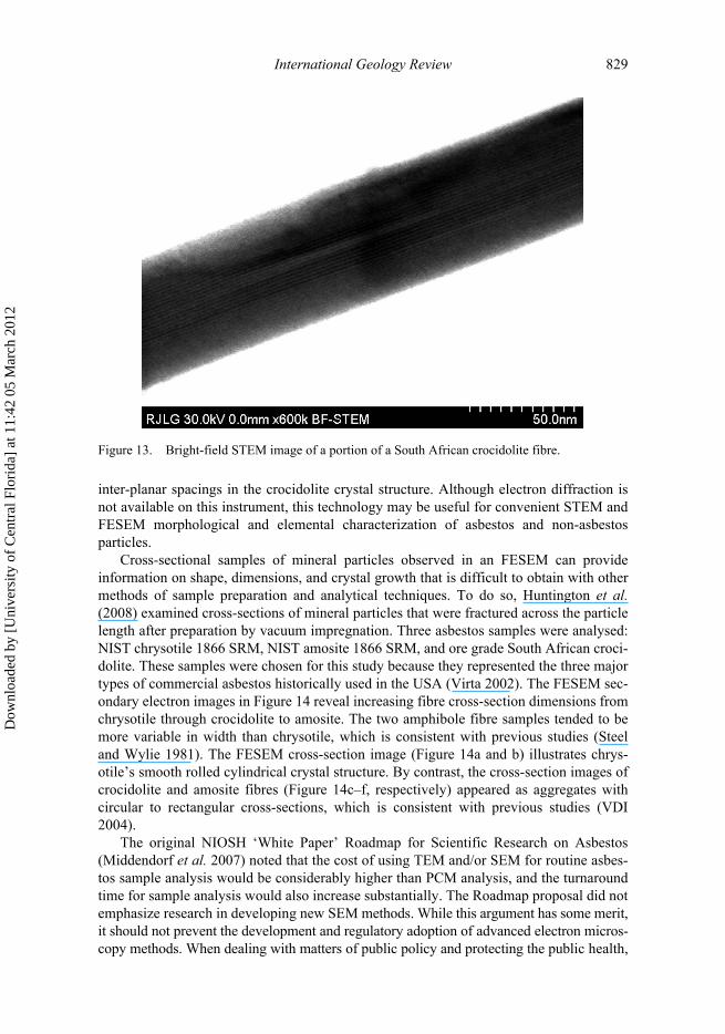
International Geology Review 829
inter-planar spacings in the crocidolite crystal structure. Although electron diffraction isnot available on this instrument, this technology may be useful for convenient STEM andFESEM morphological and elemental characterization of asbestos and non-asbestosparticles.
Cross-sectional samples of mineral particles observed in an FESEM can provideinformation on shape, dimensions, and crystal growth that is difficult to obtain with othermethods of sample preparation and analytical techniques. To do so, Huntington et al.(2008) examined cross-sections of mineral particles that were fractured across the particlelength after preparation by vacuum impregnation. Three asbestos samples were analysed:NIST chrysotile 1866 SRM, NIST amosite 1866 SRM, and ore grade South African croci-dolite. These samples were chosen for this study because they represented the three majortypes of commercial asbestos historically used in the USA (Virta 2002). The FESEM sec-ondary electron images in Figure 14 reveal increasing fibre cross-section dimensions fromchrysotile through crocidolite to amosite. The two amphibole fibre samples tended to bemore variable in width than chrysotile, which is consistent with previous studies (Steeland Wylie 1981). The FESEM cross-section image (Figure 14a and b) illustrates chrys-otile’s smooth rolled cylindrical crystal structure. By contrast, the cross-section images ofcrocidolite and amosite fibres (Figure 14c–f, respectively) appeared as aggregates withcircular to rectangular cross-sections, which is consistent with previous studies (VDI2004).
The original NIOSH ‘White Paper’ Roadmap for Scientific Research on Asbestos(Middendorf et al. 2007) noted that the cost of using TEM and/or SEM for routine asbes-tos sample analysis would be considerably higher than PCM analysis, and the turnaroundtime for sample analysis would also increase substantially. The Roadmap proposal did notemphasize research in developing new SEM methods. While this argument has some merit,it should not prevent the development and regulatory adoption of advanced electron micros-copy methods. When dealing with matters of public policy and protecting the public health,
Figure 13. Bright-field STEM image of a portion of a South African crocidolite fibre.
Dow
nloa
ded
by [
Uni
vers
ity o
f C
entr
al F
lori
da]
at 1
1:42
05
Mar
ch 2
012

830 B.R. Strohmeier et al.
it is vital that the best scientific methods are used to provide accurate measurements forrisk assessments. The original NIOSH Roadmap proposal stated that any routine use ofelectron microscopy methods for counting and sizing fibres would require an analysis ofinter-laboratory and inter-operator variability. Assessment of inter-laboratory and inter-operator variability should not pose a significant problem to implementing improved elec-tron microscopy methods because various laboratory accreditation organizations andround-robin testing protocols already exist to evaluate laboratory and analyst competencefor the PCM and TEM existing methods. Regarding SEM, and FESEM in particular,NIOSH did note in its revised Roadmap proposal that (NIOSH 2008):
Figure 14. FESEM secondary electron images. (a) and (b) chrysotile cross-sections; (c) and (d)crocidolite cross-sections; and (e) and (f) amosite cross-sections (Huntington et al. 2008)
Dow
nloa
ded
by [
Uni
vers
ity o
f C
entr
al F
lori
da]
at 1
1:42
05
Mar
ch 2
012

International Geology Review 831
Ease of sample preparation and data collection for SEM analysis compared to TEM,along with some SEM advantage in visualizing EMP (elongated mineral particles) andEMP morphology (e.g., surface characteristics), suggests a re-evaluation of SEM meth-ods for EMP characterization and mineral identification both for field and laboratoryanalysis.
This apparent change concerning SEM analysis is most likely the result of discussions onthe use of FESEM for the characterization of asbestos and other mineral particles thatwere presented at the Public Meeting for Comments on the original NIOSH Roadmapproposal (Lee and Strohmeier 2007).
It will be important to develop analysis techniques with improved resolution to visu-alize the smaller diameter fibres typical of asbestiform minerals, which pose the highestrisk, and to assure the most complete and accurate fibre counts. The major challenge forscientists is to develop and streamline cost-effective screening techniques to accuratelydetermine the fibres of highest risk (i.e. long, thin fibres) with acceptable uncertaintyand operator variability. This formidable task could possibly be accomplished with par-ticle counting protocols that allow higher uncertainty in the measurement of lower tomedium risk particles (i.e. short, wide fibres). As new methods are developed, it isimportant to point out that reported risk estimates for occupational asbestos exposurewere originally determined by PCM methods. Hence, fibre counts obtained withimproved microscope resolution capabilities would not be directly comparable to cur-rent occupational exposure limits for asbestos without developing meaningful conver-sion factors to compare the new results with the original risk data generated using theolder PCM method.
Raman spectroscopyAlthough it does not have widespread use for asbestos characterization, Raman micro-spectroscopy has been demonstrated in a number of studies to be a simple and effectiveanalytical technique for distinguishing between the six regulated asbestos phases (Bardet al. 1997; Rinaudo et al. 2003, 2004). Raman spectroscopy is a light scattering tech-nique that is used to study vibrational and rotational modes in molecular systems. It ishighly sensitive to the structural variability and particularly the chemical substitutionstypically occurring in the amphibole asbestos minerals. The accurate identification ofthe asbestos phase in question can be attained by analysing the Raman bands corre-sponding to the symmetric and asymmetric stretching modes of the different silicon-oxygen linkages from standard amphibole species. The observed Raman bands are suffi-ciently different for all six asbestos minerals, thus presenting a molecular fingerprintthat allows differentiation between various species of the serpentine and amphiboleasbestos groups. The Raman technique also has the added advantage that no samplepreparation is required. Unfortunately, one Raman study reported no important differ-ences between the Raman spectra of asbestos fibres and their non-fibrous forms, exceptin the hydroxide stretching region (Bard et al. 1997). A more recent study indicated thatUV-Raman spectroscopy was suitably sensitive for supplementing the established Euro-pean technique of SEM/EDS for an unambiguous discrimination of fibrous asbestosmaterials (Petry et al. 2006). It should be noted, however, that the optimum spatial res-olution of typical Raman instruments is only about 1 μm. Therefore, Raman microspec-troscopy cannot be easily used for characterizing single asbestos fibres and at thepresent time is primarily limited to applications on larger fibre bundles.
Dow
nloa
ded
by [
Uni
vers
ity o
f C
entr
al F
lori
da]
at 1
1:42
05
Mar
ch 2
012

832 B.R. Strohmeier et al.
Mössbauer spectroscopyMössbauer spectroscopy is an analytical technique based on the recoilless emission andabsorption of gamma rays, termed the ‘Mössbauer effect’ (Long 1984). In its mostcommon form, Mössbauer absorption spectroscopy, a detector measures the amount ofradiation a solid sample absorbs upon being exposed to a beam of gamma radiation bymeasuring the intensity of the beam after it is transmitted through the sample. Thetechnique gives precise information about the chemical, structural, magnetic, and time-dependent properties of certain chemical materials. However, Mössbauer spectroscopycan only be applied to specific elemental isotopes. Common isotopes measured byMössbauer include 57Fe, 129I, 119Sn, and 121Sb (Long 1984). None the less, Mössbauerspectroscopy is a useful technique for studying the crystal chemistry of many types ofminerals that contain these elements.
Mössbauer spectroscopy has been used to define the ferric ion to total iron ratios(Fe3+/ Total Fe) in amphibole asbestos and to determine the iron cation site location inthe amphibole crystal structure (Luys et al. 1983; Gold et al. 1997; Gunter et al. 2003,2007; Andreozzi et al. 2007; Gianfagna et al. 2007). Mössbauer investigations wereconducted because iron in amphibole asbestos has been implicated in the pathogenicityof inhaled fibres. For example, Gianfagna et al. reported differences in the ferric ion tototal iron ratios for asbestiform and non-asbestiform prismatic morphologies of fluoro-edenites from Biancavilla, Italy (Andreozzi et al. 2007; Gianfagna et al. 2007). Theobserved differences in total iron content and in the site partitioning were attributed todifferences in genetic history between the fibrous and prismatic materials. Specifically,iron in amphibole asbestos is believed to enhance the absorption and catalytic surfaceactivity of the asbestos (Gold et al. 1997). Iron can occur in either the ferrous (Fe2+) orferric (Fe3+) valance states in amphibole asbestos, and each cation has specific site pref-erences in the amphibole crystal structure. In addition, the ferrous ion has been reportedto be more reactive than the ferric ion in certain biologic environments (i.e. the lungs)(Gianfagna et al. 2007). Hence, the total iron weight percentage, the ferric ion to totaliron ratios, and the ferric ion versus ferrous ion site preferences in the amphibole crystalstructure may potentially affect the fibre’s surface chemistry and reactions at the lungtissue interface, although these molecular reactions and distinctions have yet to be pre-cisely determined.
Part II: Asbestos and man: history, health effects, and current controversiesHistorical overview and origins of asbestosAsbestos was first discovered and mined in Cyprus approximately 5000 years ago andwas used in the manufacture of cremation clothes, lamp wicks, hats, and shoes (Rossand Nolan 2003). The word asbestos in Greek meant ‘unquenchable’ or ‘indestructible’(Lee and Selikoff 1979). Asbestos was also used during prehistoric times in Finland,Sudan, and Kenya to make clay pottery with increased strength (Darcy and Alleman2004). Although asbestos-based cloth was in use in Norway, Russia, China, and Italybefore 1860 (Lee and Selikoff 1979), the rapid increase in use as thermal and fire-resistantinsulation occurred with the development of steam technology beginning at that time(Schreier 1989; Ross and Nolan 2003). Major asbestos sources supplying asbestos tothese applications included the reopened Roman-age mines in the northern Italian Alpsand the newly discovered vast chrysotile deposits in Quebec, Canada (Schreier 1989;Ross and Nolan 2003). In addition, large chrysotile deposits were discovered and
Dow
nloa
ded
by [
Uni
vers
ity o
f C
entr
al F
lori
da]
at 1
1:42
05
Mar
ch 2
012

International Geology Review 833
developed in the Ural mountains in the 1880s (Ross and Nolan 2003). By 1890, theasbestos industry was rapidly expanding with hundreds of new applications being intro-duced (Jones 1890).
Crocidolite, a commercial name, not a mineral name, was discovered in 1812 in theNorthern Cape Province of South Africa, but the deposit was not developed until 1910(Lee and Selikoff 1979). Amosite, another coined term for the asbestiform amphibolegrunerite, was discovered in the Transvaal Province, South Africa in 1907, and commer-cial production began in 1916 (Bowles 1955). After World War I, the production and useof asbestos greatly increased. Historically, 95% of the consumption of asbestos has beenchrysotile, with minor amounts of crocidolite and amosite (Ross and Nolan 2003). Thecommercial terms ‘crocidolite’ and ‘amosite’ originated because these were historicallythe most commonly used types of amphibole asbestos. In the case of amosite, the namewas coined for the source of the ore: the ‘asbestos mines of South Africa’. Amosite andcrocidolite, however, are no longer mined (Virta 2002). Anthophyllite and tremolitewere also used in very limited quantities for speciality products, but actinolite hadalmost no commercial value. Tremolite and actinolite are regulated, however, becausethey can occur as accessory minerals in other economically important mineral depositsthat are mined (Gunter et al. 2007). Other mineral fibres, which did not exhibit the phys-ical, chemical, and thermal characteristics ascribed to asbestos described above, hadlittle or no commercial value and were not considered to be asbestos. The total worldproduction of all forms of asbestos between 1931 and 1999 was 166 million tonnes, ofwhich 90–95% was the chrysotile variety (Ross and Virta 2001). Today, Russia is theworld’s leading producer of chrysotile asbestos, followed by China, Kazakhstan, Can-ada, and Brazil (USGS 2008). The last US chrysotile asbestos mine operating near KingCity, CA, was closed in 2002; hence, all asbestos currently being used in the USA isimported (USGS 2008).
During the twentieth century, asbestos was incorporated as functional components inthousands of commercial products. Asbestos applications included fire protection, heat orsound insulation, fabrication of papers and felts for flooring and roofing products, pipelinewrapping, electrical insulation, thermal and electrical insulation, friction products in brakeand clutch pads, asbestos-cement products, reinforcing agents, vinyl or asphalt tiles, andasphalt road surfacing (Virta 2001, 2002). Figure 15, a sample of woven asbestos cloth,illustrates the flexibility and high length to width ratio of asbestos fibres. Asbestos clothhas been commonly used in welding, fire protection, and safety clothing. Despite its manydesirable material properties, inhalation of asbestos fibres may pose a serious potentialhealth risk primarily from occupational exposure to ore-grade asbestos during certainmining, milling, manufacturing, installation, and post-use abatement activities. Therefore,the commercial asbestos minerals became regulated in the workplace as their potentialhealth effects became better understood.
Recently, however, environmental exposure to the so-called ‘naturally occurringasbestos’ (NOA) has also become a major potential health concern (Lee et al. 2008b).NOA is the general all-encompassing name applied to asbestos minerals found in-place intheir natural state, and typically in areas where these minerals are found in such low quan-tities that mining and commercial exploitation are not feasible. Although large commer-cial deposits of asbestos minerals are rare, there are small non-economic occurrences ofasbestiform minerals. The link between asbestos minerals and disease has generated fearof public exposure when even small quantities of asbestos minerals or minerals claimedto be asbestos are discovered in the local environment. Although there is a high prob-ability of finding non-asbestiform amphibole and serpentine minerals in many areas
Dow
nloa
ded
by [
Uni
vers
ity o
f C
entr
al F
lori
da]
at 1
1:42
05
Mar
ch 2
012

834 B.R. Strohmeier et al.
of the USA under specific geological conditions, these minerals may be easily seen andmay resemble long, thin asbestiform fibres but are not ‘asbestos’.
Health effectsAsbestos is classified as a carcinogen by state, federal, and international agencies, and allsix types of asbestos summarized in Table 1 are considered hazardous. Humans areexposed to asbestos primarily by breathing airborne asbestos fibres, which can be depos-ited deep into the lungs where they may persist for long periods. Ingestion of asbestos hasbeen proposed as a trigger for gastrointestinal cancers and other health effects; however,these links have not been demonstrated with certainty (IOM National Academy Report2006; Plumlee et al. 2006). Medical studies have shown that there is a strong associationbetween the diseases asbestosis, lung cancer, and mesothelioma and airborne asbestosexposure (Guthrie and Mossman 1993; Gunter 1994; Virta 2001; Clinkenbeard et al.2002; Ross and Nolan 2003; U.S. DHHS 2005; Plumlee et al. 2006; Gunter et al. 2007).Asbestosis is a non-cancerous lung disease that results in diffuse fibrous scarring andinflammation of the lungs, which makes breathing difficult, and may eventually lead toheart failure. Lung cancer is a malignant tumour that invades and obstructs the lung’s airpassages. Mesothelioma is a rare cancer that causes fibrosis and hardening of the thinmembrane lining the lungs, chest, and abdominal cavity. Other adverse health effectslinked to asbestos exposure include pleural effusions, pleural thickening, and pleuralplaques (Plumlee et al. 2006). Pleural plaques are masses of the fibrous protein collagenthat form in the lung and calcify, which can be detected on chest X-ray films (Ross andNolan 2003; Gunter et al. 2007). Most cases of these diseases occur to people heavilyexposed in the past to uncontrolled asbestos in the workplace or to household contacts of
Figure 15. A sample of woven asbestos cloth. (The scale is in centimetres.)
Dow
nloa
ded
by [
Uni
vers
ity o
f C
entr
al F
lori
da]
at 1
1:42
05
Mar
ch 2
012

International Geology Review 835
asbestos workers (Anderson et al. 1979; Virta 2001; Ross and Nolan 2003; U.S. DHHS2005; Gunter et al. 2007). Epidemiological studies of large cohorts of asbestos workers,exposed to variable amounts of chrysotile and amphibole asbestos, indicate that theamphiboles are more dangerous than chrysotile, with amosite and crocidolite accountingfor most mesothelioma mortality (Ross and Nolan 2003). Similarly to other respirable par-ticulates to which humans are, or have been, heavily occupationally exposed, there is reas-onable evidence that heavy and prolonged exposure to chrysotile can produce lung cancer,whereas low exposures do not present a detectable risk to health (Bernstein and Hoskins2006). Many researchers believe that amphibole asbestos fibres pose a greater health riskthan chrysotile fibres because they are less soluble and more rigid than chrysotile, allowingamphibole asbestos fibres to penetrate lung tissue and remain longer (Virta 2001; Clinken-beard et al. 2002; Bernstein et al. 2003a, 2003b; Ross and Nolan 2003; Hoskins 2004; USDHHS 2005; Bernstein and Hoskins 2006; Gunter et al. 2007; Hoskins 2008; Ilgren2008a, 2008b, 2008c). However, the medical community has reached no consensusregarding the exact mechanism or combination of mechanisms by which asbestos causesdisease (Plumlee et al. 2006). Diseases occurring from asbestos exposure typically takemany years to develop. The longer a person is exposed to asbestos and the greater theintensity of exposure, the greater the chances to develop health problems.
The toxicity of any material is a function of the amount of toxicant taken up by thebody, the amount reaching the particular site(s) of toxic action within the body, and theamount of toxicant that survives the body’s many clearance and mitigation mechanisms(Skinner et al. 1988; Plumlee et al. 2006). One current theory on the toxicity of asbestosfibres indicates that fibre dose, fibre dimensions (i.e. length and width), and fibre durabilityin lung fluid are the three primary factors determining asbestos fibre toxicity (Lippmann1990). Dose, directly related to the intensity and duration of exposure, is probably thesingle most important aspect of asbestos-related disease (Gunter et al. 2007). Dosedepends on three factors (Plumlee et al. 2006): (1) the amount of air an individual breathesin, (2) the concentration of fibres in that air, and (3) the clearance of the fibres from thelungs. Therefore, unlike most toxic materials, asbestos dose depends strictly on the totalnumber of fibres inhaled, and not on the mass or volume of fibres. The potential for any ofthe asbestos minerals to initiate disease depends in large part on the physical characteris-tics that make it possible for the fibre to reach and deposit within the alveolar portions ofthe lung (see Figure 16). Fibre length, diameter, and composition are important factors inthe deposition of fibres in the lungs and influence how long they are likely to remain in thelungs. Figure 7 shows a TEM image of a crocidolite fibre found in a sample of human lungtissue. The fibre has a length of approximately 6 μm, a width of approximately 0.1 μm,and an aspect ratio of approximately 60:1. Once a fibre has been deposited in the lung,smoking can impair the lung’s ability to clear it. Rahman et al. (2000) reported that expo-sure to cigarette smoke and/or kerosene soot effected the penetration and retention ofasbestos fibres in the lung tissues, resulting in enhanced pulmonary inflammation reac-tions. A group of asbestos workers from Britain were determined to have a relative risk oflung cancer in the range 1.4–2.6 (Hodgson and Jones 1986). Smoking combined withasbestos exposure increased the relative risk of developing lung cancer by more than 10times to 28.8 (Kjuus et al. 1986).
Fibre length is believed to be important because macrophages, the cells that normallyremove particles from the lungs, cannot engulf fibres having lengths greater than the mac-rophage diameter (Blake et al. 1998; Plumlee et al. 2006). Some macrophages may die inthe process of trying to engulf the fibres and release inflammatory cytokines and otherchemicals into the surrounding tissues in the lungs. These cellular interactions with the
Dow
nloa
ded
by [
Uni
vers
ity o
f C
entr
al F
lori
da]
at 1
1:42
05
Mar
ch 2
012

836 B.R. Strohmeier et al.
Figure 16. Schematic diagram of the human respiratory system. The gross anatomy of the lung, thecovering membranes (pleura), airways and air sacs (alveoli) are shown. The average diameter of portionsof the air flow system is indicated: trachea, 20 mm; bronchus, 8 mm; terminal and respiratory bronchi-oles, 0.5 mm; alveolar duct, 0.2 mm; alveolar sacs, 0.3 mm (Skinner et al. 1988, p. 110, Figure 2.1)
Olfactory area
AirTongue
Epiglottis(closes larynx
directing flow tooesophagus behind)
Oesophagus
Thymus
ParietalPleura
Pleural cavity(space between
visceral andparietal pleura)
Pulmonary arteriole
Elastic connective tissue
Lymphatic
Respiratory bronchioled = 0.5 mm
Alveolar duct
Pleura
Direction of bllod flow
Vusceral
Mediastinalsurfaces
Heart
Diaphragm
Air intake
Larynx
Trachead = 20 mm
(Right) Bronchusd = 8 mm
Bronchioles
Terminalbronchiole
Terminal bronchioled = 0.5 mm
Pulmonary venule
Alveolar ductd = 0.2 mm
Site ofO2-CO2 exchange
Alveolus Alveolarsacs
d = 0.3 mm
Dow
nloa
ded
by [
Uni
vers
ity o
f C
entr
al F
lori
da]
at 1
1:42
05
Mar
ch 2
012

International Geology Review 837
fibres appear to trigger collagen buildup in the lungs known as fibrosis in general or asbes-tosis if associated with asbestos inhalation. This is one normal body reaction to anytrauma. The size of a human macrophage, approximately 10–15 μm, and numerous studiesindicate that fibres less than 5 μm in length lack significant biological potency because ofmacrophage clearance (Ilgren 2004, 2008b). Fibres longer than 10–15 μm have a muchhigher probability of remaining in the lungs for an extended period of time. This providesa logical biological foundation for the importance of differentiating between ‘short’ and‘long’ fibres when characterizing asbestos-containing materials relative to health effectsand risk (Ilgren 2008b).
Deposition of fibres in the lungs is largely controlled by the fibre diameter as aerodynamicbehaviour predicts that only small-diameter fibres are likely to become airborne and reach anddeposit in the deepest airways of the lungs (Plumlee et al. 2006; Ilgren 2008c). Fibres muchgreater than 1 μm in diameter probably cannot reach the alveolar air spaces, whereas those lessthan 0.5 μm diameter may result in maximal deposition in the deeper portions of the lung(Ilgren 2008c). Lastly, fibres that dissolve in lung fluid in a matter of weeks or months appearto be less toxic than more insoluble fibres (Bernstein et al. 2003a, 2003b; Ilgren 2008a).
In addition to mesothelioma and lung cancer, the Institute of Medicine (IOM) of theNational Academy of Sciences (NAS) has published a comparison of selected cancers inthe respiratory and gastrointestinal tracts and occupational asbestos exposure (IOM 2006).In the report, they assess the ability of asbestos to be deposited throughout the respiratorytract by inhalation and the gastrointestinal tract by ingestion. In the respiratory tract, thereis a potential association of pharynx and larynx cancers to occupational asbestos exposure,whereas in the gastrointestinal tract, there is a possible association of oesophagus, stomach,colon, and rectum cancers to occupational asbestos exposure. These studies did not differen-tiate between chrysotile and amphibole fibre types owing to the difficulty of assessing mixedfibre nature of occupational exposures. The review of epidemiological evidence came fromcohort studies of occupationally exposed persons and from case-controlled studies of can-cers that assessed risk factors. The IOM committee had a four-level classification of evid-ence for causal inference: the evidence was sufficient, suggestive, inadequate to infercausality, or suggestive of no causal association. Sufficient positive association was foundbetween occupational asbestos exposure and laryngeal cancer, but not for oesophageal can-cer. There were only suggestions for cancers of the pharynx, stomach, colon, and rectum.
Sources of airborne asbestos fibres include industries that mine or manufacture asbes-tos products, the products themselves, or products that inadvertently contain asbestos asan impurity. Other potential sources are community development and construction siteswhere ACMs are used, buildings containing damaged or deteriorated ACMs, buildingswith ACM that are being torn down or renovated, sites where ACMs, have previously beenimproperly handled or disposed, as well as any naturally occurring asbestiform mineralspresent in the environment. Because of potential health concerns related to asbestos expo-sure, asbestos in manufactured goods and processes in the USA has decreased greatly overthe last 30 years, and the US government has banned any new uses of asbestos in productssince 1990; however, many products sold in the USA still contain asbestos. A recent gov-ernment report (US DHHS 2005) speculated that, because asbestos products were sowidely used, the entire US population is potentially continually exposed, and it is possiblethat most of us may still be exposed to different types of asbestos (and other amphiboleparticles) on a daily basis. Gunter has speculated that most people who reach the age of60–70 probably have millions of amphibole particles in their lungs owing to exposure toasbestos-containing products and environmental exposure to NOA over that lifetime(Gunter et al. 2007).
Dow
nloa
ded
by [
Uni
vers
ity o
f C
entr
al F
lori
da]
at 1
1:42
05
Mar
ch 2
012
838 B.R. Strohmeier et al.
Despite a large body of work, scientists do not yet know with certainty how muchexposure to asbestos can result in a person developing an asbestos-related disease.Individual susceptibility appears to be an important factor, and occupational exposurestudies are further complicated by the fact that the highest asbestos-related disease ratesgenerally occur among smokers (Virta 2001; Ross and Nolan 2003). Regardless, scientistsdo know that long-term exposure to relatively high concentrations of airborne asbestos is apotent cause of lung diseases. In addition, several asbestiform varieties of other amphiboles(e.g. richterite, winchite, etc.) have been identified and are suspected or documented topose a health risk similar to the regulated asbestos minerals (Clinkenbeard et al. 2002). Asmore information on the health effects of other asbestiform minerals becomes available,new regulations may be developed, or existing regulations modified, to include asbesti-form minerals other than those currently regulated.
The current EPA approved method of health risk assessment is termed Integrated RiskInformation System (IRIS) and is based on the evaluation of the risk of asbestos disease tocohorts occupationally exposed to commercial asbestos (US EPA 1993). The analyticalmethod for these analyses is PCM augmented by TEM when the identity of fibres is inquestion. IRIS does not address any method for assessing non-asbestos rock fragmentsand does not address the relative potency of different forms of asbestos.
EPA, recognizing the limitations of IRIS, commissioned an effort to modernize thehealth risk methodology (Hofmann and Treinies 2003). The Berman-Crump AsbestosRisk Assessment Protocol (Berman-Crump Protocol) (Berman and Crump 2003) was theresult of an EPA-funded, multi-year study which demonstrated that airborne amphiboleasbestos fibres that are longer than 10 μm with widths that are less than 0.4 μm are of mostconcern with respect to health risk and that different relative carcinogenic potenciesshould be applied for different mineral fibre types when estimating risk. The EPA partiallyfunded a collaborative study between NIOSH investigators and investigators from DukeUniversity Medical Center and University of Chicago on the role of fibre size in predict-ing lung cancer or asbestosis in chrysotile textile workers at a single South Carolina textileplant (Kuempel et al. 2006). The results of this study support the conclusions of theBerman-Crump Protocol and found that fibre length and width were statistically signific-ant predictors of lung cancer and asbestosis mortality. Lung cancer was most frequentlyindicated by long, thin fibres (i.e. greater than 40 μm length; less than 0.3 μm width),although other sizes, including those less than 5 μm length, were also indicated.
A detailed review of the results from the Berman-Crump Protocol (Berman andCrump 2003) is beyond the scope of this article. In brief, the protocol defined appropriateprocedures for evaluating asbestos risk and developed optimum values for exposure–response coefficients for lung cancer and mesothelioma and a conservative set of potencyestimates. To assess risk, depending on the specific occupational application, either thebest-estimate risk coefficients or the conservative estimates can be utilized and can becombined with appropriately determined estimates of exposure to estimate risk in anyenvironments of interest.
The Berman-Crump Protocol was the subject of a peer review consultation meetingheld in San Francisco on 25–26 February 2003 (Eastern Research Group 2003). The11-member expert panel endorsed the overall approach to risk assessment proposed in thereport, although at the time that the peer review group’s recommendations were issued,additional research and analyses recommended by the consultation panel had not beencompleted, the protocol had not been independently peer reviewed, and the EPA had notofficially adopted the protocol. More recently, the Berman-Crump method was been pub-lished in two articles in the peer reviewed literature (Berman and Crump 2008a, 2008b).
Dow
nloa
ded
by [
Uni
vers
ity o
f C
entr
al F
lori
da]
at 1
1:42
05
Mar
ch 2
012
International Geology Review 839
However, until the Berman-Crump Protocol is officially adopted by the EPA as a validrisk assessment tool or until other risk models become available, assessment of asbestosexposure risk will depend primarily on qualitative identification (i.e. the presence orabsence) of asbestos in a particular occupational or environmental setting; a responsibilitywhich necessarily falls to trained asbestos analysts/mineralogists, geologists, and otherscientists.
When discussing the potential health effects of asbestos minerals, we believe, alongwith others, that it is important to distinguish between fibres and the non-asbestiformcleavage fragment analogues of these minerals. Gamble and Gibbs (2008), Ilgren (2004),and Mossman (2003) have reviewed numerous studies demonstrating that cleavagefragments and amphibole asbestos fibres have fundamentally different properties and thatthese differences are biologically relevant. Amphiboles that occur in igneous andmetamorphic rocks range in growth habit from blocky to acicular. Amphibole fragmentsseparated, broken, or cleaved from rocks during weathering, crushing, or grinding canoccur in a variety of shapes ranging from blocky to prismatic to acicular. Asbestos fibreson the other hand attain their shape by growth, not cleavage. Nevertheless, long, thincleavage fragments, although rare, may resemble asbestos fibres under a microscope. Pris-matic and acicular single crystals and cleavage fragments, however, do not have thestrength, durability, flexibility, acid resistance, or other unique properties of asbestiformfibres. They are therefore unable to persist in the body, probably because they are shortand readily cleared from the lungs (Ilgren 2004). Although the toxicity of respirable cleav-age fragments is so much less than that of fibrous amphiboles by any reasonable measure,it appears that they are not biologically harmful (Mossman 2003; Ilgren 2004; Gamble andGibbs 2008). However, there is still no general consensus within the medical communityabout the toxicity of different fibre sizes (i.e. length, width, aspect ratio, and aerodynamicdiameter), relative toxicity of the different asbestos minerals, and the potential healtheffects of cleavage fragments as opposed to those of fibres (Plumlee et al. 2006). Thiscontroversy contributes to the overall complexity of the asbestos risk and regulationissues.
At one time, OSHA, at the recommendation of NIOSH, attempted to remove the dis-tinction between the asbestiform minerals and their non-asbestos analogues (e.g. cleav-age fragments and single crystals), thus negating the need to differentiate between them.However, in 1992, OSHA decided to keep this distinction and has separately regulatedthe asbestos minerals as asbestos and their non-asbestos analogues as nuisance dust(OSHA 1992). OSHA made this decision based on epidemiological studies whoseresults either were inconclusive or revealed no adverse health effects from non-asbestosminerals. OSHA recognized the potential interference of non-asbestos amphiboles in thecontext of determining asbestos concentrations, but left it to the analyst to provide a via-ble method of discrimination between asbestos and non-asbestos minerals. Conversely,as mentioned above, NIOSH has continued to argue for a regulation of non-asbestoscleavage fragment particles as asbestos (NIOSH 2004, 2008, 2009; Middendorf et al.2007). Given the OSHA regulatory position and the need in risk analysis to ensure thatthe measured physical properties correspond to the properties of particles with knownrisk profiles, we believe there is a need to differentiate reliably between the asbestosamphibole fibres and non-asbestos amphibole particles. Current analytical methods usedfor regulatory assessment do not provide specific guidance on how to differentiateasbestos from other non-hazardous rock fragments. Such information is required inorder to properly assess the hazards of exposure and evaluate the data to be used in riskassessment.
Dow
nloa
ded
by [
Uni
vers
ity o
f C
entr
al F
lori
da]
at 1
1:42
05
Mar
ch 2
012

840 B.R. Strohmeier et al.
In February 2007, NIOSH released their ‘White Paper’ proposal outlining a‘Roadmap’ for scientific research necessary to address current controversies related to thedefinition of asbestos, appropriate analysis techniques for asbestos identification, andvalid risk assessment methods (Middendorf et al. 2007). The purpose of this proposedresearch was to develop improved worker and public health policies and practices relatedto occupational asbestos exposure. NIOSH continues to argue for the inclusion of non-asbestos mineral particles in the definition of asbestos. For almost two decades, NIOSHhas defined airborne asbestos fibres as (NIOSH 2004, 2008, 2009; Middendorf et al.2007):
Those particles that, when examined using phase contrast microscopy, have: (1) an aspectratio of 3:1 or greater and a length greater than 5 μm; and (2) the mineralogic characteristics (i.e.,the crystal structure and elemental composition) of the asbestos minerals (chrysotile, crocidolite,amosite, anthophyllite asbestos, tremolite asbestos, and actinolite asbestos) or their nonasbestiformanalogs (the serpentine minerals antigorite and lizardite, and the amphibole minerals contained inthe cummingtonite-grunerite mineral series, the tremolite-ferroactinolite mineral series, and theglaucophane-riebeckite mineral series.)
NIOSH does not base the above definition of asbestos on all of the physical and chemicalparameters typically used by mineralogists for identifying asbestos (e.g. tensile strength,fibrous growth habit, etc.), rather NIOSH uses only specific physical criteria (i.e. particlesthat meet specific dimensional criteria) and compositional criteria (chemistry) to defineasbestos (NIOSH 2004, 2008; Middendorf et al. 2007). This inconsistency has led to theconfusion about the toxicity of various types of particles (NIOSH 2008). Other issuesraised about the minerals covered by this broad definition (Middendorf et al. 2007;NIOSH 2008) include whether other fibrous minerals, amphiboles, and zeolites, inparticular, should also be included in the definition and be regulated as asbestos orwhether the inclusion of ‘fibre-like’ cleavage fragments of non-asbestiform amphiboles inthe definition is appropriate, which is in conflict with the current OSHA policy. Stillanother issue is whether the asbestos particle dimensions noted above are appropriate.
NIOSH released revised versions of the White Paper Roadmap proposal in June of2008 (NIOSH 2008) and January of 2009 (NIOSH 2009). In the revised documents,NIOSH noted that (NIOSH 2008, 2009):
NIOSH recognizes that its descriptions of the REL [recommended exposure limit] since 1990have created confusion and caused many to infer that the additional covered minerals wereincluded by NIOSH in its definition of ‘asbestos’. NIOSH wishes to make clear that suchnonasbestiform minerals are not ‘asbestos’ or ‘asbestos minerals’. NIOSH also wishes tominimize any potential future confusion by no longer referring to particles from the nonasbes-tiform analogs of the asbestos minerals as ‘asbestos fibers’. In a clarified REL presented in thisRoadmap, NIOSH avoids referring to particles from such nonasbestiform minerals as ‘asbes-tos fibers’ and clarifies that particles meeting the specified dimensional criteria remain count-able under the REL even if they are derived from nonasbestiform minerals.
Although the revised Roadmap proposals clarify that elongated non-asbestiform mineralparticles are not ‘asbestos’, NIOSH continues to include such particles in the total RELparticle count (NIOSH 2008, 2009). Therefore, for all intents and purposes, NIOSH treatssuch elongated non-asbestiform particles as asbestos and counts them as asbestos, eventhough from mineralogical and current regulatory standpoints, they are not. Such a policycan only lead to more confusion among the various groups in the different disciplines per-forming asbestos analysis and conducting health studies and risk assessments. Clearly,
Dow
nloa
ded
by [
Uni
vers
ity o
f C
entr
al F
lori
da]
at 1
1:42
05
Mar
ch 2
012

International Geology Review 841
these controversies need to be resolved and a consensus should be established across thedifferent scientific, regulatory, and medical communities so that the definitions of asbestoscan be standardized across all government agencies and among other stakeholders. Hopefully,future research studies resulting from the NIOSH proposal will lead to a unified definition ofasbestos, other improvements in techniques for asbestos identification, and models forvalid risk assessment.
Current asbestos issuesOccupational exposure to asbestosAsbestos fibres have been incorporated as functional components in thousands ofcommercial products because of their unique physical and chemical properties. Applica-tions have included: fire protection, heat or sound insulation, fabrication of papers andfelts for flooring and roofing products, pipeline wrapping, electrical insulation, thermaland electrical insulation, friction products in brake and clutch pads, asbestos-cement products,reinforcing agents, vinyl or asphalt tiles, and asphalt road surfacing (Virta 2001, 2002).After the potential health effects of asbestos exposure became known, numerous scientificand health studies were conducted, beginning in the early- to mid-1900s, in various indus-tries and occupational settings to assess possible asbestos exposures to factory workersand other employees (Browne 1986). Such studies continue today, and a number havebeen mentioned in this review. The studies cover a wide range of occupational settingswhere harmful asbestos exposures could potentially be encountered including, forexample, such industries as:
• chrysotile mining and milling (McDonald et al. 1980, 1993; Sébastien et al. 1989);• ship construction and dockyards (Fleischer et al. 1946; Harries 1971);• railway construction (Battista et al. 1999);• cement plants (Gardner et al. 1986; Hughes et al. 1987);• insulation materials (Balzer and Cooper 1968);• drywall construction (Fischbein et al. 1979);• building demolition (Wilmoth et al. 1993);• automotive and naval gaskets (Blake et al. 2006; Mangold et al. 2006);• automotive friction products (e.g. brakes and transmissions) (McDonald et al.
1984);• chrysotile textile plants (McDonald et al. 1983, 1984; Sébastien et al. 1989; Kuempel
et al. 2006); and• various types of chemical plants (Lilis et al. 1979),
among others. There have been no active asbestos mines in operation in the USA since2002 (USGS 2008). Increased safety and health regulations, improved personal monitor-ing, and monitoring of working conditions have greatly increased worker safety, withpresent occupational asbestos exposure levels now far below the historical levels that ledto disease. In most cases, current occupational exposure levels are near outdoor back-ground levels. Ross and Nolan (2003) reported that the present health risks in modernCanadian and Russian chrysotile mines are indistinguishable from the risks associatedwith substitute materials, such as fibreglass, rock wool, and various composites. In addi-tion, the importation of asbestos-containing products into the USA and the use of importedasbestos in US-manufactured products have both decreased greatly in recent years. Most
Dow
nloa
ded
by [
Uni
vers
ity o
f C
entr
al F
lori
da]
at 1
1:42
05
Mar
ch 2
012

842 B.R. Strohmeier et al.
asbestos-containing products in use today are installed under conditions regulated byOSHA, and there are almost no asbestos-containing products manufactured specificallyfor use by the general public (Virta 2001). Even so, continued public concerns over asbes-tos have recently led the US Congress to implement a total ban of asbestos-containingproducts.
Asbestos-containing products and the ‘Ban Asbestos in America Act’Large numbers of people in the USA incorrectly believe that the use of asbestos wasbanned many years ago and that there is no present risk of exposure to asbestos throughthe use of commercial products. However, in 1991, the United States Court of Appeals forthe 5th Circuit overturned portions of the regulations proposed by the Administrator of theEPA in 1989 to totally phase out asbestos in US consumer products by 1997 (Murray2007). Although new applications for asbestos use were banned at that time, a completeban did not occur, and asbestos is still being used in some consumer and industrial prod-ucts made or imported into the USA such as building products and automotive brakes(Virta 2002; Murray 2007). In addition, asbestos may also be present as a low-level con-taminant in certain products. Because the last active asbestos mine in the USA, operatedby the King City Asbestos Corporation in California, was closed in 2002 (USGS 2008),the USA is totally dependent on imports to meet manufacturing needs. All of the asbestosimported and used in the USA is chrysotile, and Canada is the leading supplier, providing86% of the market share, of asbestos for domestic consumption in the USA (USGS 2008).The USGS reported in 2008 that the USA imported 2000 tonnes of asbestos with an esti-mated usage of 84% for roofing products and 16% for other applications (USGS 2008).
Some of the key issues surrounding asbestos use in products are the lack of uniformregulatory policies and the lack of robust, standardized measurement and risk assessmentprocedures (Hoskins 2004). Complete clarity and agreement among the various scientific,medical, and regulatory bodies on the definition of asbestos and in standard test methodol-ogies are vital to ensure that asbestiform minerals are accurately identified and differenti-ated from common rock fragments in products and in natural mixed mineralenvironments. These issues would be mitigated if government agencies had uniformasbestos health standards, which they do not. Recently proposed US legislation will fur-ther exacerbate issues with asbestos, if enacted as currently drafted. The Senate unanimouslypassed the ‘Ban Asbestos in America Act of 2007’ on October 4 of that year (Murray2007). This Bill bans the importation, manufacture, processing, and distribution of asbes-tos-containing products that contain more than 1 wt.% asbestos. A similar companion Billwas also introduced in the US House of Representatives on 2 August 2007 (McCollum2007), but the proposed House Bill (H.R.3339) never moved beyond Committee delibera-tions, and the Bill was never presented to the full House for voting. The primary purposeof these Bills was to reduce the health risks posed by ACMs and products having ACM(McCollum 2007; Murray 2007). Some members of the House argued that the passed Sen-ate Bill did not go far enough, and a revised draft version of the House Bill was introducedto the Subcommittee on Environmental and Hazardous Materials on 15 February 2008(Schneider 2008; Subcommittee on Environmental and Hazardous Materials 2008). Fol-lowing Committee deliberations (Subcommittee on Environmental and Hazardous Materi-als 2008), the final version of the House draft legislation was introduced as a new fullHouse Bill (H.R.6903) on 15 September 2008 (Green 2008). It should be noted that the100th Congress introduced most of the recent Senate and House Bills. The 111th Congresswas sworn in on January 2009. Therefore, the latest versions of the Senate and House Bills
Dow
nloa
ded
by [
Uni
vers
ity o
f C
entr
al F
lori
da]
at 1
1:42
05
Mar
ch 2
012

International Geology Review 843
to ‘Ban Asbestos in America’ will have to be reintroduced to Congress and voted on to bepassed into law. The final forms of any future Bills are unknown at this time, but it isprobable that any new Bills either will be little changed from the 2007 Senate and 2008House versions or will be a compromise between the two. Therefore, we will briefly dis-cuss the Bills in their most recent forms.
The original version of the Senate Bill would have required EPA to ban the manufac-ture, distribution, and importation of all ‘asbestos-containing products’, which it definedas (Murray 2007): ‘any product (including any part) to which asbestos is deliberately orknowingly added or in which asbestos is deliberately used or knowingly present in anyconcentration’. Similarly, the latest House Bill defines ‘asbestos-containing product’ as(Green 2008): ‘any product (including any part) to which asbestos is deliberately added, orused, or in which asbestos is otherwise present in any concentration’. However, theversion of the Senate Bill that was eventually passed (Murray 2007) only requires EPA toban ‘asbestos-containing materials’, which are defined by Title II of the Toxic SubstancesControl Act (TSCA) as (40 FCR 700, 2002): ‘any material which contains more than 1%asbestos by weight’. TSCA defines ‘asbestos’ as the asbestiform varieties of (US Code ofFederal Regulations 2002):
• chrysotile (serpentine);• crocidolite (riebeckite);• amosite (cummingtonite-grunerite);• anthophyllite;• tremolite; and• actinolite.
The Senate Bill (Murray 2007) and House Bill (Green 2008) both modify the above defi-nition for asbestos by adding: ‘any material formerly classified as tremolite, including – (i)winchite asbestos; and (ii) richterite asbestos; and any asbestiform amphibole mineral’.
As mentioned above, the latest House Bill maintains the zero-per cent asbestos limitfor most products, as did the original version of the Senate Bill, but specifies an exemp-tion limit of less than 0.25 wt.% asbestos for aggregate products (extracted from stone,sand, or gravel operations) (Green 2008). This proposed limit would affect stone, sand,and gravel operations, and by extension, the construction industry, which uses theseaggregate materials in large quantities (Hogue 2008). The House Bill would require thetesting of all aggregate products and prohibit the sale of these materials if they containmore than 0.25% asbestos by weight. This provision would place an enormous require-ment on aggregate producers to test each truckload of material leaving a sand or gravelpit for asbestos. It should be noted that consumption of aggregate products in the USA isapproximately 3 billion tons per year, although only a third of this amount comes fromareas where asbestos might be present (US Code of Federal Regulations 2002; Nolan2008). To establish sampling and analysis methods to test this quantity of rock and sim-ilar products to determine that they contains less than 0.25% asbestos is not a scientifi-cally justified approach to this problem. It would do little to protect the public healthbecause there is no generally accepted method of predicting airborne fibre levels fromthe concentration of asbestos in an ore body, especially at such low levels (Nolan 2008).A better approach, as recommended by Nolan (2008) to the House Subcommittee onEnvironmental and Hazardous Materials, would be to monitor the concentration of air-borne fibre levels at workplaces where aggregate products are produced, transported,and used.
Dow
nloa
ded
by [
Uni
vers
ity o
f C
entr
al F
lori
da]
at 1
1:42
05
Mar
ch 2
012

844 B.R. Strohmeier et al.
The Senate ‘Ban Asbestos in America Act of 2007’ (Murray 2007) defines ‘elongatedmineral particle’ as:
a single crystal or similarly elongated polycrystalline aggregate particle with a length to widthratio of 3 to 1 or greater
and ‘biopersistent elongated mineral particle’ as:
an elongated mineral particle that – (A) occurs naturally in the environment; and (B) is similarto asbestos in – (i) resistance to dissolution; (ii) leaching; and (iii) other physical, chemical, orbiological processes expected from contact with lung cells and other cells and fluids in thehuman body.
Without further clarification of these definitions, the question of whether or not a productcontains asbestos will become the subject of ongoing debate. At issue are: (1) the lack ofan accepted definition for asbestos; and (2) the need for improved sampling and analyticalmethods to distinguish between asbestos and non-asbestiform elongated mineral particles.Numerous studies have demonstrated that common rock fragments do not cause asbestos-related disease (Gunter 1994; Bernstein et al. 2003b; IOM 2006). If the definition ofasbestos is unclear and overly inclusive, certain materials may be incorrectly identified asasbestos resulting in unfounded product bans and unwarranted public fears.
The Senate ‘Ban Asbestos in America Act of 2007’ (Murray 2007) calls for theNational Academy of Sciences (NAS), EPA, and other Federal entities to:
(I) evaluate the known or potential mode of action and health effects of – (i) non-asbestiformminerals; and (ii) elongated mineral particles; and (II) to develop recommendations for ameans by which to identify, distinguish, and measure any non-asbestiform mineral orelongated mineral particle that – (i) may cause disease or health effect; or (ii) does not causeany disease or health effect.
Establishing adequate asbestos definitions and analytical methods that will reliably dis-criminate asbestos from non-asbestiform minerals for the purposes of risk assessment iscritical to that goal. However, in an effort to ban asbestos products as quickly as possible,the latest House Bill removes the call for these important studies (Green 2008; Schneider2008), which will only lead to more confusion as to the definition of asbestos and therelative health effects of asbestos and non-asbestos mineral particles.
Asbestos in public buildings and schoolsACMs in the form of fireproofing, pipe insulation, drywall, spackle, floor and ceiling tile,etc. were installed in public buildings and homes throughout the USA for more than 60years (Strenio et al. 1984; Lee and Van Orden 2008). ACM usage dramatically increasedwith the advent of spray-on fireproofing insulation and the corresponding increase inhigh-rise buildings in the early 1960s. Estimates were that at least 733,000 (and possiblymany more) public and commercial buildings that contained ACM were constructed (Leeand Van Orden 2008). During that time, spray-on asbestos-containing fireproofing typi-cally contained 5–35% asbestos fibres along with mineral wools, clay binders, adhesives,synthetic resins, and other proprietary agents (Reitze et al. 1972). In 1973, however, theEPA banned the use of spray fireproofing material containing more than 1% asbestosbecause occupational hazards associated with inhalation of asbestos fibres had been wellestablished (US EPA 1973).
Dow
nloa
ded
by [
Uni
vers
ity o
f C
entr
al F
lori
da]
at 1
1:42
05
Mar
ch 2
012

International Geology Review 845
By the late 1970s, the possible risk to human health resulting from the presence ofACM in buildings prompted widespread public and governmental concerns in the USA. Itwas believed by some that the mere presence of asbestos in buildings would result in sig-nificantly elevated airborne concentrations of asbestos giving rise to measurable risks ofasbestos disease to building occupants and maintenance workers. Sawyer postulated amodel for exposure that included spontaneous release of fibres from in-place materialsand episodic exposures through entrainment of fibres from asbestos dust and debris asprimary pathways or exposure routes for asbestos exposure to building occupants (Sawyer1977). Inspired by Sawyer’s model, some investigators postulated in the late 1970s thatthe mere presence of ACMs in buildings would cause a second wave of asbestos-relateddisease, particularly mesothelioma, in the general population (Hogue 2008). More than 30years have elapsed since the formulation of those postulates. However, no scientific dataemerged to support them and, by the early 1990s, the EPA published its ‘Green Book’concerning asbestos maintenance in buildings, stating that: ‘the health risk to most build-ing occupants . . . appears to be very low’ (US EPA 1990). The reason for this is simplythat in-place asbestos-containing surfacing materials do not spontaneously release or shedrespirable asbestos fibres, nor, under conditions of normal usage, result in elevated air-borne asbestos levels in buildings (Lee and Van Orden 2008).
Many studies have been published documenting airborne asbestos levels in buildingssimilar to ambient air concentrations, and these studies were recently reviewed (Lee andVan Orden 2008). The review covered studies of various types of buildings in the USA,Canada, and the UK. For example, based on an extensive evaluation of 49 public buildingsin five cities, the EPA, in a 1987 report to Congress, concluded that airborne asbestos lev-els in public buildings with ACMs were no different than outdoor air (US EPA 1988;Crump and Farrar 1989). Similarly, in 1992, a study conducted by the Health EffectsResearch Institute–Asbestos Research Panel (HEI-AR), under a mandate from Congress,concluded that airborne levels in well-maintained buildings with ACM were no differentthan ambient background levels (HEI-AR 1992). Other published studies produced essen-tially the same conclusions: indoor air levels are not significantly different than outdoorair levels and maintenance worker exposures are generally well below regulatory levels(Chatfield 1986; Burdett et al. 1987; Corn et al. 1991; Lee et al. 1992; Componiano et al.2004).
By 1990, the EPA had altered its guidance on asbestos to recommend that in-placeACMs in good condition be managed in place (US EPA 1990). As noted by the EPA (USEPA 1990): ‘Based upon available data, the average airborne asbestos levels in buildingsseem to be very low. Accordingly, the health risk to most building occupants also appearsto be very low’. In-place management of ACMs involves the proper use of building oper-ations and maintenance (O&M) work practices and control measures that minimize theairborne release of fibres from ACMs, reducing exposures to workers and other buildingoccupants. Much effort went into developing the recommended procedures, controls,training, oversight, and management methods for ensuring that O&M programmes areeffective in meeting these objectives (US EPA 1990). Studies have demonstrated that, ifproper O&M procedures are followed by knowledgeable, careful workers, the exposure toairborne asbestos during their work and exposure of other building occupants followingcompletion of the work are generally well below regulatory levels (Kinney et al. 1992;Price et al. 1992; Shaikh et al. 1992; Corn et al. 1994; Mlynarek et al. 1996).
In the mid-1970s, the fear over the effects of potential asbestos exposure in schools toour children became a dominant public indoor environmental concern in the USA andeventually led to the ban of asbestos-containing products in public schools. An entirely
Dow
nloa
ded
by [
Uni
vers
ity o
f C
entr
al F
lori
da]
at 1
1:42
05
Mar
ch 2
012

846 B.R. Strohmeier et al.
new industry sprung up in the mid-1980s for asbestos testing and removal of ACMs, notonly from schools, but from public and commercial buildings as well. By 1995, more than$50–$100 billion had been spent on removal of ACMs from schools, university buildings,public and commercial buildings, and private homes (Ross 1995a, 1995b; NIOSH 2004).Unwarranted removal activity continues to this day, encouraged by those who profit fromthe abatement business, despite the publication of an advisory document in 1990 by theEPA that most asbestos removal is unnecessary and even counterproductive in terms ofboth health protection and costs (US EPA 1990). Responding to pressure from parentgroups in the summer of 1993, the New York City (NYC) school system spent nearly$100 million for unwarranted asbestos removal in public schools (Wilson et al. 1994;Ross 1995a, 1995b). During this time, many schools remained closed, and parents weresubjected to mass media coverage that promoted the idea that schoolchildren mightdevelop asbestos-related cancer in the future. Further, based on fibre-in-air measurements,Ross reported that the calculated risk to NYC school children, using the most pessimisticmodels, was found to be less than six excess cancer deaths per million lifetimes, which isequivalent to smoking less than a dozen cigarettes in a lifetime (Ross 1995b). This incid-ent prompted 17 world-renowned experts on the subject of asbestos to issue a public state-ment criticizing the city’s unnecessary and costly actions (Churg et al. 1993). They stressedthat the public’s fears could have been substantially allayed through education and that sci-ence, not unreasonable emotion, should guide both the administrative and the publicresponse in these types of situations. In support of this view, a study on airborne asbestoslevels in 71 school buildings scheduled for abatement in the USA (Corn et al. 1991) andanother study on 59 school buildings in Italy (Componiano et al. 2004) both indicated thattypical indoor asbestos levels in schools were not significantly different than outdoor levels.In addition, the Corn study found that neither in-place ACM, the condition of ACM, noraccessibility of the ACM to disturbance correlated with airborne asbestos concentrations(Corn et al. 1991).
The EPA recognizes four options for abatement: removal, encapsulation, enclosure,and O&M controls (Chrostowski et al. 1991). A study on the likelihood of releases ofasbestos fibres during school abatement procedures and the associated risks reported thatthe risks to teachers and students in school buildings containing in-place ACM wereapproximately the same as risks associated with exposure to ambient asbestos by the gen-eral public and were below the levels typically of concern to regulatory agencies(Chrostowski et al. 1991). During abatement, however, the study found increased risks toboth workers and nearby individuals. Careless, everyday building maintenance generatedthe greatest risk to workers followed by removals and encapsulation. The authors claimedthat if asbestos abatement was judged by the risk criteria applied to EPA’s Superfund pro-gram, the no-action alternative would likely be selected in preference to removal in amajority of cases. However, the authors cautioned that risk managers should also take fac-tors such as the type and size of fibre, situation-specific sampling and analysis limitations,episodic peak exposures, and the number of people exposed into consideration.
Asbestos and amphibole contamination in products and materialsAsbestiform and non-asbestiform amphibole particles have been found as accessorycontaminants in mines and many other mineral deposits such as:
• copper (Ilgren 2004);• gold (Ilgren 2004);
Dow
nloa
ded
by [
Uni
vers
ity o
f C
entr
al F
lori
da]
at 1
1:42
05
Mar
ch 2
012

International Geology Review 847
• iron ore and taconite (Nolan et al. 1999; Ilgren 2004);• marble (Ilgren 2004);• nickel (Ilgren 2004);• talc (Rohl and Langer 1974; Ilgren 2004; Van Gosen et al. 2004);• vermiculite (McDonald et al. 1988; Ross and Nolan 2003; Van Gosen et al. 2005);• wollastonite {CaSiO3} (Maxim et al. 2008),
and others. As mentioned previously, the low, non-mining levels of asbestos ‘contamina-tion’ typically found in these situations would be classified as NOA. However, miners andmillers may potentially be exposed to asbestos at such sites, and products manufacturedfrom these mines may unintentionally contain asbestos if the main material of interestcontains a significant amount of asbestiform particles. Ilgren (2004) reviewed numerousstudies on various types of mines and other mineral deposits that contained minor amphibolecomponents. In those studies, the amphibole particles present in the mineral deposits were pri-marily cleavage fragments, and no attributable asbestos-related diseases were reported atthose sites. However, it is particularly important to pay close attention to the presence ofamphiboles at mining sites and to carefully examine mineral deposits for the presence ofasbestos. In some cases, if asbestiform minerals are dispersed throughout an ore body,exploitation of the mineral resource may not be possible unless very careful mineralbeneficiation procedures are followed (Ross and Nolan 2003). In cases where asbestos-containing veins are dispersed between larger volumes of uncontaminated ore, it may bepossible to selectively mine the valuable resource without disturbing the ACM.
In 2000, tremolite asbestos was reported by the media to be present in children’s cray-ons (Schneider 2000, 2001; Schneider and Smith 2000; Seattle Post-Intelligencer 2000).The alleged tremolite asbestos supposedly occurred as a contaminant in talc, a hydratedmagnesium sheet silicate {Mg6Si8O20(OH)4}, which was used as a strengthening agent inthe formulation of the crayons. Talc can occur with several crystal habits from plates tofibres and usually contains other mineral particles (Rohl and Langer 1974). Figure 17shows FESEM secondary electron images of typical ‘platy talc’ particles. The larger parti-cles do appear plate like; however, some elongated particles (i.e. aspect ratio ≥3:1) arealso visible. Reports of asbestos in crayons were highlighted in the media and fuelled neg-ative public reactions (Schneider 2000, 2001; Schneider and Smith 2000; Seattle Post-Intelligencer 2000). In each instance, a claim was made by a laboratory or ‘expert’ thatamphibole asbestos was observed in these products. Following the revelation and subse-quent national publicity, careful tests on crayons were performed by a number of scientists(US CPSC 2000; Beard et al. 2001; Verkouteren and Wylie 2001) who proved that therewere no significant amounts of asbestos fibres in these products. Particles that had beenidentified by some laboratories as ‘asbestos’ were mainly non-asbestiform mineral parti-cles (i.e. cleavage fragments) or ‘transitional fibres’ that did not fit a precise mineral cate-gory. Transitional fibres have characteristics of asbestos fibres and non-asbestos fibreswithin the same particle (Beard et al. 2001). Such fibres of mixed mineral assemblageshave physical properties outside the range of asbestos and therefore are not regulated(Beard et al. 2001; Verkouteren and Wylie 2001).
The reasons for the conflicting analysis results among the different laboratories wereattributed to confusion and inconsistencies in the definition of asbestos and the analyticalmethods used to distinguish between fibres and cleavage fragments (Beard et al. 2001;Schneider 2001). Little to no publicity followed the findings that the particles present inthe crayons were not asbestos, leaving the public completely misinformed as to the truth:that the original findings were in error and that their children’s health was not at risk. Even
Dow
nloa
ded
by [
Uni
vers
ity o
f C
entr
al F
lori
da]
at 1
1:42
05
Mar
ch 2
012

848 B.R. Strohmeier et al.
so, crayon manufacturers voluntarily removed talc from their crayon formulation(Schneider 2001). Similarly, asbestos alleged to be present in children’s play sand(Germaine 1986; Schneider 2000), proved to be incorrect after more careful study(Langer and Nolan 1987). These examples illustrate the great importance of properidentification and characterization of mineral particles alleged to be asbestos thought tobe present in materials.
Figure 17. FESEM secondary electron images of platy talc particles from R.T. Vanderbilt Co., Inc.
Dow
nloa
ded
by [
Uni
vers
ity o
f C
entr
al F
lori
da]
at 1
1:42
05
Mar
ch 2
012

International Geology Review 849
Incidence of occupational asbestos-related disease has been reported in the case of theformer vermiculite mine in Libby, MT, where a portion of the amphibole contaminant wasasbestiform (McDonald et al. 1986a, 1986b; Amandus and Wheeler 1987; Peipenset al. 2003; Rohs et al. 2008). Libby is a small town in northwest Montana withinthe Rainy Creek Complex – an igneous alkaline-ultramafic body. Libby was at onetime the site of the world’s largest vermiculite mine, accounting for almost 80% of theworld’s vermiculite production (McDonald et al. 1986b; Virta 2001). Vermiculite, a silicatemineral {(Mg,Fe,Al)3(Al,Si)4O10(OH)2·4H2O} that expands ∼10–20 times its original vol-ume when heated, has insulating and absorbent properties and is fire resistant (Peipens et al.2003; Rohs et al. 2008). The mineral has found use in the construction industry as an insula-tion and filler material and in agriculture as a soil additive and carrier agent for fertilizers andother chemicals (Germaine 1986; Langer and Nolan 1987; Virta 2001). The unprocessedvermiculite ore reportedly contained an estimated 0–5% amphibole, both asbestiform andnon-asbestiform varieties (Wylie and Verkouteren 2000; Virta 2001; Bandli et al. 2003;Gunter et al. 2003; Meeker et al. 2003; Peipens et al. 2003; Ross and Nolan 2003; Bandliand Gunter 2006). Epidemiological studies conducted in the 1980s found a high incidence ofasbestos-related disease among the mine workers (McDonald et al. 1986a, 1986b; Amandusand Wheeler 1987). National attention refocused on the small town in late 1999 when themedia reported a high incidence of asbestos disease among Libby residents (Ross and Nolan2003).
Within days of the first media reports, the EPA began an investigation and remedia-tion effort. The area is now designated as a Superfund site. The Superfund action atLibby ranks among the largest and most costly in the history of the EPA (Ross andNolan 2003). A 2003 study in the Libby area conducted by the USGS stated that: ‘theultimate resolution . . . will be years in coming, and the final costs . . . may be enor-mous’ (Meeker et al. 2003). Confirming this prediction, a report issued by the EPAOffice of Inspector General in December 2006 stated that the EPA still cannot verify theeffectiveness of its 7-year, $100 million effort to remediate asbestos in over 700 Mon-tana homes (Renner 2007).
The mineralogy in the Libby area is complex. Several researchers had incorrectlyidentified the predominant asbestiform amphibole present in Libby vermiculite as tremo-lite, but recent studies on the amphibole minerals in Libby have indicated that winchiteand/or richterite (two unregulated minerals) are most likely the predominant species ofasbestiform and non-asbestiform amphiboles present in the area (Wylie and Verkouteren2000; Gunter et al. 2003; Meeker et al. 2003; Bandli and Gunter 2006). In spite of theserecent studies, Bandli and Gunter (2006) reported that the inaccurate names tremolite andactinolite are still cited in EPA literature and in the popular press as the asbestos materialpresent in Libby vermiculite and in the Libby area. Currently, government agencies do notregulate all asbestiform minerals or cleavage fragments, so it is crucial to understand theprecise mineralogy of any potential ACM in order to properly regulate potentially ACMsand protect the public health.
One USGS study noted that the amphibole minerals in Libby continue to present for-midable challenges to the analysts, to anyone attempting to classify these materials usingexisting regulatory definitions, and particularly to those attempting to extrapolate thosemorphological features and chemical compositions to understand potential health risks(Morimoto 1988). Regardless of whether they are asbestiform, average ambient concen-trations of amphibole particles longer than 10 μm and thinner than 0.4 μm reportedly rangefrom 0.0002 fibres per ml to below detectable limits in the community (US EPA 2006). Esti-mates of asbestiform concentrations range from 1 to 10% of the ambient concentrations
Dow
nloa
ded
by [
Uni
vers
ity o
f C
entr
al F
lori
da]
at 1
1:42
05
Mar
ch 2
012
850 B.R. Strohmeier et al.
(Lee and Van Orden, to be published). Thus, amphibole concentrations at Libby are simi-lar to background concentrations for total asbestos (primarily chrysotile) in other urbanenvironments. In fact, a recent EPA 2-year study (US EPA 2009) reported that currentasbestos levels in Libby’s air are low enough that they do not pose a significant cancerrisk.
The potential asbestos exposure pathways in Libby include individuals with occupa-tional exposure, family members exposed through worker contaminated work clothing,community members exposed through ambient environmental levels, and residentsexposed through the use of vermiculite as insulation or as a soil additive. Because of themultiple exposure scenarios, causes of disease among the non-mining population areconfounded and in dispute (MSHA 2001, 2002; Price 2003). Concerns also continue theworkers who processed Libby vermiculite in manufacturing plants scattered throughoutthe USA, environmental contamination surrounding those plants, and the customers ofthose plants who used vermiculite products as insulation in their homes or in their gardens(Wright et al. 2002; Ross and Nolan 2003). Much of the debate surrounding Libby stemsfrom the lack of a coherent national policy and scientific consensus on the definition ofasbestos, the methods of identifying and classifying asbestos fibres and rock fragments,and the use of risk models that are based on exposures to commercial asbestos fibres in asituation where only a portion of the counted particles have the characteristics of asbestosfibres.
Naturally occurring asbestos (NOA) in non-mining areasPublic, business, and government concerns involving what is called naturally occurringasbestos (NOA) are currently widespread throughout the USA (Lee et al. 2008b). NOAcontamination in various geographic locations has generated great fear and worry and hasprompted, in some instances, very costly and controversial remedial actions. The case ofpurported NOA in El Dorado County, CA, is an example that illustrates the types of prob-lems that can occur as a result of the various issues related to NOA.
El Dorado County, CA, is located within the Great Valley ophiolite belt, whichincludes numerous outcrops of serpentinite and other ultramafic rock as a result oftectonic activity (Ross and Nolan 2003). The region formerly contained numerous chrys-otile asbestos mines. El Dorado County attracted many new residents in recent years, somany that the population has increased nearly six-fold since 1960 (California State Senate2005). During excavation for housing sites in El Dorado County in 1998, tremolite asbes-tos was allegedly reported at some of the sites, which alarmed homeowners and loweredhome values. The local media produced a series of articles that suggested that the countyresidents’ exposure to asbestos was endangering their health and the county has been inturmoil over the issue of NOA ever since. The media stories have focused on asbestosfound in homes near mining and construction activities, animals from the region that hadhigh levels of asbestos in their lungs, and testing at three schools and a community centre:Rolling Hills Middle School, Silva Valley Elementary School, Oak Ridge High School,and El Dorado Hills Community Centre (California State Senate 2005).
In 2003, the EPA conducted a series of tests at schools and other public areas in thecommunity of El Dorado Hills to assess potential asbestos exposure (Ecology and Envi-ronment, Inc. 2005; US EPA 2005). The testing included simulated activities that can cre-ate dust such as baseball, basketball, and soccer games at schools, running and biking onnature trails, playground activities, and gardening. The study found asbestos fibres inalmost all of the air samples collected during these tests and indicated that personal
Dow
nloa
ded
by [
Uni
vers
ity o
f C
entr
al F
lori
da]
at 1
1:42
05
Mar
ch 2
012

International Geology Review 851
exposure levels were significantly higher during most sports and play activities comparedto levels in the samples taken nearby, outside the areas of activity. The results of the studyled to extensive mitigation efforts in the county.
The EPA El Dorado study was later challenged, however, by a scientific report. After acareful and thorough particle-by-particle review of the EPA data and additional analysis ofsplit samples, the materials identified as asbestos by the EPA through its contract laboratorieswere not asbestos, based on chemistry and morphology, but were amphibole cleavage frag-ments and therefore should not be considered a major health risk (RJ Lee Group, Inc. 2005;Lee et al. 2006, 2007, 2008a). The conclusions made in the critical review were supported bya number of mineralogical and asbestos experts (Langer 2005; Wylie 2005; Ross 2007). Alengthy debate ensued, highlighting the lack of consensus on the definition of asbestos, therelative risk posed by cleavage fragments, and the methods for distinguishing them.
The most recent study in the El Dorado area was conducted in 2006 by the USGS onbehalf of the EPA (Meeker et al. 2006). The study found that the types of amphiboles inthe El Dorado Hills area are not easily characterized using standard commercial asbestostest methods. In fact, the USGS report stated that if the EPA study had been conducted asan enforcement action, it would be inappropriate to classify the amphibole particles in ElDorado Hills as an ‘actionable material’ because: (1) the majority of the particles wereprismatic, not fibrous; and (2) approximately 40% of the particles were magnesiohorn-blende, a non-regulated amphibole. Figure 4a shows an FESEM image of an elongatedprismatic actinolite mineral particle that was found in an El Dorado Hills soil sample.Although the particle is an amphibole mineral, it is clearly not a fibre and not asbestos.Figure 18 shows FESEM images of another particle found in an El Dorado Hills soil sam-ple. This particle was also an actinolite, but the particle had an irregular morphology withattached irregular mineral debris and is not an asbestos fibre. The observed morphologiesof the elongated mineral particles shown in Figures 4a and 18 are typical of those found inEl Dorado Hills (RJ Lee Group, Inc. 2005; Lee et al. 2006, 2007, 2008a). The USGS study(Meeker et al. 2006) noted that the emerging practise of fully characterizing all particlesof potential concern, both chemically and morphologically, will aid in developingappropriate analytical procedures, interpretation of epidemiological data, and develop-ment of regulatory policies to deal with situations such as the one in El Dorado Hills.Again it was concluded that the health, mineralogical, and regulatory communities con-sider a thorough evaluation of the existing asbestos definitions and analytical methods forapplication to NOA problems.
As a result of the EPA testing at Oak Ridge High School, completed mitigation effortsin the school district cost over $1.7 million and in excess of $1.8 million for a new ele-mentary school built in El Dorado Hills (Barber 2005). Data from the adjoining com-munity of Folsom, CA, indicate that the cost will be in excess of $5 million to mitigatealleged NOA concerns during the construction of a new high school (Barber 2005). Thenecessity for these costly remediation actions is still very much in debate as a result ofunanswered questions over testing methods and risk assessments. The manner in whichNOA has been addressed in El Dorado Hills has had a tremendous impact on the localgovernment and the schools, but the potential impact extends to the entire State of CA andthe nation as similar conditions and events arise elsewhere.
SummaryAs a naturally occurring mineral, asbestos has existed in the environment for millions ofyears. However, asbestos, whether it exists naturally in rocks and soil, in the workplace, or
Dow
nloa
ded
by [
Uni
vers
ity o
f C
entr
al F
lori
da]
at 1
1:42
05
Mar
ch 2
012
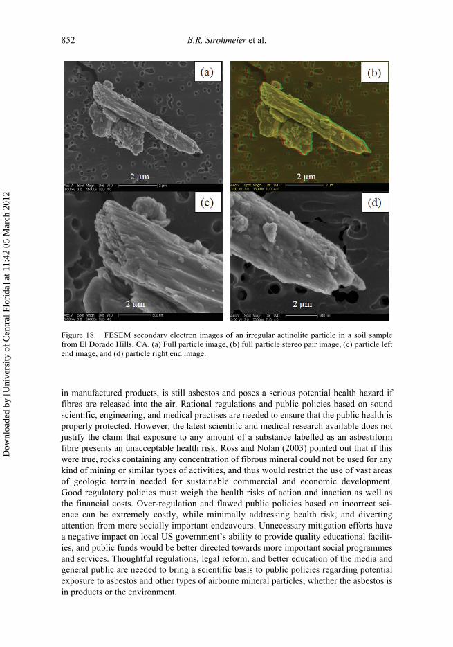
852 B.R. Strohmeier et al.
in manufactured products, is still asbestos and poses a serious potential health hazard iffibres are released into the air. Rational regulations and public policies based on soundscientific, engineering, and medical practises are needed to ensure that the public health isproperly protected. However, the latest scientific and medical research available does notjustify the claim that exposure to any amount of a substance labelled as an asbestiformfibre presents an unacceptable health risk. Ross and Nolan (2003) pointed out that if thiswere true, rocks containing any concentration of fibrous mineral could not be used for anykind of mining or similar types of activities, and thus would restrict the use of vast areasof geologic terrain needed for sustainable commercial and economic development.Good regulatory policies must weigh the health risks of action and inaction as well asthe financial costs. Over-regulation and flawed public policies based on incorrect sci-ence can be extremely costly, while minimally addressing health risk, and divertingattention from more socially important endeavours. Unnecessary mitigation efforts havea negative impact on local US government’s ability to provide quality educational facilit-ies, and public funds would be better directed towards more important social programmesand services. Thoughtful regulations, legal reform, and better education of the media andgeneral public are needed to bring a scientific basis to public policies regarding potentialexposure to asbestos and other types of airborne mineral particles, whether the asbestos isin products or the environment.
Figure 18. FESEM secondary electron images of an irregular actinolite particle in a soil samplefrom El Dorado Hills, CA. (a) Full particle image, (b) full particle stereo pair image, (c) particle leftend image, and (d) particle right end image.
Dow
nloa
ded
by [
Uni
vers
ity o
f C
entr
al F
lori
da]
at 1
1:42
05
Mar
ch 2
012

International Geology Review 853
Important areas for continued future research on asbestos include:
• reaching a consensus on the relative health effects of chrysotile versus amphiboleasbestos;
• determining whether significant differences exist between the health effects ofasbestiform mineral particles and cleavage fragments;
• determining whether low-level environmental exposure to amphibole asbestospresents a significant health risk;
• increasing our understanding of how mineral particles behave in the human lungand initiate disease;
• developing valid health risk assessment tools and models; and• developing reliable, cost-effective analytical testing methods that are reproducible,
follow sound scientific and laboratory practises, and clearly distinguish betweenasbestiform and non-asbestiform particles.
Regarding the development of new analytical methods, the use of FESEM as a comple-mentary tool to TEM or PCM appears to be a promising technique. Most importantly,accomplishing these objectives should lead to the development of a correct, comprehens-ive, and consistent definition of asbestos that will cover all areas dealing with asbestosincluding commercial interests, government regulations, the fields of mineralogy andgeology, and analytical methodologies. To achieve the above research objectives andbetter educate the public requires increased cooperation and collaboration between the sci-entific, health, and regulatory communities.
GlossaryAcicular (particles): Needle-shaped or needlelike. The term is applied in mineralogy to
straight, greatly elongated, free-standing (individual) crystals that may be bounded lat-erally and terminated by crystal faces. The aspect ratio of acicular crystals is in the samerange as those of ‘fibre’ and ‘fibrous’, but the thickness may extend to 7 mm (Skinneret al. 1988). Acicular crystals or fragments are not expected to have the strength, flexibil-ity, or other properties of asbestiform particles.
Actinolite: A monoclinic calcic amphibole with the ideal composition Ca2(Mg,Fe2+)5Si8O22(OH)2. Actinolite is the intermediate member of the Mg–Fe2+ series tremolite-actinolite-ferro-actinolite. The composition is Mg/(Mg+Fe2+) ≥0.5 and <0.9.
Actinolite asbestos: A type of amphibole asbestos composed of the mineral actinolite.The modifier asbestos is used when actinolite crystallizes in the asbestiform habit.Actinolite is one of the six asbestos minerals regulated in the USA by the OSHA.
Amosite (cummingtonite-grunerite asbestos): A commercial term for a type of amphib-ole-asbestos originally from South Africa consisting chiefly of asbestiform members ofthe cummingtonite-grunerite series. The name is an acronym derived from the asbestosmines of South Africa (Skinner et al. 1988). Grunerite commonly occurs in non-asbestiformstubby prismatic or acicular habit, but it can also occur with an asbestiform habit.Amosite is one of the six asbestos minerals regulated in the USA by the OSHA.
Amphibole: A group of common rock forming silicate minerals with the same basic structuralunit consisting of double chains of silica tetrahedra that run parallel to the c-crystallographicdirection. The general formula is A0–1B2C5T8O22(OH)2. Naming conventions are primarilybased on the composition of the A, B, C, and T cation sites. Only a few members of theamphibole mineral group can occur with asbestiform morphology. Even those amphibole
Dow
nloa
ded
by [
Uni
vers
ity o
f C
entr
al F
lori
da]
at 1
1:42
05
Mar
ch 2
012
854 B.R. Strohmeier et al.
minerals that can occur with asbestiform morphology more commonly occur in non-asbestiform morphologies (prismatic, acicular, columnar, or blocky).
Amphibole asbestos: A collective term for asbestiform varieties of amphibole group min-erals. Commercial amphibole asbestos include anthophyllite asbestos, riebeckite asbestos(crocidolite), cummingtonite-grunerite asbestos (amosite), tremolite asbestos, andactinolite asbestos and are regulated in the USA by the OSHA.
Anthophyllite: An orthorhombic amphibole with the ideal composition Mg7Si8O22(OH)2.Anthophyllite is the magnesium end member of the Mg–Fe2+ series anthophyllite-ferro-anthophyllite, with Mg/(Mg+Fe2+) ≥0.5.
Anthophyllite asbestos: A type of amphibole asbestos with ideal composition of the min-eral anthophyllite. The modifier asbestos is used when anthophyllite crystallizes in theasbestiform habit. Anthophyllite is one of the six asbestos minerals regulated in theUSA by the OSHA.
Asbestiform: In the mineralogical sense, asbestiform morphology describes a special typeof fibrosity typical of asbestos in which the fibres exhibit small fibre thickness, flexibility,separability, and general parallel arrangement of fibres in masse (Skinner et al. 1988).Asbestiform mineral fibres grow in bundles that can be separated into smaller bundlesand ultimately into fibrils.
Asbestiform can be defined under the microscope by the following characteristics.
(1) Mean aspect ratio of 20:1 or greater for fibres longer than 5 μm.(2) Very thin fibrils, usually less than 0.5 μm in width.(3) Two or more of the following:
(a) parallel fibres occurring in bundles;(b) fibre bundles displaying splayed ends;(c) fibres in the form of thin needles;(d) matted masses of individual fibres; and(e) fibres showing curvature.
This definition is based on the mineralogical properties and dimensions of both commer-cial and non-commercial asbestos (Wylie 1990).Asbestos: Asbestos is a term applied to six naturally occurring minerals exploited com-
mercially for their desirable physical properties, which are in part derived from theirasbestiform habit. The six minerals are the serpentine mineral chrysotile and theamphibole minerals grunerite asbestos (also referred to as amosite), riebeckite asbes-tos (also referred to as crocidolite), anthophyllite asbestos, tremolite asbestos, andactinolite asbestos. Individual mineral particles, however processed and regardless oftheir mineral name, are not considered to be asbestos if the length-to-width is lessthan 20:1 (Ross et al. 1984).
Asbestos-containing material (ACM): ACM is defined by Title II of the Toxic Sub-stances Control Act (TSCA) as (40 FCR 700, 2002): ‘any material which contains morethan 1% asbestos by weight’.
Asbestosis: A fibrotic lung disease associated with inhalation of asbestos. The disease ischaracterized by the inability of the lung to perform its normal functions of oxygenat-ing blood and eliminating carbon dioxide. The lung as an organ with this diseaseshows a decreased ability to expand or to respond to the action of the diaphragm. Itoccurs after long-term, heavy exposure to asbestos and is regarded as an occupationallung disease.
Dow
nloa
ded
by [
Uni
vers
ity o
f C
entr
al F
lori
da]
at 1
1:42
05
Mar
ch 2
012
International Geology Review 855
Aspect ratio: The numerical relationship of the length to the thickness of a fibre or cleav-age fragment. Microscopic counting rules typically count particles with aspect ratios of ≥3:1or 5:1. Most individual asbestiform fibrils have aspect ratios greater than 20:1.
Backscatter electron image: In an SEM, the primary beam interacts with the speci-men, which results in elastic scattering of the primary beam electrons. Elastic scat-tering events affect the trajectories of the primary beam electrons inside thespecimen without altering the energy of the electron. Backscattered electrons (BSE)are primary beam electrons that escape the specimen as a result of elastic scatteringevents. The BSE signal is strongly related to the atomic components (Z) of the spec-imen, and therefore BSE images can provide compositional and phase informationon the specimen.
Biopersistence: The ability of a particle to remain in the lung or other tissue.Bladed (particles): Bladed particles are similar to prismatic particles in that both types
have well-developed crystal faces and generally low (less than 10:1)-to-moderate (10:1to 20:1) aspect ratios. Prismatic particles have one elongated dimension and twoshorter, approximately equal dimensions (i.e. similar particle width and thickness),whereas bladed particles have one longer dimension and two unequal, shorter dimen-sions. In contrast to prismatic particles, the widths of bladed structures are typically 4–5times larger than the thickness. Bladed particles typically have a wedge-shaped cross-section. The surface texture of prismatic and bladed particles can vary between smoothand rough.
Bundle (particles): Bundles are clusters of individual readily separable particles (typi-cally fibres, but also acicular or prismatic particles). Bundles often have splayed endsand a lack of cohesion between particles in the group, as indicated by the relative dis-placement of particles with respect to one another along the length of the bundle (i.e.twisting and spreading of substructures).
Byssolite: Byssolite has been adopted by the asbestos community to describe a stiff,fibrous variety of amphibole that is not asbestiform (Dana 1932).
Chrysotile: An asbestos mineral in the serpentine group with ideal compositionMg3Si2O5(OH)4. Chrysotile is the most common commercially utilized asbestosmineral and is one of the six asbestos minerals regulated in the USA by the OSHA.
Cleavage: The property of an individual crystal to fracture or break, producing particleswith planar surfaces along specific crystallographic directions dictated by the structureof the material. Asbestiform mineral fibres do not exhibit cleavage; they achieve theirhigh aspect ratio by unidirectional growth. By contrast, non-asbestiform amphibolesand some serpentine minerals, but not chrysotile, may exhibit cleavage.
Cleavage fragment: In mineralogy, a particle produced by fracture along a mineral’scleavage planes.
Columnar (Particles): Similar to bundles, columnar particles consist of an approximatelyparallel, column-like arrangement of substructures (fibre, acicular, prismatic, or bladed)running the length of the main particle. The individual substructures in columnar parti-cles are obvious but do not appear to be readily separable.
Crocidolite (riebeckite asbestos): The commercial name of the asbestiform variety rie-beckite-magnesioriebeckite amphiboles. Crocidolite is one of the six asbestos mineralsregulated in the USA by the OSHA.
Cummingtonite: A monoclinic amphibole with the ideal composition Mg7Si8O22(OH)2.Cummingtonite is the magnesium end member of the Mg–Fe2+ series cummingtonite-grunerite, with Mg/(Mg+Fe2+) ≥0.5.
Cummingtonite asbestos: See Amosite.
Dow
nloa
ded
by [
Uni
vers
ity o
f C
entr
al F
lori
da]
at 1
1:42
05
Mar
ch 2
012

856 B.R. Strohmeier et al.
Elongated mineral particle (EMP): An EMP is a single crystal or similarly elongatedpolycrystalline aggregate particle with a length to width ratio of 3 to 1 or greater, basedon a microscopic analysis of an airborne sample using NIOSH Method 7400 or equivalent(NIOSH 2009).
Energy dispersive X-ray spectroscopy (EDS): SEM instruments are commonly equippedwith an energy dispersive X-ray spectrometer (EDS) system, which is used to measure theenergies and amounts of characteristic X-rays emitted from a site (point or area) of inter-est on a specimen. In an SEM, the primary electron beam can interact with the specimen,and several types of inelastic scattering events occur. Inelastic scattering events transferenergy from the beam (electrons) to the atoms in the specimen, leading to the generationof secondary electrons, Auger electrons, characteristic X-rays, as well as a variety of othersignals. Characteristic X-rays result when the primary electron beam interacts with atightly bound inner-shell electron of an atom ejecting the atomic electron and leaving avacancy in that shell. When the electrons in the atom relax, a characteristic X-ray is emit-ted, thereby identifying the specific element.
Erionite: Erionite is a member of the zeolite mineral group with an ideal formula of(Na2,K2,Ca,Mg)4[Al8Si28O72]·28H2O. Erionite usually occurs as thin fibres having awoolly appearance, but is not classified as asbestos or asbestiform. However, erionitewas determined to induce a high incidence of malignant pleural mesothelioma throughenvironmental exposures in the Cappadocian region of central Anatolia in Turkey.
Extinction angle: For minerals that belong to the monoclinic and triclinic crystal systems,the orientation of the optical indicatrix with respect to crystallographic orientation is notnecessarily equivalent. Thus, there is an angle between a principal vibration direction(X, Y, Z) and crystallographic direction (a, b, c). The annotation cˆZ is the anglebetween the c-crystallographic direction and the Z principal vibration direction detectedon optical analysis. This angle is measured with a PLM on crystals of known orienta-tion. It has been observed that asbestiform monoclinic minerals have anomalous extinc-tion so that the cˆZ angle is always 0°. Detailed information on optical character of thedifferent crystallographic systems can be found in Dyar and Gunter (2008) or othertreatises on optical mineralogy.
Ferro-actinolite: A monoclinic amphibole with the ideal composition Ca2Fe2+5Si8O22(OH)2.
Ferro-actinolite is the iron end member of the Mg–Fe2+ series tremolite-actinolite-ferro-actinolite, with Mg/(Mg+Fe2+) <0.5.
Fibre: A fibre is a long, thin thread or threadlike solid with a distinctive elongated shape thatmay be natural or synthetic and organic or inorganic in composition. The properties offlexibility and toughness are implied, but are not essential to the definition. Fibre dimen-sions may range from approximately 1 mm to the nanometre range. Chrysotile exhibitsthe smallest diameters of the six asbestos mineral with fibril diameter about 200Å (seeFigure 3). Most mineralogists apply the term when the aspect ratio of a mineral sample,individual, or aggregate is at least 10:1, i.e. these particles have a highly elongate mor-phology developed during growth.
Fibril: Fibril is the smallest unit that expresses the characteristics of a fibre or fibre bun-dle. See fibril cross-sections in Figure 14a and b. A single fibril of asbestos cannot beseparated along its length.
Fibrous: A morphology that exhibits parallel, radiating, or matted aggregates of fibres.The term ‘fibrous’ has been used during the last 200 years to describe all kinds ofminerals that crystallized in habits resembling organic as well as minerals includingasbestos minerals. However, the related term ‘asbestiform’ was never used for fibrousmineral habits other than for asbestos. Accordingly, ‘fibrous’ is the more general
Dow
nloa
ded
by [
Uni
vers
ity o
f C
entr
al F
lori
da]
at 1
1:42
05
Mar
ch 2
012

International Geology Review 857
term, and asbestiform is a specific type of fibrosity. While it is correct that all asbes-tos minerals are fibrous, not all minerals having fibrous habits are asbestos, e.g.fibrous talc.
Field emission scanning electron microscopy (FESEM): A field emission scanningelectron microscope (FESEM) is a special type of high-resolution scanning electronmicroscope (SEM) that uses a field emission source to generate the primary electronbeam rather than the typical heated filament source used in conventional SEM instru-ments. FESEM instruments have a magnification of approximately 500,000× or higherand a spatial resolution of about 0.005 μm (5 nm). Several FESEM images are included:see Figures 4 and 14.
Fluoro-edenite: Fluoro-edenite (NaCa2Mg5Si7AlO22F2) is a newly discovered member ofthe calcic amphibole mineral group (Gianfagna and Oberti 2001). It occurs in both pris-matic and fibrous morphologies in volcanic rocks on the flank of Mt Etna near Biancav-illa, Sicily, Italy, and is a contaminant in road and building stone that has beenassociated with non-occupational cases of mesothelioma.
Grunerite: The name given to a monoclinic amphibole mineral with the ideal composi-tion Fe7Si8O22(OH)2. Grunerite is the iron end member of the Mg–Fe2+ series cum-mingtonite-grunerite, with Mg/(Mg+Fe2+) <0.5.
Grunerite asbestos: See Amosite.Habit: The crystal habit of a mineral is the shape or form a crystal or aggregate of crystals
assumes during crystallization and is directly dependent on the environment and geo-logical conditions at the time of formation., a term that means ‘crystal morphology’.
Irregular (particles): Large (≥2 μm) ‘blocky’ particles with non-parallel, irregular sidesand irregular ends that do not fit any of the six primary particle categories (i.e. acicular,bladed, bundle, columnar, fibre, or prismatic) are separately classified as ‘irregular’.
Macrophage: A cell type that is part of the immune system. Macrophages are types ofphagocytes, i.e. they are capable of engulfing (and subsequently transporting ordestroying) foreign bodies.
Mineral: A naturally occurring element or compound defined by its chemical composi-tion and crystal structure. Asbestos is not ‘a mineral’. Asbestos is a commercial term fora collection of minerals with different compositions and different crystal structures thathave an asbestiform habit and are and were used commercially.
Mineral group: A collection of mineral species and mineral series that have common andvery similar chemical and structural units, related compositions, and crystal structures.Serpentine and amphibole are names of two different mineral groups.
Mineral series: Minerals that have compositionally equivalent chemistries and basicstructural units in which small amounts of substitution of similar elements (cations ofsimilar size, stereochemical, and bonding character) in the same site in the crystal struc-ture are members of a mineral series.
Mixed mineral environments: In mixed mineral environments, many mineral specieswith a variety of crystal habits occur together. This is by far the most common situationon the earth’s surface. Analysis of such samples requires rigorous attention to standardsand test methodologies to identify, distinguish, and quantify the species in the mix andespecially asbestiform from other fibrous minerals.
Monoclinic: One of the crystal systems in which minerals may crystallize. In the mono-clinic crystal system, the three crystallographic axes are of unequal lengths. The axes aand b, and b and c are perpendicular to each other, but a and c make an oblique anglewith each other (Dana 1932). The amphibole minerals grunerite, riebeckite, tremolite,and actinolite crystallize in the monoclinic system.
Dow
nloa
ded
by [
Uni
vers
ity o
f C
entr
al F
lori
da]
at 1
1:42
05
Mar
ch 2
012

858 B.R. Strohmeier et al.
Mesothelioma: A rare neoplasm that develops in the lining of the pleura or peritoneum,the thick organic sheets that cover the viscera and are composed of cells, and fibrousmolecular species (for more information see Stedman’s Medical Dictionary 2000).
Naturally occurring asbestos (NOA): NOA refers to asbestiform fibres that occur insoils or rock formations. NOA is usually the term used for non-commercial smalldeposits of asbestiform fibres as opposed to large commercial asbestos deposits. NOArepresents a potential health risk if the fibres are disturbed and become airborne.
Orthorhombic: In the orthorhombic crystal system, there are three crystallographic axesof unequal lengths that make angles of 90° with each other (Dana 1932). The amphib-ole mineral anthophyllite crystallizes in the orthorhombic system.
Phase contrast microscopy (PCM): Phase contrast microscopy is a technique that can beutilized to produce high-contrast images of transparent specimens, such as fibres basedon morphology. The NIOSH 7400 method uses PCM to count fibres in air samples thatare longer than 5 μm, have an aspect ratio ≥3:1, and diameters down to approximately0.25 μm. Unfortunately, PCM cannot resolve individual asbestiform fibrils becausethey are too thin and PCM cannot be used to distinguish between asbestiform minerals(e.g. chrysotile) and crocidolite, nor distinguish asbestiform fibres from other non-mineral fibres (e.g. cotton).
Plaque: A deposit of material on a flat surface, such as the epithelium of the lung orpleura.
Pleura: The membrane lining the cavities containing the lungs. The pleura consists of alayer of connective tissue covered with a layer of mesothelium. The part covering thelungs is the visceral pleura; the part lining the cavity is the parietal pleura; and thepotential space between the visceral and parietal pleura is the pleural cavity or space.
Polarized light microscopy (PLM): A polarized light microscope (PLM) utilizes trans-mitted polarized light and refractive index oils to measure optical properties and mor-phology of translucent particles. The technique permits both mineral identification andthe differentiation between asbestiform and non-asbestiform habits to be determined.See typical PLM images in Figure 2.
Prismatic: A term commonly used in descriptions of minerals for crystals exhibitingaspect ratios usually below 3:1 and grading into equant (aspect ratio = 1). Prismatic par-ticles often have a well-defined corner or edge and a crystalline termination. The termmay refer to crystals embedded in a matrix, but is more commonly used to describefree-standing euhedral crystals whether micro- or macroscopic.
Pulmonary alveolus: Thin-walled balloon-like sacs at the termini of the respiratory sys-tem bronchioles, alveolar ducts, and alveolar sacs across which gas exchange occursbetween alveolar air and the pulmonary capillaries (Stedman’s Medical Dictionary2000).
Refractive index: The refractive index, also called the index of refraction, of a substanceis defined as:
where, n is the refractive index, Vv is the velocity of light in a vacuum, and Vm is thevelocity of light in a material. The refractive indices of minerals can be measured in thepolarizing light microscope and used in mineral identification.Richterite: A monoclinic sodic-calcic amphibole with the general composition of Na(NaCa)
(Mg,Fe2+)5Si8O22(OH)2.
nV
V= v
m
Dow
nloa
ded
by [
Uni
vers
ity o
f C
entr
al F
lori
da]
at 1
1:42
05
Mar
ch 2
012

International Geology Review 859
Richterite asbestos: A type of amphibole asbestos composed of the mineral richterite.The modifier asbestos is used when richterite crystallizes in the asbestiform habit. Todate, it is not a regulated amphibole. However, it occurs in both asbestiform and non-asbestiform morphologies at the former vermiculite mine in Libby, MT.
Riebeckite: A monoclinic sodic amphibole with the ideal composition of Na2( )Si8O22(OH)2.
Riebeckite asbestos: See Crocidolite.Scanning electron microscopy (SEM): A scanning electron microscope (SEM) allows
for the observation and characterization of materials on a nanometre (nm) to microme-tre (μm) scale. In an SEM, the specimen is irradiated with a finely focused beam ofelectrons. The beam can be scanned back and forth across the specimen to form imagesor kept static to obtain an elemental analysis at one position. A variety of signals areproduced from the interaction of the primary beam and the specimen; including second-ary electrons, backscattered electrons, characteristic X-rays, and other photons of vari-ous energies. The signals can be used to examine specimen characteristics, such assurface topography, morphology, and composition.
Secondary electron image: In an SEM, the primary electron beam can interact with the speci-men and result in several types of inelastic scattering events. Inelastic scattering events resultin a transfer of energy from the primary beam electrons to the atoms of the specimen, lead-ing to the generation of secondary electrons, Auger electrons, characteristic X-rays, as wellas a variety of other signals. Secondary electrons are produced as a result of the interactionbetween the primary electron beam and weakly bound specimen electrons. The secondaryelectrons are very low-energy specimen electrons, and therefore secondary electron imagescarry information about the surface and topography of the specimen.
Serpentine: A mineral group of 1:1 layer silicates with the general formulaMg3Si2O5(OH)4, which includes the several mineral species known as polytypes, lizar-dite, chrysotile, and antigorite. Chrysotile occurs in an asbestiform habit and is definedas one of the six asbestos minerals regulated in the USA by the OSHA.
Spindle stage: The spindle stage is an accessory to the PLM and provides the ability torotate a particle about a horizontal axis in the plane of the microscope stage for dimen-sional and optical characterization of single crystals, with the added benefit of beingable to take the same particle for studies with X-ray diffraction and electron beaminstruments.
Talc: A mineral species that is a 2:1 layered silicate structure with the ideal compositionMg3Si4O10(OH)2. The term has been used as a commercial name for fibrous or platydeposits formed by hydrothermal alteration of rocks rich in silica, magnesium, and iron.Fibrous talc is not asbestos, but it may occur in association with asbestiform amphibolesor chrysotile.
Transmission electron microscopy (TEM): A transmission electron microscope(TEM) transmits a beam of electrons through a thin sample forming a projectionimage, which allows the detection of particle shape and structure down to thesmallest asbestos fibres. TEM can also be used to determine crystal structure whencombined with SAED as well as elemental composition from EDS. The NIOSH7402 method uses TEM to determine the concentration of asbestiform fibres in airsamples.
Tremolite: A calcic amphibole with the ideal composition Ca2Mg5Si8O22(OH)2 crys-tallizing in the monoclinic crystal system. Tremolite is the magnesium-rich endmember of the Mg–Fe2+ series tremolite-actinolite-ferro-actinolite, with Mg/(Mg+Fe2+) ≥0.9.
Fe Fe32+, 2
3+
Dow
nloa
ded
by [
Uni
vers
ity o
f C
entr
al F
lori
da]
at 1
1:42
05
Mar
ch 2
012

860 B.R. Strohmeier et al.
Tremolite asbestos: See Tremolite. The modifier asbestos is used when tremolite crystal-lizes in the asbestiform habit. Tremolite is one of the six asbestos minerals regulated inthe USA by the OSHA.
Vermiculite: A mineral group of 2:1 layer silicates with the general formula(Mg,Ca,K,Fe2+)3(Si,Al,Fe3+)4O10(OH)2·4H2O. Vermiculate is a ‘clay’ mineral withperfect basal cleavage. Vermiculite can be exfoliated by rapid heating to produceexpanded vermiculite, which has been used in insulation and soil conditioning products.Vermiculite is not asbestos, but it may be contaminated with asbestiform amphiboles.
Winchite: A sodic-calcic amphibole with the general composition of (NaCa)Mg4(Al,Fe3+)Si8O22(OH)2 that crystallizes in the monoclinic crystal system.
Winchite asbestos: A type of amphibole asbestos composed of the mineral winchite. Themodifier asbestos is used when winchite crystallizes in the asbestiform habit. To date, itis not a regulated amphibole. However, it occurs in both asbestiform and non-asbestiformmorphologies at the former vermiculite mine in Libby, MT.
Zeolite: Zeolites are a large mineral group of hydrated aluminosilicates containing alkaliand alkaline earth metals with a general formula of (Na2,K2,Ca,Ba)[(Al,Si)O2]n ·xH2O,whose crystal structures are as varied as the compositions and are represented in all ofthe six crystal structure classes (Gaines et al. 1997, p. 1646–1701). Some of the speciesin this group are remarkable for their continuous and in part reversible dehydration andfor their base-exchange properties; they are commonly used as commercial absorbents.Zeolites are not asbestos, but one member of this group, erionite (see above and in text),was determined to induce a high incidence of malignant pleural mesothelioma throughenvironmental exposures in the Cappadocian region of central Anatolia in Turkey.
ReferencesAmandus, H.E., and Wheeler, R., 1987, Morbidity and mortality of vermiculite miners and
millers exposed to tremolite-actinolite: American Journal of Industrial Medicine, v. 11, p.15–26.
American Society for Testing and Materials (ASTM), 2002, Standard test method for microvacuum sam-pling and indirect analysis of dust by transmission electron microscopy for asbestos mass surfaceloading: 2002 Annual Book of ASTM Standards, v. D5756: West Conshohocken, PA, ASTM.
American Society for Testing and Materials (ASTM), 1 June 2006, Standard practice for samplingand counting airborne fibres, including asbestos fibres, in mines and quarries, by phase contrastmicroscopy and transmission electron microscopy: 2006 Annual Book of ASTM Standards, v.D 7200-06: West Conshohocken, PA, ASTM.
Anderson, H.A., Lilis, R., Daum, S.M., and Selikoff, I.J., 1979, Asbestosis among household con-tacts of asbestos factory workers: Annals New York Academy of Sciences, Health Hazards ofAsbestos Exposure, v. 330, p. 387–399.
Andreozzi, B.A., Ballirano, P., Bruni, B.M., Gianfagna, A., Gunter, M.E., Mazziotti-Tagliani, S.,Pacella, A., Paoletti, L., Lee, R.J., Van Orden, D.R., and Allison, K., 2007, Fibrous amphibolesfrom Biancavilla: Crystal chemistry, environmental aspects, and health studies: Reviews inMineralogy and Geochemistry (RiMG), Amphiboles Shortcourse, Rome, Italy.
Ayer, H.E., Lynch, J.R., and Fanney, J.H., 1965, A comparison of impinger and membrane filtertechniques for evaluating air samples in asbestos plants: Annals of New York Academy of Sci-ences, v. 132, p. 274–287.
Bailey, K.F., 2004, Naturally occurring asbestos issues in the aggregates industry: Fact and fiction:Mining Engineering, v. 56, p. 33–38.
Balzer, J.L., and Cooper, W.C., 1968, The work environment of insulating workers: AmericanIndustrial Hygiene Association Journal, v. 29, no. 3, p. 222–227.
Bandli, B.R., and Gunter, M.E., 2001, Identification and characterization of mineral and asbestosparticles using the spindle stage and the scanning electron microscope: The Libby, Montana,USA amphibole-asbestos an example: Microscope, v. 49, p. 191–199.
Dow
nloa
ded
by [
Uni
vers
ity o
f C
entr
al F
lori
da]
at 1
1:42
05
Mar
ch 2
012

International Geology Review 861
Bandli, B.R., and Gunter, M.E., 2006, A review of scientific literature examining the mining history,geology, mineralogy, and amphibole asbestos health effects of the rainy creek igneous complex,Libby, Montana, USA: Inhalation Toxicology, v. 18, p. 1–14.
Bandli, B.R., Gunter, M.E., Twamley, B., Foit, F.F., Jr., and Cornelius, S.B., 2003, Optical, composi-tional, morphological, and X-ray data on eleven particles of amphibole from Libby, Montana, USA:The Canadian Mineralogist, v. 41, p. 1241–1253.
Barber, V.L., 12–16 December 2005, Executive summary of briefing information regarding naturallyoccurring asbestos: Placerville, CA, El Dorado County Superintendent of Schools, http://www.edcoe.org/supts/whats_new/asbestos/documents/talking_points_for_wa_dc_trip_1205.pdf(accessed 15 March 2010).
Bard, D., Yarwood, J., and Tylee, B., 1997, Asbestos fibre identification by Raman microspectros-copy: Journal of Raman Spectroscopy, v. 28, p. 803–809.
Baron, P.A., 1994, Measurement of fibres, in Schlecht, Ch.L.P.C., and O’Conner, P.F., eds., NIOSHmanual of analytical methods (NMAM) (fourth edition): US DHHS (NIOSH) Publication, p. 94–113, http://www.cdc.gov/niosh/docs/2003-154/pdfs/chapter-1.pdf (accessed 15 March 2010).
Battista, G., Belli, S., Comba, P., Fiumalbi, C., Grignoli, M., Loi, F., Orsi, D., and Paredes, I., 1999,Mortality due to asbestos-related causes among railway carriage and construction and repairworkers: Occupational Medicine, v. 49, no. 8, p. 536–539.
Beard, M.E., Crankshaw, O.S., Ennis, J.T., and Moore, C.E., 2001, Analysis of crayons for asbestosand other fibrous materials, and recommendations for improved analytical definitions: NC,Research Triangle Institute, Research Triangle Park.
Berman, D.W., 30 June 2006, Evaluation of the approach recently proposed for assessing asbestos-related risk in El Dorado County, California: National Stone, Sand, and Gravel Association(NSSGA), Docket No. EPA-HQ-ORD-2003-0016-0076.
Berman, D.W., and Crump, K.S., October 2003, Final draft: Technical support document for a pro-tocol to assess asbestos-related risk: EPA #9345.4-06, Washington, DC, US Environmental Pro-tection Agency.
Berman, D.W., and Crump, K.S., 2008a, Update of potency factors for asbestos-related lung cancerand mesothelioma: Critical Reviews in Toxicology, v. 38, suppl. 1, p. 1–47.
Berman, D.W., and Crump, K.S., 2008b, A meta-analysis of asbestos-related cancer that addressesfiber size and mineral type: Critical Reviews in Toxicology, v. 38, suppl. 1, p. 49–73.
Bernstein, D.M., 2007, Synthetic vitreous fibers: A review toxicology, epidemiology and regula-tions: Critical Reviews in Toxicology, v. 37, no. 10, p. 839–886.
Bernstein, D.M., Chevalier, J., and Smith, P., 2003a, Comparison of calidria chrysotile asbestos topure tremolite: Inhalation biopersistence and histopathology following short-term exposure:Inhalation Toxicology, v. 15, p. 1387–1419.
Bernstein, D.M., and Hoskins, J.A., 2006, The health effects of chrysotile: Current perspectivebased upon recent data: Regulatory Toxicology and Pharmacology, v. 45, p. 252–264.
Bernstein, D.M., Rogers, R., and Smith, P., 2003b, The biopersistence of Canadian chrysotile asbes-tos following inhalation: Inhalation Toxicology, v. 15, p. 1247–1274.
Blake, T., Castranova, V., Schwegler-Berry, D., Baron, P., Deye, G.J., Li, C., and Jones, W., 1998,Effect of fiber length on glass fiber microfiber cytotoxicity: Journal of Toxicology and Environ-mental Health Part A, v. 54, p. 243–259.
Blake, C.L., Scott Dotson, G., and Harbison, R.D., 2006, Assessment of airborne asbestos exposureduring the servicing and handling of automotive asbestos-containing gaskets: RegulationToxicology and Pharmacology, v. 45, p. 214–222.
Bloss, F.D., 1981, The spindle stage: Principles and practices: Cambridge, United Kingdom,Cambridge University Press.
Bloss, F.D., 1999, Optical crystallography: Washington, DC, Mineralogical Society of AmericaMonograph Series, no. 5
Bowles, O., 1955, The asbestos industry: Bulletin 552: Washington, DC, US Bureau of Mines, p. 122.Brown, B.M., and Gunter, M.E., 2003, Morphological and optical characterization of amphiboles
from Libby, Montana USA by spindle stage assisted – polarized light microscopy: Microscope,v. 51, p. 121–140.
Browne, K., 1986, Is asbestos or asbestosis the cause of the increased risk of lung cancer in asbestosworkers?: British Journal of Industrial Medicine, v. 43, p. 145–149.
Bunker, K.L., McAllister, D., Allison, K.A., Wagner, K., Rickabaugh, K., Levine, A.M.,Strohmeier, B.R., and Lee, R.J., 4 August 2008a, TEM and FESEM: The right combination
Dow
nloa
ded
by [
Uni
vers
ity o
f C
entr
al F
lori
da]
at 1
1:42
05
Mar
ch 2
012

862 B.R. Strohmeier et al.
for enhanced particle characterization: 2008 Microscopy & Microanalysis Meeting, Albu-querque, NM.
Bunker, K.L., Strohmeier, B.R., Harris, K.E., Hoch, R., and Lee, R.J., 8 July 2008b, ComplementaryTEM and FESEM characterization of amphibole particles in mixed mineral dust from Libby,Montana, USA: 2008 Inter/Micro Conference, Chicago, IL.
Burdett, G.J., Jaffey, S.A.M.T., and Rood, A.P., 1987, Airborne asbestos fibre levels in buildings: Asummary of UK measurements: WHO/IARC Symposium on Mineral Fibers in the Non-Occupational Environment, p. 227.
Burragato, F., Comba, P., Baiocchi, V., Palladino, D.M., Simei, S., Gianfagna, A., Paoletti, L., andPasetto, R., 2005, Geo-volcanological, mineralogical and environmental aspects of quarry mate-rials related to pleural neoplasm in the area of Biancavilla, Mount Etna (Eastern Sicily, Italy):Environmental Geology, v. 47, p. 855–868.
California Air Resources Board (CARB), 1991, Determination of asbestos content of serpentineaggregate: CARB Method 435, Sacramento, CA.
California State Senate, 11 May 2005, Naturally-occurring asbestos: Who is responsible for protect-ing the public health: Joint Informational Hearing of the Senate Health Committee and SenateEnvironmental Quality Committee, California State Senate, Sacramento, CA.
Campbell, W.J., Blake, R.L., Brown, L.L., Cather, E.E., and Sjoberg, J.J., 1977, Selectedsilicate minerals and their asbestiform varieties – mineralogical definitions and identification-characterization: United States Department of Interior, Bureau of Mines, Information Cir-cular 8751, p. 1–55.
Carbone, M., Emri, S., Dogan, A.U., Steele, I., Tuncer, M., Pass, H.I., and Baris, Y.I., 2007, A mes-othelioma epidemic in Cappadocia: Scientific developments and unexpected social outcomes:Nature Reviews Cancer, v. 7, no. 2, p. 147–154.
Carpenter, R.L., and Wilson, C.L., 1999, The inhalation toxicity of glass fibers – A review of thescientific literature: Report no. TOXDET-99-07, Naval Health Research Center Detachment(Toxicology), Wright-Patterson Air Force Base, OH.
Chatfield, E.J., May 1983, Measurement of asbestos fibre concentrations in ambient atmospheres:Study Series, no. 10, The Royal Commission on Matters of Health and Safety Arising From theUse of Asbestos in Ontario, Ontario, Canada.
Chatfield, E.J., 1986, Airborne asbestos levels in Canadian public buildings, in Chatfield, E.J., ed.,Asbestos fibre measurements in building atmospheres: Proceedings, Ontario Research Founda-tion, Mississauga, Canada, p. 177–207.
Chisholm, J.E., 1995, Discrimination between amphibole asbestos fibres and non-asbestos mineral frag-ments: Project Report Ir/L/MF/95/16, Health and Safety Laboratory, Sheffield, United Kingdom.
Chrostowski, P.C., Foster, S.A., and Anderson, E.L., 1991, Human health risks associated withasbestos abatement: Risk Analysis, v. 11, p. 465–481.
Churg, A., Corn, M., Craighead, J., Davis, J.M.G., Gaensler, E., Gee, J.B., Ilgren, E., Kuschner, M.,Langer, A., Lee, R., Mossman, B., Murray, R., Newhouse, M., Nolan, R., Ross, M., Skinner, H.C.,and Wagner, J.C., 23 September 1993, Scientists call for an end to the NYC asbestos scare: NewYork Times, http://www.sepp.org/key%20issues/asbestos/asbestnr.html (accessed 15 March 2010).
Clinkenbeard, J.P., Churchill, R.K., and Lee, K., eds., 2002, Guidelines for geologic investigationsof naturally occurring asbestos in California: Sacramento, CA, California Department ofConservation, California Geological Survey, Special Publication 124.
Componiano, A., Casciardi, S., Fiovavanti, F., and Ramires, D., 2004, Airborne asbestos levels inschool buildings in Italy: Journal of Occupational and Environmental Hygiene, v. 1, p. 256–261.
Corn, M., Crump, K., Farrar, D.B., Lee, R.J., and McFee, D.R., 1991, Airborne concentrations ofasbestos in 71 school buildings: Regulatory Toxicology and Pharmacology, v. 13, p. 99–114.
Corn, M., McArthur, B., and Dellarco, M., 1994, Asbestos exposures of building maintenancepersonnel: Applied Occupational and Environmental Hygiene, v. 9, p. 845–852.
Crane, D.T., 1992, Polarized light microscopy of asbestos: OSHA Analytical Method Manual,Method ID-191.
Crump, K.S., and Farrar, D.B., 1989, Statistical analysis of data on airborne asbestos levels collected in anEPA survey of public buildings: Regulatory Toxicology and Pharmacology, v. 10, p. 51–62.
Dana, J.D., 1932, A textbook of mineralogy (William E. Ford, ed.): New York, John Wiley & Sons, Inc.Darcy, D.J., and Alleman, T., 2004, Occupational and environmental exposure to asbestos, in
Roggli, V.L., Oury, T.D., and Sporn, T.A., eds., Pathology of asbestos-associated diseases(second edition): New York, Springer, p.. 17–19.
Dow
nloa
ded
by [
Uni
vers
ity o
f C
entr
al F
lori
da]
at 1
1:42
05
Mar
ch 2
012

International Geology Review 863
Deer, W.A., Howie, R.A., and Zussman, J., 1997, Rock-forming minerals: Double-chain silicates(second edition): Geological Society of London, v. 2, p. 137–145.
Dehoff, R.T., and Rhines, F.N., 1968, Quantitative microscopy: New York, McGraw-Hill.Dikensoy, O., 2008, Mesothelioma due to environmental exposure to erionite in Turkey: Current
Opinion in Pulmonary Medicine, v. 14, no. 4, p. 322–325.Dogan, A.U., 2003, Zeolite mineralogy and cappadocian erionite: Indoor and Built Environment, v.
12, no. 5, p. 337–342.Dogan, A.U., Baris, Y.I., Dogan, M., Emri, S., Steele, I., Elmishad, A.G., and Carbone, M., 2006,
Genetic predisposition to fiber carcinogenesis causes a mesothelioma epidemic in Turkey:Cancer Research, v. 66, no. 10, p. 5063–5068.
Dorling, M., and Zussman, J., 1987, Characteristics of asbestiform and non-asbestiform calcicamphiboles: Lithos, v. 20, p. 469–489.
Dyar, M.D., and Gunter, M.E., 2008, Mineralogy and optical mineralogy: Chantilly, VA, Minera-logical Society of America.
Eastern Research Group (ERG), 30 May 2003, Report on the Peer Consultation Workshop toDiscuss a Proposed Protocol to Assess Asbestos-Related Risk, Final Report: Prepared by ERG,Lexington, Massachusetts for US Environmental Protection Agency, Office of Solid Waste andEmergency Response, EPA Contract No. 68-C-98-148, Work Assignment 2003–05,Washington, DC.
Ecology and Environment, Inc. (EEI), 2005, El Dorado Hills naturally occurring asbestos multime-dia exposure assessment, El Dorado Hills, California: Preliminary assessment and site inspec-tion report: Interim Final: Prepared by EEI for US Environmental Protection Agency, Region 9,Contract No. 68-W-01-012, TDD No. 09-04-01-0011, Job No. 001275.0440.01CP, SanFrancisco, CA.
Edwards, G.H., and Lynch, J.R., 1968, The method used by the US Public Health Service for enu-meration of asbestos dust on membrane filters: Annals of Occupational Hygiene, v. 11, p. 1–6.
Emri, S., Demir, A., Dogan, M., Akay, H., Bozkurt, B., Carbone, M., and Baris, I., 2002, Lungdisease due to environmental exposures to erionite and asbestos in Turkey: Toxicology Let-ters, v. 127, no. 1–3, p. 251–257.
Federal Register, 30 October 1987, Interim transmission electron microscopy analytical methods –mandatory and nonmandatory – and mandatory section to determine completion of response:Federal Register 52, p. 41857–41897.
Fischbein, A., Rohl, A.N., Langer, A.M., and Selikoff, I.J., 1979, Drywall construction and asbestosexposure: American Industrial Hygiene Association Journal, v. 40, p. 402–407.
Fleischer, W.E., Viles, F.J., Jr., Gade, R.L., and Drinker, P., 1946, A health survey of pipe coveringoperations in constructing naval vessels: Journal of Industrial Hygiene and Toxicology, v. 28,no. 1, p. 9–16.
Frasca, P., De Malo, R., Newton, J., and Goldale, M., 11 July 2000, Asbestos analysis of soil byscanning electron microscopy and energy dispersive X-ray spectroscopy, standard operatingprocedure: Report no. EPA-Libby-01, Washington, DC, US Environmental Protection Agency.
Gains, R.V., Skinner, H.C.W., Ford, E.E., Mason, B., and Rosenzweig, A., 1997, Dana’s new miner-alogy: the system of new mineralogy of James Dwight Dana and Edward Salisbury Dana: NewYork, John Wiley & Sons, p. 1646–1701.
Gamble, J.F., and Gibbs, G.W., 2008, An evaluation of the risks of lung cancer and mesotheliomafrom exposure to amphibole cleavage fragments: Regulatory Toxicology and Pharmacol-ogy, v. 52, p. S154–S186.
Gardner, M.J., Winter, P.D., Pannett, B., and Powell, C.A., 1986, Follow up study of workers manufac-turing chrysotile asbestos cement products: British Journal of Industrial Medicine, v. 43, p. 726–732.
Germaine, M., 1986, Asbestos in play sand: Correspondence, New England Journal of Medi-cine, v. 315, p. 891.
Gianfagna, A., Andreozzi, G.B., Ballirano, P., and Mazziotti-Tagliani, S., 2007, Structural andchemical contrasts between prismatic and fibrous fluoro-edenite from Biancavilla, Sicily, Italy:The Canadian Mineralogist, v. 45, p. 249–262.
Gianfagna, A., and Oberti, R., 2001, Fluoro-Edenite from Biancavilla (Catania, Sicily, Italy): Crystalchemistry of a new amphibole end-member: American Mineralogist, v. 86, p. 1489–1493.
Glenn, R.E., Lee, R.J., Jastrem, L.M., Bunker, K.L., Van Orden, D.R., and Strohmeier, B.R.,2008, Asbestos: By any other name, is it still?: Chemical Regulation Reporter, v. 32, no. 21,p. 22–33.
Dow
nloa
ded
by [
Uni
vers
ity o
f C
entr
al F
lori
da]
at 1
1:42
05
Mar
ch 2
012

864 B.R. Strohmeier et al.
Gold, J., Amandusson, H., Krozer, A., Kasemo, B., Ericsson, T., Zanetti, G., and Fubini, B., 1997,Chemical characterization and reactivity of iron chelator treated amphibole asbestos: Environ-mental Health Perspective, v. 105, suppl. 5, p. 1021–1030.
Goldstein, J., Newbury, D., Joy, D., Lyman, C., Echlin, P., Lifshin, E., Sawyer, L., and Michael, J.,2003, Scanning electron microscopy and X-ray microanalysis (third edition): New York,Springer Science and Business Media Inc.
Green, G., 15 September 2008, Bruce Vento Ban Asbestos and Prevent Mesothelioma Act of 2008:Bill H.R.6903, US House of Representatives, 110th Congress, 2nd Session, Washington, DC,http://www.govtrack.us/congress/billtext.xpd?bill=h110-6903 (accessed 15 March 2010).
Gunter, M.E., 1994, Asbestos as a metaphor for teaching risk perception: Journal of GeologicalEducation, v. 42, p. 17–24, http://www.webpages.uidaho.edu/∼mgunter/NAGT/manuscripts/GunterJGEA.pdf (accessed 15 March 2010).
Gunter, M.E., 2004, The polarized light microscope: Should we teach the use of a 19th centuryinstrument in the 21st century?: Journal of Geological Education, v. 52, no. 1, p. 34–44.
Gunter, M.E., Belluso, E., and Mottana, A., 2007, Amphiboles: Environmental and health concerns:Reviews in Mineralogy and Geochemistry, v. 67, p. 453–516.
Gunter, M.E., Dyar, M.D., Twamley, B., Foit, F.F., Jr., and Cornelius, S., 2003, Composition, Fe+3/Total Fe, and crystal structure of non-asbestiform and asbestiform amphiboles from Libby,Montana, USA: American Mineralogist, v. 88, p. 1970–1978.
Gunter, M.E., Weaver, R., Bandli, B.R., Bloss, F.D., Evans, S.H., and Su, S.C., 2004, Results from aMcCrone spindle stage short course, a new version of EXCALIBR, and how to build a spindlestage: Microscope, v. 52, no. 1, p. 23–39.
Guthrie, G.D., Jr., and Mossman, B.T., eds., 1993, Reviews in mineralogy: Health effects of mineraldusts: Chantilly, VA, Mineralogical Society of America, v. 28.
Harries, P.G., 1971, Asbestos dust concentration in ship repairing: A practical approach to improv-ing asbestos hygiene in naval dockyards: Annals of Occupational Hygiene, v. 14, p. 241–254.
Harris, K.E., Bunker, K.L., Strohmeier, B.R., Hoch, R., and Lee, R.J., 2007, Discovering the truemorphology of amphibole minerals: Complementary TEM and FESEM characterization of par-ticles in mixed mineral dust, in Méndez-Vilas, A., and Díaz, J., eds., 2007, Modern research andeducational topics in microscopy: Formatex Microscopy Book ser. no. 3, Badajoz, Spain, For-matex Research Center, v. 2, p. 643–650.
Hartikainen, T., and Tossavainen, A., 1997, Quantification of silicate fiber concentrations in rockproducts and dusts by electron microscopy: American Industrial Hygiene Association Journal,v. 58, p. 264–269.
Health Effects Institute-Asbestos Research (HEI-AR), 1992, Asbestos in public and commercialbuildings: A literature review and synthesis of current knowledge: Cambridge, MA, HEI.
Hodgson, J.T., and Jones, R.D., 1986, Mortality of asbestos workers in England and wales 1971–81:British Journal of Industrial Medicine, v. 43, no. 3, p. 158–164.
Hofmann, L., and Treinies, A., 12–13 June 2003, Asbestos activities in EPA, US EnvironmentalProtection Agency, Office of Solid Waste and Emergency Response: Asbestos Mechanisms ofToxicity Workshop, Chicago, IL.
Hogue, C., 2008, Assault on asbestos: Chemical & Engineering News, v. 86, p. 35–36.Hoskins, J.A., 2004, Asbestos – the biggest problem of them all: Indoor and Built Environment,
v. 13, p. 321–323.Hoskins, J.A., 2008, Coalinga chrysotile under the microscope: Indoor and Built Environment,
v. 17, p. 3–4.Hughes, J.M., Weill, H., and Hammad, Y.Y., 1987, Mortality of workers employed in two asbestos
cement manufacturing plants: British Journal of Industrial Medicine, v. 44, p. 161–174.Huntington, J.C., Ptasienski, J.J., Bunker, K.L., Strohmeier, B.R., Van Orden, D.R., and Lee, R.J.,
2008, A new method for fracturing mineral particles for cross-sectional FESEM analysis:Periodico di Mineralogia, v. 77, no. 2, p. 43–50.
Ilgren, E.B., 2004, The biology of cleavage fragments: A brief synthesis and analysis of currentknowledge: Indoor and Built Environment, v. 13, p. 343–356.
Ilgren, E.B., 2008a, Coalinga chrysotile: Dissolution, concentration, regulation and generalrelevance: Indoor and Built Environment, v. 17, p. 42–57.
Ilgren, E.B., 2008b, The fiber length of coalinga chrysotile: Enhanced clearance due to its shortnature in aqueous solution with a brief critique on ‘Short Fiber Toxicity’: Indoor and Built Envi-ronment, v. 17, p. 5–26.
Dow
nloa
ded
by [
Uni
vers
ity o
f C
entr
al F
lori
da]
at 1
1:42
05
Mar
ch 2
012

International Geology Review 865
Ilgren, E.B., 2008c, The fiber width of coalinga chrysotile: Reduced respirability due to its thicknature in an aerosol and its ‘Ultra-Thin’ nature in aqueous solution (in vivo): Indoor and BuiltEnvironment, v. 17, p. 27–41.
Institute of Medicine (IOM), National Academy of Sciences (NAS), 2006, Asbestos: selected can-cers: Washington, DC, The National Academic Press, http://www.nap.edu/catalog/11665.html(accessed 15 March 2010).
International Organization for Standardization (ISO), 1 May 1995, Ambient air – determination ofasbestos fibres – direct-transfer transmission electron microscopy method: Geneva, Switzerland,International Organization for Standardization, Method ISO 10312.
International Organization for Standardization (ISO), 15 November 2002, Ambient air – determinationof numerical concentration of inorganic fibrous particles, scanning electron microscopy method:Geneva, Switzerland, International Organization for Standardization, Method ISO 14966.
Jones, R.H., 1890, Asbestos, its properties, occurrence and uses: London, United Kingdom, Crosby,Lockwood and Son, p. 236.
Kevex Corporation, 1983, 1988, Energy-dispersive X-ray microanalysis, an introduction: SanCarlos, CA, Kevex Corporation.
Kinney, P.L., Satterfield, M.H., and Shaikh, R.A., 1992, Airborne fiber levels during asbestosoperations and maintenance work in a large office building: Applied Occupational and Environ-mental Hygiene, v. 9, p. 825–835.
Kjuus, H., Skjaerven, R., Langard, S., Lien, J., and Aamodt, T., 1986, A case referent study of lungcancer, occupational exposures and smoking, II. Role of asbestos exposure: Scandinavian Jour-nal of Work Environment & Health, v. 12, p. 203–209.
Kuempel, E.D., Stayner, L.T., Dement, J.D., Gilbert, S.J., and Hein, M.J., 6 March 2006, Fiber size-specific exposure estimates and updated mortality analysis of chrysotile asbestos textile work-ers: Society of Toxicology 2006 Annual Meeting, San Diego, CA.
Langer, A.M., 14 December 2005, Letter to W.C. Ford of the national stone, sand and gravel associ-ation regarding a review of the November 2005 report prepared by R.J. Lee Group, Inc.: Evalu-ation of EPA’s Analytical Data from the El Dorado Hills Asbestos Evaluation Project, http://www.edcoe.org/supts/whats_new/asbestos/documents/langer_peer_review.pdf (accessed 15March 2010).
Langer, A.M., and Nolan, R.P., 1987, Asbestos in play sand: Correspondence, New England Journalof Medicine, v. 316, p. 882.
Larsen, E.S., and Berman, H., 1934, The microscopic determination of the nonopaque minerals:Bulletin 848 (second edition): Washington, DC, US Department of the Interior, US GeologicalSurvey.
Leake, B.E., Woolley, A.R., Arps, C.E.S., Birch, W.D., Gilbert, M.C., Grice, J.D., Hawthorne, F.C.,Kato, A., Kisch, H.J., Krivovichev, V.G., Linthout, K., Laird, J., Mandarino, J.A., Maresch,W.V., Nickel, E.H., Rock, N.M.S., Schumacher, J.C., Smith, D.C., Stephenson, N.C.N., Unga-retti, L., Whittaker, E.J.W., and Youzhi, G., 1997, Nomenclature of amphiboles: Report of thesubcommittee on amphiboles of the international mineralogical association, commission on newminerals and mineral names: American Mineralogist, v. 82, p. 1019–1037.
Lee, R.J., Bowman, G.A., Strohmeier, B.R., and Van Orden, D.R., 24 October 2006, Naturallyoccurring asbestos: An alternate analysis: Geological Society of America (GSA) 2006 AnnualMeeting, Philadelphia, PA.
Lee, R.J., Lally, J.S., and Fisher, R.M., 18–20 July 1977, Identification and counting of mineralfragments: National Bureau of Standards Special Publication 506, Proceedings of the Workshopon Asbestos: Definitions and Measurement Methods held at National Bureau of Standards,Gaithersburg, MD.
Lee, D.H.K., and Selikoff, I.J., 1979, Historical background to the asbestos problem: EnvironmentalResearch, v. 18, p. 300–314.
Lee, R.J., and Strohmeier, B.R., 4 May 2007, Comments regarding: Asbestos and other mineral fibers:A roadmap for scientific research: Public Meeting for Comment on Draft NIOSH DocumentEntitled Asbestos and Other Mineral Fibers: A Roadmap for Scientific Research, Department ofHealth and Human Services, Centers for Disease Control and Prevention, National Institute forOccupational Safety and Health, Washington, DC, http://www.cdc.gov/niosh/docket/pdfs/NIOSH-099/0099-050407-Lee-Strohmeier_presentation.pdf (accessed 15 March 2010).
Lee, R.J., Strohmeier, B.R., Bunker, K.L., Sanchez, M., and Van Orden, D.R., 4 June 2008a, Thechallenge of naturally occurring asbestos – characterization of amphibole particles in mixed
Dow
nloa
ded
by [
Uni
vers
ity o
f C
entr
al F
lori
da]
at 1
1:42
05
Mar
ch 2
012

866 B.R. Strohmeier et al.
mineral dust: 2008 Roy J. Shlemon Specialty Conference on The Geological Assessment ofNaturally Occurring Hazardous Substances, AEG Foundation and Association of Environmental& Engineering Geologists, Folsom, CA.
Lee, R.J., Strohmeier, B.R., Bunker, K.L., and Van Orden, D.R., 29 October 2007, Naturally occur-ring asbestos: A public policy challenge: Geological Society of America (GSA) 2007 AnnualMeeting, Denver, CO.
Lee, R.J., Strohmeier, B.R., Bunker, K.L., and Van Orden, D.R., 2008b, Naturally occurring asbestos –a recurring public policy challenge: Journal of Hazardous Materials, v. 153, p. 1–21.
Lee, R.J., and Van Orden, D.R., 2008, Airborne asbestos in buildings: Regulatory Toxicology andPharmacology, v. 50, p. 218–225.
Lee, R.J., Van Orden, D.R., Corn, M., and Crump, K.S., 1992, Exposure to airborne asbestos inbuildings: Regulatory Toxicology and Pharmacology, v. 16, p. 93–107.
Lilis, R., Daum, S., Anderson, H., Sirota, M., Andrews, G., and Selikoff, I.J., 1979, Asbestos diseasein maintenance workers of the chemical industry: Health Hazards of Asbestos Exposure, AnnalsNew York Academy of Sciences, v. 330, p. 127–135.
Lippmann, M., 1990, Effects of fiber characteristics on lung deposition, retention, and disease: Envi-ronmental Health Perspectives, v. 88, p. 311–317.
Long, G.J., ed., 1984, Mössbauer spectroscopy applied to inorganic chemistry, v. 1: New York, NY,Plenum Press.
Longo, W., 1990, Identification of amphibole fibers: What’s a practice requirement for AHERAclearance samples: ECON Magazine, v. 5, no. 4, p. 78–80.
Luys, M., De Roy, G.L., Adams, F., and Vansant, E.F., 1983, Cation site population in amphibole asbes-tos, a Mössbauer study: Journal of the Chemical Society, Faraday Trans 1, v. 79, p. 1451–1459.
Mangold, C., Clark, K., Madl, A., and Paustenbach, D., 2006, An exposure study of bystanders andworkers during the installation and removal of asbestos gaskets and packing: Journal of Envi-ronmental and Occupational Hygiene, v. 3, p. 87–98.
Martonik, J., Nash, E., and Grossman, E., 2001, The history of OSHA’s asbestos rulemakings andsome distinctive approaches that they introduced for regulating occupational exposure to toxicsubstances: American Industrial Hygiene Association Journal, v. 62, p. 208–217.
Maxim, L.D., Nebo, R., LaRosa, S., Johnston, B., Allison, K., and McConnell, E.E., 2008, ProductStewardship in Wollastonite Production: Inhalation Toxicology, v. 20, p. 1199–1214.
McCollum, B., 2 August 2007, Bruce Vento Ban Asbestos and Prevent Mesothelioma Act of 2007:Bill H.R.3339, US House of Representatives, 110th Congress, 1st Session, Washington, DC,http://www.govtrack.us/congress/bill.xpd?bill=h110-3339 (accessed 15 March 2010).
McCrone, W.C., 1980, The asbestos particle atlas: Ann Arbor, MI, Ann Arbor Science Publishers,Inc., p. 1–66.
McDonald, A.D., Fry, J.S., Woolley, A.J., and McDonald, J.C., 1983, Dust exposure and mortality inan American chrysotile textile plant: British Journal of Industrial Medicine, v. 40, p. 361–367.
McDonald, A.D., Fry, J.S., Woolley, A.J., and McDonald, J.C., 1984, Dust exposure and mortalityin an American chrysotile asbestos friction products plant: British Journal of Industrial Medi-cine, v. 41, p. 151–157.
McDonald, J.C., Harris, J., and Armstrong, B., 2004, Mortality in a cohort of vermiculite min-ers exposed to fibrous amphibole in Libby, Montana: Occupational & Environmental Medi-cine, v. 61, p. 363–366.
McDonald, J.C., Liddell, F.D.K., Dufresne, A., and McDonald, A.D., 1993, The 1891–1920 birthcohort of Quebec chrysotile miners and millers: Mortality 1976–88: British Journal of IndustrialMedicine, v. 50, p. 1073–1081.
McDonald, J.C., Liddell, F.D.K., Gibbs, G.W., Eyssen, G.E., and McDonald, A.D., 1980, Dust exposureand mortality in chrysotile mining, 1910–75: British Journal of Industrial Medicine, v. 37, p. 11–24.
McDonald, J.C., McDonald, A.D., Armstrong, B., and Sébastein, P., 1986a, Cohort study ofmortality of vermiculite miners exposed to tremolite: British Journal of Industrial Medi-cine, v. 43, p. 436–444.
McDonald, J.C., McDonald, A.D., Sébastien, P., and Moy, K., 1988, Health of vermiculite min-ers exposed to trace amounts of fibrous tremolite: British Journal of Industrial Medicine, v.45, p. 630–634.
McDonald, J.C., Sébastien, P., and Armstrong, B., 1986b, Radiological survey of past andpresent vermiculite miners exposed to tremolite: British Journal of Industrial Medicine, v.43, p. 445–449.
Dow
nloa
ded
by [
Uni
vers
ity o
f C
entr
al F
lori
da]
at 1
1:42
05
Mar
ch 2
012

International Geology Review 867
Meeker, G.P., Bern, A.M., Brownfield, I.K., Lowers, H.A., Sutley, S.J., Hoefen, T.M., and Vance,J.S., 2003, The composition and morphology of amphiboles from the Rainy Creek Complex,Near Libby, Montana: American Mineralogist, v. 88, p. 1955–1969.
Meeker, G.P., Lowers, H.A., Swayze, G.A., Van Gosen, B.S., Sutley, S.J., and Brownfield, I.K.,2006, Mineralogy and morphology of amphiboles observed in soils and rocks in El DoradoHills, CA: US Geological Survey Open-File Report 2006-1362, Reston, VA, US GeologicalSurvey.
Middendorf, P., Zumwalde, R., and Castellan, R., February 2007, Asbestos and other mineral fibers:A roadmap for scientific Research, National Institute for Occupational Safety and Health:Washington, DC, National Institute for Occupational Safety and Health, NIOSH Mineral FibersWorking Group.
Middleton, A.P., 1982, Visibility of fine fibres of asbestos during routine electron microscopicalanalysis: Annals of Occupational Hygiene, v. 25, p. 53–62.
Mlynarek, S., Corn, M., and Blake, C., 1996, Asbestos exposure of building maintenance personnel:Regulatory Toxicology and Pharmacology, v. 23, p. 213–224.
Morimoto, N., 1988, Nomenclature of pyroxenes: Subcommittee on pyroxenes, commission on newminerals and mineral names: International Mineralogical Association, American Mineralogist,v. 73, p. 1123–1133.
Mossman, B.T., 30 March–1 April 2003, Assessment of the pathogenic potential of asbestiform ver-sus nonasbestiform particulates (cleavage fragments) in in vitro (cell or oOrgan culture) modelsand bioassays: Proceedings of the International Symposium of the Health Hazard Evaluation ofFibrous Particles Associated with Taconite and the Adjacent Duluth Complex, St. Paul, MN.
Murray, P., 1 March 2007, Ban Asbestos in America Act of 2007: Bill S.742, US Senate, 110th Congress,1st Session, Washington, DC, http://www.govtrack.us/congress/billtext.xpd?bill=s110-742 (accessed15 March 2010).
National Institute for Occupational Safety and Health (NIOSH), 1977, NIOSH Manual of AnalyticalMethods (NMAM) (second edition), v. 1, P&CAM 239: NIOSH, 77-157-A, US Department ofHealth, Education, and Welfare.
National Institute for Occupational Safety and Health (NIOSH), 15 May 1989a, Asbestos (bulk) byPLM, Method 9002: NIOSH Manual of Analytical Methods, Washington, DC, National Insti-tute of Occupational Safety and Health.
National Institute for Occupational Safety and Health (NIOSH), 15 May 1989b, Asbestos by TEM,Method 7402: NIOSH Manual of Analytical Methods, Washington, DC, National Institute forOccupational Safety and Health.
National Institute for Occupational Safety and Health (NIOSH), 15 August 1994, Asbestos and otherfibers by PCM, NIOSH 7400: NIOSH Manual of Analytical Methods (NMAM), Washington,DC, National Institute of Occupational Safety and Health.
National Institute for Occupational Safety and Health (NIOSH), 2004, NIOSH pocket guide tochemical hazards: Appendix C, Washington, DC, National Institute of Occupational Safety andHealth.
National Institute for Occupational Safety and Health (NIOSH), June 2008, Asbestos fibers andother elongated mineral particles: State of the science and roadmap for research, revised draft:NIOSH Current Intelligence Bulletin, Washington, DC, National Institute for OccupationalSafety and Health.
National Institute for Occupational Safety and Health (NIOSH), 20 January 2009, Asbestos fibersand other elongated mineral particles: State of the science and roadmap for research, reviseddraft: NIOSH Current Intelligence Bulletin, Washington, DC, National Institute for Occupa-tional Safety and Health.
National Voluntary Laboratory Accreditation Program (NVLAP), August 2007, Analysis summaryfor NIST bulk asbestos proficiency testing: Test round PLM M22007, Gaithersburg, MD,National Institute of Standards and Technology (NIST).
New York Environmental Laboratory Approval Program (ELAP), 1990, Polarized-light microscopemethods for identifying and quantitating asbestos in bulk samples: New York Environmental Lab-oratory Approval Program, Certification Manual, Item 198.1. Current version dated 1 March 1997.
Nolan, R.P., 28 February 2008, Testimony of Dr. Robert P. Nolan on the Legislative Hearing on S.742 andDraft Legislation to Ban Asbestos Products Subcommittee on Environmental and Hazardous Materials,http://energycommerce.house.gov/index.php?option=com_content&view=article&id=349&catid=29&Itemid=58 (accessed 15 March 2010).
Dow
nloa
ded
by [
Uni
vers
ity o
f C
entr
al F
lori
da]
at 1
1:42
05
Mar
ch 2
012

868 B.R. Strohmeier et al.
Nolan, R.P., Langer, A.M., and Wilson, R., 1999, A risk assessment for exposure to grunerite asbestos(amosite) in an iron ore mine: Proceedings of the National Academy of Science, v. 96, p. 3412–3419.
Occupational Safety and Health Administration (OSHA), 1988, Detailed procedures for asbestos sam-pling and analysis – non-mandatory: Appendix B to 29 CFR 1910.1001, http://www.osha.gov/pls/oshaweb/owadisp.show_document?p_table=STANDARDS&p_id=9997 (accessed 15 March 2010).
Occupational Safety and Health Administration (OSHA), 1992, Occupational exposure to asbestos,tremolite, anthophyllite and actinolite: 57 Federal Register 110, 29 CFR 1910 and 1926, DocketNo. H-033-d, p. 24310.
Paoletti, L., Batisti, D., Bruno, C., Di Paola, M., Gianfagna, A., Mastrantonio, M., Nesti, M., andComba, P., 2000, Unusually high incidence of malignant pleural mesothelioma in a Town inEastern Sicily: An epidemiological and environmental study: Archives of EnvironmentalHealth, v. 55, no. 6, p. 392–398.
Paulus, H.J., 1942, Standard impinger sampling and counting technique, sampling and counting dust:Fifth Annual Meeting of the National Conference of Governmental Industrial Hygienists, B7, p. 1–7.
Peipens, L.A., Lewin, M., Campolucci, S., Lybarger, J.A., Miller, A., Middleton, D., Weis, C.,Spence, M., Black, B., and Kapil, V., 2003, Radiographic abnormalities and exposure to asbes-tos-contaminated vermiculite in the community of Libby, Montana: Environmental Health Per-spective, v. 111, no. 14, p. 1753–1759.
Perkins, R.L., and Harvey, B.W., 1993, Method for the determination of asbestos in bulk buildingmaterials: EPA/600/R-93/116, Washington, DC, US Environmental Protection Agency, Officeof Research and Development.
Petry, R., Mastalerz, R., Zahn, S., Mayerhöfer, T.G., Völksch, G., Viereck-Götte, L., Kreher-Hartmann, B., Holz, L., Lankers, M., and Popp, J., 2006, Asbestos mineral analysis by UVRaman and Energy-dispersive X-ray spectroscopy: ChemPhysChem, v. 7, p. 414–420.
Platek, S.F., Riley, R.D., and Simon, S.D., 1992, The classification of asbestos Fibres by scanningelectron microscopy and computer-digitizing tablet: Annals of Occupational Hygiene, v. 30, no.2, p. 155–171.
Plumlee, G.S., Morman, S.A., and Ziegler, T.L., 2006, The toxicological geochemistry of earth min-erals: An overview of processes and the interdisciplinary methods used to understand them:Reviews in Mineralogy and Geochemistry, v. 64, p. 5–57.
Price, B., 30 March–1 April 2003, Asbestos exposure and health risks: Libby, Montana: Programand abstracts: International Symposium on the Health Hazard Evaluation of Fibrous ParticlesAssociated with Taconite and the Adjacent Duluth Complex, St. Paul, MN.
Price, B., Krump, K.S., and Baird, E.C., 1992, Airborne asbestos levels in buildings: Maintenanceworker and occupant exposures: Journal of Exposure Analysis and Environmental Epidemiol-ogy, v. 2, p. 357–373.
Rahman, Q., Dopp, E., Lohani, M., and Schiffmann, D., 2000, Occupational and environmental fac-tors enhancing the genotoxicity of asbestos: Inhalation Toxicology, v. 12, no. 3, p. 157–165.
Reitze, W.B., Nicholson, W.J., Holaday, D.A., and Selikoff, I.J., 1972, Application of sprayed inor-ganic fiber containing asbestos: Occupational health hazards: American Industrial HygieneAssociation Journal, v. 33, p. 178–191.
Renner, R., 2007, US EPA to revisit asbestos toxicity: Environmental Science & Technology, v. 41,no. 6, p. 1808–1809.
Richmond, H.M., and Faison, C.D., March 2003, NVLAP Partnerships with NIST Technical Units:Report NISTIR, National Institute of Standards and Technology (NIST), Technology Adminis-tration, US Department of Commerce, Washington, D.C.
Rinaudo, C., Belluso, E., and Gastaldi, D., 2004, Assessment of the use of raman spectroscopy forthe determination of amphibole asbestos: Mineralogical Magazine, v. 68, p. 455–465.
Rinaudo, C., Gastaldi, D., and Belluso, E., 2003, Characterization of chrysotile, antigorite, andlizardite by FT-Raman spectroscopy: The Canadian Mineralogist, v. 41, 883–890.
RJ Lee Group, Inc., November, 2005, Evaluation of EPA’s analytical data from the El Dorado Hillsasbestos evaluation project: Report prepared for the National Stone, Sand & Gravel Association(NSSGA), RJ Lee Group, Inc., Monroeville, PA, http://www.edcoe.org/supts/whats_new/asbestos/documents/rj_lee_evaluation11-10-05.pdf (accessed 15 March 2010).
Rohl, A.N., and Langer, A.M., 1974, Identification and quantitation of asbestos in talc: Environmen-tal Health Perspective, v. 9, p. 95–109.
Rohs, A.M., Lockey, J.E., Dunning, K.K., Shukla, R., Fan, H., Hilbert, T., Borton, E., Wiot, J.,Meyer, C., Shipley, R.T., LeMasters, G.K., and Kapil, V., 2008, Low-level fiber induced radiographic
Dow
nloa
ded
by [
Uni
vers
ity o
f C
entr
al F
lori
da]
at 1
1:42
05
Mar
ch 2
012

International Geology Review 869
changes caused by Libby Vermiculite: A 25-year follow-up study: American Journal of Respiratoryand Critical Care Medicine, v. 177, p. 630–637.
Ross, M., 8 October 1995a, Failed policy on asbestos abatement: Washington Times.Ross, M., 1995b, The school asbestos abatement program: A public policy debacle: Environmental
Geology, v. 26, p. 182–188.Ross, M., 2007, Review of the RJ Lee Report titled ‘Evaluation of EPA’s analytical data from the El
Dorado Hills Evaluation Project’: Prepared for the National Stone, Sand and Gravel Associa-tion, Minerals Consultant, Washington, DC, http://www.edcoe.org/supts/whats_new/asbestos/noa.html (accessed 15 March 2010).
Ross, M., Kuntze, R.A., and Clifton, R.A., 1984, A definition of asbestos, in Levadie, B., ed.,Definitions for asbestos and other health related silicates, STP 834: Philadelphia, PA,ASTM.
Ross, M., and Nolan, R.P., 2003, History of asbestos discovery and use and asbestos-related diseasein context with the occurrence of asbestos within ophiolite complexes: Special Paper 373, inDilek, Y., and Newcomb, S., eds., Ophiolite concept and the evolution of geological thought:Boulder, Colorado, Geological Society of America, p. 447–470.
Ross, M., and Virta, R.L., 2001, Occurrence, production and uses of asbestos, in Nolan, R.P.,Langer, A.M., Ross, M., Wicks, F.J., and Martin, R.F., eds., 2001, The health effects ofchrysotile asbestos: Contributions of science to risk management decisions: Ottawa, Canada,Canadian Mineralogist Special Publication 5, p. 79–88.
Samudra, A.V., Harwood, C.F., and Stockham, J.D., August 1977, Electron microscope measure-ment of airborne asbestos concentrations: A provisional methodology manual, Report EPA 600/2-77-178, Washington, DC, US Environmental Protection Agency.
Sanchez, M.S., 2007, Characterization of amphiboles from Libby, Montana (Masters degreeThesis): Moscow, ID, University of Idaho.
Sanchez, M.S., Lee, R.J., and Van Orden, D.R., 2008, Extinction characteristics of six tremoliteswith differing morphologies: Microscope, v. 56, no. 1, p. 13–27.
Santee, K., and Lott, P.F., 2003, Asbestos analysis: A review: Applied Spectroscopy Review, v. 38,p. 355–394.
Sawyer, R.N., 1977, Asbestos exposure in a Yale building: Environmental Research, v. 13, p. 146–169.Schneider, A., 30 May 2000, Old dispute rekindled over content of Mine’s Talc: Seattle, WA, Seat-
tle Post-Intelligencer.Schneider, A., 28 February 2001, Asbestos confusion exposing millions: Search for asbestos in cray-
ons finds government detection methods obsolete: Seattle, WA, Seattle Post-Intelligencer.Schneider, A., 27 February 2008, House committee introduces bill to ban all asbestos: Seattle, WA,
Seattle Post Intelligencer.Schneider, T., Jorgensen, O., Sethi, S.A., Davies, L., Maclaren, W., Buchanan, D., Kidd, M.,
Burdett, G., Tempelman, J., and Paoletti, L., November 1997, Development of a method for thedetermination of low contents of fibres in bulk materials: Final Report, European CommunityContract No. MAT1-CT93-0003.
Schneider, A., and Smith, C., 23 May 2000, Major brands of kid’s crayons contain asbestos, testsshow: Seattle, WA, Seattle Post-Intelligencer.
Schreier, H., 1989, Asbestos in the natural environment: Amsterdam, Elsevier Science, p. 6–7.Seattle Post-Intelligencer, 30 May 2000, Tremolite: What our tests found, and what the risks are:
Special report, Seattle, WA, Seattle Post-Intelligencer.Sébastien, P., McDonald, J.C., McDonald, A.D., Case, B., and Harley, R., 1989, Respiratory cancer
in chrysotile textile and mining industries: Exposure inferences from lung analysis: British Jour-nal of Industrial Medicine, v. 46, p. 180–187.
Shaikh, R.A., Satterfield, M.H., and Kinney, P.L., 1992, Airborne fiber levels in a hospitaloperations and maintenance program: Applied occupational and environmental hygiene,v. 9, p. 811–824.
Skinner, H.C.W., Ross, M., and Frondel, C., 1988, Asbestos and other fibrous minerals: Mineralogy,crystal chemistry, and health effects: New York, NY, Oxford University Press, p. 94.
Soffritti, M., Minardi, F., Luciano, B., Degli Esposti, D., and Belpoggi, F., 2004, First experimentalevidence of peritoneal and pleural mesotheliomas induced by fluoro-edenite fibers present inEtnean volcanic materials from Biancavilla, (Sicily, Italy): European Journal of Oncology, v. 9,no. 3, p. 169–175.
Stedman’s Medical Dictionary, 2000, Lippincott Williams & Wilkins (27th edition).
Dow
nloa
ded
by [
Uni
vers
ity o
f C
entr
al F
lori
da]
at 1
1:42
05
Mar
ch 2
012

870 B.R. Strohmeier et al.
Steel, E., and Wylie, A., 1981, Geology of asbestos deposits: Mineralogical characteristics of asbestos:Littleton, CO, Society of Mining, Metallurgy, and Exploration, p. 93–99.
Strenio, J.F., Constant, P.C., Rose, D., Gabriel, M., and Lentzen, D.E., 1984, Asbestos in buildings:A national survey of asbestos-containing friable materials: EPA Contract No. 68-01-6721 and68-03-3938.
Strohmeier, B.R., Bunker, K.L., Harris, K.E., Hoch, R., and Lee, R.J., 2007a, Complementary TEMand FESEM characterization of amphibole particles in mixed mineral dust from Libby, Mon-tana, USA: Microscope, v. 55, no. 4, p. 173–188.
Strohmeier, B.R., Bunker, K.L., Harris, K.E., Hoch, R., and Lee, R.J., 2007b, The database solutionto the analysis of particle-by-particle analysis of mixed mineral dusts: Microscopy Today, v. 15,no. 6, p. 44–47.
Subcommittee on Environmental and Hazardous Materials, 15 February 2008, Bruce Vento BanAsbestos and Prevent Mesothelioma Act of 2008: House Discussion Draft, US House of Repre-sentatives, 110th Congress, 2nd Session, Washington, DC,http://energycommerce.house.gov/index.php?option=com_content&view=article&id=349&catid=29&Itemid=58 (accessed 15March 2010).
US Code of Federal Regulations, 2002, Asbestos Hazard Emergency Response, Toxic SubstancesControl Act, 40 CFR 700.
US Code of Federal Regulations, 2003, Protection of Environment, Toxic Substances Control Act,Asbestos, 40 CFR 763.
US Consumer Products Safety Commission (CPSC), August 2000, CPSC staff report on asbestosfibers in children’s crayons: Washington, DC, US Consumer Products Safety Commission.
US Department of Health and Human Services (DHHS), Public Health Service, National Toxicol-ogy Program, 2005, Asbestos: Report on Carcinogens (11th edition): NC, Research TrianglePark, http://ntp.niehs.nih.gov/ntp/roc/toc11.html (accessed 15 March 2010).
US Department of Labor, Mine Safety and Health Administration (MSHA), 2002, Measuring andcontrolling asbestos exposure: Proposed rules: Federal Register, v. 67, p. 15134–15138.
US Department of Labor, Office of the Inspector General, Mine Safety and Health Administration(MSHA), 22 March 2001, Evaluation of MSHA’s Handling of Inspections at the W.R. Grace &Company Mine in Libby, Montana: Report no. 2E-06-620-0002, Arlington, VA, Mine Safetyand Health Administration.
US Environmental Protection Agency (EPA), 1973, National emission standards for hazardous airpollutants: Asbestos, beryllium, and mercury: Federal Register, v. 38, p. 8820.
US Environmental Protection Agency (EPA), 1982, Interim method for the determination of asbes-tos in bulk building insulation samples: EPA Report 600/M-82-020, Washington, DC, USEnvironmental Protection Agency.
US Environmental Protection Agency (EPA), 1988, EPA study of asbestos-containing materialsin public buildings: A report to Congress, Washington, DC, US Environmental ProtectionAgency.
US Environmental Protection Agency (EPA), July 1990, Managing asbestos in place – a buildingowner’s guide to operations and maintenance programs for asbestos-containing materials: Publi-cation 20T-2003, Washington, DC, US Environmental Protection Agency.
US Environmental Protection Agency (EPA), 1993, Integrated Risk Information System (IRIS),Toxicological Review of Asbestos: Washington, DC, US Environmental Protection Agency,Office of Research and Development, National Center for Environmental Assessment, http://www.epa.gov/iris/subst/0371.htm (accessed 15 March 2010).
US Environmental Protection Agency (EPA), May 2005, US EPA Asbestos Assessment for ElDorado Hills - US EPA and ATSDR Hold Asbestos Meetings: Two Asbestos Reports to beReleased: US Environmental Protection Agency, Region 9, San Francisco, CA.
US Environmental Protection Agency (EPA), 31 March 2006, Phase 2 study data summary reportfor Libby, Montana, environmental mMonitoring for asbestos, evaluation of exposure to air-borne asbestos fibers during routine and special activities: Denver, CO, US Environmental Pro-tection Agency.
US Environmental Protection Agency (EPA), 9 February 2009, Summary of outdoor ambient airmonitoring for asbestos at the Libby asbestos site, Libby, Montana (October 2006 to June 2008):Denver, CO, US Environmental Protection Agency.
United States Geological Survey (USGS), January 2008, Mineral commodity summaries 2008:Washington, DC, US Geological Survey.
Dow
nloa
ded
by [
Uni
vers
ity o
f C
entr
al F
lori
da]
at 1
1:42
05
Mar
ch 2
012

International Geology Review 871
Van Gosen, B.S., Lowers, H.A., Bush, A.L., Meeker, G.P., Plumlee, G.S., Brownfield, I.K., andSutley, S.J., 2005, Reconnaissance study of the geology of US vermiculite deposits – are asbes-tos minerals common constituents?: US Geological Survey Bulletin 2192, Version 2.0, Denver,CO, US Geological Survey.
Van Gosen, B.S., Lowers, H.A., and Sutley, S.J., 2004, A USGS study of talc deposits and associatedamphibole asbestos within mined deposits of the Southern Death Valley Region, California: USGeological Survey Open-File Report 2004-1092, Denver, CO, US Geological Survey, http://pubs.usgs.gov/of/2004/1092/ (accessed 15 March 2010).
Van Orden, D.R., Allison, K.A., and Lee, R.J., 2008, Differentiating amphibole asbestos in acomplex mineral environment: Indoor and Built Environment, v. 17, p. 58–68.
Verein Deutscher Ingenieure (VDI, The Association of Engineers), 1991, Measurement of inorganicfibrous particles in ambient air: Scanning electron microscopy method, Guideline VDI 3492Part 1:1991–08, Berlin, Germany, Verein Deutscher Ingenieure, Beuth Verlag GmbH.
Verein Deutscher Ingenieure (VDI, The Association of Engineers), 1994, Indoor air pollution meas-urement; Measurement of inorganic fibrous particles; Measurement planning and procedure,scanning electron microscopy method: Guideline VDI 3492 Part 2:1994–06, Berlin, Germany,Verein Deutscher Ingenieure, Beuth Verlag GmbH .
Verein Deutscher Ingenieure (VDI, The Association of Engineers), 2004, Indoor air measurement/ambient air measurement – measurement of inorganic fibrous particles – scanning electronmicroscopy method: Guideline VDI 3492, Version 17.09.2004, Berlin, Germany, VereinDeutscher Ingenieure, Beuth Verlag GmbH.
Verkouteren, J., and Wylie, A.G., 2000, The tremolite-actinolite-ferro-actinolite series: Systematicrelationships among cell parameters, composition, optical properties, and habit, and evidence ofdiscontinuities: American Mineralogist, v. 85, p. 1239–1254.
Verkouteren, J.R., and Wylie, A.G., 30 July–2 August 2001, Microdiffraction analysis of fibrous talc:Asbestos in crayons: 2001 Denver X-ray Conference, Denver, CO, http://www.dxcicdd.com/01/pdf/D-075.pdf (accessed 15 March 2010).
Verkouteren, J., and Wylie, A.G., 2002, Anomalous optical properties of fibrous tremolite, actino-lite, and ferro-actinolite: American Mineralogist, v. 87, p. 1090–1095.
Virta, R.L., March 2001, Some facts about asbestos: USGS Fact Sheet No. FS-012-01, Reston, VA,US Geological Survey.
Virta, R.L., 2002, Asbestos: Geology, mineralogy, mining, and uses: Open-File Report 02-149,Reston, VA, US Geological Survey.
Walton, W.H., 1982, The nature, hazards, and assessment of occupational exposure to airborneasbestos dust: A review: Annals of Occupational Hygiene, v. 25, p. 115–247.
Williams, D.B., and Carter, C.B., 1996, Transmission electron microscopy: A textbook for materialsscientists: New York, Springer.
Wilmoth, R.C., Brackett, K.A., Clark, P.J., Hollett, B.A., Powers, T.J., and Millette, J.R., 6–11 April1993, Asbestos release during building demolition activities: Environmental Choices TechnicalSupplement, 1, no. 2, p. 6–11.
Wilson, R., Langer, A.M., Nolan, R.P., Gee, J.B.L., and Ross, M., 1994, Asbestos in New York Citypublic school buildings – public policy: Is there a scientific basis?: Regulatory Toxicology andPharmacology, v. 20, p. 161–169.
Wisconsin Department of Natural Resources (WDNR), 2009, Zeolites (Erionite): Madison, WI,Wisconsin Department of Natural Resources, http://dnr.wi.gov/air/emission/nr438/pollutants/702.htm (accessed 15 March 2010).
World Health Organization (WHO), 1985, Reference methods for measuring airborne man-mademineral fibres: Copenhagen, Denmark, World Health Organization.
Wright, R.S., Abraham, J.L., Harber, P., Burnett, B.R., Morris, P., and West, P., 2002, Fatal asbesto-sis 50 years after brief high intensity exposure in a vermiculite expansion plant: AmericanJournal of Respiratory and Critical Care Medicine, v. 165, p. 1145–1149.
Wylie, A.G. 1990, Discriminating amphibole cleavage fragments from asbestos: Rationale andmethodology: Proceedings of the VIIth International Pneumoconiosis Conference, Pittsburgh,PA, p. 1065–1069
Wylie, A.G., 5 December 2005, Letter to W.C. Ford of the National Stone, Sand and GravelAssociation regarding a review of the November 2005 report prepared by RJ Lee Group, Inc.:Evaluation of EPA’s Analytical Data from the El Dorado Hills Asbestos Evaluation Project,http://www.edcoe.org/supts/whats_new/asbestos/noa.html (accessed 15 March 2010).
Dow
nloa
ded
by [
Uni
vers
ity o
f C
entr
al F
lori
da]
at 1
1:42
05
Mar
ch 2
012

872 B.R. Strohmeier et al.
Wylie, A.G., Bailey, K.F., Kelse, J.W., and Lee, R.J., 1993, The importance of width in asbestosfiber carcinogenicity and its implications for public policy: American Industrial HygieneAssociation Journal, v. 54, p. 239–252.
Wylie, A.G., and Verkouteren, J.R., 2000, Amphibole asbestos from Libby, Montana: Aspects ofnomenclature: American Mineralogist, v. 85, p. 1540–1542.
Yada, K., 1967, Study of chrysotile asbestos by a high-resolution microscope: Acta Crystallograph-ica, v. 23, p. 707.
Yamate, G., Agarwal, S.C., and Gibbons, R.D., July 1984, Methodology for the measurement ofairborne asbestos by electron microscopy: IIT Research Institute, Contract no. 68-02-3266.
Dow
nloa
ded
by [
Uni
vers
ity o
f C
entr
al F
lori
da]
at 1
1:42
05
Mar
ch 2
012