What event-related potentials (ERPs) bring to social neuroscience
19
This article was downloaded by: [University of Cambridge] On: 11 August 2013, At: 11:04 Publisher: Routledge Informa Ltd Registered in England and Wales Registered Number: 1072954 Registered office: Mortimer House, 37-41 Mortimer Street, London W1T 3JH, UK Social Neuroscience Publication details, including instructions for authors and subscription information: http://www.tandfonline.com/loi/psns20 What event-related potentials (ERPs) bring to social neuroscience? Agustin Ibanez a b c , Margherita Melloni a , David Huepe b , Elena Helgiu a d , Alvaro Rivera- Rei b , Andrés Canales-Johnson b , Phil Baker a & Alvaro Moya a a Laboratory of Experimental Psychology and Neuroscience (LPEN), Institute of Cognitive Neurology (INECO), Favaloro University, Buenos Aires, Argentina b Laboratory of Cognitive and Social Neuroscience, Universidad Diego Portales, Santiago, Chile c National Scientific and Technical Research Council (CONICET), Buenos Aires, Argentina d College of Psychology, Harvard University, Cambridge, MA, USA Published online: 30 May 2012. To cite this article: Agustin Ibanez , Margherita Melloni , David Huepe , Elena Helgiu , Alvaro Rivera-Rei , Andrs Canales- Johnson , Phil Baker & Alvaro Moya (2012) What event-related potentials (ERPs) bring to social neuroscience?, Social Neuroscience, 7:6, 632-649, DOI: 10.1080/17470919.2012.691078 To link to this article: http://dx.doi.org/10.1080/17470919.2012.691078 PLEASE SCROLL DOWN FOR ARTICLE Taylor & Francis makes every effort to ensure the accuracy of all the information (the “Content”) contained in the publications on our platform. However, Taylor & Francis, our agents, and our licensors make no representations or warranties whatsoever as to the accuracy, completeness, or suitability for any purpose of the Content. Any opinions and views expressed in this publication are the opinions and views of the authors, and are not the views of or endorsed by Taylor & Francis. The accuracy of the Content should not be relied upon and should be independently verified with primary sources of information. Taylor and Francis shall not be liable for any losses, actions, claims, proceedings, demands, costs, expenses, damages, and other liabilities whatsoever or howsoever caused arising directly or indirectly in connection with, in relation to or arising out of the use of the Content. This article may be used for research, teaching, and private study purposes. Any substantial or systematic reproduction, redistribution, reselling, loan, sub-licensing, systematic supply, or distribution in any form to anyone is expressly forbidden. Terms & Conditions of access and use can be found at http:// www.tandfonline.com/page/terms-and-conditions
-
Upload
independent -
Category
Documents
-
view
4 -
download
0
Transcript of What event-related potentials (ERPs) bring to social neuroscience

This article was downloaded by: [University of Cambridge]On: 11 August 2013, At: 11:04Publisher: RoutledgeInforma Ltd Registered in England and Wales Registered Number: 1072954 Registered office: Mortimer House,37-41 Mortimer Street, London W1T 3JH, UK
Social NeurosciencePublication details, including instructions for authors and subscription information:http://www.tandfonline.com/loi/psns20
What event-related potentials (ERPs) bring to socialneuroscience?Agustin Ibanez a b c , Margherita Melloni a , David Huepe b , Elena Helgiu a d , Alvaro Rivera-Rei b , Andrés Canales-Johnson b , Phil Baker a & Alvaro Moya aa Laboratory of Experimental Psychology and Neuroscience (LPEN), Institute of CognitiveNeurology (INECO), Favaloro University, Buenos Aires, Argentinab Laboratory of Cognitive and Social Neuroscience, Universidad Diego Portales, Santiago,Chilec National Scientific and Technical Research Council (CONICET), Buenos Aires, Argentinad College of Psychology, Harvard University, Cambridge, MA, USAPublished online: 30 May 2012.
To cite this article: Agustin Ibanez , Margherita Melloni , David Huepe , Elena Helgiu , Alvaro Rivera-Rei , Andrs Canales-Johnson , Phil Baker & Alvaro Moya (2012) What event-related potentials (ERPs) bring to social neuroscience?, SocialNeuroscience, 7:6, 632-649, DOI: 10.1080/17470919.2012.691078
To link to this article: http://dx.doi.org/10.1080/17470919.2012.691078
PLEASE SCROLL DOWN FOR ARTICLE
Taylor & Francis makes every effort to ensure the accuracy of all the information (the “Content”) containedin the publications on our platform. However, Taylor & Francis, our agents, and our licensors make norepresentations or warranties whatsoever as to the accuracy, completeness, or suitability for any purpose of theContent. Any opinions and views expressed in this publication are the opinions and views of the authors, andare not the views of or endorsed by Taylor & Francis. The accuracy of the Content should not be relied upon andshould be independently verified with primary sources of information. Taylor and Francis shall not be liable forany losses, actions, claims, proceedings, demands, costs, expenses, damages, and other liabilities whatsoeveror howsoever caused arising directly or indirectly in connection with, in relation to or arising out of the use ofthe Content.
This article may be used for research, teaching, and private study purposes. Any substantial or systematicreproduction, redistribution, reselling, loan, sub-licensing, systematic supply, or distribution in anyform to anyone is expressly forbidden. Terms & Conditions of access and use can be found at http://www.tandfonline.com/page/terms-and-conditions

SOCIAL NEUROSCIENCE, 2012, 7 (6), 632–649
What event-related potentials (ERPs) bring to socialneuroscience?
Agustin Ibanez1,2,3, Margherita Melloni1, David Huepe2, Elena Helgiu1,4,Alvaro Rivera-Rei2, Andrés Canales-Johnson2, Phil Baker1, and Alvaro Moya1
1Laboratory of Experimental Psychology and Neuroscience (LPEN), Institute of Cognitive Neurology(INECO), Favaloro University, Buenos Aires, Argentina2Laboratory of Cognitive and Social Neuroscience, Universidad Diego Portales, Santiago, Chile3National Scientific and Technical Research Council (CONICET), Buenos Aires, Argentina4College of Psychology, Harvard University, Cambridge, MA, USA
Social cognitive neuroscience is a recent interdisciplinary field that studies the neural basis of the social mind.Event-related potentials (ERPs) provide precise information about the time dynamics of the brain. In this study,we assess the role of ERPs in cognitive neuroscience, particularly in the emerging area of social neuroscience.First, we briefly introduce the technique of ERPs. Subsequently, we describe several ERP components (P1, N1,N170, vertex positive potential, early posterior negativity, N2, P2, P3, N400, N400-like, late positive complex, latepositive potential, P600, error-related negativity, feedback error-related negativity, contingent negative variation,readiness potential, lateralized readiness potential, motor potential, re-afferent potential) that assess perceptual,cognitive, and motor processing. Then, we introduce ERP studies in social neuroscience on contextual effectson speech, emotional processing, empathy, and decision making. We provide an outline of ERPs’ relevance andapplications in the field of social cognitive neuroscience. We also introduce important methodological issues thatextend classical ERP research, such as intracranial recordings (iERP) and source location in dense arrays andsimultaneous functional magnetic resonance imaging recordings. Further, this review discusses possible caveatsof the ERP question assessment on neuroanatomical areas, biophysical origin, and methodological problems, andtheir relevance to explanatory pluralism and multilevel, contextual, and situated approaches to social neuroscience.
Keywords: ERP; Social neuroscience; Cognitive neuroscience; Contextual effects; Emotion; Empathy; Decision making.
Social neuroscience combines approaches fromcognitive neuroscience and social psychology andhighlights a multilevel approach to emotional, social,and cognitive phenomena, making it one of the newer,more promising fields of cognitive neuroscience(Cacioppo & Decety, 2011). Event-related potentials(ERPs) are useful for not only obtaining excellenttemporal resolution, but also engaging features such asdense arrays, single-trial analysis, source localization
Correspondence should be addressed to: Agustín Ibañez, PhD, Laboratory of Experimental Psychology & Neuroscience, Instituteof Cognitive Neurology (INECO) & CONICET, Pacheco de Melo 1854/60 (C1126AAB), Buenos Aires, Argentina. E-mail: [email protected]
This work was partially supported by grants from CONICET and FINECO.Some sections of this work have been partially published and reproduced with authorization from Ibanez, A., Baker, P., & Moya, A. Eventrelated potential studies of cognitive and social neuroscience. In P. Bright (Ed.), Neuroimaging.
algorithms, and connectivity and frequency measures,among others, which provide multiple time sources ofbrain activity in response to cognitive events.
This review briefly introduces the ERP technique.A basic description of the main components [P1, N1,N170, vertex positive potential (VPP), early poste-rior negativity (EPN), N2, P2, P3, N400, N400-like,late positive complex (LPC), late positive potential(LPP), P600, error-related negativity (ERN), feedback
© 2012 Psychology Press, an imprint of the Taylor & Francis Group, an Informa businesswww.psypress.com/socialneuroscience http://dx.doi.org/10.1080/17470919.2012.691078
Dow
nloa
ded
by [
Uni
vers
ity o
f C
ambr
idge
] at
11:
04 1
1 A
ugus
t 201
3

ERP AND SOCIAL NEUROSCIENCE 633
error-related negativity (fERN), contingent negativevariation (CNV), readiness potential (RP); lateralizedreadiness potential (LRP), motor potential (MP), re-afferent potential (RAP)] is provided. We then intro-duce studies of cognitive and social neuroscience onthe contextual effects in language, emotions, emo-tional body language (EBL), empathy, and decision-making cognition. Intracranial ERP recordings, sourcelocation in dense arrays, and co-recordings usingfunctional magnetic resonance imaging (fMRI) areintroduced. Finally, methodological limitations andtheoretical implications for the understanding of socialcognition as contextual, multilevel, and situated phe-nomena are discussed.
EVENT-RELATED POTENTIALS
ERPs use a precise tool to record time resolution (ms)of electrophysiological activity taken from the scalpresulting from the synchronous activation of severalneural subpopulations that occur in response to sen-sory, motor, or cognitive events (Luck, 2005). ERPsare the summed activation of excitatory postsynapticpotential and inhibitory postsynaptic potential elicitedby a new stimulus or subject response. ERPs have anexceptional temporal resolution in milliseconds, butare less precise for the anatomical location of theneural generators.
Electroencephalography (EEG) activity is time-locked to several presentations of similar events
(stimuli or participant responses), and these segmentedEEGs are averaged. This procedure decreases the pres-ence of noisy activity (i.e., EEG unrelated to experi-mental events or background noise) while maintainingevent-related activity. Filtering (e.g., 0.5–30 Hz), seg-mentation, artifact detection and correction, bad chan-nel replacements, re-referencing, and baseline correc-tion and averaging are some of the signal-processingsteps usually required to obtain a suitable signal-to-noise ratio (SNR) (see Figure 1). After the process-ing steps are completed, positive or negative voltagechanges in ERPs appear at specific latencies.
The simplest ERP parameters are latency (delayof appearance after an event), direction (positive ornegative), amplitude (the amount of voltage change),and topological distribution of the component on thesurface of the head (frontal, parietal, occipital, etc.).In general, ERP measurements quantify the amplitudeand latency (measured in microvolts and millisec-onds, respectively) of the waveform associated witha specific stimulus or response. Through this proce-dure, ERPs can be compared in terms of amplitude orlatency. A topographical map (voltage map/topomap)is a continuous reconstruction of electrical activity onthe scalp, normally based on spatial interpolation ofthe electrode sites. Each component usually has a rel-atively specific topographic distribution. The so-calledlong-latency components or endogenous components(ERP sensitive to changes in cognitive processing)usually are tracked following an interval of at least80–100 ms after a stimulus onset (Luck, 2005).
Figure 1. ERP SNR. (a) ERPs at temporo-occipital scalp in response to face stimuli without preprocessing and (b) with preprocessing. TheN170 component can be clearly observed after preprocessing over the right occipito-temporal sites (comparing both ellipses). (c) N170 esti-mation over a representative electrode (T8) demonstrating theSNR reduction in between average waveform (black line). (d) Voltage mapreconstruction by interpolation showing the scalp activity at 0 ms, P100, N170, 200, and P2 after the presentation of face stimuli.
Dow
nloa
ded
by [
Uni
vers
ity o
f C
ambr
idge
] at
11:
04 1
1 A
ugus
t 201
3

634 IBANEZ ET AL.
A SELECTIVE DESCRIPTION OF MAINCOMPONENTS
P100 and N100 (P1 and N1)
In the 1960s, it was found that visual stimuli situatedin visual fields with focused attention elicited compo-nents with a larger amplitude (approximately 100 msafter stimulus onset, P1 and N1) (Hillyard, Hink,Schwent, & Picton, 1973) than ignored or unnoticedstimuli (Herrmann & Knight, 2001). This amplitudeenhancement reaches its maximum in the temporal–occipital region contralateral to the localization of thestimuli and sensitive to the specific localization ofthe stimuli in the visual field. Similar results werealso obtained in the auditory modality, indicating anincreased response in the auditory primary cortex(Herrmann & Knight, 2001). The P1 and N1 compo-nents are also modulated by several factors, includingemotional saliency (Pourtois & Vuilleumier, 2006) andrelevance (Turk et al., 2011).
P200 (or P2)
The P2 component is a positive deflection occurringapproximately 200 ms after the onset of a stimulus(Hillyard et al., 1973). P200 is interpreted as index-ing selective attention and visual feature detectionprocesses (O’Donnell, Swearer, Smith, Hokama, &McCarley, 1997). Similarly, P2 is shown to be sensitiveto orthographic/phonological, semantic categoriza-tion, reward–punishment discrimination, and lexicaldecision tasks (Kotchoubey, 2005; Neely, Verwys, &Kahan, 1998).
N200 (or N2)
The N2 component is a negative deflection resultingfrom a deviation in the form or context of a pre-vailing stimulus. Usually, N2 is evoked 180–235 msfollowing the presentation of a specific visual orauditory stimulus (Folstein & Van Petten, 2008).Although N2 is considered to be a part of a fam-ily of different components, its classical compo-nent is sensitive to perceptual features, attention andnovelty/mismatch, and can be elicited through anexperimental oddball paradigm. This component isalso associated with conflict detection during the reg-ulation of successful behavior (Nieuwenhuis, Yeung,van den Wildenberg, & Ridderinkhof, 2003). TheN2 modulation comprises the anterior cingulate cortex
(ACC, a brain area susceptible to social monitor-ing of conflict) and other prefrontal cortex areas(Nieuwenhuis, Holroyd, Mol, & Coles, 2004).
N170/vertex positive potential(N170/VPP)
The N170/VPP complex has a negative peak around170 ms in the temporal–occipital regions (Bentin,Allison, Puce, Perez, & McCarthy, 1996) and atcentral–frontal positivity (VPP). The N170 (temporal–occipital) and VPP (frontal positivity) are to someextent functionally equivalent (Joyce & Rossion,2005). The source of N170 comprises the superiortemporal sulcus and the fusiform gyrus (two neu-ral areas associated with specific face processing)(Deffke et al., 2007). Its amplitude is greater for humanfaces compared to objects or other stimuli (Bentinet al., 1996). During the face-processing task, N170 issometimes followed by P2 and N250 componentsmodulated by other variables (Zheng, Mondloch, &Segalowitz, 2012). The N170 component has shownamplitude/latency modulation based on racial cues(Balas & Nelson, 2010; Herrmann et al., 2007;Ibanez, Gleichgerrcht, et al., 2010; Ito & Urland,2003; Ofan, Rubin, & Amodio, 2011; Stahl, Wiese,& Schweinberger, 2008; but see Vizioli, Foreman,Rousselet, & Caldara, 2010), emotional variables(Schyns, Petro, & Smith, 2007), and contextual effects(Fruhholz, Fehr, & Herrmann, 2009; Guillaume &Tiberghien, 2001; Ibanez, Hurtado et al., 2011).
Early posterior negativity
EPN is a mid-latency component associated withvalence processing and stimuli arousal (Schupp,Flaisch, Stockburger, & Junghofer, 2006). Many stud-ies have found a modulation for both pleasant andunpleasant emotional categories of pictures (e.g.,Dufey, Hurtado, María Fernández, Manes, & Ibáñez,2010; Wiens, Sand, & Olofsson, 2011). Nevertheless,specific effects (task or stimuli-dependent) on EPN inrelation to valence and the influence of arousal shouldbe further assessed.
P300 (or P3)
The P300 component is described as engaginghigher-order cognitive operations related to selectiveattention. The P3 amplitude may serve as a measure of
Dow
nloa
ded
by [
Uni
vers
ity o
f C
ambr
idge
] at
11:
04 1
1 A
ugus
t 201
3
ERP AND SOCIAL NEUROSCIENCE 635
covert attention that arises independently from behav-ioral responding (Polich, 2007). P300 has also beenrelated to a post-decisional “cognitive closure” mech-anism (Lockwood et al., 2008) and to consciousnessaccess (Railo, Koivisto, & Revonsuo, 2011). Its ampli-tude generally varies as a function of the temporaldistance between a target and a preceding outgoingstimulus (e.g., Polich, 2007). There are two sub-components: P3a and P3b. P3a has a more frontaldistribution and appears after an unexpected event,regardless of the type of stimulus. It is usually asso-ciated with automatic attentional modulation. P3b isrelated to attention, working memory, and superiorcognitive functions and appears at centro-parietal sites(Koivisto & Revonsuo, 2010). P3b is affected by moti-vation, sustained attention and novelty, and other psy-chological processes involved in social cognition tasks(Friedman, Cycowicz, & Gaeta, 2001).
Late positive components (LPP, PPC,P600)
Although initially described by Sutton in 1965 as aunique, frontal bilateral positivity, the LPP is consid-ered a part of a family of components. This late com-ponent (300–700 ms) is sensitive to stimuli valenceand preceding emotional context (Schupp et al., 2006).Its amplitude, according to several studies, increasesin response to motivationally relevant stimuli (Schuppet al., 2006), as well as to the semantic emotionalvalence of stimuli (Cunningham & Zelazo, 2007) andcontextual information (Cornejo et al., 2009; Hurtado,Haye, Gonzalez, Manes, & Ibanez, 2009). The LPC isa component similar to LPP and is related to reanaly-sis of incongruent stimuli (Ibanez, Manes, et al., 2010;Ibanez, Toro, et al., 2011). Finally, P600 is an indexfor second-pass parsing processes that are similarto working memory operations (Hahne & Friederici,1999). P600 is associated with superior frontal, tem-poral, and parietal regions, which are believed tocontribute to information processing during memoryrecognition.
N400 and N400-like
N400 is a negative component that appears around400 ms after the presentation of semantically unre-lated information between two words or betweena context and a word. Although this component isclassically studied in the linguistic field (Kutas &Federmeier, 2011), recent studies have included taskscombining action sequences and pictorial stimuli
(sometimes called N350 or N400-like), such ascongruent–incongruent pictures or videos of gestures,actions, and motor events (Aravena et al., 2010;Ibanez, Cardona, et al., 2012; Sitnikova, Kuperberg, &Holcomb, 2003; Willems & Hagoort, 2007). Althoughspatial resolution provided by ERP does not allow aprecise localization of N400 neural generators, evi-dence from lesion studies, magnetoencephalogram(MEG), and intracranial recordings (iERP) implicatesthat the possible sources of N400 are the tempo-ral areas: the left superior/middle temporal gyrus,the anterior–medial temporal lobe, the parahippocam-pal cortex (PHC), and the anterior fusiform gyrus(Van Petten & Luka, 2006). N400 points to a dis-tributed and multimodal system that responds to bothverbal and nonverbal stimuli (Kutas & Federmeier,2011).
Contingent negative variation
CNV is an extended and prolonged negative potentialrecorded during warned reaction time paradigms withtwo sequential stimuli: S1 and S2 (Walter, Cooper,Aldridge, McCallum, & Winter, 1964). Its scalp distri-bution always begins bilaterally and symmetrically atthe midline of the precentral–parietal regions, approx-imately 1000–1500 ms before response movement.CNV is a correlate of anticipation of the S2 (Zappoli,2003). CNV is elicited in paradigms designed to assessexpectancy, self-regulation, reward/punishment, anddecision making (Brunia & van Boxtel, 2001).
Error-related negativity and feedbackerror-related negativity
ERN is a component observed 50–100 ms after ahigh-conflict response, in which the typical responsesare inconsistent with the correct ones (Nieuwenhuiset al., 2004). ERN is an index for the general sensi-tivity of the conflict monitoring system, which can beused to predict successful patterns of control. fERNis a negative deflection that distinguishes betweenwins/losses or correct/incorrect trials in expected andunexpected outcomes (e.g., Ibanez et al., in press;San Martin, Manes, Hurtado, Isla, & Ibanez, 2010).In correct or win trials, the equivalent components arecalled correct-related negativity and feedback correct-related positivity, respectively. Both ERN and fERNoriginate in the anterior, medial, and posterior divi-sions of the cingulate cortex (Nieuwenhuis et al.,2003).
Dow
nloa
ded
by [
Uni
vers
ity o
f C
ambr
idge
] at
11:
04 1
1 A
ugus
t 201
3
636 IBANEZ ET AL.
Motor components (RP, LRP, MP, RAP)
Movement-related cortical potentials (MRCPs)are associated with self-paced movements and area measure of motor cortex excitability. MRCPsindex motor preparation and execution (Colebatch,2007). The RP (or in its original German name,Bereitschaftspotential, described in 1964 by HansHelmut Kornhuber and Lüder Deecke) precedes vol-untary muscle movement and represents the corticalcontribution to premotor planning. The LRP is a formof RP that responds to movements on one side (left orright) of the body. Derived from RP, MP, also known asthe late motor-related potential, is negativity measuredover Cz beginning shortly before the response onset(-90 ms) (Aravena et al., 2010). The MP is likelyto represent neuronal activity in the premotor andprimary cortex (M1) during motor execution (Ibanez,Cardona, et al., 2012). MP amplitude modulationis associated with measuring movement speed andprecision and short-term training effects. In addition,the RAP is a component with a peak over Cz aftermovement onset (200–300 ms). RAP is an index ofmovement-related sensory feedback to the primarysensorimotor cortex and is modulated by attention(Colebatch, 2007). Both components (MP and RAP)are modulated by higher level cognitive processes,such as action–language incongruence (Aravena et al.,2010).
REPRESENTATIVE AREAS OF SOCIALCOGNITIVE NEUROSCIENCE
Contextual approaches to language
Context-dependent effects are frequent in everydaycognition, especially in the case of language (Barrett,Lindquist, & Gendron, 2007). We listen to and usewords within other streams of words. We perceivefacial emotion along with EBL, semantics, prosody,and other situational cues. Language use can betracked by assessing the influence of context param-eters (intonation, lexical choice, prosody, and paralin-guistic clues) during communication. ERP studies ofearly [N170 and early left anterior negativity (ELAN)]and late (N400, LPC, LPP) components have providedimportant insights into temporal brain dynamics ofcontextual effects in language.
With regard to early effects, ELAN amplitudemodulation appears 100 ms after the onset of agrammatically incorrect stimulus in word-categoryviolation paradigms (as in the in room instead ofin the room) (Friederici, 2004). In addition, left
N170 triggered by rapid visual presentations of wordstogether with other stimuli (faces, objects) demon-strates a very early pathway for semantic processing.Thus, reading either words or pseudo-words seemsto affect left N170 amplitude modulation (largerN170 for pseudo-words), depending on the contex-tual information of the sentence (Kim & Lai, 2012).Similarly, late ERP research has demonstrated latemultimodal blending of meanings, action–sentencecoupling, language and social information coupling,and emotional word processing. For example, ERPresearch has found N400 amplitude enhancement fordifferent incongruent stimuli (Hagoort, 2008) not onlyon word matching and picture matching tasks (Guerraet al., 2009), but also on sentence (Ibanez, Lopez,& Cornejo, 2006; Ibanez, Riveros, et al., 2011) anddiscourse-level semantic manipulations (Nieuwland,Otten, & Van Berkum, 2007).
In addition, contextual ERP studies of languagesuggest that gestures, body actions, and everydayactions are processed as linguistic meaning. The exis-tence of a distributed and multimodal integrated sys-tem affected by both linguistic and nonlinguistic cues(Kutas & Federmeier, 2011) suggests that meaningis constructed as an emergent property of the par-allel, coordinated activity of numerous brain areas,including sensory and motor regions.
The N400, LPC, and LPP components have showna similar modulation (amplitude enhancement ofincongruent conditions) as linguistic stimuli in simpleco-speech gesture paradigms, action–sentence com-patibility tasks, and video presentations of everydayactions. Nonverbal or pragmatic information such asgestures, gaze, body postures, and goal-directed motorbehaviors enables us to accurately interact with theconspecifics of daily life. This approach is also consis-tent with the embodied view of language understand-ing (Pulvermuller & Fadiga, 2010).
Consequently, it has been suggested that the lin-guistic and action-related N400 responses are actu-ally reflecting the same component, engaging differ-ent brain areas depending on the modality of theinformation processed (Amoruso, Couto, & Ibanez,2011; Kutas & Federmeier, 2011). Further research isrequired to determine whether ERP action understand-ing indicates a common system indexing facilitatoryeffect of motor imagery, action/object observation,and speech listening (Fadiga & Craighero, 2004).
Emotion and EBL
Complex social skills depend on basic emotionalprocessing and inference (Grossmann, 2010). Facial
Dow
nloa
ded
by [
Uni
vers
ity o
f C
ambr
idge
] at
11:
04 1
1 A
ugus
t 201
3

ERP AND SOCIAL NEUROSCIENCE 637
emotional expressions act as automatic, rapid shortcutsto mentalizing and intersubjective communication.Some important areas of emotion research include faceemotional processing (Eimer & Holmes, 2007), emo-tion regulation (Hajcak, MacNamara, & Olvet, 2010),and intertwining of attention and emotion (Schuppet al., 2006).
ERP research has demonstrated early, automatic,and unaware processing of emotion in faces, words,and pictures. Early emotional discrimination of seman-tic processing (Mendez-Bertolo, Pozo, & Hinojosa,2011) seems to affect left N170 amplitude modulation(larger N170 for pseudo-words and emotional salientwords). Emotional and contextual effects indexed withthe implicit association test (see Figure 2a) and thedual valence association task evidenced automaticintegration of emotional saliency and face familiar-ity (Hurtado et al., 2009; Ibanez, Gleichgerrcht, et al.,2010; Ibanez, Hurtado, Riveros, et al., 2011; Ibanez,Petroni, et al., 2011; Ibanez, Riveros, et al., 2012;Petroni et al., 2011). Both paradigms evidenced auto-matic blending of two dimensions: face familiarityand semantic valence. Theoretical models of emo-tion perception (Vuilleumier & Pourtois, 2007) pro-pose a parallel system that indexes object recognition(e.g., triggered by the fusiform gyrus) and emo-tional discrimination (e.g., triggered by the amygdala).Emotional signs that can denote confidence or dangermay occur before and parallel to object codification.In other words, emotional significance is processedbefore a stimulus is completely identified.
In addition, LPP and LPC components indexcomplex social stimuli with emotional processingat late stages. For example, emotional awareness(indexed with the international affective picture sys-tem) (Wiens et al., 2011) and complex integrationof emotional valence and attitudes (Ibanez, Haye,Gonzalez, Hurtado, & Henriquez, 2009; Williams &Themanson, 2011) seem to be indexed at 300–700 ms(see Figure 2b).
EBL is another emergent area in neuroscienceresearch (de Gelder, 2006; de Gelder et al., 2010).Neuroimaging studies have shown that EBL acti-vates areas of emotional face processing, such asthe amygdala and the fusiform gyrus. EBL signalsare automatically perceived and influence emotionalcommunication and decision making. Meeren, vanHeijnsbergen, and de Gelder (2005) created a forcedchoice task using compound images of fearful andangry faces with bodies of either matched or mis-matched emotional expression. A P1 enhancementevidenced a rapid neural mechanism sensitive tothe degree of agreement between facial and bodilyemotional expressions. Posterior studies with similar
face–body paradigms (triggering P1 and N170 com-ponents) evidenced a contextual integration of emo-tional face and body processing (Grezes, Pichon, & deGelder, 2007; van Heijnsbergen, Meeren, Grezes, & deGelder, 2007). Thus, ERP research suggests that EMB(a) is automatic and processed early in the brain, (b)influences emotional recognition of face processing,and (c) is integrated with face processing (de Gelder,2006; de Gelder et al., 2010).
In brief, ERP research provides a background forthe dynamics of early and late emotional responses,some of their neurobiological correlates, the effectsof context, attention, arousal, and task interaction onemotional processing, and individual and developmen-tal differences (Eimer & Holmes, 2007; Hajcak et al.,2010).
Empathy
A large number of studies using fMRI (review:Decety, 2011), and more recently ERPs, have used thepresentation of stimuli depicting people in pain (i.e.,people suffering from physical injuries or express-ing facial expressions of pain) to detect the neuralunderpinnings of empathic processing. These studiessuggest that pain empathy involves a somatosensoryresonance mechanism between the other and the selfthat draws on the affective and sensory dimensions ofpain processing (Jackson, Rainville, & Decety, 2006).This mechanism provides crucial, rapid informationfor understanding and responding to the affectivestates of others (Decety, 2011).
The general paradigm of pain empathy assessedwith ERPs (Decety, Yang, & Cheng, 2010; Fan &Han, 2008; Han, Fan, & Mao, 2008) contains staticvisual stimuli of pictures of different body parts (e.g.,hand or foot). The pictures depict everyday life eventsof body parts under nonpainful situations (neutral) orpainful situations (mechanical, thermal, and pressure).For each pain situation, a neutral picture involvingthe same conditions is also presented. Studies (Decetyet al., 2010; Fan & Han, 2008; Han et al., 2008) haveshown two basic correlates of empathy: an early andautomatic response of stimuli type effects (pain vs.nonpain, indexed by a N1 frontal component) and acontrolled processing of pain empathy (as indexed bya central–parietal P3 component). In addition, furtherstudies have shown early modulation by the contextualreality of stimuli and late modulation based on cog-nitive task demands (Decety et al., 2010; Han et al.,2008; Yamada, Lamm, & Decety, 2011), as well asown versus other information (e.g., another person’sangry face vs. the participant’s own face) or priming of
Dow
nloa
ded
by [
Uni
vers
ity o
f C
ambr
idge
] at
11:
04 1
1 A
ugus
t 201
3

638 IBANEZ ET AL.
Figure 2. Early and late stages of emotional–cognitive processing. (a) Schematic representation of implicit association test (IAT). The IATis a simultaneous stimulus categorization task that works by comparing subjects’ reaction times with ERP when classifying a word (positivevs. negative) or a face (ingroup or outgroup) shown on a computer screen into one of the two response categories. Both ingroup and outgroupfaces, along with words of positive and negative valence, are presented. The subject is required to classify each stimulus to the left or to theright according to labels displayed on top of the screen. (b) Early (N170) effects of IAT. (c) N170 contextual modulation based on valence andmembership stimuli. (d) Late processing (LPP) of semantic stimuli compatibility. Modified with permission from BioMed Central Ltd (Hurtadoet al., 2009) and from Frontiers in Human Neuroscience (Ibanez et al., 2010).
Dow
nloa
ded
by [
Uni
vers
ity o
f C
ambr
idge
] at
11:
04 1
1 A
ugus
t 201
3

ERP AND SOCIAL NEUROSCIENCE 639
threat signaling (Ibanez, Hurtado, Lobos, et al., 2011).Therefore, ERP studies have provided key insightsregarding context-dependent processing in automaticcontrolled processing of pain empathy.
Decision making and reward
Evidence from animals, healthy human volunteers,and neuropsychiatric patients (Gleichgerrcht, Ibanez,Roca, Torralva, & Manes, 2010; Rangel, Camerer, &Montague, 2008) highlights the role of the frontostri-atal and limbic loops in decision making. Three mainsystems are thought to be involved in the frontos-triatal and limbic loops: a stimulus-encoding system(orbitofrontal cortex), a reward-based action selec-tion and monitoring system (cingulate cortex), and anexpected reward system (basal ganglia and amygdala).These systems are crucial in the decision-making pro-cess in both healthy volunteers (Gleichgerrcht et al.,2010) and patients with neurodegenerative diseases(Figure 3).
The action selection and monitoring system canbe tracked directly with P2, ERN, and fERN(Nieuwenhuis et al., 2004; Ridderinkhof, Ullsperger,Crone, & Nieuwenhuis, 2004). It can be assumed that
the anterior P200 is the beginning of fERN (Holroyd &Coles, 2008) and is modulated by feedback like in winsversus losses trials (San Martin et al., 2010). Classicalstudies of the ERN consist of trial and error elicitation.Miltner and colleagues asked participants to estimatethe duration of an interval (Miltner, Braun, & Coles,1997). Following a cue, participants were asked topress a button when they believed that 1 s had elapsed.After 600 ms, participants were given positive or neg-ative feedback. The study concluded that ERN peakswithin 100 ms after an incorrect response, and fERNappears around 240 ms after positive or negative feed-back. ERN and fERN amplitudes in error trials wereenhanced after implicit learning.
Gambling and decision-making tasks can also beassessed with P2, ERN, and fERN (Ibanez et al., inpress; San Martin et al., 2010; Santesso, Dzyundzyak,& Segalowitz, 2011). In these paradigms, enhancedERP amplitude modulation of wins/losses issimilar to amplitude modulation in correct/errortrials. Moreover, ERP results on decision makinghighlight the frontostriatal circuits’ temporal dynam-ics modulated by dopaminergic, serotoninergic,noradrenergic, and cholinergic neurotransmitters(Krebs, Boehler, Roberts, Song, & Woldorff, 2012).For example, methylphenidate-treated attention deficit
Figure 3. A neuroanatomical model of decision making. Three main systems are thought to be involved in decision making: a stimulus-encoding system (orbitofrontal cortex shown in red); an action selection system (ACC shown in green); and an expected reward system (basalganglia and amygdala shown in blue). The anterior, medial, and posterior cingulate cortices, together with basal ganglia (ellipse), seem tomodulate the ERN and fERN in gambling and error-monitoring tasks. Modified with permission from Nature Publishing Group (Gleichgerrchtet al., 2010).
Dow
nloa
ded
by [
Uni
vers
ity o
f C
ambr
idge
] at
11:
04 1
1 A
ugus
t 201
3

640 IBANEZ ET AL.
Figure 4. Reduction of ERN by haloperidol, but even more so by olanzapine. Effects of paroxetine were absent. ERN topographies weresimilar in the four drug conditions. Adapted with permission from de Bruijn et al. (2006).
hyperactivity disorder (ADHD) participants showeda normalized ERP modulation of performance mon-itoring compared with nonmedicated participants(Groen et al., 2008). Haloperidol and olanzapine alsoreduce ERN amplitude (de Bruijn, Sabbe, Hulstijn,Ruigt, & Verkes, 2006) (see Figure 4). Given theirfrontal–basal connection and modulation based onneurotransmitters, frontostriatal circuits seem to beessential for behavior regulation, particularly for self-action monitoring (Menzies et al., 2008). Accordingto the stated results, the ERN and the fERN aregeneric, high-level processing systems related to errorlearning and reward processing (Holroyd & Coles,2002; Ridderinkhof et al., 2004).
COMPLEMENTARY ISSUES
Intracraneal recordings
Local field potentials (LFPs) and electrocorticogra-phy (ECoG) in patients with surgically implantedelectrodes (Figure 5) have provided new methodsfor studying spatiotemporal brain dynamics of cogni-tion. Intracranial recordings help diagnose and treatneurological conditions such as epilepsy, Parkinson’sdisease, and tumors. LFP and ECoG measure directbrain activity with the highest quality of combinedtemporo-spatial resolution than any other humanneuroscience method. Compared to scalp ERP, LFPand ECoG provide a better spatial resolution (mmvs. cm) and higher frequency domains (0–500 Hz vs.
0–40 Hz) (Ritaccio et al., 2010) and are less influencedby ocular and muscular artifacts. Intracranial ERPassessment and evoked oscillatory activity haveprovided important insights into working memory,episodic memory, language, face processing, con-sciousness, and spatial cognition (Jacobs & Kahana,2010).
An important issue in decision-making cognition isthe role of corticobasal ganglia communication and itseffect on conflictive decision processes. Usually, the-oretical models accentuate the role of cortical areas indecision making (e.g., orbitofrontal area), but they tendto overemphasize the influence of subcortical areas.In a study done by Cavanagh et al. (2011), directintracranial stimulation and recordings in humans dur-ing a choice conflict task evidenced that subthalamicnucleus stimulation reverses mediofrontal influenceover the decision threshold. The study also evidenced aparallel temporal corticostriatal mechanism for facili-tating high-value actions and reducing decision thresh-olds (see Figure 6).
Source location in dense arrays
The current use of dense arrays of electrodes (from64 to 256 channels) allows a better measurementof field potentials and improves the estimation ofERPs’ brain sources. The source estimation reducesthe spatial imprecision of ERPs and links temporalinformation with low-resolution anatomical measures.Important advances on parametric and nonparametric
Dow
nloa
ded
by [
Uni
vers
ity o
f C
ambr
idge
] at
11:
04 1
1 A
ugus
t 201
3

ERP AND SOCIAL NEUROSCIENCE 641
Figure 5. Intracranial recordings. (a) Grid of 63 electrodes for ECoG in a patient with refractory epilepsy. (b) X-ray computed tomography(CT) and (c) MRI showing the electrode grid and deep electrodes for LFPs. (d) Intracranial EEG. (e) Coordinates where the electrodes weresituated over a brain surface reconstruction. (f) Motor and language areas modulated by coupling of action and semantic process during anaction–sentence compatibility effect. Normalized position of the electrodes superimposed in a render three-dimensional map of the canonicalCH2bet from MRIcron software, BSD License. Time–probability charts showing the significant effects at MP in premotor/motor areas andtemporal areas. Point-by-point p-value waveform of statistical comparison between two categories. Single trial power activity for intracranialERP. Modified and reproduced with permission from Cortex (Ibanez, Cardona, et al., 2012).
Figure 6. Cortical–subcortical interaction in decision making. (a) Proposed model of medial prefrontal cortex and subthalamic nucleus(mPFC–STN) gating of decision threshold. Action plans are gated in a corticostriatal loop (dashed line). In the presence of mPFC-detectedconflict, the STN inhibits behavioral output by raising the threshold required for the striatum to gate action plans. This results in conflict-varyingresponse times (solid lines). DBS to the STN interrupts this process, resulting in a disruption of the ability of mPFC to regulate control. RT,response time. (b) Task dynamics. During training, participants learned to choose one item in each pair (termed A/B and C/D) that was rein-forced more often (A/B, 100%/0%; C/D, 75%/25%). In this example, the butterfly might be A and the piano might be B. During testing,participants had to choose the better stimulus, leading to high-conflict choices for win–win (A/C) and lose–lose (B/D) as well as low-conflictchoices (A/D, C/B). For example, if the cake was C in training, this would reflect a high-conflict win–win cue. (c) Study I performance data(mean ± SEM). (d) Study I conflict adaptation split by accuracy (mean ± SEM). Suboptimal trials were relatively speeded compared withcorrect trials ON (but not OFF) DBS. Reproduced with permission from Nature Publishing Group (Cavanagh et al., 2011).
Dow
nloa
ded
by [
Uni
vers
ity o
f C
ambr
idge
] at
11:
04 1
1 A
ugus
t 201
3
642 IBANEZ ET AL.
40
20
Err
or d
ista
nce
mea
sure
2
0
WMN sLORETA LORETA SLFInverse solution
Surface
Middle
Deep40
20
Err
or d
ista
nce
mea
sure
2
0
WMN sLORETA LORETA SLFInverse solution
Surface
Middle
Deep
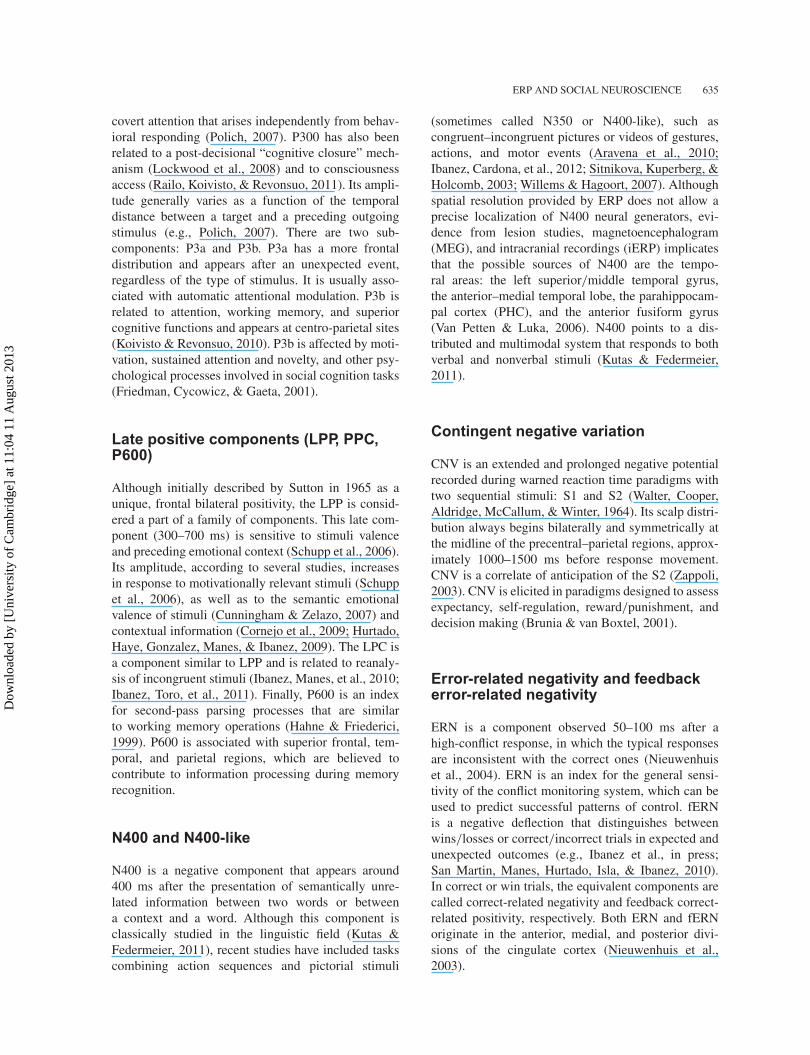
Figure 7. Comparison of different source estimation models. The box–whisker diagrams show the median (horizontal line within each box),the interquartile range (between the bottom and the top of each box), and the range of scores (shown by the whiskers). Circles represent outliers.Plots (a) and (b) show the results for each of the four inverse solutions (horizontal axis) for error measure (ED2) with anSNR (defined as theratio of source variance to noise variance) of 5 dB. (a) The results without regularization and (b) the results with regularization. RegularizedsLORETA has the lowest errors in terms of localization error. Reproduced with permission from BioMed Central Ltd (Grech et al., 2008).
methods exist (Grech et al., 2008). Several engi-neering solutions to find ERP sources using inverseproblems with parametric and nonparametric methodsare available (e.g., LORETA, sLORETA, VARETA,S-MAP, ST-MAP, Backus–Gilbert, LAURA, SLF,SSLOFO, and ALF; BESA, MUSIC, and FINES,reviewed in Grech et al. 2008). Methods of distributedsources (e.g., sLORETA, see Figure 7) combined withenhanced spatial constraints (e.g., photometry) canproduce more relevant results. Finally, principal com-ponent analysis (PCA) and independent componentanalysis (ICA) are now accessible for ERP sourcelocalization. Distributed EEG/MEG source analysisusing statistical parametric mapping of magnetic res-onance imaging (MRI) promises further advances insocial and affective neuroscience (Junghofer, Peyk,Flaisch, & Schupp, 2006).
The use of source location to improve socialneuroscience research has been demonstrated in the-ory of mind (ToM) research. Human social interactionsdepend on having a ToM, ability to infer thoughts,beliefs, intentions, and desires in other people’s minds.Although several fMRI reports of neural networksinvolved in ToM performed in the last decade exist,little is known about the temporal dynamics of thesenetworks. Leuthold, Filik, Murphy, and Mackenzie(2012) used dense-array ERPs to show that a rightN270-400 component followed by a larger positiv-ity at frontal sites is activated when decoding men-tal states from images of eyes. Moreover, sourceestimation of N270-400 and the frontal positivity
yielded two different sources at anterior temporal andorbitofrontal regions, respectively. Thus, anterior tem-poral lobe activation may reflect increased demandsof integrating general social knowledge and specificcontextual information. Orbitofrontal activation wouldlater reflect high-level mind-reading functions. Thesefindings suggest that ToM components may rely onpartially dissociable mid-latency neural mechanisms.
The use of a high number of dense arrays (improv-ing the source estimation), complementary techniquesfor electrode location (e.g., photometry), and therecent development of source localization algorithmscan provide measures of improved spatial resolutionfor further social and cognitive research.
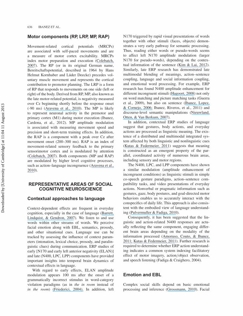
fMRI–ERP simultaneous recordings
fMRI provides an accurate spatial resolution butmeasures indirect brain signatures (hemodynamicresponse) and has poor temporal resolution. ERPsare a direct measure of cortical activity but havepoor spatial resolution. Combining fMRI and ERPsprovides a spatial and temporal fine ground reso-lution of cognitive brain activity (Gore, Horovitz,Cannistraci, & Skudlarski, 2006). Recently, removalalgorithms of fMRI artifacts on ERPs have been devel-oped, facilitating the combination of the two meth-ods. For instance, ERP/fMRI co-recording allows anenhanced study of origins and locations of ERP neuralgenerators.
Dow
nloa
ded
by [
Uni
vers
ity o
f C
ambr
idge
] at
11:
04 1
1 A
ugus
t 201
3

ERP AND SOCIAL NEUROSCIENCE 643
Figure 8. Simultaneous ERP–fMRI recordings of N170 component. (I) Grand average ERP of nine subjects at a representative temporal–occipital electrode P8, without (a, c) and with (b, d) MR acquisition when BCG artifacts are removed (a, b) or not removed (c, d). (II) Correlationsacross subjects for the N170 peak amplitude (a) and latency (b) to faces at electrode P8 reveal very high correlations between data collected withand without MR acquisition. The symbol “(2)” indicates a dot representing overlapping data points of two subjects. Reproduced with permissionfrom NeuroImage (Sadeh et al., 2008).
For example, the intertwined spatial (fusiformareas) and temporal brain dynamics (N170) of faceprocessing in the human brain have been measuredwith this methodology (Sadeh, Zhdanov, Podlipsky,Hendler, & Yovel, 2008). This study confirmedthe specific functionality of N170 facial process-ing and linked its temporal dynamics with neu-roanatomical face-processing areas (fusiform cor-tex). Moreover, face-selective characteristics of theN170 (e.g., N170 peak amplitude, latency, selectiv-ity to faces, laterality to faces) are preserved duringsimultaneous fMRI data acquisition, allowing furthercross-talk research (figure 8). Integrating fMRI andERP recordings shows promising insights into socialneuroscience.
WHAT DO ERPS CONTRIBUTE TOSOCIAL NEUROSCIENCE?
The genuine advantage of ERP studies
In the previous sections, we highlighted the spe-cific contribution of ERP processing in emotionalprocessing, contextual effects in speech, empathy,and decision making. Here, we summarize thewide-ranging benefits of ERP research. The temporal
precision of the ERP allows assessing several matters:(a) short and transient brain changes, (b) behavioralcorrelates and task modulation in a stage-by-stagesequentiation, and (c) bottom-up and top-down cog-nitive interactions.
ERP tracks the fine temporal dynamic changes ofcognitive processes. Multiple cognitive events are tran-sient and restricted to short time windows. Subtlechanges between trials can also be studied with recentapproaches to single trial dynamics (Makeig et al.,2002). The ERP technique assesses cognitive stagesfrom early windows (e.g., 80 ms after stimulus onset or700 ms before subject response) to successive dynam-ical changes over time.
ERPs can be considered the “reaction time forthe twenty-first century” (Luck, 2005). A multi-level measure of cognitive processes can be achievedusing trial-by-trial analysis and behavioral responses.Even without a behavioral response (e.g., passiveparadigms) or task-modulated behavior, ERPs can stillrecord the processing stages affected by experimentalmanipulations.
Current ERP research in social neuroscience(e.g., attention–emotion interaction, facial processing,empathy, prejudice and attitudes, decision making)highlights the role of early and late cortical dynamics.Early responses (e.g., 80–200 ms after stimulus
Dow
nloa
ded
by [
Uni
vers
ity o
f C
ambr
idge
] at
11:
04 1
1 A
ugus
t 201
3
644 IBANEZ ET AL.
onset) usually index bottom-up sensory mechanismssensitive to stimulus salience. For instance, earlymodulation refers to the facilitation of early auto-matic and pre-attentional discrimination of salientstimuli. Later stages (300–800 ms) may be consid-ered a marker of top-down control mechanisms thatsupport the processing of task-relevant stimuli. Thelate process can be interpreted as a correlate ofarousal, control, and awareness. Thus, the early/lateprocesses can be understood as an early automaticand late controlled parallel process. Time dynam-ics is crucial to the interaction of these processes,especially because neuroimaging studies sometimesprovide imprecise information. Several neuroimagingstudies, for example, suggest that the rapid detec-tion of salient targets is a pure bottom-up process(e.g., Wardak, Vanduffel, & Orban, 2010). The influ-ence of the dorsal attention network (lateral pre-frontal cortex and the ACC), however, during bothbottom-up and top-down processing (Ossandón et al.,2012) was revealed using spatial–temporal dynamicsof intracranial single trial ERP. Given the limited tem-poral resolution, previous neuroimaging studies failedto find active top-down processes in visual searchparadigms.
Thus, ERP measures present important advan-tages: (a) temporal dynamic and transient changesof cognitive events, (b) modulation of experimentalmanipulations and its combination with behavioraloutcome, and (c) differentiation and interactionbetween early bottom-up/automatic and late top-down/controlled events. The combination of ERPresearch with intracranial procedures, source locationalgorithms, and fMRI co-recordings reduces spatialresolution limitations and offers a more complete pic-ture of social and cognitive brain processes.
Limitations of ERP paradigms
There are also several methodological limitationsoften present when assessing questions of socialneuroscience with ERP assessment.
Current inferences about spatial resolution of thebrain, neuroanatomical questions, or issues regardingclear origins and functional significance of neural ERPgenerators should be avoided. ERPs used in sourcelocalization do not provide specific information inrelation to biophysical events that underlie differentcomponents (Luck, 2005; except under certain con-ditions, such as ERP–fMRI simultaneous recordings;some components with relatively unitary neural gener-ators as the occipital P1, the N170, or the ERN/fERN;or during intracranial recordings).
Further, several steps during signal preprocess-ing may yield inconsistent results when poorly con-trolled and when using different processing strategies.Multiple preprocessing stages (e.g., filtering and arti-fact rejection) and different measures for statisticalanalysis (e.g., single trial vs. subject average com-parisons; peak latency vs. peak amplitude vs. meanamplitude analysis; electrode selection and region ofinterests criteria) should be more explicitly describedand compared in ERP research. A meticulous report ofpreprocessing steps (e.g., Duncan et al., 2009) wouldmake ERP studies more comparable and reliable.
For example, the number of trials after artifactrejection tends to be dissimilar between subjects andconditions, yielding SNR differences. The percentageof trial rejections are often not reported and not statisti-cally contrasted. Analysis of independent components(ICA) is an adequate method for artifact deletion with-out trial rejection. Nevertheless, the ICA has its draw-backs, as residue of the artifact still remains in the EEGafter component removal (Shackman, McMenamin,Maxwell, Greischar, & Davidson, 2010), and directremoval of ICA components might lead to EEG dataloss (Lindsen & Bhattacharya, 2010). Given these con-cerns, the use of likelihood/mutual information basedon ICA methods is recommended (Delorme, Palmer,Onton, Oostenveld, & Makeig, 2012)
With regard to analysis, there are no explicit rulesfor electrode selection. The selection of the regionsof interest (ROIs) varies across studies. Some authorsselect different electrodes depending on each subjectto optimize task manipulation (Rousselet & Pernet,2011). This strategy may result in an invalid procedurebecause it compares the electrodes with maximumamplitudes of one subject and condition with otherelectrodes of another subject and condition. As infMRI studies (Kriegeskorte, Simmons, Bellgowan, &Baker, 2009), nonselective criteria for ROIs analysisshould be considered.
With regard to statistical comparisons, single-trialanalysis is a more sophisticated procedure than meanamplitude or mean latency analysis, but is not alwaysperformed in ERP studies (Rousselet & Pernet, 2011).However, the dynamics of single-trial analysis is notwell known and varies across tasks. Conversely, insome paradigms the subject average signal providesa simple and adequate measurement. Problems withclassic group statistics and categorical designs shouldbe replaced by reverse correlation techniques and sta-tistical modeling approaches (Liu, Agam, Madsen, &Kreiman, 2009).
In summary, ERP studies have a number of dis-similar (and usually not well-documented) resultsin preprocessing stages, in addition to arbitrary
Dow
nloa
ded
by [
Uni
vers
ity o
f C
ambr
idge
] at
11:
04 1
1 A
ugus
t 201
3

ERP AND SOCIAL NEUROSCIENCE 645
or inappropriate statistical analyses. These caveatsincrease noisy results and reduce comparability amongstudies. Inconsistencies, however, are not restrictedto ERP studies. The use of inadequate analysisstrategies (Nieuwenhuis, Forstmann, & Wagenmakers,2011), arbitrary selective data criteria (Kriegeskorteet al., 2009), or incorrect multivariate comparisons(Edward, Christine, Piotr, & Harold, 2009) is frequentin neuroimaging studies. Improving ERP researchdemands producing explicit and detailed reports, con-sensus among data preprocessing and analysis, ICAmethods, correlation techniques, statistical model-ing approaches, and nonselective criteria for ROIsanalysis.
Some theoretical remarks
The fields of social neuroscience reviewed in thisstudy suggest that basic social cognition (e.g., emo-tion, empathy, speech, decision making) is a highlysituated phenomenon (Amoruso et al., 2011; Barrettet al., 2007; de Gelder et al., 2010; Ibanez & Manes,2012). An experimental object may be processed dif-ferently because of contextual information or intrin-sic motivation. This result can occur at neurophys-iological and behavioral levels. Cognitive processseems to be a very ecological phenomenon dependenton external (world) and internal (mind) micrologi-cal circumstances. Thus, multilevel approaches (fromneuroscience, psychology, and social sciences) com-bining ERP research and social cognition may havecertain advantages: theoretical co-construction, inter-level cross-evidence, and combined explanatory strate-gies. The future of multilevel approaches to socialneuroscience should rely on the adequate interplaybetween ecological approaches and rigorous experi-mental designs. The role of ERPs in assessing timedynamics of social processes in the brain is essentialfor these future studies.
Original manuscript received 16 January 2012Revised manuscript accepted 1 May 2012
First published online 29 May 2012
REFERENCES
Amoruso, L., Couto, B., & Ibanez, A. (2011). BeyondExtrastriate Body Area (EBA) and Fusiform BodyArea (FBA): Context integration in the meaning ofactions. Frontiers in Human Neuroscience, 5, 124. doi:10.3389/fnhum.2011.00124
Aravena, P., Hurtado, E., Riveros, R., Cardona, J. F.,Manes, F., & Ibanez, A. (2010). Applauding with
closed hands: Neural signature of action–sentencecompatibility effects. PLoS ONE, 5(7), e11751. doi:10.1371/journal.pone.0011751
Balas, B., & Nelson, C. A. (2010). The role of faceshape and pigmentation in other-race face perception:An electrophysiological study. Neuropsychologia, 48(2),498–506.
Barrett, L. F., Lindquist, K. A., & Gendron, M. (2007).Language as context for the perception of emotion.Trends in Cognitive Sciences, 11(8), 327–332.
Bentin, S., Allison, T., Puce, A., Perez, E., & McCarthy,G. (1996). Electrophysiological studies of face percep-tion in humans. Journal of Cognitive Neuroscience, 8(6),551–565.
Brunia, C. H., & van Boxtel, G. J. (2001). Wait andsee. International Journal of Psychophysiology, 43(1),59–75.
Cacioppo, J. T., & Decety, J. (2011). Social neuroscience:Challenges and opportunities in the study of complexbehavior. Annals of the New York Academy of Sciences,1224, 162–173.
Cavanagh, J. F., Wiecki, T. V., Cohen, M. X., Figueroa,C. M., Samanta, J., Sherman, S. J., & Frank, M. J. (2011).Subthalamic nucleus stimulation reverses mediofrontalinfluence over decision threshold. Nature Neuroscience,14(11), 1462–1467.
Colebatch, J. G. (2007). Bereitschaftspotential andmovement-related potentials: Origin, significance,and application in disorders of human movement.Movement Disorders, 22(5), 601–610.
Cornejo, C., Simonetti, F., Ibáñez, A., Aldunate, N.,Ceric, F., López, V., & Núñez, R. E. (2009). Gesture andmetaphor comprehension: Electrophysiological evidenceof cross-modal coordination by audiovisual stimulation.Brain and Cognition, 70(1), 42–52.
Cunningham, W. A., & Zelazo, P. D. (2007). Attitudes andevaluations: A social cognitive neuroscience perspective.Trends in Cognitive Sciences, 11(3), 97–104.
de Bruijn, E. R., Sabbe, B. G., Hulstijn, W., Ruigt, G. S.,& Verkes, R. J. (2006). Effects of antipsychotic andantidepressant drugs on action monitoring in healthyvolunteers. Brain Research, 1105(1), 122–129.
de Gelder, B. (2006). Towards the neurobiology of emo-tional body language. Nature Reviews Neuroscience,7(3), 242–249.
de Gelder, B., Van den Stock, J., Meeren, H. K., Sinke,C. B., Kret, M. E., & Tamietto, M. (2010). Standing upfor the body. Recent progress in uncovering the networksinvolved in the perception of bodies and bodily expres-sions. Neuroscience and Biobehavioral Reviews, 34(4),513–527.
Decety, J. (2011). The neuroevolution of empathy. Annals ofthe New York Academy of Sciences, 1231, 35–45.
Decety, J., Yang, C. Y., & Cheng, Y. (2010). Physiciansdown-regulate their pain empathy response: Anevent-related brain potential study. NeuroImage,50(4), 1676–1682.
Deffke, I., Sander, T., Heidenreich, J., Sommer, W.,Curio, G., Trahms, L., & Lueschow, A. (2007).MEG/EEG sources of the 170-ms response to faces areco-localized in the fusiform gyrus. NeuroImage, 35(4),1495–1501.
Delorme, A., Palmer, J., Onton, J., Oostenveld, R., &Makeig, S. (2012). Independent EEG sources are dipolar.
Dow
nloa
ded
by [
Uni
vers
ity o
f C
ambr
idge
] at
11:
04 1
1 A
ugus
t 201
3

646 IBANEZ ET AL.
PLoS ONE, 7(2), e30135. doi: 10.1371/journal.pone.0030135
Dufey, M., Hurtado, E., María Fernández, A., Manes, F.,& Ibáñez, A. (2010). Exploring the relationship betweenvagal tone and event-related potentials in response to anaffective picture task. Social Neuroscience, 6(1), 48–62.
Duncan, C. C., Barry, R. J., Connolly, J. F., Fischer, C.,Michie, P. T., Naatanen, R., . . . Van Petten, C. (2009).Event-related potentials in clinical research: Guidelinesfor eliciting, recording, and quantifying mismatch neg-ativity, P300, and N400. Clinical Neurophysiology,120(11), 1883–1908.
Edward, V., Christine, H., Piotr, W., & Harold, P. (2009).Puzzlingly high correlations in fMRI studies of emo-tion, personality, and social cognition. Perspectives onPsychological Science, 4(3), 274–290.
Eimer, M., & Holmes, A. (2007). Event-related brainpotential correlates of emotional face processing.Neuropsychologia, 45(1), 15–31.
Fadiga, L., & Craighero, L. (2004). Electrophysiologyof action representation. Journal of ClinicalNeurophysiology, 21(3), 157–169.
Fan, Y., & Han, S. (2008). Temporal dynamic of neu-ral mechanisms involved in empathy for pain: Anevent-related brain potential study. Neuropsychologia,46(1), 160–173.
Folstein, J. R., & Van Petten, C. (2008). Influence of cogni-tive control and mismatch on the N2 component of theERP: A review. Psychophysiology, 45(1), 152–170.
Friederici, A. D. (2004). Event-related brain potential stud-ies in language. Current Neurology and NeuroscienceReports, 4(6), 466–470.
Friedman, D., Cycowicz, Y. M., & Gaeta, H. (2001). Thenovelty P3: An event-related brain potential (ERP) signof the brain’s evaluation of novelty. Neuroscience andBiobehavioral Reviews, 25(4), 355–373.
Fruhholz, S., Fehr, T., & Herrmann, M. (2009). Early andlate temporo-spatial effects of contextual interferenceduring perception of facial affect. International Journalof Psychophysiology, 74(1), 1–13.
Gleichgerrcht, E., Ibanez, A., Roca, M., Torralva, T., &Manes, F. (2010). Decision-making cognition in neurode-generative diseases. Nature Reviews Neurology, 6(11),611–623.
Gore, J. C., Horovitz, S. G., Cannistraci, C. J., &Skudlarski, P. (2006). Integration of fMRI, NIROT andERP for studies of human brain function. MagneticResonance Imaging, 24(4), 507–513.
Grech, R., Cassar, T., Muscat, J., Camilleri, K. P., Fabri,S. G., Zervakis, M., & Vanrumste, B. (2008). Reviewon solving the inverse problem in EEG source analysis.Journal of NeuroEngineering and Rehabilitation, 5, 25.
Grezes, J., Pichon, S., & de Gelder, B. (2007). Perceivingfear in dynamic body expressions. NeuroImage, 35(2),959–967.
Groen, Y., Wijers, A. A., Mulder, L. J., Waggeveld, B.,Minderaa, R. B., & Althaus, M. (2008). Error and feed-back processing in children with ADHD and childrenwith Autistic Spectrum Disorder: An EEG event-relatedpotential study. Clinical Neurophysiology, 119(11),2476–2493.
Grossmann, T. (2010). The development of emotion per-ception in face and voice during infancy. RestorativeNeurology and Neuroscience, 28(2), 219–236.
Guerra, S., Ibanez, A., Martin, M., Bobes, M. A., Reyes,A., Mendoza, R., . . . Sosa, M. V. (2009). N400 deficitsfrom semantic matching of pictures in probands and first-degree relatives from multiplex schizophrenia families.Brain and Cognition, 70(2), 221–230.
Guillaume, F., & Tiberghien, G. (2001). An event-relatedpotential study of contextual modifications in a facerecognition task. NeuroReport, 12(6), 1209–1216.
Hagoort, P. (2008). The fractionation of spoken languageunderstanding by measuring electrical and magneticbrain signals. Philosophical Transactions of the RoyalSociety of London Series B – Biological Sciences,363(1493), 1055–1069.
Hahne, A., & Friederici, A. D. (1999). Electrophysiologicalevidence for two steps in syntactic analysis. Early auto-matic and late controlled processes. Journal of CognitiveNeuroscience, 11(2), 194–205.
Hajcak, G., MacNamara, A., & Olvet, D. M. (2010). Event-related potentials, emotion, and emotion regulation:An integrative review. Developmental Neuropsychology,35(2), 129–155.
Han, S., Fan, Y., & Mao, L. (2008). Gender difference inempathy for pain: An electrophysiological investigation.Brain Research, 1196, 85–93.
Herrmann, C. S., & Knight, R. T. (2001). Mechanisms ofhuman attention: Event-related potentials and oscilla-tions. Neuroscience and Biobehavioral Reviews, 25(6),465–476.
Herrmann, M. J., Schreppel, T., Jager, D., Koehler, S., Ehlis,A. C., & Fallgatter, A. J. (2007). The other-race effect forface perception: An event-related potential study. Journalof Neural Transmission, 114(7), 951–957.
Hillyard, S. A., Hink, R. F., Schwent, V. L., & Picton,T. W. (1973). Electrical signs of selective attention in thehuman brain. Science, 182(4108), 177–180.
Holroyd, C. B., & Coles, M. G. H. (2002). The neuralbasis of human error processing: Reinforcement learning,dopamine, and the error-related negativity. PsychologicalReview, 109(4), 679–709.
Holroyd, C. B., & Coles, M. G. H. (2008). Dorsal anteriorcingulate cortex integrates reinforcement history to guidevoluntary behavior. Cortex, 44(5), 548–559.
Hurtado, E., Haye, A., Gonzalez, R., Manes, F., & Ibanez, A.(2009). Contextual blending of ingroup/outgroup facestimuli and word valence: LPP modulation and con-vergence of measures. BMC Neuroscience, 10, 69. doi:10.1186/1471-2202-10-69
Ibanez, A., Cardona, J., Vidal, Y., Blenkmann, A.,Aravena, P., Roca, M., . . . Bekinschtein, T. (2012).Motor-language coupling: Direct evidence from earlyParkinson disease and human cortex direct recordings.Cortex. doi:10.1016/j.Cortex2012.02.014
Ibanez, A., Cetkovich, M., Petroni, A., Urquina, H., Baez, S.,Gonzalez, M. L., . . . Manes, F. (in press). Neuralbasis of decision making and reward in euthymic bipo-lar disorder and adults with ADHD. PLoS ONE. doi10.1371/journal.pone.0037306
Ibanez, A., Gleichgerrcht, E., Hurtado, E., Gonzalez, R.,Haye, A., & Manes, F. (2010). Early neural mark-ers of implicit attitudes: N170 modulated by intergroupand evaluative contexts in IAT. Frontiers in HumanNeuroscience, 4, 188. doi: 10.3389/fnhum.2010.00188
Ibanez, A., Haye, A., Gonzalez, R., Hurtado, E., &Henriquez, R. (2009). Multi-level analysis of cultural
Dow
nloa
ded
by [
Uni
vers
ity o
f C
ambr
idge
] at
11:
04 1
1 A
ugus
t 201
3

ERP AND SOCIAL NEUROSCIENCE 647
phenomena: The role of ERPs approach to prejudice.Journal for the Theory of Social Behaviour, 39(1),81–110.
Ibanez, A., Hurtado, E., Lobos, A., Escobar, J., Trujillo, N.,Baez, S., & Decety, J. (2011). Subliminal presentationof other faces (but not own face) primes behavioral andevoked cortical processing of empathy for pain. BrainResearch, 1398, 72–85.
Ibanez, A., Hurtado, E., Riveros, R., Urquina, H., Cardona,J. F., Petroni, A., & Manes, F. (2011). Facial andsemantic emotional interference: A pilot study on thebehavioral and cortical responses to the Dual ValenceAssociation Task. Behavioral and Brain Functions, 7, 8.doi: 10.1186/1744-9081-7-8
Ibanez, A., Lopez, V., & Cornejo, C. (2006). ERPs and con-textual semantic discrimination: Degrees of congruencein wakefulness and sleep. Brain and Language, 98(3),264–275.
Ibanez, A., & Manes, F. (2012). Contextual social cognitionand the behavioral variant of frontotemporal dementia.Neurology, 78, 1354–1362.
Ibanez, A., Manes, F., Escobar, J., Trujillo, N., Andreucci, P.,& Hurtado, E. (2010). Gesture influences the process-ing of figurative language in non-native speakers: ERPevidence. Neuroscience Letters, 471(1), 48–52.
Ibanez, A., Petroni, A., Urquina, H., Torrente, F.,Torralva, T., Hurtado, E., . . . Manes, F. (2011). Corticaldeficits of emotional face processing in adults withADHD: Its relation to social cognition and executivefunction. Society for Neuroscience, 6(5–6), 464–481.
Ibanez, A., Riveros, R., Aravena, P., Vergara, V., Cardona,J. F., García, L., . . . Manes, F. (2011). When contextis difficult to integrate: Cortical measures of congruencyin schizophrenics and healthy relatives from multiplexfamilies. Schizophrenia Research, 126(1), 303–305.
Ibanez, A., Riveros, R., Hurtado, E., Gleichgerrcht, E.,Urquina, H., Herrera, E., . . . Manes, F. (2012). The faceand its emotion: Right N170 deficits in structural process-ing and early emotional discrimination in schizophrenicpatients and relatives. Psychiatry Research, 195(1–2),18–26.
Ibanez, A., Toro, P., Cornejo, C., Urquina, H., Manes, F.,Weisbrod, M., & Schroder, J. (2011). High contextualsensitivity of metaphorical expressions and gestureblending: A video event-related potential design.Psychiatry Research, 191(1), 68–75.
Ito, T. A., & Urland, G. R. (2003). Race and gender on thebrain: Electrocortical measures of attention to the raceand gender of multiply categorizable individuals. Journalof Personality and Social Psychology, 85(4), 616–626.
Jackson, P. L., Rainville, P., & Decety, J. (2006). To whatextent do we share the pain of others? Insight from theneural bases of pain empathy. Pain, 125(1–2), 5–9.
Jacobs, J., & Kahana, M. J. (2010). Direct brain recordingsfuel advances in cognitive electrophysiology. Trends inCognitive Sciences, 14(4), 162–171.
Joyce, C., & Rossion, B. (2005). The face-sensitiveN170 and VPP components manifest the same brain pro-cesses: The effect of reference electrode site. ClinicalNeurophysiology, 116(11), 2613–2631.
Junghofer, M., Peyk, P., Flaisch, T., & Schupp, H. T.(2006). Neuroimaging methods in affective neuroscience:Selected methodological issues. Progress in BrainResearch, 156, 123–143.
Kim, A., & Lai, V. (2012). Rapid interactions betweenlexical semantic and word form analysis during wordrecognition in context: Evidence from ERPs. Journal ofCognitive Neuroscience, 24(5), 1104–1112.
Koivisto, M., & Revonsuo, A. (2010). Event-related brainpotential correlates of visual awareness. Neuroscienceand Biobehavioral Reviews, 34(6), 922–934.
Kotchoubey, B. (2005). Event-related potential measuresof consciousness: Two equations with three unknowns.Progress in Brain Research, 150, 427–444.
Krebs, R. M., Boehler, C. N., Roberts, K. C., Song, A. W.,& Woldorff, M. G. (2012). The involvement of thedopaminergic midbrain and cortico-striatal-thalamic cir-cuits in the integration of reward prospect and attentionaltask demands. Cerebral Cortex, 22(3), 607–615.
Kriegeskorte, N., Simmons, W. K., Bellgowan, P. S.,& Baker, C. I. (2009). Circular analysis in systemsneuroscience: The dangers of double dipping. NatureNeuroscience, 12(5), 535–540.
Kutas, M., & Federmeier, K. D. (2011). Thirty years andcounting: Finding meaning in the N400 component ofthe event-related brain potential (ERP. Annual Review ofPsychology, 62, 621–647.
Leuthold, H., Filik, R., Murphy, K., & Mackenzie, I. G.(2012). The on-line processing of socio-emotional infor-mation in prototypical scenarios: Inferences from brainpotentials. Social Cognitive and Affective Neuroscience,7(4), 457–466.
Lindsen, J., & Bhattacharya, J. (2010). Correction ofblink artifacts using independent component analysisand empirical mode decomposition. Psychophysiology,47(5), 955–960.
Liu, H., Agam, Y., Madsen, J. R., & Kreiman, G. (2009).Timing, timing, timing: Fast decoding of object infor-mation from intracranial field potentials in human visualcortex. Neuron, 62(2), 281–290.
Lockwood, A. H., Wack, D. S., Benedict, R. H., Coad,M. L., Sussman, J. E., & Burkard, R. F. (2008). Multi-site phasic neural activity mediates the execution ofan auditory continuous performance task: A PET andelectrophysiological study. Journal of Neuroimaging,18(4), 364–374.
Luck, S. J. (2005). An introduction to the event-relatedpotential technique. Cambridge, MA: MIT Press.
Makeig, S., Westerfield, M., Jung, T. P., Enghoff, S.,Townsend, J., Courchesne, E., & Sejnowski, T. J. (2002).Dynamic brain sources of visual evoked responses.Science, 295(5555), 690–694.
Meeren, H. K., van Heijnsbergen, C. C., & de Gelder, B.(2005). Rapid perceptual integration of facial expres-sion and emotional body language. Proceedings of theNational Academy of Sciences of the United States ofAmerica, 102(45), 16518–16523.
Mendez-Bertolo, C., Pozo, M. A., & Hinojosa, J. A.(2011). Early effects of emotion on word immedi-ate repetition priming: Electrophysiological and sourcelocalization evidence. Cognitive, Affective, & BehavioralNeuroscience, 11(4), 652–665.
Menzies, L., Chamberlain, S. R., Laird, A. R., Thelen, S. M.,Sahakian, B. J., & Bullmore, E. T. (2008). Integrating evi-dence from neuroimaging and neuropsychological stud-ies of obsessive-compulsive disorder: The orbitofronto-striatal model revisited. Neuroscience and BiobehavioralReviews, 32(3), 525–549.
Dow
nloa
ded
by [
Uni
vers
ity o
f C
ambr
idge
] at
11:
04 1
1 A
ugus
t 201
3

648 IBANEZ ET AL.
Miltner, W. H. R., Braun, C. H., & Coles, M. G. H. (1997).Event-related brain potentials following incorrect feed-back in a time-estimation task: Evidence for a genericneural system for error detection. Journal of CognitiveNeuroscience, 9(6), 788–798.
Neely, J. H., VerWys, C. A., & Kahan, T. A. (1998). Reading“glasses” will prime “vision,” but reading a pair of“glasses” will not. Memory & Cognition, 26(1), 34–39.
Nieuwenhuis, S., Forstmann, B. U., & Wagenmakers,E. J. (2011). Erroneous analyses of interactionsin neuroscience: A problem of significance. NatureNeuroscience, 14(9), 1105–1107.
Nieuwenhuis, S., Holroyd, C. B., Mol, N., & Coles,M. G. (2004). Reinforcement-related brain potentialsfrom medial frontal cortex: Origins and functional sig-nificance. Neuroscience & Biobehavioral Reviews, 28(4),441–448.
Nieuwenhuis, S., Yeung, N., van den Wildenberg, W., &Ridderinkhof, K. R. (2003). Electrophysiological corre-lates of anterior cingulate function in a go/no-go task:Effects of response conflict and trial type frequency.Cognitive, Affective, & Behavioral Neuroscience, 3(1),17–26.
Nieuwland, M. S., Otten, M., & Van Berkum, J. J. (2007).Who are you talking about? Tracking discourse-level ref-erential processing with event-related brain potentials.Journal of Cognitive Neuroscience, 19(2), 228–236.
O’Donnell, B. F., Swearer, J. M., Smith, L. T., Hokama, H.,& McCarley, R. W. (1997). A topographic study ofERPs elicited by visual feature discrimination. BrainTopography, 10(2), 133–143.
Ofan, R. H., Rubin, N., & Amodio, D. M. (2011). Seeingrace: N170 responses to race and their relation to auto-matic racial attitudes and controlled processing. Journalof Cognitive Neuroscience, 23(10), 3153–3161.
Ossandón, T., Vidal, J. R., Ciumas, C., Jerbi, K., Hamamé,C. M., Dalal, S. S., . . . Lachaux, J. P. (2012).Efficient “pop-out” visual search elicits sustained broad-band gamma activity in the dorsal attention net-work. Journal of Neuroscience, 32(10), 3414–3421. doi:10.1523/jneurosci.6048-11.2012
Petroni, A., Canales-Johnson, A., Urquina, H., Guex, R.,Hurtado, E., Blenkmann, A., . . . Ibanez, A. (2011).The cortical processing of facial emotional expressionis associated with social cognition skills and executivefunctioning: A preliminary study. Neuroscience Letters,505(1), 41–46.
Polich, J. (2007). Updating P300: An integrative theoryof P3a and P3b. Clinical Neurophysiology, 118(10),2128–2148.
Pourtois, G., & Vuilleumier, P. (2006). Dynamics of emo-tional effects on spatial attention in the human visualcortex. Progress in Brain Research, 156, 67–91.
Pulvermuller, F., & Fadiga, L. (2010). Active perception:Sensorimotor circuits as a cortical basis for language.Nature Reviews Neuroscience, 11(5), 351–360.
Railo, H., Koivisto, M., & Revonsuo, A. (2011). Trackingthe processes behind conscious perception: A review ofevent-related potential correlates of visual consciousness.Consciousness and Cognition, 20(3), 972–983.
Rangel, A., Camerer, C., & Montague, P. R. (2008). Aframework for studying the neurobiology of value-baseddecision making. Nature Reviews Neuroscience, 9(7),545–556.
Ridderinkhof, K. R., Ullsperger, M., Crone, E. A., &Nieuwenhuis, S. (2004). The role of the medialfrontal cortex in cognitive control. Science, 306(5695),443–447.
Ritaccio, A., Brunner, P., Cervenka, M. C., Crone, N., Guger,C., Leuthardt, E., . . . Schalk, G. (2010). Proceedings ofthe first international workshop on advances in electro-corticography. Epilepsy & Behavior, 19(3), 204–215. doi:10.1016/j.yebeh.2010.08.028
Rousselet, G. A., & Pernet, C. R. (2011). Quantifying thetime course of visual object processing using ERPs: It’stime to up the game. Frontiers in Psychology, 2, 107. doi:10.3389/fpsyg.2011.00107
Sadeh, B., Zhdanov, A., Podlipsky, I., Hendler, T., &Yovel, G. (2008). The validity of the face-selective ERPN170 component during simultaneous recording withfunctional MRI. NeuroImage, 42(2), 778–786.
San Martin, R., Manes, F., Hurtado, E., Isla, P., & Ibanez, A.(2010). Size and probability of rewards modulate thefeedback error-related negativity associated with winsbut not losses in a monetarily rewarded gambling task.NeuroImage, 51(3), 1194–1204.
Santesso, D. L., Dzyundzyak, A., & Segalowitz, S. J. (2011).Age, sex and individual differences in punishment sensi-tivity: Factors influencing the feedback-related negativity.Psychophysiology, 48(11), 1481–1489.
Schupp, H. T., Flaisch, T., Stockburger, J., & Junghofer,M. (2006). Emotion and attention: Event-related brainpotential studies. Progress in Brain Research, 156,31–51.
Schyns, P. G., Petro, L. S., & Smith, M. L. (2007). Dynamicsof visual information integration in the brain for cat-egorizing facial expressions. Current Biology, 17(18),1580–1585.
Shackman, A. J., McMenamin, B. W., Maxwell, J. S.,Greischar, L. L., & Davidson, R. J. (2010). Identifyingrobust and sensitive frequency bands for interrogatingneural oscillations. NeuroImage, 51(4), 1319–1333.
Sitnikova, T., Kuperberg, G., & Holcomb, P. J. (2003).Semantic integration in videos of real-world events:An electrophysiological investigation. Psychophysiology,40(1), 160–164.
Stahl, J., Wiese, H., & Schweinberger, S. R. (2008).Expertise and own-race bias in face processing: An event-related potential study. NeuroReport, 19(5), 583–587.
Turk, D. J., van Bussel, K., Brebner, J. L., Toma, A. S.,Krigolson, O., & Handy, T. C. (2011). When “it”becomes “mine”: Attentional biases triggered by objectownership. Journal of Cognitive Neuroscience, 23(12),3725–3733.
van Heijnsbergen, C. C., Meeren, H. K., Grezes, J., &de Gelder, B. (2007). Rapid detection of fear in bodyexpressions, an ERP study. Brain Research, 1186,233–241.
Van Petten, C., & Luka, B. J. (2006). Neural local-ization of semantic context effects in electromagneticand hemodynamic studies. Brain and Language, 97(3),279–293.
Vizioli, L., Foreman, K., Rousselet, G. A., & Caldara, R.(2010). Inverting faces elicits sensitivity to race on theN170 component: A cross-cultural study. Journal ofVision, 10(1), 1–23.
Vuilleumier, P., & Pourtois, G. (2007). Distributed andinteractive brain mechanisms during emotion face
Dow
nloa
ded
by [
Uni
vers
ity o
f C
ambr
idge
] at
11:
04 1
1 A
ugus
t 201
3

ERP AND SOCIAL NEUROSCIENCE 649
perception: Evidence from functional neuroimaging.Neuropsychologia, 45(1), 174–194.
Walter, W. G., Cooper, R., Aldridge, V. J., McCallum,W. C., & Winter, A. L. (1964). Contingent negativevariation: An electric sign of sensorimotor associa-tion and expectancy in the human brain. Nature, 203,380–384.
Wardak, C., Vanduffel, W., & Orban, G. A. (2010).Searching for a salient target involves frontal regions.Cerebral Cortex, 20(10), 2464–2477.
Wiens, S., Sand, A., & Olofsson, J. K. (2011). Nonemotionalfeatures suppress early and enhance late emotionalelectrocortical responses to negative pictures. BiologicalPsychology, 86(1), 83–89.
Willems, R. M., & Hagoort, P. (2007). Neural evidence forthe interplay between language, gesture, and action: Areview. Brain and Language, 101(3), 278–289.
Williams, J. K., & Themanson, J. R. (2011). Neural cor-relates of the implicit association test: Evidence forsemantic and emotional processing. Social Cognitive andAffective Neuroscience, 6(4), 468–476.
Yamada, M., Lamm, C., & Decety, J. (2011). Pleasingfrowns, disappointing smiles: An ERP investigation ofcounterempathy. Emotion, 11(6), 1336–1345.
Zappoli, R. (2003). Permanent or transitory effects onneurocognitive components of the CNV complexinduced by brain dysfunctions, lesions and ablationsin humans. International Journal of Psychophysiology,48(2), 189–220.
Zheng, X., Mondloch, C. J., & Segalowitz, S. J.(2012). The timing of individual face recogni-tion in the brain. Neuropsychologia. doi: 10.1016/j.Neuropsychologia2012.02.030.
Dow
nloa
ded
by [
Uni
vers
ity o
f C
ambr
idge
] at
11:
04 1
1 A
ugus
t 201
3