Water Absorption and Diffusion Characteristics of Nanohydroxyapatite (nHA) and...
9
Hindawi Publishing Corporation Journal of Nanomaterials Volume 2013, Article ID 479109, 8 pages http://dx.doi.org/10.1155/2013/479109 Research Article Water Absorption and Diffusion Characteristics of Nanohydroxyapatite (nHA) and Poly(hydroxybutyrate-co-hydroxyvalerate-) Based Composite Tissue Engineering Scaffolds and Nonporous Thin Films Naznin Sultana 1,2 and Tareef Hayat Khan 3 1 Department of Mechanical Engineering, e University of Hong Kong, Pokfulam Road, Hong Kong 2 IJN-UTM Cardiovascular Engineering Centre (CEC), Faculty of Biosciences and Medical Engineering, Universiti Teknologi Malaysia, 81310 UTM Johor Bahru, Johor, Malaysia 3 KALAM, Faculty of Built Environment, Universiti Teknologi Malaysia, 81310 UTM Johor Bahru, Johor, Malaysia Correspondence should be addressed to Naznin Sultana; [email protected] Received 15 March 2013; Revised 10 April 2013; Accepted 10 April 2013 Academic Editor: In-Kyu Park Copyright © 2013 N. Sultana and T. H. Khan. is is an open access article distributed under the Creative Commons Attribution License, which permits unrestricted use, distribution, and reproduction in any medium, provided the original work is properly cited. Water uptake characteristics of poly(hydroxybutyrate-co-hydroxyvalerate) (PHBV-) based composite tissue engineering (TE) scaffolds incorporating nanosized hydroxyapatite (nHA) have been investigated. e water absorption of these composite scaffolds obeyed the classical diffusion theory for the initial period of time. e diffusion coefficients of the composite scaffolds during the water absorption were much faster than those for the nonporous thin films, suggesting that the water uptake process depends on the presence of porosity and porous microstructure of the composite scaffolds. e incorporation of nHA increased the water uptake of both the composite scaffolds and thin films. It was also observed that the equilibrium uptake increased with the incorporation of nHA. is increase in the water uptake was largely due to the nHA particle aggregates in the microstructure of both composite scaffolds and thin films. e activation energy for diffusion was also determined using the Arrhenius equation for both porous scaffolds and thin films and the results suggested that the activation energy for scaffolds was lower than that for thin films. 1. Introduction rough the combination of cell biology, biomaterials, and engineering, tissue engineering (TE) seeks to provide a solution to replace, repair, or regenerate tissues/organs [1, 2]. To overcome the limitations of the autogenous and allogenic bone graſt, development of new synthetic scaffolds has received increasing interest. In TE, the biomaterial based scaffolds are the most important aspects [3, 4]. A number of approaches such as phase separation, selective laser sintering, and electrospinning are being used for fabricating scaffolds for bone regeneration. Biodegradable polymers, including poly(caprolactone), poly(l-lactic acid) (PLLA), and poly(hy- droxybutyrate-co-hydroxyvalerate) (PHBV), have been pro- cessed into 3D scaffolds with the development of TE [5]. Both PLLA and PHBV possess good biocompatibility with tissue and blood and are regarded as good candidates as biodegradable polymer matrix of composite scaffolds [6]. Blending of PHBV with PLLA can shorten the degradation time and rate of PHBV which has much longer degradation time [7, 8]. Several biodegradable polymer-hydroxyapatite (HA) composites have been developed as bone substitute material or bone TE scaffolds [9–11]. As HA is a natural component of bones and possesses the osteoconductive prop- erties, polymer-HA composite scaffold is a promising system for bone tissue regeneration as it can mimic the composition and structure with the mineral phase of the bone. By guiding new tissue growth in vivo and in vitro, scaffolds play an important role in TE. e scaffolds pos- sess 3D macroporous and interconnected network with
Transcript of Water Absorption and Diffusion Characteristics of Nanohydroxyapatite (nHA) and...

Hindawi Publishing CorporationJournal of NanomaterialsVolume 2013, Article ID 479109, 8 pageshttp://dx.doi.org/10.1155/2013/479109
Research ArticleWater Absorption and Diffusion Characteristics ofNanohydroxyapatite (nHA) andPoly(hydroxybutyrate-co-hydroxyvalerate-) Based CompositeTissue Engineering Scaffolds and Nonporous Thin Films
Naznin Sultana1,2 and Tareef Hayat Khan3
1 Department of Mechanical Engineering, The University of Hong Kong, Pokfulam Road, Hong Kong2 IJN-UTMCardiovascular Engineering Centre (CEC), Faculty of Biosciences andMedical Engineering, Universiti Teknologi Malaysia,81310 UTM Johor Bahru, Johor, Malaysia
3 KALAM, Faculty of Built Environment, Universiti Teknologi Malaysia, 81310 UTM Johor Bahru, Johor, Malaysia
Correspondence should be addressed to Naznin Sultana; [email protected]
Received 15 March 2013; Revised 10 April 2013; Accepted 10 April 2013
Academic Editor: In-Kyu Park
Copyright © 2013 N. Sultana and T. H. Khan. This is an open access article distributed under the Creative Commons AttributionLicense, which permits unrestricted use, distribution, and reproduction in any medium, provided the original work is properlycited.
Water uptake characteristics of poly(hydroxybutyrate-co-hydroxyvalerate) (PHBV-) based composite tissue engineering (TE)scaffolds incorporating nanosized hydroxyapatite (nHA) have been investigated.The water absorption of these composite scaffoldsobeyed the classical diffusion theory for the initial period of time. The diffusion coefficients of the composite scaffolds during thewater absorptionweremuch faster than those for the nonporous thin films, suggesting that the water uptake process depends on thepresence of porosity and porous microstructure of the composite scaffolds. The incorporation of nHA increased the water uptakeof both the composite scaffolds and thin films. It was also observed that the equilibrium uptake increased with the incorporationof nHA. This increase in the water uptake was largely due to the nHA particle aggregates in the microstructure of both compositescaffolds and thin films. The activation energy for diffusion was also determined using the Arrhenius equation for both porousscaffolds and thin films and the results suggested that the activation energy for scaffolds was lower than that for thin films.
1. Introduction
Through the combination of cell biology, biomaterials, andengineering, tissue engineering (TE) seeks to provide asolution to replace, repair, or regenerate tissues/organs [1,2]. To overcome the limitations of the autogenous andallogenic bone graft, development of new synthetic scaffoldshas received increasing interest. In TE, the biomaterial basedscaffolds are the most important aspects [3, 4]. A number ofapproaches such as phase separation, selective laser sintering,and electrospinning are being used for fabricating scaffoldsfor bone regeneration. Biodegradable polymers, includingpoly(caprolactone), poly(l-lactic acid) (PLLA), and poly(hy-droxybutyrate-co-hydroxyvalerate) (PHBV), have been pro-cessed into 3D scaffolds with the development of TE [5].
Both PLLA and PHBV possess good biocompatibility withtissue and blood and are regarded as good candidates asbiodegradable polymer matrix of composite scaffolds [6].Blending of PHBV with PLLA can shorten the degradationtime and rate of PHBV which has much longer degradationtime [7, 8]. Several biodegradable polymer-hydroxyapatite(HA) composites have been developed as bone substitutematerial or bone TE scaffolds [9–11]. As HA is a naturalcomponent of bones and possesses the osteoconductive prop-erties, polymer-HA composite scaffold is a promising systemfor bone tissue regeneration as it can mimic the compositionand structure with the mineral phase of the bone.
By guiding new tissue growth in vivo and in vitro,scaffolds play an important role in TE. The scaffolds pos-sess 3D macroporous and interconnected network with

2 Journal of Nanomaterials
the macropore diameter of a few hundreds of microme-ters to allow cell invasion. The water uptake propertiesof composite scaffolds for tissue TE are of importance asthe primary mechanism of the polymer degradation insidebody is hydrolysis. The ingress of water into polymer-basedscaffolds can have both adverse and beneficial effects onthe properties of the scaffolds. Hydrolysis and microcrackcan be formed due to water exposure. On the other hand,breakdown of three-dimensional scaffolds can occur due toexcessive water uptake. Several studies have been performedfor dental application to study the characteristics of polymericsystems and composites [12, 13]. However, the water uptakeand diffusion characteristics of TE scaffolds have been rarelyassessed and reported.
Previously, we reported the fabrication, characteriza-tion, and in vitro biological evaluation of PHBV/PLLA-based scaffolds [14]. The aim of the present study was todetermine the water uptake and diffusion characteristicsof 100/0 PHBV/PLLA, 50/50 PHBV/PLLA blend, and 10%nHA incorporated 50/50 PHBV/PLLA composite scaffolds.For comparison, solvent-cast thin films were also used inthe study. The effect of incorporation of HA, porosity, andmicrostructures of the composite scaffolds on the diffusioncoefficient, equilibrium uptake, and temperature dependencewas also investigated.
2. Experimental
2.1. Materials and General Methods. PHBV with 6% of 3-hydroxyvaleratewas purchased fromSigma-Aldrich. In orderto blend with PHBV, PLLA with viscosity 1.6 dL g−1 (Medis-orb 100L 1A) was purchased from Lakeshore Biomaterials(Birmingham, AL). The nHA nanoparticles were producedin-house which were used for producing composite scaffoldsand composite thin films [15]. Figure 1 shows the SEMmicrograph of nanosized HA. The freeze-dried HA powdersused in this investigation consisted of tiny agglomerates ofHA nanocrystallites.The particle size of the HA powders wasfound to be in the range of 20–30 nm. Chloroform and aceticacid were analytical grade.
2.2. Fabrication of Scaffolds and Solvent Cast Films. Pure100/0 PHBV/PLLA, 50/50 PHBV/PLLA, and 10% nHA in50/50 PHBV/PLLA tissue engineering scaffolds with 10%(w/v) polymer concentration were fabricated using the emul-sion freezing/freeze-drying technique described elsewhere[16]. Briefly, to produce polymer scaffolds, 1 g polymer orpolymer blend was dissolved in 5mL chloroform and then5mL acetic acid was added. The emulsion was then homog-enized and kept frozen at −18∘C in a freezer for overnightto induce phase separation. Then the frozen emulsion wasfreeze-dried using a freeze-drying vessel (Labconco, USA).Similarly, to produce 10% nHA in 50/50 PHBV/PLLA com-posite scaffolds, 0.1 g nHA was added in the emulsion andhomogenized prior to the phase-separation process. Non-porous thin filmswith the same compositions were fabricatedusing solvent casting technique.
Figure 1: SEM micrograph of nanosized HA particles synthesizedin-house through a nanoemulsion process.
2.3. Preparation of Polymer and Composite Specimens. Rect-angular shape specimens (20mm × 5mm × 0.5mm) werecut with sharp razor blade from three types of polymerand composite scaffolds, namely, PHBV scaffolds (10%w/v),50/50 PHBV/PLLA (10%w/v) blend scaffolds, and 10% nHAin PHBV/PLLA (10%w/v) composite scaffolds. Rectangularspecimens of (20mm × 5mm × 0.5mm) were also pre-pared from solvent-cast films of the same compositions. Theporous microstructure, morphology, and pore sizes of thescaffolds specimens were studied using a scanning electronmicroscopy (SEM; Stereoscan 440, UK). In order to deter-mine the presence and distribution of nHA nanoparticles incomposite scaffolds, energy dispersive X-ray spectrometry(EDX, INCA 300, UK) was performed. The skeletal densityand the porosity of the scaffolds were measured usingequations described elsewhere [16]. The wettability of thescaffolds was measured by measuring contact angles using acontact angle measuring machine (SL200B, Shanghai SalonTech Inc., Ltd., China). With distilled water as liquid, thesessile drop method was employed. The contact angle ofthe water drop on the scaffold specimen was determined atroom temperature using proprietary software. At least threemeasurements were conducted at different locations of thescaffold specimen. As the mean ± standard deviation, thecontact angle was expressed.
2.4.Water Absorption. The specimens were placed in an ovenat 37∘C for 48 hours and then weighed to an accuracy of±0.0001 g on a balance. The specimens were then placed into100mL of distilled water at 37∘C. During the first day ofimmersion, the specimens were removed at intervals of 2,5, 10, 15min, and so forth, blotted dry on filter paper toremove excess water, weighed, and returned to the water.Following the first day, the samples were weighed daily, untilthe uptake slowed. Until there was no significant change inweight, the uptake of water was recorded. When equilibriumwas attained, the samples were then transferred to a dryingoven at 37∘C. Similarly, the experiment was repeated at 25∘Cand at 50∘C.

Journal of Nanomaterials 3
2.5. Diffusion Coefficients. For the earlier stages of uptake(usually where 𝑀
𝑡
/𝑀
∞
≤ 0.5), classical diffusion theorypredicts [1, 17] the following:
𝑀
𝑡
𝑀
∞
= 2(
𝐷𝑡
𝜋𝑙
2
)
1/2
,(1)
where 𝑀𝑡
is the mass uptake at time 𝑡, 𝑀∞
is the massuptake at equilibrium, 2𝑙 is the thickness of the specimen, 𝐷is the diffusion coefficient, and 𝑡 is the water uptake time. Aplot of𝑀
𝑡
/𝑀
∞
against 𝑡1/2 should provide a straight line forearlier stages of water uptake. Diffusion coefficients can becalculated from the slope of the straight line.
The temperature dependence of the diffusion coefficientis given by the Arrhenius equation:
ln𝐷 = ln𝐷0
−
𝑄
𝑅
(
1
𝑇
) ,(2)
where 𝑇 is the absolute temperature (∘K), 𝑄 is the activationenergy for diffusion (J/mol or eV/atom), and𝐷
𝑜
is a temper-ature independent constant (preexponential) (m2/s), 𝑅 is thegas constant (8.314 J/mol-K or 8.62 × 10⁻⁵ eV/atom).
3. Results and Discussion
Several assumptions were used for this study. They were asfollows.
(i) Distilled water was in infinite supply.(ii) Polymer and composite films and scaffolds were
surrounded by water.
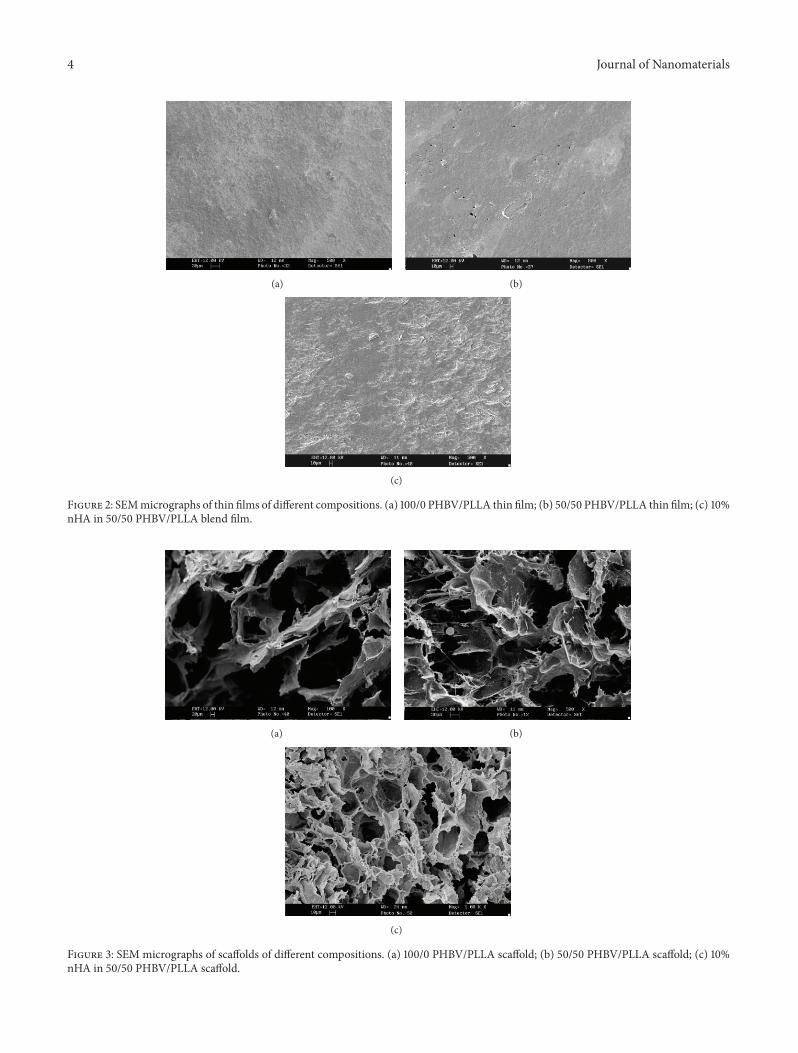
3.1.Morphological Observation. Figure 2 shows themorphol-ogies of thin films of 100/0 PHBV/PLLA, 50/50 PHBV/PLLA,and 10%nHA incorporated 50/50 PHBV/PLLA. Somemicro-pores were observed in 50/50 PHBV/PLLA thin film dueto the immiscibility of PHBV and PLLA. Figure 3 showsthemorphologies of 100/0 PHBV/PLLA, 50/50 PHBV/PLLA,and 10% nHA in 50/50 PHBV/PLLA scaffolds. It wasobserved that highly porous scaffold specimens consisted ofmany micropores. Pore structure and size depended on thephase separation process used in the fabrication technique.Scanning electron microscopy (SEM) and energy dispersiveX-ray spectroscopy showed the distribution of HA nanopar-ticles in the pore walls of the scaffolds, and some particleagglomerationwas also observed (Figure 4).The average porediameter of the 100/0 PHBV/PLLA scaffold was higher thanthat of 10% nHA incorporated 50/50 PHBV/PLLA scaffold asincorporation of nHAperturbed the phase separation processslightly (Table 1). As the scaffolds were prepared from samepolymer emulsion concentration, 10% (w/v), the porositydid not change significantly in the scaffolds. The contactangle was much higher in 100/0 PHBV/PLLA scaffold than50/50 PHBV/PLLA and 10% nHA in PHBV/PLLA scaffoldsas PHBV polymer is well known for its hydrophobic nature.Blending with PLLA and incorporporation of nHA increasedthe hydrophilicity of the composite scaffolds.
Table 1: Average pore diameter, porosity, and contact angle of thescaffold.
Scaffold Average porediameter (𝜇m) Porosity % Contact
angle100/0 PHBV/PLLA 290 75 ± 2.0
103 ± 1.6
50/50 PHBV/PLLA 271 77 ± 1.5
98 ± 2.5
10% nHA in 50/50PHBV/PLLA 250 72 ± 3.0
83 ± 1.5
3.2. Diffusion Study of Solvent-Cast Thin Films. Figure 5shows the water uptake curve of 100/0 PHBV/PLLA thinfilm at 37∘C as𝑀
𝑡
/𝑀
∞
versus the square root of immersiontime. The specimens were immersed in distilled water. Themaximum uptake at equilibrium was 1.4%. The diffusioncoefficients were calculated from the initial linear region ofthe curves.
In order to observe the temperature dependence of thediffusion coefficients of 100/0 PHBV/PLLA thin film, theexperiment was performed at three different temperatures,namely, at 25∘C, 37∘C, and 50∘C.Water uptake study was alsoperformed for 50/50 PHBV/PLLA thin film and 10%HA in50/50 PHBV/PLLA thin film following the same procedurementioned above. The calculated diffusion coefficients aregiven in Table 2. It can be seen that diffusion coefficient issignificantly higher at high temperature of 50∘C than that of25∘C, 37∘C.
In order to study the temperature dependence of diffusioncoefficients, Arrhenius plot was constructed for 10%HA in50/50 PHBV/PLLA thin films as shown in Figure 6. Fromthe slope of the curve, activation energy can be calculatedwhich is referred to as the energy required to produce thediffusive motion of one mole of atoms. The activation energycalculated from the slope of the best fit linear curve was76 ± 20KJ/mole with the regression coefficient, 𝑟2 value of0.94. The activation energy of 100/0 PHBV/PLLA and 50/50PHBV/PLLA thin film was 99.8 ± 21.9KJ/mole and 112 ±45KJ/mole, respectively.
3.3. Diffusion Study of Scaffolds. Figure 7 is the plot ofwater uptake as 𝑀
𝑡
/𝑀
∞
versus square root of immersiontime at 37∘C for 100/0 PHBV/PLLA scaffolds. The diffusioncoefficients were calculated from the initial linear regions ofthe curves. In order to study the temperature dependenceof diffusion coefficients of the scaffolds, the experiment wasperformed at three different temperatures, namely, at 25∘C,37∘C, and 50∘C.
Table 3 shows the diffusion coefficients of 100/0 PHBV/PLLA scaffolds at 25∘C, 37∘C, and 50∘C, which were 51.75 ×10⁻¹², 67.60 × 10⁻¹², and 165 × 10−12m2/s, respectively. It wasobserved that the initial water uptake of HA incorporatedcomposite scaffold was much higher than that of polymerblend scaffolds, and the diffusion coefficients were alsohigher. The value of diffusion coefficient was lower at lowtemperature and relatively high at high temperature. Figure 8shows the Arrhenius plot which represents the tempera-ture dependence of the diffusion coefficients of 10%HA in50/50 PHBV/PLLA scaffolds. From the slope of the best
4 Journal of Nanomaterials
(a) (b)
(c)
Figure 2: SEMmicrographs of thin films of different compositions. (a) 100/0 PHBV/PLLA thin film; (b) 50/50 PHBV/PLLA thin film; (c) 10%nHA in 50/50 PHBV/PLLA blend film.
(a) (b)
(c)
Figure 3: SEM micrographs of scaffolds of different compositions. (a) 100/0 PHBV/PLLA scaffold; (b) 50/50 PHBV/PLLA scaffold; (c) 10%nHA in 50/50 PHBV/PLLA scaffold.

Journal of Nanomaterials 5
(a) (b)
(c)
P Ca
P Ka1Ca Ka1
Figure 4: SEM micrographs of (a) 10% nHA in 50/50 PHBV/PLLA composite scaffold; (b) EDX spectrum; (c) Mapping of Ca and P.
fit linear line, the activation energy was calculated, whichwas 100.9 ± 5.3KJ/mole. The activation energies were found120.3±45KJ/mole for 50/50 PHBV/PLLA scaffold and 83.5±53KJ/mole for 100/0 PHBV/PLLA scaffolds, respectively.
Table 4 shows the comparison between the amountsof water uptake (g/g) at equilibrium between the differentcompositions of thin films and scaffolds. Among the dif-ferent compositions of thin films and scaffolds, the amount(g/g) of equilibrium water uptake of 10% nHA in 50/50PHBV/PLLA films and scaffolds was higher than that of100/0 PHBV/PLLA and 50/50 PHBV/PLLA thin films andscaffolds. This result was opposite to other studies wherethe uptake of HA/polymer composite films was slower thanthe pure polymeric film as the polymers were very muchhydrophilic and water uptake was high for the polymer itself[12]. In the present study, it was also observed that wateruptake increased significantly with the increasing porosity.The scaffolds which were more than 70% porous, absorbedmore water at the same condition than that of dense films.Besides, the incorporation of HA increased the water uptakein both composite thin film and composite scaffold.
Water uptake can occur by the materials in terms of“absorbed water” that means the amount of water absorbedfrom media which mainly depends on the hydrophilicityof material. Capillary water is termed as liquid water thatis “drawn in” through pores or capillaries of the materials.Moreover, the amount of water absorbed is related to the
0 100 200 300 400 500 600 700 800 900 10000
0.2
0.4
0.6
0.8
1
1.2
1.4
Square root of immersion time (s)
𝑀𝑡/𝑀
∞
Figure 5: Water uptake curve for 100/0 PHBV/PLLA thin film at37∘C in the form of𝑀
𝑡
/𝑀
∞
versus square root of immersion time.
porosity and the amount of available liquid water at thesurface of the material. For this reason, porous material canuptake and store more water whereas the nonporous (dense)material can store a limited amount of water (Table 4). It canbe seen from the plots of𝑀
𝑡
/𝑀
∞
versus square root of time(Figures 5 and 7) that the plots are almost linear for both the

6 Journal of Nanomaterials
Table 2: Diffusion coefficients of thin films at 25∘C, 37∘C, and 50∘C.
Temperature (∘C) 100/0 PHBV/PLLA thin films(m2/s)
50/50 PHBV/PLLA thin films(m2/s)
10% nHA in 50/50 PHBV/PLLA thin films(m2/s)
253.8 × 10
−13
± 1.5 × 10
−13
2.12 × 10
−13
± 0.65 × 10
−13
2.3 × 10
−13
± 0.7 × 10
−13
3711.3 × 10
−13
± 1.0 × 10
−13
3.215 × 10
−13
± 0.47 × 10
−13
11.6 × 10
−13
± 0.9 × 10
−13
5059.3 × 10
−13
± 12.0 × 10
−13
59.8 × 10
−13
± 3.53 × 10
−13
22.9 × 10
−13
± 0.8 × 10
−13
Table 3: Diffusion coefficients of scaffolds at 25∘C, 37∘C, and 50∘C.
Temperature (∘C) 100/0 PHBV/PLLA scaffolds(m2/s)
50/50 PHBV/PLLA scaffolds(m2/s)
10% nHA in 50/50 PHBV/PLLAscaffolds (m2/s)
2551.7 × 10
−12
± 1.1 × 10
−12
0.16 × 10
−11
± 0.06 × 10
−11
0.2 × 10
−11
± 0.05 × 10
−11
3767.6 × 10
−12
± 3.2 × 10
−12
3.58 × 10
−11
± 0.7 × 10
−11
1.3 × 10
−11
± 0.5 × 10
−11
50165 × 10
−12
± 9.1 × 10
−12
6.92 × 10
−11
± 2 × 10
−11
5.4 × 10
−11
± 1.35 × 10
−11
0.0031 0.00315 0.0032 0.00325 0.0033 0.00335 0.0034−28
−27
−26
−25
−24
−23
−22
−21
−20
1/𝑇
ln𝐷
Figure 6: Arrhenius plot of 10%HA in 50/50 PHBV/PLLA thinfilms (the graph is plotted as logarithm of diffusion coefficients (𝐷)versus the reciprocal of absolute temperature, 𝑇).
0 200 400 600 800 1000 1200 14000
0.2
0.4
0.6
0.8
1
1.2
1.4
Square root of immersion time (s)
𝑀𝑡/𝑀
∞
Figure 7: Water uptake curve for 100/0 PHBV/PLLA scaffolds at37∘C in the form of𝑀
𝑡
/𝑀
∞
versus square root of immersion time.
Table 4: Comparison of equilibrium water uptake between thinfilms and scaffolds at 37∘C.
Compositions of filmsand scaffoldspecimens
Equilibrium wateruptake for thin films
(g/g)
Equilibrium wateruptake for scaffolds
(g/g)100/0 PHBV/PLLA 0.01 1.2450/50 PHBV/PLLA 0.03 1.3510% nHA in 50/50PHBV/PLLA 0.09 2.97
0.0031 0.00315 0.0032 0.00325 0.0033 0.00335 0.0034−30
−29
−28
−27
−26
−25
−24
−23
−22
−21
−20
1/𝑇
ln𝐷
Figure 8: Arrhenius plot of 10%HA in 50/50 PHBV/PLLA scaffolds(the graph is plotted as logarithm of diffusion coefficients (𝐷) versusthe reciprocal of absolute temperature, 𝑇).
thin films and scaffolds in the initial period of time. As Fick’ssecond law is applicable for both the thin films and scaffoldsfor the initial period of time, it can be stated that the initialstage is diffusion controlled. Similar results were obtained byseveral studies of polymeric composite materials for dentalapplications [12, 18, 19].
It was also found that the water uptake of PHBV/PLLAscaffold was lower than 10wt% nHA incorporated PHBV/PLLA scaffold. For the scaffold specimens, when all the

Journal of Nanomaterials 7
pores were assumed to be filled with water, the equilibriumwater uptake of polymer blend scaffold and composite scaf-fold specimens were found to be 1.35 g/g and 2.97 (g/g),respectively (Table 4). Moreover, water uptake of compositescaffold specimen decreased after few days.The reasonmightbe the starting of dissolution of nHA within the compositescaffold.The initial increase of water uptake of the compositefilms and scaffolds may be due to hydrophilic nature of thenanosized nHAparticles or the inclusions ofHAnanoparticleaggregates. The nHA nano-particles may appear as looselyembedded aggregates in the polymer matrix. As a result,additional amount of water at the interface between theagglomerates and the matrix can be accommodated.
The calculated diffusion coefficients expressed are givenin Tables 2 and 3 during water absorption. The values ofdiffusion coefficients are available in the literature for otherconventional composites [18, 20]. The diffusion coefficientswere higher in scaffolds than thin films. This behaviour canoccur during the initial uptake of water through the pores ofthe scaffolds which can cause the formation of water clusters.Surface area to volume ratio and thickness can also haveeffects on this.
Temperature and composition are the two most impor-tant factors which can affect the diffusion coefficient [21].Between these two factors, temperature has the most pro-found influence on the coefficients and diffusion rates. Thediffusion coefficients are related to temperature according toArrhenius equation. From Arrhenius plots of thin film andscaffold (Figures 6 and 8), it can be seen that the plot is almostlinear. These results can further confirm that water uptake ofthin films is controlled by diffusion process. From the plotsof 𝑀𝑡
/𝑀
∞
versus square root of immersion time, it is alsopossible to estimate when a plateau region can be establishedand how long the diffusion phenomena can be observed.It was also found by other studies that after saturation,many other properties should be taken into account such asstructure and surface chemistry.
4. Conclusions
(1)The amount of water absorption or uptake dependsmainly on the hydrophilicity and porosity of thematerials. For this reason, PHBV/PLLA and 10%HAin PHBV/PLLA scaffolds and thin films absorbedmore water than that of 100/0 PHBV/PLLA scaffoldsand thin films as PHBV is more hydrophobic thanPLLA and HA. The water uptake of the scaffoldswas much higher than that of thin films because thescaffolds had high porosity.(2) For thin films and scaffold, Arrhenius plot was almost
linear. For the initial period of time, water uptake ofthe thin films was controlled by diffusion process. Itwas possible to estimate when a plateau region couldbe established and how long the diffusion phenomenacould be observed.(3) After saturation, many other properties should be
taken into account such as structure and surfacechemistry. 10%HA in 50/50 PHBV/PLLA scaffolds
showed decrease in water uptake after saturationdue to dissolution of HA and also degradation ofamorphous part of PLLA/PHBV polymer.(4)The diffusion coefficients were higher in porous scaf-
folds than thin films as the rate of diffusion increasedwith decreasing density of atomic packing. Porosityand surface to volume ratio had a profound effect onthe water uptake of scaffolds.
Conflict of Interests
Theauthors, hereby, declare that they do not have any conflictof interests.
Acknowledgments
N. Sultana thanks The University of Hong Kong (HKU)for providing her with a research studentship. This workwas supported by a GRF Grant (HKU 7182/05E) from theResearch Grants Council of Hong Kong. Assistance providedby technical staff in the Department of Mechanical Engi-neering, Faculty of Engineering, is acknowledged. Authorsalso thank Universiti Teknologi Malaysia, Ministry of HigherEducation (MOHE), RMC, IJN-UTM Cardiovascular Engi-neering Centre, and FRGSGrant (vot: 4F126), GUP Tier 1 (03H13, 05H07) for financial supports.
References
[1] E. Bell, Tissue Engineering: Current Perspectives, Birkhauser,Boston, Mass, USA, 1993.
[2] R. P. Lanza, R. S. Langer, and J. Vacanti, Principles of TissueEngineering, Elsevier/Academic Press, Amsterdam,TheNether-lands, 2007.
[3] Z. Li, Y. Su, B. Xie et al., “A tough hydrogel-hydroxyapatitebone-like composite fabricated in situ by the electrophoresisapproach,” Journal of Materials Chemistry B, vol. 1, no. 12, pp.1755–1764, 2013.
[4] J. Lacroix, J. Lao, and E. Jallot, “Green and safe in situ templatingof bioactive glass scaffolds for bone tissue engineering,” Journalof Materials Chemistry B, vol. 1, no. 13, pp. 1782–1785, 2013.
[5] S. Lalan, I. Pomerantseva, and J. P. Vacanti, “Tissue engineeringand its potential impact on surgery,” World Journal of Surgery,vol. 25, no. 11, pp. 1458–1466, 2001.
[6] C. Bastioli, Handbook of Biodegradable Polymers, Rapra Tech-nology, Shrewsbury, UK, 2005.
[7] E. Blumm andA. J. Owen, “Miscibility, crystallization andmelt-ing of poly(3-hydroxybutyrate)/poly(l-lactide) blends,” Poly-mer, vol. 36, no. 21, pp. 4077–4081, 1995.
[8] N. Sultana and M. Wang, “PHBV/PLLA-based composite scaf-folds containing nano-sized hydroxyapatite particles for bonetissue engineering,” Journal of Experimental Nanoscience, vol. 3,no. 2, pp. 121–132, 2008.
[9] P. X. Ma, R. Zhang, G. Xiao, and R. Franceschi, “Engineeringnew bone tissue in vitro on highly porous poly(𝛼-hydroxylacids)/hydroxyapatite composite scaffolds,” Journal of Biomedi-cal Materials Research, vol. 54, pp. 284–293, 2001.
[10] B. D. Ratner, BioMaterials Science: An Introduction to Materialsin Medicine, Elsevier Academic Press, San Diego, Calif, USA,2004.

8 Journal of Nanomaterials
[11] N. Sultana and T. H. Khan, “Factorial study of compressivemechanical properties and primary in vitro osteoblast responseof PHBV/PLLA scaffolds,” Journal of Nanomaterials, vol. 2012,Article ID 656914, 8 pages, 2012.
[12] M. Braden, “Water absorption characteristics of dental micro-fine composite filling materials. II. Experimental materials,”Biomaterials, vol. 5, no. 6, pp. 373–375, 1984.
[13] S. Deb, M. Braden, andW. Bonfield, “Water absorption charac-teristics of modified hydroxyapatite bone cements,” Biomateri-als, vol. 16, no. 14, pp. 1095–1100, 1995.
[14] N. Sultana and M. Wang, “PHBV/PLLA-based compositescaffolds fabricated using an emulsion freezing/freeze-dryingtechnique for bone tissue engineering: surface modificationand in vitro biological evaluation,” Biofabrication, vol. 4, no. 1,Article ID 015003, 2012.
[15] W. Y. Zhou, M. Wang, W. L. Cheung, B. C. Guo, and D.M. Jia, “Synthesis of carbonated hydroxyapatite nanospheresthrough nanoemulsion,” Journal of Materials Science: Materialsin Medicine, vol. 19, no. 1, pp. 103–110, 2008.
[16] N. Sultana and M.Wang, “Fabrication of HA/PHBV compositescaffolds through the emulsion freezing/freeze-drying processand characterisation of the scaffolds,” Journal of MaterialsScience:Materials inMedicine, vol. 19, no. 7, pp. 2555–2561, 2008.
[17] J. Crank and G. S. Park, Eds., Diffusion in Polymers, AcademicPress, New York, NY, USA, 1977.
[18] M. Braden and R. L. Clarke, “Water absorption characteristicsof dental microfine composite filling materials. I. Proprietarymaterials,” Biomaterials, vol. 5, no. 6, pp. 369–372, 1984.
[19] C. Santos, R. L. Clarke, M. Braden, F. Guitian, and K. W. M.Davy, “Water absorption characteristics of dental compositesincorporating hydroxyapatite filler,” Biomaterials, vol. 23, no. 8,pp. 1897–1904, 2002.
[20] J. Crank and G. S. Park, Diffusion in Polymers, Academic Press,London, UK, 1968.
[21] R. E. Smallman and A. H. W. Ngan, Physical Metallurgy andAdvanced Materials, Butterworth-Heinemann; Elsevier, Ams-terdam, The Netherlands, 2007.

Submit your manuscripts athttp://www.hindawi.com
ScientificaHindawi Publishing Corporationhttp://www.hindawi.com Volume 2014
CorrosionInternational Journal of
Hindawi Publishing Corporationhttp://www.hindawi.com Volume 2014
Hindawi Publishing Corporationhttp://www.hindawi.com Volume 2014
Polymer ScienceInternational Journal of
Hindawi Publishing Corporationhttp://www.hindawi.com Volume 2014
CeramicsJournal of
Hindawi Publishing Corporationhttp://www.hindawi.com Volume 2014
CompositesJournal of
NanoparticlesJournal of
Hindawi Publishing Corporationhttp://www.hindawi.com Volume 2014
Hindawi Publishing Corporationhttp://www.hindawi.com Volume 2014
International Journal of
Biomaterials
Hindawi Publishing Corporationhttp://www.hindawi.com Volume 2014
NanoscienceJournal of
TextilesHindawi Publishing Corporation http://www.hindawi.com Volume 2014
Journal of
NanotechnologyHindawi Publishing Corporationhttp://www.hindawi.com Volume 2014
Journal of
Hindawi Publishing Corporationhttp://www.hindawi.com
Volume 2014
CrystallographyJournal of
The Scientific World JournalHindawi Publishing Corporation http://www.hindawi.com Volume 2014
Hindawi Publishing Corporationhttp://www.hindawi.com Volume 2014
CoatingsJournal of
Advances in
Materials Science and EngineeringHindawi Publishing Corporationhttp://www.hindawi.com Volume 2014
Smart Materials Research
Hindawi Publishing Corporationhttp://www.hindawi.com Volume 2014
Hindawi Publishing Corporationhttp://www.hindawi.com Volume 2014
MetallurgyJournal of
Hindawi Publishing Corporationhttp://www.hindawi.com Volume 2014
BioMed Research International
Hindawi Publishing Corporationhttp://www.hindawi.com
Volume 2014
MaterialsJournal of
Nano
materials
Hindawi Publishing Corporationhttp://www.hindawi.com Volume 2014
Journal ofNanomaterials