W81XWH-14-2-0144 TITLE: Evaluation of Spine Health ... - DTIC
185
AWARD NUMBER: W81XWH-14-2-0144 TITLE: Evaluation of Spine Health and Spine Mechanics in Servicemembers with Traumatic Lower Extremity Amputation or Injury PRINCIPAL INVESTIGATOR: Bradford D. Hendershot, PhD RECIPIENT: Henry M. Jackson Foundation, for the Adv. of Mil. Med. Bethesda, MD 20817 REPORT DATE: March 30, 2019 TYPE OF REPORT: Final PREPARED FOR: U.S. Army Medical Research and Materiel Command Fort Detrick, Maryland 21702-5012 DISTRIBUTION STATEMENT: Approved for Public Release; Distribution Unlimited The views, opinions and/or findings contained in this report are those of the author(s) and should not be construed as an official Department of the Army position, policy or decision unless so designated by other documentation.
-
Upload
khangminh22 -
Category
Documents
-
view
1 -
download
0
Transcript of W81XWH-14-2-0144 TITLE: Evaluation of Spine Health ... - DTIC
AWARD NUMBER: W81XWH-14-2-0144
TITLE: Evaluation of Spine Health and Spine Mechanics in Servicemembers with Traumatic Lower Extremity Amputation or Injury
PRINCIPAL INVESTIGATOR: Bradford D. Hendershot, PhD
RECIPIENT: Henry M. Jackson Foundation, for the Adv. of Mil. Med. Bethesda, MD 20817
REPORT DATE: March 30, 2019
TYPE OF REPORT: Final
PREPARED FOR: U.S. Army Medical Research and Materiel Command Fort Detrick, Maryland 21702-5012
DISTRIBUTION STATEMENT: Approved for Public Release; Distribution Unlimited
The views, opinions and/or findings contained in this report are those of the author(s) and should not be construed as an official Department of the Army position, policy or decision unless so designated by other documentation.
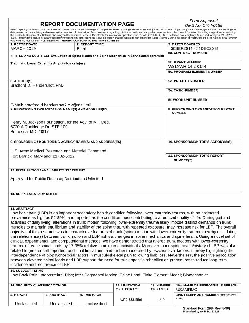
REPORT DOCUMENTATION PAGE Form Approved OMB No. 0704-0188
Public reporting burden for this collection of information is estimated to average 1 hour per response, including the time for reviewing instructions, searching existing data sources, gathering and maintaining the data needed, and completing and reviewing this collection of information. Send comments regarding this burden estimate or any other aspect of this collection of information, including suggestions for reducing this burden to Department of Defense, Washington Headquarters Services, Directorate for Information Operations and Reports (0704-0188), 1215 Jefferson Davis Highway, Suite 1204, Arlington, VA 22202-4302. Respondents should be aware that notwithstanding any other provision of law, no person shall be subject to any penalty for failing to comply with a collection of information if it does not display a currently valid OMB control number. PLEASE DO NOT RETURN YOUR FORM TO THE ABOVE ADDRESS. 1. REPORT DATEMARCH 2019
2. REPORT TYPEFinal
3. DATES COVERED30SEP2014 - 31DEC2018
5a. CONTRACT NUMBER 4. TITLE AND SUBTITLE: Evaluation of Spine Health and Spine Mechanics in Servicemembers with
Traumatic Lower Extremity Amputation or Injury 5b. GRANT NUMBER W81XWH-14-2-0144 5c. PROGRAM ELEMENT NUMBER
6. AUTHOR(S)Bradford D. Hendershot, PhD
5d. PROJECT NUMBER
5e. TASK NUMBER
E-Mail: [email protected]
5f. WORK UNIT NUMBER
7. PERFORMING ORGANIZATION NAME(S) AND ADDRESS(ES) 8. PERFORMING ORGANIZATION REPORTNUMBER
Henry M. Jackson Foundation, for the Adv. of Mil. Med. 6720-A Rockledge Dr. STE 100 Bethesda, MD 20817
9. SPONSORING / MONITORING AGENCY NAME(S) AND ADDRESS(ES) 10. SPONSOR/MONITOR’S ACRONYM(S)
U.S. Army Medical Research and Materiel Command Fort Detrick, Maryland 21702-5012 11. SPONSOR/MONITOR’S REPORT
NUMBER(S)
12. DISTRIBUTION / AVAILABILITY STATEMENT
Approved for Public Release; Distribution Unlimited
13. SUPPLEMENTARY NOTES
14. ABSTRACTLow back pain (LBP) is an important secondary health condition following lower-extremity trauma, with an estimatedprevalence as high as 52-89%, and reported as the condition most contributing to a reduced quality of life. During gait andactivities of daily living, alterations in trunk motion following lower-extremity trauma likely impose distinct demands on trunkmuscles to maintain equilibrium and stability of the spine that, with repeated exposure, may increase risk for LBP. The overallobjective of this research was to characterize features of trunk (spine) motion with lower-extremity trauma, thereby elucidatingthe relationship(s) between trunk motion and LBP risk via changes in spine mechanics and spine health. Using a novel set ofclinical, experimental, and computational methods, we have demonstrated that altered trunk motions with lower-extremitytrauma increase spinal loads by 17-95% relative to uninjured individuals. Moreover, poor spine health/history of LBP was alsorelated to greater self-reported functional limitations, and further moderated by psychosocial factors, thereby highlighting theinterdependence of biopsychosocial factors in musculoskeletal pain following limb loss. Nevertheless, the positive associationbetween elevated spinal loads and LBP support the need for trunk-specific rehabilitation procedures to reduce long-termincidence and recurrence of LBP. 15. SUBJECT TERMSLow Back Pain; Intervertebral Disc; Inter-Segmental Motion; Spine Load; Finite Element Model; Biomechanics
16. SECURITY CLASSIFICATION OF: 17. LIMITATIONOF ABSTRACT
18. NUMBEROF PAGES
19a. NAME OF RESPONSIBLE PERSONUSAMRMC
a. REPORT
Unclassified
b. ABSTRACT
Unclassified
c. THIS PAGE
Unclassified Unclassified 185
19b. TELEPHONE NUMBER (include area code)
Standard Form 298 (Rev. 8-98) Prescribed by ANSI Std. Z39.18
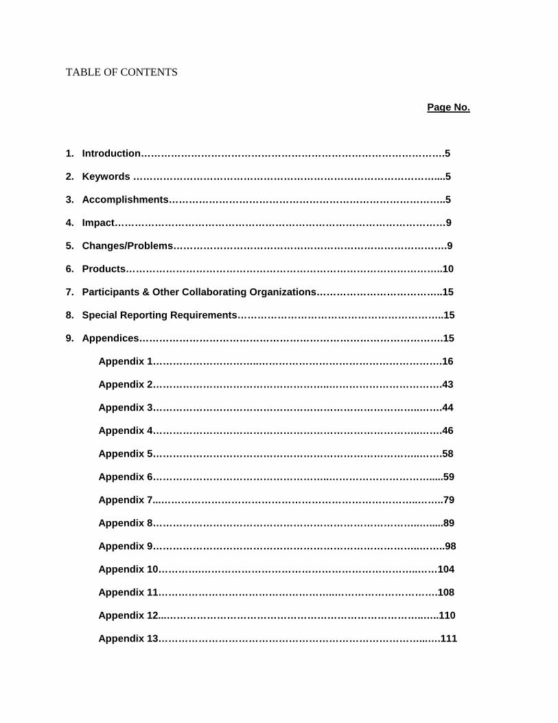
TABLE OF CONTENTS
Page No.
1. Introduction……………………………………………………………………………….5
2. Keywords ………………………………………………………………………………....5
3. Accomplishments………………………………………………………………………..5
4. Impact………………………………………………………………………………………9
5. Changes/Problems……………………………………………………………………….9
6. Products…………………………………………………………………………………..10
7. Participants & Other Collaborating Organizations………………………………..15
8. Special Reporting Requirements……………………………………………………..15
9. Appendices……………………………………………………………………………….15
Appendix 1…………………………..……………………………………………….16
Appendix 2……………………………………………..…………………………….43
Appendix 3……………………………………………………………………..…….44
Appendix 4……………………………………………………………………..…….46
Appendix 5……………………………………………………………………..…….58
Appendix 6……………………………………………..………………………….....59
Appendix 7...…………………………………………………………………..……..79
Appendix 8……………………………………………………………………..….....89
Appendix 9……………………………………………………………………..……..98
Appendix 10………….………………………………………………………..……104
Appendix 11……………………………………………..………………………….108
Appendix 12...…………………………………………………………………..…..110
Appendix 13……………………………………………………………………...….111
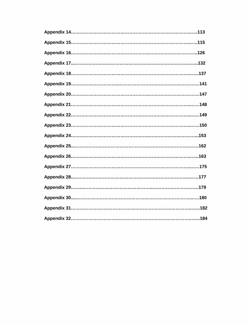
Appendix 14……………………………………………………………………..…..113
Appendix 15……………………………………………………………………..…..115
Appendix 16……………………………………………..…………………………..126
Appendix 17...…………………………………………………………………..…...132
Appendix 18……………………………………………………………………..…...137
Appendix 19…………………………..………………………………………………141
Appendix 20……………………………………………..……………………………147
Appendix 21……………………………………………………………………..……148
Appendix 22……………………………………………………………………..……149
Appendix 23……………………………………………………………………..……150
Appendix 24……………………………………………..…………………………...153
Appendix 25...…………………………………………………………………..……162
Appendix 26……………………………………………………………………..…...163
Appendix 27……………………………………………………………………..……175
Appendix 28..………….………………………………………………………..……177
Appendix 29……..………………………………………..………………………….179
Appendix 30.....…………………………………………………………………...….180
Appendix 31…………………….………………………………………………..…...182
Appendix 32………………………….…………………………………………..…...184
5
1. INTRODUCTION:
Linking lower-extremity trauma (i.e., amputation/injury) with low back pain (LBP) risk viabiomechanical theory suggests that altered and asymmetric trunk motions and correspondingpassive spinal tissue and trunk neuromuscular responses alter spine mechanics such that would,over time, adversely affect spine health. Therefore, the overall objective of this study was toinvestigate such relationships through cross-sectional evaluations of spine health and spinemechanics in persons with lower-extremity amputation/injury (with and without LBP) anduninjured controls. Traditional kinesiopathological models do not successfully describe initialdiagnosis and subsequent treatment of chronic low back pain (LBP). As a result, support for abiopsychosocial model has increased, particularly in clinical populations with concurrentmusculoskeletal disorders. However, biopsychosocial models are complex and dynamic, makingthem difficult to both research and implement in practice. Recently, the biopsychosocial modelhas been suggested as a means to elucidate the increased prevalence of chronic LBP among personswith lower limb loss. We therefore also explored the extent to which psychosocial factors mediatedthe LBP experience among persons with limb loss.
KEYWORDS: Low Back Pain; Intervertebral Disc; Inter-Segmental Motion; Spine Load;Finite Element Model
2. ACCOMPLISHMENTS:
What were the major goals of the project?
This study has three main aims, as indicated below:
Specific Aim 1: Quantify lumbar spinal alignment and inter-segmental vertebral motions withtraumatic lower-extremity amputation.Major Task 1: Obtain IRB and HRPO approvals.
Target Date: by April 2015 Actual Date: April 24, 2015 (IRB approval) / June 26, 2015 (HRPO approval)
Major Task 2: Complete biomechanical data collections, analysis, and interpretations. Target Dates: Months 6-24 (~100% complete)
Additional Milestones: One abstract presented and one manuscript submitted.
Specific Aim 2: Quantify alterations in spine mechanics (loading) with traumatic lower-extremity amputation. Major Task 3: Estimate spinal loads using collected biomechanical data as inputs into the finite element model of the lumbar spine.
Target Dates: Months 6-24 (~100% complete) Additional Milestones: One abstract presented and one manuscript published.
Specific Aim 3: Determine associations between spine loading and current spine health with traumatic lower-extremity amputation. Major Task 4: Conduct physical spinal examinations.
Target Dates: Months 6-24 (100% complete)
6
Major Task 5: Obtain magnetic resonance images of the lumbar spine for quantitative evaluation of lumbar disc health.
Target Dates: Months 6-24 (75% complete- ongoing via retrospective chart reviews) Major Task 6: Author manuscript on entire study.
Target Dates: Months 30-36 (100% complete) Additional Milestones: One abstract presented and one manuscript submitted.
What was accomplished under these goals?
Prospective data collections included biomechanical, clinical, and self-reported assessments focused on the trunk and spine to identify potential relationships with low back pain risk factors following limb loss and extremity trauma. Biomechanical assessments involved instrumented movement analyses with a focus on kinematics of the trunk and spine, as well as trunk muscle activity recorded using surface EMG. In addition, we also captured a more comprehensive understanding of current/recent history of LBP and its impact on daily life and functional activities, including the NIH Task Force’s LBP Questionnaire and a legacy LBP questionnaire (Oswestry Disability Index), as well as several psychosocial factors commonly associated with musculoskeletal pain and functional disability. The bullet points below highlight and summarize the most salient messages from our analyses to date, supplemented by the many original manuscripts/abstracts attached as appendices.
The coordination / motions between the trunk and pelvis with vs. without LLA areassociated with ~31-55, 41-83, and 3-14% larger external demands on the lower back inthe sagittal, coronal, and transverse planes, respectively
Joint contact forces within the spine are increased with LLA; notably, largest increases (upto ~65% relative to uninjured individuals) were found in joint compressive forces owingto a complex pattern and increased (6-80%) activation of trunk muscles
Peak compressive, lateral, and anteroposterior shear loads generally increased withincreasing walking speed. However, increases in compression and lateral shear withincreasing walking speed were larger among the persons with vs. without LLA, particularlyin lateral shear at the fastest speed. In contrast, peak anteroposterior shear decreased withincreasing walking speed among persons with LLA.
Evaluation of trunk muscle activities during gait identified differences in the motor controlstrategies underlying the observed trunk motion patterns. Specifically, persons with lower-extremity trauma demonstrated a second peak in erector spinae activation during mid-terminal swing (not observed in controls), and an overall longer duration of activationthroughout the gait cycle (see Butowicz et al., 2018 in Journal of Electromyography andKinesiology). Trunk neuromuscular control strategies secondary to lower-extremity traumaare seemingly driven by functional requirements to generate force proximally to helpadvance the (affected) lower limb during gait.
Interestingly, spinal loads derived from our finite element simulations indicated differentialincreases with faster walking speeds among persons with vs. without lower-extremitytrauma. At the fastest (vs. slowest) speed, increases in peak compressive and shear forces
7
were respectively 24-84% and 29-77% larger among persons with lower-extremity trauma vs. uninjured controls (see Hendershot et al., 2018 in Journal of Biomechanics). Over time, repeated exposures to these increased loads, particularly at faster walking speeds, may contribute to the elevated risk for LBP among persons with lower-extremity trauma.
When evaluating the influences of LBP on spinal loads, despite larger motions in the frontaland transverse planes, spinal loads were similar between persons with lower-extremitytrauma presenting both with and without (chronic) LBP; though these were generally stilllarger relative to uninjured controls (see Acasio et al., 2018 in Proceedings of the AmericanSociety of Biomechanics). Nevertheless, it is certainly plausible that the presence or historyof LBP have concurrently altered features of trunk-pelvic motion, as previously observedamong non-limb loss individuals with and without LBP.
Preliminary (and prior) analyses using a legacy measure for LBP disability (OswestryDisability Index; ODI) had identified minimal disability (43/58 reported less than 20%disability). However, categorization using the NIH Research Task Force (RTF) definitionsfor chronicity of LBP, which utilize both duration and frequency, told a different story(Table 1). Additional psychosocial outcomes and subcategories are also preliminarilyreported below.
Table 1. Mean (standard deviation) classification/disability scores and individual psychosocial outcomes,by low back pain (LBP) status using NIH definition.
No Current/Recent History of (chronic) LBP
(chronic) LBP
RTF Classification 10.6 (2.4) 16.9 (6.3) ODI % Disability 2.0 (3.1) 22.8 (21.1)
Pain Intensity (7 days) 0.2 (0.1) 3.2 (1.5) Pain Interference 1.1 (0.3) 3.4 (3.1) Functional Impact 1.1 (0.3) 1.7 (0.7) Anxiety and Depression 5.9 (5.2) 9.2 (7.0) Pain Catastrophizing 2.5 (3.1) 22.8 (21.8) Kinesiophobia 19.2 (3.6) 26.8 (4.3)
Impairments in trunk postural control are evident among persons with LLA, both with andwithout LBP, relative to non-LLA controls (Table 2), as evidenced by increased confidenceellipse area (t = -3.24, p = 0.004), increased mean velocity (t= -4.26, p=0.0004), andincreased mediolateral deviation in center of pressure (t = -4.33, p = 0.0004). Thesedifferences suggest LLA demonstrate less postural control when proprioceptive influencefrom the lower extremities is limited, particularly the intact limb.
8
Table 2. Mean (SD) trunk postural control variables between persons with LLA (with and without LBP), and uninjured controls, for reference.
LBP No Pain Control Effect Size (d) 95% Ellipse area (cm2) 8.33 (5.24) 5.20 (3.12) 1.96 (1.36) 0.70
Mean velocity(cm/s) 2.07 (0.74) 1.66 (0.55) 1.02 (0.25) 0.61 RMS distance- ML (cm) 0.63 (0.25) 0.47 (0.14) 0.24 (0.09) 0.76 RMS distance- AP (cm) 0.66 (0.20) 0.56 (0.20) 0.42 (0.14) 0.50
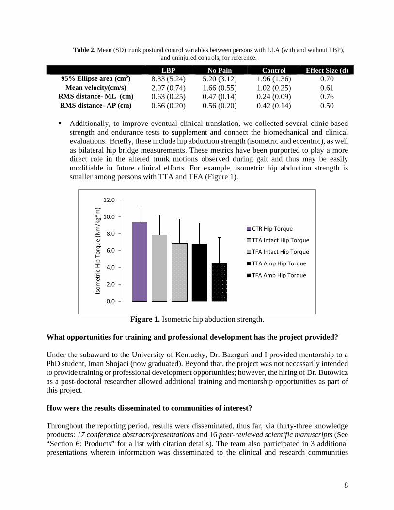
Additionally, to improve eventual clinical translation, we collected several clinic-basedstrength and endurance tests to supplement and connect the biomechanical and clinicalevaluations. Briefly, these include hip abduction strength (isometric and eccentric), as wellas bilateral hip bridge measurements. These metrics have been purported to play a moredirect role in the altered trunk motions observed during gait and thus may be easilymodifiable in future clinical efforts. For example, isometric hip abduction strength issmaller among persons with TTA and TFA (Figure 1).
Figure 1. Isometric hip abduction strength.
What opportunities for training and professional development has the project provided?
Under the subaward to the University of Kentucky, Dr. Bazrgari and I provided mentorship to a PhD student, Iman Shojaei (now graduated). Beyond that, the project was not necessarily intended to provide training or professional development opportunities; however, the hiring of Dr. Butowicz as a post-doctoral researcher allowed additional training and mentorship opportunities as part of this project.
How were the results disseminated to communities of interest?
Throughout the reporting period, results were disseminated, thus far, via thirty-three knowledge products: 17 conference abstracts/presentations and 16 peer-reviewed scientific manuscripts (See “Section 6: Products” for a list with citation details). The team also participated in 3 additional presentations wherein information was disseminated to the clinical and research communities
0.0
2.0
4.0
6.0
8.0
10.0
12.0
Isom
etric
Hip
Tor
que
(Nm
/kg*
m)
CTR Hip Torque
TTA Intact Hip Torque
TFA Intact Hip Torque
TTA Amp Hip Torque
TFA Amp Hip Torque
9
(Amputation System of Care Grand Rounds, State of the Science Symposium at USUHS, and AMSUS).
What do you plan to do during the next reporting period to accomplish the goals?
N/A (but there are several manuscripts under review that we expect to be published later this year).
4. IMPACT: Describe distinctive contributions, major accomplishments, innovations, successes, orany change in practice or behavior that has come about as a result of the project relative to:
What was the impact on the development of the principal discipline(s) of the project?
This study provides strong evidence suggesting the clinical/rehabilitation team should considerquality of trunk motion during walking to minimize future risk for low back pain. Moreover,psychosocial factors appear to augment/succeed biomechanical factors in the pathway(s)connecting lower-extremity amputation with low back pain, thereby expanding the comprehensivecare model for this patient population in considering long-term health and quality of life.
What was the impact on other disciplines?
Nothing to report.
What was the impact on technology transfer?
Nothing to report.
What was the impact on society beyond science and technology?
Our results support a prevailing model that altered trunk (spinal) motions among persons withlower-extremity trauma increase risk for the onset and/or recurrence of LBP. As we continuebuilding evidence for this model, there is likely to be a strong case for interventional approachesaimed at controlling trunk motions and spinal loads during (and beyond) rehabilitation. While thatis specific to one patient population, these relationships may advance overall public knowledgeregarding such a common and impactful musculoskeletal disorder. Over time, this will reduce thesubstantial economic costs associated with its treatment and promote enhancements inpsychological health and overall quality of life.
5. CHANGES/PROBLEMS:
Changes in approach and reasons for change
N/A
Actual or anticipated problems or delays and actions or plans to resolve them
Nothing to report
10
Changes that had a significant impact on expenditures Nothing to report. Significant changes in use or care of human subjects
No significant changes to report. As noted above, IRB/HRPO approval dates: IRB approval granted on April 1, 2015 (formal approval documents were uploaded to IRBnet on April 24) HRPO approval for WRNMMC was granted on June 26, 2015 (A-18549.1) HRPO approval for University Kentucky was granted on June 29, 2015 (A-18549.2) Walter Reed IRB official start date (permission to begin study): August 4, 2015 Walter Reed IRB continuing review date: March 30, 2019 (just renewed until 2020 to complete data analyses).
6. PRODUCTS: • Publications, conference papers, and presentations
Journal publications:
1. Golyski, P.R., Hendershot, B.D. (2018) Trunk and Pelvic Dynamics during Transient Turns among Persons with Unilateral Lower Limb Amputation. Human Movement Science 58: 41-54. Federal Support Acknowledged.
2. Hendershot, B.D., Shojaei, I., Acasio, J.C., Dearth, C.L., Bazrgari, B. (2018) Walking
Speed Differentially Alters Spinal Loads among Persons with Traumatic Lower Limb Amputation. Journal of Biomechanics 70(21): 249-254. Federal Support Acknowledged.
3. Acasio, J.C., Butowicz, C.M., Golyski, P.R., Nussbaum, M.A., and Hendershot, B.D.
(2018) Associations between trunk postural control in walking and unstable sitting at various levels of task demand. Journal of Biomechanics 75: 181-185. Federal Support Acknowledged.
4. Butowicz, C.M., Acasio, J.A., Dearth, C.L., Hendershot, B.D. (2018) Trunk Muscle Activation Patterns among Persons with Lower Limb Loss: Influences of Walking Speed. Journal of Electromyography and Kinesiology 40: 48-55. Federal Support Acknowledged.
5. Highsmith, M.J., Goff, L.M., Lewandowski, A.L., Farrokhi, S., Hendershot, B.D., Hill, O.T., Rabago, C.A., Russell-Esposito, E., Orriola, J.J., Mayer, J.M. (2018) Low Back
11
Pain in Persons with Lower Extremity Amputation: A Systematic Review of the Literature. The Spine Journal, In Press. https://doi.org/10.1016/j.spinee.2018.08.011
6. Shojaei, I., Hendershot, B.D., Ballard, M., Acasio, J.C., Dearth, C.L., Bazrgari, B. TrunkMuscle Forces and Spinal Loads in Persons with Transfemoral Amputation during Sit-to-Stand and Stand-to-Sit Activities. Clinical Biomechanics, Under Review. Federal SupportAcknowledged.
7. Butowicz, C.M., Krupenevich, R.L., Acasio, J.C., Dearth, C.L., Hendershot, B.D.Relationships between mediolateral trunk-pelvic motion, hip strength, and knee jointmoments during gait among persons with lower limb loss. Clinical Biomechanics, UnderReview. Federal Support Acknowledged.
8. Yoder, A., Silder, A., Farrokhi, S., Dearth, C.L., Hendershot, B.D. Lower Extremity JointContributions to Frontal Plane Trunk Dynamics in Persons with Transtibial Amputation.Clinical Biomechanics, Under Review. Federal Support Acknowledged.
9. Mahon, C.E., Butowicz, C.M., Dearth, C.L., Hendershot, B.D. Trunk-PelvicCoordination with Lower-Limb Amputation: Longitudinal Changes in the First Year afterInitial Ambulation. Archives of Physical Medicine and Rehabilitation, Under Review.Federal Support Acknowledged.
10. Butowicz, C.M., Dearth, C.L., and Hendershot, B.D. (2017) Impact of TraumaticExtremity Injuries beyond Acute Care: Implications for Resultant (Long-Term)Secondary Health Conditions. Advances in Wound Care- Special Issue on Amputee Careand Rehabilitation Federal Support Acknowledged.
11. Farrokhi, S, Mazzone, B, Schneider, M, Gombatto, S, Mayer, J, Highsmith, J,Hendershot, B. (2017) Biopsychosocial risk factors associated with chronic low backpain after lower limb amputation. Medical Hypotheses 108; 1-9.
12. Golyski, P.R. and Hendershot, B.D. Trunk-Pelvic Dynamics during Transient Turns inPersons with Unilateral Lower Limb Amputation. Human Movement Science, UnderRevision. Federal Support Acknowledged.
13. Butowicz, C.M., Acasio, J.A., Hendershot, B.D. Trunk Neuromuscular Control Strategiesamong Persons with Lower Limb Amputation while Walking and Performing ConcurrentTasks. Gait and Posture, Under Review. Federal Support Acknowledged.
14. Hendershot, B.D., Acasio, J.A., Shojaie, I., Dearth, C.L., Bazrgari, B. Walking SpeedDifferentially Alters Spinal Loads in Persons with Traumatic Lower Limb Amputation.Journal of Biomechanics, Under Revision. Federal Support Acknowledged.
15. Shojaei, I, Arjmand, N, Meakin, J, Bazrgari, B (2017) A model-based approach forestimation of changes in lumbar segmental kinematics associated with alterations in trunkmuscle forces. Journal of Biomechanics Federal Support Acknowledged.
12
Books or other non-periodical, one-time publications.
Pasquina, P.F., Hendershot, B.D., and Isaacson, B.M. (2016) Secondary Health Effects of Amputation (Chapter 24) Atlas of Amputations and Limb Deficiencies: Surgical, Prosthetic, and Rehabilitation Principles, 4th Edition. American Academy of Orthopaedic Surgeons: Rosemont, IL.
Other publications, conference papers, and presentations.
1. Dearth, C.L., Eskridge, S, Farrokhi, S., Hendershot, B.D., Russell Esposito, E. Living withExtremity Trauma and Limb Loss for a Lifetime: A Review of Efforts to Identify andMitigate Risk Factors for Secondary Health Conditions.
2. Hendershot, B.D., Butowicz, C.B., Mahon, C.E., Schnall, B.L., Dearth, C.L. LongitudinalChanges in Mediolateral Trunk and Pelvic Motion Among Persons with Lower LimbAmputation during the First Year of Ambulation. 2017 Meeting of the American Societyof Biomechanics (ASB), Boulder, CO.
3. Yoder, A.J., Farrokhi, S., Dearth, C.L., Hendershot, B.D. Lower Extremity JointContributions to Trunk Dynamics in Persons with Lower Extremity Amputation. 2017Meeting of the American Society of Biomechanics (ASB), Boulder, CO.
4. Golyski, P.R., and Hendershot, B.D. Trunk-Pelvic Dynamics during Transient Turns inPersons with Unilateral Lower Limb Amputation. 2017 Military Health System ResearchSymposium (MHSRS), Kissimmee, FL, USA.
5. Butowicz, C.M., Acasio, J.C., and Hendershot, B.D. Trunk Neuromuscular ControlStrategies among Persons with Lower Limb Amputation while Walking and PerformingConcurrent Tasks. 2017 Meeting of the American Society of Biomechanics (ASB),Boulder, CO.
6. Butowicz, C.M., Acasio, J.C., Dearth, C.L., Hendershot, B.D. Trunk Muscle ActivationPatterns during Walking among Persons with Lower Limb Loss. World Congress ofBiomechanics (WCB), Dublin, Ireland. Federal Support Acknowledged.
7. Butowicz, C.M., Krupenevich, R.L., Acasio, J.C., Hendershot, B.D. Relationships amongTrunk-Pelvic Motions, Hip Strength, and Knee Joint Moments during Gait amongPersons with Lower Limb Loss. World Congress of Biomechanics (WCB), Dublin,Ireland. Federal Support Acknowledged.
8. Mazzone, B., Farrokhi, S., Hendershot B.D., Watrous, J.R., McCabe, C.T. (2018)Prevalence and Relationship of Low Back Pain and Psychosocial Factors after LowerLimb Amputation among Wounded Warrior Recovery Project Participants. MilitaryHealth System Research Symposium (MHSRS), Kissimmee, FL, USA. Federal SupportAcknowledged.
13
9. Acasio, J.C., Butowicz, C.B., Dearth, C.L., Shojaei, I., Bazrgari, B., Hendershot, B.D.(2018) Trunk Muscle Activations, Motions, and Spinal Loads among Persons with LowerLimb Amputation: Influences of Chronic Low Back Pain. Military Health SystemResearch Symposium (MHSRS), Kissimmee, FL, USA. Federal Support Acknowledged.
10. Acasio, J.C., Butowicz, C.B., Dearth, C.L., Shojaei, I., Bazrgari, B., Hendershot, B.D.(2018) Trunk Muscle Forces and Spinal Loads while Walking in Persons with LowerLimb Amputation both with and without Chronic Low Back Pain. American Society ofBiomechanics (ASB), Rochester, MN, USA. Federal Support Acknowledged.
11. Acasio, J.C., Butowicz, C.B., Hendershot, B.D. (2018) Patterns of Erector SpinaeActivation and Trunk-Pelvis Kinematics in Persons with Lower Limb Amputation:Influences of Low Back Pain. American Society of Biomechanics (ASB), Rochester, MN,USA. Federal Support Acknowledged.
12. Butowicz, C.B., Krupenevich, R.L., Acasio, J.C., Hendershot, B.D. (2018) Influences ofLow Back Pain on the Energy Contributions of the Hip and Spine during Gait amongPersons with Lower Limb Loss. American Society of Biomechanics (ASB), Rochester,MN, USA. Federal Support Acknowledged.
13. Hendershot, B.D., Butowicz, C.B., Krupenevich, R.L., Acasio, J.A., Pruziner, A.L.,Miller, R.H., Goldman, S.G., Dearth, C.L. (2018) Toward Optimizing Long-Term Healthafter Limb Loss: Comprehensive Evaluations of Secondary Health Conditions. 10th
Annual Joint National Capital Region Research Competition, Bethesda, MD, USA.Federal Support Acknowledged.
14. Shojaei, I., Hendershot, B.D., Ballard, M., Acasio, J.C., Dearth, C.L., Bazrgari, B. (2018)Trunk Muscle Forces and Spinal Loads during Sit-to-Stand and Stand-to-Sit Activities:Differences between Persons with and without Unilateral Transfemoral Amputation. 15th
International Symposium on Computer Methods in Biomechanics and BiomedicalEngineering (CMBBE), Lisbon, Portugal. Federal Support Acknowledged.
15. Hendershot, B.D. (2016) Biomechanical risk factors for low back pain with extremitytrauma. The Military Health System Research Symposium (MHSRS), Kissimmee, FL,USA.
16. Hendershot, B.D., Shojaei, I., and Bazrgari, B. Faster walking speeds differentially alterspinal loads among persons with traumatic lower limb amputation. 2nd InternationalWorkshop on Spine Loading and Deformation. Julius Wolff Institute, Berlin, Germany.May 18-20, 2017.
• Website(s) or other Internet site(s)
Nothing to report.
• Technologies or techniques

14
Nothing to report.
• Inventions, patent applications, and/or licenses
Nothing to report.
• Other Products
Nothing to report.
7. PARTICIPANTS & OTHER COLLABORATING ORGANIZATIONS
What individuals have worked on the project?
Name: Bradford Hendershot, PhD Project Role: Principal Investigator, EACE/WRNMMC Nearest person month worked:
2
Contribution to Project: Provided overall project direction, including: tracking resources, ensuring regulatory compliance, coordinating data collections / analyses, and generating reports.
Funding Support: Federal Employee
Name: Babak Bazrgari, PhD Project Role: Co-Investigator, Site PI at University of Kentucky Nearest person month worked:
1
Contribution to Project: Led the finite element modeling for all biomechanical data Funding Support:
Name: Courtney Butowicz, PhD Project Role: Post-Doctoral Researcher, HJF/WRNMMC Nearest person month worked:
12
Contribution to Project: Led data collection, analysis, and interpretation with direction from the study PI.
Funding Support:
Name: Julian Acasio, MS Project Role: Research Engineer, HJF/WRNMMC Nearest person month worked:
10
Contribution to Project: Assisted with data collection, analysis, and interpretation
15
Funding Support:
Has there been a change in the active other support of the PD/PI(s) or senior/key personnel since the last reporting period?
Nothing to report.
What other organizations were involved as partners?
Nothing to report.
8. SPECIAL REPORTING REQUIREMENTS
COLLABORATIVE AWARDS: N/A
QUAD CHARTS: N/A
9. APPENDICES: N/A
Page 1 of 26
Increased and asymmetric trunk motion during level-ground walking is associated with
larger spinal loads in persons with unilateral transfemoral amputation
Iman Shojaei1, Brad D. Hendershot2, 3, Erik J. Wolf2, 4, Babak Bazrgari1,
1
Department of Biomedical Engineering, University of Kentucky, Lexington, KY, USA
2
Department of Rehabilitation, Walter Reed National Military Medical Center, Bethesda, MD 20889, USA
3
Center for Rehabilitation Sciences Research, Department of Physical Medicine and Rehabilitation,
Uniformed Services University of the Health Sciences, Bethesda, MD 20814, USA
4
DOD — VA Extremity Trauma and Amputation Center of Excellence, Walter Reed National Military
Medical Center, Bethesda, MD 20889, USA
Corresponding address: Babak Bazrgari, Department of Biomedical Engineering, University
of
Kentucky, 514E Robotic and Manufacturing Building, Lexington, KY 40506.
Abstract Word Count: 228
Main Text Word Count: 3637
Number of Figures: 5
Number of Tables: 3
Page 2 of 26
ABSTRACT
Background: During gait, alterations in trunk motion following lower limb amputation likely
impose distinct demands on trunk muscles to maintain equilibrium and stability of the spine.
However, trunk muscle responses to such changes in physical demands, and the resultant
effects on spinal loads, have yet to be determined in this population.
Methods: Trunk and pelvic kinematics collected during level-ground walking from 40 males (20
with unilateral transfemoral amputation and 20 matched controls) were used as inputs to a
kinematics-driven, nonlinear finite element model of the lower back to estimate forces in 10
global (attached to thorax) and 46 local (attached to lumbar vertebrae) trunk muscles, as well as
compression, lateral, and antero-posterior shear forces at all spinal levels.
Findings: Trunk muscle force and spinal load maxima corresponded with heel strike and toe-off
events, and were respectively 10-40% and 17-95% larger during intact vs. prosthetic stance in
persons with amputation, as well as 6-80% and 26-60% larger during intact stance relative to
controls.
Interpretation: In addition to larger individual muscle responses to overall increases and
asymmetries in trunk motion during walking, co-activations of antagonistic muscles were
needed to assure spine equilibrium in three-dimensional space, hence resulting in substantial
increases in spinal loads. Knowledge of trunk neuromuscular adaptations to changes in task
demands following amputation could inform rehabilitation procedures such to reduce long-term
incidence or recurrence of low back pain.
Keywords: Amputation, Gait, Muscle forces, Spinal loads, Low back pain
Page 3 of 26
HIGHLIGHTS:
Persons with lower limb amputation walk with large and asymmetric trunk motion
Spinal equilibrium and stability under such motions require large muscular response
Larger trunk muscle forces contribute to increase compression and shear loads
Repeated exposures to altered spinal loading may elevate low back pain risk
Page 4 of 26
1. INTRODUCTION
The prevalence of low back pain (LBP) is considerably higher in persons with lower limb
amputation (LLA) compared with able-bodied individuals (Friberg, 1984, Sherman, 1989,
Sherman et al., 1997, Smith et al., 1999). As a secondary health-related concern, LBP is
suggested to be the most important condition that adversely affects the physical performance
and quality of life in persons with LLA (Ehde et al., 2001, Taghipour et al., 2009). Providing the
projected increase in the number of people with LLA, it is important to investigate the underlying
mechanism(s) responsible for the elevated prevalence of LBP in this cohort (Reiber et al., 2010,
Devan et al., 2014).
Considering spine biomechanics, spinal loads are the resultant of interactions between internal
tissue forces (primarily from muscles) and physical demands of a given activity on the lower
back (Cholewicki and Mcgill, 1996, Calisse et al., 1999, Arjmand and Shirazi-Adl, 2005, Adams
et al., 2007, Mcgill et al., 2014). During gait, increased and asymmetric trunk motion following
LLA has been reported to impose higher physical demands on the lower back (Cappozzo and
Gazzani, 1982, Hendershot and Wolf, 2014). Such an increase in physical demand of a
common daily activity like walking would require larger responses from internal trunk tissues to
assure equilibrium and stability of the spine, hence leading to larger spinal loads that would
presumably increase the risk for LBP due to the repetitive nature of such activities (Adams et
al., 2007).
There is limited information in the literature related to internal trunk tissue responses and
resultant spinal loads during walking (Cappozzo et al., 1982, Cappozzo, 1983, Cappozzo, 1983,
Khoo et al., 1995, Cheng et al., 1998, Callaghan et al., 1999, Yoder et al., 2015). All but two of
these few earlier studies included relatively small sample sizes of able-bodied male participants
and have reported spinal loads at either the L4-L5 or L5-S1 discs. The predicted pattern of
spinal loads in these studies included symmetric local maxima occurring around heel strike and
toe off within the gait cycle, with values ranging between 1.2 to 3.0 times body weight. The other
two studies regarding internal tissue responses and resultant spinal loads during walking also
include persons with LLA (Cappozzo and Gazzani, 1982, Yoder et al., 2015). Using kinematics
data obtained from two subjects (one with transfemoral amputation and one with knee
Page 5 of 26
ankylosis), Cappozzo and Gazzani (1982) used a rigid link-segment model of the whole body to
obtain mechanical demands of walking on the lower back. A simple muscle model was then
used to calculate internal tissue responses and the resultant spinal loads. Contrary to the
patterns of spinal loads observed in able-bodied individuals, the occurrence of local maxima
among persons with LLA did not have a symmetric pattern. Rather, the maximum compression
forces were larger at the instance of prosthetic vs. intact toe off (2-3.0 vs. 1.0 times body
weight). Similar differences in patterns of trunk muscular responses during walking, and the
resultant effect on spinal loads (but at much lower magnitudes), between persons with and
without transtibial LLA have been recently reported by Yoder et al. (2015). Although these
earlier studies highlight the impact of altered and asymmetric gait on loads experienced in the
lower back, they were limited to small samples and/or a very simple biomechanical model of the
lower back.
Using a relatively large sample size, along with a biomechanical model of the lower back with
more bio-fidelity, the objective of this study was to investigate the differences in internal tissue
responses, specifically muscle forces, and resultant spinal loads during level-ground walking
between individuals with (n=20) and without (n=20) unilateral LLA. Considering that alterations
in trunk motion following amputation impose higher (and asymmetric) physical demands on the
lower back (Cappozzo and Gazzani, 1982, Hendershot and Wolf, 2014), it was hypothesized
that compared to able-bodied individuals, persons with LLA will require larger muscle forces in
the lower back to overcome the physical demands of walking while maintaining spinal stability
and equilibrium. Such increases in trunk muscle forces would, in turn, result in larger spinal
loads. A better knowledge of lower back biomechanics (i.e., in terms of spinal loads) among
individuals with LLA can inform future development of effective clinical programs aimed at
modifying lower back biomechanics such to mitigate LBP risk.
2. METHODS
2.1 Experimental study: Kinematic data collected in an earlier study were used in these
analyses (Hendershot and Wolf, 2014). Briefly, full-body kinematics from 20 males with
transfemoral amputation and 20 male able-bodied controls were collected using a 23-camera
motion capture system during level-ground walking across a 15 m level walkway at a self-
Page 6 of 26
selected speed (mean ≈ 1.35 m/s; Table-1). Here, kinematic data of interest included three-
dimensional pelvic and thorax motions that were collected by tracking markers positioned in the
mid-sagittal plane over the S1, T10, and C7 spinous processes, sternal notch, and xiphoid; and
bilaterally over the acromion, ASIS, and PSIS. All amputations were a consequence of
traumatic injuries with a mean (standard deviation) duration of 3.1 (1.4) years since amputation.
Main inclusion criteria were: (1) unilateral transfemoral amputation with no contralateral
functional impairments, (2) daily use of a prosthetic device (≥1 year post-amputation), (3) no use
of an upper-extremity assistive device (e.g., cane, crutches, walker), and (4) having no other
musculoskeletal or neurologic problem, except amputation, that may affect gait results. Details
of inclusion and exclusion criteria and other experimental methodology can be found in
Hendershot and Wolf (2014). This retrospective study was approved by Institutional Review
Boards of both University of Kentucky and Walter Reed National Military Medical Center.
Table-1 may be inserted here
2.2 Modeling study: The biomechanical model used to estimate trunk muscle responses and
resultant spinal loads included a non-linear finite element (FE) model of the spine that estimated
the required muscle forces to complete the activity using an optimization-based iterative
procedure (Arjmand and Shirazi-Adl, 2005, Arjmand and Shirazi-Adl, 2006, Bazrgari et al.,
2007, Bazrgari et al., 2008, Bazrgari et al., 2009, Arjmand et al., 2010). In this model, muscle
forces are estimated such that equilibrium equations are satisfied across the entire lumbar
spine. The finite element model included a sagittally symmetric thorax-pelvis model of the spine
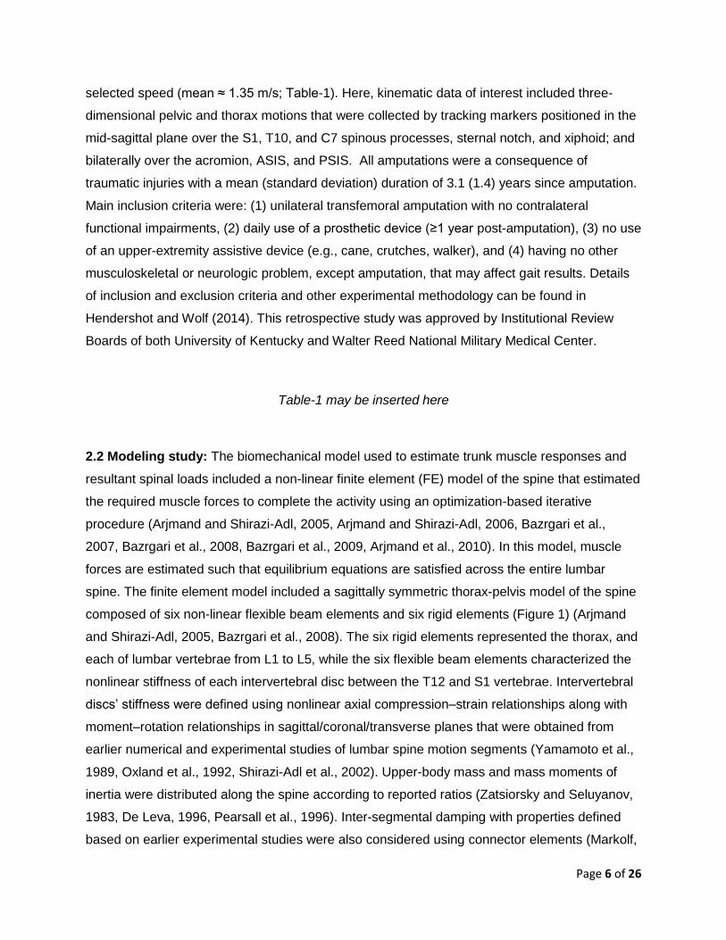
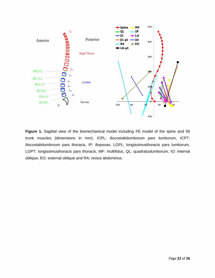
composed of six non-linear flexible beam elements and six rigid elements (Figure 1) (Arjmand
and Shirazi-Adl, 2005, Bazrgari et al., 2008). The six rigid elements represented the thorax, and
each of lumbar vertebrae from L1 to L5, while the six flexible beam elements characterized the
nonlinear stiffness of each intervertebral disc between the T12 and S1 vertebrae. Intervertebral
discs’ stiffness were defined using nonlinear axial compression–strain relationships along with
moment–rotation relationships in sagittal/coronal/transverse planes that were obtained from
earlier numerical and experimental studies of lumbar spine motion segments (Yamamoto et al.,
1989, Oxland et al., 1992, Shirazi-Adl et al., 2002). Upper-body mass and mass moments of
inertia were distributed along the spine according to reported ratios (Zatsiorsky and Seluyanov,
1983, De Leva, 1996, Pearsall et al., 1996). Inter-segmental damping with properties defined
based on earlier experimental studies were also considered using connector elements (Markolf,
Page 7 of 26
1970, Kasra et al., 1992). The muscle architecture in the biomechanical model included 56
muscles (Fig. 1); 46 muscles connecting lumbar vertebrae to the pelvis (i.e., local muscles) and
10 muscles connecting thoracic spine/rib cage to the pelvis (i.e., global muscles) (Arjmand and
Shirazi-Adl, 2005, Arjmand and Shirazi-Adl, 2006, Bazrgari et al., 2008, Bazrgari et al., 2008).
Figure 1 may be inserted here
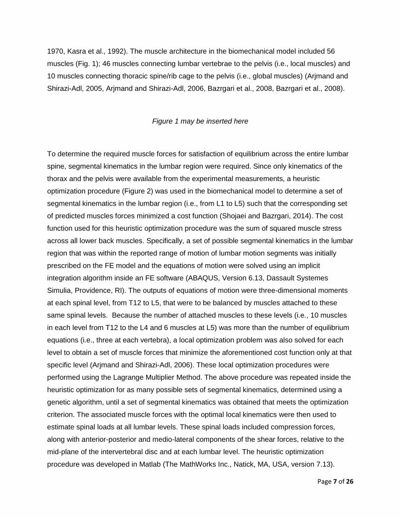
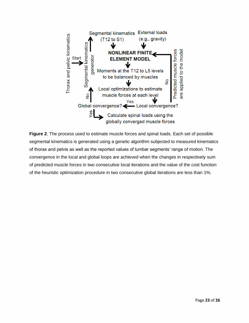
To determine the required muscle forces for satisfaction of equilibrium across the entire lumbar
spine, segmental kinematics in the lumbar region were required. Since only kinematics of the
thorax and the pelvis were available from the experimental measurements, a heuristic
optimization procedure (Figure 2) was used in the biomechanical model to determine a set of
segmental kinematics in the lumbar region (i.e., from L1 to L5) such that the corresponding set
of predicted muscles forces minimized a cost function (Shojaei and Bazrgari, 2014). The cost
function used for this heuristic optimization procedure was the sum of squared muscle stress
across all lower back muscles. Specifically, a set of possible segmental kinematics in the lumbar
region that was within the reported range of motion of lumbar motion segments was initially
prescribed on the FE model and the equations of motion were solved using an implicit
integration algorithm inside an FE software (ABAQUS, Version 6.13, Dassault Systemes
Simulia, Providence, RI). The outputs of equations of motion were three-dimensional moments
at each spinal level, from T12 to L5, that were to be balanced by muscles attached to these
same spinal levels. Because the number of attached muscles to these levels (i.e., 10 muscles
in each level from T12 to the L4 and 6 muscles at L5) was more than the number of equilibrium
equations (i.e., three at each vertebra), a local optimization problem was also solved for each
level to obtain a set of muscle forces that minimize the aforementioned cost function only at that
specific level (Arjmand and Shirazi-Adl, 2006). These local optimization procedures were
performed using the Lagrange Multiplier Method. The above procedure was repeated inside the
heuristic optimization for as many possible sets of segmental kinematics, determined using a
genetic algorithm, until a set of segmental kinematics was obtained that meets the optimization
criterion. The associated muscle forces with the optimal local kinematics were then used to
estimate spinal loads at all lumbar levels. These spinal loads included compression forces,
along with anterior-posterior and medio-lateral components of the shear forces, relative to the
mid-plane of the intervertebral disc and at each lumbar level. The heuristic optimization
procedure was developed in Matlab (The MathWorks Inc., Natick, MA, USA, version 7.13).
Page 8 of 26
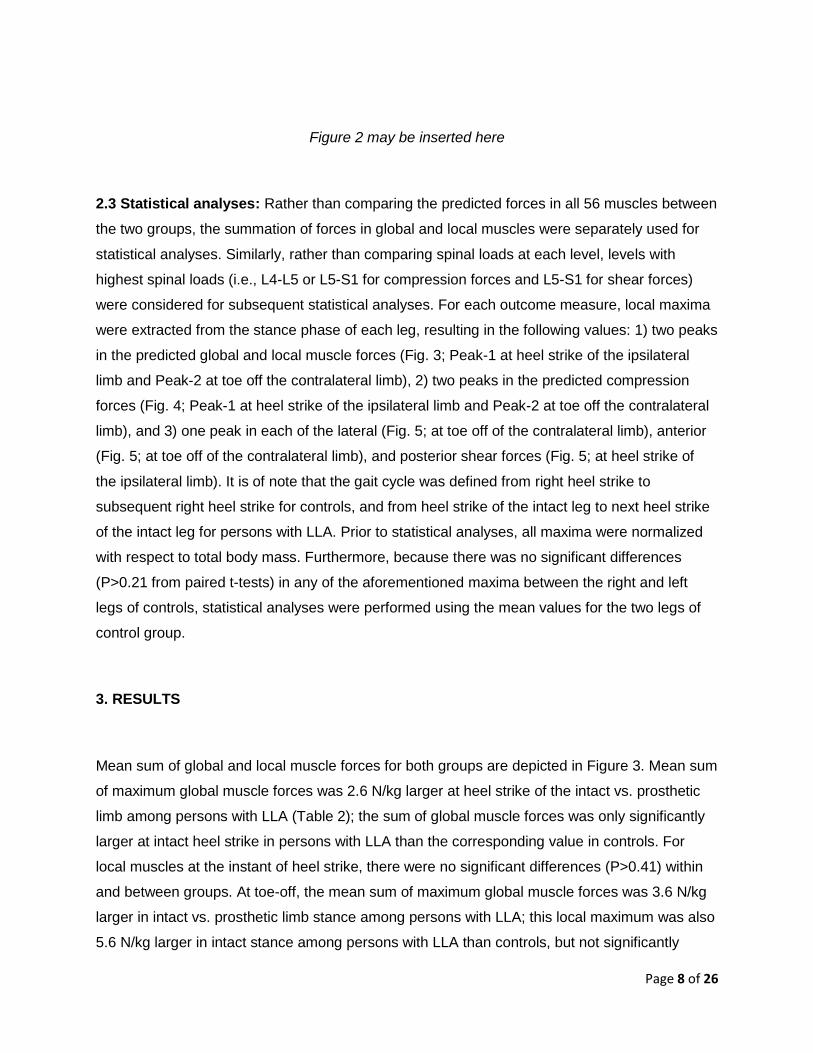
Figure 2 may be inserted here
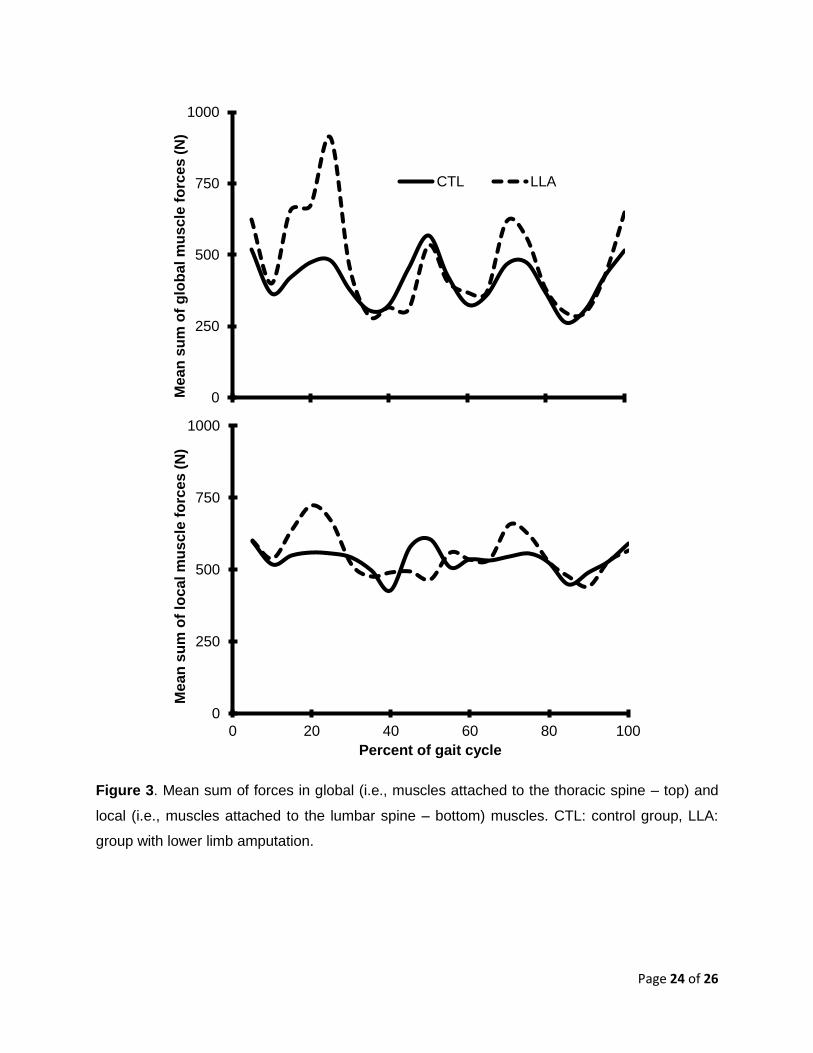
2.3 Statistical analyses: Rather than comparing the predicted forces in all 56 muscles between
the two groups, the summation of forces in global and local muscles were separately used for
statistical analyses. Similarly, rather than comparing spinal loads at each level, levels with
highest spinal loads (i.e., L4-L5 or L5-S1 for compression forces and L5-S1 for shear forces)
were considered for subsequent statistical analyses. For each outcome measure, local maxima
were extracted from the stance phase of each leg, resulting in the following values: 1) two peaks
in the predicted global and local muscle forces (Fig. 3; Peak-1 at heel strike of the ipsilateral
limb and Peak-2 at toe off the contralateral limb), 2) two peaks in the predicted compression
forces (Fig. 4; Peak-1 at heel strike of the ipsilateral limb and Peak-2 at toe off the contralateral
limb), and 3) one peak in each of the lateral (Fig. 5; at toe off of the contralateral limb), anterior
(Fig. 5; at toe off of the contralateral limb), and posterior shear forces (Fig. 5; at heel strike of
the ipsilateral limb). It is of note that the gait cycle was defined from right heel strike to
subsequent right heel strike for controls, and from heel strike of the intact leg to next heel strike
of the intact leg for persons with LLA. Prior to statistical analyses, all maxima were normalized
with respect to total body mass. Furthermore, because there was no significant differences
(P>0.21 from paired t-tests) in any of the aforementioned maxima between the right and left
legs of controls, statistical analyses were performed using the mean values for the two legs of
control group.
3. RESULTS
Mean sum of global and local muscle forces for both groups are depicted in Figure 3. Mean sum
of maximum global muscle forces was 2.6 N/kg larger at heel strike of the intact vs. prosthetic
limb among persons with LLA (Table 2); the sum of global muscle forces was only significantly
larger at intact heel strike in persons with LLA than the corresponding value in controls. For
local muscles at the instant of heel strike, there were no significant differences (P>0.41) within
and between groups. At toe-off, the mean sum of maximum global muscle forces was 3.6 N/kg
larger in intact vs. prosthetic limb stance among persons with LLA; this local maximum was also
5.6 N/kg larger in intact stance among persons with LLA than controls, but not significantly
Page 9 of 26
different between prosthetic stance relative to controls. For local muscles at the instant of toe-
off, while there were no significant differences between the values in the stance phase of intact
and prosthetic legs of persons with LLA, they were, respectively, 2.5 N/kg and 1.5 N/kg larger
than the corresponding values in controls.
Figure 3 may be inserted here
Table-2 may be inserted here
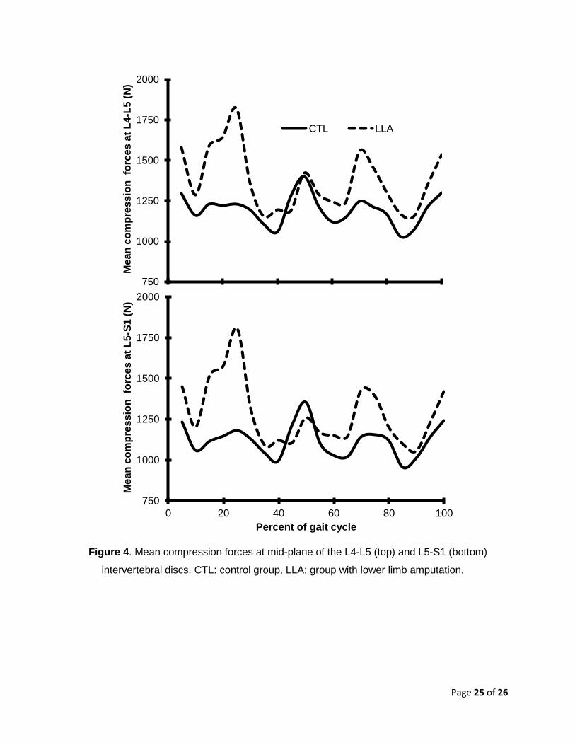
Mean compression forces were 3.4 N/kg larger at heel strike of the intact vs. prosthetic leg
among persons with LLA; the compression force at heel strike of the intact leg was also 4.8
N/kg larger than the corresponding value in controls, while there were no significant differences
between the prosthetic leg of persons with LLA and the corresponding value in controls (Table
2). Mean compression force at toe off of the contralateral limb was similar between stance of the
intact and prosthetic legs among persons with LLA, but were 8.6 N/kg (4.7 N/kg) larger during
intact (prosthetic) leg stance than the corresponding value in controls.
Figure 4 may be inserted here
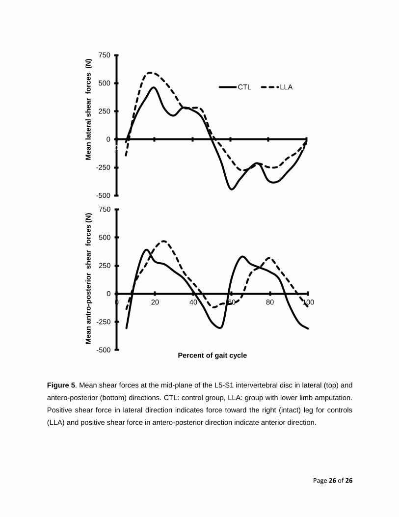
In the lateral direction, maximum shear forces were 4.3 N/kg larger in the stance phase of the
intact vs. prosthetic leg among persons with LLA (Table 2). These were also 3.3 N/kg larger in
the stance phase of intact leg of persons with LLA than the corresponding value in controls;
there were no significant differences between the stance phase of prosthetic leg and that of
controls. In the posterior direction, maximum shear forces among controls were 1.3 and 1.8
N/kg larger than the corresponding values in intact and prosthetic stance among persons with
LLA, respectively. Maximum posterior shear forces were not different between intact and
prosthetic stance among persons with LLA. In the anterior direction, maximum shear forces
were 1.4 N/kg larger in the stance phase of the intact vs. prosthetic leg among persons with
LLA; these were also 1.8 N/kg larger in the stance phase of the intact leg than the
corresponding value in controls.
Figure 5 may be inserted here
Page 10 of 26
4. DISCUSSION
In this study, trunk muscle responses to walking demands and the resultant spinal loads were
estimated in individuals with and without unilateral LLA. It was hypothesized that individuals with
LLA would require larger muscle forces to overcome the physical demands of walking while
maintaining spinal equilibrium and stability, which would in turn result in larger spinal loads
compared to individuals without amputation. The results obtained through computational
simulations and subsequent statistical analyses confirmed our hypothesis. Higher trunk muscle
forces and larger spinal loads on the lower back of individuals with unilateral LLA during walking
may be in part responsible for the reported higher prevalence of LBP among persons with vs.
without LLA.
The local maxima for muscle forces and the resultant spinal loads occurred at the instants of
heel strike and toe off within the gait cycle. These time points also happen to correspond with
the instances of large axial twist of the trunk (i.e., heel strike) and asymmetric trunk posture (i.e.
toe off where there were relatively large motions in all three planes (Hendershot and Wolf,
2014)). In addition to individual muscle responses, co-activations of antagonistic muscles were
needed under such trunk motions to assure spine equilibrium in three-dimensional space. The
effects of such an increased and asymmetric motion on muscle forces is more evident when
comparing the kinematics and associated muscle forces in the stance phase of intact and
prosthetic legs among individuals with LLA. The increases in trunk motion and its asymmetry at
instances of heel strike and toe off were more pronounced during the stance phase of the intact
leg of persons with LLA, particularly at heel strike of the ipsilateral limb (Hendershot and Wolf,
2014), that resulted in much larger muscle forces during the stance phase of intact than
prosthetic leg. Such an effect may also be a result of proximal compensations (e.g., hip-hiking)
to assist with toe clearance (Michaud et al., 2000), or simply because these individuals feel
more confident during intact (vs. prosthetic) stance to advance their center of mass.
The sum of forces in global muscles during the gait cycle was comparable with the sum of
forces in the local muscles (Fig. 3). It should be mentioned, however, global muscles were the
primary responders to activity demands during the first iteration of muscle force calculations in
Page 11 of 26
our model (i.e., the local loop in Fig. 2). As the effects of such global muscle forces were applied
into the model, during the subsequent iterations, local muscles became activated to prevent
buckling of the spine under the penalties of global muscle forces. If the summation of forces in
global and local muscles is assumed to represent the required energy for respectively
equilibrate and stabilize the spine, our results suggest that relatively equal amounts of energy
were consumed to provide equilibrium and stability to the spine during walking. However with
such an assumption, it seems that overcoming the equilibrium demands of walking impact the
spinal loads of individuals with LLA more than overcoming its segmental stability demands when
compared with able-bodied individuals. This observation is reflected in the sum of differences in
mean global muscle forces (i.e., assumed to represent differences in equilibrium demands)
between persons with and without LLA that was 955 N larger than the sum of differences in
mean local muscle forces (i.e., assumed to represent differences in stability demands) between
the same two groups. We should, however, emphasize that such interpretation is limited to
assumptions made in our optimization-based method for estimation of muscle responses to
activity demand and would require verification via measurement of muscle activity. A stabilizing
response from local muscles as suggested here should occur sooner than equilibrating
response from global muscles.
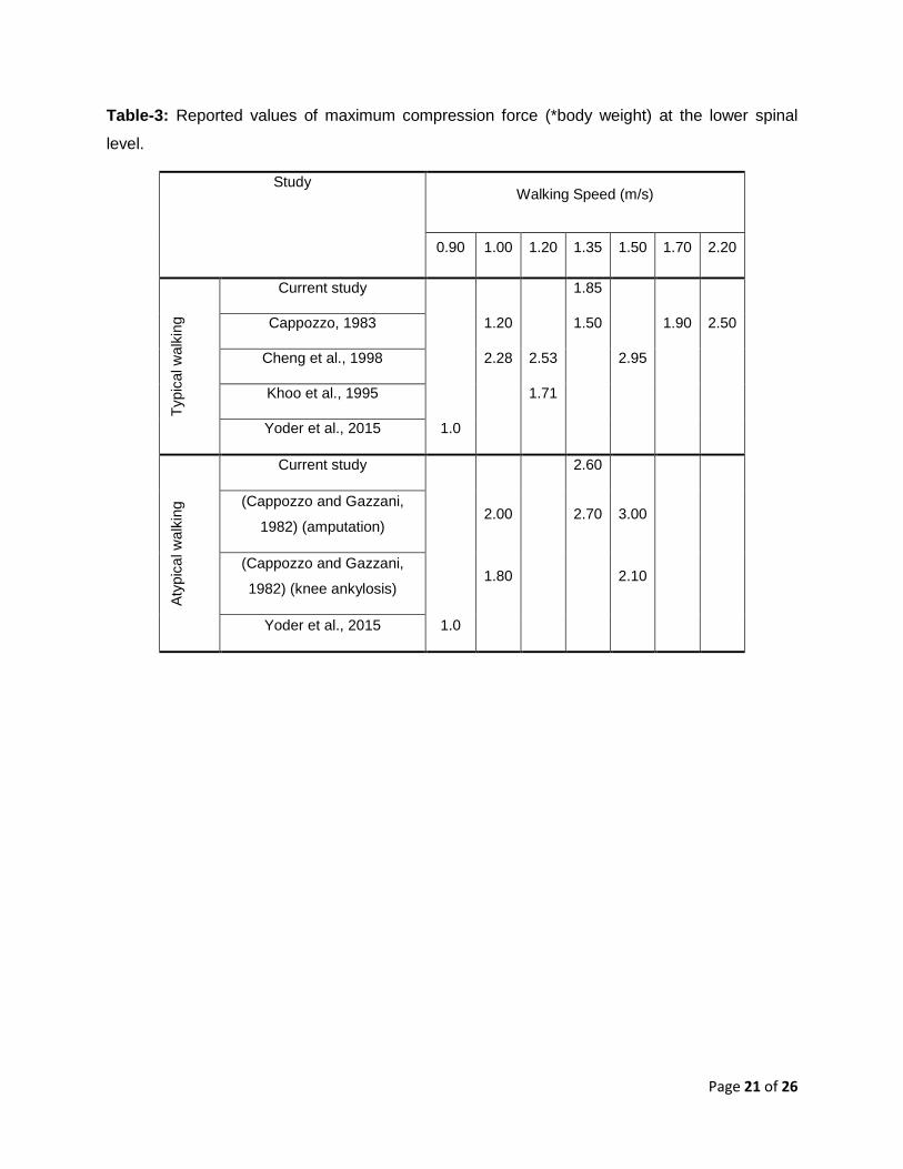
The predicted spinal loads for controls were in agreement (in terms of patterns and magnitudes)
with those obtained in earlier studies (Cappozzo, 1983, Khoo et al., 1995, Cheng et al., 1998,
Callaghan et al., 1999, Yoder et al., 2015). Depending on walking speed, the reported values of
maximum compression force at the lower spinal level ranged between 1.0 to 2.95 times body
weight for walking speeds ranging from 0.9 to 2.2 m/s (Table 3). The mean maximum
compression force from these studies, along with average walking speed, were respectively ~
1.94 times body weight at 1.4 m/s, which are comparable with our predictions of a maximum
spinal load of ~ 1.85 times body weight for an average walking speed of ~1.35 m/s. Maxima in
predicted compression forces in this study occurred around heel strike and toe off instances
within the gait cycle, which are also consistent with reported timing of maximum compression
forces in earlier studies: around toe off instants (Callaghan et al., 1999), within a short time
interval around toe off (Cappozzo, 1983), right after the heel strike and before complete toe off
(Cheng et al., 1998), and around 20% and 80% of walking cycle (Khoo et al., 1995).
Page 12 of 26
Table-3 may be inserted here
The results obtained from individuals with unilateral LLA in this study were also consistent in
pattern and magnitude with those reported by Cappozzo and Gazzani (Cappozzo and Gazzani,
1982). This earlier study reported spinal loads for two subjects (i.e., one with transfemoral
amputation and one with knee ankylosis) during level-ground walking. The reported maxima of
predicted compression forces for the person with LLA ) ranged from 2 to 3 times body weight for
walking speeds between 1.0 m/s and 1.5 m/s (Table 3), which is consistent with the range of
maxima of predicted compression forces in this study (~ 2 to 2.6 body weight). In both studies,
the maximum compression forces occurred during intact limb stance at the instance of
prosthetic toe off. In a more recent study (Yoder et al 2015), much smaller maxima (i.e., ~ body
weight) have been reported for maximum spinal loads among persons with transtibial LLA;
though smaller maxima could be due, in part, to the relatively slower walking speed and/or more
distal amputation.
The sample of persons with LLA in this study included young and physically fit members of the
military with transfemoral amputations resulting from traumatic injuries. Thus, the results cannot
be generalized to groups with other levels or etiologies of amputation. This cross sectional study
also does not provide any information about lower back biomechanics in these individuals
before the amputations, and history of LBP was not controlled in the participants, though those
with current LBP were excluded from the study. Although we accounted for individual
differences in trunk inertial properties in the non-linear FE model of spine, we used the same
passive tissue properties for all subjects since we had no access to the subject-specific
behavior of such tissues (i.e., ligaments, intervertebral discs, passive behavior of muscles and
bony structures) for these participants. Furthermore, same heights were considered in the spine
model for all subjects, though stature was not significantly different between groups.
5. CONCLUSION
Asymmetric and larger trunk motion of individuals with LLA during walking requires higher
activation and co-activation of trunk muscles to assure equilibrium and stability of the spine,
which in turn increase spinal loads. An elevated level of spinal loads during a basic activity of
Page 13 of 26
daily living like walking may increase risk of developing LBP, in particular due to the repetitive
nature of such activity. It is imperative to investigate whether those with LLA consistently
experiencing higher levels of spinal loads during other important activities of daily living (e.g.,
ascending and descending ramps or stairs) as a result of an alteration in internal tissue
responses to activity demands. Such knowledge can inform future development of effective
clinical programs aimed at reducing the risk for developing LBP via management of spinal loads
during daily activities.
ACKNOWLEDGEMENTS
This work was supported by the Office of the Assistant Secretary of Defense for Health Affairs,
through the Peer Reviewed Orthopaedic Research Program (award #W81XWH-14-2-0144), as
well as University of Kentucky’s Center for Clinical and Translational Science (NIH –
UL1TR00017). The views expressed in this manuscript are those of the authors, and do not
necessarily reflect the official policy of the Departments of the Army, Navy, Defense, nor the
United States Government.
Page 14 of 26
REFERENCES
Adams, M.A., Burton, A.K., Dolan, P. and Bogduk, N. (2007). The biomechanics of back pain,
Churchill Livingstone.
Arjmand, N., Gagnon, D., Plamondon, A., Shirazi-Adl, A. and Larivière, C., 2010. A comparative
study of two trunk biomechanical models under symmetric and asymmetric loadings. Journal of
biomechanics 43(3), 485-491.
Arjmand, N. and Shirazi-Adl, A., 2005. Biomechanics of changes in lumbar posture in static
lifting. Spine 30(23), 2637-2648.
Arjmand, N. and Shirazi-Adl, A., 2006. Sensitivity of kinematics-based model predictions to
optimization criteria in static lifting tasks. Medical engineering & physics 28(6), 504-514.
Bazrgari, B., Shirazi-Adl, A. and Arjmand, N., 2007. Analysis of squat and stoop dynamic
liftings: muscle forces and internal spinal loads. European Spine Journal 16(5), 687-699.
Bazrgari, B., Shirazi-Adl, A. and Kasra, M., 2008. Computation of trunk muscle forces, spinal
loads and stability in whole-body vibration. Journal of Sound and Vibration 318(4), 1334-1347.
Bazrgari, B., Shirazi-Adl, A. and Parnianpour, M., 2009. Transient analysis of trunk response in
sudden release loading using kinematics-driven finite element model. Clinical Biomechanics
24(4), 341-347.
Bazrgari, B., Shirazi-Adl, A., Trottier, M. and Mathieu, P., 2008. Computation of trunk equilibrium
and stability in free flexion–extension movements at different velocities. Journal of biomechanics
41(2), 412-421.
Calisse, J., Rohlmann, A. and Bergmann, G., 1999. Estimation of trunk muscle forces using the
finite element method and in vivo loads measured by telemeterized internal spinal fixation
devices. Journal of biomechanics 32(7), 727-731.
Callaghan, J.P., Patla, A.E. and Mcgill, S.M., 1999. Low back three-dimensional joint forces,
kinematics, and kinetics during walking. Clinical Biomechanics 14(3), 203-216.
Cappozzo, A., 1983. Compressive loads in the lumbar vertebral column during normal level
walking. Journal of orthopaedic research 1(3), 292-301.
Cappozzo, A., 1983. The forces and couples in the human trunk during level walking. Journal of
biomechanics 16(4), 265-277.
Page 15 of 26
Cappozzo, A., Figura, F., Gazzani, F., Leo, T. and Marchetti, M., 1982. Angular displacements
in the upper body of AK amputees during level walking. Prosthetics and orthotics international
6(3), 131-138.
Cappozzo, A. and Gazzani, F. (1982). Spinal loading during abnormal walking. Biomechanics:
Principles and Applications, Springer: 141-148.
Cheng, -.K., Chen, H.-H., Chen, G.-S. and Lee, S.-J., 1998. Influences of walking speed change
on the lumbosacral joint force distribution. Bio-medical materials and engineering 8(3), 155-165.
Cholewicki, J. and Mcgill, S.M., 1996. Mechanical stability of the< i> in vivo</i> lumbar spine:
implications for injury and chronic low back pain. Clinical Biomechanics 11(1), 1-15.
De Leva, P., 1996. Adjustments to Zatsiorsky-Seluyanov's segment inertia parameters. Journal
of biomechanics 29(9), 1223-1230.
Devan, H., Carman, A.B., Hendrick, P.A., Ribeiro, D.C. and Hale, L.A., 2014. Perceptions of low
back pain in people with lower limb amputation: a focus group study. Disability &
Rehabilitation(0), 1-11.
Ehde, D.M., Smith, D.G., Czerniecki, J.M., Campbell, K.M., Malchow, D.M. and Robinson, L.R.,
2001. Back pain as a secondary disability in persons with lower limb amputations. Archives of
physical medicine and rehabilitation 82(6), 731-734.
Friberg, O., 1984. Biomechanical significance of the correct length of lower limb prostheses: a
clinical and radiological study. Prosthetics and orthotics international 8(3), 124-129.
Hendershot, B.D. and Wolf, E.J., 2014. Three-dimensional joint reaction forces and moments at
the low back during over-ground walking in persons with unilateral lower-extremity amputation.
Clinical Biomechanics 29(3), 235-242.
Kasra, M., Shirazi-Adl, A. and Drouin, G., 1992. Dynamics of human lumbar intervertebral joints:
experimental and finite-element investigations. Spine 17(1), 93-102.
Khoo, B., Goh, J. and Bose, K., 1995. A biomechanical model to determine lumbosacral loads
during single stance phase in normal gait. Medical engineering & physics 17(1), 27-35.
Markolf, K.L. (1970). Stiffness and damping characteristics of the thoracic-lumbar spine.
Proceedings of Workshop on Bioengineering Approaches to the Problems of the Spine, NIH.
Page 16 of 26
Mcgill, S.M., Cannon, J. and Andersen, J.T., 2014. Muscle activity and spine load during pulling
exercises: Influence of stable and labile contact surfaces and technique coaching. Journal of
Electromyography and Kinesiology.
Michaud, S.B., Gard, S.A. and Childress, D.S., 2000. A preliminary investigation of pelvic
obliquity patterns during gait in persons with transtibial and transfemoral amputation. Journal of
rehabilitation research and development 37(1), 1-10.
Oxland, T.R., Lin, R.M. and Panjabi, M.M., 1992. Three‐Dimensional mechanical properties of
the thoracolumbar junction. Journal of orthopaedic research 10(4), 573-580.
Pearsall, D.J., Reid, J.G. and Livingston, L.A., 1996. Segmental inertial parameters of the
human trunk as determined from computed tomography. Annals of biomedical engineering
24(2), 198-210.
Reiber, G.E., Mcfarland, L.V., Hubbard, S., Maynard, C., Blough, D.K., Gambel, J.M. and Smith,
D.G., 2010. Servicemembers and veterans with major traumatic limb loss from Vietnam war and
OIF/OEF conflicts: Survey methods, participants, and summary findings. Journal of
rehabilitation research and development 47(4), 275-298.
Sherman, R.A., 1989. Stump and phantom limb pain. Neurologic clinics.
Sherman, R.A., Devor, M. and Heermann-Do, K. (1997). Phantom pain, Springer.
Shirazi-Adl, A., Sadouk, S., Parnianpour, M., Pop, D. and El-Rich, M., 2002. Muscle force
evaluation and the role of posture in human lumbar spine under compression. European Spine
Journal 11(6), 519-526.
Smith, D.G., Ehde, D.M., Legro, M.W., Reiber, G.E., Del Aguila, M. and Boone, D.A., 1999.
Phantom limb, residual limb, and back pain after lower extremity amputations. Clinical
orthopaedics and related research 361, 29-38.
Taghipour, H., Moharamzad, Y., Mafi, A.R., Amini, A., Naghizadeh, M.M., Soroush, M.R. and
Namavari, A., 2009. Quality of life among veterans with war-related unilateral lower extremity
amputation: a long-term survey in a prosthesis center in Iran. Journal of orthopaedic trauma
23(7), 525-530.
Yamamoto, I., Panjabi, M.M., Crisco, T. and Oxland, T., 1989. Three-dimensional movements of
the whole lumbar spine and lumbosacral joint. Spine 14(11), 1256-1260.

Page 17 of 26
Yoder, A.J., Petrella, A.J. and Silverman, A.K., 2015. Trunk-pelvis motion, joint loads, and
muscle forces during walking with a transtibial amputation. Gait & Posture In Press:
http://dx.doi.org/10.1016/j.gaitpost.2015.01.016
Zatsiorsky, V. and Seluyanov, V., 1983. The mass and inertia characteristics of the main
segments of the human body. Biomechanics viii-b 56(2), 1152-1159.
Page 18 of 26
TABLE AND FIGURE CAPTIONS
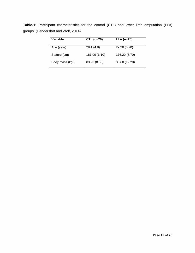
Table-1: Participant characteristics for the control (CTL) and lower limb amputation (LLA)
groups. (Hendershot and Wolf, 2014).
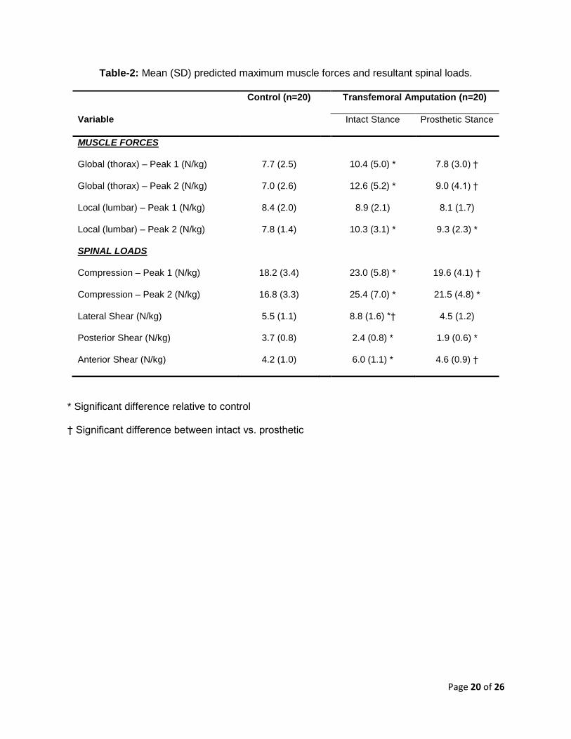
Table-2: Mean (SD) predicted maximum muscle forces and resultant spinal loads.
Table-3: Reported values of maximum compression force (*body weight) at the lower spinal
level.
Figure 1. Sagittal view of the biomechanical model including FE model of the spine and 56
trunk muscles (dimensions in mm). ICPL: iliocostalislumborum pars lumborum, ICPT:
iliocostalislumbroum pars thoracis, IP: iliopsoas, LGPL: longissimusthoracis pars lumborum,
LGPT: longissimusthoracis pars thoracis, MF: multifidus, QL: quadratuslumborum, IO: internal
oblique, EO: external oblique and RA: rectus abdominus.
Figure 2. The process used to estimate muscle forces and spinal loads. Each set of possible
segmental kinematics is generated using a genetic algorithm subjected to measured kinematics
of thorax and pelvis as well as the reported values of lumbar segments’ range of motion. The
convergence in the local and global loops are achieved when the changes in respectively sum
of predicted muscle forces in two consecutive local iterations and the value of the cost function
of the heuristic optimization procedure in two consecutive global iterations are less than 1%.
Figure 3. Mean sum of forces in global (i.e., muscles attached to the thoracic spine – top) and
local (i.e., muscles attached to the lumbar spine – bottom) muscles. CTL: control group, LLA:
group with lower limb amputation.
Figure 4. Mean compression forces at mid-plane of the L4-L5 (top) and L5-S1 (bottom)
intervertebral discs. CTL: control group, LLA: group with lower limb amputation.
Figure 5. Mean shear forces at the mid-plane of the L5-S1 in lateral (top) and antero-posterior
(bottom) directions. CTL: control group, LLA: group with lower limb amputation. Positive shear
force in lateral direction indicates force toward the right (intact) leg for controls (LLA) and
positive shear force in antero-posterior direction indicate anterior direction.
Page 19 of 26
Table-1: Participant characteristics for the control (CTL) and lower limb amputation (LLA)
groups. (Hendershot and Wolf, 2014).
Variable CTL (n=20) LLA (n=20)
Age (year) 28.1 (4.8) 29.20 (6.70)
Stature (cm) 181.00 (6.10) 176.20 (6.70)
Body mass (kg) 83.90 (8.60) 80.60 (12.20)
Page 20 of 26
Table-2: Mean (SD) predicted maximum muscle forces and resultant spinal loads.
Control (n=20) Transfemoral Amputation (n=20)
Variable Intact Stance Prosthetic Stance
MUSCLE FORCES
Global (thorax) – Peak 1 (N/kg) 7.7 (2.5) 10.4 (5.0) * 7.8 (3.0) †
Global (thorax) – Peak 2 (N/kg) 7.0 (2.6) 12.6 (5.2) * 9.0 (4.1) †
Local (lumbar) – Peak 1 (N/kg) 8.4 (2.0) 8.9 (2.1) 8.1 (1.7)
Local (lumbar) – Peak 2 (N/kg) 7.8 (1.4) 10.3 (3.1) * 9.3 (2.3) *
SPINAL LOADS
Compression – Peak 1 (N/kg) 18.2 (3.4) 23.0 (5.8) * 19.6 (4.1) †
Compression – Peak 2 (N/kg) 16.8 (3.3) 25.4 (7.0) * 21.5 (4.8) *
Lateral Shear (N/kg) 5.5 (1.1) 8.8 (1.6) *† 4.5 (1.2)
Posterior Shear (N/kg) 3.7 (0.8) 2.4 (0.8) * 1.9 (0.6) *
Anterior Shear (N/kg) 4.2 (1.0) 6.0 (1.1) * 4.6 (0.9) †
* Significant difference relative to control
† Significant difference between intact vs. prosthetic
Page 21 of 26
Table-3: Reported values of maximum compression force (*body weight) at the lower spinal
level.
Study Walking Speed (m/s)
0.90 1.00 1.20 1.35 1.50 1.70 2.20
Typic
al w
alk
ing
Current study 1.85
Cappozzo, 1983 1.20 1.50 1.90 2.50
Cheng et al., 1998
2.28 2.53 2.95
Khoo et al., 1995 1.71
Yoder et al., 2015
1.0
Aty
pic
al w
alk
ing
Current study
2.60
(Cappozzo and Gazzani,
1982) (amputation)
2.00 2.70 3.00
(Cappozzo and Gazzani,
1982) (knee ankylosis) 1.80 2.10
Yoder et al., 2015 1.0
Page 22 of 26
Figure 1. Sagittal view of the biomechanical model including FE model of the spine and 56
trunk muscles (dimensions in mm). ICPL: iliocostalislumborum pars lumborum, ICPT:
iliocostalislumbroum pars thoracis, IP: iliopsoas, LGPL: longissimusthoracis pars lumborum,
LGPT: longissimusthoracis pars thoracis, MF: multifidus, QL: quadratuslumborum, IO: internal
oblique, EO: external oblique and RA: rectus abdominus.
Rigid Thorax
L 1
S 1
Anterior Posterior
L 5
L 2 L 3 L 4
DL1-L2
DT12-L1
DL5-S1
T 1
T 12
LumbarDL2-L3
Sacrum
DL3-L4
DL4-L5
Page 23 of 26
Figure 2. The process used to estimate muscle forces and spinal loads. Each set of possible
segmental kinematics is generated using a genetic algorithm subjected to measured kinematics
of thorax and pelvis as well as the reported values of lumbar segments’ range of motion. The
convergence in the local and global loops are achieved when the changes in respectively sum
of predicted muscle forces in two consecutive local iterations and the value of the cost function
of the heuristic optimization procedure in two consecutive global iterations are less than 1%.
Page 24 of 26
Figure 3. Mean sum of forces in global (i.e., muscles attached to the thoracic spine – top) and
local (i.e., muscles attached to the lumbar spine – bottom) muscles. CTL: control group, LLA:
group with lower limb amputation.
0
250
500
750
1000
0 20 40 60 80 100
Me
an
su
m o
f g
lob
al m
us
cle
fo
rce
s (
N)
CTL LLA
0
250
500
750
1000
0 20 40 60 80 100
Mean
su
m o
f lo
cal m
uscle
fo
rces (
N)
Percent of gait cycle
Page 25 of 26
Figure 4. Mean compression forces at mid-plane of the L4-L5 (top) and L5-S1 (bottom)
intervertebral discs. CTL: control group, LLA: group with lower limb amputation.
750
1000
1250
1500
1750
2000
0 20 40 60 80 100
Mean
co
mp
ressio
n fo
rces a
t L
4-L
5 (
N)
CTL LLA
750
1000
1250
1500
1750
2000
0 20 40 60 80 100
Me
an
co
mp
res
sio
n fo
rces
at
L5
-S1
(N
)
Percent of gait cycle
Page 26 of 26
Figure 5. Mean shear forces at the mid-plane of the L5-S1 intervertebral disc in lateral (top) and
antero-posterior (bottom) directions. CTL: control group, LLA: group with lower limb amputation.
Positive shear force in lateral direction indicates force toward the right (intact) leg for controls
(LLA) and positive shear force in antero-posterior direction indicate anterior direction.
-500
-250
0
250
500
750
0 20 40 60 80 100
Mean
late
ral sh
ear
fo
rces (N
)CTL LLA
-500
-250
0
250
500
750
0 20 40 60 80 100
Me
an
an
tro
-po
ste
rio
r s
he
ar
fo
rces
(N
)
Percent of gait cycle
Disclaimer: The views expressed in this abstract are those of the authors and do not necessarily reflect the official policy of the Departments of the Army, Navy, Defense, nor the US Government.
Changes in Gait following Transfemoral Amputation Increase Spinal Loads Brad D. Hendershot1, Erik J. Wolf1, Babak Bazrgari2
1Department of Rehabilitation, Walter Reed National Military Medical Center, Bethesda, MD, USA 2Department of Biomedical Engineering, University of Kentucky, Lexington, KY, USA
Persons with transfemoral amputation (TFA) report a considerably higher prevalence of low back pain (LBP) compared to able-bodied individuals. Altered gait mechanics with TFA, particularly increased and asymmetric trunk motion, likely impose distinct demands on trunk muscles to maintain stability and equilibrium of the spine. Since alterations in trunk kinematics and muscle responses influence spinal loads, and spine loads are linked with LBP risk, the goal of this work was to demonstrate the effects of increased and asymmetric trunk kinematics with TFA on the relative contributions of external (i.e., gravity and inertia) and internal (i.e., muscle) forces to spine loads. Peak lumbar (i.e., thorax with respect to pelvis) lateral bending and forward lean obtained during gait from 20 persons with TFA (and 20 without), at 10 (4)° and 6 (3)°, respectively, were input into a kinematics-driven finite element model of the spine [1]. Total compressive and shear forces at the L5-S1 disc were computed, as well as relative contributions of internal and external forces. Influences of lumbar posture and mechanical properties of the passive ligamentous spine were also investigated. Total compressive and shear forces at the L5-S1 disc were substantially larger among persons with (vs. without) TFA, at 1548 (785) and 429 (252) N, respectively. Given the comparable contributions from external forces between groups (≈ 350 N in compression and 150 N in shear), the main cause for such higher spinal loads is the internal muscle response to spinal equilibrium requirements; muscle force contributions among persons with (vs. without) TFA were 1201 (434) N in compression and 299 (114) N in shear. Additional simulations with altered lumbar postures and passive tissues properties among persons with TFA revealed minimal changes in spinal loads (<150 N), but again with larger contributions from muscle forces. Although obtained from static simulations (i.e., no inertia), these results clearly support our hypothesis of abnormal spine loading with altered trunk motion in persons with TFA, who here, exhibited substantially larger spinal loads compared to able-bodied controls. Due to the cyclic nature of gait, repeated exposures to increased spinal loads may accelerate degenerative changes in the spine and/or increase the risk for chronic LBP. Acknowledgement: This work was supported by the Center for Rehabilitation Sciences Research (DOD Defense Health Program – NF90UG) and University of Kentucky’s Center for Clinical and Translational Science (NIH – UL1TR000117). References [1] Bazrgari B., et al. J Biomech 2008; 41: 412-421.
Low Back Pain in Service Members with Traumatic Extremity Injuries: Implications of Biomechanical Risk Factors
Brad D. Hendershot, PhD
Walter Reed National Military Medical Center, Bethesda, MD, USAUniformed Services University of the Health Sciences, Bethesda, MD, USA
BACKGROUND: Low back pain (LBP) is far-reaching within the military and general population. While LBP generally has a multifactorial etiology and complex pathogenesis, biomechanical risk factors likely contribute more substantially among persons with traumatic extremity injuries, including lower-limb amputation (LLA). Specifically, persons with unilateral LLA walk and perform other activities of daily living in ways that often disproportionately rely on the intact (vs. prosthetic) limb. Such a compensational strategy is most notably associated with increased and/or asymmetric trunk movements as compared to able-bodied individuals; these movements are of particular concern given the biomechanical association between joint motions and musculoskeletal loads [1], and perceived by individuals with LLA as primary contributors toward LBP [2].
METHODS: Kinematic and kinetic data from 40 males with unilateral transtibial (n=20) and transfemoral (n=20) amputation, and 20 uninjured males, were obtained during level-ground walking at a self-selected pace. Net external demands (inverse dynamics) and bone-bone joint contact loads (finite element modeling) at L5-S1 are summarized in an effort to better understand relationships between altered trunk/pelvic motions with LLA on musculoskeletal loads within the lower back.
RESULTS: The coordination / motions between the trunk and pelvis with vs. without LLA are associated with ~31-55, 41-83, and 3-14% larger external demands on the lower back in the sagittal, coronal, and transverse planes, respectively. Similarly, joint contact forces within the spine are increased with LLA; notably, largest increases (up to ~65% relative to uninjured individuals) were found in joint compressive forces owing to a complex pattern and increased (6-80%) activation of trunk muscles. Also of note, increases were generally larger among individuals with more proximal amputations (transfemoral vs. transtibial), consistent with changes in trunk motions.
CONCLUSION: Though walking is generally not a mechanically demanding task for the low back (i.e., loads are well below reported injury thresholds), and sometimes even considered therapeutic for individuals with LBP, altered trunk-pelvic motions with LLA during gait are associated with larger external demands on the lower back and internal loads among tissues within the spine. Given the repetitive nature of gait, over time, even minimal increases in trunk motions and musculoskeletal loads may synergistically and progressively contribute toward LBP onset/recurrence and accelerate degenerative joint changes. However, to comprehensively characterize relative and accumulated risk profiles, additional efforts are needed to classify such relationships during other activities of daily living. In doing so, future work can begin to assess the ability of specific interventions (e.g., prosthetic devices, physical therapy) to mitigate injury risk.
1. Davis and Marras (2000) Clinical Biomechanics2. Devan et al. (2015) Disability and Rehabilitation
This work was partially supported by…the Office of the Assistant Secretary of Defense for Health Affairs, through the Peer Reviewed Orthopaedic Research Program (award #W81XWH-14-2-0144)…DoD Defense Health Program (NF90UG)…DoD-VA Extremity Trauma & Amputation Center of Excellence. Views expressed are those of the authors and do not reflect the official policy or position of the Departments of the Army, Navy, or Defense, or the U.S. Government. We thank Dr. Babak Bazrgari for his assistance with spinal load calculations from the finite element modeling approach.

LEARNING OBJECTIVES: 1. Describe risk factors for low back pain with traumatic extremity injuries. 2. Define the impact of altered mechanics on the lower back and influence on low back pain risk. 3. Describe potential ways in which the elevated risk can be minimized, either clinically or with novel technologies.
COMPREHENSIVE INVITED REVIEW
Impact of Traumatic Lower Extremity InjuriesBeyond Acute Care: Movement-BasedConsiderations for Resultant Longer TermSecondary Health Conditions
Courtney M. Butowicz,1 Christopher L. Dearth,1–4
and Brad D. Hendershot1–3,*1Research and Development Section, Department of Rehabilitation, Walter Reed National Military Medical Center,
Bethesda, Maryland.2DOD-VA Extremity Trauma and Amputation Center of Excellence, Walter Reed National Military Medical Center,
Bethesda, Maryland.3Department of Rehabilitation Medicine, Uniformed Services University of the Health Sciences, Bethesda, Maryland.4Regenerative Biosciences Laboratory, Uniformed Services University of the Health Sciences, Bethesda, Maryland.
Significance: Advances in field-based trauma care, surgical techniques, andprotective equipment have collectively facilitated the survival of a historicallylarge number of service members (SMs) following combat trauma, although manysustained significant composite tissue injuries to the extremities, including limbloss (LL) and limb salvage (LS). Beyond the acute surgical and rehabilitativeefforts that focus primarily on wound care and restoring mobility, traumatic LLand LS are associated with several debilitating longer term secondary healthconditions (e.g., low back pain [LBP], osteoarthritis [OA], and cardiovasculardisease [CVD]) that can adversely impact physical function and quality of life.Recent Advances: Despite recent advancements in prosthetic and orthotic de-vices, altered movement and mechanical loading patterns have been identifiedamong persons with LL and salvage, which are purported risk factors for thedevelopment of longer term secondary musculoskeletal conditions and may limitfunctional outcomes and/or concomitantly impact cardiovascular health.Critical Issues: The increased prevalence of and risk for LBP,OA, andCVDamongthe relatively young cohort of SMs with LL and LS significantly impact physio-logical and psychological well-being, particularly over the next several decades oftheir lives.Future Directions: Longitudinal studies are needed to characterize the onset, pro-gression, and recurrence of health conditions secondary to LL and salvage. Whilenot a focus of the current review, detailed characterization of physiological bio-markers throughout the rehabilitation processmay provide additional insight intothe current understanding of disease processes of the musculoskeletal and car-diovascular systems.
Keywords: amputation, biomechanics, cardiovascular disease,limb salvage, low back pain, osteoarthritis
SCOPE AND SIGNIFICANCEExtremity trauma, including limb
loss (LL) and limb salvage (LS), iscommonly associated with an elevatedrisk for secondary health conditions
(e.g., low back pain [LBP], osteoar-thritis [OA], cardiovascular disease[CVD]) that can significantly limitphysical function, reduce quality of life(QoL), and life expectancy. This review
Brad D. Hendershot, PhD
Submitted for publication October 31, 2016.
Accepted in revised form December 15, 2016.
*Correspondence: Department of Rehabilita-
tion, Walter Reed National Military Medical
Center, 4494 N. Palmer Road, America Building
(19), Room B-320, Bethesda, MD 20889
(e-mail: [email protected]).
j 1ADVANCES IN WOUND CARE, VOLUME 00, NUMBER 00Copyright ª 2017 by Mary Ann Liebert, Inc. DOI: 10.1089/wound.2016.0714
provides an extensive commentary regarding resul-tant secondary health effects of extremity trauma inservice members (SMs), with a particular focus onfunctional outcomes and quality of movement.
TRANSLATIONAL RELEVANCE
Physiologic biomarkers provide an opportunityto enhance translation in future work to examinethe pathophysiology of the secondary health con-ditions associated with traumatic LL from a basicscience perspective.While this approach is yet to befully explored and thus was not a primary focus ofthis review, such biomarkers may augment tradi-tional analyses and support more comprehensiverisk characterization, thereby allowing cliniciansand researchers to better mitigate disease onset orprogression.
CLINICAL RELEVANCE
The increased prevalence of secondary health ef-fects following traumatic extremity injuries places asignificant physical and psychosocial burden on SMswith LL and LS. Altered movement patterns oftenresult in mechanical loading of the spine and lowerextremities,potentially increasingtheriskofLBPandOA.Adopting a biopsychosocialmodel of treatment/care may allow clinicians to utilize a multifacetedapproach to treat chronic pain and dysfunction as-sociated with resultant health effects of LL.
BACKGROUND
Musculoskeletal disorders are the most preva-lent source of disability in the United States.1,2
As a result, the annual direct costs associatedwith treatment total a substantial $900 billion.3
Among these, extremity amputation, or LL, isprojected to affect an estimated 3.6 million peopleby the year 2050.4 Approximately 185,000 indi-viduals undergo either an upper or lower ex-tremity amputation annually, primarily due totrauma, dysvascular disease, and/or osteosarco-ma.5–7 While the incidence of LL due to dysvas-cular etiologies has steadily risen among thecivilian sector, trauma remains a leading sourceof LL within the Military Health System. How-ever, prior estimates of the current/future impactof LL do not include SMs injured during combatnor do they consider individuals with LS; an al-ternative to amputation in which heroic measuresare undertaken by the military surgical teams atall echelons of care to preserve as much form andfunction of the traumatically injured limb aspossible. Despite these surgical efforts and ad-
vances in orthotic technology, many with LS areunable to achieve preinjury functional outcomes,much like those with LL.
The combat theaters of Operations: EnduringFreedom (OEF), Iraqi Freedom (OIF), New Dawn,Inherent Resolve, and Freedom’s Sentinel werecharacterized by high-energy munitions and ex-plosives. With advances in personal protectiveequipment, field-based trauma care, and surgicaltechniques, injuries sustained as a result of theseoften-improvised devices are now survivable athigher rates than conflicts past.However, traumaticextremity injuries, including LL and LS, remain ahallmark casualty of recent conflicts. Across all ser-vices, 52,351military personnel have beenwoundedin action since 20018; more than half of evacuatedSMs have sustained extremity injuries and nearly aquarter of these are open fractures.9 In addition,1,703 SMs sustained injuries requiring major (ormultiple) limb amputation (As of October 1, 2016;Data source: EACE-R). The decision to amputate alimb may be made in as few as 24h post-trauma,during the first hospitalization as a secondary sur-gical intervention, or potentially years after LS (i.e.,delayed amputation).10–13 Factors contributing tothe decision include the extent and severity of in-juries and resources available during the rehabili-tation process.14 Recent evidence suggests that SMswho undergo LS will typically experience more ex-pansive complications than individuals who un-dergo amputation.15–17 LS has been associatedwith significantly higher rates of rehospitalization,greater numbers of surgical procedures, and higherrates of surgical complications.18,19
Initial wound care and rehabilitation after LLand/or LS are critical to the recovery process. Suchefforts are generally categorized by nine distinctphases, each with specific goals and objectives.20
The complexity and interdependence between eachphase elucidate the need for an efficient interdis-ciplinary approach within the overall rehabilita-tion paradigm. Despite these comprehensive andsubstantive efforts, persons with LL and LS are atan increased risk for acute secondary health con-ditions such as phantom limb pain, wounds/sores,vascular and nerve damage, infection, decreasedphysical function, and psychosocial issues. Fur-thermore, beyond these acute conditions, personswith LL and LS are also at an elevated risk forlonger term complications including LBP, OA,and CVD, among others. Importantly, once thedisease progression initiates, these longer termresultant conditions will plague these individu-als for life, as SMs with extremity trauma aretypically younger than 30 years at the time of
2 BUTOWICZ, DEARTH, AND HENDERSHOT

injury and thus will continue living with theirinjuries for several decades.17
The long-term economic burden of trauma-related LL and LS is significant. Edwards et al.predicted the long-term (40 year) cost of traumarepair, rehabilitation, and lifelong prosthetic sup-port of British soldiers wounded in Afghanistan tobe approximately $444 million.21 In the UnitedStates, the estimated average lifetime cost oftreatment for unilateral lower LL is $342,716 and$1.4 million for Vietnam and OIF/OEF veterans,respectively.22 However, such estimates are likelyconservative, not fully accounting for costs associ-ated with novel technology/repairs or, perhaps ex-ponentially more economically burdensome overthe longer term, for the wide range of healthcarecosts associated with the treatment of secondaryhealth conditions. The ability to evaluate, predict,and ultimately treat these resultant health condi-tions would not only help reduce these costs butalso, and most importantly, preserve and/or im-prove function and QoL for those with LL and LS.
The risk for secondary health conditions is oftenrelated to physiological adaptations to trauma orpervasive surgical complications, poor biomechan-ics, and/or the prosthetic (orthotic) device itself. ForSMs, in particular, the young age at which theseinjuries occur likely presents a unique challengeover the longer term and further highlights theimportance for understanding resultant healthconditions secondary to extremity trauma. Notably,the cumulative effects of many years of functionaladaptations during gait and movement with ex-tended prosthetic/orthotic device use in otherwiseyoung and active SMs remain unclear.23,24 This isan important distinction from civilian populationsas a majority of civilians with LL are over the age of50, incurred LL as a result of vascular damage/complications, are likely less active, and may pres-ent with different resultant health conditions/outcomes for less time.25 Thus, as a preliminary steptoward addressing this knowledge gap, the purposeof this review is to provide a commentary regardingresultant health conditions associated with high-energy extremity trauma, with a primary focus onbiomechanical features of movement and associatedfunctional limitations. In particular, we highlightconsiderations for longitudinal care aimed at max-imizing QoL, for those with both LL and LS.
DISCUSSIONLow back pain
The World Health Organization describes LBPas any pain or discomfort for a variable duration in
the lumbar spine region.26 The onset of pain mayoccur suddenly, coincident to a singular traumaticevent, or develop over timewith age or as the resultof repeated microtrauma from a given (or set of)activity(ies). Often, LBP is considered idiopathic,as pain may be present without pathoanatomicalevidence of disease or structural abnormality. LBPcosts nearly $100 billion annually in the UnitedStates, with a majority of this cost associated withlost wages and decreased productivity.27 Whilecross-sectional figures indicate that chronic LBPaffects up to 33% of adults in the general popula-tion, the incidence in persons with LL who reportLBP secondary to trauma is nearly double (52–76%).28–31 Along with this significantly higherprevalence, nearly 50% of persons with LL havereported LBP as ‘‘more bothersome’’ than either re-sidual or phantom limb pain and as having a sig-nificant reduction in overall QoL metrics.28,30,32
While the exact etiology of LBP within this popu-lation is unclear, there is a growing body of evidencesuggesting that altered lumbopelvic mechanicsduring the (repetitive) gait cycle likely influencessuch risk.
Persons with lower LL frequently develop al-tered movement patterns to maintain balance andachieve forward progression inwalking.Movementpatterns can be influenced by the following, eitherindividually or in combination: socket fit/prostheticalignment, general deconditioning, leg length dis-crepancies, complications within the residual limb,and muscular imbalances.33,34 More specifically,altered movement patterns during gait affecttrunk and pelvismechanics and contribute, at leastin part, to the increased incidence of LBP in personswith lower LL and may be dependent on the extentof injury or ultimate level of amputation.35–38 Thesealterations and asymmetriesmay increase loads onthe lumbar spine during gait which, when consid-ering the repetitive gait cycle, over time may thuscontribute to the occurrence or recurrence of LBP.For example, persons with transfemoral LL tend toexhibit 10� of anterior pelvic tilt, which is consid-ered to be a compensatory mechanism to assist inthe ability to achieve hip extension during gait.Increased anterior pelvic tilt is associated withincreased lumbar lordosis, which is linked to anincreased incidence of LBP in persons with LL.28,39
Previous work has demonstrated that increasedloads on the lumbar spine are a direct source ofLBP in the general population.40,41 Mechanicalloading of the passive and active structures of thespine is affected by both internal and externalloads, such as forces produced by muscular acti-vation, ligamentous tension, gravity, and inertia.42
SECONDARY HEALTH CONDITIONS AFTER EXTREMITY TRAUMA 3
These loads can be significant, as potentially smallalterations in trunk (which accounts for nearly 2/3of the body’s mass) movement may increase jointreaction loading due to increased muscular con-tractions of the surrounding musculature.43 Theincreased demand on the active structures (mus-cles) may lead to increased forces and joint loadingon the passive structures (discs and vertebrae).The accumulation of these altered loads over timehas the potential to augment degenerative jointchanges in the spine.40
Similar to uninjured individuals with LBP, per-sons with transfemoral LL exhibit irregular trunk–pelvis coordination and movement variability.44
Specifically, persons with LL tend to walk with alarge lateral trunk lean toward the affected side; apossible neuromuscular strategy/compensation toassist in forward progression during gait.42 Thisfrontal plane motion has been reported to increasepeak joint reaction forces and moments asymmetri-cally in the lumbar spine (L5-S1 integration specif-ically) in this population. A recent report suggestedthis observed frontal plane motion as a possiblemechanistic pathway through which recurring ex-posure to altered trunk motion and cumulative spi-nal loading may contribute to LBP in persons withlower LL.42 Persons with transfemoral LL (withcurrent LBP) exhibit larger axial trunk rotationswhen compared to those without LBP, which maysubsequently affect vertebral disc degeneration andpotentially contribute to LBP recurrence.45,46 Pre-vious evidence demonstrated degenerative changesin the lumbar spine via radiographic imaging in76%of personswithLL, potentially supporting the role ofincreased trunk motion leading to degenerativechanges in this population.47
While LBP is commonly cited as a secondaryhealth effect of LL, persons with LS may also expe-rience LBP as a result of altered movement patternsduring gait and functional activities.48 Persons withLS typically experience reduced ankle function,which is associated with altered gait mechanics andincreased metabolic cost.34,49,50 However, the influ-ence of distal LS on proximal (trunk/pelvis) biome-chanics remains unstudied to date. Currently, apaucity of evidence exists relative to theprevalence ofLBP in the LS population. Therefore, further work isneeded to elucidate the relationship between LS andthe development of LBP.
In summary, LBP has been reported as the mostimportant health-related physical condition con-tributing to a reducedQoL among veteranswho hadsustained a traumatic lower extremity amputationover 20 years prior.32 Thus, identifying factorscontributing to the development and recurrence of
LBP, such as a widely prevalent and ‘‘bothersome’’secondary health concern, is critical for improvinglong-term health. Abnormal mechanical loading oflumbar spine, altered trunkand pelvis coordination,and psychosocial factors may influence the preva-lence of LBP in this population. Therapeutic inter-ventions that address the underlying impairment(s)in trunk neuromuscular responses and/or motorcontrol strategymay also contribute to reducing theprevalence and incidence of LBP among SMs withlower extremity trauma, thereby improving longerterm functional outcomes by mitigating a signifi-cant secondary impairment with a substantial ad-verse impact on daily activities. Further evidence isneeded to understand the relationship betweenthese risk factors and the incidence of LBP in per-sons with LL. In particular, no studies to date haveevaluated the influence of different prostheses ororthoses on the incidence of LBP in the traumaticLL and LS populations.
OsteoarthritisThe National Institute of Arthritis and Muscu-
loskeletal and Skin Diseases describes OA as a jointdisease affecting the cartilage, often characterizedby pain and stiffness within a joint and limitationsin physical function.51 The primary pathology isarticular cartilage deterioration, although evidencesuggests that possible morphological changes ofbone are reflective of disease onset. Within the joint,articular cartilage functions to dissipate forcessustained by the bony structures throughout mo-tion. During activities such as walking or running,when the loading velocity and intensity of thestructures are increased, the cartilage’s ability todissipate forces is reduced.52 In the general popu-lation, mechanical loading of the knee joint duringwalking has been associated with the presence, se-verity, and progression of knee OA.53–56 Personswith unilateral lower LL are 17 times more likely tosuffer from knee OA in the intact limb when com-pared to able-bodied individuals.57
As previously noted, persons with LL frequentlydevelop altered movement patterns during gait. Ofparticular importance here, those with unilateralLL preferentially utilize their intact limb, leading toincreased and prolonged loading of the intact joints.Mechanical alterations in static and dynamicalignment of the knee joint may affect joint loadingas increased forces are incurred through medial orlateral aspects of the joint. The external knee ad-duction moment (EKAM) is a vastly reported riskfactor for knee OA based on its relationship withinternal loading of the medial joint surface.58 Thesize of theEKAMand its respective angular impulse
4 BUTOWICZ, DEARTH, AND HENDERSHOT
are associated with knee OA severity and progres-sion.53,55,59,60 During gait, individuals with lowerLL asymmetrically load their intact limb to agreater extent than their involved limb, suggestingthat mechanical factors play a role in the increasedincidence of knee OA in this population.36,61 Forexample, Lloyd et al. identified larger peak kneeadductionmoments in the intact relative to involvedlimb.62 This increased mechanical loading may beexplained by decreased push-off power and groundreaction forces demonstrated with conventionalprosthetic feet.61,63 Push-off power generated by theprosthetic foot instance may affect the ground re-action forces at heel strike in the intact limb as thevelocity of an individual’s center of mass changesfrom an anterior and inferior direction to an ante-rior and superior direction during gait.64 The redi-rection of the center of mass is caused by the groundreaction impulse through the gait cycle, crudelyrelative to double-limb support.64 If the prostheticstance foot lacks adequate push-off power to propelthe center of mass anteriorly, the intact limb mustcompensate by performing more work to move thecenter of mass anterior and superior, resulting inincreased ground reaction forces and loading of theintact limb.61 Morgenroth et al. suggested that byutilizing a prosthetic foot with increased push-offpower, the peak EKAM of the intact limb may bereduced and therefore potentially decreasing theOA risk.61 This was supported as a powered ankle–foot prosthetic was able to decrease the EKAM andvertical ground reaction force in persons with lowerLL, however, the prosthetic usedwasunable to alterthe knee joint loads of the intact limb.65 Similar toLBP, the progression and severity of OA may befurther amplified by psychosocial determinants;anxiety, depression, coping strategies, and stresshave also been associated with increased pain inpatients with OA.66–68
OA is not exclusive to the LL population as indi-viduals with LS present with similar (sometimeslarger) gait and movement deviations. As high as95% of OA diagnoses among combat-wounded SMsare post-traumatic in origin.69 Chronic pain, nervedamage, and volumetric muscle loss are commonbarriers to LS rehabilitation and may serve as con-founding factors in the development of OA treat-ment plans.70,71 Ankle–foot orthoses (AFOs) arecommonly used to assist ankle function or offloadpainful structures.72 Optional therapies that in-clude sports medicine-based interventions utilizinga dynamic AFO (e.g., the Intrepid Dynamic Exos-keletal Orthosis) are available to LS patients. Suchdevices are designed to improve functional perfor-mance on tasks such as walking, changing direc-
tions, sit-to-stand, and ascending stairs.48 Whiledynamic AFOs are suggested to improve functionalcapabilities, evidence is inconclusive in its ability topositively alter gait parameters related to OA aswell as the effects of long-term use.34,73,74
Treatment modalities focused on reducing symp-toms and OA disease progression in persons with LLandLSare vital to improvingQoL.TheOsteoarthritisResearch Society International recommends biome-chanical interventions, intra-articularcorticosteroids,exercise (land and water based), self-managementand education, strength training, and weight man-agement.75 Autologous platelet-rich plasma (PRP)therapy is a therapeutic intervention that delivershigh concentrations of growth factors to an area tostimulate healing.76 Recent evidence suggests thatPRP may provide relief of knee OA symptoms inyounger patients within the early stages of cartilagedegeneration.77–79 Strength training (weight andbody-weight training) and exercises such as t’ai chihave demonstrated the ability to improve overallfunction in decreasing pain in OA patients and mayalso serve to assist in weight management.80,81
Weight reduction is considered a pragmatic therapyfor knee OA as overweight individuals demonstrate ahigh prevalence of knee OA and the risk of severityprogression increases 35% for every 5kg of weightgain.82 Strength training and weight managementare considered integral aspects of the rehabilitationparadigm for persons with LL as deficits in strengthand increases in weight influence gait, joint loading,movement efficiency, and cardiovascular health.Canes, knee braces, and foot orthotics are other po-tential treatment options to decrease movements atthe knee, reduce pain, and improve function.83–85
In summary, biomechanical factors likely play asubstantial role in the risk for OA secondary to ex-tremity trauma, whether LL or LS. While theprevalence of OA in LL and LS populations maydecrease as technological improvements in pros-thesesandorthosesare realized, further evidence isneeded to determine the specific relationship be-tween different classes or features of these devicesand OA risk factors. Unfortunately, recent techno-logical advancements in prosthetic devices haveoutpaced orthotic devices, the benefits of which areevident in the biomechanical characteristics ofpersons with LL versus LS. Nevertheless, LS typi-cally presents with more complex neurovascularinjuries and other unique challenges, which cannegatively affect functional outcomes.
Cardiovascular diseaseCVD is defined by a vast array of diseases affect-
ing the heart and blood vessels.86 CVDmay present
SECONDARY HEALTH CONDITIONS AFTER EXTREMITY TRAUMA 5
as coronary artery disease, stroke, arrhythmias,cardiomyopathy, heart disease, peripheral arterydisease, aneurysms, venous thrombosis, and/orcarditis.86,87 While CVD is largely preventable, itremains the leading cause of death worldwide, par-ticularly in lower socioeconomic demographics.86
The American Heart Association reports there are*85 million individuals with CVD in the UnitedStates, causing a staggering 2,200 deaths each andevery day.88 This is accompanied by direct and in-direct costs of nearly $315 billion.89 Risk factors forCVD include, but are not limited to, family historyand genetics, high cholesterol and lipids, high bloodpressure, diabetes, metabolic syndrome, obesity,andkidneydisease.89 Inaddition, significant combattrauma may be a risk factor for the development ofCVD.90–92 For example, Hrubec and Ryder con-ducted a 30-year follow-up ofWorldWar II veteranswith lower LL and demonstrated that the relativeriskofCVDmortalitywas increased2.4–4times thatof persons with LS.90 Similarly, Modan et al. re-ported significantly higher mortality rates of per-sons with traumatic lower LL when compared toable-bodied controls, suggesting that CVD was theprimary cause (21.9% vs. 12.1%, p<0.001).91
Thepathophysiology of increasedmortality ratesmay be a result of systemic and/or regional hemo-dynamic effects of trauma.91,93–97 Obesity and hy-pertension secondary to decreased overall activitylevels may lead to insulin regulation complicationsin persons with LL.97 When compared to uninjuredcontrols with no difference in body mass index,blood pressure, or lipid levels, persons with LL ex-hibited significantly higher increased fasting plas-ma insulin levels as well as insulin resistance.96
Increased plasma insulin levels and insulin resis-tance are risk factors for atherosclerosis and met-abolic syndrome, considered precursors to CVD.The role of psychological stressors in the develop-ment of CVD is not well understood; however, psy-chosocial factors have demonstrated involvementin the pathogenesis of CVD.98,99 Depression andpost-traumatic stress disorder have been associ-ated with increased incidence of CVD, while veter-ans with high levels of cynical distrust and angerdemonstrate an accelerated progression of athero-sclerosis, a risk factor for CVD.100–102 Limited evi-dence precludes a definitive relationship betweenpsychosocial factors and CVD risk in persons withLL, and therefore, futurework shouldprospectivelyexamine the relationship between psychosocialfactors/stressors and the development of CVD.
Hemodynamically, proximal amputation in-creases the risk of CVD development based on al-terations in proximal arterial flow. Pathogenic
mechanisms may include early reflection pulsewaves. Early return reflection pulse waves are pro-duced at arterial occlusion sites and have beenlinked to a myriad of medical complications.103 Anearlyreturnedreflectionpulsewavecreatesasecondsystolic peak, which results in an increase in aorticpressure. The increased aortic pressure generatesan increased left ventricular load resulting in leftventricular hypertrophy, atherothrombosis, andultimately cardiac death.104 Vollmar et al. sug-gested that persons with traumatic LL above theknee were five times more likely to suffer from ab-dominal aortic aneurysms when compared to heal-thy controls.94 A possible explanation may be thatafter amputation, blood flow is decreased by*25%in the terminal aortadue to alteredflowpaths in thevisceral and renal arteries, resulting in a disruptedflowpattern at the aortic bifurcation.95Altered flowpatterns, paired with increased shear stress alongthe convex aspect of the aorta and decreased shearstress along the concave aspect, are theorized todamage aorto-iliac blood vessels by increasing hy-draulic forces within the aorta.95 Persons withtransfemoral LL should have regular consultationswith appropriate medical personnel to assess therisk of abdominal aortic aneurysm.95
While the hemodynamic effects of trauma ap-pear to influence CVD risk, addressing modifiablerisk factors may be an effective strategy to helpdecrease CVD risk. It is widely accepted that ha-bitual exercise with activities such as running,walking, bicycling, rowing, and swimming in-creases aerobic capacity and decreases the risk ofCVD. When joined with dietary modifications,regular exercise can effectively reduce excess bodyweight, another risk factor for CVD. Moreover, theincreased risk of CVD in persons with LL high-lights the importance of managing modifiable riskfactors, engaging in preventative treatment strat-egies, and adopting an active lifestyle.
SUMMARY
Maintaining an active lifestyle is critically im-portant for physiological health, psychological well-being, and overall QoL. Such guidance is no differentfor individuals with LL and LS. However, given thelimited (but growing) body of evidence relatingmovement abnormalities to altered musculoskeletaldemands that may lead to the development of longerterm secondary conditions in this population, addi-tional consideration for the quality of movementduring recreational anddaily activities iswarranted.While the overwhelming focus of recent efforts hasbeen on persons with LL, the aforementioned sec-ondary health conditions are likely also major con-
6 BUTOWICZ, DEARTH, AND HENDERSHOT

cerns for those with LS. As such, we positthatanunderlying focusof clinical careandfuture research, in both cohorts, should betowardmitigating concomitant risk for thedevelopment or recurrence of chronic pain.
While advances in trauma care andprosthetic/orthotic technologies may even-tually mollify acute and subacute second-ary health effects of extremity trauma,longitudinal tracking is urgently needed tobetter understand the mechanisms bywhich secondary health effects developand progress in this population. Such ef-forts should encompass a transdisciplinaryteam, in which a comprehensive suite of evalua-tion metrics are employed; for example, traditionalclinical evaluation and movement analysis sup-plemented with local and systemic physiologicalbiomarker analyses and next-generation imagingmodalities. In doing so, a better understanding ofthe specific pathways for the development of thesesecondary health effects can be realized, thusenabling clinicians to develop and prescribe ap-propriate treatment interventions. Ultimately, di-minishing risk factors relative to the degenerationof joint and cardiovascular function will reduce theoverall prevalence of secondary health conditionsand improve QoL for our nation’s injured SMs andveterans over the longer term.
ACKNOWLEDGMENTSAND FUNDING SOURCES
This work was supported by the Office of theAssistant Secretary of Defense for Health Affairs,through the Peer Reviewed Orthopaedic ResearchProgram (Award No. W81XWH-14-2-0144 toB.D.H.) and the Orthotics and Prosthetics Out-comes Research Program (Award No. W81XWH-15-1-0669 to C.L.D.), the National Institute ofBiomedical Imaging and Bioengineering (AwardNo. 1R03EB018889-01A1 to C.L.D.), and the DoD-VA Extremity Trauma & Amputation Center ofExcellence (Public Law 110–417, National DefenseAuthorization Act 2009, Section 723). The authorsalso thank Eric Margulies for his assistance withinitial literature review.
AUTHOR DISCLOSUREAND GHOSTWRITING STATEMENT
No competing financial interests exist. The con-tent of this article was expressly written by theauthors listed. No ghostwriters were used to write
this article. The views expressed in this article arethose of the authors and do not necessarily reflectthe official policy of the Departments of the Army,Navy, Defense, nor the United States Government.
ABOUT THE AUTHORS
Courtney M. Butowicz, PhD, CSCS, is aPostdoctoral Researcher within the Department ofRehabilitation at Walter Reed National MilitaryMedical Center (WRNMMC). Dr. Butowicz’s re-search interests include clinical assessment oftrunk stability/control, development of musculo-skeletal injuries as result of impaired motor con-trol, and utilization of amultidisciplinary approachto the treatment of musculoskeletal injuries.Christopher L. Dearth, PhD, concurrentlyserves as the Facility Research Director for theExtremity Trauma and Amputation Center of Ex-cellence (EACE), Director of Research for the De-partment of Rehabilitation at WRNMMC, and theFounding Director of the Regenerative BiosciencesLaboratory at the Uniformed Services Universityof theHealth Sciences (USUHS). In these roles, Dr.Dearth leads a multidisciplinary team of cliniciansand researchers whose collective focus is on themitigation, treatment, and rehabilitation of trau-matic extremity injuries and amputations, with anoverarching synergy of efforts between the fields ofrehabilitative and regenerative medicine.Brad D.Hendershot, PhD, is a Research Biomedical En-gineer with the EACE, stationed at WRNMMC.In addition to this role, he directs activities withinthe Biomechanics and Virtual Reality Laboratorieswithin the Department of Rehabilitation. Dr.Hendershot’s research is primarily focused oncharacterizing factors underlying the high preva-lence of musculoskeletal complications secondaryto extremity trauma.
TAKE HOME MESSAGES
� Living with LL and LS over time leads to increased morbidity and mortalityfrom secondary medical and musculoskeletal problems. Awareness of thelong-term health risks associated with LL and LS, as well as the physiologicand biomechanical origin of these risks, is critical to improving outcomes
� Understanding the pathogenesis of the secondary health conditions oftraumatic LL and LS and salvage may help guide optimal management inacute, subacute, and chronic phases of care for these individuals
� Reducing modifiable risk factors through patient education, identifyingappropriate support systems, encouraging proper gait mechanics, andutilizing the prescription of evolving technologies may help mitigate long-term health conditions
SECONDARY HEALTH CONDITIONS AFTER EXTREMITY TRAUMA 7

REFERENCES
1. Summers K, Jinnett K, Bevan S. MusculoskeletalDisorders, Workforce Health and Productivityin the United States. 2015. www.tcwhp.org/musculoskeletal-disorders-workforce-health-and-productivity-united-states-0 (last accessed Au-gust 15, 2016).
2. United States Bone and Joint Initiative. TheBurden of Musculoskeletal Diseases in theUnited States. Rosemont, IL: BMUS, 2014.
3. Agency for Healthcare Research and Quality.Medical Expenditures Panel Survey (MEPS).2008–2011. http://meps.ahrq.gov/mepsweb (lastaccessed August 25, 2016).
4. Ziegler-Graham K, MacKenzie EJ, Ephraim PL,Travison TG, Brookmeyer R. Estimating theprevalence of limb loss in the United States:2005 to 2050. Arch Phys Med Rehabil 2008;89:422–429.
5. Dillingham TR, Pezzin LE, Mackenzie EJ. Racialdifferences in the incidence of limb loss sec-ondary to peripheral vascular disease: apopulation-based study. Arch Phys Med Rehabil2002;83:1252–1257.
6. Wrobel JS, Mayfield JA, Reiber GE. Geographicvariation of lower-extremity major amputation inindividuals with and without diabetes in theMedicare population. Diabetes Care 2001;24:860–864.
7. Owings MF, Kozak LJ. Ambulatory and inpatientprocedures in the United States, 1996. VitalHealth Stat 13 1998;1–119.
8. Fischer H. A Guide to US Military CasualtyStatistics: Operation Freedom’s Sentinel, Op-eration Inherent Resolve, Operation New Dawn,Operation Iraqi Freedom, and Operation EnduringFreedom. Congressional Research Service Re-port; Research Report #RS22452; Washington,DC. 2015.
9. Doukas WC, Hayda RA, Frisch HM, et al. TheMilitary Extremity Trauma Amputation/LimbSalvage (METALS) study: outcomes of amputa-tion versus limb salvage following major lower-extremity trauma. J Bone Joint Surg Am 2013;95:138–145.
10. Hansen ST, Jr. The type-IIIC tibial fracture.Salvage or amputation. J Bone Joint Surg Am1987;69:799–800.
11. Hansen ST, Jr. Overview of the severely trau-matized lower limb. Reconstruction versus am-putation. Clin Orthop Relat Res 1989;17–19.
12. Dirschl DR, Dahners LE. The mangled extremity:when should it be amputated? J Am Acad Or-thop Surg 1996;4:182–190.
13. Lange RH. Limb reconstruction versus amputa-tion decision making in massive lower extremitytrauma. Clin Orthop Relat Res 1989;92–99.
14. MacKenzie EJ, Bosse MJ, Kellam JF, et al.Factors influencing the decision to amputate orreconstruct after high-energy lower extremitytrauma. J Trauma 2002;52:641–649.
15. Bosse MJ, Ficke JR, Andersen RC. Extremity warinjuries: current management and research pri-orities. J Am Acad Orthop Surg 2012;20(Suppl 1):viii–x.
16. Dagum AB, Best AK, Schemitsch EH, MahoneyJL, Mahomed MN, Blight KR. Salvage after severelower-extremity trauma: are the outcomesworth themeans? Plast Reconstr Surg 1999;103:1212–1220.
17. Reiber GE, McFarland LV, Hubbard S, et al.Service members and veterans with major trau-matic limb loss from Vietnam war and OIF/OEFconflicts: survey methods, participants, and sum-mary findings. J Rehabil Res Dev 2010;47:275–297.
18. Harris AM, Althausen PL, Kellam J, Bosse MJ,Castillo R; Lower Extremity Assessment ProjectStudy Group. Complications following limb-threatening lower extremity trauma. J OrthopTrauma 2009;23:1–6.
19. Busse JW, Jacobs CL, Swiontkowski MF, BosseMJ, Bhandari M; Evidence-Based OrthopaedicTrauma Working Group. Complex limb salvage orearly amputation for severe lower-limb injury: ameta-analysis of observational studies. J OrthopTrauma 2007;21:70–76.
20. Esquenazi A, DiGiacomo R. Rehabilitation afteramputation. J Am Podiatr Med Assoc 2001;91:13–22.
21. Edwards MD, Phillip LCRD, Bosanquet N, BullAM, Clasper CJC. What is the magnitude andlong-term economic cost of care of the Britishmilitary Afghanistan amputee cohort? Clin Or-thop Relat Res 2015;473:2848–2855.
22. Blough DK, Hubbard S, McFarland LV, Smith DG,Gambel JM, Reiber GE. Prosthetic cost projec-tions for service members with major limb lossfrom Vietnam and OIF/OEF. J Rehabil Res Dev2010;47:387–402.
23. Stern P. The epidemiology of amputations. PhysMed Rehabil Clin N Am 1991;2:253–261.
24. Pezzin LE, Dillingham TR, MacKenzie EJ. Re-habilitation and the long-term outcomes of per-sons with trauma-related amputations. ArchPhys Med Rehabil 2000;81:292–300.
25. Dillingham TR, Pezzin LE, MacKenzie EJ. Limbamputation and limb deficiency: epidemiologyand recent trends in the United States. SouthMed J 2002;95:875–884.
26. Ehrlich GE. Low back pain. Bull World HealthOrgan 2003;81:671–676.
27. Katz JN. Lumbar disc disorders and low-backpain: socioeconomic factors and consequences.J Bone Joint Surg Am 2006;88(Suppl 2):21–24.
28. Ehde DM, Smith DG, Czerniecki JM, CampbellKM, Malchow DM, Robinson LR. Back pain as asecondary disability in persons with lower limbamputations. Arch Phys Med Rehabil 2001;82:731–734.
29. Ephraim PL, Wegener ST, MacKenzie EJ, Dil-lingham TR, Pezzin LE. Phantom pain, residuallimb pain, and back pain in amputees: results of
a national survey. Arch Phys Med Rehabil2005;86:1910–1919.
30. Smith DG, Ehde DM, Legro MW, Reiber GE, delAguila M, Boone DA. Phantom limb, residuallimb, and back pain after lower extremity am-putations. Clin Orthop Relat Res 1999;29–38.
31. Foote CE, Mac Kinnon J, Robbins C, Pessagno R,Portner MD. Long-term health and quality of lifeexperiences of Vietnam veterans with combat-related limb loss. Qual Life Res 2015;24:2853–2861.
32. Taghipour H, Moharamzad Y, Mafi AR, et al.Quality of life among veterans with war-relatedunilateral lower extremity amputation: a long-term survey in a prosthesis center in Iran. JOrthop Trauma 2009;23:525–530.
33. Sagawa Y, Turcot K, Armand S, Thevenon A,Vuillerme N, Watelain E. Biomechanics andphysiological parameters during gait in lower-limb amputees: a systematic review. Gait Pos-ture 2011;33:511–526.
34. Esposito ER, Choi HS, Owens JG, Blanck RV,Wilken JM. Biomechanical response to ankle–foot orthosis stiffness during running. Clin Bio-mech (Bristol, Avon) 2015;30:1125–1132.
35. Kulkarni J, Gaine WJ, Buckley JG, Rankine JJ,Adams J. Chronic low back pain in traumaticlower limb amputees. Clin Rehabil 2005;19:81–86.
36. Gailey R, Allen K, Castles J, Kucharik J, RoederM. Review of secondary physical conditions as-sociated with lower-limb amputation and long-term prosthesis use. J Rehabil Res Dev 2008;45:15–29.
37. Hendershot BD, Nussbaum MA. Persons withlower-limb amputation have impaired trunkpostural control while maintaining seated bal-ance. Gait Posture 2013;38:438–442.
38. Devan H, Hendrick P, Ribeiro DC, Hale LA, Car-man A. Asymmetrical movements of the lumbo-pelvıc region: Is this a potential mechanism forlow back pain in people with lower limb ampu-tation? Med Hypotheses 2014;82:77–85.
39. Day JW, Smidt GL, Lehmann T. Effect of pelvictilt on standing posture. Phys Ther 1984;64:510–516.
40. Kumar S. Theories of musculoskeletal injurycausation. Ergonomics 2001;44:17–47.
41. McGill SM. Low Back Disorders: Evidence-basedPrevention and Rehabilitation, 2nd ed. Cham-paign, IL: Human Kinetics, 2007.
42. Hendershot BD, Wolf EJ. Three-dimensionaljoint reaction forces and moments at the lowback during over-ground walking in persons withunilateral lower-extremity amputation. Clin Bio-mech (Bristol, Avon) 2014;29:235–242.
43. Gillet C, Duboy J, Barbier F, et al. Contribution ofaccelerated body masses to able-bodied gait.Am J Phys Med Rehabil 2003;82:101–109.
44. Esposito ER, Wilken JM. The relationship be-tween pelvis–trunk coordination and low back
8 BUTOWICZ, DEARTH, AND HENDERSHOT

pain in individuals with transfemoral amputa-tions. Gait Posture 2014;40:640–646.
45. Schmidt H, Kettler A, Heuer F, Simon U, Claes L,Wilke HJ. Intradiscal pressure, shear strain, andfiber strain in the intervertebral disc undercombined loading. Spine (Phila Pa 1976) 2007;32:748–755.
46. Morgenroth DC, Orendurff MS, Shakir A, SegalA, Shofer J, Czerniecki JM. The relationshipbetween lumbar spine kinematics during gaitand low-back pain in transfemoral amputees.Am J Phys Med Rehabil 2010;89:635–643.
47. Burke M, Roman V, Wright V. Bone and jointchanges in lower limb amputees. Ann Rheum Dis1978;37:252–254.
48. Patzkowski JC, Blanck RV, Owens JG, et al.Comparative effect of orthosis design on func-tional performance. J Bone Joint Surg Am 2012;94:507–515.
49. Collins SH, Kuo AD. Recycling energy to restoreimpaired ankle function during human walking.PLoS One 2010;5:e9307.
50. Waters RL, Mulroy S. The energy expenditure ofnormal and pathologic gait. Gait Posture 1999;9:207–231.
51. National Institute of Arthritis and Musculoske-letal and Skin Diseases National Institute ofHealth. What is Osteoarthritis? 2014. www.niams.nih.gov (last accessed September 7, 2016).
52. Radin ER, Paul IL, Rose RM. Pathogenesis ofprimary osteoarthritis. Lancet 1972;1:1395–1396.
53. Sharma L, Felson DT. Studying how osteoar-thritis causes disability: nothing is simple. JRheumatol 1998;25:1–4.
54. Baliunas A, Hurwitz D, Ryals A, et al. Increasedknee joint loads during walking are present insubjects with knee osteoarthritis. OsteoarthritisCartilage 2002;10:573–579.
55. Miyazaki T, Wada M, Kawahara H, Sato M,Baba H, Shimada S. Dynamic load at baselinecan predict radiographic disease progression inmedial compartment knee osteoarthritis. AnnRheum Dis 2002;61:617–622.
56. Lynn SK, Reid SM, Costigan PA. The influence ofgait pattern on signs of knee osteoarthritis inolder adults over a 5–11 year follow-up period: acase study analysis. Knee 2007;14:22–28.
57. Struyf PA, van Heugten CM, Hitters MW,Smeets RJ. The prevalence of osteoarthritis ofthe intact hip and knee among traumatic legamputees. Arch Phys Med Rehabil 2009;90:440–446.
58. Kutzner I, Trepczynski A, Heller MO, BergmannG. Knee adduction moment and medial contactforce—facts about their correlation during gait.PLoS One 2013;8:e81036.
59. Astephen JL, Deluzio KJ, Caldwell GE, DunbarMJ. Biomechanical changes at the hip, knee,and ankle joints during gait are associated withknee osteoarthritis severity. J Orthop Res 2008;26:332–341.
60. Mundermann A, Dyrby CO, Andriacchi TP. Sec-ondary gait changes in patients with medialcompartment knee osteoarthritis: increased loadat the ankle, knee, and hip during walking. Ar-thritis Rheum 2005;52:2835–2844.
61. Morgenroth DC, Segal AD, Zelik KE, et al. Theeffect of prosthetic foot push-off on mechanicalloading associated with knee osteoarthritis inlower extremity amputees. Gait Posture 2011;34:502–507.
62. Lloyd CH, Stanhope SJ, Davis IS, Royer TD.Strength asymmetry and osteoarthritis risk fac-tors in unilateral trans-tibial, amputee gait. GaitPosture 2010;32:296–300.
63. Winter DA, Sienko SE. Biomechanics of below-knee amputee gait. J Biomech 1988;21:361–367.
64. Adamczyk PG, Kuo AD. Redirection of center-of-mass velocity during the step-to-step transitionof human walking. J Exp Biol 2009;212:2668–2678.
65. Grabowski AM, D’Andrea S. Effects of a pow-ered ankle-foot prosthesis on kinetic loading ofthe unaffected leg during level-ground walking.J Neuroeng Rehabil 2013;10:1.
66. Somers TJ, Keefe FJ, Godiwala N, Hoyler GH.Psychosocial factors and the pain experience ofosteoarthritis patients: new findings and newdirections. Curr Opin Rheumatol 2009;21:501–506.
67. Rosemann T, Laux G, Szecsenyi J. Osteoarthritis:quality of life, comorbidities, medication andhealth service utilization assessed in a largesample of primary care patients. J Othop SurgRes 2007;2:1.
68. Marks R. Comorbid depression and anxiety im-pact hip osteoarthritis disability. Disabil Health J2009;2:27–35.
69. Johnson A, Cross J. Impact of traumatic arthritison a cohort of combat casualties. Paper pre-sented at: American Academy of OrthpaedicSurgeons Annual Meeting, San Diego, CA, 2011.
70. Shawen SB, Keeling JJ, Branstetter J, Kirk KL,Ficke JR. The mangled foot and leg: salvageversus amputation. Foot Ankle Clin 2010;15:63–75.
71. Grogan BF, Hsu JR; Skeletal Trauma ResearchConsortium. Volumetric muscle loss. J Am AcadOrthop Surg 2011;19(Suppl 1):S35–S37.
72. Harper NG, Esposito ER, Wilken JM, NeptuneRR. The influence of ankle-foot orthosis stiffnesson walking performance in individuals withlower-limb impairments. Clin Biomech (Bristol,Avon) 2014;29:877–884.
73. Esposito ER, Wilken JM. Biomechanical riskfactors for knee osteoarthritis when using pas-sive and powered ankle–foot prostheses. ClinBiomech (Bristol, Avon) 2014;29:1186–1192.
74. Ranz EC, Russell Esposito E, Wilken JM, Nep-tune RR. The influence of passive-dynamicankle-foot orthosis bending axis location ongait performance in individuals with lower-limb
impairments. Clin Biomech (Bristol, Avon) 2016;37:13–21.
75. Zhang W, Moskowitz RW, Nuki G, et al. OARSIrecommendations for the management of hipand knee osteoarthritis, part I: critical appraisalof existing treatment guidelines and systematicreview of current research evidence. Osteoar-thritis Cartilage 2007;15:981–1000.
76. Gobbi A, Karnatzikos G, Mahajan V, Malchira S.Platelet-rich plasma treatment in symptomaticpatients with knee osteoarthritis: preliminaryresults in a group of active patients. SportsHealth 2012;4:162–172.
77. Kon E, Buda R, Filardo G, et al. Platelet-richplasma: intra-articular knee injections producedfavorable results on degenerative cartilage le-sions. Knee Surg Sports Traumatol Arthrosc2010;18:472–479.
78. Patel S, Dhillon MS, Aggarwal S, Marwaha N,Jain A. Treatment with platelet-rich plasma ismore effective than placebo for knee osteoar-thritis: a prospective, double-blind, randomizedtrial. Am J Sports Med 2013;41:356–364.
79. Spakova T, Rosocha J, Lacko M, Harvanova D,Gharaibeh A. Treatment of knee joint osteoar-thritis with autologous platelet-rich plasma incomparison with hyaluronic acid. Am J PhysMed Rehabil 2012;91:411–417.
80. Jansen MJ, Viechtbauer W, Lenssen AF, Hen-driks EJ, de Bie RA. Strength training alone,exercise therapy alone, and exercise therapywith passive manual mobilisation each reducepain and disability in people with knee osteo-arthritis: a systematic review. J Physiother 2011;57:11–20.
81. Kang JW, Lee MS, Posadzki P, Ernst E. T’ai chi forthe treatment of osteoarthritis: a systematic reviewand meta-analysis. BMJ Open 2011;1:e000035.
82. Felson DT, Lawrence RC, Dieppe PA, et al. Os-teoarthritis: new insights. Part 1: the diseaseand its risk factors. Ann Intern Med 2000;133:635–646.
83. Kerrigan DC, Lelas JL, Goggins J, Merriman GJ,Kaplan RJ, Felson DT. Effectiveness of a lateral-wedge insole on knee varus torque in patientswith knee osteoarthritis. Arch Phys Med Rehabil2002;83:889–893.
84. Jones A, Silva PG, Silva AC, et al. Impact of caneuse on pain, function, general health and energyexpenditure during gait in patients with kneeosteoarthritis: a randomised controlled trial. AnnRheum Dis 2012;71:172–179.
85. Raja K, Dewan N. Efficacy of knee braces andfoot orthoses in conservative management ofknee osteoarthritis: a systematic review. Am JPhys Med Rehabil 2011;90:247–262.
86. Mendis S, Puska P, Norrving B. Global Atlas onCardiovascular Disease Prevention and Control.Geneva: World Health Organization, 2011.
87. Naghavi M, Wang H, Lozano R, et al. Global,regional, and national age-sex specific all-causeand cause-specific mortality for 240 causes of
SECONDARY HEALTH CONDITIONS AFTER EXTREMITY TRAUMA 9

death, 1990–2013: a systematic analysis for theGlobal Burden of Disease Study 2013. Lancet2015;385:117–171.
88. Mozaffarian D, Benjamin EJ, Go AS, et al. Ex-ecutive summary: Heart Disease and StrokeStatistics-2016 Update: A report from theAmerican Heart Association. Circulation 2016;133:447.
89. Go AS, Mozaffarian D, Roger VL, et al. Heartdisease and stroke statistics-2014 update. Cir-culation 2014;129:e28–e292.
90. Hrubec Z, Ryder RA. Traumatic limb amputationsand subsequent mortality from cardiovasculardisease and other causes. J Chronic Dis 1980;33:239–250.
91. Modan M, Peles E, Halkin H, et al. Increasedcardiovascular disease mortality rates in trau-matic lower limb amputees. Am J Cardiol 1998;82:1242–1247.
92. Naschitz J, Lenger R. Why traumatic leg ampu-tees are at increased risk for cardiovasculardiseases. QJM 2008;101:251–259.
93. Yekutiel M, Brooks M, Ohry A, Yarom J, Carel R.The prevalence of hypertension, ischaemic heartdisease and diabetes in traumatic spinal cordinjured patients and amputees. Paraplegia 1989;27:58–62.
94. Vollmar J, Pauschinger P, Paes E, Henze E, FrieschA. Aortic aneurysms as late sequelae of above-knee amputation. Lancet 1989;334:834–835.
95. Paes E, Vollmar J, Pauschinger P, Mutschler W,Henze E, Friesch A. Late vascular damage afterunilateral leg amputation [in German]. Z Un-fallchir Versicherungsmed 1989;83:227–236.
96. Peles E, Akselrod S, Goldstein DS, et al. Insulinresistance and autonomic function in traumaticlower limb amputees. Clin Auton Res 1995;5:279–288.
97. Rose H, Schweitzer P, Charoenkul V, Schwartz E.Cardiovascular disease risk factors in combatveterans after traumatic leg amputations. ArchPhys Med Rehabil 1987;68:20–23.
98. Everson-Rose SA, Lewis TT. Psychosocial factorsand cardiovascular diseases. Annu Rev PublicHealth 2005;26:469–500.
99. Rosengren A, Hawken S, Ounpuu S, et al. As-sociation of psychosocial risk factors with risk ofacute myocardial infarction in 11 119 cases and13 648 controls from 52 countries (the INTER-HEART study): case-control study. Lancet2004;364:953–962.
100. Kang HK, Bullman TA, Taylor JW. Risk of selectedcardiovascular diseases and posttraumatic stressdisorder among former World War II prisoners ofwar. Ann Epidemiol 2006;16:381–386.
101. Frasure-Smith N, Lesperance F. Reflections ondepression as a cardiac risk factor. PsychosomMed 2005;67:S19–S25.
102. Julkunen J, Salonen R, Kaplan GA, Chesney MA,Salonen JT. Hostility and the progression of
carotid atherosclerosis. Psychosom Med 1994;56:519–525.
103. Sugawara J, Hayashi K, Tanaka H. Distal shift ofarterial pressure wave reflection sites with ag-ing. Hypertension 2010;56:920–925.
104. Yano M, Kohno M, Kobayashi S, et al. Influenceof timing and magnitude of arterial wave reflec-tion on left ventricular relaxation. Am J PhysiolHeart Circ Physiol 2001;280:H1846–H1852.
Abbreviations and Acronyms
AFO ¼ ankle–foot orthosesCVD ¼ cardiovascular diseaseEACE ¼ Extremity Trauma and Amputation
Center of ExcellenceEKAM ¼ external knee adduction momentLBP ¼ low back painLL ¼ limb lossLS ¼ limb salvageOA ¼ osteoarthritisOEF ¼ Operation Enduring FreedomOIF ¼ Operation Iraqi FreedomPRP ¼ platelet-rich plasmaQoL ¼ quality of lifeSM ¼ service member
USUHS ¼ Uniformed Services University ofthe Health Sciences
WRNMMC ¼ Walter Reed National MilitaryMedical Center
10 BUTOWICZ, DEARTH, AND HENDERSHOT
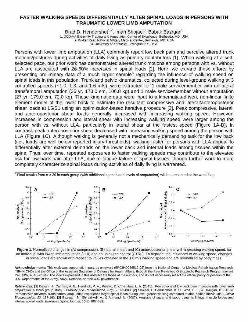
FASTER WALKING SPEEDS DIFFERENTIALLY ALTER SPINAL LOADS IN PERSONS WITH TRAUMATIC LOWER LIMB AMPUTATION
Brad D. Hendershot1,2, Iman Shojaei3, Babak Bazrgari3
1. DOD-VA Extremity Trauma and Amputation Center of Excellence, Bethesda, MD, USA 2. Walter Reed National Military Medical Center, Bethesda, MD, USA
3. University of Kentucky, Lexington, KY, USA
Persons with lower limb amputation (LLA) commonly report low back pain and perceive altered trunk motions/postures during activities of daily living as primary contributors [1]. When walking at a self-selected pace, our prior work has demonstrated altered trunk motions among persons with vs. without LLA are associated with 26-60% increases in spinal loads [2]. Here, we expand these efforts by presenting preliminary data of a much larger samplea regarding the influence of walking speed on spinal loads in this population. Trunk and pelvic kinematics, collected during level-ground walking at 3 controlled speeds (~1.0, 1.3, and 1.6 m/s), were extracted for 1 male servicemember with unilateral transfemoral amputation (35 yr, 173.0 cm, 106.8 kg) and 1 male servicemember without amputation (27 yr, 179.0 cm, 72.0 kg). These kinematic data were input to a kinematics-driven, non-linear finite element model of the lower back to estimate the resultant compressive and lateral/anteroposterior shear loads at L5/S1 using an optimization-based iterative procedure [3]. Peak compressive, lateral, and anteroposterior shear loads generally increased with increasing walking speed. However, increases in compression and lateral shear with increasing walking speed were larger among the person with vs. without LLA, particularly in lateral shear at the fastest speed (Figure 1A-B). In contrast, peak anteroposterior shear decreased with increasing walking speed among the person with LLA (Figure 1C). Although walking is generally not a mechanically demanding task for the low back (i.e., loads are well below reported injury thresholds), walking faster for persons with LLA appear to differentially alter external demands on the lower back and internal loads among tissues within the spine. Thus, over time, repeated exposures to faster walking speeds may contribute to the elevated risk for low back pain after LLA, due to fatigue failure of spinal tissues, though further work to more completely characterize spinal loads during activities of daily living is warranted. _____________________________ a Final results from n ≥ 20 in each group (with additional speeds and levels of amputation) will be presented at the workshop
Figure 1. Normalized changes in (A) compression, (B) lateral shear, and (C) anteroposterior shear with increasing walking speed, for
an individual with lower limb amputation (LLA) and an uninjured control (CTRL). To highlight the influences of walking speed, changes in spinal loads are shown with respect to values obtained in the 1.0 m/s walking speed and are normalized by body mass.
Acknowledgements: This work was supported, in part, by an award (5R03HD086512-02) from the National Center for Medical Rehabilitation Research (NIH-NICHD) and the Office of the Assistant Secretary of Defense for Health Affairs, through the Peer Reviewed Orthopaedic Research Program (award #W81XWH-14-2-0144). The views expressed in this abstract are those of the authors, and do not necessarily reflect the official policy or position of the U.S. Departments of the Army, Navy, Defense, nor the U.S. government. References: [1] Devan, H., Carman, A. B., Hendrick, P. A., Ribeiro, D. C., & Hale, L. A. (2015). Perceptions of low back pain in people with lower limb amputation: a focus group study. Disability and Rehabilitation, 37(10), 873-883. [2] Shojaei, I., Hendershot, B. D., Wolf, E. J., & Bazrgari, B. (2016). Persons with unilateral transfemoral amputation experience larger spinal loads during level-ground walking compared to able-bodied individuals. Clinical Biomechanics, 32, 157-163. [3] Bazrgari, B., Shirazi-Adl, A., & Arjmand, N. (2007). Analysis of squat and stoop dynamic liftings: muscle forces and internal spinal loads. European Spine Journal, 16(5), 687-699.
1
The Biopsychosocial Correlates of Chronic Low Back Pain after Lower Limb Amputation
Shawn Farrokhi, PT, PhD1,8
Brittney Mazzone, PT, DPT1,2
Michael Schneider, DC, PhD3
Sara Gombatto, PT, PhD4
John Mayer, DC, PhD 5
M. Jason Highsmith, PT, PhD, CP, FAAOP 5,6,8
Brad D. Hendershot, PhD7,8
1 Department of Physical and Occupational Therapy, Naval Medical Center San Diego, San
Diego, CA
2 BADER Consortium, University of Delaware, Newark, DE
3 Department of Physical Therapy, University of Pittsburgh, Pittsburgh, PA
4 Department of Physical Therapy, San Diego State, University, San Diego, CA
5 School of Physical Therapy & Rehabilitation Sciences, University of South Florida, Tampa, FL
6James A. Haley Veterans Administration Hospital, Tampa, FL
7 Department of Rehabilitation, WRNMMC, Bethesda, MD; Department of Physical Medicine
and Rehabilitation, Uniformed Services University of the Health Sciences, Bethesda, MD
8 DoD/VA Extremity Trauma and Amputation Center of Excellence
Disclaimer: The views expressed herein are those of the author(s) and do not necessarily reflect
the official policy or position of the Department of the Navy, Department of Defense, or the
United States Government.
Corresponding Author Information:
Shawn Farrokhi, PT, PhD
Senior Scientist
DoD-VA Extremity Trauma and Amputation Center of Excellence
Naval Medical Center San Diego
34800 Bob Wilson Dr.
San Diego, CA 92134
Phone: 619-532-8582
Email: [email protected]
2
ABSTRACT
Low back pain is a common secondary health condition after lower limb amputation with
important implications related to functional capabilities and overall quality of life. Despite the
high prevalence of low back pain after lower limb amputation, the underlying etiologies of the
disorder remain unknown. This special communication summarizes evidence in support of the
multifactorial, biopsychosocial model of the low back pain experience in the general population
and after lower limb amputation for identification of potential risk factors and treatment targets.
Key findings that link biological, psychological, and social factors and the experience of low
back pain after lower limb amputation are discussed while highlighting gaps in our current state
of knowledge to direct future research. Importantly, the aim of this special communication was
not to propose a new model, but rather to organize data originating from prior work into a
coherent conceptual framework to better understand the need for multifaceted and
multidisciplinary intervention approaches for effective treatment of low back pain after lower
limb amputation.
3
INTRODUCTION
Low back pain (LBP) is a common health condition worldwide, with 11-38% of the
general population reporting symptoms over a one year period.1, 2
LBP is currently considered
the leading cause of disability globally, ahead of 290 other conditions, and is responsible for 83
million years lived with disability.3 Additionally, LBP is a major source of activity limitation,
work absenteeism, and increased cost of medical care throughout much of the world.2, 4-6
LBP is
also a common and perhaps more impactful, secondary health condition after lower limb
amputation (LLA), with high estimated annual prevalence rates between 50-90%.7-13
Individuals
with LLA often report more LBP after amputation than before8, 9
and in most cases directly
attribute their LBP to their amputation.10
Additionally, presence of LBP daily or several times
per week has been associated with moderate to severe physical disability and limitations in
performing daily activities in patients with LLA.8, 9, 13-15
To this end, LBP is often rated by
patients with LLA as more bothersome than phantom or residual limb pain,11
suggesting LBP is
an important secondary musculoskeletal condition associated with functional limitation and
disability after LLA.
Despite the high prevalence of LBP after LLA, the exact etiologies of the disorder in this
population remain unknown, thereby making its treatment exceptionally challenging.
Importantly, there are currently no published guidelines specifically tailored toward the
management of LBP for individuals with LLA. Therefore, there exists a clear need for
comprehensive identification of contributing factors to the LBP experience after LLA that can
serve as a basis for the development of targeted treatments and future research investigations.
Here a new application of the multifactorial, biopsychosocial model for LBP, previously
developed for the general population,16-18
is proposed as a way of identifying risk factors and
4
potential treatment targets for treatment of LBP after LLA. The objective of this special
communication was to organize data originating from prior studies of the biopsychosocial
correlates of LBP after LLA into a coherent conceptual framework. We hypothesized that
alterations in biological, psychological, and social factors with LLA are related to the
development of LBP symptoms and disability after LLA that merit specific attention during the
clinical decision making process and for future research efforts to improve patient-related
outcomes.
The Biopsychosocial Model of Low Back Pain
Treatment of LBP has historically centered around the traditional biomedical model of
illness, which assumes a direct relationship between regional pathoanatomy and the perception
of pain.18
As such, it was expected that once the anatomical source of LBP is identified,
biochemical and/or mechanical treatments of underlying pathoanatomy would result in cessation
of pain. Despite leading to successful treatment of many other disease processes, the outcomes of
interventions based on the biomedical model have proven to be less than ideal for treatment of
LBP.18-20
One potential reason for the failure of the biomedical model to provide an effective
treatment option for LBP is that no single underlying pathoanatomical lesion has been
consistently identified,18
with up to 85% of LBP patients left without a precise pathoanatomical
diagnosis.21
Additionally, determination of pathoanatomical sources of LBP frequently lacks
interexaminer reliability and evidence for generalizability.22
The often equivocal outcomes from
many “lesion-specific” treatment options such as intra-articular corticosteroid injections19
and
spinal fusion surgeries,20
along with the generally poor predictive value of diagnostic imaging for
identification of pathoanatomical pain sources,23
have led to a recent paradigm shift toward a
“non-structural” approach for the management of LBP.24
5
A growing body of evidence now suggests that successful treatment of LBP should
include biological, psychological, and social assessments to comprehensively address the
patient’s unique pain experience.18
The so called “biopsychosocial model” of LBP suggests that
the patient’s perceptions and reactions to pain should also be considered as these factors often
lead to unnecessary avoidance of physical activity and social interactions, work absenteeism, and
high health care utilization.16, 17
Whereas the pathoanatomy may initiate the pain process, the
psychological and social factors appear to play an important role in exacerbating the biological
component of LBP by influencing the perception of pain.25
For example, it has been
hypothesized that the presence of mechanical LBP can lead to a pain-generated stress response
that could have a negative impact on the endocrine and immune systems, which in turn may
negatively affect the cognitive assessment, emotional response, coping strategies and health
practices of the individual.26
Proponents of the biopsychosocial model argue that the complex, multidimensional
nature of LBP does not lend itself to the reductionist view of the biomedical model; instead, the
patient’s unique biologic, psychological, and social factors must equally be considered.18
Therefore, the term biopsychosocial implies that the biological, psychological and social factors
are interwoven within the context of the patient’s overall LBP experience and should be directly
and concurrently considered as a part of a comprehensive treatment program.26
In support of this
theory, multidisciplinary treatment approaches that include biopsychosocial components for
treatment of LBP in adults have demonstrated positive effects on pain, disability, and health-
related quality of life.27, 28
It stands to reason that LLA likely amplifies and/or alters specific
components within the multifactorial biopsychosocial model of LBP, previously suggested for
the general population. Given that LLA may differentially affect the various components of this
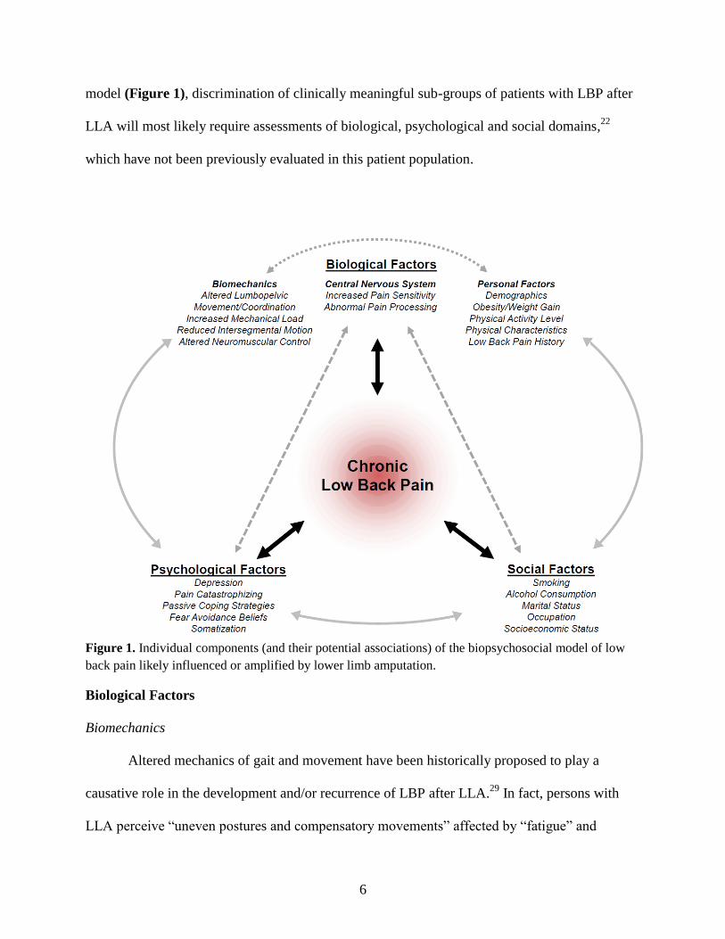
6
model (Figure 1), discrimination of clinically meaningful sub-groups of patients with LBP after
LLA will most likely require assessments of biological, psychological and social domains,22
which have not been previously evaluated in this patient population.
Figure 1. Individual components (and their potential associations) of the biopsychosocial model of low
back pain likely influenced or amplified by lower limb amputation.
Biological Factors
Biomechanics
Altered mechanics of gait and movement have been historically proposed to play a
causative role in the development and/or recurrence of LBP after LLA.29
In fact, persons with
LLA perceive “uneven postures and compensatory movements” affected by “fatigue” and
7
“prosthesis-related factors” during functional activities as the primary contributors to LBP.30
Though at the expense of higher metabolic cost of transport,31
compensatory movement
strategies adopted after LLA typically involve adaptations to maintain the body’s center of mass
within the base of support (i.e., improve stability and balance), primarily with a preference for
the intact limb, if applicable.32
During gait, for example, the intact limb (relative to prosthetic
limb) is characterized by a longer stance time, shorter step length, wider stride width, and larger
vertical ground reaction forces.33
As the trunk accounts for approximately two-thirds of total
body mass,34
altered motions of this segment play a substantial role in post-amputation
movement strategies, thereby warranting more trunk-focused biomechanical investigations for
assessing potential links with the development and persistence of LBP.
Altered trunk and pelvic movements in persons with LLA have been previously identified
in all three cardinal planes, including larger forward trunk lean and flexion-extension range of
motion, greater lateral trunk flexion (towards the prosthetic limb) and pelvic obliquity motion, as
well as more axial rotations between the shoulders/pelvis or regional/intervertebral motion
segments.35, 36
The presence (and likely severity) of LBP further influences such motions, most
notably increasing axial rotations within the lumbar region.37
LBP has also been associated with
more in-phase mediolateral coordination between the trunk and pelvis,38
which is indicative of
inter-segmental rigidity (i.e., “guarding behavior”) previously reported in able-bodied individuals
who are experiencing LBP.39, 40
Additional evidence suggests that individuals with LLA employ
an active mediolateral trunk movement strategy, inferred from increases in generation and
absorption of energy between the trunk and pelvis.41, 42
Although actively increasing mediolateral
trunk sway is likely an attempt to improve joint stability within the lower extremity by altering
lever arms of ab/adductor musculature,43
most notably within the hip among patients with
8
transfemoral amputation,7 such strategies have been associated with LBP/discomfort among
able-bodied individuals performing gait training aimed at reducing knee joint loads via trunk
lateral flexion.44
Abnormal mechanics of the spinal column, primarily larger mechanical loads and
instability, are often considered risk factors for the development of LBP.45
Of particular interest
here, characteristics of trunk motion can directly influence musculoskeletal loading,46
typically
due to altered muscular response (i.e., coactivity).45
Though walking is generally not a
mechanically demanding task for the low back (i.e., loads are well below reported injury
thresholds),47
and sometimes even considered therapeutic for individuals with LBP,48
altered
trunk-pelvic motions with LLA during gait have recently been associated with large internal
loads among tissues within the spine.49-51
Notably, largest increases (up to ~65% relative to
uninjured individuals) were found in joint compressive forces owing to a complex pattern of
muscle responses.50
Given the repetitive nature of gait, over time, even minimal increases in
trunk motions and musculoskeletal loads may synergistically and progressively contribute
toward LBP onset and/or recurrence and accelerate degenerative joint changes in the spine.
It is well accepted that the neuromuscular system plays a central role in supporting the
upper body and maintaining mechanical equilibrium and stability of the spine.52, 53
Irregular
patterns of trunk muscle recruitment have been identified among the general population with
recurrent LBP,54
and impaired postural control has been associated with spinal instability and
LBP.55
Among persons with LLA without LBP, similar assessments have identified impairments
in trunk postural control during an unstable seated balance task,56
bilateral asymmetries in trunk
mechanical and muscular responses to applied positional perturbations,57
as well as altered load-
sharing between active and passive trunk tissues during quasi-static trunk flexion/extension
9
movements.58
Additionally, substantially greater fatigability has been reported for the low-back
extensor musculature in patients with LLA with and without LBP,15
that are more pronounced
than healthy individuals with and without LBP.59
Fatigue of the low back extensors may further
contribute to increased intersegmental spinal motion and instability during prolonged functional
activities.60
Though the specific origin and functional impact of such alterations remain
somewhat speculative, these data support the theory that repeated exposure to altered loading
associated with LLA and repeated use of a prosthetic device may result in tissue and
neuromuscular adaptations and increased risk for LBP in this population.
Central Nervous System
In addition to changes in trunk/pelvic biomechanics with LLA, central nervous system
factors may also play an important biological role in the manifestation of LBP after LLA.
Because of the trauma to peripheral nerves, amputation has the potential to influence the
processing of pain signals in the peripheral and central nervous systems. Phantom limb pain has
been long described as the perception of pain in the missing (amputated) limb,61, 62
and may be
indicative of alterations in the processing of pain signals. Although it is unknown how alterations
in pain processing might influence the incidence and prevalence of secondary musculoskeletal
pain problems, such as LBP, there are several plausible explanations.
In the general population, people with LBP display generalized hypersensitivity to pain
that is reflective of central sensitization.63
Central sensitization is the increased neuronal
responsiveness to a stimulus due to prolonged or strong activity in the dorsal horn neurons that
may be associated with an episode of pain or prolonged pain.64
It is plausible that the pain
stimulus associated with amputation could elicit central sensitization and increased pain
10
sensitivity, putting persons with amputation at risk for developing secondary pain conditions.
Pain sensitivity is typically evaluated by assessing thresholds and tolerance to pain using a
variety of modalities for stimuli, including: mechanical (pressure), electrical, and thermal
(cold/heat).63
Changes in pain sensitivity also can be measured after either an inhibitory stimulus
(conditioned pain modulation), or a facilitory stimulus (temporal summation) to further elucidate
central mechanisms of pain inhibition or facilitation. Specific alterations in pain processing that
have been reported in people with LBP include local65, 66
and widespread65-70
hyperalgesia and
enhanced temporal summation of pain signals.71-74
Although people with chronic pain conditions
such as osteoarthritis, fibromyalgia, and chronic fatigue syndrome also typically display
decreased inhibition of nociceptive signals (conditioned pain modulation),75-77
most studies
report that people with chronic LBP display normal inhibition of pain signals.78-80
Pain sensitivity has been examined to a limited extent in persons with amputation. In a
small sample, Li et al.81
reported that persons with traumatic amputation and phantom limb pain
displayed decreased thresholds for sensation and pain with electrical stimuli in the unaffected
limb, suggesting central sensitization. Further, Vase et al.82
reported that people with upper limb
amputation and phantom limb pain display decreased thresholds for pressure and cold stimuli,
and enhanced temporal summation of pain signals. Inhibition of nociceptive signals has not been
explicitly examined in person with amputation, but it is plausible that those with phantom limb
pain may display decreased inhibition of pain similar to people with other chronic pain
conditions. Although the mechanisms of altered pain processing are similar in persons with
amputation and people with LBP, to our knowledge, no investigators have examined the
neurophysiology of pain in patients with amputation and secondary musculoskeletal pain
11
problems to determine whether central sensitization places them at greater risk for secondary
pain conditions.
Alterations in pain-processing areas of the brain in persons with amputation are also
consistent with changes reported in otherwise uninjured individuals with LBP. For example,
thalamic structural variations and, more specifically, decreases in gray matter of the
posterolateral thalamus have been reported in people with amputation.83
These changes appear
to be positively correlated with duration of time since amputation, suggesting that they may be
related to reduced afferent input.83
Further, Lotze et al.84
reported shifts in motor and sensory
cortical activation patterns during movement in patients with phantom limb pain compared to
pain-free persons with amputation, while Makin et al.85
reported cortical reorganization of the
sensorimotor cortex following arm amputation regardless of phantom limb pain. Collectively,
these data suggest that neuroplastic changes associated with chronic pain in persons with
amputation may involve cortical reorganization.84
Similar alterations in brain morphology,
including reduced density of gray matter in the dorsolateral prefrontal cortex, the thalamus, and
the middle cingulate cortex has been reported in patients with LBP without amputation.86
Although similar neuroplastic changes have been observed in some people with limb amputation
and in people with LBP,87
whether the similarities in mechanism might be related to the
development of LBP in persons with LLA requires further investigation. Identifying the
contribution of altered pain processing to LBP in patients with amputation could inform the
development of more targeted and individualized interventions
Personal Factors
The link between personal demographics and LBP has been well studied in the general
population. Prevalence of LBP has been reported to increase with age (up to 65), 88-92
with onset
12
typically occurring in the third decade of life.88, 92
Race and ethnicity have also been investigated
and the data supports the observation that Caucasians, Western Europeans and North Africans
are more likely to experience LBP than African Americans, Caribbeans and Latin Americans.88,
93 However, reports of gender prevalence for LBP are vastly inconsistent.
88-90, 93, 94 Age, race, and
gender have also been studied in persons with LLA. Traumatic amputations commonly occur in
a younger population,95
with as many as 63% of military service members with LLA being less
than 30 years of age.96
Non-traumatic LLA secondary to various pathological conditions such as
diabetes mellitus and cancer are more frequently seen in individuals greater than 60 years of
age.95
In a study of 255 patients with traumatic and non-traumatic LLA between the ages of 19-
86, age was shown to be modestly but significantly correlated (r =.12, p =.05) with whether
participants experienced LBP.97
Distinct gender and race features have also been reported in
previous research with the majority of patients with traumatic and non-traumatic LLA being
male98, 99
and Caucasian.99, 100
However, whether these demographic characteristics are
associated with higher prevalence of LBP experience after LLA remains unexamined.
Obesity has also been identified as a strong risk factor for LBP in the general
population.89, 90, 101
In patients with LLA, obesity appears to be prevalent and dependent on the
level of amputation, with 38% of persons with transtibial, 48% of persons with transfemoral, and
64% of persons with bilateral amputation presenting with noticeable clinical signs of obesity.102
In support of the potential link between obesity and LBP, patients with LLA and LBP appear to
have body mass index ratios above 50% of the recommended ratio compared to their
counterparts without LBP.13
The excess weight gain appears to be substantial and most common
within the first two years after LLA,103
which may be attributed to the sedentary lifestyle
immediately after amputation.104
13
13
Maintaining a healthy weight is commonly a challenge for patients with LLA due to
difficulties associated with participating in exercise and sports activities.10
Given the previous
reports of increased risk of chronic LBP development as a result of inactivity in the general
population,90, 92
the reported reductions in physical activity levels after LLA105, 106
inherently
increase the risk of LBP in this patient population. While participation in recreational or
competitive sports has been reported in 32-60% of patients with LLA,96, 107
there are fewer
barriers in younger individuals who are more likely to achieve higher levels of physical
performance due to accelerated rates of recovery and early fitting of running-specific
prostheses.108, 109
Conversely, up to 46% of older persons with LLA become non-ambulatory one
year post-amputation, which may place them at a higher risk of developing chronic LBP.110
Although clinicians often attribute functional difficulties in this population to problems with the
amputation and the prosthesis, LBP can also independently restrict activity levels in patients with
LLA and warrants further investigation.111, 112
A number of physical characteristics have also been identified as risk factors for non-
specific LBP in the general population, such as altered muscle strength/endurance, leg length
discrepancy, or previous history of LBP.92, 113-117
In persons with LLA, greater iliopsoas muscle
length but reduced back extensor strength and endurance have been associated with the presence
of LBP.15
Leg length discrepancy as a source of structural malalignment, including pelvic
obliquity and functional scoliosis,118
has also been suggested as a potential cause of LBP after
LLA but with conflicting supporting evidence. For example, in a study of 113 Finnish war-
disabled service members with amputation, those with unilateral LLA and LBP with mild and
occasional symptoms had a mean leg length discrepancy of 6.1 mm as compared to a 21.7 mm
discrepancy for those who reported severe and constant symptoms.119
In other studies, however,
14
no correlations have been reported between LBP and leg length discrepancy in persons with
LLA.120, 121
Previous history of LBP in the general population has also been suggested to almost
double the risk of future episodes of LBP.116, 117
In patients with LLA and LBP, however, only
less than 20% recall having LBP prior to their amputation,8, 9
and in most cases directly attribute
their LBP to their LLA.10
Psychological Factors
Beyond biologic factors, as an individual with LLA reintegrates within the community,
additional psychological factors can affect the risk for LBP and its eventual chronicity. Presence
of psychological risk factors in the general LBP population are suggested to affect the frequency
and intensity of follow-up medical care and the choice of interventions; whereas in their absence
the patient has enhanced potential for quick recovery.122
Recent evidence further suggests that
targeting psychological factors in patients with LBP, particularly when they are at high levels,
does seem to lead to more consistently positive results than either ignoring them or providing
omnibus interventions regardless of psychological risk factors.123
In the general population,
moderate to strong associations have been reported between onset and chronicity of LBP with
various psychological conditions such as depression, pain catastrophizing, passive coping
strategies, fear-avoidance beliefs and somatization.122-126
However, the influence of these
psychological factors on the experience of LBP after LLA has not been fully evaluated.
Depressive mood has been related to the onset of LBP, higher levels of LBP intensity,
poorer treatment outcome and transition from acute to chronic LBP.127, 128
To this end,
depressive mood has also been reported as a significant predictor of the level of LBP intensity
and bothersomeness in patients with LLA.12
Given the much higher rates of depression in
15
patients with LLA as compared to the general population,129, 130
presence of depressive mood
may play an important role in the increased risk for chronic LBP in this patient population. There
is also a growing recognition that particular kinds of coping mechanisms such as pain
catastrophizing (defined as the tendency to focus on, ruminate, and magnify pain sensations) are
correlated with the transition from acute to chronic LBP and may be associated with poor
treatment outcomes in the general population.128, 131
Prospective studies suggest that passive
coping strategies, especially high levels of pain catastrophizing before an amputation, are
associated with development and higher intensity of phantom limb pain and disability.132-135
However, the extent to which passive coping strategies could influence the LBP experience after
LLA remains unknown.
Fear of movement or injury (kinesiophobia) is another important predictor of LBP
development and chronicity that could lead to severe disability in the general population.136, 137
This fear of movement can impede the rehabilitation process and cause dysfunctional pain-
avoidance movement patterns that may lead to the development of secondary LBP after LLA. To
this end, patients with higher fear-avoidance scores are more likely to have worse outcomes at 3,
6, and 12 months.122
Although, it stands to reason that patients with LLA may develop beliefs
about their condition that may cause them to become fearful of moving and engaging in daily
activities, evidence of kinesiophobia in patients with LLA has not been previously evaluated.
Similarly, somatization is another prevalent psychological condition in patients with
LBP that includes increased reports of widespread muscle pain located along the whole spine as
well as to the legs and the head.138
Somatization may also be related to presence of sleep
disorder, anxiety, and symptoms of depression.138
Higher somatization scores have been
previously correlated with higher intensity of pain and grater disability, failure to return to work
16
at 3 months and increased likelihood of a worse outcome at 1 year in patients with LBP.122, 139
Evidence of somatization has also been previously reported in patients with traumatic LLA and
neuropathic pain with the resulting abnormal sensory processing leading to locomotor
dysfunction and body image disturbances.140
However, a number of factors such as time since
amputation, time since first prosthesis, duration of daily prosthesis use, and high prosthesis
satisfaction have shown to be negatively correlated with somatization.141
Given the evidence
suggesting that psychosocial factors can influence the outcome of rehabilitation, more research
efforts are warranted for developing clinical tools to identify when and how psychosocial factors
could be utilized in clinical decision making to improve patient-related outcomes.142
Alterations in central pain processing are also influenced by psychosocial and cognitive
factors such as pain catastrophizing, attention, stress, and expectation.64
People with amputation
have been reported to display more depressive symptoms, greater anxiety, lower quality of life,
and emotional disturbances.143
Further, neuropathic pain in persons with amputation has been
associated with depression, post-traumatic stress disorder, and catastrophizing.144
It has also been
reported that alterations in pain sensitivity and temporal summation of pain, as well as cortical
responses to painful stimuli, were modulated by pain catastrophizing.82, 145
These psychosocial
factors present in some patients with amputation and neuropathic pain, have also been associated
with chronic-recurrent LBP and alterations in pain processing.146, 147
Although no specific
association was previously reported between presence of phantom limb pain and psychological
symptoms in a small study,143
strong evidence in support of the relationship between presence of
psychosocial risk factors, alterations in central processing of pain, and LBP in patients with LLA
remains scant.
17
Social Factors
The effects of social factors such as cigarette smoking, alcohol use, marital status,
occupation, and income on the experience of LBP have been under extensive investigation in the
general population. For example, findings from systematic reviews including cross-sectional and
longitudinal studies have revealed that both current and former smokers have a higher prevalence
and incidence of LBP than “never smokers”, but the association is fairly modest.148, 149
In
military personnel with amputation, 21% report smoking cigarettes on a regular basis,150
while
other studies have found that 37-48% of males with amputation are current cigarette smokers.100,
110 Although strong evidence linking cigarette smoking and LBP after LLA is lacking; one small
study reported no difference in frequency of cigarette smoking between persons with
transfemoral amputations with and without LBP.120
Alcohol consumption has also been found to
be greater in those with LBP in the general population.92
In military personnel with amputation,
alcohol consumption and substance abuse, along with probable alcohol addiction, is more
prevalent than in their non-amputee counterparts.151
However, research evidence in support of
the association between alcohol consumption and LBP after LLA does not currently exist. Being
married is another social factors associated with higher risk of developing LBP in the general
population compared to those who are divorced or single.90
Although most reports indicate that
the majority of individuals with LLA are married,96, 150, 152, 153
marital status in at least one cohort
study was shown not to be associated with either the intensity or bothersomeness of LBP in
patients with LLA.12
Individuals with occupations involving heavy lifting/pushing/pulling and driving have
historically been identified to be more prone to development of LBP in the general population. 90,
18
92, 154-156 As for the military population, predictors of LBP include jobs involving lifting and
wearing body armor,157
with a higher incidence seen in construction workers, auto mechanics,
and law enforcement personnel.158
However, both military and non-military individuals with
LLA often return to employment in less physically demanding occupations,150, 159, 160
which may
decrease their risk of developing occupation-related LBP. Lower socioeconomic class and lower
levels of education have also been found to correlate with LBP in the general population.90
Enlisted rank and service in the Navy, Army or Air Force have been identified as risk factors for
LBP in a military sample.161
Education at or below a high school level has been reported in 27-
60% of service members with amputation96, 152
and 78% of those with amputations of
dysvascular or diabetic aetiologies.100
Of the service members with (traumatic) amputation, 31%
were junior enlisted, 49% mid to senior enlisted, and 20% were officers.96
In a sample of
individuals with dysvascular or diabetic amputations, 44% reported an income of <$25,000, 37%
between $25,001 and $50,000, and 19% an income >$50,000.100
Further investigations are
needed to determine the potential relationships between occupation, socioeconomic class, level
of education and salary with LBP experience after LLA.
Conclusions
In the United States, an estimated 185,000 persons undergo limb amputation each year as
a result of dysvascular disease (54%), trauma (45%), or cancer (1%), with the projected total
number of people living with limb loss doubling to up to 3.6 million by the year 2050.162
In
general, most amputations are major LLA (excluding toes) with increasing prevalence rates due
to dysvascular diseases such as diabetes mellitus.162, 163
Despite the high prevalence of LBP after
LLA, there currently exists a lack of understanding to identify any definite pathologic processes
or anatomic sources of pain. A growing body of evidence from studies of LBP in the general
19
population suggests that it is no longer appropriate to try to subclassify LBP solely using a
biomedical construct, and that a successful classification system must include biomedical,
psychological, and social assessments.25
Given the multifactorial nature of LBP after LLA, a
more comprehensive understanding of how amputation influences these biopsychosocial risk
factors will further allow effective stratification of care for LBP after LLA, where patients are
screened and placed in interventions designed to target their specific biopsychosocial risk
profiles. The aim of this special communication was to integrate evidence originating
predominantly from prospective studies on biopsychosocial correlates of LBP after LLA into a
coherent model that could help generate new research questions and improve our understanding
of the LBP experience in this unique patient population.
The proposed biopsychosocial model could be useful in identifying risk factors for early
identification of patients at risk for LBP and testing the effectiveness of different approaches
aimed at reducing chronic LBP-related disability after LLA. Currently, the results from
psychosocial interventions for LBP in the general population consistently show only small to
moderate effects.16, 164, 165
However, a multidisciplinary approach that addresses all three
components of the biopsychosocial model of LBP may provide a more appropriate solution
aimed at the multifaceted nature of the LBP experience after LLA.16
A number of prospective
studies have shown that psychosocial factors influence how patients respond to rehabilitative and
surgical treatment, thus indicating the interaction between physical and psychological factors are
important in determining the outcome of a given treatment for LBP after LLA.17
Another potential approach would be to implement a stratified care approach, where
patients with LLA are screened for known biopsychosocial risk factors using reliable and valid
tools, and then referred to interventions designed to target their specific problem and risk
20
profile.16
To this end, use of a stratified approach, by use of prognostic screening with matched
clinical pathways has shown promising results in management of LBP in primary care for the
general population.166
However, the current challenge to implementation of a stratified care
approach is the identification and development of a validated risk factor profiles that could be
used as a clinical guide to stratify patients with LLA into streams of care that optimize their
chance of a good outcome for treatment of LBP. Given that some factors exert an influence on
outcome regardless of treatment, whereas some only influence response to specific treatment,16
additional clarity is needed to determine which predictors of outcome are prognostic factors and
which are potential treatment effect modifiers to help guide best practice treatments and the
prevention of disability.167
Additional research and insight are needed to determine more
effective approaches to mitigate or manage LBP after LLA.
21
REFERENCES
1. Johannes CB, Le TK, Zhou X, Johnston JA, Dworkin RH. The prevalence of chronic pain in United
States adults: results of an Internet-based survey. J Pain 2010;11(11):1230-9.
2. Hoy D, Brooks P, Blyth F, Buchbinder R. The epidemiology of low back pain. Best Pract. Res. Clin.
Rheumatol. 2010;24(6):769-81.
3. Buchbinder R, Blyth FM, March LM, Brooks P, Woolf AD, Hoy DG. Placing the global burden of
low back pain in context. Best Pract. Res. Clin. Rheumatol. 2013;27(5):575-89.
4. Hoy D, Bain C, Williams G, March L, Brooks P, Blyth F et al. A systematic review of the global
prevalence of low back pain. Arthritis Rheum 2012;64(6):2028-37.
5. Hoy D, March L, Brooks P, Blyth F, Woolf A, Bain C et al. The global burden of low back pain:
estimates from the Global Burden of Disease 2010 study. 2014:annrheumdis-2013-204428.
6. Ma VY, Chan L, Carruthers KJ. Incidence, prevalence, costs, and impact on disability of common
conditions requiring rehabilitation in the United States: stroke, spinal cord injury, traumatic brain
injury, multiple sclerosis, osteoarthritis, rheumatoid arthritis, limb loss, and back pain. Arch Phys
Med Rehabil. 2014;95(5):986-95. e1.
7. Kušljugić¹ A, Kapidžić-Duraković S, Kudumović¹ Z, Čičkušić¹ A. Chronic low back pain in
individuals with lower-limb amputation. Bosn. J. Basic. Med. Sci. 2006;6(2):67-70.
8. Ehde DM, Smith DG, Czerniecki JM, Campbell KM, Malchow DM, Robinson LR. Back pain as a
secondary disability in persons with lower limb amputations. Arch Phys Med Rehabil.
2001;82(6):731-4.
9. Hammarlund CS, Carlström M, Melchior R, Persson BM. Prevalence of back pain, its effect on
functional ability and health-related quality of life in lower limb amputees secondary to trauma or
tumour: a comparison across three levels of amputation. Prosthet Orthot Int 2011;35(1):97-105.
10. Foote CE, Mac Kinnon J, Robbins C, Pessagno R, Portner MD. Long-term health and quality of life
experiences of Vietnam veterans with combat-related limb loss. Qual Life Res 2015;24(12):2853-61.
11. Smith DG, Ehde DM, Legro MW, Reiber GE, del Aguila M, Boone DA. Phantom limb, residual
limb, and back pain after lower extremity amputations. Clin. Orthop. Relat. Res. 1999;361:29-38.
12. Ephraim PL, Wegener ST, MacKenzie EJ, Dillingham TR, Pezzin LE. Phantom pain, residual limb
pain, and back pain in amputees: results of a national survey. Arch Phys Med Rehabil.
2005;86(10):1910-9.
13. Kulkarni J, Gaine W, Buckley J, Rankine J, Adams J. Chronic low back pain in traumatic lower limb
amputees. Clin Rehabil. 2005;19(1):81-6.
14. Jensen MP, Smith DG, Ehde DM, Robinsin LR. Pain site and the effects of amputation pain: further
clarification of the meaning of mild, moderate, and severe pain. Pain 2001;91(3):317-22.
15. Friel K, Domholdt E, Smith DG. Physical and functional measures related to low back pain in
individuals with lower-limb amputation: an exploratory pilot study. J Rehabil Res Dev
2005;42(2):155-66.
16. Pincus T, Kent P, Bronfort G, Loisel P, Pransky G, Hartvigsen J. Twenty-five years with the
biopsychosocial model of low back pain—is it time to celebrate? A report from the twelfth
international forum for primary care research on low back pain. Spine 2013;38(24):2118-23.
22
17. Waddell G. 1987 Volvo Award in Clinical Sciences: a new clinical model for the treatment of low-
back pain. Spine 1987;12(7):632-44.
18. Weiner BK. Spine update: the biopsychosocial model and spine care. Spine 2008;33(2):219-23.
19. Bogduk N. A Narrative Review of Intra‐Articular Corticosteroid Injections for Low Back Pain. Pain
Med. 2005;6(4):287-96.
20. Chou R, Baisden J, Carragee EJ, Resnick DK, Shaffer WO, Loeser JD. Surgery for low back pain: a
review of the evidence for an American Pain Society Clinical Practice Guideline. Spine
2009;34(10):1094-109.
21. Deyo RA, Weinstein JN. Low back pain. N Engl J Med 2001;344(5):363-70.
22. McCarthy CJ, Arnall FA, Strimpakos N, Freemont A, Oldham JA. The biopsychosocial
classification of non-specific low back pain: a systematic review. Phys Ther Rev 2004;9:17-30.
23. Boden SD, Davis D, Dina T, Patronas N, Wiesel S. Abnormal magnetic-resonance scans of the
lumbar spine in asymptomatic subjects. A prospective investigation. J Bone Joint Surg Am
1990;72(3):403-8.
24. Langevin HM, Sherman KJ. Pathophysiological model for chronic low back pain integrating
connective tissue and nervous system mechanisms. Med. Hypotheses 2007;68(1):74-80.
25. McCarthy CJ, Arnall FA, Strimpakos N, Freemont A, Oldham JA. The biopsychosocial
classification of non-specific low back pain: a systematic review. Phys Ther Rev 2013.
26. Truchon M. Determinants of chronic disability related to low back pain: towards an integrative
biopsychosocial model. Disabil Rehabil 2001;23(17):758-67.
27. Hoffman BM, Papas RK, Chatkoff DK, Kerns RD. Meta-analysis of psychological interventions for
chronic low back pain. Health Psychol 2007;26(1):1.
28. Kamper S, Apeldoorn A, Chiarotto A, Smeets R, Ostelo R, Guzman J et al. Multidisciplinary
biopsychosocial rehabilitation for chronic low back pain. Cochrane Database Syst Rev 2014.
29. Gailey R. Review of secondary physical conditions associated with lower-limb amputation and long-
term prosthesis use. J Rehabil Res Dev 2008;45(1):15.
30. Devan H, Carman AB, Hendrick PA, Ribeiro DC, Hale LA. Perceptions of low back pain in people
with lower limb amputation: a focus group study. Disabil Rehabil 2015;37(10):873-83.
31. Mattes SJ, Martin PE, Royer TD. Walking symmetry and energy cost in persons with unilateral
transtibial amputations: matching prosthetic and intact limb inertial properties. Arch Phys Med
Rehabil 2000;81(5):561-8.
32. Devan H, Hendrick P, Ribeiro DC, Hale LA, Carman A. Asymmetrical movements of the
lumbopelvic region: Is this a potential mechanism for low back pain in people with lower limb
amputation? Med Hypotheses 2014;82(1):77-85.
33. Sagawa Y, Turcot K, Armand S, Thevenon A, Vuillerme N, Watelain E. Biomechanics and
physiological parameters during gait in lower-limb amputees: a systematic review. Gait Posture
2011;33(4):511-26.
34. Winter DA. Biomechanics and motor control of human movement. 2nd ed. New York, NY: Wiley
Interscience; 1990.
35. Goujon-Pillet H, Sapin E, Fodé P, Lavaste F. Three-dimensional motions of trunk and pelvis during
transfemoral amputee gait. Arch Phys Med Rehabil 2008;89(1):87-94.

23
36. Jaegers SMHJ, Arendzen JH, de Jongh HJ. Prosthetic gait of unilateral transfemoral amputees: A
kinematic study. Arch Phys Med Rehabil 1995;76(8):736-43.
37. Morgenroth DC, Orendurff MS, Shakir A, Segal A, Shofer J, Czerniecki JM. The relationship
between lumbar spine kinematics during gait and low-back pain in transfemoral amputees. Am J
Phys Med Rehabil 2010;89(8):635-43.
38. Russell Esposito E, Wilken JM. The relationship between pelvis-trunk coordination and low back
pain in individuals with transfemoral amputations. Gait Posture 2014;40(4):640-6.
39. Seay JF, Van Emmerik RE, Hamill J. Low back pain status affects pelvis-trunk coordination and
variability during walking and running. Clin Biomech 2011;26(6):572-8.
40. Gombatto SP, Brock T, DeLork A, Jones G, Madden E, Rinere C. Lumbar spine kinematics during
walking in people with and people without low back pain. Gait posture 2015;42(4):539-44.
41. Gaffney BM, Murray AM, Christiansen CL, Davidson BS. Identification of trunk and pelvis
movement compensations in patients with transtibial amputation using angular momentum
separation. Gait Posture 2016;45:151-6.
42. Hendershot BD, Wolf EJ. Mediolateral Joint Powers at the Low Back Among Persons With
Unilateral Transfemoral Amputation. Arch Phys Med Rehabil 2015;96(1):154-7.
43. Sjödahl C, Jarnlo GB, Söderberg B, Persson B. Pelvic motion in trans‐femoral amputees in the
frontal and transverse plane before and after special gait re‐education. Prosthet Orthot Int
2003;27(3):227-37.
44. Hunt MA, Simic M, Hinman RS, Bennell KL, Wrigley TV. Feasibility of a gait retraining strategy
for reducing knee joint loading: increased trunk lean guided by real-time biofeedback. J Biomech
2011;44(5):943-7.
45. Kumar S. Theories of musculoskeletal injury causation. Ergonomics 2001;44(1):17-47.
46. Davis K, Marras W. The effects of motion on trunk biomechanics. Clin Biomech 2000;15(10):703-
17.
47. Callaghan JP, Patla AE, McGill SM. Low back three-dimensional joint forces, kinematics, and
kinetics during walking. Clin Biomech 1999;14(3):203-16.
48. Shnayderman I, Katz-Leurer M. An aerobic walking programme versus muscle strengthening
programme for chronic low back pain: a randomized controlled trial. Clin Rehabil.
2012:0269215512453353.
49. Hendershot BD, Wolf EJ. Three-Dimensional Joint Reaction Forces and Moments at the Low Back
during Over-Ground Walking in Persons with Unilateral Lower-Extremity Amputation. Clin
Biomech 2014;29(3):235-42.
50. Shojaei I, Hendershot BD, Wolf EJ, Bazrgari B. Persons with unilateral transfemoral amputation
experience larger spinal loads during level-ground walking compared to able-bodied individuals.
Clin Biomech 2016;32:157-63.
51. Yoder AJ, Petrella AJ, Silverman AK. Trunk–pelvis motion, joint loads, and muscle forces during
walking with a transtibial amputation. Gait Posture 2015;41(3):757-62.
52. Panjabi MM. The stabilizing system of the spine. Part I. Function, dysfunction, adaptation, and
enhancement. J Spinal Disord Tech 1992;5(4):383-9.
53. Panjabi MM. Clinical spinal instability and low back pain. J Electromyogr Kinesiol 2003;13(4):371-
9.
24
54. MacDonald D, Moseley GL, Hodges PW. Why do some patients keep hurting their back? Evidence
of ongoing back muscle dysfunction during remission from recurrent back pain. Pain
2009;142(3):183-8.
55. Radebold A, Cholewicki J, Polzhofer GK, Greene HS. Impaired postural control of the lumbar spine
is associated with delayed muscle response times in patients with chronic idiopathic low back pain.
Spine 2001;26(7):724-30.
56. Hendershot BD, Nussbaum MA. Persons with lower-limb amputation have impaired trunk postural
control while maintaining seated balance. Gait Posture 2013;38(3):438-42.
57. Hendershot BD, Bazrgari B, Nussbaum MA. Persons with unilateral lower-limb amputation have
altered and asymmetric trunk mechanical and neuromuscular behaviors estimated using
multidirectional trunk perturbations. J Biomech 2013;46(11):1907-12.
58. Hendershot BD, Nussbaum MA. Altered flexion-relaxation responses exist during asymmetric trunk
flexion movements among persons with unilateral lower-limb amputation. J Electromyogr Kinesiol
2014;24:120-5.
59. Demoulin C, Vanderthommen M, Duysens C, Crielaard J-M. Spinal muscle evaluation using the
Sorensen test: a critical appraisal of the literature. Joint Bone Spine 2006;73(1):43-50.
60. Panjabi MM. The stabilizing system of the spine. Part II. Neutral zone and instability hypothesis. J
Spinal Disord Tech. 1992;5(4):390-7.
61. Hsu E, Cohen SP. Postamputation pain: epidemiology, mechanisms, and treatment. J Pain Res
2013;6:121-36.
62. Ellis K. A review of amputation, phantom pain and nursing responsibilities. Br J Nurs
2002;11(3):155-7, 60-3.
63. Neziri AY, Curatolo M, Limacher A, Nuesch E, Radanov B, Andersen OK et al. Ranking of
parameters of pain hypersensitivity according to their discriminative ability in chronic low back
pain. Pain 2012;153(10):2083-91.
64. Roussel NA, Nijs J, Meeus M, Mylius V, Fayt C, Oostendorp R. Central sensitization and altered
central pain processing in chronic low back pain: fact or myth? Clin J Pain 2013;29(7):625-38.
65. Giesbrecht RJ, Battie MC. A comparison of pressure pain detection thresholds in people with
chronic low back pain and volunteers without pain. Phys Ther 2005;85(10):1085-92.
66. Laursen BS, Bajaj P, Olesen AS, Delmar C, Arendt-Nielsen L. Health related quality of life and
quantitative pain measurement in females with chronic non-malignant pain. Eur J Pain
2005;9(3):267-75.
67. Clauw DJ, Williams D, Lauerman W, Dahlman M, Aslami A, Nachemson AL et al. Pain sensitivity
as a correlate of clinical status in individuals with chronic low back pain. Spine (Phila Pa 1976)
1999;24(19):2035-41.
68. Giesecke T, Gracely RH, Grant MA, Nachemson A, Petzke F, Williams DA et al. Evidence of
augmented central pain processing in idiopathic chronic low back pain. Arthritis Rheum
2004;50(2):613-23.
69. O'Neill S, Manniche C, Graven-Nielsen T, Arendt-Nielsen L. Generalized deep-tissue hyperalgesia
in patients with chronic low-back pain. Eur J Pain 2007;11(4):415-20.
70. Derbyshire SW, Jones AK, Creed F, Starz T, Meltzer CC, Townsend DW et al. Cerebral responses
to noxious thermal stimulation in chronic low back pain patients and normal controls. NeuroImage
2002;16(1):158-68.
25
71. Peters ML, Schmidt AJ, Van den Hout MA. Chronic low back pain and the reaction to repeated
acute pain stimulation. Pain 1989;39(1):69-76.
72. Kleinbohl D, Holzl R, Moltner A, Rommel C, Weber C, Osswald PM. Psychophysical measures of
sensitization to tonic heat discriminate chronic pain patients. Pain 1999;81(1-2):35-43.
73. Flor H, Knost B, Birbaumer N. The role of operant conditioning in chronic pain: an experimental
investigation. Pain 2002;95(1-2):111-8.
74. Diers M, Koeppe C, Diesch E, Stolle AM, Holzl R, Schiltenwolf M et al. Central processing of acute
muscle pain in chronic low back pain patients: an EEG mapping study. J Clin Neurophysiol
2007;24(1):76-83.
75. Lautenbacher S, Rollman GB. Possible deficiencies of pain modulation in fibromyalgia. Clin J Pain
1997;13(3):189-96.
76. Meeus M, Nijs J, Van de Wauwer N, Toeback L, Truijen S. Diffuse noxious inhibitory control is
delayed in chronic fatigue syndrome: an experimental study. Pain 2008;139(2):439-48.
77. Kosek E, Ordeberg G. Lack of pressure pain modulation by heterotopic noxious conditioning
stimulation in patients with painful osteoarthritis before, but not following, surgical pain relief. Pain
2000;88(1):69-78.
78. Owens MA, Bulls HW, Trost Z, Terry SC, Gossett EW, Wesson-Sides KM et al. An Examination of
Pain Catastrophizing and Endogenous Pain Modulatory Processes in Adults with Chronic Low Back
Pain. Pain Med 2015.
79. Mlekusch S, Neziri AY, Limacher A, Juni P, Arendt-Nielsen L, Curatolo M. Conditioned Pain
Modulation in Patients With Acute and Chronic Low Back Pain. Clin J Pain 2016;32(2):116-21.
80. Correa JB, Costa LO, de Oliveira NT, Sluka KA, Liebano RE. Central sensitization and changes in
conditioned pain modulation in people with chronic nonspecific low back pain: a case-control study.
Exp Brain Res 2015;233(8):2391-9.
81. Li S, Melton DH, Li S. Tactile, thermal, and electrical thresholds in patients with and without
phantom limb pain after traumatic lower limb amputation. J Pain Res 2015;8:169-74.
82. Vase L, Nikolajsen L, Christensen B, Egsgaard LL, Arendt-Nielsen L, Svensson P et al. Cognitive-
emotional sensitization contributes to wind-up-like pain in phantom limb pain patients. Pain
2011;152(1):157-62.
83. Draganski B, Moser T, Lummel N, Gänssbauer S, Bogdahn U, Haas F et al. Decrease of thalamic
gray matter following limb amputation. Neuroimage 2006;31(3):951-7.
84. Lotze M, Flor H, Grodd W, Larbig W, Birbaumer N. Phantom movements and pain An fMRI study
in upper limb amputees. Brain 2001;124(11):2268-77.
85. Makin TR, Scholz J, Henderson Slater D, Johansen-Berg H, Tracey I. Reassessing cortical
reorganization in the primary sensorimotor cortex following arm amputation. Brain 2015;138(Pt
8):2140-6.
86. Ivo R, Nicklas A, Dargel J, Sobottke R, Delank KS, Eysel P et al. Brain structural and psychometric
alterations in chronic low back pain. Eur Spine J 2013;22(9):1958-64.
87. Flor H. The modification of cortical reorganization and chronic pain by sensory feedback. Appl
Psychophysiol Biofeedback 2002;27(3):215-27.
88. Andersson GB. Epidemiological features of chronic low-back pain. Lancet 1999;354(9178):581-5.
26
89. Hoy D, Brooks P, Blyth F, Buchbinder R. The Epidemiology of low back pain. Best Pract Res Clin
Rheumatol 2010;24(6):769-81.
90. Manchikanti L. Epidemiology of low back pain. Pain Physician 2000;3(2):167-92.
91. Hoy D, Bain C, Williams G, March L, Brooks P, Blyth F et al. A systematic review of the global
prevalence of low back pain. Arthritis Rheum 2012;64(6):2028-37.
92. Skovron ML. Epidemiology of low back pain. Baillieres Clin Rheumatol 1992;6(3):559-73.
93. Hoy D, March L, Brooks P, Blyth F, Woolf A, Bain C et al. The global burden of low back pain:
estimates from the Global Burden of Disease 2010 study. Ann Rheum Dis 2014;73(6):968-74.
94. Leboeuf-Yde C. Body weight and low back pain. A systematic literature review of 56 journal
articles reporting on 65 epidemiologic studies. Spine (Phila Pa 1976) 2000;25(2):226-37.
95. Esquenazi A. Amputation rehabilitation and prosthetic restoration. From surgery to community
reintegration. Disabil. Rehabil. 2015;26(14-15):831-6.
96. Doukas WC, Hayda RA, Frisch HM, Andersen RC, Mazurek MT, Ficke JR et al. The Military
Extremity Trauma Amputation/Limb Salvage (METALS) study: outcomes of amputation versus
limb salvage following major lower-extremity trauma. J Bone Joint Surg Am 2013;95(2):138-45.
97. Ehde DM, Smith DG, Czerniecki JM, Campbell KM, Malchow DM, Robinson LR. Back pain as a
secondary disability in persons with lower limb amputations. Arch Phys Med Rehabil
2001;82(6):731-4.
98. Davie-Smith F, Paul L, Nicholls N, Stuart W, Kennon B. The impact of gender, level of amputation
and diabetes on prosthetic fit rates following major lower extremity amputation. Prosthet Orthot Int
2016.
99. Frisch HM, Andersen CRC, Mazurek CMT, Ficke CJR, Keeling CJJ, Pasquina CPF et al. The
military extremity trauma amputation/limb salvage (METALS) study. J Bone Joint Surg Am
2013;95(2):138-45.
100. Webster JB, Hakimi KN, Williams RM, Turner AP, Norvell DC, Czerniecki JM. Prosthetic fitting,
use, and satisfaction following lower-limb amputation: A prospective study. J Rehabil Res Dev
2012;49(10):1493-504.
101. Shiri R, Karppinen J, Leino-Arjas P, Solovieva S, Viikari-Juntura E. The association between
obesity and low back pain: a meta-analysis. Am J Epidemiol 2010;171(2):135-54.
102. Kurdibaylo SF. Obesity and metabolic disorders in adults with lower limb amputation. J Rehabil Res
Dev 1996;33(4):387.
103. Littman AJ, Thompson ML, Arterburn DE, Bouldin E, Haselkorn JK, Sangeorzan BJ et al. Lower-
limb amputation and body weight changes in men. J Rehabil Res Dev 2015;52(2):159.
104. Kurdibaylo SF. Obesity and metabolic disorders in adults with lower limb amputation. J Rehabil Res
Dev 1996;33(4):387-94.
105. Bussmann JB, Schrauwen HJ, Stam HJ. Daily Physical Activity and Heart Rate Response in People
With a Unilateral Traumatic Transtibial Amputation. Arch Phys Med Rehabil 2008;89(3):430-4.
106. Bussmann JB, Schrauwen HJ, Stam HJ. Daily physical activity and heart rate response in people
with a unilateral traumatic transtibial amputation. Arch Phys Med Rehabil 2008;89(3):430-4.
107. Kars C, Hofman M, Geertzen JH, Pepping GJ, Dekker R. Participation in sports by lower limb
amputees in the Province of Drenthe, The Netherlands. Prosthet Orthot Int 2009;33(4):356-67.
27
108. Bragaru M, Dekker R, Geertzen J, Dijkstra P. Amputees and Sports A Systematic Review. Sports
Med 2011;41(9):721-40.
109. Pepper M, Willick S. Maximizing Physical Activity in Athletes With Amputations. Curr Sports Med
Rep 2009;8(6):339-44.
110. Nehler MR, Coll JR, Hiatt WR, Regensteiner JG, Schnickel GT, Klenke WA et al. Functional
outcome in a contemporary series of major lower extremity amputations. J Vasc Surg 2003;38(1):7-
14.
111. Devan H, Tumilty S, Smith C. Physical activity and lower-back pain in persons with traumatic
transfemoral amputation: A national cross-sectional survey. J Rehabil Res Dev 2012;49(10):1457.
112. Friel K, Domholdt E, Smith DG. Physical and functional measures related to low back pain in
individuals with lower-limb amputation: An exploratory pilot study. J Rehabil Res Dev
2005;42(2):155.
113. BIERING-SØRENSEN F. Physical measurements as risk indicators for low-back trouble over a one-
year period. Spine 1984;9(2):106-19.
114. Kujala UM, Salminen JJ, Taimela S, Oksanen A, Jaakkola L. Subject characteristics and low back
pain in young athletes and nonathletes. Med Sci Sports Exerc 1992;24(6):627-32.
115. Nourbakhsh MR, Arab AM. Relationship between mechanical factors and incidence of low back
pain. J Orthop Sport Phys 2002;32(9):447-60.
116. Papageorgiou AC, Croft PR, Thomas E, Ferry S, Jayson M, Silman AJ. Influence of previous pain
experience on the episode incidence of low back pain: results from the South Manchester Back Pain
Study. Pain 1996;66(2):181-5.
117. Hestbaek L, Leboeuf-Yde C, Manniche C. Low back pain: what is the long-term course? A review
of studies of general patient populations. Eur Spine J 2003;12(2):149-65.
118. McCaw ST, Bates BT. Biomechanical implications of mild leg length inequality. Br J Sports Med
1991;25(1):10-3.
119. Friberg O. Biomechanical significance of the correct length of lower limb prostheses: a clinical and
radiological study. Prosthet Orthot Int 1984;8(3):124-9.
120. Morgenroth DC, Shakir A, Orendurff MS, Czerniecki JM. Low-back pain in transfemoral amputees:
is there a correlation with static or dynamic leg-length discrepancy? Am J Phys Med Rehabil
2009;88(2):108-13.
121. Kulkarni J, Gaine WJ, Buckley JG, Rankine JJ, Adams J. Chronic low back pain in traumatic lower
limb amputees. Clin Rehabil. 2005;19(1):81-6.
122. Chou R, Shekelle P. Will this patient develop persistent disabling low back pain? JAMA
2010;303(13):1295-302.
123. Nicholas MK, Linton SJ, Watson PJ, Main CJ. Early identification and management of
psychological risk factors (“yellow flags”) in patients with low back pain: a reappraisal. Physical
therapy 2011.
124. Pincus T, Burton AK, Vogel S, Field AP. A systematic review of psychological factors as predictors
of chronicity/disability in prospective cohorts of low back pain. Spine 2002;27(5):E109-E20.
125. Hoogendoorn WE, van Poppel MN, Bongers PM, Koes BW, Bouter LM. Systematic review of
psychosocial factors at work and private life as risk factors for back pain. Spine 2000;25(16):2114-
25.
28
126. Ramond A, Bouton C, Richard I, Roquelaure Y, Baufreton C, Legrand E et al. Psychosocial risk
factors for chronic low back pain in primary care—a systematic review. Fam. Pract 2010:cmq072.
127. Casey CY, Greenberg MA, Nicassio PM, Harpin RE, Hubbard D. Transition from acute to chronic
pain and disability: a model including cognitive, affective, and trauma factors. Pain 2008;134(1):69-
79.
128. Linton SJ. A review of psychological risk factors in back and neck pain. Spine 2000;25(9):1148-56.
129. Cansever A. Depression in men with traumatic lower part amputation: a comparison to men with
surgical lower part amputation. Mil. Med 2003;168(2):106.
130. Singh R, Ripley D, Pentland B, Todd I, Hunter J, Hutton L et al. Depression and anxiety symptoms
after lower limb amputation: the rise and fall. Clin Rehabil. 2009;23(3):281-6.
131. Wertli MM, Rasmussen-Barr E, Held U, Weiser S, Bachmann LM, Brunner F. Fear-avoidance
beliefs—a moderator of treatment efficacy in patients with low back pain: a systematic review.
Spine J 2014;14(11):2658-78.
132. Whyte A, Carroll L. The relationship between catastrophizing and disability in amputees
experiencing phantom pain. Disabil Rehabil 2004;26(11):649-54.
133. Hanley MA, Jensen MP, Ehde DM, Hoffman AJ, Patterson DR, Robinson LR. Psychosocial
predictors of long-term adjustment to lower-limb amputation and phantom limb pain. Disabil
Rehabil 2004;26(14-15):882-93.
134. Hill A, Niven C, Knussen C. The role of coping in adjustment to phantom limb pain. Pain
1995;62(1):79-86.
135. Richardson C, Glenn S, Horgan M, Nurmikko T. A prospective study of factors associated with the
presence of phantom limb pain six months after major lower limb amputation in patients with
peripheral vascular disease. J Pain 2007;8(10):793-801.
136. Picavet HSJ, Vlaeyen JW, Schouten JS. Pain catastrophizing and kinesiophobia: predictors of
chronic low back pain. Am J Epidemiol 2002;156(11):1028-34.
137. Swinkels-Meewisse IE, Roelofs J, Schouten EG, Verbeek AL, Oostendorp RA, Vlaeyen JW. Fear of
movement/(re) injury predicting chronic disabling low back pain: a prospective inception cohort
study. Spine 2006;31(6):658-64.
138. Hagen EM, Svensen E, Eriksen HR, Ihlebæk CM, Ursin H. Comorbid subjective health complaints
in low back pain. Spine 2006;31(13):1491-5.
139. Licciardone JC, Gatchel RJ, Kearns CM, Minotti DE. Depression, somatization, and somatic
dysfunction in patients with nonspecific chronic low back pain: results from the OSTEOPATHIC
Trial. J. Am. Osteopath. Assoc. 2012;112(12):783-91.
140. Durmuş D, Safaz İ, Adıgüzel E, Uran A, Sarısoy G, Göktepe AS et al. Psychiatric symptoms in male
traumatic lower limb amputees: associations with neuropathic pain, locomotor capabilities, and
perception of body image. J Mood 2015;5(4):164-72.
141. Durmus D, Safaz I, Adıgüzel E, Uran A, Sarısoy G, Goktepe AS et al. The relationship between
prosthesis use, phantom pain and psychiatric symptoms in male traumatic limb amputees. Compr
Psychiatry 2015;59:45-53.
142. Hill JC, Fritz JM. Psychosocial influences on low back pain, disability, and response to treatment.
Phys Ther 2011;91(5):712-21.
29
143. Durmus D, Safaz I, Adiguzel E, Uran A, Sarisoy G, Goktepe AS et al. The relationship between
prosthesis use, phantom pain and psychiatric symptoms in male traumatic limb amputees. Compr
Psychiatry 2015;59:45-53.
144. Buchheit T, Van de Ven T, John Hsia HL, McDuffie M, MacLeod DB, White W et al. Pain
Phenotypes and Associated Clinical Risk Factors Following Traumatic Amputation: Results from
Veterans Integrated Pain Evaluation Research (VIPER). Pain Med 2015.
145. Vase L, Egsgaard LL, Nikolajsen L, Svensson P, Jensen TS, Arendt-Nielsen L. Pain catastrophizing
and cortical responses in amputees with varying levels of phantom limb pain: a high-density EEG
brain-mapping study. Exp Brain Res 2012;218(3):407-17.
146. George SZ, Wittmer VT, Fillingim RB, Robinson ME. Fear-avoidance beliefs and temporal
summation of evoked thermal pain influence self-report of disability in patients with chronic low
back pain. J Occup Rehabil 2006;16(1):95-108.
147. George SZ, Wittmer VT, Fillingim RB, Robinson ME. Sex and pain-related psychological variables
are associated with thermal pain sensitivity for patients with chronic low back pain. J Pain
2007;8(1):2-10.
148. Shiri R, Karppinen J, Leino-Arjas P, Solovieva S, Viikari-Juntura E. The association between
smoking and low back pain: a meta-analysis. Am. J. Med 2010;123(1):87. e7-. e35.
149. da Costa BR, Vieira ER. Risk factors for work‐related musculoskeletal disorders: a systematic
review of recent longitudinal studies. Am. J. Ind. Med 2010;53(3):285-323.
150. Ebrahimzadeh MH, Hariri S. Long-term outcomes of unilateral transtibial amputations. Mil. Med
2009;174(6):593-7.
151. Gunawardena N, Senevirathne Rde A, Athauda T. Mental health outcome of unilateral lower limb
amputee soldiers in two districts of Sri Lanka. Int J Soc Psychiatry 2007;53(2):135-47.
152. Gunawardena NS, Seneviratne Rde A, Athauda T. Functional outcomes of unilateral lower limb
amputee soldiers in two districts of Sri Lanka. Mil Med 2006;171(4):283-7.
153. Miller WC, Speechley M, Deathe B. The prevalence and risk factors of falling and fear of falling
among lower extremity amputees. Arch Phys Med Rehabil 2001;82(8):1031-7.
154. Macfarlane GJ, Thomas E, Papageorgiou AC, Croft PR, Jayson MIV, Silman AJ. Employment and
physical work activities as predictors of future low back pain. Spine 1997;22(10):1143-9.
155. Videman T, Nurminen M, Troup JD. 1990 Volvo Award in clinical sciences. Lumbar spinal
pathology in cadaveric material in relation to history of back pain, occupation, and physical loading.
Spine 1990;15(8):728-40.
156. Frymoyer JW, Pope MH, Costanza MC, Rosen JC, Goggin JE, Wilder DG. Epidemiologic studies of
low-back pain. Spine 1980;5(5):419-23.
157. Roy TC, Lopez HP. A comparison of deployed occupational tasks performed by different types of
military battalions and resulting low back pain. Mil. Med 2013;178(8):e937-43.
158. MacGregor AN, Dougherty AL, Mayo JA, Rauh MJ, Galarneua MR. Occupational Correlates of
Low Back Pain Among U.S. Marines Following Combat Deployment. Mil. Med 2012;177(7):845-9.
159. Schoppen T, Boonstra A, Groothoff JW, van Sonderen E, Goeken LN, Eisma WH. Factors related to
successful job reintegration of people with a lower limb amputation. Arch Phys Med Rehabil
2001;82(10):1425-31.
160. Millstein S, Bain D, Hunter GA. A review of employment patterns of industrial amputees--factors
influencing rehabilitation. Prosthet Orthot Int 1985;9(2):69-78.
30
161. Knox J, Orchowski J, Scher DL, Owens BD, Burks R, Belmont PJ. The incidence of low back pain
in active duty United States military service members. Spine 2011;36(18):1492-500.
162. Ziegler-Graham K, MacKenzie EJ, Ephraim PL, Travison TG, Brookmeyer R. Estimating the
prevalence of limb loss in the United States: 2005 to 2050. Arch Phys Med Rehabil 2008;89(3):422-
9.
163. Ephraim PL, Dillingham TR, Sector M, Pezzin LE, MacKenzie EJ. Epidemiology of limb loss and
congenital limb deficiency: a review of the literature. Arch Phys Med Rehabil 2003;84(5):747-61.
164. Williams A, Eccleston C, Morley S. Psychological therapies for the management of chronic pain
(excluding headache) in adults. Cochrane Database Syst Rev 2012;11.
165. Eccleston C, Morley S, Williams AdC. Psychological approaches to chronic pain management:
evidence and challenges. Br J Anaesth. 2013;111(1):59-63.
166. Hill JC, Whitehurst DG, Lewis M, Bryan S, Dunn KM, Foster NE et al. Comparison of stratified
primary care management for low back pain with current best practice (STarT Back): a randomised
controlled trial. Lancet 2011;378(9802):1560-71.
167. Hancock M, Herbert RD, Maher CG. A guide to interpretation of studies investigating subgroups of
responders to physical therapy interventions. Phys Ther 2009;89(7):698-704.

COMPREHENSIVE INVITED REVIEW
Impact of Traumatic Lower Extremity InjuriesBeyond Acute Care: Movement-BasedConsiderations for Resultant Longer TermSecondary Health Conditions
Courtney M. Butowicz,1 Christopher L. Dearth,1–4
and Brad D. Hendershot1–3,*1Research and Development Section, Department of Rehabilitation, Walter Reed National Military Medical Center,
Bethesda, Maryland.2DOD-VA Extremity Trauma and Amputation Center of Excellence, Walter Reed National Military Medical Center,
Bethesda, Maryland.3Department of Rehabilitation Medicine, Uniformed Services University of the Health Sciences, Bethesda, Maryland.4Regenerative Biosciences Laboratory, Uniformed Services University of the Health Sciences, Bethesda, Maryland.
Significance: Advances in field-based trauma care, surgical techniques, andprotective equipment have collectively facilitated the survival of a historicallylarge number of service members (SMs) following combat trauma, although manysustained significant composite tissue injuries to the extremities, including limbloss (LL) and limb salvage (LS). Beyond the acute surgical and rehabilitativeefforts that focus primarily on wound care and restoring mobility, traumatic LLand LS are associated with several debilitating longer term secondary healthconditions (e.g., low back pain [LBP], osteoarthritis [OA], and cardiovasculardisease [CVD]) that can adversely impact physical function and quality of life.Recent Advances: Despite recent advancements in prosthetic and orthotic de-vices, altered movement and mechanical loading patterns have been identifiedamong persons with LL and salvage, which are purported risk factors for thedevelopment of longer term secondary musculoskeletal conditions and may limitfunctional outcomes and/or concomitantly impact cardiovascular health.Critical Issues: The increased prevalence of and risk for LBP,OA, andCVDamongthe relatively young cohort of SMs with LL and LS significantly impact physio-logical and psychological well-being, particularly over the next several decades oftheir lives.Future Directions: Longitudinal studies are needed to characterize the onset, pro-gression, and recurrence of health conditions secondary to LL and salvage. Whilenot a focus of the current review, detailed characterization of physiological bio-markers throughout the rehabilitation processmay provide additional insight intothe current understanding of disease processes of the musculoskeletal and car-diovascular systems.
Keywords: amputation, biomechanics, cardiovascular disease,limb salvage, low back pain, osteoarthritis
SCOPE AND SIGNIFICANCEExtremity trauma, including limb
loss (LL) and limb salvage (LS), iscommonly associated with an elevatedrisk for secondary health conditions
(e.g., low back pain [LBP], osteoar-thritis [OA], cardiovascular disease[CVD]) that can significantly limitphysical function, reduce quality of life(QoL), and life expectancy. This review
Brad D. Hendershot, PhD
Submitted for publication October 31, 2016.
Accepted in revised form December 15, 2016.
*Correspondence: Department of Rehabilita-
tion, Walter Reed National Military Medical
Center, 4494 N. Palmer Road, America Building
(19), Room B-320, Bethesda, MD 20889
(e-mail: [email protected]).
j 1ADVANCES IN WOUND CARE, VOLUME 00, NUMBER 00Copyright ª 2017 by Mary Ann Liebert, Inc. DOI: 10.1089/wound.2016.0714

provides an extensive commentary regarding resul-tant secondary health effects of extremity trauma inservice members (SMs), with a particular focus onfunctional outcomes and quality of movement.
TRANSLATIONAL RELEVANCE
Physiologic biomarkers provide an opportunityto enhance translation in future work to examinethe pathophysiology of the secondary health con-ditions associated with traumatic LL from a basicscience perspective.While this approach is yet to befully explored and thus was not a primary focus ofthis review, such biomarkers may augment tradi-tional analyses and support more comprehensiverisk characterization, thereby allowing cliniciansand researchers to better mitigate disease onset orprogression.
CLINICAL RELEVANCE
The increased prevalence of secondary health ef-fects following traumatic extremity injuries places asignificant physical and psychosocial burden on SMswith LL and LS. Altered movement patterns oftenresult in mechanical loading of the spine and lowerextremities,potentially increasingtheriskofLBPandOA.Adopting a biopsychosocialmodel of treatment/care may allow clinicians to utilize a multifacetedapproach to treat chronic pain and dysfunction as-sociated with resultant health effects of LL.
BACKGROUND
Musculoskeletal disorders are the most preva-lent source of disability in the United States.1,2
As a result, the annual direct costs associatedwith treatment total a substantial $900 billion.3
Among these, extremity amputation, or LL, isprojected to affect an estimated 3.6 million peopleby the year 2050.4 Approximately 185,000 indi-viduals undergo either an upper or lower ex-tremity amputation annually, primarily due totrauma, dysvascular disease, and/or osteosarco-ma.5–7 While the incidence of LL due to dysvas-cular etiologies has steadily risen among thecivilian sector, trauma remains a leading sourceof LL within the Military Health System. How-ever, prior estimates of the current/future impactof LL do not include SMs injured during combatnor do they consider individuals with LS; an al-ternative to amputation in which heroic measuresare undertaken by the military surgical teams atall echelons of care to preserve as much form andfunction of the traumatically injured limb aspossible. Despite these surgical efforts and ad-
vances in orthotic technology, many with LS areunable to achieve preinjury functional outcomes,much like those with LL.
The combat theaters of Operations: EnduringFreedom (OEF), Iraqi Freedom (OIF), New Dawn,Inherent Resolve, and Freedom’s Sentinel werecharacterized by high-energy munitions and ex-plosives. With advances in personal protectiveequipment, field-based trauma care, and surgicaltechniques, injuries sustained as a result of theseoften-improvised devices are now survivable athigher rates than conflicts past.However, traumaticextremity injuries, including LL and LS, remain ahallmark casualty of recent conflicts. Across all ser-vices, 52,351military personnel have beenwoundedin action since 20018; more than half of evacuatedSMs have sustained extremity injuries and nearly aquarter of these are open fractures.9 In addition,1,703 SMs sustained injuries requiring major (ormultiple) limb amputation (As of October 1, 2016;Data source: EACE-R). The decision to amputate alimb may be made in as few as 24h post-trauma,during the first hospitalization as a secondary sur-gical intervention, or potentially years after LS (i.e.,delayed amputation).10–13 Factors contributing tothe decision include the extent and severity of in-juries and resources available during the rehabili-tation process.14 Recent evidence suggests that SMswho undergo LS will typically experience more ex-pansive complications than individuals who un-dergo amputation.15–17 LS has been associatedwith significantly higher rates of rehospitalization,greater numbers of surgical procedures, and higherrates of surgical complications.18,19
Initial wound care and rehabilitation after LLand/or LS are critical to the recovery process. Suchefforts are generally categorized by nine distinctphases, each with specific goals and objectives.20
The complexity and interdependence between eachphase elucidate the need for an efficient interdis-ciplinary approach within the overall rehabilita-tion paradigm. Despite these comprehensive andsubstantive efforts, persons with LL and LS are atan increased risk for acute secondary health con-ditions such as phantom limb pain, wounds/sores,vascular and nerve damage, infection, decreasedphysical function, and psychosocial issues. Fur-thermore, beyond these acute conditions, personswith LL and LS are also at an elevated risk forlonger term complications including LBP, OA,and CVD, among others. Importantly, once thedisease progression initiates, these longer termresultant conditions will plague these individu-als for life, as SMs with extremity trauma aretypically younger than 30 years at the time of
2 BUTOWICZ, DEARTH, AND HENDERSHOT
injury and thus will continue living with theirinjuries for several decades.17
The long-term economic burden of trauma-related LL and LS is significant. Edwards et al.predicted the long-term (40 year) cost of traumarepair, rehabilitation, and lifelong prosthetic sup-port of British soldiers wounded in Afghanistan tobe approximately $444 million.21 In the UnitedStates, the estimated average lifetime cost oftreatment for unilateral lower LL is $342,716 and$1.4 million for Vietnam and OIF/OEF veterans,respectively.22 However, such estimates are likelyconservative, not fully accounting for costs associ-ated with novel technology/repairs or, perhaps ex-ponentially more economically burdensome overthe longer term, for the wide range of healthcarecosts associated with the treatment of secondaryhealth conditions. The ability to evaluate, predict,and ultimately treat these resultant health condi-tions would not only help reduce these costs butalso, and most importantly, preserve and/or im-prove function and QoL for those with LL and LS.
The risk for secondary health conditions is oftenrelated to physiological adaptations to trauma orpervasive surgical complications, poor biomechan-ics, and/or the prosthetic (orthotic) device itself. ForSMs, in particular, the young age at which theseinjuries occur likely presents a unique challengeover the longer term and further highlights theimportance for understanding resultant healthconditions secondary to extremity trauma. Notably,the cumulative effects of many years of functionaladaptations during gait and movement with ex-tended prosthetic/orthotic device use in otherwiseyoung and active SMs remain unclear.23,24 This isan important distinction from civilian populationsas a majority of civilians with LL are over the age of50, incurred LL as a result of vascular damage/complications, are likely less active, and may pres-ent with different resultant health conditions/outcomes for less time.25 Thus, as a preliminary steptoward addressing this knowledge gap, the purposeof this review is to provide a commentary regardingresultant health conditions associated with high-energy extremity trauma, with a primary focus onbiomechanical features of movement and associatedfunctional limitations. In particular, we highlightconsiderations for longitudinal care aimed at max-imizing QoL, for those with both LL and LS.
DISCUSSIONLow back pain
The World Health Organization describes LBPas any pain or discomfort for a variable duration in
the lumbar spine region.26 The onset of pain mayoccur suddenly, coincident to a singular traumaticevent, or develop over timewith age or as the resultof repeated microtrauma from a given (or set of)activity(ies). Often, LBP is considered idiopathic,as pain may be present without pathoanatomicalevidence of disease or structural abnormality. LBPcosts nearly $100 billion annually in the UnitedStates, with a majority of this cost associated withlost wages and decreased productivity.27 Whilecross-sectional figures indicate that chronic LBPaffects up to 33% of adults in the general popula-tion, the incidence in persons with LL who reportLBP secondary to trauma is nearly double (52–76%).28–31 Along with this significantly higherprevalence, nearly 50% of persons with LL havereported LBP as ‘‘more bothersome’’ than either re-sidual or phantom limb pain and as having a sig-nificant reduction in overall QoL metrics.28,30,32
While the exact etiology of LBP within this popu-lation is unclear, there is a growing body of evidencesuggesting that altered lumbopelvic mechanicsduring the (repetitive) gait cycle likely influencessuch risk.
Persons with lower LL frequently develop al-tered movement patterns to maintain balance andachieve forward progression inwalking.Movementpatterns can be influenced by the following, eitherindividually or in combination: socket fit/prostheticalignment, general deconditioning, leg length dis-crepancies, complications within the residual limb,and muscular imbalances.33,34 More specifically,altered movement patterns during gait affecttrunk and pelvismechanics and contribute, at leastin part, to the increased incidence of LBP in personswith lower LL and may be dependent on the extentof injury or ultimate level of amputation.35–38 Thesealterations and asymmetriesmay increase loads onthe lumbar spine during gait which, when consid-ering the repetitive gait cycle, over time may thuscontribute to the occurrence or recurrence of LBP.For example, persons with transfemoral LL tend toexhibit 10� of anterior pelvic tilt, which is consid-ered to be a compensatory mechanism to assist inthe ability to achieve hip extension during gait.Increased anterior pelvic tilt is associated withincreased lumbar lordosis, which is linked to anincreased incidence of LBP in persons with LL.28,39
Previous work has demonstrated that increasedloads on the lumbar spine are a direct source ofLBP in the general population.40,41 Mechanicalloading of the passive and active structures of thespine is affected by both internal and externalloads, such as forces produced by muscular acti-vation, ligamentous tension, gravity, and inertia.42
SECONDARY HEALTH CONDITIONS AFTER EXTREMITY TRAUMA 3
These loads can be significant, as potentially smallalterations in trunk (which accounts for nearly 2/3of the body’s mass) movement may increase jointreaction loading due to increased muscular con-tractions of the surrounding musculature.43 Theincreased demand on the active structures (mus-cles) may lead to increased forces and joint loadingon the passive structures (discs and vertebrae).The accumulation of these altered loads over timehas the potential to augment degenerative jointchanges in the spine.40
Similar to uninjured individuals with LBP, per-sons with transfemoral LL exhibit irregular trunk–pelvis coordination and movement variability.44
Specifically, persons with LL tend to walk with alarge lateral trunk lean toward the affected side; apossible neuromuscular strategy/compensation toassist in forward progression during gait.42 Thisfrontal plane motion has been reported to increasepeak joint reaction forces and moments asymmetri-cally in the lumbar spine (L5-S1 integration specif-ically) in this population. A recent report suggestedthis observed frontal plane motion as a possiblemechanistic pathway through which recurring ex-posure to altered trunk motion and cumulative spi-nal loading may contribute to LBP in persons withlower LL.42 Persons with transfemoral LL (withcurrent LBP) exhibit larger axial trunk rotationswhen compared to those without LBP, which maysubsequently affect vertebral disc degeneration andpotentially contribute to LBP recurrence.45,46 Pre-vious evidence demonstrated degenerative changesin the lumbar spine via radiographic imaging in76%of personswithLL, potentially supporting the role ofincreased trunk motion leading to degenerativechanges in this population.47
While LBP is commonly cited as a secondaryhealth effect of LL, persons with LS may also expe-rience LBP as a result of altered movement patternsduring gait and functional activities.48 Persons withLS typically experience reduced ankle function,which is associated with altered gait mechanics andincreased metabolic cost.34,49,50 However, the influ-ence of distal LS on proximal (trunk/pelvis) biome-chanics remains unstudied to date. Currently, apaucity of evidence exists relative to theprevalence ofLBP in the LS population. Therefore, further work isneeded to elucidate the relationship between LS andthe development of LBP.
In summary, LBP has been reported as the mostimportant health-related physical condition con-tributing to a reducedQoL among veteranswho hadsustained a traumatic lower extremity amputationover 20 years prior.32 Thus, identifying factorscontributing to the development and recurrence of
LBP, such as a widely prevalent and ‘‘bothersome’’secondary health concern, is critical for improvinglong-term health. Abnormal mechanical loading oflumbar spine, altered trunkand pelvis coordination,and psychosocial factors may influence the preva-lence of LBP in this population. Therapeutic inter-ventions that address the underlying impairment(s)in trunk neuromuscular responses and/or motorcontrol strategymay also contribute to reducing theprevalence and incidence of LBP among SMs withlower extremity trauma, thereby improving longerterm functional outcomes by mitigating a signifi-cant secondary impairment with a substantial ad-verse impact on daily activities. Further evidence isneeded to understand the relationship betweenthese risk factors and the incidence of LBP in per-sons with LL. In particular, no studies to date haveevaluated the influence of different prostheses ororthoses on the incidence of LBP in the traumaticLL and LS populations.
OsteoarthritisThe National Institute of Arthritis and Muscu-
loskeletal and Skin Diseases describes OA as a jointdisease affecting the cartilage, often characterizedby pain and stiffness within a joint and limitationsin physical function.51 The primary pathology isarticular cartilage deterioration, although evidencesuggests that possible morphological changes ofbone are reflective of disease onset. Within the joint,articular cartilage functions to dissipate forcessustained by the bony structures throughout mo-tion. During activities such as walking or running,when the loading velocity and intensity of thestructures are increased, the cartilage’s ability todissipate forces is reduced.52 In the general popu-lation, mechanical loading of the knee joint duringwalking has been associated with the presence, se-verity, and progression of knee OA.53–56 Personswith unilateral lower LL are 17 times more likely tosuffer from knee OA in the intact limb when com-pared to able-bodied individuals.57
As previously noted, persons with LL frequentlydevelop altered movement patterns during gait. Ofparticular importance here, those with unilateralLL preferentially utilize their intact limb, leading toincreased and prolonged loading of the intact joints.Mechanical alterations in static and dynamicalignment of the knee joint may affect joint loadingas increased forces are incurred through medial orlateral aspects of the joint. The external knee ad-duction moment (EKAM) is a vastly reported riskfactor for knee OA based on its relationship withinternal loading of the medial joint surface.58 Thesize of theEKAMand its respective angular impulse
4 BUTOWICZ, DEARTH, AND HENDERSHOT

are associated with knee OA severity and progres-sion.53,55,59,60 During gait, individuals with lowerLL asymmetrically load their intact limb to agreater extent than their involved limb, suggestingthat mechanical factors play a role in the increasedincidence of knee OA in this population.36,61 Forexample, Lloyd et al. identified larger peak kneeadductionmoments in the intact relative to involvedlimb.62 This increased mechanical loading may beexplained by decreased push-off power and groundreaction forces demonstrated with conventionalprosthetic feet.61,63 Push-off power generated by theprosthetic foot instance may affect the ground re-action forces at heel strike in the intact limb as thevelocity of an individual’s center of mass changesfrom an anterior and inferior direction to an ante-rior and superior direction during gait.64 The redi-rection of the center of mass is caused by the groundreaction impulse through the gait cycle, crudelyrelative to double-limb support.64 If the prostheticstance foot lacks adequate push-off power to propelthe center of mass anteriorly, the intact limb mustcompensate by performing more work to move thecenter of mass anterior and superior, resulting inincreased ground reaction forces and loading of theintact limb.61 Morgenroth et al. suggested that byutilizing a prosthetic foot with increased push-offpower, the peak EKAM of the intact limb may bereduced and therefore potentially decreasing theOA risk.61 This was supported as a powered ankle–foot prosthetic was able to decrease the EKAM andvertical ground reaction force in persons with lowerLL, however, the prosthetic usedwasunable to alterthe knee joint loads of the intact limb.65 Similar toLBP, the progression and severity of OA may befurther amplified by psychosocial determinants;anxiety, depression, coping strategies, and stresshave also been associated with increased pain inpatients with OA.66–68
OA is not exclusive to the LL population as indi-viduals with LS present with similar (sometimeslarger) gait and movement deviations. As high as95% of OA diagnoses among combat-wounded SMsare post-traumatic in origin.69 Chronic pain, nervedamage, and volumetric muscle loss are commonbarriers to LS rehabilitation and may serve as con-founding factors in the development of OA treat-ment plans.70,71 Ankle–foot orthoses (AFOs) arecommonly used to assist ankle function or offloadpainful structures.72 Optional therapies that in-clude sports medicine-based interventions utilizinga dynamic AFO (e.g., the Intrepid Dynamic Exos-keletal Orthosis) are available to LS patients. Suchdevices are designed to improve functional perfor-mance on tasks such as walking, changing direc-
tions, sit-to-stand, and ascending stairs.48 Whiledynamic AFOs are suggested to improve functionalcapabilities, evidence is inconclusive in its ability topositively alter gait parameters related to OA aswell as the effects of long-term use.34,73,74
Treatment modalities focused on reducing symp-toms and OA disease progression in persons with LLandLSare vital to improvingQoL.TheOsteoarthritisResearch Society International recommends biome-chanical interventions, intra-articularcorticosteroids,exercise (land and water based), self-managementand education, strength training, and weight man-agement.75 Autologous platelet-rich plasma (PRP)therapy is a therapeutic intervention that delivershigh concentrations of growth factors to an area tostimulate healing.76 Recent evidence suggests thatPRP may provide relief of knee OA symptoms inyounger patients within the early stages of cartilagedegeneration.77–79 Strength training (weight andbody-weight training) and exercises such as t’ai chihave demonstrated the ability to improve overallfunction in decreasing pain in OA patients and mayalso serve to assist in weight management.80,81
Weight reduction is considered a pragmatic therapyfor knee OA as overweight individuals demonstrate ahigh prevalence of knee OA and the risk of severityprogression increases 35% for every 5kg of weightgain.82 Strength training and weight managementare considered integral aspects of the rehabilitationparadigm for persons with LL as deficits in strengthand increases in weight influence gait, joint loading,movement efficiency, and cardiovascular health.Canes, knee braces, and foot orthotics are other po-tential treatment options to decrease movements atthe knee, reduce pain, and improve function.83–85
In summary, biomechanical factors likely play asubstantial role in the risk for OA secondary to ex-tremity trauma, whether LL or LS. While theprevalence of OA in LL and LS populations maydecrease as technological improvements in pros-thesesandorthosesare realized, further evidence isneeded to determine the specific relationship be-tween different classes or features of these devicesand OA risk factors. Unfortunately, recent techno-logical advancements in prosthetic devices haveoutpaced orthotic devices, the benefits of which areevident in the biomechanical characteristics ofpersons with LL versus LS. Nevertheless, LS typi-cally presents with more complex neurovascularinjuries and other unique challenges, which cannegatively affect functional outcomes.
Cardiovascular diseaseCVD is defined by a vast array of diseases affect-
ing the heart and blood vessels.86 CVDmay present
SECONDARY HEALTH CONDITIONS AFTER EXTREMITY TRAUMA 5
as coronary artery disease, stroke, arrhythmias,cardiomyopathy, heart disease, peripheral arterydisease, aneurysms, venous thrombosis, and/orcarditis.86,87 While CVD is largely preventable, itremains the leading cause of death worldwide, par-ticularly in lower socioeconomic demographics.86
The American Heart Association reports there are*85 million individuals with CVD in the UnitedStates, causing a staggering 2,200 deaths each andevery day.88 This is accompanied by direct and in-direct costs of nearly $315 billion.89 Risk factors forCVD include, but are not limited to, family historyand genetics, high cholesterol and lipids, high bloodpressure, diabetes, metabolic syndrome, obesity,andkidneydisease.89 Inaddition, significant combattrauma may be a risk factor for the development ofCVD.90–92 For example, Hrubec and Ryder con-ducted a 30-year follow-up ofWorldWar II veteranswith lower LL and demonstrated that the relativeriskofCVDmortalitywas increased2.4–4times thatof persons with LS.90 Similarly, Modan et al. re-ported significantly higher mortality rates of per-sons with traumatic lower LL when compared toable-bodied controls, suggesting that CVD was theprimary cause (21.9% vs. 12.1%, p<0.001).91
Thepathophysiology of increasedmortality ratesmay be a result of systemic and/or regional hemo-dynamic effects of trauma.91,93–97 Obesity and hy-pertension secondary to decreased overall activitylevels may lead to insulin regulation complicationsin persons with LL.97 When compared to uninjuredcontrols with no difference in body mass index,blood pressure, or lipid levels, persons with LL ex-hibited significantly higher increased fasting plas-ma insulin levels as well as insulin resistance.96
Increased plasma insulin levels and insulin resis-tance are risk factors for atherosclerosis and met-abolic syndrome, considered precursors to CVD.The role of psychological stressors in the develop-ment of CVD is not well understood; however, psy-chosocial factors have demonstrated involvementin the pathogenesis of CVD.98,99 Depression andpost-traumatic stress disorder have been associ-ated with increased incidence of CVD, while veter-ans with high levels of cynical distrust and angerdemonstrate an accelerated progression of athero-sclerosis, a risk factor for CVD.100–102 Limited evi-dence precludes a definitive relationship betweenpsychosocial factors and CVD risk in persons withLL, and therefore, futurework shouldprospectivelyexamine the relationship between psychosocialfactors/stressors and the development of CVD.
Hemodynamically, proximal amputation in-creases the risk of CVD development based on al-terations in proximal arterial flow. Pathogenic
mechanisms may include early reflection pulsewaves. Early return reflection pulse waves are pro-duced at arterial occlusion sites and have beenlinked to a myriad of medical complications.103 Anearlyreturnedreflectionpulsewavecreatesasecondsystolic peak, which results in an increase in aorticpressure. The increased aortic pressure generatesan increased left ventricular load resulting in leftventricular hypertrophy, atherothrombosis, andultimately cardiac death.104 Vollmar et al. sug-gested that persons with traumatic LL above theknee were five times more likely to suffer from ab-dominal aortic aneurysms when compared to heal-thy controls.94 A possible explanation may be thatafter amputation, blood flow is decreased by*25%in the terminal aortadue to alteredflowpaths in thevisceral and renal arteries, resulting in a disruptedflowpattern at the aortic bifurcation.95Altered flowpatterns, paired with increased shear stress alongthe convex aspect of the aorta and decreased shearstress along the concave aspect, are theorized todamage aorto-iliac blood vessels by increasing hy-draulic forces within the aorta.95 Persons withtransfemoral LL should have regular consultationswith appropriate medical personnel to assess therisk of abdominal aortic aneurysm.95
While the hemodynamic effects of trauma ap-pear to influence CVD risk, addressing modifiablerisk factors may be an effective strategy to helpdecrease CVD risk. It is widely accepted that ha-bitual exercise with activities such as running,walking, bicycling, rowing, and swimming in-creases aerobic capacity and decreases the risk ofCVD. When joined with dietary modifications,regular exercise can effectively reduce excess bodyweight, another risk factor for CVD. Moreover, theincreased risk of CVD in persons with LL high-lights the importance of managing modifiable riskfactors, engaging in preventative treatment strat-egies, and adopting an active lifestyle.
SUMMARY
Maintaining an active lifestyle is critically im-portant for physiological health, psychological well-being, and overall QoL. Such guidance is no differentfor individuals with LL and LS. However, given thelimited (but growing) body of evidence relatingmovement abnormalities to altered musculoskeletaldemands that may lead to the development of longerterm secondary conditions in this population, addi-tional consideration for the quality of movementduring recreational anddaily activities iswarranted.While the overwhelming focus of recent efforts hasbeen on persons with LL, the aforementioned sec-ondary health conditions are likely also major con-
6 BUTOWICZ, DEARTH, AND HENDERSHOT
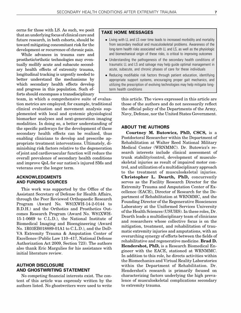
cerns for those with LS. As such, we positthatanunderlying focusof clinical careandfuture research, in both cohorts, should betowardmitigating concomitant risk for thedevelopment or recurrence of chronic pain.
While advances in trauma care andprosthetic/orthotic technologies may even-tually mollify acute and subacute second-ary health effects of extremity trauma,longitudinal tracking is urgently needed tobetter understand the mechanisms bywhich secondary health effects developand progress in this population. Such ef-forts should encompass a transdisciplinaryteam, in which a comprehensive suite of evalua-tion metrics are employed; for example, traditionalclinical evaluation and movement analysis sup-plemented with local and systemic physiologicalbiomarker analyses and next-generation imagingmodalities. In doing so, a better understanding ofthe specific pathways for the development of thesesecondary health effects can be realized, thusenabling clinicians to develop and prescribe ap-propriate treatment interventions. Ultimately, di-minishing risk factors relative to the degenerationof joint and cardiovascular function will reduce theoverall prevalence of secondary health conditionsand improve QoL for our nation’s injured SMs andveterans over the longer term.
ACKNOWLEDGMENTSAND FUNDING SOURCES
This work was supported by the Office of theAssistant Secretary of Defense for Health Affairs,through the Peer Reviewed Orthopaedic ResearchProgram (Award No. W81XWH-14-2-0144 toB.D.H.) and the Orthotics and Prosthetics Out-comes Research Program (Award No. W81XWH-15-1-0669 to C.L.D.), the National Institute ofBiomedical Imaging and Bioengineering (AwardNo. 1R03EB018889-01A1 to C.L.D.), and the DoD-VA Extremity Trauma & Amputation Center ofExcellence (Public Law 110–417, National DefenseAuthorization Act 2009, Section 723). The authorsalso thank Eric Margulies for his assistance withinitial literature review.
AUTHOR DISCLOSUREAND GHOSTWRITING STATEMENT
No competing financial interests exist. The con-tent of this article was expressly written by theauthors listed. No ghostwriters were used to write
this article. The views expressed in this article arethose of the authors and do not necessarily reflectthe official policy of the Departments of the Army,Navy, Defense, nor the United States Government.
ABOUT THE AUTHORS
Courtney M. Butowicz, PhD, CSCS, is aPostdoctoral Researcher within the Department ofRehabilitation at Walter Reed National MilitaryMedical Center (WRNMMC). Dr. Butowicz’s re-search interests include clinical assessment oftrunk stability/control, development of musculo-skeletal injuries as result of impaired motor con-trol, and utilization of amultidisciplinary approachto the treatment of musculoskeletal injuries.Christopher L. Dearth, PhD, concurrentlyserves as the Facility Research Director for theExtremity Trauma and Amputation Center of Ex-cellence (EACE), Director of Research for the De-partment of Rehabilitation at WRNMMC, and theFounding Director of the Regenerative BiosciencesLaboratory at the Uniformed Services Universityof theHealth Sciences (USUHS). In these roles, Dr.Dearth leads a multidisciplinary team of cliniciansand researchers whose collective focus is on themitigation, treatment, and rehabilitation of trau-matic extremity injuries and amputations, with anoverarching synergy of efforts between the fields ofrehabilitative and regenerative medicine.Brad D.Hendershot, PhD, is a Research Biomedical En-gineer with the EACE, stationed at WRNMMC.In addition to this role, he directs activities withinthe Biomechanics and Virtual Reality Laboratorieswithin the Department of Rehabilitation. Dr.Hendershot’s research is primarily focused oncharacterizing factors underlying the high preva-lence of musculoskeletal complications secondaryto extremity trauma.
TAKE HOME MESSAGES
� Living with LL and LS over time leads to increased morbidity and mortalityfrom secondary medical and musculoskeletal problems. Awareness of thelong-term health risks associated with LL and LS, as well as the physiologicand biomechanical origin of these risks, is critical to improving outcomes
� Understanding the pathogenesis of the secondary health conditions oftraumatic LL and LS and salvage may help guide optimal management inacute, subacute, and chronic phases of care for these individuals
� Reducing modifiable risk factors through patient education, identifyingappropriate support systems, encouraging proper gait mechanics, andutilizing the prescription of evolving technologies may help mitigate long-term health conditions
SECONDARY HEALTH CONDITIONS AFTER EXTREMITY TRAUMA 7
REFERENCES
1. Summers K, Jinnett K, Bevan S. MusculoskeletalDisorders, Workforce Health and Productivityin the United States. 2015. www.tcwhp.org/musculoskeletal-disorders-workforce-health-and-productivity-united-states-0 (last accessed Au-gust 15, 2016).
2. United States Bone and Joint Initiative. TheBurden of Musculoskeletal Diseases in theUnited States. Rosemont, IL: BMUS, 2014.
3. Agency for Healthcare Research and Quality.Medical Expenditures Panel Survey (MEPS).2008–2011. http://meps.ahrq.gov/mepsweb (lastaccessed August 25, 2016).
4. Ziegler-Graham K, MacKenzie EJ, Ephraim PL,Travison TG, Brookmeyer R. Estimating theprevalence of limb loss in the United States:2005 to 2050. Arch Phys Med Rehabil 2008;89:422–429.
5. Dillingham TR, Pezzin LE, Mackenzie EJ. Racialdifferences in the incidence of limb loss sec-ondary to peripheral vascular disease: apopulation-based study. Arch Phys Med Rehabil2002;83:1252–1257.
6. Wrobel JS, Mayfield JA, Reiber GE. Geographicvariation of lower-extremity major amputation inindividuals with and without diabetes in theMedicare population. Diabetes Care 2001;24:860–864.
7. Owings MF, Kozak LJ. Ambulatory and inpatientprocedures in the United States, 1996. VitalHealth Stat 13 1998;1–119.
8. Fischer H. A Guide to US Military CasualtyStatistics: Operation Freedom’s Sentinel, Op-eration Inherent Resolve, Operation New Dawn,Operation Iraqi Freedom, and Operation EnduringFreedom. Congressional Research Service Re-port; Research Report #RS22452; Washington,DC. 2015.
9. Doukas WC, Hayda RA, Frisch HM, et al. TheMilitary Extremity Trauma Amputation/LimbSalvage (METALS) study: outcomes of amputa-tion versus limb salvage following major lower-extremity trauma. J Bone Joint Surg Am 2013;95:138–145.
10. Hansen ST, Jr. The type-IIIC tibial fracture.Salvage or amputation. J Bone Joint Surg Am1987;69:799–800.
11. Hansen ST, Jr. Overview of the severely trau-matized lower limb. Reconstruction versus am-putation. Clin Orthop Relat Res 1989;17–19.
12. Dirschl DR, Dahners LE. The mangled extremity:when should it be amputated? J Am Acad Or-thop Surg 1996;4:182–190.
13. Lange RH. Limb reconstruction versus amputa-tion decision making in massive lower extremitytrauma. Clin Orthop Relat Res 1989;92–99.
14. MacKenzie EJ, Bosse MJ, Kellam JF, et al.Factors influencing the decision to amputate orreconstruct after high-energy lower extremitytrauma. J Trauma 2002;52:641–649.
15. Bosse MJ, Ficke JR, Andersen RC. Extremity warinjuries: current management and research pri-orities. J Am Acad Orthop Surg 2012;20(Suppl 1):viii–x.
16. Dagum AB, Best AK, Schemitsch EH, MahoneyJL, Mahomed MN, Blight KR. Salvage after severelower-extremity trauma: are the outcomes worth themeans? Plast Reconstr Surg 1999;103:1212–1220.
17. Reiber GE, McFarland LV, Hubbard S, et al.Service members and veterans with major trau-matic limb loss from Vietnam war and OIF/OEFconflicts: survey methods, participants, and sum-mary findings. J Rehabil Res Dev 2010;47:275–297.
18. Harris AM, Althausen PL, Kellam J, Bosse MJ,Castillo R; Lower Extremity Assessment ProjectStudy Group. Complications following limb-threatening lower extremity trauma. J OrthopTrauma 2009;23:1–6.
19. Busse JW, Jacobs CL, Swiontkowski MF, BosseMJ, Bhandari M; Evidence-Based OrthopaedicTrauma Working Group. Complex limb salvage orearly amputation for severe lower-limb injury: ameta-analysis of observational studies. J OrthopTrauma 2007;21:70–76.
20. Esquenazi A, DiGiacomo R. Rehabilitation afteramputation. J Am Podiatr Med Assoc 2001;91:13–22.
21. Edwards MD, Phillip LCRD, Bosanquet N, BullAM, Clasper CJC. What is the magnitude andlong-term economic cost of care of the Britishmilitary Afghanistan amputee cohort? Clin Or-thop Relat Res 2015;473:2848–2855.
22. Blough DK, Hubbard S, McFarland LV, Smith DG,Gambel JM, Reiber GE. Prosthetic cost projec-tions for service members with major limb lossfrom Vietnam and OIF/OEF. J Rehabil Res Dev2010;47:387–402.
23. Stern P. The epidemiology of amputations. PhysMed Rehabil Clin N Am 1991;2:253–261.
24. Pezzin LE, Dillingham TR, MacKenzie EJ. Re-habilitation and the long-term outcomes of per-sons with trauma-related amputations. ArchPhys Med Rehabil 2000;81:292–300.
25. Dillingham TR, Pezzin LE, MacKenzie EJ. Limbamputation and limb deficiency: epidemiologyand recent trends in the United States. SouthMed J 2002;95:875–884.
26. Ehrlich GE. Low back pain. Bull World HealthOrgan 2003;81:671–676.
27. Katz JN. Lumbar disc disorders and low-backpain: socioeconomic factors and consequences.J Bone Joint Surg Am 2006;88(Suppl 2):21–24.
28. Ehde DM, Smith DG, Czerniecki JM, CampbellKM, Malchow DM, Robinson LR. Back pain as asecondary disability in persons with lower limbamputations. Arch Phys Med Rehabil 2001;82:731–734.
29. Ephraim PL, Wegener ST, MacKenzie EJ, Dil-lingham TR, Pezzin LE. Phantom pain, residuallimb pain, and back pain in amputees: results of
a national survey. Arch Phys Med Rehabil2005;86:1910–1919.
30. Smith DG, Ehde DM, Legro MW, Reiber GE, delAguila M, Boone DA. Phantom limb, residuallimb, and back pain after lower extremity am-putations. Clin Orthop Relat Res 1999;29–38.
31. Foote CE, Mac Kinnon J, Robbins C, Pessagno R,Portner MD. Long-term health and quality of lifeexperiences of Vietnam veterans with combat-related limb loss. Qual Life Res 2015;24:2853–2861.
32. Taghipour H, Moharamzad Y, Mafi AR, et al.Quality of life among veterans with war-relatedunilateral lower extremity amputation: a long-term survey in a prosthesis center in Iran. JOrthop Trauma 2009;23:525–530.
33. Sagawa Y, Turcot K, Armand S, Thevenon A,Vuillerme N, Watelain E. Biomechanics andphysiological parameters during gait in lower-limb amputees: a systematic review. Gait Pos-ture 2011;33:511–526.
34. Esposito ER, Choi HS, Owens JG, Blanck RV,Wilken JM. Biomechanical response to ankle–foot orthosis stiffness during running. Clin Bio-mech (Bristol, Avon) 2015;30:1125–1132.
35. Kulkarni J, Gaine WJ, Buckley JG, Rankine JJ,Adams J. Chronic low back pain in traumaticlower limb amputees. Clin Rehabil 2005;19:81–86.
36. Gailey R, Allen K, Castles J, Kucharik J, RoederM. Review of secondary physical conditions as-sociated with lower-limb amputation and long-term prosthesis use. J Rehabil Res Dev 2008;45:15–29.
37. Hendershot BD, Nussbaum MA. Persons withlower-limb amputation have impaired trunkpostural control while maintaining seated bal-ance. Gait Posture 2013;38:438–442.
38. Devan H, Hendrick P, Ribeiro DC, Hale LA, Car-man A. Asymmetrical movements of the lumbo-pelvıc region: Is this a potential mechanism forlow back pain in people with lower limb ampu-tation? Med Hypotheses 2014;82:77–85.
39. Day JW, Smidt GL, Lehmann T. Effect of pelvictilt on standing posture. Phys Ther 1984;64:510–516.
40. Kumar S. Theories of musculoskeletal injurycausation. Ergonomics 2001;44:17–47.
41. McGill SM. Low Back Disorders: Evidence-basedPrevention and Rehabilitation, 2nd ed. Cham-paign, IL: Human Kinetics, 2007.
42. Hendershot BD, Wolf EJ. Three-dimensionaljoint reaction forces and moments at the lowback during over-ground walking in persons withunilateral lower-extremity amputation. Clin Bio-mech (Bristol, Avon) 2014;29:235–242.
43. Gillet C, Duboy J, Barbier F, et al. Contribution ofaccelerated body masses to able-bodied gait.Am J Phys Med Rehabil 2003;82:101–109.
44. Esposito ER, Wilken JM. The relationship be-tween pelvis–trunk coordination and low back
8 BUTOWICZ, DEARTH, AND HENDERSHOT
pain in individuals with transfemoral amputa-tions. Gait Posture 2014;40:640–646.
45. Schmidt H, Kettler A, Heuer F, Simon U, Claes L,Wilke HJ. Intradiscal pressure, shear strain, andfiber strain in the intervertebral disc undercombined loading. Spine (Phila Pa 1976) 2007;32:748–755.
46. Morgenroth DC, Orendurff MS, Shakir A, SegalA, Shofer J, Czerniecki JM. The relationshipbetween lumbar spine kinematics during gaitand low-back pain in transfemoral amputees.Am J Phys Med Rehabil 2010;89:635–643.
47. Burke M, Roman V, Wright V. Bone and jointchanges in lower limb amputees. Ann Rheum Dis1978;37:252–254.
48. Patzkowski JC, Blanck RV, Owens JG, et al.Comparative effect of orthosis design on func-tional performance. J Bone Joint Surg Am 2012;94:507–515.
49. Collins SH, Kuo AD. Recycling energy to restoreimpaired ankle function during human walking.PLoS One 2010;5:e9307.
50. Waters RL, Mulroy S. The energy expenditure ofnormal and pathologic gait. Gait Posture 1999;9:207–231.
51. National Institute of Arthritis and Musculoske-letal and Skin Diseases National Institute ofHealth. What is Osteoarthritis? 2014. www.niams.nih.gov (last accessed September 7, 2016).
52. Radin ER, Paul IL, Rose RM. Pathogenesis ofprimary osteoarthritis. Lancet 1972;1:1395–1396.
53. Sharma L, Felson DT. Studying how osteoar-thritis causes disability: nothing is simple. JRheumatol 1998;25:1–4.
54. Baliunas A, Hurwitz D, Ryals A, et al. Increasedknee joint loads during walking are present insubjects with knee osteoarthritis. OsteoarthritisCartilage 2002;10:573–579.
55. Miyazaki T, Wada M, Kawahara H, Sato M,Baba H, Shimada S. Dynamic load at baselinecan predict radiographic disease progression inmedial compartment knee osteoarthritis. AnnRheum Dis 2002;61:617–622.
56. Lynn SK, Reid SM, Costigan PA. The influence ofgait pattern on signs of knee osteoarthritis inolder adults over a 5–11 year follow-up period: acase study analysis. Knee 2007;14:22–28.
57. Struyf PA, van Heugten CM, Hitters MW,Smeets RJ. The prevalence of osteoarthritis ofthe intact hip and knee among traumatic legamputees. Arch Phys Med Rehabil 2009;90:440–446.
58. Kutzner I, Trepczynski A, Heller MO, BergmannG. Knee adduction moment and medial contactforce—facts about their correlation during gait.PLoS One 2013;8:e81036.
59. Astephen JL, Deluzio KJ, Caldwell GE, DunbarMJ. Biomechanical changes at the hip, knee,and ankle joints during gait are associated withknee osteoarthritis severity. J Orthop Res 2008;26:332–341.
60. Mundermann A, Dyrby CO, Andriacchi TP. Sec-ondary gait changes in patients with medialcompartment knee osteoarthritis: increased loadat the ankle, knee, and hip during walking. Ar-thritis Rheum 2005;52:2835–2844.
61. Morgenroth DC, Segal AD, Zelik KE, et al. Theeffect of prosthetic foot push-off on mechanicalloading associated with knee osteoarthritis inlower extremity amputees. Gait Posture 2011;34:502–507.
62. Lloyd CH, Stanhope SJ, Davis IS, Royer TD.Strength asymmetry and osteoarthritis risk fac-tors in unilateral trans-tibial, amputee gait. GaitPosture 2010;32:296–300.
63. Winter DA, Sienko SE. Biomechanics of below-knee amputee gait. J Biomech 1988;21:361–367.
64. Adamczyk PG, Kuo AD. Redirection of center-of-mass velocity during the step-to-step transitionof human walking. J Exp Biol 2009;212:2668–2678.
65. Grabowski AM, D’Andrea S. Effects of a pow-ered ankle-foot prosthesis on kinetic loading ofthe unaffected leg during level-ground walking.J Neuroeng Rehabil 2013;10:1.
66. Somers TJ, Keefe FJ, Godiwala N, Hoyler GH.Psychosocial factors and the pain experience ofosteoarthritis patients: new findings and newdirections. Curr Opin Rheumatol 2009;21:501–506.
67. Rosemann T, Laux G, Szecsenyi J. Osteoarthritis:quality of life, comorbidities, medication andhealth service utilization assessed in a largesample of primary care patients. J Othop SurgRes 2007;2:1.
68. Marks R. Comorbid depression and anxiety im-pact hip osteoarthritis disability. Disabil Health J2009;2:27–35.
69. Johnson A, Cross J. Impact of traumatic arthritison a cohort of combat casualties. Paper pre-sented at: American Academy of OrthpaedicSurgeons Annual Meeting, San Diego, CA, 2011.
70. Shawen SB, Keeling JJ, Branstetter J, Kirk KL,Ficke JR. The mangled foot and leg: salvageversus amputation. Foot Ankle Clin 2010;15:63–75.
71. Grogan BF, Hsu JR; Skeletal Trauma ResearchConsortium. Volumetric muscle loss. J Am AcadOrthop Surg 2011;19(Suppl 1):S35–S37.
72. Harper NG, Esposito ER, Wilken JM, NeptuneRR. The influence of ankle-foot orthosis stiffnesson walking performance in individuals withlower-limb impairments. Clin Biomech (Bristol,Avon) 2014;29:877–884.
73. Esposito ER, Wilken JM. Biomechanical riskfactors for knee osteoarthritis when using pas-sive and powered ankle–foot prostheses. ClinBiomech (Bristol, Avon) 2014;29:1186–1192.
74. Ranz EC, Russell Esposito E, Wilken JM, Nep-tune RR. The influence of passive-dynamicankle-foot orthosis bending axis location ongait performance in individuals with lower-limb
impairments. Clin Biomech (Bristol, Avon) 2016;37:13–21.
75. Zhang W, Moskowitz RW, Nuki G, et al. OARSIrecommendations for the management of hipand knee osteoarthritis, part I: critical appraisalof existing treatment guidelines and systematicreview of current research evidence. Osteoar-thritis Cartilage 2007;15:981–1000.
76. Gobbi A, Karnatzikos G, Mahajan V, Malchira S.Platelet-rich plasma treatment in symptomaticpatients with knee osteoarthritis: preliminaryresults in a group of active patients. SportsHealth 2012;4:162–172.
77. Kon E, Buda R, Filardo G, et al. Platelet-richplasma: intra-articular knee injections producedfavorable results on degenerative cartilage le-sions. Knee Surg Sports Traumatol Arthrosc2010;18:472–479.
78. Patel S, Dhillon MS, Aggarwal S, Marwaha N,Jain A. Treatment with platelet-rich plasma ismore effective than placebo for knee osteoar-thritis: a prospective, double-blind, randomizedtrial. Am J Sports Med 2013;41:356–364.
79. Spakova T, Rosocha J, Lacko M, Harvanova D,Gharaibeh A. Treatment of knee joint osteoar-thritis with autologous platelet-rich plasma incomparison with hyaluronic acid. Am J PhysMed Rehabil 2012;91:411–417.
80. Jansen MJ, Viechtbauer W, Lenssen AF, Hen-driks EJ, de Bie RA. Strength training alone,exercise therapy alone, and exercise therapywith passive manual mobilisation each reducepain and disability in people with knee osteo-arthritis: a systematic review. J Physiother 2011;57:11–20.
81. Kang JW, Lee MS, Posadzki P, Ernst E. T’ai chi forthe treatment of osteoarthritis: a systematic reviewand meta-analysis. BMJ Open 2011;1:e000035.
82. Felson DT, Lawrence RC, Dieppe PA, et al. Os-teoarthritis: new insights. Part 1: the diseaseand its risk factors. Ann Intern Med 2000;133:635–646.
83. Kerrigan DC, Lelas JL, Goggins J, Merriman GJ,Kaplan RJ, Felson DT. Effectiveness of a lateral-wedge insole on knee varus torque in patientswith knee osteoarthritis. Arch Phys Med Rehabil2002;83:889–893.
84. Jones A, Silva PG, Silva AC, et al. Impact of caneuse on pain, function, general health and energyexpenditure during gait in patients with kneeosteoarthritis: a randomised controlled trial. AnnRheum Dis 2012;71:172–179.
85. Raja K, Dewan N. Efficacy of knee braces andfoot orthoses in conservative management ofknee osteoarthritis: a systematic review. Am JPhys Med Rehabil 2011;90:247–262.
86. Mendis S, Puska P, Norrving B. Global Atlas onCardiovascular Disease Prevention and Control.Geneva: World Health Organization, 2011.
87. Naghavi M, Wang H, Lozano R, et al. Global,regional, and national age-sex specific all-causeand cause-specific mortality for 240 causes of
SECONDARY HEALTH CONDITIONS AFTER EXTREMITY TRAUMA 9
death, 1990–2013: a systematic analysis for theGlobal Burden of Disease Study 2013. Lancet2015;385:117–171.
88. Mozaffarian D, Benjamin EJ, Go AS, et al. Ex-ecutive summary: Heart Disease and StrokeStatistics-2016 Update: A report from theAmerican Heart Association. Circulation 2016;133:447.
89. Go AS, Mozaffarian D, Roger VL, et al. Heartdisease and stroke statistics-2014 update. Cir-culation 2014;129:e28–e292.
90. Hrubec Z, Ryder RA. Traumatic limb amputationsand subsequent mortality from cardiovasculardisease and other causes. J Chronic Dis 1980;33:239–250.
91. Modan M, Peles E, Halkin H, et al. Increasedcardiovascular disease mortality rates in trau-matic lower limb amputees. Am J Cardiol 1998;82:1242–1247.
92. Naschitz J, Lenger R. Why traumatic leg ampu-tees are at increased risk for cardiovasculardiseases. QJM 2008;101:251–259.
93. Yekutiel M, Brooks M, Ohry A, Yarom J, Carel R.The prevalence of hypertension, ischaemic heartdisease and diabetes in traumatic spinal cordinjured patients and amputees. Paraplegia 1989;27:58–62.
94. Vollmar J, Pauschinger P, Paes E, Henze E, FrieschA. Aortic aneurysms as late sequelae of above-knee amputation. Lancet 1989;334:834–835.
95. Paes E, Vollmar J, Pauschinger P, Mutschler W,Henze E, Friesch A. Late vascular damage afterunilateral leg amputation [in German]. Z Un-fallchir Versicherungsmed 1989;83:227–236.
96. Peles E, Akselrod S, Goldstein DS, et al. Insulinresistance and autonomic function in traumaticlower limb amputees. Clin Auton Res 1995;5:279–288.
97. Rose H, Schweitzer P, Charoenkul V, Schwartz E.Cardiovascular disease risk factors in combatveterans after traumatic leg amputations. ArchPhys Med Rehabil 1987;68:20–23.
98. Everson-Rose SA, Lewis TT. Psychosocial factorsand cardiovascular diseases. Annu Rev PublicHealth 2005;26:469–500.
99. Rosengren A, Hawken S, Ounpuu S, et al. As-sociation of psychosocial risk factors with risk ofacute myocardial infarction in 11 119 cases and13 648 controls from 52 countries (the INTER-HEART study): case-control study. Lancet2004;364:953–962.
100. Kang HK, Bullman TA, Taylor JW. Risk of selectedcardiovascular diseases and posttraumatic stressdisorder among former World War II prisoners ofwar. Ann Epidemiol 2006;16:381–386.
101. Frasure-Smith N, Lesperance F. Reflections ondepression as a cardiac risk factor. PsychosomMed 2005;67:S19–S25.
102. Julkunen J, Salonen R, Kaplan GA, Chesney MA,Salonen JT. Hostility and the progression of
carotid atherosclerosis. Psychosom Med 1994;56:519–525.
103. Sugawara J, Hayashi K, Tanaka H. Distal shift ofarterial pressure wave reflection sites with ag-ing. Hypertension 2010;56:920–925.
104. Yano M, Kohno M, Kobayashi S, et al. Influenceof timing and magnitude of arterial wave reflec-tion on left ventricular relaxation. Am J PhysiolHeart Circ Physiol 2001;280:H1846–H1852.
Abbreviations and Acronyms
AFO ¼ ankle–foot orthosesCVD ¼ cardiovascular disease
EACE ¼ Extremity Trauma and AmputationCenter of Excellence
EKAM ¼ external knee adduction momentLBP ¼ low back pain
LL ¼ limb lossLS ¼ limb salvage
OA ¼ osteoarthritisOEF ¼ Operation Enduring FreedomOIF ¼ Operation Iraqi Freedom
PRP ¼ platelet-rich plasmaQoL ¼ quality of lifeSM ¼ service member
USUHS ¼ Uniformed Services University ofthe Health Sciences
WRNMMC ¼ Walter Reed National MilitaryMedical Center
10 BUTOWICZ, DEARTH, AND HENDERSHOT
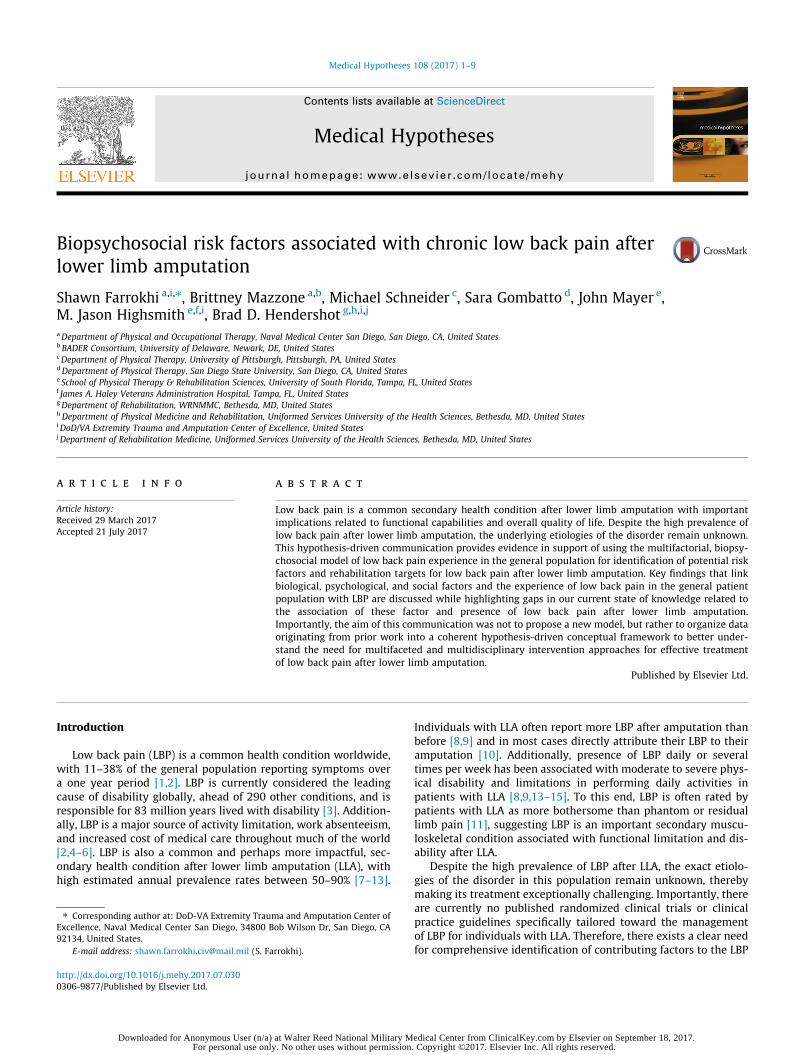
Medical Hypotheses 108 (2017) 1–9
Contents lists available at ScienceDirect
Medical Hypotheses
journal homepage: www.elsevier .com/locate /mehy
Biopsychosocial risk factors associated with chronic low back pain afterlower limb amputation
http://dx.doi.org/10.1016/j.mehy.2017.07.0300306-9877/Published by Elsevier Ltd.
⇑ Corresponding author at: DoD-VA Extremity Trauma and Amputation Center ofExcellence, Naval Medical Center San Diego, 34800 Bob Wilson Dr, San Diego, CA92134, United States.
E-mail address: [email protected] (S. Farrokhi).
Downloaded for Anonymous User (n/a) at Walter Reed National Military Medical Center from ClinicalKey.com by Elsevier on September 18, 2017.For personal use only. No other uses without permission. Copyright ©2017. Elsevier Inc. All rights reserved.
Shawn Farrokhi a,i,⇑, Brittney Mazzone a,b, Michael Schneider c, Sara Gombatto d, John Mayer e,M. Jason Highsmith e,f,i, Brad D. Hendershot g,h,i,j
aDepartment of Physical and Occupational Therapy, Naval Medical Center San Diego, San Diego, CA, United StatesbBADER Consortium, University of Delaware, Newark, DE, United StatescDepartment of Physical Therapy, University of Pittsburgh, Pittsburgh, PA, United StatesdDepartment of Physical Therapy, San Diego State University, San Diego, CA, United Statese School of Physical Therapy & Rehabilitation Sciences, University of South Florida, Tampa, FL, United Statesf James A. Haley Veterans Administration Hospital, Tampa, FL, United StatesgDepartment of Rehabilitation, WRNMMC, Bethesda, MD, United StateshDepartment of Physical Medicine and Rehabilitation, Uniformed Services University of the Health Sciences, Bethesda, MD, United StatesiDoD/VA Extremity Trauma and Amputation Center of Excellence, United StatesjDepartment of Rehabilitation Medicine, Uniformed Services University of the Health Sciences, Bethesda, MD, United States
a r t i c l e i n f o a b s t r a c t
Article history:Received 29 March 2017Accepted 21 July 2017
Low back pain is a common secondary health condition after lower limb amputation with importantimplications related to functional capabilities and overall quality of life. Despite the high prevalence oflow back pain after lower limb amputation, the underlying etiologies of the disorder remain unknown.This hypothesis-driven communication provides evidence in support of using the multifactorial, biopsy-chosocial model of low back pain experience in the general population for identification of potential riskfactors and rehabilitation targets for low back pain after lower limb amputation. Key findings that linkbiological, psychological, and social factors and the experience of low back pain in the general patientpopulation with LBP are discussed while highlighting gaps in our current state of knowledge related tothe association of these factor and presence of low back pain after lower limb amputation.Importantly, the aim of this communication was not to propose a new model, but rather to organize dataoriginating from prior work into a coherent hypothesis-driven conceptual framework to better under-stand the need for multifaceted and multidisciplinary intervention approaches for effective treatmentof low back pain after lower limb amputation.
Published by Elsevier Ltd.
Introduction
Low back pain (LBP) is a common health condition worldwide,with 11–38% of the general population reporting symptoms overa one year period [1,2]. LBP is currently considered the leadingcause of disability globally, ahead of 290 other conditions, and isresponsible for 83 million years lived with disability [3]. Addition-ally, LBP is a major source of activity limitation, work absenteeism,and increased cost of medical care throughout much of the world[2,4–6]. LBP is also a common and perhaps more impactful, sec-ondary health condition after lower limb amputation (LLA), withhigh estimated annual prevalence rates between 50–90% [7–13].
Individuals with LLA often report more LBP after amputation thanbefore [8,9] and in most cases directly attribute their LBP to theiramputation [10]. Additionally, presence of LBP daily or severaltimes per week has been associated with moderate to severe phys-ical disability and limitations in performing daily activities inpatients with LLA [8,9,13–15]. To this end, LBP is often rated bypatients with LLA as more bothersome than phantom or residuallimb pain [11], suggesting LBP is an important secondary muscu-loskeletal condition associated with functional limitation and dis-ability after LLA.
Despite the high prevalence of LBP after LLA, the exact etiolo-gies of the disorder in this population remain unknown, therebymaking its treatment exceptionally challenging. Importantly, thereare currently no published randomized clinical trials or clinicalpractice guidelines specifically tailored toward the managementof LBP for individuals with LLA. Therefore, there exists a clear needfor comprehensive identification of contributing factors to the LBP
2 S. Farrokhi et al. /Medical Hypotheses 108 (2017) 1–9
experience after LLA that can serve as a basis for the developmentof targeted rehabilitation strategies and future research investiga-tions. Here a new application of the multifactorial, biopsychosocialmodel for LBP, previously developed for the general population[16–18], is proposed as a way of identifying risk factors and poten-tial intervention targets for treatment of LBP after LLA. The objec-tive of this hypothesis-driven communication was to organize dataoriginating from prior studies of the biopsychosocial correlates ofLBP after LLA into a coherent conceptual framework. We hypothe-sized that alterations in biological, psychological, and social factorsare related to the development of LBP symptoms and disabilityafter LLA that merit specific attention during the clinical decisionmaking process and for future research efforts to improvepatient-related outcomes.
The biopsychosocial model of low back pain
Treatment of LBP has historically centered around the tradi-tional biomedical model of illness, which assumes a direct relation-ship between regional pathoanatomy and the perception of pain[18]. As such, it was expected that once the anatomical source ofLBP is identified, biochemical and/or mechanical treatments ofunderlying pathoanatomy would result in cessation of pain.Despite leading to successful treatment of many other disease pro-cesses, the outcomes of interventions based on the biomedicalmodel have proven to be less than ideal for treatment of LBP[18–20]. One potential reason for the failure of the biomedicalmodel to provide an effective treatment option for LBP is that nosingle underlying pathoanatomical lesion has been consistentlyidentified [18], with up to 85% of LBP patients left without a precisepathoanatomical diagnosis [21]. Additionally, determining thepathoanatomical sources of LBP frequently lacks interexaminerreliability and adequate evidence for generalizability [22]. Theoften equivocal outcomes from many ‘‘lesion-specific” treatmentoptions such as intra-articular corticosteroid injections [19] andspinal fusion surgeries [20], along with the generally poor predic-tive value of diagnostic imaging to identify pathoanatomicalsources of pain [23], have led to a recent paradigm shift toward a‘‘non-structural” approach for the management of LBP [24].
A growing body of evidence now suggests that successful treat-ment of LBP should include biological, psychological, and socialassessments to comprehensively address the patient’s unique painexperience [18]. The so called ‘‘biopsychosocial model” of LBP sug-gests that the patient’s perceptions and reactions to pain shouldalso be considered as these factors often lead to unnecessary avoid-ance of physical activity and social interactions, work absenteeism,and high health care utilization [16,17]. Whereas the pathoanat-omymay initiate the pain process, the psychological and social fac-tors appear to play an important role in exacerbating the biologicalcomponent of LBP by influencing the perception of pain [25]. Forexample, it has been hypothesized that the presence of mechanicalLBP can lead to a pain-generated stress response that could have anegative impact on the endocrine and immune systems, which inturn may negatively affect the cognitive assessment, emotionalresponse, coping strategies and health practices of the individual[26].
The hypothesis
Proponents of the biopsychosocial model argue that the com-plex, multidimensional nature of LBP does not lend itself to thereductionist view of the biomedical model; instead, the patient’sunique biologic, psychological, and social factors must equally beconsidered [18]. Therefore, the term biopsychosocial implies thatthe biological, psychological and social factors are interwoven
Downloaded for Anonymous User (n/a) at Walter Reed National Military MFor personal use only. No other uses without permissio
within the context of the patient’s overall LBP experience andshould be directly and concurrently considered as a part of a com-prehensive treatment program [26]. In support of this theory, mul-tidisciplinary treatment approaches that include biopsychosocialcomponents for treatment of LBP in adults have demonstrated pos-itive effects on pain, disability, and health-related quality of life[27,28]. It stands to reason that LLA likely amplifies and/or altersspecific components within the multifactorial biopsychosocialmodel of LBP, previously suggested for the general population.Given that LLA may differentially affect the various componentsof this model (Fig. 1), we hypothesize that discriminating clinicallymeaningful sub-groups of patients with LBP after LLA will mostlikely require assessments of biological, psychological and socialdomains [22].
Biological factors
BiomechanicsAltered mechanics of gait and movement have been historically
proposed to play a causative role in the development and/or recur-rence of LBP after LLA [29]. In fact, persons with LLA perceive ‘‘un-even postures and compensatory movements” affected by‘‘fatigue” and ‘‘prosthesis-related factors” during functional activi-ties as the primary contributors to LBP [30]. Though at the expenseof higher metabolic cost of transport [31], compensatory move-ment strategies adopted after LLA typically involve adaptationsto maintain the body’s center of mass within the base of support(i.e., improve stability and balance), primarily with a preferencefor the intact limb, if applicable [32]. During gait, for example,the intact limb (relative to prosthetic limb) is characterized by alonger stance time, shorter step length, wider stride width, and lar-ger vertical ground reaction forces [33]. As the trunk accounts forapproximately two-thirds of total body mass [34], altered motionsof this segment play a substantial role in post-amputation move-ment strategies, thereby warranting more trunk-focused biome-chanical investigations for assessing potential links with thedevelopment and persistence of LBP.
Altered trunk and pelvic movements in persons with LLA havebeen previously identified in all three cardinal planes, includinglarger forward trunk lean and flexion-extension range of motion,greater lateral trunk flexion (towards the prosthetic limb) and pel-vic obliquity motion, as well as more axial rotations between theshoulders/pelvis or regional/intervertebral motion segments[35,36]. The presence (and likely severity) of LBP further influencessuch trunk and pelvic movements [37]. For instance, it has beenreported that patients with transfemoral amputation and LBP ele-vate their pelvis on the intact side, minimize their lumbar lateralflexion, and keep their lumbar spine rotated toward the prostheticlimb throughout the gait cycle as compared to patients with trans-femoral amputation without LBP [38].
LBP has also been associated with more in-phase mediolateralcoordination between the trunk and pelvis [39], which is indicativeof inter-segmental rigidity (i.e., ‘‘guarding behavior”) previouslyreported in able-bodied individuals who are experiencing LBP[40,41]. Additional evidence suggests that individuals with LLAemploy an active mediolateral trunk movement strategy, inferredfrom increases in generation and absorption of energy betweenthe trunk and pelvis [42,43]. Although actively increasing medio-lateral trunk sway is likely an attempt to improve joint stabilitywithin the lower extremity by altering lever arms of ab/adductormusculature [44], most notably within the hip among patientswith transfemoral amputation [7], such strategies have been asso-ciated with LBP/discomfort among able-bodied individuals per-forming gait training aimed at reducing knee joint loads viatrunk lateral flexion [45].
edical Center from ClinicalKey.com by Elsevier on September 18, 2017.n. Copyright ©2017. Elsevier Inc. All rights reserved.
Fig. 1. Individual components (and their potential associations) of the biopsychosocial model of low back pain likely influenced or amplified by lower limb amputation. The *
symbol identifies the components of the model that are supported in the literature for the general low back pain patient population but lack validation by studies performedin patients with lower limb amputation and low back pain.
S. Farrokhi et al. /Medical Hypotheses 108 (2017) 1–9 3
Abnormal mechanics of the spinal column, primarily largermechanical loads and instability, are often considered risk factorsfor the development of LBP [46]. Of particular interest here, charac-teristics of trunk motion can directly influence musculoskeletalloading [47], typically due to altered muscular response (i.e., coac-tivity) [46]. Though walking is generally not a mechanicallydemanding task for the low back (i.e., loads are well belowreported injury thresholds) [48], and sometimes even consideredtherapeutic for individuals with LBP [49], altered trunk-pelvicmotions with LLA during gait have recently been associated withlarge internal loads among tissues within the spine [50–52]. Nota-bly, largest increases (up to �65% relative to uninjured individuals)were found in joint compressive forces owing to a complex patternof muscle responses [51]. Given the repetitive nature of gait, overtime, even minimal increases in trunk motions and musculoskele-tal loads may synergistically and progressively contribute towardLBP onset and/or recurrence and accelerate degenerative jointchanges in the spine. To date, however, altered spinal loads afterLLA has not been directly associated with development and/orpresence of LBP in published studies.
It is well accepted that the neuromuscular system plays a cen-tral role in supporting the upper body and maintaining mechanicalequilibrium and stability of the spine [53,54]. Irregular patterns oftrunk muscle recruitment have been identified among the generalpopulation with recurrent LBP[55], and impaired postural controlhas been associated with spinal instability and LBP [56]. Amongpersons with LLA without LBP, similar assessments have identifiedimpairments in trunk postural control during an unstable seatedbalance task [57], bilateral asymmetries in trunk mechanical andmuscular responses to applied positional perturbations [58], aswell as altered load-sharing between active and passive trunk tis-sues during quasi-static trunk flexion/extension movements [59].
Downloaded for Anonymous User (n/a) at Walter Reed National Military MedFor personal use only. No other uses without permission. C
Additionally, substantially greater fatigability has been reportedfor the low-back extensor musculature in patients with LLA withand without LBP [15], that are more pronounced than healthy indi-viduals with and without LBP [60]. Fatigue of the low back exten-sors may further contribute to increased intersegmental spinalmotion and instability during prolonged functional activities [61].Though the specific origin and functional impact of such alter-ations remain somewhat speculative, these data support the the-ory that repeated exposure to altered loading associated withLLA and repeated use of a prosthetic device may result in tissueand neuromuscular adaptations and increased risk for LBP in thispopulation.
Central nervous systemIn addition to changes in trunk/pelvis biomechanics with LLA,
central nervous system factors may also play an important biolog-ical role in the manifestation of LBP after LLA. Because of thetrauma to peripheral nerves, amputation has the potential to influ-ence the processing of pain signals in the peripheral and centralnervous systems. Phantom limb pain has been long described asthe perception of pain in the missing (amputated) limb [62,63],and may be indicative of altered pain signal processing. Althoughit is unknown how alterations in pain processing might influencethe incidence and prevalence of secondary musculoskeletal painproblems, such as LBP, there are several plausible explanations.
In the general population, people with LBP display generalizedhypersensitivity to pain that is reflective of central sensitization[64]. Central sensitization is the increased neuronal responsivenessto a stimulus due to prolonged or strong activity in the dorsal hornneurons that may be associated with an episode of pain or pro-longed pain [65]. It is plausible that the pain stimulus associatedwith limb amputation could elicit central sensitization and
ical Center from ClinicalKey.com by Elsevier on September 18, 2017.opyright ©2017. Elsevier Inc. All rights reserved.
4 S. Farrokhi et al. /Medical Hypotheses 108 (2017) 1–9
increased pain sensitivity, putting persons with amputation at riskfor developing secondary pain conditions. Pain sensitivity is typi-cally evaluated by assessing thresholds and tolerance to pain usinga variety of modalities for stimuli, including: mechanical (pres-sure), electrical, and thermal (cold/heat) [64].
Changes in pain sensitivity also can be measured after either aninhibitory stimulus (conditioned pain modulation), or a facilitorystimulus (temporal summation) to further elucidate central mech-anisms of pain inhibition or facilitation. Specific alterations in painprocessing that have been reported in people with LBP includelocal [66,67] and widespread [66–71] hyperalgesia and enhancedtemporal summation of pain signals [72–75]. Although peoplewith chronic pain conditions such as osteoarthritis, fibromyalgia,and chronic fatigue syndrome also typically display decreased inhi-bition of nociceptive signals (conditioned pain modulation) [76–78], most studies report that people with chronic LBP display nor-mal inhibition of pain signals [79–81].
Pain sensitivity has been examined to a limited extent in per-sons with limb amputation. In a small sample, Li et al. [82]reported that persons with traumatic amputation and phantomlimb pain displayed decreased thresholds for sensation and painwith electrical stimuli in the unaffected limb, suggesting centralsensitization. Further, Vase et al. [83] reported that people withupper limb amputation and phantom limb pain display decreasedthresholds for pressure and cold stimuli, and enhanced temporalsummation of pain signals. Inhibition of nociceptive signals hasnot been explicitly examined in person with amputation, but it isplausible that those with phantom limb pain may displaydecreased inhibition of pain similar to people with other chronicpain conditions. Although the mechanisms of altered pain process-ing are similar in persons with amputation and people with LBP, toour knowledge, no prior investigations have examined the neuro-physiology of pain in patients with amputation and secondarymusculoskeletal pain problems such as LBP to determine whethercentral sensitization places them at greater risk for secondary painconditions.
Alterations in pain-processing areas of the brain in persons withamputation are also consistent with changes reported in otherwiseuninjured individuals with LBP. For example, thalamic structuralvariations and, more specifically, decreases in gray matter of theposterolateral thalamus have been reported in people with ampu-tation [84]. These changes appear to be positively correlated withduration of time since amputation, suggesting that they may berelated to reduced afferent input [84]. Further, Lotze et al. [85]reported shifts in motor and sensory cortical activation patternsduring movement in patients with phantom limb pain comparedto pain-free persons with amputation, while Makin et al. [86]reported cortical reorganization of the sensorimotor cortex follow-ing arm amputation regardless of phantom limb pain. Collectively,these data suggest that neuroplastic changes associated withchronic pain in persons with amputation may involve cortical reor-ganization [85]. Similar alterations in brain morphology, includingreduced density of gray matter in the dorsolateral prefrontal cor-tex, the thalamus, and the middle cingulate cortex has beenreported in patients with LBP without amputation [87]. Althoughsimilar neuroplastic changes have been observed in some peoplewith limb amputation and in people with LBP [88], whether thesimilarities in mechanism might be related to the developmentof LBP in persons with LLA requires further investigation. Identify-ing the contribution of altered pain processing to LBP in patientswith amputation could inform the development of more targetedand individualized interventions.
Personal factorsThe link between personal demographics and LBP has been well
studied in the general population. Prevalence of LBP has been
Downloaded for Anonymous User (n/a) at Walter Reed National Military MFor personal use only. No other uses without permissio
reported to increase with age (up to 65), with onset typicallyoccurring in the third decade of life [89–93]. Race and ethnicityhave also been investigated and the data supports the observationthat Caucasians, Western Europeans and North Africans are morelikely to experience LBP than African Americans, Caribbeans andLatin Americans [89,94]. However, reports of gender prevalencefor LBP are vastly inconsistent [89–91,94,95]. Age, race, and genderhave also been studied in persons with LLA. Traumatic amputa-tions commonly occur in a younger population [96], with as manyas 63% of military service members with LLA being less than30 years of age [97]. Non-traumatic LLA secondary to variouspathological conditions such as diabetes mellitus and cancer aremore frequently seen in individuals greater than 60 years of age[96]. In a study of 255 patients with traumatic and non-traumatic LLA between the ages of 19–86, age was shown to bemodestly but significantly correlated (r = 0.12, p = 0.05) withwhether participants experienced LBP [98]. Distinct gender andrace features have also been reported in previous research withthe majority of patients with traumatic and non-traumatic LLAbeing male [99,100] and Caucasian [100,101]. However, whetherdemographic characteristics are associated with higher prevalenceof LBP experience after LLA remains unexamined.
Obesity has also been identified as a strong risk factor for LBP inthe general population [90,91,102]. In patients with LLA, obesityappears to be prevalent and dependent on the level of amputation,with 38% of persons with transtibial, 48% of persons with trans-femoral, and 64% of persons with bilateral amputation presentingwith noticeable clinical signs of obesity [103]. In support of thepotential link between obesity and LBP, patients with LLA andLBP appear to have body mass index ratios above 50% of the recom-mended ratio compared to their counterparts without LBP [13].The excess weight gain appears to be substantial and most com-mon within the first two years after LLA [104], which may beattributed to the sedentary lifestyle immediately after amputation[13,105] .
Maintaining a healthy weight is commonly a challenge forpatients with LLA due to difficulties associated with participatingin exercise and sports activities [10]. Given the previous reportsof increased risk of chronic LBP development as a result of inactiv-ity in the general population [91,93], the reported reductions inphysical activity levels after LLA [106,107] inherently increasethe risk of LBP in this patient population. While participation inrecreational or competitive sports has been reported in 32–60%of patients with LLA [97,108], there are fewer barriers in youngerindividuals who are more likely to achieve higher levels of physicalperformance due to accelerated rates of recovery and early fittingof running-specific prostheses [109,110]. Conversely, up to 46% ofolder persons with LLA become non-ambulatory one year post-amputation, which may place them at a higher risk of developingchronic LBP [111]. Although clinicians often attribute functionaldifficulties in this population to problems with the amputationand the prosthesis, LBP can also independently restrict activitylevels in patients with LLA and warrants further investigation[112,113].
A number of physical characteristics have also been identifiedas risk factors for non-specific LBP in the general population, suchas altered muscle strength/endurance, leg length discrepancy, orprevious history of LBP [93,114–118]. In persons with LLA, greateriliopsoas muscle length but reduced back extensor strength andendurance have been associated with the presence of LBP [15].Leg length discrepancy as a source of structural malalignment,including pelvic obliquity and functional scoliosis [119], has alsobeen suggested as a potential cause of LBP after LLA but with con-flicting supporting evidence. For example, in a study of 113 Finnishwar-disabled service members with amputation, those with unilat-eral LLA and LBP with mild and occasional symptoms had a mean
edical Center from ClinicalKey.com by Elsevier on September 18, 2017.n. Copyright ©2017. Elsevier Inc. All rights reserved.
S. Farrokhi et al. /Medical Hypotheses 108 (2017) 1–9 5
leg length discrepancy of 6.1 mm as compared to a 21.7 mm dis-crepancy for those who reported severe and constant symptoms[120]. In other studies, however, no correlations have beenreported between LBP and leg length discrepancy in persons withLLA [121,122]. Previous history of LBP in the general populationhas also been suggested to almost double the risk of future epi-sodes of LBP [117,118]. In patients with LLA and LBP, however, onlyless than 20% recall having LBP prior to their amputation [8,9], andin most cases directly attribute their LBP to their LLA [10].
Psychological factors
Beyond biologic factors, as an individual with LLA reintegrateswithin the community, additional psychological factors can affectthe risk for LBP and its eventual chronicity. Presence of psycholog-ical risk factors in the general LBP population are suggested toaffect the frequency and intensity of follow-up medical care andthe choice of interventions; whereas in their absence the patienthas enhanced potential for quick recovery [123]. Recent evidencefurther suggests that targeting psychological factors in patientswith LBP, particularly when they are at high levels, does seem tolead to more consistently positive results than either ignoringthem or providing omnibus interventions regardless of psycholog-ical risk factors [124]. In the general population, moderate tostrong associations have been reported between onset andchronicity of LBP with various psychological conditions such asdepression, pain catastrophizing, passive coping strategies, fear-avoidance beliefs and somatization [123–127]. However, the influ-ence of these psychological factors on the experience of LBP afterLLA has not been fully evaluated.
Depressive mood has been related to the onset of LBP, higherlevels of LBP intensity, poorer treatment outcome and transitionfrom acute to chronic LBP [128,129]. To this end, depressive moodhas also been reported as a significant predictor of the level of LBPintensity and bothersomeness in patients with LLA [12]. Given themuch higher rates of depression in patients with LLA as comparedto the general population [130,131], presence of depressive moodmay play an important role in the increased risk for chronic LBPin this patient population. There is also a growing recognition thatparticular kinds of coping mechanisms such as pain catastrophiz-ing (defined as the tendency to focus on, ruminate, and magnifypain sensations) are correlated with the transition from acute tochronic LBP and may be associated with poor treatment outcomesin the general population [129,132]. Prospective studies suggestthat passive coping strategies, especially high levels of pain catas-trophizing before an amputation, are associated with developmentand higher intensity of phantom limb pain and disability [133–136]. However, the extent to which passive coping strategies couldinfluence the LBP experience after LLA remains unknown.
Fear of movement or injury (kinesiophobia) is another impor-tant predictor of LBP development and chronicity that could leadto severe disability in the general population [137,138]. This fearof movement can impede the rehabilitation process and cause dys-functional pain-avoidance movement patterns that may lead to thedevelopment of secondary LBP after LLA. To this end, patients withhigher fear-avoidance scores are more likely to have worse out-comes at 3, 6, and 12 months [123]. Although, it stands to reasonthat patients with LLA may develop beliefs about their conditionthat may cause them to become fearful of moving and engagingin daily activities, evidence of kinesiophobia in patients with LLAand LBP has not been previously evaluated.
Similarly, somatization is another prevalent psychological con-dition in patients with LBP that includes increased reports of wide-spread muscle pain located along the whole spine as well as to thelegs and the head [139]. Somatization may also be related to pres-ence of sleep disorder, anxiety, and symptoms of depression [139].
Downloaded for Anonymous User (n/a) at Walter Reed National Military MedFor personal use only. No other uses without permission. C
Higher somatization scores have been previously correlated withhigher intensity of pain and grater disability, failure to return towork at 3 months and increased likelihood of a worse outcome at1 year in patients with LBP [123,140]. Evidence of somatizationhas also been previously reported in patients with traumatic LLAand neuropathic pain with the resulting abnormal sensory process-ing leading to locomotor dysfunction and body image disturbances[141]. However, a number of factors such as time since amputa-tion, time since first prosthesis, duration of daily prosthesis use,and high prosthesis satisfaction have shown to be negatively corre-lated with somatization [142]. Given the evidence suggesting thatpsychosocial factors can influence the outcome of rehabilitation,more research efforts are warranted for developing clinical toolsto identify when and how psychosocial factors could be utilizedin clinical decision making to improve outcomes in patients withLLA and LBP [143].
Alterations in central pain processing are also influenced bypsychosocial and cognitive factors such as pain catastrophizing,attention, stress, and expectation [65]. People with amputationhave been reported to display more depressive symptoms, greateranxiety, lower quality of life, and emotional disturbances [144].Further, neuropathic pain in persons with amputation has beenassociated with depression, post-traumatic stress disorder, andcatastrophizing [145]. It has also been reported that alterationsin pain sensitivity and temporal summation of pain, as well as cor-tical responses to painful stimuli, were modulated by pain catas-trophizing [83,146]. These psychosocial factors present in somepatients with amputation and neuropathic pain, have also beenassociated with chronic-recurrent LBP and alterations in pain pro-cessing [147,148]. Although no specific association was previouslyreported between presence of phantom limb pain and psychologi-cal symptoms in a small study [144], strong evidence in support ofthe relationship between presence of psychosocial risk factors,alterations in central processing of pain, and LBP in patients withLLA remains scant.
Social factors
The effects of social factors such as cigarette smoking, alcoholuse, marital status, occupation, and income on the experience ofLBP have been under extensive investigation in the general popu-lation. For example, findings from systematic reviews includingcross-sectional and longitudinal studies have revealed that bothcurrent and former smokers have a higher prevalence and inci-dence of LBP than ‘‘never smokers”, but the association is fairlymodest [149,150]. In military personnel with amputation, 21%report smoking cigarettes on a regular basis [151], while otherstudies have found that 37–48% of males with amputation are cur-rent cigarette smokers [101,111]. Although strong evidence linkingcigarette smoking and LBP after LLA is lacking; one small studyreported no difference in frequency of cigarette smoking betweenpersons with transfemoral amputations with and without LBP[121]. Alcohol consumption has also been found to be greater inthose with LBP in the general population [93]. In military personnelwith amputation, alcohol consumption and substance abuse, alongwith probable alcohol addiction is more prevalent than in theirnon-amputee counterparts [152]. However, research evidence insupport of the association between alcohol consumption and LBPafter LLA does not currently exist. Being married is another socialfactors associated with higher risk of developing LBP in the generalpopulation compared to those who are divorced or single [91].Although most reports indicate that the majority of individualswith LLA are married [97,151,153,154], marital status in at leastone cohort study was shown not to be associated with either theintensity or bothersomeness of LBP in patients with LLA [12].
ical Center from ClinicalKey.com by Elsevier on September 18, 2017.opyright ©2017. Elsevier Inc. All rights reserved.
6 S. Farrokhi et al. /Medical Hypotheses 108 (2017) 1–9
Individuals with occupations involving heavy lifting/pushing/pulling and driving have historically been identified to be moreprone to development of LBP in the general population[91,93,155–157]. As for the military population, predictors of LBPinclude jobs involving lifting and wearing body armor [158], witha higher incidence seen in construction workers, auto mechanics,and law enforcement personnel [159]. However, both militaryand non-military individuals with LLA often return to employmentin less physically demanding occupations [151,160,161], whichmay decrease their risk of developing occupation-related LBP.Lower socioeconomic class and lower levels of education have alsobeen found to correlate with LBP in the general population [91].Enlisted rank and service in the Navy, Army or Air Force have beenidentified as risk factors for LBP in a military sample [162]. Educa-tion at or below a high school level has been reported in 27–60% ofservice members with amputation [97,153] and 78% of those withamputations of dysvascular or diabetic aetiologies [101]. Of theservice members with (traumatic) amputation, 31% were juniorenlisted, 49% mid to senior enlisted, and 20% were officers [97].In a sample of individuals with dysvascular or diabetic amputa-tions, 44% reported an income of <$25,000, 37% between $25,001and $50,000, and 19% an income >$50,000 [101]. Further investiga-tions are needed to determine the potential relationships betweenoccupation, socioeconomic class, education level and salary withLBP after LLA.
Consequences of the hypothesis and discussion
In the United States, an estimated 185,000 persons undergolimb amputation each year as a result of dysvascular disease(54%), trauma (45%), or cancer (1%), with the projected total num-ber of people living with limb loss doubling to up to 3.6 million bythe year 2050 [163]. In general, most amputations are major LLA(excluding toes) with increasing prevalence rates due to dysvascu-lar diseases such as diabetes mellitus [163,164]. Despite the highprevalence of LBP after LLA, there currently exists a lack of under-standing to identify any definite pathologic processes or anatomicsources of pain. A growing body of evidence from studies of LBP inthe general population suggests that it is no longer appropriate tosubclassify LBP solely using a biomedical construct, and that a suc-cessful classification system must include biomedical, psychologi-cal, and social assessments [25]. Given the multifactorial nature ofLBP after LLA, a more comprehensive understanding of how ampu-tation influences these biopsychosocial risk factors will furtherallow effective stratification of care for LBP after LLA, wherepatients are screened and placed in interventions designed to tar-get their specific biopsychosocial risk profiles. The aim of this spe-cial communication was to integrate evidence originatingpredominantly from prospective studies on biopsychosocial corre-lates of LBP in the general patient population with LBP into acoherent hypothesis-driven model that could help generate newresearch questions and improve our understanding of the LBPexperience after LLA.
The proposed biopsychosocial model could be useful in identi-fying risk factors for early identification of patients at risk for LBPand testing the effectiveness of different approaches aimed atreducing chronic LBP-related disability after LLA. Currently, theresults from psychosocial interventions for LBP in the general pop-ulation consistently show only small to moderate effects[16,165,166]. However, a multidisciplinary approach thataddresses all three components of the biopsychosocial model ofLBP may provide a more appropriate solution aimed at the multi-faceted nature of the LBP experience after LLA [16]. A number ofprospective studies have shown that psychosocial factors influencehow patients respond to rehabilitative and surgical treatment, thus
Downloaded for Anonymous User (n/a) at Walter Reed National Military MFor personal use only. No other uses without permissio
indicating the interaction between physical and psychological fac-tors are important in determining the outcome of a given treat-ment for LBP after LLA [17].
Another potential approach would be to implement a stratifiedcare approach, where patients with LLA are screened for knownbiopsychosocial risk factors using reliable and valid tools, and thenreferred to interventions designed to target their specific problemand risk profile [16]. To this end, use of a stratified approach, by useof prognostic screening with matched clinical pathways has shownpromising results in management of LBP in primary care for thegeneral population [167]. However, the current challenge to imple-mentation of a stratified care approach is the identification anddevelopment of a validated risk factor profiles that could be usedas a clinical guide to stratify patients with LLA into streams of carethat optimize their chance of a good outcome for treatment of LBP.Given that some factors exert an influence on outcome regardlessof treatment, whereas some only influence response to specifictreatment [16], additional clarity is needed to determine whichpredictors of outcome are prognostic factors and which are poten-tial treatment effect modifiers to help guide best practice treat-ments and the prevention of disability [168]. Additional researchand insight are needed to determine more effective approachesto mitigate or manage LBP after LLA.
Disclaimer
The views expressed herein are those of the author(s) and donot necessarily reflect the official policy or position of the Depart-ment of the Navy, Department of Defense, or the United StatesGovernment.
Conflict of interest statement
The authors declare that they have no competing interests.
References
[1] Johannes CB, Le TK, Zhou X, Johnston JA, Dworkin RH. The prevalence ofchronic pain in United States adults: results of an Internet-based survey. JPain 2010;11:1230–9.
[2] Hoy D, Brooks P, Blyth F, Buchbinder R. The epidemiology of low back pain.Best Pract Res Clin Rheumatol 2010;24:769–81.
[3] Buchbinder R, Blyth FM, March LM, Brooks P, Woolf AD, Hoy DG. Placing theglobal burden of low back pain in context. Best Pract Res Clin Rheumatol2013;27:575–89.
[4] Hoy D, Bain C, Williams G, et al. A systematic review of the global prevalenceof low back pain. Arthritis Rheum 2012;64:2028–37.
[5] Hoy D, March L, Brooks P, et al. The global burden of low back pain: estimatesfrom the Global Burden of Disease 2010 study. Ann Rheumat Dis 2014. pp.annrheumdis-2013-204428.
[6] Ma VY, Chan L, Carruthers KJ. Incidence, prevalence, costs, and impact ondisability of common conditions requiring rehabilitation in the United States:stroke, spinal cord injury, traumatic brain injury, multiple sclerosis,osteoarthritis, rheumatoid arthritis, limb loss, and back pain. Arch PhysMed Rehabil 2014;95:986–95. e981.
[7] Kušljugic A, Kapidzic-Durakovic S, Kudumovic Z, Cickušic A. Chronic low backpain in individuals with lower-limb amputation. Bosn J Basic Med Sci2006;6:67–70.
[8] Ehde DM, Smith DG, Czerniecki JM, Campbell KM, Malchow DM, Robinson LR.Back pain as a secondary disability in persons with lower limb amputations.Arch Phys Med Rehabil 2001;82:731–4.
[9] Hammarlund CS, Carlström M, Melchior R, Persson BM. Prevalence of backpain, its effect on functional ability and health-related quality of life in lowerlimb amputees secondary to trauma or tumour: a comparison across threelevels of amputation. Prosthet Orthot Int 2011;35:97–105.
[10] Foote CE, Mac Kinnon J, Robbins C, Pessagno R, Portner MD. Long-term healthand quality of life experiences of Vietnam veterans with combat-related limbloss. Qual Life Res 2015;24:2853–61.
[11] Smith DG, Ehde DM, Legro MW, Reiber GE, del Aguila M, Boone DA. Phantomlimb, residual limb, and back pain after lower extremity amputations. ClinOrthop Relat Res 1999;361:29–38.
[12] Ephraim PL, Wegener ST, MacKenzie EJ, Dillingham TR, Pezzin LE. Phantompain, residual limb pain, and back pain in amputees: results of a nationalsurvey. Arch Phys Med Rehabil 2005;86:1910–9.
edical Center from ClinicalKey.com by Elsevier on September 18, 2017.n. Copyright ©2017. Elsevier Inc. All rights reserved.
S. Farrokhi et al. /Medical Hypotheses 108 (2017) 1–9 7
[13] Kulkarni J, Gaine W, Buckley J, Rankine J, Adams J. Chronic low back pain intraumatic lower limb amputees. Clin Rehabil 2005;19:81–6.
[14] Jensen MP, Smith DG, Ehde DM, Robinsin LR. Pain site and the effects ofamputation pain: further clarification of the meaning of mild, moderate, andsevere pain. Pain 2001;91:317–22.
[15] Friel K, Domholdt E, Smith DG. Physical and functional measures related tolow back pain in individuals with lower-limb amputation: an exploratorypilot study. J Rehabil Res Dev 2005;42:155–66.
[16] Pincus T, Kent P, Bronfort G, Loisel P, Pransky G, Hartvigsen J. Twenty-fiveyears with the biopsychosocial model of low back pain—is it time tocelebrate? A report from the twelfth international forum for primary careresearch on low back pain. Spine 2013;38:2118–23.
[17] Waddell G. Volvo award in clinical sciences: a new clinical model for thetreatment of low-back pain. Spine 1987;12(1987):632–44.
[18] Weiner BK. Spine update: the biopsychosocial model and spine care. Spine2008;33:219–23.
[19] Bogduk N. A narrative review of intra-articular corticosteroid injections forlow back pain. Pain Med 2005;6:287–96.
[20] Chou R, Baisden J, Carragee EJ, Resnick DK, Shaffer WO, Loeser JD. Surgery forlow back pain: a review of the evidence for an American Pain Society ClinicalPractice Guideline. Spine 2009;34:1094–109.
[21] Deyo RA, Weinstein JN. Low back pain. N Engl J Med 2001;344:363–70.[22] McCarthy CJ, Arnall FA, Strimpakos N, Freemont A, Oldham JA. The
biopsychosocial classification of non-specific low back pain: a systematicreview. Phys Therapy Rev 2004;9:17–30.
[23] Boden SD, Davis D, Dina T, Patronas N, Wiesel S. Abnormal magnetic-resonance scans of the lumbar spine in asymptomatic subjects. A prospectiveinvestigation. J Bone Joint Surg 1990;72:403–8.
[24] Langevin HM, Sherman KJ. Pathophysiological model for chronic low backpain integrating connective tissue and nervous system mechanisms. MedHypotheses 2007;68:74–80.
[25] McCarthy CJ, Arnall FA, Strimpakos N, Freemont A, Oldham JA. Thebiopsychosocial classification of non-specific low back pain: a systematicreview. Phys Ther Rev 2013.
[26] Truchon M. Determinants of chronic disability related to low back pain:towards an integrative biopsychosocial model. Disabil Rehabil2001;23:758–67.
[27] Hoffman BM, Papas RK, Chatkoff DK, Kerns RD. Meta-analysis ofpsychological interventions for chronic low back pain. Health Psychol2007;26:1.
[28] S. Kamper, A. Apeldoorn, A. Chiarotto, et al., Multidisciplinary biopsychosocialrehabilitation for chronic low back pain (Review), 2014.
[29] Gailey R. Review of secondary physical conditions associated with lower-limbamputation and long-term prosthesis use. J Rehabil Res Dev 2008;45:15.
[30] Devan H, Carman AB, Hendrick PA, Ribeiro DC, Hale LA. Perceptions of lowback pain in people with lower limb amputation: a focus group study. DisabilRehabil 2015;37:873–83.
[31] Mattes SJ, Martin PE, Royer TD. Walking symmetry and energy cost in personswith unilateral transtibial amputations: matching prosthetic and intact limbinertial properties. Arch Phys Med Rehabil 2000;81:561–8.
[32] Devan H, Hendrick P, Ribeiro DC, Hale LA, Carman A. Asymmetricalmovements of the lumbopelvic region: is this a potential mechanism forlow back pain in people with lower limb amputation? Med Hypotheses2014;82:77–85.
[33] Sagawa Y, Turcot K, Armand S, Thevenon A, Vuillerme N, Watelain E.Biomechanics and physiological parameters during gait in lower-limbamputees: a systematic review. Gait Posture 2011;33:511–26.
[34] Winter DA. Biomechanics and motor control of human movement. New York,NY: Wiley Interscience; 1990.
[35] Goujon-Pillet H, Sapin E, Fodé P, Lavaste F. Three-dimensional motions oftrunk and pelvis during transfemoral amputee gait. Arch Phys Med Rehabil2008;89:87–94.
[36] Jaegers SMHJ, Arendzen JH, de Jongh HJ. Prosthetic gait of unilateraltransfemoral amputees: a kinematic study. Arch Phys Med Rehabil1995;76:736–43.
[37] Morgenroth DC, Orendurff MS, Shakir A, Segal A, Shofer J, Czerniecki JM. Therelationship between lumbar spine kinematics during gait and low-back painin transfemoral amputees. Am J Phys Med Rehabil 2010;89:635–43.
[38] Devan H, Dillon MP, Carman AB, et al. Spinal and Pelvic kinematics during gaitin people with lower-limb amputation, with and without low back pain: anexploratory study. JPO: J Prosthetics Orthotics 2017;29:121–9.
[39] Russell Esposito E, Wilken JM. The relationship between pelvis-trunkcoordination and low back pain in individuals with transfemoralamputations. Gait Posture 2014;40:640–6.
[40] Seay JF, Van Emmerik RE, Hamill J. Low back pain status affects pelvis-trunkcoordination and variability during walking and running. Clin Biomech2011;26:572–8.
[41] Gombatto SP, Brock T, DeLork A, Jones G, Madden E, Rinere C. Lumbar spinekinematics during walking in people with and people without low back pain.Gait Posture 2015;42:539–44.
[42] Gaffney BM, Murray AM, Christiansen CL, Davidson BS. Identification of trunkand pelvis movement compensations in patients with transtibial amputationusing angular momentum separation. Gait Posture 2016;45:151–6.
[43] Hendershot BD, Wolf EJ. Mediolateral joint powers at the low back amongpersons with unilateral transfemoral amputation. Arch Phys Med Rehabil2015;96:154–7.
Downloaded for Anonymous User (n/a) at Walter Reed National Military MedFor personal use only. No other uses without permission. C
[44] Sjödahl C, Jarnlo GB, Söderberg B, Persson B. Pelvic motion in trans-femoralamputees in the frontal and transverse plane before and after special gait re-education. Prosthet Orthot Int 2003;27:227–37.
[45] Hunt MA, Simic M, Hinman RS, Bennell KL, Wrigley TV. Feasibility of a gaitretraining strategy for reducing knee joint loading: increased trunk leanguided by real-time biofeedback. J Biomech 2011;44:943–7.
[46] Kumar S. Theories of musculoskeletal injury causation. Ergonomics2001;44:17–47.
[47] Davis K, Marras W. The effects of motion on trunk biomechanics. ClinBiomech 2000;15:703–17.
[48] Callaghan JP, Patla AE, McGill SM. Low back three-dimensional joint forces,kinematics, and kinetics during walking. Clin Biomech 1999;14:203–16.
[49] Shnayderman I, Katz-Leurer M. An aerobic walking programme versusmuscle strengthening programme for chronic low back pain: a randomizedcontrolled trial. Clin Rehabil 2012. p. 0269215512453353.
[50] Hendershot BD, Wolf EJ. Three-dimensional joint reaction forces andmoments at the low back during over-ground walking in persons withunilateral lower-extremity amputation. Clin Biomech 2014;29:235–42.
[51] Shojaei I, Hendershot BD, Wolf EJ, Bazrgari B. Persons with unilateraltransfemoral amputation experience larger spinal loads during level-groundwalking compared to able-bodied individuals. Clin Biomech 2016;32:157–63.
[52] Yoder AJ, Petrella AJ, Silverman AK. Trunk–pelvis motion, joint loads, andmuscle forces during walking with a transtibial amputation. Gait Posture2015;41:757–62.
[53] Panjabi MM. The stabilizing system of the spine. Part I. Function, dysfunction,adaptation, and enhancement. J Spinal Disord Tech 1992;5:383–9.
[54] Panjabi MM. Clinical spinal instability and low back pain. J ElectromyogrKinesiol 2003;13:371–9.
[55] MacDonald D, Moseley GL, Hodges PW. Why do some patients keep hurtingtheir back? Evidence of ongoing back muscle dysfunction during remissionfrom recurrent back pain. Pain 2009;142:183–8.
[56] Radebold A, Cholewicki J, Polzhofer GK, Greene HS. Impaired postural controlof the lumbar spine is associated with delayed muscle response times inpatients with chronic idiopathic low back pain. Spine 2001;26:724–30.
[57] Hendershot BD, Nussbaum MA. Persons with lower-limb amputation haveimpaired trunk postural control while maintaining seated balance. GaitPosture 2013;38:438–42.
[58] Hendershot BD, Bazrgari B, Nussbaum MA. Persons with unilateral lower-limb amputation have altered and asymmetric trunk mechanical andneuromuscular behaviors estimated using multidirectional trunkperturbations. J Biomech 2013;46:1907–12.
[59] Hendershot BD, Nussbaum MA. Altered flexion-relaxation responses existduring asymmetric trunk flexion movements among persons with unilaterallower-limb amputation. J Electromyogr Kinesiol 2014;24:120–5.
[60] Demoulin C, Vanderthommen M, Duysens C, Crielaard J-M. Spinal muscleevaluation using the Sorensen test: a critical appraisal of the literature. JointBone Spine 2006;73:43–50.
[61] Panjabi MM. The stabilizing system of the spine. Part II. Neutral zone andinstability hypothesis. J Spinal Disord Tech 1992;5:390–7.
[62] Hsu E, Cohen SP. Postamputation pain: epidemiology, mechanisms, andtreatment. J Pain Res 2013;6:121–36.
[63] Ellis K. A review of amputation, phantom pain and nursing responsibilities. BrJ Nurs 2002;11:155–7. 160-153.
[64] Neziri AY, Curatolo M, Limacher A, et al. Ranking of parameters of painhypersensitivity according to their discriminative ability in chronic low backpain. Pain 2012;153:2083–91.
[65] Roussel NA, Nijs J, Meeus M, Mylius V, Fayt C, Oostendorp R. Centralsensitization and altered central pain processing in chronic low back pain:fact or myth? Clin J Pain 2013;29:625–38.
[66] Giesbrecht RJ, Battie MC. A comparison of pressure pain detection thresholdsin people with chronic low back pain and volunteers without pain. Phys Ther2005;85:1085–92.
[67] Laursen BS, Bajaj P, Olesen AS, Delmar C, Arendt-Nielsen L. Health relatedquality of life and quantitative pain measurement in females with chronicnon-malignant pain. Eur J Pain 2005;9:267–75.
[68] Clauw DJ, Williams D, Lauerman W, et al. Pain sensitivity as a correlate ofclinical status in individuals with chronic low back pain. Spine (Phila Pa 1976)1999;24:2035–41.
[69] Giesecke T, Gracely RH, Grant MA, et al. Evidence of augmented central painprocessing in idiopathic chronic low back pain. Arthritis Rheum2004;50:613–23.
[70] O’Neill S, Manniche C, Graven-Nielsen T, Arendt-Nielsen L. Generalized deep-tissue hyperalgesia in patients with chronic low-back pain. Eur J Pain2007;11:415–20.
[71] Derbyshire SW, Jones AK, Creed F, et al. Cerebral responses to noxiousthermal stimulation in chronic low back pain patients and normal controls.NeuroImage 2002;16:158–68.
[72] Peters ML, Schmidt AJ, Van den Hout MA. Chronic low back pain and thereaction to repeated acute pain stimulation. Pain 1989;39:69–76.
[73] Kleinbohl D, Holzl R, Moltner A, Rommel C, Weber C, Osswald PM.Psychophysical measures of sensitization to tonic heat discriminate chronicpain patients. Pain 1999;81:35–43.
[74] Flor H, Knost B, Birbaumer N. The role of operant conditioning in chronicpain: an experimental investigation. Pain 2002;95:111–8.
ical Center from ClinicalKey.com by Elsevier on September 18, 2017.opyright ©2017. Elsevier Inc. All rights reserved.
8 S. Farrokhi et al. /Medical Hypotheses 108 (2017) 1–9
[75] Diers M, Koeppe C, Diesch E, et al. Central processing of acute muscle pain inchronic low back pain patients: an EEG mapping study. J Clin Neurophysiol2007;24:76–83.
[76] Lautenbacher S, Rollman GB. Possible deficiencies of pain modulation infibromyalgia. Clin J Pain 1997;13:189–96.
[77] Meeus M, Nijs J, Van de Wauwer N, Toeback L, Truijen S. Diffuse noxiousinhibitory control is delayed in chronic fatigue syndrome: an experimentalstudy. Pain 2008;139:439–48.
[78] Kosek E, Ordeberg G. Lack of pressure pain modulation by heterotopicnoxious conditioning stimulation in patients with painful osteoarthritisbefore, but not following, surgical pain relief. Pain 2000;88:69–78.
[79] Owens MA, Bulls HW, Trost Z, et al. An examination of pain catastrophizingand endogenous pain modulatory processes in adults with chronic low backpain. Pain Med 2015.
[80] Mlekusch S, Neziri AY, Limacher A, Juni P, Arendt-Nielsen L, Curatolo M.Conditioned pain modulation in patients with acute and chronic low backpain. Clin J Pain 2016;32:116–21.
[81] Correa JB, Costa LO, de Oliveira NT, Sluka KA, Liebano RE. Central sensitizationand changes in conditioned pain modulation in people with chronicnonspecific low back pain: a case-control study. Exp Brain Res2015;233:2391–9.
[82] Li S, Melton DH, Li S. Tactile, thermal, and electrical thresholds in patientswith and without phantom limb pain after traumatic lower limb amputation.J Pain Res 2015;8:169–74.
[83] Vase L, Nikolajsen L, Christensen B, et al. Cognitive-emotional sensitizationcontributes to wind-up-like pain in phantom limb pain patients. Pain2011;152:157–62.
[84] Draganski B, Moser T, Lummel N, et al. Decrease of thalamic gray matterfollowing limb amputation. Neuroimage 2006;31:951–7.
[85] Lotze M, Flor H, Grodd W, Larbig W, Birbaumer N. Phantom movements andpain An fMRI study in upper limb amputees. Brain 2001;124:2268–77.
[86] Makin TR, Scholz J, Henderson Slater D, Johansen-Berg H, Tracey I.Reassessing cortical reorganization in the primary sensorimotor cortexfollowing arm amputation. Brain 2015;138:2140–6.
[87] Ivo R, Nicklas A, Dargel J, et al. Brain structural and psychometric alterationsin chronic low back pain. Eur Spine J 2013;22:1958–64.
[88] Flor H. The modification of cortical reorganization and chronic pain bysensory feedback. Appl Psychophysiol Biofeedback 2002;27:215–27.
[89] Andersson GB. Epidemiological features of chronic low-back pain. Lancet1999;354:581–5.
[90] Hoy D, Brooks P, Blyth F, Buchbinder R. The Epidemiology of low back pain.Best Pract Res Clin Rheumatol 2010;24:769–81.
[91] Manchikanti L. Epidemiology of low back pain. Pain Physician2000;3:167–92.
[92] Hoy D, Bain C, Williams G, et al. A systematic review of the global prevalenceof low back pain. Arthritis Rheum 2012;64:2028–37.
[93] Skovron ML. Epidemiology of low back pain. Baillieres Clin Rheumatol1992;6:559–73.
[94] Hoy D, March L, Brooks P, et al. The global burden of low back pain: estimatesfrom the Global Burden of Disease 2010 study. Ann Rheum Dis2014;73:968–74.
[95] Leboeuf-Yde C. Body weight and low back pain. A systematic literaturereview of 56 journal articles reporting on 65 epidemiologic studies. Spine(Phila Pa 1976) 2000;25:226–37.
[96] Esquenazi A. Amputation rehabilitation and prosthetic restoration. Fromsurgery to community reintegration. Disability Rehabil 2015;26:831–6.
[97] Doukas WC, Hayda RA, Frisch HM, et al. The Military Extremity TraumaAmputation/Limb Salvage (METALS) study: outcomes of amputation versuslimb salvage following major lower-extremity trauma. J Bone Joint Surg Am2013;95:138–45.
[98] Ehde DM, Smith DG, Czerniecki JM, Campbell KM, Malchow DM, Robinson LR.Back pain as a secondary disability in persons with lower limb amputations.Arch Phys Med Rehabil 2001;82:731–4.
[99] Davie-Smith F, Paul L, Nicholls N, Stuart W, Kennon B. The impact of gender,level of amputation and diabetes on prosthetic fit rates following major lowerextremity amputation. Prosthet Orthot Int 2016.
[100] Frisch HM, Andersen CRC, Mazurek CMT, et al. The military extremity traumaamputation/limb salvage (METALS) study. J Bone Joint Surg Am2013;95:138–45.
[101] Webster JB, Hakimi KN, Williams RM, Turner AP, Norvell DC, Czerniecki JM.Prosthetic fitting, use, and satisfaction following lower-limb amputation: aprospective study. J Rehabil Res Dev 2012;49:1493–504.
[102] Shiri R, Karppinen J, Leino-Arjas P, Solovieva S, Viikari-Juntura E. Theassociation between obesity and low back pain: a meta-analysis. Am JEpidemiol 2010;171:135–54.
[103] Kurdibaylo SF. Obesity and metabolic disorders in adults with lower limbamputation. J Rehabil Res Dev 1996;33:387.
[104] Littman AJ, Thompson ML, Arterburn DE, et al. Lower-limb amputation andbody weight changes in men. J Rehabil Res Dev 2015;52:159.
[105] Kurdibaylo SF. Obesity and metabolic disorders in adults with lower limbamputation. J Rehabil Res Dev 1996;33:387–94.
[106] Bussmann JB, Schrauwen HJ, Stam HJ. Daily physical activity and heart rateresponse in people with a unilateral traumatic transtibial amputation. ArchPhys Med Rehabil 2008;89:430–4.
Downloaded for Anonymous User (n/a) at Walter Reed National Military MFor personal use only. No other uses without permissio
[107] Bussmann JB, Schrauwen HJ, Stam HJ. Daily physical activity and heart rateresponse in people with a unilateral traumatic transtibial amputation. ArchPhys Med Rehabil 2008;89:430–4.
[108] Kars C, Hofman M, Geertzen JH, Pepping GJ, Dekker R. Participation in sportsby lower limb amputees in the Province of Drenthe, The Netherlands.Prosthet Orthot Int 2009;33:356–67.
[109] Bragaru M, Dekker R, Geertzen J, Dijkstra P. Amputees and sports a systematicreview. Sports Med 2011;41:721–40.
[110] Pepper M, Willick S. Maximizing physical activity in athletes withamputations. Curr Sports Med Rep 2009;8:339–44.
[111] Nehler MR, Coll JR, Hiatt WR, et al. Functional outcome in a contemporaryseries of major lower extremity amputations. J Vasc Surg 2003;38:7–14.
[112] Devan H, Tumilty S, Smith C. Physical activity and lower-back pain in personswith traumatic transfemoral amputation: a national cross-sectional survey. JRehabil Res Dev 2012;49:1457.
[113] Friel K, Domholdt E, Smith DG. Physical and functional measures related tolow back pain in individuals with lower-limb amputation: an exploratorypilot study. J Rehabil Res Dev 2005;42:155.
[114] Biering-Sørensen F. Physical measurements as risk indicators for low-backtrouble over a one-year period. Spine 1984;9:106–19.
[115] Kujala UM, Salminen JJ, Taimela S, Oksanen A, Jaakkola L. Subjectcharacteristics and low back pain in young athletes and nonathletes. MedSci Sports Exerc 1992;24:627–32.
[116] Nourbakhsh MR, Arab AM. Relationship between mechanical factors andincidence of low back pain. J Orthop Sport Phys 2002;32:447–60.
[117] Papageorgiou AC, Croft PR, Thomas E, Ferry S, Jayson M, Silman AJ. Influenceof previous pain experience on the episode incidence of low back pain:results from the South Manchester Back Pain Study. Pain 1996;66:181–5.
[118] Hestbaek L, Leboeuf-Yde C, Manniche C. Low back pain: what is the long-term course? A review of studies of general patient populations. Eur Spine J2003;12:149–65.
[119] McCaw ST, Bates BT. Biomechanical implications of mild leg lengthinequality. Br J Sports Med 1991;25:10–3.
[120] Friberg O. Biomechanical significance of the correct length of lower limbprostheses: a clinical and radiological study. Prosthet Orthot Int1984;8:124–9.
[121] Morgenroth DC, Shakir A, Orendurff MS, Czerniecki JM. Low-back pain intransfemoral amputees: is there a correlation with static or dynamic leg-length discrepancy? Am J Phys Med Rehabil 2009;88:108–13.
[122] Kulkarni J, GaineWJ, Buckley JG, Rankine JJ, Adams J. Chronic low back pain intraumatic lower limb amputees. Clin Rehabil 2005;19:81–6.
[123] Chou R, Shekelle P. Will this patient develop persistent disabling low backpain? JAMA 2010;303:1295–302.
[124] Nicholas MK, Linton SJ, Watson PJ, Main CJ. Early identification andmanagement of psychological risk factors (‘‘yellow flags”) in patients withlow back pain: a reappraisal. Phys Ther 2011.
[125] Pincus T, Burton AK, Vogel S, Field AP. A systematic review of psychologicalfactors as predictors of chronicity/disability in prospective cohorts of lowback pain. Spine 2002;27:E109–20.
[126] Hoogendoorn WE, van Poppel MN, Bongers PM, Koes BW, Bouter LM.Systematic review of psychosocial factors at work and private life as riskfactors for back pain. Spine 2000;25:2114–25.
[127] Ramond A, Bouton C, Richard I, et al. Psychosocial risk factors for chronic lowback pain in primary care—a systematic review. Family Pract 2010. p.cmq072.
[128] Casey CY, Greenberg MA, Nicassio PM, Harpin RE, Hubbard D. Transition fromacute to chronic pain and disability: a model including cognitive, affective,and trauma factors. Pain 2008;134:69–79.
[129] S.J. Linton, A review of psychological risk factors in back and neck pain,SPINE-PHILADELPHIA-HARPER AND ROW PUBLISHERS THEN JB LIPPINCOTTCOMPANY- 25 (2000), pp. 1148–1156.
[130] Cansever A. Depression in men with traumatic lower part amputation: acomparison to men with surgical lower part amputation. Mil Med2003;168:106.
[131] Singh R, Ripley D, Pentland B, et al. Depression and anxiety symptoms afterlower limb amputation: the rise and fall. Clin Rehabil 2009;23:281–6.
[132] Wertli MM, Rasmussen-Barr E, Held U, Weiser S, Bachmann LM, Brunner F.Fear-avoidance beliefs—a moderator of treatment efficacy in patients withlow back pain: a systematic review. Spine J 2014;14:2658–78.
[133] Whyte A, Carroll L. The relationship between catastrophizing and disability inamputees experiencing phantom pain. Disabil Rehabil 2004;26:649–54.
[134] Hanley MA, Jensen MP, Ehde DM, Hoffman AJ, Patterson DR, Robinson LR.Psychosocial predictors of long-term adjustment to lower-limb amputationand phantom limb pain. Disabil Rehabil 2004;26:882–93.
[135] Hill A, Niven C, Knussen C. The role of coping in adjustment to phantom limbpain. Pain 1995;62:79–86.
[136] Richardson C, Glenn S, Horgan M, Nurmikko T. A prospective study of factorsassociated with the presence of phantom limb pain six months after majorlower limb amputation in patients with peripheral vascular disease. J Pain2007;8:793–801.
[137] Picavet HSJ, Vlaeyen JW, Schouten JS. Pain catastrophizing andkinesiophobia: predictors of chronic low back pain. Am J Epidemiol2002;156:1028–34.
[138] Swinkels-Meewisse IE, Roelofs J, Schouten EG, Verbeek AL, Oostendorp RA,Vlaeyen JW. Fear of movement/(re) injury predicting chronic disabling lowback pain: a prospective inception cohort study. Spine 2006;31:658–64.
edical Center from ClinicalKey.com by Elsevier on September 18, 2017.n. Copyright ©2017. Elsevier Inc. All rights reserved.
S. Farrokhi et al. /Medical Hypotheses 108 (2017) 1–9 9
[139] Hagen EM, Svensen E, Eriksen HR, Ihlebæk CM, Ursin H. Comorbid subjectivehealth complaints in low back pain. Spine 2006;31:1491–5.
[140] Licciardone JC, Gatchel RJ, Kearns CM, Minotti DE. Depression, somatization,and somatic dysfunction in patients with nonspecific chronic low back pain:results from the OSTEOPATHIC Trial. J Am Osteopathic Assoc2012;112:783–91.
[141] Durmus� D, Safaz _I, Adıgüzel E, et al. Psychiatric symptoms in male traumaticlower limb amputees: associations with neuropathic pain, locomotorcapabilities, and perception of body image. J Mood Disord 2015;5:164–72.
[142] Durmus D, Safaz I, Adıgüzel E, et al. The relationship between prosthesis use,phantom pain and psychiatric symptoms in male traumatic limb amputees.Compr Psychiatry 2015;59:45–53.
[143] Hill JC, Fritz JM. Psychosocial influences on low back pain, disability, andresponse to treatment. Phys Ther 2011;91:712–21.
[144] Durmus D, Safaz I, Adiguzel E, et al. The relationship between prosthesis use,phantom pain and psychiatric symptoms in male traumatic limb amputees.Compr Psychiatry 2015;59:45–53.
[145] Buchheit T, Van de Ven T, John Hsia HL, et al. Pain phenotypes and associatedclinical risk factors following traumatic amputation: results from veteransintegrated pain evaluation research (VIPER). Pain Med 2015.
[146] Vase L, Egsgaard LL, Nikolajsen L, Svensson P, Jensen TS, Arendt-Nielsen L.Pain catastrophizing and cortical responses in amputees with varying levelsof phantom limb pain: a high-density EEG brain-mapping study. Exp BrainRes 2012;218:407–17.
[147] George SZ, Wittmer VT, Fillingim RB, Robinson ME. Fear-avoidance beliefsand temporal summation of evoked thermal pain influence self-report ofdisability in patients with chronic low back pain. J Occup Rehabil2006;16:95–108.
[148] George SZ, Wittmer VT, Fillingim RB, Robinson ME. Sex and pain-relatedpsychological variables are associated with thermal pain sensitivity forpatients with chronic low back pain. J Pain 2007;8:2–10.
[149] Shiri R, Karppinen J, Leino-Arjas P, Solovieva S, Viikari-Juntura E. Theassociation between smoking and low back pain: a meta-analysis. Am JMed 2010;123:87. e87-87. e35.
[150] da Costa BR, Vieira ER. Risk factors for work-related musculoskeletaldisorders: a systematic review of recent longitudinal studies. Am J Ind Med2010;53:285–323.
[151] Ebrahimzadeh MH, Hariri S. Long-term outcomes of unilateral transtibialamputations. Mil Med 2009;174:593–7.
[152] Gunawardena N, Senevirathne Rde A, Athauda T. Mental health outcome ofunilateral lower limb amputee soldiers in two districts of Sri Lanka. Int J SocPsychiatry 2007;53:135–47.
[153] Gunawardena NS, Seneviratne Rde A, Athauda T. Functional outcomes ofunilateral lower limb amputee soldiers in two districts of Sri Lanka. Mil Med2006;171:283–7.
Downloaded for Anonymous User (n/a) at Walter Reed National Military MedFor personal use only. No other uses without permission. C
[154] Miller WC, Speechley M, Deathe B. The prevalence and risk factors of fallingand fear of falling among lower extremity amputees. Arch Phys Med Rehabil2001;82:1031–7.
[155] Macfarlane GJ, Thomas E, Papageorgiou AC, Croft PR, Jayson MIV, Silman AJ.Employment and physical work activities as predictors of future low backpain. Spine 1997;22:1143–9.
[156] Videman T, Nurminen M, Troup JD. 1990 Volvo Award in clinical sciences.Lumbar spinal pathology in cadaveric material in relation to history of backpain, occupation, and physical loading. Spine (Phila Pa 1976)1990;15:728–40.
[157] Frymoyer JW, Pope MH, Costanza MC, Rosen JC, Goggin JE, Wilder DG.Epidemiologic studies of low-back pain. Spine (Phila Pa 1976)1980;5:419–23.
[158] Roy TC, Lopez HP. A comparison of deployed occupational tasks performed bydifferent types of military battalions and resulting low back pain. Mil Med2013;178:e937–943.
[159] MacGregor AN, Dougherty AL, Mayo JA, Rauh MJ, Galarneua MR.Occupational correlates of low back pain among U.S. Marines followingcombat deployment. Mil Med 2012;177:845–9.
[160] Schoppen T, Boonstra A, Groothoff JW, van Sonderen E, Goeken LN, EismaWH. Factors related to successful job reintegration of people with a lowerlimb amputation. Arch Phys Med Rehabil 2001;82:1425–31.
[161] Millstein S, Bain D, Hunter GA. A review of employment patterns of industrialamputees–factors influencing rehabilitation. Prosthet Orthot Int1985;9:69–78.
[162] Knox J, Orchowski J, Scher DL, Owens BD, Burks R, Belmont PJ. The incidenceof low back pain in active duty United States military service members. Spine(Phila Pa 1976) 2011;36:1492–500.
[163] Ziegler-Graham K, MacKenzie EJ, Ephraim PL, Travison TG, Brookmeyer R.Estimating the prevalence of limb loss in the United States: 2005 to 2050.Arch Phys Med Rehabil 2008;89:422–9.
[164] Ephraim PL, Dillingham TR, Sector M, Pezzin LE, MacKenzie EJ. Epidemiologyof limb loss and congenital limb deficiency: a review of the literature. ArchPhys Med Rehabil 2003;84:747–61.
[165] Williams A, Eccleston C, Morley S. Psychological therapies for themanagement of chronic pain (excluding headache) in adults. CochraneDatabase Syst Rev 2012;11.
[166] Eccleston C, Morley S, Williams ADC. Psychological approaches to chronicpain management: evidence and challenges. Br J Anaesth 2013;111:59–63.
[167] Hill JC, Whitehurst DG, Lewis M, et al. Comparison of stratified primary caremanagement for low back pain with current best practice (STarT Back): arandomised controlled trial. Lancet 2011;378:1560–71.
[168] Hancock M, Herbert RD, Maher CG. A guide to interpretation of studiesinvestigating subgroups of responders to physical therapy interventions.Phys Ther 2009;89:698–704.
ical Center from ClinicalKey.com by Elsevier on September 18, 2017.opyright ©2017. Elsevier Inc. All rights reserved.
Journal of Biomechanics xxx (2017) xxx–xxx
Contents lists available at ScienceDirect
Journal of Biomechanicsjournal homepage: www.elsevier .com/locate / jb iomech
www.JBiomech.com
Short communication
A model-based approach for estimation of changes in lumbar segmentalkinematics associated with alterations in trunk muscle forces
https://doi.org/10.1016/j.jbiomech.2017.09.0240021-9290/� 2017 Elsevier Ltd. All rights reserved.
⇑ Corresponding author at: F. Joseph Halcomb III, M.D. Department of BiomedicalEngineering, University of Kentucky, 514E Robotic and Manufacturing Building,Lexington, KY 40506, USA.
E-mail address: [email protected] (B. Bazrgari).
Please cite this article in press as: Shojaei, I., et al. A model-based approach for estimation of changes in lumbar segmental kinematics associatealterations in trunk muscle forces. J. Biomech. (2017), https://doi.org/10.1016/j.jbiomech.2017.09.024
Iman Shojaei a, Navid Arjmand b, Judith R. Meakin c, Babak Bazrgari a,⇑a F. Joseph Halcomb III, M.D. Department of Biomedical Engineering, University of Kentucky, Lexington, KY 40506, USAbDepartment of Mechanical Engineering, Sharif University of Technology, Tehran, Iranc School of Physics and Astronomy, University of Exeter, Physics Building, Stocker Road, Exeter EX4 4QL, UK
a r t i c l e i n f o a b s t r a c t
Article history:Accepted 25 September 2017Available online xxxx
Keywords:Trunk muscle forcesFinite element analysisOptimization proceduresTrunk neuromuscular strategiesLumbar segmental kinematicsImage-based modeling
The kinematics information from imaging, if combined with optimization-based biomechanical models,may provide a unique platform for personalized assessment of trunk muscle forces (TMFs). Such amethod, however, is feasible only if differences in lumbar spine kinematics due to differences in TMFscan be captured by the current imaging techniques. A finite element model of the spine within an opti-mization procedure was used to estimate segmental kinematics of lumbar spine associated with five dif-ferent sets of TMFs. Each set of TMFs was associated with a hypothetical trunk neuromuscular strategythat optimized one aspect of lower back biomechanics. For each set of TMFs, the segmental kinematicsof lumbar spine was estimated for a single static trunk flexed posture involving, respectively, 40� and10� of thoracic and pelvic rotations. Minimum changes in the angular and translational deformationsof a motion segment with alterations in TMFs ranged from 0� to 0.7� and 0 mm to 0.04 mm, respectively.Maximum changes in the angular and translational deformations of a motion segment with alterations inTMFs ranged from 2.4� to 7.6� and 0.11 mm to 0.39 mm, respectively. The differences in kinematics oflumbar segments between each combination of two sets of TMFs in 97% of cases for angular deformationand 55% of cases for translational deformation were within the reported accuracy of current imagingtechniques. Therefore, it might be possible to use image-based kinematics of lumbar segments along withcomputational modeling for personalized assessment of TMFs.
� 2017 Elsevier Ltd. All rights reserved.
1. Introduction
Neuromuscular control of spinal equilibrium and stabilitychanges in the presence of pain or following exposure to knownrisk factors for low back pain (LBP) (Muslim et al., 2013;Radebold et al., 2000, 2001; Toosizadeh et al., 2013). Such alter-ations may cause deformations and/or forces in lower back tissuessuch that exceed injury/pain thresholds instantaneously or cumu-latively (Adams et al., 2013; Coenen et al., 2014; Marras et al.,2001; Panjabi, 1992a,b). Despite such a significant role, the currentmethods for personalized assessment of trunk muscle forces(TMFs) are limited. Kinematic measures of lumbo-pelvic coordina-tion, though capable of distinguishing patients with LBP from con-trols (Vazirian et al., 2016), do not provide much information aboutindividual muscle forces. Specifically, neuromuscular redundancy
in control of lumbo-pelvic motion as well as individual variabilityin mechanical behavior of passive lumbar tissues hinder relatingmeasured kinematics data to TMFs. The commonly used surfaceelectromyography (EMG)-based methods for the assessment ofTMFs, on the other hand, can only provide information about theactivity of superficial trunk muscles. Further, the relationshipbetween EMG measures of muscle activity and actual muscle forceis still unclear (Staudenmann et al., 2010). Finite element andmulti-joint biomechanical models of the spine with detailed mus-culature have also been developed and used for general assessmentof TMFs (Arjmand and Shirazi-Adl, 2006a,b; Dreischarf et al., 2014;Ezquerro et al., 2004; Hughes, 2000; Stokes and Gardner-Morse,2001). These models often implemented optimization proceduresto estimate TMFs (Arjmand and Shirazi-Adl, 2006b; Daniel, 2011;Hughes, 2000; Stokes and Gardner-Morse, 2001) and are not suit-able for personalized assessment of TMFs due to assumptionsmade related to lumbar segmental rotations and the requirementfor a priori knowledge of trunk neuromuscular strategy (e.g., astrategy that minimizes stress in muscles).
d with
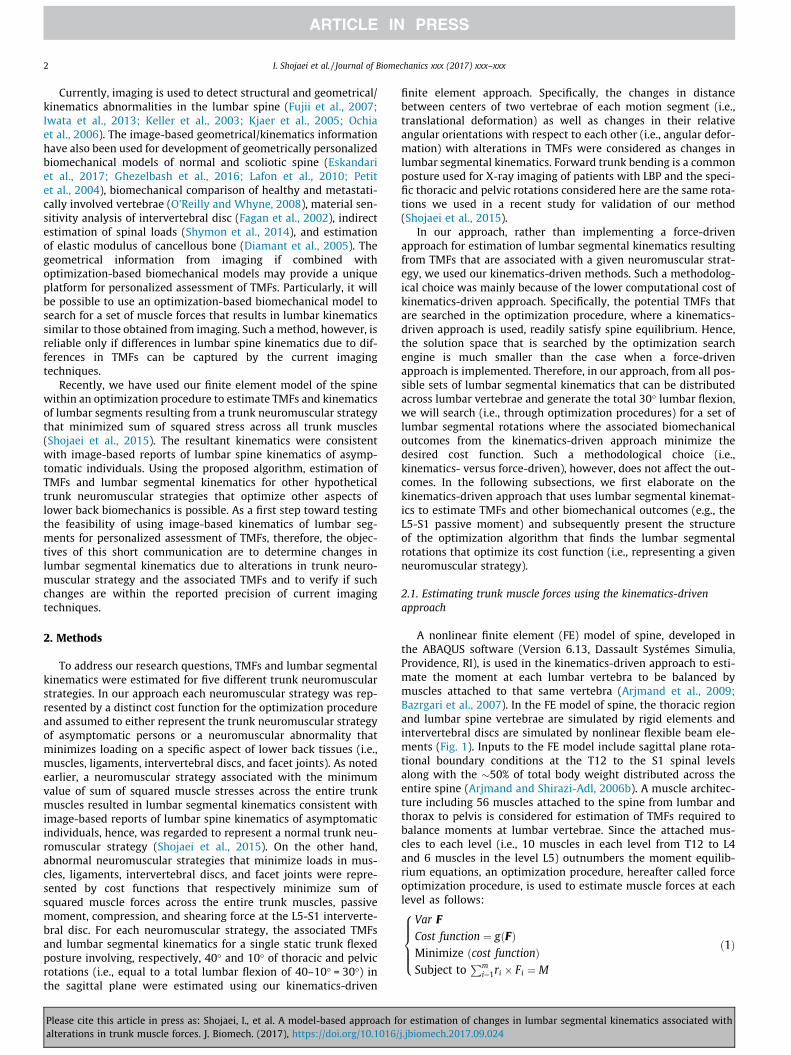
2 I. Shojaei et al. / Journal of Biomechanics xxx (2017) xxx–xxx
Currently, imaging is used to detect structural and geometrical/kinematics abnormalities in the lumbar spine (Fujii et al., 2007;Iwata et al., 2013; Keller et al., 2003; Kjaer et al., 2005; Ochiaet al., 2006). The image-based geometrical/kinematics informationhave also been used for development of geometrically personalizedbiomechanical models of normal and scoliotic spine (Eskandariet al., 2017; Ghezelbash et al., 2016; Lafon et al., 2010; Petitet al., 2004), biomechanical comparison of healthy and metastati-cally involved vertebrae (O’Reilly and Whyne, 2008), material sen-sitivity analysis of intervertebral disc (Fagan et al., 2002), indirectestimation of spinal loads (Shymon et al., 2014), and estimationof elastic modulus of cancellous bone (Diamant et al., 2005). Thegeometrical information from imaging if combined withoptimization-based biomechanical models may provide a uniqueplatform for personalized assessment of TMFs. Particularly, it willbe possible to use an optimization-based biomechanical model tosearch for a set of muscle forces that results in lumbar kinematicssimilar to those obtained from imaging. Such a method, however, isreliable only if differences in lumbar spine kinematics due to dif-ferences in TMFs can be captured by the current imagingtechniques.
Recently, we have used our finite element model of the spinewithin an optimization procedure to estimate TMFs and kinematicsof lumbar segments resulting from a trunk neuromuscular strategythat minimized sum of squared stress across all trunk muscles(Shojaei et al., 2015). The resultant kinematics were consistentwith image-based reports of lumbar spine kinematics of asymp-tomatic individuals. Using the proposed algorithm, estimation ofTMFs and lumbar segmental kinematics for other hypotheticaltrunk neuromuscular strategies that optimize other aspects oflower back biomechanics is possible. As a first step toward testingthe feasibility of using image-based kinematics of lumbar seg-ments for personalized assessment of TMFs, therefore, the objec-tives of this short communication are to determine changes inlumbar segmental kinematics due to alterations in trunk neuro-muscular strategy and the associated TMFs and to verify if suchchanges are within the reported precision of current imagingtechniques.
2. Methods
To address our research questions, TMFs and lumbar segmentalkinematics were estimated for five different trunk neuromuscularstrategies. In our approach each neuromuscular strategy was rep-resented by a distinct cost function for the optimization procedureand assumed to either represent the trunk neuromuscular strategyof asymptomatic persons or a neuromuscular abnormality thatminimizes loading on a specific aspect of lower back tissues (i.e.,muscles, ligaments, intervertebral discs, and facet joints). As notedearlier, a neuromuscular strategy associated with the minimumvalue of sum of squared muscle stresses across the entire trunkmuscles resulted in lumbar segmental kinematics consistent withimage-based reports of lumbar spine kinematics of asymptomaticindividuals, hence, was regarded to represent a normal trunk neu-romuscular strategy (Shojaei et al., 2015). On the other hand,abnormal neuromuscular strategies that minimize loads in mus-cles, ligaments, intervertebral discs, and facet joints were repre-sented by cost functions that respectively minimize sum ofsquared muscle forces across the entire trunk muscles, passivemoment, compression, and shearing force at the L5-S1 interverte-bral disc. For each neuromuscular strategy, the associated TMFsand lumbar segmental kinematics for a single static trunk flexedposture involving, respectively, 40� and 10� of thoracic and pelvicrotations (i.e., equal to a total lumbar flexion of 40–10� = 30�) inthe sagittal plane were estimated using our kinematics-driven
Please cite this article in press as: Shojaei, I., et al. A model-based approach foalterations in trunk muscle forces. J. Biomech. (2017), https://doi.org/10.1016/
finite element approach. Specifically, the changes in distancebetween centers of two vertebrae of each motion segment (i.e.,translational deformation) as well as changes in their relativeangular orientations with respect to each other (i.e., angular defor-mation) with alterations in TMFs were considered as changes inlumbar segmental kinematics. Forward trunk bending is a commonposture used for X-ray imaging of patients with LBP and the speci-fic thoracic and pelvic rotations considered here are the same rota-tions we used in a recent study for validation of our method(Shojaei et al., 2015).
In our approach, rather than implementing a force-drivenapproach for estimation of lumbar segmental kinematics resultingfrom TMFs that are associated with a given neuromuscular strat-egy, we used our kinematics-driven methods. Such a methodolog-ical choice was mainly because of the lower computational cost ofkinematics-driven approach. Specifically, the potential TMFs thatare searched in the optimization procedure, where a kinematics-driven approach is used, readily satisfy spine equilibrium. Hence,the solution space that is searched by the optimization searchengine is much smaller than the case when a force-drivenapproach is implemented. Therefore, in our approach, from all pos-sible sets of lumbar segmental kinematics that can be distributedacross lumbar vertebrae and generate the total 30� lumbar flexion,we will search (i.e., through optimization procedures) for a set oflumbar segmental rotations where the associated biomechanicaloutcomes from the kinematics-driven approach minimize thedesired cost function. Such a methodological choice (i.e.,kinematics- versus force-driven), however, does not affect the out-comes. In the following subsections, we first elaborate on thekinematics-driven approach that uses lumbar segmental kinemat-ics to estimate TMFs and other biomechanical outcomes (e.g., theL5-S1 passive moment) and subsequently present the structureof the optimization algorithm that finds the lumbar segmentalrotations that optimize its cost function (i.e., representing a givenneuromuscular strategy).
2.1. Estimating trunk muscle forces using the kinematics-drivenapproach
A nonlinear finite element (FE) model of spine, developed inthe ABAQUS software (Version 6.13, Dassault Systémes Simulia,Providence, RI), is used in the kinematics-driven approach to esti-mate the moment at each lumbar vertebra to be balanced bymuscles attached to that same vertebra (Arjmand et al., 2009;Bazrgari et al., 2007). In the FE model of spine, the thoracic regionand lumbar spine vertebrae are simulated by rigid elements andintervertebral discs are simulated by nonlinear flexible beam ele-ments (Fig. 1). Inputs to the FE model include sagittal plane rota-tional boundary conditions at the T12 to the S1 spinal levelsalong with the �50% of total body weight distributed across theentire spine (Arjmand and Shirazi-Adl, 2006b). A muscle architec-ture including 56 muscles attached to the spine from lumbar andthorax to pelvis is considered for estimation of TMFs required tobalance moments at lumbar vertebrae. Since the attached mus-cles to each level (i.e., 10 muscles in each level from T12 to L4and 6 muscles in the level L5) outnumbers the moment equilib-rium equations, an optimization procedure, hereafter called forceoptimization procedure, is used to estimate muscle forces at eachlevel as follows:
Var FCost function ¼ gðFÞMinimize ðcost functionÞSubject to
Pmi¼1ri � Fi ¼ M
8>>><>>>:
ð1Þ
r estimation of changes in lumbar segmental kinematics associated withj.jbiomech.2017.09.024
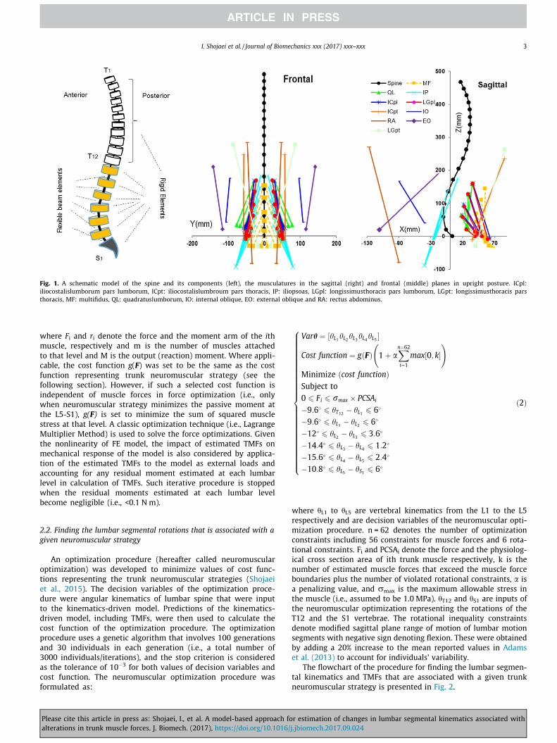
Fig. 1. A schematic model of the spine and its components (left), the musculatures in the sagittal (right) and frontal (middle) planes in upright posture. ICpl:iliocostalislumborum pars lumborum, ICpt: iliocostalislumbroum pars thoracis, IP: iliopsoas, LGpl: longissimusthoracis pars lumborum, LGpt: longissimusthoracis parsthoracis, MF: multifidus, QL: quadratuslumborum, IO: internal oblique, EO: external oblique and RA: rectus abdominus.
I. Shojaei et al. / Journal of Biomechanics xxx (2017) xxx–xxx 3
where Fi and ri denote the force and the moment arm of the ithmuscle, respectively and m is the number of muscles attachedto that level and M is the output (reaction) moment. Where appli-cable, the cost function g(F) was set to be the same as the costfunction representing trunk neuromuscular strategy (see thefollowing section). However, if such a selected cost function isindependent of muscle forces in force optimization (i.e., onlywhen neuromuscular strategy minimizes the passive moment atthe L5-S1), g(F) is set to minimize the sum of squared musclestress at that level. A classic optimization technique (i.e., LagrangeMultiplier Method) is used to solve the force optimizations. Giventhe nonlinearity of FE model, the impact of estimated TMFs onmechanical response of the model is also considered by applica-tion of the estimated TMFs to the model as external loads andaccounting for any residual moment estimated at each lumbarlevel in calculation of TMFs. Such iterative procedure is stoppedwhen the residual moments estimated at each lumbar levelbecome negligible (i.e., <0.1 N m).
2.2. Finding the lumbar segmental rotations that is associated with agiven neuromuscular strategy
An optimization procedure (hereafter called neuromuscularoptimization) was developed to minimize values of cost func-tions representing the trunk neuromuscular strategies (Shojaeiet al., 2015). The decision variables of the optimization proce-dure were angular kinematics of lumbar spine that were inputto the kinematics-driven model. Predictions of the kinematics-driven model, including TMFs, were then used to calculate thecost function of the optimization procedure. The optimizationprocedure uses a genetic algorithm that involves 100 generationsand 30 individuals in each generation (i.e., a total number of3000 individuals/iterations), and the stop criterion is consideredas the tolerance of 10�3 for both values of decision variables andcost function. The neuromuscular optimization procedure wasformulated as:
Please cite this article in press as: Shojaei, I., et al. A model-based approach foalterations in trunk muscle forces. J. Biomech. (2017), https://doi.org/10.1016/
Varh ¼ ½hL1hL2hL3hL4hL5 �
Cost function ¼ gðFÞ 1þ aXn¼62
i¼1
max½0; k� !
Minimize ðcost functionÞSubject to0 6 Fi 6 rmax � PCSAi
�9:6� 6 hT12 � hL1 6 6�
�9:6� 6 hL1 � hL2 6 6�
�12� 6 hL2 � hL3 6 3:6�
�14:4� 6 hL3 � hL4 6 1:2�
�15:6� 6 hL4 � hL5 6 2:4�
�10:8� 6 hL5 � hS1 6 6�
8>>>>>>>>>>>>>>>>>>>>>>>>><>>>>>>>>>>>>>>>>>>>>>>>>>:
ð2Þ
where hL1 to hL5 are vertebral kinematics from the L1 to the L5respectively and are decision variables of the neuromuscular opti-mization procedure. n = 62 denotes the number of optimizationconstraints including 56 constraints for muscle forces and 6 rota-tional constraints. Fi and PCSAi denote the force and the physiolog-ical cross section area of ith trunk muscle respectively, k is thenumber of estimated muscle forces that exceed the muscle forceboundaries plus the number of violated rotational constraints, a isa penalizing value, and rmax is the maximum allowable stress inthe muscle (i.e., assumed to be 1.0 MPa). hT12 and hS1 are inputs ofthe neuromuscular optimization representing the rotations of theT12 and the S1 vertebrae. The rotational inequality constraintsdenote modified sagittal plane range of motion of lumbar motionsegments with negative sign denoting flexion. These were obtainedby adding a 20% increase to the mean reported values in Adamset al. (2013) to account for individuals’ variability.
The flowchart of the procedure for finding the lumbar segmen-tal kinematics and TMFs that are associated with a given trunkneuromuscular strategy is presented in Fig. 2.
r estimation of changes in lumbar segmental kinematics associated withj.jbiomech.2017.09.024
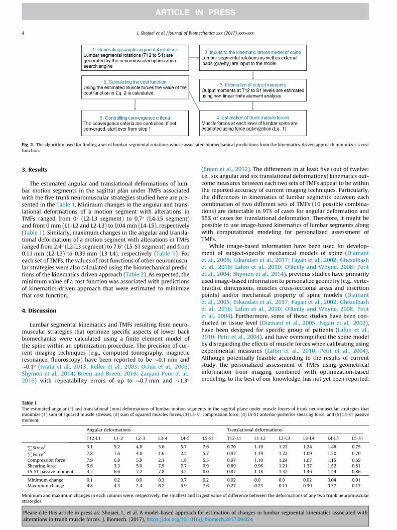
Fig. 2. The algorithm used for finding a set of lumbar segmental rotations whose associated biomechanical predictions from the kinematics-driven approach minimizes a costfunction.
4 I. Shojaei et al. / Journal of Biomechanics xxx (2017) xxx–xxx
3. Results
The estimated angular and translational deformations of lum-bar motion segments in the sagittal plan under TMFs associatedwith the five trunk neuromuscular strategies studied here are pre-sented in the Table 1. Minimum changes in the angular and trans-lational deformations of a motion segment with alterations inTMFs ranged from 0� (L2-L3 segment) to 0.7� (L4-L5 segment)and from 0 mm (L1-L2 and L2-L3) to 0.04 mm (L4-L5), respectively(Table 1). Similarly, maximum changes in the angular and transla-tional deformations of a motion segment with alterations in TMFsranged from 2.4� (L2-L3 segment) to 7.6� (L5-S1 segment) and from0.11 mm (L2-L3) to 0.39 mm (L3-L4), respectively (Table 1). Foreach set of TMFs, the values of cost functions of other neuromuscu-lar strategies were also calculated using the biomechanical predic-tions of the kinematics-driven approach (Table 2). As expected, theminimum value of a cost function was associated with predictionsof kinematics-driven approach that were estimated to minimizethat cost function.
4. Discussion
Lumbar segmental kinematics and TMFs resulting from neuro-muscular strategies that optimize specific aspects of lower backbiomechanics were calculated using a finite element model ofthe spine within an optimization procedure. The precision of cur-rent imaging techniques (e.g., computed tomography, magneticresonance, fluoroscopy) have been reported to be �0.1 mm and�0.1� (Iwata et al., 2013; Keller et al., 2003; Ochia et al., 2006;Shymon et al., 2014; Breen and Breen, 2016; Zanjani-Pour et al.,2016) with repeatability errors of up to �0.7 mm and �1.3�
Table 1The estimated angular (�) and translational (mm) deformations of lumbar motion segmeminimize (1) sum of squared muscle stresses, (2) sum of squared muscles forces, (3) L5-S1moment.
Angular deformations
T12-L1 L1-2 L2-3 L3-4 L4-5PStress2 3.1 5.2 4.8 3.6 5.7PForce2 7.8 7.6 4.8 1.6 2.5
Compression force 7.9 6.8 5.9 2.1 1.8Shearing force 5.6 3.3 5.0 7.5 7.7L5-S1 passive moment 4.2 6.6 7.2 7.8 4.2
Minimum change 0.1 0.2 0.0 0.3 0.7Maximum change 4.8 4.3 2.4 6.2 5.9
Minimum and maximum changes in each column were, respectively, the smallest and lastrategies.
Please cite this article in press as: Shojaei, I., et al. A model-based approach foalterations in trunk muscle forces. J. Biomech. (2017), https://doi.org/10.1016/
(Breen et al., 2012). The differences in at least five (out of twelve:i.e., six angular and six translational deformations) kinematics out-comemeasures between each two sets of TMFs appear to be withinthe reported accuracy of current imaging techniques. Particularly,the differences in kinematics of lumbar segments between eachcombination of two different sets of TMFs (10 possible combina-tions) are detectable in 97% of cases for angular deformation and55% of cases for translational deformation. Therefore, it might bepossible to use image-based kinematics of lumbar segments alongwith computational modeling for personalized assessment ofTMFs.
While image-based information have been used for develop-ment of subject-specific mechanical models of spine (Diamantet al., 2005; Eskandari et al., 2017; Fagan et al., 2002; Ghezelbashet al., 2016; Lafon et al., 2010; O’Reilly and Whyne, 2008; Petitet al., 2004; Shymon et al., 2014), previous studies have primarilyused image-based information to personalize geometry (e.g., verte-bra/disc dimensions, muscles cross-sectional areas and insertionpoints) and/or mechanical property of spine models (Diamantet al., 2005; Eskandari et al., 2017; Fagan et al., 2002; Ghezelbashet al., 2016; Lafon et al., 2010; O’Reilly and Whyne, 2008; Petitet al., 2004). Furthermore, some of these studies have been con-ducted in tissue level (Diamant et al., 2005; Fagan et al., 2002),have been designed for specific group of patients (Lafon et al.,2010; Petit et al., 2004), and have oversimplified the spine modelby disregarding the effects of muscle forces when calibrating usingexperimental measures (Lafon et al., 2010; Petit et al., 2004).Although potentially feasible according to the results of currentstudy, the personalized assessment of TMFs using geometricalinformation from imaging combined with optimization-basedmodeling, to the best of our knowledge, has not yet been reported.
nts in the sagittal plane under muscle forces of trunk neuromuscular strategies thatcompression force, (4) L5-S1 anterior-posterior shearing force, and (5) L5-S1 passive
Translational deformations
L5-S1 T12-L1 L1-L2 L2-L3 L3-L4 L4-L5 L5-S1
7.6 0.70 1.10 1.22 1.24 1.48 0.75
5.7 0.97 1.19 1.22 1.09 1.20 0.70
5.5 0.97 1.10 1.24 1.07 1.15 0.690.9 0.89 0.96 1.21 1.37 1.52 0.810.0 0.87 1.18 1.32 1.46 1.44 0.86
0.2 0.02 0.0 0.0 0.02 0.04 0.017.6 0.27 0.23 0.11 0.39 0.37 0.17
rgest value of difference between the deformations of any two trunk neuromuscular
r estimation of changes in lumbar segmental kinematics associated withj.jbiomech.2017.09.024
Table 2The value of cost functions (horizontal top) under the five sets of muscle forces associated with neuromuscularstrategies (vertical left) studied here.
I. Shojaei et al. / Journal of Biomechanics xxx (2017) xxx–xxx 5
The value of cost function of each neuromuscular strategy, asexpected, increased when calculated using predictions of thekinematics-driven approach associated with the other cost func-tions (Table 2). However, what is notable in results presented inTable 2 is that alterations in TMFs, for example due to an abnormaltrunk neuromuscular strategy, could result in loads and/or defor-mations in some areas of lower back that are larger than what isnormally resisted by those areas. For instance, TMFs associatedwith the hypothetical neuromuscular strategy that minimizedshearing force at the L5-S1 intervertebral disc resulted in anincrease of �350 N in compression force when compared to TMFsthat were associated with the a strategy that was considered nor-mal in this study (i.e., the strategy that minimizes sum of squaredmuscle stresses). Similarly, a strategy that minimized compressionforce or muscle forces, compared to the normal strategy, led tolarge muscle stresses. Although the short term effect of a specifictrunk neuromuscular strategy can be beneficial, for instance byprotecting the injured tissues, the long term consequences ofaltered trunk neuromuscular strategy could be an injury to otherlumbar tissues due to compensatory resisted larger than normalloads (Hodges and Smeets, 2015).
In the present study, we postulated that trunk neuromuscularstrategies optimize some aspects of lower back biomechanics.Though alterations in neuromuscular strategy have been reportedin the literature, our assumption might not be accurate and wasmerely made for the purpose of this feasibility study (i.e., todemonstrate changes in lumbar segmental kinematics with alter-ations in TMFs are within the reported accuracy of current imagingtechniques). Furthermore, in all cases, the abnormal neuromuscu-lar strategy that minimized loads in a tissue was represented by asingle-force cost function which was a simplified assumption. Forexample, minimizing the loads on the facet joint involves reducingboth shearing and compression forces, though shearing is the dom-inant force in characterizing facet joint environment. Whetherthere are one-on-one relationships between sets of TMFs (or theresultant kinematics) and neuromuscular strategies or whetherall differences in trunk neuromuscular strategies result in detect-able change in TMFs remains to be investigated in future. Theavailability of personalized assessment of TMFs, as proposed in thisshort communication, should, however, facilitate such futureresearch efforts.
In conclusion, results of this feasibility study, support the ideaof image-based personalized assessment of TMFs using computa-tional models. Specifically, a geometrically and materiallysubject-specified model of the spine can be used in future to obtaina set of TMFs, as individualized TMFs, that generates the closestlumbar kinematics to those measured from imaging. The accuracy
Please cite this article in press as: Shojaei, I., et al. A model-based approach foalterations in trunk muscle forces. J. Biomech. (2017), https://doi.org/10.1016/
of such assessment strategy can further be improved by imple-menting dynamic rather than static assessment tasks. However,immediate research question to be addressed will be the reliabilityand validity of such an image-based method for personalizedassessment of TMFs.
Acknowledgements
This work was supported, in part, by an award(5R03HD086512-02) from the National Center for Medical Rehabil-itation Research (NIH-NICHD) and an award from the Office of theAssistant Secretary of Defense for Health Affairs, through the PeerReviewed Orthopaedic Research Program (award #W81XWH-14-2-0144).
Conflict of interest statement
We declare that all authors have no financial or personal relation-ships with other persons or organizations that might inappropri-ately influence our work presented therein.
References
Adams, M.A., Burton, K., Bogduk, N., 2013. The biomechanics of back pain. ElsevierHealth Sciences.
Arjmand, N., Gagnon, D., Plamondon, A., Shirazi-Adl, A., Lariviere, C., 2009.Comparison of trunk muscle forces and spinal loads estimated by twobiomechanical models. Clin. Biomech. 24, 533–541.
Arjmand, N., Shirazi-Adl, A., 2006a. Model and in vivo studies on human trunk loadpartitioning and stability in isometric forward flexions. J. Biomech. 39, 510–521.
Arjmand, N., Shirazi-Adl, A., 2006b. Sensitivity of kinematics-based modelpredictions to optimization criteria in static lifting tasks. Med. Eng. Phys. 28,504–514.
Bazrgari, B., Shirazi-Adl, A., Arjmand, N., 2007. Analysis of squat and stoop dynamicliftings: muscle forces and internal spinal loads. Eur. Spine J. 16, 687–699.
Breen, A., Breen, A., 2016. Accuracy and repeatability of quantitative fluoroscopy forthe measurement of sagittal plane translation and finite centre of rotation in thelumbar spine. Med. Eng. Phys. 38, 607–614.
Breen, A.C., Teyhen, D.S., Mellor, F.E., Breen, A.C., Wong, K.W., Deitz, A., 2012.Measurement of intervertebral motion using quantitative fluoroscopy: report ofan international forum and proposal for use in the assessment of degenerativedisc disease in the lumbar spine. Adv. Orthop. 2012 https://doi.org/10.1155/2012/802350.
Coenen, P., Kingma, I., Boot, C.R., Bongers, P.M., van Dieën, J.H., 2014. Cumulativemechanical low-back load at work is a determinant of low-back pain. Occup.Environ. Med. 71, 332–337.
Daniel, M., 2011. Role of optimization criterion in static asymmetric analysis oflumbar spine load. WMW Wiener Medizinische Wochenschrift 161, 477–485.
Diamant, I., Shahar, R., Gefen, A., 2005. How to select the elastic modulus forcancellous bone in patient-specific continuum models of the spine. Med. Biol.Eng. Comput. 43, 465–472.
Dreischarf, M., Zander, T., Shirazi-Adl, A., Puttlitz, C., Adam, C., Chen, C., Goel, V.,Kiapour, A., Kim, Y., Labus, K., 2014. Comparison of eight published static finite
r estimation of changes in lumbar segmental kinematics associated withj.jbiomech.2017.09.024
6 I. Shojaei et al. / Journal of Biomechanics xxx (2017) xxx–xxx
element models of the intact lumbar spine: predictive power of modelsimproves when combined together. J. Biomech. 47, 1757–1766.
Eskandari, A., Arjmand, N., Shirazi-Adl, A., Farahmand, F., 2017. Subject-specific2D/3D image registration and kinematics-driven musculoskeletal model of thespine. J. Biomech. 57, 18–26.
Ezquerro, F., Simón, A., Prado, M., Pérez, A., 2004. Combination of finite elementmodeling and optimization for the study of lumbar spine biomechanicsconsidering the 3D thorax–pelvis orientation. Med. Eng. Phys. 26, 11–22.
Fagan, M., Julian, S., Siddall, D., Mohsen, A., 2002. Patient-specific spine models. Part1: finite element analysis of the lumbar intervertebral disc—a materialsensitivity study. Proc. Inst. Mech. Eng. [H] 216, 299–314.
Fujii, R., Sakaura, H., Mukai, Y., Hosono, N., Ishii, T., Iwasaki, M., Yoshikawa, H.,Sugamoto, K., 2007. Kinematics of the lumbar spine in trunk rotation: in vivothree-dimensional analysis using magnetic resonance imaging. Eur. Spine J. 16,1867–1874.
Ghezelbash, F., Shirazi-Adl, A., Arjmand, N., El-Ouaaid, Z., Plamondon, A., 2016.Subject-specific biomechanics of trunk: musculoskeletal scaling, internal loadsand intradiscal pressure estimation. Biomech. Model. Mechanobiol. 15, 1699–1712.
Hodges, P.W., Smeets, R.J., 2015. Interaction between pain, movement, and physicalactivity: short-term benefits, long-term consequences, and targets fortreatment. Clin. J. Pain 31, 97–107.
Hughes, R.E., 2000. Effect of optimization criterion on spinal force estimates duringasymmetric lifting. J. Biomech. 33, 225–229.
Iwata, T., Miyamoto, K., Hioki, A., Ohashi, M., Inoue, N., Shimizu, K., 2013. In vivomeasurement of lumbar foramen during axial loading using a compressiondevice and computed tomography. Clin. Spine Surgery 26, E177–E182.
Keller, T.S., Harrison, D.E., Colloca, C.J., Harrison, D.D., Janik, T.J., 2003. Prediction ofosteoporotic spinal deformity. Spine (Phila Pa 1976) 28, 455–462.
Kjaer, P., Leboeuf-Yde, C., Korsholm, L., Sorensen, J.S., Bendix, T., 2005. Magneticresonance imaging and low back pain in adults: a diagnostic imaging study of40-year-old men and women. Spine (Phila Pa 1976) 30, 1173–1180.
Lafon, Y., Lafage, V., Steib, J.-P., Dubousset, J., Skalli, W., 2010. In vivo distribution ofspinal intervertebral stiffness based on clinical flexibility tests. Spine (Phila Pa1976) 35, 186–193.
Marras, W.S., Davis, K.G., Ferguson, S.A., Lucas, B.R., Gupta, P., 2001. Spine loadingcharacteristics of patients with low back pain compared with asymptomaticindividuals. Spine (Phila Pa 1976) 26, 2566–2574.
Muslim, K., Bazrgari, B., Hendershot, B., Toosizadeh, N., Nussbaum, M.A., Madigan,M.L., 2013. Disturbance and recovery of trunk mechanical and neuromuscularbehaviors following repeated static trunk flexion: influences of duration andduty cycle on creep-induced effects. Appl. Ergon. 44, 643–651.
Please cite this article in press as: Shojaei, I., et al. A model-based approach foalterations in trunk muscle forces. J. Biomech. (2017), https://doi.org/10.1016/
O’Reilly, M.A., Whyne, C.M., 2008. Comparison of computed tomography basedparametric and patient-specific finite element models of the healthy andmetastatic spine using a mesh-morphing algorithm. Spine (Phila Pa 1976) 33,1876–1881.
Ochia, R.S., Inoue, N., Renner, S.M., Lorenz, E.P., Lim, T.-H., Andersson, G.B., An, H.S.,2006. Three-dimensional in vivo measurement of lumbar spine segmentalmotion. Spine (Phila Pa 1976) 31, 2073–2078.
Panjabi, M.M., 1992a. The stabilizing system of the spine. Part I. Function,dysfunction, adaptation, and enhancement. J. Spinal Disorders Tech. 5, 383–389.
Panjabi, M.M., 1992b. The stabilizing system of the spine. Part II. Neutral zone andinstability hypothesis. J. Spinal Disorders Tech. 5, 390–397.
Petit, Y., Aubin, C.-É., Labelle, H., 2004. Patient-specific mechanical properties of aflexible multi-body model of the scoliotic spine. Med. Biol. Eng. Comput. 42, 55–60.
Radebold, A., Cholewicki, J., Panjabi, M.M., Patel, T.C., 2000. Muscle response patternto sudden trunk loading in healthy individuals and in patients with chronic lowback pain. Spine (Phila Pa 1976) 25, 947–954.
Radebold, A., Cholewicki, J., Polzhofer, G.K., Greene, H.S., 2001. Impaired posturalcontrol of the lumbar spine is associated with delayed muscle response times inpatients with chronic idiopathic low back pain. Spine (Phila Pa 1976) 26, 724–730.
Shojaei, I., Arjmand, N., Bazrgari, B., 2015. An optimization-based method forprediction of lumbar spine segmental kinematics from the measurements ofthorax and pelvic kinematics. Int. J. Numer. Meth. Biomed. Eng. 31.
Shymon, S.J., Yaszay, B., Dwek, J.R., Proudfoot, J.A., Donohue, M., Hargens, A.R., 2014.Altered disc compression in children with idiopathic low back pain: an uprightMRI backpack study. Spine (Phila Pa 1976) 39, 243.
Staudenmann, D., Roeleveld, K., Stegeman, D.F., Van Dieën, J.H., 2010.Methodological aspects of SEMG recordings for force estimation–a tutorialand review. J. Electromyogr. Kinesiol. 20, 375–387.
Stokes, I.A., Gardner-Morse, M., 2001. Lumbar spinal muscle activation synergiespredicted by multi-criteria cost function. J. Biomech. 34, 733–740.
Toosizadeh, N., Bazrgari, B., Hendershot, B., Muslim, K., Nussbaum, M.A., Madigan,M.L., 2013. Disturbance and recovery of trunk mechanical and neuromuscularbehaviours following repetitive lifting: influences of flexion angle and lift rateon creep-induced effects. Ergonomics 56, 954–963.
Vazirian, M., Van Dillen, L.R., Bazrgari, B., 2016. Lumbopelvic rhythm in the sagittalplane: a review of the effects of participants and task characteristics. Int.Musculoskelet. Med. 38, 51–58.
Zanjani-Pour, S., Winlove, C.P., Smith, C.W., Meakin, J.R., 2016. Image driven subject-specific finite element models of spinal biomechanics. J. Biomech. 49, 919–925.
r estimation of changes in lumbar segmental kinematics associated withj.jbiomech.2017.09.024

Title: Living with Extremity Trauma and Limb Loss for a Lifetime: A Review of Efforts to Identify and Mitigate Risk Factors for Secondary Health Complications Objectives: 1. Identify and describe key risk factors for musculoskeletal conditions secondary to extremity trauma / limb loss 2. Identify and describe key risk factors for cardiovascular disease secondary to extremity trauma / limb loss 3. Identify and describe effective approaches to mitigate onset and progression of these secondary health conditions Type of Presentation: Panel Discussion (45min) Target Audience: all clinicians (and researchers) involved with the care of Servicemembers and Veterans with extremity trauma and amputation Presentation Summary: Extremity trauma, including limb loss, is commonly associated with an increased prevalence of and risk for developing deleterious secondary health conditions, such as musculoskeletal and cardiovascular disease [1,2], among others. These secondary health conditions significantly impact functional outcomes and quality of life over the longer term and, thus, are particularly concerning given the relatively young age of Servicemembers with extremity trauma and potential for cumulative, lifelong disability. Here, we focus on synthesizing recent efforts at each Military Treatment Facility (WRNMMC, SAMMC, NMCSD) and NHRC; specifically, those toward identifying and tracking risk factors for these complications secondary to extremity trauma and limb loss. The Extremity Trauma and Amputation Center of Excellence is composed of clinicians and researchers who are actively conducting numerous efforts with the collective aim to comprehensively understand the specific factors (e.g., biomechanical, personal, physiological, psychological) underpinning the development and progression of these deleterious secondary health conditions. These include epidemiological efforts to characterize this patient population and their health over time, biomechanical calculations of joint loads during activity, local/systemic biomarkers (e.g., blood/synovial fluid, respectively), changes in patient-level risk factors, and self-reported quality of life metrics. We have identified several instances where altered gait mechanics are associated with abnormal joint loading parameters as likely contributors to the high prevalence and incidence of low back and knee pain among persons with limb loss. Specifically, increased trunk motions in persons with vs. without limb loss are associated with greater external demands on the lower back [3] and up to a 65% increase in joint compressive forces [4], and closely mimic the movement patterns seen in individuals who have low back pain [5]. Peak joint contact forces and loading rates within the intact
knee are also respectively 17 and 24% larger among persons with vs. without unilateral limb loss [6]. However, level ground walking is likely not the only contributor to the risk of knee osteoarthritis [7] as other common movements, such as hopping when moving without a prosthesis, place the knee at even greater risk [8]. Advancements in prosthetic designs offering powered push-off can also mitigate risk factors by reducing knee joint loads [7]. Alterations in health habits (e.g. tobacco/alcohol use) and ensuing physical inactivity and weight gain [9], which are commonly observed after lower limb trauma and amputation, can increase the risk for musculoskeletal and cardiovascular disease. A more holistic understanding of the specific pathways by which these secondary conditions develop is urgently needed, particularly for clinicians to prescribe effective treatment interventions which decrease concomitant risk. Similarly, it is important for individuals with limb loss to be cognizant of the multi-factorial contributors to these conditions. Identifying and mitigating the risk of health effects secondary to amputation is a positive step towards setting patients up for lifelong health. Knowledge of adverse movement mechanics, practical metrics and tests for evaluation, and the effectiveness of different prosthetic designs will provide information useful for treating individuals with limb loss. This session will deliver this information through a summary of the clinically-applicable research efforts at the DoD’s Military Treatment Facilities to assist clinicians in their efforts to serve the men and women who have served our country. References: [1] Gailey et al. 2008 [2] Butowicz et al. 2017 [3] Hendershot et al. 2014 [4] Shojaie et al. 2016 [5] Russell Esposito 2014 [6] Miller et al. 2017 [7] Russell Esposito 2015 [8] Krupenovich et al. 2017 [9] Eckard and Pruziner 2016 Biosketches: Christopher Dearth, PhD, concurrently serves as the Facility Research Director for the Extremity Trauma & Amputation Center of Excellence (EACE), Director of the Research & Development Section within the Department of Rehabilitation at WRNMMC, and the Founding Director of the Regenerative Biosciences Laboratory at the Uniformed Services University of the Health Sciences (USUHS). In these roles, Dr. Dearth leads a multidisciplinary team of clinicians and researchers whose collective focus is on the mitigation, treatment and rehabilitation of traumatic extremity injuries and amputations by implementation of clinically relevant research aimed at optimizing the quality of life of service members and veterans. Dr. Dearth’s preclinical and clinical research interests include: development of innovative regenerative medicine & tissue engineering constructs, utilization of next generation imaging modalities for extremity trauma applications, evaluation of advanced (i.e., powered and intuitively controlled) prosthetics for Service Members and Veterans with limb loss, and championing a synergy of efforts between the fields of rehabilitative and regenerative medicine. Susan Eskridge, PhD, is a research epidemiologist with Leidos working as a contractor at the Naval Health Research Center in San Diego, CA. Her primary research interests center around injuries during deployment including outcomes after extremity injury and mild traumatic brain injury. In addition, Dr. Eskridge has been a physical therapist for over 30 years. Early in her career, she was a staff physical therapist on the rehabilitation team and in the Motion Analysis Laboratory at Children’s Hospital, San Diego. In addition to her clinical work, she was an assistant professor at Chapman University in the Department of Physical Therapy. She
completed a Master of Science degree in physical therapy at the University of Southern California in 1986 and a Ph.D. in public health, epidemiology at the University of California, San Diego and San Diego State University in 2011. She is a registered physical therapist in California as well as certified in kinesiological electromyography in California. Shawn Farrokhi, PT, PhD, is the Senior Scientist for the DoD-VA Extremity Trauma and Amputation Center of Excellence (EACE) at the Naval Medical Center San Diego. With more than 16 years of combined experience in academia, clinical research and patient care, Dr. Farrokhi offers a unique perspective towards advancing the field of rehabilitation for military Service Members and Veterans with orthopedic injuries. While on faculty at University Pittsburgh, Dr. Farrokhi received a K12 Career Development award through the Comprehensive Opportunities in Rehabilitation Research Training (CORRT). As a CORRT scholar, Dr. Farrokhi was awarded protected time to complete a Master of Science degree in Clinical Research, which provided him with the relevant training and experience in the design and implementation of epidemiological studies and clinical trials. As a CORRT scholar, Dr. Farrokhi was also able to gain practical research experience and mentorship in conducting clinically relevant translational studies to better understand the underlying mechanisms and appropriate treatment options for the management of various orthopedic conditions. Using his training and previous research experience in his current position, Dr. Farrokhi has established a sustainable multidisciplinary research program aimed at better understanding the risk factors and effective intervention strategies for the treatment of secondary musculoskeletal conditions in wounded warriors with traumatic extremity injuries and limb loss. In addition to his research activities, Dr. Farrokhi currently spends approximately 15% of his time treating Service Members and Veterans with musculoskeletal conditions at the Naval Medical Center San Diego, where he has developed a novel, evidence-supported, movement retraining clinic focused on treatment of common movement disorders. Dr. Farrokhi’s continued clinical involvement allows him to contribute to his proposed research agenda in ways that will ensure continued clinical relevance. Brad Hendershot, PhD, is a Research Biomedical Engineer with the EACE, stationed at WRNMMC. In addition to this role, he directs activities within the Biomechanics and Virtual Reality Laboratories within the Department of Rehabilitation. Dr. Hendershot also holds an appointment, as Assistant Professor, within the Department of Rehabilitation Medicine at USUHS. He received his BS in Bioengineering from Penn State and PhD in Biomedical Engineering from Virginia Tech. Dr. Hendershot’s research is primarily focused on the mechanics and neuromuscular control of human movement, with a particular emphasis on the trunk/spine in both uninjured and patient populations. Recently, his research aims to comprehensively characterize factors underlying the high prevalence of musculoskeletal complications secondary to extremity trauma, to mitigate development/recurrence over the longer term and ultimately improve quality of life for Service Members and Veterans with limb loss. Elizabeth Russell Esposito, PhD, is a Research Biomedical Engineer and acting Senior Scientist at the Center for the Intrepid, with an adjunct Assistant Professor appointment at USUHS. Dr. Russell Esposito received her BS from the University of Delaware and PhD from University of Massachusetts in Kinesiology with a focus on Biomechanics and Motor Control. She previously served as the Biomechanics Lab Director at the Andrews Institute in Gulf Breeze, Florida and as adjunct Assistant Professor at University of West Florida. The goal of her research efforts is to generate to information and knowledge to make significant improvements in clinical practice. A primary track stems from early investigations on preventative methods to reduce or mitigate biomechanical risk factors for overuse musculoskeletal injuries to improve health, mobility and function. This research incorporates gait analysis, metabolic factors,
dynamical systems and movement coordination analyses, and musculoskeletal modeling to determine how to concurrently improve performance while establishing strategies to reduce the risk of injury over time. Dr. Russell Esposito also contributes to the scientific community as a reviewer for over a dozen scientific journals and serves roles within numerous professional organizations.
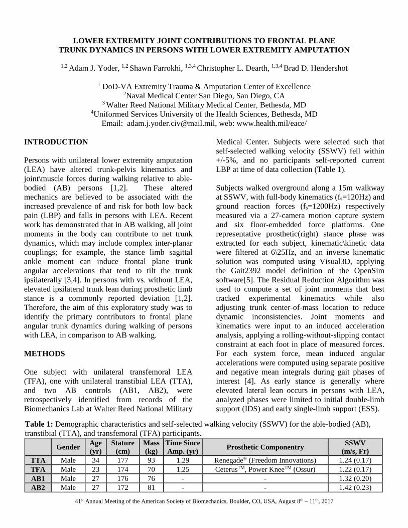
41st Annual Meeting of the American Society of Biomechanics, Boulder, CO, USA, August 8th – 11th, 2017
LOWER EXTREMITY JOINT CONTRIBUTIONS TO FRONTAL PLANE
TRUNK DYNAMICS IN PERSONS WITH LOWER EXTREMITY AMPUTATION
1,2 Adam J. Yoder, 1,2 Shawn Farrokhi, 1,3,4 Christopher L. Dearth, 1,3,4 Brad D. Hendershot
1 DoD-VA Extremity Trauma & Amputation Center of Excellence
2Naval Medical Center San Diego, San Diego, CA 3 Walter Reed National Military Medical Center, Bethesda, MD
4Uniformed Services University of the Health Sciences, Bethesda, MD
Email: [email protected], web: www.health.mil/eace/
INTRODUCTION
Persons with unilateral lower extremity amputation
(LEA) have altered trunk-pelvis kinematics and
joint\muscle forces during walking relative to able-
bodied (AB) persons [1,2]. These altered
mechanics are believed to be associated with the
increased prevalence of and risk for both low back
pain (LBP) and falls in persons with LEA. Recent
work has demonstrated that in AB walking, all joint
moments in the body can contribute to net trunk
dynamics, which may include complex inter-planar
couplings; for example, the stance limb sagittal
ankle moment can induce frontal plane trunk
angular accelerations that tend to tilt the trunk
ipsilaterally [3,4]. In persons with vs. without LEA,
elevated ipsilateral trunk lean during prosthetic limb
stance is a commonly reported deviation [1,2].
Therefore, the aim of this exploratory study was to
identify the primary contributors to frontal plane
angular trunk dynamics during walking of persons
with LEA, in comparison to AB walking.
METHODS
One subject with unilateral transfemoral LEA
(TFA), one with unilateral transtibial LEA (TTA),
and two AB controls (AB1, AB2), were
retrospectively identified from records of the
Biomechanics Lab at Walter Reed National Military
Medical Center. Subjects were selected such that
self-selected walking velocity (SSWV) fell within
+/-5%, and no participants self-reported current
LBP at time of data collection (Table 1).
Subjects walked overground along a 15m walkway
at SSWV, with full-body kinematics (fs=120Hz) and
ground reaction forces (fs=1200Hz) respectively
measured via a 27-camera motion capture system
and six floor-embedded force platforms. One
representative prosthetic(right) stance phase was
extracted for each subject, kinematic\kinetic data
were filtered at 6\25Hz, and an inverse kinematic
solution was computed using Visual3D, applying
the Gait2392 model definition of the OpenSim
software[5]. The Residual Reduction Algorithm was
used to compute a set of joint moments that best
tracked experimental kinematics while also
adjusting trunk center-of-mass location to reduce
dynamic inconsistencies. Joint moments and
kinematics were input to an induced acceleration
analysis, applying a rolling-without-slipping contact
constraint at each foot in place of measured forces.
For each system force, mean induced angular
accelerations were computed using separate positive
and negative mean integrals during gait phases of
interest [4]. As early stance is generally where
elevated lateral lean occurs in persons with LEA,
analyzed phases were limited to initial double-limb
support (IDS) and early single-limb support (ESS).
Table 1: Demographic characteristics and self-selected walking velocity (SSWV) for the able-bodied (AB),
transtibial (TTA), and transfemoral (TFA) participants.
Gender Age
(yr)
Stature
(cm)
Mass
(kg)
Time Since
Amp. (yr) Prosthetic Componentry
SSWV
(m/s, Fr)
TTA Male 34 177 93 1.29 Renegade® (Freedom Innovations) 1.24 (0.17)
TFA Male 23 174 70 1.25 CeterusTM, Power KneeTM (Ossur) 1.22 (0.17)
AB1 Male 27 176 76 - - 1.32 (0.20)
AB2 Male 27 172 81 - - 1.42 (0.23)
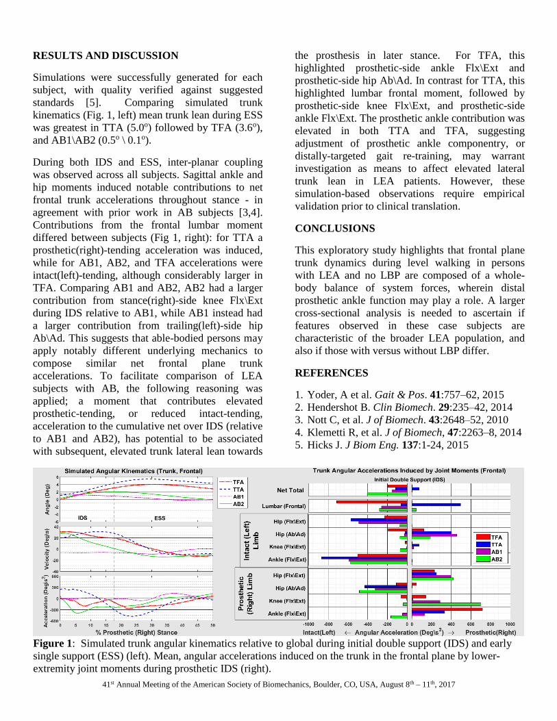
41st Annual Meeting of the American Society of Biomechanics, Boulder, CO, USA, August 8th – 11th, 2017
Figure 1: Simulated trunk angular kinematics relative to global during initial double support (IDS) and early
single support (ESS) (left). Mean, angular accelerations induced on the trunk in the frontal plane by lower-
extremity joint moments during prosthetic IDS (right).
RESULTS AND DISCUSSION
Simulations were successfully generated for each
subject, with quality verified against suggested
standards [5]. Comparing simulated trunk
kinematics (Fig. 1, left) mean trunk lean during ESS
was greatest in TTA (5.0o) followed by TFA (3.6o),
and AB1\AB2 (0.5o \ 0.1o).
During both IDS and ESS, inter-planar coupling
was observed across all subjects. Sagittal ankle and
hip moments induced notable contributions to net
frontal trunk accelerations throughout stance - in
agreement with prior work in AB subjects [3,4].
Contributions from the frontal lumbar moment
differed between subjects (Fig 1, right): for TTA a
prosthetic(right)-tending acceleration was induced,
while for AB1, AB2, and TFA accelerations were
intact(left)-tending, although considerably larger in
TFA. Comparing AB1 and AB2, AB2 had a larger
contribution from stance(right)-side knee Flx\Ext
during IDS relative to AB1, while AB1 instead had
a larger contribution from trailing(left)-side hip
Ab\Ad. This suggests that able-bodied persons may
apply notably different underlying mechanics to
compose similar net frontal plane trunk
accelerations. To facilitate comparison of LEA
subjects with AB, the following reasoning was
applied; a moment that contributes elevated
prosthetic-tending, or reduced intact-tending,
acceleration to the cumulative net over IDS (relative
to AB1 and AB2), has potential to be associated
with subsequent, elevated trunk lateral lean towards
the prosthesis in later stance. For TFA, this
highlighted prosthetic-side ankle Flx\Ext and
prosthetic-side hip Ab\Ad. In contrast for TTA, this
highlighted lumbar frontal moment, followed by
prosthetic-side knee Flx\Ext, and prosthetic-side
ankle Flx\Ext. The prosthetic ankle contribution was
elevated in both TTA and TFA, suggesting
adjustment of prosthetic ankle componentry, or
distally-targeted gait re-training, may warrant
investigation as means to affect elevated lateral
trunk lean in LEA patients. However, these
simulation-based observations require empirical
validation prior to clinical translation.
CONCLUSIONS
This exploratory study highlights that frontal plane
trunk dynamics during level walking in persons
with LEA and no LBP are composed of a whole-
body balance of system forces, wherein distal
prosthetic ankle function may play a role. A larger
cross-sectional analysis is needed to ascertain if
features observed in these case subjects are
characteristic of the broader LEA population, and
also if those with versus without LBP differ.
REFERENCES
1. Yoder, A et al. Gait & Pos. 41:757–62, 2015
2. Hendershot B. Clin Biomech. 29:235–42, 2014
3. Nott C, et al. J of Biomech. 43:2648–52, 2010
4. Klemetti R, et al. J of Biomech, 47:2263–8, 2014
5. Hicks J. J Biom Eng. 137:1-24, 2015
Trunk-Pelvic Dynamics during Transient Turns in Persons with Unilateral Lower Limb Amputation
Pawel R. Golyski 2 and Brad D. Hendershot 1,2
1 DOD-VA Extremity Trauma and Amputation Center of Excellence 2 Department of Rehabilitation, Walter Reed National Military Medical Center
Background: Persons with lower limb amputation (LLA) often walk with compensatory movement strategies involving a prominent reliance on the trunk and pelvis. Altered kinematic features and coordination of these two segments have been associated with elevated demands on the low back [1], increased inter-segmental rigidity [2], and larger trunk muscular forces and spinal loads [3-4]. Consequently, biomechanical factors are considered to play an important role in the development or recurrence of longer-term musculoskeletal complications after LLA (e.g., LBP; [5,6]); however, evaluations of other common ambulatory tasks are limited. Turning is ubiquitous in daily life and can be biomechanically challenging for individuals with musculoskeletal impairments [7]. Thus, the purpose of this study was to characterize proximal compensations using inter-segmental momenta and coordination during transient (90-degree) turning tasks among persons with vs. without LLA. We hypothesized persons with vs. without LLA execute turns with altered trunk-pelvic segmental coordination and larger ranges in segmental momenta, to overcome challenges associated with altered or impaired ankle function (a key joint for executing turns). Methods: Three-dimensional trunk and pelvic kinematics, momenta, and coordination phase/variability were calculated among 8 persons with LLA (n=4 with transtibial and n=4 with transfemoral amputation), and 5 uninjured controls, performing 90-degree turns to the left (n=10) and right (n=10). Participants walked at their self-selected speed along a 12-foot straight path and were verbally cued to turn left or right at a specified and consistent point, self-selecting the turning strategy (i.e., step vs. spin) and pivot limb. Results: There were no differences in the frequency of intersegmental coordination phase (or its variability) between persons with and without LLA. However, ranges of motion and segmental momenta differed between persons with and without LLA, depending on the plane and turn strategy employed. In the sagittal plane, ranges of motion and momenta were respectively larger and smaller in step turns among persons with vs. without LLA. In the frontal plane, ranges of motion and momenta were respectively smaller and larger among persons with LLA. In the transverse plane, ranges in pelvic momenta were larger/smaller during spin/step turns, respectively, among persons with vs. without LLA. Conclusions: Although no differences in the frequency or variability of inter-segmental coordination were observed, altered ranges of motion and momenta among persons with LLA indicate similar reliance on proximal segments during turning as in walking. Such altered and task-dependent modulation of trunk-pelvic dynamics among persons with LLA provides additional support for a hypothesis linking repeated exposures to altered trunk-pelvic dynamics with elevated LBP risk over the long term. References: [1] Hendershot and Wolf. ClinBiomech (2014) [2] Esposito and Wilken. GaitPosture (2014) [3] Shojaei, et al. ClinBiomech (2016) [4] Yoder, et al. GaitPosture (2015) [5] Devan, et al. MedHypoth (2014) [6] Gailey. JRRD (2008) [7] Taylor, et al. HumMovSci (2005) The views expressed herein are those of the author and do not reflect the official policy of the US Army or Defense, nor the US Government.
41st Annual Meeting of the American Society of Biomechanics, Boulder, CO, USA, August 8th – 11th, 2017
LONGITUDINAL CHANGES IN MEDIOLATERAL TRUNK AND PELVIC MOTION AMONG PERSONS WITH LOWER LIMB AMPUTATION DURING THE FIRST YEAR OF AMBULATION
1-3 Brad D. Hendershot, 2 Courtney M. Butowicz, 1,2 Caitlin E. Mahon, 2 Barri L. Schnall, 1-3Christopher L. Dearth
1 DOD/VA Extremity Trauma and Amputation Center of Excellence
2 Walter Reed National Military Medical Center, Bethesda, MD, USA 3 Uniformed Services University of the Health Sciences, Bethesda, MD, USA
Email: [email protected]
INTRODUCTION Persons with lower limb amputation (LLA) often walk with compensational movement strategies involving a prominent reliance on the trunk and pelvis [1]. Resulting increases in trunk and pelvic motions compared to able-bodied individuals have been associated with an elevated risk for low back pain (LBP; [2,3]). In the frontal plane, larger trunk range of motion (ROM) and peak lateral flexion (on the prosthetic side, specifically) among persons with vs. without LLA are often assumed to be a result of an active/learned neuromuscular movement strategy to compensate for weak (or missing) musculature in the residual limb [4]. Identifying the extent to which trunk and pelvic motions change or evolve over time following amputation, particularly beginning at time of initial ambulation, could provide additional insight into contributing factors and, ultimately, guidance for rehabilitation strategies to mitigate deleterious consequences of such motions. Thus, the purpose of this study was to characterize features of trunk and pelvic motion in the frontal plane among persons with LLA as a function of time after amputation. We hypothesized the overall magnitude of mediolateral trunk and pelvic motion would increase with increasing time of ambulation, suggesting a learned proximal movement strategy. METHODS Thirty-two males with unilateral LLA (Table 1) completed gait analyses at 5 distinct time points: 0, 2, 4, 6, and 12 months after initial ambulation (defined as able to walk 50ft independently without assistive devices). Participants walked overground across a 15m level walkway at 3 controlled speeds (completed in a randomized order): Froude2 (mean=0.96 m/s), Froude3 (mean=1.20 m/s), and
Froude4 (mean=1.44 m/s). Full-body kinematics were recorded by tracking (120Hz) reflective markers using a 23-camera motion capture system (Vicon, Oxford, UK). All participants provided informed consent to procedures approved by the Walter Reed National Military Medical Center Institutional Review Board. Note, participants were not required to wear the same prosthetic components throughout, though only 3 of the 32 changed between similar passive energy storage and return feet (all TF wore microprocessor knees). Table 1. Mean (SD) participant demographics, obtained at visit t=0,
for persons with unilateral transtibial (TT) and transfemoral (TF) amputation. All amputations were the result of traumatic injuries.
TT (n=22) TF (n=10) Age (yr) 27.1 (6.0) 28.5 (6.2) Stature (cm) 178.5 (4.0) 177.9 (7.8) Body Mass (kg) 84.2 (11.1) 83.9 (10.8)
Mediolateral trunk and pelvic ROM, and peak trunk lateral flexion in prosthetic limb stance, were computed for each walking speed within Visual3D (C-Motion, Germantown, MD, USA). The trunk was modeled as a single rigid segment, defined proximally by the acromia and C7 vertebrae, and distally at the T10 spinal level. All segmental kinematics were defined in the global coordinate system (relative to vertical). Because not all participants completed all five time points (mean=3, range 2-5), mixed-model repeated measures analyses of variance (ANOVA) were used to compare trunk ROM and peak lateral flexion by group (TT vs. TF) and time, and their first-order interaction. All statistical analyses were performed using SPSS (Version 21, IBM Corp., Armonk, NY, USA), with statistical significance determined when p<0.05.
41st Annual Meeting of the American Society of Biomechanics, Boulder, CO, USA, August 8th – 11th, 2017
RESULTS AND DISCUSSION There were no differences in pelvis ROM by time (p>0.09) or between groups (p>0.56) at any speed. However, both trunk ROM and peak lateral flexion in prosthetic limb stance decreased (all p<0.024) over time (Figure 1), wherein the 0 and 12 month times points were significantly different. At Froude2 and Froude4, these did not differ between groups (p>0.28). At Froude3, however, there was a significant interaction between visit and group (p=0.014); trunk ROM and peak lateral flexion decreased among persons with TF but not TT amputation, primarily due to the larger values at t=0 among persons with TF amputation. In contrast to our hypothesis, mediolateral trunk ROM and peak lateral flexion in prosthetic limb stance decreased with increasing time after initial ambulation, regardless of walking speed, most notably within the first 2 months of ambulation among persons with TF amputation. Such a trend suggests mediolateral movements of the trunk and pelvis may be more of a near-term reaction to walking with a prosthesis than a proactive movement strategy learned over time. However, these individuals were also participating in extensive rehabilitation throughout this one-year period and, thus, these changes may also reflect physical therapy and other gait training techniques. Nevertheless, the magnitudes of these motions remain larger than persons without LLA (see gray shaded areas in Figure 1), as well as compared to values reported elsewhere from individuals with LLA who are generally evaluated greater than 12
months post amputation [1,5]; suggesting these motions may continue to change over time. Future work should therefore longitudinally evaluate trunk and pelvic motions for extended durations to capture longer-term changes, particularly when these individuals are no longer receiving frequent rehabilitative care. In summary, data reported here identifies, for the first time, temporal relationships of mediolateral trunk motions within the first year of ambulation among persons with LLA. These data have longer-term implications for the surveillance of LBP onset and recurrence, and may help identify important biomechanical factors in its causation. REFERENCES 1. Goujon-Pillet H, et al. Arch Phys Med Rehab 89,
87-94, 2008. 2. Devan H, et al. Med Hypoth 82, 77-85, 2014. 3. Hendershot BD, et al. Clin Biomech 29, 235-42,
2014. 4. Molina RF, et al. Gait Posture 37, 436-9, 2013. 5. Jaegers SMHJ, et al. Arch Phys Med Rehab 76,
736-43, 1995. ACKNOWLEDGMENTS This work was supported, in part, by the Peer Reviewed Orthopaedic Research Program (Award W81XWH-14-2-0144) and the Center for Rehabilitation Sciences Research (HU0001-15-2-003). The views expressed in this abstract are those of the authors, and do not necessarily reflect the official policies of the Departments of the Army, Navy, Defense, nor the United States Government.
Figure 1. Froude3 walking speed. Mean mediolateral (M-L) pelvis range of motion (ROM; left), trunk ROM (middle), and peak trunk lateral flexion in prosthetic limb stance (right) among persons with transtibial (TT) and transfemoral (TF) amputation with increasing time after initial ambulation.
Error bars represent standard errors. Grey dotted lines (shaded areas) respectively represent mean (standard deviations) for 6 males without amputation walking at a similar speed. Asterisks indicate significant differences in mean responses between the first (0 month) and last (12 month)
time points; group differences within a given time point are also indicated (‡).
41st Annual Meeting of the American Society of Biomechanics, Boulder, CO, USA, August 8th – 11th, 2017
TRUNK NEUROMUSCULAR CONTROL STRATEGIES AMONG PERSONS WITH LOWER LIMB AMPUTATION WHILE WALKING AND PERFORMING CONCURRENT TASKS
1 Courtney M. Butowicz, 1 Julian Acasio, and 1-3 Brad D. Hendershot
1 Walter Reed National Military Medical Center, Bethesda, MD, USA
2 DOD-VA Extremity Trauma and Amputation Center of Excellence 3 Uniformed Services University of the Health Sciences, Bethesda, MD, USA
email: [email protected]
INTRODUCTION Altered trunk-pelvic motions and inter-segmental coordination in persons with unilateral lower limb amputation (ULLA) have been associated with increased risk for secondary health conditions (e.g., low back pain) [1-3]. Kinematic adaptations within these proximal segments may be a result of adopted neuromuscular strategies to account for absent sensory information and/or altered musculoskeletal tissues within the lower extremity due to trauma. In contrast, these adaptations may also indicate impaired trunk neuromuscular control, in that the neuro-musculoskeletal system cannot successfully govern kinematic variability (i.e., mechanical disturbances) and control errors in order to maintain stability. Non-linear analyses quantify the structure of variability over time using stochastic dynamics and may posit how the neuromuscular system controls the trunk in persons with varying levels of limb loss while walking. Thus, the objectives of this study are to: (1) characterize trunk local stability (as an indication of neuromuscular control) in persons with varying levels of ULLA during gait (vs. controls), and (2) evaluate the influences of concurrent cognitive challenges, as the ability to control the trunk during gait requires attentional resources, potentially further influencing trunk stability [4]. We hypothesize that persons with transfemoral ULLA will demonstrate decreased trunk local stability (greater lyapunov exponent) compared to persons with transtibial ULLA and able-bodied controls. Secondarily, persons with transfemoral ULLA will demonstrate larger decreases in local stability with increased cognitive challenge compared to transtibial ULLA and able-bodied controls.
METHODS Sixteen males with traumatic ULLA [8 transfemoral (TFA; mean age: 35.4 yrs) and 8 transtibial (TTA; mean age: 33.5 yrs)] and eight controls (CTR; mean age: 27.1 yrs) provided written informed consent, and all study procedures were approved by the local IRB. Participants completed a repeated measures design with three distinct conditions (in random order). For each, participants walked for 8 minutes within the Computer Assisted Rehabilitation Environment (CAREN; Motek Medical BV, Netherlands) at their self-selected speed (mean=1.1±0.2 m/s) while performing a secondary cognitive task (“easy” and “hard”) – each involving distinct attentional demands – or no (“none”) secondary task. For the “easy” task, a single object was displayed on a screen within direct line-of-sight with changing shapes. Participants were asked to press a button whenever a square appeared. For the “hard” task, two objects were displayed simultaneously, with individually changing shapes and colors. Participants were asked to press a button when both objects were either the same shape or same color. Three-dimensional trunk positions were tracked (120Hz) using a 12-camera motion capture system (Vicon; Oxford, UK). Accelerations were derived using model-based computations in Visual3D (C-motion; Germantown, MD). Local stability of trunk movements was quantified from trunk accelerations using short-term maximum lyapunov exponents (λ) [5]. Trunk ranges of motion (ROM) were also calculated in each plane as the angular deviation relative to the global coordinate system. A two-way ANOVA compared group (CTR, TTA, TFA) and task (none, easy, hard) for each plane of movement (p<0.05). Eta squared for main effects of group are presented as an index of the power of the effect.
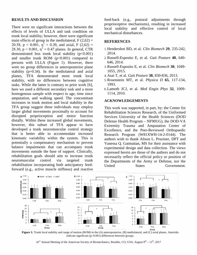
41st Annual Meeting of the American Society of Biomechanics, Boulder, CO, USA, August 8th – 11th, 2017
RESULTS AND DISCUSSION There were no significant interactions between the effects of levels of ULLA and task condition on trunk local stability; however, there were significant main effects of group in the mediolateral, F (2,62) = 20.58, p < 0.001, η2 = 0.39, and axial, F (2,62) = 28.30, p < 0.001, η2 = 0.47 planes. In general, CTR demonstrated less trunk local stability (p<0.001) and smaller trunk ROM (p<0.001) compared to persons with ULLA (Figure 1). However, there were no group differences in anteroposterior plane stability (p=0.34). In the mediolateral and axial planes, TFA demonstrated more trunk local stability, with no differences between cognitive tasks. While the latter is contrary to prior work [6], here we used a different secondary task and a more homogenous sample with respect to age, time since amputation, and walking speed. The concomitant increases in trunk motion and local stability in the TFA group suggest these individuals may employ larger global movements proximally to account for disrupted proprioception and motor function distally. Within these increased global movements, however, this subset of TFA appear to have developed a trunk neuromuscular control strategy that is better able to accommodate increased kinematic variability within the system. This is potentially a compensatory mechanism to prevent balance impairments that can accompany trunk movements outside the base of support. Clinically, rehabilitation goals should aim to increase trunk neuromuscular control via targeted trunk rehabilitation incorporating both anticipatory feed-forward (e.g., active muscle stiffness) and reactive
feed-back (e.g., postural adjustments through proprioceptive mechanisms), resulting in increased local stability and effective control of local mechanical disturbances. REFERENCES 1. Hendershot BD, et al. Clin Biomech 29, 235-242,
2014. 2. Russell-Esposito E, et al. Gait Posture 40, 640-
646, 2014. 3. Russell-Esposito E, et al. Clin Biomech 30, 1049-
1055, 2015. 4. Asai T, et al. Gait Posture 38, 830-836, 2013. 5. Rosenstein MT, et al. Physica D 65, 117-134,
1993. 6. Lamoth JCJ, et al. Med Engin Phys 32, 1009-
1114, 2010.
ACKNOWLEDGEMENTS This work was supported, in part, by: the Center for Rehabilitation Sciences Research, of the Uniformed Services University of the Health Sciences (DOD Defense Health Program – NF90UG), the DOD-VA Extremity Trauma and Amputation Center of Excellence, and the Peer-Reviewed Orthopaedic Research Program (W81XWH-14-2-0144). The authors wish to thank Alison L. Pruziner, DPT and Vanessa Q. Gatmaitan, MS for their assistance with experimental design and data collection. The views expressed herein are those of the authors and do not necessarily reflect the official policy or position of the Departments of the Army or Defense, nor the United States Government.
Figure 1: Trunk local stability and range of motion (ROM) in the (A) anteroposterior, (B) mediolateral, and (C) axial planes. Asterisks
indicate significant (p<0.001) differences between groups.
Contents lists available at ScienceDirect
Human Movement Science
journal homepage: www.elsevier.com/locate/humov
Full Length Article
Trunk and pelvic dynamics during transient turns amongindividuals with unilateral traumatic lower limb amputation
Pawel R. Golyskia, Brad D. Hendershota,b,c,⁎
a Research & Development Service, Department of Rehabilitation, Walter Reed National Military Medical Center, Bethesda, MD, USAbDoD-VA Extremity Trauma and Amputation Center of Excellence, USAcDepartment of Rehabilitation Medicine, Uniformed Services University of the Health Sciences, Bethesda, MD, USA
A R T I C L E I N F O
Keywords:Lower limb lossCoordinationMomentumTurnsBiomechanics
A B S T R A C T
Prior work has identified alterations in trunk-pelvic dynamics with lower limb amputation (LLA)during in-line walking; however, evaluations of other ambulatory tasks are limited. Turns areubiquitous in daily life but can be challenging for individuals with LLA, prompting additional orunique proximal compensations when changing direction, which over time may lead to devel-opment of low back pain. We hypothesized such proximal kinematic differences between personswith and without LLA would exist in the sagittal and frontal planes. Three-dimensional trunk andpelvic kinematics, translational and rotational momenta, and coordination phase/variabilitywere compared among eight persons with unilateral LLA (4 with transfemoral amputation and 4with transtibial amputation), and five uninjured controls, who performed 90-degree turns to theleft (n= 10) and right (n= 10). Participants self-selected the turn strategy (i.e., step vs. spin)and pivot limb in response to verbal cues regarding when and which direction to turn.Coordination variability and translational angular momenta did not differ between groups ineither turn type. During spin turns, frontal rotational angular momenta were larger and frontaltrunk-pelvis range of motion was smaller among persons with vs. without LLA. During step turns,pelvis leading transverse coordination was more frequent, frontal trunk rotational angular mo-mentum was smaller, and sagittal pelvis range of motion was larger among persons with vs.without LLA. Altered and task-dependent modulation of trunk-pelvic dynamics among personswith LLA provides additional support for a potential link between repeated exposures to alteredtrunk-pelvic dynamics with elevated low back pain risk.
1. Introduction
Persons with lower limb amputation (LLA) often walk with compensatory movement strategies involving a prominent reliance onthe trunk and pelvis (Goujon-Pillet, Sapin, Fodé, & Lavaste, 2008). Altered kinematic features and coordination of these two segmentshave been associated with elevated demands on the low back (Hendershot & Wolf, 2014), increased inter-segmental rigidity (RussellEsposito & Wilken, 2014), and larger trunk muscular forces and spinal loads (Shojaie, Hendershot, Wolf, & Bazrgari, 2016; Yoder,Petrella, & Silverman, 2015). These altered loads and asymmetric trunk-pelvis kinematics among persons with LLA have beensuggested as key factors in disc degeneration and passive ligamentous strain potentially leading to development of low back pain(LBP; Devan, Hendrick, Ribeiro, Hale, & Carman, 2014; Gailey, Allen, Castles, Kucharik, & Roeder, 2008). As such, differences intrunk/pelvis kinematics between persons with and without LLA have been characterized during in-line walking (Goujon-Pillet et al.,
https://doi.org/10.1016/j.humov.2018.01.006Received 14 April 2017; Received in revised form 6 January 2018; Accepted 9 January 2018
⁎ Corresponding author at: 4954 N. Palmer Road, America Building (19), Room B-320, Bethesda, MD 20889, USA.E-mail address: [email protected] (B.D. Hendershot).
Human Movement Science 58 (2018) 41–54
0167-9457/ Published by Elsevier B.V.
T
2008; Hendershot & Wolf, 2014; Morgenroth et al., 2010). Yet, in-line walking is but one movement among many required forfunctional independence. Thus, characterizing the extent to which persons with LLA utilize proximal compensations during other(perhaps more demanding) tasks/activities of daily living would facilitate a more comprehensive understanding of biomechanicalcontributors to LBP risk.
Transient (i.e., non-steady-state) tasks embedded within in-line walking are ubiquitous and often necessary to adequately na-vigate an environment. Turns, in particular, account for approximately half of daily steps (Glaister, Bernatz, Klute, & Orendurff, 2007;Sedgeman, Goldie, & Iansek, 1994). Biomechanically, turns require a redirection of the body’s center of mass, typically as a change indirection between 76 and 120 degrees (Sedgeman et al., 1994) executed using either a step (turn direction is contralateral to pivotleg) or spin strategy (turn direction is ipsilateral to pivot leg; Taylor, Dabnichki, & Strike, 2005). Among persons with LLA, com-promised ankle function alters control of braking/propulsive and mediolateral forces during a turn (albeit along a circular vs. or-thogonal path; Segal, Orendurff, Czerniecki, Shofer, & Klute, 2008; Ventura, Segal, Klute, & Neptune, 2011), thereby likely ne-cessitating proximal adaptations of the trunk/pelvis to adequately redirect the body’s center of mass. Furthermore, proximalcompensations during turns may also exist to minimize discomfort within the residual limb-socket interface, particularly as it relatesto torsion/shear (Heitzmann et al., 2015).
Inter-segmental coordination and momentum have been used for identification of compensational movement strategies duringambulation. For example, persons with unilateral LLA generate and arrest larger trunk and pelvic segmental momenta during walking(Gaffney, Murray, Christiansen, & Davidson, 2016), as well as alter segmental coordination strategies dependent on the presence ofcurrent LBP (Russell Esposito & Wilken, 2014). While recent efforts have similarly identified altered trunk-pelvic coordinationstrategies in able-bodied individuals (with and without LBP) executing turns (Smith & Kulig, 2016), there exist no studies specificallyfocused on trunk and pelvic compensations during turns among persons with LLA. Thus, the primary purpose of this study was tocharacterize proximal compensations using inter-segmental momenta and coordination during transient (90-degree) turns amongpersons with LLA. Although turns are predominantly associated with movement in the transverse plane, it was hypothesized thatpersons with vs. without LLA execute turns with altered trunk-pelvic segmental coordination, particularly in the sagittal and frontalplanes, to overcome the aforementioned challenges associated with modulating braking/propulsive and mediolateral forces withaltered ankle function. Secondarily, we hypothesized that such alterations in trunk-pelvic coordination would also be associated withlarger ranges of segmental momenta among persons with vs. without LLA.
2. Methods
2.1. Participants
Eight persons with unilateral LLA of traumatic etiology (four with transtibial amputation [TTA], three with transfemoral am-putation, and one with knee disarticulation [TFA]) and five persons without LLA (uninjured controls; CTRL) completed this study(Table 1). All participants provided informed consent approved by the Walter Reed National Military Medical Center InstitutionalReview Board. All participants were free of neurological and orthopaedic injury aside from lower limb amputation, were able toambulate over even terrain without an assistive device, and were not experiencing any moderate or severe discomfort/pain, re-gardless of cause, at any point during data collection, as measured by overall pain scores less than 4 cm on a 10 cm Visual AnalogScale (Jensen, Chen, & Brugger, 2003). Of the persons with TTA, 2 wore the RUSH and 2 wore the Vari-Flex XC foot. Of the personswith TFA or knee disarticulation, 2 wore the X3 microprocessor knee and Vari-Flex XC foot, 1 wore the X2 microprocessor knee andVari-Flex XC foot, and 1 wore the Total Knee 2100 mechanical knee and Vari-Flex XC foot.
Table 1Demographic information by participant category (CTRL=uninjured controls, TTA=persons with transtibial amputation, and TFA=persons with transfemoralamputation or knee disarticulation). Note, there were no significant differences in demographic information or walking speeds (all P > .167).
Age (yr) Months Since Amputation Height (m) Mass (kg) In-line Walking Speed (m/s)
CTRL 20 1.8 61.5 1.428 1.7 88.4 1.431 1.9 105.7 1.428 1.9 72.6 1.329 1.8 83.5 1.3
TTA 24 5.5 1.8 90.9 1.427 47.8 1.8 106.9 1.434 133.3 1.9 89.9 1.545 17.7 1.8 135.6 1.5
TFA 34 59.7 1.7 71.4 1.123 15.8 1.9 96.2 1.426 59.0 1.7 74.9 1.425 32.9 1.7 101.2 1.2
P.R. Golyski, B.D. Hendershot Human Movement Science 58 (2018) 41–54
42
2.2. Experimental procedures
Each participant performed 20 turns involving a 90-degree change in direction to the left (n= 10) and right (n=10).Participants walked at their self-selected speed along a 12-foot straight path and were verbally cued to turn left or right at a specifiedand consistent location (approximately 6 feet away from the turning point, allowing the participant to ultimately self-select the pivotlimb). Turn direction was randomized, and no specific guidance was provided for which foot or type of turn (i.e., step vs. spin) toemploy. Full-body kinematics were collected by tracking (120 Hz) 70 reflective markers with a 27-camera motion capture system(Vicon, Oxford, UK). Markers were placed on the C7 and T10 spinous processes, sternal notch, xiphoid process, and bilaterally on theacromia, ASIS, and PSIS. Lower and upper extremities were tracked as 6 DOF segments, with markers placed accordingly (Collins,Ghoussayni, Ewins, & Kent, 2009). All kinematic data were filtered at 6 Hz using a 5th order Butterworth filter.
2.3. Dependent measures and data analyses
The pivot foot and type of turn (step or spin) were first determined using a previously described, automated method (cf. Golyski &Hendershot, 2017), and heel strike/toe-off events were calculated using the position of the feet relative to the pelvis (Zeni, Richards,& Higginson, 2008). Step lengths were calculated for the step leading into pivot and the step after pivot as the absolute distancebetween the positions of heel strikes of each respective step relative to pivot. Stride widths were evaluated using the heel strikepositions of the steps before, during, and after the turn (Huxham, Gong, Baker, Morris, & Iansek, 2006).
Three-dimensional trunk segmental kinematics were computed, relative to the pelvis, using Visual3D (Version 5.02.27, C-MotionInc., Germantown, MD, USA), with local coordinate systems defined by a static calibration trial. Trunk-pelvis range of motion wascalculated for each plane over the period from heel strike of the step before pivot to the toe-off of the step after pivot. Individual trunkand pelvic segmental trajectories were also computed and exported to MATLAB (Release 2015a, The MathWorks, Inc. Natick, MA,USA). Using these, tri-planar translational (Eq. (1)) and rotational (Eq. (2)) angular momenta of the trunk and pelvis segments werecalculated as described by Gaffney et al. (2016), and normalized by each participant’s body mass, height, and self-selected in-linewalking speed (Herr & Popovic, 2008). Translational angular momentum (TAM) for the trunk and pelvis segments was calculated as:
= − × −h r r v vm( ) ( )i Foot i Foot i i Foot/ (1)
where ri is the position vector of the segment’s center of mass, rFoot is the position vector of the pivot foot, mi is the mass of thesegment, vi is the velocity vector of the segment’s center of mass, and vFoot is the velocity vector of the pivot foot. TAM was evaluatedonly during the period from heel strike before the turn to toe-off after the turn (i.e. pivot stance). Rotational angular momentum(RAM) for the trunk and pelvis segments was calculated as:
=h I w·i i i (2)
where Ii is the moment of inertia tensor for the segment of interest and wi is the segment’s angular velocity vector. RAM wasevaluated during the same period as trunk-pelvis range of motion. Both TAM and RAM were resolved in the three planes of motion,defined using center of mass velocity (to define a forward direction), gravity, and the resulting cross product; TAM and RAM ranges(i.e., max-min) were extracted within each plane for subsequent analyses.
Finally, inter-segmental coordination of the trunk and pelvis in each plane of movement was calculated using a vector codingmethod described by Needham, Naemi, and Chockalingam (2014). For this, each turn was subsequently divided into two phases: (1)pivot stance; defined as the period from heel strike to toe-off of the foot in stance during the apex of the turn, and (2) pivot swing;defined as the period from pivot foot toe-off to subsequent ipsilateral heel strike. Time-series trajectories of the trunk and pelvic angledefined a 0–360° relative coupling angle, which at each time point is separated into one of eight 45° bins to evaluate the frequency ofa given coordination mode (in-phase, anti-phase, trunk-phase, and pelvic-phase) in both pivot stance and pivot swing; circularstatistics were used to define the mean and variability of each coupling angle/phase while preserving directionality of the trunk-pelvis relative coupling angle (Hamill, Haddad, & McDermott, 2000; Needham et al., 2014; Watson & Batschelet, 1982). Note, acommon alternative method for assessing segmental coordination is continuous relative phase, but this method does not explicitlyquantify trunk- and pelvic-phase coordination modes (i.e., dominance of a given segment).
2.4. Statistical analyses
Given that turn type was not controlled as part of the experimental design (i.e., the pivot foot was selected by the participant), andno a priori hypotheses were formulated as to how turn type would influence the dependent variables, no explicit comparisons weremade between turn strategies. Instead, Mann-Whitney U tests were used to compare all dependent measures between persons withLLA vs. CTRL, separately within each turn type; statistical significance was concluded at P < .050. All statistical analyses wereperformed in SPSS (version 21.0; IBM SPSS Inc., Chicago, IL). Unless otherwise specified, data are reported as medians (interquartileranges). In total, 60 (of 80) trials/turns from persons with TFA, 71 (of 80) from persons with TTA, and 77 (of 100) from personswithout LLA were included as part of subsequent analyses due to marker drop out and/or in-line walking periods of insufficientlength before and after the turn.
P.R. Golyski, B.D. Hendershot Human Movement Science 58 (2018) 41–54
43
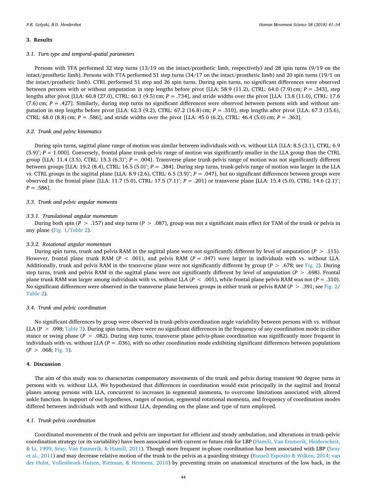
3. Results
3.1. Turn type and temporal-spatial parameters
Persons with TFA performed 32 step turns (13/19 on the intact/prosthetic limb, respectively) and 28 spin turns (9/19 on theintact/prosthetic limb). Persons with TTA performed 51 step turns (34/17 on the intact/prosthetic limb) and 20 spin turns (19/1 onthe intact/prosthetic limb). CTRL performed 51 step and 26 spin turns. During spin turns, no significant differences were observedbetween persons with or without amputation in step lengths before pivot [LLA: 58.9 (11.2), CTRL: 64.0 (7.9) cm; P= .343], steplengths after pivot [LLA: 60.8 (27.0), CTRL: 60.1 (9.5) cm; P= .734], and stride widths over the pivot [LLA: 13.8 (11.0), CTRL: 17.6(7.6) cm; P= .427]. Similarly, during step turns no significant differences were observed between persons with and without am-putation in step lengths before pivot [LLA: 62.3 (9.2), CTRL: 67.2 (16.8) cm; P= .310], step lengths after pivot [LLA: 67.3 (15.6),CTRL: 68.0 (8.8) cm; P= .586], and stride widths over the pivot [LLA: 45.0 (6.2), CTRL: 46.4 (5.0) cm; P= .363].
3.2. Trunk and pelvic kinematics
During spin turns, sagittal plane range of motion was similar between individuals with vs. without LLA [LLA: 8.5 (3.1), CTRL: 6.9(5.9)°; P=1.000]. Conversely, frontal plane trunk-pelvis range of motion was significantly smaller in the LLA group than the CTRLgroup [LLA: 11.4 (3.5), CTRL: 15.3 (6.3)°; P= .004]. Transverse plane trunk-pelvis range of motion was not significantly differentbetween groups [LLA: 19.2 (8.4), CTRL: 16.5 (5.0)°; P= .384]. During step turns, trunk-pelvis range of motion was larger in the LLAvs. CTRL groups in the sagittal plane [LLA: 8.9 (2.6), CTRL: 6.5 (3.9)°; P= .047], but no significant differences between groups wereobserved in the frontal plane [LLA: 11.7 (5.0), CTRL: 17.5 (7.1)°; P= .201] or transverse plane [LLA: 15.4 (5.0), CTRL: 14.6 (2.1)°;P= .586].
3.3. Trunk and pelvic angular momenta
3.3.1. Translational angular momentumDuring both spin (P > .157) and step turns (P > .087), group was not a significant main effect for TAM of the trunk or pelvis in
any plane (Fig. 1/Table 2).
3.3.2. Rotational angular momentumDuring spin turns, trunk and pelvis RAM in the sagittal plane were not significantly different by level of amputation (P > .115).
However, frontal plane trunk RAM (P < .001), and pelvis RAM (P= .047) were larger in individuals with vs. without LLA.Additionally, trunk and pelvis RAM in the transverse plane were not significantly different by group (P > .678; see Fig. 2). Duringstep turns, trunk and pelvis RAM in the sagittal plane were not significantly different by level of amputation (P > .698). Frontalplane trunk RAM was larger among individuals with vs. without LLA (P < .001), while frontal plane pelvis RAM was not (P= .310).No significant differences were observed in the transverse plane between groups in either trunk or pelvis RAM (P > .391; see Fig. 2/Table 2).
3.4. Trunk and pelvic coordination
No significant differences by group were observed in trunk-pelvis coordination angle variability between persons with vs. withoutLLA (P > .098; Table 3). During spin turns, there were no significant differences in the frequency of any coordination mode in eitherstance or swing phase (P > .082). During step turns, transverse plane pelvis-phase coordination was significantly more frequent inindividuals with vs. without LLA (P= .036), with no other coordination mode exhibiting significant differences between populations(P > .068; Fig. 3).
4. Discussion
The aim of this study was to characterize compensatory movements of the trunk and pelvis during transient 90 degree turns inpersons with vs. without LLA. We hypothesized that differences in coordination would exist principally in the sagittal and frontalplanes among persons with LLA, concurrent to increases in segmental momenta, to overcome limitations associated with alteredankle function. In support of our hypotheses, ranges of motion, segmental rotational momenta, and frequency of coordination modesdiffered between individuals with and without LLA, depending on the plane and type of turn employed.
4.1. Trunk-pelvis coordination
Coordinated movements of the trunk and pelvis are important for efficient and steady ambulation, and alterations in trunk-pelviccoordination strategy (or its variability) have been associated with current or future risk for LBP (Hamill, Van Emmerik, Heiderscheit,& Li, 1999; Seay, Van Emmerik, & Hamill, 2011). Though more frequent in-phase coordination has been associated with LBP (Seayet al., 2011) and may decrease relative motion of the trunk to the pelvis as a guarding strategy (Russell Esposito & Wilken, 2014; vander Hulst, Vollenbroek-Hutten, Rietman, & Hermens, 2010) by preventing strain on anatomical structures of the low back, in the
P.R. Golyski, B.D. Hendershot Human Movement Science 58 (2018) 41–54
44
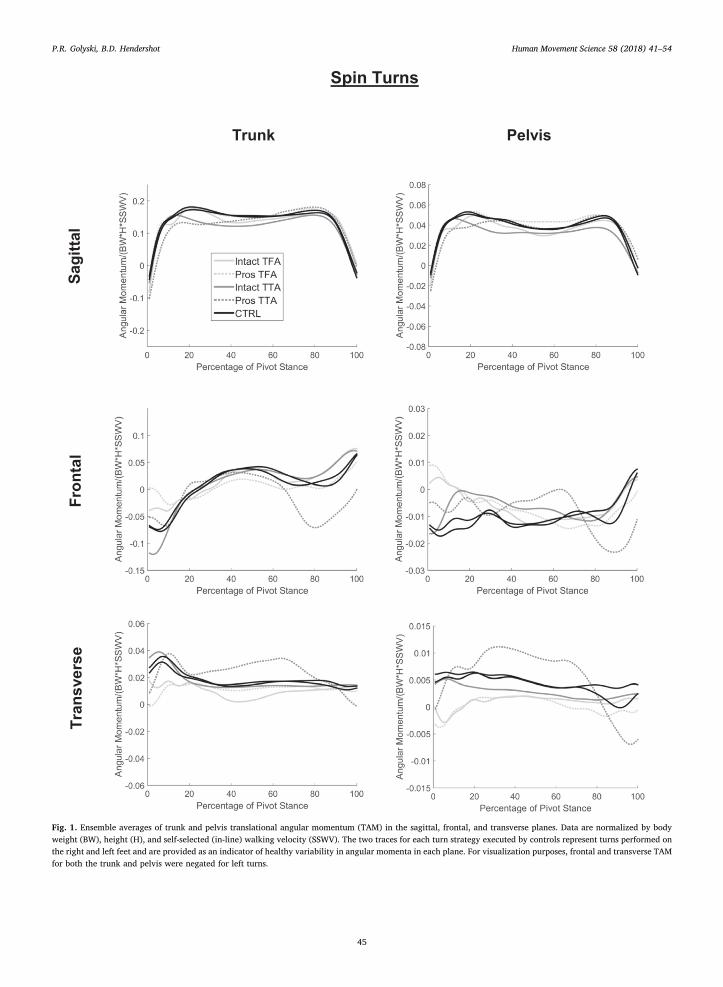
Fig. 1. Ensemble averages of trunk and pelvis translational angular momentum (TAM) in the sagittal, frontal, and transverse planes. Data are normalized by bodyweight (BW), height (H), and self-selected (in-line) walking velocity (SSWV). The two traces for each turn strategy executed by controls represent turns performed onthe right and left feet and are provided as an indicator of healthy variability in angular momenta in each plane. For visualization purposes, frontal and transverse TAMfor both the trunk and pelvis were negated for left turns.
P.R. Golyski, B.D. Hendershot Human Movement Science 58 (2018) 41–54
45
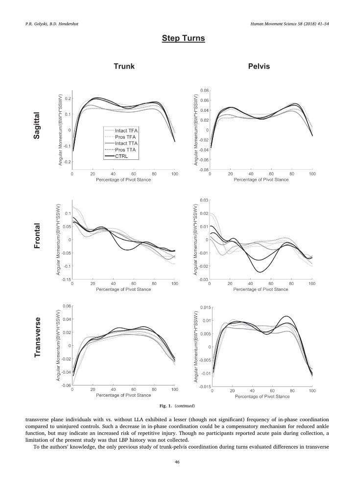
transverse plane individuals with vs. without LLA exhibited a lesser (though not significant) frequency of in-phase coordinationcompared to uninjured controls. Such a decrease in in-phase coordination could be a compensatory mechanism for reduced anklefunction, but may indicate an increased risk of repetitive injury. Though no participants reported acute pain during collection, alimitation of the present study was that LBP history was not collected.
To the authors’ knowledge, the only previous study of trunk-pelvis coordination during turns evaluated differences in transverse
Fig. 1. (continued)
P.R. Golyski, B.D. Hendershot Human Movement Science 58 (2018) 41–54
46
plane coordination only, in persons with and without LBP during spin turns (Smith & Kulig, 2016). The dominant in-phase co-ordination in the transverse plane during stance was consistent with this work, though no significant differences were found betweengroups.
Characterization of trunk-pelvis coordination during in-line walking (Russell Esposito & Wilken, 2014) found higher frequenciesof anti-phase coordination in individuals with TFA relative to uninjured controls in the sagittal and frontal planes. In contrast, weonly observed a significant increase in transverse plane pelvic-phase coordination in persons with vs. without LLA during step turns,which are more biomechanically similar to in-line walking than spin turns (Taylor et al., 2005). In contrast to the hypothesizedchanges in sagittal and frontal coordination, the only significant difference between groups was in the transverse plane during stepturns, which nonetheless suggests alternative proximal movement strategies within the LLA population. In support of our hypothesis,differences between populations were observed in trunk-pelvis range of motion and angular momenta in the sagittal and frontalplanes.
4.2. Sagittal plane
Significantly larger sagittal trunk-pelvis range of motion during step turns among persons with vs. without LLA is consistent withprevious observations of trunk-pelvis kinematics during in-line walking (Goujon-Pillet et al., 2008). Such larger trunk flexion anglesmay be a compensation to facilitate hip extension, which is hampered by hip flexion contractures (Gailey et al., 2008), but thismotion may also increase demand on trunk extensors (Hendershot & Wolf, 2014). Moreover, this more extreme sagittal trunk-pelvicmovement is also consistent with the larger (though not significant) observed pelvis RAM in persons with vs. without LLA, and inagreement with a previous study of in-line walking (Gaffney et al., 2016).
Table 2Median (interquartile range) ranges in trunk and pelvic translational angular momenta (TAM) and rotational angular momenta (RAM) for individuals withunilateral lower limb amputation (LLA) and uninjured controls (CTRL), during spin/step turns. TAM was calculated during pivot stance. RAM was calculatedduring the period from heel strike of the step before the pivot step to toe-off of the step after the pivot step, respectively. For metrics marked by * and **,groups were significantly different at the α=0.05 and α=0.001 levels, respectively. All momenta are normalized by body weight, height, and self-selected(in-line) walking speed.
Spin Turns
LLA CTRL
Sagittal Trunk TAM Range 0.2781 (0.0824) 0.2645 (0.0608)Pelvis TAM Range 0.0733 (0.0206) 0.0695 (0.0172)Trunk RAM Range 0.0022 (0.0011) 0.0021 (0.0008)Pelvis RAM Range 0.0004 (0.0002) 0.0003 (0.0001)
Frontal Trunk TAM Range 0.1420 (0.1087) 0.1474 (0.0927)Pelvis TAM Range 0.0337 (0.0179) 0.0320 (0.0213)Trunk RAM Range** 0.0055 (0.0022) 0.0038 (0.0011)Pelvis RAM Range* 0.0006 (0.0004) 0.0005 (0.0001)
Transverse Trunk TAM Range 0.0313 (0.0158) 0.0278 (0.0189)Pelvis TAM Range 0.0135 (0.0076) 0.0084 (0.0058)Trunk RAM Range 0.0059 (0.0023) 0.0056 (0.0021)Pelvis RAM Range 0.0014 (0.0010) 0.0017 (0.0007)
Step Turns
LLA CTRL
Sagittal Trunk TAM Range 0.2931 (0.0506) 0.3095 (0.0591)Pelvis TAM Range 0.0808 (0.0221) 0.0835 (0.0162)Trunk RAM Range 0.0023 (0.0013) 0.0024 (0.0008)Pelvis RAM Range 0.0004 (0.0003) 0.0003 (0.0001)
Frontal Trunk TAM Range 0.1817 (0.1046) 0.1580 (0.0539)Pelvis TAM Range 0.0326 (0.0204) 0.0385 (0.0215)Trunk RAM Range** 0.0046 (0.0021) 0.0030 (0.0005)Pelvis RAM Range 0.0006 (0.0004) 0.0005 (0.0002)
Transverse Trunk TAM Range 0.0683 (0.0182) 0.0717 (0.0210)Pelvis TAM Range 0.0235 (0.0090) 0.0270 (0.0082)Trunk RAM Range 0.0058 (0.0034) 0.0070 (0.0030)Pelvis RAM Range 0.0015 (0.0011) 0.0014 (0.0006)
Units: Angular Momentum/(Body Weight*Height*Self Selected Walking Velocity).
P.R. Golyski, B.D. Hendershot Human Movement Science 58 (2018) 41–54
47
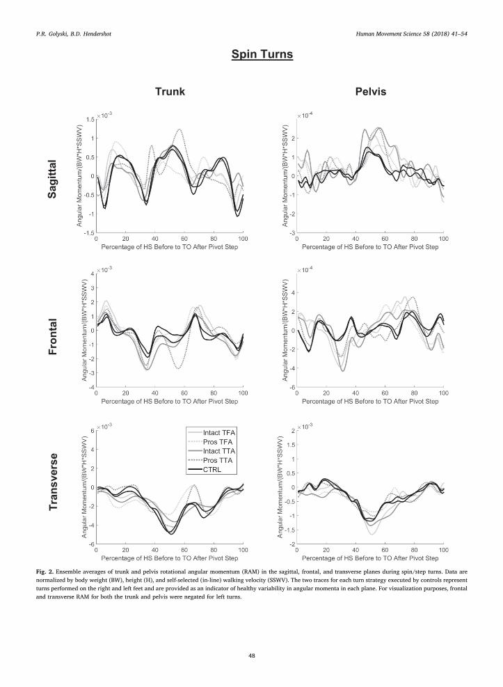
Fig. 2. Ensemble averages of trunk and pelvis rotational angular momentum (RAM) in the sagittal, frontal, and transverse planes during spin/step turns. Data arenormalized by body weight (BW), height (H), and self-selected (in-line) walking velocity (SSWV). The two traces for each turn strategy executed by controls representturns performed on the right and left feet and are provided as an indicator of healthy variability in angular momenta in each plane. For visualization purposes, frontaland transverse RAM for both the trunk and pelvis were negated for left turns.
P.R. Golyski, B.D. Hendershot Human Movement Science 58 (2018) 41–54
48
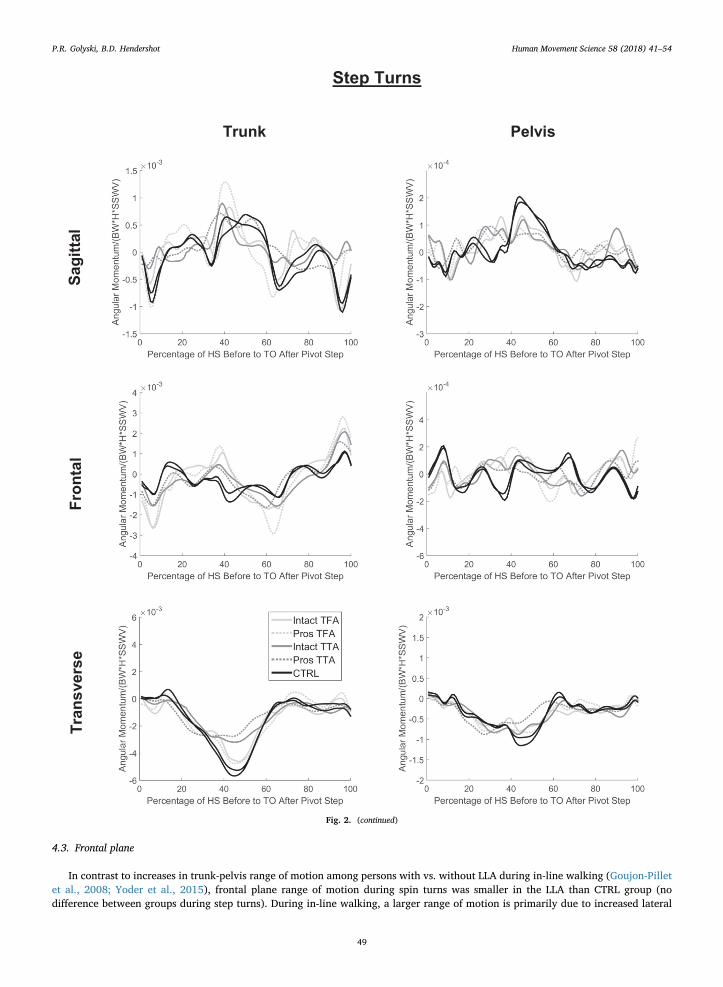
4.3. Frontal plane
In contrast to increases in trunk-pelvis range of motion among persons with vs. without LLA during in-line walking (Goujon-Pilletet al., 2008; Yoder et al., 2015), frontal plane range of motion during spin turns was smaller in the LLA than CTRL group (nodifference between groups during step turns). During in-line walking, a larger range of motion is primarily due to increased lateral
Fig. 2. (continued)
P.R. Golyski, B.D. Hendershot Human Movement Science 58 (2018) 41–54
49
trunk lean over the prosthetic limb and is considered a compensation, at least in part, for reduced residual limb function (Hendershot& Wolf, 2014; Rueda et al., 2013). Future studies exploring turns on the intact vs. prosthetic side may elucidate the basis for reducedfrontal plane range of motion, though we speculate the relative decrease in lateral trunk lean throughout turns may be a result of themore proximal (i.e., hip vs. ankle) strategy and generally not leaning into/away from the turn to minimize excursions of the bodycenter of mass and improve stability (Ventura et al., 2011). Despite the trends in frontal plane trunk-pelvis range of motion beinginconsistent with those of existing literature, differences in frontal plane trunk RAM (which is dependent on segmental angularvelocity) between groups during both turn types were apparent. Such differences are consistent with our hypothesis and previouswork identifying larger ranges in whole body frontal plane angular momentum in persons with LLA (albeit during in-line walking;Silverman & Neptune, 2011). Large changes in whole-body angular momentum in the frontal plane have also been correlated withpoorer clinical balance outcomes post-stroke (Nott, Neptune, & Kautz, 2014). Moreover, such deviations in trunk and pelvis angularmomentum in the frontal plane are of particular interest since these segments are the principal contributors to whole body angularmomentum in the frontal plane (Herr & Popovic, 2008). During spin turns the more extreme frontal trunk angular velocity coupledwith smaller trunk-pelvic range of motion could suggest a trunk-stiffening strategy (Arendt-Nielsen, Graven-Nielsen, Svarrer, &Svensson, 1996; Lamoth et al., 2002), similar to the segmental rigidity identified among persons with TFA during in-line walking(Russell Esposito & Wilken, 2014); however, such a stiffening strategy would likely be associated with increased in-phase co-ordination (Wu et al., 2014) – a trend we did not observe here with the vector coding method.
4.4. Transverse plane
Larger axial trunk rotations have been observed in persons with TFA during in-line walking (Goujon-Pillet et al., 2008), which areconcerning given the association of such rotations with LBP (Fujiwara et al., 2000; Morgenroth, Medverd, Seyedali, & Czerniecki,2014). We did not observe differences in transverse plane trunk-pelvis range of motion, though this could be attributed to turnsrequiring more control over transverse plane angular displacements. However, during step turns, range in transverse trunk RAM wassmaller, albeit not significantly, in persons with LLA compared to uninjured controls. As illustrated in Fig. 2, at approximately 50% ofthe turn the trunk RAM was smaller for turns on both the prosthetic and intact limbs in persons with TFA and TTA vs. controls,indicating a smaller peak trunk angular velocity in the LLA group. This contradicts previous preliminary findings which suggestedpersons with unilateral TTA execute step turns with larger transverse trunk angular velocities than uninjured controls (Taylor &Strike, 2009).
Though the host of kinematic differences between turns and in-line walking (Taylor et al., 2005) precludes direct comparison ofangular momentum components to previous work, qualitatively, transverse trunk and pelvis RAM were the most different in shapebetween the two ambulation tasks (c.f. Gaffney et al., 2016), stemming from the seemingly necessary peak in transverse angularvelocity. Moreover, differences in the range of TAM/RAM between in-line walking and turns were most pronounced in the transverseplane, and were larger during transient turns by factors of 2 and 3 for TAM and RAM, respectively.
Table 3Median (interquartile range) variability of trunk-pelvis coupling angle during spin/step turns in stance/swing of the pivot limb for persons withunilateral lower limb amputation (LLA), and uninjured controls (CTRL). No significant differences were found between groups at the α=0.05level.
Spin Turns
LLA CTRL
Sagittal Pivot Stance (°) 21.7 (33.9) 23.2 (17.6)Pivot Swing (°) 8.7 (20.2) 19.6 (15.0)
Frontal Pivot Stance (°) 10.8 (27.1) 14.7 (18.7)Pivot Swing (°) 20.1 (33.5) 27.5 (25.6)
Transverse Pivot Stance (°) 12.1 (21.1) 8.8 (6.2)Pivot Swing (°) 18.0 (43.0) 26.0 (20.1)
Step Turns
LLA CTRL
Sagittal Pivot Stance (°) 23.2 (7.5) 22.7 (17.1)Pivot Swing (°) 22.2 (31.2) 13.8 (19.9)
Frontal Pivot Stance (°) 15.9 (9.1) 13.5 (8.0)Pivot Swing (°) 28.2 (31.9) 28.1 (20.9)
Transverse Pivot Stance (°) 14.7 (6.0) 12.3 (10.4)Pivot Swing (°) 23.5 (26.6) 24.9 (15.7)
P.R. Golyski, B.D. Hendershot Human Movement Science 58 (2018) 41–54
50
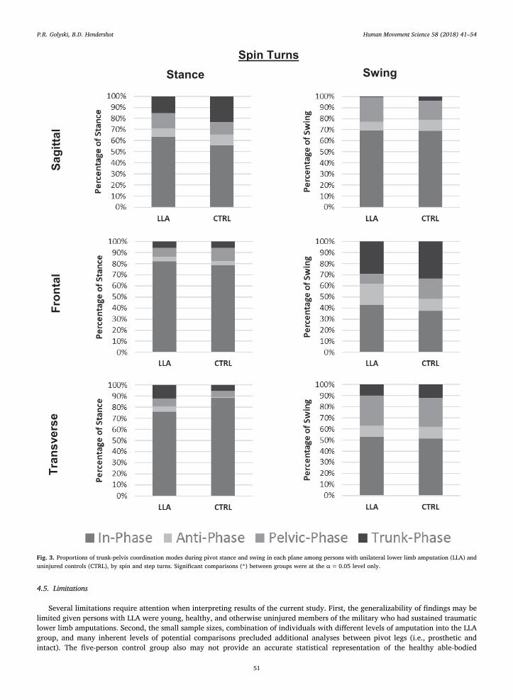
4.5. Limitations
Several limitations require attention when interpreting results of the current study. First, the generalizability of findings may belimited given persons with LLA were young, healthy, and otherwise uninjured members of the military who had sustained traumaticlower limb amputations. Second, the small sample sizes, combination of individuals with different levels of amputation into the LLAgroup, and many inherent levels of potential comparisons precluded additional analyses between pivot legs (i.e., prosthetic andintact). The five-person control group also may not provide an accurate statistical representation of the healthy able-bodied
Fig. 3. Proportions of trunk-pelvis coordination modes during pivot stance and swing in each plane among persons with unilateral lower limb amputation (LLA) anduninjured controls (CTRL), by spin and step turns. Significant comparisons (*) between groups were at the α=0.05 level only.
P.R. Golyski, B.D. Hendershot Human Movement Science 58 (2018) 41–54
51
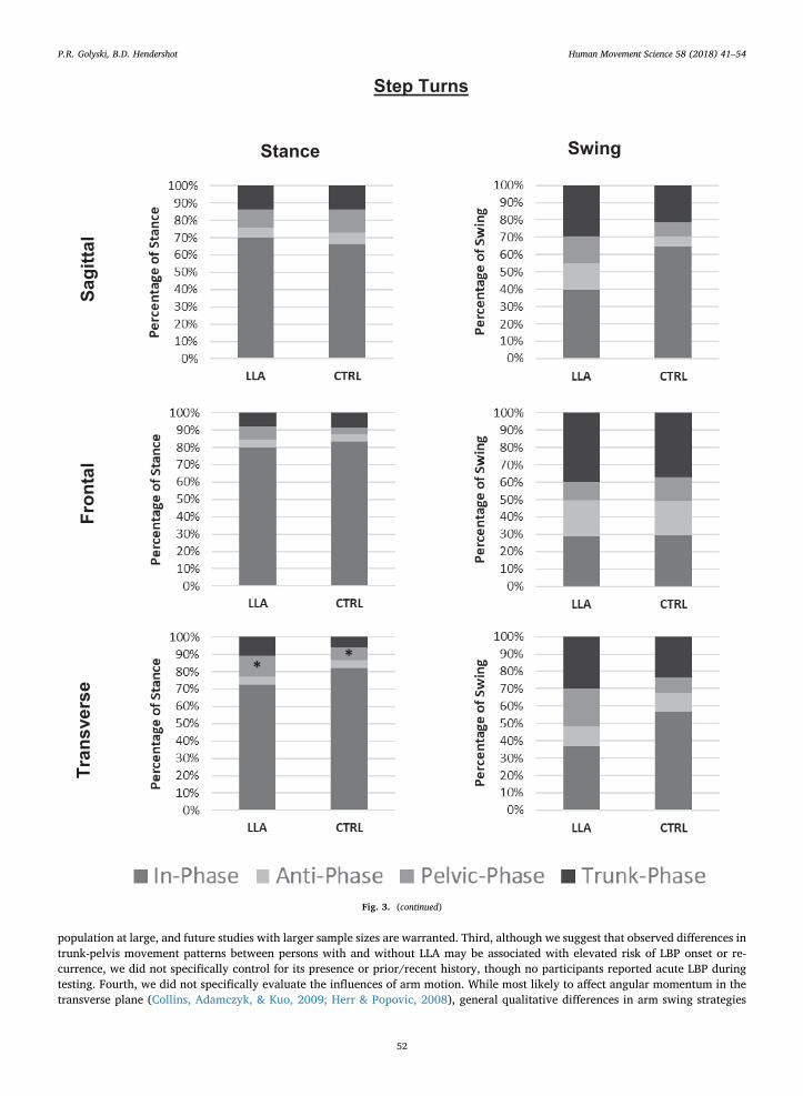
population at large, and future studies with larger sample sizes are warranted. Third, although we suggest that observed differences intrunk-pelvis movement patterns between persons with and without LLA may be associated with elevated risk of LBP onset or re-currence, we did not specifically control for its presence or prior/recent history, though no participants reported acute LBP duringtesting. Fourth, we did not specifically evaluate the influences of arm motion. While most likely to affect angular momentum in thetransverse plane (Collins, Adamczyk, & Kuo, 2009; Herr & Popovic, 2008), general qualitative differences in arm swing strategies
Fig. 3. (continued)
P.R. Golyski, B.D. Hendershot Human Movement Science 58 (2018) 41–54
52

between groups were not observed. Finally, the turn cueing paradigm used was intended to represent transient changes in directionencountered in daily life, though the somewhat unpredictable, verbally-cued direction may have resulted in events that are moredifficult to reproduce than turns in other studies wherein participants walked along a more consistent circular path (Segal et al.,2008; Ventura et al., 2011). Future work can control for such variability with alternative cueing methods (e.g., visual, compared toour auditory cues; Heitzmann et al., 2015), thereby also supporting explicit comparisons between step vs. spin turns, and potentialinteractions with the chosen pivot limb (i.e., prosthetic vs. intact).
4.6. Summary
We compared features of trunk-pelvic segmental motion and coordination between persons with and without LLA during 90-degree turns executed using self-selected step and spin strategies. We observed differences in the frequencies of inter-segmentalcoordination, trunk-pelvis ranges of motion, and segmental momenta across levels of amputation, depending on the plane andmethod of turn employed. Nevertheless, the identified compensatory adaptations used by persons with unilateral LLA to execute thiscommon, but biomechanically challenging, task may be “maladaptive” and thus predispose these individuals to developing LBP (orits recurrence) with repeated exposure over the longer term.
Acknowledgements
This work was supported, in part, by the DoD-VA Extremity Trauma and Amputation Center of Excellence, the BADER Consortiumvia the Congressionally Designated Medical Research Program (Award number W81XWH-11-2-0222), and the Office of the AssistantSecretary of Defense for Health Affairs through the Peer Reviewed Orthopaedic Research Program (award # W81XWH-14-2-0144).The authors also wish to acknowledge Ms. Elizabeth Bell and Dr. Erik Wolf for their assistance with initial data collection andprocessing.
References
Arendt-Nielsen, L., Graven-Nielsen, T., Svarrer, H., & Svensson, P. (1996). The influence of low back pain on muscle activity and coordination during gait: A clinicaland experimental study. Pain, 64(2), 231–240. http://dx.doi.org/10.1016/0304-3959(95)00115-8.
Collins, S. H., Adamczyk, P. G., & Kuo, A. D. (2009). Dynamic arm swinging in human walking. Proceedings. Biological Sciences/The Royal Society, 276(1673),3679–3688. http://dx.doi.org/10.1098/rspb.2009.0664.
Collins, T. D., Ghoussayni, S. N., Ewins, D. J., & Kent, J. A. (2009). A six degrees-of-freedom marker set for gait analysis: Repeatability and comparison with a modifiedHelen Hayes set. Gait and Posture, 30(2), 173–180. http://dx.doi.org/10.1016/j.gaitpost.2009.04.004.
Devan, H., Hendrick, P., Ribeiro, D. C., Hale, L. A., & Carman, A. (2014). Asymmetrical movements of the lumbopelvic region: Is this a potential mechanism for lowback pain in people with lower limb amputation? Medical Hypotheses, 82(1), 77–85. http://dx.doi.org/10.1016/j.mehy.2013.11.012.
Fujiwara, A., Lim, T., An, H. S., Tanaka, N., Jeon, C. H., Andersson, G. B., & Haughton, V. M. (2000). The effect of disc degeneration and facet joint osteoarthritis on thesegmental flexibility of the lumbar spine. Spine, 25(23), 3036–3044. http://dx.doi.org/10.1097/00007632-200012010-00011.
Gaffney, B. M., Murray, A. M., Christiansen, C. L., & Davidson, B. S. (2016). Identification of trunk and pelvis movement compensations in patients with transtibialamputation using angular momentum separation. Gait and Posture, 45, 151–156. http://dx.doi.org/10.1016/j.gaitpost.2016.01.015.
Gailey, R., Allen, K., Castles, J., Kucharik, J., & Roeder, M. (2008). Review of secondary physical conditions associated with lower-limb amputation and long-termprosthesis use. Journal of Rehabilitation Research and Development, 45(1), 15–30. http://dx.doi.org/10.1682/JRRD.2006.11.0147.
Glaister, B. C., Bernatz, G. C., Klute, G. K., & Orendurff, M. S. (2007). Video task analysis of turning during activities of daily living. Gait and Posture, 25(2), 289–294.http://dx.doi.org/10.1016/j.gaitpost.2006.04.003.
Golyski, P. R., & Hendershot, B. D. (2017). A computational algorithm for classifying step and spin turns using pelvic center of mass trajectory and foot position.Journal of Biomechanics, 54, 96–100. http://dx.doi.org/10.1016/j.jbiomech.2017.01.023.
Goujon-Pillet, H., Sapin, E., Fodé, P., & Lavaste, F. (2008). Three-dimensional motions of trunk and pelvis during transfemoral amputee gait. Archives of PhysicalMedicine and Rehabilitation, 89(1), 87–94. http://dx.doi.org/10.1016/j.apmr.2007.08.136.
Hamill, J., Haddad, J. M., & McDermott, W. J. (2000). Issues in quantifying variability from a dynamical systems perspective. Journal of Applied Biomechanics, 16(4),407–418. http://dx.doi.org/10.1123/jab.16.4.407.
Hamill, J., Van Emmerik, R. E. A., Heiderscheit, B. C., & Li, L. (1999). A dynamical systems approach to lower extremity running injuries. Clinical Biomechanics, 14(5),297–308. http://dx.doi.org/10.1016/S0268-0033(98)90092-4.
Heitzmann, D. W., Pieschel, K., Alimusaj, M., Block, J., Putz, C., & Wolf, S. I. (2015). Functional effects of a prosthetic torsion adapter in trans-tibial amputees duringunplanned spin and step turns. Prosthetics and Orthotics International, 40(5), 558–565. http://dx.doi.org/10.1177/0309364615592698.
Hendershot, B. D., & Wolf, E. J. (2014). Three-dimensional joint reaction forces and moments at the low back during over-ground walking in persons with unilaterallower-extremity amputation. Clinical Biomechanics, 29(3), 235–242. http://dx.doi.org/10.1016/j.clinbiomech.2013.12.005.
Herr, H., & Popovic, M. (2008). Angular momentum in human walking. The Journal of Experimental Biology, 211(Pt 4), 467–481. http://dx.doi.org/10.1242/jeb.008573.
Huxham, F., Gong, J., Baker, R., Morris, M., & Iansek, R. (2006). Defining spatial parameters for non-linear walking. Gait and Posture, 23(2), 159–163. http://dx.doi.org/10.1016/j.gaitpost.2005.01.001.
Jensen, M. P., Chen, C., & Brugger, A. M. (2003). Interpretation of visual analog scale ratings and change scores: A reanalysis of two clinical trials of postoperative pain.Journal of Pain, 4(7), 407–414. http://dx.doi.org/10.1016/S1526-5900(03)00716-8.
Lamoth, C. J. C., Meijer, O. G., Wuisman, P. I. J. M., van Dieën, J. H., Levin, M. F., & Beek, P. J. (2002). Pelvis-thorax coordination in the transverse plane duringwalking in persons with nonspecific low back pain. Spine, 27(4), E92–E99. http://dx.doi.org/10.1097/00007632-200202150-00016.
Morgenroth, D. C., Medverd, J. R., Seyedali, M., & Czerniecki, J. M. (2014). The relationship between knee joint loading rate during walking and degenerative changeson magnetic resonance imaging. Clinical Biomechanics (Bristol, Avon), 29(6), 664–670. http://dx.doi.org/10.1016/j.clinbiomech.2014.04.008.
Morgenroth, D. C., Orendurff, M. S., Shakir, A., Segal, A., Shofer, J., & Czerniecki, J. M. (2010). The relationship between lumbar spine kinematics during gait and low-back pain in transfemoral amputees. American Journal of Physical Medicine & Rehabilitation, 89(8), 635–643. http://dx.doi.org/10.1097/PHM.0b013e3181e71d90.
Needham, R., Naemi, R., & Chockalingam, N. (2014). Quantifying lumbar-pelvis coordination during gait using a modified vector coding technique. Journal ofBiomechanics, 47(5), 1020–1026. http://dx.doi.org/10.1016/j.jbiomech.2013.12.032.
Nott, C. R., Neptune, R. R., & Kautz, S. A. (2014). Relationships between frontal-plane angular momentum and clinical balance measures during post-stroke hemi-paretic walking. Gait and Posture, 39(1), 129–134. http://dx.doi.org/10.1016/j.gaitpost.2013.06.008.
Rueda, F. M., Diego, I. M. A., Sánchez, A. M., Tejada, M. C., Montero, F. M. R., & Page, J. C. M. (2013). Knee and hip internal moments and upper-body kinematics in
P.R. Golyski, B.D. Hendershot Human Movement Science 58 (2018) 41–54
53
the frontal plane in unilateral transtibial amputees. Gait and Posture, 37(3), 436–439. http://dx.doi.org/10.1016/j.gaitpost.2012.08.019.Russell Esposito, E., & Wilken, J. M. (2014). The relationship between pelvis-trunk coordination and low back pain in individuals with transfemoral amputations. Gait
and Posture, 40(4), 640–646. http://dx.doi.org/10.1016/j.gaitpost.2014.07.019.Seay, J. F., Van Emmerik, R. E. A., & Hamill, J. (2011). Low back pain status affects pelvis-trunk coordination and variability during walking and running. Clinical
Biomechanics, 26(6), 572–578. http://dx.doi.org/10.1016/j.clinbiomech.2010.11.012.Sedgeman, R., Goldie, P., & Iansek, R. (1994). Development of a measure of turning during walking. Advancing rehabilitation conference proceedings La Trobe University
1994 (pp. 26–31). Retrieved from file:///L:/S/Sedgeman R et al 1994.pdf.Segal, A. D., Orendurff, M. S., Czerniecki, J. M., Shofer, J. B., & Klute, G. K. (2008). Local dynamic stability in turning and straight-line gait. Journal of Biomechanics,
41(7), 1486–1493. http://dx.doi.org/10.1016/j.jbiomech.2008.02.012.Shojaei, I., Hendershot, B. D., Wolf, E. J., & Bazrgari, B. (2016). Persons with unilateral transfemoral amputation experience larger spinal loads during level-ground
walking compared to able-bodied individuals. Clinical Biomechanics, 32, 157–163. http://dx.doi.org/10.1016/j.clinbiomech.2015.11.018.Silverman, A. K., & Neptune, R. R. (2011). Differences in whole-body angular momentum between below-knee amputees and non-amputees across walking speeds.
Journal of Biomechanics, 44(3), 379–385. http://dx.doi.org/10.1016/j.jbiomech.2010.10.027.Smith, J. A., & Kulig, K. (2016). Trunk-pelvis coordination during turning: A cross sectional study of young adults with and without a history of low back pain. Clinical
Biomechanics, 36, 58–64. http://dx.doi.org/10.1016/j.clinbiomech.2016.05.011.Taylor, M. J. D., Dabnichki, P., & Strike, S. C. (2005). A three-dimensional biomechanical comparison between turning strategies during the stance phase of walking.
Human Movement Science, 24(4), 558–573. http://dx.doi.org/10.1016/j.humov.2005.07.005.Taylor, M. J. D., & Strike, S. C. (2009). Strategies used by transtibial amputees to suddenly change direction whilst walking. Gait and Posture, 30(2009), S85. http://dx.
doi.org/10.1016/j.gaitpost.2009.08.126.van der Hulst, M., Vollenbroek-Hutten, M. M., Rietman, J. S., & Hermens, H. J. (2010). Lumbar and abdominal muscle activity during walking in subjects with chronic
low back pain: Support of the “guarding” hypothesis? Journal of Electromyography and Kinesiology, 20(1), 31–38. http://dx.doi.org/10.1016/j.jelekin.2009.03.009.Ventura, J. D., Segal, A. D., Klute, G. K., & Neptune, R. R. (2011). Compensatory mechanisms of transtibial amputees during circular turning. Gait and Posture, 34(3),
307–312. http://dx.doi.org/10.1016/j.gaitpost.2011.05.014.Watson, G. S., & Batschelet, E. (1982). Circular statistics in biology. Technometrics, Vol. 24London: Academic Presshttp://dx.doi.org/10.2307/1267831.Wu, W. H., Lin, X. C., Meijer, O. G., Gao, J. T., Hu, H., Prins, M. R., ... Bruijn, S. M. (2014). Effects of experimentally increased trunk stiffness on thorax and pelvis
rotations during walking. Human Movement Science, 33(1), 194–202. http://dx.doi.org/10.1016/j.humov.2013.09.002.Yoder, A. J., Petrella, A. J., & Silverman, A. K. (2015). Trunk-pelvis motion, joint loads, and muscle forces during walking with a transtibial amputation. Gait and
Posture, 41(3), 757–762. http://dx.doi.org/10.1016/j.gaitpost.2015.01.016.Zeni, J. A., Richards, J. G., & Higginson, J. S. (2008). Two simple methods for determining gait events during treadmill and overground walking using kinematic data.
Gait and Posture, 27(4), 710–714. http://dx.doi.org/10.1016/j.gaitpost.2007.07.007.
P.R. Golyski, B.D. Hendershot Human Movement Science 58 (2018) 41–54
54
Journal of Biomechanics 70 (2018) 249–254
Contents lists available at ScienceDirect
Journal of Biomechanicsjournal homepage: www.elsevier .com/locate / jb iomech
www.JBiomech.com
Walking speed differentially alters spinal loads in persons withtraumatic lower limb amputation
https://doi.org/10.1016/j.jbiomech.2017.11.0260021-9290/Published by Elsevier Ltd.
⇑ Corresponding author at: 4954 N. Palmer Road, America Building (19), Room B-320, Bethesda, MD 20889, USA.
E-mail address: [email protected] (B.D. Hendershot).
Brad D. Hendershot a,b,c,⇑, Iman Shojaei d, Julian C. Acasio b, Christopher L. Dearth a,b,c, Babak Bazrgari d
aDoD-VA Extremity Trauma and Amputation Center of Excellence, Bethesda, MD, USAbResearch and Development Section, Department of Rehabilitation, Walter Reed National Military Medical Center, Bethesda, MD, USAcDepartment of Rehabilitation Medicine, Uniformed Services University of the Health Sciences, Bethesda, MD, USAd F. Joseph Halcomb III, M.D. Department of Biomedical Engineering, University of Kentucky, Lexington, KY, USA
a r t i c l e i n f o
Article history:Accepted 22 November 2017
Keywords:Limb lossGaitBiomechanicsTrunkLow back pain
a b s t r a c t
Persons with lower limb amputation (LLA) perceive altered motions of the trunk/pelvis during activitiesof daily living as contributing factors for low back pain. When walking (at a singular speed), larger trunkmotions among persons with vs. without LLA are associated with larger spinal loads; however, modulat-ing walking speed is necessary in daily life and thus understanding the influences of walking speed onspinal loads in persons with LLA is of particular interest here. Three-dimensional trunk-pelvic kinematics,collected during level-ground walking at self-selected (SSW) and two controlled speeds (�1.0 and �1.4m/s), were obtained for seventy-eight participants: 26 with transfemoral and 26 with transtibial ampu-tation, and 26 uninjured controls (CTR). Using a kinematics-driven, non-linear finite element model of thelower back, the resultant compressive and mediolateral/anteroposterior shear loads at the L5/S1 spinallevel were estimated. Peak values were extracted and compiled. Despite walking slower at SSW speeds(�0.21 m/s), spinal loads were 8–14% larger among persons with transfemoral amputation vs. CTR.Across all participants, peak compressive, mediolateral, and anteroposterior shear loads increased withincreasing walking speed. At the fastest (vs. slowest) controlled speed, these increases were respectively24–84% and 29–77% larger among persons with LLA relative to CTR. Over time, repeated exposures tothese increased spinal loads, particularly at faster walking speeds, may contribute to the elevated riskfor low back pain among persons with LLA. Future work should more completely characterize relativerisk in daily life between persons with vs. without LLA by analyzing additional activities and tissue-level responses.
Published by Elsevier Ltd.
1. Introduction
Persons with unilateral lower limb amputation (LLA) – bothabove and below the knee – commonly report low back pain(Hammarlund et al., 2011; Kulkarni et al., 2005) and perceivealtered trunk motions/postures during activities of daily living asprimary contributors to its onset and recurrence (Devan et al.,2015). Indeed, altered trunk motion can adversely influence themechanical environment among spinal structures and tissueswithin the lower back, especially when the motion occurs in mul-tiple planes simultaneously (Davis and Marras, 2000). Such alter-ations in the mechanical environment of the lower back maylead to pain if the associated changes in force and/or deformation
experienced in lower back tissues, instantaneously or cumula-tively, exceed tolerances (Coenen et al., 2014; Kumar, 2001). Thelatter is of particular interest here given that many activities ofdaily living are highly repetitive and thus warrant considerationwhen assessing cumulative injury risk among persons with LLA.
Walking is a critically important activity of daily living. Whilenot overly demanding on the lower back, walking neverthelessexposes the spine to a large number of loading cycles. For example,healthy adults with a moderately active lifestyle take approxi-mately seven to thirteen thousand steps per day (Tudor-Lockeet al., 2011). Although persons with LLA often take fewer steps(�half, though dependent on functional classification level;Halsne et al., 2013; Stepien et al., 2007), prior work has reportedincreases and asymmetries in trunk-pelvic motions during walkingamong persons with vs. without LLA (Goujon-Pillet et al., 2008;Jaegers et al., 1995). Recently, these differences were associatedwith larger mechanical demands on the lower back as well as lar-
250 B.D. Hendershot et al. / Journal of Biomechanics 70 (2018) 249–254
ger internal trunk muscle responses and resultant spinal loads(Hendershot and Wolf, 2014; Shojaei et al., 2016; Yoder et al.,2015). Repeated exposures to these elevated demands and loadsmay thus contribute to the higher prevalence and recurrence oflow back pain among persons with LLA. However, these prior stud-ies have predominantly focused on a singular (often self-selected)walking speed. Given that the amplitudes of trunk motion andacceleration increase among uninjured individuals with increasingwalking speed (Kavanagh, 2009; Thorstensson et al., 1984), it isimportant to understand the influences of walking speed on trunkmotions and spinal loads in persons with LLA.
Although the selection of an optimal walking speed is often gov-erned by minimizing metabolic costs of transport (e.g., Ralston,1958), the ability to increase/decrease walking speed remainsimportant for many aspects of daily living (e.g., community ambu-lation and recreational activities). Modulation of walking speed canbe achieved through a variety of temporal-spatial, kinematic, andkinetic mechanisms (Neptune et al., 2008), which are achieved pri-marily via the ankle plantarflexors during step-step transitions(Jonkers et al., 2009; Requiao et al., 2005). Although persons withLLA lack active ankle function (on the prosthetic side), these indi-viduals can typically compensate via other joints within the lowerextremity (e.g., the knee or hip; Fey et al., 2010; Silverman et al.,2008). Of particular interest here, persons with LLA also employa seemingly active trunk movement strategy (Hendershot andWolf, 2015) that, given its relatively large mass, may differentiallyalter inertial demands of walking on the lower back and surround-ing musculature with changing walking speed. Among uninjuredindividuals, increases in trunk motion at faster walking speedshave been associated with elevated demands/loads on the lowback, albeit modest (Callaghan et al., 1999; Cheng et al., 1998);however, such a relationship has not been evaluated among per-sons with LLA, wherein there is an increased reliance on theseproximal segments. The purpose of this study was therefore toquantify and compare trunk muscle responses and resultant spinalloads among persons with and without LLA across multiple walk-ing speeds. It was hypothesized that, with increasing walkingspeed, persons with vs. without LLA increase their trunk muscleforces more, hence experiencing larger increases in spinal loads;secondarily, these increases would be largest among persons withmore proximal levels of LLA (i.e., transfemoral).
2. Methods
2.1. Experimental procedures
This study retrospectively evaluated biomechanical data fromseventy-eight male participants (Table 1) – 26 with unilateraltranstibial (TTA), 26 with unilateral transfemoral (TFA) amputa-tion, and 26 uninjured controls (CTR) – walking overground alonga 15 m level walkway at one self-selected (SSW) and two addi-tional (controlled) speeds (�1.0 and 1.4 m/s). All persons withLLA were independently ambulatory without the use of assistive
Table 1Mean (standard deviation) participant demographics by group: uninjured controls(CTR), persons with unilateral transtibial amputation (TTA), and persons withunilateral transfemoral amputation (TFA). The duration of time elapsed betweeninjury and biomechanical testing is also indicated. Note, there were no significant (P> .27) group-level differences in these measures.
CTR (n = 26) TTA (n = 26) TFA (n = 26)
Age (yr) 28.0 (4.7) 28.2 (6.6) 32.3 (8.8)Stature (cm) 167.8 (6.6) 177.9 (6.1) 176.5 (6.5)Body mass (kg) 85.7 (12.7) 88.7 (11.2) 84.0 (13.2)Time (months) N/A 13.6 (16.9) 36.0 (78.7)
devices (e.g., canes, walkers). Additionally, all amputations werethe result of traumatic injuries, and the participants reported noadditional underlying musculoskeletal conditions. This retrospec-tive study was approved by Institutional Review Boards of boththe Walter Reed National Military Medical Center and Universityof Kentucky.
Three-dimensional kinematic data of the pelvis and thorax werecollected by tracking (120 Hz) reflective markers positioned in themid-sagittal plane over the S1, T10, and C7 spinous processes, ster-nal notch, and xiphoid; and bilaterally over the acromion, and theanterior/posterior superior iliac spines. All kinematic data (markertrajectories) were low-pass filtered using a fourth-order, bidirec-tional filter (cut-off frequency = 6 Hz). Controlled speeds were dic-tated using an auditory tone (‘‘beep”) that sounded when thehorizontal component of the velocity of the sternal notch markerwas within 5% of the intended speed. Multiple passes were per-formed at each speed such that �10 complete gait cycles couldbe obtained.
2.2. Dependent measures and analyses
Kinematic data was calculated and analyzed using Visual3D (C-Motion, Germantown, MD, USA) and custom MATLAB (Mathworks,Inc., Natick, MA, USA) scripts. Global trunk and pelvis angles, aswell as pelvis center of mass position, were normalized and aver-aged over each stride. Relative trunk-pelvic angles were similarlycalculated. Trunk-pelvic ranges of motion (ROM) were calculatedas the difference between the maximum and minimum relativetrunk-pelvic angles in all three planes.
To estimate trunk muscle responses and resultant spinal loads,these kinematic data were used as inputs to a non-linear finite ele-ment model of the spine with an optimization-based iterative pro-cedure (Bazrgari et al., 2007), previously validated in a variety ofdynamics tasks (Bazrgari et al., 2008a, 2008b, and 2009), coveringa range of trunk motions and postures. The sagitally symmetricmodel is composed of six rigid elements representing the thoraxand each lumbar vertebrae (L1-L5) along with six non-linear flex-ible beam elements representing the intervertebral discs/liga-ments between T12 and S1. Mass and inertial properties weredistributed along the spine according to reported ratios. Fifty-sixmuscles were represented in the model: 46 muscles connectingthe individual lumbar vertebrae to the pelvis (i.e., local) and 10muscles connecting the thoracic spine/rib cage to the pelvis (i.e.,global).
Muscle forces are estimated via a heuristic optimization of equi-librium across the lumbar spine (via changing lumbar segmentalkinematics) to satisfy a cost function that minimizes the sum ofsquared muscle stresses across all 56 muscles. A custom MATLAB(Mathworks, Inc., Natick, MA, USA) script was used to control theoptimization procedure whereas a finite element software package(ABAQUS; version 6.13, Dassault Systemes Simulia, Providence, RI,USA) was used to estimate muscle forces and associated spinalloads within the non-linear FE model.
Rather than comparing the individual forces in each of the 56muscles, the summation forces in all local and global muscles werecalculated, hereby referred to as ‘‘local” and ‘‘global” muscle force.Similarly, rather than comparing spinal loads for all lumbar levels,loads (i.e., compression, as well as anteroposterior [A-P] and medi-olateral shear [M-L]) were compiled from the L5/S1 spinal level(i.e., the level that usually experiences the maximum spinal loads).From all outcomes, peak values were extracted and evaluatedusing a linear mixed-model analysis of variance (between factor= group; within factor = speed). Participants were considered ran-dom effects with the correlation among repeated measuresassumed to follow a compound symmetry model. Statistical signif-
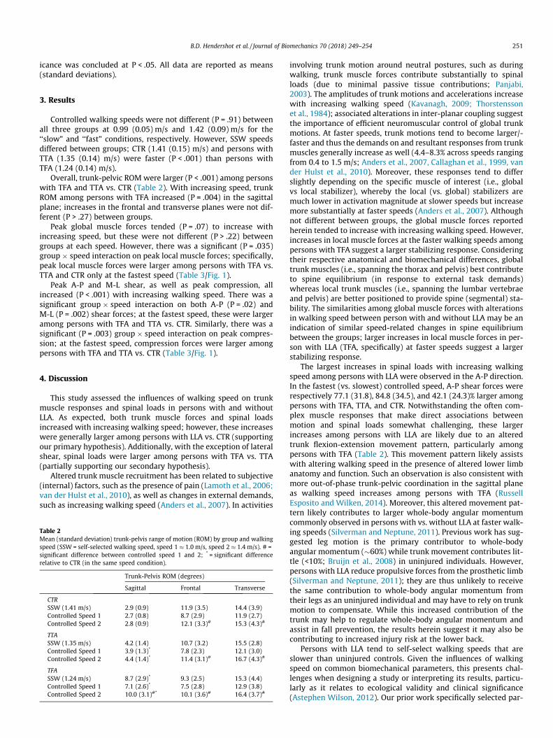
B.D. Hendershot et al. / Journal of Biomechanics 70 (2018) 249–254 251
icance was concluded at P < .05. All data are reported as means(standard deviations).
3. Results
Controlled walking speeds were not different (P = .91) betweenall three groups at 0.99 (0.05) m/s and 1.42 (0.09) m/s for the‘‘slow” and ‘‘fast” conditions, respectively. However, SSW speedsdiffered between groups; CTR (1.41 (0.15) m/s) and persons withTTA (1.35 (0.14) m/s) were faster (P < .001) than persons withTFA (1.24 (0.14) m/s).
Overall, trunk-pelvic ROM were larger (P < .001) among personswith TFA and TTA vs. CTR (Table 2). With increasing speed, trunkROM among persons with TFA increased (P = .004) in the sagittalplane; increases in the frontal and transverse planes were not dif-ferent (P > .27) between groups.
Peak global muscle forces tended (P = .07) to increase withincreasing speed, but these were not different (P > .22) betweengroups at each speed. However, there was a significant (P = .035)group � speed interaction on peak local muscle forces; specifically,peak local muscle forces were larger among persons with TFA vs.TTA and CTR only at the fastest speed (Table 3/Fig. 1).
Peak A-P and M-L shear, as well as peak compression, allincreased (P < .001) with increasing walking speed. There was asignificant group � speed interaction on both A-P (P = .02) andM-L (P = .002) shear forces; at the fastest speed, these were largeramong persons with TFA and TTA vs. CTR. Similarly, there was asignificant (P = .003) group � speed interaction on peak compres-sion; at the fastest speed, compression forces were larger amongpersons with TFA and TTA vs. CTR (Table 3/Fig. 1).
4. Discussion
This study assessed the influences of walking speed on trunkmuscle responses and spinal loads in persons with and withoutLLA. As expected, both trunk muscle forces and spinal loadsincreased with increasing walking speed; however, these increaseswere generally larger among persons with LLA vs. CTR (supportingour primary hypothesis). Additionally, with the exception of lateralshear, spinal loads were larger among persons with TFA vs. TTA(partially supporting our secondary hypothesis).
Altered trunk muscle recruitment has been related to subjective(internal) factors, such as the presence of pain (Lamoth et al., 2006;van der Hulst et al., 2010), as well as changes in external demands,such as increasing walking speed (Anders et al., 2007). In activities
Table 2Mean (standard deviation) trunk-pelvis range of motion (ROM) by group and walkingspeed (SSW = self-selected walking speed, speed 1 � 1.0 m/s, speed 2 � 1.4 m/s). # =significant difference between controlled speed 1 and 2; * = significant differencerelative to CTR (in the same speed condition).
Trunk-Pelvis ROM (degrees)
Sagittal Frontal Transverse
CTRSSW (1.41 m/s) 2.9 (0.9) 11.9 (3.5) 14.4 (3.9)Controlled Speed 1 2.7 (0.8) 8.7 (2.9) 11.9 (2.7)Controlled Speed 2 2.8 (0.9) 12.1 (3.3)# 15.3 (4.3)#
TTASSW (1.35 m/s) 4.2 (1.4) 10.7 (3.2) 15.5 (2.8)Controlled Speed 1 3.9 (1.3)* 7.8 (2.3) 12.1 (3.0)Controlled Speed 2 4.4 (1.4)* 11.4 (3.1)# 16.7 (4.3)#
TFASSW (1.24 m/s) 8.7 (2.9)* 9.3 (2.5) 15.3 (4.4)Controlled Speed 1 7.1 (2.6)* 7.5 (2.8) 12.9 (3.8)Controlled Speed 2 10.0 (3.1)#* 10.1 (3.6)# 16.4 (3.7)#
involving trunk motion around neutral postures, such as duringwalking, trunk muscle forces contribute substantially to spinalloads (due to minimal passive tissue contributions; Panjabi,2003). The amplitudes of trunk motions and accelerations increasewith increasing walking speed (Kavanagh, 2009; Thorstenssonet al., 1984); associated alterations in inter-planar coupling suggestthe importance of efficient neuromuscular control of global trunkmotions. At faster speeds, trunk motions tend to become larger/-faster and thus the demands on and resultant responses from trunkmuscles generally increase as well (4.4–8.3% across speeds rangingfrom 0.4 to 1.5 m/s; Anders et al., 2007, Callaghan et al., 1999, vander Hulst et al., 2010). Moreover, these responses tend to differslightly depending on the specific muscle of interest (i.e., globalvs local stabilizer), whereby the local (vs. global) stabilizers aremuch lower in activation magnitude at slower speeds but increasemore substantially at faster speeds (Anders et al., 2007). Althoughnot different between groups, the global muscle forces reportedherein tended to increase with increasing walking speed. However,increases in local muscle forces at the faster walking speeds amongpersons with TFA suggest a larger stabilizing response. Consideringtheir respective anatomical and biomechanical differences, globaltrunk muscles (i.e., spanning the thorax and pelvis) best contributeto spine equilibrium (in response to external task demands)whereas local trunk muscles (i.e., spanning the lumbar vertebraeand pelvis) are better positioned to provide spine (segmental) sta-bility. The similarities among global muscle forces with alterationsin walking speed between person with and without LLA may be anindication of similar speed-related changes in spine equilibriumbetween the groups; larger increases in local muscle forces in per-son with LLA (TFA, specifically) at faster speeds suggest a largerstabilizing response.
The largest increases in spinal loads with increasing walkingspeed among persons with LLA were observed in the A-P direction.In the fastest (vs. slowest) controlled speed, A-P shear forces wererespectively 77.1 (31.8), 84.8 (34.5), and 42.1 (24.3)% larger amongpersons with TFA, TTA, and CTR. Notwithstanding the often com-plex muscle responses that make direct associations betweenmotion and spinal loads somewhat challenging, these largerincreases among persons with LLA are likely due to an alteredtrunk flexion-extension movement pattern, particularly amongpersons with TFA (Table 2). This movement pattern likely assistswith altering walking speed in the presence of altered lower limbanatomy and function. Such an observation is also consistent withmore out-of-phase trunk-pelvic coordination in the sagittal planeas walking speed increases among persons with TFA (RussellEsposito and Wilken, 2014). Moreover, this altered movement pat-tern likely contributes to larger whole-body angular momentumcommonly observed in persons with vs. without LLA at faster walk-ing speeds (Silverman and Neptune, 2011). Previous work has sug-gested leg motion is the primary contributor to whole-bodyangular momentum (�60%) while trunk movement contributes lit-tle (<10%; Bruijn et al., 2008) in uninjured individuals. However,persons with LLA reduce propulsive forces from the prosthetic limb(Silverman and Neptune, 2011); they are thus unlikely to receivethe same contribution to whole-body angular momentum fromtheir legs as an uninjured individual and may have to rely on trunkmotion to compensate. While this increased contribution of thetrunk may help to regulate whole-body angular momentum andassist in fall prevention, the results herein suggest it may also becontributing to increased injury risk at the lower back.
Persons with LLA tend to self-select walking speeds that areslower than uninjured controls. Given the influences of walkingspeed on common biomechanical parameters, this presents chal-lenges when designing a study or interpreting its results, particu-larly as it relates to ecological validity and clinical significance(Astephen Wilson, 2012). Our prior work specifically selected par-
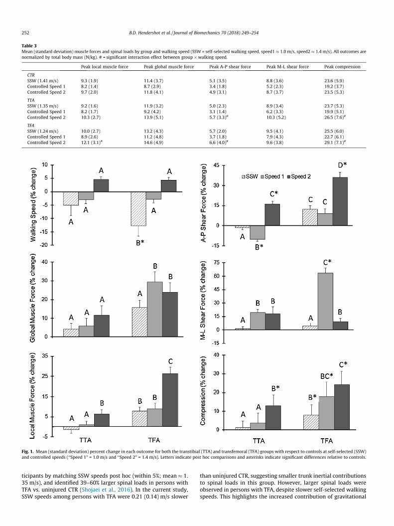
Table 3Mean (standard deviation) muscle forces and spinal loads by group and walking speed (SSW = self-selected walking speed, speed1 � 1.0 m/s, speed2 � 1.4 m/s). All outcomes arenormalized by total body mass (N/kg). # = significant interaction effect between group � walking speed.
Peak local muscle force Peak global muscle force Peak A-P shear force Peak M-L shear force Peak compression
CTRSSW (1.41 m/s) 9.3 (1.9) 11.4 (3.7) 5.1 (3.5) 8.8 (3.6) 23.6 (5.9)Controlled Speed 1 8.2 (1.4) 8.7 (2.9) 3.4 (1.8) 5.2 (2.3) 19.2 (3.7)Controlled Speed 2 9.7 (2.0) 11.8 (4.1) 4.9 (3.1) 8.7 (3.7) 23.5 (5.3)
TTASSW (1.35 m/s) 9.2 (1.6) 11.9 (3.2) 5.0 (2.3) 8.9 (3.4) 23.7 (5.3)Controlled Speed 1 8.2 (1.7) 9.2 (4.2) 3.1 (1.4) 6.2 (3.3) 19.9 (5.1)Controlled Speed 2 10.3 (2.7) 13.9 (5.1) 5.7 (3.3)# 10.3 (5.2) 26.5 (7.6)#
TFASSW (1.24 m/s) 10.0 (2.7) 13.2 (4.3) 5.7 (2.0) 9.5 (4.1) 25.5 (6.0)Controlled Speed 1 8.9 (2.6) 11.2 (4.8) 3.7 (1.8) 7.9 (4.3) 22.7 (6.1)Controlled Speed 2 12.1 (3.1)# 14.6 (4.9) 6.6 (4.0)# 9.6 (3.8) 29.1 (7.1)#
Fig. 1. Mean (standard deviation) percent change in each outcome for both the transtibial (TTA) and transfemoral (TFA) groups with respect to controls at self-selected (SSW)and controlled speeds (‘‘Speed 1” = 1.0 m/s and ‘‘Speed 2” = 1.4 m/s). Letters indicate post hoc comparisons and asterisks indicate significant differences relative to controls.
252 B.D. Hendershot et al. / Journal of Biomechanics 70 (2018) 249–254
ticipants by matching SSW speeds post hoc (within 5%; mean � 1.35 m/s), and identified 39–60% larger spinal loads in persons withTFA vs. uninjured CTR (Shojaei et al., 2016). In the current study,SSW speeds among persons with TFA were 0.21 (0.14) m/s slower
than uninjured CTR, suggesting smaller trunk inertial contributionsto spinal loads in this group. However, larger spinal loads wereobserved in persons with TFA, despite slower self-selected walkingspeeds. This highlights the increased contribution of gravitational
B.D. Hendershot et al. / Journal of Biomechanics 70 (2018) 249–254 253
demand to spinal loads among persons with TFA due to larger andmore asymmetric trunk ROM. One might also presume the relativedifferences in the slow and fast vs. SSW speeds within each groupmay require more or less ‘‘effort” and thereby differentially affectthe relationships reported herein; however, a sensitivity analysisrevealed these differences in SSW did not influence any of thedependent measures.
Several limitations should be considered when interpreting thecurrent results. Persons with LLA were young and generally activemilitary personnel with injuries sustained due to trauma. Thus, theresults may not be generalizable to all etiologies of amputation(e.g., older or as a result of dysvascular conditions). This studywas cross-sectional and the durations of time since injury amongpersons with LLA were relatively short and highly variable (median = 23 months, range = 6–408 months). As such, it is possible thatgait patterns may change (improve or decline) over time and theassociated influences on spinal loads with changing walking speedmay differ if assessed longitudinally. Moreover, the retrospectivenature of this study limited the range of available walking speeds.Additional analyses at slower (i.e. <1.0 m/s) and faster (i.e. >1.4 m/s) walking speeds, or speeds more consistently spaced relative toeach participant’s SSW, may elucidate additional relationshipsamong spinal loads and walking speed in persons with LLA.Although estimates of these model simulations are highly depen-dent on the accuracy and reliability of kinematic data, prior worksuggests high intra-lab reliability and low standard errors of mea-surement (Kaufman et al., 2016). Additionally, trunk muscleresponses and segmental kinematics were estimated using an opti-mization procedure assuming similar responses between personswith and without LLA; however, future work is needed with elec-tromyography, imaging modalities, or other modeling techniquesto understand these more directly. Such efforts would also supportfuture tissue-level analyses incorporating physiological propertiesand biological responses (e.g., Lotz et al., 2013). Finally, we did notexplicitly include contributions of arm swing in the model(Angelini et al., 2016), though participants were not instructed toalter arm swing and full body kinematics were collected and couldbe evaluated in subsequent analyses.
Walking is generally not a mechanically demanding activity forthe lower back. For example, prior work in uninjured individualshas found peak compressive loads ranging from one to three timesbody weight when walking over level ground at varying speeds(Callaghan et al., 1999; Cheng et al., 1998). These magnitudes aresubstantially lower than during other activities (e.g., manual mate-rial handling or lifting tasks) and below injury thresholds. How-ever, walking is a highly repetitive task with estimates of 1.5–4million cycles per year depending activity level. Thus, we posit thatincreases in spinal loads among persons with vs. without LLA war-rant consideration when assessing injury risk. While tasks involv-ing high physical demands on the lower back or which have a highrate/repetition have been traditionally considered high risk for lowback pain (Putz-Anderson et al., 1997), a recent review paper sug-gests that repetition of low-force tasks seems to result in modestincreases in risk; however, surprisingly rapid increases in risk aresubsequently observed under high-force tasks (Gallagher andHeberger, 2013). Although not reported here, mean values of eachcomponent of spinal load across the entire gait cycle were similarlylarger among persons with vs. without LLA, and also tended toincrease more with increasing walking speed among persons withLLA, suggesting not just peak loads but the overall mechanicalenvironment is elevated throughout.
In summary, the results presented herein indicate walkingspeed differentially alters trunk muscle responses and spinal loadsamong persons with vs. without LLA. Walking faster for personswith LLA was associated with larger increases in the estimatedloads among tissues within the spine, regardless of SSW speed.
Over time, repeated exposure to these larger spinal loads duringsuch a common and important activity of daily living may con-tribute to the elevated risk for low back pain after LLA, particularlydue to fatigue failure of spinal tissues. Further work to more com-pletely characterize spinal loads during other activities of daily liv-ing is warranted, thereby supporting future clinicalrecommendations for controlling risk over the longer term.
Acknowledgements
The authors wish to thank Matthew Ballard for his assistance withfinite element model computations, and Dr. Richard Kryscio for hisassistance with statistical analyses. This work was supported, inpart, by an award (5R03HD086512-02) from the National Centerfor Medical Rehabilitation Research (NIH-NICHD) and the Officeof the Assistant Secretary of Defense for Health Affairs, throughthe Peer Reviewed Orthopaedic Research Program (award#W81XWH-14-2-0144). The views expressed in this manuscriptare those of the authors, and do not necessarily reflect the officialpolicy or position of the U.S. Departments of the Army, Defense,nor the U.S. government.
Conflict of interest
All authors have no financial or personal relationships withother persons or organizations that might inappropriately influ-ence our work presented therein.
References
Anders, C., Wagner, H., Puta, C., Grassme, R., Petrovitch, A., Scholle, H.-C., 2007.Trunk muscle activation patterns during walking at different speeds. J.Electromyogr. Kinesiol. 17, 245–252.
Angelini, L., Di Puccio, F., Zander, T., Schmidt, H., 2016. Influence of arm motion onspatio-temporal gait parameters and on force data. IOSR J. Sports Phys. Edu. 3,12–17.
Astephen Wilson, J.L., 2012. Challenges in dealing with walking speed in kneeosteoarthritis gait analyses. Clin. Biomech. (Bristol, Avon) 27, 210–212.
Bazrgari, B., Shirazi-Adl, A., Arjmand, N., 2007. Analysis of squat and stoop dynamicliftings: muscle forces and internal spinal loads. Eur. Spine J. 16, 687–699.
Bazrgari, B., Shirazi-Adl, A., Trottier, M., Matheiu, P., 2008a. Computation of trunkequilibrium and stability in free flexion-extension movements at differentvelocities. J. Biomech. 41 (2), 412–421.
Bazrgari, B., Shirazi-Adl, A., Kasra, M., 2008b. Seated whole body vibrations withhigh-magnitude accelerations – relative roles of inertia and muscle forces. J.Biomech. 41 (12), 2639–2646.
Bazrgari, B., Shirazi-Adl, A., Lariviere, C., 2009. Trunk response analysis undersudden forward perturbations using a kinematics-driven model. J. Biomech. 42(9), 1193–1200.
Bruijn, S.M., Meijer, O.G., van Dieën, J.H., Kingma, I., Lamoth, C.J.C., 2008.Coordination of leg swing, thorax rotations, and pelvis rotations during gait:the organisation of total body angular momentum. Gait Posture 27, 455–462.
Callaghan, J.P., Patla, A.E., McGill, S.M., 1999. Low back three-dimensional jointforces, kinematics, and kinetics during walking. Clin. Biomech. 14, 203–216.
Cheng, K., Chen, H., Chen, S., Lee, S., 1998. Influences of walking speed change on thelumbosacral joint force distribution. Bio-Med. Mater. Eng. 8, 155–165.
Coenen, P., Kingma, I., Boot, C.R., Bongers, P.M., van Dieen, J.H., 2014. Cumulativemechanical low-back load at work is a determinant of low-back pain. Occup.Environ. Med. 71, 332–337.
Davis, K.G., Marras, W.S., 2000. The effects of motion on trunk biomechanics. Clin.Biomech. 15, 703–717.
Devan, H., Carman, A.B., Hendrick, P.A., Ribeiro, D.C., Hale, L.A., 2015. Perceptions oflow back pain in people with lower limb amputation: a focus group study.Disabil. Rehabil. 37, 873–883.
Fey, N.P., Silverman, A.K., Neptune, R.R., 2010. The influence of increasing steady-state walking speed on muscle activity in below-knee amputees. J.Electromyogr. Kinesiol 20, 155–161.
Gallagher, S., Heberger, J.R., 2013. Examining the interaction of force and repetitionon musculoskeletal disorder risk: a systematic literature review. Hum. Factors55, 108–124.
Goujon-Pillet, H., Sapin, E., Fodé, P., Lavaste, F., 2008. Three-dimensional motions oftrunk and pelvis during transfemoral amputee gait. Arch. Phys. Med. Rehabil.89, 87–94.
Halsne, E.G., Waddingham, M.G., Hafner, B.J., 2013. Long-term activity in and amongpersons with transfemoral amputation. J. Rehabil. Res. Dev. 50, 515–530.
254 B.D. Hendershot et al. / Journal of Biomechanics 70 (2018) 249–254
Hammarlund, C.S., Carlstrom, M., Melchior, R., Persson, B.M., 2011. Prevalence ofback pain, its effect on functional ability and health-related quality of life inlower limb amputees secondary to trauma or tumour: a comparison acrossthree levels of amputation. Prosthet. Orthot. Int. 35, 97–105.
Hendershot, B.D., Wolf, E.J., 2014. Three-dimensional joint reaction forces andmoments at the low back during over-ground walking in persons withunilateral lower-extremity amputation. Clin. Biomech. (Bristol, Avon) 29,235–242.
Hendershot, B.D., Wolf, E.J., 2015. Mediolateral joint powers at the low back amongpersons with unilateral transfemoral amputation. Arch. Phys. Med. Rehabil. 96(1), 154–157.
Jaegers, S.M.H.J., Arendzen, J.H., de Jongh, H.J., 1995. Prosthetic gait of unilateraltransfemoral amputees: a kinematic study. Arch. Phys. Med. Rehabil. 76, 736–743.
Jonkers, I., Delp, S., Patten, C., 2009. Capacity to Increase Walking Speed is Limitedby impaired hip and ankle power generation in lower functioning persons post-stroke. Gait Posture 29, 129–137.
Kaufman, K., Miller, E., Kingsbury, T., Russell Esposito, E., Wolf, E., Wilken, J., et al.,2016. Reliability of 3D gait data across multiple laboratories. Gait Posture 49,375–381.
Kavanagh, J.J., 2009. Lower trunk motion and speed-dependence during walking. J.Neuroeng. Rehabil. 6, 9.
Kulkarni, J., Gaine, W.J., Buckley, J.G., Rankine, J.J., Adams, J., 2005. Chronic low backpain in traumatic lower limb amputees. Clin. Rehabil. 19, 81–86.
Kumar, S., 2001. Theories of musculoskeletal injury causation. Ergonomics 44, 17–47.
Lamoth, C.J.C., Meijer, O.G., Daffertshofer, A., Wuisman, P.I.J.M., Beek, P.J., 2006.Effects of chronic low back pain on trunk coordination and back muscle activityduring walking: changes in motor control. Eur. Spine J. 15, 23–40.
Lotz, J.C., Fields, A.J., Liebenberg, E.C., 2013. The Role of the vertebral end plate inlow back pain. Global Spine J 03, 153–164.
Neptune, R.R., Sasaki, K., Kautz, S.A., 2008. The effect of walking speed on musclefunction and mechanical energetics. Gait Posture 28, 135–143.
Panjabi, M.M., 2003. Clinical spinal instability and low back pain. J. Electromyogr.Kinesiol. 13, 371–379.
Putz-Anderson, V., Bernard, B.P., Burt, S.E., Cole, L.L., Fairfield-Estill, C., Fine, L.J.,et al., 1997. Musculoskeletal disorders and workplace factors. National Institutefor Occupational Safety and Health (NIOSH), p. 104.
Ralston, H.J., 1958. Energy-speed relation and optimal speed during level walking.Int. Z. Angew. Physiol. 17, 277–283.
Requiao, L.F., Nadeau, S., Milot, M.H., Gravel, D., Bourbonnais, D., Gagnon, D., 2005.Quantification of level of effort at the plantarflexors and hip extensors andflexor muscles in healthy subjects walking at different cadences. J.Electromyogr. Kinesiol. 15, 393–405.
Russell Esposito, E., Wilken, J.M., 2014. The relationship between pelvis-trunkcoordination and low back pain in individuals with transfemoral amputations.Gait Posture 40, 640–646.
Shojaei, I., Hendershot, B.D., Wolf, E.J., Bazrgari, B., 2016. Persons with unilateraltransfemoral amputation experience larger spinal loads during level-groundwalking compared to able-bodied individuals. Clin. Biomech. (Bristol, Avon) 32,157–163.
Silverman, A.K., Fey, N.P., Portillo, A., Walden, J.G., Bosker, G., Neptune, R.R., 2008.Compensatory mechanisms in below-knee amputee gait in response toincreasing steady-state walking speeds. Gait Posture 28, 602–609.
Silverman, A.K., Neptune, R.R., 2011. Differences in whole-body angular momentumbetween below-knee amputees and non-amputees across walking speeds. J.Biomech. 44, 379–385.
Stepien, J.M., Cavenett, S., Taylor, L., Crotty, M., 2007. Activity levels among lower-limb amputees: self-report versus step activity monitor. Arch. Phys. Med.Rehabil. 88, 896–900.
Thorstensson, A., Nilsson, J., Carlson, H., Zomlefer, M.R., 1984. Trunk movements inhuman locomotion. Acta Physiol. Scand 121, 9–22.
Tudor-Locke, C., Craig, C.L., Brown, W.J., Clemes, S.A., De Cocker, K., Giles-Corti, B.,et al., 2011. Howmany steps/day are enough? for adults. The Int. J. Behav. Nutri.Phys. Activ. 8. 79-79.
van der Hulst, M., Vollenbroek-Hutten, M.M., Rietman, J.S., Hermens, H.J., 2010.Lumbar and abdominal muscle activity during walking in subjects with chroniclow back pain: support of the ‘‘guarding” hypothesis? J. Electromyogr. Kinesiol.20, 31–38.
Yoder, A.J., Petrella, A.J., Silverman, A.K., 2015. Trunk-pelvis motion, joint loads, andmuscle forces during walking with a transtibial amputation. Gait Posture 41,757–762.
Journal of Biomechanics 75 (2018) 181–185
Contents lists available at ScienceDirect
Journal of Biomechanicsjournal homepage: www.elsevier .com/locate / jb iomech
www.JBiomech.com
Short communication
Associations between trunk postural control in walking and unstablesitting at various levels of task demand
https://doi.org/10.1016/j.jbiomech.2018.05.0060021-9290/Published by Elsevier Ltd.
⇑ Corresponding author at: Extremity Trauma and Amputation Center of Excel-lence, Walter Reed National Military Medical Center, 4494 N. Palmer Road,Bethesda, MD 20889, United States.
E-mail address: [email protected] (B.D. Hendershot).
Downloaded for Anonymous User (n/a) at Walter Reed National Military Medical Center from ClinicalKey.com by Elsevier on October 29, 2018.For personal use only. No other uses without permission. Copyright ©2018. Elsevier Inc. All rights reserved.
Julian C. Acasio a, Courtney M. Butowicz a, Pawel R. Golyski a, Maury A. Nussbaumb,Brad D. Hendershot a,c,d,⇑aResearch & Development Section, Department of Rehabilitation, Walter Reed National Military Medical Center, United StatesbDepartment of Industrial and Systems Engineering, Virginia Tech, United StatescDoD-VA Extremity Trauma and Amputation Center of Excellence, United StatesdDepartment of Rehabilitation Medicine, Uniformed Services University of the Health Sciences, United States
a r t i c l e i n f o
Article history:Accepted 3 May 2018
Keywords:Center of pressureSample entropyLyapunov exponentsBiomechanicsTrunk stability
a b s t r a c t
Trunk postural control (TPC) has been investigated in several populations and tasks. Previous workobserved targeted training of TPC via isolated trunk control tasks may improve performance in otheractivities (e.g., walking). However, the nature of this relationship remains unknown. We therefore inves-tigated the relationship between TPC, at both the global (i.e., response to finite perturbations) and local(i.e., resistance to continuous perturbations) levels, during walking and unstable sitting, both at varyinglevels of task demand. Thirteen individuals (11 Male, 2 Female) with no recent history (past 12 months)of illness, injury, or musculoskeletal disorders walked on a dual-belt treadmill at four speeds (�20%,�10%, +10%, and + 20% of self-selected walking speed) and completed an unstable sitting task at fourlevels of chair instability (100, 75, 60, and 45% of an individual’s ‘‘neutral” stability as defined by the grav-itational gradient). Three-dimensional trunk and pelvic kinematics were collected. Tri-planar Lyapunovexponents and sample entropy characterized local TPC. Global TPC was characterized by ranges of motionand, for seated trials, metrics derived from center-of-pressure time series (i.e., path length, 95% confi-dence ellipse area, mean velocity, and RMS position). No strong or significant correlations (�0.057 < q< 0.206) were observed between local TPC during walking and unstable sitting tasks. However, globalTPC declined in both walking and unstable sitting as task demand increased, with a moderate inter-task relationship (0.336 < q < 0.544). While the mechanisms regulating local TPC are inherently different,global TPC may be similarly regulated across both tasks, supporting future translation of improvementsin TPC between tasks.
Published by Elsevier Ltd.
1. Introduction
Physical pathologies including stroke (Verheyden et al., 2006),lower limb loss (Hendershot and Nussbaum, 2013), and low backpain (Lamoth et al., 2006) can adversely influence trunk posturalcontrol (TPC). While TPC has been studied extensively, reportedmeasures vary between tasks and specific features of dynamic sys-tems (i.e., global and local). Here, we consider global TPC as theability of a system to respond to finite (‘‘global”) perturbations(e.g., slip or trip), while local TPC is the ability to resist infinitesimal(‘‘local”) perturbations (e.g., natural gait fluctuations). During gait,
global TPC has been indirectly quantified by characterizing seg-mental motions, such as trunk position variability (Dingwell andMarin, 2006) and ranges of motion (ROM). Meanwhile, non-linear measures, including Lyapunov exponents (Asgari et al.,2015, Dingwell and Marin, 2006) and sample entropy (SampEn;Lamoth et al., 2010), have characterized local TPC. During unstablesitting, global TPC is often characterized using metrics derivedfrom center-of-pressure (CoP) time series (Hendershot andNussbaum, 2013; Radebold et al., 2001) and ROM (Larivièreet al., 2015); while local TPC has also been characterized by non-linear analyses of CoP (Larivière et al., 2015; Van Dieën et al.,2010). In both walking and unstable sitting, TPC generally declineswith increasing task demand as evidenced by larger values of TPCmeasures described previously (Dingwell and Marin, 2006;Radebold et al., 2001).
182 J.C. Acasio et al. / Journal of Biomechanics 75 (2018) 181–185
Altered TPC can adversely influence performance in functionalactivities (e.g., walking), particularly given the relative mass andposition of the trunk. Indeed, TPC deficits are associated with anincreased risk of falls (Grimbergen et al., 2008, Tinetti et al., 1988)and musculoskeletal injury (Zazulak et al., 2007). Trunk-specificexercise regimens are therefore often proposed or utilized to helpmitigate these risks and, in populationswith impaired TPC, incorpo-rated into rehabilitation efforts (e.g., Karthikbabu et al., 2011). Suchisolated TPC tasks have been shown to reduce pain and functionaldisability scores in individuals with LBP (O’Sullivan et al., 1997,Carpes et al., 2008) and improve gait parameters in patients afterstroke (Karthikbabu et al., 2011). These observations suggest thatimprovements to TPC may translate between tasks, but thereremains a limited understanding of the effectiveness of such reha-bilitation paradigms since the relationship between TPC mecha-nisms in isolated (e.g., unstable sitting) and functional (e.g.,walking) activities has not been investigated thoroughly. Evidencecomparing local TPC in two upright tasks (standing and walking)observed little-to-no correlation between them (Kang andDingwell, 2006). However, only a single level of demand was inves-tigated, and TPC during an isolated task (i.e., unstable sitting) wasnot determined. We thus explored the relationships between TPCduring two distinct tasks, walking and unstable sitting, when bothare performed at varying levels of task demand. As TPC has beenobserved to decrease with increasing demand in both tasks, wehypothesized that increases in respective task demands of walkingand unstable sitting would be similarly reflected in decrements toTPC, as evidenced by strong inter-task correlations among TPCmea-sures at each level of demand.
2. Methods
2.1. Study design and procedures
Thirteen participants with no current or recent history of ill-ness, injury, or musculoskeletal disorders within the past 12months (Table 1) completed walking and unstable sitting trials atvarying demand levels. For walking trials, participants walked onan instrumented dual-belt treadmill (Bertec, Columbus, OH) at fourspeeds relative to self-selected walking speed (SSWS; Table 1),determined from the mean velocity of five over-ground trialsacross a 15 m walkway: �20%, �10%, +10%, and +20% SSWS. Rela-tive (vs. absolute) speeds were chosen to better normalize taskdemand across participants, with the expectation that fasterspeeds increase demand (Dingwell and Marin, 2006). At eachspeed, a 30-s acclimation period was provided before two minutesof data collection. For seated trials, participants sat on an unstablechair (Hendershot and Nussbaum, 2013) with eyes open at fourlevels of instability, relative to an individual’s gravitational gradi-ent (rG): 100, 75, 60, and 45% rG (with instability increasing as%rG decreased). rG was calculated using previously establishedmethods (Slota et al., 2008) and determined neutral seated stabil-ity. Participants completed four 60-s trials per condition. However,only the final (i.e., fourth) trial was used for data analyses; the priorthree practice trials were used to attenuate learning effects (VanDaele et al., 2007). By the final trial, all participants successfullycompleted the unstable sitting task (i.e., the seat did not contactthe base of support). Participants were asked to keep the chair leveland arms crossed throughout trials.
Table 1Mean (standard deviation) participant demographic information and self-selected walking
N Age (years) Stature
13 (11 M, 2 F) 28.7 (7.2) 177.1 (
Downloaded for Anonymous User (n/a) at Walter Reed National Military For personal use only. No other uses without permissio
An 18-camera motion capture system (Qualisys, Göteborg, Swe-den) collected (120 Hz) 10 surface-marker locations to estimatethree-dimensional trunk and pelvic kinematics for all tasks. Mark-ers were placed over the T10 and C7 spinous processes, sternalnotch, xiphoid, and bilaterally over the acromion, ASIS, and PSIS.During seated trials, kinetic data were collected (1200 Hz) usinga force platform (AMTI, OR6-7-2000, Watertown, MA) mountedbeneath the chair. Task and condition order were randomizedand counterbalanced, respectively, with 60-s rests providedbetween trials. Prior to data collection, participants gave informedconsent to protocols approved by the local Institutional ReviewBoard.
2.2. Pre-processing
Data were analyzed using Visual3D (C-motion, Germantown,MD) and MATLAB (Mathworks, Natick, MA). Kinematic and kineticdata were low-pass filtered (Butterworth, 4th order, cut-off fre-quencies 6 and 10 Hz, respectively). Three-dimensional trunkangles (relative to pelvis) were determined using 6DOF inversedynamics in Visual3D. For each walking trial, 75 strides of datawere analyzed and resampled to 101 points per stride (i.e., 0–100% gait cycle). For unstable sitting trials, the first and last fiveseconds of data were removed to account for initial and anticipa-tory adjustments respectively.
2.3. Global TPC analyses
For both tasks, tri-planar trunk-pelvic ROM were determined.Though ROM does not directly quantify global TPC (i.e., responseto a perturbation), increases in trunk ROM have been observed inpopulations with impaired TPC such as fall-prone populations(Tinetti et al., 1988, Grimbergen et al., 2008). Thus, though partic-ipants were not perturbed in the current protocol, ROM providedan indirect characterization of global TPC. For seated trials CoPpath length, mean velocity, 95% confidence ellipse area (CEA),and RMS positions in the anteroposterior and mediolateral direc-tions were also determined (Prieto et al., 1996).
2.4. Local TPC analysis
Maximum short-term Lyapunov exponents (ks; Rosensteinet al., 1993) and SampEn (Richman and Moorman, 2000) were usedto characterize local stability of trunk-pelvic angles. ks quantifiesthe rate of convergence/divergence of initially neighboring trajec-tories. Negative and positive ks values respectively indicate conver-gence (i.e., stability) and divergence (i.e., instability); largerpositive values represent a decreased ability to resist local pertur-bations (i.e., decreased local TPC). Here, tri-planar ks were calcu-lated via state spaces reconstructed from trunk-pelvic angles andtheir time-delayed copies (Dingwell et al., 2001). Global false near-est neighbor and mutual average information analyses respectivelydetermined embedding dimensions (m = 6) and time delays (s = 10and s = 100 samples for walking and seated conditions,respectively).
Unlike ks, SampEn does not directly characterize the response tolocal perturbations. Rather, it characterizes the prevalence of localperturbations within the system by quantifying its regularity(Richman and Moorman, 2000). Larger values of SampEn indicate
speeds (SSWS).
(cm) Mass (kg) SSWS (m/s)
6.3) 74.6 (11.4) 1.46 (0.18)
Medical Center from ClinicalKey.com by Elsevier on October 29, 2018.n. Copyright ©2018. Elsevier Inc. All rights reserved.
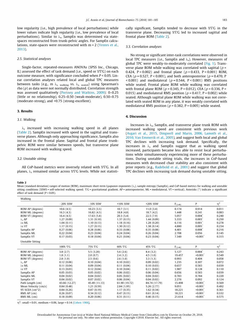
J.C. Acasio et al. / Journal of Biomechanics 75 (2018) 181–185 183
low regularity (i.e., high prevalence of local perturbations) whilelower values indicate high regularity (i.e., low prevalence of localperturbations). Similar to ks, SampEn was determined via state-spaces reconstructed from trunk-pelvic angles. For SampEn calcu-lations, state-spaces were reconstructed with m = 2 (Yentes et al.,2013).
2.5. Statistical analyses
Single-factor, repeated-measures ANOVAs (SPSS Inc., Chicago,IL) assessed the effect of task demand (i.e., speed or %rG) on eachoutcome measure, with significance concluded when P < 0.05. Lin-ear correlation analyses related local and global TPC measuresbetween tasks (e.g., m ks, walking vs. ks, seated) using Spearman’srho (q) as data were not normally distributed. Correlation strengthwas assessed qualitatively (Portney and Watkins, 2009): 0–0.25(little or no relationship), 0.25–0.50 (weak-moderate), 0.50–0.75(moderate-strong), and >0.75 (strong-excellent).
3. Results
3.1. Walking
ks increased with increasing walking speed in all planes(Table 2). SampEn increased with speed in the sagittal and trans-verse planes. Although only approaching significance, SampEn alsoincreased in the frontal plane. Sagittal and frontal plane trunk-pelvic ROM were similar between speeds, but transverse planeROM increased with walking speed.
3.2. Unstable sitting
All CoP-based metrics were inversely related with %rG. In allplanes, ks remained similar across %rG levels. While not statisti-
Table 2Mean (standard deviation) ranges of motion (ROM), maximum short-term Lyapunov exponsitting conditions (SSWS = self-selected walking speed; rG = gravitational gradient, AP =effect of task demand (P < 0.05).
Walking
�20% SSW �10% SSW +10% S
ROM AP (degrees) 10.6 (4.5) 10.23 (3.5) 10.7 (3ROM ML (degrees) 16.3 (4.3) 16.82 (4.5) 18.8 (4ROM VT (degrees) 16.4 (4.5) 17.63 (5.8) 20.3 (5ks AP 1.27 (0.09) 1.31 (0.10) 1.37 (0ks ML 1.04 (0.11) 1.10 (0.13) 1.18 (0ks VT 1.17 (0.15) 1.27 (0.15) 1.30 (0SampEn AP 0.27 (0.06) 0.28 (0.06) 0.33 (0SampEn ML 0.22 (0.04) 0.23 (0.04) 0.24 (0SampEn VT 0.17 (0.03) 0.18 (0.04) 0.21 (0
Unstable Sitting
100% rG 75% rG 60% r
ROM AP (degrees) 3.8 (2.7) 5.5 (3.20) 5.6 (2.ROM ML (degrees) 1.8 (1.1) 2.0 (0.7) 2.4 (1.ROM VT (degrees) 2.8 (1.9) 2.5 (0.6) 2.6 (1.ks AP 0.12 (0.06) 0.10 (0.04) 0.10 (0ks ML 0.11 (0.04) 0.09 (0.04) 0.11 (0ks VT 0.13 (0.03) 0.12 (0.04) 0.10 (0SampEn AP 0.05 (0.03) 0.05 (0.02) 0.06 (0SampEn ML 0.04 (0.02) 0.04 (0.02) 0.05 (0SampEn VT 0.04 (0.02) 0.07 (0.02) 0.06 (0Path Length (cm) 43.66 (12.27) 45.49 (11.13) 61.90 (Mean Velocity (cm/s) 0.84 (0.48) 1.21 (0.59) 2.84 (195 %CEA (cm^2) 0.84 (0.26) 0.87 (0.19) 1.17 (0RMS AP (cm) 0.26 (0.07) 0.34 (0.12) 0.47 (0RMS ML (cm) 0.18 (0.09) 0.20 (0.06) 0.31 (0
g2: small = 0.01, medium = 0.06, large = 0.14 (Cohen 1988).
Downloaded for Anonymous User (n/a) at Walter Reed National Military MeFor personal use only. No other uses without permission. C
cally significant, SampEn tended to decrease with %rG in thetransverse plane. Decreasing %rG led to increased sagittal andfrontal plane ROM (Table 2).
3.3. Correlation analyses
No strong or significant inter-task correlations were observed inlocal TPC measures (i.e., SampEn and ks). However, measures ofglobal TPC were weakly-to-moderately correlated (Fig. 1). Trans-verse plane ROM while walking was correlated with sagittal (q =0.424, P = 0.002) and frontal plane (q = 0.433, P = 0.001) ROM,CEA (q = 0.527, P < 0.001), and both anteroposterior (q = 0.470, P< 0.001) and mediolateral (q = 0.544, P < 0.001) RMS positionswhile seated. Frontal plane ROM while walking was correlatedwith frontal plane ROM (q = 0.345, P = 0.012), CEA (q = 0.336, P =0.015) and mediolateral RMS position (q = 0.417, P = 0.002) whileseated. Although sagittal plane ROM while walking was not corre-lated with seated ROM in any plane, it was weakly correlated withmediolateral RMS position (q = 0.382, P = 0.005) while seated.
4. Discussion
Increases in ks, SampEn, and transverse plane trunk ROM withincreased walking speed are consistent with previous work(Asgari et al., 2015, Dingwell and Marin, 2006, Lamoth et al.,2010, Van Emmerik et al., 2005), and suggest both local and globalTPC declines with increasing task demand. Specifically, theincreases in ks and SampEn suggest that as walking speedincreased, participants became less able to resist local perturba-tions while simultaneously experiencing more of these perturba-tions. During unstable sitting trials, the increases in CoP-basedmeasures with decreased chair stability are also consistent withprior reports (e.g., Radebold et al., 2001) and suggest that globalTPC declines with increasing task demand during unstable sitting.
ents (ks), sample entropy (SampEn), and CoP-based metrics for walking and unstableanteroposterior, ML = mediolateral, VT = vertical). Asterisks (*) indicate a significant
SW +20% SSW F(3,48) P g2
.1) 11.0 (3.4) 0.174 0.914 0.011
.8) 18.7 (4.2) 1.448 0.241 0.083
.4) 22.5 (7.9) 5.057 0.004* 0.240
.15) 1.44 (0.09) 5.333 0.003* 0.250
.16) 1.28 (0.20) 6.116 0.001* 0.278
.12) 1.38 (0.14) 4.880 0.005* 0.234
.08) 0.35 (0.08) 4.401 0.008* 0.216
.04) 0.26 (0.04) 2.708 0.056 0.145
.04) 0.23 (0.04) 7.349 <0.001* 0.315
G 45% rG F(3,48) P g2
6) 8.4 (3.2) 5.127 0.004* 0.2432) 4.5 (1.0) 19.457 <0.001* 0.5490) 3.3 (1.3) 0.993 0.404 0.058.02) 0.09 (0.02) 1.235 0.307 0.072.04) 0.10 (0.03) 0.657 0.583 0.039.04) 0.11 (0.02) 1.987 0.128 0.110.02) 0.06 (0.04) 0.656 0.583 0.039.02) 0.04 (0.02) 0.783 0.784 0.220.04) 0.04 (0.02) 2.276 0.092 0.12419.72) 84.74 (17.79) 15.498 <0.001* 0.569.95) 5.26 (2.77) 9.051 <0.001* 0.492.35) 1.62 (0.34) 18.221 <0.001* 0.361.15) 0.61 (0.14) 18.221 <0.001* 0.532.11) 0.46 (0.15) 21.614 <0.001* 0.575
dical Center from ClinicalKey.com by Elsevier on October 29, 2018.opyright ©2018. Elsevier Inc. All rights reserved.
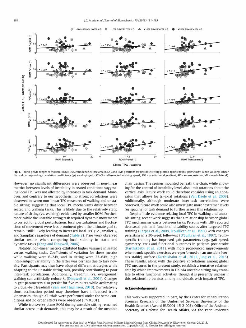
Fig. 1. Trunk-pelvic ranges of motion (ROM), 95% confidence ellipse area (CEA), and RMS positions for unstable sitting plotted against trunk-pelvic ROMwhile walking. Linearfits and corresponding correlation coefficients (q) are displayed. (SSWS = self-selected walking speed; rG = gravitational gradient, AP = anteroposterior, ML = mediolateral).
184 J.C. Acasio et al. / Journal of Biomechanics 75 (2018) 181–185
However, no significant differences were observed in non-linearmetrics between levels of instability in seated conditions suggest-ing local TPC was not affected by increases in task demand. More-over, and contrary to our hypothesis, no strong correlations wereobserved between non-linear TPC measures of walking and unsta-ble sitting, suggesting that local TPC mechanisms differ betweenseated and walking tasks. This is likely due to the relatively staticnature of sitting (vs. walking), evidenced by smaller ROM. Further-more, while the unstable sitting task required dynamic movementsto correct for global perturbations, local perturbations and fluctua-tions of movement were less prominent given the ultimate goal toremain ‘‘still”, likely leading to increased local TPC (i.e., smaller ksand SampEn) regardless of demand (Table 2). Prior work observedsimilar results when comparing local stability in static anddynamic tasks (Kang and Dingwell, 2006).
Notably, non-linear metrics exhibited higher variance in seatedversus walking tasks. Coefficients of variation for these metricswhile walking were 6–24%, and in sitting were 23–64%; highinter-subject variability in the latter was perhaps due to task nov-elty. Participants may thus have adopted different strategies whileadapting to the unstable sitting task, possibly contributing to poorinter-task correlations. Additionally, treadmill (vs. overground)walking can artificially reduce ks (Dingwell et al., 2001). Changesin gait parameters also persist for five minutes while acclimatingto a dual-belt treadmill (Zeni and Higginson, 2010). Our relativelyshort acclimation period may therefore have influenced trunkkinematics, though all trials were performed under the same con-ditions and no order effects were observed (P > 0.301).
While transverse plane ROM during unstable sitting remainedsimilar across task demands, this may be a result of the unstable
Downloaded for Anonymous User (n/a) at Walter Reed National Military For personal use only. No other uses without permissio
chair design. The springs mounted beneath the chair, while allow-ing for the control of instability level, also limit rotations about thevertical axis. Future work could therefore consider using an appa-ratus that allows for tri-axial rotations (Van Daele et al., 2009).Additionally, although moderate inter-task correlations wereobserved, future work could also investigate more ‘‘extreme” levels(or spacing) of task demand to further assess this relationship.
Despite little evidence relating local TPC in walking and unsta-ble sitting, recent work suggests that a relationship between globalTPC mechanisms exists between tasks. Persons with LBP reporteddecreased pain and functional disability scores after targeted TPCtraining (Carpes et al., 2008, O’Sullivan et al., 1997) with changespersisting in a 30-week follow-up (O’Sullivan et al., 1997). Trunk-specific training has improved gait parameters (e.g., gait speed,symmetry, etc.) and functional outcomes in patients post-stroke(Karthikbabu et al., 2011), with more pronounced improvementswhen trunk-specific exercises were performed on an unstable (ver-sus stable) surface (Karthikbabu et al., 2011, Jung et al., 2016).These results, along with the positive correlations among globalTPC measures in the present study, establish a tentative relation-ship by which improvements in TPC via unstable sitting may trans-late to other functional activities, though it is presently unclear ifthis relationship persists among individuals with impaired TPC.
Acknowledgements
This work was supported, in part, by the Center for RehabilitationSciences Research of the Uniformed Services University of theHealth Sciences (Award HU0001-15-2-003), Office of the AssistantSecretary of Defense for Health Affairs, via the Peer Reviewed
Medical Center from ClinicalKey.com by Elsevier on October 29, 2018.n. Copyright ©2018. Elsevier Inc. All rights reserved.
J.C. Acasio et al. / Journal of Biomechanics 75 (2018) 181–185 185
Orthopaedic Research Program (Award W81XWH-14-2-0144), andthe DoD-VA Extremity Trauma and Amputation Center of Excel-lence (Public Law 110-417, National Defense Authorization Act2009). The views expressed herein are those of the authors, anddo not necessarily reflect the official policy or position of theDepartment of Defense or U.S. Government.
Conflict of Interest
The authors have no financial or personal relationships withother persons or organizations that might inappropriately influ-ence our work presented herein.
References
Asgari, M., Sanjari, M.A., Mokhtarinia, H.R., Moeini Sedeh, S., Khalaf, K., Parnianpour,M., 2015. The effects of movement speed on kinematic variability and dynamicstability of the trunk in healthy individuals and low back pain patients. ClinBiomech (Bristol, Avon) 30 (7), 682–688. https://doi.org/10.1016/j.clinbiomech.2015.05.005.
Carpes, F.P., Reinehr, F.B., Mota, C.B., 2008. Effects of a program for trunk strengthand stability on pain, low back and pelvis kinematics, and body balance: a pilotstudy. J. Bodyw. Mov. Ther. 12 (1), 22–30. https://doi.org/10.1016/j.jbmt.2007.05.001.
Cohen, J., 1988. Statistical Power Analysis for the Behavioral Sciences. ErlbaumAssociates, Hillsdale, NJ.
Dingwell, J.B., Cusumano, J.P., Cavanagh, P.R., Sternad, D., 2001. Local dynamicstability versus kinematic variability of continuous overground and treadmillwalking. J. Biomech. Eng. 123 (1), 27. https://doi.org/10.1115/1.1336798.
Dingwell, J.B., Marin, L.C., 2006. Kinematic variability and local dynamic stability ofupper body motions when walking at different speeds. J. Biomech. 39 (3), 444–452. https://doi.org/10.1016/j.jbiomech.2004.12.014.
Grimbergen, Y.A.M., Knol, M.J., Bloem, B.R., Kremer, B.P.H., Roos, R.A.C., Munneke,M., 2008. Falls and gait disturbances in Huntington’s disease. Mov. Disord. 23(7), 970–976. https://doi.org/10.1002/mds.22003.
Hendershot, B.D., Nussbaum, M.A., 2013. Persons with lower-limb amputation haveimpaired trunk postural control while maintaining seated balance. Gait Posture.38, 438–442.
Jung, K.S., Cho, H.Y., In, T.S., 2016. Trunk exercises performed on an unstable surfaceimprove trunk muscle activation, postural control, and gait speed in patientswith stroke. J Phys Ther Sci 28, 940–944.
Kang, H.G., Dingwell, J.B., 2006. A direct comparison of local dynamic stabilityduring unperturbed standing and walking. Exp. Brain Res. 172 (1), 35–48.https://doi.org/10.1007/s00221-005-0224-6.
Karthikbabu, S., Nayak, A., Vijayakumar, K., Mirsi, Z.K., Suresh, B.V., Ganesan, S.,Joshuan, A.M., 2011. Comparison of physio ball and plinth trunk exerciseregimens on trunk control and functional balance in patients with acute stroke:a pilot randomized controlled trial. Clinical Rehabilitation 25 (8), 709–719.
Lamoth, C.J., Ainsworth, E., Polomski, W., Houdijk, H., 2010. Variability and stabilityanalysis of walking of transfemoral amputees. Med. Eng. Phys. 32 (9), 1009–1014. https://doi.org/10.1016/j.medengphy.2010.07.001.
Lamoth, C.J., Daffertshofer, A., Meijer, O.G., Beek, P.J., 2006. How do persons withchronic low back pain speed up and slow down? Trunk-pelvis coordination and
Downloaded for Anonymous User (n/a) at Walter Reed National Military MeFor personal use only. No other uses without permission. C
lumbar erector spinae activity during gait. Gait Posture 23 (2), 230–239. https://doi.org/10.1016/j.gaitpost.2005.02.006.
Larivière, C., Gagnon, D.H., Mecheri, H., 2015. Trunk postural control in unstablesitting: Effect of sex and low back pain status. Clin. Biomech. (Bristol, Avon) 30(9), 933–939. https://doi.org/10.1016/j.clinbiomech.2015.07.006.
O’Sullivan, P.B., Phyty, G.D.M., Twomey, L.T., Allison, G.T., 1997. Evaluation ofspecific stabilizing exercise in the treatment of chronic low back pain withradiologic diagnosis of spondylolysis or spondylolisthesis. Spine 22 (24), 2959–2967.
Portney, L.G., Watkins, M.P., 2009. Foundations of Clinical Research: Application toPractice. Pearson, Upper Saddle River, NJ.
Prieto, T.E., Myklebust, J.B., Hoffmann, R.G., Lovett, E.G., Myklebust, B.M., 1996.Measures of postural steadiness: differences between healthy young andelderly adults. IEEE Transact. Biomed. Eng. 43 (9), 956–966.
Radebold, A., Cholewicki, J., Polzhofer, G.K., Greene, H.S., 2001. Impaired Posturalcontrol of the lumbar spine is associated with delayed muscle response times inpatients with chronic idiopathic low back pain. Spine 26 (7), 724–730.
Richman, J.S., Moorman, R.J., 2000. Physiological time-series analysis usingapproximate entropy and sample entropy. Am. J. Physiol. Heart Circ. Physiol278, H2039–H2049.
Rosenstein, M.T., Collins, J.J., De Luca, C.J., 1993. A practical method for calculatinglargest Lyapunov exponents from small data sets. Physica D 65, 117–134.
Slota, G.P., Granata, K.P., Madigan, M.L., 2008. Effects of seated whole-bodyvibration on postural control of the trunk during unstable seated balance.Clin .Biomech. (Bristol, Avon) 23 (4), 381–386.
Tinetti, M.E., Speechley, M., Ginter, S.F., 1988. Risk factors for falls among elderlypersons living in the community. New England J. Med. 319 (26), 1701–1707.
Van Daele, U., Huyvaert, S., Hagman, F., Duquet, W., Van Gheluwe, B., Vaes, P., 2007.Reproducibility of postural control measurement during unstable sitting in lowback pain patients. BMC Musculoskelet Disord 8 (44), 1471–2474.
Van Daele, U., Hagman, F., Truijen, S., Vorlat, P., Van Gheluwe, B., Vaes, P., 2009.Differences in balance strategies between nonspecific and chronic low backpain patients and healthy control subjects during unstable sitting. Spine 34(11), 1233–1238.
Van Dieen, J.H., Koppes, L.L.J., Twisk, J.W.R., 2010. Postural sway parameters inseated balanceing; their reliability and relationship with balance performance.Gait Posture 31, 42–46.
Van Emmerik, R.E., McDermott, W.J., Haddad, J.M., Van Wegen, E.E., 2005. Age-related changes in upper body adaptation to walking speed in humanlocomotion. Gait Posture 22 (3), 233–239. https://doi.org/10.1016/j.gaitpost.2004.09.006.
Verheyden, G., Vereeck, L., Truijen, S., Troch, M., Herregodts, I., De Lafosse, C.,Weerdt, W., 2006. Trunk performance after stroke and the relationship withbalance, gait, and functional ability. Clin. Rehabilitation 20, 451–458.
Yentes, J.M., Hunt, N., Schmid, K.K., Kaipust, J.P., McGrath, D., Stergiou, N., 2013. Theappropriate use of approximate entropy and sample entropy with short datasets. Annal. Biomed. Eng. 41 (2), 349–365.
Zazulak, B.T., Hewett, T.E., Reeves, N.P., Goldberg, B., Cholewicki, J., 2007. Deficits inneuromuscular control of the trunk predict knee injury risk: a prospectivebiomechanical-epidemiologic study. Am. J. Sports Med. 35 (7), 1123–1130.https://doi.org/10.1177/0363546507301585.
Zeni Jr., J.A., Higginson, J.S., 2010. Gait parameters and stride-to-stride variabilityduring familiarization to walking on a split-belt treadmill. Clin. Biomech.(Bristol, Avon) 25 (4), 383–386. https://doi.org/10.1016/j.clinbiomech.2009.11.002.
dical Center from ClinicalKey.com by Elsevier on October 29, 2018.opyright ©2018. Elsevier Inc. All rights reserved.
Contents lists available at ScienceDirect
Journal of Electromyography and Kinesiology
journal homepage: www.elsevier.com/locate/jelekin
Trunk muscle activation patterns during walking among persons with lowerlimb loss: Influences of walking speed
Courtney M. Butowicza, Julian C. Acasioa, Christopher L. Deartha,b,c, Brad D. Hendershota,b,c,⁎
a Research and Development Section, Department of Rehabilitation, Walter Reed National Military Medical Center, Bethesda, MD, USAbDoD-VA Extremity Trauma and Amputation Center of Excellence, USAc Department of Rehabilitation Medicine, Uniformed Services University of the Health Sciences, Bethesda, MD, USA
A R T I C L E I N F O
Keywords:Neuromuscular controlAmputationElectromyographyGait analysis
A B S T R A C T
Persons with lower limb amputation (LLA) walk with altered trunk-pelvic motions. The underlying trunk muscleactivation patterns associated with these motions may provide insight into neuromuscular control strategies postLLA and the increased incidence of low back pain (LBP). Eight males with unilateral LLA and ten able-bodiedcontrols (CTR) walked over ground at 1.0 m/s, 1.3m/s, 1.6 m/s, and self-selected speeds. Trunk muscle onsets/offsets were determined from electromyographic activity of bilateral thoracic (TES) and lumbar (LES) erectorspinae. Trunk-pelvic kinematics were simultaneously recorded. There were no differences in TES onset timesbetween groups; however, LLA demonstrated a second TES onset during mid-to-terminal swing (not seen inCTR), and activation for a larger percentage of the gait cycle. LLA (vs. CTR) demonstrated an earlier onset of LESand activation for a larger percentage of the gait cycle at most speeds. LLA walked with increased frontal planetrunk ROM, and a more in-phase inter-segmental coordination at all speeds. These data collectively suggest thattrunk neuromuscular control strategies secondary to LLA are driven by functional needs to generate torqueproximally to advance the affected limb during gait, though this strategy may have unintended deleteriousconsequences such as increasing LBP risk over time.
1. Introduction
Low back pain (LBP) is a common deleterious health conditionsecondary to lower limb amputation (LLA), with prevalence rates ashigh as 52–89% (Ehde et al., 2001; Ephraim et al., 2005; Kušljugićet al., 2006). Frequent incidences of LBP are linked to severe physicaldisability and performance limitations of activities of daily living (Ehdeet al., 2001; Kulkarni et al., 2005). The etiology of LBP is typicallymultifactorial, with physical (i.e., biomechanical), psychological (i.e.,anxiety) and social (i.e., support structure) risk factors considered in theholistic approach to understanding the disorder in both able-bodied andindividuals with LLA (Farrokhi et al., 2017). Biomechanical factors,specifically, such as altered trunk-pelvic motion and coordinationduring repetitive/cyclical tasks (i.e., walking), are commonly posited toplay a predominant role in LBP risk among persons with LLA (Devanet al., 2014; Esposito and Wilken, 2014; Hendershot and Wolf, 2014).
To assist balance and forward progression during walking, parti-cularly as speed increases, persons with LLA laterally flex the trunktoward the prosthetic limb during ipsilateral stance and minimize re-lative motion between the trunk and pelvis in the axial plane (Esposito
and Wilken, 2014; Goujon-Pillet et al., 2008; Hendershot and Wolf,2014; Jaegers et al., 1995). Able-bodied individuals demonstrate in-creases in trunk motion and muscle activity as walking speed increases(Anders et al., 2007; Callaghan et al., 1999; Saunders et al., 2005).Similarly, increased trunk motion as a function of walking speed is alsoobserved in persons with LLA (Jaegers et al., 1995). As speed increases,axial trunk-pelvic coordination evolves from a synchronous in-phasepattern (i.e., rotations in the same direction) to a more asynchronousanti-phase pattern (i.e., rotation in opposite directions) (Lamoth et al.,2006a). This is comparable between persons with LLA (with andwithout LBP) and able-bodied individuals; however, persons with LLAdemonstrate a more anti-phase coordination pattern in the sagittalplane and a more in-phase coordination pattern in the frontal plane(Esposito and Wilken, 2014). The frontal plane (in-phase) coordinationpattern is suggested as a protective ‘‘guarding’’ of the trunk (Lamothet al., 2002); a compensatory mechanism to increase stability (Espositoand Wilken, 2014). However, the trunk muscle activation patternsdriving kinematic outcomes remain unknown.
Coordinated trunk muscle responses maintain equilibrium, max-imize energy efficiency, and govern unexpected disturbances
https://doi.org/10.1016/j.jelekin.2018.03.006Received 22 December 2017; Received in revised form 23 March 2018; Accepted 24 March 2018
⁎ Corresponding author at: 4954 N. Palmer Road, America Building (19), Room B-320, Bethesda, MD 20889, USA.E-mail address: [email protected] (B.D. Hendershot).
Journal of Electromyography and Kinesiology 40 (2018) 48–55
1050-6411/ Published by Elsevier Ltd.
T
Downloaded for Anonymous User (n/a) at Walter Reed National Military Medical Center from ClinicalKey.com by Elsevier on April 10, 2018.For personal use only. No other uses without permission. Copyright ©2018. Elsevier Inc. All rights reserved.
characteristic of normal environmental conditions (i.e., sudden changesin walking speed) (Lamoth et al., 2006b). For example, the thoracicerector spinae (contralateral to the stance limb) concentrically contractprior to lumbar thoracic spinae, thereby inverting the curvature of thespine toward the swing limb and moving the upper trunk. The sub-sequent contraction of the lumbar erector spinae then eccentricallycontrols the trunk while aiding pelvis and swing leg elevation, with theupper aspect of the trunk as the inertial reference (Anderson et al.,2003; Ceccato et al., 2009; Shiavi, 1990). Impaired trunk neuromus-cular control may manifest as altered timing and activation of trunkmusculature, which is associated with LBP (Arendt-Nielsen et al., 2010;van der Hulst et al., 2010; Vogt et al., 2003). For instance, during theswing phase of gait able-bodied individuals with vs. without LBP de-monstrate increased lumbar and thoracic erector spinae activation,earlier onsets of lumbar erector spinae (LES), and increased co-con-traction of trunk flexors and extensors (Arendt-Nielsen et al., 2010;Lamoth et al., 2006a; van der Hulst et al., 2010; Vogt et al., 2003). Thisincreased and prolonged activation during swing, when paraspinalmuscles are typically silent, suggests a protective mechanism to in-crease spinal stability. Moreover, increased activation of LES duringswing is noted in LBP patients (vs. controls) when walking at fasterspeeds, suggesting an attempt to increase stiffness and thus spinal sta-bility during speed-dependent perturbations (Lamoth et al., 2006b).While aberrant activation timing and magnitude of trunk musculatureduring gait are characteristic of persons with LBP, it is unknown whe-ther persons with LLA exhibit similar changes.
Therefore, the first objective of this study was to determine trunkmuscle activation patterns and corresponding trunk-pelvic segmentalcoordination in persons with LLA. We hypothesized that persons withLLA would demonstrate similar muscle activation and segmental co-ordination patterns to able-bodied individuals with LBP (e.g., earlier,prolonged activation and more in-phase segmental coordination)(Arendt-Nielsen et al., 2010; Lamoth et al., 2006a; van der Hulst et al.,2010; Vogt et al., 2003). The second objective was to determine howthese patterns modulate with walking speed, hypothesizing that personswith LLA would demonstrate increased axial and frontal plane seg-mental motion with corresponding muscular activation patterns asspeeds increase.
2. Methods
2.1. Participants
Eight males with unilateral LLA (three transfemoral, five transtibial)and ten able-bodied controls (CTR) participated in this study (Table 1).LLA participants wore energy storage and return feet, microprocessorknees (as relevant), and their prosthesis for 15 h per day (on average viaself-report). LLA participants were at least one-year post traumatic in-jury and could ambulate without the use of an assistive device. LLAparticipants were excluded if they presented with any of the following:pre-existing spinal pathology or chronic LBP prior to traumatic ampu-tation or injury, co-existing spinal trauma which occurred at the time ofthe traumatic amputation or injury, diagnosed neurologic deficit(s),including traumatic brain injury, any underlying musculoskeletal
disorders (not including amputation) resulting in functional impair-ment, upper-extremity amputations above the wrist, and/or pain ordiscomfort, regardless of cause (> 3/10 on a VAS for pain), with 100%weight bearing in socket or which interferes with performance offunctional activities. All participants gave written informed consent toprocedures approved by the local Institutional Review Board.
2.2. Experimental procedures
Participants walked along a 15m walkway at four speeds (5 trials ineach): 1.0, 1.3, 1.6 m/s, and self-selected walking (SSW) speeds. Non-SSW speeds were enforced within 5% of desired speed via auditoryfeedback using a custom LabVIEW VI (National Instruments, Austin,TX). Full-body kinematics were recorded by tracking (120 Hz) the lo-cations of 51 surface-markers using an 18-camera motion capturesystem (Qualisys, Göteborg, Sweden). Electromyographic (EMG) ac-tivities of the erector spinae were simultaneously recorded (1200 Hz,Motion Lab Systems, Baton Rouge, LA), pre-amplified per channel witha 500 Hz anti-alias low pass filter, using rectangular bipolar Ag/AgClsurface electrodes. Electrodes were placed bilaterally at the thoraciclongissimus (TES, 4 cm lateral to T9) and lumbar iliocostalis (LES, 6 cmlateral to L2) (Willigenburg et al., 2013), with reference electrodeplaced on the ulnar head. Prior to electrode application, skin wasshaved, abraded, and cleaned with alcohol.
2.3. Data analysis
All data were analyzed using Visual3D (C-motion, Germantown,MD) and MATLAB (Mathworks, Natick, MA). Kinematic data were low-pass filtered (Butterworth, 6 Hz). Gait events were determined usingpreviously published methods (Zeni et al., 2008). Three-dimensionaltrunk and pelvis angles, corresponding ranges of motion (ROM; relativeto lab), and angular velocities were calculated. Tri-planar continuousrelative phase (CRP) was calculated from angles and angular velocitiesduring each stride (right heel strike to right heel strike) (Hamill et al.,1999). EMG data were normalized to the respective pre-amplificationgains, high-pass filtered (Butterworth, cut-off frequency 20 Hz), andfull-wave rectified. A root mean square envelope was then calculatedusing a 50ms smoothing window (Anders et al., 2007). EMG signalswere resampled to 1201 samples per stride and averaged across allstrides and participants within each group. For all analyses, the rightlimb of CTR was used for comparison against the intact and affectedlimbs of LLA, as there were no differences (p > 0.05) between limbsamong CTR.
EMG onsets and offsets were determined by visual inspection(Hodges and Bui, 1996a; Saunders et al., 2005); EMG onset was definedas the first upward deviation in EMG amplitude above baseline levels ofactivity; EMG offset was determined when the level of EMG activityreturned to baseline and remained there for> 5% of the gait cycle(Hodges and Richardson et al., 1999; Saunders et al., 2005). Four re-viewers independently analyzed each EMG signal and identified allperceived onsets and offsets of muscle activity within each time series.All occurrences of onset/offset were determined using the same criteria,and named sequentially (i.e., first/second) within the software onceidentified by each rater. A total of 32 EMG signals (four muscles× fourspeeds× two groups) were analyzed in a random order. Reviewerscompleted this analysis twice with at least 24 h between analyses andwere blinded to analysis results to reduce rater bias (Portney andWatkins et al., 2009; Tenan et al., 2017). Intraclass Correlation Coef-ficients (ICC3,1) were used to determine intra-rater reliability betweenanalyses with values ranging from ICC3,1= 0.86–0.98. Analyses withICC values greater than 0.75 are considered to have “good” reliability(Portney and Watkins, 2009); thus all data used met this criteria andwere consistent with prior work (Tenan et al., 2017). The mean of theeight visual detections (two per reviewer) was used to evaluate onsetand offset (Hodges and Bui, 1996a; Solnik et al., 2010; Tenan et al.,
Table 1Mean (standard deviation) participant demographics. * Indicates significantdifference (p < 0.05) between groups. Abbreviations: ODI: Oswestry DisabilityIndex; CTR: able-bodied controls; LLA: persons with lower limb amputation.
Group Age (yrs) Stature (cm) Mass (kg) ODI Time since injury(months)
CTR 29.1(7.8)*
176.9 (7.0) 74.8(14.9)
2.4 (4.9) NA
LLA 37.9(8.6)
177.9 (8.4) 88.2(9.3)*
7.3 (11.9) 95.6 (51.4)
C.M. Butowicz et al. Journal of Electromyography and Kinesiology 40 (2018) 48–55
49
Downloaded for Anonymous User (n/a) at Walter Reed National Military Medical Center from ClinicalKey.com by Elsevier on April 10, 2018.For personal use only. No other uses without permission. Copyright ©2018. Elsevier Inc. All rights reserved.
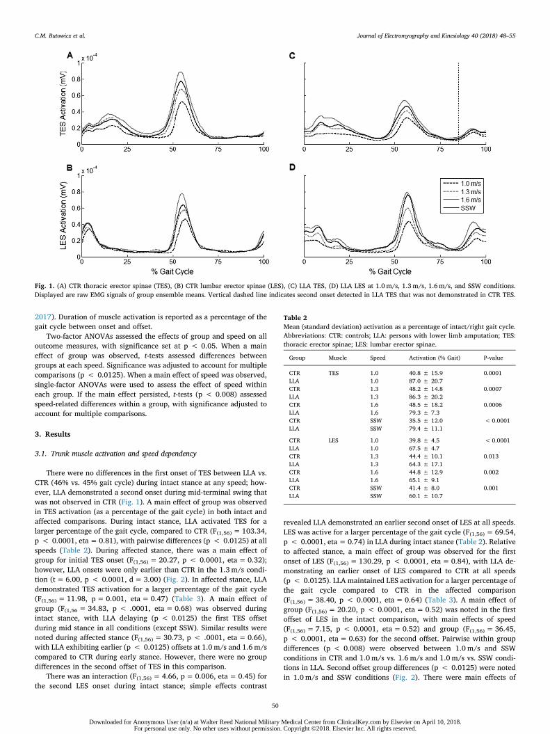
2017). Duration of muscle activation is reported as a percentage of thegait cycle between onset and offset.
Two-factor ANOVAs assessed the effects of group and speed on alloutcome measures, with significance set at p < 0.05. When a maineffect of group was observed, t-tests assessed differences betweengroups at each speed. Significance was adjusted to account for multiplecomparisons (p < 0.0125). When a main effect of speed was observed,single-factor ANOVAs were used to assess the effect of speed withineach group. If the main effect persisted, t-tests (p < 0.008) assessedspeed-related differences within a group, with significance adjusted toaccount for multiple comparisons.
3. Results
3.1. Trunk muscle activation and speed dependency
There were no differences in the first onset of TES between LLA vs.CTR (46% vs. 45% gait cycle) during intact stance at any speed; how-ever, LLA demonstrated a second onset during mid-terminal swing thatwas not observed in CTR (Fig. 1). A main effect of group was observedin TES activation (as a percentage of the gait cycle) in both intact andaffected comparisons. During intact stance, LLA activated TES for alarger percentage of the gait cycle, compared to CTR (F(1,56) = 103.34,p < 0.0001, eta= 0.81), with pairwise differences (p < 0.0125) at allspeeds (Table 2). During affected stance, there was a main effect ofgroup for initial TES onset (F(1,56) = 20.27, p < 0.0001, eta= 0.32);however, LLA onsets were only earlier than CTR in the 1.3m/s condi-tion (t= 6.00, p < 0.0001, d=3.00) (Fig. 2). In affected stance, LLAdemonstrated TES activation for a larger percentage of the gait cycle(F(1,56) = 11.98, p= 0.001, eta= 0.47) (Table 3). A main effect ofgroup (F(1,56= 34.83, p < .0001, eta= 0.68) was observed duringintact stance, with LLA delaying (p < 0.0125) the first TES offsetduring mid stance in all conditions (except SSW). Similar results werenoted during affected stance (F(1,56) = 30.73, p < .0001, eta= 0.66),with LLA exhibiting earlier (p < 0.0125) offsets at 1.0m/s and 1.6m/scompared to CTR during early stance. However, there were no groupdifferences in the second offset of TES in this comparison.
There was an interaction (F(1,56)= 4.66, p= 0.006, eta= 0.45) forthe second LES onset during intact stance; simple effects contrast
revealed LLA demonstrated an earlier second onset of LES at all speeds.LES was active for a larger percentage of the gait cycle (F(1,56) = 69.54,p < 0.0001, eta= 0.74) in LLA during intact stance (Table 2). Relativeto affected stance, a main effect of group was observed for the firstonset of LES (F(1,56) = 130.29, p < 0.0001, eta= 0.84), with LLA de-monstrating an earlier onset of LES compared to CTR at all speeds(p < 0.0125). LLA maintained LES activation for a larger percentage ofthe gait cycle compared to CTR in the affected comparison(F(1,56) = 38.40, p < 0.0001, eta= 0.64) (Table 3). A main effect ofgroup (F(1,56)= 20.20, p < 0.0001, eta= 0.52) was noted in the firstoffset of LES in the intact comparison, with main effects of speed(F(1,56) = 7.15, p < 0.0001, eta= 0.52) and group (F(1,56) = 36.45,p < 0.0001, eta= 0.63) for the second offset. Pairwise within groupdifferences (p < 0.008) were observed between 1.0 m/s and SSWconditions in CTR and 1.0m/s vs. 1.6 m/s and 1.0 m/s vs. SSW condi-tions in LLA. Second offset group differences (p < 0.0125) were notedin 1.0m/s and SSW conditions (Fig. 2). There were main effects of
Fig. 1. (A) CTR thoracic erector spinae (TES), (B) CTR lumbar erector spinae (LES), (C) LLA TES, (D) LLA LES at 1.0 m/s, 1.3 m/s, 1.6m/s, and SSW conditions.Displayed are raw EMG signals of group ensemble means. Vertical dashed line indicates second onset detected in LLA TES that was not demonstrated in CTR TES.
Table 2Mean (standard deviation) activation as a percentage of intact/right gait cycle.Abbreviations: CTR: controls; LLA: persons with lower limb amputation; TES:thoracic erector spinae; LES: lumbar erector spinae.
Group Muscle Speed Activation (% Gait) P-value
CTR TES 1.0 40.8 ± 15.9 0.0001LLA 1.0 87.0 ± 20.7CTR 1.3 48.2 ± 14.8 0.0007LLA 1.3 86.3 ± 20.2CTR 1.6 48.5 ± 18.2 0.0006LLA 1.6 79.3 ± 7.3CTR SSW 35.5 ± 12.0 < 0.0001LLA SSW 79.4 ± 11.1
CTR LES 1.0 39.8 ± 4.5 < 0.0001LLA 1.0 67.5 ± 4.7CTR 1.3 44.4 ± 10.1 0.013LLA 1.3 64.3 ± 17.1CTR 1.6 44.8 ± 12.9 0.002LLA 1.6 65.1 ± 9.1CTR SSW 41.4 ± 8.0 0.001LLA SSW 60.1 ± 10.7
C.M. Butowicz et al. Journal of Electromyography and Kinesiology 40 (2018) 48–55
50
Downloaded for Anonymous User (n/a) at Walter Reed National Military Medical Center from ClinicalKey.com by Elsevier on April 10, 2018.For personal use only. No other uses without permission. Copyright ©2018. Elsevier Inc. All rights reserved.
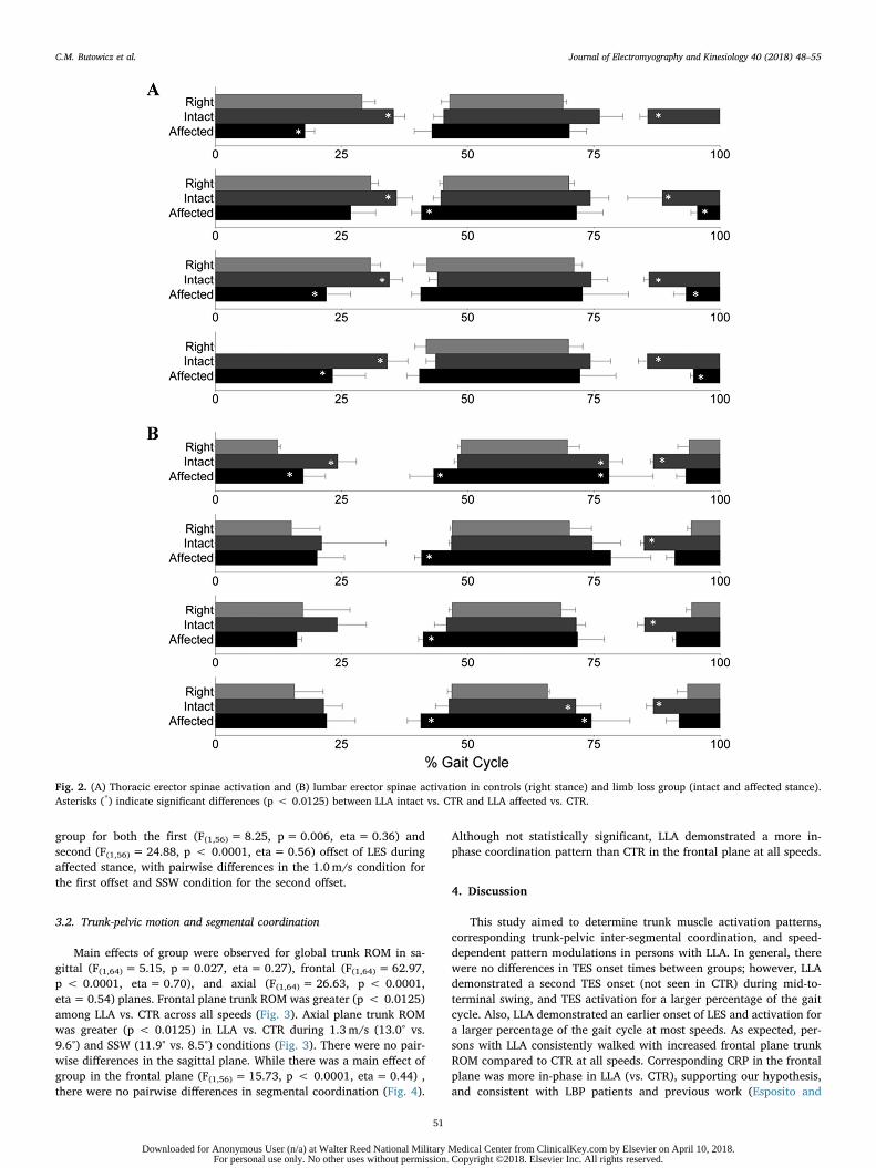
group for both the first (F(1,56)= 8.25, p= 0.006, eta= 0.36) andsecond (F(1,56) = 24.88, p < 0.0001, eta= 0.56) offset of LES duringaffected stance, with pairwise differences in the 1.0m/s condition forthe first offset and SSW condition for the second offset.
3.2. Trunk-pelvic motion and segmental coordination
Main effects of group were observed for global trunk ROM in sa-gittal (F(1,64)= 5.15, p=0.027, eta= 0.27), frontal (F(1,64) = 62.97,p < 0.0001, eta= 0.70), and axial (F(1,64) = 26.63, p < 0.0001,eta= 0.54) planes. Frontal plane trunk ROM was greater (p < 0.0125)among LLA vs. CTR across all speeds (Fig. 3). Axial plane trunk ROMwas greater (p < 0.0125) in LLA vs. CTR during 1.3m/s (13.0° vs.9.6°) and SSW (11.9° vs. 8.5°) conditions (Fig. 3). There were no pair-wise differences in the sagittal plane. While there was a main effect ofgroup in the frontal plane (F(1,56) = 15.73, p < 0.0001, eta= 0.44) ,there were no pairwise differences in segmental coordination (Fig. 4).
Although not statistically significant, LLA demonstrated a more in-phase coordination pattern than CTR in the frontal plane at all speeds.
4. Discussion
This study aimed to determine trunk muscle activation patterns,corresponding trunk-pelvic inter-segmental coordination, and speed-dependent pattern modulations in persons with LLA. In general, therewere no differences in TES onset times between groups; however, LLAdemonstrated a second TES onset (not seen in CTR) during mid-to-terminal swing, and TES activation for a larger percentage of the gaitcycle. Also, LLA demonstrated an earlier onset of LES and activation fora larger percentage of the gait cycle at most speeds. As expected, per-sons with LLA consistently walked with increased frontal plane trunkROM compared to CTR at all speeds. Corresponding CRP in the frontalplane was more in-phase in LLA (vs. CTR), supporting our hypothesis,and consistent with LBP patients and previous work (Esposito and
Fig. 2. (A) Thoracic erector spinae activation and (B) lumbar erector spinae activation in controls (right stance) and limb loss group (intact and affected stance).Asterisks (*) indicate significant differences (p < 0.0125) between LLA intact vs. CTR and LLA affected vs. CTR.
C.M. Butowicz et al. Journal of Electromyography and Kinesiology 40 (2018) 48–55
51
Downloaded for Anonymous User (n/a) at Walter Reed National Military Medical Center from ClinicalKey.com by Elsevier on April 10, 2018.For personal use only. No other uses without permission. Copyright ©2018. Elsevier Inc. All rights reserved.
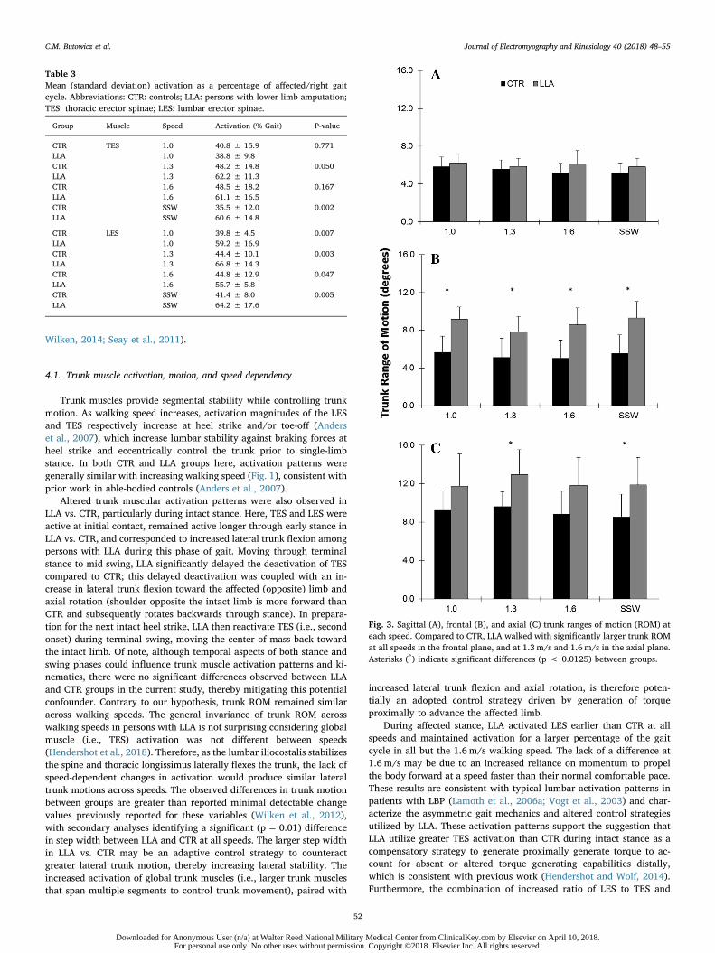
Wilken, 2014; Seay et al., 2011).
4.1. Trunk muscle activation, motion, and speed dependency
Trunk muscles provide segmental stability while controlling trunkmotion. As walking speed increases, activation magnitudes of the LESand TES respectively increase at heel strike and/or toe-off (Anderset al., 2007), which increase lumbar stability against braking forces atheel strike and eccentrically control the trunk prior to single-limbstance. In both CTR and LLA groups here, activation patterns weregenerally similar with increasing walking speed (Fig. 1), consistent withprior work in able-bodied controls (Anders et al., 2007).
Altered trunk muscular activation patterns were also observed inLLA vs. CTR, particularly during intact stance. Here, TES and LES wereactive at initial contact, remained active longer through early stance inLLA vs. CTR, and corresponded to increased lateral trunk flexion amongpersons with LLA during this phase of gait. Moving through terminalstance to mid swing, LLA significantly delayed the deactivation of TEScompared to CTR; this delayed deactivation was coupled with an in-crease in lateral trunk flexion toward the affected (opposite) limb andaxial rotation (shoulder opposite the intact limb is more forward thanCTR and subsequently rotates backwards through stance). In prepara-tion for the next intact heel strike, LLA then reactivate TES (i.e., secondonset) during terminal swing, moving the center of mass back towardthe intact limb. Of note, although temporal aspects of both stance andswing phases could influence trunk muscle activation patterns and ki-nematics, there were no significant differences observed between LLAand CTR groups in the current study, thereby mitigating this potentialconfounder. Contrary to our hypothesis, trunk ROM remained similaracross walking speeds. The general invariance of trunk ROM acrosswalking speeds in persons with LLA is not surprising considering globalmuscle (i.e., TES) activation was not different between speeds(Hendershot et al., 2018). Therefore, as the lumbar iliocostalis stabilizesthe spine and thoracic longissimus laterally flexes the trunk, the lack ofspeed-dependent changes in activation would produce similar lateraltrunk motions across speeds. The observed differences in trunk motionbetween groups are greater than reported minimal detectable changevalues previously reported for these variables (Wilken et al., 2012),with secondary analyses identifying a significant (p=0.01) differencein step width between LLA and CTR at all speeds. The larger step widthin LLA vs. CTR may be an adaptive control strategy to counteractgreater lateral trunk motion, thereby increasing lateral stability. Theincreased activation of global trunk muscles (i.e., larger trunk musclesthat span multiple segments to control trunk movement), paired with
increased lateral trunk flexion and axial rotation, is therefore poten-tially an adopted control strategy driven by generation of torqueproximally to advance the affected limb.
During affected stance, LLA activated LES earlier than CTR at allspeeds and maintained activation for a larger percentage of the gaitcycle in all but the 1.6m/s walking speed. The lack of a difference at1.6 m/s may be due to an increased reliance on momentum to propelthe body forward at a speed faster than their normal comfortable pace.These results are consistent with typical lumbar activation patterns inpatients with LBP (Lamoth et al., 2006a; Vogt et al., 2003) and char-acterize the asymmetric gait mechanics and altered control strategiesutilized by LLA. These activation patterns support the suggestion thatLLA utilize greater TES activation than CTR during intact stance as acompensatory strategy to generate proximally generate torque to ac-count for absent or altered torque generating capabilities distally,which is consistent with previous work (Hendershot and Wolf, 2014).Furthermore, the combination of increased ratio of LES to TES and
Table 3Mean (standard deviation) activation as a percentage of affected/right gaitcycle. Abbreviations: CTR: controls; LLA: persons with lower limb amputation;TES: thoracic erector spinae; LES: lumbar erector spinae.
Group Muscle Speed Activation (% Gait) P-value
CTR TES 1.0 40.8 ± 15.9 0.771LLA 1.0 38.8 ± 9.8CTR 1.3 48.2 ± 14.8 0.050LLA 1.3 62.2 ± 11.3CTR 1.6 48.5 ± 18.2 0.167LLA 1.6 61.1 ± 16.5CTR SSW 35.5 ± 12.0 0.002LLA SSW 60.6 ± 14.8
CTR LES 1.0 39.8 ± 4.5 0.007LLA 1.0 59.2 ± 16.9CTR 1.3 44.4 ± 10.1 0.003LLA 1.3 66.8 ± 14.3CTR 1.6 44.8 ± 12.9 0.047LLA 1.6 55.7 ± 5.8CTR SSW 41.4 ± 8.0 0.005LLA SSW 64.2 ± 17.6
Fig. 3. Sagittal (A), frontal (B), and axial (C) trunk ranges of motion (ROM) ateach speed. Compared to CTR, LLA walked with significantly larger trunk ROMat all speeds in the frontal plane, and at 1.3 m/s and 1.6 m/s in the axial plane.Asterisks (*) indicate significant differences (p < 0.0125) between groups.
C.M. Butowicz et al. Journal of Electromyography and Kinesiology 40 (2018) 48–55
52
Downloaded for Anonymous User (n/a) at Walter Reed National Military Medical Center from ClinicalKey.com by Elsevier on April 10, 2018.For personal use only. No other uses without permission. Copyright ©2018. Elsevier Inc. All rights reserved.
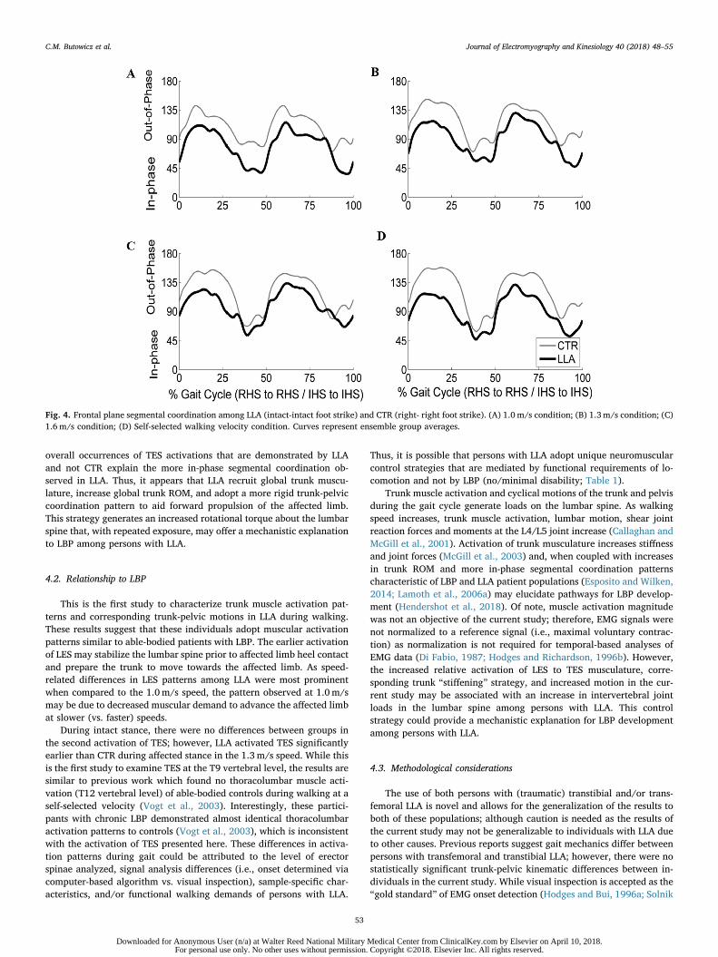
overall occurrences of TES activations that are demonstrated by LLAand not CTR explain the more in-phase segmental coordination ob-served in LLA. Thus, it appears that LLA recruit global trunk muscu-lature, increase global trunk ROM, and adopt a more rigid trunk-pelviccoordination pattern to aid forward propulsion of the affected limb.This strategy generates an increased rotational torque about the lumbarspine that, with repeated exposure, may offer a mechanistic explanationto LBP among persons with LLA.
4.2. Relationship to LBP
This is the first study to characterize trunk muscle activation pat-terns and corresponding trunk-pelvic motions in LLA during walking.These results suggest that these individuals adopt muscular activationpatterns similar to able-bodied patients with LBP. The earlier activationof LES may stabilize the lumbar spine prior to affected limb heel contactand prepare the trunk to move towards the affected limb. As speed-related differences in LES patterns among LLA were most prominentwhen compared to the 1.0m/s speed, the pattern observed at 1.0m/smay be due to decreased muscular demand to advance the affected limbat slower (vs. faster) speeds.
During intact stance, there were no differences between groups inthe second activation of TES; however, LLA activated TES significantlyearlier than CTR during affected stance in the 1.3m/s speed. While thisis the first study to examine TES at the T9 vertebral level, the results aresimilar to previous work which found no thoracolumbar muscle acti-vation (T12 vertebral level) of able-bodied controls during walking at aself-selected velocity (Vogt et al., 2003). Interestingly, these partici-pants with chronic LBP demonstrated almost identical thoracolumbaractivation patterns to controls (Vogt et al., 2003), which is inconsistentwith the activation of TES presented here. These differences in activa-tion patterns during gait could be attributed to the level of erectorspinae analyzed, signal analysis differences (i.e., onset determined viacomputer-based algorithm vs. visual inspection), sample-specific char-acteristics, and/or functional walking demands of persons with LLA.
Thus, it is possible that persons with LLA adopt unique neuromuscularcontrol strategies that are mediated by functional requirements of lo-comotion and not by LBP (no/minimal disability; Table 1).
Trunk muscle activation and cyclical motions of the trunk and pelvisduring the gait cycle generate loads on the lumbar spine. As walkingspeed increases, trunk muscle activation, lumbar motion, shear jointreaction forces and moments at the L4/L5 joint increase (Callaghan andMcGill et al., 2001). Activation of trunk musculature increases stiffnessand joint forces (McGill et al., 2003) and, when coupled with increasesin trunk ROM and more in-phase segmental coordination patternscharacteristic of LBP and LLA patient populations (Esposito and Wilken,2014; Lamoth et al., 2006a) may elucidate pathways for LBP develop-ment (Hendershot et al., 2018). Of note, muscle activation magnitudewas not an objective of the current study; therefore, EMG signals werenot normalized to a reference signal (i.e., maximal voluntary contrac-tion) as normalization is not required for temporal-based analyses ofEMG data (Di Fabio, 1987; Hodges and Richardson, 1996b). However,the increased relative activation of LES to TES musculature, corre-sponding trunk “stiffening” strategy, and increased motion in the cur-rent study may be associated with an increase in intervertebral jointloads in the lumbar spine among persons with LLA. This controlstrategy could provide a mechanistic explanation for LBP developmentamong persons with LLA.
4.3. Methodological considerations
The use of both persons with (traumatic) transtibial and/or trans-femoral LLA is novel and allows for the generalization of the results toboth of these populations; although caution is needed as the results ofthe current study may not be generalizable to individuals with LLA dueto other causes. Previous reports suggest gait mechanics differ betweenpersons with transfemoral and transtibial LLA; however, there were nostatistically significant trunk-pelvic kinematic differences between in-dividuals in the current study. While visual inspection is accepted as the“gold standard” of EMG onset detection (Hodges and Bui, 1996a; Solnik
Fig. 4. Frontal plane segmental coordination among LLA (intact-intact foot strike) and CTR (right- right foot strike). (A) 1.0 m/s condition; (B) 1.3m/s condition; (C)1.6 m/s condition; (D) Self-selected walking velocity condition. Curves represent ensemble group averages.
C.M. Butowicz et al. Journal of Electromyography and Kinesiology 40 (2018) 48–55
53
Downloaded for Anonymous User (n/a) at Walter Reed National Military Medical Center from ClinicalKey.com by Elsevier on April 10, 2018.For personal use only. No other uses without permission. Copyright ©2018. Elsevier Inc. All rights reserved.
et al., 2010; Tenan et al., 2017), this method is inherently variable andsusceptible to human error. Reviewers in the current study demon-strated good test-retest reliability that is consistent with previous work,mitigating this concern. The general lack of significant post hoc dif-ferences between groups at each speed may be a function of smallsample size and thus type II error. Nevertheless, the large effect sizesobserved as group main effects support our hypotheses as well asconfidence in the presence of group differences in trunk muscle acti-vation patterns. Future work should also consider assessing anteriortrunk muscular activation to determine flexor/extensor co-activationstrategies as well as a reference criterion that allows for the comparisonof activation magnitudes between groups.
4.4. Conclusions
Persons with LLA demonstrate altered activation of posterior trunkmuscles (i.e., earlier onsets and delayed offsets) compared to able-bodied controls during walking. While prior work in able-bodied in-dividuals with LBP has suggested that altered LES activation patternsare a function of poor neuromuscular control and efforts to increaselumbar stability, it appears persons with LLA adopt proximal strategiesto advance the affected limb during over-ground walking. However, thedifferential patterns of muscular activation and trunk-pelvic motionsmay influence spinal loads and subsequently increase LBP risk. Furtherwork is needed to explicitly relate muscular activation patterns (andmagnitudes) with spinal loading during walking in persons with LLA.
Acknowledgements
This work was supported, in part, by the Office of the AssistantSecretary of Defense for Health Affairs, through the Peer ReviewedOrthopaedic Research Program (award #W81XWH-14-2-0144), theCenter for Rehabilitation Science Research, Department ofRehabilitation Medicine, Uniformed Services University, Bethesda, MD(awards HU0001-11-1-0004 and HU0001-15-2-0003), and the DoD-VAEACE (Public Law 110-417, National Defense Authorization Act 2009,Section 723). The views expressed herein are those of the authors, anddo not necessarily reflect the official policy or position of the U.S.Department of Defense, nor the U.S. government.
Conflict of interest
The authors declare that there is no conflict of interest.
References
Anders, C., Wagner, H., Puta, C., Grassme, R., Petrovitch, A., Scholle, H.-C., 2007. Trunkmuscle activation patterns during walking at different speeds. J. Electromyogr.Kinesiol. 17 (2), 245–252.
Anderson, F.C., Pandy, M.G., 2003. Individual muscle contributions to support in normalwalking. Gait Posture 17 (2), 159–169.
Arendt-Nielsen, L., Nie, H., Laursen, M.B., Laursen, B.S., Madeleine, P., Simonsen, O.H.,Graven-Nielsen, T., 2010. Sensitization in patients with painful knee osteoarthritis.Pain 149 (3), 573–581.
Callaghan, J.P., McGill, S., 2001. Low back joint loading and kinematics during standingand unsupported sitting. Ergonomics 44 (3), 280–294. http://dx.doi.org/10.1080/00140130118276.
Callaghan, J.P., Patla, A.E., McGill, S.M., 1999. Low back three-dimensional joint forces,kinematics, and kinetics during walking. Clin. Biomech. 14 (3), 203–216.
Ceccato, J.-C., De Sèze, M., Azevedo, C., Cazalets, J.-R., 2009. Comparison of trunk ac-tivity during gait initiation and walking in humans. PLoS One 4 (12), e8193.
Devan, H., Hendrick, P., Ribeiro, D.C., Hale, L.A., Carman, A., 2014. Asymmetricalmovements of the lumbopelvic region: is this a potential mechanism for low backpain in people with lower limb amputation? Med. Hypotheses 82 (1), 77–85. http://dx.doi.org/10.1016/j.mehy.2013.11.012.
Di Fabio, R.P., 1987. Reliability of computerized surface electromyography for de-termining the onset of muscle activity. Phys. Therapy 67 (1), 43–48.
Ehde, D.M., Smith, D.G., Czerniecki, J.M., Campbell, K.M., Malchow, D.M., Robinson,L.R., 2001. Back pain as a secondary disability in persons with lower limb amputa-tions. Arch. Phys. Med. Rehabil. 82 (6), 731–734.
Ephraim, P.L., Wegener, S.T., MacKenzie, E.J., Dillingham, T.R., Pezzin, L.E., 2005.
Phantom pain, residual limb pain, and back pain in amputees: results of a nationalsurvey. Arch. Phys. Med. Rehabil. 86 (10), 1910–1919.
Esposito, E.R., Wilken, J.M., 2014. The relationship between pelvis–trunk coordinationand low back pain in individuals with transfemoral amputations. Gait Posture 40 (4),640–646.
Farrokhi, S., Mazzone, B., Schneider, M., Gombatto, S., Mayer, J., Highsmith, M.J.,Hendershot, B.D., 2017. Biopsychosocial risk factors associated with chronic lowback pain after lower limb amputation. Med. Hypotheses 108, 1–9.
Goujon-Pillet, H., Sapin, E., Fode, P., Lavaste, F., 2008. Three-dimensional motions oftrunk and pelvis during transfemoral amputee gait. Arch. Phys. Med. Rehabil. 89 (1),87–94. http://dx.doi.org/10.1016/j.apmr.2007.08.136.
Hamill, J., van Emmerik, R.E., Heiderscheit, B.C., Li, L., 1999. A dynamical systems ap-proach to lower extremity running injuries. Clin. Biomech. 14 (5), 297–308.
Hendershot, B.D., Shojaei, I., Acasio, J., Dearth, C., Bazrgari, B., 2018. Walking speeddifferentially alters spinal loads in persons with traumatic lower limb amputation. J.Biomech. 70 (21), 249–254.
Hendershot, B.D., Wolf, E.J., 2014. Three-dimensional joint reaction forces and momentsat the low back during over-ground walking in persons with unilateral lower-ex-tremity amputation. Clin. Biomech. 29 (3), 235–242. http://dx.doi.org/10.1016/j.clinbiomech.2013.12.005.
Hodges, P.W., Bui, B.H., 1996a. A comparison of computer-based methods for the de-termination of onset of muscle contraction using electromyography.Electroencephalogr. Clin. Neurophysiol./Electromyogr. Motor Contr. 101 (6),511–519.
Hodges, P.W., Richardson, C.A., 1996b. Inefficient muscular stabilization of the lumbarspine associated with low back pain: a motor control evaluation of transversus ab-dominis. Spine 21 (22), 2640–2650.
Hodges, P.W., Richardson, C.A., 1999. Altered trunk muscle recruitment in people withlow back pain with upper limb movement at different speeds. Arch. Phys. Med.Rehabil. 80 (9), 1005–1012.
Jaegers, S.M., Arendzen, J.H., de Jongh, H.J., 1995. Prosthetic gait of unilateral trans-femoral amputees: a kinematic study. Arch. Phys. Med. Rehabil. 76 (8), 736–743.
Kulkarni, J., Gaine, W.J., Buckley, J.G., Rankine, J.J., Adams, J., 2005. Chronic low backpain in traumatic lower limb amputees. Clin. Rehabil. 19 (1), 81–86.
Kušljugić, A., Kapidžić-Duraković, S., Kudumović, Z., Čičkušić, A., 2006. Chronic lowback pain in individuals with lower-limb amputation. Bosnian J. Basic Med. Sci. 6(2),67–70.
Lamoth, C.J., Daffertshofer, A., Meijer, O.G., Beek, P.J., 2006a. How do persons withchronic low back pain speed up and slow down? Trunk–pelvis coordination andlumbar erector spinae activity during gait. Gait Posture 23 (2), 230–239.
Lamoth, C.J., Meijer, O.G., Daffertshofer, A., Wuisman, P.I., Beek, P.J., 2006b. Effects ofchronic low back pain on trunk coordination and back muscle activity duringwalking: changes in motor control. Eur. Spine J. 15 (1), 23–40.
Lamoth, C.J., Meijer, O.G., Wuisman, P.I., van Dieën, J.H., Levin, M.F., Beek, P.J., 2002.Pelvis-thorax coordination in the transverse plane during walking in persons withnonspecific low back pain. Spine 27 (4), E92–E99.
McGill, S.M., Grenier, S., Kavcic, N., Cholewicki, J., 2003. Coordination of muscle activityto assure stability of the lumbar spine. J. Electromyogr. Kinesiol. 13 (4), 353–359.
Portney, L.G., Watkins, M.P., 2009. Foundations of Clinical Research: Applications toPractice, third ed. Pearson Prentice Hall, Saddle River, New Jersey, USA.
Saunders, S.W., Schache, A., Rath, D., Hodges, P.W., 2005. Changes in three dimensionallumbo-pelvic kinematics and trunk muscle activity with speed and mode of loco-motion. Clin. Biomech. 20 (8), 784–793.
Seay, J.F., Van Emmerik, R.E., Hamill, J., 2011. Low back pain status affects pelvis-trunkcoordination and variability during walking and running. Clin. Biomech. 26 (6),572–578.
Shiavi, R., 1990. Quantitative representation of electromyographic patterns generatedduring human locomotion. IEEE Eng. Med. Biol. Mag. 9 (1), 58–60.
Solnik, S., Rider, P., Steinweg, K., DeVita, P., Hortobágyi, T., 2010. Teager-Kaiser energyoperator signal conditioning improves EMG onset detection. Eur. J. Appl. Physiol.110 (3), 489–498.
Tenan, M.S., Tweedell, A.J., Haynes, C.A., 2017. Analysis of statistical and standard al-gorithms for detecting muscle onset with surface electromyography. PloS One 12 (5),e0177312.
van der Hulst, M., Vollenbroek-Hutten, M.M., Rietman, J.S., Hermens, H.J., 2010. Lumbarand abdominal muscle activity during walking in subjects with chronic low backpain: support of the “guarding” hypothesis? J. Electromyogr. Kinesiol. 20 (1), 31–38.
Vogt, L., Pfeifer, K., Banzer, W., 2003. Neuromuscular control of walking with chroniclow-back pain. Manual Ther. 8 (1), 21–28.
Wilken, J.M., Rodriguez, K.M., Brawner, M., Darter, B.J., 2012. Reliability and minimaldetectible change values for gait kinematics and kinetics in healthy adults. GaitPosture 35 (2), 301–307.
Willigenburg, N.W., Kingma, I., van Dieën, J.H., 2013. Center of pressure trajectories,trunk kinematics and trunk muscle activation during unstable sitting in low back painpatients. Gait Posture 38 (4), 625–630.
Zeni, J., Richards, J., Higginson, J., 2008. Two simple methods for determining gaitevents during treadmill and overground walking using kinematic data. Gait Posture27 (4), 710–714.
CourtneyButowicz, PhD, CSCS, is a Postdoctoral Researcher within the Department ofRehabilitation at Walter Reed National Military Medical Center (WRNMMC). Dr.Butowicz’s research interests include: clinical assessment of trunk stability/control, de-velopment of musculoskeletal injuries as result of impaired motor control, developmentand treatment of low back pain in service members and veterans with limb loss, andutilization of a multi-disciplinary approach to the treatment of musculoskeletal injuries.
C.M. Butowicz et al. Journal of Electromyography and Kinesiology 40 (2018) 48–55
54
Downloaded for Anonymous User (n/a) at Walter Reed National Military Medical Center from ClinicalKey.com by Elsevier on April 10, 2018.For personal use only. No other uses without permission. Copyright ©2018. Elsevier Inc. All rights reserved.
JulianAcasio, MS, is a Research Engineer within the Department of Rehabilitation atWRNMMC. He received his BS and MS in Biomedical Engineering from NorthwesternUniversity. Mr. Acasio’s research interests focus on the mechanics and control of humanmovement, with particular interest in persons with traumatic limb loss.
Christopher L. Dearth, PhD, concurrently serves as the Facility Research Director for theExtremity Trauma & Amputation Center of Excellence (EACE), Director of Research forthe Department of Rehabilitation at WRNMMC, and the Founding Director of theRegenerative Biosciences Laboratory at the Uniformed Services University of the HealthSciences (USUHS). In these roles, Dr. Dearth leads a multidisciplinary team of cliniciansand researchers whose collective focus is on the mitigation, treatment and rehabilitationof traumatic extremity injuries and amputations by implementation of clinically relevantresearch aimed at optimizing the quality of life of service members and veterans. Dr.Dearth’s preclinical and clinical research interests include: development of innovativeregenerative medicine & tissue engineering constructs, utilization of next generationimaging modalities for extremity trauma applications, evaluation of advanced (i.e.,
powered and intuitively controlled) prosthetics for service members and veterans withlimb loss, and championing a synergy of efforts between the fields of rehabilitative andregenerative medicine.
Brad D. Hendershot, PhD, is a Research Biomedical Engineer with the EACE, stationedat WRNMMC. In addition to this role, he directs activities within the Biomechanics andVirtual Reality Laboratories within the Department of Rehabilitation. Dr. Hendershot alsoholds an appointment, as Assistant Professor, within the Department of RehabilitationMedicine at USUHS. He received his BS in Bioengineering from Penn State and PhD inBiomedical Engineering from Virginia Tech. Dr. Hendershot’s research is primarily fo-cused on the mechanics and neuromuscular control of human movement, with a parti-cular emphasis on the trunk/spine in both uninjured and patient populations. Recently,his research aims to characterize factors underlying the high prevalence of musculoske-letal complications secondary to extremity trauma, to mitigate development/recurrenceover the longer term and ultimately improve quality of life for service members andveterans with limb loss.
C.M. Butowicz et al. Journal of Electromyography and Kinesiology 40 (2018) 48–55
55
Downloaded for Anonymous User (n/a) at Walter Reed National Military Medical Center from ClinicalKey.com by Elsevier on April 10, 2018.For personal use only. No other uses without permission. Copyright ©2018. Elsevier Inc. All rights reserved.
ARTICLE IN PRESS
The Spine Journal 000 (2018) 1−12
Review Article
FDA device/
Author discl
OTH: Nothing
The disclosu
* Correspon
8900 Grand O
E-mail add
https://doi.org/
1529-9430/© 2
D
Low back pain in persons with lower extremity amputation:
a systematic review of the literature
M. Jason Highsmith, DPT, PhD, CP, FAAOPa,e,g,*, Lisa M. Goff, MSPTh,Amanda L. Lewandowski, DPT, COMTi, Shawn Farrokhi, PT, PhDb,l,
Brad D. Hendershot, PhDc,j, Owen T. Hill, PhDd,Christopher A. R�abago, PT, PhDd,k, Elizabeth Russell-Esposito, PhDd,k,
John J. Orriola, MSf, John M. Mayer, DC, PhD, FACSMm
a Extremity Trauma & Amputation Center of Excellence (EACE), US Department of Veterans’ Affairs, US Department of
Defense, 13000 Bruce B. Downs Blvd., Tampa, FL, 33612, USAb Extremity Trauma & Amputation Center of Excellence (EACE), US Department of Veterans’ Affairs, US Department of
Defense, 34800 Bob Wilson Dr. San Diego, CA, 92134, USAc Extremity Trauma & Amputation Center of Excellence (EACE), US Department of Veterans’ Affairs, US Department of
Defense, Bethesda, 4494 North Palmer Rd, Bethesda, MD, 20889, USAd Extremity Trauma & Amputation Center of Excellence (EACE), US Department of Veterans’ Affairs, US Department of
Defense, Ft. Sam Houston, 3851 Roger Brooke Dr, San Antonio, TX, 78234, USAe School of Physical Therapy & Rehabilitation Sciences, Morsani College of Medicine, University of South Florida, 3515 E.
Fletcher Ave. Tampa, FL, 33612, USAf Shimberg Health Sciences Library, Morsani College of Medicine, University of South Florida, 12901 Bruce B. Downs Blvd.,
Tampa, FL, 33612g 319th Minimal Care Detachment, U.S. Army Reserves, Pinellas Park, 2801 Grand Ave. Pinellas Park, FL, USA, 33782h Center of Innovation on Disability & Rehabilitation Research, James A. Haley Veterans’ Administration Hospital, 8900
Grand Oak Ci, Tampa, FL, 33637, USAi Select Physical Therapy, Brandon, 2410 W. Brandon Blvd. Brandon, FL, 33511, USA
jDepartment of Rehabilitation, Research and Development Section, Walter Reed National Military Medical Center, Bethesda,
4494 North Palmer Rd, Bethesda, MD, 20889, USAk Center for the Intrepid, Department of Rehabilitation Medicine, Brooke Army Medical Center, JBSA, Ft. Sam Houston, 3851
Roger Brooke Dr, San Antonio, TX, 78234, USAl The Department of Physical and Occupational Therapy, Naval Medical Center San Diego, San Diego, 34800 Bob Wilson Dr.
San Diego, CA, 92134, USAmU.S. Spine & Sport Foundation, 3760 Convoy St #101. San Diego, CA, 92111, USA
Received 26 March 2018; revised 14 August 2018; accepted 20 August 2018
Abstract B
drug sta
osures:
to disc
re key c
ding au
ak Circl
ress: m
10.1016
018 Els
ownloa
ACKGROUND CONTEXT: Lower extremity amputation (LEA) is associated with an elevated
risk for development and progression of secondary health conditions. Low back pain (LBP) is one
such condition adversely affecting function, independence, and quality of life.
PURPOSE: The purpose of this study was to systematically review the literature to determine the
strength of evidence relating the presence and severity of LBP secondary to LEA, thereby support-
ing the formulation of empirical evidence statements (EESs) to guide practice and future research.
STUDY DESIGN/SETTING: Systematic review of the literature.
METHODS: A systematic review of five databases was conducted followed by evaluation of evi-
dence and synthesis of EESs.
tus: Not applicable
MJH: Nothing to disclose. LMG: Nothing to disclose. ALL: Nothing to disclose. SF: Nothing to disclose. BDH: Nothing to disclose.
lose. CAR: Nothing to disclose. ERE: Nothing to disclose. JJO: Nothing to disclose. JMM: Nothing to disclose.
an be found on the Table of Contents and atwww.TheSpineJournalOnline.com.
thor. Extremity Trauma & Amputation Center of Excellence (EACE), US Department of Veterans’ Affairs, US Department of Defense,
e (151R), Tampa, FL 33637-1022, USA. Tel.: (813) 558-3936; fax: (813) 558-3990.
[email protected] (M.J. Highsmith).
/j.spinee.2018.08.011
evier Inc. All rights reserved.
ded for Anonymous User (n/a) at Walter Reed National Military Medical Center from ClinicalKey.com by Elsevier on October 29, 2018.For personal use only. No other uses without permission. Copyright ©2018. Elsevier Inc. All rights reserved.
ARTICLE IN PRESS
2 M.J. Highsmith et al. / The Spine Journal 00 (2018) 1−12
Downlo
RESULTS: Seventeen manuscripts were included. From these, eight EESs were synthesized
within the following categories: epidemiology, amputation level, function, disability, leg
length, posture, spinal kinematics, and osseointegrated prostheses. Only the EES on epidemi-
ology was supported by evidence at the moderate confidence level given support by eight
moderate quality studies. The four EESs for amputation level, leg length, posture, and spinal
kinematics were supported by evidence at the low confidence level given that each of these
statements had some evidence not supporting the statement but ultimately more evidence
(and of higher quality) currently supporting the statement. The remaining three EESs that
addressed function, disability and osseointegrated prosthetic use were all supported by single
studies or had comparable evidence that disagreed with study findings rendering insufficient
evidence to support the respective EES.
CONCLUSIONS: Based on the state of the current evidence, appropriate preventative and, partic-
ularly, treatment strategies to manage LBP in persons with LEA remain a knowledge gap and an
area of future study. © 2018 Elsevier Inc. All rights reserved.
Keywords: A
mputee; Limb loss; Lumbago; Rehabilitation; Spinal pain; Transfemoral; Transtibial.aded for Anonymous User (n/a) at Walter Reed National Military Medical Center from ClinicalKey.com by Elsevier on OctobeFor personal use only. No other uses without permission. Copyright ©2018. Elsevier Inc. All rights reserved.
Introduction
Common musculoskeletal derangements of the spine
that contribute to low back pain (LBP) include discogenic
dysfunction, facet joint syndrome, sacroiliac joint syn-
drome, spinal instability, and postural syndrome [1].
There are many factors related to spinal derangements
including behavioral, congenital, traumatic, disease pro-
cesses, and others. These derangements and factors can
co-exist, leading to varying levels of disability attributed
to LBP. Severe lower extremity trauma, including lower
extremity amputation (LEA), can further confound and
complicate the clinical presentation and management of
LBP [2].
The prevalence of LBP is much higher (52%−89%)
among persons with LEA as compared to the general non-
amputee population (12%−45%) [3−6]. Low back pain has
been considered more bothersome than residual and phan-
tom limb pain [4]. In a cross-sectional survey of persons
with LEA (n = 255), 52% rated their pain as persistent and
25% described their pain as frequent and severely interfer-
ing with daily activities [6]. Performance of daily activities
with altered anatomy and biomechanics may be related to
the development of LBP following LEA [7−10]. Personswith LEA present unique challenges to rehabilitation clini-
cians managing their LBP. Clinical practice guidelines
highlighting efficacious interventions to manage LBP in
this group are not available. However, sparse evidence
regarding the underlying mechanisms, prevalence, inten-
sity, and management of LBP among those with LEA is
available. A systematic review and synthesis of evidence in
these areas may inform the development of targeted inven-
tions and lead to improved rehabilitation in this population.
Therefore, the purpose of this project was to systematically
review and evaluatethe literature, and to formulate empiri-
cal evidence statements (EESs) regarding the etiology, epi-
demiology, and management of patients with LEA and
LBP.
Materials and methods
Search strategy
A search strategy used in several previous prosthesis-
and amputation-related systematic reviews was imple-
mented[11,12]. Five medical literature databases (Medline/
Pubmed, CINAHL, EMBASE Elsevier, Web of Science,
and Cochrane Clinical Trials Register) were searched on
January 1, 2016 based on the following terms (Table 1):
� Primary search terms (target population): transtibial, transfemoral,
lower extremity, and amputee.
� Secondary search terms (target comorbidity): low back pain, sciatica,
lumbago, back pain, back disorder, spinal disease, and backache.
Searches were prelimited using the following criteria:
English language, abstract available, and peer reviewed. A
manual search of included articles’ reference lists was also
conducted in the event very recent publications or key-
words missed important publications in the electronic auto-
mated search.
Screening
Resulting references were exported to EndNote (vX7,
Thompson, CA, USA) bibliographic citation software. Two
reviewers independently screened resulting references’
titles, then abstracts, and finally full text articles according
to inclusion/exclusion criteria (listed below). Articles were
then classified as either (i) pertinent, (ii) not pertinent, or
(iii) uncertain pertinence. Full-text articles were then
reviewed for all manuscripts classified as pertinent or
uncertain pertinence. Disagreements regarding citations of
uncertain pertinence were resolved by having a third
reviewer independently review full-text articles, discuss,
and reach agreement on ultimate inclusion or exclusion.
Inclusion criteria were as follows: (1) peer-reviewed
publication; (2) English language; (3) published within
the previous 10 years (2006−2016); and (4) study
r 29, 2018.
Table 1
Selected sample search term sets and from the Medline and CINAHL databases
Database Medline CINAHL
General search term set Dorsalgia[tiab] OR exp Back Pain OR backache[tiab] OR
(lumbar pain)[tiab] OR coccyx[tiab] OR coccydynia[tiab]
OR sciatica[tiab] OR sciatic neuropathy/ OR spondylosis
[tiab] OR lumbago[tiab]
"lumbago" OR (MH "Spondylolisthesis") OR (MH "Spondy-
lolysis") OR (MH "Thoracic Vertebrae") OR lumbar N2 ver-
tebra OR (MH "Lumbar Vertebrae") OR "coccydynia" OR
"coccyx" OR "sciatica" OR (MH "Sciatica") OR (MH "Coc-
cyx") OR lumbar N5 pain OR lumbar W1 pain OR "back-
ache" OR (MH "Low Back Pain") OR (MH "Back Pain+")
OR "dorsalgia"
Amput* string ((((("Lower Extremity"[Mesh] OR lower extrem*[TIAB] OR
lower extrem*[OT] OR lower limb*[TIAB] OR lower limb*
[OT] OR leg[TIAB] OR leg[OT] OR legs[TIAB] OR legs
[OT] OR hip[TIAB] OR hip[OT] OR hips[TIAB] OR hips
[OT] OR thigh*[TIAB] OR thigh*[OT] OR foot[TIAB] OR
foot[OT] OR feet[TIAB] OR feet[OT] OR "Knee Join-
t"[Mesh] OR knee[TIAB] OR knee[OT] OR knees[TIAB] OR
knees[OT] OR "Ankle Joint"[Mesh] OR ankle*[TIAB] OR
ankle*[OT] OR "Femur"[Mesh] OR femur*[TIAB] OR
femur*[OT] OR transfemoral[TIAB] OR transfemoral[OT]
OR trans-femoral[TIAB] OR trans-femoral[OT] OR "Tibia"[-
Mesh] OR tibia*[TIAB] OR tibia*[OT] OR transtibial[TIAB]
OR transtibial[OT] OR trans-tibial[TIAB] OR trans-tibial
[OT] OR transpelvic[TIAB] OR transpelvic[OT] OR trans-
pelvic[TIAB] OR trans-pelvic[OT] OR syme’s[TIAB] OR
syme’s[OT] OR symes[TIAB] OR symes[OT])))
((MH "Lower Extremity+") OR (TI lower extrem* OR AB
lower extrem*) OR (TI lower limb* OR AB lower limb*)
OR (TI leg OR AB leg) OR (TI legs OR AB legs) OR (TI
hip OR AB hip) OR (TI hips OR AB hips) OR (TI foot
OR AB foot) OR (TI feet OR AB feet) OR (MH "Knee
Joint+") OR (TI knee OR AB knee) OR (TI knees OR AB
knees) OR (MH "Ankle Joint") OR (TI ankle* OR AB
ankle*) OR (MH "Femur+") OR (TI femur* OR AB
femur*) OR (TI transfemoral OR AB transfemoral) OR
(TI trans-femoral OR AB trans-femoral) OR (MH
"Tibia") OR (TI tibia* OR AB tibia*) OR (TI transtibial
OR AB transtibial) OR (TI trans-tibial OR AB trans-tib-
ial) OR (TI transpelvic OR AB transpelvic) OR (TI trans-
pelvic OR AB trans-pelvic) OR (TI syme’s OR AB
syme’s) OR (TI symes OR AB symes) OR (TI thigh* OR
AB thigh*))
AND AND
(("Amputation"[Mesh] OR amput*[TIAB] OR amput*[OT]
OR disarticulat*[TIAB] OR disarticulat*[OT] OR hemipel-
vectom*[TIAB] OR hemipelvectom*[OT] OR "Ampu-
tees"[Mesh] OR "Amputation Stumps"[Mesh] OR
"Artificial Limbs"[Mesh] OR artificial limb*[TIAB] OR
artificial limb*[OT] OR "Amputation, Traumatic"[Mesh]
OR "Prostheses and Implants"[Mesh:noexp] OR residual
limb*[TIAB] OR residual limb*[OT] OR limb loss*[TIAB]
OR limb loss*[OT] OR prosthe*[TIAB] OR prosthe*[OT]
OR stump*[TIAB] OR stump*[OT]))))
((MH "Amputation+") OR (TI amput* OR AB amput*) OR
(TI disarticulat* OR AB disarticulat*) OR (TI hemipelvec-
tom* OR AB hemipelvectom*) OR (MH "Amputees") OR
(MH "Amputation, Traumatic") OR (MH "Limb Prosthe-
sis") OR (TI prosthe* OR AB prosthe*) OR (TI artificial
limb* OR AB artificial limb*) OR (TI limb loss OR AB
limb loss) OR (TI residual limb* OR AB residual limb*) OR
(TI stump* OR AB stump*) OR (MH "Prostheses and
Implants"))
ARTICLE IN PRESS
M.J. Highsmith et al. / The Spine Journal 00 (2018) 1−12 3
included subjects with both lower extremity amputation
and low back pain;
Exclusion criteria were as follows: publication date out-
side of the 10-year search window; nonhuman subject
research; non-English language; pediatric studies; studies
of patients with bilateral lower extremity amputations;
case report or case series methodology; studies of digit or
partial foot amputation; hypothesis, editorial, classifica-
tion, or taxonomy papers; thesis, dissertation, and prelimi-
nary or pilot level research; and duplicate publication.
Study data
Data from each article including demographic, anthropo-
metric, dependent and independent variables, quantifiable
outcomes, and conclusions were entered into an Excel data-
base (Microsoft Corporation. Redmond, WA, USA). These
data were verified by a multidisciplinary team (ie, physical
therapists, prosthetists, chiropractors, and biomechanists) for
completeness and accuracy. Data were assessed for the abil-
ity to aggregate for descriptive characteristics (ie, anthropo-
metrics) as well as outcomes (ie kinematic data and pain)
Downloaded for Anonymous User (n/a) at Walter Reed National Military MeFor personal use only. No other uses without permission. C
and to calculate effect sizes (Cohen D) [13]. To prevent dou-
ble counting of subject data, data from systematic reviews
were not included in the extraction and aggregation.
Quality assessment
The study design and methodologic quality of those pub-
lications meeting eligibility criteria were independently
assessed by two reviewers according to the American
Academy of Orthotists and Prosthetists (AAOP) State-of-
the-Science Evidence Report Guideline Protocol [14]. Prior
to assessment, the two raters participated in a prelaunch
reliability procedure. Test articles were assigned to the two
reviewers for assessment. The process was repeated until
90% agreement was attained regarding use of the AAOP
rating tool as scored by a third rater. Reviewers discussed
pertinent issues until consensus on study design and meth-
odological quality was obtained for the included publica-
tions. Each reviewer rated each study according to the
AAOP Study Design Classification Scale that describes the
type of study design [14]. The State of the Science Confer-
ence Quality Assessment Form [14] was used to rate
dical Center from ClinicalKey.com by Elsevier on October 29, 2018.opyright ©2018. Elsevier Inc. All rights reserved.
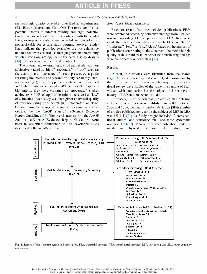
ARTICLE IN PRESS
4 M.J. Highsmith et al. / The Spine Journal 00 (2018) 1−12
methodologic quality of studies classified as experimental
(E1−E5) or observational (O1−O6). The form identifies 18
potential threats to internal validity and eight potential
threats to external validity. In accordance with the guide-
lines, examples of criteria are provided and described as
not applicable for certain study designs; however, guide-
lines indicate that provided examples are not exhaustive
and that reviewers should use their judgment in determining
which criteria are not applicable for certain study designs
[14]. Threats were evaluated and tabulated.
The internal and external validity of each study was then
subjectively rated as “high,” “moderate,” or “low” based on
the quantity and importance of threats present. As a guide
for rating the internal and external validity separately, stud-
ies achieving ≥ 80% of applicable criteria were classified
as “high.” If studies achieved <80% but >50% of applica-
ble criteria, they were classified as “moderate.” Studies
achieving ≤ 50% of applicable criteria received a “low”
classification. Each study was then given an overall quality
of evidence rating of either “high,” “moderate,” or “low”
by combining the ratings of internal and external validity as
outlined by the AAOP State-of-the-Science Evidence
Report Guidelines [14]. The overall ratings from the AAOP
State-of-the-Science Evidence Report Guidelines were
used in assigning confidence to the developed EESs
described in the Results section.
Fig. 1. Results of the literature search and application. TTA, transtibial ampute
amputation.
Downloaded for Anonymous User (n/a) at Walter Reed National Military MFor personal use only. No other uses without permission.
Empirical evidence statements
Based on results from the included publications, EESs
were developed describing collective findings from included
research regarding LBP in persons with LEA. Reviewers
rated the level of confidence of each EES as “high,”
“moderate,” “low,” or “insufficient,” based on the number of
publications contributing to the statement; the methodologic
quality of those studies and whether the contributing findings
were confirmatory or conflicting [14].
Results
In total, 302 articles were identified from the search
(Fig. 1). Ten articles required eligibility determination by
the third rater. In most cases, articles requiring the addi-
tional review were studies of the spine in a sample of indi-
viduals with amputation but the subjects did not have a
history of LBP and thus were excluded.
Ultimately, 17 of the original 302 articles met inclusion
criteria. Four articles were published in 2009. Between
2006 and 2016, the mean (standard deviation [SD]) number
of articles published per year on the subject of LBP in LEA
was 1.5 (1.1) (Fig. 2). Study designs included 13 cross-sec-
tional studies, one controlled trial, and three systematic
reviews (Table 2). Manuscripts were published predomi-
nantly in physical medicine, rehabilitative, and
e; TFA, transfemoral amputee; LBP, low back pain; LEA, lower extremity
edical Center from ClinicalKey.com by Elsevier on October 29, 2018.Copyright ©2018. Elsevier Inc. All rights reserved.
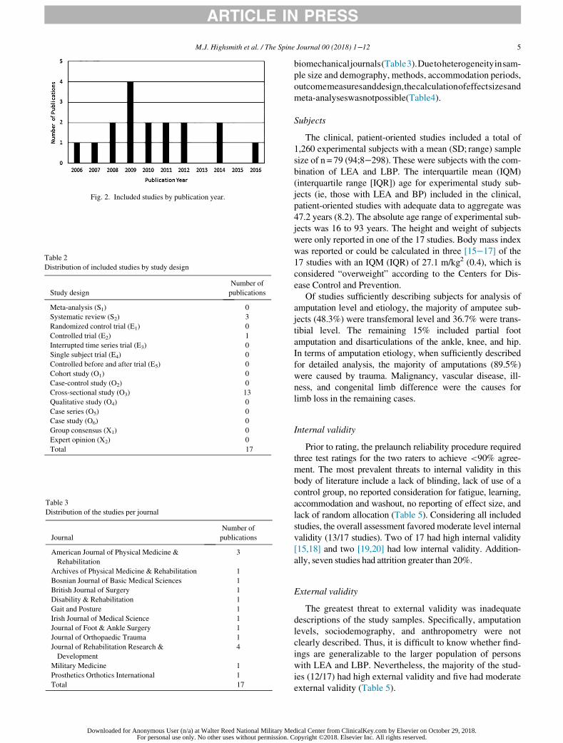
Fig. 2. Included studies by publication year.
Table 2
Distribution of included studies by study design
Study design
Number of
publications
Meta-analysis (S1) 0
Systematic review (S2) 3
Randomized control trial (E1) 0
Controlled trial (E2) 1
Interrupted time series trial (E3) 0
Single subject trial (E4) 0
Controlled before and after trial (E5) 0
Cohort study (O1) 0
Case-control study (O2) 0
Cross-sectional study (O3) 13
Qualitative study (O4) 0
Case series (O5) 0
Case study (O6) 0
Group consensus (X1) 0
Expert opinion (X2) 0
Total 17
able 3
istribution of the studies per journal
Journal
Number of
publications
American Journal of Physical Medicine &
Rehabilitation
3
Archives of Physical Medicine & Rehabilitation 1
Bosnian Journal of Basic Medical Sciences 1
British Journal of Surgery 1
Disability & Rehabilitation 1
Gait and Posture 1
Irish Journal of Medical Science 1
Journal of Foot & Ankle Surgery 1
Journal of Orthopaedic Trauma 1
Journal of Rehabilitation Research &
Development
4
Military Medicine 1
Prosthetics Orthotics International 1
Total 17
ARTICLE IN PRESS
M.J. Highsmith et al. / The Spine Journal 00 (2018) 1−12 5
T
D
Downloaded for Anonymous User (n/a) at Walter Reed National Military MeFor personal use only. No other uses without permission. C
biomechanicaljournals(Table3).Duetoheterogeneityinsam-
ple size and demography, methods, accommodation periods,
outcomemeasuresanddesign,thecalculationofeffectsizesand
meta-analyseswasnotpossible(Table4).
Subjects
The clinical, patient-oriented studies included a total of
1,260 experimental subjects with a mean (SD; range) sample
size of n = 79 (94;8−298). These were subjects with the com-
bination of LEA and LBP. The interquartile mean (IQM)
(interquartile range [IQR]) age for experimental study sub-
jects (ie, those with LEA and BP) included in the clinical,
patient-oriented studies with adequate data to aggregate was
47.2 years (8.2). The absolute age range of experimental sub-
jects was 16 to 93 years. The height and weight of subjects
were only reported in one of the 17 studies. Body mass index
was reported or could be calculated in three [15−17] of the17 studies with an IQM (IQR) of 27.1 m/kg2 (0.4), which is
considered “overweight” according to the Centers for Dis-
ease Control and Prevention.
Of studies sufficiently describing subjects for analysis of
amputation level and etiology, the majority of amputee sub-
jects (48.3%) were transfemoral level and 36.7% were trans-
tibial level. The remaining 15% included partial foot
amputation and disarticulations of the ankle, knee, and hip.
In terms of amputation etiology, when sufficiently described
for detailed analysis, the majority of amputations (89.5%)
were caused by trauma. Malignancy, vascular disease, ill-
ness, and congenital limb difference were the causes for
limb loss in the remaining cases.
Internal validity
Prior to rating, the prelaunch reliability procedure required
three test ratings for the two raters to achieve <90% agree-
ment. The most prevalent threats to internal validity in this
body of literature include a lack of blinding, lack of use of a
control group, no reported consideration for fatigue, learning,
accommodation and washout, no reporting of effect size, and
lack of random allocation (Table 5). Considering all included
studies, the overall assessment favored moderate level internal
validity (13/17 studies). Two of 17 had high internal validity
[15,18] and two [19,20] had low internal validity. Addition-
ally, seven studies had attrition greater than 20%.
External validity
The greatest threat to external validity was inadequate
descriptions of the study samples. Specifically, amputation
levels, sociodemography, and anthropometry were not
clearly described. Thus, it is difficult to know whether find-
ings are generalizable to the larger population of persons
with LEA and LBP. Nevertheless, the majority of the stud-
ies (12/17) had high external validity and five had moderate
external validity (Table 5).
dical Center from ClinicalKey.com by Elsevier on October 29, 2018.opyright ©2018. Elsevier Inc. All rights reserved.
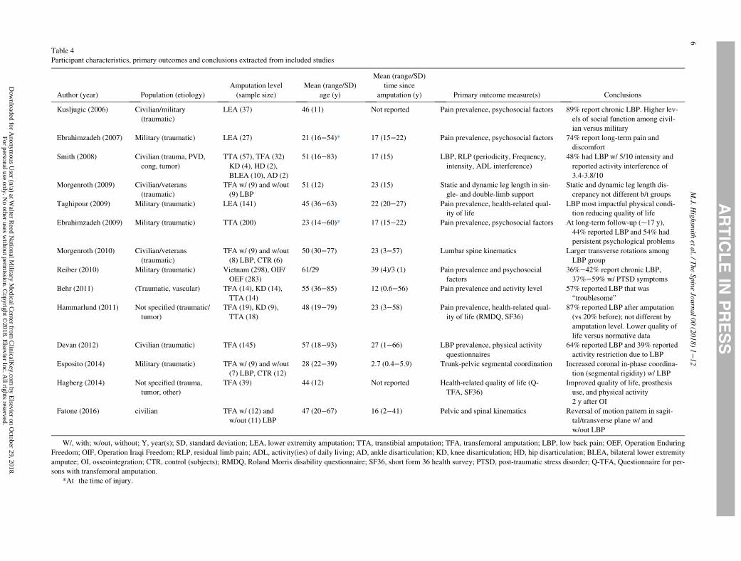
Table 4
Participant characteristics, primary outcomes and conclusions extracted from included studies
Author (year) Population (etiology)
Amputation level
(sample size)
Mean (range/SD)
age (y)
Mean (range/SD)
time since
amputation (y) Primary outcome measure(s) Conclusions
Kusljugic (2006) Civilian/military
(traumatic)
LEA (37) 46 (11) Not reported Pain prevalence, psychosocial factors 89% report chronic LBP. Higher lev-
els of social function among civil-
ian versus military
Ebrahimzadeh (2007) Military (traumatic) LEA (27) 21 (16−54)* 17 (15−22) Pain prevalence, psychosocial factors 74% report long-term pain and
discomfort
Smith (2008) Civilian (trauma, PVD,
cong, tumor)
TTA (57), TFA (32)
KD (4), HD (2),
BLEA (10), AD (2)
51 (16−83) 17 (15) LBP, RLP (periodicity, Frequency,
intensity, ADL interference)
48% had LBP w/ 5/10 intensity and
reported activity interference of
3.4-3.8/10
Morgenroth (2009) Civilian/veterans
(traumatic)
TFA w/ (9) and w/out
(9) LBP
51 (12) 23 (15) Static and dynamic leg length in sin-
gle- and double-limb support
Static and dynamic leg length dis-
crepancy not different b/t groups
Taghipour (2009) Military (traumatic) LEA (141) 45 (36−63) 22 (20−27) Pain prevalence, health-related qual-
ity of life
LBP most impactful physical condi-
tion reducing quality of life
Ebrahimzadeh (2009) Military (traumatic) TTA (200) 23 (14−60)* 17 (15−22) Pain prevalence, psychosocial factors At long-term follow-up (»17 y),
44% reported LBP and 54% had
persistent psychological problems
Morgenroth (2010) Civilian/veterans
(traumatic)
TFA w/ (9) and w/out
(8) LBP, CTR (6)
50 (30−77) 23 (3−57) Lumbar spine kinematics Larger transverse rotations among
LBP group
Reiber (2010) Military (traumatic) Vietnam (298), OIF/
OEF (283)
61/29 39 (4)/3 (1) Pain prevalence and psychosocial
factors
36%−42% report chronic LBP,
37%−59% w/ PTSD symptoms
Behr (2011) (Traumatic, vascular) TFA (14), KD (14),
TTA (14)
55 (36−85) 12 (0.6−56) Pain prevalence and activity level 57% reported LBP that was
“troublesome”
Hammarlund (2011) Not specified (traumatic/
tumor)
TFA (19), KD (9),
TTA (18)
48 (19−79) 23 (3−58) Pain prevalence, health-related qual-
ity of life (RMDQ, SF36)
87% reported LBP after amputation
(vs 20% before); not different by
amputation level. Lower quality of
life versus normative data
Devan (2012) Civilian (traumatic) TFA (145) 57 (18−93) 27 (1−66) LBP prevalence, physical activity
questionnaires
64% reported LBP and 39% reported
activity restriction due to LBP
Esposito (2014) Military (traumatic) TFA w/ (9) and w/out
(7) LBP, CTR (12)
28 (22−39) 2.7 (0.4−5.9) Trunk-pelvic segmental coordination Increased coronal in-phase coordina-
tion (segmental rigidity) w/ LBP
Hagberg (2014) Not specified (trauma,
tumor, other)
TFA (39) 44 (12) Not reported Health-related quality of life (Q-
TFA, SF36)
Improved quality of life, prosthesis
use, and physical activity
2 y after OI
Fatone (2016) civilian TFA w/ (12) and
w/out (11) LBP
47 (20−67) 16 (2−41) Pelvic and spinal kinematics Reversal of motion pattern in sagit-
tal/transverse plane w/ and
w/out LBP
W/, with; w/out, without; Y, year(s); SD, standard deviation; LEA, lower extremity amputation; TTA, transtibial amputation; TFA, transfemoral amputation; LBP, low back pain; OEF, Operation Enduring
Freedom; OIF, Operation Iraqi Freedom; RLP, residual limb pain; ADL, activity(ies) of daily living; AD, ankle disarticulation; KD, knee disarticulation; HD, hip disarticulation; BLEA, bilateral lower extremity
amputee; OI, osseointegration; CTR, control (subjects); RMDQ, Roland Morris disability questionnaire; SF36, short form 36 health survey; PTSD, post-traumatic stress disorder; Q-TFA, Questionnaire for per-
sons with transfemoral amputation.
*At the time of injury.
ARTICLE
INPRESS
6M.J.
Highsm
ithetal./TheSpineJournal00(2018)1−
12
Dow
nloaded for Anonym
ous User (n/a) at W
alter Reed N
ational Military M
edical Center from
ClinicalK
ey.com by E
lsevier on October 29, 2018.
For personal use only. No other uses w
ithout permission. C
opyright ©2018. E
lsevier Inc. All rights reserved.
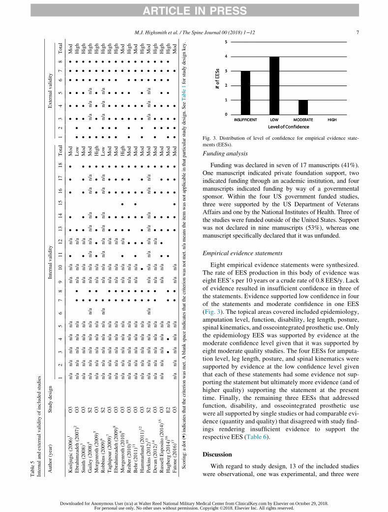
Table5
Internalandexternalvalidityofincluded
studies
Author(year)
Studydesign
Internalvalidity
Externalvalidity
12
34
56
78
910
11
12
13
14
15
16
17
18
Total
12
34
56
78
Total
Kusljugic(2006)1
O3
n/a
n/a
n/a
n/a
n/a
n/a
n/a
�n/a
��
��
�Mod
��
��
��
Mod
Ebrahim
zadeh
(2007)2
O3
n/a
n/a
n/a
n/a
n/a
��
n/a
n/a
n/a
�Low
��
��
��
��
High
Smith(2008)3
O3
n/a
n/a
n/a
n/a
n/a
�n/a
n/a
�n/a
��
��
�Mod
��
��
��
�High
Gailey(2008)4
S2
n/a
n/a
n/a
n/a
n/a
n/a
��
n/a
n/a
n/a
n/a
n/a
n/a
n/a
n/a
�Mod
��
n/a
n/a
n/a
��
�High
Morgenroth
(2009)5
O3
n/a
n/a
n/a
n/a
n/a
��
n/a
n/a
��
��
��
�High
��
��
��
�High
Robbins(2009)6
S2
n/a
n/a
n/a
n/a
n/a
n/a
�n/a
n/a
n/a
n/a
n/a
n/a
n/a
n/a
�Low
��
n/a
n/a
n/a
��
�High
Taghipour(2009)7
O3
n/a
n/a
n/a
n/a
n/a
��
n/a
n/a
n/a
��
��
�Mod
��
��
��
��
High
Ebrahim
zadeh
(2009)8
O3
n/a
n/a
n/a
n/a
n/a
��
n/a
n/a
n/a
��
��
�Mod
��
��
��
��
High
Morgenroth
(2010)9
O3
n/a
n/a
n/a
n/a
n/a
��
n/a
n/a
�n/a
��
��
��
High
��
��
�Mod
Reiber
(2010)10
O3
n/a
n/a
n/a
n/a
n/a
��
n/a
n/a
��
��
��
Mod
��
��
��
��
High
Behr(2011)11
O3
n/a
n/a
n/a
n/a
n/a
��
n/a
n/a
��
��
��
Mod
��
��
�Mod
Ham
marlund(2011)12
O3
n/a
n/a
n/a
n/a
n/a
��
��
��
��
�Mod
��
��
��
��
High
Perkins(2012)13
S2
n/a
n/a
n/a
n/a
n/a
n/a
��
n/a
n/a
n/a
n/a
n/a
n/a
n/a
n/a
�Mod
n/a
n/a
n/a
��
�Mod
Devan
(2012)14
O3
n/a
n/a
n/a
n/a
n/a
��
n/a
n/a
n/a
��
��
Mod
��
��
��
�High
RussellEsposito
(2014)15
O3
n/a
n/a
n/a
n/a
n/a
n/a
n/a
��
��
��
�Mod
��
��
��
�High
Hagberg(2014)16
E2
��
n/a
��
��
��
��
Mod
��
��
��
��
High
Fatone(2016)17
O3
n/a
n/a
n/a
n/a
n/a
��
n/a
n/a
��
��
��
Mod
��
��
�Mod
Scoring:adot(�)indicates
thatthecriterionwas
met.Ablankspaceindicates
thatthecriterionwas
notmet.n/ameanstheitem
was
notapplicableinthatparticularstudydesign.See
Table1forstudydesignkey.
Fig. 3. Distribution of level of confidence for empirical evidence state-
ments (EESs).
ARTICLE IN PRESS
M.J. Highsmith et al. / The Spine Journal 00 (2018) 1−12 7
Downloaded for Anonymous User (n/a) at Walter Reed National Military MeFor personal use only. No other uses without permission. C
Funding analysis
Funding was declared in seven of 17 manuscripts (41%).
One manuscript indicated private foundation support, two
indicated funding through an academic institution, and four
manuscripts indicated funding by way of a governmental
sponsor. Within the four US government funded studies,
three were supported by the US Department of Veterans
Affairs and one by the National Institutes of Health. Three of
the studies were funded outside of the United States. Support
was not declared in nine manuscripts (53%), whereas one
manuscript specifically declared that it was unfunded.
Empirical evidence statements
Eight empirical evidence statements were synthesized.
The rate of EES production in this body of evidence was
eight EES’s per 10 years or a crude rate of 0.8 EES/y. Lack
of evidence resulted in insufficient confidence in three of
the statements. Evidence supported low confidence in four
of the statements and moderate confidence in one EES
(Fig. 3). The topical areas covered included epidemiology,
amputation level, function, disability, leg length, posture,
spinal kinematics, and osseointegrated prosthetic use. Only
the epidemiology EES was supported by evidence at the
moderate confidence level given that it was supported by
eight moderate quality studies. The four EESs for amputa-
tion level, leg length, posture, and spinal kinematics were
supported by evidence at the low confidence level given
that each of these statements had some evidence not sup-
porting the statement but ultimately more evidence (and of
higher quality) supporting the statement at the present
time. Finally, the remaining three EESs that addressed
function, disability, and osseointegrated prosthetic use
were all supported by single studies or had comparable evi-
dence (quantity and quality) that disagreed with study find-
ings rendering insufficient evidence to support the
respective EES (Table 6).
Discussion
With regard to study design, 13 of the included studies
were observational, one was experimental, and three were
dical Center from ClinicalKey.com by Elsevier on October 29, 2018.opyright ©2018. Elsevier Inc. All rights reserved.
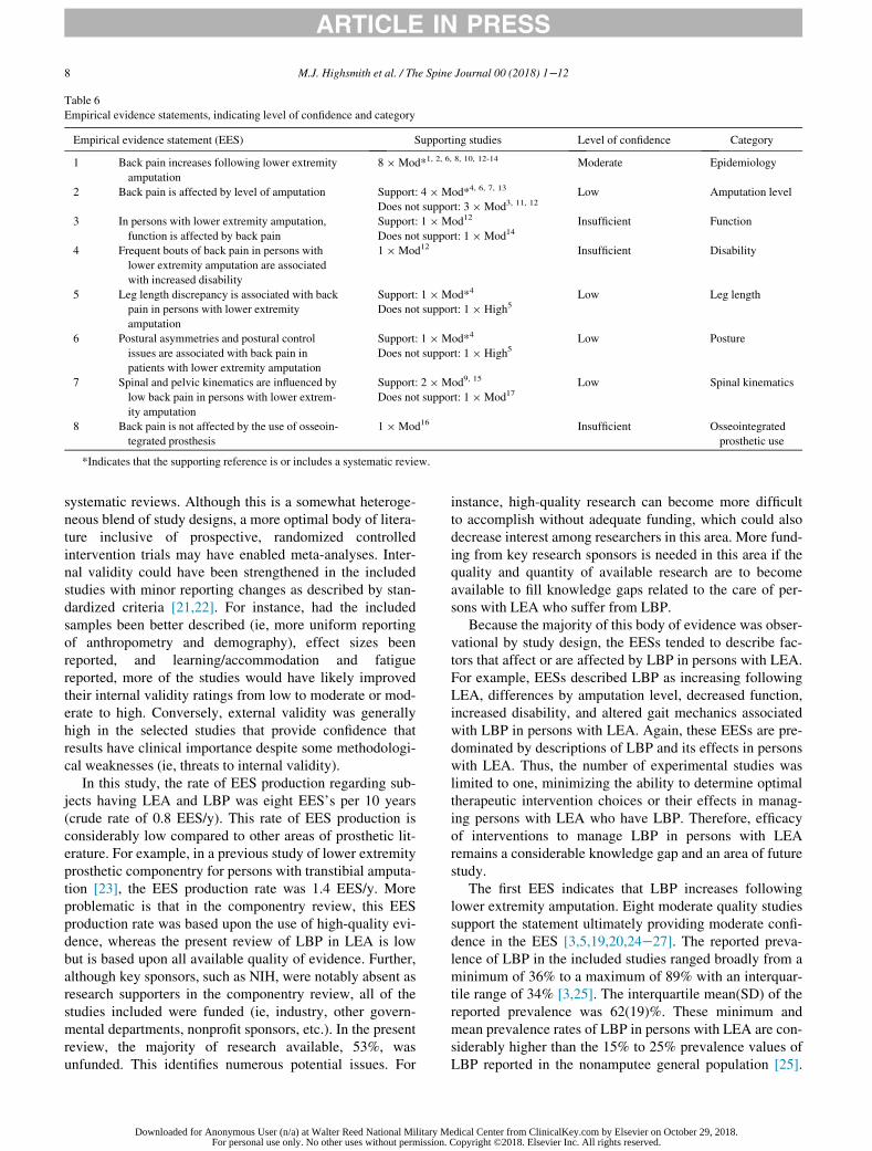
Table 6
Empirical evidence statements, indicating level of confidence and category
Empirical evidence statement (EES) Supporting studies Level of confidence Category
1 Back pain increases following lower extremity
amputation
8 £Mod*1, 2, 6, 8, 10, 12-14 Moderate Epidemiology
2 Back pain is affected by level of amputation Support: 4 £Mod*4, 6, 7, 13 Low Amputation level
Does not support: 3£Mod3, 11, 12
3 In persons with lower extremity amputation,
function is affected by back pain
Support: 1 £Mod12 Insufficient Function
Does not support: 1£Mod14
4 Frequent bouts of back pain in persons with
lower extremity amputation are associated
with increased disability
1 £Mod12 Insufficient Disability
5 Leg length discrepancy is associated with back
pain in persons with lower extremity
amputation
Support: 1 £Mod*4 Low Leg length
Does not support: 1£ High5
6 Postural asymmetries and postural control
issues are associated with back pain in
patients with lower extremity amputation
Support: 1 £Mod*4 Low Posture
Does not support: 1£ High5
7 Spinal and pelvic kinematics are influenced by
low back pain in persons with lower extrem-
ity amputation
Support: 2 £Mod9, 15 Low Spinal kinematics
Does not support: 1£Mod17
8 Back pain is not affected by the use of osseoin-
tegrated prosthesis
1 £Mod16 Insufficient Osseointegrated
prosthetic use
*Indicates that the supporting reference is or includes a systematic review.
ARTICLE IN PRESS
8 M.J. Highsmith et al. / The Spine Journal 00 (2018) 1−12
systematic reviews. Although this is a somewhat heteroge-
neous blend of study designs, a more optimal body of litera-
ture inclusive of prospective, randomized controlled
intervention trials may have enabled meta-analyses. Inter-
nal validity could have been strengthened in the included
studies with minor reporting changes as described by stan-
dardized criteria [21,22]. For instance, had the included
samples been better described (ie, more uniform reporting
of anthropometry and demography), effect sizes been
reported, and learning/accommodation and fatigue
reported, more of the studies would have likely improved
their internal validity ratings from low to moderate or mod-
erate to high. Conversely, external validity was generally
high in the selected studies that provide confidence that
results have clinical importance despite some methodologi-
cal weaknesses (ie, threats to internal validity).
In this study, the rate of EES production regarding sub-
jects having LEA and LBP was eight EES’s per 10 years
(crude rate of 0.8 EES/y). This rate of EES production is
considerably low compared to other areas of prosthetic lit-
erature. For example, in a previous study of lower extremity
prosthetic componentry for persons with transtibial amputa-
tion [23], the EES production rate was 1.4 EES/y. More
problematic is that in the componentry review, this EES
production rate was based upon the use of high-quality evi-
dence, whereas the present review of LBP in LEA is low
but is based upon all available quality of evidence. Further,
although key sponsors, such as NIH, were notably absent as
research supporters in the componentry review, all of the
studies included were funded (ie, industry, other govern-
mental departments, nonprofit sponsors, etc.). In the present
review, the majority of research available, 53%, was
unfunded. This identifies numerous potential issues. For
Downloaded for Anonymous User (n/a) at Walter Reed National Military MFor personal use only. No other uses without permission.
instance, high-quality research can become more difficult
to accomplish without adequate funding, which could also
decrease interest among researchers in this area. More fund-
ing from key research sponsors is needed in this area if the
quality and quantity of available research are to become
available to fill knowledge gaps related to the care of per-
sons with LEA who suffer from LBP.
Because the majority of this body of evidence was obser-
vational by study design, the EESs tended to describe fac-
tors that affect or are affected by LBP in persons with LEA.
For example, EESs described LBP as increasing following
LEA, differences by amputation level, decreased function,
increased disability, and altered gait mechanics associated
with LBP in persons with LEA. Again, these EESs are pre-
dominated by descriptions of LBP and its effects in persons
with LEA. Thus, the number of experimental studies was
limited to one, minimizing the ability to determine optimal
therapeutic intervention choices or their effects in manag-
ing persons with LEA who have LBP. Therefore, efficacy
of interventions to manage LBP in persons with LEA
remains a considerable knowledge gap and an area of future
study.
The first EES indicates that LBP increases following
lower extremity amputation. Eight moderate quality studies
support the statement ultimately providing moderate confi-
dence in the EES [3,5,19,20,24−27]. The reported preva-
lence of LBP in the included studies ranged broadly from a
minimum of 36% to a maximum of 89% with an interquar-
tile range of 34% [3,25]. The interquartile mean(SD) of the
reported prevalence was 62(19)%. These minimum and
mean prevalence rates of LBP in persons with LEA are con-
siderably higher than the 15% to 25% prevalence values of
LBP reported in the nonamputee general population [25].
edical Center from ClinicalKey.com by Elsevier on October 29, 2018.Copyright ©2018. Elsevier Inc. All rights reserved.
ARTICLE IN PRESS
M.J. Highsmith et al. / The Spine Journal 00 (2018) 1−12 9
One study reported significantly increased LBP after ampu-
tation as opposed to before amputation [5]. Furthermore,
the characteristics and consequences of this pain in persons
with LEA have been described as progressive, disabling,
and contributing to limitations in occupation, recreation,
and socialization [19,20]. With regard to function, LBP in
LEAs has been associated with problems sitting, sleeping,
and traveling [3]. Finally, LBP in this population has been
associated with decreased health-related quality of life [5].
The second EES states that back pain is affected by level
of amputation. Four moderate quality studies support the
statement whereas three moderate quality studies do not
support it. Ultimately, this yields a low level of confidence
in the statement. Importantly, two of the studies supporting
the statement were systematic literature reviews [20,28].
Both concluded that persons with transfemoral level ampu-
tation reported LBP with a higher prevalence than their
transtibial counterparts, which was consistent with two
additional clinical studies [27,29]. Perkins et al. suggest
that the increased susceptibility to LBP at the higher ampu-
tation level may in part be the result of myofascial changes
following transfemoral amputation along with gait pattern
alterations [27]. Confirming these proposed causes for LBP
following LEA through further research could lead to
improvements in prevention and management.
Adverse effects of function related to LBP are the sub-
ject of the third EES. Hammarlund et al. used the Roland
Morris Disability Questionnaire (RMDQ), a valid and reli-
able measure of functional capacity relative to perceived
back pain in a sample of 46 nondysvascular lower extremity
amputees [5]. They concluded that nearly all participants
with LBP daily or several times per week reported severe
or moderate disability on the RMDQ. Devan et al. assessed
the relationship of back pain on function in terms of physi-
cal activity [26]. Overall, they concluded that there was no
relationship between physical activity of LEAs with or
without LBP and that there was an equal distribution of per-
sons with LBP in low, medium, and high physical activity
groups. They did however find that those reporting activity
limitations due to LBP had lower physical activity scores
than those with LBP who did not have physical activity lim-
itations. It is important to note that this difference in func-
tion related to LBP is potentially confounded by the use of
two different outcome approaches and further by the fact
that Devan et al. studied those with traumatic transfemoral
amputation, whereas the Hammarlund et al. sample was
more heterogeneous by amputation level [5,26]. Nonethe-
less, further evidence is needed to understand which ele-
ments of function may potentially be impaired by LBP in
persons with LEA.
Increasing frequency of LBP episodes and associated
disability is the subject of EES four. A single, moderate-
quality study supports EES four with a significant associa-
tion (p = .003) between LEAs who reported LBP daily or
several times per week and those reporting moderate or
severe disability [5]. Devan et al. studied the relationship
Downloaded for Anonymous User (n/a) at Walter Reed National Military MeFor personal use only. No other uses without permission. C
between LBP and physical activity [26]. Their findings cre-
ate further ambiguity in understanding disability as it
relates to LBP in LEAs. That is, they found no association
between physical activity in LEAs with or without LBP.
One additional systematic review concluded that the major-
ity of LEAs with LBP report minimal to no impact on
social, recreational or work activities [20]. Conversely,
approximately 25% described their LBP as severely inter-
fering with these activities. These are important findings
but do not directly relate to the issue of bout frequency of
LBP. Ultimately, although the association identified by
Hammarlund et al. was significant, the fact that only a sin-
gle study supports the conclusion is presently insufficient to
confidently support the statement at this time [5].
In EES five, association is made between leg length dis-
crepancy and the presence of LBP in persons with LEA.
One clinical study used the RMDQ to identify LEAs with
LBP and those without [15]. Motion analysis was then used
to determine leg length differences during static standing,
dynamically during single and double limb support in gait
and with either the prosthetic or sound foot leading. This
single, high-quality clinical study did not find a relationship
between leg length discrepancy and LBP. Conversely, a
systematic literature review [28] indicates that leg length
discrepancy among lower extremity prosthetic users is
among many contributors to LBP. Further, the review states
that those using prostheses that are of the same length as the
sound limb have significantly fewer pain symptoms com-
pared to those with length asymmetries between the intact
and prosthetic limbs. Postural asymmetries reportedly result
from these disparities. For instance, leg length differences
of 12.5 mm have been associated with as much as 4˚ of lat-
eral sacral tilt. It has been further reported that only 15% of
LEAs use prostheses of equal length to the sound limb,
whereas 34% of prosthesis users have prosthetic leg length
differences greater than 20 mm, and that in 79% of cases,
the prosthesis is the shorter limb. Given this disagreement
between a single clinical study [15] and a systematic litera-
ture review [28], there is low confidence in the evidence
supporting EES five. This statement indicates an associa-
tion between leg length discrepancy and back pain in per-
sons with lower extremity amputation. One additional
clarifying point is that Morgenroth et al. studied LEAs with
chronic LBP as opposed to acute onset cases. Thus, it is not
currently possible to determine causation of LBP as a result
of leg length discrepancy using these findings. Rather, their
study is more useful in assisting to determine whether leg
length discrepancy has a role in altering symptoms in
chronic LBP cases among those with LEA [15].
Empirical evidence statement six is somewhat related to
EES five. Though EES five directly addresses leg length
discrepancy, EES six indicates that postural asymmetries
and postural control issues are associated with LBP in
patients with LEA. Gailey et al. report that persons with
LEA tend to stand with increased sway and with increased
weight bearing on the sound limb and that this may be
dical Center from ClinicalKey.com by Elsevier on October 29, 2018.opyright ©2018. Elsevier Inc. All rights reserved.
ARTICLE IN PRESS
10 M.J. Highsmith et al. / The Spine Journal 00 (2018) 1−12
related to the lack of proprioception from the prosthesis
[28]. Postural abnormalities observed in those with LEA
are numerous including coronal and sagittal compensatory
pelvic tilt, increased lumbar lordosis, involved-side hip
flexion contracture, lateral trunk asymmetry and more [28].
Morgenroth et al. state that LBP is a common secondary
disabling condition affecting TFA and that it is common
clinical practice to assess for and correct postural asymme-
try in the form of leg length discrepancy [15]. In their sam-
ple of subjects with longstanding transfemoral amputation
and moderate, persistent LBP, leg length, discrepancies
were not different relative to a similar population without
LBP. Morgenroth et al. concluded that in longstanding
transfemoral amputees with chronic symptoms, their LBP
was unlikely to be related to their postural asymmetry [15].
Confidence in EES six is low given support from a system-
atic review but a lack of support from one clinical study,
both of moderate quality.
Relative to EES seven, there is limited (n = 3 studies) evi-
dence reporting alterations in trunk, spinal, and pelvic
motions among persons with LEA with LBP (EES seven).
Although LBP is multifactorial, repeated exposures to
altered trunk-pelvic motions is a purported risk factor for the
onset or recurrence of LBP secondary to LEA [7,9,30−33].The presence of LBP among persons with (transfemoral)
LEA is associated with larger axial rotations of the lumbar
spine [18], more rigid (in-phase) trunk-pelvic coordination
strategies [16], and an apparent (albeit underpowered) trend
toward a reversal in patterns of trunk-pelvic motion in the
sagittal and transverse planes [17]. Such findings begin sug-
gesting linkage of specific trunk/spinal and pelvic kinematic
patterns with LBP secondary to LEA. However, the presence
and magnitude of LBP has been inconsistently characterized
using a variety of approaches, including binary yes/no, visual
analog scale (0−10), question(s) within the Prosthesis Evalu-ation Questionnaire, and the Grade Questionnaire. Thus,
there is a clear need for more consistent and comprehensive
quantification of LBP in future work. For example, using the
NIH task force for chronic LBP questionnaire that aims to
classify LBP by its impact (ie, intensity, interference, and
physical function), or using a minimal dataset to describe
participants and reporting responder analyses in addition to
mean outcomes could be useful [34]. Moreover, considerable
prior work among non-amputation individuals have identi-
fied substantial influences of LBP on trunk and pelvic
motions[35−37], begging the question of the relative contri-
butions of LBP and LEA on the observed movement pat-
terns. To that end, additional biomechanical metrics are
needed to understand the underlying factors driving the
movement patterns.
A single moderate quality study of 39 subjects sup-
ports the final EES [38]. This statement indicates that
back pain is not affected by use of an osseointegrated
prosthesis. Interfacing the prosthesis with a socket has
been associated with adverse effects to skin, comfort,
and function [23]. For example, skin erosions of varying
Downloaded for Anonymous User (n/a) at Walter Reed National Military MFor personal use only. No other uses without permission.
degrees, pain, and unreliable suspension can all poten-
tially emerge related to socket use [39]. Anchoring the
prosthesis to the residual limb via osseointegration pur-
portedly mitigates some of the aforementioned compli-
cations. Another issue potentially associated with socket
use is that gait pattern alterations due to pain or instabil-
ity could also lead to LBP. Hagberg et al. surveyed LBP
in a single item from the Questionnaire for Persons with
Transfemoral Amputation [38] finding that at 2 years
following osseointegration, approximately 40% of sub-
jects reported reduced LBP, nearly 40% were
unchanged, and nearly 20% reported an increase in their
LBP symptoms. Compared with baseline, these differen-
ces were not statistically significant. Of note, the authors
indicated the small sample size and reliance solely upon
subjective outcomes limited the strength of evidence.
Findings were also confounded by the fact that pros-
thetic components were changed throughout the 2-year
follow-up period. More research is needed to identify
and characterize the relationships between osseointe-
grated prosthetic use and LBP.
Clinical practice guidelines (CPGs) for primary care
management of LBP in the general population usually rec-
ommend focused history and examination, limited use of
diagnostic imaging, self-care, brief education, nonsteroidal
anti-inflammatory drugs, manual therapy, and exercise
[40]. The US Department of Defense (DoD) and the US
Department of Veterans Affairs (VA) similarly have CPGs
for persons with LEA and for those with LBP [41,42].
None of the articles uncovered in this systematic review
assessed the appropriateness of these recommendations for
patients with LEA suffering from LBP. Whether the LBP
CPGs can be applied to LEAs or if modifications in the
treatment approach are needed is unknown. Further, the
recommendations in the VA/DoD include measuring the
intensity of LBP but also to initiate a strengthening program
for the upper and lower extremities as well as the core to
prevent the development of LBP [42]. These recommenda-
tions in the second version of the VA/DoD CPG were for-
warded from the original CPG and were largely based in
expert opinion. These recommendations remain untested in
this population. Future research is needed to clarify clinical
decision-making processes for management of LBP in
lower extremity amputees.
Limitations
This body of literature only included a single experimen-
tal study [38] and a single study with high internal and
external validity [15]. The majority of the included studies
were observational, of moderate overall quality, and
unfunded. Viewed in aggregate, the subjects studied were
somewhat heterogeneous with regard to age, LEA etiology,
time since LEA, and included both military and civilian
sectors; the methodological quality could be improved with
standardized reporting in most cases [21,22]. An example
edical Center from ClinicalKey.com by Elsevier on October 29, 2018.Copyright ©2018. Elsevier Inc. All rights reserved.
ARTICLE IN PRESS
M.J. Highsmith et al. / The Spine Journal 00 (2018) 1−12 11
may include more thorough sample descriptions. Addition-
ally, incorporating blinding (ie, raters, statisticians) would
also improve internal validity. Further, important factors
are missed due to reporting omissions in many cases
such as gender, race or ethnic considerations. Finally,
this review uncovered three potential etiologies of LBP in
LEA, namely, leg length discrepancy (ESS five), postural
asymmetries and control issues (ESS six), and altered spinal
kinematics. However, a causal relationship between these
potential etiologies and LBP in LEA has not been estab-
lished and requires further research.
Conclusions
Because the majority of this body of evidence was obser-
vational instead of experimental, the EESs produced tended
to describe factors affecting or that are affected by LBP in
persons with LEA. More specifically, the EESs supported
observationally have concluded that back pain in LEAs has
relationships with the following phenomena: increased expe-
riences, level of LEA, leg length differences, postural issues
as well as spinal and pelvic kinematics. With only a single
experimental study, the ability to determine optimal therapeu-
tic intervention choices or their effects in managing LEAs
who have LBP is greatly limited. Therefore, efficacy of inter-
ventions to manage LBP in persons with LEA remains a con-
siderable knowledge gap and an area of future study.
Acknowledgments
Contents of the manuscript represent the opinions of the
authors and not necessarily those of the Department of
Defense, Department of Veterans Affairs or the Department
of the Army. This project was funded, in part, through an
unrestricted gift by the Lincoln College Education and
Research Fund to the University of South Florida Founda-
tion. The funding agencies provided financial support only
and had no other role in the systematic review.
References
[1] Flavell CA, Gordon S, Marshman L. Classification characteristics of a
chronic low back pain population using a combined McKenzie and
patho-anatomical assessment. Man Ther 2016;26:201–7.
[2] Farrokhi S, Mazzone B, Schneider MJ, Gombatto S, Mayer J, High-
smith MJ, Hendershot BD. Biopsychosocial risk factors associated
with chronic low back pain after lower limb amputation. Med
Hypotheses 2017;108:1–9.
[3] Kusljugic A, Kapidzic-Durakovic S, Kudumovic Z, Cickusic A.
Chronic low back pain in individuals with lower-limb amputation.
Bosn J Basic Med Sci 2006;6:67–70.
[4] Smith DG, Ehde DM, Legro MW, Reiber GE, del Aguila M, Boone
DA. Phantom limb, residual limb, and back pain after lower extremity
amputations. Clin Orthop Relat Res 1999:29–38.
[5] Hammarlund CS, Carlstrom M, Melchior R, Persson BM. Prevalence
of back pain, its effect on functional ability and health-related quality
of life in lower limb amputees secondary to trauma or tumour: a com-
parison across three levels of amputation. Prosthet Orthot Int
2011;35:97–105.
Downloaded for Anonymous User (n/a) at Walter Reed National Military MeFor personal use only. No other uses without permission. C
[6] Ehde DM, Smith DG, Czerniecki JM, Campbell KM, Malchow DM,
Robinson LR. Back pain as a secondary disability in persons with
lower limb amputations. Arch Phys Med Rehabil 2001;82:731–4.
[7] Hendershot BD, Wolf EJ. Three-dimensional joint reaction forces and
moments at the low back during over-ground walking in persons with
unilateral lower-extremity amputation. Clin Biomech (Bristol, Avon)
2014;29:235–42.
[8] Tranberg R, Zugner R, Karrholm J. Improvements in hip- and pelvic
motion for patients with osseointegrated trans-femoral prostheses.
Gait Posture 2011;33:165–8.
[9] Yoder AJ, Petrella AJ, Silverman AK. Trunk-pelvis motion, joint
loads, and muscle forces during walking with a transtibial amputa-
tion. Gait Posture 2015;41:757–62.
[10] Yu CH, Hung YC, Lin YH, Chen GX, Wei SH, Huang CH, et al. A
3D mathematical model to predict spinal joint and hip joint force for
trans-tibial amputees with different SACH foot pylon adjustments.
Gait Posture 2014;40:545–8.
[11] Highsmith MJ, Kahle JT, Bongiorni DR, Sutton BS, Groer S, Kauf-
man KR. Safety, energy efficiency, and cost efficacy of the C-Leg for
transfemoral amputees: a review of the literature. Prosthet Orthot Int
2010;34:362–77.
[12] Kahle JT, Orriola JJ, Johnston W, Highsmith MJ. The effects of
vacuum-assisted suspension on residual limb physiology, wound
healing and function: a systematic review. Technol Innov 2014;15:
333–41.
[13] Cohen J (Ed.). Statistical power analysis for the behavioral sciences.
(2nd ed). Hillsdale, NJ: Erlbaum; 1988.
[14] American Academy of Orthotists & Prosthetists. State of the Science
Evidence Report Guidelines. Washington, DC: American Academy
of Orthotists & Prosthetists; 2008.
[15] Morgenroth DC, Shakir A, Orendurff MS, Czerniecki JM. Low-back
pain in transfemoral amputees: is there a correlation with static or
dynamic leg-length discrepancy? Am J Phys Med Rehabil
2009;88:108–13.
[16] Russell Esposito E, Wilken JM. The relationship between pelvis-
trunk coordination and low back pain in individuals with transfemoral
amputations. Gait Posture 2014;40:640–6.
[17] Fatone S, Stine R, Gottipati P, Dillon M. Pelvic and spinal motion
during walking in persons with transfemoral amputation with and
without low back pain. Am J Phys Med Rehabil 2016;95:
438–47.
[18] Morgenroth DC, Orendurff MS, Shakir A, Segal A, Shofer J, Czer-
niecki JM. The relationship between lumbar spine kinematics during
gait and low-back pain in transfemoral amputees. Am J Phys Med
Rehabil 2010;89:635–43.
[19] Ebrahimzadeh MH, Rajabi MT. Long-term outcomes of patients
undergoing war-related amputations of the foot and ankle. J Foot
Ankle Surg 2007;46:429–33.
[20] Robbins CB, Vreeman DJ, Sothmann MS, Wilson SL, Oldridge NB.
A review of the long-term health outcomes associated with war-
related amputation. Mil Med 2009;174:588–92.
[21] Liberati A, Altman DG, Tetzlaff J, Mulrow C, Gotzsche PC, Ioanni-
dis JP, et al. The PRISMA statement for reporting systematic reviews
and meta-analyses of studies that evaluate healthcare interventions:
explanation and elaboration. BMJ 2009;339:b2700.
[22] Moher D, Liberati A, Tetzlaff J, Altman DG. Preferred reporting
items for systematic reviews and meta-analyses: the PRISMA state-
ment. BMJ 2009;339:b2535.
[23] Highsmith MJ, Kahle JT, Miro RM, Orendurff MS, Lewandowski
AL, Orriola JJ, et al. Prosthetic interventions for people with transti-
bial amputation: Systematic review and meta-analysis of high-quality
prospective literature and systematic reviews. J Rehabil Res Dev
2016;53:157–84.
[24] Ebrahimzadeh MH, Fattahi AS. Long-term clinical outcomes of Ira-
nian veterans with unilateral transfemoral amputation. Disabil Reha-
bil 2009;31:1873–7.
dical Center from ClinicalKey.com by Elsevier on October 29, 2018.opyright ©2018. Elsevier Inc. All rights reserved.
ARTICLE IN PRESS
12 M.J. Highsmith et al. / The Spine Journal 00 (2018) 1−12
[25] Reiber GE, McFarland LV, Hubbard S, Maynard C, Blough DK,
Gambel JM, Smith DG. Servicemembers and veterans with major
traumatic limb loss from Vietnam war and OIF/OEF conflicts: survey
methods, participants, and summary findings. J Rehabil Res Dev
2010;47:275–97.[26] Devan H, Tumilty S, Smith C. Physical activity and lower-back pain
in persons with traumatic transfemoral amputation: a national cross-
sectional survey. J Rehabil Res Dev 2012;49:1457–66.
[27] Perkins ZB, De’Ath HD, Sharp G, Tai NR. Factors affecting out-
come after traumatic limb amputation. Br J Surg 2012;99(Suppl
1):S75–86.
[28] Gailey R, Allen K, Castles J, Kucharik J, Roeder M. Review of
secondary physical conditions associated with lower-limb ampu-
tation and long-term prosthesis use. J Rehabil Res Dev
2008;45:15–29.
[29] Taghipour H, Moharamzad Y, Mafi AR, Amini A, Naghizadeh MM,
Soroush MR. Quality of life among veterans with war-related unilat-
eral lower extremity amputation: a long-term survey in a prosthesis
center in Iran. J Orthop Trauma 2009;23:525–30.
[30] Hendershot BD, Wolf EJ. Persons with unilateral transfemoral ampu-
tation have altered lumbosacral kinetics during sitting and standing
movements. Gait Posture 2015;42:204–9.
[31] Hendershot BD, Wolf EJ. Mediolateral joint powers at the low back
among persons with unilateral transfemoral amputation. Arch Phys
Med Rehabil 2015;96:154–7.
[32] Hendershot BD, Toosizadeh N, Muslim K, Madigan ML, NussbaumMA.
Evidence for an exposure-response relationship between trunk flexion and
impairments in trunk postural control. J Biomech 2013;46:2554–7.
[33] Hendershot BD, Nussbaum MA. Persons with lower-limb amputation
have impaired trunk postural control while maintaining seated bal-
ance. Gait Posture 2013;38:438–42.
Downloaded for Anonymous User (n/a) at Walter Reed National Military MFor personal use only. No other uses without permission.
[34] Deyo RA, Dworkin SF, Amtmann D, Andersson G, Borenstein D,
Carragee E, et al. Report of the NIH Task Force on research standards
for chronic low back pain. Phys Ther 2015;95:e1–18.
[35] van Dieen JH, Selen LP, Cholewicki J. Trunk muscle activation in
low-back pain patients, an analysis of the literature. J Electromyogr
Kinesiol 2003;13:333–51.
[36] Hodges P, van den Hoorn W, Dawson A, Cholewicki J. Changes in
the mechanical properties of the trunk in low back pain may be asso-
ciated with recurrence. J Biomech 2009;42:61–6.
[37] Seay JF, Van Emmerik RE, Hamill J. Low back pain status affects
pelvis-trunk coordination and variability during walking and running.
Clin Biomech (Bristol, Avon) 2011;26:572–8.
[38] Hagberg K, Hansson E, Branemark R. Outcome of percutaneous osseoin-
tegrated prostheses for patients with unilateral transfemoral amputation at
two-year follow-up. Arch Phys Med Rehabil 2014;95:2120–7.
[39] Highsmith MJ, Kahle JT, Klenow TD, Andrews CR, Lewis KL, Bradley
RC, et al. Interventions to manage residual limb ulceration due to pros-
thetic use in individuals with lower extremity amputation: a systematic
review of the literature. Technol Innov 2016;18:115–23.
[40] Chou R, Qaseem A, Snow V, Casey D, Cross Jr. JT, Shekelle P, et al.
Diagnosis and treatment of low back pain: a joint clinical practice
guideline from the American College of Physicians and the American
Pain Society. Ann Intern Med 2007;147:478–91.
[41] U.S. Department of Veterans Affairs. U.S. Department of Defense.
VA/DoD clinical practice guideline for diagnosis and treatment of
low back pain, v2.0. Available at: https://wwwhealthquality.va.gov/
guidelines/Pain/lbp/; 2017. Accessed February 5, 2018;2017 .
[42] U.S. Department of Veterans Affairs. U.S. Department of Defense. VA/
DoD clinical practice guideline for rehabilitation of individuals with
lower limb amputation, v2.0: Available at: https://www.healthquality.va.
gov/guidelines/Rehab/amp/; 2017. Accessed February 5, 2018;2017.
edical Center from ClinicalKey.com by Elsevier on October 29, 2018.Copyright ©2018. Elsevier Inc. All rights reserved.
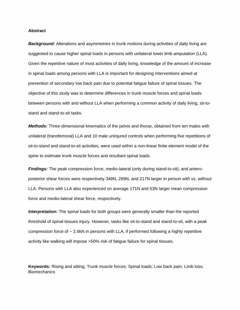
Abstract
Background: Alterations and asymmetries in trunk motions during activities of daily living are
suggested to cause higher spinal loads in persons with unilateral lower limb amputation (LLA).
Given the repetitive nature of most activities of daily living, knowledge of the amount of increase
in spinal loads among persons with LLA is important for designing interventions aimed at
prevention of secondary low back pain due to potential fatigue failure of spinal tissues. The
objective of this study was to determine differences in trunk muscle forces and spinal loads
between persons with and without LLA when performing a common activity of daily living, sit-to-
stand and stand-to-sit tasks.
Methods: Three-dimensional kinematics of the pelvis and thorax, obtained from ten males with
unilateral (transfemoral) LLA and 10 male uninjured controls when performing five repetitions of
sit-to-stand and stand-to-sit activities, were used within a non-linear finite element model of the
spine to estimate trunk muscle forces and resultant spinal loads.
Findings: The peak compression force, medio-lateral (only during stand-to-sit), and antero-
posterior shear forces were respectively 348N, 269N, and 217N larger in person with vs. without
LLA. Persons with LLA also experienced on average 171N and 53N larger mean compression
force and medio-lateral shear force, respectively.
Interpretation: The spinal loads for both groups were generally smaller than the reported
threshold of spinal tissues injury. However, tasks like sit-to-stand and stand-to-sit, with a peak
compression force of ~ 2.6kN in persons with LLA, if performed following a highly repetitive
activity like walking will impose >50% risk of fatigue failure for spinal tissues.
Keywords: Rising and sitting; Trunk muscle forces; Spinal loads; Low back pain; Limb loss; Biomechanics
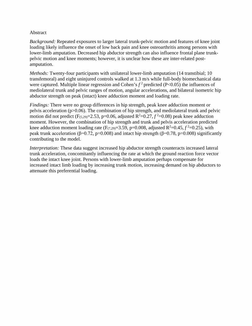
Abstract
Background: Repeated exposures to larger lateral trunk-pelvic motion and features of knee joint loading likely influence the onset of low back pain and knee osteoarthritis among persons with lower-limb amputation. Decreased hip abductor strength can also influence frontal plane trunk-pelvic motion and knee moments; however, it is unclear how these are inter-related post-amputation.
Methods: Twenty-four participants with unilateral lower-limb amputation (14 transtibial; 10 transfemoral) and eight uninjured controls walked at 1.3 m/s while full-body biomechanical data were captured. Multiple linear regression and Cohen’s f 2 predicted (P<0.05) the influences of mediolateral trunk and pelvic ranges of motion, angular accelerations, and bilateral isometric hip abductor strength on peak (intact) knee adduction moment and loading rate.
Findings: There were no group differences in hip strength, peak knee adduction moment or pelvis acceleration (p>0.06). The combination of hip strength, and mediolateral trunk and pelvic motion did not predict (F(5,29)=2.53, p=0.06, adjusted R2=0.27, f 2=0.08) peak knee adduction moment. However, the combination of hip strength and trunk and pelvis acceleration predicted knee adduction moment loading rate (F(7,29)=3.59, p=0.008, adjusted R2=0.45, f 2=0.25), with peak trunk acceleration (β=0.72, p=0.008) and intact hip strength (β=0.78, p=0.008) significantly contributing to the model.
Interpretation: These data suggest increased hip abductor strength counteracts increased lateral trunk acceleration, concomitantly influencing the rate at which the ground reaction force vector loads the intact knee joint. Persons with lower-limb amputation perhaps compensate for increased intact limb loading by increasing trunk motion, increasing demand on hip abductors to attenuate this preferential loading.
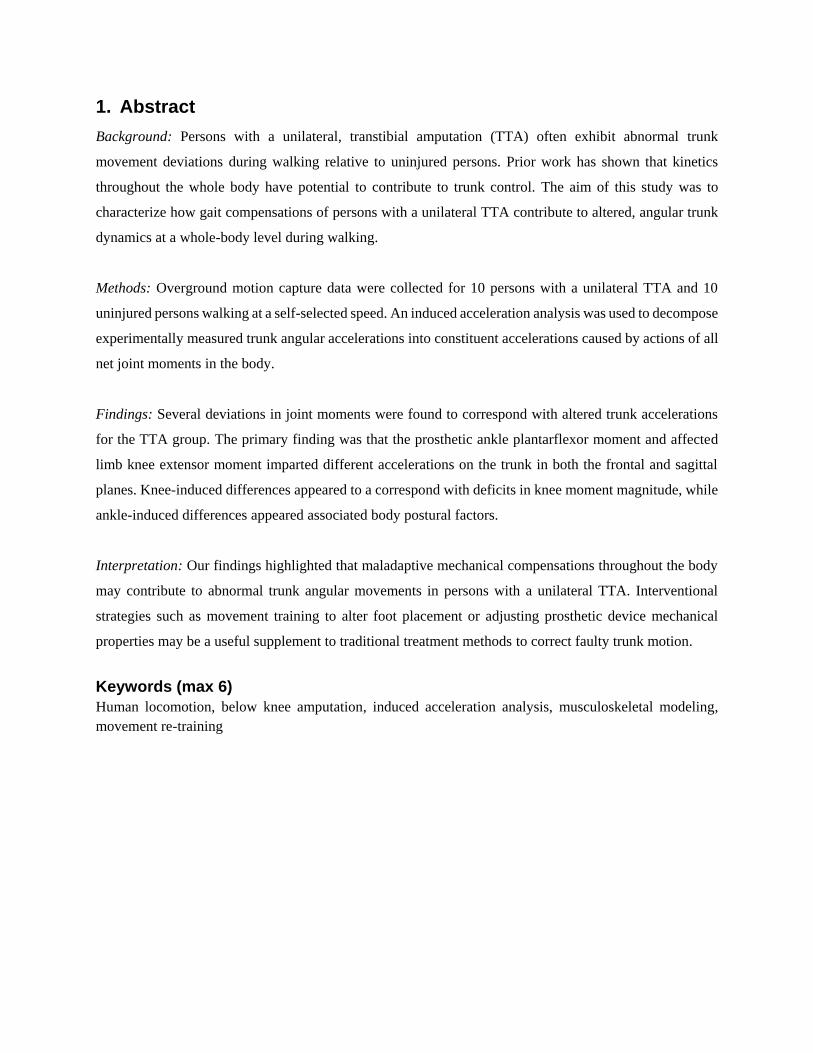
1. Abstract Background: Persons with a unilateral, transtibial amputation (TTA) often exhibit abnormal trunk
movement deviations during walking relative to uninjured persons. Prior work has shown that kinetics
throughout the whole body have potential to contribute to trunk control. The aim of this study was to
characterize how gait compensations of persons with a unilateral TTA contribute to altered, angular trunk
dynamics at a whole-body level during walking.
Methods: Overground motion capture data were collected for 10 persons with a unilateral TTA and 10
uninjured persons walking at a self-selected speed. An induced acceleration analysis was used to decompose
experimentally measured trunk angular accelerations into constituent accelerations caused by actions of all
net joint moments in the body.
Findings: Several deviations in joint moments were found to correspond with altered trunk accelerations
for the TTA group. The primary finding was that the prosthetic ankle plantarflexor moment and affected
limb knee extensor moment imparted different accelerations on the trunk in both the frontal and sagittal
planes. Knee-induced differences appeared to a correspond with deficits in knee moment magnitude, while
ankle-induced differences appeared associated body postural factors.
Interpretation: Our findings highlighted that maladaptive mechanical compensations throughout the body
may contribute to abnormal trunk angular movements in persons with a unilateral TTA. Interventional
strategies such as movement training to alter foot placement or adjusting prosthetic device mechanical
properties may be a useful supplement to traditional treatment methods to correct faulty trunk motion.
Keywords (max 6) Human locomotion, below knee amputation, induced acceleration analysis, musculoskeletal modeling,
movement re-training
ABSTRACT Objective: To investigate trunk-pelvic kinematic outcomes with time from initial ambulation with a prosthesis, and amputation level, among persons with unilateral lower limb loss. It was hypothesized the magnitudes of trunk-pelvic range of motion (ROM) will increase and pelvic-trunk coordination will increase (become more out-of-phase) with increasing time of ambulation. Secondarily, persons with more proximal limb loss will initially exhibit less trunk and pelvic ROM, and more in-phase trunk-pelvic coordination. Design: Inception cohort with up to five repeated biomechanical evaluations during a one-year period (0, 2, 4, 6, and 12 months) after initial ambulation with a prosthesis. Setting: Biomechanics laboratory within Military Treatment Facility Participants: Thirty-two males with unilateral lower limb loss (twenty-two with transtibial limb loss and ten with transfemoral limb loss). Interventions: Not applicable. Main Outcome Measures: Triplanar trunk-pelvic ROM, and intersegmental coordination (continuous relative phase; CRP), were computed as participants walked overground at a self-selected (~1.30 m/s) and controlled (~1.20 m/s) speed. Results: With increasing time after initial ambulation, trunk ROM generally decreased, most notably for persons with transfemoral limb loss, while pelvic ROM generally remained consistent. Mean CRP became more out-of-phase over time, and frontal CRP was more in-phase for persons with transfemoral vs. transtibial limb loss. Conclusions: Temporal relationships in the features of trunk-pelvic motions within the first year of ambulation after limb loss have longer-term implications for the surveillance of LBP onset and recurrence, and may help identify important biomechanical factors in its causation. Future work should therefore continue longitudinal evaluations of trunk-pelvic motions, as well as injury rate and pain level. Key Words: Extremity Trauma; Extremities; Wounds and Injuries; Biomechanics; Locomotion; Rehabilitation; Torso
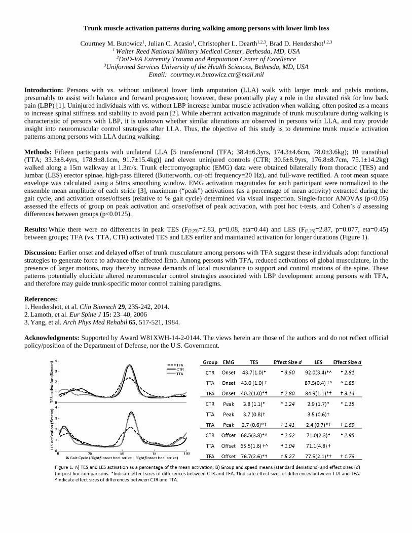
Trunk muscle activation patterns during walking among persons with lower limb loss
Courtney M. Butowicz1, Julian C. Acasio1, Christopher L. Dearth1,2,3, Brad D. Hendershot1,2,3
1 Walter Reed National Military Medical Center, Bethesda, MD, USA 2DoD-VA Extremity Trauma and Amputation Center of Excellence
3Uniformed Services University of the Health Sciences, Bethesda, MD, USA Email: [email protected]
Introduction: Persons with vs. without unilateral lower limb amputation (LLA) walk with larger trunk and pelvis motions, presumably to assist with balance and forward progression; however, these potentially play a role in the elevated risk for low back pain (LBP) [1]. Uninjured individuals with vs. without LBP increase lumbar muscle activation when walking, often posited as a means to increase spinal stiffness and stability to avoid pain [2]. While aberrant activation magnitude of trunk musculature during walking is characteristic of persons with LBP, it is unknown whether similar alterations are observed in persons with LLA, and may provide insight into neuromuscular control strategies after LLA. Thus, the objective of this study is to determine trunk muscle activation patterns among persons with LLA during walking. Methods: Fifteen participants with unilateral LLA [5 transfemoral (TFA; 38.4±6.3yrs, 174.3±4.6cm, 78.0±3.6kg); 10 transtibial (TTA; 33.3±8.4yrs, 178.9±8.1cm, 91.7±15.4kg)] and eleven uninjured controls (CTR; 30.6±8.9yrs, 176.8±8.7cm, 75.1±14.2kg) walked along a 15m walkway at 1.3m/s. Trunk electromyographic (EMG) data were obtained bilaterally from thoracic (TES) and lumbar (LES) erector spinae, high-pass filtered (Butterworth, cut-off frequency=20 Hz), and full-wave rectified. A root mean square envelope was calculated using a 50ms smoothing window. EMG activation magnitudes for each participant were normalized to the ensemble mean amplitude of each stride [3], maximum (“peak”) activations (as a percentage of mean activity) extracted during the gait cycle, and activation onset/offsets (relative to % gait cycle) determined via visual inspection. Single-factor ANOVAs (p<0.05) assessed the effects of group on peak activation and onset/offset of peak activation, with post hoc t-tests, and Cohen’s d assessing differences between groups (p<0.0125). Results: While there were no differences in peak TES (F(2,23)=2.83, p=0.08, eta=0.44) and LES (F(2,23)=2.87, p=0.077, eta=0.45) between groups; TFA (vs. TTA, CTR) activated TES and LES earlier and maintained activation for longer durations (Figure 1). Discussion: Earlier onset and delayed offset of trunk musculature among persons with TFA suggest these individuals adopt functional strategies to generate force to advance the affected limb. Among persons with TFA, reduced activations of global musculature, in the presence of larger motions, may thereby increase demands of local musculature to support and control motions of the spine. These patterns potentially elucidate altered neuromuscular control strategies associated with LBP development among persons with TFA, and therefore may guide trunk-specific motor control training paradigms. References: 1. Hendershot, et al. Clin Biomech 29, 235-242, 2014. 2. Lamoth, et al. Eur Spine J 15: 23–40, 2006 3. Yang, et al. Arch Phys Med Rehabil 65, 517-521, 1984. Acknowledgments: Supported by Award W81XWH-14-2-0144. The views herein are those of the authors and do not reflect official policy/position of the Department of Defense, nor the U.S. Government.
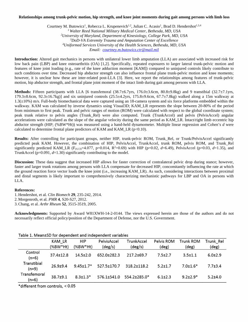
Relationships among trunk-pelvic motion, hip strength, and knee joint moments during gait among persons with limb loss
Courtney M. Butowicz1, Rebecca L. Krupenevich1,2, Julian C. Acasio1, Brad D. Hendershot1,3,4
1 Walter Reed National Military Medical Center, Bethesda, MD, USA 2University of Maryland, Department of Kinesiology, College Park, MD, USA
3DoD-VA Extremity Trauma and Amputation Center of Excellence 4Uniformed Services University of the Health Sciences, Bethesda, MD, USA
Email: [email protected]
Introduction: Altered gait mechanics in persons with unilateral lower limb amputation (LLA) are associated with increased risk for low back pain (LBP) and knee osteoarthritis (OA) [1,2]. Specifically, repeated exposures to larger lateral trunk-pelvic motion and features of knee joint loading (e.g., rate of the knee adduction moment [KAM]) compared to uninjured controls likely contribute to such conditions over time. Decreased hip abductor strength can also influence frontal plane trunk-pelvic motion and knee moments; however, it is unclear how these are inter-related post-LLA [3]. Here, we report the relationships among features of trunk-pelvic motion, hip abductor strength, and frontal plane joint moment of the intact limb during gait among persons with LLA. Methods: Fifteen participants with LLA [6 transfemoral (38.7±6.7yrs, 176.0±3.6cm, 80.8±9.8kg) and 9 transtibial (32.7±7.1yrs, 179.3±8.6cm, 92.3±16.7kg)] and six uninjured controls (25.5±4.2yrs, 175.8±9.6cm, 67.7±7.8kg) walked along a 15m walkway at 1.3(±10%) m/s. Full-body biomechanical data were captured using an 18-camera system and six force platforms embedded within the walkway. KAM was calculated by inverse dynamics using Visual3D; KAM_LR represents the slope between 20-80% of the period from minimum to first peak. Trunk and pelvic ranges of motion (ROM) were calculated with respect to the global coordinate system; peak trunk relative to pelvis angles (Trunk_Rel) were also computed. Trunk (TrunkAccel) and pelvis (PelvisAccel) angular accelerations were calculated as the slope of the angular velocity during the same period as KAM_LR. Intact/right limb eccentric hip abductor strength (HIP; (%BW*Ht)) was measured using a hand-held dynamometer. Multiple linear regression and Cohen’s d were calculated to determine frontal plane predictors of KAM and KAM_LR (p<0.10).
Results: After controlling for participant groups, neither HIP, trunk-pelvic ROM, Trunk_Rel, or Trunk/PelvisAccel significantly predicted peak KAM. However, the combination of HIP, PelvisAccel, TrunkAccel, trunk ROM, pelvis ROM, and Trunk_Rel significantly predicted KAM_LR (F(4,13)=4.077, p=0.014, R2=0.69) with HIP (p=0.02, d=6.49), PelvisAccel (p=0.03, d=1.35), and TrunkAccel (p=0.091, d=1.30) significantly contributing to the model. Discussion: These data suggest that increased HIP allows for faster correction of contralateral pelvic drop during stance; however, faster and larger trunk rotations among persons with LLA compensate for decreased HIP, concomitantly influencing the rate at which the ground reaction force vector loads the knee joint (i.e., increasing KAM_LR). As such, considering interactions between proximal and distal segments is likely important to comprehensively characterizing mechanistic pathways for LBP and OA in persons with LLA. References: 1. Hendershot, et al. Clin Biomech 29, 235-242, 2014. 2. Morgenroth, et al. PMR 4, S20-S27, 2012. 3. Chang, et al. Arthr Rheum 52, 3515-3519, 2005. Acknowledgments: Supported by Award W81XWH-14-2-0144. The views expressed herein are those of the authors and do not necessarily reflect official policy/position of the Department of Defense, nor the U.S. Government.
Prevalence and Relationship of Low Back Pain and Psychosocial Factors after Lower Limb Amputation among Wounded Warrior Recovery Project Participants
Brittney Mazzone, PT, DPT1,2; Shawn Farrokhi, PT, Ph.D.1,2; Brad D. Hendershot Ph.D;1,3,4
Cameron T. McCabe, Ph.D.;5 Jessica R. Watrous, Ph.D.5
1DoD-VA Extremity Trauma and Amputation Center of Excellence (EACE);
2Naval Medical Center San Diego, San Diego, CA; 3Walter Reed National Military Medical Center; 4Uniformed Services University of the Health Sciences, Bethesda, MD; 5Naval Health
Research Center, San Diego, CA Background: Low back pain (LBP) is a common secondary health condition after limb amputation with important implications related to functional capabilities and quality of life. To date, however, the majority of data regarding the prevalence of LBP after amputation have come from studies of older military veterans or civilians with limb loss. As such, there is limited information on the prevalence of LBP after limb amputation in younger Service members from recent conflicts. Additionally, a growing body of evidence suggests that psychosocial factors, such as depression symptoms, significantly influence the experience of LBP in patients without amputation. However, there is currently a dearth of information available regarding the association of psychosocial factors and LBP after limb amputation. The purpose of this study was to assess the prevalence and potential association of LBP with psychosocial factors in Service members with deployment-related lower limb amputations. Methods: Data on psychosocial factors, including quality of life, post-traumatic stress disorder (PTSD), and depression, comes from the Wounded Warrior Recovery Project (WWRP). The WWRP is an ongoing, web-based, longitudinal study that aims to gather patient-reported outcomes of deployment-related injured Service members. The Military Health System Data Repository was utilized to extract medical record data. Diagnostic codes were queried for at least one instance of coding related to LBP. The population of interest was individuals with deployment-related amputations. Of the current WWRP sample of 4,974 individuals who were injured on deployment between June 2004 and May 2013 and completed a baseline WWRP assessment between 2012 and 2017, 81individuals had lower limb amputations. The majority of the sample of Service members with amputations were male (99%), enlisted (79%), Army (78%), and blast-related injuries (95%). General linear models were utilized to analyze associations between LBP and psychosocial factors, while controlling for injury severity and time since amputation. Results: In this sample, 58% of individuals with amputations had been diagnosed with LBP by a medical provider; 31% screened positive for PTSD using the PTSD Checklist and 32% screened positive for depression using the Center for Epidemiological Studies Depression Scale. Among individuals with amputations, those with LBP reported lower quality of life (0.415, standard error [SE] = .014) compared to those with amputations without LBP (0.470, SE = .016) (B = .055, p =.01; η2 = .085). Similarly, individuals with amputations and LBP reported higher PTSD scores (40.95, SE = 2.11) compared to those without LBP (33.91, SE = 2.43) (B = -7.039, p
=.033; η2 = .059). There was no significant difference between depression scores in individuals with amputations with (15.17, SE = 1.50) or without LBP (11.51, SE = 1.72) (p =.113). Conclusions: Presence of LBP after limb amputation appears to be associated with greater PTSD symptoms and lower quality of life. Given the cross-sectional nature of the current data, determination of a cause-and-effect relationship was not possible. Further research is needed to assess the efficacy of addressing psychosocial factors as part of a multi-disciplinary approach for improving pain and function in Service members with amputations and LBP. Disclaimer: I am a military service member (or employee of the U.S. Government). This work was prepared as part of my official duties. Title 17, U.S.C. §105 provides the “Copyright protection under this title is not available for any work of the United States Government.” Title 17, U.S.C. §101 defines a U.S. Government work as work prepared by a military service member or employee of the U.S. Government as part of that person’s official duties. This work was supported with resources provided by the U.S. Navy Bureau of Medicine and Surgery’s Wounded, Ill, and Injured Psychological Health and Traumatic Brain Injury Program, Work Unit No. 60808 and Extremity Trauma and Amputation Center for Excellence (EACE), Work Unit N1333The views expressed in this research are those of the authors and do not necessarily reflect the official policy or position of the Department of the Navy, Department of the Army, Department of the Air Force, Department of Veterans Affairs, Department of Defense, or the U.S. Government. Approved for public release; distribution unlimited. This research has been conducted in compliance with all applicable federal regulations governing the protection of human subjects in research (Protocol NHRC.2009.0014). References: 1. Durmus D, Safaz I, Adiguzel E, et al. The relationship between prosthesis use, phantom pain
and psychiatric symptoms in male traumatic limb amputees. Compr Psychiatry 2015;59:45–53.
2. Ephraim PL, Wegener ST, MacKenzie EJ, Dillingham TR, Pezzin LE. Phantom pain, residual limb pain, and back pain in amputees: results of a national survey. Arch Phys Med Rehabil 2005;86:1910–9.
3. Farrokhi S, Mazzone B, Schneider M, et al. Biopsychosocial risk factors associated with chronic low back pain after lower limb amputation. Medical Hypotheses 2017;108:1-9.
4. Pincus T, Kent P, Bronfort G, et al. Twenty-five years with the biopsychosocial model of low back pain—is it time to celebrate? A report from the twelfth international forum for primary care research on low back pain. Spine 2013;38:2118–23.
5. Weiner BK. Spine update: the biopsychosocial model and spine care. Spine 2008;33:219–23.
Trunk Muscle Activations, Motions, and Spinal Loads among Persons with Lower Limb Amputation: Influences of Chronic Low Back Pain
Julian C. Acasio1, Courtney M. Butowicz1, Iman Shojaei2,
Babak Bazrgari2, Christopher L. Dearth1,3,4, Brad D. Hendershot1,3,4
1 Walter Reed National Military Medical Center, Bethesda, MD, USA
2 University of Kentucky, Lexington, KY, USA 3 DoD-VA Extremity Trauma and Amputation Center of Excellence, USA
4 Uniformed Services University of the Health Sciences, Bethesda, MD, USA Email: [email protected]
INTRODUCTION: Low back pain (LBP) is a prevalent and costly musculoskeletal disability, particularly within the military, where LBP is among the leading causes of medical visits and lost duty days [1]. Although most LBP remains idiopathic, physical (biomechanical) factors likely contribute more substantially in certain populations (e.g., persons with lower limb amputation; LLA) [2]. During activities of daily living, altered trunk-pelvic motion in persons with (vs. without) LLA impose greater mechanical loads on spinal tissues, and thus have been suggested as a risk factor for the development of LBP [3]. Moreover, in the presence of LBP, further alterations in trunk-pelvic motion have been characterized [4-6]; yet, features of trunk muscle activations underlying these altered motions with LBP and effects on spinal loads remain unclear. The purpose of this study was therefore to evaluate trunk-pelvic motion, muscle activities, and spinal loads among persons with LLA both with and without LBP (and a control group without LBP for reference). Among persons with (vs. without) LLA, we hypothesized trunk muscle activations and corresponding trunk-pelvic motions with vs. without LBP would be associated with larger spinal loads, supporting a pathway wherein repeated exposure to abnormal lumbopelvic mechanics can adversely affect spine health.
METHODS: Eighteen persons with LLA – 8 with LBP (“LLA-P”) and 10 without LBP (“LLA-NP”) – and 10 uninjured controls without LBP (“CTR-NP”) participated in this cross-sectional, IRB-approved study. The LLA-P group reported chronic LBP (n=7 every day or nearly every day in the most recent 6 months; n=1 at least half of days in the most recent 6 months [7]. Mean (standard deviation) pain in the past seven days = 3.8 (1.3). Participants walked overground across a 15m walkway at 1.3m/s while an 18-camera motion capture system (Qualisys, Göteborg, Sweden) tracked (120Hz) trunk and pelvis kinematics. Simultaneously, electromyographic (EMG) activities were sampled (1200Hz) bilaterally at two levels of the erector spinae (T9 [TES] and L2 [LES]). Kinematic data were low-pass filtered (Butterworth, 6 Hz), EMG data were high-pass filtered (Butterworth, 20 Hz), full-wave rectified, and smoothed with a 50ms RMS window. Tri-planar trunk and pelvis angles and angular velocities were calculated in Visual3D (C-motion, Germantown, MD). Global trunk and pelvic ranges of motion (ROM) were determined and trunk-pelvic continuous relative phases (CRP) were calculated in MATLAB (Mathworks, Natick, MA). EMG data were normalized to the signal mean and down-sampled to match kinematic capture rate, and processed bilaterally for individuals with LLA (i.e., on both the intact and affected side) and for the right side of CTR-NP as no differences were observed between left and right sides. Cross-correlations related EMG and trunk and pelvic rotation time series during both intact and affected strides. Finally, trunk-pelvic kinematic data were input to a non-linear finite element model of the spine [8], wherein a heuristic optimization procedure estimated trunk muscle forces and spinal loads by minimizing the sum of squared muscle stress (i.e., the cost function) across 56 muscles. Individual muscle forces were summed across local (i.e., connecting individual lumbar vertebrae to the pelvis) and global muscles (i.e., connecting the thorax/rib cage to the pelvis). Spinal loads were compiled from the intervertebral level at which maximum spinal loads occurred (i.e., L5/S1). Peak spinal loads and muscle forces were determined and normalized to body mass. Separate one-way repeated-measure ANOVAs assessed the effect of group (LLA-P, LLA-NP, CTR-NP) on ROM, mean CRP, magnitudes
of cross-correlation coefficients (R), and spinal loads (P<0.05). Bonferroni-corrected t-tests assessed pairwise differences (P<0.0167).
RESULTS: Larger frontal plane trunk ROM were observed in both LLA-P and LLA-NP vs. CTR-NP (P<0.008). Transverse plane trunk ROM were larger in LLA-P vs. CTR-NP (P<0.001) but similar to LLA-NP (P=0.032). A main effect was observed in frontal plane mean CRP, with LLA-NP exhibiting smaller (i.e., more in-phase) CRP than CTR-NP (P=0.013). Main effects were observed in the R-values of both intact and affected side LES and sagittal trunk angles. Affected-side LES activations more strongly correlated with sagittal trunk angles in LLA-P and LLA-NP vs. CTR (P<0.003). Intact-side LES more strongly correlated with sagittal trunk angles in LLA-NP vs. CTR-NP (P=0.005), and with transverse trunk angles in LLA-NP vs. LLA-P (P=0.010). R-values of affected -side LES and sagittal pelvis angles were greater in LLA-NP vs. CTR-NP (P=0.011) but similar between LLA-P vs. CTR-NP (P=0.023). R-values of trunk and pelvis angles and TES did not differ between groups. No main effects were observed in peak spinal loads (P>0.078) or global muscle forces (P=0.076). However, peak local muscle forces differed between groups (P=0.017); local muscle forces were larger in CTR-NP vs. both LLA-P (P=0.012) and LLA-NP (P=0.016).
CONCLUSION: Though sagittal plane kinematics were similar between groups, R-values of LES activation patterns and sagittal plane trunk and pelvis angles were larger in both LLA-P and LLA-NP vs. CTR, suggesting an active LES control strategy that may be especially important in the LLA-P group as they tended to walk with more anterior trunk lean than CTR. In the transverse plane, smaller trunk ROM and stronger correlations between intact LES and trunk angles were observed in LLA-NP vs. LLA-P, suggesting LLA-NP are better able to control transverse plane trunk rotations. Despite larger trunk ROM in both the frontal and transverse planes, a lack of differences in spinal loads among LLA-P and LLA-NP vs. CTR are contrary to both our hypotheses (LLA-P ≠ LLA-NP) and prior work [LLA-NP > CTR; 9]. Nevertheless, larger transverse motions with vs. without LBP are consistent with prior work in persons with (transfemoral) LLA [4]. Interestingly, all persons in the LLA-P group had transtibial LLA while those in the LLA-NP group comprised a combination of both transtibial (n=7) and transfemoral (n=3) LLA. In the absence of LBP, alterations in the characteristics of trunk-pelvic motion are typically larger in persons with transfemoral vs. transtibial LLA [2]. It is therefore possible that presence of chronic LBP has concurrently increased trunk-pelvic motions in a group that is otherwise more similar to uninjured CTR. Also, while we identified group differences in local muscle forces, further consideration may be warranted for the model/optimization assumptions regarding muscle recruitment strategies with vs. without LBP [10]. Of note, despite categorization of chronic LBP, participants in the LLA-P group at the time of testing reported mean (standard deviation) numerical pain scores of 2.2 (1.3).
In summary, although prior work has identified larger spinal loads in persons with vs. without LLA, the current (cross-sectional) results do not necessarily support the notion that larger spinal loads during walking influence the persistence of LBP. It is however possible that individuals in the LLA-P group experienced larger spinal loads at some point prior to developing LBP and, thus, future work is needed to longitudinally characterize the temporal relationships in these outcomes with time since LLA to better elucidate the causal relationships.
REFERENCES: [1] Knox Spine 2011. [2] Devan Phys Med Rehab 2017. [3] Hendershot Clin Biomech 2014 [4] Devan J Prosthet Orthot 2017. [5] Morgenroth AJPMR 2010. [6] Fatone AJPMR 2016. [7] Deyo The J Pain 2014. [8] Bazrgari Eur Spine J 2007. [9] Shojaei Clin Biomech 2016. [10] van Dieen Spine 2003.
ACKNOWLEDGEMENTS: This work was funded by award W81XWH-14-02-0144, and supported by the EACE and CRSR. The views expressed are those of the authors and do not reflect the official policy of the Department of Army/Navy/Air Force, Department of Defense, or U.S. Government.
Learning Objectives:
1. Describe risk factors for low back pain secondary to LLA
2. Describe differences in trunk-specific outcomes between those with LLA with and without low back pain
3. Describe clinical considerations to mitigate the impact of LBP secondary to LLA
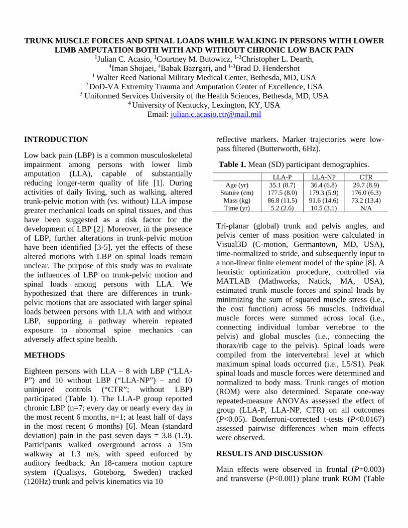
TRUNK MUSCLE FORCES AND SPINAL LOADS WHILE WALKING IN PERSONS WITH LOWER LIMB AMPUTATION BOTH WITH AND WITHOUT CHRONIC LOW BACK PAIN
1Julian C. Acasio, 1Courtney M. Butowicz, 1-3Christopher L. Dearth, 4Iman Shojaei, 4Babak Bazrgari, and 1-3Brad D. Hendershot
1 Walter Reed National Military Medical Center, Bethesda, MD, USA 2 DoD-VA Extremity Trauma and Amputation Center of Excellence, USA
3 Uniformed Services University of the Health Sciences, Bethesda, MD, USA 4 University of Kentucky, Lexington, KY, USA
Email: [email protected]
INTRODUCTION
Low back pain (LBP) is a common musculoskeletal impairment among persons with lower limb amputation (LLA), capable of substantially reducing longer-term quality of life [1]. During activities of daily living, such as walking, altered trunk-pelvic motion with (vs. without) LLA impose greater mechanical loads on spinal tissues, and thus have been suggested as a risk factor for the development of LBP [2]. Moreover, in the presence of LBP, further alterations in trunk-pelvic motion have been identified [3-5], yet the effects of these altered motions with LBP on spinal loads remain unclear. The purpose of this study was to evaluate the influences of LBP on trunk-pelvic motion and spinal loads among persons with LLA. We hypothesized that there are differences in trunk-pelvic motions that are associated with larger spinal loads between persons with LLA with and without LBP, supporting a pathway wherein repeated exposure to abnormal spine mechanics can adversely affect spine health.
METHODS
Eighteen persons with LLA – 8 with LBP (“LLA-P”) and 10 without LBP (“LLA-NP”) – and 10 uninjured controls (“CTR”; without LBP) participated (Table 1). The LLA-P group reported chronic LBP (n=7; every day or nearly every day in the most recent 6 months, n=1; at least half of days in the most recent 6 months) [6]. Mean (standard deviation) pain in the past seven days = 3.8 (1.3). Participants walked overground across a 15m walkway at 1.3 m/s, with speed enforced by auditory feedback. An 18-camera motion capture system (Qualisys, Göteborg, Sweden) tracked (120Hz) trunk and pelvis kinematics via 10
reflective markers. Marker trajectories were low-pass filtered (Butterworth, 6Hz).
Table 1. Mean (SD) participant demographics.
Tri-planar (global) trunk and pelvis angles, and pelvis center of mass position were calculated in Visual3D (C-motion, Germantown, MD, USA), time-normalized to stride, and subsequently input to a non-linear finite element model of the spine [8]. A heuristic optimization procedure, controlled via MATLAB (Mathworks, Natick, MA, USA), estimated trunk muscle forces and spinal loads by minimizing the sum of squared muscle stress (i.e., the cost function) across 56 muscles. Individual muscle forces were summed across local (i.e., connecting individual lumbar vertebrae to the pelvis) and global muscles (i.e., connecting the thorax/rib cage to the pelvis). Spinal loads were compiled from the intervertebral level at which maximum spinal loads occurred (i.e., L5/S1). Peak spinal loads and muscle forces were determined and normalized to body mass. Trunk ranges of motion (ROM) were also determined. Separate one-way repeated-measure ANOVAs assessed the effect of group (LLA-P, LLA-NP, CTR) on all outcomes (P<0.05). Bonferroni-corrected t-tests (P<0.0167) assessed pairwise differences when main effects were observed.
RESULTS AND DISCUSSION
Main effects were observed in frontal (P=0.003) and transverse (P<0.001) plane trunk ROM (Table
LLA-P LLA-NP CTR Age (yr) 35.1 (8.7) 36.4 (6.8) 29.7 (8.9)
Stature (cm) 177.5 (8.0) 179.3 (5.9) 176.0 (6.3) Mass (kg) 86.8 (11.5) 91.6 (14.6) 73.2 (13.4) Time (yr) 5.2 (2.6) 10.5 (3.1) N/A
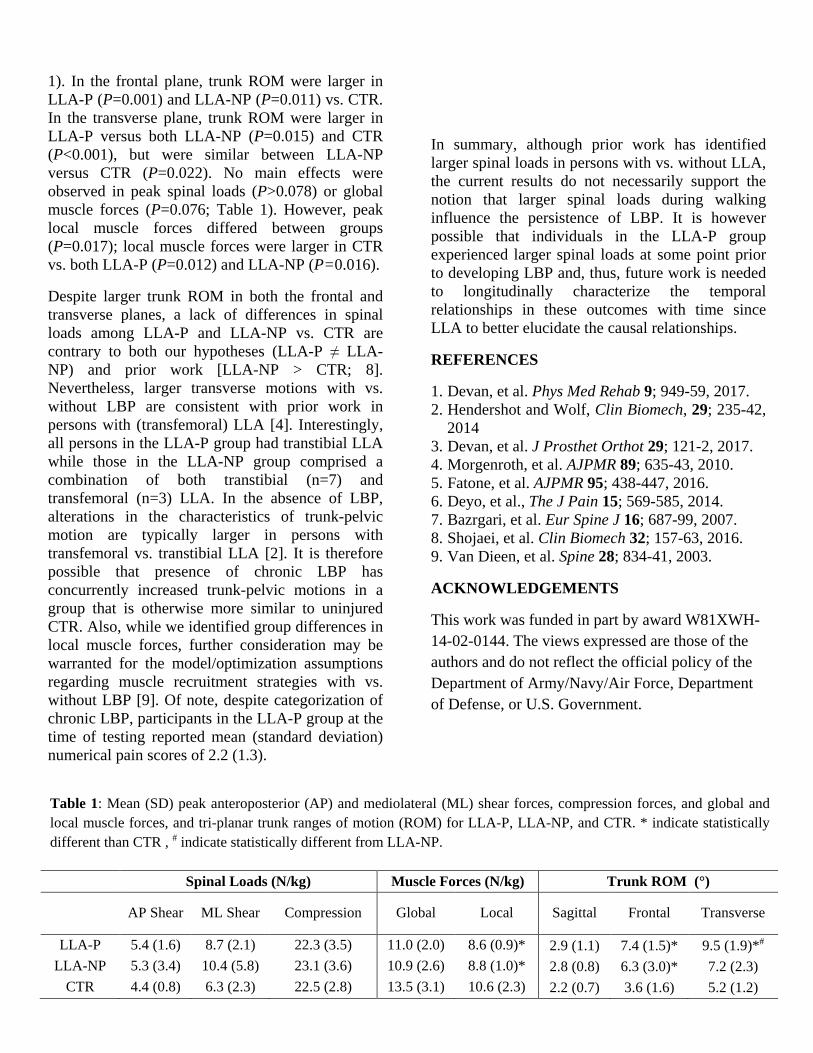
Table 1: Mean (SD) peak anteroposterior (AP) and mediolateral (ML) shear forces, compression forces, and global and local muscle forces, and tri-planar trunk ranges of motion (ROM) for LLA-P, LLA-NP, and CTR. * indicate statistically different than CTR , # indicate statistically different from LLA-NP.
1). In the frontal plane, trunk ROM were larger in LLA-P (P=0.001) and LLA-NP (P=0.011) vs. CTR. In the transverse plane, trunk ROM were larger in LLA-P versus both LLA-NP (P=0.015) and CTR (P<0.001), but were similar between LLA-NP versus CTR (P=0.022). No main effects were observed in peak spinal loads (P>0.078) or global muscle forces (P=0.076; Table 1). However, peak local muscle forces differed between groups (P=0.017); local muscle forces were larger in CTR vs. both LLA-P (P=0.012) and LLA-NP (P=0.016).
Despite larger trunk ROM in both the frontal and transverse planes, a lack of differences in spinal loads among LLA-P and LLA-NP vs. CTR are contrary to both our hypotheses (LLA-P ≠ LLA-NP) and prior work [LLA-NP > CTR; 8]. Nevertheless, larger transverse motions with vs. without LBP are consistent with prior work in persons with (transfemoral) LLA [4]. Interestingly, all persons in the LLA-P group had transtibial LLA while those in the LLA-NP group comprised a combination of both transtibial (n=7) and transfemoral (n=3) LLA. In the absence of LBP, alterations in the characteristics of trunk-pelvic motion are typically larger in persons with transfemoral vs. transtibial LLA [2]. It is therefore possible that presence of chronic LBP has concurrently increased trunk-pelvic motions in a group that is otherwise more similar to uninjured CTR. Also, while we identified group differences in local muscle forces, further consideration may be warranted for the model/optimization assumptions regarding muscle recruitment strategies with vs. without LBP [9]. Of note, despite categorization of chronic LBP, participants in the LLA-P group at the time of testing reported mean (standard deviation) numerical pain scores of 2.2 (1.3).
In summary, although prior work has identified larger spinal loads in persons with vs. without LLA, the current results do not necessarily support the notion that larger spinal loads during walking influence the persistence of LBP. It is however possible that individuals in the LLA-P group experienced larger spinal loads at some point prior to developing LBP and, thus, future work is needed to longitudinally characterize the temporal relationships in these outcomes with time since LLA to better elucidate the causal relationships.
REFERENCES
1. Devan, et al. Phys Med Rehab 9; 949-59, 2017. 2. Hendershot and Wolf, Clin Biomech, 29; 235-42,
2014 3. Devan, et al. J Prosthet Orthot 29; 121-2, 2017. 4. Morgenroth, et al. AJPMR 89; 635-43, 2010. 5. Fatone, et al. AJPMR 95; 438-447, 2016. 6. Deyo, et al., The J Pain 15; 569-585, 2014. 7. Bazrgari, et al. Eur Spine J 16; 687-99, 2007. 8. Shojaei, et al. Clin Biomech 32; 157-63, 2016. 9. Van Dieen, et al. Spine 28; 834-41, 2003.
ACKNOWLEDGEMENTS
This work was funded in part by award W81XWH-14-02-0144. The views expressed are those of the authors and do not reflect the official policy of the Department of Army/Navy/Air Force, Department of Defense, or U.S. Government.
Spinal Loads (N/kg) Muscle Forces (N/kg) Trunk ROM (°)
AP Shear ML Shear Compression Global Local Sagittal Frontal Transverse
LLA-P 5.4 (1.6) 8.7 (2.1) 22.3 (3.5) 11.0 (2.0) 8.6 (0.9)* 2.9 (1.1) 7.4 (1.5)* 9.5 (1.9)*#
LLA-NP 5.3 (3.4) 10.4 (5.8) 23.1 (3.6) 10.9 (2.6) 8.8 (1.0)* 2.8 (0.8) 6.3 (3.0)* 7.2 (2.3) CTR 4.4 (0.8) 6.3 (2.3) 22.5 (2.8) 13.5 (3.1) 10.6 (2.3) 2.2 (0.7) 3.6 (1.6) 5.2 (1.2)
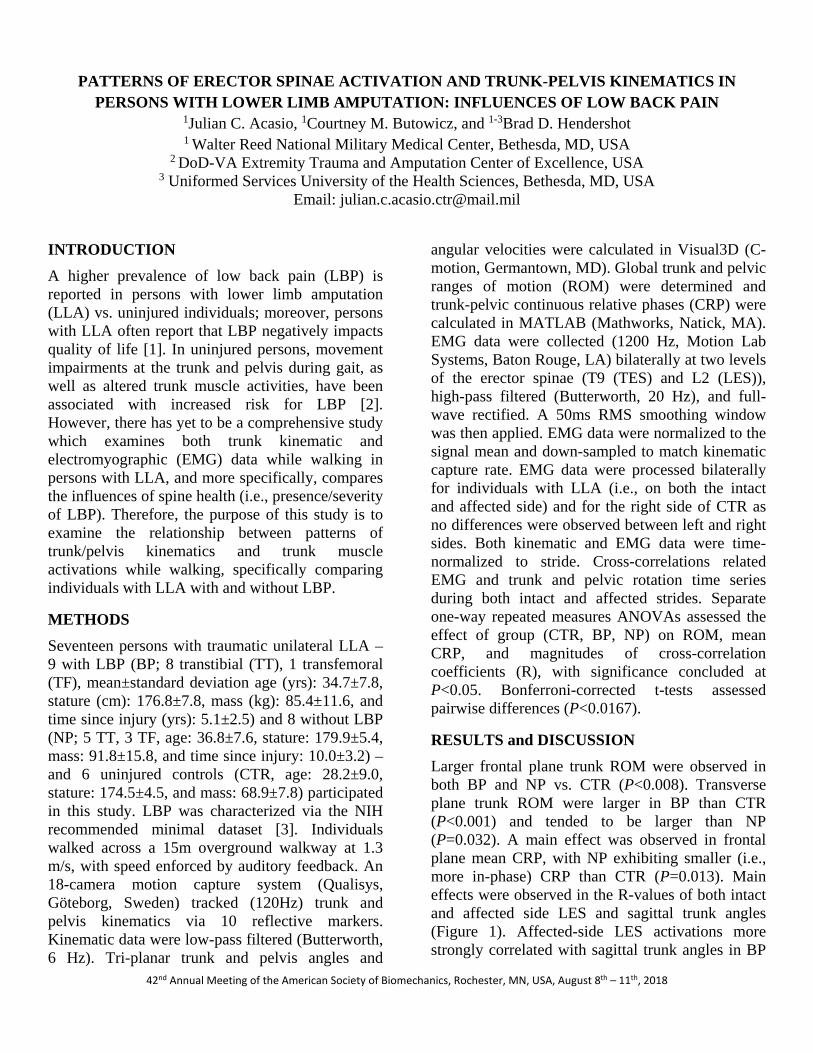
42nd Annual Meeting of the American Society of Biomechanics, Rochester, MN, USA, August 8th – 11th, 2018
PATTERNS OF ERECTOR SPINAE ACTIVATION AND TRUNK-PELVIS KINEMATICS IN PERSONS WITH LOWER LIMB AMPUTATION: INFLUENCES OF LOW BACK PAIN
1Julian C. Acasio, 1Courtney M. Butowicz, and 1-3Brad D. Hendershot 1 Walter Reed National Military Medical Center, Bethesda, MD, USA
2 DoD-VA Extremity Trauma and Amputation Center of Excellence, USA 3 Uniformed Services University of the Health Sciences, Bethesda, MD, USA
Email: [email protected]
INTRODUCTION A higher prevalence of low back pain (LBP) is reported in persons with lower limb amputation (LLA) vs. uninjured individuals; moreover, persons with LLA often report that LBP negatively impacts quality of life [1]. In uninjured persons, movement impairments at the trunk and pelvis during gait, as well as altered trunk muscle activities, have been associated with increased risk for LBP [2]. However, there has yet to be a comprehensive study which examines both trunk kinematic and electromyographic (EMG) data while walking in persons with LLA, and more specifically, compares the influences of spine health (i.e., presence/severity of LBP). Therefore, the purpose of this study is to examine the relationship between patterns of trunk/pelvis kinematics and trunk muscle activations while walking, specifically comparing individuals with LLA with and without LBP.
METHODS Seventeen persons with traumatic unilateral LLA – 9 with LBP (BP; 8 transtibial (TT), 1 transfemoral (TF), mean±standard deviation age (yrs): 34.7±7.8, stature (cm): 176.8±7.8, mass (kg): 85.4±11.6, and time since injury (yrs): 5.1±2.5) and 8 without LBP (NP; 5 TT, 3 TF, age: 36.8±7.6, stature: 179.9±5.4, mass: 91.8±15.8, and time since injury: 10.0±3.2) – and 6 uninjured controls (CTR, age: 28.2±9.0, stature: 174.5±4.5, and mass: 68.9±7.8) participated in this study. LBP was characterized via the NIH recommended minimal dataset [3]. Individuals walked across a 15m overground walkway at 1.3 m/s, with speed enforced by auditory feedback. An 18-camera motion capture system (Qualisys, Göteborg, Sweden) tracked (120Hz) trunk and pelvis kinematics via 10 reflective markers. Kinematic data were low-pass filtered (Butterworth, 6 Hz). Tri-planar trunk and pelvis angles and
angular velocities were calculated in Visual3D (C-motion, Germantown, MD). Global trunk and pelvic ranges of motion (ROM) were determined and trunk-pelvic continuous relative phases (CRP) were calculated in MATLAB (Mathworks, Natick, MA). EMG data were collected (1200 Hz, Motion Lab Systems, Baton Rouge, LA) bilaterally at two levels of the erector spinae (T9 (TES) and L2 (LES)), high-pass filtered (Butterworth, 20 Hz), and full-wave rectified. A 50ms RMS smoothing window was then applied. EMG data were normalized to the signal mean and down-sampled to match kinematic capture rate. EMG data were processed bilaterally for individuals with LLA (i.e., on both the intact and affected side) and for the right side of CTR as no differences were observed between left and right sides. Both kinematic and EMG data were time-normalized to stride. Cross-correlations related EMG and trunk and pelvic rotation time series during both intact and affected strides. Separate one-way repeated measures ANOVAs assessed the effect of group (CTR, BP, NP) on ROM, mean CRP, and magnitudes of cross-correlation coefficients (R), with significance concluded at P<0.05. Bonferroni-corrected t-tests assessed pairwise differences (P<0.0167).
RESULTS and DISCUSSION Larger frontal plane trunk ROM were observed in both BP and NP vs. CTR (P<0.008). Transverse plane trunk ROM were larger in BP than CTR (P<0.001) and tended to be larger than NP (P=0.032). A main effect was observed in frontal plane mean CRP, with NP exhibiting smaller (i.e., more in-phase) CRP than CTR (P=0.013). Main effects were observed in the R-values of both intact and affected side LES and sagittal trunk angles (Figure 1). Affected-side LES activations more strongly correlated with sagittal trunk angles in BP
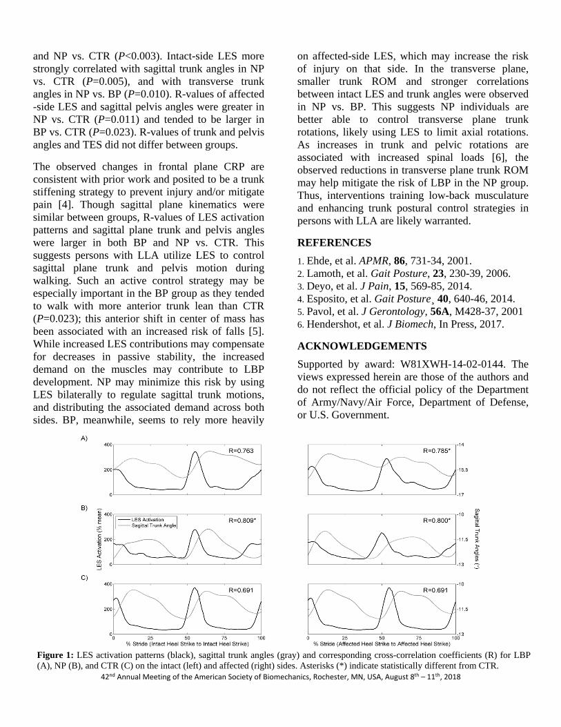
42nd Annual Meeting of the American Society of Biomechanics, Rochester, MN, USA, August 8th – 11th, 2018
Figure 1: LES activation patterns (black), sagittal trunk angles (gray) and corresponding cross-correlation coefficients (R) for LBP (A), NP (B), and CTR (C) on the intact (left) and affected (right) sides. Asterisks (*) indicate statistically different from CTR.
and NP vs. CTR (P<0.003). Intact-side LES more strongly correlated with sagittal trunk angles in NP vs. CTR (P=0.005), and with transverse trunk angles in NP vs. BP (P=0.010). R-values of affected -side LES and sagittal pelvis angles were greater in NP vs. CTR (P=0.011) and tended to be larger in BP vs. CTR (P=0.023). R-values of trunk and pelvis angles and TES did not differ between groups.
The observed changes in frontal plane CRP are consistent with prior work and posited to be a trunk stiffening strategy to prevent injury and/or mitigate pain [4]. Though sagittal plane kinematics were similar between groups, R-values of LES activation patterns and sagittal plane trunk and pelvis angles were larger in both BP and NP vs. CTR. This suggests persons with LLA utilize LES to control sagittal plane trunk and pelvis motion during walking. Such an active control strategy may be especially important in the BP group as they tended to walk with more anterior trunk lean than CTR (P=0.023); this anterior shift in center of mass has been associated with an increased risk of falls [5]. While increased LES contributions may compensate for decreases in passive stability, the increased demand on the muscles may contribute to LBP development. NP may minimize this risk by using LES bilaterally to regulate sagittal trunk motions, and distributing the associated demand across both sides. BP, meanwhile, seems to rely more heavily
on affected-side LES, which may increase the risk of injury on that side. In the transverse plane, smaller trunk ROM and stronger correlations between intact LES and trunk angles were observed in NP vs. BP. This suggests NP individuals are better able to control transverse plane trunk rotations, likely using LES to limit axial rotations. As increases in trunk and pelvic rotations are associated with increased spinal loads [6], the observed reductions in transverse plane trunk ROM may help mitigate the risk of LBP in the NP group. Thus, interventions training low-back musculature and enhancing trunk postural control strategies in persons with LLA are likely warranted.
REFERENCES 1. Ehde, et al. APMR, 86, 731-34, 2001. 2. Lamoth, et al. Gait Posture, 23, 230-39, 2006. 3. Deyo, et al. J Pain, 15, 569-85, 2014. 4. Esposito, et al. Gait Posture¸ 40, 640-46, 2014. 5. Pavol, et al. J Gerontology, 56A, M428-37, 2001 6. Hendershot, et al. J Biomech, In Press, 2017.
ACKNOWLEDGEMENTS Supported by award: W81XWH-14-02-0144. The views expressed herein are those of the authors and do not reflect the official policy of the Department of Army/Navy/Air Force, Department of Defense, or U.S. Government.
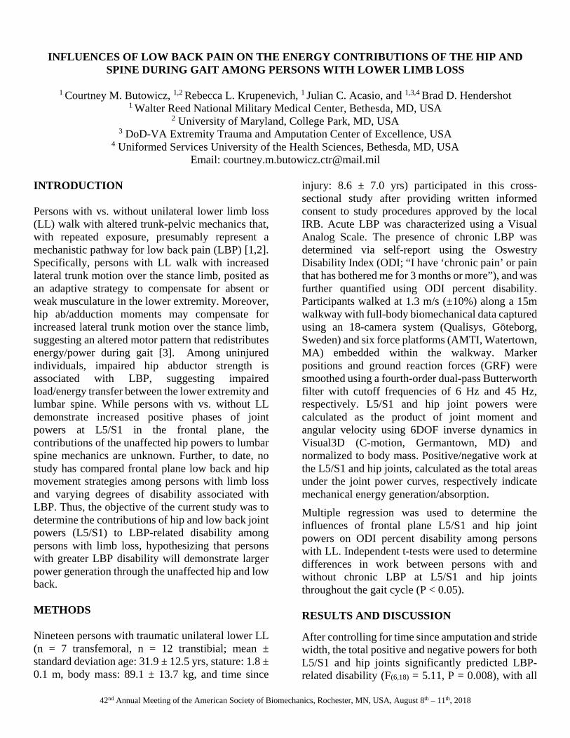
42nd Annual Meeting of the American Society of Biomechanics, Rochester, MN, USA, August 8th – 11th, 2018
INFLUENCES OF LOW BACK PAIN ON THE ENERGY CONTRIBUTIONS OF THE HIP AND SPINE DURING GAIT AMONG PERSONS WITH LOWER LIMB LOSS
1 Courtney M. Butowicz, 1,2 Rebecca L. Krupenevich, 1 Julian C. Acasio, and 1,3,4 Brad D. Hendershot
1 Walter Reed National Military Medical Center, Bethesda, MD, USA 2 University of Maryland, College Park, MD, USA
3 DoD-VA Extremity Trauma and Amputation Center of Excellence, USA 4 Uniformed Services University of the Health Sciences, Bethesda, MD, USA
Email: [email protected]
INTRODUCTION Persons with vs. without unilateral lower limb loss (LL) walk with altered trunk-pelvic mechanics that, with repeated exposure, presumably represent a mechanistic pathway for low back pain (LBP) [1,2]. Specifically, persons with LL walk with increased lateral trunk motion over the stance limb, posited as an adaptive strategy to compensate for absent or weak musculature in the lower extremity. Moreover, hip ab/adduction moments may compensate for increased lateral trunk motion over the stance limb, suggesting an altered motor pattern that redistributes energy/power during gait [3]. Among uninjured individuals, impaired hip abductor strength is associated with LBP, suggesting impaired load/energy transfer between the lower extremity and lumbar spine. While persons with vs. without LL demonstrate increased positive phases of joint powers at L5/S1 in the frontal plane, the contributions of the unaffected hip powers to lumbar spine mechanics are unknown. Further, to date, no study has compared frontal plane low back and hip movement strategies among persons with limb loss and varying degrees of disability associated with LBP. Thus, the objective of the current study was to determine the contributions of hip and low back joint powers (L5/S1) to LBP-related disability among persons with limb loss, hypothesizing that persons with greater LBP disability will demonstrate larger power generation through the unaffected hip and low back. METHODS Nineteen persons with traumatic unilateral lower LL (n = 7 transfemoral, n = 12 transtibial; mean ± standard deviation age: 31.9 ± 12.5 yrs, stature: 1.8 ± 0.1 m, body mass: 89.1 ± 13.7 kg, and time since
injury: 8.6 ± 7.0 yrs) participated in this cross-sectional study after providing written informed consent to study procedures approved by the local IRB. Acute LBP was characterized using a Visual Analog Scale. The presence of chronic LBP was determined via self-report using the Oswestry Disability Index (ODI; “I have ‘chronic pain’ or pain that has bothered me for 3 months or more”), and was further quantified using ODI percent disability. Participants walked at 1.3 m/s (±10%) along a 15m walkway with full-body biomechanical data captured using an 18-camera system (Qualisys, Göteborg, Sweden) and six force platforms (AMTI, Watertown, MA) embedded within the walkway. Marker positions and ground reaction forces (GRF) were smoothed using a fourth-order dual-pass Butterworth filter with cutoff frequencies of 6 Hz and 45 Hz, respectively. L5/S1 and hip joint powers were calculated as the product of joint moment and angular velocity using 6DOF inverse dynamics in Visual3D (C-motion, Germantown, MD) and normalized to body mass. Positive/negative work at the L5/S1 and hip joints, calculated as the total areas under the joint power curves, respectively indicate mechanical energy generation/absorption. Multiple regression was used to determine the influences of frontal plane L5/S1 and hip joint powers on ODI percent disability among persons with LL. Independent t-tests were used to determine differences in work between persons with and without chronic LBP at L5/S1 and hip joints throughout the gait cycle (P < 0.05). RESULTS AND DISCUSSION After controlling for time since amputation and stride width, the total positive and negative powers for both L5/S1 and hip joints significantly predicted LBP-related disability (F(6,18) = 5.11, P = 0.008), with all
42nd Annual Meeting of the American Society of Biomechanics, Rochester, MN, USA, August 8th – 11th, 2018
but positive hip power significantly contributing to the prediction. Total positive and negative work through the unaffected hip and low back explain 58% of the variance in the model. While there were no significant (P > 0.11) differences in the total work at L5/S1 or hip joints between persons with LL whom identified themselves as having chronic LBP (acute pain = 1.4 ± 1.7, ODI percent disability = 26.5 ± 24.7) and those who did not (acute pain = 0.1 ± 0.3, ODI percent disability = 11.3 ± 12.4), there were distinct differences in joint power waveform characteristics throughout the gait cycle. At L5/S1, persons with LL and chronic LBP demonstrate greater energy absorption during loading response, whereas those without LBP demonstrate greater energy absorption just prior to toe-off (Figure 1). Persons with LL and chronic LBP walk with greater trunk motion during early stance yet demonstrate larger energy absorption at the hip. Although not reported here, this is likely the result of greater hip joint angular velocity, counteracting larger trunk lateral flexion and contralateral pelvic drop during early stance, as a means to maintain mediolateral balance. Such a hip dominant strategy could also have implications for the increased joint loading and prevalence of hip osteoarthritis among persons with LL [5]. The two distinct negative power phases at L5/S1 among persons with LL without chronic LBP are similar to previous reports [4]; in contrast, persons with LL and chronic LBP demonstrate a smaller L5/S1 negative peak power at toe-off that is coupled with a larger positive peak power at the hip. The larger power generation at the hip suggests an active hip strategy to control the mediolateral movement of the center of mass as it moves from peak lateral flexion over the stance limb towards the subsequent heel-strike of the affected limb. Thus, persons with limb loss and LBP may adopt a compensatory strategy to avoid pain and/or to account for impaired neuromuscular control of the trunk. Future research should focus on developing interventions geared towards improving neuromuscular control strategies of the trunk-pelvic-hip complex, thereby reducing possible mechanisms of LBP-related disability.
Figure 1: L5/S1 (top) and unaffected hip joint (bottom) powers during the gait cycle (unaffected heel strike (UHS) to unaffected heel strike (UHS)) among persons with limb loss with vs. without self-identified chronic LBP. REFERENCES 1. Hendershot, et al Clin Biomech 29: 235-242,
2014. 2. Hendershot, et al. J Biomech in press: 2018. 3. Rueda, et al. Gait & Posture 37: 436-439, 2013. 4. Hendershot, et al. APMR 96: 154-157, 2015. 5. Hurwitz, et al. J Biomech 31: 919-925, 1998. ACKNOWLEDGEMENTS Supported by Award: W81XWH-14-2-0144. The views expressed herein are those of the authors and do not necessarily reflect the official policy or position of the Departments of the Army or Defense, nor the United States Government.
Toe-off
Toward Optimizing Long-term Health after Limb Loss: Comprehensive Evaluations of Secondary Health Conditions
1-3Brad D. Hendershot, 1Courtney M. Butowicz, 1,4Rebecca L. Krupenevich, 1Julian C. Acasio, 1-3Alison L. Pruziner, 4Ross H. Miller, 1-3Stephen M. Goldman, 1-3Christopher L. Dearth
1Walter Reed National Military Medical Center, Bethesda, MD, USA 2DoD-VA Extremity Trauma and Amputation Center of Excellence
3Uniformed Services University of the Health Sciences, Bethesda, MD, USA
4University of Maryland, Department of Kinesiology, College Park, MD, USA
Background: Extremity trauma, including limb loss, is commonly associated with an increased prevalence of and risk for developing secondary health conditions (e.g., low back pain and osteoarthritis) [1,2]. While the underlying etiologies of these disorders after limb loss remain unclear, there is growing support for the biopsychosocial model toward identifying the multifactorial contributors to their onset and progression [3,4]. Importantly, such an approach requires comprehensive and concurrent evaluation of several domains to effectively characterize risk. The objective of this meta-analysis is therefore to explore and describe relationships among biological and psychosocial outcomes associated with low back pain and (contralateral) knee joint health among individuals with unilateral lower limb loss. Design/Methods: Eighteen males with traumatic, unilateral lower limb loss (10 transtibial, 8 transfemoral) completed a comprehensive evaluation consisting of biomechanical, biochemical, and psycho-social assessments. Estimates of mechanical loads at the contralateral knee (peak adduction moment) and low back (peak L5/S1 lateral moment) were calculated from full-body biomechanical data collected as participants walked along a 15m walkway. Biochemical data were obtained via blood draws to quantify serum levels of hyaluronan, stromelysin-1, and cartilage oligomeric matrix protein by ELISA as markers of cartilage degradation. Psychosocial outcomes were also obtained using validated, patient-administered instruments for fear of movement, pain catastrophizing, anxiety, and depression. These outcomes were collectively associated with low back pain disability [5] and knee-related quality of life [6] scores via regression analyses, controlling for level of amputation and time since injury (mean [SD] = 128 [86] months). All participants provided informed consent to procedures approved by the Walter Reed National Military Medical Center Institutional Review Board. Results: With regard to low back pain disability (mean [SD]= 11.5 [18.0]), the collective suite of biopsychosocial outcomes accounted for 61.1% of variance within the model (vs. 13.5% when only controlling for level of amputation or time since injury exclusively); greater fear of movement was a significant predictor (p=0.016) of low back pain disability. With regard to knee-related quality of life (mean [SD]= 82.9 [17.5]), the comprehensive model accounted for 62.3% of variance (vs. 31.5%); greater anxiety/depression was a significant predictor (p=0.042) of lower knee-related quality of life. Conclusions: Although risk for pain and joint degeneration within the low back and contralateral knee after unilateral limb loss is largely theorized as a biological/mechanical process [e.g., 7,8], psychosocial factors most influenced these outcomes in the current (cross-sectional) dataset. Additional participants and follow-up evaluations will be important to characterize biopsychosocial correlates (and their relative timing with respect to onset) of these secondary health conditions. Given the relatively young age of Service Members with extremity trauma and thus potential for cumulative, lifelong disability, such an approach is critical for optimizing long-term outcomes and quality of life. References: [1] Gailey et al., 2008 J Rehabil Res & Devel [2] Butowicz et al., 2017 Adv Wound Care [3] Farrokhi et al., 2017 Medical Hypotheses [4] Farrokhi et al., 2016 Military Medicine [5] Fairbank et al., 1980 Physiotherapy [6] Roos et al., 1999 Scan J Rheumatology [7] Miller et al., 2017 Peer J [8] Hendershot et al., 2014 Clin Biomech
1
TRUNK MUSCLE FORCES AND SPINAL LOADS DURING SIT-TO-STAND AND STAND-TO-SIT ACTIVITIES: DIFFERENCES BETWEEN PERSONS WITH AND WITHOUT UNILATERAL
TRANSFEMORAL AMPUTATION
Shojaei I, Hendershot B, Ballard M, Acasio, J, Dearth C, Bazrgari B
Low back pain (LBP) is a significant secondary health problem in persons with unilateral lower limb
amputation. In particular, persons with versus without transfemoral amputation (TFA) often adopt different
trunk postures/motions when performing activities of daily living to overcome the physical limitation(s)
imposed by amputation. Such differences in trunk postures/motions, if associated with even moderate
increases in spinal loads across all activities of daily living, can lead to LBP via cumulative damages in
spinal tissues. The objective of this study was to compare spinal loads between persons with (n=10) and
without (n=10) TFA when performing sit-to-stand and stand-to-sit activities. A non-linear finite element
model of the lumbar spine and trunk muscles, adjusted for participant height and weight, was used to
calculate trunk muscle forces and the resultant spinal loads. Model inputs were kinematics of thorax and
pelvis measured when participants performed sit-to-stand and stand-to-sit activities. Forces within
superficial muscles (attached between pelvis and thorax spine) were 145 N larger* in person with versus
without TFA, while forces within deeper muscles (attached between pelvis and lumbar spine) were 57 N
larger during stand-to-sit versus sit-to-stand. The resultant mean and peak values of compression force at
L5-S1 were respectively 171 N (~12%) and 348 N (~16%) larger in persons with TFA. The maximum
value of anterior-posterior shear force at L5-S1 was also 217 N (~24%) larger in persons with TFA.
Finally, in persons with TFA the mean and maximum values of lateral shear force at L5-S1 were
respectively 68 N (~92%) and 215 N (~81%) larger during stand-to-sit versus sit-to-stand. The peak value
of shear force experienced at L5-S1 (~1.1 kN) among persons with TFA during sit-to-stand was within the
reported range of threshold of injury (i.e., 1-2 kN) for lumbar spine motion segments. Considering we
have recently reported persons with versus without TFA experience larger spinal loads during walking,
characterization of these loads during (other) activities of daily living further highlights their potential role
in LBP after TFA, and may assist with the development of trunk-specific movement retraining or other
preventative therapies.
*p<0.05 in all reported results