Airway distensibility with lung inflation after allogeneic haematopoietic stem-cell transplantation
Upload
independentCategory
view
1download
0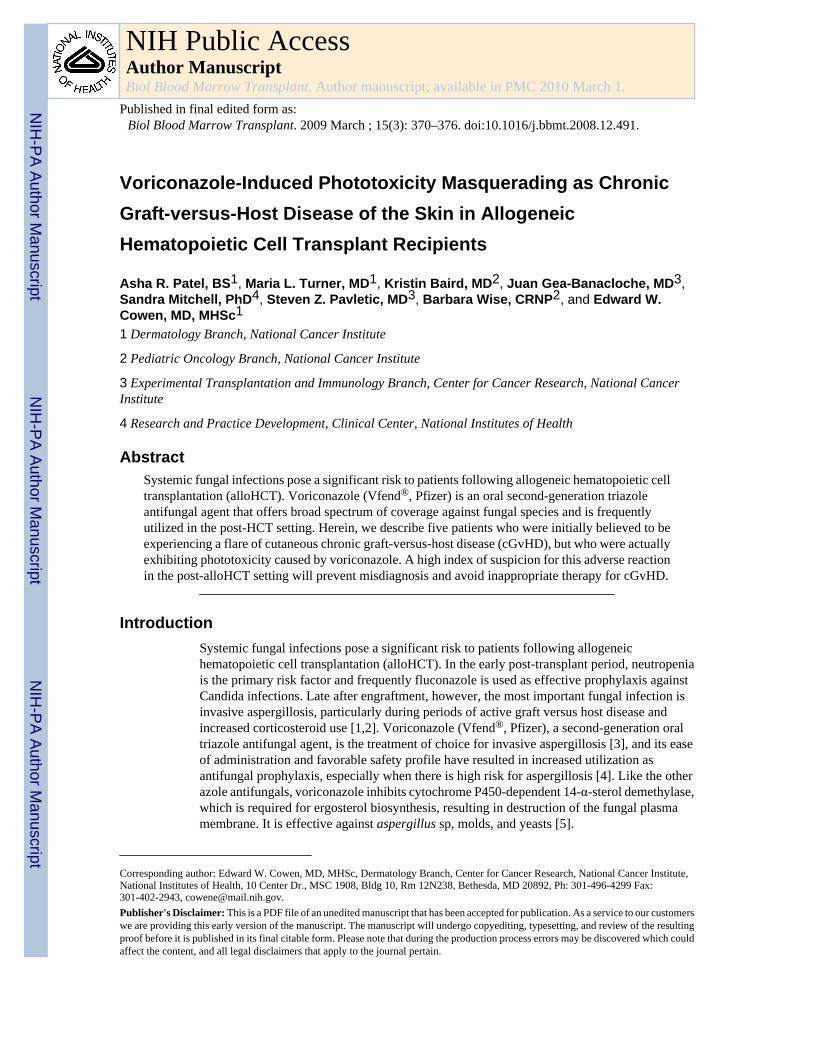
Voriconazole-Induced Phototoxicity Masquerading as ChronicGraft-versus-Host Disease of the Skin in AllogeneicHematopoietic Cell Transplant Recipients
Asha R. Patel, BS1, Maria L. Turner, MD1, Kristin Baird, MD2, Juan Gea-Banacloche, MD3,Sandra Mitchell, PhD4, Steven Z. Pavletic, MD3, Barbara Wise, CRNP2, and Edward W.Cowen, MD, MHSc1
1 Dermatology Branch, National Cancer Institute
2 Pediatric Oncology Branch, National Cancer Institute
3 Experimental Transplantation and Immunology Branch, Center for Cancer Research, National CancerInstitute
4 Research and Practice Development, Clinical Center, National Institutes of Health
AbstractSystemic fungal infections pose a significant risk to patients following allogeneic hematopoietic celltransplantation (alloHCT). Voriconazole (Vfend®, Pfizer) is an oral second-generation triazoleantifungal agent that offers broad spectrum of coverage against fungal species and is frequentlyutilized in the post-HCT setting. Herein, we describe five patients who were initially believed to beexperiencing a flare of cutaneous chronic graft-versus-host disease (cGvHD), but who were actuallyexhibiting phototoxicity caused by voriconazole. A high index of suspicion for this adverse reactionin the post-alloHCT setting will prevent misdiagnosis and avoid inappropriate therapy for cGvHD.
IntroductionSystemic fungal infections pose a significant risk to patients following allogeneichematopoietic cell transplantation (alloHCT). In the early post-transplant period, neutropeniais the primary risk factor and frequently fluconazole is used as effective prophylaxis againstCandida infections. Late after engraftment, however, the most important fungal infection isinvasive aspergillosis, particularly during periods of active graft versus host disease andincreased corticosteroid use [1,2]. Voriconazole (Vfend®, Pfizer), a second-generation oraltriazole antifungal agent, is the treatment of choice for invasive aspergillosis [3], and its easeof administration and favorable safety profile have resulted in increased utilization asantifungal prophylaxis, especially when there is high risk for aspergillosis [4]. Like the otherazole antifungals, voriconazole inhibits cytochrome P450-dependent 14-α-sterol demethylase,which is required for ergosterol biosynthesis, resulting in destruction of the fungal plasmamembrane. It is effective against aspergillus sp, molds, and yeasts [5].
Corresponding author: Edward W. Cowen, MD, MHSc, Dermatology Branch, Center for Cancer Research, National Cancer Institute,National Institutes of Health, 10 Center Dr., MSC 1908, Bldg 10, Rm 12N238, Bethesda, MD 20892, Ph: 301-496-4299 Fax:301-402-2943, [email protected]'s Disclaimer: This is a PDF file of an unedited manuscript that has been accepted for publication. As a service to our customerswe are providing this early version of the manuscript. The manuscript will undergo copyediting, typesetting, and review of the resultingproof before it is published in its final citable form. Please note that during the production process errors may be discovered which couldaffect the content, and all legal disclaimers that apply to the journal pertain.
NIH Public AccessAuthor ManuscriptBiol Blood Marrow Transplant. Author manuscript; available in PMC 2010 March 1.
Published in final edited form as:Biol Blood Marrow Transplant. 2009 March ; 15(3): 370–376. doi:10.1016/j.bbmt.2008.12.491.
NIH
-PA Author Manuscript
NIH
-PA Author Manuscript
NIH
-PA Author Manuscript
Following FDA approval in 2001, expanded utilization of voriconazole has led to increasedrecognition of the drug’s potential side effects, which include vision changes (20%),hallucinations (15%) hepatic enzyme abnormalities (12–20%), and skin reactions (17%) [5].In preliminary clinical studies, a photosensitive rash was reported in only 2% of patients(41/2090) [5]. Herein, we describe five patients who were believed to be suffering fromrecalcitrant skin cGvHD, but who, were actually experiencing cutaneous phototoxicity causedby voriconazole. A high index of suspicion for this adverse reaction in post-alloHCT patientswill facilitate the proper diagnosis and avoid inappropriate therapy for cGvHD.
Materials and MethodsIn this retrospective analysis, five patients with cGvHD who developed voriconazole-associated phototoxicity were identified at the National Institutes of Health (NIH) dermatologyservice between May 2003 and August 2007. Medical records, clinical photography, andhistological specimens were reviewed. Patient characteristics are summarized in Table 1.
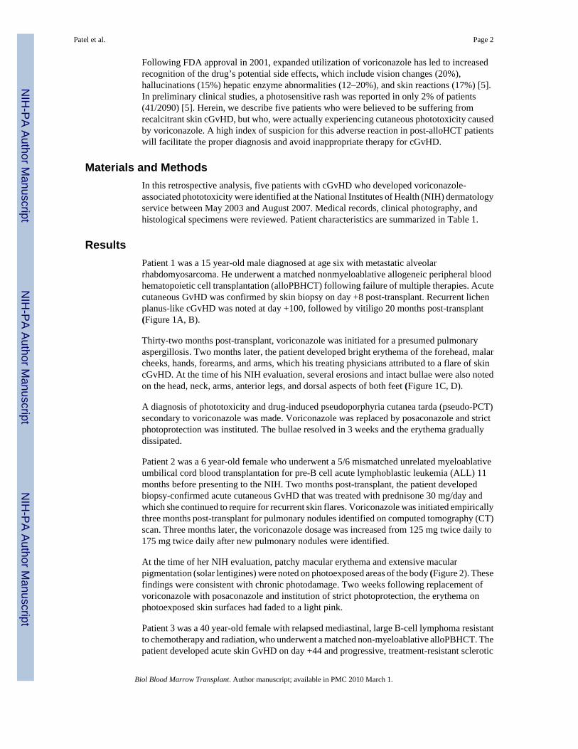
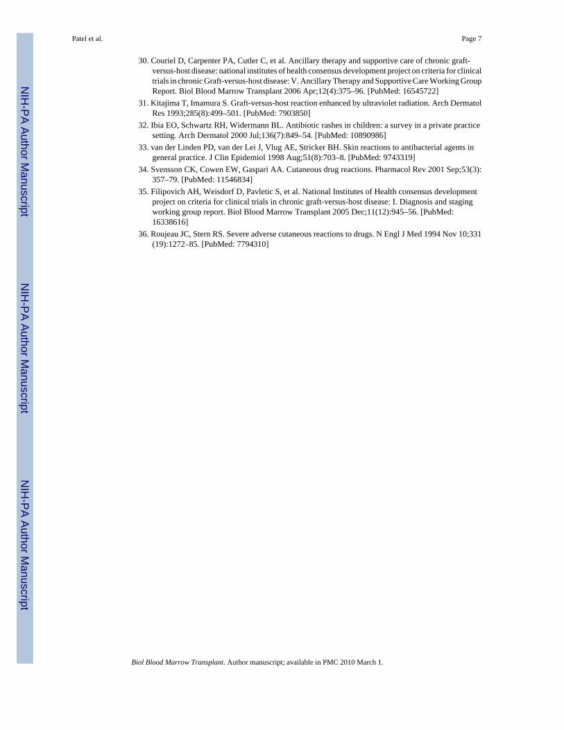
ResultsPatient 1 was a 15 year-old male diagnosed at age six with metastatic alveolarrhabdomyosarcoma. He underwent a matched nonmyeloablative allogeneic peripheral bloodhematopoietic cell transplantation (alloPBHCT) following failure of multiple therapies. Acutecutaneous GvHD was confirmed by skin biopsy on day +8 post-transplant. Recurrent lichenplanus-like cGvHD was noted at day +100, followed by vitiligo 20 months post-transplant(Figure 1A, B).
Thirty-two months post-transplant, voriconazole was initiated for a presumed pulmonaryaspergillosis. Two months later, the patient developed bright erythema of the forehead, malarcheeks, hands, forearms, and arms, which his treating physicians attributed to a flare of skincGvHD. At the time of his NIH evaluation, several erosions and intact bullae were also notedon the head, neck, arms, anterior legs, and dorsal aspects of both feet (Figure 1C, D).
A diagnosis of phototoxicity and drug-induced pseudoporphyria cutanea tarda (pseudo-PCT)secondary to voriconazole was made. Voriconazole was replaced by posaconazole and strictphotoprotection was instituted. The bullae resolved in 3 weeks and the erythema graduallydissipated.
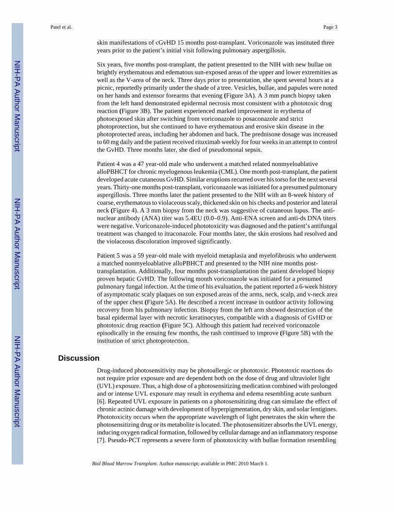
Patient 2 was a 6 year-old female who underwent a 5/6 mismatched unrelated myeloablativeumbilical cord blood transplantation for pre-B cell acute lymphoblastic leukemia (ALL) 11months before presenting to the NIH. Two months post-transplant, the patient developedbiopsy-confirmed acute cutaneous GvHD that was treated with prednisone 30 mg/day andwhich she continued to require for recurrent skin flares. Voriconazole was initiated empiricallythree months post-transplant for pulmonary nodules identified on computed tomography (CT)scan. Three months later, the voriconazole dosage was increased from 125 mg twice daily to175 mg twice daily after new pulmonary nodules were identified.
At the time of her NIH evaluation, patchy macular erythema and extensive macularpigmentation (solar lentigines) were noted on photoexposed areas of the body (Figure 2). Thesefindings were consistent with chronic photodamage. Two weeks following replacement ofvoriconazole with posaconazole and institution of strict photoprotection, the erythema onphotoexposed skin surfaces had faded to a light pink.
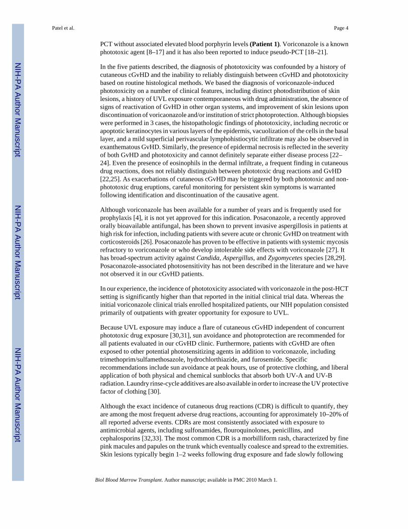
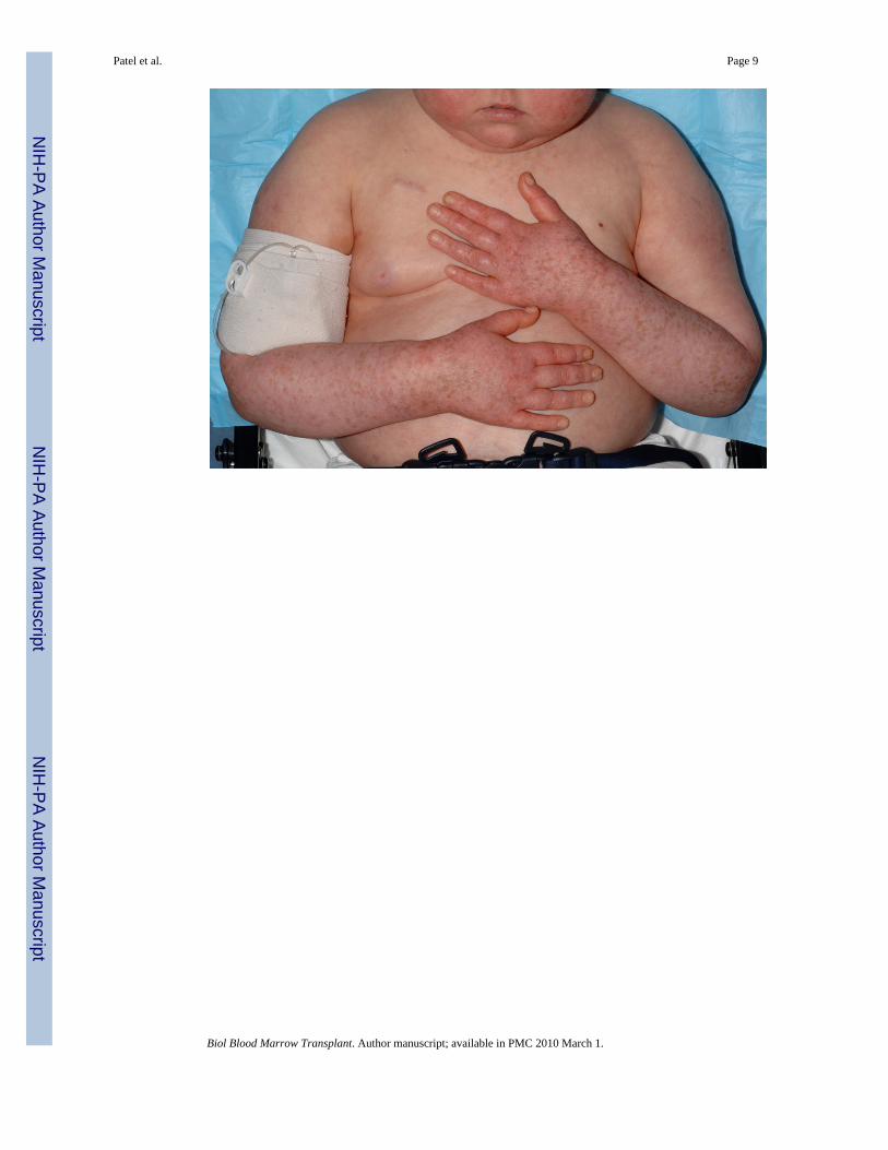
Patient 3 was a 40 year-old female with relapsed mediastinal, large B-cell lymphoma resistantto chemotherapy and radiation, who underwent a matched non-myeloablative alloPBHCT. Thepatient developed acute skin GvHD on day +44 and progressive, treatment-resistant sclerotic
Patel et al. Page 2
Biol Blood Marrow Transplant. Author manuscript; available in PMC 2010 March 1.
NIH
-PA Author Manuscript
NIH
-PA Author Manuscript
NIH
-PA Author Manuscript
skin manifestations of cGvHD 15 months post-transplant. Voriconazole was instituted threeyears prior to the patient’s initial visit following pulmonary aspergillosis.
Six years, five months post-transplant, the patient presented to the NIH with new bullae onbrightly erythematous and edematous sun-exposed areas of the upper and lower extremities aswell as the V-area of the neck. Three days prior to presentation, she spent several hours at apicnic, reportedly primarily under the shade of a tree. Vesicles, bullae, and papules were notedon her hands and extensor forearms that evening (Figure 3A). A 3 mm punch biopsy takenfrom the left hand demonstrated epidermal necrosis most consistent with a phototoxic drugreaction (Figure 3B). The patient experienced marked improvement in erythema ofphotoexposed skin after switching from voriconazole to posaconazole and strictphotoprotection, but she continued to have erythematous and erosive skin disease in thephotoprotected areas, including her abdomen and back. The prednisone dosage was increasedto 60 mg daily and the patient received rituximab weekly for four weeks in an attempt to controlthe GvHD. Three months later, she died of pseudomonal sepsis.
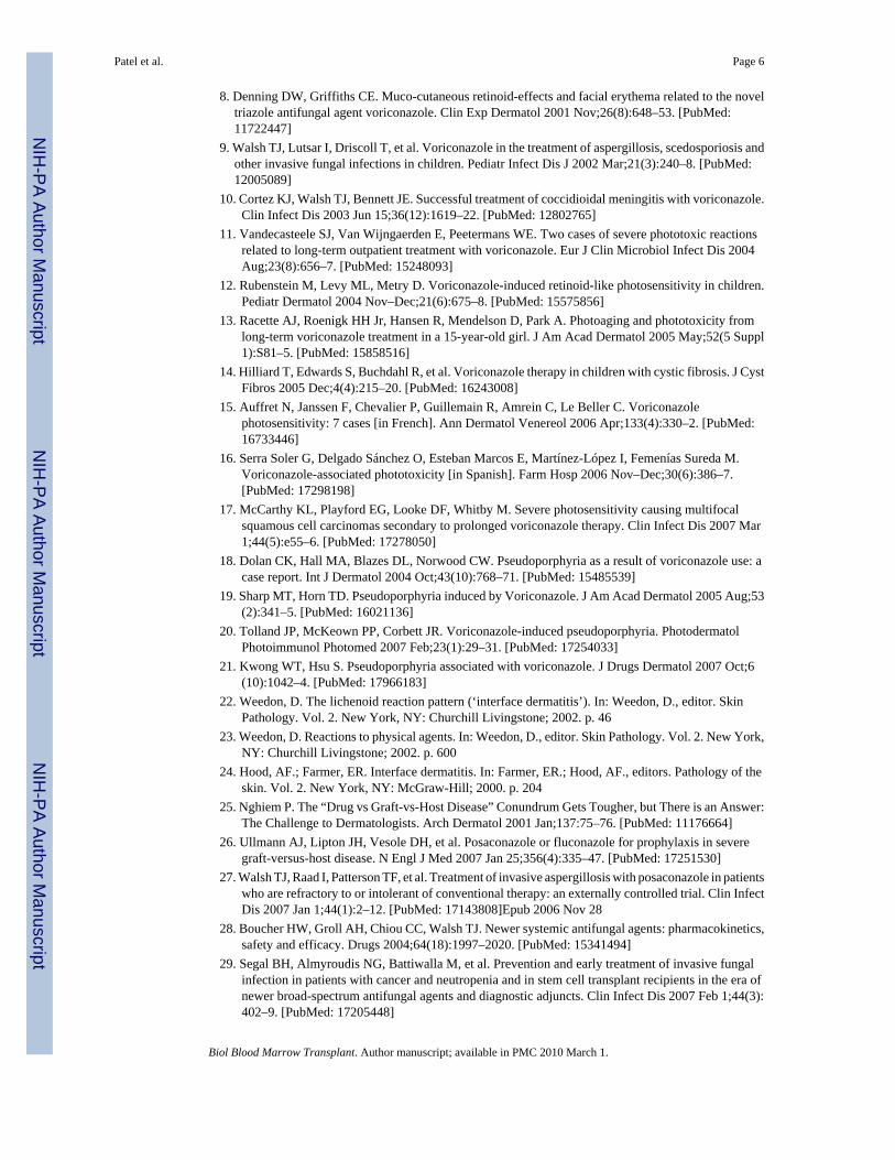
Patient 4 was a 47 year-old male who underwent a matched related nonmyeloablativealloPBHCT for chronic myelogenous leukemia (CML). One month post-transplant, the patientdeveloped acute cutaneous GvHD. Similar eruptions recurred over his torso for the next severalyears. Thirty-one months post-transplant, voriconazole was initiated for a presumed pulmonaryaspergillosis. Three months later the patient presented to the NIH with an 8-week history ofcoarse, erythematous to violaceous scaly, thickened skin on his cheeks and posterior and lateralneck (Figure 4). A 3 mm biopsy from the neck was suggestive of cutaneous lupus. The anti-nuclear antibody (ANA) titer was 5.4EU (0.0–0.9). Anti-ENA screen and anti-ds DNA titerswere negative. Voriconazole-induced phototoxicity was diagnosed and the patient’s antifungaltreatment was changed to itraconazole. Four months later, the skin erosions had resolved andthe violaceous discoloration improved significantly.
Patient 5 was a 59 year-old male with myeloid metaplasia and myelofibrosis who underwenta matched nonmyeloablative alloPBHCT and presented to the NIH nine months post-transplantation. Additionally, four months post-transplantation the patient developed biopsyproven hepatic GvHD. The following month voriconazole was initiated for a presumedpulmonary fungal infection. At the time of his evaluation, the patient reported a 6-week historyof asymptomatic scaly plaques on sun exposed areas of the arms, neck, scalp, and v-neck areaof the upper chest (Figure 5A). He described a recent increase in outdoor activity followingrecovery from his pulmonary infection. Biopsy from the left arm showed destruction of thebasal epidermal layer with necrotic keratinocytes, compatible with a diagnosis of GvHD orphototoxic drug reaction (Figure 5C). Although this patient had received voriconazoleepisodically in the ensuing few months, the rash continued to improve (Figure 5B) with theinstitution of strict photoprotection.
DiscussionDrug-induced photosensitivity may be photoallergic or phototoxic. Phototoxic reactions donot require prior exposure and are dependent both on the dose of drug and ultraviolet light(UVL) exposure. Thus, a high dose of a photosensitizing medication combined with prolongedand or intense UVL exposure may result in erythema and edema resembling acute sunburn[6]. Repeated UVL exposure in patients on a photosensitizing drug can simulate the effect ofchronic actinic damage with development of hyperpigmentation, dry skin, and solar lentigines.Phototoxicity occurs when the appropriate wavelength of light penetrates the skin where thephotosensitizing drug or its metabolite is located. The photosensitizer absorbs the UVL energy,inducing oxygen radical formation, followed by cellular damage and an inflammatory response[7]. Pseudo-PCT represents a severe form of phototoxicity with bullae formation resembling
Patel et al. Page 3
Biol Blood Marrow Transplant. Author manuscript; available in PMC 2010 March 1.
NIH
-PA Author Manuscript
NIH
-PA Author Manuscript
NIH
-PA Author Manuscript
PCT without associated elevated blood porphyrin levels (Patient 1). Voriconazole is a knownphototoxic agent [8–17] and it has also been reported to induce pseudo-PCT [18–21].
In the five patients described, the diagnosis of phototoxicity was confounded by a history ofcutaneous cGvHD and the inability to reliably distinguish between cGvHD and phototoxicitybased on routine histological methods. We based the diagnosis of voriconazole-inducedphototoxicity on a number of clinical features, including distinct photodistribution of skinlesions, a history of UVL exposure contemporaneous with drug administration, the absence ofsigns of reactivation of GvHD in other organ systems, and improvement of skin lesions upondiscontinuation of voricaonazole and/or institution of strict photoprotection. Although biopsieswere performed in 3 cases, the histopathologic findings of phototoxicity, including necrotic orapoptotic keratinocytes in various layers of the epidermis, vacuolization of the cells in the basallayer, and a mild superficial perivascular lymphohistiocytic infiltrate may also be observed inexanthematous GvHD. Similarly, the presence of epidermal necrosis is reflected in the severityof both GvHD and phototoxicity and cannot definitely separate either disease process [22–24]. Even the presence of eosinophils in the dermal infiltrate, a frequent finding in cutaneousdrug reactions, does not reliably distinguish between phototoxic drug reactions and GvHD[22,25]. As exacerbations of cutaneous cGvHD may be triggered by both phototoxic and non-phototoxic drug eruptions, careful monitoring for persistent skin symptoms is warrantedfollowing identification and discontinuation of the causative agent.
Although voriconazole has been available for a number of years and is frequently used forprophylaxis [4], it is not yet approved for this indication. Posaconazole, a recently approvedorally bioavailable antifungal, has been shown to prevent invasive aspergillosis in patients athigh risk for infection, including patients with severe acute or chronic GvHD on treatment withcorticosteroids [26]. Posaconazole has proven to be effective in patients with systemic mycosisrefractory to voriconazole or who develop intolerable side effects with voriconazole [27]. Ithas broad-spectrum activity against Candida, Aspergillus, and Zygomycetes species [28,29].Posaconazole-associated photosensitivity has not been described in the literature and we havenot observed it in our cGvHD patients.
In our experience, the incidence of phototoxicity associated with voriconazole in the post-HCTsetting is significantly higher than that reported in the initial clinical trial data. Whereas theinitial voriconazole clinical trials enrolled hospitalized patients, our NIH population consistedprimarily of outpatients with greater opportunity for exposure to UVL.
Because UVL exposure may induce a flare of cutaneous cGvHD independent of concurrentphototoxic drug exposure [30,31], sun avoidance and photoprotection are recommended forall patients evaluated in our cGvHD clinic. Furthermore, patients with cGvHD are oftenexposed to other potential photosensitizing agents in addition to voriconazole, includingtrimethoprim/sulfamethosazole, hydrochlorthiazide, and furosemide. Specificrecommendations include sun avoidance at peak hours, use of protective clothing, and liberalapplication of both physical and chemical sunblocks that absorb both UV-A and UV-Bradiation. Laundry rinse-cycle additives are also available in order to increase the UV protectivefactor of clothing [30].
Although the exact incidence of cutaneous drug reactions (CDR) is difficult to quantify, theyare among the most frequent adverse drug reactions, accounting for approximately 10–20% ofall reported adverse events. CDRs are most consistently associated with exposure toantimicrobial agents, including sulfonamides, flouroquinolones, penicillins, andcephalosporins [32,33]. The most common CDR is a morbilliform rash, characterized by finepink macules and papules on the trunk which eventually coalesce and spread to the extremities.Skin lesions typically begin 1–2 weeks following drug exposure and fade slowly following
Patel et al. Page 4
Biol Blood Marrow Transplant. Author manuscript; available in PMC 2010 March 1.
NIH
-PA Author Manuscript
NIH
-PA Author Manuscript
NIH
-PA Author Manuscript

discontinuation. By contrast, urticarial hypersensitivity reactions usually develop within 1–2days of drug initiation, and individual lesions resolve within 24 hours. The most worrisomeCDRs are erythema multiforme/Stevens-Johnson syndrome/toxic epidermal necrolysis, aspectrum of severe drug hypersensitivity with variable skin involvement ranging from localizeddusky targetoid plaques to widespread skin sloughing with mucosal membrane involvementand significant associated mortality. Often symptoms of a severe CDR do not present untilseveral weeks after initial drug exposure [34].
Attention to the details of the clinical presentation and the drug administration history are ofutmost importance in establishing the correct diagnosis in patients with cGvHD, given theircomplex medical history and the high likelihood of polypharmacy. In this setting, whenconfronted with a new onset eruption that is not considered diagnostic of cGvHD based onNIH consensus criteria [35], helpful clinical criteria in defining a CDR include: 1) exclusionof other causes for the eruption, such as viral exanthem; 2) identification of a temporalrelationship between drug use and onset of the rash 3) improvement following drug cessation;and reactivation of the rash should upon rechallenge (if performed) [36].
As voriconazole has become a frequent choice for antifungal prophylaxis and treatment in thesetting of alloHCT, the possibility of voriconazole-induced phototoxicity should be consideredin all new skin eruptions, even those that simulate a flare of cutaneous cGvHD in patients withknown cutaneous cGvHD. The recognition of voriconazole phototoxicity is especiallyimportant as efficacious treatment alternatives are readily available, and accurate diagnosis ofphototoxic drug reaction will prevent misdiagnosis of cGvHD and unnecessaryimmunosuppression.
AcknowledgementsFunding/Support: This research was supported in part by the Intramural Research Program of the NIH, Center forCancer Research, National Cancer Institute. Ms. Patel was supported by the Clinical Research Training Program, apublic-private partnership supported jointly by the NIH and Pfizer Inc (via a grant to the Foundation for NIH fromPfizer Inc).
References1. Marr KA, Carter RA, Boeckh M, Martin P, Corey L. Invasive aspergillosis in allogeneic stem cell
transplant recipients: changes in epidemiology and risk factors. Blood 2002;100:4358–4366.[PubMed: 12393425]
2. Upton A, Kirby KA, Carpenter P, Boeckh M, Marr KA. Invasive aspergillosis following hematopoieticcell transplantation: outcomes and prognostic factors associated with mortality. Clin Infect Dis2007;44:531–540. [PubMed: 17243056]
3. Herbrecht R, Denning DW, Patterson TF, et al. Voriconazole versus amphotericin B for primary therapyof invasive aspergillosis. N Engl J Med 2002;347:408–415. [PubMed: 12167683]
4. Wingard JR, Carter SL, Walsh TJ, et al. Results of a Randomized, Double-Blind Trial of Fluconazole(FLU) vs. Voriconazole (VORI) for the Prevention of Invasive Fungal Infections (IFI) in 600Allogeneic Blood and Marrow Transplant (BMT) Patients. ASH Annual Meeting Abstracts2007;110:163.
5. Vfend (Voriconazole, Oral and Intravenous Formulations), NDA 21–267, Briefing Document for FDAAntiviral Drugs Advisory Committee Meeting, 4 October 2001. Pfizer Inc. [online]. [Accessed 2007October 18]. Available from URL:http://www.fda.gov/ohrms/dockets/ac/01/briefing/3792b2_01_Pfizer.pdf
6. Stein KR, Scheinfeld NS. Drug-induced photoallergic and phototoxic reactions. Expert Opin Drug Saf2007 Jul;6(4):431–43. [PubMed: 17688387]
7. Toback AC, Anders JE. Phototoxicity from systemic agents. Dermatol Clin 1986 Apr;4(2):223–30.[PubMed: 3456856]
Patel et al. Page 5
Biol Blood Marrow Transplant. Author manuscript; available in PMC 2010 March 1.
NIH
-PA Author Manuscript
NIH
-PA Author Manuscript
NIH
-PA Author Manuscript
8. Denning DW, Griffiths CE. Muco-cutaneous retinoid-effects and facial erythema related to the noveltriazole antifungal agent voriconazole. Clin Exp Dermatol 2001 Nov;26(8):648–53. [PubMed:11722447]
9. Walsh TJ, Lutsar I, Driscoll T, et al. Voriconazole in the treatment of aspergillosis, scedosporiosis andother invasive fungal infections in children. Pediatr Infect Dis J 2002 Mar;21(3):240–8. [PubMed:12005089]
10. Cortez KJ, Walsh TJ, Bennett JE. Successful treatment of coccidioidal meningitis with voriconazole.Clin Infect Dis 2003 Jun 15;36(12):1619–22. [PubMed: 12802765]
11. Vandecasteele SJ, Van Wijngaerden E, Peetermans WE. Two cases of severe phototoxic reactionsrelated to long-term outpatient treatment with voriconazole. Eur J Clin Microbiol Infect Dis 2004Aug;23(8):656–7. [PubMed: 15248093]
12. Rubenstein M, Levy ML, Metry D. Voriconazole-induced retinoid-like photosensitivity in children.Pediatr Dermatol 2004 Nov–Dec;21(6):675–8. [PubMed: 15575856]
13. Racette AJ, Roenigk HH Jr, Hansen R, Mendelson D, Park A. Photoaging and phototoxicity fromlong-term voriconazole treatment in a 15-year-old girl. J Am Acad Dermatol 2005 May;52(5 Suppl1):S81–5. [PubMed: 15858516]
14. Hilliard T, Edwards S, Buchdahl R, et al. Voriconazole therapy in children with cystic fibrosis. J CystFibros 2005 Dec;4(4):215–20. [PubMed: 16243008]
15. Auffret N, Janssen F, Chevalier P, Guillemain R, Amrein C, Le Beller C. Voriconazolephotosensitivity: 7 cases [in French]. Ann Dermatol Venereol 2006 Apr;133(4):330–2. [PubMed:16733446]
16. Serra Soler G, Delgado Sánchez O, Esteban Marcos E, Martínez-López I, Femenías Sureda M.Voriconazole-associated phototoxicity [in Spanish]. Farm Hosp 2006 Nov–Dec;30(6):386–7.[PubMed: 17298198]
17. McCarthy KL, Playford EG, Looke DF, Whitby M. Severe photosensitivity causing multifocalsquamous cell carcinomas secondary to prolonged voriconazole therapy. Clin Infect Dis 2007 Mar1;44(5):e55–6. [PubMed: 17278050]
18. Dolan CK, Hall MA, Blazes DL, Norwood CW. Pseudoporphyria as a result of voriconazole use: acase report. Int J Dermatol 2004 Oct;43(10):768–71. [PubMed: 15485539]
19. Sharp MT, Horn TD. Pseudoporphyria induced by Voriconazole. J Am Acad Dermatol 2005 Aug;53(2):341–5. [PubMed: 16021136]
20. Tolland JP, McKeown PP, Corbett JR. Voriconazole-induced pseudoporphyria. PhotodermatolPhotoimmunol Photomed 2007 Feb;23(1):29–31. [PubMed: 17254033]
21. Kwong WT, Hsu S. Pseudoporphyria associated with voriconazole. J Drugs Dermatol 2007 Oct;6(10):1042–4. [PubMed: 17966183]
22. Weedon, D. The lichenoid reaction pattern (‘interface dermatitis’). In: Weedon, D., editor. SkinPathology. Vol. 2. New York, NY: Churchill Livingstone; 2002. p. 46
23. Weedon, D. Reactions to physical agents. In: Weedon, D., editor. Skin Pathology. Vol. 2. New York,NY: Churchill Livingstone; 2002. p. 600
24. Hood, AF.; Farmer, ER. Interface dermatitis. In: Farmer, ER.; Hood, AF., editors. Pathology of theskin. Vol. 2. New York, NY: McGraw-Hill; 2000. p. 204
25. Nghiem P. The “Drug vs Graft-vs-Host Disease” Conundrum Gets Tougher, but There is an Answer:The Challenge to Dermatologists. Arch Dermatol 2001 Jan;137:75–76. [PubMed: 11176664]
26. Ullmann AJ, Lipton JH, Vesole DH, et al. Posaconazole or fluconazole for prophylaxis in severegraft-versus-host disease. N Engl J Med 2007 Jan 25;356(4):335–47. [PubMed: 17251530]
27. Walsh TJ, Raad I, Patterson TF, et al. Treatment of invasive aspergillosis with posaconazole in patientswho are refractory to or intolerant of conventional therapy: an externally controlled trial. Clin InfectDis 2007 Jan 1;44(1):2–12. [PubMed: 17143808]Epub 2006 Nov 28
28. Boucher HW, Groll AH, Chiou CC, Walsh TJ. Newer systemic antifungal agents: pharmacokinetics,safety and efficacy. Drugs 2004;64(18):1997–2020. [PubMed: 15341494]
29. Segal BH, Almyroudis NG, Battiwalla M, et al. Prevention and early treatment of invasive fungalinfection in patients with cancer and neutropenia and in stem cell transplant recipients in the era ofnewer broad-spectrum antifungal agents and diagnostic adjuncts. Clin Infect Dis 2007 Feb 1;44(3):402–9. [PubMed: 17205448]
Patel et al. Page 6
Biol Blood Marrow Transplant. Author manuscript; available in PMC 2010 March 1.
NIH
-PA Author Manuscript
NIH
-PA Author Manuscript
NIH
-PA Author Manuscript
30. Couriel D, Carpenter PA, Cutler C, et al. Ancillary therapy and supportive care of chronic graft-versus-host disease: national institutes of health consensus development project on criteria for clinicaltrials in chronic Graft-versus-host disease: V. Ancillary Therapy and Supportive Care Working GroupReport. Biol Blood Marrow Transplant 2006 Apr;12(4):375–96. [PubMed: 16545722]
31. Kitajima T, Imamura S. Graft-versus-host reaction enhanced by ultraviolet radiation. Arch DermatolRes 1993;285(8):499–501. [PubMed: 7903850]
32. Ibia EO, Schwartz RH, Widermann BL. Antibiotic rashes in children: a survey in a private practicesetting. Arch Dermatol 2000 Jul;136(7):849–54. [PubMed: 10890986]
33. van der Linden PD, van der Lei J, Vlug AE, Stricker BH. Skin reactions to antibacterial agents ingeneral practice. J Clin Epidemiol 1998 Aug;51(8):703–8. [PubMed: 9743319]
34. Svensson CK, Cowen EW, Gaspari AA. Cutaneous drug reactions. Pharmacol Rev 2001 Sep;53(3):357–79. [PubMed: 11546834]
35. Filipovich AH, Weisdorf D, Pavletic S, et al. National Institutes of Health consensus developmentproject on criteria for clinical trials in chronic graft-versus-host disease: I. Diagnosis and stagingworking group report. Biol Blood Marrow Transplant 2005 Dec;11(12):945–56. [PubMed:16338616]
36. Roujeau JC, Stern RS. Severe adverse cutaneous reactions to drugs. N Engl J Med 1994 Nov 10;331(19):1272–85. [PubMed: 7794310]
Patel et al. Page 7
Biol Blood Marrow Transplant. Author manuscript; available in PMC 2010 March 1.
NIH
-PA Author Manuscript
NIH
-PA Author Manuscript
NIH
-PA Author Manuscript
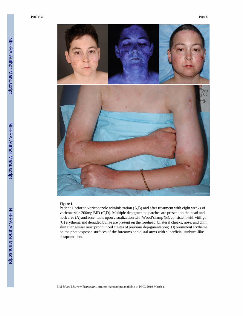
Figure 1.Patient 1 prior to voriconazole administration (A,B) and after treatment with eight weeks ofvoriconazole 200mg BID (C,D). Multiple depigmented patches are present on the head andneck area (A) and accentuate upon visualization with Wood’s lamp (B), consistent with vitiligo;(C) erythema and denuded bullae are present on the forehead, bilateral cheeks, nose, and chin;skin changes are most pronounced at sites of previous depigmentation; (D) prominent erythemaon the photoexposed surfaces of the forearms and distal arms with superficial sunburn-likedesquamation.
Patel et al. Page 8
Biol Blood Marrow Transplant. Author manuscript; available in PMC 2010 March 1.
NIH
-PA Author Manuscript
NIH
-PA Author Manuscript
NIH
-PA Author Manuscript
Patel et al. Page 9
Biol Blood Marrow Transplant. Author manuscript; available in PMC 2010 March 1.
NIH
-PA Author Manuscript
NIH
-PA Author Manuscript
NIH
-PA Author Manuscript

Figure 2.Patient 2 following eleven months treatment with voriconazole 175mg BID. Erythema andmultiple round tan/brown macules on the dorsal surfaces of the hands and forearms suggestiveof ongoing phototoxicity and chronic photodamage, respectively. Similar erythema andmacular pigmentation is present on the upper chest.
Patel et al. Page 10
Biol Blood Marrow Transplant. Author manuscript; available in PMC 2010 March 1.
NIH
-PA Author Manuscript
NIH
-PA Author Manuscript
NIH
-PA Author Manuscript
Patel et al. Page 11
Biol Blood Marrow Transplant. Author manuscript; available in PMC 2010 March 1.
NIH
-PA Author Manuscript
NIH
-PA Author Manuscript
NIH
-PA Author Manuscript
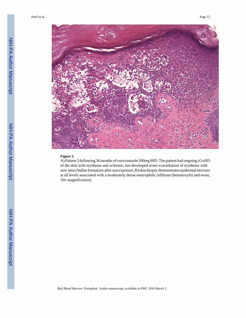
Figure 3.A) Patient 3 following 36 months of voriconazole 200mg BID. The patient had ongoing cGvHDof the skin with erythema and sclerosis, but developed acute exacerbation of erythema withnew intact bullae formation after sun exposure; B) skin biopsy demonstrates epidermal necrosisat all levels associated with a moderately dense neutrophilic infiltrate (hematoxylin and eosin,10x magnification).
Patel et al. Page 12
Biol Blood Marrow Transplant. Author manuscript; available in PMC 2010 March 1.
NIH
-PA Author Manuscript
NIH
-PA Author Manuscript
NIH
-PA Author Manuscript
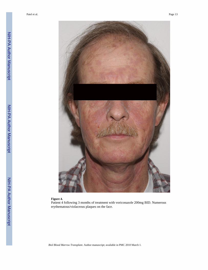
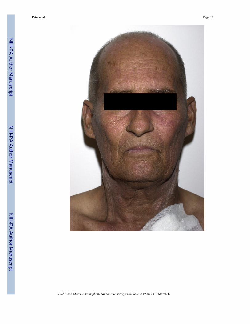
Figure 4.Patient 4 following 3 months of treatment with voriconazole 200mg BID. Numerouserythematous/violaceous plaques on the face.
Patel et al. Page 13
Biol Blood Marrow Transplant. Author manuscript; available in PMC 2010 March 1.
NIH
-PA Author Manuscript
NIH
-PA Author Manuscript
NIH
-PA Author Manuscript
Patel et al. Page 14
Biol Blood Marrow Transplant. Author manuscript; available in PMC 2010 March 1.
NIH
-PA Author Manuscript
NIH
-PA Author Manuscript
NIH
-PA Author Manuscript

Patel et al. Page 15
Biol Blood Marrow Transplant. Author manuscript; available in PMC 2010 March 1.
NIH
-PA Author Manuscript
NIH
-PA Author Manuscript
NIH
-PA Author Manuscript
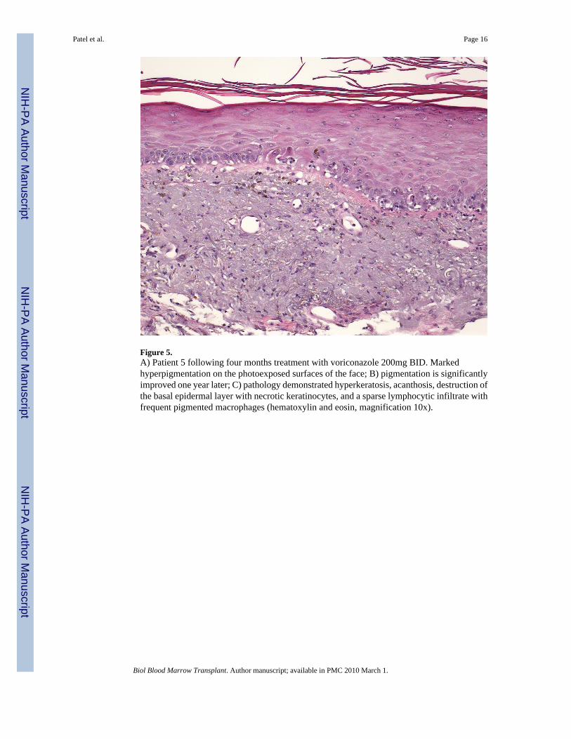
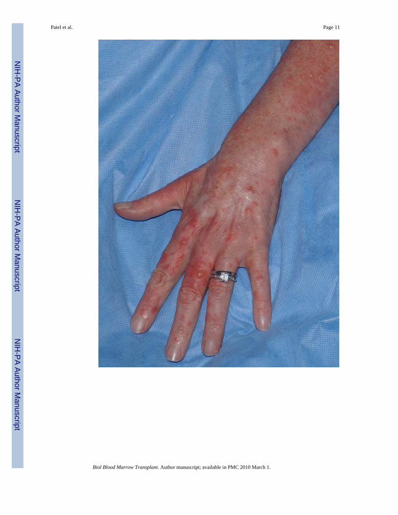
Figure 5.A) Patient 5 following four months treatment with voriconazole 200mg BID. Markedhyperpigmentation on the photoexposed surfaces of the face; B) pigmentation is significantlyimproved one year later; C) pathology demonstrated hyperkeratosis, acanthosis, destruction ofthe basal epidermal layer with necrotic keratinocytes, and a sparse lymphocytic infiltrate withfrequent pigmented macrophages (hematoxylin and eosin, magnification 10x).
Patel et al. Page 16
Biol Blood Marrow Transplant. Author manuscript; available in PMC 2010 March 1.
NIH
-PA Author Manuscript
NIH
-PA Author Manuscript
NIH
-PA Author Manuscript

NIH
-PA Author Manuscript
NIH
-PA Author Manuscript
NIH
-PA Author Manuscript
Patel et al. Page 17Ta
ble
1Pa
tient
cha
ract
eris
tics
Patie
ntA
ge/S
exPr
imar
y di
seas
e
Hx
acut
esk
inG
vHD
Hx
chro
nic
skin
GvH
D
cGvH
D o
fot
her
orga
nsy
stem
sV
oric
onaz
ole
dose
Lat
ency
betw
een
vori
cona
zole
initi
atio
nan
dph
otot
oxic
ity
Con
curr
ent
pote
ntia
llyph
otos
ensi
tizin
gdr
ugs
cGV
HD
ther
apy
attim
e of
pho
toto
xici
tydi
agno
sis
Res
olut
ion
of p
hoto
toxi
city
115
/MA
lveo
lar r
habd
omyo
sarc
oma
Yes
Yes
Ora
l20
0mg
BID
2 w
eeks
Non
ePr
edni
sone
8m
g Q
D3
wee
ks
26/
FPr
e-B
cel
l ALL
Yes
Yes
Non
e17
5mg
BID
6 m
onth
sH
CTZ
Pred
niso
ne 2
5mg
BID
Tacr
olim
us 0
.5m
gQ
AM
, 0.2
5 Q
PMM
MF
750m
g q1
2hr
Hyd
roco
rtiso
ne 2
.5%
BID
(fac
e)Fl
uoci
nolo
ne 0.
1% B
ID(b
ody)
2 w
eeks
340
/FLa
rge
B-c
ell l
ymph
oma
Yes
Yes
Ocu
lar
Ora
lH
epat
ic
200m
g B
ID42
mon
ths
Am
itrip
tylin
eEx
traco
rpor
eal
phot
ophe
resi
sPr
edni
sone
40m
g Q
DFl
uoci
nolo
ne so
lutio
n(s
calp
)C
lobe
taso
l oin
tmen
t0.
05%
(bod
y)Ta
crol
imus
oin
tmen
t(f
ace)
Impr
oved
in3
days
,co
ntin
ued
cGvH
D fl
are
447
/MC
ML
Yes
Yes
Ocu
lar
Hep
atic
Pulm
onar
y
200m
g B
ID1
mon
thN
one
Pred
niso
ne 5
0mg
QO
DM
MF
1gm
q12
hr4
mon
ths
559
/MM
yelo
id m
etap
lasi
a/m
yelo
fibro
sis
No
No
Ocu
lar
Hep
atic
Pulm
onar
y
200m
g B
ID3
mon
ths
Lisi
nopr
ilFu
rose
mid
ePr
edni
sone
20m
g Q
OD
3 m
onth
s
Abb
revi
atio
ns: A
LL, a
cute
lym
phob
last
ic le
ukem
ia; C
ML,
chr
onic
mye
loge
nous
leuk
emia
; HC
TZ, h
ydro
chlo
roth
iazi
de; M
MF,
myc
ophe
nola
te m
ofet
il
Biol Blood Marrow Transplant. Author manuscript; available in PMC 2010 March 1.
Copyright © 2022 FDOKUMEN