Volume 4, Chapter 1-5: Aquatic and Wet Marchantiophyta ...
62
Glime, J. M. 2021. Aquatic and Wet Marchantiophyta, Order Jungermanniales: Lophocoleineae, Part 1. Chapt. 1-5. In: Glime, 1-5-1 J. M. Bryophyte Ecology. Volume 4. Habitat and Role. Ebook sponsored by Michigan Technological University and the International Association of Bryologists. Last updated 11 April 2021 and available at <http://digitalcommons.mtu.edu/bryophyte-ecology/>. CHAPTER 1-5 AQUATIC AND WET MARCHANTIOPHYTA ORDER JUNGERMANNIALES: LOPHOCOLEINEAE, PART 1 TABLE OF CONTENTS Suborder Lophocoleineae ...................................................................................................................................................... 1-5-2 Blepharostomaceae ........................................................................................................................................................ 1-5-2 Blepharostoma trichophyllum ................................................................................................................................ 1-5-2 Herbertaceae .................................................................................................................................................................. 1-5-8 Herbertus sendtneri................................................................................................................................................ 1-5-8 Lepidoziaceae ................................................................................................................................................................ 1-5-9 Bazzainia denudata ................................................................................................................................................ 1-5-9 Bazzania praerupta .............................................................................................................................................. 1-5-10 Bazzania tricrenata .............................................................................................................................................. 1-5-10 Bazzania trilobata ................................................................................................................................................ 1-5-12 Hygrolembidium boschianum............................................................................................................................... 1-5-16 Kurzia makinoana ................................................................................................................................................ 1-5-16 Kurzia pauciflora ................................................................................................................................................. 1-5-19 Kurzia trichoclados .............................................................................................................................................. 1-5-21 Lepidozia reptans ................................................................................................................................................. 1-5-22 Lepidozia trichodes .............................................................................................................................................. 1-5-24 Zoopsis argentea .................................................................................................................................................. 1-5-24 Lophocoleaceae............................................................................................................................................................ 1-5-25 Chiloscyphus ........................................................................................................................................................ 1-5-25 Chiloscyphus pallescens....................................................................................................................................... 1-5-25 Chiloscyphus pallescens var. fragilis ................................................................................................................... 1-5-28 Chiloscyphus polyanthos ...................................................................................................................................... 1-5-28 Chiloscyphus polyathos var. rivularis .................................................................................................................. 1-5-30 Hepatostolonophora paucistipula ........................................................................................................................ 1-5-31 Heteroscyphus argutus ......................................................................................................................................... 1-5-32 Heteroscyphus coalitus ........................................................................................................................................ 1-5-33 Heteroscyphus denticulatus.................................................................................................................................. 1-5-36 Heteroscyphys planiusculus ................................................................................................................................. 1-5-36 Heteroscyphus zollingri ....................................................................................................................................... 1-5-37 Lophocolea........................................................................................................................................................... 1-5-37 Lophocolea bidentata ........................................................................................................................................... 1-5-37 Lophocolea heterophylla...................................................................................................................................... 1-5-39 Lophocolea minor ................................................................................................................................................ 1-5-42 Lophocolea mollis ................................................................................................................................................ 1-5-45 Lophocolea semiteres ........................................................................................................................................... 1-5-45 Pachyglossa ......................................................................................................................................................... 1-5-48 Pachyglossa austrigena subsp. okaritana ............................................................................................................ 1-5-48 Pachyglossa dissitifolia........................................................................................................................................ 1-5-48 Pachyglossa tenacifolia ....................................................................................................................................... 1-5-49 Mastigophoraceae ........................................................................................................................................................ 1-5-50 Mastigophora diclados......................................................................................................................................... 1-5-50 Summary .............................................................................................................................................................................. 1-5-51 Acknowledgments ............................................................................................................................................................... 1-5-51 Literature Cited .................................................................................................................................................................... 1-5-52
-
Upload
khangminh22 -
Category
Documents
-
view
3 -
download
0
Transcript of Volume 4, Chapter 1-5: Aquatic and Wet Marchantiophyta ...
Glime, J. M. 2021. Aquatic and Wet Marchantiophyta, Order Jungermanniales: Lophocoleineae, Part 1. Chapt. 1-5. In: Glime, 1-5-1 J. M. Bryophyte Ecology. Volume 4. Habitat and Role. Ebook sponsored by Michigan Technological University and the International Association of Bryologists. Last updated 11 April 2021 and available at <http://digitalcommons.mtu.edu/bryophyte-ecology/>.
CHAPTER 1-5 AQUATIC AND WET MARCHANTIOPHYTA
ORDER JUNGERMANNIALES: LOPHOCOLEINEAE, PART 1
TABLE OF CONTENTS
Suborder Lophocoleineae ...................................................................................................................................................... 1-5-2 Blepharostomaceae ........................................................................................................................................................ 1-5-2 Blepharostoma trichophyllum ................................................................................................................................ 1-5-2 Herbertaceae .................................................................................................................................................................. 1-5-8 Herbertus sendtneri ................................................................................................................................................ 1-5-8 Lepidoziaceae ................................................................................................................................................................ 1-5-9 Bazzainia denudata ................................................................................................................................................ 1-5-9 Bazzania praerupta .............................................................................................................................................. 1-5-10 Bazzania tricrenata .............................................................................................................................................. 1-5-10 Bazzania trilobata ................................................................................................................................................ 1-5-12 Hygrolembidium boschianum............................................................................................................................... 1-5-16 Kurzia makinoana ................................................................................................................................................ 1-5-16 Kurzia pauciflora ................................................................................................................................................. 1-5-19 Kurzia trichoclados .............................................................................................................................................. 1-5-21 Lepidozia reptans ................................................................................................................................................. 1-5-22 Lepidozia trichodes .............................................................................................................................................. 1-5-24 Zoopsis argentea .................................................................................................................................................. 1-5-24 Lophocoleaceae ............................................................................................................................................................ 1-5-25 Chiloscyphus ........................................................................................................................................................ 1-5-25 Chiloscyphus pallescens ....................................................................................................................................... 1-5-25 Chiloscyphus pallescens var. fragilis ................................................................................................................... 1-5-28 Chiloscyphus polyanthos ...................................................................................................................................... 1-5-28 Chiloscyphus polyathos var. rivularis .................................................................................................................. 1-5-30 Hepatostolonophora paucistipula ........................................................................................................................ 1-5-31 Heteroscyphus argutus ......................................................................................................................................... 1-5-32 Heteroscyphus coalitus ........................................................................................................................................ 1-5-33 Heteroscyphus denticulatus .................................................................................................................................. 1-5-36 Heteroscyphys planiusculus ................................................................................................................................. 1-5-36 Heteroscyphus zollingri ....................................................................................................................................... 1-5-37 Lophocolea ........................................................................................................................................................... 1-5-37 Lophocolea bidentata ........................................................................................................................................... 1-5-37 Lophocolea heterophylla ...................................................................................................................................... 1-5-39 Lophocolea minor ................................................................................................................................................ 1-5-42 Lophocolea mollis ................................................................................................................................................ 1-5-45 Lophocolea semiteres ........................................................................................................................................... 1-5-45 Pachyglossa ......................................................................................................................................................... 1-5-48 Pachyglossa austrigena subsp. okaritana ............................................................................................................ 1-5-48 Pachyglossa dissitifolia ........................................................................................................................................ 1-5-48 Pachyglossa tenacifolia ....................................................................................................................................... 1-5-49 Mastigophoraceae ........................................................................................................................................................ 1-5-50 Mastigophora diclados ......................................................................................................................................... 1-5-50 Summary .............................................................................................................................................................................. 1-5-51 Acknowledgments ............................................................................................................................................................... 1-5-51 Literature Cited .................................................................................................................................................................... 1-5-52
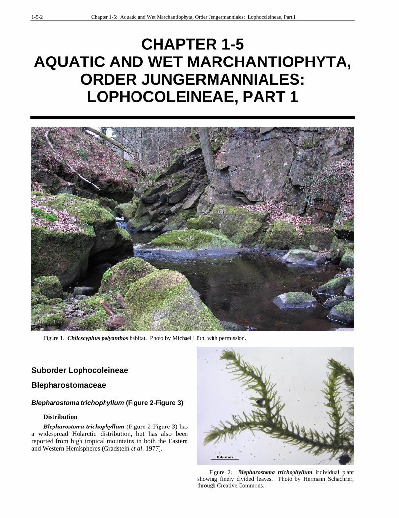
1-5-2 Chapter 1-5: Aquatic and Wet Marchantiophyta, Order Jungermanniales: Lophocoleineae, Part 1
CHAPTER 1-5 AQUATIC AND WET MARCHANTIOPHYTA,
ORDER JUNGERMANNIALES: LOPHOCOLEINEAE, PART 1
Figure 1. Chiloscyphus polyanthos habitat. Photo by Michael Lüth, with permission.
Suborder Lophocoleineae
Blepharostomaceae
Blepharostoma trichophyllum (Figure 2-Figure 3)
Distribution
Blepharostoma trichophyllum (Figure 2-Figure 3) has a widespread Holarctic distribution, but has also been reported from high tropical mountains in both the Eastern and Western Hemispheres (Gradstein et al. 1977).
Figure 2. Blepharostoma trichophyllum individual plant showing finely divided leaves. Photo by Hermann Schachner, through Creative Commons.
Chapter 1-5: Aquatic and Wet Marchantiophyta, Order Jungermanniales: Lophocoleineae, Part 1 1-5-3
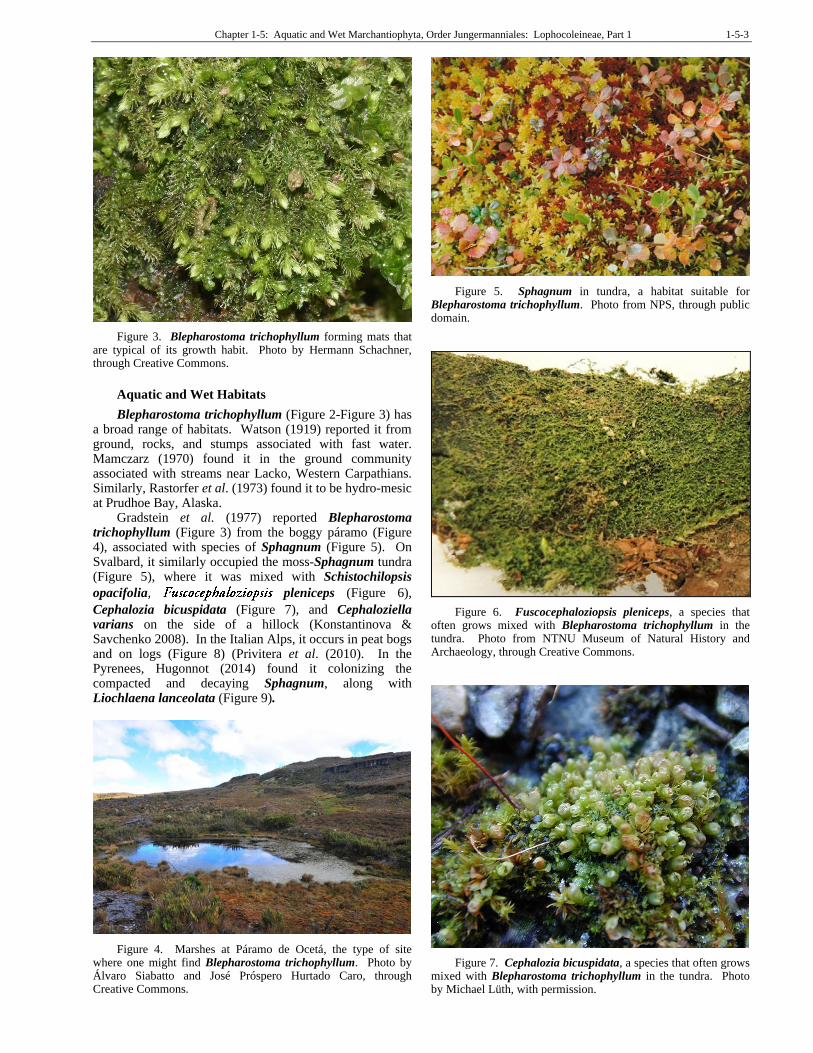
Figure 3. Blepharostoma trichophyllum forming mats that are typical of its growth habit. Photo by Hermann Schachner, through Creative Commons.
Aquatic and Wet Habitats
Blepharostoma trichophyllum (Figure 2-Figure 3) has a broad range of habitats. Watson (1919) reported it from ground, rocks, and stumps associated with fast water. Mamczarz (1970) found it in the ground community associated with streams near Lacko, Western Carpathians. Similarly, Rastorfer et al. (1973) found it to be hydro-mesic at Prudhoe Bay, Alaska.
Gradstein et al. (1977) reported Blepharostoma trichophyllum (Figure 3) from the boggy páramo (Figure 4), associated with species of Sphagnum (Figure 5). On Svalbard, it similarly occupied the moss-Sphagnum tundra (Figure 5), where it was mixed with Schistochilopsis
opacifolia, pleniceps (Figure 6),
Cephalozia bicuspidata (Figure 7), and Cephaloziella varians on the side of a hillock (Konstantinova & Savchenko 2008). In the Italian Alps, it occurs in peat bogs and on logs (Figure 8) (Privitera et al. (2010). In the Pyrenees, Hugonnot (2014) found it colonizing the compacted and decaying Sphagnum, along with Liochlaena lanceolata (Figure 9).
Figure 4. Marshes at Páramo de Ocetá, the type of site where one might find Blepharostoma trichophyllum. Photo by Álvaro Siabatto and José Próspero Hurtado Caro, through Creative Commons.
Figure 5. Sphagnum in tundra, a habitat suitable for Blepharostoma trichophyllum. Photo from NPS, through public domain.
Figure 6. Fuscocephaloziopsis pleniceps, a species that often grows mixed with Blepharostoma trichophyllum in the tundra. Photo from NTNU Museum of Natural History and Archaeology, through Creative Commons.
Figure 7. Cephalozia bicuspidata, a species that often grows mixed with Blepharostoma trichophyllum in the tundra. Photo by Michael Lüth, with permission.
1-5-4 Chapter 1-5: Aquatic and Wet Marchantiophyta, Order Jungermanniales: Lophocoleineae, Part 1
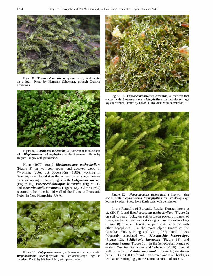
Figure 8. Blepharostoma trichophyllum in a typical habitat on a log. Photo by Hermann Schachner, through Creative Commons.
Figure 9. Liochlaena lanceolata, a liverwort that associates with Blepharostoma trichophyllum in the Pyrenees. Photo by Hugues Tinguy with permission.
Hong (1977) found Blepharostoma trichophyllum (Figure 3) on wet soil, rocks, and decayed wood in Wyoming, USA, but Söderström (1989), working in Sweden, never found it in the earliest decay stages (stages 1-3), occurring in later stages with Calypogeia suecica (Figure 10), Fuscocephaloziopsis leucantha (Figure 11), and Neoorthocaulis attenuatus (Figure 12). Glime (1982) reported it from the humid wall of the Flume at Franconia Notch in New Hampshire, USA.
Figure 10. Calypogeia suecica, a liverwort that occurs with Blepharostoma trichophyllum on late-decay-stage logs in Sweden. Photo by Michael Lüth, with permission.
Figure 11. Fuscocephaloziopsis leucantha, a liverwort that occurs with Blepharostoma trichophyllum on late-decay-stage logs in Sweden. Photo by David T. Holyoak, with permission.
Figure 12. Neoorthocaulis attenuatus, a liverwort that occurs with Blepharostoma trichophyllum on late-decay-stage logs in Sweden. Photo from Earth.com, with permission.
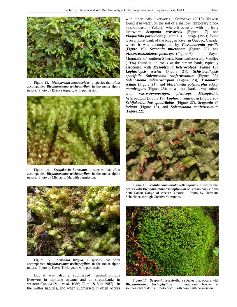
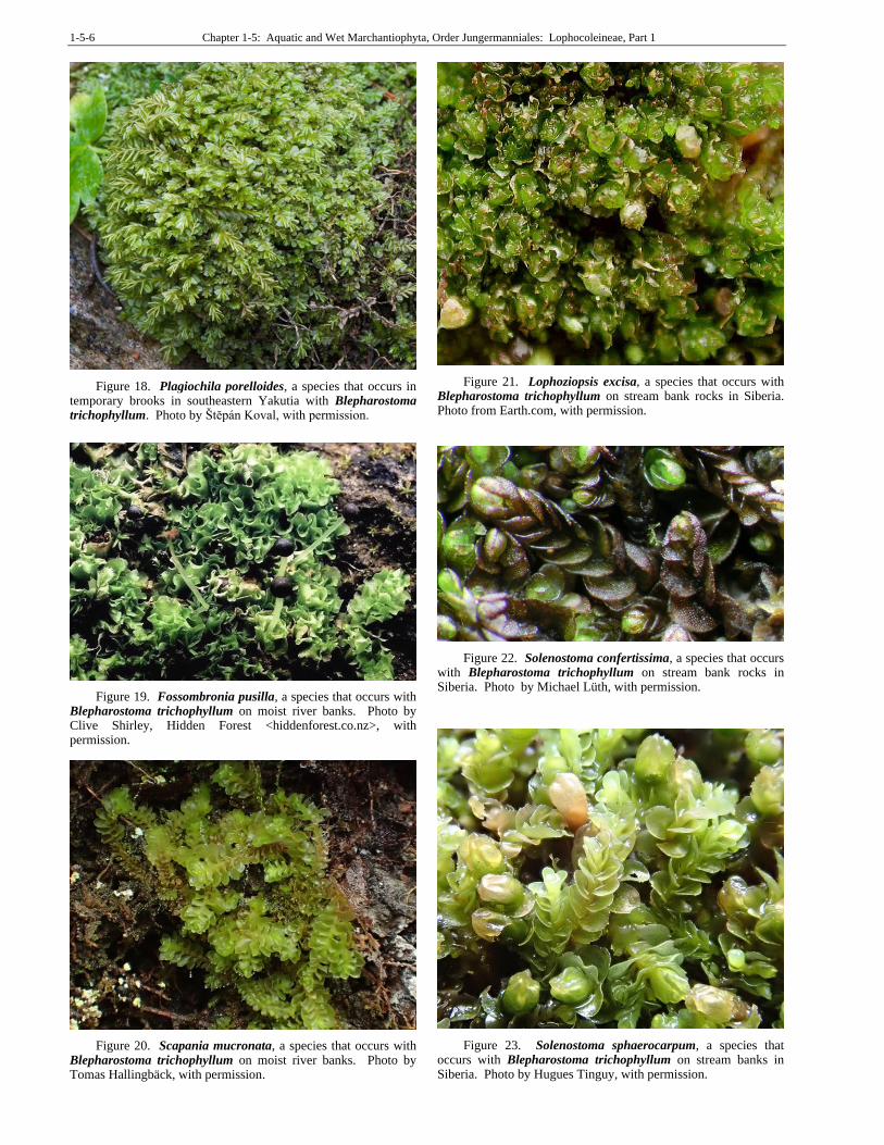
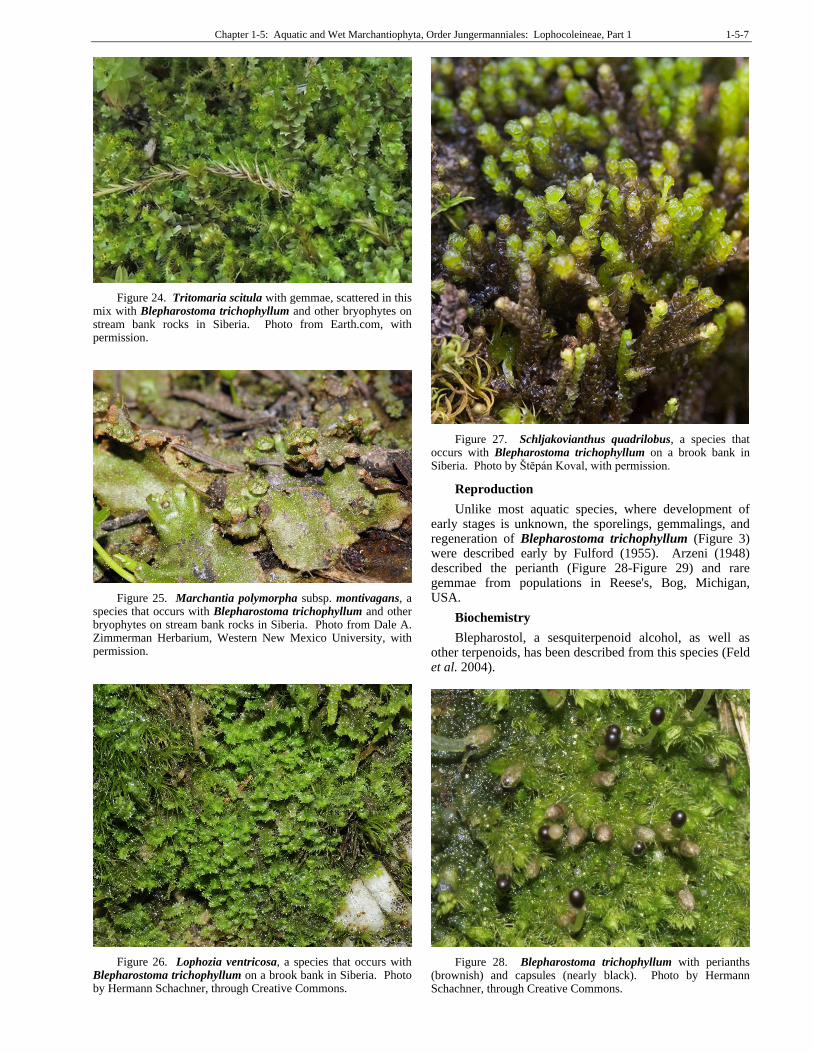
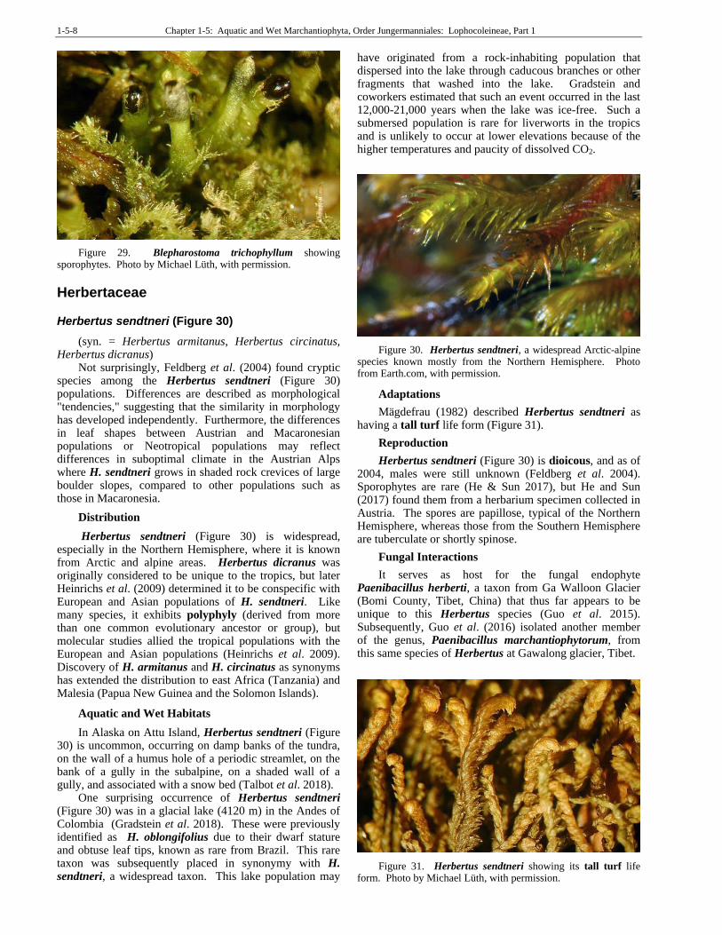
In the Republic of Buryatia, Russia, Konstantinova et al. (2018) found Blepharostoma trichophyllum (Figure 3) on soil-covered rocks, on soil between rocks, on banks of rivers, on trails under roots sticking out and on mossy logs (Figure 8) in mixed forests, in pure mats or mixed with other bryophytes. In the moist alpine tundra of the Canadian Yukon, Hong and Vitt (1977) found it was frequently associated with Mesoptychia heterocolpos (Figure 13), Schljakovia kunzeana (Figure 14), and Scapania irrigua (Figure 15). In the Sette-Daban Range of eastern Yakutia, Sofronova and Sofronov (2010) found it with mixed with Radula complanata (Figure 16) on stream banks. Dulin (2008) found it on stream and river banks, as well as on rotting logs, in the Komi Republic of Russia.
Chapter 1-5: Aquatic and Wet Marchantiophyta, Order Jungermanniales: Lophocoleineae, Part 1 1-5-5
Figure 13. Mesoptychia heterocolpos, a species that often accompanies Blepharostoma trichophyllum in the moist alpine tundra. Photo by Blanka Aguero, with permission.
Figure 14. Schljakovia kunzeana, a species that often accompanies Blepharostoma trichophyllum in the moist alpine tundra. Photo by Michael Lüth, with permission.
Figure 15. Scapania irrigua, a species that often accompanies Blepharostoma trichophyllum in the moist alpine tundra. Photo by David T. Holyoak, with permission.
But it was also a submerged hemicalciphilous liverwort in montane streams and on streambanks in western Canada (Vitt et al. 1986; Glime & Vitt 1987). In the wetter habitats, and when submersed, it often occurs
with other leafy liverworts. Sofronova (2013) likewise found it in water, on the soil of a shallow, temporary brook in southeastern Yakutia, where it occurred with the leafy liverworts Scapania crassiretis (Figure 17) and Plagiochila porelloides (Figure 18). Lepage (1953) found it on a moist bank of the Roggan River in Québec, Canada, where it was accompanied by Fossombronia pusilla (Figure 19), Scapania mucronata (Figure 20), and
pleniceps (Figure 6). In the Sayan
Mountains of southern Siberia, Konstantinova and Vasiljev (1994) found it on rocks at the stream bank, typically associated with Mesoptychia heterocolpos (Figure 13), Lophoziopsis excisa (Figure 21), Schistochilopsis opacifolia, Solenostoma confertissimum (Figure 22), Solenostoma sphaerocarpum (Figure 23), Tritomaria scitula (Figure 24), and Marchantia polymorpha subsp. montivagans (Figure 25); on a brook bank it was mixed
with pleniceps, Mesoptychia
heterocolpos (Figure 13), Lophozia ventricosa (Figure 26), Schljakovianthus quadrilobus (Figure 27), Scapania cf. irrigua (Figure 15), and Solenostoma confertissimum (Figure 22).
Figure 16. Radula complanata with capsules, a species that occurs with Blepharostoma trichophyllum on stream banks in the Sette-Daban Range of eastern Yakutia. Photo by Hermann Schachner, through Creative Commons.
Figure 17. Scapania crassiretis, a species that occurs with Blepharostoma trichophyllum in temporary brooks in southeastern Yakutia. Photo from Earth.com, with permission.
1-5-6 Chapter 1-5: Aquatic and Wet Marchantiophyta, Order Jungermanniales: Lophocoleineae, Part 1
Figure 18. Plagiochila porelloides, a species that occurs in temporary brooks in southeastern Yakutia with Blepharostoma trichophyllum. Photo by Štĕpán Koval, with permission.
Figure 19. Fossombronia pusilla, a species that occurs with Blepharostoma trichophyllum on moist river banks. Photo by Clive Shirley, Hidden Forest <hiddenforest.co.nz>, with permission.
Figure 20. Scapania mucronata, a species that occurs with Blepharostoma trichophyllum on moist river banks. Photo by Tomas Hallingbäck, with permission.
Figure 21. Lophoziopsis excisa, a species that occurs with Blepharostoma trichophyllum on stream bank rocks in Siberia. Photo from Earth.com, with permission.
Figure 22. Solenostoma confertissima, a species that occurs with Blepharostoma trichophyllum on stream bank rocks in Siberia. Photo by Michael Lüth, with permission.
Figure 23. Solenostoma sphaerocarpum, a species that occurs with Blepharostoma trichophyllum on stream banks in Siberia. Photo by Hugues Tinguy, with permission.
Chapter 1-5: Aquatic and Wet Marchantiophyta, Order Jungermanniales: Lophocoleineae, Part 1 1-5-7
Figure 24. Tritomaria scitula with gemmae, scattered in this mix with Blepharostoma trichophyllum and other bryophytes on stream bank rocks in Siberia. Photo from Earth.com, with permission.
Figure 25. Marchantia polymorpha subsp. montivagans, a species that occurs with Blepharostoma trichophyllum and other bryophytes on stream bank rocks in Siberia. Photo from Dale A. Zimmerman Herbarium, Western New Mexico University, with permission.
Figure 26. Lophozia ventricosa, a species that occurs with Blepharostoma trichophyllum on a brook bank in Siberia. Photo by Hermann Schachner, through Creative Commons.
Figure 27. Schljakovianthus quadrilobus, a species that occurs with Blepharostoma trichophyllum on a brook bank in Siberia. Photo by Štĕpán Koval, with permission.
Reproduction
Unlike most aquatic species, where development of early stages is unknown, the sporelings, gemmalings, and regeneration of Blepharostoma trichophyllum (Figure 3) were described early by Fulford (1955). Arzeni (1948) described the perianth (Figure 28-Figure 29) and rare gemmae from populations in Reese's, Bog, Michigan, USA.
Biochemistry
Blepharostol, a sesquiterpenoid alcohol, as well as other terpenoids, has been described from this species (Feld et al. 2004).
Figure 28. Blepharostoma trichophyllum with perianths (brownish) and capsules (nearly black). Photo by Hermann Schachner, through Creative Commons.
1-5-8 Chapter 1-5: Aquatic and Wet Marchantiophyta, Order Jungermanniales: Lophocoleineae, Part 1
Figure 29. Blepharostoma trichophyllum showing sporophytes. Photo by Michael Lüth, with permission.
Herbertaceae
Herbertus sendtneri (Figure 30)
(syn. = Herbertus armitanus, Herbertus circinatus, Herbertus dicranus)
Not surprisingly, Feldberg et al. (2004) found cryptic species among the Herbertus sendtneri (Figure 30) populations. Differences are described as morphological "tendencies," suggesting that the similarity in morphology has developed independently. Furthermore, the differences in leaf shapes between Austrian and Macaronesian populations or Neotropical populations may reflect differences in suboptimal climate in the Austrian Alps where H. sendtneri grows in shaded rock crevices of large boulder slopes, compared to other populations such as those in Macaronesia.
Distribution
Herbertus sendtneri (Figure 30) is widespread, especially in the Northern Hemisphere, where it is known from Arctic and alpine areas. Herbertus dicranus was originally considered to be unique to the tropics, but later Heinrichs et al. (2009) determined it to be conspecific with European and Asian populations of H. sendtneri. Like many species, it exhibits polyphyly (derived from more than one common evolutionary ancestor or group), but molecular studies allied the tropical populations with the European and Asian populations (Heinrichs et al. 2009). Discovery of H. armitanus and H. circinatus as synonyms has extended the distribution to east Africa (Tanzania) and Malesia (Papua New Guinea and the Solomon Islands).
Aquatic and Wet Habitats
In Alaska on Attu Island, Herbertus sendtneri (Figure 30) is uncommon, occurring on damp banks of the tundra, on the wall of a humus hole of a periodic streamlet, on the bank of a gully in the subalpine, on a shaded wall of a gully, and associated with a snow bed (Talbot et al. 2018).
One surprising occurrence of Herbertus sendtneri (Figure 30) was in a glacial lake (4120 m) in the Andes of Colombia (Gradstein et al. 2018). These were previously identified as H. oblongifolius due to their dwarf stature and obtuse leaf tips, known as rare from Brazil. This rare taxon was subsequently placed in synonymy with H. sendtneri, a widespread taxon. This lake population may
have originated from a rock-inhabiting population that dispersed into the lake through caducous branches or other fragments that washed into the lake. Gradstein and coworkers estimated that such an event occurred in the last 12,000-21,000 years when the lake was ice-free. Such a submersed population is rare for liverworts in the tropics and is unlikely to occur at lower elevations because of the higher temperatures and paucity of dissolved CO2.
Figure 30. Herbertus sendtneri, a widespread Arctic-alpine species known mostly from the Northern Hemisphere. Photo from Earth.com, with permission.
Adaptations
Mägdefrau (1982) described Herbertus sendtneri as having a tall turf life form (Figure 31).
Reproduction
Herbertus sendtneri (Figure 30) is dioicous, and as of 2004, males were still unknown (Feldberg et al. 2004). Sporophytes are rare (He & Sun 2017), but He and Sun (2017) found them from a herbarium specimen collected in Austria. The spores are papillose, typical of the Northern Hemisphere, whereas those from the Southern Hemisphere are tuberculate or shortly spinose.
Fungal Interactions
It serves as host for the fungal endophyte Paenibacillus herberti, a taxon from Ga Walloon Glacier (Bomi County, Tibet, China) that thus far appears to be unique to this Herbertus species (Guo et al. 2015). Subsequently, Guo et al. (2016) isolated another member of the genus, Paenibacillus marchantiophytorum, from this same species of Herbertus at Gawalong glacier, Tibet.
Figure 31. Herbertus sendtneri showing its tall turf life form. Photo by Michael Lüth, with permission.
Chapter 1-5: Aquatic and Wet Marchantiophyta, Order Jungermanniales: Lophocoleineae, Part 1 1-5-9
Biochemistry
Sun et al. (2010) used extracts from five bryophyte species, including Herbertus sendtneri (Figure 30-Figure 31), to determine effects on seed germination and seedling physiology of the cucumber. They found that all of these extracts promoted growth of the radicle at some concentrations and that Herbertus sendtneri extracts could enhance chlorophyll content. It could also enhance the content of soluble sugar.
Lepidoziaceae
Bazzania denudata (Figure 36-Figure 32)
Distribution
Bazzania denudata (Figure 36-Figure 32) is distributed in North America from Alaska, southward to Oregon, Montana, and Kentucky, USA (Clark & Frye 1942). It also occurs in Greenland and Central Europe (Schuster 1969).
Figure 32. Bazzania denudata, an epiphyte, but also occurring on moist sandstone canyon walls. Photo from Botany Website, UBC, with permission.
Aquatic and Wet Habitats
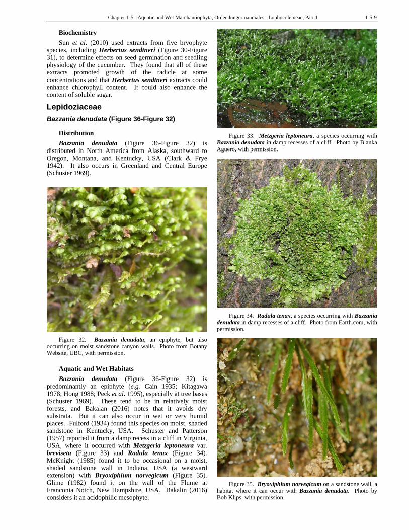
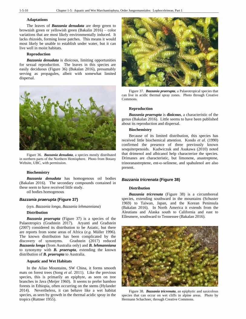
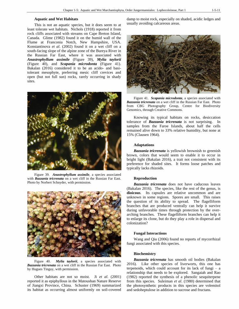
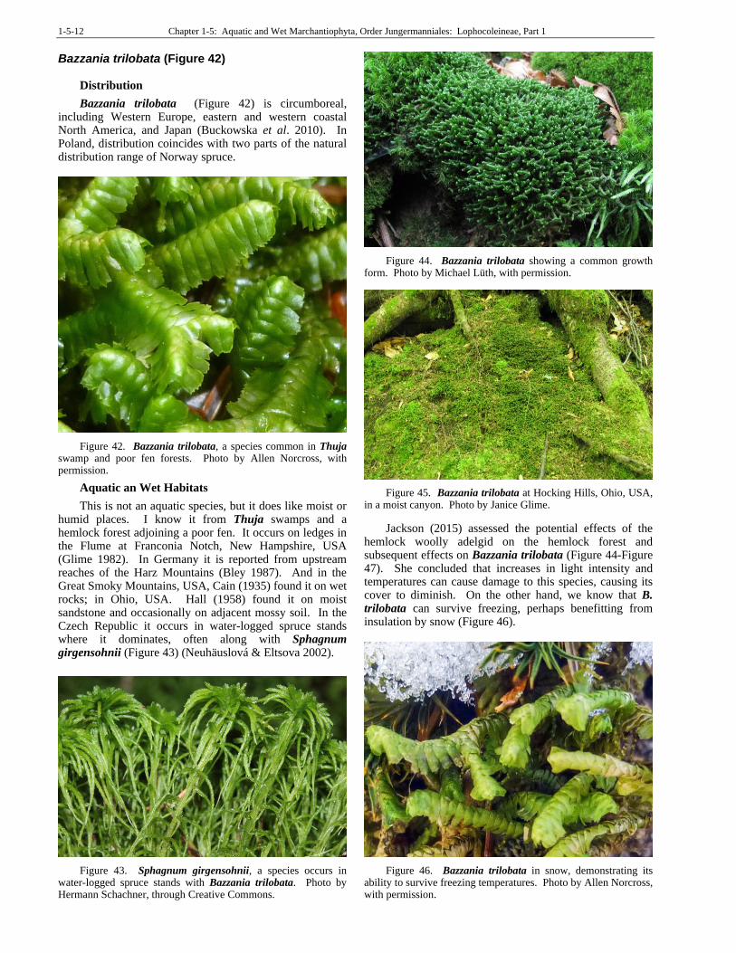
Bazzania denudata (Figure 36-Figure 32) is predominantly an epiphyte (e.g. Cain 1935; Kitagawa 1978; Hong 1988; Peck et al. 1995), especially at tree bases (Schuster 1969). These tend to be in relatively moist forests, and Bakalan (2016) notes that it avoids dry substrata. But it can also occur in wet or very humid places. Fulford (1934) found this species on moist, shaded sandstone in Kentucky, USA. Schuster and Patterson (1957) reported it from a damp recess in a cliff in Virginia, USA, where it occurred with Metzgeria leptoneura var. breviseta (Figure 33) and Radula tenax (Figure 34). McKnight (1985) found it to be occasional on a moist, shaded sandstone wall in Indiana, USA (a westward extension) with Bryoxiphium norvegicum (Figure 35). Glime (1982) found it on the wall of the Flume at Franconia Notch, New Hampshire, USA. Bakalin (2016) considers it an acidophilic mesophyte.
Figure 33. Metzgeria leptoneura, a species occurring with Bazzania denudata in damp recesses of a cliff. Photo by Blanka Aguero, with permission.
Figure 34. Radula tenax, a species occurring with Bazzania denudata in damp recesses of a cliff. Photo from Earth.com, with permission.
Figure 35. Bryoxiphium norvegicum on a sandstone wall, a habitat where it can occur with Bazzania denudata. Photo by Bob Klips, with permission.
1-5-10 Chapter 1-5: Aquatic and Wet Marchantiophyta, Order Jungermanniales: Lophocoleineae, Part 1
Adaptations
The leaves of Bazzania denudata are deep green to brownish green or yellowish green (Bakalin 2016) – color variations that are most likely environmentally induced. It lacks rhizoids, forming loose patches. This means it would most likely be unable to establish under water, but it can live well in moist habitats.
Reproduction
Bazzania denudata is dioicous, limiting opportunities for sexual reproduction. The leaves in this species are easily deciduous (Figure 36) (Bakalan 2016), presumably serving as propagules, albeit with somewhat limited dispersal.
Figure 36. Bazzania denudata, a species mostly distributed in northern parts of the Northern Hemisphere. Photo from Botany Website, UBC, with permission.
Biochemistry
Bazzania denudata has homogenous oil bodies (Bakalan 2016). The secondary compounds contained in these seem to have received little study.
oil bodies homogenous
Bazzania praerupta (Figure 37)
(syn. Bazzania longa, Bazzania lehmanniana)
Distribution
Bazzania praerupta (Figure 37) is a species of the Palaeotropics (Gradstein 2017). Aryanti and Gradstein (2007) considered its distribution to be Asiatic, but there are reports from some areas of Africa (e.g. Müller 1996). The known distribution has been complicated by the discovery of synonyms. Gradstein (2017) reduced Bazzania longa (from Australia only) and B. lehmanniana to synonymy with B. praerupta, extending the known distribution of B. praerupta to Australia.
Aquatic and Wet Habitats
In the Ailao Mountains, SW China, it forms smooth mats on forest trees (Song et al. 2011). Like the previous species, this is primarily an epiphyte, as seen on tree branches in Java (Meijer 1960). It seems to prefer bamboo forests in Ethiopia, often occurring on the stems (Hylander 2014). Nevertheless, it can behave like a wet habitat species, as seen by growth in the thermal acidic spray in the tropics (Ruttner 1955).
Figure 37. Bazzania praerupta, a Palaeotropical species that can live in acidic thermal spray zones. Photo through Creative Commons.
Reproduction
Bazzania praerupta is dioicous, a characteristic of the genus (Bakalan 2016). Little seems to have been published about its reproduction and dispersal.
Biochemistry
Because of its limited distribution, this species has received little biochemical attention. Kondo et al. (1990) confirmed the presence of three previously known sesquiterpenoids. Kudwiczuk and Asakawa (2010) noted that drimenol and albicanol help characterize the species. Drimanes are characteristic, but limonene, anastreptene,
trinoranastreptene, ent--selinene, and spahulenol are also present.
Bazzania tricrenata (Figure 38)
Distribution
Bazzania tricrenata (Figure 38) is a circumboreal species, extending southward in the mountains (Schuster 1969) to Taiwan, Japan, and the Korean Peninsula (Bakalan 2016). In North America it extends from the Aleutians and Alaska south to California and east to Ellesmere, southward to Tennessee (Bakalan 2016).
Figure 38. Bazzania tricrenata, an epiphytic and saxicolous species that can occur on wet cliffs in alpine areas. Photo by Hermann Schachner, through Creative Commons.
Chapter 1-5: Aquatic and Wet Marchantiophyta, Order Jungermanniales: Lophocoleineae, Part 1 1-5-11
Aquatic and Wet Habitats
This is not an aquatic species, but it does seem to at least tolerate wet habitats. Nichols (1918) reported it from rock cliffs associated with streams on Cape Breton Island, Canada. Glime (1982) found it on the humid wall of the Flume at Franconia Notch, New Hampshire, USA. Konstantinova et al. (2002) found it on a wet cliff on a south-facing slope of the alpine zone of the Bureya River in the Russian Far East, where it was associated with Anastrophyllum assimile (Figure 39), Mylia taylorii (Figure 40), and Scapania microdonta (Figure 41). Bakalan (2016) considered it to be an acido- and basi-tolerant mesophyte, preferring mesic cliff crevices and open (but not full sun) rocks, rarely occurring in shady sites.
Figure 39. Anastrophyllum assimile, a species associated with Bazzania tricrenata on a wet cliff in the Russian Far East. Photo by Norbert Schnyder, with permission.
Figure 40. Mylia taylorii, a species associated with Bazzania tricrenata on a wet cliff in the Russian Far East. Photo by Hugues Tinguy, with permission.
Other habitats are not so moist. Ji et al. (2001) reported it as epiphyllous in the Matoushan Nature Reserve of Jiangxi Province, China. Schuster (1969) summarized its habitat as occurring almost uniformly on soil-covered
damp to moist rock, especially on shaded, acidic ledges and usually avoiding calcareous areas.
Figure 41. Scapania microdonta, a species associated with Bazzania tricrenata on a wet cliff in the Russian Far East. Photo from CBG Photography Group, Centre for Biodiversity Genomics, through Creative Commons.
Knowing its typical habitats on rocks, desiccation tolerance of Bazzania tricrenata is not surprising. In samples from the Faroe Islands, about half the cells remained alive down to 33% relative humidity, but none at 15% (Clausen 1964).
Adaptations
Bazzania tricrenata is yellowish brownish to greenish brown, colors that would seem to enable it to occur in bright light (Bakalan 2016), a trait not consistent with its preference for shaded sites. It forms loose patches and typically lacks rhizoids.
Reproduction
Bazzania tricrenata does not have caducous leaves (Bakalan 2016). The species, like the rest of the genus, is dioicous. Its capsules are relative uncommon and are unknown in some regions. Spores are small. This raises the question of its ability to spread. The flagelliform branches that are produced ventrally can help it survive during unfavorable times through protection by the over-arching branches. These flagelliform branches can help it to enlarge its clone, but do they play a role in dispersal and colonization?
Fungal Interactions
Wang and Qiu (2006) found no reports of mycorrhizal fungi associated with this species.
Biochemistry
Bazzania tricrenata has smooth oil bodies (Bakalan 2016). Like other species of liverworts, this one has terpenoids, which could account for its lack of fungi – a relationship that needs to be explored. Sangaiah and Rao (1982) reported the synthesis of a phenolic sesquiterpene from this species. Suleiman et al. (1980) determined that the photosynthetic products in this species are volemitol and sedoheptulose in addition to sucrose and fructans.
1-5-12 Chapter 1-5: Aquatic and Wet Marchantiophyta, Order Jungermanniales: Lophocoleineae, Part 1
Bazzania trilobata (Figure 42)
Distribution
Bazzania trilobata (Figure 42) is circumboreal, including Western Europe, eastern and western coastal North America, and Japan (Buckowska et al. 2010). In Poland, distribution coincides with two parts of the natural distribution range of Norway spruce.
Figure 42. Bazzania trilobata, a species common in Thuja swamp and poor fen forests. Photo by Allen Norcross, with permission.
Aquatic an Wet Habitats
This is not an aquatic species, but it does like moist or humid places. I know it from Thuja swamps and a hemlock forest adjoining a poor fen. It occurs on ledges in the Flume at Franconia Notch, New Hampshire, USA (Glime 1982). In Germany it is reported from upstream reaches of the Harz Mountains (Bley 1987). And in the Great Smoky Mountains, USA, Cain (1935) found it on wet rocks; in Ohio, USA. Hall (1958) found it on moist sandstone and occasionally on adjacent mossy soil. In the Czech Republic it occurs in water-logged spruce stands where it dominates, often along with Sphagnum girgensohnii (Figure 43) (Neuhäuslová & Eltsova 2002).
Figure 43. Sphagnum girgensohnii, a species occurs in water-logged spruce stands with Bazzania trilobata. Photo by Hermann Schachner, through Creative Commons.
Figure 44. Bazzania trilobata showing a common growth form. Photo by Michael Lüth, with permission.
Figure 45. Bazzania trilobata at Hocking Hills, Ohio, USA, in a moist canyon. Photo by Janice Glime.
Jackson (2015) assessed the potential effects of the hemlock woolly adelgid on the hemlock forest and subsequent effects on Bazzania trilobata (Figure 44-Figure 47). She concluded that increases in light intensity and temperatures can cause damage to this species, causing its cover to diminish. On the other hand, we know that B. trilobata can survive freezing, perhaps benefitting from insulation by snow (Figure 46).
Figure 46. Bazzania trilobata in snow, demonstrating its ability to survive freezing temperatures. Photo by Allen Norcross, with permission.
Chapter 1-5: Aquatic and Wet Marchantiophyta, Order Jungermanniales: Lophocoleineae, Part 1 1-5-13
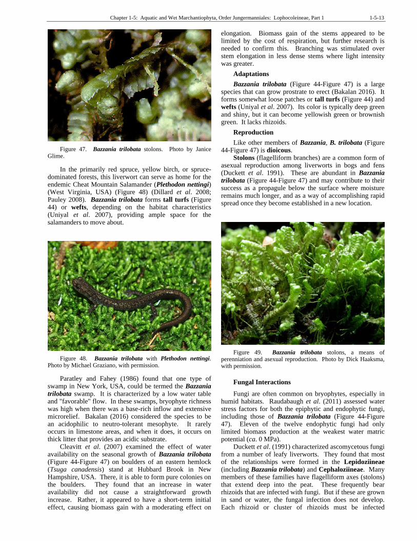
Figure 47. Bazzania trilobata stolons. Photo by Janice Glime.
In the primarily red spruce, yellow birch, or spruce-dominated forests, this liverwort can serve as home for the endemic Cheat Mountain Salamander (Plethodon nettingi) (West Virginia, USA) (Figure 48) (Dillard et al. 2008; Pauley 2008). Bazzania trilobata forms tall turfs (Figure 44) or wefts, depending on the habitat characteristics (Uniyal et al. 2007), providing ample space for the salamanders to move about.
Figure 48. Bazzania trilobata with Plethodon nettingi. Photo by Michael Graziano, with permission.
Paratley and Fahey (1986) found that one type of swamp in New York, USA, could be termed the Bazzania trilobata swamp. It is characterized by a low water table and "favorable" flow. In these swamps, bryophyte richness was high when there was a base-rich inflow and extensive microrelief. Bakalan (2016) considered the species to be an acidophilic to neutro-tolerant mesophyte. It rarely occurs in limestone areas, and when it does, it occurs on thick litter that provides an acidic substrate.
Cleavitt et al. (2007) examined the effect of water availability on the seasonal growth of Bazzania trilobata (Figure 44-Figure 47) on boulders of an eastern hemlock (Tsuga canadensis) stand at Hubbard Brook in New Hampshire, USA. There, it is able to form pure colonies on the boulders. They found that an increase in water availability did not cause a straightforward growth increase. Rather, it appeared to have a short-term initial effect, causing biomass gain with a moderating effect on
elongation. Biomass gain of the stems appeared to be limited by the cost of respiration, but further research is needed to confirm this. Branching was stimulated over stem elongation in less dense stems where light intensity was greater.
Adaptations
Bazzania trilobata (Figure 44-Figure 47) is a large species that can grow prostrate to erect (Bakalan 2016). It forms somewhat loose patches or tall turfs (Figure 44) and wefts (Uniyal et al. 2007). Its color is typically deep green and shiny, but it can become yellowish green or brownish green. It lacks rhizoids.
Reproduction
Like other members of Bazzania, B. trilobata (Figure 44-Figure 47) is dioicous.
Stolons (flagelliform branches) are a common form of asexual reproduction among liverworts in bogs and fens (Duckett et al. 1991). These are abundant in Bazzania trilobata (Figure 44-Figure 47) and may contribute to their success as a propagule below the surface where moisture remains much longer, and as a way of accomplishing rapid spread once they become established in a new location.
Figure 49. Bazzania trilobata stolons, a means of perenniation and asexual reproduction. Photo by Dick Haaksma, with permission.
Fungal Interactions
Fungi are often common on bryophytes, especially in humid habitats. Raudabaugh et al. (2011) assessed water stress factors for both the epiphytic and endophytic fungi, including those of Bazzania trilobata (Figure 44-Figure 47). Eleven of the twelve endophytic fungi had only limited biomass production at the weakest water matric potential (ca. 0 MPa).
Duckett et al. (1991) characterized ascomycetous fungi from a number of leafy liverworts. They found that most of the relationships were formed in the Lepidoziineae (including Bazzania trilobata) and Cephaloziineae. Many members of these families have flagelliform axes (stolons) that extend deep into the peat. These frequently bear rhizoids that are infected with fungi. But if these are grown in sand or water, the fungal infection does not develop. Each rhizoid or cluster of rhizoids must be infected
1-5-14 Chapter 1-5: Aquatic and Wet Marchantiophyta, Order Jungermanniales: Lophocoleineae, Part 1
independently – there is no internal connection between them.
Oil Bodies and Biochemistry
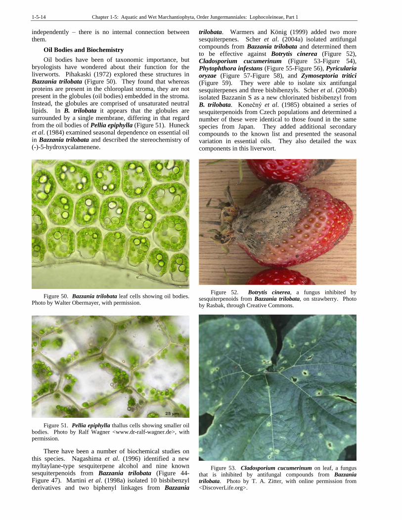
Oil bodies have been of taxonomic importance, but bryologists have wondered about their function for the liverworts. Pihakaski (1972) explored these structures in Bazzania trilobata (Figure 50). They found that whereas proteins are present in the chloroplast stroma, they are not present in the globules (oil bodies) embedded in the stroma. Instead, the globules are comprised of unsaturated neutral lipids. In B. trilobata it appears that the globules are surrounded by a single membrane, differing in that regard from the oil bodies of Pellia epiphylla (Figure 51). Huneck et al. (1984) examined seasonal dependence on essential oil in Bazzania trilobata and described the stereochemistry of (-)-5-hydroxycalamenene.
Figure 50. Bazzania trilobata leaf cells showing oil bodies. Photo by Walter Obermayer, with permission.
Figure 51. Pellia epiphylla thallus cells showing smaller oil bodies. Photo by Ralf Wagner <www.dr-ralf-wagner.de>, with permission.
There have been a number of biochemical studies on this species. Nagashima et al. (1996) identified a new myltaylane-type sesquiterpene alcohol and nine known sesquiterpenoids from Bazzania trilobata (Figure 44-Figure 47). Martini et al. (1998a) isolated 10 bisbibenzyl derivatives and two biphenyl linkages from Bazzania
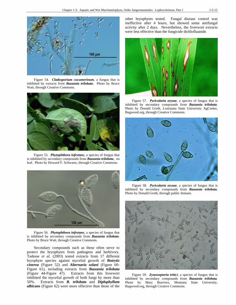
trilobata. Warmers and König (1999) added two more sesquiterpenes. Scher et al. (2004a) isolated antifungal compounds from Bazzania trilobata and determined them to be effective against Botrytis cinerea (Figure 52), Cladosporium cucumerinum (Figure 53-Figure 54), Phytophthora infestans (Figure 55-Figure 56), Pyricularia oryzae (Figure 57-Figure 58), and Zymoseptoria tritici (Figure 59). They were able to isolate six antifungal sesquiterpenes and three bisbibenzyls. Scher et al. (2004b) isolated Bazzanin S as a new chlorinated bisbibenzyl from B. trilobata. Konečný et al. (1985) obtained a series of sesquiterpenoids from Czech populations and determined a number of these were identical to those found in the same species from Japan. They added additional secondary compounds to the known list and presented the seasonal variation in essential oils. They also detailed the wax components in this liverwort.
Figure 52. Botrytis cinerea, a fungus inhibited by sesquiterpenoids from Bazzania trilobata, on strawberry. Photo by Rasbak, through Creative Commons.
Figure 53. Cladosporium cucumerinum on leaf, a fungus that is inhibited by antifungal compounds from Bazzania trilobata. Photo by T. A. Zitter, with online permission from <DiscoverLife.org>.
Chapter 1-5: Aquatic and Wet Marchantiophyta, Order Jungermanniales: Lophocoleineae, Part 1 1-5-15
Figure 54. Cladosporium cucumerinum, a fungus that is inhibited by extracts from Bazzania trilobata. Photo by Bruce Watt, through Creative Commons.
Figure 55. Phytophthora infestans, a species of fungus that is inhibited by secondary compounds from Bazzania trilobata, on leaf. Photo by Howard F. Schwartz, through Creative Commons.
Figure 56. Phytophthora infestans, a species of fungus that is inhibited by secondary compounds from Bazzania trilobata. Photo by Bruce Watt, through Creative Commons.
Secondary compounds such as these often serve to protect the bryophytes from pathogens and herbivory. Tadesse et al. (2003) tested extracts from 17 different bryophyte species against mycelial growth of Botrytis cinerea (Figure 52) and Alternaria solani (Figure 60-Figure 61), including extracts from Bazzania trilobata (Figure 44-Figure 47). Extracts from this liverwort inhibited the mycelial growth of both fungi by more than 50%. Extracts from B. trilobata and Diplophyllum albicans (Figure 62) were more effective than those of the
other bryophytes tested. Fungal disease control was ineffective after 4 hours, but showed some antifungal activity after 2 days. Nevertheless, the liverwort extracts were less effective than the fungicide dichlofluanide.
Figure 57. Pyricularia oryzae, a species of fungus that is inhibited by secondary compounds from Bazzania trilobata. Photo by Donald Groth, Louisiana State University AgCenter, Bugwood.org, through Creative Commons.
Figure 58. Pyricularia oryzae, a species of fungus that is inhibited by secondary compounds from Bazzania trilobata. Photo by Donald Groth, through public domain.
Figure 59. Zymoseptoria tritici, a species of fungus that is inhibited by secondary compounds from Bazzania trilobata. Photo by Mary Burrows, Montana State University, Bugwood.org, through Creative Commons.
1-5-16 Chapter 1-5: Aquatic and Wet Marchantiophyta, Order Jungermanniales: Lophocoleineae, Part 1
Figure 60. Alternaria solani leaf lesions, a species of fungus that is inhibited up to 50% by secondary compounds from Bazzania trilobata. Photo from Clemson University – USDA Cooperative Extension Slide Series, through Creative Commons.
Figure 61. Alternaria solani conidia; this species is inhibited up to 50% by extracts from Bazzania trilobata. Photo by E. McKenzie, Landcare Research, Australia, through Creative Commons.
Figure 62. Diplophyllum albicans, a species that is one of the best inhibitors of Alternaria solani and Botrytis cinerea among the bryophytes tested. Photo by David T. Holyoak, with permission.
Biochemistry
But in addition to the secondary compounds that seem to be useful in protecting the plants from pathogens and herbivory, the liverworts can also possess lignan (phytoestrogens; class of polyphenolic compounds including many found in plants and noted for having antioxidant and estrogenic activity) derivatives (Martini et al. 1998b; Scher et al. 2003).
Hygrolembidium boschianum
(syn. = Lembidium boschianum)
Distribution
Hygrolembidium boschianum occurs in the Southern Hemisphere, including southern South America, Australia, and nearby islands (EOL 2021).
Aquatic and Wet Habitats
Hygrolembidium boschianum occurs in sulfur springs in the tropics (Ruttner 1955). Gradstein (2011) verified this habitat with his report of the species submerged in sulfur springs in Indonesia. There seems to be little known about it ecologically.
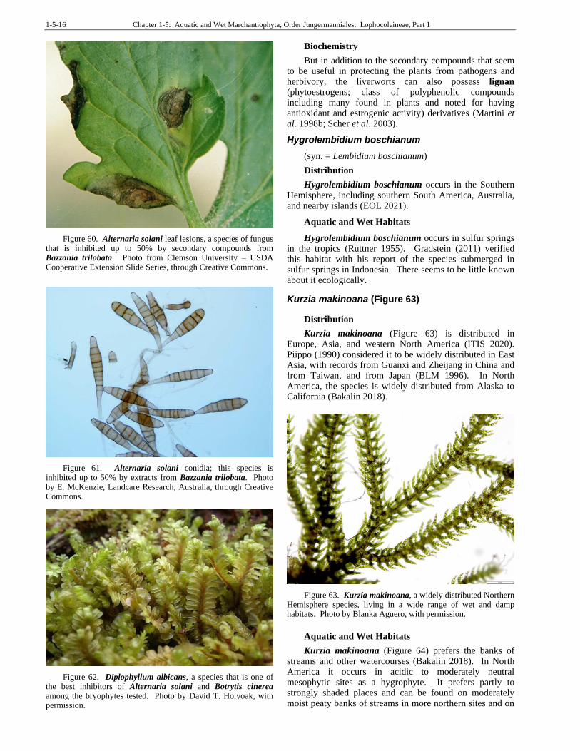
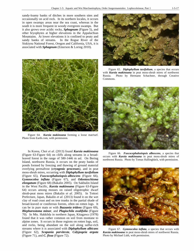
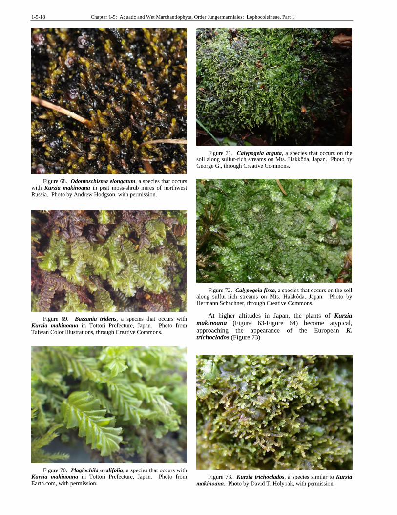
Kurzia makinoana (Figure 63)
Distribution
Kurzia makinoana (Figure 63) is distributed in Europe, Asia, and western North America (ITIS 2020). Piippo (1990) considered it to be widely distributed in East Asia, with records from Guanxi and Zheijang in China and from Taiwan, and from Japan (BLM 1996). In North America, the species is widely distributed from Alaska to California (Bakalin 2018).
Figure 63. Kurzia makinoana, a widely distributed Northern Hemisphere species, living in a wide range of wet and damp habitats. Photo by Blanka Aguero, with permission.
Aquatic and Wet Habitats
Kurzia makinoana (Figure 64) prefers the banks of streams and other watercourses (Bakalin 2018). In North America it occurs in acidic to moderately neutral mesophytic sites as a hygrophyte. It prefers partly to strongly shaded places and can be found on moderately moist peaty banks of streams in more northern sites and on
Chapter 1-5: Aquatic and Wet Marchantiophyta, Order Jungermanniales: Lophocoleineae, Part 1 1-5-17
sandy-loamy banks of ditches in more southern sites and occasionally on acid rock. In its northern locales, it occurs in open swampy areas near the sea coast, whereas in the south it is more frequent in woody evergreen swamps. But it also grows over acidic rocks, Sphagnum (Figure 5), and other bryophytes at higher elevations in the Appalachian Mountains. At lower elevations it is confined to peaty and sandy banks of streams. In the Rogue River of the Siskiyou National Forest, Oregon and California, USA, it is associated with Sphagnum (Emerson & Loring 2010).
Figure 64. Kurzia makinoana forming a loose mat/turf. Photo from Earth.com, with permission.
In Korea, Choi et al. (2013) found Kurzia makinoana (Figure 63-Figure 64) on cliffs along streams in a broad-leaved forest in the range of 580-1446 m asl. On Bering Island, northwest Russia, it occurs on the peaty banks of ponds formed by freezing and thawing of ground material overlying permafrost (cryogenic processes), and in peat moss-shrub mires, occurring with Diplophyllum taxifolium (Figure 65), Fuscocephaloziopsis albescens (Figure 66), Gymnocolea inflata (Figure 67), and Odontoschisma elongatum (Figure 68) (Bakalin 2005). On Sakhalin Island in the West Pacific, Kurzia makinoana (Figure 63-Figure 64) occurs among mosses on raised oligotrophic dwarf shrub-peat moss mires (Bakalin et al. 2005). In Tottori Prefecture, Japan, Bakalin et al. (2013) found it on the wet clay of road crust and on tree trunks in the partial shade of broad-leaved or coniferous forests, often on rotten logs. It can be in pure mats or with Bazzania tridens (Figure 69), Blepharostoma minor, and Plagiochila ovalifolia (Figure 70). In Mts. Hakkôda in northern Japan, Kitagawa (1978) found that it was rather common on soil from montane to alpine zones. It occurs in those regions on rotten logs, soil, and rocks, being abundant on the soil along sulfur-rich streams where it is associated with Diplophyllum albicans (Figure 62), Scapania parvitexta, Calypogeia arguta (Figure 71), and C. fissa (Figure 72).
Figure 65. Diplophyllum taxifolium, a species that occurs with Kurzia makinoana in peat moss-shrub mires of northwest Russia. Photo by Hermann Schachner, through Creative Commons.
Figure 66. Fuscocephaloziopsis albescens, a species that occurs with Kurzia makinoana in peat moss-shrub mires of northwest Russia. Photo by Tomas Hallingbäck, with permission.
Figure 67. Gymnocolea inflata, a species that occurs with Kurzia makinoana in peat moss-shrub mires of northwest Russia. Photo by Michael Lüth, with permission.
1-5-18 Chapter 1-5: Aquatic and Wet Marchantiophyta, Order Jungermanniales: Lophocoleineae, Part 1
Figure 68. Odontoschisma elongatum, a species that occurs with Kurzia makinoana in peat moss-shrub mires of northwest Russia. Photo by Andrew Hodgson, with permission.
Figure 69. Bazzania tridens, a species that occurs with Kurzia makinoana in Tottori Prefecture, Japan. Photo from Taiwan Color Illustrations, through Creative Commons.
Figure 70. Plagiochila ovalifolia, a species that occurs with Kurzia makinoana in Tottori Prefecture, Japan. Photo from Earth.com, with permission.
Figure 71. Calypogeia arguta, a species that occurs on the soil along sulfur-rich streams on Mts. Hakkôda, Japan. Photo by George G., through Creative Commons.
Figure 72. Calypogeia fissa, a species that occurs on the soil along sulfur-rich streams on Mts. Hakkôda, Japan. Photo by Hermann Schachner, through Creative Commons.
At higher altitudes in Japan, the plants of Kurzia makinoana (Figure 63-Figure 64) become atypical, approaching the appearance of the European K. trichoclados (Figure 73).
Figure 73. Kurzia trichoclados, a species similar to Kurzia makinoana. Photo by David T. Holyoak, with permission.
Chapter 1-5: Aquatic and Wet Marchantiophyta, Order Jungermanniales: Lophocoleineae, Part 1 1-5-19
Adaptations
Kurzia makinoana (Figure 63-Figure 64) is a tiny leafy liverwort, dull or deep green to brownish-green (BLM 1996). It occurs in dense tufts or patches with interwoven stems and occasionally creeps among the stems of other bryophytes. Such growth patterns can help it to maintain moisture.
Reproduction
Kurzia makinoana (Figure 63-Figure 64) is dioicious (BLM 1996).
Biochemistry
The tiny Kurzia makinoana (Figure 63-Figure 64) is aromatic (BLM 1996). It produces the monoterpene limonene as well as a number of sesquiterpenoids (Toyota et al. 1997). The chemical constituents differ from those of other Lepidoziaceae (Asakawa 1982). Among these compounds in Kurzia makinoana several (sesquiterpene lactones) (Asakawa et al. 2013) are known for their cytotoxic activity against P-388 lymphocytic leukemia cells (Asakawa 1995).
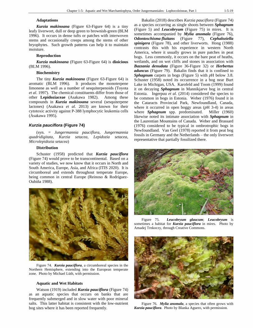
Kurzia pauciflora (Figure 74)
(syn. = Jungermannia pauciflora, Jungermannia quadridigitata, Kurzia setacea, Lepidozia setacea, Microlepidozia setacea)
Distribution
Schuster (1958) predicted that Kurzia pauciflora (Figure 74) would prove to be transcontinental. Based on a variety of studies, we now know that it occurs in North and South America, Europe, Asia, and Africa (ITIS 2020). It is circumboreal and extends throughout temperate Europe, being common in central Europe (Reinoso & Rodríguez-Oubiña 1988).
Figure 74. Kurzia pauciflora, a circumboreal species in the Northern Hemisphere, extending into the European temperate zone. Photo by Michael Lüth, with permission.
Aquatic and Wet Habitats
Watson (1919) included Kurzia pauciflora (Figure 74) as an aquatic species that occurs on banks that are frequently submerged and in slow water with poor mineral salts. This latter habitat is consistent with the low-nutrient bog sites where it has been reported frequently.
Bakalin (2018) describes Kurzia pauciflora (Figure 74) as a species occurring as single shoots between Sphagnum (Figure 5) and Leucobryum (Figure 75) in mires. It is sometimes accompanied by Mylia anomala (Figure 76), Odontoschisma fluitans (Figure 77), Cephaloziella spinigera (Figure 78), and other liverworts. Hong (1988) contrasts this with his experience in western North America, where it usually grows in pure patches in peat bogs. Less commonly, it occurs on the bare peat of heaths, wetlands, and on wet cliffs and stones in association with Bazzania denudata (Figure 36-Figure 32) or Herbertus aduncus (Figure 79). Bakalin finds that it is confined to Sphagnum carpets in bogs (Figure 5) with pH below 3.8. Schuster (1958) noted its occurrence in a bog near Burt Lake in Michigan, USA. Karofeld and Toom (1999) found it on decaying Sphagnum in Mannikjarve bog in central Estonia. Ingerpuu et al. (2014) considered the species to be common in bogs in Estonia. Weber (1976) found it in the Cataracts Provincial Park, Newfoundland, Canada, where it occurred in open boggy areas (pH 3-4) in areas where Sphagnum spp. predominated. Miller (1960) likewise noted its intimate association with Sphagnum in the Laurentian Mountains of Canada. Weber and Brassard (1976) considered to be typical in ombrotrophic bogs in Newfoundland. Van Geel (1978) reported it from peat bog fossils in Germany and the Netherlands – the only liverwort representative that partially fossilized there.
Figure 75. Leucobryum glaucum; Leucobryum is sometimes a habitat for Kurzia pauciflora in mires. Photo by Amadej Trnkoczy, through Creative Commons.
Figure 76. Mylia anomala, a species that often grows with Kurzia pauciflora. Photo by Blanka Aguero, with permission.
1-5-20 Chapter 1-5: Aquatic and Wet Marchantiophyta, Order Jungermanniales: Lophocoleineae, Part 1
Figure 77. Odontoschisma fluitans, a species that often grows with Kurzia pauciflora. Photo by David T. Holyoak, with permission.
Figure 78. Cephaloziella spinigera female shoot, a species that often grows with Kurzia pauciflora. Photo by David Wagner, with permission.
Figure 79. Herbertus aduncus, a species that often grows with Kurzia pauciflora on bare peat of heaths, wetlands, and on wet cliffs and stones. Photo by Botany Website, UBC, with permission.
In the Atlantic blanket bogs in the maritime regions of North-western Europe, water table and pH were major determinants of the bryophyte flora, whereas ammonia was important in determining the tracheophyte flora
(Sottocornola et al. 2009). Kurzia pauciflora (Figure 74), along with Mylia anomala (Figure 76) were the most common species in the sampling, with K. pauciflora along with species of Cephalozia (Figure 80) exhibiting optimal conditions at a lower pH than that of other bryophytes.
Figure 80. Cephalozia bicuspidata; some species of Cephalozia share habitats at optimal conditions of low pH with Kurzia pauciflora. Photo by Hugues Tinguy, with permission.
On the other hand, van Baaren et al. (1988) found it to be characteristic of mesotrophic fens in the Netherlands, typically as a dominant species.
But Redfearn (19622) also found it in association with the moss Tetraphis pellucida (Figure 81) on moist, shaded, vertical dolomite of east-facing bluffs in Douglas County, Missouri, USA.
Figure 81. Tetraphis pellucida, a moss species associated with Kurzia pauciflora on moist, shaded, vertical dolomite bluffs. Photo by Hermann Schachner, through Creative Commons.
Albinsson (1997) determined that Kurzia pauciflora (Figure 74) belongs to a group of liverworts with a relatively wide ecological amplitude. One secret to its success in habitats with other bryophytes might be its extensive system of underground axes (Hugonnot et al. 2015). These exhibit profuse branching and can reach a maximum depth of 10 cm. They permit the colonization of successive layers of substrate, contributing to the success of the species. They do best on dead rather than live Sphagnum (Figure 5) and therefore benefit from disturbance.
Chapter 1-5: Aquatic and Wet Marchantiophyta, Order Jungermanniales: Lophocoleineae, Part 1 1-5-21
Van Diggelen et al. (2015) report Kurzia pauciflora (Figure 74) as a red-listed species that often achieves as high a cover value as Sphagnum (Figure 5) species in restoration sites for acidified and eutrophied fens, most likely due to its regeneration from dead peat layers.
Reproduction
Kurzia pauciflora is dioicous (Earth.com 2021), but it seems to have other mechanisms for regeneration and asexual reproduction. Duckett and Clymo (1988) found that Kurzia pauciflora (Figure 74) and other species with well-developed underground axes regenerate poorly at the surface, but that their regeneration is much more successful down to 12 cm or so below the surface; they can still be found at 24-30 cm depth. Their presence in these lower layers occurs in both bogs with a live Sphagnum‐covered surface (Figure 5) and from a much older cut peat surface recently recolonized by liverworts. These results support the contention that regeneration is mainly from the underground axes rather than from spores or gemmae. The underground biomass of these species is typically large.
Interactions
All the axes of Kurzia pauciflora (Figure 74) have fungal associates, and it is possible that the fungi are partially saprophytic or parasitic (Duckett & Clymo 1988). Liepiņa (2012) reported that fungal infection causes swollen rhizoids in this species.
Wang and Qiu (2006) noted reports of mycorrhizal relationships with Kurzia pauciflora (Figure 74). But earlier, Duckett et al. (1991) considered that the rhizoid-Ascomycete associations and flagelliform branches seen in Kurzia pauciflora represent secondary parasitic infections rather than a mutualistic relationship. They further argued that the nitrogen fixation observed in these liverworts was due to Cyanobacteria (Figure 82) on the surface of the plants.
Figure 82. Microcoleus (Cyanobacteria), a nitrogen-fixing periphyton organism such as those you might find on Kurzia pauciflora. Photo by Yuuji Tsukii, with permission.
Kowalczyk et al. (1997) used Kurzia pauciflora (Figure 74), among nine others, to demonstrate sterilization techniques. Using commercial bleach (Ace) diluted with distilled water at 1:1 and 1:3 ratios of bleach to water. The optimal sterilization time was 0.5-2.0 minutes. They determined that the fragments to be sterilized should not be larger than 3x3 mm, taken from the terminal portions of the thallus or leafless shoots of the leafy gametophytes. Greater success is achieved with healthy plants that are turgid.
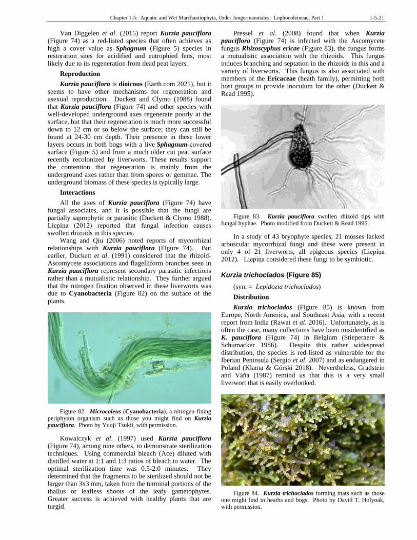
Pressel et al. (2008) found that when Kurzia pauciflora (Figure 74) is infected with the Ascomycete fungus Rhizoscyphus ericae (Figure 83), the fungus forms a mutualistic association with the rhizoids. This fungus induces branching and septation in the rhizoids in this and a variety of liverworts. This fungus is also associated with members of the Ericaceae (heath family), permitting both host groups to provide inoculum for the other (Duckett & Read 1995).
Figure 83. Kurzia pauciflora swollen rhizoid tips with fungal hyphae. Photo modified from Duckett & Read 1995.
In a study of 43 bryophyte species, 21 mosses lacked arbuscular mycorrhizal fungi and these were present in only 4 of 21 liverworts, all epigeous species (Liepiņa 2012). Liepiņa considered these fungi to be symbiotic.
Kurzia trichoclados (Figure 85)
(syn. = Lepidozia trichoclados)
Distribution
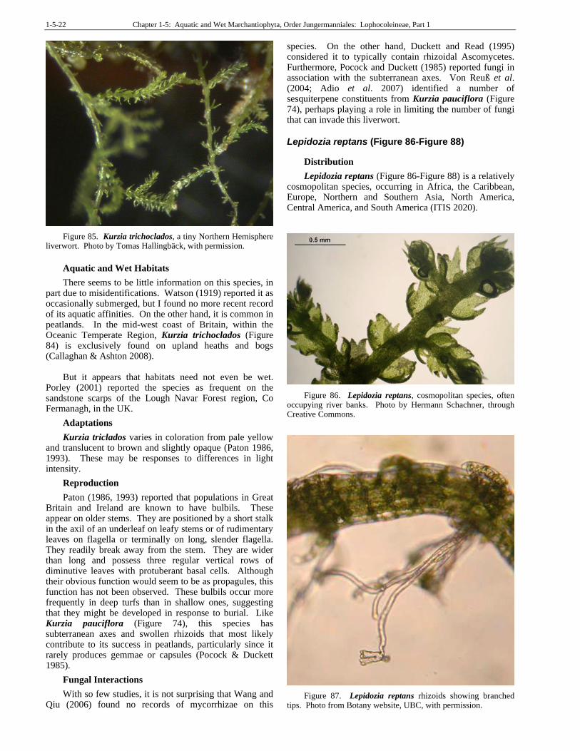
Kurzia trichoclados (Figure 85) is known from Europe, North America, and Southeast Asia, with a recent report from India (Rawat et al. 2016). Unfortunately, as is often the case, many collections have been misidentified as K. pauciflora (Figure 74) in Belgium (Stieperaere & Schumacker 1986). Despite this rather widespread distribution, the species is red-listed as vulnerable for the Iberian Peninsula (Sergio et al. 2007) and as endangered in Poland (Klama & Górski 2018). Nevertheless, Gradstein and Váňa (1987) remind us that this is a very small liverwort that is easily overlooked.
Figure 84. Kurzia trichoclados forming mats such as those one might find in heaths and bogs. Photo by David T. Holyoak, with permission.
1-5-22 Chapter 1-5: Aquatic and Wet Marchantiophyta, Order Jungermanniales: Lophocoleineae, Part 1
Figure 85. Kurzia trichoclados, a tiny Northern Hemisphere liverwort. Photo by Tomas Hallingbäck, with permission.
Aquatic and Wet Habitats
There seems to be little information on this species, in part due to misidentifications. Watson (1919) reported it as occasionally submerged, but I found no more recent record of its aquatic affinities. On the other hand, it is common in peatlands. In the mid-west coast of Britain, within the Oceanic Temperate Region, Kurzia trichoclados (Figure 84) is exclusively found on upland heaths and bogs (Callaghan & Ashton 2008).
But it appears that habitats need not even be wet. Porley (2001) reported the species as frequent on the sandstone scarps of the Lough Navar Forest region, Co Fermanagh, in the UK.
Adaptations
Kurzia triclados varies in coloration from pale yellow and translucent to brown and slightly opaque (Paton 1986, 1993). These may be responses to differences in light intensity.
Reproduction
Paton (1986, 1993) reported that populations in Great Britain and Ireland are known to have bulbils. These appear on older stems. They are positioned by a short stalk in the axil of an underleaf on leafy stems or of rudimentary leaves on flagella or terminally on long, slender flagella. They readily break away from the stem. They are wider than long and possess three regular vertical rows of diminutive leaves with protuberant basal cells. Although their obvious function would seem to be as propagules, this function has not been observed. These bulbils occur more frequently in deep turfs than in shallow ones, suggesting that they might be developed in response to burial. Like Kurzia pauciflora (Figure 74), this species has subterranean axes and swollen rhizoids that most likely contribute to its success in peatlands, particularly since it rarely produces gemmae or capsules (Pocock & Duckett 1985).
Fungal Interactions
With so few studies, it is not surprising that Wang and Qiu (2006) found no records of mycorrhizae on this
species. On the other hand, Duckett and Read (1995) considered it to typically contain rhizoidal Ascomycetes. Furthermore, Pocock and Duckett (1985) reported fungi in association with the subterranean axes. Von Reuß et al. (2004; Adio et al. 2007) identified a number of sesquiterpene constituents from Kurzia pauciflora (Figure 74), perhaps playing a role in limiting the number of fungi that can invade this liverwort.
Lepidozia reptans (Figure 86-Figure 88)
Distribution
Lepidozia reptans (Figure 86-Figure 88) is a relatively cosmopolitan species, occurring in Africa, the Caribbean, Europe, Northern and Southern Asia, North America, Central America, and South America (ITIS 2020).
Figure 86. Lepidozia reptans, cosmopolitan species, often occupying river banks. Photo by Hermann Schachner, through Creative Commons.
Figure 87. Lepidozia reptans rhizoids showing branched tips. Photo from Botany website, UBC, with permission.
Chapter 1-5: Aquatic and Wet Marchantiophyta, Order Jungermanniales: Lophocoleineae, Part 1 1-5-23
Aquatic and Wet Habitats
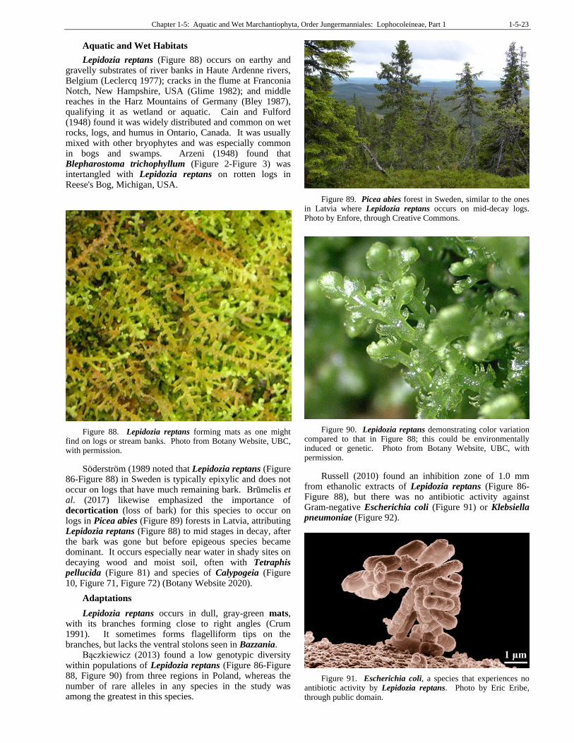
Lepidozia reptans (Figure 88) occurs on earthy and gravelly substrates of river banks in Haute Ardenne rivers, Belgium (Leclercq 1977); cracks in the flume at Franconia Notch, New Hampshire, USA (Glime 1982); and middle reaches in the Harz Mountains of Germany (Bley 1987), qualifying it as wetland or aquatic. Cain and Fulford (1948) found it was widely distributed and common on wet rocks, logs, and humus in Ontario, Canada. It was usually mixed with other bryophytes and was especially common in bogs and swamps. Arzeni (1948) found that Blepharostoma trichophyllum (Figure 2-Figure 3) was intertangled with Lepidozia reptans on rotten logs in Reese's Bog, Michigan, USA.
Figure 88. Lepidozia reptans forming mats as one might find on logs or stream banks. Photo from Botany Website, UBC, with permission.
Söderström (1989 noted that Lepidozia reptans (Figure 86-Figure 88) in Sweden is typically epixylic and does not occur on logs that have much remaining bark. Brūmelis et al. (2017) likewise emphasized the importance of decortication (loss of bark) for this species to occur on logs in Picea abies (Figure 89) forests in Latvia, attributing Lepidozia reptans (Figure 88) to mid stages in decay, after the bark was gone but before epigeous species became dominant. It occurs especially near water in shady sites on decaying wood and moist soil, often with Tetraphis pellucida (Figure 81) and species of Calypogeia (Figure 10, Figure 71, Figure 72) (Botany Website 2020).
Adaptations
Lepidozia reptans occurs in dull, gray-green mats, with its branches forming close to right angles (Crum 1991). It sometimes forms flagelliform tips on the branches, but lacks the ventral stolons seen in Bazzania.
Bączkiewicz (2013) found a low genotypic diversity within populations of Lepidozia reptans (Figure 86-Figure 88, Figure 90) from three regions in Poland, whereas the number of rare alleles in any species in the study was among the greatest in this species.
Figure 89. Picea abies forest in Sweden, similar to the ones in Latvia where Lepidozia reptans occurs on mid-decay logs. Photo by Enfore, through Creative Commons.
Figure 90. Lepidozia reptans demonstrating color variation compared to that in Figure 88; this could be environmentally induced or genetic. Photo from Botany Website, UBC, with permission.
Russell (2010) found an inhibition zone of 1.0 mm from ethanolic extracts of Lepidozia reptans (Figure 86-Figure 88), but there was no antibiotic activity against Gram-negative Escherichia coli (Figure 91) or Klebsiella pneumoniae (Figure 92).
Figure 91. Escherichia coli, a species that experiences no antibiotic activity by Lepidozia reptans. Photo by Eric Eribe, through public domain.

1-5-24 Chapter 1-5: Aquatic and Wet Marchantiophyta, Order Jungermanniales: Lophocoleineae, Part 1
Figure 92. Klebsiella pneumoniae, a species that experiences no antibiotic activity by Lepidozia reptans. Photo by IAID, through Creative Commons.
In ravine habitats, it can provide substrate for slime molds (Ing 1983). The nature of this relationship needs to be explored – is it mutualism, competition, or just a preference for the same habitat?
Reproduction
Lepidozia reptans (Figure 86-Figure 88, Figure 90) is autoicous, making it easier to achieve sexual reproduction (Crum 1991). On the other hand, its lack of ventral stolons denies it of that reproductive advantage as seen in Bazzania.
Biochemistry
Several biochemical studies have included this species. Connolly et al. (1986) described the structure of a sesquiterpene diol from Lepidozia reptans (Figure 86-Figure 88, Figure 90). Rieck et al. (1997) determined the structure of another new sesquiterpene alcohol. Zhang et al. (2010) identified lignans and described a new cadinane sesquiterpenoid lactone from this species. Li et al. (2018) identified five new terpenoids and nine known ones from Chinese populations of Lepidozia reptans, screening them for anti-inflammatory compounds. Suleiman et al. (1980) identified volemitol and sedoheptulose as photosynthetic products.
Lepidozia trichodes
Distribution
Lepidozia trichodes has been known for a long time from Java and Bolivia (Stapf 1894-1896). Chuah-Petiot (2011) reported it from Malaysia. Gao and Bai (2002) considered it to be endemic to China and Taiwan, but in fact it is now known from a number of islands north of Australia (DiscoverLife (2020). Even before Gao and Bai considered it to be endemic, it was reported from the Philippines (del Rosario 1967). Pócs and Ninh (2005)
subsequently reported it from Vietnam, Lai et al. (2008) from Thailand, and Aryanti and Gradstein (2007; Ariyanti et al. 2009) from Sulawesi, Indonesia. Siregar et al. (2018) added distributio in Papua New Guinea, Japan, and India.
Aquatic and Wet Habitats
Ruttner (1955) reported Lepidozia trichodes from acidic thermal spray in the tropics. Kitayama (1995) likewise reported it from the tropics, occurring in the cloud forest of Mount Kinabalu, Sabah, Malaysia, in dense "moss balls" with other leafy liverworts. Piippo (1984) found it in both the rainforests and cloud forests of the Huon Peninsula, Papua New Guinea. It occurs on moist bark, and although these are not aquatic habitats, they have long moist periods. Pócs and Ninh (2005) found it (rarely) on streambed rocks in Vietnam. Logatec et al. (2019) found it along the trail to a mossy forest (almost always humid) in the Philippines.
Adaptations
In Lepidozia trichodes of the montane rainforest of Peninsular Malaya, the rhizoids are almost exclusive to the flagella (Pocock et al. 1984). Most of them exhibit terminal ramifications, a response to contact with the substratum.
Fungal Interactions
Lepidozia trichodes swollen tips, also on the flagellar axes, contain abundant fungal hyphae (Pocock et al. 1984).
Zoopsis argentea (Figure 93)
Distribution
Zoopsis argentea (Figure 93) has a relatively small distribution, occurring in Australia and southern Asia (ITIS 2020).
Aquatic and Wet Habitats
Only Ruttner (1955) seems to attribute it to a somewhat aquatic existence, describing it from acidic thermal spray in the tropics. Rather, it is typically a species of older logs, 33-67 years (Turner & Pharo 2005). In Tasmania, it occurs on the lowest levels of the buttress of Eucalyptus obliqua (Figure 94) (Kantvilas & Jarman 2004).
Figure 93. Zoopsis argentea, a species of Australia and southern Asia. Photo by Peter de Lange, through Creative Commons.
Chapter 1-5: Aquatic and Wet Marchantiophyta, Order Jungermanniales: Lophocoleineae, Part 1 1-5-25
Figure 94. Eucalyptus obliqua, showing bases where one might find Zoopsis argentea. Photo by Forest and Kim Starr, with limited online permission.
Adaptations
In Zoopsis argentea (Figure 93), the stem has totally taken over the photosynthetic role of the plant (Thiers 1988), forming deep green mats (Allison 1985). The stem is flattened and the leaves reduced (Figure 95), possibly an adaptation to its tropical habitats.
Figure 95. Zoopsis argentea showing the photosynthetic stem and reduced, flattened leaves. Photo by Tom Thekathyil, with permission.
Lophocoleaceae
Chiloscyphus (Figure 96-Figure 98, Figure 106-Figure 108)
Chiloscyphus (Figure 96-Figure 98, Figure 106-Figure 108) is a genus that in central France occurs in streams where it is embedded in basaltic rocks with elevated levels of Cu, Zn, Sr, V, Ba, Ni, and Co (Samecka-Cymerman & Kempers 1999). Aquatic varieties are almost black, whereas the typical variety ranges from deep yellow to pale green (Figure 104) to brownish green (Figure 96) (Salachna 2007). Submerged plants often lack rhizoids. It seems that common garden studies in a variety of habitat conditions would be helpful in understanding this genus.
Figure 96. Chiloscyphus polyanthos brownish form. Photo by A. Neuman, through Creative Commons.
The species are autoicous or dioicous. Sporophytes (Figure 104) are produced in late winter and spring and can be abundant.
Chiloscyphus pallescens (Figure 97)
Järvinen (1983) considered Chiloscyphus to have three taxa in Europe. While she separated variety fragilis and variety rivularis from typical Chiloscyphus polyanthos (Figure 106-Figure 107), she considered Chiloscyphus pallescens (Figure 97) to be conspecific with Chiloscyphus polyanthos. Nevertheless, in 2016 Söderström et al. considered these two to be separate species and placed variety fragilis in C. pallescens. Factors related to the environment cause leaf variation that could account for the differences in interpretation.
Figure 97. Chiloscyphus pallescens, a widespread species that is mostly aquatic, but also occurs above water. Photo by Hermann Schachner, through Creative Commons.
1-5-26 Chapter 1-5: Aquatic and Wet Marchantiophyta, Order Jungermanniales: Lophocoleineae, Part 1
Distribution
Chiloscyphus pallescens (Figure 97) is a species in North America from Alaska to Mexico, Europe, Asia, and Africa (ITIS 2020).
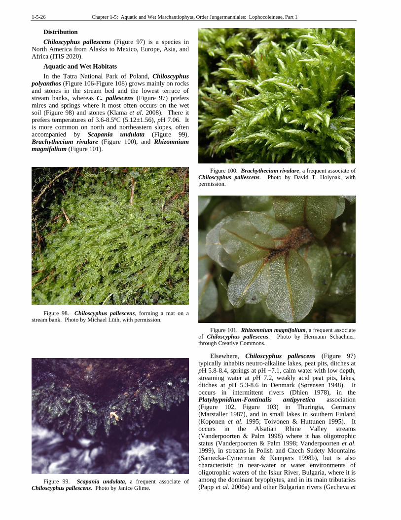
Aquatic and Wet Habitats
In the Tatra National Park of Poland, Chiloscyphus polyanthos (Figure 106-Figure 108) grows mainly on rocks and stones in the stream bed and the lowest terrace of stream banks, whereas C. pallescens (Figure 97) prefers mires and springs where it most often occurs on the wet soil (Figure 98) and stones (Klama et al. 2008). There it prefers temperatures of 3.6-8.5ºC (5.12±1.56), pH 7.06. It is more common on north and northeastern slopes, often accompanied by Scapania undulata (Figure 99), Brachythecium rivulare (Figure 100), and Rhizomnium magnifolium (Figure 101).
Figure 98. Chiloscyphus pallescens, forming a mat on a stream bank. Photo by Michael Lüth, with permission.
Figure 99. Scapania undulata, a frequent associate of Chiloscyphus pallescens. Photo by Janice Glime.
Figure 100. Brachythecium rivulare, a frequent associate of Chiloscyphus pallescens. Photo by David T. Holyoak, with permission.
Figure 101. Rhizomnium magnifolium, a frequent associate of Chiloscyphus pallescens. Photo by Hermann Schachner, through Creative Commons.
Elsewhere, Chiloscyphus pallescens (Figure 97) typically inhabits neutro-alkaline lakes, peat pits, ditches at pH 5.8-8.4, springs at pH ~7.1, calm water with low depth, streaming water at pH 7.2, weakly acid peat pits, lakes, ditches at pH 5.3-8.6 in Denmark (Sørensen 1948). It occurs in intermittent rivers (Dhien 1978), in the Platyhypnidium-Fontinalis antipyretica association (Figure 102, Figure 103) in Thuringia, Germany (Marstaller 1987), and in small lakes in southern Finland (Koponen et al. 1995; Toivonen & Huttunen 1995). It occurs in the Alsatian Rhine Valley streams (Vanderpoorten & Palm 1998) where it has oligotrophic status (Vanderpoorten & Palm 1998; Vanderpoorten et al. 1999), in streams in Polish and Czech Sudety Mountains (Samecka-Cymerman & Kempers 1998b), but is also characteristic in near-water or water environments of oligotrophic waters of the Iskur River, Bulgaria, where it is among the dominant bryophytes, and in its main tributaries (Papp et al. 2006a) and other Bulgarian rivers (Gecheva et
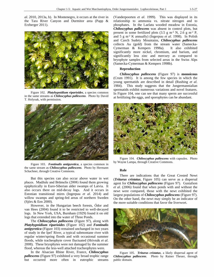
Chapter 1-5: Aquatic and Wet Marchantiophyta, Order Jungermanniales: Lophocoleineae, Part 1 1-5-27
al. 2010, 2013a, b). In Montenegro, it occurs at the river in the Tara River Canyon and Durmitor area (Papp & Erzberger 2011).
Figure 102. Platyhypnidium riparioides, a species common in the same streams as Chiloscyphus pallescens. Photo by David T. Holyoak, with permission.
Figure 103. Fontinalis antipyretica, a species common in the same stream as Chiloscyphus pallescens. Photo by Hermann Schachner, through Creative Commons.
But this species can also occur above water in wet places. Madžule and Brūmelis (2008) found them growing epiphytically in Euro-Siberian alder swamps of Latvia. It also occurs there on mid-decay logs. And it occurs in Estonian transitional mires (Ingerpuu et al. 2014) and willow swamps and spring-fed areas of northern Sweden (Sjörs & Een 2000).
However, in the Hungarian beech forests, Ódor and van Hees (2004) found it to be restricted to well-decayed logs. In New York, USA, Burnham (1929) found it on old logs that extended into the water of Three Ponds.
The Chiloscyphus pallescens (Figure 97), along with Platyhypnidium riparioides (Figure 102) and Fontinalis antipyretica (Figure 103) remained unchanged in two years of study in the Ipel' River, a typical submontane river with regular winter/spring floods and with occasional summer floods, while tracheophyte cover fluctuated (Hrivnák et al. 2008). These bryophytes were not damaged by the summer flood, whereas the less-well attached tracheophytes were.
In the Alsacian Rhine River, France, Chiloscyphus pallescens (Figure 97) exhibited a very broad trophic range but occurred more often in eutrophic streams
(Vanderpoorten et al. 1999). This was displayed in its relationship to ammonia vs. nitrate nitrogen and to phosphates. In the Laelatu wooded meadow in Estonia, Chiloscyphus pallescens was absent in control plots, but present in some fertilized plots (3.5 g m-2 N, 2.6 g m-2 P, and 5 g m-2 K annually) (Ingerpuu et al. 1998). In Polish and Czech Sudety Mountains, Chiloscyphus pallescens collects Au (gold) from the stream water (Samecka-Cymerman & Kempers 1998a). It also exhibited significantly more nickel, chromium, and barium, and significantly less zinc and mercury as compared to bryophyte samples from selected areas in the Swiss Alps (Samecka-Cymerman & Kempers 1998b).
Reproduction
Chiloscyphus pallescens (Figure 97) is monoicous (Crum 1991). It is among the few species in which the young spermatids are described in detail (Rushing et al. 1984). This study suggests that the Jungermannialian spermatids exhibit numerous variations and novel features. In Figure 104, one can see that many sperm are successful at fertilizing the eggs, and sporophytes can be abundant.
Figure 104. Chiloscyphus pallescens with capsules. Photo by Wayne Lampa, through Creative Commons.
Role
There are indications that the Great Crested Newt (Triturus cristatus, Figure 105) can serve as a dispersal agent for Chiloscyphus pallescens (Figure 97). Gustafson et al. (2006) found that when ponds with and without the newt were compared, those with the newt exhibited the largest populations of Chiloscyphus pallescens (Figure 97). On the other hand, the newt may simply be an indicator of the more suitable conditions that favor the liverwort.
Figure 105. Triturus cristatus, a likely dispersal agent of Chiloscyphus pallescens. Photo by Rainer Theuer, through public domain.
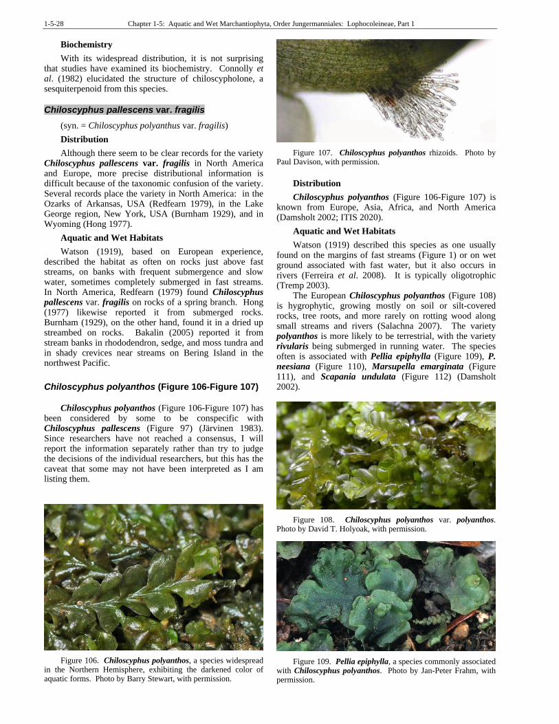
1-5-28 Chapter 1-5: Aquatic and Wet Marchantiophyta, Order Jungermanniales: Lophocoleineae, Part 1
Biochemistry
With its widespread distribution, it is not surprising that studies have examined its biochemistry. Connolly et al. (1982) elucidated the structure of chiloscypholone, a sesquiterpenoid from this species.
Chiloscyphus pallescens var. fragilis
(syn. = Chiloscyphus polyanthus var. fragilis)
Distribution
Although there seem to be clear records for the variety Chiloscyphus pallescens var. fragilis in North America and Europe, more precise distributional information is difficult because of the taxonomic confusion of the variety. Several records place the variety in North America: in the Ozarks of Arkansas, USA (Redfearn 1979), in the Lake George region, New York, USA (Burnham 1929), and in Wyoming (Hong 1977).
Aquatic and Wet Habitats
Watson (1919), based on European experience, described the habitat as often on rocks just above fast streams, on banks with frequent submergence and slow water, sometimes completely submerged in fast streams. In North America, Redfearn (1979) found Chiloscyphus pallescens var. fragilis on rocks of a spring branch. Hong (1977) likewise reported it from submerged rocks. Burnham (1929), on the other hand, found it in a dried up streambed on rocks. Bakalin (2005) reported it from stream banks in rhododendron, sedge, and moss tundra and in shady crevices near streams on Bering Island in the northwest Pacific.
Chiloscyphus polyanthos (Figure 106-Figure 107)
Chiloscyphus polyanthos (Figure 106-Figure 107) has
been considered by some to be conspecific with Chiloscyphus pallescens (Figure 97) (Järvinen 1983). Since researchers have not reached a consensus, I will report the information separately rather than try to judge the decisions of the individual researchers, but this has the caveat that some may not have been interpreted as I am listing them.
Figure 106. Chiloscyphus polyanthos, a species widespread in the Northern Hemisphere, exhibiting the darkened color of aquatic forms. Photo by Barry Stewart, with permission.
Figure 107. Chiloscyphus polyanthos rhizoids. Photo by Paul Davison, with permission.
Distribution
Chiloscyphus polyanthos (Figure 106-Figure 107) is known from Europe, Asia, Africa, and North America (Damsholt 2002; ITIS 2020).
Aquatic and Wet Habitats
Watson (1919) described this species as one usually found on the margins of fast streams (Figure 1) or on wet ground associated with fast water, but it also occurs in rivers (Ferreira et al. 2008). It is typically oligotrophic (Tremp 2003).
The European Chiloscyphus polyanthos (Figure 108) is hygrophytic, growing mostly on soil or silt-covered rocks, tree roots, and more rarely on rotting wood along small streams and rivers (Salachna 2007). The variety polyanthos is more likely to be terrestrial, with the variety rivularis being submerged in running water. The species often is associated with Pellia epiphylla (Figure 109), P. neesiana (Figure 110), Marsupella emarginata (Figure 111), and Scapania undulata (Figure 112) (Damsholt 2002).
Figure 108. Chiloscyphus polyanthos var. polyanthos. Photo by David T. Holyoak, with permission.
Figure 109. Pellia epiphylla, a species commonly associated with Chiloscyphus polyanthos. Photo by Jan-Peter Frahm, with permission.
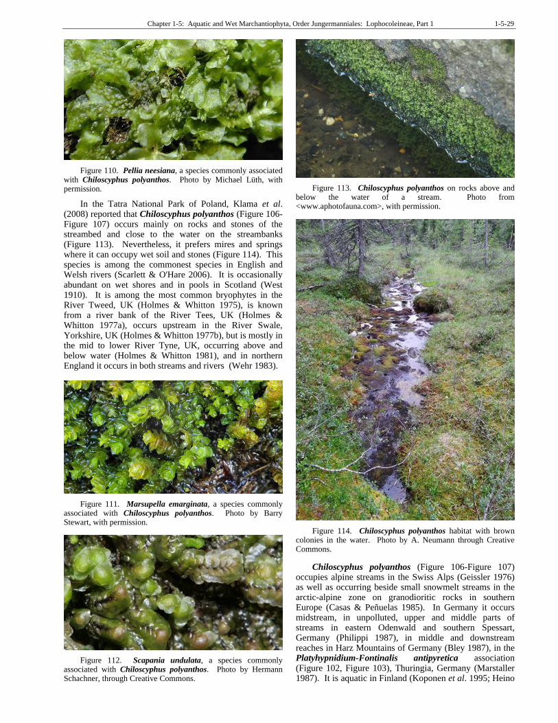
Chapter 1-5: Aquatic and Wet Marchantiophyta, Order Jungermanniales: Lophocoleineae, Part 1 1-5-29
Figure 110. Pellia neesiana, a species commonly associated with Chiloscyphus polyanthos. Photo by Michael Lüth, with permission.
In the Tatra National Park of Poland, Klama et al. (2008) reported that Chiloscyphus polyanthos (Figure 106-Figure 107) occurs mainly on rocks and stones of the streambed and close to the water on the streambanks (Figure 113). Nevertheless, it prefers mires and springs where it can occupy wet soil and stones (Figure 114). This species is among the commonest species in English and Welsh rivers (Scarlett & O'Hare 2006). It is occasionally abundant on wet shores and in pools in Scotland (West 1910). It is among the most common bryophytes in the River Tweed, UK (Holmes & Whitton 1975), is known from a river bank of the River Tees, UK (Holmes & Whitton 1977a), occurs upstream in the River Swale, Yorkshire, UK (Holmes & Whitton 1977b), but is mostly in the mid to lower River Tyne, UK, occurring above and below water (Holmes & Whitton 1981), and in northern England it occurs in both streams and rivers (Wehr 1983).
Figure 111. Marsupella emarginata, a species commonly associated with Chiloscyphus polyanthos. Photo by Barry Stewart, with permission.
Figure 112. Scapania undulata, a species commonly associated with Chiloscyphus polyanthos. Photo by Hermann Schachner, through Creative Commons.
Figure 113. Chiloscyphus polyanthos on rocks above and below the water of a stream. Photo from <www.aphotofauna.com>, with permission.
Figure 114. Chiloscyphus polyanthos habitat with brown colonies in the water. Photo by A. Neumann through Creative Commons.
Chiloscyphus polyanthos (Figure 106-Figure 107) occupies alpine streams in the Swiss Alps (Geissler 1976) as well as occurring beside small snowmelt streams in the arctic-alpine zone on granodioritic rocks in southern Europe (Casas & Peñuelas 1985). In Germany it occurs midstream, in unpolluted, upper and middle parts of streams in eastern Odenwald and southern Spessart, Germany (Philippi 1987), in middle and downstream reaches in Harz Mountains of Germany (Bley 1987), in the Platyhypnidium-Fontinalis antipyretica association (Figure 102, Figure 103), Thuringia, Germany (Marstaller 1987). It is aquatic in Finland (Koponen et al. 1995; Heino
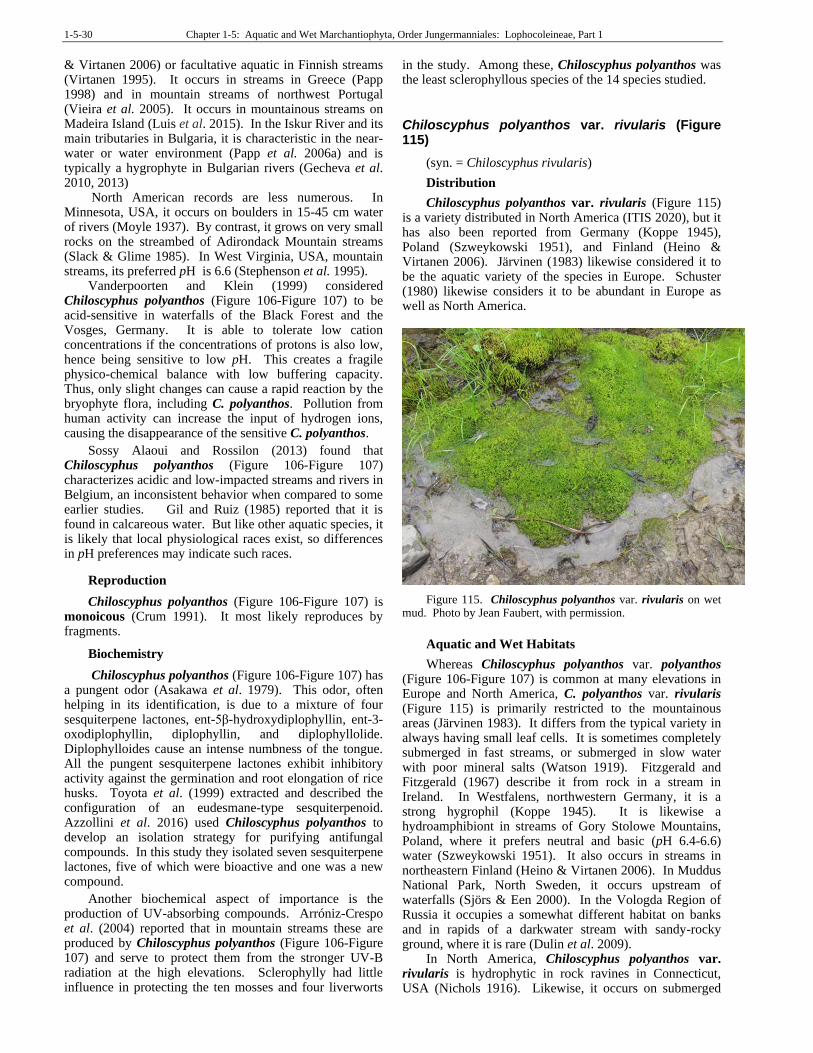
1-5-30 Chapter 1-5: Aquatic and Wet Marchantiophyta, Order Jungermanniales: Lophocoleineae, Part 1
& Virtanen 2006) or facultative aquatic in Finnish streams (Virtanen 1995). It occurs in streams in Greece (Papp 1998) and in mountain streams of northwest Portugal (Vieira et al. 2005). It occurs in mountainous streams on Madeira Island (Luis et al. 2015). In the Iskur River and its main tributaries in Bulgaria, it is characteristic in the near-water or water environment (Papp et al. 2006a) and is typically a hygrophyte in Bulgarian rivers (Gecheva et al. 2010, 2013)
North American records are less numerous. In Minnesota, USA, it occurs on boulders in 15-45 cm water of rivers (Moyle 1937). By contrast, it grows on very small rocks on the streambed of Adirondack Mountain streams (Slack & Glime 1985). In West Virginia, USA, mountain streams, its preferred pH is 6.6 (Stephenson et al. 1995).
Vanderpoorten and Klein (1999) considered Chiloscyphus polyanthos (Figure 106-Figure 107) to be acid-sensitive in waterfalls of the Black Forest and the Vosges, Germany. It is able to tolerate low cation concentrations if the concentrations of protons is also low, hence being sensitive to low pH. This creates a fragile physico-chemical balance with low buffering capacity. Thus, only slight changes can cause a rapid reaction by the bryophyte flora, including C. polyanthos. Pollution from human activity can increase the input of hydrogen ions, causing the disappearance of the sensitive C. polyanthos.
Sossy Alaoui and Rossilon (2013) found that Chiloscyphus polyanthos (Figure 106-Figure 107) characterizes acidic and low-impacted streams and rivers in Belgium, an inconsistent behavior when compared to some earlier studies. Gil and Ruiz (1985) reported that it is found in calcareous water. But like other aquatic species, it is likely that local physiological races exist, so differences in pH preferences may indicate such races.
Reproduction
Chiloscyphus polyanthos (Figure 106-Figure 107) is monoicous (Crum 1991). It most likely reproduces by fragments.
Biochemistry
Chiloscyphus polyanthos (Figure 106-Figure 107) has a pungent odor (Asakawa et al. 1979). This odor, often helping in its identification, is due to a mixture of four sesquiterpene lactones, ent-5β-hydroxydiplophyllin, ent-3-oxodiplophyllin, diplophyllin, and diplophyllolide. Diplophylloides cause an intense numbness of the tongue. All the pungent sesquiterpene lactones exhibit inhibitory activity against the germination and root elongation of rice husks. Toyota et al. (1999) extracted and described the configuration of an eudesmane-type sesquiterpenoid. Azzollini et al. 2016) used Chiloscyphus polyanthos to develop an isolation strategy for purifying antifungal compounds. In this study they isolated seven sesquiterpene lactones, five of which were bioactive and one was a new compound.
Another biochemical aspect of importance is the production of UV-absorbing compounds. Arróniz-Crespo et al. (2004) reported that in mountain streams these are produced by Chiloscyphus polyanthos (Figure 106-Figure 107) and serve to protect them from the stronger UV-B radiation at the high elevations. Sclerophylly had little influence in protecting the ten mosses and four liverworts
in the study. Among these, Chiloscyphus polyanthos was the least sclerophyllous species of the 14 species studied.
Chiloscyphus polyanthos var. rivularis (Figure 115)
(syn. = Chiloscyphus rivularis)
Distribution
Chiloscyphus polyanthos var. rivularis (Figure 115) is a variety distributed in North America (ITIS 2020), but it has also been reported from Germany (Koppe 1945), Poland (Szweykowski 1951), and Finland (Heino & Virtanen 2006). Järvinen (1983) likewise considered it to be the aquatic variety of the species in Europe. Schuster (1980) likewise considers it to be abundant in Europe as well as North America.
Figure 115. Chiloscyphus polyanthos var. rivularis on wet mud. Photo by Jean Faubert, with permission.
Aquatic and Wet Habitats
Whereas Chiloscyphus polyanthos var. polyanthos (Figure 106-Figure 107) is common at many elevations in Europe and North America, C. polyanthos var. rivularis (Figure 115) is primarily restricted to the mountainous areas (Järvinen 1983). It differs from the typical variety in always having small leaf cells. It is sometimes completely submerged in fast streams, or submerged in slow water with poor mineral salts (Watson 1919). Fitzgerald and Fitzgerald (1967) describe it from rock in a stream in Ireland. In Westfalens, northwestern Germany, it is a strong hygrophil (Koppe 1945). It is likewise a hydroamphibiont in streams of Gory Stolowe Mountains, Poland, where it prefers neutral and basic (pH 6.4-6.6) water (Szweykowski 1951). It also occurs in streams in northeastern Finland (Heino & Virtanen 2006). In Muddus National Park, North Sweden, it occurs upstream of waterfalls (Sjörs & Een 2000). In the Vologda Region of Russia it occupies a somewhat different habitat on banks and in rapids of a darkwater stream with sandy-rocky ground, where it is rare (Dulin et al. 2009).
In North America, Chiloscyphus polyanthos var. rivularis is hydrophytic in rock ravines in Connecticut, USA (Nichols 1916). Likewise, it occurs on submerged
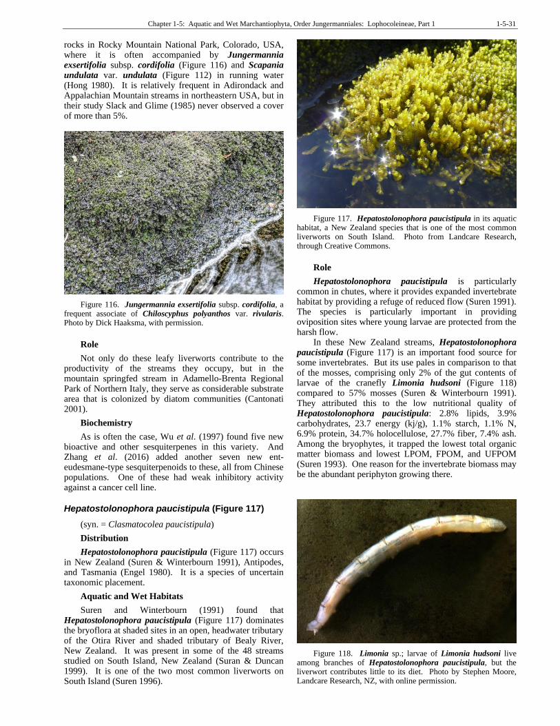
Chapter 1-5: Aquatic and Wet Marchantiophyta, Order Jungermanniales: Lophocoleineae, Part 1 1-5-31
rocks in Rocky Mountain National Park, Colorado, USA, where it is often accompanied by Jungermannia exsertifolia subsp. cordifolia (Figure 116) and Scapania undulata var. undulata (Figure 112) in running water (Hong 1980). It is relatively frequent in Adirondack and Appalachian Mountain streams in northeastern USA, but in their study Slack and Glime (1985) never observed a cover of more than 5%.
Figure 116. Jungermannia exsertifolia subsp. cordifolia, a frequent associate of Chiloscyphus polyanthos var. rivularis. Photo by Dick Haaksma, with permission.
Role
Not only do these leafy liverworts contribute to the productivity of the streams they occupy, but in the mountain springfed stream in Adamello-Brenta Regional Park of Northern Italy, they serve as considerable substrate area that is colonized by diatom communities (Cantonati 2001).
Biochemistry
As is often the case, Wu et al. (1997) found five new bioactive and other sesquiterpenes in this variety. And Zhang et al. (2016) added another seven new ent-eudesmane-type sesquiterpenoids to these, all from Chinese populations. One of these had weak inhibitory activity against a cancer cell line.
Hepatostolonophora paucistipula (Figure 117)
(syn. = Clasmatocolea paucistipula)
Distribution
Hepatostolonophora paucistipula (Figure 117) occurs in New Zealand (Suren & Winterbourn 1991), Antipodes, and Tasmania (Engel 1980). It is a species of uncertain taxonomic placement.
Aquatic and Wet Habitats
Suren and Winterbourn (1991) found that Hepatostolonophora paucistipula (Figure 117) dominates the bryoflora at shaded sites in an open, headwater tributary of the Otira River and shaded tributary of Bealy River, New Zealand. It was present in some of the 48 streams studied on South Island, New Zealand (Suran & Duncan 1999). It is one of the two most common liverworts on South Island (Suren 1996).
Figure 117. Hepatostolonophora paucistipula in its aquatic habitat, a New Zealand species that is one of the most common liverworts on South Island. Photo from Landcare Research, through Creative Commons.
Role
Hepatostolonophora paucistipula is particularly common in chutes, where it provides expanded invertebrate habitat by providing a refuge of reduced flow (Suren 1991). The species is particularly important in providing oviposition sites where young larvae are protected from the harsh flow.
In these New Zealand streams, Hepatostolonophora paucistipula (Figure 117) is an important food source for some invertebrates. But its use pales in comparison to that of the mosses, comprising only 2% of the gut contents of larvae of the cranefly Limonia hudsoni (Figure 118) compared to 57% mosses (Suren & Winterbourn 1991). They attributed this to the low nutritional quality of Hepatostolonophora paucistipula: 2.8% lipids, 3.9% carbohydrates, 23.7 energy (kj/g), 1.1% starch, 1.1% N, 6.9% protein, 34.7% holocellulose, 27.7% fiber, 7.4% ash. Among the bryophytes, it trapped the lowest total organic matter biomass and lowest LPOM, FPOM, and UFPOM (Suren 1993). One reason for the invertebrate biomass may be the abundant periphyton growing there.
Figure 118. Limonia sp.; larvae of Limonia hudsoni live among branches of Hepatostolonophora paucistipula, but the liverwort contributes little to its diet. Photo by Stephen Moore, Landcare Research, NZ, with online permission.
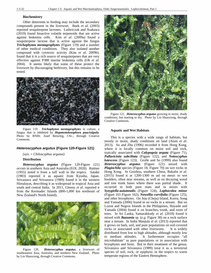
1-5-32 Chapter 1-5: Aquatic and Wet Marchantiophyta, Order Jungermanniales: Lophocoleineae, Part 1
Biochemistry
Other deterrents to feeding may include the secondary compounds present in the liverwort. Baek et al. (2003) reported sesquiterpene lactones. Ludwiczuk and Asakawa (2019) found bioactive volatile terpenoids that are active against leukemia cells. Kim et al. (2009a) found a sesquiterpene lactone that is active against the fungus Trichophyton mentagrophytes (Figure 119) and a number of other medical conditions. They also isolated another compound with cytotoxic activity (Kim et al. 2009b). found that it is a rich source of sesquiterpenes that are very effective against P388 murine leukemia cells (Oh et al. 2004). It seems likely that some of these protect the liverwort by discouraging herbivory, but this remains to be tested.
Figure 119. Trichophyton mentagrophytes in culture, a fungus that is inhibited by Hepatostolonophora paucistipula. Photo by RNDr. Josef Reischig, CSc., through Creative Commons.
Heteroscyphus argutus (Figure 120-Figure 121)
(syn. = Chiloscyphus argutus)
Distribution
Heteroscyphus argutus (Figure 120-Figure 121) occurs in southern Asia and Australia (EOL 2020). Ruttner (1955) noted it from a tuff wall in the tropics. Satake (1983) reported it as aquatic from Kyushu, Japan. Srivastava and Srivastava (1989) found it in the western Himalayas, describing it as widespread in tropical Asia and south and central India. In 2011, Glenny et al. reported it from the Kermadec Islands (800-1,000 km northeast of New Zealand's North Island).
Figure 120. Heteroscyphus argutus, a liverwort of southeastern Asia, Australia, and northern New Zealand. Photo by Lin Shanxiong, through Creative Commons.
Figure 121. Heteroscyphus argutus growing in moist, shady conditions, but starting to dry. Photo by Lin Shanxiong, through Creative Commons.
Aquatic and Wet Habitats
This is a species with a wide range of habitats, but mostly in moist, shady conditions on land (Alam et al. 2013). So and Zhu (1996) recorded it from Hong Kong, where it is locally common on moist soil and rock, typically associated with Calypogeia arguta (Figure 71), Pallavicinia subciliata (Figure 122), and Notoscyphus lutescens (Figure 123). Grolle and So (1999) also found Heteroscyphus argutus (Figure 121) mixed with Plagiochila species (Figure 18, Figure 70) on wet rocks in Hong Kong. In Guizhou, southern China, Bakalin et al. (2015) found it at 1200-1300 m asl on mesic to wet boulders, often near streams, as well as on decaying wood and tree trunk bases where there was partial shade. It occurred in both pure mats and in mixes with Syzygiella autumnalis (Figure 124), Lophocolea minor (Figure 161-Figure 162), Nowellia curvifolia (Figure 125), and other bryophytes. On Jeju (Cheju) Island, Korea, Song and Yamada (2006) found in on rocks in a stream. But on Luzon and Negros Islands in the Philippines, Hayashi and Yamada (2004) found it on branches, trunk, and roots of trees. In Sri Lanka, Samarakkody et al. (2018) found it mixed with Bazzania sp. (e.g. Figure 38) on a rock surface near a stream. In India Manjula et al. (2013) reported that it grows on bark, soil, and pure populations on soil-covered rocks or associated with other liverworts. It is widely distributed from low to high altitudes, although mostly low to medium altitudes. It furthermore occupies "all microhabitats" as pure populations or in association with bryophytes and ferns. But in their treatment of the genus, Srivastava and Srivastava (1989) treat it as a terrestrial species of soil, rock, or epiphytic in the tropics to warm temperate regions of the Eastern Hemisphere.
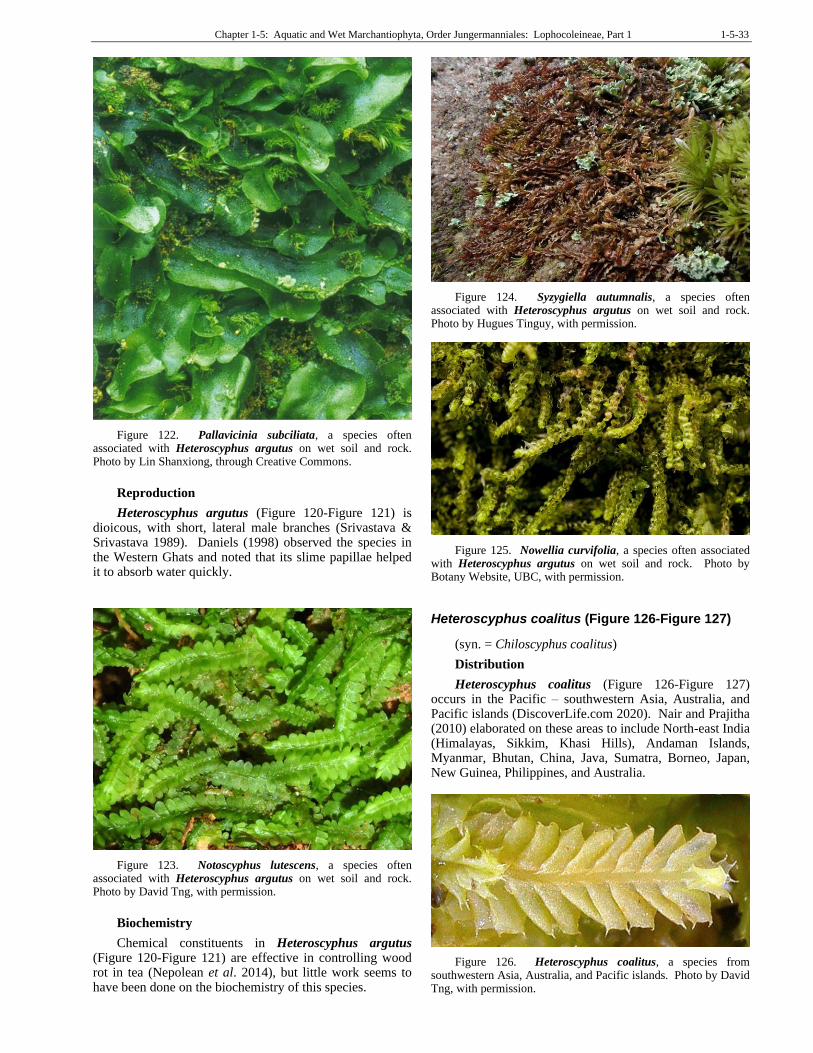
Chapter 1-5: Aquatic and Wet Marchantiophyta, Order Jungermanniales: Lophocoleineae, Part 1 1-5-33
Figure 122. Pallavicinia subciliata, a species often associated with Heteroscyphus argutus on wet soil and rock. Photo by Lin Shanxiong, through Creative Commons.
Reproduction
Heteroscyphus argutus (Figure 120-Figure 121) is dioicous, with short, lateral male branches (Srivastava & Srivastava 1989). Daniels (1998) observed the species in the Western Ghats and noted that its slime papillae helped it to absorb water quickly.
Figure 123. Notoscyphus lutescens, a species often associated with Heteroscyphus argutus on wet soil and rock. Photo by David Tng, with permission.
Biochemistry
Chemical constituents in Heteroscyphus argutus (Figure 120-Figure 121) are effective in controlling wood rot in tea (Nepolean et al. 2014), but little work seems to have been done on the biochemistry of this species.
Figure 124. Syzygiella autumnalis, a species often associated with Heteroscyphus argutus on wet soil and rock. Photo by Hugues Tinguy, with permission.
Figure 125. Nowellia curvifolia, a species often associated with Heteroscyphus argutus on wet soil and rock. Photo by Botany Website, UBC, with permission.
Heteroscyphus coalitus (Figure 126-Figure 127)
(syn. = Chiloscyphus coalitus)
Distribution
Heteroscyphus coalitus (Figure 126-Figure 127) occurs in the Pacific – southwestern Asia, Australia, and Pacific islands (DiscoverLife.com 2020). Nair and Prajitha (2010) elaborated on these areas to include North-east India (Himalayas, Sikkim, Khasi Hills), Andaman Islands, Myanmar, Bhutan, China, Java, Sumatra, Borneo, Japan, New Guinea, Philippines, and Australia.
Figure 126. Heteroscyphus coalitus, a species from southwestern Asia, Australia, and Pacific islands. Photo by David Tng, with permission.
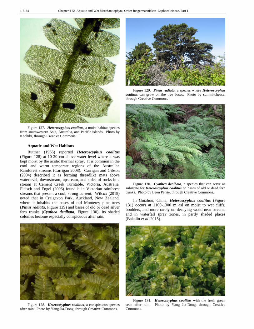
1-5-34 Chapter 1-5: Aquatic and Wet Marchantiophyta, Order Jungermanniales: Lophocoleineae, Part 1
Figure 127. Heteroscyphus coalitus, a moist habitat species from southwestern Asia, Australia, and Pacific islands. Photo by Kochibi, through Creative Commons.
Aquatic and Wet Habitats
Ruttner (1955) reported Heteroscyphus coalitus (Figure 128) at 10-20 cm above water level where it was kept moist by the acidic thermal spray. It is common in the cool and warm temperate regions of the Australian Rainforest streams (Carrigan 2008). Carrigan and Gibson (2004) described it as forming threadlike mats above waterlevel, downstream, upstream, and sides of rocks in a stream at Cement Creek Turntable, Victoria, Australia. Fleisch and Engel (2006) found it in Victorian rainforest streams that present a cool, strong current. Wilcox (2018) noted that in Craigavon Park, Auckland, New Zealand, where it inhabits the bases of old Monterey pine trees (Pinus radiata, Figure 129) and bases of old or dead silver fern trunks (Cyathea dealbata, Figure 130), its shaded colonies become especially conspicuous after rain.
Figure 128. Heteroscyphus coalitus, a conspicuous species after rain. Photo by Yang Jia-Dong, through Creative Commons.
Figure 129. Pinus radiata, a species where Heteroscyphus coalitus can grow on the tree bases. Photo by summitcheese, through Creative Commons.
Figure 130. Cyathea dealbata, a species that can serve as substrate for Heteroscyphus coalitus on bases of old or dead fern trunks. Photo by Leon Perrie, through Creative Commons.
In Guizhou, China, Heteroscyphus coalitus (Figure 131) occurs at 1100-1300 m asl on moist to wet cliffs, boulders, and more rarely on decaying wood near streams and in waterfall spray zones, in partly shaded places (Bakalin et al. 2015).
Figure 131. Heteroscyphus coalitus with the fresh green seen after rain. Photo by Yang Jia-Dong, through Creative Commons.
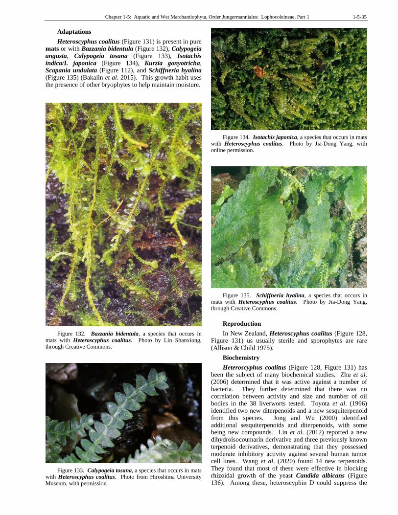
Chapter 1-5: Aquatic and Wet Marchantiophyta, Order Jungermanniales: Lophocoleineae, Part 1 1-5-35
Adaptations
Heteroscyphus coalitus (Figure 131) is present in pure mats or with Bazzania bidentula (Figure 132), Calypogeia angusta, Calypogeia tosana (Figure 133), Isotachis indica/I. japonica (Figure 134), Kurzia gonyotricha, Scapania undulata (Figure 112), and Schiffneria hyalina (Figure 135) (Bakalin et al. 2015). This growth habit uses the presence of other bryophytes to help maintain moisture.
Figure 132. Bazzania bidentula, a species that occurs in mats with Heteroscyphus coalitus. Photo by Lin Shanxiong, through Creative Commons.
Figure 133. Calypogeia tosana, a species that occurs in mats with Heteroscyphus coalitus. Photo from Hiroshima University Museum, with permission.
Figure 134. Isotachis japonica, a species that occurs in mats with Heteroscyphus coalitus. Photo by Jia-Dong Yang, with online permission.
Figure 135. Schiffneria hyalina, a species that occurs in mats with Heteroscyphus coalitus. Photo by Jia-Dong Yang, through Creative Commons.
Reproduction
In New Zealand, Heteroscyphus coalitus (Figure 128, Figure 131) us usually sterile and sporophytes are rare (Allison & Child 1975).
Biochemistry
Heteroscyphus coalitus (Figure 128, Figure 131) has been the subject of many biochemical studies. Zhu et al. (2006) determined that it was active against a number of bacteria. They further determined that there was no correlation between activity and size and number of oil bodies in the 38 liverworts tested. Toyota et al. (1996) identified two new diterpenoids and a new sesquiterpenoid from this species. Jong and Wu (2000) identified additional sesquiterpenoids and diterpenoids, with some being new compounds. Lin et al. (2012) reported a new dihydroisocoumarin derivative and three previously known terpenoid derivatives, demonstrating that they possessed moderate inhibitory activity against several human tumor cell lines. Wang et al. (2020) found 14 new terpenoids. They found that most of these were effective in blocking rhizoidal growth of the yeast Candida albicans (Figure 136). Among these, heteroscyphin D could suppress the
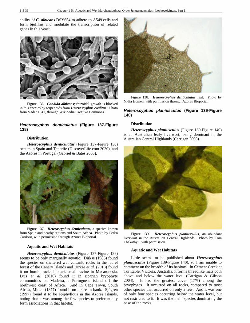
1-5-36 Chapter 1-5: Aquatic and Wet Marchantiophyta, Order Jungermanniales: Lophocoleineae, Part 1
ability of C. albicans DSY654 to adhere to A549 cells and form biofilms and modulate the transcription of related genes in this yeast.
Figure 136. Candida albicans; rhizoidal growth is blocked in this species by terpenoids from Heteroscyphus coalitus. Photo from Vader 1941, through Wikipedia Creative Commons.
Heteroscyphus denticulatus (Figure 137-Figure 138)
Distribution
Heteroscyphus denticulatus (Figure 137-Figure 138) occurs in Spain and Tenerife (DiscoverLife.com 2020), and the Azores in Portugal (Gabriel & Bates 2005).
Figure 137. Heteroscyphus denticulatus, a species known from Spain and nearby regions and South Africa. Photo by Pedro Cardoso, with permission through Azores Bioportal.
Aquatic and Wet Habitats
Heteroscyphus denticulatus (Figure 137-Figure 138) seems to be only marginally aquatic. Dirkse (1985) found the species on sheltered wet volcanic rocks in the laurel forest of the Canary Islands and Dirkse et al. (2018) found it on humid rocks in dark small ravine in Macaronesia. Luís et al. (2010) found it in riparian bryophyte communities on Madeira, a Portuguese island off the northwest coast of Africa. And in Cape Town, South Africa, Mitten (1877) found it on a stream bank. Sjögren (1997) found it to be epiphyllous in the Azores Islands, noting that it was among the few species to preferentially form associations in that habitat.
Figure 138. Heteroscyphus denticulatus leaf. Photo by Nidia Homen, with permission through Azores Bioportal.
Heteroscyphus planiusculus (Figure 139-Figure 140)
Distribution
Heteroscyphus planiusculus (Figure 139-Figure 140) is an Australian leafy liverwort, being dominant in the Australian Central Highlands (Carrigan 2008).
Figure 139. Heteroscyphus planiusculus, an abundant liverwort in the Australian Central Highlands. Photo by Tom Thekathyil, with permission.
Aquatic and Wet Habitats
Little seems to be published about Heteroscyphus planiusculus (Figure 139-Figure 140), so I am unable to comment on the breadth of its habitats. In Cement Creek at Turntable, Victoria, Australia, it forms threadlike mats both above and below the water level (Carrigan & Gibson 2004). It had the greatest cover (17%) among the bryophytes. It occurred on all rocks, compared to most other species that occurred on only a few. And it was one of only four species occurring below the water level, but not restricted to it. It was the main species dominating the base of the rocks.
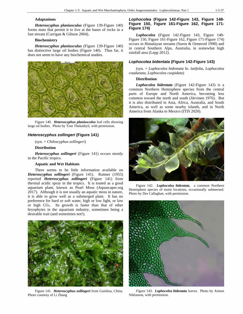
Chapter 1-5: Aquatic and Wet Marchantiophyta, Order Jungermanniales: Lophocoleineae, Part 1 1-5-37
Adaptations
Heteroscyphus planiusculus (Figure 139-Figure 140) forms mats that permit it to live at the bases of rocks in a fast stream (Carrigan & Gibson 2004).
Biochemistry
Heteroscyphus planiusculus (Figure 139-Figure 140) has distinctive large oil bodies (Figure 140). Thus far, it does not seem to have any biochemical studies.
Figure 140. Heteroscyphus planiusculus leaf cells showing large oil bodies. Photo by Tom Thekathyil, with permission.
Heteroscyphus zollingeri (Figure 141)
(syn. = Chiloscyphus zollingeri)
Distribution
Heteroscyphus zollingeri (Figure 141) occurs mostly in the Pacific tropics.
Aquatic and Wet Habitats
There seems to be little information available on Heteroscyphus zollingeri (Figure 141). Ruttner (1955) reported Heteroscyphus zollingeri (Figure 141) from thermal acidic spray in the tropics. It is touted as a good aquarium plant, known as Pearl Moss (Aquascaper.org 2017). Although it is not usually an aquatic moss in nature, it is able to grow well as a submerged plant. It has no preference for hard or soft water, high or low light, or low or high CO2. Its growth is faster than that of other bryophytes in the aquarium industry, sometimes being a desirable trait (and sometimes not!).
Figure 141. Heteroscyphus zollingeri from Guizhou, China. Photo courtesy of Li Zhang
Lophocolea (Figure 142-Figure 143, Figure 148-Figure 150, Figure 161-Figure 162, Figure 171-Figure 174)
Lophocolea (Figure 142-Figure 143, Figure 148-Figure 150, Figure 161-Figure 162, Figure 171-Figure 174) occurs in Himalayan streams (Suren & Ormerod 1998) and in central Southern Alps, Australia, in somewhat high rainfall area (Lepp 2012).
Lophocolea bidentata (Figure 142-Figure 143)
(syn. = Lophocolea bidentata fo. latifolia, Lophocolea coadunata, Lophocolea cuspidata)
Distribution
Lophocolea bidentata (Figure 142-Figure 143) is a common Northern Hemisphere species from the central parts of Europe and North America, becoming less common toward the north and south (Järvinen 1976). But it is also distributed in Asia, Africa, Australia, and South America, as well as some nearby islands, and in North America from Alaska to Mexico (ITIS 2020).
Figure 142. Lophocolea bidentata, a common Northern Hemisphere species of moist locations, occasionally submersed. Photo by Des Callaghan, with permission.
Figure 143. Lophocolea bidentata leaves. Photo by Aimon Niklasson, with permission.
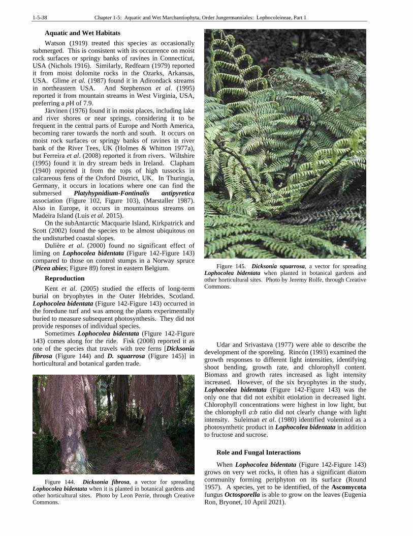
1-5-38 Chapter 1-5: Aquatic and Wet Marchantiophyta, Order Jungermanniales: Lophocoleineae, Part 1
Aquatic and Wet Habitats
Watson (1919) treated this species as occasionally submerged. This is consistent with its occurrence on moist rock surfaces or springy banks of ravines in Connecticut, USA (Nichols 1916). Similarly, Redfearn (1979) reported it from moist dolomite rocks in the Ozarks, Arkansas, USA. Glime et al. (1987) found it in Adirondack streams in northeastern USA. And Stephenson et al. (1995) reported it from mountain streams in West Virginia, USA, preferring a pH of 7.9.
Järvinen (1976) found it in moist places, including lake and river shores or near springs, considering it to be frequent in the central parts of Europe and North America, becoming rarer towards the north and south. It occurs on moist rock surfaces or springy banks of ravines in river bank of the River Tees, UK (Holmes & Whitton 1977a), but Ferreira et al. (2008) reported it from rivers. Wiltshire (1995) found it in dry stream beds in Ireland. Clapham (1940) reported it from the tops of high tussocks in calcareous fens of the Oxford District, UK. In Thuringia, Germany, it occurs in locations where one can find the submersed Platyhypnidium-Fontinalis antipyretica association (Figure 102, Figure 103), (Marstaller 1987). Also in Europe, it occurs in mountainous streams on Madeira Island (Luis et al. 2015).
On the subAntarctic Macquarie Island, Kirkpatrick and Scott (2002) found the species to be almost ubiquitous on the undisturbed coastal slopes.
Dulière et al. (2000) found no significant effect of liming on Lophocolea bidentata (Figure 142-Figure 143) compared to those on control stumps in a Norway spruce (Picea abies; Figure 89) forest in eastern Belgium.
Reproduction
Kent et al. (2005) studied the effects of long-term burial on bryophytes in the Outer Hebrides, Scotland. Lophocolea bidentata (Figure 142-Figure 143) occurred in the foredune turf and was among the plants experimentally buried to measure subsequent photosynthesis. They did not provide responses of individual species.
Sometimes Lophocolea bidentata (Figure 142-Figure 143) comes along for the ride. Fisk (2008) reported it as one of the species that travels with tree ferns [Dicksonia fibrosa (Figure 144) and D. squarrosa (Figure 145)] in horticultural and botanical garden trade.
Figure 144. Dicksonia fibrosa, a vector for spreading Lophocolea bidentata when it is planted in botanical gardens and other horticultural sites. Photo by Leon Perrie, through Creative Commons.
Figure 145. Dicksonia squarrosa, a vector for spreading Lophocolea bidentata when planted in botanical gardens and other horticultural sites. Photo by Jeremy Rolfe, through Creative Commons.
Udar and Srivastava (1977) were able to describe the development of the sporeling. Rincón (1993) examined the growth responses to different light intensities, identifying shoot bending, growth rate, and chlorophyll content. Biomass and growth rates increased as light intensity increased. However, of the six bryophytes in the study, Lophocolea bidentata (Figure 142-Figure 143) was the only one that did not exhibit etiolation in decreased light. Chlorophyll concentrations were highest in low light, but the chlorophyll a:b ratio did not clearly change with light intensity. Suleiman et al. (1980) identified volemitol as a photosynthetic product in Lophocolea bidentata in addition to fructose and sucrose.
Role and Fungal Interactions
When Lophocolea bidentata (Figure 142-Figure 143) grows on very wet rocks, it often has a significant diatom community forming periphyton on its surface (Round 1957). A species, yet to be identified, of the Ascomycota fungus Octosporella is able to grow on the leaves (Eugenia Ron, Bryonet, 10 April 2021).
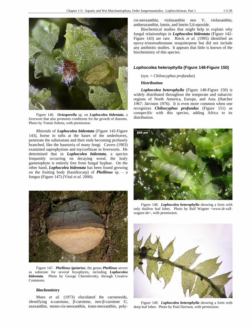
Chapter 1-5: Aquatic and Wet Marchantiophyta, Order Jungermanniales: Lophocoleineae, Part 1 1-5-39
Figure 146. Octosporella sp. on Lophocolea bidentata, a liverwort that also promotes conditions for the growth of diatoms. Photo by Tomás Sobota, with permission.
Rhizoids of Lophocolea bidentata (Figure 142-Figure 143), borne in tufts at the bases of the underleaves, penetrate the substratum and their ends becoming profusely branched, like the haustoria of many fungi. Cavers (1903) examined saprophytism and mycorrhizae in liverworts. He determined that in Lophocolea bidentata, a species frequently occurring on decaying wood, the leafy gametophyte is entirely free from fungal hyphae. On the other hand, Lophocolea bidentata has been found growing on the fruiting body (basidiocarp) of Phellinus sp. – a fungus (Figure 147) (Vital et al. 2000).
Figure 147. Phellinus igniarius; the genus Phellinus serves as substrate for several bryophytes, including Lophocolea bidentata. Photo by George Chernilevsky, through Creative Commons.
Biochemistry
Mues et al. (1973) elucidated the carotenoids, identifying α-carotene, β-carotene, neo-β-carotene U, zeaxanthin, mono-cis-neoxanthin, trans-neoxanthin, poly-
cis-neoxanthin, violaxanthin neo V, violaxanthin, antheraxanthin, lutein, and lutein-5,6-epoxide.
Biochemical studies that might help to explain why fungal relationships in Lophocolea bidentata (Figure 142-Figure 143) are rare. Rieck et al. (1995) identified an epoxy-trinoreudesmane sesquiterpene but did not include any antibiotic studies. It appears that little is known of the biochemistry of this species.
Lophocolea heterophylla (Figure 148-Figure 150)
(syn. = Chiloscyphus profundus)
Distribution
Lophocolea heterophylla (Figure 148-Figure 150) is widely distributed throughout the temperate and subarctic regions of North America, Europe, and Asia (Hatcher 1967; Järvinen 1976). It is even more common when one recognizes Chiloscyphus profundus (Figure 151) as conspecific with this species, adding Africa to its distribution.
Figure 148. Lophocolea heterophylla showing a form with only shallow leaf lobes. Photo by Ralf Wagner <www.dr-ralf-wagner.de>, with permission.
Figure 149. Lophocolea heterophylla showing a form with deep leaf lobes. Photo by Paul Davison, with permission.
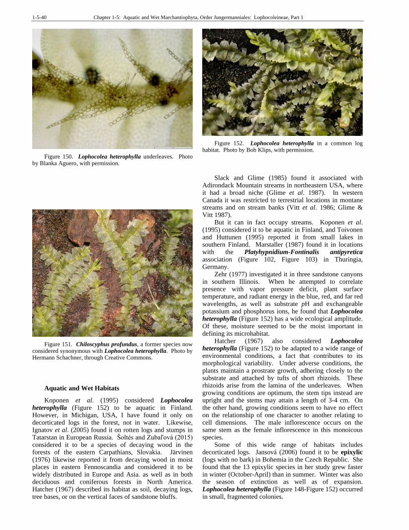
1-5-40 Chapter 1-5: Aquatic and Wet Marchantiophyta, Order Jungermanniales: Lophocoleineae, Part 1
Figure 150. Lophocolea heterophylla underleaves. Photo by Blanka Aguero, with permission.
Figure 151. Chiloscyphus profundus, a former species now considered synonymous with Lophocolea heterophylla. Photo by Hermann Schachner, through Creative Commons.
Aquatic and Wet Habitats
Koponen et al. (1995) considered Lophocolea heterophylla (Figure 152) to be aquatic in Finland. However, in Michigan, USA, I have found it only on decorticated logs in the forest, not in water. Likewise, Ignatov et al. (2005) found it on rotten logs and stumps in Tatarstan in European Russia. Šoltés and Zubaľová (2015) considered it to be a species of decaying wood in the forests of the eastern Carpathians, Slovakia. Järvinen (1976) likewise reported it from decaying wood in moist places in eastern Fennoscandia and considered it to be widely distributed in Europe and Asia. as well as in both deciduous and coniferous forests in North America. Hatcher (1967) described its habitat as soil, decaying logs, tree bases, or on the vertical faces of sandstone bluffs.
Figure 152. Lophocolea heterophylla in a common log habitat. Photo by Bob Klips, with permission.
Slack and Glime (1985) found it associated with Adirondack Mountain streams in northeastern USA, where it had a broad niche (Glime et al. 1987). In western Canada it was restricted to terrestrial locations in montane streams and on stream banks (Vitt et al. 1986; Glime & Vitt 1987).
But it can in fact occupy streams. Koponen et al. (1995) considered it to be aquatic in Finland, and Toivonen and Huttunen (1995) reported it from small lakes in southern Finland. Marstaller (1987) found it in locations with the Platyhypnidium-Fontinalis antipyretica association (Figure 102, Figure 103) in Thuringia, Germany.
Zehr (1977) investigated it in three sandstone canyons in southern Illinois. When he attempted to correlate presence with vapor pressure deficit, plant surface temperature, and radiant energy in the blue, red, and far red wavelengths, as well as substrate pH and exchangeable potassium and phosphorus ions, he found that Lophocolea heterophylla (Figure 152) has a wide ecological amplitude. Of these, moisture seemed to be the moist important in defining its microhabitat.
Hatcher (1967) also considered Lophocolea heterophylla (Figure 152) to be adapted to a wide range of environmental conditions, a fact that contributes to its morphological variability. Under adverse conditions, the plants maintain a prostrate growth, adhering closely to the substrate and attached by tufts of short rhizoids. These rhizoids arise from the lamina of the underleaves. When growing conditions are optimum, the stem tips instead are upright and the stems may attain a length of 3-4 cm. On the other hand, growing conditions seem to have no effect on the relationship of one character to another relating to cell dimensions. The male inflorescence occurs on the same stem as the female inflorescence in this monoicous species.
Some of this wide range of habitats includes decorticated logs. Jansová (2006) found it to be epixylic (logs with no bark) in Bohemia in the Czech Republic. She found that the 13 epixylic species in her study grew faster in winter (October-April) than in summer. Winter was also the season of extinction as well as of expansion. Lophocolea heterophylla (Figure 148-Figure 152) occurred in small, fragmented colonies.
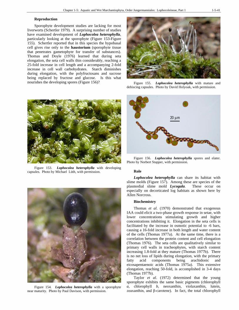
Chapter 1-5: Aquatic and Wet Marchantiophyta, Order Jungermanniales: Lophocoleineae, Part 1 1-5-41
Reproduction
Sporophyte development studies are lacking for most liverworts (Schertler 1979). A surprising number of studies have examined development of Lophocolea heterophylla, particularly looking at the sporophyte (Figure 153-Figure 155). Schertler reported that in this species the hypobasal cell gives rise only to the haustorium (sporophyte tissue that penetrates gametophyte for transfer of substances). Thomas and Doyle (1976) learned that during seta elongation, the seta cell walls thin considerably, reaching a 25-fold increase in cell length and a accompanying 2-fold increase in cell wall carbohydrates. Starch diminishes during elongation, with the polyfructosans and sucrose being replaced by fructose and glucose. Is this what nourishes the developing spores (Figure 156)?
Figure 153. Lophocolea heterophylla with developing capsules. Photo by Michael Lüth, with permission.
Figure 154. Lophocolea heterophylla with a sporophyte near maturity. Photo by Paul Davison, with permission.
Figure 155. Lophocolea heterophylla with mature and dehiscing capsules. Photo by David Holyoak, with permission.
Figure 156. Lophocolea heterophylla spores and elater. Photo by Norbert Stapper, with permission.
Role
Lophocolea heterophylla can share its habitat with slime molds (Figure 157). Among these are species of the plasmodial slime mold Lycogala. These occur on especially on decorticated log habitats as shown here by Allen Norcross.
Biochemistry
Thomas et al. (1970) demonstrated that exogenous IAA could elicit a two-phase growth response in setae, with lower concentrations stimulating growth and higher concentrations inhibiting it. Elongation in the seta cells is facilitated by the increase in osmotic potential to -6 bars, causing a 16-fold increase in both length and water content of the cells (Thomas 1977a). At the same time, there is a correlation between the protein content and cell elongation (Thomas 1976). The seta cells are qualitatively similar to primary cell walls in tracheophytes, with starch content increasing 1.8-fold at they mature (Thomas 1977b). There is no net loss of lipids during elongation, with the primary fatty acid components being arachidonic and eicosapentaenoic acids (Thomas 1975a). This extensive elongation, reaching 50-fold, is accomplished in 3-4 days (Thomas 1977b).
Taylor et al. (1972) determined that the young sporophyte exhibits the same basic pigments (chlorophyll a, chlorophyll b, neoxanthin, violaxanthin, lutein, zeaxanthin, and β-carotene). In fact, the total chlorophyll
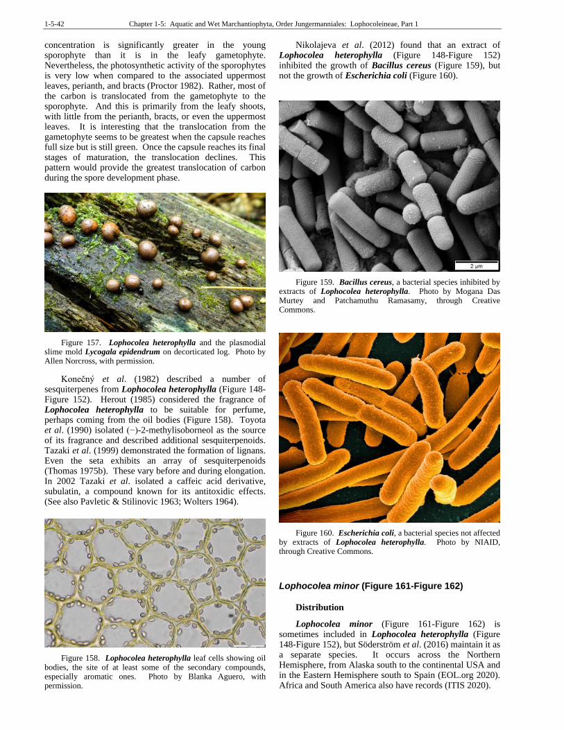
1-5-42 Chapter 1-5: Aquatic and Wet Marchantiophyta, Order Jungermanniales: Lophocoleineae, Part 1
concentration is significantly greater in the young sporophyte than it is in the leafy gametophyte. Nevertheless, the photosynthetic activity of the sporophytes is very low when compared to the associated uppermost leaves, perianth, and bracts (Proctor 1982). Rather, most of the carbon is translocated from the gametophyte to the sporophyte. And this is primarily from the leafy shoots, with little from the perianth, bracts, or even the uppermost leaves. It is interesting that the translocation from the gametophyte seems to be greatest when the capsule reaches full size but is still green. Once the capsule reaches its final stages of maturation, the translocation declines. This pattern would provide the greatest translocation of carbon during the spore development phase.
Figure 157. Lophocolea heterophylla and the plasmodial slime mold Lycogala epidendrum on decorticated log. Photo by Allen Norcross, with permission.
Konečný et al. (1982) described a number of sesquiterpenes from Lophocolea heterophylla (Figure 148-Figure 152). Herout (1985) considered the fragrance of Lophocolea heterophylla to be suitable for perfume, perhaps coming from the oil bodies (Figure 158). Toyota et al. (1990) isolated (−)-2-methylisoborneol as the source of its fragrance and described additional sesquiterpenoids. Tazaki et al. (1999) demonstrated the formation of lignans. Even the seta exhibits an array of sesquiterpenoids (Thomas 1975b). These vary before and during elongation. In 2002 Tazaki et al. isolated a caffeic acid derivative, subulatin, a compound known for its antitoxidic effects. (See also Pavletic & Stilinovic 1963; Wolters 1964).
Figure 158. Lophocolea heterophylla leaf cells showing oil bodies, the site of at least some of the secondary compounds, especially aromatic ones. Photo by Blanka Aguero, with permission.
Nikolajeva et al. (2012) found that an extract of Lophocolea heterophylla (Figure 148-Figure 152) inhibited the growth of Bacillus cereus (Figure 159), but not the growth of Escherichia coli (Figure 160).
Figure 159. Bacillus cereus, a bacterial species inhibited by extracts of Lophocolea heterophylla. Photo by Mogana Das Murtey and Patchamuthu Ramasamy, through Creative Commons.
Figure 160. Escherichia coli, a bacterial species not affected by extracts of Lophocolea heterophylla. Photo by NIAID, through Creative Commons.
Lophocolea minor (Figure 161-Figure 162)
Distribution
Lophocolea minor (Figure 161-Figure 162) is sometimes included in Lophocolea heterophylla (Figure 148-Figure 152), but Söderström et al. (2016) maintain it as a separate species. It occurs across the Northern Hemisphere, from Alaska south to the continental USA and in the Eastern Hemisphere south to Spain (EOL.org 2020). Africa and South America also have records (ITIS 2020).
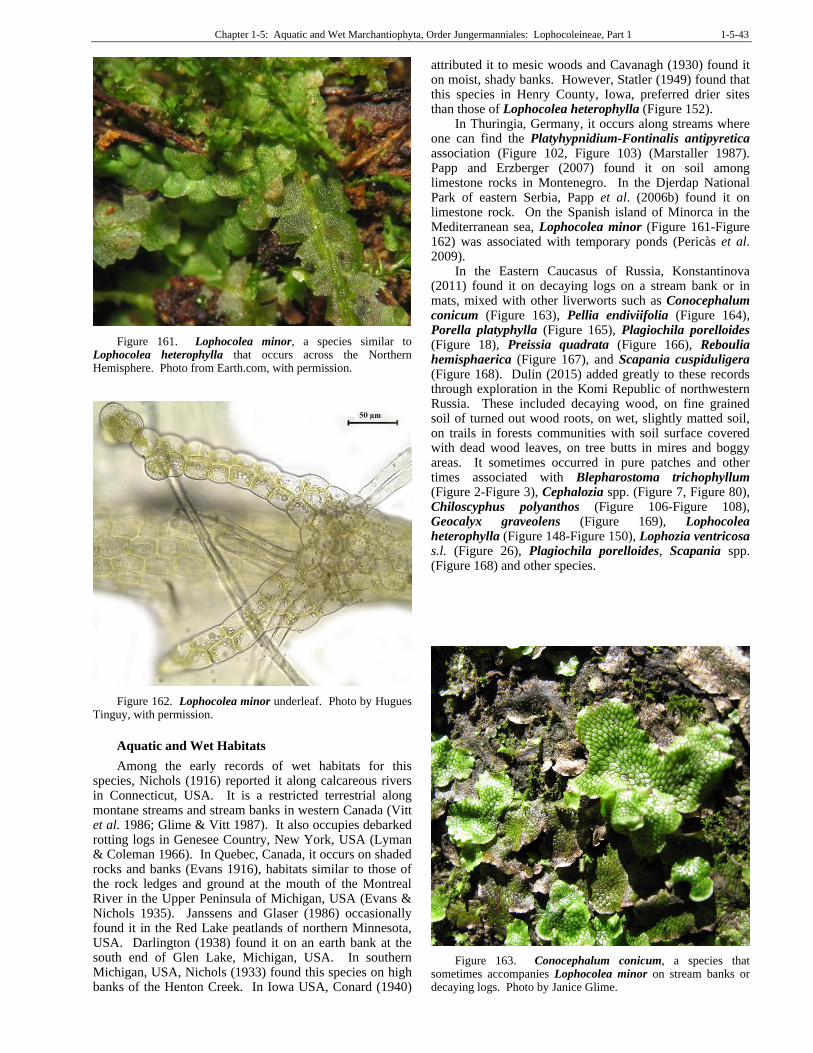
Chapter 1-5: Aquatic and Wet Marchantiophyta, Order Jungermanniales: Lophocoleineae, Part 1 1-5-43
Figure 161. Lophocolea minor, a species similar to Lophocolea heterophylla that occurs across the Northern Hemisphere. Photo from Earth.com, with permission.
Figure 162. Lophocolea minor underleaf. Photo by Hugues Tinguy, with permission.
Aquatic and Wet Habitats
Among the early records of wet habitats for this species, Nichols (1916) reported it along calcareous rivers in Connecticut, USA. It is a restricted terrestrial along montane streams and stream banks in western Canada (Vitt et al. 1986; Glime & Vitt 1987). It also occupies debarked rotting logs in Genesee Country, New York, USA (Lyman & Coleman 1966). In Quebec, Canada, it occurs on shaded rocks and banks (Evans 1916), habitats similar to those of the rock ledges and ground at the mouth of the Montreal River in the Upper Peninsula of Michigan, USA (Evans & Nichols 1935). Janssens and Glaser (1986) occasionally found it in the Red Lake peatlands of northern Minnesota, USA. Darlington (1938) found it on an earth bank at the south end of Glen Lake, Michigan, USA. In southern Michigan, USA, Nichols (1933) found this species on high banks of the Henton Creek. In Iowa USA, Conard (1940)
attributed it to mesic woods and Cavanagh (1930) found it on moist, shady banks. However, Statler (1949) found that this species in Henry County, Iowa, preferred drier sites than those of Lophocolea heterophylla (Figure 152).
In Thuringia, Germany, it occurs along streams where one can find the Platyhypnidium-Fontinalis antipyretica association (Figure 102, Figure 103) (Marstaller 1987). Papp and Erzberger (2007) found it on soil among limestone rocks in Montenegro. In the Djerdap National Park of eastern Serbia, Papp et al. (2006b) found it on limestone rock. On the Spanish island of Minorca in the Mediterranean sea, Lophocolea minor (Figure 161-Figure 162) was associated with temporary ponds (Pericàs et al. 2009).
In the Eastern Caucasus of Russia, Konstantinova (2011) found it on decaying logs on a stream bank or in mats, mixed with other liverworts such as Conocephalum conicum (Figure 163), Pellia endiviifolia (Figure 164), Porella platyphylla (Figure 165), Plagiochila porelloides (Figure 18), Preissia quadrata (Figure 166), Reboulia hemisphaerica (Figure 167), and Scapania cuspiduligera (Figure 168). Dulin (2015) added greatly to these records through exploration in the Komi Republic of northwestern Russia. These included decaying wood, on fine grained soil of turned out wood roots, on wet, slightly matted soil, on trails in forests communities with soil surface covered with dead wood leaves, on tree butts in mires and boggy areas. It sometimes occurred in pure patches and other times associated with Blepharostoma trichophyllum (Figure 2-Figure 3), Cephalozia spp. (Figure 7, Figure 80), Chiloscyphus polyanthos (Figure 106-Figure 108), Geocalyx graveolens (Figure 169), Lophocolea heterophylla (Figure 148-Figure 150), Lophozia ventricosa s.l. (Figure 26), Plagiochila porelloides, Scapania spp. (Figure 168) and other species.
Figure 163. Conocephalum conicum, a species that sometimes accompanies Lophocolea minor on stream banks or decaying logs. Photo by Janice Glime.
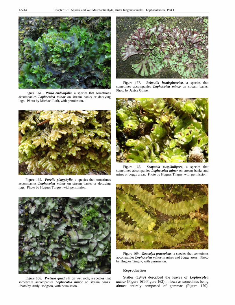
1-5-44 Chapter 1-5: Aquatic and Wet Marchantiophyta, Order Jungermanniales: Lophocoleineae, Part 1
Figure 164. Pellia endiviifolia, a species that sometimes accompanies Lophocolea minor on stream banks or decaying logs. Photo by Michael Lüth, with permission.
Figure 165. Porella platyphylla, a species that sometimes accompanies Lophocolea minor on stream banks or decaying logs. Photo by Hugues Tinguy, with permission.
Figure 166. Preissia quadrata on wet rock, a species that sometimes accompanies Lophocolea minor on stream banks. Photo by Andy Hodgson, with permission.
Figure 167. Reboulia hemisphaerica, a species that sometimes accompanies Lophocolea minor on stream banks. Photo by Janice Glime.
Figure 168. Scapania cuspiduligera, a species that sometimes accompanies Lophocolea minor on stream banks and mires or boggy areas. Photo by Hugues Tinguy, with permission.
Figure 169. Geocalyx graveolens, a species that sometimes accompanies Lophocolea minor in mires and boggy areas. Photo by Hugues Tinguy, with permission.
Reproduction
Statler (1949) described the leaves of Lophocolea minor (Figure 161-Figure 162) in Iowa as sometimes being almost entirely composed of gemmae (Figure 170).
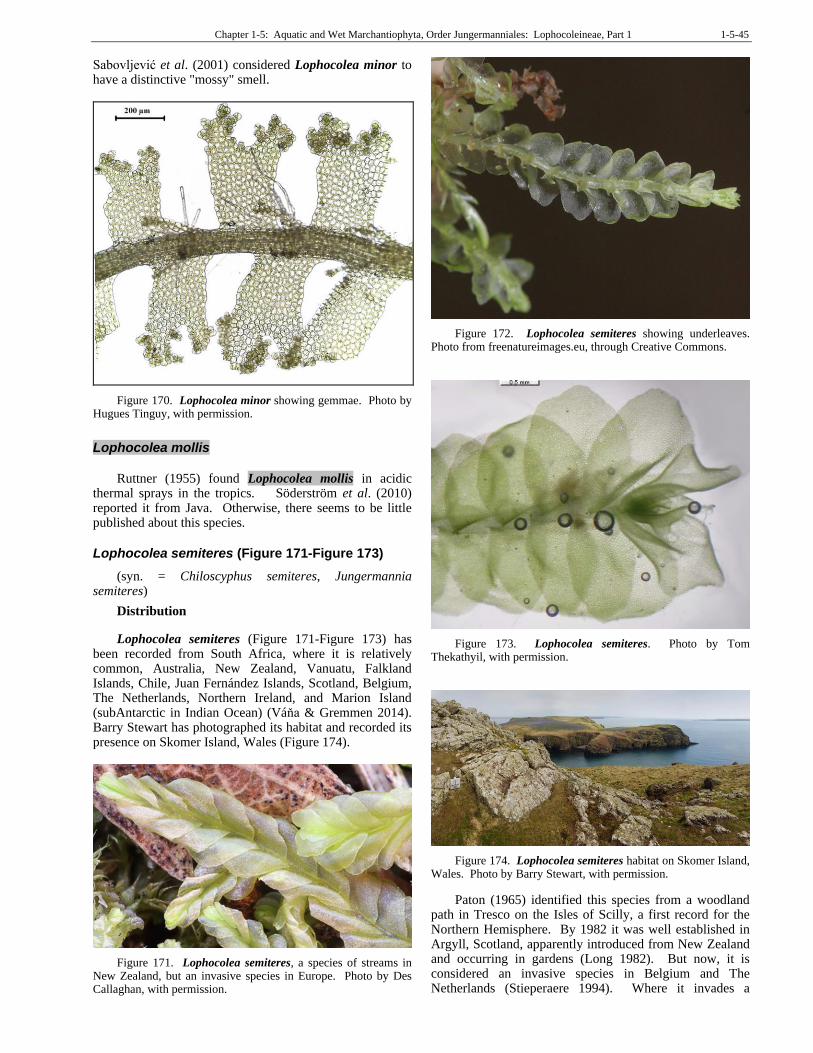
Chapter 1-5: Aquatic and Wet Marchantiophyta, Order Jungermanniales: Lophocoleineae, Part 1 1-5-45
Sabovljević et al. (2001) considered Lophocolea minor to have a distinctive "mossy" smell.
Figure 170. Lophocolea minor showing gemmae. Photo by Hugues Tinguy, with permission.
Lophocolea mollis
Ruttner (1955) found Lophocolea mollis in acidic
thermal sprays in the tropics. Söderström et al. (2010) reported it from Java. Otherwise, there seems to be little published about this species.
Lophocolea semiteres (Figure 171-Figure 173)
(syn. = Chiloscyphus semiteres, Jungermannia semiteres)
Distribution
Lophocolea semiteres (Figure 171-Figure 173) has been recorded from South Africa, where it is relatively common, Australia, New Zealand, Vanuatu, Falkland Islands, Chile, Juan Fernández Islands, Scotland, Belgium, The Netherlands, Northern Ireland, and Marion Island (subAntarctic in Indian Ocean) (Váňa & Gremmen 2014). Barry Stewart has photographed its habitat and recorded its presence on Skomer Island, Wales (Figure 174).
Figure 171. Lophocolea semiteres, a species of streams in New Zealand, but an invasive species in Europe. Photo by Des Callaghan, with permission.
Figure 172. Lophocolea semiteres showing underleaves. Photo from freenatureimages.eu, through Creative Commons.
Figure 173. Lophocolea semiteres. Photo by Tom Thekathyil, with permission.
Figure 174. Lophocolea semiteres habitat on Skomer Island, Wales. Photo by Barry Stewart, with permission.
Paton (1965) identified this species from a woodland path in Tresco on the Isles of Scilly, a first record for the Northern Hemisphere. By 1982 it was well established in Argyll, Scotland, apparently introduced from New Zealand and occurring in gardens (Long 1982). But now, it is considered an invasive species in Belgium and The Netherlands (Stieperaere 1994). Where it invades a
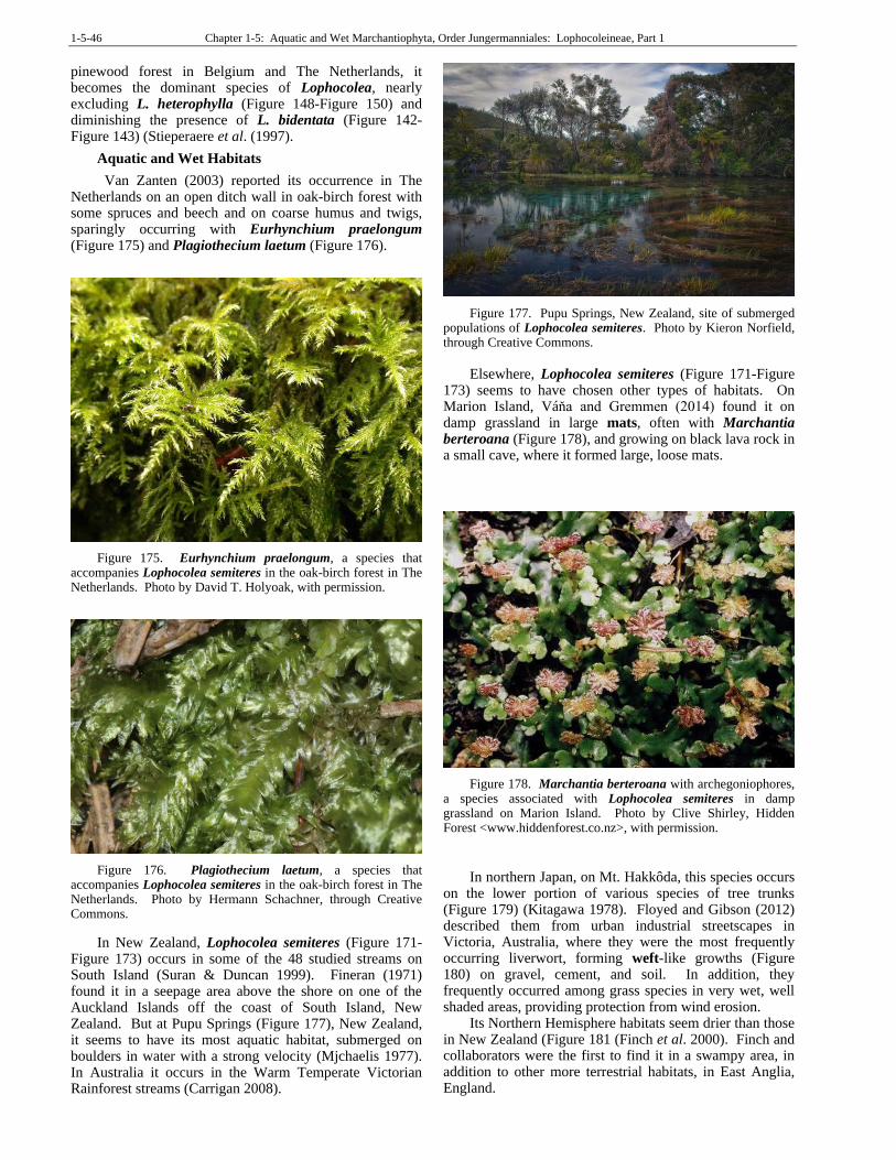
1-5-46 Chapter 1-5: Aquatic and Wet Marchantiophyta, Order Jungermanniales: Lophocoleineae, Part 1
pinewood forest in Belgium and The Netherlands, it becomes the dominant species of Lophocolea, nearly excluding L. heterophylla (Figure 148-Figure 150) and diminishing the presence of L. bidentata (Figure 142-Figure 143) (Stieperaere et al. (1997).
Aquatic and Wet Habitats
Van Zanten (2003) reported its occurrence in The Netherlands on an open ditch wall in oak-birch forest with some spruces and beech and on coarse humus and twigs, sparingly occurring with Eurhynchium praelongum (Figure 175) and Plagiothecium laetum (Figure 176).
Figure 175. Eurhynchium praelongum, a species that accompanies Lophocolea semiteres in the oak-birch forest in The Netherlands. Photo by David T. Holyoak, with permission.
Figure 176. Plagiothecium laetum, a species that accompanies Lophocolea semiteres in the oak-birch forest in The Netherlands. Photo by Hermann Schachner, through Creative Commons.
In New Zealand, Lophocolea semiteres (Figure 171-Figure 173) occurs in some of the 48 studied streams on South Island (Suran & Duncan 1999). Fineran (1971) found it in a seepage area above the shore on one of the Auckland Islands off the coast of South Island, New Zealand. But at Pupu Springs (Figure 177), New Zealand, it seems to have its most aquatic habitat, submerged on boulders in water with a strong velocity (Mjchaelis 1977). In Australia it occurs in the Warm Temperate Victorian Rainforest streams (Carrigan 2008).
Figure 177. Pupu Springs, New Zealand, site of submerged populations of Lophocolea semiteres. Photo by Kieron Norfield, through Creative Commons.
Elsewhere, Lophocolea semiteres (Figure 171-Figure 173) seems to have chosen other types of habitats. On Marion Island, Váňa and Gremmen (2014) found it on damp grassland in large mats, often with Marchantia berteroana (Figure 178), and growing on black lava rock in a small cave, where it formed large, loose mats.
Figure 178. Marchantia berteroana with archegoniophores, a species associated with Lophocolea semiteres in damp grassland on Marion Island. Photo by Clive Shirley, Hidden Forest <www.hiddenforest.co.nz>, with permission.
In northern Japan, on Mt. Hakkôda, this species occurs on the lower portion of various species of tree trunks (Figure 179) (Kitagawa 1978). Floyed and Gibson (2012) described them from urban industrial streetscapes in Victoria, Australia, where they were the most frequently occurring liverwort, forming weft-like growths (Figure 180) on gravel, cement, and soil. In addition, they frequently occurred among grass species in very wet, well shaded areas, providing protection from wind erosion.
Its Northern Hemisphere habitats seem drier than those in New Zealand (Figure 181 (Finch et al. 2000). Finch and collaborators were the first to find it in a swampy area, in addition to other more terrestrial habitats, in East Anglia, England.
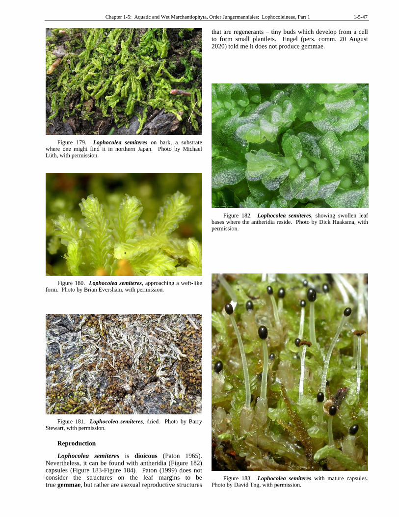
Chapter 1-5: Aquatic and Wet Marchantiophyta, Order Jungermanniales: Lophocoleineae, Part 1 1-5-47
Figure 179. Lophocolea semiteres on bark, a substrate where one might find it in northern Japan. Photo by Michael Lüth, with permission.
Figure 180. Lophocolea semiteres, approaching a weft-like form. Photo by Brian Eversham, with permission.
Figure 181. Lophocolea semiteres, dried. Photo by Barry Stewart, with permission.
Reproduction
Lophocolea semiteres is dioicous (Paton 1965). Nevertheless, it can be found with antheridia (Figure 182) capsules (Figure 183-Figure 184). Paton (1999) does not consider the structures on the leaf margins to be true gemmae, but rather are asexual reproductive structures
that are regenerants – tiny buds which develop from a cell to form small plantlets. Engel (pers. comm. 20 August 2020) told me it does not produce gemmae.
Figure 182. Lophocolea semiteres, showing swollen leaf bases where the antheridia reside. Photo by Dick Haaksma, with permission.
Figure 183. Lophocolea semiteres with mature capsules. Photo by David Tng, with permission.
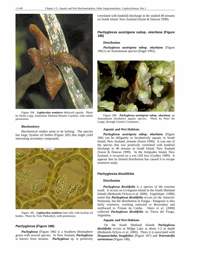
1-5-48 Chapter 1-5: Aquatic and Wet Marchantiophyta, Order Jungermanniales: Lophocoleineae, Part 1
Figure 184. Lophocolea semiteres dehisced capsule. Photo by Heino Lepp, Australian National Botanic Gardens, with online permission.
Biochemistry
Biochemical studies seem to be lacking. The species has large, hyaline oil bodies (Figure 185) that might yield interesting secondary compounds.
Figure 185. Lophocolea semiteres leaf cells with hyaline oil bodies. Photo by Tom Thekathyil, with permission.
Pachyglossa (Figure 186)
Pachyglossa (Figure 186) is a Southern Hemisphere genus with several species. In New Zealand, Pachyglossa is known from streams. Pachyglossa sp. is positively
correlated with bankfull discharge in the studied 48 streams on South Island, New Zealand (Suran & Duncan 1999).
Pachyglossa austrigena subsp. okaritana (Figure 186)
Distribution
Pachyglossa austrigena subsp. okaritana (Figure 186) is an Australasian species (Engel 1992).
Figure 186. Pachyglossa austrigena subsp. okaritana, an Australasian facultative aquatic species. Photo by Peter De Lange, through Creative Commons.
Aquatic and Wet Habitats
Pachyglossa austrigena subsp. okaritana (Figure 186) can be obligately or facultatively aquatic in South Island, New Zealand, streams (Suren 1996). It was one of the species that was positively correlated with bankfull discharge in 48 streams in South Island, New Zealand (Suran & Duncan 1999). In the Antipodes Island, New Zealand, it occurred on a wet cliff face (Godley 1989). It appears that its limited distribution has caused it to escape extensive study.
Pachyglossa dissitifolia
Distribution
Pachyglossa dissitifolia is a species of the extreme south. It occurs on Livingston Island in the South Shetland Islands (Bednarek-Ochyra et al. 2000). Engelskjøn (1986) noted that Pachyglossa dissitifolia occurs on the Antarctic Peninsula, but the distribution in Fuegia - Patagonia is also fairly extensive, reaching eastward to Bouvetøya and northward to Tristan da Cunha. Otero et al. (2008) collected Pachyglossa dissitifolia on Tierra del Fuego, Argentina.
Aquatic and Wet Habitats
On the South Shetland Islands Pachyglossa dissitifolia occurs in Midge Lake at about 1-2 m depth (Bednarek-Ochyra et al. 2000). There it is associated with Drepanocladus longifolius (Figure 187) and Warnstorfia sarmentosa (Figure 188).
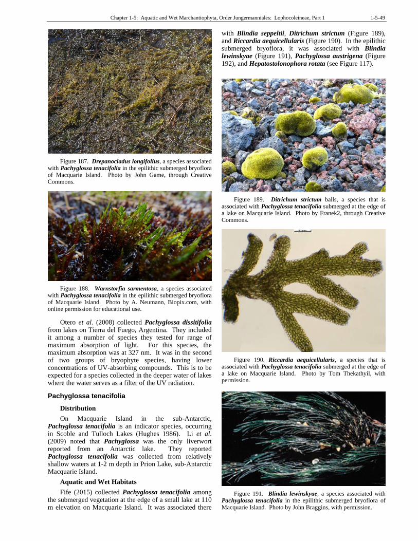
Chapter 1-5: Aquatic and Wet Marchantiophyta, Order Jungermanniales: Lophocoleineae, Part 1 1-5-49
Figure 187. Drepanocladus longifolius, a species associated with Pachyglossa tenacifolia in the epilithic submerged bryoflora of Macquarie Island. Photo by John Game, through Creative Commons.
Figure 188. Warnstorfia sarmentosa, a species associated with Pachyglossa tenacifolia in the epilithic submerged bryoflora of Macquarie Island. Photo by A. Neumann, Biopix.com, with online permission for educational use.
Otero et al. (2008) collected Pachyglossa dissitifolia from lakes on Tierra del Fuego, Argentina. They included it among a number of species they tested for range of maximum absorption of light. For this species, the maximum absorption was at 327 nm. It was in the second of two groups of bryophyte species, having lower concentrations of UV-absorbing compounds. This is to be expected for a species collected in the deeper water of lakes where the water serves as a filter of the UV radiation.
Pachyglossa tenacifolia
Distribution
On Macquarie Island in the sub-Antarctic, Pachyglossa tenacifolia is an indicator species, occurring in Scoble and Tulloch Lakes (Hughes 1986). Li et al. (2009) noted that Pachyglossa was the only liverwort reported from an Antarctic lake. They reported Pachyglossa tenacifolia was collected from relatively shallow waters at 1-2 m depth in Prion Lake, sub-Antarctic Macquarie Island.
Aquatic and Wet Habitats
Fife (2015) collected Pachyglossa tenacifolia among the submerged vegetation at the edge of a small lake at 110 m elevation on Macquarie Island. It was associated there
with Blindia seppeltii, Ditrichum strictum (Figure 189), and Riccardia aequicellularis (Figure 190). In the epilithic submerged bryoflora, it was associated with Blindia lewinskyae (Figure 191), Pachyglossa austrigena (Figure 192), and Hepatostolonophora rotata (see Figure 117).
Figure 189. Ditrichum strictum balls, a species that is associated with Pachyglossa tenacifolia submerged at the edge of a lake on Macquarie Island. Photo by Franek2, through Creative Commons.
Figure 190. Riccardia aequicellularis, a species that is associated with Pachyglossa tenacifolia submerged at the edge of a lake on Macquarie Island. Photo by Tom Thekathyil, with permission.
Figure 191. Blindia lewinskyae, a species associated with Pachyglossa tenacifolia in the epilithic submerged bryoflora of Macquarie Island. Photo by John Braggins, with permission.
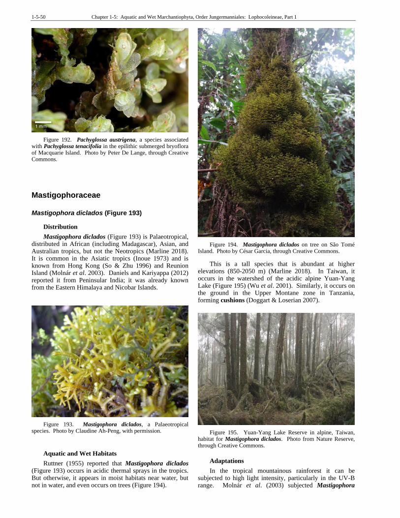
1-5-50 Chapter 1-5: Aquatic and Wet Marchantiophyta, Order Jungermanniales: Lophocoleineae, Part 1
Figure 192. Pachyglossa austrigena, a species associated with Pachyglossa tenacifolia in the epilithic submerged bryoflora of Macquarie Island. Photo by Peter De Lange, through Creative Commons.
Mastigophoraceae
Mastigophora diclados (Figure 193)
Distribution
Mastigophora diclados (Figure 193) is Palaeotropical, distributed in African (including Madagascar), Asian, and Australian tropics, but not the Neotropics (Marline 2018). It is common in the Asiatic tropics (Inoue 1973) and is known from Hong Kong (So & Zhu 1996) and Reunion Island (Molnár et al. 2003). Daniels and Kariyappa (2012) reported it from Peninsular India; it was already known from the Eastern Himalaya and Nicobar Islands.
Figure 193. Mastigophora diclados, a Palaeotropical species. Photo by Claudine Ah-Peng, with permission.
Aquatic and Wet Habitats
Ruttner (1955) reported that Mastigophora diclados (Figure 193) occurs in acidic thermal sprays in the tropics. But otherwise, it appears in moist habitats near water, but not in water, and even occurs on trees (Figure 194).
Figure 194. Mastigophora diclados on tree on São Tomé Island. Photo by César Garcia, through Creative Commons.
This is a tall species that is abundant at higher elevations (850-2050 m) (Marline 2018). In Taiwan, it occurs in the watershed of the acidic alpine Yuan-Yang Lake (Figure 195) (Wu et al. 2001). Similarly, it occurs on the ground in the Upper Montane zone in Tanzania, forming cushions (Doggart & Loserian 2007).
Figure 195. Yuan-Yang Lake Reserve in alpine, Taiwan, habitat for Mastigophora diclados. Photo from Nature Reserve, through Creative Commons.
Adaptations
In the tropical mountainous rainforest it can be subjected to high light intensity, particularly in the UV-B range. Molnár et al. (2003) subjected Mastigophora
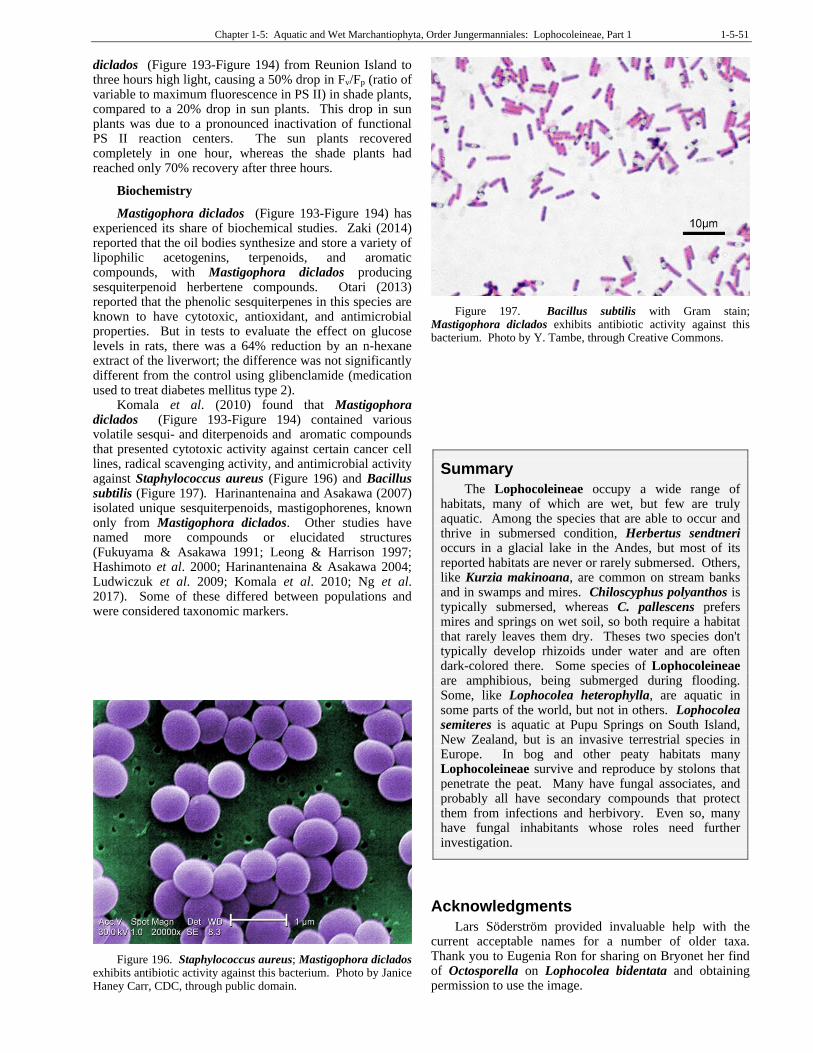
Chapter 1-5: Aquatic and Wet Marchantiophyta, Order Jungermanniales: Lophocoleineae, Part 1 1-5-51
diclados (Figure 193-Figure 194) from Reunion Island to three hours high light, causing a 50% drop in Fv/Fp (ratio of variable to maximum fluorescence in PS II) in shade plants, compared to a 20% drop in sun plants. This drop in sun plants was due to a pronounced inactivation of functional PS II reaction centers. The sun plants recovered completely in one hour, whereas the shade plants had reached only 70% recovery after three hours.
Biochemistry
Mastigophora diclados (Figure 193-Figure 194) has experienced its share of biochemical studies. Zaki (2014) reported that the oil bodies synthesize and store a variety of lipophilic acetogenins, terpenoids, and aromatic compounds, with Mastigophora diclados producing sesquiterpenoid herbertene compounds. Otari (2013) reported that the phenolic sesquiterpenes in this species are known to have cytotoxic, antioxidant, and antimicrobial properties. But in tests to evaluate the effect on glucose levels in rats, there was a 64% reduction by an n-hexane extract of the liverwort; the difference was not significantly different from the control using glibenclamide (medication used to treat diabetes mellitus type 2).
Komala et al. (2010) found that Mastigophora diclados (Figure 193-Figure 194) contained various volatile sesqui- and diterpenoids and aromatic compounds that presented cytotoxic activity against certain cancer cell lines, radical scavenging activity, and antimicrobial activity against Staphylococcus aureus (Figure 196) and Bacillus subtilis (Figure 197). Harinantenaina and Asakawa (2007) isolated unique sesquiterpenoids, mastigophorenes, known only from Mastigophora diclados. Other studies have named more compounds or elucidated structures (Fukuyama & Asakawa 1991; Leong & Harrison 1997; Hashimoto et al. 2000; Harinantenaina & Asakawa 2004; Ludwiczuk et al. 2009; Komala et al. 2010; Ng et al. 2017). Some of these differed between populations and were considered taxonomic markers.
Figure 196. Staphylococcus aureus; Mastigophora diclados exhibits antibiotic activity against this bacterium. Photo by Janice Haney Carr, CDC, through public domain.
Figure 197. Bacillus subtilis with Gram stain; Mastigophora diclados exhibits antibiotic activity against this bacterium. Photo by Y. Tambe, through Creative Commons.
Summary
The Lophocoleineae occupy a wide range of habitats, many of which are wet, but few are truly aquatic. Among the species that are able to occur and thrive in submersed condition, Herbertus sendtneri occurs in a glacial lake in the Andes, but most of its reported habitats are never or rarely submersed. Others, like Kurzia makinoana, are common on stream banks and in swamps and mires. Chiloscyphus polyanthos is typically submersed, whereas C. pallescens prefers mires and springs on wet soil, so both require a habitat that rarely leaves them dry. Theses two species don't typically develop rhizoids under water and are often dark-colored there. Some species of Lophocoleineae are amphibious, being submerged during flooding. Some, like Lophocolea heterophylla, are aquatic in some parts of the world, but not in others. Lophocolea semiteres is aquatic at Pupu Springs on South Island, New Zealand, but is an invasive terrestrial species in Europe. In bog and other peaty habitats many Lophocoleineae survive and reproduce by stolons that penetrate the peat. Many have fungal associates, and probably all have secondary compounds that protect them from infections and herbivory. Even so, many have fungal inhabitants whose roles need further investigation.
Acknowledgments
Lars Söderström provided invaluable help with the current acceptable names for a number of older taxa. Thank you to Eugenia Ron for sharing on Bryonet her find of Octosporella on Lophocolea bidentata and obtaining permission to use the image.
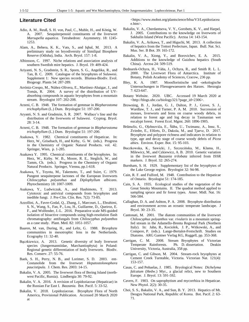
1-5-52 Chapter 1-5: Aquatic and Wet Marchantiophyta, Order Jungermanniales: Lophocoleineae, Part 1
Literature Cited Adio, A. M., Reuß, S. H. von, Paul, C., Muhle, H., and König, W.
A. 2007. Sesquiterpenoid constituents of the liverwort Marsupella aquatica. Tetrahedron: Asymmetry. 18: 1245-1253.
Alam, A., Behera, K. K., Vats, S., and Iqbal, M. 2013. A preliminary study on bryodiversity of Similipal Biosphere Reserve (Odisha), India. Arch. Bryol. 157: 1-8.
Albinsson, C. 1997. Niche relations and association analysis of southern Swedish mire hepatics. J. Bryol. 19: 409-424.
Ariyanti, N. S., Gradstein, S. R., Sporn, S. G., Angelika, R., and Tan, B. C. 2009. Catalogue of the bryophytes of Sulawesi. Supplement 1: New species records. Blumea-Biodiv. Evol. Biogeogr. Plants 54: 287-289.
Arróniz-Crespo, M., Núñez-Olivera, E., Martínez-Abaigar, J., and Tomás, R. 2004. A survey of the distribution of UV-absorbing compounds in aquatic bryophytes from a mountain stream. Bryologist 107: 202-208.
Arzeni, C. B. 1948. The formation of gemmae in Blepharostoma trichophyllum (L.) Dum. Bryologist 51: 197-200.
Aryanti, N. S. and Gradstein, S. R. 2007. Wallace’s line and the distribution of the liverworts of Sulawesi. Cryptog. Bryol. 28: 3-14.
Arzeni, C. B. 1948. The formation of gemmae in Blepharostoma trichophyllum (L.) Dum. Bryologist 51: 197-200.
Asakawa, Y. 1982. Chemical constituents of Hepaticae. In: Herz, W., Grisebach, H., and Kirby, G. W. (eds.). Progress in the Chemistry of Organic Natural Products. vol. 42. Springer, Wien, p. 1-285.
Asakawa Y. 1995. Chemical constituents of the bryophytes. In: Herz, W., Kirby. W. B., Moore, R. E., Steglich, W., and Tamm, Ch. (eds.). Progress in the Chemistry of Organic Natural Products. Springer, Vienna, pp.1-618.
Asakawa, Y., Toyota, M., Takemoto, T., and Suire, C. 1979. Pungent sesquiterpene lactones of the European liverworts Chiloscyphus polyanthus and Diplophyllum albicans. Phytochemistry 18: 1007-1009.
Asakawa, Y., Ludwiczuk, A., and Hashimoto, T. 2013. Cytotoxic and antiviral compounds from bryophytes and inedible fungi. J. Pre-Clin. Clin. Res. 7(2): 73-85.
Azzollini, A., Favre-Godal, Q., Zhang, J., Marcourt, L., Ebrahimi, S. N., Wang, S., Fan, P., Lou, H., Guillarme, D., Queiroz, E. F., and Wolfender, J.-L. 2016. Preparative scale MS-guided isolation of bioactive compounds using high-resolution flash chromatography: antifungals from Chiloscyphus polyanthos as a case study. Plant. Med. 82: 1051-1057.
Baaren, M. van, During, H., and Leltz, G. 1988. Bryophyte communities in mesotrophic fens in the Netherlands. Ecography 11: 32-40.
Bączkiewicz, A. 2013. Genetic diversity of leafy liverwort species (Jungermanniidae, Marchantiophyta) in Poland: Regional genetic differentiation of leafy liverworts. Biodiv. Res. Conserv. 27: 55-76.
Baek, S. H., Perry, N. B., and Lorimer, S. D. 2003. ent-Costunolide from the liverwort Hepatostolonophora paucistipula. J. Chem. Res. 2003: 14-15.
Bakalin, V. A. 2005. The liverwort flora of Bering Island (north-west Pacific, Russia). Lindbergia 30: 79-92.
Bakalin, V. A. 2016. A revision of Lepidoziaceae (Hepaticae) in the Russian Far East I. Bazzania. Bot. Pacif. 5: 33-52.
Bakalin, V. 2018. Lepidoziaceae. Bryophyte Flora of North America, Provisional Publication. Accessed 20 March 2020 at
<https://www.mobot.org/plantscience/bfna/V3/Lepidoziaceae.htm>.
Bakalin, V. A., Cherdantseva, V. Y., Gorobets, K. V., and Harpel, J. 2005. Contributions to the knowledge on liverworts of Sakhalin Island (West Pacific). Arctoa 14: 143-154.
Bakalin, V. A., Arikawa, T., and Higuchi, M. 2013. A collection of hepatics from the Tottori Prefecture, Japan. Bull. Nat. Sci. Mus. Ser. B Bot. 39: 165-172.
Bakalin, V. A., Xiong, Y., and Borovichev, E. A. 2015. Additions to the knowledge of Guizhou hepatics (South China). Arctoa 24: 509-519.
Bednarek-Ochyra, H., Váňa, J., Ochyra, R., and Smith R. L. L. 2000. The Liverwort Flora of Antarctica. Institute of Botany, Polish Academy of Sciences, Cracow, 236 pp.
Bley, K. A. 1987. Moosfloristische und -oekologische Untersuchungen in Fliessgewaessern des Harzes. Herzogia 7: 623-647.
Botany Website. 2020. UBC. Accessed 19 March 2020 at <http://blogs.ubc.ca/biology321/?page_id=2306>.
Browning, B. J., Jordan, G. J., Dalton, P. J., Grove, S. J., Wardlaw, T. J., and Turner, P. A. M. 2010. Succession of mosses, liverworts and ferns on coarse woody debris, in relation to forest age and log decay in Tasmanian wet eucalypt forest. Forest Ecol. Mgmt. 260: 1896-1905.
Brūmelis, G., Oļehnoviča, E., Šūba, U., Treimane, A., Inne, S., Zviedre, E., Elferts, D., Daksša, M., and Tjarve, D. 2017. Bryophyte and polypore richness and indicators in relation to type, age and decay stage of coarse woody debris of Picea abies. Environ. Exper. Bot. 15: 95-103.
Buczkowska, K., Sawicki, J., Szczecińska, M., Klama, H., Milewicz, M., and Czkiewicz, A. B. 2010. Genetic variation in the liverwort Bazzania trilobata inferred from ISSR markers. J. Bryol. 32: 265-274.
Burnham, S. H. 1929. Supplementary list of the bryophytes of the Lake George region. Bryologist 32: 94-98.
Cain, R. F. and Fulford, M. 1948. Contribution to the Hepaticae of Ontario. Bryologist 51: 174-185.
Cain, S. A. 1935. Ecological studies of the vegetation of the Great Smoky Mountains. II. The quadrat method applied to sampling spruce and fir forest types. Amer. Midl. Nat. 16: 566-584.
Callaghan, D. A. and Ashton, P. A. 2008. Bryophyte distribution and environment across an oceanic temperate landscape. J Bryol. 30: 23-35.
Cantonati, M. 2001. The diatom communities of the liverwort Chiloscyphus polyanthos var. rivularis in a mountain spring-fed stream in the Adamello-Brenta Regional Park (Northern Italy). In: Jahn, R., Kociolek, J. P., Witkowski, A., and Compere, P. (eds.). Lange-Bertalot-Festschrift. Studies on Diatoms. ARG Gantner Verlag KG, Ruggell, pp. 353-368.
Carrigan, C. M. 2008. Stream Bryophytes of Victorian Temperate Rainforests. Ph. D. dissertation. Deakin University, Victoria, Australia, 358 pp.
Carrigan, C. and Gibson, M. 2004. Stream-rock bryophytes at Cement Creek Turntable, Victoria. Victorian Nat. 121(4): 153-157.
Casas, C. and Peñuelas, J. 1985. Bryological Notes: Dichelyma falcatum (Hedw.) Myr., a glacial relict, new to Southern Europe. J. Bryol. 13: 591-592.
Cavers, F. 1903. On saprophytism and mycorrhiza in Hepaticae. New Phytol. 2(2): 30-35.
Choi, S. S., Bakalin, V. A., and Sun, B. Y. 2013. Hepatics of Mt. Deogyu National Park, Republic of Korea. Bot. Pacif. 2: 63-71.
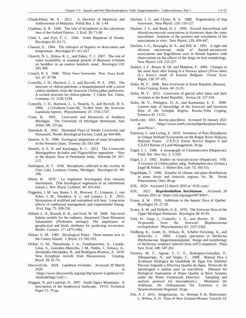
Chapter 1-5: Aquatic and Wet Marchantiophyta, Order Jungermanniales: Lophocoleineae, Part 1 1-5-53
Chuah-Petiot, M. S. 2011. A checklist of Hepaticae and Anthocerotae of Malaysia. Polish Bot. J. 56: 1-44.
Clapham, A. R. 1940. The role of bryophytes in the calcareous fens of the Oxford District. J. Ecol. 28: 71-80.
Clark, L. and Frye, T. C. 1942. Some Hepaticae of Alaska. Bryologist 45: 63-71.
Clausen, E. 1964. The tolerance of hepatics to desiccation and temperature. Bryologist 67: 411-417.
Cleavitt, N. L., Klima, K. L., and Fahey, T. J. 2007. The role of water availability in seasonal growth of Bazzania trilobata on boulders in an eastern hemlock stand. Bryologist 110: 295-308.
Conard, H. S. 1940. Thirty Iowa liverworts. Proc. Iowa Acad. Sci. 47: 97-99.
Connolly, J. D., Harrison, L. J., and Rycroft, D. S. 1982. The structure of chiloscypholone, a sesquiterpenoid with a novel carbon skeleton, from the liverwort Chiloscyphus pallescens. A revised structure for chiloscyphone. J. Chem. Soc. Chem. Commun. 21: 1236-1238.
Connolly, J. D., Harrison, L. J., Huneck, S., and Rycroft, D. S. 1986. (+)-Eudesm-3-ene-6β, 7α-diol from the liverwort Lepidozia reptans. Phytochemistry 25: 1745-1747.
Crum, H. 1991. Liverworts and Hornworts of Southern Michigan. The University of Michigan Herbarium, Ann Arbor, MI, 233 pp.
Damsholt, K. 2002. Illustrated Flora of Nordic Liverworts and Hornworts, Nordic Bryological Society, Lund, pp 404-408.
Daniels, A. D. 1998. Ecological adaptations of some bryophytes of the Western Ghats. Forestry 26: 193-194.
Daniels, A. E. D. and Kariyappa, K. C. 2012. The Liverworts Mastigophora diclados and Plagiochilion oppositum – New to the hepatic flora of Peninsular India. Nelumbo 54: 207-212.
Darlington, H. T. 1938. Bryophytes collected in the vicinity of Glen Lake, Leelanau County, Michigan. Bryologist 41: 99-106.
Dhien, R. 1978. La végétation bryologique d'un ruisseau intermittent. [The bryological vegetation of an intermittent stream.]. Rev. Bryol. Lichénol. 44: 453-454.
Diggelen, J. M. van, Bense, I. H., Brouwer, E., Limpens, J., van Schie, J. M., Smolders, A. J., and Lamers, L. P. 2015. Restoration of acidified and eutrophied rich fens: Long-term effects of traditional management and experimental liming. Ecol. Eng. 75: 208-216.
Dillard, L. O., Russell, K. R., and Ford, W. M. 2008. Site-level habitat models for the endemic, threatened Cheat Mountain Salamander (Plethodon nettingi): The importance of geophysical and biotic attributes for predicting occurrence. Biodiv. Conserv. 17: 1475-1492.
Dirkse, G. M. 1985. Bryological Notes: Three mosses new to the Canary Islands. J. Bryol. 13: 592-593.
Dirkse, G. M., Nieuwkoop, J. A., Vanderpoorten, A., Losada-Lima, A., González-Mancebo, J. M., Patiño, J., Sotiaux, A., Hernández-Hernández, R., and Rodríguez-Romero, A. 2018. New bryophyte records from Macaronesia. Cryptog. Bryol. 39: 61-76.
DiscoverLife. 2020. Lepidozia trichodes. Accessed 20 March 2020 at <https://www.discoverlife.org/mp/20q?search=Lepidozia+trichodes&flags=col2:>.
Doggart, N. and Loserian, D. 2007. South Nguru Mountains: A description of the biophysical landscape. TFCG Technical Paper 11: 70 pp.
Duckett, J. G. and Clymo, R. S. 1988. Regeneration of bog liverworts. New Phytol. 110: 119-127.
Duckett, J. G. and Read, D. J. 1995. Ericoid mycorrhizas and rhizoid‐ascomycete associations in liverworts share the same mycobiont: Isolation of the partners and resynthesis of the associations in vitro. New Phytol. 129: 439-447.
Duckett, J. G., Renzaglia, K. S., and Pell, K. 1991. A light and electron microscope study of rhizoid–ascomycete associations and flagelliform axes in British hepatics with observations on the effects of the fungi on host morphology. New Phytol. 118: 233-257.
Dulière, J. F., Bruyn, R. De, and Malaisse, F. 2000. Changes in the moss layer after liming in a Norway spruce (Picea abies (L.) Karst.) stand of Eastern Belgium. Forest Ecol. Mgmt. 136: 97-105.
Dulin, M. V. 2008. Rare liverworts in Komi Republic (Russia). Folia Cryptog. Eston. 44: 23-31.
Dulin, M. V. 2015. Liverworts of glacial relict lakes and their vicinities in the Komi Republic. Arctoa 24: 527-535.
Dulin, M. V., Philippov, D. A., and Karmazina, E. V. 2009. Current state of knowledge of the liverwort and hornwort flora of the Vologda Region, Russia. Folia Cryptog. Estonica 45: 13-22.
Earth.com. 2021. Kurzia pauciflora. Accessed 10 January 2021 at <https://www.earth.com/earthpedia/plant/kurzia-pauciflora/>.
Emerson, C. and Loring, S. 2010. Inventory of Rare Bryophytes in Unique Wetland Ecosystems on the Rogue River-Siskiyou National Forest. U.S.D.A Forest Service Region 6 and U.S.D.I Bureau of Land Management, 30 pp.
Engel, J. J. 1980. A monograph of Clasmatocolea (Hepaticae). Field. Bot. New Ser. 3: 1-229.
Engel, J. J. 1992. Studies on Geocalycaceae (Hepaticae). VIII. A revision of Chiloscyphus subg. Notholophocolea (Schust.) Engel & Schust. J. Hattori Bot. Lab. 72: 105-l15.
Engelskjøn, T. 1986. Zonality of climate and plant distributions in some Arctic and Antarctic regions, No. 30. Norsk Polarinstitutt, Oslo, 49 pp.
EOL. 2020. Accessed 23 March 2020 at <EOL.com>
EOL. 2021. Hygrolembidium boschianum. Accessed 20 January 2021 at <https://eol.org/pages/6074393>.
Evans, A. W. 1916. Additions to the hepatic flora of Quebec. Bryologist 19: 27-30.
Evans, A. W. and Nichols, G. E. 1935. The liverwort flora of the Upper Michigan Peninsula. Bryologist 38: 81-91.
Feld, H., Zapp, J., Connolly, J. D., and Becker, H. 2004. Terpenoids from the liverwort Blepharostoma trichophyllum. Phytochemistry 65: 2357-2362.
Feldberg, K., Groth, H., Wilson, R., Schäfer-Verwimp, A., and Heinrichs, J. 2004. Cryptic speciation in Herbertus (Herbertaceae, Jungermanniopsida): Range and morphology of Herbertus sendtneri inferred from nrITS sequences. Plant Syst. Evol. 249: 247-261.
Ferreira, M. T., Aguiar, F. C. F., Rodríguez-González, P., Albuquerque, A., and Sérgio, C. 2008. Manual Para a Avaliação Biológica da Qualidade da Água Em Sistemas Fluviais Segundo a Directiva Quadro da Água. Protocolo de amostragem e análise para os macrófitos. [Manual for Biological Assessment of Water Quality in River Systems under the Water Framework Directive. Sampling and analysis protocol for macrophytes.]. Ministério Do Ambiente, Do Ordenamento Do Território e Do Desenvolvimento Regional, 18 pp.
Fife, A. J. 2015. Seligeriaceae. In: Heenan, P. B., Breitwieser, I., Wilton, A. D. Flora of New Zealand-Mosses. Fascicle 24.
1-5-54 Chapter 1-5: Aquatic and Wet Marchantiophyta, Order Jungermanniales: Lophocoleineae, Part 1
Manaaki Whenua Press, Lincoln. <http://dx. doi. org/10.7931/B1K01P>.
Finch, R. A., Fisk, R. J., Straus, D. F., and Stevenson, C. R. 2000. Lophocolea semiteres new to East Anglia. J. Bryol. 22: 146-148.
Fineran, B. A. 1971. A catalogue of bryophytes, lichens, and fungi collected on the Auckland Islands. J. Royal Soc. N. Z. 1: 215-229.
Fisk, R. 2008. More tree fern aliens. Field Bryol. 96: 9.
Fitzgerald, J. W. and Fitzgerald, R. D. 1967. Bryophytes of County Armagh. Irish Nat. J. 15: 324-326.
Fleisch, M. and Engel, J. J. 2006. Stream bryophytes in Victorian rainforest streams. Victorian Nat. 123: 279-281.
Floyed, A. and Gibson, M. 2012. Bryophytes of urban industrial streetscapes in Victoria, Australia. Victorian Nat. 129: 203-214.
Fukuyama, Y. and Asakawa, Y. 1991. Novel neurotrophic isocuparane-type sesquiterpene dimers, mastigophorenes A, B, C and D, isolated from the liverwort Mastigophora diclados. J. Chem. Soc. Perkin Trans. 1: 2737-2741.
Fulford, M. 1934. List of Hepaticae of Kentucky. Bryologist 37: 21-28.
Fulford, M. 1955. Sporelings, gemmalings and regeneration in Blepharostoma trichophyllum (L.) Dum. Mitteil. Thüring. Bot. Gesellsch 1: 245-258.
Gabriel, R. and Bates, J. W. 2005. Bryophyte community composition and habitat specificity in the natural forests of Terceira, Azores. Plant Ecol. 177: 125-144.
Gao, C. and Bai, X. L. 2002. Lepidozia suyungii (Lepidoziaceae, Hepaticae), a new species from southwestern China, with discussion of the species of Lepidozia in China. J. Hattori Bot. Lab. 92: 191-197.
Gecheva, G., Yurukova, L., Cheshmedjiev, S., and Ganeva, A. 2010. Distribution and bioindication role of aquatic bryophytes in Bulgarian rivers. Biotech. Biotech. Equip. 24(supp1.): 164-170.
Gecheva, G., Yurukova, L., and Cheshmedjiev, S. 2013a. Patterns of aquatic macrophyte species composition and distribution in Bulgarian rivers. Turkish J. Bot. 37: 99-110.
Gecheva, G. and Yurukova, L. 2013b. Reference aquatic macrophyte communities at rivers in southwestern Bulgaria. Comptes Rendus L Acad. Bulgare Sci. Sci. Math. Nat. 66: 223-230.
Geel, B. van. 1978. A palaeoecological study of Holocene peat bog sections in Germany and the Netherlands, based on the analysis of pollen, spores and macro-and microscopic remains of fungi, algae, cormophytes and animals. Rev. Palaeobot. Palynol. 25: 1-120.
Geissler, P. 1976. Zur Vegetation alpiner Fliessgewässer. Pflanzensoziologisch-ökologische Untersuchungen hygrophiler Moosgesellschaften in den östlichen Schweizer Alpen. Beitr. Kryptogamenfl. Schweiz 14(2): 1-52.
Gil, J. A. and Ruiz, P. 1985. The aquatic basophilous bryophytic communities of south east Spain. Herzogia 7: 211-228.
Glenny, D., Fife, A. J., Brownsey, P. J., Renner, M. A., Braggins, J. E., Beever, J. E., and Hitchmough, R. 2011. Threatened and uncommon bryophytes of New Zealand (2010 revision). N. Z. J. Bot. 49: 305-327.
Glime, J. M. 1982. Bryophytes of the Flume, Franconia Notch State Park, N. H. Rhodora 84: 149-152.
Glime, J. M. and Vitt, D. H. 1987. A comparison of bryophyte species diversity and niche structure of montane streams and stream banks. Can. J. Bot. 65: 1824-1837.
Glime, J. M., Slack, N. G., and Meston, C. L. 1987. A comparison of the Levins' and Freeman-Tukey niche width measures for the bryophytes in an Adirondack stream. Symp. Biol. Hung. 35: 457-466.
Godley, E. J. 1989. The flora of Antipodes Island. N. Z. J. Bot. 27: 531-564.
Gradstein, S. R. 2011. Guide to the Liverworts and Hornworts in Java. SEAMEO BRYOTROP, Southeast Asian Regional Centre for Tropical Biology, Bogor, Indonesia, 129 pp.
Gradstein, S. R. 2017. Bazzania (Marchantiophyta) in South America. Nova Hedw. 105: 243-266.
Gradstein, S. R. and Váňa, J. 1987. On the occurrence of Laurasian liverworts in the tropics. Mem. N. Y. Bot. Gard. 45: 388-425.
Gradstein, S. R., Cleef, A. M., and Fulford, M. H. 1977. Studies on Colombian Cryptogams II. Hepaticae: Oil body structure and ecological distribution of selected species of tropical Andean Jungermanniales. Proc. Kon. Ned. Acad. Wetensch. Ser. C 80: 377-420.
Gradstein, S. R., Uribe-M, J., Gil-N, J. E., Morales, C., and Negritto, M. A. 2018. Liverworts new to Colombia. Caldasia 40: 82-90.
Grolle, R. and So, M. L. 1999. Studies of Plagiochila sect. Subtropicae in Asia. Bryologist 102: 67-75.
Guo, G. N., Zhou, X., Zhao, R., Chen, X. Y., Chen, Z. L., Li, X. D., and Li, Y. H. 2015. Paenibacillus herberti sp. nov., an endophyte isolated from Herbertus sendtneri. Antonie van Leeuwenhoek 108: 587-596.
Guo, G. N., Zhou, X., Chen, Z. L., Yang, Z. W., Li, X. D., and Li, Y. H. 2016. Paenibacillus marchantiophytorum sp. nov., isolated from the liverwort Herbertus sendtneri. Internat. J. Syst. Evol. Microbiol. 66: 755-761.
Gustafson, D. H., Pettersson, C. J., and Malmgren, J. C. 2006. Great Crested Newts (Triturus cristatus) as indicators of aquatic plant diversity. Herpetol. J. 16: 347-352.
Hall, G. W. 1958. Notes on the hepatic flora of the Athens area. Ohio J. Sci. 58: 15-19.
Harinantenaina, L. and Asakawa, Y. 2004. Chemical constituents of Malagasy liverworts, Part II: Mastigophoric acid methyl ester of biogenetic interest from Mastigophora diclados (Lepicoleaceae subf. Mastigophoroideae). Chem. Pharm. Bull. 52: 1382-1384.
Harinantenaina, L. and Asakawa, Y. 2007. Malagasy liverworts, source of new and biologically active compounds. Nat. Prod. Comm. 2(6): 1934578X0700200616.
Hashimoto, T., Irita, H., Takaoka, S., Tanaka, M., and Asakawa, Y. 2000. New chlorinated cyclic bis (bibenzyls) from the liverworts Herbertus sakuraii and Mastigophora diclados. Tetrahedron 56: 3153-3159.
Hatcher, R. E. 1967. Experimental studies of variation in Hepaticae. I. Induced variation in Lophocolea heterophylla. Brittonia 19: 178-201.
Hayashi, M. and Yamada, K. 2004. Heteroscyphus (Hepaticae; Geocalycaceae) from Luzon and Negros Islands, Philippines, collected by Dr. and Mrs. A. J. Sharp and Dr. Z. Iwatsuki. Bryol. Res. 8: 269-270.
He, X. and Sun, Y. 2017. Rare sporophyte found in Europe for Herbertus sendtneri with a range expansion to Africa and Malesia. Phytotaxa 324: 42-50.
Heino, J. and Virtanen, R. 2006. Relationships between distribution and abundance vary with spatial scale and ecological group in stream bryophytes. Freshwat. Biol. 51: 1879-1889.
Heinrichs, J., Hentschel, J., Feldberg, K., Bombosch, A., and Schneider, H. 2009. Phylogenetic biogeography and
Chapter 1-5: Aquatic and Wet Marchantiophyta, Order Jungermanniales: Lophocoleineae, Part 1 1-5-55
taxonomy of disjunctly distributed bryophytes. J. Syst. Evol. 47: 497-508.
Herout, V. 1985. An approach to the isolation of the fragrant principle of the liverwort Lophocolea heterophylla. Flavour Fragr. J. 1: 43-46.
Holmes, N. T. H. and Whitton, B. A. 1975. Macrophytes of the River Tweed. Trans. Bot. Soc. Edinburgh 42: 369-381.
Holmes, N. T. H. and Whitton, B. A. 1977a. Macrophytic vegetation of River Tees in 1975 – Observed and predicted changes. Freshwat. Biol. 7: 43-60.
Holmes, N. T. H. and Whitton, B. A. 1977b. Macrophytic vegetation of River Swale, Yorkshire. Freshwat. Biol. 7: 545-558.
Holmes, N. T. H. and Whitton, B. A. 1981. Plants of the River Tyne system before the Kielder water scheme. Naturalist 106: 97-107.
Hong, W. S. 1977. An annotated checklist of the Hepaticae of Wyoming. Bryologist 80: 480-491.
Hong, W. S. 1980. Hepaticae of Rocky Mountain National Park, Colorado. Bryologist 83: 351-354.
Hong, W. S. 1988. The family Lepidoziaceae in North America west of the hundredth meridian. Bryologist 91: 326-333.
Hong, W. S. and Vitt, D. H. 1977. Hepaticae of the Yukon Territory, Canada. Bryologist 79: 461-469.
Hrivnák, R., Otahelová, H., and Valachovic, M. 2008. Macrophytes of the Ipel’River: Effect of flood to species composition and distribution. Large Rivers 18: 157-166.
Hughes, J. M. 1986. The relations between aquatic plant communities and lake characteristics on Macquarie Island. N. Z. J. Bot. 24: 271-278.
Hugonnot, V. 2014. Geocalyx graveolens (Schrad.) Nees in the oriental part of the Pyrenees. Bol. Soc. Esp. Briol. 42-43: 1-6.
Hugonnot, V., Baluet, G., and Celle, J. 2015. Aerial and underground biomasses of three Sphagnum-dwelling liverworts. J. Bryol. 37: 247-249.
Huneck, S., Janicke, S., Meinunger, L., Snatke, G., and Connolly, J. D. 1984. Seasonal dependence of the essential oil from Bazzania trilobata. The stereochemistry and absolute configuration of (-)-5-hydroxycalamenene. J. Hattori Bot. Lab. 57: 337-342.
Hylander, K. 2014. Ethiopia. New national and regional bryophyte records, 40. J. Bryol. 36: 224.
Ignatov, M. S., Ignatova, E. A., and Konstantinova, N. A. 2005. Bryophyte flora of the Volzhsko-Kamskiy Nature Reserve (Tatarstan, European Russia). Arctoa 14: 49-66.
Ing, B. 1983. A ravine association of Myxomycetes. J. Biogeogr. 10: 299-306.
Ingerpuu, N., Kull, K., and Vellak, K. 1998. Bryophyte vegetation in a wooded meadow: Relationships with phanerogam diversity and responses to fertilisation. Plant Ecol. 134: 163-171.
Ingerpuu, N., Nurkse, K., and Vellak, K. 2014. Bryophytes in Estonian mires. Estonian J. Ecol. 63: 3-14.
Inoue, H. 1973. The genus Fossombronia Raddi in Japan. J. Hattori Bot. Lab. 37: 293-297.
ITIS. 2020. Integrated Taxonomic Information System. Accessed in 2020 at <http://www.itis.gov>.
Jackson, M. R. 2015. Potential effects of eastern hemlock decline on the hemlock-associated liverwort Bazzania trilobata. Honors Project, Smith College, Northampton, MA. <https://scholarworks.smith.edu/theses/1568>, 51 pp.
Jansová, I. 2006. Seasonal growth and dynamics of epixylic bryophytes in Bohemian old-growth forest. J. Bryol. 28: 123-132.
Janssens, J. A. and Glaser, P. H. 1986. The bryophyte flora and major peat-forming mosses at Red Lake peatland, Minnesota. Can. J. Bot. 64: 427-442.
Järvinen, I. 1976. Distribution maps of Lophocolea, Geocalyx and Harpanthus in Eastern Fennoscandia. Lindbergia 3: 337-343.
Järvinen, I. 1983. Taxonomy and distribution of the European taxa of the genus Chiloscyphus (Hepaticae, Geocalycaceae). Ann. Bot. Fenn. 20: 87-99.
Ji, M., Luo, Si., and Chen, Y. 2000. A study on the epiphyllous liverworts from Matoushan Nature Reserve of Jiangxi Province, China. Acta Agric. Univ. Jiangxiensis 23:467-472.
Jong, J. R. and Wu, C. L. 2000. Sesquiterpenoids and diterpenoids from the liverwort Heteroscyphus coalitus. J. Chinese Chem. Soc. 47: 359-362.
Kantvilas, G. and Jarman, S. J. 2004. Lichens and bryophytes on Eucalyptus obliqua in Tasmania: Management implications in production forests. Biol. Conserv. 117: 359-373.
Karofeld, E. and Toom, M. 1999. Mud-bottoms in Männikjärve bog, central Estonia. Proc. Estonian Acad. Sci. Biol. Ecol. 48: 216-235.
Kent, M., Owen, N. W., and Dale, M. P. 2005. Photosynthetic responses of plant communities to sand burial on the Machair dune systems of the Outer Hebrides, Scotland. Ann. Bot. 95: 869-877.
Kim, M. J., Lee, J. S., and Baek, S. H. 2009a. In vitro biological activity of germacranolide sesquiterpene lactones. Orient. Pharm. Exper. Med. 9: 192-199.
Kim, M. J., Lee, J. S., and Baek, S. H. 2009b. In vitro cytotoxic activity of (-)-ent-costunolide. Orient. Pharm. Exper. Med. 9: 97-99.
Kirkpatrick, J. B. and Scott, J. J. 2002. Change in undisturbed vegetation on the coastal slopes of subantarctic Macquarie Island, 1980-1995.
Kitagawa, N. 1978. Hepaticae of Mts. Hakkôda, Northern Japan. Ecol. Rev. 19: 45-58.
Kitayama, K. 1995. Biophysical conditions of the montane cloud forests of Mount Kinabalu, Sabah, Malaysia. In: Hamilton, L. S., Juvik, J. O., and Scatena, F. N. (eds.). Tropical Montane Cloud Forests. Springer, New York, NY, pp. 183-197.
Klama, H. and Górski, P. 2018. Red list of liverworts and hornworts of Poland (2018). Cryptog. Bryol. 39: 415-441.
Klama, H., Salachna, A., Smieja, A., Buczkowska, K., and Bączkiewicz, A. 2008. The species of genus Chiloscyphus in Tatra National Park (S Poland). Scripta Fac. Rerum Nat. Univ. Ostraviensis 186: 126-133.
Komala, I., Ito, T., Nagashima, F., Yagi, Y., and Asakawa, Y. 2010. Cytotoxic, radical scavenging and antimicrobial activities of sesquiterpenoids from the Tahitian liverwort Mastigophora diclados (Brid.) Nees (Mastigophoraceae). J. Nat. Med. 64: 417-422.
Kondo, K., Toyota, M., and Asakawa, Y. 1990. ent-Eudesmane-type sesquiterpenoids from Bazzania species. Phytochemistry 29: 2197-2199.
Konečný, K., Ubik, K., Vašíčková, S., Streibl, M., and Herout, V. 1982. Constituents of the liverwort Lophocolea heterophylla. Coll. Czech. Chem. Comm. 47: 3164-3169.
Konečný, K., Streibl, M., Vašíčková, S., Buděšínský, M., Šaman, D., Ubik, K., and Herout, V. 1985. Constituents of the liverwort Bazzania trilobata of Czech origin. Coll. Czech. Chem. Comm. 50: 80-93.
1-5-56 Chapter 1-5: Aquatic and Wet Marchantiophyta, Order Jungermanniales: Lophocoleineae, Part 1
Konstantinova, N. A. 2011. Contribution to the hepatic flora of the Republic of Dagestan (Eastern Caucasus, Russia). Arctoa 20: 175-182.
Konstantinova, N. and Savchenko, A. 2008. Contributions to the hepatic flora of Svalbard. Lindbergia 33: 13-22.
Konstantinova, N. A. and Vasiljev, A. N. 1994. On the hepatic flora of Sayan Mountains (south Siberia). Arctoa 3: 123-132.
Konstantinova, N. A., Bakalin, V. A., Potemkin, A. D., and Ignatov, M. S. 2002. Hepatic flora of the Upper Bureya River (Russian Far East). Arctoa 11: 393-398.
Konstantinova, N. A., Mamontov, Y. S., and Savchenko, A. N. 2018. On the liverwort flora of Tunkinskiy National Park (Republic of Buryatia, Russia). Arctoa 27: 131-139.
Koponen, T., Karttunen, K., and Piippo, S. 1995. Suomen vesisammalkasvio. [Aquatic bryophytes of Finland.]. Bryobrothera 3: 1-86.
Koppe, F. 1945. Die Wassermoose Westfalens. Arch. Hydrobiol. 41: 68-91.
Kowalczyk, A., Przywara, L., and Kuta, E. 1997. In vitro culture of liverworts. Acta Biol. Cracoviensia Ser. Bot. 39: 27-33.
Lai, M. J., Zhu, R. L., and Chantanaorrapint, S. 2008. Liverworts and hornworts of Thailand: An updated checklist and bryofloristic accounts. Ann. Bot. Fenn. 45: 321-341.
Leclercq, L. 1977. Vegetation et caracteristiques physico-chimiques de deux rivieres de Haute Ardenne (Belgique): La Helle et la Roer superieure. Lejeunia 88: 1-42.
Leong, Y. W. and Harrison, L. J. 1997. Ent-trachylobane diterpenoids from the liverwort Mastigophora diclados. Phytochemistry 45: 1457-1459.
Lepage, E. 1953. Materials for a better knowledge of the hepatic flora of northern Québec. Bryologist 56: 101-115.
Lepp, Heino. 2012. Australian Bryophytes. Australian National Botanic Gardens and Australian National Herbarium Accessed 7 December 2019 at <https://www.anbg.gov.au/bryophyte/ecology-aquatic.html>.
Li, S.-P., Ochyra, R., Wu, P.-C., Seppelt, R. D., Cai, M.-H., Wang, H.-Y., and Li, C.-S. 2009. Drepanocladus longifolius (Amblystegiaceae), an addition to the moss flora of King George Island, South Shetland Islands, with a review of Antarctic benthic mosses. Polar Biol. 32: 1415-1425.
Li, S., Niu, H., Qiao, Y., Zhu, R., Sun, Y., Ren, Z., Yuan, H., Gao, Y., Li, Y., Chen, W., Zhou, J., and Lou, H. 2018. Terpenoids isolated from Chinese liverworts Lepidozia reptans and their anti-inflammatory activity. Bioorg. Med. Chem. 26: 2392-2400.
Liepiņa, L. 2012. Occurrence of fungal structures in bryophytes of the boreo-nemoral zone. Environ Exper Biol. 10: 35-40.
Lin, Z. M., Wang, L. N., Guo, D. X., Wang, X. N., Liu, Y. Q., Zhou, J. C., and Lou, H. X. 2012. Secondary metabolites from the liverwort Heteroscyphus coalitus. Phytochem. Lett. 5: 510-513.
Logatoc, E. L., Hadsall, A. S., Malabrigo, P. L. Jr., and Gruezo, W. S. 2019. Notes on liverwort specimens from the Philippine Cordilleras in the Botanical Herbarium of the UPLB Museum of Natural History (CAHUP). Mtn. J. Sci. Interdis. Res. (formerly Benguet State Univ. Res. J.) 79(2): 7-20.
Long, D. G. 1982. Lophocolea semiteres (Lehm.) Mitl. established in Argyll, Scotland. J. Bryol. 12: 113-115.
Ludwiczuk, A. and Asakawa, Y. 2010. Chemosystematics of selected liverworts collected in Borneo. Bryo. Div. Evol. 31: 33-42.
Ludwiczuk, A. and Asakawa, Y. 2019. Bryophytes as a source of bioactive volatile terpenoids – A review. Food Chem. Toxicol. 132: 110649.
Ludwiczuk, A., Komala, I., Pham, A., Bianchini, J. P., Raharivelomanana, P., and Asakawa, Y. 2009. Volatile components from selected Tahitian liverworts. Nat. Prod. Comm. 4(10): 1934578X0900401015.
Luís, L., Bergamini, A., Figueira, R., and Sim-Sim, M. 2010. Riparian bryophyte communities on Madeira: Patterns and determinants of species richness and composition. J. Bryol. 32: 32-45.
Luis, L., Bergamin, A., and Sim-Sim, M. 2015. Which environmental factors best explain variation of species richness and composition of stream bryophytes? A case study from mountainous streams in Madeira Island. Aquat. Bot. 123: 37-46.
Lyman, E. L. and Coleman, B. B. 1966. The Bryoflora of the Genesee Country, New York 3. A Preliminary Survey of the Bryophytes of Orleans County. Bryologist 69: 118-124.
Madžule, L. and Brūmelis, G. 2008. Ecology of epixylic bryophytes in Eurosiberian alder swamps of Latvia. Acta Univ. Latviensis Biol. 745: 103-114.
Mägdefrau, K. 1982. Life-forms of bryophytes. In: Smith, A. J. E. Bryophyte Ecology. Springer, Dordrecht, pp. 45-58.
Mamczarz, H. 1970. The bryophyte communities in streams near Lacko in the Sadecki Beskid. Ann. Univ. Mariae Curie-Sklodowska, Sec. C Biol. 3. Biol. 25: 105-136.
Manjula, K. M., Vidya, V., Manju, C. N., and Prakashkumar, R. 2013. Systematic studies on the family Geocalycaceae (Marchantiophyta) of Kerala, India. Arch. Bryol. 176: 1-16.
Marline, L. 2018. Diversity and biogeography of Madagascan bryophytes with an analysis of taxic and functional diversity along an elevational gradient in Marojejy National Park. Ph.D. dissertation, University of Cape Town, South Africa.
Marstaller, R. 1987. Die Moosgesellschaften der Klasse Platyhypnidio-Fontinalietea antipyreticae Philippi 1956. 30. Beitrag zur Moosvegetation Thueringens [The moss communities of the association Platyhypnidio-Fontinalietea antipyreticae Philippi 1956. 30. Contribution to the Thuringian moss vegetation]. Phytocoenologia 15: 85-138.
Martini, U., Zapp, J., and Becker, H. 1998a. Chlorinated macrocyclic bisbibenzyls from the liverwort Bazzania trilobata. Phytochemistry 47: 89-96.
Martini, U., Zapp, J., and Becker, H. 1998b. Lignans from the liverwort Bazzania trilobata. Phytochemistry 49: 1139-1146.
McKnight, B. N. 1985. Notes on the bryophytes of Indiana: I. Additions to the flora. Proc. Indiana Acad. Sci. 95: 101-106.
Meijer, W. 1960. Notes on the species of Bazzania (Hepaticae). Blumea 10: 367-384.
Miller, H. A. 1960. The Bryological Foray of the Ninth International Botanical Congress. Bryologist 63: 53-59.
Mitten, W. 1877. List of Hepaticae collected by the Rev. AE Eaton at the Cape of Good Hope (August and September 1874). Bot. J. Linn. Soc. 16(91): 187-196.
Mjchaelis, F. B. 1977. Biological features of Pupu Springs, New Zealand. J. Marine Freshwat. Res. 11: 357-373.
Molnár, I., Orbán, S., Pócs, T., Sass-Gyarmati, A., Lehodzki, E., and Dulai, S. 2003. Photosynthetic responses of Mastigophora diclados (Brid. ex Web.) Nees ecotypes to excess light in consequence of their microhabitats in Reunion Island: A fluorescence induction study. Acta Acad. Paedagog. Agr. Sec. Biol. 24: 215-233.
Chapter 1-5: Aquatic and Wet Marchantiophyta, Order Jungermanniales: Lophocoleineae, Part 1 1-5-57
Morgan, J. W. 2004. Bryophyte composition in a native grassland community subjected to different long-term fire regimes. Cunninghamia 8: 485-489.
Moyle, J. B. 1937. Notes on Minnesota aquatic bryophytes. Bryologist 40: 114-116.
Mues, R., Edelbluth, E., and Zinsmeister, H. D. 1973. Das Carotinoid muster von Lophocolea bidentata (L.) Dum. Öster. Bot. Zeits. 122: 177-184.
Müller, F. 1996. Beitrag zur Moosflora der Insel Bioko (= Fernando Poo), Äquatorial-Guinea. Trop. Bryol. 12: 75-96.
Nagashima, F., Momosaki, S., Watanabe, Y., Takaoka, S., Huneck, S., and Asakawa, Y. 1996. Sesquiterpenoids from the liverworts Bazzania trilobata and Porella canariensis. Phytochemistry 42: 1361-1366.
Nair, M. C. and Prajitha, B. 2010. Systematic studies on Bryophytes of Northern Western Ghats in Kerala. Final Report. Malabar Botanical Garden Kozhikode-14 Kerala, India, 97 pp.
Nepolean, P., Balamurugan, A., Jayanthi, R., Mareeswaran, J., and Premkumar, R. 2014. Bioefficacy of certain chemical and biofungicides against Hypoxylon spp. causing wood rot disease in tea. J. Plant. Crops 42: 341-347.
Neuhäuslová, Z. and Eltsova, V. 2002. The Bazzanio‐Piceetum in the Bohemian Forest (Šumava Mts.), Czech Republic. Fed. Rep.: Zeit. Bot. Taxon. Geobot. 113: 367-379.
Ng, S. Y., Kamada, T., and Vairappan, C. S. 2017. New pimarane-type diterpenoid and ent-eudesmane-type sesquiterpenoid from Bornean liverwort Mastigophora diclados. Rec. Nat. Prod. 11: 508-513.
Nichols, G. E. 1916. The vegetation of Connecticut. V. Plant societies along rivers and streams. Bull. Torrey Bot. Club 43: 235-264.
Nichols, G. E. 1918. The vegetation of northern Cape Breton Island, Nova Scotia. Trans. Conn. Acad. Arts Sci. 22: 249-467.
Nichols, G. E. 1933. Notes on Michigan bryophytes. II. Bryologist 36: 69-78.
Nikolajeva, V., Liepina, L., Petrina, Z., Krumina, G., Grube, M., and Muiznieks, I. 2012. Antibacterial activity of extracts from some bryophytes. Adv. Microbiol 2: 345-353.
Ódor, P. and Hees, A. F. van. 2004. Preferences of dead wood inhabiting bryophytes for decay stage, log size and habitat types in Hungarian beech forests. J. Bryol. 26: 79-95.
Oh, H. J., Kwag, J. S., Kim, M. J., Perry, N. B., Na, Y. S., Kim, H. M., and Baek, S. H. 2004. Pharmacological screening of crude extracts from medicinal plants (I). J. Physiol. Pathol. Korean Med. 18: 250-253.
Otari, A. 2013. Uji efek Antihiperglikemia ekstrak n-Heksan dari Lumut Hati (Mastigophora diclados) dengan metode Induksi Aloksan. [Test the antihyperglycemic effect of n-hexane extract from liverworts (Mastigophora diclados) using the alloxan induction method.]. <http://repository.uinjkt.ac.id/dspace/handle/123456789/2428>.
Otero, S., Cezon, K., Martinez-Abaigar, J., and Nunez-Olivera, E. 2008. Ultraviolet-absorbing capacity of aquatic bryophytes from Tierra del Fuego (Argentina). J. Bryol. 30: 290-296.
Papp, B. 1998. Investigation of the bryoflora of some streams in Greece. Stud. Bot. Hung. 29: 59-67.
Papp, B. and Erzberger, P. 2007. Contributions to the bryophyte flora of Montenegro. Stud. Bot. Hung. 38: 79-94.
Papp, B. and Erzberger, P. 2011. Additions to the bryophyte flora of the Tara river canyon and the Durmitor area, Montenegro. Stud. Bot. Hung. 42: 31-39.
Papp, B., Ganeva, A., and Natcheva, R. 2006a. Bryophyte vegetation of Iskur River and its main tributaries. Phytol. Balcanica 12: 181-189.
Papp, B., Erzberger, P., and Sabovljević, M. 2006b. Contribution to the bryophyte flora of the Djerdap National Park (E Serbia). Studia Bot. Hung. 37: 131-144.
Paratley, R. D. and Fahey, T. J. 1986. Vegetation-environment relations in a conifer swamp in central New York. Bull. Torrey Bot. Club 113: 357-371.
Paton, J. A. 1965. Lophocolea semiteres (Lehm.) Mitt. and Telaranea murphyae sp. nov. established on Tresco. Trans. Brit. Bryol. Soc. 4: 775-779.
Paton, J. A. 1986. Bulbils on Kurzia trichoclados (K. Muell.) Grolle in Great Britain and Ireland. J. Bryol. 14: 181-182.
Paton, J. A. 1993. Tubers on Conocephalum conicum (L.) Lindb. thalli. J. Bryol. 17: 503-505.
Paton, J. 1999. The Liverwort Flora of the British Isles. Harley Books, Colchester.
Pauley, T. K. 2008. The Appalachian inferno: Historical causes for the disjunct distribution of Plethodon nettingi (Cheat Mountain Salamander). Northeast. Nat. 15: 595-606.
Pavletic, Z. and Stilinovic, B. 1963. Untersuchungenuber die antibiotische Wirkung von Moosex traktenaufeinige Bakterien. Acta Bot Croat 22: 133-139.
Peck, J. E., Hong, W. S., and McCune, B. 1995. Diversity of epiphytic bryophytes on three host tree species, Thermal Meadow, Hotsprings Island, Queen Charlotte Islands, Canada. Bryologist 98: 123-128.
Pericàs, J., Rosselló, J. A., Mascaró, J., and Fraga, P. 2009. The importance of bryophytes in the Mediterranean temporary ponds of Minorca. In: International Conference on Mediterranean Temporary Ponds. Proceedings & Abstracts. Consell Insular de Menorca. Recerca, Vol. 14, p. 395.
Philippi, G. 1987. Die Wassermoosvegetation im östlichen Odenwald und südlichen Spessart. [The water moss vegetation in the eastern Odenwald and southern Spessart.]. Carolinea 45: 89-98.
Pihakaski, K. 1972. Histochemical studies on the oil bodies of the liverworts Pellia epiphylla and Bazzania trilobata. Ann. Bot. Fenn. 9: 65-76.
Piippo, S. 1984. Bryophyte flora of the Huon Peninsula, Papua New Guinea. VI. Lepidoziaceae subfam. Lepidozioideae, Calypogeiaceae, Adelanthaceae, Caphaloziaceae subfam. Cephalozioideae and subfam. Odontoschismatoideae and Jubulaceae (Hepaticae). Ann. Bot. Fenn. 21: 309-335.
Piippo, S. 1990. Annotated catalogue of Chinese Hepaticae and Anthocerotae. J. Hattori Bot. Lab. 68: 1-192.
Pocock, K. and Duckett, J. G. 1985. On the occurrence of branched and swollen rhizoids in British hepatics: Their relationships with the substratum and associations with fungi. New Phytol. 99: 281-304.
Pocock, K., G. Duckett, J., Grolle, R., Mohamed, M. A. H., and Pang, W. C. 1984. Branched and swollen rhizoids in hepatics from montane rain forest in Peninsular Malaya. J. Bryol. 13: 241-246.
Pócs, T. and Ninh, T. 2005. Contribution to the bryoflora of Vietnam, VI. On the liverwort flora of Vu Quang Nature Reserve. Acta Bot. Hung. 47: 151-171.
Porley, R. D. 2001. Mosses and liverworts of the sandstone scarps of the Lough Navar Forest region, Co Fermanagh. Irish Nat. J. 26: 393-404.
Pressel, S., Ligrone, R., and Duckett, J. G. 2008. Chapter Six: the ascomycete Rhizoscyphus ericae elicits a range of host responses in the rhizoids of leafy liverworts: An experimental and cytological analysis. Field. Bot. 47: 59-72.
1-5-58 Chapter 1-5: Aquatic and Wet Marchantiophyta, Order Jungermanniales: Lophocoleineae, Part 1
Privitera, M., Aleffi, M., Bertani, G., Campisi, P., Carratello, A., Codogno, M., Cogoni, A., Miserere, L., Perego, S., Puglisi, M., Sguazzin, F., Tacchi, R., and Zimbone, A. 2010. Survey of the bryophyte diversity of the Carnic Alps, including some interesting records for the Italian bryoflora. Flora Medit. 20: 193-209.
Proctor, M. C. F. 1982. 14C experiments on the nutrition of liverwort sporophytes: Pellia epiphylla, Cephalozia bicuspidata and Lophocolea heterophylla. J. Bryol. 12: 279-285.
Rastorfer, J. R., Webster, H. J., and Smith, D. K. 1973. Floristic and ecologic studies of bryophytes of selected habitats at Prudhoe Bay, Alaska. Ohio State University, Research Foundation, Columbus, Ohio 43212, Institute of Polar Studies Report No. 49 (rf 3462-a1), 20 pp.
Raudabaugh, D. B., Overton, B. E., Zelski, S. E., and Miller, A. N. 2011. Pure culture response of bryophilous fungi to matric-induced water stress. Mycosphere 2: 656-667.
Rawat, K. K., Sahu, V., Verma, P. K., and Asthana, A. K. 2016. Kurzia trichoclados (Lepidoziaceae, Marchantiophyta) - A disjunct liverwort newly reported for India. Phytotaxa 261: 287-290.
Redfearn, P. L. Jr. 1979. Bryophytes of the Interior Highlands of North America XVI. Species of the Ozark National Forest, Sylamore Division, and Adjacent Areas in Stone County, Arkansas. Bryologist 82: 483-486.
Reinoso, J. and Rodríguez-Oubiña, J. 1988. Mylia anomala (Hook.) Gray y Kurzia pauciflora (Dicks.) Grolle en España. Orsis: Organismes Sistemes 3: 15-19.
Reuß, S. H. von, Wu, C.-H., Muhle, H., and König, W. A. 2004. Sesquiterpene constituents from the essential oils of the liverworts Mylia taylorii and Mylia nuda. Phytochemistry 65: 2277-2291.
Rieck, A., Bülow, N., and König, W. A. 1995. An epoxy-trinoreudesmane sesquiterpene from the liverwort Lophocolea bidentata. Phytochemistry 40: 847-851.
Rieck, A., Bülow, N., Jung, S., Saritas, Y., and König, W. A. 1997. (+)-1 (10)-Spirovetiven-7β-ol from the liverwort Lepidozia reptans. Phytochemistry 44: 453-457.
Rincón, E. 1993. Growth responses of six bryophyte species to different light intensities. Can. J. Bot. 71: 661-665.
Rosario, R. M. del. 1967. Some liverworts from the Philippines. Bryologist 70: 360-363.
Round, F. E. 1957. The diatom community of some Bryophyta growing on sandstone. Bot. J. Linn. Soc. 55: 657-661.
Rushing, A. E., Carothers, Z. B., and Duckett, J. G. 1984. Comparative spermatogenesis in the Jungermanniales (Hepaticae) I. Observations on the structure of the young spermatids of Cephalozia lunulifolia and Chiloscyphus pallescens. J. Bryol. 13: 247-256.
Russell, M. D. 2010. Antibiotic activity of extracts from some bryophytes in South Western British Columbia. Med. Stud. J. Australia 2: 9-14.
Ruttner, F. 1955. Zur Okologie tropischer Wassermoose. [On the ecology of tropical water moss.]. Arch. Hydrobiol. (Suppl.) 21: 343-381.
Sabovljević, M., Bijelović, A., and Grubišić, D. 2001. Bryophytes as a potential source of medicinal compounds. Pregl. članak Rev. 21(1): 17-29.
Salachna, A. 2007. A review of main morphological features in European taxa of the genus Chiloscyphus Corda (Marchantiophyta, Geocalycaceae). Nowellia Bryol. 34: 13-17.
Samarakkody, S. P., Ruklani, N. C. S., and Rubasinghe, S. C. K. 2018. Three new species records of leafy liverworts
(Marchantiophyta, Jungermannidae) to Sri Lanka. Lindbergia 41: 1-5.
Samecka-Cymerman, A. and Kempers, A. J. 1998a. Bioindication of gold by aquatic bryophytes. Acta Hydrochim. Hydrobiol. 26(2): 90-94.
Samecka-Cymerman, A., and Kempers, A. J. 1998b. Comparison between natural background concentrations of heavy metals in bryophytes from the Sudety Mountians and Swiss Alps. Chemosphere 36: 2661-2671.
Samecka-Cymerman, A. and Kempers, A. J. 1999. Background concentrations of heavy metals in aquatic bryophytes used for biomonitoring in basaltic areas (a case study from central France). Environ. Geol. 39: 117-122.
Sangaiah, R. and Rao, G. K. 1982. Studies in terpenoids: Part
LV‐Synthesis of 5-hydroxycalamenene, a phenolic
sesquiterpene from Bazzania tricrenata. Chem. Inform. 21B: 13.
Satake, K. 1983. Elemental composition of water and aquatic bryophytes collected from the central part of Kyushu (Mt. Kuju, Mt. Aso and the city of Kumamoto). Proc. Bryol. Soc. Japan 3: 137-140.
Scarlett, P. and O’Hare, M. 2006. Community structure of in-stream bryophytes in English and Welsh rivers. Hydrobiologia 553: 143-152.
Scher, J. M., Zapp, J., and Becker, H. 2003. Lignan derivatives from the liverwort Bazzania trilobata. Phytochemistry 62: 769-777.
Scher, J. M., Speakman, J. B., Zapp, J., and Becker, H. 2004a. Bioactivity guided isolation of antifungal compounds from the liverwort Bazzania trilobata (L.) SF Gray. Phytochemistry 65: 2583-2588.
Scher, J. M., Zapp, J., Becker, H., Kather, N., Kolz, J., Speicher, A., Dreyer, M., Maksimenka, K., and Bringmann, G. 2004b. Optically active bisbibenzyls from Bazzania trilobata: Isolation and stereochemical analysis by chromatographic, chiroptical, and computational methods. Tetrahedron 60: 9877-9881.
Schertler, M. M. 1979. Development of the archegonium and embryo in Lophocolea heterophylla. Bryologist 82: 576-582.
Schuster, R. M. 1958. Notes on Nearctic Hepaticae VI: Phytogeographical relationships of critical species in Minnesota and adjacent areas of the Great Lakes. Rhodora 60: 209-234.
Schuster, R. M. 1969. The Hepaticae and Anthocerotae of North America East of the Hundredth Meridian. Vol. II. Columbia University press, New York and London.
Schuster, R. M. 1980. The Hepaticae and Anthocerotae of North America East of the Hundredth Meridian. Vol. IV. Columbia University press, New York and London, 1334 pp.
Schuster, R. M. and Patterson, P. M. 1957. Noteworthy Hepaticae from Virginia. Rhodora 59: 251-259.
Sérgio, C., Brugués, M., Cros, R. M., Casas, C., and Garcia, C. 2007. The 2006 Red List and an updated checklist of bryophytes of the Iberian Peninsula (Portugal, Spain and Andorra). Lindbergia 31: 109-125.
Siregar, E. S., Pasaribu, N., and Nababan, I. G. 2018. The liverworts family Lepidoziaceae in Aek Nauli Parapat natural forests, North Sumatra, Indonesia. J. Physics: Conference Ser. 1116, No. 5, p. 052063. IOP Publishing.
Sjögren, E. 1997. Epiphyllous bryophytes in the Azores Islands. ARQUIPÉLAGO. Ciên. Biol. Marinhas = Life Marine Sci. 15: 1-49.
Sjörs, H. and Een, G. 2000. Wetland bryophytes in Muddus National Park, North Sweden. J. Bryol. 22: 223-236.
Chapter 1-5: Aquatic and Wet Marchantiophyta, Order Jungermanniales: Lophocoleineae, Part 1 1-5-59
Slack, N. G. and Glime, J. M. 1985. Niche relationships of mountain stream bryophytes. Bryologist 88: 7-18.
So, M. L. and Zhu, R. L. 1996. Studies on Hong Kong hepatics II. Notes on some newly recorded liverworts from Hong Kong. Trop. Bryol. 12: 11-20.
Söderström, L. 1989. Regional distribution patterns of bryophyte species on spruce logs in northern Sweden. Bryologist 92: 349-355.
Söderström, L., Gradstein, S. R., and Hagborg, A. 2010. Checklist of the hornworts and liverworts of Java. Phytotaxa 9: 53-149.
Söderström, L., Hagborg, A., Konrat, M. von, Bartholomew-Began, S., Bell, D, Briscoe, L., Brown, E., Cargill, D. C., Costa, D. P., Crandall-Stotler, B. J., Cooper, E. D., Dauphin, G., Engel, J.J., Feldberg, K., Glenny, D., Gradstein, S. R., He, X., Heinrichs, J., Hentschel, J., Ilkiu-Borges, A. L., Katagiri, T., Konstantinova, N. A., Larraín, J., Long, D. G., Nebel, M., Pócs, T., Puche, F., Reiner-Drehwald, E., Renner, M. A. M., Sass-Gyarmati, A., Schäfer-Verwimp, A., Moragues, J. G. S., Stotler, R. E., Sukkharak, P., Thiers, B. M., Uribe, J., Váňa, J., Villarreal, J. C., Wigginton, M., Zhang, L., and Zhu, R.-L. 2016. World checklist of hornworts and liverworts. PhytoKeys 59: 1-828.
Sofronova, E. V. 2013. Liverworts of the Algama River basin (Tokinsky Stanovik range, south-eastern Yakutia). Arctoa 22: 139-144.
Sofronova, E. V. and Sofronov, R. R. 2010. Liverworts of the Sette-Daban Range (Eastern Yakutia). Arctoa 19: 191-194.
Sollows, M. C., Frego, K. A., and Norfolk, C. 2001. Recovery of Bazzania trilobata following desiccation. Bryologist 104: 421-429.
Šoltés, R. and Zubaľová, I. 2015. Bryophytes of the special areas of conservation - Humenský Sokol and Brekovský hradný vrch (the eastern Carpathians, Slovakia). Thaiszia 25: 97-110.
Song, J. S. and Yamada, K. 2006. Hepatic flora from Jeju (Cheju) Island, Korea. J. Hattori Bot. Lab. 100: 443-450.
Song, L., Liu, W. Y., Ma, W. Z., and Tan, Z. H. 2011. Bole epiphytic bryophytes on Lithocarpus xylocarpus (Kurz) Markgr. in the Ailao Mountains, SW China. Ecol. Res. 26: 351-363.
Sørensen, H. 1948. Studies on the ecology of Danish water- and bog mosses. Dansk Bot. Ark. 12(10): 1-46.
Sossey Alaoui, K. and Rosillon, F. 2013. Macrophytic distribution and trophic state of some natural and impacted watercourses – Belgium Wallonia. Internat. J. Water Sci. 2(3): 1-11.
Sottocornola, M., Laine, A., Kiely, G., Byrne, K. A., and Tuittila, E. S. 2009. Vegetation and environmental variation in an Atlantic blanket bog in South-western Ireland. Plant Ecol. 203: 69-81.
Srivastava, S. C. and Srivastava, A. 1989. The genus Heteroscyphus Schiffn. in the western Himalayas. Lindbergia 15: 195-202.
Stapf, O. 1894-96. On the flora of Mt. Kinabalu, in North Borneo. Trans. Linn. Soc. London Bot. 4: 69-264.
Statler, S. S. 1949. Further study on the liverworts of Henry County, Iowa. Proc. Iowa Acad. Sci. 56: 179-181.
Stephenson, S. L., Studlar, S. M., McQuattie, C. J., and Edwards, P. J. 1995. Effects of acidification on bryophyte communities in West Virginia mountain streams. J. Environ. Qual. 24: 116-125.
Stieperaere, H. 1994. Lophocolea semiteres (Lehm.) Mitt. in Belgium and The Netherlands, another antipodal bryophyte spreading on the European continent. Lindbergia 19: 29-36.
Stieperaere, H. and Schumacker, R. 1986. The three species of Kurzia (Hepaticae) of the Belgian flora. Bull. Jardin Bot. Nat. Belgique/Bull. Nat. Plant. Belgie 56: 163-177.
Stieperaere, H., Heylen, O., and Podoor, N. 1997. Differences in species composition of the bryophyte layer of some Belgian and Dutch pinewoods with and without the invading hepatic Lophocolea semiteres (Lehm.) Mitt. J. Bryol. 19: 425-434.
Suleiman, A. A. A., Gadsden, M., Sutcliffe, T. P., and Lewis, D. H. 1980. Photosynthetic products in leafy liverworts and their taxonomic significance. J. Bryol. 11: 161-168.
Sun, Y., Zhang, L. N., Yang, H. W., and Liu, Q. 2010. Effect of extracting liquor from five species of bryophytes on seed germination and seedling physiological characteristics of cucumber. J. Guizhou Normal Univ. (Nat. Sci.) 28(4): 25-28.
Suren, A. M. 1991. Bryophytes as invertebrate habitat in two New Zealand alpine streams. Freshwat. Biol. 26: 399-418.
Suren, A. 1993. Bryophytes and associated invertebrates in first‐order alpine streams of Arthur's Pass, New Zealand. N. Z. J. Marine Freshwat. Res. 27: 479-494.
Suren, A. M. 1996. Bryophyte distribution patterns in relation to macro‐, meso‐, and micro‐scale variables in South Island, New Zealand streams. N. Z. J. Marine Freshwat. Res. 30: 501-523.
Suren, A. M. and Duncan, M. J. 1999. Rolling stones and mosses: Effect of substrate stability on bryophyte communities in streams. J. N. Amer. Benthol. Soc. 18: 457-467.
Suren, A. M. and Ormerod, S. J. 1998. Aquatic bryophytes in Himalayan streams: Testing a distribution model in a highly heterogeneous environment. Freshwat. Biol. 40: 697-716.
Suren, A. M. and Winterbourn, M. J. 1991. Consumption of aquatic bryophytes by alpine stream invertebrates in New Zealand. N. Z. J. Marine Freshwat. Res. 25: 331-343.
Szweykowski, J. 1951. On the distribution of moss-communities in the streams of the Gory Stolowe Mountains (Sudeten). Poznan Soc. Friends Sci., Dept. Math. Nat. Sci., Publ. Biol. Sec. 13(3): 41-46.
Tadesse, M., Steiner, U., Hindorf, H., and Dehne, H. W. 2003. Bryophyte extracts with activity against plant pathogenic fungi. SINET: Ethiopian J. Sci. 26: 55-62.
Talbot, S. S., Schofield, W. B., Váňa, J., and Talbot, S. L. 2018. Liverworts from Attu Island, Near Islands, Aleutian Islands, Alaska (USA) with comparison to the Commander Islands (Russia). Bot. Pacif. 7: 127-141.
Taylor, J., Thomas, R. J., and Otero, J. G. 1972. Chromatographic and spectrophotometric analyses of the photosynthetic pigments of the gametophyte and the sporophyte of Lophocolea heterophylla. Bryologist 75: 36-42.
Tazaki, H., Hayashida, T., Ishikawa, F., Taguchi, D., Takasawa, T., and Nabeta, K. 1999. Lignan biosynthesis in liverworts Jamesoniella autumnalis and Lophocolea heterophylla. Tetrahedron Lett. 40(1): 101-104.
Tazaki, H., Ito, M., Miyoshi, M., Kawabata, J., Fukushi, E., Fujita, T., Motouri, M., Furuki, T., and Nabeta, K. 2002. Subulatin, an antioxidic caffeic acid derivative isolated from the in vitro cultured liverworts, Jungermannia subulata, Lophocolea heterophylla, and Scapania parvitexta. Biosci. Biotech. Biochem. 66: 255-261.
Thiers, B. M. 1988. Morphological adaptations of the Jungermanniales (Hepaticae) to the tropical habitat. J. Hattori Bot. Lab. 64: 5-12.
1-5-60 Chapter 1-5: Aquatic and Wet Marchantiophyta, Order Jungermanniales: Lophocoleineae, Part 1
Thomas, R. J. 1975a. Lipid composition of maturing and elongate liverwort sporophytes. Phytochemistry 14: 623-626.
Thomas, R. J. 1975b. Sesquiterpenoids in the sporophyte of Lophocolea heterophylla. Bryologist 78: 75-77.
Thomas, R. J. 1976. Correlation between protein content and cell elongation in setae of Lophocolea heterophylla sporophytes. J. Hattori Bot. Lab. 40: 87-90.
Thomas, R. J. 1977a. Water relations of elongating seta cells in sporophytes of the liverwort Lophocolea heterophylla. Bryologist 18: 345-347.
Thomas, R. J. 1977b. Wall analyses of Lophocolea seta cells (Bryophyta) before and after elongation. Plant Physiol. 59: 337-340.
Thomas, R. J. and Doyle, W. T. 1976. Changes in the carbohydrate constituents of elongating Lophocolea heterophylla setae (Hepaticae). Amer. J. Bot. 63: 1054-1059.
Thomas, R. J., Taylor, J., and Montague, M. J. 1970. The growth response of Lophocolea heterophylla setae segments to exogenous indole-3-acetic acid. Bryologist 73: 713-715.
Toyota, M., Asakawa, Y., and Frahm, J. T. 1990. Homomono-and sesquiterpenoids from the liverwort Lophocolea heterophylla. Phytochemistry 29: 2334-2337.
Toyota, M., Nakamura, I., Takaoka, S., Kan, Y., and Asakawa, Y. 1996. Terpenoid constituents of the liverwort Heteroscyphus coalitus. Phytochemistry 41: 575-580.
Toyota, M., YoshIda, T., and Asakawa, Y. 1997. Chemical constituents of 12 selected Japanese liverworts. J. Hattori Bot. Lab. 81: 249-256.
Toyota, M., Saito, T., and Asakawa, Y. 1999. The absolute configuration of eudesmane-type sesquiterpenoids found in the Japanese liverwort Chiloscyphus polyanthos. Phytochemistry 51: 913-920.
Tremp, H. 2003. Ecological traits of aquatic bryophytes and bioindication. Accessed on 31 October 2003 at <http://www.uni-hohenheim.de/www320/german/homepages/horst/image/pdfs/aquatic_bryophytes.pdf>.
Turner, P. A. M. and Pharo, E. J. 2005. Influence of substrate type and forest age on bryophyte species distribution in Tasmanian mixed forest. Bryologist 108: 67-85.
Udar, R. and Srivastava, S. C. 1977. Sporeling development in a leafy liverwort Lophocolea bidentata (L.) Dum. New Bot. 1977: 124-127.
Uniyal, P. L., Govindapyari, H., and Gupta, P. 2007. Adaptive strategies of bryophytes. Chapt. 5. In: Singh, P. (ed.). Biodiversity, Conservation and Systematics. Scientific Publishers, Jodhpur, India, pp. 67-91.
Váňa, J. and Gremmen, N. J. M. 2014. New national and regional bryophyte records, 38. J. Bryol. 36: 61-72.
Vanderpoorten, A. and Klein, J. P. 1999. Variations of aquatic bryophyte assemblages in the Rhine Rift related to water quality. 2. The waterfalls of the Vosges and the Black Forest. J. Bryol. 21: 109-115.
Vanderpoorten, A. and Palm, R. 1998. Canonical variables of aquatic bryophyte combinations for predicting water trophic level. Hydrobiologia 386: 85-93.
Vanderpoorten, A., Klein, J. P., Stieperaere, H., and Trémolières, M. 1999. Variations of aquatic bryophyte assemblages in the Rhine Rift related to water quality. 1. The Alsatian Rhine floodplain. J. Bryol. 21: 17-23.
Vieira, C., Sérgio, C., and Séneca, A. 2005. Threatened bryophytes occurrence in Portuguese stream habitat. Bol. Soc. Esp. Briol. 26-27: 103-118.
Virtanen, V. 1995. Floristic composition and habitat ecology of stream bryophytes in Lohja parish, southern Finland. Ann. Bot. Fennici 32: 179-192.
Vital, D. M., Capelari, M., Gugliotta, A. M., and Bodoni, V. L. R. 2000. Bryophytes on fungi. Trop. Bryol. 19: 31-40.
Vitt, D. H., Glime, J. M., and LaFarge-England, C. 1986. Bryophyte vegetation and habitat gradients of montane streams in western Canada. Hikobia 9: 367-385.
Wang, B. and Qiu, Y. L. 2006. Phylogenetic distribution and evolution of mycorrhizas in land plants. Mycorrhiza 16(5): 299-363.
Wang, X., Jin, X.-Y., Zhou, J.-C., Zhu, R.-X., Qiao, Y-N., Zhang, J.-Z., Li, Y., Zhang, C.-Y., Chen, W., Chang, W.-Q., and Lou, H.-X. 2020. Terpenoids from the Chinese liverwort Heteroscyphus coalitus and their anti-virulence activity against Candida albicans. Phytochemistry 174, 112324.
Warmers, U. and König, W. A. 1999. Sesquiterpene constituents of the liverwort Bazzania trilobata. Phytochemistry 52: 99-104.
Watson, W. 1919. The bryophytes and lichens of fresh water. J. Ecol. 7: 71-83.
Weber, D. P. 1976. The Bryophytes of Cataracts Provincial Park, Newfoundland. M.S. thesis, Department of Biology, Memorial University of Newfoundland, 85 pp.
Weber, D. P. and Brassard, G. R. 1976. Bryophytes of Cataracts Provincial Park, Newfoundland: ecology and phytogeography. Can. J. Bot. 54: 1697-1708.Yamada, K. and Iwatsuki, Z. 2006. Catalog of the hepatics of Japan. New liverwort records from Sakhalin Province. 3, p. 226. J. Hattori Bot. Lab. 99: 1-106.
Wehr, J. D. 1983. Accumulation of heavy metals by aquatic bryophytes in streams and rivers in Northern England. Doctoral dissertation, Durham University, 432 pp. Available at Durham E-Theses Online: <http://etheses.dur.ac.uk/7184/>.
West, G. 1910. An epitome of a comparative study of the dominant phanerogamic and higher cryptogamic flora of aquatic habit, in Seven Lake areas of Scotland. In: Murray, J. 1910, Bathymetrical Survey of the Scottish Fresh-water Locks, Conducted Under The Direction of Sir John Murray… and Laurence Pullar… During The Years 1897 to 1909. Report on the Scientific Results. Edinburgh, Challenger Office, pp. 156-260.
Wilcox, M. 2018. Botany of Craigavon Park, Blockhouse Bay, Auckland. Auckland Bot. Soc. 73: 93-102.
Wiltshire, E. 1995. The mosses and liverworts of Foynes Island and Adjacent Mainland, Co Limerick. Irish Nat. J. 25: 123-128.
Wolters, B. 1964. Die Verbreitunganti fungaler Eigenschaftenbei Moosen. Planta 62: 88-96.
Wu, C., Gunatilaka, A. L., McCabe, F. L., Johnson, R. K., Spjut, R. W., and Kingston, D. G. 1997. Bioactive and other sesquiterpenes from Chiloscyphus rivularis. J. Nat. Prod. 60: 1281-1286.
Wu, J. T., Chang, S. C., Wang, Y. S., Wang, Y. F., and Hsu, M. K. 2001. Characteristics of the acidic environment of the Yuanyang Lake (Taiwan). Bot. Bull. Acad. Sinica 42: 17-22.
Zaki, M. M. 2014. Isolasi Senyawa Metabolit Sekunder dari Ekstrak n-Heksana Lumut Hati Mastigophora diclados (Brid. Ex Web) Nees. Fakultas Kedokteran dan Ilmu Kesehatan UIN Syarif Hidayatullah, Jakarta. [Isolation of secondary metabolites compound from n-Hexane extract Mastigophora diclados (Brid. ex Web) Nees.]. Dissertation. Fakultas Kedokteran dan Ilmu Kesehatan UIN Syarif Hidayatullah,

Chapter 1-5: Aquatic and Wet Marchantiophyta, Order Jungermanniales: Lophocoleineae, Part 1 1-5-61
Jakarta. <http://repository.uinjkt.ac.id/dspace/handle/123456789/24303>.
Zanten, B. O. van.. 2003. Sematophyllum substrumulosum (Humpe) Britt. nieuw voor Nederland en eerste vondst van Lophocolea semiteres in Drenthe. Buxbaumiella 63: 7-14.
Zehr, D. R. 1977. An autecological investigation of selected bryophytes in three sandstone canyons in southern Illinois. Bryologist 80: 571-583.
Zhang, Y. L., Guo, D.-X., Wang, L, and Lou, H.-X. 2010. A new cadinane sesquiterpenoid lactone from Lepidozia reptans. Chinese J. Nat. Med. 8: 177-179.
Zhang, J.-Z., Qiao, Y.-N., Li, L., Wang, Y.-J., Li, Y., Fei, X., Zhou, J.-C., Wang, X., Fan, P.-H., and Lou, H.-X. 2016. ent-eudesmane-type sesquiterpenoids from the Chinese liverwort Chiloscyphus polyanthus var. rivularis. Plant. Med. 82: 1128-1133.
Zhu, R. L., Wang, D., Xu, L., Shi, R. P., Wang, J., and Zheng, M. 2006. Antibacterial activity in extracts of some bryophytes from China and Mongolia. J. Hattori Bot. Lab. 100: 603-615.

1-5-62 Chapter 1-5: Aquatic and Wet Marchantiophyta, Order Jungermanniales: Lophocoleineae, Part 1