Serum proteins and their diagnostic utility in veterinary medicine
Upload
khangminh22Category
view
0download
0
1
SCIENTIFIC WORKSSERIES C. VETERINARY MEDICINE
Volume LXIV (2), 2018

2

3
SCIENTIFIC WORKSSERIES C
VETERINARY MEDICINE
Volume LXIV (2)
University of Agronomic Sciencesand Veterinary Medicine of Bucharest
Faculty of Veterinary Medicine
BucharesT2018

4
SCIENTIFIC COMMITTEE
• Sarah BAILLIE – Bristol Veterinary School, University of Bristol, United Kingdom • Stelian BĂRĂITĂREANU – University of Agronomic Sciences and Veterinary Medicine of
Bucharest, Romania • Florica BĂRBUCEANU – The Institute for Diagnosis and Animal Health, University of
Agronomic Sciences and Veterinary Medicine of Bucharest, Romania • Laurenţiu-George BENGA – Veterinary Laboratory of the Central Unit for Animal Research and
Welfare Affairs at the University Hospital, Heinrich Heine University Dusseldorf, Germany • Cristin COMAN – Cantacuzino National Institute of Research – Development for Microbiology
and Immunology, Bucharest, Romania • Emilia CIOBOTARU – Faculty of Veterinary Medicine, USAMV Bucharest, Romania • Mario CODREANU – Faculty of Veterinary Medicine, USAMV Bucharest, Romania • Romeo CRISTINA – Faculty of Veterinary Medicine, BUASVM Timisoara, Romania • Aurel DAMIAN – Faculty of Veterinary Medicine, UASVM Cluj-Napoca, Romania • Nicolae DOJANĂ – Faculty of Veterinary Medicine, USAMV Bucharest, Romania • Florin FURNARIS – Faculty of Veterinary Medicine, USAMV Bucharest, Romania • Ioan Ștefan GROZA – Faculty of Veterinary Medicine, UASVM Cluj-Napoca, Romania • Viorel HERMAN – Faculty of Veterinary Medicine, BUASVM Timișoara, Romania • Cornel IGNA – Faculty of Veterinary Medicine, BUASVM Timisoara, Romania • Mariana IONIȚĂ – Faculty of Veterinary Medicine, USAMV Bucharest, Romania • Anja KIPAR – Institute of Veterinary Pathology, Vetsuisse Faculty Zurich, University of Zurich • Leonardo LEONARDI – University of Perugia, Italy • Dumitru MILITARU – Academy of Agricultural and Forestry Sciences, Bucharest, Romania • Manuella MILITARU – Faculty of Veterinary Medicine, USAMV Bucharest, Romania • Ioan Liviu MITREA – Faculty of Veterinary Medicine, USAMV Bucharest, Romania • Liviu MIRON – Faculty of Veterinary Medicine, UASVM Iași, Romania • Aurel MUSTE – Faculty of Veterinary Medicine, UASVM Cluj-Napoca, Romania • Laurenț OGNEAN – Faculty of Veterinary Medicine, UASVM Cluj-Napoca, Romania • Ștefania RAITA – Faculty of Veterinary Medicine, USAMV Bucharest, Romania • Ionel PAPUC – Faculty of Veterinary Medicine, UASVM Cluj-Napoca, Romania • Aneta POP – Faculty of Veterinary Medicine, USAMV Bucharest, Romania • Gabriel PREDOI – Faculty of Veterinary Medicine, USAMV Bucharest, Romania • Marina SPÎNU – Faculty of Veterinary Medicine, UASVM Cluj-Napoca, Romania • Gheorghe SOLCAN – Faculty of Veterinary Medicine, UASVM Iași, Romania • Dana TĂPĂLOAGĂ – Faculty of Veterinary Medicine, USAMV Bucharest, Romania

5
EDITORIAL BOARD
General Editor: Prof. D.V.M. PhD. Gabriel PREDOI Executive Editor: Prof. PhD. Aneta POP
Members: Sarah BAILLIE, Emilia CIOBOTARU,
Nicolae DOJANĂ, Mariana IONIȚĂ, Horst Erich KÖNIG, Ioan Liviu MITREA, Dana TĂPĂLOAGĂ, Constantin VLĂGIOIU
Secretariat: Mărgărita Oana GHIMPEȚEANU, Petronela ROȘU
PUBLISHERS:
University of Agronomic Sciences and Veterinary Medicine of Bucharest, Romania - Faculty of Veterinary Medicine
Address: 105 Splaiul Independentei, District 5, Zip code 050097, Bucharest, Romania Phone: + 40 21 318 04 69,
E-mail: [email protected], Webpage: www.fmvb.ro
CERES Publishing House Address: 29 Oastei Street, District l, Bucharest, Romania
Phone: + 40 21 317 90 23, E-mail: [email protected], Webpage: www.editura-ceres.ro
Copyright 2018 To be cited: Scientific Works. Series C. Veterinary Medicine, Vol. LXIV (2), 2018
The publishers are not responsible for the opinions published in the Volume.
They represent the authors’ point of view.
ISSN 2065-1295, ISSN 2343-9394 (CD-ROM), ISSN 2067-3663 (Online), ISSN-L 2065-1295
International Database Indexing: Index Copernicus; CABI; Google Scholar; Scipio; OCLC; PNB (Polish Scholarly Bibliography);
Cite Factor; Research Bible; Universal Impact Factor

6

7
SUMMARY
FUNDAMENTAL SCIENCES
ANATOMICAL PARTICULARITIES OF THE COCCYX IN OSTRICH (STRUTHIO CAMELUS) - Florina DUMITRESCU, Iulian DUMITRESCU, Cristian BELU, Diana LICSANDRU, Petronela ROȘU, Gabriel PREDOI …………………………………...……. 11 ASPECTS REGARDING THE MORPHOLOGY OF CERVICAL VERTEBRAE IN COYPU (MYOCASTOR COYPUS) - Anca ȘEICARU, Cristian BELU ……………...…… 16
CLINICAL SCIENCES
CANINE HERPESVIRUS-1 SPECIFIC SEROCONVERSION AND CLINICAL ASPECTS IN KENNEL DOGS FROM ROMANIA - Dragoș COBZARIU, George Andrei NECULA, Stelian BĂRĂITĂREANU, Georgeta ȘTEFAN, Doina DANEȘ ........................................... 23 SYRINGOSUBARACHNOID SHUNT PLACEMENT IN A CAVALIER KING CHARLES SPANIEL DOG DIAGNOSED WITH CHIARI LIKE MALFORMATION AND SYRINGOMYELIA. CASE REPORT - Cătălina Anca CUCOȘ, Ateș BARUT, Iuliana IONAȘCU, Radu CONSTANTINESCU, Constantin VLĂGIOIU ....................................... 29 MYASTHENIA GRAVIS IN A LABRADOR RETRIEVER DOG – CASE PRESENTATION - Laura DARIE, Cristina FERNOAGĂ .................................................... 35 HEMATOLOGICAL CHANGES ASSOCIATED WITH SUBCLINICAL MASTITIS IN GOATS - Kalin HRISTOV, Roman PEPOVICH, Branimir NIKOLOV, Georgi STOIMENOV, Petar STAMBEROV …………………………………………………..…… 38 MICROSCOPICAL AND PHYSICO-CHEMICAL ASPECTS OF THE COMPOSITION AND INTEGRITY OF RAW DRIED SALAMI WITH NOBLE MOULD - Isabela Voichița ISACONI (BULAI), Ștefania RAITA, Claudia Mariana CONSTANTINESCU, Teodoru SOARE, Manuella MILITARU ................................................................................................ 42 OPEN REDUCTION METHODS OF LUXATIONS IN DOGS AND CATS: A COMPA-RATIVE STUDY - Andreea ISTRATE, Alexandra PETEOACA, Andrei TANASE, Jacqueline MOCANU, Catalin MICSA, Gina GIRDAN, Emilia CIOBOTARU ……....…. 48 MRI FINDINGS OF THE CERVICAL SPINE IN 3 BEAGLE DOGS - Alexandru Gabriel NEAGU, Mihai SĂVESCU, Ruxandra Georgiana TUDOR, Niculae TUDOR, Constantin VLĂGIOIU ................................................................................................................................. 54 A REVIEW OF ROBOTIC SURGERY EVOLUTION, CURRENT APPLICATIONS AND FUTURE PROSPECTS - Alexandra PETEOACA, Andreea ISTRATE, Andrei TANASE, Jacqueline MOCANU, Catalin MICSA, Lucian IONITA ...................................................... 59 SEROLOGICAL SURVEY OF CAPRINE ARTHRITIS-ENCEPHALITIS VIRUS INFECTION IN SIBIU COUNTY, ROMANIA - Adrian-Valentin POTÂRNICHE, Constantin CERBU, Diana OLAH, Monica SUĂTEAN, Catrinel PEREDI, Silvian GURANDA, Marina SPÎNU ...................................................................................................... 70

8
PARVOVIROSIS: A CASE REPORT AND A REVIEW OF LITERATURE - Monica SUĂTEAN, Catrinel Alexandra PEREDI, Diana Ioana OLAH, Mihaela NICULAE, Adrian Valentin POTÂRNICHE, Marina SPÎNU, G.F. BRUDAȘCĂ ................................ 73 RESEARCHES REGARDING THE CENTRAL VENOUS CATHETER MANAGEMENT IN DOGS UNDERGOING HEMODIALYSIS - Alina ȘTEFĂNESCU, Bogdan Alexandru VIȚĂLARU ................................................................................................................................. 78 POSTOPERATIVE ANALGESIC MANAGEMENT OF GERIATRIC DOGS THAT UNDERWENT SOFT TISSUE SURGERY - Ruxandra TUDOR, Andra DEGAN, Ruxandra COSTEA, Gabriel PREDOI …………………………………….…………….….. 82 A REVIEW OF NEUROLOGICAL EXAMINATION-DIFFERENTIAL DIAGNOSIS FOR INTRACRANIAL DISEASES IN CATS AND DOGS - Raluca Mihaela TURBATU, Cristina FERNOAGA, Nicolae TUDOR, Constantin VLAGIOIU ....................................... 88
ANIMAL PRODUCTION, PUBLIC HEALTH
AND FOOD QUALITY CONTROL
MANAGEMENT AND POTANTIAL BIOGASE QUANTITIES OF WASTE FROM ANIMAL BREEDING ENTERPRISES: ANTALYA CASE - Hasan ERTOP, Atilgan ATILGAN, Ali YUCEL, Burak SALTUK ……………………………………..…………..... 97 INFLUENCE OF PROBIOTICS CLOSTAT® AND LAKTINA® ON THE AMINO ACID COMPOSITION OF PHEASANT MEAT - Tandzhu MEHMEDOV, Eva GYUROVA, Stanislav RADANSKI, Zapriyanka SHINDARSKA, Petar STAMBEROV ........................ 105
EXPERIMENTAL MEDICINE IN VITRO TRIAL ON USING AMPROLIUM CLORHIDRAT TO CONTROL NOSEMA INFECTION IN HONEY BEES - Adrian DUMITRU, Gabriela CHIOVEANU, Mariana IONITA, Gheorghe DOBRE, Ioan Liviu MITREA ........................................................... 111
VETERINARY EDUCATION
ERASMUS PROGRAM FOR VETERINARY FIELD AFTER ROMANIA'S JOINING THE EUROPEAN UNION: CHANGING LIVES, OPENING MINDS - Nicolae Tiberiu CONSTANTIN, Ecaterina ȘTEFAN, Mărgărita GHIMPEȚEANU, Monica FURNARIS 119

9
FundamentalScienceS

10
ANATOMICAL PARTICULARITIES OF THE COCCYX IN OSTRICH
(STRUTHIO CAMELUS)
Florina DUMITRESCU, Iulian DUMITRESCU, Cristian BELU, Diana LICSANDRU, Petronela ROȘU, Gabriel PREDOI
University of Agronomic Sciences and Veterinary Medicine of Bucharest,
Faculty of Veterinary Medicine, 105 Spaiul Independenței, District 5, Bucharest, Romania
Corresponding author email: [email protected]
Abstract This study, made on adult ostrich specimens, has the purpose of identifying the main characteristics of the pelvic belt at this bird and was motivated by the desire to complete existing data and to present anatomic elements using the terminology recommended by the Nomina Anatomica Avium. A very strong connection has been noticed between the ilium and the sacrum, strengthened by the forging in the medial plane of the preacetabular parts of the ilium. This aspect is very different from the ischium, which turns from a wide bone at the rest of the species, into a strong long bone at the ostrich that, with the exception of the cranial extremity, is separated through a wide space from the ilium. The absence of well-known anatomical features of birds from other orders was noticed, such as : the ilio-caudal fossa, the iliac oblique crest, the infracristal cavity, etc while observing the existence of some specific features like an unique type of pubic symphysis. Key words: ostrich, cooccyx, antitrochanter, pubis. INTRODUCTION This species, by the scientific name of Struthio camelus, is the biggest living bird. It is valuable for feather production, egg production, as well for its skin and red meat with a very low fat content. (5). The domestic ostrich (Struthio camelus domesticus) is the result of over 100 years of selective cross-breedings, in the arid regions of South Africa, conducted with the purpose of improving this species’ economical features. Although it does present some common characteristics of the locomotory system of other bird species (2, 6), the ratite have a series of anatomical differences determined by the loss of flight capacity but also by their size, being the largest representatives of their class. In specialty literature, data regarding the skeletal system in ostrich is relatively brief (1,4,5) and doesn’t include the recommended terminology of the Nomina Anatomica Avium (1,4,7,8), reason for which we have conducted a detailed study of the pelvic girdle at this species, seeking to complete the knowledge regarding the anatomy of this species.
MATERIALS AND METHODS The study material was represented by parts from 10 adult specimens of different sexes, with weights between 70 and 85 kg, some corpses coming from zoos or private owners, brought to the Faculty of Veterinary Medicine of Bucharest in view of necropsy. Other specimens were provided from slaughterhouses. The bones were prepared through classic methods (maceration, manual cleaning, washing, degreasing, whitening and drying) then measurements were made, as well as describing the features and acquiring photographs. The identification, description and the certification of the formations were carried out according to the Nomina Anatomica Avium – 1993. RESULTS AND DISCUSSIONS At the ostrich, the ilium is a massive bone, composed of a shorter and wider preaceabular part (Ala preacetabularis ilii) and a very long and almost completely independent of the ischium, postacetabular part (Ala postacetabularis ilii) [Fig. 1(1,2)]. The ratio between the length of the preacetabular part and

11
ANATOMICAL PARTICULARITIES OF THE COCCYX IN OSTRICH
(STRUTHIO CAMELUS)
Florina DUMITRESCU, Iulian DUMITRESCU, Cristian BELU, Diana LICSANDRU, Petronela ROȘU, Gabriel PREDOI
University of Agronomic Sciences and Veterinary Medicine of Bucharest,
Faculty of Veterinary Medicine, 105 Spaiul Independenței, District 5, Bucharest, Romania
Corresponding author email: [email protected]
Abstract This study, made on adult ostrich specimens, has the purpose of identifying the main characteristics of the pelvic belt at this bird and was motivated by the desire to complete existing data and to present anatomic elements using the terminology recommended by the Nomina Anatomica Avium. A very strong connection has been noticed between the ilium and the sacrum, strengthened by the forging in the medial plane of the preacetabular parts of the ilium. This aspect is very different from the ischium, which turns from a wide bone at the rest of the species, into a strong long bone at the ostrich that, with the exception of the cranial extremity, is separated through a wide space from the ilium. The absence of well-known anatomical features of birds from other orders was noticed, such as : the ilio-caudal fossa, the iliac oblique crest, the infracristal cavity, etc while observing the existence of some specific features like an unique type of pubic symphysis. Key words: ostrich, cooccyx, antitrochanter, pubis. INTRODUCTION This species, by the scientific name of Struthio camelus, is the biggest living bird. It is valuable for feather production, egg production, as well for its skin and red meat with a very low fat content. (5). The domestic ostrich (Struthio camelus domesticus) is the result of over 100 years of selective cross-breedings, in the arid regions of South Africa, conducted with the purpose of improving this species’ economical features. Although it does present some common characteristics of the locomotory system of other bird species (2, 6), the ratite have a series of anatomical differences determined by the loss of flight capacity but also by their size, being the largest representatives of their class. In specialty literature, data regarding the skeletal system in ostrich is relatively brief (1,4,5) and doesn’t include the recommended terminology of the Nomina Anatomica Avium (1,4,7,8), reason for which we have conducted a detailed study of the pelvic girdle at this species, seeking to complete the knowledge regarding the anatomy of this species.
MATERIALS AND METHODS The study material was represented by parts from 10 adult specimens of different sexes, with weights between 70 and 85 kg, some corpses coming from zoos or private owners, brought to the Faculty of Veterinary Medicine of Bucharest in view of necropsy. Other specimens were provided from slaughterhouses. The bones were prepared through classic methods (maceration, manual cleaning, washing, degreasing, whitening and drying) then measurements were made, as well as describing the features and acquiring photographs. The identification, description and the certification of the formations were carried out according to the Nomina Anatomica Avium – 1993. RESULTS AND DISCUSSIONS At the ostrich, the ilium is a massive bone, composed of a shorter and wider preaceabular part (Ala preacetabularis ilii) and a very long and almost completely independent of the ischium, postacetabular part (Ala postacetabularis ilii) [Fig. 1(1,2)]. The ratio between the length of the preacetabular part and
Scientific Works. Series C. Veterinary Medicine. Vol. LXIV (2), 2018ISSN 2065-1295; ISSN 2343-9394 (CD-ROM); ISSN 2067-3663 (Online); ISSN-L 2065-1295

12
the postacetabular part (taking as a landmark a vertical plane that crosses through the center of the acetabular hole) is 1 : 2. On the dorsal side (Facies dorsalis), the dorsal iliac fossa (Fossa iliaca dorsalis) has the appeareance of an almost plain surface, strongly ventrally inclined and marked for the most part by lines and coarse crests. The dorsal iliac crest (Crista iliaca dorsalis) [Fig.1(5)], rectilinear in its middle third, is united with the one on the opposite side, forming a median elongated relief that becomes progressively thicker at the cranial extremity. In the caudal part, the dorsal iliac crest will separate from her symmetrical one, recurving laterally to end through a prominence oriented towards the dorsal margin of the antitrochanter from which it is separated by a large incisure.
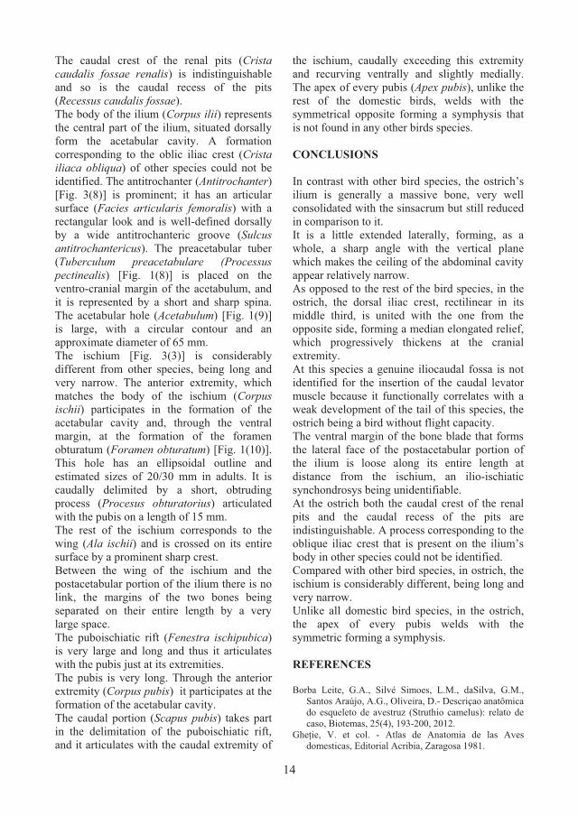
Fig. 1. The coccyx of the ostrich – lateral view
(original) 1-the preacetabular part of the ilium; 2-the
postacetabular part of the ilium; 3-ischium; 4-pubis; 5-the dorsal iliac crest; 6-the dorso-lateral crest of ilium ;
70- the lateral iliac crest ; 8-the preacetabular tuber (pectineus) ; 9-the acetabular hole; 10-the obturator
hole; 11-the antitrochanter; 12-the obturator process; 13-the transverse processes of the sinsacrum’s
vertebrae; 14-the infracristal blade; 15- the ischio-pubic hole; 16-scapus pubis; 17-the apex of the pubis
The lateral iliac crest (Crista iliaca lateralis) [Fig.1(7)] is convex in the cranial half and concave in the caudal one, oriented in ventro-lateral direction. At a small distance from the cranial margin of the acetabular hole it
extends ventrally reaching the preacetabular tuber (pectineal process) [Fig.1(8)]. The plain ventral face (Facies ventralis) is ventro-medially oriented towards the spinous or dorsal crest of the sinsacrum (Crista spinosa synsacri). The sharp angle that forms with the median plan enables us to consider, from a topographic point of view, this face as a medial face rather than a ventral one. Apart from this, between the ventral face and the spinous crest a wide space can be observed, with an almost triangular outline in a transversal section. In the inferior part, this space is delimitated by the transverse processes of the last two thoracic vertebrae and the first three-four lumbar vertebrae that, through their apex, articulate with the ventro-median margin of this face while leaving very large spaces in the area of the intertransversary openings (Fenestrae intertransversarie) [Fig. 3(2)] at this species. At young birds, where the joints between the ilium and the top of the transverse processes did not weld, the articular areas (Arae articulares vertebrales) that correspond to these processes can be observed.
Fig. 2. The coccyx of the ostrich- cranial view (original)
1-the cranial terminal face of the second last thoracic vertebrae ; 2-the thorny crest of the sinsacrum; 3-the
transverse processes of the second last lumbar vertebrae; 4-the antitrochanter; 5- the dorsal iliac crests united in a
medial plan; 6-pubis; 7-the pubic symphysis.
The postacetabular part of the ilium also has three faces: a dorsal, a lateral and a ventral one. Because of the totally different appearance of the coccyx in these ratite, the terms for these faces don’t fully correspond to the topographic reality. The dorsal face (Facies dorsalis) is separated by the lateral face through the dorso-lateral crest of the ilium (Crista dorsolateralis ilii) (indistinct at some anseriformes, for example). The dorso-lateral crest is long, almost rectilinear (with the exception of the extre-mities) and is more prominent in the cranial half than the caudal one. In the caudal part, the caudal crest thickens, becomes coarse and slightly orients ventro-laterally, ending through a sharp process that matches the dorso-lateral spina of the ilium (Spina dorsolateralis ilii). Cranially, the crest is oriented latero-ventrally, connecting to the caudal extremity of the dorsal iliac crest, at the level of the tuber that is oriented towards the antetro-chanter, a process reminded earlier that is not mentioned in specialty nomenclature. The dorsal side of the postacetabular part is very long, transversally convex on its entire length. A genuine iliocaudal fossa (Fossa iliocaudalis) is not identified. At the species at which it exists, this fossa is destined to the insertion the levator caudal muscle. Taking into account the role of the tail in directing flight, the absence of this fossa in the ostrich would be due to the weak development of the tail musculature in ratite, birds without the capacity of flight. Unlike the preacetabular part, the medial margin of this dorsal face is not in contact with the symmetric one and for most of its part it also remains separated from the sinsacrum through a large space. The ilio-sacral suture (Suture iliosynsacrala) is present just in the posterior third of the bone. The lateral face (Facies lateralis) of the postacetabular portion is vaster compared to the dorsal one. The ventral margin of the bone blade that constitutes this face (Lamina
infracristalis ilii) is loose on its entire length and there is no ilio-ischiatic synchondrosis (Syncondrosis ilioischiatica) therefore the ilioischiatic hole doesn’t form either (Foramen ilioischiaticum). The infracristal cavity cannot be identified (Cavitas infracristalis), the reduced depression is situated caudally from the ilioischiatic hole and it is destined to the ischiofemuralis muscle. However, this side is obliquely crossed in a cranio-caudal and ventro-lateral way by an obvious crest, not mentioned in literature.
Fig. 3. the coccyx of the ostrich – ventral view (original) 1-sinsacrum; 2-the intertransversal openings; 3-ischium; 4-pubis; 5-the apex of the pubis; 6-the lateral crest of the ilium; 7-the prepubic tuber; 8-the antitrochanter; 9-the dorso-lateral spina of the ilium; 10-ischio-pubic hole.
The ventral side of the postacetabular part orients itself in a medial direction and is articulated with the transverse processes of the vertebrae that compose the sinsacrum [Fig. 3(1)].

13
the postacetabular part (taking as a landmark a vertical plane that crosses through the center of the acetabular hole) is 1 : 2. On the dorsal side (Facies dorsalis), the dorsal iliac fossa (Fossa iliaca dorsalis) has the appeareance of an almost plain surface, strongly ventrally inclined and marked for the most part by lines and coarse crests. The dorsal iliac crest (Crista iliaca dorsalis) [Fig.1(5)], rectilinear in its middle third, is united with the one on the opposite side, forming a median elongated relief that becomes progressively thicker at the cranial extremity. In the caudal part, the dorsal iliac crest will separate from her symmetrical one, recurving laterally to end through a prominence oriented towards the dorsal margin of the antitrochanter from which it is separated by a large incisure.
Fig. 1. The coccyx of the ostrich – lateral view
(original) 1-the preacetabular part of the ilium; 2-the
postacetabular part of the ilium; 3-ischium; 4-pubis; 5-the dorsal iliac crest; 6-the dorso-lateral crest of ilium ;
70- the lateral iliac crest ; 8-the preacetabular tuber (pectineus) ; 9-the acetabular hole; 10-the obturator
hole; 11-the antitrochanter; 12-the obturator process; 13-the transverse processes of the sinsacrum’s
vertebrae; 14-the infracristal blade; 15- the ischio-pubic hole; 16-scapus pubis; 17-the apex of the pubis
The lateral iliac crest (Crista iliaca lateralis) [Fig.1(7)] is convex in the cranial half and concave in the caudal one, oriented in ventro-lateral direction. At a small distance from the cranial margin of the acetabular hole it
extends ventrally reaching the preacetabular tuber (pectineal process) [Fig.1(8)]. The plain ventral face (Facies ventralis) is ventro-medially oriented towards the spinous or dorsal crest of the sinsacrum (Crista spinosa synsacri). The sharp angle that forms with the median plan enables us to consider, from a topographic point of view, this face as a medial face rather than a ventral one. Apart from this, between the ventral face and the spinous crest a wide space can be observed, with an almost triangular outline in a transversal section. In the inferior part, this space is delimitated by the transverse processes of the last two thoracic vertebrae and the first three-four lumbar vertebrae that, through their apex, articulate with the ventro-median margin of this face while leaving very large spaces in the area of the intertransversary openings (Fenestrae intertransversarie) [Fig. 3(2)] at this species. At young birds, where the joints between the ilium and the top of the transverse processes did not weld, the articular areas (Arae articulares vertebrales) that correspond to these processes can be observed.
Fig. 2. The coccyx of the ostrich- cranial view (original)
1-the cranial terminal face of the second last thoracic vertebrae ; 2-the thorny crest of the sinsacrum; 3-the
transverse processes of the second last lumbar vertebrae; 4-the antitrochanter; 5- the dorsal iliac crests united in a
medial plan; 6-pubis; 7-the pubic symphysis.
The postacetabular part of the ilium also has three faces: a dorsal, a lateral and a ventral one. Because of the totally different appearance of the coccyx in these ratite, the terms for these faces don’t fully correspond to the topographic reality. The dorsal face (Facies dorsalis) is separated by the lateral face through the dorso-lateral crest of the ilium (Crista dorsolateralis ilii) (indistinct at some anseriformes, for example). The dorso-lateral crest is long, almost rectilinear (with the exception of the extre-mities) and is more prominent in the cranial half than the caudal one. In the caudal part, the caudal crest thickens, becomes coarse and slightly orients ventro-laterally, ending through a sharp process that matches the dorso-lateral spina of the ilium (Spina dorsolateralis ilii). Cranially, the crest is oriented latero-ventrally, connecting to the caudal extremity of the dorsal iliac crest, at the level of the tuber that is oriented towards the antetro-chanter, a process reminded earlier that is not mentioned in specialty nomenclature. The dorsal side of the postacetabular part is very long, transversally convex on its entire length. A genuine iliocaudal fossa (Fossa iliocaudalis) is not identified. At the species at which it exists, this fossa is destined to the insertion the levator caudal muscle. Taking into account the role of the tail in directing flight, the absence of this fossa in the ostrich would be due to the weak development of the tail musculature in ratite, birds without the capacity of flight. Unlike the preacetabular part, the medial margin of this dorsal face is not in contact with the symmetric one and for most of its part it also remains separated from the sinsacrum through a large space. The ilio-sacral suture (Suture iliosynsacrala) is present just in the posterior third of the bone. The lateral face (Facies lateralis) of the postacetabular portion is vaster compared to the dorsal one. The ventral margin of the bone blade that constitutes this face (Lamina
infracristalis ilii) is loose on its entire length and there is no ilio-ischiatic synchondrosis (Syncondrosis ilioischiatica) therefore the ilioischiatic hole doesn’t form either (Foramen ilioischiaticum). The infracristal cavity cannot be identified (Cavitas infracristalis), the reduced depression is situated caudally from the ilioischiatic hole and it is destined to the ischiofemuralis muscle. However, this side is obliquely crossed in a cranio-caudal and ventro-lateral way by an obvious crest, not mentioned in literature.
Fig. 3. the coccyx of the ostrich – ventral view (original) 1-sinsacrum; 2-the intertransversal openings; 3-ischium; 4-pubis; 5-the apex of the pubis; 6-the lateral crest of the ilium; 7-the prepubic tuber; 8-the antitrochanter; 9-the dorso-lateral spina of the ilium; 10-ischio-pubic hole.
The ventral side of the postacetabular part orients itself in a medial direction and is articulated with the transverse processes of the vertebrae that compose the sinsacrum [Fig. 3(1)].
14
The caudal crest of the renal pits (Crista caudalis fossae renalis) is indistinguishable and so is the caudal recess of the pits (Recessus caudalis fossae). The body of the ilium (Corpus ilii) represents the central part of the ilium, situated dorsally form the acetabular cavity. A formation corresponding to the oblic iliac crest (Crista iliaca obliqua) of other species could not be identified. The antitrochanter (Antitrochanter) [Fig. 3(8)] is prominent; it has an articular surface (Facies articularis femoralis) with a rectangular look and is well-defined dorsally by a wide antitrochanteric groove (Sulcus antitrochantericus). The preacetabular tuber (Tuberculum preacetabulare (Processus pectinealis) [Fig. 1(8)] is placed on the ventro-cranial margin of the acetabulum, and it is represented by a short and sharp spina. The acetabular hole (Acetabulum) [Fig. 1(9)] is large, with a circular contour and an approximate diameter of 65 mm. The ischium [Fig. 3(3)] is considerably different from other species, being long and very narrow. The anterior extremity, which matches the body of the ischium (Corpus ischii) participates in the formation of the acetabular cavity and, through the ventral margin, at the formation of the foramen obturatum (Foramen obturatum) [Fig. 1(10)]. This hole has an ellipsoidal outline and estimated sizes of 20/30 mm in adults. It is caudally delimited by a short, obtruding process (Procesus obturatorius) articulated with the pubis on a length of 15 mm. The rest of the ischium corresponds to the wing (Ala ischii) and is crossed on its entire surface by a prominent sharp crest. Between the wing of the ischium and the postacetabular portion of the ilium there is no link, the margins of the two bones being separated on their entire length by a very large space. The puboischiatic rift (Fenestra ischipubica) is very large and long and thus it articulates with the pubis just at its extremities. The pubis is very long. Through the anterior extremity (Corpus pubis) it participates at the formation of the acetabular cavity. The caudal portion (Scapus pubis) takes part in the delimitation of the puboischiatic rift, and it articulates with the caudal extremity of
the ischium, caudally exceeding this extremity and recurving ventrally and slightly medially. The apex of every pubis (Apex pubis), unlike the rest of the domestic birds, welds with the symmetrical opposite forming a symphysis that is not found in any other birds species. CONCLUSIONS In contrast with other bird species, the ostrich’s ilium is generally a massive bone, very well consolidated with the sinsacrum but still reduced in comparison to it. It is a little extended laterally, forming, as a whole, a sharp angle with the vertical plane which makes the ceiling of the abdominal cavity appear relatively narrow. As opposed to the rest of the bird species, in the ostrich, the dorsal iliac crest, rectilinear in its middle third, is united with the one from the opposite side, forming a median elongated relief, which progressively thickens at the cranial extremity. At this species a genuine iliocaudal fossa is not identified for the insertion of the caudal levator muscle because it functionally correlates with a weak development of the tail of this species, the ostrich being a bird without flight capacity. The ventral margin of the bone blade that forms the lateral face of the postacetabular portion of the ilium is loose along its entire length at distance from the ischium, an ilio-ischiatic synchondrosys being unidentifiable. At the ostrich both the caudal crest of the renal pits and the caudal recess of the pits are indistinguishable. A process corresponding to the oblique iliac crest that is present on the ilium’s body in other species could not be identified. Compared with other bird species, in ostrich, the ischium is considerably different, being long and very narrow. Unlike all domestic bird species, in the ostrich, the apex of every pubis welds with the symmetric forming a symphysis. REFERENCES Borba Leite, G.A., Silvé Simoes, L.M., daSilva, G.M.,
Santos Araújo, A.G., Oliveira, D.- Descriçao anatômica do esqueleto de avestruz (Struthio camelus): relato de caso, Biotemas, 25(4), 193-200, 2012.
Gheție, V. et col. - Atlas de Anatomia de las Aves domesticas, Editorial Acribia, Zaragosa 1981.
Hou, L., Zhou, Z., Zhang, F., Zhao, W.- A Miocene astrich fossil from Gansu Province, northwest China- Chinese Science Bulletin, vol. 50, Nr. 16, 1808-1810, 2005.
Pop, C., Pentea, M.- The osteological features of the skeleton in ostrich (Strithio camelus), Lucrări științifice, Med. vet. vol XL, Timișoara, 2007.
Predoi, G., Belu, C., Dumitrescu, I., Georgescu, B., Roșu, P., Bițoiu, C. – The morphology of the shoulder and elbow joints in Ostrich (Struthio camelus) – Anatomia Histologia Embryologia, volume 39, number 4, Wiley-Blackwell, Print ISSN 0340-2096, Online ISSN 1439-0264, pg. 266, 2010.
Predoi, G. et. col.- Anatomia comparată a animalelor domestice, Ed. Ceres București, 2011.
Tamilselvan, S., Iniyah, K., Jayachitra, S., Sivagnanam, S., Balasundaram, K., Lavanya, C.- Gross anatomy of os coxae of ostrich (Struthio camelus), International Journal of current Microbiology and Applied Sciences ISSN: 2319-7706, Volume 4, Number 4, pp 201-205 2015.
Zhang, R., Wang, H., Zeng, G., Zhou, C., Pan, R., Wang, Q., Li, J.- Anatomical study of the ostrich (Struthio camelus) foot locomotor system, Indian J. Anim. Res., 50(4), 476-483, 2016.
*** - (2005) – Nomina Anatomica Veterinaria (Fifth Edition) Zürich and Ithaca, New York.

15
The caudal crest of the renal pits (Crista caudalis fossae renalis) is indistinguishable and so is the caudal recess of the pits (Recessus caudalis fossae). The body of the ilium (Corpus ilii) represents the central part of the ilium, situated dorsally form the acetabular cavity. A formation corresponding to the oblic iliac crest (Crista iliaca obliqua) of other species could not be identified. The antitrochanter (Antitrochanter) [Fig. 3(8)] is prominent; it has an articular surface (Facies articularis femoralis) with a rectangular look and is well-defined dorsally by a wide antitrochanteric groove (Sulcus antitrochantericus). The preacetabular tuber (Tuberculum preacetabulare (Processus pectinealis) [Fig. 1(8)] is placed on the ventro-cranial margin of the acetabulum, and it is represented by a short and sharp spina. The acetabular hole (Acetabulum) [Fig. 1(9)] is large, with a circular contour and an approximate diameter of 65 mm. The ischium [Fig. 3(3)] is considerably different from other species, being long and very narrow. The anterior extremity, which matches the body of the ischium (Corpus ischii) participates in the formation of the acetabular cavity and, through the ventral margin, at the formation of the foramen obturatum (Foramen obturatum) [Fig. 1(10)]. This hole has an ellipsoidal outline and estimated sizes of 20/30 mm in adults. It is caudally delimited by a short, obtruding process (Procesus obturatorius) articulated with the pubis on a length of 15 mm. The rest of the ischium corresponds to the wing (Ala ischii) and is crossed on its entire surface by a prominent sharp crest. Between the wing of the ischium and the postacetabular portion of the ilium there is no link, the margins of the two bones being separated on their entire length by a very large space. The puboischiatic rift (Fenestra ischipubica) is very large and long and thus it articulates with the pubis just at its extremities. The pubis is very long. Through the anterior extremity (Corpus pubis) it participates at the formation of the acetabular cavity. The caudal portion (Scapus pubis) takes part in the delimitation of the puboischiatic rift, and it articulates with the caudal extremity of
the ischium, caudally exceeding this extremity and recurving ventrally and slightly medially. The apex of every pubis (Apex pubis), unlike the rest of the domestic birds, welds with the symmetrical opposite forming a symphysis that is not found in any other birds species. CONCLUSIONS In contrast with other bird species, the ostrich’s ilium is generally a massive bone, very well consolidated with the sinsacrum but still reduced in comparison to it. It is a little extended laterally, forming, as a whole, a sharp angle with the vertical plane which makes the ceiling of the abdominal cavity appear relatively narrow. As opposed to the rest of the bird species, in the ostrich, the dorsal iliac crest, rectilinear in its middle third, is united with the one from the opposite side, forming a median elongated relief, which progressively thickens at the cranial extremity. At this species a genuine iliocaudal fossa is not identified for the insertion of the caudal levator muscle because it functionally correlates with a weak development of the tail of this species, the ostrich being a bird without flight capacity. The ventral margin of the bone blade that forms the lateral face of the postacetabular portion of the ilium is loose along its entire length at distance from the ischium, an ilio-ischiatic synchondrosys being unidentifiable. At the ostrich both the caudal crest of the renal pits and the caudal recess of the pits are indistinguishable. A process corresponding to the oblique iliac crest that is present on the ilium’s body in other species could not be identified. Compared with other bird species, in ostrich, the ischium is considerably different, being long and very narrow. Unlike all domestic bird species, in the ostrich, the apex of every pubis welds with the symmetric forming a symphysis. REFERENCES Borba Leite, G.A., Silvé Simoes, L.M., daSilva, G.M.,
Santos Araújo, A.G., Oliveira, D.- Descriçao anatômica do esqueleto de avestruz (Struthio camelus): relato de caso, Biotemas, 25(4), 193-200, 2012.
Gheție, V. et col. - Atlas de Anatomia de las Aves domesticas, Editorial Acribia, Zaragosa 1981.
Hou, L., Zhou, Z., Zhang, F., Zhao, W.- A Miocene astrich fossil from Gansu Province, northwest China- Chinese Science Bulletin, vol. 50, Nr. 16, 1808-1810, 2005.
Pop, C., Pentea, M.- The osteological features of the skeleton in ostrich (Strithio camelus), Lucrări științifice, Med. vet. vol XL, Timișoara, 2007.
Predoi, G., Belu, C., Dumitrescu, I., Georgescu, B., Roșu, P., Bițoiu, C. – The morphology of the shoulder and elbow joints in Ostrich (Struthio camelus) – Anatomia Histologia Embryologia, volume 39, number 4, Wiley-Blackwell, Print ISSN 0340-2096, Online ISSN 1439-0264, pg. 266, 2010.
Predoi, G. et. col.- Anatomia comparată a animalelor domestice, Ed. Ceres București, 2011.
Tamilselvan, S., Iniyah, K., Jayachitra, S., Sivagnanam, S., Balasundaram, K., Lavanya, C.- Gross anatomy of os coxae of ostrich (Struthio camelus), International Journal of current Microbiology and Applied Sciences ISSN: 2319-7706, Volume 4, Number 4, pp 201-205 2015.
Zhang, R., Wang, H., Zeng, G., Zhou, C., Pan, R., Wang, Q., Li, J.- Anatomical study of the ostrich (Struthio camelus) foot locomotor system, Indian J. Anim. Res., 50(4), 476-483, 2016.
*** - (2005) – Nomina Anatomica Veterinaria (Fifth Edition) Zürich and Ithaca, New York.

16
ASPECTS REGARDING THE MORPHOLOGY OF CERVICAL VERTEBRAE IN COYPU (MYOCASTOR COYPUS)
Anca ȘEICARU, Cristian BELU
University of Agronomic Sciences and Veterinary Medicine of Bucharest, Faculty of Veterinary
Medicine, 105 Spaiul Independenței, District 5, Bucharest, Romania
Corresponding author email: [email protected] Abstract This study was done on three bodies of adult coypu. The material was obtained by cleaning and macerating bones in 37°C water. Following the whitening process the bones were consequently cleaned under a water stream and left to dry. The cervical vertebrae generally have the spinous processes at a uniform height, oriented caudally for vertebrae III-V, dorsally for vertebra VI and cranially for vertebra VII. The body of the cervical vertebrae is short and flattened dorso-ventrally; the cranial and caudal terminal facets are plane. The ventral vertebral crest is absent. The transverse processes are the same length, slightly more developed at vertebra VII. The transverse foramen is present, wide in diameter, being replaced at vertebra VII by a vertebral incisura. The atlas has rounded transverse processes, oriented dorsally. The transverse foramen is located on the caudal edge of the atlas wing. The lateral vertebral foramen and the alar foramen are joined through a thick alar notch. The axis has a thick, developed spinous process that ends in a tuberosity. Its transverse processes slightly surpass the caudal terminal facet. Key words: cervical vertebrae, coypu, vertebral column. INTRODUCTION Morphological aspects of the cervical vertebrae in coypu are important because they present certain particularities compared to other species of rodents (Coțofan et al., 1982, Hrițcu et al., 2000, Predoi, 2012). There are 7 cervical vertebrae, and the diverse aspects described in this study complete already existing data from scientific literature (Coțofan et al., 1987, Coțofan et al., 2003, Predoi et al., 2001). The body of the cervical vertebrae is reduced, transversally wider and with a very large vertebral canal. The transverse foramen is wide, oval in shape and constantly located on the caudal edge of the atlas wings. Unlike in scientific literature, where a cranial vertebra incisura is described for the atlas, this study revealed the presence of a large lateral vertebral foramen. The alar notch between the alar foramen and the lateral vertebral foramen is thick. Particularities of this species were also discovered in the transverse processes of vertebrae V and VI. In the seventh vertebra the transverse foramen is replaced with an incisura.
MATERIALS AND METHODS
Three bodies of adult coypu were used as material. The process of controlled maceration was used as method. This particular method takes the following steps:
� Skinning the body � Eviscerating the body � Manually removing muscle mass from
the bones. The controlled maceration technique includes submerging the bones in water at 37°C. Following the maceration process the bones were washed under a stream of water and were submitted to a whitening process. For the whitening process a solution of hydrogen peroxide 11% was used. After the whitening process the bones were once again cleaned under a stream of water and left to dry at room temperature. Following the preparation process, the bones were studied and photographed. The naming of the structures was done in concordance with the norms imposed by Nomina Anatomica Veterinaria - 2005.
RESULTS AND DISCUSSIONS
In the cervical region, the vertebrae are characterised by a short body which is transversally wide. A wide vertebral canal can be observed. The atlas has well developed rounded wings that are slightly deviated dorsally (Fig. 1, 2, 3).
Fig. 1 Atlas of coypu, cranial view (original) 1-dorsal tubercle; 2-ventral arch; 3-glenoid cavities; 4-
wings of the atlas.
The transverse foramen is large, oval shaped, placed horizontally on the caudal edge of the atlas wings. Compared to scientific literature, this study reveals the bone to have a large lateral vertebral foramen as well as an alar foramen, united through an alar notch. The cranial articular surfaces are slightly excavated.
Fig. 2 Atlas of coypu, dorsal view (original) 1-dorsal tubercle; 2-wings of the atlas; 3-hole alar foramen; 4-transverse foramen; 5-caudal articular
surfaces; 6-vertebral foramen.
Fig. 3 Atlas of coypu, caudal view (original) 1-dorsal tubercle; 2-ventral arch; 3-wings of the
atlas; 4-caudal articular surfaces; 5-vertebral foramen; 6-transverse foramen.
The axis has a well-developed spinous process, thick all throughout its length, which ends dorsally in a reduced tuberosity. The transverse processes are developed and slightly surpass the terminal caudal facet. The transverse foramina are broad with vertebral incisurae which are also wide, while the terminal caudal facet has a shallow glenoid cavity. The terminal cranial facet features an odontoid process in the shape of a cone that is slightly deviated dorsally.
Fig. 4 Axis to coypu, lateral view (original) 1- odontoid process; 2-spinous process;
3-caudal articular processes; 4-transverse processes; 5-transverse foramen.
Scientific Works. Series C. Veterinary Medicine. Vol. LXIV (2), 2018ISSN 2065-1295; ISSN 2343-9394 (CD-ROM); ISSN 2067-3663 (Online); ISSN-L 2065-1295

17
ASPECTS REGARDING THE MORPHOLOGY OF CERVICAL VERTEBRAE IN COYPU (MYOCASTOR COYPUS)
Anca ȘEICARU, Cristian BELU
University of Agronomic Sciences and Veterinary Medicine of Bucharest, Faculty of Veterinary
Medicine, 105 Spaiul Independenței, District 5, Bucharest, Romania
Corresponding author email: [email protected] Abstract This study was done on three bodies of adult coypu. The material was obtained by cleaning and macerating bones in 37°C water. Following the whitening process the bones were consequently cleaned under a water stream and left to dry. The cervical vertebrae generally have the spinous processes at a uniform height, oriented caudally for vertebrae III-V, dorsally for vertebra VI and cranially for vertebra VII. The body of the cervical vertebrae is short and flattened dorso-ventrally; the cranial and caudal terminal facets are plane. The ventral vertebral crest is absent. The transverse processes are the same length, slightly more developed at vertebra VII. The transverse foramen is present, wide in diameter, being replaced at vertebra VII by a vertebral incisura. The atlas has rounded transverse processes, oriented dorsally. The transverse foramen is located on the caudal edge of the atlas wing. The lateral vertebral foramen and the alar foramen are joined through a thick alar notch. The axis has a thick, developed spinous process that ends in a tuberosity. Its transverse processes slightly surpass the caudal terminal facet. Key words: cervical vertebrae, coypu, vertebral column. INTRODUCTION Morphological aspects of the cervical vertebrae in coypu are important because they present certain particularities compared to other species of rodents (Coțofan et al., 1982, Hrițcu et al., 2000, Predoi, 2012). There are 7 cervical vertebrae, and the diverse aspects described in this study complete already existing data from scientific literature (Coțofan et al., 1987, Coțofan et al., 2003, Predoi et al., 2001). The body of the cervical vertebrae is reduced, transversally wider and with a very large vertebral canal. The transverse foramen is wide, oval in shape and constantly located on the caudal edge of the atlas wings. Unlike in scientific literature, where a cranial vertebra incisura is described for the atlas, this study revealed the presence of a large lateral vertebral foramen. The alar notch between the alar foramen and the lateral vertebral foramen is thick. Particularities of this species were also discovered in the transverse processes of vertebrae V and VI. In the seventh vertebra the transverse foramen is replaced with an incisura.
MATERIALS AND METHODS
Three bodies of adult coypu were used as material. The process of controlled maceration was used as method. This particular method takes the following steps:
� Skinning the body � Eviscerating the body � Manually removing muscle mass from
the bones. The controlled maceration technique includes submerging the bones in water at 37°C. Following the maceration process the bones were washed under a stream of water and were submitted to a whitening process. For the whitening process a solution of hydrogen peroxide 11% was used. After the whitening process the bones were once again cleaned under a stream of water and left to dry at room temperature. Following the preparation process, the bones were studied and photographed. The naming of the structures was done in concordance with the norms imposed by Nomina Anatomica Veterinaria - 2005.
RESULTS AND DISCUSSIONS
In the cervical region, the vertebrae are characterised by a short body which is transversally wide. A wide vertebral canal can be observed. The atlas has well developed rounded wings that are slightly deviated dorsally (Fig. 1, 2, 3).
Fig. 1 Atlas of coypu, cranial view (original) 1-dorsal tubercle; 2-ventral arch; 3-glenoid cavities; 4-
wings of the atlas.
The transverse foramen is large, oval shaped, placed horizontally on the caudal edge of the atlas wings. Compared to scientific literature, this study reveals the bone to have a large lateral vertebral foramen as well as an alar foramen, united through an alar notch. The cranial articular surfaces are slightly excavated.
Fig. 2 Atlas of coypu, dorsal view (original) 1-dorsal tubercle; 2-wings of the atlas; 3-hole alar foramen; 4-transverse foramen; 5-caudal articular
surfaces; 6-vertebral foramen.
Fig. 3 Atlas of coypu, caudal view (original) 1-dorsal tubercle; 2-ventral arch; 3-wings of the
atlas; 4-caudal articular surfaces; 5-vertebral foramen; 6-transverse foramen.
The axis has a well-developed spinous process, thick all throughout its length, which ends dorsally in a reduced tuberosity. The transverse processes are developed and slightly surpass the terminal caudal facet. The transverse foramina are broad with vertebral incisurae which are also wide, while the terminal caudal facet has a shallow glenoid cavity. The terminal cranial facet features an odontoid process in the shape of a cone that is slightly deviated dorsally.
Fig. 4 Axis to coypu, lateral view (original) 1- odontoid process; 2-spinous process;
3-caudal articular processes; 4-transverse processes; 5-transverse foramen.

18
Fig. 5 Axis to coypu, ventro-caudal view (original) 1-the body of axis; 2-spinous process; 3-caudal
articular processes; 4-transversale process; 5- odontoid process; 6-transversale foramen.
The cranial articular surfaces are triangular in aspect. The caudal articular processes emerge at a distance from the spinous process and appear flat (Fig. 4, 5). The third cervical vertebra has a short, dorso-ventrally flattened body. The ventral vertebral crest is absent. Both the terminal cranial facet and the terminal caudal facet appear flat. The spinous process is relatively well developed, but it lacks a dorsal tuberosity. The transverse processes exceed the terminal caudal facet, oriented dorso-caudally. The transverse foramen is wide. (Fig. 6)
Fig. 6. Cervical vertebrae III-VII in coypu, dorsal view (original)
1-transverse processes; 2-cranial articular processes; 3-caudal articular processes; 4-spinous processes.
The fourth cervical vertebra exhibits wider and thicker transverse processes compared to the third vertebra, while the spinous process is dorsally widened and levelled with the one corresponding to the previous vertebra. The fifth cervical vertebra has a widened transverse process, oriented in the same direction as in the case of the third and fourth vertebrae. The transverse foramen is wide. Ventro-medially from the transverse process an osseous dint can be observed, which is flattened, oriented cranially and ended with a crest. The sixth vertebra has a short body, flattened dorso-ventrally, and it exhibits flat terminal faces as well as the absence of the ventral vertebral foramen. The transverse foramen is very wide. The transverse processes feature ventro-caudally two thick osseous laminae which are well detached and flattened.
Fig. 7 Cervical vertebrae III-VII in coypu, ventral view (original)
1-vertebral body; 2-transverse processes;
Fig. 8 Cervical vertebrae III-VII to coypu, lateral view (original)
1-spinous processes; 2-cranial articular processes; 3-caudal articular processes; 4-transverse processes.
The seventh cervical vertebra features its spinous process oriented cranially, widened at the same height as in the previous vertebrae. The transverse process is very wide dorso-ventrally and is oriented caudally. Instead of transverse foramina, vertebral incisurae are present (Fig. 7, 8). CONCLUSIONS The bodies of the cervical vertebrae is short and flattened dorso-ventrally, and it lacks a ventral vertebral crest. The vertebral canal of this species is particularly wide. Unlike what is presented in existing specialty literature, the atlas does present a wide lateral vertebral foramen and not a lateral vertebral incisura. The alar foramen is also present. The transverse foramen is on the caudal edge of the atlas wings. The axis does present lateral vertebral incisurae, a cone-shaped odontoid process that is slightle deviated dorsally and the caudal terminal facet is represented by a shallow glenoid cavity. The spinous process of the III-VII vertebrae is thick and levelled.
In the fifth vertebra, the transverse process features an osseous dint that is flattened cranially and ends with a crest. In the sixth vertebra the transverse processes feature ventro-caudally two thick osseous laminae which are well detached and flattened. The seventh vertebra features an incisura in place of the transverse foramina.
REFERENCES Coțofan V., Cura P., Negrea A., Hrițcu Valentina, 1987.
Contribuții la studiul scheletelui axial la dihorul de crescătorie, Lucr. Sem. șt Med. Vet., Iași.
Coțofan V., Hrițcu Valentina, Negrea S., Cura P., Cozariuc I., 1982. Caractere morfologice diferențiale ale scheletului axial la nutri comparativ cu unele mamifere domestice. Lucr. Sem. șt „Metode noi de sporire a producției la animale“, Iași.
Coțofan V., Predoi G., 2003. Anatomia topografică a animalelor domestice, Ed. BIC ALL, București.
Hrițcu Valentina, Coțofan V., 2000. Anatomia animalelor de blană: nutria, dihorul. Editura „Ion Ionescu de la Brad“, Iași.
Predoi G., 2012. Anatomia topografică a animalelor domestice, Ed. Ceres, București.
Predoi G., Belu C., 2001. Anatomia animalelor domestice (Anatomie clinică), Ed. BIC ALL, București.
*** - (2005) – Nomina Anatomica Veterinaria (Fifth Edition) Zürich and Ithaca, New York.

19
Fig. 5 Axis to coypu, ventro-caudal view (original) 1-the body of axis; 2-spinous process; 3-caudal
articular processes; 4-transversale process; 5- odontoid process; 6-transversale foramen.
The cranial articular surfaces are triangular in aspect. The caudal articular processes emerge at a distance from the spinous process and appear flat (Fig. 4, 5). The third cervical vertebra has a short, dorso-ventrally flattened body. The ventral vertebral crest is absent. Both the terminal cranial facet and the terminal caudal facet appear flat. The spinous process is relatively well developed, but it lacks a dorsal tuberosity. The transverse processes exceed the terminal caudal facet, oriented dorso-caudally. The transverse foramen is wide. (Fig. 6)
Fig. 6. Cervical vertebrae III-VII in coypu, dorsal view (original)
1-transverse processes; 2-cranial articular processes; 3-caudal articular processes; 4-spinous processes.
The fourth cervical vertebra exhibits wider and thicker transverse processes compared to the third vertebra, while the spinous process is dorsally widened and levelled with the one corresponding to the previous vertebra. The fifth cervical vertebra has a widened transverse process, oriented in the same direction as in the case of the third and fourth vertebrae. The transverse foramen is wide. Ventro-medially from the transverse process an osseous dint can be observed, which is flattened, oriented cranially and ended with a crest. The sixth vertebra has a short body, flattened dorso-ventrally, and it exhibits flat terminal faces as well as the absence of the ventral vertebral foramen. The transverse foramen is very wide. The transverse processes feature ventro-caudally two thick osseous laminae which are well detached and flattened.
Fig. 7 Cervical vertebrae III-VII in coypu, ventral view (original)
1-vertebral body; 2-transverse processes;
Fig. 8 Cervical vertebrae III-VII to coypu, lateral view (original)
1-spinous processes; 2-cranial articular processes; 3-caudal articular processes; 4-transverse processes.
The seventh cervical vertebra features its spinous process oriented cranially, widened at the same height as in the previous vertebrae. The transverse process is very wide dorso-ventrally and is oriented caudally. Instead of transverse foramina, vertebral incisurae are present (Fig. 7, 8). CONCLUSIONS The bodies of the cervical vertebrae is short and flattened dorso-ventrally, and it lacks a ventral vertebral crest. The vertebral canal of this species is particularly wide. Unlike what is presented in existing specialty literature, the atlas does present a wide lateral vertebral foramen and not a lateral vertebral incisura. The alar foramen is also present. The transverse foramen is on the caudal edge of the atlas wings. The axis does present lateral vertebral incisurae, a cone-shaped odontoid process that is slightle deviated dorsally and the caudal terminal facet is represented by a shallow glenoid cavity. The spinous process of the III-VII vertebrae is thick and levelled.
In the fifth vertebra, the transverse process features an osseous dint that is flattened cranially and ends with a crest. In the sixth vertebra the transverse processes feature ventro-caudally two thick osseous laminae which are well detached and flattened. The seventh vertebra features an incisura in place of the transverse foramina.
REFERENCES Coțofan V., Cura P., Negrea A., Hrițcu Valentina, 1987.
Contribuții la studiul scheletelui axial la dihorul de crescătorie, Lucr. Sem. șt Med. Vet., Iași.
Coțofan V., Hrițcu Valentina, Negrea S., Cura P., Cozariuc I., 1982. Caractere morfologice diferențiale ale scheletului axial la nutri comparativ cu unele mamifere domestice. Lucr. Sem. șt „Metode noi de sporire a producției la animale“, Iași.
Coțofan V., Predoi G., 2003. Anatomia topografică a animalelor domestice, Ed. BIC ALL, București.
Hrițcu Valentina, Coțofan V., 2000. Anatomia animalelor de blană: nutria, dihorul. Editura „Ion Ionescu de la Brad“, Iași.
Predoi G., 2012. Anatomia topografică a animalelor domestice, Ed. Ceres, București.
Predoi G., Belu C., 2001. Anatomia animalelor domestice (Anatomie clinică), Ed. BIC ALL, București.
*** - (2005) – Nomina Anatomica Veterinaria (Fifth Edition) Zürich and Ithaca, New York.

20

21
clinicalScienceS

22
CANINE HERPESVIRUS-1 SPECIFIC SEROCONVERSION
AND CLINICAL ASPECTS IN KENNEL DOGS FROM ROMANIA
Dragoș COBZARIU1, George Andrei NECULA1, Stelian BĂRĂITĂREANU1*, Georgeta ȘTEFAN1, Doina DANEȘ1
1University of Agronomic Sciences and Veterinary Medicine of Bucharest,
59 Marasti Blvd, District 1, Bucharest, Romania
*Corresponding author email: [email protected] Abstract The Canine herpesvirus-1 (CHV-1) is causing in dogs a wide range of reproductive problems: infertility, foetal resorption, abortion, weak puppies, stillborn, low conception rate, small litter size and neonatal mortality, according to the age and pregnancy stage. The aims of the study where to assess the status of CHV-1 infection and to investigate the clinical pattern of the disease, in three Romanian kennel dogs. Blood samples from 44 subjects, aged from 1 to 5.5 years (20 dogs from kennel A, 16 dogs from kennel B, and 8 from kennel C), without history of vaccination against CHV-1 where submitted to study. The serum samples were analysed for the detection of antibodies to CHV-1 by immunofluorescence assays. In this survey, the average of seropositive animals were being 86.36%, but ranged from 100% in kennel A and B, to 25.00% in kennel C. Registered reproductive disorders were represented by neonatal mortality (70%) and infertility (30%). Our study emphasizes the widespread of CHV-1 infection and strengthens the recommendation for the animals’ immune status assessment before their breeding season. Key words: CHV-1, immunofluorescence assay, canine infectious diseases, reproductive pathology. INTRODUCTION Canine herpes virus infection (CHI) is an acute disease reported in dogs, wolves and coyotes, clinically characterized by respiratory, ocular and genital/reproductive disorders (Carmichael et al., 1965; Poste and King, 1971; Carmichael and Greene, 1998). The first description of CHI was done by Carmichael et al. (1965) as a fatal septicaemia disease of puppies. Since then, numerous studies have been carried out, enabling the complete characterization of the etiological agent and the worldwide spread of Canine herpesvirus-1 (CHV-1) (Spertzel et al., 1965; Lundgren et Clapper, 1969; Huxtable and Farrow, 1970; Delisle, 1982; Takumi et al., 1990; Gaskell and Willoughby, 1999; Carmichael and Greene, 1998). CHV-1 is a virus belonging to family Herpesviridae, subfamily. Alphaherpesvirinae, genus Varicellovirus. CHV-1, Feline herpes virus-1 (FVH-1) and Phocine herpesvirus-1 (PhHV-1) are closely related genetically (Gaskell and Willoughby, 1999). Most viruses range in size from 115 to 175 nanometres (nm). The virus is replicating in Dog Kidney Cells,
producing cytopathic effect in 2-3 days (Spertzel et al., 1965; Carmichael and Greene, 1998). The highest prevalence of CHI is obvious mainly in animal clusters without specific surveillance programs. It has been reported in the USA (Carmichael et al., 1965; Lundgren et Clapper, 1969), Europe (Delisle, 1982), Australia (Huxtable and Farrow, 1970), Asia and Oceania (Takumi et al., 1990). CHV-1 can be transmitted horizontally through direct contact with infected material (e.g., uterine secretions, oronasal secretions) and transplacental (Hashimoto et al., 1982). The infection is prevalent in many countries and produces significant losses due to reproductive pathology and neonatal death (Carmichael and Greene, 1998). The reproductive pathology is represented by low conception rate, embryonic and foetal death followed by resorption or abortion or stillborn puppies and small litter size (Poste and King, 1971). Also, CHV-1 is one of the etiological agents of the canine infectious respiratory disease com-plex, alongside several other canine viruses, such as canine adenovirus type 2, canine

23
Scientific Works. Series C. Veterinary Medicine. Vol. LXIV (2), 2018ISSN 2065-1295; ISSN 2343-9394 (CD-ROM); ISSN 2067-3663 (Online); ISSN-L 2065-1295
CANINE HERPESVIRUS-1 SPECIFIC SEROCONVERSION
AND CLINICAL ASPECTS IN KENNEL DOGS FROM ROMANIA
Dragoș COBZARIU1, George Andrei NECULA1, Stelian BĂRĂITĂREANU1*, Georgeta ȘTEFAN1, Doina DANEȘ1
1University of Agronomic Sciences and Veterinary Medicine of Bucharest,
59 Marasti Blvd, District 1, Bucharest, Romania
*Corresponding author email: [email protected] Abstract The Canine herpesvirus-1 (CHV-1) is causing in dogs a wide range of reproductive problems: infertility, foetal resorption, abortion, weak puppies, stillborn, low conception rate, small litter size and neonatal mortality, according to the age and pregnancy stage. The aims of the study where to assess the status of CHV-1 infection and to investigate the clinical pattern of the disease, in three Romanian kennel dogs. Blood samples from 44 subjects, aged from 1 to 5.5 years (20 dogs from kennel A, 16 dogs from kennel B, and 8 from kennel C), without history of vaccination against CHV-1 where submitted to study. The serum samples were analysed for the detection of antibodies to CHV-1 by immunofluorescence assays. In this survey, the average of seropositive animals were being 86.36%, but ranged from 100% in kennel A and B, to 25.00% in kennel C. Registered reproductive disorders were represented by neonatal mortality (70%) and infertility (30%). Our study emphasizes the widespread of CHV-1 infection and strengthens the recommendation for the animals’ immune status assessment before their breeding season. Key words: CHV-1, immunofluorescence assay, canine infectious diseases, reproductive pathology. INTRODUCTION Canine herpes virus infection (CHI) is an acute disease reported in dogs, wolves and coyotes, clinically characterized by respiratory, ocular and genital/reproductive disorders (Carmichael et al., 1965; Poste and King, 1971; Carmichael and Greene, 1998). The first description of CHI was done by Carmichael et al. (1965) as a fatal septicaemia disease of puppies. Since then, numerous studies have been carried out, enabling the complete characterization of the etiological agent and the worldwide spread of Canine herpesvirus-1 (CHV-1) (Spertzel et al., 1965; Lundgren et Clapper, 1969; Huxtable and Farrow, 1970; Delisle, 1982; Takumi et al., 1990; Gaskell and Willoughby, 1999; Carmichael and Greene, 1998). CHV-1 is a virus belonging to family Herpesviridae, subfamily. Alphaherpesvirinae, genus Varicellovirus. CHV-1, Feline herpes virus-1 (FVH-1) and Phocine herpesvirus-1 (PhHV-1) are closely related genetically (Gaskell and Willoughby, 1999). Most viruses range in size from 115 to 175 nanometres (nm). The virus is replicating in Dog Kidney Cells,
producing cytopathic effect in 2-3 days (Spertzel et al., 1965; Carmichael and Greene, 1998). The highest prevalence of CHI is obvious mainly in animal clusters without specific surveillance programs. It has been reported in the USA (Carmichael et al., 1965; Lundgren et Clapper, 1969), Europe (Delisle, 1982), Australia (Huxtable and Farrow, 1970), Asia and Oceania (Takumi et al., 1990). CHV-1 can be transmitted horizontally through direct contact with infected material (e.g., uterine secretions, oronasal secretions) and transplacental (Hashimoto et al., 1982). The infection is prevalent in many countries and produces significant losses due to reproductive pathology and neonatal death (Carmichael and Greene, 1998). The reproductive pathology is represented by low conception rate, embryonic and foetal death followed by resorption or abortion or stillborn puppies and small litter size (Poste and King, 1971). Also, CHV-1 is one of the etiological agents of the canine infectious respiratory disease com-plex, alongside several other canine viruses, such as canine adenovirus type 2, canine
24
respiratory coronavirus, canine influenza virus, and canine parainfluenza virus (Buonavoglia and Martella, 2007), as well as bacteria Bordetella bronchiseptica, Streptococcus equi subsp. Zooepidemicus, and Mycoplasma cynos (Zeugswetter et al., 2007; Priestnall et al., 2010; Singh et al., 2015). Respiratory disorders are described in older dogs and the clinical signs are mild, usually restricted to the upper respiratory tract (e.g. nasal discharge, coughing); the pneumonia is rare (Appel et al., 1969). Diagnosis of any suspicion of CHI, followed by correct management and a cautious attitude towards animals with clinical signs are crucial in the fight against this pathogen. These state of art make Ronsse et al. (2003) to conclude that “A good collaboration between breeders, veterinarians and laboratories will allow a rapid intervention, able to limit the often considerable economic losses” (Ronsse et al., 2003). In Romania, CHI is underdiagnosed and frequently the symptomatology is associated with other causes. Also, the confirmation diagnosis is not applied on a regular basis. This led to the lack of knowledge of the prevalence of CHV-1 infections in Romania. Since dog breeding increased in Romania in the recent years then it has become an imperative requirement to confirm the status of breeding animals - kennel animals, versus CHV using appropriate diagnostic methods. The primary objective of this research was to design a comprehensive protocol for the diagnosis of CHV-1 infection, according to the Romanian particularities. The second objective was to identify among canines with reproductive disorders the ones who, based on the anamnesis provided by the owner and on the clinical signs expressed, match to the specific pattern of the CHV infection suspicion. The third objective was to propose an easy, fast, highly specific and reasonably priced method, in order to confirm the diagnosis suspected/presumed. The last objective was to associate clinical symptomatology with the animal's immune status in relation to the presence of CHV-1 infection.
MATERIALS AND METHODS For this survey, three Romanian breeding kennels were chosen, below identified as A, B and C. The size and structure of populations are listed as fallow.
Table 1. The size and structure of Romanian breeding kennels
Population Kennel A Kennel B Kennel C Total Male 5 4 2 1
Female 15 12 6 33 Total 20 16 8 44
In total, investigated kennels owned 44 dogs (Table 1) of different ages, breeds, number of pregnancies, and performance in reproduction and without history of vaccination against CHV-1. Blood samples from each subject, where collected from the cephalic vein, in vacutainer blood collection tubes without additives, Samples were stored in the refrigerator until centrifugation (15 min at 2,200 rpm) and the sera has been collected in sterile Eppendorf tubes (1.5 ml). The serum was stored at −20°C until serological testing. The serum samples were analysed with a commercial immunofluorescence assay (IFA) designed to detect antibodies to CHV-1 (FluoHERPESVIRUS canine, Agrolabo, Italy). IFA method was performed as recommended by the manufacturer. Briefly, all reagents were brought to room temperature (20-25°C) before testing and each serum has been diluted 1:80 in buffered saline. For each serum to be tested and for the Negative and Positive controls were used 20 µl in the individual slide wells, pre-coated with inactivated cells infected with CHV antigens. Incubation was done in humid chamber for 30 minutes at 37°C. The conjugate anti-Dog-IgG-FITC was added in each well, in the same volume (20 µl/well) and incubated in the same conditions in the dark. The lecture of stained substrate slides was performed at 400X mag-nification. The samples providing negative results at 1:80 screening dilution were consi-dered negative for CHV-IgG antibodies, and the ones providing positive test results at 1:80 screening dilution were considered positive for CHV-IgG antibodies.
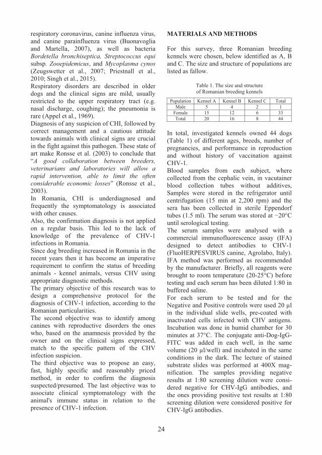
RESULTS AND DISCUSSIONS In the first investigated kennel, the serological assessment of CHV-1 circulation was based on the history of the infertile mating and on the neonatal mortality (Table 1).
Table 1. Reproductive pathology associated with immune status in kennel A
No. #
Breed Gender IFA result
No of infertile matings
No of litter
s
Litter size
Neonatal mortality
1. Rottweiler F + 1 1 4 2
2. Rottweiler F + 0 0 0 0
3. Rottweiler F + 0 1 4 4
4. Rottweiler F + 0 1 4 0
5. Rottweiler F + 0 1 5 0
6. Rottweiler F + 0 1 4 0
7. Rottweiler F + 0 1 5 2
8. Rottweiler F + 1 0 0 0
9. Rottweiler F + 0 0 0 0
10. Rottweiler F + 0 1 6 0
11. Rottweiler F + 0 1 5 5
12. Rottweiler F + 0 0 0 0
13. Rottweiler F + 0 1 5 5
14. Rottweiler F + 1 0 0 0
15. Rottweiler F + 0 1 7 2
16. Rottweiler M + - - - -
17. Rottweiler M + - - - -
18. Rottweiler M + - - - -
19. Rottweiler M + - - - -
20. Rottweiler M + - - - -
TOTAL 3 20 In the kennel A, the reproductive disorders suddenly appeared, with several cases in a short period of time. The intensity of signs was different in affected animals: 100% neonatal mortality in bitches’ litters #3, #11, and #13, while bitch #7 had 60.00% neonatal mortality, bitch #1 had 50.00%, bitch #15 had 28.57%, and bitches #4, #5, #6, and #10 had 0.00% neonatal mortality. Bitch #1 seems to have been the most affected, expressing both, infertile matings and neonatal mortality.
Overall, in the kennel A was 20 cases of neonatal mortality (death in first 72 hours of life), in a total of nine calving with 49 new-borne puppies. In second investigated kennel, the serological evaluation of CHV-1 circulation started after several multiple cases of neonatal mortality (Table 2).
Table 2. Reproductive pathology associated with immune status in kennel B
No. # Breed Gender IFA
result
No of infertile matings
No of litter
s
Litter size
Neonatal mortality
1. Cane Corso F + 0 1 7 3
2. Cane Corso F + 0 2
6 2
8 1
3. Cane Corso F + 0 2
10 3
10 1
4. American Staffordshire Terrier
F + 0 2 7 2
7 2
5. American Staffordshire Terrier
F + 1 1 6 6
6. American Staffordshire Terrier
M + - - - -
7. American Staffordshire Terrier
M + - - - -
8. American Bully F + 0 0 0 0
9. American Bully F + 0 0 0 0
10. American Staffordshire Terrier
F + 1 0 0 0
11. American Bully M + - - - -
12. American Staffordshire Terrier
F + 0 2 10 2
8 1
13. Cane Corso F + 1 1 8 8
14. Cane Corso M + - - - -
15. Cane Corso F + 0 2 16 0
16. American Staffordshire Terrier
F + 0 1 5 5
TOTAL 3 36

25
respiratory coronavirus, canine influenza virus, and canine parainfluenza virus (Buonavoglia and Martella, 2007), as well as bacteria Bordetella bronchiseptica, Streptococcus equi subsp. Zooepidemicus, and Mycoplasma cynos (Zeugswetter et al., 2007; Priestnall et al., 2010; Singh et al., 2015). Respiratory disorders are described in older dogs and the clinical signs are mild, usually restricted to the upper respiratory tract (e.g. nasal discharge, coughing); the pneumonia is rare (Appel et al., 1969). Diagnosis of any suspicion of CHI, followed by correct management and a cautious attitude towards animals with clinical signs are crucial in the fight against this pathogen. These state of art make Ronsse et al. (2003) to conclude that “A good collaboration between breeders, veterinarians and laboratories will allow a rapid intervention, able to limit the often considerable economic losses” (Ronsse et al., 2003). In Romania, CHI is underdiagnosed and frequently the symptomatology is associated with other causes. Also, the confirmation diagnosis is not applied on a regular basis. This led to the lack of knowledge of the prevalence of CHV-1 infections in Romania. Since dog breeding increased in Romania in the recent years then it has become an imperative requirement to confirm the status of breeding animals - kennel animals, versus CHV using appropriate diagnostic methods. The primary objective of this research was to design a comprehensive protocol for the diagnosis of CHV-1 infection, according to the Romanian particularities. The second objective was to identify among canines with reproductive disorders the ones who, based on the anamnesis provided by the owner and on the clinical signs expressed, match to the specific pattern of the CHV infection suspicion. The third objective was to propose an easy, fast, highly specific and reasonably priced method, in order to confirm the diagnosis suspected/presumed. The last objective was to associate clinical symptomatology with the animal's immune status in relation to the presence of CHV-1 infection.
MATERIALS AND METHODS For this survey, three Romanian breeding kennels were chosen, below identified as A, B and C. The size and structure of populations are listed as fallow.
Table 1. The size and structure of Romanian breeding kennels
Population Kennel A Kennel B Kennel C Total Male 5 4 2 1
Female 15 12 6 33 Total 20 16 8 44
In total, investigated kennels owned 44 dogs (Table 1) of different ages, breeds, number of pregnancies, and performance in reproduction and without history of vaccination against CHV-1. Blood samples from each subject, where collected from the cephalic vein, in vacutainer blood collection tubes without additives, Samples were stored in the refrigerator until centrifugation (15 min at 2,200 rpm) and the sera has been collected in sterile Eppendorf tubes (1.5 ml). The serum was stored at −20°C until serological testing. The serum samples were analysed with a commercial immunofluorescence assay (IFA) designed to detect antibodies to CHV-1 (FluoHERPESVIRUS canine, Agrolabo, Italy). IFA method was performed as recommended by the manufacturer. Briefly, all reagents were brought to room temperature (20-25°C) before testing and each serum has been diluted 1:80 in buffered saline. For each serum to be tested and for the Negative and Positive controls were used 20 µl in the individual slide wells, pre-coated with inactivated cells infected with CHV antigens. Incubation was done in humid chamber for 30 minutes at 37°C. The conjugate anti-Dog-IgG-FITC was added in each well, in the same volume (20 µl/well) and incubated in the same conditions in the dark. The lecture of stained substrate slides was performed at 400X mag-nification. The samples providing negative results at 1:80 screening dilution were consi-dered negative for CHV-IgG antibodies, and the ones providing positive test results at 1:80 screening dilution were considered positive for CHV-IgG antibodies.
RESULTS AND DISCUSSIONS In the first investigated kennel, the serological assessment of CHV-1 circulation was based on the history of the infertile mating and on the neonatal mortality (Table 1).
Table 1. Reproductive pathology associated with immune status in kennel A
No. #
Breed Gender IFA result
No of infertile matings
No of litter
s
Litter size
Neonatal mortality
1. Rottweiler F + 1 1 4 2
2. Rottweiler F + 0 0 0 0
3. Rottweiler F + 0 1 4 4
4. Rottweiler F + 0 1 4 0
5. Rottweiler F + 0 1 5 0
6. Rottweiler F + 0 1 4 0
7. Rottweiler F + 0 1 5 2
8. Rottweiler F + 1 0 0 0
9. Rottweiler F + 0 0 0 0
10. Rottweiler F + 0 1 6 0
11. Rottweiler F + 0 1 5 5
12. Rottweiler F + 0 0 0 0
13. Rottweiler F + 0 1 5 5
14. Rottweiler F + 1 0 0 0
15. Rottweiler F + 0 1 7 2
16. Rottweiler M + - - - -
17. Rottweiler M + - - - -
18. Rottweiler M + - - - -
19. Rottweiler M + - - - -
20. Rottweiler M + - - - -
TOTAL 3 20 In the kennel A, the reproductive disorders suddenly appeared, with several cases in a short period of time. The intensity of signs was different in affected animals: 100% neonatal mortality in bitches’ litters #3, #11, and #13, while bitch #7 had 60.00% neonatal mortality, bitch #1 had 50.00%, bitch #15 had 28.57%, and bitches #4, #5, #6, and #10 had 0.00% neonatal mortality. Bitch #1 seems to have been the most affected, expressing both, infertile matings and neonatal mortality.
Overall, in the kennel A was 20 cases of neonatal mortality (death in first 72 hours of life), in a total of nine calving with 49 new-borne puppies. In second investigated kennel, the serological evaluation of CHV-1 circulation started after several multiple cases of neonatal mortality (Table 2).
Table 2. Reproductive pathology associated with immune status in kennel B
No. # Breed Gender IFA
result
No of infertile matings
No of litter
s
Litter size
Neonatal mortality
1. Cane Corso F + 0 1 7 3
2. Cane Corso F + 0 2
6 2
8 1
3. Cane Corso F + 0 2
10 3
10 1
4. American Staffordshire Terrier
F + 0 2 7 2
7 2
5. American Staffordshire Terrier
F + 1 1 6 6
6. American Staffordshire Terrier
M + - - - -
7. American Staffordshire Terrier
M + - - - -
8. American Bully F + 0 0 0 0
9. American Bully F + 0 0 0 0
10. American Staffordshire Terrier
F + 1 0 0 0
11. American Bully M + - - - -
12. American Staffordshire Terrier
F + 0 2 10 2
8 1
13. Cane Corso F + 1 1 8 8
14. Cane Corso M + - - - -
15. Cane Corso F + 0 2 16 0
16. American Staffordshire Terrier
F + 0 1 5 5
TOTAL 3 36

26
As in the previous described kennel, in the kennel B, the main reproductive disorder was the neonatal mortality. The intensity of clinical signs registered variation from one case to another, with 100% neonatal mortality in litter of bitches #5, #13, and #16, 30.00% in litter of bitch #1, 28.57% in litter of bitch #4, 21.43% in litter of bitch #2, 20.00% in litter of bitch #3, 16.67% in litter of bitch #12, and 0% in litter of bitch #15. Bitch #13 have been the most affected: she suffered one infertile mating and a calving with 100% neonatal mortality. It must be also emphasized the persistence of neonatal mortality in all bitches with two litters. In this kennel, all breeds (Cane Corso, American Staffordshire Terrier, and American Bully) were affected. Generally, in the kennel B was 36 cases of neonatal mortality from the 13 calving, with 108 new-borne puppies. In the kennel C, the serological evaluation of CHV-1 circulation started after three cases of major neonatal mortality (Table 3).
Table 3. Reproductive pathology associated with immune status in kennel C
No. # Breed Gender IFA
result
No of infertile matings
No of litters
Litter size
Neonatal mortality
1. Havanese
Cuban Bichon
F – 0 3 15 0
2. Yorkshire Terrier F – 0 2 12 6
3. Yorkshire Terrier M – - - - -
4. Havanese
Cuban Bichon
F – 1 0 0 0
5. Havanese
Cuban Bichon
F – 1 0 0 0
6. Bichon Maltese F + 0 2 10 4
7. Yorkshire Terrier F + 0 2 10 6
8. Havanese
Cuban Bichon
M – - - - -
TOTAL 2 16 In the kennel C, they are eight breeding animals from three breeds (Bichon Maltese, Havanese Cuban Bichon, and Yorkshire Terrier). Despite the limited number of animals, the reproductive disorders were considerable, with 16 cases of neonatal mortality from the 47 new-borne puppies. However, serological evaluation for CHV-1 revealed the presence of specific
antibodies only in two bitches with neonatal mortality (#6 and # 7). Neonatal mortality in bitch #6 (Bichon Maltese breed) was 40% and in bitch #7 (Yorkshire Terrier breed) was 60%. The intensity of signs was quite similar in both cases. However, the negative result obtained in bitch #2 cannot exclude the involvement of CHV-1 in neonatal mortality. Also, here cannot be excluded the CHV-1 infection in all the negative dogs of the contaminated kennel. Previous studies have shown that CHV-1 is a weak immunogenic virus and detectable antibodies can be recorded within 2-3 weeks after infection (Takumi et al. 1990). Even more, antibodies decrease quite rapidly and cannot be detected after few months (Carmichael and Greene 1998). The reproductive disorders recorded in kennels A, B and C are graphically represented in figures 1, 2 and 3.
Figure 1. Reproductive disorders recorded in kennel A
Figure 2. Reproductive disorders recorded in kennel B
86,96%
13,04%
Neonatal mortality Infertile matings
92,31%
7,69%
Neonatal mortality Infertile matings
Figure 3. Reproductive disorders recorded in kennel C
In all kennels, neonatal mortality covered 90% (72/80) of reproductive disorders, and infertility 10% (8/80).
Figure 4. Positive IFA-CHV-IgG result in a female dog
with a recent history of neonatal mortality (dilution 1:80; magnification 400X)
Figure 5. Negative IFA-CHV-IgG result in a female dog
without a history of reproductive disorders (dilution 1:80; magnification 400X)
This study was the first approach in order to assess the presence of CHV-1 infection in dog breeding kennels from the Bucharest-Ilfov area. As resulting following the serologic assays and, as to be expected, in this area too, the dogs have been exposed to CHV-1. In Romania, similar studies have been done by Chielaru (2014) in five Northeast counties: Bacau, Galati, Iasi, Suceava, and Vaslui. Research on seroprevalence of canine herpesvirus-1 infection in northwest Romania revealed 20.55% (37/180) seropositive animals. The highest seroprevalences were in Iasi (27.77%) and Vaslui (26.86%) and lowest in Bacau (15.38%), Galati (11.11%) and Suceava (5.00%) (Chielaru, 2014). In our survey, the average of seropositive animals are being 86.36%, ranging from 100% in kennel A (Table 1) and B (Table 2) to 25% in kennel C (Table 3). Previous Romanian CHV-1 seroprevalence data are much lower than those obtained in this study, but it is not relevant to compare those data because the subjects investigated have different status. In the Chelaru (2014) survey were investigated several solitary lifestyle animals and in our study were investigated only breeding animals in kennels. Dog populations with 100% positive titres were reported in Finnish breeding kennels, facing reproductive problems, while lower values were obtained in breeding kennels without reproductive problems (65%) (Dahlbom et al., 2009). Our data are quite similar with other European serological surveys that shown high prevalence in households or breeding kennels in Belgium (45.80%) (Ronsse et al., 2002), England (88.00%) (Reading and Field, 1999), Italy (27.9%) (Sagazio et al., 1998), Lithuania (26.88%) (Musayeva et al., 2013), the Netherlands (39.30%) (Rijsewijk et al, 1999), and Turkish (62.1%) (Yesilbag et al., 2012). In our survey, reproductive disorders were reported mainly in females that provided a positive result in the serological test for CHV-1. There were also serologically positive female CHV-1 who did not show reproductive disorders, as did serologically negative female CHV-1 who had reproductive disorders. These cases, even if singular, require the extension of investigations to other aetiologies of reproductive disorders but also to the protective effect on gestation of anti-CHV-1 antibodies.
88,89%
11,11%
Neonatal mortality Infertile matings
27
As in the previous described kennel, in the kennel B, the main reproductive disorder was the neonatal mortality. The intensity of clinical signs registered variation from one case to another, with 100% neonatal mortality in litter of bitches #5, #13, and #16, 30.00% in litter of bitch #1, 28.57% in litter of bitch #4, 21.43% in litter of bitch #2, 20.00% in litter of bitch #3, 16.67% in litter of bitch #12, and 0% in litter of bitch #15. Bitch #13 have been the most affected: she suffered one infertile mating and a calving with 100% neonatal mortality. It must be also emphasized the persistence of neonatal mortality in all bitches with two litters. In this kennel, all breeds (Cane Corso, American Staffordshire Terrier, and American Bully) were affected. Generally, in the kennel B was 36 cases of neonatal mortality from the 13 calving, with 108 new-borne puppies. In the kennel C, the serological evaluation of CHV-1 circulation started after three cases of major neonatal mortality (Table 3).
Table 3. Reproductive pathology associated with immune status in kennel C
No. # Breed Gender IFA
result
No of infertile matings
No of litters
Litter size
Neonatal mortality
1. Havanese
Cuban Bichon
F – 0 3 15 0
2. Yorkshire Terrier F – 0 2 12 6
3. Yorkshire Terrier M – - - - -
4. Havanese
Cuban Bichon
F – 1 0 0 0
5. Havanese
Cuban Bichon
F – 1 0 0 0
6. Bichon Maltese F + 0 2 10 4
7. Yorkshire Terrier F + 0 2 10 6
8. Havanese
Cuban Bichon
M – - - - -
TOTAL 2 16 In the kennel C, they are eight breeding animals from three breeds (Bichon Maltese, Havanese Cuban Bichon, and Yorkshire Terrier). Despite the limited number of animals, the reproductive disorders were considerable, with 16 cases of neonatal mortality from the 47 new-borne puppies. However, serological evaluation for CHV-1 revealed the presence of specific
antibodies only in two bitches with neonatal mortality (#6 and # 7). Neonatal mortality in bitch #6 (Bichon Maltese breed) was 40% and in bitch #7 (Yorkshire Terrier breed) was 60%. The intensity of signs was quite similar in both cases. However, the negative result obtained in bitch #2 cannot exclude the involvement of CHV-1 in neonatal mortality. Also, here cannot be excluded the CHV-1 infection in all the negative dogs of the contaminated kennel. Previous studies have shown that CHV-1 is a weak immunogenic virus and detectable antibodies can be recorded within 2-3 weeks after infection (Takumi et al. 1990). Even more, antibodies decrease quite rapidly and cannot be detected after few months (Carmichael and Greene 1998). The reproductive disorders recorded in kennels A, B and C are graphically represented in figures 1, 2 and 3.
Figure 1. Reproductive disorders recorded in kennel A
Figure 2. Reproductive disorders recorded in kennel B
86,96%
13,04%
Neonatal mortality Infertile matings
92,31%
7,69%
Neonatal mortality Infertile matings
Figure 3. Reproductive disorders recorded in kennel C
In all kennels, neonatal mortality covered 90% (72/80) of reproductive disorders, and infertility 10% (8/80).
Figure 4. Positive IFA-CHV-IgG result in a female dog
with a recent history of neonatal mortality (dilution 1:80; magnification 400X)
Figure 5. Negative IFA-CHV-IgG result in a female dog
without a history of reproductive disorders (dilution 1:80; magnification 400X)
This study was the first approach in order to assess the presence of CHV-1 infection in dog breeding kennels from the Bucharest-Ilfov area. As resulting following the serologic assays and, as to be expected, in this area too, the dogs have been exposed to CHV-1. In Romania, similar studies have been done by Chielaru (2014) in five Northeast counties: Bacau, Galati, Iasi, Suceava, and Vaslui. Research on seroprevalence of canine herpesvirus-1 infection in northwest Romania revealed 20.55% (37/180) seropositive animals. The highest seroprevalences were in Iasi (27.77%) and Vaslui (26.86%) and lowest in Bacau (15.38%), Galati (11.11%) and Suceava (5.00%) (Chielaru, 2014). In our survey, the average of seropositive animals are being 86.36%, ranging from 100% in kennel A (Table 1) and B (Table 2) to 25% in kennel C (Table 3). Previous Romanian CHV-1 seroprevalence data are much lower than those obtained in this study, but it is not relevant to compare those data because the subjects investigated have different status. In the Chelaru (2014) survey were investigated several solitary lifestyle animals and in our study were investigated only breeding animals in kennels. Dog populations with 100% positive titres were reported in Finnish breeding kennels, facing reproductive problems, while lower values were obtained in breeding kennels without reproductive problems (65%) (Dahlbom et al., 2009). Our data are quite similar with other European serological surveys that shown high prevalence in households or breeding kennels in Belgium (45.80%) (Ronsse et al., 2002), England (88.00%) (Reading and Field, 1999), Italy (27.9%) (Sagazio et al., 1998), Lithuania (26.88%) (Musayeva et al., 2013), the Netherlands (39.30%) (Rijsewijk et al, 1999), and Turkish (62.1%) (Yesilbag et al., 2012). In our survey, reproductive disorders were reported mainly in females that provided a positive result in the serological test for CHV-1. There were also serologically positive female CHV-1 who did not show reproductive disorders, as did serologically negative female CHV-1 who had reproductive disorders. These cases, even if singular, require the extension of investigations to other aetiologies of reproductive disorders but also to the protective effect on gestation of anti-CHV-1 antibodies.
88,89%
11,11%
Neonatal mortality Infertile matings

28
CONCLUSION Our study emphasizes the widespread of CHV-1 infection. This recommends the assessment of the immune status of the animals before their breeding season. The breeding kennels should benefit from a protocol of surveillance and prophylaxis for those infectious diseases impairing their health status and reproductive performance. REFERENCES Appel M.J., Menegus M., Parsonson I.M., Carmichael
L.E., 1969. Pathogenesis of canine herpesvirus in specific-pathogen-free dogs: 5- to 12-week-old pups, American Journal of Veterinary Research, 30:2067-2073.
Buonavoglia C., Martella V., 2007. Canine respiratory viruses. Veterinary Research, BioMed Central, 38(2):355-373.
Carmichael L.E., Greene C.E., 1998. Canine herpes virus infection. C.E. Greene (Ed.), Infectious diseases of the dog and cat, W. B. Saunders Publishing House, Philadelphia, 28-32.
Carmichael L.E., Squire R.A., Krook L., 1965. Clinical and pathologic features of a fatal viral disease of newborn pups. American Journal of Veterinary Research, 26(113):803-814.
Chelaru A., 2014. Epidemiological, chemical and immunological research in canine herpesviruses. PhD Thesis. University of Agricultural Sciences and Veterinary Medicine Iasi.
Dahlbom M., Johnsson M., Myllys V., Taponen J., Andersson M., 2009. Seroprevalence of canine herpesvirus-1 and Brucella canis in Finnish breeding kennels with and without reproductive problems. Reproduction in Domestic Animals, 44(1):128-131.
Delisle F., 1982. L’herpèsvirose canine. Recueil De Medecine Veterinaire, 158:669-676.
Gaskell R., Willoughby K., 1999. Herpesviruses of carnivores. Veterinary Microbiology, 69(1-2):73-88.
Hashimoto A., Hirai K., Yamaguchi T., Fujimoto Y., 1982. Experimental transplacental infection of pregnant dogs with canine herpesvirus. American Journal of Veterinary Research, 43(5):844-850.
Huxtable C.R., Farrow B.R.H., 1970. Canine Herpesvirus as a suspected cause of neonatal mortality in puppies. Australian Veterinary Journal, 46(7):344-345.
Lundgren D.L., Clapper W.E., 1969. Neutralization of canine herpesvirus by dog and human serum: a survey. American Journal of Veterinary Research, 30:479-482.
Musayeva K., Šengaut J., Petkevičius S., Malakauskas A., Gerulis G., Šalomskas A. , 2013. Seroprevalence of canine herpes virus in Lithuanian dog population. Veterinarija ir Zootechnika, 61(83):48-52.
Poste G., King N., 1971. Isolation of a herpesvirus from the canine genital tract: association with infertility, abortion and stillbirths. Veterinary Record, 88(9):229-233.
Priestnall S.L., Erles K., Brooks H.W., Cardwell J.M., Waller A.S., Paillot R., Robinson C., Darby A.C., Holden M.T.G, Schöniger S., 2010. Characterization of pneumonia due to Streptococcus equi subsp. Zooepidemicus in dogs. Clinical and Vaccine Immunology:CVI, 17(11):1790-1796.
Reading M.J., Field H.J., 1999. Detection of high levels of canine herpes virus-1 neutralising antibody in kennel dogs using a novel serum neutralisation test, Research in Veterinary Science, 66(3):273-275.
Rijsewijk F.A., Luiten E.J., Daus F.J., van der Heijden R.W., van Oirschot J.T., 1999. Prevalence of antibodies against canine herpesvirus 1 in dogs in The Netherlands in 1997-1998, Veterinary Microbiology, 65(1):1-7.
Ronsse V., Poulet H., Verstegen J., Thiry E., 2003. L’herpèsvirose canine. Annales De Medecine Veterinaire, 147(2):65-76.
Ronsse V., Verstegen J., Onclin K., Guiot A.L., Aeberle C., Nauwynck H.J., Poulet H., 2002. Seroprevalence of canine herpesvirus-1 in the Belgian dog population in 2000. Reproduction in Domestic Animals, 37(5):299-304.
Sagazio P., Cirone F., Pratelli A., Tempesta M., Buonavoglia D., Sasanelli M., Rubino G., 1998. Infezione da herpesvirus del cane: diffusione sierologica in Puglia, Obiettivi e Documenti Veterinari, 5:63-67.
Singh P.L., Singh B.R., Bhardwaj M., Prassanvadhana, Sinha D.K., Boby N., Agrawa R.K., Pawde A.M., 2015. Detection of Bordetella bronchiseptica in serum of apparently healthy and clinically sick pet dogs. Advances in Animal and Veterinary Sciences, 3(2):123-127.
Spertzel R.O., Huxsoll D.L., McConnell S.J., Binn L.N., Yager R.H., 1965. Recovery and characterization of a herpes-like virus from dog kidney cell cultures. Proceedings of the Society for Experimental Biology and Medicine. Society for Experimental Biology and Medicine, 120(3):651-655.
Takumi A., Kusanagi K., Tuchiya K., Xuan X., Azetaka M., Takahashi E., 1990. Serodiagnosis of canine herpesvirus infection-development of an enzymelinked immunosorbent assay and its comparison with two improved methods of serum neutralization test. The Japanese Journal of Veterinary Science, 52:241-250.
Yesilbag K., Yalcın E., Tuncer P., Yılmaz Z., 2012. Seroprevalence of canine herpesvirus-1 in Turkish dog population. Research in Veterinary Science, 92(1):36-39.
Zeugswetter F, Weissenböck H, Shibly S, Hassan J, Spergser J., 2007. Lethal bronchopneumonia caused by Mycoplasma cynos in a litter of Golden Retriever puppies. Veterinary Record, 161(18):626–627.
SYRINGOSUBARACHNOID SHUNT PLACEMENT IN A CAVALIER KING
CHARLES SPANIEL DOG DIAGNOSED WITH CHIARI LIKE MALFORMATION AND SYRINGOMYELIA. CASE REPORT
Cătălina Anca CUCOȘ1, Ateș BARUT2, Iuliana IONAȘCU1, Radu CONSTANTINESCU1,
Constantin VLĂGIOIU1
1University of Agronomic Sciences and Veterinary Medicine of Bucharest, Faculty of Veterinary Medicine, 59 Marasti Blvd, District 1, Bucharest, Romania
2PetCode Animal Hospital, Ankara, Turkey
Corresponding author email: [email protected]
Abstract The aim of this paper was to describe the syringosubarachnoid shunting as surgical management in Chiari like malformation and Syringomyelia, and to track the outcome of the patient. A Cavalier King Charles Spaniel female dog was referred for neurological evaluation due to the intense sudden attacks of pain, occurring more and more often lately. A year earlier, the dog was diagnosed with Chiari-like malformation and syringomyelia, undergoing to a craniocervical decompression with durotomy. Postoperatively the clinical signs improved only for a short period of time, afterwards the clinical symptoms progressively reappeared, worsening in the last two months, thus requiring another surgical intervention. The dog was subjected to surgical placement of a syringosubarachnoid shunt at the cervical spinal cord, the patient being evaluated periodically during the hospitalization, and after that at one, four and twelve months postoperatively. In the follow-up period the patient presented good results with improvement of the clinical signs. This case report highlights the success of cervical syringosubarachnoid shunt placement in the management of canine Chiari-like malformation with syringomyelia, especially when the other therapeutic options were unsuccessful. Key words: Chiari like malformation, dogs, syringomyelia, syringosubarachnoid shunt, magnetic resonance imaging. INTRODUCTION The Cavalier King Charles Spaniel (CKCS) is a small, popular toy dog, originated in the United Kingdom. In Romania the breed presented a grown in popularity in the last ten years, CKCS dogs are sweet-tempered, good with children, playful, and affectionate. Unfortunately, this breed presents a high predisposition to develop Chiari-like malfor-mation and Syringomyelia. Chiari like malformation (CM) and syringo-myelia (SM) represents an important complex disorder of the nervous system, commonly encountered in the Cavalier King Charles Spaniel dogs, but it can also be diagnosed in other brachycephalic and toy breeds, such as Griffon Bruxellois, Affenpinschers, Yorkshire terriers, Maltese, Chihuahuas, Pomeranians, Boston terriers, Papillons and their crosses (Marino et al., 2012; Rusbridge, 2013). Canine Chiari like malformation resembles the human Arnold-Chiari type I malformation, a
congenital disorder in which the mismatch between the caudal fossa volume, which is too small, and its contents, lead to herniation of the cerebellum and sometimes also the brainstem into or through the foramen magnum (FM), resulting in cerebrospinal fluid flow distur-bance (Freeman et al., 2014; Rusbridge, 2007; Rusbridge, 2014). CM may lead to the development of fluid filled cavities (named also syrinx) within the paren-chyma of the spinal cord, condition known as syringomyelia. Most of the times Chiari-like malformation and syringomyelia evolve together, however, the conditions can evolve independently of the other, affecting dogs aged between 6 months to 10 years old (Rusbridge, 2014; Platt, 2004; Zachary et al., 2011). The complete mechanism for the development of SM is not fully understood, the most popular theory is that the cavities are formed secondary to the disrupted cerebrospinal fluid (CSF) flow and inconstant CSF pressure (DeLahunta et al.,

29
CONCLUSION Our study emphasizes the widespread of CHV-1 infection. This recommends the assessment of the immune status of the animals before their breeding season. The breeding kennels should benefit from a protocol of surveillance and prophylaxis for those infectious diseases impairing their health status and reproductive performance. REFERENCES Appel M.J., Menegus M., Parsonson I.M., Carmichael
L.E., 1969. Pathogenesis of canine herpesvirus in specific-pathogen-free dogs: 5- to 12-week-old pups, American Journal of Veterinary Research, 30:2067-2073.
Buonavoglia C., Martella V., 2007. Canine respiratory viruses. Veterinary Research, BioMed Central, 38(2):355-373.
Carmichael L.E., Greene C.E., 1998. Canine herpes virus infection. C.E. Greene (Ed.), Infectious diseases of the dog and cat, W. B. Saunders Publishing House, Philadelphia, 28-32.
Carmichael L.E., Squire R.A., Krook L., 1965. Clinical and pathologic features of a fatal viral disease of newborn pups. American Journal of Veterinary Research, 26(113):803-814.
Chelaru A., 2014. Epidemiological, chemical and immunological research in canine herpesviruses. PhD Thesis. University of Agricultural Sciences and Veterinary Medicine Iasi.
Dahlbom M., Johnsson M., Myllys V., Taponen J., Andersson M., 2009. Seroprevalence of canine herpesvirus-1 and Brucella canis in Finnish breeding kennels with and without reproductive problems. Reproduction in Domestic Animals, 44(1):128-131.
Delisle F., 1982. L’herpèsvirose canine. Recueil De Medecine Veterinaire, 158:669-676.
Gaskell R., Willoughby K., 1999. Herpesviruses of carnivores. Veterinary Microbiology, 69(1-2):73-88.
Hashimoto A., Hirai K., Yamaguchi T., Fujimoto Y., 1982. Experimental transplacental infection of pregnant dogs with canine herpesvirus. American Journal of Veterinary Research, 43(5):844-850.
Huxtable C.R., Farrow B.R.H., 1970. Canine Herpesvirus as a suspected cause of neonatal mortality in puppies. Australian Veterinary Journal, 46(7):344-345.
Lundgren D.L., Clapper W.E., 1969. Neutralization of canine herpesvirus by dog and human serum: a survey. American Journal of Veterinary Research, 30:479-482.
Musayeva K., Šengaut J., Petkevičius S., Malakauskas A., Gerulis G., Šalomskas A. , 2013. Seroprevalence of canine herpes virus in Lithuanian dog population. Veterinarija ir Zootechnika, 61(83):48-52.
Poste G., King N., 1971. Isolation of a herpesvirus from the canine genital tract: association with infertility, abortion and stillbirths. Veterinary Record, 88(9):229-233.
Priestnall S.L., Erles K., Brooks H.W., Cardwell J.M., Waller A.S., Paillot R., Robinson C., Darby A.C., Holden M.T.G, Schöniger S., 2010. Characterization of pneumonia due to Streptococcus equi subsp. Zooepidemicus in dogs. Clinical and Vaccine Immunology:CVI, 17(11):1790-1796.
Reading M.J., Field H.J., 1999. Detection of high levels of canine herpes virus-1 neutralising antibody in kennel dogs using a novel serum neutralisation test, Research in Veterinary Science, 66(3):273-275.
Rijsewijk F.A., Luiten E.J., Daus F.J., van der Heijden R.W., van Oirschot J.T., 1999. Prevalence of antibodies against canine herpesvirus 1 in dogs in The Netherlands in 1997-1998, Veterinary Microbiology, 65(1):1-7.
Ronsse V., Poulet H., Verstegen J., Thiry E., 2003. L’herpèsvirose canine. Annales De Medecine Veterinaire, 147(2):65-76.
Ronsse V., Verstegen J., Onclin K., Guiot A.L., Aeberle C., Nauwynck H.J., Poulet H., 2002. Seroprevalence of canine herpesvirus-1 in the Belgian dog population in 2000. Reproduction in Domestic Animals, 37(5):299-304.
Sagazio P., Cirone F., Pratelli A., Tempesta M., Buonavoglia D., Sasanelli M., Rubino G., 1998. Infezione da herpesvirus del cane: diffusione sierologica in Puglia, Obiettivi e Documenti Veterinari, 5:63-67.
Singh P.L., Singh B.R., Bhardwaj M., Prassanvadhana, Sinha D.K., Boby N., Agrawa R.K., Pawde A.M., 2015. Detection of Bordetella bronchiseptica in serum of apparently healthy and clinically sick pet dogs. Advances in Animal and Veterinary Sciences, 3(2):123-127.
Spertzel R.O., Huxsoll D.L., McConnell S.J., Binn L.N., Yager R.H., 1965. Recovery and characterization of a herpes-like virus from dog kidney cell cultures. Proceedings of the Society for Experimental Biology and Medicine. Society for Experimental Biology and Medicine, 120(3):651-655.
Takumi A., Kusanagi K., Tuchiya K., Xuan X., Azetaka M., Takahashi E., 1990. Serodiagnosis of canine herpesvirus infection-development of an enzymelinked immunosorbent assay and its comparison with two improved methods of serum neutralization test. The Japanese Journal of Veterinary Science, 52:241-250.
Yesilbag K., Yalcın E., Tuncer P., Yılmaz Z., 2012. Seroprevalence of canine herpesvirus-1 in Turkish dog population. Research in Veterinary Science, 92(1):36-39.
Zeugswetter F, Weissenböck H, Shibly S, Hassan J, Spergser J., 2007. Lethal bronchopneumonia caused by Mycoplasma cynos in a litter of Golden Retriever puppies. Veterinary Record, 161(18):626–627.
SYRINGOSUBARACHNOID SHUNT PLACEMENT IN A CAVALIER KING
CHARLES SPANIEL DOG DIAGNOSED WITH CHIARI LIKE MALFORMATION AND SYRINGOMYELIA. CASE REPORT
Cătălina Anca CUCOȘ1, Ateș BARUT2, Iuliana IONAȘCU1, Radu CONSTANTINESCU1,
Constantin VLĂGIOIU1
1University of Agronomic Sciences and Veterinary Medicine of Bucharest, Faculty of Veterinary Medicine, 59 Marasti Blvd, District 1, Bucharest, Romania
2PetCode Animal Hospital, Ankara, Turkey
Corresponding author email: [email protected]
Abstract The aim of this paper was to describe the syringosubarachnoid shunting as surgical management in Chiari like malformation and Syringomyelia, and to track the outcome of the patient. A Cavalier King Charles Spaniel female dog was referred for neurological evaluation due to the intense sudden attacks of pain, occurring more and more often lately. A year earlier, the dog was diagnosed with Chiari-like malformation and syringomyelia, undergoing to a craniocervical decompression with durotomy. Postoperatively the clinical signs improved only for a short period of time, afterwards the clinical symptoms progressively reappeared, worsening in the last two months, thus requiring another surgical intervention. The dog was subjected to surgical placement of a syringosubarachnoid shunt at the cervical spinal cord, the patient being evaluated periodically during the hospitalization, and after that at one, four and twelve months postoperatively. In the follow-up period the patient presented good results with improvement of the clinical signs. This case report highlights the success of cervical syringosubarachnoid shunt placement in the management of canine Chiari-like malformation with syringomyelia, especially when the other therapeutic options were unsuccessful. Key words: Chiari like malformation, dogs, syringomyelia, syringosubarachnoid shunt, magnetic resonance imaging. INTRODUCTION The Cavalier King Charles Spaniel (CKCS) is a small, popular toy dog, originated in the United Kingdom. In Romania the breed presented a grown in popularity in the last ten years, CKCS dogs are sweet-tempered, good with children, playful, and affectionate. Unfortunately, this breed presents a high predisposition to develop Chiari-like malfor-mation and Syringomyelia. Chiari like malformation (CM) and syringo-myelia (SM) represents an important complex disorder of the nervous system, commonly encountered in the Cavalier King Charles Spaniel dogs, but it can also be diagnosed in other brachycephalic and toy breeds, such as Griffon Bruxellois, Affenpinschers, Yorkshire terriers, Maltese, Chihuahuas, Pomeranians, Boston terriers, Papillons and their crosses (Marino et al., 2012; Rusbridge, 2013). Canine Chiari like malformation resembles the human Arnold-Chiari type I malformation, a
congenital disorder in which the mismatch between the caudal fossa volume, which is too small, and its contents, lead to herniation of the cerebellum and sometimes also the brainstem into or through the foramen magnum (FM), resulting in cerebrospinal fluid flow distur-bance (Freeman et al., 2014; Rusbridge, 2007; Rusbridge, 2014). CM may lead to the development of fluid filled cavities (named also syrinx) within the paren-chyma of the spinal cord, condition known as syringomyelia. Most of the times Chiari-like malformation and syringomyelia evolve together, however, the conditions can evolve independently of the other, affecting dogs aged between 6 months to 10 years old (Rusbridge, 2014; Platt, 2004; Zachary et al., 2011). The complete mechanism for the development of SM is not fully understood, the most popular theory is that the cavities are formed secondary to the disrupted cerebrospinal fluid (CSF) flow and inconstant CSF pressure (DeLahunta et al.,
Scientific Works. Series C. Veterinary Medicine. Vol. LXIV (2), 2018ISSN 2065-1295; ISSN 2343-9394 (CD-ROM); ISSN 2067-3663 (Online); ISSN-L 2065-1295

30
2009; Rusbridge, 2014; Cerda-Gonzalez et al., 2009). The common clinical sign encountered is scratching in the air near the neck - „phantom” scratching, which is the reason why the condi-tion is known as “neck scratcher’s disease”. Other important sings are the neuropathic pain, expressed by yelping after a sudden posture change, frequent and characteristic manifest-tation in CM/SM, along with the head, neck and spinal discomfort (Plessas et al., 2012; Platt, 2004; Rusbridge, 2014; Park et al., 2015) A variety of non-specific signs accompanies the clinical presentation of CM/SM, including behavioural changes, hyperesthesia, cervical scoliosis, thoracic and pelvic limb ataxia, “bunny-hopping” pelvic limb gait, proprio-ceptive deficits, vestibular signs and facial nerve paralysis (Rusbridge, 2007; Plessas et al., 2012; Platt et al., 2012). The gold standard to diagnose CM/SM is magnetic resonance imaging scan (MRI), so that accurate measurements of the caudal cranial cavity can be performed, including the position and shape of the cerebellum, the extension and the symmetry of syringomyelia can also be determined (Platt, 2004; Rusbridge, 2014). British Veterinary Association (BVA) proposed a CM/SM classification scheme. Based on the position of the cerebellum, CM is structured in 3 grades, from grade 0 - no signs of CM, to grade 2, where the cerebellum is severe herniated through FM. There is also a grading system for SM, according on the syrinx size there are 3 grades of SM, grade 0 SM – normal spinal cord, to grade 2 SM, where the syrinx diameter is greater than 2 mm (Freeman et al., 2014; Rusbridge, 2014). The CM/SM grading is important, because it will guide treatment and prognosis. MATERIALS AND METHODS A 4-year-old Cavalier King Charles Spaniel female dog was referred for neurological evaluation due to the intense sudden attacks of pain, occurring more and more often lately. The patient was diagnosed a year before with CM and SM, and at that time a craniocervical decompression was performed. After the
surgery the patient presented a favorable evolution in the first three months, with the improvement of the clinical signs. Unfortu-nately, after this period the clinical signs progressively deteriorated over time. In the last two months the dog presented a progressive neuropathic pain, expressed by sudden yelping, neck and spinal discomfort, scoliosis, apathy and depression. Besides these clinical signs, the patient also presented „phantom” scratching and “bunny-hopping” pelvic limb gait. Due to the medical history and progressive clinical signs, we performed an MRI scan of the brain and spinal cord. Sagittal and transverse T1 and T2 weighted images were acquired. The scan revealed cerebellar herniation, with secondary FM obstruction – corresponding to grade 2 CM (Figure 1).
Figure 1 – T2 weighted mid-sagittal image of the caudal
cranial fossa and upper cervical spinal cord. The cerebellar vermis is impacted into foramen magnum.
Note the syrinx present in the cervical spine (red star).
Figure 2 – T2 weighted transverse image
of the cervical syrinx. The maximum width of the spinal fluid-filled cavity measures 6.3 mm.
The maximum syrinx width is located cervical, (Figure 1 and Figure 2), marking the position where the shunt will be surgically placed. The width of the syrinx is measured in a transverse plan, in this case the maximum internal diameter is larger than 2 mm, corresponding to grade 2 SM (Figure 2).
The dog was subjected to surgical placement of a syringo-subarachnoid shunt. Prior to the sur-gery, pre-medication of the dog was performed with 20 mg/kg intravenous cefazolin sodium (Iespor, I.E. Ulagay), 0.03 mg/kg intravenous medetomidine hydrochloride (Domitor, Orion Pharma, Pfizer Animal Health) and 0.04 mg/kg intravenous atropine sulfate (Atropin 0,2%, Vetaș). Anesthesia was induced with 4 mg/kg intravenous propofol (Propofol 1%, Fresenius Kabi), and maintained with isoflurane 1 to 2% (Forane, Baxter) in 100% oxygen. Fluid therapy (0,9% sodium chloride injection USP, Baxter) was administered during the entire surgical procedure in constant rate infusion at 10 ml/kg/hour. Postoperatively analgesia was provided with intravenous 4 mg/kg tramadol hydrochloride (Contramal 100 mg, AbdiIbrahim, Grunenthal) at every 12 hours, for 5 days. After the surgical intervention cefazolin was administered in dose of 20 mg/kg, for 7 days, and in order to decrease the neuropathic pain gabapentin was given for 2 weeks, in dose of 10 mg/kg PO orally, every 8 hours. The dog was positioned in sternal recumbency on a special operating table, which allows to flex the patient’s neck at 60 degrees. The cutaneous region of interest is aseptically prepared, the first step of the surgery is to perform dorsal laminectomy at C3-C4 (Figure 3), where the syrinx presents the largest width, previously measured on the magnetic resonance imagining examination.
Figure 3 - Intraoperative aspect, performing dorsal
cervical laminectomy.
A ventriculoperitoneal catheter was used to create the syringosubarachnoid shunt. Due to the syrinx size, the catheter is tailored to approximately 2,5 cm length (Figure 4), at both terminal ends of the catheter three fenestrations were made, in order to ensure adequately drainage of CSF. One loop is made on the middle of the tube, using nylon thread, having the role of anchoring the tube to the intervertebral ligament.
Figure 4 – The catheter used to create the
syringosubarachnoid shunt is tailored from a ventriculo-peritonal catheter.
Dura mater and arachnoid are incised with an 11 scalpel blade, presenting an elongated triangular blade, which allows to obtain a thin and precise incision. The spinal cord is incised in the dorsal midline, penetrating the syrinx. One end of the catheter was introduced into the lumen of the syrinx, in a cranial direction (Figure 5). The shunt is secured using the nylon loop from the middle of the tube, suturing the shunt to the intervertebral ligament. The protruding end of the catheter was caudally inserted into the subarachnoid space, thereby achieving the communication between the syrinx lumen and the subarachnoid cavity (Figure 6). Dura mater was not sutured; homologous adipose tissue was placed over the laminec-tomy defect. Suture of the muscle, subcuta-neous and skin layers was routinely carried out.

31
2009; Rusbridge, 2014; Cerda-Gonzalez et al., 2009). The common clinical sign encountered is scratching in the air near the neck - „phantom” scratching, which is the reason why the condi-tion is known as “neck scratcher’s disease”. Other important sings are the neuropathic pain, expressed by yelping after a sudden posture change, frequent and characteristic manifest-tation in CM/SM, along with the head, neck and spinal discomfort (Plessas et al., 2012; Platt, 2004; Rusbridge, 2014; Park et al., 2015) A variety of non-specific signs accompanies the clinical presentation of CM/SM, including behavioural changes, hyperesthesia, cervical scoliosis, thoracic and pelvic limb ataxia, “bunny-hopping” pelvic limb gait, proprio-ceptive deficits, vestibular signs and facial nerve paralysis (Rusbridge, 2007; Plessas et al., 2012; Platt et al., 2012). The gold standard to diagnose CM/SM is magnetic resonance imaging scan (MRI), so that accurate measurements of the caudal cranial cavity can be performed, including the position and shape of the cerebellum, the extension and the symmetry of syringomyelia can also be determined (Platt, 2004; Rusbridge, 2014). British Veterinary Association (BVA) proposed a CM/SM classification scheme. Based on the position of the cerebellum, CM is structured in 3 grades, from grade 0 - no signs of CM, to grade 2, where the cerebellum is severe herniated through FM. There is also a grading system for SM, according on the syrinx size there are 3 grades of SM, grade 0 SM – normal spinal cord, to grade 2 SM, where the syrinx diameter is greater than 2 mm (Freeman et al., 2014; Rusbridge, 2014). The CM/SM grading is important, because it will guide treatment and prognosis. MATERIALS AND METHODS A 4-year-old Cavalier King Charles Spaniel female dog was referred for neurological evaluation due to the intense sudden attacks of pain, occurring more and more often lately. The patient was diagnosed a year before with CM and SM, and at that time a craniocervical decompression was performed. After the
surgery the patient presented a favorable evolution in the first three months, with the improvement of the clinical signs. Unfortu-nately, after this period the clinical signs progressively deteriorated over time. In the last two months the dog presented a progressive neuropathic pain, expressed by sudden yelping, neck and spinal discomfort, scoliosis, apathy and depression. Besides these clinical signs, the patient also presented „phantom” scratching and “bunny-hopping” pelvic limb gait. Due to the medical history and progressive clinical signs, we performed an MRI scan of the brain and spinal cord. Sagittal and transverse T1 and T2 weighted images were acquired. The scan revealed cerebellar herniation, with secondary FM obstruction – corresponding to grade 2 CM (Figure 1).
Figure 1 – T2 weighted mid-sagittal image of the caudal
cranial fossa and upper cervical spinal cord. The cerebellar vermis is impacted into foramen magnum.
Note the syrinx present in the cervical spine (red star).
Figure 2 – T2 weighted transverse image
of the cervical syrinx. The maximum width of the spinal fluid-filled cavity measures 6.3 mm.
The maximum syrinx width is located cervical, (Figure 1 and Figure 2), marking the position where the shunt will be surgically placed. The width of the syrinx is measured in a transverse plan, in this case the maximum internal diameter is larger than 2 mm, corresponding to grade 2 SM (Figure 2).
The dog was subjected to surgical placement of a syringo-subarachnoid shunt. Prior to the sur-gery, pre-medication of the dog was performed with 20 mg/kg intravenous cefazolin sodium (Iespor, I.E. Ulagay), 0.03 mg/kg intravenous medetomidine hydrochloride (Domitor, Orion Pharma, Pfizer Animal Health) and 0.04 mg/kg intravenous atropine sulfate (Atropin 0,2%, Vetaș). Anesthesia was induced with 4 mg/kg intravenous propofol (Propofol 1%, Fresenius Kabi), and maintained with isoflurane 1 to 2% (Forane, Baxter) in 100% oxygen. Fluid therapy (0,9% sodium chloride injection USP, Baxter) was administered during the entire surgical procedure in constant rate infusion at 10 ml/kg/hour. Postoperatively analgesia was provided with intravenous 4 mg/kg tramadol hydrochloride (Contramal 100 mg, AbdiIbrahim, Grunenthal) at every 12 hours, for 5 days. After the surgical intervention cefazolin was administered in dose of 20 mg/kg, for 7 days, and in order to decrease the neuropathic pain gabapentin was given for 2 weeks, in dose of 10 mg/kg PO orally, every 8 hours. The dog was positioned in sternal recumbency on a special operating table, which allows to flex the patient’s neck at 60 degrees. The cutaneous region of interest is aseptically prepared, the first step of the surgery is to perform dorsal laminectomy at C3-C4 (Figure 3), where the syrinx presents the largest width, previously measured on the magnetic resonance imagining examination.
Figure 3 - Intraoperative aspect, performing dorsal
cervical laminectomy.
A ventriculoperitoneal catheter was used to create the syringosubarachnoid shunt. Due to the syrinx size, the catheter is tailored to approximately 2,5 cm length (Figure 4), at both terminal ends of the catheter three fenestrations were made, in order to ensure adequately drainage of CSF. One loop is made on the middle of the tube, using nylon thread, having the role of anchoring the tube to the intervertebral ligament.
Figure 4 – The catheter used to create the
syringosubarachnoid shunt is tailored from a ventriculo-peritonal catheter.
Dura mater and arachnoid are incised with an 11 scalpel blade, presenting an elongated triangular blade, which allows to obtain a thin and precise incision. The spinal cord is incised in the dorsal midline, penetrating the syrinx. One end of the catheter was introduced into the lumen of the syrinx, in a cranial direction (Figure 5). The shunt is secured using the nylon loop from the middle of the tube, suturing the shunt to the intervertebral ligament. The protruding end of the catheter was caudally inserted into the subarachnoid space, thereby achieving the communication between the syrinx lumen and the subarachnoid cavity (Figure 6). Dura mater was not sutured; homologous adipose tissue was placed over the laminec-tomy defect. Suture of the muscle, subcuta-neous and skin layers was routinely carried out.

32
Figure 5 - Intraoperative aspect - one end of the catheter was cranially placed in the spinal cavity.
Note the nylon loop present on the middle of the shunt.
Figure 6 - Intraoperative aspect, orientation of the free-end catheter towards the subarachnoid space,
achieving the syringosubarachnoid communication.
RESULTS AND DISCUSSIONS The MRI scan revealed cerebellar herniation, a wide medullar syrinx located cervical, with the maximum width at the level of the third and fourth cervical vertebrae. In the transverse plan, the maximum internal diameter of the syrinx is 6,36 mm, corresponding to grade 2 SM. Postoperative MRI was not performed. The syringosubarachnoid shunt was placed at the level of C3-C4, the surgery was successful, no intraoperative or postoperative compli-cations were noted. We choose to manufacture the tube that we used for shunting from a ventriculo-peritonal catheter, witch fitted optimum to the desired
dimensions, and it is flexible and thin. The tube should allow the CSF passage and must be big enough to avoid debris occlusion. In a study performed by Motta in 2012, he chooses to use an equine ocular lavage tube, due to its size, cost and proprieties. The surgical technique that he used resembles, but he did not anchor the shunt to the intervertebral ligament (Motta et al., 2012). In the early postoperative follow-up period the patient presented a mild neck pain, only for 24 hours. The dog did not presented ataxia, gait abnormalities or proprioceptive placement deficits. After 5 days the dog was discharged, and evaluated after that at 1, 4 and 12 months after the syringosubarachnoid shunt placement.
The neurological examinations in the follow-up period showed progressive improvement of the clinical signs, the neurological pain decreased, and the painful episodes are fewer and have low intensity. At three years after the syringosubarachnoid shunt placement the patient presents an amelioration of the clinical signs comparative to the preoperative period, notably decreased neuropathic pain, the intensity and the frequency of the pain attacks reduced, scratching and yelping diminished, overall increasing the life quality of the patient. The main purpose of the treatment is to reduce the pain. There are two treatment options for CM/SM, medical therapy or surgical intervention, which are elected according to clinical symptoms, and can be combined (Rusbridge, 2014). The most common used surgical technique is craniocervical decompression, which can provide a satisfactory quality of life for a period of time between a few months and two years postoperatively, according to the CM/SM grade and also to the clinical signs. Another surgery technique described is syringosubarachnoid shunting procedure, with a better outcome for patients. Syringosubarachnoid shunt placement can be an option with good results, especially for those patients in which foramen magnum decompression has failed (Motta et al., 2012; Rusbridge, 2014). Currently the scientific literature offers more information about craniocervical decompression technique, syringosubarachnoid shunting representing a relatively new procedure used in dogs. In patients that had craniocervical decompression, although the syrinx size still persists after this procedure, clinical amelioration is mostly due to the improvement of the CSF flow (Park et al., 2015; Rusbridge, 2014). In human medicine better results were noticed in patients that underwent syringosubarachnoid shunt placement compared to the ones that underwent only foramen magna decompression or syringoperitoneal shunting. The syringosubarachnoid shunt placement has favorable results regarding the pain control, this technique is preferred to be used in patients with large syringes (Hida et al., 1995).
CONCLUSIONS Chiari like malformation and syringomyelia represents a complex and progressive neurologic condition, in whose diagnosis and surgical management the magnetic resonance imagining plays an essential role. Syringosubarachnoid shunt placement represents a safe and efficient procedure, which leads to clinical improvement of the clinical signs and reestablishes the quality of life. Syringosubarachnoid shunting can be an alternative option with good results, especially in cases in which foramen magnum decompression has failed. REFERENCES British Veterinary Assocication. (2013). Chiari
Malformation/Syringomyelia Scheme (CM/SM Scheme). Canine Health Schemes http://www.bva.co.uk/canine_health_schemes/ChiariMalformationSyringomyeliaSchemeCMSMScheme.aspx: Assessed 2013 July 8.
Cerda-Gonzalez S., Olby N.J., Mccullough S., Pease A.P., Broadstone R., Osborne J.A., 2009. Morphology of The Caudal Fossa In Cavalier King Charles Spaniels. Veterinary Radiology & Ultrasound, Vol. 50, No. 1, pp 37–46.
DeLahunta A., Glass E., 2009. Veterinary neuroanatomy and clinical neurology, 3rd edition, Saunders Elsevier Inc. Publication, USA.
Freeman A.C., Platt S.R., Kent M., Huguet E., Rusbridge, C., Holmes S., 2014. Chiari-Like Malformation and Syringomyelia in American Brussels Griffon Dogs. Journal of Veterinary Internal Medicine; 28:1551–1559.
Hida K., Iwasaki Y., Koyanagi I., Sawamura Y., Abe H., 1995. Surgical indication and results of foramen magnum decompression versus syringosubarachnoid shunting for syringomyelia associated with Chiari I malformation, Neurosurgery. 1995 Oct;37(4):673-9
Marino D.J., Loughin C.A., Dewey C.W., Marino L.J., Sackman J.J., Lesser M.L., 2012. Morphometric features of the craniocervical junction region in dogs with suspected Chiari-like malformation determined by combined use of magnetic resonance imaging and computed tomography. American Journal of Veterinary Research. 2012; 73(1): 105-11. Epub 2011/12/30.
Motta L., Skerritt G.C., 2012. Syringosubarachnoid shunt as a management for syringohydromyelia in dogs, Journal of Small Animal Practice, 53, 2015-2012.
Park S.S., Kim H.J., Ryu J.M., Han H.J., 2015. Chiari-Like Malformation with Syringomyelia in the Small Breed Dogs: Prognosis Evaluation after Surgical Management (Case Report). Global Veterinaria 15 (1): 129-132.

33
Figure 5 - Intraoperative aspect - one end of the catheter was cranially placed in the spinal cavity.
Note the nylon loop present on the middle of the shunt.
Figure 6 - Intraoperative aspect, orientation of the free-end catheter towards the subarachnoid space,
achieving the syringosubarachnoid communication.
RESULTS AND DISCUSSIONS The MRI scan revealed cerebellar herniation, a wide medullar syrinx located cervical, with the maximum width at the level of the third and fourth cervical vertebrae. In the transverse plan, the maximum internal diameter of the syrinx is 6,36 mm, corresponding to grade 2 SM. Postoperative MRI was not performed. The syringosubarachnoid shunt was placed at the level of C3-C4, the surgery was successful, no intraoperative or postoperative compli-cations were noted. We choose to manufacture the tube that we used for shunting from a ventriculo-peritonal catheter, witch fitted optimum to the desired
dimensions, and it is flexible and thin. The tube should allow the CSF passage and must be big enough to avoid debris occlusion. In a study performed by Motta in 2012, he chooses to use an equine ocular lavage tube, due to its size, cost and proprieties. The surgical technique that he used resembles, but he did not anchor the shunt to the intervertebral ligament (Motta et al., 2012). In the early postoperative follow-up period the patient presented a mild neck pain, only for 24 hours. The dog did not presented ataxia, gait abnormalities or proprioceptive placement deficits. After 5 days the dog was discharged, and evaluated after that at 1, 4 and 12 months after the syringosubarachnoid shunt placement.
The neurological examinations in the follow-up period showed progressive improvement of the clinical signs, the neurological pain decreased, and the painful episodes are fewer and have low intensity. At three years after the syringosubarachnoid shunt placement the patient presents an amelioration of the clinical signs comparative to the preoperative period, notably decreased neuropathic pain, the intensity and the frequency of the pain attacks reduced, scratching and yelping diminished, overall increasing the life quality of the patient. The main purpose of the treatment is to reduce the pain. There are two treatment options for CM/SM, medical therapy or surgical intervention, which are elected according to clinical symptoms, and can be combined (Rusbridge, 2014). The most common used surgical technique is craniocervical decompression, which can provide a satisfactory quality of life for a period of time between a few months and two years postoperatively, according to the CM/SM grade and also to the clinical signs. Another surgery technique described is syringosubarachnoid shunting procedure, with a better outcome for patients. Syringosubarachnoid shunt placement can be an option with good results, especially for those patients in which foramen magnum decompression has failed (Motta et al., 2012; Rusbridge, 2014). Currently the scientific literature offers more information about craniocervical decompression technique, syringosubarachnoid shunting representing a relatively new procedure used in dogs. In patients that had craniocervical decompression, although the syrinx size still persists after this procedure, clinical amelioration is mostly due to the improvement of the CSF flow (Park et al., 2015; Rusbridge, 2014). In human medicine better results were noticed in patients that underwent syringosubarachnoid shunt placement compared to the ones that underwent only foramen magna decompression or syringoperitoneal shunting. The syringosubarachnoid shunt placement has favorable results regarding the pain control, this technique is preferred to be used in patients with large syringes (Hida et al., 1995).
CONCLUSIONS Chiari like malformation and syringomyelia represents a complex and progressive neurologic condition, in whose diagnosis and surgical management the magnetic resonance imagining plays an essential role. Syringosubarachnoid shunt placement represents a safe and efficient procedure, which leads to clinical improvement of the clinical signs and reestablishes the quality of life. Syringosubarachnoid shunting can be an alternative option with good results, especially in cases in which foramen magnum decompression has failed. REFERENCES British Veterinary Assocication. (2013). Chiari
Malformation/Syringomyelia Scheme (CM/SM Scheme). Canine Health Schemes http://www.bva.co.uk/canine_health_schemes/ChiariMalformationSyringomyeliaSchemeCMSMScheme.aspx: Assessed 2013 July 8.
Cerda-Gonzalez S., Olby N.J., Mccullough S., Pease A.P., Broadstone R., Osborne J.A., 2009. Morphology of The Caudal Fossa In Cavalier King Charles Spaniels. Veterinary Radiology & Ultrasound, Vol. 50, No. 1, pp 37–46.
DeLahunta A., Glass E., 2009. Veterinary neuroanatomy and clinical neurology, 3rd edition, Saunders Elsevier Inc. Publication, USA.
Freeman A.C., Platt S.R., Kent M., Huguet E., Rusbridge, C., Holmes S., 2014. Chiari-Like Malformation and Syringomyelia in American Brussels Griffon Dogs. Journal of Veterinary Internal Medicine; 28:1551–1559.
Hida K., Iwasaki Y., Koyanagi I., Sawamura Y., Abe H., 1995. Surgical indication and results of foramen magnum decompression versus syringosubarachnoid shunting for syringomyelia associated with Chiari I malformation, Neurosurgery. 1995 Oct;37(4):673-9
Marino D.J., Loughin C.A., Dewey C.W., Marino L.J., Sackman J.J., Lesser M.L., 2012. Morphometric features of the craniocervical junction region in dogs with suspected Chiari-like malformation determined by combined use of magnetic resonance imaging and computed tomography. American Journal of Veterinary Research. 2012; 73(1): 105-11. Epub 2011/12/30.
Motta L., Skerritt G.C., 2012. Syringosubarachnoid shunt as a management for syringohydromyelia in dogs, Journal of Small Animal Practice, 53, 2015-2012.
Park S.S., Kim H.J., Ryu J.M., Han H.J., 2015. Chiari-Like Malformation with Syringomyelia in the Small Breed Dogs: Prognosis Evaluation after Surgical Management (Case Report). Global Veterinaria 15 (1): 129-132.

34
Platt S., (2004), Chapter 13 - Neck and back pain, British
Small Animal Veterinary Association - Manual of Canine and Feline Neurology, edited by Platt S., Olby N., Third edition, published by British Small Animal Veterinary Association, Gloucester, England.
Platt S., Garosi L., 2012. Small animal neurological emergencies, 1st ed., Manson Publishing, USA.
Plessas I. N., Rusbridge C., Driver C.J., Chandler K.E., Craig A., McGonnell I.M., Brodbelt D.C., Volk H.A., 2012. Long-term outcome of Cavalier King Charles spaniel dogs with clinical signs associated with Chiari-like malformation and syringomyelia, Veterinary Record, Journal of the British Veterinary Association, Volume 171, Issue 20, pag 501-555.
Rusbridge C., 2007. Chiari-Like Malformation with Syringomyelia in the Cavalier King Charles Spaniel: Long-Term Outcome after Surgical Management, Veterinary Surgery Issue 36, pag 396–405.
Rusbridge C., 2013. Chiari–like malformation and syringomyelia. European Journal Companian Animal Pratice 23: 70–89.
Rusbridge C., 2014. Chiari-like malformation and syringomyelia, published in the special issue on hereditary diseases of the European Journal of Companion Animal Practice, Issue 23, pag 70-89.
Zachary J.F., McGavin M.D., (2011), Pathologic Basis of Veterinary Disease, 5th Edition, Mosby, Inc., an affiliate of Elsevier Inc. Press, USA.
MYASTHENIA GRAVIS IN A LABRADOR RETRIEVER DOG –
CASE PRESENTATION
Laura DARIE, Cristina FERNOAGĂ
University of Agronomic Sciences and Veterinary Medicine of Bucharest, Faculty of Veterinary Medicine, 105 Spaiul Independenței, District 5, Bucharest, Romania
*Corresponding author e-mail: [email protected]
Abstract The patient, Labrador Retriever, male, eight years old, was presented at veterinary doctor showing muscle weakness, walked heavily on hind limbs and muscle contractions. The first signs of muscle weakness appeared five months ago, they have progressively worsened and 14 days before the consult it stopped barking. No changes were observed in biochemical and hematological blood tests as well as imagistic examinations, but the neurological examination revealed the decrease of spinal reflexes in all four limbs. Myasthenia gravis was suspected and the diagnosis was based on clinical signs and the favorable response to administration of neostigmine 1mg / kg intravenously. Key words: myasthenia gravis, muscle fatigue, neostigmine INTRODUCTION Myasthenia gravis is an autoimmune, neuromuscular disorder and it is manifested by an attack against acetylcholine receptors from the post-synaptic membrane of the neuro-motor plaque, causing their degradation or blockage, resulting in motor deficiency and skeletal muscle weakness. (Burns, 2010, Hotineanu and Stasiuc 2010, Marx et al., 2015, Thomas, 2014). It may have genetic determinism or can be acquired ( Bexfield et al.,2006, Dissanayake et al.,2016, Herder et al.,2017). Myasthenia gravis is characterized by a decrease in the number of acetylcholine recep-tors (AchR) from the postsynaptic muscular membrane and by flattening or simplifying postsynaptic fold crests. These defects lead to a diminished efficacy of neuromuscular transmission, and even if normal AchR release occurs, the post-synaptic potentials produced are of low power and do not trigger the potentials of muscle fiber action, resulting in insufficient transmission of information in a significant number of neuromuscular junctions, which leads to the weakness of muscle contraction. Neuromuscular abnormalities of myasthenia gravis are induced by an autoimmune response mediated by specific anti-AchR antibodies. These antibodies reduce the number of AchR available in muscle
junctions through three different mechanisms: AchR can be degraded more rapidly through a mechanism involving their cross-coupling followed by accelerated endocytosis; the active sites of AchR, where they normally bind to Ach, may be blocked by antibodies, the postsynaptic muscle membrane may be damaged by complement-associated antibodies (Bexfield et al., 2006, Ciobanu, 2011). Symptomatology of myasthenia gravis is clinically expressed by abnormal weakness of volunteer muscles at resting restraining with restoration to rest, ocular signs (palpitations), dysphagia with nasal regurgitation of liquids, fallout of the mandible, weakening of the neck and neck muscles, weakening of the pelvis, difficulty walking (Dissanayake et al. 2016, Fraser et al., 1970, Shelton, 2016). However, muscle weakness is not always associated with physical effort. Muscular weakness at the facial, pharyngeal or esophageal level is common, and in many situations there is a mega-esophagus in the absence of generalized muscular weakness (focal myasthenia), so megaesophagus may be a primary affection or a symptom (Thomas, 2014). Paraclinic investigations are an indispensable adjunct to the clinical examination, in many cases having diagnostic certainty (Fraser et al., 1970). Myasthenia gravis occurs with a higher frequency in adult dogs, especially in the

35
Platt S., (2004), Chapter 13 - Neck and back pain, British
Small Animal Veterinary Association - Manual of Canine and Feline Neurology, edited by Platt S., Olby N., Third edition, published by British Small Animal Veterinary Association, Gloucester, England.
Platt S., Garosi L., 2012. Small animal neurological emergencies, 1st ed., Manson Publishing, USA.
Plessas I. N., Rusbridge C., Driver C.J., Chandler K.E., Craig A., McGonnell I.M., Brodbelt D.C., Volk H.A., 2012. Long-term outcome of Cavalier King Charles spaniel dogs with clinical signs associated with Chiari-like malformation and syringomyelia, Veterinary Record, Journal of the British Veterinary Association, Volume 171, Issue 20, pag 501-555.
Rusbridge C., 2007. Chiari-Like Malformation with Syringomyelia in the Cavalier King Charles Spaniel: Long-Term Outcome after Surgical Management, Veterinary Surgery Issue 36, pag 396–405.
Rusbridge C., 2013. Chiari–like malformation and syringomyelia. European Journal Companian Animal Pratice 23: 70–89.
Rusbridge C., 2014. Chiari-like malformation and syringomyelia, published in the special issue on hereditary diseases of the European Journal of Companion Animal Practice, Issue 23, pag 70-89.
Zachary J.F., McGavin M.D., (2011), Pathologic Basis of Veterinary Disease, 5th Edition, Mosby, Inc., an affiliate of Elsevier Inc. Press, USA.
MYASTHENIA GRAVIS IN A LABRADOR RETRIEVER DOG –
CASE PRESENTATION
Laura DARIE, Cristina FERNOAGĂ
University of Agronomic Sciences and Veterinary Medicine of Bucharest, Faculty of Veterinary Medicine, 105 Spaiul Independenței, District 5, Bucharest, Romania
*Corresponding author e-mail: [email protected]
Abstract The patient, Labrador Retriever, male, eight years old, was presented at veterinary doctor showing muscle weakness, walked heavily on hind limbs and muscle contractions. The first signs of muscle weakness appeared five months ago, they have progressively worsened and 14 days before the consult it stopped barking. No changes were observed in biochemical and hematological blood tests as well as imagistic examinations, but the neurological examination revealed the decrease of spinal reflexes in all four limbs. Myasthenia gravis was suspected and the diagnosis was based on clinical signs and the favorable response to administration of neostigmine 1mg / kg intravenously. Key words: myasthenia gravis, muscle fatigue, neostigmine INTRODUCTION Myasthenia gravis is an autoimmune, neuromuscular disorder and it is manifested by an attack against acetylcholine receptors from the post-synaptic membrane of the neuro-motor plaque, causing their degradation or blockage, resulting in motor deficiency and skeletal muscle weakness. (Burns, 2010, Hotineanu and Stasiuc 2010, Marx et al., 2015, Thomas, 2014). It may have genetic determinism or can be acquired ( Bexfield et al.,2006, Dissanayake et al.,2016, Herder et al.,2017). Myasthenia gravis is characterized by a decrease in the number of acetylcholine recep-tors (AchR) from the postsynaptic muscular membrane and by flattening or simplifying postsynaptic fold crests. These defects lead to a diminished efficacy of neuromuscular transmission, and even if normal AchR release occurs, the post-synaptic potentials produced are of low power and do not trigger the potentials of muscle fiber action, resulting in insufficient transmission of information in a significant number of neuromuscular junctions, which leads to the weakness of muscle contraction. Neuromuscular abnormalities of myasthenia gravis are induced by an autoimmune response mediated by specific anti-AchR antibodies. These antibodies reduce the number of AchR available in muscle
junctions through three different mechanisms: AchR can be degraded more rapidly through a mechanism involving their cross-coupling followed by accelerated endocytosis; the active sites of AchR, where they normally bind to Ach, may be blocked by antibodies, the postsynaptic muscle membrane may be damaged by complement-associated antibodies (Bexfield et al., 2006, Ciobanu, 2011). Symptomatology of myasthenia gravis is clinically expressed by abnormal weakness of volunteer muscles at resting restraining with restoration to rest, ocular signs (palpitations), dysphagia with nasal regurgitation of liquids, fallout of the mandible, weakening of the neck and neck muscles, weakening of the pelvis, difficulty walking (Dissanayake et al. 2016, Fraser et al., 1970, Shelton, 2016). However, muscle weakness is not always associated with physical effort. Muscular weakness at the facial, pharyngeal or esophageal level is common, and in many situations there is a mega-esophagus in the absence of generalized muscular weakness (focal myasthenia), so megaesophagus may be a primary affection or a symptom (Thomas, 2014). Paraclinic investigations are an indispensable adjunct to the clinical examination, in many cases having diagnostic certainty (Fraser et al., 1970). Myasthenia gravis occurs with a higher frequency in adult dogs, especially in the
Scientific Works. Series C. Veterinary Medicine. Vol. LXIV (2), 2018ISSN 2065-1295; ISSN 2343-9394 (CD-ROM); ISSN 2067-3663 (Online); ISSN-L 2065-1295

36
German Shepherd, Golden Retriever and Labrador Retriever, Jack Russell Terrier (Blakey et al., 2017) and is rare in cats (Shelton, 2016). Diagnosis is often delayed, due to the rare frequency of the disorder and due to mani-festations that may be confused with various metabolic or neurological pathologies (Ciobanu, 2011). Antibodies to acetylcholine receptors can be detected in the serum of sick animals by indirect immunohistopathological examination using normal muscle as a substrate (Marx et al., 2015, Thomas 2014), the diagnosis of certainty is established after detection of antibodies in the serum, but about 2% of cases with genera-lized myasthenia gravis may be seronegative (Shelton, 2002). The most common complications are regurgi-tation and aspiration pneumonia (Thomas 2014). Generalized muscle weakness disappears rapidly after i.v. of edrophonium chloride (0.1-0.2 mg / kg), and this treatment is often used as a diagnostic test. Administration of short-acting anticolinesterase (edrophonium chloride) produces a dramatic increase in muscle strength (Thomas 2014). Treatment consists of administering long-acting anticolinesterase and immunosuppressive for the chronic condition. The treatment is anti-cholesterase drugs - pyridostigmine (1-3 mg / kg, PO, bid-tid) or neostigmine (0.04 mg / kg, SC, qid). Immunosuppressive doses of prednisolone and other immunity-modulatory drugs are recommended in animals that do not respond to anti-hemostasis and / or chronic disease. Prognosis is usually good, and in many cases remission is spontaneous, and is proven by lowering the antibody titer. Prognosis is reserved for animals with persistent muscle weakness or aspiration pneumonia, which is the leading cause of death or euthanasia (Atiba et al., 2014, Thomas 2014). MATERIALS AND METHODS In November 2017, a Labrador, 8, year-old, chocolate colored dog, was presented at the FMVB Medical Clinic for showing changes in the rear train and no improvement in anti-inflammatory treatments for the past 4-5
months. The patient lives in and out of the house and has a 9-year-old Labrador female partner. It is not castrated and was vaccinated until it was 5 years of age, internally and externally deworming. The clinical examina-tion was performed and it was revealed that the voice was altered, the owner stating that it was not barking, trying, but it sounds very raucous. It tired quickly. The neurological examination revealed:
- status: present, - consumption: cheerful - posture: prefers decubitus in the sterno-
abdominal - cranial nerves: fallen inferior eyelids,
"menace" reaction delayed in both eyes - panniculus delayed on both sides - normal perianal reflex - normal spinal flashes, but the hind limbs
trembled - proprioception - in the "robber" and
"flexion-extension" examinations, the reactions were delayed and difficult.
When examining walking: - kyphosis, - walks a few steps then sits, - intolerance to effort. It takes a few steps
and then sits down. Walking gets harder and harder, the rear train's muscles get tense and the dog starts jumping, then it sits on the hind limbs, then lateral or abdominal decubitus.
Diagnosis: suspicion of myasthenia gravis based on exercise intolerance and fatigue while walking. RESULTS AND DISCUSSIONS With symptomatic, supportive and immune-suppressive treatment in case of adverse response to symptomatic treatment, prognosis is favorable (Gilhus and Verschuuren, 2015). The therapeutic trial with myosin (neostigmine bromide) was performed intravenously. 3 minutes after administration, the patient walked normal. The therapeutic trial is used in human medicine and commonly used in veterinary medicine to diagnose myasthenia gravis (Gilhus and Verschuuren, 2015). The patient progressed favorably and therapy was instituted with Mestinon cp 60 mg, 1 mg / kg at 6 h p.o.
Supportive treatment has been recommended for mild physical activity and a diet to prevent weight gain. At the following check-up, it has been found that it was walking normal, but it also had slightly dropped eyelids. Continued treatment with Mestinon 60 mg, 1 mg / kg at 8 hours and moderate physical activity. After 3 months it returns to control and investigation. CONCLUSIONS Radiological imaging should be performed as soon as possible after the occurrence of clinical signs to exclude megahexophagus. Sympto-matic treatment with neostigmine bromide will be instituted immediately, for the patients to have a good quality of life. Doses are based on the body's response to treatment, paying attention to the side effects of the drug. The patient should be monitored periodically. The diagnostic and treatment algorithm should include the in-situ strict examination of patients with myasthenia gravis in order to institute effective treatment. ACNOWLEDGEMENTS We are grateful to Professor Emilia Ciobotaru for her unconditional help. REFERENCES Atiba A., Yoshida C., Nakashima N., Ueno H., Uzuka
Y., 2014. Focal Myasthenia Gravis in Two Dogs, Journal of Advanced Veterinary Research Volume 4, Issue 3 ,145-148
Bexfield N.H., Watson P.J., Herrtage M.E., 2006. Management of Myasthenia Gravis Using Cyclos-porine in 2 Dogs, J Vet Intern Med, 20:1487–1490
Blakey T.J, Michaels J.R., Guo L.T., Hodshon A.J. and Shelton D.G., 2017. Congenital Myasthenic syn-drome in a Mixed Breed Dog, Front Vet Sci.; 4: 173
Burns T.M., 2010. History of Outcome Measures for Myasthenia Gravis, Muscle Nerve. Jul;42(1):5-13
A.Ciobanu, 2011. Anale Științifice USMF “Nicolae Testemițanu”, Editia a XII-a, Volumul 4, 97-1
Dissanayake D.R.A., Silva I.D., Senapathi S., de Silva D.D.N., Mallikarachchi M.D.H.S., Gunathilaka W.G.D.A., Kumara W.R.B., Rajapaksha E., Fernando W.C.R., 2016. The Diagnosis and Treatment of Aquired Myasthenia Gravis in Two Adult Dogs Using Oral Neostigmine Bromide, S.L.Vet.J., 63(1) (A)
Foy D.S., Trepanier A.L., Shelton G.D., 2011. Cholinergic crisis after neostigmine administration in a dog with acquired focal myasthenia gravis, Journal of Veterinary Emergency and Critical Care 21(5), 547–551.
Fraser D.C., Palmer A.C., Senior J.E.B., Parker J.D., Yealland M.F.T., 1970. Myasthenia gravis in the dog, J. Neurol. Neurosurg. Psychiat., 33, 431-437.
Gilhus N. E., Verschuuren J.J., 2015. Myasthenia gravis: subgroup classifi cation and therapeutic
Strategies, Lancet Neurol; 14: 1023–36 Herder V., Ciurkiewicz M., Baumgartner W,
Jagannathan V., Leeb T., 2017. Frame shift variant in the CHRNE gene in a juvenile dog with suspected myasthenia gravis-like disease, Animal genetics, 48(5), p. 625
Hopkins L., 1992. Canine myasthenia gravis, Journal of Small Animal Practice 33, 477-484
Hotineanu V., Stasiuc M., 2010. Revista Arta Medica, nr. 3, 34-38.
Marx A., Porubsky S., Belharazem D., Saruhan-Direskeneli G., Schalke B., Ströbel P., 2015. Thymoma related myasthenia gravis in humans and potential animal models, Experimental Neurology 270:55-65
Thomas W.B., 2014, Merck, Editia X, Editura Medicala Callisto, Bucuresti, 752, 1135
Shelton D.S., 2016. Myasthenia gravis and congenital myasthenic syndromes in dogs and cats: A history and mini-review Neuromuscular Disorders 26, 331-334
Shelton G.D., 2002. Myasthenia gravis and disorders of neuromuscular transmission. Vet Clin North Am Small Anim Pract (2002) 32(1):189–205.

37
German Shepherd, Golden Retriever and Labrador Retriever, Jack Russell Terrier (Blakey et al., 2017) and is rare in cats (Shelton, 2016). Diagnosis is often delayed, due to the rare frequency of the disorder and due to mani-festations that may be confused with various metabolic or neurological pathologies (Ciobanu, 2011). Antibodies to acetylcholine receptors can be detected in the serum of sick animals by indirect immunohistopathological examination using normal muscle as a substrate (Marx et al., 2015, Thomas 2014), the diagnosis of certainty is established after detection of antibodies in the serum, but about 2% of cases with genera-lized myasthenia gravis may be seronegative (Shelton, 2002). The most common complications are regurgi-tation and aspiration pneumonia (Thomas 2014). Generalized muscle weakness disappears rapidly after i.v. of edrophonium chloride (0.1-0.2 mg / kg), and this treatment is often used as a diagnostic test. Administration of short-acting anticolinesterase (edrophonium chloride) produces a dramatic increase in muscle strength (Thomas 2014). Treatment consists of administering long-acting anticolinesterase and immunosuppressive for the chronic condition. The treatment is anti-cholesterase drugs - pyridostigmine (1-3 mg / kg, PO, bid-tid) or neostigmine (0.04 mg / kg, SC, qid). Immunosuppressive doses of prednisolone and other immunity-modulatory drugs are recommended in animals that do not respond to anti-hemostasis and / or chronic disease. Prognosis is usually good, and in many cases remission is spontaneous, and is proven by lowering the antibody titer. Prognosis is reserved for animals with persistent muscle weakness or aspiration pneumonia, which is the leading cause of death or euthanasia (Atiba et al., 2014, Thomas 2014). MATERIALS AND METHODS In November 2017, a Labrador, 8, year-old, chocolate colored dog, was presented at the FMVB Medical Clinic for showing changes in the rear train and no improvement in anti-inflammatory treatments for the past 4-5
months. The patient lives in and out of the house and has a 9-year-old Labrador female partner. It is not castrated and was vaccinated until it was 5 years of age, internally and externally deworming. The clinical examina-tion was performed and it was revealed that the voice was altered, the owner stating that it was not barking, trying, but it sounds very raucous. It tired quickly. The neurological examination revealed:
- status: present, - consumption: cheerful - posture: prefers decubitus in the sterno-
abdominal - cranial nerves: fallen inferior eyelids,
"menace" reaction delayed in both eyes - panniculus delayed on both sides - normal perianal reflex - normal spinal flashes, but the hind limbs
trembled - proprioception - in the "robber" and
"flexion-extension" examinations, the reactions were delayed and difficult.
When examining walking: - kyphosis, - walks a few steps then sits, - intolerance to effort. It takes a few steps
and then sits down. Walking gets harder and harder, the rear train's muscles get tense and the dog starts jumping, then it sits on the hind limbs, then lateral or abdominal decubitus.
Diagnosis: suspicion of myasthenia gravis based on exercise intolerance and fatigue while walking. RESULTS AND DISCUSSIONS With symptomatic, supportive and immune-suppressive treatment in case of adverse response to symptomatic treatment, prognosis is favorable (Gilhus and Verschuuren, 2015). The therapeutic trial with myosin (neostigmine bromide) was performed intravenously. 3 minutes after administration, the patient walked normal. The therapeutic trial is used in human medicine and commonly used in veterinary medicine to diagnose myasthenia gravis (Gilhus and Verschuuren, 2015). The patient progressed favorably and therapy was instituted with Mestinon cp 60 mg, 1 mg / kg at 6 h p.o.
Supportive treatment has been recommended for mild physical activity and a diet to prevent weight gain. At the following check-up, it has been found that it was walking normal, but it also had slightly dropped eyelids. Continued treatment with Mestinon 60 mg, 1 mg / kg at 8 hours and moderate physical activity. After 3 months it returns to control and investigation. CONCLUSIONS Radiological imaging should be performed as soon as possible after the occurrence of clinical signs to exclude megahexophagus. Sympto-matic treatment with neostigmine bromide will be instituted immediately, for the patients to have a good quality of life. Doses are based on the body's response to treatment, paying attention to the side effects of the drug. The patient should be monitored periodically. The diagnostic and treatment algorithm should include the in-situ strict examination of patients with myasthenia gravis in order to institute effective treatment. ACNOWLEDGEMENTS We are grateful to Professor Emilia Ciobotaru for her unconditional help. REFERENCES Atiba A., Yoshida C., Nakashima N., Ueno H., Uzuka
Y., 2014. Focal Myasthenia Gravis in Two Dogs, Journal of Advanced Veterinary Research Volume 4, Issue 3 ,145-148
Bexfield N.H., Watson P.J., Herrtage M.E., 2006. Management of Myasthenia Gravis Using Cyclos-porine in 2 Dogs, J Vet Intern Med, 20:1487–1490
Blakey T.J, Michaels J.R., Guo L.T., Hodshon A.J. and Shelton D.G., 2017. Congenital Myasthenic syn-drome in a Mixed Breed Dog, Front Vet Sci.; 4: 173
Burns T.M., 2010. History of Outcome Measures for Myasthenia Gravis, Muscle Nerve. Jul;42(1):5-13
A.Ciobanu, 2011. Anale Științifice USMF “Nicolae Testemițanu”, Editia a XII-a, Volumul 4, 97-1
Dissanayake D.R.A., Silva I.D., Senapathi S., de Silva D.D.N., Mallikarachchi M.D.H.S., Gunathilaka W.G.D.A., Kumara W.R.B., Rajapaksha E., Fernando W.C.R., 2016. The Diagnosis and Treatment of Aquired Myasthenia Gravis in Two Adult Dogs Using Oral Neostigmine Bromide, S.L.Vet.J., 63(1) (A)
Foy D.S., Trepanier A.L., Shelton G.D., 2011. Cholinergic crisis after neostigmine administration in a dog with acquired focal myasthenia gravis, Journal of Veterinary Emergency and Critical Care 21(5), 547–551.
Fraser D.C., Palmer A.C., Senior J.E.B., Parker J.D., Yealland M.F.T., 1970. Myasthenia gravis in the dog, J. Neurol. Neurosurg. Psychiat., 33, 431-437.
Gilhus N. E., Verschuuren J.J., 2015. Myasthenia gravis: subgroup classifi cation and therapeutic
Strategies, Lancet Neurol; 14: 1023–36 Herder V., Ciurkiewicz M., Baumgartner W,
Jagannathan V., Leeb T., 2017. Frame shift variant in the CHRNE gene in a juvenile dog with suspected myasthenia gravis-like disease, Animal genetics, 48(5), p. 625
Hopkins L., 1992. Canine myasthenia gravis, Journal of Small Animal Practice 33, 477-484
Hotineanu V., Stasiuc M., 2010. Revista Arta Medica, nr. 3, 34-38.
Marx A., Porubsky S., Belharazem D., Saruhan-Direskeneli G., Schalke B., Ströbel P., 2015. Thymoma related myasthenia gravis in humans and potential animal models, Experimental Neurology 270:55-65
Thomas W.B., 2014, Merck, Editia X, Editura Medicala Callisto, Bucuresti, 752, 1135
Shelton D.S., 2016. Myasthenia gravis and congenital myasthenic syndromes in dogs and cats: A history and mini-review Neuromuscular Disorders 26, 331-334
Shelton G.D., 2002. Myasthenia gravis and disorders of neuromuscular transmission. Vet Clin North Am Small Anim Pract (2002) 32(1):189–205.

38
HEMATOLOGICAL CHANGES ASSOCIATED WITH SUBCLINICAL
MASTITIS IN GOATS
Kalin HRISTOV¹, Roman PEPOVICH¹, Branimir NIKOLOV², Georgi STOIMENOV¹, Petar STAMBEROV¹
1University of Forestry, Faculty Veterinary Medicine, 10 Kliment Ohridsky Blvd.,
Sofia 1797, Bulgaria 2Private Veterinary Clinic "Vet Serviz", 19 Byalo more Str., Pleven, 5801, Bulgaria
Corresponding author email: [email protected]
Abstract The aim of this study was to investigate haematological changes in lactating goats with subclinical mastitis. Determination of mammary health status was based on CMT results, microbiological and cytological examination. Blood samples were taken from all goats included in the study by venopuncture of the jugular vein and blood was collected in vacuum blood collection tubes. The results showed that the mean ± SE in healthy animals and goats with subclinical mastitis were as follows: Red Blood Cell count (RBC) 10.43 ± 0.63×10¹²/L and 9.38 ± 0.42×10¹²/L; Haemoglobin (Hb) 85.69 ± 2.43 g/L and 77.13 ± 1.73 g/L; Hematocrit (HCT) 18.77 ± 0.87 % and 17.77 ± 0.68 %; Red Blood Cell Distribution (RDW) 21.55 ± 0.16 % and 22.21 ± 0.13 %; Mean Cell Volume (MCV) 16.96 ± 0.29 fL and 15.63 ± 0.20 fL; Mean Cell Haemoglobin (MCH) 6.18 ± 0.10 pg and 6.73 ± 0.09 pg; Mean Cell Haemoglobin Concentration (MCHC) 379.81 ± 3.72 g/L and 378 ± 3.12 g/L; White Blood Cell Count (WBC) 13.37 ± 1.60×10⁹/L and 16.66 ± 1.23×10⁹/L. The RBC and Hb were significantly higher (P<0.05) in normal lactating compared to goats with subclinical mastitis. RDW, MCH and WBC count was significantly lower in healthy goats. Key words: Hematological parameters, goats, subclinical mastitis. INTRODUCTION Inflammation of the mammary gland in lacta-ting productive animals is one of the main diseases causing significant economic losses for farmers. Mastitis in small ruminants in its subclinical form is estimated with an annual prevalence of 5-30% (Bergonier et al., 2003). Diagnosis is based on the use of various indi-cators of inflammation and isolation of micro-organisms, most commonly coagulase-negative staphylococci being isolated (Wilson et al., 1995). The major changes include the migra-tion of ions, proteins and enzymes from the blood into the milk due to an increased blood vessel permeability and active phagocytic inva-sion occurrence, resulting in an increase in cel-lular composition and reduction of several components in the milk. All of the affected halves produce specific inflammatory sub-stances - acute phase proteins, which can also be used as indicators of an inflammatory process. Somatic cell levels and the isolation of micro-bial agents is accepted as the "gold standard" in the diagnosis of subclinical mastitis. In goats, the level of somatic cells as an indicator for
mammary gland infection should be carefully interpreted. Overall, goat milk contains a higher amount of cytoplasmic particles and epithelial cells than cows (Paape and Capuco, 1997), due to the peculiarities of milk secretion in this species. The existence of a correlation between somatic cell level and California mastitis test (CMT), has been investigated by other authors (Persson and Olofsson, 2011). According to Contreras et al. (1996) scores 0 and 1 show 79% of uninfected halves, and in the 2-3 score there is an increase in somatic cells and suspicion of infection, which requires sampling for microbiological examination. MATERIALS AND METHODS Animals This study used 32 locally bred goats, between 3 and 7 years of age, from licensed farms in Bulgaria. Milking was done manually, herds were free of brucellosis, tuberculosis and mycoplasmosis. All of the animals were clinically healthy and free from internal and external parasites. Their health status was eva-luated based on rectal temperature, heart rate,
respiratory profile, appetite, faecal consistency. The animals were divided into two experi-mental groups. In group A (healthy animals) included only animals with both mammary halves with CMT score (-) or (+). Group B (with subclinical mastitis) included only animals in which both mammary halves had a CMT score (++) or (+++). In additional microbiological and cytological study of these samples, the diagnosis of subclinical mastitis was confirmed. Sample collection Blood samples were collected from all animals, by venopuncture of the jugular vein with closed blood collection system in vacuum tubes, con-taining EDTA. Samples were transported in ice box to the laboratory for analysis. Haematolo-gical analysis included red blood cell count (RBC), haemoglobin (Hb), hematocrit (HCT), red blood cell distribution (RDW), mean cell volume (MCV), mean cell haemoglobin (MCH), mean cell haemoglobin concentration (MCHC), white blood cell (WBC), count using BC-2800 haematology analyzer (Mindray, China). All udder halves of the lactating animals were examined using a CMT-Test (Kruuse, Denmark) to detect subclinical mastitis. The CMT reagent reacts with DNA of epithelial and inflammatory cells present in the milk. CMT results were read immediately and were scored for each teat depending on the amount and thickness of gel formed. In this study, CMT scores of ‘0’ and ‘trace’ were considered as negative or normal while CMT scores of 1 (weak positive), 2 (distinct positive) and 3 (strong positive) were taken as indicators of subclinical mastitis. Statistical analysis Statistical analysis was performed using SPSS 16.0 RESULTS AND DISCUSSIONS Table 1 shows the results of the CMT test of the animals included in the study. According to these results, the first group (healthy), included 9 negative (-) and 7 weak positive (+) goats. The second group (with subclinical mastitis) included 12 animals with (++) and 4 with (+++). Diagnosis of subclinical mastitis was confirmed in the previous microbiological and
cytological study of these samples (Hristov et al., 2016).
Table 1. Results of CMT analysis of goats
Investiga-ted
CMT score
(-) (+) (++) (+++)
goats 32 9 7 12 4
halves 64 19 13 26 6
Table 2. Results of haematological analysis
of goat blood samples
Parameter healthy goats with
subclinical mastitis
RBC x 10¹²/L
Mean ± SE 10.43 ± 0.63* 9.38 ± 0.42
Min 5.41 6.63 Max 14.84 12.91
Hb g/L
Mean ± SE 85.69 ± 2.43* 77.13 ± 1.73
Min 65 68 Max 98 89
HCT %
Mean ± SE 18.77 ± 0.87 17.77 ± 0.68
Min 11.5 11.9 Max 22.6 20.4
RDW %
Mean ± SE 21.55 ± 0.16 22.21±0.13*
Min 20 21.4 Max 22.4 23.3
MCV fL
Mean ± SE 16.96 ± 0.29 15.63 ± 0.20
Min 15.5 14.3 Max 18.8 16.8
MCH pg
Mean ± SE 6.18 ± 0.10 6.73 ± 0.09*
Min 5.5 6.1 Max 6.6 7.3
MCHC g/L
Mean ± SE 379.81 ± 3.72 378 ± 3.12
Min 359 360 Max 402 396
WBC x 10 /L
Mean ± SE 13.37 ± 1.60 16.66±1.23*
Min 7.8 8.2 Max 18.9 23.5
* P ≤ 0.05 statistically significant difference between groups Table 2 illustrates the haematological parame-ters for healthy and goats with subclinical mastitis. The ranges and means ± SE of haema-
Scientific Works. Series C. Veterinary Medicine. Vol. LXIV (2), 2018ISSN 2065-1295; ISSN 2343-9394 (CD-ROM); ISSN 2067-3663 (Online); ISSN-L 2065-1295

39
HEMATOLOGICAL CHANGES ASSOCIATED WITH SUBCLINICAL
MASTITIS IN GOATS
Kalin HRISTOV¹, Roman PEPOVICH¹, Branimir NIKOLOV², Georgi STOIMENOV¹, Petar STAMBEROV¹
1University of Forestry, Faculty Veterinary Medicine, 10 Kliment Ohridsky Blvd.,
Sofia 1797, Bulgaria 2Private Veterinary Clinic "Vet Serviz", 19 Byalo more Str., Pleven, 5801, Bulgaria
Corresponding author email: [email protected]
Abstract The aim of this study was to investigate haematological changes in lactating goats with subclinical mastitis. Determination of mammary health status was based on CMT results, microbiological and cytological examination. Blood samples were taken from all goats included in the study by venopuncture of the jugular vein and blood was collected in vacuum blood collection tubes. The results showed that the mean ± SE in healthy animals and goats with subclinical mastitis were as follows: Red Blood Cell count (RBC) 10.43 ± 0.63×10¹²/L and 9.38 ± 0.42×10¹²/L; Haemoglobin (Hb) 85.69 ± 2.43 g/L and 77.13 ± 1.73 g/L; Hematocrit (HCT) 18.77 ± 0.87 % and 17.77 ± 0.68 %; Red Blood Cell Distribution (RDW) 21.55 ± 0.16 % and 22.21 ± 0.13 %; Mean Cell Volume (MCV) 16.96 ± 0.29 fL and 15.63 ± 0.20 fL; Mean Cell Haemoglobin (MCH) 6.18 ± 0.10 pg and 6.73 ± 0.09 pg; Mean Cell Haemoglobin Concentration (MCHC) 379.81 ± 3.72 g/L and 378 ± 3.12 g/L; White Blood Cell Count (WBC) 13.37 ± 1.60×10⁹/L and 16.66 ± 1.23×10⁹/L. The RBC and Hb were significantly higher (P<0.05) in normal lactating compared to goats with subclinical mastitis. RDW, MCH and WBC count was significantly lower in healthy goats. Key words: Hematological parameters, goats, subclinical mastitis. INTRODUCTION Inflammation of the mammary gland in lacta-ting productive animals is one of the main diseases causing significant economic losses for farmers. Mastitis in small ruminants in its subclinical form is estimated with an annual prevalence of 5-30% (Bergonier et al., 2003). Diagnosis is based on the use of various indi-cators of inflammation and isolation of micro-organisms, most commonly coagulase-negative staphylococci being isolated (Wilson et al., 1995). The major changes include the migra-tion of ions, proteins and enzymes from the blood into the milk due to an increased blood vessel permeability and active phagocytic inva-sion occurrence, resulting in an increase in cel-lular composition and reduction of several components in the milk. All of the affected halves produce specific inflammatory sub-stances - acute phase proteins, which can also be used as indicators of an inflammatory process. Somatic cell levels and the isolation of micro-bial agents is accepted as the "gold standard" in the diagnosis of subclinical mastitis. In goats, the level of somatic cells as an indicator for
mammary gland infection should be carefully interpreted. Overall, goat milk contains a higher amount of cytoplasmic particles and epithelial cells than cows (Paape and Capuco, 1997), due to the peculiarities of milk secretion in this species. The existence of a correlation between somatic cell level and California mastitis test (CMT), has been investigated by other authors (Persson and Olofsson, 2011). According to Contreras et al. (1996) scores 0 and 1 show 79% of uninfected halves, and in the 2-3 score there is an increase in somatic cells and suspicion of infection, which requires sampling for microbiological examination. MATERIALS AND METHODS Animals This study used 32 locally bred goats, between 3 and 7 years of age, from licensed farms in Bulgaria. Milking was done manually, herds were free of brucellosis, tuberculosis and mycoplasmosis. All of the animals were clinically healthy and free from internal and external parasites. Their health status was eva-luated based on rectal temperature, heart rate,
respiratory profile, appetite, faecal consistency. The animals were divided into two experi-mental groups. In group A (healthy animals) included only animals with both mammary halves with CMT score (-) or (+). Group B (with subclinical mastitis) included only animals in which both mammary halves had a CMT score (++) or (+++). In additional microbiological and cytological study of these samples, the diagnosis of subclinical mastitis was confirmed. Sample collection Blood samples were collected from all animals, by venopuncture of the jugular vein with closed blood collection system in vacuum tubes, con-taining EDTA. Samples were transported in ice box to the laboratory for analysis. Haematolo-gical analysis included red blood cell count (RBC), haemoglobin (Hb), hematocrit (HCT), red blood cell distribution (RDW), mean cell volume (MCV), mean cell haemoglobin (MCH), mean cell haemoglobin concentration (MCHC), white blood cell (WBC), count using BC-2800 haematology analyzer (Mindray, China). All udder halves of the lactating animals were examined using a CMT-Test (Kruuse, Denmark) to detect subclinical mastitis. The CMT reagent reacts with DNA of epithelial and inflammatory cells present in the milk. CMT results were read immediately and were scored for each teat depending on the amount and thickness of gel formed. In this study, CMT scores of ‘0’ and ‘trace’ were considered as negative or normal while CMT scores of 1 (weak positive), 2 (distinct positive) and 3 (strong positive) were taken as indicators of subclinical mastitis. Statistical analysis Statistical analysis was performed using SPSS 16.0 RESULTS AND DISCUSSIONS Table 1 shows the results of the CMT test of the animals included in the study. According to these results, the first group (healthy), included 9 negative (-) and 7 weak positive (+) goats. The second group (with subclinical mastitis) included 12 animals with (++) and 4 with (+++). Diagnosis of subclinical mastitis was confirmed in the previous microbiological and
cytological study of these samples (Hristov et al., 2016).
Table 1. Results of CMT analysis of goats
Investiga-ted
CMT score
(-) (+) (++) (+++)
goats 32 9 7 12 4
halves 64 19 13 26 6
Table 2. Results of haematological analysis
of goat blood samples
Parameter healthy goats with
subclinical mastitis
RBC x 10¹²/L
Mean ± SE 10.43 ± 0.63* 9.38 ± 0.42
Min 5.41 6.63 Max 14.84 12.91
Hb g/L
Mean ± SE 85.69 ± 2.43* 77.13 ± 1.73
Min 65 68 Max 98 89
HCT %
Mean ± SE 18.77 ± 0.87 17.77 ± 0.68
Min 11.5 11.9 Max 22.6 20.4
RDW %
Mean ± SE 21.55 ± 0.16 22.21±0.13*
Min 20 21.4 Max 22.4 23.3
MCV fL
Mean ± SE 16.96 ± 0.29 15.63 ± 0.20
Min 15.5 14.3 Max 18.8 16.8
MCH pg
Mean ± SE 6.18 ± 0.10 6.73 ± 0.09*
Min 5.5 6.1 Max 6.6 7.3
MCHC g/L
Mean ± SE 379.81 ± 3.72 378 ± 3.12
Min 359 360 Max 402 396
WBC x 10 /L
Mean ± SE 13.37 ± 1.60 16.66±1.23*
Min 7.8 8.2 Max 18.9 23.5
* P ≤ 0.05 statistically significant difference between groups Table 2 illustrates the haematological parame-ters for healthy and goats with subclinical mastitis. The ranges and means ± SE of haema-

40
tological parameters were as follows: RBC x 10¹²/L (A) 5.41 – 14.84 and 10.43 ± 0.63; (B) 6.63 – 12.91 and 9.38 ± 0.42; Hb g/L (A) 65 - 98 and 85.69 ± 2.43; (B) 68 - 89 and 77.13 ± 1.73; HCT % (A) 11.5 – 22.6 and 18.77 ± 0.87; (B) 11.9 – 20.4 and 17.77 ± 0.68; RDW % (A) 20 – 22.4 and 21.55 ± 0.16; (B) 21.4 – 23.3 and 22.21 ± 0.13; MCV fL (A) 15.5 – 18.8 and 16.96 ± 0.29; (B) 14.3 – 16.8 and 15.63 ± 0.20; MCH pg (A) 5.5 – 6.6 and 6.18 ± 0.10 (B) 6.1 – 7.3 and 6.73 ± 0.09; MCHC g/L (A) 359 - 402 and 379.81 ± 3.72 (B) 360 - 396 and 378 ± 3.12; WBC x 109/L (A) 7.8 – 18.9 and 13.37 ± 1.60; (B) 8.2 – 23.5 and 16.66 ± 1.23. The mean RBC and Hb averaged in this study were statistically higher in healthy animals than those with subclinical mastitis. At the same time, MCH, RDW and WBC values were statistically higher in goats with subclinical mastitis. The MCV, HCT and MCHC values did not show statistically significant differrences. Although there is a significant decrease (P≤0.05) in RBC and Hb in animals with sub-clinical mastitis, those parameters are within the normal limit for this species (Douglas J. Weiss and K. Jane Wardrop, 2010). This indi-cates that the mentioned blood indicators are not substantially affected by the inflammation. The broad range of measured values as well as the difference in the two groups of animals can be explained by the influence of many additional factors such as nutrition, habitat, environment, age, reproductive status and stress. (Zumbo et al., 2011; Waziri et al., 2010). Changes in the mean values of MCV, MCH, MCHC and RDW, relative to the reference values for healthy goats, are insignificant and have no clinical relevance. This is confirmed by the results of other studies (Piccione et al., 2014), reporting values in healthy goats similar to ours or even higher. A significant increase (P≤0.05) of WBC was observed in goats affected by subclinical mas-titis compared to healthy animals. A similar increase in WBC with an increase in absolute number of monocytes, eosinophils and neutrophils has also been reported in lactating cows in other studies (Alhussien et al., 2015; Sarvesha et al., 2016). WBCs increase as a result of the invasion and spread of pathogenic microorganisms in the mammary gland and systemic reaction of the body. At the same
time, such increase in WBC is not always found in all lactating animals (Dang et al., 2007; Khaled et al., 2015). This is indicative that in some cases the process can be localized only in the mammary gland without systemic response. A possible explanation for these findings could be the different immunological status of the animals and the degree of patho-genicity of microorganisms causing subclinical mastitis. CONCLUSIONS The haematological parameters in goats with subclinical mastitis are mainly expressed by the elevation of the WBC levels. Changes in other parameters (RBC, Hb, MCV, MCH, MCHC, RDW) are insignificant in relation to healthy animals and have no clinical relevance. ACKNOWLEDGMENTS This research work was carried out with the support of University of Forestry and project BG05M2OP001-2.009-0034 "Support for the development of scientific capacity in the University of Forestry“, Operational Program "Science and Education for Smart Growth", co-funded by the European Union through the European Structural and Investment Funds. REFERENCES Alhussien M., Kaur M., Manjari P., Kimothi S.P.,
Mohanty A.K., Dang A.K., 2015. A comparative study on the blood and milk cell counts of healthy, subclinical and clinical mastitis Karan Fries cows, Veterinary World 8(5):685-689
Bergonier, D., R. de Cremoux, R. Rupp, G. Lagriffoul, X. Berthelot, 2003. Mastitis of dairy small ruminants. Vet. Res. 34, 689-716.
Contreras, A., D. Sierra, J. Corrales, A. Sanchez, J. Marco. 1996. Physiological threshold of somatic cell count and California mastitis test for diagnosis of caprine subclinical mastitis. Small Rumin. Res. 21. 259–264
Dang, A.K., Kapila, S., Tomar, P. and Singh, C., 2007. Immunity of the buffalo mammary gland during different physiological stages. Asian-Aust. J. Anim. Sci., 20(8):1174-1181.
Douglas J. Weiss and K. Jane Wardrop, 2010. Schalm’s veterinary hematology - 6th ed.,.Blackwell Publishing Ltd,
Hristov, K., Popova, T., Pepovich R., Nikolov B., 2016. Characterization of Microbial Causative Agents of
Subclinical Mastitis in Goats in Bulgaria, Int.J.Curr. Microbiol.App.Sci, 5(8): 316-323
Khaled A.S. El-khabaz, Safaa S. Malek, Hussein A. Hussein, 2015. Bacteriological, cytological, and hematological changes associated the ovine subclinical mastitis, Assiut Vet. Med. J., 61(145): 236-241
Paape, M. J. and A. V. Capuco, 1997. Cellular defense mechanisms in the udder and lactation of goats. J. Anim Sci, 75: 556-565
Persson, Y. and I. Olofsson, 2011. Direct and indirect measurement of somatic cell countas indicator of intramammary infection in dairy goats, Acta Veterinaria Scandinavica, 53:15
Piccione, G., Vincenzo Monteverde, Maria Rizzo, Irene Vazzana, Anna Assenza, Alesssandro Zumbo & Pietro Paolo Niutta, 2014. Reference intervals of some electrophoretic and haematological parameters in Italian goats: comparison between Girgentana and Aspromontana breeds, Journal of Applied Animal Research, 42(4): 434-439
Sarvesha, K., Satyanarayana, M. L., Narayanaswamy, H. D., Rao, S., Yathiraj, S., Isloor, S., Mukartal, S. Y., Srikanth, M., Anuradha, M. E., Kamal, H., 2016. Effect of subclinical and clinical mastitis on haematobiochemical profile and milk leukocyte count in indigenous cows, Journal of Cell and Tissue Research, 16(3) 5829-5834
Waziri M.A., Ribadu A.Y., Sivachelvan N., 2010. Changes in the serum proteins, hematological and some serum biochemical profiles in the gestation period in the Sahel goats. Vet. Arhiv., 80: 215–224.
Wilson D. J., K. N. Stewart, P. M. Sears., 1995. Effects of stage of lactation, production, parity and season on somatic-cell counts in infected and uninfected dairy goats. Small Rumin Res., 16:165-169.
Zumbo A., Scianò S., Messina V., Casella S., di Rosa A.R., Piccione G., 2011. Haematological profile of Messinese goat kids and their dams during the first month post-partum., Anim. Sci. Pap. Rep., 29: 223 –230.

41
tological parameters were as follows: RBC x 10¹²/L (A) 5.41 – 14.84 and 10.43 ± 0.63; (B) 6.63 – 12.91 and 9.38 ± 0.42; Hb g/L (A) 65 - 98 and 85.69 ± 2.43; (B) 68 - 89 and 77.13 ± 1.73; HCT % (A) 11.5 – 22.6 and 18.77 ± 0.87; (B) 11.9 – 20.4 and 17.77 ± 0.68; RDW % (A) 20 – 22.4 and 21.55 ± 0.16; (B) 21.4 – 23.3 and 22.21 ± 0.13; MCV fL (A) 15.5 – 18.8 and 16.96 ± 0.29; (B) 14.3 – 16.8 and 15.63 ± 0.20; MCH pg (A) 5.5 – 6.6 and 6.18 ± 0.10 (B) 6.1 – 7.3 and 6.73 ± 0.09; MCHC g/L (A) 359 - 402 and 379.81 ± 3.72 (B) 360 - 396 and 378 ± 3.12; WBC x 109/L (A) 7.8 – 18.9 and 13.37 ± 1.60; (B) 8.2 – 23.5 and 16.66 ± 1.23. The mean RBC and Hb averaged in this study were statistically higher in healthy animals than those with subclinical mastitis. At the same time, MCH, RDW and WBC values were statistically higher in goats with subclinical mastitis. The MCV, HCT and MCHC values did not show statistically significant differrences. Although there is a significant decrease (P≤0.05) in RBC and Hb in animals with sub-clinical mastitis, those parameters are within the normal limit for this species (Douglas J. Weiss and K. Jane Wardrop, 2010). This indi-cates that the mentioned blood indicators are not substantially affected by the inflammation. The broad range of measured values as well as the difference in the two groups of animals can be explained by the influence of many additional factors such as nutrition, habitat, environment, age, reproductive status and stress. (Zumbo et al., 2011; Waziri et al., 2010). Changes in the mean values of MCV, MCH, MCHC and RDW, relative to the reference values for healthy goats, are insignificant and have no clinical relevance. This is confirmed by the results of other studies (Piccione et al., 2014), reporting values in healthy goats similar to ours or even higher. A significant increase (P≤0.05) of WBC was observed in goats affected by subclinical mas-titis compared to healthy animals. A similar increase in WBC with an increase in absolute number of monocytes, eosinophils and neutrophils has also been reported in lactating cows in other studies (Alhussien et al., 2015; Sarvesha et al., 2016). WBCs increase as a result of the invasion and spread of pathogenic microorganisms in the mammary gland and systemic reaction of the body. At the same
time, such increase in WBC is not always found in all lactating animals (Dang et al., 2007; Khaled et al., 2015). This is indicative that in some cases the process can be localized only in the mammary gland without systemic response. A possible explanation for these findings could be the different immunological status of the animals and the degree of patho-genicity of microorganisms causing subclinical mastitis. CONCLUSIONS The haematological parameters in goats with subclinical mastitis are mainly expressed by the elevation of the WBC levels. Changes in other parameters (RBC, Hb, MCV, MCH, MCHC, RDW) are insignificant in relation to healthy animals and have no clinical relevance. ACKNOWLEDGMENTS This research work was carried out with the support of University of Forestry and project BG05M2OP001-2.009-0034 "Support for the development of scientific capacity in the University of Forestry“, Operational Program "Science and Education for Smart Growth", co-funded by the European Union through the European Structural and Investment Funds. REFERENCES Alhussien M., Kaur M., Manjari P., Kimothi S.P.,
Mohanty A.K., Dang A.K., 2015. A comparative study on the blood and milk cell counts of healthy, subclinical and clinical mastitis Karan Fries cows, Veterinary World 8(5):685-689
Bergonier, D., R. de Cremoux, R. Rupp, G. Lagriffoul, X. Berthelot, 2003. Mastitis of dairy small ruminants. Vet. Res. 34, 689-716.
Contreras, A., D. Sierra, J. Corrales, A. Sanchez, J. Marco. 1996. Physiological threshold of somatic cell count and California mastitis test for diagnosis of caprine subclinical mastitis. Small Rumin. Res. 21. 259–264
Dang, A.K., Kapila, S., Tomar, P. and Singh, C., 2007. Immunity of the buffalo mammary gland during different physiological stages. Asian-Aust. J. Anim. Sci., 20(8):1174-1181.
Douglas J. Weiss and K. Jane Wardrop, 2010. Schalm’s veterinary hematology - 6th ed.,.Blackwell Publishing Ltd,
Hristov, K., Popova, T., Pepovich R., Nikolov B., 2016. Characterization of Microbial Causative Agents of
Subclinical Mastitis in Goats in Bulgaria, Int.J.Curr. Microbiol.App.Sci, 5(8): 316-323
Khaled A.S. El-khabaz, Safaa S. Malek, Hussein A. Hussein, 2015. Bacteriological, cytological, and hematological changes associated the ovine subclinical mastitis, Assiut Vet. Med. J., 61(145): 236-241
Paape, M. J. and A. V. Capuco, 1997. Cellular defense mechanisms in the udder and lactation of goats. J. Anim Sci, 75: 556-565
Persson, Y. and I. Olofsson, 2011. Direct and indirect measurement of somatic cell countas indicator of intramammary infection in dairy goats, Acta Veterinaria Scandinavica, 53:15
Piccione, G., Vincenzo Monteverde, Maria Rizzo, Irene Vazzana, Anna Assenza, Alesssandro Zumbo & Pietro Paolo Niutta, 2014. Reference intervals of some electrophoretic and haematological parameters in Italian goats: comparison between Girgentana and Aspromontana breeds, Journal of Applied Animal Research, 42(4): 434-439
Sarvesha, K., Satyanarayana, M. L., Narayanaswamy, H. D., Rao, S., Yathiraj, S., Isloor, S., Mukartal, S. Y., Srikanth, M., Anuradha, M. E., Kamal, H., 2016. Effect of subclinical and clinical mastitis on haematobiochemical profile and milk leukocyte count in indigenous cows, Journal of Cell and Tissue Research, 16(3) 5829-5834
Waziri M.A., Ribadu A.Y., Sivachelvan N., 2010. Changes in the serum proteins, hematological and some serum biochemical profiles in the gestation period in the Sahel goats. Vet. Arhiv., 80: 215–224.
Wilson D. J., K. N. Stewart, P. M. Sears., 1995. Effects of stage of lactation, production, parity and season on somatic-cell counts in infected and uninfected dairy goats. Small Rumin Res., 16:165-169.
Zumbo A., Scianò S., Messina V., Casella S., di Rosa A.R., Piccione G., 2011. Haematological profile of Messinese goat kids and their dams during the first month post-partum., Anim. Sci. Pap. Rep., 29: 223 –230.

42
MICROSCOPICAL AND PHYSICO-CHEMICAL ASPECTS
OF THE COMPOSITION AND INTEGRITY OF RAW DRIED SALAMI WITH NOBLE MOULD
Isabela Voichița ISACONI (BULAI)1, Ștefania RAITA1, Claudia Mariana CONSTANTINESCU1,
Teodoru SOARE1, Manuella MILITARU1
1University of Agronomic Sciences and Veterinary Medicine of Bucharest, Faculty of Veterinary Medicine,
105 Spaiul Independenței, District 5, Bucharest, Romania
Corresponding author email: [email protected] Abstract Raw dried salamis with noble mould are among Romania’s most popular meat food products. The assumption that there might be an unknown side to the structural integrity of the ingredients in such products has developed over time. A physico-chemical analysis was carried out on six salami samples of various brands of the raw dried salamis known as “Salam de Sibiu” and “Salam de Hateg”, both available on the retail market. The following parameters were measured: humidity percentage, nitrites content, NaCl content, easily hydrolysable nitrogen, percentage of fat and amount of protein. All samples underwent the histopathologic examination routine procedure with paraffin and HE (haematoxylin and eosin) stain. The values of the integrity parameters comply with the applicable legislation, but vary significantly from a producer to another. The histopathologic examination indicated the presence of animal tissue and plant structures. We consider this examination to be useful in identifying structural non-compliance. Key words: raw dried salamis, physico-chemical analysis, histopathologic examination. INTRODUCTION Salami is dried fermented sausage consisting of pork mixed in different proportions with beef, mutton or horse meat with different additives, represented by salt, spices and curing salt (Feiner, 2006). Salami was produced for the first time in Italy more than 270 years ago (Leistner, 1986). The European countries recognized for the production of salami are: Italy, Germany, Spain, France and Hungary. They produce several hundred million kilograms of salami per year (Bertolini et al., 2006). The suitable technology is selected according to climatic conditions, as seen in the Mediterranean region and Southern Europe, where meat products are dried to lower the amount of water taking advantage of long dry and sunny days, while in Northern Europe fermented sausages require smoke for subsequent preservation (Toldra, 2014). In Northern and Central Europe, smoked salami is preferred, and maturation is controlled by the addition of acid-starter cultures, while in Southern Europe, salami is slowly dried in the
air and it is often mould-maturated. Preparations that are typical for a region or area have specific characteristics derived from the use of ingredients and local production techniques that are deeply rooted in tradition and linked to the territory where they come from (Aquilati et al., 2016). In Romania, among the most popular varieties of raw dried salami we mention: Banat salami, Bacau salami, Sibiu salami and Hateg sausages (Mencinicopschi et al., 2006). The Sibiu salami is a type of salami that is made of raw meat. Due to the artificial climate conditions this product can be manufactured in any part of the country throughout the year. The materials used for the Sibiu salami are pork and pork fat. The pig has to be healthy, to have a reduced degree of contamination and to be properly refrigerated. It must neither be too young, nor too fat. It needs to have a certain ratio of water/protein and fat/protein content, to be rich in heminic compounds (myoglobin), to have a low amount of connective tissue and to have an optimal water retention capacity (the PSE-pale, soft, exsudative and DFD- dark, firm, dry meat
is excluded). Also, boars, pregnant sows or animals fed with oleaginous plants or fish flour are rejected. In the production of the Sibiu salami are used only half-carcasses from pigs that weigh over 130 kg, belong to the Man-galita breed and are degreased and refrigerated for minimum 72 hours before processing. The fat used must have increased consistency and high freshness. The mixture of spices can be made according to different recipes or accor-ding to the requirements of the beneficiary It can include the following: sodium chloride, glucono-lactone, carbohydrates, ascorbic acid/ ascorbates, organic food acids (citric, lactic, tartaric), enibahar, garlic, sugar, and starter cultures (Comanaru, 2000). The quality of the Sibiu salami depends directly on the quality of raw material as it is raw dried salami that is processed only by cold smoking and maturing by drying, without the use of thermal treatments. The histological method and physico-chemical analysis are the mostly applied techniques for detecting unauthorized tissues in meat pro-ducts. Sezer et al. (2013) showed in their study that in a type of sausage they found animal hair of root of hair, spleen, esophagus and epi-thelium of sensory organs, tissues that should not exist in this food product (Sezer et al., 2013). In another study also using a histolo-gical approach Malakauskienė et al. (2016) dis-covered nerve tissue, fat and blood vessels in canned sausages (Malakauskienė et al., 2016). Therefore, the physico-chemical and histolo-gical evaluation of the composition of this type of meat preparation is essential for the quality assessment. The aim of the present paper is to evaluate the quality of the Sibiu Salami by analysing the regular physico-chemical parameters and by performing the histological analysis of the composition in order to emphasise the parti-cularities derived from each method in terms of product integrity and composition assessment. MATERIALS AND METHODS Physico-chemical analysis were carried out on six salami samples of various brands of raw dried salami known as “Salam de Sibiu” and “Salam de Hateg”, both available in the retail chain. The analysed samples of “Salam de
Sibiu” are similar in terms of formulation. However, “Salam de Hateg” distinguishes by the inclusion of beef mixed with pork and a hydrolysed vegetable protein (Table 1).
Table 1. Label composition of analyzed samples Sibiu salami Sample A Sample B Sample C
label composition
pork, fat, salt, sugars, spices, ascorbic acid, sodium nitrite
pork, fat, salt, sugars, spices, ascorbic acid, brandy 0.4%, sodium nitrite
unknown recipe
Sibiu salami Sample D Sample E Sample F
label composition
pork, fat, salt, sugars, spices, ascorbic acid, sodium nitrite, starter culture
pork, fat, salt, dextrose,
spices, sodium ascorbate,
sodium nitrite
pork, fat, salt, dextrose, spices, ascorbic acid, sodium ascorbate, sodium nitrite, starter culture
Hațeg salami Sample G
label composition
beef, pork, soybean protein, salt, sugar, spices and spice extracts, corn hydrolysed protein, sodium ascorbate, carmine, sodium nitrite, starter culture
The following analyses were performed: mois-ture content, nitrites content, collagen/ protein ratio, NaCl content, easily hydrolysable nitrogen, lipids and proteins content. From each sample 200-300 g were taken according to protocol, from the middle and the ends of the bars (Purcărea, 2015). The samples were previously prepared accor-ding to the methods that were going to be used and with the results that were going to be obtained. The determination of moisture content was obtained by oven drying. The determination of the nitrite content was performed by Griess method according to the SR EN 12014-3:2005. For the determination of total nitrogen and total content of protein the Kjeldahl method was used, according to SR ISO 937: 2007. The content of easily hydrolysable nitrogen was indirectly determined by titration with NaOH according to SR 9065-7: 2007. The content of lipids was determined by using the Soxhlet method. The regulations for the minimum and maxi-mum values of the parameters pursued in this study for the product groups mentioned above are found in The Order MARD 560 / 16.08.2006 and the means of conducting the analyses are described in ISO Reference Standards. In order to obtain high accuracy results, the analyses were performed at a laboratory accredited by RENAR.
Scientific Works. Series C. Veterinary Medicine. Vol. LXIV (2), 2018ISSN 2065-1295; ISSN 2343-9394 (CD-ROM); ISSN 2067-3663 (Online); ISSN-L 2065-1295

43
MICROSCOPICAL AND PHYSICO-CHEMICAL ASPECTS
OF THE COMPOSITION AND INTEGRITY OF RAW DRIED SALAMI WITH NOBLE MOULD
Isabela Voichița ISACONI (BULAI)1, Ștefania RAITA1, Claudia Mariana CONSTANTINESCU1,
Teodoru SOARE1, Manuella MILITARU1
1University of Agronomic Sciences and Veterinary Medicine of Bucharest, Faculty of Veterinary Medicine,
105 Spaiul Independenței, District 5, Bucharest, Romania
Corresponding author email: [email protected] Abstract Raw dried salamis with noble mould are among Romania’s most popular meat food products. The assumption that there might be an unknown side to the structural integrity of the ingredients in such products has developed over time. A physico-chemical analysis was carried out on six salami samples of various brands of the raw dried salamis known as “Salam de Sibiu” and “Salam de Hateg”, both available on the retail market. The following parameters were measured: humidity percentage, nitrites content, NaCl content, easily hydrolysable nitrogen, percentage of fat and amount of protein. All samples underwent the histopathologic examination routine procedure with paraffin and HE (haematoxylin and eosin) stain. The values of the integrity parameters comply with the applicable legislation, but vary significantly from a producer to another. The histopathologic examination indicated the presence of animal tissue and plant structures. We consider this examination to be useful in identifying structural non-compliance. Key words: raw dried salamis, physico-chemical analysis, histopathologic examination. INTRODUCTION Salami is dried fermented sausage consisting of pork mixed in different proportions with beef, mutton or horse meat with different additives, represented by salt, spices and curing salt (Feiner, 2006). Salami was produced for the first time in Italy more than 270 years ago (Leistner, 1986). The European countries recognized for the production of salami are: Italy, Germany, Spain, France and Hungary. They produce several hundred million kilograms of salami per year (Bertolini et al., 2006). The suitable technology is selected according to climatic conditions, as seen in the Mediterranean region and Southern Europe, where meat products are dried to lower the amount of water taking advantage of long dry and sunny days, while in Northern Europe fermented sausages require smoke for subsequent preservation (Toldra, 2014). In Northern and Central Europe, smoked salami is preferred, and maturation is controlled by the addition of acid-starter cultures, while in Southern Europe, salami is slowly dried in the
air and it is often mould-maturated. Preparations that are typical for a region or area have specific characteristics derived from the use of ingredients and local production techniques that are deeply rooted in tradition and linked to the territory where they come from (Aquilati et al., 2016). In Romania, among the most popular varieties of raw dried salami we mention: Banat salami, Bacau salami, Sibiu salami and Hateg sausages (Mencinicopschi et al., 2006). The Sibiu salami is a type of salami that is made of raw meat. Due to the artificial climate conditions this product can be manufactured in any part of the country throughout the year. The materials used for the Sibiu salami are pork and pork fat. The pig has to be healthy, to have a reduced degree of contamination and to be properly refrigerated. It must neither be too young, nor too fat. It needs to have a certain ratio of water/protein and fat/protein content, to be rich in heminic compounds (myoglobin), to have a low amount of connective tissue and to have an optimal water retention capacity (the PSE-pale, soft, exsudative and DFD- dark, firm, dry meat
is excluded). Also, boars, pregnant sows or animals fed with oleaginous plants or fish flour are rejected. In the production of the Sibiu salami are used only half-carcasses from pigs that weigh over 130 kg, belong to the Man-galita breed and are degreased and refrigerated for minimum 72 hours before processing. The fat used must have increased consistency and high freshness. The mixture of spices can be made according to different recipes or accor-ding to the requirements of the beneficiary It can include the following: sodium chloride, glucono-lactone, carbohydrates, ascorbic acid/ ascorbates, organic food acids (citric, lactic, tartaric), enibahar, garlic, sugar, and starter cultures (Comanaru, 2000). The quality of the Sibiu salami depends directly on the quality of raw material as it is raw dried salami that is processed only by cold smoking and maturing by drying, without the use of thermal treatments. The histological method and physico-chemical analysis are the mostly applied techniques for detecting unauthorized tissues in meat pro-ducts. Sezer et al. (2013) showed in their study that in a type of sausage they found animal hair of root of hair, spleen, esophagus and epi-thelium of sensory organs, tissues that should not exist in this food product (Sezer et al., 2013). In another study also using a histolo-gical approach Malakauskienė et al. (2016) dis-covered nerve tissue, fat and blood vessels in canned sausages (Malakauskienė et al., 2016). Therefore, the physico-chemical and histolo-gical evaluation of the composition of this type of meat preparation is essential for the quality assessment. The aim of the present paper is to evaluate the quality of the Sibiu Salami by analysing the regular physico-chemical parameters and by performing the histological analysis of the composition in order to emphasise the parti-cularities derived from each method in terms of product integrity and composition assessment. MATERIALS AND METHODS Physico-chemical analysis were carried out on six salami samples of various brands of raw dried salami known as “Salam de Sibiu” and “Salam de Hateg”, both available in the retail chain. The analysed samples of “Salam de
Sibiu” are similar in terms of formulation. However, “Salam de Hateg” distinguishes by the inclusion of beef mixed with pork and a hydrolysed vegetable protein (Table 1).
Table 1. Label composition of analyzed samples Sibiu salami Sample A Sample B Sample C
label composition
pork, fat, salt, sugars, spices, ascorbic acid, sodium nitrite
pork, fat, salt, sugars, spices, ascorbic acid, brandy 0.4%, sodium nitrite
unknown recipe
Sibiu salami Sample D Sample E Sample F
label composition
pork, fat, salt, sugars, spices, ascorbic acid, sodium nitrite, starter culture
pork, fat, salt, dextrose,
spices, sodium ascorbate,
sodium nitrite
pork, fat, salt, dextrose, spices, ascorbic acid, sodium ascorbate, sodium nitrite, starter culture
Hațeg salami Sample G
label composition
beef, pork, soybean protein, salt, sugar, spices and spice extracts, corn hydrolysed protein, sodium ascorbate, carmine, sodium nitrite, starter culture
The following analyses were performed: mois-ture content, nitrites content, collagen/ protein ratio, NaCl content, easily hydrolysable nitrogen, lipids and proteins content. From each sample 200-300 g were taken according to protocol, from the middle and the ends of the bars (Purcărea, 2015). The samples were previously prepared accor-ding to the methods that were going to be used and with the results that were going to be obtained. The determination of moisture content was obtained by oven drying. The determination of the nitrite content was performed by Griess method according to the SR EN 12014-3:2005. For the determination of total nitrogen and total content of protein the Kjeldahl method was used, according to SR ISO 937: 2007. The content of easily hydrolysable nitrogen was indirectly determined by titration with NaOH according to SR 9065-7: 2007. The content of lipids was determined by using the Soxhlet method. The regulations for the minimum and maxi-mum values of the parameters pursued in this study for the product groups mentioned above are found in The Order MARD 560 / 16.08.2006 and the means of conducting the analyses are described in ISO Reference Standards. In order to obtain high accuracy results, the analyses were performed at a laboratory accredited by RENAR.

44
Ta
ble
2. P
hysic
o-ch
emic
al p
aram
eter
s of a
naly
zed
sam
ples
N
o.
Sample
Moi
stur
e (%
) ni
trite
s (m
g/kg
) N
aCl (
%)
Easi
ly h
ydro
lyza
ble
nitro
gen
(m
g N
H3/1
00g)
Fa
ts (%
) Pr
otei
ns g
/100
g
Reg*
Result
Literature
Reg.**
Result
Literature
Reg*
Result
Literature
Reg.*
Result
Literature
Reg.*
Result
Literature
Reg*
Result
Literature
1.
A
30
29.1
7 39
,8 (1
) 15
0 0.
79
1-6
(7)
6 4.
10
4.32
(1)
200
73.6
1 63
.59-
176.
3(8)
46
38.8
6 18
.5-3
1.1
(9)
20
23.7
9 31
,3 (1
)
2.
B
30
25.6
6 36
,25
(2)
150
0.54
0.
6-3.
4 (9
) 6
4.04
4.
70 (2
) 20
0 12
9.46
17
.3-3
2.03
(9)
46
40.3
1
20
25.7
8 25
,76
(2)
3.
C
30
27.5
9 48
,6 (3
) 15
0 0.
38
6
3.95
3.
8 (3
) 20
0 64
.99
46
39
.83
20
24
.21
15,5
(3)
4.
D
30
27.4
7 30
,5 (5
) 15
0 0.
43
6
5.26
5.
50
200
129.
48
46
40
.35
20
25
.80
20,3
(5)
5.
E 30
29
.51
75,4
5(6)
150
0.56
6 3.
80
4.90
20
0 89
.78
46
38
.81
20
29
.19
21,2
9 (6
)
6.
F 30
26
.4
15
0 0.
53
6
5.35
1.
72-1
.96
(9)
200
95.9
7
46
39
20
25
.83
7.
G
35
31.8
9 38
,00
(4)
150
0.45
6 3.
9 4.
32 (4
) 20
0 43
.36
50
38
.83
16
23
.44
29,7
(4)
A
vera
ge
28
.24
0.53
4.
34
89.5
2
39
.43
25,4
3
St
d. d
ev.
2.
11
0.13
37
0.66
47
32.1
954
0.71
12
1.94
48
(*O
rd. 5
60/2
006;
**
Ord
. 438
/295
/200
2; 1 Za
nard
i et a
l., 2
010;
2 Cas
iragh
i et a
l., 1
996;
3 Van
Sch
alkw
yk e
t al.,
201
1; 4
Dem
eyer
, 200
7; 5 O
cker
man
and
Bas
u, 2
007;
6 Con
te e
t al.,
201
2; 7 P
ădur
aru
et a
l., 2
010;
8 Ju
de e
t al.,
201
1; 9
Dob
rinas
et a
l., 2
013)
The freshness of the samples, evaluated by the determination of easy hydrolyzable nitrogen was appropriate, the maximum limit of 200 mg NH3 / 100 g of product was not exceeded. The values obtained ranged between 43.36-129.48 mgNH3 / 100 g, with a mean of 89.52 ± 32.1554. As the standard deviation indicates, easy hydrolysable nitrogen values vary significantly from one sample to another. Nevertheless, a similar variation in the values was reported by other authors for similar products; Jude et al. (2011) communicating for the analysed samples an interval between 63.59 and 176.3 mgNH3 / 100g. A significantly lower value for easily hydroly-sable nitrogen was reported by Dobrinas et al., (2013) (the value of f is 21.93159, p is 0.000668, the results are statistically significant at p <0.05) for pork and sheep samples, their study range being 17.3-32.03 mgNH3/100g, with an average of 26.76 ± 5.71 mgNH3/100 g. The protein percentage was superior to the minimum value for the considered meat products category, the values for the studied samples ranging between 23.44-29.19%, with a mean of 25.43 ± 1.9448. In literature, there are recorded much lower protein levels, such as 15.5% (Van Schalkwyk et al., 2011), but also higher, up to 29.7% (Demeyer, 2007), or even 31.3 (Zanardi et al., 2010). Histological findings were revealed in various tissues: stranded muscle tissue (Figure 1), different types of conjunctive tissue, dominant fat tissue (Figure 2), vascular structures and nerve threads (Figure 3).
Figure 1. Left - cross-sectional muscle fibers
and reduced area of adipose tissue (ob.10x, HE stain); Right - cross-sectional muscle tissue, homogenized
muscle fibers and inconsistent spacing from the endomisium (ob.40x, HE stain).
Figure 2. Left - Adipose tissue (ob. 10x, HE stain); Right
- Adipose tissue, conjunctival stroma and obliquely sliced muscle tissue (ob. 10x, HE stain)
Figure 3. Left - Amorphous structure, adipose tissue and cross-sectional vessel (ob. 20, HE stain); Right - Muscle
fibers and nerve threads (ob. 4x, HE stain)
In some sections there were found vegetal structures with different morphology and tincture than that of animal tissues (Figure 4).
Figure 4. Muscle tissue and adipose tissue; fragment of
plant fiber (ob. 10x, HE stain)
The composition and techniques of preparing a food product are key elements for its quality. Although the analyses show differences in physico-chemical properties from one sample to another, it is important that when notifying any changes to the original recipe, these should be redressed in order to preserve the quality of the product. The values of the integrity parameters comply with the applicable legislation, but vary significantly from one producer to another. These significant variations in parameter values integrity lead to important quality differences between products

45
Ta
ble
2. P
hysic
o-ch
emic
al p
aram
eter
s of a
naly
zed
sam
ples
N
o.
Sample
Moi
stur
e (%
) ni
trite
s (m
g/kg
) N
aCl (
%)
Easi
ly h
ydro
lyza
ble
nitro
gen
(m
g N
H3/1
00g)
Fa
ts (%
) Pr
otei
ns g
/100
g
Reg*
Result
Literature
Reg.**
Result
Literature
Reg*
Result
Literature
Reg.*
Result
Literature
Reg.*
Result
Literature
Reg*
Result
Literature
1.
A
30
29.1
7 39
,8 (1
) 15
0 0.
79
1-6
(7)
6 4.
10
4.32
(1)
200
73.6
1 63
.59-
176.
3(8)
46
38.8
6 18
.5-3
1.1
(9)
20
23.7
9 31
,3 (1
)
2.
B
30
25.6
6 36
,25
(2)
150
0.54
0.
6-3.
4 (9
) 6
4.04
4.
70 (2
) 20
0 12
9.46
17
.3-3
2.03
(9)
46
40.3
1
20
25.7
8 25
,76
(2)
3.
C
30
27.5
9 48
,6 (3
) 15
0 0.
38
6
3.95
3.
8 (3
) 20
0 64
.99
46
39
.83
20
24
.21
15,5
(3)
4.
D
30
27.4
7 30
,5 (5
) 15
0 0.
43
6
5.26
5.
50
200
129.
48
46
40
.35
20
25
.80
20,3
(5)
5.
E 30
29
.51
75,4
5(6)
150
0.56
6 3.
80
4.90
20
0 89
.78
46
38
.81
20
29
.19
21,2
9 (6
)
6.
F 30
26
.4
15
0 0.
53
6
5.35
1.
72-1
.96
(9)
200
95.9
7
46
39
20
25
.83
7.
G
35
31.8
9 38
,00
(4)
150
0.45
6 3.
9 4.
32 (4
) 20
0 43
.36
50
38
.83
16
23
.44
29,7
(4)
A
vera
ge
28
.24
0.53
4.
34
89.5
2
39
.43
25,4
3
St
d. d
ev.
2.
11
0.13
37
0.66
47
32.1
954
0.71
12
1.94
48
(*O
rd. 5
60/2
006;
**
Ord
. 438
/295
/200
2; 1 Za
nard
i et a
l., 2
010;
2 Cas
iragh
i et a
l., 1
996;
3 Van
Sch
alkw
yk e
t al.,
201
1; 4
Dem
eyer
, 200
7; 5 O
cker
man
and
Bas
u, 2
007;
6 Con
te e
t al.,
201
2; 7 P
ădur
aru
et a
l., 2
010;
8 Ju
de e
t al.,
201
1; 9
Dob
rinas
et a
l., 2
013)
The freshness of the samples, evaluated by the determination of easy hydrolyzable nitrogen was appropriate, the maximum limit of 200 mg NH3 / 100 g of product was not exceeded. The values obtained ranged between 43.36-129.48 mgNH3 / 100 g, with a mean of 89.52 ± 32.1554. As the standard deviation indicates, easy hydrolysable nitrogen values vary significantly from one sample to another. Nevertheless, a similar variation in the values was reported by other authors for similar products; Jude et al. (2011) communicating for the analysed samples an interval between 63.59 and 176.3 mgNH3 / 100g. A significantly lower value for easily hydroly-sable nitrogen was reported by Dobrinas et al., (2013) (the value of f is 21.93159, p is 0.000668, the results are statistically significant at p <0.05) for pork and sheep samples, their study range being 17.3-32.03 mgNH3/100g, with an average of 26.76 ± 5.71 mgNH3/100 g. The protein percentage was superior to the minimum value for the considered meat products category, the values for the studied samples ranging between 23.44-29.19%, with a mean of 25.43 ± 1.9448. In literature, there are recorded much lower protein levels, such as 15.5% (Van Schalkwyk et al., 2011), but also higher, up to 29.7% (Demeyer, 2007), or even 31.3 (Zanardi et al., 2010). Histological findings were revealed in various tissues: stranded muscle tissue (Figure 1), different types of conjunctive tissue, dominant fat tissue (Figure 2), vascular structures and nerve threads (Figure 3).
Figure 1. Left - cross-sectional muscle fibers
and reduced area of adipose tissue (ob.10x, HE stain); Right - cross-sectional muscle tissue, homogenized
muscle fibers and inconsistent spacing from the endomisium (ob.40x, HE stain).
Figure 2. Left - Adipose tissue (ob. 10x, HE stain); Right
- Adipose tissue, conjunctival stroma and obliquely sliced muscle tissue (ob. 10x, HE stain)
Figure 3. Left - Amorphous structure, adipose tissue and cross-sectional vessel (ob. 20, HE stain); Right - Muscle
fibers and nerve threads (ob. 4x, HE stain)
In some sections there were found vegetal structures with different morphology and tincture than that of animal tissues (Figure 4).
Figure 4. Muscle tissue and adipose tissue; fragment of
plant fiber (ob. 10x, HE stain)
The composition and techniques of preparing a food product are key elements for its quality. Although the analyses show differences in physico-chemical properties from one sample to another, it is important that when notifying any changes to the original recipe, these should be redressed in order to preserve the quality of the product. The values of the integrity parameters comply with the applicable legislation, but vary significantly from one producer to another. These significant variations in parameter values integrity lead to important quality differences between products

46
that all fall into what is considered to be the premium category. According to the histopathological examina-tion, it is found that the products under examination show specific tissues of pork and pork fat. Vegetable structures are met either in the form of fibers or in the form of basophilic anchovy deposits. What has kept our attention is the homogeneous appearance of the muscular fibers and their inconsistent spacing from the endomisium. Appearance could be associated with the dehydration process following the treatment of meat with salt. The homogeni-zation of the fibers without revealing contractile protein-specific striations may be an aspect associated with muscle tissue maturation and we consider that the integrity of the analyzed products is not negatively influenced. CONCLUSIONS
The values of physico-chemical parameters are in accordance with the applicable legislation. However, statistically, they vary significantly from the same preparation analysed by other specialists.The histopathological examination indicated the presence of animal tissue and plant structures. The morphological analysis complements the data on the integrity and quality of raw-dried salami. In accordance with the data recorded by literature, the present study does not find structures foreign to the salami recipe. We consider this examination to be useful in identifying structural non-compliances.
REFERENCES
Aquilanti L., Garofalo C., Osimani A., Clementi F., 2016. Ecology of lactic acid bacteria and coagulase negative cocci in fermented dry sausages manufactured in Italy and other Mediterranean countries: International Food Research Journal 23;(2): 429-445
ANOVA, version 9.1. SAS institute Inc., Cary, USA, 2003. One way analysis of variance using SAS.
Bertolini M., Ferretti G., Grassi A., Montanari R., 2006. Seasoning process design optimization for an ascending flow ripening chamber. J. Food Eng. 77, 529–538
Casiraghi E., Pompei C., Dellaglio, Giovanni S.P., Virgili R., 1996. Quality Attributes of Milano Salami, an Italian Dry-Cured Sausage. Journal of Agricultural and Food Chemistry 44(5), 1248-1252.
Comanaru A., 2000.Tehnologia de fabricare a salamului de Sibiu.
Conte A., Marino R., Della M.A., Sevi A., Del Nobile M.A., 2012. Influence of different casings on salami produced with meat from buffalo and podolian cattle. Journal of Food 35, 127-136
Demeyer D., 2007. Composition and nutrition. In F. Toldrá (Ed.), Handbook of fermented meat and poultry. Iowa, USA: Blackwell Publishing. 243−256.
Dobrinas S., Soceanu A., Popescu V., Stanciu G., Suliman S., 2013. Quality control of some traditional meat products, Scientific Study & Research, Chemistry & Chemical Engineering, Biotechnology, Food Industry, 2013, 14 (1), ISSN 1582-540X, 029 – 040.
Feiner G., 2006. Meat products handbook: practical science and technology. Raw Fermented Salami. Woodhead Publishing Limited, Cambridge LK, (Chapter 16). 314-375
Jude E., Bara L., Bei M.F., 2011. Research on chemical and physical parameters in assessing the dynamics of italian salami, Analele Universității din Ordea, Fascicula Protecția Mediului, vol. XVII, 2011, 939-944.
Leistner L., 1986. About raw fermented sausage (in German). Fleischwirtschaft 1986, 66, 290–300.
Malakauskienė S., Alionienė I., Džiugienė D., Babrauskienė V., Riedel C., Alter T., Malakauskas M., 2016. Histological analysis for quality evaluation of cured meat sausages ISSN 1392-2130. VETERINARIJA IR ZOOTECHNIKA (Vet Med Zoot). T. 74 (96).
Mencinicopschi G., Cironeanu I., Neagu M., Marina A., Pană E.L., 2006. Produse româneşti din carne conform cerinţelor Uniunii Europene: Procesare. Control. Reţete / Prof.univ.dr. Gheorghe Mencinicopschi, Dr. med. vet. Ioan Cironeanu; Colaboratori :Ing. Monica-Mariana Neagu, Dr.med.vet. Aneta Marina, Dr.med.vet. Eugenia- Eugenia- Livia Pană. - Bucureşti Alt Press Tour, ISBN 973-88016-1-3.2006
Ockerman H.W., Basu L., 2007. Production and consumption of fermented meat products. In F. Toldrá (Ed.), Handbook of fermented meat and poultry.Iowa, USA: Blackwell Publishing. 9−15
Ordinul 560/2006 pentru aprobarea Normelor cu privire la comercializarea produselor din carne, emis de Ministerul agriculturii, padurilor si dezvoltarii rurale, publicat in: monitorul oficial nr. 10 din 8 ianuarie 2007.
Ord. 438/295/2002 al Ministrului sănătăţii şi familiei şi al ministrului agriculturii, alimentaţiei şi pădurilor pentru aprobarea Normelor privind aditivii alimentari destinaţi utilizării în produsele alimentare pentru consum uman.
Păduraru G., Savu C., Georgescu M., 2010. Research on the food preservative (additives) in some meat products and toxicological implications. Journal of EcoAgriTourism, Volume 6, Issue 4, Publisher Transilvania University Press, 104-108.
Purcărea C., 2015.Controlul și Analiza cărnii și a preparatelor din carne, pește și produse piscicole, ouă și produse avicole, Îndrumător de laborator, Ed. Universității Oradea
SR EN 12014-3:2005 Standard pentru determinarea
conţinutului de nitraţi şi/sau nitriţi al produselor alimentare din carne prin metoda spectrometrică, după reducerea enzimatică a nitraţilor la nitriţi.
SR 9065-7:2007 Standard pentru determinarea azotului uşor hidrolizabil şi a amoniacului din carne şi preparate de carne.
SR ISO 937:2007 Metodă de referinţă pentru determinarea conţinutului de azot din carne şi din produse din carne.
Sezer C., Aksoy A., Çelebi Ö., Deprem T., Öğün M., Bilge N., Vatansever L., Güven A., 2013. Evaluation of the quality characteristics of fermented sausages and sausage-like products sold in Kars, research article, Kafkas University, Kars Junior College, Food Technology Program, Kars, Turkey. Eurasian Journal of Veterinary Sciences. 29(3) 143-149.
Toldra F., 2014. Handbook of Fermented Meat and Poultry. Chichester, UK: John Wiley and Sons. p. 3. ISBN 9781118522653.
Van Schalkwyk D.L., McMillin K.W., Booyse M., Witthuhn R.C., Hoffman L.C., 2011. Physico-chemical, microbiological, textural and sensory attributes of matured game salami produced from springbok (Antidorcas marsupialis), gemsbok (Oryx gazella), kudu (Tragelaphus strepsiceros) and zebra (Equus burchelli) harvested in Namibia. Meat Science,Volume 88, Issue 1,2011,ISSN 0309-1740, 36-44.
Zanardi E., Ghidini S., Conter M., Ianieri A., 2010. Mineral composition of Italian salami and effect of NaCl partial replacement on compositional, physicochemical and sensory parameters. Meat Sci. 86, 742–747.

47
that all fall into what is considered to be the premium category. According to the histopathological examina-tion, it is found that the products under examination show specific tissues of pork and pork fat. Vegetable structures are met either in the form of fibers or in the form of basophilic anchovy deposits. What has kept our attention is the homogeneous appearance of the muscular fibers and their inconsistent spacing from the endomisium. Appearance could be associated with the dehydration process following the treatment of meat with salt. The homogeni-zation of the fibers without revealing contractile protein-specific striations may be an aspect associated with muscle tissue maturation and we consider that the integrity of the analyzed products is not negatively influenced. CONCLUSIONS
The values of physico-chemical parameters are in accordance with the applicable legislation. However, statistically, they vary significantly from the same preparation analysed by other specialists.The histopathological examination indicated the presence of animal tissue and plant structures. The morphological analysis complements the data on the integrity and quality of raw-dried salami. In accordance with the data recorded by literature, the present study does not find structures foreign to the salami recipe. We consider this examination to be useful in identifying structural non-compliances.
REFERENCES
Aquilanti L., Garofalo C., Osimani A., Clementi F., 2016. Ecology of lactic acid bacteria and coagulase negative cocci in fermented dry sausages manufactured in Italy and other Mediterranean countries: International Food Research Journal 23;(2): 429-445
ANOVA, version 9.1. SAS institute Inc., Cary, USA, 2003. One way analysis of variance using SAS.
Bertolini M., Ferretti G., Grassi A., Montanari R., 2006. Seasoning process design optimization for an ascending flow ripening chamber. J. Food Eng. 77, 529–538
Casiraghi E., Pompei C., Dellaglio, Giovanni S.P., Virgili R., 1996. Quality Attributes of Milano Salami, an Italian Dry-Cured Sausage. Journal of Agricultural and Food Chemistry 44(5), 1248-1252.
Comanaru A., 2000.Tehnologia de fabricare a salamului de Sibiu.
Conte A., Marino R., Della M.A., Sevi A., Del Nobile M.A., 2012. Influence of different casings on salami produced with meat from buffalo and podolian cattle. Journal of Food 35, 127-136
Demeyer D., 2007. Composition and nutrition. In F. Toldrá (Ed.), Handbook of fermented meat and poultry. Iowa, USA: Blackwell Publishing. 243−256.
Dobrinas S., Soceanu A., Popescu V., Stanciu G., Suliman S., 2013. Quality control of some traditional meat products, Scientific Study & Research, Chemistry & Chemical Engineering, Biotechnology, Food Industry, 2013, 14 (1), ISSN 1582-540X, 029 – 040.
Feiner G., 2006. Meat products handbook: practical science and technology. Raw Fermented Salami. Woodhead Publishing Limited, Cambridge LK, (Chapter 16). 314-375
Jude E., Bara L., Bei M.F., 2011. Research on chemical and physical parameters in assessing the dynamics of italian salami, Analele Universității din Ordea, Fascicula Protecția Mediului, vol. XVII, 2011, 939-944.
Leistner L., 1986. About raw fermented sausage (in German). Fleischwirtschaft 1986, 66, 290–300.
Malakauskienė S., Alionienė I., Džiugienė D., Babrauskienė V., Riedel C., Alter T., Malakauskas M., 2016. Histological analysis for quality evaluation of cured meat sausages ISSN 1392-2130. VETERINARIJA IR ZOOTECHNIKA (Vet Med Zoot). T. 74 (96).
Mencinicopschi G., Cironeanu I., Neagu M., Marina A., Pană E.L., 2006. Produse româneşti din carne conform cerinţelor Uniunii Europene: Procesare. Control. Reţete / Prof.univ.dr. Gheorghe Mencinicopschi, Dr. med. vet. Ioan Cironeanu; Colaboratori :Ing. Monica-Mariana Neagu, Dr.med.vet. Aneta Marina, Dr.med.vet. Eugenia- Eugenia- Livia Pană. - Bucureşti Alt Press Tour, ISBN 973-88016-1-3.2006
Ockerman H.W., Basu L., 2007. Production and consumption of fermented meat products. In F. Toldrá (Ed.), Handbook of fermented meat and poultry.Iowa, USA: Blackwell Publishing. 9−15
Ordinul 560/2006 pentru aprobarea Normelor cu privire la comercializarea produselor din carne, emis de Ministerul agriculturii, padurilor si dezvoltarii rurale, publicat in: monitorul oficial nr. 10 din 8 ianuarie 2007.
Ord. 438/295/2002 al Ministrului sănătăţii şi familiei şi al ministrului agriculturii, alimentaţiei şi pădurilor pentru aprobarea Normelor privind aditivii alimentari destinaţi utilizării în produsele alimentare pentru consum uman.
Păduraru G., Savu C., Georgescu M., 2010. Research on the food preservative (additives) in some meat products and toxicological implications. Journal of EcoAgriTourism, Volume 6, Issue 4, Publisher Transilvania University Press, 104-108.
Purcărea C., 2015.Controlul și Analiza cărnii și a preparatelor din carne, pește și produse piscicole, ouă și produse avicole, Îndrumător de laborator, Ed. Universității Oradea
SR EN 12014-3:2005 Standard pentru determinarea
conţinutului de nitraţi şi/sau nitriţi al produselor alimentare din carne prin metoda spectrometrică, după reducerea enzimatică a nitraţilor la nitriţi.
SR 9065-7:2007 Standard pentru determinarea azotului uşor hidrolizabil şi a amoniacului din carne şi preparate de carne.
SR ISO 937:2007 Metodă de referinţă pentru determinarea conţinutului de azot din carne şi din produse din carne.
Sezer C., Aksoy A., Çelebi Ö., Deprem T., Öğün M., Bilge N., Vatansever L., Güven A., 2013. Evaluation of the quality characteristics of fermented sausages and sausage-like products sold in Kars, research article, Kafkas University, Kars Junior College, Food Technology Program, Kars, Turkey. Eurasian Journal of Veterinary Sciences. 29(3) 143-149.
Toldra F., 2014. Handbook of Fermented Meat and Poultry. Chichester, UK: John Wiley and Sons. p. 3. ISBN 9781118522653.
Van Schalkwyk D.L., McMillin K.W., Booyse M., Witthuhn R.C., Hoffman L.C., 2011. Physico-chemical, microbiological, textural and sensory attributes of matured game salami produced from springbok (Antidorcas marsupialis), gemsbok (Oryx gazella), kudu (Tragelaphus strepsiceros) and zebra (Equus burchelli) harvested in Namibia. Meat Science,Volume 88, Issue 1,2011,ISSN 0309-1740, 36-44.
Zanardi E., Ghidini S., Conter M., Ianieri A., 2010. Mineral composition of Italian salami and effect of NaCl partial replacement on compositional, physicochemical and sensory parameters. Meat Sci. 86, 742–747.

48
OPEN REDUCTION METHODS OF LUXATIONS IN DOGS AND CATS: A COMPARATIVE STUDY
Andreea ISTRATE, Alexandra PETEOACA, Andrei TANASE, Jacqueline MOCANU,
Catalin MICSA, Gina GIRDAN, Emilia CIOBOTARU
Univeristy of Agronomical Sciences and Veterinary Medicine Bucharest, Faculty of Veterinary Medicine, 105 Splaiul Independentei, District 5, Bucharest, Romania
Corresponding author e-mail: [email protected]
Abstract BACKGROUND: The most frequent luxations encountered in small animal pathology are located at the humero-radio-ulnar joint and tibio-tarsal-metatarsal joint, usually with traumatic etiology. OBJECTIVE: The aim of this study is to determine which elbow luxation reduction method has the best advantages, taking into consideration some specific variables such as long term prognosis, post-operative lameness and the extent of soft-tissue trauma caused by the open reduction techniques. MATERIALS AND METHODS: Two surgical techniques have been used comparatively: a technique using screws and cerclage wire and one using modified bone staples. This study was conducted on 12 cases, of which six being treated using screws and cerclage wire and six using metallic implants. CONCLUSIONS: Surgical reduction of traumatic luxations in dogs and cats using modified bone staples has a better outcome with minimal damage on joint dynamics and soft-tissue trauma and should be considered as a reduction method of choice for all breeds of dogs and cats, having lower surgical costs, a shorter interventional time and a better post-operative recovery. Key words: cat, dog, elbow luxations, orthopaedics, veterinary surgery. INTRODUCTION Because of the bony anatomy of the region and the inherent stability provided by the muscular and ligamentous structures of the joint, traumatic luxation of the elbow joint is usually uncommon in dogs and rare in cats (Tobias and Johnston, 2012). Strong collateral ligaments (CL) provide stability and the anconeal process interlocking within the olecranon fossa prevents luxation when the joint is extended (Schaeffer, 1999). The ulna is prevented to move medially by the medial epicondyle of the humerus, but the rounded shape of the lateral epicondyle permits the anconeal process of the radius to move off the lateral epicondylar crest when the elbow is flexed more than 90 degrees Medial luxations are usually accompanied by severe ligamentous damage (Piermattei et al., 1997). Recognition of collateral ligament damage is important for optimal management of elbow luxation. Diagnosis of rupture is based on physical examination after closed joint reduction and the need for further operative stabilization based on those findings (Billings et al., 1992). Integrity
of the collateral ligaments could be assessed in the dog by the range of pronation and supination achieved during manual rotation of the manus with the elbow and carpus both flexed to 90 degrees to position the anconeus caudal to the olecranon fossa so that rotary joint stability was provided primarily by the CLs (Campbell, 1971). Rupture or avulsion of the medial collateral ligament (MCL) increases pronation to 100 degrees, whereas rupture of the lateral collateral ligament (LCL) increases supination to 120-140 degrees (Farrell et al., 2007). Closed reduction of luxations should first be attempted in elbow joints with good stability, but when this is not possible and ligaments are ruptured or avulsed, open reduction and stabilisation should be considered. Most of recent literature has focused only on canine elbow luxation with just one report of a traumatic elbow luxation in a cat. This study has the purpose of evaluating presentation, post-operative lameness, the extent of soft-tissue trauma and limb function in two separate open reduction techniques for both dogs and cats with traumatic elbow luxation.
MATERIALS AND METHODS The study was conducted on 12 cases, 9 dogs and 3 cats, with traumatic elbow luxation caused by car accidents, high-rise syndrome and dog attacks, treated by using two different open reduction techniques. Records of all animals were reviewed for history, clinical examination findings and treatments provided. Diagnosis Diagnosis of traumatic elbow luxation was made using both clinical and radiological findings. Patients had a history of recent injuries, such as car accidents or high-rise syn-drome. All patients were non-weight bearing on the affected limb and carried the forelimb in a characteristic position - the antebrachium in abduction and external rotation and the elbow joint in slight flexion. The elbow was swollen and painful when palpated, revealing a proeminent radial head, an indistinct lateral humeral condyle and a lateral displacement of the olecranon. The range of motion was limited. Diagnosis is confirmed on a cranio-caudal (CrCd) radiographic projection where a lateral displacement of the radius and ulna is apparent. The lateral view shows an uneven joint space between the humeral condyle and the radius and ulna. Considering the traumatic cause, patients were assessed for concurrent injuries before surgery. Surgical management As soon as preoperative assessments were complete and no contraindications were identified, the patients were anesthetized safely for both closed and open reduction procedure. If previous closed reduction failed to reduce elbow luxation, open reduction procedures were applied according to clinical status of the animal. The surgical techniques used in this study consisted in placement of screws at the attach-ment site of the collateral ligament in both humeral epicondyle and radial neck and a cerclage wire looped around each screw in a figure of eight pattern or placement of modified bone staples in the radial head and the humeral epicondyle. After surgical reduction and stabilization, the joint is evaluated for stability and radiographs
are taken in order to assess bone alignment and implant positioning. A soft padded bandage is applied for 24 to 48 hours to improve the patient's comfort and to minimize soft tissue swelling. Aftercare In patients with mild to moderate post-reduc-tion instability, the standard of care recom-mended consisted in providing external support in the form of a (spica) splint or a soft-padded bandage for at least 5 to 7 days. After removal of bandage or splint, owners were encouraged to initiate rehabilitation measures. Six weeks post-operatively, owners were contacted by telephone and were asked to answer questions regarding function of the affected limb, 'excellent' being used to describe animals with no noticeable lameness, 'good' for slight lameness and 'poor' for failure to use the leg or in case of serious to severe lameness. (Table 1). Owner satisfaction was rated as ‘satisfied’ or ‘not satisfied'.
Table 1. Clinical parameters used for assessment of post-operative results
Excellent Good Poor
Lameness No lameness
Slight lameness
Serious to severe
lameness
Weight-bearing
Full weight bearing
Weight bearing to tolerance
Standing with no weight bearing
Limitation of joint
movement
No limitation of
joint movement
Mild limitation of
joint movement
Moderate to serious
limitation of joint
movement
Pain on movement
No pain on motion of affected
joint
None to slight pain
on motion of affected joint
Moderate to severe pain on
motion of affected joint
RESULTS AND DISCUSSIONS Individual case results for 9 dogs and 3 cats in this study are presented in Table 2. Epidemiology and clinical signs The study included several small and medium dog breeds, with a mean weight of 15.3kg, mostly being males (66%).
Scientific Works. Series C. Veterinary Medicine. Vol. LXIV (2), 2018ISSN 2065-1295; ISSN 2343-9394 (CD-ROM); ISSN 2067-3663 (Online); ISSN-L 2065-1295

49
OPEN REDUCTION METHODS OF LUXATIONS IN DOGS AND CATS: A COMPARATIVE STUDY
Andreea ISTRATE, Alexandra PETEOACA, Andrei TANASE, Jacqueline MOCANU,
Catalin MICSA, Gina GIRDAN, Emilia CIOBOTARU
Univeristy of Agronomical Sciences and Veterinary Medicine Bucharest, Faculty of Veterinary Medicine, 105 Splaiul Independentei, District 5, Bucharest, Romania
Corresponding author e-mail: [email protected]
Abstract BACKGROUND: The most frequent luxations encountered in small animal pathology are located at the humero-radio-ulnar joint and tibio-tarsal-metatarsal joint, usually with traumatic etiology. OBJECTIVE: The aim of this study is to determine which elbow luxation reduction method has the best advantages, taking into consideration some specific variables such as long term prognosis, post-operative lameness and the extent of soft-tissue trauma caused by the open reduction techniques. MATERIALS AND METHODS: Two surgical techniques have been used comparatively: a technique using screws and cerclage wire and one using modified bone staples. This study was conducted on 12 cases, of which six being treated using screws and cerclage wire and six using metallic implants. CONCLUSIONS: Surgical reduction of traumatic luxations in dogs and cats using modified bone staples has a better outcome with minimal damage on joint dynamics and soft-tissue trauma and should be considered as a reduction method of choice for all breeds of dogs and cats, having lower surgical costs, a shorter interventional time and a better post-operative recovery. Key words: cat, dog, elbow luxations, orthopaedics, veterinary surgery. INTRODUCTION Because of the bony anatomy of the region and the inherent stability provided by the muscular and ligamentous structures of the joint, traumatic luxation of the elbow joint is usually uncommon in dogs and rare in cats (Tobias and Johnston, 2012). Strong collateral ligaments (CL) provide stability and the anconeal process interlocking within the olecranon fossa prevents luxation when the joint is extended (Schaeffer, 1999). The ulna is prevented to move medially by the medial epicondyle of the humerus, but the rounded shape of the lateral epicondyle permits the anconeal process of the radius to move off the lateral epicondylar crest when the elbow is flexed more than 90 degrees Medial luxations are usually accompanied by severe ligamentous damage (Piermattei et al., 1997). Recognition of collateral ligament damage is important for optimal management of elbow luxation. Diagnosis of rupture is based on physical examination after closed joint reduction and the need for further operative stabilization based on those findings (Billings et al., 1992). Integrity
of the collateral ligaments could be assessed in the dog by the range of pronation and supination achieved during manual rotation of the manus with the elbow and carpus both flexed to 90 degrees to position the anconeus caudal to the olecranon fossa so that rotary joint stability was provided primarily by the CLs (Campbell, 1971). Rupture or avulsion of the medial collateral ligament (MCL) increases pronation to 100 degrees, whereas rupture of the lateral collateral ligament (LCL) increases supination to 120-140 degrees (Farrell et al., 2007). Closed reduction of luxations should first be attempted in elbow joints with good stability, but when this is not possible and ligaments are ruptured or avulsed, open reduction and stabilisation should be considered. Most of recent literature has focused only on canine elbow luxation with just one report of a traumatic elbow luxation in a cat. This study has the purpose of evaluating presentation, post-operative lameness, the extent of soft-tissue trauma and limb function in two separate open reduction techniques for both dogs and cats with traumatic elbow luxation.
MATERIALS AND METHODS The study was conducted on 12 cases, 9 dogs and 3 cats, with traumatic elbow luxation caused by car accidents, high-rise syndrome and dog attacks, treated by using two different open reduction techniques. Records of all animals were reviewed for history, clinical examination findings and treatments provided. Diagnosis Diagnosis of traumatic elbow luxation was made using both clinical and radiological findings. Patients had a history of recent injuries, such as car accidents or high-rise syn-drome. All patients were non-weight bearing on the affected limb and carried the forelimb in a characteristic position - the antebrachium in abduction and external rotation and the elbow joint in slight flexion. The elbow was swollen and painful when palpated, revealing a proeminent radial head, an indistinct lateral humeral condyle and a lateral displacement of the olecranon. The range of motion was limited. Diagnosis is confirmed on a cranio-caudal (CrCd) radiographic projection where a lateral displacement of the radius and ulna is apparent. The lateral view shows an uneven joint space between the humeral condyle and the radius and ulna. Considering the traumatic cause, patients were assessed for concurrent injuries before surgery. Surgical management As soon as preoperative assessments were complete and no contraindications were identified, the patients were anesthetized safely for both closed and open reduction procedure. If previous closed reduction failed to reduce elbow luxation, open reduction procedures were applied according to clinical status of the animal. The surgical techniques used in this study consisted in placement of screws at the attach-ment site of the collateral ligament in both humeral epicondyle and radial neck and a cerclage wire looped around each screw in a figure of eight pattern or placement of modified bone staples in the radial head and the humeral epicondyle. After surgical reduction and stabilization, the joint is evaluated for stability and radiographs
are taken in order to assess bone alignment and implant positioning. A soft padded bandage is applied for 24 to 48 hours to improve the patient's comfort and to minimize soft tissue swelling. Aftercare In patients with mild to moderate post-reduc-tion instability, the standard of care recom-mended consisted in providing external support in the form of a (spica) splint or a soft-padded bandage for at least 5 to 7 days. After removal of bandage or splint, owners were encouraged to initiate rehabilitation measures. Six weeks post-operatively, owners were contacted by telephone and were asked to answer questions regarding function of the affected limb, 'excellent' being used to describe animals with no noticeable lameness, 'good' for slight lameness and 'poor' for failure to use the leg or in case of serious to severe lameness. (Table 1). Owner satisfaction was rated as ‘satisfied’ or ‘not satisfied'.
Table 1. Clinical parameters used for assessment of post-operative results
Excellent Good Poor
Lameness No lameness
Slight lameness
Serious to severe
lameness
Weight-bearing
Full weight bearing
Weight bearing to tolerance
Standing with no weight bearing
Limitation of joint
movement
No limitation of
joint movement
Mild limitation of
joint movement
Moderate to serious
limitation of joint
movement
Pain on movement
No pain on motion of affected
joint
None to slight pain
on motion of affected joint
Moderate to severe pain on
motion of affected joint
RESULTS AND DISCUSSIONS Individual case results for 9 dogs and 3 cats in this study are presented in Table 2. Epidemiology and clinical signs The study included several small and medium dog breeds, with a mean weight of 15.3kg, mostly being males (66%).

50
Ta
ble
2. S
umm
ary
of si
gnal
men
t, cl
inic
al fi
ndin
gs a
nd tr
eatm
ent o
f the
12
case
s pre
sent
ed w
ith e
lbow
luxa
tion
C
ase
no.
Bre
ed
Sex
Age
(y
ears
) W
eigh
t (k
g)
Con
curr
ent
inju
ries
O
pen
redu
ctio
n
type
St
abili
ty fo
llow
ing
redu
ctio
n L
amen
ess
scor
e D
ogs 1
Bic
hon
mal
tese
M
4.
0 4.
0 R
adiu
s and
uln
a fr
actu
re
Scre
ws a
nd c
ercl
age
wire
G
ood
Goo
d
2 C
ross
bree
d M
2.
0 25
.0
Non
e M
odifi
ed b
one
stap
le
Stab
le
Goo
d
3 C
ross
bree
d F
7.0
22.0
Su
perf
icia
l w
ound
s M
odifi
ed b
one
stap
le
Goo
d Ex
celle
nt
4 Y
orks
hire
terri
er
M
0.1
1.0
Non
e Sc
rew
s and
cer
clag
e w
ire
Goo
d Ex
celle
nt
5 B
eagl
e M
4.
0 18
.0
Non
e Sc
rew
s and
cer
clag
e w
ire
Stab
le
Poor
6 C
ross
bree
d F
6.0
15.0
N
one
Mod
ified
bon
e st
aple
G
ood
Exce
llent
7 C
ross
bree
d F
3.5
21.5
N
one
Scre
ws a
nd c
ercl
age
wire
St
able
G
ood
8 G
erm
an sh
orth
aire
d po
inte
r M
4.
5 20
.7
Supe
rfic
ial
wou
nds
Mod
ified
bon
e st
aple
G
ood
Exce
llent
9 C
ross
bree
d M
11
.0
10.5
C
rani
al d
islo
catio
n of
radi
us
Scre
ws a
nd c
ercl
age
wire
St
able
Po
or
Cat
s 1 D
SH
F 2.
0 3.
0 N
one
Mod
ified
bon
e st
aple
G
ood
Exce
llent
2 D
SH
M
0.3
1.5
Supe
rfic
ial w
ound
s M
odifi
ed b
one
stap
le
Goo
d Ex
celle
nt
3 N
orw
egia
n Fo
rest
ca
t M
5.
0 4.
2 N
one
Scre
ws a
nd c
ercl
age
wire
G
ood
Goo
d
Two of the cats in the study were Domestic Shorthairs (DSH), with a mean weight of 2.25 kg and one was a Norwegian Forest cat, two females and one male. Most of the luxations were caused by car accidents (7 cases, 58%), other causes included high-rise syndrome and dog attacks. As concurrent injuries we found 3 cases of superficial wounds, one radius and ulna fracture and one cranial dislocation of radius. All cases sustained lateral elbow luxation, with obvious collateral ligament damage on physical examination (Figures 1 and 2). Treatment General anaesthesia was applied for closed reduction in order to achieve the profound muscle relaxation needed for manipulating the elbow into position. Neuromuscular blockage may be helpful in cases of concurrent fractures. Closed reduction was the method of choice for all animals. If closed reduction failed to reduce elbow luxation, open reduction techniques were used. Open reduction is also indicated in cases of avulsion, fractures of the origins of the collateral ligaments, articular fractures, intra-
articular interposition of soft tissues, chronic luxations, marked instability or reluxation after closed reduction. Anatomic landmarks for open reduction of elbow luxation are the radial head, olecranon and anconeal processes and lateral humeral condyle (Fossum, 2013). The patients are positioned in lateral recumbency and the leg is prepped from the shoulder to the carpus. Half of the patients were treated using a surgical technique that consists in exposing the elbow by using a limited approach to the head of the radius and lateral compartments of the elbow joint. This method is best suited in luxations with ligament damage but it can also be used in cases of minimal ligament damage. Following reduction, stability can be enhanced by primary repair of the collateral ligament. If the ligament has torn from its attachment to the bone or it is completely destroyed it can be replaced using two screws, one in the humeral epicondyle and one in the radial head and a figure eight cerclage wire looped around each of them (Figure 3). The other half of the patients was treated using a surgical technique that consists in a lateral, smaller approach of the elbow joint. After reducing the luxation, a modified bone staple is used, one part of the implant being shorter than the other. The shorter part is inserted in the radial head and the longer one in
Figure 1. Lateral luxation of elbow in a 5yo Norwegian Forest cat, CrCd view
Figure 2. Lateral elbow luxation in a 3.5yo Crossbreed dog, lateral view

51
Ta
ble
2. S
umm
ary
of si
gnal
men
t, cl
inic
al fi
ndin
gs a
nd tr
eatm
ent o
f the
12
case
s pre
sent
ed w
ith e
lbow
luxa
tion
C
ase
no.
Bre
ed
Sex
Age
(y
ears
) W
eigh
t (k
g)
Con
curr
ent
inju
ries
O
pen
redu
ctio
n
type
St
abili
ty fo
llow
ing
redu
ctio
n L
amen
ess
scor
e D
ogs 1
Bic
hon
mal
tese
M
4.
0 4.
0 R
adiu
s and
uln
a fr
actu
re
Scre
ws a
nd c
ercl
age
wire
G
ood
Goo
d
2 C
ross
bree
d M
2.
0 25
.0
Non
e M
odifi
ed b
one
stap
le
Stab
le
Goo
d
3 C
ross
bree
d F
7.0
22.0
Su
perf
icia
l w
ound
s M
odifi
ed b
one
stap
le
Goo
d Ex
celle
nt
4 Y
orks
hire
terri
er
M
0.1
1.0
Non
e Sc
rew
s and
cer
clag
e w
ire
Goo
d Ex
celle
nt
5 B
eagl
e M
4.
0 18
.0
Non
e Sc
rew
s and
cer
clag
e w
ire
Stab
le
Poor
6 C
ross
bree
d F
6.0
15.0
N
one
Mod
ified
bon
e st
aple
G
ood
Exce
llent
7 C
ross
bree
d F
3.5
21.5
N
one
Scre
ws a
nd c
ercl
age
wire
St
able
G
ood
8 G
erm
an sh
orth
aire
d po
inte
r M
4.
5 20
.7
Supe
rfic
ial
wou
nds
Mod
ified
bon
e st
aple
G
ood
Exce
llent
9 C
ross
bree
d M
11
.0
10.5
C
rani
al d
islo
catio
n of
radi
us
Scre
ws a
nd c
ercl
age
wire
St
able
Po
or
Cat
s 1 D
SH
F 2.
0 3.
0 N
one
Mod
ified
bon
e st
aple
G
ood
Exce
llent
2 D
SH
M
0.3
1.5
Supe
rfic
ial w
ound
s M
odifi
ed b
one
stap
le
Goo
d Ex
celle
nt
3 N
orw
egia
n Fo
rest
ca
t M
5.
0 4.
2 N
one
Scre
ws a
nd c
ercl
age
wire
G
ood
Goo
d
Two of the cats in the study were Domestic Shorthairs (DSH), with a mean weight of 2.25 kg and one was a Norwegian Forest cat, two females and one male. Most of the luxations were caused by car accidents (7 cases, 58%), other causes included high-rise syndrome and dog attacks. As concurrent injuries we found 3 cases of superficial wounds, one radius and ulna fracture and one cranial dislocation of radius. All cases sustained lateral elbow luxation, with obvious collateral ligament damage on physical examination (Figures 1 and 2). Treatment General anaesthesia was applied for closed reduction in order to achieve the profound muscle relaxation needed for manipulating the elbow into position. Neuromuscular blockage may be helpful in cases of concurrent fractures. Closed reduction was the method of choice for all animals. If closed reduction failed to reduce elbow luxation, open reduction techniques were used. Open reduction is also indicated in cases of avulsion, fractures of the origins of the collateral ligaments, articular fractures, intra-
articular interposition of soft tissues, chronic luxations, marked instability or reluxation after closed reduction. Anatomic landmarks for open reduction of elbow luxation are the radial head, olecranon and anconeal processes and lateral humeral condyle (Fossum, 2013). The patients are positioned in lateral recumbency and the leg is prepped from the shoulder to the carpus. Half of the patients were treated using a surgical technique that consists in exposing the elbow by using a limited approach to the head of the radius and lateral compartments of the elbow joint. This method is best suited in luxations with ligament damage but it can also be used in cases of minimal ligament damage. Following reduction, stability can be enhanced by primary repair of the collateral ligament. If the ligament has torn from its attachment to the bone or it is completely destroyed it can be replaced using two screws, one in the humeral epicondyle and one in the radial head and a figure eight cerclage wire looped around each of them (Figure 3). The other half of the patients was treated using a surgical technique that consists in a lateral, smaller approach of the elbow joint. After reducing the luxation, a modified bone staple is used, one part of the implant being shorter than the other. The shorter part is inserted in the radial head and the longer one in
Figure 1. Lateral luxation of elbow in a 5yo Norwegian Forest cat, CrCd view
Figure 2. Lateral elbow luxation in a 3.5yo Crossbreed dog, lateral view

52
the humeral condyles (Figures 4, 5 and 6). This method is used when there is minimal ligament damage, in order to increase stability of the joint and prevent relaxation. Aftercare The elbow is most stable when moderately extended to the normal standing angle of 140 degrees (Piermattei et al., 1997). After reduction, in order to maintain stability, a soft-
padded bandage with lateral splint can be used for about 5 days. Exercise should be restricted within the house or leash for about 2 weeks post-operatively. Exercises of flexion and extension should be started after removal of the bandage in order to regain mobility of the joint. In cases of ligament damage, a more rigid immobilization is needed, using a spica splint, maintained for about 2 weeks. Exercise is restricted within the house or leash for 3-4 weeks. After splint removal, exercises of flexion and extension of the joint need to be started (Mitchell, 2011).
Figure 4. Reduced elbow luxation in a 2yo DSH cat using bone staples, CrCd view
Figure 3. Reduced elbow luxation in a 4yo Beagle using the screws and cerclage wire surgical
technique, CrCd view
Figure 5. Reduced elbow luxation in a 6yo Crossbreed dog using a modified bone staple, CrCd view
Figure 6. Reduced elbow luxation in a 6yo Crossbreed dog using modified bone staples,
lateral view
Post-reduction results After 6 to 9 weeks, all owners were contacted by telephone and were asked to rate the outcome of the intervention. 4 dogs and 2 cats were rated as 'excellent' (50%), meaning they regained full mobility after surgery, 3 dogs and one cat were rated as 'good' (33%), meaning they still had some slight lameness at the time we called and 2 dogs were rated as 'poor' (17%), meaning they had serious lameness of the affected limb. Owners were mostly satisfied with the final outcome, except the owners of the 2 dogs that still hadn't regained full mobility. CONCLUSIONS Lateral luxation was diagnosed in all patients of this study, which is consistent with other studies that have reported between 92% and 100% (Billings et al., 1992; Schaeffer et al., 1999). The aforementioned open reduction techniques had an overall good postoperative outcome (83% were either 'good' or 'excellent'). The method using bone staples is considered to be better than the one using screws and cerclage wire, with a better stability following reduction, a need for a shorter time of exercise restriction and a better post-operative lameness score and owner satisfaction. This should be considered the method of choice for open
reduction of traumatic elbow luxations if there is minimal ligament damage. REFERENCES Billings, L., Vasseur, P., Todoroff, R., Johnson, W.,
1992. Clinical results after reduction of traumatic elbow luxations in nine dogs and one cat. J Am Anim Hosp Assoc.28:137–142.
Bongartz, A., Carofiglio, F., Piaia, T., Ballgand, M., 2008. Traumatic partial elbow luxation in a dog. Journal of Small Animal Practice, 49: 359-362.
Campbell, J., 1971. Luxation and ligamentous injuries of the elbow of the dog. Vet Clin North Am.1:429–441.
Farrell, M., Draffan, D., Gemmill, T., Mellor, D., Carmichael, S., 2007. In Vitro Validation of a Technique for Assessment of Canine and Feline Elbow Joint Collateral Ligament Integrity and Description of a New Method for Collateral Ligament Prosthetic Replacement. Veterinary Surgery 36:548–556.
Fossum, T. W., 2013. Small Animal Surgery, Fourth Edition. Elsevier Mosby, Missouri.
Mitchell, K. E., 2011. Traumatic elbow luxation in 14 dogs and 11 cats. The Journal of the Australian Veterinary Association LTD, 89:213–216.
Piermattei, D., Flo, G., 1997.Piermattei and Flo’s handbook of small animal orthopedics and fracture repair. WB Saunders, Philadelphia.
Schaeffer, I., Wolvekamp, P., Meij, B., Theijse, L., Hazewinkel, H., 1999. Traumatic luxation of the elbow in 31 dogs. Vet Comp Orthopaedic Traumatol 12:33–39.
Tobias, K. M., Johnston, S. A., 2012. Veterinary Surgery Small Animal. Elsevier Saunders, Missouri.

53
the humeral condyles (Figures 4, 5 and 6). This method is used when there is minimal ligament damage, in order to increase stability of the joint and prevent relaxation. Aftercare The elbow is most stable when moderately extended to the normal standing angle of 140 degrees (Piermattei et al., 1997). After reduction, in order to maintain stability, a soft-
padded bandage with lateral splint can be used for about 5 days. Exercise should be restricted within the house or leash for about 2 weeks post-operatively. Exercises of flexion and extension should be started after removal of the bandage in order to regain mobility of the joint. In cases of ligament damage, a more rigid immobilization is needed, using a spica splint, maintained for about 2 weeks. Exercise is restricted within the house or leash for 3-4 weeks. After splint removal, exercises of flexion and extension of the joint need to be started (Mitchell, 2011).
Figure 4. Reduced elbow luxation in a 2yo DSH cat using bone staples, CrCd view
Figure 3. Reduced elbow luxation in a 4yo Beagle using the screws and cerclage wire surgical
technique, CrCd view
Figure 5. Reduced elbow luxation in a 6yo Crossbreed dog using a modified bone staple, CrCd view
Figure 6. Reduced elbow luxation in a 6yo Crossbreed dog using modified bone staples,
lateral view
Post-reduction results After 6 to 9 weeks, all owners were contacted by telephone and were asked to rate the outcome of the intervention. 4 dogs and 2 cats were rated as 'excellent' (50%), meaning they regained full mobility after surgery, 3 dogs and one cat were rated as 'good' (33%), meaning they still had some slight lameness at the time we called and 2 dogs were rated as 'poor' (17%), meaning they had serious lameness of the affected limb. Owners were mostly satisfied with the final outcome, except the owners of the 2 dogs that still hadn't regained full mobility. CONCLUSIONS Lateral luxation was diagnosed in all patients of this study, which is consistent with other studies that have reported between 92% and 100% (Billings et al., 1992; Schaeffer et al., 1999). The aforementioned open reduction techniques had an overall good postoperative outcome (83% were either 'good' or 'excellent'). The method using bone staples is considered to be better than the one using screws and cerclage wire, with a better stability following reduction, a need for a shorter time of exercise restriction and a better post-operative lameness score and owner satisfaction. This should be considered the method of choice for open
reduction of traumatic elbow luxations if there is minimal ligament damage. REFERENCES Billings, L., Vasseur, P., Todoroff, R., Johnson, W.,
1992. Clinical results after reduction of traumatic elbow luxations in nine dogs and one cat. J Am Anim Hosp Assoc.28:137–142.
Bongartz, A., Carofiglio, F., Piaia, T., Ballgand, M., 2008. Traumatic partial elbow luxation in a dog. Journal of Small Animal Practice, 49: 359-362.
Campbell, J., 1971. Luxation and ligamentous injuries of the elbow of the dog. Vet Clin North Am.1:429–441.
Farrell, M., Draffan, D., Gemmill, T., Mellor, D., Carmichael, S., 2007. In Vitro Validation of a Technique for Assessment of Canine and Feline Elbow Joint Collateral Ligament Integrity and Description of a New Method for Collateral Ligament Prosthetic Replacement. Veterinary Surgery 36:548–556.
Fossum, T. W., 2013. Small Animal Surgery, Fourth Edition. Elsevier Mosby, Missouri.
Mitchell, K. E., 2011. Traumatic elbow luxation in 14 dogs and 11 cats. The Journal of the Australian Veterinary Association LTD, 89:213–216.
Piermattei, D., Flo, G., 1997.Piermattei and Flo’s handbook of small animal orthopedics and fracture repair. WB Saunders, Philadelphia.
Schaeffer, I., Wolvekamp, P., Meij, B., Theijse, L., Hazewinkel, H., 1999. Traumatic luxation of the elbow in 31 dogs. Vet Comp Orthopaedic Traumatol 12:33–39.
Tobias, K. M., Johnston, S. A., 2012. Veterinary Surgery Small Animal. Elsevier Saunders, Missouri.

54
MRI FINDINGS OF THE CERVICAL SPINE IN 3 BEAGLE DOGS
Alexandru Gabriel NEAGU, Mihai SĂVESCU, Ruxandra Georgiana TUDOR,
Niculae TUDOR, Constantin VLĂGIOIU
University of Agronomic Sciences and Veterinary Medicine of Bucharest – Faculty of Veterinary
Medicine, Bucharest, Romania
Corresponding author email: [email protected] Abstract Degenerative disc disease is a condition of the spine caused by dehydration and degeneration of intervertebral discs being an inevitable process with increasing age of the animals. These changes can lead to clinical and pathological diseases characterized differently, such as Hansen type II (protrusion) and Hansen type I (extrusion) disc disease. Medical records of three Beagle dogs, aged 3-8 years, who were presented in the clinic of the Faculty of Veterinary Medicine of Bucharest with neurological features, were reviewed. Animals were evaluated by radiographic and MRI imaging. Radiographic examination was conducted in lateral incidence at the level of cervical segment. MRI images were obtained with a low field magnetic resonance of 0.3 Tesla, through: T1 SE (spin echo), T2 FSE (fast spin echo) and T1 sequences after the administration of contrast agent, dogs were positioned in lateral decubitus under inhalatory anesthesia.Radiographic examination revealed narrowing of the intervertebral space in two cases, at the level of C2-C3. MRI examination revealed intervertebral disc degeneration and dehydration at a total number of eight discs in the cervical segment from all three cases. Hansen type II (protrusion) compression on the spinal cord has been registered in one case at C3-C4 level, and in two cases was detected Hansen type I (extrusion) at C2-C3 level, with the movement of the disc fragments in the spine, confirmed by radiographic examination. Magnetic resonance imaging has provided a useful, safe, non-invasive evaluation of the cervical spinal cord. Key words: dog, extrusion, MRI, protrusion. INTRODUCTION Intervertebral disc herniation is a spinal cord disease commonly found in dogs, with two types of degenerate intervertebral discs hernias, namely intervertebral disc extrusion or Hansen type I and intervertebral disc protrusions or Hansen type II (Hansen, 1952; Bergknut et al., 2013). Both types of disc disease are associated with different pathological and clinical features (Decker et al., 2016). Extrusion of the intervertebral disc is characterized by a sudden and unexpected herniation of the degenerated and/or calcified nucleus pulp by a rupture of the fibrous ring, followed by the migration of the disc material into the vertebral canal. The usual affection is found in young dogs belonging to the chondrodystrophic breeds and is accompanied by an acute onset of clinical signs, which may range from cervical hyper-esthesia to paraplegia (Hansen, 1952, Smolders et al., 2013). Protrusion of the intervertebral disc is characterized by the slow, progressive and focal extension of the fibrous ring and the longitudinal dorsal ligament into the vertebral
canal. This type of disorder is common in older non-chondrodystrophic dogs, where clinical signs, such as paresis and limb ataxia, slowly settle (Hansen, 1952; Smolders et al., 2013). In practice, it is very important to make an exact distinguish between the extrusion and protrusion of the disc before the treatment. Magnetic resonance imaging (MRI) is consi-dered to be the best way to diagnose inter-vertebral disc disease in dogs because it is safe, non-invasive and offers high-resolution digital images in multiple planes. It also provides a good view of the parenchyma of the spinal cord and other vertebral structures (Hansen, 1952; Murthy et al., 2014). The objectives of this study were to describe and establish the type of disc degenerative disease in 3 Beagle dogs evaluated by neuro-logical, radiographic and magnetic resonance examination. MATERIALS AND METHODS The medical records of 3 dogs from the Beagle breed (case 1: 5 years old, female; case 2: 3
years old, male; case 3: 8 years, male) were reviewed at the clinic of the Faculty of Veterinary Medicine from Bucharest with neurological signs. The clinical evaluation was done according to the methodology described in the specialized textbooks (Vlăgioiu and Tudor, 2012). Radiographic images were obtained from latero-lateral incidence with an Optimus Bucky Diagnost (Philips, Germany) using a focal length of 100 cm. The MRI examination was done using a VET MR GRADE with a power of 0.3 Tesla (ESAOTE, Italy) using a permanent magnet and dedicated coils. The protocols used to obtain the images consist of T1 Spin Eco (SE) and T2 Fast Spin Eco (FSE) in three planes (sagittal, transversal and dorsal) with a slices thickness of 3 mm and postcontrast images obtained in T1 sequence after administration of the intravenous contrast substance (Multihance, 0.2 ml/kg). For the MRI examination, the animals were positioned in lateral/sterno-abdominal decubitus and were anaesthetized using premedication protocol composed of one of the following: Diazepam,
Butorphanol or Domitor, then induction with propofol (Propovet, IV), then intubated and maintained under general anesthesia with Isoflurane in 100% oxygen and adjusted in accordance with the depth of anesthesia, to obtain superior image quality and without motion artifacts. RESULTS AND DISCUSSIONS Clinical examination by general examination methods revealed pain in the cervical segment, and after the neurological examination a disc degeneration at the C2-T2 level was suspected, but other pathologies such as myelopathy, vascular disease or a trauma at that level have not been excluded. Neurological signs were represented by: limb ataxia and low proprio-ception of the anterior limbs. The radiographic examination revealed the narrowing of the C2-C3 intervertebral space in cases 1 and 3 (Figure 1), suspecting a disc herniation, and in case 2 there were no radiographic changes.
Figure 1 – Latero-lateral incidence of the cervical spine in case 3: collapse
of intervertebral space at the level of C2-C3 (arrows) is observed
The magnetic resonance imaging examination revealed the following aspects: degeneration and dehydration of the intervertebral disc at a total of 8 discs in the cervical segment in all the three cases (case 1: 4 discs, case 2: 1 disc, case 3: 3 discs), from which one case (case 2) had disc protrusion (Hansen type II) at the C3-C4 intervertebral space with compression on the spinal cord (Figure 2), and in cases 1 and 3 disc
extrusion was revealed (Hansen type I) at the C2-C3 level, as follows: median, right paramedian (case 1) with the caudal migration of the disc fragment in the vertebral canal (Figure 4 and 5), respectively (case 3) left paramedian disc extrusion (Figure 3). The results of the MRI examination confirm the radiography suspicion of disc herniation at the C2-C3 level.
Scientific Works. Series C. Veterinary Medicine. Vol. LXIV (2), 2018ISSN 2065-1295; ISSN 2343-9394 (CD-ROM); ISSN 2067-3663 (Online); ISSN-L 2065-1295

55
MRI FINDINGS OF THE CERVICAL SPINE IN 3 BEAGLE DOGS
Alexandru Gabriel NEAGU, Mihai SĂVESCU, Ruxandra Georgiana TUDOR,
Niculae TUDOR, Constantin VLĂGIOIU
University of Agronomic Sciences and Veterinary Medicine of Bucharest – Faculty of Veterinary
Medicine, Bucharest, Romania
Corresponding author email: [email protected] Abstract Degenerative disc disease is a condition of the spine caused by dehydration and degeneration of intervertebral discs being an inevitable process with increasing age of the animals. These changes can lead to clinical and pathological diseases characterized differently, such as Hansen type II (protrusion) and Hansen type I (extrusion) disc disease. Medical records of three Beagle dogs, aged 3-8 years, who were presented in the clinic of the Faculty of Veterinary Medicine of Bucharest with neurological features, were reviewed. Animals were evaluated by radiographic and MRI imaging. Radiographic examination was conducted in lateral incidence at the level of cervical segment. MRI images were obtained with a low field magnetic resonance of 0.3 Tesla, through: T1 SE (spin echo), T2 FSE (fast spin echo) and T1 sequences after the administration of contrast agent, dogs were positioned in lateral decubitus under inhalatory anesthesia.Radiographic examination revealed narrowing of the intervertebral space in two cases, at the level of C2-C3. MRI examination revealed intervertebral disc degeneration and dehydration at a total number of eight discs in the cervical segment from all three cases. Hansen type II (protrusion) compression on the spinal cord has been registered in one case at C3-C4 level, and in two cases was detected Hansen type I (extrusion) at C2-C3 level, with the movement of the disc fragments in the spine, confirmed by radiographic examination. Magnetic resonance imaging has provided a useful, safe, non-invasive evaluation of the cervical spinal cord. Key words: dog, extrusion, MRI, protrusion. INTRODUCTION Intervertebral disc herniation is a spinal cord disease commonly found in dogs, with two types of degenerate intervertebral discs hernias, namely intervertebral disc extrusion or Hansen type I and intervertebral disc protrusions or Hansen type II (Hansen, 1952; Bergknut et al., 2013). Both types of disc disease are associated with different pathological and clinical features (Decker et al., 2016). Extrusion of the intervertebral disc is characterized by a sudden and unexpected herniation of the degenerated and/or calcified nucleus pulp by a rupture of the fibrous ring, followed by the migration of the disc material into the vertebral canal. The usual affection is found in young dogs belonging to the chondrodystrophic breeds and is accompanied by an acute onset of clinical signs, which may range from cervical hyper-esthesia to paraplegia (Hansen, 1952, Smolders et al., 2013). Protrusion of the intervertebral disc is characterized by the slow, progressive and focal extension of the fibrous ring and the longitudinal dorsal ligament into the vertebral
canal. This type of disorder is common in older non-chondrodystrophic dogs, where clinical signs, such as paresis and limb ataxia, slowly settle (Hansen, 1952; Smolders et al., 2013). In practice, it is very important to make an exact distinguish between the extrusion and protrusion of the disc before the treatment. Magnetic resonance imaging (MRI) is consi-dered to be the best way to diagnose inter-vertebral disc disease in dogs because it is safe, non-invasive and offers high-resolution digital images in multiple planes. It also provides a good view of the parenchyma of the spinal cord and other vertebral structures (Hansen, 1952; Murthy et al., 2014). The objectives of this study were to describe and establish the type of disc degenerative disease in 3 Beagle dogs evaluated by neuro-logical, radiographic and magnetic resonance examination. MATERIALS AND METHODS The medical records of 3 dogs from the Beagle breed (case 1: 5 years old, female; case 2: 3
years old, male; case 3: 8 years, male) were reviewed at the clinic of the Faculty of Veterinary Medicine from Bucharest with neurological signs. The clinical evaluation was done according to the methodology described in the specialized textbooks (Vlăgioiu and Tudor, 2012). Radiographic images were obtained from latero-lateral incidence with an Optimus Bucky Diagnost (Philips, Germany) using a focal length of 100 cm. The MRI examination was done using a VET MR GRADE with a power of 0.3 Tesla (ESAOTE, Italy) using a permanent magnet and dedicated coils. The protocols used to obtain the images consist of T1 Spin Eco (SE) and T2 Fast Spin Eco (FSE) in three planes (sagittal, transversal and dorsal) with a slices thickness of 3 mm and postcontrast images obtained in T1 sequence after administration of the intravenous contrast substance (Multihance, 0.2 ml/kg). For the MRI examination, the animals were positioned in lateral/sterno-abdominal decubitus and were anaesthetized using premedication protocol composed of one of the following: Diazepam,
Butorphanol or Domitor, then induction with propofol (Propovet, IV), then intubated and maintained under general anesthesia with Isoflurane in 100% oxygen and adjusted in accordance with the depth of anesthesia, to obtain superior image quality and without motion artifacts. RESULTS AND DISCUSSIONS Clinical examination by general examination methods revealed pain in the cervical segment, and after the neurological examination a disc degeneration at the C2-T2 level was suspected, but other pathologies such as myelopathy, vascular disease or a trauma at that level have not been excluded. Neurological signs were represented by: limb ataxia and low proprio-ception of the anterior limbs. The radiographic examination revealed the narrowing of the C2-C3 intervertebral space in cases 1 and 3 (Figure 1), suspecting a disc herniation, and in case 2 there were no radiographic changes.
Figure 1 – Latero-lateral incidence of the cervical spine in case 3: collapse
of intervertebral space at the level of C2-C3 (arrows) is observed
The magnetic resonance imaging examination revealed the following aspects: degeneration and dehydration of the intervertebral disc at a total of 8 discs in the cervical segment in all the three cases (case 1: 4 discs, case 2: 1 disc, case 3: 3 discs), from which one case (case 2) had disc protrusion (Hansen type II) at the C3-C4 intervertebral space with compression on the spinal cord (Figure 2), and in cases 1 and 3 disc
extrusion was revealed (Hansen type I) at the C2-C3 level, as follows: median, right paramedian (case 1) with the caudal migration of the disc fragment in the vertebral canal (Figure 4 and 5), respectively (case 3) left paramedian disc extrusion (Figure 3). The results of the MRI examination confirm the radiography suspicion of disc herniation at the C2-C3 level.

56
Figure 2 – Images in sagittal T2 (A) and transverse T2 (B) planes, at the C3-C4 intervertebral space, case 2: can be observed the hipointenity of the degenerate disc, dehydrated, but also moderate dorsal compression (A) respectively median compression of the meninges and spinal cord without breaking
the fibrous ring (B). Light diffuse hiperintensity area caused by compression (arrows).
Figure 3 - Images in sagittal T2 (A) and transverse T2 (B) planes, at the C2-C3 intervertebral space,
case 3: can be observed the hipointensity of the degenerate, dehydrated disc (A), but also a heterogeneous area, well-defined iso/hipointensity, left paramedian (B) compressing the spinal cord (arrows).
Figure 4 - Images in sagittal T1 (A) and T2 (B) planes, at the C2-C3 intervertebral space,
case 1: can be observed the hipointensity of the degenerate, dehydrated disc, but also a heterogeneous area, well-defined hipointensity (disc material), slightly migrated caudally, compressing the spinal cord (arrows).
B A
A B
A B
Figure 5 - Images in transverse T1 (A) and T2 (B) planes, at the C2-C3 intervertebral space, case 1: can be
observed a slightly heterogeneous area, median, right paramedian, which disrupts the medullar parenchyma causing a medullary compression (arrows).
In the literature (Hansen, 1951, 1952), it is assumed that the disc degenerations in the condrodistrofic (CD) and non-condrodistrofic (NCD) breeds appears different depending on the age and the speed at which the degenerative changes spread, considering there are two different degenerative processes. Further studies have supported this hypothesis, showing that there are important differences between the disc degeneration processes recorded in the CD breeds compared to those encountered in NCD breeds (Bergknut, 2013). In this paper are presented the imaging, radiographic and MRI aspects of three chondrodistrofic (Beagle) dogs (3-8 years old).Intervertebral disc disease is a common cause of back pain and neurological deficits in dogs and in individuals witch present disc calcifi-cations that increases the risk of intervertebral disc disease (Lappalainen A., and Col. 2001). The clinical manifestations expressed by the animals that we evaluated were in agreement with previous data stating that intervertebral disc herniation caused different degrees of myelopathy, including pain, paralysis, or permanent paralysis, and the Beagle breed was most affected by the cervical intervertebral disc herniation (34.8%) (Itoh H., et al., 2008). It has also been shown that disc extrusion commonly affects small dogs in the cervical segment at C2-C3 and large at C6-C7 (Cherrone et al. 2004) or even C5-C6 and C6-C7 (Ryan and Col. 2008, Hillman et al., 2009, Costa Costa et
al., 2012). In our report, disc changes have been identified at C2-C3 (case 1 and 3) and C3-C4 level (case 2), in concordance with previous studies. In a study by Kranenburg et al. (2013), type I hernia has been reported to frequently affect the cervical segment (17/49), followed by the thoracolumbar segment (31/49) and the lumbo-sacral segment (1/49), while type II hernia frequently affects the lumbosacral segment (16/25), followed by thoracolumbar segment (5/25) and cervical segment (4/25), concluding that the cervical segment presents an increased risk of developing a type I disc disease and a low risk for a type II disc disease (Kranenburg et al., 2013). Risk of occurrence and the development of intervertebral disc degeneration and their associated disorders is higher in chondrody-strophic breeds, suggesting that besides the potential genetic component, some external factors such as physical activity and the environment in which the animals live (Lappalainen A., and Column 2001).
CONCLUSIONS In the presence of cervical pain, a neurological examination accompanied by a detailed imaging examination is needed to identify the presence of spinal cord injuries. Because cervical pain can have multiple causes, including intervertebral disc disease, magnetic
A B

57
Figure 2 – Images in sagittal T2 (A) and transverse T2 (B) planes, at the C3-C4 intervertebral space, case 2: can be observed the hipointenity of the degenerate disc, dehydrated, but also moderate dorsal compression (A) respectively median compression of the meninges and spinal cord without breaking
the fibrous ring (B). Light diffuse hiperintensity area caused by compression (arrows).
Figure 3 - Images in sagittal T2 (A) and transverse T2 (B) planes, at the C2-C3 intervertebral space,
case 3: can be observed the hipointensity of the degenerate, dehydrated disc (A), but also a heterogeneous area, well-defined iso/hipointensity, left paramedian (B) compressing the spinal cord (arrows).
Figure 4 - Images in sagittal T1 (A) and T2 (B) planes, at the C2-C3 intervertebral space,
case 1: can be observed the hipointensity of the degenerate, dehydrated disc, but also a heterogeneous area, well-defined hipointensity (disc material), slightly migrated caudally, compressing the spinal cord (arrows).
B A
A B
A B
Figure 5 - Images in transverse T1 (A) and T2 (B) planes, at the C2-C3 intervertebral space, case 1: can be
observed a slightly heterogeneous area, median, right paramedian, which disrupts the medullar parenchyma causing a medullary compression (arrows).
In the literature (Hansen, 1951, 1952), it is assumed that the disc degenerations in the condrodistrofic (CD) and non-condrodistrofic (NCD) breeds appears different depending on the age and the speed at which the degenerative changes spread, considering there are two different degenerative processes. Further studies have supported this hypothesis, showing that there are important differences between the disc degeneration processes recorded in the CD breeds compared to those encountered in NCD breeds (Bergknut, 2013). In this paper are presented the imaging, radiographic and MRI aspects of three chondrodistrofic (Beagle) dogs (3-8 years old).Intervertebral disc disease is a common cause of back pain and neurological deficits in dogs and in individuals witch present disc calcifi-cations that increases the risk of intervertebral disc disease (Lappalainen A., and Col. 2001). The clinical manifestations expressed by the animals that we evaluated were in agreement with previous data stating that intervertebral disc herniation caused different degrees of myelopathy, including pain, paralysis, or permanent paralysis, and the Beagle breed was most affected by the cervical intervertebral disc herniation (34.8%) (Itoh H., et al., 2008). It has also been shown that disc extrusion commonly affects small dogs in the cervical segment at C2-C3 and large at C6-C7 (Cherrone et al. 2004) or even C5-C6 and C6-C7 (Ryan and Col. 2008, Hillman et al., 2009, Costa Costa et
al., 2012). In our report, disc changes have been identified at C2-C3 (case 1 and 3) and C3-C4 level (case 2), in concordance with previous studies. In a study by Kranenburg et al. (2013), type I hernia has been reported to frequently affect the cervical segment (17/49), followed by the thoracolumbar segment (31/49) and the lumbo-sacral segment (1/49), while type II hernia frequently affects the lumbosacral segment (16/25), followed by thoracolumbar segment (5/25) and cervical segment (4/25), concluding that the cervical segment presents an increased risk of developing a type I disc disease and a low risk for a type II disc disease (Kranenburg et al., 2013). Risk of occurrence and the development of intervertebral disc degeneration and their associated disorders is higher in chondrody-strophic breeds, suggesting that besides the potential genetic component, some external factors such as physical activity and the environment in which the animals live (Lappalainen A., and Column 2001).
CONCLUSIONS In the presence of cervical pain, a neurological examination accompanied by a detailed imaging examination is needed to identify the presence of spinal cord injuries. Because cervical pain can have multiple causes, including intervertebral disc disease, magnetic
A B

58
resonance imaging (MRI) is a useful method in confirming structural and positional alterations of intervertebral discs, thus excluding other pathologies and helping to guide the physician in choosing the appropriate therapy. REFERENCES Bergknut N., Meij B.P., Hagman R., et al., 2013.
Intervertebral disc disease in dogs – Part 1: a new histological grading scheme for classification of intervertebral disc degeneration in dogs. Veterinary Journal, 195, 156–163.
Cherrone K. L., Dewey C. W., Coates J. R., Bergman R. L., 2004. A retrospective comparison of cervical intervertebral disk disease in nonchondrodystrophic large dogs versus small dogs. Journal of the American Animal Hospital Association 40, 316-320.
Da Costa R. C., Echandi R. L., Beauchamp D., 2012. Computed tomography myelographic findings in dogs with cervical spondylomyelopathy. Veterinary Radiology & Ultrasound, 53, 64-70.
Decker S.D., Gomes S.A., Packer R.M., Kenny P.J., Beltran E., Parzefall B., Fenn J., Nair D., Nye G., Volk H.A., 2016. Evaluation of magnetic resonance imaging guidelines for differentiation between thoracolumbar intervertebral disk extrusions and intervertebral disk protrusions in dogs. Vet Radiol Ultrasound, Vol. 57, No. 5, 526–533.
Hansen, H.J., 1951. A pathologic-anatomical interpretation of disc degeneration in dogs. Acta Orthopaedica Scandinavica 20, 280–293.
Hansen H.J., 1952. A pathologic-anatomical study on disc degeneration in dog, with special reference to the so-called enchondrosis intervertebralis. Acta Orthopaedica Scandinavica Suppliment 11, 1–117.
Hillman, R. B., Kengeri S. S., Waters, D. J., 2009. Reevaluation of predictive factors for complete recovery in dogs with nonambulatory tetraparesis secondary to cervical disk herniation. Journal of the American Animal Hospital Association 45, 155-163.
Itoh H., Hara Y., Yoshimi N., Harada Y., Nezu Y., Yogo T., Ochi H., Hasegawa D., Orima H., Tagawa M., 2008. A retrospective study of intervertebral disc herniation in dogs in Japan: 297 cases. The Journal of Veterinary Medical Sciences, 70 (7), 701-706.
Kranenburg C. H.J., Grinwis C.M.G., Bergknut N., Gahrmann N., Voorhout G., Hazewinkel A.W. H., Meij B.P., 2013. Intervertebral disc disease in dogs – Part 2: Comparison of clinical, magnetic resonance imaging, and histological findings in 74 surgically treated dogs, The Veterinary Journal 195, 164–171.
Lappalainen A., Norrgard M., Alm K., Snellman M., Laitinen O., 2001. Calcification of the intervertebral disc and curvature of the radius and ulna: a radiographic survey of Finnish Miniature Dachshunds. Acta Veterinaria Scandinavica, 42, 229-236.
Murthy V.D., Gaitero L., Monteith G., 2014. Clinical and magnetic resonance imaging (MRI) findings in 26 dogs with canine osseous-associated cervical spondylomyelopathy. Canadian Veterinary Journal. Volume 55, no. 3, 169-174.
Smolders L.A., Bergknut N., Grinwis G.C.M.,et al., 2013. Intervertebral disc degeneration in the dog. Part 2: chondrodystrophic and non-chondrodystrophic breeds. Veteterinary Journal, 195, 292–29.
Ryan T. M., Platt S. R., Llabres-Diaz F. J., Mcconnell J. F., Adams, V. J., 2008. Detection of spinal cord compression in dogs with cervical intervertebral disc disease by magnetic resonance imaging. Veterinary Record 163, 11-15.
Vlăgioiu şi Tudor, 2012. Semiologie veterinară și tehnici de examinare, Editura Sitech.
A REVIEW OF ROBOTIC SURGERY EVOLUTION, CURRENT APPLICATIONS AND FUTURE PROSPECTS
Alexandra PETEOACA, Andreea ISTRATE, Andrei TANASE, Jacqueline MOCANU,
Catalin MICSA, Lucian IONITA
University of Agronomic Sciences and Veterinary Medicine of Bucharest, Faculty of Veterinary Medicine, 105 Spl. Independentei, District 5, 005097, Bucharest, Romania
Corresponding author email: [email protected]
Abstract The aim of this paper is to review the background of robotic surgery, the existing technology associated with this field, the main robotic surgical procedures, the training methods, the financial aspect of using these systems and future prospects for robotic-assisted surgery. Given the fact that robotic surgery has a history of only 30 years, the surgical possibilities are not researched to their full extent. The most appreciated feature of the robotic system is considered to be the high mobility and wide array of movement allowing access to do surgery on tissues inaccessible to classical approaches. With regards to the downside of this technique, it’s almost unanimous accepted that it’s cost related. To write this article over 60 literature sources about the history and development of surgical robots also concerning case studies, animal and human trials and their results obtained throughout the past few years were reviewed. Numerous trials that analyze new surgical applications are undergoing and although the system has been mainly used for soft tissue small interventions, its limits are yet to be determined. Keywords: animal model, da Vinci system, minimally invasive, robotic surgery. INTRODUCTION Robotic surgery represents the newest and the most controversial type of minimally invasive surgery. Although the first notions of robotic systems were mentioned for the first time centuries ago, robots were introduced to most fields like manufacturing, space exploration, research, transportation, in the past couple of decades, being just a matter of time for them to be designed for surgical purposes. The development of new technology, at such a fast pace, brings new attributes to robotic systems and combined with the training of more and more surgeons in these techniques, it permanently modifies the existing data on advantages, disadvantages and possible uses of robotic surgery. This review intends to offer complex and detailed data on the current situation of robotic surgery, analyzing multiple literature sources. Information about the background and beginning of robotic surgery, about the robotic systems available on the market, the training methods and about the procedures performed with this technology will be presented in this paper.
To write this article over 60 literature sources were reviewed. The papers were selected using specific criteria, using the following guiding points: � Articles have to be published between
2000 and 2018; � Articles have to be published as full
papers; � Particular keywords were used for each
chapter of the review; � Articles should have at least 15
references; � Articles have to be written in English; � Articles have to published from different
medical settings; � Their content had to be relevant to the
subjects approached in this review. All articles published before 2000, that only had an abstract presentation, that had less than 15 references, with no English translation and that had an irrelevant content were eliminated from reviewing. Over 200 articles were analyzed but only 62 met the criteria. Robotic surgery is becoming more and more popular amongst the minimally invasive surgical centers, numerous hospitals already using this system or being in the process of implementing such protocols.

59
resonance imaging (MRI) is a useful method in confirming structural and positional alterations of intervertebral discs, thus excluding other pathologies and helping to guide the physician in choosing the appropriate therapy. REFERENCES Bergknut N., Meij B.P., Hagman R., et al., 2013.
Intervertebral disc disease in dogs – Part 1: a new histological grading scheme for classification of intervertebral disc degeneration in dogs. Veterinary Journal, 195, 156–163.
Cherrone K. L., Dewey C. W., Coates J. R., Bergman R. L., 2004. A retrospective comparison of cervical intervertebral disk disease in nonchondrodystrophic large dogs versus small dogs. Journal of the American Animal Hospital Association 40, 316-320.
Da Costa R. C., Echandi R. L., Beauchamp D., 2012. Computed tomography myelographic findings in dogs with cervical spondylomyelopathy. Veterinary Radiology & Ultrasound, 53, 64-70.
Decker S.D., Gomes S.A., Packer R.M., Kenny P.J., Beltran E., Parzefall B., Fenn J., Nair D., Nye G., Volk H.A., 2016. Evaluation of magnetic resonance imaging guidelines for differentiation between thoracolumbar intervertebral disk extrusions and intervertebral disk protrusions in dogs. Vet Radiol Ultrasound, Vol. 57, No. 5, 526–533.
Hansen, H.J., 1951. A pathologic-anatomical interpretation of disc degeneration in dogs. Acta Orthopaedica Scandinavica 20, 280–293.
Hansen H.J., 1952. A pathologic-anatomical study on disc degeneration in dog, with special reference to the so-called enchondrosis intervertebralis. Acta Orthopaedica Scandinavica Suppliment 11, 1–117.
Hillman, R. B., Kengeri S. S., Waters, D. J., 2009. Reevaluation of predictive factors for complete recovery in dogs with nonambulatory tetraparesis secondary to cervical disk herniation. Journal of the American Animal Hospital Association 45, 155-163.
Itoh H., Hara Y., Yoshimi N., Harada Y., Nezu Y., Yogo T., Ochi H., Hasegawa D., Orima H., Tagawa M., 2008. A retrospective study of intervertebral disc herniation in dogs in Japan: 297 cases. The Journal of Veterinary Medical Sciences, 70 (7), 701-706.
Kranenburg C. H.J., Grinwis C.M.G., Bergknut N., Gahrmann N., Voorhout G., Hazewinkel A.W. H., Meij B.P., 2013. Intervertebral disc disease in dogs – Part 2: Comparison of clinical, magnetic resonance imaging, and histological findings in 74 surgically treated dogs, The Veterinary Journal 195, 164–171.
Lappalainen A., Norrgard M., Alm K., Snellman M., Laitinen O., 2001. Calcification of the intervertebral disc and curvature of the radius and ulna: a radiographic survey of Finnish Miniature Dachshunds. Acta Veterinaria Scandinavica, 42, 229-236.
Murthy V.D., Gaitero L., Monteith G., 2014. Clinical and magnetic resonance imaging (MRI) findings in 26 dogs with canine osseous-associated cervical spondylomyelopathy. Canadian Veterinary Journal. Volume 55, no. 3, 169-174.
Smolders L.A., Bergknut N., Grinwis G.C.M.,et al., 2013. Intervertebral disc degeneration in the dog. Part 2: chondrodystrophic and non-chondrodystrophic breeds. Veteterinary Journal, 195, 292–29.
Ryan T. M., Platt S. R., Llabres-Diaz F. J., Mcconnell J. F., Adams, V. J., 2008. Detection of spinal cord compression in dogs with cervical intervertebral disc disease by magnetic resonance imaging. Veterinary Record 163, 11-15.
Vlăgioiu şi Tudor, 2012. Semiologie veterinară și tehnici de examinare, Editura Sitech.
A REVIEW OF ROBOTIC SURGERY EVOLUTION, CURRENT APPLICATIONS AND FUTURE PROSPECTS
Alexandra PETEOACA, Andreea ISTRATE, Andrei TANASE, Jacqueline MOCANU,
Catalin MICSA, Lucian IONITA
University of Agronomic Sciences and Veterinary Medicine of Bucharest, Faculty of Veterinary Medicine, 105 Spl. Independentei, District 5, 005097, Bucharest, Romania
Corresponding author email: [email protected]
Abstract The aim of this paper is to review the background of robotic surgery, the existing technology associated with this field, the main robotic surgical procedures, the training methods, the financial aspect of using these systems and future prospects for robotic-assisted surgery. Given the fact that robotic surgery has a history of only 30 years, the surgical possibilities are not researched to their full extent. The most appreciated feature of the robotic system is considered to be the high mobility and wide array of movement allowing access to do surgery on tissues inaccessible to classical approaches. With regards to the downside of this technique, it’s almost unanimous accepted that it’s cost related. To write this article over 60 literature sources about the history and development of surgical robots also concerning case studies, animal and human trials and their results obtained throughout the past few years were reviewed. Numerous trials that analyze new surgical applications are undergoing and although the system has been mainly used for soft tissue small interventions, its limits are yet to be determined. Keywords: animal model, da Vinci system, minimally invasive, robotic surgery. INTRODUCTION Robotic surgery represents the newest and the most controversial type of minimally invasive surgery. Although the first notions of robotic systems were mentioned for the first time centuries ago, robots were introduced to most fields like manufacturing, space exploration, research, transportation, in the past couple of decades, being just a matter of time for them to be designed for surgical purposes. The development of new technology, at such a fast pace, brings new attributes to robotic systems and combined with the training of more and more surgeons in these techniques, it permanently modifies the existing data on advantages, disadvantages and possible uses of robotic surgery. This review intends to offer complex and detailed data on the current situation of robotic surgery, analyzing multiple literature sources. Information about the background and beginning of robotic surgery, about the robotic systems available on the market, the training methods and about the procedures performed with this technology will be presented in this paper.
To write this article over 60 literature sources were reviewed. The papers were selected using specific criteria, using the following guiding points: � Articles have to be published between
2000 and 2018; � Articles have to be published as full
papers; � Particular keywords were used for each
chapter of the review; � Articles should have at least 15
references; � Articles have to be written in English; � Articles have to published from different
medical settings; � Their content had to be relevant to the
subjects approached in this review. All articles published before 2000, that only had an abstract presentation, that had less than 15 references, with no English translation and that had an irrelevant content were eliminated from reviewing. Over 200 articles were analyzed but only 62 met the criteria. Robotic surgery is becoming more and more popular amongst the minimally invasive surgical centers, numerous hospitals already using this system or being in the process of implementing such protocols.
Scientific Works. Series C. Veterinary Medicine. Vol. LXIV (2), 2018ISSN 2065-1295; ISSN 2343-9394 (CD-ROM); ISSN 2067-3663 (Online); ISSN-L 2065-1295

60
Due to its rising medical popularity both amongst doctors and patients, a paper that gathers the most recent data on this subject was thought necessary. Being a new field that develops at such a fast pace, the need for meta-analysis is high, idea supported by the fact that one of the most mentioned drawbacks of adopting this system is the poor literature on this subject associated with the lack of feedback. History and evolution of robotic surgery The use of robotics in surgery is first mentioned in 1985, which makes the history of this field only 33 years long. “Robota” is a Czech word used to describe forced labor and it was used to define artificial people almost a century ago. Although terms like “robotics” and “robots” are first used associated with science fiction literature and movies, these words were taken over by the scientific departments of several research fields, robots being designed for multiple purposes. Surgical robots have an impressive history, with multiple events and historical premieres during a short amount of time (Fig. 1). Da Vinci envisions a humanoid model over 500 years ago, for which he uses several mechanical details. His work on this human-shaped mechanical model inspires the Intuitive Surgical company to name their product the “da Vinci robot”, which is currently high-end surgical robotic technology. Imagined at first to be used in out of reach areas such as outer space, deep underwater or to be used around dangerous materials and substances, the robots were reinvented once the microelectronics and digital imagining have developed at an increasing pace, making robotics suitable for the surgical field. The da Vinci robot is the most used surgical robot, due to its multiple functions and characteristics (Lanfranco et al., 2004). The telesurgical system was intended for different purposes, in the beginning, gradually being adapted to the popular system that it is today. This technology drew attention to it from the start. In 1972 NASA was considering telepresence surgery as a solution to provide surgical support to astronauts.
At that point in time, the technical development couldn’t allow this project to be implemented. The telesurgery concept was further developed and made tangible the remote manipulation of scopes and different types of instruments. The Defense Advanced Research Projects Administration (DARPA) also shown interest in this technology, as was seen as a viable option to perform surgery on soldiers in areas that surgeons can’t have immediate access. Several years later, this system was developed for commercial use, bringing it into minimally invasive centers around the world.
Figure. 1. The timeline of surgical robots development
The Intuitive Surgical Corporation launches the da Vinci robot in 1999 as groundbreaking medical technology, although the ZEUS robot, that was built on a similar concept, was already in use but with fewer characteristics (Shah et al., 2014). The main robotic systems Robotic surgery can be divided into 3 types, considering the surgeon’s implication during the procedure: � supervisory controlled: robotic arms
execute the procedure following a predetermined program;
� telesurgical: remote surgery is conducted by the robotic arms manipulated from a distance by a specialized surgeon;
� shared-control: the surgeon performs the procedure using robotic arms as an aid, due to their characteristics of accurately manipulating the instruments.
Most medical robots were designed for a certain purpose, to be used in a certain type of procedure or for a specific medical field like neurosurgery, urology, gynecology, but every device is envisioned to extend the human skills, to compensate for surgeon errors, but not to replace the doctor’s role in medical procedures. Another way to classify the robotic systems is by their procedural role: � passive role; � restricted role; � active role.
The current tendency is to use robots with active role that move more of the responsibility on the machines, decreasing this way the load that was supported by the surgeons. The main robotic systems used until present day are the following: � Cyberknife is an autonomous device that
accurately positions and delivers radiation therapy to a tumor, guided by preloaded CT images correlated with real-time x-ray images.
� Aesop is a robotic system with voice control, that manipulates an endoscope. It has a restricted role because it’s used only for visual support.
� Robodoc is a robot used in orthopedic procedures, specifically designed for hip replacement. This system is used for a
certain part of this complex procedure, its role being bone drilling.
� Acrobot is another example of the use of robotics in orthopedic surgery. It has a similar role to Robodoc but it’s used in the total knee arthroplasty procedure.
� NeuroMate is one of the first medical robots used in surgery, being also a system guided by preoperative images, that has as the main characteristic the ability to precisely position instruments, eliminating the human hand error.
� ZEUS is a robot used in laparoscopic and thoracoscopic surgical procedures. It has three robotic arms, one of them being strictly used for the endoscope, being an AESOP arm with voice control, while the other two are used to manipulate surgical laparoscopic instruments. The voice commands refer to the position of the endoscope, asking it to move vertically or horizontally until the command “stop” is given. This system had added a flexible wrist device. The telesurgical technology used here was improved and replaced by the one utilized in the da Vinci robot.
� Da Vinci robot is the most recent and complex robotic system and with its recent updates has become the most desirable technology for more and more minimally invasive surgical centers (Camarillo et al., 2004).
Given the wide array of possible procedures, correlated with all its features, often when robotic surgery is the subject of a debate, the use of the da Vinci robot is implied. This is why explaining how medical robots work will be made on this particular one. Unlike all its predecessors, da Vinci it’s the first that was approved by the FDA to be used in general laparoscopic surgery. It is a teleoperated system, which implies that the surgeon manipulates instruments and scopes attached to the robotic arms, from a console placed at a distance from the actual surgical field (Freeman and Towle, 2015). This technology is not intended to be autonomous, but to act as an extension of human capabilities, the surgeon being the one to control every movement of the instruments. Because of this particularity, these systems are often called “master-slave manipulators”.

61
Due to its rising medical popularity both amongst doctors and patients, a paper that gathers the most recent data on this subject was thought necessary. Being a new field that develops at such a fast pace, the need for meta-analysis is high, idea supported by the fact that one of the most mentioned drawbacks of adopting this system is the poor literature on this subject associated with the lack of feedback. History and evolution of robotic surgery The use of robotics in surgery is first mentioned in 1985, which makes the history of this field only 33 years long. “Robota” is a Czech word used to describe forced labor and it was used to define artificial people almost a century ago. Although terms like “robotics” and “robots” are first used associated with science fiction literature and movies, these words were taken over by the scientific departments of several research fields, robots being designed for multiple purposes. Surgical robots have an impressive history, with multiple events and historical premieres during a short amount of time (Fig. 1). Da Vinci envisions a humanoid model over 500 years ago, for which he uses several mechanical details. His work on this human-shaped mechanical model inspires the Intuitive Surgical company to name their product the “da Vinci robot”, which is currently high-end surgical robotic technology. Imagined at first to be used in out of reach areas such as outer space, deep underwater or to be used around dangerous materials and substances, the robots were reinvented once the microelectronics and digital imagining have developed at an increasing pace, making robotics suitable for the surgical field. The da Vinci robot is the most used surgical robot, due to its multiple functions and characteristics (Lanfranco et al., 2004). The telesurgical system was intended for different purposes, in the beginning, gradually being adapted to the popular system that it is today. This technology drew attention to it from the start. In 1972 NASA was considering telepresence surgery as a solution to provide surgical support to astronauts.
At that point in time, the technical development couldn’t allow this project to be implemented. The telesurgery concept was further developed and made tangible the remote manipulation of scopes and different types of instruments. The Defense Advanced Research Projects Administration (DARPA) also shown interest in this technology, as was seen as a viable option to perform surgery on soldiers in areas that surgeons can’t have immediate access. Several years later, this system was developed for commercial use, bringing it into minimally invasive centers around the world.
Figure. 1. The timeline of surgical robots development
The Intuitive Surgical Corporation launches the da Vinci robot in 1999 as groundbreaking medical technology, although the ZEUS robot, that was built on a similar concept, was already in use but with fewer characteristics (Shah et al., 2014). The main robotic systems Robotic surgery can be divided into 3 types, considering the surgeon’s implication during the procedure: � supervisory controlled: robotic arms
execute the procedure following a predetermined program;
� telesurgical: remote surgery is conducted by the robotic arms manipulated from a distance by a specialized surgeon;
� shared-control: the surgeon performs the procedure using robotic arms as an aid, due to their characteristics of accurately manipulating the instruments.
Most medical robots were designed for a certain purpose, to be used in a certain type of procedure or for a specific medical field like neurosurgery, urology, gynecology, but every device is envisioned to extend the human skills, to compensate for surgeon errors, but not to replace the doctor’s role in medical procedures. Another way to classify the robotic systems is by their procedural role: � passive role; � restricted role; � active role.
The current tendency is to use robots with active role that move more of the responsibility on the machines, decreasing this way the load that was supported by the surgeons. The main robotic systems used until present day are the following: � Cyberknife is an autonomous device that
accurately positions and delivers radiation therapy to a tumor, guided by preloaded CT images correlated with real-time x-ray images.
� Aesop is a robotic system with voice control, that manipulates an endoscope. It has a restricted role because it’s used only for visual support.
� Robodoc is a robot used in orthopedic procedures, specifically designed for hip replacement. This system is used for a
certain part of this complex procedure, its role being bone drilling.
� Acrobot is another example of the use of robotics in orthopedic surgery. It has a similar role to Robodoc but it’s used in the total knee arthroplasty procedure.
� NeuroMate is one of the first medical robots used in surgery, being also a system guided by preoperative images, that has as the main characteristic the ability to precisely position instruments, eliminating the human hand error.
� ZEUS is a robot used in laparoscopic and thoracoscopic surgical procedures. It has three robotic arms, one of them being strictly used for the endoscope, being an AESOP arm with voice control, while the other two are used to manipulate surgical laparoscopic instruments. The voice commands refer to the position of the endoscope, asking it to move vertically or horizontally until the command “stop” is given. This system had added a flexible wrist device. The telesurgical technology used here was improved and replaced by the one utilized in the da Vinci robot.
� Da Vinci robot is the most recent and complex robotic system and with its recent updates has become the most desirable technology for more and more minimally invasive surgical centers (Camarillo et al., 2004).
Given the wide array of possible procedures, correlated with all its features, often when robotic surgery is the subject of a debate, the use of the da Vinci robot is implied. This is why explaining how medical robots work will be made on this particular one. Unlike all its predecessors, da Vinci it’s the first that was approved by the FDA to be used in general laparoscopic surgery. It is a teleoperated system, which implies that the surgeon manipulates instruments and scopes attached to the robotic arms, from a console placed at a distance from the actual surgical field (Freeman and Towle, 2015). This technology is not intended to be autonomous, but to act as an extension of human capabilities, the surgeon being the one to control every movement of the instruments. Because of this particularity, these systems are often called “master-slave manipulators”.

62
During the brief history of this technology, only ZEUS and da Vinci systems were approved to assist in surgery and nowadays only the latter is still produced, making it the sole robotic device of its type (Morris, 2005).The da Vinci system has three main components: � the surgeon’s console; � the patient-side cart; � the visual tower.
The surgeon’s console (Fig. 2) is the interface between the surgeon and the robot and has multiple functions such as: � The console viewer offers HD images of
the surgical site, with the possibility of magnification up to ten times;
� The master controllers are also called fingertip controllers, being manipulated by the surgeon’s hands. The motion scaling feature is one of the most appreciated characteristics allowing the adjustment of hand-to-instrument movement ratios. Another great feature associated with the master manipulators is the hand tremor filter which removes any unwanted movement (Bodner et al., 2004);
� The footswitch panel is used to perform other tasks such as switching between instruments or adjusting the light;
� Numerous ergonomic settings are designed to offer the surgeon comfort during surgery;
� After the latest upgrades, the newer da Vinci robot offers the dual console feature, a technical innovation that allows collaboration between surgeons, being also a great training asset.
A widely cited disadvantage of robotic systems is the absence of tactile feedback (Wottawa et al., 2015). The patient side cart represents the main robotic component with four active arms. Endowrist instruments can be attached to the robotic arms that control them (Fig. 3). Changing the instruments require the help of a medical professional. A wide selection of instruments is available such as: � energy instruments used for coagulation,
cutting or dissection; � grasping instruments;
� needle drivers that have a cutting blade integrated;
� retracting instruments; � suction and irrigation probes; � clip appliers, probe graspers and other
specific instruments. When replaced the instruments are automatically recognized and their type and function is displayed. The endowrist instruments are defined by their micro-articulation near the active tip, that gives them 7 degrees of freedom, that being the greatest possible motion around a joint, making these arms able to reach around, beyond and behind. Some studies were done on the endowrist particularly, approaching aspects like suture damage, grasping (Hirano et al., 2010; Teoh et al., 2017). The force applied can be set from the computer to deliver as much needed for the task at hand. In addition to the active instruments, a lightweight 3D HD camera is introduced inside the body for vision setup. The vision cart stores components of the visual system and monitors that allow the team to watch the intervention. The new system has the TilePro feature that is a multi-image display that can show on screen simultaneous images of the surgical field and two other video sources like ultrasound or EKG (www.intuitivesurgical.com, 2018).
Figure 3. Da Vinci patient-side cart
Figure 2. Da Vinci console
Robotic surgery applications Being performed on live animals just for experimental or training purposes at the moment, robotic surgery is still a field that exclusively defines human medicine (Gastrich et al., 2011). Over the past 20 years, numerous articles were published mentioning the use of robotic surgery as an alternative to laparoscopic or open surgery or even as a unique, groundbreaking treatment, but the lack of sufficient data makes it impossible, at this moment, to establish its real utility (Al-Naami et al., 2013; Alemzadeh et al., 2016; Singh, 2011). After reviewing the literature on robotic surgery applications, it can be said that the most frequent procedures belong to the following fields: � Neurological surgery; � Urological surgery; � Gynecological surgery; � Cardiothoracic surgery; � Gastrointestinal surgery; � General surgery.
In a short period of time, robotic interventions have been executed in numerous surgical fields, rapidly becoming a possibility for almost every medical specialty. The most cited procedures which can be performed with the assist of a robotic system are urological and gynecological. Robotic surgery is an option for a significant number of urological procedures but its great success and groundbreaking techniques, that made the da Vinci system so popular, apply to the prostate followed by the kidneys and bladder (Yates et al., 2011). The prostatectomy performed on patients diagnosed with prostate cancer is the most discussed procedure of its type (Orvieto and Patel, 2009). While there are a large number of studies that report the advantages of this method and implicitly the superiority of robot-assisted radical prostatectomy (RARP) when compared with the laparoscopic radical prostatectomy (LRP), other reviews conclude that the literature is limited and that the methodological quality is low, questioning the validity of the results that declare the robotic procedure superior to the laparoscopic one (Boggie et al., 2014; Box and Ahlering, 2008;
Kang et al., 2010). Although the procedure’s costs are high, the robotic radical prostatectomy tends to become the golden standard in treating patients with prostate cancer. Robots assist in oncological surgeries of the kidneys and bladder with a better outcome than in classical procedures. The most common robotic procedures done on kidneys are the partial nephrectomy and pyeloplasty, while for the bladder the method of choice for a patient with cancer is the robotic-assisted radical cystectomy (Benway et al., 2009; Bozzini et al., 2016; Dal Moro, 2017; Hubert et al., 2003; Kingo et al., 2016). Other urological interventions are performed such as ureteral reconstruction (Brandao et al., 2016). The first cardiac surgery performed with a robotic system was in 1999 and since then the most challenging surgical interventions are being performed using the da Vinci system (Kim et al., 2018; Poffo et al., 2013). The major types of robotic heart surgeries: � Coronary artery bypass; � Mitral valve repair or replacement; � Tricuspid valve repair; � Atrial septal defect repair; � Patent foramen ovale repair; � Removal of cardiac tumors.
Other thoracic procedures are experimentally done, such as robot-assisted pulmonary lobectomy (Lococo et al., 2014). Regarding general surgery, complicated and demanding procedures such as pancreatectomy, Whipple surgery, liver resection for transplant have been done especially in the last 10 years (Baek and Kim, 2014; Lomanto, 2001; Panaro et al., 2011; Rashid et al., 2015; Tselios, 2013). Robotic surgery in gynecology is growing at a fast pace, being used in the present to treat oncological pathologies and also fibroids, endometriosis, pelvic prolapse (Scandola et al., 2011). In the fields of neurosurgery and orthopedics specially designed robots, with limited and precise functions are used, the da Vinci system, which can be used only for soft tissues, not being one of them (Beasley, 2012). Training methods One of the most challenging aspects of implementing a robotic surgical center is

63
During the brief history of this technology, only ZEUS and da Vinci systems were approved to assist in surgery and nowadays only the latter is still produced, making it the sole robotic device of its type (Morris, 2005).The da Vinci system has three main components: � the surgeon’s console; � the patient-side cart; � the visual tower.
The surgeon’s console (Fig. 2) is the interface between the surgeon and the robot and has multiple functions such as: � The console viewer offers HD images of
the surgical site, with the possibility of magnification up to ten times;
� The master controllers are also called fingertip controllers, being manipulated by the surgeon’s hands. The motion scaling feature is one of the most appreciated characteristics allowing the adjustment of hand-to-instrument movement ratios. Another great feature associated with the master manipulators is the hand tremor filter which removes any unwanted movement (Bodner et al., 2004);
� The footswitch panel is used to perform other tasks such as switching between instruments or adjusting the light;
� Numerous ergonomic settings are designed to offer the surgeon comfort during surgery;
� After the latest upgrades, the newer da Vinci robot offers the dual console feature, a technical innovation that allows collaboration between surgeons, being also a great training asset.
A widely cited disadvantage of robotic systems is the absence of tactile feedback (Wottawa et al., 2015). The patient side cart represents the main robotic component with four active arms. Endowrist instruments can be attached to the robotic arms that control them (Fig. 3). Changing the instruments require the help of a medical professional. A wide selection of instruments is available such as: � energy instruments used for coagulation,
cutting or dissection; � grasping instruments;
� needle drivers that have a cutting blade integrated;
� retracting instruments; � suction and irrigation probes; � clip appliers, probe graspers and other
specific instruments. When replaced the instruments are automatically recognized and their type and function is displayed. The endowrist instruments are defined by their micro-articulation near the active tip, that gives them 7 degrees of freedom, that being the greatest possible motion around a joint, making these arms able to reach around, beyond and behind. Some studies were done on the endowrist particularly, approaching aspects like suture damage, grasping (Hirano et al., 2010; Teoh et al., 2017). The force applied can be set from the computer to deliver as much needed for the task at hand. In addition to the active instruments, a lightweight 3D HD camera is introduced inside the body for vision setup. The vision cart stores components of the visual system and monitors that allow the team to watch the intervention. The new system has the TilePro feature that is a multi-image display that can show on screen simultaneous images of the surgical field and two other video sources like ultrasound or EKG (www.intuitivesurgical.com, 2018).
Figure 3. Da Vinci patient-side cart
Figure 2. Da Vinci console
Robotic surgery applications Being performed on live animals just for experimental or training purposes at the moment, robotic surgery is still a field that exclusively defines human medicine (Gastrich et al., 2011). Over the past 20 years, numerous articles were published mentioning the use of robotic surgery as an alternative to laparoscopic or open surgery or even as a unique, groundbreaking treatment, but the lack of sufficient data makes it impossible, at this moment, to establish its real utility (Al-Naami et al., 2013; Alemzadeh et al., 2016; Singh, 2011). After reviewing the literature on robotic surgery applications, it can be said that the most frequent procedures belong to the following fields: � Neurological surgery; � Urological surgery; � Gynecological surgery; � Cardiothoracic surgery; � Gastrointestinal surgery; � General surgery.
In a short period of time, robotic interventions have been executed in numerous surgical fields, rapidly becoming a possibility for almost every medical specialty. The most cited procedures which can be performed with the assist of a robotic system are urological and gynecological. Robotic surgery is an option for a significant number of urological procedures but its great success and groundbreaking techniques, that made the da Vinci system so popular, apply to the prostate followed by the kidneys and bladder (Yates et al., 2011). The prostatectomy performed on patients diagnosed with prostate cancer is the most discussed procedure of its type (Orvieto and Patel, 2009). While there are a large number of studies that report the advantages of this method and implicitly the superiority of robot-assisted radical prostatectomy (RARP) when compared with the laparoscopic radical prostatectomy (LRP), other reviews conclude that the literature is limited and that the methodological quality is low, questioning the validity of the results that declare the robotic procedure superior to the laparoscopic one (Boggie et al., 2014; Box and Ahlering, 2008;
Kang et al., 2010). Although the procedure’s costs are high, the robotic radical prostatectomy tends to become the golden standard in treating patients with prostate cancer. Robots assist in oncological surgeries of the kidneys and bladder with a better outcome than in classical procedures. The most common robotic procedures done on kidneys are the partial nephrectomy and pyeloplasty, while for the bladder the method of choice for a patient with cancer is the robotic-assisted radical cystectomy (Benway et al., 2009; Bozzini et al., 2016; Dal Moro, 2017; Hubert et al., 2003; Kingo et al., 2016). Other urological interventions are performed such as ureteral reconstruction (Brandao et al., 2016). The first cardiac surgery performed with a robotic system was in 1999 and since then the most challenging surgical interventions are being performed using the da Vinci system (Kim et al., 2018; Poffo et al., 2013). The major types of robotic heart surgeries: � Coronary artery bypass; � Mitral valve repair or replacement; � Tricuspid valve repair; � Atrial septal defect repair; � Patent foramen ovale repair; � Removal of cardiac tumors.
Other thoracic procedures are experimentally done, such as robot-assisted pulmonary lobectomy (Lococo et al., 2014). Regarding general surgery, complicated and demanding procedures such as pancreatectomy, Whipple surgery, liver resection for transplant have been done especially in the last 10 years (Baek and Kim, 2014; Lomanto, 2001; Panaro et al., 2011; Rashid et al., 2015; Tselios, 2013). Robotic surgery in gynecology is growing at a fast pace, being used in the present to treat oncological pathologies and also fibroids, endometriosis, pelvic prolapse (Scandola et al., 2011). In the fields of neurosurgery and orthopedics specially designed robots, with limited and precise functions are used, the da Vinci system, which can be used only for soft tissues, not being one of them (Beasley, 2012). Training methods One of the most challenging aspects of implementing a robotic surgical center is

64
training the surgeons. Being such a new specialization few regulations and training protocols exist (Brinkman et al., 2016; Hung et al., 2013). Most published articles and reviews state that a step by step training program should be applied. A surgeon that participates in robotic surgery training has to follow several phases, such as: � A didactic phase; � Inanimate laboratory; � Cadaveric laboratory; � Animal laboratory; � Operative observation and operating
under supervision (Chitwood et al., 2001).
The first step of the learning process is gaining theoretical knowledge. The trainee needs to learn about the robot’s functions, its uses and the possible technical problems that can occur during surgery. This step includes also learning how to properly position the patient, followed by correct port placement, both being of the utmost importance in assuring access to target organs. Online courses are available on basic robotic surgery concepts, offering information on robotic components and troubleshooting. The second step in robotic surgery training is the so-called inanimate laboratory or skills laboratory. This part can include several training methods depending on the curriculum in place. Training exercises from the basic movement of the robotic arms to more complex procedures can be performed during this lab, but they are mainly used for learning how to control the instruments, adjust the camera and familiarize with the system. Also designed for gaining hands-on skills, the virtual reality (VR) simulators are a great, cost-efficient learning tool. Currently, five VR simulators are available for robotic training: � Robotic Surgical Simulator (RoSS); � dV-Trainer; � SEP Robot; � Da Vinci Skills Simulator (Culligan et
al., 2014); � Robotex mentor (Schreuder et al., 2011).
These simulators have been upgraded over the years, offering now the possibility of learning specific procedures not only basic skills. The trainee watches a video of a specific intervention being performed while the robotic arms move accordingly with the surgical steps.
After registering all the movements, the trainee can try and reproduce them (Bric et al., 2014; Hammound et al., 2008; Hart and Karthigasu, 2007; Liu and Curet, 2015; Beyer-Berjot and Aggarwal, 2013). Apart from using synthetic dummies or inorganic models, surgeons can be trained using wet labs. The training material used for the wet labs is represented by animal or human cadavers, animal tissues and organs or live animal models. These models can be used for procedural training, enabling the surgeon to practice in the most realistic conditions. Cadaveric models are an excellent material to practice dissection, excision and other basic techniques, while the live animal models allow in addition, vascular control which can only be learned during wet labs. Laboratories that use live animal models are more expensive, harder to schedule and in some countries raise ethical or religion-related problems (Sridhar et al., 2017). Although dogs, sheep and other live animal models have been used, the most chosen animal model for surgical training sessions is the pig. Swine are considered a great model due to their similarities with human anatomy and physiology (Ganpule et al., 2015; Joseph et al., 2008). Surgeons should enroll to live animal labs as the last step of their training or near the ending of their learning curve (Fig. 4). After gaining practical skills, the next step for a surgeon is observing live cases, starting with videos and continuing in the operating room. To finish their training and start performing robotic surgery, the trainee needs to be under direct supervision of an expert, a proctor at first and then a mentor, the latter allowing the trainee more opportunities to operate (Santok et al., 2016). Mentoring can be made easier by the mentoring console, which is a dual console with two modes: “swap” and “nudge”, that allow the surgeons to operate simultaneously or to shift the control from one surgeon to another. There are studies that bring arguments in favor of surgeons undergoing laparoscopic training before learning robotic techniques (Abaza, 2009). An important aspect of the training is the “learning curve”, which represents the number of surgical procedures performed before a doctor has an acceptable surgical outcome, analyzing parameters such as blood loss,
complication rate and the conversion rate to open surgery (Hanly et al., 2004; Heemskerk et al., 2007; Hassan et al., 2015; Hayn et al., 2010; Lenihan et al., 2008). The learning curve for hysterectomies in considered 50 cases and for RARP is 40 cases. A form of evaluation is also required to assess the technical skills of surgical trainees. Several methods are in the process of implementation at the moment, but often the Objective Structured Assessment of Technical Skills (OSATS) is used as a global rating scale (Niitsu et al., 2012). More and more medical centers with training facilities, approved to teach residents and fellows, try to include robotic surgery into their programs, creating curriculum levels and regulations. Financial aspects The greatest problem that medical centers face when considering to introduce a robotic system to their hospitals, is the cost. Robotic surgery is expensive due to the cost of equipment, maintenance and repair fees, due to the cost of additional surgical training and of an increased operating room setup time (Hussain et al., 2014). The da Vinci Surgical System, used for soft tissues surgery, being the main surgical robot, has costs per unit that range from 1 to 2.5 million dollars (Barbash and Glied, 2010). To this amount are added maintenance costs of over 100.000 $ per year. Every surgery depending on its complexity and duration brings additional costs of 2.000$ to 4.000$,
which includes drapes and replacement tools. A cost analysis was made in Turkey and concluded that money can be saved if the time used in the preparation stage is shortened. In this study, the time before the actual intervention started, was divided into three intervals: first ending when the anesthesia was ready, the second one when the drapes were secured and the last one ended after the ports were introduced. All of these steps can be realized faster by properly trained professionals, reducing the costs (Zeybek et al., 2014). This being the financial situation associated with robotic surgery, a cost-effectiveness analysis has to be done, weighing the potential benefits, such as lower complication rate, reduced hospital stay, smaller incision and disadvantages represented by the increased costs. There are not enough studies published to conclude if the robotic surgery procedures are superior to the laparoscopic ones and it can’t be said if the increased costs bring real medical advantages (Davies, 2014). Many authors consider that in the future, with increasing the competition between the robot manufacturers and with more trained surgeons being able to perform robotic surgery, the costs will be reduced. Another aspect of the prohibitive prices is the monopoly that the manufacturer of da Vinci robot has over the production of robotic parts. In the future, when this technology will lose its patent protection, cheaper components will be produced by other companies, robots becoming more affordable and accessible even for veterinary medicine (Mayhew, 2014). Future of robotic surgery With a short history of under 30 years, robotic surgical systems represent cutting-edge technology, being the most advanced surgical equipment on the market. Although they have numerous advantages, these systems can be further improved and redesigned, steadily evolving. For now, telesurgery is possible only with the surgeon being in the proximity of the patient, but it is envisioned that wireless commands will be developed and the distance between the surgeon and the patient can significantly grow.
Figure 4. Training session for robotic surgery using a swine model.

65
training the surgeons. Being such a new specialization few regulations and training protocols exist (Brinkman et al., 2016; Hung et al., 2013). Most published articles and reviews state that a step by step training program should be applied. A surgeon that participates in robotic surgery training has to follow several phases, such as: � A didactic phase; � Inanimate laboratory; � Cadaveric laboratory; � Animal laboratory; � Operative observation and operating
under supervision (Chitwood et al., 2001).
The first step of the learning process is gaining theoretical knowledge. The trainee needs to learn about the robot’s functions, its uses and the possible technical problems that can occur during surgery. This step includes also learning how to properly position the patient, followed by correct port placement, both being of the utmost importance in assuring access to target organs. Online courses are available on basic robotic surgery concepts, offering information on robotic components and troubleshooting. The second step in robotic surgery training is the so-called inanimate laboratory or skills laboratory. This part can include several training methods depending on the curriculum in place. Training exercises from the basic movement of the robotic arms to more complex procedures can be performed during this lab, but they are mainly used for learning how to control the instruments, adjust the camera and familiarize with the system. Also designed for gaining hands-on skills, the virtual reality (VR) simulators are a great, cost-efficient learning tool. Currently, five VR simulators are available for robotic training: � Robotic Surgical Simulator (RoSS); � dV-Trainer; � SEP Robot; � Da Vinci Skills Simulator (Culligan et
al., 2014); � Robotex mentor (Schreuder et al., 2011).
These simulators have been upgraded over the years, offering now the possibility of learning specific procedures not only basic skills. The trainee watches a video of a specific intervention being performed while the robotic arms move accordingly with the surgical steps.
After registering all the movements, the trainee can try and reproduce them (Bric et al., 2014; Hammound et al., 2008; Hart and Karthigasu, 2007; Liu and Curet, 2015; Beyer-Berjot and Aggarwal, 2013). Apart from using synthetic dummies or inorganic models, surgeons can be trained using wet labs. The training material used for the wet labs is represented by animal or human cadavers, animal tissues and organs or live animal models. These models can be used for procedural training, enabling the surgeon to practice in the most realistic conditions. Cadaveric models are an excellent material to practice dissection, excision and other basic techniques, while the live animal models allow in addition, vascular control which can only be learned during wet labs. Laboratories that use live animal models are more expensive, harder to schedule and in some countries raise ethical or religion-related problems (Sridhar et al., 2017). Although dogs, sheep and other live animal models have been used, the most chosen animal model for surgical training sessions is the pig. Swine are considered a great model due to their similarities with human anatomy and physiology (Ganpule et al., 2015; Joseph et al., 2008). Surgeons should enroll to live animal labs as the last step of their training or near the ending of their learning curve (Fig. 4). After gaining practical skills, the next step for a surgeon is observing live cases, starting with videos and continuing in the operating room. To finish their training and start performing robotic surgery, the trainee needs to be under direct supervision of an expert, a proctor at first and then a mentor, the latter allowing the trainee more opportunities to operate (Santok et al., 2016). Mentoring can be made easier by the mentoring console, which is a dual console with two modes: “swap” and “nudge”, that allow the surgeons to operate simultaneously or to shift the control from one surgeon to another. There are studies that bring arguments in favor of surgeons undergoing laparoscopic training before learning robotic techniques (Abaza, 2009). An important aspect of the training is the “learning curve”, which represents the number of surgical procedures performed before a doctor has an acceptable surgical outcome, analyzing parameters such as blood loss,
complication rate and the conversion rate to open surgery (Hanly et al., 2004; Heemskerk et al., 2007; Hassan et al., 2015; Hayn et al., 2010; Lenihan et al., 2008). The learning curve for hysterectomies in considered 50 cases and for RARP is 40 cases. A form of evaluation is also required to assess the technical skills of surgical trainees. Several methods are in the process of implementation at the moment, but often the Objective Structured Assessment of Technical Skills (OSATS) is used as a global rating scale (Niitsu et al., 2012). More and more medical centers with training facilities, approved to teach residents and fellows, try to include robotic surgery into their programs, creating curriculum levels and regulations. Financial aspects The greatest problem that medical centers face when considering to introduce a robotic system to their hospitals, is the cost. Robotic surgery is expensive due to the cost of equipment, maintenance and repair fees, due to the cost of additional surgical training and of an increased operating room setup time (Hussain et al., 2014). The da Vinci Surgical System, used for soft tissues surgery, being the main surgical robot, has costs per unit that range from 1 to 2.5 million dollars (Barbash and Glied, 2010). To this amount are added maintenance costs of over 100.000 $ per year. Every surgery depending on its complexity and duration brings additional costs of 2.000$ to 4.000$,
which includes drapes and replacement tools. A cost analysis was made in Turkey and concluded that money can be saved if the time used in the preparation stage is shortened. In this study, the time before the actual intervention started, was divided into three intervals: first ending when the anesthesia was ready, the second one when the drapes were secured and the last one ended after the ports were introduced. All of these steps can be realized faster by properly trained professionals, reducing the costs (Zeybek et al., 2014). This being the financial situation associated with robotic surgery, a cost-effectiveness analysis has to be done, weighing the potential benefits, such as lower complication rate, reduced hospital stay, smaller incision and disadvantages represented by the increased costs. There are not enough studies published to conclude if the robotic surgery procedures are superior to the laparoscopic ones and it can’t be said if the increased costs bring real medical advantages (Davies, 2014). Many authors consider that in the future, with increasing the competition between the robot manufacturers and with more trained surgeons being able to perform robotic surgery, the costs will be reduced. Another aspect of the prohibitive prices is the monopoly that the manufacturer of da Vinci robot has over the production of robotic parts. In the future, when this technology will lose its patent protection, cheaper components will be produced by other companies, robots becoming more affordable and accessible even for veterinary medicine (Mayhew, 2014). Future of robotic surgery With a short history of under 30 years, robotic surgical systems represent cutting-edge technology, being the most advanced surgical equipment on the market. Although they have numerous advantages, these systems can be further improved and redesigned, steadily evolving. For now, telesurgery is possible only with the surgeon being in the proximity of the patient, but it is envisioned that wireless commands will be developed and the distance between the surgeon and the patient can significantly grow.
Figure 4. Training session for robotic surgery using a swine model.

66
The da Vinci system is used with manually switching the instruments, but manufacturing an automated system will save time and will remove the need for an assistant to do this job. Another way to improve this system is integrating voice command that can control several functions, making the system even more automated. Surgeons suggest that robotic systems should have integrated diagnostic testing equipment like ultrasonography or small microscopes. In the following years, the surgical robots are expected to become smaller, with more sensors, better cameras, with new and revolutionary instruments. Besides the technical improvements and novelties envisioned for this systems, the surgical efficiency is expected to increase due to shorter operative time, once the surgeons gain more experience. Surgical robots may change direction from being multifunction, proper for every procedure system to smaller, simpler, more task focused devices. Robotic surgery faces some challenges concerning ethical and legal aspects, that have to be addressed in the future, such as: � If a technical problem occurs during
surgery it goes under malpractice liability or the blame goes to the technical team or manufacturer?
� If the robotic surgery offers a marginal benefit is it ethical to impose a financial burden on medical systems or the patients (Kumar Pal et al., 2011)?
� If telesurgery will become possible for longer distances, the regulation of which country will apply when surgery is performed, when the surgeon and the patient are in different countries?
� Should some training requirements be set in place?
Although robotic systems were build to address the limitations of laparoscopic surgery, they will push the boundaries of medicine by further extending human abilities, but not being able to take over for human surgeons in the near future. CONCLUSIONS Robotic surgery is described among minimally invasive techniques as revolutionary, with
several advantages associated, such as reduced blood loss, less postoperative pain, shorter hospital stays, smaller incisions that bring cosmetic benefits. Besides the benefits brought to patients, robotic surgery seems to overcome the limitation of laparoscopic surgery, making it possible to reach areas inaccessible to conventional laparoscopic instruments, offering a 3D magnification of the surgical site, instruments that have articulation with 7 degrees of freedom and a specially designed console that brings a more comfortable setting to the surgeon. Having all these characteristics and improving surgical precision and dexterity, robotic surgery may seem a perfect choice, but numerous questions about this system remain to be answered. Longer operative times and the expensive equipment make the cost of a robotic surgical intervention go higher than for a similar procedure performed laparoscopically. Almost every study to date highlights the need for more randomized studies to be conducted, in different settings to determine if the benefit of using a robotic system overcomes the cost of its implementation. Studies need to be run to evaluate the true benefits of robotic surgery, to demonstrate its superiority over laparoscopic procedures or to confirm their similar results. It is suggested that nowadays some medical centers embraced robotic surgery to keep their status as a cutting-edge facility, because of its popularity, the apparently reduced morbidity, its potential use for groundbreaking procedures and the ability to conduct experimental studies with this technology. Although the cost slows the expanding of robotic systems, a couple thousand robots are used all over the world and with more studies started every day and a prediction of a diminished cost in the future, the reluctance that most surgeons and patients manifest will fade. This field also faces challenges about vague regulations, about the fact that few residency centers offer robotic surgery training and that a small number of training facilities exist. These facilities need a second system to be used exclusively for training which implies huge additional costs. Robotic surgery will be further improved, new instruments and accessories will be developed and it will become an important part of the
history of minimally invasive surgery once it will be settled when and for what type of procedure it will be best used, once all the questions about its efficacy, safety, cost-effectiveness and training requirements will be answered. It is considered that will become an interest for veterinary surgery as well, as soon as it becomes more affordable and more applica-tions become available. This prediction is supported by the fact that veterinarians already collaborate with human medicine specialists in training sessions that use live animals. REFERENCES Abaza R., 2009. The robotic surgery era and the role of
laparoscopy training. Therapeutic Advances in Urology, 1(3): 161-165.
Alemzadeh H., Raman J., Leveson N., Kalbarczyk Z., Iyer R., 2016. Adverse Events in Robotic Surgery: A Retrospective Study of 14 Years of FDA Data. PLOS ONE, 11(4): 0151470.
Al-Naami M., Anjum M., Aldohayan A., Al-Khaya, K., Alkharji H., 2013. Robotic general surgery experience: a gradual progress from simple to more complex procedures. The International Journal of Medical Robotics and Computer Assisted Surgery, 9(4): 486-491.
Baek S., Kim S., 2014. Robotics in general surgery: An evidence-based review. Asian Journal of Endoscopic Surgery, 7(2): 117-123.
Barbash G., Glied S., 2010. New Technology and Health Care Costs — The Case of Robot-Assisted Surgery. New England Journal of Medicine, 363(8): 701-704.
Beasley R., 2012. Medical Robots: Current Systems and Research Directions. Journal of Robotics, 1-14.
Benway B., Bhayani S., Rogers C., Dulabon L., Patel M., Lipkin M., Wang A., Stifelman, M., 2009. Robot Assisted Partial Nephrectomy Versus Laparoscopic Partial Nephrectomy for Renal Tumors: A Multi-Institutional Analysis of Perioperative Outcomes. The Journal of Urology, 182(3): 866-873.
Beyer-Berjot L., Aggarwal R., 2013. Toward technology-supported surgical training: the potential of virtual simulators in laparoscopic surgery. Scandinavian Journal of Surgery, 102(4): 221-226.
Bodner J., Wykypiel H., Wetscher G., Schmid T., 2004. First experiences with the da Vinci™ operating robot in thoracic surgery. European Journal of Cardio-Thoracic Surgery, 25(5): 844-851.
Boggi U., Palladino S., Massimetti G., Vistoli F., Caniglia F., De Lio N., Perrone V., Barbarello L., Belluomini M., Signori S., Amorese G., Mosca F., 2014. Laparoscopic robot-assisted versus open total pancreatectomy: a case-matched study. Surgical Endoscopy, 29(6): 1425-1432.
Box G., Ahlering T., 2008. Robotic radical prostatectomy: long-term outcomes. Current Opinion in Urology, 18(2): 173-179.
Bozzini G., Gidaro S., Taverna G., 2016. Robot-Assisted Laparoscopic Partial Nephrectomy with the ALF–X Robot on Pig Models. European Urology, 69(2): 376-377.
Brandao L., Laydner H., Akca O., Autorino R., Zargar H., De S., Krishnam J., Pallavi P., Monga M., Stein R., Magi-Galluzzi C., Andreoni C., Kaouk J., 2016. Robot-assisted ureteral reconstruction using a tubularized peritoneal flap: a novel technique in a chronic porcine model, World Journal of Urology, 35(1): 89-96.
Bric J., Connolly M., Kastenmeier A., Goldblatt M., Gould J., 2014. Proficiency training on a virtual reality robotic surgical skills curriculum. Surgical Endoscopy, 28(12): 3343-3348.
Brinkman W., de Angst I., Schreuder H., Schout B., Draaisma W., Verweij L., Hendrikx A., van der Poel H., 2016. Current training on the basics of robotic surgery in the Netherlands: Time for a multidisciplinary approach?. Surgical Endoscopy, 31(1): 281-287.
Camarillo D., Krummel T., Salisbury J., 2004. Robotic technology in surgery: Past, present, and future. The American Journal of Surgery, 188(4): 2-15.
Chitwood W., Nifong L., Chapman W., Felger J., Bailey B., Ballint T., Mendleson K., Kim V., Young J., Albrecht R., 2001. Robotic Surgical Training in an Academic Institution. Annals of Surgery, 234(4): 475-486.
Culligan P., Gurshumov E., Lewis C., Priestley J., Komar J., Salamon C., 2014. Predictive Validity of a Training Protocol Using a Robotic Surgery Simulator. Female Pelvic Medicine & Reconstructive Surgery, 20(1): 48-51.
Dal Moro F., 2017. How robotic surgery is changing our understanding of anatomy. Arab Journal of Urology.
Davies B., 2015. Robotic Surgery – A Personal View of the Past, Present and Future. International Journal of Advanced Robotic Systems, 12(5): 54.
Freeman L., Towle Millard H., 2015. Advanced Surgical Platforms: NOTES and Robotic Surgery. Small Animal Laparoscopy and Thoracoscopy: 243-252.
Ganpule A., Chhabra J., Desai M., 2015. Chicken and porcine models for training in laparoscopy and robotics, Current Opinion in Urology, 25(2): 158-162.
Gastrich M., Barone J., Bachmann G., Anderson M., Balica A., 2011. Robotic surgery: review of the latest advances, risks, and outcomes. Journal of Robotic Surgery, 5(2): 79-97.
Hammoud M., Nuthalapaty F., Goepfert A., Casey P., Emmons S., Espey E., Kaczmarczyk J., Katz N., Neutens J., Peskin E., 2008. To the point: medical education review of the role of simulators in surgical training. American Journal of Obstetrics and Gynecology, 199(4): 338-343.
Hanly E., Marohn M., Bachman S., Talamini M., Hacker S., Howard R., Schenkman N., 2004. Multiservice laparoscopic surgical training using the daVinci surgical system. The American Journal of Surgery, 187(2): 309-315.
Hart R., Karthigasu K., 2007. The benefits of virtual reality simulator training for laparoscopic surgery.

67
The da Vinci system is used with manually switching the instruments, but manufacturing an automated system will save time and will remove the need for an assistant to do this job. Another way to improve this system is integrating voice command that can control several functions, making the system even more automated. Surgeons suggest that robotic systems should have integrated diagnostic testing equipment like ultrasonography or small microscopes. In the following years, the surgical robots are expected to become smaller, with more sensors, better cameras, with new and revolutionary instruments. Besides the technical improvements and novelties envisioned for this systems, the surgical efficiency is expected to increase due to shorter operative time, once the surgeons gain more experience. Surgical robots may change direction from being multifunction, proper for every procedure system to smaller, simpler, more task focused devices. Robotic surgery faces some challenges concerning ethical and legal aspects, that have to be addressed in the future, such as: � If a technical problem occurs during
surgery it goes under malpractice liability or the blame goes to the technical team or manufacturer?
� If the robotic surgery offers a marginal benefit is it ethical to impose a financial burden on medical systems or the patients (Kumar Pal et al., 2011)?
� If telesurgery will become possible for longer distances, the regulation of which country will apply when surgery is performed, when the surgeon and the patient are in different countries?
� Should some training requirements be set in place?
Although robotic systems were build to address the limitations of laparoscopic surgery, they will push the boundaries of medicine by further extending human abilities, but not being able to take over for human surgeons in the near future. CONCLUSIONS Robotic surgery is described among minimally invasive techniques as revolutionary, with
several advantages associated, such as reduced blood loss, less postoperative pain, shorter hospital stays, smaller incisions that bring cosmetic benefits. Besides the benefits brought to patients, robotic surgery seems to overcome the limitation of laparoscopic surgery, making it possible to reach areas inaccessible to conventional laparoscopic instruments, offering a 3D magnification of the surgical site, instruments that have articulation with 7 degrees of freedom and a specially designed console that brings a more comfortable setting to the surgeon. Having all these characteristics and improving surgical precision and dexterity, robotic surgery may seem a perfect choice, but numerous questions about this system remain to be answered. Longer operative times and the expensive equipment make the cost of a robotic surgical intervention go higher than for a similar procedure performed laparoscopically. Almost every study to date highlights the need for more randomized studies to be conducted, in different settings to determine if the benefit of using a robotic system overcomes the cost of its implementation. Studies need to be run to evaluate the true benefits of robotic surgery, to demonstrate its superiority over laparoscopic procedures or to confirm their similar results. It is suggested that nowadays some medical centers embraced robotic surgery to keep their status as a cutting-edge facility, because of its popularity, the apparently reduced morbidity, its potential use for groundbreaking procedures and the ability to conduct experimental studies with this technology. Although the cost slows the expanding of robotic systems, a couple thousand robots are used all over the world and with more studies started every day and a prediction of a diminished cost in the future, the reluctance that most surgeons and patients manifest will fade. This field also faces challenges about vague regulations, about the fact that few residency centers offer robotic surgery training and that a small number of training facilities exist. These facilities need a second system to be used exclusively for training which implies huge additional costs. Robotic surgery will be further improved, new instruments and accessories will be developed and it will become an important part of the
history of minimally invasive surgery once it will be settled when and for what type of procedure it will be best used, once all the questions about its efficacy, safety, cost-effectiveness and training requirements will be answered. It is considered that will become an interest for veterinary surgery as well, as soon as it becomes more affordable and more applica-tions become available. This prediction is supported by the fact that veterinarians already collaborate with human medicine specialists in training sessions that use live animals. REFERENCES Abaza R., 2009. The robotic surgery era and the role of
laparoscopy training. Therapeutic Advances in Urology, 1(3): 161-165.
Alemzadeh H., Raman J., Leveson N., Kalbarczyk Z., Iyer R., 2016. Adverse Events in Robotic Surgery: A Retrospective Study of 14 Years of FDA Data. PLOS ONE, 11(4): 0151470.
Al-Naami M., Anjum M., Aldohayan A., Al-Khaya, K., Alkharji H., 2013. Robotic general surgery experience: a gradual progress from simple to more complex procedures. The International Journal of Medical Robotics and Computer Assisted Surgery, 9(4): 486-491.
Baek S., Kim S., 2014. Robotics in general surgery: An evidence-based review. Asian Journal of Endoscopic Surgery, 7(2): 117-123.
Barbash G., Glied S., 2010. New Technology and Health Care Costs — The Case of Robot-Assisted Surgery. New England Journal of Medicine, 363(8): 701-704.
Beasley R., 2012. Medical Robots: Current Systems and Research Directions. Journal of Robotics, 1-14.
Benway B., Bhayani S., Rogers C., Dulabon L., Patel M., Lipkin M., Wang A., Stifelman, M., 2009. Robot Assisted Partial Nephrectomy Versus Laparoscopic Partial Nephrectomy for Renal Tumors: A Multi-Institutional Analysis of Perioperative Outcomes. The Journal of Urology, 182(3): 866-873.
Beyer-Berjot L., Aggarwal R., 2013. Toward technology-supported surgical training: the potential of virtual simulators in laparoscopic surgery. Scandinavian Journal of Surgery, 102(4): 221-226.
Bodner J., Wykypiel H., Wetscher G., Schmid T., 2004. First experiences with the da Vinci™ operating robot in thoracic surgery. European Journal of Cardio-Thoracic Surgery, 25(5): 844-851.
Boggi U., Palladino S., Massimetti G., Vistoli F., Caniglia F., De Lio N., Perrone V., Barbarello L., Belluomini M., Signori S., Amorese G., Mosca F., 2014. Laparoscopic robot-assisted versus open total pancreatectomy: a case-matched study. Surgical Endoscopy, 29(6): 1425-1432.
Box G., Ahlering T., 2008. Robotic radical prostatectomy: long-term outcomes. Current Opinion in Urology, 18(2): 173-179.
Bozzini G., Gidaro S., Taverna G., 2016. Robot-Assisted Laparoscopic Partial Nephrectomy with the ALF–X Robot on Pig Models. European Urology, 69(2): 376-377.
Brandao L., Laydner H., Akca O., Autorino R., Zargar H., De S., Krishnam J., Pallavi P., Monga M., Stein R., Magi-Galluzzi C., Andreoni C., Kaouk J., 2016. Robot-assisted ureteral reconstruction using a tubularized peritoneal flap: a novel technique in a chronic porcine model, World Journal of Urology, 35(1): 89-96.
Bric J., Connolly M., Kastenmeier A., Goldblatt M., Gould J., 2014. Proficiency training on a virtual reality robotic surgical skills curriculum. Surgical Endoscopy, 28(12): 3343-3348.
Brinkman W., de Angst I., Schreuder H., Schout B., Draaisma W., Verweij L., Hendrikx A., van der Poel H., 2016. Current training on the basics of robotic surgery in the Netherlands: Time for a multidisciplinary approach?. Surgical Endoscopy, 31(1): 281-287.
Camarillo D., Krummel T., Salisbury J., 2004. Robotic technology in surgery: Past, present, and future. The American Journal of Surgery, 188(4): 2-15.
Chitwood W., Nifong L., Chapman W., Felger J., Bailey B., Ballint T., Mendleson K., Kim V., Young J., Albrecht R., 2001. Robotic Surgical Training in an Academic Institution. Annals of Surgery, 234(4): 475-486.
Culligan P., Gurshumov E., Lewis C., Priestley J., Komar J., Salamon C., 2014. Predictive Validity of a Training Protocol Using a Robotic Surgery Simulator. Female Pelvic Medicine & Reconstructive Surgery, 20(1): 48-51.
Dal Moro F., 2017. How robotic surgery is changing our understanding of anatomy. Arab Journal of Urology.
Davies B., 2015. Robotic Surgery – A Personal View of the Past, Present and Future. International Journal of Advanced Robotic Systems, 12(5): 54.
Freeman L., Towle Millard H., 2015. Advanced Surgical Platforms: NOTES and Robotic Surgery. Small Animal Laparoscopy and Thoracoscopy: 243-252.
Ganpule A., Chhabra J., Desai M., 2015. Chicken and porcine models for training in laparoscopy and robotics, Current Opinion in Urology, 25(2): 158-162.
Gastrich M., Barone J., Bachmann G., Anderson M., Balica A., 2011. Robotic surgery: review of the latest advances, risks, and outcomes. Journal of Robotic Surgery, 5(2): 79-97.
Hammoud M., Nuthalapaty F., Goepfert A., Casey P., Emmons S., Espey E., Kaczmarczyk J., Katz N., Neutens J., Peskin E., 2008. To the point: medical education review of the role of simulators in surgical training. American Journal of Obstetrics and Gynecology, 199(4): 338-343.
Hanly E., Marohn M., Bachman S., Talamini M., Hacker S., Howard R., Schenkman N., 2004. Multiservice laparoscopic surgical training using the daVinci surgical system. The American Journal of Surgery, 187(2): 309-315.
Hart R., Karthigasu K., 2007. The benefits of virtual reality simulator training for laparoscopic surgery.

68
Current Opinion in Obstetrics and Gynecology, 19(4): 297-302.
Hassan S., Dudhia J., Syed L., Patel K., Farshidpour M., Cunningham S., Kowdley G., 2015. Conventional Laparoscopic vs Robotic Training: Which is Better for Naive Users? A Randomized Prospective Crossover Study. Journal of Surgical Education, 72(4): 592-599.
Hayn M., Hussain A., Mansour A., Andrews P., Carpentier P., Castle E., Dasgupta P., Rimington P., Thomas R., Khan S., Kibel A., Kim H., Manoharan M., Menon M., Mottrie A., Ornstein D., Peabody J., Pruthi R., Palou Redorta J., Richstone L., Schanne F., Stricker H., Wiklund P., Chandrasekhar R., Wilding G., Guru, K., 2010. The Learning Curve of Robot-Assisted Radical Cystectomy: Results from the International Robotic Cystectomy Consortium. European Urology, 58(2): 197-202.
Heemskerk J., van Gemert W., de Vries J., Greve J., Bouvy N., 2007. Learning Curves of Robot-assisted Laparoscopic Surgery Compared With Conventional Laparoscopic Surgery. Surgical Laparoscopy, Endos-copy & Percutaneous Techniques, 17(3): 171-174.
Hirano Y., Ishikawa N., Watanabe G., 2010. Suture damage after grasping with EndoWrist of the da Vinci Surgical System. Minimally Invasive Therapy & Allied Technologies, 19(4): 203-206.
Hubert J., Feuillu B., Mangin P., Lobontiu A., Artis M., Villemot J., 2003. Laparoscopic computer-assisted pyeloplasty: the results of experimental surgery in pigs. BJU International, 92(4): 437-440.
Hung A., Jayaratna I., Teruya K., Desai M., Gill I., Goh A., 2013. Comparative assessment of three standardized robotic surgery training methods. BJU International, 112(6): 864-871.
Hussain A., Malik A., Halim M., Ali A., 2014. The use of robotics in surgery: a review. International Journal of Clinical Practice, 68(11): 1376-1382.
Joseph J., Oleynikov D., Rentschler M., Dumpert J., Patel H., 2008. Microrobot Assisted Laparoscopic Urological Surgery in a Canine Model.
Kang D., Hardee M., Fesperman S., Stoffs T., Dahm P., 2010. Low Quality of Evidence for Robot-Assisted Laparoscopic Prostatectomy: Results of a Systematic Review of the Published Literature. European Urology, 57(6): 930-937.
Kim E., Lim C., Kim D., Kim J., Park K., 2018. Robot-Assisted Cardiac Surgery Using the Da Vinci Surgical System: A Single Center Experience.
Kingo P., Rasmussen T., Nørregaard R., Borre M., Høyer S., Jensen J., 2016. Evaluation of robot-assisted laparoscopic versus open cystectomy and effect of carbon dioxide-pneumoperitoneum on histopathological findings in ureteroenteric anastomoses: results from an experimental randomized porcine study, Scandinavian Journal of Urology, 51(1): 50-56.
Kumar Pal R., Kumar S., Kumar A., 2011. The Journey of Robotic Surgery: Past, Present and Future.
Lanfranco A., Castellanos A., Desai J., Meyers W., 2004. Robotic Surgery. Annals of Surgery, 239(1): 14-21.
Lenihan J., Kovanda C., Seshadri-Kreaden U., 2008. What is the Learning Curve for Robotic Assisted
Gynecologic Surgery?. Journal of Minimally Invasive Gynecology, 15(5): 589-594.
Liu M., Curet M., 2015. A Review of Training Research and Virtual Reality Simulators for the da Vinci Surgical System. Teaching and Learning in Medicine, 27(1): 12-26.
Lococo A., Larocca V., Marino F., De Filippis A., Cesario A., Lococo F., 2014. Experimental Robotic Pulmonary Lobectomy With the TELELAP/ALFX System in the Ovine Model. Surgical Innovation, 22(3): 252-256.
Lomanto D., 2001. Robotically Assisted Laparoscopic Cholecystectomy. Archives of Surgery, 136(10): 1106.
Mayhew P., 2013. Recent advances in soft tissue minimally invasive surgery. Journal of Small Animal Practice, 55(2): 75-83..
Morris B., 2005. Robotic Surgery: Applications, Limitations, and Impact on Surgical Education. Medscape General Medicine, 7(3):72.
Niitsu H., Hirabayashi N., Yoshimitsu M., Mimura T., Taomoto J., Sugiyama Y., Murakami S., Saeki S., Mukaida H., Takiyama W., 2012. Using the Objective Structured Assessment of Technical Skills (OSATS) global rating scale to evaluate the skills of surgical trainees in the operating room. Surgery Today, 43(3): 271-275.
Orvieto M., Patel V., 2009. Evolution of Robot-Assisted Radical Prostatectomy. Scandinavian Journal of Surgery, 98(2): 76-88.
Panaro F., Piardi T., Cag M., Cinqualbre J., Wolf P., Audet M., 2011. Robotic Liver Resection as a Bridge to Liver Transplantation. JSLS : Journal of the Society of Laparoendoscopic Surgeons, 15(1): 86-89.
Poffo R., Toschi A., Pope R., Celullare A., Benício A., Fischer C., Vieira M., Teruya A., Hatanaka D., Rusca G., Makdisse M., 2013. Cirurgia robótica em Cardiologia: um procedimento seguro e efetivo. Einstein (São Paulo), 11(3): 296-302.
Rashid O., Mullinax J., Pimiento J., Meredith K., Malafa M., 2015. Robotic Whipple Procedure for Pancreatic Cancer: The Moffitt Cancer Center Pathway. Cancer Control, 22(3): 340-351.
Santok G., Raheem A., Kim L., Chang K., Chung B., Choi Y., Rha K., 2016. Proctorship and mentoring: Its backbone and application in robotic surgery. Investigative and Clinical Urology, 57(Suppl 2): S114.
Scandola M., Grespan L., Vicentini M., Fiorini, P., 2011. Robot-Assisted Laparoscopic Hysterectomy vs Traditional Laparoscopic Hysterectomy: Five Metaanalyses. Journal of Minimally Invasive Gynecology, 18(6): 705-715.
Schreuder H., Wolswijk R., Zweemer R., Schijven M., Verheijen R., 2011. Training and learning robotic surgery, time for a more structured approach: a systematic review. BJOG: An International Journal of Obstetrics & Gynaecology, 119(2): 137-149.
Shah J., Vyas A., Vyas D., 2014. The History of Robotics in Surgical Specialties. American Journal of Robotic Surgery, 1(1): 12-20.
Singh I., 2011. Robotics in urological surgery: Review of current status and maneuverability, and comparison
of robot-assisted and traditional laparoscopy. Computer Aided Surgery, 16(1): 38-45.
Sridhar A., Briggs T., Kelly J., Nathan S., 2017. Training in Robotic Surgery—an Overview. Current Urology Reports, 18(8).
Teoh A., Chan S., Yip H., Wong V., Chiu P. Ng E., 2017. Randomized controlled trial of EndoWrist-enabled robotic versus human laparoendoscopic single-site access surgery (LESS) in the porcine model, Surgical Endoscopy.
TSELIOS J., 2013. REVIEW ARTICLE: The Progress Of Robotic Surgery In The Area Of General Surgery.
Wottawa C., Genovese B., Nowroozi B., Hart S., Bisley J., Grundfest W., Dutson E., 2015. Evaluating tactile feedback in robotic surgery for potential clinical application using an animal model. Surgical Endoscopy, 30(8): 3198-3209.
www.intuitivesurgical.com., 2018. da Vinci Si Surgical System Products: Surgeon Controls & Console. [online]
Available at: https://www.intuitivesurgical.com/ products/davinci_surgical_system/davinci_surgical_system_si/surgeon-control.php [Accessed 1 Apr. 2018].
Yates D., Vaessen C., Roupret M., 2011. From Leonardo to da Vinci: the history of robot-assisted surgery in urology. BJU International, 108(11): 1708-1713.
Zeybek B., Oge T., Kilic C., Borahay M., Kilic G., 2014. A financial analysis of operating room charges for robot-assisted gynaecologic surgery: Efficiency strategies in the operating room for reducing the costs. Journal of the Turkish German Gynecological Association, 15(1): 25-29.

69
Current Opinion in Obstetrics and Gynecology, 19(4): 297-302.
Hassan S., Dudhia J., Syed L., Patel K., Farshidpour M., Cunningham S., Kowdley G., 2015. Conventional Laparoscopic vs Robotic Training: Which is Better for Naive Users? A Randomized Prospective Crossover Study. Journal of Surgical Education, 72(4): 592-599.
Hayn M., Hussain A., Mansour A., Andrews P., Carpentier P., Castle E., Dasgupta P., Rimington P., Thomas R., Khan S., Kibel A., Kim H., Manoharan M., Menon M., Mottrie A., Ornstein D., Peabody J., Pruthi R., Palou Redorta J., Richstone L., Schanne F., Stricker H., Wiklund P., Chandrasekhar R., Wilding G., Guru, K., 2010. The Learning Curve of Robot-Assisted Radical Cystectomy: Results from the International Robotic Cystectomy Consortium. European Urology, 58(2): 197-202.
Heemskerk J., van Gemert W., de Vries J., Greve J., Bouvy N., 2007. Learning Curves of Robot-assisted Laparoscopic Surgery Compared With Conventional Laparoscopic Surgery. Surgical Laparoscopy, Endos-copy & Percutaneous Techniques, 17(3): 171-174.
Hirano Y., Ishikawa N., Watanabe G., 2010. Suture damage after grasping with EndoWrist of the da Vinci Surgical System. Minimally Invasive Therapy & Allied Technologies, 19(4): 203-206.
Hubert J., Feuillu B., Mangin P., Lobontiu A., Artis M., Villemot J., 2003. Laparoscopic computer-assisted pyeloplasty: the results of experimental surgery in pigs. BJU International, 92(4): 437-440.
Hung A., Jayaratna I., Teruya K., Desai M., Gill I., Goh A., 2013. Comparative assessment of three standardized robotic surgery training methods. BJU International, 112(6): 864-871.
Hussain A., Malik A., Halim M., Ali A., 2014. The use of robotics in surgery: a review. International Journal of Clinical Practice, 68(11): 1376-1382.
Joseph J., Oleynikov D., Rentschler M., Dumpert J., Patel H., 2008. Microrobot Assisted Laparoscopic Urological Surgery in a Canine Model.
Kang D., Hardee M., Fesperman S., Stoffs T., Dahm P., 2010. Low Quality of Evidence for Robot-Assisted Laparoscopic Prostatectomy: Results of a Systematic Review of the Published Literature. European Urology, 57(6): 930-937.
Kim E., Lim C., Kim D., Kim J., Park K., 2018. Robot-Assisted Cardiac Surgery Using the Da Vinci Surgical System: A Single Center Experience.
Kingo P., Rasmussen T., Nørregaard R., Borre M., Høyer S., Jensen J., 2016. Evaluation of robot-assisted laparoscopic versus open cystectomy and effect of carbon dioxide-pneumoperitoneum on histopathological findings in ureteroenteric anastomoses: results from an experimental randomized porcine study, Scandinavian Journal of Urology, 51(1): 50-56.
Kumar Pal R., Kumar S., Kumar A., 2011. The Journey of Robotic Surgery: Past, Present and Future.
Lanfranco A., Castellanos A., Desai J., Meyers W., 2004. Robotic Surgery. Annals of Surgery, 239(1): 14-21.
Lenihan J., Kovanda C., Seshadri-Kreaden U., 2008. What is the Learning Curve for Robotic Assisted
Gynecologic Surgery?. Journal of Minimally Invasive Gynecology, 15(5): 589-594.
Liu M., Curet M., 2015. A Review of Training Research and Virtual Reality Simulators for the da Vinci Surgical System. Teaching and Learning in Medicine, 27(1): 12-26.
Lococo A., Larocca V., Marino F., De Filippis A., Cesario A., Lococo F., 2014. Experimental Robotic Pulmonary Lobectomy With the TELELAP/ALFX System in the Ovine Model. Surgical Innovation, 22(3): 252-256.
Lomanto D., 2001. Robotically Assisted Laparoscopic Cholecystectomy. Archives of Surgery, 136(10): 1106.
Mayhew P., 2013. Recent advances in soft tissue minimally invasive surgery. Journal of Small Animal Practice, 55(2): 75-83..
Morris B., 2005. Robotic Surgery: Applications, Limitations, and Impact on Surgical Education. Medscape General Medicine, 7(3):72.
Niitsu H., Hirabayashi N., Yoshimitsu M., Mimura T., Taomoto J., Sugiyama Y., Murakami S., Saeki S., Mukaida H., Takiyama W., 2012. Using the Objective Structured Assessment of Technical Skills (OSATS) global rating scale to evaluate the skills of surgical trainees in the operating room. Surgery Today, 43(3): 271-275.
Orvieto M., Patel V., 2009. Evolution of Robot-Assisted Radical Prostatectomy. Scandinavian Journal of Surgery, 98(2): 76-88.
Panaro F., Piardi T., Cag M., Cinqualbre J., Wolf P., Audet M., 2011. Robotic Liver Resection as a Bridge to Liver Transplantation. JSLS : Journal of the Society of Laparoendoscopic Surgeons, 15(1): 86-89.
Poffo R., Toschi A., Pope R., Celullare A., Benício A., Fischer C., Vieira M., Teruya A., Hatanaka D., Rusca G., Makdisse M., 2013. Cirurgia robótica em Cardiologia: um procedimento seguro e efetivo. Einstein (São Paulo), 11(3): 296-302.
Rashid O., Mullinax J., Pimiento J., Meredith K., Malafa M., 2015. Robotic Whipple Procedure for Pancreatic Cancer: The Moffitt Cancer Center Pathway. Cancer Control, 22(3): 340-351.
Santok G., Raheem A., Kim L., Chang K., Chung B., Choi Y., Rha K., 2016. Proctorship and mentoring: Its backbone and application in robotic surgery. Investigative and Clinical Urology, 57(Suppl 2): S114.
Scandola M., Grespan L., Vicentini M., Fiorini, P., 2011. Robot-Assisted Laparoscopic Hysterectomy vs Traditional Laparoscopic Hysterectomy: Five Metaanalyses. Journal of Minimally Invasive Gynecology, 18(6): 705-715.
Schreuder H., Wolswijk R., Zweemer R., Schijven M., Verheijen R., 2011. Training and learning robotic surgery, time for a more structured approach: a systematic review. BJOG: An International Journal of Obstetrics & Gynaecology, 119(2): 137-149.
Shah J., Vyas A., Vyas D., 2014. The History of Robotics in Surgical Specialties. American Journal of Robotic Surgery, 1(1): 12-20.
Singh I., 2011. Robotics in urological surgery: Review of current status and maneuverability, and comparison
of robot-assisted and traditional laparoscopy. Computer Aided Surgery, 16(1): 38-45.
Sridhar A., Briggs T., Kelly J., Nathan S., 2017. Training in Robotic Surgery—an Overview. Current Urology Reports, 18(8).
Teoh A., Chan S., Yip H., Wong V., Chiu P. Ng E., 2017. Randomized controlled trial of EndoWrist-enabled robotic versus human laparoendoscopic single-site access surgery (LESS) in the porcine model, Surgical Endoscopy.
TSELIOS J., 2013. REVIEW ARTICLE: The Progress Of Robotic Surgery In The Area Of General Surgery.
Wottawa C., Genovese B., Nowroozi B., Hart S., Bisley J., Grundfest W., Dutson E., 2015. Evaluating tactile feedback in robotic surgery for potential clinical application using an animal model. Surgical Endoscopy, 30(8): 3198-3209.
www.intuitivesurgical.com., 2018. da Vinci Si Surgical System Products: Surgeon Controls & Console. [online]
Available at: https://www.intuitivesurgical.com/ products/davinci_surgical_system/davinci_surgical_system_si/surgeon-control.php [Accessed 1 Apr. 2018].
Yates D., Vaessen C., Roupret M., 2011. From Leonardo to da Vinci: the history of robot-assisted surgery in urology. BJU International, 108(11): 1708-1713.
Zeybek B., Oge T., Kilic C., Borahay M., Kilic G., 2014. A financial analysis of operating room charges for robot-assisted gynaecologic surgery: Efficiency strategies in the operating room for reducing the costs. Journal of the Turkish German Gynecological Association, 15(1): 25-29.

70
SEROLOGICAL SURVEY OF CAPRINE ARTHRITIS-ENCEPHALITIS
VIRUS INFECTION IN SIBIU COUNTY, ROMANIA Adrian-Valentin POTÂRNICHE1, Constantin CERBU1, Diana OLAH1,
Monica SUĂTEAN1, Catrinel PEREDI1, Silvian GURANDA1, Marina SPÎNU1 1University of Agricultural Sciences and Veterinary Medicine of Cluj-Napoca,
3-5 Mănăștur Str., Cluj-Napoca, Romania
Corresponding author email: [email protected]
Abstract
Caprine arthritis-encephalitis (CAE) is an economically important viral disease of goats, caused by a single-stranded RNA lentivirus belonging to the Retroviridae family, Orthoretrovirinae subfamily. CAE virus (CAEV) infection is widespread among dairy goats in most industrialized countries. Clinical disease in goats includes encephalitis in kids, chronic arthritis, inflammatory mastitis and progressive respiratory disease in adults. Transmission of CAEV occurs generally via colostrum and milk consumption, but horizontal transmission also contributes to disease spread. Most goats infected remain virus positive for life, generally asymptomatic and can develop CAE months to years later. The aim of the study was to investigate the seroprevalence for CAEV infection in goat herds in 6 villages from Sibiu: Arpașu de Jos, Avrig, Cârțișoara, Laslea, Porumbacul de Jos, Șelimbăr. A number of 15,947 serum samples were collected over a 3 year period. All samples were analyzed using a commercial Agar Gel Immunodiffusion (AGID) test. The results have shown that the overall seroprevalence was higher 9.06% in the last year of the study than in previous two years 3.54% and 2.29%. The biggest difference was found in Laslea with 17 positive samples (1.63%) in the first year and 86 positive samples in the 3rd year (14.90%). The result of the study provides useful information on animal health and herd management factors. Proper prevention and control measures should be taken in order to prevent infection spreading and economic losses due to CAEV infection in the coming future. Key words: CAE, AGID, seroprevalence. INTRODUCTION Caprine arthritis-encephalitis (CAE) was first described in the United States by Cork et al., in 1974, while the causal virus was first isolated in the same country from the synovial fluid of an arthritic goat (Crawford et al., 1980). Caprine arthritis-encephalitis has emerged as a significant and costly disease of goats (Smith & Sherman, 2009). CAE is produced by a single-stranded RNA lentivirus belonging to the family Retroviridae. The retroviridae consist of non-oncogenic viruses that produce multi-organ disease characterized by long incubation periods and persistent infection (Balbin & Mingala, 2017). In general, goats with CAE virus (CAEV) do not show clinical signs of infection and detection of specific antibodies is the only test that shows the infection. The development of a clinical disease may take several months to years, probably due to very low seroconversion (Balbin & Mingala, 2017; Kaba et al., 2009). The infection causes slow and persistent infla-mmatory diseases characterized by synovitis,
polyarthritis, mastitis and pneumonia in adult goats and encephalomyelitis in kids (Kaba et al., 2009; Lofstedt, 2016). Monocyte/macrophage lineage is the main target for the virus. In these cells, virus replication is closely associated with the differentiation and maturation of monocytes to macrophages (Kaba et al., 2009). CAEV is transmitted by direct contract via colostrum (lactogenic transmission) and by horizontal transmission through respiratory secretions (OIE, 2017). Live animal trade re-presents a major risk in the diffusion of CAEV from one herd to another (APHIS, 2007). Infection with CAEV in goats from Romania has been reported (Gurău et al., 2015; Enache et al., 2017). The ideal method of confirming the diagnosis of caprine arthritis encephalitis is the combination of clinical and serological methods. Nevertheless serology is the most cost-efective method for diagnosing (Spînu & Șandru, 2017). Agar gel immunodiffusion test (AGID) and enzyme-linked immunosorbent assay (ELISA) are considered sufficiently reliable to be used in control programs. The control of CAE infection could be made by
serological methods (ELISA or AGID). Both tests are considered sufficiently reliable for use in control programs. AGID test is reported to be more specific but less sensitive than the ELISA (Lofstedt, 2016). PCR can become a detection test in rigorous eradication programs to determine the infectious status of animals that cannot be diagnosed with serological certainty (Spînu & Șandru, 2017). There are no vaccines available for CAE (Reina et al., 2013). Control programs remain the only approach to avoid infection and most control programs focus on elimination of infected animals and their progeny (Minguijon et al., 2015; Perez et al., 2010). The aim of the study was to investigate by AGID the serological prevalence of CAEV infection in goat herds from a semi-intensive goat raising area located in Sibiu county. MATERIALS AND METHODS A number of 15,947 serum samples were collected over a 3 year period and analyzed using a commercial Agar Gel Immunodiffusion (AGID) test. The samples were taken randomly from goat herds from 6 villages from Sibiu county: Arpașu de Jos, Avrig, Cârțișoara, Laslea, Porumbacul de Jos, Șelimbăr (Fig. 1). All samples were analyzed using a commercial Agar Gel Immunodiffusion (AGID) test (Porquer Montpellier, France) according to the manufacturer’s instructions. The analysis of collected data was realized using commercially available spreadsheet software (Excel 2007).
Figure 1: Assay-sampling from all 6 villages from Sibiu
RESULTS AND DISCUSSIONS The distribution of the results obtained in all 6 villages is presented in table 1. A total of 129/5621 (2.29%) serum samples were positive for CAEV in the AGID in the 1st
year. Seropositive animals were present in 5/6 villages, indicating widespread infection. In the 2nd year the number of positive samples increased 275/7766 (3.54%). The results have shown that the overall seroprevalence was 9.06% higher in the last year of the study than in previous two years in which values of 3.54% and 2.29% were recorded. The highest difference was found in Laslea with 17 positive samples (1.63%) in the first year and 86 positive samples in the 3rd year (14.90%). Two main CAE developmental patterns were observed in the studied flocks. From year one to year three, in 50% of the locations, there was a steady, from 2 to 10 fold increase, of the positive animals (from 3.29 to 6.46% and 0.81 to 10.67% respectively), while in the other 50%, a sudden of 2 to 5 fold increase (3.32 to 8.09% and 0.0 to 5.82%, respectively), was noticed (Fig. 2). These data supported the impact of differentiated raising systems’ and biosecurity measures’ implementation in various flocks. The movement of animals among dairy farms might increase the risk to introduce infected goats and cause higher seroprevalence in those farms. At national level, the seroprevalence (38.46%) reported by Gurău et al. (2015) is much higher, due to the restricted area (Brăila County) of the analyses carried out by they authors.
Figure 2: Comparison of the CAEV seropositive goats in
all 6 villages from Sibiu
Scientific Works. Series C. Veterinary Medicine. Vol. LXIV (2), 2018ISSN 2065-1295; ISSN 2343-9394 (CD-ROM); ISSN 2067-3663 (Online); ISSN-L 2065-1295

71
SEROLOGICAL SURVEY OF CAPRINE ARTHRITIS-ENCEPHALITIS
VIRUS INFECTION IN SIBIU COUNTY, ROMANIA Adrian-Valentin POTÂRNICHE1, Constantin CERBU1, Diana OLAH1,
Monica SUĂTEAN1, Catrinel PEREDI1, Silvian GURANDA1, Marina SPÎNU1 1University of Agricultural Sciences and Veterinary Medicine of Cluj-Napoca,
3-5 Mănăștur Str., Cluj-Napoca, Romania
Corresponding author email: [email protected]
Abstract
Caprine arthritis-encephalitis (CAE) is an economically important viral disease of goats, caused by a single-stranded RNA lentivirus belonging to the Retroviridae family, Orthoretrovirinae subfamily. CAE virus (CAEV) infection is widespread among dairy goats in most industrialized countries. Clinical disease in goats includes encephalitis in kids, chronic arthritis, inflammatory mastitis and progressive respiratory disease in adults. Transmission of CAEV occurs generally via colostrum and milk consumption, but horizontal transmission also contributes to disease spread. Most goats infected remain virus positive for life, generally asymptomatic and can develop CAE months to years later. The aim of the study was to investigate the seroprevalence for CAEV infection in goat herds in 6 villages from Sibiu: Arpașu de Jos, Avrig, Cârțișoara, Laslea, Porumbacul de Jos, Șelimbăr. A number of 15,947 serum samples were collected over a 3 year period. All samples were analyzed using a commercial Agar Gel Immunodiffusion (AGID) test. The results have shown that the overall seroprevalence was higher 9.06% in the last year of the study than in previous two years 3.54% and 2.29%. The biggest difference was found in Laslea with 17 positive samples (1.63%) in the first year and 86 positive samples in the 3rd year (14.90%). The result of the study provides useful information on animal health and herd management factors. Proper prevention and control measures should be taken in order to prevent infection spreading and economic losses due to CAEV infection in the coming future. Key words: CAE, AGID, seroprevalence. INTRODUCTION Caprine arthritis-encephalitis (CAE) was first described in the United States by Cork et al., in 1974, while the causal virus was first isolated in the same country from the synovial fluid of an arthritic goat (Crawford et al., 1980). Caprine arthritis-encephalitis has emerged as a significant and costly disease of goats (Smith & Sherman, 2009). CAE is produced by a single-stranded RNA lentivirus belonging to the family Retroviridae. The retroviridae consist of non-oncogenic viruses that produce multi-organ disease characterized by long incubation periods and persistent infection (Balbin & Mingala, 2017). In general, goats with CAE virus (CAEV) do not show clinical signs of infection and detection of specific antibodies is the only test that shows the infection. The development of a clinical disease may take several months to years, probably due to very low seroconversion (Balbin & Mingala, 2017; Kaba et al., 2009). The infection causes slow and persistent infla-mmatory diseases characterized by synovitis,
polyarthritis, mastitis and pneumonia in adult goats and encephalomyelitis in kids (Kaba et al., 2009; Lofstedt, 2016). Monocyte/macrophage lineage is the main target for the virus. In these cells, virus replication is closely associated with the differentiation and maturation of monocytes to macrophages (Kaba et al., 2009). CAEV is transmitted by direct contract via colostrum (lactogenic transmission) and by horizontal transmission through respiratory secretions (OIE, 2017). Live animal trade re-presents a major risk in the diffusion of CAEV from one herd to another (APHIS, 2007). Infection with CAEV in goats from Romania has been reported (Gurău et al., 2015; Enache et al., 2017). The ideal method of confirming the diagnosis of caprine arthritis encephalitis is the combination of clinical and serological methods. Nevertheless serology is the most cost-efective method for diagnosing (Spînu & Șandru, 2017). Agar gel immunodiffusion test (AGID) and enzyme-linked immunosorbent assay (ELISA) are considered sufficiently reliable to be used in control programs. The control of CAE infection could be made by
serological methods (ELISA or AGID). Both tests are considered sufficiently reliable for use in control programs. AGID test is reported to be more specific but less sensitive than the ELISA (Lofstedt, 2016). PCR can become a detection test in rigorous eradication programs to determine the infectious status of animals that cannot be diagnosed with serological certainty (Spînu & Șandru, 2017). There are no vaccines available for CAE (Reina et al., 2013). Control programs remain the only approach to avoid infection and most control programs focus on elimination of infected animals and their progeny (Minguijon et al., 2015; Perez et al., 2010). The aim of the study was to investigate by AGID the serological prevalence of CAEV infection in goat herds from a semi-intensive goat raising area located in Sibiu county. MATERIALS AND METHODS A number of 15,947 serum samples were collected over a 3 year period and analyzed using a commercial Agar Gel Immunodiffusion (AGID) test. The samples were taken randomly from goat herds from 6 villages from Sibiu county: Arpașu de Jos, Avrig, Cârțișoara, Laslea, Porumbacul de Jos, Șelimbăr (Fig. 1). All samples were analyzed using a commercial Agar Gel Immunodiffusion (AGID) test (Porquer Montpellier, France) according to the manufacturer’s instructions. The analysis of collected data was realized using commercially available spreadsheet software (Excel 2007).
Figure 1: Assay-sampling from all 6 villages from Sibiu
RESULTS AND DISCUSSIONS The distribution of the results obtained in all 6 villages is presented in table 1. A total of 129/5621 (2.29%) serum samples were positive for CAEV in the AGID in the 1st
year. Seropositive animals were present in 5/6 villages, indicating widespread infection. In the 2nd year the number of positive samples increased 275/7766 (3.54%). The results have shown that the overall seroprevalence was 9.06% higher in the last year of the study than in previous two years in which values of 3.54% and 2.29% were recorded. The highest difference was found in Laslea with 17 positive samples (1.63%) in the first year and 86 positive samples in the 3rd year (14.90%). Two main CAE developmental patterns were observed in the studied flocks. From year one to year three, in 50% of the locations, there was a steady, from 2 to 10 fold increase, of the positive animals (from 3.29 to 6.46% and 0.81 to 10.67% respectively), while in the other 50%, a sudden of 2 to 5 fold increase (3.32 to 8.09% and 0.0 to 5.82%, respectively), was noticed (Fig. 2). These data supported the impact of differentiated raising systems’ and biosecurity measures’ implementation in various flocks. The movement of animals among dairy farms might increase the risk to introduce infected goats and cause higher seroprevalence in those farms. At national level, the seroprevalence (38.46%) reported by Gurău et al. (2015) is much higher, due to the restricted area (Brăila County) of the analyses carried out by they authors.
Figure 2: Comparison of the CAEV seropositive goats in
all 6 villages from Sibiu

72
Table 1: Distribution of CAEV seropositive samples according to the herd origin
Villages Results
1st year 2nd year 3rd year Samples + % Samples + % Samples + %
Arpașu de Jos 341 0 0 1277 12 0.94 361 21 5.82 Avrig 1701 56 3.29 1622 86 5.30 526 34 6.46
Cârțișoara 254 2 0.79 337 3 0.89 116 4 3.45 Laslea 1042 17 1.63 2111 77 3.65 577 86 14.90
Porumbacul de Jos 866 7 0.81 994 43 4.33 300 32 10.67 Șelimbăr 1417 47 3.32 1425 54 3.79 680 55 8.09 TOTAL 5621 129 2.29 7766 275 3.54 2560 232 9.06
CONCLUSIONS The obtained data provided useful information on the influence of herd health management factors in controlling the spreading of CAE in the selected flocks. Proper prevention and control measures must be designed to prevent further economic losses due to CAEV infection in the area. REFERENCES APHIS, 2007, Caprine arthritis encephalitis virus,
Animal and Plant Health Inspection Service, US Department of Agriculture, Maryland, USA.
Balbin M.M., Mingala C.N., 2017, Emerging and re- emerging Infectious Diseases of Livestock, Springer, 191-213.
Cork L.C., Hadlow W.J., Crawford T.B., Gorham J.R., Piper R.C., 1974, Infectious leukoencephalomyelitis of young goats, J Infect Dis, 129(2) : 134-141.
Crawford T.B., Adams D.S., Cheevers W.P., Cork L.C., 1980, Chronic arthritis in goats caused by a retrovirus, Science, 207 : 997-999.
Enache D.A., Baraitareanu S.,Dan M., Gurau M.R., Otelea F., Dobre A., Danes D., 2017, Preliminary results of mvv and caev seroprevalence in Romanian sheep and goats, Scientific Works. Series C. Veterinary Medicine, LXIII(1) : 95-100.
Gurău M.R., Baraitareanu S., Danes D., 2015, Serological survey of caprine arthritis-encephalitis virus infection in a southeastern Romanian farm, Scientific Works. Series C. Veterinary Medicine, LXI(2) : 169 – 171.
Kaba J., Rola M., Materniak M., Kuzmak J., Nowicki M., 2009, Isolation and characterization of caprine arthritis encephalitis virus in goats from Poland, Polish Journal of Veterinary Sciences, 12(2) : 183- 188.
Lofstedt J., 2016, Caprine arthritis and encephalitis, Merck Veterinary Manual, In Aiello S.E. (Ed.), Kenilwork, NJ: Merck, 747-749.
Minguijon E., Reina R., Perez M., Polledo L., Villoria M., Ramirez H., Leginagoikoa I., Badiola J.J., Garcia-Marin J.F., de Adres D., Lujan L., Amorena B., Juste R.A., 2015, Small ruminant lentivirus infections and diseases, Veterinary Microbiology, 181 : 75-89.
Office International des Epizootes, 2017, Manual of Diagnostic Tests and Vaccines for Terrestrial Animals, 7th Ed., Paris, France, ch. 2.7.2/3.
Perez M., Biescas E., de Andres X., Leginagoikoa I., Salazar E., Merriatua E., Reina R., Bolea R., de Andres D., Juste R. A., Cancer J., Gracia J., Amorena B., Badiola J.J., Lujan L., 2010, Visna/maedi virus serology in sheep: survey, risk factors and implementation of a successful control programme in Aragon (Spain), Vet. J., 186: 221-225.
Smith M.C., Sherman D.M., 2009, Goat meicine 2nd ed., Wiley and Blackwell.
Spînu M., Șandru D.C., 2017, Retroviral diseases of animals, Risoprint, Cluj-Napoca, 165-173.
PARVOVIROSIS: A CASE REPORT AND A REVIEW OF LITERATURE
Monica SUĂTEAN, Catrinel Alexandra PEREDI, Diana Ioana OLAH,
Mihaela NICULAE, Adrian Valentin POTÂRNICHE, Marina SPÎNU, G.F. BRUDAȘCĂ
University of Agricultural Sciences and Veterinary Medicine of Cluj-Napoca, 3-5 Mănăștur Str., Cluj-Napoca, Romania
Corresponding author email: [email protected]
Abstract This paper aims to present a case report of three young Labrador puppies six months old, puppies living in three different homes, but belonging to the same litter in Cluj-Napoca. In addition to this this paper aims to review the literature regarding parvovirosis. Parvovirosis is an extremely contagious and fatal disease that occurs especially in young dogs, affecting the gastrointestinal tract and in more extreme cases it also affects the heart of the patients. Parvovirosis is produced by a virus of the Parvoviridae family, the Parvovirus genus, more exactly CPV-2. Preventable through vaccination, the newest cases have shown that even the best vaccines are ineffective, resulting in the apparition of the horrible disease. The diagnosis is quite simple to put, the clinical signs being extremely relevant. Additional tests may be used, helping with the assessment regarding the evolution of the disease. The prognosis of this disease depends of the virulence of the virus strain as well as the response of the organism to the treatment. However, it is also important that the treatment is started as soon as possible, because without it, the prognosis is fatal. These three young Labrador puppies are the living example that the vaccines used in them for the prevention of this fatal disease were not efficient. The disease first appeared in cats 1978, but since then the disease has appeared to dogs as well. Lately in Cluj there has been an explosion of cases, the virus attacked without considering the vaccine status of patients. This paper will present the outcome of the treatment in these particular cases, more exactly two males and one female, belonging to the same litter, all three being treated inside the Infectious Diseases Clinic of the Veterinary Medicine Faculty in Cluj Napoca. Key words: parvovirosis; dogs; infectious. INTRODUCTION Parvovirosis is a infectious, highly contagious disease, which was first described in cats in 1978, but it had rapidly evolved and developed in dogs as well (Carmichael, 2005). This is one of the most important viruses that infect both the wild and domestic canids, being found worldwide. Parvovirus type 2-c (CPV-2) is a small non-enveloped virus, belongs to the Parvoviridae family and contains a single stranded DNA of about 5.2 kb in length ( Miranda et al., 2015). The virus has a , non-enveloped icosahedral capsids (Xie & Chapman, 1996). The capsid contains 60 protein subunits of VP1 and VP2 (54–55 copies). These all share the same structure and are produced by the alternative splicing of viral mRNAs (Miranda et al.,2016). The central core of the capsid is highly conserved that is composed of an eight-stranded, antiparallel β-barrel with flexible loops between the β-strands. These strands
form the surface of the capsid which has a 22 Å spike on the threefold axes, a 15 Å deep canyon that surrounds the cylindrical structures at the fivefold axes, and as well as a 15 Å deep depression “dimple” at the twofold axes. Moreover the threefold axes are the most antigenic region of the capsid. They serve as a target for neutralizing antibodies (Agbandje et al., 1993). Studies have shown that the disease occurs in 6–12 week-old pups; while younger dogs are generally protected from infection by maternally-derived immunity (Desario C., et al., 2005). Other studies have shown that puppies between 6 weeks and 6 months are more vulnerable to the disease (Prittie, 2004). In contrast with the literature findings, the oldest patient with parvovirosis was 12 months old. Regarding the breed susceptibility, literature cites that certain black and tan colored dogs have a higher sensitivity to the disease. Rottweilers, Doberman Pinscher and Pitbull

73
Table 1: Distribution of CAEV seropositive samples according to the herd origin
Villages Results
1st year 2nd year 3rd year Samples + % Samples + % Samples + %
Arpașu de Jos 341 0 0 1277 12 0.94 361 21 5.82 Avrig 1701 56 3.29 1622 86 5.30 526 34 6.46
Cârțișoara 254 2 0.79 337 3 0.89 116 4 3.45 Laslea 1042 17 1.63 2111 77 3.65 577 86 14.90
Porumbacul de Jos 866 7 0.81 994 43 4.33 300 32 10.67 Șelimbăr 1417 47 3.32 1425 54 3.79 680 55 8.09 TOTAL 5621 129 2.29 7766 275 3.54 2560 232 9.06
CONCLUSIONS The obtained data provided useful information on the influence of herd health management factors in controlling the spreading of CAE in the selected flocks. Proper prevention and control measures must be designed to prevent further economic losses due to CAEV infection in the area. REFERENCES APHIS, 2007, Caprine arthritis encephalitis virus,
Animal and Plant Health Inspection Service, US Department of Agriculture, Maryland, USA.
Balbin M.M., Mingala C.N., 2017, Emerging and re- emerging Infectious Diseases of Livestock, Springer, 191-213.
Cork L.C., Hadlow W.J., Crawford T.B., Gorham J.R., Piper R.C., 1974, Infectious leukoencephalomyelitis of young goats, J Infect Dis, 129(2) : 134-141.
Crawford T.B., Adams D.S., Cheevers W.P., Cork L.C., 1980, Chronic arthritis in goats caused by a retrovirus, Science, 207 : 997-999.
Enache D.A., Baraitareanu S.,Dan M., Gurau M.R., Otelea F., Dobre A., Danes D., 2017, Preliminary results of mvv and caev seroprevalence in Romanian sheep and goats, Scientific Works. Series C. Veterinary Medicine, LXIII(1) : 95-100.
Gurău M.R., Baraitareanu S., Danes D., 2015, Serological survey of caprine arthritis-encephalitis virus infection in a southeastern Romanian farm, Scientific Works. Series C. Veterinary Medicine, LXI(2) : 169 – 171.
Kaba J., Rola M., Materniak M., Kuzmak J., Nowicki M., 2009, Isolation and characterization of caprine arthritis encephalitis virus in goats from Poland, Polish Journal of Veterinary Sciences, 12(2) : 183- 188.
Lofstedt J., 2016, Caprine arthritis and encephalitis, Merck Veterinary Manual, In Aiello S.E. (Ed.), Kenilwork, NJ: Merck, 747-749.
Minguijon E., Reina R., Perez M., Polledo L., Villoria M., Ramirez H., Leginagoikoa I., Badiola J.J., Garcia-Marin J.F., de Adres D., Lujan L., Amorena B., Juste R.A., 2015, Small ruminant lentivirus infections and diseases, Veterinary Microbiology, 181 : 75-89.
Office International des Epizootes, 2017, Manual of Diagnostic Tests and Vaccines for Terrestrial Animals, 7th Ed., Paris, France, ch. 2.7.2/3.
Perez M., Biescas E., de Andres X., Leginagoikoa I., Salazar E., Merriatua E., Reina R., Bolea R., de Andres D., Juste R. A., Cancer J., Gracia J., Amorena B., Badiola J.J., Lujan L., 2010, Visna/maedi virus serology in sheep: survey, risk factors and implementation of a successful control programme in Aragon (Spain), Vet. J., 186: 221-225.
Smith M.C., Sherman D.M., 2009, Goat meicine 2nd ed., Wiley and Blackwell.
Spînu M., Șandru D.C., 2017, Retroviral diseases of animals, Risoprint, Cluj-Napoca, 165-173.
PARVOVIROSIS: A CASE REPORT AND A REVIEW OF LITERATURE
Monica SUĂTEAN, Catrinel Alexandra PEREDI, Diana Ioana OLAH,
Mihaela NICULAE, Adrian Valentin POTÂRNICHE, Marina SPÎNU, G.F. BRUDAȘCĂ
University of Agricultural Sciences and Veterinary Medicine of Cluj-Napoca, 3-5 Mănăștur Str., Cluj-Napoca, Romania
Corresponding author email: [email protected]
Abstract This paper aims to present a case report of three young Labrador puppies six months old, puppies living in three different homes, but belonging to the same litter in Cluj-Napoca. In addition to this this paper aims to review the literature regarding parvovirosis. Parvovirosis is an extremely contagious and fatal disease that occurs especially in young dogs, affecting the gastrointestinal tract and in more extreme cases it also affects the heart of the patients. Parvovirosis is produced by a virus of the Parvoviridae family, the Parvovirus genus, more exactly CPV-2. Preventable through vaccination, the newest cases have shown that even the best vaccines are ineffective, resulting in the apparition of the horrible disease. The diagnosis is quite simple to put, the clinical signs being extremely relevant. Additional tests may be used, helping with the assessment regarding the evolution of the disease. The prognosis of this disease depends of the virulence of the virus strain as well as the response of the organism to the treatment. However, it is also important that the treatment is started as soon as possible, because without it, the prognosis is fatal. These three young Labrador puppies are the living example that the vaccines used in them for the prevention of this fatal disease were not efficient. The disease first appeared in cats 1978, but since then the disease has appeared to dogs as well. Lately in Cluj there has been an explosion of cases, the virus attacked without considering the vaccine status of patients. This paper will present the outcome of the treatment in these particular cases, more exactly two males and one female, belonging to the same litter, all three being treated inside the Infectious Diseases Clinic of the Veterinary Medicine Faculty in Cluj Napoca. Key words: parvovirosis; dogs; infectious. INTRODUCTION Parvovirosis is a infectious, highly contagious disease, which was first described in cats in 1978, but it had rapidly evolved and developed in dogs as well (Carmichael, 2005). This is one of the most important viruses that infect both the wild and domestic canids, being found worldwide. Parvovirus type 2-c (CPV-2) is a small non-enveloped virus, belongs to the Parvoviridae family and contains a single stranded DNA of about 5.2 kb in length ( Miranda et al., 2015). The virus has a , non-enveloped icosahedral capsids (Xie & Chapman, 1996). The capsid contains 60 protein subunits of VP1 and VP2 (54–55 copies). These all share the same structure and are produced by the alternative splicing of viral mRNAs (Miranda et al.,2016). The central core of the capsid is highly conserved that is composed of an eight-stranded, antiparallel β-barrel with flexible loops between the β-strands. These strands
form the surface of the capsid which has a 22 Å spike on the threefold axes, a 15 Å deep canyon that surrounds the cylindrical structures at the fivefold axes, and as well as a 15 Å deep depression “dimple” at the twofold axes. Moreover the threefold axes are the most antigenic region of the capsid. They serve as a target for neutralizing antibodies (Agbandje et al., 1993). Studies have shown that the disease occurs in 6–12 week-old pups; while younger dogs are generally protected from infection by maternally-derived immunity (Desario C., et al., 2005). Other studies have shown that puppies between 6 weeks and 6 months are more vulnerable to the disease (Prittie, 2004). In contrast with the literature findings, the oldest patient with parvovirosis was 12 months old. Regarding the breed susceptibility, literature cites that certain black and tan colored dogs have a higher sensitivity to the disease. Rottweilers, Doberman Pinscher and Pitbull
Scientific Works. Series C. Veterinary Medicine. Vol. LXIV (2), 2018ISSN 2065-1295; ISSN 2343-9394 (CD-ROM); ISSN 2067-3663 (Online); ISSN-L 2065-1295

74
terriers being some of them (Nelson et al. 1998). Another research quotes the Labrador Retriever and German Shepherd as two breeds that commonly develop parvovirosis ( Nandi et al., 2010). Goddard and Leisewitz (2010) declare that male that are not neutered have twice the bigger risk of developing CPV, than the sexally intact females. There are two ways of contamination, the direct one, that takes place through the fecal-oral route, and the indirect one through exposure to infected objects, clothes, people. It is extremely important to be noted that the fecal excretion will take place up to 4 weeks after the clinical sign have been ameliorated. The replication begins in the lymphoid tissue of the oropharynx, mesenteric lymph nodes and thymus. From here it is spread to the intestinal crypts of the small intestine via hematogenous spread (Amelia Goddard). Diagnosis of CPV can be based on the clinical signs but literature cites the tests that can detect viral antigens by means of antibody based methods, because they are handy and available on the field. However, their sensitivity, has been proven inferior to molecular testing. An immunochromatographic (IC) test was compared to molecular techniques, and it was revealed that the sensitivity of the test did not exceed 50%, whereas the specificity was 100%. It was believed that these poor results were because the virus doesn’t always shed in the feces or it is shed in low amounts (Desario et al., 2005). Some studies have revealed that hemagglutination and virus isolation were tried as alternative techniques to diagnosing CPV but it is rather difficult and only well equipped laboratories are prepared for this. Subsequently, several PCR assays were developed that displayed increased sensitivity and specificity in comparison with traditional methods (Marulappa et.al, 2009). Treatment ideally also consists of crystalloid IV fluids and/or colloids, antiemetics and broad-spectrum antibiotic injections (Macintire et al, 2008). Fluid therapy is extremely important, combating dehydration and electrolyte imbalance. The fluids are typically a mix of a sterile, balanced electrolyte solution, with an appropriate amount of B-complex vitamins,
Haes. Some authors recommend enrofloxacin as a broad spectrum antibiotic but some of them not, because it affects the growing cartilage. There have been some reports saying that oseltamivir cand reduce the severity of the disease and shorten the disease. This neuraminidase inhibitor may limit the virus ability to invade the intestinal crypt cells, decreasing the toxin production (Macintire, et al, 2006). Without the treatment CPV is fatal. This is a preventable disease through vaccination and rigorous disinfection Ettinger et al 1995), Oh et al, 2006). MATERIALS AND METHODS Three Labrador Retrievers, 6 months old puppies, two males and one female, ( Marco fig 1, Missy, fig. 2 and Spark fig. 3) presented in the fall of 2017 at the Emergency Hospital from the University of Agricultural Sciences and Veterinary Medicine in Cluj-Napoca. They live in separate homes but they come from the same litter, all presented vomiting and haemorrhagic diarrhea and they were further redirected to the Infectious Diseases clinic. The health books shown that all three subjects were vaccinated with the vaccination scheme complete, the vaccines used are believed to be good and efficient. The last vaccine was 3 months before the signs developed. Internal deworming was as well done properly. Rapid snap tests were taken and all three came out as positive. Haematology revealed leukopenia in different stages for the three puppies. Coproparazitologic exam revealed that one of the puppies has Sarcocystis spp. After further questioning the owner we learned that the fed him raw meat. In Table 1 it is shown the medication treatment applied to the dogs, describing the action mechanism and the general use of them. The treatment plan for all of the three patients varied on body weight and the dehydration degree. Parvovirosis being a viral disease, the only treatment that is appropriate is a symptomatic one. It aims to combat dehydration by stopping vomiting and diarrhea as well as strengthening the immune system through vitamin therapy. An important step in the process is general antibiotic therapy as well
as an anti-infectious medication. In addition to this autohemotherapy was done to all three puppies.
Fig. 1. Marco one of the Labradors (original)
Fig. 2. Missy the female Labrador (original)
Fig. 3. Spark the third puppy (original)
Table. 1.Therapy medication, dosage and their action
Medication Dosage Action
Ringer Solution Depends on the dehydration
It is used to supplement fluids and salts in the blood.
Duphalyte 25-50 ml/5 kg Solution made from vitamins ( B1, B6, B12, Nicotinamide, Dexpanthenol), Electrolytes (Ca, Mg, Cl) and Aminoacids (Arginine, Cisteine, Anhydrous dextrose)
Tetraspan 10-20 ml/kg It promotes retention of the fluid in the vascular system through the exertion of oncotic pressure
Metronidazole 10-15 mg/kg The mechanism is not entirely known but it appears that in anaerobic conditions it binds to DNA and causes cell death.
Pantoprazole 2 mg/kg (can be divided into two doses)
"Selective proton pump inhibitor", a medicine that reduces the amount of acid produced by the stomach.
Hyoscine Butylbromide
1 ml/10 kg Is effective against spastic pain in the gastrointestinal organs, bile and urethral tract
C Vitamin 1 g/day It stimulates tissue oxidation processes and has antitoxic and anti infectious action; helps maintain the integrity of the capillaries. It interferes with the healing of the lesions and the blood clotting. It increases the effectiveness of antimicrobial therapy.
Etamsylate 2 ml Improves platelet adhesion, increases capillary resistance and reduces its permeability, shortening bleeding time and reducing blood loss.
Carbazochrome 2,5-5 ml It is capable of stopping low-intensity bleeding.
Amoxicillin + clavulanic acid
8,75 mg/kg (once a day)
Broad spectrum antibiotic, eficient against both Gram negative and Gram positive bacteria
Maropitant 1 mg/kg Inhibits vomiting reflex by blocking NK-1 in medullary vomiting centre.
Enteroguard 1 tablet/ 3 kg Enteroguard M tablets are used to prevent and combat primary and secondary enteropathy produced by bacteria and protozoa
Eridiarom 1tablet/3 kg Eridiarom reduces intestinal peristalsis without constipation; is astringent of the gastrointestinal mucosa, bacte-riostatic, antiseptic, antihistaminic, antiparasitic and hypoglycemic.
No-Spa 2 mg/kg Drotaverine is an antispasmodic agent whose action is based on significant inhibition of phosphodiesterase enzyme (PDE), responsible for AMP-cyclic hydrolysis (AMPc) in AMP, resulting in smooth muscle relaxation
Autohaemo-therapy
5 ml of the patient
Injected in the sc axillary region

75
terriers being some of them (Nelson et al. 1998). Another research quotes the Labrador Retriever and German Shepherd as two breeds that commonly develop parvovirosis ( Nandi et al., 2010). Goddard and Leisewitz (2010) declare that male that are not neutered have twice the bigger risk of developing CPV, than the sexally intact females. There are two ways of contamination, the direct one, that takes place through the fecal-oral route, and the indirect one through exposure to infected objects, clothes, people. It is extremely important to be noted that the fecal excretion will take place up to 4 weeks after the clinical sign have been ameliorated. The replication begins in the lymphoid tissue of the oropharynx, mesenteric lymph nodes and thymus. From here it is spread to the intestinal crypts of the small intestine via hematogenous spread (Amelia Goddard). Diagnosis of CPV can be based on the clinical signs but literature cites the tests that can detect viral antigens by means of antibody based methods, because they are handy and available on the field. However, their sensitivity, has been proven inferior to molecular testing. An immunochromatographic (IC) test was compared to molecular techniques, and it was revealed that the sensitivity of the test did not exceed 50%, whereas the specificity was 100%. It was believed that these poor results were because the virus doesn’t always shed in the feces or it is shed in low amounts (Desario et al., 2005). Some studies have revealed that hemagglutination and virus isolation were tried as alternative techniques to diagnosing CPV but it is rather difficult and only well equipped laboratories are prepared for this. Subsequently, several PCR assays were developed that displayed increased sensitivity and specificity in comparison with traditional methods (Marulappa et.al, 2009). Treatment ideally also consists of crystalloid IV fluids and/or colloids, antiemetics and broad-spectrum antibiotic injections (Macintire et al, 2008). Fluid therapy is extremely important, combating dehydration and electrolyte imbalance. The fluids are typically a mix of a sterile, balanced electrolyte solution, with an appropriate amount of B-complex vitamins,
Haes. Some authors recommend enrofloxacin as a broad spectrum antibiotic but some of them not, because it affects the growing cartilage. There have been some reports saying that oseltamivir cand reduce the severity of the disease and shorten the disease. This neuraminidase inhibitor may limit the virus ability to invade the intestinal crypt cells, decreasing the toxin production (Macintire, et al, 2006). Without the treatment CPV is fatal. This is a preventable disease through vaccination and rigorous disinfection Ettinger et al 1995), Oh et al, 2006). MATERIALS AND METHODS Three Labrador Retrievers, 6 months old puppies, two males and one female, ( Marco fig 1, Missy, fig. 2 and Spark fig. 3) presented in the fall of 2017 at the Emergency Hospital from the University of Agricultural Sciences and Veterinary Medicine in Cluj-Napoca. They live in separate homes but they come from the same litter, all presented vomiting and haemorrhagic diarrhea and they were further redirected to the Infectious Diseases clinic. The health books shown that all three subjects were vaccinated with the vaccination scheme complete, the vaccines used are believed to be good and efficient. The last vaccine was 3 months before the signs developed. Internal deworming was as well done properly. Rapid snap tests were taken and all three came out as positive. Haematology revealed leukopenia in different stages for the three puppies. Coproparazitologic exam revealed that one of the puppies has Sarcocystis spp. After further questioning the owner we learned that the fed him raw meat. In Table 1 it is shown the medication treatment applied to the dogs, describing the action mechanism and the general use of them. The treatment plan for all of the three patients varied on body weight and the dehydration degree. Parvovirosis being a viral disease, the only treatment that is appropriate is a symptomatic one. It aims to combat dehydration by stopping vomiting and diarrhea as well as strengthening the immune system through vitamin therapy. An important step in the process is general antibiotic therapy as well
as an anti-infectious medication. In addition to this autohemotherapy was done to all three puppies.
Fig. 1. Marco one of the Labradors (original)
Fig. 2. Missy the female Labrador (original)
Fig. 3. Spark the third puppy (original)
Table. 1.Therapy medication, dosage and their action
Medication Dosage Action
Ringer Solution Depends on the dehydration
It is used to supplement fluids and salts in the blood.
Duphalyte 25-50 ml/5 kg Solution made from vitamins ( B1, B6, B12, Nicotinamide, Dexpanthenol), Electrolytes (Ca, Mg, Cl) and Aminoacids (Arginine, Cisteine, Anhydrous dextrose)
Tetraspan 10-20 ml/kg It promotes retention of the fluid in the vascular system through the exertion of oncotic pressure
Metronidazole 10-15 mg/kg The mechanism is not entirely known but it appears that in anaerobic conditions it binds to DNA and causes cell death.
Pantoprazole 2 mg/kg (can be divided into two doses)
"Selective proton pump inhibitor", a medicine that reduces the amount of acid produced by the stomach.
Hyoscine Butylbromide
1 ml/10 kg Is effective against spastic pain in the gastrointestinal organs, bile and urethral tract
C Vitamin 1 g/day It stimulates tissue oxidation processes and has antitoxic and anti infectious action; helps maintain the integrity of the capillaries. It interferes with the healing of the lesions and the blood clotting. It increases the effectiveness of antimicrobial therapy.
Etamsylate 2 ml Improves platelet adhesion, increases capillary resistance and reduces its permeability, shortening bleeding time and reducing blood loss.
Carbazochrome 2,5-5 ml It is capable of stopping low-intensity bleeding.
Amoxicillin + clavulanic acid
8,75 mg/kg (once a day)
Broad spectrum antibiotic, eficient against both Gram negative and Gram positive bacteria
Maropitant 1 mg/kg Inhibits vomiting reflex by blocking NK-1 in medullary vomiting centre.
Enteroguard 1 tablet/ 3 kg Enteroguard M tablets are used to prevent and combat primary and secondary enteropathy produced by bacteria and protozoa
Eridiarom 1tablet/3 kg Eridiarom reduces intestinal peristalsis without constipation; is astringent of the gastrointestinal mucosa, bacte-riostatic, antiseptic, antihistaminic, antiparasitic and hypoglycemic.
No-Spa 2 mg/kg Drotaverine is an antispasmodic agent whose action is based on significant inhibition of phosphodiesterase enzyme (PDE), responsible for AMP-cyclic hydrolysis (AMPc) in AMP, resulting in smooth muscle relaxation
Autohaemo-therapy
5 ml of the patient
Injected in the sc axillary region

76
RESULTS AND DISCUSSIONS The treatment lasted between 8 days, each patient receiving medical care as needed, the longest treatment being in the Spark patient, a patient who was also diagnosed with Sarcocystosis. The patient Marco was the first of the brothers who showed appetite for food and water, consuming them without vomiting or without diarrhea. This happened on day 4 of treatment, the next day again vomiting. Treatment of this patient continued until day 6, when body temperature was normal, present appetite, consciousness and joy, consumed food without any problems, and the haematological examination revealed an excellent state of health. Missy the female patient has developed an easier form of the disease, which only vomits one time during the course of the disease. From the very beginning hematology revealed a moderate leukopenia compared to her siblings. This supports the literature that states that females are more resistant to this virus. Given that it contacted a mild form, recovery was shorter. The third patient, Spark suffered from a slightly increased form of hemorrhagic gastroenteritis due to the presence in the feces of the Sarcocystis parasite. The treatment of this patient lasted 8 days, but at the end of the patient, the patient was clinically healthy. Dog owners have been informed throughout the treatment of the evolution of the cases, as well as the measures to be taken to prevent as far as possible the spread of the virus. They were familiar with disinfectants that have the ability to destroy the virus (ecocide, sodium hypochlorite). It is important to remember that dogs that pass through parvovirosis and survive, remain bearers and eliminators of the virus for a period of time that may vary from week to month. The prognosis of this disease is generally reserved. For these cases, the prognosis was favorable because they were already vaccinated, being more or less immunized, as well as the fact that they were presented to the doctor as soon as they discovered that their puppies feel ill. Samples from each dog were taken for further analysis, regarding the strain producing parvovirosis.
CONCLUSIONS Parovirosis is a worldwide spread infectious disease that affects young dogs and it can be fatal without treatment. It seems like even if the dogs were vaccinated and well taken care of they still developed the disease, lighter forms of it, all thanks to the immunity the vaccination conferred them. It is not known why they contracted the disease, giving that they were immunized. This remains still unclear, but there are steps to solve the puzzle. Parvovirosis is a fairly studied disease, but it seems that the strains are very powerful and there are still so many aspects unknown. REFERENCES Agbandje M., McKenna R., Rossmann M.G., Strassheim
M.L., Parrish C.R., 1993, Structure determination of feline panleukopenia virus empty particles. Proteins 16, 155–171;
Carmichael, L. E. ,2005, An annotated historical account of canine parvovirus. J Vet Med B Infect Dis Vet Public Health 52, 303–311.
Decaro N., Buonavoglia C., 2012. Canine parvovirus–a review of epidemiological and diagnostic aspects, with emphasis on type 2c. Vet. Microbiol. 155 (1), 1–12;
Desario C., Decaro N., Campolo M., Elia G., Martella V., Ricci D., Lorusso E. & Buonavoglia C., 2005, Clinical and virological findings in pups naturally infected by canine parvovirus type 2 Glu-426 mutant. J Vet Diagn Invest 17, 133–138;
Ettinger, Stephen J., Feldman, Edward C. , 1995, Textbook of Veterinary Internal Medicine (4th ed.). W.B. Saunders Company.
Goddard A., Leisewitz A. L. , 2010, Canine Parvovirus, Vet Clin Small Anim 40, 1041–1053;
Macintire, Douglass K., 2006, "Treatment of Parvoviral Enteritis". Proceedings of the Western Veterinary Conference. Retrieved 2007-06-09;
Macintire, Douglass K. , 2008, "Treatment of Severe Parvoviral Enteritis". Proceedings of the CVC Veterinary Conference, Kansas City. Retrieved 2011-08-10;
Marulappa S.Y., Kapil S., 2009. Simple tests for rapid detection of canine parvovirus antigen and canine parvovirus-specific antibodies. Clin. Vaccine Immunol. 16, 127–131;
Miranda C., Calvarheira J., Parrish R.C., Thompson G., 2015, Factors affecting the occurence of canine parvovirus in dogs, Veterinary Microbiology,Volume 180, Issues 1–2, 59-64;
Miranda C., Thompson G., 2016, Canine parvovirus: the worldwide occurrence of antigenic variants , Journal of General Virology (2016), 97, 2043–2057;
Nandi S., Kumar M., 2010, Canine Parvovirus: Current Perspective, Indian J Virol. 2010 Jun; 21(1): 31–44;
Nelson, Richard W.; Couto, C. Guillermo, 1998, Small Animal Internal Medicine (2nd ed.). Mosby. ISBN 0-8151-6351-7;
Oh J, Ha G, Cho Y, Kim M, An D, Hwang K, Lim Y, Park B, Kang B, Song D (2006). "One-Step Immunochromatography Assay Kit for Detecting
Antibodies to Canine Parvovirus". Clin. Vaccine Immunol. 13 (4): 520–4;
Prittie, J., 2004, Canine parvoviral enteritis: a review of diagnosis, management, and prevention. J. Vet. Emerg. Crit. Care 13, 167–176.

77
RESULTS AND DISCUSSIONS The treatment lasted between 8 days, each patient receiving medical care as needed, the longest treatment being in the Spark patient, a patient who was also diagnosed with Sarcocystosis. The patient Marco was the first of the brothers who showed appetite for food and water, consuming them without vomiting or without diarrhea. This happened on day 4 of treatment, the next day again vomiting. Treatment of this patient continued until day 6, when body temperature was normal, present appetite, consciousness and joy, consumed food without any problems, and the haematological examination revealed an excellent state of health. Missy the female patient has developed an easier form of the disease, which only vomits one time during the course of the disease. From the very beginning hematology revealed a moderate leukopenia compared to her siblings. This supports the literature that states that females are more resistant to this virus. Given that it contacted a mild form, recovery was shorter. The third patient, Spark suffered from a slightly increased form of hemorrhagic gastroenteritis due to the presence in the feces of the Sarcocystis parasite. The treatment of this patient lasted 8 days, but at the end of the patient, the patient was clinically healthy. Dog owners have been informed throughout the treatment of the evolution of the cases, as well as the measures to be taken to prevent as far as possible the spread of the virus. They were familiar with disinfectants that have the ability to destroy the virus (ecocide, sodium hypochlorite). It is important to remember that dogs that pass through parvovirosis and survive, remain bearers and eliminators of the virus for a period of time that may vary from week to month. The prognosis of this disease is generally reserved. For these cases, the prognosis was favorable because they were already vaccinated, being more or less immunized, as well as the fact that they were presented to the doctor as soon as they discovered that their puppies feel ill. Samples from each dog were taken for further analysis, regarding the strain producing parvovirosis.
CONCLUSIONS Parovirosis is a worldwide spread infectious disease that affects young dogs and it can be fatal without treatment. It seems like even if the dogs were vaccinated and well taken care of they still developed the disease, lighter forms of it, all thanks to the immunity the vaccination conferred them. It is not known why they contracted the disease, giving that they were immunized. This remains still unclear, but there are steps to solve the puzzle. Parvovirosis is a fairly studied disease, but it seems that the strains are very powerful and there are still so many aspects unknown. REFERENCES Agbandje M., McKenna R., Rossmann M.G., Strassheim
M.L., Parrish C.R., 1993, Structure determination of feline panleukopenia virus empty particles. Proteins 16, 155–171;
Carmichael, L. E. ,2005, An annotated historical account of canine parvovirus. J Vet Med B Infect Dis Vet Public Health 52, 303–311.
Decaro N., Buonavoglia C., 2012. Canine parvovirus–a review of epidemiological and diagnostic aspects, with emphasis on type 2c. Vet. Microbiol. 155 (1), 1–12;
Desario C., Decaro N., Campolo M., Elia G., Martella V., Ricci D., Lorusso E. & Buonavoglia C., 2005, Clinical and virological findings in pups naturally infected by canine parvovirus type 2 Glu-426 mutant. J Vet Diagn Invest 17, 133–138;
Ettinger, Stephen J., Feldman, Edward C. , 1995, Textbook of Veterinary Internal Medicine (4th ed.). W.B. Saunders Company.
Goddard A., Leisewitz A. L. , 2010, Canine Parvovirus, Vet Clin Small Anim 40, 1041–1053;
Macintire, Douglass K., 2006, "Treatment of Parvoviral Enteritis". Proceedings of the Western Veterinary Conference. Retrieved 2007-06-09;
Macintire, Douglass K. , 2008, "Treatment of Severe Parvoviral Enteritis". Proceedings of the CVC Veterinary Conference, Kansas City. Retrieved 2011-08-10;
Marulappa S.Y., Kapil S., 2009. Simple tests for rapid detection of canine parvovirus antigen and canine parvovirus-specific antibodies. Clin. Vaccine Immunol. 16, 127–131;
Miranda C., Calvarheira J., Parrish R.C., Thompson G., 2015, Factors affecting the occurence of canine parvovirus in dogs, Veterinary Microbiology,Volume 180, Issues 1–2, 59-64;
Miranda C., Thompson G., 2016, Canine parvovirus: the worldwide occurrence of antigenic variants , Journal of General Virology (2016), 97, 2043–2057;
Nandi S., Kumar M., 2010, Canine Parvovirus: Current Perspective, Indian J Virol. 2010 Jun; 21(1): 31–44;
Nelson, Richard W.; Couto, C. Guillermo, 1998, Small Animal Internal Medicine (2nd ed.). Mosby. ISBN 0-8151-6351-7;
Oh J, Ha G, Cho Y, Kim M, An D, Hwang K, Lim Y, Park B, Kang B, Song D (2006). "One-Step Immunochromatography Assay Kit for Detecting
Antibodies to Canine Parvovirus". Clin. Vaccine Immunol. 13 (4): 520–4;
Prittie, J., 2004, Canine parvoviral enteritis: a review of diagnosis, management, and prevention. J. Vet. Emerg. Crit. Care 13, 167–176.

78
RESEARCHES REGARDING THE CENTRAL VENOUS CATHETER
MANAGEMENT IN DOGS UNDERGOING HEMODIALYSIS
Alina ȘTEFĂNESCU, Bogdan Alexandru VIȚĂLARU
Faculty of Veterinary Medicine Bucharest, Splaiul Independenței 105, Bucharest, Romania
Corresponding author email: [email protected]
Abstract Central venous catheter associated infections are the major cause of morbidity and mortality in patients undergoing hemodialysis. Lockings with antibiotics associated with heparin are used for both prophylaxis of infections and central venous catheter longevity. The present study was conducted on 20 dogs treated in the Hemodialysis Clinic from the Faculty of Veterinary Medicine Bucharest. 10 dogs received a locking with 2500 IU/ml heparin combined with vancomycin 2.5 mg/ml and 10 had locking with 5000 IU/ml heparin. Locking solutions were maintained for 24 hours in each patient. Local reactions were observed on the jugular vein, and the central venous catheter function was evaluated in each patient. Catheters were maintained at least 10 days. There were no local reactions in the batch with vancomycin and heparin locking. In the heparin locking batch, local reactions occurred in three subjects on the third day, and central venous catheter functioning progressively altered until day 10. Locking with heparin and vancomycin are an effective alternative, the catheter having a longer maintenance period. Combining an antibiotic with heparin provides superior results to prevent catheter-related infections. Key words: hemodialysis, catheter, infection, heparin, antibiotic. INTRODUCTION Central venous catheter (CVC) use is increasingly higher in veterinary practice in the management of critical patients when peripheral venous access is difficult or even impossible, and is the primary element in the therapy of patients with acute or chronic renal failure undergoing hemodialysis (HD). Vascular access is the most important basic requirement of successful extracorporeal renal replacement therapy (ERRT) and an adequately functioning hemodialysis catheter allows for smooth and efficient patient management. Catheter manipulations maneuvers disturb the fibrinolytic system, triggering a cascade of coagulation and inflammation at this level. Finally, if the endogenous fibrinolytic system has the ability to excess coagulation, thrombosis occurs in the catheter. Central venous catheter associated infections is a major cause of morbidity and mortality in patients undergoing hemodialysis regardless of the duration of therapy. A standard therapy management of hemodialysis catheter-related infections and bacteremia management involves both catheter replacement and systemic antibiotics therapy.
Therefore, lockings of antibiotics associated with heparin are used for the infections prophylaxis and the longevity of the central venous catheter. Since the antibiotic does not penetrate into the body and remains strictly into the catheter, its toxicity is not of concern for the renal function. Therefore the purpose of this study is driven by the need to maintain central venous catheter perfectly functional for a longer period of time, given the purpose for which it is used (hemodialysis) and to standardize a method as better locking catheter. MATERIALS AND METHODS Local reactions were observed in the jugular vein area, and the central venous catheter function was evaluated in each patient. For each patient, the catheters were maintained for at least 10 days. In the 5000 I.U./ml sodium heparin locking batch, local reactions (hyperemia or inflam-mation, or both) occurred in 6 patients from day 3 and the catheter function progressively altered. In the 2500 I.U./ml sodium heparin combined with 2.5 mg/ml vancomycin locking batch,
there were no local reactions and the catheter function has been kept constant in the proper parameters throughout its use. RESULTS AND DISCUSSIONS Local reactions were observed in the jugular vein area, and the central venous catheter function was evaluated in each patient. For each patient, the catheters were maintained for at least 10 days. In the 5000 I.U./ml sodium heparin locking batch, local reactions (hyperemia or inflame-mation, or both) occurred in 6 patients from day 3 and the catheter function progressively altered. In the 2500 I.U./ml sodium heparin combined with 2.5 mg/ml vancomycin locking batch, there were no local reactions and the catheter function has been kept constant in the proper parameters throughout its use. Tabel 1. Representation of the catheter function and local
reactions occurrence for days one to five in the batch with 5000 I.U./ml sodium heparin locking
Tabel 2. Representation of the catheter function and local reactions occurrence for days six to 10 in the
batch with 5000 I.U./ml sodium heparin locking
Crt. No.
PATIENT Breed, Age, Sex.
DAY 6
Local reaction
(R)
Catheter function
(F)
DAY 7
Local reaction
(R)
Catheter function
(F)
DAY 8
Local reaction
(R)
Catheter function
(F)
DAY 9
Local reaction
(R)
Catheter function
(F)
DAY 10
Local reaction
(R)
Catheter function
(F)
1. HUMPHREY,
BEAGLE, 13 YO, M
R + F +
R + F +
R + + F +
R + + F +
R + + + F -
2. REX, GERMAN
SHEPHERD, 9 YO, M
R + F + +
R + + F +
R + + F +
R + + F +
R + + + F -
3. PUPICEL, SHAR-PEI,
5 YO, M
R + F + +
R + F + +
R + F + +
R + + F + +
R + + F +
4. RICKY,
ROTTWEILER, 7 YO, M
R + F + +
R + F + +
R + F + +
R + F + +
R + + F + +
5.
ADOLF, GOLDEN
RETRIEVER, 13 YO, M
R - - F + + +
R - - F + +
R - - F + +
R - F + +
R - F + +
6. NERO,
LABRADOR, 6 YO, M
R + + F + +
R + + F +
R + F -
R + + F - -
R + + F - -
7.
BARCCA, GERMAN
SHEPHERD, 7 YO, F
R - - - F + + +
R - - - F + + +
R - - F + + +
R - - F + + +
R - - F + + +
8. DELTA, CANE CORSO, 5 YO, F
R - - F + + +
R - - F + + +
R - - F + +
R - - F + +
R - - F + +
9.
NORI, GOLDEN MIXED BREED, 8 YO, M
R + F +
R + F - -
R + + F - -
R + + F - - -
R + + + F - - -
10.
CLEO, GERMAN
SHEPHERD, 2 YO, F
R - - - F + + +
R - - - F + + +
R - - F + + +
R - - F + +
R - - F + +
Table 3. Representation of the catheter function and local
reactions occurrence for days one to five in the batch with sodium heparin and vancomycin locking
Crt. No.
PATIENT Breed, Age,
Sex.
DAY 1
Local reaction
(R)
Catheter function
(F)
DAY 2
Local reaction
(R)
Catheter function
(F)
DAY 3
Local reaction
(R)
Catheter function
(F)
DAY 4
Local reaction
(R)
Catheter function
(F)
DAY 5
Local reaction
(R)
Catheter function
(F)
1.
PEPE, ABRUZZI
SHEPHERD, 5 YO, M
R - - - F + + +
R - - - F + + +
R - - - F + + +
R - - - F + + +
R - - - F + + +
2.
DARIUS, MIXED BREED, 7 YO, M
R - - - F + + +
R - - - F + + +
R - - - F + + +
R - - - F + + +
R - - - F + + +
3. MURPHY, SHAR-PEI,
5 YO, M
R - - - F + + +
R - - - F + + +
R - - - F + + +
R - - - F + + +
R - - - F + + +
4.
FOXY, CHOW CHOW, 8 YO, F
R - - - F + + +
R - - - F + + +
R - - - F + + +
R - - - F + + +
R - - - F + + +
5.
AKSEL, GOLDEN
RETRIEVER, 6 YO, M
R - - - F + + +
R - - - F + + +
R - - - F + + +
R - - - F + + +
R - - - F + + +
6.
DONNA, MIXED BREED, 2 YO, F
R - - - F + + +
R - - - F + + +
R - - - F + + +
R - - - F + + +
R - - - F + + +
7. MAX,
PITTBULL, 3 YO, M
R - - - F + + +
R - - - F + + +
R - - - F + + +
R - - - F + + +
R - - - F + + +
8. MARK,
AMSTAFF, 2 YO, M
R - - - F + + +
R - - - F + + +
R - - - F + + +
R - - - F + +
R - - - F + +
9.
BELLA, MIXED BREED, 12 YO, F
R - - - F + + +
R - - - F + + +
R - - - F + + +
R - - - F + + +
R - - - F + + +
Crt. No.
PATIENT Breed,
Age, Sex.
DAY 1
Local reaction
(R)
Catheter function
(F)
DAY 2
Local reaction
(R)
Catheter function
(F)
DAY 3
Local reaction
(R)
Catheter function
(F)
DAY 4
Local reaction
(R)
Catheter function
(F)
DAY 5
Local reaction
(R)
Catheter function
(F)
1. HUMPHREY,
BEAGLE, 13 YO, M
R - - - F + + +
R - - F + + +
R + F + +
R + F + +
R + F + +
2. REX, GERMAN
SHEPHERD, 9 YO, M
R - - - F + + +
R - - - F + + +
R + F + +
R + F + +
R + F + +
3. PUPICEL, SHAR-PEI,
5 YO, M
R - - - F + + +
R - - F + +
R + F + +
R + F + +
R + F + +
4. RICKY,
ROTTWEILER, 7 YO, M
R - - - F + + +
R - - F + + +
R + F + +
R + F + +
R + F + +
5.
ADOLF, GOLDEN
RETRIEVER, 13 YO, M
R - - - F + + +
R - - - F + + +
R - - - F + + +
R - - F + + +
R - - F + + +
6. NERO,
LABRADOR, 6 YO, M
R - - - F + + +
R - - F + + +
R + F + + +
R + F + +
R + F + +
7.
BARCCA, GERMAN
SHEPHERD, 7 YO, F
R - - - F + + +
R - - - F + + +
R - - - F + + +
R - - - F + + +
R - - - F + + +
8. DELTA, CANE CORSO, 5 YO, F
R - - - F + + +
R - - - F + + +
R - - - F + + +
R - - - F + + +
R - - - F + + +
9. NORI, GOLDEN MIXED BREED,
8 YO, M
R - - - F + + +
R - F + + +
R + F + +
R + F + +
R + F + +
10.
CLEO, GERMAN
SHEPHERD, 2 YO, F
R - - - F + + +
R - - - F + + +
R - - - F + + +
R - - - F + + +
R - - - F + + +
Scientific Works. Series C. Veterinary Medicine. Vol. LXIV (2), 2018ISSN 2065-1295; ISSN 2343-9394 (CD-ROM); ISSN 2067-3663 (Online); ISSN-L 2065-1295

79
RESEARCHES REGARDING THE CENTRAL VENOUS CATHETER
MANAGEMENT IN DOGS UNDERGOING HEMODIALYSIS
Alina ȘTEFĂNESCU, Bogdan Alexandru VIȚĂLARU
Faculty of Veterinary Medicine Bucharest, Splaiul Independenței 105, Bucharest, Romania
Corresponding author email: [email protected]
Abstract Central venous catheter associated infections are the major cause of morbidity and mortality in patients undergoing hemodialysis. Lockings with antibiotics associated with heparin are used for both prophylaxis of infections and central venous catheter longevity. The present study was conducted on 20 dogs treated in the Hemodialysis Clinic from the Faculty of Veterinary Medicine Bucharest. 10 dogs received a locking with 2500 IU/ml heparin combined with vancomycin 2.5 mg/ml and 10 had locking with 5000 IU/ml heparin. Locking solutions were maintained for 24 hours in each patient. Local reactions were observed on the jugular vein, and the central venous catheter function was evaluated in each patient. Catheters were maintained at least 10 days. There were no local reactions in the batch with vancomycin and heparin locking. In the heparin locking batch, local reactions occurred in three subjects on the third day, and central venous catheter functioning progressively altered until day 10. Locking with heparin and vancomycin are an effective alternative, the catheter having a longer maintenance period. Combining an antibiotic with heparin provides superior results to prevent catheter-related infections. Key words: hemodialysis, catheter, infection, heparin, antibiotic. INTRODUCTION Central venous catheter (CVC) use is increasingly higher in veterinary practice in the management of critical patients when peripheral venous access is difficult or even impossible, and is the primary element in the therapy of patients with acute or chronic renal failure undergoing hemodialysis (HD). Vascular access is the most important basic requirement of successful extracorporeal renal replacement therapy (ERRT) and an adequately functioning hemodialysis catheter allows for smooth and efficient patient management. Catheter manipulations maneuvers disturb the fibrinolytic system, triggering a cascade of coagulation and inflammation at this level. Finally, if the endogenous fibrinolytic system has the ability to excess coagulation, thrombosis occurs in the catheter. Central venous catheter associated infections is a major cause of morbidity and mortality in patients undergoing hemodialysis regardless of the duration of therapy. A standard therapy management of hemodialysis catheter-related infections and bacteremia management involves both catheter replacement and systemic antibiotics therapy.
Therefore, lockings of antibiotics associated with heparin are used for the infections prophylaxis and the longevity of the central venous catheter. Since the antibiotic does not penetrate into the body and remains strictly into the catheter, its toxicity is not of concern for the renal function. Therefore the purpose of this study is driven by the need to maintain central venous catheter perfectly functional for a longer period of time, given the purpose for which it is used (hemodialysis) and to standardize a method as better locking catheter. MATERIALS AND METHODS Local reactions were observed in the jugular vein area, and the central venous catheter function was evaluated in each patient. For each patient, the catheters were maintained for at least 10 days. In the 5000 I.U./ml sodium heparin locking batch, local reactions (hyperemia or inflam-mation, or both) occurred in 6 patients from day 3 and the catheter function progressively altered. In the 2500 I.U./ml sodium heparin combined with 2.5 mg/ml vancomycin locking batch,
there were no local reactions and the catheter function has been kept constant in the proper parameters throughout its use. RESULTS AND DISCUSSIONS Local reactions were observed in the jugular vein area, and the central venous catheter function was evaluated in each patient. For each patient, the catheters were maintained for at least 10 days. In the 5000 I.U./ml sodium heparin locking batch, local reactions (hyperemia or inflame-mation, or both) occurred in 6 patients from day 3 and the catheter function progressively altered. In the 2500 I.U./ml sodium heparin combined with 2.5 mg/ml vancomycin locking batch, there were no local reactions and the catheter function has been kept constant in the proper parameters throughout its use. Tabel 1. Representation of the catheter function and local
reactions occurrence for days one to five in the batch with 5000 I.U./ml sodium heparin locking
Tabel 2. Representation of the catheter function and local reactions occurrence for days six to 10 in the
batch with 5000 I.U./ml sodium heparin locking
Crt. No.
PATIENT Breed, Age, Sex.
DAY 6
Local reaction
(R)
Catheter function
(F)
DAY 7
Local reaction
(R)
Catheter function
(F)
DAY 8
Local reaction
(R)
Catheter function
(F)
DAY 9
Local reaction
(R)
Catheter function
(F)
DAY 10
Local reaction
(R)
Catheter function
(F)
1. HUMPHREY,
BEAGLE, 13 YO, M
R + F +
R + F +
R + + F +
R + + F +
R + + + F -
2. REX, GERMAN
SHEPHERD, 9 YO, M
R + F + +
R + + F +
R + + F +
R + + F +
R + + + F -
3. PUPICEL, SHAR-PEI,
5 YO, M
R + F + +
R + F + +
R + F + +
R + + F + +
R + + F +
4. RICKY,
ROTTWEILER, 7 YO, M
R + F + +
R + F + +
R + F + +
R + F + +
R + + F + +
5.
ADOLF, GOLDEN
RETRIEVER, 13 YO, M
R - - F + + +
R - - F + +
R - - F + +
R - F + +
R - F + +
6. NERO,
LABRADOR, 6 YO, M
R + + F + +
R + + F +
R + F -
R + + F - -
R + + F - -
7.
BARCCA, GERMAN
SHEPHERD, 7 YO, F
R - - - F + + +
R - - - F + + +
R - - F + + +
R - - F + + +
R - - F + + +
8. DELTA, CANE CORSO, 5 YO, F
R - - F + + +
R - - F + + +
R - - F + +
R - - F + +
R - - F + +
9.
NORI, GOLDEN MIXED BREED, 8 YO, M
R + F +
R + F - -
R + + F - -
R + + F - - -
R + + + F - - -
10.
CLEO, GERMAN
SHEPHERD, 2 YO, F
R - - - F + + +
R - - - F + + +
R - - F + + +
R - - F + +
R - - F + +
Table 3. Representation of the catheter function and local
reactions occurrence for days one to five in the batch with sodium heparin and vancomycin locking
Crt. No.
PATIENT Breed, Age,
Sex.
DAY 1
Local reaction
(R)
Catheter function
(F)
DAY 2
Local reaction
(R)
Catheter function
(F)
DAY 3
Local reaction
(R)
Catheter function
(F)
DAY 4
Local reaction
(R)
Catheter function
(F)
DAY 5
Local reaction
(R)
Catheter function
(F)
1.
PEPE, ABRUZZI
SHEPHERD, 5 YO, M
R - - - F + + +
R - - - F + + +
R - - - F + + +
R - - - F + + +
R - - - F + + +
2.
DARIUS, MIXED BREED, 7 YO, M
R - - - F + + +
R - - - F + + +
R - - - F + + +
R - - - F + + +
R - - - F + + +
3. MURPHY, SHAR-PEI,
5 YO, M
R - - - F + + +
R - - - F + + +
R - - - F + + +
R - - - F + + +
R - - - F + + +
4.
FOXY, CHOW CHOW, 8 YO, F
R - - - F + + +
R - - - F + + +
R - - - F + + +
R - - - F + + +
R - - - F + + +
5.
AKSEL, GOLDEN
RETRIEVER, 6 YO, M
R - - - F + + +
R - - - F + + +
R - - - F + + +
R - - - F + + +
R - - - F + + +
6.
DONNA, MIXED BREED, 2 YO, F
R - - - F + + +
R - - - F + + +
R - - - F + + +
R - - - F + + +
R - - - F + + +
7. MAX,
PITTBULL, 3 YO, M
R - - - F + + +
R - - - F + + +
R - - - F + + +
R - - - F + + +
R - - - F + + +
8. MARK,
AMSTAFF, 2 YO, M
R - - - F + + +
R - - - F + + +
R - - - F + + +
R - - - F + +
R - - - F + +
9.
BELLA, MIXED BREED, 12 YO, F
R - - - F + + +
R - - - F + + +
R - - - F + + +
R - - - F + + +
R - - - F + + +
Crt. No.
PATIENT Breed,
Age, Sex.
DAY 1
Local reaction
(R)
Catheter function
(F)
DAY 2
Local reaction
(R)
Catheter function
(F)
DAY 3
Local reaction
(R)
Catheter function
(F)
DAY 4
Local reaction
(R)
Catheter function
(F)
DAY 5
Local reaction
(R)
Catheter function
(F)
1. HUMPHREY,
BEAGLE, 13 YO, M
R - - - F + + +
R - - F + + +
R + F + +
R + F + +
R + F + +
2. REX, GERMAN
SHEPHERD, 9 YO, M
R - - - F + + +
R - - - F + + +
R + F + +
R + F + +
R + F + +
3. PUPICEL, SHAR-PEI,
5 YO, M
R - - - F + + +
R - - F + +
R + F + +
R + F + +
R + F + +
4. RICKY,
ROTTWEILER, 7 YO, M
R - - - F + + +
R - - F + + +
R + F + +
R + F + +
R + F + +
5.
ADOLF, GOLDEN
RETRIEVER, 13 YO, M
R - - - F + + +
R - - - F + + +
R - - - F + + +
R - - F + + +
R - - F + + +
6. NERO,
LABRADOR, 6 YO, M
R - - - F + + +
R - - F + + +
R + F + + +
R + F + +
R + F + +
7.
BARCCA, GERMAN
SHEPHERD, 7 YO, F
R - - - F + + +
R - - - F + + +
R - - - F + + +
R - - - F + + +
R - - - F + + +
8. DELTA, CANE CORSO, 5 YO, F
R - - - F + + +
R - - - F + + +
R - - - F + + +
R - - - F + + +
R - - - F + + +
9. NORI, GOLDEN MIXED BREED,
8 YO, M
R - - - F + + +
R - F + + +
R + F + +
R + F + +
R + F + +
10.
CLEO, GERMAN
SHEPHERD, 2 YO, F
R - - - F + + +
R - - - F + + +
R - - - F + + +
R - - - F + + +
R - - - F + + +

80
Table 4. Representation of the catheter function and local reactions occurrence for days five to 10 in the batch with
sodium heparin and vancomycin locking
Legend: R - moderate local hyperemia; R - - light local hyperemia; R - - - absent local hyperemia; R + severe local hyperemia; R + + severe local hyperemia and the occurrence of local inflammation; R + + + severe local hyperemia and the occurrence of local infection; F - catheter function in only 2 - 3 patient position; F - - catheter function in only 1 - 2 patient position; F - - - catheter function in only 1 patient position; F + hard catheter function; F + + mild catheter function; F + + + perfect catheter function. The picture (Figure 1) was obtained after the necropsy examination at the request and with the consent of the owner, by the benevolence of the Pathological Anatomy Discipline of the Faculty of Veterinary Medicine in Bucharest.
Figure. 1. The histological aspect of the transverse section through the cranial vena cava at the incidence
with the catheter tip in a patient undergoing hemodialysis, whom died from cardiopulmonary arrest 7
days after central venous catheter assembly CONCLUSIONS The heparin and vancomycin locking is a better alternative, patients being protected from various local inflammatory infections or reactions, and the catheter has a longer duration of operation to complete hemodialysis therapy. Combining an antibiotic with heparin provides an alternative to stop infections and inflame-mation associated with the central venous catheter. REFERENCES Chalhoub S., Langsto, C.E., Poeppel K., (2011).
Vascular access for extracorporeal renal replacement therapy in veterinary patients. Veterinary Clinics of North America: Small Animal Practice, 41(1), 147-161.
Krishnasami Z., Carlton D., Bimbo L., Taylor M. E., Balkovetz D. F, Barker J., Allon M., Management of hemodialysis catheter-related bacteremia with an adjunctive antibiotic lock solution, Kidney International, Vol. 61 (2002), pp. 1136–1142
Megan B. Bestul, Heather L. VandenBussche, Antibiotic Lock Technique: Review of the Literature, Pharmacotherapy: The Journal of Human Pharmacology and Drug Therapy, Volume 25, Issue 2, pages 211–227, February 2005.
Poole, C. V., Carlton, D., Bimbo, L., & Allon, M. (2004). Treatment of catheter-related bacteraemia with an antibiotic lock protocol: effect of bacterial pathogen. Nephrology Dialysis Transplantation, 19(5), 1237-1244.
Ross Linda, (2011), Acute Kidney Injury in Dogs and Cats, în Kidney Diseases and Renal Replacement Therapies, Editura W. B. Saunders, Philadelphia, USA.
Crt. No.
PATIENT Breed, Age,
Sex.
DAY 6
Local reaction
(R)
Catheter function
(F)
DAY 7
Local reaction
(R)
Catheter function
(F)
DAY 8
Local reaction
(R)
Catheter function
(F)
DAY 9
Local reaction
(R)
Catheter function
(F)
DAY 10
Local reaction
(R)
Catheter function
(F)
1.
PEPE, ABRUZZI
SHEPHERD, 5 YO, M
R - - - F + + +
R - - - F + + +
R - - - F + + +
R - - - F + + +
R - - - F + + +
2.
DARIUS, MIXED BREED, 7 YO, M
R - - - F + + +
R - - - F + + +
R - - - F + + +
R - - - F + + +
R - - - F + + +
3. MURPHY, SHAR-PEI,
5 YO, M
R - - - F + + +
R - - - F + + +
R - - - F + + +
R - - - F + + +
R - - - F + + +
4.
FOXY, CHOW CHOW, 8 YO, F
R - - - F + + +
R - - - F + + +
R - - - F + + +
R - - - F + + +
R - - - F + + +
5.
AKSEL, GOLDEN
RETRIEVER, 6 YO, M
R - - - F + + +
R - - - F + + +
R - - - F + +
R - - - F + +
R - - - F + +
6.
DONNA, MIXED BREED, 2 YO, F
R - - - F + + +
R - - - F + + +
R - - - F + + +
R - - - F + + +
R - - - F + + +
7. MAX,
PITTBULL, 3 YO, M
R - - - F + + +
R - - - F + +
R - - - F + +
R - - - F + +
R - - - F +
8. MARK,
AMSTAFF, 2 YO, M
R - - - F + +
R - - - F + +
R - - - F + +
R - - - F + +
R - - - F + +
9.
BELLA, MIXED BREED, 12 YO, F
R - - - F + + +
R - - - F + + +
R - - - F + +
R - - - F + +
R - - - F + +
10. LARA,
LABRADOR, 9 YO, F
R - - - F + + +
R - - - F + + +
R - - - F + + +
R - - - F + +
R - - - F + +
Serkan Oncu, MD; and Serhan Sakarya, Comparison of Different Doses of Vancomycin and Teicoplanin Lock Solutions in Catheters Colonized with Staphylococcus epidermidis: An In Vitro, Blinded, Antibiotic Lock Study, Current Therapeutic Research, vol. 66, nr 4, JULY/AuGusT 2005
Stryjewski M. E., Szczech Lynda A., Benjamin D. K.,. Inrig J. .K Jr., Kanafani Zeina A., Engemann J. J.,
Chu Vivian H., Joyce Maria J., Reller L. B., Corey G. R., Fowler V. G. Jr., (2007), Use of Vancomycin or First-Generation Cephalosporins for the Treatment of Hemodialysis-Dependent Patients with Methicillin-Susceptible Staphylococcus aureus Bacteremia, Institutul de cercetare Duke, Departamentele Boli Infecţioase, Nefrologie, Microbiologie, Universitatea Duke.

81
Table 4. Representation of the catheter function and local reactions occurrence for days five to 10 in the batch with
sodium heparin and vancomycin locking
Legend: R - moderate local hyperemia; R - - light local hyperemia; R - - - absent local hyperemia; R + severe local hyperemia; R + + severe local hyperemia and the occurrence of local inflammation; R + + + severe local hyperemia and the occurrence of local infection; F - catheter function in only 2 - 3 patient position; F - - catheter function in only 1 - 2 patient position; F - - - catheter function in only 1 patient position; F + hard catheter function; F + + mild catheter function; F + + + perfect catheter function. The picture (Figure 1) was obtained after the necropsy examination at the request and with the consent of the owner, by the benevolence of the Pathological Anatomy Discipline of the Faculty of Veterinary Medicine in Bucharest.
Figure. 1. The histological aspect of the transverse section through the cranial vena cava at the incidence
with the catheter tip in a patient undergoing hemodialysis, whom died from cardiopulmonary arrest 7
days after central venous catheter assembly CONCLUSIONS The heparin and vancomycin locking is a better alternative, patients being protected from various local inflammatory infections or reactions, and the catheter has a longer duration of operation to complete hemodialysis therapy. Combining an antibiotic with heparin provides an alternative to stop infections and inflame-mation associated with the central venous catheter. REFERENCES Chalhoub S., Langsto, C.E., Poeppel K., (2011).
Vascular access for extracorporeal renal replacement therapy in veterinary patients. Veterinary Clinics of North America: Small Animal Practice, 41(1), 147-161.
Krishnasami Z., Carlton D., Bimbo L., Taylor M. E., Balkovetz D. F, Barker J., Allon M., Management of hemodialysis catheter-related bacteremia with an adjunctive antibiotic lock solution, Kidney International, Vol. 61 (2002), pp. 1136–1142
Megan B. Bestul, Heather L. VandenBussche, Antibiotic Lock Technique: Review of the Literature, Pharmacotherapy: The Journal of Human Pharmacology and Drug Therapy, Volume 25, Issue 2, pages 211–227, February 2005.
Poole, C. V., Carlton, D., Bimbo, L., & Allon, M. (2004). Treatment of catheter-related bacteraemia with an antibiotic lock protocol: effect of bacterial pathogen. Nephrology Dialysis Transplantation, 19(5), 1237-1244.
Ross Linda, (2011), Acute Kidney Injury in Dogs and Cats, în Kidney Diseases and Renal Replacement Therapies, Editura W. B. Saunders, Philadelphia, USA.
Crt. No.
PATIENT Breed, Age,
Sex.
DAY 6
Local reaction
(R)
Catheter function
(F)
DAY 7
Local reaction
(R)
Catheter function
(F)
DAY 8
Local reaction
(R)
Catheter function
(F)
DAY 9
Local reaction
(R)
Catheter function
(F)
DAY 10
Local reaction
(R)
Catheter function
(F)
1.
PEPE, ABRUZZI
SHEPHERD, 5 YO, M
R - - - F + + +
R - - - F + + +
R - - - F + + +
R - - - F + + +
R - - - F + + +
2.
DARIUS, MIXED BREED, 7 YO, M
R - - - F + + +
R - - - F + + +
R - - - F + + +
R - - - F + + +
R - - - F + + +
3. MURPHY, SHAR-PEI,
5 YO, M
R - - - F + + +
R - - - F + + +
R - - - F + + +
R - - - F + + +
R - - - F + + +
4.
FOXY, CHOW CHOW, 8 YO, F
R - - - F + + +
R - - - F + + +
R - - - F + + +
R - - - F + + +
R - - - F + + +
5.
AKSEL, GOLDEN
RETRIEVER, 6 YO, M
R - - - F + + +
R - - - F + + +
R - - - F + +
R - - - F + +
R - - - F + +
6.
DONNA, MIXED BREED, 2 YO, F
R - - - F + + +
R - - - F + + +
R - - - F + + +
R - - - F + + +
R - - - F + + +
7. MAX,
PITTBULL, 3 YO, M
R - - - F + + +
R - - - F + +
R - - - F + +
R - - - F + +
R - - - F +
8. MARK,
AMSTAFF, 2 YO, M
R - - - F + +
R - - - F + +
R - - - F + +
R - - - F + +
R - - - F + +
9.
BELLA, MIXED BREED, 12 YO, F
R - - - F + + +
R - - - F + + +
R - - - F + +
R - - - F + +
R - - - F + +
10. LARA,
LABRADOR, 9 YO, F
R - - - F + + +
R - - - F + + +
R - - - F + + +
R - - - F + +
R - - - F + +
Serkan Oncu, MD; and Serhan Sakarya, Comparison of Different Doses of Vancomycin and Teicoplanin Lock Solutions in Catheters Colonized with Staphylococcus epidermidis: An In Vitro, Blinded, Antibiotic Lock Study, Current Therapeutic Research, vol. 66, nr 4, JULY/AuGusT 2005
Stryjewski M. E., Szczech Lynda A., Benjamin D. K.,. Inrig J. .K Jr., Kanafani Zeina A., Engemann J. J.,
Chu Vivian H., Joyce Maria J., Reller L. B., Corey G. R., Fowler V. G. Jr., (2007), Use of Vancomycin or First-Generation Cephalosporins for the Treatment of Hemodialysis-Dependent Patients with Methicillin-Susceptible Staphylococcus aureus Bacteremia, Institutul de cercetare Duke, Departamentele Boli Infecţioase, Nefrologie, Microbiologie, Universitatea Duke.

82
POSTOPERATIVE ANALGESIC MANAGEMENT OF GERIATRIC DOGS
THAT UNDERWENT SOFT TISSUE SURGERY
Ruxandra TUDOR, Andra DEGAN, Ruxandra COSTEA, Gabriel PREDOI
University of Agronomic Science and Veterinary Medicine of Bucharest, 59 Marasti Blvd, District 1, Bucharest, Romania
Corresponding author email: [email protected]
Abstract This study was performed in order to evaluate the postoperative analgesic management of geriatric dogs during the first 12 hours after the soft tissue surgery. The study was conducted on sixty dogs, aged between 8-16 years, that were presented at the Faculty of Veterinary Medicine of Bucharest for soft tissue surgery procedures, between August - November 2017. Patients were divided in four groups (15 dogs /group) assigned to a different analgesic management that we intended to evaluate (Group 1- Tramadol, Group 2- Lidocaine in constant rate infusion, Group 3- Acetaminophen, Group 4- Ketamine+Lidocaine). Premedication was made with Midazolam 0.2 mg/kg and Butorphanol 0.2 mg/kg, injected intramuscularly (IM). Induction was obtained with Propofol 4-6 mg/kg intravenously. All patients were intubated, and maintenance was performed with Isoflurane. All patients were evaluated after the procedures using the Glasgow Composite Pain Scale (GCPS). During the evaluation period, the best results were recorded in groups 2 and 3 which were given Lidocaine and Lidocaine+Ketamine in a constant rate infusion with a pain scale of 2/24, compared with lower results in groups 1 and 4 with a GCPS of 5/24, where additional analgesic medication was needed. Key words: analgesia, geriatric patient, Glasgow Composite Pain Scale. INTRODUCTION Nowadays pain is described as a complex, multi-dimensional experience involving sensory and affective components. ( Lumb & Jones, 5th Edition), being described by the International Association for the Study of Pain as “an unpleasant sensory and emotional experience associated with actual or potential tissue damage or described in terms of such damage (A Arntz, L Claassens, 2004). At its simplest, pain can be classified as acute or chronic and according to the postoperative situation, it can be the most intense in the first 24 hours after surgery, declining gradually over time (Mathews K, et al., 2014). Acute pain, for exemple, can take different forms, depending on its severity, from mild to agonizing. Furthermore pain can also be divided into somatic pain, which arises in the skin (superficial) or in more profound tissues, like muscles, bones or joints (deep pain) and visceral pain (Robertson, 2002; Joshi and Gebhart, 2000). Regarding the causes of pain, it has been shown that pain can be produced particularly by tissue or nerve damage, inflammatory processes, viral infections or
demyelination being characterised by pain hypersensitivity (Vinuela-Fernandez et al. 2007). It`s important to notice that patients, during and in the postoperative period suffer different changes and unpleasant events like pain, reduced organ function and prolonged hospitalisation (Kehlet H, Holte K. 2001). Therefore, the administration of an adequate postoperative analgesia is mandatory. Once noxious stimuli are formed, the peripheral nociceptors are activated, increasing the neuronal excitability, with a sensitization in the peripheral and central areas (Woolf C, Chong M., 1993). For a better understading, it`s important to know that the noxious stimulus is mediated by a nociceptive sensory system. Nociceptors represent the free endings of primary sensory neurons. The primary afferent nerve fibers that carry information from these free nerve endings to their central location consist of two main types: unmyelinated C fibers and myelinated A delta fibers. Activation of specific receptors and ion channels (present in most tissues and organs) in peripheral unmyelinated nerve endings by chemical, mechanical, or thermal stimuli causes the initiation of action potentials that propagate the
stimulus along the axons of primary afferent nerve fibers to synaptic sites in the dorsal horn of the spinal cord. This triggers the release of neurotransmitters, including glutamate and substance P, which activate neurons located in the spinal cord. (Perl ER., 2007). Postoperative analgesia is required all the time, no matter what type of surgery was conducted, allowing a better recovery of the animal and a quicker return to its physiological normality. However, patients hospitalized after an orthopedic surgery may require different classes of drugs compared to the ones that underwent a soft tissue surgery. Analgesia protocols can include classes of drugs like: nonsteroidal anti-inflammatory drugs, local anesthetics, dissociative agents, acetaminophen. Opioids are another class of drugs, offering good sedation and mild to very good analgesia. Tramadol, a drug recommended in both human and veterinary medicine, is considered an opioid-like drug, being a weak µ-receptor which can promote analgesia by 3 different ways: 1. Its active metabolite, O-desmethyltramadol, which has analgesic properties, but comparing with humans, animals are not able to produce this metabolite in the same amount, therefore the analgesia seems to be weak ; 2. creating a binding with its enantiomer to µ-opioid receptors or due to inhibition of the reuptake of norepinephrine and serotonine (Giorgi et al., 2009; DeRossi et al., 2013). This drug can be used for the treatment of moderate to severe pain, acute or cronic at a bolus dose of 1-2 mg/kg. Depending of the severity of pain, the dose can reach 8 mg/kg. (Costea R., 2017). Lidocaine is a local anesthetic agent which belongs to amide group (Feary et al. 2005) used for regional anesthesia or systemic analgesia (Costea R., 2017). Its benefits in veterinary medicine is widespread, acting like an antiarrythmic drug, especially in the treatment of ventricular tachycardia. Lidocaine has also an anti-inflammatory, anti-endotoxic effect, being a prokinetic agent (Bettschart-Wolfensberger & Larenza 2007; Enderle et al. 2008; Dzikiti 2013). Systemically administration offers analgesia for different types of pain (Ness, T. J., 2000; Smith, L. J. et al., 2002). This drug has a short life, about 60-90 minutes, compared with other local
anesthetics, therefore is recommended its use as a constant rate infusion (CRI- 0.5-2 mg/kg/h), after the administration of a loading dose of 1-2 mg/kg i.v. Has been shown that using this administration technique the anesthetist can achieve an effective plasma concentration for systemic analgesia (Bettschart-Wolfensberger & Larenza 2007; Ringer et al. 2007). Smith et al. (2014), suggested that an intraoperative CRI of lidocaine provided postoperative analgesia similar to morphine CRI after ophthalmologic surgery in dogs, when given a loading dose (1 mg/kg ) followed by a CRI (25 µg/kg/ minute). Ketamine is a dissociative drug, commonly used in both human and veterinary anaesthesia (Noemi R. et al., 2017), being a non-competitive antagonist of the N-methyl D-aspartate (NMDA) receptors that may prevent central sensitization and cumulative depolarization from occurring (Pozzi et al. 2006). Besides its anesthetic effect, lower sub-anesthetic doses have been administrated as an adjunctive analgesic agent in dogs (Slingsby & Waterman-Pearson 2000; Wagner et al. 2002; Sarrau et al. 2007). Most of the authors recommended first the administration of a loading dose of 0.15–0.7 mg/kg-1 ) followed by a CRI (2–10 µg/kg-1/minute-1 ) (Wagner et al. 2002; Sarrau et al. 2007; Costea R., 2017). Due to the side effects, also, other authors consider that ketamine might be used as part of a multimodal analgesic approach but not as a sole method of providing pain relief in dogs after surgery (Wagner et al. 2002; Sarrau et al. 2007). Acetaminophen is considered a safe analgesic and antipyretic drug at therapeutic doses for dogs, however an overdose can induce hepatotoxicity, being the one of the major causes of acute hepatic failure (Chun LJ. Et al, 2009). MATERIALS AND METHODS Our study was conducted in the Clinic of the Faculty of Veterinary Medicine Bucharest, from August 2017 to November 2017, on sixty geriatric patients. Breeds enrolled included various mixed and pure breed dogs with age between 8-16 years old and body weight between 5-45 kg. The patients were anesthetized for various soft tissue surgical
Scientific Works. Series C. Veterinary Medicine. Vol. LXIV (2), 2018ISSN 2065-1295; ISSN 2343-9394 (CD-ROM); ISSN 2067-3663 (Online); ISSN-L 2065-1295

83
POSTOPERATIVE ANALGESIC MANAGEMENT OF GERIATRIC DOGS
THAT UNDERWENT SOFT TISSUE SURGERY
Ruxandra TUDOR, Andra DEGAN, Ruxandra COSTEA, Gabriel PREDOI
University of Agronomic Science and Veterinary Medicine of Bucharest, 59 Marasti Blvd, District 1, Bucharest, Romania
Corresponding author email: [email protected]
Abstract This study was performed in order to evaluate the postoperative analgesic management of geriatric dogs during the first 12 hours after the soft tissue surgery. The study was conducted on sixty dogs, aged between 8-16 years, that were presented at the Faculty of Veterinary Medicine of Bucharest for soft tissue surgery procedures, between August - November 2017. Patients were divided in four groups (15 dogs /group) assigned to a different analgesic management that we intended to evaluate (Group 1- Tramadol, Group 2- Lidocaine in constant rate infusion, Group 3- Acetaminophen, Group 4- Ketamine+Lidocaine). Premedication was made with Midazolam 0.2 mg/kg and Butorphanol 0.2 mg/kg, injected intramuscularly (IM). Induction was obtained with Propofol 4-6 mg/kg intravenously. All patients were intubated, and maintenance was performed with Isoflurane. All patients were evaluated after the procedures using the Glasgow Composite Pain Scale (GCPS). During the evaluation period, the best results were recorded in groups 2 and 3 which were given Lidocaine and Lidocaine+Ketamine in a constant rate infusion with a pain scale of 2/24, compared with lower results in groups 1 and 4 with a GCPS of 5/24, where additional analgesic medication was needed. Key words: analgesia, geriatric patient, Glasgow Composite Pain Scale. INTRODUCTION Nowadays pain is described as a complex, multi-dimensional experience involving sensory and affective components. ( Lumb & Jones, 5th Edition), being described by the International Association for the Study of Pain as “an unpleasant sensory and emotional experience associated with actual or potential tissue damage or described in terms of such damage (A Arntz, L Claassens, 2004). At its simplest, pain can be classified as acute or chronic and according to the postoperative situation, it can be the most intense in the first 24 hours after surgery, declining gradually over time (Mathews K, et al., 2014). Acute pain, for exemple, can take different forms, depending on its severity, from mild to agonizing. Furthermore pain can also be divided into somatic pain, which arises in the skin (superficial) or in more profound tissues, like muscles, bones or joints (deep pain) and visceral pain (Robertson, 2002; Joshi and Gebhart, 2000). Regarding the causes of pain, it has been shown that pain can be produced particularly by tissue or nerve damage, inflammatory processes, viral infections or
demyelination being characterised by pain hypersensitivity (Vinuela-Fernandez et al. 2007). It`s important to notice that patients, during and in the postoperative period suffer different changes and unpleasant events like pain, reduced organ function and prolonged hospitalisation (Kehlet H, Holte K. 2001). Therefore, the administration of an adequate postoperative analgesia is mandatory. Once noxious stimuli are formed, the peripheral nociceptors are activated, increasing the neuronal excitability, with a sensitization in the peripheral and central areas (Woolf C, Chong M., 1993). For a better understading, it`s important to know that the noxious stimulus is mediated by a nociceptive sensory system. Nociceptors represent the free endings of primary sensory neurons. The primary afferent nerve fibers that carry information from these free nerve endings to their central location consist of two main types: unmyelinated C fibers and myelinated A delta fibers. Activation of specific receptors and ion channels (present in most tissues and organs) in peripheral unmyelinated nerve endings by chemical, mechanical, or thermal stimuli causes the initiation of action potentials that propagate the
stimulus along the axons of primary afferent nerve fibers to synaptic sites in the dorsal horn of the spinal cord. This triggers the release of neurotransmitters, including glutamate and substance P, which activate neurons located in the spinal cord. (Perl ER., 2007). Postoperative analgesia is required all the time, no matter what type of surgery was conducted, allowing a better recovery of the animal and a quicker return to its physiological normality. However, patients hospitalized after an orthopedic surgery may require different classes of drugs compared to the ones that underwent a soft tissue surgery. Analgesia protocols can include classes of drugs like: nonsteroidal anti-inflammatory drugs, local anesthetics, dissociative agents, acetaminophen. Opioids are another class of drugs, offering good sedation and mild to very good analgesia. Tramadol, a drug recommended in both human and veterinary medicine, is considered an opioid-like drug, being a weak µ-receptor which can promote analgesia by 3 different ways: 1. Its active metabolite, O-desmethyltramadol, which has analgesic properties, but comparing with humans, animals are not able to produce this metabolite in the same amount, therefore the analgesia seems to be weak ; 2. creating a binding with its enantiomer to µ-opioid receptors or due to inhibition of the reuptake of norepinephrine and serotonine (Giorgi et al., 2009; DeRossi et al., 2013). This drug can be used for the treatment of moderate to severe pain, acute or cronic at a bolus dose of 1-2 mg/kg. Depending of the severity of pain, the dose can reach 8 mg/kg. (Costea R., 2017). Lidocaine is a local anesthetic agent which belongs to amide group (Feary et al. 2005) used for regional anesthesia or systemic analgesia (Costea R., 2017). Its benefits in veterinary medicine is widespread, acting like an antiarrythmic drug, especially in the treatment of ventricular tachycardia. Lidocaine has also an anti-inflammatory, anti-endotoxic effect, being a prokinetic agent (Bettschart-Wolfensberger & Larenza 2007; Enderle et al. 2008; Dzikiti 2013). Systemically administration offers analgesia for different types of pain (Ness, T. J., 2000; Smith, L. J. et al., 2002). This drug has a short life, about 60-90 minutes, compared with other local
anesthetics, therefore is recommended its use as a constant rate infusion (CRI- 0.5-2 mg/kg/h), after the administration of a loading dose of 1-2 mg/kg i.v. Has been shown that using this administration technique the anesthetist can achieve an effective plasma concentration for systemic analgesia (Bettschart-Wolfensberger & Larenza 2007; Ringer et al. 2007). Smith et al. (2014), suggested that an intraoperative CRI of lidocaine provided postoperative analgesia similar to morphine CRI after ophthalmologic surgery in dogs, when given a loading dose (1 mg/kg ) followed by a CRI (25 µg/kg/ minute). Ketamine is a dissociative drug, commonly used in both human and veterinary anaesthesia (Noemi R. et al., 2017), being a non-competitive antagonist of the N-methyl D-aspartate (NMDA) receptors that may prevent central sensitization and cumulative depolarization from occurring (Pozzi et al. 2006). Besides its anesthetic effect, lower sub-anesthetic doses have been administrated as an adjunctive analgesic agent in dogs (Slingsby & Waterman-Pearson 2000; Wagner et al. 2002; Sarrau et al. 2007). Most of the authors recommended first the administration of a loading dose of 0.15–0.7 mg/kg-1 ) followed by a CRI (2–10 µg/kg-1/minute-1 ) (Wagner et al. 2002; Sarrau et al. 2007; Costea R., 2017). Due to the side effects, also, other authors consider that ketamine might be used as part of a multimodal analgesic approach but not as a sole method of providing pain relief in dogs after surgery (Wagner et al. 2002; Sarrau et al. 2007). Acetaminophen is considered a safe analgesic and antipyretic drug at therapeutic doses for dogs, however an overdose can induce hepatotoxicity, being the one of the major causes of acute hepatic failure (Chun LJ. Et al, 2009). MATERIALS AND METHODS Our study was conducted in the Clinic of the Faculty of Veterinary Medicine Bucharest, from August 2017 to November 2017, on sixty geriatric patients. Breeds enrolled included various mixed and pure breed dogs with age between 8-16 years old and body weight between 5-45 kg. The patients were anesthetized for various soft tissue surgical

84
procedures: splenectomy, mammectomy, ovariohysterectomy. Physical examination, complete blood exams were also taken into account. Following paraclinical examination, four (4) groups were formed depending on the analgesic drugs that we intended to use. Each patient was considered an adequate candidate for anesthesia and American Society of Anesthesiologist (ASA) scores were recorded for each animal. Dogs with an ASA score of II or III were included in the study. For each group, premedication was made with butorphanol 0.2 mg/kg and midazolam 0.2 mg/kg administered intramuscularly (IM). Anesthesia was induced with Propofol (4-6 mg/kg IV). Spontaneous or intermittent positive-pressure ventilation (IPPV) were maintained by the use of a volume-cycled ventilator delivering 12-15 breaths/minute to achieve a target end-tidal CO2 of 35-45 mm/Hg. Oxygen flow was initially delivered at 2 L/min with the vaporizer set to achieve an end-tidal concentration C% of 2.0% isoflurane within 10 minutes of induction. After the target concentration was achieved, oxygen flow was decreased to (500+10/kg) L/min, and isoflurane was constantly maintained at 1.5 vol. % in all cases. Crystalloid solutions were administered at a rate of 3-5 ml/kg/h IV throughout anesthesia. ECG, heart rate, etCo2, SpO2 and esophageal temperature were monitored. Temperature was maintained between 37ºC-38 ºC by using a warm electrical blanket.
Fig. 1. Patient monitoring
At the end of the surgery the isoflurane was switched off and the residual anesthetic was flushed from the breathing circuit. Patients
were extubated when they began to breathe spontaneously. The patients were taken into the ICU department where they received, accor-ding to the group classification: tramadol 2 mg/kg IV every 12 hours, patients from Group 1, Group 2 a CRI with Lidocaine (1 mg/kg IV ``loading dose`` then 0.025 mg/kg/min), aceta-minophen 10 mg/kg every 12 hours was given to Group 3 and for the 4th group, a mixed Lidocaine and Ketamine in a CRI was administered (Lidocaine 1 mg/kg ``loading dose then 0.025 mg/kg/min + Ketamine 0.2-2 mg/kg/h). All patients were evaluated using the Glasgow Composite Pain Scale (GCPS). Behavior cate-gories used to assess pain included vocali-zation, attention to wound area, mobility, res-ponse to touch, demeanor and posture/activity. A categorical score was assigned within each behavior category based on the severity of the behavior or the response observed (Table 1). Potential cumulative pain scores ranged from 0 (least painful) to 23 (most painful). To ensure interpretative consistency, a single person was trained in evaluating the dogs for pain. The person was blinded to treatment. The person first observed the dog`s behavior from a distance so as not to disturb the dog, then the assessor increased his interaction with the dog, including manipulation of the surgical site and removing the dog from the cage to allow the dog to move around.
Table 1. Glasgow Composite Pain Scale (GCPS)
Behavior Category
Score Definition
Vocalization 0 1 2 3
Quiet Whimpering or crying Groaning Screaming
Attention 0 1 2 3
Ignoring Looking Rubbing Chewing
Mobility 0 1 2 3 4
Normal Lame Slow or reluctant Stiff Refuses to move
Response to touch
0 1 2 3 4 5
Do nothing Looks around Flinch Growl or guard area Snap Cry
Demeanor 0
1 2
3
4
Happy and content and bouncy Quiet Indifferent or nonresponsive to surroundings Nervous, anxious or fearful Depressed or nonresponsive to stimulation
Posture/ activity
0 1
Comfortable Unsettled
Based on the dogs’ response, each of the six behavior categories were scored. Records of the GCPS for each patient were assessed at 15 min., 30 min., 45 min., 1, 2, 6 and 12 hours after the analgesic drug was given. The results for the four groups were compared and analyzed according to the protocol`s effect at 12 hours after the first administration. All dogs were observed for adverse reactions following pain medication therapy. Adverse reactions were characterized as minor if they were self-limiting and did not require addi-tional therapy. Minor reactions included sedation, dysphoria, salivation and loss of appetite. RESULTS AND DISCUSSION Four treatment groups were created with a number of 15 patients in each group and a total of 60 dogs. Breeds of dogs included mixed breed dogs (n=26), Labrador Retrievers (n=11), Golden Retrievers (n=6), German Shepherd Dog (n=4), Bichon (n=10), West Highland Terrier (n=3). The analgesic protocols were well tolerated throughout the study period. Minor adverse effects occurred in 3/15 (20%) dogs from Group 3 which had a Lidocaine + Ketamine medication therapy, represented by salivation and vocalization versus 0 adverse effects reported in the other three groups. Overall 5/60 (8.33%) dogs required additional rescue analgesic therapy based on their pain scores. This included 3 dogs from Group 1 treated with tramadol, and 2 dogs from Group 4 treated with acetaminophen. During the evaluation period, the best results were recorded in groups 2 and 3 which were given lidocaine and lidocaine+ketamine in a constant rate infusion with a pain scale of 2/24,
compared with a poor result in groups 1 and 4 with a Glasgow pain score of 5/24, where additional analgesic medication was needed.
Table 2. Adverse effects and additional rescue doses of drugs
Nr. Adverse effects of the analgesic
drugs
Additional rescue
doses
Group 1 Tramadol
15 0 3
Group 2 Lidocaine
15 0 0
Group 3 Lidocaina+ Ketamina
15 3 salivation
vocalization
0
Group 4 Acetaminophen
15 0 2
Analgesic drugs have been widely used for postoperative pain management in dogs (Wagner et al., 2002; Steagall et al., 2006; Lin et al., 2008; Uilenreef et al. 2008; Ortega & Cruz, 2011; Columbano et al., 2012). In this study, we compared the effects of aceta-minophen, tramadol, lidocaine, lidocaine and ketamine administered postoperatively as analgesics for dogs that underwent soft tissue surgeries. We found a difference in clinical analgesic effects of acetaminophen and tramadol compared with Lidocaine and Lidocaine/Ketamine constant rate infusion. A Glasgow Composite Measure Pain Scores through the study period were different between groups 1 and 4 compared to groups 2 and 3.
Tabel 3. Evaluation of GCPS at 12 hours after drug administration
G 1 G 2 G 3 G 4 Vocalization 1 1 0 1 Attention to wound
0 0 0 0
Mobility 2 1 1 2 Response to touch
1 0 0 1
Demeanor 1 0 0 1 Posture 0 0 0 0
G1-Tramadol, G2- Lidocaine CRI, G3- Lidocaine+Ketamine CRI, G4- Acetaminophen
According to our study this may represent the low capacity of tramadol and acetaminophen to

85
procedures: splenectomy, mammectomy, ovariohysterectomy. Physical examination, complete blood exams were also taken into account. Following paraclinical examination, four (4) groups were formed depending on the analgesic drugs that we intended to use. Each patient was considered an adequate candidate for anesthesia and American Society of Anesthesiologist (ASA) scores were recorded for each animal. Dogs with an ASA score of II or III were included in the study. For each group, premedication was made with butorphanol 0.2 mg/kg and midazolam 0.2 mg/kg administered intramuscularly (IM). Anesthesia was induced with Propofol (4-6 mg/kg IV). Spontaneous or intermittent positive-pressure ventilation (IPPV) were maintained by the use of a volume-cycled ventilator delivering 12-15 breaths/minute to achieve a target end-tidal CO2 of 35-45 mm/Hg. Oxygen flow was initially delivered at 2 L/min with the vaporizer set to achieve an end-tidal concentration C% of 2.0% isoflurane within 10 minutes of induction. After the target concentration was achieved, oxygen flow was decreased to (500+10/kg) L/min, and isoflurane was constantly maintained at 1.5 vol. % in all cases. Crystalloid solutions were administered at a rate of 3-5 ml/kg/h IV throughout anesthesia. ECG, heart rate, etCo2, SpO2 and esophageal temperature were monitored. Temperature was maintained between 37ºC-38 ºC by using a warm electrical blanket.
Fig. 1. Patient monitoring
At the end of the surgery the isoflurane was switched off and the residual anesthetic was flushed from the breathing circuit. Patients
were extubated when they began to breathe spontaneously. The patients were taken into the ICU department where they received, accor-ding to the group classification: tramadol 2 mg/kg IV every 12 hours, patients from Group 1, Group 2 a CRI with Lidocaine (1 mg/kg IV ``loading dose`` then 0.025 mg/kg/min), aceta-minophen 10 mg/kg every 12 hours was given to Group 3 and for the 4th group, a mixed Lidocaine and Ketamine in a CRI was administered (Lidocaine 1 mg/kg ``loading dose then 0.025 mg/kg/min + Ketamine 0.2-2 mg/kg/h). All patients were evaluated using the Glasgow Composite Pain Scale (GCPS). Behavior cate-gories used to assess pain included vocali-zation, attention to wound area, mobility, res-ponse to touch, demeanor and posture/activity. A categorical score was assigned within each behavior category based on the severity of the behavior or the response observed (Table 1). Potential cumulative pain scores ranged from 0 (least painful) to 23 (most painful). To ensure interpretative consistency, a single person was trained in evaluating the dogs for pain. The person was blinded to treatment. The person first observed the dog`s behavior from a distance so as not to disturb the dog, then the assessor increased his interaction with the dog, including manipulation of the surgical site and removing the dog from the cage to allow the dog to move around.
Table 1. Glasgow Composite Pain Scale (GCPS)
Behavior Category
Score Definition
Vocalization 0 1 2 3
Quiet Whimpering or crying Groaning Screaming
Attention 0 1 2 3
Ignoring Looking Rubbing Chewing
Mobility 0 1 2 3 4
Normal Lame Slow or reluctant Stiff Refuses to move
Response to touch
0 1 2 3 4 5
Do nothing Looks around Flinch Growl or guard area Snap Cry
Demeanor 0
1 2
3
4
Happy and content and bouncy Quiet Indifferent or nonresponsive to surroundings Nervous, anxious or fearful Depressed or nonresponsive to stimulation
Posture/ activity
0 1
Comfortable Unsettled
Based on the dogs’ response, each of the six behavior categories were scored. Records of the GCPS for each patient were assessed at 15 min., 30 min., 45 min., 1, 2, 6 and 12 hours after the analgesic drug was given. The results for the four groups were compared and analyzed according to the protocol`s effect at 12 hours after the first administration. All dogs were observed for adverse reactions following pain medication therapy. Adverse reactions were characterized as minor if they were self-limiting and did not require addi-tional therapy. Minor reactions included sedation, dysphoria, salivation and loss of appetite. RESULTS AND DISCUSSION Four treatment groups were created with a number of 15 patients in each group and a total of 60 dogs. Breeds of dogs included mixed breed dogs (n=26), Labrador Retrievers (n=11), Golden Retrievers (n=6), German Shepherd Dog (n=4), Bichon (n=10), West Highland Terrier (n=3). The analgesic protocols were well tolerated throughout the study period. Minor adverse effects occurred in 3/15 (20%) dogs from Group 3 which had a Lidocaine + Ketamine medication therapy, represented by salivation and vocalization versus 0 adverse effects reported in the other three groups. Overall 5/60 (8.33%) dogs required additional rescue analgesic therapy based on their pain scores. This included 3 dogs from Group 1 treated with tramadol, and 2 dogs from Group 4 treated with acetaminophen. During the evaluation period, the best results were recorded in groups 2 and 3 which were given lidocaine and lidocaine+ketamine in a constant rate infusion with a pain scale of 2/24,
compared with a poor result in groups 1 and 4 with a Glasgow pain score of 5/24, where additional analgesic medication was needed.
Table 2. Adverse effects and additional rescue doses of drugs
Nr. Adverse effects of the analgesic
drugs
Additional rescue
doses
Group 1 Tramadol
15 0 3
Group 2 Lidocaine
15 0 0
Group 3 Lidocaina+ Ketamina
15 3 salivation
vocalization
0
Group 4 Acetaminophen
15 0 2
Analgesic drugs have been widely used for postoperative pain management in dogs (Wagner et al., 2002; Steagall et al., 2006; Lin et al., 2008; Uilenreef et al. 2008; Ortega & Cruz, 2011; Columbano et al., 2012). In this study, we compared the effects of aceta-minophen, tramadol, lidocaine, lidocaine and ketamine administered postoperatively as analgesics for dogs that underwent soft tissue surgeries. We found a difference in clinical analgesic effects of acetaminophen and tramadol compared with Lidocaine and Lidocaine/Ketamine constant rate infusion. A Glasgow Composite Measure Pain Scores through the study period were different between groups 1 and 4 compared to groups 2 and 3.
Tabel 3. Evaluation of GCPS at 12 hours after drug administration
G 1 G 2 G 3 G 4 Vocalization 1 1 0 1 Attention to wound
0 0 0 0
Mobility 2 1 1 2 Response to touch
1 0 0 1
Demeanor 1 0 0 1 Posture 0 0 0 0
G1-Tramadol, G2- Lidocaine CRI, G3- Lidocaine+Ketamine CRI, G4- Acetaminophen
According to our study this may represent the low capacity of tramadol and acetaminophen to

86
produce acceptable levels of analgesia in the immediate postoperative period, when used in singular analgesic protocols. CONCLUSIONS This study represents a clinical investigation of acetaminophen, tramadol, lidocaine and lidocaine/ketamine analgesia in geriatric dogs. Based on this study, the analgesic effect of tramadol and acetaminophen at the doses of 2 mg/kg iv respectively 10 mg/kg iv were not satisfactory compared with the analgesic effect given by the continuous infusions of Lidocaine at 0.025 mg/kg/min with a loading dose of 1 mg/kg iv. and Lidocaine (1 mg/kg loading dose then 0.025 mg/kg/min CRI) +Ketamine (0.2-2 mg/kg/h CRI). Some adverse reactions were recorded after administration of Ketamine+Lidocaine, like vocalization and salivation, therefore we recommend the use of Ketamine with another analgesic drug that also has sedative effect. A multimodal analgesia it is considered to be effective in reducing postoperative pain after soft tissue procedures. REFERENCES Arntz, A., & Claassens, L. (2004). The meaning of pain
influences its experienced intensity. Pain, 109(1), 20-25.
Bettschart-Wolfensberger, R., & Larenza, M. P. (2007). Balanced anesthesia in the equine. Clinical Techniques in Equine Practice, 6(2), 104-110.
Brighton Dzikiti, T. (2013). Intravenous anaesthesia in goats: A review. Journal of the South African Veterinary Association, 84(1), 1-8.
Chun, L. J., Tong, M. J., Busuttil, R. W., & Hiatt, J. R. (2009). Acetaminophen hepatotoxicity and acute liver failure. Journal of clinical gastroenterology, 43(4), 342-349.
Costea Ruxandra, (2017), Anesteziologie, Editura Printech
Columbano, N., Secci, F., Careddu, G. M., Sotgiu, G., Rossi, G., & Driessen, B. (2012). Effects of lidocaine constant rate infusion on sevoflurane requirement, autonomic responses, and postoperative analgesia in dogs undergoing ovariectomy under opioid-based balanced anesthesia. The Veterinary Journal, 193(2), 448-455.
DeRossi, R, Módolo, TJ, Maciel, FB et al. 2013, Efficacy of epidural lidocaine combined with tramadol or neostigmine on perineal analgesia in the horse. Equine Vet J. 45: 497–502
Enderle, A. K., Levionnois, O. L., Kuhn, M., & Schatzmann, U. (2008). Clinical evaluation of
ketamine and lidocaine intravenous infusions to reduce isoflurane requirements in horses under general anaesthesia. Veterinary anaesthesia and analgesia, 35(4), 297-305.
Feary, D. J., Mama, K. R., Wagner, A. E., & Thomasy, S. (2005). Influence of general anesthesia on pharmacokinetics of intravenous lidocaine infusion in horses. American journal of veterinary research, 66(4), 574-580.
Giorgi, M., Saccomanni, G., Łebkowska-Wieruszewska, B., & Kowalski, C. (2009). Pharmacokinetic evaluation of tramadol and its major metabolites after single oral sustained tablet administration in the dog: a pilot study. The Veterinary Journal, 180(2), 253-255.
Holte, K., & Kehlet, H. (2001). Epidural analgesia and risk of anastomotic leakage. Regional anesthesia and pain medicine, 26(2), 111-117.
Joshi, S. K., & Gebhart, G. F. (2000). Visceral pain. Current Pain and Headache Reports, 4(6), 499-506.
Lin, J. G., & Chen, W. L. (2008). Acupuncture analgesia: a review of its mechanisms of actions. The American journal of Chinese medicine, 36(04), 635-645.Lumb and Jones, 5th Edition
Mathews, K., Kronen, P. W., Lascelles, D., Nolan, A., Robertson, S., Steagall, P. V., ... & Yamashita, K. (2014). Guidelines for recognition, assessment and treatment of pain. Journal of Small Animal Practice, 55(6).
Ness, T. J. (2000). Intravenous lidocaine inhibits visceral nociceptive reflexes and spinal neurons in the rat. Anesthesiology: The Journal of the American Society of Anesthesiologists, 92(6), 1685-1691.
Ortega, M., & Cruz, I. (2011). Evaluation of a constant rate infusion of lidocaine for balanced anesthesia in dogs undergoing surgery. The Canadian Veterinary Journal, 52(8), 856.
Perl, E. R. (2007). Ideas about pain, a historical view. Nature Reviews Neuroscience, 8(1), 71-80.
Pozzi, A., Muir III, W. W., & Traverso, F. (2006). Prevention of central sensitization and pain by N-methyl-D-aspartate receptor antagonists. Journal of the American Veterinary Medical Association, 228(1), 53-60.
Ringer, S. K., Kalchofner, K., Boller, J., Fürst, A., & Bettschart‐Wolfensberger, R. (2007). A clinical comparison of two anaesthetic protocols using lidocaine or medetomidine in horses. Veterinary anaesthesia and analgesia, 34(4), 257-268.
Robertson, S. A. (2002). What is pain? Journal of the American Veterinary Medical Association, 221(2), 202-205.
Romagnoli, N., Bektas, R. N., Kutter, A. P., Barbarossa, A., Roncada, P., Hartnack, S., & Bettschart-Wolfensberger, R. (2017).
Pharmacokinetics of ketamine and norketamine enantiomers after racemic or S-ketamine IV bolus administration in dogs during sevoflurane anaesthesia. Research in Veterinary Science, 112, 208-213.
Sarrau, S, Jourdan, J, Dupuis-Soyris, F et al. Effects of postoperative ketamine infusion on pain control and
feeding behaviour in bitches undergoing mastectomy. J Small Anim Pract. 2007; 48: 670–676
Slingsby, L. S., & Waterman-Pearson, A. E. (2000). The post-operative analgesic effects of ketamine after canine ovariohysterectomy—a comparison between pre-or post-operative administration. Research in veterinary science, 69(2), 147-152.
Smith, L. J., Shih, A., Miletic, G., & Miletic, V. (2002). Continual systemic infusion of lidocaine provides analgesia in an animal model of neuropathic pain. Pain, 97(3), 267-273.
Smith, L. J., Bentley, E., Shih, A., & Miller, P. E. (2004). Systemic lidocaine infusion as an analgesic for intraocular surgery in dogs: a pilot study. Veterinary anaesthesia and analgesia, 31(1), 53-63.
Steagall, P. V. M., Carnicelli, P., Taylor, P. M., Luna, S. P. L., Dixon, M., & Ferreira, T. H. (2006). Effects of subcutaneous methadone, morphine, buprenorphine or saline on thermal and pressure thresholds in cats. Journal of veterinary pharmacology and therapeutics, 29(6), 531-537.
Uilenreef, J. J., Murrell, J. C., McKusick, B. C., & Hellebrekers, L. J. (2008). Dexmedetomidine continuous rate infusion during isoflurane anaesthesia in canine surgical patients. Veterinary anaesthesia and analgesia, 35(1), 1-12.
Viñuela-Fernández, I., Jones, E., Welsh, E. M., & Fleetwood-Walker, S. M. (2007). Pain mechanisms and their implication for the management of pain in farm and companion animals. The Veterinary Journal, 174(2), 227-239.
Wagner, A. E., Walton, J. A., Hellyer, P. W., Gaynor, J. S., & Mama, K. R. (2002). Use of low doses of ketamine administered by constant rate infusion as an adjunct for postoperative analgesia in dogs. Journal of the American Veterinary Medical Association, 221(1), 72-75.
Woolf, C. J., & Chong, M. S. (1993). Preemptive analgesia-treating postoperative pain by preventing the establishment of central sensitization. Anesthesia & Analgesia, 77(2), 362-379.

87
produce acceptable levels of analgesia in the immediate postoperative period, when used in singular analgesic protocols. CONCLUSIONS This study represents a clinical investigation of acetaminophen, tramadol, lidocaine and lidocaine/ketamine analgesia in geriatric dogs. Based on this study, the analgesic effect of tramadol and acetaminophen at the doses of 2 mg/kg iv respectively 10 mg/kg iv were not satisfactory compared with the analgesic effect given by the continuous infusions of Lidocaine at 0.025 mg/kg/min with a loading dose of 1 mg/kg iv. and Lidocaine (1 mg/kg loading dose then 0.025 mg/kg/min CRI) +Ketamine (0.2-2 mg/kg/h CRI). Some adverse reactions were recorded after administration of Ketamine+Lidocaine, like vocalization and salivation, therefore we recommend the use of Ketamine with another analgesic drug that also has sedative effect. A multimodal analgesia it is considered to be effective in reducing postoperative pain after soft tissue procedures. REFERENCES Arntz, A., & Claassens, L. (2004). The meaning of pain
influences its experienced intensity. Pain, 109(1), 20-25.
Bettschart-Wolfensberger, R., & Larenza, M. P. (2007). Balanced anesthesia in the equine. Clinical Techniques in Equine Practice, 6(2), 104-110.
Brighton Dzikiti, T. (2013). Intravenous anaesthesia in goats: A review. Journal of the South African Veterinary Association, 84(1), 1-8.
Chun, L. J., Tong, M. J., Busuttil, R. W., & Hiatt, J. R. (2009). Acetaminophen hepatotoxicity and acute liver failure. Journal of clinical gastroenterology, 43(4), 342-349.
Costea Ruxandra, (2017), Anesteziologie, Editura Printech
Columbano, N., Secci, F., Careddu, G. M., Sotgiu, G., Rossi, G., & Driessen, B. (2012). Effects of lidocaine constant rate infusion on sevoflurane requirement, autonomic responses, and postoperative analgesia in dogs undergoing ovariectomy under opioid-based balanced anesthesia. The Veterinary Journal, 193(2), 448-455.
DeRossi, R, Módolo, TJ, Maciel, FB et al. 2013, Efficacy of epidural lidocaine combined with tramadol or neostigmine on perineal analgesia in the horse. Equine Vet J. 45: 497–502
Enderle, A. K., Levionnois, O. L., Kuhn, M., & Schatzmann, U. (2008). Clinical evaluation of
ketamine and lidocaine intravenous infusions to reduce isoflurane requirements in horses under general anaesthesia. Veterinary anaesthesia and analgesia, 35(4), 297-305.
Feary, D. J., Mama, K. R., Wagner, A. E., & Thomasy, S. (2005). Influence of general anesthesia on pharmacokinetics of intravenous lidocaine infusion in horses. American journal of veterinary research, 66(4), 574-580.
Giorgi, M., Saccomanni, G., Łebkowska-Wieruszewska, B., & Kowalski, C. (2009). Pharmacokinetic evaluation of tramadol and its major metabolites after single oral sustained tablet administration in the dog: a pilot study. The Veterinary Journal, 180(2), 253-255.
Holte, K., & Kehlet, H. (2001). Epidural analgesia and risk of anastomotic leakage. Regional anesthesia and pain medicine, 26(2), 111-117.
Joshi, S. K., & Gebhart, G. F. (2000). Visceral pain. Current Pain and Headache Reports, 4(6), 499-506.
Lin, J. G., & Chen, W. L. (2008). Acupuncture analgesia: a review of its mechanisms of actions. The American journal of Chinese medicine, 36(04), 635-645.Lumb and Jones, 5th Edition
Mathews, K., Kronen, P. W., Lascelles, D., Nolan, A., Robertson, S., Steagall, P. V., ... & Yamashita, K. (2014). Guidelines for recognition, assessment and treatment of pain. Journal of Small Animal Practice, 55(6).
Ness, T. J. (2000). Intravenous lidocaine inhibits visceral nociceptive reflexes and spinal neurons in the rat. Anesthesiology: The Journal of the American Society of Anesthesiologists, 92(6), 1685-1691.
Ortega, M., & Cruz, I. (2011). Evaluation of a constant rate infusion of lidocaine for balanced anesthesia in dogs undergoing surgery. The Canadian Veterinary Journal, 52(8), 856.
Perl, E. R. (2007). Ideas about pain, a historical view. Nature Reviews Neuroscience, 8(1), 71-80.
Pozzi, A., Muir III, W. W., & Traverso, F. (2006). Prevention of central sensitization and pain by N-methyl-D-aspartate receptor antagonists. Journal of the American Veterinary Medical Association, 228(1), 53-60.
Ringer, S. K., Kalchofner, K., Boller, J., Fürst, A., & Bettschart‐Wolfensberger, R. (2007). A clinical comparison of two anaesthetic protocols using lidocaine or medetomidine in horses. Veterinary anaesthesia and analgesia, 34(4), 257-268.
Robertson, S. A. (2002). What is pain? Journal of the American Veterinary Medical Association, 221(2), 202-205.
Romagnoli, N., Bektas, R. N., Kutter, A. P., Barbarossa, A., Roncada, P., Hartnack, S., & Bettschart-Wolfensberger, R. (2017).
Pharmacokinetics of ketamine and norketamine enantiomers after racemic or S-ketamine IV bolus administration in dogs during sevoflurane anaesthesia. Research in Veterinary Science, 112, 208-213.
Sarrau, S, Jourdan, J, Dupuis-Soyris, F et al. Effects of postoperative ketamine infusion on pain control and
feeding behaviour in bitches undergoing mastectomy. J Small Anim Pract. 2007; 48: 670–676
Slingsby, L. S., & Waterman-Pearson, A. E. (2000). The post-operative analgesic effects of ketamine after canine ovariohysterectomy—a comparison between pre-or post-operative administration. Research in veterinary science, 69(2), 147-152.
Smith, L. J., Shih, A., Miletic, G., & Miletic, V. (2002). Continual systemic infusion of lidocaine provides analgesia in an animal model of neuropathic pain. Pain, 97(3), 267-273.
Smith, L. J., Bentley, E., Shih, A., & Miller, P. E. (2004). Systemic lidocaine infusion as an analgesic for intraocular surgery in dogs: a pilot study. Veterinary anaesthesia and analgesia, 31(1), 53-63.
Steagall, P. V. M., Carnicelli, P., Taylor, P. M., Luna, S. P. L., Dixon, M., & Ferreira, T. H. (2006). Effects of subcutaneous methadone, morphine, buprenorphine or saline on thermal and pressure thresholds in cats. Journal of veterinary pharmacology and therapeutics, 29(6), 531-537.
Uilenreef, J. J., Murrell, J. C., McKusick, B. C., & Hellebrekers, L. J. (2008). Dexmedetomidine continuous rate infusion during isoflurane anaesthesia in canine surgical patients. Veterinary anaesthesia and analgesia, 35(1), 1-12.
Viñuela-Fernández, I., Jones, E., Welsh, E. M., & Fleetwood-Walker, S. M. (2007). Pain mechanisms and their implication for the management of pain in farm and companion animals. The Veterinary Journal, 174(2), 227-239.
Wagner, A. E., Walton, J. A., Hellyer, P. W., Gaynor, J. S., & Mama, K. R. (2002). Use of low doses of ketamine administered by constant rate infusion as an adjunct for postoperative analgesia in dogs. Journal of the American Veterinary Medical Association, 221(1), 72-75.
Woolf, C. J., & Chong, M. S. (1993). Preemptive analgesia-treating postoperative pain by preventing the establishment of central sensitization. Anesthesia & Analgesia, 77(2), 362-379.

88
A REVIEW OF NEUROLOGICAL EXAMINATION-DIFFERENTIAL DIAGNOSIS FOR INTRACRANIAL DISEASES IN CATS AND DOGS
Raluca Mihaela TURBATU, Cristina FERNOAGA, Nicolae TUDOR, Constantin VLAGIOIU
Faculty of Veterinary Medicine, Department of Clinical Sciences,
105 Splaiul Independentei, 105, Bucharest, Romania
Corresponding author email: [email protected]
Abstract This review paper aimed to gather together the most relevant findings concerning the neurological examination and the localization of the lesion in the central nervous system. Clinical signs are specific depending on the regions of the brain involved (forebrain, brain stem, cerebellum or vestibular apparatus). In order to obtain and correlate these signs to a certain localization of the lesion, it is necessary that some steps be strictly followed when examining the animal. The first step is to obtain the full medical history of the patient by discussing with the owner. The physician must then perform the physical examination, followed by a complete and correct neurological examination. Neurological signs can be associated to a certain region of the brain. The mental status is evaluated first, followed by the behavior and the way the animal interacts with the environment, the postural reactions (head, body, limbs) and the gait, cranial nerves, proprioception, spinal reflexes, panniculus and perineal reflexes. When the examination is complete, all findings are taken into consideration and an accurate neuroanatomic diagnosis is established. Key words: neuroanatomic diagnosis, forebrain, brain stem, cerebellum, vestibular system. INTRODUCTION In our country, although veterinary neurology is at a starting point, the number of patients with neurological deficits is increasing. Consi-dering this situation, study and interpretation of the available scientific articles is essential in order to develop new clinical skills and to adjust the neurological examination protocol so it can suit our cases. In order to establish an accurate etiological diagnosis, proper interpretation of neurological deficits and precise neuroanatomical localiza-tion are essential. Diseases that affect one of the four regions of the brain (forebrain, brain stem, cerebellum or vestibular apparatus) are accompanied by a set of clinical features that are specific to each one of them. For the clinicians, the real challenge is to make a differential diagnosis between a systemic, orthopaedic, cardiovascular and neurological disease. Sometimes clinical signs can indicate a lesion that affects the nervous system, but the etiological diagnosis requires a series of additional investigations in order to be established. In that case, an anatomic diagnosis helps the physician choose the most sensitive investigations for the suspected area.
This review briefly discusses the steps that have to be followed in order to localize an intracranial nervous system lesion. MATERIALS AND METHOD In order to make a selection of the most relevant articles for the debated subject, we searched in the veterinary scientific databases Pubmed and ScienceDirect using the keywords: neurologic examination, intracranial lesions, brain disease, vestibular disease. Considering the fact that veterinary neurology is a field that has developed especially in the last decades, the written articles on this subject are fewer compared to other areas. We have found about 40 articles, from which we have selected those who were written later than 2009. The provided information was compared with the information available in the reference books of neurology. The common points and differences which have resulted from this analysis are presented in the following sections. RESULTS AND DISCUSSIONS The neurologic examination is preceded by anamnesis and a complete and correct physical examination.
The anamnesis is a very important part of the evaluation and should include signalment of the patient and chief concern (the reason why the owner is presenting the pet to the veterinarian). Patient’s age, breed and sex are factors that helps the clinician focus on a certain pathology. For example, congenital or development diseases occur in the young, whereas neoplasia and degenerative disorders are more common in older patients (Thomas, 2010). The onset, evolution and course of the illness are parameters that can lead to the etiologic diagnosis. The onset should be defined as peracute to acute (over minutes to hours), subacute (onset over days), chronic (onset over several days, weeks or months) or episodic (animal return to normal in between episodes). Inflammatory and metabolic disorders are diseases that progress over several days, while tumors and degenerative diseases have a slowly progressive course. Current therapy which includes prescribed or nonprescription drugs is also an important factor that should be taken into consideration before the anamnesis is being finished. Other general information that can be helpful for the diagnosis are vaccination status, diet, previous travel history, drug reactions and the animal’s environment, including the potential for toxin exposure (Platt & Garosi, 2012). The physical examination include examination of the respiratory movements, pulse, body temperature, coat and skin, mucous membrane, peripheral lymph nodes and other notable abnormalities. Carefully following these steps, the veterinarian can exclude a systemic illness that affects the nervous system, such as fever associated with meningoencephalitis, otitis in patients with vestibular syndrome (Rijnberk & van Sluijs, 2008). The first step of the neurological examination is based on observation, as there is a lot of important information that can be gathered only this way. Using the observation, the clinician should asses the mentation, behavior, posture and the gait. Then, a hands-on neurological examination should be undertaken. That includes: postural reactions, spinal reflexes, sensory testing (superficial and deep pain), cranial nerve examination. The order in which the different parts of the examination are completed is mainly determined by the degree
of cooperation of the patient, which usually improves as the examination progresses. Trying to perform the examination in spite of strong resistance by the patient makes both the observation and their interpretation difficult. It is better to start the examination with the procedures that are least likely to upset the patient and delay the painful tests until the end of the examination (Adams & Brown, 2009). Mental status and behavior When abnormal, mentation can be a sign of forebrain or brain stem dysfunction. Normal consciousness implies wakefulness and awareness of the environment and is asses by observing for appropriate or inappropriate response to the environment. The following levels of consciousness can be distinguished: - alert- normal response to environmental stimuli; - disoriented/confused- abnormal response to environment; - depressed/obtundent- less responsive to environment; - stuporous- unconscious but can be roused by painful stimuli; - comatose- unconscious and unresponsive to any (including painful) stimuli. (Packer, 2013) Common behavior abnormalities in patients with brain disease include disruptions in the pet’s normal sleep-wake cycle, restlessness, aimless pacing or wandering, getting stuck in corners, the tendency to turn and cycle to one side, decreased or abnormal interaction with the family, aggression, loss of learnt behavior and excessive vocalization (Thomas, 2010). Posture Normal posture is characterized by symmetrical and equal bearing of weigh by all limbs, together with symmetrical positioning of the head, neck, trunk, and tail as appropriate for the breed. Searching for postural abnormalities can be very useful as they provide important infor-mation for lesion localization. The abnorma-lities that can be found in the head posture are head tilt, head turn, dorsal and ventral flexion. Head tilt implies rotation of the median plane of the head, with one ear lower than the other and it indicates vestibular dysfunction (DeLahunta & Glass, 2010). With head turn or
Scientific Works. Series C. Veterinary Medicine. Vol. LXIV (2), 2018ISSN 2065-1295; ISSN 2343-9394 (CD-ROM); ISSN 2067-3663 (Online); ISSN-L 2065-1295
89
A REVIEW OF NEUROLOGICAL EXAMINATION-DIFFERENTIAL DIAGNOSIS FOR INTRACRANIAL DISEASES IN CATS AND DOGS
Raluca Mihaela TURBATU, Cristina FERNOAGA, Nicolae TUDOR, Constantin VLAGIOIU
Faculty of Veterinary Medicine, Department of Clinical Sciences,
105 Splaiul Independentei, 105, Bucharest, Romania
Corresponding author email: [email protected]
Abstract This review paper aimed to gather together the most relevant findings concerning the neurological examination and the localization of the lesion in the central nervous system. Clinical signs are specific depending on the regions of the brain involved (forebrain, brain stem, cerebellum or vestibular apparatus). In order to obtain and correlate these signs to a certain localization of the lesion, it is necessary that some steps be strictly followed when examining the animal. The first step is to obtain the full medical history of the patient by discussing with the owner. The physician must then perform the physical examination, followed by a complete and correct neurological examination. Neurological signs can be associated to a certain region of the brain. The mental status is evaluated first, followed by the behavior and the way the animal interacts with the environment, the postural reactions (head, body, limbs) and the gait, cranial nerves, proprioception, spinal reflexes, panniculus and perineal reflexes. When the examination is complete, all findings are taken into consideration and an accurate neuroanatomic diagnosis is established. Key words: neuroanatomic diagnosis, forebrain, brain stem, cerebellum, vestibular system. INTRODUCTION In our country, although veterinary neurology is at a starting point, the number of patients with neurological deficits is increasing. Consi-dering this situation, study and interpretation of the available scientific articles is essential in order to develop new clinical skills and to adjust the neurological examination protocol so it can suit our cases. In order to establish an accurate etiological diagnosis, proper interpretation of neurological deficits and precise neuroanatomical localiza-tion are essential. Diseases that affect one of the four regions of the brain (forebrain, brain stem, cerebellum or vestibular apparatus) are accompanied by a set of clinical features that are specific to each one of them. For the clinicians, the real challenge is to make a differential diagnosis between a systemic, orthopaedic, cardiovascular and neurological disease. Sometimes clinical signs can indicate a lesion that affects the nervous system, but the etiological diagnosis requires a series of additional investigations in order to be established. In that case, an anatomic diagnosis helps the physician choose the most sensitive investigations for the suspected area.
This review briefly discusses the steps that have to be followed in order to localize an intracranial nervous system lesion. MATERIALS AND METHOD In order to make a selection of the most relevant articles for the debated subject, we searched in the veterinary scientific databases Pubmed and ScienceDirect using the keywords: neurologic examination, intracranial lesions, brain disease, vestibular disease. Considering the fact that veterinary neurology is a field that has developed especially in the last decades, the written articles on this subject are fewer compared to other areas. We have found about 40 articles, from which we have selected those who were written later than 2009. The provided information was compared with the information available in the reference books of neurology. The common points and differences which have resulted from this analysis are presented in the following sections. RESULTS AND DISCUSSIONS The neurologic examination is preceded by anamnesis and a complete and correct physical examination.
The anamnesis is a very important part of the evaluation and should include signalment of the patient and chief concern (the reason why the owner is presenting the pet to the veterinarian). Patient’s age, breed and sex are factors that helps the clinician focus on a certain pathology. For example, congenital or development diseases occur in the young, whereas neoplasia and degenerative disorders are more common in older patients (Thomas, 2010). The onset, evolution and course of the illness are parameters that can lead to the etiologic diagnosis. The onset should be defined as peracute to acute (over minutes to hours), subacute (onset over days), chronic (onset over several days, weeks or months) or episodic (animal return to normal in between episodes). Inflammatory and metabolic disorders are diseases that progress over several days, while tumors and degenerative diseases have a slowly progressive course. Current therapy which includes prescribed or nonprescription drugs is also an important factor that should be taken into consideration before the anamnesis is being finished. Other general information that can be helpful for the diagnosis are vaccination status, diet, previous travel history, drug reactions and the animal’s environment, including the potential for toxin exposure (Platt & Garosi, 2012). The physical examination include examination of the respiratory movements, pulse, body temperature, coat and skin, mucous membrane, peripheral lymph nodes and other notable abnormalities. Carefully following these steps, the veterinarian can exclude a systemic illness that affects the nervous system, such as fever associated with meningoencephalitis, otitis in patients with vestibular syndrome (Rijnberk & van Sluijs, 2008). The first step of the neurological examination is based on observation, as there is a lot of important information that can be gathered only this way. Using the observation, the clinician should asses the mentation, behavior, posture and the gait. Then, a hands-on neurological examination should be undertaken. That includes: postural reactions, spinal reflexes, sensory testing (superficial and deep pain), cranial nerve examination. The order in which the different parts of the examination are completed is mainly determined by the degree
of cooperation of the patient, which usually improves as the examination progresses. Trying to perform the examination in spite of strong resistance by the patient makes both the observation and their interpretation difficult. It is better to start the examination with the procedures that are least likely to upset the patient and delay the painful tests until the end of the examination (Adams & Brown, 2009). Mental status and behavior When abnormal, mentation can be a sign of forebrain or brain stem dysfunction. Normal consciousness implies wakefulness and awareness of the environment and is asses by observing for appropriate or inappropriate response to the environment. The following levels of consciousness can be distinguished: - alert- normal response to environmental stimuli; - disoriented/confused- abnormal response to environment; - depressed/obtundent- less responsive to environment; - stuporous- unconscious but can be roused by painful stimuli; - comatose- unconscious and unresponsive to any (including painful) stimuli. (Packer, 2013) Common behavior abnormalities in patients with brain disease include disruptions in the pet’s normal sleep-wake cycle, restlessness, aimless pacing or wandering, getting stuck in corners, the tendency to turn and cycle to one side, decreased or abnormal interaction with the family, aggression, loss of learnt behavior and excessive vocalization (Thomas, 2010). Posture Normal posture is characterized by symmetrical and equal bearing of weigh by all limbs, together with symmetrical positioning of the head, neck, trunk, and tail as appropriate for the breed. Searching for postural abnormalities can be very useful as they provide important infor-mation for lesion localization. The abnorma-lities that can be found in the head posture are head tilt, head turn, dorsal and ventral flexion. Head tilt implies rotation of the median plane of the head, with one ear lower than the other and it indicates vestibular dysfunction (DeLahunta & Glass, 2010). With head turn or

90
torticollis, the head is perpendicular to the ground, but nose is turned to one side. Head turn is a sig of forebrain disease. Decerebrate rigidity is associated with a rostral brain stem lesion and is characterized by extension of all limbs and opistotonus and implies a comatose mental status. Decerebellate rigidity is specific for acute cerebellar lesion and is characterized by opistotonus, thoracic limbs extension, flexion of the pelvic limbs and normal mental status. Schiff-Sherrington posture consists of extension of the thoracic limbs and paralysis of the pelvic limbs and is associated with an acute thoracic or lumbar spinal cord lesion (Platt & Olby, 2012). Evaluation of the gait and other abnormal movements A normal gate requires intact function of the brainstem, cerebellum, spinal cord and sensory and motor peripheral nerves, neuromuscular junctions and muscles. Evaluation of the gait should be done with the aim of determining if the animal is ataxic, paretic or lame and which limbs are involved (Platt & Garosi, 2012). Ataxia is an inability to perform normal, coordinated motor activity that is not caused by weakness, musculoskeletal abnormality, or abnormal movements such as tremor or myoclonus. Ataxia can evolve with hyper-metria or hypometria. The 3 types of ataxia are sensory, cerebellar and vestibular. Sensory ataxia is caused by a lesion affecting the general prioprioceptive pathways in the peripheral nerve, dorsal root, spinal cord, brain stem or forebrain. The loss of the sense of limb and body position causes clumsiness and incoordination, resulting in a wide-based stance and a swaying gait. Cerebellar ataxia is cause by the cerebellar dysfunction and is accompanied by hypermetria and an overreaching, high stepping gait. Vestibular ataxia is cause by a lesion of the vestibular system. Is characterized by leaning, veering, stumbling, falling, and in severe affected individuals, rolling to the affected side. The animal may stand with a broad base and exhibit exaggerated extensor tone of the contralateral limbs, accompanied by a decrease of tone in the ipsilateral limbs (Kent et al., 2010).
Paralysis is a complete loss of the motor function, whereas paresis indicates a partial loss of motor function. Based on whether a lesion affects the UMN or LMN system, two types of paresis can be distinguished: UMN paresis and LMN paresis. UMN paresis is characterised by normal or exaggerated myotatic reflexes, and spasticity. Spasticity predominates in the antigravity muscles, and results in a stilted gait characte-rised by decreased limb flexion during protraction. LMN paresis is characterised by a short-strided gait that may mimic lameness, tendency to collapse, trembling, hopping and ventral neck flexion. Lameness is usually associated with pain from orthopaedic disease. Additionally, it can be associated with nervous system dysfunction referred to as nerve root signature (referred pain down a limb causing lameness or elevation of the limb, resulting from entrapment of the spinal nerve, usually due to a lateralized disc extrusion or nerve root tumour). (Adams & Brown, 2009) Abnormal movements include: tremors, myoclonus, myotonia and abnormal muscle tonus. Tremor can be localized to one region of the body or can be generalised. A terminal tremor (intention tremor) occurs as the body parts nears a target during goal-oriented movements. Head tremor is specific for cerebellar disorders and happens when the patient tries to eat, drink or sniff an object. A generalised postural tremor is seen in dogs with steroid-responsive tremor syndrome, intoxications or metabolic disorders. Myotonia is a delayed relaxation of muscle following voluntary contraction. It may occur with some congenital and acquired myopathies and the percussion of a myotonic muscle often results in a dimple" appearance. Myoclonus is a brief, shock like contraction of skeletal muscle. Encephalomyelitis caused by canine distemper is the most common cause of abnormal myoclonus in dogs. Other inflame-matory diseases of the nervous system that can cause myoclonus are granulomatous meningoencephalomyelitis, bacterial encephalitis, protozoal encephalitis or steroid-responsive meningitis-arteritis.
Muscle tone may be abnormal in spinal cord injury. Muscle hypotonicity occurs when LMN lesions are present. Muscle hypertonicity or spasticity are characteristic for UMN lesions (Thomas, 2010) (Lorenz et al., 2010). Postural reactions Postural reactions assess the same pathways involves in gait and are useful in making a differential diagnosis between orthopaedic and neurologic disorders. The proprioceptive tests are: proprioceptive positioning, hopping, wheelbarrowing, visual placing and tactile placing, hemiwalking and hemistanding. Proprioceptive positioning implies turning over the paw so the dorsal surface be in contact with the ground. The animal should immediately return the paw to a normal position. This test evaluates proprioception in the proximal region of the limb. A delayed or absent response indicates neurological disease. For hopping, the animal is supported under the abdomen and allowed to weigh bear on one leg and is then moved laterally. This test is sensitive for subtle weakness or asymmetry. Wheelbarrowing implies supporting the animal’s hindquarters and walk him forwards, with the head’s raised. The normal response is that the animal has to walk on the thoracic limbs without scuffing or stumbling. This test is used in order to identify any lesions that affect the thoracic limbs. The tactile placing is tested by covering the patient’s eyes and move it toward the edge of the table. When the paw touches the table, the animal should immediately place the limb forward to rest the paw on the table surface. The thoracic and the pelvic limbs are tested, and the left and right side are compared. Visual placing is tested similarly, except that the patient’s eyes are not covered. With this test, each eye can be evaluated individually by covering the contralateral eye, and the temporal and nasal visual fields can be evaluated by approaching the table from the side of the patient. Hemiwalking can be performed by holding up the limbs on one side of the body and moving the patient laterally. Normal animals will hop on the limbs while keeping the foots under their body for support (Platt & Olby, 2012).
Spinal reflexes Following gait and postural reactions testing, the clinician should be able to narrow down the lesion localization as being cranial to T3 spinal cord segments, caudal to T3 spinal cord segments or within the peripheral nervous system (peripheral nerve, neuromuscular junction or muscles). Spinal reflexes evaluation helps to narrow down further the lesion localization by testing the integrity of the C6-T2 and L4-S3 intumes-cences, as well as the respective segmental sensory and motor nerves (LMN) that form the peripheral nerves and the muscles innervated (Platt & Garosi, 2012). Clinically, there are a number of reflexes that can be evaluated in the animal: Extensor carpi radialis reflex, thoracic limb flexion reflex, patellar reflex, pelvic limb flexion reflex, perineal and panniculus reflex. Myotatic reflexes are graded with regard to force of contraction, speed of contraction, and length or range of motion, using the following scale: 0-absent, 1-reduced, 2-normal, 3-exaggerated, 4-markedly exaggerated with clonus. Weak or absent reflexes can occur with lesions that affect any part of the reflex arc, severe rigidity or muscle contraction that limits joints movement or the state of spinal shock, which can occur immediately after severe spinal cord injury. Causes of exaggerated reflexes are lesions in the UMN pathways cranial to the spinal segment involved in the reflex or a lesion of the L6-S1 spinal segments or sciatic nerve that can cause an exaggerated patellar reflex. Exaggerated reflexes can be found in anxious or excited patients. For the perineal reflex, a gentle stimulation is applied to the perineal area with the tip of forceps. A normal response is contraction of the anal sphincter. An absent or depressed response indicates a lesion in the sacral spinal cord or pudendal nerve. For panniculus reflex, a pinprick stimulus is applied to the skin at the level of L5 and subsequently moved cranially. A normal response is bilateral contraction of the cutaneous trunci muscle. An absent reflex suggests a lesion slightly cranial to the point of stimulation so this test is most useful to narrow

91
torticollis, the head is perpendicular to the ground, but nose is turned to one side. Head turn is a sig of forebrain disease. Decerebrate rigidity is associated with a rostral brain stem lesion and is characterized by extension of all limbs and opistotonus and implies a comatose mental status. Decerebellate rigidity is specific for acute cerebellar lesion and is characterized by opistotonus, thoracic limbs extension, flexion of the pelvic limbs and normal mental status. Schiff-Sherrington posture consists of extension of the thoracic limbs and paralysis of the pelvic limbs and is associated with an acute thoracic or lumbar spinal cord lesion (Platt & Olby, 2012). Evaluation of the gait and other abnormal movements A normal gate requires intact function of the brainstem, cerebellum, spinal cord and sensory and motor peripheral nerves, neuromuscular junctions and muscles. Evaluation of the gait should be done with the aim of determining if the animal is ataxic, paretic or lame and which limbs are involved (Platt & Garosi, 2012). Ataxia is an inability to perform normal, coordinated motor activity that is not caused by weakness, musculoskeletal abnormality, or abnormal movements such as tremor or myoclonus. Ataxia can evolve with hyper-metria or hypometria. The 3 types of ataxia are sensory, cerebellar and vestibular. Sensory ataxia is caused by a lesion affecting the general prioprioceptive pathways in the peripheral nerve, dorsal root, spinal cord, brain stem or forebrain. The loss of the sense of limb and body position causes clumsiness and incoordination, resulting in a wide-based stance and a swaying gait. Cerebellar ataxia is cause by the cerebellar dysfunction and is accompanied by hypermetria and an overreaching, high stepping gait. Vestibular ataxia is cause by a lesion of the vestibular system. Is characterized by leaning, veering, stumbling, falling, and in severe affected individuals, rolling to the affected side. The animal may stand with a broad base and exhibit exaggerated extensor tone of the contralateral limbs, accompanied by a decrease of tone in the ipsilateral limbs (Kent et al., 2010).
Paralysis is a complete loss of the motor function, whereas paresis indicates a partial loss of motor function. Based on whether a lesion affects the UMN or LMN system, two types of paresis can be distinguished: UMN paresis and LMN paresis. UMN paresis is characterised by normal or exaggerated myotatic reflexes, and spasticity. Spasticity predominates in the antigravity muscles, and results in a stilted gait characte-rised by decreased limb flexion during protraction. LMN paresis is characterised by a short-strided gait that may mimic lameness, tendency to collapse, trembling, hopping and ventral neck flexion. Lameness is usually associated with pain from orthopaedic disease. Additionally, it can be associated with nervous system dysfunction referred to as nerve root signature (referred pain down a limb causing lameness or elevation of the limb, resulting from entrapment of the spinal nerve, usually due to a lateralized disc extrusion or nerve root tumour). (Adams & Brown, 2009) Abnormal movements include: tremors, myoclonus, myotonia and abnormal muscle tonus. Tremor can be localized to one region of the body or can be generalised. A terminal tremor (intention tremor) occurs as the body parts nears a target during goal-oriented movements. Head tremor is specific for cerebellar disorders and happens when the patient tries to eat, drink or sniff an object. A generalised postural tremor is seen in dogs with steroid-responsive tremor syndrome, intoxications or metabolic disorders. Myotonia is a delayed relaxation of muscle following voluntary contraction. It may occur with some congenital and acquired myopathies and the percussion of a myotonic muscle often results in a dimple" appearance. Myoclonus is a brief, shock like contraction of skeletal muscle. Encephalomyelitis caused by canine distemper is the most common cause of abnormal myoclonus in dogs. Other inflame-matory diseases of the nervous system that can cause myoclonus are granulomatous meningoencephalomyelitis, bacterial encephalitis, protozoal encephalitis or steroid-responsive meningitis-arteritis.
Muscle tone may be abnormal in spinal cord injury. Muscle hypotonicity occurs when LMN lesions are present. Muscle hypertonicity or spasticity are characteristic for UMN lesions (Thomas, 2010) (Lorenz et al., 2010). Postural reactions Postural reactions assess the same pathways involves in gait and are useful in making a differential diagnosis between orthopaedic and neurologic disorders. The proprioceptive tests are: proprioceptive positioning, hopping, wheelbarrowing, visual placing and tactile placing, hemiwalking and hemistanding. Proprioceptive positioning implies turning over the paw so the dorsal surface be in contact with the ground. The animal should immediately return the paw to a normal position. This test evaluates proprioception in the proximal region of the limb. A delayed or absent response indicates neurological disease. For hopping, the animal is supported under the abdomen and allowed to weigh bear on one leg and is then moved laterally. This test is sensitive for subtle weakness or asymmetry. Wheelbarrowing implies supporting the animal’s hindquarters and walk him forwards, with the head’s raised. The normal response is that the animal has to walk on the thoracic limbs without scuffing or stumbling. This test is used in order to identify any lesions that affect the thoracic limbs. The tactile placing is tested by covering the patient’s eyes and move it toward the edge of the table. When the paw touches the table, the animal should immediately place the limb forward to rest the paw on the table surface. The thoracic and the pelvic limbs are tested, and the left and right side are compared. Visual placing is tested similarly, except that the patient’s eyes are not covered. With this test, each eye can be evaluated individually by covering the contralateral eye, and the temporal and nasal visual fields can be evaluated by approaching the table from the side of the patient. Hemiwalking can be performed by holding up the limbs on one side of the body and moving the patient laterally. Normal animals will hop on the limbs while keeping the foots under their body for support (Platt & Olby, 2012).
Spinal reflexes Following gait and postural reactions testing, the clinician should be able to narrow down the lesion localization as being cranial to T3 spinal cord segments, caudal to T3 spinal cord segments or within the peripheral nervous system (peripheral nerve, neuromuscular junction or muscles). Spinal reflexes evaluation helps to narrow down further the lesion localization by testing the integrity of the C6-T2 and L4-S3 intumes-cences, as well as the respective segmental sensory and motor nerves (LMN) that form the peripheral nerves and the muscles innervated (Platt & Garosi, 2012). Clinically, there are a number of reflexes that can be evaluated in the animal: Extensor carpi radialis reflex, thoracic limb flexion reflex, patellar reflex, pelvic limb flexion reflex, perineal and panniculus reflex. Myotatic reflexes are graded with regard to force of contraction, speed of contraction, and length or range of motion, using the following scale: 0-absent, 1-reduced, 2-normal, 3-exaggerated, 4-markedly exaggerated with clonus. Weak or absent reflexes can occur with lesions that affect any part of the reflex arc, severe rigidity or muscle contraction that limits joints movement or the state of spinal shock, which can occur immediately after severe spinal cord injury. Causes of exaggerated reflexes are lesions in the UMN pathways cranial to the spinal segment involved in the reflex or a lesion of the L6-S1 spinal segments or sciatic nerve that can cause an exaggerated patellar reflex. Exaggerated reflexes can be found in anxious or excited patients. For the perineal reflex, a gentle stimulation is applied to the perineal area with the tip of forceps. A normal response is contraction of the anal sphincter. An absent or depressed response indicates a lesion in the sacral spinal cord or pudendal nerve. For panniculus reflex, a pinprick stimulus is applied to the skin at the level of L5 and subsequently moved cranially. A normal response is bilateral contraction of the cutaneous trunci muscle. An absent reflex suggests a lesion slightly cranial to the point of stimulation so this test is most useful to narrow

92
down thoracolumbar spinal cord injuries (Dewey & da Costa, 2015). Sensory testing The purpose of testing pain perception is to detect and map out any areas of sensory loss. This testing aids anatomic localization and determination of prognosis. Superficial pain sensation is tested by pinching the skin between the digits, while deep pain sensation is tested by pinching across the bone of the digit or tail. Two types of response may be seen: a reflex flexion of the limb or skin twitch, indicating that the sensory neurons and spinal segments are intact and a behavioural response, such as crying or biting which indicates that the ascending pain pathways in the spinal cord and brain stem to the forebrain are intact. Lack of deep pain sensation is a poor prog-nostic indicator of severe spinal cord injuries, because the fibres for deep pain sensation are the most resistant to injury. If deep pain sensation is lost, all spinal cord function is lost (Thomas, 2010). Cranial nerves There are 12 pairs of cranial nerves. Each has a different role in controlling the different functions of muscles or gland within the head. There are a series of tests that can be used to assess different combination of nerves, like menace response, pupillary light reflex, palpe-bral reflex, corneal reflex, response to nasal stimulation, oculovestibular reflex, gag reflex. Cranial nerve I is tested if the owner is concerned about the animal loss of smell. The olfactory nerve is tested with a small morsel of canned food. The normal response implies a sniffing behaviour, although the patient can’t see the food. The loss of smell is uncommonly recognised and is caused in general by nasal disease, rather than brain disease. Cranial nerve II requires more than one test in order to be asses. The important tests are cotton balls dropping, menace response and pupillary light reflex. The visual following is asses by dropping cotton balls and observing if the patient’s eyes and head are following the object. The menace response implies moving the hand toward the animal’s eye in a threating way and observing a blink response.
The pupillary light reflex is tested with a bright light. The response should be pupillary light constriction. Blindness with normal pupils indicates a lesion of the forebrain, dience-phalon, optic radiation or occipital cortex. Cranial nerves III, IV and VI subserve eye movements and are tested together. The veterinarian should observe spontaneous eye movement when the patient looks about and then move the patient’s head to induce physiological nystagmus. Pathological nystagmus can be observed when the head is at rest or in a neutral position (spontaneous nystagmus) or when the head is moved into certain positions (positional nys-tagmus). Pathologic nystagmus results form a unilateral disturbance in the normal bilaterally tonic influences provided by vestibular neurons to the motor nuclei of the extraocular muscles (Rossmeisl, 2010). The corneal sensation is tested by touching the cornea. The normal response is blink and globe retraction. Resting pupil size should be evaluated. If anisocoria is present, the doctor must determine which pupil is abnormal. Causes of an abnormally small pupil are uveitis or a painful corneal condition. Causes of an abnormally large pupil with normal vision are iris atrophy, dysautonomia or lesions of the oculomotor nerve or its nucleus in the brainstem. Cranial nerve V provides sensory innervation to the face and motor innervation to the muscles of mastication. The 3 branches should be tested separately. The ophthalmic branch is tested by touching the medial cantus of the eye and expect for a blink response. The maxillary branch is tested touching the upper lip, lateral to the canine tooth. The normal response is wrinkling of the face and a blink. The mandi-bular branch is tested by touch the lower lip. Cranial nerve VII should be asses by observing the patient’s face for asymmetric eyelid closure, spontaneous blinking or a drooping ear. The facial nerve also mediated tearing, which is evaluated with Schirmer test. Cranial nerve VIII is responsible for hearing and vestibular function. The test should include a loud noise to which the alert patient should respond orientating the head and ears toward the noise. Other signs of the nerve deficiency
include same symptoms as the vestibular syndrome. Cranial nerves IX and X are tested together because they supply motor and sensory innervation to the pharynx. The effect of the test should be pharyngeal contraction after touching it. Also, the doctor should ask for any signs of dysphagia, voice change or inspiratory stridor. Cranial nerve XI supplies motor innervation to the trapezius muscle and its function is difficult to be evaluated. Cranial nerve XII innervates the muscles of the tongue. Any sign of atrophy, asymmetry or deviation of the tongue should be assessed. Also, the patient should be observed when is drinking water (Platt & Olby, 2012) (Lorenz et. al., 2010). Intracranial lesion localization After performing the neurological examination, all the findings have to be corelated in order to establish if the patient suffers from a neurologic disease and to localize the lesion. For the forebrain, characteristic signs include alterated mental status with depression or disorientation, contralateral blindness with normal pupillary light reflex, abnormal move-ments like ipsilateral circling, head turn, head pressing, low postural responses in contra-lateral limbs. Abnormal behavior and seizures are highly predictable for forebrain lesions. Most animals with brainstem lesions present abnormal mental status with depression, stupor or coma, paresis of all or contralateral limbs, several cranial nerve deficits, possibly decerebrate rigidity, accompanied by respire-tory or cardiac abnormalities. Gait deficits vary from mild, ipsilateral hemiparesis to tetraplegia with normal to exaggerated spinal reflexes. Patients with cerebellar lesions will present with generalized ataxia, intention tremor of head and eye and a truncal sway. A typical sign for cerebellar lesion is hypermetria. There should be no changes in mentation or behavior at all. Acute cerebellar lesions can induce decerebellate rigidity (Platt & Garosi, 2012) (Packer, 2013). Diseases of the vestibular system cause varyingly severe balance and postural distur-bance along with vestibular ataxia. Clinical signs may be a result of dysfunction of the
peripheral or central components of the vestibular apparatus, as shown in table 1. Peripheral vestibular disease does not affect strength or general proprioception. Sponta-neous or positional horizontal or rotatory nystagmus can occur, and the fast phase will be away from the side of the lesion. Peripheral vestibular lesions can also affect the facial nerve and postganglionic sympathetic inner-vation to the head (Horner syndrome). Vestibular signs associated with a depressed level of consciousness, spastic hemiparesis, cranial nerve V-XII deficits, or general proprio-ceptive deficits on the same side as the vestibular deficits should be considered to indicate a central vestibular disorder (Rossmeisl, 2010) (DeLahunta & Glass, 2010).
Table 1- Differentiating clinical features of peripheral and central vestibular disease (Rossmeisl, 2010)
Clinical sign Peripheral vestibular lesion
Central vestibular lesion
Head tilt Toward lesion To either side
Pathologic nystagmus
Direction not altered by head position
Horizontal or rotatory Fast phase away
from lesion
Direction may change with head
position Horizontal, rotatory
or vertical
Postural reactions Normal Deficits ipsilateral to lesion
Conscious proprioception Normal Deficits ipsilateral to
lesion Cranial nerve
deficits ±ipsilateral CN VII ±CNN V-XII ipsilateral to lesion
Horner syndrome ±postganglionic ±preganglionic (rare)
Consciousness Normal Disoriented if acute Normal to comatose
CONCLUSIONS In order to obtain an accurate neuroanatomic diagnosis, stages of the neurological exami-nation must be strictly followed. All findings – normal or abnormal- should be taken into consideration for a precise localization of a lesion. Each of the four regions of the brain is characterised by a specific series of clinical signs. The most important elements that have to be asses in order to establish a differential diagnosis are the mental status, the behaviour, the gait, the proprioception, the cranial nerves and spinal reflexes.

93
down thoracolumbar spinal cord injuries (Dewey & da Costa, 2015). Sensory testing The purpose of testing pain perception is to detect and map out any areas of sensory loss. This testing aids anatomic localization and determination of prognosis. Superficial pain sensation is tested by pinching the skin between the digits, while deep pain sensation is tested by pinching across the bone of the digit or tail. Two types of response may be seen: a reflex flexion of the limb or skin twitch, indicating that the sensory neurons and spinal segments are intact and a behavioural response, such as crying or biting which indicates that the ascending pain pathways in the spinal cord and brain stem to the forebrain are intact. Lack of deep pain sensation is a poor prog-nostic indicator of severe spinal cord injuries, because the fibres for deep pain sensation are the most resistant to injury. If deep pain sensation is lost, all spinal cord function is lost (Thomas, 2010). Cranial nerves There are 12 pairs of cranial nerves. Each has a different role in controlling the different functions of muscles or gland within the head. There are a series of tests that can be used to assess different combination of nerves, like menace response, pupillary light reflex, palpe-bral reflex, corneal reflex, response to nasal stimulation, oculovestibular reflex, gag reflex. Cranial nerve I is tested if the owner is concerned about the animal loss of smell. The olfactory nerve is tested with a small morsel of canned food. The normal response implies a sniffing behaviour, although the patient can’t see the food. The loss of smell is uncommonly recognised and is caused in general by nasal disease, rather than brain disease. Cranial nerve II requires more than one test in order to be asses. The important tests are cotton balls dropping, menace response and pupillary light reflex. The visual following is asses by dropping cotton balls and observing if the patient’s eyes and head are following the object. The menace response implies moving the hand toward the animal’s eye in a threating way and observing a blink response.
The pupillary light reflex is tested with a bright light. The response should be pupillary light constriction. Blindness with normal pupils indicates a lesion of the forebrain, dience-phalon, optic radiation or occipital cortex. Cranial nerves III, IV and VI subserve eye movements and are tested together. The veterinarian should observe spontaneous eye movement when the patient looks about and then move the patient’s head to induce physiological nystagmus. Pathological nystagmus can be observed when the head is at rest or in a neutral position (spontaneous nystagmus) or when the head is moved into certain positions (positional nys-tagmus). Pathologic nystagmus results form a unilateral disturbance in the normal bilaterally tonic influences provided by vestibular neurons to the motor nuclei of the extraocular muscles (Rossmeisl, 2010). The corneal sensation is tested by touching the cornea. The normal response is blink and globe retraction. Resting pupil size should be evaluated. If anisocoria is present, the doctor must determine which pupil is abnormal. Causes of an abnormally small pupil are uveitis or a painful corneal condition. Causes of an abnormally large pupil with normal vision are iris atrophy, dysautonomia or lesions of the oculomotor nerve or its nucleus in the brainstem. Cranial nerve V provides sensory innervation to the face and motor innervation to the muscles of mastication. The 3 branches should be tested separately. The ophthalmic branch is tested by touching the medial cantus of the eye and expect for a blink response. The maxillary branch is tested touching the upper lip, lateral to the canine tooth. The normal response is wrinkling of the face and a blink. The mandi-bular branch is tested by touch the lower lip. Cranial nerve VII should be asses by observing the patient’s face for asymmetric eyelid closure, spontaneous blinking or a drooping ear. The facial nerve also mediated tearing, which is evaluated with Schirmer test. Cranial nerve VIII is responsible for hearing and vestibular function. The test should include a loud noise to which the alert patient should respond orientating the head and ears toward the noise. Other signs of the nerve deficiency
include same symptoms as the vestibular syndrome. Cranial nerves IX and X are tested together because they supply motor and sensory innervation to the pharynx. The effect of the test should be pharyngeal contraction after touching it. Also, the doctor should ask for any signs of dysphagia, voice change or inspiratory stridor. Cranial nerve XI supplies motor innervation to the trapezius muscle and its function is difficult to be evaluated. Cranial nerve XII innervates the muscles of the tongue. Any sign of atrophy, asymmetry or deviation of the tongue should be assessed. Also, the patient should be observed when is drinking water (Platt & Olby, 2012) (Lorenz et. al., 2010). Intracranial lesion localization After performing the neurological examination, all the findings have to be corelated in order to establish if the patient suffers from a neurologic disease and to localize the lesion. For the forebrain, characteristic signs include alterated mental status with depression or disorientation, contralateral blindness with normal pupillary light reflex, abnormal move-ments like ipsilateral circling, head turn, head pressing, low postural responses in contra-lateral limbs. Abnormal behavior and seizures are highly predictable for forebrain lesions. Most animals with brainstem lesions present abnormal mental status with depression, stupor or coma, paresis of all or contralateral limbs, several cranial nerve deficits, possibly decerebrate rigidity, accompanied by respire-tory or cardiac abnormalities. Gait deficits vary from mild, ipsilateral hemiparesis to tetraplegia with normal to exaggerated spinal reflexes. Patients with cerebellar lesions will present with generalized ataxia, intention tremor of head and eye and a truncal sway. A typical sign for cerebellar lesion is hypermetria. There should be no changes in mentation or behavior at all. Acute cerebellar lesions can induce decerebellate rigidity (Platt & Garosi, 2012) (Packer, 2013). Diseases of the vestibular system cause varyingly severe balance and postural distur-bance along with vestibular ataxia. Clinical signs may be a result of dysfunction of the
peripheral or central components of the vestibular apparatus, as shown in table 1. Peripheral vestibular disease does not affect strength or general proprioception. Sponta-neous or positional horizontal or rotatory nystagmus can occur, and the fast phase will be away from the side of the lesion. Peripheral vestibular lesions can also affect the facial nerve and postganglionic sympathetic inner-vation to the head (Horner syndrome). Vestibular signs associated with a depressed level of consciousness, spastic hemiparesis, cranial nerve V-XII deficits, or general proprio-ceptive deficits on the same side as the vestibular deficits should be considered to indicate a central vestibular disorder (Rossmeisl, 2010) (DeLahunta & Glass, 2010).
Table 1- Differentiating clinical features of peripheral and central vestibular disease (Rossmeisl, 2010)
Clinical sign Peripheral vestibular lesion
Central vestibular lesion
Head tilt Toward lesion To either side
Pathologic nystagmus
Direction not altered by head position
Horizontal or rotatory Fast phase away
from lesion
Direction may change with head
position Horizontal, rotatory
or vertical
Postural reactions Normal Deficits ipsilateral to lesion
Conscious proprioception Normal Deficits ipsilateral to
lesion Cranial nerve
deficits ±ipsilateral CN VII ±CNN V-XII ipsilateral to lesion
Horner syndrome ±postganglionic ±preganglionic (rare)
Consciousness Normal Disoriented if acute Normal to comatose
CONCLUSIONS In order to obtain an accurate neuroanatomic diagnosis, stages of the neurological exami-nation must be strictly followed. All findings – normal or abnormal- should be taken into consideration for a precise localization of a lesion. Each of the four regions of the brain is characterised by a specific series of clinical signs. The most important elements that have to be asses in order to establish a differential diagnosis are the mental status, the behaviour, the gait, the proprioception, the cranial nerves and spinal reflexes.

94
Revealing and interpretation of these signs is the key for a correct intracranial lesion localisation. REFERENCES Adams R.D., Brown R.H., 2009. Approach to the patient
with neurologic disease, Principles of Neurology, 8th Edition, New York, 3-10.
DeLahunta A., Glass E., 2010. Vestibular system: special proprioception. Veterinary neuroanatomy and clinical neurology,3rd Edition, Saunders Elsevier, Riverport Lane, St. Louis, 319-347.
Dewey C.W., da Costa R.C, 2015. Practical guide to feline and canine neurology, 3rd edition,Wiley Blackwell, Iowa, United State, Chap. 3, 29-45.
Kent M., Platt S.R., Schatzberg S.J., 2010. The neurology of balance: Function and dysfunction of the vestibular system in dogs and cats. Vet.J , 185(3):247-258.
Lorenz M.D., Coastes J., Kent M., 2010. Handbook of veterinary neurology, 5th Edition. Saunders, Elsevier, Riverport Lane, St. Louis, Chap 1, 16-35.
Packer. A. R., 2013. Small animal neurological examination, iBooks, Chap.2, 20-22.
Platt S., Garosi L., 2012. Small animal neurological emergencies, Manson publishing, London, Chap. 1, 16-24.
Platt S., Olby N., 2013. BSAVA Manual of canine and feline neurology, 4th Edition. British small animal veterinary association, Gloucester, Chap. 1, 9-24.
Rijnberk A., van Sluijs F.J., 2008. Medical History and Physical Examination in Companion Animals, Saunders Elsevier, Riverport Lane, St. Louis, Chap 18, 170-174.
Rossmeisl J.H. Jr., 2010. Vestibular disease in dogs and cats. Vet Clinic North America Small Animal practice, 40 (1):81-100.
Thomas W.B., 2010. Evaluation of veterinary patients with brain disease. Vet Clinic North America Small Animal practice, 40 (1):1-19.
Thomson C.E., Hahn C., 2012. Veterinary neuro-anatomy: a clinical approach, Saunders Elsevier, Riverport Lane, St. Louis, Chap. 13, 134-136.

95
Revealing and interpretation of these signs is the key for a correct intracranial lesion localisation. REFERENCES Adams R.D., Brown R.H., 2009. Approach to the patient
with neurologic disease, Principles of Neurology, 8th Edition, New York, 3-10.
DeLahunta A., Glass E., 2010. Vestibular system: special proprioception. Veterinary neuroanatomy and clinical neurology,3rd Edition, Saunders Elsevier, Riverport Lane, St. Louis, 319-347.
Dewey C.W., da Costa R.C, 2015. Practical guide to feline and canine neurology, 3rd edition,Wiley Blackwell, Iowa, United State, Chap. 3, 29-45.
Kent M., Platt S.R., Schatzberg S.J., 2010. The neurology of balance: Function and dysfunction of the vestibular system in dogs and cats. Vet.J , 185(3):247-258.
Lorenz M.D., Coastes J., Kent M., 2010. Handbook of veterinary neurology, 5th Edition. Saunders, Elsevier, Riverport Lane, St. Louis, Chap 1, 16-35.
Packer. A. R., 2013. Small animal neurological examination, iBooks, Chap.2, 20-22.
Platt S., Garosi L., 2012. Small animal neurological emergencies, Manson publishing, London, Chap. 1, 16-24.
Platt S., Olby N., 2013. BSAVA Manual of canine and feline neurology, 4th Edition. British small animal veterinary association, Gloucester, Chap. 1, 9-24.
Rijnberk A., van Sluijs F.J., 2008. Medical History and Physical Examination in Companion Animals, Saunders Elsevier, Riverport Lane, St. Louis, Chap 18, 170-174.
Rossmeisl J.H. Jr., 2010. Vestibular disease in dogs and cats. Vet Clinic North America Small Animal practice, 40 (1):81-100.
Thomas W.B., 2010. Evaluation of veterinary patients with brain disease. Vet Clinic North America Small Animal practice, 40 (1):1-19.
Thomson C.E., Hahn C., 2012. Veterinary neuro-anatomy: a clinical approach, Saunders Elsevier, Riverport Lane, St. Louis, Chap. 13, 134-136.
animal PROductiOn, PuBlic HealtH
and FOOd QualitY cOntROl

96
MANAGEMENT AND POTANTIAL BIOGASE QUANTITIES OF WASTE
FROM ANIMAL BREEDING ENTERPRISES: ANTALYA CASE
Hasan ERTOP1, Atilgan ATILGAN1*, Ali YUCEL2, Burak SALTUK3
Suleyman Demirel University, Agriculture Faculty, 32260, Isparta/Turkey 2 Osmaniye Korkut Ata University, Karacaoglan Campus, 80000, Osmaniye/Turkey
3Siirt University, Agriculture Faculty, Biosystem Engineering Department, 56100 Siirt/Turkey
*Corresponding author email:[email protected]
Abstract In the livestock enterprises, the wastes which are produced after the production can be shown as a pollutant source when not properly managed. Wastes from livestock enterprises must be stored and managed properly. The use of renewable energy sources is increasing due to the reduction of fossil energy resources and the environmental pollution of these resources. Biogas energy production among renewable energy sources in the direction of developing technology and increasing population needs can be applied in areas where animal breeding facilities are carried out. In case of animal wastes reaching underground and surface water resources for biogas production, water pollution will occur. Therefore, the properly storage of wastes on animal barns and transfer to biogas plants may remove the potential negative effects on water resources. In cases where waste from livestock enterprises is not stored properly, these enterprises will be the source of pollutants for water resources. In study area, primarily livestock potential enterprises that will cause pollution problems to water resources have been determined. Potential biogas quantities to be generated from these facilities have been calculated and the potential contributions of livestock enterprises to energy costs have been investigated. Key words: Antalya, Animal waste, Biogas, Water pollution. INTRODUCTION Wastes that develop as a result of industrial and agricultural activities are among the primary reasons of water pollution (Kaplan et.al., 1999). In this regard, nitrogen and phosphorous are especially important with regard to both underground water pollution and eutrophication in surface waters. Eutrophication along with related ecological impacts prohibit the use of all water resources as drinking and service water in addition to endangering them (Sharpley, 1995; Anonymous, 2000). Animal wastes may pollute surface and under-ground water resources as a result of uncon-trolled waste management. This is caused due to impacts related with the animals directly rea-ching a water source, manure piles, surface flow waters seeping from shelters and open feeding areas, seepage from manure storage structures, submergence of storage areas in addition to surface flow from manure appli-cation areas and pastures (Anonymous, 2005). Pollution of waters with nitrate (NO3) is among the primary fields of interest among manure
related pollution. Because nitrate (NO3) is increasingly accumulated in the soil with manure that is defined as output in animal production and as input in plant growth. Certain amounts of this accumulated nitrate depending on various conditions such as soil, topography, climate etc. are washed and reach various depths of the soil and mostly the surface and underground water sources (Kaplan et. al. 1999).Nitrate pollution in underground waters attracts worldwide attention as a significant problem for areas that are near agricultural fields (McLay et.al., 2001). A significant amount of manure is obtained in livestock businesses depending on capacity. Manure processing is a primary concern for many businesses and issues such as the cleaning, removal, storage and use of manure are either not noticed or emphasized. Solid and liquid manure that is obtained at various plants is either removed in an uncontrolled manner or is thrown away. The failure to make use of organic manure results in wasting of national wealth in addition to significant environmental issues (Şimşek et.al., 2001).

97
Scientific Works. Series C. Veterinary Medicine. Vol. LXIV (2), 2018ISSN 2065-1295; ISSN 2343-9394 (CD-ROM); ISSN 2067-3663 (Online); ISSN-L 2065-1295
MANAGEMENT AND POTANTIAL BIOGASE QUANTITIES OF WASTE
FROM ANIMAL BREEDING ENTERPRISES: ANTALYA CASE
Hasan ERTOP1, Atilgan ATILGAN1*, Ali YUCEL2, Burak SALTUK3
Suleyman Demirel University, Agriculture Faculty, 32260, Isparta/Turkey 2 Osmaniye Korkut Ata University, Karacaoglan Campus, 80000, Osmaniye/Turkey
3Siirt University, Agriculture Faculty, Biosystem Engineering Department, 56100 Siirt/Turkey
*Corresponding author email:[email protected]
Abstract In the livestock enterprises, the wastes which are produced after the production can be shown as a pollutant source when not properly managed. Wastes from livestock enterprises must be stored and managed properly. The use of renewable energy sources is increasing due to the reduction of fossil energy resources and the environmental pollution of these resources. Biogas energy production among renewable energy sources in the direction of developing technology and increasing population needs can be applied in areas where animal breeding facilities are carried out. In case of animal wastes reaching underground and surface water resources for biogas production, water pollution will occur. Therefore, the properly storage of wastes on animal barns and transfer to biogas plants may remove the potential negative effects on water resources. In cases where waste from livestock enterprises is not stored properly, these enterprises will be the source of pollutants for water resources. In study area, primarily livestock potential enterprises that will cause pollution problems to water resources have been determined. Potential biogas quantities to be generated from these facilities have been calculated and the potential contributions of livestock enterprises to energy costs have been investigated. Key words: Antalya, Animal waste, Biogas, Water pollution. INTRODUCTION Wastes that develop as a result of industrial and agricultural activities are among the primary reasons of water pollution (Kaplan et.al., 1999). In this regard, nitrogen and phosphorous are especially important with regard to both underground water pollution and eutrophication in surface waters. Eutrophication along with related ecological impacts prohibit the use of all water resources as drinking and service water in addition to endangering them (Sharpley, 1995; Anonymous, 2000). Animal wastes may pollute surface and under-ground water resources as a result of uncon-trolled waste management. This is caused due to impacts related with the animals directly rea-ching a water source, manure piles, surface flow waters seeping from shelters and open feeding areas, seepage from manure storage structures, submergence of storage areas in addition to surface flow from manure appli-cation areas and pastures (Anonymous, 2005). Pollution of waters with nitrate (NO3) is among the primary fields of interest among manure
related pollution. Because nitrate (NO3) is increasingly accumulated in the soil with manure that is defined as output in animal production and as input in plant growth. Certain amounts of this accumulated nitrate depending on various conditions such as soil, topography, climate etc. are washed and reach various depths of the soil and mostly the surface and underground water sources (Kaplan et. al. 1999).Nitrate pollution in underground waters attracts worldwide attention as a significant problem for areas that are near agricultural fields (McLay et.al., 2001). A significant amount of manure is obtained in livestock businesses depending on capacity. Manure processing is a primary concern for many businesses and issues such as the cleaning, removal, storage and use of manure are either not noticed or emphasized. Solid and liquid manure that is obtained at various plants is either removed in an uncontrolled manner or is thrown away. The failure to make use of organic manure results in wasting of national wealth in addition to significant environmental issues (Şimşek et.al., 2001).

98
Biogas that is produced by transforming organic wastes into methane gas in an oxygen free environment can be used as a source of heat and electricity in businesses and the materials that remains at the end of the process can also be used as an enriched source of manure (Gül, 2006). The sulfur emission of biogas fuels is almost zero, they reduce acid rain, contribute to the atmospheric carbon cycle while preventing the increase of global warming (Saraçoğlu, 2004). The study was carried out at the Antalya district of the Mediterranean Region in Turkey where people are mostly engaged in agri-culture. Greenhouse cultivation is popular at the coastal parts of the city due to suitable climate conditions, however the number of livestock businesses increases with increasing distance from the sea. The objective of the study was to evaluate the total numbers of cattle per district in addition to the potential pollution and potential biogas production values for the total number of cattle. It has been considered that the study results shall contribute to the betterment of manure utilization applications in the study region as well as raising awareness regarding the pollution potential for water sources. MATERIAL AND METHODS Data regarding the capacities of cattle breeding businesses active in the region were acquired from 2016 VETBIS system data by way of Antalya Directorate of Provincial Food, Agriculture and Livestock. Livestock businesses were determined based on a consideration of their potential to pollute the water resources. In this regard, livestock businesses at the districts of Alanya, Manavgat, Korkuteli and Elmalı comprise the study material. Whereas the potential biogas energy amounts that can be obtained from businesses were calculated according to Hill (1982) and Ekinci et.al. (2010). Average amount of manure obtained from adult cattle can be taken as 43
kg/day for the calculation of manure amount. Accordingly, an adult cattle produces 1-1.5 tons manure/month. The energy value of the biogas volume due to cattle manure can be considered 20 MJ m-3 (Ekinci et.al., 2010). The MJ value that is taken as basis corresponds to the thermal energy value for the annual amount of manure that can be acquired from one cattle. In addition, the distance to water sources of the villages where the cattle breeding businesses in the study area are located in along with the elevation differences between the business and water source have been determined via Google Maps (Anonymous, 2018). RESULTS AND DISCUSSION
Greenhouse cultivation in the city of Antalya is carried out in areas close to the coast with plant production as the primary agricultural output. The fact that the Taurus Mountains are located to the north of the city decreases the microclimate effect and thereby ovine and cattle breeding or fruit gardens resistant to cold climate are observed more in these areas. It is observed that the wastes and manure from cattle businesses in the region are directly discharged to the rivers. It was determined as a result of a comparison between the districts in the study region that livestock breeding is carried out in villages near water sources especially in the Korkuteli and Elmalı districts. When the total cattle numbers of the district in the study area were examined, it was determined that the cattle capacity of Korkuteli, Alanya, Elmalı and Manavgat are 20214, 4952, 10615 and 5734 respectively and that the distance of the businesses to the water sources was less than 5 km. Potential wet manure and biogas energy amount was calculated based on the number of animals in the study region according to Hill (1982) and Ekinci et.al. (2010). The distances to the water sources and potential pollution levels were tried to be determined for the wet manure amount from the businesses in the study region.
Figure 1. Satellite views of cattle breeding enterprises that may create potential pollution in the Korkuteli District. It was observed that the businesses in the region do not aim to store their manure and wastes, that they do not give importance to storage structures and that they are not very sensitive towards the environment. It is specified in studies carried out in our country that the livestock breeding businesses do not give necessary importance to storing the manure formed (Saltuk, 2017).
Therefore, similar findings were observed in the study area. Especially businesses in rural regions away from the district centers spread out the manure on the field and let it wait there. Wet manure amount and potential biogas energy amount that can be obtained from the Korkuteli district with the highest number of cattle in the study region have been given in Table 1.
Table 1. The amount of potential wet manure and obtainable biogas energy in the Korkuteli District
Village Animal numbers Water source
Distance of water source (km)
Altitude difference
(m)
Annual average
amount of wet manure
(kg)
Annual amount of obtainable biogas energy
(MJ)
Bahçeyaka 385 Kızıldere 0.6 49 6042575 2528716,485 Bayat 2972 Korkuteli River 0.5 2 46645540 19520377,64
Büyükköy 2262 Korkuteli River 5 26 35502090 14857030,36 Çaykenarı 601 Irrigation Canal 1.2 3 9432695 3947424,954 Çomaklı 1890 Dam 5 6 29663550 12413699,11 Dereköy 319 Korkuteli River 0.5 27 5006705 2095222,23 Duraliler 131 Kozağacı Pond 1.8 105 2056045 860420,4143 Esenyurt 150 Korkuteli River 0.4 10 2354250 985214,2149 Gümüşlü 830 Yelten Pond 5 192 13026850 5451518,656 İmrahor 152 Korkuteli River 0.5 12 2385640 998350,4044
Kırkpınar 636 Osmankalfalar Pond 3 184 9982020 4177308,271 Kozağacı 450 Kozağacı Pond 1 20 7062750 2955642,645 Küçükköy 2505 River 1.7 83 39315975 16453077,39 Mamatlar 533 Irrigation Canal 1.5 38 8365435 3500764,51
Osmankalfalar 300 Osmankalfalar Pond 2 57 4708500 1970428,43 Sülekler 368 Korkuteli River 0.8 2 5775760 2417058,874
Yazır 763 Korkuteli River 1 6 11975285 5011456,306 Yelten 3383 Yeşilyayla Pond 3.8 20 53096185 22219864,59
Yeşilyayla 1584 Yeşilyayla Pond 1.2 14 24860880 10403862,11 Toplam 20214 - - - 317258730 132767437,6

99
Biogas that is produced by transforming organic wastes into methane gas in an oxygen free environment can be used as a source of heat and electricity in businesses and the materials that remains at the end of the process can also be used as an enriched source of manure (Gül, 2006). The sulfur emission of biogas fuels is almost zero, they reduce acid rain, contribute to the atmospheric carbon cycle while preventing the increase of global warming (Saraçoğlu, 2004). The study was carried out at the Antalya district of the Mediterranean Region in Turkey where people are mostly engaged in agri-culture. Greenhouse cultivation is popular at the coastal parts of the city due to suitable climate conditions, however the number of livestock businesses increases with increasing distance from the sea. The objective of the study was to evaluate the total numbers of cattle per district in addition to the potential pollution and potential biogas production values for the total number of cattle. It has been considered that the study results shall contribute to the betterment of manure utilization applications in the study region as well as raising awareness regarding the pollution potential for water sources. MATERIAL AND METHODS Data regarding the capacities of cattle breeding businesses active in the region were acquired from 2016 VETBIS system data by way of Antalya Directorate of Provincial Food, Agriculture and Livestock. Livestock businesses were determined based on a consideration of their potential to pollute the water resources. In this regard, livestock businesses at the districts of Alanya, Manavgat, Korkuteli and Elmalı comprise the study material. Whereas the potential biogas energy amounts that can be obtained from businesses were calculated according to Hill (1982) and Ekinci et.al. (2010). Average amount of manure obtained from adult cattle can be taken as 43
kg/day for the calculation of manure amount. Accordingly, an adult cattle produces 1-1.5 tons manure/month. The energy value of the biogas volume due to cattle manure can be considered 20 MJ m-3 (Ekinci et.al., 2010). The MJ value that is taken as basis corresponds to the thermal energy value for the annual amount of manure that can be acquired from one cattle. In addition, the distance to water sources of the villages where the cattle breeding businesses in the study area are located in along with the elevation differences between the business and water source have been determined via Google Maps (Anonymous, 2018). RESULTS AND DISCUSSION
Greenhouse cultivation in the city of Antalya is carried out in areas close to the coast with plant production as the primary agricultural output. The fact that the Taurus Mountains are located to the north of the city decreases the microclimate effect and thereby ovine and cattle breeding or fruit gardens resistant to cold climate are observed more in these areas. It is observed that the wastes and manure from cattle businesses in the region are directly discharged to the rivers. It was determined as a result of a comparison between the districts in the study region that livestock breeding is carried out in villages near water sources especially in the Korkuteli and Elmalı districts. When the total cattle numbers of the district in the study area were examined, it was determined that the cattle capacity of Korkuteli, Alanya, Elmalı and Manavgat are 20214, 4952, 10615 and 5734 respectively and that the distance of the businesses to the water sources was less than 5 km. Potential wet manure and biogas energy amount was calculated based on the number of animals in the study region according to Hill (1982) and Ekinci et.al. (2010). The distances to the water sources and potential pollution levels were tried to be determined for the wet manure amount from the businesses in the study region.
Figure 1. Satellite views of cattle breeding enterprises that may create potential pollution in the Korkuteli District. It was observed that the businesses in the region do not aim to store their manure and wastes, that they do not give importance to storage structures and that they are not very sensitive towards the environment. It is specified in studies carried out in our country that the livestock breeding businesses do not give necessary importance to storing the manure formed (Saltuk, 2017).
Therefore, similar findings were observed in the study area. Especially businesses in rural regions away from the district centers spread out the manure on the field and let it wait there. Wet manure amount and potential biogas energy amount that can be obtained from the Korkuteli district with the highest number of cattle in the study region have been given in Table 1.
Table 1. The amount of potential wet manure and obtainable biogas energy in the Korkuteli District
Village Animal numbers Water source
Distance of water source (km)
Altitude difference
(m)
Annual average
amount of wet manure
(kg)
Annual amount of obtainable biogas energy
(MJ)
Bahçeyaka 385 Kızıldere 0.6 49 6042575 2528716,485 Bayat 2972 Korkuteli River 0.5 2 46645540 19520377,64
Büyükköy 2262 Korkuteli River 5 26 35502090 14857030,36 Çaykenarı 601 Irrigation Canal 1.2 3 9432695 3947424,954 Çomaklı 1890 Dam 5 6 29663550 12413699,11 Dereköy 319 Korkuteli River 0.5 27 5006705 2095222,23 Duraliler 131 Kozağacı Pond 1.8 105 2056045 860420,4143 Esenyurt 150 Korkuteli River 0.4 10 2354250 985214,2149 Gümüşlü 830 Yelten Pond 5 192 13026850 5451518,656 İmrahor 152 Korkuteli River 0.5 12 2385640 998350,4044
Kırkpınar 636 Osmankalfalar Pond 3 184 9982020 4177308,271 Kozağacı 450 Kozağacı Pond 1 20 7062750 2955642,645 Küçükköy 2505 River 1.7 83 39315975 16453077,39 Mamatlar 533 Irrigation Canal 1.5 38 8365435 3500764,51
Osmankalfalar 300 Osmankalfalar Pond 2 57 4708500 1970428,43 Sülekler 368 Korkuteli River 0.8 2 5775760 2417058,874
Yazır 763 Korkuteli River 1 6 11975285 5011456,306 Yelten 3383 Yeşilyayla Pond 3.8 20 53096185 22219864,59
Yeşilyayla 1584 Yeşilyayla Pond 1.2 14 24860880 10403862,11 Toplam 20214 - - - 317258730 132767437,6

100
As can be seen Table 1, Yelten village has the highest wet manure and biogas energy amount that can be potentially obtained in the Korkuteli district. Bayat village with the second highest cattle number in the district has the highest probability of polluting the water sources with animal wastes. It was determined that the distance to the water sources in 6 out of the 19 villages with cattle in
the district is less than 1 km. The businesses in these villages close to the water sources made up 21.5% of the total district potential. It was determined upon an evaluation of proximity to water sources and the number of animals that 6 villages of the Korkuteli district were about 0 - 0,8 km away from water sources, whereas the distances of 9 villages from water sources was 1-2 km and that the remaining 4 villages were 2,1 - 5 km away from water sources.
Figure 2. Satellite views of cattle breeding enterprises that may create potential pollution in the Alanya District
Wet manure amount and potential biogas energy amount that can be obtained from the
Alanya district in the study region have been given in Table 2.
Table 2. The amount of potential wet manure and obtainable biogas energy in the Alanya District
Village Animal numbers
Water source
Distance of water source
(km)
Altitude difference
(m)
Annual average
amount of wet manure (kg)
Annual amount of obtainable biogas energy
(MJ) Akçatı 88 Dim Dam 2 602 1381160 577992,340
Alacami 321 Dim River 4 262 5038095 2108358,420 Çamlıca 692 Oba River 5 270 10860940 4545121,578 Dereköy 906 Dim River 1 359 14219670 5950693,858 Fakırcalı 118 Oba River 1.8 471 1852010 775035,182
Gümüşkavak 260 Dim Dam 2 343 4080700 1707704,639 Kestel 538 Dim River 2 20 8443910 3533634,984
Mahmutlar 998 Dim River 5 15 15663610 6554958,576 Mahmutseydi 606 Dim River 3.2 264 9511170 3980265,428
Uzunöz 172 Dim Dam 2 493 2699540 1129712,300 Yaylalı 253 Dim Dam 1 42 3970835 1661727,975 Toplam 4952 - - - 77721640 32525205,280
It was observed that the number of cattle in the Alanya district was low in comparison with the province in general, but that the number of
animals increased especially in businesses that are close to the water sources. It was concluded that solid and liquid wastes of
businesses that are close to the water sources may mix in with the surface waters thus resulting in environmental pollution as well as economic losses. As can be seen in Table 2, Mahmutlar village has the highest wet manure
and biogas energy amount that can be obtained from the district of Alanya. It is observed that Yaylalı village has the highest probability of polluting the water sources with wastes from cattle.
Figure 3. Satellite views of cattle breeding enterprises that may create potential pollution in the Elmalı District
The amount of wet manure and potential biogas energy amount that can be obtained
from the district of Elmalı have been given in Table 3.
Table 3. The amount of potential wet manure and obtainable biogas energy in the Elmalı District
Village Animal numbers Water source
Distance of water source (km)
Altitude difference
(m)
Annual average
amount of wet manure (kg)
Annual amount of obtainable biogas energy
(MJ) Afşar 122 Akçay 0.5 4 1914790 801307,561 Ahatlı 72 Gavurçay River 2.8 59 1130040 472902,823
Akçaeniş 181 Akçay 0.9 2 2887880 1188825,153 Akçay 155 Akçay 2.1 18 2432725 1018054,689
Bayralar 1172 Akçay 0.5 2 18394540 7697807,065 Beyler 54 Akçay 2 1 847530 354677,117
Çaybaşı 197 Akçay 0.5 2 3091915 1293914,67 Düdenköy 377 Kurutma Canal 0.5 3 5917015 2476171,727
Eymir 388 Kurutma Canal 2.5 3 6089660 2548420,769 Geçit 381 Kurutma Canal 0.7 2 5979795 2502444,105
Hacımusalar 155 Akçay 1.4 7 2432725 1018054,689 İmircik 94 Kurutma Canal 2 13 1475330 617400,907 İslamlar 55 Gavurçay River 0.4 7 863225 361245,212 Karamık 92 Avlan Lake 3.4 17 1443940 604264,718
Kışla 908 Kurutma Canal 3.5 15 14251060 5963830,047 Kızılca 108 Akçay 3.4 72 1695060 709354,235
Kuzuköy 124 Kurutma Canal 4.3 11 1946180 814443,750 Mursal 354 Kurutma Canal 3.8 13 5556030 2325105,547
Pirhasanlar 112 Kurutma Canal 5 2 1757840 735626,613 Sarılar 164 Akçay 1 6 2573980 1077167,541
Tavullar 624 Akçay 4 1 9793680 4098491,134 Tekkeköy 289 Akçay 2.2 4 4535855 1898179,387

101
As can be seen Table 1, Yelten village has the highest wet manure and biogas energy amount that can be potentially obtained in the Korkuteli district. Bayat village with the second highest cattle number in the district has the highest probability of polluting the water sources with animal wastes. It was determined that the distance to the water sources in 6 out of the 19 villages with cattle in
the district is less than 1 km. The businesses in these villages close to the water sources made up 21.5% of the total district potential. It was determined upon an evaluation of proximity to water sources and the number of animals that 6 villages of the Korkuteli district were about 0 - 0,8 km away from water sources, whereas the distances of 9 villages from water sources was 1-2 km and that the remaining 4 villages were 2,1 - 5 km away from water sources.
Figure 2. Satellite views of cattle breeding enterprises that may create potential pollution in the Alanya District
Wet manure amount and potential biogas energy amount that can be obtained from the
Alanya district in the study region have been given in Table 2.
Table 2. The amount of potential wet manure and obtainable biogas energy in the Alanya District
Village Animal numbers
Water source
Distance of water source
(km)
Altitude difference
(m)
Annual average
amount of wet manure (kg)
Annual amount of obtainable biogas energy
(MJ) Akçatı 88 Dim Dam 2 602 1381160 577992,340
Alacami 321 Dim River 4 262 5038095 2108358,420 Çamlıca 692 Oba River 5 270 10860940 4545121,578 Dereköy 906 Dim River 1 359 14219670 5950693,858 Fakırcalı 118 Oba River 1.8 471 1852010 775035,182
Gümüşkavak 260 Dim Dam 2 343 4080700 1707704,639 Kestel 538 Dim River 2 20 8443910 3533634,984
Mahmutlar 998 Dim River 5 15 15663610 6554958,576 Mahmutseydi 606 Dim River 3.2 264 9511170 3980265,428
Uzunöz 172 Dim Dam 2 493 2699540 1129712,300 Yaylalı 253 Dim Dam 1 42 3970835 1661727,975 Toplam 4952 - - - 77721640 32525205,280
It was observed that the number of cattle in the Alanya district was low in comparison with the province in general, but that the number of
animals increased especially in businesses that are close to the water sources. It was concluded that solid and liquid wastes of
businesses that are close to the water sources may mix in with the surface waters thus resulting in environmental pollution as well as economic losses. As can be seen in Table 2, Mahmutlar village has the highest wet manure
and biogas energy amount that can be obtained from the district of Alanya. It is observed that Yaylalı village has the highest probability of polluting the water sources with wastes from cattle.
Figure 3. Satellite views of cattle breeding enterprises that may create potential pollution in the Elmalı District
The amount of wet manure and potential biogas energy amount that can be obtained
from the district of Elmalı have been given in Table 3.
Table 3. The amount of potential wet manure and obtainable biogas energy in the Elmalı District
Village Animal numbers Water source
Distance of water source (km)
Altitude difference
(m)
Annual average
amount of wet manure (kg)
Annual amount of obtainable biogas energy
(MJ) Afşar 122 Akçay 0.5 4 1914790 801307,561 Ahatlı 72 Gavurçay River 2.8 59 1130040 472902,823
Akçaeniş 181 Akçay 0.9 2 2887880 1188825,153 Akçay 155 Akçay 2.1 18 2432725 1018054,689
Bayralar 1172 Akçay 0.5 2 18394540 7697807,065 Beyler 54 Akçay 2 1 847530 354677,117
Çaybaşı 197 Akçay 0.5 2 3091915 1293914,67 Düdenköy 377 Kurutma Canal 0.5 3 5917015 2476171,727
Eymir 388 Kurutma Canal 2.5 3 6089660 2548420,769 Geçit 381 Kurutma Canal 0.7 2 5979795 2502444,105
Hacımusalar 155 Akçay 1.4 7 2432725 1018054,689 İmircik 94 Kurutma Canal 2 13 1475330 617400,907 İslamlar 55 Gavurçay River 0.4 7 863225 361245,212 Karamık 92 Avlan Lake 3.4 17 1443940 604264,718
Kışla 908 Kurutma Canal 3.5 15 14251060 5963830,047 Kızılca 108 Akçay 3.4 72 1695060 709354,235
Kuzuköy 124 Kurutma Canal 4.3 11 1946180 814443,750 Mursal 354 Kurutma Canal 3.8 13 5556030 2325105,547
Pirhasanlar 112 Kurutma Canal 5 2 1757840 735626,613 Sarılar 164 Akçay 1 6 2573980 1077167,541
Tavullar 624 Akçay 4 1 9793680 4098491,134 Tekkeköy 289 Akçay 2.2 4 4535855 1898179,387

102
Yakaçiftlik 601 Kurutma Canal 0.2 13 9432695 3947424,954 Yuva 3706 Kurutma Canal 3 18 58165670 24341359,201
Zümrütova 130 Akçay 1.7 10 2040350 853852,320 Toplam 10615 - - - 166602425 69720325,938
The fact that businesses in the Elmalı district are small and family owned may decrease potential pollution and the fruit gardens in the region such as apple, peach and plum allows for the use of the generated manure. However, it should be kept in mind that the generated manure should be stored in a suitable environment for this purpose. The nutrients in the manure can be preserved in this manner
while also eliminating the pollution problem. Table 3 indicates the highest amount of wet manure and biogas energy in the Elmalı district can be obtained from the Yuva village. While Bayralar village with the second highest number of cattle in the district was determined to have the highest probability of polluting the water sources.
Figure 4. Satellite views of cattle breeding enterprises that may create potential pollution in the Manavgat District
Amounts of wet manure and potential biogas that can be obtained from the Manavgat
district have been given in Table 4.
Table 4. The amount of potential wet manure and obtainable biogas energy in the Manavgat District
Village Animal numbers Water source
Distance of water source (km)
Altitude difference
(m)
Annual average
amount of wet manure
(kg)
Annual amount of obtainable
biogas energy (MJ)
Değirmenli 114 Manavgat Dam 0.2 12 1789230 748762,803 Dolbazlar 132 Manavgat River 4.2 22 2071740 866988,509 Evrenseki 1008 Kargıçayırı River 4.6 17 15820560 6620639,523
Gaziler 72 Köprüçay 1.5 55 1130040 472902,823 Güzelyalı 141 Manavgat Dam 1 94 2212995 926101,362 Hatipler 77 Manavgat River 3.4 39 1208515 505743,297
Oymapınar 51 Manavgat Dam 0.5 23 800445 334972,833 Kalemler 447 Kargıçayırı River 3.5 50 7015665 2935938,360
Karabucak 482 Köprüçay 4 276 7564990 3165821,677 Karabük 199 Köprüçay 0.4 38 3123305 1307050,858 Ulukapı 1449 Manavgat River 2.8 71 22742055 9517169,315
Sağırin 1092 Köprüçay 2 20 17138940 7172359,484 Saraçlı 318 Manavgat Dam 4 84 4991010 2088654,135 Sarılar 152 Manavgat River 2 46 2385640 998350,404 Total 5734 - - - 89995130 37661455,38
As can be seen in Table 4, the highest wet manure and biogas energy amounts in the Manavgat district can be obtained from the Ulukapı village. Whereas Değirmenli village has the highest probability of polluting water sources with cattle waste. Nitrate level in the well waters near the manure storage areas of businesses may reach dangerous levels for humans and animals. Excessive nitrate intake may result in pain, vomiting as well as comas and deaths in animals (Kaya and Akar, 2002). It may lead to the onset of various hereditary diseases in humans as a result of the impair-ment of the immune system (Weyer et al., 2001). Manure and wastes that are generated as a result of any livestock breeding activity should be stored in suitable environments. In addition, the animal manure generated can be transformed into biogas and electrical energy thus decreasing the energy costs of businesses while also paving the way for a greener environment (Bilgin, 2003). CONCLUSION A total of 651577925 tons of wet manure may potentially be obtained from the cattle in the study region. In addition, it has also been determined that a total of 272674424,2 MJ biogas energy amount can be obtained from this wet manure. Energy costs of businesses may be decreased if this potential biogas energy can be used in the businesses. It can be stated that the potential pollution due to cattle breeding businesses in the region may be high in the Elmalı and Korkuteli districts depending on the number of cattle and that the districts of Alanya and Manavgat may have lower levels of potential pollution. Amount of manure generated at the businesses vary depending on the number of cattle. It should be kept in mind that the generated manure has different ratios of organisms that are harmful for health and thus their contact with water sources should be prevented. It has been observed that no precautions have been taken in majority of the cattle breeding businesses in the study area for the storage of the liquid or solid wastes without
harming the environment. Animal wastes that are randomly stored on lands that are close to the water sources may mix in with the surface waters, accumulate around the living areas thus resulting in the formation of odors and flies. The demands of the region for potential new and renewable energy sources may be met from these areas and the proper storage of manure may contribute to plant production while also reducing the energy costs of the businesses by way of the potential biogas and electrical energy it may supply. REFERENCES Anonymous, 2000. Manure Production and
Characteristics. American Society of Agricultural Engineers, 1998. In: ASAE Standards, 45th Edition. ASAE, St. Joseph, MI, pp. 646–648.
Anonymous, 2005. Code of Good Agricultural Practice for the Prevention of Pollution of Water Department of Agriculture and Rural Development. ISBN: 1 85527 577 5, North Ireland. www.scotland.gov.uk
Anonymous, 2018. Antalya, date: 25.01.2018. https://www.google.com/earth/Antalya
Bilgin, N. 2003. Biyogaz Nedir? Tarım ve Köyişleri Bakanlığı, Köy Hizmetleri Genel Müdürlüğü, Ankara Araştırma Enstitüsü. (In Turkish)
Ekinci, K., Kulcu, R., Kaya, D., Yaldiz, O., Ertekin, C., Ozturk, H., 2010. The Prospective of Potential Biogas Plants that can utilize Animal Manure in Turkey. Energy Exploration & Exploitation, 28(3):187-206.
Gül, N., 2006. Tavuk Gübresinden Biyogaz Üretim Potansiyelinin Araştırılması. Süleyman Demirel Üniversitesi, Fen Bilimleri Enstitüsü, Yüksek Lisans Tezi, 57s, Isparta. (In Turkish)
Hill, D.T., 1982. A Comprehensive Dynamic Model for Animal Waste Methanogenesis. Transactions of the ASAE 25 5, pp. 1374–1380.
Kaplan, M., Sönmez, S., Tokmak, S., 1999. Antalya Kumluca Yöresi Kuyu Sularının Nitrat İçerikleri. Turkish. Journal of Agriculture and Forestry 23(1999) 309- 313© Tübitak, Ankara. (In Turkish)
Kaya, S., Akar, F., 2002. Metaller, Diğer Organik Maddeler ve Radyoetkin Maddeler 240- 243.Veteriner Hekimliğinde Toksikoloji 2. Baskı. Ed: S. Kaya, İ. Pirinçci, A. Bilgili., Ankara. (In Turkish)
McLay, C.D.A, Dragten, R, Sparling, G., Selvarajah, N., 2001. Predicting groundwater nitrate concentration a region of mixed agricultural land use: a comparison of three approaches. Environmental Pollution Journal, 115,191-204.
Saraçoğlu, N. 2004. Türkiye‘nin Enerji Üretiminde Biyokütle Kaynaklarından Yararlanma Olanakları,

103
Yakaçiftlik 601 Kurutma Canal 0.2 13 9432695 3947424,954 Yuva 3706 Kurutma Canal 3 18 58165670 24341359,201
Zümrütova 130 Akçay 1.7 10 2040350 853852,320 Toplam 10615 - - - 166602425 69720325,938
The fact that businesses in the Elmalı district are small and family owned may decrease potential pollution and the fruit gardens in the region such as apple, peach and plum allows for the use of the generated manure. However, it should be kept in mind that the generated manure should be stored in a suitable environment for this purpose. The nutrients in the manure can be preserved in this manner
while also eliminating the pollution problem. Table 3 indicates the highest amount of wet manure and biogas energy in the Elmalı district can be obtained from the Yuva village. While Bayralar village with the second highest number of cattle in the district was determined to have the highest probability of polluting the water sources.
Figure 4. Satellite views of cattle breeding enterprises that may create potential pollution in the Manavgat District
Amounts of wet manure and potential biogas that can be obtained from the Manavgat
district have been given in Table 4.
Table 4. The amount of potential wet manure and obtainable biogas energy in the Manavgat District
Village Animal numbers Water source
Distance of water source (km)
Altitude difference
(m)
Annual average
amount of wet manure
(kg)
Annual amount of obtainable
biogas energy (MJ)
Değirmenli 114 Manavgat Dam 0.2 12 1789230 748762,803 Dolbazlar 132 Manavgat River 4.2 22 2071740 866988,509 Evrenseki 1008 Kargıçayırı River 4.6 17 15820560 6620639,523
Gaziler 72 Köprüçay 1.5 55 1130040 472902,823 Güzelyalı 141 Manavgat Dam 1 94 2212995 926101,362 Hatipler 77 Manavgat River 3.4 39 1208515 505743,297
Oymapınar 51 Manavgat Dam 0.5 23 800445 334972,833 Kalemler 447 Kargıçayırı River 3.5 50 7015665 2935938,360
Karabucak 482 Köprüçay 4 276 7564990 3165821,677 Karabük 199 Köprüçay 0.4 38 3123305 1307050,858 Ulukapı 1449 Manavgat River 2.8 71 22742055 9517169,315
Sağırin 1092 Köprüçay 2 20 17138940 7172359,484 Saraçlı 318 Manavgat Dam 4 84 4991010 2088654,135 Sarılar 152 Manavgat River 2 46 2385640 998350,404 Total 5734 - - - 89995130 37661455,38
As can be seen in Table 4, the highest wet manure and biogas energy amounts in the Manavgat district can be obtained from the Ulukapı village. Whereas Değirmenli village has the highest probability of polluting water sources with cattle waste. Nitrate level in the well waters near the manure storage areas of businesses may reach dangerous levels for humans and animals. Excessive nitrate intake may result in pain, vomiting as well as comas and deaths in animals (Kaya and Akar, 2002). It may lead to the onset of various hereditary diseases in humans as a result of the impair-ment of the immune system (Weyer et al., 2001). Manure and wastes that are generated as a result of any livestock breeding activity should be stored in suitable environments. In addition, the animal manure generated can be transformed into biogas and electrical energy thus decreasing the energy costs of businesses while also paving the way for a greener environment (Bilgin, 2003). CONCLUSION A total of 651577925 tons of wet manure may potentially be obtained from the cattle in the study region. In addition, it has also been determined that a total of 272674424,2 MJ biogas energy amount can be obtained from this wet manure. Energy costs of businesses may be decreased if this potential biogas energy can be used in the businesses. It can be stated that the potential pollution due to cattle breeding businesses in the region may be high in the Elmalı and Korkuteli districts depending on the number of cattle and that the districts of Alanya and Manavgat may have lower levels of potential pollution. Amount of manure generated at the businesses vary depending on the number of cattle. It should be kept in mind that the generated manure has different ratios of organisms that are harmful for health and thus their contact with water sources should be prevented. It has been observed that no precautions have been taken in majority of the cattle breeding businesses in the study area for the storage of the liquid or solid wastes without
harming the environment. Animal wastes that are randomly stored on lands that are close to the water sources may mix in with the surface waters, accumulate around the living areas thus resulting in the formation of odors and flies. The demands of the region for potential new and renewable energy sources may be met from these areas and the proper storage of manure may contribute to plant production while also reducing the energy costs of the businesses by way of the potential biogas and electrical energy it may supply. REFERENCES Anonymous, 2000. Manure Production and
Characteristics. American Society of Agricultural Engineers, 1998. In: ASAE Standards, 45th Edition. ASAE, St. Joseph, MI, pp. 646–648.
Anonymous, 2005. Code of Good Agricultural Practice for the Prevention of Pollution of Water Department of Agriculture and Rural Development. ISBN: 1 85527 577 5, North Ireland. www.scotland.gov.uk
Anonymous, 2018. Antalya, date: 25.01.2018. https://www.google.com/earth/Antalya
Bilgin, N. 2003. Biyogaz Nedir? Tarım ve Köyişleri Bakanlığı, Köy Hizmetleri Genel Müdürlüğü, Ankara Araştırma Enstitüsü. (In Turkish)
Ekinci, K., Kulcu, R., Kaya, D., Yaldiz, O., Ertekin, C., Ozturk, H., 2010. The Prospective of Potential Biogas Plants that can utilize Animal Manure in Turkey. Energy Exploration & Exploitation, 28(3):187-206.
Gül, N., 2006. Tavuk Gübresinden Biyogaz Üretim Potansiyelinin Araştırılması. Süleyman Demirel Üniversitesi, Fen Bilimleri Enstitüsü, Yüksek Lisans Tezi, 57s, Isparta. (In Turkish)
Hill, D.T., 1982. A Comprehensive Dynamic Model for Animal Waste Methanogenesis. Transactions of the ASAE 25 5, pp. 1374–1380.
Kaplan, M., Sönmez, S., Tokmak, S., 1999. Antalya Kumluca Yöresi Kuyu Sularının Nitrat İçerikleri. Turkish. Journal of Agriculture and Forestry 23(1999) 309- 313© Tübitak, Ankara. (In Turkish)
Kaya, S., Akar, F., 2002. Metaller, Diğer Organik Maddeler ve Radyoetkin Maddeler 240- 243.Veteriner Hekimliğinde Toksikoloji 2. Baskı. Ed: S. Kaya, İ. Pirinçci, A. Bilgili., Ankara. (In Turkish)
McLay, C.D.A, Dragten, R, Sparling, G., Selvarajah, N., 2001. Predicting groundwater nitrate concentration a region of mixed agricultural land use: a comparison of three approaches. Environmental Pollution Journal, 115,191-204.
Saraçoğlu, N. 2004. Türkiye‘nin Enerji Üretiminde Biyokütle Kaynaklarından Yararlanma Olanakları,

104
V.Ulusal Temiz Enerji Semp. 26-28 Mayıs 2004. İstanbul, s.485-497. (In Turkish)
Saltuk, B,. Gümüş, Z., Aydın, Y., Solak, M., 2017 Current Situation of Livestock in Siirt Province and Environmental Impacts Asian Research Journal of Agriculture7(3): 1-8, 2017; Article no.ARJA.37754 ISSN: 2456-561X
Sharpley, A.N., 1995. Dependence of runoff phosphorus on extractable soil phosphorus. J. Environ. Qual. 24, pp.920-926. Elsevier, UK.
Şimşek, E., Yaslıoğlu, E., Arıcı, İ. 2001. Süt Sığırcılığı İşletmelerinde Gübre Yönetimi ve Gübre işletim Sistemlerinin Planlanması. GAP II. Tarım Kongresi, 2. Cilt, 715-722 s, Şanlıurfa.
Weyer P.J., Cerhan J.R., Kross B.C., Hallberg, G.R., Kantamneni J., Breuer G., Jones M.P., Zheng W. and Lynch C.F., 2001. Municipal Drinking Water Nitrate Level and Cancer Risk in Older Women: The Iowa Women's Health Study. Epidemiology. May 12(3). 327-338. (In Turkish).
INFLUENCE OF PROBIOTICS CLOSTAT® AND LAKTINA®
ON THE AMINO ACID COMPOSITION OF PHEASANT MEAT
Tandzhu MEHMEDOV1, Eva GYUROVA2, Stanislav RADANSKI1, Zapriyanka SHINDARSKA1, Petar STAMBEROV1
1University of Forestry, Faculty of Veterinary Medicine, 10 Kliment Ohridski Blvd,
1756, Sofia, Bulgaria 2National Diagnostic and Research Veterinary Medical Institute, National Centre of Food Safety,
15 Pencho Slaveikov Blvd, 1606, Sofia, Bulgaria
Corresponding author email: [email protected] com Abstract The main objective of our study was to determine the impact of CloSTAT® and Laktina® probiotics on the amino acid composition of pheasant meat. The experiment included 90 one-day pheasant chicks (Phasianus colchicus colchicus), divided into 3 groups grown under free access to food and water for 3 months. All birds were fed with standard pheasant feed, to the second group it was added CloSTAT® probiotics (0.5 g / kg fodder); and Laktina® probiotic (0.5 g / l water) was added to the third group. At the end of the experiment, five pheasants were sampled from each group after slaughtering,. The following amino acids have been tested: asparagine, threonine, serine, glutamine, proline, cystine, glycine, alanine, valine, methionine, isoleucine, leucine, tyrosine, phenylalanine, histidine, lysine and arginine. The results of the experiment show that the probiotic CloSTAT® influences, albeit not statistically-significantly, the amino acid composition of pheasant meat. Key words: Pheasants, meat, amino acids, probiotics. INTRODUCTION Probiotics are real alternative to the nutrition antibiotics. They are defined as viable microor-ganisms (bacteria or yeast) that competitively exclude colonization of intestinal pathogens and demonstrate a beneficial effect on the health of the host when ingested (Salminen et al., 1998). Probiotics are oldest feed additives in poultry nutrition (Gálik, 2012). Kabir (2009), Ivanović et al. (2012) and Maiorano et al. (2012) studied the impact of probiotics on meat quality in broilers, but nobody explored the impact of probiotics on pheasant’s meat. As reported by Tucak et al. (2004) biological value of the meat of pheasants which were fed naturally is higher in comparison to the meat of pheasants fed with commercial mixtures. In many countries, the pheasant is selected with the aim of producing high quality meat with very desirable nutritional values (Santos Schmidt et al. 2007). There are only few publi-cations on amino acid composition of pheasant meat. The knowledge of amino acid compo-sition of pheasant meat can be used to determine its potential nutritional value.
Pheasant meat is consumed relatively rarely in comparison with hen meat, pork, or beef (Chisholm et al. 2008). Straková et al. (2006) compared the amino acid composition of pheasant and chicken meats at the age of 42 days. The knowledge of the amino acid composition of food is very important. It is useful for the determination of the potential nutritional value (Young & Pellett 1984). The main objective of our study was to determine the impact of CloSTAT® and Laktina® probiotics on the amino acid composition of pheasant meat. MATERIALS AND METHODS The experiments were carried out with 90 one-day pheasant chicks (Phasianus colchicus colchicus) divided into 3 groups of 30 birds each (I group – control; II group – experimental with probiotic CloSTAT®; III group - experimental with probiotic Laktina®). They are bred on the floor, in controlled environment, with an extended light period (24 h / day) and free access to food and water for 90 days. All pheasants were received identical in composi-

105
V.Ulusal Temiz Enerji Semp. 26-28 Mayıs 2004. İstanbul, s.485-497. (In Turkish)
Saltuk, B,. Gümüş, Z., Aydın, Y., Solak, M., 2017 Current Situation of Livestock in Siirt Province and Environmental Impacts Asian Research Journal of Agriculture7(3): 1-8, 2017; Article no.ARJA.37754 ISSN: 2456-561X
Sharpley, A.N., 1995. Dependence of runoff phosphorus on extractable soil phosphorus. J. Environ. Qual. 24, pp.920-926. Elsevier, UK.
Şimşek, E., Yaslıoğlu, E., Arıcı, İ. 2001. Süt Sığırcılığı İşletmelerinde Gübre Yönetimi ve Gübre işletim Sistemlerinin Planlanması. GAP II. Tarım Kongresi, 2. Cilt, 715-722 s, Şanlıurfa.
Weyer P.J., Cerhan J.R., Kross B.C., Hallberg, G.R., Kantamneni J., Breuer G., Jones M.P., Zheng W. and Lynch C.F., 2001. Municipal Drinking Water Nitrate Level and Cancer Risk in Older Women: The Iowa Women's Health Study. Epidemiology. May 12(3). 327-338. (In Turkish).
INFLUENCE OF PROBIOTICS CLOSTAT® AND LAKTINA®
ON THE AMINO ACID COMPOSITION OF PHEASANT MEAT
Tandzhu MEHMEDOV1, Eva GYUROVA2, Stanislav RADANSKI1, Zapriyanka SHINDARSKA1, Petar STAMBEROV1
1University of Forestry, Faculty of Veterinary Medicine, 10 Kliment Ohridski Blvd,
1756, Sofia, Bulgaria 2National Diagnostic and Research Veterinary Medical Institute, National Centre of Food Safety,
15 Pencho Slaveikov Blvd, 1606, Sofia, Bulgaria
Corresponding author email: [email protected] com Abstract The main objective of our study was to determine the impact of CloSTAT® and Laktina® probiotics on the amino acid composition of pheasant meat. The experiment included 90 one-day pheasant chicks (Phasianus colchicus colchicus), divided into 3 groups grown under free access to food and water for 3 months. All birds were fed with standard pheasant feed, to the second group it was added CloSTAT® probiotics (0.5 g / kg fodder); and Laktina® probiotic (0.5 g / l water) was added to the third group. At the end of the experiment, five pheasants were sampled from each group after slaughtering,. The following amino acids have been tested: asparagine, threonine, serine, glutamine, proline, cystine, glycine, alanine, valine, methionine, isoleucine, leucine, tyrosine, phenylalanine, histidine, lysine and arginine. The results of the experiment show that the probiotic CloSTAT® influences, albeit not statistically-significantly, the amino acid composition of pheasant meat. Key words: Pheasants, meat, amino acids, probiotics. INTRODUCTION Probiotics are real alternative to the nutrition antibiotics. They are defined as viable microor-ganisms (bacteria or yeast) that competitively exclude colonization of intestinal pathogens and demonstrate a beneficial effect on the health of the host when ingested (Salminen et al., 1998). Probiotics are oldest feed additives in poultry nutrition (Gálik, 2012). Kabir (2009), Ivanović et al. (2012) and Maiorano et al. (2012) studied the impact of probiotics on meat quality in broilers, but nobody explored the impact of probiotics on pheasant’s meat. As reported by Tucak et al. (2004) biological value of the meat of pheasants which were fed naturally is higher in comparison to the meat of pheasants fed with commercial mixtures. In many countries, the pheasant is selected with the aim of producing high quality meat with very desirable nutritional values (Santos Schmidt et al. 2007). There are only few publi-cations on amino acid composition of pheasant meat. The knowledge of amino acid compo-sition of pheasant meat can be used to determine its potential nutritional value.
Pheasant meat is consumed relatively rarely in comparison with hen meat, pork, or beef (Chisholm et al. 2008). Straková et al. (2006) compared the amino acid composition of pheasant and chicken meats at the age of 42 days. The knowledge of the amino acid composition of food is very important. It is useful for the determination of the potential nutritional value (Young & Pellett 1984). The main objective of our study was to determine the impact of CloSTAT® and Laktina® probiotics on the amino acid composition of pheasant meat. MATERIALS AND METHODS The experiments were carried out with 90 one-day pheasant chicks (Phasianus colchicus colchicus) divided into 3 groups of 30 birds each (I group – control; II group – experimental with probiotic CloSTAT®; III group - experimental with probiotic Laktina®). They are bred on the floor, in controlled environment, with an extended light period (24 h / day) and free access to food and water for 90 days. All pheasants were received identical in composi-
Scientific Works. Series C. Veterinary Medicine. Vol. LXIV (2), 2018ISSN 2065-1295; ISSN 2343-9394 (CD-ROM); ISSN 2067-3663 (Online); ISSN-L 2065-1295

106
tional and nutritional value of standard commercial feed mixtures for pheasants, balanced by protein, energy, amino acids, etc., according to the requirements of NRC (1994). Nutritional value of the feed mixture are presented in Table 1.
Тable 1.Chemical composition of feeding mixtures
Nutritive value Starter (0-28 day)
Grower (29-90day)
Moisture,% 11,1 11,8
ME, (Kcal/kg) 2872 2912
ME (MJ/kg) 12 12,2
Crude Protein,% 28 24,1
Crude Fats,% 3,6 3,3
Linoleic acid,% 1,6 1,4
Crude Fiber,% 3,8 3,6
Crude ash,% 5,8 5,5
Ca,% 1,07 0,98
Available phosphorus,% 0,54 0,51
Phosphorus,% 0,84 0,8
Sodium,% 0,21 0,18
Chlorine,% 0,21 0,22
Chlorides,% 0,3 0,33
Lysine,% 1,7 1,41
Methionine,% 0,54 0,5
Methionine + Cysteine,% 1 0,93
Treonine,% 1,05 0,92
Tryptophane,% 0,35 0,3
Arginine,% 1,85 ---
From hatching up to 28 days, the pheasants were fed with a "Starter" commercial feed mixtures with 28% crude protein and ME 2872 Kcal / kg, and from 29 to 90 days with "Grower" commercial feed mixtures with 24.1% crude protein and ME 2912 Kcal / kg. To the commercial feed mixtures for the first group (I group - positive control) for prophy-laxis was added semduramicin sodium as a commercial product Aviax 5% (Phibro Animal Health Corporation) and to the drinking water was added antibiotic growth promoter Enrofloxacin and Colistin as a commercial product QUINOCOL (CEVA SANTE ANIMALE, France) in dose (1 ml / 2 l of water) from the 1st to the 5th day. To the commercial feed mixtures for the second group (II group) was added probiotic CloSTAT® (Kemin, Inc., USA) in dose (0,5 g / kg forage);
and nothing to the drinking water was added. To the commercial feed mixtures for the third group (III group) nothing was added and to the drinking water was added probiotic Laktina® (Lactina, Bulgaria) in dose (0,5 g / l of water). All doses used in this study are by the recommendation of the manufacturer. All birds was vaccinated as follow: against Newcastle with vaccine CEVAC BI L® by instillation into the eye according to the instructions for use of the vaccine from the manufacturer, at the 7th, 28th, 64th and 80th day; against Gumboro with vaccine CEVAC GUMBO L® by drinking water according to the instructions for use of the vaccine, at the 14th and the 22nd day; and against Avian Pox with vaccine CEVAC FP L® by applying in the wing fold according to the instructions for use of the vaccine-on the 56th day. In this study was used suplements like: probiotic CloSTAT® (Kemin, Inc.) containing: Bacillus subtilis 2x10⁷ cfu / g spores, Maltodextrine, Calcium Carbonate and probiotic Laktina® (Lactinia Ltd.) containing Lactobacillus bulgaricus, Streptococcus thermophilus, Lactobacillus casei, Bifidobacterium longum, Lactobacillus acidophilus tbc in 1g not less than 1 billion. Laboratory analysis to establish the quality of the fresh meat of pheasants were performed separately for breast and leg muscles in all three experimental groups. Samples were taken from the pectoral muscles (breast) and femoral muscles (leg). The muscle was separated from the bones and the skin and subcutaneous fat were also removed. The determination of the amino acid composition of the pheasant meat was made using an automated amino acid analyzer based on the principle of ion-exchange column chromatography by the method of Moore and Stein (Moore and Stein, 1963). In this study, the following amino acids were deteminated: asparagine, threonine, serine, glutamine, proline, cystine, glycine, alanine, valine, methionine, isoleucine, leucine, tyrosine, phenylalanine, histidine, lysine and arginine. All data in our study were analysed statistically using the Program StatMost 3.6, Dataxiom Software, 2003. The results are expressed as means ± SD (standard deviation). The level of statistical significance was at P ≤ 0.05.
RESULTS AND DISCUSSION The data of our analysis of amino acid content of breast and leg muscles of pheasants are presented in Table 2.
Table 2. Comparative amino acid composition of the pheasant meat in breast and led muscles(n = 5)
The results of the amino acid profile of the breasts and legs meat showed, that nine of ten possible essential amino acids have been identified. There is no tryptophan, which is an essential amino acid and generally in pheasant meat has the lowest values - 0.29 % of all essential amino acid by scientific data (Petkov, R., 1999). One of other amino acids (essential and semi-essential) hydroxyproline was not found, which according to the literature also has the lowest values – 0.04 % (Petkov, R., 1999). The remaining essential amino acids are within the permissible limits for pheasant meat, which confirms the biological value of the meat. The study conducted by Brudnicki et al. (2012) reported that the meat of the farm pheasants in comparison to that of the wild
pheasants was characterized by higher levels of 12 from the total of 15 amino acids analysed. Comparing the results of the amino acid composition of pheasant meat with the amino acid profile of poultry meat, an increase in the essential amino acids in pheasants except tryptophan was found, which is not found in our studies. The resulting amino acid profile for breasts meat and leg meat showed of a high biological value of the protein in the pheasant meat of the experimental groups. The total amino acid content in the meat of pheasants receiving the probiotic CloSTAT® was higher than that of the control group, and the group received the probiotic Laktina®. Similar is the trend for both types of meat, more pronounced for leg meat than for breast meat. CONCLUSIONS Nine of ten possible essential amino acids have been identified in the meat of pheasants. The total amino acid content in the meat of pheasants receiving the probiotic CloSTAT® was higher than that of the control group. Hydroxyproline and tryptophan was not found in the pheasant meat from all tested groups. The results of the experiment show that the probiotic CloSTAT® influences, albeit not statistically-significantly, the amino acid composition of pheasant meat. ACKNOWLEDGEMENTS This research work was carried out with the support of University of Forestry and project BG05M2OP001-2.009-0034 "Support for the development of scientific capacity in the University of Forestry“, Operational Program "Science and Education for Smart Growth", co-funded by the European Union through the European Structural and Investment Funds. REFERENCES Brudnicki A., Kułakowska A., Pietruszyńska D., Łożyca-
Kapłon M., Wach J., 2012, Differences in the amino acid composition of the breast muscle of wild and farmed pheasants. Czech J. Food Sci.,30: 309–313.
Chisholm J., Sánchez A., Brown J., Hird H., 2008, The development of species-specific Real-time PCR assays for the detection of pheasant and quail in food. Food Analytical Methods, 1: 190–194.
Amino acids
breast muscles leg muscles
g/100 g fresh meat
I gro
up
cont
rol
IІ g
roup
C
lost
at
ІІI
grou
p L
actin
a
Іgro
up
cont
rol
ІІ g
roup
C
lost
at
ІІІ g
roup
L
actin
a
asparagine % 2,17 ±0,16
2,17 ±0,12
2,07 ±0,08
2,31 ±0,14
2,64 ±0,11
2,40 ±0,13
threonine % 0,834 ±0,04
0,864 ±0,05
0,819 ±0,05
0,927 ±0,03
1,060 ±0,05
0,991 ±0,04
serine % 0,62 ±0,08
0,66 ±0,09
0,62 ±0,06
0,65 ±0,06
0,74 ±0,05
0,78 ±0,07
glutamine % 3,98 ±0,02
4,10 ±0,11
3,99 ±0,11
4,24 ±0,11
4,85 ±0,10
4,17 ±0,13
proline % 0,98 ±0,03
1,06 ±0,04
1,01 ±0,03
0,10 ±0,04
1,13 ±0,05
1,03 ±0,06
cystine % 0,23 ±0,04
0,26 ±0,08
0,27 ±0,02
0,31 ±0,02
0,35 ±0,09
0,27 ±0,06
glycine % 1,04 ±0,06
1,07 ±0,03
1,04 ±0,06
1,03 ±0,08
1,18 ±0,08
1,05 ±0,07
alanine % 1,26 ±0,02
1,30 ±0,03
1,29 ±0,02
1,43 ±0,02
1,63 ±0,02
1,38 ±0,03
valine % 1,12 ±0,01
1,13 ±0,03
1,13 ±0,01
1,40 ±0,01
1,60 ±0,03
1,35 ±0,02
methionine % 0,25 ±0,16
0,29 ±0,14
0,37 ±0,04
0,525 ±0,10
0,600 ±0,22
0,344 ±0,14
isoleucine % 1,07 ±0,01
1,01 ±0,02
1,05 ±0,03
1,26 ±0,06
1,44 ±0,04
1,23 ±0,03
leucine % 1,80 ±0,02
1,83 ±0,03
1,79 ±0,05
2,07 ±0,02
2,37 ±0,03
2,04 ±0,04
tyrosine % 0,65 ±0,08
0,66 ±0,13
0,68 ±0,02
0,79 ±0,10
0,90 ±0,10
0,77 ±0,09
phenylalanine % 0,89 ±0,02
0,91 ±0,02
0,89 ±0,01
0,98 ±0,01
1,12 ±0,02
0,95 ±0,01
histidine % 0,83 ±0,02
0,87 ±0,03
0,82 ±0,03
1,70 ±0,03
1,94 ±0,02
1,72 ±0,03
lysine % 2,03 ±0,02
2,08 ±0,05
2,05 ±0,08
2,32 ±0,02
2,66 ±0,05
2,33 ±0,06
arginine % 1,25 ±0,04
1,30 ±0,03
1,21 ±0,01
1,50 ±0,03
1,71 ±0,04
1,55 ±0,08
Total % 20,95 21,64 21,24 24,41 27,91 24,34

107
tional and nutritional value of standard commercial feed mixtures for pheasants, balanced by protein, energy, amino acids, etc., according to the requirements of NRC (1994). Nutritional value of the feed mixture are presented in Table 1.
Тable 1.Chemical composition of feeding mixtures
Nutritive value Starter (0-28 day)
Grower (29-90day)
Moisture,% 11,1 11,8
ME, (Kcal/kg) 2872 2912
ME (MJ/kg) 12 12,2
Crude Protein,% 28 24,1
Crude Fats,% 3,6 3,3
Linoleic acid,% 1,6 1,4
Crude Fiber,% 3,8 3,6
Crude ash,% 5,8 5,5
Ca,% 1,07 0,98
Available phosphorus,% 0,54 0,51
Phosphorus,% 0,84 0,8
Sodium,% 0,21 0,18
Chlorine,% 0,21 0,22
Chlorides,% 0,3 0,33
Lysine,% 1,7 1,41
Methionine,% 0,54 0,5
Methionine + Cysteine,% 1 0,93
Treonine,% 1,05 0,92
Tryptophane,% 0,35 0,3
Arginine,% 1,85 ---
From hatching up to 28 days, the pheasants were fed with a "Starter" commercial feed mixtures with 28% crude protein and ME 2872 Kcal / kg, and from 29 to 90 days with "Grower" commercial feed mixtures with 24.1% crude protein and ME 2912 Kcal / kg. To the commercial feed mixtures for the first group (I group - positive control) for prophy-laxis was added semduramicin sodium as a commercial product Aviax 5% (Phibro Animal Health Corporation) and to the drinking water was added antibiotic growth promoter Enrofloxacin and Colistin as a commercial product QUINOCOL (CEVA SANTE ANIMALE, France) in dose (1 ml / 2 l of water) from the 1st to the 5th day. To the commercial feed mixtures for the second group (II group) was added probiotic CloSTAT® (Kemin, Inc., USA) in dose (0,5 g / kg forage);
and nothing to the drinking water was added. To the commercial feed mixtures for the third group (III group) nothing was added and to the drinking water was added probiotic Laktina® (Lactina, Bulgaria) in dose (0,5 g / l of water). All doses used in this study are by the recommendation of the manufacturer. All birds was vaccinated as follow: against Newcastle with vaccine CEVAC BI L® by instillation into the eye according to the instructions for use of the vaccine from the manufacturer, at the 7th, 28th, 64th and 80th day; against Gumboro with vaccine CEVAC GUMBO L® by drinking water according to the instructions for use of the vaccine, at the 14th and the 22nd day; and against Avian Pox with vaccine CEVAC FP L® by applying in the wing fold according to the instructions for use of the vaccine-on the 56th day. In this study was used suplements like: probiotic CloSTAT® (Kemin, Inc.) containing: Bacillus subtilis 2x10⁷ cfu / g spores, Maltodextrine, Calcium Carbonate and probiotic Laktina® (Lactinia Ltd.) containing Lactobacillus bulgaricus, Streptococcus thermophilus, Lactobacillus casei, Bifidobacterium longum, Lactobacillus acidophilus tbc in 1g not less than 1 billion. Laboratory analysis to establish the quality of the fresh meat of pheasants were performed separately for breast and leg muscles in all three experimental groups. Samples were taken from the pectoral muscles (breast) and femoral muscles (leg). The muscle was separated from the bones and the skin and subcutaneous fat were also removed. The determination of the amino acid composition of the pheasant meat was made using an automated amino acid analyzer based on the principle of ion-exchange column chromatography by the method of Moore and Stein (Moore and Stein, 1963). In this study, the following amino acids were deteminated: asparagine, threonine, serine, glutamine, proline, cystine, glycine, alanine, valine, methionine, isoleucine, leucine, tyrosine, phenylalanine, histidine, lysine and arginine. All data in our study were analysed statistically using the Program StatMost 3.6, Dataxiom Software, 2003. The results are expressed as means ± SD (standard deviation). The level of statistical significance was at P ≤ 0.05.
RESULTS AND DISCUSSION The data of our analysis of amino acid content of breast and leg muscles of pheasants are presented in Table 2.
Table 2. Comparative amino acid composition of the pheasant meat in breast and led muscles(n = 5)
The results of the amino acid profile of the breasts and legs meat showed, that nine of ten possible essential amino acids have been identified. There is no tryptophan, which is an essential amino acid and generally in pheasant meat has the lowest values - 0.29 % of all essential amino acid by scientific data (Petkov, R., 1999). One of other amino acids (essential and semi-essential) hydroxyproline was not found, which according to the literature also has the lowest values – 0.04 % (Petkov, R., 1999). The remaining essential amino acids are within the permissible limits for pheasant meat, which confirms the biological value of the meat. The study conducted by Brudnicki et al. (2012) reported that the meat of the farm pheasants in comparison to that of the wild
pheasants was characterized by higher levels of 12 from the total of 15 amino acids analysed. Comparing the results of the amino acid composition of pheasant meat with the amino acid profile of poultry meat, an increase in the essential amino acids in pheasants except tryptophan was found, which is not found in our studies. The resulting amino acid profile for breasts meat and leg meat showed of a high biological value of the protein in the pheasant meat of the experimental groups. The total amino acid content in the meat of pheasants receiving the probiotic CloSTAT® was higher than that of the control group, and the group received the probiotic Laktina®. Similar is the trend for both types of meat, more pronounced for leg meat than for breast meat. CONCLUSIONS Nine of ten possible essential amino acids have been identified in the meat of pheasants. The total amino acid content in the meat of pheasants receiving the probiotic CloSTAT® was higher than that of the control group. Hydroxyproline and tryptophan was not found in the pheasant meat from all tested groups. The results of the experiment show that the probiotic CloSTAT® influences, albeit not statistically-significantly, the amino acid composition of pheasant meat. ACKNOWLEDGEMENTS This research work was carried out with the support of University of Forestry and project BG05M2OP001-2.009-0034 "Support for the development of scientific capacity in the University of Forestry“, Operational Program "Science and Education for Smart Growth", co-funded by the European Union through the European Structural and Investment Funds. REFERENCES Brudnicki A., Kułakowska A., Pietruszyńska D., Łożyca-
Kapłon M., Wach J., 2012, Differences in the amino acid composition of the breast muscle of wild and farmed pheasants. Czech J. Food Sci.,30: 309–313.
Chisholm J., Sánchez A., Brown J., Hird H., 2008, The development of species-specific Real-time PCR assays for the detection of pheasant and quail in food. Food Analytical Methods, 1: 190–194.
Amino acids
breast muscles leg muscles
g/100 g fresh meat
I gro
up
cont
rol
IІ g
roup
C
lost
at
ІІI
grou
p L
actin
a
Іgro
up
cont
rol
ІІ g
roup
C
lost
at
ІІІ g
roup
L
actin
a
asparagine % 2,17 ±0,16
2,17 ±0,12
2,07 ±0,08
2,31 ±0,14
2,64 ±0,11
2,40 ±0,13
threonine % 0,834 ±0,04
0,864 ±0,05
0,819 ±0,05
0,927 ±0,03
1,060 ±0,05
0,991 ±0,04
serine % 0,62 ±0,08
0,66 ±0,09
0,62 ±0,06
0,65 ±0,06
0,74 ±0,05
0,78 ±0,07
glutamine % 3,98 ±0,02
4,10 ±0,11
3,99 ±0,11
4,24 ±0,11
4,85 ±0,10
4,17 ±0,13
proline % 0,98 ±0,03
1,06 ±0,04
1,01 ±0,03
0,10 ±0,04
1,13 ±0,05
1,03 ±0,06
cystine % 0,23 ±0,04
0,26 ±0,08
0,27 ±0,02
0,31 ±0,02
0,35 ±0,09
0,27 ±0,06
glycine % 1,04 ±0,06
1,07 ±0,03
1,04 ±0,06
1,03 ±0,08
1,18 ±0,08
1,05 ±0,07
alanine % 1,26 ±0,02
1,30 ±0,03
1,29 ±0,02
1,43 ±0,02
1,63 ±0,02
1,38 ±0,03
valine % 1,12 ±0,01
1,13 ±0,03
1,13 ±0,01
1,40 ±0,01
1,60 ±0,03
1,35 ±0,02
methionine % 0,25 ±0,16
0,29 ±0,14
0,37 ±0,04
0,525 ±0,10
0,600 ±0,22
0,344 ±0,14
isoleucine % 1,07 ±0,01
1,01 ±0,02
1,05 ±0,03
1,26 ±0,06
1,44 ±0,04
1,23 ±0,03
leucine % 1,80 ±0,02
1,83 ±0,03
1,79 ±0,05
2,07 ±0,02
2,37 ±0,03
2,04 ±0,04
tyrosine % 0,65 ±0,08
0,66 ±0,13
0,68 ±0,02
0,79 ±0,10
0,90 ±0,10
0,77 ±0,09
phenylalanine % 0,89 ±0,02
0,91 ±0,02
0,89 ±0,01
0,98 ±0,01
1,12 ±0,02
0,95 ±0,01
histidine % 0,83 ±0,02
0,87 ±0,03
0,82 ±0,03
1,70 ±0,03
1,94 ±0,02
1,72 ±0,03
lysine % 2,03 ±0,02
2,08 ±0,05
2,05 ±0,08
2,32 ±0,02
2,66 ±0,05
2,33 ±0,06
arginine % 1,25 ±0,04
1,30 ±0,03
1,21 ±0,01
1,50 ±0,03
1,71 ±0,04
1,55 ±0,08
Total % 20,95 21,64 21,24 24,41 27,91 24,34

108
Gálik, B., 2012. The effect of phytogenic feed additives
on nutrients utilization in non-ruminants, (Assoc. prof. thesis). Nitra: Slovak University of Agriculture.
Ivanović S., Pisinov B., Maslic-Strizak D., Savic B., Stojanovic Z., 2012. Influence of probiotics on quality of chicken meat. African Journal of Agricultural Research, 7(14), 2191-2196, available online at http://www.academicjournals.org/AJAR
Kabir S.M.L., 2009. Effect of probiotics on broiler meat quality. African Journal of Biotechnology, 8 (15), 3623-3627.
Maiorano G., Sobolewska A., Cianciullo K., Walasik G., Elminowska-Wenda, A., Slawinska S., Tavaniello J., Zylinska J., Bardowski M., Bednarczyk, 2012. Influence of in ovo prebiotic and synbiotic admi-nistration on meat quality of broiler chickens. Poultry Science, 91(11), 2963-2969.
Moore S., Stein W.H., 1963, Chromatographic determi-nation of amino acids by the use of automatic recor-ding equipment. In: Colowick S.P., Kaplan N.O. (eds): Methods in Enzymology. Academic Press, New York
National Research Council, 1994. Nutrients Require-ments of Poultry. 9th revised ed. National Academy Press, Washington D.C, pp: 44.
Petkov R., 1999. The game. SD "Mary", Sofia. Salminen, S.A von Wright, Morelli L., Martean P.,
Brassart D., de Vos W.M., Fonden R.,Matills-Sandholm T.,1998. Demonstration of safety of probiotic-A Review, Intl. J. Food Microbiol., 44:93-106.
Santos Schmidt E.M., Paulillo A.C., Dittrich R.L.,Santin E., Linder da Silva P.C., Beltrame O., Gonçalves de Oliveira E., 2007, The effect of age on hematological and serum biochemical values on juvenile ring-necked pheasants (Phasianus colchicus).Stracova E., Suchy P., Vitula F.,Vecerek V., 2006, Differences in the amino acid composition of muscles from pheasant and broiler chickens. Archiv Tierzucht, 49: 508-514.
Tucak Z., Škrivanko M., Krznarić M., Posavčević Š., Bošković I., 2004, Indicators of biological value of the pheasant meat originated from natural and controlled breeding. Acta Agriculture Slovenica, 1: 87–91.
Young V.R., Pellett P.L., 1984, Amino acid composition in relation to protein nutritional quality of meat and poultry products. American Journal of Clinical Nutrition, 40: 737–742.

109
Gálik, B., 2012. The effect of phytogenic feed additives
on nutrients utilization in non-ruminants, (Assoc. prof. thesis). Nitra: Slovak University of Agriculture.
Ivanović S., Pisinov B., Maslic-Strizak D., Savic B., Stojanovic Z., 2012. Influence of probiotics on quality of chicken meat. African Journal of Agricultural Research, 7(14), 2191-2196, available online at http://www.academicjournals.org/AJAR
Kabir S.M.L., 2009. Effect of probiotics on broiler meat quality. African Journal of Biotechnology, 8 (15), 3623-3627.
Maiorano G., Sobolewska A., Cianciullo K., Walasik G., Elminowska-Wenda, A., Slawinska S., Tavaniello J., Zylinska J., Bardowski M., Bednarczyk, 2012. Influence of in ovo prebiotic and synbiotic admi-nistration on meat quality of broiler chickens. Poultry Science, 91(11), 2963-2969.
Moore S., Stein W.H., 1963, Chromatographic determi-nation of amino acids by the use of automatic recor-ding equipment. In: Colowick S.P., Kaplan N.O. (eds): Methods in Enzymology. Academic Press, New York
National Research Council, 1994. Nutrients Require-ments of Poultry. 9th revised ed. National Academy Press, Washington D.C, pp: 44.
Petkov R., 1999. The game. SD "Mary", Sofia. Salminen, S.A von Wright, Morelli L., Martean P.,
Brassart D., de Vos W.M., Fonden R.,Matills-Sandholm T.,1998. Demonstration of safety of probiotic-A Review, Intl. J. Food Microbiol., 44:93-106.
Santos Schmidt E.M., Paulillo A.C., Dittrich R.L.,Santin E., Linder da Silva P.C., Beltrame O., Gonçalves de Oliveira E., 2007, The effect of age on hematological and serum biochemical values on juvenile ring-necked pheasants (Phasianus colchicus).Stracova E., Suchy P., Vitula F.,Vecerek V., 2006, Differences in the amino acid composition of muscles from pheasant and broiler chickens. Archiv Tierzucht, 49: 508-514.
Tucak Z., Škrivanko M., Krznarić M., Posavčević Š., Bošković I., 2004, Indicators of biological value of the pheasant meat originated from natural and controlled breeding. Acta Agriculture Slovenica, 1: 87–91.
Young V.R., Pellett P.L., 1984, Amino acid composition in relation to protein nutritional quality of meat and poultry products. American Journal of Clinical Nutrition, 40: 737–742.
exPeRimental medicine

110
IN VITRO TRIAL ON USING AMPROLIUM CLORHIDRAT TO CONTROL NOSEMA INFECTION IN HONEY BEES
Adrian DUMITRU1, Gabriela CHIOVEANU 2, Mariana IONITA1,
Gheorghe DOBRE3, Ioan Liviu MITREA1
1University of Agronomic Sciences and Veterinary Medicine of Bucharest, Faculty of Veterinary
Medicine, Dep. of Parasitology and Parasitic Diseases & Animal Biology 105 Splaiul Independentei, 5th district, 050097, Bucharest, Romania
2Institute for Diagnosis and Animal Heath, National Reference laboratory for Honey Bee Diseases, 63, Dr. N. Staicovici street, District 5, 050557, Bucharest, Romania
3Romapis-Federatia Asociatiilor Apicole din Romania. 21 N. Balcescu Blvd, District 1, 010044, Bucharest, Romania
Corresponding author email: [email protected]
Abstract Nosema spp., a microsporidian parasite (Microspora: Microsporidida), is well known for the negative impact on the bee colony. In areas with temperate climate, the nosemosis’s evolution in the apiary is different from season to season. During spring, when in the hive, due to the consumption of honey and bee bread reserves contaminated with spores, the infectious pressure is increasing, the disease worsens, and there is need for a medical intervention. For this, there is an increasing interest for additional products to control this infection. Therefore, this study aimed to test amprolium hydrochloride 20% (C14H19CIN4), a product which has a structure similar to the B1 vitamin (thiamine) towards which it is a competitive antagonist, for controlling Nosema infection in honey bees. The trial was carried out under laboratory conditions and the microclimate parameters have been monitorized. There were used two experimental modules and one control, each module consisting of two batches of bees naturally infected with Nosema spp., with at least 100 bees (122-185) per batch. The batches have been organized according to the current standards, in wooden cages (with the size of 190/150/50 mm) equipped with window, ventilation mesh and feeder. Throughout the trial, the product has proved to control the development of the parasite so as at an initial infection level of approx. 5,750,000 spores/bee, by the end, in the experimental batches no spores of Nosema spp. were detected. Also, there were not signs for adverse effects on bees. However, additional field and laboratory studies are necessary toward to develop an integrated control program over the bees’ active season. Key words: Nosema; honeybee; amprolium; in vitro trial. INTRODUCTION Microsporidida includes obligatory intra-cellular parasites forming single-cell spores that infect different hosts; of them, Nosema species commonly infect invertebrates, including Hymenopteran insects. Two species of Nosema genus, described as Nosema apis and Nosema ceranae, cause a serious diseases in honey bees called nosemosis, well-known all over the world for its negative impact on honey bees. Spores of Nosema spp. are unicellular and have an extrusive polar tube, long and spiral filament, for the host penetration. Vegetative forms develop in the host, strictly intracellular, in the epithelial cells of the middle intestine, causing
digestive dysfunction and metabolic disorders (Aioanei et al., 2011; Mitrea, 2011). Nosemosis’ occurrence in the bee colony, especially in temperate areas shows differences from season to season. This disease is considered to be a major problem during spring, when an imbalance in the host-parasite relationship occurs, with a negative effect on the survival and / or on the production capacity of the honeybee colony (Bailey, 1976; Chioveanu, 2009; Crane, 1975). Due to the ubiquity of the spores and the precariousness of the host-parasite relationship equilibrium, it is necessary to constantly monitor the disease and to intervene quickly during of critical periods, such as: in spring after winter, in spring and autumn during of

111
Scientific Works. Series C. Veterinary Medicine. Vol. LXIV (2), 2018ISSN 2065-1295; ISSN 2343-9394 (CD-ROM); ISSN 2067-3663 (Online); ISSN-L 2065-1295
IN VITRO TRIAL ON USING AMPROLIUM CLORHIDRAT
TO CONTROL NOSEMA INFECTION IN HONEY BEES
Adrian DUMITRU1, Gabriela CHIOVEANU 2, Mariana IONITA1, Gheorghe DOBRE3, Ioan Liviu MITREA1
1University of Agronomic Sciences and Veterinary Medicine of Bucharest, Faculty of Veterinary
Medicine, Dep. of Parasitology and Parasitic Diseases & Animal Biology 105 Splaiul Independentei, 5th district, 050097, Bucharest, Romania
2Institute for Diagnosis and Animal Heath, National Reference laboratory for Honey Bee Diseases, 63, Dr. N. Staicovici street, District 5, 050557, Bucharest, Romania
3Romapis-Federatia Asociatiilor Apicole din Romania. 21 N. Balcescu Blvd, District 1, 010044, Bucharest, Romania
Corresponding author email: [email protected]
Abstract Nosema spp., a microsporidian parasite (Microspora: Microsporidida), is well known for the negative impact on the bee colony. In areas with temperate climate, the nosemosis’s evolution in the apiary is different from season to season. During spring, when in the hive, due to the consumption of honey and bee bread reserves contaminated with spores, the infectious pressure is increasing, the disease worsens, and there is need for a medical intervention. For this, there is an increasing interest for additional products to control this infection. Therefore, this study aimed to test amprolium hydrochloride 20% (C14H19CIN4), a product which has a structure similar to the B1 vitamin (thiamine) towards which it is a competitive antagonist, for controlling Nosema infection in honey bees. The trial was carried out under laboratory conditions and the microclimate parameters have been monitorized. There were used two experimental modules and one control, each module consisting of two batches of bees naturally infected with Nosema spp., with at least 100 bees (122-185) per batch. The batches have been organized according to the current standards, in wooden cages (with the size of 190/150/50 mm) equipped with window, ventilation mesh and feeder. Throughout the trial, the product has proved to control the development of the parasite so as at an initial infection level of approx. 5,750,000 spores/bee, by the end, in the experimental batches no spores of Nosema spp. were detected. Also, there were not signs for adverse effects on bees. However, additional field and laboratory studies are necessary toward to develop an integrated control program over the bees’ active season. Key words: Nosema; honeybee; amprolium; in vitro trial. INTRODUCTION Microsporidida includes obligatory intra-cellular parasites forming single-cell spores that infect different hosts; of them, Nosema species commonly infect invertebrates, including Hymenopteran insects. Two species of Nosema genus, described as Nosema apis and Nosema ceranae, cause a serious diseases in honey bees called nosemosis, well-known all over the world for its negative impact on honey bees. Spores of Nosema spp. are unicellular and have an extrusive polar tube, long and spiral filament, for the host penetration. Vegetative forms develop in the host, strictly intracellular, in the epithelial cells of the middle intestine, causing
digestive dysfunction and metabolic disorders (Aioanei et al., 2011; Mitrea, 2011). Nosemosis’ occurrence in the bee colony, especially in temperate areas shows differences from season to season. This disease is considered to be a major problem during spring, when an imbalance in the host-parasite relationship occurs, with a negative effect on the survival and / or on the production capacity of the honeybee colony (Bailey, 1976; Chioveanu, 2009; Crane, 1975). Due to the ubiquity of the spores and the precariousness of the host-parasite relationship equilibrium, it is necessary to constantly monitor the disease and to intervene quickly during of critical periods, such as: in spring after winter, in spring and autumn during of

112
colony unifications and during of the active season, subsequently of the formation of artificial swarms (Chioveanu et al., 2004; Glavinic et al., 2017; Popovici et al., 2012). At the end of the pause period, in spring, when inside the hive, due to the consumption of honey and beebread contaminated with spores, the infectious pressure increases, the disease becomes acute and treatment is needed. In critical periods, it is necessary to support the bee colony with energy and / or proteic bio-stimulators, supplemented with suppressing drug products (fungi-static or fungicidal ones) against the parasitic stages. In this respect, there is an increasing interest on finding additional products with action against of the Nosema microsporidia parasites (Chioveanu et al., 2004; Glavinic et al., 2017) Given to the anticoccidian spectrum on different species (such as fish, birds, mammals) and starting from the efficiency of Amprolium hydrochloride treatment (icbmv.ro), the aim of the present study was to test under laboratory conditions the potential and efficacy of this product to control Nosema infection in honey bees. MATERIALS AND METHODS In this study, the product Amprolium hydrochloride 20% solution (C14H19CIN4) – a veterinary antiprotozoal (coccidiostat) that
interferes with thiamine metabolism, was assessed. The product has a similar structure to vitamin B1 (thiamine), required by the enzymatic system of glucose metabolism, to which it is a competitive antagonist (http://www.icbmv.ro/). The biological material used in the study was represented by honey bees (Apis mellifera) from an apiary in Prahova county (Valenii de Munte, Romania). Before organizing the experimental trial, a pre-selection study was performed to determine the level of Nosema infection by investigating 7 colonies for selection of those colonies appropriate for the experimental study (Table 1). The level of infection with Nosema spores was determined in laboratory using a quantitative method. Briefly described: for each analyzed sample, 60 bee abdomens were macerated in 60 ml of distilled water (ddH2O). The resulting suspension was centrifuged for 6 minutes at 800 r/min. The sediment was re-suspended, supplementing it to 60 ml with distilled water and examined for presence of Nosema spp. spore. From each sample, 10 µl were used to load a Burker-Turk hemocytometer counting chamber and spores were counted (5 aliquots/sample were counted and based on the results the arithmetic mean was determined, obtaining the number of spores/an analyzed unit) (Chioveanu et al., 2009; Ionita and Mitrea, 2013; OIE, 2013).
Table 1. Data on the level of Nosema infection (no. of spores) in the investigated bee colonies
for selection of the bee batches
Level of Nosema infection in the investigated bee colonies (colony ID)
Number of positive colonies Selection as
Colonies with 23 spores/unit = 5 750 000 spores/ bee (ID: 724)
1 Experimental colony
Colonies with 1 spore/unit = 250 000 spores/ bee (ID: 759; 778; 625;793)
4 Clinical healthy colonies
Colonies with 5 spores/unit = 1 250 000 spores/bee (ID: 721)
1 Reserve colony
Colonies with 8 spores/ unit = 2 000 000 spores /bee ID: 767)
1 Reserve colony
Figure 1. Nosema spp. spores in honey bees (direct smear; x 400) In order to perform „in vitro” trial, two experimental modules were selected from the colony ID-724 where it has been found an infection with Nosema spp. of 23 spores/ unit which highlights a level of infection of 5,750,000 spores per bee. Besides of the two experimental modules, in the trial was included also a control module, each module of two batches. The groups of bees were kept in wooden cages (with 190/ 150/ 50mm size) fitted with glass, ventilation mesh and feeding device, with at least 100 bees/cage (Figure 2). The tested bees had at their disposal food such as salubrious crystallized honey. The trial was conducted in laboratory conditions, in a heated room at a temperature around 28 ºC and 28% humidity (Figure 3).
Figure 2. Bee cage used in the experiment
Figure 3. Monitoring the microclimate parameters (temperature and humidity) during the experiment
29,1 29,1 29,2
29 29,2
29,1 29
29,1 29,1 29,1 29,1 29 29
29,1 29,1 29,2
29,1 29,1 29
28,1 28,2
28,1 28 28
28,1 28
28,1 28,128,2
28,3 28,1 28,1
28 28,1
28,2 28,1 28,1 28,1
27,2 27,4 27,6 27,8
28 28,228,4 28,6 28,8
29 29,2 29,4
Day 1
Day 2
Day 3
Day 4
Day 5
Day 6
Day 7
Day 8
Day 9
Day 10
Day 11
Day 12
Day 13
Day 14
Day 15
Day 16
Day 17
Day 18
Day 19
Monitoring the microclimate parameters temperature oC and humidity % UI - 2017
Temperatura Umiditate

113
colony unifications and during of the active season, subsequently of the formation of artificial swarms (Chioveanu et al., 2004; Glavinic et al., 2017; Popovici et al., 2012). At the end of the pause period, in spring, when inside the hive, due to the consumption of honey and beebread contaminated with spores, the infectious pressure increases, the disease becomes acute and treatment is needed. In critical periods, it is necessary to support the bee colony with energy and / or proteic bio-stimulators, supplemented with suppressing drug products (fungi-static or fungicidal ones) against the parasitic stages. In this respect, there is an increasing interest on finding additional products with action against of the Nosema microsporidia parasites (Chioveanu et al., 2004; Glavinic et al., 2017) Given to the anticoccidian spectrum on different species (such as fish, birds, mammals) and starting from the efficiency of Amprolium hydrochloride treatment (icbmv.ro), the aim of the present study was to test under laboratory conditions the potential and efficacy of this product to control Nosema infection in honey bees. MATERIALS AND METHODS In this study, the product Amprolium hydrochloride 20% solution (C14H19CIN4) – a veterinary antiprotozoal (coccidiostat) that
interferes with thiamine metabolism, was assessed. The product has a similar structure to vitamin B1 (thiamine), required by the enzymatic system of glucose metabolism, to which it is a competitive antagonist (http://www.icbmv.ro/). The biological material used in the study was represented by honey bees (Apis mellifera) from an apiary in Prahova county (Valenii de Munte, Romania). Before organizing the experimental trial, a pre-selection study was performed to determine the level of Nosema infection by investigating 7 colonies for selection of those colonies appropriate for the experimental study (Table 1). The level of infection with Nosema spores was determined in laboratory using a quantitative method. Briefly described: for each analyzed sample, 60 bee abdomens were macerated in 60 ml of distilled water (ddH2O). The resulting suspension was centrifuged for 6 minutes at 800 r/min. The sediment was re-suspended, supplementing it to 60 ml with distilled water and examined for presence of Nosema spp. spore. From each sample, 10 µl were used to load a Burker-Turk hemocytometer counting chamber and spores were counted (5 aliquots/sample were counted and based on the results the arithmetic mean was determined, obtaining the number of spores/an analyzed unit) (Chioveanu et al., 2009; Ionita and Mitrea, 2013; OIE, 2013).
Table 1. Data on the level of Nosema infection (no. of spores) in the investigated bee colonies
for selection of the bee batches
Level of Nosema infection in the investigated bee colonies (colony ID)
Number of positive colonies Selection as
Colonies with 23 spores/unit = 5 750 000 spores/ bee (ID: 724)
1 Experimental colony
Colonies with 1 spore/unit = 250 000 spores/ bee (ID: 759; 778; 625;793)
4 Clinical healthy colonies
Colonies with 5 spores/unit = 1 250 000 spores/bee (ID: 721)
1 Reserve colony
Colonies with 8 spores/ unit = 2 000 000 spores /bee ID: 767)
1 Reserve colony
Figure 1. Nosema spp. spores in honey bees (direct smear; x 400) In order to perform „in vitro” trial, two experimental modules were selected from the colony ID-724 where it has been found an infection with Nosema spp. of 23 spores/ unit which highlights a level of infection of 5,750,000 spores per bee. Besides of the two experimental modules, in the trial was included also a control module, each module of two batches. The groups of bees were kept in wooden cages (with 190/ 150/ 50mm size) fitted with glass, ventilation mesh and feeding device, with at least 100 bees/cage (Figure 2). The tested bees had at their disposal food such as salubrious crystallized honey. The trial was conducted in laboratory conditions, in a heated room at a temperature around 28 ºC and 28% humidity (Figure 3).
Figure 2. Bee cage used in the experiment
Figure 3. Monitoring the microclimate parameters (temperature and humidity) during the experiment
29,1 29,1 29,2
29 29,2
29,1 29
29,1 29,1 29,1 29,1 29 29
29,1 29,1 29,2
29,1 29,1 29
28,1 28,2
28,1 28 28
28,1 28
28,1 28,128,2
28,3 28,1 28,1
28 28,1
28,2 28,1 28,1 28,1
27,2 27,4 27,6 27,8
28 28,228,4 28,6 28,8
29 29,2 29,4
Day 1
Day 2
Day 3
Day 4
Day 5
Day 6
Day 7
Day 8
Day 9
Day 10
Day 11
Day 12
Day 13
Day 14
Day 15
Day 16
Day 17
Day 18
Day 19
Monitoring the microclimate parameters temperature oC and humidity % UI - 2017
Temperatura Umiditate

114
Amprolium hydrochloride 20% was administrated in 1/1 sugar syrup (60% SU), in an amount of 1g (5ml) per litre. The pH of the sugar syrup was adjusted with food vinegar to 3.5 - 4. The medicated syrup was administrated by spraying on bees, every day, such as: 0.2 ml (200ppm a.s.) (Module 1), respectively 1 ml (1 mg) (Module 2) (Table 2). For the control module, 1 ml of sugar syrup 1/1 (60% SU) was administrated. During of the trial, daily, there were performed observations regarding the general condition of bees and data on clinical signs and mortality were registered; the dead bees were counted and eliminated from each batch. Samples of dead bees were collected for subsequently laboratory investigations in order to determine the level of infection with Nosema spp.
Table 2. Experimental design: the experimental modules and dose of medicated syrup
Module Batch Syrup with Amprolium hydrochloride* (dose)
1. Experimental
1A 0.2 ml (200ppm s.a.) 1B 0.2 ml (200ppm s.a.)
2. Experimental
2A 1 ml (1 mgr s.a.) 2B 1 ml (1 mgr s.a.)
3. Control
Control 1 1ml sugar syrup** Control 2 1 ml sugar syrup**
*administered only to the experimental modules ** without Amprolium RESULTS AND DISCUSSIONS The study regarding the potential of Amprolium hydrochloride product in controlling Nosema infection in bees, performed in laboratory conditions over a 19-days period, allowed us to highlight the suppressive effect of the product. Bee colonies from both experimental and control groups were examined daily, registering: the microclimate parameters, clinical status and mortality of bees. The results are presented in Table 3. In this respect, no spores of Nosema spp. were found at the end of the trial in the experimental modules (to which the product was administered in different doses), while for bees in the control batches Nosema infection was still present and with increased intensivity (from 23 spores / unit = 5,750,000 spores / bees to 31 spores / unit = 7,625,000 spores / bees).
The survival rate of bees in the three modules (the two experimental modules and the control one) was not influenced during of the procedures. There were no significant differences between the two experimental modules, to which were given different doses; moreover, a 5 times increased dose did not show a negative influence on the analyzed bees. The survival rate of bees in the control batch, under laboratory conditions, during the test registered lower degrees than the survival period of the bees in the two experimental modules. Mortality of bee in the experimental module 1 occurred after 16-19 days, after 16-18 days for the experimental module 2, and after 15 days for the control module, respectively (Table 3).
Table 3. Level of infection with Nosema spp. and bee loss (number of dead bees) per day and per module
Day
Batches included into the present study
Control Module Experimentale Module Control
1 Control
2 Batch 1 A
Batch 1 B
Batch 2 A
Batch 2 B
Day 1
Day 2
Day 3
Day 4 2
Day 5 2 3
Day 6 9 8 4
Day 7 8 11 4 6 7
Day 8 5 10 5 7 9 8
Day 9 8 10 12 13 11 7
Day 10 16 14 14 14 10 12
Day 11 13 19 15 12 14 14
Day 12 23 13 14 13 19 17
Day 13 21 18 15 12 19 19
Day 14 22 17 17 16 17 20
Day 15 13 16 16 17 20 23
Day 16 15 8 18 23
Day 17 18 22
Day 18 18 20
Day 19 22
Total no of bees 142 139 185 122 144 185
Spores/bee 7625000 7625000 0 0 0 0
From the presented data, the Amprolium hydrochloride 20% has proven to be able to control the development of Nosema spp. parasite. In the experimental groups, at the
beginning of the trial, there was recorded an infection rate of 23 spores / unit, which means 5,750,000 spores / bee, while at the end of the experiment, for the experimental batches after administration of Amprolium hydrochloride, no spores of Nosema spp. were detected. There were no registered clinical signs or other adverse effects on bees. In the control group, the degree of infection increased from 23 to 31 spores / unit, which represents an infection rate of 7,750,000 spores / bees. In this respect, it is known that at an infection of 36 spores / unit, which highlights 9,000,000 spores/bee, the changes in the intestinal epithelial cells are of irreversible nature (Chioveanu et al., 2009). Taking into account the manufacturer's recommendations for the use of the product in other species, the therapeutic dose for Amprolium hydrochloride was estimated at 10 mg/kg alive weight (equivalent to 10,000 bees), being administered daily for 7 days in 200 ml syrup, the dose of substance which was able of showing a therapeutic effect. The therapeutic target for the use of amprolium hydrochloride as a drug is important because the host-parasite’s metabolic pathways are common and the product blocks the thiamine absorption of protozoans, preventing the synthesis of carbohydrates. The product does not create resistance problems and no secondary phenomena have been observed. Long-term administration may result in thiamine deficiency (B1 vitamin). Thus, administration of the product should be performed only on a diagnostic basis. Also, due to the very rapid evolution of the disease, the duration of treatment should not last longer than 5-7 days in order to avoid the risk of drug interference with bee metabolism. In case of side effects, the treatment is discontinued and B1 vitamin should be given. This experimental study was intended to be prospective, for testing and assessing a therapeutic dose. However, additional field studies, in the apiary are necessary. Analyzing the results, Amprolium hydrochloride 20% did not prove to have negative influence for the viability and vitality of bees even at a five times higher concentration than the maximum estimated therapeutic dose. Nonetheless, we admit that products used against nosemosis can stop the disease but without succeeding in the
destruction of the spores in the hive. Disease in latent form may re-occur at any time following the emergence of favourable factors. Therefore, use of Amprolium hydrochloride together with effective prevention and control measures can solve this. For this reason, additional field and laboratory studies requires a development of an integrated pest control program on bees' Nosemosis. CONCLUSIONS Using the 20% Amprolium hydrochloride product to control nosemosis in bees under laboratory conditions, it was observed that it blocks the multiplication of Nosema parasites, preventing the clinical diseases. However, in order to develop an integrated control program for the prevention and control of nosemosis, additional field and laboratory studies are required. REFERENCES Aioanei F., Stavrescu–Bedivan M.M., 2011. Zoology of
invertebrates. University textbook, Bioflux Publishing House, Cluj-Napoca.
Bailey. L., 1976. La pathogenese et l`Ecologie de Nosema apis. Apimondia Symposium de biologie et pathologie apicoles. Merelbeke-Belgique. Apimondia Publishing House, Bucharest, 41.
Chioveanu G., Ionescu D., Mardare A., 2004. Control of Nosemosis – treatment with "Protofil", Apiacta 39: 31–38.
Chioveanu G., Cioranu R., Coste H., 2009. Nosema Diagnosis in Romanian Bee Colonies, Congres APIMONDIA, Montpelier, France.
Crane E., 1975. Honey. Morison and Gibb Ltd, London and Edinburgh.
Forsgren E., Fries I., 2003. Acid food and nosemosis. Commission of Bee Pathology. Apimondia Publishing House.
Glavinic U., Stankovic B., Draskovic V., Stevanovic J., Petrovic T., Lakic N., Stanimirovic Z., 2017. Dietary amino acid and vitamin complex protects honey bee from immunosuppression caused by Nosema ceranae. PLOS ONE 12(11): e0187726. https://doi.org/10.1371/journal.pone.0187726
Ionita M., Mitrea I.L., 2013. Diagnosis of Parasitosis in Animals - Laboratory Guide, vol. I, Ceres Publishing House, Bucharest
Mitrea I.L., 2011. Parasitology and Parasitic Diseases, Ceres Publishing House, Bucharest
Popovici D.C., Balint A., Colibar O.M., 2012. New and old aspects in bee care. Nutrition. Nosemosis. BrumaR Publishing House. Timișoara.
Steche W., 1976. Questions ouvertes concernant la biologie de Nosema apis Zander. Apimondia

115
Amprolium hydrochloride 20% was administrated in 1/1 sugar syrup (60% SU), in an amount of 1g (5ml) per litre. The pH of the sugar syrup was adjusted with food vinegar to 3.5 - 4. The medicated syrup was administrated by spraying on bees, every day, such as: 0.2 ml (200ppm a.s.) (Module 1), respectively 1 ml (1 mg) (Module 2) (Table 2). For the control module, 1 ml of sugar syrup 1/1 (60% SU) was administrated. During of the trial, daily, there were performed observations regarding the general condition of bees and data on clinical signs and mortality were registered; the dead bees were counted and eliminated from each batch. Samples of dead bees were collected for subsequently laboratory investigations in order to determine the level of infection with Nosema spp.
Table 2. Experimental design: the experimental modules and dose of medicated syrup
Module Batch Syrup with Amprolium hydrochloride* (dose)
1. Experimental
1A 0.2 ml (200ppm s.a.) 1B 0.2 ml (200ppm s.a.)
2. Experimental
2A 1 ml (1 mgr s.a.) 2B 1 ml (1 mgr s.a.)
3. Control
Control 1 1ml sugar syrup** Control 2 1 ml sugar syrup**
*administered only to the experimental modules ** without Amprolium RESULTS AND DISCUSSIONS The study regarding the potential of Amprolium hydrochloride product in controlling Nosema infection in bees, performed in laboratory conditions over a 19-days period, allowed us to highlight the suppressive effect of the product. Bee colonies from both experimental and control groups were examined daily, registering: the microclimate parameters, clinical status and mortality of bees. The results are presented in Table 3. In this respect, no spores of Nosema spp. were found at the end of the trial in the experimental modules (to which the product was administered in different doses), while for bees in the control batches Nosema infection was still present and with increased intensivity (from 23 spores / unit = 5,750,000 spores / bees to 31 spores / unit = 7,625,000 spores / bees).
The survival rate of bees in the three modules (the two experimental modules and the control one) was not influenced during of the procedures. There were no significant differences between the two experimental modules, to which were given different doses; moreover, a 5 times increased dose did not show a negative influence on the analyzed bees. The survival rate of bees in the control batch, under laboratory conditions, during the test registered lower degrees than the survival period of the bees in the two experimental modules. Mortality of bee in the experimental module 1 occurred after 16-19 days, after 16-18 days for the experimental module 2, and after 15 days for the control module, respectively (Table 3).
Table 3. Level of infection with Nosema spp. and bee loss (number of dead bees) per day and per module
Day
Batches included into the present study
Control Module Experimentale Module Control
1 Control
2 Batch 1 A
Batch 1 B
Batch 2 A
Batch 2 B
Day 1
Day 2
Day 3
Day 4 2
Day 5 2 3
Day 6 9 8 4
Day 7 8 11 4 6 7
Day 8 5 10 5 7 9 8
Day 9 8 10 12 13 11 7
Day 10 16 14 14 14 10 12
Day 11 13 19 15 12 14 14
Day 12 23 13 14 13 19 17
Day 13 21 18 15 12 19 19
Day 14 22 17 17 16 17 20
Day 15 13 16 16 17 20 23
Day 16 15 8 18 23
Day 17 18 22
Day 18 18 20
Day 19 22
Total no of bees 142 139 185 122 144 185
Spores/bee 7625000 7625000 0 0 0 0
From the presented data, the Amprolium hydrochloride 20% has proven to be able to control the development of Nosema spp. parasite. In the experimental groups, at the
beginning of the trial, there was recorded an infection rate of 23 spores / unit, which means 5,750,000 spores / bee, while at the end of the experiment, for the experimental batches after administration of Amprolium hydrochloride, no spores of Nosema spp. were detected. There were no registered clinical signs or other adverse effects on bees. In the control group, the degree of infection increased from 23 to 31 spores / unit, which represents an infection rate of 7,750,000 spores / bees. In this respect, it is known that at an infection of 36 spores / unit, which highlights 9,000,000 spores/bee, the changes in the intestinal epithelial cells are of irreversible nature (Chioveanu et al., 2009). Taking into account the manufacturer's recommendations for the use of the product in other species, the therapeutic dose for Amprolium hydrochloride was estimated at 10 mg/kg alive weight (equivalent to 10,000 bees), being administered daily for 7 days in 200 ml syrup, the dose of substance which was able of showing a therapeutic effect. The therapeutic target for the use of amprolium hydrochloride as a drug is important because the host-parasite’s metabolic pathways are common and the product blocks the thiamine absorption of protozoans, preventing the synthesis of carbohydrates. The product does not create resistance problems and no secondary phenomena have been observed. Long-term administration may result in thiamine deficiency (B1 vitamin). Thus, administration of the product should be performed only on a diagnostic basis. Also, due to the very rapid evolution of the disease, the duration of treatment should not last longer than 5-7 days in order to avoid the risk of drug interference with bee metabolism. In case of side effects, the treatment is discontinued and B1 vitamin should be given. This experimental study was intended to be prospective, for testing and assessing a therapeutic dose. However, additional field studies, in the apiary are necessary. Analyzing the results, Amprolium hydrochloride 20% did not prove to have negative influence for the viability and vitality of bees even at a five times higher concentration than the maximum estimated therapeutic dose. Nonetheless, we admit that products used against nosemosis can stop the disease but without succeeding in the
destruction of the spores in the hive. Disease in latent form may re-occur at any time following the emergence of favourable factors. Therefore, use of Amprolium hydrochloride together with effective prevention and control measures can solve this. For this reason, additional field and laboratory studies requires a development of an integrated pest control program on bees' Nosemosis. CONCLUSIONS Using the 20% Amprolium hydrochloride product to control nosemosis in bees under laboratory conditions, it was observed that it blocks the multiplication of Nosema parasites, preventing the clinical diseases. However, in order to develop an integrated control program for the prevention and control of nosemosis, additional field and laboratory studies are required. REFERENCES Aioanei F., Stavrescu–Bedivan M.M., 2011. Zoology of
invertebrates. University textbook, Bioflux Publishing House, Cluj-Napoca.
Bailey. L., 1976. La pathogenese et l`Ecologie de Nosema apis. Apimondia Symposium de biologie et pathologie apicoles. Merelbeke-Belgique. Apimondia Publishing House, Bucharest, 41.
Chioveanu G., Ionescu D., Mardare A., 2004. Control of Nosemosis – treatment with "Protofil", Apiacta 39: 31–38.
Chioveanu G., Cioranu R., Coste H., 2009. Nosema Diagnosis in Romanian Bee Colonies, Congres APIMONDIA, Montpelier, France.
Crane E., 1975. Honey. Morison and Gibb Ltd, London and Edinburgh.
Forsgren E., Fries I., 2003. Acid food and nosemosis. Commission of Bee Pathology. Apimondia Publishing House.
Glavinic U., Stankovic B., Draskovic V., Stevanovic J., Petrovic T., Lakic N., Stanimirovic Z., 2017. Dietary amino acid and vitamin complex protects honey bee from immunosuppression caused by Nosema ceranae. PLOS ONE 12(11): e0187726. https://doi.org/10.1371/journal.pone.0187726
Ionita M., Mitrea I.L., 2013. Diagnosis of Parasitosis in Animals - Laboratory Guide, vol. I, Ceres Publishing House, Bucharest
Mitrea I.L., 2011. Parasitology and Parasitic Diseases, Ceres Publishing House, Bucharest
Popovici D.C., Balint A., Colibar O.M., 2012. New and old aspects in bee care. Nutrition. Nosemosis. BrumaR Publishing House. Timișoara.
Steche W., 1976. Questions ouvertes concernant la biologie de Nosema apis Zander. Apimondia

116
Symposium de biologie et pathologie apicoles. Merelbeke-Belgique. Apimondia Publishing House, Bucharest, 25.
OECD 420 guideline for testing of chemicals, 2001 www.oecd.org/chemicalsafety/risk-assessment/ 1948362.pdf
OIE-Office International Des Epizooties. Manual of Diagnostic Tests and Vaccines for Terrestrial
Animals. Chapter 2.2.4.Nosemosis of honey bees. 2013.http://www.oie.int/fileadmin/Home/fr/Health_standards/tahm/2.02.04
https://ro.wikipedia.org/wiki/Doză_letală The toxicity scale after Hodge and Sterner.
http://www.icbmv.ro/ro/nomenclator-produse/ view=details&id=18035

117
Symposium de biologie et pathologie apicoles. Merelbeke-Belgique. Apimondia Publishing House, Bucharest, 25.
OECD 420 guideline for testing of chemicals, 2001 www.oecd.org/chemicalsafety/risk-assessment/ 1948362.pdf
OIE-Office International Des Epizooties. Manual of Diagnostic Tests and Vaccines for Terrestrial
Animals. Chapter 2.2.4.Nosemosis of honey bees. 2013.http://www.oie.int/fileadmin/Home/fr/Health_standards/tahm/2.02.04
https://ro.wikipedia.org/wiki/Doză_letală The toxicity scale after Hodge and Sterner.
http://www.icbmv.ro/ro/nomenclator-produse/ view=details&id=18035
VeteRinaRY educatiOn

118
ERASMUS PROGRAM FOR VETERINARY FIELD AFTER ROMANIA'S JOINING THE EUROPEAN UNION: CHANGING LIVES, OPENING MINDS
Nicolae Tiberiu CONSTANTIN, Ecaterina ȘTEFAN, Mărgărita GHIMPEȚEANU,
Monica FURNARIS
University of Agronomic Science and Veterinary Medicine, Faculty of Veterinary Medicine Bucharest, no.105, Splaiul Independenței Str., 050097, Bucharest, Romania,
Corresponding author email: [email protected]
Abstract After Romania has joined the European Union (EU) in 2007, the Erasmus program opened "new doors" for the academic community all around the county. Nowadays, the education and training are essential in achieving a better Romania, Europe and World. After 20 years of Erasmus program in Romania, celebrated last year, it can be seen the impact of Erasmus generation; former Erasmus students being now European Parliament members or governors in other countries. Morover, the longevity of this program (more than 30 years) makes the process of internationalization to be most commonly declared aspect of the Erasmus experience. Incoming and outgoing persons data were collected for10 academic years, starting to 2006-2007, then it was used the interview technique for exponents from each group: outgoing undergraduates, outgoing PhD students, incoming PhD students and outgoing teachers. The main purpose of this paper is to highlight the importance of the international cooperation in the mobility of persons generally, and of undergraduates particularly. This fact is revealed by the double acceptance Erasmus+ mobility: outgoing and incoming undergraduates. Key words: Erasmus program, Veterinary medicine, Romania’s accession to EU. INTRODUCTION The idea of student mobility as European integration model was initiated by Lijphart in 1964, but it started in June 1987 under the European Commission coordination as Erasmus program (Oborune, 2013). This program was named after the Dutch philosopher and catholic monk Desiderius Erasmus of Rotterdam (1466-1536), but, at the same time, Erasmus is an acronym meaning EuRopean community Action Scheme for the Mobility of University Students (Feyen and Krzaklewska, 2012). At the beginning, the program involved 11 countries, with 3244 students travelling abroad for study stages. Since then, Erasmus program has undergone a series of major changes: increasing the number of involved program countries (more than 33), and including partener countriens (from other continents) (Krzaklewska and Krupnik, 2008; Vossensteyn
et al. 2010; González et al., 2011; Feyen and Krzaklewska, 2012). According to UNESCO statistics, about half a million students studied abroad in the early 1970s, about a million in the early 1980s, and about 1,5 million in the mid-1990s, as a result of Sorbonne Declaration (1998), Bologna Declaration (1999), and Lisbon Strategy (2000) (Otero, 2008). Around 90% of European higher education institutions (more than 4,000) from 34 European countries are joining Erasmus. The proportion of students in the Erasmus program varies between 0.1% and 1.5% of all students enrolled, with the exception of Luxemburg where the participation rate exceeds 6% (Vossensteyn et al. 2010; Ballatore and Ferede, 2013). Starting with 1995, Erasmus program became part of Socrates program, and the spectrum of its activities was gradually widened to include educators’ mobility and international
119
Scientific Works. Series C. Veterinary Medicine. Vol. LXIV (2), 2018ISSN 2065-1295; ISSN 2343-9394 (CD-ROM); ISSN 2067-3663 (Online); ISSN-L 2065-1295
ERASMUS PROGRAM FOR VETERINARY FIELD AFTER ROMANIA'S
JOINING THE EUROPEAN UNION: CHANGING LIVES, OPENING MINDS
Nicolae Tiberiu CONSTANTIN, Ecaterina ȘTEFAN, Mărgărita GHIMPEȚEANU, Monica FURNARIS
University of Agronomic Science and Veterinary Medicine, Faculty of Veterinary Medicine
Bucharest, no.105, Splaiul Independenței Str., 050097, Bucharest, Romania,
Corresponding author email: [email protected]
Abstract After Romania has joined the European Union (EU) in 2007, the Erasmus program opened "new doors" for the academic community all around the county. Nowadays, the education and training are essential in achieving a better Romania, Europe and World. After 20 years of Erasmus program in Romania, celebrated last year, it can be seen the impact of Erasmus generation; former Erasmus students being now European Parliament members or governors in other countries. Morover, the longevity of this program (more than 30 years) makes the process of internationalization to be most commonly declared aspect of the Erasmus experience. Incoming and outgoing persons data were collected for10 academic years, starting to 2006-2007, then it was used the interview technique for exponents from each group: outgoing undergraduates, outgoing PhD students, incoming PhD students and outgoing teachers. The main purpose of this paper is to highlight the importance of the international cooperation in the mobility of persons generally, and of undergraduates particularly. This fact is revealed by the double acceptance Erasmus+ mobility: outgoing and incoming undergraduates. Key words: Erasmus program, Veterinary medicine, Romania’s accession to EU. INTRODUCTION The idea of student mobility as European integration model was initiated by Lijphart in 1964, but it started in June 1987 under the European Commission coordination as Erasmus program (Oborune, 2013). This program was named after the Dutch philosopher and catholic monk Desiderius Erasmus of Rotterdam (1466-1536), but, at the same time, Erasmus is an acronym meaning EuRopean community Action Scheme for the Mobility of University Students (Feyen and Krzaklewska, 2012). At the beginning, the program involved 11 countries, with 3244 students travelling abroad for study stages. Since then, Erasmus program has undergone a series of major changes: increasing the number of involved program countries (more than 33), and including partener countriens (from other continents) (Krzaklewska and Krupnik, 2008; Vossensteyn
et al. 2010; González et al., 2011; Feyen and Krzaklewska, 2012). According to UNESCO statistics, about half a million students studied abroad in the early 1970s, about a million in the early 1980s, and about 1,5 million in the mid-1990s, as a result of Sorbonne Declaration (1998), Bologna Declaration (1999), and Lisbon Strategy (2000) (Otero, 2008). Around 90% of European higher education institutions (more than 4,000) from 34 European countries are joining Erasmus. The proportion of students in the Erasmus program varies between 0.1% and 1.5% of all students enrolled, with the exception of Luxemburg where the participation rate exceeds 6% (Vossensteyn et al. 2010; Ballatore and Ferede, 2013). Starting with 1995, Erasmus program became part of Socrates program, and the spectrum of its activities was gradually widened to include educators’ mobility and international

120
cooperation among universities. After 11 years and, more than one million students that studied abroad, the Socrates-Erasmus program collaboration ended. EU replaced in 2007 the Socrates program with ”Lifelong Learning Program” (LLP), that was designed to give the opportunity to people at any stage of their educational life (student placements in enterprises, university staff training, and teaching business staff) to develop their skills and training across Europe. Starting with 2014, the activities of LLP continue under the new Erasmus + program (Sigales, 2010; Jacobone and Moro, 2015). Romania started the participation in the Erasmus program in 1998 (Stoika, 2013). Ten years later, 2.937 of outgoing Erasmus students were enrolled in this program, representing 14% from the total number of students abroad, but less than students enrolled in 2004 (2.960 students). In 2008, there were 863 incoming Erasmus students compared to 602 in 2005 that studied in Romania (Vossensteyn et al. 2010). For this paper, our purpose was represented by the statistical analysis regarding Erasmus program in the Faculty of Veterinary Medicine of Bucharest, in correlation to Romania’s joining the EU. Another aim was to look through our faculty students’ and teachers’ eyes in order to obtain an objective opinion regarding this program. MATERIALS AND METHODS The study was based on official data provided by International Relations Department of the University of Agronomical Sciences and Veterinary Medicine of Bucharest in 2017. Official data of Erasmus program were collected in 2017 for a decade of academic years for the undergraduates, PhD students, and teachers of Faculty of Veterinary Medicine. Outgoing and incoming persons, framed as undergraduated students, PhD students and teachers were considered during the study period. To find new results about the Erasmus program perception, a standard questionnaire was conceived for exponents from each group: outgoing undergraduateds, outgoing PhD students, incoming PhD students and outgoing teachers (table 1).
Table 1. The questionnaire for the Erasmus program exponents
Questions 1. Do you consider that the Erasmus program is
sufficiently well promoted in our university/ faculty?
2. Do you consider easy to get an Erasmus internship?
3. Do you consider that the financial support is sufficient for this internship?
4. Would you like to have a double funding (Erasmus program and university)?
5. Do you think there are enough universities abroad to choose from for an Erasmus internship?
6. Do you consider the information gathered in this internship is useful?
7. Regarding the equivalence of ECTS after returning from the internship, do you think it is difficult?
8. You think you have been disadvantaged as a Romanian at the internship/mobility?
9. Do you think that Romania's entry into the EU has brought advantages to the Erasmus program?
10. Regarding the experience gained from this internship/mobility, would you recommend to other people to apply?
11. Would you repeat this experience? And if so, at the same center or another one?
12. Do you think you left a good impression after completing the internship?
13. Do you think that you had stimulated the curiosity of foreign students / teachers to follow a similar internship within our University?
There were 13 questions the subjects had to answer with “yes” or “no”. The number of people who coordinated this program in the faculty and the number of bilateral agreement were taken in consideration. RESULTS AND DISCUSSIONS After analysing the data of outgoing undergraduates during the last 10 academic years, it can be said that after 2007-2008 academic year till 2016-2017 academic year, the number increased from 1 to 4. During this 10 academic years, the total number of outgoing undergraduates was 42 (figure 1). The highest number of outgoing undergraduates was 6, between 2008-2009 and 2010-2011 academic years. The incoming undergraduates started to come in 2010-2011 academic year, increasing progressively from 3 to 10 in 2015-2016 academic year. An essential mention is that all
the incoming undergraduates were from Italy. Regarding the incoming teachers, the number was more or less constant, starting with 4 in 2010-2011, to 5 in 2016-2017 academic year. A similar situation was observed for outgoing teachers, from 1 in 2006-2007 academic year to 2 in 2007-2008 academic year, but this number varied until 2016-2017 academic year.
Figure 1. Graphical representation of outgoing and incoming students and teachers from 2006 to 2017
In 2016-2017 academic year it was a premiere for our university, when a Romanian undergraduate started as an outgoing Erasmus undergraduate, graduating in the outgoing country, then starting the PhD program in the same country and, finally coming in our faculty as incoming PhD student. Starting with 2003-2004 academic year up to present, just 3 persons were Erasmus program coordinators for our faculty. At this moment, the Faculty of Veterinary Medicine of Bucharest has 8 bilateral agreements (Austria, Bulgaria, Italy, Poland, Portugal, Spain and Turkey). Regarding the questionnaire’s answers, probably the most interesting one is at the first question („Do you consider that the Erasmus program is sufficiently well promoted in our University/Faculty?”), where 75% of the exponents answered „no”. At the question „Do you think that Romania's entry into the EU has brought advantages to the Erasmus stage?”, half of them answered with „yes” (table 2). For all exponents the financial support was not enough, a double founding being consedered a need. For all of them, the information gathered
in this internship was useful, and for the future they would recommend to other people to apply for the Erasmus program.
Table 2. The Erasmus exponents’ answers of the questionnaire
Question number
Undergra- duates
outgoing
PhD students outgoing
PhD students incoming
Teachers outgoing
1 Yes No No No 2 Yes Yes Yes Yes3 No No No No 4 Yes Yes Yes Yes5 No No Yes No 6 Yes Yes Yes Yes7 Yes Yes No No 8 No No No No 9 Yes Yes No No
10 Yes Yes Yes Yes11 Yes/No Yes/No Yes/Yes Yes/No 12 Yes Yes Yes Yes13 No Yes No Yes
CONCLUSIONS Following the above results, the first conclusion is that the number of incoming students is continuously increasing, while the amount of outgoing students decreased starting with 2009-2010 academic year. Another important conclusion is that the subjects didn’t neglect the financial support. To correct these deficiencies, the University will double the places and the amount of money for outgoing students in 2017-2018 academic year. The highest number of incoming students was in 2015-2016 academic year, when 10 students came in our faculty to study. As a crowning achievement of this program, in the Faculty of Veterinary Medicine of Bucharest, it was implemented the English language program in 2013-2014 academic year. REFERENCES Aktan E., Burcu S., 2010 – An inquiry on application
process of EU Erasmus programme & students’view regarding Erasmus programme of student exchange. European perspectives on internationalization. Exedra, (special issue): 239-268
Ballatore M., Ferede M.K., 2013 – The Erasmus programme in France, Italy and the Unaited Kingdom: student mobility as a signal of distinction and privilege. European Educational Research Journal, 12(3):525-533
Feyen B., Krzaklewska E., 2012 – The Erasmus programme and the `Generation ERASMUS’ – a short overview. Ed. Peter Lang

121
cooperation among universities. After 11 years and, more than one million students that studied abroad, the Socrates-Erasmus program collaboration ended. EU replaced in 2007 the Socrates program with ”Lifelong Learning Program” (LLP), that was designed to give the opportunity to people at any stage of their educational life (student placements in enterprises, university staff training, and teaching business staff) to develop their skills and training across Europe. Starting with 2014, the activities of LLP continue under the new Erasmus + program (Sigales, 2010; Jacobone and Moro, 2015). Romania started the participation in the Erasmus program in 1998 (Stoika, 2013). Ten years later, 2.937 of outgoing Erasmus students were enrolled in this program, representing 14% from the total number of students abroad, but less than students enrolled in 2004 (2.960 students). In 2008, there were 863 incoming Erasmus students compared to 602 in 2005 that studied in Romania (Vossensteyn et al. 2010). For this paper, our purpose was represented by the statistical analysis regarding Erasmus program in the Faculty of Veterinary Medicine of Bucharest, in correlation to Romania’s joining the EU. Another aim was to look through our faculty students’ and teachers’ eyes in order to obtain an objective opinion regarding this program. MATERIALS AND METHODS The study was based on official data provided by International Relations Department of the University of Agronomical Sciences and Veterinary Medicine of Bucharest in 2017. Official data of Erasmus program were collected in 2017 for a decade of academic years for the undergraduates, PhD students, and teachers of Faculty of Veterinary Medicine. Outgoing and incoming persons, framed as undergraduated students, PhD students and teachers were considered during the study period. To find new results about the Erasmus program perception, a standard questionnaire was conceived for exponents from each group: outgoing undergraduateds, outgoing PhD students, incoming PhD students and outgoing teachers (table 1).
Table 1. The questionnaire for the Erasmus program exponents
Questions 1. Do you consider that the Erasmus program is
sufficiently well promoted in our university/ faculty?
2. Do you consider easy to get an Erasmus internship?
3. Do you consider that the financial support is sufficient for this internship?
4. Would you like to have a double funding (Erasmus program and university)?
5. Do you think there are enough universities abroad to choose from for an Erasmus internship?
6. Do you consider the information gathered in this internship is useful?
7. Regarding the equivalence of ECTS after returning from the internship, do you think it is difficult?
8. You think you have been disadvantaged as a Romanian at the internship/mobility?
9. Do you think that Romania's entry into the EU has brought advantages to the Erasmus program?
10. Regarding the experience gained from this internship/mobility, would you recommend to other people to apply?
11. Would you repeat this experience? And if so, at the same center or another one?
12. Do you think you left a good impression after completing the internship?
13. Do you think that you had stimulated the curiosity of foreign students / teachers to follow a similar internship within our University?
There were 13 questions the subjects had to answer with “yes” or “no”. The number of people who coordinated this program in the faculty and the number of bilateral agreement were taken in consideration. RESULTS AND DISCUSSIONS After analysing the data of outgoing undergraduates during the last 10 academic years, it can be said that after 2007-2008 academic year till 2016-2017 academic year, the number increased from 1 to 4. During this 10 academic years, the total number of outgoing undergraduates was 42 (figure 1). The highest number of outgoing undergraduates was 6, between 2008-2009 and 2010-2011 academic years. The incoming undergraduates started to come in 2010-2011 academic year, increasing progressively from 3 to 10 in 2015-2016 academic year. An essential mention is that all
the incoming undergraduates were from Italy. Regarding the incoming teachers, the number was more or less constant, starting with 4 in 2010-2011, to 5 in 2016-2017 academic year. A similar situation was observed for outgoing teachers, from 1 in 2006-2007 academic year to 2 in 2007-2008 academic year, but this number varied until 2016-2017 academic year.
Figure 1. Graphical representation of outgoing and incoming students and teachers from 2006 to 2017
In 2016-2017 academic year it was a premiere for our university, when a Romanian undergraduate started as an outgoing Erasmus undergraduate, graduating in the outgoing country, then starting the PhD program in the same country and, finally coming in our faculty as incoming PhD student. Starting with 2003-2004 academic year up to present, just 3 persons were Erasmus program coordinators for our faculty. At this moment, the Faculty of Veterinary Medicine of Bucharest has 8 bilateral agreements (Austria, Bulgaria, Italy, Poland, Portugal, Spain and Turkey). Regarding the questionnaire’s answers, probably the most interesting one is at the first question („Do you consider that the Erasmus program is sufficiently well promoted in our University/Faculty?”), where 75% of the exponents answered „no”. At the question „Do you think that Romania's entry into the EU has brought advantages to the Erasmus stage?”, half of them answered with „yes” (table 2). For all exponents the financial support was not enough, a double founding being consedered a need. For all of them, the information gathered
in this internship was useful, and for the future they would recommend to other people to apply for the Erasmus program.
Table 2. The Erasmus exponents’ answers of the questionnaire
Question number
Undergra- duates
outgoing
PhD students outgoing
PhD students incoming
Teachers outgoing
1 Yes No No No 2 Yes Yes Yes Yes3 No No No No 4 Yes Yes Yes Yes5 No No Yes No 6 Yes Yes Yes Yes7 Yes Yes No No 8 No No No No 9 Yes Yes No No
10 Yes Yes Yes Yes11 Yes/No Yes/No Yes/Yes Yes/No 12 Yes Yes Yes Yes13 No Yes No Yes
CONCLUSIONS Following the above results, the first conclusion is that the number of incoming students is continuously increasing, while the amount of outgoing students decreased starting with 2009-2010 academic year. Another important conclusion is that the subjects didn’t neglect the financial support. To correct these deficiencies, the University will double the places and the amount of money for outgoing students in 2017-2018 academic year. The highest number of incoming students was in 2015-2016 academic year, when 10 students came in our faculty to study. As a crowning achievement of this program, in the Faculty of Veterinary Medicine of Bucharest, it was implemented the English language program in 2013-2014 academic year. REFERENCES Aktan E., Burcu S., 2010 – An inquiry on application
process of EU Erasmus programme & students’view regarding Erasmus programme of student exchange. European perspectives on internationalization. Exedra, (special issue): 239-268
Ballatore M., Ferede M.K., 2013 – The Erasmus programme in France, Italy and the Unaited Kingdom: student mobility as a signal of distinction and privilege. European Educational Research Journal, 12(3):525-533
Feyen B., Krzaklewska E., 2012 – The Erasmus programme and the `Generation ERASMUS’ – a short overview. Ed. Peter Lang

122
González C.R., Mesanza R.B., Mariel P., 2011 – The determinants of international student mobility flows: an empirical study on the Erasmus programme. Higher education, 62(4):413-430
Jacobone V., Moro G., 2015 – Evaluating the impact of the Erasmus programme: skills and European identity. Assessment & Evaluation in higher education, 40 (2):309-328
Krzaklewska E., Krupnik S., 2008 – The role of the Erasmus programme in enchancing intercultural dialogue. Presentation of the results from the Erasmus student etwork survey 2007. Proceedings of the 4th International Barcelona Conference of Higher Education, Higher education for intercultural dialogue and multiculuralism, vol. 6
Oborune K., 2013 – Becoming more European after ERASMUS? The Impact of the ERASMUS
Programme on Political and Cultural Identity. Epiphany, 6 (1)
Otero M.S., 2008 – The socio-economic background of Erasmus students: a trend towards wider inclusion? International review of education, 52(2):135-154
Sigalas E., 2010 – The role of personal benefits in public support for the EU: Learning from the Erasmus students. West European Politics, 33(6):1341-1361
Stoika R., 2011 - Education for sustainable development. The economics of Balkan and Eastern Europe countries in the changed word, 253-257
Vossensteyn H., Beerkens M., Cremonini L., Besançon B., Focken N., Leurs B., McCoshan A., Mozuraityte N., Huisman J., Otero M.S., Bótas P.C.P., de Wit H., 2010 – Improving the participation in the Erasmus programme. Fina report to the European Parliament - http://www.europarl.europa.eu/studies.
Copyright © 2022 FDOKUMEN