Viscoelastic characterizations of acellular dermal matrix (ADM) preparations for use as injectable...
14
Viscoelastic characterizations of acellular dermal matrix (ADM) preparations for use as injectable implants Hsiu-O Ho, Yu-Ting Tsai, Ray-Neng Chen, Ming-Thau Sheu Graduate Institute of Pharmaceutical Sciences, College of Pharmacy, Taipei Medical University, 250 Wu-Hsing Street, Taipei, Taiwan, Republic of China Received 13 January 2003; revised 23 February 2004; accepted 11 March 2004 Published online 20 May 2004 in Wiley InterScience (www.interscience.wiley.com). DOI: 10.1002/jbm.a.30065 Abstract: Viscoelastic characteristics of acellular dermal matrix (ADM) preparations with various additives were analyzed with creep curves, stress–strain relationships, and the storage modulus with reference to those of ADM prep- arations crosslinked with glutaraldehyde. Creep curves for all ADM preparations were determined to comply with the Kelvin–Voigt model. The stress–strain plots of all ADM preparations compared were described as linear. The stor- age modulus of all ADM preparations was maintained at a nearly constant level throughout the range of oscillating frequencies applied. ADM preparations crosslinked with glutaraldehyde showed that both Young’s modulus (E) for the spring part and retardation time () in the Kelvin–Voigt model, and hence viscosity () for the liquid part, increased with an increasing concentration of glutaraldehyde. Higher Young’s modulus and viscosity and a greater extent of the “solid” response of ADM preparations crosslinked with glu- taraldehyde might have been responsible for the longer persistence that was demonstrated after implantation. The increase in ADM concentration and the addition of various additives to ADM preparations, including -hydroxy acid (citric acid, lactic acid, and glycolic acid) and hyaluronic acid, resulted in similar effects on the viscoelastic character- istics of the ADM preparations, but they were less effica- cious than those crosslinked with glutaraldehyde. Among them, increasing ADM concentration to 200 mg/mL and addition of glycolic acid at a concentration of 2% improved the viscoelastic characteristics of the resulting ADM prepa- rations so that their level of persistence was closer to that of material crosslinked with glutaraldehyde. On the contrary, the influence on viscoelastic characteristics of adding PVP greatly differed from that of hyaluronic acid and was only apparent when adding concentrations of PVP of 10%. Sim- ilarly, viscoelastic characteristics of the ADM preparations examined were also so sensitive to temperature that the persistence of ADM preparations after implantation at body temperature would deteriorate. © 2004 Wiley Periodicals, Inc. J Biomed Mater Res 70A: 83–96, 2004 Key words: acellular dermal matrix; creep curve; stress– strain relationship; storage modulus; -hydroxy acid; hyal- uronic acid INTRODUCTION Collagen films and sponges developed in recent years for use in various biomedical devices generally require surgery to accomplish specific clinical needs. With the shift toward minimally invasive surgical pro- cedures, the development of injectable implants has become the focus of research interest. Injectability ob- viates the need for incisions and surgical dissection; in addition, this would allow for more precise contour- ing of the implant to suit patients’ needs. The use of collagen as an injectable material has been reviewed by Matton et al. 1 The clinical use of these injectable materials has been expanded from cosmetic applica- tions 2 and urinary incontinence 3,4 to soft tissue aug- mentation such as vesicoureteral reflux 5 and laryngeal rehabilitation. 6 The most commonly used injectable materials for soft tissue replacement to date are derived from either crosslinked bovine collagen, autologous skin, or fat tissue. Once injected, bovine collagen persists for an average of only 3– 6 months 7,8 Furthermore, injectable autologous fat has been reported to persist for a shorter period of time than bovine collagen. 7 Mainly, all these injectable implant materials require a me- chanical function as indicated by maintaining the size and shape of the implant at the site of injection, des- ignated as persistence of the implant materials. There- fore, in addition to persistence, the ideal injectable implants for soft tissue replacement material should be composed of safe, readily available, off-the-shelf, nonmigratory, and easy-to-use materials that match the tissue at the injection site as closely as possible. Correspondence to: M.-T. Sheu; e-mail: [email protected] Contract grant sponsor: National Council of Science, Re- public of China; contract grant number: NSC-90-B038-2318- 031 © 2004 Wiley Periodicals, Inc.
-
Upload
independent -
Category
Documents
-
view
1 -
download
0
Transcript of Viscoelastic characterizations of acellular dermal matrix (ADM) preparations for use as injectable...

Viscoelastic characterizations of acellular dermal matrix(ADM) preparations for use as injectable implants
Hsiu-O Ho, Yu-Ting Tsai, Ray-Neng Chen, Ming-Thau SheuGraduate Institute of Pharmaceutical Sciences, College of Pharmacy, Taipei Medical University, 250 Wu-Hsing Street,Taipei, Taiwan, Republic of China
Received 13 January 2003; revised 23 February 2004; accepted 11 March 2004Published online 20 May 2004 in Wiley InterScience (www.interscience.wiley.com). DOI: 10.1002/jbm.a.30065
Abstract: Viscoelastic characteristics of acellular dermalmatrix (ADM) preparations with various additives wereanalyzed with creep curves, stress–strain relationships, andthe storage modulus with reference to those of ADM prep-arations crosslinked with glutaraldehyde. Creep curves forall ADM preparations were determined to comply with theKelvin–Voigt model. The stress–strain plots of all ADMpreparations compared were described as linear. The stor-age modulus of all ADM preparations was maintained at anearly constant level throughout the range of oscillatingfrequencies applied. ADM preparations crosslinked withglutaraldehyde showed that both Young’s modulus (E) forthe spring part and retardation time (�) in the Kelvin–Voigtmodel, and hence viscosity (�) for the liquid part, increasedwith an increasing concentration of glutaraldehyde. HigherYoung’s modulus and viscosity and a greater extent of the“solid” response of ADM preparations crosslinked with glu-taraldehyde might have been responsible for the longerpersistence that was demonstrated after implantation. Theincrease in ADM concentration and the addition of variousadditives to ADM preparations, including �-hydroxy acid
(citric acid, lactic acid, and glycolic acid) and hyaluronicacid, resulted in similar effects on the viscoelastic character-istics of the ADM preparations, but they were less effica-cious than those crosslinked with glutaraldehyde. Amongthem, increasing ADM concentration to �200 mg/mL andaddition of glycolic acid at a concentration of �2% improvedthe viscoelastic characteristics of the resulting ADM prepa-rations so that their level of persistence was closer to that ofmaterial crosslinked with glutaraldehyde. On the contrary,the influence on viscoelastic characteristics of adding PVPgreatly differed from that of hyaluronic acid and was onlyapparent when adding concentrations of PVP of �10%. Sim-ilarly, viscoelastic characteristics of the ADM preparationsexamined were also so sensitive to temperature that thepersistence of ADM preparations after implantation at bodytemperature would deteriorate. © 2004 Wiley Periodicals,Inc. J Biomed Mater Res 70A: 83–96, 2004
Key words: acellular dermal matrix; creep curve; stress–strain relationship; storage modulus; �-hydroxy acid; hyal-uronic acid
INTRODUCTION
Collagen films and sponges developed in recentyears for use in various biomedical devices generallyrequire surgery to accomplish specific clinical needs.With the shift toward minimally invasive surgical pro-cedures, the development of injectable implants hasbecome the focus of research interest. Injectability ob-viates the need for incisions and surgical dissection; inaddition, this would allow for more precise contour-ing of the implant to suit patients’ needs. The use ofcollagen as an injectable material has been reviewedby Matton et al.1 The clinical use of these injectable
materials has been expanded from cosmetic applica-tions2 and urinary incontinence3,4 to soft tissue aug-mentation such as vesicoureteral reflux5 and laryngealrehabilitation.6
The most commonly used injectable materials forsoft tissue replacement to date are derived from eithercrosslinked bovine collagen, autologous skin, or fattissue. Once injected, bovine collagen persists for anaverage of only 3–6 months7,8 Furthermore, injectableautologous fat has been reported to persist for ashorter period of time than bovine collagen.7 Mainly,all these injectable implant materials require a me-chanical function as indicated by maintaining the sizeand shape of the implant at the site of injection, des-ignated as persistence of the implant materials. There-fore, in addition to persistence, the ideal injectableimplants for soft tissue replacement material shouldbe composed of safe, readily available, off-the-shelf,nonmigratory, and easy-to-use materials that matchthe tissue at the injection site as closely as possible.
Correspondence to: M.-T. Sheu; e-mail: [email protected] grant sponsor: National Council of Science, Re-
public of China; contract grant number: NSC-90-B038-2318-031
© 2004 Wiley Periodicals, Inc.
Injectable implants based on concentrated viscoelas-tic collagen formulations first attracted attention dueto their improved persistence without sacrificing in-jectability. However, because these types of implantare free to flow between planes of the host tissue, thereis an eventual loss of implant efficiency in augmenta-tion and loss of focal placement control. Slightlyhigher resistance to flow has been obtained by usingcrosslinking,9,10 which is also known to slow the biore-sorption of collagen.11 Attempts have also been madeto crosslink collagen solutions in situ after implanta-tion using temperature-activated or photosensitiza-tion methods. Laude et al.12 recently reported aunique collagen formulation that is injectable andcompacts after implantation, achieving completely fo-cal applications without crosslinking.
Currently, because of its excellent results in a vari-ety of biomedical applications, the study and perfor-mance of acellular dermal matrix, derived from full-thickness skin treated to remove cells and cellularcomponents but retaining the native dermal structure,have attracted attention from many fields.12,13 Manyrecent studies have developed several methods forproducing acellular dermal matrices (ADMs) fromvarious types of skin. These methods generally in-clude treatment with trypsin, freeze-thawing, and pro-longed incubation with enzymes.14,15 Two commercialproducts of AlloDerm (derived from human dermis)and XenoDerm (derived from porcine dermis) so pro-cessed are available for clinical applications. Micron-ized or particulate AlloDerm is produced from Allo-Derm. It has been reported that processing AlloDermin liquid nitrogen to obtain the micronized form re-sulted in less damage to the collagen matrix, and itexhibited longer persistence.13 This particulate matrixalso exhibits rapid repopulation by host cells thatshould enhance revascularization and remodeling.Previously, a method of processing porcine skin toproduce an ADM for biomedical applications was de-veloped and optimized.16 In this study, the viscoelas-tic characteristics of ADM so obtained in micronizedform were examined, and the influence of variousadditives on the viscoelastic characteristics was com-pared with reference to these ADM preparationscrosslinked with glutaraldehyde.
MATERIALS AND METHODS
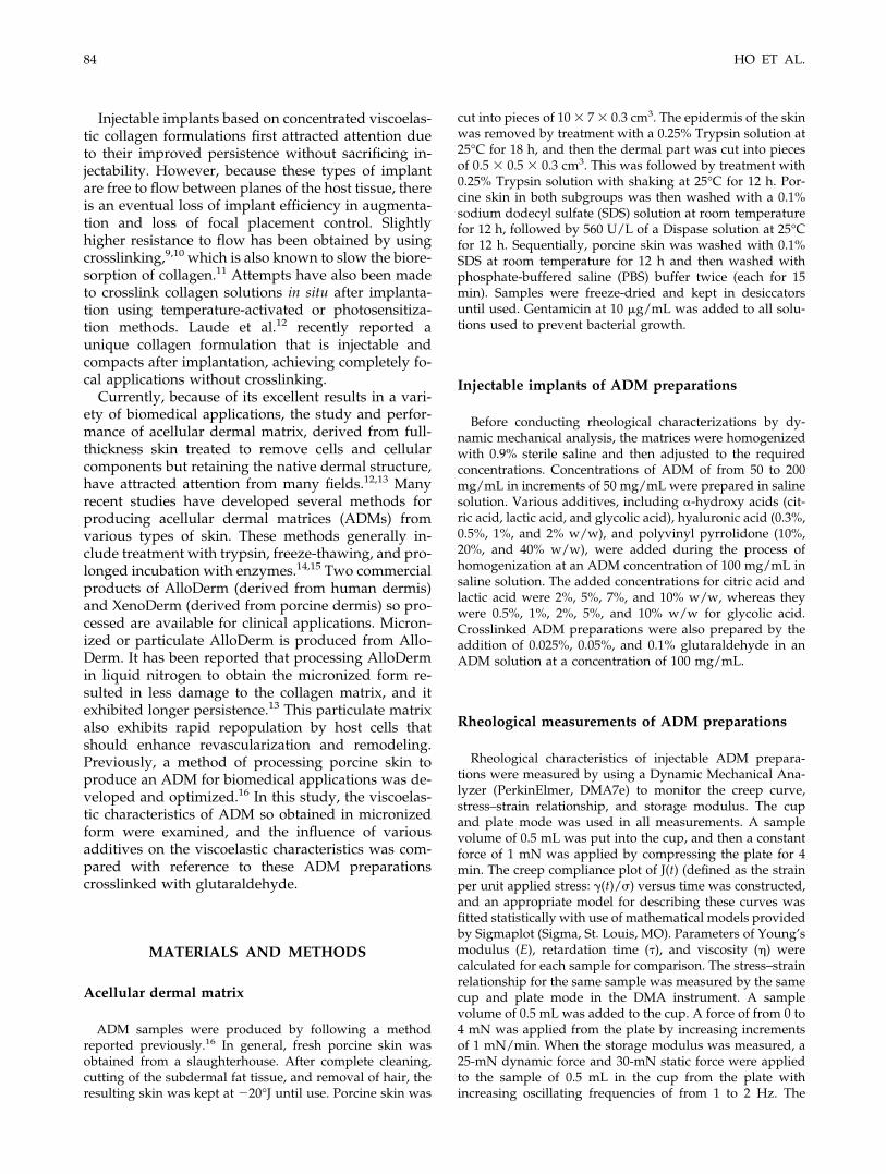
Acellular dermal matrix
ADM samples were produced by following a methodreported previously.16 In general, fresh porcine skin wasobtained from a slaughterhouse. After complete cleaning,cutting of the subdermal fat tissue, and removal of hair, theresulting skin was kept at �20°J until use. Porcine skin was
cut into pieces of 10 � 7 � 0.3 cm3. The epidermis of the skinwas removed by treatment with a 0.25% Trypsin solution at25°C for 18 h, and then the dermal part was cut into piecesof 0.5 � 0.5 � 0.3 cm3. This was followed by treatment with0.25% Trypsin solution with shaking at 25°C for 12 h. Por-cine skin in both subgroups was then washed with a 0.1%sodium dodecyl sulfate (SDS) solution at room temperaturefor 12 h, followed by 560 U/L of a Dispase solution at 25°Cfor 12 h. Sequentially, porcine skin was washed with 0.1%SDS at room temperature for 12 h and then washed withphosphate-buffered saline (PBS) buffer twice (each for 15min). Samples were freeze-dried and kept in desiccatorsuntil used. Gentamicin at 10 �g/mL was added to all solu-tions used to prevent bacterial growth.
Injectable implants of ADM preparations
Before conducting rheological characterizations by dy-namic mechanical analysis, the matrices were homogenizedwith 0.9% sterile saline and then adjusted to the requiredconcentrations. Concentrations of ADM of from 50 to 200mg/mL in increments of 50 mg/mL were prepared in salinesolution. Various additives, including �-hydroxy acids (cit-ric acid, lactic acid, and glycolic acid), hyaluronic acid (0.3%,0.5%, 1%, and 2% w/w), and polyvinyl pyrrolidone (10%,20%, and 40% w/w), were added during the process ofhomogenization at an ADM concentration of 100 mg/mL insaline solution. The added concentrations for citric acid andlactic acid were 2%, 5%, 7%, and 10% w/w, whereas theywere 0.5%, 1%, 2%, 5%, and 10% w/w for glycolic acid.Crosslinked ADM preparations were also prepared by theaddition of 0.025%, 0.05%, and 0.1% glutaraldehyde in anADM solution at a concentration of 100 mg/mL.
Rheological measurements of ADM preparations
Rheological characteristics of injectable ADM prepara-tions were measured by using a Dynamic Mechanical Ana-lyzer (PerkinElmer, DMA7e) to monitor the creep curve,stress–strain relationship, and storage modulus. The cupand plate mode was used in all measurements. A samplevolume of 0.5 mL was put into the cup, and then a constantforce of 1 mN was applied by compressing the plate for 4min. The creep compliance plot of J(t) (defined as the strainper unit applied stress: �(t)/) versus time was constructed,and an appropriate model for describing these curves wasfitted statistically with use of mathematical models providedby Sigmaplot (Sigma, St. Louis, MO). Parameters of Young’smodulus (E), retardation time (�), and viscosity (�) werecalculated for each sample for comparison. The stress–strainrelationship for the same sample was measured by the samecup and plate mode in the DMA instrument. A samplevolume of 0.5 mL was added to the cup. A force of from 0 to4 mN was applied from the plate by increasing incrementsof 1 mN/min. When the storage modulus was measured, a25-mN dynamic force and 30-mN static force were appliedto the sample of 0.5 mL in the cup from the plate withincreasing oscillating frequencies of from 1 to 2 Hz. The
84 HO ET AL.

storage modulus was calculated correspondingly, and theplot of the storage modulus versus frequency was con-structed.
RESULTS AND DISCUSSION
Many polymeric systems exhibit behaviors thatcombine both elastic (solid) and viscous (liquid) prop-erties, in which the applied stress is proportional toboth the resultant strain and the rate of strain.17–19
Such systems are termed viscoelastic, which has beendefined as the “simultaneous existence of viscous (liq-uid) and elastic (solid) properties in the materials.”20
The viscoelastic behaviors of two injectable forms ofcollagen, nonfibrillar concentrated solutions and re-constituted fibrillar suspensions, have been investi-gated with shear creep experiments in a parallel-plateapparatus.15 Extrusion-force measurements oncrosslinked collagen suspensions have been used tocharacterize the rheological properties. The effect ofhydrophobic forces on the rheology of concentrateddispersions of collagen fibers in aqueous solution wasstudied by dynamic rheological measurements overtemperatures ranging from 283 to 308 K.21 Nonlinearviscoelastic properties of bovine skin collagen gel so-lutions [diluting 3 mg/mL Vitrogen to 1 or 1.5 mg/mLwith Dulbecco’s modified Eagles’ medium (DMEM)]have been revealed with the use of a servo-controlledlinear activator to impose an axial strain on the gel.22
Mechanical characteristics of engineered collagenfibrils (ECF) have been evaluated from compressionand permeability experiments.12 In this study, the cupand plate mode of dynamic mechanical analysis wasapplied for the rheological characterizations of ADMpreparations.
Because crosslinking of the polymeric nature of col-lagen with glutaraldehyde was able to slightly in-crease the resistance to flow between the planes of thehost tissue,9,10 improvement in the persistence withcrosslinking is evident. Therefore, ADM preparationscrosslinked with glutaraldehyde were selected as areference to compare rheological characteristics forthese ADM preparations containing various additives.Figure 1 illustrates the creep curves for ADM prepa-rations crosslinked with various concentrations of glu-taraldehyde. The creep compliance plot of J(t) (�(t)/)versus time (t) describes the strain of a viscoelasticbody with respect to an applied stress. The mechanicalmodel of a viscoelastic material showing both viscos-ity of a liquid state and elasticity of a solid state, whichcan be represented in two different ways by Maxwelland Voigt elements, may be combined into a general-ized model to incorporate all possibilities of flow anddeformation of non-Newtonian materials. On the basis
of these findings, the best fitting equation for thesecurves in Figure 1 was found to be y a*(1 � e�bx),which is consistent with the Kelvin–Voigt model rep-
Figure 1. (A) Creep curve, (B) stress–strain relationship,and (C) storage modulus measurements for ADM prepara-tions (100 mg/mL) crosslinked with various concentrationsof glutaraldehyde (GLA). (•) GLA 0.0%, (■) GLA 0.025%,(Œ) GLA 0.05%, (�) GLA 0.1% (n 3).
ADM PREPARATIONS AS INJECTABLE IMPLANTS 85
resented as��t�
�1E�1 � e�
t��. Correspondingly, E,
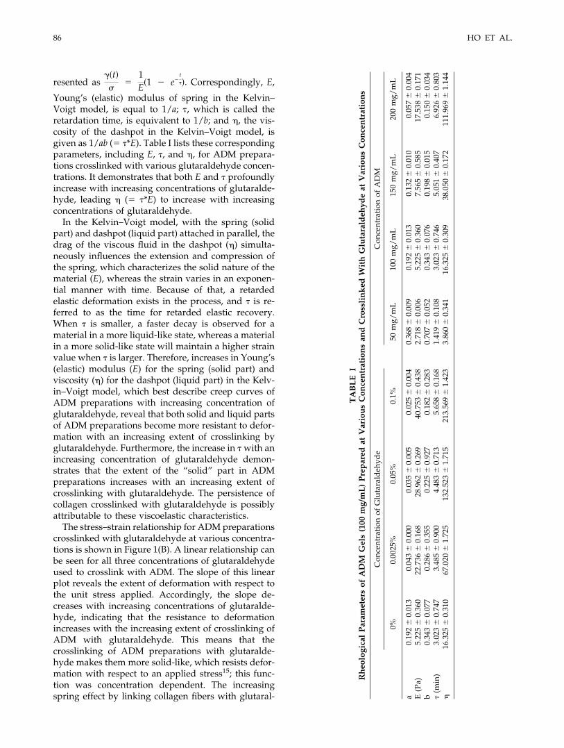
Young’s (elastic) modulus of spring in the Kelvin–Voigt model, is equal to 1/a; �, which is called theretardation time, is equivalent to 1/b; and �, the vis-cosity of the dashpot in the Kelvin–Voigt model, isgiven as 1/ab ( �*E). Table I lists these correspondingparameters, including E, �, and �, for ADM prepara-tions crosslinked with various glutaraldehyde concen-trations. It demonstrates that both E and � profoundlyincrease with increasing concentrations of glutaralde-hyde, leading � ( �*E) to increase with increasingconcentrations of glutaraldehyde.
In the Kelvin–Voigt model, with the spring (solidpart) and dashpot (liquid part) attached in parallel, thedrag of the viscous fluid in the dashpot (�) simulta-neously influences the extension and compression ofthe spring, which characterizes the solid nature of thematerial (E), whereas the strain varies in an exponen-tial manner with time. Because of that, a retardedelastic deformation exists in the process, and � is re-ferred to as the time for retarded elastic recovery.When � is smaller, a faster decay is observed for amaterial in a more liquid-like state, whereas a materialin a more solid-like state will maintain a higher strainvalue when � is larger. Therefore, increases in Young’s(elastic) modulus (E) for the spring (solid part) andviscosity (�) for the dashpot (liquid part) in the Kelv-in–Voigt model, which best describe creep curves ofADM preparations with increasing concentration ofglutaraldehyde, reveal that both solid and liquid partsof ADM preparations become more resistant to defor-mation with an increasing extent of crosslinking byglutaraldehyde. Furthermore, the increase in � with anincreasing concentration of glutaraldehyde demon-strates that the extent of the “solid” part in ADMpreparations increases with an increasing extent ofcrosslinking with glutaraldehyde. The persistence ofcollagen crosslinked with glutaraldehyde is possiblyattributable to these viscoelastic characteristics.
The stress–strain relationship for ADM preparationscrosslinked with glutaraldehyde at various concentra-tions is shown in Figure 1(B). A linear relationship canbe seen for all three concentrations of glutaraldehydeused to crosslink with ADM. The slope of this linearplot reveals the extent of deformation with respect tothe unit stress applied. Accordingly, the slope de-creases with increasing concentrations of glutaralde-hyde, indicating that the resistance to deformationincreases with the increasing extent of crosslinking ofADM with glutaraldehyde. This means that thecrosslinking of ADM preparations with glutaralde-hyde makes them more solid-like, which resists defor-mation with respect to an applied stress15; this func-tion was concentration dependent. The increasingspring effect by linking collagen fibers with glutaral-
TA
BL
EI
Rh
eolo
gica
lP
aram
eter
sof
AD
MG
els
(100
mg/
mL
)P
rep
ared
atV
ario
us
Con
cen
trat
ion
san
dC
ross
lin
ked
Wit
hG
luta
rald
ehyd
eat
Var
iou
sC
once
ntr
atio
ns
Con
cent
rati
onof
Glu
tara
ldeh
yde
Con
cent
rati
onof
AD
M
0%0.
0025
%0.
05%
0.1%
50m
g/m
L10
0m
g/m
L15
0m
g/m
L20
0m
g/m
L
a0.
192
0.
013
0.04
3
0.00
00.
035
0.
005
0.02
5
0.00
40.
368
0.
009
0.19
2
0.01
30.
132
0.
010
0.05
7
0.00
4E
(Pa)
5.22
5
0.36
022
.736
0.
168
28.9
62
0.26
940
.753
0.
438
2.71
8
0.00
65.
225
0.
360
7.56
5
0.58
517
.538
0.
171
b0.
343
0.
077
0.28
6
0.35
50.
225
0.
927
0.18
2
0.28
30.
707
0.
052
0.34
3
0.07
60.
198
0.
015
0.15
0
0.03
4�
(min
)3.
023
0.
747
3.48
5
0.90
04.
483
0.
713
5.65
8
0.16
81.
419
0.
108
3.02
3
0.74
65.
051
0.
407
6.92
6
0.80
3�
16.3
25
0.31
067
.020
1.
725
132.
523
1.
715
213.
569
1.
423
3.86
0
0.34
116
.325
0.
309
38.0
50
0.17
211
1.96
9
1.14
4
86 HO ET AL.
dehyde might be responsible for the increasing extentof the spring part in ADM preparations.
Figure 1(C) presents the plots of the storage modu-lus (E�) versus the oscillating frequency (Hz) for ADMpreparations crosslinked with different concentrationsof glutaraldehyde. E� of ADM preparations increaseswith increasing concentrations of glutaraldehyde.However, the smallest change in E� with respect to theoscillating frequency in the range examined was ob-served. E� was measured by simultaneously applyingstatic and dynamic forces to the materials with anoscillating frequency which increased from 1 to 2 Hz.E� represents the energy stored per cycle within thesample (i.e., the “solid” response).23 Therefore, thehigher E� is, as a result of the increased extent ofcrosslinking with glutaraldehyde, the greater the ex-tent of rigidity or a solid-like status that a material willpossess.
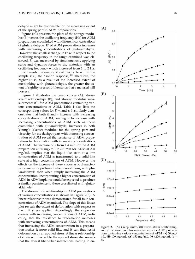
Figure 2 illustrates the creep curves (A), stress–strain relationships (B), and storage modulus mea-surements (C) for ADM preparations containing var-ious concentrations of ADM. Table I also lists thecorresponding values for E, �, and �. It similarly dem-onstrates that both E and � increase with increasingconcentrations of ADM, leading � to increase withincreasing concentrations of ADM such as thosecrosslinked with glutaraldehyde. Increases in bothYoung’s (elastic) modulus for the spring part andviscosity for the dashpot part with increasing concen-tration of ADM reveal the resistance of ADM prepa-rations to deformation with increasing concentrationsof ADM. The increase of � from 1.4 min for the ADMpreparation at 50 mg/mL to 6.6 min for ADM at 200mg/mL implies that the liquid-like state at a lowconcentration of ADM is transformed to a solid-likestate at a high concentration of ADM. However, theeffects on the increase of these viscoelastic character-istics are more profound when crosslinking with glu-taraldehyde than when simply increasing the ADMconcentration. Incorporating a higher concentration ofADM in ADM implants would be expected to producea similar persistence to those crosslinked with glutar-aldehyde.
The stress–strain relationship for ADM preparationsof various concentrations is shown in Figure 2(B). Alinear relationship was demonstrated for all four con-centrations of ADM examined. The slope of this linearplot reveals the extent of deformation with respect tothe unit stress applied. Accordingly, the slope de-creases with increasing concentrations of ADM, indi-cating that the resistance to deformation increaseswith increasing concentrations of ADM. This meansthat increasing the ADM concentration in a prepara-tion makes it more solid-like, and it can thus resistdeformation by an applied stress. A linear relationshipof strain with respect to the applied stress also revealsthat the fewest fiber–fiber interactions leading to en-
Figure 2. (A) Creep curve, (B) stress–strain relationship,and (C) storage modulus measurements for ADM prepara-tions containing various concentrations of ADM. (•) 50 mg/mL, (■) 100 mg/mL, (Œ) 150 mg/mL, (�) 200 mg/mL (n 3).
ADM PREPARATIONS AS INJECTABLE IMPLANTS 87
tanglement of collagen fibrils exist or that there is nodisruption of the even distribution of collagen fibrilsdue to there being no tendency of reaching a plateauor suddenly decreasing. Possibly, this is due to the factthat ADM is suspended in saline solution with only aminimal amount of dissolved collagen. Nevertheless,the changing extent of strain with respect to the ap-plied stress is lower (or more resistant) in ADM prep-arations containing as high as 200 mg/mL than thatwith crosslinked ADM at a concentration of 100mg/mL and the highest amount of glutaraldehyde(0.1%). Figure 2(C) presents the plots of storage mod-ulus (E�) versus frequency (Hz) for ADM preparationsat four different concentrations. Similarly, E� of ADMpreparations increases with an increasing concentra-tion of ADM. The smallest changes in E� were shownwith respect to the oscillating frequency in the rangeexamined as well. Because E� increases with increas-ing ADM concentrations, a corresponding increase inthe extent of the rigidity or the solid-like status wouldbe expected. However, ADM preparations only needto be crosslinked with 0.025% glutaraldehyde to havea storage modulus close to that of ADM preparationscontaining a concentration as high as 200 mg/mL.
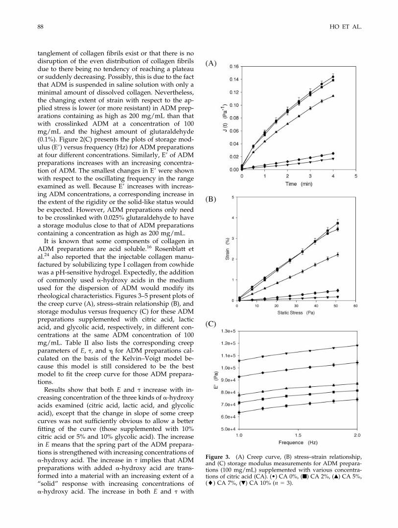
It is known that some components of collagen inADM preparations are acid soluble.16 Rosenblatt etal.24 also reported that the injectable collagen manu-factured by solubilizing type I collagen from cowhidewas a pH-sensitive hydrogel. Expectedly, the additionof commonly used �-hydroxy acids in the mediumused for the dispersion of ADM would modify itsrheological characteristics. Figures 3–5 present plots ofthe creep curve (A), stress–strain relationship (B), andstorage modulus versus frequency (C) for these ADMpreparations supplemented with citric acid, lacticacid, and glycolic acid, respectively, in different con-centrations at the same ADM concentration of 100mg/mL. Table II also lists the corresponding creepparameters of E, �, and � for ADM preparations cal-culated on the basis of the Kelvin–Voigt model be-cause this model is still considered to be the bestmodel to fit the creep curve for those ADM prepara-tions.
Results show that both E and � increase with in-creasing concentration of the three kinds of �-hydroxyacids examined (citric acid, lactic acid, and glycolicacid), except that the change in slope of some creepcurves was not sufficiently obvious to allow a betterfitting of the curve (those supplemented with 10%citric acid or 5% and 10% glycolic acid). The increasein E means that the spring part of the ADM prepara-tions is strengthened with increasing concentrations of�-hydroxy acid. The increase in � implies that ADMpreparations with added �-hydroxy acid are trans-formed into a material with an increasing extent of a“solid” response with increasing concentrations of�-hydroxy acid. The increase in both E and � with
Figure 3. (A) Creep curve, (B) stress–strain relationship,and (C) storage modulus measurements for ADM prepara-tions (100 mg/mL) supplemented with various concentra-tions of citric acid (CA). (•) CA 0%, (■) CA 2%, (Œ) CA 5%,(�) CA 7%, (�) CA 10% (n 3).
88 HO ET AL.

increasing �-hydroxy acid concentrations also resultsin increased viscosity of the liquid part of ADM prep-arations. In a comparison of the three �-hydroxy acidsexamined, only the addition of glycolic acid couldmodify the viscoelastic characteristics of ADM prepa-rations to an extent similar to those of preparationscrosslinked with glutaraldehyde. The clinical use ofglycolic acid for the treatment of skin disorders seemsto elucidate an interaction between glycolic acid andcollagen fibers that is responsible for this effectivemodification of the viscoelastic characteristics of ADMpreparations, which contain collagen as the main com-ponent.25,26
The stress–strain plots in Figures 3(B), 4(B), and 5(B)for ADM preparations with added �-hydroxy acidsalso are closer to linearity as shown by the ADMpreparations with different concentrations of ADM.The slopes of the stress–strain plots decrease withincreasing concentration of the respective �-hydroxyacids, revealing that the addition of �-hydroxy acidsmakes ADM preparations more solid-like so that theycan resist deformation with respect to the appliedstress; the extent was the greatest for glycolic acid.Storage modulus measurements shown in Figures3(C), 4(C), and 5(C) give the same information (i.e., therigidity or solid response of ADM preparations in-creases with increasing concentrations of �-hydroxyacids), and this effect is the most profound for glycolicacid at the same concentration as lactic acid and citricacid. In conclusion, the Kelvin–Voigt model is the bestfitting model that describes the rheological character-istics of ADM preparations with added �-hydroxyacids. Glycolic acid is the most appropriate additivefor increasing the solid response or rigidity of ADMpreparations, even improving viscoelastic characteris-tics to an extent comparable with that of preparationscrosslinked with glutaraldehyde.
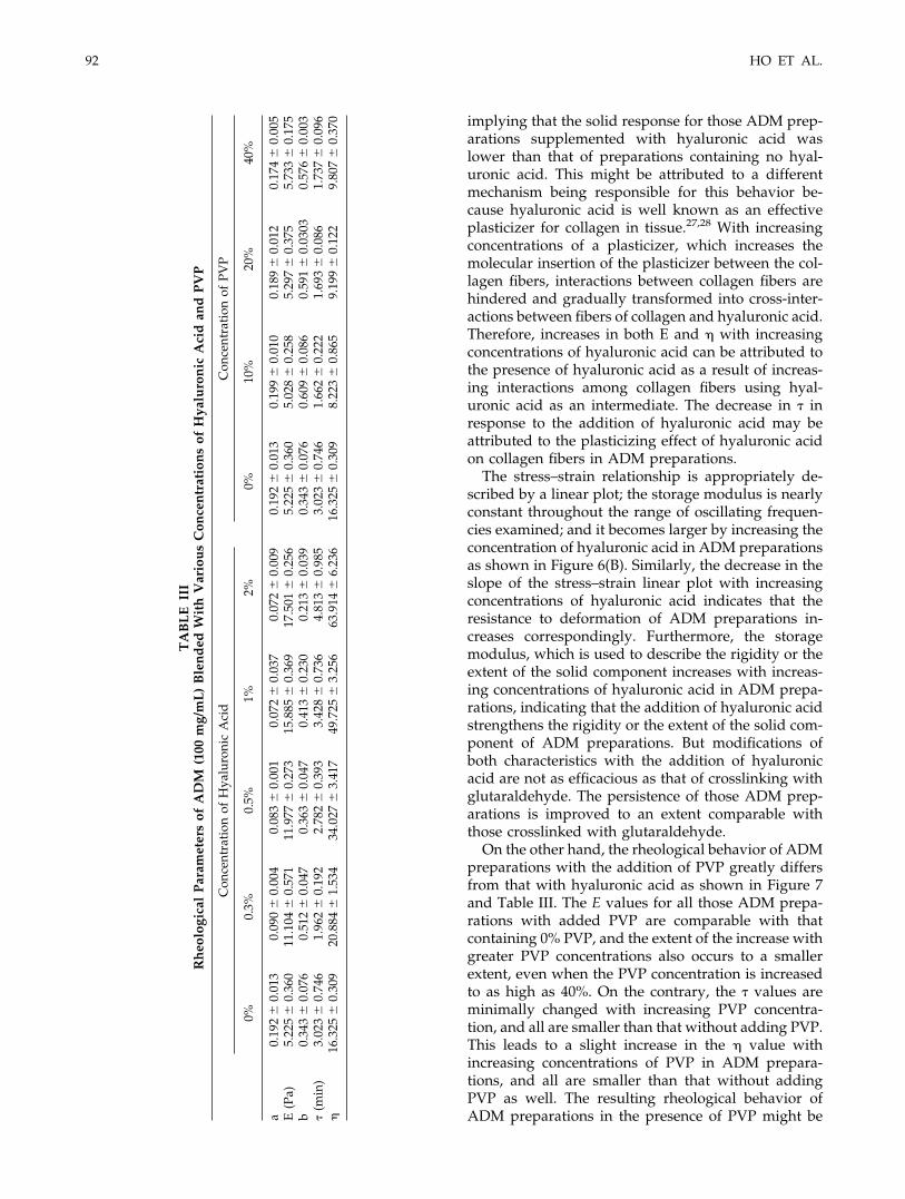
The effects on the rheological characteristics ofADM preparations at a 100 mg/mL concentrationblended with polymeric materials of hyaluronic acidand PVP were compared. Figures 6 and 7 present therespective results of creep curves (A), stress–strainplots (B), and storage modulus measurements (C) forhyaluronic acid and PVP. Table III also lists the cor-responding creep parameters of E, �, and � calculatedon the basis of the Kelvin–Voigt model because thismodel is still considered to be the best model to fit thecreep curve for those ADM preparations.
Both E and � gradually increase with increasingconcentrations of hyaluronic acid as evidenced in Ta-ble III. The increase in both E and � similarly leads to� increasing with higher concentrations of hyaluronicacid. This demonstrates that both the spring and dash-pot parts are strengthened by the addition of hyal-uronic acid. However, a smaller � value (1.94 min for0.3% hyaluronic acid vs 2.89 min for 0.0% hyaluronicacid) resulted from the addition of hyaluronic acid,
TA
BL
EII
Rh
eolo
gica
lP
aram
eter
sof
AD
MG
els
(100
mg/
mL
)P
rep
ared
Wit
hV
ario
us
Con
cen
trat
ion
sof
Cit
ric
Aci
d,L
acti
cA
cid
,an
dG
lyco
lic
Aci
d
Con
cent
rati
onof
citr
icac
idC
once
ntra
tion
ofL
acti
cA
cid
Con
cent
rati
onof
Gly
colic
Aci
d
0%2%
5%7%
10%
0%2%
5%7%
10%
0.5%
1%2%
5%10
%
a0.
192
0.
013
0.17
9
0.00
90.
162
0.
008
0.07
0
0.01
00.
851
0.
047
0.19
2
0.01
30.
121
0.
147
0.13
2
0.00
20.
072
0.
006
0.03
8
0.02
20.
142
0.
0240
0.06
3
0.00
30.
059
0.
010
0.46
9
0.30
90.
335
0.
280
E(P
a)5.
225
0.
360
5.58
3
0.29
66.
167
0.
343
14.0
75
0.33
61.
176
0.
065
5.22
5
0.36
08.
276
0.
936
7.57
7
0.13
813
.821
1.
125
25.7
40
1.41
37.
152
1.
168
15.6
75
0.81
717
.064
2.
786
3.03
8
2.23
29.
578
12
.787
b0.
343
0.
076
0.35
8
0.04
40.
298
0.
024
0.22
8
0.04
70.
004
0.
000
0.34
3
0.07
60.
687
0.
054
0.29
0
0.03
10.
226
0.
050
0.13
5
0.02
30.
410
0.
149
0.33
6
0.02
20.
120
0.
0353
0.01
2
0.01
10.
062
0.
051
�(m
in)
3.02
3
0.74
62.
813
0.
328
3.36
4
0.28
94.
386
0.
325
208.
8
8.76
43.
023
0.
746
1.46
0
0.11
73.
472
0.
388
4.59
4
1.16
47.
543
1.
263
2.62
6
0.79
72.
981
0.
206
8.81
9
2.65
826
5.96
6
347.
937
35.8
90
40.1
24�
16.3
25
0.30
915
.482
0.
998
20.9
49
0.62
061
.256
0.
745
244.
639
3.
080
16.3
25
0.30
912
.243
0.
573
27.3
69
2.28
162
.045
4.
852
191.
200
23
.587
19.8
66
2.87
844
.628
1.
873
154.
377
7.
642
414.
121
34
1.39
719
4.16
1
158.
304
ADM PREPARATIONS AS INJECTABLE IMPLANTS 89

Figure 4. (A) Creep curve, (B) stress–strain relationship,and (C) storage modulus measurements for ADM prepara-tions (100 mg/mL) supplemented with various concentra-tions of lactic acid (LA). (•) LA 0%, (■) LA 2%, (Œ) LA 5%,(�) LA 7%, (�) LA 10% (n 3).
Figure 5. (A) Creep curve, (B) stress–strain relationship,and (C) storage modulus measurements for ADM prepara-tions (100 mg/mL) supplemented with various concentra-tions of glycolic acid (GA). (•) GA 0%, (■) GA 0.5%, (Œ) GA1%, (�) GA 2%, (�) GA 5%, (E) GA 10% (n 3).
90 HO ET AL.
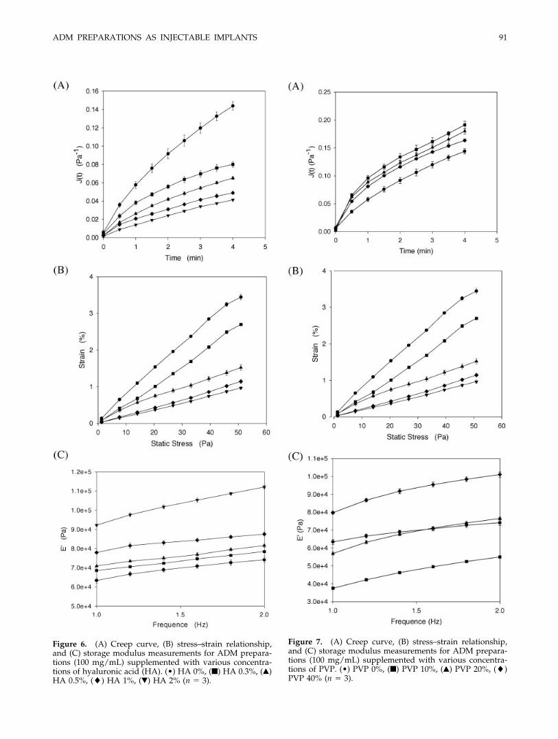
Figure 6. (A) Creep curve, (B) stress–strain relationship,and (C) storage modulus measurements for ADM prepara-tions (100 mg/mL) supplemented with various concentra-tions of hyaluronic acid (HA). (•) HA 0%, (■) HA 0.3%, (Œ)HA 0.5%, (�) HA 1%, (�) HA 2% (n 3).
Figure 7. (A) Creep curve, (B) stress–strain relationship,and (C) storage modulus measurements for ADM prepara-tions (100 mg/mL) supplemented with various concentra-tions of PVP. (•) PVP 0%, (■) PVP 10%, (Œ) PVP 20%, (�)PVP 40% (n 3).
ADM PREPARATIONS AS INJECTABLE IMPLANTS 91
implying that the solid response for those ADM prep-arations supplemented with hyaluronic acid waslower than that of preparations containing no hyal-uronic acid. This might be attributed to a differentmechanism being responsible for this behavior be-cause hyaluronic acid is well known as an effectiveplasticizer for collagen in tissue.27,28 With increasingconcentrations of a plasticizer, which increases themolecular insertion of the plasticizer between the col-lagen fibers, interactions between collagen fibers arehindered and gradually transformed into cross-inter-actions between fibers of collagen and hyaluronic acid.Therefore, increases in both E and � with increasingconcentrations of hyaluronic acid can be attributed tothe presence of hyaluronic acid as a result of increas-ing interactions among collagen fibers using hyal-uronic acid as an intermediate. The decrease in � inresponse to the addition of hyaluronic acid may beattributed to the plasticizing effect of hyaluronic acidon collagen fibers in ADM preparations.
The stress–strain relationship is appropriately de-scribed by a linear plot; the storage modulus is nearlyconstant throughout the range of oscillating frequen-cies examined; and it becomes larger by increasing theconcentration of hyaluronic acid in ADM preparationsas shown in Figure 6(B). Similarly, the decrease in theslope of the stress–strain linear plot with increasingconcentrations of hyaluronic acid indicates that theresistance to deformation of ADM preparations in-creases correspondingly. Furthermore, the storagemodulus, which is used to describe the rigidity or theextent of the solid component increases with increas-ing concentrations of hyaluronic acid in ADM prepa-rations, indicating that the addition of hyaluronic acidstrengthens the rigidity or the extent of the solid com-ponent of ADM preparations. But modifications ofboth characteristics with the addition of hyaluronicacid are not as efficacious as that of crosslinking withglutaraldehyde. The persistence of those ADM prep-arations is improved to an extent comparable withthose crosslinked with glutaraldehyde.
On the other hand, the rheological behavior of ADMpreparations with the addition of PVP greatly differsfrom that with hyaluronic acid as shown in Figure 7and Table III. The E values for all those ADM prepa-rations with added PVP are comparable with thatcontaining 0% PVP, and the extent of the increase withgreater PVP concentrations also occurs to a smallerextent, even when the PVP concentration is increasedto as high as 40%. On the contrary, the � values areminimally changed with increasing PVP concentra-tion, and all are smaller than that without adding PVP.This leads to a slight increase in the � value withincreasing concentrations of PVP in ADM prepara-tions, and all are smaller than that without addingPVP as well. The resulting rheological behavior ofADM preparations in the presence of PVP might be
TA
BL
EII
IR
heo
logi
cal
Par
amet
ers
ofA
DM
(100
mg/
mL
)B
len
ded
Wit
hV
ario
us
Con
cen
trat
ion
sof
Hya
luro
nic
Aci
dan
dP
VP
Con
cent
rati
onof
Hya
luro
nic
Aci
dC
once
ntra
tion
ofPV
P
0%0.
3%0.
5%1%
2%0%
10%
20%
40%
a0.
192
0.
013
0.09
0
0.00
40.
083
0.
001
0.07
2
0.03
70.
072
0.
009
0.19
2
0.01
30.
199
0.
010
0.18
9
0.01
20.
174
0.
005
E(P
a)5.
225
0.
360
11.1
04
0.57
111
.977
0.
273
15.8
85
0.36
917
.501
0.
256
5.22
5
0.36
05.
028
0.
258
5.29
7
0.37
55.
733
0.
175
b0.
343
0.
076
0.51
2
0.04
70.
363
0.
047
0.41
3
0.23
00.
213
0.
039
0.34
3
0.07
60.
609
0.
086
0.59
1
0.03
030.
576
0.
003
�(m
in)
3.02
3
0.74
61.
962
0.
192
2.78
2
0.39
33.
428
0.
736
4.81
3
0.98
53.
023
0.
746
1.66
2
0.22
21.
693
0.
086
1.73
7
0.09
6�
16.3
25
0.30
920
.884
1.
534
34.0
27
3.41
749
.725
3.
256
63.9
14
6.23
616
.325
0.
309
8.22
3
0.86
59.
199
0.
122
9.80
7
0.37
0
92 HO ET AL.

attributed to the lubricating effect of the linear poly-meric chains of PVP on suspended particles or poly-meric fibers14 and partly to the viscous nature of PVP,which is commonly used as a viscosity-building ex-cipient in the pharmaceutical field. However, thestress–strain relationship shows that the slopes of thelinear plots are quite similar to those of ADM prepa-rations with the addition of different concentrations ofPVP; however, all are higher than that without addingPVP, which is contrary to the decrease with increasing
supplemented concentrations as revealed above. Thisfinding means that the presence of PVP in ADM prep-arations can accelerate the deformation of the result-ing preparation, hence improving fluidization of theADM preparations during injection. Therefore, persis-tent improvements with the addition of PVP wouldexpectedly be less promising.
The storage modulus (E�) at the same frequency forADM preparations with the addition of 10% PVP waslower than those containing no PVP, whereas that for
Figure 8. (A) Creep curve, (B) stress–strain relationship, and (C) storage modulus measurements for TPC preparationsobtained by further treating ADM with pepsin, and (D) creep curve, (E) stress–strain relationship, (F) storage modulusmeasurements for TPC preparations obtained directly from porcine skin. (•) 50 mg/mL, (■) 100 mg/mL, (Œ) 150 mg/mL, (�)200 mg/mL (n 3).
ADM PREPARATIONS AS INJECTABLE IMPLANTS 93

ADM preparations containing PVP increased with in-creasing concentrations of PVP of from 10% to 40% forall ranges of frequencies examined. This finding indi-cates that the addition of PVP modifies the interac-tions between collagen fibers, resulting in a decreasedstorage modulus when PVP is added. The lubricatingeffect of PVP may also possibly be responsible for this.Furthermore, the rigidity or solid component of ADMpreparations in the presence of PVP increases withincreasing concentrations of PVP, but not to an extentcomparable with that of preparations crosslinked withglutaraldehyde.
The viscoelastic characteristics of telopeptide-poorcollagen (TPC) preparations obtained either by furthertreating ADM or directly from porcine skin with a pHprecipitation method reported previously16 were com-pared. Figure 8 illustrates the creep curves, stress–strain relationships, and storage modulus measure-ments for both types of telopeptide-poor collagenmixtures at different concentrations. Table IV also liststhe corresponding creep parameters of E, �, and �calculated on the basis of the Kelvin–Voigt modelbecause this model is still considered to be the bestmodel to fit the creep curves. Obviously, the tendencyfor E and � to increase with increasing TPC concen-trations is similar to that for ADM preparations. Butthe relative changes in both values with respect to theconcentration was the greatest for ADM preparations,followed by that for TPC obtained from ADM; theTPC obtained directly from porcine skin was thesmallest. The � values of the three preparations andtheir relative changes with increasing concentrationsfollowed the same order as those for E and �. Thestress–strain relationship as indicated in Figure 8(B,E)reveals that resistance to deformation by ADM prep-arations also followed the same order. Results of stor-age modulus measurements as revealed in Figure8(C,F) demonstrate that the extent of rigidity or thesolid response for TPC preparations obtained fromADM was greater than that of the TPC obtained fromporcine skin; but both were less than that for ADMpreparations. We concluded that preparations con-taining a purer form of collagen present different vis-coelastic characteristics from those of ADM prepara-tions, which contain components of the extracellularmatrix in influential amounts in addition to collagen.The particulate form of ADM preparations was alsoexpected to have different rheological characteristicsfrom these two TPC preparations, both of which con-tain greater amounts of the acid-soluble fraction ofcollagen. Overall, these two TPC preparations wereless efficacious in maintaining the same persistence asthat crosslinked with glutaraldehyde.
The effect of temperature on the viscoelastic char-acteristics of ADM preparations at different concen-trations was evaluated, and results are presented inFigure 9. The corresponding creep parameters of E, �,
TA
BL
EIV
Rh
eolo
gica
lP
aram
eter
sof
Var
iou
sC
once
ntr
atio
ns
ofA
DM
and
Tel
opep
tid
e-P
oor
Col
lage
n(T
PC
)P
rep
arat
ion
sO
bta
ined
Eit
her
by
Furt
her
Tre
atin
gA
DM
Wit
hP
epsi
n(I
)or
Dir
ectl
yFr
omP
orci
ne
Sk
in(I
I)
Con
cent
rati
onof
AD
MC
once
ntra
tion
ofT
PC(I
)C
once
ntra
tion
ofT
PC(I
I)
50m
g/m
L10
0m
g/m
L15
0m
g/m
L20
0m
g/m
L50
mg/
mL
100
mg/
mL
150
mg/
mL
200
mg/
mL
50m
g/m
L10
0m
g/m
L15
0m
g/m
L20
0m
g/m
L
a0.
368
0.
009
0.19
2
0.01
30.
132
0.
010
0.05
7
0.00
40.
528
0.
0648
0.21
9
0.01
50.
158
0.
003
0.05
2
0.00
50.
843
0.
161
0.56
5
0.03
70.
376
0.
048
0.21
3
0.00
1E
(Pa)
2.71
8
0.06
5.22
5
0.36
07.
565
0.
585
17.5
38
0.17
11.
745
0.
256
4.45
8
0.56
96.
325
1.
077
14.2
56
1.25
61.
212
0.
209
1.77
4
0.11
42.
688
0.
359
4.67
3
0.02
0b
0.70
7
0.05
20.
343
0.
076
0.19
8
0.01
50.
150
0.
034
0.78
5
0.04
510.
398
0.
045
0.25
5
0.09
80.
239
0.
056
0.93
7
0.11
10.
674
0.
007
0.45
7
0.04
70.
261
0.
004
�(m
in)
1.41
9
0.10
83.
023
0.
746
5.05
1
0.40
76.
926
0.
803
1.24
7
0.15
82.
405
0.
074
4.05
6
0.10
44.
258
0.
147
1.18
2
0.10
31.
449
0.
105
2.36
2
0.66
43.
960
0.
758
�3.
860
0.
341
16.3
25
0.30
938
.050
0.
172
111.
969
1.
144
2.25
8
0.25
611
.589
0.
589
25.4
48
0.41
267
.589
0.
326
1.32
3
0.50
42.
478
0.
134
6.22
5
0.55
715
.981
0.
116
94 HO ET AL.

TA
BL
EV
Rh
eolo
gica
lP
aram
eter
sof
AD
MG
els
Pre
par
edat
Var
iou
sC
once
ntr
atio
ns
and
Mea
sure
dat
25an
d37
°C
Con
cent
rati
onof
AD
M(2
5°C
)C
once
ntra
tion
ofA
DM
(37°
C)
50m
g/m
L10
0m
g/m
L15
0m
g/m
L20
0m
g/m
L50
mg/
mL
100
mg/
mL
150
mg/
mL
200
mg/
mL
a0.
368
0.
009
0.19
2
0.01
30.
132
0.
010
0.05
7
0.00
40.
386
0.
014
0.28
4
0.01
00.
256
0.
015
0.22
6
0.02
7E
(Pa)
2.71
8
0.00
65.
225
0.
360
7.56
5
0.58
517
.538
0.
171
2.58
4
0.00
93.
516
0.
132
3.90
9
0.22
74.
448
0.
507
b0.
707
0.
052
0.34
3
0.07
60.
198
0.
015
0.15
0
0.03
40.
800
0.
013
0.51
5
0.04
30.
370
0.
049
0.38
6
0.08
5�
(min
)1.
419
0.
108
3.02
3
0.74
65.
051
0.
407
6.92
6
0.80
31.
250
0.
021
1.94
8
0.15
82.
637
0.
392
2.68
7
0.65
9�
3.86
0
0.34
116
.325
0.
309
38.0
50
0.17
211
1.96
9
1.14
43.
233
0.
045
6.75
8
0.23
110
.841
0.
740
11.2
78
0.68
7
Figure 9. (A) Creep curve, (B) stress–strain relationship,and (C) storage modulus measurements for ADM prepara-tions containing various concentrations of ADM (circles: 50mg/mL; squares: 100 mg/mL; upper triangles: 150 mg/mL;diamonds: 200 mg/mL) at different temperatures: 25°C(closed symbols) and 37°C (open symbols) (n 3).
ADM PREPARATIONS AS INJECTABLE IMPLANTS 95

and � measured at 25 and 37°C and calculated on thebasis of the Kelvin–Voigt model are listed in Table Vbecause this model is still considered to be the bestmodel to fit the creep curves. The increase in E and �,and hence �, with increasing concentrations of ADMpreparations was maintained to be true even if thetemperature at which measurements were conductedwas increased to 37°C. But a significant decrease in Eand �, and hence �, was shown for the same ADMconcentration when the temperature of the measure-ments was increased from 25 to 37°C (Table V). Thisphenomenon was especially obvious at higher concen-trations of ADM preparations. Viscoelastic character-istics of ADM preparations are so sensitive to temper-ature effects that the persistence of such ADMpreparations after implantation at body temperaturewould seriously deteriorate.
CONCLUSIONS
The improved persistence of collagen implants bycrosslinking with glutaraldehyde might be attributedto a higher Young’s modulus (E) of the spring part, ahigher viscosity (�) of the liquid part in the Kelvin–Voigt model, and a greater extent of the solid responseas indicated by a larger value for the retardation time(�). Increasing the concentration of ADM and the ad-dition of various concentrations of additives in ADMpreparations may produce a similar influence on theseviscoelastic characteristics, but in a less efficaciousway than that produced by crosslinking with glutar-aldehyde. Increasing the ADM concentration to higherthan 200 mg/mL and adding glycolic acid to a con-centration � 2% would possibly produce the samelevel of persistence as that crosslinked with glutaral-dehyde as evidenced by the resulting viscoelasticcharacteristics. However, the effect of temperature onthe viscoelastic characteristics, especially at higherconcentrations of ADM, was so profound that itwould be harmful to the persistence of such ADMpreparations after implantation at body temperature.
References
1. Matton G, Anseeuw A, De Keyser F. The history of injectablebiomaterials and the biology of collagen. Aesthetic Plast Surg1985;9:133–140.
2. Copperman LS, MacKinnon V, Bechler G, Pharriss BB. Inject-able collagen: a six-year clinical investigation. Aesthetic PlastSurg 1985;9:145–151.
3. Herschorn S, Steele DJ, Radomski SB. Follow-up intraurethralcollagen for female stress urinary incontinence. J Urol 1996;156:1305–1309.
4. Benshushan A, Brzezinski A, Shoshani O, Rojansky N. Peri-urethrual injection for the treatment of urinary incontinence.Obstet Gynecol Surv 1998;53:383–388.
5. Stenberg A, Lackgren G. A new bioimplant for the endoscopictreatment of vesicourethral reflux: experimental, and short-term clinical results. J Urol 1995;154:800–803.
6. Ford CN, Martin DW, Warner TF. Injectable collagen in laryn-geal rehabilitation. Laryngoscope 1984;94:513–518.
7. Gromley DE, Eremia S. Quantitative assessment of augmenta-tion therapy. J Dermatol Surg Oncol 1990;16:1147–1151.
8. Matti BA, Nicolle FV. Clinical use of zyplast in correction ofage- and disease-related contour deficiencies of the face. Aes-thetic Plast Surg 1990;14:227–234.
9. Chan RW, Titze IR. Viscosities of implantable biomaterials invocal fold augmentation surgery. Laryngoscope 1998;108:725–731.
10. Wallace DG, Condell RA, Donovan JW, Paivinen A, Rhee WM,Wade SB. Multiple denaturational transitions in fibrillar colla-gen. Biopolymers 1986;25:1875–1895.
11. Chvapil M, Owen JA, Clark DS. Effect of collagen crosslinkingon the rate of resorption of implanted collagen tubing in rab-bits. J Biomed Mater Res 1977;11:297–314.
12. Laude D, Odlum K, Rudnicki S, Bachrach N. A novel injectablecollagen matrix: in vitro characterization and in vivo evalua-tion. J Biomech Eng 2000;122:231–235.
13. Griffey S, Schwade ND, Wright CG. Particulate dermal matrixas an injectable soft tissue replacement material. J BiomedMater Res 2001;58:10–15.
14. Jones DS. Dynamic mechanical analysis of polymeric systemsof pharmaceutical and biomedical significance. Int J Pharm1999;179:167–178.
15. Wallace DG, Rhee W, Weiss B. Shear creep of injectable colla-gen biomaterials. J Biomed Mater Res 1987;21:861–880.
16. Ray-Neng Chen, Hsiu-O Ho, Yu-Ting Tsai, Ming-Thau Sheu.Process development of an acellular dermal matrix (ADM) forbiomedical applications. Biomaterials 2004;25:2679–2686.
17. Ferry JD. Viscoelastic properties of polymers. New York: JohnWiley; 1980.
18. Ward IM, Hadley DW. An introduction to the mechanicalproperties of solid polymers. Chichester, UK: John Wiley; 1993.
19. Craig DQM, Johnson FA. Pharmaceutical applications of dy-namic mechanical thermal analysis. Thermochim Acta 1995;248:97–115.
20. Barnes HA, Hutton JF, Walters K. An introduction to rheology.Amsterdam: Elsevier; 1996.
21. Rosenblatt J, Devereux B, Wallace DG. Dynamic rheologicalstudies of hydrophobic interactions in injectable collagen bio-materials. J Appl Polym Sci 1993;50:953–963.
22. Ozerdem B, Tozeren A. Physical response of collagen gels totensile strain. J Biomech Eng 1995;117:397–401.
23. Rosenblatt J, Devereux B, Wallace DG. Effect of electrostaticforces on the dynamic rheological properties of injectable col-lagen biomaterials. Biomaterials 1992;13:878–886.
24. Rosenblatt J, Devereux B, Wallace DG. Injectable collagen as apH-sensitive hydrogel. Biomaterials 1994;15:985–995.
25. Ditre CM, Griffin TD, Murphy GF, Sueki H, Telegan B, JohnsonWC, Yu RJ, Van Scott EJ. Effects of alpha-hydroxy acids onphotoaged skin: a pilot clinical, histologic, and ultrastructuralstudy. J Am Acad Dermatol 1996;34:187–195.
26. Kim SJ, Park JH, Kim DH, Won YH, Maibach HI. Increased invivo collagen synthesis and in vitro cell proliferative effect ofglycolic acid. Dermatol Surg 1998;24:1054–1058.
27. Bakos D, Soldan M, Hernandez-Fuentes I. Hydroxyapatite-collagen-hyaluronic acid composite. 1999;20:191–195.
28. Frank P, Gendler E. Hyaluronic acid for soft-tissue augmenta-tion. Clin Plast Surg 2001;28:121–126.
96 HO ET AL.