Virtual Interactomics of Proteins from Biochemical Standpoint
22
Hindawi Publishing Corporation Molecular Biology International Volume 2012, Article ID 976385, 22 pages doi:10.1155/2012/976385 Review Article Virtual Interactomics of Proteins from Biochemical Standpoint Jaroslav Kubrycht, 1 Karel Sigler, 2 and Pavel Souˇ cek 3 1 Department of Physiology, Second Medical School, Charles University, 150 00 Prague, Czech Republic 2 Laboratory of Cell Biology, Institute of Microbiology, Academy of Sciences of the Czech Republic, 142 20 Prague, Czech Republic 3 Toxicogenomics Unit, National Institute of Public Health, 100 42 Prague, Czech Republic Correspondence should be addressed to Jaroslav Kubrycht, [email protected] Received 27 March 2012; Revised 18 May 2012; Accepted 18 May 2012 Academic Editor: Alessandro Desideri Copyright © 2012 Jaroslav Kubrycht et al. This is an open access article distributed under the Creative Commons Attribution License, which permits unrestricted use, distribution, and reproduction in any medium, provided the original work is properly cited. Virtual interactomics represents a rapidly developing scientific area on the boundary line of bioinformatics and interactomics. Protein-related virtual interactomics then comprises instrumental tools for prediction, simulation, and networking of the majority of interactions important for structural and individual reproduction, differentiation, recognition, signaling, regulation, and metabolic pathways of cells and organisms. Here, we describe the main areas of virtual protein interactomics, that is, structurally based comparative analysis and prediction of functionally important interacting sites, mimotope-assisted and combined epitope prediction, molecular (protein) docking studies, and investigation of protein interaction networks. Detailed information about some interesting methodological approaches and online accessible programs or databases is displayed in our tables. Considerable part of the text deals with the searches for common conserved or functionally convergent protein regions and subgraphs of conserved interaction networks, new outstanding trends and clinically interesting results. In agreement with the presented data and relationships, virtual interactomic tools improve our scientific knowledge, help us to formulate working hypotheses, and they frequently also mediate variously important in silico simulations. 1. General Remarks Many important findings in pharmacology, cell biology, and pathobiology have been achieved with the aid of virtual interactomics including computer-aided structural analysis, prediction and in silico simulation of interacting sites, protein complexes, and interaction networks. Virtual interactomics has been developed in the last thirty years, and it is in fact based on gradual bioinformatic processing of experimental data. These data were usually obtained from individual studies of interactions, and various large-scale experimental methods such as the two-hybrid system, phage display library studies reverse interactomics, SPOT arrays or microarray studies, and extended sequence studies [1–7]. In addition to sequence data, three-dimensional (3D) structures are ever more frequently required for interactomic predictions. X-ray crystallography or nuclear magnetic res- onance studies represent the most frequent sources of 3D structures, whereas combination of electron microscopy of molecular complexes with X-ray crystallography turns out to be interesting for the same purpose [8–11]. Alternatively, sophisticated 3D structure simulations such as homology modeling or combination of cryoelectron microscopy den- sities, and molecular dynamics appear to be also useful for approximating conventional 3D input at least in some cases [12–14]. In addition to 3D shape, solvent and surface accessi- bilities (or more likely actual dynamic accessibility following from accompanying interacting structures or proteolysis; cf. [15, 16]) were considered to be important criteria for reeval- uation of possible interaction sites. Many experimentally investigated and predicted structural relationships were also stored in interactomic databases to be selectively found, processed and compared. Moreover, some interaction data differently stored in multiple databases have been searched with the aid of special data mining servers such as Dasmiweb and PINA v2.0 ([17, 18]; see also Table 1).
-
Upload
khangminh22 -
Category
Documents
-
view
1 -
download
0
Transcript of Virtual Interactomics of Proteins from Biochemical Standpoint

Hindawi Publishing CorporationMolecular Biology InternationalVolume 2012, Article ID 976385, 22 pagesdoi:10.1155/2012/976385
Review Article
Virtual Interactomics of Proteins fromBiochemical Standpoint
Jaroslav Kubrycht,1 Karel Sigler,2 and Pavel Soucek3
1 Department of Physiology, Second Medical School, Charles University, 150 00 Prague, Czech Republic2 Laboratory of Cell Biology, Institute of Microbiology, Academy of Sciences of the Czech Republic, 142 20 Prague, Czech Republic3 Toxicogenomics Unit, National Institute of Public Health, 100 42 Prague, Czech Republic
Correspondence should be addressed to Jaroslav Kubrycht, [email protected]
Received 27 March 2012; Revised 18 May 2012; Accepted 18 May 2012
Academic Editor: Alessandro Desideri
Copyright © 2012 Jaroslav Kubrycht et al. This is an open access article distributed under the Creative Commons AttributionLicense, which permits unrestricted use, distribution, and reproduction in any medium, provided the original work is properlycited.
Virtual interactomics represents a rapidly developing scientific area on the boundary line of bioinformatics and interactomics.Protein-related virtual interactomics then comprises instrumental tools for prediction, simulation, and networking of the majorityof interactions important for structural and individual reproduction, differentiation, recognition, signaling, regulation, andmetabolic pathways of cells and organisms. Here, we describe the main areas of virtual protein interactomics, that is, structurallybased comparative analysis and prediction of functionally important interacting sites, mimotope-assisted and combined epitopeprediction, molecular (protein) docking studies, and investigation of protein interaction networks. Detailed information aboutsome interesting methodological approaches and online accessible programs or databases is displayed in our tables. Considerablepart of the text deals with the searches for common conserved or functionally convergent protein regions and subgraphs ofconserved interaction networks, new outstanding trends and clinically interesting results. In agreement with the presented dataand relationships, virtual interactomic tools improve our scientific knowledge, help us to formulate working hypotheses, and theyfrequently also mediate variously important in silico simulations.
1. General Remarks
Many important findings in pharmacology, cell biology, andpathobiology have been achieved with the aid of virtualinteractomics including computer-aided structural analysis,prediction and in silico simulation of interacting sites, proteincomplexes, and interaction networks. Virtual interactomicshas been developed in the last thirty years, and it is in factbased on gradual bioinformatic processing of experimentaldata. These data were usually obtained from individualstudies of interactions, and various large-scale experimentalmethods such as the two-hybrid system, phage display librarystudies reverse interactomics, SPOT arrays or microarraystudies, and extended sequence studies [1–7].
In addition to sequence data, three-dimensional (3D)structures are ever more frequently required for interactomicpredictions. X-ray crystallography or nuclear magnetic res-onance studies represent the most frequent sources of 3D
structures, whereas combination of electron microscopy ofmolecular complexes with X-ray crystallography turns outto be interesting for the same purpose [8–11]. Alternatively,sophisticated 3D structure simulations such as homologymodeling or combination of cryoelectron microscopy den-sities, and molecular dynamics appear to be also useful forapproximating conventional 3D input at least in some cases[12–14]. In addition to 3D shape, solvent and surface accessi-bilities (or more likely actual dynamic accessibility followingfrom accompanying interacting structures or proteolysis; cf.[15, 16]) were considered to be important criteria for reeval-uation of possible interaction sites. Many experimentallyinvestigated and predicted structural relationships were alsostored in interactomic databases to be selectively found,processed and compared. Moreover, some interaction datadifferently stored in multiple databases have been searchedwith the aid of special data mining servers such as Dasmiweband PINA v2.0 ([17, 18]; see also Table 1).
2 Molecular Biology International
Ta
ble
1:So
me
mor
ere
cen
ton
line
acce
ssib
lebi
oin
form
atic
tool
s.
Pro
gram
sP
urp
ose
Inpu
tEv
alu
atio
nto
ols
and
proc
edu
res
Com
pare
dst
ruct
ure
sIn
tern
etac
cess
ibili
tyR
efer
ence
s
Pro
gram
tool
s
3D-B
LAST
(tw
om
eth
ods)
∗ Ide
nti
fica
tion
of23
stat
esof
the
stru
ctu
ral
alph
abet
PD
BA
CF
∗ SA
SM∗ s
tru
ctu
rala
lph
abet
sequ
ence
data
base
sh
ttp:
//3d
-bla
st.li
fe.n
ctu
.edu
.tw
/[4
1,42
]∗ C
ompa
riso
nof
prot
ein
fold
su
sin
gsp
her
ical
pola
rFo
uri
erba
sis
fun
ctio
ns.
∗ Car
bo-l
ike
sim
ilari
tysc
ore
∗ SP
Fsh
ape
den
sity
rep.
htt
p://
thre
edbl
ast.
lori
a.fr
/
DA
SMIw
eb
On
line
inte
grat
ion
,an
alys
isan
das
sess
men
tofd
is-
trib
ute
dpr
otei
nin
tera
ctio
nda
taan
dpr
edic
tion
s(i
nte
ract
omic
min
ing)
som
epr
otei
nid
enti
fier
s
liter
atu
recu
rati
on,
pred
icti
on,3
Dan
alys
is
reco
rds
ofin
tera
ctio
ns
htt
p://
ww
w.d
asm
iweb
.de
[17]
FFA
S03
Serv
erac
cept
sa
use
rsu
pplie
dpr
otei
nse
quen
cean
dau
tom
atic
ally
gen
erat
esa
profi
le,
wh
ich
isth
enco
mpa
red
wit
hse
vera
lse
tsof
sequ
ence
profi
les
ofpr
otei
ns
data
base
s
QS
PPA
DS
htt
p://
ffas
.god
zikl
ab.o
rg/
[36,
37]
Kin
aseP
hos
2.0
Pre
dict
ion
ofki
nas
e-sp
ecifi
cph
osph
oryl
atio
nsi
tes
base
don
the
site
sequ
ence
sfr
omda
taba
ses
QS
k-fo
ld+
Jack
knif
ecr
oss-
valid
atio
ns
phos
phor
ylat
ion
site
sfr
omP
hos
pho.
ELM
Swis
s-P
rot
htt
p://
Kin
aseP
hos
2.m
bc.n
ctu
.edu
.tw
/[6
2]
Ph
os3D
Met
hod
ofph
osph
oryl
atio
n-s
ite
pred
icti
onba
sed
on3D
stru
ctu
ral
info
rmat
ion
asso
ciat
edw
ith
530
phys
-ch
em-p
r
QS
(+3D
con
text
)
SVM
,spa
tial
amin
oac
idpr
open
siti
esP
DB
coor
din
ates
htt
p://
phos
3d.m
pim
p-go
lm.m
pg.d
e/[6
3]
SeSA
WId
enti
fica
tion
offu
nct
ion
ally
orev
olu
tion
arily
con
serv
edm
otif
sba
sed
onba
lan
cin
gbe
twee
nse
quen
cean
dst
ruct
ura
lsim
ilari
ties
∗ PD
BQ
F,ID
;∗P
DB
QF,
ID,
IDt
Pva
lue
SeSA
Wsc
ore
con
serv
eddo
mai
ns
tem
plat
esh
ttp:
//sy
sim
m.if
rec.
osak
a-u
.ac.
jp/S
eSA
W/
[61]
Dat
abas
es
AD
AN
Pre
dict
ion
ofpr
otei
n-p
rote
inin
tera
ctio
ns
ofdi
f-fe
ren
tm
odu
lar
prot
ein
dom
ain
sm
edia
ted
bylin
ear
mot
ifs
PD
BA
CF
PSS
MP
Iin
tegr
ated
DaS
th
ttp:
//ad
an-e
mbl
.ibm
c.u
mh
.es/
[64]
CD
D(R
PS
BLA
ST)
Con
serv
eddo
mai
nre
lati
onsh
ips,
dom
ain
loca
tion
offr
equ
ent
bin
din
gsi
tes
QS
mod
elM
SAde
rive
dP
SSM
Con
serv
eddo
mai
ns
(PSS
M)
htt
p://
ww
w.n
cbi.n
lm.n
ih.g
ov/S
tru
ctu
re/
cdd/
cdd.
shtm
lh
ttp:
//w
ww
.ncb
i.nlm
.nih
.gov
/BLA
ST[6
5–67
]
GW
IDD
Doc
kin
gda
taba
se;
inte
grat
edre
sou
rce
for
stu
dies
ofpr
otei
n-p
rote
inin
tera
ctio
ns
onth
ege
nom
esc
ale
sear
chin
terf
ace
dock
ing
tech
niq
ues
DaS
th
ttp:
//gw
idd.
bioi
nfo
rmat
ics.
ku.e
du[6
8]
Meg
aMot
ifB
ase
(str
uct
ura
lda
taba
se)
3Dm
otif
orie
nta
tion
,in
term
otif
dist
ance
s,so
l-ve
nt
acce
ssib
ility
,se
con
dary
stru
ctu
reco
nte
nt,
hydr
ogen
bon
din
g,re
sidu
alpa
ckag
ing,
fam
il-ia
r/su
perf
amili
ar/n
one
mot
ifre
lati
onsh
ip
mot
ifor
sequ
ence
patt
ern
com
plex
eval
uat
ion
incl
udi
ng
proj
ecti
on
DS
+D
aSt
htt
p://
caps
.ncb
s.re
s.in
/Meg
aMot
ifba
se/i
nde
x.h
tml
[69]
Molecular Biology International 3
Ta
ble
1:C
onti
nu
ed.
Pro
gram
sP
urp
ose
Inpu
tEv
alu
atio
nto
ols
and
proc
edu
res
Com
pare
dst
ruct
ure
sIn
tern
etac
cess
ibili
tyR
efer
ence
s
PT
GL
Dat
abas
efo
rse
con
dary
stru
ctu
re-b
ased
prot
ein
topo
logi
es;v
isu
aliz
atio
nof
topo
logy
diag
ram
san
d3D
stru
ctu
res
PD
BQ
FU
LN4D
grap
hth
eory
DS
+D
aSt
htt
p://
ptgl
.un
i-fr
ankf
urt
.de/
[70]
Rsi
teD
BD
atab
ase
ofpr
otei
nbi
ndi
ng
pock
ets
wh
ich
inte
r-ac
tw
ith
sin
gle
stra
nd
RN
A
PD
Bco
de,
UP
S
3Dar
ran
gem
ent
ofph
ys-c
hem
-pr
3D-C
BP
htt
p://
bioi
nfo
3d.c
s.ta
u.a
c.il/
Rsi
teD
B/
[71]
∗In
depe
nde
nt
alte
rnat
ives
;3D
-CB
P:3
Dco
nse
nsu
sbi
ndi
ng
patt
ern
sim
port
ant
for
prot
ein
-nu
cleo
tide
reco
gnit
ion
;DS:
data
base
sequ
ence
s;D
SA:d
oubl
ese
quen
ceal
ign
men
t;D
aSt:
data
base
stru
ctu
res;
CS:
list
ofco
mpa
red
sequ
ence
s;ID
,ID
t:ch
ain
orte
mpl
ate
ID,
resp
ecti
vely
;M
SA:
mu
ltip
lese
quen
ceal
ign
men
t;Q
S:qu
ery
sequ
ence
(s)
(in
stea
dof
QS,
clon
aln
ames
orgi
nu
mbe
rsca
nbe
use
din
maj
orit
yof
give
nap
proa
ches
);ph
ys-c
hem
-pr:
phys
icoc
hem
ical
prop
erti
es;P
DB
:Pro
tein
data
ban
k;P
DB
QF:
PD
B-f
orm
atte
dqu
ery
file
;PD
BA
CF:
PD
B-d
eriv
edat
omco
ordi
nat
efi
le;P
PA:p
rofi
le-p
rofi
leal
ign
men
t;P
SSM
:pos
itio
n-
spec
ific
scor
ing
mat
rice
s;P
SSM
PI:
PSS
Mfo
rpr
otei
n-p
rote
inin
tera
ctio
ns
calc
ula
ted
byFo
ldX
;rep
.:re
pres
enta
tion
s;SA
SM:s
tru
ctu
rala
lph
abet
subs
titu
tion
mat
rix;
SPF:
sph
eric
alpo
lar
Fou
rier
;SV
M:s
upp
ort
vect
orm
ach
ines
;UL
N4D
grap
hth
eory
:un
iqu
elin
ear
not
atio
ns
offo
ur
desc
ript
ion
sfo
rpr
otei
nst
ruct
ure
son
diff
eren
tab
stra
ctio
nle
vels
base
don
grap
hth
eory
;UP
S:u
nbo
un
dpr
otei
nst
ruct
ure
.
4 Molecular Biology International
Virtual interactomic studies are mostly realized viacomputer programs including numerous online accessibletools. Three types of computer data processing are importantfor online interactomic predictions on molecular level, thatis, (i) structure comparisons, (ii) molecular docking studies,and (iii) reevaluation of current database data, and accessibleor proposed protein interaction networks. Similarly, threetypes of interacting structures can be distinguished withrespect to their different molecular origin. This concerns (i)conserved structures, (ii) randomly/quasi-randomly in vitrogenerated or rapidly evolved structures (e.g., mimotopesand disordered regions), and (iii) binding sites of antigenreceptors expressed in specific immune cell clones, which areusually developed during the regular recombination process,and later also at the time of immune response.
In contrast to reactomics, interactomics described heredeals exclusively with interactions, and thus concerns onlyinteractions of enzyme active sites but not their subsequentreaction mechanism. Consequently, modeling of enzymereactions exceeds the topic of this review. In addition, sincewe dealt here with protein interactomics, this paper also doesnot contain information about interactions of DNA withnonpeptide ligands.
2. Structural Similarities ofInteracting Sites
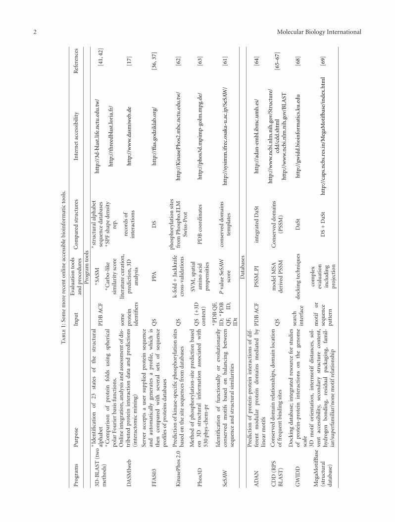
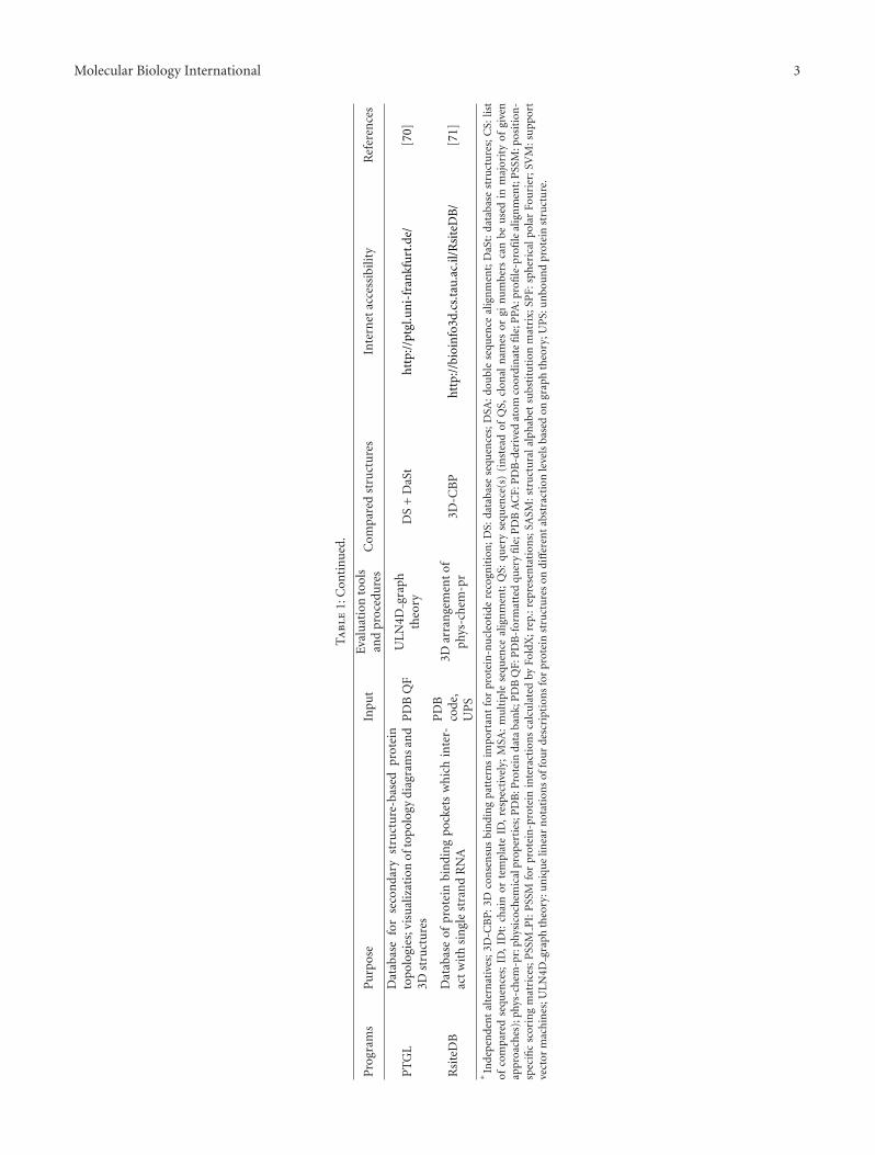
By using various structurally based programs, many pro-tein interactions can be predicted based on the occur-rence of phylogenetically conserved or convergently devel-oped functionally important common structural features(motifs, sequence patterns, consensi, constructs of conserveddomain sequences, supersecondary structures, supersec-ondary motifs, 3D-arranged structural patterns and pock-ets). However, diversification within protein families, andsuperfamilies causes losses of interacting structures or dis-ables their accessibility. On the other hand, new interactivepairs frequently appear in cases of disordered proteinregions, that is, peptide segments naturally occurring inmultiple conformation variants [19–21]. The attendantstability problems, as well as some additional problems withmolecular analysis, were diminished by the developmentof databases enabling reevaluation of selected structuralrelationships. The databases inform us about similar orcommon structural features, frequent locations of bindingsites in related domains, solvent accessibility, and locationof investigated segments in 3D structures of proteins (seeTable 1).
During the last twenty years, conventional sequence-based search for conserved structures frequently combineddifferent evaluations of sequence similarities. The corre-sponding protocols usually combined double sequence, andmultiple sequence comparisons like BLASTP, PSI-BLAST,RPS-BLAST, Clustal W and MUSCLE [22, 23]. In addition,highly selective PHI-BLAST or PSI-BLAST searches withspecifically restricted representative query sequences suchas consensi (and also sequence patterns) made it possibleto locate the corresponding potentially interacting sites
in extended sequence sets [23–26]. Except for currentconserved sequences, extremely variable but still definedstructures such as heptad repeats were investigated forpurposes of molecular topology [27, 28]. These repeats canbe written as an alphabet with generalized characters rep-resenting aa groups instead of individual aa (hydrophobic,charged, polar, etc.). Together with usual (motif-relatedsequence block derived) patterns an important part ofheptad repeats can be searched in database sequences andpartially reevaluated by means of PROSITE programs [29–32]. Apart from regions with highly conserved sequences,additional conserved structures were found when combiningevaluation of primary and secondary structures [33], PSI-BLAST and secondary structure [34], or when using foldrecognition [34, 35]. The compared sequence queries weremoreover evaluated on the sophisticated widely used FFAS03server (about 250 references) providing the third genera-tion of the profile-profile alignment and fold-recognitionalgorithm mediated by program FFAS (fold, and functionassignment system; [36, 37]; Table 1). The sensitivity FFAS-related profile-profile comparison is now widely recognizedand many Web servers implementing such algorithmsare available, for example, HHPRED, COMPASS, COMA,PHYRE, GenThreader, FORTE and webPRC [37]. Morerecent multiple sequence alignment program BCL::Alignincludes also combined evaluation of structural similarities[38] (for applications, see, e.g., [39, 40]). The correspondingscoring function is a weighted sum of scores derived basedon (i) the traditional PAM, and BLOSUM scoring matrices,(ii) position-specific scoring matrices by PSI-BLAST, (iii)secondary structure predicted by a variety of methods, (iv)chemical properties, and (v) gap penalties. Monte Carloalgorithm was then used to determine necessary optimizedweights in cases of sequence alignment and fold recognition.
Input of 3D coordinates or their transformed represen-tations was necessary for other structural studies predictingalso functional interaction sites. The corresponding researchyielded two servers with different 3D-BLAST programsenabling us to compare folds and fold families [41, 42] (fordetails see also Table 1). Alternative structural comparisonsubstituting 3D relationships was performed when searchingfor the maximum contact map overlaps [43]. The extendedcontact map comparison appeared very early [44]. Contactmap is in fact determined by the matrix of distances betweenindividual amino acids, contact threshold and specificationof contact types [45, 46]. This map can be visualizedby CMView software [46]. Similarly to sequence motifs,conserved patterns of 3D peptide arrangements (CP-3D-A)were considered as an additional type of structure-functionmotifs. These structures were recorded by sequence inde-pendent 3D-templates and can be searched on EvolutionaryTrace Annotation server [47, 48]. Frequent functionallyimportant CP-3D-A occurred, for instance, in active sites ofproteins containing porphyrin rings including members ofcytochrome P450 superfamily [49]. Another recent interest-ing approach consisted in generalized motif search in largealphabet (cumulative occurrence of several alphabets in thiscase) inputs including simultaneously evaluation of DNAsequence, protein sequence and supersecondary structure
Molecular Biology International 5
motifs [50]. According to the authors, large alphabets areimportant in cases when structures share little similarity atprimary level.
Similarly to multiple sequence alignment, various formsof multiple structural alignments (MSTA) have been gen-erated. Older attempts at MSTA were based on secondarystructural alignment [51–53]. On the other hand, recentMSTA approaches determined common spatial (3D) struc-tures. These approaches include new strategies employing“molecular sieving” of protein structures, minimization of anenergy function over the low-dimensional space of the rela-tive rotations, and translations of the molecules, geometrichashing and contact-window-derived motif library [54–58].
In spite of the increasing possibility of database reeval-uation, some structurally based programs predicting inter-acting sites became at least relatively autonomous. Alarge number of specific and relatively autonomous pro-grams concerned the prediction of epitopes (for detailssee Section 3). Similarly, various possibilities of predictionof phosphorylation sites were frequently investigated (twoexamples in Table 1), since phosphorylation reactions rep-resent an important signaling and regulatory network incell biology and pathobiology. The interest followed alsofrom the extended building of databases related to phos-phorylation sites, which brought many interesting insights.For instance, in accordance with the linear motif atlasfor phosphorylation-dependent signaling, tyrosine kinasesmutated in cancer exhibited lower specificity than their non-oncogenic relatives [59]. Similarly, collection and motif-based prediction of phosphorylation sites in human virusproteins suggested a substantial role for human kinases inregulation/mediation of viral protein functions [60]. In con-trast to programs predicting specific types of interacting sites,SeSAW represents an example of general online accessibleprogram allowing prediction of possible functionally impor-tant structures [61] (see also Table 1). The correspondingbalance between different combined structural evaluationsconcerned, among others, data present in position specificscoring matrix (PSSM), template-derived PSSM and tem-plate functional annotations.
3. Mimotope and Epitope Interactions
Epitopes are defined as the structures responsible for inter-action of antigens with binding sites of antigen receptors.On the other hand, mimotopes belong to artificially pre-pared peptides, which interact with natural templates (mostlikely proteins), and thus mimic other peptides or organiccompounds in their functionally important interactions.Mimotope development is based on synthetic peptide orphage display libraries, whereas natural development ofspecific cell clones is necessary for epitope recognition byspecific antigen receptors. This means that both epitopesand mimotopes can sometimes considerably differ from theusual conserved structures mentioned above. In spite of thedescribed difference, a unifying point between mimotopesand epitopes exists, because mimotopes were originallydefined as peptides mimicking epitopes [72], forming thus
only a subset of the later current mimotope repertoire.In addition to this historical linkage, rapidly developing(diverging) structures such as molecular mimicry enablingparasitic attack and adaptation of pathogenic viruses andbacteria, disordered regions of proteins, and protein loopsappear to be good candidates for extended investigation ofmimotope similarities (cf. [21, 73–75]) and mimotope basedprediction (see below).
Mimotopes were originally derived in studies with aphage display library. This phage technology was discoveredin the eighties [76]. The corresponding boom in the ninetiesthen comprised novel random, partially randomized or gene-fragment-derived oligopeptide libraries able to functionallymimic epitopes, autoepitopes, short peptide ligands, proteinkinase or proteinase substrates, as well as peptides mimickingorganic substances such as biotin when interacting withsteptavidin [77–85]. Mimotope similarities were frequentlydefined using sequence patterns, whereas additional typesof nonsequence structural similarities were also described(see, e.g., [81, 83]). More recent biotechnological research ofmimotopes yielded potential peptide drugs [86–89], peptidevaccines [90–92], and peptides suitable for specific (mostlynanoparticle mediated) drug delivery to tumor cells, brain,atherosclerotic plaques, and other therapeutically importantsites of human or animal organisms [93–97]. In spite ofthis considerable progress, virtual interactomic tools arenot still able to compare or predict organic drugs basedon their effective spatial similarity with functionally activemimotopes.
Database registration and authentication of mimotopeshave been performed for more than ten years [98–100].Special programs comparing primary structures of epitopeswere simultaneously developed (programs FINDMAP, andEPIMAP [101, 102]). Some of them employed also multiple-sequence alignment evaluation (program MIMOP [103]).Similarly, coexisting peptide databases were established toprocess the accumulated information. These databases (i)recognized sequence subsets classified after in vitro evolutionof phage display libraries, (ii) offered many integratingprograms, and (iii) made it possible to find all mimotopesets that have the 3D structure of a target-template complex(databases ASPD, RELIC and MimoDB [98, 99, 104, 105]). Inaddition, novel mimotope-assisted computer-aided epitopeprediction was discovered. This prediction came from boththe 3D structure of an interacting partner and sequencesof similarly interacting mimotopes, and has also concernedsome interacting partners different from specific antigenreceptors and antigens (programs PepSurf, Pep-3D-Search,and MimoPro [106–108]). Based on this approach, improvedspecificity, and extended the repertoire of predicted epitopesor other interacting partners were achieved. Further progressin programming then resulted in accelerated computationin spite of more complicated, and precise strategies of dataprocessing. In addition, pattern recognition algorithm wasdeveloped, which can effectively be employed to screen amixture of antibodies, and define the breadth of epitopesrecognized by polyserum directed against specific proteins[109]. This possibility appears to be interesting with respectto future vaccine design.
6 Molecular Biology International
The recent status of epitope prediction is still far fromresolved due to insufficient extent of the datasets, and stillrequires continuous improvement of database organization[110, 111]. In such state, mimotope-assisted epitope predic-tion mentioned above and combined approaches representa certain improvement in the quality of epitope prediction.Online accessible combined approaches of B-cell epitopeprediction mostly evaluate 3D structures together withsolvent accessibility (programs CEP, ElliPro, PEPOP and3D alternative of Epitopia [112–115]; Table 2). Similarlyto many other combined approaches, combined restrictiondiminishes the number of false positivities but simultane-ously can cause increased number of false negativities. Forinstance, the widely used requirement of solvent accessibilitymentioned above would in fact eliminate at least part ofconserved autoepitopes, which contain hydrophobic patterns(cf. [116–118]). To diminish losses following from employedcombinations or effects of too strict (sure) thresholds, ametaserver was developed that sums up the results fromsix epitope-predicting servers [119]. In addition, some newtypes of online accessible prediction of linear epitopesappeared to complete the preceding results [120, 121].
An interesting input simplification has been achievedwith a novel web server CBTOBE. This server uses learning ofsupport vector machines based on physicochemical profiles,and makes it possible to predict conformation epitopes basedon a sole sequence input [122]. A recent private alternativeof CBTOBE was based on older evaluation of secondarystructure and solvent accessibility (server COBEpro [123])further complemented by evolutionary information andmachine-learning-derived evaluation [124].
Combined predictions of epitopes presented to T-cellreceptors have integrated class I MHC (major histocom-patibility complex) peptide binding affinity, TAP transportefficiency and prediction of proteasome cleavage (programsEpiJen, NetCTL-1.2, FRED [125–128]). Nevertheless, inde-pendent simulations of peptide-binding affinity of variousMHC molecules have been also proposed [129] as wellas the corresponding neural network-based learning [130].Both combined, and sole approaches then represent startingsteps for further reevaluation with respect to T-cell receptorinteractions, for example, using learning of support vectormachines, and strict kernels based on 531 physicochemicalproperties (POPISK [131]). Important information aboutexisting T-cell, and B-cell epitopes can be also obtained fromthe Immune Epitope Database (IEDB [132, 133]). Amongothers, EpitopeViewer of the 3D structural subcomponentof IEDB (IEDB-3D) allows the user to visualize, render andanalyze the structure, and save structural and contact viewsas high-quality pictures for publication [133].
Since production of various vaccines, and detectionkits with mononoclonal antibodies requires high efficiencyof preparations, special searches for conserved epitopeshave been developed for this purpose. Though the mean-ing of crucial term “conserved epitope” exceeds pri-mary structure relationships, the repertoires of the corre-sponding structures appear to be sometimes considerablylimited. In fact, structural or regulatory adaptations ofpathogenic microogranisms and viruses cause less stability of
immunologically important, and promising promiscuous(cytotoxic or helper) T-cell epitopes broadly cross-reactingwith different MHC antigens, different frequencies of T-cell epitopes specific usually only for certain unique MHCmolecule, as well as losses of immunogenicity or evenabsence of immune response. In addition to currentevaluation, drug effects (cf. effects in docking studiesdescribed below) and accompanying structural variability(e.g., enzyme polymorphisms or special pathogeneticaleffects) have to be considered with respect to possible peptideepitope modifications, possible bias, or improvement oftherapeutical design. It is a question whether conservedspatial structures following from multiple structural align-ments mentioned above can be also interesting for predictionof conserved conformation of B-cell epitopes. The secondpart of Table 2 contains some web servers interesting withrespect to prediction of conserved epitopes, whereas selectedexamples related to AIDS research follow.
The first more complex prediction of HIV epitopes wasbased on sequence conservativeness, secondary structure,solvent accessibility, hydrophilicity and flexibility [134].The study pointed to an unfavorably frequent occurrenceof changes in secondary structures predicted as antigenic.Certain progress in the research of conserved HIV-1 epitopeswas achieved when assessing their possible interactions withMHC antigens. This comprised peptide prediction based onEpiMatrix score [135], construction of the peptide propertymodel from a training dataset [136, 137] and combined T-cell epitope evaluation mentioned above [126, 127]. Lately,phylogenetic hidden Markov models allowed to predict HIV-1-related T-cell epitopes based on contiguous aa positionsthat evolve under immune pressure dependent among otherson host HLA alleles [138]. In a recent paper, structure-function analysis based on a specifically devised mathe-matical model revealed that protection from neutralization(shielding of neutralization-sensitive domains) is enforcedby intersubunit contact between the variable loops 1 and 2(gp120 V1V2) of HIV-1, and domains of neighboring gp120subunits in the trimer encompassing the V3 loop [139].
4. Protein Docking
Molecular docking is a method, which predicts the preferredreciprocal orientation of two molecules when they boundto each other to form a stable complex [147]. In case ofsimulated protein interactions, the authors currently speakabout protein docking rather than about molecular docking,since molecular docking represents a term comprisingalso nucleic acid interactions with nonpeptide molecules.Formerly, molecular docking simulated “lock-and-key” typeof protein interactions. Its original variants appeared in theeighties and reassumed interpretations of older moleculargraphics [148–152]. These approaches were restricted bycomplementarity demands or simplified requirements forenergy minimization. Lately, “hand-in-glove” analogy wasfound to be more appropriate for molecular docking than the“lock-and-key” one [153]. In addition to this conventionalmodel, three new models have been recently developed for
Molecular Biology International 7
Ta
ble
2:E
pito
pe
pred
icti
onor
reev
alu
atio
non
acce
ssib
lese
rver
s.
Pro
gram
sP
urp
ose
Tool
sor
proc
edu
res
ofev
alu
atio
nIn
put
Ou
tpu
tIn
tern
etac
cess
ibili
tyR
efer
ence
s
Epi
top
epr
edic
tion
Mim
oDB
2.0
Mim
otop
eda
taba
seM
ySQ
Lre
lati
onal
data
base
acco
rdin
gto
men
uSt
ruct
ure
svi
sual
izat
ion
,al
ign
men
ts,a
nd
sofo
rth
htt
p://
imm
un
et.c
n/m
imod
b/[1
05]
Mim
oPro
Map
sa
grou
pof
mim
otop
esba
ckto
aso
urc
ean
tige
nso
asto
loca
teth
ein
tera
ctin
gep
itop
eon
the
anti
gen
Bra
nch
and
bou
nd
opti
miz
atio
n(a
nal
-ys
isof
over
lapp
ing
patc
hes
onth
esu
r-fa
ceof
apr
otei
n)
PD
Bid
enti
fier
mim
otop
ese
quen
ces
Scor
e+
3Dlo
cati
onin
anti
gen
htt
p://
info
rmat
ics.
nen
u.e
du.c
n/M
imoP
ro/
[108
]
ViP
RV
iru
spa
thog
enda
taba
seIn
tegr
atio
nof
vari
-ou
sre
sou
rces
acco
rdin
gto
men
uSt
ruct
ure
s,an
not
atio
ns,
and
sofo
rth
htt
p://
ww
w.v
iprb
rc.o
rg/
[140
]
Met
aMH
CP
redi
ctio
nof
MH
Cbi
ndi
ng
epit
opes
met
a-ap
proa
chQ
SFo
ur
met
apre
dict
orsc
ores
incl
udi
ng
Met
aSV
Mp
scor
eh
ttp:
//w
ww
.bio
kdd.
fuda
n.e
du.c
n/S
ervi
ce/M
etaM
HC
.htm
l[1
41]
Epi
topi
aP
redi
ctio
nof
B-c
elle
pito
pes
∗ MLA
∗ MLA
+3D
+SA
∗ QS
∗ PD
BA
CF
Imm
un
ogen
icit
ysc
ore
+pr
obab
ility
scor
e+
colo
rsc
ale
reco
rdon
QS
htt
p://
epit
opia
.tau
.ac.
il[1
15]
Elli
Pro
New
stru
ctu
re-b
ased
tool
for
the
pred
icti
onof
anti
body
epit
opes
3D+
SA+
flx
+an
tige
nic
ity
∗ QS
∗ PD
BA
CF
Scor
e+
visu
aliz
edep
itop
e3D
stru
ctu
rean
d3D
loca
tion
htt
p://
tool
s.im
mu
nee
pito
pe.
org/
tool
s/E
lliP
ro[1
14]
MH
CP
red
Pre
dict
ion
ofcl
ass
IIm
ouse
MH
Cp
epti
debi
ndi
ng
affin
ity
ISC
-PL
S,SY
BY
Lso
ftw
are
pack
age
QS
Bin
din
gaffi
nit
y(p
IC50
)h
ttp:
//w
ww
.jen
ner
.ac.
uk/
MH
CP
red
[129
]
Stab
leep
itop
esan
dva
ccin
es
Bay
esB
SVM
-bas
edpr
edic
tion
oflin
ear
B-c
elle
pito
pes
usi
ng
Bay
esFe
atu
reE
xtra
ctio
n
Res
idu
eco
nse
rvat
ion
+po
siti
on-s
peci
fic
resi
due
prop
ensi
ties
QS
Res
idu
eep
itop
epr
open
sity
scor
eh
ttp:
//w
ww
.imm
un
opre
d.or
g/ba
yesb
/in
dex.
htm
l[1
20]
Opt
iTop
eSe
lect
ion
ofop
tim
alpe
ptid
esfo
rep
itop
e-ba
sed
vacc
ines
Mu
ltis
tep
para
llel
eval
uat
ion
MSA
ofA
glis
tof
ES
tabl
e:E
S,E
Sre
late
dM
HC
Frac
tion
ofov
eral
lim
mu
nog
enic
ity
cove
red
MH
Ch
ttp:
//w
ww
.epi
tool
kit.
org/
opti
top
e[1
42]
PV
S
PV
Sre
turn
sa
vari
abili
ty-m
aske
dse
quen
ce,
wh
ich
can
besu
bmit
ted
toth
eR
AN
KP
EP
serv
erto
pred
ict
con
serv
edT
-cel
lep
itop
es.
3Dvi
sual
izat
ion
ofM
SAde
rive
dse
quen
ceva
riab
ility
“per
site
”
PD
BA
CF
3Dm
apof
vari
abili
tyfr
agm
ents
wit
hn
ova
riab
lere
sidu
esan
dth
eir
3Dlo
cati
onin
anti
gen
htt
p://
imed
.med
.ucm
.es/
PV
S/[1
43]
8 Molecular Biology International
Ta
ble
2:C
onti
nu
ed.
Pro
gram
sP
urp
ose
Tool
sor
proc
edu
res
ofev
alu
atio
nIn
put
Ou
tpu
tIn
tern
etac
cess
ibili
tyR
efer
ence
s
PE
PV
AC
Pre
dict
ion
ofM
HC
I;se
rver
can
also
iden
tify
con
serv
edan
dpr
omis
cuou
sM
HC
Ilig
ands
PSS
M,
dist
ance
mat
rix,
phyl
ogen
iccl
ust
erin
gal
gori
thm
Gen
ome,
HLA
-su
per
typ
esSe
lect
edpe
ptid
ese
quen
ces
scor
eh
ttp:
//im
mu
nax
.dfc
i.har
vard
.edu
/PE
PV
AC
/[1
44]
RA
NK
PE
PP
redi
ctio
nof
MH
CI
and
MH
CII
ligan
dsP
rofi
leco
mpa
riso
nG
enom
e,H
LA-
sup
erty
pes
,Q
S
Sele
cted
pept
ide
sequ
ence
ssc
ore
htt
p://
ww
w.m
ifou
nda
tion
.org
/Too
ls/r
ankp
ep.h
tml
[145
,146
]
∗In
depe
nde
nta
lter
nat
ives
;Ag:
anti
gen
;BFE
:Bay
esFe
atu
reE
xtra
ctio
n;fl
x:fl
exib
ility
;ES:
epit
ope
sequ
ence
s;H
LA:h
um
anle
uko
cyte
anti
gen
s(h
um
anM
HC
);IS
C-P
LS:i
tera
tive
self
-con
sist
entp
arti
al-l
east
-squ
ares
base
dad
diti
vem
eth
od;M
HC
:alle
les
ofm
ajor
his
toco
mpa
tibi
lity
anti
gen
s;M
LA
:mac
hin
ele
arn
ing
algo
rith
m;M
SA:m
ult
iple
sequ
ence
alig
nm
ent;
PD
B:p
rote
inda
taba
nk;
PD
BA
CF:
PD
B-d
eriv
edat
omco
ordi
nat
efi
le;Q
S:qu
ery
sequ
ence
(s);
SA:s
olve
nt
acce
ssib
ility
;SV
M:s
upp
ort
vect
orm
ach
ines
.
Molecular Biology International 9
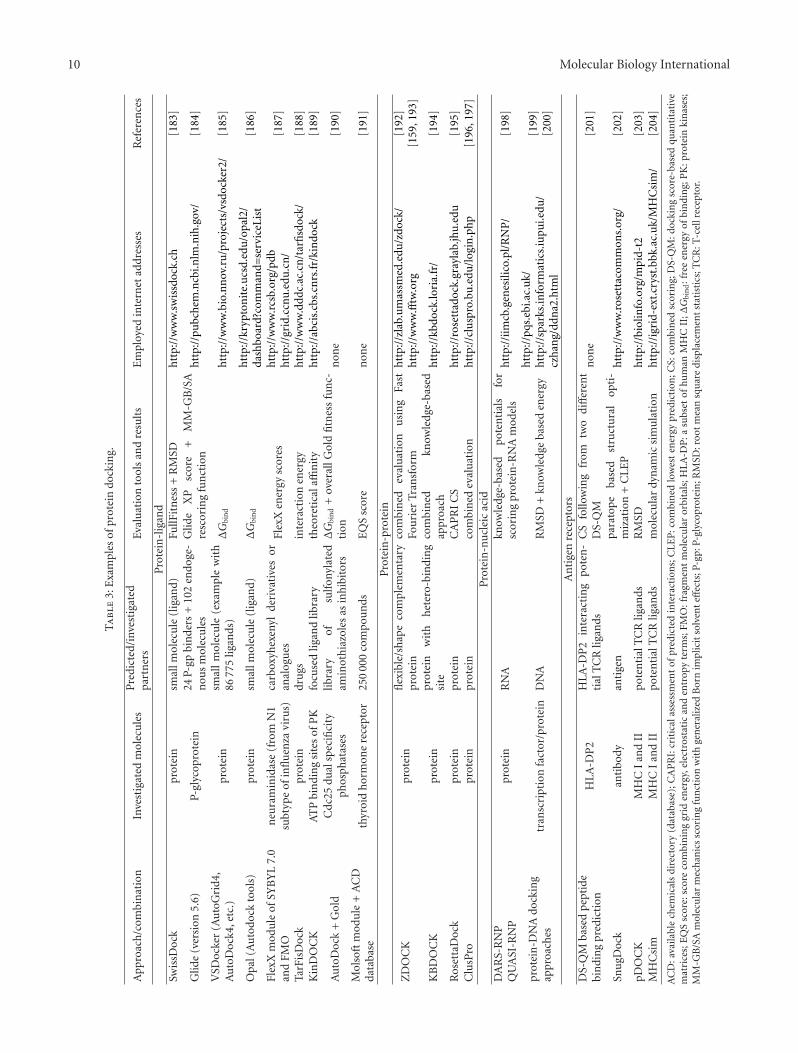
docking studies of protein-protein interactions [154], thatis, (i) conformer selection model, using a novel ensem-ble docking algorithm, (ii) induced fit model, employingenergy-gradient-based backbone minimization, and (iii)combined conformer selection/induced fit model. Physics-based molecular mechanics allowed the development ofthe force fields, which enabled the assessment of rela-tive binding strength [155]. Alternative knowledge-basedapproaches were evolved to derive a statistical potentialfor interactions from a large database of protein-ligandcomplexes [156]. Affinity evaluation, statistical methodsand combined procedures represent frequent alternatives ofevaluation in recent protein docking approaches (Table 3).In addition to usual full 3D models simulating molecularcomplexes, contact docking was also proposed. This dockingis based on contact map representations of molecules(cf. chapter 3). According to the authors contact dockingappears feasible, and is able to complement other com-putational methods for the prediction of protein-proteininteractions [157].
The number of molecules whose interactions can bescanned by current protein docking depends usually onthe complexity of evaluated interactions and manner ofsimulation. For instance, interaction of 142 drugs inhibitingPoly-(ADP-ribose)-polymerase was tested in a large-scalevirtual docking screening together with 300 000 addedorganic compounds with the aid of the program Lead Finder[158], whereas only 176 protein-protein interactions wereanalyzed by efficient program ZDOCK during extendedsimulation [159]. In addition to current output, somedocking studies even verified the results of simulation usingin silico experiments. For instance, aa involvement in enzymeinteractions was proved with simulated mutation (programSYBYL 6.7 and Internal Coordinate Mechanics method [160,161]) whereas other authors compared separate simulatedeffects of inhibitors and substrates (program BioDock [162]).
In addition to current molecular docking simulations(Table 3), certain docking studies analyzed modulationeffects of additional molecules on the evaluated interac-tion [163–165]. Homology modeling followed by extensivemolecular dynamics simulation was used to identify nonpep-tidic small organic compounds (among 150 000 compounds)that bind to a human leukocyte antigen HLA-DR1301,and block the presentation of myelin basic protein peptide(aa positions 152–165) to T-cells. This peptide representsone of the epitopes critical for multiple sclerosis. In silicoselection resulted in a set of 106 small molecules, twolead compounds were confirmed to specifically block IL-2secretion by DR1301-restricted T-cells in a dose-dependentand reversible manner [163].
Pockets opening to protein surface represents potentialsites for ligand binding or protein-protein interactions thatwere indeed identified in some cases [166–168]. Conse-quently, an algorithm BDOCK facilitating pocket-basedprediction of protein binding site was proposed to improveprotein docking [169], and moreover new algorithms pre-dicting pockets are still proposed [170]. Many pockets wereonly transiently present on simulated surfaces of investigatedproteins (e.g., Bcl-xl, interleukin 2, MDM2), and were all
opened only 2.5 picosecond when using model intervalscorresponding to ten nanosecond range [171].
Some docking studies comprised phylogenic aspectsof protein interactions. It was observed that except forantibody-antigen complexes [172], the surface density ofconserved residue positions at the interface regions ofinteracting protein surfaces is high. The correspondingcombination of the residue conservation information witha widely used shape complementarity algorithm improvedthe ability of protein docking to predict the native structureof protein-protein complexes. Efficient comparative dockingconsists in selection based on conservation in terms ofchain positions and primary structure in the first step,and the following high-throughput structure-based dockingevaluation [173] (see also Table 3). Recent combined strategyincluding multiple sequence alignment and fold recognitionanalysis has been proposed to perform more precise docking,and predict also protein function [174].
Bioinspired algorithms were also applied to molecu-lar docking simulations including neural networks, evolu-tionary computing and swarm intelligence [175]. Thoughneural networks participated in some older evaluationsworking in conventional programs of molecular docking[176–179], their independent scoring functions for dockinghave appeared lately [180, 181]. An extreme neural net-work approach required only sequence input to perform adocking-like procedure [182]. Predictions followed from atrained model that simulated binding or nonbinding stages.
5. Protein Interaction Networks
Protein interaction networks (PINs) are usually repre-sented by graphs. Nodes of these graphs denote interactingmolecules, whereas each edge linking two nodes indicatesthe corresponding interaction. The prevailing part of PIN isusually constituted by protein-protein interaction network(P-PIN). In addition to P-PIN, we can observe a recordof protein interactions with (i) nonpeptidic hormones ormediators and drugs, (ii) processed, targeted, and functionalcomplexes forming RNA, and (iii) recombining, hyper-mutating, repairing, replicating, twisting/untwisting, andtranscribed DNA. These nonpeptidic compounds representin fact inputs, outputs, interaction-stabilizing agents, orrelay batons of P-PIN. In cases of DNA, and RNA, somepapers appeared dealing with special protein-RNA, andprotein-DNA interaction networks [205–208]. Protein-DNAnetworks moreover combined both the protein-centric andthe DNA-centric points of view [205].
Like in other related networks, clusters are recognizedin P-PIN. Two main types of P-PIN-related clusters canbe distinguished, that is, protein complexes and functionalmodules [209, 210]. Protein complexes are groups of proteinsthat interact with each other at the same time and place,forming a single multimolecular machine (e.g., metabolicmultienzyme complexes). Functional modules consist ofproteins that participate in a particular cellular processwhile binding to each other at a different time and place(e.g., multiple signaling cascades are functional modules
10 Molecular Biology International
Ta
ble
3:E
xam
ples
ofpr
otei
ndo
ckin
g.
App
roac
h/c
ombi
nat
ion
Inve
stig
ated
mol
ecu
les
Pre
dict
ed/i
nves
tiga
ted
part
ner
sEv
alu
atio
nto
ols
and
resu
lts
Em
ploy
edin
tern
etad
dres
ses
Ref
eren
ces
Pro
tein
-lig
and
Swis
sDoc
kpr
otei
nsm
allm
olec
ule
(lig
and)
FullF
itn
ess
+R
MSD
htt
p://
ww
w.s
wis
sdoc
k.ch
[183
]
Glid
e(v
ersi
on5.
6)P-
glyc
opro
tein
24P-
gpbi
nde
rs+
102
endo
ge-
nou
sm
olec
ule
sG
lide
XP
scor
e+
MM
-GB
/SA
resc
orin
gfu
nct
ion
htt
p://
pubc
hem
.ncb
i.nlm
.nih
.gov
/[1
84]
VSD
ocke
r(A
uto
Gri
d4,
Au
toD
ock4
,etc
.)pr
otei
nsm
all
mol
ecu
le(e
xam
ple
wit
h86
775
ligan
ds)
ΔG
bin
dh
ttp:
//w
ww
.bio
.nn
ov.r
u/p
roje
cts/
vsdo
cker
2/[1
85]
Opa
l(A
uto
dock
tool
s)pr
otei
nsm
allm
olec
ule
(lig
and)
ΔG
bin
dh
ttp:
//kr
ypto
nit
e.u
csd.
edu
/opa
l2/
dash
boar
d?co
mm
and=
serv
iceL
ist
[186
]
Flex
Xm
odu
leof
SYB
YL
7.0
and
FMO
neu
ram
inid
ase
(fro
mN
1su
btyp
eof
infl
uen
zavi
rus)
carb
oxyh
exen
ylde
riva
tive
sor
anal
ogu
esFl
exX
ener
gysc
ores
htt
p://
ww
w.r
csb.
org/
pdb
htt
p://
grid
.ccn
u.e
du.c
n/
[187
]
TarF
isD
ock
prot
ein
dru
gsin
tera
ctio
nen
ergy
htt
p://
ww
w.d
ddc.
ac.c
n/t
arfi
sdoc
k/[1
88]
Kin
DO
CK
AT
Pbi
ndi
ng
site
sof
PK
focu
sed
ligan
dlib
rary
theo
reti
cala
ffin
ity
htt
p://
abci
s.cb
s.cn
rs.f
r/ki
ndo
ck[1
89]
Au
toD
ock
+G
old
Cdc
25du
alsp
ecifi
city
phos
phat
ases
libra
ryof
sulf
onyl
ated
amin
oth
iazo
les
asin
hib
itor
sΔG
bin
d+
over
allG
old
fitn
ess
fun
c-ti
onn
one
[190
]
Mol
soft
mod
ule
+A
CD
data
base
thyr
oid
hor
mon
ere
cept
or25
000
0co
mpo
un
dsE
QS
scor
en
one
[191
]
Pro
tein
-pro
tein
ZD
OC
Kpr
otei
nfl
exib
le/s
hap
eco
mpl
emen
tary
prot
ein
com
bin
edev
alu
atio
nu
sin
gFa
stFo
uri
erTr
ansf
orm
htt
p://
zlab
.um
assm
ed.e
du/z
dock
/h
ttp:
//w
ww
.fftw
.org
[192
][1
59,1
93]
KB
DO
CK
prot
ein
prot
ein
wit
hh
eter
o-bi
ndi
ng
site
com
bin
edkn
owle
dge-
base
dap
proa
chh
ttp:
//kb
dock
.lori
a.fr
/[1
94]
Ros
etta
Doc
kpr
otei
npr
otei
nC
AP
RI
CS
htt
p://
rose
ttad
ock.
gray
lab.
jhu
.edu
[195
]C
lusP
ropr
otei
npr
otei
nco
mbi
ned
eval
uat
ion
htt
p://
clu
spro
.bu
.edu
/log
in.p
hp
[196
,197
]P
rote
in-n
ucl
eic
acid
DA
RS-
RN
PQ
UA
SI-R
NP
prot
ein
RN
Akn
owle
dge-
base
dpo
ten
tial
sfo
rsc
orin
gpr
otei
n-R
NA
mod
els
htt
p://
iim
cb.g
enes
ilico
.pl/
RN
P/
[198
]
prot
ein
-DN
Ado
ckin
gap
proa
ches
tran
scri
ptio
nfa
ctor
/pro
tein
DN
AR
MSD
+kn
owle
dge
base
den
ergy
htt
p://
pqs.
ebi.a
c.u
k/h
ttp:
//sp
arks
.info
rmat
ics.
iupu
i.edu
/cz
han
g/dd
na2
.htm
l
[199
][2
00]
An
tige
nre
cept
ors
DS-
QM
base
dpe
ptid
ebi
ndi
ng
pred
icti
onH
LA-D
P2
HL
A-D
P2
inte
ract
ing
pote
n-
tial
TC
Rlig
ands
CS
follo
win
gfr
omtw
odi
ffer
ent
DS-
QM
non
e[2
01]
Snu
gDoc
kan
tibo
dyan
tige
npa
rato
peba
sed
stru
ctu
ral
opti
-m
izat
ion
+C
LEP
htt
p://
ww
w.r
oset
taco
mm
ons.
org/
[202
]
pDO
CK
MH
CI
and
IIpo
ten
tial
TC
Rlig
ands
RM
SDh
ttp:
//bi
olin
fo.o
rg/m
pid-
t2[2
03]
MH
Csi
mM
HC
Ian
dII
pote
nti
alT
CR
ligan
dsm
olec
ula
rdy
nam
icsi
mu
lati
onh
ttp:
//ig
rid-
ext.
crys
t.bb
k.ac
.uk/
MH
Csi
m/
[204
]
AC
D:a
vaila
ble
chem
ical
sdi
rect
ory
(dat
abas
e);C
AP
RI:
crit
ical
asse
ssm
ent
ofpr
edic
ted
inte
ract
ion
s;C
LE
P:c
ombi
ned
low
est
ener
gypr
edic
tion
;CS:
com
bin
edsc
orin
g;D
S-Q
M:d
ocki
ng
scor
e-ba
sed
quan
tita
tive
mat
rice
s;E
QS
scor
e:sc
ore
com
bin
ing
grid
ener
gy,e
lect
rost
atic
and
entr
opy
term
s;FM
O:f
ragm
ent
mol
ecu
lar
orbi
tals
;HLA
-DP
:asu
bset
ofhu
man
MH
CII
;ΔG
bin
d:f
ree
ener
gyof
bin
din
g;P
K:p
rote
inki
nas
es;
MM
-GB
/SA
mol
ecu
lar
mec
han
ics
scor
ing
fun
ctio
nw
ith
gen
eral
ized
Bor
nim
plic
itso
lven
teff
ects
;P-g
p:P-
glyc
opro
tein
;RM
SD:r
oot
mea
nsq
uar
edi
spla
cem
ent
stat
isti
cs;T
CR
:T-c
ellr
ecep
tor.
Molecular Biology International 11
sometimes including also protein complexes). An importanttype of functional modules, that is, responsive functionalmodules (RFMs), includes protein interactions activatedunder specific conditions. These RFMs appear to be interest-ing with respect to prediction of potential biomarkers [211].
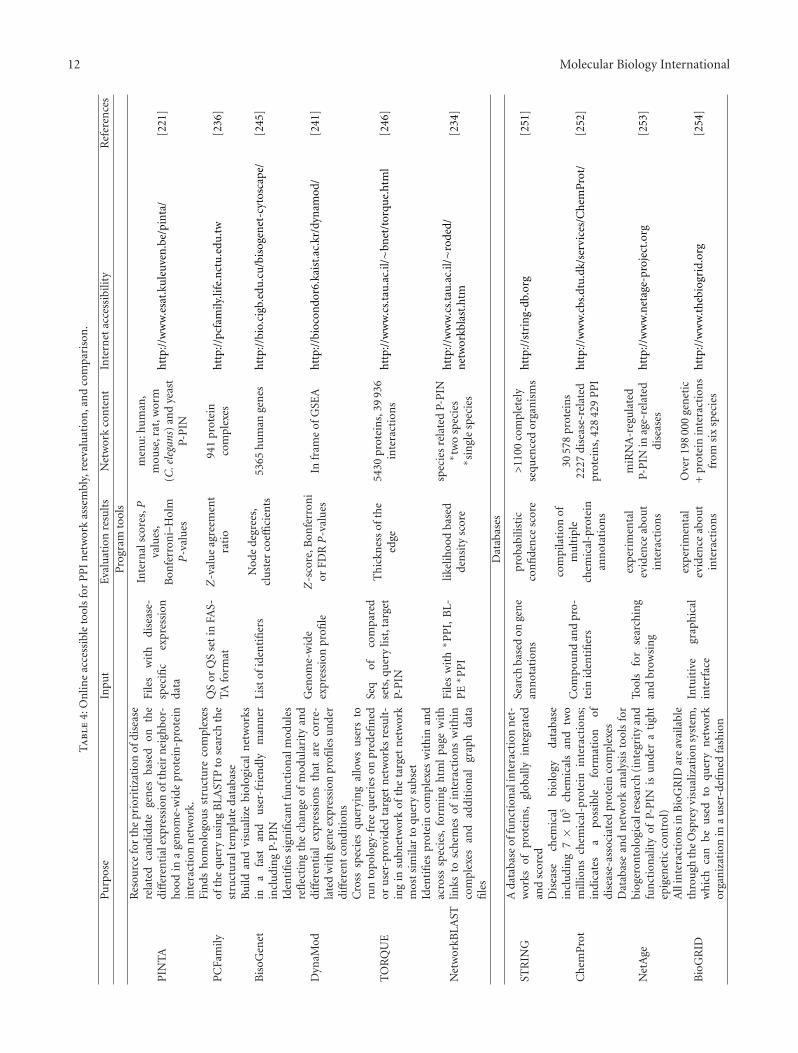
Much attention has been paid to the identification ofsmall conserved subgraphs, particularly those occurringsignificantly often within the biological networks. Theseconserved subgraphs were referred to as network motifs orsimply as motifs (similarly to primary structure motifs),and were considered as basic building blocks of complexnetworks including P-PIN [212, 213]. Ancestral pathways orfunctional modules including conserved interactions werederived when comparing P-PIN of phylogenetically distantspecies such as human, Saccharomyces cerevisiae, Drosophilamelanogaster and Caenorhabditis elegans, and various bac-terial species [214, 215]. Proteins necessary for proteasomefunction, transcription, RNA processing and translationwere frequent in conserved subgraphs of compared Eukary-ota, whereas DNA repair proteins prevailed in conservedprokaryotic subgraphs. Incorporation of literature-curatedand evolutionarily conserved interactions also allowed todevelop an interaction network for 54 proteins providing atool for understanding Purkinje cell degeneration [216]. Thisresult made it possible to find experimentally 770 mostlynovel protein-protein interactions using a stringent yeasttwo-hybrid screen.
Recent integrative approaches combined at least tran-scriptional data with P-PIN analyses, and at most five lay-ers including phenotype association with single-nucleotidepolymorphism, disease-tissue, drug-tissue and drug-generelationships, in addition to the topical P-PIN record [217](see also Table 4). Two-layer comparison of disease-relatedmRNA expression, and human P-PIN was for instance usedtogether with hierarchical clustering of networks to elucidatehuman disease similarities. The results led to a hypothesisconsidering common usage of some drugs in the diseases,which exhibited close relationship with respect to givenclustering [218]. More extended analysis of gene expressionoverlays with protein-protein interaction, transcriptional-regulatory and signaling networks identified distinct driver-networks for each of the three common clinical breast cancersubtypes, that is, oestrogen receptor positive, human epider-mal growth factor receptor 2 positive and triple receptor-negative breast cancers [219]. Integrative online accessibletools reevaluating P-PIN relationships were developed topredict candidate genes critical for the occurrence of differentdiseases (cf. network-based disease gene prioritization; [220,221]).
The majority of proteins reciprocally interact via oneor two interactions. This number substantially increasesto tens, hundreds and more, when considering multi-interactive proteins denoted in PIN as hubs [222]. Inagreement with the sense of the term, hubs are the principalagents in the interaction network and affect its functionand stability [223]. Hubs were enriched in kinase andadaptor proteins including those interacting with frequentlydisordered phosphorylated protein regions [224, 225]. Sim-ilarly, many pathogenetically important proteins belonged
to hubs, for example, the widely known tumor suppressorp53, alpha-synuclein involved in Parkinson disease and smallmultifunctional core protein necessary for orchestration ofviral progeny in Flaviviridae [226, 227]. In principle, twotypes of hubs were distinguished, that is, static “party hubs”and dynamic “date hubs.” Party hubs are markedly morephylogenetically conserved, and their expression is highlycorrelated with their interacting partners in contrast to lessconserved date hubs more frequently containing disorderedregions and participating in cell signaling [228–230]. Hubswith two or more domains are more likely to connectdistinct functional modules than single-domain hubs [224].In addition to studies of biochemically interesting hubs,docking procedures (mediated by program ClusPro [231])were recently employed together with comparative modelingto construct P-PIN [232], whereas domain-domain, domain-motif and motif-motif interactions were distinguished insome other P-PIN [233].
Extended P-PIN research yielded many tools allowing P-PIN-based prediction of protein complex formation [234–237], identification of hubs and multifunctional proteins[238, 239], identification of functional modules [240–242],network-based disease gene prioritization [217, 221, 243], aswell as network building [244, 245], cross-species querying[246] or comparison [234, 237]. Some of these approachesas well as several examples of P-PIN-related databases aredescribed in Table 4. The instrumental progress in PINresearch also led to an increased number of the correspond-ing methodological reviews, for example, [247–250].
6. Accuracy as an Important Parameter
A detailed evaluation of accuracy of all the above approacheswould require a separate review. The obstacles consist firstof all in presence of complementary information in articlesinaccessible for biologists and on web pages. Such statecomplicates mining of accuracy data first of all in thecase, when server-related papers represent only the finalstep of author’s efforts. Accuracy evaluation or even thecorresponding references are also rarely present in the papersconcerning new online databases. In addition, some authorsalso use other criterions related to accuracy to evaluatethe corresponding performance value, whereas only someof such evaluations are widely known as valid accuracysubstitution, for example, AUC (area under the ROC curve)discussed below.
In spite of the obstacles, we can mention several examplesof high accuracy concerning the above approaches. Excellentaccuracy values higher then 0.90 were found in the casesof Conserved Domain Database (or its RPS BLAST server;[257]; Table 1) and older 3D-BLAST variant under a broadrange of conditions (Table 1), whereas similarly interestingAUC values (higher than 0.90) were mostly found also inthe cases of recent 3D BLAST variant (Table 1), and theselected strategy of SVM- and machine-learning-based epi-tope prediction known as CBTOBE (chapter 3; [122]). Thesame accuracy levels were rarely achieved in certain subsetswhen employing ADAN, KinasePhos 2.0 (Table 1), ElliPro,
12 Molecular Biology International
Ta
ble
4:O
nlin
eac
cess
ible
tool
sfo
rP
PI
net
wor
kas
sem
bly,
reev
alu
atio
n,a
nd
com
pari
son
.
Pu
rpos
eIn
put
Eval
uat
ion
resu
lts
Net
wor
kco
nte
nt
Inte
rnet
acce
ssib
ility
Ref
eren
ces
Pro
gram
tool
s
PIN
TA
Res
ourc
efo
rth
epr
iori
tiza
tion
ofdi
seas
ere
late
dca
ndi
date
gen
esba
sed
onth
edi
ffer
enti
alex
pres
sion
ofth
eir
nei
ghbo
r-h
ood
ina
gen
ome-
wid
epr
otei
n-p
rote
inin
tera
ctio
nn
etw
ork.
File
sw
ith
dise
ase-
spec
ific
expr
essi
onda
ta
Inte
rnal
scor
es,P
valu
es,
Bon
ferr
oni–
Hol
mP
-val
ues
men
u:h
um
an,
mou
se,r
at,w
orm
(C.e
lega
ns)
and
yeas
tP-
PIN
htt
p://
ww
w.e
sat.
kule
uven
.be/
pin
ta/
[221
]
PC
Fam
ilyFi
nds
hom
olog
ous
stru
ctu
reco
mpl
exes
ofth
equ
ery
usi
ng
BLA
STP
tose
arch
the
stru
ctu
ralt
empl
ate
data
base
QS
orQ
Sse
tin
FAS-
TAfo
rmat
Z-v
alu
eag
reem
ent
rati
o94
1pr
otei
nco
mpl
exes
htt
p://
pcfa
mily
.life
.nct
u.e
du.t
w[2
36]
Bis
oGen
etB
uild
and
visu
aliz
ebi
olog
ical
net
wor
ksin
afa
stan
du
ser-
frie
ndl
ym
ann
erin
clu
din
gP-
PIN
List
ofid
enti
fier
sN
ode
degr
ees,
clu
ster
coeffi
cien
ts53
65hu
man
gen
esh
ttp:
//bi
o.ci
gb.e
du.c
u/b
isog
enet
-cyt
osca
pe/
[245
]
Dyn
aMod
Iden
tifi
essi
gnifi
can
tfu
nct
ion
alm
odu
les
refl
ecti
ng
the
chan
geof
mod
ula
rity
and
diff
eren
tial
expr
essi
ons
that
are
corr
e-la
ted
wit
hge
ne
expr
essi
onpr
ofile
su
nde
rdi
ffer
ent
con
diti
ons
Gen
ome-
wid
eex
pres
sion
profi
leZ
-sco
re,B
onfe
rron
ior
FDRP
-val
ues
Infr
ame
ofG
SEA
htt
p://
bioc
ondo
r6.k
aist
.ac.
kr/d
ynam
od/
[241
]
TO
RQ
UE
Cro
sssp
ecie
squ
eryi
ng
allo
ws
use
rsto
run
topo
logy
-fre
equ
erie
son
pred
efin
edor
use
r-pr
ovid
edta
rget
net
wor
ksre
sult
-in
gin
subn
etw
ork
ofth
eta
rget
net
wor
km
ost
sim
ilar
toqu
ery
subs
et
Seq
ofco
mpa
red
sets
,qu
ery
list,
targ
etP-
PIN
Th
ickn
ess
ofth
eed
ge54
30pr
otei
ns,
3993
6in
tera
ctio
ns
htt
p://
ww
w.c
s.ta
u.a
c.il/∼b
net
/tor
que.
htm
l[2
46]
Net
wor
kBLA
ST
Iden
tifi
espr
otei
nco
mpl
exes
wit
hin
and
acro
sssp
ecie
s,fo
rmin
gh
tml
page
wit
hlin
ksto
sch
emes
ofin
tera
ctio
ns
wit
hin
com
plex
esan
dad
diti
onal
grap
hda
tafi
les
File
sw
ith∗ P
PI,
BL-
PE∗ P
PI
likel
ihoo
dba
sed
den
sity
scor
e
spec
ies
rela
ted
P-P
IN∗ t
wo
spec
ies
∗ sin
gle
spec
ies
htt
p://
ww
w.c
s.ta
u.a
c.il/∼r
oded
/n
etw
orkb
last
.htm
[234
]
Dat
abas
es
STR
ING
Ada
taba
seof
fun
ctio
nal
inte
ract
ion
net
-w
orks
ofpr
otei
ns,
glob
ally
inte
grat
edan
dsc
ored
Sear
chba
sed
onge
ne
ann
otat
ion
spr
obab
ilist
icco
nfi
den
cesc
ore
>11
00co
mpl
etel
yse
quen
ced
orga
nis
ms
htt
p://
stri
ng-
db.o
rg[2
51]
Ch
emP
rot
Dis
ease
chem
ical
biol
ogy
data
base
incl
udi
ng
7×
105
chem
ical
san
dtw
om
illio
ns
chem
ical
-pro
tein
inte
ract
ion
s;in
dica
tes
apo
ssib
lefo
rmat
ion
ofdi
seas
e-as
soci
ated
prot
ein
com
plex
es
Com
pou
nd
and
pro-
tein
iden
tifi
ers
com
pila
tion
ofm
ult
iple
chem
ical
-pro
tein
ann
otat
ion
s
3057
8pr
otei
ns
2227
dise
ase-
rela
ted
prot
ein
s,42
842
9P
PI
htt
p://
ww
w.c
bs.d
tu.d
k/se
rvic
es/C
hem
Pro
t/[2
52]
Net
Age
Dat
abas
ean
dn
etw
ork
anal
ysis
tool
sfo
rbi
oger
onto
logi
calr
esea
rch
(in
tegr
ity
and
fun
ctio
nal
ity
ofP-
PIN
isu
nde
ra
tigh
tep
igen
etic
con
trol
)
Tool
sfo
rse
arch
ing
and
brow
sin
g
exp
erim
enta
lev
iden
ceab
out
inte
ract
ion
s
miR
NA
-reg
ula
ted
P-P
INin
age-
rela
ted
dise
ases
htt
p://
ww
w.n
etag
e-pr
ojec
t.or
g[2
53]
Bio
GR
ID
All
inte
ract
ion
sin
Bio
GR
IDar
eav
aila
ble
thro
ugh
the
Osp
rey
visu
aliz
atio
nsy
stem
,w
hic
hca
nbe
use
dto
quer
yn
etw
ork
orga
niz
atio
nin
au
ser-
defi
ned
fash
ion
Intu
itiv
egr
aph
ical
inte
rfac
e
exp
erim
enta
lev
iden
ceab
out
inte
ract
ion
s
Ove
r19
800
0ge
net
ic+
prot
ein
inte
ract
ion
sfr
omsi
xsp
ecie
sh
ttp:
//w
ww
.th
ebio
grid
.org
[254
]
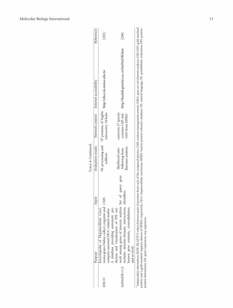
Molecular Biology International 13
Ta
ble
4:C
onti
nu
ed.
Pu
rpos
eIn
put
Eval
uat
ion
resu
lts
Net
wor
kco
nte
nt
Inte
rnet
acce
ssib
ility
Ref
eren
ces
EH
CO
En
cycl
oped
iaof
Hep
atoc
ellu
lar
Car
ci-
nom
age
nes
On
line
colle
ct,o
rgan
ize
and
com
pare
un
sort
edH
CC
-rel
ated
stu
dies
CM
SN
Lpr
oces
sin
gan
dso
ftbo
ts97
prot
ein
s,47
hig
hly
inte
ract
ive,
18hu
bsh
ttp:
//eh
co.ii
s.si
nic
a.ed
u.t
w[2
55]
IntN
etD
Bv1
.0
Ada
taba
sepr
ovid
ing
auto
mat
icpr
e-di
ctio
nan
dvi
sual
izat
ion
ofP
PI
net
-w
ork
amon
gge
nes
ofin
tere
st;
anal
ysis
incl
ude
sdo
mai
n-d
omai
nin
tera
ctio
ns,
know
nge
ne
con
text
s,cr
ossv
alid
atio
n,
and
sofo
rth
list
ofqu
ery
gen
eid
enti
fier
s
likel
ihoo
dra
tio
follo
win
gfr
omB
ayes
ian
anal
ysis
con
cern
s27
spec
ies
con
tain
sG
SPan
dG
SNfr
omH
PR
Dh
ttp:
//h
anla
b.ge
net
ics.
ac.c
n/I
ntN
etD
B.h
tm[2
56]
∗In
depe
nde
nta
lter
nat
ives
;BLP
E:B
LAST
PE
valu
esbe
twee
npa
irs
ofpr
otei
ns
from
each
ofth
eco
mpa
red
spec
ies;
CM
S:co
nte
ntm
anag
emen
tsys
tem
;GSE
A:g
ene
sete
nri
chm
enta
nal
yses
;GSP
,GSN
:gol
dst
anda
rdpo
siti
vean
da
gold
stan
dard
neg
ativ
eda
tase
tof
HP
RD
,res
pec
tive
ly;H
CC
:hep
atoc
ellu
lar
carc
inom
a;H
PR
D:h
um
anpr
otei
nre
fere
nce
data
base
;NL
:nat
ura
llan
guag
e;P
E:p
roba
bilis
tic
eval
uat
ion
;PP
I:pr
otei
n-
prot
ein
inte
ract
ion
s;Q
S:qu
ery
sequ
ence
s;se
q:se
quen
ces.
14 Molecular Biology International
RANKPEP (Table 2), SVM and machine-learning-basedepitope prediction improved by evolutionary informationBEOracle (chapter 3; [124]) and docking tool HADDOCK(chapter 4; [178]). Similarly, restricted subset-related AUCvalues of 0.90 and 0.93 indicated a good predicting abilitywhen using docking-like Partner-aware prediction (chapter4; [182]), and combined evaluation based on Glide X andmolecular mechanics (Table 3) to simulate interactions of 25antibodies and P-glycoprotein, respectively. Frequent subsetaccuracies of higher values than the gold standard valueof 0.80 were observed during runs of programs Phos3D,KinasePhos 2.0 (Table 1), ElliPro (Table 2) and CBTOBEmentioned above (chapter 3; [122]), and two docking toolsGlide XP and Gold (Table 2; [258]). Comparable AUC valuesthen concerned RANKPEP (Table 2), BEOracle mentionedabove (chapter 3; [124]) and network-based disease geneprioritization called DADA (chapter 5; [236]). The lowoccurrence of P-PIN in our overview of accuracy evaluationfollowed from the fact that the performance of P-PIN wasalternatively evaluated with the aid of precision or recall.An interesting approach—Lead Finder—was developed toimprove the accuracy of protein-ligand docking, bindingenergy estimation and virtual screening. High enrichmentfactors were obtained for almost all of the targets and sevendifferent docking programs resulting in an excellent averageAUC value of 0.92 [258].
7. Tendencies, Improvements, and Interplays
In summary, virtual interactomics represents a highlysophisticated, and relatively autonomous subarea of bothinteractomics and bioinformatics. Its development can becharacterized by several independent and integrative tenden-cies. The independent tendencies include (i) more rapid andprecise molecular modeling of protein complexes (includingquantum mechanic methods and rapid pharmacologicalscreening based on docking), (ii) more complex evaluationof structural similarities (e.g., large alphabets and combinedprediction of functional sites), (iii) extensive assembly oflarge-scale PIN and assembly of PIN or PIN subgraphs withdynamical properties, and (iv) restriction of conserved PINsubgraphs and clinically interesting responsive functionalmodules. The integrative tendencies then comprise (i)establishment of structurally functionally, and purposefullyoriented databases and associated data mining servers,(ii) questions concerning phylogenic relationships betweenstructural differences in PIN subgraphs, and the differencesin protein structures of subgraph forming molecules, (iii)PIN prediction and revision/reevaluation of empirically pro-posed PIN using comparative molecular modeling, dockingand bio-inspired algorithms, (iv) comparative differentialmultilayer network approaches including clinical treatmentcases, and (v) attempts to construct global networks withina single cell, and hopefully, within the system of differentreciprocally communicating cell types (e.g., immune sys-tem). Since interactomics simplifies reactomic simulations,it can also markedly and rapidly complete reactomic results,for example, in prediction of enzyme-inhibitor effects and
specificity of protein kinase interactions mentioned above. Inthe end, we also dealt here with accuracy, which represents animportant parameter of tool choice together with reasonabletool accessibility, and possibilities to test several alternativesbefore prediction, simulation and network analysis or touse two or several independent methods. In conclusion,many interesting and important interplays occur inside thevirtual interactomic area as well as in its boundary lines andperformance evaluation.
Abbreviations
3D: Three-dimensionalaa: Amino acid residues in protein sequencePIN: Protein interaction network(s)P-PIN: Protein-protein interaction network(s).
References
[1] A. Espejo, J. Cote, A. Bednarek, S. Richard, and M. T.Bedford, “A protein-domain microarray identifies novelprotein-protein interactions,” Biochemical Journal, vol. 367,no. 3, pp. 697–702, 2002.
[2] S. H. Lo, “Reverse interactomics: from peptides to proteinsand to functions,” ACS Chemical Biology, vol. 2, no. 2, pp.93–95, 2007.
[3] B. Suter, S. Kittanakom, and I. Stagljar, “Two-hybrid tech-nologies in proteomics research,” Current Opinion in Biotech-nology, vol. 19, no. 4, pp. 316–323, 2008.
[4] D. J. Briant, J. M. Murphy, G. C. Leung, and F. Sicheri, “Rapididentification of linear protein domain binding motifs usingpeptide SPOT arrays,” Methods in Molecular Biology, vol. 570,pp. 175–185, 2009.
[5] S. Hui and G. D. Bader, “Proteome scanning to predict PDZdomain interactions using support vector machines,” BMCBioinformatics, vol. 11, article 507, 2010.
[6] A. Daemen, M. Signoretto, O. Gevaert, J. A. K. Suykens, andB. De Moor, “Improved microarray-based decision supportwith graph encoded interactome data,” PLoS ONE, vol. 5, no.4, Article ID e10225, 2010.
[7] J. Huang, B. Ru, and P. Dai, “Bioinformatics resources andtools for phage display,” Molecules, vol. 16, no. 1, pp. 694–709, 2011.
[8] A. D. J. van Dijk, R. Kaptein, R. Boelens, and A. M. J. J.Bonvin, “Combining NMR relaxation with chemical shiftperturbation data to drive protein-protein docking,” Journalof Biomolecular NMR, vol. 34, no. 4, pp. 237–244, 2006.
[9] S. Klinge, R. Nez-Ramırez, O. Llorca, and L. Pellegrini, “3Darchitecture of DNA Pol α reveals the functional core ofmulti-subunit replicative polymerases,” The EMBO Journal,vol. 28, no. 13, pp. 1978–1987, 2009.
[10] J. L. Stark and R. Powers, “Application of NMR andmolecular docking in structure-based drug discovery,” Topicsin Current Chemistry, vol. 326, pp. 1–34, 2012.
[11] J. Orts, S. Bartoschek, C. Griesinger, P. Monecke, and T.Carlomagno, “An NMR-based scoring function improves theaccuracy of binding pose predictions by docking by twoorders of magnitude,” Journal of Biomolecular NMR, vol. 52,no. 1, pp. 23–30, 2012.
[12] D. J. Diller and R. Li, “Kinases, homology models, and highthroughput docking,” Journal of Medicinal Chemistry, vol. 46,no. 22, pp. 4638–4647, 2003.

Molecular Biology International 15
[13] F. Forster and E. Villa, “Integration of cryo-EM with atomicand protein-protein interaction data,” Methods in Enzymol-ogy, vol. 483, pp. 47–72, 2010.
[14] C. N. Cavasotto, “Homology models in docking and high-throughput docking,” Current Topics in Medicinal Chemistry,vol. 11, no. 12, pp. 1528–1534, 2011.
[15] Y. Tsfadia, R. Friedman, J. Kadmon, A. Selzer, E. Nachliel, andM. Gutman, “Molecular dynamics simulations of palmitateentry into the hydrophobic pocket of the fatty acid bindingprotein,” FEBS Letters, vol. 581, no. 6, pp. 1243–1247, 2007.
[16] M. Gochin, G. Zhou, and A. H. Phillips, “Paramagneticrelaxation assisted docking of a small indole compound inthe HIV-1 gp41 hydrophobic pocket,” ACS Chemical Biology,vol. 6, no. 3, pp. 267–274, 2011.
[17] H. Blankenburg, F. Ramırez, J. Buch, and M. Albrecht, “DAS-MIweb: online integration, analysis and assessment of dis-tributed protein interaction data,” Nucleic Acids Research, vol.37, no. 2, pp. W122–W128, 2009.
[18] M. J. Cowley, M. Pinese, K. S. Kassahn et al., “PINA v2.0:mining interactome modules,” Nucleic Acids Research, vol. 40,pp. D862–D865, 2011.
[19] M. Sickmeier, J. A. Hamilton, T. LeGall et al., “DisProt: thedatabase of disordered proteins,” Nucleic Acids Research, vol.35, no. 1, pp. D786–D793, 2007.
[20] B. Meszaros, I. Simon, Z. Dosztanyi, et al., “Predictionof protein binding regions in disordered proteins,” PLoSComputational Biology, vol. 5, no. 5, Article ID e1000376,2009.
[21] B. Meszaros, I. Simon, and Z. Dosztanyi, “The expandingview of protein-protein interactions: complexes involvingintrinsically disordered proteins,” Physical Biology, vol. 8, no.3, Article ID 035003, 2011.
[22] A. Biagini and A. Puigserver, “Sequence analysis of theaminoacylase-1 family. A new proposed signature for met-alloexopeptidases,” Comparative Biochemistry and PhysiologyB, vol. 128, no. 3, pp. 469–481, 2001.
[23] J. Kubrycht, K. Sigler, M. Ruzicka, P. Soucek, J. Borecky, andP. Jezek, “Ancient phylogenetic beginnings of immunoglob-ulin hypermutation,” Journal of Molecular Evolution, vol. 63,no. 5, pp. 691–706, 2006.
[24] D. Przybylski and B. Rost, “Consensus sequences improvePSI-BLAST through mimicking profile-profile alignments,”Nucleic Acids Research, vol. 35, no. 7, pp. 2238–2246, 2007.
[25] D. Przybylski and B. Rost, “Powerful fusion: PSI-BLAST andconsensus sequences,” Bioinformatics, vol. 24, no. 18, pp.1987–1993, 2008.
[26] K. O. Wrzeszczynski and B. Rost, “Cell cycle kinases predictedfrom conserved biophysical properties,” Proteins, vol. 74, no.3, pp. 655–668, 2009.
[27] K. Beck, I. Hunter, and J. Engel, “Structure and functionof laminin: anatomy of a multidomain glycoprotein,” TheFASEB Journal, vol. 4, no. 2, pp. 148–160, 1990.
[28] X.-N. Dong, Y. Xiao, M. P. Dierich, and Y. H. Chen, “N-and C-domains of HIV-1 gp41: mutation, structure andfunctions,” Immunology Letters, vol. 75, no. 3, pp. 215–220,2001.
[29] P. R. Sibbald, H. Sommerfeldt, and P. Argos, “Automatedprotein sequence pattern handling and PROSITE searching,”Computer Applications in the Biosciences, vol. 7, no. 4, pp.535–536, 1991.
[30] A. Gattiker, E. Gasteiger, and A. Bairoch, “ScanProsite: areference implementation of a PROSITE scanning tool,” ApplBioinformatics, vol. 1, no. 2, pp. 107–108, 2002.
[31] C. J. A. Sigrist, E. De Castro, P. S. Langendijk-Genevaux, V. LeSaux, A. Bairoch, and N. Hulo, “ProRule: a new database con-taining functional and structural information on PROSITEprofiles,” Bioinformatics, vol. 21, no. 21, pp. 4060–4066, 2005.
[32] C. J. A. Sigrist, L. Cerutti, E. De Castro et al., “PROSITE, aprotein domain database for functional characterization andannotation,” Nucleic Acids Research, vol. 38, no. 1, Article IDgkp885, pp. D161–D166, 2009.
[33] R. L. Dunbrack Jr., “Comparative modeling of CASP3 targetsusing PSI-BLAST and SCWRL,” Proteins, supplement 3, pp.81–87, 1999.
[34] A. Elofsson, “A study on protein sequence alignment quality,”Proteins, vol. 46, no. 3, pp. 330–339, 2002.
[35] H. Zhou and Y. Zhou, “Single-body residue-level knowledge-based energy score combined with sequence-profile and sec-ondary structure information for fold recognition,” Proteins,vol. 55, no. 4, pp. 1005–1013, 2004.
[36] L. Jaroszewski, L. Rychlewski, Z. Li, W. Li, and A. Godzik,“FFAS03: a server for profile-profile sequence alignments,”Nucleic Acids Research, vol. 33, no. 2, pp. W284–W288, 2005.
[37] L. Jaroszewski, Z. Li, X. H. Cai, C. Weber, and A. Godzik,“FFAS server: novel features and applications,” Nucleic AcidsResearch, vol. 39, no. 2, pp. W38–W44, 2011.
[38] E. Dong, J. Smith, S. Heinze, N. Alexander, and J. Meiler,“BCL::Align-Sequence alignment and fold recognition witha custom scoring function online,” Gene, vol. 422, no. 1-2,pp. 41–46, 2008.
[39] K. Fallen, S. Banerjee, J. Sheehan et al., “The Kir channelimmunoglobulin domain is essential for Kir1.1 (ROMK)thermodynamic stability, trafficking and gating,” Channels,vol. 3, no. 1, pp. 57–68, 2009.
[40] B. Vishnepolsky and M. Pirtskhalava, “CONTSOR—a newknowledge-based fold recognition potential, based on sidechain orientation and contacts between residue terminalgroups,” Protein Science, vol. 21, no. 1, pp. 134–141, 2012.
[41] J.-M. Yang and C. H. Tung, “Protein structure databasesearch and evolutionary classification,” Nucleic AcidsResearch, vol. 34, no. 13, pp. 3646–3659, 2006.
[42] L. Mavridis, A. W. Ghoorah, V. Venkatraman, and D. W.Ritchie, “Representing and comparing protein folds and foldfamilies using three-dimensional shape-density representa-tions,” Proteins, vol. 80, no. 2, pp. 530–545, 2012.
[43] P. Di Lena, P. Fariselli, L. Margara, M. Vassura, and R.Casadio, “Fast overlapping of protein contact maps byalignment of eigenvectors,” Bioinformatics, vol. 26, no. 18, pp.2250–2258, 2010.
[44] M. A. Rodionov and M. S. Johnson, “Residue-residuecontact substitution probabilities derived from aligned three-dimensional structures and the identification of commonfolds,” Protein Science, vol. 3, no. 12, pp. 2366–2377, 1994.
[45] A. Godzik, J. Skolnick, and A. Kolinski, “Regularities interac-tion patterns of globular proteins,” Protein Engineering, vol.6, no. 8, pp. 801–810, 1993.
[46] C. Vehlow, H. Stehr, M. Winkelmann et al., “CMView:interactive contact map visualization and analysis,” Bioin-formatics, vol. 27, no. 11, Article ID btr163, pp. 1573–1574,2011.
[47] J. A. Barker and J. M. Thornton, “An algorithm forconstraint-based structural template matching: applicationto 3D templates with statistical analysis,” Bioinformatics, vol.19, no. 13, pp. 1644–1649, 2003.
[48] R. M. Ward, E. Venner, B. Daines et al., “Evolutionary traceannotation server: automated enzyme function prediction in

16 Molecular Biology International
protein structures using 3D templates,” Bioinformatics, vol.25, no. 11, pp. 1426–1427, 2009.
[49] J. C. Nebel, “Generation of 3D templates of active sites ofproteins with rigid prosthetic groups,” Bioinformatics, vol. 22,no. 10, pp. 1183–1189, 2006.
[50] P. P. Kuksa and V. Pavlovic, “Efficient motif finding algo-rithms for large-alphabet inputs,” BMC Bioinformatics, vol.11, supplement 8, article S1, 2010.
[51] O. Dror, H. Benyamini, R. Nussinov, and H. Wolfson,“MASS: multiple structural alignment by secondary struc-tures,” Bioinformatics, vol. 19, no. 1, pp. i95–i104, 2003.
[52] O. Dror, H. Benyamini, R. Nussinov, and H. J. Wolfson,“Multiple structural alignment by secondary structures:algorithm and applications,” Protein Science, vol. 12, no. 11,pp. 2492–2507, 2003.
[53] A. Williams, D. R. Gilbert, and D. R. Westhead, “Multiplestructural alignment for distantly related all β structuresusing TOPS pattern discovery and simulated annealing,”Protein Engineering, vol. 16, no. 12, pp. 913–923, 2003.
[54] A. S. Konagurthu, J. C. Whisstock, P. J. Stuckey, andA. M. Lesk, “MUSTANG: a multiple structural alignmentalgorithm,” Proteins, vol. 64, no. 3, pp. 559–574, 2006.
[55] C. Micheletti and H. Orland, “MISTRAL: a tool for energy-based multiple structural alignment of proteins,” Bioinfor-matics, vol. 25, no. 20, pp. 2663–2669, 2009.
[56] A. S. Konagurthu, C. F. Reboul, J. W. Schmidberger et al.,“MUSTANG-MR structural sieving server: applications inprotein structural analysis and crystallography,” PLoS ONE,vol. 5, no. 4, Article ID e10048, 2010.
[57] W. Y. Siu, N. Mamoulis, S. M. Yiu, and H. L. Chan, “Adata-mining approach for multiple structural alignment ofproteins,” Bioinformation, vol. 4, no. 8, pp. 366–370, 2010.
[58] H. Sun, A. Sacan, H. Ferhatosmanoglu, and Y. Wang,“Smolign: a spatial motifs based protein multiple structuralalignment method,” IEEE/ACM Transactions on Computa-tional Biology and Bioinformatics, vol. 9, no. 1, 14 pages, 2012.
[59] M. L. Miller, L. J. Jensen, F. Diella et al., “Linear motif atlasfor phosphorylation-dependent signaling,” Science Signaling,vol. 1, no. 35, p. ra2, 2008.
[60] D. Schwartz and G. M. Church, “Collection and motif-based prediction of phosphorylation sites in human viruses,”Science Signaling, vol. 3, no. 137, p. rs2, 2010.
[61] D. M. Standley, R. Yamashita, A. R. Kinjo, H. Toh, andH. Nakamura, “SeSAW: balancing sequence and structuralinformation in protein functional mapping,” Bioinformatics,vol. 26, no. 9, pp. 1258–1259, 2010.
[62] Y. H. Wong, T. Y. Lee, H. K. Liang et al., “KinasePhos2.0: a web server for identifying protein kinase-specificphosphorylation sites based on sequences and couplingpatterns,” Nucleic acids research, vol. 35, pp. W588–W594,2007.
[63] P. Durek, C. Schudoma, W. Weckwerth, J. Selbig, and D.Walther, “Detection and characterization of 3D-signaturephosphorylation site motifs and their contribution towardsimproved phosphorylation site prediction in proteins,” BMCBioinformatics, vol. 10, article 117, 2009.
[64] J. A. Encinar, G. Fernandez-Ballester, I. E. Sanchez et al.,“ADAN: a database for prediction of protein-protein inter-action of modular domains mediated by linear motifs,”Bioinformatics, vol. 25, no. 18, pp. 2418–2424, 2009.
[65] A. Marchler-Bauer, A. R. Panchenko, B. A. Shoemarker, P.A. Thiessen, L. Y. Geer, and S. H. Bryant, “CDD: a databaseof conserved domain alignments with links to domain
three-dimensional structure,” Nucleic Acids Research, vol. 30,no. 1, pp. 281–283, 2002.
[66] A. Marchler-Bauer, J. B. Anderson, F. Chitsaz et al., “CDD:specific functional annotation with the Conserved DomainDatabase,” Nucleic Acids Research, vol. 37, no. 1, pp. D205–D210, 2009.
[67] A. Marchler-Bauer, S. Lu, J. B. Anderson et al., “CDD: aConserved Domain Database for the functional annotationof proteins,” Nucleic Acids Research, vol. 39, no. 1, pp. D225–D229, 2011.
[68] P. J. Kundrotas, Z. Zhu, and I. A. Vakser, “GWIDD: genome-wide protein docking database,” Nucleic Acids Research, vol.38, no. 1, pp. D513–D517, 2009.
[69] G. Pugalenthi, P. N. Suganthan, R. Sowdhamini, and S.Chakrabarti, “MegaMotifBase: a database of structuralmotifs in protein families and superfamilies,” Nucleic AcidsResearch, vol. 36, no. 1, pp. D218–D221, 2008.
[70] P. May, A. Kreuchwig, T. Steinke, and I. Koch, “PTGL: adatabase for secondary structure-based protein topologies,”Nucleic Acids Research, vol. 38, no. 1, pp. D326–D330, 2010.
[71] A. Shulman-Peleg, R. Nussinov, and H. J. Wolfson, “RsiteDB:a database of protein binding pockets that interact with RNAnucleotide bases,” Nucleic Acids Research, vol. 37, no. 1, pp.D369–D373, 2009.
[72] H. M. Geysen, S. J. Rodda, and T. J. Mason, “A priori delin-eation of a peptide which mimics a discontinuous antigenicdeterminant,” Molecular Immunology, vol. 23, no. 7, pp. 709–715, 1986.
[73] J. M. Penninger and K. Bachmaier, “Review of microbialinfections and the immune response to cardiac antigens,”Journal of Infectious Diseases, vol. 181, no. 6, pp. S498–S504,2000.
[74] N. Fernandez-Fuentes, B. Oliva, and A. Fiser, “A supersec-ondary structure library and search algorithm for modelingloops in protein structures,” Nucleic Acids Research, vol. 34,no. 7, pp. 2085–2097, 2006.
[75] J. E. Libbey, L. L. McCoy, and R. S. Fujinami, “Molecularmimicry in multiple sclerosis,” International Review of Neu-robiology, vol. 79, pp. 127–147, 2007.
[76] G. P. Smith, “Filamentous fusion phage: novel expressionvectors that display cloned antigens on the virion surface,”Science, vol. 228, no. 4705, pp. 1315–1317, 1985.
[77] J. J. Devlin, L. C. Panganiban, and P. E. Devlin, “Ran-dom peptide libraries: a source of specific protein bindingmolecules,” Science, vol. 249, no. 4967, pp. 404–406, 1990.
[78] J. K. Scott and G. P. Smith, “Searching for peptide ligandswith an epitope library,” Science, vol. 249, no. 4967, pp. 386–390, 1990.
[79] C. L. Jellis, T. J. Cradick, P. Rennert et al., “Defining criticalresidues in the epitope for a HIV-neutralizing monoclonalantibody using phage display and peptide array technolo-gies,” Gene, vol. 137, no. 1, pp. 63–68, 1993.
[80] D. J. Matthews and J. A. Wells, “Substrate phage: selectionof protease substrates by monovalent phage display,” Science,vol. 260, no. 5111, pp. 1113–1117, 1993.
[81] E. Koivunen, B. Wang, and E. Ruoslahti, “Isolation of a highlyspecific ligand for the α5β1 integrin from a phage displaylibrary,” Journal of Cell Biology, vol. 124, no. 3, pp. 373–380,1994.
[82] R. Pasqualini, E. Koivunen, and E. Ruoslahti, “A peptideisolated from phage display libraries is a structural andfunctional mimic of an RGD-binding site on integrins,”Journal of Cell Biology, vol. 130, no. 5, pp. 1189–1196, 1995.
Molecular Biology International 17
[83] R. Schmitz, G. Baumann, and H. Gram, “Catalytic specificityof phosphotyrosine kinases Blk, Lyn, c-Src and Syk asassessed by phage display,” Journal of Molecular Biology, vol.260, no. 5, pp. 664–677, 1996.
[84] F. Fack, B. Hugle-Dorr, D. Song, I. Queitsch, G. Petersen, andE. K. F. Bautz, “Epitope mapping by phage display: randomversus gene-fragment libraries,” Journal of ImmunologicalMethods, vol. 206, no. 1-2, pp. 43–52, 1997.
[85] M. Bluthner, C. Schafer, C. Schneider, and F. A. Bautz, “Iden-tification of major linear epitopes on the sp100 nuclear PBCautoantigen by the gene-fragment phage-display technol-ogy,” Autoimmunity, vol. 29, no. 1, pp. 33–42, 1999.
[86] R. C. Ladner, A. K. Sato, J. Gorzelany, and M. de Souza,“Phage display-derived peptides as therapeutic alternatives toantibodies,” Drug Discovery Today, vol. 9, no. 12, pp. 525–529, 2004.
[87] R. Brissette, J. K. A. Prendergast, and N. I. Goldstein, “Iden-tification of cancer targets and therapeutics using phage dis-play,” Current Opinion in Drug Discovery and Development,vol. 9, no. 3, pp. 363–369, 2006.
[88] T. Hohm, P. Limbourg, and D. Hoffmann, “A multiobjectiveevolutionary method for the design of peptidic mimotopes,”Journal of Computational Biology, vol. 13, no. 1, pp. 113–125,2006.
[89] G. Castel, M. Chteoui, B. Heyd, and N. Tordo, “Phage displayof combinatorial peptide libraries: application to antiviralresearch,” Molecules, vol. 16, no. 5, pp. 3499–3518, 2011.
[90] T. Sherev, K. H. Wiesmuller, and P. Walden, “Mimotopesof tumor-associated T-cell epitopes for cancer vaccinesdetermined with combinatorial peptide libraries,” AppliedBiochemistry and Biotechnology B, vol. 25, no. 1, pp. 53–61,2003.
[91] Y. W. Zhong, D. P. Xu, X. D. Li, J. Z. Dai, B. Xu, and L. Li,“Epitope screening of influenza A (H3N2) by using phagedisplay library,” Zhonghua Shi Yan He Lin Chuang Bing DuXue Za Zhi, vol. 23, no. 4, pp. 272–274, 2009.
[92] E.-C. Sun, J. Zhao, T. Yang et al., “Identification of aconserved JEV serocomplex B-cell epitope by screening aphage-display peptide library with a mAb generated againstWest Nile virus capsid protein,” Virology Journal, vol. 8, no.100, pp. 1–10, 2011.
[93] D. A. Rothenfluh, H. Bermudez, C. P. O’Neil, and J. A.Hubbell, “Biofunctional polymer nanoparticles for intra-articular targeting and retention in cartilage,” Nature Mate-rials, vol. 7, no. 3, pp. 248–254, 2008.
[94] R. J. Giordano, J. K. Edwards, R. M. Tuder, W. Arap, and R.Pasqualini, “Combinatorial ligand-directed lung targeting,”Proceedings of the American Thoracic Society, vol. 6, no. 5, pp.411–415, 2009.
[95] R. J. Passarella, D. E. Spratt, A. E. van der Ende et al.,“Targeted nanoparticles that deliver a sustained, specificrelease of paclitaxel to irradiated tumors,” Cancer Research,vol. 70, no. 11, pp. 4550–4559, 2010.
[96] G. Y. Lee, J. H. Kim, G. T. Oh, B. H. Lee, I. C. Kwon, andI. S. Kim, “Molecular targeting of atherosclerotic plaquesby a stabilin-2-specific peptide ligand,” Journal of ControlledRelease, vol. 155, no. 2, pp. 211–217, 2011.
[97] J. Li, L. Feng, L. Fan et al., “Targeting the brain withPEG-PLGA nanoparticles modified with phage-displayedpeptides,” Biomaterials, vol. 32, no. 21, pp. 4943–4950, 2011.
[98] V. P. Valuev, D. A. Afonnikov, M. P. Ponomarenko, L.Milanesi, and N. A. Kolchanov, “ASPD (Artificially SelectedProteins/Peptides Database): a database of proteins and
peptides evolved in vitro,” Nucleic Acids Research, vol. 30, no.1, pp. 200–202, 2002.
[99] S. Mandava, L. Makowski, S. Devarapalli, J. Uzubell, and D.J. Rodi, “RELIC—a bioinformatics server for combinatorialpeptide analysis and identification of protein-ligand interac-tion sites,” Proteomics, vol. 4, no. 5, pp. 1439–1460, 2004.
[100] J. L. Casey, A. M. Coley, and M. Foley, “Phage display of pep-tides in ligand selection for use in affinity chromatography,”Methods in Molecular Biology, vol. 421, pp. 111–124, 2007.
[101] B. M. Mumey, B. W. Bailey, B. Kirkpatrick, A. J. Jesaitis,T. Angel, and E. A. Dratz, “A new method for mappingdiscontinuous antibody epitopes to reveal structural featuresof proteins,” Journal of Computational Biology, vol. 10, no. 3-4, pp. 555–567, 2003.
[102] B. Mumey, N. Ohler, T. Angel, A. Jesaitis, and E. Dratz,“Filtering epitope alignments to improve protein surfaceprediction,” Frontiers of High Performance Computing andNetworking, vol. 4331, pp. 648–657, 2006.
[103] V. Moreau, C. Granier, S. Villard, D. Laune, and F. Molina,“Discontinuous epitope prediction based on mimotopeanalysis,” Bioinformatics, vol. 22, no. 9, pp. 1088–1095, 2006.
[104] B. Ru, J. Huang, P. Dai et al., “MimoDB: a new repositoryfor mimotope data derived from phage display technology,”Molecules, vol. 15, no. 11, pp. 8279–8288, 2010.
[105] J. Huang, B. Ru, P. Zhu et al., “MimoDB 2.0: a mimotopedatabase and beyond,” Nucleic Acids Research, vol. 40, pp.D271–D277, 2012.
[106] I. Mayrose, T. Shlomi, N. D. Rubinstein et al., “Epitope map-ping using combinatorial phage-display libraries: a graph-based algorithm,” Nucleic Acids Research, vol. 35, no. 1, pp.69–78, 2007.
[107] Y. X. Huang, Y. L. Bao, S. Y. Guo, Y. Wang, C. G. Zhou, andY. X. Li, “Pep-3D-Search: a method for B-cell epitope predic-tion based on mimotope analysis,” BMC Bioinformatics, vol.9, article 538, 2008.
[108] W. H. Chen, P. P. Sun, Y. Lu, W. W. Guo, Y. X. Huang, andZ. Q. Ma, “MimoPro: a more efficient Web-based toolfor epitope prediction using phage display libraries,” BMCBioinformatics, vol. 12, article 199, 2011.
[109] G. F. Denisova, D. A. Denisov, and J. L. Bramson, “Apply-ing bioinformatics for antibody epitope prediction usingaffinity-selected mimotopes—relevance for vaccine design,”Immunome Research, vol. 6, supplement 2, article S6, 2010.
[110] C. J. Bryson, T. D. Jones, and M. P. Baker, “Prediction ofimmunogenicity of therapeutic proteins: validity of compu-tational tools,” BioDrugs, vol. 24, no. 1, pp. 1–8, 2010.
[111] P. Sun, W. Chen, Y. Huang, H. Wang, Z. Ma, and Y. Lv,“Epitope prediction based on random peptide library screen-ing: benchmark dataset and prediction tools evaluation,”Molecules, vol. 16, no. 6, pp. 4971–4993, 2011.
[112] U. Kulkarni-Kale, S. Bhosle, and A. S. Kolaskar, “CEP:a conformational epitope prediction server,” Nucleic AcidsResearch, vol. 33, no. 2, pp. W168–W171, 2005.
[113] V. Moreau, C. Fleury, D. Piquer et al., “PEPOP: computa-tional design of immunogenic peptides,” BMC Bioinformat-ics, vol. 9, article 71, 2008.
[114] J. Ponomarenko, H.-H. Bui, W. Li et al., “ElliPro: a newstructure-based tool for the prediction of antibody epitopes,”BMC Bioinformatics, vol. 9, article 514, 2008.
[115] N. D. Rubinstein, I. Mayrose, E. Martz, and T. Pupko,“Epitopia: a web-server for predicting B-cell epitopes,” BMCBioinformatics, vol. 10, article 287, 2009.
[116] J. Van de Water, M. E. Gershwin, P. Leung, A. Ansari, andR. L. Coppel, “The autoepitope of the 74-kD mitochondrial
18 Molecular Biology International
autoantigen of primary biliary cirrhosis corresponds tothe functional site of dihydrolipoamide acetyltransferase,”Journal of Experimental Medicine, vol. 167, no. 6, pp. 1791–1799, 1988.
[117] R. F. El-Kased, C. Koy, T. Deierling et al., “Mass spectrometricand peptide chip epitope mapping of rheumatoid arthritisautoantigen RA33,” European Journal of Mass Spectrometry,vol. 15, no. 6, pp. 747–759, 2009.
[118] V. Burkart, R. K. Siegenthaler, E. Blasius et al., “High affinitybinding of hydrophobic and autoantigenic regions of proin-sulin to the 70 kDa chaperone DnaK,” BMC Biochemistry, vol.11, no. 1, article 44, 2010.
[119] S. Liang, D. Zheng, D. M. Standley, B. Yao, M. Zacharias,and C. Zhang, “EPSVR and EPMeta: prediction of antigenicepitopes using support vector regression and multiple serverresults,” BMC Bioinformatics, vol. 11, article 381, 2010.
[120] L. J. K. Wee, D. Simarmata, Y. W. Kam, L. F. P. Ng, andJ. C. Tong, “SVM-based prediction of linear B-cell epitopesusing Bayes Feature Extraction,” BMC Genomics, vol. 11,supplement 4, article S21, 2010.
[121] C.-H. Su, N. R. Pal, K.-L. Lin, and I.-F. Chung, “Identificationof amino acid propensities that are strong determinants oflinear B-cell epitope using neural networks,” PLoS ONE, vol.7, no. 2, Article ID e30617, 2012.
[122] H. R. Ansari and G. P. Raghava, “Identification of confor-mational B-cell Epitopes in an antigen from its primarysequence,” Immunome Research, vol. 6, no. 1, article 6, 2010.
[123] M. J. Sweredoski and P. Baldi, “COBEpro: a novel system forpredicting continuous B-cell epitopes,” Protein Engineering,Design and Selection, vol. 22, no. 3, pp. 113–120, 2009.
[124] Y. Wang, W. Wu, N. N. Negre, K. P. White, C. Li, and P.K. Shah, “Determinants of antigenicity and specificity inimmune response for protein sequences,” BMC Bioinformat-ics, vol. 12, article 251, 2011.
[125] M. V. Larsen, C. Lundegaard, K. Lamberth et al., “An inte-grative approach to CTL epitope prediction: a combinedalgorithm integrating MHC class I binding, TAP transportefficiency, and proteasomal cleavage predictions,” EuropeanJournal of Immunology, vol. 35, no. 8, pp. 2295–2303, 2005.
[126] M. V. Larsen, C. Lundegaard, K. Lamberth, S. Buus, O.Lund, and M. Nielsen, “Large-scale validation of methods forcytotoxic T-lymphocyte epitope prediction,” BMC Bioinfor-matics, vol. 8, article 424, 2007.
[127] I. A. Doytchinova, P. Guan, and D. R. Flower, “EpiJen:a server for multistep T cell epitope prediction,” BMCBioinformatics, vol. 7, article 131, 2006.
[128] M. Feldhahn, P. Donnes, P. Thiel, and O. Kohlbacher,“FRED—a framework for T-cell epitope detection,” Bioinfor-matics, vol. 25, no. 20, pp. 2758–2759, 2009.
[129] C. K. Hattotuwagama, C. P. Toseland, P. Guan et al., “Towardprediction of class II mouse major histocompatibility com-plex peptide binding affinity: in silico bioinformatic eval-uation using partial least squares, a robust multivariatestatistical technique,” Journal of Chemical Information andModeling, vol. 46, no. 3, pp. 1491–1502, 2006.
[130] C. Lundegaard, O. Lund, and M. Nielsen, “Prediction ofepitopes using neural network based methods,” Journal ofImmunological Methods, vol. 374, no. 1-2, pp. 26–34, 2011.
[131] C.-W. Tung, M. Ziehm, A. Kamper, O. Kohlbacher, and S.-Y. Ho, “POPISK: T-cell reactivity prediction using supportvector machines and string kernels,” BMC Bioinformatics,vol. 12, article 446, 2011.
[132] Y. Kim, A. Sette, and B. Peters, “Applications for T-cellepitope queries and tools in the Immune Epitope Database
and Analysis Resource,” Journal of Immunological Methods,vol. 374, no. 1-2, pp. 62–69, 2011.
[133] J. Ponomarenko, N. Papangelopoulos, D. M. Zajonc, B.Peters, A. Sette, and P. E. Bourne, “IEDB-3D: structural datawithin the immune epitope database,” Nucleic Acids Research,vol. 39, no. 1, pp. D1164–D1170, 2011.
[134] S. Modrow, B. H. Hahn, and G. M. Shaw, “Computer-assistedanalysis of envelope protein sequences of seven humanimmunodeficiency virus isolates: prediction of antigenicepitopes in conserved and variable regions,” Journal ofVirology, vol. 61, no. 2, pp. 570–578, 1987.
[135] J. R. A. Schafer, B. M. Jesdale, J. A. George, N. M. Kouttab,and A. S. De Groot, “Prediction of well-conserved HIV-1ligands using a matrix-based algorithm, EpiMatrix,” Vaccine,vol. 16, no. 19, pp. 1880–1884, 1998.
[136] M. H. Sung and R. Simon, “Genomewide conserved epitopeprofiles of HIV-1 predicted by biophysical properties of MHCbinding peptides,” Journal of Computational Biology, vol. 11,no. 1, pp. 125–145, 2004.
[137] Y. Xiao and M. R. Segal, “Prediction of genomewideconserved epitope profiles of HIV-1: classifier choice andpeptide representation,” Statistical Applications in Geneticsand Molecular Biology, vol. 4, no. 1, article 25, 2005.
[138] M. Lacerda, K. Scheffler, and C. Seoighe, “Epitope discoverywith phylogenetic hidden markov models,” Molecular Biologyand Evolution, vol. 27, no. 5, pp. 1212–1220, 2010.
[139] P. Rusert, A. Krarup, C. Magnus et al., “Interaction of thegp120 V1V2 loop with a neighboring gp120 unit shields theHIV envelope trimer against cross-neutralizing antibodies,”Journal of Experimental Medicine, vol. 208, no. 7, pp. 1419–1433, 2011.
[140] B. E. Pickett, E. L. Sadat, Y. Zhang et al., “ViPR: an openbioinformatics database and analysis resource for virologyresearch,” Nucleic Acids Research, vol. 40, pp. D593–D598,2012.
[141] X. Hu, W. Zhou, K. Udaka, H. Mamitsuka, and S. Zhu,“MetaMHC: a meta approach to predict peptides binding toMHC molecules,” Nucleic Acids Research, vol. 38, no. 2, pp.W474–W479, 2010.
[142] N. C. Toussaint and O. Kohlbacher, “OptiTope—a web serverfor the selection of an optimal set of peptides for epitope-based vaccines,” Nucleic Acids Research, vol. 37, no. 2, pp.W617–W622, 2009.
[143] M. Garcia-Boronat, C. M. Diez-Rivero, E. L. Reinherz, and P.A. Reche, “PVS: a web server for protein sequence variabilityanalysis tuned to facilitate conserved epitope discovery,”Nucleic Acids Research, vol. 36, pp. W35–W41, 2008.
[144] P. A. Reche and E. L. Reinherz, “PEPVAC: a web server formulti-epitope vaccine development based on the predictionof supertypic MHC ligands,” Nucleic Acids Research, vol. 33,no. 2, pp. W138–W142, 2005.
[145] P. A. Reche, J. P. Glutting, and E. L. Reinherz, “Prediction ofMHC class I binding peptides using profile motifs,” HumanImmunology, vol. 63, no. 9, pp. 701–709, 2002.
[146] P. A. Reche, J. P. Glutting, H. Zhang, and E. L. Reinherz,“Enhancement to the RANKPEP resource for the predictionof peptide binding to MHC molecules using profiles,”Immunogenetics, vol. 56, no. 6, pp. 405–419, 2004.
[147] T. Lengauer and M. Rarey, “Computational methods forbiomolecular docking,” Current Opinion in Structural Biol-ogy, vol. 6, no. 3, pp. 402–406, 1996.
[148] C. Hansch, “Dihydrofolate reductase inhibition. A study inthe use of X-ray crystallography, molecular graphics, andquantitative structure-activity relations in drug design,” Drug
Molecular Biology International 19
Intelligence and Clinical Pharmacy, vol. 16, no. 5, pp. 391–395, 1982.
[149] I. D. Kuntz, J. M. Blaney, S. J. Oatley, R. Langridge, and T.E. Ferrin, “A geometric approach to macromolecule-ligandinteractions,” Journal of Molecular Biology, vol. 161, no. 2, pp.269–288, 1982.
[150] E. Horjales and C. I. Branden, “Docking of cyclohexanol-derivatives into the active site of liver alcohol dehydrogenase.Using computer graphics and energy minimization,” Journalof Biological Chemistry, vol. 260, no. 29, pp. 15445–15451,1985.
[151] R. L. DesJarlais, R. P. Sheridan, J. S. Dixon, I. D. Kuntz, and R.Venkataraghavan, “Docking flexible ligands to macromolec-ular receptors by molecular shape,” Journal of MedicinalChemistry, vol. 29, no. 11, pp. 2149–2153, 1986.
[152] F. M. L. G. Stamato, E. Longo, R. Ferreira, and O. Tapia, “Thecatalytic mechanism of serine proteases. III. An Indo-ISCRFstudy of the methylacetate docking in α-chymotrypsin,”Journal of Theoretical Biology, vol. 118, no. 1, pp. 45–59, 1986.
[153] W. L. Jorgensen, “Rusting of the lock and key model forprotein-ligand binding,” Science, vol. 254, no. 5034, pp. 954–955, 1991.
[154] S. Chaudhury and J. J. Gray, “Conformer selection andinduced fit in flexible backbone protein-protein dockingusing computational and NMR ensembles,” Journal of Molec-ular Biology, vol. 381, no. 4, pp. 1068–1087, 2008.
[155] A. J. Olson and D. S. Goodsell, “Automated docking andthe search for HIV protease inhibitors,” SAR and QSAR inenvironmental research, vol. 8, no. 3-4, pp. 273–285, 1998.
[156] I. Muegge, Y. C. Martin, P. J. Hajduk, and S. W. Fesik,“Evaluation of PMF scoring in docking weak ligands to theFK506 binding protein,” Journal of Medicinal Chemistry, vol.42, no. 14, pp. 2498–2503, 1999.
[157] S. Lise, A. Walker-Taylor, and D. T. Jones, “Docking proteindomains in contact space,” BMC Bioinformatics, vol. 7, article310, 2006.
[158] F. N. Novikov, V. S. Stroylov, O. V. Stroganov, V. Kulkov,and G. G. Chilov, “Developing novel approaches to improvebinding energy estimation and virtual screening: a PARP casestudy,” Journal of Molecular Modeling, vol. 15, no. 11, pp.1337–1347, 2009.
[159] B. G. Pierce, Y. Hourai, and Z. Weng, “Accelerating proteindocking in ZDOCK using an advanced 3D convolutionlibrary,” PLoS ONE, vol. 6, no. 9, Article ID e24657, 2011.
[160] H. Cho, C. W. Yun, W. K. Park et al., “Modulation of theactivity of pro-inflammatory enzymes, COX-2 and iNOS, bychrysin derivatives,” Pharmacological Research, vol. 49, no. 1,pp. 37–43, 2004.
[161] S. Bai, T. Du, and E. Khosravi, “Applying internal coor-dinate mechanics to model the interactions between 8R-lipoxygenase and its substrate,” BMC Bioinformatics, vol. 11,supplement 6, article S2, 2010.
[162] A. Pedretti, L. Villa, and G. Vistoli, “Modeling of bindingmodes and inhibition mechanism of some natural ligandsof farnesyl transferase using molecular docking,” Journal ofMedicinal Chemistry, vol. 45, no. 7, pp. 1460–1465, 2002.
[163] N. K. U. Koehler, C. Y. Yang, J. Varady et al., “Structure-based discovery of nonpeptidic small organic compounds toblock the T cell response to myelin basic protein,” Journal ofMedicinal Chemistry, vol. 47, no. 21, pp. 4989–4997, 2004.
[164] A. W. Michels, D. A. Ostrov, L. Zhang et al., “Structure-basedselection of small molecules to alter allele-specific MHC classII antigen presentation,” Journal of Immunology, vol. 187, no.11, pp. 5921–5930, 2011.
[165] Zaheer-Ul-Haq and W. Khan, “Molecular and structuraldeterminants of adamantyl susceptibility to HLA-DRs allelicvariants: an in silico approach to understand the mechanismof MLEs,” Journal of Computer-Aided Molecular Design, vol.25, no. 1, pp. 81–101, 2011.
[166] A. Bhinge, P. Chakrabarti, K. Uthanumallian, K. Bajaj,K. Chakraborty, and R. Varadarajan, “Accurate detectionof protein:ligand binding sites using molecular dynamicssimulations,” Structure, vol. 12, no. 11, pp. 1989–1999, 2004.
[167] X. N. Tang, C. W. Lo, Y. C. Chuang et al., “Prediction of thebinding mode between GSK3β and a peptide derived fromGSKIP using molecular dynamics simulation,” Biopolymers,vol. 95, no. 7, pp. 461–471, 2011.
[168] X. Li, O. Keskin, B. Ma, R. Nussinov, and J. Liang, “Protein-protein interactions: hot spots and structurally conservedresidues often locate in complemented pockets that pre-organized in the unbound states: implications for docking,”Journal of Molecular Biology, vol. 344, no. 3, pp. 781–795,2004.
[169] B. Huang and M. Schroeder, “Using protein binding siteprediction to improve protein docking,” Gene, vol. 422, no.1-2, pp. 14–21, 2008.
[170] C. Hetenyi and D. van der Spoel, “Toward prediction offunctional protein pockets using blind docking and pocketsearch algorithms,” Protein Science, vol. 20, no. 5, pp. 880–893, 2011.
[171] S. Eyrisch and V. Helms, “Transient pockets on proteinsurfaces involved in protein-protein interaction,” Journal ofMedicinal Chemistry, vol. 50, no. 15, pp. 3457–3464, 2007.
[172] Y. Duan, B. V. B. Reddy, and Y. N. Kaznessis, “Residue con-servation information for generating near-native structuresin protein-protein docking,” Journal of Bioinformatics andComputational Biology, vol. 4, no. 4, pp. 793–806, 2006.
[173] S. J. Cockell, B. Oliva, and R. M. Jackson, “Structure-based evaluation of in silico predictions of protein-proteininteractions using comparative docking,” Bioinformatics, vol.23, no. 5, pp. 573–581, 2007.
[174] C. L. Pierri, G. Parisi, and V. Porcelli, “Computationalapproaches for protein function prediction: a combinedstrategy from multiple sequence alignment to moleculardocking-based virtual screening,” Biochimica et BiophysicaActa, vol. 1804, no. 9, pp. 1695–1712, 2010.
[175] G. Heberle and W. F. de Azevedo, “Bio-inspired algorithmsapplied to molecular docking simulations,” Current Medici-nal Chemistry, vol. 18, no. 9, pp. 1339–1352, 2011.
[176] P. N. Palma, L. Krippahl, J. E. Wampler, and J. J. G. Moura,“Bigger: a new (soft) docking algorithm for predictingprotein interactions,” Proteins, vol. 39, no. 4, pp. 372–384,2000.
[177] F. Giordanetto, S. Cotesta, C. Catana et al., “Novel scoringfunctions comprising QXP, SASA, and protein side-chainentropy terms,” Journal of Chemical Information and Com-puter Sciences, vol. 44, no. 3, pp. 882–893, 2004.
[178] A. D. J. van Dijk, S. J. De Vries, C. Dominguez, H. Chen,H.-X. Zhou, and A. M. J. J. Bonvin, “Data-driven docking:HADDOCK’S adventures in CAPRI,” Proteins, vol. 60, no. 2,pp. 232–238, 2005.
[179] S. Betzi, K. Suhre, B. Chetrit, F. Guerlesquin, and X. Morelli,“GFscore: a general nonlinear consensus scoring function forhigh-throughput docking,” Journal of Chemical Informationand Modeling, vol. 46, no. 4, pp. 1704–1712, 2006.
[180] J. D. Durrant and J. A. McCammon, “NNScore: a neural-network-based scoring function for the characterization of
20 Molecular Biology International
protein-ligand complexes,” Journal of Chemical Informationand Modeling, vol. 50, no. 10, pp. 1865–1871, 2010.
[181] J. D. Durrant and J. A. McCammon, “NNScore 2.0: a neural-network receptor-ligand scoring function,” Journal of Chemi-cal Information and Modeling, vol. 51, no. 11, pp. 2897–2903,2011.
[182] S. Ahmad and K. Mizuguchi, “Partner-aware prediction ofinteracting residues in protein-protein complexes fromsequence data,” PLoS ONE, vol. 6, no. 12, Article ID e29104,2011.
[183] A. Grosdidier, V. Zoete, and O. Michielin, “SwissDock,a protein-small molecule docking web service based onEADock DSS,” Nucleic Acids Research, vol. 39, no. 2, pp.W270–W277, 2011.
[184] E. Dolghih, C. Bryant, A. R. Renslo, and M. P. Jacobson, “Pre-dicting binding to P-glycoprotein by flexible receptor dock-ing,” PLoS Computational Biology, vol. 7, no. 6, Article IDe1002083, 2011.
[185] N. D. Prakhov, A. L. Chernorudskiy, and M. R. Gainullin,“VSDocker: a tool for parallel high-throughput virtualscreening using AutoDock on Windows-based computerclusters,” Bioinformatics, vol. 26, no. 10, pp. 1374–1375, 2010.
[186] J. Ren, N. Williams, L. Clementi, S. Krishnan, and W. W.Li, “Opal web services for biomedical applications,” NucleicAcids Research, vol. 38, no. 2, pp. W724–W731, 2010.
[187] Q. Zhang, J. Yang, K. Liang et al., “Binding interactionanalysis of the active site and its inhibitors for neuraminidase(N1 Subtype) of human influenza virus by the integration ofmolecular ducking, FMO calculation and 3D-QSAR CoMFAmodeling,” Journal of Chemical Information and Modeling,vol. 48, no. 9, pp. 1802–1812, 2008.
[188] H. Li, Z. Gao, L. Kang et al., “TarFisDock: a web serverfor identifying drug targets with docking approach,” NucleicAcids Research, vol. 34, pp. W219–W224, 2006.
[189] L. Martin, V. Catherinot, and G. Labesse, “kinDOCK: a toolfor comparative docking of protein kinase ligands,” NucleicAcids Research, vol. 34, pp. W325–W329, 2006.
[190] A. Lavecchia, S. Cosconati, V. Limongelli, and E. Novellino,“Modeling of Cdc25B dual specifity protein phosphataseinhibitors: docking of ligands and enzymatic inhibitionmechanism,” ChemMedChem, vol. 1, no. 5, pp. 540–550,2006.
[191] M. Schapira, B. M. Raaka, S. Das et al., “Discovery of diversethyroid hormone receptor antagonists by high-throughputdocking,” Proceedings of the National Academy of Sciences ofthe United States of America, vol. 100, no. 12, pp. 7354–7359,2003.
[192] R. Chen, L. Li, and Z. Weng, “ZDOCK: an initial-stageprotein-docking algorithm,” Proteins, vol. 52, no. 1, pp. 80–87, 2003.
[193] B. Pierce, W. Tong, and Z. Weng, “M-ZDOCK: a grid-basedapproach for Cn symmetric multimer docking,” Bioinformat-ics, vol. 21, no. 8, pp. 1472–1478, 2005.
[194] A. W. Ghoorah, M. D. Devignes, M. Smaıl-Tabbone, and D.W. Ritchie, “Spatial clustering of protein binding sites fortemplate based protein docking,” Bioinformatics, vol. 27, no.20, pp. 2820–2827, 2011.
[195] S. Lyskov and J. J. Gray, “The RosettaDock server for localprotein-protein docking,” Nucleic acids research, vol. 36, pp.W233–238, 2008.
[196] S. R. Comeau, D. W. Gatchell, S. Vajda, and C. J. Camacho,“ClusPro: an automated docking and discrimination method
for the prediction of protein complexes,” Bioinformatics, vol.20, no. 1, pp. 45–50, 2004.
[197] S. R. Comeau, D. W. Gatchell, S. Vajda, and C. J. Camacho,“ClusPro: a fully automated algorithm for protein-proteindocking,” Nucleic Acids Research, vol. 32, pp. W96–W99,2004.
[198] I. Tuszynska and J. M. Bujnicki, “DARS-RNP and QUASI-RNP: new statistical potentials for protein-RNA docking,”BMC Bioinformatics, vol. 12, article 348, 2011.
[199] Z. Liu, J. T. Guo, T. Li, and Y. Xu, “Structure-based predictionof transcription factor binding sites using a protein-DNAdocking approach,” Proteins, vol. 72, no. 4, pp. 1114–1124,2008.
[200] B. Xu, Y. Yang, H. Liang, and Y. Zhou, “An all-atomknowledge-based energy function for protein-DNA thread-ing, docking decoy discrimination, and prediction oftranscription-factor binding profiles,” Proteins, vol. 76, no. 3,pp. 718–730, 2009.
[201] A. Patronov, I. Dimitrov, D. R. Flower, and I. Doytchinova,“Peptide binding prediction for the human class II MHCallele HLA-DP2: a molecular docking approach,” BMCStructural Biology, vol. 11, article 32, 2011.
[202] A. Sircar and J. J. Gray, “SnugDock: paratope structural opti-mization during antibody-antigen docking compensates forerrors in antibody homology models,” PLoS ComputationalBiology, vol. 6, no. 1, Article ID e1000644, 2010.
[203] J. M. Khan and S. Ranganathan, “PDOCK: a new techniquefor rapid and accurate docking of peptide ligands to MajorHistocompatibility Complexes,” Immunome Research, vol. 6,supplement 1, article S2, 2010.
[204] S. J. Todman, M. D. Halling-Brown, M. N. Davies, D. R.Flower, M. Kayikci, and D. S. Moss, “Toward the atomisticsimulation of T cell epitopes. Automated construction ofMHC: peptide structures for free energy calculations,” Jour-nal of Molecular Graphics and Modelling, vol. 26, no. 6, pp.957–961, 2008.
[205] R. Sathyapriya, M. S. Vijayabaskar, and S. Vishveshwara,“Insights into protein-DNA interactions through structurenetwork analysis,” PLoS Computational Biology, vol. 4, no. 9,Article ID e1000170, 2008.
[206] L. Saiz and J. M. G. Vilar, “Protein-protein/DNA interactionnetworks: versatile macromolecular structures for the controlof gene expression,” IET Systems Biology, vol. 2, no. 5, pp.247–255, 2008.
[207] J. Mata, “Genome-wide mapping of myosin protein-RNAnetworks suggests the existence of specialized protein pro-duction sites,” The FASEB Journal, vol. 24, no. 2, pp. 479–484,2010.
[208] M. Ascano, M. Hafner, P. Cekan, S. Gerstberger, and T.Tuschl, “Identification of RNA-protein interaction networksusing PAR-CLIP,” Wiley Interdisciplinary Reviews: RNA, vol.3, no. 2, pp. 159–177, 2011.
[209] V. Spirin and L. A. Mirny, “Protein complexes and functionalmodules in molecular networks,” Proceedings of the NationalAcademy of Sciences of the United States of America, vol. 100,no. 21, pp. 12123–12128, 2003.
[210] J. Wang, M. Li, Y. Deng, and Y. Pan, “Recent advances inclustering methods for protein interaction networks,” BMCGenomics, vol. 11, no. 3, article S10, 2010.
[211] Z. Wu, X. Zhao, and L. Chen, “Identifying responsive func-tional modules from protein-protein interaction network,”Molecules and Cells, vol. 27, no. 3, pp. 271–277, 2009.
[212] R. Milo, S. Shen-Orr, S. Itzkovitz, N. Kashtan, D. Chklovskii,and U. Alon, “Network motifs: simple building blocks of
Molecular Biology International 21
complex networks,” Science, vol. 298, no. 5594, pp. 824–827,2002.
[213] G. Ciriello and C. Guerra, “A review on models andalgorithms for motif discovery in protein-protein interactionnetworks,” Briefings in Functional Genomics and Proteomics,vol. 7, no. 2, pp. 147–156, 2008.
[214] J. Dutkowski and J. Tiuryn, “Identification of functionalmodules from conserved ancestral protein-protein interac-tions,” Bioinformatics, vol. 23, no. 13, pp. i149–i158, 2007.
[215] X. Qian and B. J. Yoon, “Effective identification of conservedpathways in biological networks using hidden Markov mod-els,” PLoS ONE, vol. 4, no. 12, Article ID e8070, 2009.
[216] J. Lim, T. Hao, C. Shaw et al., “A protein-protein interactionnetwork for human inherited ataxias and disorders ofpurkinje cell degeneration,” Cell, vol. 125, no. 4, pp. 801–814,2006.
[217] L. Wang, P. Khankhanian, S. E. Baranzini, and P. Mousavi,“iCTNet: a Cytoscape plugin to produce and analyze integra-tive complex traits networks,” BMC Bioinformatics, vol. 12,article 380, 2011.
[218] S. Suthram, J. T. Dudley, A. P. Chiang, R. Chen, T. J. Hastie,and A. J. Butte, “Network-based elucidation of humandisease similarities reveals common functional modulesenriched for pluripotent drug targets,” PLoS ComputationalBiology, vol. 6, no. 2, Article ID e1000662, 2010.
[219] B. Dutta, L. Pusztai, Y. Qi et al., “A network-based, integrativestudy to identify core biological pathways that drive breastcancer clinical subtypes,” British Journal of Cancer, vol. 106,no. 6, pp. 1107–1116, 2012.
[220] S. Erten, G. Bebek, and M. Koyuturk, “Vavien: an algorithmfor prioritizing candidate disease genes based on topologicalsimilarity of proteins in interaction networks,” Journal ofComputational Biology, vol. 18, no. 11, pp. 1561–1574, 2011.
[221] D. Nitsch, L. C. Tranchevent, J. P. Gonalves, J. K. Vogt, S. C.Madeira, and Y. Moreau, “PINTA: a web server for network-based gene prioritization from expression data,” Nucleic AcidsResearch, vol. 39, no. 2, pp. W334–W338, 2011.
[222] A. K. Dunker, M. S. Cortese, P. Romero, L. M. Iakoucheva,and V. N. Uversky, “Flexible nets: the roles of intrinsicdisorder in protein interaction networks,” The FEBS Journal,vol. 272, no. 20, pp. 5129–5148, 2005.
[223] A. Patil, K. Kinoshita, and H. Nakamura, “Hub promiscuityin protein-protein interaction networks,” International Jour-nal of Molecular Sciences, vol. 11, no. 4, pp. 1930–1943, 2010.
[224] A. Patil, K. Kinoshita, and H. Nakamura, “Domain distribu-tion and intrinsic disorder in hubs in the human protein-protein interaction network,” Protein Science, vol. 19, no. 8,pp. 1461–1468, 2010.
[225] D. M. Bustos, “The role of protein disorder in the 14-3-3interaction network,” Molecular Biosystems, vol. 8, no. 1, pp.178–184, 2012.
[226] R. Ivanyi-Nagy and J. L. Darlix, “Intrinsic disorder in the coreproteins of flaviviruses,” Protein and Peptide Letters, vol. 17,no. 8, pp. 1019–1025, 2010.
[227] J. Wang, Z. Cao, L. Zhao, and S. Li, “Novel strategies for drugdiscovery based on intrinsically disordered proteins (IDPs),”International Journal of Molecular Sciences, vol. 12, no. 5, pp.3205–3219, 2011.
[228] D. Ekman, S. Light, A. K. Bjorklund, and A. Elofsson, “Whatproperties characterize the hub proteins of the protein-protein interaction network of Saccharomyces cerevisiae?”Genome Biology, vol. 7, no. 6, article R45, 2006.
[229] G. P. Singh, M. Ganapathi, and D. Dash, “Role of intrinsicdisorder in transient interactions of hub proteins,” Proteins,vol. 66, no. 4, pp. 761–765, 2007.
[230] B. Kahali, S. Ahmad, and T. C. Ghosh, “Exploring theevolutionary rate differences of party hub and date hub pro-teins in Saccharomyces cerevisiae protein-protein interactionnetwork,” Gene, vol. 429, no. 1-2, pp. 18–22, 2009.
[231] A. R. Katebi, A. Kloczkowski, and R. L. Jernigan, “Structuralinterpretation of protein-protein interaction network,” BMCStructural Biology, vol. 10, supplement 1, article S4, 2010.
[232] F. Wang, M. Liu, B. Song et al., “Prediction and character-ization of protein-protein interaction networks in swine,”Proteome Science, vol. 10, article 2, 2012.
[233] E. Pang and K. Lin, “Yeast protein-protein interaction bind-ing sites: prediction from the motif-motif, motif-domain anddomain-domain levels,” Molecular BioSystems, vol. 6, no. 11,pp. 2164–2173, 2010.
[234] M. Kalaev, M. Smoot, T. Ideker, and R. Sharan, “Net-workBLAST: comparative analysis of protein networks,”Bioinformatics, vol. 24, no. 4, pp. 594–596, 2008.
[235] S. H. Jung, B. Hyun, W. H. Jang, H. Y. Hur, and D. S. Han,“Protein complex prediction based on simultaneous proteininteraction network,” Bioinformatics, vol. 26, no. 3, pp. 385–391, 2009.
[236] Y.-S. Lo, C. Y. Lin, and J. M. Yang, “PCFamily: a web serverfor searching homologous protein complexes,” Nucleic AcidsResearch, vol. 38, no. 2, pp. W516–W522, 2010.
[237] H. T. Phan and M. J. Sternberg, “PINALOG: a novel approachto align protein interaction networks—implications forcomplex detection and function prediction,” Bioinformatics,vol. 28, pp. 1239–1245, 2012.
[238] A. D. Fox, B. J. Hescott, A. C. Blumer, and D. K. Slonim,“Connectedness of PPI network neighborhoods identifiesregulatory hub proteins,” Bioinformatics, vol. 27, no. 8, pp.1135–1142, 2011.
[239] E. Becker, B. Robisson, C. E. Chapple, A. Guenoche, andC. Brun, “Multifunctional proteins revealed by overlappingclustering in protein interaction network,” Bioinformatics,vol. 28, no. 1, pp. 84–90, 2012.
[240] M. T. Dittrich, G. W. Klau, A. Rosenwald, T. Dandekar,and T. Muller, “Identifying functional modules in protein-protein interaction networks: an integrated exact approach,”Bioinformatics, vol. 24, no. 13, pp. i223–i231, 2008.
[241] C. H. Sun, T. Hwang, K. Oh, and G. S. Yi, “DynaMod:dynamic functional modularity analysis,” Nucleic AcidsResearch, vol. 38, no. 2, pp. W103–W108, 2010.
[242] G. Cui, R. Shrestha, and K. Han, “ModuleSearch: findingfunctionalmodules in a protein-protein interaction net-work,” Computer Methods in Biomechanics and BiomedicalEngineering, vol. 15, no. 7, pp. 691–699, 2012.
[243] S. Erten, G. Bebek, R. M. Ewing, and M. Koyuturk, “DADA:degree-aware algorithms for network-based disease geneprioritization,” BioData Mining, vol. 4, no. 1, article 19, 2011.
[244] P. Shannon, A. Markiel, O. Ozier et al., “Cytoscape: asoftware environment for integrated models of biomolecularinteraction networks,” Genome Research, vol. 13, no. 11, pp.2498–2504, 2003.
[245] A. Martin, M. E. Ochagavia, L. C. Rabasa, J. Miranda, J.Fernandez-de-Cossio, and R. Bringas, “BisoGenet: a new toolfor gene network building, visualization and analysis,” BMCBioinformatics, vol. 11, article 91, 2010.
[246] S. Bruckner, F. Huffner, R. M. Karp, R. Shamir, and R. Sha-ran, “Torque: topology-free querying of protein interaction
22 Molecular Biology International
networks,” Nucleic Acids Research, vol. 37, no. 2, pp. W106–W108, 2009.
[247] E. Franzosa, B. Linghu, and Y. Xia, “Computational recon-struction of protein-protein interaction networks: algo-rithms and issues,” Methods in Molecular Biology, vol. 541,pp. 89–100, 2009.
[248] S. S. Li and C. Wu, “Using peptide array to identify bindingmotifs and interaction networks for modular domains,”Methods in Molecular Biology, vol. 570, pp. 67–76, 2009.
[249] M. E. Sardiu and M. P. Washburn, “Building protein-proteininteraction networks with proteomics and informatics tools,”Journal of Biological Chemistry, vol. 286, no. 27, pp. 23645–23651, 2011.
[250] R. Sanz-Pamplona, A. Berenguer, X. Sole et al., “Tools forprotein-protein interaction network analysis in cancerresearch,” Clinical and Translational Oncology, vol. 14, no. 1,pp. 3–14, 2012.
[251] D. Szklarczyk, A. Franceschini, M. Kuhn et al., “The STRINGdatabase in 2011: functional interaction networks of pro-teins, globally integrated and scored,” Nucleic Acids Research,vol. 39, no. 1, pp. D561–D568, 2011.
[252] O. Taboureau, S. K. Nielsen, K. Audouze et al., “ChemProt:a disease chemical biology database,” Nucleic Acids Research,vol. 39, no. 1, pp. D367–D372, 2011.
[253] R. Tacutu, A. Budovsky, and V. E. Fraifeld, “The NetAgedatabase: a compendium of networks for longevity, age-related diseases and associated processes,” Biogerontology, vol.11, no. 4, pp. 513–522, 2010.
[254] B.-J. Breitkreutz, C. Stark, T. Reguly et al., “The BioGRIDinteraction Database: 2008 update,” Nucleic Acids Research,vol. 36, no. 1, pp. D637–D640, 2008.
[255] C. N. Hsu, J. M. Lai, C. H. Liu et al., “Detection of theinferred interaction network in hepatocellular carcinomafrom EHCO (Encyclopedia of Hepatocellular Carcinomagenes Online),” BMC Bioinformatics, vol. 8, article 66, 2007.
[256] K. Xia, D. Dong, and J. D. J. Han, “IntNetDB v1.0:an integrated protein-protein interaction network databasegenerated by a probabilistic model,” BMC Bioinformatics, vol.7, article 508, 2006.
[257] J. H. Fong and A. Marchler-Bauer, “Protein subfamilyassignment using the Conserved Domain Database,” BMCResearch Notes, vol. 1, article 114, 2008.
[258] O. V. Stroganov, F. N. Novikov, V. S. Stroylov, V. Kulkov, andG. G. Chilov, “Lead finder: an approach to improve accuracyof protein-ligand docking, binding energy estimation, andvirtual screening,” Journal of Chemical Information andModeling, vol. 48, no. 12, pp. 2371–2385, 2008.