Vibrational spectral studies of methyl 3-(4-methoxyphenyl)prop-2-enoate, a new organic non-linear...
16
JOURNAL OF RAMAN SPECTROSCOPY J. Raman Spectrosc. 2005; 36: 221–236 Published online 31 December 2004 in Wiley InterScience (www.interscience.wiley.com). DOI: 10.1002/jrs.1279 Vibrational spectral studies of methyl 3-(4-methoxy- phenyl)prop-2-enoate, a new organic non-linear optic crystal D. Sajan, 1 J. Binoy, 1 I. Hubert Joe, 1 V. S. Jayakumar 1∗ and J Zaleski 2 1 Department of Physics, Mar Ivanios College, Thiruvananthapuram-695 015, Kerala, India 2 Institute of Chemistry, University of Opole, Olesksa 48, 45-052 Opole, Poland Received 24 March 2004; Accepted 9 October 2004 Single crystals of methyl 3-(4-methoxyphenyl)prop-2-enoate were grown by the slow evaporation technique and vibrational spectral analysis was carried out using near-IR Fourier transform Raman and Fourier transform IR spectroscopy. Ab initio quantum computations were also performed at the HF/6–311G (d,p) level to derive the equilibrium geometry, vibrational wavenumbers and intensities and first hyperpolarizability. The large NLO efficiency predicted for the first time in this new class of compounds was confirmed by powder efficiency experiments. Hartree–Fock calculations reveal that the endocyclic angle at the junction of the propeonate group and the phenyl ring is decreased from 120 ◦ by 2.5 ◦ , whereas the two neighbouring angles around the ring are increased by 2.1 ◦ and 1.2 ◦ , associated with charge- transfer interaction. Vibrational analysis indicates the lowering of asymmetric stretching modes of Me1 and Me2 due to the electronic effects simultaneously caused by back-donation and induction due to the presence of the oxygen atom. The occurrence of Fermi resonance is also identified. The carbonyl stretching vibrations were lowered owing to conjugation and the hydrogen bonding network inside the crystal. The vibrational spectra confirm that the charge-transfer interaction between the — COOCH 3 group and phenyl ring through the ethylenic bridge must be responsible for simultaneous IR and Raman activation of C 7 C 18 stretching and ring modes 8 and 19. The large intensity differences observed between the 8a and 8b modes in both the IR and Raman spectra are due to the algebraic difference of the electronic effects of the substitutents. The charge transfer interaction between the — COOCH 3 group and phenyl ring through the ethylenic bridge resulting in p-electron cloud movement from donor to acceptor can make the molecule highly polarized. Intramolecular charge transfer must be responsible for the NLO activity of MMP. Copyright 2004 John Wiley & Sons, Ltd. KEYWORDS: vibrational spectra; near-infrared Fourier transform Raman; Fourier transform infrared; conjugation; Fermi resonance; electronic effects; charge-transfer interaction; ab initio calculations INTRODUCTION Phenolic acid derivatives have attracted great attention in medical research, owing to their anti-inflammatory and anti-mutagenic effects and anti-tumoral activity in dif- ferent human cancer cell lines. 1–6 Many of them are involved in metabolic reactions and are naturally occurring in many plant-derived food products which are respon- sible for the browning process, 7–9 and some phenolic esters have been reported as drugs against cardiovas- cular diseases. 10 O-Diphenolic cinnamic acid compounds Ł Correspondence to: V. S. Jayakumar, Department of Physics, Mar Ivanios College, Thiruvananthapuram-695 015, Kerala, India. E-mail: [email protected] are common metabolites in the plant kingdom, and these compounds are gaining importance in life science. 11 Pho- todimerization and polymerization studies of such com- pounds have also attracted much interest recently. 12,13 Molecules that show asymmetric polarization induced by electron donor and acceptor groups in -electron conjugated molecules are responsible for electro-optic and non-linear optical (NLO) properties, such as frequency doubling. 14 The -electron cloud movement from donor to acceptor makes the molecule highly polarized. The peculiar physical prop- erties of this interesting class of compounds are governed by the high degree of electronic charge delocalization along the charge-transfer axis and by the low bandgaps. Owing to their potential applications in various fields, the NLO properties Copyright 2004 John Wiley & Sons, Ltd.
-
Upload
independent -
Category
Documents
-
view
0 -
download
0
Transcript of Vibrational spectral studies of methyl 3-(4-methoxyphenyl)prop-2-enoate, a new organic non-linear...

JOURNAL OF RAMAN SPECTROSCOPYJ. Raman Spectrosc. 2005; 36: 221–236Published online 31 December 2004 in Wiley InterScience (www.interscience.wiley.com). DOI: 10.1002/jrs.1279
Vibrational spectral studies of methyl 3-(4-methoxy-phenyl)prop-2-enoate, a new organic non-linear opticcrystal
D. Sajan,1 J. Binoy,1 I. Hubert Joe,1 V. S. Jayakumar1∗ and J Zaleski2
1 Department of Physics, Mar Ivanios College, Thiruvananthapuram-695 015, Kerala, India2 Institute of Chemistry, University of Opole, Olesksa 48, 45-052 Opole, Poland
Received 24 March 2004; Accepted 9 October 2004
Single crystals of methyl 3-(4-methoxyphenyl)prop-2-enoate were grown by the slow evaporation techniqueand vibrational spectral analysis was carried out using near-IR Fourier transform Raman and Fouriertransform IR spectroscopy. Ab initio quantum computations were also performed at the HF/6–311G(d,p) level to derive the equilibrium geometry, vibrational wavenumbers and intensities and firsthyperpolarizability. The large NLO efficiency predicted for the first time in this new class of compoundswas confirmed by powder efficiency experiments. Hartree–Fock calculations reveal that the endocyclicangle at the junction of the propeonate group and the phenyl ring is decreased from 120◦ by 2.5◦, whereasthe two neighbouring angles around the ring are increased by 2.1◦ and 1.2◦, associated with charge-transfer interaction. Vibrational analysis indicates the lowering of asymmetric stretching modes of Me1and Me2 due to the electronic effects simultaneously caused by back-donation and induction due to thepresence of the oxygen atom. The occurrence of Fermi resonance is also identified. The carbonyl stretchingvibrations were lowered owing to conjugation and the hydrogen bonding network inside the crystal.The vibrational spectra confirm that the charge-transfer interaction between the — COOCH3 group andphenyl ring through the ethylenic bridge must be responsible for simultaneous IR and Raman activationof C7 C18 stretching and ring modes 8 and 19. The large intensity differences observed between the 8aand 8b modes in both the IR and Raman spectra are due to the algebraic difference of the electroniceffects of the substitutents. The charge transfer interaction between the — COOCH3 group and phenylring through the ethylenic bridge resulting in p-electron cloud movement from donor to acceptor can makethe molecule highly polarized. Intramolecular charge transfer must be responsible for the NLO activity ofMMP. Copyright 2004 John Wiley & Sons, Ltd.
KEYWORDS: vibrational spectra; near-infrared Fourier transform Raman; Fourier transform infrared; conjugation; Fermiresonance; electronic effects; charge-transfer interaction; ab initio calculations
INTRODUCTION
Phenolic acid derivatives have attracted great attention inmedical research, owing to their anti-inflammatory andanti-mutagenic effects and anti-tumoral activity in dif-ferent human cancer cell lines.1 – 6 Many of them areinvolved in metabolic reactions and are naturally occurringin many plant-derived food products which are respon-sible for the browning process,7 – 9 and some phenolicesters have been reported as drugs against cardiovas-cular diseases.10 O-Diphenolic cinnamic acid compounds
ŁCorrespondence to: V. S. Jayakumar, Department of Physics, MarIvanios College, Thiruvananthapuram-695 015, Kerala, India.E-mail: [email protected]
are common metabolites in the plant kingdom, and thesecompounds are gaining importance in life science.11 Pho-todimerization and polymerization studies of such com-pounds have also attracted much interest recently.12,13
Molecules that show asymmetric polarization induced byelectron donor and acceptor groups in �-electron conjugatedmolecules are responsible for electro-optic and non-linearoptical (NLO) properties, such as frequency doubling.14 The�-electron cloud movement from donor to acceptor makesthe molecule highly polarized. The peculiar physical prop-erties of this interesting class of compounds are governed bythe high degree of electronic charge delocalization along thecharge-transfer axis and by the low bandgaps. Owing to theirpotential applications in various fields, the NLO properties
Copyright 2004 John Wiley & Sons, Ltd.

222 D. Sajan et al.
of molecules and their hyperpolarizabilities have becomean area of active research, and much experimental15,16 andtheoretical effort17 – 20 has been focused on bulk NLO proper-ties and also the calculation of the first hyperpolarizabilitiesof molecules. The efforts towards a deeper knowledge ofthe relationships between molecular architecture, non-linearresponse and hyperpolarizability, using vibrational spectraof the molecules, can lead to the discovery of new efficientmaterials for technological applications.
Near-IR Fourier transform (NIR-FT) Raman spectroscopycombined with quantum chemical computations has recentlybeen considered an effective tool in the vibrational analysisof biological compounds20,21 and natural products,22 sincefluorescence-free Raman spectra and computed resultscan help in the unambiguous identification of vibrationalmodes. Both IR and Raman spectroscopy along with single-crystal x-ray diffraction23 have been used to elucidate theinformation regarding the dimerization of cinnamic acid24 – 27
compounds. Photoinduced coupling and polymorphismof cinnamic acid compounds have been studied usingvibrational spectroscopy,28 NMR29 and surface-enhancedRaman scattering.30,31 The goal of the present work wasto use vibrational spectroscopy along with ab intio self-consistent field (SCF) Hartree–Fock (HF) computations toderive information about steric repulsion, Fermi resonance,conjugation, intramolecular charge transfer, H-bonding,possibility of non-linear optical response and other featuresof bonding of the new phenolic acid derivative methyl 3-(4-methoxyphenyl)prop-2-enoate crystal (Fig. 1)
EXPERIMENTAL
PreparationSingle crystals of methyl 3-(4-methoxyphenyl)prop-2-enoate(MMP) were prepared in methanol solution by the slowevaporation technique.32 Colourless, transparent crystals ofMMP were obtained within 2 weeks. Repeated recrystal-lization yielded good-quality crystals. Single crystals with amaximum size of around 0.6 mm were obtained.
IR and Raman measurementsThe IR spectrum (Fig. 2) of this sample was recorded using aPerkin-Elmer Spectrum One FT-IR spectrometer in the region450–4000 cm�1 with the standard KBr pellet technique with
Figure 1. Optimized molecular structure of MMP.
Figure 2. FT-IR spectrum of MMP.
Figure 3. NIR-FT Raman spectrum of MMP.
1 mg of sample per 300 mg of KBr. The spectral resolutionwas 4 cm�1.
The NIR-FT Raman spectrum (Fig. 3) was obtained ona Bruker RFS 100/s spectrophotometer with the powdersample taken in a capillary tube and the Raman spectrawere recorded with an Nd : YAG laser at 1064 nm with anoutput of 300 mW used as the excitation source and with aliquid nitrogen-cooled Ge-diode detector; 1000 scans wereaccumulated with a total registration time of about 30 min.The spectral resolution after apodization was 2 cm�1. Acorrection according to the fourth-power scattering factorwas performed, but no instrumental correction was made.The upper limit for the wavenumbers is 3500 cm�1 owing tothe detector sensitivity and the lower limit is around 10 cm�1
owing to the Rayleigh line cut-off by the notch filter.The NIR-FT Raman spectrum (Fig. 4) for an MMP sample
in saturated methanol solution in a capillary tube wasrecorded using a Bruker IFS66 NIR-FT instrument equippedwith an FRA106 Raman module, with an Nd : YAG laser at1064 nm and output 300 mW as the excitation source. Thespectral resolution after apodization was 6 cm�1.
Second-harmonic generation efficiencymeasurementsThe NLO SHG efficiency of MMP (particle size 100–200 µm)was measured by the Kurtz–Perry powder SHG method33
Copyright 2004 John Wiley & Sons, Ltd. J. Raman Spectrosc. 2005; 36: 221–236

Vibrational spectra of methyl 3-(4-methoxyphenyl)prop-2-enoate 223
0.0144
92.9780.014
0.013
0.012
0.011
0.010
0.009
0.007
0.005
0.006
0.004
0.002
0.001
Ram
an In
tens
ity/A
rbitr
. Uni
ts
0.000050.0 500
123.933
249.633
478.6281004.835
855.626
1041.206
1090.090
1179.617 1456.817
1641.498
1611.810
1547.387
1118.206
1161.777
1583.222
1701.302
1797.528
3465.649
2949.541
2839.654
1000 1500 2000 3000 3500
0.003
0.008
Wavenumber/cm-1
Figure 4. NIR-FT Raman solution spectrum of MMP.
using a Q-switched Nd : YAG laser of wavelength 1064 nm.The input laser beam was passed through the sample afterreflection from an IR reflector. The output from the samplewas filtered by an IR filter to eliminate the fundamental andthe second harmonic was detected using a monochromatorand PMT. The second harmonic generation efficiency ofMMP was evaluated as 2.3 times that of urea.
CRYSTAL STRUCTURE
MMP crystallizes in the space group Pca21�C52v� with four
formula units in the unit cell. The molecules are joinedhead-to-tail through a C—HÐ Ð ÐO hydrogen bond betweenthe methoxy and ester groups, forming characteristic zigzagchains along the c-axis. The molecules are piled up along theaxis to form two parallel plane-to-plane stacks, overlappingto form columns. The angle between molecules fromneighbouring stacks33 is 50.4°.
COMPUTATIONAL DETAILS
Ab intio quantum chemical computations were performedat the HF/6–31G(d), HF/6–31G(d,p) and HF/6–311G(d,p)
levels of theory obtain the optimized geometry (Fig. 1) andvibrational wavenumbers of normal modes of MMP usingthe Gaussian 98 program package.34 Molecular geometrieswere fully optimized by Berny’s optimization algorithmusing redundant internal coordinates. All optimized struc-tures were confirmed to be minimum energy conformations.Harmonic vibrational wavenumbers were calculated usinganalytic second derivatives to confirm the convergence tominima on the potential surface and to evaluate the zero-point vibrational energies (ZPVE). The inclusion of ‘d’ polar-ization and double-zeta function in the split valance basis setis expected to produce a marked improvement in the calcu-lated geometry.35 At the optimized structure of the examinedspecies no imaginary frequency modes were obtained, prov-ing that a true minimum on the potential energy surfacewas found. The optimum geometry was determined by min-imizing the energy with respect to all geometric parameterswithout imposing molecular symmetry constraints. As sug-gested by Scott and Radom36 for a better comparison withexperiment, the calculated wavenumbers are scaled by anempirical factor of 0.9045. This scaling factor accounts forsystematic errors caused by basis set incompleteness, neglect
Copyright 2004 John Wiley & Sons, Ltd. J. Raman Spectrosc. 2005; 36: 221–236

224 D. Sajan et al.
of electron correlation and vibrational anharmonicity.37 TheHF/6–311G(d,p)-calculated vibrational wavenumbers pre-sented in the Table 4 and Raman and infrared intensities areshown in Figs 5 and 6.
The first hyperpolarizability (ˇ0) of this of novel molec-ular system, and related properties (ˇ, ˛0 and ˛) of MMPwere calculated using the HF/6–311G(d,p) basis set, basedon the finite-field approach. In the presence of an appliedelectric field, the energy of a system is a function of the electricfield. The first hyperpolarizability is a third-rank tensor thatcan be described by a 3 ð 3 ð 3 matrix. The 27 componentsof the 3D matrix can be reduced to 10 components due to theKleinman symmetry.38 It can be given in the lower tetrahe-dral format. It is obvious that the lower part of the 3 ð 3 ð 3matrix is a tetrahedron. The components of ˇ are defined asthe coefficients in the Taylor series expansion of the energyin the external electric field. When the external electric fieldis weak and homogeneous, this expansion becomes
E D E° � �˛F˛ � 1/2˛˛ˇF˛Fˇ � 16
ˇ˛ˇ� F˛FˇF� C . . .
where E° is the energy of the unperturbed molecules, F˛
is the field at the origin �˛ and ˛˛ˇ and ˇ˛ˇ� are thecomponents of dipole moment, polarizability and the firsthyperpolarizabilities, respectively.
The calculated first hyperpolarizability of MMP is5.940 ð 10�30 esu, which is 30 times that of urea. The values
3500
Inte
nsity
3000
Wavenumber/cm-1
2500 2000 1500 1000 500
Figure 5. Calculated Raman wavenumbers usingHF/6–311G(d,p).
3500
Inte
nsity
3000
Wavenumber/cm-1
2500 2000 1500 1000 500
Figure 6. Calculated infrared wavenumbers usingHF/6–311G(d,p).
of LUMO and HOMO levels are computed to be 2.19 eVand �8.162 eV respectively and the energy difference is10.352 eV.
OPTIMIZED GEOMETRIES
The optimized structural parameters of MMP for differentlevels of computation are listed in Tables 1–3. The corre-sponding values from x-ray diffraction are also given forcomparison. The molecular structure of the compound withatom numbering scheme adopted in the computations isshown in Fig. 4. From Table 1, it can be seen that there aresome deviations in the computed geometric parameters fromthose obtained from XRD data33 and these differences areprobably due to the intermolecular interactions in the crys-talline state. All the bond lengths of H atoms determinedexperimentally by the x-ray method are 0.96 A. The correctvalue determined by the neutron diffraction method wouldbe around 1.08 A
The structure of MMP, as given by XRD,33 is nearly planarwith an average deviation of non-hydrogen atoms from theleast-squares plane of 0.146 A. The interesting structuralfeatures of MMP are the shortening of the endocyclicangle C2 —C1 —C6 at the junction of the phenyl ring and
Table 1. Optimized bond lengths (A) in MMP
BondHF/
6–31G(d)HF/
6–31G(d,p)HF/
6–311G(d,p)Experi-
mental33
C1 —C2 1.402 1.402 1.401 1.403C2 —C3 1.372 1.371 1.370 1.372C3 —C4 1.398 1.398 1.398 1.392C4 —C5 1.384 1.383 1.384 1.388C5 —C6 1.389 1.389 1.389 1.382C6 —C1 1.386 1.384 1.386 1.392C1 —C7 1.469 1.469 1.469 1.460C2 —H8 1.074 1.075 1.074 0.960C3 —H9 1.074 1.074 1.074 0.960C4 —O10 1.343 1.344 1.341 1.357C5 —H11 1.072 1.073 1.072 0.960C6 —H12 1.076 1.076 1.076 0.959O10 —C13 1.401 1.401 1.401 1.417C13 —H14 1.085 1.085 1.086 0.960C13 —H15 1.085 1.085 1.086 0.960C13 —H16 1.079 1.080 1.080 0.961C7 —H17 1.076 1.077 1.077 0.960C7 —C18 1.328 1.328 1.327 1.321C18 —H19 1.076 1.073 1.073 0.960C18 —O20 1.477 1.477 1.478 1.468C20 —O21 1.193 1.193 1.188 1.194C20 —O22 1.327 1.327 1.325 1.324O22 —C23 1.415 1.415 1.415 1.452C23 —H24 1.081 1.081 1.082 0.960C23 —H25 1.079 1.080 1.080 0.959C23 —H26 1.081 1.082 1.082 0.959
Copyright 2004 John Wiley & Sons, Ltd. J. Raman Spectrosc. 2005; 36: 221–236

Vibrational spectra of methyl 3-(4-methoxyphenyl)prop-2-enoate 225
Table 2. Optimized bond angles (°) in MMP
AngleHF/6–31G(d)
HF/6–31G(d,p)
HF/6–311G(d,p) Experimental33
C2 —C1 —C6 117.487 117.508 117.496 117.3(2)C2 —C1 —C7 123.501 123.494 123.478 123.1(2)C6 —C1 —C7 119.012 118.997 119.026 119.5(2)C1 —C2 —C3 121.231 121.219 121.226 122.3(2)C1 —C2 —H8 120.091 120.115 120.107 119(2)C3 —C2 —H8 118.679 118.665 118.667 119(2)C2 —C3 —C4 120.342 120.326 120.394 119.2(2)C2 —C3 —H9 121.234 121.259 121.247 121(2)C4 —C3 —H9 118.424 118.414 118.358 121(2)C3 —C4 —C5 119.489 119.523 119.405 119.5(2)C3 —C4 —O10 115.715 115.689 115.743 115. 6(2)C5 —C4 —O10 124.796 124.788 124.856 124.9(2)C4 —C5 —C6 119.353 119.328 119.391 120.8(2)C4 —C5 —H11 121.392 121.402 121.405 120(2)C6 —C5 —H11 119.256 119.207 119.203 120(2)C5 —C6 —C1 122.099 122.094 122.090 120.9(2)C1 —C6 —H12 119.363 119.348 119.342 120(2)C5 —C6 —H12 118.538 118.557 118.569 120(2)C1 —C7 —H17 115.520 115.612 115.560 117(2)C1 —C7 —C18 127.919 127.922 127.929 126.9(2)C4 —O10 —C13 119.886 119.905 119.992 118.2(2)O10 —C13 —H14 111.347 111.400 111.341 110(2)O10 —C13 —H15 111.347 111.400 111.341 110(2)O10 —C13 —H16 106.164 106.169 106.218 110(2)H14 —C13 —H15 109.441 109.537 109.379 110(2)H14 —C13 —H16 109.229 109.181 109.181 110(2)H15 —C13 —H16 109.229 109.181 109.181 110(2)C7 —C18 —C20 123.674 123.743 123.733 121.2(2)C7 —C18 —H19 119.901 119.876 119.814 120(2)C19 —C18 —C20 116.425 116.381 116.454 119.5(2)C18 —C20 —O21 125.960 125.684 125.715 125.7(2)C18 —C20 —O22 111.280 111.308 111.125 111.1(2)O21 —C20 —O22 123.029 123.008 123.715 123.2(2)C20 —O22 —C23 116.881 116.864 117.156 116.2(2)O22 —C23 —H24 110.608 110.639 110.609 110(2)O22 —C23 —H25 105.877 105.942 105.883 110(2)O22 —C23 —H26 110.608 110.639 110.609 110(2)H24 —C23 —H25 110.277 110.276 110.299 110(2)H24 —C23 —H26 109.164 109.045 109.115 110(2)H24 —C23 —H26 110.277 110.276 110.299 110(2)
propeonate group and the increase in the two neighbouringangles around the ring C1 —C6 —C5 and C1 —C2 —C3
resulting from the charge-transfer interaction of the phenylring and ester group through the C7 C18 double bond. Theevidence for such an interaction can be inferred from XRDdata.39,40 HF calculation indicates increases in the anglesC1 —C6 —C5 and C1 —C2 —C3 by 2.1° and 1.2°, respectively,and a reduction in the endocyclic angle C2 —C1 —C6 by 2.5°,associated with the charge-transfer interaction.39,40
The HF calculation also gives a shortening of the angleC6 —C1 —C7 by 1° and an increase in the angle C2 —C1 —C7
by 3.5° from 120° at the C1 position and this asymmetryof exocyclic angles reveals the repulsion between Me2 andthe phenyl ring. The asymmetry of the exocyclic anglesO10 —C4 —C3 and O10 —C4 —C5 is greater at the C4 position,which gives a higher repulsion of Me1 with the phenylring. The computed values of the above-mentioned anglescorrelate well with the experimental results.33
The reduction in the angle C18 —C20 —O22 to 111.1°
from 120° is associated with two repulsive interactions,repulsion between Me2 and O21, which is evident fromthe enlargement in the angle C21 —C20 —C22 by 3.2°. Similarrepulsion between O21 and C7 —H17 leads to enlargement ofC18 —C20 —C21 by 5.7°. The angle C20 —O22 —C23 is reducedby 2.8° resulting from the repulsion between the two lonepairs of O22 and the neighbouring atoms. Also, the repulsionbetween H8 and H19 leads to the enlargement of anglesC2 —C1 —C7 and C1 —C7 —C18 by 3.5° and 7.9°, respectively,from 120°.
Apart from the x-ray crystal data, the ab initio com-putation does not provide twisting of phenyl ring and—COOCH3 groups with respect to C7 C18 double bond.Since HF calculations are performed on a single molecule,as a monomer the compound is not expected to producesuch a twisting. It is obvious that such an interaction arisesdue to the intermolecular C—HÐ Ð ÐO interaction between O21
and Me1 to form a zigzag chain along the c-axis in buildingof the crystal. Moreover, carbonyl oxygen may show attrac-tion with the methyl hydrogen of the ester group betweenadjacent layers and the evidence for such an interaction canbe inferred from the OÐ Ð ÐH distance of 2.92 A in the XRDdata. The torsion angle deviations corresponding to C7 C18
double bond twist with the phenyl ring and the —COOCH3
group are found to be 9.3° and 5.6°, respectively, from x-raydiffraction results. Hence the planarity of the molecule pre-dicted by ab initio computations is observed to be disturbedby intermolecular hydrogen bonding and other interactions.
VIBRATIONAL SPECTRAL ANALYSIS
The vibrational analysis of MMP was performed based onthe characteristic vibrations of methoxy, carbonyl and phenylgroups and the ethylenic bridge. The computed vibrationalwavenumbers, their IR and Raman activities, depolariza-tion ratios, force constants and the atomic displacementscorresponding to the different normal modes were usedfor identifying the vibrational modes unambiguously. Thecalculated vibrational wavenumbers, measured infrared andRaman band positions and their tentative assignments aregiven in Table 4.
Methoxy group vibrationsThe wavenumbers of the vibrational modes of methoxygroups in MMP are known to be influenced by a
Copyright 2004 John Wiley & Sons, Ltd. J. Raman Spectrosc. 2005; 36: 221–236

226 D. Sajan et al.
Table 3. Optimized torsion angles (°) in MMP
Torsion angle HF/6–1G(d) HF/6–31G(d,p) HF/6–311G(d,p) Experimental33
C6 —C1 —C2 —C3 0.0001 0.001 0.0006 �1.9(3)C6 —C1 —C2 —H8 �179.999 �179.999 �179.998 178(2)C7 —C1 —C2 —C3 �179.999 �179.999 �179.999 176.9(3)C7 —C1 —C2 —H8 0.0003 0.002 0.0012 �3.2(3)C2 —C1 —C6 —C5 �0.0002 �0.001 �0.008 1.2(3)C2 —C1 —C6 —H12 179.999 179.999 179.999 �179(2)C7 —C1 —C6 —C5 179.999 179.999 179.999 �177.7(3)C7 —C1 —C6 —H12 �0.0001 �0.0008 �0.005 2(2)C2 —C1 —C7 —H17 �179.999 �179.984 �179.991 �171(2)C2 —C1 —C7 —C18 0.0025 0.015 0.010 8.7(3)C6 —C1 —C7 —H17 0.0022 0.015 0.009 10(2)C6 —C1 —C7 —C18 �179.999 �179.985 �179.989 �170.1(3)C1 —C2 —C3 —C4 0.0 �0.001 0.0 1.6(3)C1 —C2 —C3 —H9 �179.999 �179.999 �179.997 �179(2)H8 —C2 —C3 —C4 179.999 179.998 179.999 �178.4(3)H8 —C2 —C3 —H9 �0.0001 �0.0007 �0.0004 2(2)C2 —C3 —C4 —C5 �0.0001 �0.0005 �0.0003 �0.3(3)C2 —C3 —C4 —O10 180.0 179.999 179.999 179.3(3)H9 —C3 —C4 —C5 179.999 179.999 179.999 179.6(3)H9 —C3 —C4 —O10 �0.0001 �0.0006 �0.0004 �0.7(3)C3 —C4 —C5 —C6 0.0 0.0002 0.0002 �0.5(3)C3 —C4 —C5 —H11 �179.999 �179.999 �179.999 180(2)O10 —C4 —C5 —C6 180.0 179.999 179.999 179.9(3)O10 —C4 —C5 —H11 0.0 0.0 0.0 �1(2)C3 —C4 —O10 —C13 180.0 179.999 179.999 2.9(3)C5 —C4 —O10 —C13 0.0 0.0 0.0 �177.4(3)C4 —C5 —C6 —C1 0.0001 0.0008 0.0004 1.6(3)C4 —C5 —C6 —H12 �180.0 �179.997 �179.998 �179(2)H11 —C5 —C6 —C1 �179.999 �179.998 �179.998 �179(2)H11 —C5 —C6 —H12 0.0 0.0 0.00 2(2)C1 —C7 —C18 —H19 0.001 0.0018 0.002 �4(2)C1 —C7 —C18 —C20 180.0 �179.997 179.998 176.6(3)H17 —C7 —C18 —H19 �179.999 �179.998 �179.998 177(2)H17 —C7 —C18 —C20 0.0002 0.0024 �0.001 0(2)C4 —O10 —C13 —H14 61.220 61.217 61.278 60(2)C4 —O10 —C13 —H15 �61.22 �61.217 �61.278 �60(2)C4 —O10 —C13 —H16 �180.00 180.0 180.0 �179.9(2)C7 —C18 —C20 —O21 0.0016 �0.034 0.011 0.0(3)C7 —C18 —C20 —O22 179.999 �179.999 179.989 �174.1(3)H19 —C18 —C20 —O21 �179.999 179.999 �179.999 �174(2)H19 —C18 —C20 —O22 �0.0012 0.032 �0.011 6(2)C18 —C20 —O22 —C23 �179.999 179.968 �179.999 179.8(3)O21 —C20 —O22 —C23 �0.0014 0.032 �0.010 0.6(3)C20 —O22 —C23 —H24 60.535 60.482 60.504 60(2)C20 —O22 —C23 —H25 �179.999 �179.999 �179.999 �180(2)C20 —O22 —C23 —H26 �60.535 �60.481 �60.5052 �60(2)
variety of interesting interactions such as electroniceffects, intermolecular hydrogen bonding in the crystallinenetwork33 (Fig. 7) and Fermi resonance. Electronic effects
such as back-donation and induction, mainly caused bythe presence of oxygen atom adjacent to CH3 group, canshift the position of CH stretching and bending modes.41,42
Copyright 2004 John Wiley & Sons, Ltd. J. Raman Spectrosc. 2005; 36: 221–236

Vibrational spectra of methyl 3-(4-methoxyphenyl)prop-2-enoate 227
Table 4. Calculated vibrational wavenumbers, measured infrared and Raman band positions (cm�1) and assignments for MMP
HF/6–311G (d,p) �Raman/
IR intensity Raman activityDepolariza-
Forceconstant
�cal/cm�1 �IR/cm�1 cm�1 Absolute Relative Absolute Relative tion ratio mDyne/A Assignment
3408 w C20 O21 overtone3052 3064 w 3070 w 13.589 2.284 125.188 9.300 0.249 7.325 2, C–H arom. stretch3049 3054 w 3058 w 11.533 1.938 83.238 6.184 0.332 7.307 20a, C–H arom. stretch3041 3033 w 3033 m 1.559 0.262 44.319 3.293 0.241 7.285 7b,C–H arom.
stretch C C18 –H19 stretch3025 3019 sh 8.238 1.384 39.551 2.938 0.638 7.171 20b, C–H arom. stretch3009 3000 w 3006 sh 1.983 0.333 1.481 0.110 0.453 7.104 C6 –H12 C C7 –H17 stretch2988 2962 m 2960 sh 33.106 5.563 96.829 7.193 0.653 7.115 Me2 asym. stretch2977 2948 m 2950 m 42.262 7.101 148.867 11.060 0.474 7.018 Me1 asym. stretch2922 2920 sh 2919 w† 51.046 8.578 51.703 3.840 0.750 6.799 Me1 sym. stretch overtone2899 53.557 9.000 151.231 11.235 0.006 6.229 Me2 sym. stretch2866 2844 m 2846 m† 57.964 9.740 125.244 9.304 0.023 6.114 Me1 sym. stretch
2768 w2641 w2362 w2104 w Combination/overtone2011 w1940 w1904 w1843 w1781 w
1774 1718 vvs 1716 m 334.568 56.218 13.712 1.019 0.383 24.104 C20 O21 stretch1664 1638 vs 1639 vvs 347.883 58.455 1346.103 100.00 0.317 11.206 C7 C18 stretch1627 1603 vs 1604 vs 205.501 34.531 767.568 57.022 0.389 10.334 8a ring stretch1586 1575 m 1577 w 84.982 14.280 91.429 6.792 0.428 10.898 8b ring stretch1520 1514 vs 1513 vw 218.533 36.721 6.358 0.472 0.517 4.147 19a ring stretch1468 1467 m 6.939 1.166 17.705 1.315 0.750 1.619 Me1 asym. bend1458 1454 m 6.659 1.119 16.675 1.239 0.750 1.598 Me2 asym. bend1457 1444 m 68.221 11.463 3.986 0.296 0.694 1.842 Me1 C Me2 i.p. umbrella
mode1456 1433 m 1428 w 2.354 0.396 2.398 0.178 0.750 1.825 Me1 o.p. umbrella
mode C Me2 in-phaseumbrella mode
1419 1420 m 1419 vvw 59.354 9.973 59.950 4.454 0.360 4.029 19b, ring stretch1334 1332 s 324.639 54.549 37.342 2.774 0.271 3.162 C7 –C18 –H17 C C8 –C18 –H19
i.p. bend1322 1317 m 1315 w 6.679 1.122 57.164 4.247 0.264 1.706 3, C–H arom. i.p.
bend C C–H aliph. i.p. bend1295 1304 s 1304 vvw 15.519 2.608 9.239 0.687 0.658 1.970 C–H arom. i.p. bend1288 1289 vs 595.128 100.00 19.634 1.459 0.224 3.578 C4 –O10stretch C C–H arom.
i.p. bend1223 1256 s 1256 vw 330.333 55.506 67.563 5.020 0.319 2.204 C20 –O22 stretch C C18 –H19
i.p. bend1202 1207 s 1207 m 5.499 0.924 32.339 2.403 0.188 1.820 15, C–H i.p. bend C C1 –C7
stretch1194 1193 s 94.796 15.929 150.485 11.179 0.363 2.036 C1 –C7 –H17 i.p. bend C 9a,
C–H i.p. bend C Me1 andMe2 deformation
1190 1173 vvs 1176 m 301.001 50.578 45.014 3.344 0.257 1.721 Me1 rocking
(continued overleaf )
Copyright 2004 John Wiley & Sons, Ltd. J. Raman Spectrosc. 2005; 36: 221–236

228 D. Sajan et al.
Table 4. (Continued)
HF/6–311G (d,p) �Raman/
IR intensity Raman activityDepolariza-
Forceconstant
�cal/cm�1 �IR/cm�1 cm�1 Absolute Relative Absolute Relative Depolarization ratio mDyne/A Assignment
1166 1170 s 221.815 37.272 176.710 13.128 0.287 1.200 Me2 rocking1087 1111 m 13.720 2.305 8.528 0.634 0.733 1.424 18b, C–H i.p. bend1034 1026 s 1024 wsh 9.347 1.571 49.422 3.672 0.373 3.895 C18 –H19 i.p. bend C Me2
deformation994 1011 s 1012 w 0.375 0.063 2.914 0.216 0.382 1.950 18a, C–H arom. i.p. bend988 984 s 982 vvw 0.132 0.022 0.240 0.018 0.750 0.947 17a, C–H arom. o.p. bend962 962 m 3.231 0.543 2.893 0.215 0.750 0.898 5, C–H arom. i.p. bend941 946 m 935 w 8.734 1.468 3.255 0.242 0.172 2.796 C20 O21 O22 bend896 933 m 22.227 3.735 21.196 1.575 0.750 1.223 C18 –H19 o.p. bend850 867 m 852 97.241 16.340 2.204 0.164 0.750 0.926 17b, C–H arom. o.p. bend830 839 s 840 vvw 4.379 0.736 0.311 0.023 0.750 0.624 10a, C–H arom. o.p. bend829 824 s 5.407 0.906 10.365 0.77 0.168 2.533 1, ring breath767 767 s 769 m 17.156 2.883 33.684 2.502 0.207 1.976 C4 –O10 –C13bend C
C20 –O22 –C23 bend758 741 wsh 744 vw 0.8923 0.150 4.218 0.313 0.750 1.908 C18 –H19 o.p. bend705 710 sh 0.910 0.153 0.241 0.018 0.750 1.102 C–H arom. o.p. bend632 636 m 637 w 4.453 0.748 11.332 0.848 0.669 2.008 6b, radial skeletal544 552 s 553 w 39.847 6.700 4.280 0.318 0.179 1.064 Ring puckering525 528 s 19.375 3.256 0.442 0.033 0.750 0.476 16b, CCC bend
520 s Me1 torsion overtone490 501 m 1.235 0.208 0.016 0.002 0.716 0.808 C7 –C18 –H19 i.p. bend435 449 w 4.011 0.674 0.052 0.004 0.243 0.693 16a C–C–C bend377 385 w 4.097 0.689 2.752 0.204 0.750 0.408 Me1 torsion252 227 m 0.133 0.022 0.249 0.018 0.750 0.062 Me2 C Me1 torsion201 202 wsh 1.610 0.271 1.503 0.112 0.750 0.087 Me1 torsion C C19 –C18 –C17
bend100 104 s 8.123 1.365 0.066 0.005 0.750 0.028 Me2 torsion71 68 wsh 0.578 0.097 0.142 0.011 0.666 0.016 Me1 C Me2 torsion45 47 sh 0.001 0.000 0.380 0.028 0.750 0.009
Of the two methoxy groups Me1 and Me2 of MMP, x-raydiffraction studies revealed that there is weak intermolecularhydrogen bonding of Me1 with the carbonyl oxygen of theneighbouring molecule and this C—HÐ Ð ÐO interaction playsa key role in the formation of the crystalline network.
Vibrational spectral studies on esters have shown thatasymmetric and symmetric methyl stretching bands canbe observed around 2960 and 2846 cm�1, respectively.41 – 43
The computed wavenumbers of modes corresponding toMe1 and Me2 using different basis sets are given inTable 5. The asymmetric stretching modes of Me1 andMe2 are calculated to be 2977 and 2988 cm�1, respectively.The lowering of this mode for Me1 is probably dueto its proximity to the phenyl ring. The correspondingexperimental values for Me1 and Me2 are lowered by29 and 26 cm�1, respectively. The asymmetric stretchingmodes of methoxy groups of the ester part MMP areobserved as a medium-intensity shoulder in the IR and
Raman spectra around 2950 cm�1. This lowering may beattributed to the electronic effects simultaneously caused byback-donation42,44 and induction due to the presence of theoxygen atom.44
For a molecule containing a methoxy group, the electroniccharge is back-donated from the lone pair of oxygen atomto the �Ł orbital of C—H bonds, causing weakening of theC—H bonds.44 This is followed by the increase in C—Hbond distance and the decrease in C—H force constantsand can result in enhancement of IR band intensitiesof the CH stretching modes.42 Another electronic effect,induction, produces stronger polarization of C—H bondsalong with an increase in C—H force constant and charge onthe hydrogen atom and with a decrease in C—H bondlength and IR CH stretching intensity.42 In MMP, themethoxy hydrogen atoms are subjected simultaneously toback-donation and induction, which cause a decrease inthe asymmetric methyl stretching wavenumbers and the
Copyright 2004 John Wiley & Sons, Ltd. J. Raman Spectrosc. 2005; 36: 221–236

Vibrational spectra of methyl 3-(4-methoxyphenyl)prop-2-enoate 229
Figure 7. Packing diagram of MMP.
Table 5. Calculated vibrational wavenumbers (cm�1) of methoxy groups using different basis sets
�IR/cm�1 �Raman/cm�1HF/6–31G(d)
�cal/cm�1HF/6–31G(d,p)
�cal/cm�1HF/6–311G(d,p)
�cal/cm�1 Assignment
2962 m 2960 sh 2986 2980 2988 Me2 asym. stretch2948 m 2950 m 2975 2978 2977 Me1 sym. stretch2920 sh 2919 w 2925 2928 2922 Me1 sym. stretch overtone
2902 2877 2899 Me2 sym. stretch2844 w 2846 m 2867 2867 2846 Me1 sym. stretch1467 m 1473 1476 1468 Me1 asym. bend1454 m 1467 1470 1458 Me2 asym. bend1444 m 1458 1465 1457 Me1 and Me2 i.p. umbrella mode1433 s 1456 1455 1456 Me1 o.p. and Me2 i.p. umbrella mode1173 s 1176 m 1186 1193 1190 Me1 rocking1170 m 1164 1169 1161 Me2 rocking
227 m 252 250 252 Me1 torsion202 wsh 207 216 201 Me1 and Me2 torsion
104 s 97 92 100 Me1 torsion68 wsh 75 72 71 Me2 torsion47 sh 52 46 45 Me1 and Me2 torsion
enhancement of IR intensities as reported in the literature forsimilar molecular systems.42
The x-ray diffraction results show that the methoxygroup Me1 has an important role in forming the crystallinenetwork through C—HÐ Ð ÐO interactions.33 Spectroscopicinvestigations of C—HÐ Ð ÐO interactions have shown that it
can cause shortening of the C—H bond distance along withan increase in CH stretching wavenumber and is namedthe ‘blue shifting hydrogen bond’.20,45 – 48 The dominantelectronic effects arising from back-donation and inductionand the presence of an oxygen atom tend to shift theCH stretching wavenumbers to lower values, overcoming
Copyright 2004 John Wiley & Sons, Ltd. J. Raman Spectrosc. 2005; 36: 221–236

230 D. Sajan et al.
the C—HÐ Ð ÐO interaction. The calculated wavenumbercorresponding to methyl asymmetric stretching of Me1found at 2977 cm�1 has been red shifted to 2948 and2950 cm�1 in the infrared and Raman spectra, respectively.
The symmetric stretching vibrational modes of boththe methoxy groups Me1 and Me2 of MMP fall in thesame region. The wavenumbers of the symmetric stretchingmodes of Me1 and Me2 are calculated to be 2899 and2866 cm�1, respectively. The CH stretching wavenumber ofthe CH3 group is lowered to 2844 cm�1 in the IR spectrumand 2846 cm�1 in the Raman spectrum, which may beattributed to the electronic effects. The first overtone ofthe symmetric bend (¾2866 cm�1) falling on the symmetricCH stretching band positions produces two bands ofcomparable intensities, equally displaced on either side ofthis wavenumber (2844 and 2920 cm�1), possibly resultingfrom Fermi resonance with one or more CH stretchinglevels.44,49 The anharmonic interactions, which couple theselevels, have to be strong and probably associated withthe existence of the oxygen atom adjacent to a �-system.This is justified by the lower intensity of the symmetricstretching band of Me2 compared with that of Me1. Hencethe possibility of a Fermi resonance doublet at 2920 and2844 cm�1 in the IR spectrum arising from Me1 makesunambiguous identification of symmetric stretching bands ofMe1 and Me2 difficult. A full quantum mechanical treatmentof MMP and a vibrational analysis of a larger class of similarcompounds are being taken up separately for a detailedinvestigation of such interactions involved.
The asymmetric bending vibrations of methoxy groupsnormally appear around 1460 cm�1. The medium-intensitybands at 1467 and 1454 cm�1 in the IR spectrum areassigned to the Me1 and Me2 asymmetric bending modes,respectively. The Me1 asymmetric bending modes are shiftedto higher wavenumbers than those of Me2, probably owingto its proximity to the phenyl ring and intermolecularC—HÐ Ð ÐO hydrogen bond interaction in the crystal. Theumbrella modes of Me1 and Me2, which are stronglycoupled with each other, are observed as medium-intensitybands around 1444 and 1433 cm�1 in the IR spectrum, withthe corresponding Raman band at 1428 cm�1. The higherwavenumber corresponds to umbrella mode of Me1 in phasewith Me2 and the lower wavenumber arises from the out-of-phase umbrella mode (Fig. 8), which is in agreement with thecalculated results. The electronic effects must be responsiblefor the observed lowering of symmetric bending modes byabout 20 cm�1 compared with the computed results.
The rocking vibrations of the CH3 group in MMP appearas mixed vibrations. The mixing of CO stretch and CH3
rock, as predicted by computations, result in the intensebands at 1173 and 1170 cm�1 in the IR spectrum and thecorresponding Raman bands at 1176 cm�1. The torsionalmodes of Me1 and Me2, which are strongly coupled with eachother, are observed around 200 cm�1, which is in agreementwith the calculated results also. The strong band in the
Raman spectrum at 520 cm�1 is probably an overtone of theMe1 torsional mode, which is absent in the computed results.
Carbonyl group vibrationsCarbonyl group vibrations give rise to characteristic bandsin vibrational spectra and, for this reason, such bands havebeen the subject of extensive studies.20,41,50 – 56 The intensity ofthese bands can increase owing to conjugation or formationof hydrogen bonds. The increase in conjugation, therefore,leads the intensification of the Raman lines and increasedIR band intensities. The carbonyl stretching vibrations insaturated esters are expected in the region 1750–1735 cm�1.The stretching mode of the carbonyl group of the ester partmay be lowered to around 1720 cm�1 in the presence ofconjugation. In MMP, the conjugation of the C20 O21 bondwith C7 C18 may increase its single bond character, resultingin lowered values of carbonyl stretching wavenumbers.The sharp intense band in the IR spectrum at 1718 cm�1
is assigned to the C20 O21 stretching mode, which isalso observed in the Raman spectrum at 1716 cm�1 as amedium-intensity band. The results of computations givethe wavenumber of this mode as 1774 cm�1. The deviationof the calculated wavenumber in the HF calculation for thismode can be attributed to the underestimation of the largedegree of �-electron delocalization due to conjugation in themolecule. This has been confirmed with DFT calculationsand solution spectra of MMP in methanol. The observedweak band in the IR spectrum at 3408 cm�1 is probably anovertone of the C20 O21 stretching vibration, which is absentin the computed results.
The strong band at 1289 cm�1 in the IR spectrumcorresponding to the computed wavenumber 1288 cm�1
is assigned to C4 –O10 stretching mode, which is coupledwith CH in-plane bending modes. The intense band at1256 cm�1 in the IR spectrum and the corresponding Ramanband at 1256 cm�1 have been assigned to the C20 –O22
stretch. However, these vibrational modes are coupled withthe C18 –H19 in-plane band, so they cannot be separatedand identified in an unambiguous manner. The medium-intensity band at 946 cm�1 in the IR spectrum and the bandat 935 cm�1 in the Raman spectrum have been assigned tothe C20 O21 –O22 bend, which is supported by calculationalso. The strong IR band at 768 cm�1 and the correspondingRaman band at 769 cm�1 can be attributed to C4 –C10 –C13
and C20 –O22 –C23 bending modes.
Ethylenic bridge vibrationsThe vibrations of the ethylenic bridge are highly sensitiveto the degree of charge transfer between the donor andthe acceptor groups, hence such stretching modes are ofspecial interest for spectroscopists.53 – 55 The absorption bandsarising from C C stretching vibrations are usually observedaround 1640 cm�1 for cinnamic acid derivatives.12,13,29 – 32
In MMP the C7 C18 stretching is observed as an intenseband at 1638 cm�1 in the IR spectrum and 1639 cm�1
Copyright 2004 John Wiley & Sons, Ltd. J. Raman Spectrosc. 2005; 36: 221–236

Vibrational spectra of methyl 3-(4-methoxyphenyl)prop-2-enoate 231
νcal =3052 cm-1
νcal =2988 cm-1
νcal =2922 cm-1
νcal =2866 cm-1
νcal =3025 cm-1
νcal =2977 cm-1
νcal =2899 cm-1
νcal =1774 cm-1
Figure 8. Atomic displacement corresponding to selected vibrational modes of MMP.
in the Raman spectrum, whereas the calculated value is1664 cm�1 for pure C C stretch, the difference arising froman underestimation of molecular electron delocalization forthe ethylenic double bond56 in the HF calculation. Thebands associated with the C7 C18 stretching mode arefound to be strongly and simultaneously active in boththe IR and Raman spectra. Even in the absence of inversionsymmetry, in most cases, the strongest bands in the Ramanspectrum are weak in the IR spectrum and vice versa.However, the intramolecular charge transfer from the donorto acceptor group through a single–double bond conjugatedpath can induce large variations of both the moleculardipole moment and molecular polarizability, making IR andRaman activity strong at the same time.53 – 55 Thus in MMP,
simultaneous IR and Raman activation of C7 C18 stretchingmodes clearly points to the charge-transfer interactionbetween the —COOCH3 group and phenyl ring throughthe ethylenic bridge. The �-electron cloud movement fromdonor to acceptor can make the molecule highly polarizedand the intramolecular charge-transfer interaction must beresponsible for the NLO properties of MMP.
The aliphatic CH stretching bands are expected around3020 cm�1 for cinnamic acid derivatives.29 – 32 The band at3000 cm�1 in the IR spectrum and the band at 3006 cm�1
in the Raman spectrum are attributed to the C7 –H17
stretching mode, which is mixed with C6 –H12 stretching.The bands corresponding to in-plane bending and out-of-plane bending modes are observed, as expected, in the
Copyright 2004 John Wiley & Sons, Ltd. J. Raman Spectrosc. 2005; 36: 221–236

232 D. Sajan et al.
νcal =1664 cm-1
νcal =1586 cm-1
νcal =1468 cm-1
νcal =1457 cm-1
νcal =1627 cm-1
νcal =1520 cm-1
νcal =1458 cm-1
νcal =1456 cm-1
Figure 8. (Continued).
regions 1510–1000 cm�1 and 1000–750 cm�1, respectively.The medium-intensity band in the IR spectrum at 501 cm�1
is assigned to the H17 –C7 C18 –H19 in-plane bending mode,as predicted by computations also.
Phenyl ring vibrationsFor para-disubstituted benzene, it is established that thewavenumber of mode 8b, which is less substituent sensitive,is lower than that of 8a. The phenyl ring mode 8a manifestsas very strong bands in the IR and Raman spectra at 1603and 1604 cm�1, respectively.41,50 – 52,57 Its relatively weakercompanion 8b can be found as a medium-intensity bandin the IR spectrum at 1575 cm�1 and as a weak band inthe Raman spectrum at 1577 cm�1, their intensities beingdependent on the algebraic difference of the electronic
effects of the substitutents.51,52 This fact is justified by thelarge intensity differences observed between the 8a and 8bmodes in both the IR and Raman spectra. Moreover, thewavenumbers of these modes are found to be lower thanthose predicted by computed results, which indicates theweakening of carbon—carbon bonds of the phenyl ringin the crystalline state.57 With donor substituents, the 19amode in para-disubstituted benzene can be expected above1500 cm�1 with higher intensity and 19b appears as a weakband51,52 around 1400 cm�1. These vibrations are expectedto interact slightly with CH in-plane bending, hydrogenand its carbon moving oppositely but the substituents arenearly motionless.51 The modes corresponding to 19a can befound at 1514 cm�1 in the IR spectrum and at 1516 cm�1 in theRaman spectrum; 19b can be observed as a medium-intensity
Copyright 2004 John Wiley & Sons, Ltd. J. Raman Spectrosc. 2005; 36: 221–236

Vibrational spectra of methyl 3-(4-methoxyphenyl)prop-2-enoate 233
νcal =1419 cm-1
νcal =1322 cm-1
νcal =1190 cm-1
νcal =1087 cm-1
νcal =1334 cm-1
νcal =1288 cm-1
νcal =1166 cm-1
νcal =1034 cm-1
Figure 8. (Continued).
band at 1420 cm�1 in the IR spectrum and the correspondingband in the Raman spectrum is very weak at 1419 cm�1. Thecomputed results are in good agreement with the assignmentof ring mode 19. The simultaneous IR and Raman activationof the phenyl ring modes of 8 and 19 also provide evidencesfor the charge-transfer interaction between the donors andthe acceptor group through the �-system.
The carbon–hydrogen stretching vibrations give rise tobands in the region 3100–3000 cm�1 in the para-disubstitutedphenyl ring.43,50 – 52,57 The most intense polarized Ramanband at 3070 cm�1 with weak IR absorption at 3064 cm�1
is assigned to mode 2 of benzene, which corresponds tothe aromatic CH stretching. The most intense IR band, and
the corresponding second strongest CH stretching band inthe Raman spectrum, has a major contribution from 7band is coupled with C18 –H19 stretching, as revealed bycomputation. The CH stretching bands of the ethylenicbridge can be found at 3019 and 3000 cm�1, which aremixed with 20b. However, the contribution from 20b, withcharacteristic higher IR intensity, dominates the IR band at3000 cm�1. The CH stretching mode 20a observed on thelow-wavenumber side of benzene mode 2, can be found asa weak shoulder in the Raman spectrum at 3058 cm�1 anda weak band in the IR spectrum at 3054 cm�1. The weakovertone vibrations overlap in the region 2768–1850 cm�1
and are found to be broader.
Copyright 2004 John Wiley & Sons, Ltd. J. Raman Spectrosc. 2005; 36: 221–236

234 D. Sajan et al.
νcal =994 cm-1
νcal =544 cm-1
νcal =255 cm-1
νcal =100 cm-1
νcal =824 cm-1
νcal =490 cm-1
νcal =202 cm-1
νcal =71 cm-1
Figure 8. (Continued).
In para-disubstituted benzene the phenyl modes 3, 9a, 18aand 18b have been reported to have CH in-plane bendingcharacter and can be expected in the region 1300–1000 cm�1.Among these, 3, 18a and 18b could be identified as weakerbands in the region of in-plane bending vibrations. Theintensity enhancement of 9a is due to the difference in thecharacter of two substitutents, as reported earlier,57 andis strongly coupled with C1 –C7 –H17 bend and Me1 andMe2 deformations. The bands corresponding to modes 3,
9a, 18b and 18a of benzene are found at 1317, 1193, 1111and 1011 cm�1, respectively, in the IR spectrum, amongwhich Raman active modes 3 and 18a are found at 1315 and1012 cm�1, respectively.
The absorption bands arising from CH out-of-planebending vibrations50 – 52,57 are usually observed in the region1000–675 cm�1. The 17a and 17b CH out-of-plane bendingvibrations are observed as strong bands at 984 and 867 cm�1
in the IR spectrum and the corresponding Raman bands at
Copyright 2004 John Wiley & Sons, Ltd. J. Raman Spectrosc. 2005; 36: 221–236
Vibrational spectra of methyl 3-(4-methoxyphenyl)prop-2-enoate 235
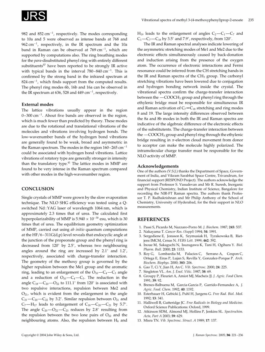
982 and 852 cm�1, respectively. The modes correspondingto 10a and 5 were observed as intense bands at 768 and962 cm�1, respectively, in the IR spectrum and the 10aband in Raman can be observed at 769 cm�1, which aresupported by computations also. The ring breathing modesfor the para-disubstituted phenyl ring with entirely differentsubstituents57 have been reported to be strongly IR activewith typical bands in the interval 780–840 cm�1. This isconfirmed by the strong band in the infrared spectrum at824 cm�1, which finds support from the computed results.The phenyl ring modes 6b, 16b and 16a can be observed inthe IR spectrum at 636, 528 and 449 cm�1, respectively.
External modesThe lattice vibrations usually appear in the region0–300 cm�1. About five bands are observed in the region,which is much fewer than predicted by theory. These modesare due to the rotational and translational vibrations of themolecules and vibrations involving hydrogen bonds. Thelow-wavenumber bands of the hydrogen bond vibrationsare generally found to be weak, broad and asymmetric inthe Raman spectrum. The modes in the region 160–265 cm�1
could be associated with hydrogen bond vibrations. Latticevibrations of rotatory type are generally stronger in intensitythan the translatory type.41 The lattice modes in MMP arefound to be very intense in the Raman spectrum comparedwith other modes in the high-wavenumber region.
CONCLUSION
Single crystals of MMP were grown by the slow evaporationtechnique. The NLO SHG efficiency was tested using a Q-switched Nd : YAG laser of wavelength 1064 nm, which isapproximately 2.3 times that of urea. The calculated firsthyperpolarizability of MMP is 5.940 ð 10�30 esu, which is 30times that of urea. The equilibrium geometry optimizationof MMP, carried out using ab initio quantum computationsat the HF/6–311G(d,p) level reveals that endocyclic angle atthe junction of the propeonate group and the phenyl ring isdecreased from 120° by 2.5°, whereas two neighbouringangles around the ring are increased by 2.1° and 1.2°,respectively, associated with charge-transfer interaction.The geometry of the methoxy group is governed by thehigher repulsion between the Me1 group and the aromaticring, leading to an enlargement of the O10 —C4 —C3 angleand a reduction of O10 —C4 —C5. The reduction in theangle C18 —C20 —O22 to 111.1° from 120° is associated withtwo repulsive interactions, repulsion between Me2 andO21, which is evident from the enlargement in the angleC21 —C20 —C22 by 3.2°. Similar repulsion between O21 andC7 —H17 leads to enlargement of C18 —C20 —C21 by 5.7°.The angle C20 —O22 —C23 reduces by 2.8° resulting fromthe repulsion between the two lone pairs of O22 and theneighbouring atoms. Also, the repulsion between H8 and
H19 leads to the enlargement of angles C2 —C1 —C7 andC1 —C7 —C18 by 3.5° and 7.9°, respectively, from 120°.
The IR and Raman spectral analyses indicate lowering ofthe asymmetric stretching modes of Me1 and Me2 due to theelectronic effects simultaneously caused by back-donationand induction arising from the presence of the oxygenatom. The occurrence of electronic interactions and Fermiresonance could be inferred from the CH stretching bands inthe IR and Raman spectra of the CH3 group. The carbonylstretching vibrations have been lowered due to conjugationand hydrogen bonding network inside the crystal. Thevibrational spectra confirm the charge-transfer interactionbetween the —COOCH3 group and phenyl ring through theethylenic bridge must be responsible for simultaneous IRand Raman activation of C7 C18 stretching and ring modes8 and 19. The large intensity differences observed betweenthe 8a and 8b modes in both the IR and Raman spectra areindicative of the algebraic difference of the electronic effectsof the substitutents. The charge-transfer interaction betweenthe —COOCH3 group and phenyl ring through the ethylenicbridge resulting in �-electron cloud movement from donorto acceptor can make the molecule highly polarized. Theintramolecular charge transfer must be responsible for theNLO activity of MMP.
AcknowledgementsOne of the authors (V.S.J.) thanks the Department of Space, Govern-ment of India, and Vikram Sarabhai Space Centre, Trivandrum, forfinancial support (RESPOND Project). The authors acknowledge thesupport from Professor S. Vasudevan and Mr R. Suresh, Inorganicand Physical Chemistry, Indian Institute of Science, Bangalore forrecording the NIR-FT Raman spectra. The authors thank Profes-sor T. P. Radhakrishnan and Mr Philip Anthony of the School ofChemistry, University of Hyderabad, for the their support in NLOmeasurements.
REFERENCES
1. Passi S, Picardo M, Nazzaro-Porro M. J. Biochem. 1987; 245: 537.2. Nakayama T. Cancer Res. (Suppl) 1994; 54: 1991.3. Sergediene E, Jonsson K, Szymusiak H, Tyrakowska B, Riet-
jens IMCM, Cenas N. FEBS Lett. 1999; 462: 392.4. Inoue M, Sakaguchi N, Isuzugawa K, Tani H, Ogihara Y. Biol.
Pharm. Bull. 2000; 23: 1153.5. Roy G, Lombardia M, Palacios C, Serrano A, Cespon C,
Ortega E, Eiras P, Lujan S, Revilla Y, Gonzalez-Porque P. Arch.Biochem. Biophys. 2000; 383: 206.
6. Gao T, Ci Y, Jian H, An C. Vib. Spectrosc. 2000; 24: 225.7. Singleton VL. Am. J. Enol. Vitic. 1987; 38: 69.8. Groupy P, Fleuriet A, Amiot MJ, Macheix JJ. J. Agric. Food Chem.
1991; 39: 92.9. Brenes-Balbuena M, Garcia-Garcia P, Garrido-Fernandez A, J.
Agric. Food. Chem. 1992; 40: 1192.10. Esterbauer H, Gebicki J, Puhl H, Jurgens G. Free Rad. Biol. Med.
1992; 13: 341.11. Halliwell B, Gutteridge JC. Free Radicals in Biology and Medicine.
Oxford Science Publications: Oxford, 1999.12. Atkinson SDM, Almond MJ, Hollins P, Jenkins SL. Spectrochim.
Acta, Part A 2003; 59: 629.13. Misra TN. Vib. Spectrosc. Struct. A 1989; 17: 137.
Copyright 2004 John Wiley & Sons, Ltd. J. Raman Spectrosc. 2005; 36: 221–236

236 D. Sajan et al.
14. Zyss J, Nicoud JF. Curr. Opin. Solid State Mater. Sci. 1996; 1: 533.15. Mingaleev SF, Kivshar Yu S. Opt. Photonics News 2002; 13: 48.16. Sukhorukov AA, Kivshar Yu S, J. Opt. Soc. Am. B 2002; 19: 772.17. Maroulis GJ. J. Chem. Phys. 2000; 113: 1813.18. Maroulis GJ. J. Mol. Struct. (THEOCHEM) 2003; 633: 177.19. Li Y, Li Z-R, Wu D, Li R-Y, Hao X-Y, Sun C-C. J. Phys. Chem. B.
2004; 108: 3145.20. Sajan D, Binoy J, Pradeep B, Venkata Krishna K, Kartha VB,
Hubert JI, Jayakumar VS. Spectrochim. Acta, Part A 2004; 60: 174.21. Abraham JP, Hubert JI, George V, Nielsen OF, Jayakumar VS.
Spectrochim. Acta, Part A 2003; 59: 193.22. Binoy J, Abraham JP, Hubert JI, Jayakumar VS, Pettit GR,
Nielsen OF. J. Raman Spectrosc. 2004; 35: 939.23. Enkelmann E, Wegner G. J. Am. Chem. Soc. 1993; 115: 103 90.24. Ghosh M, Mandal TK, Chakrabarti S. J. Raman Spectrosc. 1996;
29: 807.25. Ghosh M, Chakrabarti S, Misra TN. J. Phys. Chem. Solids. 1996;
57: 1891.26. Chakrabarti S, Maity AK, Misra TN. J. Polym. Sci., Part A 1992;
30: 1625.27. Ghosh M, Chakrabarti S, Misra TN. J. Raman Spectrosc. 1998; 29:
263.28. Allen SDM, Almond MJ, Hollins P, Mascetti J. Spectrochim. Acta,
Part A 2000; 56: 2423.29. Hanni K, Kuwae A, Takai T, Senda H, Kunimoto K. Spectrochim.
Acta, Part A 2001; 57: 513.30. Sanchez-Cortes S, Garcia-Ramos JV. Spectrochim. Acta, Part A
1999; 55: 2935.31. Castro JL, Lopez Ramirez MR, Lopez Tocon I, Otero JC. J. Raman
Spectrosc. 2002; 33: 455.32. Bujak M, Zaleski J, Prezhdo V, Uspienskij B. Acta Crystallogr.
Sect. C 2002; 58: O76.33. Kurtz SK, Perry TT, J. Appl. Phys. 1968; 39: 3798.34. Frisch MJ, Trucks GW, Schlegel HB, Scuseria GE, Robb MA,
Cheeseman JR, Zakrzewski VG, Montgomery JA, Stratmann RE,Burant JC, Dapprich S, Millam JM, Daniels AD, Kudin KN,Strain MC, Farkas O, Tomasi J, Barone V, Cossi M, Cammi R,Mennucci B, Pomelli C, Adamo C, Clifford S, Ochterski J,Petersson GA, Ayala PY, Cui Q, Monocular K, Malick DK,Rabuck AD, Raghavachari K, Foresman JB, Kieslowski J,Ortiz JV, Baboul AG, Stefano BB, Liu G, Liashenko A, Piskorz P,Komaromi I, Gomperts R, Martin RL, Fox DJ, Keith T,
Al-Laham MA, Peng CY, Nanayakkara A, Challacombe M,Gill PMW, Johnson B, Chen W, Wong MW, Andres JL,Gonzalez C, Head-Gordon M, Replogle ES, Pople JA. Gaussian98, Revision A.9. Gaussian: Pittsburgh, PA, 1998.
35. Michelle M, Pietro FWJ, Hehre WJ, Binkly S, Mark J, Gorden MS,Defrees DJ, Pople JA. J. Chem. Phys. 1982; 7: 77.
36. Scott AP, Radom L. J. Phys. Chem. 1996; 100: 16 503.37. Foresman JB, Frisch A. Exploring Chemistry with Electronic
Structure Methods (2nd edn). Gaussian: Pittsburgh, PA, 1996.38. Kleinman DA. Phys. Rev. 1962; 126: 1977.39. Domenicano A, Vaciago A, Coulson CA. Acta Crystallogr. Sect. B
1975; 31: 221.40. Domenicano A, Vaciago A, Coulson CA. Acta Crystallogr. Sect. B
1975; 31: 1630.41. Smith B. Infrared Spectral Interpretation, a Systematic Approach.
CRC Press: Washington, DC, 1999.42. Gussoni M, Castiglioni C. J. Mol. Struct. 2000; 521: 1.43. Palomar J, De Paz JLG, Catalan J. Chem. Phys. 1999; 246: 167.44. Rumi M, Zerbi G. J. Mol. Struct. 1999; 509: 11.45. Hobza P, Halvas Z. Chem. Rev. 2000; 100: 4253.46. Hobza P. Phys. Chem. Chem. Phys. 2000; 3: 2555.47. Mrazkova E, Hobza P. J. Phys. Chem. A 2003; 107: 1032.48. Karpfen A, Kryachko ES. J. Phys. Chem. A 2003; 107: 9724.49. Horak M, Lippincott ER, Khanna RK. Spectrochim. Acta, Part A
1967; 23: 1111.50. Dollish FR, Fateley WG, Bentley FF. Characteristic Raman
Frequencies of Organic Compounds. J Wiley: New York, 1997.51. Colthup NB, Daly LH, Wiberley SE. Introduction to Infrared and
Raman Spectroscopy. Academic Press: New York, 1990.52. Socrates G. Infrared Characteristic Group Frequencies. Wiley-
Interscience: New York, 1980.53. Arenas JF, Tocon IL, Otero JC, Marcos JI. J. Mol. Struct. 1995; 349:
29.54. Tommasini M, Castiglioni C, Del Zoppo M, Zerbi G. J. Mol.
Struct. 1999; 480–481: 179.55. Del Zoppo M, Tommasini M, Castiglioni C, Zerbi G. Chem. Phys.
Lett. 1998; 287: 1998.56. Ruiz Delgado MC, Hernandez V, Casado J, Lopez Navarrete JT,
Raimundo JM, Blanchard P, Roncali J. J. Mol. Struct. 2003;651–653: 151.
57. Varsanyi G. Vibrational Spectra of Benzene Derivatives. AcademicPress: New York, 1969.
Copyright 2004 John Wiley & Sons, Ltd. J. Raman Spectrosc. 2005; 36: 221–236