Reactivation of latent HIV-1 provirus via targeting protein phosphatase-1
Upload
georgetownCategory
view
3download
0
Valosin containing protein (VCP/p97) is a novel substrate for theprotein tyrosine phosphatase PTPL1
Ogan D. Abaana,1, Wiljan Hendriksb, Aykut Ürena, Jeffrey A. Toretskya, and Hayriye V.Erkizana,*
aDepartments of Oncology and Pediatrics, Lombardi Comprehensive Cancer Center, GeorgetownUniversity, Washington, DC 20057, USA bDepartment of Cell Biology, Nijmegen Centre forMolecular Life Sciences, Radboud University Nijmegen Medical Centre, Nijmegen, TheNetherlands
AbstractIdentification of Protein Tyrosine Phosphatase (PTP) substrates is critical in understanding cellularrole in normal cells as well as cancer cells. We have previously shown that reduction of PTPL1protein levels in Ewings sarcoma (ES) inhibit cell growth and tumorigenesis. Therefore, we soughtto identify novel PTPL1 substrates that may be important for tumorigenesis. In this current work,we demonstrated that mouse embryonic fibroblasts without PTPL1 catalytic activity fail to formfoci when transfected with oncogenes. We proved that catalytic activity of PTPL1 is important forES cell growth. Using a substrate-trapping mutant of PTPL1 we identified putative PTPL1substrates by mass-spectrometry. One of these putative substrates was characterized as ValosinContaining Protein (VCP/p97). Using multiple biochemical assays we validated VCP as a novelsubstrate of PTPL1. We also provide evidence that tyrosine phosphorylation of VCP might beimportant for its midbody localization during cytokinesis. In conclusion, our work identifies VCPas a new substrate for PTPL1, which may be important in cellular transformation. Ourinvestigation link an oncogenic transcription factor EWS-FLI1, with a key transcriptional targetprotein tyrosine phosphatase PTPL1, and its substrate VCP. Given our observation that PTPL1catalytic activity is important for cell transformation, our results may also suggest that VCPregulation by PTPL1 might be important for tumorigenesis.
KeywordsES; PTPL1; VCP; Phosphorylation
© 2012 Elsevier Inc. All rights reserved.*Correspondence to: 3970 Reservoir Rd, NW, New Res Bldg Room E316, Washington, DC 20057, USA. Fax: +1 202 687 1434.1Current address: Genetics Branch, NCI, NIH, Bethesda MD 20892, USA.
Authors’ contributionsODA designed and carried out experiments and wrote the original manuscript, HVE carried out experiments, contributed significantlyto the writing of the original manuscript, and rewrote the manuscript in response to the critique, WH contributed key reagents andwriting of the manuscript, AU and JAT supervised the project and finalized the manuscript.
Appendix A. Supplementary materialSupplementary data associated with this article can be found in the online version at http://dx.doi.org/10.1016/j.yexcr.2012.09.003.
NIH Public AccessAuthor ManuscriptExp Cell Res. Author manuscript; available in PMC 2014 January 01.
Published in final edited form as:Exp Cell Res. 2013 January 1; 319(1): 1–11. doi:10.1016/j.yexcr.2012.09.003.
NIH
-PA Author Manuscript
NIH
-PA Author Manuscript
NIH
-PA Author Manuscript
IntroductionElucidating key signalling pathways in oncogenesis has revealed biologically vulnerableproteins, which can serve as valuable therapeutic targets. Ewings sarcoma (ES) arecharacterized by a pathognomonic translocation which fuses the N-terminal of the geneEWS with the C-terminal of an ets-family gene [1]. The protein product of this translocationis an oncogenic transcription factor called EWS-FLI1. Genes modulated by EWSFLI1 havea profound influence on ES tumorigenesis. Our previous work showed that protein tyrosinephosphatase L1 (PTPL1) is a direct transcriptional target of EWS-FLI1 and critical for themaintenance of the oncogenic phenotype [2].
PTPL1 (a.k.a PTPN13, FAP-1, PTP-BAS, PTP1E) is a 270 kDa non-receptor type proteintyrosine phosphatase with published opposing roles in tumorigenesis [3]. Few putativePTPL1 substrates have been identified and additional substrates may clarify its cell-specificfunctions. These substrates include IRS-1 [4], IκBα [5], HER2 [6], STAT4 [7] and Src [8].Even though no cell cycle dependent substrates of PTPL1 have been identified, there isevidence that PTPL1 is localized to the centrosomes and to the midbody during the cellcycle [9]. Interestingly, in breast cancer PTPL1 acts as a tumor suppressor through IRS-1dephosphorylation [4] and PTPL1 expression is a prognostic factor for favorable outcome inthese cancers [10]. Since PTPL1 likely has different functions in diverse cellular contexts, amore complete knowledge of the enzymatic substrates and protein scaffold partners ofPTPL1 are necessary to resolve ambiguity.
A multifunctional chaperon Valosin Containing Protein (VCP) has many diverse roles in thecell [11,12]. A central role of VCP is to link ubiquitinated substrates with either theubiquitin conjugation or deconjugation machineries in endoplasmic reticulum-associateddegradation (ERAD) [13] and ubiquitin-proteosome pathways [14] thereby influencingprotein fate in the cell [15]. VCP in the context of ERAD and ubiquitin-proteosomepathways is being widely studied. On the other hand, VCP’s role in cell cycle has been firstproposed in Saccharomyces cerevisiae in 1998 [16], however until recently, has not beeninvestigated further. Studies show that VCP is involved in the terminal stages of celldivision by participating in the re-emergence of the nuclear envelope in Xenopus eggextracts by removing Aurora B [17]. In addition VCP has been shown to antagonize AuroraB in HeLa cells for proper chromosome segregation [18]. Such emerging studies suggest acritical role for VCP in cell cycle.
To enhance our understanding of the role of PTPL1 in ES tumorigenesis we sought toidentify novel PTPL1 substrates. In this study, we generated a substrate-trapping mutant ofPTPL1 and used it in a screen for novel substrates. Our screen identified VCP as a candidatesubstrate of PTPL1 and further biochemical studies validated VCP as a novel PTPL1substrate. Further on we provide critical evidence suggesting a role for VCP in late stagemitosis, specifically during cytokinesis. In addition, a key finding in our study demonstratesthe importance of PTPL1 catalytic activity in oncogenic transformation.
MethodsCell lines
MEFs were isolated from PTP-BLΔPTP/ΔPTP or wild type mice according to standardprocedures [19]. TC32 ES cells were maintained in RPMI (Invitrogen) with 10% FBS and1% HEPES (Invitrogen). HEK293 cells and MEFs (PTP-BLΔPTP/ΔPTP and PTP-BLWT/WT)were maintained in DMEM (Invitrogen) with 10% FBS (Quality Biologicals). COLO-357,COLO-PL and COLO-SL were described before [20] and were a kind gift from Dr JohnJessup.
Abaan et al. Page 2
Exp Cell Res. Author manuscript; available in PMC 2014 January 01.
NIH
-PA Author Manuscript
NIH
-PA Author Manuscript
NIH
-PA Author Manuscript
AntibodiesActin-HRP, pY99, PTPL1 and GST antibodies were purchased from Santa CruzBiotechnology. VCP antibodies were purchased from Abcam. 4G10 phosphotyrosineantibody was purchased from Upstate. Flag-M5 antibody was purchased from Sigma.
PlasmidsFull-length Flag-tagged PTPL1 and GFP-VCP plasmids were generous gifts from Dr. JanSaras and Dr Len Neckers, respectively. Flag-tagged PTPL1 was cloned into pCDNA4/TO(Invitrogen). ΔPTP-PTPL1 was generated by removing the PTP domain of PTPL1 usingrestriction digest. The GST-PTP constructs were created by cloning the cDNA part encodingthe PTPL1 phosphatase domain into pET42 vector (Stratagene). All of the cloning strategieswere designed using pDRAW32 DNA Analysis Software (http://www.acaclone.com).Primers for site-directed mutagen esis were designed with PrimerX (http://bioinformatics.org/primerx) and all the reactions were performed by QuikChange II XLSite-Directed Mutagenesis Kit (Stratagene) according to the manufacturer’s protocol. Site-directed mutagenesis primer sequences were 5-ACTGCCTGGCCAGCCCATGATACACCTTC for the PTPL1-DA mutation and 5-CAATGACGATGACCTGTTCGGCGG TACCACCATGG for VCP-YF mutation. Thefidelity of all constructs was confirmed via sequencing.
Soft-agar assaysSoft-agar assays were performed in 12 well plates using 0.4% SeaPlaque Agar (CambrexBioscience) in PBS. Colonies were stained with 200–300 µl per well of 50 mg/ml MTT(Sigma) for 3 h at 37 °C and imaged with Kodak 2D imaging system.
GST fusion protein purificationBL21 CodonPlus Competent cells (Stratagene) were transformed with pET42-GST-PTPplasmids. An aliquot was induced with 1 mM IPTG for 4 h at 37 °C, lysed and recombinantproteins were affinity purified using GSH-Agarose beads.
In vitro substrate-trappingFor small scale experiments, log phase TC32 cells were treated with 300 µM pervanadate for1 h before being lysed with NP40 lysis buffer (50 mM Tris pH 7.5, 5 mM EDTA, 150 mMNaCl, 20 mM NaF, 1% NP40, 10 mM Iodoacetic acid, 1 mM PMSF, 2 µg/ml Aprotinin and2 µg/ml Leupeptin). The lysate was treated with 10 mM DTT for 10 min on ice prior to pull-down. Two milligrams of protein was mixed with 10 µl of affinity purified GST-PTPrecombinant protein on GSH-Agarose beads. Protein complexes were eluted from the beadswith 2X sample buffer, boiled and then subject to immunoblotting. Scaled up experimentswere done with 17 mg of protein and 100 µl of recombinant protein. Bands were excisedfrom Coomassie blue stained SDS-PAGE. Tryptic peptides were extracted and analyzedwith MALDI-TOF-TOF spectrometer (4800 Proteomics Analyzer).
In vivo substrate-trappingTC32 cells were transiently transfected with full-length Flag-tagged PTPL1 constructs usingelectroporation. After 24 h, cells were lysed and 2 mg of protein was mixed with 30 µl ofFlag- Agarose beads (Sigma). Samples were subjected to immunoblotting. The immunoblotswere developed using the LAS-3000 Imaging System (Fujifilm Life Science) and bandintensities were quantified using the built in software.
Abaan et al. Page 3
Exp Cell Res. Author manuscript; available in PMC 2014 January 01.
NIH
-PA Author Manuscript
NIH
-PA Author Manuscript
NIH
-PA Author Manuscript
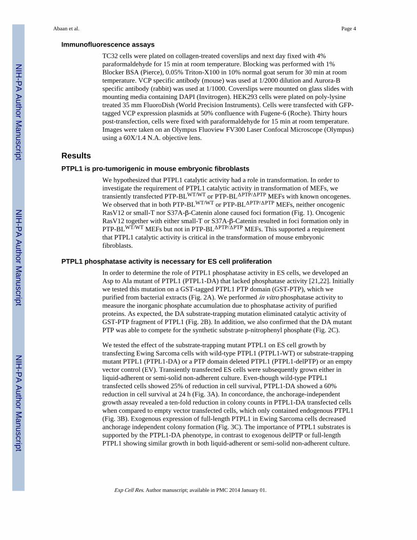
Immunofluorescence assaysTC32 cells were plated on collagen-treated coverslips and next day fixed with 4%paraformaldehyde for 15 min at room temperature. Blocking was performed with 1%Blocker BSA (Pierce), 0.05% Triton-X100 in 10% normal goat serum for 30 min at roomtemperature. VCP specific antibody (mouse) was used at 1/2000 dilution and Aurora-Bspecific antibody (rabbit) was used at 1/1000. Coverslips were mounted on glass slides withmounting media containing DAPI (Invitrogen). HEK293 cells were plated on poly-lysinetreated 35 mm FluoroDish (World Precision Instruments). Cells were transfected with GFP-tagged VCP expression plasmids at 50% confluence with Fugene-6 (Roche). Thirty hourspost-transfection, cells were fixed with paraformaldehyde for 15 min at room temperature.Images were taken on an Olympus Fluoview FV300 Laser Confocal Microscope (Olympus)using a 60X/1.4 N.A. objective lens.
ResultsPTPL1 is pro-tumorigenic in mouse embryonic fibroblasts
We hypothesized that PTPL1 catalytic activity had a role in transformation. In order toinvestigate the requirement of PTPL1 catalytic activity in transformation of MEFs, wetransiently transfected PTP-BLWT/WT or PTP-BLΔPTP/ΔPTP MEFs with known oncogenes.We observed that in both PTP-BLWT/WT or PTP-BLΔPTP/ΔPTP MEFs, neither oncogenicRasV12 or small-T nor S37A-β-Catenin alone caused foci formation (Fig. 1). OncogenicRasV12 together with either small-T or S37A-β-Catenin resulted in foci formation only inPTP-BLWT/WT MEFs but not in PTP-BLΔPTP/ΔPTP MEFs. This supported a requirementthat PTPL1 catalytic activity is critical in the transformation of mouse embryonicfibroblasts.
PTPL1 phosphatase activity is necessary for ES cell proliferationIn order to determine the role of PTPL1 phosphatase activity in ES cells, we developed anAsp to Ala mutant of PTPL1 (PTPL1-DA) that lacked phosphatase activity [21,22]. Initiallywe tested this mutation on a GST-tagged PTPL1 PTP domain (GST-PTP), which wepurified from bacterial extracts (Fig. 2A). We performed in vitro phosphatase activity tomeasure the inorganic phosphate accumulation due to phosphatase activity of purifiedproteins. As expected, the DA substrate-trapping mutation eliminated catalytic activity ofGST-PTP fragment of PTPL1 (Fig. 2B). In addition, we also confirmed that the DA mutantPTP was able to compete for the synthetic substrate p-nitrophenyl phosphate (Fig. 2C).
We tested the effect of the substrate-trapping mutant PTPL1 on ES cell growth bytransfecting Ewing Sarcoma cells with wild-type PTPL1 (PTPL1-WT) or substrate-trappingmutant PTPL1 (PTPL1-DA) or a PTP domain deleted PTPL1 (PTPL1-delPTP) or an emptyvector control (EV). Transiently transfected ES cells were subsequently grown either inliquid-adherent or semi-solid non-adherent culture. Even-though wild-type PTPL1transfected cells showed 25% of reduction in cell survival, PTPL1-DA showed a 60%reduction in cell survival at 24 h (Fig. 3A). In concordance, the anchorage-independentgrowth assay revealed a ten-fold reduction in colony counts in PTPL1-DA transfected cellswhen compared to empty vector transfected cells, which only contained endogenous PTPL1(Fig. 3B). Exogenous expression of full-length PTPL1 in Ewing Sarcoma cells decreasedanchorage independent colony formation (Fig. 3C). The importance of PTPL1 substrates issupported by the PTPL1-DA phenotype, in contrast to exogenous delPTP or full-lengthPTPL1 showing similar growth in both liquid-adherent or semi-solid non-adherent culture.
Abaan et al. Page 4
Exp Cell Res. Author manuscript; available in PMC 2014 January 01.
NIH
-PA Author Manuscript
NIH
-PA Author Manuscript
NIH
-PA Author Manuscript
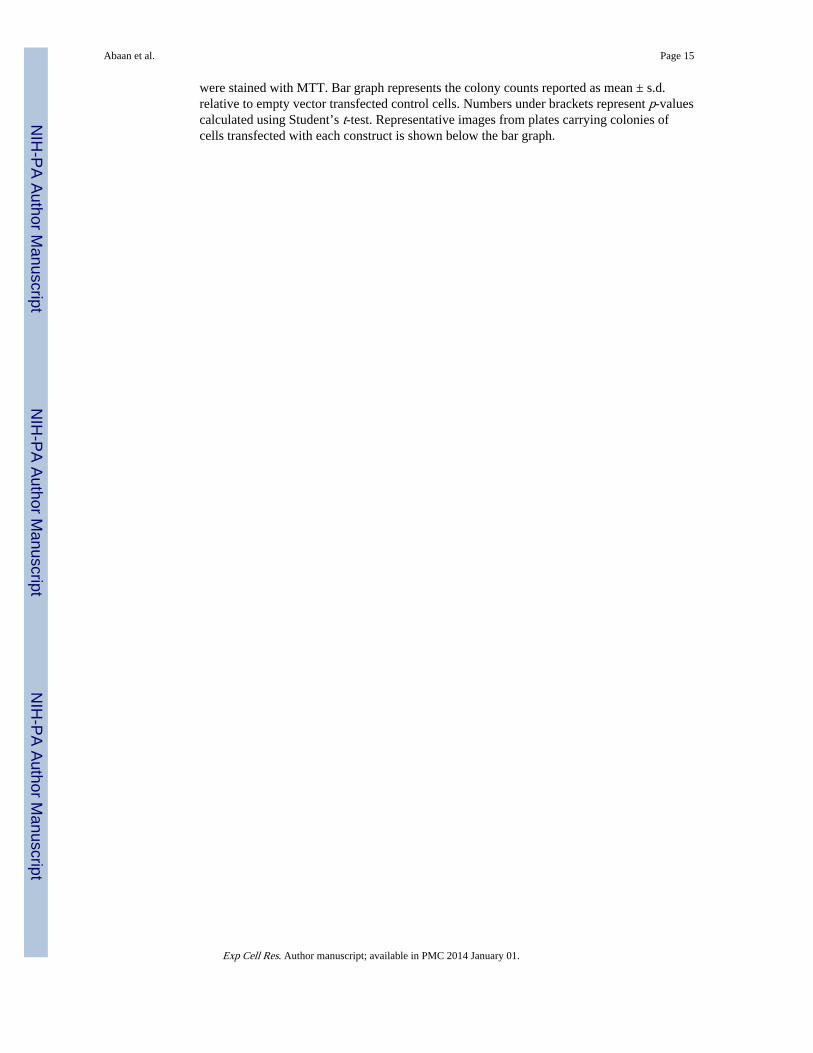
Substrate-trapping PTPL1 shows enhanced affinity for phosphoproteinsES cells may appear to require a regulated amount of PTPL1 for cell survival andtransformation. The 90% decrease in colony formation in substrate trapping mutant (DA)transfected cells directed us to determine PTPL1 substrates, which might be involved intumorigenesis. In order to identify substrates, we used an in vitro affinity assay withrecombinant GST fusion proteins for both wild type (GST-PTP-WT) and substrate-trappingmutant PTPL1 phosphatase domain (GST-PTP-DA). Lysates from TC32 cells were pre-treated with pervanadate, and compared to untreated controls, to identify proteins that boundto GST-PTP-WT, GST-PTPDA, or GST-alone. Phosphotyrosine immunoblottingdemonstrated that PTP-DA was able to form stable complexes with tyrosine phosphorylatedproteins whereas neither PTP-WT nor GST-alone bound phosphoproteins (Fig. 4A).Reprobing the membrane with GST antibodies confirmed equal recombinant protein loading(Fig. 4A, lower panel). In order to identify potential substrate proteins, we scaled up theaffinity-purification using 17 mg of total protein (Fig. 4B). Those protein bands thatappeared to be differentially present or more abundantly bound to PTP-DA (arrow heads)were isolated and analyzed by mass-spectrometry. This experiment provided a list ofproteins that were identified by mass-spectrometry in the four most prominent protein bands(Table 1).
97 kDa protein identified as Valosin Containing ProteinIn three rounds of affinity purification, the most reproducible protein band was found at amolecular weight of 97 kDa (Supplemental Fig. 1A). Mass-spectrometry analyses ofduplicate samples from two independent experiments showed an average of 15 and 30peptides, respectively, which identified this 97 kDa protein as Valosin Containing Protein(VCP). Peptide coverage for VCP was 25% and 46% in the two replicate mass-spectrometryexperiments (Supplemental Fig. 1B). These results identify the 97 kDa protein as VCP, butas such do not demonstrate that VCP is a PTPL1 substrate. Mass-spectrometry analysisyielded additional proteins as possible substrates, but their identity could not be assessed asrobustly as for VCP.
VCP is a novel PTPL1 substrateDefinitive validation of VCP as a PTPL1 substrate was confirmed according to establishedcriteria [22]. First, we showed that VCP can affinity purify with PTP-DA but not with PTP-WT or with GST alone. The affinity purification was only successful after cells werepretreated with pervanadate (Fig. 5A, top panel) suggesting that PTP-DA could only bindtyrosine phosphorylated VCP. Pervanadate treatment did not affect VCP protein stability asdemonstrated by the equal levels of VCP in the input (Fig. 5A, middle panel). Reprobingwith phosphotyrosine antibodies confirmed VCP tyrosine phosphorylation (Fig. 5A, lowerpanel). In order to further demonstrate that VCP is a PTPL1 substrate, we performedadditional studies in HEK cells. In a second approach, TC32 cells were transfected witheither full-length Flag-PTPL1-WT or Flag-PTPL1-DA. Protein complexes from total celllysates were immunoprecipitated using Flag-specific antibodies. Results demonstrated thatendogenous VCP co-immunoprecipitates with PTPL1-DA but not with PTPL1-WT, whichshowed similar levels compared to the background in control cells transfected with GFP(Fig. 5B). Densitometric analysis of the immunoblot revealed a 5.7 fold enrichment of VCPin PTPL1-DA over PTPL1-WT immunoprecipitation (Fig. 5B lower panel). In order tofurther validate VCP as a PTPL1 substrate, we evaluated mouse embryonic fibroblasts(MEF) derived from PTP-BLΔPTP/ΔPTP animals, which lack the catalytic PTP domain of themouse ortholog of PTPL1, PTP-BL [23]. VCP was immunoprecipitated from these MEFcells grown under regular growth conditions and immunoblotted for antibodies againstphospho-tyrosine. In PTP-BLΔPTP/ΔPTP MEFs we measured a 2.8-fold increase in VCPtyrosine phosphorylation when compared to PTP-BLWT/WT MEFs (Fig. 5C upper panel).
Abaan et al. Page 5
Exp Cell Res. Author manuscript; available in PMC 2014 January 01.
NIH
-PA Author Manuscript
NIH
-PA Author Manuscript
NIH
-PA Author Manuscript
Lastly, we investigated whether PTPL1 can dephosphorylate VCP in vitro. TC32 cells werepervanadate treated to enrich for phosphotyrosine-VCP, followed by VCP- specificimmunoprecipitation. The immunoprecipitates were mixed with GST-PTP wild-type or Aspto Ala mutant constructs in the presence or absence of ortho-vanadate. The reactions werethen resolved on SDS-PAGE and immunoblotted for phosphotyrosine or GST. The resultsfrom this experiment demonstrated that phospho-VCP was completely dephosphorylated byPTP-WT but not with PTPDA recombinant protein, and the dephosphorylation by PTP-WTwas inhibited by ortho-vanadate showing a protein tyrosine phosphatase specific action (Fig.5D, upper panel). Equal recombinant protein loading was confirmed with immunoblottingagainst GST specific antibody (Fig. 5D, lower panel). These four separate validationsprovide further proof that VCP is a PTPL1 substrate.
There is one tyrosine residue in VCP that is phosphorylated in ES cellsVCP tyrosine residue, Y805, has been shown as the major site for tyrosine phosphorylation[24]. In order to determine if other VCP tyrosines residues are phosphorylated in ES cells,we transiently transfected TC32 cells with wild type VCP or various VCP Y805 mutants,Y805E and Y805F, all fused to GFP. No phosphorylation was seen with a phospho-tyrosineimmunoprecipitation for either of these single amino acid substitutions (Fig. 6, upper panel).Therefore, Y805 is the sole residue phosphorylated in ES cells. Immunoblotting with GFP ofprecipitated proteins also showed that the tagged-VCP protein can interact with endogenousVCP as expected since VCP forms a hexamer in the cell (Fig. 6 middle and lower panel)[25].
VCP tyrosine phosphorylation is induced with stimulationThe role of VCP tyrosine phosphorylation in mammalian cells remains cryptic. In yeast,tyrosine phosphorylation of VCP was suggested to be a cell cycle specific event [16]. Wetherefore evaluated whether VCP tyrosine phosphorylation would be altered upon serumstarvation and subsequent stimulation. Overnight serum starved cells displayed very lowlevels of phosphotyrosine-VCP while serum stimulation of cells increased phosphorylation(Fig. 7). Cells that were grown in regular growth media had higher basal levels ofphosphorylation (Fig. 7). Pervanadate treatment of either serum starved cells or cells grownin regular media resulted in robust VCP phosphorylation, (Fig. 7). These results suggestedthat VCP tyrosine phosphorylation is a physiological event triggered by serum stimulation,suggesting cell growth dependent tyrosine phosphorylation of VCP thus supporting aregulatory role for PTPL1.
Tyrosine 805 may be required for VCP localization to the midbodyIn view of our findings that there is serum-induced tyrosine phosphorylation of VCP (Fig. 7)and the separate observation by others that PTPL1 enriches in the midbody [9] we sought forlocalization of VCP. In TC32 cells, VCP displayed diffuse staining in both non-dividingcells and dividing cells (Fig. 8). However, in dividing cells at the stage of cytokinesis,enhanced staining of exogenously expressed GFP-VCP was observed at structures thatresemble the midbody (Fig. 8). Multiple attempts to show co-enrichment of PTPL1 and VCPat the midbody complex were unsuccessful, which may be due to difficulties in over-expressing PTPL1 variants (data not shown) and the ability of the antibodies to identify thenative protein. However, expression studies with VCP phospho-mutants provided evidencefor the role of VCP tyrosine phosphorylation. When we transiently transfected HEK293cells with GFP fused wild type VCP or a Y805F mutant construct, VCP localization in cellthroughout cell cycle did not differ regardless of the Y805 residue (Supplemental Fig. 3).However, we did observe reduced VCP localization at the midbody during cytokinesis whenY805 is mutated (Fig. 8 lowest panel). In cells expressing GFP-VCP-Y805F only 20% ofthe cells at cytokinesis (n = 10) displayed GFP fluorescence at the midbody while 70% of
Abaan et al. Page 6
Exp Cell Res. Author manuscript; available in PMC 2014 January 01.
NIH
-PA Author Manuscript
NIH
-PA Author Manuscript
NIH
-PA Author Manuscript

wild type transfected cells at cytokinesis (n = 13) displayed GFP-positivity at the midbody.We conclude that tyrosine 805 is important for midbody localization of VCP.
DiscussionPhosphatases are no longer considered housekeeping enzymes and understanding theirregulatory potential in cell biology requires identifying specific substrates. Identification ofPTP substrates can be performed using techniques such as substrate-trapping and strictcriteria for the validation of bona fide PTP substrates have been defined [22]. We previouslyidentified PTPL1 as a critical transcriptional target of the oncogene EWS-FLI1 [2]. In orderto understand why an oncogenic transcription factor would activate transcription of PTPL1,we first assessed that PTPL1 phosphatase activity influences ES cell survival and continuedwith a search for the PTPL1 substrates involved. Our affinity purification provided a list ofpotential PTPL1 substrates of which we validate VCP as a substrate using establishedcriteria [22].
We observed a series of proteins that were identified by the PTPL1-DA substrate-trappingmutant. Multiple Coomassie-stained protein bands with apparent molecular weights of 500,250, 97, and 40 kDa, were reproducibly observed. The 97 kDa band was identified by massspectroscopy as VCP and rigorously validated as a substrate in the current study. VCP wasidentified as a substrate for PTPH1 [26], which resembles PTPL1 in protein domain build upsince both phosphatases contain FERM and PDZ domains that are involved in membranetargeting and protein-protein interactions, respectively, suggesting that PTPL1 and PTPH1may have overlapping substrates. We were unable to unequivocally demonstrate a functionallink between PTPL1 and VCP in expression studies, which may reflect their respectiveimpact on cell growth and the need for reliable inducible expression systems to performsuch studies. One of the other potential PTPL1 substrates, the 250 kDa protein, could beidentified with high confidence (Table 1) as microtubule-associated protein 1 B (MAP1B).MAP1B also carries a terminal tyrosine that is critical for its function [27]. However,phosphorylation at this site has not been reported yet, leaving the possibility that MAP1B isa functional substrate of PTPL1 to be explored in future work.
Evidence shows that PTPL1 can either support tumor growth or function as a tumorsuppressor in different experimental models [3,28]. In support of PTPL1 as a tumor growthenhancing protein as in shown in anchorage independent growth of MEF cells, many solidtumors of different tissue of origin display higher staining for PTPL1 compared to benigntissue [29]. ES [2] and the pancreatic cancer cell lines we looked at, PTPL1 levels are higherin metastases (Supplemental Fig. 2). Both wild-type PTPL1 and delPTP over-expression inES cells resulted in a decrease in growth. This contrasts the need for full-length PTPL1 thatis required for oncogenic transformation. Together, these data suggests that theoverexpression of PTPL1 behaves differently than its absence in difference cell types (EScells and fibroblasts) and that these differences can account for discrepancies in the literatureabout the role of PTPL1 in oncogenesis. In addition, changes in PTPL1 intracellular levelslikely alter the balance in the protein interaction partners of PTPL1 based upon PTPL1having 5 PDZ domains and a FERM domain capable of numerous protein-proteininteractions. Therefore, PTPL1 functions as a bridging factor regardless of its catalyticactivity.
VCP knock-out in mice is early embryonic lethal at the peri-implantation stage [30],suggesting a very fundamental and indispensable role of VCP in the cell. A central role forVCP is to direct ubiquitinated proteins to protosomal destruction or recycling, thus, theregulation of VCP by phosphorylation may control protein fate in the cell and the overallviability and fidelity of the cell. Higher VCP mRNA and/or protein is a poor prognostic
Abaan et al. Page 7
Exp Cell Res. Author manuscript; available in PMC 2014 January 01.
NIH
-PA Author Manuscript
NIH
-PA Author Manuscript
NIH
-PA Author Manuscript
factor in many types of carcinoma [31], however, there is no direct evidence that VCP is atumor suppressor or a tumor promoter. However, there are a number of VCP mutationsidentified as the cause for an autosomal dominant disease called inclusion body myopathywith Paget disease of bone and frontotemporal dementia (IBMPFD), which affects multipletissues, including muscle, bone, and the cerebral cortex [32]. How tyrosine phosphorylationof VCP fits into IBMPFD or cancer is not clear at this point.
Our work suggests that lack of VCP-Y805 phosphorylation prevents VCP localization to themidbody during cytokinesis. This could be due to lack of proper complex formation orsimply due mislocalization of VCP in the cell. For the time being, the definite role for VCPtyrosine phosphorylation remains elusive, however, earlier studies demonstrated thatphosphorylation does not affect VCP ATPase activity [24]. In addition our co-immunoprecipitation studies using the VCP-Y805 mutants show that these mutant proteinare still able to complex with the endogenous VCP in forming the VCP hexamer [25]suggesting that they maintain structural functionality. Recent findings in the context of cellcycle and VCP revealed a direct link between VCP and Aurora B during various stages ofthe cell cycle. Our study also suggests a direct interaction between VCP and Aurora Bduring cytokinesis. In addition, VCP localization to the midbody might be regulated by itstyrosine phosphorylation. In addition, the precise special and temporal interplay betweenVCP and PTPL1 has yet to be revealed. Our data suggests a putative function in cytokinesis.In one scenario dephosphorylation of VCP could trigger events such as AuroraB degredationat the end of cell cycle [17]. Interplay between VCP and PTPL1 could reduce the thresholdfor a well controlled event such as cytokinesis to complete cell cycle. Such hypotheses needfurther investigation into the exact mechanism of VCP and PTPL1 interaction
The PTPL1 literature clearly demonstrates a complex and poorly defined role for PTPL1 intumorigenesis. The conclusions from these studies suggest that each single observationcould be indeed cell type specific. In our study, mouse embryonic fibroblasts that lackPTPL1 catalytic domain failed to transform in the presence of two oncogenes while PTPL1wild-type fibroblasts did, supporting an oncogenic role for PTPL1 in this context. Incontrast, normal mouse epithelial cells transfected with oncogenic Ras and depleted ofPTPL1 were able to maintain anchorage-independent growth and grow in immuno-competent mice [33] supporting a tumor-suppressor role for PTPL1. Studies in cancers ofepithelial origin line breast [8] and prostate [34] support the tumor-suppressor role. While inES cells which are of mesenchymal origin we observe an oncogenic role for PTPL1.Therefore, there are clear physiological difference in PTPL1 function between epithelial andmesenchymal cells, which remain elusive, that confound the general role of PTPL1 intumorigenesis. Thus data so far suggests a complex role for PTPL1. It is also important topoint out that PTPH1 resembles PTPL1 in its domain structure and PTPH1 overexpressionin NIH3T3 mouse fibroblasts result in growth inhibition while a DA mutant does not. Thisobservation also demonstrated how fine-tuned these phosphatases are in cells of similarorigin. Even-though VCP is a substrate for both PTPs, the exact biochemical link betweenVCP and PTPL1 or PTPH1 remains unknown.
ConclusionsIn conclusion, we demonstrate that VCP is a novel, validated, substrate of PTPL1. Our datasuggests that tyrosine phosphorylation of VCP may be responsible for its proper localizationduring cell cycle. In addition, the VCP kinase remains cryptic, further obscuring theregulatory role of VCP phosphorylation. Even though it is not evident how VCPdephosphorylation by PTPL1 regulates tumorigenesis, it is possible that such an event maybe required to overcome a limiting step during cytokinesis in malignancy. There might be ayet to be discovered complex biochemical process that involves a major protein degradation
Abaan et al. Page 8
Exp Cell Res. Author manuscript; available in PMC 2014 January 01.
NIH
-PA Author Manuscript
NIH
-PA Author Manuscript
NIH
-PA Author Manuscript
controller, VCP, and a protein tyrosine phosphatase, such as PTPL1. Further studies into therole of VCP tyrosine phosphorylation in the context of cell cycle may clarify both PTPL1’sand VCP’s role in tumorigenesis.
Supplementary MaterialRefer to Web version on PubMed Central for supplementary material.
AcknowledgmentsWe thank Dr. Steven Byers for critical reading the manuscript. We thank the Flow Cytometry Shared Resourceespecially Dr. Karen Creswell and Michelle Lombard, the Proteomics Shared Resource and Dr. Amrita Cheema fortheir technical help and for Mrs. Kerry Adam for editorial assistance. This work was generously supported by theChildren’s Cancer Foundation of Baltimore, MD (J.A.T. and A.Ü.), Go4theGoal Foundation (to J.A.T.), Dani’sFoundation of Denver, CO (J.A.T.), the Liddy Shriver Sarcoma Initiative (J.A.T.), the Amschwand Sarcoma CancerFoundation (J.A.T.), Burroughs-Wellcome Clinical Scientist Award in Translational Research (J.A.T.), and theNational Institutes of Health, USA (to J.A.T. (CA88004)). Additional NIH support through the Cancer CenterSupport Grant P30 CA051008 for use of Proteomics, Flow Cytometry/Cell Sorting and Microscopy core facilities.
References1. Uren A, Toretsky JA. Ewing’s sarcoma oncoprotein EWS-FLI1: the perfect target without a
therapeutic agent. Future Oncol. 2005; 1:521–528. [PubMed: 16556028]
2. Abaan OD, Levenson A, Khan O, Furth PA, Uren A, Toretsky JA. PTPL1 is a direct transcriptionaltarget of EWS-FLI1 and modulates Ewing’s Sarcoma tumorigenesis. Oncogene. 2005; 24:2715–2722. [PubMed: 15782144]
3. Abaan OD, Toretsky JA. PTPL1: a large phosphatase with a split personality. Cancer MetastasisRev. 2008; 27:205–214. [PubMed: 18265946]
4. Dromard M, Bompard G, Glondu-Lassis M, Puech C, Chalbos D, Freiss G. The putative tumorsuppressor gene PTPN13/PTPL1 induces apoptosis through insulin receptor substrate-1dephosphorylation. Cancer Res. 2007; 67:6806–6813. [PubMed: 17638892]
5. Maekawa K, Imagawa N, Naito A, Harada S, Yoshie O, Takagi S. Association of protein-tyrosinephosphatase PTP-BAS with the transcription-factor-inhibitory protein IkappaBalpha throughinteraction between the PDZ1 domain and ankyrin repeats. Biochem. J. 1999; 337(Pt 2):179–184.[PubMed: 9882613]
6. Zhu JH, Chen R, Yi W, Cantin GT, Fearns C, Yang Y, Yates JR 3rd, Lee JD. Protein tyrosinephosphatase PTPN13 negatively regulates Her2/ErbB2 malignant signaling. Oncogene. 2007
7. Nakahira M, Tanaka T, Robson BE, Mizgerd JP, Grusby MJ. Regulation of signal transducer andactivator of transcription signaling by the tyrosine phosphatase PTP-BL. Immunity. 2007; 26:163–176. [PubMed: 17306571]
8. Glondu-Lassis M, Dromard M, Lacroix-Triki M, Nirde P, Puech C, Knani D, Chalbos D, Freiss G.PTPL1/PTPN13 regulates breast cancer cell aggressiveness through direct inactivation of Srckinase. Cancer Res. 2010; 70:5116–5126. [PubMed: 20501847]
9. Herrmann L, Dittmar T, Erdmann KS. The protein tyrosine phosphatase PTP-BL associates with themidbody and is involved in the regulation of cytokinesis. Mol. Biol. Cell. 2003; 14:230–240.[PubMed: 12529439]
10. Revillion F, Puech C, Rabenoelina F, Chalbos D, Peyrat JP, Freiss G. Expression of the putativetumor suppressor gene PTPN13/PTPL1 is an independent prognostic marker for overall survival inbreast cancer. Int. J. Cancer. 2009; 124:638–643. [PubMed: 19004008]
11. Woodman PG. p97, a protein coping with multiple identities. J. Cell. Sci. 2003; 116:4283–4290.[PubMed: 14514884]
12. Wang Q, Song C, Li CC. Molecular perspectives on p97-VCP: progress in understanding itsstructure and diverse biological functions. J. Struct. Biol. 2004; 146:44–57. [PubMed: 15037236]
13. Rumpf S, Jentsch S. Functional division of substrate processing cofactors of the ubiquitin-selectiveCdc48 chaperone. Mol. Cell. 2006; 21:261–269. [PubMed: 16427015]
Abaan et al. Page 9
Exp Cell Res. Author manuscript; available in PMC 2014 January 01.
NIH
-PA Author Manuscript
NIH
-PA Author Manuscript
NIH
-PA Author Manuscript
14. Elsasser S, Finley D. Delivery of ubiquitinated substrates to protein-unfolding machines. Nat. Cell.Biol. 2005; 7:742–749. [PubMed: 16056265]
15. Halawani D, Latterich M. p97: The cell’s molecular purgatory? Mol. Cell. 2006; 22:713–717.[PubMed: 16793541]
16. Madeo F, Schlauer J, Zischka H, Mecke D, Frohlich KU. Tyrosine phosphorylation regulates cellcycle-dependent nuclear localization of Cdc48p. Mol. Biol. Cell. 1998; 9:131–141. [PubMed:9436996]
17. Ramadan K, Bruderer R, Spiga FM, Popp O, Baur T, Gotta M, Meyer HH. Cdc48/p97 promotesreformation of the nucleus by extracting the kinase Aurora B from chromatin. Nature. 2007;450:1258–1262. [PubMed: 18097415]
18. Dobrynin G, Popp O, Romer T, Bremer S, Schmitz MH, Gerlich DW, Meyer H. Cdc48/p97-Ufd1-Npl4 antagonizes Aurora B during chromosome segregation in HeLa cells. J. Cell. Sci. 2011;124:1571–1580. [PubMed: 21486945]
19. Glondu-Lassis M, Dromard M, Chavey C, Puech C, Fajas L, Hendriks W, Freiss G.Downregulation of protein tyrosine phosphatase PTP-BL represses adipogenesis. Int. J. Biochem.Cell. Biol. 2009; 41:2173–2180. [PubMed: 19782949]
20. Bruns CJ, Harbison MT, Kuniyasu H, Eue I, Fidler IJ. In vivo selection and characterization ofmetastatic variants from human pancreatic adenocarcinoma by using orthotopic implantation innude mice. Neoplasia. 1999; 1:50–62. [PubMed: 10935470]
21. Blanchetot C, Chagnon M, Dube N, Halle M, Tremblay ML. Substrate-trapping techniques in theidentification of cellular PTP targets. Methods. 2005; 35:44–53. [PubMed: 15588985]
22. Tiganis T, Bennett AM. Protein tyrosine phosphatase function: the substrate perspective. Biochem.J. 2007; 402:1–15. [PubMed: 17238862]
23. Wansink DG, Peters W, Schaafsma I, Sutmuller RP, Oerlemans F, Adema GJ, Wieringa B, VanDer Zee CE, Hendriks W. Mild Impairment of Motor Nerve Repair in Mice Lacking PTP-BLTyrosine Phosphatase Activity. Physiol Genomics. 2004
24. Egerton M, Samelson LE. Biochemical characterization of valosin-containing protein, a proteintyrosine kinase substrate in hematopoietic cells. J. Biol. Chem. 1994; 269:11435–11441. [PubMed:8157674]
25. Pye VE, Beuron F, Keetch CA, McKeown C, Robinson CV, Meyer HH, Zhang X, Freemont PS.Structural insights into the p97-Ufd1-Npl4 complex. Proc. Natl. Acad. Sci. USA. 2007; 104:467–472. [PubMed: 17202270]
26. Baldisseri DM, Rustandi RR, Zhang Z, Tang C, Bair CL, Landar A, Zimmer DB, Weber DJ. 1 H,13 C and 15 N NMR sequence-specific resonance assignments for rat apo- S100A1(alpha alpha).J. Biomol. NMR. 1999; 14:91–92. [PubMed: 10382312]
27. Utreras E, Jimenez-Mateos EM, Contreras-Vallejos E, Tortosa E, Perez M, Rojas S, Saragoni L,Maccioni RB, Avila J, Gonzalez-Billault C. Microtubule-associated protein 1B interaction withtubulin tyrosine ligase contributes to the control of microtubule tyrosination. Dev. Neurosci. 2008;30:200–210. [PubMed: 18075266]
28. Freiss G, Chalbos D. PTPN13/PTPL1: An Important Regulator of Tumor Aggressiveness.Anticancer Agents Med. Chem. 2011; 11:78–88. [PubMed: 21235435]
29. Foehr ED, Lorente G, Vincent V, Nikolich K, Urfer R. FAS associated phosphatase (FAP-1)blocks apoptosis of astrocytomas through dephosphorylation of FAS. J. Neurooncol. 2005;74:241–248. [PubMed: 16187021]
30. Engelhardt M, Zeiser R, Ihorst G, Finke J, Muller CI. High-dose chemotherapy and autologousperipheral blood stem cell transplantation in adult patients with high-risk or advanced Ewing andsoft tissue sarcoma. J. Cancer Res. Clin. Oncol. 2007; 133:1–11. [PubMed: 16835746]
31. Tsujimoto Y, Tomita Y, Hoshida Y, Kono T, Oka T, Yamamoto S, Nonomura N, Okuyama A,Aozasa K. Elevated expression of valosin-containing protein (p97) is associated with poorprognosis of prostate cancer. Clin. Cancer Res. 2004; 10:3007–3012. [PubMed: 15131036]
32. Ju JS, Weihl CC. Inclusion body myopathy, Paget’s disease of the bone and fronto-temporaldementia: a disorder of autophagy. Hum. Mol. Genet. 2010; 19:R38–R45. [PubMed: 20410287]
33. Spanos WC, Hoover A, Harris GF, Wu S, Strand GL, Anderson ME, Klingelhutz AJ, Hendriks W,Bossler AD, Lee JH. The PDZ binding motif of human papillomavirus type 16 E6 induces
Abaan et al. Page 10
Exp Cell Res. Author manuscript; available in PMC 2014 January 01.
NIH
-PA Author Manuscript
NIH
-PA Author Manuscript
NIH
-PA Author Manuscript

PTPN13 loss which allows anchorage-independent growth and synergizes with ras for invasivegrowth. J. Virol. 2008; 82:2493–2500. [PubMed: 18160445]
34. Castilla C, Flores ML, Conde JM, Medina R, Torrubia FJ, Japon MA, Saez C. Downregulation ofprotein tyrosine phosphatase PTPL1 alters cell cycle and upregulates invasion-related genes inprostate cancer cells. Clin. Exp. Metastasis. 2012; 29:349–358. [PubMed: 22274591]
Abaan et al. Page 11
Exp Cell Res. Author manuscript; available in PMC 2014 January 01.
NIH
-PA Author Manuscript
NIH
-PA Author Manuscript
NIH
-PA Author Manuscript
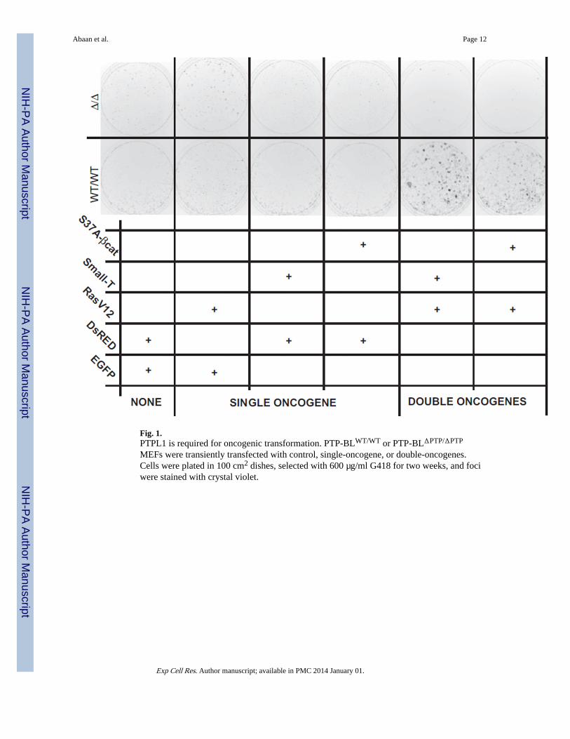
Fig. 1.PTPL1 is required for oncogenic transformation. PTP-BLWT/WT or PTP-BLΔPTP/ΔPTP
MEFs were transiently transfected with control, single-oncogene, or double-oncogenes.Cells were plated in 100 cm2 dishes, selected with 600 µg/ml G418 for two weeks, and fociwere stained with crystal violet.
Abaan et al. Page 12
Exp Cell Res. Author manuscript; available in PMC 2014 January 01.
NIH
-PA Author Manuscript
NIH
-PA Author Manuscript
NIH
-PA Author Manuscript
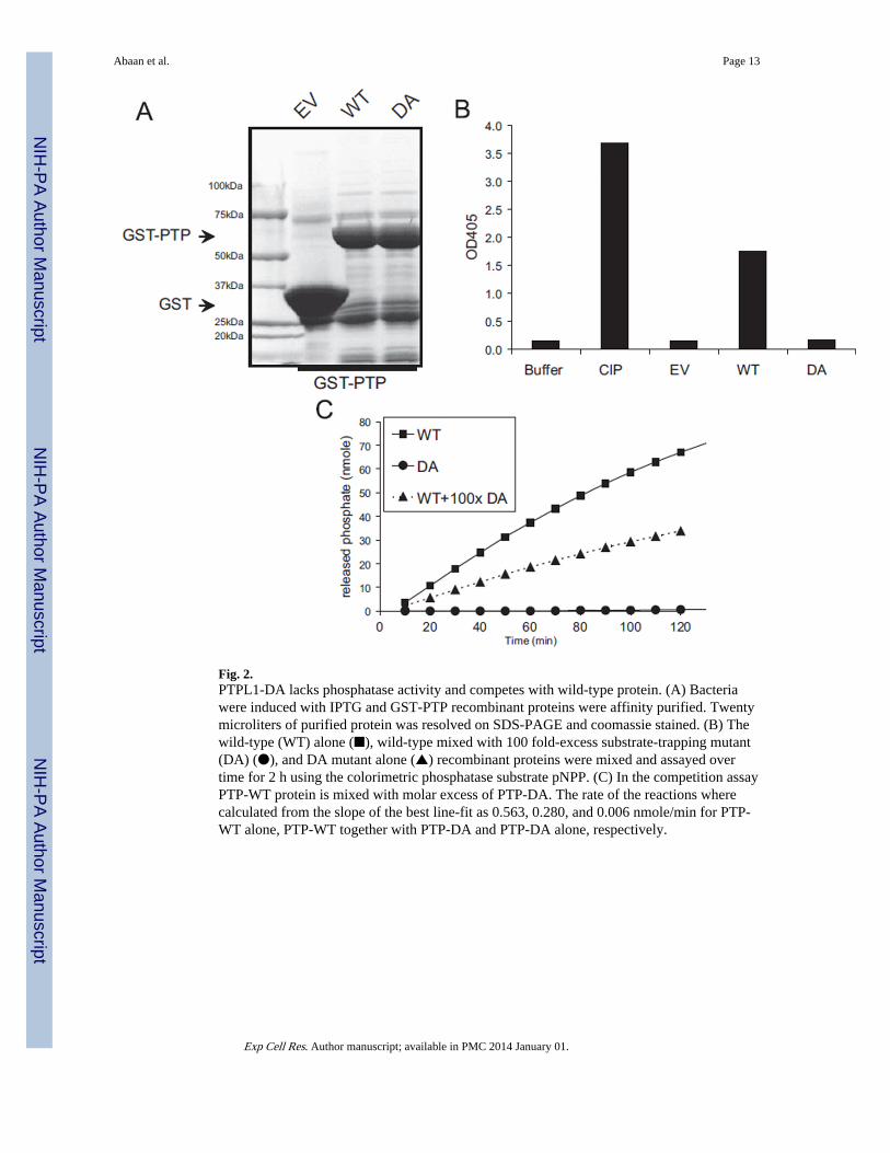
Fig. 2.PTPL1-DA lacks phosphatase activity and competes with wild-type protein. (A) Bacteriawere induced with IPTG and GST-PTP recombinant proteins were affinity purified. Twentymicroliters of purified protein was resolved on SDS-PAGE and coomassie stained. (B) Thewild-type (WT) alone (■), wild-type mixed with 100 fold-excess substrate-trapping mutant(DA) (●), and DA mutant alone (▲) recombinant proteins were mixed and assayed overtime for 2 h using the colorimetric phosphatase substrate pNPP. (C) In the competition assayPTP-WT protein is mixed with molar excess of PTP-DA. The rate of the reactions wherecalculated from the slope of the best line-fit as 0.563, 0.280, and 0.006 nmole/min for PTP-WT alone, PTP-WT together with PTP-DA and PTP-DA alone, respectively.
Abaan et al. Page 13
Exp Cell Res. Author manuscript; available in PMC 2014 January 01.
NIH
-PA Author Manuscript
NIH
-PA Author Manuscript
NIH
-PA Author Manuscript
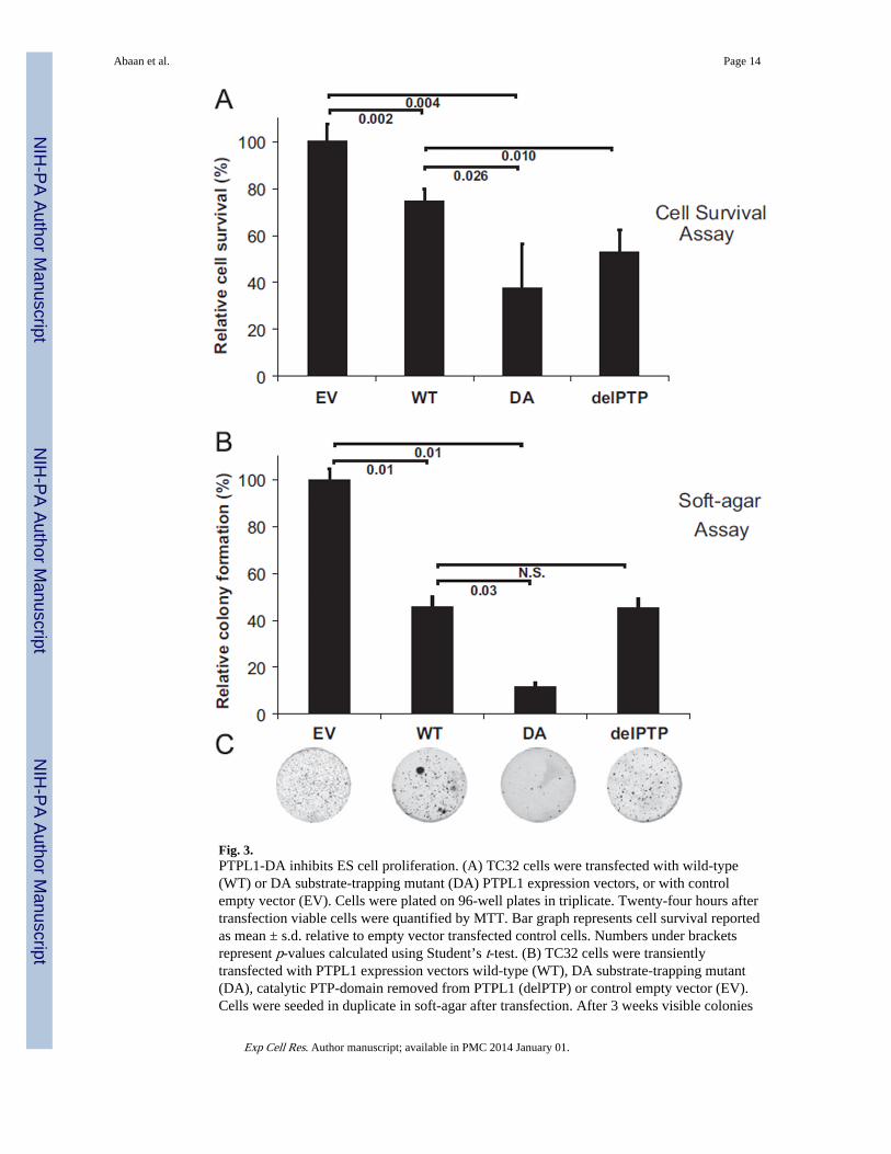
Fig. 3.PTPL1-DA inhibits ES cell proliferation. (A) TC32 cells were transfected with wild-type(WT) or DA substrate-trapping mutant (DA) PTPL1 expression vectors, or with controlempty vector (EV). Cells were plated on 96-well plates in triplicate. Twenty-four hours aftertransfection viable cells were quantified by MTT. Bar graph represents cell survival reportedas mean ± s.d. relative to empty vector transfected control cells. Numbers under bracketsrepresent p-values calculated using Student’s t-test. (B) TC32 cells were transientlytransfected with PTPL1 expression vectors wild-type (WT), DA substrate-trapping mutant(DA), catalytic PTP-domain removed from PTPL1 (delPTP) or control empty vector (EV).Cells were seeded in duplicate in soft-agar after transfection. After 3 weeks visible colonies
Abaan et al. Page 14
Exp Cell Res. Author manuscript; available in PMC 2014 January 01.
NIH
-PA Author Manuscript
NIH
-PA Author Manuscript
NIH
-PA Author Manuscript
were stained with MTT. Bar graph represents the colony counts reported as mean ± s.d.relative to empty vector transfected control cells. Numbers under brackets represent p-valuescalculated using Student’s t-test. Representative images from plates carrying colonies ofcells transfected with each construct is shown below the bar graph.
Abaan et al. Page 15
Exp Cell Res. Author manuscript; available in PMC 2014 January 01.
NIH
-PA Author Manuscript
NIH
-PA Author Manuscript
NIH
-PA Author Manuscript
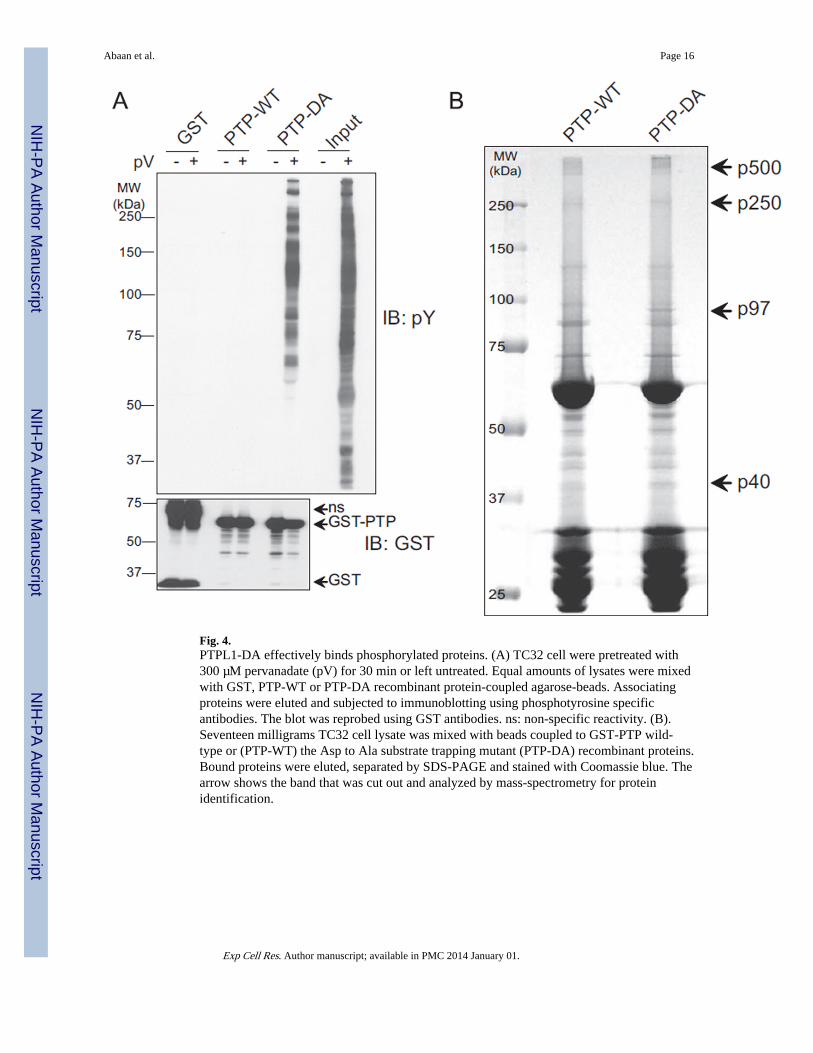
Fig. 4.PTPL1-DA effectively binds phosphorylated proteins. (A) TC32 cell were pretreated with300 µM pervanadate (pV) for 30 min or left untreated. Equal amounts of lysates were mixedwith GST, PTP-WT or PTP-DA recombinant protein-coupled agarose-beads. Associatingproteins were eluted and subjected to immunoblotting using phosphotyrosine specificantibodies. The blot was reprobed using GST antibodies. ns: non-specific reactivity. (B).Seventeen milligrams TC32 cell lysate was mixed with beads coupled to GST-PTP wild-type or (PTP-WT) the Asp to Ala substrate trapping mutant (PTP-DA) recombinant proteins.Bound proteins were eluted, separated by SDS-PAGE and stained with Coomassie blue. Thearrow shows the band that was cut out and analyzed by mass-spectrometry for proteinidentification.
Abaan et al. Page 16
Exp Cell Res. Author manuscript; available in PMC 2014 January 01.
NIH
-PA Author Manuscript
NIH
-PA Author Manuscript
NIH
-PA Author Manuscript
Fig. 5.Validation of VCP as a PTPL1 substrate. (A) Five microliters of GST pull-downs (similar toFig. 2A) were separated by SDS-PAGE and immunoblotted using VCP specific antibodies.Top panel is a 60 s exposure while the middle panel is a 1 s exposure of the VCP blot. Sameblot was reprobed for phosphotyrosine antibodies (bottom panel). (B) TC32 were cellstransiently transfected with either GFP or full-length Flag-tagged PTPL1-WT or full-lengthFlag-tagged PTPL1-DA expression vectors using electroporation and lysed after 24 h.Protein complexes were immunoprecipitated with Flag-M2-Agarose beads, andimmunoblotted using Flag-M5 (top panel) or VCP (bottom panel) antibodies. Numbersbelow the figure represent relative band density in arbitrary units determined bydensitometry as fold over PTPL1-WT for the Flag-M5 immunoblot or GFP for the VCPimmunoblot. (C) VCP immunoprecipitated from PTP-BLWT/WT or PTP-BLΔPTP/ΔPTP
MEFs maintained under regular growth conditions was immunoblotted withphosphotyrosine and VCP specific antibodies. Numbers below the figure represent relativeband density in arbitrary units determined by densitometry as fold over PTP-BLWT/WT. (D)VCP was immunoprecipitated from pervanadate treated TC32 cells. Immunoprecipitateswere aliquot and mixed with GST, PTP-WT or PTP-DA recombinant proteins for 1 h at 37°C in the presence or absence of ortho-vanadate. Reactions were run on SDS-PAGE and
Abaan et al. Page 17
Exp Cell Res. Author manuscript; available in PMC 2014 January 01.
NIH
-PA Author Manuscript
NIH
-PA Author Manuscript
NIH
-PA Author Manuscript
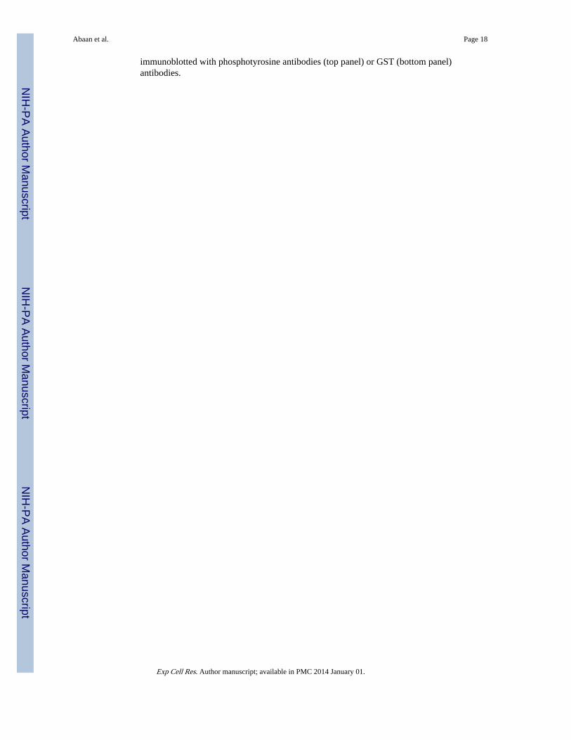
immunoblotted with phosphotyrosine antibodies (top panel) or GST (bottom panel)antibodies.
Abaan et al. Page 18
Exp Cell Res. Author manuscript; available in PMC 2014 January 01.
NIH
-PA Author Manuscript
NIH
-PA Author Manuscript
NIH
-PA Author Manuscript
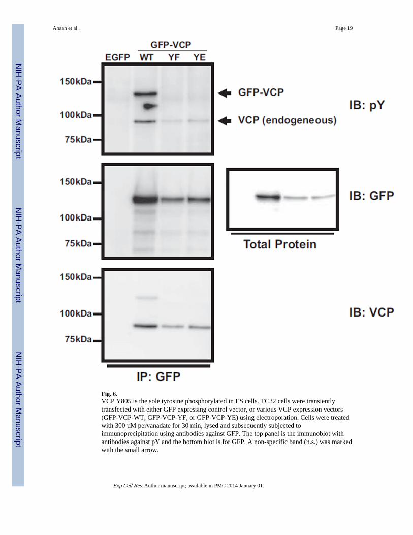
Fig. 6.VCP Y805 is the sole tyrosine phosphorylated in ES cells. TC32 cells were transientlytransfected with either GFP expressing control vector, or various VCP expression vectors(GFP-VCP-WT, GFP-VCP-YF, or GFP-VCP-YE) using electroporation. Cells were treatedwith 300 µM pervanadate for 30 min, lysed and subsequently subjected toimmunoprecipitation using antibodies against GFP. The top panel is the immunoblot withantibodies against pY and the bottom blot is for GFP. A non-specific band (n.s.) was markedwith the small arrow.
Abaan et al. Page 19
Exp Cell Res. Author manuscript; available in PMC 2014 January 01.
NIH
-PA Author Manuscript
NIH
-PA Author Manuscript
NIH
-PA Author Manuscript
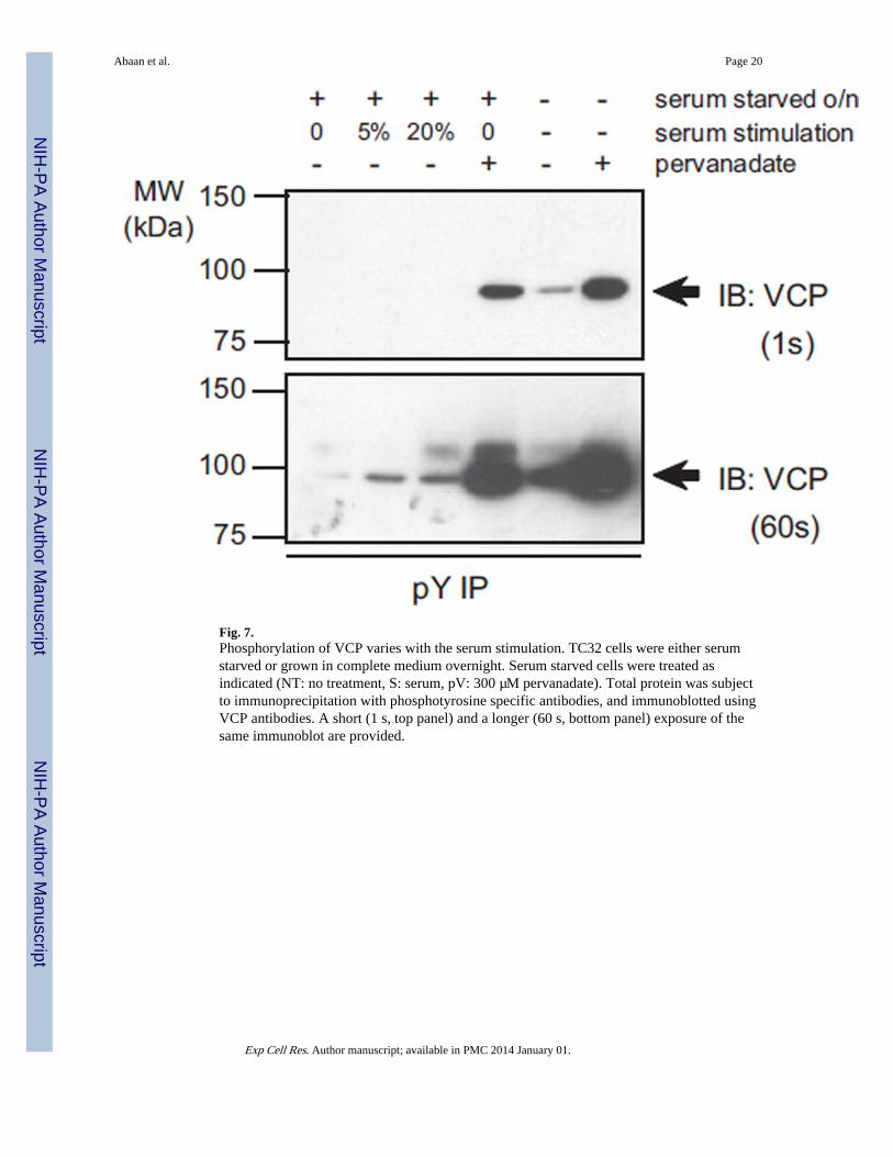
Fig. 7.Phosphorylation of VCP varies with the serum stimulation. TC32 cells were either serumstarved or grown in complete medium overnight. Serum starved cells were treated asindicated (NT: no treatment, S: serum, pV: 300 µM pervanadate). Total protein was subjectto immunoprecipitation with phosphotyrosine specific antibodies, and immunoblotted usingVCP antibodies. A short (1 s, top panel) and a longer (60 s, bottom panel) exposure of thesame immunoblot are provided.
Abaan et al. Page 20
Exp Cell Res. Author manuscript; available in PMC 2014 January 01.
NIH
-PA Author Manuscript
NIH
-PA Author Manuscript
NIH
-PA Author Manuscript
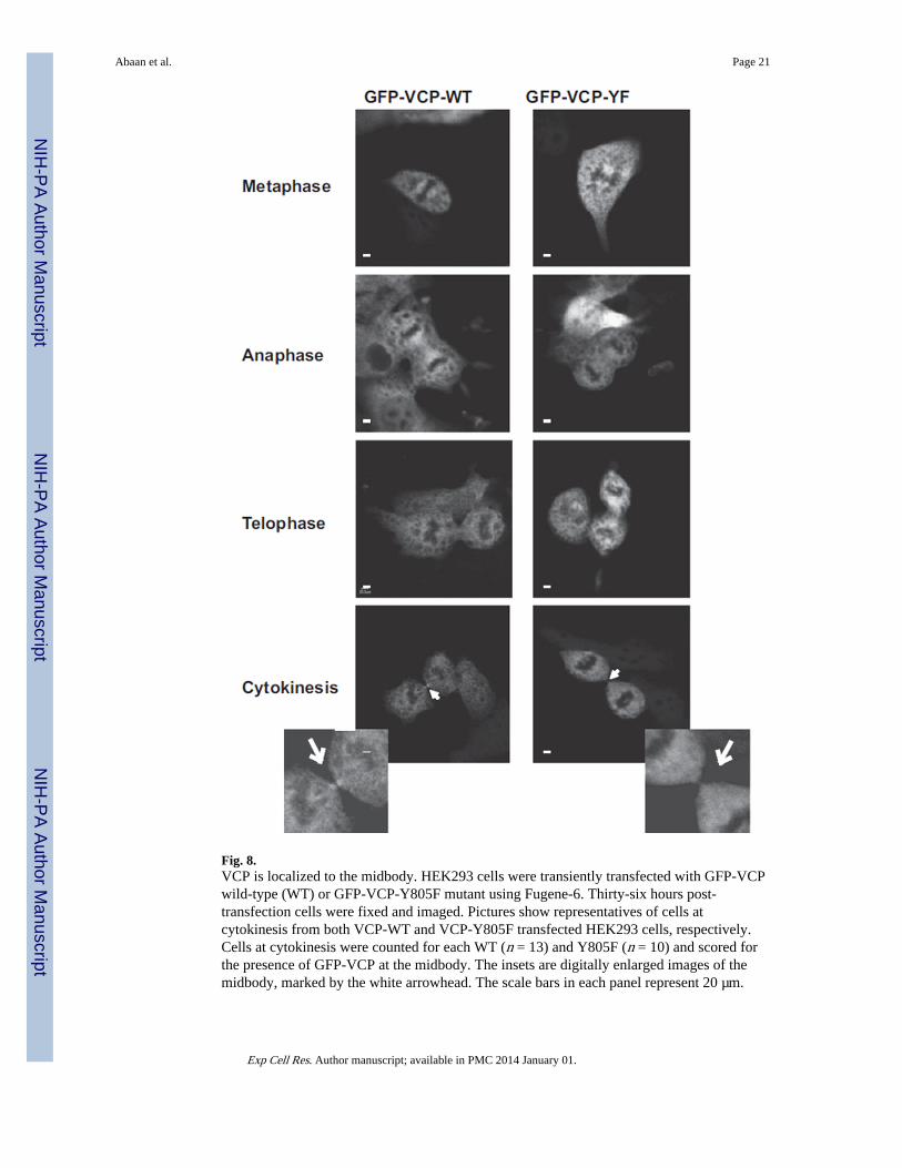
Fig. 8.VCP is localized to the midbody. HEK293 cells were transiently transfected with GFP-VCPwild-type (WT) or GFP-VCP-Y805F mutant using Fugene-6. Thirty-six hours post-transfection cells were fixed and imaged. Pictures show representatives of cells atcytokinesis from both VCP-WT and VCP-Y805F transfected HEK293 cells, respectively.Cells at cytokinesis were counted for each WT (n = 13) and Y805F (n = 10) and scored forthe presence of GFP-VCP at the midbody. The insets are digitally enlarged images of themidbody, marked by the white arrowhead. The scale bars in each panel represent 20 µm.
Abaan et al. Page 21
Exp Cell Res. Author manuscript; available in PMC 2014 January 01.
NIH
-PA Author Manuscript
NIH
-PA Author Manuscript
NIH
-PA Author Manuscript
NIH
-PA Author Manuscript
NIH
-PA Author Manuscript
NIH
-PA Author Manuscript
Abaan et al. Page 22
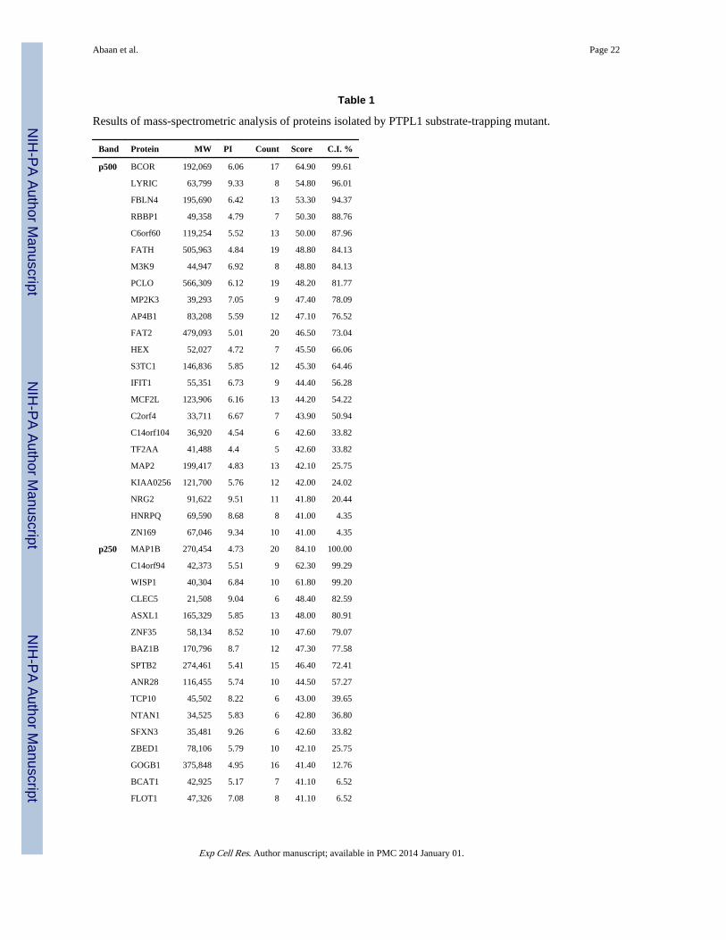
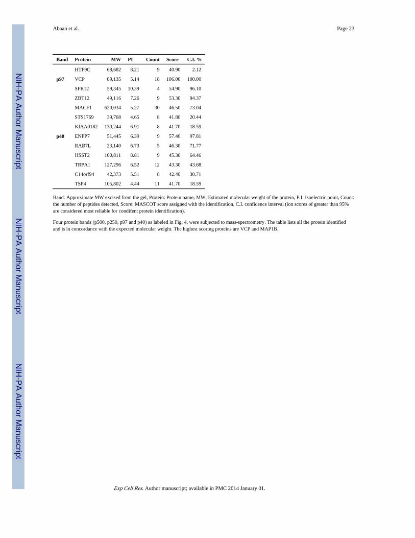
Table 1
Results of mass-spectrometric analysis of proteins isolated by PTPL1 substrate-trapping mutant.
Band Protein MW PI Count Score C.I. %
p500 BCOR 192,069 6.06 17 64.90 99.61
LYRIC 63,799 9.33 8 54.80 96.01
FBLN4 195,690 6.42 13 53.30 94.37
RBBP1 49,358 4.79 7 50.30 88.76
C6orf60 119,254 5.52 13 50.00 87.96
FATH 505,963 4.84 19 48.80 84.13
M3K9 44,947 6.92 8 48.80 84.13
PCLO 566,309 6.12 19 48.20 81.77
MP2K3 39,293 7.05 9 47.40 78.09
AP4B1 83,208 5.59 12 47.10 76.52
FAT2 479,093 5.01 20 46.50 73.04
HEX 52,027 4.72 7 45.50 66.06
S3TC1 146,836 5.85 12 45.30 64.46
IFIT1 55,351 6.73 9 44.40 56.28
MCF2L 123,906 6.16 13 44.20 54.22
C2orf4 33,711 6.67 7 43.90 50.94
C14orf104 36,920 4.54 6 42.60 33.82
TF2AA 41,488 4.4 5 42.60 33.82
MAP2 199,417 4.83 13 42.10 25.75
KIAA0256 121,700 5.76 12 42.00 24.02
NRG2 91,622 9.51 11 41.80 20.44
HNRPQ 69,590 8.68 8 41.00 4.35
ZN169 67,046 9.34 10 41.00 4.35
p250 MAP1B 270,454 4.73 20 84.10 100.00
C14orf94 42,373 5.51 9 62.30 99.29
WISP1 40,304 6.84 10 61.80 99.20
CLEC5 21,508 9.04 6 48.40 82.59
ASXL1 165,329 5.85 13 48.00 80.91
ZNF35 58,134 8.52 10 47.60 79.07
BAZ1B 170,796 8.7 12 47.30 77.58
SPTB2 274,461 5.41 15 46.40 72.41
ANR28 116,455 5.74 10 44.50 57.27
TCP10 45,502 8.22 6 43.00 39.65
NTAN1 34,525 5.83 6 42.80 36.80
SFXN3 35,481 9.26 6 42.60 33.82
ZBED1 78,106 5.79 10 42.10 25.75
GOGB1 375,848 4.95 16 41.40 12.76
BCAT1 42,925 5.17 7 41.10 6.52
FLOT1 47,326 7.08 8 41.10 6.52
Exp Cell Res. Author manuscript; available in PMC 2014 January 01.
NIH
-PA Author Manuscript
NIH
-PA Author Manuscript
NIH
-PA Author Manuscript
Abaan et al. Page 23
Band Protein MW PI Count Score C.I. %
HTF9C 68,682 8.21 9 40.90 2.12
p97 VCP 89,135 5.14 18 106.00 100.00
SFR12 59,345 10.39 4 54.90 96.10
ZBT12 49,116 7.26 9 53.30 94.37
MACF1 620,034 5.27 30 46.50 73.04
STS1769 39,768 4.65 8 41.80 20.44
KIAA0182 130,244 6.91 8 41.70 18.59
p40 ENPP7 51,445 6.39 9 57.40 97.81
RAB7L 23,140 6.73 5 46.30 71.77
HSST2 100,811 8.81 9 45.30 64.46
TRPA1 127,296 6.52 12 43.30 43.68
C14orf94 42,373 5.51 8 42.40 30.71
TSP4 105,802 4.44 11 41.70 18.59
Band: Approximate MW excised from the gel, Protein: Protein name, MW: Estimated molecular weight of the protein, P.I: Isoelectric point, Count:the number of peptides detected, Score: MASCOT score assigned with the identification, C.I. confidence interval (ion scores of greater than 95%are considered most reliable for condifent protein identification).
Four protein bands (p500, p250, p97 and p40) as labeled in Fig. 4, were subjected to mass-spectrometry. The table lists all the protein identifiedand is in concordance with the expected molecular weight. The highest scoring proteins are VCP and MAP1B.
Exp Cell Res. Author manuscript; available in PMC 2014 January 01.
Copyright © 2022 FDOKUMEN