Effects of nitration on the structure and aggregation of α-synuclein
Upload
independentCategory
view
1download
0
Free Radical Biology & Medicine 50 (2011) 749–762
Contents lists available at ScienceDirect
Free Radical Biology & Medicine
j ourna l homepage: www.e lsev ie r.com/ locate / f reeradb iomed
Original Contribution
Factors influencing protein tyrosine nitration—structure-based predictive models
Alexander S. Bayden a, Vasily A. Yakovlev b, Paul R. Graves b, Ross B. Mikkelsen b,⁎, Glen E. Kellogg a,⁎a Department of Medicinal Chemistry and Institute for Structural Biology and Drug Discovery, Massey Cancer Center, Virginia Commonwealth University, Richmond, VA 23298, USAb Department of Radiation Oncology, Massey Cancer Center, Virginia Commonwealth University, Richmond, VA 23298, USA
⁎ Corresponding authors.E-mail addresses: [email protected] (R.B. Mikkelsen
(G.E. Kellogg).
0891-5849/$ – see front matter © 2010 Elsevier Inc. Aldoi:10.1016/j.freeradbiomed.2010.12.016
a b s t r a c t
a r t i c l e i n f oArticle history:Received 8 October 2010Revised 15 November 2010Accepted 10 December 2010Available online 21 December 2010
Keywords:Tyrosine nitrationHydropathic interactionsOxidative stressTyrosyl radicalFree radicals
Models for exploring tyrosine nitration in proteins have been created based on 3D structural features of 20proteins for which high-resolution X-ray crystallographic or NMR data are available and for which nitration of35 total tyrosines has been experimentally proven under oxidative stress. Factors suggested in previous workto enhance nitration were examined with quantitative structural descriptors. The role of neighboring acidicand basic residues is complex: for the majority of tyrosines that are nitrated the distance to the heteroatom ofthe closest charged side chain corresponds to the distance needed for suspected nitrating species to formhydrogen bond bridges between the tyrosine and that charged amino acid. This suggests that such bridgesplay a very important role in tyrosine nitration. Nitration is generally hindered for tyrosines that are buriedand for those tyrosines for which there is insufficient space for the nitro group. For in vitro nitration, closedenvironments with nearby heteroatoms or unsaturated centers that can stabilize radicals are somewhatfavored. Four quantitative structure-based models, depending on the conditions of nitration, have beendeveloped for predicting site-specific tyrosine nitration. The best model, relevant for both in vitro and in vivocases, predicts 30 of 35 tyrosine nitrations (positive predictive value) and has a sensitivity of 60/71 (11 falsepositives).
l rights reserved.
© 2010 Elsevier Inc. All rights reserved.
Investigation of protein tyrosine nitration has intensified over thepast 2 decades, leading to a better understanding of the role of thisposttranslational modification in cellular signaling [1]. Althoughinitially considered to be a marker of oxidative stress, there is agrowing body of experimental data suggesting that nitration oftyrosine fulfills the criteria of a signal-transducing mechanism [1,2].For example, tyrosine nitration has been detected under physiologicalconditions in most organ systems and in a number of cellular models.Furthermore, accumulating data support a strong link betweenprotein tyrosine nitration and the activation of signaling pathwaysin a variety of cellular responses and pathological conditions,including the cellular response to irradiation, acute and chronicinflammation, graft rejection, chronic hypoxia, tumor vascularizationand the microenvironment, atherosclerosis, myocardial infarction,chronic obstructive pulmonary disease, diabetes, Parkinson disease,and Alzheimer disease [3–20].
Nitration at tyrosine residues occurs both in vitro and in vivo.Generally, one of the two tyrosine aromatic hydrogens that are orthowith respect to the hydroxyl group is replaced by a nitro group.However, in some instances, nitrotyrosine can further react and replacea second hydrogen atom with another nitro group. Most commonly,tyrosine is nitrated posttranslationally in two steps as shown in
Scheme 1. First, the tyrosine is oxidized to a tyrosyl radical, which inone of its resonance forms is nitrated in the second step. Severalchemical nitrating species have been implicated in tyrosine nitration,including •NO, •NO2, ONOO−, O2
•−, H2O2, NO2−, NO2CO3
−, and CO3•− [21].
NO2Cl has also been suggested as a contributor; however, its role intyrosinenitration in vivo is highly unlikely [22]. Althoughmost evidencesuggests that tyrosine nitration occurs posttranslationally, it is formallypossible that tyrosine does not have to undergo nitration within theprotein. Nitrotyrosine can be transported into cells and then incorpo-rated in proteins during translation [23]. In this case, however, therandom incorporation of nitrotyrosine residues into the protein shouldbe observed and this is not the case in vivo.
Tyrosine nitration in proteins does not occur randomly. Mostproteins contain tyrosine residues (natural abundance: 3.2%) [24],and tyrosine is often surface-exposed in proteins (only 15% of tyrosineresidues are at least 95% buried) and should be easily nitrated [1,25].However, not all exposed tyrosine residues and not all proteins arenitrated. Neither does the abundance of tyrosine residues in a givenprotein predict whether it is a target for nitration [1,25,26]. Thisevidence strongly argues that protein tyrosine nitration is a selectiveprocess.
Specific amino acid sequences determine the specificity for otherposttranslational modifications involving tyrosine. For example, thepeptide sequence surrounding a tyrosine residue contribute to thesubstrate specificity of tyrosine kinases [27]. An analysis of theprimary sequences of proteins nitrated under similar conditions failedto reveal a specific or unique sequence requirement [26].

Scheme 1. One mechanism for tyrosine nitration.
750 A.S. Bayden et al. / Free Radical Biology & Medicine 50 (2011) 749–762
Several studies have attempted to determine factors that promoteselective tyrosine nitration. It has been reported that tyrosines aremore likely to become nitrated when they are in loops [28], whichcan be expressed as its location with respect to turn-inducing aminoacids such as glycine and proline [25]. The role of sulfur-containingamino acids in tyrosine nitration has been debated. Some evidencesuggests that these amino acids impede tyrosine nitration [26,29] bycompeting with tyrosine for nitrating species, whereas otherscontradict this finding [30], suggesting that sulfur-containingamino acids promote tyrosine nitration. Souza and co-investigatorssuggest that the presence of acidic residues near the target tyrosinemakes it more susceptible to nitration [31]. Lin et al. have shownthat when glutamic acid 149 is mutated to alanine in cytochromeP450 2B1, a protein known to be nitrated at tyrosine 190, thisnitration is substantially reduced [32]. It has also been shown thattyrosine nitration is facilitated by the presence of a nearby basicamino acid [30] and that the hydrophobicity or hydrophilicity of theenvironment influences tyrosine nitration [33–35]. Finally, thepresence of transition metals seems to encourage nitration oftyrosines [36–39].
Table 1Proteins and residues used in tyrosine nitration study
Protein PDB code Nitrated tyrosines
In vivo nitrationMuscle creatine kinase 1i0e 14, 20Actin 1j6z 91, 198, 240Dihydropyrimidinase-related protein 1 1kcx 316a
IκBα (monomeric) 1ikn 181a
Calcineurin 1m63 224
Mitochondrial creatine kinase 1qk1 274Clathrin adaptor protein complex 1 1w63 574a
Kelch-like ECH-associated protein 1 1u6d 345, 491, 537Escherichia coli ribonucleotide reductase protein R2 1rib 122a, 273, 289Muscle glycogen phosphorylase 2amv 113, 161
14-3-3 β 2bq0 84p53 (tetramerization domain)b 2j0c 327Triosephosphate isomerase 2jk2 67a, 208Brain-type creatine kinase, B chain 3b6r 269Fructose-1,6-bisphosphate aldolase 3bv4 173, 203In vitro nitrationsp53 (DNA-binding domain) 2fej 107Bovine ribonuclease A 1jvt 115a
Bovine Cu Zn SOD 1e9q 108Peroxiredoxin I 2rii NoneActin 1j6z 53, 198, 240, 362E. coli ribonucleotide reductase protein R2 1rib 62, 122a
Muscle creatine kinase 1i0e 82Bacteriorhodopsin 1brd 26Porcine aconitase 1boj NoneGST-1 2h8a 92Lysozyme 2zxs 20, 23
PDB, Protein Data Bank.a These tyrosine environments were represented in model training by single subunits altb Experimental evidence suggests that p53 is nitrated in its monomeric form. This protei
Unfortunately, to date there is still no reliable model for predictingtyrosine nitration, as single factors do not satisfactorily explain itsselectivity. Other studies have stressed the importance of the proteinsecondary structure and the local “structural environment” ofnitrotyrosine sites [1,25,26]. For example, Gow et al. proposed amechanism defining specificity of tyrosine nitration that requiresconsideration of the local environment of tyrosine residues within thesecondary and tertiary structures of a protein [2].
In this paper we report the first comprehensive and quantitativeinvestigation of protein structural features that influence tyrosinenitration and in so doing have tested the above anecdotal andqualitative proposals. We have also created an extensive set ofadditional possible structural factors with potential roles in site-specific tyrosine nitration. Our analysis tested these structural metricsagainst a training set of known nitrated and (probably) nonnitratedtyrosines; thus, we have built statistical models that can predicttyrosine nitration under different conditions.
Materials and methods
Data set
The nitrated tyrosineswe have considered in this work are listed inTable 1. They consist of three overlapping sets: proteins that arenitrated in vitro by chemical means, proteins that are nitrated in vivoby physiological mechanisms, and the union set of all proteins that arenitrated. Thus, results in this work are referenced to these data sets:“in vivo,” “in vitro,” or “All.” For the in vivo and in vitro data sets, thepositive controls were tyrosines that have been shown experimen-tally to be nitrated (see Table 1). For these data sets, we used asnegative controls tyrosines in those same proteins that were notreported as nitrated. Additional negative controls available forperoxiredoxin I and porcine aconitase were also used in training our
Tyrosines used as nonnitrated controls Ref.
39, 82, 125, 140, 173, 174, 279 [40]53, 69, 133, 143, 166, 169, 188, 218, 279, 294, 306, 337, 362 [41]32, 36, 75, 135, 145, 167, 170, 174, 182, 251, 290, 336, 395, 431, 479 [30]195, 248, 251, 254, 289 [42]113, 119, 124, 132, 140, 159, 170, 175, 258, 260, 262, 288, 291, 311,315, 324, 341
[30]
9, 15, 34, 77, 95, 115, 120, 168, 169, 354 [30]6, 70, 72, 76, 121, 136, 229, 276, 277, 300, 328, 333, 361, 405, 421,425, 455, 524, 526, 566
[30]
329, 334, 341, 375, 396, 426, 443, 473, 490, 520, 525, 567, 572, 584 [43]2, 28, 33, 62, 79, 156, 157, 166, 194, 209, 307, 310 [44]51, 52, 83, 84, 185, 203, 226, 233, 262, 280, 297, 374, 404, 472, 511,524, 548, 553, 587, 613, 726, 731, 732, 777, 780, 791, 820
[40]
21, 50, 106, 120, 127, 130, 151, 180, 181, 213 [30]None [45]47, 164 [30]39, 68, 82, 100, 125, 173, 174, 279 [30]58, 84, 137, 213, 222, 243, 301, 327, 342 [30,43]
103, 126, 163, 205, 220, 234, 236 [45]25, 73,76, 92, 97 [25]None [46]116 [46]69, 91, 133, 143, 166, 169, 188, 218, 279, 294, 306, 337 [44]2, 28, 33, 79, 156, 157, 166, 194, 209, 273, 289, 307, 310 [47]14, 20, 39, 125, 140, 173, 174, 279 [43]43, 57, 79, 83, 147, 150, 185 [48]43, 60, 74, 96, 136, 178, 206, 223, 268, 274, 301, 306 [49]18, 115, 120, 137, 145 [50]53 [25]
hough there are potentially interacting residues in other subunits of that protein.n/residue was not used in training, but only in testing the model.
751A.S. Bayden et al. / Free Radical Biology & Medicine 50 (2011) 749–762
models (Table 1). For the All data set, the positive controls weretyrosines used in the two independent data sets. However, becausenitration conditions are typically much harsher in vitro than in vivo,we could not confidently use the in vivo negative controls in the Alldata set and so used only the in vitro negative controls.
Molecular modeling
Protein structures were obtained from the RCSB Protein Data Bank[51] and manipulations on them were performed with the Sybyl 7.3molecular modeling program [52]. For each protein in the training setonly one chain containing nitrated tyrosines was used for buildingmodels. Additional chains, ligands, waters, ions, and prosthetic groupswere removed. Missing hydrogens were added to the remaining chainusing tools in the Sybyl Biopolymer suite.
For each tyrosine examined in this study, three molecular modelswere built: first, for the wild-type (nonnitrated) protein; second, forthe tyrosine nitrated at the 3-position (CE1); and third, for thetyrosine nitrated at the 5-position (CE2). To nitrate at the 3- or the 5-position, the appropriate hydrogen atom (HE1 or HE2) was replacedby a nitro group. All models, nitrated or not, were geometry optimizedfor all atoms within 7 Å of the phenyl oxygen of the tyrosine with thefollowing settings: force field, Tripos; dielectric constant, 6; charges,Gasteiger-Hückel; optimization algorithm, Powell; termination crite-ria, energy gradient b0.05 kcal/Å-mol or 5000 iterations.
Some proteins in the training set had multiple subunits (seeTable 1). In most cases we did not have definitive information aboutwhether the auxiliary subunits (i.e., not containing the tyrosine ofinterest) are present at the time and under the conditions when thenitration occurs (as opposed to the conditions under which the X-raycrystals were grown). For consistency, we used only the chain/subunitcontaining the nitrated tyrosine for model training, deleting others.However, we recognized that additional subunits could have animpact on tyrosine nitration. So, for the multimeric proteins, we latertested our one-chain training set models on the all-subunit proteinmolecular models.
Hydropathic analysis and computational titration
A number of our descriptors and structural analysis tools involvedhydropathic analysis using the HINT program [53,54]. HINT is acomputational model and empirical free-energy force field based onthe partitioning of small molecules between 1-octanol and water, i.e.,log Po/w. HINT interaction scores are calculated as the total of atom–
atom pair-wise interactions; they have been shown to correlate withfree energy of association [54]. Computational titration is anapplication based on HINT that optimizes the placement andorientation of protons so as to have an idealized interaction betweenthe “ligand” and the “receptor.” This idealized interaction may includeprotonation or deprotonation of basic and acidic residues, and/orligand functional groups, respectively [55–58]. For computationaltitration the protein model was separated into two parts: tyrosine asthe pseudo-ligand and the rest of the protein as the pseudo-receptor.The backbone portions of these two fragments are at covalent(bonding) distance from each other and steric repulsion wouldcompletely dominate the computed HINT interactions, so thebackbone portion of the tyrosine was deleted such that the remainderof the tyrosine, i.e., only its side chain, was treated as if it were aligand.
Computational titration was performed on our public Web serverat http://hinttools.isbdd.vcu.edu/CT [59] with the following settings:rotation of amide groups on asparagine and glutamine residues wasallowed; on acid residues, aspartic and glutamic acid, and on C-termini the carboxylate/carboxylic acid groups were titrated andhydrogens, in the acid case, were allowed to rotate; histidine wastitrated and its ring orientationwas checked; lysine and argininewere
titrated and for unprotonated lysine the amine rotation wasoptimized, whereas for unprotonated arginine the hydrogen place-ment was optimized; for serine, threonine, and tyrosine the –XH(X=O, S) rotation was optimized, whereas for tyrosine the hydroxylgroup was also titrated.
Statistical model building
The number of potentially significant descriptors coded for thisstudy was large and many were correlated. Thus, it was impractical togenerate models manually and we developed an automated approachwith a stochastic algorithm scheme based on ant-colony optimization[60–62], which has been successfully used in constructing QSARmodels [63]. In our implementation of the ant-colony-based algo-rithm each descriptor–value pair was assigned some amount of“pheromone.” In each iteration, a fixed number of descriptor–valuepairs were picked randomly with a probability proportional to theamount of “pheromone” associated with them. If it was possible toconstruct a model from these descriptor–value pairs that was betterthan any other model found so far in that run, then the pickeddescriptor–value pairs were rewarded with the amount of “phero-mone” proportional to the number of cycles needed to find this newmodel after finding the previous “best” model.
For the largest amount of flexibility in proposing mechanisms, weconstructed independent statistical models for three sets of tyrosinenitration data: nitrations that occur in vivo, nitrations that occur invitro, and All nitrations (the union set). To avoid constructingoverdetermined and statistically irrelevant models due to the limitedtraining data available, our algorithm allowed each model to includeonly a limited number (3 or 4) of descriptor–value pairs. In analogy tothe ant-colony model, these descriptor–value pairs are path segmentsand a model is an entire path of three or four segments. Each pair hada threshold score value assigned to it. If a calculated descriptor valuefor a particular tyrosine was greater than its threshold value, then thatcalculated value was added to a total score for that tyrosine. A modelcreated in this way also included a critical value of the final score,which is, in effect, a metric for ant-trail path length. Ideally, tyrosineswith a total score over that critical value were nitrated and mosthaving a total score below that value were not.
Results
To probe the many assertions made in the past concerningstructural factors important to tyrosine nitration, we created a largenumber of descriptors (summarized in Supplementary Table S1) thatrepresented these factors in terms of quantitative and testablehypotheses. Throughout this contribution (including the supplemen-tary material), histograms are used to illustrate the responsiveness ofnitration to a panel of structural descriptors. In all of these figures,vertical black bars represent tyrosines in the data set(s) that arenitrated and vertical white bars represent those that are not. The yaxes of these diagrams represent the relative fractions of nitrated andnonnitrated tyrosines as a function of the x-axis descriptors. Note thatthere are many more nonnitrated tyrosines in the data sets thannitrated tyrosines, so each white, nonnitrated graph bar representsseveral times more tyrosines than a similarly sized black bar.Additionally, inset into most of these plots is a horizontal bar at anotable break point that indicates the fraction of tyrosines nitrated(and not nitrated) above or below that break point. This indicates anoverall usefulness of the descriptor in differentiating the two cases—a“perfect” descriptor would show 100% on both sides of the breakpoint. Under Discussion, we build multiple-descriptor models thatattempt to combine the chemical and structural information from themost successful of the various hypotheses.
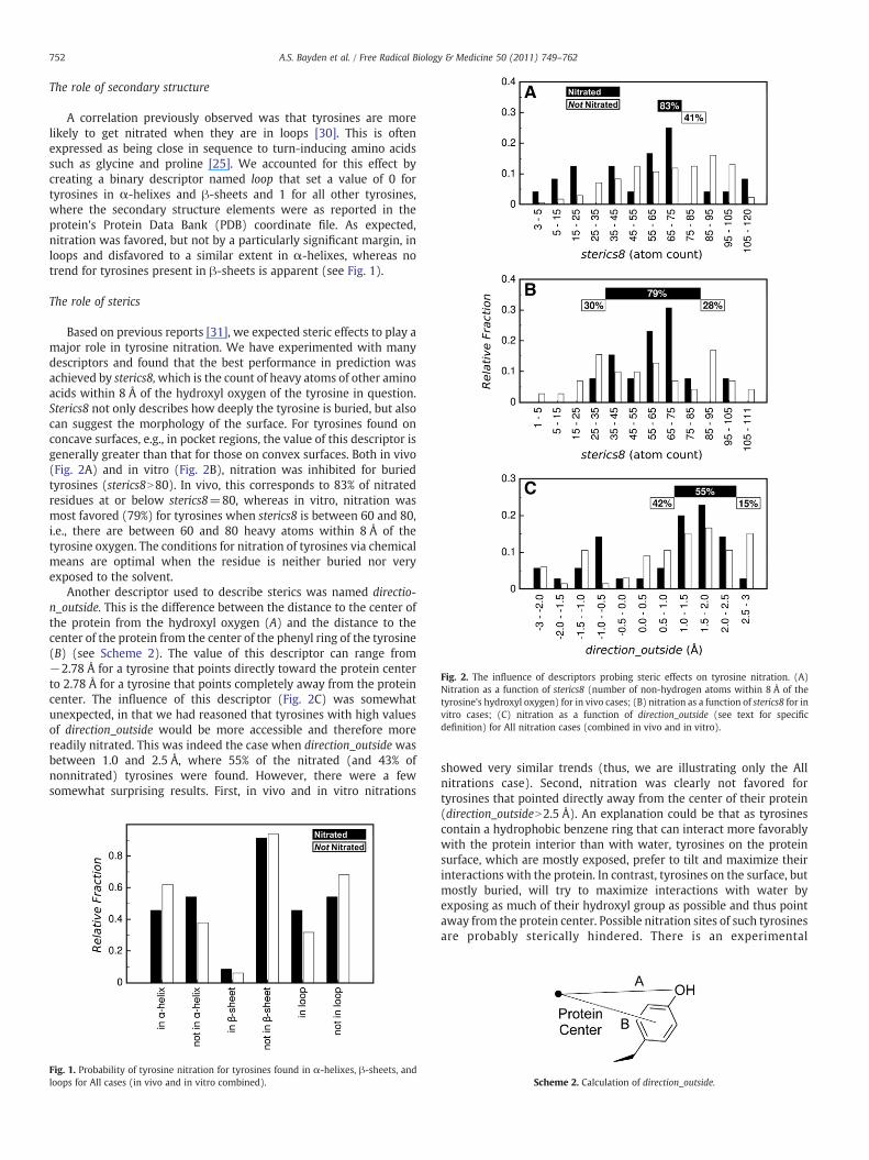
Fig. 2. The influence of descriptors probing steric effects on tyrosine nitration. (A)Nitration as a function of sterics8 (number of non-hydrogen atoms within 8 Å of thetyrosine's hydroxyl oxygen) for in vivo cases; (B) nitration as a function of sterics8 for invitro cases; (C) nitration as a function of direction_outside (see text for specificdefinition) for All nitration cases (combined in vivo and in vitro).
752 A.S. Bayden et al. / Free Radical Biology & Medicine 50 (2011) 749–762
The role of secondary structure
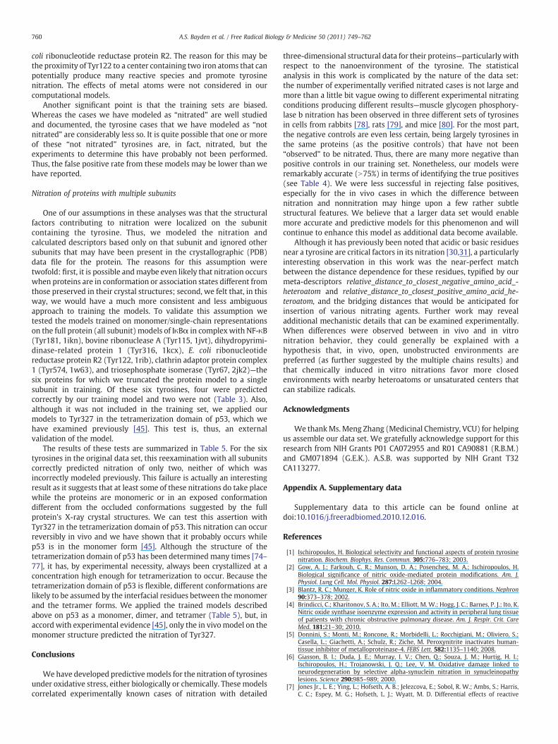
A correlation previously observed was that tyrosines are morelikely to get nitrated when they are in loops [30]. This is oftenexpressed as being close in sequence to turn-inducing amino acidssuch as glycine and proline [25]. We accounted for this effect bycreating a binary descriptor named loop that set a value of 0 fortyrosines in α-helixes and β-sheets and 1 for all other tyrosines,where the secondary structure elements were as reported in theprotein's Protein Data Bank (PDB) coordinate file. As expected,nitration was favored, but not by a particularly significant margin, inloops and disfavored to a similar extent in α-helixes, whereas notrend for tyrosines present in β-sheets is apparent (see Fig. 1).
The role of sterics
Based on previous reports [31], we expected steric effects to play amajor role in tyrosine nitration. We have experimented with manydescriptors and found that the best performance in prediction wasachieved by sterics8,which is the count of heavy atoms of other aminoacids within 8 Å of the hydroxyl oxygen of the tyrosine in question.Sterics8 not only describes how deeply the tyrosine is buried, but alsocan suggest the morphology of the surface. For tyrosines found onconcave surfaces, e.g., in pocket regions, the value of this descriptor isgenerally greater than that for those on convex surfaces. Both in vivo(Fig. 2A) and in vitro (Fig. 2B), nitration was inhibited for buriedtyrosines (sterics8N80). In vivo, this corresponds to 83% of nitratedresidues at or below sterics8=80, whereas in vitro, nitration wasmost favored (79%) for tyrosines when sterics8 is between 60 and 80,i.e., there are between 60 and 80 heavy atoms within 8 Å of thetyrosine oxygen. The conditions for nitration of tyrosines via chemicalmeans are optimal when the residue is neither buried nor veryexposed to the solvent.
Another descriptor used to describe sterics was named directio-n_outside. This is the difference between the distance to the center ofthe protein from the hydroxyl oxygen (A) and the distance to thecenter of the protein from the center of the phenyl ring of the tyrosine(B) (see Scheme 2). The value of this descriptor can range from−2.78 Å for a tyrosine that points directly toward the protein centerto 2.78 Å for a tyrosine that points completely away from the proteincenter. The influence of this descriptor (Fig. 2C) was somewhatunexpected, in that we had reasoned that tyrosines with high valuesof direction_outside would be more accessible and therefore morereadily nitrated. This was indeed the case when direction_outside wasbetween 1.0 and 2.5 Å, where 55% of the nitrated (and 43% ofnonnitrated) tyrosines were found. However, there were a fewsomewhat surprising results. First, in vivo and in vitro nitrations
Fig. 1. Probability of tyrosine nitration for tyrosines found in α-helixes, β-sheets, andloops for All cases (in vivo and in vitro combined).
showed very similar trends (thus, we are illustrating only the Allnitrations case). Second, nitration was clearly not favored fortyrosines that pointed directly away from the center of their protein(direction_outsideN2.5 Å). An explanation could be that as tyrosinescontain a hydrophobic benzene ring that can interact more favorablywith the protein interior than with water, tyrosines on the proteinsurface, which are mostly exposed, prefer to tilt and maximize theirinteractions with the protein. In contrast, tyrosines on the surface, butmostly buried, will try to maximize interactions with water byexposing as much of their hydroxyl group as possible and thus pointaway from the protein center. Possible nitration sites of such tyrosinesare probably sterically hindered. There is an experimental
Scheme 2. Calculation of direction_outside.

Fig. 3. The influence of two descriptors probing the role of backbone and polar/nonpolar side chains on tyrosine nitration. (A) Nitration as a function ofdistance_to_backbone, which measures the distance from the hydroxyl oxygen ofthe tyrosine to the closest heavy backbone atom of another amino acid in in vivocases; (B) nitration as a function of distance_to_backbone for in vitro cases; (C)nitration as a function of distance_to_polar_sidechain for in vivo cases; (D) nitrationas a function of distance_to_polar_sidechain for in vitro cases.
753A.S. Bayden et al. / Free Radical Biology & Medicine 50 (2011) 749–762
consideration that must also be considered in this argument—surfaceresidues, such as tyrosines standing up on the surface, generally haveless certain coordinates in crystallography because of thermal motionand other factors that lead to disorder. Also interesting was thatnitration was strongly favored for tyrosines within the narrow rangeof direction_outside of−1.0 to−0.5 Å. These tyrosines are tilted awayto a modest degree from the protein surface.
The roles of backbone and polar/nonpolar side chains
Although it is easier to transmit charge from one location in aprotein to another through its backbone [64,65], side-chain atomsnear the backbone are less likely to be on the protein surface. Toquantitatively describe these competing effects we introduced threedescriptors, distance_to_backbone_O, distance_to_backbone_N, anddistance_to_backbone, to measure the distance from the hydroxyloxygen of the target tyrosine to the closest oxygen, nitrogen, or anyheavy atom of the backbone, respectively (with the exception of thoseof target tyrosine). For the last descriptor, no clear trends wereobserved in vivo (Fig. 3A), although nitration is slightly favored fortyrosines when distance_to_backbone is greater than 5 Å (50%nitrated), where they are more exposed, more accessible to nitratingspecies, and better able to participate in protein–protein interactions.Somewhat more clear is that in vitro nitration (Fig. 3B) is favored fortyrosines for which distance_to_backbone is under 4 Å (57% nitrated),which is consistent with a mechanistic role for charge transferreactions [66,67], as might be expected for chemically inducednitration.
It has been previously reported that hydrophobicity and/orhydrophilicity of the surrounding environment influences tyrosinenitration [33–35]. We used three descriptors to help us in assessingthe hydrophobicity of the environment: distance_to_polar_sidechain,distance_to_non-polar_sidechain, and distance_to_neutral_sidechain.These descriptors measured the shortest distance from the hydroxyloxygen of tyrosine to the closest heavy (non-hydrogen) atom in theside chain of polar (arginine, asparagine, aspartic acid, glutamic acid,glutamine, histidine, lysine, serine, threonine, and tyrosine), nonpolar(alanine, cysteine, glycine, isoleucine, leucine, methionine, phenylal-anine, proline, tryptophan, and valine), or neutral (alanine, aspara-gine, cysteine, glutamine, glycine, isoleucine, leucine, methionine,phenylalanine, proline, serine, threonine, tryptophan, tyrosine, andvaline) amino acid residues. The most useful descriptor of these threewas distance_to_polar_sidechain. Proximity of the tyrosine hydroxyl toheavy atoms of polar side chains had somewhat opposite effects invitro and in vivo (see Figs. 3C and D). In vivo, tyrosine nitration wasdisfavored (65% not nitrated) when the closest heavy atom belongingto a polar side chain was less than 4 Å away from the tyrosine'shydroxyl oxygen. In vitro, nitration was favored (93% nitrated) whensuch an atom was less than 5 Å away, again supporting the possibleinvolvement of a charge-transfer mechanism for these nitrations.Similarly, distance_to_neutral_sidechain (data not shown) indicatedthat nitration was disfavored for tyrosines when the hydroxyl oxygenwas over 4 Å away from the closest heavy atom of the neutral sidechain.
The role of acidic and basic residues
Some have argued that the presence of acidic residues near thetarget tyrosine makes it more susceptible to nitration [31], whereasothers have shown that tyrosine nitration is facilitated by the presenceof a nearby basic amino acid residue [30]. In addition, nitratedtyrosines have been shown to interact favorably with arginine [42,45].We modeled the effects of nearby basic and acidic amino acids withseveral descriptors. First, because the mechanism of nitration mayinvolve a tyrosyl radical, we assessed the relative abilities of thetyrosines’ environments to support this with protonation_difference—
the difference in the protein-tyrosine HINT scores between the bestscoring computational titration model in which the target tyrosine isprotonated (neutral) and the best scoring model in which it isdeprotonated (tyrosinate). The value of this descriptor is greaterwhen

754 A.S. Bayden et al. / Free Radical Biology & Medicine 50 (2011) 749–762
proton acceptor groups such as deprotonated carboxylates are close tothe hydroxyl oxygen of tyrosine and can “accept” the tyrosyl protonand proton donor groups are far away and cannot stabilize thedeprotonated tyrosine by donating their protons. In contrast, the valueof protonation_difference is lower (more negative) when there arebasic amino acids nearby. In neutral nanoenvironments, protonation_-difference has a value near 0. Protonation_difference showed similarbehavior with tyrosine nitration for the in vivo (Fig. 4A) and in vitro(Fig. 4B) nitration cases. For both in vitro and in vivo tyrosines, whenthe value of protonation_difference is moderately to highly negative,i.e.,−25 or less, tyrosines are unlikely (13% in vivo, 14% in vitro) to getnitrated.
We also attempted to describe the influence of acidic and basicamino acids with geometric descriptors that measured the distancefrom the hydroxyl oxygen of the target tyrosine to the closest atom ofanother amino acid meeting various criteria. For the descriptorsdistance_to_ASP_O, distance_to_GLU_O, distance_to_ARG_N, distance_-to_HIS_N, and distance_to_LYS_N (all not shown) the criteria were metby the side-chain heteroatom of the corresponding amino acid. Fordistance_to_acidic_sidechain (Fig. 5A) these criteria were met by anyheavy atom in a side chain of an acidic amino acid (aspartic orglutamic acid), and for distance_to_basic_sidechain (Fig. 5B), by anyheavy atom in a side chain of a basic amino acid (arginine, histidine, orlysine). Because these last two descriptors exhibited patterns similarto those of descriptors based on the distances to side-chainheteroatoms of the individual amino acid types, they were used inour further analyses. First, it is clear that when the closest acidic orbasic side chain is 9 Å ormore distant, a tyrosine is fairly unlikely to benitrated, but we had expected these descriptors to be morediscriminating. On closer examination, we noted a more interestingeffect in the histograms: there are distance-dependent regions ofnitration, most with increased probability. The locations at around 4to 5, 6 to 9, and 10 to 12 Å for acidic side chains (trace in Fig. 5A) andaround 3 to 5, 6 to 7, 8 to 9, and 10 to 11 Å for basic side chains (tracein Fig. 5B) roughly correspond to protein geometries that allow
Fig. 4. The influence of a descriptor probing the role of acidic and basic residues ontyrosine nitration. (A) Nitration as a function of protonation_difference for in vivo cases;(B) nitration as a function of protonation_difference for in vitro cases.
Fig. 5. Influence of descriptors probing the roles of acidic and basic residues on tyrosinenitration. (A) Nitration as a function of distance_to_acidic_sidechain, which measuresthe distance from the hydroxyl oxygen of the target tyrosine to the closest heavy atomof a side chain of an acidic amino acid (Asp or Glu), for All cases. (B) Nitration as afunction of distance_to_basic_sidechain,which measures the distance from the hydroxyloxygen of the target tyrosine to the closest heavy atom of a side chain of a basic aminoacid (Arg, His, or Lys), for All cases. (C) Scheme illustrating species and “sizes” of agentsimplicated in tyrosine nitrations and their geometries in possible intermediates. Thelengths are the calculated optimal atom–atom distances for the minimum energyconformations of the depicted species. The line profiles superposed on the histogramsin (A) and (B) illustrate “peaks” at distances at which nitration occurs (not necessarilydistances of enhanced nitration probability). These peaks roughly correspond to manyof the bridging lengths shown in (C).
various nitrating species to form hydrogen bond bridges between thehydroxyl group of tyrosine and the side chains of acidic or basic aminoacids (see Fig. 5C). These observations suggested the creation of a set

Fig. 6. Influence of a descriptor probing the role of ionization states on tyrosinenitration. (A) Nitration as a function of multiplicity_of_states, which is the number ofpossible protonation states for the tyrosine and its close environment, for in vivo cases;(B) nitration as a function ofmultiplicity_of_states for in vitro cases. The minimum valuefor this descriptor is 2 because the tyrosine itself has two states: protonated anddeprotonated.
755A.S. Bayden et al. / Free Radical Biology & Medicine 50 (2011) 749–762
of meta-descriptors that aggregate these distance-dependent “peaks”of tyrosine nitration probability. This topic is discussed underDiscussion.
Interestingly, hydrogen bonding to the tyrosine hydroxyl seems tosomewhat hinder in vivo nitration (see Supplementary Figs. S1 andS2), possibly by precluding that tyrosine from interacting with anitrating species. In summary, tyrosine nitration is definitelyinfluenced by the presence of neighboring acidic and basic residues,but the effect seems to be largely indirect. These residues may playtheir largest role by stabilizing intermediate states at which nitratingagents are attacking the tyrosine.
The role of sulfur
Although the role of sulfur-containing groups in tyrosine nitrationhas been debated [1,26,29,30], it does not seem that sulfur atomsimmediately in the vicinity of a tyrosine play much of a role in itsnitration (see Supplementary Fig. S3), but, in fact, may impedenitration by competing with the nitrating species or by reactingdirectly with the tyrosyl radical intermediate as suggested byIschiropoulos [1] and Zhang et al. [29].
The role of conjugated ring systems
The oxidation step that leads to nitration could be the result ofoxidation of another residue followed by electron transfer fromtyrosine to that residue. For example, pulse-radiolysis experimentswith lysozyme demonstrated that a tryptophan residue is oxidizedfirst, followed by electron transfer from a tyrosine, which is thennitrated in a subsequent step [68]. Such electron transfer events canoccurwith rates as high as 103–106 s−1 for peptides and120–1000 s−1
for proteins [68–70]. Thus, the presence of nearby conjugated ringsystems could play a role in tyrosine nitration. Our structure-basedanalysis (Supplementary Fig. S4) suggests the influence of more thanone effect: nearby conjugated rings can compete with the tyrosine forthe oxidizing species while also stabilizing its radical state.
The role of ionization states
Having a degree of ionization state flexibility, i.e., acidic and basicresidues, in the region surrounding the tyrosine would seem toprovide alternative mechanisms for the tyrosine to redistributecharge and/or for radical species to return to their ground state. Thedescriptor we have used to describe this environment is calledmultiplicity_of_states and is the number of protonation state ensem-bles considered by computational titration [55–59] for the tyrosineand its neighborhood. The higher this number, the more acids, bases,and/or alternate ionization state schemes are available for the region.For counting ionization states, tyrosine/tyrosinate counts as 2; anammonium/amine, e.g., lysine, counts as 2; a carboxylate/carboxylicacid, e.g., aspartic and glutamic acid, counts as 3 (ionized, orprotonated at either oxygen); etc. [55–59]. The total number ofavailable states is the product of these. Overall, most environmentshave a modest number of states (≤6); in vivo, 83% of nitrations occurin this region with half of these when there are 6 states (Fig. 6A). Invitro, 64% of nitrations occur in the absence of other (than the tyrosineitself) ionizable residues with the remainder in environments withbetween 6 and 36 states (Fig. 6B).
The role of hydropathic interactions
Several additional descriptors were derived from the HINT force-field model, which is designed to empirically estimate hydropathicinteractions and the free energy of interaction between species[54,71,72]. As described under Materials and methods, the side chainsof wild-type or nitrated tyrosines were modeled as ligands and scored
in their surrounding protein as if it were a “receptor.” First, wecalculated WT_score—the HINT interaction score between the sidechain of the tyrosine and the rest of the protein. Its value is large whenthe tyrosine interacts favorably with the rest of the protein, i.e., hasintramolecular hydrogen bonds, hydrophobic interactions, etc. Previ-ous work has shown that 515 HINT score units are approximately1 kcal mol−1 [53,54]. In simple terms, tyrosines that interact favorablywith the rest of the protein would not likely be nitrated because manyof these favorable interactions would probably be lost. In vivo,nitration is favored (54% nitrated vs 28% not nitrated) for tyrosineswhen the value of WT_score is negative (Fig. 7A). In vitro, nitration isonly slightly favored (86% vs 69% not nitrated) when the WT_score isgreater than −100 (Fig. 7B). The nitrated_score descriptor is similarexcept that it assesses the interactions of the putative nitratedtyrosine side chain with the rest of the protein. In vivo (Fig. 7C),nitration is slightly favored when nitrated_score is negative (71%nitrated vs 56% not nitrated) and disfavored when it is positive (44%not nitrated vs 29% nitrated). In vitro (Fig. 7D), the effect is somewhatthe opposite: nitration is favored (64%) when nitrated_score is greaterthan −100 HINT units and disfavored (65%) when nitrated_score isless than −100 HINT units. (Further analyses of these effects areillustrated in Supplementary Fig. S5.) The in vivo data are somewhatcounterintuitive. We suggest that because in vivo tyrosine nitrationoften occurs concomitant with a change in interprotein interactions, anitrated tyrosine with significant favorable intraprotein interactionswill probably not participate in interprotein interactions.
It should be noted that with all structure-based descriptors,particularly those that assess the environment postnitration, a keyassumption is that the nitration does not result in a major structuralrearrangement of the protein. Also, it continues to be evident that invivo and in vitro nitrations are favored under somewhat differentconditions. An interesting inference about structure can be drawn,however, from the in vivo nitration data. As expected, a number of

Fig. 7. Influence of descriptors probing the role of hydropathic interactions on tyrosinenitration. (A) Nitration as a function of WT_score, which is the HINT score between thetyrosine and the rest of the protein in HINT score units, for in vivo cases; (B) nitration asa function of WT_score for in vitro cases; (C) nitration as a function of nitrated_score,which is the HINT score between the putatively nitrated tyrosine and the rest of theprotein in HINT score units, for in vivo cases; (D) nitration as a function of nitrated_scorefor in vitro cases.
1 The optimum distances from tyrosine's hydroxyl oxygen to the closest heteroatomof various types of acidic and basic amino acid side chains used in the calculation ofabs_relative_distance_to_closest_positive_amino_acid_heteroatom and abs_relative_dis-tance_to_closest_negative_amino_acid_heteroatom were arginine (4.5 and 11.5 Å),histidine (8.5 Å), lysine (6.5 Å), aspartate (8.0 Å), and glutamate (4.5 and 7.0 Å).
756 A.S. Bayden et al. / Free Radical Biology & Medicine 50 (2011) 749–762
tyrosines with poor (negative)WT_score values (Fig. 7A) are nitrated;however, it is somewhat counterintuitive that tyrosines with poornitrated_score values (Fig. 7C) would be nitrated. As this indicates thatthese nitrotyrosines would be unstable in their environment, we caninfer that some structure rearrangement must simultaneously occurto alleviate unfavorable interactions.
Discussion
From the results above, we have probed most, if not all, of thevarious hypotheses concerning nitration of tyrosines in proteins. Thiswork used three-dimensional structural data rather than sequence toexamine the biochemical and related factors involved.Whereas othershave examined similar nitration factors through detailed biostatisticsapproaches using sequence analyses [30,31,73], or with largelyqualitative 3D analyses [25,30], this is the first detailed andquantitative investigation of the relationship between nanoenviron-ment and tyrosine nitration. Clearly, all of the structural effects wedescribed, alongwith the relevant descriptorswe constructed to probethem, are individually not competent to predict nitration. However,nearly all of the effects we probed are telling a piece of the story. Thewide range of suggestions made in the literature regarding whatstructural effects promote nitration is, at a minimum, indicative of thepossibility of multiple nitration mechanisms. In this discussion wedescribe our efforts to construct meta-descriptors that can representthe interesting distance-dependent behavior noted in our examina-tion of the roles of neighboring acidic and basic residues, describe thebuilding of predictive multivariable models for tyrosine nitration thatincorporate information gleaned from several descriptors, and returnto the issue of tyrosine nitration in multiple subunit proteins.
Meta-descriptors describing acidic or basic environment
We observed a very interesting distance-dependent effect inFigs. 5A and B. There were peaks of high nitration probability veryclosely corresponding to the chemically meaningful bridging dis-tances that would be expected (Fig. 5C) from various chemical entitiesthat have been implicated in tyrosine nitration. To superimpose theseregions with increased incidence of tyrosine nitration, we introducedtwo meta-descriptors: relative_distance_to_closest_negative_amino_a-cid_heteroatom to aggregate the tyrosine nitration regions created bythe presence of heteroatoms from acidic aspartate and glutamateamino acid side chains and relative_distance_to_closest_positive_ami-no_acid_heteroatom to aggregate the tyrosine nitration regionscreated by the presence of heteroatoms from basic arginine, histidine,and lysine amino acid side chains. Both of these descriptors werecalculated in the same way in three steps: (1) We obtained thedistance from the hydroxyl oxygen of the tyrosine to the closestrelevant side-chain heteroatom of each amino acid of interest1, e.g., asin descriptors such as distance_to_Asp_O. (2) For each descriptor thuscalculated, there were one or two values in the All nitration data setaround which there were an significant number of nitrated cases. Thedescriptors were then adjusted by subtracting the closest center value(Figs. 5A and B). (3) The resulting descriptor with the smallestabsolute value (among those calculated for each residue type) wasrecorded as the final descriptor, i.e., either relative_distance_to_clo-sest_negative_amino_acid_heteroatom or relative_distance_to_closest_-positive_amino_acid_heteroatom. The histograms for these tworesulting descriptors both had a region near 0 where tyrosinenitration was very probable; however, this region was somewhatmore prominent for acidic amino acids than that for basic amino acids(see Fig. 8).
Building multiple descriptor models
Most of the descriptors we used in the investigations above do givesome useful information with respect to nitration or nonnitration of

Fig. 8. Creation of meta-descriptors for distances to acidic and basic side chains. See textfor further description. (A) Nitration as a function of relative_distance_to_closest_nega-tive_amino_acid_heteroatom, which is the difference between the distance from thehydroxyl oxygen of the target tyrosine to the side-chain oxygen of Asp or Glu and thedistance that is optimal for nitration, for All cases; (B) nitration as a function ofrelative_distance_to_closest_positive_amino_acid_heteroatom, which is the differencebetween the distance from the hydroxyl oxygen of the target tyrosine to the side-chainnitrogen of Arg, His, or Lys and the distance that is optimal for nitration, for All cases.
757A.S. Bayden et al. / Free Radical Biology & Medicine 50 (2011) 749–762
tyrosines. However, none of them on their own provided more than a2:1 discrimination enhancement between nitrated and not nitratedcases. One approach to further our understanding is to developmultiple variable regression models that combine the informationfrom several descriptors into a single predictive mathematical model.In addition to the descriptors we investigated while testing thevarious hypotheses of tyrosine nitration above, we also developed anumber of other structural descriptors. The large number of potentialdescriptors necessitated a two-step process in building models. Our
Table 2Model data for multiple descriptor tyrosine nitration predictions
Data set Model Descriptor
Descriptors Qualitya
In vivo 3 0.779 abs_relative_distance_to_closest_negabs_relative_distance_to_closest_posidistance_to_sidechain_SER_THR_TYR_
In vitro 3 0.828 abs_relative_distance_to_closest_negabs_relative_distance_to_closest_posidistance_to_neutral_sidechain
All 3 0.809 abs_relative_distance_to_closest_negabs_relative_distance_to_closest_posisterics8
4 0.845 abs_relative_distance_to_closest_negabs_relative_distance_to_closest_posidistance_to_backbonenitration_gain
a Quality score of model: model quality=√[TP/(TP+FN)×(TN/(TN+FP)], where TP repb If descriptor value exceeds the threshold.c the reward or penalty will be assessed to the model.d Score fraction represents the relative significance of that descriptor to the overall mod
first automated attempts used a liberal limit in the number ofdescriptors allowed in the generated models. As expected, manymodels were thus created with high-scoring solutions. The resultingtop models were seemingly very close to one another in value, butwere probably statistically overdetermined and often lacked chemicaland/or structural interpretability. However, from these successfulpreliminary models we obtained the working subset of descriptors(Supplementary Table S1) that were partially validated by thepreliminary model building and that could, more importantly, beeasily understood in terms of structure.
Although there are several statistical methods for buildingmultiple-descriptor models, in this work we have used a novelmodel-building technique based on ant-colony optimization (seeMaterials andmethods) to derive more universally predictive models.This method is more compatible with the diverse nature of thedescriptors and training set data we have available, i.e., nitration/nonnitration are both binary and “fuzzy” in that the definition ofnitration is with respect to a varying threshold. Concomitantly, asseen above for most descriptors, nitration is not a smooth function ofthe descriptor values. In the ant-colony optimization method thevalues of the particular descriptors for each tyrosine are compared tothreshold values; if that parameter exceeds the threshold value for atyrosine, a specific score reward or penalty is assessed. Tyrosinesachieving the highest scores are thus presumed by these models tohave the highest likelihood of being nitrated. Because of the limitednumber of “positive” data points, i.e., verified experimental tyrosinenitration, in the training sets, we restricted our models to includingonly three or four independent descriptors.
Even with the reduced descriptor set, unrestricted model buildingwas informative, but a very large fraction of the models reporteddescriptors that did not apply to the entire data set, e.g., distance_to_-LYS_sidechain, when some of the nitrated cases did not actually haveneighboring lysines; contained pairs of obviously (or sometimes lessobviously) correlated descriptors, such as both distance_to_polar_side-chain and distance_to_basic_sidechain; or contained obviously mutuallyexclusive descriptor pairs, such as both alpha and loop.Wemodified ourapproach to take advantage of the meta-descriptors that indicated therelative distances to negative and positive amino acid heteroatoms.These inherently incorporate multiple effects and are more universal inscope. To assist in the automated model building, we further modifiedtheir definition and created two additional meta-descriptors, abs_rela-tive_distance_to_closest_negative_amino_acid_heteroatom and abs_rela-tive_distance_to_closest_positive_amino_acid_heteroatom, which aresimply the absolute values of their parent meta-descriptors. However,with the latter, two rules are required: to define the start and end
Thresholdvalueb
Rewardor penaltyc
Scorefractiond
ative_amino_acid_heteroatom 0.220 −3 0.333tive_amino_acid_heteroatom 0.450 −4 0.444O 10.1 2 0.222ative_amino_acid_heteroatom 1.02 −2 0.286tive_amino_acid_heteroatom 0.463 −2 0.286
3.91 −3 0.429ative_amino_acid_heteroatom 1.02 −4 0.444tive_amino_acid_heteroatom 0.452 −3 0.333
75.5 −2 0.222ative_amino_acid_heteroatom 0.599 −7 0.333tive_amino_acid_heteroatom 0.434 −6 0.286
3.54 −5 0.238−75.1 3 0.143
resents true positives; FP, false positives; TN, true negatives; and FN, false negatives.
el.

Fig. 9. Multiple-descriptor models for tyrosine nitration. (A) Model for the in vivo dataset with three variables; (B) model for the in vitro data set with three variables; (C)model for the All data set with three variables; (D) model with four variables for the Alldata set. False negatives (where true nitration was not predicted) are marked with thePDB codes and residue IDs from those protein structures.
Table 3Model predictions for all nitrated tyrosines from the training set (monomeric proteins)
PDB code Nitratedtyrosine
Three-descriptor modelpredictions
Four-descriptormodel predictions(All)
In vivo In vitro All
1brd 26 ● ● ●1e9q 108 ● ● ●1i0e 14 ● ● ●1i0e 20 ● ● ●1i0e 82 ● ● ●1ikn 181 ● ● ●1j6z 53 ● ● ●1j6z 91 ● ● ●1j6z 198 ● ● ● ●1j6z 240 ● ○ ○ ○1j6z 362 ● ● ●1jvt 115 ● ● ●1kcx 316 ● ● ●1m63 224 ● ● ●1qk1 274 ● ● ●1rib 62 ● ● ●1rib 122 ○ ○ ○ ○1rib 273 ● ○ ●1rib 289 ● ● ●1u6d 345 ● ● ●1u6d 491 ● ● ●1u6d 537 ○ ● ●1w63 574 ○ ○ ○2amv 113 ● ● ●2amv 161 ● ● ●2bq0 84 ● ● ●2fej 107 ● ● ●2h8a 92 ● ● ●2jk2 67 ● ● ●2jk2 208 ○ ○ ○2zxs 20 ● ● ●2zxs 23 ○ ○ ●3b6r 269 ● ● ●3bv4 173 ● ○ ●3bv4 203 ● ○ ○
Filled circles are tyrosines predicted to be nitrated (true positives). Empty circles aretyrosines not predicted to be nitrated (false negatives).
758 A.S. Bayden et al. / Free Radical Biology & Medicine 50 (2011) 749–762
regions of increased nitration probability. Because these descriptorsboth encompass multiple effects and their use mitigates the risk ofcorrelated descriptors encoding similar information, we ultimatelyforced our model builder to use both of them.
Three-descriptor models were built for each of the data sets: invivo, in vitro, and All nitrations. An additional four-descriptor modelwas built for All nitrations. Four-descriptor models were not built forthe component in vivo and in vitro data sets because we felt that therewere insufficient data to build statistically valid models. Table 2 liststhemodel data for the four independent models. The models based onthree and four descriptors are illustrated in Fig. 9. The specific resultswith respect to the positive controls in the models are set out in
Table 3. The three-descriptor and four-descriptor models seemed todo a quite reasonable job in sorting the known nitration cases, withthe four-descriptor model (for All cases) doing a significantly betterjob than the three-descriptor model for this data set.
In all models, except the in vivo model, the abs_relative_distance_-to_closest_negative_amino_acid_heteroatom meta-descriptor wasmore useful than the abs_relative_distance_to_closest_positive_ami-no_acid_heteroatommeta-descriptor (see Table 2, Score fraction). Theformer descriptor was associated with a larger acceptable range and/or bigger penalty. All four models used different descriptors tosupplement the two meta-descriptors (Table 2). The three-descriptorin vivo model favored nitration when the closest hydroxyl oxygen ofanother neutral amino acid was over 10.1 Å away from the hydroxyloxygen of the target tyrosine. The three-descriptor in vitro modelfavored nitration when the closest heavy atom of a neutral side chainwas within 3.91 Å of the hydroxyl oxygen of the target tyrosine. Thiswas the only model in which the additional descriptor carried moreweight (score fraction) than both the meta-descriptors. The three-descriptor and the four-descriptor models for all tyrosine nitrationsused different additional descriptors: in the three-descriptor model,nitration was favored when there were 75 or fewer heavy atomswithin 8 Å of the target tyrosine's hydroxyl oxygen (sterics8), whereasin the four-descriptor model nitration for tyrosines was favored whentheir hydroxyl oxygens were within 3.54 Å of the backbone(distance_to_backbone) and did not have their interactions withother amino acids deteriorate by more than 75.1 HINT units(nitration_gain). No sulfur-related descriptor made it into any ofthese models. That is not to say sulfur has no effect on tyrosine

Fig. 10.Molecular models illustrating tyrosine nitration. (A) Tyr198 of actin (PDB 1j6z) is experimentally known to be nitrated and received the highest score in the four-descriptormodel for All nitrations cases. Notes: (1) The environment is basic and computational titration suggests that the optimal ionization state is tyrosyl with a hydrogen bond to thebackbone, (2) the closest positive heteroatom of a basic side chain is on Arg196, (3) the closest negative heteroatom of an acidic side chain is on Glu253. (B) Model of actin withnitrated tyrosine nTyr198. Note: (4) The nitro group of nTyr198 is probably directed into the solvent and interacting with (not shown) water molecules. (C) Tyr79 of E. coliribonucleotide reductase protein R2 (PDB 1rib) is not known to be experimentally nitrated and received the lowest score in the four-descriptor model for All nitrations cases. Notes:(5) The closest negative heteroatom of an acidic side chain is on Glu283, (6) the closest positive heteroatom of a basic side chain is on Arg149, (7) the presence of a nearby hydroxylgroup (Ser211) is an additional unfavorable factor. (D) Model of ribonucleotide reductase protein R2 with nitrated tyrosine nTyr79. Notes: (8) Purple contours illustrate changes inhydropathic interactions due to nitration with a reduction in hydrophobic interactions between the tyrosine and the rest of the protein, (9) red contours indicate a reduction inhydrogen bonding and acid–base interactions as well as steric clashes between the tyrosine and the rest of the protein.
Table 5Evaluation of nitration cases with multiple subunits
PDBcode
Nitratedtyrosine
Protein form Three-descriptormodel predictions
Four-descriptormodel predictions
759A.S. Bayden et al. / Free Radical Biology & Medicine 50 (2011) 749–762
nitration. Sulfur atoms less than 9 Å from the tyrosine's hydroxyloxygen lower the probability of in vivo nitration significantly.However, this condition is seldom met, so it does not lend itself tobeing used in universal predictive models that are based on a smallnumber of descriptors and limited data.
Molecular models of best case and worst case scenarios arepresented in Fig. 10. The nitration of Tyr198 of actin is modeled inFigs. 10A and B. All of the factors in the four-descriptor model are metin this case and indeed it received the highest score in the model. Incontrast, Tyr79 of Escherichia coli ribonucleotide reductase protein R2received the lowest score in the four-descriptor model, suggesting
Table 4Performance of multiple descriptor models
Three-descriptor models Four-descriptormodel (All)
In vivo In vitro All
True positives 20 11 27 30True negatives 123 62 56 55False positives 46 9 10 11False negatives 4 3 8 5
Note that in all training sets the number of tyrosines not nitrated far exceeded thenumber of tyrosines that were, i.e., there are many more negatives than positives in thedata set.
that it is very unlikely to be nitrated. An overall success rate of ourmodels is presented in Table 4. For the four-descriptor model of allnitrations, our model predicted 30/35 or 86% of the known-to-be-nitrated cases in our training set. Interestingly, most false negativespredicted by these models were predicted by more than one model.For example, all four models missed the nitration of Tyr122 in the E.
(All)In vivo In vitro All
1ikn 181 Complex withp65/p50
○ ○ ○
1jvt 115 Dimer ● ● ●1kcx 316 Dimer ○ ○ ○1rib 122 Dimer ○ ○ ○ ○1w63 574 Heterodimer ○ ○ ○2jk2 67 Dimer ● ● ●2j0z 327 Monomer ● ○ ○2j0z 327 Dimer ○ ○ ○2j0z 327 Tetramer ○ ○ ○
Filled circles are tyrosines predicted to be nitrated (true positives). Empty circles aretyrosines not predicted to be nitrated (false negatives).
760 A.S. Bayden et al. / Free Radical Biology & Medicine 50 (2011) 749–762
coli ribonucleotide reductase protein R2. The reason for this may bethe proximity of Tyr122 to a center containing two iron atoms that canpotentially produce many reactive species and promote tyrosinenitration. The effects of metal atoms were not considered in ourcomputational models.
Another significant point is that the training sets are biased.Whereas the cases we have modeled as “nitrated” are well studiedand documented, the tyrosine cases that we have modeled as “notnitrated” are considerably less so. It is quite possible that one or moreof these “not nitrated” tyrosines are, in fact, nitrated, but theexperiments to determine this have probably not been performed.Thus, the false positive rate from these models may be lower than wehave reported.
Nitration of proteins with multiple subunits
One of our assumptions in these analyses was that the structuralfactors contributing to nitration were localized on the subunitcontaining the tyrosine. Thus, we modeled the nitration andcalculated descriptors based only on that subunit and ignored othersubunits that may have been present in the crystallographic (PDB)data file for the protein. The reasons for this assumption weretwofold: first, it is possible andmaybe even likely that nitration occurswhen proteins are in conformation or association states different fromthose preserved in their crystal structures; second, we felt that, in thisway, we would have a much more consistent and less ambiguousapproach to training the models. To validate this assumption wetested the models trained on monomer/single-chain representationson the full protein (all subunit) models of IκBα in complexwith NF-κB(Tyr181, 1ikn), bovine ribonuclease A (Tyr115, 1jvt), dihydropyrimi-dinase-related protein 1 (Tyr316, 1kcx), E. coli ribonucleotidereductase protein R2 (Tyr122, 1rib), clathrin adaptor protein complex1 (Tyr574, 1w63), and triosephosphate isomerase (Tyr67, 2jk2)—thesix proteins for which we truncated the protein model to a singlesubunit in training. Of these six tyrosines, four were predictedcorrectly by our training model and two were not (Table 3). Also,although it was not included in the training set, we applied ourmodels to Tyr327 in the tetramerization domain of p53, which wehave examined previously [45]. This test is, thus, an externalvalidation of the model.
The results of these tests are summarized in Table 5. For the sixtyrosines in the original data set, this reexamination with all subunitscorrectly predicted nitration of only two, neither of which wasincorrectly modeled previously. This failure is actually an interestingresult as it suggests that at least some of these nitrations do take placewhile the proteins are monomeric or in an exposed conformationdifferent from the occluded conformations suggested by the fullprotein's X-ray crystal structures. We can test this assertion withTyr327 in the tetramerization domain of p53. This nitration can occurreversibly in vivo and we have shown that it probably occurs whilep53 is in the monomer form [45]. Although the structure of thetetramerization domain of p53 has been determinedmany times [74–77], it has, by experimental necessity, always been crystallized at aconcentration high enough for tetramerization to occur. Because thetetramerization domain of p53 is flexible, different conformations arelikely to be assumed by the interfacial residues between themonomerand the tetramer forms. We applied the trained models describedabove on p53 as a monomer, dimer, and tetramer (Table 5), but, inaccord with experimental evidence [45], only the in vivomodel on themonomer structure predicted the nitration of Tyr327.
Conclusions
Wehave developed predictivemodels for the nitration of tyrosinesunder oxidative stress, either biologically or chemically. These modelscorrelated experimentally known cases of nitration with detailed
three-dimensional structural data for their proteins—particularly withrespect to the nanoenvironment of the tyrosine. The statisticalanalysis in this work is complicated by the nature of the data set:the number of experimentally verified nitrated cases is not large andmore than a little bit vague owing to different experimental nitratingconditions producing different results—muscle glycogen phosphory-lase b nitration has been observed in three different sets of tyrosinesin cells from rabbits [78], rats [79], and mice [80]. For the most part,the negative controls are even less certain, being largely tyrosines inthe same proteins (as the positive controls) that have not been“observed” to be nitrated. Thus, there are many more negative thanpositive controls in our training set. Nonetheless, our models wereremarkably accurate (N75%) in terms of identifying the true positives(see Table 4). We were less successful in rejecting false positives,especially for the in vivo cases in which the difference betweennitration and nonnitration may hinge upon a few rather subtlestructural features. We believe that a larger data set would enablemore accurate and predictive models for this phenomenon and willcontinue to enhance this model as additional data become available.
Although it has previously been noted that acidic or basic residuesnear a tyrosine are critical factors in its nitration [30,31], a particularlyinteresting observation in this work was the near-perfect matchbetween the distance dependence for these residues, typified by ourmeta-descriptors relative_distance_to_closest_negative_amino_acid_-heteroatom and relative_distance_to_closest_positive_amino_acid_he-teroatom, and the bridging distances that would be anticipated forinsertion of various nitrating agents. Further work may revealadditional mechanistic details that can be examined experimentally.When differences were observed between in vivo and in vitronitration behavior, they could generally be explained with ahypothesis that, in vivo, open, unobstructed environments arepreferred (as further suggested by the multiple chains results) andthat chemically induced in vitro nitrations favor more closedenvironments with nearby heteroatoms or unsaturated centers thatcan stabilize radicals.
Acknowledgments
We thankMs. Meng Zhang (Medicinal Chemistry, VCU) for helpingus assemble our data set. We gratefully acknowledge support for thisresearch from NIH Grants P01 CA072955 and R01 CA90881 (R.B.M.)and GM071894 (G.E.K.). A.S.B. was supported by NIH Grant T32CA113277.
Appendix A. Supplementary data
Supplementary data to this article can be found online atdoi:10.1016/j.freeradbiomed.2010.12.016.
References
[1] Ischiropoulos, H. Biological selectivity and functional aspects of protein tyrosinenitration. Biochem. Biophys. Res. Commun. 305:776–783; 2003.
[2] Gow, A. J.; Farkouh, C. R.; Munson, D. A.; Posencheg, M. A.; Ischiropoulos, H.Biological significance of nitric oxide-mediated protein modifications. Am. J.Physiol. Lung Cell. Mol. Physiol. 287:L262–L268; 2004.
[3] Blantz, R. C.; Munger, K. Role of nitric oxide in inflammatory conditions. Nephron90:373–378; 2002.
[4] Brindicci, C.; Kharitonov, S. A.; Ito, M.; Elliott, M. W.; Hogg, J. C.; Barnes, P. J.; Ito, K.Nitric oxide synthase isoenzyme expression and activity in peripheral lung tissueof patients with chronic obstructive pulmonary disease. Am. J. Respir. Crit. CareMed. 181:21–30; 2010.
[5] Donnini, S.; Monti, M.; Roncone, R.; Morbidelli, L.; Rocchigiani, M.; Oliviero, S.;Casella, L.; Giachetti, A.; Schulz, R.; Ziche, M. Peroxynitrite inactivates human-tissue inhibitor of metalloproteinase-4. FEBS Lett. 582:1135–1140; 2008.
[6] Giasson, B. I.; Duda, J. E.; Murray, I. V.; Chen, Q.; Souza, J. M.; Hurtig, H. I.;Ischiropoulos, H.; Trojanowski, J. Q.; Lee, V. M. Oxidative damage linked toneurodegeneration by selective alpha-synuclein nitration in synucleinopathylesions. Science 290:985–989; 2000.
[7] Jones Jr., L. E.; Ying, L.; Hofseth, A. B.; Jelezcova, E.; Sobol, R. W.; Ambs, S.; Harris,C. C.; Espey, M. G.; Hofseth, L. J.; Wyatt, M. D. Differential effects of reactive

761A.S. Bayden et al. / Free Radical Biology & Medicine 50 (2011) 749–762
nitrogen species on DNA base excision repair initiated by the alkyladenine DNAglycosylase. Carcinogenesis 30:2123–2129; 2009.
[8] Kang, M.; Ross, G. R.; Akbarali, H. I. The effect of tyrosine nitration of L-type Ca2+
channels on excitation-transcription coupling in colonic inflammation. Br. J.Pharmacol. 159:1226–1235; 2010.
[9] Koeck, T.; Willard, B.; Crabb, J. W.; Kinter, M.; Stuehr, D. J.; Aulak, K. S. Glucose-mediated tyrosine nitration in adipocytes: targets and consequences. Free Radic.Biol. Med. 46:884–892; 2009.
[10] MacMillan-Crow, L. A.; Crow, J. P.; Kerby, J. D.; Beckman, J. S.; Thompson, J. A.Nitration and inactivation of manganese superoxide dismutase in chronicrejection of human renal allografts. Proc. Natl Acad. Sci. USA 93:11853–11858;1996.
[11] Naito, Y.; Takagi, T.; Okada, H.; Nukigi, Y.; Uchiyama, K.; Kuroda, M.; Handa, O.;Kokura, S.; Yagi, N.; Kato, Y.; Osawa, T.; Yoshikawa, T. Expression of induciblenitric oxide synthase and nitric oxide-modified proteins in Helicobacter pylori-associated atrophic gastric mucosa. J. Gastroenterol. Hepatol. 23 (Suppl. 2):S250–S257; 2008.
[12] Pacher, P.; Beckman, J. S.; Liaudet, L. Nitric oxide and peroxynitrite in health anddisease. Physiol. Rev. 87:315–424; 2007.
[13] Pavlides, S.; Tsirigos, A.; Vera, I.; Flomenberg, N.; Frank, P. G.; Casimiro, M. C.;Wang, C.; Fortina, P.; Addya, S.; Pestell, R. G.; Martinez-Outschoorn, U. E.; Sotgia,F.; Lisanti, M. P. Loss of stromal caveolin-1 leads to oxidative stress, mimicshypoxia and drives inflammation in the tumor microenvironment, conferring the"reverse Warburg effect": a transcriptional informatics analysis with validation.Cell Cycle 9:2201–2219; 2010.
[14] Pieper, G. M.; Ionova, I. A.; Cooley, B. C.; Migrino, R. Q.; Khanna, A. K.; Whitsett, J.;Vasquez-Vivar, J. Sepiapterin decreases acute rejection and apoptosis in cardiactransplants independently of changes in nitric oxide and inducible nitric-oxidesynthase dimerization. J. Pharmacol. Exp. Ther. 329:890–899; 2009.
[15] Reyes, J. F.; Reynolds, M. R.; Horowitz, P. M.; Fu, Y.; Guillozet-Bongaarts, A. L.;Berry, R.; Binder, L. I. A possible link between astrocyte activation and taunitration in Alzheimer's disease. Neurobiol. Dis. 31:198–208; 2008.
[16] Reynolds, M. R.; Berry, R. W.; Binder, L. I. Site-specific nitration and oxidativedityrosine bridging of the tau protein by peroxynitrite: implications forAlzheimer's disease. Biochemistry 44:1690–1700; 2005.
[17] Reynolds, M. R.; Berry, R. W.; Binder, L. I. Nitration in neurodegeneration:deciphering the "Hows" "nYs". Biochemistry 46:7325–7336; 2007.
[18] Smith, D. J. Mitochondrial dysfunction in mouse models of Parkinson's diseaserevealed by transcriptomics and proteomics. J. Bioenerg. Biomembr. 41:487–491;2009.
[19] Upmacis, R. K. Atherosclerosis: a link between lipid intake and protein tyrosinenitration. Lipid Insights 2:75–78; 2008.
[20] Zhang, L.; Chen, C. L.; Kang, P. T.; Garg, V.; Hu, K.; Green-Church, K. B.; Chen, Y. R.Peroxynitrite-mediated oxidative modifications of complex II: relevance inmyocardial infarction. Biochemistry 49:2529–2539; 2010.
[21] Radi, R. Nitric oxide, oxidants, and protein tyrosine nitration. Proc. Natl Acad. Sci.USA 101:4003–4008; 2004.
[22] Whiteman, M.; Siau, J. L.; Halliwell, B. Lack of tyrosine nitration by hypochlorousacid in the presence of physiological concentrations of nitrite: implications for therole of nitryl chloride in tyrosine nitration in vivo. J. Biol. Chem. 278:8380–8384;2003.
[23] Neumann, H.; Hazen, J. L.; Weinstein, J.; Mehl, R. A.; Chin, J. W. Geneticallyencoding protein oxidative damage. J. Am. Chem. Soc. 130:4028–4033; 2008.
[24] Clementi, C.; Carloni, P.; Maritan, A. Protein design is a key factor for subunit–subunit association. Proc. Natl Acad. Sci. USA 96:9616–9621; 1999.
[25] Souza, J. M.; Daikhin, E.; Yudkoff, M.; Raman, C. S.; Ischiropoulos, H. Factorsdetermining the selectivity of protein tyrosine nitration. Arch. Biochem. Biophys.371:169–178; 1999.
[26] Ischiropoulos, H. Biological tyrosine nitration: a pathophysiological function ofnitric oxide and reactive oxygen species. Arch. Biochem. Biophys. 356:1–11; 1998.
[27] Kemp, B. E.; Pearson, R. B. Protein kinase recognition sequence motifs. TrendsBiochem. Sci. 15:342–346; 1990.
[28] Creighton, T. E. Proteins: Structure and Molecular Properties. Freeman, New York;1993.
[29] Zhang, H.; Xu, Y.; Joseph, J.; Kalyanaraman, B. Intramolecular electron transferbetween tyrosyl radical and cysteine residue inhibits tyrosine nitration andinduces thiyl radical formation in model peptides treated with myeloperoxidase,H2O2, and NO2
−: EPR SPIN trapping studies. J. Biol. Chem. 280:40684–40698;2005.
[30] Sacksteder, C. A.; Qian, W. J.; Knyushko, T. V.; Wang, H.; Chin, M. H.; Lacan, G.;Melega, W. P.; Camp, D. G. I. I.; Smith, R. D.; Smith, D. J.; Squier, T. C.; Bigelow, D. J.Endogenously nitrated proteins in mouse brain: links to neurodegenerativedisease. Biochemistry 45:8009–8022; 2006.
[31] Souza, J. M.; Peluffo, G.; Radi, R. Protein tyrosine nitration—functional alteration orjust a biomarker? Free Radic. Biol. Med. 45:357–366; 2008.
[32] Lin, H. L.; Zhang, H.;Waskell, L.; Hollenberg, P. F. The highly conserved Glu149 andTyr190 residues contribute to peroxynitrite-mediated nitrotyrosine formationand the catalytic activity of cytochrome P450 2B1. Chem. Res. Toxicol. 18:1203–1210; 2005.
[33] Bartesaghi, S.; Ferrer-Sueta, G.; Peluffo, G.; Valez, V.; Zhang, H.; Kalyanaraman, B.;Radi, R. Protein tyrosine nitration in hydrophilic and hydrophobic environments.Amino Acids 32:501–515; 2007.
[34] Bartesaghi, S.; Valez, V.; Trujillo, M.; Peluffo, G.; Romero, N.; Zhang, H.;Kalyanaraman, B.; Radi, R. Mechanistic studies of peroxynitrite-mediated tyrosinenitration in membranes using the hydrophobic probe N-t-BOC-L-tyrosine tert-butyl ester. Biochemistry 45:6813–6825; 2006.
[35] Lin, H. L.; Myshkin, E.; Waskell, L.; Hollenberg, P. F. Peroxynitrite inactivation ofhuman cytochrome P450s 2B6 and 2E1: heme modification and site-specificnitrotyrosine formation. Chem. Res. Toxicol. 20:1612–1622; 2007.
[36] Thomas, D. D.; Espey, M. G.; Vitek, M. P.; Miranda, K. M.; Wink, D. A. Proteinnitration is mediated by heme and free metals through Fenton-type chemistry: analternative to the NO/O2
− reaction. Proc. Natl Acad. Sci. USA 99:12691–12696;2002.
[37] Eiserich, J. P.; Hristova, M.; Cross, C. E.; Jones, A. D.; Freeman, B. A.; Halliwell, B.;van der Vliet, A. Formation of nitric oxide-derived inflammatory oxidants bymyeloperoxidase in neutrophils. Nature 391:393–397; 1998.
[38] Bian, K.; Gao, Z.; Weisbrodt, N.; Murad, F. The nature of heme/iron-inducedprotein tyrosine nitration. Proc. Natl Acad. Sci. USA 100:5712–5717; 2003.
[39] Batthyany, C.; Souza, J. M.; Duran, R.; Cassina, A.; Cervenansky, C.; Radi, R. Timecourse and site(s) of cytochrome c tyrosine nitration by peroxynitrite.Biochemistry 44:8038–8046; 2005.
[40] Kanski, J.; Hong, S. J.; Schöneich, C. Proteomic analysis of protein nitration in agingskeletal muscle and identification of nitrotyrosine-containing sequences in vivoby nanoelectrospray ionization tandem mass spectrometry. J. Biol. Chem. 280:24261–24266; 2005.
[41] Aslan, M.; Ryan, T. M.; Townes, T. M.; Coward, L.; Kirk, M. C.; Barnes, S.; Alexander,C. B.; Rosenfeld, S. S.; Freeman, B. A. Nitric oxide-dependent generation of reactivespecies in sickle cell disease: actin tyrosine induces defective cytoskeletalpolymerization. J. Biol. Chem. 278:4194–4204; 2003.
[42] Yakovlev, V. A.; Barani, I. J.; Rabender, C. S.; Black, S. M.; Leach, J. K.; Graves, P. R.;Kellogg, G. E.; Mikkelsen, R. B. Tyrosine nitration of IκBα: a novel mechanism forNF-κB activation. Biochemistry 46:11671–11683; 2007.
[43] Graves, P.R.; Yakovlev, V.A.; Mikkelsen, R.B. unpublished data.[44] Lepoivre, M.; Houee-Levin, C.; Coeytaux, K.; Decottignies, P.; Auger, G.; Lemaire, G.
Nitration of the tyrosyl radical in ribonucleotide reductase by nitrogen dioxide: aγ radiolysis study. Free Radic. Biol. Med. 38:1511–1517; 2005.
[45] Yakovlev, V. A.; Bayden, A. S.; Graves, P. A.; Kellogg, G. E.; Mikkelsen, R. B. Nitrationof the tumor suppressor protein p53 at tyrosine 327 promotes p53 oligomeri-zation and activation. Biochemistry 49:5331–5339; 2010.
[46] MacMillan-Crow, L. A.; Crow, J. P.; Thompson, J. A. Peroxynitrite-mediatedinactivation of manganese superoxide dismutase involves nitration and oxidationof critical tyrosine residues. Biochemistry 37:1613–1622; 1998.
[47] Guittet, O.; Decottignies, P.; Serani, L.; Henry, Y.; Le Marechal, P.; Laprevote, O.;Lepoivre, M. Peroxynitrite-mediated nitration of the stable free radical tyrosineresidue of the ribonucleotide reductase small subunit. Biochemistry 39:4640–4648; 2000.
[48] Lemke, H. -D.; Oesterhelt, D. The role of tyrosine residues in the function ofbacteriorhodopsin. Eur. J. Biochem. 115:595–604; 1981.
[49] Han, D.; Canali, R.; Garcia, J.; Aguilera, R.; Gallaher, T. K.; Cadenas, E. Sites andmechanisms of aconitase inactivation by peroxynitrite: modulation by citrate andglutathione. Biochemistry 44:11986–11996; 2005.
[50] Ji, Y.; Neverova, I.; Van Eyk, J. E.; Bennett, B. M. Nitration of tyrosine 92 mediatesthe activation of rat microsomal glutathione S-transferase by peroxynitrite. J. Biol.Chem. 281:1986–1991; 2006.
[51] Berman, H. M.; Westbrook, J.; Feng, Z.; Gilliland, G.; Bhat, T. N.; Weissig, H.;Shindyalov, I. N.; Bourne, P. E. The Protein Data Bank. Nucleic Acids Res. 28:235–242; 2000.
[52] Available at http://www.tripos.com.[53] Abraham, D. J.; Kellogg, G. E.; Holt, J. M.; Ackers, G. K. Hydropathic analysis of the
non-covalent interactions between molecular subunits of structurally character-ized hemoglobins. J. Mol. Biol. 272:613–632; 1997.
[54] Cozzini, P.; Fornabaio, M.; Marabotti, A.; Abraham, D. J.; Kellogg, G. E.; Mozzarelli,A. Simple, intuitive calculations of free energy of binding for protein–ligandcomplexes. 1. Models without explicit constrained water. J. Med. Chem. 45:2469–2483; 2002.
[55] Kellogg, G. E.; Fornabaio, M.; Chen, D. L.; Abraham, D. J.; Spyrakis, F.; Cozzini, P.;Mozzarelli, A. Tools for building a comprehensive modeling system for virtualscreening under real biological conditions. J. Mol. Graph. Model. 24:434–439;2006.
[56] Kellogg, G. E.; Chen, D. L. The Importance of being exhaustive: optimization ofbridging structural water molecules and water networks in models of biologicalsystems. Chem. Biodivers. 1:98–105; 2004.
[57] Tripathi, A.; Fornabaio, M.; Spyrakis, F.; Mozzarelli, A.; Cozzini, P.; Kellogg, G. E.Complexity in modeling and understanding protonation states: computationaltitration of HIV-1-protease-inhibitor complexes. Chem. Biodivers. 4:2564–2577;2007.
[58] Fornabaio, M.; Cozzini, P.; Mozzarelli, A.; Abraham, D. J.; Kellogg, G. E. Simple,intuitive calculations of free energy of binding for protein–ligand complexes. 2.Computational titration and pH effects in molecular models of neuraminidase–inhibitor complexes. J. Med. Chem. 46:4487–4500; 2003.
[59] Bayden, A. S.; Fornabaio, M.; Scarsdale, J. N.; Kellogg, G. E. Web application forstudying the free energy of binding and protonation states of protein–ligandcomplexes based on HINT. J. Comput.-Aided Mol. Des. 23:621–632; 2009.
[60] Colorni, A.; Dorigo, M.; Maniezzo, V. Distributed optimization by ant colonies.Actes de la Première Conférence Européenne sur la Vie Artificielle, Paris, France.Elsevier, Amsterdam,pp. 134–142; 1991.
[61] Dorigo, M. Optimization, Learning and Natural Algorithms. Milan: Politecnico diMilano; 1992. [Ph.D. thesis].
[62] Shamsipur, M.; Zare-Shahabadi, V.; Hemmateenejad, B.; Akhond, M. Combinationof ant colony optimization with various local search strategies: a novel method forvariable selection in multivariate calibration and QSPR study. QSAR Comb. Sci. 28:1263–1275; 2009.

762 A.S. Bayden et al. / Free Radical Biology & Medicine 50 (2011) 749–762
[63] Izrailev, S.; Agrafiotis, D. K. A novel method for building regression tree models forQSAR based on artificial ant colony systems. J. Chem. Inf. Comput. Sci. 41:176–180;2001.
[64] Marcus, R. A.; Sutin, N. Electron transfers in chemistry and biology. Biochim.Biophys. Acta 811:265–322; 1985.
[65] Gray, H. B.; Winkler, J. R. Electron transfer in proteins. Annu. Rev. Biochem. 65:537–561; 1996.
[66] Onuchic, J. N.; Beratan, D. N.; Winkler, J. R.; Gray, H. B. Pathway analysis ofprotein electron-transfer reactions. Annu. Rev. Biophys. Biomol. Struct. 21:349–377; 1992.
[67] Dolana, E. A.; Yelle, R. B.; Beck, B. W.; Fischer, J. T.; Ichiye, T. Protein control ofelectron transfer rates via polarization: molecular dynamics studies of rubre-doxin. Biophys. J. 86:2030–2036; 2004.
[68] Stuart-Audette, M.; Blouquit, Y.; Faraggi, M.; Sicard-Roselli, C.; Houee-Levin, C.;Jolles, P. Re-evaluation of intramolecular long-range electron transfer betweentyrosine and tryptophan in lysozymes: evidence for the participation of otherresidues. Eur. J. Biochem. 270:3565–3571; 2003.
[69] Tanner, C.; Navaratnam, S.; Parsons, B. J. Intramolecular electron transfer in thedipeptide histidyltyrosine: a pulse radiolysis study. Free Radic. Biol. Med. 24:671–678; 1998.
[70] Prutz, W. A.; Butler, J.; Land, E. J. Methionyl→ tyrosyl radical transitions initiatedby Br2•− in peptide model systems and ribonuclease A. Int. J. Radiat. Biol. Relat. Stud.Phys. Chem. Med. 47:149–156; 1985.
[71] Kellogg, G. E.; Burnett, J. C.; Abraham, D. J. Very empirical treatment of solvationand entropy: a force field derived from log Po/w. J. Comput.-Aided Mol. Des. 15:381–393; 2001.
[72] Kellogg, G. E.; Abraham, D. J. Hydrophobicity: is logPo/w more than the sum of itsparts? Eur. J. Med. Chem. 35:651–661; 2000.
[73] Elfering, S. L.; Haynes, V. L.; Traaseth, N. J.; Ettl, A.; Giulivi, C. Aspects, mechanism,and biological relevance of mitochondrial protein nitration sustained by mitochon-drial nitric oxide synthase. Am. J. Physiol. Heart Circ. Physiol. 286:H22–H29; 2004.
[74] Mora, P.; Carbajo, R. J.; Pineda-Lucena, A.; Sanchez Del Pino, M. M.; Perez-Paya, E.Solvent-exposed residues located in the beta-sheet modulate the stability of thetetramerization domain of p53—a structural and combinatorial approach. Proteins71:1670–1685; 2008.
[75] Mittl, P. R.; Chene, P.; Grutter, M. G. Crystallization and structure solution of p53(residues 326–356) by molecular replacement using an NMR model as template.Acta Crystallogr. D 54:86–89; 1998.
[76] Jeffrey, P. D.; Gorina, S.; Pavletich, N. P. Crystal structure of the tetramerizationdomain of the p53 tumor suppressor at 1.7 angstroms. Science 267:1498–1502;1995.
[77] Lee, W.; Harvey, T. S.; Yin, Y.; Yau, P.; Litchfield, D.; Arrowsmith, C. H. NMRsolution structure of the tetrameric minimum transforming domain of p53. Nat.Struct. Biol. 1:877–890; 1994.
[78] Sharov, V. S.; Galeva, N. A.; Dremina, E. S.; Williams, T. D.; Schöneich, C. Activationof rabbit muscle glycogen phosphorylase b by peroxynitrite revisited: does thenitration of Tyr613 in the allosteric inhibition site control enzymatic function?Arch. Biochem. Biophys. 484:155–166; 2009.
[79] Sharov, V. S.; Galeva, N. A.; Kanski, J.; Williams, T. D.; Schöneich, C. Age-associatedtyrosine nitration of rat skeletal muscle glycogen phosphorylase b: characteriza-tion by HPLC–nanoelectrospray–tandem mass spectrometry. Exp. Gerontol. 41:407–416; 2006.
[80] Dairou, J.; Pluvinage, B.; Noiran, J.; Petit, E.; Vinh, J.; Haddad, I.; Mary, J.;Dupret, J. -M.; Rodrigues-Lima, F. Nitration of a critical tyrosine residue in theallosteric inhibitor site of muscle glycogen phosphorylase impairs its catalyticactivity. J. Mol. Biol. 372:1009–1021; 2007.
Copyright © 2022 FDOKUMEN