Validation of sepsis screening tool using StO2 in emergency department patients
6
Association for Academic Surgery Validation of sepsis screening tool using StO 2 in emergency department patients Corbin E. Goerlich, BS, a Charles E. Wade, PhD, a James J. McCarthy, MD, b John B. Holcomb, MD, a and Laura J. Moore, MD a, * a Department of Surgery, University of Texas Medical School at Houston, Center for Translational Injury Research (CeTIR), Houston, Texas b Department of Emergency Medicine, University of Texas Medical School at Houston, Center for Translational Injury Research (CeTIR), Houston, Texas article info Article history: Received 6 December 2013 Received in revised form 24 February 2014 Accepted 5 March 2014 Available online 13 March 2014 Keywords: StO 2 Sepsis Screening Emergency department Near infrared spectroscopy NIRS Spot Check Emergency room Triage Systemic inflammatory response syndrome abstract Background: Sepsis is a deleterious systemic response to an infection with a high incidence of morbidity and mortality, affecting more than a million patients a year in the US. The purpose of this study was to develop a screening tool for the early identification of sepsis in emergency department patients using readily available information at triage. Materials and methods: This prospective, observational study took place at an academic ter- tiary referral hospital. Over a period of 10 wk, all patients who were seen at triage were screened for study enrollment. Inclusion criteria were adult (age 18 y) nontrauma patients and exclusion criteria were prisoners and pregnant women. Using a Spot Check StO 2 device to measure StO 2 value, heart rate, respiratory rate, and temperature, these values were used to generate a cumulative screening score indicating whether a patient may have sepsis. Results: A total of 500 patients were screened. The incidence of sepsis in the present study population was 8.4%. The screening tool yielded a sensitivity of 85.7%, a specificity of 78.4%, a positive predictive value of 26.7%, and a negative predictive value of 98.4%. Conclusions: Heart rate, respiratory rate, and temperature have good diagnostic potential for the early identification of sepsis among emergency department triage personnel. Addi- tionally, early evidence suggests StO 2 may play a complementary and synergistic role in the early identification of sepsis by triage personnel. ª 2014 Elsevier Inc. All rights reserved. 1. Introduction Sepsis is a deleterious systemic response to an infection with a high incidence of morbidity and mortality, affecting more than a million patients a year. Sepsis accounts for more than 1,141,000 cases, 193,970 deaths, and $16.4 billion dollars in healthcare costs annually and is the leading cause of multiple organ failure and mortality in noncoronary intensive care units (ICUs) in the US [1e3]. It is estimated that there are 260,000 explicit sepsis cases presenting to the emergency department (ED) every year with an ICU admission rate of 31% and an ICU mortality rate of 40% [4]. In a landmark study, it was shown that * Corresponding author. Department of Surgery, University of Texas Medical School at Houston, Center for Translational Injury Research, 6431 Fannin Street, Houston, TX 77030. Tel.: þ1 713 500 7244; fax: þ1 713 500 0685. E-mail address: [email protected] (L.J. Moore). Available online at www.sciencedirect.com ScienceDirect journal homepage: www.JournalofSurgicalResearch.com journal of surgical research 190 (2014) 270 e275 0022-4804/$ e see front matter ª 2014 Elsevier Inc. All rights reserved. http://dx.doi.org/10.1016/j.jss.2014.03.020
Transcript of Validation of sepsis screening tool using StO2 in emergency department patients
ww.sciencedirect.com
j o u r n a l o f s u r g i c a l r e s e a r c h 1 9 0 ( 2 0 1 4 ) 2 7 0e2 7 5
Available online at w
ScienceDirect
journal homepage: www.JournalofSurgicalResearch.com
Association for Academic Surgery
Validation of sepsis screening tool using StO2
in emergency department patients
Corbin E. Goerlich, BS,a Charles E. Wade, PhD,a James J. McCarthy, MD,b
John B. Holcomb, MD,a and Laura J. Moore, MDa,*aDepartment of Surgery, University of Texas Medical School at Houston, Center for Translational Injury Research
(CeTIR), Houston, TexasbDepartment of Emergency Medicine, University of Texas Medical School at Houston, Center for Translational Injury
Research (CeTIR), Houston, Texas
a r t i c l e i n f o
Article history:
Received 6 December 2013
Received in revised form
24 February 2014
Accepted 5 March 2014
Available online 13 March 2014
Keywords:
StO2
Sepsis
Screening
Emergency department
Near infrared spectroscopy
NIRS
Spot Check
Emergency room
Triage
Systemic inflammatory response
syndrome
* Corresponding author. Department of Surge6431 Fannin Street, Houston, TX 77030. Tel.:
E-mail address: [email protected]/$ e see front matter ª 2014 Elsevhttp://dx.doi.org/10.1016/j.jss.2014.03.020
a b s t r a c t
Background: Sepsis is a deleterious systemic response to an infection with a high incidence
of morbidity and mortality, affecting more than a million patients a year in the US. The
purpose of this study was to develop a screening tool for the early identification of sepsis in
emergency department patients using readily available information at triage.
Materials and methods: This prospective, observational study took place at an academic ter-
tiary referral hospital. Over a period of 10 wk, all patients who were seen at triage were
screened for study enrollment. Inclusion criteria were adult (age �18 y) nontrauma patients
and exclusion criteria were prisoners and pregnant women. Using a Spot Check StO2 device
to measure StO2 value, heart rate, respiratory rate, and temperature, these values were used
to generate a cumulative screening score indicating whether a patient may have sepsis.
Results: A total of 500 patients were screened. The incidence of sepsis in the present study
population was 8.4%. The screening tool yielded a sensitivity of 85.7%, a specificity of 78.4%,
a positive predictive value of 26.7%, and a negative predictive value of 98.4%.
Conclusions: Heart rate, respiratory rate, and temperature have good diagnostic potential for
the early identification of sepsis among emergency department triage personnel. Addi-
tionally, early evidence suggests StO2 may play a complementary and synergistic role in
the early identification of sepsis by triage personnel.
ª 2014 Elsevier Inc. All rights reserved.
1. Introduction
healthcare costs annually and is the leading cause of multipleSepsis is a deleterious systemic response to an infection with a
high incidence of morbidity and mortality, affecting more than
a million patients a year. Sepsis accounts for more than
1,141,000 cases, 193,970 deaths, and $16.4 billion dollars in
ry, University of Texas Meþ1 713 500 7244; fax: þ1
du (L.J. Moore).ier Inc. All rights reserved
organ failure andmortality in noncoronary intensive care units
(ICUs) in the US [1e3]. It is estimated that there are 260,000
explicit sepsis cases presenting to the emergency department
(ED) every year with an ICU admission rate of 31% and an ICU
mortality rate of 40% [4]. In a landmark study, itwas shown that
dical School at Houston, Center for Translational Injury Research,713 500 0685.
.
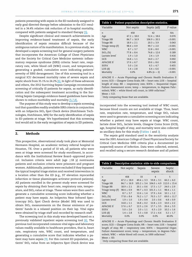
Table 1 e Patient population descriptive statistics.
Variable Not septic Septic (all) P value
n 458 42 NA
Age 47.2 � 18.0 52.4 � 18.6 0.076
Triage HR 84.7 � 16.9 106.9 � 20.5 <0.001
Triage RR 18.9 � 2.1 20.3 � 3.9 0.043
Triage temp (F) 98.4 � 0.9 99.7 � 2.0 <0.001
WBC* 8.7 � 3.7 12.8 � 8.0 <0.001
StO2 (%) 77.8 � 8.4 74.8 � 12.6 0.189
Lactate level* 1.9 � 1.0 2.4 � 1.9 0.416
GCS 14.8 � 1.1 14.0 � 2.7 0.002
APACHE II* 17.6 � 6.7 19.6 � 8.6 0.549
SOFA* 5.0 � 2.6 6.5 � 4.7 0.455
LOS (d) 1.4 � 2.8 6.4 � 6.6 <0.001
Mortality 0.0% 4.8% (n ¼ 2) <0.001
APACHE II ¼ Acute Physiology and Chronic Health Evaluation II
score; GCS ¼ Glasgow Coma Scale; HR ¼ heart rate; LOS ¼ hospital
length of stay; RR ¼ respiratory rate; SOFA ¼ Sequential Organ
Failure Assessment score; temp ¼ temperature, in degrees Fahr-
enheit; WBC ¼ white blood cell count, in 1000 cells/mm3.
Mean � S.D.*Only comparing those that are available.
j o u r n a l o f s u r g i c a l r e s e a r c h 1 9 0 ( 2 0 1 4 ) 2 7 0e2 7 5 271
patients presenting with sepsis in the ED randomly assigned to
early goal-directed therapy before admission to the ICU resul-
ted in a 34.4% relative risk reduction of in-hospital mortality,
compared with patients assigned to standard therapy [5].
Despite significant clinical and research achievements in
improving evidence-based treatment of sepsis, the early
identification of sepsis remains difficult because of the
ambiguous nature of itsmanifestation. In a previous study, we
developed a sepsis screening tool for general surgery patients
that incorporates the American College of Chest Physicians
and the Society for Critical Care Medicine systemic inflam-
matory response syndrome (SIRS) criteria: heart rate, respi-
ratory rate, white blood cell (WBC) count, and temperature,
generating a graded, cumulative score indicating patients’
severity of SIRS derangement. Use of this screening tool in a
surgical ICU decreased mortality rates of severe sepsis and
septic shock from 35.1% to 24.2% [6]. In light of these advances
and others, the 2012 Surviving Sepsis Campaign recommends
screening of critically ill patients for sepsis, as early identifi-
cation and the subsequent treatment according to the Sur-
viving Sepsis Campaign evidence-based guidelines have been
shown to reduce morbidity and mortality [5,7,8].
The purpose of this study was to develop a sepsis screening
tool that quantifies readily available SIRS criteria in conjunction
with an InSpectra StO2 Spot Check device (Hutchinson Tech-
nologies, Hutchinson, MN) for the early identification of sepsis
in ED patients at triage. We hypothesized that this screening
tool would aid in the early recognition of sepsis in ED patients.
Table 2 e Descriptive statistics: side-to-side comparison.
Variable Not septic Sepsis Severesepsis
Septicshock
n 458 33 4 5
Age 47.2 � 18.0 49.4 � 3.3 59.8 � 3.1 66.2 � 6.0
Triage HR 84.7 � 16.9 105.2 � 3.4 104.0 � 4.9 112.0 � 15.4
Triage RR 18.9 � 2.1 20.1 � 0.6 17.5 � 1.7 24.0 � 2.9
Triage temp (F) 98.4 � 0.9 99.7 � 0.3 101.3 � 1.1 98.4 � 1.1
WBC* 8.7 � 3.7 12.6 � 1.4 17.9 � 6.4 10.1 � 1.2
StO2 (%) 77.8 � 8.4 76.2 � 1.7 70.3 � 5.7 69.0 � 11.7
Lactate level* 1.9 � 1.0 1.9 � 0.4 2.0 � 0.6 4.8 � 0.9
GCS 14.8 � 1.1 14.8 � 0.2 12.8 � 2.3 10.0 � 2.2
APACHE II* 17.6 � 6.7 12.5 � 2.6 17.7 � 3.5 26.4 � 3.3
SOFA* 5.0 � 2.6 2.0 � 1.1 6.3 � 2.0 10.2 � 1.6
LOS (d) 1.4 � 2.8 5.5 � 0.8 17.2 � 6.6 3.2 � 1.7
Mortality 0.0% 0.0% 0.0% 40.0%
APACHE II ¼ Acute Physiology and Chronic Health Evaluation II
score; GCS ¼ Glasgow Coma Scale; HR ¼ heart rate; LOS ¼ hospital
length of stay; RR ¼ respiratory rate; SOFA ¼ Sequential Organ
Failure Assessment score; temp ¼ temperature, in degrees Fahr-
enheit; WBC ¼ white blood cell count, in 1000 cells/mm3.
Mean � SD.*Only comparing those that are available.
2. Methods
This prospective, observational study took place at Memorial
Hermann Hospital, an academic tertiary referral hospital in
Houston, TX. Over a period of 10 wk, all patients who were
seen at triage were screened for study enrollment, in accor-
dance with the Institutional Review Boardeapproved proto-
col. Inclusion criteria were adult (age �18 y) nontrauma
patients and exclusion criteria were prisoners and pregnant
women. Additionally, patients were excluded if they bypassed
the typical hospital triage station and received intervention in
a location other than the ED (e.g., ST elevation myocardial
infarction or tissue plasminogen activator protocol patients).
All patients enrolled in the present study were screened for
sepsis by obtaining their heart rate, respiratory rate, temper-
ature, and StO2 value at triage. These values were then used to
generate a cumulative screening score indicating whether a
patient may have sepsis. An InSpectra Near Infrared Spec-
troscopy StO2 Spot Check device (Model 300) was used to
obtain StO2 measurements on the thenar eminence of pa-
tients’ hands in a relaxed position on their lap. Vital signs
were obtained by triage staff and recorded by research staff.
The screening tool in this study was developed based on a
previously validated inpatient sepsis screening tool [6]. The
previous tool incorporated common vital signs and laboratory
values readily available to healthcare providers, that is, heart
rate, respiratory rate, WBC count, and temperature, and
generating a cumulative score that indicates whether a pa-
tient may have sepsis [9]. For this current ED population, pa-
tients’ StO2 value from an InSpectra Spot Check device was
incorporated into the screening tool instead of WBC count,
because blood counts are not available at triage. Thus, heart
rate, respiration rate, temperature, and triage StO2 values
were used to generate a cumulative screening score indicating
whether a patient may have sepsis at triage. WBC count,
lactate draw StO2, lactate levels, Glasgow Coma Score (GCS),
age, hospital length of stay, and mortality were also collected
as ancillary data for this study (Tables 1 and 2).
The sepsis gold standard used in the sensitivity analysis
was the 2001 American College of Chest Physicians/Society for
Critical Care Medicine SIRS criteria plus a documented (or
suspected) source of infection. Data were collected, entered,
and recalled using a secure research database and all patient
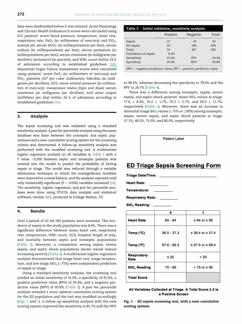
Table 3 e Initial validation, sensitivity analysis.
Positive Negative Total
Sepsis 13 29 42
No sepsis 20 438 458
Total 33 467 500
Prevalence of sepsis 8.4%
Sensitivity 31.0% PPV 39.4%
Specificity 95.6% NPV 93.8%
NPV ¼ negative predictive value; PPV ¼ positive predictive value.
j o u r n a l o f s u r g i c a l r e s e a r c h 1 9 0 ( 2 0 1 4 ) 2 7 0e2 7 5272
data were deidentified before it was entered. Acute Physiology
and Chronic Health Evaluation II scores were calculated using
ICU patients’ worst blood pressure, temperature, heart rate,
respiratory rate, PaO2 (in millimeters of mercury) and FiO2,
arterial pH, serum HCO3� (in milliequivalents per liter), serum
sodium (in milliequivalents per liter), serum potassium (in
milliequivalents per liter), serum creatinine (in milligrams per
deciliter), hematocrit (in percent), and WBC count within 24 h
of admission according to established guidelines [10].
Sequential Organ Failure Assessment scores were calculated
using patients’ worst PaO2 (in millimeters of mercury) and
FiO2, platelets (103 per cubic millimeter), bilirubin (in milli-
grams per deciliter), GCS, mean arterial pressure (in millime-
ters of mercury), vasopressor status (type and dose), serum
creatinine (in milligrams per deciliter), and urine output
(milliliters per day) within 24 h of admission according to
established guidelines [11].
3. Analysis
The sepsis screening tool was validated using a standard
sensitivity analysis. A post hocpercentile analysisusing the same
database was done between the nonseptic and septic pop-
ulations and a new cumulative scoring system for the screening
criteria was determined. A follow-up sensitivity analysis was
performed with the modified screening tool. A multivariate
logistic regression analysis on all variables in Table 1 with a
P value <0.200 between septic and nonseptic patients was
entered into the model to predict the probability of having
sepsis at triage. The model was reduced through a variable
elimination technique in which the nonsignificant variables
were removed in a serial fashion, and the analysis repeated until
only statistically significant (P < 0.050) variables remained [12].
The sensitivity, logistic regression, and post hoc percentile ana-
lyses were done using STATA data analysis and statistical
software, version 12.1, produced in College Station, TX.
Fig. 1 e ED sepsis screening tool, with a new cumulative
scoring system.
4. Results
Over a period of 10 wk 500 patients were screened. The inci-
dence of sepsis in the study population was 8.4%. There was a
significant difference between mean heart rate, respiratory
rate, temperature, WBC count, GCS, hospital length of stay,
and mortality between septic and nonseptic populations
(Table 1). Moreover, a comparison among sepsis, severe
sepsis, and septic shock populations shows trends toward
increasing severity (Table 2). Amultivariate logistic regression
analysis demonstrated that triage heart rate, triage tempera-
ture, and low triage StO2 (<75%) were independent predictors
of sepsis at triage.
Using a standard sensitivity analysis, the screening tool
yielded an initial sensitivity of 31.0%, a specificity of 95.6%, a
positive predictive value (PPV) of 39.4%, and a negative pre-
dictive value (NPV) of 93.8% (Table 3). A post hoc percentile
analysis revealed a more optimal cumulative scoring system
for the ED population and the tool was modified accordingly
(Figs. 1 and 2). A follow-up sensitivity analysis with the new
scoring system improved the sensitivity to 85.7% and the NPV
to 98.4%, whereas decreasing the specificity to 78.4% and the
PPV to 26.7% (Table 4).
There was a difference among nonseptic, sepsis, severe
sepsis, and septic shock patients’ mean StO2 values at triage:
77.8 � 8.4%, 76.2 � 1.7%, 70.3 � 5.7%, and 69.0 � 11.7%,
respectively (Table 2). Moreover, there was an increase in
abnormal triage StO2 values (<75% or�90%) among nonseptic,
sepsis, severe sepsis, and septic shock patients at triage:
37.1%, 48.5%, 75.0%, and 80.0%, respectively.
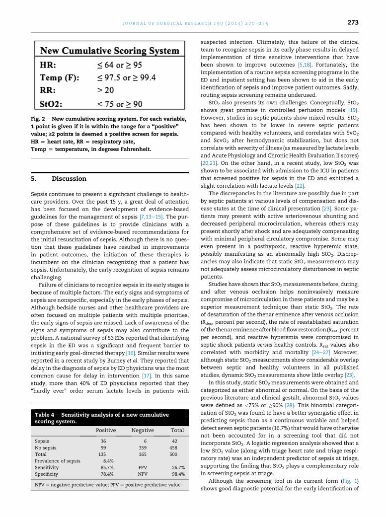
Fig. 2 e New cumulative scoring system. For each variable,
1 point is given if it is within the range for a “positive”
value; ‡2 points is deemed a positive screen for sepsis.
HR [ heart rate, RR [ respiratory rate,
Temp [ temperature, in degrees Fahrenheit.
j o u r n a l o f s u r g i c a l r e s e a r c h 1 9 0 ( 2 0 1 4 ) 2 7 0e2 7 5 273
5. Discussion
Sepsis continues to present a significant challenge to health-
care providers. Over the past 15 y, a great deal of attention
has been focused on the development of evidence-based
guidelines for the management of sepsis [7,13e15]. The pur-
pose of these guidelines is to provide clinicians with a
comprehensive set of evidence-based recommendations for
the initial resuscitation of sepsis. Although there is no ques-
tion that these guidelines have resulted in improvements
in patient outcomes, the initiation of these therapies is
incumbent on the clinician recognizing that a patient has
sepsis. Unfortunately, the early recognition of sepsis remains
challenging.
Failure of clinicians to recognize sepsis in its early stages is
because of multiple factors. The early signs and symptoms of
sepsis are nonspecific, especially in the early phases of sepsis.
Although bedside nurses and other healthcare providers are
often focused on multiple patients with multiple priorities,
the early signs of sepsis are missed. Lack of awareness of the
signs and symptoms of sepsis may also contribute to the
problem. A national survey of 53 EDs reported that identifying
sepsis in the ED was a significant and frequent barrier to
initiating early goal-directed therapy [16]. Similar results were
reported in a recent study by Burney et al. They reported that
delay in the diagnosis of sepsis by ED physicians was themost
common cause for delay in intervention [17]. In this same
study, more than 40% of ED physicians reported that they
“hardly ever” order serum lactate levels in patients with
Table 4 e Sensitivity analysis of a new cumulativescoring system.
Positive Negative Total
Sepsis 36 6 42
No sepsis 99 359 458
Total 135 365 500
Prevalence of sepsis 8.4%
Sensitivity 85.7% PPV 26.7%
Specificity 78.4% NPV 98.4%
NPV ¼ negative predictive value; PPV ¼ positive predictive value.
suspected infection. Ultimately, this failure of the clinical
team to recognize sepsis in its early phase results in delayed
implementation of time sensitive interventions that have
been shown to improve outcomes [5,18]. Fortunately, the
implementation of a routine sepsis screening programs in the
ED and inpatient setting has been shown to aid in the early
identification of sepsis and improve patient outcomes. Sadly,
routing sepsis screening remains underused.
StO2 also presents its own challenges. Conceptually, StO2
shows great promise in controlled perfusion models [19].
However, studies in septic patients show mixed results. StO2
has been shown to be lower in severe septic patients
compared with healthy volunteers, and correlates with SvO2
and ScvO2 after hemodynamic stabilization, but does not
correlate with severity of illness (asmeasured by lactate levels
and Acute Physiology and Chronic Health Evaluation II scores)
[20,21]. On the other hand, in a recent study, low StO2 was
shown to be associated with admission to the ICU in patients
that screened positive for sepsis in the ED and exhibited a
slight correlation with lactate levels [22].
The discrepancies in the literature are possibly due in part
by septic patients at various levels of compensation and dis-
ease states at the time of clinical presentation [23]. Some pa-
tients may present with active arteriovenous shunting and
decreased peripheral microcirculation, whereas others may
present shortly after shock and are adequately compensating
with minimal peripheral circulatory compromise. Some may
even present in a posthypoxic, reactive hyperemic state,
possibly manifesting as an abnormally high StO2. Discrep-
ancies may also indicate that static StO2 measurements may
not adequately assess microcirculatory disturbances in septic
patients.
Studies have shown that StO2measurements before, during,
and after venous occlusion helps noninvasively measure
compromise ofmicrocirculation in these patients andmay be a
superior measurement technique than static StO2. The rate
of desaturation of the thenar eminence after venous occlusion
(Rdes, percent per second), the rate of reestablished saturation
of thethenareminenceafterbloodflowrestoration (Rsat, percent
per second), and reactive hyperemia were compromised in
septic shock patients versus healthy controls. Rsat values also
correlated with morbidity and mortality [24e27] Moreover,
although static StO2 measurements show considerable overlap
between septic and healthy volunteers in all published
studies, dynamic StO2 measurements show little overlap [23].
In this study, static StO2 measurements were obtained and
categorized as either abnormal or normal. On the basis of the
previous literature and clinical gestalt, abnormal StO2 values
were defined as <75% or �90% [28]. This binomial categori-
zation of StO2 was found to have a better synergistic effect in
predicting sepsis than as a continuous variable and helped
detect seven septic patients (16.7%) thatwould have otherwise
not been accounted for in a screening tool that did not
incorporate StO2. A logistic regression analysis showed that a
low StO2 value (along with triage heart rate and triage respi-
ratory rate) was an independent predictor of sepsis at triage,
supporting the finding that StO2 plays a complementary role
in screening sepsis at triage.
Although the screening tool in its current form (Fig. 1)
shows good diagnostic potential for the early identification of
j o u r n a l o f s u r g i c a l r e s e a r c h 1 9 0 ( 2 0 1 4 ) 2 7 0e2 7 5274
sepsis among ED triage personnel, careful examination of the
sensitivity analysis reveals ways the tool can be further
improved. Namely, future modifications of the tool should
elucidate the possibility of a source of infection. Only 20% of
false-positive patients (20 of 99) had a documented source of
infection at discharge or admission and 67% (4 of 6) false-
negative patients had an obvious infection source at triage.
Thus, a modification of the screening tool that incorporates
simple screening questions (analogous to a mini “review of
systems” for tuberculosis screening) may aid in determining a
potential source of infection and help limit false positives and
false negatives [29].
There are also limitations of the screening tool for this
population. Patients exhibiting limited SIRS criteria and
leukocytosis are prone to being missed (all six false-negative
patients had an elevated WBC count [12,000 cells/mm3]). Also,
it should be noted that because the incidence of sepsis in this
population is low (8.4%), the PPV is inherently limited, regard-
less of the modifications made to the tool. Moreover, this
screening tool is designed to limit the amount of septic patients
missed at triage (i.e., limit the number of false negatives). Thus,
the sensitivity and NPV of this ED sepsis screening tool were
maximized, conceding some level of specificity and PPV.
6. Conclusions
Heart rate, respiratory rate, and temperature have good
diagnostic potential for the early identification of sepsis
among ED triage personnel. Additionally, early evidence sug-
gests that StO2 may play a complementary and synergistic
role in the early identification of sepsis by triage personnel.
However, characterization of StO2 in this population needs to
be investigated further. Moreover, the screening tool’s post hoc
modifications must be validated in a separate prospective
study, which is currently underway.
Acknowledgment
The Spot Check Device used in this studywas provided for use
by Hutchinson Technologies.
Author contributions: C.E.G. contributed toward analysis
and interpretation, data collection, and writing of the manu-
script. C.E.W. contributed toward conception and design,
analysis and interpretation, critical revision of the manuscript,
and obtaining of funding. J.J.M. contributed toward conception
and design and critical revision of the manuscript. J.B.H.
contributed toward critical revision of the article and obtaining
of funding. L.J.M. contributed toward conception and design,
analysis and interpretation, critical revision of the manuscript,
obtaining of funding, and writing of the manuscript.
Disclosure
The authors have no financial or nonfinancial interest in the
subject matter or materials discussed in this article.
r e f e r e n c e s
[1] ProductsdData Briefs. Number 62, June 2011 [Internet] [cited2013 Nov 13]. Available from: http://www.cdc.gov/nchs/data/databriefs/db62.htm.
[2] Sands KE, Bates DW, Lanken PN, et al. Epidemiology of sepsissyndrome in 8 academic medical centers. JAMA 1997;278:234.
[3] Moore LJ, Moore FA, Todd SR, Jones SL, Turner KL, Bass BL.Sepsis in general surgery: the 2005-2007 national surgicalquality improvement program perspective. Arch Surg (ChicIll 1960) 2010;145:695.
[4] Filbin MR, Arias SA, Camargo CA Jr, Barche A, Pallin DJ.Sepsis visits and antibiotic utilization in U.S. emergencydepartments. Crit Care Med 2014;42:528.
[5] Rivers E, Nguyen B, Havstad S, et al. Early goal-directedtherapy in the treatment of severe sepsis and septic shock. NEngl J Med 2001;345:1368.
[6] Moore LJ, Jones SL, Kreiner LA, et al. Validation of a screeningtool for the early identificationof sepsis. J Trauma2009;66:1539.
[7] Dellinger RP, Levy MM, Rhodes AMB, et al. Surviving sepsiscampaign: international guidelines for management ofsevere sepsis and septic shock: 2012. Crit Care Med 2013;41:580.
[8] Moore LJ, Moore FA. Early diagnosis and evidence-based careof surgical sepsis. J Intensive Care Med 2013;28:107.
[9] Levy MM, Fink MP, Marshall JC, et al. 2001 SCCM/ESICM/ACCP/ATS/SIS International Sepsis Definitions Conference.Intensive Care Med 2003;29:530.
[10] Knaus WA, Draper EA, Wagner DP, Zimmerman JE. APACHEII: a severity of disease classification system. Crit Care Med1985;13:818.
[11] Vincent JL, de Mendonca A, Cantraine F, et al. Use of theSOFA score to assess the incidence of organ dysfunction/failure in intensive care units: results of a multicenter,prospective study. Working group on “sepsis-relatedproblems” of the European Society of Intensive CareMedicine. Crit Care Med 1998;26:1793.
[12] Bursac Z, Gauss CH, Williams DK, Hosmer DW. Purposefulselection of variables in logistic regression. Source Code BiolMed 2008;3:17.
[13] Dellinger RP, Carlet JM, Masur H, et al. Surviving SepsisCampaign guidelines for management of severe sepsis andseptic shock. Intensive Care Med 2004;30:536.
[14] Dellinger RP, Levy MM, Carlet JM, et al. Surviving sepsiscampaign: international guidelines for management ofsevere sepsis and septic shock: 2008. Crit Care Med 2008;36:296.
[15] Hollenberg SM, Ahrens TS, Annane D, et al. Practiceparameters for hemodynamic support of sepsis in adultpatients: 2004 update. Crit Care Med 2004;32:1928.
[16] Carlbom DJ, Rubenfeld GD. Barriers to implementingprotocol-based sepsis resuscitation in the emergencydepartmentdresults of a national survey. Crit Care Med2007;35:2525.
[17] Burney M, Underwood J, McEvoy S, et al. Early detection andtreatment of severe sepsis in the emergency department:identifying barriers to implementation of a protocol-basedapproach. J Emerg Nurs 2012;38:512.
[18] Kumar A, Roberts D, Wood KE, et al. Duration of hypotensionbefore initiation of effective antimicrobial therapy is thecritical determinant of survival in human septic shock. CritCare Med 2006;34:1589.
[19] Putnam B, Bricker S, Fedorka P, et al. The correlation ofnear-infrared spectroscopy with changes in oxygendelivery in a controlled model of altered perfusion. AmSurg 2007;73:1017.
j o u r n a l o f s u r g i c a l r e s e a r c h 1 9 0 ( 2 0 1 4 ) 2 7 0e2 7 5 275
[20] Mesquida J, Masip J, Gili G, Artigas A, Baigorri F. Thenaroxygen saturation measured by near infrared spectroscopyas a noninvasive predictor of low central venous oxygensaturation in septic patients. Intensive Care Med 2009;35:1106.
[21] Mulier KE, Skarda DE, Taylor JH, et al. Near-infraredspectroscopy in patients with severe sepsis: correlationwith invasive hemodynamic measurements. Surg Infect2008;9:515.
[22] Leichtle SW, Kaoutzanis C, Brandt M-M, Welch KB, Purtill M-A. Tissue oxygen saturation for the risk stratification ofseptic patients. J Crit Care 2013;28:1111.e1.
[23] Lipcsey M, Woinarski NC, Bellomo R. Near infraredspectroscopy (NIRS) of the thenar eminence in anesthesiaand intensive care. Ann Intensive Care 2012;2:11.
[24] Skarda DE, Mulier KE, Myers DE, Taylor JH, Beilman GJ.Dynamic near-infrared spectroscopy measurements inpatients with severe sepsis. Shock (Augusta Ga) 2007;27:348.
[25] Creteur J, Carollo T, Soldati G, Buchele G, De Backer D,Vincent J-L. The prognostic value of muscle StO2 in septicpatients. Intensive Care Med 2007;33:1549.
[26] Doerschug KC, Delsing AS, Schmidt GA, Haynes WG.Impairments in microvascular reactivity are related to organfailure in human sepsis. Am J Physiol Heart Circ Physiol 2007;293:H1065.
[27] Payen D, Luengo C, Heyer L, et al. Is thenar tissuehemoglobin oxygen saturation in septic shock related tomacrohemodynamic variables and outcome? Crit Care 2009;13(Suppl 5):S6.
[28] Cohn SM, Nathens AB, Moore FA, et al. Tissue oxygensaturation predicts the development of organ dysfunctionduring traumatic shock resuscitation. J Trauma 2007;62:44.discussion 54-55.
[29] Moran GJ, Barrett TW, Mower WR, et al. Decision instrumentfor the isolation of pneumonia patients with suspectedpulmonary tuberculosis admitted through US emergencydepartments. Ann Emerg Med 2009;53:625.